tupaia oasl1 promotes cellular antiviral immune responses
TRANSCRIPT
of December 28, 2020.This information is current as
MAVSImmune Responses by Recruiting MDA5 to
OASL1 Promotes Cellular AntiviralTupaia
Zheng, Rui Bi and Yong-Gang YaoYu-Lin Yao, Dandan Yu, Ling Xu, Tianle Gu, Yu Li, Xiao
http://www.jimmunol.org/content/205/12/3419doi: 10.4049/jimmunol.2000740November 2020;
2020; 205:3419-3428; Prepublished online 13J Immunol
MaterialSupplementary
0.DCSupplementalhttp://www.jimmunol.org/content/suppl/2020/11/12/jimmunol.200074
Referenceshttp://www.jimmunol.org/content/205/12/3419.full#ref-list-1
, 8 of which you can access for free at: cites 48 articlesThis article
average*
4 weeks from acceptance to publicationFast Publication! •
Every submission reviewed by practicing scientistsNo Triage! •
from submission to initial decisionRapid Reviews! 30 days* •
Submit online. ?The JIWhy
Subscriptionhttp://jimmunol.org/subscription
is online at: The Journal of ImmunologyInformation about subscribing to
Permissionshttp://www.aai.org/About/Publications/JI/copyright.htmlSubmit copyright permission requests at:
Email Alertshttp://jimmunol.org/alertsReceive free email-alerts when new articles cite this article. Sign up at:
Print ISSN: 0022-1767 Online ISSN: 1550-6606. Immunologists, Inc. All rights reserved.Copyright © 2020 by The American Association of1451 Rockville Pike, Suite 650, Rockville, MD 20852The American Association of Immunologists, Inc.,
is published twice each month byThe Journal of Immunology
by Yong-G
ang Yao on D
ecember 28, 2020
http://ww
w.jim
munol.org/
Dow
nloaded from
by Yong-G
ang Yao on D
ecember 28, 2020
http://ww
w.jim
munol.org/
Dow
nloaded from
by Yong-G
ang Yao on D
ecember 28, 2020
http://ww
w.jim
munol.org/
Dow
nloaded from
by Yong-G
ang Yao on D
ecember 28, 2020
http://ww
w.jim
munol.org/
Dow
nloaded from
The Journal of Immunology
Tupaia OASL1 Promotes Cellular Antiviral ImmuneResponses by Recruiting MDA5 to MAVS
Yu-Lin Yao,*,†,‡ Dandan Yu,*,†,‡ Ling Xu,*,† Tianle Gu,*,†,‡ Yu Li,*,†,‡ Xiao Zheng,*,†,x
Rui Bi,*,†,‡ and Yong-Gang Yao*,†,‡,{,‖
Melanoma differentiation-associated gene 5 (MDA5) is a key cytoplasmic dsRNA sensor. Upon binding to invading viral RNA,
activated MDA5 is recruited to mitochondria and interacts with mitochondrial antiviral signaling gene (MAVS) to initiate innate
antiviral immune responses. The elegant regulation of this process remains elusive. In this study, using the Chinese tree shrew
(Tupaia belangeri chinensis), which is genetically close to primates, we identified the Tupaia oligoadenylate synthetases-like 1
(tOASL1) as a positive regulator of the Tupaia MDA5 (tMDA5) and Tupaia MAVS (tMAVS)–mediated IFN signaling. Over-
expression of tOASL1 significantly potentiated the RNA virus-triggered induction of the type I IFNs and downstream antiviral
genes. Conversely, knockdown of tOASL1 had an impaired antiviral immune response. Mechanistically, tOASL1 was associated
with mitochondria and directly interacted with tMDA5 and tMAVS. Upon RNAvirus infection, tOASL1 enhanced the interaction
between tMDA5 and tMAVS via its OAS and UBL domains. Our results revealed a novel mechanism by which tOASL1 contrib-
utes to host antiviral responses via enhancing tMDA5 and tMAVS interaction. The Journal of Immunology, 2020, 205: 3419–3428.
The innate immune system provides the critical first line ofhost defense against pathogen invasions via recognitionof pathogen-associated molecular patterns aided by pat-
tern recognition receptors, which initiate a series of signalingcascades leading to the induction of the type I IFN and proin-flammatory cytokines (1, 2). Viral RNAs derived from the viral
genome or its replication intermediates are mainly recognizedby retinoic acid inducible gene I (RIG-I)–like receptors (RLRs)
including RIG-I, melanoma differentiation-associated gene 5
(MDA5), and laboratory of genetics and physiology 2 (LGP2)
(3, 4). RIG-I and MDA5 contain two caspase activation and
recruitment domains (CARDs), a helicase domain, and a C-
terminal domain (CTD), and recognize different types of RNA
viruses (3, 4). Following ligand binding, RIG-I and MDA5 are
recruited to the mitochondrion-associated membrane where
they bind to mitochondrial antiviral signaling gene [MAVS
(5), also named as IPS-1 (6), Cardif (7) or VISA (8)] via homo-
typic CARD interaction and promote a massive MAVS prion-like
fiber formation (9, 10). Subsequently, MAVS recruited down-
stream antiviral kinases, such as TRAF3, TBK1/IKKε, and IKKa/
b, which further phosphorylate the transcription factors IRF3 and
NF-kB to induce the production of type I IFNs. The IFNs trigger
the JAK/STAT signaling pathway to increase the expression of
hundreds of IFN-stimulated genes (ISGs) and eventually help the
host cells to defend from invading viruses (1, 11). In contrast to
RIG-I and MDA5, the LGP2 lacks the CARDs and regulates the
RLR signaling in response to certain viruses (12–14). To invoke a
proper antiviral immune response, signaling proteins involved in
these cascades were precisely regulated by immune regulators.
Negative regulators dampen innate immune activation and in-
flammation to avoid additional tissue damage, whereas rapid and
robust induction of IFN aided by positive regulators is critical for
effective clearance of invading viruses (15).The 29,59-oligoadenylate synthetases (OAS) family proteins are
composed of OAS1, OAS2, OAS3, and OAS-like (OASL). All
OAS members are characterized by their highly conserved N-
terminal OAS domain and play a critical role in blocking viral
infections (16, 17). Among them, OAS1, OAS2 and OAS3, pos-
sess the 29-59 oligoadenylates synthetase activity and exert an-
tiviral activity via the canonical RNase L–dependent pathway
(18). The OASL is devoid of enzyme activity to synthetize 29-59
oligoadenylates and exerts its antiviral activity as a multifaceted
immune regulator (19). In human beings, the OASL counteracts
the RNA virus infections by interacting and enhancing RIG-I
*Key Laboratory of Animal Models and Human Disease Mechanisms of the ChineseAcademy of Sciences and Yunnan Province, Kunming Institute of Zoology, ChineseAcademy of Sciences, Kunming, Yunnan 650223, China; †Kunming Institute ofZoology – Chinese University of Hong Kong Joint Laboratory of Bioresources andMolecular Research in Common Diseases, Kunming Institute of Zoology, ChineseAcademy of Sciences, Kunming, Yunnan 650223, China; ‡Kunming College of LifeScience, University of Chinese Academy of Sciences, Kunming, Yunnan 650204,China; xSchool of Life Sciences, Division of Life Sciences and Medicine, Universityof Science and Technology of China, Hefei, Anhui 230026, China; {National Re-source Center for Non-Human Primates, Kunming Institute of Zoology, ChineseAcademy of Sciences, Kunming, Yunnan 650107, China; and ‖Primate Facility, Na-tional Research Facility for Phenotypic and Genetic Analysis of Model Animals,Kunming Institute of Zoology, Chinese Academy of Sciences, Kunming, Yunnan650107, China
ORCIDs: 0000-0002-7309-4250 (X.Z.); 0000-0002-2955-0693 (Y.-G.Y.).
Received for publication June 23, 2020. Accepted for publication October 15, 2020.
This work was supported by the National Natural Science Foundation of China(U1902215 and U1402224 [to Y.-G.Y.]), the West Light Foundation of the ChineseAcademy of Sciences (xbzg-zdsys-201909 [to Y.-G.Y.]), and the Yunnan ProvincialNatural Science Foundation (2018FB046 and 202001AS070023 [to D.Y.]).
Address correspondence and reprint requests to Dr. Yong-Gang Yao, Kunming Insti-tute of Zoology, Chinese Academy of Sciences, 21 Qingsong Road, Kunming,Yunnan 650204, China. E-mail address: [email protected]
The online version of this article contains supplemental material.
Abbreviations used in this article: CARD, caspase activation and recruitment domain;CAS, Chinese Academy of Sciences; CTD, C-terminal domain; EMCV, encephalo-myocarditis virus; F, forward; hMAVS, human MAVS; hMDA5, human MDA5;hOASL, human OASL; ISD, IFN stimulatory DNA; ISG, IFN-stimulated gene; ISRE,IFN-stimulated response element; KIZ, Kunming Institute of Zoology; KO, knock-out; LGP2, laboratory of genetics and physiology 2; MAVS, mitochondrial antiviralsignaling gene; MDA5, melanoma differentiation-associated gene 5; MOI, multiplic-ity of infection; NDV, Newcastle disease virus; OAS, oligoadenylate synthetase;OASL, OAS-like; poly I:C H, high m.w. poly I:C; RIG-I, retinoic acid induciblegene I; RLR, RIG-I–like receptor; RT-qPCR, quantitative real-time PCR; SeV, Sendaivirus; sgRNA, small guide RNA; siNC, small interfering negative control; siRNA,small interfering RNA; sitOASL, small interfering tOASL; tMAVS, Tupaia MAVS;tMDA5, Tupaia MDA5; tOASL1, Tupaia OASL1; TSPRC, tree shrew primary renalcell; VSV, vesicular stomatitis virus; WT, wild-type.
Copyright� 2020 by The American Association of Immunologists, Inc. 0022-1767/20/$37.50
www.jimmunol.org/cgi/doi/10.4049/jimmunol.2000740
by Yong-G
ang Yao on D
ecember 28, 2020
http://ww
w.jim
munol.org/
Dow
nloaded from
activation (20). However, the OASL has an inhibitory effect oncGAS activity to reduce IFN induction during DNA virus in-fection (21). Unlike human, mouse Oasl1 specifically binds toIRF7 59 untranslated region and inhibits the translation of IRF7mRNA, thus negatively regulating the antiviral innate immunityregardless of DNA and RNA virus infections (22).We recently characterized the OASs (including tOAS1, tOAS2,
Tupaia OASL [tOASL] 1, and tOASL2) of the Chinese treeshrew (Tupaia belangeri chinensis) (23), which is a smallmammal genetically closer to primates (24, 25) and has beenused as a good model for studying infectious diseases (26–30)and other diseases (31–33). We found that the antiviral activitiesof tOAS1 and tOAS2 were dependent on the canonical RNase Lpathway, whereas those of tOASL1 and tOASL2 were inde-pendent on the RNase L pathway (23). In this study, we aimedto test whether tOASs block viral replications through regulatingantiviral innate immune responses. We identified tOASL1 as apositive regulator in RNA virus–induced production of type I IFNmediated by Tupaia MDA5 (tMDA5) and Tupaia MAVS (tMAVS).
Materials and MethodsEthics statement
The healthy adult Chinese tree shrews (n = 10) were introduced from theexperimental animal core facility of the Kunming Institute of Zoology(KIZ), Chinese Academy of Sciences (CAS). After euthanasia, we isolatedtree shrew primary renal cells (TSPRCs) according to the method ofenzyme-assisted dissection, as described in our previous study (34). Allexperimental procedures were performed with the approval of the Insti-tutional Animal Care and Use Committee of the KIZ (Approval No.:SYDW20110315001).
Cells, viruses, ligands, and plasmids
Vero, HEK293T, and HeLa cells were introduced from Kunming CellBank, KIZ, CAS. Cells were cultured at 37˚C in 5% CO2 with DMEM(11965-092, DMEM; Life Technologies-BRL) supplemented with 10%FBS (10099-141, FBS; Life Technologies-BRL) and 13 penicillin/streptomycin (10378016; Life Technologies-BRL). DNA virus (HSV-1)and RNA viruses (including Sendai virus [SeV], encephalomyocarditisvirus [EMCV], Newcastle disease virus [NDV], and vesicular stomatitisvirus [VSV]) were taken from our previous study (34). High m.w. poly I:C(poly I:C H) (catalog no. tlrl-piclv), poly dA:dT (catalog no. tlrl-patn), andIFN stimulatory DNA (ISD) (catalog no. tlrl-isdn) were from InvivoGen.
Expression vectors for tOAS1, tOAS2, tOASL1, tOASL2, tMAVS,tMDA5, and Tupaia LGP2 were taken from our previous studies (23, 34,35). The Myc-tagged tOASL1 was subcloned into pCS2-N-myc withBspEI and SacI. Expression vector for tTBK1 was generated using specificprimers and was cloned into pCMV-3Tag-8 vectors (SupplementalTable I). The tree shrew IFNB1 promoter luciferase reporter (IFN-b-Luc: pGL3-tIFN-b-promoter), ISRE-Luc (219092, ISRE cis-reporter;Stratagene) and NF-kB-Luc (631912, pNFkB-TA-Luc; Clontech Labora-tories), and pRL-SV40-Renilla (as an internal control; Promega) were fromour previous studies (34, 35). We created pCMV-3Tag-8 and pCMV-HAexpression vectors for different domains of tOASL1 (tOASL1-OAS andtOASL1-UBL), tMDA5 (tMDA5-CARD, tMDA5-Helicase, and tMDA5-CTD), and tMAVS (tMAVS-CARD, tMAVS pattern recognition receptors,and tMAVS-TM) (Supplemental Table I). All constructs were confirmed bydirect sequencing.
Abs
We used the following primary Abs: mouse monoclonal anti-FLAG(M20008; Abmart), mouse monoclonal anti-HA (3724; Cell SignalTechnology), mouse anti–c-Myc (9E11) (MA1-16637; Life Technologies),rabbit monoclonal anti-FLAG (14973; Cell Signal Technology), mousemonoclonal anti–b-actin (E1C602-2; EnoGene), mouse monoclonal anti-GAPDH (E12-052-4; EnoGene), rabbit monoclonal anti-TBK1 (3504;Cell Signal Technology), rabbit monoclonal anti–phospho-TBK1 (Ser172)(5483; Cell Signal Technology), rabbit monoclonal anti-IRF3 (4302; CellSignal Technology), and rabbit monoclonal anti–phospho-IRF3 (Ser396)(4947; Cell Signal Technology). The NF-kB pathway sample kit (9936;Cell Signal Technology), which contains Abs against phospho-p65(Ser536), p65, phospho-IKKa/b (Ser176/180), IKKa/b, phospho-IkBa
(Ser32), and IkBa, was used to test the activation of the NF-kB signaling.We used the following secondary Abs: peroxidase-conjugated anti-mouseAb (474-1806; KPL), peroxidase-conjugated anti-rabbit Ab (074-1506;KPL), Alexa Fluor 594–conjugated anti-rabbit IgG (A21207; Invitrogen),and Alexa Fluor 488–conjugated anti-mouse IgG (A21202; Invitrogen).
Generation of tOASL1 knockout cell
We used the CRISPR-Cas9 system (36) to knock out the tOASL1 gene inthe TSR6 cell line that was established in our previous study (37).Briefly, small guide RNAs (sgRNAs) (tOASL1-sgRNA-forward [F]: 59-CACCGTCGTCTTTCCCTCACGGTT-39/tOASL1-sgRNA-reverse: 59-AAACAACCGTGAGTGGAAAGACGAC-39) targeting tOASL1 wereannealed and cloned into the pX330-T7 vector (a kind gift from Dr. P.Zheng, KIZ) expressing mCherry. The TSR6 cells were transfectedwith the pX330-T7 vector carrying the sgRNAs by using Lipofect-amine 3000 (L3000008; Invitrogen). The transfected cells expressingmCherry were sorted by flow cytometry and cultured for 48 h. Singlecells were manually picked with a mouth pipette for expansion for 3wk. We used AxyPrep Multisource Genomic DNA Miniprep Kit(26817KC1; Axygen) to extract genomic DNA of single TSR6 cellswith potential knockout (KO) of tOASL1. The gene region spanningthe sgRNA targeting sites was amplified by using primer pair tOASL1-sgRNA-Fc: 59-AGGTGCTGGACTCTGTGAC-39/tOASL1-sgRNA-Rc:59-AGGTGCTGGACTCTGTGAC-39. The PCR products were sequencedby using primer tOASL1-sgRNA-Fc to screen for mutation(s). We were ableto pick up a cell clone with a deletion of 14 bp (c.66_79del) that disrupts thetranslation of tOASL1 protein. The KO of endogenous tOASL1 protein wasfurther validated by Western blot.
RNA interference and transfection
The small interfering RNAs (siRNAs) targeting tOASL1 and small inter-fering negative controls (siNC) were synthesized by RiboBio (Guangzhou,China). We designed three siRNAs targeting the tOASL1 gene: small in-terfering tOASL (sitOASL) 1-1, 59-GCACTACACCTTCCAACAT-39;sitOASL1-2, 59-CCTTACGAGTCCATAAAGA-39; and sitOASL1-3, 59-ACACTACAGCATCCAAGAT-39. Transfection of siRNA into TSPRCsusing Lipofectamine 3000 was performed according to manufacturer’sinstructions, as described in our previous study (38). The knockdown ef-ficiency of siRNAs was evaluated at the mRNA and protein levels.
RNA extraction and quantitative real-time PCR
Total RNA was extracted from TSPRCs using the RNAsimple Total RNAKit (DP419; TIANGEN) according to the manufacturer’s instruction. Asdescribed in our previous studies (23, 35, 39), the quality of total RNAwasmeasured on a biophotometer (Eppendorf). About 2 mg total RNA (with anA260/A280 ratio of 1.8–2.0) was used to synthesize cDNA by using oligo-dT18 primer and Moloney murine leukemia virus reverse transcriptase(M1701; Promega). For detection of the viral RNA, we used a virus-specific primer (SeV R15360: 59-ACCAGACAAGAGTTTAAGAGA-TATG-39) to transcribe the SeV RNA and used a primer pair SeV-F15160:59-TGTTCGGGGCCAGGCAAAAT-39/SeV-R15247: 59-GTTCTGCAC-GATAGGGACTA-39 for quantitative real-time PCR (RT-qPCR). For quan-tification of EMCV RNA, we used a specific primer for the L antisense(59-GGCCGTCATGGTGGCGAATAAGCGCACTCTCTCACTTTTGA-39) for the transcription and used EMCV common F: 59-AATAAAT-CATAAGGCCGTCATGGTGGCGAATAA-39/L reverse primer: 59-A-ATAAATCATAATCGAAAACGACTTCCATGTCT-39 for RT-qPCR. Fordetection of NDV RNA, we used a specific primer (NDV R15168: 59-ACCAAACAAAGATTTGGTGAATGAC-39) to transcribe the NDVRNA and used primer pair NDV-F15160: 59-CTTATTTACCCCTTA-CAATC-39/NDV-R15247: 59-CTGAGACCCAGTATTGTGAC-39 forRT-qPCR. For detection of HSV-1 RNA, we used HSV-1-F: 59-TGGGACACATGCCTTCTTGG-39/HSV-1-R15247: 59-ACCCTTAGTCA-GACTCTGTTACTTACCC-39 for RT-qPCR.
In brief, RT-qPCR was conducted using iTaq Universal SYBR GreenSupermix (172-5125; Bio-Rad Laboratories) with gene-specific primerpairs (Supplemental Table I) on a CFX Connect Real-Time PCR DetectionSystem (Bio-Rad Laboratories). The thermal cycling protocol was onecycle at 95˚C for 1 min, 40 cycles of 95˚C for 15 s, and 55˚C for 15 s. Thetree shrew b-actin transcript was used for the normalization of the targetgene (35).
Western blotting
We followed the procedures for Western blotting and immunoprecipitationin our previous studies (23, 34). In brief, TSPRCs and HEK293T cells weretransfected with the indicated expression vectors and/or tOASL1 siRNAs
3420 TUPAIA OASL1 INTERACTS WITH MDA5 AND MAVS
by Yong-G
ang Yao on D
ecember 28, 2020
http://ww
w.jim
munol.org/
Dow
nloaded from
for 48 h using Lipofectamine 3000. Cells were then harvested and lysed inRIPA lysis buffer (P0013; Beyotime Institute of Biotechnology) on ice.Protein concentration was determined by using the BCA protein assay kit(P0012; Beyotime Institute of Biotechnology). Equal amount (20 mg) ofcellular protein for each sample was used for Western blotting. Electro-phoresis was performed using 12% SDS-PAGE. Proteins were transferredonto polyvinylidene fluoride membranes (IPVH00010; Roche Diagnostics)using the standard procedure. After having blocking in 5% nonfat dry milkdissolved in TBS (#9997; Cell Signaling Technology) containing 0.1%Tween 20 (TBST [0.1%, P1379; Sigma]) at room temperature for 2 h, themembranes were incubated with the respective Abs against FLAG (1:5000),HA (1:5000), Myc (1:5000), OASL (1:1000), IRF3 (1:1000), phospho-IRF3(Ser396) (1:1000), TBK1 (1:1000), phospho-TBK1 (Ser172) (1:1000), p65(1:1000), phospho-p65 (Ser536) (1:1000), phospho-IKKa/b (Ser176/180)(1:1000), IKKa/b (1:1000), phospho-IkBa (Ser32) (1:1000), IkBa(1:1000), GAPDH (1:10,000), or b-actin (1:10,000) overnight at 4˚C. Themembranes were washed three times (5 min each time) with TBST andwere incubated with anti-mouse or anti-rabbit secondary Ab (1:10,000;KPL), dependent on the primary Ab, for 1 h at room temperature. Afterwashing, the protein bands were visualized using the ECL reagents(WBKLS0500; MilliporeSigma).
Coimmunoprecipitation assay
Cells were lysed in RIPA lysis buffer on ice for 1 h, followed by a centrifugeat 12,000 3 g for 10 min at 4˚C. Appropriate Abs were incubated withprotein G agarose beads (15920010; Life Technologies) to form a complexfor 2 h at room temperature. Then, the protein G agarose bead–conjugatedAbs were mixed with the cell lysates to form the protein complex over-night at 4˚C. After four washes with the RIPA lysis buffer, the immuno-precipitates were resuspended in loading sample buffer and were analyzedby immunoblot analysis.
Luciferase reporter assay
TSPRCs were seeded in 24-well plates at a density of 1 3 104 cells andcultured overnight. Cells were transiently transfected with 0.1 mg of theluciferase reporter vector, 0.01 mg pRL-SV40-Renilla, together with theindicated amount of empty vector or expression vector by using X-tremeGENE HP DNA Transfection Reagent (06366236001; Roche). Af-ter transfection for 36 h, cells were left untreated or infected with NDV(multiplicity of infection [MOI] = 1) or HSV-1 (MOI = 1) for 12 h. Cellswere harvested for luciferase activity using the Dual-Luciferase ReporterAssay System (E1960; Promega) on an Infinite M1000 Pro multimodemicroplate reader (30064852; Tecan).
Immunofluorescence analysis
TSPRCs were seeded on glass coverslips and transfected with the indicatedexpression vectors for 36 h, and then cells were left uninfected or infectedwith NDV (MOI = 1) for the indicated times. After three washes with PBS,cells were fixed with 300 ml 4% paraformaldehyde for 15 min at 37˚C,followed by a permeabilized procedure using 0.1% Triton X-100 (Sigma-Aldrich) for 15 min at room temperature. Cells were then incubated withmouse anti-FLAG (1:500), rabbit anti-FLAG (1:500), and mouse anti-HA(1:500) overnight at 4˚C. After washing with PBS three times, cells wereincubated with the indicated secondary Ab (1:500) at room temperature for2 h. Cell nuclei were stained with 1 mg/ml DAPI (10236276001; RocheDiagnostics). The slides were analyzed using an Olympus FluoView 1000confocal microscope (Olympus).
Mitochondrion isolation
Crude mitochondrion preparations were isolated by using the MitochondriaCrude Isolation Kit (Beyotime Institute of Biotechnology), as described inour previous studies (39). We used the indicated amounts of proteinase Kto treat crude mitochondria (∼40 mg) for 30 min on ice, followed by atreatment with 1 mM PMSF (Sigma-Aldrich) to stop proteinase K activity.Protein denaturation was performed at 95˚C for 5 min in NaDodSO4
loading buffer (Beyotime Institute of Biotechnology), followed by theWestern blot assay.
VSV plaque assay
We followed the procedure described in our recent study for the VSV plaqueassay (38). Briefly, Vero cells were seeded in six-well plates and wereinfected with VSV supernatants serially diluted (10-fold) in serum-freeDMEM. The supernatant was discarded after 1 h postinfection, and 1%low melting-point agarose with growth medium (2 ml/well) was overlaidon the infected cells. At 3 d postinfection, the overlaid agarose was
discarded, and cells were fixed with 4% formaldehyde for 20 min. Cellswere further stained with 0.2% crystal violet (Beyotime Institute of Bio-technology) at room temperature for 10 min. The PFU per sample unitvolume (PFU/ml) was determined by counting the number of plaques ineach well.
Statistical analysis
Comparison of the mRNA levels between different groups was analyzed byusing the GraphPad software (GraphPad Software) with two-tailed un-paired Student t test. Data were presented as mean 6 SD. A p value ,0.05was considered to be statistically significant.
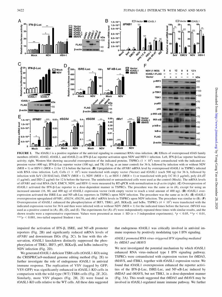
ResultstOASL1 positively regulated the RNA virus–triggered type IIFN signaling
To determine the potential roles of tOAS family members inregulating antiviral signaling, we first performed a functionalscreening using the IFN-b promoter reporter in TSPRCs over-expressing tOAS members. We found that overexpression oftOASL1 significantly enhanced the IFN-b promoter activationinduced by an RNA virus–NDV but not by a DNA virus–HSV-1infection (Fig. 1A). The potentiated effect of tOASL1 over-expression on type I IFN signaling upon RNA virus infection, asdemonstrated by increased tIFNB1 mRNA expression, could bewell reproduced by challenging with various RNA viruses in-cluding SeV, EMCV, NDV, and dsRNA analogue poly I:C H(Fig. 1B, left). tOASL1 overexpression reduced the viral mRNAlevels of SeV, EMCV, and NDV (Fig. 1B, right). Consistent withthe pattern of Fig. 1A, tOASL1 overexpression had no obviouschange on tIFNB1 mRNA expression in TSPRCs stimulated byHSV-1 and DNA ligands, including poly dA:dT and ISD (Fig. 1B,left), although a lower level of HSV-1 viral RNA was found inTSPRCs overexpressing tOASL1 relative to the empty vectorgroup (Fig. 1B, right). Note that the inhibitory effect of tOASL1overexpression on HSV-1 viral RNA level was much inferior tothose of the viral levels of SeV, EMCV, and NDV (Fig. 1B, right).These results indicated that tOASL1 was actively involved inRNA virus–induced type I IFN signaling. As NDV infection hadthe best stimulation effect in TSPRCs, we used this virus forstimulation in the subsequent assays unless otherwise stated.Concordantly, tOASL1 overexpression could activate the NDV-triggered IFN-b promoter activity in a dose-dependent manner(Fig. 1C). Because IFN-b promotor activation is driven by IFN-stimulated response element (ISRE) and NF-kB, we next testedthe activation of ISRE and NF-kB promoters in the presence oftOASL1 overexpression and NDV infection. Overexpression oftOASL1 significantly potentiated NDV-induced ISRE and NF-kBactivations (Fig. 1D) and enhanced mRNA expression of tIFNB1and its downstream genes including tISG54, tISG56, and tMx1(Fig. 1E). Consistently, tOASL1 overexpression increased phos-phorylation of TBK1, IRF3, p65, IKKa/b, and IkBa upon NDVinfection in a time-dependent manner (Fig. 1F). These resultsdemonstrated that tOASL1 activated the IRF3 and NF-kB sig-naling and positively regulated the antiviral immune responses.
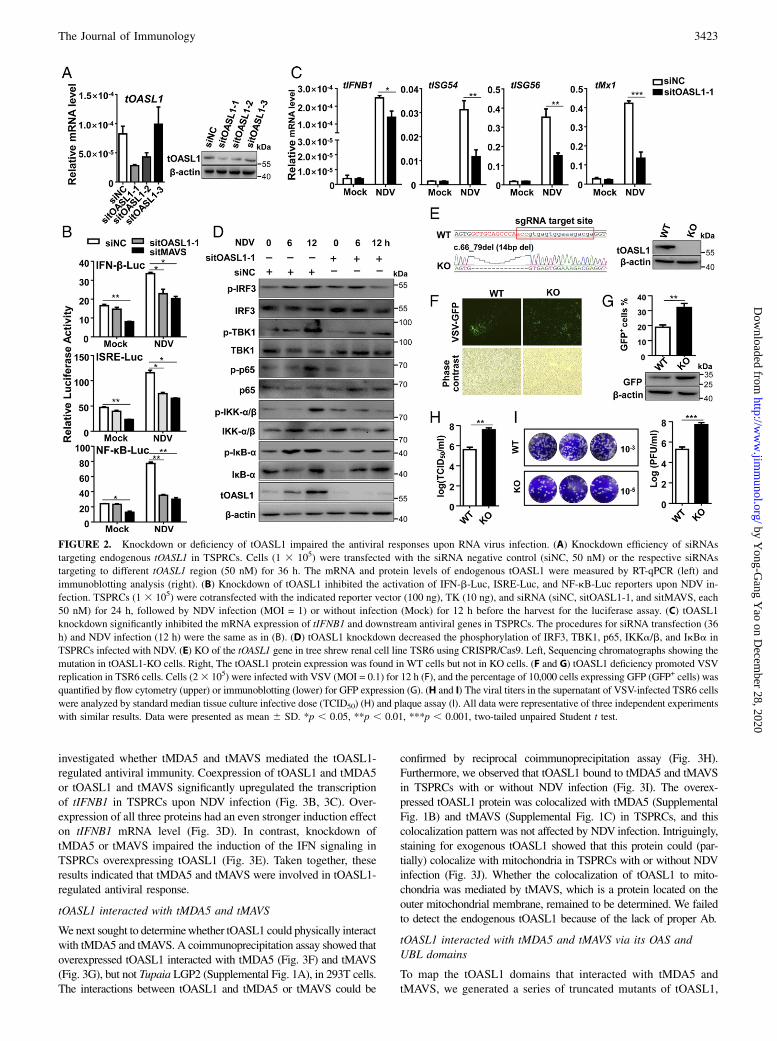
Endogenous tOASL1 was critically required for antiviralimmune responses to RNA virus
To characterize the roles of tOASL1 in antiviral responses, weperformed knockdown assays using siRNAs targeting tOASL1 toreduce endogenous tOASL1 expression. Among the three tOASL1siRNAs, sitOASL1-1 exhibited the highest knockdown effi-ciency, whereas sitOASL1-3 had no obvious inhibitory effect ontOASL1 expression (Fig. 2A). We used sitOASL1-1 for thesubsequent experiments. Consistent with the upregulation effectof tOASL1 (Fig. 1C, 1D), knockdown of tOASL1 significantly
The Journal of Immunology 3421
by Yong-G
ang Yao on D
ecember 28, 2020
http://ww
w.jim
munol.org/
Dow
nloaded from
impaired the activation of IFN-b, ISRE, and NF-kB promoterreporters (Fig. 2B) and significantly reduced mRNA levels oftIFNB1 and downstream ISGs (Fig. 2C). In line with this ob-servation, tOASL1 knockdown distinctly suppressed the phos-phorylation of TBK1, IRF3, p65, IKKa/b, and IkBa induced byNDV infection (Fig. 2D).We generated tOASL1-deficient TSR6 cells (tOASL1-KO) using
the CRISPR/Cas9-mediated genome editing method (Fig. 2E) tofurther investigate the role of endogenous tOASL1 in antiviralimmune response. The replication of VSV (tagged by GFP;VSV-GFP) was significantly enhanced in tOASL1-KO cells incomparison with the wild-type (WT) TSR6 cells (Fig. 2F, 2G).Similarly, more VSV plaques (Fig. 2H, 2I) were found intOASL1-KO cells relative to the WT cells. All these data suggested
that endogenous tOASL1 was critically involved in antiviral im-mune responses by positively modulating type I IFN signaling.
tOASL1 promoted RNA virus–triggered IFN signaling mediatedby tMDA5 and tMAVS
We next investigated the potential mechanism by which tOASL1enhanced RNA virus–induced type I IFN signaling pathway.
TSPRCs were cotransfected with expression vectors for tMDA5,
tMAVS, and tTBK1, together with tOASL1 expression vector. We
found that tOASL1 overexpression enhanced the reporter activi-
ties of the IFN-b-Luc, ISRE-Luc, and NF-kB-Luc induced by
tMDA5 and tMAVS, but not TBK1, in a dose-dependent manner
(Fig. 3A). This result suggested that tMDA5 and tMAVS may be
involved in tOASL1-regulated innate immune pathway. We further
FIGURE 1. The tOASL1 is a positive regulator of the antiviral signaling to counteract RNA virus infection. (A) Effects of overexpressed tOAS family
members (tOAS1, tOAS2, tOASL1, and tOASL2) on IFN-b-Luc reporter activation upon NDV and HSV-1 infection. Left, IFN-b-Luc reporter luciferase
activity; right, Western blot showing successful overexpression of the indicated proteins. TSPRCs (1 3 105) were cotransfected with the indicated ex-
pression vector (400 ng), IFN-b-Luc reporter vector (100 ng), and TK (10 ng, as an inner control) for 36 h, followed by infection with or without NDV
(MOI = 1) or HSV-1 (MOI = 1) for 12 h before the harvest. (B) Upregulation of the tIFNB1 mRNA level by overexpressed tOASL1 in TSPRCs infected
with RNA virus infection. Left, Cells (1 3 105) were transfected with empty vector (Vector) and tOASL1 (each 500 ng) for 36 h, followed by
infection with SeV (20 HAU/ml), EMCV (MOI = 1), NDV (MOI = 1), or HSV-1 (MOI = 1) or transfected with poly I:C H (1 mg/ml), poly dA:dT
(1 mg/ml), and ISD (2 mg/ml) for 12 h before the harvest. The uninfected or untransfected cells were used as the control (Mock). The mRNA levels
of tIFNB1 and viral RNA (SeV, EMCV, NDV, and HSV-1) were measured by RT-qPCR with normalization to b-actin (right). (C) Overexpression of
tOASL1 activated the IFN-b-Luc reporter in a dose-dependent manner in TSPRCs. The procedure was the same as in (A), except for using an
increased amount (16, 80, and 400 ng) of tOASL1 expression vector (with empty vector to reach a total amount of 400 ng). (D) tOASL1 over-
expression activated the ISRE-Luc and NF-kB-Luc reporters in TSPRCs upon NDV infection. The procedure was the same as in (A). (E) tOASL1
overexpression upregulated tIFNB1, tISG54, tISG56, and tMx1 mRNA levels in TSPRCs upon NDV infection. The procedure was similar to (B). (F)
Overexpression of tOASL1 enhanced the phosphorylation of IRF3, TBK1, p65, IKKa/b, and IkBa. TSPRCs (4 3 105) were transfected with the
indicated expression vector for 36 h and then were infected with or without NDV (MOI = 1) for the indicated times before the harvest. tMVAS was
used as a positive control in (A), (B), (D), and (E). The experiments for (A)–(F) were independently repeated three times with similar results, and the
shown results were a representative experiment. Values were presented as mean 6 SD (n = 3 independent experiments). *p , 0.05, **p , 0.01,
***p , 0.001, two-tailed unpaired Student t test.
3422 TUPAIA OASL1 INTERACTS WITH MDA5 AND MAVS
by Yong-G
ang Yao on D
ecember 28, 2020
http://ww
w.jim
munol.org/
Dow
nloaded from
investigated whether tMDA5 and tMAVS mediated the tOASL1-regulated antiviral immunity. Coexpression of tOASL1 and tMDA5or tOASL1 and tMAVS significantly upregulated the transcriptionof tIFNB1 in TSPRCs upon NDV infection (Fig. 3B, 3C). Over-expression of all three proteins had an even stronger induction effecton tIFNB1 mRNA level (Fig. 3D). In contrast, knockdown oftMDA5 or tMAVS impaired the induction of the IFN signaling inTSPRCs overexpressing tOASL1 (Fig. 3E). Taken together, theseresults indicated that tMDA5 and tMAVS were involved in tOASL1-regulated antiviral response.
tOASL1 interacted with tMDA5 and tMAVS
Wenext sought to determinewhether tOASL1 could physically interactwith tMDA5 and tMAVS. A coimmunoprecipitation assay showed thatoverexpressed tOASL1 interacted with tMDA5 (Fig. 3F) and tMAVS(Fig. 3G), but not Tupaia LGP2 (Supplemental Fig. 1A), in 293T cells.The interactions between tOASL1 and tMDA5 or tMAVS could be
confirmed by reciprocal coimmunoprecipitation assay (Fig. 3H).Furthermore, we observed that tOASL1 bound to tMDA5 and tMAVSin TSPRCs with or without NDV infection (Fig. 3I). The overex-pressed tOASL1 protein was colocalized with tMDA5 (SupplementalFig. 1B) and tMAVS (Supplemental Fig. 1C) in TSPRCs, and thiscolocalization pattern was not affected by NDV infection. Intriguingly,staining for exogenous tOASL1 showed that this protein could (par-tially) colocalize with mitochondria in TSPRCs with or without NDVinfection (Fig. 3J). Whether the colocalization of tOASL1 to mito-chondria was mediated by tMAVS, which is a protein located on theouter mitochondrial membrane, remained to be determined. We failedto detect the endogenous tOASL1 because of the lack of proper Ab.
tOASL1 interacted with tMDA5 and tMAVS via its OAS andUBL domains
To map the tOASL1 domains that interacted with tMDA5 andtMAVS, we generated a series of truncated mutants of tOASL1,
FIGURE 2. Knockdown or deficiency of tOASL1 impaired the antiviral responses upon RNA virus infection. (A) Knockdown efficiency of siRNAs
targeting endogenous tOASL1 in TSPRCs. Cells (1 3 105) were transfected with the siRNA negative control (siNC, 50 nM) or the respective siRNAs
targeting to different tOASL1 region (50 nM) for 36 h. The mRNA and protein levels of endogenous tOASL1 were measured by RT-qPCR (left) and
immunoblotting analysis (right). (B) Knockdown of tOASL1 inhibited the activation of IFN-b-Luc, ISRE-Luc, and NF-kB-Luc reporters upon NDV in-
fection. TSPRCs (1 3 105) were cotransfected with the indicated reporter vector (100 ng), TK (10 ng), and siRNA (siNC, sitOASL1-1, and sitMAVS, each
50 nM) for 24 h, followed by NDV infection (MOI = 1) or without infection (Mock) for 12 h before the harvest for the luciferase assay. (C) tOASL1
knockdown significantly inhibited the mRNA expression of tIFNB1 and downstream antiviral genes in TSPRCs. The procedures for siRNA transfection (36
h) and NDV infection (12 h) were the same as in (B). (D) tOASL1 knockdown decreased the phosphorylation of IRF3, TBK1, p65, IKKa/b, and IkBa in
TSPRCs infected with NDV. (E) KO of the tOASL1 gene in tree shrew renal cell line TSR6 using CRISPR/Cas9. Left, Sequencing chromatographs showing the
mutation in tOASL1-KO cells. Right, The tOASL1 protein expression was found in WT cells but not in KO cells. (F and G) tOASL1 deficiency promoted VSV
replication in TSR6 cells. Cells (23 105) were infected with VSV (MOI = 0.1) for 12 h (F), and the percentage of 10,000 cells expressing GFP (GFP+ cells) was
quantified by flow cytometry (upper) or immunoblotting (lower) for GFP expression (G). (H and I) The viral titers in the supernatant of VSV-infected TSR6 cells
were analyzed by standard median tissue culture infective dose (TCID50) (H) and plaque assay (I). All data were representative of three independent experiments
with similar results. Data were presented as mean 6 SD. *p , 0.05, **p , 0.01, ***p , 0.001, two-tailed unpaired Student t test.
The Journal of Immunology 3423
by Yong-G
ang Yao on D
ecember 28, 2020
http://ww
w.jim
munol.org/
Dow
nloaded from
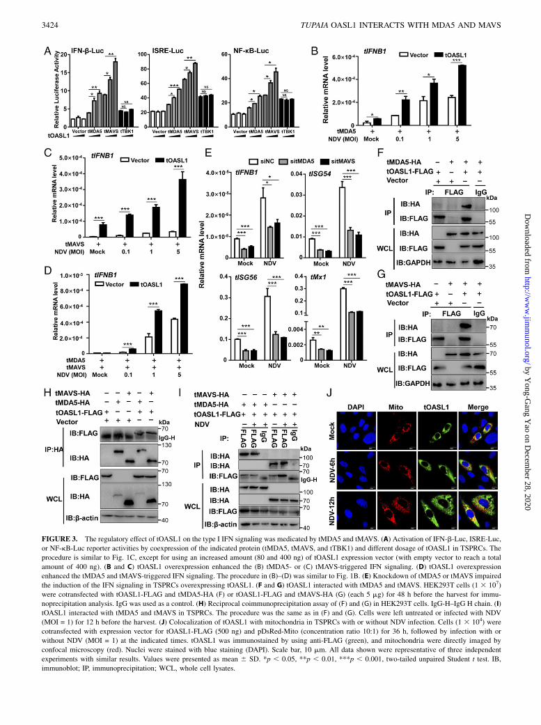
FIGURE 3. The regulatory effect of tOASL1 on the type I IFN signaling was medicated by tMDA5 and tMAVS. (A) Activation of IFN-b-Luc, ISRE-Luc,
or NF-kB-Luc reporter activities by coexpression of the indicated protein (tMDA5, tMAVS, and tTBK1) and different dosage of tOASL1 in TSPRCs. The
procedure is similar to Fig. 1C, except for using an increased amount (80 and 400 ng) of tOASL1 expression vector (with empty vector to reach a total
amount of 400 ng). (B and C) tOASL1 overexpression enhanced the (B) tMDA5- or (C) tMAVS-triggered IFN signaling. (D) tOASL1 overexpression
enhanced the tMDA5 and tMAVS-triggered IFN signaling. The procedure in (B)–(D) was similar to Fig. 1B. (E) Knockdown of tMDA5 or tMAVS impaired
the induction of the IFN signaling in TSPRCs overexpressing tOASL1. (F and G) tOASL1 interacted with tMDA5 and tMAVS. HEK293T cells (1 3 107)
were cotransfected with tOASL1-FLAG and tMDA5-HA (F) or tOASL1-FLAG and tMAVS-HA (G) (each 5 mg) for 48 h before the harvest for immu-
noprecipitation analysis. IgG was used as a control. (H) Reciprocal coimmunoprecipitation assay of (F) and (G) in HEK293T cells. IgG-H–IgG H chain. (I)
tOASL1 interacted with tMDA5 and tMAVS in TSPRCs. The procedure was the same as in (F) and (G). Cells were left untreated or infected with NDV
(MOI = 1) for 12 h before the harvest. (J) Colocalization of tOASL1 with mitochondria in TSPRCs with or without NDV infection. Cells (1 3 104) were
cotransfected with expression vector for tOASL1-FLAG (500 ng) and pDsRed-Mito (concentration ratio 10:1) for 36 h, followed by infection with or
without NDV (MOI = 1) at the indicated times. tOASL1 was immunostained by using anti-FLAG (green), and mitochondria were directly imaged by
confocal microscopy (red). Nuclei were stained with blue staining (DAPI). Scale bar, 10 mm. All data shown were representative of three independent
experiments with similar results. Values were presented as mean 6 SD. *p , 0.05, **p , 0.01, ***p , 0.001, two-tailed unpaired Student t test. IB,
immunoblot; IP, immunoprecipitation; WCL, whole cell lysates.
3424 TUPAIA OASL1 INTERACTS WITH MDA5 AND MAVS
by Yong-G
ang Yao on D
ecember 28, 2020
http://ww
w.jim
munol.org/
Dow
nloaded from
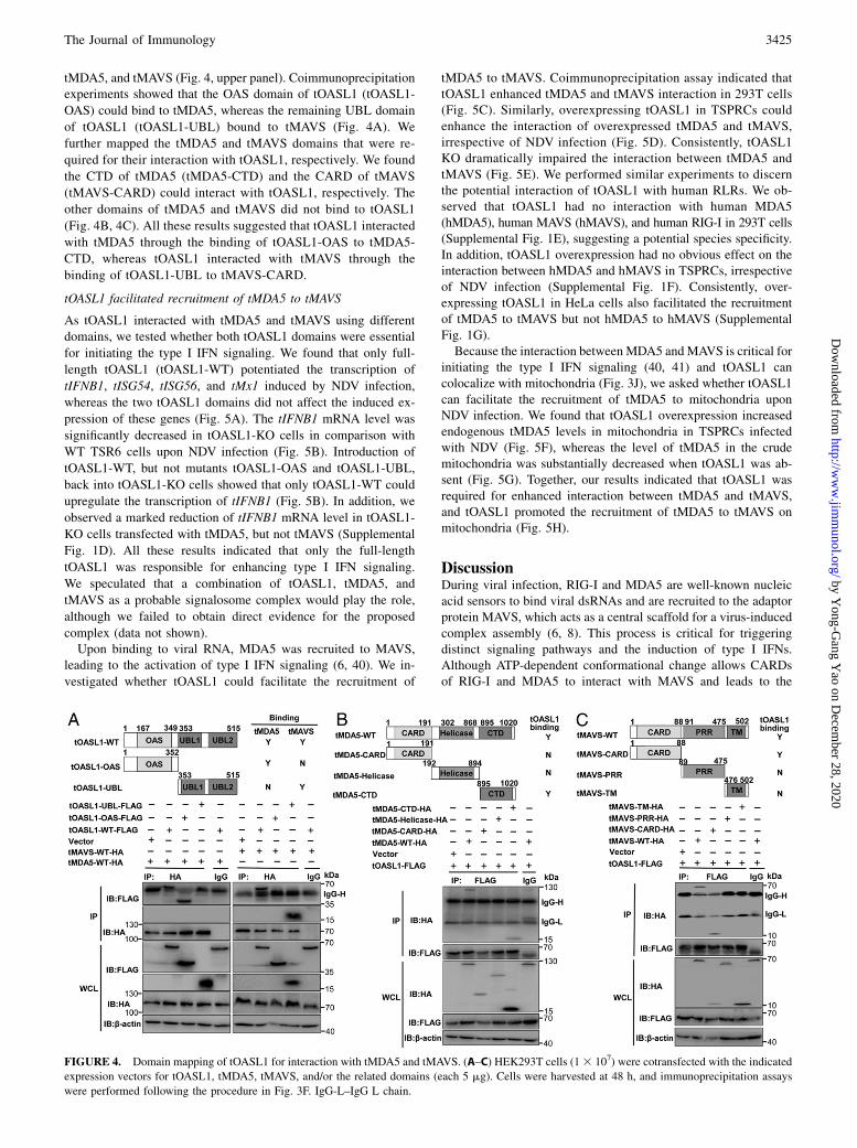
tMDA5, and tMAVS (Fig. 4, upper panel). Coimmunoprecipitationexperiments showed that the OAS domain of tOASL1 (tOASL1-
OAS) could bind to tMDA5, whereas the remaining UBL domain
of tOASL1 (tOASL1-UBL) bound to tMAVS (Fig. 4A). We
further mapped the tMDA5 and tMAVS domains that were re-
quired for their interaction with tOASL1, respectively. We found
the CTD of tMDA5 (tMDA5-CTD) and the CARD of tMAVS
(tMAVS-CARD) could interact with tOASL1, respectively. The
other domains of tMDA5 and tMAVS did not bind to tOASL1
(Fig. 4B, 4C). All these results suggested that tOASL1 interacted
with tMDA5 through the binding of tOASL1-OAS to tMDA5-
CTD, whereas tOASL1 interacted with tMAVS through the
binding of tOASL1-UBL to tMAVS-CARD.
tOASL1 facilitated recruitment of tMDA5 to tMAVS
As tOASL1 interacted with tMDA5 and tMAVS using differentdomains, we tested whether both tOASL1 domains were essential
for initiating the type I IFN signaling. We found that only full-
length tOASL1 (tOASL1-WT) potentiated the transcription of
tIFNB1, tISG54, tISG56, and tMx1 induced by NDV infection,
whereas the two tOASL1 domains did not affect the induced ex-
pression of these genes (Fig. 5A). The tIFNB1 mRNA level was
significantly decreased in tOASL1-KO cells in comparison with
WT TSR6 cells upon NDV infection (Fig. 5B). Introduction of
tOASL1-WT, but not mutants tOASL1-OAS and tOASL1-UBL,
back into tOASL1-KO cells showed that only tOASL1-WT could
upregulate the transcription of tIFNB1 (Fig. 5B). In addition, we
observed a marked reduction of tIFNB1 mRNA level in tOASL1-
KO cells transfected with tMDA5, but not tMAVS (Supplemental
Fig. 1D). All these results indicated that only the full-length
tOASL1 was responsible for enhancing type I IFN signaling.
We speculated that a combination of tOASL1, tMDA5, and
tMAVS as a probable signalosome complex would play the role,
although we failed to obtain direct evidence for the proposed
complex (data not shown).Upon binding to viral RNA, MDA5 was recruited to MAVS,
leading to the activation of type I IFN signaling (6, 40). We in-
vestigated whether tOASL1 could facilitate the recruitment of
tMDA5 to tMAVS. Coimmunoprecipitation assay indicated thattOASL1 enhanced tMDA5 and tMAVS interaction in 293T cells(Fig. 5C). Similarly, overexpressing tOASL1 in TSPRCs couldenhance the interaction of overexpressed tMDA5 and tMAVS,irrespective of NDV infection (Fig. 5D). Consistently, tOASL1KO dramatically impaired the interaction between tMDA5 andtMAVS (Fig. 5E). We performed similar experiments to discernthe potential interaction of tOASL1 with human RLRs. We ob-served that tOASL1 had no interaction with human MDA5(hMDA5), human MAVS (hMAVS), and human RIG-I in 293T cells(Supplemental Fig. 1E), suggesting a potential species specificity.In addition, tOASL1 overexpression had no obvious effect on theinteraction between hMDA5 and hMAVS in TSPRCs, irrespectiveof NDV infection (Supplemental Fig. 1F). Consistently, over-expressing tOASL1 in HeLa cells also facilitated the recruitmentof tMDA5 to tMAVS but not hMDA5 to hMAVS (SupplementalFig. 1G).Because the interaction between MDA5 andMAVS is critical for
initiating the type I IFN signaling (40, 41) and tOASL1 cancolocalize with mitochondria (Fig. 3J), we asked whether tOASL1can facilitate the recruitment of tMDA5 to mitochondria uponNDV infection. We found that tOASL1 overexpression increasedendogenous tMDA5 levels in mitochondria in TSPRCs infectedwith NDV (Fig. 5F), whereas the level of tMDA5 in the crudemitochondria was substantially decreased when tOASL1 was ab-sent (Fig. 5G). Together, our results indicated that tOASL1 wasrequired for enhanced interaction between tMDA5 and tMAVS,and tOASL1 promoted the recruitment of tMDA5 to tMAVS onmitochondria (Fig. 5H).
DiscussionDuring viral infection, RIG-I and MDA5 are well-known nucleicacid sensors to bind viral dsRNAs and are recruited to the adaptorprotein MAVS, which acts as a central scaffold for a virus-inducedcomplex assembly (6, 8). This process is critical for triggeringdistinct signaling pathways and the induction of type I IFNs.Although ATP-dependent conformational change allows CARDsof RIG-I and MDA5 to interact with MAVS and leads to the
FIGURE 4. Domain mapping of tOASL1 for interaction with tMDA5 and tMAVS. (A–C) HEK293T cells (13 107) were cotransfected with the indicated
expression vectors for tOASL1, tMDA5, tMAVS, and/or the related domains (each 5 mg). Cells were harvested at 48 h, and immunoprecipitation assays
were performed following the procedure in Fig. 3F. IgG-L–IgG L chain.
The Journal of Immunology 3425
by Yong-G
ang Yao on D
ecember 28, 2020
http://ww
w.jim
munol.org/
Dow
nloaded from
formation of the MAVS signalosome (10), how this process ismediated remains unclear. Several lines of evidence suggestthat some host proteins are critical for controlling the associ-ation of RIG-I or MDA5 with the adaptor MAVS, such asECSIT signaling integrator (ECSIT) (42), Zyxin (43), NLRfamily pyrin domain containing 12 (44), and the most recentlyreported nucleotide binding oligomerization domain containing1 (45). In this study, we found that tOASL1, a member of 29,59-OAS family proteins, potentiated RNA virus–induced signalingcascade in the Chinese tree shrew by recruiting tMDA5 totMAVS. This finding provided another line of evidence for thecomplexity of assembling the MAVS signalosome, especiallyconsidering the fact that RIG-I was naturally lost in the Chinesetree shrew (24, 34).
The OASL belonged to the OAS family proteins characterized byits N-terminal OASL domain but was devoid of the enzymaticactivity to activate the OAS/RNase L pathway (19). In our previousstudy, we found that the cDNA of tOASL1 was 2176 bp in lengthand encoded a polypeptide of 514 aa, which shared 75.4% residueidentity to human ortholog of OASL. tOASL1 contained a typicalN-terminal OAS domain and two tandem ubiquitin-like domainsat the C-terminal. Phylogenetic analyses revealed that tOASL1was first clustered with the corresponding orthologs of primates,indicating that tOASL1 was relatively conserved in the Chinesetree shrew (23). In addition, we found that the mRNA levels oftOASL1 and the other three tOASs (tOAS1, tOAS2, and tOASL2)were significantly increased in cells upon DNA virus (HSV-1) orRNAvirus (SeV, NDV, and VSV) infection compared with uninfected
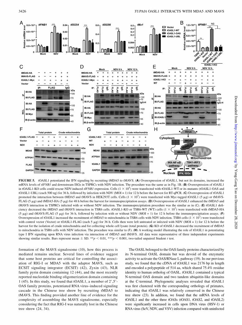
FIGURE 5. tOASL1 potentiated the IFN signaling by recruiting tMDA5 to tMAVS. (A) Overexpression of tOASL1, but not its domains, increased the
mRNA levels of tIFNB1 and downstream ISGs in TSPRCs with NDV infection. The procedure was the same as in Fig. 1B. (B) Overexpression of tOASL1
in tOASL1-KO cells could rescue NDV-induced tIFNB1 expression. Cells (1 3 105) were transfected with tOASL1-WT or its mutants (tOASL1-OAS and
tOASL1-UBL) (each 500 ng) for 36 h, followed by infection with NDV (MOI = 1) for 12 h before the harvest for RT-qPCR. (C) Overexpression of tOASL1
promoted the interaction between tMDA5 and tMAVS in HEK293T cells. Cells (1 3 107) were transfected with Myc-tagged tOASL1 (5 mg) or tMAVS-
FLAG (5 mg) and tMDA5-HA (5 mg) for 48 h before the harvest for immunoprecipitation assays. (D) Overexpression of tOASL1 enhanced the tMDA5 and
tMAVS interaction in TSPRCs infected with or without NDV infection. The immunoprecipitation procedure was the similar as in (C). (E) tOASL1 defi-
ciency decreased the tMDA5 and tMAVS interaction in TSR6 cells. tOASL1-KO or TSR6-WT (WT) cells (1 3 107) were transfected with tMDA5-HA
(5 mg) and tMAVS-FLAG (5 mg) for 36 h, followed by infection with or without NDV (MOI = 1) for 12 h before the immunoprecipitation assays. (F)
Overexpression of tOASL1 increased the recruitment of tMDA5 to mitochondria in TSR6 cells with NDV infection. TSR6 cells (1 3 107) were transfected
with control vector (Vector) or tOASL1-FLAG (each 5 mg) for 36 h. Cells then were left untreated or infected with NDV (MOI = 1) for 12 h before the
harvest for the isolation of crude mitochondria and for collecting whole cell lysate (total protein). (G) KO of tOASL1 decreased the recruitment of tMDA5
to mitochondria in TSR6 cells with NDV infection. The procedure was similar to (F). (H) A working model illustrating the role of tOASL1 in potentiating
type I IFN signaling upon RNA virus infection via interaction of tMDA5 and tMAVS. All data were representative of three independent experiments
showing similar results. Bars represent mean 6 SD. **p , 0.01, ***p , 0.001, two-tailed unpaired Student t test.
3426 TUPAIA OASL1 INTERACTS WITH MDA5 AND MAVS
by Yong-G
ang Yao on D
ecember 28, 2020
http://ww
w.jim
munol.org/
Dow
nloaded from
TSPRCs (23). These available results indicated that tOASs wereISGs. Several studies had reported that OASL played an essentialrole in modulating virus-induced type I IFN signaling pathway(20–22, 46), albeit the role of OASL in human (20, 21) wasdifferent from that of mouse (22), indicating a potential species-specific pattern. Similar to human OASL (hOASL) (20) andporcine OASL (46), the tOASL1 was actively involved in RNAvirus–induced IFN induction pathway and acted as a positiveregulator. However, tOASL1 differed from hOASL in severalaspects. First, although hOASL and tOASL1 commonly pro-moted RNA virus–induced IFN induction, tOASL1 had no effecton IFN production upon DNA virus (Fig. 1A, 1B), whereashOASL had an inhibitory effect (21). Note that mouse Oasl1 hadan inhibitory effect on both RNA virus– and DNA virus–inducedIFN inductions (22). The exact reason for this species-specificrole remained unknown, but this offered a good opportunity forus to learn the evolutionary complexity and compensation of theimmune system. Second, different from hOASL, which inter-acted and colocalized with RIG-I to mediate the downstreameffect (20), tOASL1 interacted with tMDA5 and tMAVS andpotentiated the tMDA5- and tMAVS-triggered IFN inductions(Fig. 3). We believe this difference was compatible with the factthat RIG-I was naturally lost, and MDA5 had a functional re-placement in the Chinese tree shrew (34). Moreover, we foundthat tOASL1 could colocalize and interacted with MAVS. Sim-ilar to the mitochondrion-localized ZNFX1 (47), no predictedmitochondrial targeting motif was found for tOASL1. The OASdomain had been reported to be associated with mitochondria(48), and the UBL domain of tOASL1 was critical for interactingwith tMAVS (Fig. 4A), which might be responsible for the mi-tochondrial localization of tOASL1, irrespective of RNA virusinfection. To our knowledge, this is the first time that OASL wasfound to be localized in mitochondria, which would facilitate itsnatural interaction with MAVS.Another interesting observation in this study is that tOASL1
promoted the recruitment of tMDA5 to tMAVS (Fig. 5C–G,Supplemental Fig. 1G). In particular, tOASL1 interacted withtMDA5 and tMAVS using different domains, which might behelpful for forming the tOASL1–tMDA5–tMAVS complex. It ispitiful that we were unable to isolate this protein complex (pre-sumably because of its large size) and make further character-ization. Note that overexpression of tOASL1 domains, but notfull-length tOASL1, could not enhance NDV-induced transcrip-tion of tIFNB1 and downstream antiviral genes (Fig. 5A). In ad-dition, tOASL1 deficiency dramatically impaired the interactionbetween tMDA5 and tMAVS (Fig. 5E). On this point, tOASL1 notonly acts as a scaffold protein bridging tMDA5 and tMAVS butalso works as a positive regulator of antiviral signaling to coun-teract RNA virus infection.In summary, we identified tOASL1 as a positive regulator of
the type I IFN induction by recruiting tMDA5 to tMAVS tocounteract RNA virus infection. We further elucidated themolecular mechanism by which tOASL1 positively regulates theanti-RNAvirus innate immunity. The multifaceted role of OASLillustrated a unique picture of the innate immunity in differentspecies.
AcknowledgmentsWe thank Dr. Rongcan Luo for technical assistance. We also thank Dr. Yong
Wu for critical discussion and communication of unpublished results. The
study was done at KIZ, CAS.
DisclosuresThe authors have no financial conflicts of interest.
References1. Akira, S., S. Uematsu, and O. Takeuchi. 2006. Pathogen recognition and innate
immunity. Cell 124: 783–801.2. Wu, J., and Z. J. Chen. 2014. Innate immune sensing and signaling of cytosolic
nucleic acids. Annu. Rev. Immunol. 32: 461–488.3. Kato, H., O. Takeuchi, S. Sato, M. Yoneyama, M. Yamamoto, K. Matsui,
S. Uematsu, A. Jung, T. Kawai, K. J. Ishii, et al. 2006. Differential roles ofMDA5 and RIG-I helicases in the recognition of RNA viruses. Nature 441: 101–105.
4. Yoneyama, M., and T. Fujita. 2009. RNA recognition and signal transduction byRIG-I-like receptors. Immunol. Rev. 227: 54–65.
5. Seth, R. B., L. Sun, C. K. Ea, and Z. J. Chen. 2005. Identification and charac-terization of MAVS, a mitochondrial antiviral signaling protein that activatesNF-kappaB and IRF 3. Cell 122: 669–682.
6. Kawai, T., K. Takahashi, S. Sato, C. Coban, H. Kumar, H. Kato, K. J. Ishii,O. Takeuchi, and S. Akira. 2005. IPS-1, an adaptor triggering RIG-I- and Mda5-mediated type I interferon induction. Nat. Immunol. 6: 981–988.
7. Meylan, E., J. Curran, K. Hofmann, D. Moradpour, M. Binder,R. Bartenschlager, and J. Tschopp. 2005. Cardif is an adaptor protein in the RIG-I antiviral pathway and is targeted by hepatitis C virus. Nature 437: 1167–1172.
8. Xu, L. G., Y. Y. Wang, K. J. Han, L. Y. Li, Z. Zhai, and H. B. Shu. 2005. VISA isan adapter protein required for virus-triggered IFN-beta signaling. Mol. Cell 19:727–740.
9. Hou, F., L. Sun, H. Zheng, B. Skaug, Q. X. Jiang, and Z. J. Chen. 2011. MAVSforms functional prion-like aggregates to activate and propagate antiviral innateimmune response. [Published erratum appears in 2011 Cell 146: 841.] Cell 146:448–461.
10. Potter, J. A., R. E. Randall, and G. L. Taylor. 2008. Crystal structure of humanIPS-1/MAVS/VISA/Cardif caspase activation recruitment domain. BMC Struct.Biol. 8: 11.
11. Schneider, W. M., M. D. Chevillotte, and C. M. Rice. 2014. Interferon-stimulated genes: a complex web of host defenses. Annu. Rev. Immunol. 32:513–545.
12. Yoneyama, M., M. Kikuchi, K. Matsumoto, T. Imaizumi, M. Miyagishi, K. Taira,E. Foy, Y. M. Loo, M. Gale, Jr., S. Akira, et al. 2005. Shared and uniquefunctions of the DExD/H-box helicases RIG-I, MDA5, and LGP2 in antiviralinnate immunity. J. Immunol. 175: 2851–2858.
13. Satoh, T., H. Kato, Y. Kumagai, M. Yoneyama, S. Sato, K. Matsushita,T. Tsujimura, T. Fujita, S. Akira, and O. Takeuchi. 2010. LGP2 is a positiveregulator of RIG-I- and MDA5-mediated antiviral responses. Proc. Natl. Acad.Sci. USA 107: 1512–1517.
14. Ablasser, A., and S. Hur. 2020. Regulation of cGAS- and RLR-mediated im-munity to nucleic acids. Nat. Immunol. 21: 17–29.
15. Liu, J., C. Qian, and X. Cao. 2016. Post-translational modification control ofinnate immunity. Immunity 45: 15–30.
16. Eskildsen, S., J. Justesen, M. H. Schierup, and R. Hartmann. 2003. Character-ization of the 29-59-oligoadenylate synthetase ubiquitin-like family. NucleicAcids Res. 31: 3166–3173.
17. Hartmann, R., J. Justesen, S. N. Sarkar, G. C. Sen, and V. C. Yee. 2003. Crystalstructure of the 29-specific and double-stranded RNA-activated interferon-induced antiviral protein 29-59-oligoadenylate synthetase. Mol. Cell 12: 1173–1185.
18. Silverman, R. H. 2007. Viral encounters with 29,59-oligoadenylate synthetaseand RNase L during the interferon antiviral response. J. Virol. 81: 12720–12729.
19. Zhu, J., A. Ghosh, and S. N. Sarkar. 2015. OASL-a new player in controllingantiviral innate immunity. Curr. Opin. Virol. 12: 15–19.
20. Zhu, J., Y. Zhang, A. Ghosh, R. A. Cuevas, A. Forero, J. Dhar, M. S. Ibsen,J. L. Schmid-Burgk, T. Schmidt, M. K. Ganapathiraju, et al. 2014. Antiviralactivity of human OASL protein is mediated by enhancing signaling of the RIG-IRNA sensor. Immunity 40: 936–948.
21. Ghosh, A., L. Shao, P. Sampath, B. Zhao, N. V. Patel, J. Zhu, B. Behl,R. A. Parise, J. H. Beumer, R. J. O’Sullivan, et al. 2019. Oligoadenylate-synthetase-family protein OASL inhibits activity of the DNA sensor cGASduring DNA virus infection to limit interferon production. Immunity 50: 51–63.e5.
22. Lee, M. S., B. Kim, G. T. Oh, and Y. J. Kim. 2013. OASL1 inhibits translation ofthe type I interferon-regulating transcription factor IRF7. [Published erratumappears in 2013 Nat. Immunol. 14: 355.]; [Published erratum appears in 2013Nat. Immunol. 14: 877.] Nat. Immunol. 14: 346–355.
23. Yao, Y. L., D. Yu, L. Xu, Y. Fan, Y. Wu, T. Gu, J. Chen, L. B. Lv, and Y. G. Yao.2019. Molecular characterization of the 29,59-oligoadenylate synthetase familyin the Chinese tree shrew (Tupaia belangeri chinensis). Cytokine 114: 106–114.
24. Fan, Y., Z. Y. Huang, C. C. Cao, C. S. Chen, Y. X. Chen, D. D. Fan, J. He,H. L. Hou, L. Hu, X. T. Hu, et al. 2013. Genome of the Chinese tree shrew. Nat.Commun. 4: 1426.
25. Fan, Y., M. S. Ye, J. Y. Zhang, L. Xu, D. D. Yu, T. L. Gu, Y. L. Yao, J. Q. Chen,L. B. Lv, P. Zheng, et al. 2019. Chromosomal level assembly and populationsequencing of the Chinese tree shrew genome. Zool. Res. 40: 506–521.
26. Xu, S., X. Li, J. Yang, Z. Wang, Y. Jia, L. Han, L. Wang, and Q. Zhu. 2019.Comparative pathogenicity and transmissibility of pandemic H1N1, avian H5N1,and human H7N9 influenza viruses in tree shrews. Front. Microbiol. 10: 2955.
27. Xiao, J., R. Liu, and C. S. Chen. 2017. Tree shrew (Tupaia belangeri) as a novellaboratory disease animal model. Zool. Res. 38: 127–137.
28. Yao, Y. G. 2017. Creating animal models, why not use the Chinese tree shrew(Tupaia belangeri chinensis)? Zool. Res. 38: 118–126.
29. Amako, Y., K. Tsukiyama-Kohara, A. Katsume, Y. Hirata, S. Sekiguchi,Y. Tobita, Y. Hayashi, T. Hishima, N. Funata, H. Yonekawa, and M. Kohara.
The Journal of Immunology 3427
by Yong-G
ang Yao on D
ecember 28, 2020
http://ww
w.jim
munol.org/
Dow
nloaded from
2010. Pathogenesis of hepatitis C virus infection in Tupaia belangeri. J. Virol.84: 303–311.
30. Xu, L., D. D. Yu, Y. H. Ma, Y. L. Yao, R. H. Luo, X. L. Feng, H. R. Cai, J. B. Han,X. H. Wang, M. H. Li, et al. 2020. COVID-19-like symptoms observed in Chinesetree shrews infected with SARS-CoV-2. Zool. Res. 41: 517–526.
31. Zhang, J., R. C. Luo, X. Y. Man, L. B. Lv, Y. G. Yao, and M. Zheng. 2020. Theanatomy of the skin of the Chinese tree shrew is very similar to that of humanskin. Zool. Res. 41: 208–212.
32. Fan, Y., R. Luo, L. Y. Su, Q. Xiang, D. Yu, L. Xu, J. Q. Chen, R. Bi, D. D. Wu,P. Zheng, and Y. G. Yao. 2018. Does the genetic feature of the Chinese treeshrew (Tupaia belangeri chinensis) support its potential as a viable model forAlzheimer’s disease research? J. Alzheimers Dis. 61: 1015–1028.
33. Ni, R. J., Y. Tian, X. Y. Dai, L. S. Zhao, J. X. Wei, J. N. Zhou, X. H. Ma, andT. Li. 2020. Social avoidance behavior in male tree shrews and prosocial be-havior in male mice toward unfamiliar conspecifics in the laboratory. Zool. Res.41: 258–272.
34. Xu, L., D. Yu, Y. Fan, L. Peng, Y. Wu, and Y. G. Yao. 2016. Loss of RIG-I leadsto a functional replacement with MDA5 in the Chinese tree shrew. Proc. Natl.Acad. Sci. USA 113: 10950–10955.
35. Xu, L., D. Yu, L. Peng, Y. Fan, J. Chen, Y. T. Zheng, C. Wang, and Y. G. Yao.2015. Characterization of a MAVS ortholog from the Chinese tree shrew (Tupaiabelangeri chinensis). Dev. Comp. Immunol. 52: 58–68.
36. Ma, X., A. S. Wong, H. Y. Tam, S. Y. Tsui, D. L. Chung, and B. Feng. 2018.In vivo genome editing thrives with diversified CRISPR technologies. Zool. Res.39: 58–71.
37. Gu, T., D. Yu, Y. Li, L. Xu, Y. L. Yao, and Y. G. Yao. 2019. Establishment andcharacterization of an immortalized renal cell line of the Chinese tree shrew(Tupaia belangeri chinesis). Appl. Microbiol. Biotechnol. 103: 2171–2180.
38. Xu, L., D. Yu, L. Peng, Y. Wu, Y. Fan, T. Gu, Y. L. Yao, J. Zhong, X. Chen, andY. G. Yao. 2020. An alternative splicing of tupaia STING modulated anti-RNA virus responses by targeting MDA5-LGP2 and IRF3. J. Immunol. 204:3191–3204.
39. Xu, L., L. Peng, T. Gu, D. Yu, and Y. G. Yao. 2019. The 3’UTR of human MAVSmRNA contains multiple regulatory elements for the control of protein expres-sion and subcellular localization. Biochim. Biophys. Acta Gene Regul. Mech.1862: 47–57.
40. Wu, B., and S. Hur. 2015. How RIG-I like receptors activate MAVS. Curr. Opin.Virol. 12: 91–98.
41. Takeuchi, O., and S. Akira. 2008. MDA5/RIG-I and virus recognition. Curr.Opin. Immunol. 20: 17–22.
42. Lei, C. Q., Y. Zhang, M. Li, L. Q. Jiang, B. Zhong, Y. H. Kim, and H. B. Shu.2015. ECSIT bridges RIG-I-like receptors to VISA in signaling events of innateantiviral responses. J. Innate Immun. 7: 153–164.
43. Kouwaki, T., M. Okamoto, H. Tsukamoto, Y. Fukushima, M. Matsumoto,T. Seya, and H. Oshiumi. 2017. Zyxin stabilizes RIG-I and MAVS interactionsand promotes type I interferon response. Sci. Rep. 7: 11905.
44. Chen, S.-T., L. Chen, D. S.-C. Lin, S.-Y. Chen, Y.-P. Tsao, H. Guo, F.-J. Li, W.-T. Tseng, J. W. Tam, C.-W. Chao, et al. 2019. NLRP12 regulates anti-viral RIG-Iactivation via interaction with TRIM25. Cell Host Microbe 25: 602–616.e7.
45. Wu, X. M., J. Zhang, P. W. Li, Y. W. Hu, L. Cao, S. Ouyang, Y. H. Bi, P. Nie, andM. X. Chang. 2020. NOD1 promotes antiviral signaling by binding viral RNAand regulating the interaction of MDA5 and MAVS. J. Immunol. 204: 2216–2231.
46. Li, L.-F., J. Yu, Y. Zhang, Q. Yang, Y. Li, L. Zhang, J. Wang, S. Li, Y. Luo,Y. Sun, and H. J. Qiu. 2017. Interferon-inducible oligoadenylate synthetase-likeprotein acts as an antiviral effector against classical swine fever virus via theMDA5-mediated type I interferon-signaling pathway. J. Virol. 91: e01514-16.
47. Wang, Y., S. Yuan, X. Jia, Y. Ge, T. Ling, M. Nie, X. Lan, S. Chen, and A. Xu.2019. Mitochondria-localised ZNFX1 functions as a dsRNA sensor to initiateantiviral responses through MAVS. Nat. Cell Biol. 21: 1346–1356.
48. Kjær, K. H., J. Pahus, M. F. Hansen, J. B. Poulsen, E. I. Christensen, J. Justesen,and P. M. Martensen. 2014. Mitochondrial localization of the OAS1 p46 iso-form associated with a common single nucleotide polymorphism. BMC CellBiol. 15: 33.
3428 TUPAIA OASL1 INTERACTS WITH MDA5 AND MAVS
by Yong-G
ang Yao on D
ecember 28, 2020
http://ww
w.jim
munol.org/
Dow
nloaded from
1
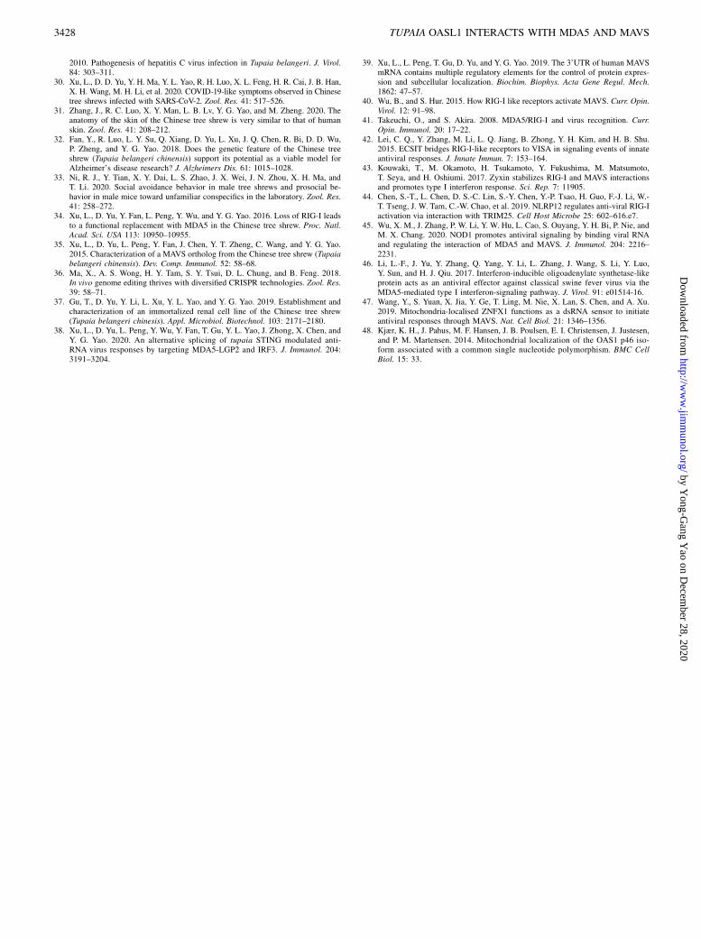
Table S1. Primers and vectors used in this study.
Primer Sequence (5'-3') Restriction
endonuclease a
Application and vector
tIFNB1-F ACCACTTGGAAACCATGC - Analytical RT-qPCR for tIFNB1
tIFNB1-R TTTCCACTCGGACTATCG
tISG54-F CTATCTGTATTGCCGTATTGG - Analytical RT-qPCR for tISG54
tISG54-R CTTCTGTCCTCTCCTCTG
tMx1-F GAAGACATTAGACTAGAACAAGAA - Analytical RT-qPCR for tMx1
tMx1-R TCCTGGCAGTAGACAATC
tISG56-F AGAGCAGCCGTCATTTAC - Analytical RT-qPCR for tISG56
tISG56-R CAGGGCTTCCTTTAGTTC
tOASL1-BspE I-F ACTCCGGAATGGCACTGGCCGAGGAGC BspE I PCR for constructing tOASL1-Myc vector
using pCS-myc-N vector tOASL1-Sac I-R CCGAGCTCCTACCTGGATGGAAACAGA Sac I
tOASL1-OAS-NotI-F TTGCGGCCGCATGGCACTGGCCGAGGAGC Not I PCR for constructing tOASL1-OAS-FLAG
vector using pCMV-3Tag-8 vector tOASL1-OAS-EcoRV-R TTGATATCGTCTCGGGCTCTCTGCACA EcoR V
tOASL1-UBL-NotI-F TTGCGGCCGCATGATCCAGGTGACAGTGG Not I PCR for constructing tOASL1-UBL-FLAG
vector using pCMV-3Tag-8 vector tOASL1-UBL-XhoI-R CCCTCGAGCCTGGATGGAAACAGAGGC Xho I
tMDA5-CARD-EcoRI-F CGGAATTCAAATGTCGAATGGGCATTCCT EcoR I PCR for constructing HA-tMDA5-CARD
vector using pCMV-HA vector tMDA5-CARD-XhoI-R CCCTCGAGCTAATCGTTTCCTGTTTTT Xho I
tMDA5-Helicase-EcoRI-F CGGAATTCAACTACAGCTCAGGCCTTACC EcoR I PCR for constructing HA-tMDA5-Helicase
vector using pCMV-HA vector tMDA5-Helicase-XhoI-R CCCTCGAGCTAAATCTTATGAGCATAC Xho I
tMDA5-CTD-EcoRI-F CGGAATTCAACCATCATTAATAACTTTGC EcoR I PCR for constructing HA-tMDA5-CTD
vector using pCMV-HA vector tMDA5-CTD-XhoI-R CCCTCGAGCTAATCCTCATCACTAAAC Xho I
tMAVS-CARD-XhoI-F CCCTCGAGAAATGTCATTTGCCGAGAACA Xho I PCR for constructing HA-tMAVS-CARD
vector using pCMV-HA vector tMAVS-CARD-NotI-R TTGCGGCCGCACGGGTCACTTCCTCGGCG Not I
tMAVS-PRR-XhoI-F CCCTCGAGAAATGGTCTACCAGACCTACC Xho I PCR for constructing HA-tMAVS-PRR
vector using pCMV-HA vector tMAVS-PRR-NotI-R TTGCGGCCGCAACCCTGACACACGGCTCC Not I
tMAVS-TM-XhoI-F CCCTCGAGAAATGTCATGGCTTGGGGTGG Xho I PCR for constructing HA-tMAVS-TM
vector using pCMV-HA vector tMAVS-TM-NotI-R TTGCGGCCGCTCACTGGAGTGGGCGCCGC Not I
tTBK1- SacI-F CGAGCTCATGCAGAGCACTTCCAATCA Sac I PCR for constructing tTBK1-FLAG vector
using pCMV-3Tag-8 vector tTBK1- XhoI-R CCGCTCGAGAAGACAGTCCACGTTGCGAA Xho I
a Restriction endonuclease sites introduced by PCR are underlined. RT-qPCR, quantitative real-time PCR.
2
ND
V 6
h
ND
V1
2 h
Mo
ck
DAPI tOASL1tMDA5 MergeB
DAPI tOASL1tMAVS Merge
ND
V 6
h
ND
V1
2 h
Mo
ck
CA
IB:HA
IB:FLAG
IB:HA
IB:FLAG
IB:GAPDH
tOASL1-FLAG
tLGP2-HA
WCL
IP
IP: FLAG IgGVector + +
+ + +
+ + +
35
kDa
55
70
55
70
D
tIFNB1
0
WT tOASL1-KO
2.0×10⁻⁵
4.0×10⁻⁵
6.0×10⁻⁵
8.0×10⁻⁵
1.0×10⁻⁴
Re
lati
ve
mR
NA
le
ve
l
**
Vector tMDA5 tMAVS
IB:β-actin
IB:Myc
IB:FLAG
IB:HA
IB:FLAG
IB:HA
WC
LIP
IP: FLAG IgG
tOASL1-Myc
hMAVS-FLAG
hMDA5-HA
Vector ++ + +
+ + ++
+ + ++
100
kDa
70
100
70
55
40
+ +
++
++
IgG FLAG
Mock NDV
IgG-H
F
IB:β-actin
IB:Myc
IB:FLAG
IB:HA
IB:FLAG
IB:HA
WC
LIP
IP: FLAG IgG
tOASL1-Myc
hMAVS-FLAG
hMDA5-HA
Vector ++ + +
+ + +
+ + +
100
kDa
70
100
70
55
40
++
IgG FLAG
tMAVS-FLAG
tMDA5-HA
+
+
+ +
++
G
IB:β-actin
IB:HA
IB:FLAG
IB:HA
IB:FLAG
tOASL1-HA
hMDA5-FLAG
hMAVS-FLAG
hRIG-I-FLAG
+ +
+
+
+
+
+
+ +
tMAVS-FLAG + +
IP: HA IgG
IPW
CL
70
kDa
55
70
55
40
E
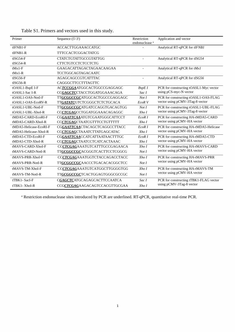
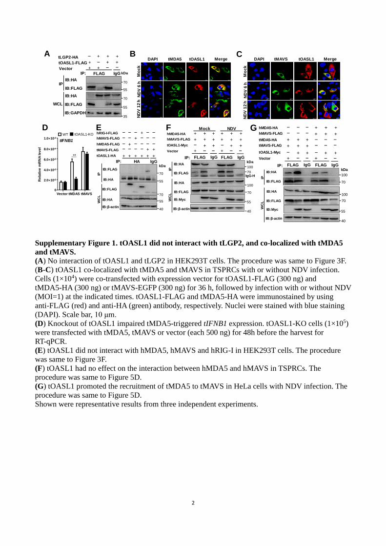
Supplementary Figure 1. tOASL1 did not interact with tLGP2, and co-localized with tMDA5
and tMAVS.
(A) No interaction of tOASL1 and tLGP2 in HEK293T cells. The procedure was same to Figure 3F.
(B-C) tOASL1 co-localized with tMDA5 and tMAVS in TSPRCs with or without NDV infection.
Cells (1×10⁴) were co-transfected with expression vector for tOASL1-FLAG (300 ng) and
tMDA5-HA (300 ng) or tMAVS-EGFP (300 ng) for 36 h, followed by infection with or without NDV
(MOI=1) at the indicated times. tOASL1-FLAG and tMDA5-HA were immunostained by using
anti-FLAG (red) and anti-HA (green) antibody, respectively. Nuclei were stained with blue staining
(DAPI). Scale bar, 10 μm.
(D) Knockout of tOASL1 impaired tMDA5-triggered tIFNB1 expression. tOASL1-KO cells (1×10⁵)
were transfected with tMDA5, tMAVS or vector (each 500 ng) for 48h before the harvest for
RT-qPCR.
(E) tOASL1 did not interact with hMDA5, hMAVS and hRIG-I in HEK293T cells. The procedure
was same to Figure 3F.
(F) tOASL1 had no effect on the interaction between hMDA5 and hMAVS in TSPRCs. The
procedure was same to Figure 5D.
(G) tOASL1 promoted the recruitment of tMDA5 to tMAVS in HeLa cells with NDV infection. The
procedure was same to Figure 5D.
Shown were representative results from three independent experiments.