rapid cytopathic effects of clostridium perfringens beta ... · c enteritis is considered a...
TRANSCRIPT

INFECTION AND IMMUNITY, July 2010, p. 2966–2973 Vol. 78, No. 70019-9567/10/$12.00 doi:10.1128/IAI.01284-09Copyright © 2010, American Society for Microbiology. All Rights Reserved.
Rapid Cytopathic Effects of Clostridium perfringens Beta-Toxin onPorcine Endothelial Cells�†
Corinne Gurtner,1 Francesca Popescu,1 Marianne Wyder,1 Esther Sutter,1 Friederike Zeeh,2Joachim Frey,3 Conrad von Schubert,4 and Horst Posthaus1*
Institute of Animal Pathology,1 Porcine Clinic,2 Institute of Veterinary Bacteriology,3 and Division of Molecular Pathobiology,4
Vetsuisse Faculty, University of Bern, 3001 Bern, Switzerland
Received 17 November 2009/Returned for modification 19 December 2009/Accepted 10 April 2010
Clostridium perfringens type C isolates cause fatal, segmental necro-hemorrhagic enteritis in animals andhumans. Typically, acute intestinal lesions result from extensive mucosal necrosis and hemorrhage in theproximal jejunum. These lesions are frequently accompanied by microvascular thrombosis in affected intes-tinal segments. In previous studies we demonstrated that there is endothelial localization of C. perfringens typeC �-toxin (CPB) in acute lesions of necrotizing enteritis. This led us to hypothesize that CPB contributes tovascular necrosis by directly damaging endothelial cells. By performing additional immunohistochemicalstudies using spontaneously diseased piglets, we confirmed that CPB binds to the endothelial lining of vesselsshowing early signs of thrombosis. To investigate whether CPB can disrupt the endothelium, we exposedprimary porcine aortic endothelial cells to C. perfringens type C culture supernatants and recombinant CPB.Both treatments rapidly induced disruption of the actin cytoskeleton, cell border retraction, and cell shrinkage,leading to destruction of the endothelial monolayer in vitro. These effects were followed by cell death. Cyto-pathic and cytotoxic effects were inhibited by neutralization of CPB. Taken together, our results suggest thatCPB-induced disruption of endothelial cells may contribute to the pathogenesis of C. perfringens type Centeritis.
The anaerobic, spore-forming bacterium Clostridium perfrin-gens is an important Gram-positive pathogen of humans andanimals (18, 42). It causes diverse gastrointestinal diseases,such as food poisoning, enterotoxemia, and enteritis, as well aswound infections and septicemias (37). The virulence of dif-ferent C. perfringens strains is related to the production of alarge array of exotoxins (34). C. perfringens type C isolates aredefined by production of two major toxins, �-toxin (CPA) and�-toxin (CPB). In addition, type C isolates may secrete othertoxins, such as �2-toxin (CPB2), perfringolysin (PFO), entero-toxin (CPE), and TpeL (2, 34, 36) C. perfringens type C strainscause severe, acute, necrotizing enteritis in livestock and hu-mans (18, 42). Outbreaks of human type C enteritis wererecorded after the Second World War in Germany (20), butthis disease has been reported only sporadically in developedcountries (25, 27, 39, 51). A similar disease has been diagnosedmore frequently in parts of Southeast Asia (7, 17, 26), partic-ularly in the highlands of Papua New Guinea (23), where it wasa frequent cause of childhood mortality until vaccination pro-grams were initiated (24). C. perfringens type C causes enteritismore frequently in animals, such as calves, sheep, goats, andparticularly pigs (42, 43). Typically, neonatal piglets are af-fected from the first day of life until they are approximately 3weeks old. The peracute to acute type of the disease affects
piglets within the first few days postpartum (12, 14, 43). Mac-roscopic lesions at necropsy are pathognomonic, with deep,segmental mucosal necrosis and massive hemorrhage in thesmall intestine. In most cases the lesions are confined to theproximal jejunum; however, they can extend into the distalsmall intestine and the colon. This suggests that lesions areinitiated in the upper small intestine and can spread rapidlythroughout the intestine. In addition to these marked necro-hemorrhagic lesions, thrombosis of small vessels in the laminapropria and submucosa is a consistent finding (12, 14). A moreprotracted clinical course of type C enteritis is seen mainly inpiglets that die when they are 1 to 3 weeks old (12, 14, 43). Thepathological lesion is a segmental to diffuse fibrino-necrotizingenteritis. Histopathologically, such cases are characterized bydemarcation of the deeply necrotic mucosa by marked infiltra-tion of neutrophilic granulocytes. Similar acute and subacuteforms of type C enteritis also occur in humans (6, 18, 21). Inhumans, however, subacute lesions are more often described asmultifocal patchy necrosis of the small intestine. Again, muco-sal and submucosal vascular thrombosis is a frequent finding,especially in acute lesions (20, 21).
Besides the clear epidemiological evidence for the impor-tance of CPB in type C enteritis (41, 42), recent experimentalstudies using a rabbit intestinal loop model and a mouse in-fection model clearly demonstrated that CPB is the essentialvirulence factor of type C strains (38, 47, 49). In rabbit ilealloops, application of purified CPB and infection with C. per-fringens type C strains caused villous tip necrosis, which indi-cated that there was initial intestinal epithelial damage. Vas-cular thrombosis in mucosal and submucosal vessels was alsoobserved in this model. In general, the vascular damage ob-served in naturally occurring and experimentally induced type
* Corresponding author. Mailing address: Institute of Animal Pa-thology, Vetsuisse Faculty, University of Bern, P.O. Box 8466, 3001Bern, Switzerland. Phone: 41-(0)31-6312399. Fax: 41-(0)31-6312635.E-mail: [email protected].
† Supplemental material for this article may be found at http://iai.asm.org/.
� Published ahead of print on 19 April 2010.
2966
on May 1, 2020 by guest
http://iai.asm.org/
Dow
nloaded from

C enteritis is considered a secondary effect due to massiveepithelial and mucosal necrosis (22, 43). However, the poten-tial direct effects of exotoxins on vascular endothelia duringtype C enteritis have never been investigated.
CPB is a beta-barrel-pore-forming toxin (9) that has beenshown to form oligomers in the membrane of human endothe-lial cells (44) and the human HL 60 cell line (31). So far,cytotoxic and cytopathic effects of CPB have been demon-strated only for HL 60 cells. HeLa, Vero, CHO, MDCK, Cos-7,P-815, and PC12 cells were not sensitive to this toxin (31, 40).These findings indicate that CPB toxicity is cell type specificand most likely occurs via binding to specific membrane recep-tors. Recently, we localized CPB at vascular endothelial cells inacute type C enteritis lesions in piglets and a human patient(28, 29). As a result of this, we hypothesized that direct tar-geting of endothelial cells and induction of local vascular dam-age could contribute to the rapid tissue necrosis observed inthe acute form of type C enteritis. To validate our initialreports, we performed additional immunohistochemical stud-ies with naturally diseased piglets and subsequently studied thedirect cytopathic effects of CPB on cultured primary porcineendothelial cells. The objectives of this study were (i) to eval-uate the susceptibility of porcine endothelial cells to CPB invitro, (ii) to characterize early morphological changes inducedby CPB in these cells, and (iii) to relate the findings obtainedto pathological lesions observed in acute type C enteritis inpiglets. Our results reveal for the first time that porcineaortic endothelial cell (PAEC) cultures are highly sensitiveto CPB, which results in rapid disruption of the actin cy-toskeleton and retraction of the cell borders progressing tomarked cell shrinkage.
MATERIALS AND METHODS
Animals, histopathology, and immunohistochemistry. Tissues from three1-day-old piglets, which were euthanized for a routine diagnostic workup ofpiglet mortality in their herds, were used for this investigation. These animalswere necropsied immediately after they were euthanized, and they were subse-quently diagnosed with type C enteritis based on classical macroscopic (Fig. 1A)and histopathological lesions in combination with isolation of C. perfringens typeC from the intestine (1, 14). Tissues from a piglet which was euthanized anddiagnosed with coccidiosis based on histopathological and parasitological eval-uations were used as controls. Bacteriologically, C. perfringens type A (cpa�
cpb2�), but not C. perfringens type C, was isolated from the small and largeintestines. Samples of the small intestine, colon, liver, lungs, spleen, brain, andkidneys of each animal were fixed in 10% buffered formalin 15 min after theanimals were euthanized. After 24 h of fixation, tissue sections were embeddedin paraffin, routinely processed for histology, cut into 5-�m sections, and stainedwith hematoxylin and eosin (H&E). An immunohistochemistry analysis usinganti-CPB and anti-CPA monoclonal antibodies as a control was performed asdescribed previously (28).
Cell cultures. Thoracic segments of the aortas of a slaughtered 3-month-oldpig and a 5-day-old piglet, which were euthanized for diagnostic purposes, wereobtained under sterile conditions, placed in culture medium (Dulbecco modifiedEagle medium [DMEM] containing 10% fetal calf serum [FCS], 1� antibiotic-antimycotic [Gibco], and 20 mM L-glutamine), transferred to a cell cultureworkbench, opened longitudinally, and washed. Thirty minutes after the animalsdied, endothelial cells were scraped off and transferred to fibronectin-coatedtissue culture plates to establish separate primary cell cultures from the twodonors. Primary porcine aortic endothelial cell (PAEC) cultures were grown toconfluence at 37°C in the presence of 5% CO2 and passaged. The endothelialorigin and purity of cells were verified by immunofluorescence using anti-ratPECAM-1 antibody (Millipore) (46). Cells in passage 3 were frozen in liquidnitrogen and used as stock cultures. Porcine primary fibroblasts were isolatedfrom skin excised from a 5-day-old piglet that was euthanized for diagnosticpurposes. The tissue was placed in DMEM containing 10� antibiotic-antimy-
cotic and incubated for 6 h at 4°C. The epidermal layer was manually separatedfrom the dermis after overnight incubation in 10 mg/ml dispase (Roche). Dermaltissue was dissociated by incubation in trypsin-EDTA (Bioconcept) with gentleagitation, and cells were seeded into tissue culture flasks in DMEM containing10% FCS, 20 mM L-glutamine, 1 ng/ml acidic fibroblast growth factor (aFGF),and 1 ng/ml basic fibroblast growth factor (bFGF) (Gibco). Fibroblasts in pas-sage 3 were frozen as stock cultures in liquid nitrogen. For all experiments,PAEC and fibroblasts were thawed, propagated, seeded at a density of 1.33 � 104
per cm2, and grown to confluence for 5 (PAEC) or 7 (fibroblasts) days. The cellsused for experiments were exclusively passage 4 to 9 cells. To eliminate thepossibility of donor-specific differences in endothelial cell responses, each exper-iment was performed separately with PAEC from both donors. All experimentswere performed in duplicate.
Production of C. perfringens culture supernatants. Two clinical porcine C.perfringens type C isolates from piglets with necrotizing enteritis and two C.perfringens type A isolates from healthy piglets (Table 1) were grown on bloodagar plates. DNA extraction and PCR amplification of cpa, cpb, cpb2, etx, cpe,pfoA, netB, and tpeL were performed as described previously (1, 2, 8, 19).
FIG. 1. Pathology and immunohistochemistry. (A) Distribution ofmacroscopic lesions of acute C. perfringens type C enteritis in a 2-day-old piglet (stomach on left side; CO, colon). The arrows indicate theextent of typical necro-hemorrhagic lesions in the proximal jejunum.The asterisk indicates a histopathological sampling site in a macro-scopically unaffected segment of the caudal jejunum. (B) H&E-stainedhistological sections of a macroscopically unaffected segment, showingearly signs of epithelial degeneration at villous tips and thrombusformation (arrow) in an underlying vessel. (C) Immunohistochemicallabeling with MAb-CPB of a serial section of the specimen shown inpanel B, showing positive staining on the endothelial lining of a vessel(arrowhead). Note that no immunoreactivity was observed with epi-thelial cells. (D) H&E-stained control tissue from a piglet with super-ficial necrotizing enteritis due to I. suis infection. (E) Immunohisto-chemistry of sections revealed no CPB specific staining. (B and C)Magnification, �1,000. (D and E) Magnification, �400.
VOL. 78, 2010 C. PERFRINGENS BETA-TOXIN EFFECTS ON ENDOTHELIAL CELLS 2967
on May 1, 2020 by guest
http://iai.asm.org/
Dow
nloaded from
Late-log-phase culture supernatants were produced using liquid TGY anaerobicbroth (3% tryptic soy broth, 2% glucose, 1% yeast extract, 0.1% L-cysteine) asdescribed previously (8). After harvest, culture supernatants were chilled on ice,centrifuged (7,500 � g, 20 min), sterile filtered, aliquoted, and stored at �20°C.
Expression and purification of rCPB. The cpb gene was amplified from strainJF 3721 by performing PCR with primers adapted from primers used by Gibertet al. (10) that introduced a SmaI site at the 5� end (TTCCCGGGGCAGCAATGATATAGGTAAAACTACTAC) and a NotI site at the 3� end (TTGCGGCCGCCTAAATAGCTGTTACTTTGTGAG). Sequencing confirmed that therewas 100% identity with the previously published gene sequence. The cpb genewas cloned into pGEM-T (Promega) and subcloned into pET 43.1a (Novagen),and recombinant CPB (rCPB) was expressed in Escherichia coli BL21 (Novagen)in a fusion with an N-terminal 495-amino-acid Nus-Tag protein. After affinitypurification on Ni columns, the Nus-Tag protein was cleaved using a thrombincleavage capture kit (Novagen). To obtain a sufficient yield of rCPB, the man-ufacturer’s protocol was changed so that the preparation was incubated with 5U/ml of biotinylated thrombin for 4 h at 4°C. Even under these conditions, aconsiderable amount of fusion protein remained uncleaved, and degradation ofthe fusion protein, indicated by additional bands (see Fig. 3, lanes 2 and 3),clearly occurred. Thrombin was subsequently removed using streptavidin-coatedbeads. rCPB was harvested at a concentration of 60 �g/ml (for quantification, seebelow). To eliminate toxic effects of contaminating proteins or trace amounts ofthrombin, noncleaved rCPB-Nus-Tag fusion protein and mock purification fromE. coli BL21 harboring the empty vector, which was incubated with thrombin asdescribed above for rCPB, were used as controls in all experiments.
Quantification of rCPB. Recombinant CPB was subjected to 10% SDS-PAGE,and protein bands were stained using Sypro Ruby protein gel staining (Sigma).The Precision Plus unstained standard (Bio-Rad) was used as a reference, andprotein bands were photographed using the U:Genius gel documentation system(Syngene). Concentrations of rCPB were calculated using AIDA software (Im-age Analyzer for Windows). Western blotting was performed as described below.
Western blotting and quantification of CPB in culture supernatants. C. per-fringens culture supernatants were boiled in Laemmli buffer, and 20-�l sampleswere subjected to 10% SDS-PAGE and then transferred to nitrocellulose mem-branes and developed with anti-CPB monoclonal antibody 10A2 (MAb-CPB)(1:1,000 dilution; Centre for Veterinary Biologics, Ames, IA) and goat anti-mouse IRDye 800 secondary antibody (1:5,000 dilution; Li-Cor Biosciences).Serial dilutions of rCPB were used to create a standard curve for quantificationof CPB in culture supernatants by Western blotting. CPB signals were visualizedand scanned using an Odyssee infrared imager (Li-Cor). CPB signal intensity wasmeasured using Odyssee 2.1 software, and concentrations were calculated andsubsequently equalized to obtain a concentration of 2.5 �g CPB/ml of TGYbroth (Table 1).
Cytotoxicity assays. Confluent cells were exposed to 1:10 to 1:1,280 (vol/vol)dilutions of C. perfringens culture supernatants in serum-free endothelial cellculture medium (SFM) (Gibco). rCPB was added to SFM to obtain CPB con-centrations similar to those estimated for diluted C. perfringens type C culturesupernatants. Cells grown in 96-well plates were incubated with 150 �l of SFMcontaining different dilutions of culture supernatants or recombinant toxin prep-arations at 37°C in the presence of 5% CO2. At different time points, 3-(4,5-dimethylthiazolyl-2)-2,5-diphenyltetrazolium bromide (MTT) cell viability testswere performed as described previously (30). SFM containing 10% TGY brothwas used to determine reference values. The MTT test values for each concen-tration were determined in duplicate, and each experiment was performed twicefor each cell culture, which resulted in four data points per group and dilution.All data were expressed as percentages of the baseline value (value obtained withserum-free medium). The effects of group and dilution on the values wereassessed using two-way analysis of variance (ANOVA) (full model, includinginteraction). In addition, a one-way ANOVA with the post hoc Tukey-Kramermultiple-comparison routine was used to identify significant group differencesfor each dilution. The overall level of statistical significance (P value) used was
�0.05. All statistical analyses were performed with the NCSS 2007 statisticalsoftware package (NCSS, Kaysville, UT).
Cytopathic effects and light microscopy. Confluent PAEC and porcine fibro-blasts grown in LabTek slides (Nunc) were incubated with 200 �l SFM contain-ing the concentrations of C. perfringens culture supernatants, rCPB, and controlpreparations used for MTT assays. After 1 and 3 h of incubation, the mediumwas removed, and the cells were fixed and stained using a Diff-Quik staining kit(BD Biosciences). Cells were viewed and photographed using an OlympusBHX-51 microscope with a ProgRres C5 digital camera and ProgRres CapturePro 2.7 software (Jenoptik).
Imaging of live cells. Confluent PAEC cultures in collagen-coated glass bot-tom six-well plates (MatTek) and confluent porcine fibroblasts were incubatedwith SFM containing 5 �g/ml propidium iodide (33) and 32 ng/ml rCPB or JF3721 culture supernatant diluted so that it contained the same concentration ofCPB. Cells were placed on a TE2000E-PFS microscope (Nikon) equipped witha CFI Plan Fluor ELWD �40 objective (Nikon), an Orca-ER charge-coupleddevice (CCD) camera (Hamamatsu), and an incubation chamber (Life ImagingServices), and images were recorded at 37°C in the presence of 5% CO2 for 20 hat 5-min intervals using the NIS Elements software (Nikon).
Immunofluorescence. Confluent PAEC and porcine fibroblasts grown inLabTek slides (Nunc) were incubated with 200 �l SFM containing concentra-tions of C. perfringens culture supernatants, rCPB, and purified controls similarto the concentrations used for MTT assays. After 1 and 3 h of incubation, cellswere washed with phosphate-buffered saline (PBS), fixed (1% paraformalde-hyde, 20 min, room temperature), and permeabilzed (0.1% Triton X-100, 5 min).F- and G-actin were stained using Phalloidin 546 (Alexa Fluor), and nuclei werestained with Hoechst 33258 (Molecular Probes). All washing steps were per-formed with PBS (pH 7.5; three times for 5 min). Coverslips were mounted withfluorescence mounting medium (Dako Cytomation). Slides were viewed with aNikon Eclipse 800 fluorescence microscope. Digital photographs were processedusing the OpenLAB 5.0 software package (Improvision).
Antibody neutralization experiments. C. perfringens culture supernatants andrecombinant toxin preparations were preincubated with neutralizing mouse anti-CPB monoclonal antibody (MAb-CPB) or anti-CPA monoclonal antibody(MAb-CPA) (Centre for Veterinary Biologics) for 30 min at room temperature.For complete inhibition with excess amounts of antibody, 10 �l antibody stocksolution per 100 �l SFM containing toxin was used. Serial 10-fold dilutions of theantibody were used to examine dose-dependent inhibition of CPB.
RESULTS
Histopathology and immunohistochemistry. Our previousstudies demonstrated that CPB bound to vascular endothelialcells in piglets with acute C. perfringens type C enteritis termi-nal lesions. To eliminate the possibility of postmortem bindingof CPB to necrotic vessels, we investigated three piglets whichwere euthanized and subsequently diagnosed with typical acuteC. perfringens type C enteritis (Fig. 1A). We specifically exam-ined macroscopically unaffected distal segments of the jeju-num, because these areas are the areas most likely to exhibitlesions indicative of progression of necrosis along the smallintestine. Histopathology and immunohistochemistry analysesof jejunal segments with fully developed necro-hemorrhagiclesions revealed widespread vascular thrombosis and endothe-lial localization of CPB, as described previously (28) (data notshown). In caudal, macroscopically unaffected jejunal seg-ments there was only multifocal loss of epithelial cells and
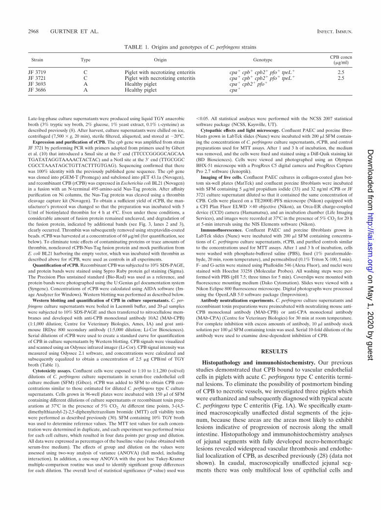
TABLE 1. Origins and genotypes of C. perfringens strains
Strain Type Origin Genotype CPB concn(�g/ml)
JF 3719 C Piglet with necrotizing enteritis cpa� cpb� cpb2� pfo� tpeL� 2.5JF 3721 C Piglet with necrotizing enteritis cpa� cpb� cpb2� pfo� tpeL� 2.5JF 3693 A Healthy piglet cpa� cpb2� pfo�
JF 3686 A Healthy piglet cpa�
2968 GURTNER ET AL. INFECT. IMMUN.
on May 1, 2020 by guest
http://iai.asm.org/
Dow
nloaded from
occasional villous tip necrosis. These segments also exhibitedearly signs of fibrin thrombus formation in vessels in the laminapropria (Fig. 1B). Strong CPB staining was seen on the endo-thelial lining of these vessels but not on epithelial cells (Fig.1C). No lesions or evidence of CPB was seen in colon, liver,lung, kidney, and brain sections (data not shown). Controlsections from a piglet with superficial necrotizing enteritis dueto Isospora suis infection did not show a CPB-positive reaction(Fig. 1D and E). These results suggested that CPB-mediateddamage to vascular endothelial cells of mucosal vessels couldplay a role during the rapid spread of intestinal necrosis in C.perfringens type C enteritis.
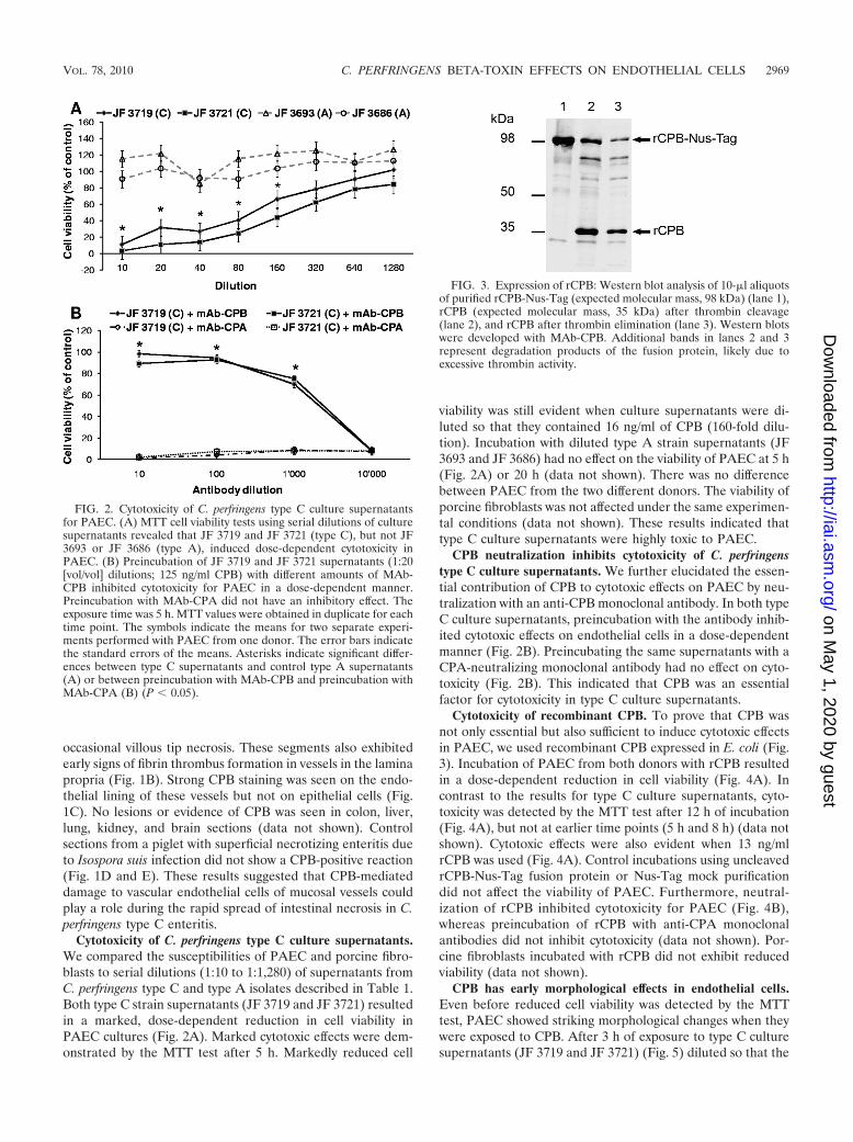
Cytotoxicity of C. perfringens type C culture supernatants.We compared the susceptibilities of PAEC and porcine fibro-blasts to serial dilutions (1:10 to 1:1,280) of supernatants fromC. perfringens type C and type A isolates described in Table 1.Both type C strain supernatants (JF 3719 and JF 3721) resultedin a marked, dose-dependent reduction in cell viability inPAEC cultures (Fig. 2A). Marked cytotoxic effects were dem-onstrated by the MTT test after 5 h. Markedly reduced cell
viability was still evident when culture supernatants were di-luted so that they contained 16 ng/ml of CPB (160-fold dilu-tion). Incubation with diluted type A strain supernatants (JF3693 and JF 3686) had no effect on the viability of PAEC at 5 h(Fig. 2A) or 20 h (data not shown). There was no differencebetween PAEC from the two different donors. The viability ofporcine fibroblasts was not affected under the same experimen-tal conditions (data not shown). These results indicated thattype C culture supernatants were highly toxic to PAEC.
CPB neutralization inhibits cytotoxicity of C. perfringenstype C culture supernatants. We further elucidated the essen-tial contribution of CPB to cytotoxic effects on PAEC by neu-tralization with an anti-CPB monoclonal antibody. In both typeC culture supernatants, preincubation with the antibody inhib-ited cytotoxic effects on endothelial cells in a dose-dependentmanner (Fig. 2B). Preincubating the same supernatants with aCPA-neutralizing monoclonal antibody had no effect on cyto-toxicity (Fig. 2B). This indicated that CPB was an essentialfactor for cytotoxicity in type C culture supernatants.
Cytotoxicity of recombinant CPB. To prove that CPB wasnot only essential but also sufficient to induce cytotoxic effectsin PAEC, we used recombinant CPB expressed in E. coli (Fig.3). Incubation of PAEC from both donors with rCPB resultedin a dose-dependent reduction in cell viability (Fig. 4A). Incontrast to the results for type C culture supernatants, cyto-toxicity was detected by the MTT test after 12 h of incubation(Fig. 4A), but not at earlier time points (5 h and 8 h) (data notshown). Cytotoxic effects were also evident when 13 ng/mlrCPB was used (Fig. 4A). Control incubations using uncleavedrCPB-Nus-Tag fusion protein or Nus-Tag mock purificationdid not affect the viability of PAEC. Furthermore, neutral-ization of rCPB inhibited cytotoxicity for PAEC (Fig. 4B),whereas preincubation of rCPB with anti-CPA monoclonalantibodies did not inhibit cytotoxicity (data not shown). Por-cine fibroblasts incubated with rCPB did not exhibit reducedviability (data not shown).
CPB has early morphological effects in endothelial cells.Even before reduced cell viability was detected by the MTTtest, PAEC showed striking morphological changes when theywere exposed to CPB. After 3 h of exposure to type C culturesupernatants (JF 3719 and JF 3721) (Fig. 5) diluted so that the
FIG. 2. Cytotoxicity of C. perfringens type C culture supernatantsfor PAEC. (A) MTT cell viability tests using serial dilutions of culturesupernatants revealed that JF 3719 and JF 3721 (type C), but not JF3693 or JF 3686 (type A), induced dose-dependent cytotoxicity inPAEC. (B) Preincubation of JF 3719 and JF 3721 supernatants (1:20[vol/vol] dilutions; 125 ng/ml CPB) with different amounts of MAb-CPB inhibited cytotoxicity for PAEC in a dose-dependent manner.Preincubation with MAb-CPA did not have an inhibitory effect. Theexposure time was 5 h. MTT values were obtained in duplicate for eachtime point. The symbols indicate the means for two separate experi-ments performed with PAEC from one donor. The error bars indicatethe standard errors of the means. Asterisks indicate significant differ-ences between type C supernatants and control type A supernatants(A) or between preincubation with MAb-CPB and preincubation withMAb-CPA (B) (P � 0.05).
FIG. 3. Expression of rCPB: Western blot analysis of 10-�l aliquotsof purified rCPB-Nus-Tag (expected molecular mass, 98 kDa) (lane 1),rCPB (expected molecular mass, 35 kDa) after thrombin cleavage(lane 2), and rCPB after thrombin elimination (lane 3). Western blotswere developed with MAb-CPB. Additional bands in lanes 2 and 3represent degradation products of the fusion protein, likely due toexcessive thrombin activity.
VOL. 78, 2010 C. PERFRINGENS BETA-TOXIN EFFECTS ON ENDOTHELIAL CELLS 2969
on May 1, 2020 by guest
http://iai.asm.org/
Dow
nloaded from

medium contained 32 ng CPB/ml, methanol-fixed cells exhib-ited marked cell shrinkage and karyopyknosis. Incubation ofPAEC with 32 ng rCPB/ml medium induced similar, but lesssevere, morphological changes (Fig. 5). Live cell imaging re-vealed that after 15 min of exposure to type C supernatants theintercellular spaces between PAEC started to widen (see Fig.S1 in the supplemental material). These spaces expanded withtime, as PAEC exhibited rapid and progressive cell border retrac-tion, cell shrinkage, cytoplasmic blebbing, and cell rounding. Sim-ilar concentrations of rCPB induced the same morphologicalchanges, albeit less rapidly (see Fig. S2 in the supplemental ma-terial). Cytoplasmic blebbing was observed only at higher concen-trations (1 �g/ml rCPB) (data not shown). Corresponding to theresults of the cell viability test, propidium iodide influx into thenuclei of the majority of cells was observed between 2 and 4 hafter the initial exposure to type C supernatants and 10 to 12 hafter the initial exposure to 32 ng rCPB/ml medium (see Fig.S1 and S2 in the supplemental material). Cytopathic effectswere eliminated by preincubation with anti-CPB monoclonalantibody (Fig. 5) but not by preincubation with anti-CPA an-tibodies (data not shown). Control type A supernatants (Fig.5), rCPB-Nus-Tag, and Nus-Tag mock purification (data not
shown) did not induce cytopathic effects, even after prolongedincubation (up to 20 h) (data not shown). Porcine fibroblastsdid not show comparable morphological alterations under thesame experimental conditions (see Fig. S3 and S4 in the sup-plemental material). These results demonstrated that CPB notonly was highly active with PAEC but was the essential toxinthat induced rapid morphological changes in these cells.
CPB induces rapid cytoskeletal disruption in endothelialcells. One hour after PAEC were exposed to rCPB (at con-centrations ranging from 32 ng/ml to 1 �g/ml), marked disrup-tion of actin filaments was observed (Fig. 6). This effect pro-gressed over time (Fig. 6), and preincubation of rCPB withMAb-CPB inhibited this effect. Uncleaved rCPB-Nus-Tag fu-sion protein did not induce cytoskeletal disruption, even atconcentrations up to 3 �g/ml (Fig. 6). Type C culture super-natants, but not type A culture supernatants, induced disrup-tion of the actin cytoskeleton in PAEC (data not shown).Again, preincubation with neutralizing anti-CPB antibody in-hibited the cytoskeletal changes (data not shown). Porcinefibroblasts were not affected under the same experimental con-
FIG. 4. Cytotoxicity of rCPB for PAEC. (A) Exposure of PAEC torCPB, but not exposure of PAEC to rCPB-Nus-Tag or mock-purifiedNus-Tag, resulted in a dose-dependent reduction in cell viability.(B) Preincubation of rCPB with MAb-CPB inhibited cytotoxicity forPAEC in a dose-dependent manner. The exposure time was 12 h. MTTvalues were obtained from two separate experiments performed induplicate for each time point. The symbols indicate the means for twoseparate experiments with PAEC from one donor. The error barsindicate the standard errors of the means. The asterisks in panel Aindicate statistical significance for comparisons of rCPB with bothcontrol preparations (P � 0.05). The asterisks in panel B indicatestatistical significance for comparisons of rCPB with 1,000- and 10,000-fold dilutions of MAb-CPB and control preparations (P � 0.05).
FIG. 5. CPB-induced cytopathic effects on PAEC: photomicro-graphs of PAEC after 3 h of exposure to serum-free medium (SFM),a JF 3719 culture supernatant dilution containing 32 ng/ml CPB, 32ng/ml rCPB, and a 1:10 (vol/vol) dilution of JF 3693 culture superna-tant. Only CPB-containing media induced cell shrinkage and wideningof intercellular gaps. Preincubation with neutralizing anti-CPB mono-clonal antibodies (mAb-CPB) inhibited the cytopathic effects. Cellswere fixed in methanol and stained with Diff-Quik. Magnification,�400.
2970 GURTNER ET AL. INFECT. IMMUN.
on May 1, 2020 by guest
http://iai.asm.org/
Dow
nloaded from

ditions (Fig. 6). These results indicated that CPB induced rapiddisruption of the actin cytoskeleton in PAEC.
DISCUSSION
Acute C. perfringens type C enteritis in animals and humansis characterized by segmental mucosal necrosis and hemor-rhage of the small intestine and, in fewer cases, the largeintestine (6, 14, 20, 23). Vascular thrombosis of small andmedium-sized vessels occurs in the mucosa of affected small-intestine segments in the acute form of type C enteritis inpiglets and humans (12, 14, 20, 21). This observation has re-ceived little attention because vascular damage has been con-sidered a direct consequence of widespread epithelial necrosis(21, 43). Our recent observation that CPB was consistentlylocalized at endothelial cells in acute lesions of spontaneoustype C enteritis in piglets (28) led us to hypothesize that dis-ruption of endothelial cells by CPB could contribute to thepathogenesis of type C enteritis. Therefore, in the presentstudy, we tested whether CPB has cytopathic and cytotoxiceffects on cultured porcine endothelial cells in vitro.
Our results show that PAEC were highly sensitive to CPB-containing type C culture supernatants. The immediate cyto-pathic effects in PAEC were characterized by rapid disruptionof the actin cytoskeleton and retraction of the cell borders,which progressed to marked cell shrinkage. These effects weredose dependent and induced by type C culture supernatantsbut not by type A culture supernatants. To prove that CPB wasthe essential factor causing the cytopathic effects in PAEC invitro, we used two independent approaches: (i) neutralization
of CPB by monoclonal antibodies (38, 47, 49) and (ii) use ofrecombinant CPB. Neutralization of CPB inhibited cytopathiceffects on PAEC in a dose-dependent manner. Additionally,rCPB induced cytopathic and cytotoxic effects in endothelialcells similar to those induced by type C culture supernatants.Again, these effects were inhibited by neutralization of CPB.Importantly, cytoskeletal disruption and cell border retractionoccurred as early as 1 h after exposure of PAEC to very lowconcentrations (32 ng/ml) of rCPB. Loss of cell viability andpropidium iodide influx, indicating cell death, became evidentlater, after 8 to 12 h of exposure. This was in contrast to themore rapid cell death that occurred between 2 and 5 h afterexposure to type C culture supernatants. The difference mostlikely was due to synergistic effects of other toxins or enzymessecreted by type C strains into culture supernatants.
Endothelial cells form a vital barrier that controls the ex-change of cells, macromolecules, and fluids between the vas-cular lumen and the surrounding tissue. They also maintain thenormal blood flow due to their antiplatelet, anticoagulant, andfibrinolytic properties (5, 48). Disruption of the endothelialbarrier leads to increased vascular permeability along withtissue edema and hemorrhage. Furthermore, it augments localcoagulation and vascular thrombosis (5, 48). Cytopathic effectson endothelial cells, such as those observed in our study, couldtherefore contribute to vascular thrombosis. Johannsen et al.(15, 16) demonstrated that in experimental type C infections inpiglets endothelial damage and microvascular thrombosis insmall-intestine villi occurred shortly after ultrastructural alter-ations in enterocytes but before marked epithelial necrosisbecame evident. Our in vitro results indicate for the first timethat direct interaction of CPB with endothelial cells could beinvolved in this process.
In our study, we used readily obtainable aortic endothelialcells as a model for porcine vascular endothelial cells. Byperforming all experiments with PAEC from two different pigswe eliminated donor-specific differences in cellular reactions(4). However, cultured macrovascular endothelial cells candiffer from microvascular endothelial cells in phenotype and inthe response to toxins (35). Therefore, we could not eliminatedifferences in the susceptibility of small-intestine microvascu-lar endothelial cell cultures to CPB. Nevertheless, as reportedpreviously for E. coli Shiga toxin, target organ-derived micro-vascular endothelial cell cultures could be expected to be moresensitive to toxins than aortic endothelial cells (13, 32). Moreimportantly, our in situ investigations demonstrated that therewas binding of CPB to endothelial cells in small-intestine mu-cosal vessels. Thus, it is conceivable that our in vitro results canbe extrapolated to intestinal microvascular endothelial cells.Consistent with our previous study (28), CPB was detected byimmunohistochemistry only in or adjacent to small-intestinelesions. This suggests that CPB acts mainly on endothelial cellsclose to damaged mucosa. A possible explanation for this isthat once CPB is absorbed by the bloodstream, rapid degra-dation and dilution might prevent accumulation of the toxin atconcentrations required for marked systemic endothelial dam-age and thrombosis. The proposed pathogenic mechanism forendothelial damage involves initial uptake of CPB through theintestinal epithelial barrier. The experimental trials with pig-lets carried out by Johannsen et al. (15, 16) and the recentstudies using the rabbit ileal loop model (38, 49) indicated that
FIG. 6. rCPB-induced disruption of the actin cytoskeleton inPAEC: fluorescent staining of F-actin in PAEC after 1 and 3 h and inporcine fibroblasts after 3 h of incubation with serum-free medium(SFM), 32 ng/ml rCPB, 32 ng/ml rCPB preincubated with MAb-CPB,and 3 �g/ml rCPB-Nus-Tag fusion protein. Disruption of the actincytoskeleton was visible 1 h after exposure to rCPB. The effect in-creased over time (3 h) and was inhibited by neutralization of rCPB.The actin cytoskeleton of porcine fibroblasts was not affected.
VOL. 78, 2010 C. PERFRINGENS BETA-TOXIN EFFECTS ON ENDOTHELIAL CELLS 2971
on May 1, 2020 by guest
http://iai.asm.org/
Dow
nloaded from

epithelial cell necrosis is an initial event during the pathogen-esis of type C enteritis. Additionally, C. perfringens type Cdamages intestinal epithelial cell-like Caco-2 cells in vitro (50).It is therefore conceivable that the initial overgrowth of C.perfringens type C and toxin production damage the intestinalepithelial barrier, leading to increased absorption of CPB andother toxins from the intestine. Interestingly, we could notdemonstrate binding of CPB to epithelial cells in the intestinesof naturally diseased piglets. However, the lesions in theseanimals represent the final stage of the acute disease, wherenecrosis has already occurred to a large extent. The 6-h exper-imental infections in rabbit intestinal loops, therefore, moreclosely represent the initial stages of the disease. Thus, wide-spread small-intestine microvascular thrombosis could be acharacteristic lesion only during later phases of the disease,when mucosal necrosis extends to large areas of the intestine.Based on our results, we hypothesize that when CPB is takenup after initial damage of the jejunal epithelium, it diffusestoward and binds to endothelial cells of blood vessels withinvilli. Binding to unknown receptors would result in oligo-merization, membrane pore formation, and cytopathic ef-fects leading to disruption of the endothelial barrier. Imme-diate consequences of this would be tissue edema andextravasation of erythrocytes into the surrounding tissue. Ifsufficient amounts of CPB can reach the local vasculature,cytopathic effects on endothelial cells could contribute torapid microvascular thrombosis and ischemic mucosal in-farction. This in turn would favor clostridial proliferation andfurther toxin secretion. Direct endothelial toxicity and poten-tially epithelial toxicity, as well as hematogenous spread ofCPB and other toxins, could then lead to the fulminant andprogressive tissue necrosis observed in the acute form of C.perfringens type C enteritis. Effects of CPB on cultured porcinesmall-intestine epithelial cells are currently being investigatedin our laboratory. Preliminary results obtained using similarexperimental approaches indicate that intestinal epithelial cellsare less sensitive to rCPB and type C culture supernatants thanendothelial cells are (unpublished data). However, detailedstudies on the effect of CPB on additional parameters, such asparacellular permeability in epithelial layers, should be per-formed.
Different histotoxic clostridial infections frequently causemassive hemorrhaging and necrosis in affected tissues (45). Forexample, C. perfringens type A-induced gas gangrene is char-acterized by rapid spread of myonecrosis, which for the mostpart involves rapid expansion of vascular thrombosis (5). C.perfringens can produce several toxins, such as CPA and PFO,which induce a procoagulative state locally (3, 5, 11), and it ispossible that they also contribute to vascular thrombosis intype C enteritis. Nevertheless, direct endothelial damage me-diated by CPB or similar toxins might be an important mech-anism by which clostridia induce fulminant progression of isch-emic tissue necrosis.
Taken together, our results suggest that CPB-mediated dis-ruption of the endothelial barrier contributes to the pathogen-esis of C. perfringens type C enteritis. Further analysis of themolecular interaction of the bacterium and its toxins with in-testinal epithelial cells and intestinal microvascular endothelialcells is needed to unravel the complex pathogenesis of thefulminant intestinal necrosis observed in this disease. Because
hemorrhaging and ischemic necrosis are typical features ofmany histotoxic clostridial infections, targeted disruption ofthe endothelium by exotoxins might be a common pathogenicmechanism used by these pathogens. Our approach, combiningin vitro studies based on potential target cell cultures derivedfrom natural hosts with in situ investigation of spontaneouslyaffected animals, is an extension of current experimental ani-mal models and provides a basis for more in-depth experimen-tal studies, including infectious trials with pigs. Due to thesimilarities between spontaneous animal and human clostridialdiseases, such studies should improve our understanding of thepathogenesis of these diseases in animals and humans.
ACKNOWLEDGMENTS
This project was supported by a young investigator grant from theUniversity of Bern (H.P.). F.P. was a recipient of a pathology trainingfellowship from Pfizer, Ltd., Sandwich, United Kingdom.
We thank E. Vilei, Y. Schlatter, and A. Thomann for help withbacteriological work, B. Grabscheid for immunohistochemical stain-ing, M. Doherr for statistical analysis, and R. Kalaji for editorialchanges.
REFERENCES
1. Albini, S., I. Brodard, A. Jaussi, N. Wollschlaeger, J. Frey, R. Miserez, andC. Abril. 2008. Real-time multiplex PCR assays for reliable detection ofClostridium perfringens toxin genes in animal isolates. Vet. Microbiol. 127:179–185.
2. Amimoto, K., T. Noro, E. Oishi, and M. Shimizu. 2007. A novel toxinhomologous to large clostridial cytotoxins found in culture supernatant ofClostridium perfringens type C. Microbiology 153:1198–1206.
3. Awad, M. M., D. M. Ellemor, R. L. Boyd, J. J. Emmins, and J. I. Rood. 2001.Synergistic effects of alpha-toxin and perfringolysin O in Clostridium per-fringens-mediated gas gangrene. Infect. Immun. 69:7904–7910.
4. Bouis, D., G. A. Hospers, C. Meijer, G. Molema, and N. H. Mulder. 2001.Endothelium in vitro: a review of human vascular endothelial cell lines forblood vessel-related research. Angiogenesis 4:91–102.
5. Bryant, A. E. 2003. Biology and pathogenesis of thrombosis and procoagu-lant activity in invasive infections caused by group A streptococci and Clos-tridium perfringens. Clin. Microbiol. Rev. 16:451–462.
6. Cooke, R. 1979. The pathology of pig bel. P. N. G. Med. J. 22:35–38.7. Eason, R. J., and R. van Rij. 1984. Pigless pigbel: enteritis necroticans in the
Solomon Islands. P. N. G. Med. J. 27:42–44.8. Fisher, D. J., M. E. Fernandez-Miyakawa, S. Sayeed, R. Poon, V. Adams, J. I.
Rood, F. A. Uzal, and B. A. McClane. 2006. Dissecting the contributions ofClostridium perfringens type C toxins to lethality in the mouse intravenousinjection model. Infect. Immun. 74:5200–5210.
9. Geny, B., and M. R. Popoff. 2006. Bacterial protein toxins and lipids: poreformation or toxin entry into cells. Biol. Cell 98:667–678.
10. Gibert, M., C. Jolivet-Reynaud, and M. R. Popoff. 1997. Beta2 toxin, a noveltoxin produced by Clostridium perfringens. Gene 203:65–73.
11. Hickey, M. J., R. Y. Kwan, M. M. Awad, C. L. Kennedy, L. F. Young, P. Hall,L. M. Cordner, D. Lyras, J. J. Emmins, and J. I. Rood. 2008. Molecular andcellular basis of microvascular perfusion deficits induced by Clostridiumperfringens and Clostridium septicum. PLoS Pathog. 4:e1000045.
12. Hogh, P. 1969. Necrotizing infectious enteritis in piglets, caused by Clostrid-ium perfringens type C. 3. Pathological changes. Acta Vet. Scand. 10:57–83.
13. Jacewicz, M. S., D. W. Acheson, D. G. Binion, G. A. West, L. L. Lincicome,C. Fiocchi, and G. T. Keusch. 1999. Responses of human intestinal micro-vascular endothelial cells to Shiga toxins 1 and 2 and pathogenesis of hemor-rhagic colitis. Infect. Immun. 67:1439–1444.
14. Jaggi, M., N. Wollschlager, C. Abril, S. Albini, C. Brachelente, M. Wyder,and H. Posthaus. 2009. Retrospective study on necrotizing enteritis in pigletsin Switzerland. Schweiz. Arch. Tierheilkd. 151:369–375.
15. Johannsen, U., S. Menger, W. Erwerth, and B. Kohler. 1986. Clostridiumperfringens type C enterotoxemia (necrotizing enteritis) in suckling pigs. 2.Light and electron microscopic studies of the pathology and pathogenesis ofexperimental Clostridium perfringens type C toxin poisoning. Arch. Exp.Veterinaermed. 40:881–894.
16. Johannsen, U., S. Menger, W. Erwerth, and B. Kohler. 1986. Clostridiumperfringens type C enterotoxemia (necrotizing enteritis) of suckling pigs. 3.Light and electron microscopic studies of the pathology and pathogenesis ofexperimental Clostridium perfringens type C infection. Arch. Exp. Veteri-naermed. 40:895–909.
17. Johnson, S., P. Echeverria, D. N. Taylor, S. R. Paul, R. Coninx, J. Sakurai,B. Eampokalap, P. Jimakorn, R. A. Cooke, G. W. Lawrence, et al. 1987.
2972 GURTNER ET AL. INFECT. IMMUN.
on May 1, 2020 by guest
http://iai.asm.org/
Dow
nloaded from

Enteritis necroticans among Khmer children at an evacuation site in Thai-land. Lancet ii:496–500.
18. Johnson, S., and D. N. Gerding. 1997. Enterotoxemic infections, p. 117–140. In J. I. Rood, B. A. McClaine, J. G. Songer, and R. W. Titball (ed.),The clostridia: molecular biology and pathogenesis. Academic Press, SanDiego, CA.
19. Keyburn, A. L., J. D. Boyce, P. Vaz, T. L. Bannam, M. E. Ford, D. Parker, A.Di Rubbo, J. I. Rood, and R. J. Moore. 2008. NetB, a new toxin that isassociated with avian necrotic enteritis caused by Clostridium perfringens.PLoS Pathog. 4:e26.
20. Kreft, B., K. Dalhoff, and K. Sack. 2000. Necrotizing enterocolitis: a histor-ical and current review. Med. Klin. 95:435–441.
21. Lawrence, G. 1979. The pathogenesis of pig-bel in Papua New Guinea.P. N. G. Med. J. 22:39–49.
22. Lawrence, G. 2005. The pathogenesis of pig-bel in Papua New Guinea. 1979.P. N. G. Med. J. 48:39–49.
23. Lawrence, G., and P. D. Walker. 1976. Pathogenesis of enteritis necroticansin Papua New Guinea. Lancet i:125–126.
24. Lawrence, G. W., D. Lehmann, G. Anian, C. A. Coakley, G. Saleu, M. J.Barker, and M. W. Davis. 1990. Impact of active immunisation againstenteritis necroticans in Papua New Guinea. Lancet 336:1165–1167.
25. Li, D. Y., A. O. Scheimann, J. G. Songer, R. E. Person, M. Horwitz, L. Resar,and K. B. Schwarz. 2004. Enteritis necroticans with recurrent enterocutane-ous fistulae caused by Clostridium perfringens in a child with cyclic neutro-penia. J. Pediatr. Gastroenterol. Nutr. 38:213–215.
26. Mandrella, B. 2007. A recent outbreak of necrotizing enteritis in eastern SriLanka. Trop. Dr. 37:52–54.
27. Matsuda, T., Y. Okada, E. Inagi, Y. Tanabe, Y. Shimizu, K. Nagashima, J.Sakurai, M. Nagahama, and S. Tanaka. 2007. Enteritis necroticans ‘pigbel’in a Japanese diabetic adult. Pathol. Int. 57:622–626.
28. Miclard, J., M. Jaggi, E. Sutter, M. Wyder, B. Grabscheid, and H. Posthaus.2009. Clostridium perfringens beta-toxin targets endothelial cells in necro-tizing enteritis in piglets. Vet. Microbiol. 137:320–325.
29. Miclard, J., J. van Baarlen, M. Wyder, B. Grabscheid, and H. Posthaus.2009. Clostridium perfringens beta-toxin binding to vascular endothelial cellsin a human case of enteritis necroticans. J. Med. Microbiol. 58:826–828.
30. Mosmann, T. 1983. Rapid colorimetric assay for cellular growth and survival:application to proliferation and cytotoxicity assays. J. Immunol. Methods65:55–63.
31. Nagahama, M., S. Hayashi, S. Morimitsu, and J. Sakurai. 2003. Biologicalactivities and pore formation of Clostridium perfringens beta toxin in HL 60cells. J. Biol. Chem. 278:36934–36941.
32. Obrig, T. G., C. B. Louise, C. A. Lingwood, B. Boyd, L. Barley-Maloney, andT. O. Daniel. 1993. Endothelial heterogeneity in Shiga toxin receptors andresponses. J. Biol. Chem. 268:15484–15488.
33. Petit, L., M. Gibert, A. Gourch, M. Bens, A. Vandewalle, and M. R. Popoff.2003. Clostridium perfringens epsilon toxin rapidly decreases membranebarrier permeability of polarized MDCK cells. Cell. Microbiol. 5:155–164.
34. Petit, L., M. Gibert, and M. R. Popoff. 1999. Clostridium perfringens: toxi-notype and genotype. Trends Microbiol. 7:104–110.
35. Ribatti, D., B. Nico, A. Vacca, L. Roncali, and F. Dammacco. 2002. Endo-
thelial cell heterogeneity and organ specificity. J. Hematother. Stem CellRes. 11:81–90.
36. Rood, J. I. 1998. Virulence genes of Clostridium perfringens. Annu. Rev.Microbiol. 52:333–360.
37. Rood, J. I., B. A. McClaine, J. G. Songer, and R. W. Titball (ed.). 1997.The clostridia: molecular biology and pathogenesis. Academic Press, SanDiego, CA.
38. Sayeed, S., F. A. Uzal, D. J. Fisher, J. Saputo, J. E. Vidal, Y. Chen, P. Gupta,J. I. Rood, and B. A. McClane. 2008. Beta toxin is essential for the intestinalvirulence of Clostridium perfringens type C disease isolate CN3685 in arabbit ileal loop model. Mol. Microbiol. 67:15–30.
39. Severin, W. P., A. A. de la Fuente, and M. F. Stringer. 1984. Clostridiumperfringens type C causing necrotising enteritis. J. Clin. Pathol. 37:942–944.
40. Shatursky, O., R. Bayles, M. Rogers, B. H. Jost, J. G. Songer, and R. K.Tweten. 2000. Clostridium perfringens beta-toxin forms potential-depen-dent, cation-selective channels in lipid bilayers. Infect. Immun. 68:5546–5551.
41. Songer, J. G. 2010. Clostridia as agents of zoonotic disease. Vet. Microbiol.140:399–404.
42. Songer, J. G. 1996. Clostridial enteric diseases of domestic animals. Clin.Microbiol. Rev. 9:216–234.
43. Songer, J. G., and F. A. Uzal. 2005. Clostridial enteric infections in pigs. J.Vet. Diagn. Invest. 17:528–536.
44. Steinthorsdottir, V., H. Halldorsson, and O. S. Andresson. 2000. Clostridiumperfringens beta-toxin forms multimeric transmembrane pores in humanendothelial cells. Microb. Pathog. 28:45–50.
45. Stevens, D. L. 1997. Necrotizing clostridial soft tissue infections, p. 141–151. In J. I. Rood, B. A. McClaine, J. G. Songer, and R. W. Titball (ed.),The clostridia: molecular biology and pathogenesis. Academic Press, SanDiego, CA.
46. Tsai, S. H., Y. W. Liu, W. C. Tang, Z. W. Zhou, C. Y. Hwang, G. Y. Hwang,B. R. Ou, C. P. Hu, V. C. Yang, and J. K. Chen. 2007. Characterization ofporcine arterial endothelial cells cultured on amniotic membrane, a potentialmatrix for vascular tissue engineering. Biochem. Biophys. Res. Commun.357:984–990.
47. Uzal, F. A., J. Saputo, S. Sayeed, J. E. Vidal, D. J. Fisher, R. Poon, V. Adams,M. E. Fernandez-Miyakawa, J. I. Rood, and B. A. McClane. 2009. Develop-ment and application of new mouse models to study the pathogenesis ofClostridium perfringens type C enterotoxemias. Infect. Immun. 77:5291–5299.
48. van Hinsbergh, V. W. 2001. The endothelium: vascular control of haemo-stasis. Eur. J. Obstet. Gynecol. Reprod. Biol. 95:198–201.
49. Vidal, J. E., B. A. McClane, J. Saputo, J. Parker, and F. A. Uzal. 2008. Effectsof Clostridium perfringens beta-toxin on the rabbit small intestine and colon.Infect. Immun. 76:4396–4404.
50. Vidal, J. E., K. Ohtani, T. Shimizu, and B. A. McClane. 2009. Contact withenterocyte-like Caco-2 cells induces rapid upregulation of toxin productionby Clostridium perfringens type C isolates. Cell. Microbiol. 11:1306–1328.
51. Watson, D. A., J. H. Andrew, S. Banting, J. R. Mackay, R. G. Stillwell, andM. Merrett. 1991. Pig-bel but no pig: enteritis necroticans acquired in Aus-tralia. Med. J. Aust. 155:47–50.
Editor: S. R. Blanke
VOL. 78, 2010 C. PERFRINGENS BETA-TOXIN EFFECTS ON ENDOTHELIAL CELLS 2973
on May 1, 2020 by guest
http://iai.asm.org/
Dow
nloaded from