produÇÃo de enzimas hidrolÍticas pelo fungo … · quanto inorgânicas. sob condições...
TRANSCRIPT
PRODUÇÃO DE ENZIMAS HIDROLÍTICAS
PELO FUNGO Myrothecium verrucaria
COM ÊNFASE EM SUA ATIVIDADE
QUERATINOLÍTICA.
FABIANA GUILLEN MOREIRA-GASPARIN
Maringá
2006
UNIVERSIDADE ESTADUAL DE MARINGÁ Programa de Pós-graduação em Ciências Biológicas
PRODUÇÃO DE ENZIMAS HIDROLÍTICAS
PELO FUNGO Myrothecium verrucaria
COM ÊNFASE EM SUA ATIVIDADE
QUERATINOLÍTICA.
FABIANA GUILLEN MOREIRA-GASPARIN
Orientadora: Profa. Dra. Rosane Marina Peralta
Tese apresentada ao programa de Pós Graduação em Ciências Biológicas da Universidade Estadual de Maringá, como parte dos requisitos para obtenção do título de Doutor em Ciências (Área: Biologia Celular)
Maringá
2006

Dedico esta tese a aqueles para os
quais sei que esta tem a mesma
importância que para mim, meu
marido Marcos, meus pais Aloisio e
Maria e meus irmãos Veridiana e
Aloisio.
AGRADECIMENTOS
A Deus, não só pela conclusão desta tese, mas por todas as oportunidades até aqui permitidas.
À Prof(a). Dr(a). Rosane Marina Peralta, pela competente orientação que certamente contribuiu para meu crescimento científico. Ao meu marido Marcos, pelo estímulo, paciência e compreensão, principalmente durante a etapa final desta tese e por muitas vezes acreditar mais em mim do que eu mesma. Aos meus pais Aloisio e Maria, que sempre estiveram comigo em pensamento e orações para que fosse possível a concretização deste sonho. Aos meus irmãos Veridiana e Aloisio, que desde o início sempre me apoiaram e incentivaram. Aos meus sogros, João Luis e Cacilda, pelo carinho e atenção que dispensaram a mim durante a etapa final deste doutorado. Aos amigos do laboratório, pela amizade, em especial, à Maria, nossa querida “Pingo”, pela ajuda imprescindível, e também, à Alvina, pela colaboração para o desenvolvimento dos experimentos,. À Prof(a). Dr(a) Cristina Giatti Marques de Souza, pela amizade, atenção e ensinamentos que enriqueceram minha formação científica. Às amigas de pós-graduação, Cinthia, Andréa, Daniela e Ana Maria, pelos alegres e inesquecíveis momentos vividos dentro e fora do laboratório. Á CAPES, pela concessão de bolsa de estudos. Ao Programa de Pós-Graduação em Ciências Biológicas – Área de Concentração Biologia Celular, pela oportunidade de qualificação para a pesquisa e o ensino. E a todos aqueles que de alguma forma contribuíram para a realização de mais uma etapa na minha vida, muito obrigada.
APRESENTAÇÃO
Esta tese de doutoramento está apresentada na forma de três artigos científicos:
Moreira, F.G., Reis, D., Costa, M.A.F., Souza, C.G.M. and Peralta, R.M. (2005). Production of
hydrolytic enzymes by the plant pathogenic fungus Myrothecium verrucaria in submerged
cultures. Brazilian Journal of Microbiology, 36, 7-11
Moreira, F.G., Souza, C.G.M., Costa, M.A., Reis, S. and Peralta, R.M. Degradation of chicken
feathers by Myrothecium verrucaria. Mycopathologia
Moreira, F.G. and Peralta, R.M. Purification and partial characterization of a keratinolytic protease
from the non dermatophytic fungus Myrothecium verrucaria. Enzyme and Microbial Technology.
RESUMO
Os fungos são microrganismos heterotróficos que utilizam moléculas orgânicas como fonte
de carbono. Em relação ao nutriente nitrogênio, os fungos podem utilizar tanto fontes orgânicas
quanto inorgânicas. Sob condições naturais, os fungos necessitam secretar enzimas extracelulares
capazes de degradar as moléculas orgânicas complexas. A maioria das enzimas extracelulares dos
microrganismos são depolimerases, principalmente hidrolases, que atuam em polissacarídeos,
proteínas, lipídeos e ácidos nucléicos. Proteases extracelulares são amplamente encontradas entre
as enzimas secretadas pelos microrganismos. Estas enzimas são facilmente detectadas e são
freqüentemente sintetizadas em altos títulos, sendo objeto de muitas investigações. As numerosas
funções e a variabilidade de condições nas quais as proteases são sintetizadas explicam a
multiplicidade destas enzimas. O papel das enzimas proteolíticas extracelulares de fungos não está
restrito à sua participação na nutrição destes microrganismos. Enzimas proteolíticas secretadas
pelos fungos podem ter papel em sua patogenicidade, provocando severas doenças.
O termo protease tem como sinônimos os termos peptidase, enzima proteolítica, proteinase
e peptídeo hidrolase. As proteases incluem todas as enzimas que catalisam a clivagem das ligações
peptídicas das proteínas, resultando na produção de peptídeos e aminoácidos livres. Algumas
proteínas apresentam certas características estruturais que tornam mais difícil sua hidrólise. As
queratinas são encontradas em células epiteliais de vertebrados e representam os maiores
constituintes da pele e seus apêndices tais como unhas, cabelos, penas e lã. A queratina do cabelo
(queratina dura) é semelhante a encontrada na epiderme (queratina macia), visto possuir estrutura
em α-hélice , mas difere desta última por conter menor quantidade de cisteína. Nas penas, a
queratina assume conformação β, que é mais facilmente hidrolisada quando comparada com a α-
queratina. A resistência à hidrólise da queratina é conferida principalmente pelo seu alto teor em
pontes dissulfeto. Entretanto, ela pode ser degradada por algumas espécies de fungos e em menor
extensão por bactérias que secretam as enzimas queratinolíticas (queratinases, EC 3.4.99.11), que
tem a habilidade degradar queratina nativa em peptideos menores que são subseqüentemente
absorvidos pelas células.
Milhões de toneladas de penas de aves são produzidas anualmente como resíduos da
criação de aves. O desenvolvimento de métodos enzimáticos e/ou microbiológicos para a hidrólise
de queratina a proteínas solúveis e aminoácidos é extremamente atraente, visto que oferece uma
condição barata e suave para a obtenção de produtos de alto valor. Este processo pode ser fonte de
aminoácidos raros como cisteína, serina e prolina, ou pode ser usado no desenvolvimento de
fertilizantes de lenta liberação de nitrogênio, colas e biofilmes. Diversos microrganismos produtores
de queratinases tem sido descritos como hábeis em degradar penas de aves e uma estratégia
simples de selecionar microrganismos com potencial queratinolítica é o uso de penas de aves como
substrato único nos meios de cultivos.
Myrothecium verrucaria (Albertini and Schwein) Ditmar:Fr, é um fungo fitopatogênico que
ataca uma série de plantas, incluindo soja, girassol, arroz e tomate. M. verrucaria tem sido também
investigado devido ao seu potencial uso no controle biológico de pragas e também pelo fato de ser
produtor da enzima bilirrubina oxidase (BOD). Esta enzima é utilizada na determinação da
bilirrubina, um produto do catabolismo de heme-proteínas. Outras enzimas envolvidas na
degradação de polissacarídeos de parede celular e lignina também tem sido objeto de estudos. M.
verrucaria é conhecido produtor de endoquitinases, xilanases, pectinases e lacases.
A produção de proteases extracelulares por fungos fitopatogênicos tem sido bem
documentada. Tem sido proposto que em algumas interações planta-fungo, essas enzimas tem
função patogênica, mas nenhum estudo prévio havia sido realizado em M. verrucaria. O objetivo do
primeiro trabalho foi, portanto, avaliar as enzimas produzidas por M. verrucaria em culturas
submersas potencialmente envolvidas em sua fitopatogenicidade utilizando diversos substratos. O
fungo produziu diferentes depolimerases e glicosidases, sendo xilanase, pectinase e protease as
mais importantes. Atividade lípase foi encontrada nos cultivos em óleo de oliva, enquanto atividade
protease foi detectada em todas as culturas. Xilanase e pectinase foram otimamente ativas em pH
4,5-5,5, enquanto protease foi ativa em ampla faixa de pH variando de 3,5 a 11,0. As três enzimas
tiveram atividade máxima a 40o C e foram estáveis por várias horas quando armazenadas em
temperaturas inferiores a 50º C.
No segundo trabalho, descrevemos a capacidade de M. verrucaria crescer em culturas
submersas e em estado sólido utilizando penas de aves como únicas fontes de carbono e nitrogênio
e eficientemente degradar queratina de penas de aves. Nos dois tipos de culturas, máxima atividade
proteolítica foi obtida após quatro dias de cultivo (130 e 250 U/ml, para culturas submersas e em
estado sólido, respectivamente). Foi observado que o crescimento M. verrucaria em culturas
submersas usando penas de aves intactas, resultou em quase completa degradação das mesmas
após 48 h. Os filtrados das culturas hidrolisou material queratinoso na seguinte ordem: queratina de
penas de aves > queratina de lã de carneiro > queratina de cabelo humano > queratina de unha
humana. A melhor condição para os filtrados de cultura degradarem queratina de penas foi pH 9,0 e
40o C. Atividade queratinolítica foi fortemente inibida por PMSF e EDTA, sugerindo a presença de
metalo-serina-protease. Análise por SDS-PAGE mostrou que protease foi a principal e menor
proteína secretada pelo fungo sob as condições de cultura usada neste trabalho. Os resultados
obtidos neste estudo permitiram concluir que a conversão microbiana de penas de aves por M.
verrucaria representa uma alternativa atraente para o uso de penas como fonte de proteínas e para
reduzir o impacto ambiente do acúmulo de resíduos de penas de aves após o processamento de
aves para o consumo humano.
No terceiro estudo, uma protease queratinolítica de Myrothecium verrucaria foi purificada
até aparente homogeneidade eletroforética usando uma única etapa cromatográfica com um
rendimento de 66.7% A enzima é uma proteína monomérica com massa molecular de 22 e 23 kDa
determinada por SDS-PAGE e gel filtração, respectivamente. A enzima mostrou-se estável em
ampla faixa de pH (5,0-12,0) e a temperaturas de até 45º C. Ela foi severamente inibida por PMSF e
parcialmente inibida por EDTA, o que sugere ser uma metalo-serina-protease. A enzima purificada
foi capaz de hidrolisar queratina de farinha de penas com a produção de moléculas com grupos
amino livres. A eficiência da hidrólise foi aumentada quando a queratina foi previamente tratada com
agentes redutores.
Entre os fungos, a maior parte dos estudos de produção, isolamento, purificação e
caracterização de queratinases tem sido realizados com fungos dermatofíticos, embora algumas
queratinases de fungos não dermatofíticos tenham sido descritas. Os dados apresentados neste
trabalho podem ser úteis na compreensão dos mecanismos envolvidos na degradação de queratina
Palavras-chaves: protease, queratinase, Myrothecium verrucaria, hidrolases
ABSTRACT
It is well know that fungi are heterotrophic microorganisms using organic substances as a
source of carbon. With regard to nitrogen nutrition, fungi can use both inorganic and organic sources.
Under natural conditions fungi have to secrete extra cellular enzymes capable of degrading complex
natural organic substances. Most of the extracellular enzymes of micro organisms are
depolymerises, mostly hydrolases, which act on polysaccharides, proteins, lipids, and nucleic acids.
Extracellular proteases, are, apparently, widely found among enzymes secreted by microorganisms.
These enzymes can be easily detected, are often synthesized with high yields and have become an
object of thorough investigation. Their numerous functions and the variety of conditions in which
proteases are synthesized and work explain the multiplicity of these enzymes. The role of
extracellular proteolytic enzymes of fungi is not restricted to their participation in nutrition of these
micro organisms. Proteolytic enzymes secreted by the fungi can also play a role of factors of
pathogenicity, provoking serious diseases.
The term protease is synonymous with peptidase, proteolytic enzyme, proteinase and
peptide hydrolase. The proteases include all enzymes that catalyse the cleavage of the peptide
bonds of proteins, digesting these proteins into peptides or free amino acids. However, some
proteins have certain structural characteristics that make their hydrolysis more difficult. The structural
protein keratin is resistant to the activity of a broad range of proteases, mainly due to its high content
of disulfide bridges. Keratins are the most abundant proteins in epithelial cells of vertebrates and are
the major constituents of skin and its appendages such as nail, hair, feather, and wool. The keratin
chain of hair (hard keratin) is similar to that of epidermis (soft keratin), since it is tightly packed in an
α-helix, but differs from the latter in that it contains a several fold higher amount of cystein. However,
in feathers, the polypeptide assumes a β-conformation, which is more readily hydrolyzed compared
with α-keratin. Keratin can be degraded by some species of fungi and to a less extend in bacteria,
that secrete keratinolytic enzymes (keratinases, EC 3.4.99.11). These enzymes have the ability to
degrade native keratin into smaller peptides entities that can subsequently be absorbed by cells.
Worldwide poultry processing plants produce millions of tons of feathers as a waste product
annually. The development of enzymatic and/or microbiological methods for the hydrolysis of feather
into soluble proteins and amino acids is extremely attractive, as it offers a cheap and easy method
for the production of valuable products. It could be used to produce rare amino acids like serine,
cystein and proline, or for the development of slow-release nitrogen fertilizers, glues, and
biodegradable films. Keratinase-producing microorganisms have been described as able to degrade
feathers, and one strategy for selecting potential new keratinolytic micro organism is the use of
poultry feathers as the only substrate in the cultures.
Myrothecium verrucaria (Albertini and Schwein) Ditmar:Fr, is a ubiquitous phytopathogenic
fungus which attacks a wide range of plant crops, including soybean, sunflower, tomato, and rice.
M. verrucaria has also been investigated due to its potential use in biological weed control and due to
its capability to produce the enzyme bilirubin oxidase (BOD). This enzyme is used for the enzymatic
determination of bilirubin, a catabolic product of heme proteins. Other enzymes involved in
degradation of cell wall polysaccharides and lignin has also been studied, including endochitinases,
xylanases, pectinases and laccases.
The production of extracellular proteases by plant pathogenic fungi is well documented, and
it has been proposed that in some fungus-plant interactions these enzymes may function as
pathogenic factors, but until now, no work has been done on M. verrucaria. Considering this, the aim
of the first study was to get an overview of the enzymes produced by M. verrucaria in submerged
cultures that are potentially involved in its phytopathogenicity. The capability of M. verrucaria to
produce extra cellular hydrolytic enzymes in submerged cultures was studied using several isolated
substrates. The fungus was able to produce different depolymerases and glycosidases, with
xylanase, pectinase and protease being the most important. Xylanase and pectinase were optimally
active at pH 4.5-5.5, while protease was active in a large range of pH 3.5 to 11.0. All three enzymes
were maximally active at 40o C and were stable for several hours at temperature up to 50o C.
In the second study, the capability of M. verrucaria to grown in submerged (SC) and solid
state (SSC) cultures using chicken feathers as the only source of carbon and nitrogen and its ability
to efficiently degrade chicken feather keratin is reported. In both types of cultures, maximal protease
production was obtained after 4 days of cultivation (130 and 250 U/ml in SC and SSC, respectively).
It was observed that the growth of M. verrucaria in submerged culture using intact feathers resulted
in almost complete degradation of intact feathers within 48 h. The culture filtrates hydrolyzed
keratinous substrates in the following order: chicken feather keratin > sheep wool keratin > human
nail keratin > human hair keratin. The best conditions for culture filtrates to efficiently degraded
feather keratin was pH 9.0 and 40o C. Keratinolytic activity was strongly inhibited by PMSF and
EDTA, indicating the presence of serine and metallo protease. SDS-PAGE analysis showed that
protease was the main and smaller protein secreted by the fungus under the culture conditions used
in this study. The results obtained in this study permit the conclusion that chicken feather microbial
conversion by M. verrucaria represents an attractive alternative for improving the use of feather as a
feed protein and to reduce the impact of poultry feather accumulation as a waste product after the
processing of chickens for human consumption.
In the third study, a Myrothecium verrucaria keratinolytic protease was purified to apparent
electrophoretic homogeneity using one unique chromatographic step with a high yield of 66.7%. The
enzyme was a monomeric protein with molecular mass of 22 and 23 kDa by SDS-PAGE and gel
filtration, respectively. The enzyme was stable in a broad range of pH (5.0-12.0) and temperature up
to 45o C, being optimally active at 40oC and pH 8.0-9.0. It was strongly inhibited by PMSF and
partially inhibited by EDTA, which suggests to be a metallo serine protease. The purified enzyme
was able to hydrolyze chicken feather meal producing free amino groups. The efficiency of
hydrolysis was improved when the keratin was previously treated with reducing agents.
Among fungi, the most part of studies on production, isolation, purification and
characterization of keratinase have been carried out with dermatophytic fungi, although some non
dermatophytic fungi keratinase has been already described. The data presented in this work may be
useful in understanding the mechanism of keratin degradation.
Key-words: protease, keratinase, Myrothecium verrucaria, hydrolases
ARTIGO 1
PRODUCTION OF HYDROLYTIC ENZYMES BY THE PLANT
PATHOGENIC FUNGUS Myrothecium verrucaria IN SUBMERGED
CULTURES.
Fabiana Guillen Moreira, Simone dos Reis, Maria Aparecida Ferreira Costa, Cristina Giatti
Marques de Souza, Rosane Marina Peralta.
Laboratório de Bioquímica e Fisiologia de Microrganismos, Departamento de Bioquímica,
Universidade Estadual de Maringá, 87020900, Maringá, PR, e-mail:[email protected]
ABSTRACT
The capability of the plant pathogenic fungus Myrothecium verrucaria to produce extracellular
hydrolytic enzymes in submerged cultures was studied using several substrates. The fungus was
able to produce different depolymerases and glycosidases, being xylanase, pectinase and protease
the most important of them. Lipase was found in cultures developed in the presence of olive oil, while
protease activity was detected in all cultures. Xylanase and pectinase were optimally active at pH
4.5-5.5, while protease was active in a large range of pH 3.5 to 11.0. All three enzymes were
maximally active at 40o C and they were stable for several hours at temperature up to 50o C.
Key words: carbohydrases, hydrolytic enzymes, Myrothecium verrucaria, protease.
RESUMO
A capacidade do fungo fitopatogênico Myrothecium verrucaria produzir enzimas hidrolíticas
extracelulares em culturas submersas foi estudada utilizando diversos substratos. O fungo foi capaz
de produzir diferentes depolimerases e glicosidases, sendo xilanases, pectinases e proteases as
mais importantes. Atividade lipase foi encontrada nos filtrados das culturas desenvolvidas na
presença de óleo de oliva, enquanto atividade proteolítica foi detectada em todas as culturas.
Xilanase e pectinase foram otimamente ativas em pH 4,5 a 5,5, enquanto protease foi ativa em
ampla faixa de pH (3.5 a 11.0). As três enzimas foram otimamente ativas 40o C e estáveis por
várias horas a temperaturas até 50o C.
Palavras-chave: carboidrases, enzimas hidrolíticas, Myrothecium verrucaria, protease.
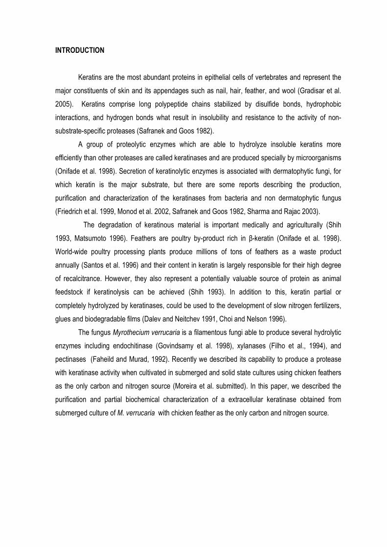
INTRODUCTION
Plant pathogens produce a range of enzymes capable of degrading plant cell wall
components (1,23). Among the economically important plant pathogens, Myrothecium verrucaria
(Albertini and Schwein) Ditmar:Fr, is a ubiquitous phytopathogenic fungus which attacks a wide
range of plants, including cucumber (20), soybean (16), upland cotton (11), sunflower (3), birdsfoot
trefoil (6), tomato (25), rice (24) and corn (27).
Extracellular proteins secreted by fungus are able to macerate tissues and degrade cell wall
components. They must thus contain the enzymes corresponding to the types of glycosidic linkages
present in the cell wall polysaccharides. Extracellular enzymes are important to fungi not only for
digestion but also in many instances for the pathogenic process: the enzymes may function in
overcoming the natural resistance of the host as well as in providing soluble products that can be
absorbed and used as food (9). M. verrucaria is known to produce endochitinase (8,26), xylanases
(7), pectinases (5) and bilirrubin oxidases (10). The production of extracellular proteases by plant
pathogenic fungi is also well documented, and it has been proposed that in some fungus-plant
interactions these enzymes may function as pathogenic factors (4,17,19,20,23).
Studies of enzyme production by a phytopathogenic fungus are complicated by the presence
of plant, particularly by the presence of plant enzymes and microbial enzyme inhibitors that occur in
the plants. The most practical way to study the production of enzyme by a fungus is therefore to
study the production of its enzymes on artificial growth media that contain no plant or enzyme
inhibitors produced by the plant. Considering that, the aim of this study was to get an overview of
the enzymes that are produced by M. verrucaria in submerged cultures possibly involved with its
phytopathogenicity.
MATERIAL AND METHODS
Microorganism Myrothecium verrucaria (Albertini and Schwein) Ditmar:Fr CCT 1886 was obtained
from the Collection culture of Fundação André Tosello, Campinas, SP, Brazil and it was maintained
on potato dextrose agar.
Enzyme production. For enzyme production in submerged cultures, 1 x 10⁹ spores were
transferred to 50 mL of mineral media (15) containing 0.5% (w/v) of one carbon source, and pH
adjusted to 6.0. The cultures were incubated at 28o C on a rotary shaker at 120 rpm. After 7 days, the
mycelia were removed from the culture media by filtration. To determine the dry weight of the
mycelia, they were dried overnight at 60o C. To study the effect of association of different
substrates in the production of enzymes, each substrate or mixture of substrates were added to a
final concentration of 1% (w/v).
Enzyme determinations. Glycoside hydrolase activities (β-glucosidase and β-xylosidase) were
determined by measuring the rate of p-nitrophenol released from the appropriate p-nitrophenyl
derivatives (p-nitrophenyl-β-glucopiranoside and p-nitrophenyl-β-xylopyranoside, respectively). The
standard reaction mixture contained 20 µl of enzyme solution and 1 mg/mL of substrate in
phosphate buffer 0.1 M, pH 6.0. After 15 min of incubation at 50o C, reactions were stopped by the
addition of 2 ml of 0.1 M Na2CO3 and the p-nitrophenol liberated was determined
spectrophotometrically at 410 nm (22). Polysaccharidase activities (as xylanase, pectinase and
carboxymethylcellulase, CMCase) were determined by measuring the amount of reducing sugar
(xylose, galacturonic acid and glucose) released from various substrates (xylan, polygalacturonic
acid and carboxymethylcellulose, respectively). A 20 µl volume of enzyme solution was incubated at
50o C for 30 min. in 1.0 ml of substrate solution polysaccharide (2 mg/ml) dissolved in phosphate
buffer, 0.1 M, pH 6.0. Reactions were stopped by the addition of 1 ml of dinitrosalicylic reagent (14).
Tubes were placed in a boiled water bath for 5 min. The A540 nm was read with appropriate single
sugars as standards (glucose, xylose and galacturonic acid, respectively to carboxymethylcellulase,
xylanase and polygalacturonase activities). Lipase was determined using an olive oil emulsion as
substrate. The liberated free fatty acids were titrated with 0.05 M NaOH and phenolphtalein as
indicator (18). Protease was determined using casein as substrate. The released tyrosine was
estimated by Lowry’s method (12).
Effect of pH on the activity of xylanase, polygalacturonase and protease. The effects of pH on
the activity of enzymes were determined in a series of McIlvaine’s buffers with pH values from 3.5 to
8.3 (13), and 0.1 M glycine-NaOH (pH 8.5 to 10.0).
Effect of temperature on enzyme activity and stability. The effects of temperature on the activity
of enzymes were carried out at temperature ranging from 25 to 70o C. Thermal stability was
investigated by incubating the enzyme at 30, 40, 50 and 60o C for 1 hour. Immediately afterwards the
enzymes were immersed in an ice bath and then the activities were tested under standard conditions.
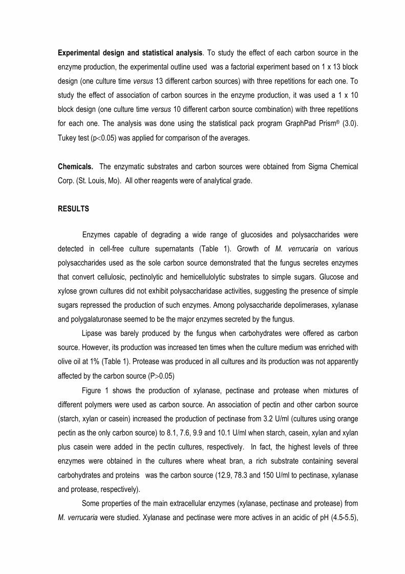
Experimental design and statistical analysis. To study the effect of each carbon source in the
enzyme production, the experimental outline used was a factorial experiment based on 1 x 13 block
design (one culture time versus 13 different carbon sources) with three repetitions for each one. To
study the effect of association of carbon sources in the enzyme production, it was used a 1 x 10
block design (one culture time versus 10 different carbon source combination) with three repetitions
for each one. The analysis was done using the statistical pack program GraphPad Prism® (3.0).
Tukey test (p<0.05) was applied for comparison of the averages.
Chemicals. The enzymatic substrates and carbon sources were obtained from Sigma Chemical
Corp. (St. Louis, Mo). All other reagents were of analytical grade.
RESULTS
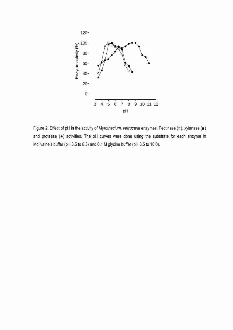
Enzymes capable of degrading a wide range of glucosides and polysaccharides were
detected in cell-free culture supernatants (Table 1). Growth of M. verrucaria on various
polysaccharides used as the sole carbon source demonstrated that the fungus secretes enzymes
that convert cellulosic, pectinolytic and hemicellulolytic substrates to simple sugars. Glucose and
xylose grown cultures did not exhibit polysaccharidase activities, suggesting the presence of simple
sugars repressed the production of such enzymes. Among polysaccharide depolimerases, xylanase
and polygalaturonase seemed to be the major enzymes secreted by the fungus.
Lipase was barely produced by the fungus when carbohydrates were offered as carbon
source. However, its production was increased ten times when the culture medium was enriched with
olive oil at 1% (Table 1). Protease was produced in all cultures and its production was not apparently
affected by the carbon source (P>0.05)
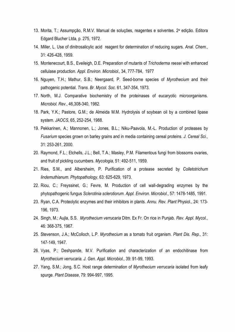
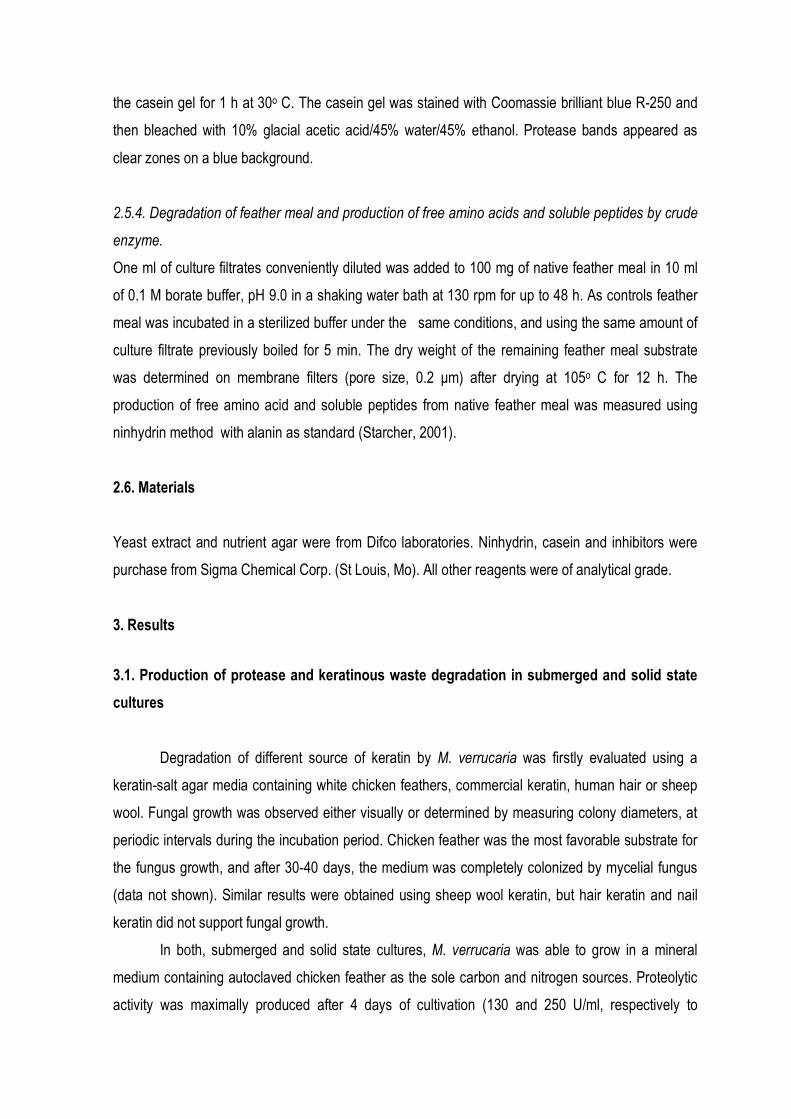
Figure 1 shows the production of xylanase, pectinase and protease when mixtures of
different polymers were used as carbon source. An association of pectin and other carbon source
(starch, xylan or casein) increased the production of pectinase from 3.2 U/ml (cultures using orange
pectin as the only carbon source) to 8.1, 7.6, 9.9 and 10.1 U/ml when starch, casein, xylan and xylan
plus casein were added in the pectin cultures, respectively. In fact, the highest levels of three
enzymes were obtained in the cultures where wheat bran, a rich substrate containing several
carbohydrates and proteins was the carbon source (12.9, 78.3 and 150 U/ml to pectinase, xylanase
and protease, respectively).
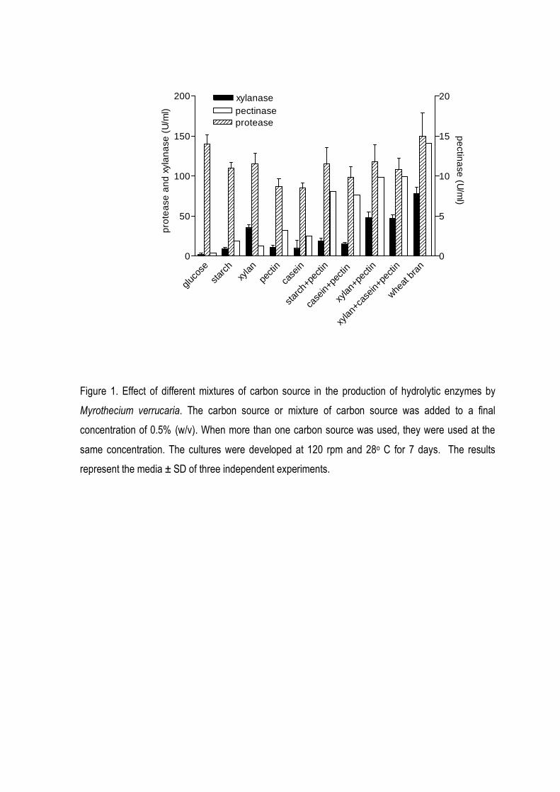
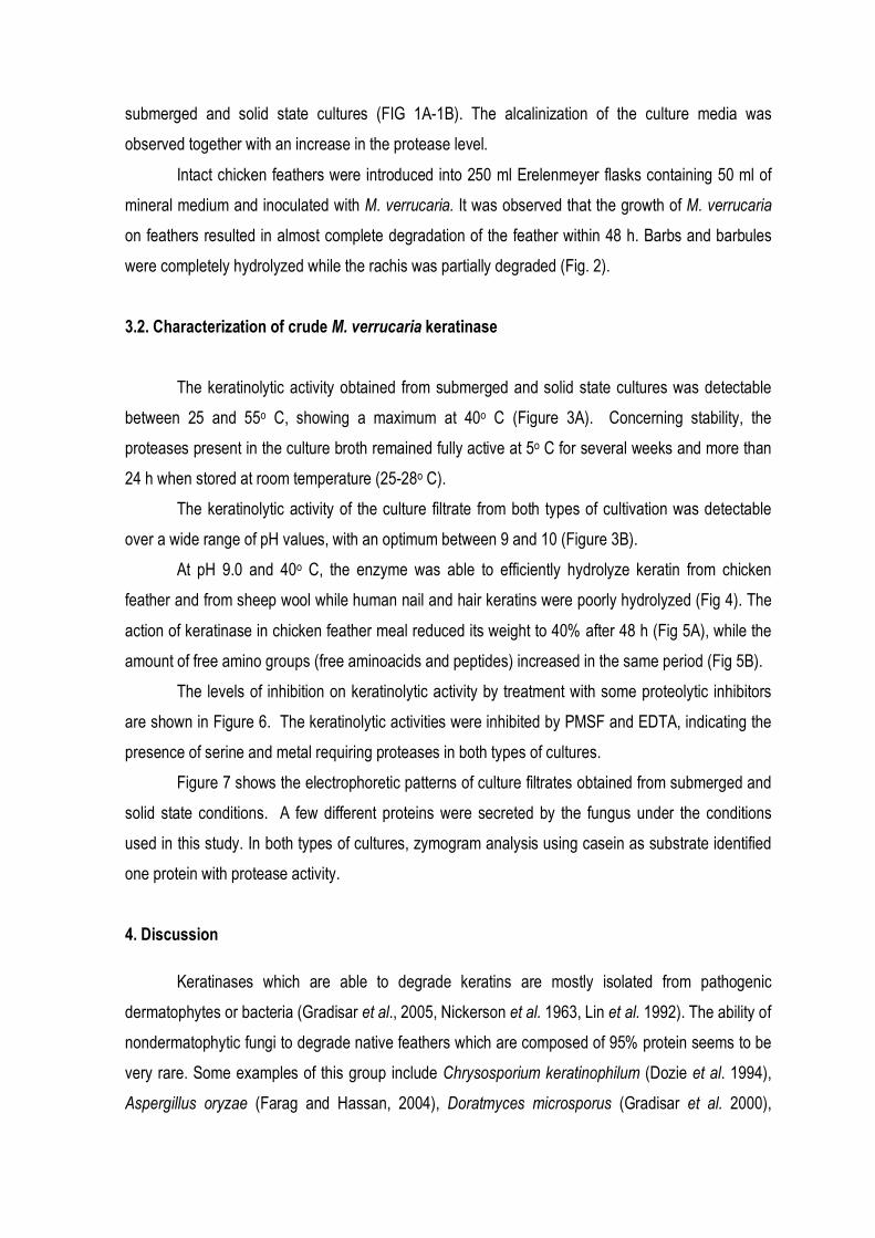
Some properties of the main extracellular enzymes (xylanase, pectinase and protease) from
M. verrucaria were studied. Xylanase and pectinase were more actives in an acidic of pH (4.5-5.5),

while the best pH for protease activity was between pH 8.0 and 9.0 (Fig 2). Figure 2 suggest the
existence of more than one group of protease, one with optimum pH between 6.0-7.0, and other with
optimum pH between 8.0-9.0. All enzymes were optimally active at 40o C and they retained more
than 95% of initial activity after 60 min at 50o C (data not shown).
DISCUSSION
In the present work we have shown that M. verrucaria produces polysaccharide
depolymerases and glucosidases necessary to degrade important structural cell wall
polysaccharides, particularly pectin and hemicellulose (Table 1). The secretion of several enzymes
provides this phytopathogenic fungus with the ability to attack hosts which differ in their
polysaccharide cell wall compositions (22). The secretion of glycosidases combined with the
polysaccharide depolymerases may also remove side groups of heteropolysaccharides, making
easier the action of endoenzymes (21).
Our results showed the capability of M. verrucaria to produce other hydrolytic enzymes such
as proteases and lipases. These enzymes may be involved in the capability of the fungus to invade
vegetal tissues. M. verrucaria protease was identified as an alkaline protease (Fig. 2). However,
differently from xylanase and pectinase that were active in a an acidic range of pH, protease from M.
verrucaria showed high activity at alkaline pH, although it remained active in a large range of pH,
what could indicate the presence of multiple isoenzymes, with different optimum pH.
It has been suggested that the proteases may facilitate located penetration of the plant cell
wall by breaking down the fibrous glycoproteins that contribute to cell wall stability (2). Some
phytopathogenic fungi such as Fusarium, Alternaria, and Rhizoctonia produced serine alkaline
proteases, which are indispensable for their growth (9,19). They are probably nutrient-mobilizing
enzymes whose primary function is the support of fungal growth after host cell death has occurred.
The highest levels of enzymic activities have been obtained when wheat bran was the main
substrate (Fig. 1). Wheat bran is a very rich substrate consisting of a mixture of proteins, fat, soluble
and insoluble carbohydrates. It is probable that the several hydrolytic enzymes secreted by the
fungus present a synergy in the degradation of wheat bran components.
In conclusion, in the present study, it was observed the capability of M. verrucaria to produce
and secrete different hydrolytic enzymes. Studies of production of enzymes using several plant
materials is in progress in our laboratory. This study will serve to increase the understanding of
factors that control the production, activity, and the role of M. verrucaria hydrolytic enzymes in its
phyto-pathogenicity.
ACKNOWLEDGEMENTS
This work was supported by grants from Conselho Nacional de Desenvolvimento Científico e
Tecnológico (CNPq) and Fundação Araucária. R.M. Peralta is research fellow of CNPq. F.G.
Moreira is a recipient of CAPES Fellowships. S. Reis is recipient of CNPq Fellowships. We thank
Alvina Chaves for her technical assistance.
REFERENCES
1. Baer, D.; Gudmestad, N.C. In vitro cellulolytic activity of the plant pathogen Clavibacter
michiganensis subsp. sepedonicus. Can. J. Microbiol., 877-888, 1995.
2. Carpita, N.C.; Gibeaut, D.M. Structural models of primary cell walls in flowering plants:
consistency of molecular structure with the physical properties of the walls during growth. Plant
J., 3: 1-10, 1993.
3. De Romano, A.B. Myrothecium verrucaria (Alb. Y Scw.) nuevo patogeno del girasol. Sunfl.
Newsl., 3: 8-10, 1979.
4. Dobinson, K.F.; Lecomte, N.; Lazarovits, G. Production of an extracelular trypsin-like protease by
the fungal plant pathogen Verticillium dahliae. Can. J. Microbiol., 43: 227-233, 1996.
5. Faheild, S.M.M.; Murad, H.A. Utilization of polygalacturonase from Myrothecium verrucaria for
maceration of orange pupl. Deustche Lebensmittel-Rundschau, 88: 117-119, 1992.
6. Farr, D.F.; Bills, G.F.; Chamuris, G.P.; Rossman, A.Y. Fungi on Plants and Plant Products in the
United States. American Phytopathological Society, St. Paul, Minn, 1989.
7. Filho, E.F.X.; Puls, J.; Coughlan, M.P. Physicochemical and catalytic properties of a low
molecular weight endo 1,4 β xylanase from Myrothecium verrucaria. Enz. Microbiol. Technol.,
15: 535-540, 1994.
8. Govindsamy, V.; Gunaratna, K.R.; Balasubramanian, R. Properties of extracellular chitinase from
Myrothecium verrucaria, an antagonist to the groundnut rust Puccinia arachidis. Can. J. Plant
Pathol., 20, 62-68, 1998.
9. Griffin, D.H. Fungal physiology. Wiley-Liss John Wiley & Sons, Inc. Publ, 1994.
10. Guo, J.; Liang, X.X.; Mo, P.S.; Li, G.X. Purification and properties of bilirrubin oxidase from
Myrothecium verrucaria. Appl. Biochem. Biotechnol., 31: 135-143, 1991.
11. Heyn, A.N.J. The occurrence of Myrothecium on field cotton. Text. Res. J., 28: 44-445, 1958.
12. Lowry, O.H.; Rosebrough, N.J.; Farr, A.L.; Randall, R.L. Protein measurement with the Folin
phenol reagent. J. Biol. Chem., 193: 265-274, 1951.
13. Morita, T.; Assumpção, R.M.V. Manual de soluções, reagentes e solventes. 2a edição. Editora
Edgard Blucher Ltda, p. 275, 1972.
14. Miller, L. Use of dinitrosalicylic acid reagent for determination of reducing sugars. Anal. Chem.,
31: 426-428, 1959.
15. Montenecourt, B.S., Eveileigh, D.E. Preparation of mutants of Trichoderma reesei with enhanced
cellulase production. Appl. Environ. Microbiol., 34, 777-784, 1977
16. Nguyen, T.H.; Mathur, S.B.; Neergaard, P. Seed-borne species of Myrothecium and their
pathogenic potential. Trans. Br. Mycol. Soc. 61, 347-354, 1973.
17. North, M.J. Comparative biochemistry of the proteinases of eucaryotic microorganisms.
Microbiol. Rev., 46,308-340, 1982.
18. Park, Y.K.; Pastore, G.M.; de Almeida M.M. Hydrolysis of soybean oil by a combined lipase
system. JAOCS, 65, 252-254, 1988.
19. Pekkarinen, A.; Mannonen, L.; Jones, B.L.; Niku-Paavola, M.-L. Production of proteases by
Fusarium species grown on barley grains and in media containing cereal proteins. J. Cereal Sci.,
31: 253-261, 2000.
20. Raymond, F.L.; Etchells, J.L.; Bell, T.A.; Masley, P.M. Filamentous fungi from blossoms ovaries,
and fruit of pickling cucumbers. Mycologia, 51: 492-511, 1959.
21. Ries, S.M., and Albersheim, P. Purification of a protease secreted by Colletotrichum
lindemuthianum. Phytopathology, 63: 625-629, 1973.
22. Riou, C.; Freyssinet, G.; Fevre, M. Production of cell wall-degrading enzymes by the
phytopathogenic fungus Sclerotinia sclerotiorum. Appl. Environ. Microbiol., 57: 1478-1485, 1991.
23. Ryan, C.A. Proteolytic enzymes and their inhibitors in plants. Annu. Rev. Plant Physiol., 24: 173-
196, 1973.
24. Singh, M.; Aujla, S.S. Myrothecium verrucaria Ditm. Ex Fr. On rice in Punjab. Rev. Appl. Mycol.,
46: 368-375, 1967.
25. Stevenson, J.A.; McColloch, L.P. Myrothecium as a tomato fruit organism. Plant Dis. Rep., 31:
147-149, 1947.
26. Vyas, P.; Deshpande, M.V. Purification and characterization of an endochitinase from
Myrothecium verrucaria. J. Gen. Appl. Microbiol., 39: 91-99, 1993.
27. Yang, S.M.; Jong, S.C. Host range determination of Myrothecium verrucaria isolated from leafy
spurge. Plant Disease, 79: 994-997, 1995.
gluco
se
starc
hxy
lan
pecti
n
case
in
starc
h+pe
ctin
case
in+pe
ctin
xylan
+pec
tin
xylan
+cas
ein+p
ectin
wheat
bra
n0
50
100
150
200 xylanase
protease
0
5
10
15
20
pectinase
pro
tea
se a
nd
xyl
an
ase
(U
/ml)
pe
ctina
se (U
/ml)
Figure 1. Effect of different mixtures of carbon source in the production of hydrolytic enzymes by
Myrothecium verrucaria. The carbon source or mixture of carbon source was added to a final
concentration of 0.5% (w/v). When more than one carbon source was used, they were used at the
same concentration. The cultures were developed at 120 rpm and 28o C for 7 days. The results
represent the media ± SD of three independent experiments.
3 4 5 6 7 8 9 10 11 12
0
20
40
60
80
100
120
pH
Enz
yme
activ
ity (
%)
Figure 2. Effect of pH in the activity of Myrothecium. verrucaria enzymes. Pectinase (○), xylanase (■)
and protease (●) activities. The pH curves were done using the substrate for each enzyme in
McIlvaine's buffer (pH 3.5 to 8.3) and 0.1 M glycine buffer (pH 8.5 to 10.0).
Table 1. Production of enzymes by M. verrucaria in submerged cultures
Carbon source
(0.5%)
Biomass
(mg)
β-glu (U/mL) β-xyl (U/mL)
Xylanase
(U/mL)
Pectinase (**)
(U/mL)
CMCase
(U/mL)
Protease
U/mL)
Lipase
(U/mL)
Xylose 58±7 4.2±1.3 0.9±0.1 1.4±0.5 Nd Nd 45.9±4.6 0.11±0.05
Glucose 137±15 8.4±2.4 1.9±0.5 1.9±0.3 Nd Nd 81.4±11.5 1.23±0.08
Maltose 83±9 9.1±2.6 1.5±0.2 6.1±0.3 1.3±0.2 Nd 58.1±6.8 0.83±0.07
Lactose 125±14 7.8±1.0 1.3±0.3 14.7±1.0 Nd Nd 89.0±9.0 0.52±0.05
Cellobiose 77±9 8.3±3.8 3.7±0.5 7.9±0.6 1.5±0.6 0.3±0.1 56.8±6.0 0.37±0.04
Sucrose 55±9 5.1±1.1 0.9±0.2 5.4±0.7 1.2±0.4 Nd 35.3±4.4 0.35±0.03
Xylan 56±8 18.7±0.8 (*) 15.8±0.6 (*) 26.8±3.5 (*) 2.7±0.9 0.2±0.1 33.7±3.2 0.50±0.05
CMcellulose 21±3 3.2±0.3 0.1±0.04 1.7±0.2 0.4±0.04 0.2±0.04 10.1±0.9 0.07±0.01
Orange pectin 79±9 18.0±2.2 (*) 15.1±1.1 (*) 7.7±2.2 7.7±0.3 (*) 0.3±0.2 60.2±4.6 0.36±0.05
Starch 85±10 7.8±3.2 3.1±0.3 7.6±1.7 3.6±0.5 Nd 58.4±5.4 0.48±0.05
Casein 57±7 5.2±2.2 1.3±0.2 6.9±1.6 2.4±0.4 Nd 33.4±3.4 0.23±0.02
Ovoalbumin 66±8 4.0±1.8 4.1±0.7 10.4±4.0 1.3±0.1 Nd 43.4±3.7 0.30±0.04
Olive oil 139±19 6.6±2.5 5.0±0.8 14.2±1.9 3.3±0.8 0.5±0.3 69.5±5.8 7.42±0.4 (*)
The cultures were developed at 120 rpm and 28o C for 7 days. The results represent the media±SD of three independent experiments. ND=not detectable
activity; (*) significantly different among classes, p<0.05 (Tukey's test) ; (**) as polygalacturonase activity.
ARTIGO 2
DEGRADATION OF CHICKEN FEATHERS BY
Myrothecium verrucaria
Fabiana Guillen Moreira, Cristina Giatti Marques de Souza1, Maria Aparecida Ferreira Costa2
Simone Reis and Rosane Marina Peralta2*
1Departamento de Agronomia, 2Departamento de Bioquímica, Universidade Estadual de Maringá,
Av. Colombo, 5790, 87020-900, Maringá, PR, Brazil.

Abstract
Aims: Protease production by Myrothecium verrucaria in submerged (SC) and solid state cultures
(SSC) using chicken feather meal, a poultry-processing industry waste.
Methods and Results: In both types of cultures, maximal protease production was obtained after 4
days of cultivation (130 and 250 U/ml in SC and SSC, respectively). It was observed that the growth
of M. verrucaria in submerged culture using intact feathers resulted in almost complete degradation
of intact feathers within 48 h. The culture filtrates have hydrolyzed keratinous substrates in the
following order: chicken feather keratin > sheep wool keratin > human nail keratin > human hair
keratin. The best condition for efficient degradation of feather keratin by M. verrucaria culture filtrate
was pH 9.0 and 40o C. Keratinolytic activity was strongly inhibited by PMSF and EDTA, indicating
the presence of serine and metalo protease. SDS-PAGE analysis has shown that protease was the
main and smaller protein secreted by the fungus under the culture conditions used in this work.
Conclusion: The filamentous fungus Myrothecium verrucaria was characterized for the first time as
a producer of a keratinase with ability to completely hydrolyze chicken feathers with industrial
purpose.
Significance and impact of the study: Chicken feather microbial conversion by M. verrucaria
represents an attractive alternative for improving the use of feather as a feed protein and to reduce
the impact of poultry feather accumulation as a waste, after processing the chickens for human
consumption.
Key words: chicken feather, poultry waste, protease, Myrothecium verrucaria, keratinase
Introduction
Feathers are poultry by-product rich in protein, mainly β-keratin, generated in very large
amounts as a waste product from the poultry-processing industry (Onifade et al. 1998). Industrially, a
great part of the feather waste is cooked under high pressure and temperature, producing a feather
meal that can be incorporated into poultry food-stuff as a protein supplement (Bhargava and O’Neil
1975, Papadopoulos et al. 1986). However, due to the high degree of cross-linking by disulphide
bond, hydrogen bonding and hydrophobic interactions, keratin is insoluble and shows high
mechanical stability and resistance to proteolysis. Indeed, keratin is poorly digested by common
digestive enzymes, such as trypsin and pepsin (Baker et al. 1981).
Development of enzymatic and/or microbiological methods for the hydrolysis of feather
releasing soluble proteins and amino acids is extremely attractive, because it offers a cheap and mild
reaction condition for the production of valuable products. It could be a source of rare amino acids
like serine, cystein and proline, or could be used to the development of slow-release nitrogen
fertilizers, glues, and biodegradable films (Dalev and Neitchev 1991; Choi and Nelson 1996).
Environmental considerations make attractive the use of keratinolytic enzymes in the production of
amino acids and peptides. Keratinase-producing microorganisms have been described as able to
degrade feathers, and one strategy to select potential new keratinolytic microorganism is the use of
poultry feathers as the only substrate in the cultures (Onifade et al. 1998; Sharma and Rajak 2003).
Different keratinases have been isolated and characterized from saprophytic and parasitic
fungi (Wawrzkiewcz et al 1991; Gradisar et al 2000; Santos et al 1996; Farag and Hassan 2004;
Friedrich et al. 1999), and actinomycetes (Bockle et al. 1995; Letourneau et al 1998; Bernal et al
2006; De Azeredo et al. 2006). Some of these microorganisms are pathogenic, such as
dermatophytes, making them unsuitable for large-scale applications.
The fungus Myrothecium verrucaria is a filamentous fungi able to produce several hydrolytic
enzymes including endochitinase (Govindsamy et al. 1998), xylanases (Filho et al. 1994), and
pectinases (Faheild and Murad 1992). Recently we described its capability to produce protease
when cultivated in submerged cultures using several carbohydrates and proteins as substrate
(Moreira et al, 2005). In this paper, we describe its capability to grow on chicken feather and
efficiently degrade chicken feather keratin.
2. Materials and methods
2.1. Microorganism.
Myrothecium verrucaria (Albertini and Schwein) Ditmar:Fr CCT 1886 was obtained from the
Collection culture of Fundação André Tosello, Campinas, SP, Brazil, and it was maintained on potato
dextrose agar.
2.2. Pretreatment of the feathers.
White chicken feathers were supplied by a local industry. Freshly plucked wet feathers were washed
extensively with water and detergent. Wet feathers were dried in a ventilated oven at 40o C, for 72 h.
The feathers were then milled in a ball mill and passed through a small-mesh grid to remove coarse
particles. In some experiments, intact feathers were used as substrate.
2.3. Culture conditions
2.3.1. Submerged cultures
One x 109 spores of M. verrucaria were transferred to 250 ml Erlenmeyer flasks containing 50 ml of
mineral media (Montenecourt and Eveileigh 1977) and 1.0% (w/v) of chicken feathers as substrate.
The medium was previously sterilized by autoclaving at 121o C for 15 min. The cultures were
incubated at 28o C on a rotary shaker, at 120 rpm. At periodic intervals, the cultures were filtered,
and the filtrates were used as a source of enzymes. Results were expressed as the mean of at least
three different cultures.
2.3.2. Solid state cultures
One x 109 spores of M. verrucaria were transferred to 250 ml Erlenmeyer flasks containing 5 g of
chicken feathers. Mineral medium (Montenecourt and Eveileigh, 1977) was used to adjust the
moisture content to 75%. Dry weight of the substrate and moisture content were determined
gravimetrically, after drying samples at 60o C. Incubation was carried out at 28o C. At periodic
intervals, 25 ml of cold water was added to the cultures and the mixtures were shaken for 1 h, at 4o
C. The mixtures were filtered, and the filtrates were used as a source of enzymes. Results were
expressed as the mean of at least three different cultures.
2.4. Enzyme assays.
The protease activity of the culture filtrates was assayed using 1.0% (w/v) casein in 0.1 M borate
buffer, pH 9.0, as substrate (Boer and Peralta, 2000). One (1.0) ml casein was incubated at 40o C for
1 h with 1.0 ml of a suitable diluted culture filtrate. The reaction was stopped by the addition of 2.0 ml
of 0.6 M trichloroacetic acid. After incubation of 15 min in an ice bath, the reactions were mixed on a
vortex mixer, and the tubes were centrifuged at 3,000 rpm for 10 min. The tyrosine released was
estimated by Lowry’s method using a tyrosine standard curve. A unit of enzyme activity was defined
as the amount of enzyme producing 1 µg of tyrosine per min. Keratinolytic activity was assayed by
the method of Nickerson et al. (1963) using feather meal as substrate. A reaction mixture containing
10 mg of substrate, 1.0 ml of borate buffer (0.1 M, pH 9.0), and 1.0 ml of a suitable diluted culture
filtrate was incubated in a magnetic stirrer with a stirring bar for 1 h, at 40o C. The reaction was
stopped by boiling and the mixture was filtered through filter paper. The production of free amino
group was estimated using a reaction with the ninhydrin reagent (Starcher, 2001).
2.5. Partial crude enzyme characterization
2.5.1. Effect of temperature and pH on keratinolytic activity.
The effect of temperature on keratinolytic activity was carried out at temperature ranging from 25 to
60o C. The effect of pH on the activity of the enzyme was determined in a series of McIlvaine’s
buffers with pH values from 5.0 to 8.3, 0.1 M tris-HCl buffer (pH 8.0-9.5) and 0.1 M borate buffer (pH
8.5-12.0)
2.5.2. Effect of protease inhibitors.
The protease inhibitors phenylmethanesulphonyl fluoride (PMSF, 1 mM), EDTA (5 mM), and
pepstatin A (0.7 µg/ml) were added to the enzyme preparations and incubated 15 min at room
temperature, before being tested for keratinase activity.
2.5.3. SDS-PAGE and zymogram.
SDS-PAGE was performed on 10% polyacrilamide gel (Laemmli 1970). Protein bands were
visualized by silver staining. Proteolytic activity was detected in situ with 0.2% (wt/vol) casein as the
substrate. After the electrophoresis, the gel was washed once with a mixture of equal parts of
isopropanol-borate buffer (0.1 M, pH 9.0), for 30 min, and once with borate buffer to remove the SDS
and fix the proteins. The enzymatic reaction was performed incubating the polyacrilamide gel with
the casein gel for 1 h at 30o C. The casein gel was stained with Coomassie brilliant blue R-250 and
then bleached with 10% glacial acetic acid/45% water/45% ethanol. Protease bands appeared as
clear zones on a blue background.
2.5.4. Degradation of feather meal and production of free amino acids and soluble peptides by crude
enzyme.
One ml of culture filtrates conveniently diluted was added to 100 mg of native feather meal in 10 ml
of 0.1 M borate buffer, pH 9.0 in a shaking water bath at 130 rpm for up to 48 h. As controls feather
meal was incubated in a sterilized buffer under the same conditions, and using the same amount of
culture filtrate previously boiled for 5 min. The dry weight of the remaining feather meal substrate
was determined on membrane filters (pore size, 0.2 µm) after drying at 105o C for 12 h. The
production of free amino acid and soluble peptides from native feather meal was measured using
ninhydrin method with alanin as standard (Starcher, 2001).
2.6. Materials
Yeast extract and nutrient agar were from Difco laboratories. Ninhydrin, casein and inhibitors were
purchase from Sigma Chemical Corp. (St Louis, Mo). All other reagents were of analytical grade.
3. Results
3.1. Production of protease and keratinous waste degradation in submerged and solid state
cultures
Degradation of different source of keratin by M. verrucaria was firstly evaluated using a
keratin-salt agar media containing white chicken feathers, commercial keratin, human hair or sheep
wool. Fungal growth was observed either visually or determined by measuring colony diameters, at
periodic intervals during the incubation period. Chicken feather was the most favorable substrate for
the fungus growth, and after 30-40 days, the medium was completely colonized by mycelial fungus
(data not shown). Similar results were obtained using sheep wool keratin, but hair keratin and nail
keratin did not support fungal growth.
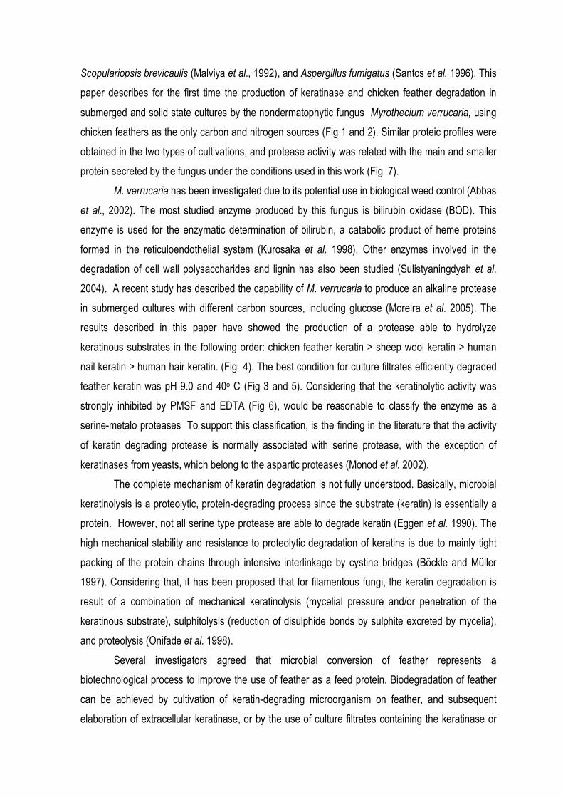
In both, submerged and solid state cultures, M. verrucaria was able to grow in a mineral
medium containing autoclaved chicken feather as the sole carbon and nitrogen sources. Proteolytic
activity was maximally produced after 4 days of cultivation (130 and 250 U/ml, respectively to
submerged and solid state cultures (FIG 1A-1B). The alcalinization of the culture media was
observed together with an increase in the protease level.
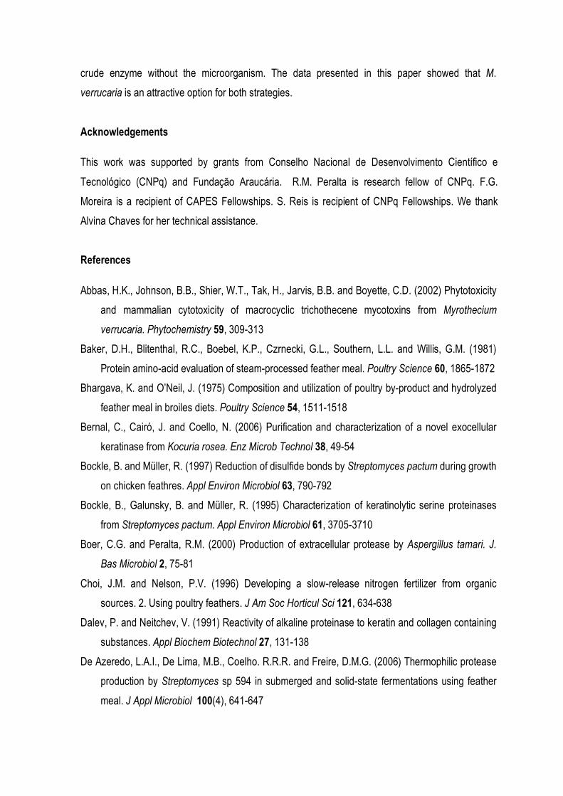
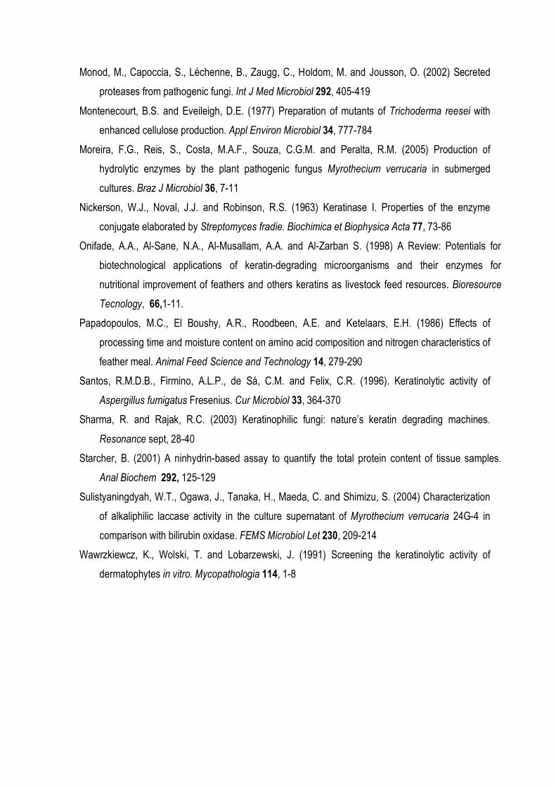
Intact chicken feathers were introduced into 250 ml Erelenmeyer flasks containing 50 ml of
mineral medium and inoculated with M. verrucaria. It was observed that the growth of M. verrucaria
on feathers resulted in almost complete degradation of the feather within 48 h. Barbs and barbules
were completely hydrolyzed while the rachis was partially degraded (Fig. 2).
3.2. Characterization of crude M. verrucaria keratinase
The keratinolytic activity obtained from submerged and solid state cultures was detectable
between 25 and 55o C, showing a maximum at 40o C (Figure 3A). Concerning stability, the
proteases present in the culture broth remained fully active at 5o C for several weeks and more than
24 h when stored at room temperature (25-28o C).
The keratinolytic activity of the culture filtrate from both types of cultivation was detectable
over a wide range of pH values, with an optimum between 9 and 10 (Figure 3B).
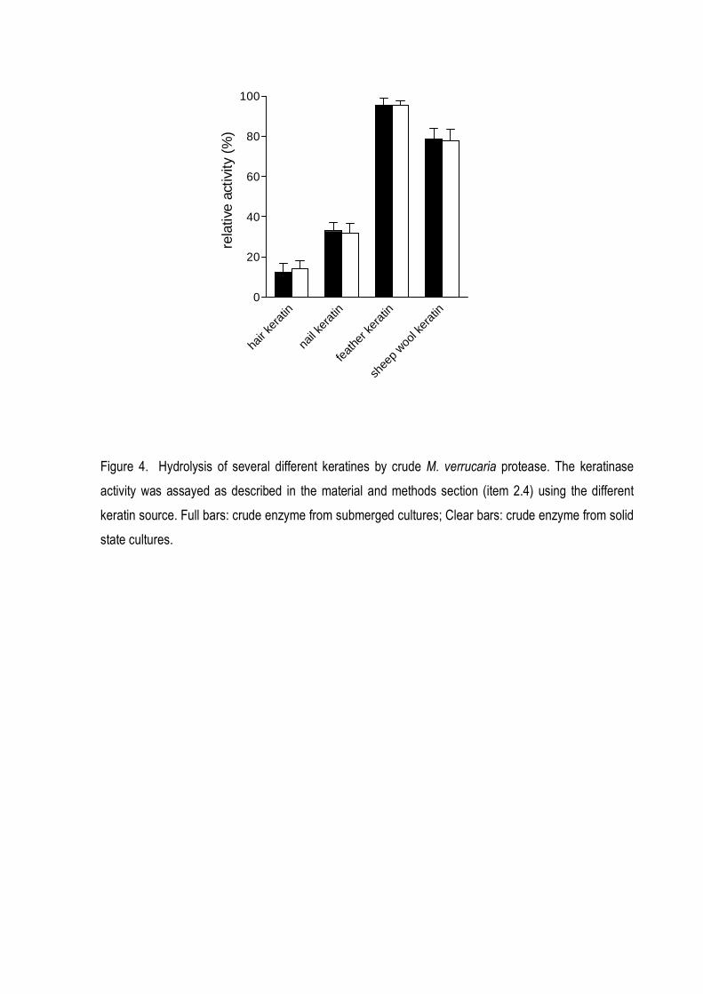
At pH 9.0 and 40o C, the enzyme was able to efficiently hydrolyze keratin from chicken
feather and from sheep wool while human nail and hair keratins were poorly hydrolyzed (Fig 4). The
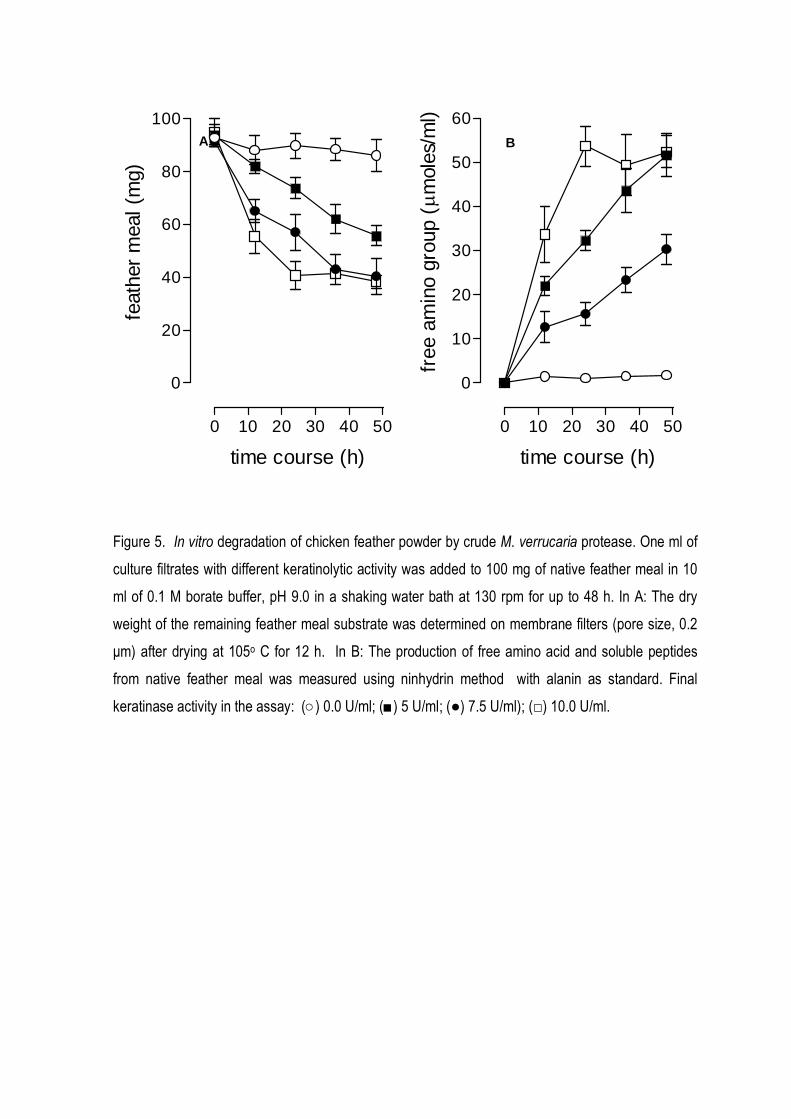
action of keratinase in chicken feather meal reduced its weight to 40% after 48 h (Fig 5A), while the
amount of free amino groups (free aminoacids and peptides) increased in the same period (Fig 5B).
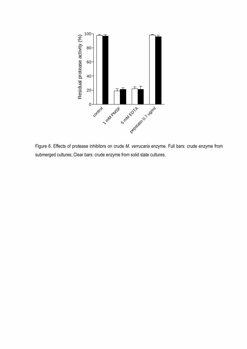
The levels of inhibition on keratinolytic activity by treatment with some proteolytic inhibitors
are shown in Figure 6. The keratinolytic activities were inhibited by PMSF and EDTA, indicating the
presence of serine and metal requiring proteases in both types of cultures.
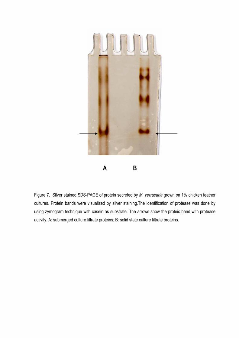
Figure 7 shows the electrophoretic patterns of culture filtrates obtained from submerged and
solid state conditions. A few different proteins were secreted by the fungus under the conditions
used in this study. In both types of cultures, zymogram analysis using casein as substrate identified
one protein with protease activity.
4. Discussion
Keratinases which are able to degrade keratins are mostly isolated from pathogenic
dermatophytes or bacteria (Gradisar et al., 2005, Nickerson et al. 1963, Lin et al. 1992). The ability of
nondermatophytic fungi to degrade native feathers which are composed of 95% protein seems to be
very rare. Some examples of this group include Chrysosporium keratinophilum (Dozie et al. 1994),
Aspergillus oryzae (Farag and Hassan, 2004), Doratmyces microsporus (Gradisar et al. 2000),
Scopulariopsis brevicaulis (Malviya et al., 1992), and Aspergillus fumigatus (Santos et al. 1996). This
paper describes for the first time the production of keratinase and chicken feather degradation in
submerged and solid state cultures by the nondermatophytic fungus Myrothecium verrucaria, using
chicken feathers as the only carbon and nitrogen sources (Fig 1 and 2). Similar proteic profiles were
obtained in the two types of cultivations, and protease activity was related with the main and smaller
protein secreted by the fungus under the conditions used in this work (Fig 7).
M. verrucaria has been investigated due to its potential use in biological weed control (Abbas
et al., 2002). The most studied enzyme produced by this fungus is bilirubin oxidase (BOD). This
enzyme is used for the enzymatic determination of bilirubin, a catabolic product of heme proteins
formed in the reticuloendothelial system (Kurosaka et al. 1998). Other enzymes involved in the
degradation of cell wall polysaccharides and lignin has also been studied (Sulistyaningdyah et al.
2004). A recent study has described the capability of M. verrucaria to produce an alkaline protease
in submerged cultures with different carbon sources, including glucose (Moreira et al. 2005). The
results described in this paper have showed the production of a protease able to hydrolyze
keratinous substrates in the following order: chicken feather keratin > sheep wool keratin > human
nail keratin > human hair keratin. (Fig 4). The best condition for culture filtrates efficiently degraded
feather keratin was pH 9.0 and 40o C (Fig 3 and 5). Considering that the keratinolytic activity was
strongly inhibited by PMSF and EDTA (Fig 6), would be reasonable to classify the enzyme as a
serine-metalo proteases To support this classification, is the finding in the literature that the activity
of keratin degrading protease is normally associated with serine protease, with the exception of
keratinases from yeasts, which belong to the aspartic proteases (Monod et al. 2002).
The complete mechanism of keratin degradation is not fully understood. Basically, microbial
keratinolysis is a proteolytic, protein-degrading process since the substrate (keratin) is essentially a
protein. However, not all serine type protease are able to degrade keratin (Eggen et al. 1990). The
high mechanical stability and resistance to proteolytic degradation of keratins is due to mainly tight
packing of the protein chains through intensive interlinkage by cystine bridges (Böckle and Müller
1997). Considering that, it has been proposed that for filamentous fungi, the keratin degradation is
result of a combination of mechanical keratinolysis (mycelial pressure and/or penetration of the
keratinous substrate), sulphitolysis (reduction of disulphide bonds by sulphite excreted by mycelia),
and proteolysis (Onifade et al. 1998).
Several investigators agreed that microbial conversion of feather represents a
biotechnological process to improve the use of feather as a feed protein. Biodegradation of feather
can be achieved by cultivation of keratin-degrading microorganism on feather, and subsequent
elaboration of extracellular keratinase, or by the use of culture filtrates containing the keratinase or
crude enzyme without the microorganism. The data presented in this paper showed that M.
verrucaria is an attractive option for both strategies.
Acknowledgements
This work was supported by grants from Conselho Nacional de Desenvolvimento Científico e
Tecnológico (CNPq) and Fundação Araucária. R.M. Peralta is research fellow of CNPq. F.G.
Moreira is a recipient of CAPES Fellowships. S. Reis is recipient of CNPq Fellowships. We thank
Alvina Chaves for her technical assistance.
References
Abbas, H.K., Johnson, B.B., Shier, W.T., Tak, H., Jarvis, B.B. and Boyette, C.D. (2002) Phytotoxicity
and mammalian cytotoxicity of macrocyclic trichothecene mycotoxins from Myrothecium
verrucaria. Phytochemistry 59, 309-313
Baker, D.H., Blitenthal, R.C., Boebel, K.P., Czrnecki, G.L., Southern, L.L. and Willis, G.M. (1981)
Protein amino-acid evaluation of steam-processed feather meal. Poultry Science 60, 1865-1872
Bhargava, K. and O’Neil, J. (1975) Composition and utilization of poultry by-product and hydrolyzed
feather meal in broiles diets. Poultry Science 54, 1511-1518
Bernal, C., Cairó, J. and Coello, N. (2006) Purification and characterization of a novel exocellular
keratinase from Kocuria rosea. Enz Microb Technol 38, 49-54
Bockle, B. and Müller, R. (1997) Reduction of disulfide bonds by Streptomyces pactum during growth
on chicken feathres. Appl Environ Microbiol 63, 790-792
Bockle, B., Galunsky, B. and Müller, R. (1995) Characterization of keratinolytic serine proteinases
from Streptomyces pactum. Appl Environ Microbiol 61, 3705-3710
Boer, C.G. and Peralta, R.M. (2000) Production of extracellular protease by Aspergillus tamari. J.
Bas Microbiol 2, 75-81
Choi, J.M. and Nelson, P.V. (1996) Developing a slow-release nitrogen fertilizer from organic
sources. 2. Using poultry feathers. J Am Soc Horticul Sci 121, 634-638
Dalev, P. and Neitchev, V. (1991) Reactivity of alkaline proteinase to keratin and collagen containing
substances. Appl Biochem Biotechnol 27, 131-138
De Azeredo, L.A.I., De Lima, M.B., Coelho. R.R.R. and Freire, D.M.G. (2006) Thermophilic protease
production by Streptomyces sp 594 in submerged and solid-state fermentations using feather
meal. J Appl Microbiol 100(4), 641-647
Dozie, I.N.S., Okeke, C.N. and Unaeze, N.C. (1994) A thermostable, alkaline-active, keratinolytic
proteinase from Chrysosporium keratinophilum. World J Microbiol Biotechnol 10, 563-567.
Eggen, R., Geerming, A., Watts, J. and de Vos, W. (1990) Characterization of pirolysin, a
hyperthermoactive serine protease from the archaebacterium Pyrococcus furiosus. FEMS Lett
71, 17-20
Faheild, S.M.M. and Murad, H.A. (1992) Utilization of polygalacturonase from Myrothecium
verrucaria for maceration of orange pulp. Deustche Lebensmittel-Rundschau 88, 117-119.
Farag, A.M. and Hassan, M.A. (2004) Purification, characterization and immobilization of a
keratinase from Aspergillus oryzae. Enz Microbial Technol 34, 85-93
Filho, E.F.X., Puls, J. and Coughlan, M.P. (1994) Physicochemical and catalytic properties of a low
molecular weight endo 1,4 β xylanase from Myrothecium verrucaria. Enz Microbiol Technol
15,535-540.
Friedrich, J., Gradisar, H., Mandin, D. and Chaumont, J.P. (1999) Screening fungi for synthesis of
keratinolytic enzymes. Let Appl Microbiol 28, 127-130
Govindsamy, V., Gunaratna, K.R. and Balasubramanian, R. (1998). Properties of extracellular
chitinase from Myrothecium verrucaria, an antagonist to the groundnut rust Puccinia arachidis.
Can J Plant Patho, 20, 62-68.
Gradisar, H., kern, S. and Friedrich, J. (2000) Keratinase of Doratomyces microsporus. Appl
Microbiol Biotechnol 53, 196-200
Gradisar, H., Friedrich, J., Krizaj, I. and Jerala, R. (2005) Similarities and specificities of fungal
keratinolytic proteases: comparison of keratinases of Paecilomyces marquandii and
Doratomyces microsporus to some known proteases. Appl Environ Microbiol 71, 3420-3426
Kurosaka, K., Senba, S., Tsubota, H. and Kondo, H. (1998) A new enzymatic assay for selectiviely
measuring conjugated bilirubin concentration in serum with use of bilirubin oxidase. Clinica
Chimica Acta 269, 125-136
Laemmli, U.K. (1970) Cleavage of structural proteins during the assembly of the head of
bacteriophage T4. Nature 227, 680-685
Letourneau, F., Soussotte, V., Bressollier, P., Branland, P. and Verneuil, M. (1998) Keratinolytic
activity of a Streptomyces sp. SK 1-02: a new isolate strain. Let Appl Microbiol 26,77-80
Lin, X., Lee, C., Casale, E. and Shih, J. (1992) Purification and characterization of a keratinase from
a feather-degrading Bacillus licheniformis strain. Appl Environ Microbiol 58, 3271-3275
Malviya, H.K., Rajak, R.C. and Hasija, S.K. (1992) Purification and partial characterization of two
extracellular keratinases of Scopulariopsis brevicaulis. Mycopathologia 119, 161-165
Monod, M., Capoccia, S., Léchenne, B., Zaugg, C., Holdom, M. and Jousson, O. (2002) Secreted
proteases from pathogenic fungi. Int J Med Microbiol 292, 405-419
Montenecourt, B.S. and Eveileigh, D.E. (1977) Preparation of mutants of Trichoderma reesei with
enhanced cellulose production. Appl Environ Microbiol 34, 777-784
Moreira, F.G., Reis, S., Costa, M.A.F., Souza, C.G.M. and Peralta, R.M. (2005) Production of
hydrolytic enzymes by the plant pathogenic fungus Myrothecium verrucaria in submerged
cultures. Braz J Microbiol 36, 7-11
Nickerson, W.J., Noval, J.J. and Robinson, R.S. (1963) Keratinase I. Properties of the enzyme
conjugate elaborated by Streptomyces fradie. Biochimica et Biophysica Acta 77, 73-86
Onifade, A.A., Al-Sane, N.A., Al-Musallam, A.A. and Al-Zarban S. (1998) A Review: Potentials for
biotechnological applications of keratin-degrading microorganisms and their enzymes for
nutritional improvement of feathers and others keratins as livestock feed resources. Bioresource
Tecnology, 66,1-11.
Papadopoulos, M.C., El Boushy, A.R., Roodbeen, A.E. and Ketelaars, E.H. (1986) Effects of
processing time and moisture content on amino acid composition and nitrogen characteristics of
feather meal. Animal Feed Science and Technology 14, 279-290
Santos, R.M.D.B., Firmino, A.L.P., de Sá, C.M. and Felix, C.R. (1996). Keratinolytic activity of
Aspergillus fumigatus Fresenius. Cur Microbiol 33, 364-370
Sharma, R. and Rajak, R.C. (2003) Keratinophilic fungi: nature’s keratin degrading machines.
Resonance sept, 28-40
Starcher, B. (2001) A ninhydrin-based assay to quantify the total protein content of tissue samples.
Anal Biochem 292, 125-129
Sulistyaningdyah, W.T., Ogawa, J., Tanaka, H., Maeda, C. and Shimizu, S. (2004) Characterization
of alkaliphilic laccase activity in the culture supernatant of Myrothecium verrucaria 24G-4 in
comparison with bilirubin oxidase. FEMS Microbiol Let 230, 209-214
Wawrzkiewcz, K., Wolski, T. and Lobarzewski, J. (1991) Screening the keratinolytic activity of
dermatophytes in vitro. Mycopathologia 114, 1-8
0 1 2 3 4 5 6 7
0
20
40
60
80
100
120
140
5
6
7
8
9
10
time course (days)
prot
ease
act
ivity
(U
/ml)
pH
0 1 2 3 4 5 6 7
0
50
100
150
200
250
300
5
6
7
8
9
10
time course (days)
prot
ease
act
ivity
(U
/ml)
pH
A
B
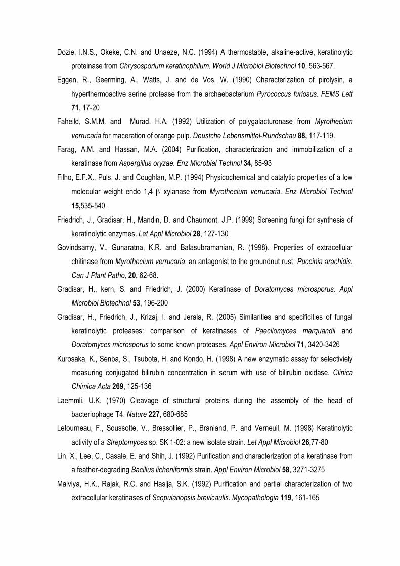
Figure 1. Time course of protease production by M. verrucaria in submerged (A) and solid state
cultures (B). The submerged cultures were developed at 120 rpm and 28o C for up 6 days in mineral
medium containing chicken feather (1.0% w/v) as the only carbon and nitrogen sources. In B: solid
state cultures. Protease activity was determined in the culture filtrates using casein as substrate. The
results represent the media ± SD of three independent experiments. (●) protease activity; (○) pH
Figure 2. Degradation of chicken feathers by M. verrucaria in submerged culture after 0 (A), 48 (B)
and 72 h (C) of cultivation.
A B C

20 30 40 50 60 70 80
0
20
40
60
80
100
120
temperature (oC)
Ker
atin
ase
activ
ity (
%)
4 5 6 7 8 9 101112
0
20
40
60
80
100
120
pH
Ker
atin
ase
activ
ity (
%)A B
Figure 3. Effect of temperature (A) and pH (B) on crude M. verrucaria keratinolytic activity.
hair
kera
tin
nail k
erat
in
feat
her k
erat
in
shee
p woo
l ker
atin
0
20
40
60
80
100
rela
tive
activ
ity (
%)
Figure 4. Hydrolysis of several different keratines by crude M. verrucaria protease. The keratinase
activity was assayed as described in the material and methods section (item 2.4) using the different
keratin source. Full bars: crude enzyme from submerged cultures; Clear bars: crude enzyme from solid
state cultures.
0 10 20 30 40 50
0
10
20
30
40
50
60
time course (h)
free
am
ino
grou
p (µ
mol
es/m
l)0 10 20 30 40 50
0
20
40
60
80
100
time course (h)
feat
her
mea
l (m
g)A B
Figure 5. In vitro degradation of chicken feather powder by crude M. verrucaria protease. One ml of
culture filtrates with different keratinolytic activity was added to 100 mg of native feather meal in 10
ml of 0.1 M borate buffer, pH 9.0 in a shaking water bath at 130 rpm for up to 48 h. In A: The dry
weight of the remaining feather meal substrate was determined on membrane filters (pore size, 0.2
µm) after drying at 105o C for 12 h. In B: The production of free amino acid and soluble peptides
from native feather meal was measured using ninhydrin method with alanin as standard. Final
keratinase activity in the assay: (○) 0.0 U/ml; (■) 5 U/ml; (●) 7.5 U/ml); (□) 10.0 U/ml.
cont
rol
1 m
M P
MSF
5 m
M E
DTA
peps
tatin
0.7
ug/
ml
0
20
40
60
80
100
Res
idua
l pro
teas
e ac
tivity
(%
)
Figure 6. Effects of protease inhibitors on crude M. verrucaria enzyme. Full bars: crude enzyme from
submerged cultures; Clear bars: crude enzyme from solid state cultures.
A B
Figure 7. Silver stained SDS-PAGE of protein secreted by M. verrucaria grown on 1% chicken feather
cultures. Protein bands were visualized by silver staining.The identification of protease was done by
using zymogram technique with casein as substrate. The arrows show the proteic band with protease
activity. A: submerged culture filtrate proteins; B: solid state culture filtrate proteins.
ARTIGO 3
PURIFICATION AND PARTIAL CHARACTERIZATION OF A
KERATINOLYTIC PROTEASE FROM THE NON DERMATOPHYTIC
FUNGUS Myrothecium verrucaria
Fabiana Guillen Moreira and Rosane Marina Peralta
Departamento de Bioquímica, Universidade Estadual de Maringá, Av. Colombo, 5790, 87020-900,
Maringá, PR, Brazil.
ABSTRACT
A Myrothecium verrucaria keratinolytic protease was purified to apparent electrophoretic
homogeneity using an unique chromatographic step with a high yield of 62%. The molecular mass
was estimated to be 22 kDa (SDS-PAGE analysis) or 23 kDa (gel chromatography analysis),
suggesting a monomeric structure. The enzyme was stable in a broad range of pH (5.0-12.0) and
temperature up to 45o C, being optimally active at 40oC and pH 8.0-9.0. It was strongly inhibited by
PMSF and partially inhibited by EDTA, which suggests to be a metallo serine protease. The purified
enzyme was able to hydrolyze chicken feather meal producing free amino groups. The efficiency of
hydrolysis was improved when the keratin was previously treated with reducing agents.
KEY-WORDS: Myrothecium verrucaria, protease, keratinase, chicken feather.
INTRODUCTION
Keratins are the most abundant proteins in epithelial cells of vertebrates and represent the
major constituents of skin and its appendages such as nail, hair, feather, and wool (Gradisar et al.
2005). Keratins comprise long polypeptide chains stabilized by disulfide bonds, hydrophobic
interactions, and hydrogen bonds what result in insolubility and resistance to the activity of non-
substrate-specific proteases (Safranek and Goos 1982).
A group of proteolytic enzymes which are able to hydrolyze insoluble keratins more
efficiently than other proteases are called keratinases and are produced specially by microorganisms
(Onifade et al. 1998). Secretion of keratinolytic enzymes is associated with dermatophytic fungi, for
which keratin is the major substrate, but there are some reports describing the production,
purification and characterization of the keratinases from bacteria and non dermatophytic fungus
(Friedrich et al. 1999, Monod et al. 2002, Safranek and Goos 1982, Sharma and Rajac 2003).
The degradation of keratinous material is important medically and agriculturally (Shih
1993, Matsumoto 1996). Feathers are poultry by-product rich in β-keratin (Onifade et al. 1998).
World-wide poultry processing plants produce millions of tons of feathers as a waste product
annually (Santos et al. 1996) and their content in keratin is largely responsible for their high degree
of recalcitrance. However, they also represent a potentially valuable source of protein as animal
feedstock if keratinolysis can be achieved (Shih 1993). In addition to this, keratin partial or
completely hydrolyzed by keratinases, could be used to the development of slow nitrogen fertilizers,
glues and biodegradable films (Dalev and Neitchev 1991, Choi and Nelson 1996).
The fungus Myrothecium verrucaria is a filamentous fungi able to produce several hydrolytic
enzymes including endochitinase (Govindsamy et al. 1998), xylanases (Filho et al., 1994), and
pectinases (Faheild and Murad, 1992). Recently we described its capability to produce a protease
with keratinase activity when cultivated in submerged and solid state cultures using chicken feathers
as the only carbon and nitrogen source (Moreira et al. submitted). In this paper, we described the
purification and partial biochemical characterization of a extracellular keratinase obtained from
submerged culture of M. verrucaria with chicken feather as the only carbon and nitrogen source.
2. MATERIAL AND METHODS
2. Materials and methods
2.1. Microorganism.
Myrothecium verrucaria (Albertini and Schwein) Ditmar:Fr CCT 1886 was obtained from the
Collection Culture of Fundação André Tosello, Campinas, SP, and it was maintained on potato
dextrose agar.
2.2. Pretreatment of the feathers.
White chicken feathers were supplied by a local industry. Freshly plucked wet feathers were washed
extensively with water and detergent. Wet feathers were dried in a ventilated oven at 40o C for 72 h.
The feathers were then milled in a ball mill and passed through a small-mesh grid to remove coarse
particles.
2.3. Culture conditions
One x 109 M. verrucaria spores were transferred to 250 ml Erlenmeyer flasks containing 50 ml of
mineral media (Montenecourt and Eveileigh 1977) and 1.0% (w/v) chicken feathers as substrate. The
medium was previously sterilized by autoclaving at 121o C for 15 min. The cultures were incubated at
28o C on a rotary shaker at 120 rpm. After 4 days of cultivation, the culture broth was filtered
thorough Whatman no 1 filter paper to retain non-degraded feathers, and centrifuged (5000 x g for 20
min at 4o C). The supernatant was recovered and used for further purification.
2.4. Enzyme assay.
Keratinolytic activity was assayed by the method of Nickerson et al. (1963) using chicken feather
meal as substrate. The reaction mixture containing 10 mg of substrate, 1.0 ml of borate buffer (0.1
M, pH 9.0), and 1.0 ml of a suitable diluted culture filtrate was incubated under agitation using a
stirring bar for 1 h at 40o C. The reaction was stopped by boiling and the mixture was filtered
through filter paper. The production of free amino group was estimated using a reaction with
ninhydrin reagent and alanin as the standard (Starcher, 2001). The protease activity of the culture
filtrates was assayed using 1.0% (w/v) casein in 0.1 M borate buffer, pH 9.0, as substrate (Boer and
Peralta, 2000). One (1.0) ml casein was incubated at 40o C for 1 h with 1.0 ml of a suitable diluted
culture filtrate. The reaction was stopped by the addition of 2.0 ml of 0.6 M trichloroacetic acid. After
15 min in an ice bath, the mixtures were mixing on a vortex mixer, and the tubes were centrifuged at
3,000 rpm for 10 min. The tyrosine released was estimated by Lowry’s method using a tyrosine
standard curve. A unit of enzyme activity was defined as the amount of enzyme producing 1 µg of
tyrosine per min.
2.5. Estimation of protein
Protein was estimated by dye-binding method (Bradford 1982), using bovine serum albumin as
standard protein. In the column chromatography elution, the amount of protein was measured in
terms of the absorbance at 280 nm. The specific activity was expressed as the enzymatic activity per
mg of protein.
2.6. Enzyme purification
All purification steps were carried out below 15o C unless otherwise specified. The material
containing crude protease was centrifuged at 15,000 g for 30 min to remove particulate material. The
culture filtrate was concentrated about 10-fold by ultra filtration (Amicon YM-10, 10 kDa cut-off). After
removal of insoluble substances by centrifugation, the material was dialyzed against water and
concentrated by freeze-drying. The material was re suspended in 1.0 ml of 50 mM borate buffer, pH
9.0 and applied to a Sephadex G-100 gel filtration (90 cm x 25 mm) equilibrated and eluted with 50
mM borate buffer (pH 9.0) at a flow rate of 10 ml/h. The protease active fractions were pooled and
concentrated by freeze-drying, and stored at -10o C.
2.7. Partial characterization of purified protease
2.7.1. Effect of temperature and pH on keratinolytic activity and stabilityy.
The effect of temperature on keratinolytic activity was carried out at temperature ranging from 25 to
60o C. The effect of pH on the activity of enzyme was determined in a series of McIlvaine’s buffers
with pH values from 5.0 to 8.3, 0.1 M tris-HCl buffer (pH 8.0-9.5) and 0.1 M borate buffer (pH 8.5-
12.0). The thermo stability and pH stability was assessed of M. verrucaria keratinase, incubating
aliquots of enzymes at different temperatures (between 5 and 60o C) for different times, and stored
at different pH value (between pH 5 and 12) for 12 h. After the treatments, the samples were
assayed for residual keratinolytic activity.
2.7.2. Effect of protease inhibitors.
To study the influence of protease inhibitors, purified protease was pre incubated with the following
protease inhibitors: phenylmethanesulphonyl fluoride (PMSF, 1 mM), EDTA (5 mM), pepstatin A (0.7
µg/ml), iodoacetamide (0.05 mM), dithiothreitol (DTT, 1 mM), β-mercaptoethanol (5 mM); all of
them in 0.1 M borate buffer (pH 9.0) for 15 min at 30o C. Keratinase and protease activities were
determined as standard conditions.
2.7.3. Effect of other chemicals on the keratinolytic activity
The effects of solvents and detergents on keratinase and protease activities were tested by
incubating purified enzyme in 0.1 M of borate buffer (pH 9.0) containing, isopropanol, methanol,
ethanol, buthanol, glycerol, acetonitrile, Triton X-100, SDS, Tween 80 and NaN3 were incubated for
15 min at 30o C. The enzymatic assay was started by adding the substrate and enzyme activity was
measured as described above. To determine the effect of cations on keratinase and protease
activities, the purified enzyme was incubated in the presence of each cation (as chloride salt) at 5.0
mM.
2.7.4. Electrophoretic analysis and determination of molecular mass.
The molecular mass of the purified protease was estimated under denaturing conditions
electrophoresis. SDS-PAGE was performed on 10% polyacrilamide gel (Laemmli 1970). The
following Mr standards (MW-70 kit-Sigma) were used: bovine serum albumin (66 kDa), ovoalbumin
(45 kDa), pepsin (34.7 kDa), trypsinogen (24 kDa), and β-lactoglobulin (18. kDa). Protein bands were
visualized by silver staining. The protease molecular mass was also estimated by gel filtration
chromatography using Sephadex G-100 equilibrated with 50 mM borate buffer, pH 9.0 plus 0.1 M
NaCl. The column was calibrated with standards proteins (MWGF-70 kit-Sigma).
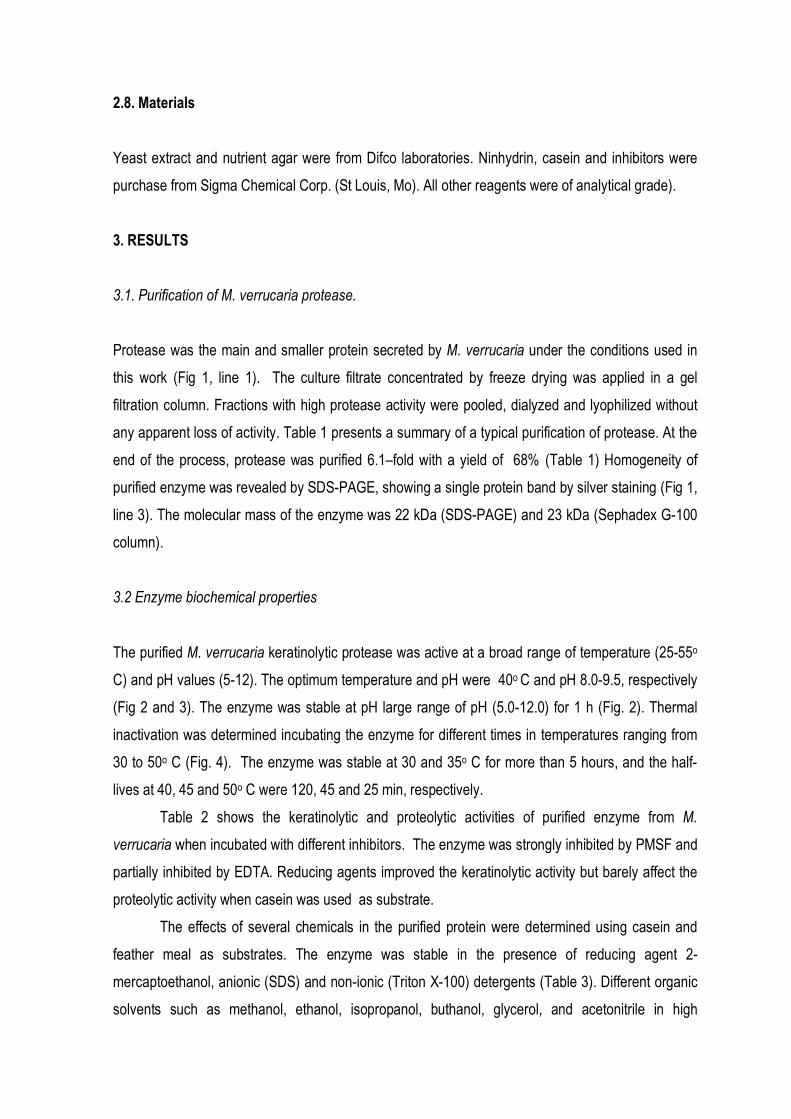
2.8. Materials
Yeast extract and nutrient agar were from Difco laboratories. Ninhydrin, casein and inhibitors were
purchase from Sigma Chemical Corp. (St Louis, Mo). All other reagents were of analytical grade).
3. RESULTS
3.1. Purification of M. verrucaria protease.
Protease was the main and smaller protein secreted by M. verrucaria under the conditions used in
this work (Fig 1, line 1). The culture filtrate concentrated by freeze drying was applied in a gel
filtration column. Fractions with high protease activity were pooled, dialyzed and lyophilized without
any apparent loss of activity. Table 1 presents a summary of a typical purification of protease. At the
end of the process, protease was purified 6.1–fold with a yield of 68% (Table 1) Homogeneity of
purified enzyme was revealed by SDS-PAGE, showing a single protein band by silver staining (Fig 1,
line 3). The molecular mass of the enzyme was 22 kDa (SDS-PAGE) and 23 kDa (Sephadex G-100
column).
3.2 Enzyme biochemical properties
The purified M. verrucaria keratinolytic protease was active at a broad range of temperature (25-55o
C) and pH values (5-12). The optimum temperature and pH were 40o C and pH 8.0-9.5, respectively
(Fig 2 and 3). The enzyme was stable at pH large range of pH (5.0-12.0) for 1 h (Fig. 2). Thermal
inactivation was determined incubating the enzyme for different times in temperatures ranging from
30 to 50o C (Fig. 4). The enzyme was stable at 30 and 35o C for more than 5 hours, and the half-
lives at 40, 45 and 50o C were 120, 45 and 25 min, respectively.
Table 2 shows the keratinolytic and proteolytic activities of purified enzyme from M.
verrucaria when incubated with different inhibitors. The enzyme was strongly inhibited by PMSF and
partially inhibited by EDTA. Reducing agents improved the keratinolytic activity but barely affect the
proteolytic activity when casein was used as substrate.
The effects of several chemicals in the purified protein were determined using casein and
feather meal as substrates. The enzyme was stable in the presence of reducing agent 2-
mercaptoethanol, anionic (SDS) and non-ionic (Triton X-100) detergents (Table 3). Different organic
solvents such as methanol, ethanol, isopropanol, buthanol, glycerol, and acetonitrile in high
concentrations had a low significant influence on the keratinolytic and proteolytic activities. The most
divalente cations tested in concentrations as high as 5 mM, had not influence on the keratinolytic and
proteolytic activities. The enzyme was inhibited by Ag+, Cd+, Pb+ and Hg+. The enzyme was not
affected by sodium azide.
At pH 9.0 and 40o C, the purified enzyme was able to hydrolyze efficiently keratin from
chicken feather meal, producing soluble free amino group molecules (Fig 5). When the feather meal
was previously reduced by 2-mercaptoethanol, the efficiency of hydrolysis was higher.
Figure 6 show the action of purified keratinase on intact feather. In vitro, keratinase altered
the structure of intact feather, but the degradation of feather was not efficient than that obtained in
vivo, when the feather was used as substrate for the growth of the fungus.
4. DISCUSSION
In this paper we described the purification and partial biochemical characterization of M.
verrucaria protease with keratinolytic activity produced in submerged cultures using chicken feather
meal as the only substrate. The enzyme was purified 9.29 fold to a specific activity of 12,851.85
U/mg (Table 1). By SDS-PAGE, a single protein band was obtained (Fig 1). The enzyme was a
monomeric protein with molecular mass of 22 kDa and optimally active at pH 8.0-9.0 and 40o C
(Table 2, Fig 1). Most keratinases have some common characteristics despite their different origins.
They belong mainly to the extracellular serine proteases, with the exception of keratinases from
yeasts, which belong to the aspartic proteases (Monod et al. 2002). The molecular masses of these
type or protease range from 20 kDa to 60 kDa. They are mostly active in alkaline environments, with
optimal activity at temperature up to 50o C (Gradisar et al. 2005).
The analysis of the effect of protease inhibitors (Table 3) showed that purified enzyme was
totally inhibited by the serine protease inhibitor PMSF and partially by EDTA. The maximum
increase of keratinolytic activity was observed in the presence of the reducing agents DTT and βME.
Since DTT and βME are know to cleave disulphide bridges, an influence either on enzyme or on
substrate was possible. However, the reducing agents improved the enzyme activity only when the
substrate was keratin and the activity of enzyme remained unchanged when casein was the
substrate. This result permits to conclude that the reducing agents acted on the keratin and not on
the enzymes. In fact a more efficient hydrolysis of feather meal could be observed when the
substrate was previously reduced by the addition of βME (Fig.5). The reduction of disulfide bonds
improved significantly the keratin degradation. However, the results presented in this paper did not
support the capability of purified protease from M. verrucaria efficiently degrade native feather (Fig
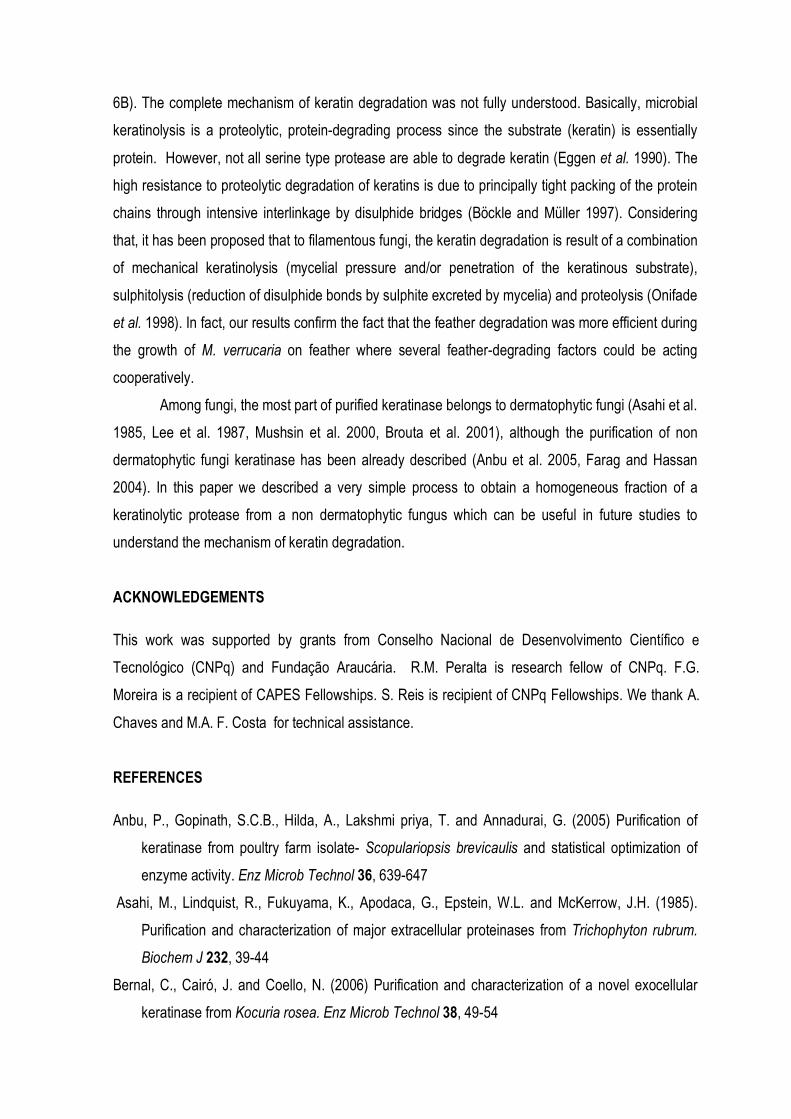
6B). The complete mechanism of keratin degradation was not fully understood. Basically, microbial
keratinolysis is a proteolytic, protein-degrading process since the substrate (keratin) is essentially
protein. However, not all serine type protease are able to degrade keratin (Eggen et al. 1990). The
high resistance to proteolytic degradation of keratins is due to principally tight packing of the protein
chains through intensive interlinkage by disulphide bridges (Böckle and Müller 1997). Considering
that, it has been proposed that to filamentous fungi, the keratin degradation is result of a combination
of mechanical keratinolysis (mycelial pressure and/or penetration of the keratinous substrate),
sulphitolysis (reduction of disulphide bonds by sulphite excreted by mycelia) and proteolysis (Onifade
et al. 1998). In fact, our results confirm the fact that the feather degradation was more efficient during
the growth of M. verrucaria on feather where several feather-degrading factors could be acting
cooperatively.
Among fungi, the most part of purified keratinase belongs to dermatophytic fungi (Asahi et al.
1985, Lee et al. 1987, Mushsin et al. 2000, Brouta et al. 2001), although the purification of non
dermatophytic fungi keratinase has been already described (Anbu et al. 2005, Farag and Hassan
2004). In this paper we described a very simple process to obtain a homogeneous fraction of a
keratinolytic protease from a non dermatophytic fungus which can be useful in future studies to
understand the mechanism of keratin degradation.
ACKNOWLEDGEMENTS
This work was supported by grants from Conselho Nacional de Desenvolvimento Científico e
Tecnológico (CNPq) and Fundação Araucária. R.M. Peralta is research fellow of CNPq. F.G.
Moreira is a recipient of CAPES Fellowships. S. Reis is recipient of CNPq Fellowships. We thank A.
Chaves and M.A. F. Costa for technical assistance.
REFERENCES
Anbu, P., Gopinath, S.C.B., Hilda, A., Lakshmi priya, T. and Annadurai, G. (2005) Purification of
keratinase from poultry farm isolate- Scopulariopsis brevicaulis and statistical optimization of
enzyme activity. Enz Microb Technol 36, 639-647
Asahi, M., Lindquist, R., Fukuyama, K., Apodaca, G., Epstein, W.L. and McKerrow, J.H. (1985).
Purification and characterization of major extracellular proteinases from Trichophyton rubrum.
Biochem J 232, 39-44
Bernal, C., Cairó, J. and Coello, N. (2006) Purification and characterization of a novel exocellular
keratinase from Kocuria rosea. Enz Microb Technol 38, 49-54
Bockle, B. and Müller, R. (1997) Reduction of disulfide bonds by Streptomyces pactum during growth
on chicken feathres. Appl Environ Microbiol 63, 790-792
Boer, C.G. and Peralta, R.M. (2000) Production of extracellular protease by Aspergillus tamari. J.
Bas Microbiol 2, 75-81
Bradford, M.M. (1982) A rapid and sensitive method for the quantification of microgram quantities of
protein utilizing the principle of protein-dye binding. Anal. Biochem. 72, 248-254
Brouta, F., Descamps, F., Fett, T., Losson, B., Gerday, C. and Mignon, B. (2001) Purification and
characterization of a 43.5 kDa keratinolytic metalloprotease from Microsporum canis. Med
Mycol 39, 269-275
Choi, J.M. and Nelson, P.V. (1996) Developing a slow-release nitrogen fertilizer from organic
sources. 2. Using poultry feathers. J Am Soc Horticul Sci 121, 634-638
Dalev, P. and Neitchev, V. (1991) Reactivity of alkaline proteinase to keratin and collagen containing
substances. Appl Biochem Biotechnol 27, 131-138
Faheild, S.M.M. and Murad, H.A. (1992) Utilization of polygalacturonase from Myrothecium
verrucaria for maceration of orange pupl. Deustche Lebensmittel-Rundschau 88, 117-119.
Farag, A.M. and Hassan, M.A. (2004) Purification, characterization and immobilization of a
keratinase from Aspergillus oryzae. Enz Microbial Technol 34, 85-93
Filho, E.F.X., Puls, J. and Coughlan, M.P. (1994) Physicochemical and catalytic properties of a low
molecular weight endo 1,4 β xylanase from Myrothecium verrucaria. Enz Microbiol Technol
15,535-540.
Friedrich, J., Gradisar, H., Mandin, D. and Chaumont, J.P. (1999) Screening fungi for synthesis of
keratinolytic enzymes. Let Appl Microbiol 28, 127-130
Govindsamy, V., Gunaratna, K.R. and Balasubramanian, R. (1998). Properties of extracellular
chitinase from Myrothecium verrucaria, an antagonist to the groundnut rust Puccinia arachidis.
Can J Plant Patho, 20, 62-68.
Gradisar, H., Friedrich, J., Krizaj, I. and Jerala, R. (2005) Similarities and specificities of fungal
keratinolytic proteases: comparison of keratinases of Paecilomyces marquandii and
Doratomyces microsporus to some known proteases. Appl Environ Microbiol 71, 3420-3426
Laemmli, U.K. (1970) Cleavage of structural proteins during the assembly of the head of
bacteriophage T4. Nature 227, 680-685
Lee, K.H., Park, K.K., Park, S.H. and Lee, J.B. (1987) Isolation, purification and characterization of
keratinolytic proteinase from Microsporum canis Yonsei. Med J, 28, 31-38
Monod, M., Capoccia, S., Léchenne, B., Zaugg, C., Holdom, M. and Jousson, O. (2002) Secreted
proteases from pathogenic fungi. Int J Med Microbiol 292, 405-419
Montenecourt, B.S. and Eveileigh, D.E. (1977) Preparation of mutants of Trichoderma reesei with
enhanced cellulose production. Appl Environ Microbiol 34, 777-784
Muhsin, T.M. and Aubaid, A.H. (2000) Partial purification and some biochemical characteristics of
exocellular keratinase from Trichophyton mentagrophytes var erinacei. Mycopathologia 150,
121-125
Moreira, F.G., Reis, S., Costa, M.A.F., Souza, C.G.M. and Peralta, R.M. Degradation of chicken
feathers by Myrothecium verruaria. J. Appl. Biotechnol. Submitted.
Nickerson, W.J., Noval, J.J. and Robinson, R.S. (1963) Keratinase I. Properties of the enzyme
conjugate elaborated by Streptomyces fradie. Biochimica et Biophysica Acta 77, 73-86
Onifade, A.A., Al-Sane, N.A., Al-Musallam, A.A. and Al-Zarban S. (1998) A Review: Potentials for
biotechnological applications of keratin-degrading microorganisms and their enzymes for
nutritional improvement of feathers and others keratins as livestock feed resources. Bioresource
Tecnology, 66,1-11.
Safranek, W.W. and Goos, R.D. (1982) Degradation of wool by saprophytic fungi. Can J Microbiol 28,
137-140
Santos, R.M.D.B., Firmino, A.L.P., de Sá, C.M. and Felix, C.R. (1996). Keratinolytic activity of
Aspergillus fumigatus Fresenius. Cur Microbiol 33, 364-370
Sharma, R. and Rajak, R.C. (2003) Keratinophilic fungi: nature’s keratin degrading machines.
Resonance sept, 28-40
Shih, J.C.H. (1993) recent development in poultry waste and feather utilization – a review. Poultry
Science 72, 1617-1620
Starcher, B. (2001). A ninhydrin-based assay to quantify the total protein content of tissue samples.
Anal Biochem 292, 125-129
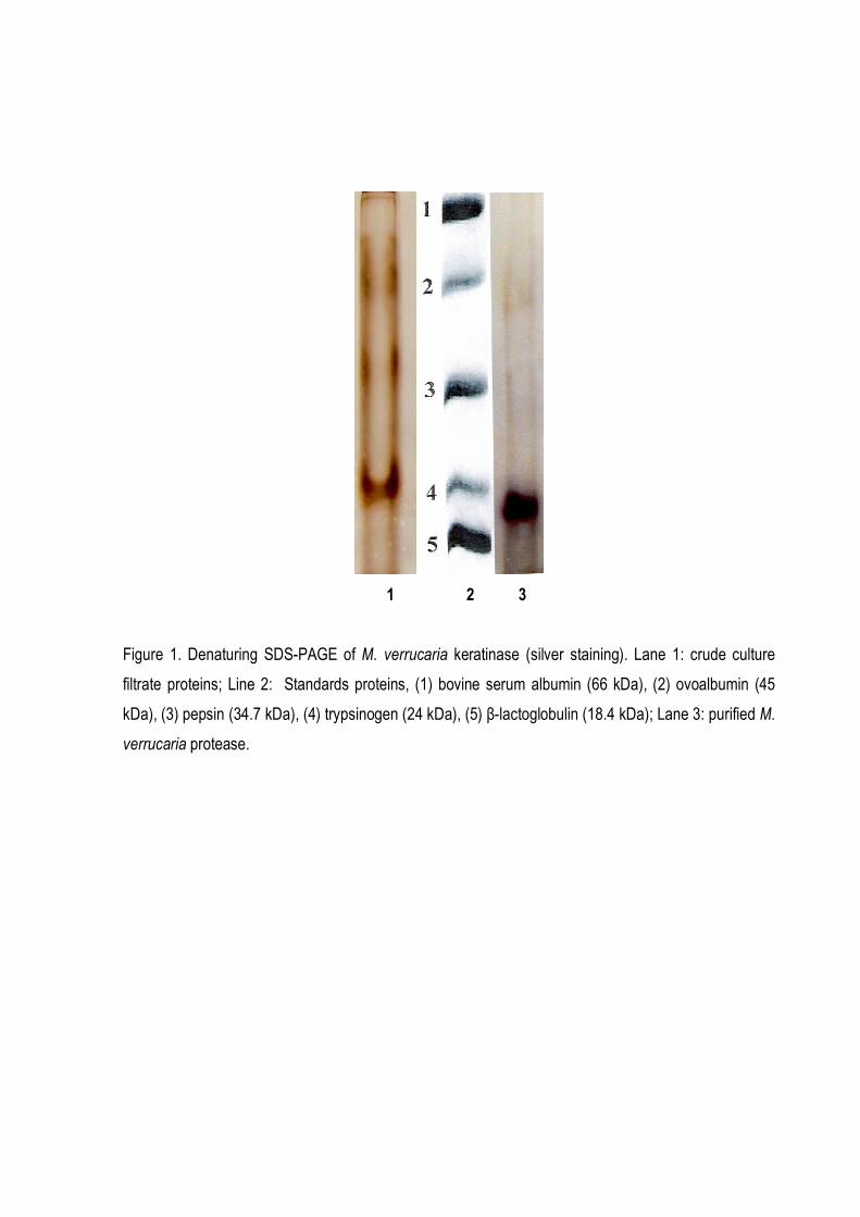
1 2 3
Figure 1. Denaturing SDS-PAGE of M. verrucaria keratinase (silver staining). Lane 1: crude culture
filtrate proteins; Line 2: Standards proteins, (1) bovine serum albumin (66 kDa), (2) ovoalbumin (45
kDa), (3) pepsin (34.7 kDa), (4) trypsinogen (24 kDa), (5) β-lactoglobulin (18.4 kDa); Lane 3: purified M.
verrucaria protease.
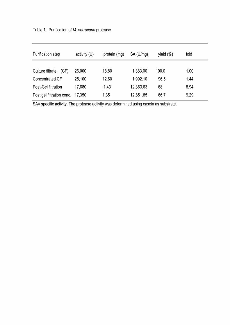
Table 1. Purification of M. verrucaria protease
Purification step activity (U) protein (mg) SA (U/mg) yield (%) fold
Culture filtrate (CF) 26,000 18.80 1,383.00 100.0 1.00
Concentrated CF 25,100 12.60 1,992.10 96.5 1.44
Post-Gel filtration 17,680 1.43 12,363.63 68 8.94
Post gel filtration conc. 17,350 1.35 12,851.85 66.7 9.29
SA= specific activity. The protease activity was determined using casein as substrate.
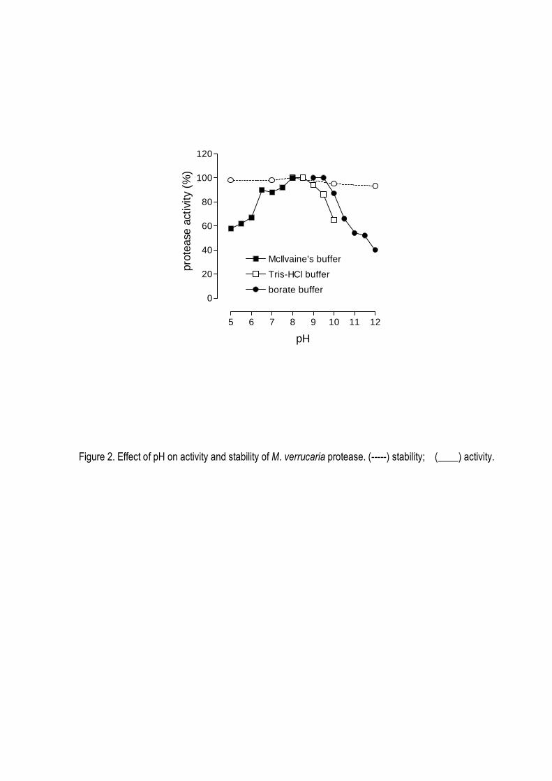
5 6 7 8 9 10 11 12
0
20
40
60
80
100
120
McIlvaine's buffer
Tris-HCl buffer
borate buffer
pH
prot
ease
act
ivity
(%
)
Figure 2. Effect of pH on activity and stability of M. verrucaria protease. (-----) stability; (____) activity.
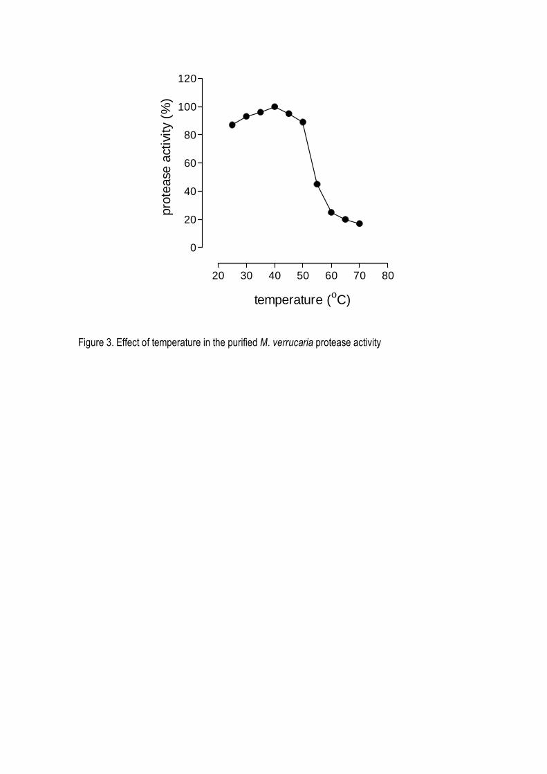
20 30 40 50 60 70 80
0
20
40
60
80
100
120
temperature (oC)
prot
ease
act
ivity
(%
)
Figure 3. Effect of temperature in the purified M. verrucaria protease activity
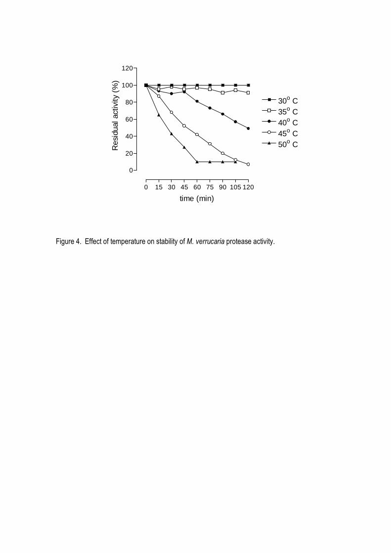
0 15 30 45 60 75 90 105 120
0
20
40
60
80
100
120
30o C35o C40o C45o C50o C
time (min)
Res
idua
l act
ivity
(%
)
Figure 4. Effect of temperature on stability of M. verrucaria protease activity.
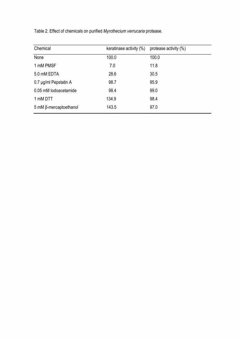
Table 2. Effect of chemicals on purified Myrothecium verrucaria protease.
Chemical keratinase activity (%) protease activity (%)
None 100.0 100.0
1 mM PMSF 7.0 11.8
5.0 mM EDTA 28.6 30.5
0.7 µg/ml Pepstatin A 98.7 95.9
0.05 mM Iodoacetamide 98.4 99.0
1 mM DTT 134.9 98.4
5 mM β-mercaptoethanol 143.5 97.0
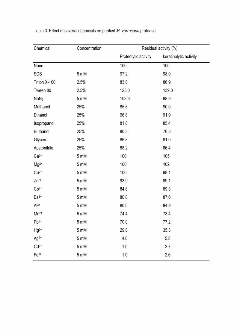
Table 3. Effect of several chemicals on purified M. verrucaria protease
Chemical Concentration Residual activity (%)
Proteolytic activity keratinolytic activity
None 100 100
SDS 5 mM 97.2 98.0
Triton X-100 2.5% 83.8 86.9
Tween 80 2.5% 125.0 139.0
NaN3 5 mM 103.6 98.9
Methanol 25% 85.8 90.0
Ethanol 25% 96.8 91.9
Isopropanol 25% 81.8 85.4
Buthanol 25% 80.3 76.8
Glycerol 25% 86.8 81.0
Acetonitrile 25% 88.2 86.4
Ca2+ 5 mM 100 105
Mg2+ 5 mM 100 102
Cu2+ 5 mM 100 98.1
Zn2+ 5 mM 93.9 89.1
Co2+ 5 mM 84.8 89.3
Ba2+ 5 mM 80.8 87.6
Al3+ 5 mM 80.0 84.9
Mn2+ 5 mM 74.4 73.4
Pb2+ 5 mM 70.0 77.2
Hg2+ 5 mM 29.8 35.3
Ag2+ 5 mM 4.0 5.8
Cd2+ 5 mM 1.0 2.7
Fe3+ 5 mM 1.0 2.6
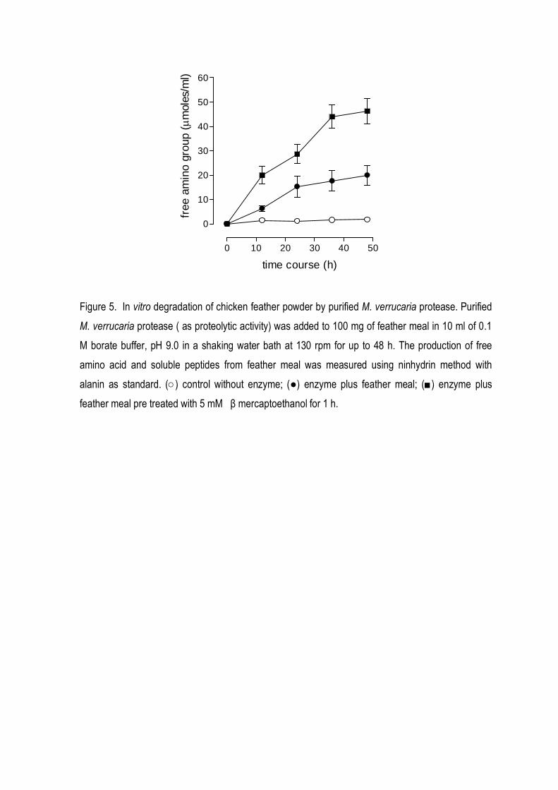
0 10 20 30 40 50
0
10
20
30
40
50
60
time course (h)
free
am
ino
grou
p (µ
mol
es/m
l)
Figure 5. In vitro degradation of chicken feather powder by purified M. verrucaria protease. Purified
M. verrucaria protease ( as proteolytic activity) was added to 100 mg of feather meal in 10 ml of 0.1
M borate buffer, pH 9.0 in a shaking water bath at 130 rpm for up to 48 h. The production of free
amino acid and soluble peptides from feather meal was measured using ninhydrin method with
alanin as standard. (○) control without enzyme; (●) enzyme plus feather meal; (■) enzyme plus
feather meal pre treated with 5 mM β mercaptoethanol for 1 h.
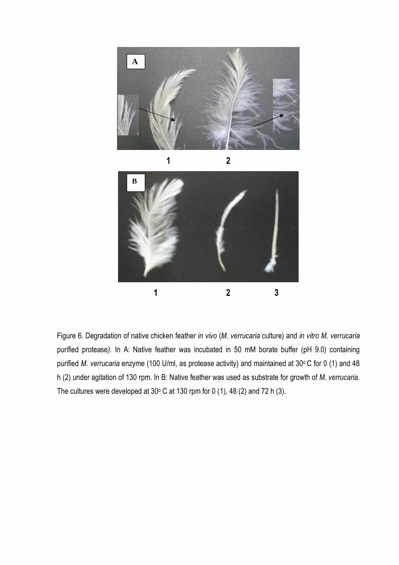
1 2
1 2 3
Figure 6. Degradation of native chicken feather in vivo (M. verrucaria culture) and in vitro M. verrucaria
purified protease). In A: Native feather was incubated in 50 mM borate buffer (pH 9.0) containing
purified M. verrucaria enzyme (100 U/ml, as protease activity) and maintained at 30o C for 0 (1) and 48
h (2) under agitation of 130 rpm. In B: Native feather was used as substrate for growth of M. verrucaria.
The cultures were developed at 30o C at 130 rpm for 0 (1), 48 (2) and 72 h (3).
A
B