lenin israel proano minaca - ufsc
TRANSCRIPT


LENIN ISRAEL PROANO MINACA
INFLUÊNCIA DA ESPESSURA DE MATRIZ COLÁGENA NA ADESÃO,
MORFOLOGIA, VIABILIDADE E PROLIFERAÇÃO CELULAR
FLORIANÓPOLIS
2020
Dissertação submetida ao Programa de Pós-Graduação
em Odontologia da Universidade Federal de Santa
Catarina, na área de concentração de Implantodontia,
como requisito parcial para a obtenção de título de
Mestre em Odontologia.
Orientadora: Profª. Dra. Ariadne Cristiane Cabral da Cruz
Coorientadora: Profª. Dra. Izabella Thaís da Silva

Ficha de identificação da obra elaborada pelo autor, através do Programa de Geração Automática da Biblioteca Universitária da UFSC.
Proano, Lenin
INFLUÊNCIA DA ESPESSURA DE MATRIZ COLÁGENA NA ADESÃO,MORFOLOGIA, VIABILIDADE E PROLIFERAÇÃO CELULAR / LeninProano ; orientador, Profa. Dra. Ariadne Cristiane Cabral
da Cruz, coorientador, Porfa. Dra. Izabella Thaís daSilva, 2020.
47 p.
Dissertação (mestrado) - Universidade Federal de SantaCatarina, Centro de Ciências da Saúde, Programa de PósGraduação em Odontologia, Florianópolis, 2020. Inclui referências. 1. Odontologia. 2. Regeneração tecidual. 3. Matrizes deColágeno. 4. Viabilidade celular . 5. Proliferação celular.I. Cabral da Cruz, Profa. Dra. Ariadne Cristiane . II. da
Silva, Porfa. Dra. Izabella Thaís . III. UniversidadeFederal de Santa Catarina. Programa de Pós-Graduação emOdontologia. IV. Título.

LENIN ISRAEL PROANO MINACA
INFLUÊNCIA DA ESPESSURA DE MATRIZ COLÁGENA NA ADESÃO,
MORFOLOGIA, VIABILIDADE E PROLIFERAÇÃO CELULAR
O presente trabalho em nível de mestrado foi avaliado e aprovado por banca examinadora
composta pelos seguintes membros:
Profa. Dra. Ariadne Cristiane Cabral da Cruz
Presidente
Universidade Federal de Santa Catarina
Prof. Dr. Ricardo de Souza Magini
Universidade Federal de Santa Catarina
Prof. Dr. Felipe Perozzo Daltoé
Universidade Federal de Santa Catarina
Certificamos que esta é a versão original e final do trabalho de conclusão que foi
julgado adequado para obtenção do título de mestre em Odontologia.
____________________________
Profa. Dra. Elena Riet Correa Riveiro
Coordenação do Programa de Pós-Graduação
____________________________
Profa. Dra. Ariadne Cristiane Cabral da Cruz
Orientadora
Florianópolis, 2020.

Este trabalho é dedicado aos meus pais e irmão que sempre
fizeram tudo para me apoiar e brindar o melhor deles para que eu
pudesse fazer o que gosto e me apaixona. Todo meu trabalho e
esforço é para eles.

AGRADECIMENTOS
Agradeço a Deus por me dar a saúde necessária para meu desenvolvimento diário, por
cuidar de mim e dos meus seres amados enquanto não estou perto deles. Por ser a luz que
sempre guia meu caminho e me acompanha. Por ser a luz que preciso quando mais necessito.
Aos meus pais e irmão, Lenin, Shirley e Alex, que mesmo longe estão comigo me
fazendo sentir todo o seu carinho e apoio. Que me ensinaram valores fundamentais no meu
desenvolvimento, e, sem dúvida, fizeram a diferença nas minhas decisões na vida. Sempre são
meu exemplo a seguir. Admiro vocês por sua dedicação e carinho, demostrados sempre de
diferentes formas. Ao meu irmão com quem compartilhei tudo na vida, quem sempre esteve
comigo em todos os momentos, desde quando jogávamos bola até nos momentos ruins e
problemáticos. Sinto saudades de vocês todos os dias, mas são vocês que me motivam para
continuar. Quero fazer vocês se sentirem orgulhosos de mim sempre. São tudo o que eu tenho
e os amo com todo meu coração.
Aos meus professores, grandes amigos em Bauru, Valmir, Thiago, Lucas e Vinicius,
que me motivaram e inspiraram para seguir os seus passos no caminho do ensino.
Adicionalmente, quero fazer um agradecimento especial ao Professor Eric Tincoco, que além
de fazer parte do grupo de professores mencionado, tem me mostrado o lado humano da nossa
área de trabalho. Obrigado por ser uma excelente pessoa e professor. Obrigado por me abrir as
portas da sua casa e da sua família e por me tratar como se eu fosse membro dela. Por me passar
os conhecimentos que me levaram a acreditar mais em mim e que me fizeram gostar tanto da
Implantodontia. Que bom poder trabalhar com a sua equipe, no mesmo lugar onde comecei.
Gratidão pela parceria de todos vocês sempre.
Aos meus professores na UFSC, começando pelo Professor Ricardo Magini, que sem
me conhecer e antes mesmo de eu me tornar aluno do mestrado, me aconselhou e mostrou seu
apoio para eu correr atrás do que eu queria, e que no final aconteceu. Dentro do CEPID,
agradeço aos professores Cesar Benfatti e Marco Bianchini que sempre estiveram abertos a
escutar, opinar e encaminhar as minhas ideias, e que conseguem nos guiar de maneira incrível.
Mas, sem dúvida quem tem me marcado nesta etapa é minha orientadora, a Professora Ariadne
Cruz, que como ela fala, me roubou para eu ser seu orientado num momento que eu não tinha
começado trabalhar com pesquisa ainda e no qual estava preocupado sobre como iria avançar
no meu mestrado. Ela sendo uma excelente professora e cientista, me deu seu apoio e soube me

ensinar e guiar com a delicadeza e o carinho de uma mãe, o que a caracteriza sempre. É uma
pessoa íntegra e admirável em todas as suas faces. Que se preocupa com as pessoas com as
quais trabalha, vendo-as como pessoas mesmo e não só como alunos ou colegas de trabalho.
Isso demostra uma qualidade humana sem igual. Me deu uma luz, e mais do que isso, me
motivou e inspirou para trabalhar numa área que não tinha conhecimento nenhum. E ainda
assim me deu sua confiança e me fez sentir segurança. Hoje eu gosto da pesquisa tanto quanto
da clínica, e isso é graças a você. Obrigado pelos conselhos e palavras de apoio quando as
precisei também. Gratidão sempre Profe.
Aos meus amigos que compartilharam tantas experiências de vida comigo desde
criança, Meli, Andres, Fausto e Julio, que sempre estão presentes, mesmo de longe. São tantos
anos de amizade que já não somos mais amigos, somos irmãos. Obrigado por estarem comigo
sempre.
Aos meus amigos e colegas que compartilharam comigo o início deste caminho de
ensino fora do meu país, Diego e Ana. Que souberam ser parceiros e verdadeiros amigos em
todo momento, enquanto moramos juntos e ainda agora. Sempre se preocupando e me dando
palavras de ânimo durante toda minha caminhada. Saudades de vocês. Logo estaremos juntos
trabalhando como sempre.
Aos meus amigos e colegas da UFSC. Meus irmãos, Mario, Paulo, Lincoln e Giovani
que fizeram esta caminhada muito mais agradável, que desde o início me receberam com os
braços abertos e me fizeram sentir em casa. São a família que escolhemos, ainda mais
importante quando estamos longe de casa. Obrigado pelo trabalho juntos, pela parceria de
sempre e pelos momentos de lazer que ajudam a descontrair e a continuar na caminhada. Às
minhas queridas amigas e colegas de trabalho, Mari e Raissa. À vocês duas, junto com a nossa
orientadora, devo meu mestrado. Sua parceria, cuidado com os detalhes e paixão pelo trabalho
fazem as coisas fluir. Obrigado por estarem comigo nesse aprendizado, por estarem nos
momentos bons, quando nossos experimentos ficavam perfeitos, e também quando nada dava
certo, chegando até o ponto de chorar no laboratório. Por me acompanharem nos dias que
chegávamos cedo no laboratório para fazer nossos experimentos e só saíamos tarde da noite.
Nesses momentos percebi que vocês são amigas de verdade. Gratidão por tudo o que me
ensinaram. Por todos esses motivos, admiro muito vocês.
A todos meus colegas do CEPID, que são uma família e me integraram nela desde o
momento que cheguei. O nosso ambiente de trabalho é incrível, sem egoísmo, todos se ajudando
e dando uma mão em tudo o que precisamos. Gosto muito de fazer parte desse lugar e, sem
dúvida, o aprendizado é muito melhor com todos vocês.
Também aos meus colegas do LVA, que sempre foram parceiros a toda hora. Sempre
se disponibilizaram para ajudar e também para trabalhar em todo momento. Não posso deixar
de mencionar as professoras Izabella e Gislaine, que me acolheram e brindaram com todo o seu
conhecimento. São excelentes pessoas e me ajudaram sempre que precisei. É um verdadeiro
prazer aprender com vocês.
Por último, mas não menos importante, às minhas coorientadas de TCC, Grazi e Fer,
que além de queridas e boas amigas, são excelentes alunas, muito interessadas e responsáveis.
Sou muito grato pela ajuda incondicional que me brindaram durante todo meu mestrado. Tem
sido um prazer trabalhar com vocês e vamos deixar seus trabalhos perfeitos para quando chegar
a hora de vocês defenderem. Sem dúvida, podem contar comigo no que vocês precisarem.
Finalmente, agradeço à todas a pessoas que estiveram envolvidas nessa caminhada.
Gosto de lembrar sempre das pessoas, mas do que dos títulos o dos profissionais. Com certeza,
cada pessoa que tem passado pela minha vida tem aportado um grão de areia, permitindo que
eu tivesse chegado neste ponto, seja com uma palavra, um conselho ou um simples
cumprimento.
Gratidão com a vida por me permitir viver todas estas experiências.

“Porque te confundes y agitas ante los problemas de la vida? Déjame el cuidado de tus
cosas y toda ira mejor. Cuando te entregues a mi todo se resolverá con tranquilidad según mis
designios. No te desesperes, no me dirijas una oración agitada como si quisieras exigirme el
cumplimiento de tus deseos. Cierra los ojos del alma y di con calma:
DIOS YO CONFIO EN TI …”

RESUMO
Considerando que o uso de enxertos autógenos em cirurgias plásticas periodontais e peri-
implantares estão relacionados à alta morbidade pós-operatória, sangramento e disponibilidade
limitada de tecidos no local doador, matrizes xenogênicas têm sido utilizadas com crescente
frequência em cirurgias mucogengivais. Este estudo in vitro teve como objetivo avaliar a
influência da espessura de matriz xenogênica de colágeno na adesão, morfologia, viabilidade e
proliferação celular, bem como na degradação das matrizes. As matrizes Lumina Coat® e
Mucograft® foram avaliadas, seguindo os grupos estabelecidos: SLC - Lumina Coat® de
espessura única, disponível comercialmente com 2 mm de espessura; DLC - Lumina Coat® de
espessura dupla, com o dobro da espessura da SLC; e MG - Mucograft®, com 4mm de
espessura (controle positivo). Foram avaliadas a degradação das matrizes, a topografia da
superfície, a viabilidade de fibroblastos gengivais humanos (HGF) e de células-tronco de dentes
decíduos esfoliados humanos (SHED), bem como a sua morfologia, adesão e proliferação. Os
resultados de todos os grupos foram comparados através da análise de variância unidirecional
(ANOVA) seguida pelo teste post hoc de Tukey, comparando todos os tratamentos em cada
tempo experimental individualmente. As diferenças entre os conjuntos de dados com p <0,05
foram consideradas estatisticamente significantes. Todos os grupos foram biocompatíveis para
HGF e SHED, uma vez que as viabilidades foram superiores a 70% nos dias 1, 3 e 7. Além
disso, o grupo DLC promoveu as maiores viabilidades de HGF e SHED no dia 1.
Adicionalmente, DLC demonstrou maior proliferação de HGF e SHED comparado ao controle
positivo (MG; p <0,05) no dia 7. Por outro lado, o MG demonstrou taxas mais altas de
proliferação de HGF e SHED comparado com DLC no dia 3. Em relação à degradação das
matrizes, as matrizes SLC foram completamente degradadas no dia 14, enquanto as matrizes
DLC e MG apresentaram 48,41% e 20,52% de sua massa inicial, respectivamente, no dia 35.
Concluindo-se, o aumento da espessura da matriz xenogênica de colágeno melhorou a
viabilidade e proliferação dos fibroblastos gengivais humanos e células-tronco mesenquimais
humanas, bem como impediu a degradação precoce da matriz.
Palavras-chave: Matriz Colágena. Regeneração tecidual. Viabilidade celular. Proliferação
celular.
ABSTRACT
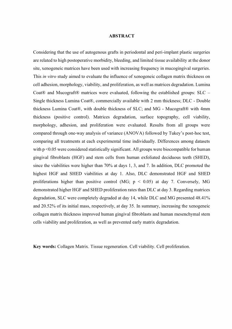
Considering that the use of autogenous grafts in periodontal and peri-implant plastic surgeries
are related to high postoperative morbidity, bleeding, and limited tissue availability at the donor
site, xenogeneic matrices have been used with increasing frequency in mucogingival surgeries.
This in vitro study aimed to evaluate the influence of xenogeneic collagen matrix thickness on
cell adhesion, morphology, viability, and proliferation, as well as matrices degradation. Lumina
Coat® and Mucograft® matrices were evaluated, following the established groups: SLC –
Single thickness Lumina Coat®, commercially available with 2 mm thickness; DLC - Double
thickness Lumina Coat®, with double thickness of SLC; and MG - Mucograft® with 4mm
thickness (positive control). Matrices degradation, surface topography, cell viability,
morphology, adhesion, and proliferation were evaluated. Results from all groups were
compared through one-way analysis of variance (ANOVA) followed by Tukey’s post-hoc test,
comparing all treatments at each experimental time individually. Differences among datasets
with p <0.05 were considered statistically significant. All groups were biocompatible for human
gingival fibroblasts (HGF) and stem cells from human exfoliated deciduous teeth (SHED),
since the viabilities were higher than 70% at days 1, 3, and 7. In addition, DLC promoted the
highest HGF and SHED viabilities at day 1. Also, DLC demonstrated HGF and SHED
proliferations higher than positive control (MG; p < 0.05) at day 7. Conversely, MG
demonstrated higher HGF and SHED proliferation rates than DLC at day 3. Regarding matrices
degradation, SLC were completely degraded at day 14, while DLC and MG presented 48.41%
and 20.52% of its initial mass, respectively, at day 35. In summary, increasing the xenogeneic
collagen matrix thickness improved human gingival fibroblasts and human mesenchymal stem
cells viability and proliferation, as well as prevented early matrix degradation.
Key words: Collagen Matrix. Tissue regeneration. Cell viability. Cell proliferation.

LISTA DE ABREVIATURAS E SIGLAS
ETC Enxerto de tecido conjuntivo
MC Matriz Colágena
PCL Poli (ε-caprolactone)
PLA Ácido polilático
PEG Poli (etilenoglicol)
PLGA Poli (lático-co-glicólico)
MEV Microscopia eletrônica de varredura
HGF Fibroblastos gengivais humanos
SHED Células-tronco derivadas da polpa de dentes decíduos esfoliados humanos
MTS [3-(4,5- dimethylthiazol-2-yl)-5-(3-carboxymethoxyphenyl)-2-(4-sulfophenyl)- 2H-
tetrazolium, inner salt]
FGG Free gingival graft
CTG Connective tissue graft
SEM Scanning Electronic Microscopy
EDS Energy-Dispersive x-ray Spectroscopy
DMEM Dulbecco's modified Eagle's medium
FBS Fetal bovine serum
HMDS Hexamethyldisilazane

LISTA DE FIGURAS
Figure 1: Scanning electronic microscopy (SEM) images of matrices ..................... 29
Figure 2:Cytotoxicity assay by MTS colorimetric test at days 1, 3, and 7.. ............. 30
Figure 3: Proliferation assay by PicoGreen® reagent at days 3 and 7 ...................... 31
Figure 4: Scanning electronic microscopy (SEM) images ........................................ 32
Figure 5: Degradation assay. ..................................................................................... 33

SUMÁRIO
1 INTRODUÇÃO ............................................................................................... 15
2 OBJETIVOS .................................................................................................... 19
2.1 Objetivo Geral .............................................................................................. 19
2.2 Objetivos Específicos .................................................................................... 19
3 ARTIGO ......................................................................................................... 20
INTRODUCTION ...................................................................................................... 24
MATERIALS AND METHODS ................................................................................... 25
RESULTS ................................................................................................................. 28
DISCUSSION............................................................................................................ 33
CONCLUSIONS ........................................................................................................ 36
REFERENCES ........................................................................................................... 37
4 CONSIDERAÇÕES FINAIS ................................................................................ 40
REFERÊNCIAS ..................................................................................................... 41
PRODUÇÕES DURANTE O MESTRADO ................................................................. 47

15
1 INTRODUÇÃO
A cirurgia plástica periodontal e peri-implantar vem sendo realizada com maior
frequência atualmente, em função dos seus resultados clínicos promissores, bem como do
aumento das demandas estéticas por parte dos pacientes (ZUHR; BÄUMER; HÜRZELER,
2014). Adicionalmente, essas cirurgias são consideradas terapia essencial para correção de
defeitos mucogengivais em dentes e implantes (ZUHR; BÄUMER; HÜRZELER, 2014).
Assim, como objetivos das cirurgias mucogengivais periodontais e peri-implantares, pode-se
incluir o aumento da espessura do tecido queratinizado, recobrimentos radiculares, aumento de
tecidos moles no contorno de implantes em áreas edêntulas unitárias ou múltiplas,
reconstruções papilares, dentre outros (CAIRO, 2017;CAIRO et al., 2017; ZUCCHELLI et al.,
2020).
Os procedimentos cirúrgicos mucogengivais convencionais, na maioria dos casos,
exigem a remoção de enxerto de tecido conjuntivo ou enxerto de tecido gengival livre de áreas
doadoras, como o palato ou a área retromolar (JEPSEN et al., 2013; ZUCCHELLI et al., 2020).
Entretanto, esta cirurgia adicional para remoção do tecido da área doadora frequentemente está
associada com elevada morbidade, sangramento pós-cirúrgico e dificuldade do paciente se
alimentar (CAIRO et al., 2017; TONETTI; JEPSEN, 2014). Além disso, os casos clínicos em
que há necessidade de tratamento de vários dentes / implantes simultaneamente representam
um enorme desafio ao cirurgião-dentista, tendo em vista a limitada quantidade de tecido
autógeno disponível na área doadora, bem como o aumento considerável do tempo cirúrgico
em função da remoção de tecidos doadores extensos (ZUHR; BÄUMER; HÜRZELER, 2014).
Convém destacar que os biomateriais, quanto à origem, são classificados em:
Autógeno – obtido do próprio individuo; Alógeno ou homogeno – obtido de indivíduos da
mesma espécie, contudo geneticamente diferentes; Xenógeno ou heterógeno – advém de doares
de diferente espécie do receptor, como bovinos e suínos; e Aloplásticos – material sintético
(ABBAS; LICHTMAN, 2017).
Assim, biomateriais xenogênicos e alogênicos têm sido propostos como uma
alternativa aos enxertos de tecido autógeno. Estes biomateriais permitem o tratamento de
múltiplos sítios em uma única intervenção cirúrgica, evitam a cirurgia adicional na área doadora
e, consequentemente, diminuem o tempo cirúrgico (TAVELLI et al., 2020; THOMA et al.,
2010). O colágeno tem sido o material de escolha para a confecção de matrizes empregadas em

16
cirurgias mucogengivais, em decorrência de sua biocompatibilidade, biodegradação, baixo
nível de imunogenicidade e habilidade de formar uma rede de poros interconectados que facilita
a adesão e proliferação celular (PARENTEAU-BAREIL; GAUVIN; BERTHOD, 2010), além
de ser facilmente preparado e estar disponível em grande quantidade na natureza (GROVER;
CAMERON; BEST, 2012). É importante destacar que o colágeno faz parte da constituição dos
tecidos do corpo humano (GROVER; CAMERON; BEST, 2012; KEANE; BADYLAK, 2014),
sendo a principal proteína estrutural encontrada nos tecidos conjuntivos, formando parte de
tecidos como pele, tendão, cartilagem e ossos (DASKALOVA et al., 2014). Assim, vários
estudos relatam que a confecção e uso do colágeno em biomateriais é uma forma eficaz de
suporte para o crescimento e maturação celular (MATTHEWS et al., 2002; SELL et al., 2008;
SHIELDS et al., 2004). Tem sido demostrado que, tanto o colágeno tipo I, II e III podem
reproduzir fibras similares ou mesmo mimetizar completamente as propriedades estruturais e
biológicas da matriz extracelular em condições ideais (CHEVALLAY; HERBAGE, 2000).
O colágeno pode ser usado na sua forma original ou reconstituída. No caso da
utilização na forma reconstituída, pode haver alteração nas propriedades mecânicas e na
biocompatibilidade. Processos químicos, físico-químicos ou enzimáticos, também podem
promover modificações para potencializar os processos regenerativos decorrentes do uso da
matriz (MEYER, 2019). Adicionalmente, a topografia, morfologia e estabilidade da superfície
aparecem como características importantes no desenvolvimento de matrizes, e têm sido
relatadas como ponto chave no processo de adesão e proliferação celular. Efetivamente, o
desenho das matrizes deveria imitar a microestrutura tecidual e o microambiente celular para
um melhor desempenho, mimetizando a matriz extracelular do tecido a ser regenerado. Assim,
estas características representam uma ferramenta importante no controle da atividade celular
(FUJIOKA-KOBAYASHI et al., 2020).
Por sua vez, o colágeno quando usado em matrizes com pouca espessura, além de
menor resistência mecânica, apresenta um comprometimento da estabilidade estrutural,
particularmente quando usado em ambientes úmidos, como a cavidade bucal (SADEGHI et al.,
2018). Assim, para superar essas limitações, as matrizes a base de colágeno têm sido
modificadas, tanto em estrutura como em composição, com intuito de aprimorar as propriedades
mecânicas (DONG; LV, 2016). Estratégias como cross-linking têm sido usadas para o aumento
da estabilidade e resistência estrutural em biomateriais. O princípio para esse processo
compreende a formação de ligações covalentes entre as moléculas de colágeno usando agentes

17
químicos sintéticos ou naturais. Os agentes mais comumente usados para gerar reações de
cross-link são os grupos aldeídos, mas o emprego de agentes naturais como carboidratos e
extratos naturais também tem sido estudado (DELGADO et al., 2015). Apesar do emprego de
agentes de cross-link, de uma forma geral, ter fornecido melhora nas propriedades mecânicas
do colágeno, eles têm sido relacionados à efeitos citotóxicos in vivo (BARNES et al., 2007;
DONG; LV, 2016).
Outra estratégia que vem sendo estudada, é a associação de colágeno com outros
polímeros naturais e sintéticos. A associação com quitosana tem se mostrado biocompatível e
sem efeito antigênico, além de fornecer um adequado coeficiente de degradação (NILSEN-
NYGAARD et al., 2015; YAN et al., 2015). Polímeros sintéticos, tais como poli (ε-
caprolactone) (PCL), ácido polilático (PLA), poli (etilenoglicol) (PEG), poli (lático-co-
glicólico) (PLGA) são também usados para regeneração tecidual. Quando empregados na
estrutura de matrizes colágenas (HAAPARANTA et al., 2014; ZHANG et al., 2015) podem
promover um padrão de poros que favorece a migração e proliferação celular (JIA et al., 2013).
É importante destacar que, pequenas modificações na concentração, alinhamento e
quantidade das fibras colágenas podem gerar mudanças relevantes na resposta biológica destes
biomateriais (GROVER; CAMERON; BEST, 2012). Ao se observar as características dos
tecidos humanos, nota-se uma estrutura complexa, caracterizada pela heterogeneidade na
disposição das fibras colágenas, com espaços entre as fibras que guiam o movimento celular e
permitem uma adesão e proliferação celular de maneira tridimensional pelas vias de menor
resistência. Assim, uma matriz colágena deveria mimetizar esta estrutura tridimensional
observada in vivo, propiciando, além do padrão de migração celular favorável destacado acima,
a formação de vasos sanguíneos e consequentemente a nutrição do tecido (WOLF et al., 2009).
Ao se observar as matrizes de colágeno disponíveis comercialmente, nota-se que a
maioria consegue reproduzir de forma bastante fiel as caraterísticas dos tecidos humanos.
Modelos de estudos in vitro têm sido propostos e executados para avaliar os padrões de
migração celular, observando que em estruturas tridimensionais, o padrão de migração é similar
aos tecidos humanos. Por outro lado, nos modelos bidimensionais que não apresentam uma rede
de fibras e poros mais espessa, a migração celular é comprometida (DOYLE et al., 2009). A
matriz Mucograft foi escolhida como controle positivo por ser uma matriz colágena usada
para regeneração tecidual periodontal e peri-implantar, sendo considerada uma matriz de
referência.

18
Escolheu-se como modelo de células, os fibroblastos gengivais humanos (HGF) e
células-tronco derivadas da polpa de dentes decíduos esfoliados humanos (SHED), uma vez
que essas células estão diretamente relacionadas à regeneração dos tecidos moles periodontais
e peri-implantares. Além desse fato, por serem células humanas, se assemelham mais às
condições humanas in vivo comparado com linhagens não humanas.
Assim, o objetivo deste trabalho foi avaliar in vitro a influência da espessura de matriz
colágena xenogênica na adesão celular, morfologia, viabilidade e proliferação, bem como na
degradação da matriz.

19
2 OBJETIVOS
2.1 Objetivo Geral
Avaliar a influência da espessura de uma matriz de colágeno xenogênica na adesão,
morfologia, viabilidade e proliferação celular, bem como na degradação da matriz.
2.2 Objetivos Específicos
- Determinar as características de superfície das matrizes de colágeno Lumina Coat®
com a espessura de 2mm disponível comercialmente (SLC); Lumina Coat® com o dobro da
espessura disponível comercialmente (DLC); e Mucograft®, com a espessura disponível
comercialmente (MG), por meio de microscopia eletrônica de varredura (MEV).
- Determinar os elementos químicos presentes na superfície das matrizes SLC, DLC e
MG, empregando-se espectroscopia com energia dispersiva de raios-X.
- Avaliar a adesão e morfologia de fibroblastos gengivais humanos (HGF) e células-
tronco derivadas da polpa de dentes decíduos esfoliados humanos (SHED) semeadas sobre as
matrizes SLC, DLC e MG, por meio de MEV, após 7 dias de cultivo celular.
- Determinar a biocompatibilidade das matrizes SLC, DLC e MG em HGF e SHED,
empregando-se o teste colorimétrico MTS, em 1, 3 e 7 dias.
- Analisar a proliferação de HGF e SHED promovida pelas matrizes SLC, DLC e MG,
por meio do teste PicoGreen®, em 3 e 7 dias.
- Avaliar a degradação das matrizes SLC, DLC e MG, por meio da determinação da
massa das matrizes em 1, 3, 7, 14, 21, 28, e 35 dias.
20
3 ARTIGO
Increasing the thickness of the collagen xenogeneic matrix prevents early matrix
degradation and improves the proliferation, adhesion, and viability of human
gingival fibroblasts and mesenchymal stem cells
L. Proaño1,2 (https://orcid.org/0000-0002-0939-5040, [email protected]), R. B.
Curtarelli1,2 (https://orcid.org/0000-0001-7147-9010, [email protected]), M. B.
Sordi1,2, (http://orcid.org/0000-0001-7873-0765, [email protected]), I. T. Silva2,3
(https://orcid.org/0000-0002-1893-4317, [email protected]), G. Fongaro2,4
(https://orcid.org/0000-0001-5596-3320, [email protected]), Márcio Côrrea6
(http://orcid.org/0000-0002-9063-7974, [email protected]), M. A. Bianchini1,6
(https://orcid.org/0000-0001-6135-701X, [email protected]), A. C. C. Cruz1,2,6
(http://orcid.org/0000-0001-7306-4708, [email protected])
1Center for Research on Dental Implants, Federal University of Santa Catarina,
Florianópolis, Brazil.
2Laboratory of Applied Virology, Federal University of Santa Catarina, Florianópolis,
Brazil.
3Department of Pharmaceutics Science, Federal University of Santa Catarina,
Florianópolis, Brazil.
4Department of Microbiology, Immunology, and Parasitology, Federal University of
Santa Catarina, Florianópolis, Brazil.
6Department of Dentistry, Federal University of Santa Catarina, Florianópolis, Brazil.

21
Corresponding author:
Ariadne Cristiane Cabral da Cruz
Departamento de Odontologia, Universidade Federal de Santa Catarina
Campus Universitário – Trindade, CEP: 88.040-970, Florianópolis, SC – Brasil
Telefone: +55 48 3721-3407
E-mail: [email protected]
ACKNOWLEDGMENTS
Authors thank Curityba Biotech® for providing stem cells from human exfoliated deciduous
teeth (SHED). Authors appreciate the support from the Laboratório Multiusuário de Estudos
em Biologia (LAMEB) and Laboratório Central de Microscopia Eletrônica (LCME) at the
Universidade Federal de Santa Catarina (UFSC) for providing their equipments and
infrastructures to perform some experimental analyses.
ETHICAL APPROVAL
Human gingival fibroblasts (HGF) were obtained from the primary culture of three healthy
patients who needed a distal wedge periodontal procedure. The experimental protocol (number
21/09) was approved by the Human Research Ethics Committee of the Federal University of
Santa Catarina (UFSC), under authorization number 062.
CONFLICT OF INTEREST
Marco Aurélio Bianchini is a speaker from Criteria Biomateriais.
The present project was not supported by any grant.
22
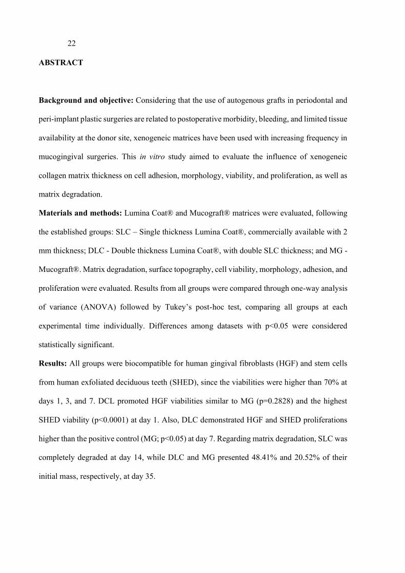
ABSTRACT
Background and objective: Considering that the use of autogenous grafts in periodontal and
peri-implant plastic surgeries are related to postoperative morbidity, bleeding, and limited tissue
availability at the donor site, xenogeneic matrices have been used with increasing frequency in
mucogingival surgeries. This in vitro study aimed to evaluate the influence of xenogeneic
collagen matrix thickness on cell adhesion, morphology, viability, and proliferation, as well as
matrix degradation.
Materials and methods: Lumina Coat® and Mucograft® matrices were evaluated, following
the established groups: SLC – Single thickness Lumina Coat®, commercially available with 2
mm thickness; DLC - Double thickness Lumina Coat®, with double SLC thickness; and MG -
Mucograft®. Matrix degradation, surface topography, cell viability, morphology, adhesion, and
proliferation were evaluated. Results from all groups were compared through one-way analysis
of variance (ANOVA) followed by Tukey’s post-hoc test, comparing all groups at each
experimental time individually. Differences among datasets with p<0.05 were considered
statistically significant.
Results: All groups were biocompatible for human gingival fibroblasts (HGF) and stem cells
from human exfoliated deciduous teeth (SHED), since the viabilities were higher than 70% at
days 1, 3, and 7. DCL promoted HGF viabilities similar to MG (p=0.2828) and the highest
SHED viability (p<0.0001) at day 1. Also, DLC demonstrated HGF and SHED proliferations
higher than the positive control (MG; p<0.05) at day 7. Regarding matrix degradation, SLC was
completely degraded at day 14, while DLC and MG presented 48.41% and 20.52% of their
initial mass, respectively, at day 35.

23
Conclusion: Increasing the xenogeneic collagen matrix thickness improved human gingival
fibroblasts and human mesenchymal stem cells viability and proliferation, as well as prevented
early matrix degradation.
Keywords: collagen matrix, cell viability, cell proliferation, cell adhesion, tissue regeneration

24
INTRODUCTION
Mucogingival surgeries are performed frequently due to the promising clinical
outcomes of periodontal and peri-implant plastic surgeries and the increasing patient’s aesthetic
demands.1 Conventional mucogingival procedures require a second surgical site to remove a
free gingival graft (FGG) or a connective tissue graft (CTG).2 This additional surgery site is
associated with high morbidity, postsurgical bleeding, and feeding difficulties.3 Additionally,
multiple simultaneous treatment sites represent a clinical challenge to the surgeon, prolonged
surgical time, and commonly there is a limited tissue availability of donor site for mucogingival
surgeries.4,5
Therefore, xenogeneic and allogeneic graft materials have been proposed as an
alternative to autogenous graft tissues. These biomaterials allow the treatment of multiple sites
in one surgical intervention, prevent the additional donor site surgery, and reduce surgical
time.6,7 Collagen has been the eligible component to develop matrices for mucogingival
regeneration surgeries due to its biocompatibility, suitable biodegradation, low levels of
immunogenicity, and the possibility to form a porous interconnected network structure that
facilitates the cell migration and adhesion.8–10
Concerning xenogeneic matrices, the promising clinical findings are related to the
matrix adaptation to the surgical site, blood clot stabilization, and tissue integration, which will
promote the cellular ingrowth. Also, the matrix physico-chemical characteristics, such as
collagen type composition, surface characteristics, and matrix degradation are directly
associated with the clinical outcomes,8,11,12 including long-lasting tissue stability and
keratinized tissue formation.11,13,14 In this sense, the present in vitro study aimed to evaluate the

25
influence of xenogeneic collagen matrices thickness on cell adhesion, morphology, viability,
and proliferation, as well as matrices degradation.
MATERIALS AND METHODS
Collagen matrices
Lumina Coat® (Criteria Biomateriais, Sao Paulo, Brazil) and Mucograft® (Geistlich
Pharma AG, Wolhusen, Switzerland) were evaluated, following the established groups: SLC -
Single thickness Lumina Coat®, commercially available with 2 mm thickness; DLC - Double
thickness Lumina Coat®, with a double thickness of SLC; and MG - Mucograft®, as
commercially available. In a laminar flow cabinet, matrices of all groups were divided on
fragments of 5 mm x 5 mm each for SLC and MG, and 5 x 10 mm for DLC. DLC matrices
were folded and sutured at the borders (4.0 Nylon, Ethicon, Johnson & Johnson, São Paulo, SP,
Brazil) to double their thickness.
Scanning electronic microscopy and energy-dispersive x-ray spectroscopy
Surface topography analyses of SLC, DLC, and MG were performed using a high
resolution scanning electron microscope (JEOL JMS-6390LV, Massachusetts, USA) at 10 kV.
Previously, samples were spin-coated with 300 Å gold. Each sample was photographed in three
distinct fields at magnifications of 500X, 1000X, and 2500X. Analyses were performed in
triplicate. The surface chemical composition of SLC, DLC, and MG were assessed by Energy-
Dispersive X-ray Spectroscopy (EDS, JEOL JMS-6390LV, Massachusetts, USA), using 500X
magnification SEM images. Analyses were performed in triplicate.

26
Cell lineages and culture conditions
To evaluate the cell viability, proliferation, adhesion, and morphology, all cell lineages
described below were incubated at 37 oC in an atmosphere with 5% CO2. Cells were seeded in
triplicate at a density of 2.0×10⁴ cells per well or per sample.
Human gingival fibroblasts
Human gingival fibroblasts (HGF) were obtained from the primary culture of three
healthy patients who needed distal wedge periodontal procedures, according to Littuma et al.
(2019).15 The experimental protocol was approved by the Human Research Ethics Committee
and each volunteer signed written consent. Experiments were conducted in full accordance with
ethical principles, including the Helsinki Declaration. After the establishment of the fibroblast
lineage, HGF were cultivated with Dulbecco's modified Eagle's medium (DMEM, Gibco,
Massachusetts, USA) with 10% fetal bovine serum (FBS, Gibco, Massachusetts, USA) and
were applied in the following experiments using cell passages from 4 up to 6.
SHED
Stem cells from Human Exfoliated Deciduous teeth (SHED, Curityba Biotech™ Cell
Processing Center, Curitiba, Brazil) were cultured in DMEM with 10% FBS, according to
Littuma et al. (2019).15 SHED passages from 4 up to 6 were used.
Cytotoxicity assay
Matrices cytotoxicity were evaluated by MTS [3-(4,5- dimethylthiazol-2-yl)-5-(3-
carboxymethoxyphenyl)-2-(4-sulfophenyl)- 2H-tetrazolium, inner salt] (CellTiter 96®,
Promega, Sao Paulo, Brasil) colorimetric assay15 evaluating HGF and SHED at days 1, 3, and
7. Cells were seeded over SLC, DLC, and MG matrices, in triplicate, placed in 96-well plates.
Samples were maintained immobile for 4 h to allow cell adhesion, then the culture medium was

27
added up to 200 µL. Also, cell control groups were performed using HGF and SHED (2.0×10⁴
cells/well, in 96-wells plates) seeded directly into the wells.
At each experimental time, 40 μL of MTS solution were added per well and the plates
were incubated for 150 min. Absorbances were measured by spectrophotometry (Infinite M200,
Tecan Trading AG, Männedorf, Switzerland) at 490 nm. Cell viability percentages were
calculated relative to cell control group absorbances.
Cell proliferation
HGF and SHED were seeded over SLC, DLC, and MG matrices as described at item
2.4 Cytotoxicity assay. Cell proliferations were assessed by PicoGreen® assay (InvitrogenTM,
Massachusetts, USA). The analyses were performed following the manufacturer's
recommendations at days 3 and 7, according to Costa et al. (2014),16 with minor modifications.
Fluorescent spectrophotometry (Infinite M200, Tecan Trading AG, Männedorf, Switzerland)
was used at 360/440 nm (Ex / Em). Analyses were performed in triplicate.
Cell morphology and adhesion
To visualize cell morphology and cell ability to adhere on matrix surfaces, SEM (JEOL
JMS-6390LV, Massachusetts, USA) was carried out after item 2.5 Cell Proliferation at day 7.
Therefore, matrices were carefully washed with phosphate-buffered saline (PBS, Gibco,
Massachusetts, USA), fixed using 4% glutaraldehyde for 30 min at room temperature,
dehydrated with progressive alcohol concentrations, 50% to 100%, and finalized with
hexamethyldisilazane (HMDS, Sigma-Aldrich). The dried matrices were spin-coated with 300
Å gold. SEM was performed in triplicate at 10 kV and samples were photographed at two
distinct fields at 500X and 1000X magnifications.

28
Matrices degradation assay
Matrices degradation were evaluated according to the mass loss. Therefore, the
determination of SLC, DLC, and MG matrices initial mass (n=3) was performed. Then, samples
were submerged in 3 mL PBS into 24-well plates and maintained at 37 °C, according to Cruz
et al. (2016),17 with modifications. At days 1, 3, 7, 14, 21, 28, and 35, samples were individually
dried, weighed at sterile conditions, and placed back into the wells.
Statistical Analysis
Three independent experiments were performed to confirm the reproducibility of the
results. Graph Pad Software Inc. (7.0 version, San Diego, CA, USA) was employed for
statistical analyses. Results of matrices cytotoxicity and cell proliferation from all groups were
compared using one-way analysis of variance (ANOVA) followed by Tukey’s post-hoc test,
comparing all treatments at each experimental time individually. Differences among datasets
with p <0.05 were considered statistically significant.
RESULTS
Scanning electronic microscopy and energy-dispersive X-ray spectroscopy
According to Figure 1, SLC, DLC, and MG matrices demonstrated irregular fibers and
porous surfaces. SLC and DLC provided similar SEM images showing the same fiber
arrangement and porosity pattern (Figure 1A and 1B). Meanwhile, MG fibers were thicker and
more continuous, resulting in a different porosity pattern (Figure 1C). EDS analyses
demonstrated that SLC, DLC, and MG were essentially composed of carbon, oxygen, and
sodium chemical elements (Figure 1D, 1E, and 1F).

29
Figure 1: Scanning electronic microscopy (SEM) images of matrices. A) Single Lumina Coat (SLC); B) Double Lumina Coat (DLC); and C) Mucograft (MG) at 500x magnification. Imagens are representative (n=3). Energy-dispersive X-ray spectroscopy (EDS) graphs presenting the chemical composition surface of D) Single Lumina Coat (SLC); E) Double Lumina Coat (DLC), and F) Mucograft (MG).
Cytotoxicity assay
On the topic of Figure 2A, SLC, DLC, and MG matrices were biocompatible with both
HGF and SHED at days 1, 3, and 7, since all groups demonstrated cell viabilities over 70%
[ISO 10993‐5:2009(E)].18 All groups presented HGF viability over 100% at day 1, and DLC
promoted higher HGF viability than SLC (p=0.049) and cell control (p=0.0066). At day 3, SLC
and DLC demonstrated similar HGF viabilities (p=0.1222), MG was similar to DLC
(p=0.2634), and cell control was similar to SLC (p=0.7071). All groups promoted similar HGF
viabilities at day 7 (p=0.5922).
At day 1, SHED (Figure 2B) demonstrated viabilities higher than 70% for all groups.
Additionally, DLC promoted the highest SHED viability, followed by MG (p=0.0001), while
SLC and cell control promoted similar results (p=0.9965) at day 1. At day 3, no substantial
difference was found among groups (p=0.0441). No significant difference was found among
groups at day 7 (p=0.0748).

30
Figure 2:Cytotoxicity assay by MTS colorimetric test at days 1, 3, and 7. Different lower case letters refer to significant differences (ANOVA/Tukey test, p <0.05) among groups. Percentages of viable cells were calculated concerning cell control group. A) Human gingival fibroblasts (HGF) and B) mesenchymal stem cells from human exfoliated deciduous teeth (SHED). Single Lumina Coat (SLC); B) Double Lumina Coat (DLC); and C) Mucograft (MG).
Cell proliferation
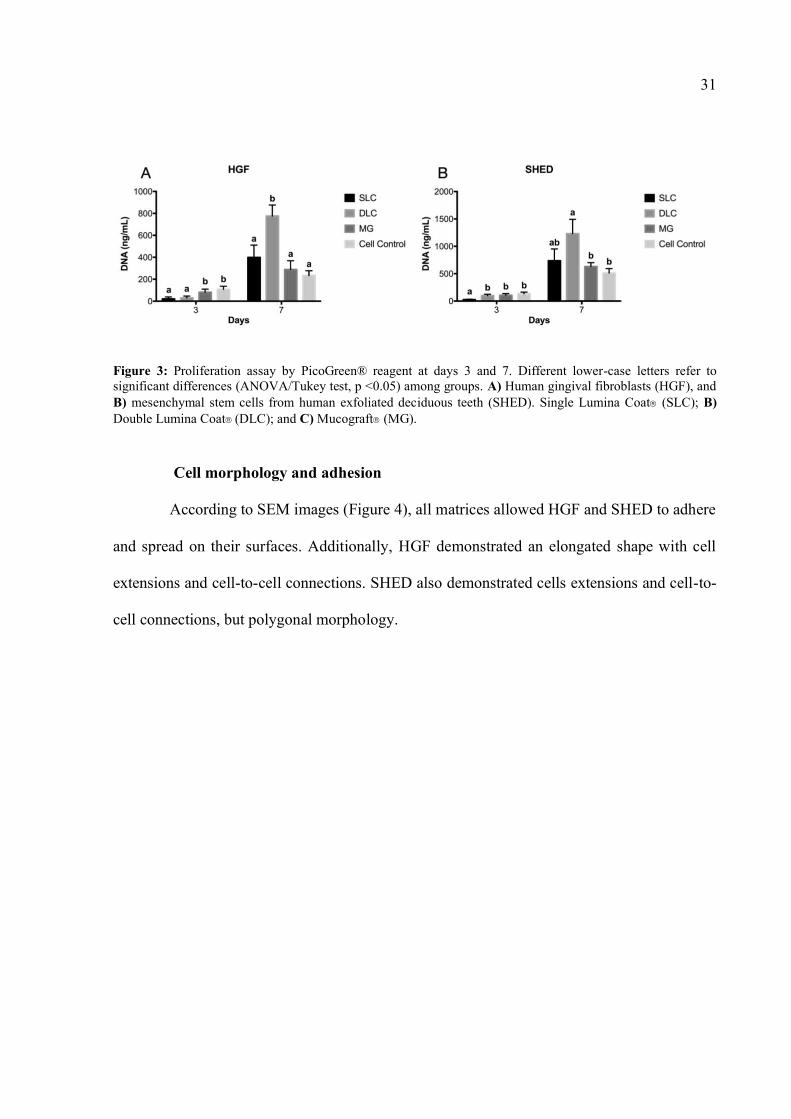
As observed in Figure 3A, SLC and DLC promoted lower HGF proliferation than MG
and cell control at day 3 (p=0.0005). Conversely, at day 7, DLC demonstrated the highest HGF
proliferation (p=0.0033).
Regarding SHED (Figure 3B), DLC, MG, and cell control promoted similar cell proliferation,
while SLC demonstrated the lowest SHED proliferation at day 3 (p=0.0001). At day 7, MG and
cell control demonstrated lower SHED proliferation rates than SLC and DLC (p=0.0218).
31
Figure 3: Proliferation assay by PicoGreen® reagent at days 3 and 7. Different lower-case letters refer to significant differences (ANOVA/Tukey test, p <0.05) among groups. A) Human gingival fibroblasts (HGF), and B) mesenchymal stem cells from human exfoliated deciduous teeth (SHED). Single Lumina Coat (SLC); B) Double Lumina Coat (DLC); and C) Mucograft (MG).
Cell morphology and adhesion
According to SEM images (Figure 4), all matrices allowed HGF and SHED to adhere
and spread on their surfaces. Additionally, HGF demonstrated an elongated shape with cell
extensions and cell-to-cell connections. SHED also demonstrated cells extensions and cell-to-
cell connections, but polygonal morphology.

32
Figure 4: Scanning electronic microscopy (SEM) images at 500x and 1000x magnificant. Human gingival fibroblasts (HGF) and mesenchymal stem cells from human exfoliated deciduous teeth (SHED) adhered and spread on surfaces of Single Lumina Coat (SLC), Double Lumina Coat (DLC), and Mucograft (MG) matrices after 7 days of cells seeded. Images are representative (n=3).
Degradation assay
According to Figure 5, at day 1, all groups demonstrated similar mass loss. At day 3,
SLC, DLC, and MG lost 1.35%, 3.13%, and 43.84%, respectively. SLC and MG demonstrated
similar loss of mass at day 7 (72.69% and 65.07%, respectively), while DLC demonstrated
20.53% mass loss. At day 14, SLC matrices were completely degraded, while DLC and MG
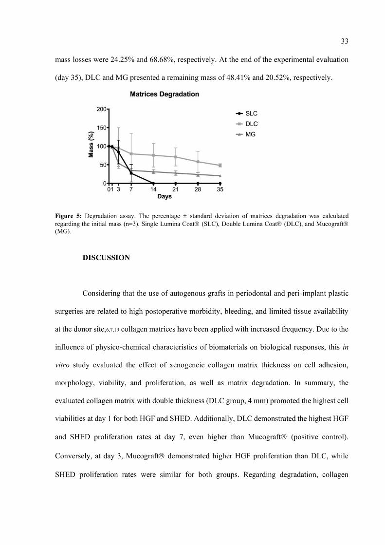
33
mass losses were 24.25% and 68.68%, respectively. At the end of the experimental evaluation
(day 35), DLC and MG presented a remaining mass of 48.41% and 20.52%, respectively.
Figure 5: Degradation assay. The percentage standard deviation of matrices degradation was calculated regarding the initial mass (n=3). Single Lumina Coat (SLC), Double Lumina Coat (DLC), and Mucograft (MG).
DISCUSSION
Considering that the use of autogenous grafts in periodontal and peri-implant plastic
surgeries are related to high postoperative morbidity, bleeding, and limited tissue availability
at the donor site,6,7,19 collagen matrices have been applied with increased frequency. Due to the
influence of physico-chemical characteristics of biomaterials on biological responses, this in
vitro study evaluated the effect of xenogeneic collagen matrix thickness on cell adhesion,
morphology, viability, and proliferation, as well as matrix degradation. In summary, the
evaluated collagen matrix with double thickness (DLC group, 4 mm) promoted the highest cell
viabilities at day 1 for both HGF and SHED. Additionally, DLC demonstrated the highest HGF
and SHED proliferation rates at day 7, even higher than Mucograft (positive control).
Conversely, at day 3, Mucograft demonstrated higher HGF proliferation than DLC, while
SHED proliferation rates were similar for both groups. Regarding degradation, collagen

34
matrices with single thickness were completely degraded at day 14, while double thickness and
Mucograft matrices presented 48.41% and 20.52% of their initial mass, respectively, at day
35. These findings are relevant for clinical practice, since the regeneration of periodontal and
peri-implant soft tissues is closely related to the ability of xenogeneic matrices used in
mucogingival surgeries to promote cell adhesion, migration, and proliferation. In addition, the
matrices must be biocompatible and demonstrate an adequate degradation rate to enable
architecture maintenance while exerting the support for cell adhesion, proliferation, and
differentiation, as well as to allow an adequate formation of blood supply to transport nutrients
and remove metabolic waste.
It is relevant to mention that this study used human gingival fibroblasts and human
mesenchymal stem cells from the dental origin. Human lineages are more representative of the
patient’s condition than established commercially available cell lineages from not human
species. Also, the evaluated cell lineages are directly related to the matrices clinical application
for soft tissue regeneration. Herein, all matrices demonstrated irregular surfaces with ideal
porosity pattern for cell spreading, adhesion, and proliferation. This information was
corroborated by cell viability findings, which demonstrated that all evaluated matrices were
biocompatible for HGF and SHED.
Therefore, we observed a positive influence regarding the increase of matrix thickness
on HGF and SHED behavior, probably due to the delayed matrix degradation observed in the
double thickness group. Despite their interesting biological properties, deficient structural
stability and mechanical strength were observed in experimental and clinical situations
evaluating thin collagen matrices.20 To deal with this limitation, collagen matrices have been
modified using mechanical and chemical procedures, such as ultraviolet and gamma radiation,
and dehydrothermal treatment, to reduce their solubility and reabsorption, and improve the

35
mechanical properties.21–23 Additionally, cross-linking strategies applied to collagen matrices
to improve their mechanical and structural properties are available, but such strategies could
lead to negative effects on cell responses. Therefore, providing an improved structure and
environment for cell migration, adhesion, and proliferation has an important role in many
biological processes, such as cell motility, growth, metabolism, development, and
differentiation, which are directly related to tissue regeneration.8 Thus, volumetric and
thickness modifications of collagen matrices, as performed herein, are interesting alternatives
in soft tissue regenerative therapies.
Moreover, biomaterials for tissue regeneration should mimic extracellular matrix,
reproducing the natural connective tissue and acting as a scaffold, maintaining an architecture
for cell migration, vessels formation, and, finally, tissue regeneration.1,17 Collagen types I and
III are the principal extracellular matrix components and the main fibrous structural protein in
the living organisms. Therefore, collagen matrices have been pointed out as promisor
biomaterial for soft tissue regeneration as several studies have proven that they are suitable for
mature cell growth and proliferation.20,24,25 Mucograft was chosen as a positive control since
it is a resorbable two-layer xenogeneic collagen matrix used for periodontal and peri-implant
tissue regeneration. The literature has indicated Mucograft as a benchmark biomaterial.13,26
This matrix is composed of collagen types I and III with no cross-linking or chemical
treatments. Additionally, Mucograft has two phases: 1) a compact structure of denser collagen
and smooth texture to improve wound healing and increase cell adhesion, and 2) a porous
surface that allows blood clot formation, tissue integration, and angiogenesis.13
Regarding the limitations of this study, it is an in vitro study. Nevertheless, human
gingival fibroblasts and mesenchymal stem cells were applied since they resemble more closely
the in vivo human conditions than cells from animal origin. However, further in vivo and clinical

36
studies are suggested to confirm the influence of double collagen matrix thickness on
biomaterial degradation and cell behavior. Finally, future studies could evaluate other types of
collagen matrices commercially available.
CONCLUSIONS
In summary, increasing the xenogeneic collagen matrix thickness improved human
gingival fibroblasts and dental-derived mesenchymal stem cells viability and proliferation, and
prevented early matrix degradation.

37
REFERENCES
1. Cairo F. Periodontal plastic surgery of gingival recessions at single and multiple teeth.
Periodontol. 2000. 2017;75:296–316.
2. Zucchelli G, Tavelli L, McGuire MK, et al. Autogenous soft tissue grafting for
periodontal and peri‐implant plastic surgical reconstruction. J. Periodontol. 2020;91:9–
16.
3. Stavropoulou C, Atout RN, Brownlee M, Schroth RJ, Kelekis-Cholakis A. A randomized
clinical trial of cyanoacrylate tissue adhesives in donor site of connective tissue grafts.
J. Periodontol. 2019;90:608–615.
4. Griffin TJ, Cheung WS, Zavras AI, Damoulis PD. Postoperative Complications
Following Gingival Augmentation Procedures. J. Periodontol. 2006;77:2070–2079.
5. Wessel JR, Tatakis DN. Patient Outcomes Following Subepithelial Connective Tissue
Graft and Free Gingival Graft Procedures. J. Periodontol. 2008;79:425–430.
6. McGuire MK, Scheyer ET, Schupbach P. Growth Factor–Mediated Treatment of
Recession Defects: A Randomized Controlled Trial and Histologic and Microcomputed
Tomography Examination. J. Periodontol. 2009;80:550–564.
7. Wilson TG, McGuire MK, Nunn ME. Evaluation of the Safety and Efficacy of
Periodontal Applications of a Living Tissue-Engineered Human Fibroblast- Derived
Dermal Substitute. II. Comparison to the Subepithelial Connective Tissue Graft: A
Randomized Controlled Feasibility Study. J. Periodontol. 2005;76:881–889.
8. Chevallay B, Herbage D. Collagen-based biomaterials as 3D scaffold for cell cultures:
Applications for tissue engineering and gene therapy. Med. Biol. Eng. Comput.
2000;38:211–218.

38
9. Kim SG, Lee DS, Lee S, Jang JH. Osteocalcin/fibronectin-functionalized collagen
matrices for bone tissue engineering. J. Biomed. Mater. Res. - Part A. 2015;103:2133–
2140.
10. Tavelli L, McGuire MK, Zucchelli G, et al. Extracellular matrix-based scaffolding
technologies for periodontal and peri-implant soft tissue regeneration. J. Periodontol.
2020;91:17–25.
11. Zuhr O, Bäumer D, Hürzeler M. The addition of soft tissue replacement grafts in plastic
periodontal and implant surgery: Critical elements in design and execution. J. Clin.
Periodontol. 2014;41:S123–S142.
12. Rusu D, Boariu M, Stratul Ștefan I, et al. Interaction between a 3d collagen matrix used
for periodontal soft tissue regeneration and t-lymphocytes: An in vitro pilot study. Exp.
Ther. Med. 2019;17:990–996.
13. Sanz M, Lorenzo R, Aranda JJ, Martin C, Orsini M. Clinical evaluation of a new collagen
matrix (Mucograft® prototype) to enhance the width of keratinized tissue in patients
with fixed prosthetic restorations: A randomized prospective clinical trial. J. Clin.
Periodontol. 2009;36:868–876.
14. Thoma DS, Benić GI, Zwahlen M, Hämmerle CHF, Jung RE. A systematic review
assessing soft tissue augmentation techniques. Clin. Oral Implants Res. 2009;20:146–
165.
15. Littuma GJS, Sordi MB, Borges Curtarelli R, et al. Titanium coated with poly(lactic‐co‐
glycolic) acid incorporating simvastatin: Biofunctionalization of dental prosthetic
abutments. J. Periodontal Res. 2020;55:116–124.
16. Costa PF, Vaquette C, Zhang Q, et al. Advanced tissue engineering scaffold design for
regeneration of the complex hierarchical periodontal structure. J. Clin. Periodontol.

39
2014;41:283–294.
17. Cruz ACC, Caon T, Menin Á, et al. Adipose-Derived Stem Cells Decrease Bone
Morphogenetic Protein Type 2-Induced Inflammation In Vivo. J. Oral Maxillofac. Surg.
2016;74:505–514.
18. Organization IS. Biological evaluation of medical devices–Part 5: Tests for in vitro
cytotoxicity. Int. Organ. 2009;2007:1–11.
19. Stavropoulou C. Comparison of Cyanoacrylate Tissue Adhesives to
Polytetrafluoroethylene Sutures in the Donor Site of Connective Tissue Grafts – A
Randomized Clinical Trial. 2018;
20. Sadeghi R, Mahdavi P, Lee WS, et al. A novel, cell-permeable, collagen-based
membrane promotes fibroblast migration. J. Periodontal Res. 2018;53:727–735.
21. Wang W, Zhang Y, Ye R, Ni Y. Physical crosslinkings of edible collagen casing. Int. J.
Biol. Macromol. 2015;81:920–925.
22. Takitoh T, Bessho M, Hirose M, et al. Gamma-cross-linked nonfibrillar collagen gel as
a scaffold for osteogenic differentiation of mesenchymal stem cells. J. Biosci. Bioeng.
2015;119:217–225.
23. Maslennikova A, Kochueva M, Ignatieva N, et al. Effects of gamma irradiation on
collagen damage and remodeling. Int. J. Radiat. Biol. 2015;91:240–247.
24. Sell SA, Francis MP, Garg K, et al. Cross-linking methods of electrospun fibrinogen
scaffolds for tissue engineering applications. Biomed. Mater. 2008;3:.
25. Matthews JA, Wnek GE, Simpson DG, Bowlin GL. Electrospinning of collagen
nanofibers. Biomacromolecules. 2002;3:232–238.
26. Schmitt CM, Tudor C, Kiener K, et al. Vestibuloplasty: Porcine Collagen Matrix Versus
Free Gingival Graft: A Clinical and Histologic Study. J. Periodontol. 2013;84:914–923.

40
4 CONSIDERAÇÕES FINAIS
Tendo em vista os resultados deste trabalho in vitro, pode-se concluir que o aumento
da espessura da matriz xenogênica de colágeno melhorou a viabilidade e a proliferação de
fibroblastos gengivais humanos e células-tronco mesenquimais de origem dentária, bem como
preveniu a degradação precoce da matriz. Estes achados são importantes para a prática clínica,
uma vez que a regeneração dos tecidos moles periodontais e peri-implantares está intimamente
relacionada à capacidade das matrizes xenogênicas utilizadas nas técnicas cirúrgicas
mucogengivais em propiciar a adesão, migração e proliferação celular. Além de que as matrizes
devem ser biocompatíveis e apresentarem uma taxa de degradação adequada para permitir que
as matrizes exerçam a função de suporte para a adesão, proliferação e diferenciação celular,
bem como permitir uma formação adequada de suprimento sanguíneo para transportar
nutrientes e remover resíduos metabólicos. Apesar deste estudo ter empregado fibroblastos
gengivais humanos e células-tronco mesenquimais humanas, que se assemelham às condições
humanas in vivo, estudos in vivo e clínicos devem ser realizados para confirmar os achados
desta pesquisa. Adicionalmente, sugere-se estudos futuros para avaliar outros tipos de matrizes
de colágeno disponíveis comercialmente.
41
REFERÊNCIAS
ABBAS, A. K.; LICHTMAN, A. H.; PILLAI S. Cellular and molecular
immunology. 9 ed. Philadelphia, United States: Elsevier, 2017
BARNES, C. P. et al. Cross-linking electrospun type II collagen tissue engineering
scaffolds with carbodiimide in ethanol. Tissue Engineering, [s.l.], v. 13, no 7, p. 1593–1605,
2007. ISSN: 10763279, DOI: 10.1089/ten.2006.0292.
CAIRO, F. Periodontal plastic surgery of gingival recessions at single and multiple
teeth. Periodontology 2000, [s.l.], v. 75, no 1, p. 296–316, 2017. ISSN: 09066713, DOI:
10.1111/prd.12186.
CAIRO, F. et al. Xenogeneic collagen matrix versus connective tissue graft for
buccal soft tissue augmentation at implant site. A randomized, controlled clinical trial.
Journal of Clinical Periodontology, [s.l.], v. 44, no 7, p. 769–776, 2017. ISSN: 03036979, DOI:
10.1111/jcpe.12750.
CHEVALLAY, B.; HERBAGE, D. Collagen-based biomaterials as 3D scaffold for
cell cultures: Applications for tissue engineering and gene therapy. Medical and Biological
Engineering and Computing, [s.l.], v. 38, no 2, p. 211–218, 2000. ISSN: 01400118, DOI:
10.1007/BF02344779.
COSTA, P. F. et al. Advanced tissue engineering scaffold design for regeneration
of the complex hierarchical periodontal structure. Journal of Clinical Periodontology, [s.l.],
v. 41, p. 283–294, 2014. DOI: 10.1111/jcpe.12214.
CRUZ, A. C. C. et al. Adipose-Derived Stem Cells Decrease Bone Morphogenetic
Protein Type 2-Induced Inflammation In Vivo. Journal of Oral and Maxillofacial Surgery,
[s.l.], v. 74, no 3, p. 505–514, 2016. ISSN: 02782391, DOI: 10.1016/j.joms.2015.09.006.
DASKALOVA, A. et al. Controlling the porosity of collagen, gelatin and elastin
biomaterials by ultrashort laser pulses. Applied Surface Science, [s.l.], v. 292, p. 367–377,
2014. ISSN: 01694332, DOI: 10.1016/j.apsusc.2013.11.145.
DELGADO, L. M. et al. To Cross-Link or Not to Cross-Link? Cross-Linking
Associated Foreign Body Response of Collagen-Based Devices. Tissue Engineering - Part
B: Reviews, [s.l.], v. 21, no 3, p. 298–313, 2015. ISSN: 19373376, DOI:
10.1089/ten.teb.2014.0290.

42
DONG, C.; LV, Y. Application of collagen scaffold in tissue engineering: Recent
advances and new perspectives. Polymers, [s.l.], v. 8, no 2, p. 1–20, 2016. ISSN: 20734360,
DOI: 10.3390/polym8020042.
DOYLE, A. D. et al. One-dimensional topography underlies three-dimensional fi
brillar cell migration. Journal of Cell Biology, [s.l.], v. 184, no 4, p. 481–490, 2009. ISSN:
00219525, DOI: 10.1083/jcb.200810041.
FUJIOKA-KOBAYASHI, M. et al. In vitro Observation of Macrophage
Polarization and Gingival Fibroblast Behavior on Three-dimensional Xenogeneic
Collagen Matrixes. Journal of biomedical materials research. Part A, [s.l.], no February, p. 1–
11, 2020. ISSN: 1552-4965, DOI: 10.1002/jbm.a.36911.
GRIFFIN, T. J. et al. Postoperative Complications Following Gingival
Augmentation Procedures. Journal of Periodontology, [s.l.], v. 77, no 12, p. 2070–2079, 2006.
ISSN: 0022-3492, DOI: 10.1902/jop.2006.050296.
GROVER, C. N.; CAMERON, R. E.; BEST, S. M. Investigating the morphological,
mechanical and degradation properties of scaffolds comprising collagen, gelatin and
elastin for use in soft tissue engineering. Journal of the Mechanical Behavior of Biomedical
Materials, [s.l.], v. 10, p. 62–74, 2012. ISSN: 17516161, DOI: 10.1016/j.jmbbm.2012.02.028.
HAAPARANTA, A. M. et al. Preparation and characterization of collagen/PLA,
chitosan/PLA, and collagen/chitosan/PLA hybrid scaffolds for cartilage tissue
engineering. Journal of Materials Science: Materials in Medicine, [s.l.], v. 25, no 4, p. 1129–
1136, 2014. ISSN: 15734838, DOI: 10.1007/s10856-013-5129-5.
JEPSEN, K. et al. Treatment of gingival recession defects with a coronally
advanced flap and a xenogeneic collagen matrix: A multicenter randomized clinical trial.
Journal of Clinical Periodontology, [s.l.], v. 40, no 1, p. 82–89, 2013. ISSN: 03036979, DOI:
10.1111/jcpe.12019.
JIA, L. et al. Stem cell differentiation on electrospun nanofibrous substrates for
vascular tissue engineering. Materials Science and Engineering C, [s.l.], v. 33, no 8, p. 4640–
4650, 2013. ISSN: 09284931, DOI: 10.1016/j.msec.2013.07.021.
KEANE, T. J.; BADYLAK, S. F. Biomaterials for tissue engineering applications.
Seminars in Pediatric Surgery, [s.l.], v. 23, no 3, p. 112–118, 2014. ISSN: 15329453, DOI:
10.1053/j.sempedsurg.2014.06.010.
KIM, S. G. et al. Osteocalcin/fibronectin-functionalized collagen matrices for bone

43
tissue engineering. Journal of Biomedical Materials Research - Part A, [s.l.], v. 103, no 6, p.
2133–2140, 2015. ISSN: 15524965, DOI: 10.1002/jbm.a.35351.
LITTUMA, G. J. S. et al. Titanium coated with poly(lactic‐co‐glycolic) acid
incorporating simvastatin: Biofunctionalization of dental prosthetic abutments. Journal
of Periodontal Research, [s.l.], v. 55, no 1, p. 116–124, 2020. ISSN: 0022-3484, DOI:
10.1111/jre.12695.
MASLENNIKOVA, A. et al. Effects of gamma irradiation on collagen damage and
remodeling. International Journal of Radiation Biology, [s.l.], v. 91, no 3, p. 240–247, 2015.
ISSN: 13623095, DOI: 10.3109/09553002.2014.969848.
MATTHEWS, J. A. et al. Electrospinning of collagen nanofibers.
Biomacromolecules, [s.l.], v. 3, no 2, p. 232–238, 2002. ISSN: 15257797, DOI:
10.1021/bm015533u.
MCGUIRE, M. K.; SCHEYER, E. T.; SCHUPBACH, P. Growth Factor–Mediated
Treatment of Recession Defects: A Randomized Controlled Trial and Histologic and
Microcomputed Tomography Examination. Journal of Periodontology, [s.l.], v. 80, no 4, p.
550–564, 2009. ISSN: 0022-3492, DOI: 10.1902/jop.2009.080502.
MEYER, M. Processing of collagen based biomaterials and the resulting materials
properties. BioMedical Engineering Online, [s.l.], v. 18, no 1, p. 1–74, 2019. ISSN: 1475925X,
DOI: 10.1186/s12938-019-0647-0.
NILSEN-NYGAARD, J. et al. Chitosan: Gels and interfacial properties. Polymers,
[s.l.], v. 7, no 3, p. 552–579, 2015. ISSN: 20734360, DOI: 10.3390/polym7030552.
ORGANIZATION IS. Biological evaluation of medical devices–Part 5: Tests for
in vitro cytotoxicity. International Organization, [s.l.], v. 2007, p. 1–11, 2009. ISBN:
2831846102, ISSN: 0013-936X, DOI: 10.1021/es0620181.
PARENTEAU-BAREIL, R.; GAUVIN, R.; BERTHOD, F. Collagen-based
biomaterials for tissue engineering applications. Materials, [s.l.], v. 3, no 3, p. 1863–1887,
2010. ISBN: 1418682756, ISSN: 19961944, DOI: 10.3390/ma3031863.
RUSU, D. et al. Interaction between a 3d collagen matrix used for periodontal soft
tissue regeneration and t-lymphocytes: An in vitro pilot study. Experimental and
Therapeutic Medicine, [s.l.], v. 17, no 2, p. 990–996, 2019. ISSN: 17921015, DOI:
10.3892/etm.2018.6979.
SADEGHI, R. et al. A novel, cell-permeable, collagen-based membrane promotes

44
fibroblast migration. Journal of Periodontal Research, [s.l.], v. 53, no 5, p. 727–735, 2018.
ISSN: 16000765, DOI: 10.1111/jre.12557.
SANZ, M. et al. Clinical evaluation of a new collagen matrix (Mucograft®
prototype) to enhance the width of keratinized tissue in patients with fixed prosthetic
restorations: A randomized prospective clinical trial. Journal of Clinical Periodontology,
[s.l.], v. 36, no 10, p. 868–876, 2009. ISSN: 03036979, DOI: 10.1111/j.1600-
051X.2009.01460.x.
SCHMITT, C. M. et al. Vestibuloplasty: Porcine Collagen Matrix Versus Free
Gingival Graft: A Clinical and Histologic Study. Journal of Periodontology, [s.l.], v. 84, no
7, p. 914–923, 2013. ISSN: 0022-3492, DOI: 10.1902/jop.2012.120084.
SELL, S. A. et al. Cross-linking methods of electrospun fibrinogen scaffolds for
tissue engineering applications. Biomedical Materials, [s.l.], v. 3, no 4, 2008. ISSN:
1748605X, DOI: 10.1088/1748-6041/3/4/045001.
SHIELDS, K. J. et al. Mechanical properties and cellular proliferation of
electrospun collagen type II. Tissue Engineering, [s.l.], v. 10, no 9–10, p. 1510–1517, 2004.
ISSN: 10763279, DOI: 10.1089/ten.2004.10.1510.
STAVROPOULOU, C. Comparison of Cyanoacrylate Tissue Adhesives to
Polytetrafluoroethylene Sutures in the Donor Site of Connective Tissue Grafts – A
Randomized Clinical Trial. [s.l.], 2018.
STAVROPOULOU, Chrysi et al. A randomized clinical trial of cyanoacrylate
tissue adhesives in donor site of connective tissue grafts. Journal of Periodontology, [s.l.],
v. 90, no 6, p. 608–615, 2019. ISSN: 00223492, DOI: 10.1002/JPER.18-0475.
TAKITOH, T. et al. Gamma-cross-linked nonfibrillar collagen gel as a scaffold for
osteogenic differentiation of mesenchymal stem cells. Journal of Bioscience and
Bioengineering, [s.l.], v. 119, no 2, p. 217–225, 2015. ISSN: 13474421, DOI:
10.1016/j.jbiosc.2014.07.008.
TAVELLI, L. et al. Extracellular matrix-based scaffolding technologies for
periodontal and peri-implant soft tissue regeneration. Journal of Periodontology, [s.l.], v.
91, no 1, p. 17–25, 2020. ISSN: 00223492, DOI: 10.1002/JPER.19-0351.
THOMA, D. S. et al. A systematic review assessing soft tissue augmentation
techniques. Clinical Oral Implants Research, [s.l.], v. 20, no SUPPL. 4, p. 146–165, 2009.
ISSN: 09057161, DOI: 10.1111/j.1600-0501.2009.01784.x.

45
THOMA, D. et al. Soft tissue volume augmentation by the use of collagen-based
matrices: A volumetric analysis. Journal of Clinical Periodontology, [s.l.], v. 37, no 7, p. 659–
666, 2010. ISSN: 03036979, DOI: 10.1111/j.1600-051X.2010.01581.x.
TONETTI, M. S.; JEPSEN, S. Clinical efficacy of periodontal plastic surgery
procedures: Consensus Report of Group 2 of the 10th European Workshop on
Periodontology. Journal of Clinical Periodontology, [s.l.], v. 41, no November 2013, p. S36–
S43, 2014. ISSN: 1600051X, DOI: 10.1111/jcpe.12219.
WANG, W. et al. Physical crosslinkings of edible collagen casing. International
Journal of Biological Macromolecules, [s.l.], v. 81, p. 920–925, 2015. ISSN: 18790003, DOI:
10.1016/j.ijbiomac.2015.09.032.
WESSEL, J. R.; TATAKIS, D. N. Patient Outcomes Following Subepithelial
Connective Tissue Graft and Free Gingival Graft Procedures. Journal of Periodontology,
[s.l.], v. 79, no 3, p. 425–430, 2008. ISSN: 0022-3492, DOI: 10.1902/jop.2008.070325.
WILSON, T. G.; MCGUIRE, M. K.; NUNN, M. E. Evaluation of the Safety and
Efficacy of Periodontal Applications of a Living Tissue-Engineered Human Fibroblast-
Derived Dermal Substitute. II. Comparison to the Subepithelial Connective Tissue Graft:
A Randomized Controlled Feasibility Study. Journal of Periodontology, [s.l.], v. 76, no 6, p.
881–889, 2005. ISSN: 0022-3492, DOI: 10.1902/jop.2005.76.6.881.
WOLF, K. et al. Collagen-based cell migration models in vitro and in vivo.
Seminars in Cell and Developmental Biology, [s.l.], v. 20, no 8, p. 931–941, 2009. ISSN:
10849521, DOI: 10.1016/j.semcdb.2009.08.005.
YAN, F. et al. Chitosan-collagen porous scaffold and bone marrow mesenchymal
stem cell transplantation for ischemic stroke. Neural Regeneration Research, [s.l.], v. 10, no
9, p. 1421–1426, 2015. ISSN: 18767958, DOI: 10.4103/1673-5374.163466.
ZHANG, Q. et al. Characterization of polycaprolactone/collagen fibrous scaffolds
by electrospinning and their bioactivity. International Journal of Biological
Macromolecules, [s.l.], v. 76, p. 94–101, 2015. ISSN: 18790003, DOI:
10.1016/j.ijbiomac.2015.01.063.
ZUCCHELLI, G. et al. Autogenous soft tissue grafting for periodontal and peri‐
implant plastic surgical reconstruction. Journal of Periodontology, [s.l.], v. 91, no 1, p. 9–
16, 2020. ISSN: 0022-3492, DOI: 10.1002/JPER.19-0350.
ZUHR, O.; BÄUMER, D.; HÜRZELER, M. The addition of soft tissue replacement

46
grafts in plastic periodontal and implant surgery: Critical elements in design and
execution. Journal of Clinical Periodontology, [s.l.], v. 41, p. S123–S142, 2014. ISSN:
1600051X, DOI: 10.1111/jcpe.12185.

47
PRODUÇÕES DURANTE O MESTRADO
• Artigo intitulado “A simple technique to repair feldspathic porcelain chipping in
screw-retained implant‐supported prosthesis: a case report”, submetido no Journal of
Dental Research, Dental Clinics, Dental Prospects, no dia 9 de julho de 2020.
• Artigo intitulado “Increasing the thickness of the collagen xenogeneic matrix prevents
early matrix degradation and improves the proliferation, adhesion, and viability of
human gingival fibroblasts and mesenchymal stem cells”, submetido no Journal of
Periodontal Research no dia 21 de julho de 2020.
• Apresentação do trabalho intitulado “Comparaçao de Bio-adesivo a base de
Cyanoacrilato e sutura convencional na cicatrização do palato após remoção de
enxerto: estudo piloto” na 36a reunião anual da Sociedade Brasileira de Pesquisa
Odontológica (SBPqO) no dia 5 de setembro de 2019.
• Participação no projeto de extensão intitulado “Hands-on de técnicas de sutura”
promovida pela Liga Acadêmica de Periodontia Prof. Ricardo de Souza Magini da
UFSC.
• Participação no Encontro Acadêmico de Atualização em Odontologia com a
apresentação dos trabalhos intitulados “Manipulação de Tecidos moles em dentes e
implantes” e “Hands-On de técnicas de suturas”.
• Participação como membro de banca avaliadora de trabalho de conclusão de curso
(TCC) dos trabalhos “Biofuncionalização de abutments – Avaliação da agregação e
liberação de sinvastatina incorporada em PLGA na superfície de titânio” da aluna
Amanda Chaves, apresentado no dia 23 de outubro de 2019, e “Diferentes Técnicas
para Correçao de Sorriso Gengival: Série de Casos” da aluna Fernanda Lemos,
apresentado no dia 22 de julho de 2020.