faculdade de ciÊncias departamento de quÍmica e...
TRANSCRIPT

UNIVERSIDADE DE LISBOA
FACULDADE DE CIÊNCIAS
DEPARTAMENTO DE QUÍMICA E BIOQUÍMICA
Characterization of CFTR nonsense mutations
using novel CFTR minigenes
João Pedro Pacheco Conde de Amorim
DISSERTAÇÃO
MESTRADO EM BIOQUÍMICA
Especialização em Bioquímica Médica
2013

UNIVERSIDADE DE LISBOA
FACULDADE DE CIÊNCIAS
DEPARTAMENTO DE QUÍMICA E BIOQUÍMICA
Characterization of CFTR nonsense mutations
using novel CFTR minigenes
João Pedro Pacheco Conde de Amorim
DISSERTAÇÃO
MESTRADO EM BIOQUÍMICA
Especialização em Bioquímica Médica
Orientadores: Doutora Anabela S. Ramalho
Professora Doutora Margarida D. Amaral
2013

3
Index
I Agradecimentos ...................................................................................................................................................................................... 5
II Resumo ....................................................................................................................................................................................................... 7
III Abstract ...................................................................................................................................................................................................... 9
IV Abbreviations ........................................................................................................................................................................................10
V Notes ..........................................................................................................................................................................................................13
1 Introduction ...........................................................................................................................................................................................14
1.1 Cystic Fibrosis – Overview ..............................................................................................................................................................14
1.2 Clinical Features of Cystic Fibrosis ..............................................................................................................................................15
1.3 CFTR structure and function ..........................................................................................................................................................16
1.4 Classification of CFTR mutations .................................................................................................................................................18
1.4.1- Class I – Mutations that lead to no protein production ........................................................................................................19
1.4.2- Class II – Mutants that prevent intracellular traffic ...............................................................................................................19
1.4.3- Class III - Mutations affecting the regulation of the chloride channel ..........................................................................19
1.4.4- Class IV - Mutations that lead to defective chloride transport .........................................................................................20
1.4.5 – Class V – Mutations that lead reduced levels of protein ................................................................................................20
1.4.6– Class VI – Reduced stability or altered regulation of separate ion channels ............................................................20
1.5 Current and upcoming Cystic Fibrosis treatments ..............................................................................................................20
1.6 Nonsense-mediated mRNA decay and CF nonsense mutations ....................................................................................22
1.7 PTC-containing transcripts resistant to NMD ........................................................................................................................24
1.9 Premature termination codon read-through .........................................................................................................................25
2 Objectives ................................................................................................................................................................................................27
3 Materials and Methods ......................................................................................................................................................................28
3.1 Production of vectors to study the susceptibility of CFTR mutant transcripts to NMD ....................................28
3.1.1 Bacterial strain ......................................................................................................................................................................................28
3.1.2 Plasmid vectors .....................................................................................................................................................................................28
3.1.3 Production of competent bacteria ...............................................................................................................................................29
3.1.4 Transformation of competent bacteria .....................................................................................................................................29
3.1.5 DNA extraction and quantification ..............................................................................................................................................30
3.1.6 Cloning-out .............................................................................................................................................................................................31

4
3.1.7 Site-directed Mutagenesis ...............................................................................................................................................................31
3.1.8 DNA sequencing ...................................................................................................................................................................................32
3.2 Production of transient and stable cell lines ..................................................................................................................................33
3.2.1 Characterization, culture and maintenance of cell lines ...................................................................................................33
3.2.2 Transient transfections .....................................................................................................................................................................34
3.2.3 Flp-In system for the establishment of stable cell lines ....................................................................................................34
3.3 RT-PCR analysis of CFTR PTC-containing transcripts .......................................................................................................36
3.3.1 RNA extraction ......................................................................................................................................................................................36
3.3.2 cDNA synthesis .....................................................................................................................................................................................38
3.3.3 Polymerase chain reaction ..............................................................................................................................................................37
3.3.3.1 Semi-quantitative analysis ..........................................................................................................................................................40
3.4 Biochemical and functional characterization of CFTR nonsense variants ...............................................................38
3.4.1 Preparation of total protein extracts ..........................................................................................................................................38
3.4.2 Western blot ...........................................................................................................................................................................................40
3.4.3 Immunofluorescence .........................................................................................................................................................................39
3.4.4 Iodide Efflux ...........................................................................................................................................................................................40
3.5 Pharmacological treatments ..........................................................................................................................................................41
3.5.1 Pharmacological indirect inhibition of Nonsense-mediated mRNA decay ..............................................................41
3.5.2 Pharmacological induction of PTC read-through .................................................................................................................41
4 Results .......................................................................................................................................................................................................42
4.1 Production of plasmid vectors ......................................................................................................................................................42
4.2 Evaluation of the effects of nonsense mutations on CFTR expression ......................................................................45
4.3 Assessment of NMD susceptibility of CFTR nonsense mutants ....................................................................................48
4.4 Assessment of intracellular localization and function of CFTR nonsense mutants .............................................51
4.5 PTC read-through and NMD inhibition in CFTR nonsense mutants by chemical compounds .......................54
5 Discussion ................................................................................................................................................................................................55
6 Final remarks and future perspectives .....................................................................................................................................60
7 Bibliography ...........................................................................................................................................................................................62
8 Appendices ..............................................................................................................................................................................................70
Appendix I – pcDNA5/FRT plasmid map .................................................................................................................................................70
Appendix II – pOG44 plasmid map .............................................................................................................................................................71
Appendix III – CFTR polypeptide and cDNA sequence......................................................................................................................72

5
I Agradecimentos
Gostaria de aproveitar este espaço, para a todos aqueles que de alguma forma
contribuíram para a sua realização, e que me apoiaram ao longo deste último ano.
Agradeço em primeiro às minhas orientadoras, à Doutora Anabela S. Rama-
lho por me ter aceitado no seu projeto, por toda a sua ajuda, disponibilidade e con-
fiança, que foram essenciais para o desenvolvimento deste trabalho, e por estar sem-
pre pronta a discutir atá as ideias mais absurdas. À Professora Margarida Amaral,
agradeço a forma como me acolheu no seu grupo, e por providenciar um ambiente
de trabalho altamente estimulante que sempre nos desafia a trabalhar e a pensar
melhor, e a encarar os erros e tentativas falhadas como oportunidades que de al-
guma forma podem ser aproveitadas.
Agradeço a todos os meus colegas de laboratório, que muitas vezes foram
bem mais que isso, por toda ajuda, até nos momentos mais difíceis, e por fazerem
com que o meu dia-a-dia nunca se tornasse rotineiro. Um enorme obrigado à Veró-
nica, a quem devo muito do que aprendi e por sempre me dizer aquilo que precisava
de ouvir, quer eu quisesse quer não. Agradeço à Sara Canato pela sua boa disposição
e por nunca levar a mal nenhuma das minhas piadas, mesmo aquelas de gosto mais
duvidoso, à Susana “Suz&Tina” Igreja e à sua cadela Lola por nunca deixarem que os
meus dias se tornassem entediantes, à Ana Marta “Chica Bacana” Romão toda a
ajuda nos stainnings, e por me apresentar a todo o universo de vídeos recônditos do
Youtube, e por introduzir todo um novo padrão do que é a indumentária própria
para o local de trabalho. À Ana Cachaço agradeço todas as lições de vida que retirei
dos seus eloquentes monólogos e ainda toda ajuda em todo o trabalho de micrósco-
pia. Agradeço ainda ao Prof. Carlos, à Marta, ao José, à Sara Afonso, Ines, Inna,
Onófrio e Nikhil por todo o apoio.
Agradeço aos meus três mosqueteiros, Cátia, Carlos e Sid, por estarem sem-
pre comigo nos bons e nos maus momentos durante o meu percurso nesta faculdade,
com quem cresci e aprendi imenso, com quem sei que posso sempre contar para
julgar os meus comportamentos mais infames.
Agradeço ainda aos meus amigos de longa data, Carlos e Graciano e por esta-
rem sempre disponíveis quando preciso mesmo quando não lhes digo nada durante

6
meses, e à minha grande amiga Tânia, que mesmo do outro lado do Oceano Atlântico
não se importa de me ouvir reclamar sobre tudo e mais alguma coisa e por encontrar
sempre uma forma de me pôr a sorrir.
Por fim gostaria de agradecer à minha família. À minha tia Guida, ao meu tio
Lico e ao Lima por se preocuparem sempre comigo por em várias instâncias terem-
me dado o apoio necessário para concluir este trajeto. Ao meu primo Francisco, por
ser um autêntico irmão, com quem posso contar para tudo. E especialmente aos
meus Avós, Luís e Isabel, que me criaram e educaram melhor do que se fossem meus
pais, que fizeram todos os esforços e sacrifícios possíveis e imagináveis para que eu
pudesse fazer sempre aquilo que ambicionava, e a quem devo tudo o que sou hoje.

7
II Resumo
A Fibrose Quística (FQ) é a doença recessiva autossómica letal mais comum
na população caucasiana e apresenta, na Europa, uma taxa de incidência de 1 em
3500 recém nascidos, enquanto que em Portugal, 1 em cada 6000 novos nados-vivos
apresenta a doença. A doença é causada por mutações no gene CFTR (do inglês Cystic
Fibrosis Transmembrane Conductance Regulator) que levam à formação de uma pro-
teína (com o mesmo nome) com função anormal ou reduzida ou até à completa ini-
bição da expressão da mesma). A CFTR exerce a sua função de canal de cloreto e de
outros aniões na membrana apical de células epiteliais de vários tecidos.
Do ponto de vista clínico, a FQ é caracterizada por um rápido declínio da fun-
ção pulmonar devido à obstrução das vias respiratórias causada por infeções bacte-
rianas recorrentes e persistentes. Devido ao ambiente hiper-inflamatório provo-
cado por estas infeções, a remodelação do tecido pulmonar ocorre a um ritmo au-
mentado, que culmina na formação de fibrose no tecido e na consequente perda de
função. Este fenótipo pulmonar é o principal responsável pela morbilidade e morta-
lidade dos doentes com FQ. Para além do tecido pulmonar, outros órgãos e tecidos
são igualmente afetados, sendo que os pacientes apresentam frequentemente pro-
blemas digestivos graves e são geralmente inférteis (todos os homens e uma grande
percentagem das mulheres).
Até à data, foram identificadas mais 1900 alterações no gene CFTR sendo a
maioria causadora de doença, sendo a deleção do resíduo de fenilalanina na posição
508 da cadeia peptídica a mais comum das detetadas em pacientes e portadores
(~90% de todos os casos). Mutações nonsense levam, na maioria dos casos, à degra-
dação total ou quase total dos transcritos de CFTR, ao desencadearem o mecanismo
de degradação do mRNA denominado NMD (do inglês Nonsense-Mediated mRNA De-
cay).
Estudos recentes, realizados com gene que codifica para a β-globina, demons-
traram que a presença de codões stop prematuros em proximidade com o codão de
início da tradução não desencadeiam a degradação dos transcritos uma vez que não
levam à ativação de NMD. No entanto persiste a dúvida se tal sucederá em genes
bastante maiores como é o caso do gene CFTR.

8
O principal objetivo deste trabalho passou, portanto, por uma maior compre-
ensão do mecanismo de degradação NMD, no contexto de genes de grandes dimen-
sões, usando como modelo o gene CFTR. Mais concretamente, pretendeu-se carac-
terizar várias mutações nonsense, previamente detetadas em doentes com FQ e/ou
portadores, localizadas em proximidade com o codão AUG (Q2X, S4X e Q39X),
usando minigenes de CFTR gerados através de engenharia genética, bem como vali-
dar esse modelo para o estudo de NMD, ao induzir este mesmo mecanismo com a
mutação G542X, tal como haveria sido reportado em estudos prévios. Pretendeu-se
ainda testar capacidade de vários fármacos em induzir o read-through das várias
mutações nonsense.
Foram gerados vários plasmídeos codificando minigenes de CFTR, contendo
intrões normais e artificialmente construídos, com as várias mutações estudadas e
em seguida estabelecido um modelo de expressão estável e isogénica desses mes-
mos minigenes em células HEK 293.
Através de RT-PCR foi demonstrado que a presença da mutação G542X na
sequência do minigene de CFTR levou à ativação de NMD e consequente degradação
dos transcritos de CFTR, enquanto os transcritos dos variantes com mutações pró-
ximas do codão de iniciação não foram degradados.
Foi observada, a partir da análise por imunodeteção, a ocorrência da reinici-
ação da tradução da CFTR nos variantes resistentes à degradação por NMD, e que
proteína produzida, apesar de não possuir a região N-terminal era capaz de migrar
para a membrana celular. Ensaios de efluxo de iodeto indicaram que a proteína trun-
cada apresentava atividade reduzida e retardada.
Não foram no entanto bem-sucedidas as tentativas de promover através de
fármacos o read-through dos codões de stop prematuros em nenhum dos variantes.
Palavras-Chave: Fibrose Quística; CFTR; PTC; mutações nonsense próximas
de AUG; resistência a NMD.

9
III Abstract
Cystic Fibrosis is the most common lethal autosomic recessive disorder in the
Caucasian population, affecting 1 in 6000 newborns in Portugal, and is caused by
mutations in the CFTR gene which encodes for the CFTR protein.
Since its recognition, more than 1900 CFTR mutations have been identified,
being the deletion of a phenylalanine at position 508 the most prevalent of all. Most
nonsense mutations lead to complete loss of protein expression due to transcript
quick degradation via NMD pathway. However recent studies of the β-globin gene
showed some nonsense variants with AUG-proximal PTCs are resistant to this deg-
radation mechanism.
The principle aims of this study were to generate and validate a CFTR
minigene model that could be used for the study of NMD in the context of CFTR, a
far larger gene than β-globin (~190kb vs ~4 kb), to characterize, using said model,
naturally occurring AUG-proximal CFTR nonsense mutations, and test the efficacy
several pharmacological read-through promoting agents.
CFTR plasmid minigenes containing normal and artificially constructed in-
trons as well as several naturally occurring nonsense mutations (Q2X, S4X, Q39X
and G542X) were generated and a model for stable and isogenic expression of these
minigenes in HEK 293 cells established.
By RT-PCR analysis we showed that the presence of the mutation G542X, was
at the minigene sequence was able to activate NMD, while CFTR transcripts with
AUG-proximal nonsense mutations were not degraded.
It was shown by western blot essays that AUG-proximal nonsense variants
expressed a truncated form of CFTR lacking the N-terminus region which probably
resulted from the occurrence of translation re-initiation. Residual levels of this trun-
cated form of CFTR were also detected at the cytoplasmic membrane and iodide ef-
flux essays indicated that it possessed reduced and delayed channel function.
However, attempts of pharmacologically promote PTC read-through in any
nonsense variant were deemed unsuccessful.
Keywords: Cystic Fibrosis; CFTR; PTC; AUG-Proximal; NMD resistance.

10
IV Abbreviations
ABC – ATP-binding cassette
ABCC7 - ATP-binding cassette sub-family C member 7
AFT – arginine-framed-tripeptide
ATP – adenosine triphosphate
BSA – bovine serum albumin
CaCC – calcium activated chloride channel
cAMP – cyclic adenosine monophosphate
cDNA – complementary DNA
CF – cystic fibrosis
CFBE41o-/CFBE – cystic fibrosis bronchial epithelial (cell line)
CFF – cystic fibrosis foundation
CFTR – cystic fibrosis transmembrane conductance regulator
CHX – cyclohexamide
DAPI – 4',6-diamidino-2-phenylindole
dsDNA – double stranded DNA
DMSO – dimethylsulfoxide
eIF - eukaryotic initiation factor
EJC – exon junction complex
EMEM – Eagle’s minimum essential medium
ENaC – epithelial sodium channel
ER – endoplasmic reticulum
eRF – eukaryotic release factors
ERQC – endoplasmic reticulum quality control
FBS – fetal bovine serum
FLP – flippase
FRT- flippase recombination target
GDP – guanosine diphosphate
GFP – green fluorescent protein
GTP – guanosine triphosphate
GTPase – guanosine triphosphate hydrolase

11
HBSS - Hank’s balanced salt solution
IgG – Immunoglobulin G
IVS – intervening sequence, intron
LB – Luria broth
MAPK – mitogen-activated protein kinase
MEM – minimal essential medium
mRNA – messenger RNA
MSD – membrane-spanning domain
NBD – nucleotide binding domain
NHERF – Na+/H+ exchanger regulatory cofactor
NMD – nonsense mediated mRNA decay
ORCC – outwardly rectifying chloride channel
ORF – open reading frame
PABPC1 - poly-A binding protein complex 1
PAGE – polyacrylamide gel electrophoresis
PBS – phosphate buffered saline
PBS-T – phosphate buffered saline supplemented with 0.1% Tween
PCR – polymerase chain reaction
PDZ – post synaptic density protein (PSD95), Drosophila disc large tumor
suppressor
(Dlg1), and zonula occludens-1 protein (zo-1) domain
PKA – protein kinase A
PM - plasma membrane
PTC – premature termination codon
PVDF – polyvinylidene difluoride
R-domain – regulatory domain
ROMK – renal outer medullary potassium channel
RT – room temperature
RT-PCR – Reverse transcriptase polimerase chain reaction
SD – standard deviation
SDS – sodium dodecylsulphate
siRNA – small interfering RNA
SMG-1 - Serine/threonine-protein kinase 1

12
SNAP23 - Synaptosomal-associated protein 23
SNARE – soluble N-ethyl-maleimide sensitive factor Attachment Protein re-
ceptors
sq-RT-PCR - semi quantitative reverse transcriptase polymerase chain reac-
tion
SYN1A – syntaxin 1A
TM – transmembrane segment
uORF - upstream open reading frame
UPF - up-frameshift (proteins)

13
V Notes
The CFTR mutations refered in the text are denominaded using the legacy
name1. The CFTR sequence used here was same deposited in the Genbank with the
accession number M26886. The exons and introns numbering was done using the
legacy numbers (from 1 to 24 including 6a and 6bm 14a and14b and 17a and 17b;
and not from 1 to 27).

14
1 Introduction
1.1 Cystic Fibrosis – Overview
Cystic Fibrosis (CF, MIM#2109700) is the most common lethal autosomal re-
cessive disorder in the Caucasian population2, affecting 1 in 3500 newborns in Eu-
rope corresponding to ~30000 patients3. The disease frequency is variable among
ethnic groups, being highest in Northeastern Europe and quite rare among oriental
populations4. In Portugal it is estimated that 1 in 6000 newborns are affected5.
The first description of CF as a disorder in its own right was made in 1938 by
Dr. Dorothy Hansine Andersen, based on autopsy studies of malnourished infants,
who also came to find that it had a recessive autosomal pattern of inheritance6. At
the time, cystic fibrosis was described as a digestive disorder, since the first detect-
able symptoms were intestinal obstruction, and was associated with progressive fi-
brosis of the pancreatic tissue7. Since infants affected with the disease died at a very
young age, these were the only noticeable symptoms. With the advances in the
healthcare, the digestive problems in newborns and infants were gradually over-
came, revealing the onset of problems in the respiratory tract with the progression
of the disease, which became the major cause for morbidity and mortality for CF
patients.
During the 1948 heat wave in New York, Dr. Paul di Sant’Agnese observed
that babies presented increased risk for heat prostration, which led to the discovery
that CF patient’s presented sweat with an extremely high concentration of salt,
which persisted after the heat wave subsided8. In 1959, the sweat test became pri-
mary test for CF diagnosis. This excess of salt in the sweat of CF patients, was later
identified as a result of defective transport of chloride (Cl-) by the sweat glands9. In
other studies, the chloride movement, from epithelia to airway lumen, was found
diminished10, while it was possible to observe an increased reabsorption of sodium
(Na+) in the epithelium11. This defective chloride transport was also detected in the
pancreas and intestinal epithelium, the other most affected tissues4.
In 1989, was identified, using a positional cloning strategy, that a single gene
was responsible for onset of the disease. The discovery of the CF gene led to the
demonstration that the impaired chloride transport is due to the failure of a cAMP-

15
regulated Cl- channel, which is expressed in a large number of epithelial tissues, and
was then cautiously named cystic fibroses transmembrane condunctance regulator
(CFTR or ABCC7; MIM# 602421)12,13,14.
All CF patients analyzed, were found to harbor mutations in the CFTR gene,
the most common of which being a deletion of three nucleotides encoding for a phe-
nylalanine in position 508 (commonly termed as ΔF508 or F508del)13
1.2 Clinical Features of Cystic Fibrosis
As was described in the previous section, CF is caused by mutations in CFTR,
an anion selective ion channel, required for the normal function of epithelial lining
the airways, intestinal tract, ducts in the pancreas, as well as salivary and sweat
glands15, and the absence of its activity results in the failure of ionic and water ho-
meostasis at exocrine epithelial surfaces16.
In the respiratory tract, CF is manifested by the obstruction of the airways
by thick, dehydrated mucus that prevents proper mucocilliary clearance17. This
leads to recurring bacterial infections, especially Pseudomonas aeruginosa and
Staphylococcus aureus species, that generate a hyper inflammation environment in
the lungs of patients from a very early age18, which exacerbates tissue remodeling
processes and fibrosis19. This degradation of the lung tissue and loss of its function
is the main cause of morbidity and mortality among CF patients20.
In 85% of the CF patients present defects in the gastrointestinal tract, namely
pancreatic insufficiency as a result of the obstruction of the pancreatic ducts, intes-
tinal obstruction called meconium ileus, and some develop liver disease at some time
during the course of the disease. In adults with CF, infertility is almost universal in
males, due to congenital bilateral absence of the vas deferens, and is also frequent
in females. Patients also have elevated concentrations of sodium chloride in the
sweat; in fact, the sweat test, which measures the amount of salt in the sweat of pa-
tients, is still one of the fundamental tools for the establishment of a CF diagnosis21.

16
1.3 CFTR structure and function
The CFTR gene (or ABCC7) is located on the long arm of chromosome 7, at
the region 7q31.212,13,14, and is one of the largest human genes, spanning ~190 kb.
After transcription and splicing its mRNA comprises 6129 bp4, of which 4443 bases
code to a protein with 1480 amino acid residues, after translation22. The gene con-
sists of TATA-less promoter, 27 exons and 26 introns (Figure 1.1)23. The sizes of
both exons and introns vary greatly, with the exons ranging from the 38 bp of exon
14b to the 724 bp of exon 13, while the smallest intron (intron 22) comprises 600bp
and 28085 bp the largest (intron 10)24.
Figure 1.1 Scheme illustrating the CFTR gene, mRNA and protein. TM - transmembrane segments cluster (TM1 and TM2); NBD – nucleotide-binding domain (NBD1 and NBD2); R- regulatory domain; N – amino terminal; C – Carboxyl terminal; aa – amino acid residue. Adapted from Zielinski and Tsui (1995)25 by MD Amaral
Soon after its discovery, the CFTR protein was identified as a member of the
ATP binding cassette (ABC) transporter family due to structure similarity26. Like
other ABC transporters, it has two membrane spanning domains (MSDs) with six
mRNA – 6.5 kb, 4443bp ORF
Protein – 1480 aa
Gene – 190 kb
Transcription+Splicing
Translation

17
transmembrane segments each, portions of which form the pore through which an-
ions pass, two nucleotide binding domains (NBD1 and NBD2) and regulatory do-
main (RD). Both NBDs bind and hydrolyze ATP, which drives channel opening and
closure, respectively27, while the RD, absent in all other ABC transporters, contains
consensus sites for phosphorylation by various kinases. Phosphorylation of the RD
by PKA in response to cyclic AMP is regarded as the major determinant for opening
of the channel28. In the C-terminus, CFTR has a PSD95, Dlg1, ZO-1 (PDZ)-binding
motif through which it is involved in complex PDZ-based protein interaction net-
works29.
The main function attributed to CFTR is that of an Cl- conducting channel30.
However, unlike other ABC transporters, CFTR is unable to drive ion transport con-
trary to a gradient, thus functioning as passive channel that allows bidirectional flow
of ions when open31.
The gating of the CFTR Cl− channel is tightly regulated by the balance of ki-
nase and phosphatase activity in the cell and by cellular ATP levels22.
In addition to chloride, CFTR is also able to transport other anions such as,
iodide (I-), bromide (Br-), nitrate (NO3-), bicarbonate (HCO3-) and glu-
conate(HOCH2(CHOH)4COO-)32–34, and also glutathione35 and its thiocyanate conju-
gates36.
While reported, it is still debated whether CFTR is responsible for the
transport of HCO3- in vivo37. Permeability of CFTR this anion is quite low when com-
pared to that of Cl- (25% of the permeability to chloride), and its main transporter,
the Cl-/HCO3- transporter, is also expressed in most secretory epithelia. However,
HCO3- transport is defective in patients with CF, which partially accounts for the loss
of pancreatic function, which indicates that if not directly transporting bicarbonate,
CFTR might act upon the regulation of the process38.
In fact, regulation of other channels present at the cell membrane, such as the
epithelial sodium channel (ENaC)39, the outwardly rectifying channels(ORCCs)40,
the renal outer medullary potassium channel (ROMK)41 and the calcium activated
chloride channels (CaCCs)42, has been attributed to CFTR. However the exact mech-
anisms by which CFTR exerts its regulatory function are not yet well established,
because it is hard to distinguish between the effects cause by changes in CFTR itself
and those that arise from an altered chloride conductance43

18
1.4 Classification of CFTR mutations
As to date, there were described more than 1940 CFTR mutations, a number
that comprises disease causing mutations and polymorphisms that do not affect the
carriers’ phenotype. From these mutations, 40.2% are missense mutations (782 mu-
tations), 15.9% frameshift mutations (309), 11.6% splicing mutations (226), 8.3%
nonsense mutations (161), 4.5% are deletions (87) and 0.8% mutations in the pro-
moting region (15)1.
Currently these mutations are also divided in 6 different, classes defined ac-
cording to the molecular changes caused by the different CFTR variants (Figure
1.2)44,45.
Figure 2.2 Cystic fibrosis transmembrane conductance regulator (CFTR) gene mutations are categorised into
six classes. Mutation classes I, II, V and VI result in an absence or reduced quantity of CFTR protein at the cell
membrane, whereas mutation classes III and IV influence the function or activity of CFTR at the cell membrane.
Class I mutations are associated with the greatest disruption to CFTR-mediated chloride transport; in general,
chloride transport gradually increases through the remaining five classes, with the greatest activity being ob-
served in Class IV–VI mutations. Adapted from Derichs (2013)46.
19
1.4.1- Class I – Mutations that lead to no protein production
Class I mutations are those that can give origin to premature terminations
codons (PTC), such as nonsense mutations or frameshift mutations, or alter the nor-
mal splicing pattern, and completely inhibit CFTR synthesis. These mutations are
thus associated with more severe CF phenotypes47.
Modification of the codons that codifies for the glycine in the position 542,
arginine in position 553 or for the glutamine in position 637 into a stop codons
(G542x, R553X and Q637X, respectively), are examples of class I mutations. G542X
and R553X are two of the most common mutations in European countries after the
F508del mutation48. These mutations lead to total or near total degradation of the
respective transcripts by nonsense mediated mRNA decay (NMD) preventing any
protein production. Any residual proteins that might be translated from these tran-
scripts correspond to a truncated and/or instable forms that are unable to pass the
cellular quality control mechanism, and are quickly degraded. These mutations will
be discussed with more depth in section 1.9.
1.4.2- Class II – Mutants that prevent intracellular traffic
CFTR variants with class II mutations are not correctly folded and processed,
and are thus incapable to reach apical membrane of epithelial cells. F508del, the
most common CF causing mutation, is a representative of this class of CFTR muta-
tions. By not being correctly folded, F508del is unable to be fully glycosylated and is
sequestered in the endoplasmic reticulum (ER), without ever being transported to
the apical membrane.
1.4.3- Class III - Mutations affecting the regulation of the chloride
channel
These mutations cause a defective response of CFTR to the phosphorylation
of the RD by PKA after activation by cAMP. These mutations are generally located in
the NBDs, affecting their interaction with ATP, and the thus interfering with the cor-
rect gating of the chloride channel. Class III mutations effects range from slight loss
of function (G551S, glycine to serine), and reduction of the response to cAMP
(S1255P, serine to proline), to total loss of function (G551D, glycine to aspartate).

20
1.4.4- Class IV - Mutations that lead to defective chloride transport
Class IV mutations, such as R117H (arginine to histidine), lead to the synthe-
sis of proteins able to be transported to the membrane and respond to stimulus, but
have reduced chloride transport function. Patients with these mutations an inter-
mediate CF phenotype
1.4.5 – Class V – Mutations that lead reduced levels of protein
This class includes missense mutations (A455E, alanine to glutamate) and
those that affect the correct splicing of pre-mRNA (3272-26A>G or TGmTn sequence
in intron 8), which produce low levels of functional protein and reduced levels of
transcripts49,50. These mutations are related to mild CF phenotypes.
1.4.6– Class VI – Reduced stability or altered regulation of separate
ion channels
In this group are also included membrane-rescued F508del, the deletion of
the CFTR start site (120del23), N287Y (asparagine to tyrosine), and variants with
nonsense or frameshift mutations that originate PTCs in the last exon of the CFTR
gene and are translated in truncated proteins near the carboxyl terminus (C-termi-
nus).
These proteins while being correctly processed, transported to the mem-
brane and presenting normal function are unstable and have an increased turnover
at the cell surface51.
1.5 Current and upcoming Cystic Fibrosis treatments
Cystic fibrosis is a life-threatening disease, and CF patients have a short mean
life expectancy of ~37 years.
Lung disease is the main cause of morbidity and mortality in CF patients, so
most of the different therapeutic approaches currently used focus on the ameliora-
tion of the respiratory symptoms by antibiotics and anti-inflammatory treatments,
that combat chronic infections and consequent chronic lung inflammation, and
21
treatments directed towards restoring the levels of airway surface liquid and reduc-
ing mucus thickness52. Lung transplantation is still the ultimate and choice for pa-
tients with end-stage lung disease.
However, these treatments are not long-term effective, with pulmonary in-
fections recurring even after treatment, and survival rate after 4 years on lung trans-
plant is less than 50%53. Thus new therapies must be developed to target not symp-
toms, but the basic molecular defect of CF54.
Due to the large spectrum of CF causing mutations, mutation-specific are not
a viable option. However, common therapeutic strategies directed towards groups
of mutations that lead to similar phenotypes, as mentioned in the previous section,
were and are being developed with promising results55.
Aminoglycosides and non-aminoglycosides were described to being able to
suppress some class I nonsense mutations, leading to production of full-length pro-
tein56,57, a topic that will be further discussed in section 1.8.
For class II mutations, chemical corrector and pharmacological chaperones
were shown to being able to stabilize CFTR structure and promote its correct fold-
ing, thus correcting the traffic defect58. From these, VX-809 was able correct
F508del-CFTR folding and is currently in Phase III clinical trial59.
CFTR activators, such as genistein flavonoids, were tested in class II muta-
tions in order to bypass the channel gating defect of these variants, acting thus as
potentiators. Ivacaftor, a CFTR potentiatior developed by Vertex Pharmaceuticals, is
now approved to be prescribed to patients with the G551D mutation, after success-
ful clinical trials showing its ability to restore CFTR function60. The kind of thera-
peutic approach might also be implemented in class IV mutations, which also pre-
sent reduced CFTR function.
A combination of therapies be implemented of the previous therapies might
be implemented in class V and VI mutations, from potentiators to compounds pro-
mote the bypass of nonsense mutations in order to enhance CFTR activity and/or
increase its protein levels. Drugs that correct splicing defects might also be able to
increase the levels of normal CFTR transcripts, and thus a viable therapeutic option
for patients with these mutations61.
CF was also proposed as good target for gene therapy, but initial studies were
not able to provide encouraging results due to the difficulty of gene transfer into the

22
lung. However, recent studies show that lentiviral vectors may be able to evade the
immune system and, thereby, increase gene transfer efficacy. In addition, systemic
and topical administration of a variety of stem/progenitor cells, as well as first at-
tempts as producing a tissue-engineered lung, are starting to appear as viable cell
therapy-based approaches for CF62.
1.6 Nonsense-mediated mRNA decay and CF nonsense
mutations
The expression of protein-coding genes in eukaryotes involves the orchestra-
tion of transcriptional and posttranscriptional processes. To ensure the fidelity of
these processes, the eukaryotic cell has evolved several quality control mechanisms.
One such mechanism is the nonsense-mediated mRNA decay (NMD) pathway. NMD
rids the cell of aberrant mRNAs that have acquired premature translation termina-
tion codons (PTCs) and may, once translated give origin to truncated proteins with
potentially deleterious function63.
Recent data shows that in addition to mRNA containing PTCs, NMD also tar-
gets a subset of endogenous transcripts64. These endogenous NMD targets do not
contain PTCs, although some have recognizable features such as upstream open
reading frames (uORFs) that would make translation termination events appear as
premature65.
Erroneous gene expression such as errors in transcription, abnormal mRNA
processing, somatic mutations, abnormal or alternative splicing, or nonproductive
programmed DNA rearrangements can originate PTC-containing mRNA. PTCs can
also be inherited as in the case of genetic mutations in disease genes, like CFTR66,67.
However (as mentioned in section 1.7), not all mRNAs with PTCS in their sequence
trigger NMD. Those positioned less than 50 bp close to next exon junction complex
(EJC), or near to the normal termination and initiation codons fail to activate
NMD68,69.
The core NMD machinery comprises three trans-acting factors, called up-
frameshift (UPF) proteins, UPF1, UPF2 and UPF3. During translation, stop codons

23
are recognized by eukaryotic release factors eRF1 and eRF3 present in the transla-
tion apparatus, which recruit UPF1, which in turn recruits protein kinase SMG-1,
that together with eRFs forms the SURF complex and eventually lead to translation
termination and ribosomal dissociation. However recognition of PTC leads to the
interaction of the SURF complex with UPF2 and 3 present at the downstream EJC
leads to UPF1 phosphorylation that triggers the transcript degradation, as observed
in figure 1.369.
Figure 3.3 - Early molecular events preparing an mRNA to be degraded by nonsense-mediated mRNA decay
(NMD). Translation of an mRNA during the pioneer round of translation (step 1; also see Figure 2) leads to
recognition of the stop codon by the eukaryotic release factors eRF1 and eRF3, which recruit the NMD factor
UPF1 (labeled 1; step 2) (22, 124). UPF1, in turn, recruits the protein kinase SMG-1 (S1), which together with
the eRFs forms a transient complex called SURF (step 3) (22). In an aberrant mRNA like the one shown, the SURF
complex interacts with an EJC downstream (step 4). This interaction may be an obligate requirement for SMG-1
to phosphorylate UPF1 (step 5), which then probably triggers subsequent steps that ultimately degrade the
mRNA (see Figure 4) and recycles release factors and the 40S and 60S ribosomal subunits (step 6). Abbrevia-
tions: CBC, cap-binding complex; EJC, exon-junction complex; S1, protein kinase SMG-1; SMG-1, suppressor with
morphogenetic effect on genitalia-1; SURF complex, the SMG-1, UPF1, eRF complex; UPF1 (labeled 1), UPF2 (la-
beled 2), UPF3b (labeled 3b), up-frameshift proteins. Adapted from Chang (2007)69.
Transcript degradation
24
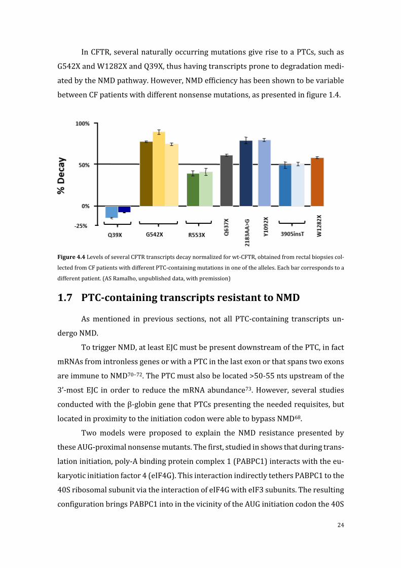
In CFTR, several naturally occurring mutations give rise to a PTCs, such as
G542X and W1282X and Q39X, thus having transcripts prone to degradation medi-
ated by the NMD pathway. However, NMD efficiency has been shown to be variable
between CF patients with different nonsense mutations, as presented in figure 1.4.
Figure 4.4 Levels of several CFTR transcripts decay normalized for wt-CFTR, obtained from rectal biopsies col-
lected from CF patients with different PTC-containing mutations in one of the alleles. Each bar corresponds to a
different patient. (AS Ramalho, unpublished data, with premission)
1.7 PTC-containing transcripts resistant to NMD
As mentioned in previous sections, not all PTC-containing transcripts un-
dergo NMD.
To trigger NMD, at least EJC must be present downstream of the PTC, in fact
mRNAs from intronless genes or with a PTC in the last exon or that spans two exons
are immune to NMD70–72. The PTC must also be located >50-55 nts upstream of the
3’-most EJC in order to reduce the mRNA abundance73. However, several studies
conducted with the β-globin gene that PTCs presenting the needed requisites, but
located in proximity to the initiation codon were able to bypass NMD68.
Two models were proposed to explain the NMD resistance presented by
these AUG-proximal nonsense mutants. The first, studied in shows that during trans-
lation initiation, poly-A binding protein complex 1 (PABPC1) interacts with the eu-
karyotic initiation factor 4 (eIF4G). This interaction indirectly tethers PABPC1 to the
40S ribosomal subunit via the interaction of eIF4G with eIF3 subunits. The resulting
configuration brings PABPC1 into in the vicinity of the AUG initiation codon the 40S

25
during the initial phase of translation elongation brings it into close contact with an
AUG-proximal PTC in a transcript where the ORF is quite short. This proximity to
the PTC allows PABPC1 to interact with the release factor eRF3 at the termination
complex, thus impairing the association of UPF1 to the ribonucleoprotein complex,
resulting in efficient translation termination and inhibition of NMD74.
While other postulates that this immunity NMD, is due to the presence of al-
ternative initiation sites downstream of the PTC, and that the presence of ribosome
initiating the translation elongation process at those sites, might contribute to the
transcript stability and prevents NMD triggering75.
1.9 Premature termination codon read-through
As above-mentioned the presence of in-frame PTCs can lead to the produc-
tion of truncated nonfunctional or deleterious proteins, or trigger NMD resulting no
production of protein at all. Therapeutic approaches aimed at promoting transla-
tional read-through of the PTCs (figure 1.5), and thus enable the synthesis and ex-
pression of full-length functional proteins were developed with relatively positive
results76.
Read-through of PTCs can be achieved by suppressor transfer RNAs (tRNAs),
factors that decrease translation-termination efficiency, such as small-interfering
RNAs (siRNAs) directed against the translation-termination factors and RNA anti-
sense that targets the nonsense mutation region77.
Another extensively studied approach that has reached clinical trials is read-
through by drugs affecting the ribosome decoding site, such as aminoglycoside an-
tibiotics like G418 and Gentamicin. However, aminoglycosides have severe side ef-
fects, such as nephrotoxicity and ototoxicity, when used at high concentrations
and/or used long-term78. While searching for non-aminoglycosides capable of pro-
moting read-through, a high-throughput screening revealed a small molecule, Ata-
luren (previously PTC124), which can read-through PTCs without severe side ef-
fects79. This molecule is now in phase 3 clinical trial for CF patients with nonsense mu-
tations80. However, unfortunately, the preliminary results showed that not all the pa-

26
tients responded to the treatment and the patients that are being treated with amino-
glycosides antibiotic due to bacterial infection prior to the clinical trial were the ones to
show worse results81.
Figure 1.5. The effect of read-through strategies on protein translation. Several ways exist for modifying the nor-
mal processes that occur during termination by a premature termination codon (PTC). (a) Normal translation,
(b) In the presence of a PTC, there is no tRNA matching the stop codon. Instead, the release factors eRF1 and
eRF3 bind and terminate translation by releasing the polypeptide, which is a truncated protein. (c) When ami-
noglycosides, PTC124, or negamycin bind to the rRNA there is no premature termination of translation, despite
the presence of a PTC. An alteration of rRNA conformation is induced upon binding of the small molecule, reduc-
ing the accuracy of the codon–anticodon interaction. This enables incorporation of an aminoacetylated tRNA,
moving translation towards the canonical stop codon and originating a full-length protein. These proteins often
contain missense mutations at the PTC location because near-cognate tRNAs may recognize the codon sequence
by two nucleotides, allowing the insertion of near-cognate amino acids instead of the cognate amino acid. There-
fore, the proteins produced may be functional or non-functional depending on whether or not the missense mu-
tations affect conformation and binding to other proteins. The interaction of aminoglycosides, negamycin, and
PTC124 with the rRNA may be different. In this picture, and for the sake of simplicity, the interaction between
the read-through agent and the rRNA is depicted at the same spot in the three strategies. (d) Depletion of release
factors eRF1 and/or eRF3 leaves the A site available to the entrance of any tRNA that can interact with the PTC,
promoting missense read-through. This may lead to the production of a full-length protein carrying missense
mutations. (e) The suppressor-tRNA anticodon is mutated to be complementary to the PTC, so it is able to rec-
ognize the PTC and insert the cognate amino acid. A competition occurs between suppressor-tRNA and the re-
lease factors eRF1 and eRF3 for the A site of rRNA. When the suppressor-tRNA enters the A site, with successful
interaction with the mRNA PTC, the cognate amino acid is bound to the nascent polypeptide, with read-through
of the PTC, and a normal full-length protein is produced. Adapted from Bordeira-Carriço (2012) 82

27
2 Objectives
The main objective of the present work was to gain a better understanding of
the NMD mechanism in large genes using as model the CFTR gene.
More specifically, we intended to characterize the naturally occurring CFTR
AUG-proximal nonsense mutations, Q2X, S4X and Q39X using novel CFTR mini-
genes by testing their ability to escape NMD in comparison to other CFTR nonsense
mutations which induce NMD, such as G542X. Furthermore, we aimed also to deter-
mine the effect of several pharmacological compounds in read-through transcripts
containing these mutations.
To accomplish the main objectives, this work was divided into several tasks
that were necessary to achieve, as described below:
1) Generate CFTR minigenes containing several CF-causing nonsense
mutations;
2) Validate those minigenes as bonafide NMD models of CFTR transcripts
in immortalized human embryonic kidney (HEK293) cells;
3) Test the ability of different CFTR nonsense mutations to trigger NMD;
4) Investigate the possible cause for NMD resistance of CFTR transcripts
containing AUG-proximal nonsense mutations;
5) Test the potential of read-through promoting drugs to restore full-
length CFTR expression of nonsense variants.

28
3 Materials and Methods
3.1 Production of vectors to study the susceptibility of
CFTR mutant transcripts to NMD
3.1.1 Bacterial strain
The bacterial strain used for cloning and DNA amplification was XL1-Blue
(Stratagene), which is tetracycline resistant. XL1-Blue cells are endonuclease (endA)
deficient, which greatly improves the quality of miniprep DNA, and are recombina-
tion (recA) deficient, improving insert stability. The hsdR mutation prevents the
cleavage of cloned DNA by the EcoK endonuclease system.
The lacIqZΔ M15 gene on the F´ episome allows blue-white color screening.
XL1-Blue Genotype: recA1 endA1 gyrA96 thi-1 hsdR17 supE44 relA1 lac [F´ proAB
lacIqZΔM15 Tn10 (Tetr)]. (Genes listed signify mutant alleles. Genes on the F´ epi-
some, however, are wild-type unless indicated otherwise).
3.1.2 Plasmid vectors
pcDNA5/FRT/CFTR wt (kindly provided by the Garry Cutting lab, at Johns
Hopkins Hospital,Baltimore,USA), comprises the complementary DNA (cDNA) from
the CFTR wild-type gene from position 122 to 4725 (Genebank accession number
M28668) in the PmeI site of the multi-cloning site of pCDNA5/FRT vector (Invitro-
gen). All the others recombinant vectors are produced using this as template.
pcDNA5/FRT/CFTR+IVS4art+IVS5, corresponds to the wt-CFTR cDNA with the in-
sertion of artificial constructed intron (IVS) 4 (with approximately ~300nts of the
5’ of the normal IVS4 sequence joined with ~300 nts 3’ of the of the normal IVS4
sequence) between exon 4 and 5 and insertion of normal full-length IVS5 sequence
between exon 5 and 6a, pcDNA5/FRT/CFTR+IVS13+IVS14a+IVS14b, wt-CFTR
cDNA with the insertion of normal intron 13 sequence, artificial constructed IVS14a
(produced as IVS4art) and normal sequence of IVS14b introduced in the correct lo-
cation of the introns, and pOG44, a vector which encodes for the flipase recom-
binase. These vectors produced in the lab prior to this work. The

29
pCDNA5/FRT/CFTR vectores containing introns will be hereafter referred as CFTR
minigenes.
Maps and cloning schemes for these vectors are available in Appendices. All
these plasmids contain the ampicillin resistance gene, which was used for selection
of transformed bacteria.
The empty vector (pcDNA5/FRT) was produced here thought the removal of
fragment containing the CFTR open reading frame of p
cDNA5/FRT/CFTR+IVS14aart+IVS14b+IVS15, as described in section 3.1.6. Non-
sense mutants of CFTR (Q2X, S4X, Q39X and G542X) vectors were obtained by site-
directed mutagenesis using as template the CFTR minigenes as described in section
3.1.7.
3.1.3 Production of competent bacteria
Bacteria were plated in LB-agar medium supplemented with tetracycline (25
µg/ml) and a single colony was used to inoculate 10mL of LB medium overnight at
37ºC with adequate shaking (220rpm). Tis pre-inoculum was the used to inoculate
at dilution 1/100 100mL of LB medium, grown in the previous conditions to a final
concentration of 5 × 10⁷ bacteria/mL (0.3 abs at 600nm). Bacteria were transferred
to ice and centrifuged at 100g for 15min at 4ºC. The pellet was the resuspended and
incubated on ice for 15 min in 33mL of RF1 buffer (100mM RbCl, 50mM MnCl2,
30mM KCH3COO pH 7.5, 10mM CaCL2, 15% (w/v) glycerol, pH 5.8) and pelleted
again in the same conditions following a resuspention and incubation of 15min on
ice in 8.3mL of RF2 buffer (10mM MOPS, 10mM RbCl, 15mM CaCl2, 15% (w/v) glyc-
erol, pH5.8). 200µL aliquots were then rapidly frozen with liquid nitrogen and
stored at -80ºC.
3.1.4 Transformation of competent bacteria
Bacteria were transformed by incubating 200 µL of competent cells with DNA
(0.1-10ng of plasmid DNA) for 45 min on ice, followed by heat-shock (2min at 42ºC),
90sec incubation on ice, and then allowing antibiotic resistance to be expressed by
growth in antibiotic-free LB medium for 1 h at 37ºC at 220rpm. Bacteria were the

30
pelleted (5000g for 2min), the supernatant was discarded and the pellet resus-
pended in the remaining supernatant. The suspension was then plated into LB-agar
supplemented with 100µg/mL ampicillin and left to grow o/n at 37ºC.
Transformed bacteria colonies were grown o/n in 10mL of LB medium sup-
plemented with 100µg/mL ampicillin at 37ºC at 220rpm and used the following day
to extract plasmid DNA.
Individual clones were stored at -20ºC in 1.5mL of LB medium supplemented
with 15% (w/v).
3.1.5 DNA extraction and quantification
Small scale plasmid DNA was purified with the QIAprep Spin Miniprep Kit
(QIAGEN) according to the manufacturer’s instructions.
DNA concentration was determined by measurement of absorbance at
260nm (one absorbance unit corresponding to 50µg/mL of dsDNA using Nanodrop
200 spectrophotometer (Alfagene) and its purity was evaluated by the assessment
of the A260/A280 ratio. Only DNA with a ratio above 1.8 was considered pure
enough for further use. Only DNA concentrations above 500ng/µL were considered
acceptable for cell transfection.
3.1.6 Cloning-out
In order to produce the empty vector pcDNA5/FRT, the fragment containing
the CFTR gene was cloned-out from the plasmid
pcDNA5/FRT/CFTR+IVS14aart+IVS14b+IVS15. The plasmid was hydrolized with
PmeI endonuclease (Fermentas) and the resulting fragments were separated by aga-
rose gel electrophoresis. The band containing the fragment corresponding to the
empty vector were excised from the gel and frozen for 10 min at -80ºC following a
centrifugation of 5 min at 10000 g. The agarose gel was then discarded and the liq-
uid resulting from the previous centrifugation was incubated with T4-DNA ligase
and ligase buffer (Roche) for 5 min at room temperature. Bacteria were then trans-
formed with the ligation product, plated into LB-agar supplemented with 100
µg/mL ampicillin and left to grow o/n at 37ºC.

31
Single colonies were then grown o/n in 10 mL of LB medium supplemented
with 100 µg/mL ampicillin for plasmid extraction and sequencing.
3.1.7 Site-directed Mutagenesis
Nonsense mutations were introduced into pcDNA5/FRT/CFTR+IVS4art+
IVS5 and pcDNA5/FRT/CFTR+IVS14aart+IVS14b+IVS15 using the KOD Hot
Start Kit (Toyobo) with complementary pairs of mutagenic primers described in ta-
ble 3.2. The PCR programs used are displayed in table 3.1. The amplification of plas-
mids that underwent site-directed mutagenesis was confirmed by agarose gel elec-
trophoresis, and the PCR products incubated with DpnI, a restriction enzyme that
specifically hydrolyzes methylated and hemi-methylated DNA, in order to com-
pletely degrade the template DNA, that is heavy methylated due to its bacterial
origin, giving origin to pure sample of the mutagenized plasmid that was synthetized
in vitro.
After hydrolysis, bacteria were transformed with PCR products. Following
transformation, plasmid DNA was extracted and the presence of each mutation was
confirmed by automatic DNA sequencing.
Table 3.1 – PCR program used for the insertion of nonsense mutations by site-directed mutagenesis
32
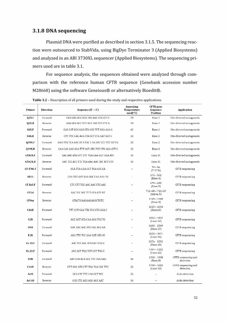
3.1.8 DNA sequencing
Plasmid DNA were purified as described in section 3.1.5. The sequencing reac-
tion were outsourced to StabVida, using BigDye Terminator 3 (Applied Biosystems)
and analyzed in an ABI 3730XL sequencer (Applied Biosystems). The sequencing pri-
mers used are in table 3.1.
For sequence analysis, the sequences obtained were analyzed through com-
parison with the reference human CFTR sequence (Genebank accession number
M28668) using the software Geneious® or alternatively Bioedit®.
Table 3.2 – Description of all primers used during the study and respective applications
33
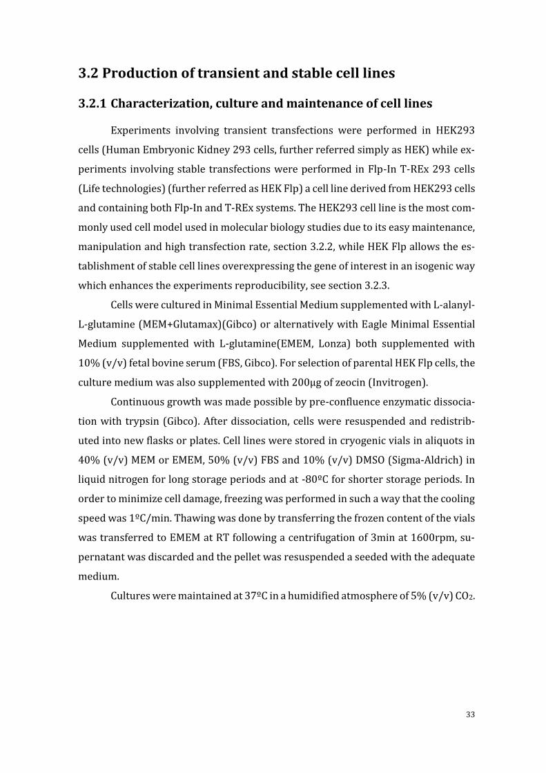
3.2 Production of transient and stable cell lines
3.2.1 Characterization, culture and maintenance of cell lines
Experiments involving transient transfections were performed in HEK293
cells (Human Embryonic Kidney 293 cells, further referred simply as HEK) while ex-
periments involving stable transfections were performed in Flp-In T-REx 293 cells
(Life technologies) (further referred as HEK Flp) a cell line derived from HEK293 cells
and containing both Flp-In and T-REx systems. The HEK293 cell line is the most com-
monly used cell model used in molecular biology studies due to its easy maintenance,
manipulation and high transfection rate, section 3.2.2, while HEK Flp allows the es-
tablishment of stable cell lines overexpressing the gene of interest in an isogenic way
which enhances the experiments reproducibility, see section 3.2.3.
Cells were cultured in Minimal Essential Medium supplemented with L-alanyl-
L-glutamine (MEM+Glutamax)(Gibco) or alternatively with Eagle Minimal Essential
Medium supplemented with L-glutamine(EMEM, Lonza) both supplemented with
10% (v/v) fetal bovine serum (FBS, Gibco). For selection of parental HEK Flp cells, the
culture medium was also supplemented with 200µg of zeocin (Invitrogen).
Continuous growth was made possible by pre-confluence enzymatic dissocia-
tion with trypsin (Gibco). After dissociation, cells were resuspended and redistrib-
uted into new flasks or plates. Cell lines were stored in cryogenic vials in aliquots in
40% (v/v) MEM or EMEM, 50% (v/v) FBS and 10% (v/v) DMSO (Sigma-Aldrich) in
liquid nitrogen for long storage periods and at -80ºC for shorter storage periods. In
order to minimize cell damage, freezing was performed in such a way that the cooling
speed was 1ºC/min. Thawing was done by transferring the frozen content of the vials
was transferred to EMEM at RT following a centrifugation of 3min at 1600rpm, su-
pernatant was discarded and the pellet was resuspended a seeded with the adequate
medium.
Cultures were maintained at 37ºC in a humidified atmosphere of 5% (v/v) CO2.
34
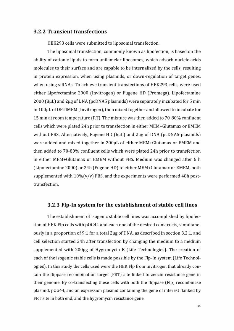
3.2.2 Transient transfections
HEK293 cells were submitted to liposomal transfection.
The liposomal transfection, commonly known as lipofection, is based on the
ability of cationic lipids to form unilamelar liposomes, which adsorb nucleic acids
molecules to their surface and are capable to be internalized by the cells, resulting
in protein expression, when using plasmids, or down-regulation of target genes,
when using siRNAs. To achieve transient transfections of HEK293 cells, were used
either Lipofectamine 2000 (Invitrogen) or Fugene HD (Promega). Lipofectamine
2000 (8µL) and 2µg of DNA (pcDNA5 plasmids) were separately incubated for 5 min
in 100µL of OPTIMEM (Invitrogen), then mixed together and allowed to incubate for
15 min at room temperature (RT). The mixture was then added to 70-80% confluent
cells which were plated 24h prior to transfection in either MEM+Glutamax or EMEM
without FBS. Alternatively, Fugene HD (6µL) and 2µg of DNA (pcDNA5 plasmids)
were added and mixed together in 200µL of either MEM+Glutamax or EMEM and
then added to 70-80% confluent cells which were plated 24h prior to transfection
in either MEM+Glutamax or EMEM without FBS. Medium was changed after 6 h
(Lipofectamine 2000) or 24h (Fugene HD) to either MEM+Glutamax or EMEM, both
supplemented with 10%(v/v) FBS, and the experiments were performed 48h post-
transfection.
3.2.3 Flp-In system for the establishment of stable cell lines
The establishment of isogenic stable cell lines was accomplished by lipofec-
tion of HEK Flp cells with pOG44 and each one of the desired constructs, simultane-
ously in a proportion of 9:1 for a total 2µg of DNA, as described in section 3.2.1, and
cell selection started 24h after transfection by changing the medium to a medium
supplemented with 200µg of Hygromycin B (Life Technologies). The creation of
each of the isogenic stable cells is made possible by the Flp-In system (Life Technol-
ogies). In this study the cells used were the HEK Flp from Invitrogen that already con-
tain the flippase recombination target (FRT) site linked to zeocin resistance gene in
their genome. By co-transfecting these cells with both the flippase (Flp) recombinase
plasmid, pOG44, and an expression plasmid containing the gene of interest flanked by
FRT site in both end, and the hygromycin resistance gene.

35
The presence of the Flp recombinase induces the integration of the fragment
in the plasmid flanked by the FRT site in recombinase target site present in the cells
genome leading to the stable expression of the gene of interest, loss of the zeocin
resistance and acquisition of resistance to Hygromycin B (Figure 3.1). By this system
is only integrated a single copy of the fragment of the plasmid of interest per cell,
originating in a cell population with isogenic expression pattern of the gene of inter-
est, after selection with Hygromycin B.
Figure 3.1 – Schematic representation of the general mechanism for generating cell lines with constitutive ex-
pression of a gene of interest via Flp-In system. Adapted from the product manual provided by the manufacturer
(Invitrogen).

36
This avoids the need for single clone selection and the establishment of iso-
genic stable cell lines was accomplished by lipofection of HEK Flp cells with pOG44
and each one of the desired constructs, simultaneously in a proportion of 9:1 for a
total 2µg of DNA, as described in section 3.2.1, and cell selection started 24h after
transfection by changing the medium to a medium supplemented with 200µg of Hy-
gromycin B.
3.3 RT-PCR analysis of CFTR PTC-containing transcripts
3.3.1 RNA extraction
RNA from HEK293 and HEK Flp was extracted with the nucleospin RNA II kit
(Macherey-Nagel) following the protocol provided by the manufacturer. Every step
was executed in RNAse free environment to avoid RNA degradation. Following ex-
traction were quantitated using the Nanodrop 200 spectrophotometer, and purity
was assessed by the A260/A280 ratio. Only RNA samples with a ratio above 2.0 were
deemed acceptable. The extracted samples stored at -80ºC.
3.3.2 cDNA synthesis
For the synthesis of cDNA, 500ng of the previously extracted mRNA were in-
cubated with 1µL of oligo-dTs (short sequence of deoxy-thymine nucleotides) (Invi-
trogen) for 10 min at 60ºC. This first step promotes the denaturation of the second-
ary structure of the RNA facilitating the binding of the oligo-dTs to the poly-A (poly-
adenosine) strand of mRNAs and enhancing reverse transcription yield. Afterwards
the pre-mix containing the RNA and the oligo-dTs was put on ice and was added
6.9µL of an RT (reverse transcription) mix containing, 1x 1st strand Buffer (4µL of a
5x stock) (Invitrogen), 0.1M DTT (1µL of 1M stock)(Invitrogen) , 40U/µL RNase In-
hibitor (0.5µL) (Invitrogen), 25mM dNTP mix (0,4µL)(Invitrogen), and ddH2O (bi-
distillated water)(Invitrogen) for a final volume of 19µL. This mixture was then in-
cubated at 42ºC for 2min, followed by the addition of 1µL of Superscript II RT en-
zyme (200U/µL, Invitrogen), and further incubation at 42ºC for 1h. The reaction was

37
stopped by inactivating the enzyme with an incubation of the mixture at 70ºC for
15min. Samples were stored at -20ºC.
3.3.3 Polymerase chain reaction
After RNA extraction and cDNA synthesis, see sections 3.3.1 and 3.3.2, detec-
tion and semi-quantitative analysis of CFTR transcripts were done by regular poly-
merase chain reaction (PCR) and a semi-quantitative variant of this method, respec-
tively.
The PCR is a widely spread technique used in most research labs focused on
medical and biological research due to large spectrum of applications that goes from
gene expression detection to genetic engineering. It consists on cycles of repeated
heating and cooling of the reaction for DNA melting, primers annealing and enzy-
matic replication of DNA by a DNA polymerase.
For the regular PCR method, a pre-reaction mix was prepared, containing 1µL
of each of the reverse primer and forward primer, (10pmol/µL)(primers pairs in
table3.2, which are complementary with the interest sequences, 0.2µL of Taq Poly-
merase (NZYtech), 5 µL of polymerase Buffer solution (10x concentrated, NZYtech),
3µL MgCl2 (50 mM), 0.2 µL dNTPs mix (25 pmol/µL of each dNTP) and water for a
final volume of 49µL, per reaction, being possible to scale up or down according to
the experiments necessities. All these steps were executed in DNA free environment
to avoid contamination of the reagents and on ice to avoid unspecific amplification.
Afterwards, 1µL of cDNA was added to each reaction. The PCR reactions were exe-
cuted in a T-Professional thermo-cycler (Biometra) following the heating-cooling
cycles stated on table 3.3. After the thermo-cycling, the contents of each reaction
were analyzed by agarose gel electrophoresis. A 2% (m/v) agarose (Lonza) gel in
TAE buffer (…) with 1U of RedSafe(Chembio was runned for at least 40 min at 120
Volts. After electrophoresis the PCR products were visualized by exposing the gel to
UV light in a dark container coupled to a Kodak photographic machine (Kodak EDAS
290 Electrophoresis Documentation System). The images capture and analysis was
done using the Kodak EDAS 290 Electrophoresis Documentation System software.

38
3.3.3.1 Semi-quantitative analysis
For semi-quantitation of expression of the CFTR gene by each of the nonsense
mutants, there were introduced variations to the PCR method. Instead of letting the
reaction conclude all the heating-cooling cycles as in a regular PCR, 5µL of each re-
action, were collected after 20, 22, 24, 26, 28 and 30 cycles. Afterwards, the ampli-
fied DNA was separated and detected, as described previously (section 3.3.3), and
signal intensity of the bands corresponding to CFTR and β-actin were quantified
with the IMAGE J software. This allowed for a comparative analysis of the quantity
of CFTR transcripts between each of the cell lines expressing the different CFTR var-
iants at the exponential phase of replication which occurred between cycles 22 and
28, when CFTR signal was detected.
Table 3.3 – PCR program used for the detection of CFTR and β-actin transcripts by RT-PCR
3.4 Biochemical and functional characterization of CFTR
nonsense variants
3.4.1 Preparation of total protein extracts
For Western blot (WB), protein extracts were prepared by cell lysis with a
lysis buffer with following composition; 1.5% (w/v) SDS (Sodium dodecyl sul-
fate)(Gibco) ; 5% (v/v) glycerol; 0.01% (w/v) bromophenol blue; 0.05 mM dithio-
treithol; 0.095M Tris pH 6.8). In order to reduce sample viscosity, DNA was sheared

39
by enzymatic action of 5U/mL benzonase (Sigma-Aldrich) in the presence of 2.5mM
MgCl2.
Samples total protein concentration was assessed by Lowry protein assay83.
3.4.2 Western blot
Protein extracts were separated by SDS-PAGE (polyacrylamide gel electro-
phoresis) on 7% polyacrylamide mini-gels for 2-3hours at 120mV. After separation,
the proteins were transferred from the gel onto Immobillon polivinylidene difluo-
ride (PVDF) membranes (Milipore). Membranes were then blocked with 5%
skimmed milk (Nestlé) in phosphate-buffered saline with Tween detergent (Bio
Rad) (PBS-T, NaCl 137mM; KCl 2.7mM; KH2PO4 6.5mM, pH 7.4, 1% (v/v) Tween) for
1h at RT. After blocking the membranes were incubated, o/n at 4ºC, with primary
antibodies, 596 (CFF, mouse, 1:3000), α-calnexin (Milipore, mouse, 1:3000), MM13-
4 (Milipore, mouse, 1:1000), in 5% (w/v) skimmed milk in PBS-T, followed by
3x15min washes with PBS-T, and then incubated for 1h at RT with the horseradish
peroxidase-conjugated secondary anti-mouse IgG antibody (Bio Rad, Donkey,
1:10000), diluted in 5% (w/v) skimmed milk in PBS-T. Membranes were again
washed 3x15min in PBS-T. Blots were developed with the ImmunStar Western C
Chemiluminescence kit (BioRad) using the Chemidoc XRS+ analyser (BioRad) for sig-
nal detection and caption.
Signal quantification was performed with the ImageLab software (BioRad).
3.4.3 Immunofluorescence
Cells were grown in 10x10mm coverslips placed in P24 well plates coated
with 0.001% (w/v) poly-L-lysine, until 40-60% confluence was reached. The cells
were washed 3x10min with HBSS (Hank’s balanced salt solution) (Gibco) with
140rpm agitation, and in order to avoid unspecific binding of the antibodies, a block-
ing step with 1% (w/v) BSA in HBSS was performed for 30min. Then the cells were
incubated with TexasRed-conjugated wheat germ agglutinin (WGA)(Sigma Aldrich)
diluted 1:200 (v/v) in HBSS with 0.5% (w/v) BSA, and washed 5x5min with HBSS

40
with 140rpm agitation. All the steps until fixation were executed on ice as to avoid
WGA internalization by the cells.
Cells were fixed for 45min with 2% (v/v) formaldehyde, washed 3x10min
with PBS supplemented with CaCl2 and MgCl2 (PBS+/+), and permeabilized by incu-
bating with 0.2% (w/v) Triton X-100 (BioRad) in PBS+/+ for 15min at RT. After
washing 3x10min with PBS+/+ at 140rpm at RT, cells were incubated, o/n at 4ºC in
a humid chamber, with anti-CFTR antibody 570 (CFF) diluted 1:500 (v/v) in PBS+/+
with 0,5% (w/v) BSA, and washed 3x10min with PBS+/+ at 140 rpm. This was fol-
lowed by an incubation with an anti-mouse IgG antibody conjugated with alexa 488
(Bio Rad), diluted 1:1000 (v/v) in PBS+/+, for 1h at RT. Then the cells were washed
3x10min at 140rpm with PBS+/+. Finally the coverslips were mounted in Vec-
tashield (Vector labs) with 1.5µg/mL 49,6-diamidino-2-phenyl-indole (DAPI) for
nuclear detection on glass slides and visualized in wide-field fluorescence micros-
copy with the microscope (Zeiss, Jena) and images were captured, edited and ana-
lyzed with ZEN 9 microscopy software (Zeiss).
3.4.4 Iodide Efflux
Cells were grown in P60 plates coated with 0.001% (w/v) poly-L-lysine.
After achieving total or near total (90-95%) confluence, cells were gently
washed two times with 2.5 ml of an I- loading buffer (136 mM sodium iodide, 3 mM
potassium nitrate, 2 mM calcium nitrate, 11 mM glucose and 20mM HEPES, pH 7.4)
previously warmed to 37ºC. Afterwards the cells were incubated, for 1h at 37ºC, in
2.5mL of loading buffer
Following the incubation cells were slowly and gently washed ten times for
1 min with 2.5 ml of an I- free efflux buffer (136 mM sodium nitrate, 3 mM potassium
nitrate, 2mM calcium nitrate, 11mM glucose and 20mM HEPES, pH 7.4) also warmed
to 37ºC, equilibrating for 5 min in I- free efflux buffer after the last wash.
After the equilibration period, the buffer was removed and replaced with
fresh efflux buffer which was collected to P35 plate protect from incident light, and
substituted with fresh efflux buffer after a 1 min incubation. This step was repeated
from 0-4 min time-points.

41
At time point 0 min the fresh efflux buffer added was supplemented with
10µM of forskolin and 50µM of genistein, two CFTR agonists that stimulate the chan-
nel opening. The collection and renewal step with the efflux buffer containing the
CFTR agonists was repeated until the 4 min time-point.
After minute 4 the efflux buffer was substituted for efflux buffer without ag-
onists, and the cycle repeated until time point 10.
Afterwards iodide concentrations from each collected samples were meas-
ured with an I- selective electrode (Orion).
Simultaneously, were prepared, p35 plates, solutions with crescent concen-
trations of NaI; 1µM; 10 µM; 50 µM and 100 µM. These solutions were used to deter-
mine the standard curve of voltage values, measured by the I- selective electrode,
versus iodide concentration, which will allow calculating iodide concentration of
each collected sample.
3.5 Pharmacological treatments
3.5.1 Pharmacological indirect inhibition of Nonsense-mediated
mRNA decay
In order to induce indirect inhibition of NMD, HEK Flp cells, stably expressing
different CFTR variants, were grown in P24 wells plates coated with 0.001% of poly-
L-lysine, and incubated for 5h at 37ºC, with serum free EMEM or MEM+GlutaMAX
supplemented with 100 µg/ml of cyclohexamide (CHX) (Sigma-Aldrich). After the
incubation period cells are lysed. RNA extracted and analyzed by RT-PCR, as de-
scribed in section 3.3.
3.5.2 Pharmacological induction of PTC read-through
In order to study the efficacy of some of these drugs in the induction of PTC
read-through on CFTR transcripts containing in their sequence on different posi-
tions, HEK Flp cells, stably expressing different CFTR variants were in treated, for
24h at 37ºC, with 50µg/ml of G-418, 50µg/ml of gentamicin, 1mM of Ataluren
(PTC124) or 0.2% (v/v) of DMSO. After treatment cells were submitted to western
blot and mRNA analysis, as described in previous sections.

42
4 Results
4.1 Production of plasmid vectors
In order to study several naturally occurring AUG-proximal nonsense muta-
tions in the CFTR gene, five new plasmid vectors were produced. Three CFTR minig-
enes vectors were used as templates to produce these new vectors as follows: i)
pcDNA5/FRT/CFTR IVS4art+IVS5 (a pCDNA5/FRT vector containing the full-length
CFTR coding sequence as well as the natural occurring CFTR intron 5 (IVS5); ii) and
an artificially constructed intron 4 (IVS4art, with just ~200bp from the 5’ end and
200 bp from the 3’end of the natural occurring intron); and iii) pcDNA5/FRT/CFTR
IVS13+IVS14aart+IVS14b produced in the lab prior to this work.
The vectors produced in this work were:
1) CFTR mutants minigenes containing the naturally occurring nonsense
mutations: Q2X (glutamine to stop codon), S4X (serine to stop codon)
and Q39X (glutamine to stop codon);
2) A G542X (glycine to stop codon) mutant with CFTR introns 13, 14a
(artificially constructed similarly to intron 4 art) and 14b, which is re-
ported to naturally induce NMD84.
3) An empty vector (pcDNA5/FRT), in order to disregard vector interfer-
ence;
The template vectors have a multicloning site that includes two Pme I re-
striction sites bisecting the CFTR ORF (open-reading frame), and an gene that en-
codes for ampicillin resistance in bacteria and another for hygromycin B resistance
in mammalian cells, as well as the short flippase (Flp) recognition target (FRT) sites
flanking the CFTR sequence.
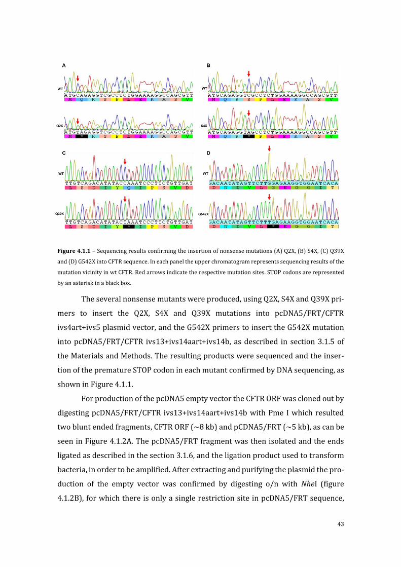
43
Figure 4.1.1 – Sequencing results confirming the insertion of nonsense mutations (A) Q2X, (B) S4X, (C) Q39X
and (D) G542X into CFTR sequence. In each panel the upper chromatogram represents sequencing results of the
mutation vicinity in wt CFTR. Red arrows indicate the respective mutation sites. STOP codons are represented
by an asterisk in a black box.
The several nonsense mutants were produced, using Q2X, S4X and Q39X pri-
mers to insert the Q2X, S4X and Q39X mutations into pcDNA5/FRT/CFTR
ivs4art+ivs5 plasmid vector, and the G542X primers to insert the G542X mutation
into pcDNA5/FRT/CFTR ivs13+ivs14aart+ivs14b, as described in section 3.1.5 of
the Materials and Methods. The resulting products were sequenced and the inser-
tion of the premature STOP codon in each mutant confirmed by DNA sequencing, as
shown in Figure 4.1.1.
For production of the pcDNA5 empty vector the CFTR ORF was cloned out by
digesting pcDNA5/FRT/CFTR ivs13+ivs14aart+ivs14b with Pme I which resulted
two blunt ended fragments, CFTR ORF (~8 kb) and pCDNA5/FRT (~5 kb), as can be
seen in Figure 4.1.2A. The pcDNA5/FRT fragment was then isolated and the ends
ligated as described in the section 3.1.6, and the ligation product used to transform
bacteria, in order to be amplified. After extracting and purifying the plasmid the pro-
duction of the empty vector was confirmed by digesting o/n with NheI (figure
4.1.2B), for which there is only a single restriction site in pcDNA5/FRT sequence,
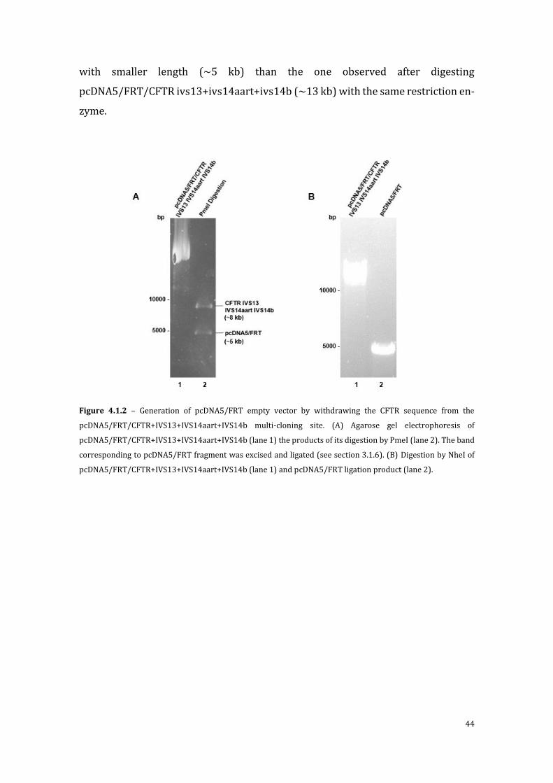
44
with smaller length (~5 kb) than the one observed after digesting
pcDNA5/FRT/CFTR ivs13+ivs14aart+ivs14b (~13 kb) with the same restriction en-
zyme.
Figure 4.1.2 – Generation of pcDNA5/FRT empty vector by withdrawing the CFTR sequence from the
pcDNA5/FRT/CFTR+IVS13+IVS14aart+IVS14b multi-cloning site. (A) Agarose gel electrophoresis of
pcDNA5/FRT/CFTR+IVS13+IVS14aart+IVS14b (lane 1) the products of its digestion by PmeI (lane 2). The band
corresponding to pcDNA5/FRT fragment was excised and ligated (see section 3.1.6). (B) Digestion by NheI of
pcDNA5/FRT/CFTR+IVS13+IVS14aart+IVS14b (lane 1) and pcDNA5/FRT ligation product (lane 2).

45
4.2 Evaluation of the effects of nonsense mutations on
CFTR expression
HEK 293 cells were transiently transfected with each of the nonsense mu-
tants CFTR minigenes produced in section 3.2.2, as well as the wt-CFTR minigenes.
Expression of CFTR transcripts from these minigenes was detected by RT-PCR (see
section 3.3.2) in the region comprised by exon 8-10 (primer pair B3R/C16D; frag-
ment with 390 bp)) evaluated by semi quantitative RT-PCR (see section 3.3.3). Mu-
tations Q2X, S4X and Q39X result in production of CFTR transcripts (Figure 4.2.1A
lanes 3, 4 and 5), having expression levels similar to those of wt-CFTR constructs
(Figure 4.2.1A lanes 1 and 2). The CFTR transcript produced by the G542X mutant
minigene (Figure 4.2.1 A lane 6) is in most replicates almost undetectable, its levels
are very low in comparison to those of wt-CFTR(<5%) and the other nonsense var-
iants. As expected the non-transfected cells (Figure 4.2.1A lane 8) and the cells
transfected with the empty vector do not present any CFTR-mRNA. The quantifica-
tion of the transcripts produced by the mutants and wt-CFTR minigenes is observed
in Figure 4.2.1B and it is possible to observe that there are significant differences
between the levels of the CFTR transcripts obtained from the cells transfected with
AUG proximal nonsense mutants (Q2X, S4X and Q39X) and the non-AUG proximal
nonsense mutant G542X. Furthermore the levels of Q2X, S4X and Q39X are similar
to wt-CFTR minigene (Figure 4.2.1B) suggesting no degradation of these transcripts.
The production of CFTR protein by these transiently transfected cells was as-
sessed by Western blot (see section 3.4.2) using an CFTR antibody that recognizes
an epitope at the C-terminus (aa 1204-1211) of the CFTR protein (see Figure
4.2.2A).
Both wt-CFTR minigenes originate normal CFTR protein, i.e., with both fully
glycosylated (band C, ~170kDa) and core-glycosylated (band B, ~150kDa) forms
(Fig 4.2.2A, lane 1 and 2). CFTR variants with nonsense mutations in proximity to
the translation initiation codon (Q2X, S4X and Q39X) led to the production of a form
of CFTR of lower molecular weight (~120kDa), for which it is unclear whether it
corresponds to the fully- or to the core glycosylated forms (Figure 4.2.2A lanes 3, 4
and 5). For the G542X mutant, empty vector and non-transfected cells no CFTR pro-
tein was detected (Figure 4.4A lanes 6, 7 and 8 respectively).

46
Figure 4.2.1 – mRNA analysis of HEK 293 cells transiently expressing CFTR minigenes. (A) Detection of CFTR
and β-actin mRNA by RT-PCR after 35 cycles. (B) Quantification of CFTR transcripts after 24 cycles (sq-RT-PCR).
Results are presented in arbitrary units and are normalized to β-actin signal. Error bars represent mean±SD of
the values of each collected sample (n=6). Asterisks and diamonds indicate significant differences from either
wt-CFTR or from G542X, respectively (p-value <0.05).
The alternative form observed for Q2X, S4X and Q39X likely corresponds to
a truncated form originated by translation re-initiation at an alternative Kozak con-
sensus initiation site present downstream of the nonsense mutation, as previously
shown of other CFTR mutations85. To determine whether this hypothesis is correct
and to determine if no truncated G542X protein is produced we repeated the West-
ern blot with another CFTR antibody (MM13-4), which recognizes an epitope at the
N-terminus region (aa 24–35) of CFTR (Figure 4.2.2B). This antibody was unable to
detected the CFTR form produced by the Q2X, S4X, Q39X and G542X (Figure 4.2.2B
lane 2, 3, 4 and 7, respectively) but could detect the CFTR protein produced by the
wt-CFTR minigene. These results show that the alternative forms detected for Q2X,
S4X, Q39X result from re-initiation of CFTR translation downstream of position 39.
Moreover, it was possible to verify that no protein is produced from the G542X mu-
tant.

47
Figure 4.2.2 - Detection of CFTR. Whole cell lysates of HEK 293 cells transiently expressing wt-and CFTR minig-
enes with nonsense mutations were analysed by western blot. (A) CFTR detection with 596 antibody (comprises
CFTR a.a. res. 1204-1211). Both wt-CFTR minigenes were able to acquire full maturation, which is observed by
the presence of the full-glycosylated CFTR characteristic (band C). A band (indicated by the red arrows) with
lower molecular weight (120KDa) than band B (core-glycosylated CFTR) was observed on variants with an AUG-
proximal nonsense mutation (lane 3-5). (B) CFTR detection with MM13-4 antibody (epitope comprises CFTR
a.a. res. 25-36), which recognizes CFTR N-terminus region. CFTR was only detected in cells expressing wt-CFTR.

48
4.3 Assessment of NMD susceptibility of CFTR nonsense
mutants
In order to discard the influence of transfection efficiency and the variability
of expression patterns between transfections, HEK cell lines stably expressing a sin-
gle copy of each of the constructs under study were generated. Plasmid incorpora-
tion was ensured by treating the cells with selective marker hygromycin B (200µM),
for variable periods of time, depending on cell growth rate, starting 48h after trans-
fection.
After positive selection of each cell line, CFTR expression patterns were as-
sessed by semi-quantitative (sq) RT-PCR and Western blot analysis (see sections
3.3.3.1 and 3.4.2, respectively).
Figure 4.3.1 – sq-RT-PCR analysis of CFTR transcripts extracted from HEK TREX Flp-In cells expressing CFTR
minigenes. Results are presented in arbitrary units and are normalized to β-actin signal. Error bars represent
mean±SD of the values of each collected sample (n=12). Asterisks and diamonds indicate significant differences
from either wt-CFTR or from G542X (p-value <0.01 and <0.001, respectively).
The presence of nonsense mutations in proximity with the AUG start codon
(Q2X, S4X and Q39X) prevented, as was observed in transiently transfected cells
(section 4.2), degradation of CFTR transcripts and led to the production of a trun-
cated form of CFTR lacking the N-terminal region through alternative initiation.
Transcript levels of these variants were comparable to those obtained for wt-CFTR
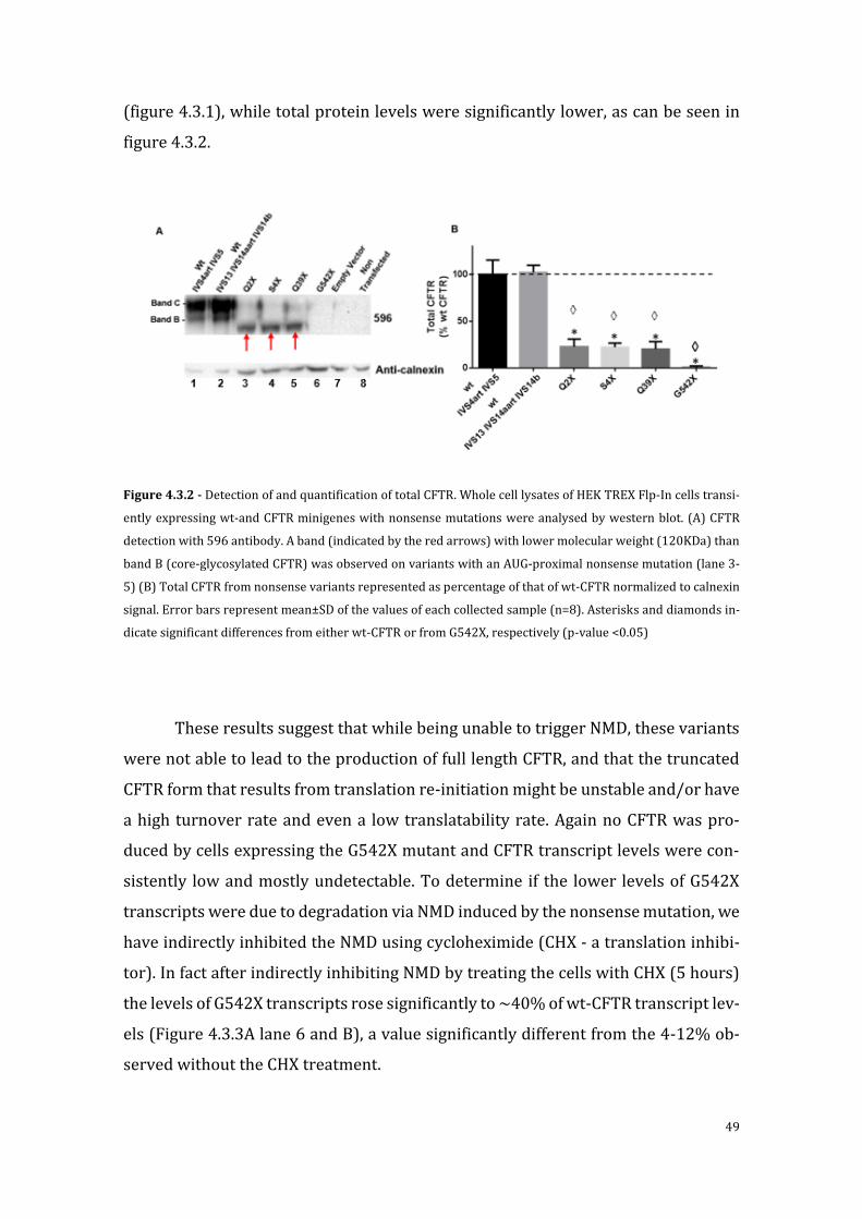
49
(figure 4.3.1), while total protein levels were significantly lower, as can be seen in
figure 4.3.2.
Figure 4.3.2 - Detection of and quantification of total CFTR. Whole cell lysates of HEK TREX Flp-In cells transi-
ently expressing wt-and CFTR minigenes with nonsense mutations were analysed by western blot. (A) CFTR
detection with 596 antibody. A band (indicated by the red arrows) with lower molecular weight (120KDa) than
band B (core-glycosylated CFTR) was observed on variants with an AUG-proximal nonsense mutation (lane 3-
5) (B) Total CFTR from nonsense variants represented as percentage of that of wt-CFTR normalized to calnexin
signal. Error bars represent mean±SD of the values of each collected sample (n=8). Asterisks and diamonds in-
dicate significant differences from either wt-CFTR or from G542X, respectively (p-value <0.05)
These results suggest that while being unable to trigger NMD, these variants
were not able to lead to the production of full length CFTR, and that the truncated
CFTR form that results from translation re-initiation might be unstable and/or have
a high turnover rate and even a low translatability rate. Again no CFTR was pro-
duced by cells expressing the G542X mutant and CFTR transcript levels were con-
sistently low and mostly undetectable. To determine if the lower levels of G542X
transcripts were due to degradation via NMD induced by the nonsense mutation, we
have indirectly inhibited the NMD using cycloheximide (CHX - a translation inhibi-
tor). In fact after indirectly inhibiting NMD by treating the cells with CHX (5 hours)
the levels of G542X transcripts rose significantly to ~40% of wt-CFTR transcript lev-
els (Figure 4.3.3A lane 6 and B), a value significantly different from the 4-12% ob-
served without the CHX treatment.

50
Figure 4.3.3 – Effect of NMD indirect inhibition on cells stably expressing CFTR variants with nonsense muta-
tions. (A) Detection of CFTR transcripts after treatment with CHX (25µM) for 5 hours. (B) Comparative sq-RT-
PCR analysis of CFTR transcripts between untreated and treated cells. Results are presented in arbitrary units
and are normalized to β-actin signal. Error bars represent mean±SD of the values of each collected sample (n=3).
Asterisks and diamonds indicate significant differences from either wt-CFTR or the untreated counterpart, re-
spectively (p-value <0.05).
51
4.4 Assessment of intracellular localization and function
of CFTR nonsense mutants
In order to determine the intracellular localization of the truncated forms of
CFTR originated by the Q2X, S4X, Q39X variants, immunofluorescence essays were
performed in the stably transfected cells. For the detection of CFTR the primary 570
anti-CFTR antibody was used and a green fluorescent (alexa 488 secondary anti-
body that recognizes the CFTR primary antibody). In parallel the plasma membrane
(PM) was stained with marker wheat germ agglutinin (red) and the nucleus with
DAPI (blue).
As shown in Figure 4.3.4, CFTR protein can be observed at PM of HEK cells
expressing wt-CFTR, while the F508del-CFTR mutant is predominantly localized at
the cytoplasm. Cells expressing the Q2X, S4X, Q39X variants, produced CFTR with a
localization pattern similar to that of F508del (that is predominantly at the ER) but
also with some PM staining. In accordance with biochemical analysis results no
CFTR was detected in the cells expressing the G542X variant (Figure 4.3.4). For the
cells transfected with the empty vector and the non-transfected cells no CFTR pro-
tein was detected as expected.
These immunofluorescence results show that residual levels of the CFTR pro-
duced by the cells expressing the Q2X, S4X and Q39X variants can be detected at the
PM. In order to assess the channel activity of these truncated CFTR forms, the iodide
efflux assay wase performed (Section 3.4.4).
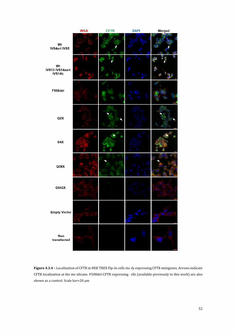
52
Figure 4.3.4 – Localization of CFTR in HEK TREX Flp-In cells stably expressing CFTR minigenes. Arrows indicate
CFTR localization at the membrane. F508del-CFTR expressing cells (available previously to this work) are also
shown as a control. Scale bar=20 µm

53
These immunofluorescence results show that residual levels of the CFTR pro-
duced by the cells expressing the Q2X, S4X and Q39X variants can be detected at the
PM. In order to assess the channel activity of these truncated CFTR forms, the iodide
efflux assay wase performed (Section 3.4.4).
Figure 4.3.5- Functional assessment of CFTR nonsense variants by the iodide efflux essay. (A-I) Time-course
analysis of Cl- channel activity and response to agonists Forskolin and Genistein of CFTR variants stably and
isogenically expressed in HEK Flp cells; (A) wt CFTR with introns 4art and 5; (B) wt CFTR with introns 13, 14aart
and 14b; (C) F508del; (D) Q2X with introns 4art and 5; (E) S4X with introns 4art and 5; (F) Q39X with introns
4art and 5; (G) G542X with introns 13, 14aart and 14b; (H) pcDNA5/FRT empty vector; (I) Non transfected HEK
Flp cells. From minute 0 to 4 forskolin (10 µM) and genistein (50 µM) were added to the efflux buffer. (J) Sum-
mary of the peak iodide efflux magnitudes of the several CFTR variants in absolute values. Symbols and error
bars represent mean ± SD of the values at each time point (n=6). Asterisks and diamonds indicate significant
differences from either wt-CFTR or from F508del, respectively (p-value <0.05).
Cells expressing Q2X, S4X and Q39X mutants produced a residual response
upon stimulation with forskolin (figure 4.9 panels D, E and F), while no response
was detected for G542X-expressing cells. However, these were significantly lower
◊ ◊ ◊ * * *
* * * *

54
than the responses generated by wt-CFTR cells (figure 4.8A) and higher than that of
F508del-CFTR cells (figure 4.8C). CFTR produced by the Q2X, S4X and Q39X cell
lines showed a delayed (minutes 2-5 vs 1-4) and lower (peaked at ~18 vs ~56
nmol/min) response when compared to that generated by wt-CFTR.
4.5 PTC read-through and NMD inhibition in CFTR non-
sense mutants by chemical compounds
As the previous results showed, the mutations Q2X, S4X and Q39X prevented
degradation CFTR transcripts as occurred with mutation G542X, but resulted in a
truncated form of CFTR, lacking the N-terminal region, which only exhibited partial
channel function.
In order to restore full-length and fully functional CFTR in the cells express-
ing the Q2X, S4X and Q39X mutants and G542X, cells were treated with the amino-
glycoside antibiotics gentamicin and G418 as well as with the investigational drug
Ataluren (PTC-124), previously described to induce the read-through of nonsense
mutations either in context of CF or other diseases. However, in our hands none of
the treatments resulted in the detection of full-length CFTR protein for either of non-
sense mutants. Higher compound concentrations or longer treatment periods than
those described in section 3.5.1 lead to complete cell death in all performed at-
tempts.
The results for Ataluren were not completely unexpected, when taking into
account the very modest results from the phase 3 clinical trial and the several exist-
ing reports contesting its putative ability to induce read-through80,86. However, both
aminoglycosides (G418 and gentamicin) used in this study are validated as read-
though promoting agents56 and as such they should have been able to induce the
production of detectable full-length CFTR in the treated cells containing CFTR non-
sense mutations.
55
5 Discussion
The present study was based on previous data by AS Ramalho in the Amarl
lab showing that CFTR transcripts extracted from colon biopsies from CF patients
with the nonsense mutation Q39X, did not undergo degradation through NMD, as
normally occurs for with transcripts containing PTCs further downstream in the in
the CFTR sequence, such as G542X.
Previous in vitro studies showed that β-globin nonsense mutations in close
proximity to initiation codon also protect messengers from undergoing NMD68. It
was proposed that this might be due to two reasons. Firstly, to the interaction of the
PABPC1 with translation termination complex at the nonsense mutation site thus
inhibiting the activation of NMD pathway intermediaries and their interaction with
EJC complex present downstream of the PTC74. Alternatively mRNA protention from
NMD could result from re-initiation of translation at a Kozak consensus downstream
of the mutation which would stabilize these PTC-containing transcripts75. However,
this type of studies had not been performed in larger genes such as CFTR. In fact,
most studies on the NMD pathway use the β-globin gene as model87. This is precisely
due to the small size of the β-globin gene (~1650bp codifying sequence, ~4kb before
splicing) which allows for the insertion of the complete genomic sequence in plas-
mid vectors that usually possess a cloning limit of ~20 kb88. The CFTR gene, in turn,
largely exceed these values (~190kb, 6.5kb after splicing). Thus it is important to
validate these β-globin based models for larger genes to determine whether they are
still valid.
Previously, several novel CFTR minigenes had been produced at the Amaral
lab for the study CFTR splicing altering mutations. These minigenes consisted of
plasmid vectors containing the full-length CFTR cDNA sequence
(pcDNA5/FRT/CFTR) where introns were inserted. Depending on intron size, nor-
mal (small) or artificially constructed (large) introns were inserted. CFTR tran-
scripts and protein produced by cells transfected with these constructs were shown
to be identical to those produced by CFTR cDNA, i.e., in the absence of introns in its
sequence89. These minigenes differ greatly from the constructs usually used in splic-
ing studies of larger genes, which do not not contain the full-length CTR sequence,
but solely the exons and introns of interest90. The major advange of the mini-genes

56
produced in this work is that they can provide information, not only, on the effects
of splicing mutations at transcript levels but also on the resulting proteins.
Since previous reports showed that the presence of one intron downstream
of PTC is enough to prompt mRNA degradation through NMD72, these previously
existing CFTR minigenes were used as the base for the mutant constructs produced
in this study.
The aims of this study were: 1) to validate the generated minigenes as tools
to study in NMD pathway; and 2) to determine if naturally occurring AUG-proximal
CFTR nonsense mutations were resistant to NMD and the possible causes for the
protection.
Our data, both in transiently and stably transfected HEK cells, show that the
presence of the G542X mutation leads to CFTR mRNA degradation. This, however,
was attenuated by treating the cells with translation inhibitor CHX for 5h before
RNA extraction. In fact, by inhibiting translation we indirectly inhibited NMD since
this degradation pathway is entirely dependent on the first round of translation91,92.
These results suggest that when containing a PTC in its sequence these CFTR minig-
enes are in fact capable of inducing NMD. However, assays with direct inhibition of
NMD should be performed, in order to further strengthen this conclusion. Previous
studies showed direct inhibit of NMD can be accomplished by silencing UPF1 or by
treatment with several drugs that promote this inhibition, such as caffeine or wort-
mannin, by suppressing UPF1 phosphorylation93–95.
On the other hand, the CFTR transcripts from the AUG-proximal nonsense
variants Q2X, S4X and Q39X were detected at similar levels of those of wt-CFTR,
even though all mutations are located at more than 50-54 nucleotides upstream of
the last existing intron (the minimum distance in order to trigger NMD by the EJC
rule)72. These data suggest that, similarly to what was observed for the β-globin, the
PTC proximity to the translation initiation in the context of a large gene such as CFTR
also plays a role in the susceptibility of transcripts to NMD.
These mutants lead to the expression of a shorter form of CFTR which lacks
the N-terminus, while no full-length CFTR was detected. Similar results are reported
for a CFTR mutation that deletes the CFTR translation initiation site (c.123del23)85.
For this mutation, it was shown that in the absence of normal initiation codon, co-
dons corresponding to M150, M152 or M156 as well as other downstream in-frame

57
AUG codons could be used as alternative initiation sites due to their high comple-
mentarity to the Kozak consensus. When taken together with the present results,
those data support that also in CFTR the presence of an alternative translation initi-
ation site downstream of the AUG-proximal PTC protects messengers from NMD.
However, we cannot rule out the possibility that the interaction between PABPC1
and the translation termination complex developed at the PTC near the AUG can also
plays a role in this NMD protection observed for the Q2X, S4X and Q39X mutations.
The influence of the position of the nonsense near the AUG codon should thus be
tested. This could be done by inhibiting the interaction between these complexes
(PABPC1-Translation termination complex) by genetic engineering as shown in
original study that established the AUG-proximity model for NMD resistance74.
Our data here also showed that the expressed CFTR protein resulting from
Q2X, S4X and Q39X variants was also at lower levels from what would be expected
taking into account the high level of transcripts obtained with these variants (Q2X,
S4X and Q39X). The lack of the N-terminal region in the CFTR protein may be re-
sponsible for the loss of protein stability and probable increased turnover rate as is
the case of the previously described c.123del23 variant and also altered channel ac-
tivity85. The N-terminal region is known to interact with syntaxin 1A and this inter-
action regulates CFTR traffic to/ stability at the PM. Furthermore, the interaction
between the N-terminus and regulatory domain (RD) of CFTR can regulate CFTR
channel activity96,97.
Furthermore, point mutations at the N-terminal region were shown to hinder
CFTR maturation, also suggesting that this region is important for the folding and
processing of CFTR98. The immunofluorescence assays performed in this work
showed that residual amounts of the CFTR produced by the AUG-proximal nonsense
variants are detectable at the PM, albeit in reduced amounts. This does not imply
that the expressed truncated forms of CFTR are fully or partially glycosylated or
even presents any form of folding homology with full-length CFTR.
CFTR N-terminus harbours an arginine-framed-tripeptidide (AFT) motif at
residues 29-39 that acts as retention/retrieval signal for CFTR at ER exit check-
point99. Removal of this AFT, as in the truncated forms detected here, could also ex-
plain their presence at the PM. Also at the N-terminus are also located several bind-
ing sites for proteins of the SNARE family some of which have an important role in

58
mediating the of CFTR to PM, especially at residues 1-79, which might explain why,
even without the AFT motif, only a residual fraction of the N-truncated CFTR is lo-
cated at the membrane100.
SNARE proteins, namely the complex SNAP23-SYN1A, were shown to down-
regulate CFTR Cl- channel activity by blocking the interaction between the N-termi-
nus with the RD and NBD1 of CFTR thus preventing PKA-mediated CFTR activa-
tion101. Reports in Xenopus oocytes showed that segments TM1-4 are components
of the conductivity pore and selectivity filter of CFTR while the resulting channels
showed reduced open probability and single channel conductance102. This could ex-
plain the reduced and delayed activity measured in the CFTR variants with nonsense
mutations near the start codon (translation re-initiation at M150/152/156 is trans-
lated in the loss of TM1)
Despite these multiple motifs playing a role in the positive and negative mod-
ulation of CFTR traffic/activity, our data show that the N-terminus is not completely
essential for channel PM traffic or conductance,.
Several reports in the context of different diseases caused by nonsense mu-
tations showed that a number of aminoglycosides, such as G418 and gentamicin,
were capable of inducing PTC read-through and thus restore production of full-
length protein103. Despite such read-through ability, these drugs were however, also
shown to cause high toxicity after long treatment periods104. To overcome this set-
back, screenings were then performed in order to find non-aminoglycosidic com-
pounds capable of promoting PTC read-through, among which Ataluren showed the
most promising results79,105. In the present study, we tested the ability of these com-
pounds (G418, gentamicin and Ataluren) to restore production of full-length CFTR
protein by inducing PTC read-through in the CFTR nonsense minigenes. However,
our data showed that none of the compounds tested was able to induce the produc-
tion of normal CFTR protein.
It was previously reported that the efficacy of these compound was directly
related to the level of transcripts containing PTCs106. We thus expected to detect re-
sidual full-length CFTR after treating with these drugs at least the cells expressing
the Q2X, S4X and Q39X variants (which presented high levels of transcripts). Maybe
the re-initiation of translation that is observed for these mutants inhibits the effect
of these compounds in reading through.
59
While the integrity of G418 and gentamicin, due to their short shelf-life, might
also be plausible cause for its inefficacy, for Ataluren this was not the case. Indeed,
this compound had been acquired shortly before the initiation of this study and it
was stored as indicated by the manufacturer. Of note, recent Ataluren Phase III clin-
ical trials for CF patients with nonsense mutations also produced negative results81.
This inability to induce PTC read-through was recently explored and further con-
firmed Ataluren inefficacy. In fact, it was shown that Ataluren has an off-target effect
in firefly luciferase (FLuc), the reporter used during the drug development, thus
compromising the initial findings107.
Notwithstanding, for the nonsense mutants Q2X, S4X and Q39X that result in
CFTR forms that are able to traffic to the PM (although in low levels) and that have
reduced CFTR channel activity, the CFTR correctors and potentiators, (that are now
being developed for other CFTR mutation like the F508del) are likely to cause a ben-
eficial effect.

60
6 Final remarks and future perspectives
This work provides a basis for the future study of the effects of nonsense mu-
tations in the CFTR gene.
A model for the study of the NMD in the context of CF was here successfully
generated. Its applicability was also demonstrated by evidencing the effects of the
presence of PTCs in close proximity to the translation initiation codon of CFTR.
Indeed, our data demonstrated that CFTR transcripts with the mutations
Q2X, S4X and Q39X are protected from NMD, but the mechanisms for the develop-
ment of such protection were only superficially explored by the findings of occur-
rence of translation re-initiation. Further studies on the effects of the possible inter-
action between PABPC1 and the translation termination complex formed at the PTC
site are yet to be elucidated.
CFTR minigenes with introns located in close proximity to the other CFTR
nonsense mutations of interest are currently under development.
Further characterization and consequent classification of the studied muta-
tions can be completed with pulse-chase and/or CHX-chase analysis and glycosidase
essays in order ascertain CFTR stability, processing and maturation status. Comple-
mentary functional analysis such as whole cell patch-clamp, once optimized in the
Amarl lab, also represents a plausible approach to study the functional properties of
the truncated forms of CFTR.
Pharmacological testing of read-through promoting agents must be opti-
mized and repeated in order to draw any significant conclusions about the utility of
this model in drug screens.
A very promising therapeutic line of research will be to attempt enhancing of
the poor PM localization and residual channel function of the truncated CFTR result-
ing from the Q2X, S4X and Q39X, by other pharmacological compounds such as the
approved drug CFTR potentiator VX-770(Ivacaftor, Kalydeco) and CFTR correctors
(e.g., VX-809 which is already in clinical trials), which can be tested to treat CF pa-
tients carrying these mutations.
Finally, it would be of great relevance to perform these studies in a cell line
more physiologically relevant to the CF context than HEK 293 cells., such as the
cystic fibrosis bronchial epithelial cells (CFBE41o-, more commonly referred as

61
CFBE). The CFBE are derived from bronchial epithelial cells extracted from a donor
with CF, and when grown on filters these cells polarize, creating clearly differenti-
ated apical and basolateral membranes. This characteristic allows their use in
Ussing chamber experiments, which is preferential assay (already performed in the
lab) to characterize the function of CFTR variants.

62
7 Bibliography
1. Cystic Fibrosis Mutation Database: Statistics. at <http://www.genet.sickkids.on.ca/cftr/StatisticsPage.html>
2. Welsh MJ, Ramsey BW, Accurso F, C. G. Cystic Fibrosis. , Scriver CR, Beaudet AL, Sly WS, Val. D, Ed. Metab. Mol. basis Inherit. Dis. 8th ed., 5121–5188 (2001).
3. Southern, K. W. et al. A survey of newborn screening for cystic fibrosis in Europe. J. Cyst. Fibros. 6, 57–65 (2007).
4. Collins, F. S. Cystic fibrosis: molecular biology and therapeutic implications. Science 256, 774–9 (1992).
5. Farrell, P. M. The prevalence of cystic fibrosis in the European Union. J. Cyst. Fibros. 7, 450–3 (2008).
6. ANDERSEN, D. H. CELIAC SYNDROME. Am. J. Dis. Child. 72, 62 (1946).
7. ANDERSEN, D. H. CYSTIC FIBROSIS OF THE PANCREAS AND ITS RELATION TO CELIAC DISEASE<subtitle>A CLINICAL AND PATHOLOGIC STUDY</subtitle>. Arch. Pediatr. Adolesc. Med. 56, 344 (1938).
8. DI SANT’AGNESE, P. A., DARLING, R. C., PERERA, G. A. & SHEA, E. ABNORMAL ELECTROLYTE COMPOSITION OF SWEAT IN CYSTIC FIBROSIS OF THE PANCREAS: Clinical Significance and Relationship to the Disease. Pediatrics 12, 549–563 (1953).
9. Quinton, P. M. Chloride impermeability in cystic fibrosis. Nature 301, 421–2 (1983).
10. Knowles, M. R. et al. Abnormal ion permeation through cystic fibrosis respiratory epithelium. Science 221, 1067–70 (1983).
11. Boucher, R. C., Stutts, M. J., Knowles, M. R., Cantley, L. & Gatzy, J. T. Na+ transport in cystic fibrosis respiratory epithelia. Abnormal basal rate and response to adenylate cyclase activation. J. Clin. Invest. 78, 1245–52 (1986).
12. Kerem, B. et al. Identification of the cystic fibrosis gene: genetic analysis. Science 245, 1073–80 (1989).
13. Riordan, J. R. et al. Identification of the cystic fibrosis gene: cloning and characterization of complementary DNA. Science 245, 1066–73 (1989).
14. Rommens, J. M. et al. Identification of the cystic fibrosis gene: chromosome walking and jumping. Science 245, 1059–65 (1989).
63
15. Ameen, N., Silvis, M. & Bradbury, N. A. Endocytic trafficking of CFTR in health and disease. J. Cyst. Fibros. 6, 1–14 (2007).
16. Riordan, J. R. CFTR function and prospects for therapy. Annu. Rev. Biochem. 77, 701–26 (2008).
17. Boucher, R. C. New concepts of the pathogenesis of cystic fibrosis lung disease. Eur. Respir. J. 23, 146–58 (2004).
18. Saiman, L. Microbiology of early CF lung disease. Paediatr. Respir. Rev. 5 Suppl A, S367–9 (2004).
19. Gibson, R. L., Burns, J. L. & Ramsey, B. W. Pathophysiology and management of pulmonary infections in cystic fibrosis. Am. J. Respir. Crit. Care Med. 168, 918–51 (2003).
20. Heijerman, H. Infection and inflammation in cystic fibrosis: a short review. J. Cyst. Fibros. 4 Suppl 2, 3–5 (2005).
21. Rowe, S. M., Miller, S. & Sorscher, E. J. Cystic fibrosis. N. Engl. J. Med. 352, 1992–2001 (2005).
22. Sheppard, D. N. & Welsh, M. J. Structure and function of the CFTR chloride channel. Physiol. Rev. 79, S23–45 (1999).
23. Zielenski, J. et al. Genomic DNA sequence of the cystic fibrosis transmembrane conductance regulator (CFTR) gene. Genomics 10, 214–28 (1991).
24. Ellsworth, R. E. et al. Comparative genomic sequence analysis of the human and mouse cystic fibrosis transmembrane conductance regulator genes. Proc. Natl. Acad. Sci. U. S. A. 97, 1172–7 (2000).
25. Zielenski, J. & Tsui, L. C. Cystic fibrosis: genotypic and phenotypic variations. Annu. Rev. Genet. 29, 777–807 (1995).
26. Gadsby, D. C., Vergani, P. & Csanády, L. The ABC protein turned chloride channel whose failure causes cystic fibrosis. Nature 440, 477–83 (2006).
27. Hwang, T.-C. & Sheppard, D. N. Gating of the CFTR Cl- channel by ATP-driven nucleotide-binding domain dimerisation. J. Physiol. 587, 2151–61 (2009).
28. Gadsby, D. C. & Nairn, A. C. Control of CFTR channel gating by phosphorylation and nucleotide hydrolysis. Physiol. Rev. 79, S77–S107 (1999).
29. Li, C. & Naren, A. P. Macromolecular complexes of cystic fibrosis transmembrane conductance regulator and its interacting partners. Pharmacol. Ther. 108, 208–23 (2005).
64
30. Anderson, M. P. et al. Demonstration that CFTR is a chloride channel by alteration of its anion selectivity. Science 253, 202–5 (1991).
31. Tsai, M.-F. CFTR: an ion channel with a transporter-type energy-coupling mechanism. J. Gen. Physiol. 140, 343–5 (2012).
32. Linsdell, P. et al. Permeability of wild-type and mutant cystic fibrosis transmembrane conductance regulator chloride channels to polyatomic anions. J. Gen. Physiol. 110, 355–64 (1997).
33. Tabcharani, J. A., Linsdell, P. & Hanrahan, J. W. Halide permeation in wild-type and mutant cystic fibrosis transmembrane conductance regulator chloride channels. J. Gen. Physiol. 110, 341–54 (1997).
34. Ishiguro, H. et al. CFTR functions as a bicarbonate channel in pancreatic duct cells. J. Gen. Physiol. 133, 315–26 (2009).
35. Kogan, I. et al. CFTR directly mediates nucleotide-regulated glutathione flux. EMBO J. 22, 1981–9 (2003).
36. Childers, M., Eckel, G., Himmel, A. & Caldwell, J. A new model of cystic fibrosis pathology: lack of transport of glutathione and its thiocyanate conjugates. Med. Hypotheses 68, 101–12 (2007).
37. Hug, M. J., Tamada, T. & Bridges, R. J. CFTR and bicarbonate secretion by [correction of to] epithelial cells. News Physiol. Sci. 18, 38–42 (2003).
38. Kim, D. & Steward, M. C. The role of CFTR in bicarbonate secretion by pancreatic duct and airway epithelia. J. Med. Invest. 56 Suppl, 336–42 (2009).
39. Stutts, M. J. et al. CFTR as a cAMP-dependent regulator of sodium channels. Science 269, 847–50 (1995).
40. Schwiebert, E. M. et al. CFTR regulates outwardly rectifying chloride channels through an autocrine mechanism involving ATP. Cell 81, 1063–73 (1995).
41. Welling, P. A. & Ho, K. A comprehensive guide to the ROMK potassium channel: form and function in health and disease. Am. J. Physiol. Renal Physiol. 297, F849–63 (2009).
42. Perez-Cornejo, P. & Arreola, J. Regulation of Ca(2+)-activated chloride channels by cAMP and CFTR in parotid acinar cells. Biochem. Biophys. Res. Commun. 316, 612–7 (2004).
43. Kunzelmann, K. & Schreiber, R. CFTR, a regulator of channels. J. Membr. Biol. 168, 1–8 (1999).

65
44. Wilschanski, M. et al. Correlation of sweat chloride concentration with classes of the cystic fibrosis transmembrane conductance regulator gene mutations. J. Pediatr. 127, 705–710 (1995).
45. Wilschanski, M. & Durie, P. R. Patterns of GI disease in adulthood associated with mutations in the CFTR gene. Gut 56, 1153–63 (2007).
46. Derichs, N. Targeting a genetic defect: cystic fibrosis transmembrane conductance regulator modulators in cystic fibrosis. Eur. Respir. Rev. 22, 58–65 (2013).
47. Rowntree, R. K. & Harris, A. The phenotypic consequences of CFTR mutations. Ann. Hum. Genet. 67, 471–85 (2003).
48. The molecular genetic epidemiology of cystic fibrosis, 2004 WHO report http://www.who.int/genomics/publications/en/HGN_WB_04.02_report.pdf. at <http://www.who.int/genomics/publications/en/HGN_WB_04.02_report.pdf>
49. Ramalho, A. S. et al. Five percent of normal cystic fibrosis transmembrane conductance regulator mRNA ameliorates the severity of pulmonary disease in cystic fibrosis. Am. J. Respir. Cell Mol. Biol. 27, 619–27 (2002).
50. Chu, C. S., Trapnell, B. C., Curristin, S., Cutting, G. R. & Crystal, R. G. Genetic basis of variable exon 9 skipping in cystic fibrosis transmembrane conductance regulator mRNA. Nat. Genet. 3, 151–6 (1993).
51. Finding new medicines to fight CF: multiple steps of a success story - 1750-1172-7-S2-A19.pdf. at <http://www.ojrd.com/content/pdf/1750-1172-7-S2-A19.pdf>
52. Ratjen, F. & Döring, G. Cystic fibrosis. Lancet 361, 681–9 (2003).
53. Davis, P. B. Cystic fibrosis since 1938. Am. J. Respir. Crit. Care Med. 173, 475–82 (2006).
54. Proesmans, M., Vermeulen, F. & De Boeck, K. What’s new in cystic fibrosis? From treating symptoms to correction of the basic defect. Eur. J. Pediatr. 167, 839–49 (2008).
55. Elborn, J. S. The impact of personalised therapies on respiratory medicine. Eur. Respir. Rev. 22, 72–4 (2013).
56. Prayle, A. & Smyth, A. R. Aminoglycoside use in cystic fibrosis: therapeutic strategies and toxicity. Curr. Opin. Pulm. Med. 16, 604–10 (2010).
57. Sermet-Gaudelus, I. et al. Ataluren (PTC124) induces cystic fibrosis transmembrane conductance regulator protein expression and activity in

66
children with nonsense mutation cystic fibrosis. Am. J. Respir. Crit. Care Med. 182, 1262–72 (2010).
58. Loo, T. W. & Clarke, D. M. Repair of CFTR folding defects with correctors that function as pharmacological chaperones. Methods Mol. Biol. 741, 23–37 (2011).
59. Combined Kalydeco & VX-809 Clinical Trial FAQs - CF Foundation. at <http://www.cff.org/research/clinicalresearch/faqs/combinedkalydeco-vx-809/>
60. McPhail, G. L. & Clancy, J. P. Ivacaftor: the first therapy acting on the primary cause of cystic fibrosis. Drugs Today (Barc). 49, 253–60 (2013).
61. Boyle, M. P. & De Boeck, K. A new era in the treatment of cystic fibrosis: correction of the underlying CFTR defect. Lancet Respir. Med. 1, 158–163 (2013).
62. Griesenbach, U. & W.F.W. Alton, E. Progress in Gene and Cell Therapy for Cystic Fibrosis Lung Disease. Curr. Pharm. Des. 18, 642–662 (2012).
63. Akimitsu, N. Messenger RNA surveillance systems monitoring proper translation termination. J. Biochem. 143, 1–8 (2008).
64. Rehwinkel, J., Letunic, I., Raes, J., Bork, P. & Izaurralde, E. Nonsense-mediated mRNA decay factors act in concert to regulate common mRNA targets. RNA 11, 1530–44 (2005).
65. Mendell, J. T., Sharifi, N. A., Meyers, J. L., Martinez-Murillo, F. & Dietz, H. C. Nonsense surveillance regulates expression of diverse classes of mammalian transcripts and mutes genomic noise. Nat. Genet. 36, 1073–8 (2004).
66. Weischenfeldt, J., Lykke-Andersen, J. & Porse, B. Messenger RNA surveillance: neutralizing natural nonsense. Curr. Biol. 15, R559–62 (2005).
67. Mühlemann, O., Eberle, A. B., Stalder, L. & Zamudio Orozco, R. Recognition and elimination of nonsense mRNA. Biochim. Biophys. Acta 1779, 538–49 (2008).
68. Inácio, A. et al. Nonsense mutations in close proximity to the initiation codon fail to trigger full nonsense-mediated mRNA decay. J. Biol. Chem. 279, 32170–80 (2004).
69. Chang, Y.-F., Imam, J. S. & Wilkinson, M. F. The nonsense-mediated decay RNA surveillance pathway. Annu. Rev. Biochem. 76, 51–74 (2007).
70. Lykke-Andersen, J., Shu, M. D. & Steitz, J. A. Human Upf proteins target an mRNA for nonsense-mediated decay when bound downstream of a termination codon. Cell 103, 1121–31 (2000).

67
71. Zhang, J. & Maquat, L. E. Evidence that the decay of nucleus-associated nonsense mRNA for human triosephosphate isomerase involves nonsense codon recognition after splicing. RNA 2, 235–43 (1996).
72. Zhang, J., Sun, X., Qian, Y., LaDuca, J. P. & Maquat, L. E. At Least One Intron Is Required for the Nonsense-Mediated Decay of Triosephosphate Isomerase mRNA: a Possible Link between Nuclear Splicing and Cytoplasmic Translation. Mol. Cell. Biol. 18, 5272–5283 (1998).
73. Nagy, E. & Maquat, L. E. A rule for termination-codon position within intron-containing genes: when nonsense affects RNA abundance. Trends Biochem. Sci. 23, 198–9 (1998).
74. Peixeiro, I. et al. Interaction of PABPC1 with the translation initiation complex is critical to the NMD resistance of AUG-proximal nonsense mutations. Nucleic Acids Res. 40, 1160–73 (2012).
75. Neu-Yilik, G. et al. Mechanism of escape from nonsense-mediated mRNA decay of human beta-globin transcripts with nonsense mutations in the first exon. RNA 17, 843–54 (2011).
76. Kellermayer, R. Translational readthrough induction of pathogenic nonsense mutations. Eur. J. Med. Genet. 49, 445–50
77. Kulyté, A., Dryselius, R., Karlsson, J. & Good, L. Gene selective suppression of nonsense termination using antisense agents. Biochim. Biophys. Acta 1730, 165–72 (2005).
78. Durante-Mangoni, E., Grammatikos, A., Utili, R. & Falagas, M. E. Do we still need the aminoglycosides? Int. J. Antimicrob. Agents 33, 201–5 (2009).
79. Welch, E. M. et al. PTC124 targets genetic disorders caused by nonsense mutations. Nature 447, 87–91 (2007).
80. PTC Therapeutics Completes Enrollment of Phase 3 Trial of Ataluren in Patients with Cystic Fibrosis (NASDAQ:PTCT). at <http://ptct.client.shareholder.com/releasedetail.cfm?ReleaseID=538118>
81. 2013 - 36th ECFS Conference, Lisbon, Portugal (2013). at <https://www.ecfs.eu/lisbon2013>
82. Bordeira-Carriço, R., Pêgo, A. P., Santos, M. & Oliveira, C. Cancer syndromes and therapy by stop-codon readthrough. Trends Mol. Med. 18, 667–78 (2012).
83. Peterson, G. L. A simplification of the protein assay method of Lowry et al. which is more generally applicable. Anal. Biochem. 83, 346–356 (1977).

68
84. Linde, L. et al. Nonsense-mediated mRNA decay affects nonsense transcript levels and governs response of cystic fibrosis patients to gentamicin. J. Clin. Invest. 117, 683–92 (2007).
85. Ramalho, A. S. et al. Deletion of CFTR translation start site reveals functional isoforms of the protein in CF patients. Cell. Physiol. Biochem. 24, 335–46 (2009).
86. Roberts, R. G. A read-through drug put through its paces. PLoS Biol. 11, e1001458 (2013).
87. Brogna, S. & Wen, J. Nonsense-mediated mRNA decay (NMD) mechanisms. Nat. Struct. Mol. Biol. 16, 107–13 (2009).
88. DNA Cloning with Plasmid Vectors. (2000). at <http://www.ncbi.nlm.nih.gov/books/NBK21498/>
89. Masvidal, L. et al. Assessing the residual CFTR gene expression in human nasal epithelium cells bearing CFTR splicing mutations causing cystic fibrosis. Eur. J. Hum. Genet. (2013). doi:10.1038/ejhg.2013.238
90. Garcia-Blanco, M. & Cooper, T. A. Use of minigene systems to dissect alternative splicing elements. Methods 37, 331–340 (2005).
91. Schneider-Poetsch, T. et al. Inhibition of eukaryotic translation elongation by cycloheximide and lactimidomycin. Nat. Chem. Biol. 6, 209–217 (2010).
92. Schell, T., Kulozik, A. E. & Hentze, M. W. Integration of splicing, transport and translation to achieve mRNA quality control by the nonsense-mediated decay pathway. Genome Biol. 3, 1–6 (2002).
93. Mendell, J. T., ap Rhys, C. M. J. & Dietz, H. C. Separable roles for rent1/hUpf1 in altered splicing and decay of nonsense transcripts. Science 298, 419–22 (2002).
94. Silva, A. L. & Romão, L. The mammalian nonsense-mediated mRNA decay pathway: To decay or not to decay! Which players make the decision? FEBS Lett. 583, 499–505 (2009).
95. Usuki, F. et al. Inhibition of nonsense-mediated mRNA decay rescues the phenotype in Ullrich’s disease. Ann. Neurol. 55, 740–4 (2004).
96. Naren, A. P., Quick, M. W., Collawn, J. F., Nelson, D. J. & Kirk, K. L. Syntaxin 1A inhibits CFTR chloride channels by means of domain-specific protein-protein interactions. Proc. Natl. Acad. Sci. U. S. A. 95, 10972–7 (1998).
97. Fu, J., Ji, H.-L., Naren, A. P. & Kirk, K. L. A cluster of negative charges at the amino terminal tail of CFTR regulates ATP-dependent channel gating. J. Physiol. 536, 459–470 (2001).

69
98. Gené, G. G. et al. N-terminal CFTR missense variants severely affect the behavior of the CFTR chloride channel. Hum. Mutat. 29, 738–49 (2008).
99. Roxo-Rosa, M. et al. Revertant mutants G550E and 4RK rescue cystic fibrosis mutants in the first nucleotide-binding domain of CFTR by different mechanisms. Proc. Natl. Acad. Sci. U. S. A. 103, 17891–6 (2006).
100. Lu, Y. Co- and Posttranslational Translocation Mechanisms Direct Cystic Fibrosis Transmembrane Conductance Regulator N Terminus Transmembrane Assembly. J. Biol. Chem. 273, 568–576 (1998).
101. Ganeshan, R., Di, A., Nelson, D. J., Quick, M. W. & Kirk, K. L. The interaction between syntaxin 1A and cystic fibrosis transmembrane conductance regulator Cl- channels is mechanistically distinct from syntaxin 1A-SNARE interactions. J. Biol. Chem. 278, 2876–85 (2003).
102. Morales, M. M. Alternate Translation Initiation Codons Can Create Functional Forms of Cystic Fibrosis Transmembrane Conductance Regulator. J. Biol. Chem. 270, 11941–11946 (1995).
103. Zingman, L. V, Park, S., Olson, T. M., Alekseev, A. E. & Terzic, A. Aminoglycoside-induced translational read-through in disease: overcoming nonsense mutations by pharmacogenetic therapy. Clin. Pharmacol. Ther. 81, 99–103 (2007).
104. Hanberger, H. et al. Rational use of aminoglycosides--review and recommendations by the Swedish Reference Group for Antibiotics (SRGA). Scand. J. Infect. Dis. 45, 161–75 (2013).
105. Du, L. et al. Nonaminoglycoside compounds induce readthrough of nonsense mutations. J. Exp. Med. 206, 2285–97 (2009).
106. Keeling, K. M., Wang, D., Conard, S. E. & Bedwell, D. M. Suppression of premature termination codons as a therapeutic approach. (2012). at <http://informahealthcare.com/doi/abs/10.3109/10409238.2012.694846>
107. McElroy, S. P. et al. A lack of premature termination codon read-through efficacy of PTC124 (Ataluren) in a diverse array of reporter assays. PLoS Biol. 11, e1001593 (2013).

70
8 Appendices
Appendix I – pcDNA5/FRT plasmid map

71
Appendix II – pOG44 plasmid map
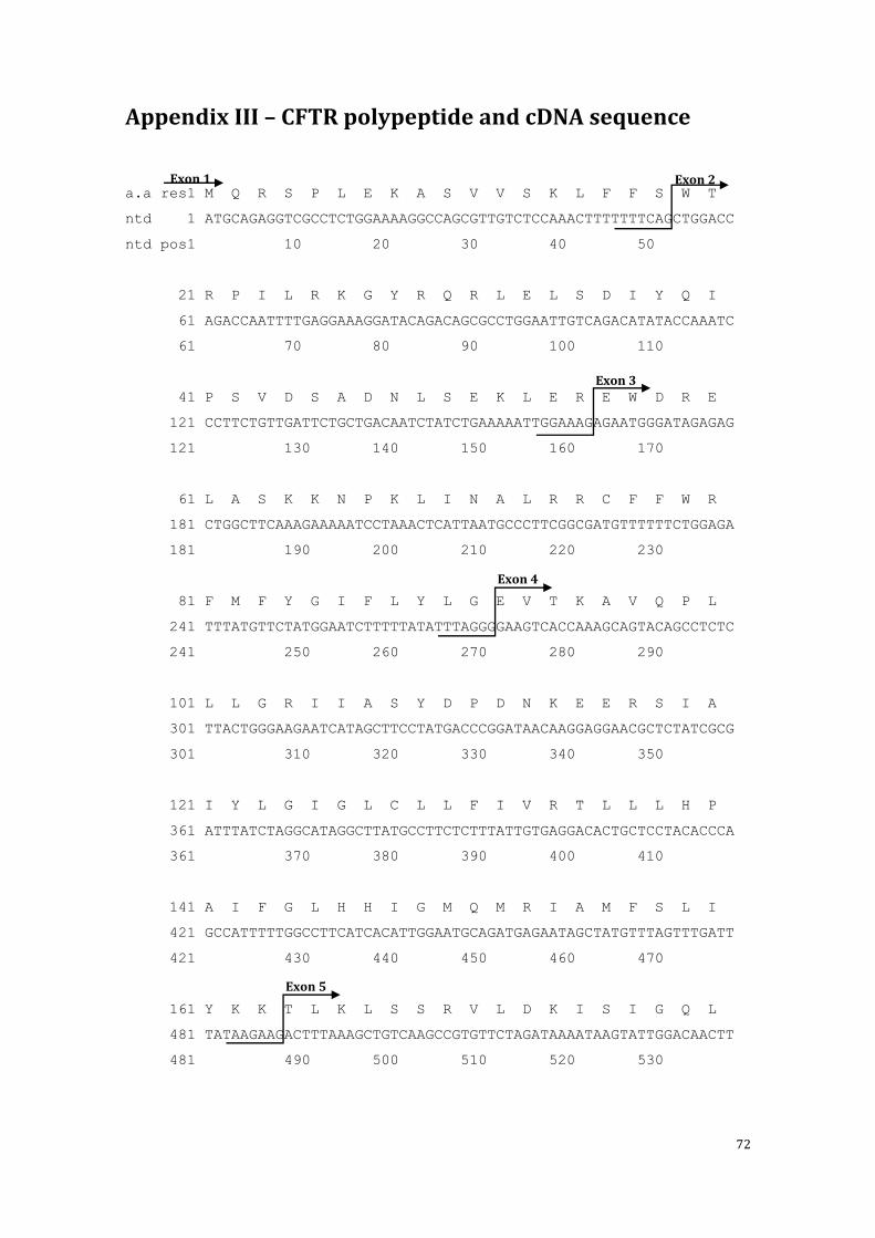
72
Appendix III – CFTR polypeptide and cDNA sequence
a.a res1 M Q R S P L E K A S V V S K L F F S W T
ntd 1 ATGCAGAGGTCGCCTCTGGAAAAGGCCAGCGTTGTCTCCAAACTTTTTTTCAGCTGGACC
ntd pos1 10 20 30 40 50
21 R P I L R K G Y R Q R L E L S D I Y Q I
61 AGACCAATTTTGAGGAAAGGATACAGACAGCGCCTGGAATTGTCAGACATATACCAAATC
61 70 80 90 100 110
41 P S V D S A D N L S E K L E R E W D R E
121 CCTTCTGTTGATTCTGCTGACAATCTATCTGAAAAATTGGAAAGAGAATGGGATAGAGAG
121 130 140 150 160 170
61 L A S K K N P K L I N A L R R C F F W R
181 CTGGCTTCAAAGAAAAATCCTAAACTCATTAATGCCCTTCGGCGATGTTTTTTCTGGAGA
181 190 200 210 220 230
81 F M F Y G I F L Y L G E V T K A V Q P L
241 TTTATGTTCTATGGAATCTTTTTATATTTAGGGGAAGTCACCAAAGCAGTACAGCCTCTC
241 250 260 270 280 290
101 L L G R I I A S Y D P D N K E E R S I A
301 TTACTGGGAAGAATCATAGCTTCCTATGACCCGGATAACAAGGAGGAACGCTCTATCGCG
301 310 320 330 340 350
121 I Y L G I G L C L L F I V R T L L L H P
361 ATTTATCTAGGCATAGGCTTATGCCTTCTCTTTATTGTGAGGACACTGCTCCTACACCCA
361 370 380 390 400 410
141 A I F G L H H I G M Q M R I A M F S L I
421 GCCATTTTTGGCCTTCATCACATTGGAATGCAGATGAGAATAGCTATGTTTAGTTTGATT
421 430 440 450 460 470
161 Y K K T L K L S S R V L D K I S I G Q L
481 TATAAGAAGACTTTAAAGCTGTCAAGCCGTGTTCTAGATAAAATAAGTATTGGACAACTT
481 490 500 510 520 530
Exon 1 Exon 2
Exon 3
Exon 4
Exon 5

73
181 V S L L S N N L N K F D E G L A L A H F
541 GTTAGTCTCCTTTCCAACAACCTGAACAAATTTGATGAAGGACTTGCATTGGCACATTTC
541 550 560 570 580 590
201 V W I A P L Q V A L L M G L I W E L L Q
601 GTGTGGATCGCTCCTTTGCAAGTGGCACTCCTCATGGGGCTAATCTGGGAGTTGTTACAG
601 610 620 630 640 650
221 A S A F C G L G F L I V L A L F Q A G L
661 GCGTCTGCCTTCTGTGGACTTGGTTTCCTGATAGTCCTTGCCCTTTTTCAGGCTGGGCTA
661 670 680 690 700 710
241 G R M M M K Y R D Q R A G K I S E R L V
721 GGGAGAATGATGATGAAGTACAGAGATCAGAGAGCTGGGAAGATCAGTGAAAGACTTGTG
721 730 740 750 760 770
261 I T S E M I E N I Q S V K A Y C W E E A
781 ATTACCTCAGAAATGATTGAAAATATCCAATCTGTTAAGGCATACTGCTGGGAAGAAGCA
781 790 800 810 820 830
281 M E K M I E N L R Q T E L K L T R K A A
841 ATGGAAAAAATGATTGAAAACTTAAGACAAACAGAACTGAAACTGACTCGGAAGGCAGCC
841 850 860 870 880 890
301 Y V R Y F N S S A F F F S G F F V V F L
901 TATGTGAGATACTTCAATAGCTCAGCCTTCTTCTTCTCAGGGTTCTTTGTGGTGTTTTTA
901 910 920 930 940 950
321 S V L P Y A L I K G I I L R K I F T T I
961 TCTGTGCTTCCCTATGCACTAATCAAAGGAATCATCCTCCGGAAAATATTCACCACCATC
961 970 980 990 1000 1010
341 S F C I V L R M A V T R Q F P W A V Q T
1021 TCATTCTGCATTGTTCTGCGCATGGCGGTCACTCGGCAATTTCCCTGGGCTGTACAAACA
1021 1030 1040 1050 1060 1070
361 W Y D S L G A I N K I Q D F L Q K Q E Y
1081 TGGTATGACTCTCTTGGAGCAATAAACAAAATACAGGATTTCTTACAAAAGCAAGAATAT
1081 1090 1100 1110 1120 1130
Exon 6a
Exon 6b
Exon 7
Exon 8
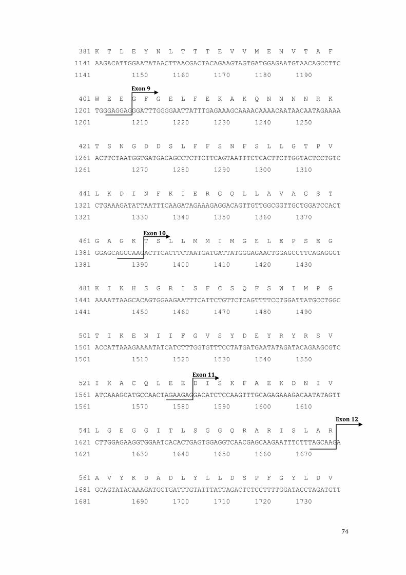
74
381 K T L E Y N L T T T E V V M E N V T A F
1141 AAGACATTGGAATATAACTTAACGACTACAGAAGTAGTGATGGAGAATGTAACAGCCTTC
1141 1150 1160 1170 1180 1190
401 W E E G F G E L F E K A K Q N N N N R K
1201 TGGGAGGAGGGATTTGGGGAATTATTTGAGAAAGCAAAACAAAACAATAACAATAGAAAA
1201 1210 1220 1230 1240 1250
421 T S N G D D S L F F S N F S L L G T P V
1261 ACTTCTAATGGTGATGACAGCCTCTTCTTCAGTAATTTCTCACTTCTTGGTACTCCTGTC
1261 1270 1280 1290 1300 1310
441 L K D I N F K I E R G Q L L A V A G S T
1321 CTGAAAGATATTAATTTCAAGATAGAAAGAGGACAGTTGTTGGCGGTTGCTGGATCCACT
1321 1330 1340 1350 1360 1370
461 G A G K T S L L M M I M G E L E P S E G
1381 GGAGCAGGCAAGACTTCACTTCTAATGATGATTATGGGAGAACTGGAGCCTTCAGAGGGT
1381 1390 1400 1410 1420 1430
481 K I K H S G R I S F C S Q F S W I M P G
1441 AAAATTAAGCACAGTGGAAGAATTTCATTCTGTTCTCAGTTTTCCTGGATTATGCCTGGC
1441 1450 1460 1470 1480 1490
501 T I K E N I I F G V S Y D E Y R Y R S V
1501 ACCATTAAAGAAAATATCATCTTTGGTGTTTCCTATGATGAATATAGATACAGAAGCGTC
1501 1510 1520 1530 1540 1550
521 I K A C Q L E E D I S K F A E K D N I V
1561 ATCAAAGCATGCCAACTAGAAGAGGACATCTCCAAGTTTGCAGAGAAAGACAATATAGTT
1561 1570 1580 1590 1600 1610
541 L G E G G I T L S G G Q R A R I S L A R
1621 CTTGGAGAAGGTGGAATCACACTGAGTGGAGGTCAACGAGCAAGAATTTCTTTAGCAAGA
1621 1630 1640 1650 1660 1670
561 A V Y K D A D L Y L L D S P F G Y L D V
1681 GCAGTATACAAAGATGCTGATTTGTATTTATTAGACTCTCCTTTTGGATACCTAGATGTT
1681 1690 1700 1710 1720 1730
Exon 9
Exon 10
Exon 11
Exon 12

75
581 L T E K E I F E S C V C K L M A N K T R
1741 TTAACAGAAAAAGAAATATTTGAAAGCTGTGTCTGTAAACTGATGGCTAACAAAACTAGG
1741 1750 1760 1770 1780 1790
601 I L V T S K M E H L K K A D K I L I L H
1801 ATTTTGGTCACTTCTAAAATGGAACATTTAAAGAAAGCTGACAAAATATTAATTTTGCAT
1801 1810 1820 1830 1840 1850
621 E G S S Y F Y G T F S E L Q N L Q P D F
1861 GAAGGTAGCAGCTATTTTTATGGGACATTTTCAGAACTCCAAAATCTACAGCCAGACTTT
1861 1870 1880 1890 1900 1910
641 S S K L M G C D S F D Q F S A E R R N S
1921 AGCTCAAAACTCATGGGATGTGATTCTTTCGACCAATTTAGTGCAGAAAGAAGAAATTCA
1921 1930 1940 1950 1960 1970
661 I L T E T L H R F S L E G D A P V S W T
1981 ATCCTAACTGAGACCTTACACCGTTTCTCATTAGAAGGAGATGCTCCTGTCTCCTGGACA
1981 1990 2000 2010 2020 2030
681 E T K K Q S F K Q T G E F G E K R K N S
2041 GAAACAAAAAAACAATCTTTTAAACAGACTGGAGAGTTTGGGGAAAAAAGGAAGAATTCT
2041 2050 2060 2070 2080 2090
701 I L N P I N S I R K F S I V Q K T P L Q
2101 ATTCTCAATCCAATCAACTCTATACGAAAATTTTCCATTGTGCAAAAGACTCCCTTACAA
2101 2110 2120 2130 2140 2150
721 M N G I E E D S D E P L E R R L S L V P
2161 ATGAATGGCATCGAAGAGGATTCTGATGAGCCTTTAGAGAGAAGGCTGTCCTTAGTACCA
2161 2170 2180 2190 2200 2210
741 D S E Q G E A I L P R I S V I S T G P T
2221 GATTCTGAGCAGGGAGAGGCGATACTGCCTCGCATCAGCGTGATCAGCACTGGCCCCACG
2221 2230 2240 2250 2260 2270
761 L Q A R R R Q S V L N L M T H S V N Q G
2281 CTTCAGGCACGAAGGAGGCAGTCTGTCCTGAACCTGATGACACACTCAGTTAACCAAGGT
2281 2290 2300 2310 2320 2330
Exon 13

76
781 Q N I H R K T T A S T R K V S L A P Q A
2341 CAGAACATTCACCGAAAGACAACAGCATCCACACGAAAAGTGTCACTGGCCCCTCAGGCA
2341 2350 2360 2370 2380 2390
801 N L T E L D I Y S R R L S Q E T G L E I
2401 AACTTGACTGAACTGGATATATATTCAAGAAGGTTATCTCAAGAAACTGGCTTGGAAATA
2401 2410 2420 2430 2440 2450
821 S E E I N E E D L K E C F F D D M E S I
2461 AGTGAAGAAATTAACGAAGAAGACTTAAAGGAGTGCTTTTTTGATGATATGGAGAGCATA
2461 2470 2480 2490 2500 2510
841 P A V T T W N T Y L R Y I T V H K S L I
2521 CCAGCAGTGACTACATGGAACACATACCTTCGATATATTACTGTCCACAAGAGCTTAATT
2521 2530 2540 2550 2560 2570
861 F V L I W C L V I F L A E V A A S L V V
2581 TTTGTGCTAATTTGGTGCTTAGTAATTTTTCTGGCAGAGGTGGCTGCTTCTTTGGTTGTG
2581 2590 2600 2610 2620 2630
881 L W L L G N T P L Q D K G N S T H S R N
2641 CTGTGGCTCCTTGGAAACACTCCTCTTCAAGACAAAGGGAATAGTACTCATAGTAGAAAT
2641 2650 2660 2670 2680 2690
901 N S Y A V I I T S T S S Y Y V F Y I Y V
2701 AACAGCTATGCAGTGATTATCACCAGCACCAGTTCGTATTATGTGTTTTACATTTACGTG
2701 2710 2720 2730 2740 2750
921 G V A D T L L A M G F F R G L P L V H T
2761 GGAGTAGCCGACACTTTGCTTGCTATGGGATTCTTCAGAGGTCTACCACTGGTGCATACT
2761 2770 2780 2790 2800 2810
941 L I T V S K I L H H K M L H S V L Q A P
2821 CTAATCACAGTGTCGAAAATTTTACACCACAAAATGTTACATTCTGTTCTTCAAGCACCT
2821 2830 2840 2850 2860 2870
Exon 14b
Exon 14b
Exon 15

77
961 M S T L N T L K A G G I L N R F S K D I
2881 ATGTCAACCCTCAACACGTTGAAAGCAGGTGGGATTCTTAATAGATTCTCCAAAGATATA
2881 2890 2900 2910 2920 2930
981 A I L D D L L P L T I F D F I Q L L L I
2941 GCAATTTTGGATGACCTTCTGCCTCTTACCATATTTGACTTCATCCAGTTGTTATTAATT
2941 2950 2960 2970 2980 2990
1001 V I G A I A V V A V L Q P Y I F V A T V
3001 GTGATTGGAGCTATAGCAGTTGTCGCAGTTTTACAACCCTACATCTTTGTTGCAACAGTG
3001 3010 3020 3030 3040 3050
1021 P V I V A F I M L R A Y F L Q T S Q Q L
3061 CCAGTGATAGTGGCTTTTATTATGTTGAGAGCATATTTCCTCCAAACCTCACAGCAACTC
3061 3070 3080 3090 3100 3110
1041 K Q L E S E G R S P I F T H L V T S L K
3121 AAACAACTGGAATCTGAAGGCAGGAGTCCAATTTTCACTCATCTTGTTACAAGCTTAAAA
3121 3130 3140 3150 3160 3170
1061 G L W T L R A F G R Q P Y F E T L F H K
3181 GGACTATGGACACTTCGTGCCTTCGGACGGCAGCCTTACTTTGAAACTCTGTTCCACAAA
3181 3190 3200 3210 3220 3230
1081 A L N L H T A N W F L Y L S T L R W F Q
3241 GCTCTGAATTTACATACTGCCAACTGGTTCTTGTACCTGTCAACACTGCGCTGGTTCCAA
3241 3250 3260 3270 3280 3290
1101 M R I E M I F V I F F I A V T F I S I L
3301 ATGAGAATAGAAATGATTTTTGTCATCTTCTTCATTGCTGTTACCTTCATTTCCATTTTA
3301 3310 3320 3330 3340 3350
1121 T T G E G E G R V G I I L T L A M N I M
3361 ACAACAGGAGAAGGAGAAGGAAGAGTTGGTATTATCCTGACTTTAGCCATGAATATCATG
3361 3370 3380 3390 3400 3410
1141 S T L Q W A V N S S I D V D S L M R S V
3421 AGTACATTGCAGTGGGCTGTAAACTCCAGCATAGATGTGGATAGCTTGATGCGATCTGTG
3421 3430 3440 3450 3460 3470
Exon 16
Exon 17a
Exon 17b
Exon 18
Exon 19

78
1161 S R V F K F I D M P T E G K P T K S T K
3481 AGCCGAGTCTTTAAGTTCATTGACATGCCAACAGAAGGTAAACCTACCAAGTCAACCAAA
3481 3490 3500 3510 3520 3530
1181 P Y K N G Q L S K V M I I E N S H V K K
3541 CCATACAAGAATGGCCAACTCTCGAAAGTTATGATTATTGAGAATTCACACGTGAAGAAA
3541 3550 3560 3570 3580 3590
1201 D D I W P S G G Q M T V K D L T A K Y T
3601 GATGACATCTGGCCCTCAGGGGGCCAAATGACTGTCAAAGATCTCACAGCAAAATACACA
3601 3610 3620 3630 3640 3650
1221 E G G N A I L E N I S F S I S P G Q R V
3661 GAAGGTGGAAATGCCATATTAGAGAACATTTCCTTCTCAATAAGTCCTGGCCAGAGGGTG
3661 3670 3680 3690 3700 3710
1241 G L L G R T G S G K S T L L S A F L R L
3721 GGCCTCTTGGGAAGAACTGGATCAGGGAAGAGTACTTTGTTATCAGCTTTTTTGAGACTA
3721 3730 3740 3750 3760 3770
1261 L N T E G E I Q I D G V S W D S I T L Q
3781 CTGAACACTGAAGGAGAAATCCAGATCGATGGTGTGTCTTGGGATTCAATAACTTTGCAA
3781 3790 3800 3810 3820 3830
1281 Q W R K A F G V I P Q K V F I F S G T F
3841 CAGTGGAGGAAAGCCTTTGGAGTGATACCACAGAAAGTATTTATTTTTTCTGGAACATTT
3841 3850 3860 3870 3880 3890
1301 R K N L D P Y E Q W S D Q E I W K V A D
3901 AGAAAAAACTTGGATCCCTATGAACAGTGGAGTGATCAAGAAATATGGAAAGTTGCAGAT
3901 3910 3920 3930 3940 3950
1321 E V G L R S V I E Q F P G K L D F V L V
3961 GAGGTTGGGCTCAGATCTGTGATAGAACAGTTTCCTGGGAAGCTTGACTTTGTCCTTGTG
3961 3970 3980 3990 4000 4010
1341 D G G C V L S H G H K Q L M C L A R S V
4021 GATGGGGGCTGTGTCCTAAGCCATGGCCACAAGCAGTTGATGTGCTTGGCTAGATCTGTT
4021 4030 4040 4050 4060 4070
Exon 20
Exon 21
Exon 22

79
1361 L S K A K I L L L D E P S A H L D P V T
4081 CTCAGTAAGGCGAAGATCTTGCTGCTTGATGAACCCAGTGCTCATTTGGATCCAGTAACA
4081 4090 4100 4110 4120 4130
1381 Y Q I I R R T L K Q A F A D C T V I L C
4141 TACCAAATAATTAGAAGAACTCTAAAACAAGCATTTGCTGATTGCACAGTAATTCTCTGT
4141 4150 4160 4170 4180 4190
1401 E H R I E A M L E C Q Q F L V I E E N K
4201 GAACACAGGATAGAAGCAATGCTGGAATGCCAACAATTTTTGGTCATAGAAGAGAACAAA
4201 4210 4220 4230 4240 4250
1421 V R Q Y D S I Q K L L N E R S L F R Q A
4261 GTGCGGCAGTACGATTCCATCCAGAAACTGCTGAACGAGAGGAGCCTCTTCCGGCAAGCC
4261 4270 4280 4290 4300 4310
1441 I S P S D R V K L F P H R N S S K C K S
4321 ATCAGCCCCTCCGACAGGGTGAAGCTCTTTCCCCACCGGAACTCAAGCAAGTGCAAGTCT
4321 4330 4340 4350 4360 4370
1461 K P Q I A A L K E E T E E E V Q D T R L
4381 AAGCCCCAGATTGCTGCTCTGAAAGAGGAGACAGAAGAAGAGGTGCAAGATACAAGGCTT
4381 4390 4400 4410 4420 4430
1481 STP
4441 TAG
4441
Exon 23
Exon 24