estudo morfológico e molecular de - repositorio.ufba.br · quem busca e vence obstáculos, no...
TRANSCRIPT

Programa de Pós-graduação em Diversidade Animal
Universidade Federal da Bahia
Ueslei da Conceição Lopes
Estudo morfológico e molecular de
Geodia gibberosa Lamarck, 1815
(Astrophorida, Geodiidae)
Salvador
2012
Ueslei da Conceição Lopes
Estudo morfológico e molecular de
Geodia gibberosa Lamarck, 1815
(Astrophorida, Geodiidae)
Dissertação apresentada ao Instituto de Biologia da Universidade Federal da Bahia para a obtenção do Título de Mestre pelo Programa de Pós-Graduação em Diversidade Animal, na Área de Zoologia. Orientadora: Carla M. M. da Silva Co-Orientadora: Patrícia D. de Freitas
Salvador
2012
Ficha Catalográfica
Lopes, Ueslei da Conceição Estudo morfológico e molecular de Geodia gibberosa Lamarck, 1815 (Astrophorida, Geodiidae) 109 páginas Dissertação (Mestrado) - Instituto de Biologia da Universidade Federal da Bahia. Departamento de Zoologia. Programa de Pós-Graduação em Diversidade Animal. 1. Geodia 2. Taxonomia 3. Complexo de espécies I. Universidade Federal da Bahia. Instituto de Biologia. Departamento de Zoologia. Programa de Pós-Graduação em Diversidade Animal.


Dedicatória
Por todo carinho, confiança, apoio e
amor, dedico esse trabalho
aos meus pais.
Epígrafe
“O sucesso nasce do querer,
da determinação e persistência em se
chegar a um objetivo. Mesmo não atingindo o alvo,
quem busca e vence obstáculos, no mínimo fará coisas admiráveis.”
José de Alencar.

Agradecimentos
Em virtude da correria que um mestrado representa, nós, estudantes, costumamos
brincar que essa etapa não tem meio, apenas início e fim. Mas certamente essa assertiva não
condiz com a realidade, uma vez que, durante todo o processo (no ‘meio’ dele), muitas
pessoas passam pelas nossas vidas e muitas vezes, da forma mais simples, nos ajudam, nos
incentivam, deixam suas marcas. São essas pessoas a quem eu especialmente agradeço:
À minha orientadora, a Profa. Dra. Carla Menegola, por todos esses anos de
convivência, apoio e amizade.
À minha co-orientadora, Profa. Dra. Patrícia Domingues de Freitas, da Universidade
Federal de São Carlos (UFSCar), pela oportunidade e confiança depositadas em mim e por
todo o seu apoio.
À querida Profa. Dra. Gisele Lôbo-Hajdu da Universidade do Estado do Rio de
Janeiro (UERJ), por todo o seu apoio, amizade e incentivo na área da biologia molecular,
desde os tempos de minha iniciação científica.
Ao Prof. Dr. Eduardo Hajdu, do Depto. de Invertebrados do Museu Nacional
(MNRJ), pelo empréstimo de espécimes e apoio ao meu projeto.
Ao Prof. Dr. Pedro Galetti Jr. por ter me integrado ao dia-a-dia e dado pleno acesso
às dependências do Laboratório de Biodiversidade Molecular e Conservação (LBMC) da
UFSCar, fazendo com que eu me sentisse parte integrante de sua equipe. Muito obrigado!
Aos queridos professores Flora Fernandes e Rodrigo Barban Zucoloto do Dept. de
Biologia Geral da UFBA, por sempre terem esclarecido minhas dúvidas referentes à
biologia molecular e por todo o apoio ao longo desses anos.
À Profa. Dra. Luciana Veiga, do Depto. de Biologia Geral da UFBA e do
Laboratório de Biologia Molecular ‘Carmen Lemos’ (LBM), por todo o seu carinho, apoio
e amizade.
Ao Prof. Dr. Rob van Soest do Zoölogisch Museum van Amsterdam (Holanda) por
ter intercedido na obtenção de amostras provenientes do Caribe, num momento em que o
museu encontrava-se fechado para empréstimos e ao Victor Cedro, da Universidade Federal
de Alagoas (UFAL), pela coleta e por gentilmente ter enviado o material.
Ao Prof. Dr. Pedro Alcolado do Instituto de Oceanología (Cuba), ao Prof. Dr. Hans
Rapp da Universitetet i Bergen (Noruega) e à Profa. Dra. Patricia Gomez da Universidad
Autónoma do Mexico (México) pela doação e envio de espécimes.
Ao Prof. Dr. Klaus Rützler do National Museum of Natural History (E.U.A.), e ao
Prof. Dr. Rob van Syoc e à Christina Piotrowski da California Academy of Science
(E.U.A.), pelo empréstimo de material comparativo.
À pesquisadora Josivete Pinheiro da Universidade Federal de Pernambuco (UFPE),
por ter me concedido amostras de esponjas.
À Sula Salani do MNRJ e à Cecília Licarião e orientadora, a Profa. Dra. Helena
Cascon da Universidade Federal do Ceará (UFC), pelo empréstimo de espécimes
provenientes do Ceará.
Ao grande amigo João Guilherme DeMarchi, por ter me ajudado a coletar esponjas
e me ciceroneado em João Pessoa - PB, assim como aos seus familiares por terem me
recebido em sua casa em 2011.
Aos professores e ao Programa de Pós-graduação em Diversidade Animal
(PPGDA), pelo apoio, dedicação e paciência.
À minha turma de pós-graduação: Roberta Canário, Rafael Abreu, Byanca Sardeiro,
Diogo França, Fábio Quinteiro e Marlla Matos, pelo companheirismo e amizade.
Ao Fábio Quinteiro e à Luciana Martins, pelas dicas valiosas dos programas
computacionais de tratamento de imagens.
À Dra. Adriana Rangel, ao Dr. Cláudio Figueira e à Dra. Lúcia Moreno do Centro
de Pesquisa Gonçalo Moniz da Fundação Oswaldo Cruz (Fiocruz - BA), pelo auxílio e
sessões cedidas de microscópio eletrônico de varredura.
À Patrícia Ferreira, Luciano Lopes, Fred Hanai e Angela Fushita pelos bons
momentos em São Carlos.
Ao Bruno Rossini, Marcelo de Bello Cioffi e Bráulio Queiroz que, sem me
conhecerem, abriram as portas de sua casa em São Carlos, onde fiquei hospedado durante
todo o período em que estive na cidade, em 2010.
À Equipe LBMC, meus amigos: Adriana Kazue Takako, Alexander Ferreira, Aline
Galindo, Alline Braga, André, Andiara Silos, Dorivaldo Marques, Karen Rodriguez, Camilla
Alves, Carolina Machado, Danielly V. Blanco, Eliana Paviotti, Josiane Ribolli, Luísa
Simbine, Leonardo Niero, Marcos Tokuda, Renata Miotto, Tailise Guerreiro, Tamilyn

Kaori Ishizuca e Pedro Gallo, pela boa convivência, companheirismo, amizade e pelos
ótimos momentos (incluindo as memoráveis reuniões na Bom Pedaço!) ao longo de 2011.
Um agradecimento especial à Carla Guinart (Carlota), por toda a sua ajuda ao longo
desses três últimos anos, por ter me acolhido e me ajudado na bancada desde o início.
Muito obrigado!
Aos bons amigos e parceiros Bruno Henrique Saranholi e Lucas Caldano, por toda
ajuda, amizade e pelas muitas risadas.
Um agradecimento muito especial aos bons amigos Carlos Congrains, Jorge
Ramirez e Karla Chávez, por toda a ajuda no meu projeto e por gentilmente terem me
recebido em sua casa. Muchas gracias por todo!!
Ao Prof. Dr. Armando Vieira, a Luis Sartori e Inessa Lacativa, do Laboratório de
Ficologia da UFSCar, pela assistência e concessão do analisador de imagens, com o qual
pude realizar as medidas das espículas.
Aos ‘novos e velhos’ amigos e companheiros do Laboratório de Biologia de
Porifera & Fauna Associada (Labpor): Lourianne Mangueira, Cris Castello Branco, Anaíra
Lage, Rosana Fernandes, Renato Guimarães, Alisson Santana, Ana Carolina Almeida, Karol
Rebello, Fernandinha Cavalcanti, Emílio Lanna e à Facelúcia Barros pela amizade, apoio e
boa convivência dentro e fora do trabalho ao longo desses anos. Muito obrigado por tudo!
Um agradecimento especial ao vitorioso Júlio Cesar Fernandez que, apesar de todas
as adversidades enfrentadas ao longo de seu mestrado, sempre se mostrou disposto a
ajudar, reservando parte do seu escasso tempo para auxiliar-me com programas
computacionais e em coletas na orla de Salvador.
À minha querida Luciana Martins, Luly. Foram tantos os momentos que
compartilhamos anseios, frustrações, alegrias e apoio, que precisaria de mais três páginas
para agradecê-la.
À Milla Souto pela companhia, pela amizade, por sempre ter me incentivado em
meus projetos e por ter ‘puxado minha orelha’ quando isso se fazia necessário. Sempre foi
pra mim um modelo de profissionalismo, força e dedicação.
Ao Conselho Nacional de Desenvolvimento Científico e Tecnológico (CNPq), pela
concessão da bolsa, sem a qual boa parte do projeto não teria sido viabilizada.
E, por fim, a toda minha família, meus pais e amigos pelo apoio incondicional,
amizade e carinho. Obrigado!
Índice
Resumo 11
Abstract 12
Lista de lustrações 13
Lista de tabelas 14
Lista de abreviaturas e siglas 15
Introdução geral 16
Capítulo 1. Going deeper with Geodia gibberosa (Astrophorida, Geodiidae):
investigating an alleged species complex
23
Abstract/Resumo 24
1. Introduction 26
2. Materials and methods 27
2.1. Sampling and fixation 27
2.2. Morphologic analysis 27
2.3. DNA extraction, PCR amplification and sequencing 28
2.4. Phylogenetic and distance analysis 29
2.5. Abbreviation 30
3. Morphologic results 30
4. Molecular results 35
5. Discussion 36
6. Conclusion 38
Acknowledgements 38
References cited 39
Capítulo 2. Description of a new species of Geodia and new record of
Pachymatisma (Astrophorida, Demospongiae) from the Caribbean zone
70
Abstract/Resumé 71
1. Introduction 72
2. Materials and methods 73
2.1. Sampling 73
2.2. Morphologic analysis 73
2.3. DNA extraction, PCR amplification and sequencing 74
2.4. Sequence analysis 74
3. Results 75
3.1. Systematic description and spicules nomenclature 75
3.2. Molecular results 80
4. Discussion and considerations 81
Acknowledgements 81
References cited 82
Conclusões gerais 91
Referências bibliográficas 92
Apêndices 99
Anexo 103
11
Resumo
Geodia gibberosa é uma das espécies que apresentam as maiores distribuições geográfica e
batimétrica em comparação com suas congêneres. Com variações significativas na
morfologia externa e no conjunto espicular, é considerada uma espécie com alto grau de
polimorfismo e, muitas vezes, de difícil identificação. Tais características têm levado
diversos pesquisadores a questionar os limites de sua variabilidade e a levantar hipóteses
acerca da existência de espécies crípticas. Com o presente estudo, se objetivou avaliar o
status taxonômico dessa espécie, aliando informações de morfologia e de biologia
molecular, a qual empregou a utilização dos marcadores mitocondriais COI (subunidade I
da citocromo c oxidase) e ATP6 (subunidade 6 da ATP sintase). Embora tenham sido
observadas variações na morfologia das espículas e registradas categorias de megaescleras
nunca vistas no holótipo, não foi possível encontrar nenhum padrão que possibilitasse a
separação de G. gibberosa em espécies morfologicamente distintas. Contudo, através das
ferramentas moleculares, foi determinada a existência de, pelo menos, três clados
fortemente suportados, possibilitando a aceitação da hipótese de que G. gibberosa
representa um complexo de espécies. Acrescido a isso, no presente trabalho, é descrita uma
nova espécie de Geodia para a costa da Venezuela, com a apresentação de uma chave
taxonômica para as 12 espécies nominais agora válidas que ocorrem no Caribe, e registrada
a primeira ocorrência de Pachymatisma johnstonia para o oceano Atlântico ocidental.
Palavras-chave: Variação morfológica, marcadores mitocondriais, Demospongiae, Geodia,
complexo de espécies, Pachymatisma.
12
Abstract
Geodia gibberosa is a species with one of the largest geographic and bathymetric
distributions when compared to its peers. With significant variations in both external
morphology and espicule repertoire, it is considered a species with a high degree of
polymorphism and often difficult to identify. These characteristics have led several
researchers to question the limits of its variability and led them to postulate hypotheses
about the existence of cryptic species. The aims of the present study was to evaluate the
taxonomic status of this species, combining information from morphology and molecular
biology, which used the mitochondrial markers COI (subunit I of cytochrome c oxidase)
and ATP6 (ATP synthase subunit 6). Although variations in the morphology of the spicules
were observed, and new categories of megascleres were registered for the holotype, it was
not possible to find any pattern that could allow us the split G. gibberosa into
morphologically distinct species. However, through the molecular approach, it was
determined the existence of at least three highly supported clades, enabling the acceptance
of the hypothesis that G. gibberosa represents a species complex. Added to this, in the
present study, we describe a new species of Geodia for the coast of Venezuela, with a
taxonomic key to the 12 nominal species that occur in the Caribbean up to now, and the
first occurrence of Pachymatisma johnstonia for western Atlantic Ocean.
Key words: Morphological variation, mitochondrial markers, Demospongiae, Geodia,
species complex, Pachymatisma.

13
Lista de ilustrações
Introdução geral
Figura 1. Representação dos canais inalantes e exalantes em Geodiidae 17
Figura 2. Filogenia da Ordem Astrophorida 21
Capítulo 1
Figure 1. Prancha de morfologia externa 63
Figure 2. Prancha com as megaescleras 64
Figure 3. Prancha com as microescleras 65
Figure 4. Mapa com os pontos de coleta dos clados 66
Figure 5. Árvore para COI 67
Figure 6. Árvore para ATP6 68
Capítulo 2
Figure 1. Mapa com os pontos de coleta dos espécimes 87
Figure 2. Pracha de morfologia externa e interna de Geodia leispheroxyastera sp. nov. 88
Figure 3. Prancha de morfologia externa e interna de Pachymatisma johnstonia 89

14
Lista de tabelas
Capítulo 1
Table 1. Tabela de medidas de espículas 50
Table 2. Tabela de distâncias genéticas 62
Capítulo 2
Table 1. Sítios polimórifcos em Pachymatisma johnstonia 90
Apêndice 2
Tabela A. Relação de primers adicionais testados 101

15
Lista de abreviaturas
ATP6 – Subunidade 6 da ATP sintase
BCT - Bocas Del Toro
BI - Inferência bayesiana
bp - Pares de base
CNPGG - Colección Nacional del Phylum Porifera Gerardo Green, Universidad Nacional
Autónoma de Mexico, Mexico
COI - Subunidade I da Citocromo c Oxidase
DNA - Ácido desoxirribonucléico
dNTP - Deoxinucleotídeos fosfatados
FIOCRUZ - Fundação Oswaldo Cruz: Centro de Pesquisas Gonçalo Muniz, Salvador
FL - Flórida
MCN - Museu de Ciências Naturais da Fundação Zoobotânica do Rio Grande do Sul, Porto
Alegre, Rio Grande do Sul, Brasil
MNHN - Muséum National d'Histoire Naturelle, Paris, França
MNRJ - Museu Nacional do Rio de Janeiro, Rio de Janeiro, Brasil
mtDNA - DNA mitocondrial
ML - Máxima verossimilhança
NCBI - National Center for Biotechnology Information
NJ – Neighbor Joining
PCR - Reação em Cadeia da Polimerase
PEG - Polietilenoglicol
UFBA-POR - Coleção de Porifera, Museu de Zoologia, Universidade Federal da Bahia
UFC - Universidade Federal do Ceará, Fortaleza
UFSCar - Universidade Federal de São Carlos, São Carlos, São Paulo, Brasil
UNDP/FAO - Programa das Nações Unidas para o Desenvolvimento/ Organização das
Nações Unidas para Agricultura e Alimentação
USNM - Smithsonian Institution, National Museum of Natural History, EUA
ZMA POR - Instituut voor Systematiek en Populatiebiologie, Zoölogisch Museum,
Porifera Collection, Holanda
ZMBN - Universitetsmuseet i Bergen, Universitetet i Bergen, Noruega
16
Introdução geral
1. O GÊNERO GEODIA
Lamarck (1815) descreveu um novo gênero da família “Alcyons” e lhe atribuiu o
nome Geodia em alusão a “aberturas maiores que poros, reunidas em uma faceta orbicular”,
cujo aspecto de crivo isolado se assemelhava às cavidades das rochas, conhecidas como
‘geodos’. O autor notou que as espécies pertencentes a esse gênero, “apresentam forma
subglobosa, são côncavas e internamente ocas”. São também caracterizadas pela grande
quantidade de fibras que lhes conferem resistência e, mesmo quando secas, ocorre a
manutenção da forma. Em sua superfície são encontrados poros esparsamente distribuídos,
além do crivo isolado, cujas aberturas não se destinam à entrada de água na esponja
(LAMARCK, 1815).
Posteriormente, esse gênero foi incluído na família “Geodiadae”, juntamente com
Caminus Schmidt, 1862, Cydonium Fleming, 1828 (sinônimo de Geodia), Erylus Gray,
1867, Pachymatisma Johnston, 1842 e Triate Gray, 1867 (sinônimo de Erylus), levando-se
em conta, além da morfologia externa, as categorias espiculares (GRAY, 1867). Desse
modo, as esponjas “massivas, com uma cavidade central coberta por membrana reticulada
ou perfurada, e que possuem espículas alongadas com dois ou três ramos recurvados nas
extremidades, e microescleras dispostas sobre a superfície”, passaram a ser classificadas
como Geodia.
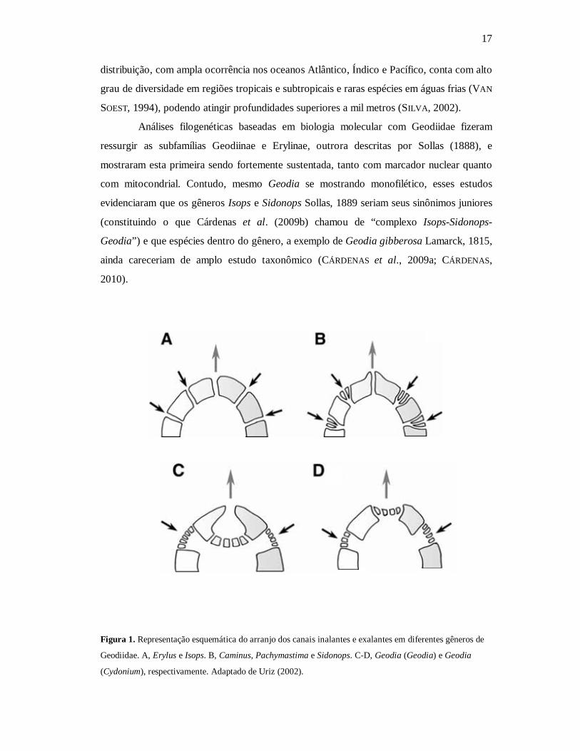
A revisão mais recente da família considerou seis gêneros válidos e destacou que
as espécies pertencentes a Geodia são distinguidas por apresentar óxeas longas, triênios
regulares radialmente dispostos na superfície (ou próximas a ela), e por um conjunto de
espículas pequenas constituído por esterrásteres globulares e euásteres. Contudo, é o
padrão de organização cribiporal de poros e ósculos (Figura 1) o caráter que diferencia
Geodia dos demais, dentro de Geodiidae (URIZ, 2002).
Esse táxon, que data do Cretáceo (RIGBY & SMITH, 1992), conta atualmente com
145 espécies nominais e pode ser considerado, em termos quantitativos, o grupo
taxonômico mais significativo da família, sendo seguido pelos gêneros Erylus e Isops Sollas,
1880, com 66 e 20 espécies, respectivamente (VAN SOEST et al., 2012). Sua expressiva
17
distribuição, com ampla ocorrência nos oceanos Atlântico, Índico e Pacífico, conta com alto
grau de diversidade em regiões tropicais e subtropicais e raras espécies em águas frias (VAN
SOEST, 1994), podendo atingir profundidades superiores a mil metros (SILVA, 2002).
Análises filogenéticas baseadas em biologia molecular com Geodiidae fizeram
ressurgir as subfamílias Geodiinae e Erylinae, outrora descritas por Sollas (1888), e
mostraram esta primeira sendo fortemente sustentada, tanto com marcador nuclear quanto
com mitocondrial. Contudo, mesmo Geodia se mostrando monofilético, esses estudos
evidenciaram que os gêneros Isops e Sidonops Sollas, 1889 seriam seus sinônimos juniores
(constituindo o que Cárdenas et al. (2009b) chamou de “complexo Isops-Sidonops-
Geodia”) e que espécies dentro do gênero, a exemplo de Geodia gibberosa Lamarck, 1815,
ainda careceriam de amplo estudo taxonômico (CÁRDENAS et al., 2009a; CÁRDENAS,
2010).
Figura 1. Representação esquemática do arranjo dos canais inalantes e exalantes em diferentes gêneros de
Geodiidae. A, Erylus e Isops. B, Caminus, Pachymastima e Sidonops. C-D, Geodia (Geodia) e Geodia
(Cydonium), respectivamente. Adaptado de Uriz (2002).
18
1.1. A ESPÉCIE GEODIA GIBBEROSA LAMARCK, 1815
Descrita para a costa da Guiana Francesa, Geodia gibberosa, constitui a espécie-
tipo do gênero. Usualmente essa espécie é utilizada em estudos de química de produtos
naturais, tendo sido encontrada uma ação neurotóxica proveniente de seus extratos
(RANGEL et al., 2001), além de diversos lipídios (NAKANISH et al.,1953; CARBALLEIRA &
RODRIGUEZ, 1991; ROD’KINA, 2005). Produtos derivados de organismos marinhos têm
atraído cada vez mais a atenção da comunidade científica e, de certo modo, encorajado
novas pesquisas devido ao possível potencial econômico (MENDOLA, 2003).
Acreditava-se que a disposição radial das espículas de G. gibberosa pudesse
configurar um mecanismo de proteção mecânico à espécie, mas estudos de ecologia
indicaram o contrário (CHANAS & PAWLIK, 1995; DUNLAP & PAWLIK, 1998) e mostraram
que, além de se tratar de uma espécie extremamente palatável, sendo considerada o
alimento preferido de algumas tartarugas (e.g., Eretmochelys imbricata (Linnaeus, 1766))
(PAWLIK et al., 1995) e de peixes (e.g., Sparisoma aurofrenatum (Valenciennes, 1840) e
Sparisoma chrysopterum (Bloch & Schneider, 1801)) (DUNLAP & PAWLIK, 1996; 1998),
G. gibberosa carece de defesas químicas (ENGEL & PAWLIK, 2005). Essa suposta
vulnerabilidade, no entanto, é atenuada pela alelopatia promovida pela espécie, a qual
estimula o crescimento de outras esponjas, e.g. Tedania ignis (Duchassaing & Michelotti,
1864), Lissodendoryx isodictyalis (Carter, 1882) e Haliclona (Reniera) tubifera (George &
Wilson, 1919), garantindo-lhe proteção indireta contra a predação (ENGEL & PAWLIK,
2000; 2005).
Dos representantes de Geodia que ocorrem no Atlântico ocidental, G. gibberosa é
a espécie que apresenta as maiores distribuições geográfica e batimétrica. Há registros de
sua ocorrência partindo da Carolina do Norte, nos Estados Unidos, a São Sebastião em São
Paulo, Brasil, com profundidades que variam de 0,1 m (Salvador, Brasil) a 33 m (costa da
Carolina do Sul, EUA) (presente estudo). Outros trabalhos ainda expandem essa
distribuição, registrando-a na costa ocidental africana (TOPSENT, 1918; LÉVI, 1959) no
Pacífico oriental (DE LAUBENFELS, 1936a). Amplitudes similares a estas são vistas apenas
em Geodia neptuni Sollas, 1889 e Geodia corticostylifera Hajdu et al., 1992, cuja
distribuição vai do México ao Rio de Janeiro, e da Jamaica a São Paulo, respectivamente
(SILVA, 2002). Essa espécie pode ocorrer tanto em mangues (HECHTEL, 1965; PULITZER-
FINALI, 1986; WULFF, 2000) quanto em costões rochosos (WIENDENMAYER, 1977) e em
19
recifes (PAWLIK et al., 1995), assumindo tamanhos distintos de acordo com o tipo de
ambiente (CÁRDENAS et al., 2009a).
G. gibberosa é descrita na literatura como uma espécie polimórfica. Suas formas
vão de espessa e incrustante a um padrão maciço e globoso, nas cores branca, marrom,
verde e preta, possuindo ósculos uniporais e poros cribriporais (CÁRDENAS et al., 2009a),
além de registros esporádicos de categorias espiculares que não constam de sua descrição
original (WIENDENMAYER, 1977; SILVA, 2002; MURICY & HAJDU, 2006; MURICY et al.,
2008). Tais peculiaridades fazem dessa espécie um táxon, muitas vezes, de difícil
identificação, tanto no ambiente quanto em laboratório, e mostram que “estudos
abrangentes se fazem necessários visando avaliar os limites de sua variabilidade” (HAJDU et
al., 1992). Um reflexo disso pode ser evidenciado com as sinonimizações realizadas até o
momento (SILVA, 2002).
Schmidt (1870) criou o gênero Pyxitis e transferiu G. gibberosa para o táxon
recém-criado. Von Lendenfeld (1903) redefiniu Geodia e sinonimizou Cydonium Fleming e
Pyxitis Schimidt. Hechtel (1965), por sua vez, criou os subgêneros Geodia Lamarck e
Cydonium Fleming, baseando-se nas disposições e características dos poros e ósculos.
Porém, a utilização de tais arranjos como caracteres diagnósticos passou a ser contestada,
assim como a validade desses e de outros subgêneros, os quais seriam sinônimos de Geodia
(DE LAUBENFELS, 1936b; HAJDU et al., 1992; SILVA, 2002). Geodia cariboea Duchassaing
& Michelotti, 1864, que foi descrita como “grande e amplamente fixada, com superfície não
porosa, levemente reticulada e com o crivo de ósculos ausente”, é considerada sinônima de
G. gibberosa (DE LAUBENFELS, 1936b; HECHTEL, 1965). Das 12 espécies de Geodia
descritas por Bowerbank (1872; 1873a; 1873b; 1874), que também analisou o espécime-
tipo, quatro (Geodia tumulosa, Geodia media, Geodia dysoni e Geodia reticulata) foram
sinonimizadas por apresentarem basicamente o mesmo conjunto espicular, com algumas
diferenças no arranjo de poros e ósculos (CARTER, 1882; DE LAUBENFELS, 1936a;
HECHTEL, 1965; HAJDU et al., 1992). As espécies Sidonops stromatodes e Geodia
media var. leptoraphes, ambas descritas por Uliczka (1929), assim como Geodia
flexisclera Pulitzer-Finali, 1986 também foram consideradas sinônimas de G. gibberosa
(HAJDU et al., 1992).
Dado o polimorfismo apresentado por essa espécie, Cárdenas et al. (2009a)
levantaram a hipótese de que poderiam existir duas ou mais espécies crípticas em
20
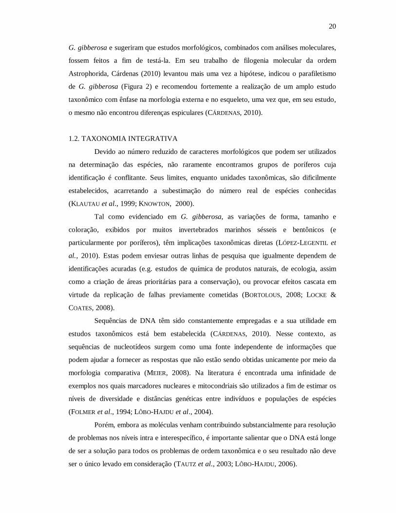
G. gibberosa e sugeriram que estudos morfológicos, combinados com análises moleculares,
fossem feitos a fim de testá-la. Em seu trabalho de filogenia molecular da ordem
Astrophorida, Cárdenas (2010) levantou mais uma vez a hipótese, indicou o parafiletismo
de G. gibberosa (Figura 2) e recomendou fortemente a realização de um amplo estudo
taxonômico com ênfase na morfologia externa e no esqueleto, uma vez que, em seu estudo,
o mesmo não encontrou diferenças espiculares (CÁRDENAS, 2010).
1.2. TAXONOMIA INTEGRATIVA
Devido ao número reduzido de caracteres morfológicos que podem ser utilizados
na determinação das espécies, não raramente encontramos grupos de poríferos cuja
identificação é conflitante. Seus limites, enquanto unidades taxonômicas, são dificilmente
estabelecidos, acarretando a subestimação do número real de espécies conhecidas
(KLAUTAU et al., 1999; KNOWTON, 2000).
Tal como evidenciado em G. gibberosa, as variações de forma, tamanho e
coloração, exibidos por muitos invertebrados marinhos sésseis e bentônicos (e
particularmente por poríferos), têm implicações taxonômicas diretas (LÓPEZ-LEGENTIL et
al., 2010). Estas podem enviesar outras linhas de pesquisa que igualmente dependem de
identificações acuradas (e.g. estudos de química de produtos naturais, de ecologia, assim
como a criação de áreas prioritárias para a conservação), ou provocar efeitos cascata em
virtude da replicação de falhas previamente cometidas (BORTOLOUS, 2008; LOCKE &
COATES, 2008).
Sequências de DNA têm sido constantemente empregadas e a sua utilidade em
estudos taxonômicos está bem estabelecida (CÁRDENAS, 2010). Nesse contexto, as
sequências de nucleotídeos surgem como uma fonte independente de informações que
podem ajudar a fornecer as respostas que não estão sendo obtidas unicamente por meio da
morfologia comparativa (MEIER, 2008). Na literatura é encontrada uma infinidade de
exemplos nos quais marcadores nucleares e mitocondriais são utilizados a fim de estimar os
níveis de diversidade e distâncias genéticas entre indivíduos e populações de espécies
(FOLMER et al., 1994; LÔBO-HAJDU et al., 2004).
Porém, embora as moléculas venham contribuindo substancialmente para resolução
de problemas nos níveis intra e interespecífico, é importante salientar que o DNA está longe
de ser a solução para todos os problemas de ordem taxonômica e o seu resultado não deve
ser o único levado em consideração (TAUTZ et al., 2003; LÔBO-HAJDU, 2006).
21
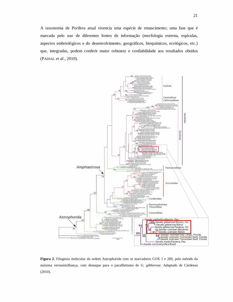
A taxonomia de Porifera atual vivencia uma espécie de renascimento; uma fase que é
marcada pelo uso de diferentes fontes de informação (morfologia externa, espículas,
aspectos embriológicos e do desenvolvimento, geográficos, bioquímicos, ecológicos, etc.)
que, integradas, podem conferir maior robustez e confiabilidade aos resultados obtidos
(PADIAL et al., 2010).
Figura 2. Filogenia molecular da ordem Astrophorida com os marcadores COX 1 e 28S, pelo método da
máxima verossimilhança, com destaque para o parafiletismo de G. gibberosa. Adaptado de Cárdenas
(2010).
22
2. OBJETIVOS
2.1. OBJETIVO GERAL
Caracterizar o polimorfismo de G. gibberosa Lamarck, 1815 e testar, por meio das análises
morfológica e molecular, a hipótese desta representar um complexo de espécies.
2.2. OBJETIVOS ESPECÍFICOS
- Realizar amplo estudo taxonômico da espécie G. gibberosa, contemplando diversas
localidades ao longo da costa brasileira e Caribe;
- Verificar o status taxonômico da espécie;
- Testar marcadores moleculares;
- Avaliar a variabilidade morfológica e molecular de distintas populações, visando a
subsidiar estudo filogeográfico.
Capítulo 1
GOING DEEPER WITH GEODIA GIBBEROSA (ASTROPHORIDA, GEODIIDAE): INVESTIGATING AN ALLEGED
SPECIES COMPLEX
Ueslei Lopes, Patrícia Domingues de Freitas & Carla Menegola
Artigo a ser submetido no periódico Zootaxa

24
Going deeper with Geodia gibberosa (Astrophorida, Geodiidae): investigating an alleged species complex
UESLEI LOPES1, PATRÍCIA DOMINGUES DE FREITAS2, CARLA MENEGOLA1
1 Instituto de Biologia, Departamento de Zoologia, Universidade Federal da Bahia, Campus de Ondina, Rua Barão de Jeremoabo, s/n, 40170-115, Salvador, Bahia, Brazil. E-mail: [email protected], [email protected]. 2 Departamento de Genética e Evolução, Centro de Ciências Biológicas e da Saúde, Universidade Federal de São Carlos, Via Washington Luis, Km 235, Monjolinho, 13565-905 - São Carlos, São Paulo – Brazil. E-mail: [email protected].
Abstract
Diverse studies applying molecular techniques have been revealing the existence of cryptic
species of sponges, especially among those considered cosmopolitan or wide-distributed
and with scarce morphologic features. Geodia gibberosa Lamarck, 1815 is registered for a
large extension of the Western Atlantic Ocean, but contrary to many other species of
poriferans, it presents a rich spicule repertoire and a significant variability in gross
morphology, which brings difficulties to taxonomists to delimit its boundaries. We used COI
and ATP6 mitochondrial markers and the classic taxonomic method of morphology to
estimate the variability within G. gibberosa, aiming to test the hypothesis of the existence of
cryptic species. Three distinct clades were found with both molecular markers, but no
distinguishable morphologic pattern that could allow us to separate species was evidenced.
Although our results suggested a low variability for the COI and ATP6 genes, these
outcomes have matched with previous studies with poriferans, supportting the status of G.
gibberosa as a species complex.
Key words: Marine sponge, Demospongiae, morphological variation, redescription,
mitochondrial markers, cryptic species.

25
Resumo
Diversos estudos empregando técnicas moleculares têm detectado a presença de espécies
crípticas em esponjas, especialmente naquelas consideradas cosmopolitas ou amplamente
distribuídas e que de carecem caracteres morfológicos. Geodia gibberosa Lamarck, 1815
tem sido registrada ao longo do oceano Atlântico ocidental, mas diferentemente de outras
espécies, esta possui um conjunto espicular rico e significativa variabilidade em sua
morfologia externa, que culminam por dificultar a delimitação da espécie. Nesse trabalho
foram utilizados os marcadores mitocondriais COI e ATP6, assim como os métodos
clássicos de taxonomia morfológica, visando estimar a variabilidade dentro de G. gibberosa
e, desse modo, testar a hipótese de existência de espécies crípticas. Três clados distintos
foram obtidos com ambos os marcadores, mas nenhum padrão morfológico, que permitisse
a separação de espécies, foi evidenciado. Mesmo os resultados sugerindo baixa
variabilidade dos marcadores mitocondriais, estes se mostraram compatíveis com outros
trabalhos já desenvolvidos com poríferos e permitem afirmar que G. gibberosa representa
um complexo de espécies.
Palavras-chave: Esponjas marinhas, Demospongiae, variação morfológica, redescrição,
marcadores mitocondriais, espécie crípticas.
26
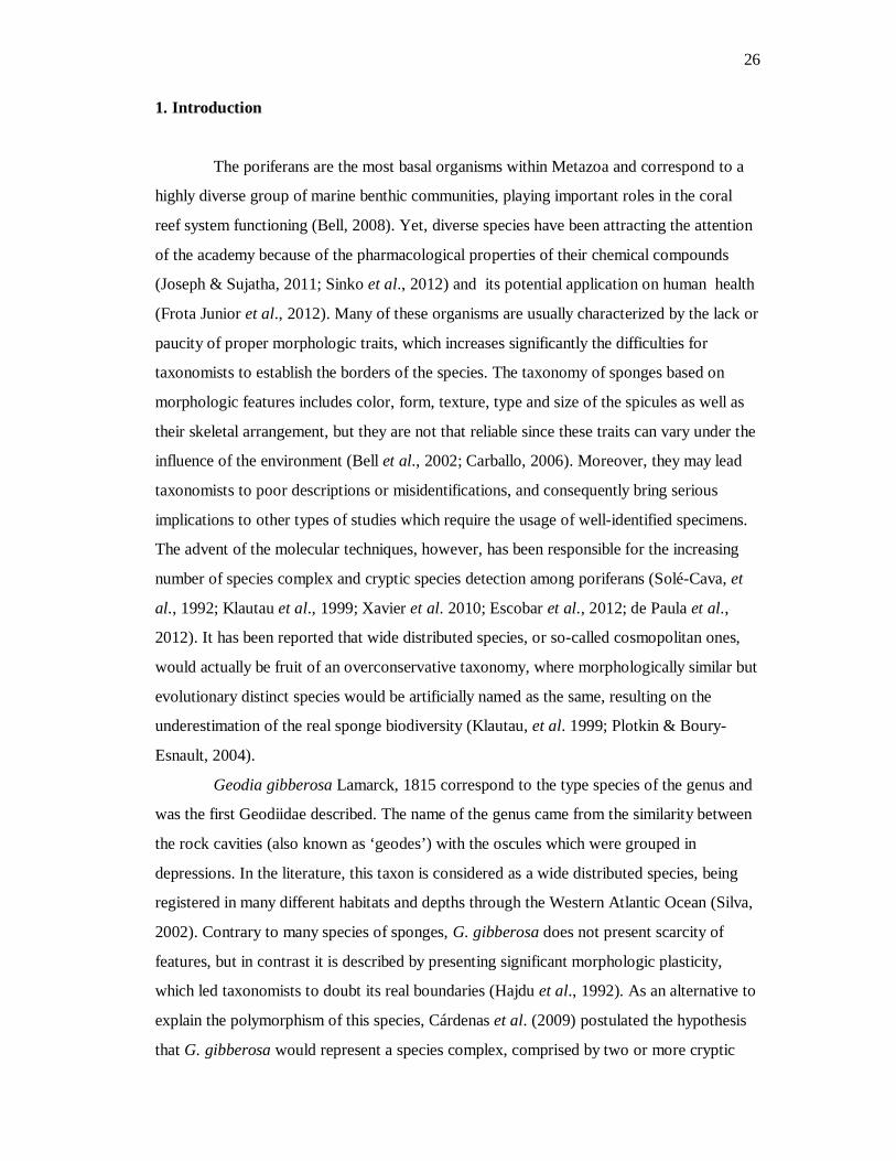
1. Introduction
The poriferans are the most basal organisms within Metazoa and correspond to a
highly diverse group of marine benthic communities, playing important roles in the coral
reef system functioning (Bell, 2008). Yet, diverse species have been attracting the attention
of the academy because of the pharmacological properties of their chemical compounds
(Joseph & Sujatha, 2011; Sinko et al., 2012) and its potential application on human health
(Frota Junior et al., 2012). Many of these organisms are usually characterized by the lack or
paucity of proper morphologic traits, which increases significantly the difficulties for
taxonomists to establish the borders of the species. The taxonomy of sponges based on
morphologic features includes color, form, texture, type and size of the spicules as well as
their skeletal arrangement, but they are not that reliable since these traits can vary under the
influence of the environment (Bell et al., 2002; Carballo, 2006). Moreover, they may lead
taxonomists to poor descriptions or misidentifications, and consequently bring serious
implications to other types of studies which require the usage of well-identified specimens.
The advent of the molecular techniques, however, has been responsible for the increasing
number of species complex and cryptic species detection among poriferans (Solé-Cava, et
al., 1992; Klautau et al., 1999; Xavier et al. 2010; Escobar et al., 2012; de Paula et al.,
2012). It has been reported that wide distributed species, or so-called cosmopolitan ones,
would actually be fruit of an overconservative taxonomy, where morphologically similar but
evolutionary distinct species would be artificially named as the same, resulting on the
underestimation of the real sponge biodiversity (Klautau, et al. 1999; Plotkin & Boury-
Esnault, 2004).
Geodia gibberosa Lamarck, 1815 correspond to the type species of the genus and
was the first Geodiidae described. The name of the genus came from the similarity between
the rock cavities (also known as ‘geodes’) with the oscules which were grouped in
depressions. In the literature, this taxon is considered as a wide distributed species, being
registered in many different habitats and depths through the Western Atlantic Ocean (Silva,
2002). Contrary to many species of sponges, G. gibberosa does not present scarcity of
features, but in contrast it is described by presenting significant morphologic plasticity,
which led taxonomists to doubt its real boundaries (Hajdu et al., 1992). As an alternative to
explain the polymorphism of this species, Cárdenas et al. (2009) postulated the hypothesis
that G. gibberosa would represent a species complex, comprised by two or more cryptic

27
species, and suggested that a study allying both morphologic and molecular analyses should
be conducted to test this.
Given that, we, thus, aimed with the present work (1) to develop a taxonomic
study based on morphologic characters of the samples, obtained from almost the entire
species’ geographic distribution, and (2) concomitantly apply molecular markers to verify if
G. gibberosa represents a species complex or simply corresponds to a species with
remarkable variable morphologic features.
2. Materials and methods
2.1. Sampling and fixation
Specimens were collected by C. Menegola, J. C. Fernandéz, U. Lopes, J. G. B. De
Marchi and V. Cedro in the cities of Salvador (Pituba, 13°00’32” S, 38°27’37” W; Barra,
13°00’36” S, 38°31’43” W), Maceió (Guaxuma, 09°35’29” S, 35°40’00” W), São Miguel
dos Milagres (09°16’6.47” S, 35°21’40.27” W), João Pessoa (Ponta do Cabo Branco,
07°08’23” S, 34°47’47” W), and in Panama (Bocas del Toro, 09°16’ 37.8” N , 82°10’16.2”
W) in the intertidal zone. Vouchers of the collected samples were preserved in 96% ethanol
in the field and the fixative was replaced two times at least, once after six hours and another
later than 24 hours, so that the water could be removed completely (Cárdenas, personal
communication). Vouchers were stored at -20° C until the DNA extraction. The entire
specimens were preserved in 80% ethanol and deposited at the Porifera Collection at Museu
de Zoologia of Universidade Federal da Bahia (UFBA-POR) for further morphologic
analysis. Digital photos were taken both in situ and ex situ whenever possible. Materials
from other localities were provided by some researchers as donation or by loaning from
diverse institutions. Spicule measures of all the samples examined in the present study, as
well as its respective localities, are listed in the Table 1.
2.2. Morphologic analysis
Dissociated spicules and thick sections mounts were obtained according to Mothes
de Moraes (1985). Each dimension of 30 spicules per spicule type was measured unless
otherwise noted (N: number of spicules measured) in the Table 1. Spicule measurements
28
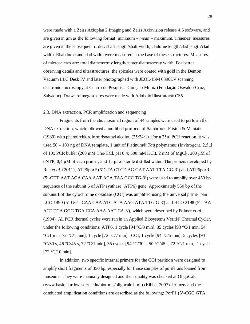
were made with a Zeiss Axioplan 2 Imaging and Zeiss Axiovision release 4.5 software, and
are given in μm as the following format: minimum – mean – maximum. Triaenes’ measures
are given in the subsequent order: shaft length/shaft width; cladome length/clad length/clad
width. Rhabdome and clad width were measured at the base of these structures. Measures
of microscleres are: total diameter/ray length/center diameter/ray width. For better
observing details and ultrastructures, the spicules were coated with gold in the Denton
Vacuum LLC Desk IV and later photographed with JEOL-JSM 6390LV scanning
electronic microscopy at Centro de Pesquisas Gonçalo Muniz (Fundação Oswaldo Cruz,
Salvador). Draws of megascleres were made with Adobe® Illustrator® CS5.
2.3. DNA extraction, PCR amplification and sequencing
Fragments from the choanosomal region of 44 samples were used to perform the
DNA extraction, which followed a modified protocol of Sambrook, Fristch & Maniatis
(1989) with phenol:chloroform:isoamyl alcohol (25:24:1). For a 25μl PCR reaction, it was
used 50 – 100 ng of DNA template, 1 unit of Platinum® Taq polymerase (Invitrogen), 2,5μl
of 10x PCR buffer (200 mM Tris-HCl, pH 8.4; 500 mM KCl), 2 mM of MgCl2, 200 μM of
dNTP, 0,4 μM of each primer, and 15 μl of sterile distilled water. The primers developed by
Rua et al. (2011), ATP6porF (5’GTA GTC CAG GAT AAT TTA GG-3’) and ATP6porR
(5’-GTT AAT AGA CAA AAT ACA TAA GCC TG-3’) were used to amplify over 450 bp
sequence of the subunit 6 of ATP synthase (ATP6) gene. Approximately 550 bp of the
subunit I of the cytochrome c oxidase (COI) was amplified using the universal primer pair
LCO 1490 (5’-GGT CAA CAA ATC ATA AAG ATA TTG G-3') and HCO 2198 (5'-TAA
ACT TCA GGG TGA CCA AAA AAT CA-3'), which were described by Folmer et al.
(1994). All PCR thermal cycles were ran in an Applied Biosystems Veriti® Thermal Cycler,
under the following conditions: ATP6, 1 cycle [94 °C/3 min], 35 cycles [93 °C/1 min, 54
°C/1 min, 72 °C/1 min], 1 cycle [72 °C/7 min]; COI, 1 cycle [94 °C/5 min], 5 cycles [94
°C/30 s, 46 °C/45 s, 72 °C/1 min], 35 cycles [94 °C/30 s, 50 °C/45 s, 72 °C/1 min], 1 cycle
[72 °C/10 min].
In addition, two specific internal primers for the COI partition were designed to
amplify short fragments of 350 bp, especially for those samples of poriferans loaned from
museums. They were manually designed and their quality was checked at OligoCalc
(www.basic.northwestern.edu/biotools/oligocalc.html) (Kibbe, 2007). Primers and the
conducted amplification conditions are described as the following: PorF1 (5’-CGG GTA
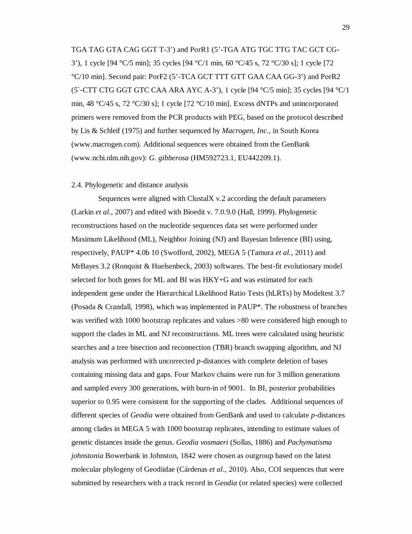
29
TGA TAG GTA CAG GGT T-3’) and PorR1 (5’-TGA ATG TGC TTG TAC GCT CG-
3’), 1 cycle [94 °C/5 min]; 35 cycles [94 °C/1 min, 60 °C/45 s, 72 °C/30 s]; 1 cycle [72
°C/10 min]. Second pair: PorF2 (5’-TCA GCT TTT GTT GAA CAA GG-3’) and PorR2
(5`-CTT CTG GGT GTC CAA ARA AYC A-3’), 1 cycle [94 °C/5 min]; 35 cycles [94 °C/1
min, 48 °C/45 s, 72 °C/30 s]; 1 cycle [72 °C/10 min]. Excess dNTPs and unincorporated
primers were removed from the PCR products with PEG, based on the protocol described
by Lis & Schleif (1975) and further sequenced by Macrogen, Inc., in South Korea
(www.macrogen.com). Additional sequences were obtained from the GenBank
(www.ncbi.nlm.nih.gov): G. gibberosa (HM592723.1, EU442209.1).
2.4. Phylogenetic and distance analysis
Sequences were aligned with ClustalX v.2 according the default parameters
(Larkin et al., 2007) and edited with Bioedit v. 7.0.9.0 (Hall, 1999). Phylogenetic
reconstructions based on the nucleotide sequences data set were performed under
Maximum Likelihood (ML), Neighbor Joining (NJ) and Bayesian Inference (BI) using,
respectively, PAUP* 4.0b 10 (Swofford, 2002), MEGA 5 (Tamura et al., 2011) and
MrBayes 3.2 (Ronquist & Huelsenbeck, 2003) softwares. The best-fit evolutionary model
selected for both genes for ML and BI was HKY+G and was estimated for each
independent gene under the Hierarchical Likelihood Ratio Tests (hLRTs) by Modeltest 3.7
(Posada & Crandall, 1998), which was implemented in PAUP*. The robustness of branches
was verified with 1000 bootstrap replicates and values >80 were considered high enough to
support the clades in ML and NJ reconstructions. ML trees were calculated using heuristic
searches and a tree bisection and reconnection (TBR) branch swapping algorithm, and NJ
analysis was performed with uncorrected p-distances with complete deletion of bases
containing missing data and gaps. Four Markov chains were run for 3 million generations
and sampled every 300 generations, with burn-in of 9001. In BI, posterior probabilities
superior to 0.95 were consistent for the supporting of the clades. Additional sequences of
different species of Geodia were obtained from GenBank and used to calculate p-distances
among clades in MEGA 5 with 1000 bootstrap replicates, intending to estimate values of
genetic distances inside the genus. Geodia vosmaeri (Sollas, 1886) and Pachymatisma
johnstonia Bowerbank in Johnston, 1842 were chosen as outgroup based on the latest
molecular phylogeny of Geodiidae (Cárdenas et al., 2010). Also, COI sequences that were
submitted by researchers with a track record in Geodia (or related species) were collected
30
from GenBank to estimate the genetic distances within the genus. Nevertheless, only two
sequences of Geodia vosmaeri for ATP6 gene were available at GenBank, which hampered
the chances to avaluate the genetic distances within Geodia.
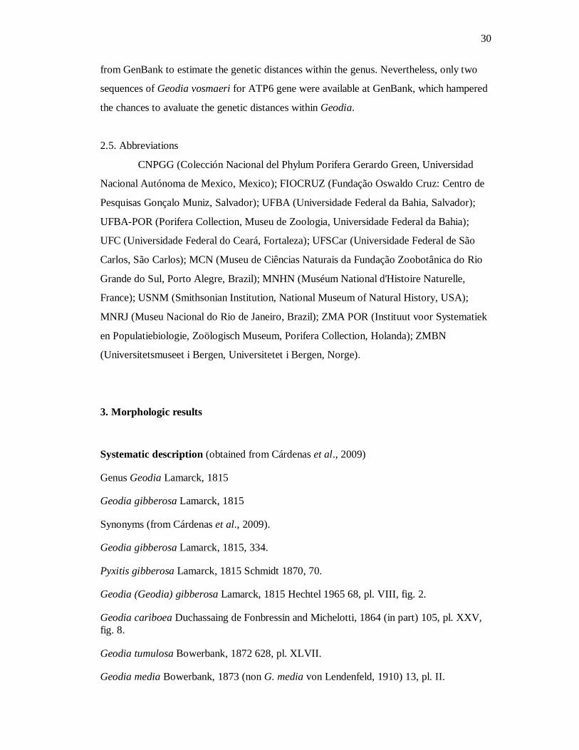
2.5. Abbreviations
CNPGG (Colección Nacional del Phylum Porifera Gerardo Green, Universidad
Nacional Autónoma de Mexico, Mexico); FIOCRUZ (Fundação Oswaldo Cruz: Centro de
Pesquisas Gonçalo Muniz, Salvador); UFBA (Universidade Federal da Bahia, Salvador);
UFBA-POR (Porifera Collection, Museu de Zoologia, Universidade Federal da Bahia);
UFC (Universidade Federal do Ceará, Fortaleza); UFSCar (Universidade Federal de São
Carlos, São Carlos); MCN (Museu de Ciências Naturais da Fundação Zoobotânica do Rio
Grande do Sul, Porto Alegre, Brazil); MNHN (Muséum National d'Histoire Naturelle,
France); USNM (Smithsonian Institution, National Museum of Natural History, USA);
MNRJ (Museu Nacional do Rio de Janeiro, Brazil); ZMA POR (Instituut voor Systematiek
en Populatiebiologie, Zoölogisch Museum, Porifera Collection, Holanda); ZMBN
(Universitetsmuseet i Bergen, Universitetet i Bergen, Norge).
3. Morphologic results
Systematic description (obtained from Cárdenas et al., 2009)
Genus Geodia Lamarck, 1815
Geodia gibberosa Lamarck, 1815
Synonyms (from Cárdenas et al., 2009).
Geodia gibberosa Lamarck, 1815, 334.
Pyxitis gibberosa Lamarck, 1815 Schmidt 1870, 70.
Geodia (Geodia) gibberosa Lamarck, 1815 Hechtel 1965 68, pl. VIII, fig. 2.
Geodia cariboea Duchassaing de Fonbressin and Michelotti, 1864 (in part) 105, pl. XXV, fig. 8.
Geodia tumulosa Bowerbank, 1872 628, pl. XLVII.
Geodia media Bowerbank, 1873 (non G. media von Lendenfeld, 1910) 13, pl. II.
31
Geodia dysoni Bowerbank, 1873 14, pl. III.
Geodia reticulata Bowerbank, 1874 300, pl. XLVI, figs. 14–20.
Sidonops stromatodes Uliczka, 1929 54, figs. 51–56, pl. I, fig. 10.
Geodia media var. leptoraphes Uliczka, 1929 56, figs. 57–67, pl. I, fig. 11.
Geodia flexisclera Pulitzer-Finali, 1986 76, figs. 10–11.
Holotype. MNHN DT–608, dry, French Guiana
Material examined. Geodia gibberosa, MNHN DT–608, schizoloholotype (MCN
4757), French Guyana; USNM 33314, off South Carolina, USA, 32°49’24” N/ 78°39’12”
W, South Carolina Marine Resources coll., 12.II.1981, 33m depth; ZMAPOR 17978,
Georgia, USA, 31°36’00” N/ 80°47’25” W, R. Ruzicka coll., 23.VIII.2004; ZMAPOR
17983, Georgia, USA, 31°24’4” N/ 80°52’8” W, R. Ruzicka, 27.VIII.2004; USNM 48248,
off South Georgia, 31°23’42”N/ 80°53’06” W, Georgia Marine Resources for Minerals
Management Service Coll., 04.III.1981, 17m depth; USNM 42560, Naples, Florida, USA,
25°46’01” N/ 82°23’49” W, Continental Shelf Associates coll., 12.II.1982, 26.1m depth;
CNPGG 078, Yucatán, Mexico, Justo Sierra coll., 00.IV.1985; CNPGG 1203, Yucatán,
Mexico, Justo Sierra coll., 25.IV.1985; CNPGG 1191, Yucatán, Mexico, Justo Sierra coll.;
18.X.1985; USNM 32866, Columbus Cay, Belize, I. Macintyre coll., 26.IV.1979, 10-15m
depth; USNM 32872, Carrie Bow Cay, Blue Hole, Belize, G. Hendler coll., 21.III.1979,
18-27m depth; UFBA-POR 3627, Bocas del Toro, Panama, Carla Menegola coll.,
29.VII.2010, 0.1 m; ZMBN 77928 and ZMBN 81780, Bocas del Toro, Paco Cárdenas
coll., 0.5-1m depth; UFBA-POR 3061, Archipeago Sabana-Camagüey, Cuba, 23°15’58” N/
80°55’59” W, Z. Marcos coll., 16.III.2001, 1m depth; UFBA-POR 3062, Arquipélago
Sabana-Camagüey, Cuba, 22°04’13” N/ 81°33’39” W, P. Alcolado coll., 15.VIII.1980, 1m
depth; UFBA-POR 3071, Norte da Cayeria de San Felipe, Cuba, 21°57’56” N/ 83°34’05”
W, P. Alcolado coll., 20.X.1968, 22m depth; UFBA-POR 3622, UFBA-POR 3623 and
UFBA-POR 3624, Oeste de Grand Cul de Suc, Guadeloupe (France) 16°20’30” N;
61°41’18”W, P. Alcolado coll., 2010, 0.5m depth; ZMAPOR 17737, Martinica, France,
15°59’36” N/ 61°24’12” W, P. T. Michaelis coll., 16.VI.2002; UFBA-POR 4140, Praia dos
Dois Coqueiros, Caucaia, Ceará, 03°41’30”S/ 38°36’10”W, C. Licarião and T. Soraya
colls., 07.X.2010; MNRJ 14755, Praia dos Dois Coqueiros, Caucaia, Ceará, 03°41’30”S/
32
38°36’10”W, S. Salani coll., 15.I.2010; MNRJ 9148, MNRJ 9149, MNRJ 9150, MNRJ
9151 and MNRJ 9154, Maracajaú, Rio Grande do Norte, Brazil, 24.IX.2004; UFBA-POR
4041, UFBA-POR 4042, UFBA-POR 4043 and UFBA-POR 4044, Ponta do Cabo Branco,
Paraíba, Brazil, U. Lopes and J.G.B. DeMarchi colls., 19.III.2011, 0.1 - 0.5m depth;
UFBA-POR 3273, São Miguel dos Milagres, Alagoas, Brazil, C. Menegola coll.,
15.II.2010, 0.5m depth; UFBA-POR 4045, Guaxuma, Maceió, Alagoas, Brazil, V. Cedro
coll., 19.II.2011; UFBA-POR 4046, UFBA-POR 4047, UFBA-POR 4048, UFBA-POR
4049, Pituba, Salvador, Bahia, Brazil, Coll. U. Lopes, 02.V.2011, 0.1-0.5m depth; MNRJ
8396, Salvador, Bahia, Brazil, E. Hajdu and C. Santos colls., 07.VI.2004, 5 - 12,5m depth;
UFBA-POR 964, Capitania dos Portos, Salvador, Bahia, Brazil, 02.VI.1988; UFBA-POR
4150, Mar Grande, Veracruz, Bahia, Alexandre Borges coll., 12°58’22” S/ 38°36’33”W, 0-
1m depth; UFBA-POR 1817, Pedra Pequena, Veracruz, Bahia, Brazil, 09.VI.2004; MNRJ
287, São Sebastião, São Paulo, Brazil, 40°45’3.6” N/ 73°59’ 0.24” W, E. Hajdu and G.
Muricy, 24.I.1996, 10 - 15m depth.
Description. External morphology of the samples. Mostly massive with
tendency to become lobate, but globose and encrusting shapes were found (Figure 1 A-F).
Color is brownish-green in mangroves, and varies from light brown to whitish with beige
choanosome in rocky shores. After fixation the color changes to brownish yellow, grey,
purple or remains whitish, and the oscular regions are usually darker than the rest of the
body (Figure 1 D-H). The surface is usually smooth to the touch and micro-hispid around
the oscules. The sponges have a hard consistency and its interior is compact. Oscules (1 - 2
mm) are uniporal, individualized by its sphincter, circular or ovoid, and frequently clustered
in lobes (diameter = 12 mm) (Figure 1G-H, K). Pores can be both uni- or cribriporal (0.1 -
0.2 mm) and do not follow any distributional pattern throughout the surface (Figure 1 I-J).
Skelenton. The cortex (175 - 1150 μm) is subdivided into a thin ectocortex of
acanthospheroxyaster II, usually irregular, and a thick endocortex of sterrasters. A third thin
layer of sponging fibers (60 - 75 μm), located right under the endocortex, may occur.
Bellow the cortex, bundles of oxeas and triaenes are radially organized in the choanosome,
becoming a bit confused towards the inner portion. Strongyloxeas are found in the
choanosome, but they are mostly positioned around the oscules. Abundant acanthoxyasters
and acanthospheroxyasters II, as well as developing and mature sterrasters, are scattered

33
throughout the choanosome while acanthospheroxyasters I are rare and placed under the
cortex (Figure 1 K-L).
Spicules. Megascleres. (a) choanosomal oxea (Figure 2A-D), stout or slender,
straight or slightly curved, with acerate, blunt or hastate tips; (b) cortical strongyloxea
(Figure 2E), fusiform, slender, mostly straight with hastate tip; (c) ortotriaene often
transitional to plagiotriaene (Figure 2F-G). Rhabdome, stout or slightly curved with hastate
or blunt tip. Cladome with cladi mostly forward directed, directed at right angles to the
rhabd or bent; (d) anatriaene (Figure 2H), choanosomal, rare. Rhabdome, long, slender,
slightly curved. Cladome, minute, with short cladi sharply downward curved; (f) protriaene
(Figure 2I), choanosomal, rare. Rhabdome, long, slender, slightly curved with hastate tip.
Cladome with slender cladi sharply curved forward from the rhabdome; (g) plagiotriaene
(Figure 2J), choanosomal, rare, slender. Rhabdome, mostly straight with hastate tip.
Cladome, cladi forward directed with hastate tips.
Microscleres. (a) sterraster (Figure 3A-C), mostly ovoid or spherical with 3 - 10
smooth star-like branched tubercules; (b) acanthoxyaster (Figure 3D), minute center, 5 - 10
rays with up to 30 spines; (c) acanthospheroxyaster I (Figure 3E), rare, medium center, 13 -
14 rays with up to 20 spines; (d) acanthospheroxyaster II (Figure 3F), robust center with
minute spined rays.
Habitat of the collected samples. Panama: on mangrove roots, ca. 1m depth.
Brazil: on rocky shores, in crevices or under rocks, 0.1 - 0.5m depth.
Ecology. G. gibberosa seems to be very palatable to turtles and parrotfishes
(Pawlik et al., 1995; Dunlap & Pawlik, 1996; 1998; Wulff, 2000) which explains the fact of
this species usually be found in hidden habitats. In the Caribbean, G. gibberosa is usually
found in mangroves while the Brazilian individuals are frequently found in crevices or under
rocks. They are usually associated to algae, Palythoa sp. and other invertebrates, such as
echinoderms, polychaetans and sponges.
Geographical distribution. USA, North Carolina (Wells et al., 1960), South
Carolina (Schmidt, 1870), Florida (Little, 1963; Engel & Pawlik, 2005), Georgia (Freeman
et al., 2007), Texas (Riggs et al., 1998), California (Sim & Bakus, 1986); Mexico (Topsent,
1889; Winfield & Ortiz, 2010); Belize (Rützler et al. 2000); Cuba (Alcolado, 1976; Sardiñas
& Alcolado, 2004; Alcolado et al. 2004; Díaz & Rützler, 2009); Jamaica (Bowerbank,
1872; Hechtel, 1965; Wulff, 2000; Díaz & Rützler, 2009); St. Thomas (Duchassaing &
Michelotti, 1864); Tortola (Duchassaing & Michelotti, 1864) ; Dominican Republic
34
(Bowerbank, 1873); Puerto Rico (Bowerbank, 1873; Sollas, 1888; Pulitzer-Finali, 1986);
St. John (Uliczka, 1929); Barbados (Uliczka, 1929; van Soest & Stentoft, 1988); Bahamas
(de Laubenfels, 1949; Wiedenmayer, 1977); Bermudas (de Laubenfels, 1950); Guadeloupe
(Díaz & Rützler, 2009); Martinique (Bowerbank, 1873; Sollas, 1888); Honduras
(Bowerbank, 1873); Panama, Atlantic coast (de Laubenfels, 1936; Wulff, 2000; Cárdenas et
al. 2009); Panama, Pacific coast (de Laubenfels, 1936); Colombia (Díaz & Zea, 2008);
Curaçao (Arndt, 1927; van Soest, 1981); Saba (Silva, 2002); Venezuela (Carter, 1882;
Wulff, 2000; Díaz & Rützler, 2009); St. Vicent (Carter, 1882); French Guiana (Lamarck,
1815); Brazil, Ceará, Dois Coqueiros Beach (present study); Rio Grande do Norte,
Maracajaú (Muricy et al. 2006); Paraíba, Ponta do Cabo Branco (present study); Alagoas,
São Miguel dos Milagres (present study); Alagoas, Maceió (present study); Bahia,
Salvador, Praia da Pituba (present study); Bahia, Salvador, Capitania dos Portos (present
study); Bahia, Veracruz, Pedra Pequena (present study); Bahia, Veracruz, Mar Grande; São
Paulo, São Sebastião (Rangel et al., 2001) (Figure 4); São Tomé, West Africa (Topsent,
1918; Lévi, 1959).
Bathymetrical distribution. From 0.1m (Salvador, Bahia, Brazil) to 33m (off
South Carolina) (present study).
Remarks. This is the first study with G. gibberosa which included samples from
almost the entire species geographic distribution, encompassing the region from South
Carolina, USA, to São Sebastião, Brazil. Specimens with the massive, encrusting and
globular morphotypes were analyzed, but the former shape seemed to be more common.
Massive and encrusting samples were both found in crevices or under rocks. The oscules
are uniporal, and pore arrangements are both uni- and cribriporal, reinforcing previous
suggestions that the arrangement of the incurrent and excurrent canals does not seem to be
a reliable character to separate genera in Geodiidae (Hajdu et al., 1992; Silva, 2002;
Cárdenas et al., 2009). The three-layered cortex once noticed by Hechtel (1965) and
Wiedenmmeyer (1977) were seen in the specimens from Guadeloupe (France) (UFBA-POR
3622, UFBA-POR 3623 and UFBA-POR 3624), but the layer of fibers was gradually
discontinuous. The measurements of the spicules were in accordance with previous studies
of the species (Sollas, 1888; Wells et al., 1960). Additional categories of triaenes, such as
ana-, pro-, meso- and promesotriaenes have already been observed in G.gibberosa (Hechtel,
1965; van Soest & Stent, 1988; Silva 2002 and Cárdenas et al., 2009), but this is the first
work which registers anatriaenes to the holotype. Although the rhabdome of these spicules

35
were broken, it is clearly different from those seen in Geodia papyracea Hechtel, 1965
(which are minute and more abundant), and its cladi are much shorter when compared to
those in Geodia glariosa Sollas, 1886, Geodia australis Silva & Mothes, 2000 and Geodia
riograndensis Silva & Mothes, 2000. Rare protriaenes were found in 25% of the samples
analyzed, but was not seen in the holotype which probably is related to the small size of the
fragment obtained from the museum. Furthermore, rare and slender plagiotriaenes are
noticed for the first time to the species, being present in 28 specimens of the 44 observed
(including the holotype). This spicule differs from the transitional variations of orthotriena
essentially in size, with the rhabdome lengh at least two times smaller, and the form of the
cladome, which cladi are upward directed. The recurrent description of a second category
of oxea (Sollas, 1888; Hechtel, 1965; van Soest & Stentoft, 1988; Muricy et al., 2006;
Muricy et al., 2008; Hajdu et al., 2012) should be properly switched to cortical
strongyloxea, since this fusiform spicule has a blunt end. Additionally, the term strongylaster
should be standardized as acanthospheroxyaster II in accordance to the prominent spine at
their tips and the robust sphere-like center presented by the spicule. Also, it was noticed
that the acanthospheroxyaster II varies significantly in morphology, but not in size, which
allows us to assume that, as soon as the spicule gets mature, the center increases and rays
tend to be minute (Figure 3F). That, however, cannot be affirmed for sure once no study of
spicule formation was developed in the present work.
4. Molecular results
The resulting dataset for COI partition comprised 32 sequences of 551 bp, which
45 sites were parsimony informative. Although the ATP6 showed less variation than COI
with 29 parsimony informative sites, this dataset only consisted of 10 sequences of 422 bp.
Four distinct clades within G. gibberosa were found for the COI partition (Figure 5), but
given we did not manage to amplify the DNA of all individuals for ATP6 (Figure 6), just
three groups and one specimen of the outgroup (I, II, III and P. johnstonia) could be
verified and will be used here only for comparison. The posterior probabilities as well as the
values of bootstrap for ML and NJ methods are written above each branch, for values
higher than 50%, when in congruence with the BI trees. All individuals of the Clade I were
collected in the coast of Salvador city, except the specimen A121, which was sampled in the

36
Ilha dos Frades (domain of the same city). Clade II comprises specimens from the U.S.A.,
Mexico, Guadeloupe and Northeastern Brazil (from Maceió to Caucaia), but also includes
one sample from Salvador, Ilha de Vera Cruz and São Sebastião each. A specimen with a
very thick cortex from Mexico (A314) presented the highest divergence value among the
clades of the COI tree and was kept apart from the Clade III, which comprises individuals
from Panama, exclusively. The sample A316 was just amplified with the ATP6 and
clustered in Clade II in the ATP6 topology. The well-supported Clade IV includes
specimens from Belize and Georgia. Genetic divergence values found for both genes were
quite similar, ranging from 3.6% - 6.4%, and 2.8% - 5.8% in COI and ATP6 tree,
respectively. Maximum intra-group divergence was detected in the Clade IV with 0.7% for
COI, and 0.3% for ATP6 in the first clade. Divergence between ingroup and outgroup for
COI was over 10% whereas for ATP6 it was 8.4% (Table 2).
5. Discussion
Unlike many poriferans which lack proper morphologic characters (Erwin &
Thacker, 2007; Redmond & McCormack, 2008; Erpenbeck et al., 2012), species of
Astrophorida possess a significant spicule repertoire (Cárdenas et al., 2011), and this fact
theoretically should facilitate their identification/characterization. Our morphologic results
describe G. gibberosa as possessing at least three different morphotypes, with six different
categories of megascleres and four of microscleres, but some of these spicules somehow
differ significantly in abundance. Diverse studies have been demonstrating that sponge
morphologic features, such as gross morphology and spicule sizes may change under the
influence of environmental factors (Mercurio et al., 2000; Bell et al., 2002; Meroz-Fine et
al., 2005; Cavalcanti et al., 2007; Massaro et al., 2012), which could explain the variability
addressed to G. gibberosa hitherto. But, specimens collected in nearby localities were both
variable in gross morphology (e.g. those from João Pessoa, which were massive and
encrusting) and even in spicule repertoire (e.g. the samples from Salvador) so that any
pattern that could allow us to separate species morphologically, thus, was not evidenced.
A plausible explanation for the infrequency of some categories of spicules such as
ana- and protriaenes could be related to its non-structural function, differently from oxeas,

37
orthotriaenes and sterrasters which play an important role in this subject. Moreover, Burton
(1949), in his study about the development of Geodia barretti Bowerbank, 1958, noticed
that these triaenes remained the last ones to be produced by that species (what might occur
to G. gibberosa as well). Regarding what we call secondary triaenes (here represented by
the ana-, pro- and the minute plagiotriaenes), it is important to note that, in all 38 specimens
where these spicules were present, there was often a combination of just two of each
categories. This stresses, in our opinion, the complementary function of these spicules to
provide mechanical reinforcement of the choanosome and support of the cortex, particularly
the ectocortex. On the other hand, the scarcity of acanthospheroxyasters I and the minute
plagiotriaene should be further investigated.
In the contrary, our molecular results based on the mitochondrial DNA (mtDNA)
suggest that G. gibberosa represents a species complex, comprised by three distinct species,
at least. The genetic distances for COI fragment observed in the present work were not as
high as those found by Blanquer & Uríz (2007) (up to 22%), but they matched with
previous studies with Cliona celata Grant, 1826 developed by Xavier et al. (2010) and de
Paula et al. (2012) (p-distance varied between 6% and 8% and between 2% and 8%,
respectively), and also were quite similar to the divergence values detected in other
invertebrates (Carmona et al., 2011; Neusser et al., 2011; Hemery et al., 2012). The values
of genetic divergences found in the genus (uncorrected p-distance between 1.1% to 8.2%)
(see Appendix A) may be considered insufficient to separate species, since diverse authors
have already stressed the slow evolution of the COI partition for some basal marine
invertebrates (Wörheide, 2006; Huang et al., 2008; Bucklin et al., 2010). However, the low
intraspecific distances within the clades (less than 1%) followed by the high support of the
branches and the congruence between both topologies encourage us to accept the
Cárdenas’s hypothesis (2009).
Even our molecular results clearly suggesting the split of G. gibberosa into distinct
genetic groups, it is curious to realize that the Clade II remained distributed through the
same geographic extension occupied by the so-called G. gibberosa, whereas clades I and III
are restricted to one single area each. This observation is contrary to the current consensus
about statuses of broadly distributed species, especially when we consider the low dispersal
capacity of the sponge larvae (Maldonado, 2006). Maybe this pattern presented by the
Clade II could be explained by the maintenance of an uninterrupted gene flow, or this
distribution could be a fruit of anthropic actions. Its occurrence in some places in Salvador
38
and in São Sebastião could also support the latter hypothesis, given that these regions
represent important Brazilian harbor areas. Both Clade I and II seem to coexist in the same
region, but the high distances between the former and the other clades might be an evidence
of reproductive isolation (Xavier et al., 2010), or its occurrence in one single area could be
an effect of subsampling. Regarding the restricted distribution of the Panamanian clade,
which comprises only individuals from mangrove, Duran & Rützler (2006) already pointed
out that environmental differences and disparities of ecological interactions could indicate
great potential for ecological speciation between habitats. The Yucatán samples were
clustered in different clades and the sample 314 presented the highest genetic divergences.
We suggest that the population of that locality should be proper investigated in the future,
as well as more collections should be provided to stablish the status of the Clade IV.
6. Conclusion
Additionally to the variation in gross morphology already described in the
literature, our results revealed that Geodia gibberosa possesses officially more spicule
categories than expected, and that a rich spicule repertoire does not necessarily represent a
synonymous of easy identification, since any morphological pattern could be established to
separate it into different species. On the other hand, via molecular analysis, it was possible
to conclude that at least three genetically distinct species comprise the so-called G.
gibberosa and made possible to realize how the diversity within this complex was
underestimated. In an era of a technologic advancement and the resurrection of a new
taxonomy, we not only encourage the usage of molecular techniques in solving questions
like this, but also stimulate the application of other sources (e.g. embryological, histological,
biochemical, etc.) with the intention to get closer to the real extant biodiversity.
Acknowledgements
We specially thank Eduardo Hajdu and Sula Salani (Museu Nacional, Rio de
Janeiro), Rob van Soest and Elly Beglinger (Zoölogisch Museum van Amsterdam), Pedro
Alcolado (Instituto de Oceanología, Havana), Hans Tore Rapp (Universitetet i Bergen)

39
Patricia Gomez (Universidad Autónoma de México, México D.F.), Helena Cascon and
Cecília Licarião (Universidade Federal do Ceará, Fortaleza), Klaus Rützler (National
Museum of Natural History, Washington D.C.) and Josivete Pinheiro (Universidade Federal
de Pernambuco, Recife) for providing putative samples of G. gibberosa. Júlio Fernandez,
João G. B. DeMarchi and Victor Cedro are also thanked for helping with the collection of
samples throughout the Northeastern Brazilian coast. The authors are grateful to Armando
Vieira, Luis Sartori, Inessa Lacativa and the team of the Laboratório de Ficologia
(Universidade Federal de São Carlos, São Carlos) for allowing us to use the light
microscope and make the measurements in São Carlos. We gratefully acknowledge Adriana
Rangel, Cláudio Figueira and Maria Lúcia Moreno (Fundação Oswaldo Cruz - Centro de
Pesquisa Gonçalo Moniz, Salvador) for the help with the SEM. Pedro M. Galetti Jr., the
entire team of the Laboratório de Biodiversidade Molecular e Conservação, as well as the
staff of Departamento de Genética e Evolução (Universidade Federal de São Carlos, São
Carlos), are further thanked for the daily assistance. Luciana Martins and Fábio Quinteiro
are thanked for the precious tips on vector graphics software. We acknowledge Carlos
Congrains and Jorge Ramirez for the precious assistance on the molecular part of the work.
This research was partially funded by Conselho Nacional de Desenvolvimento Científico
(CNPq, Brazil; grant nr. 133909/2010-7, fellowship to Ueslei Lopes) and Coordenação de
Aperfeiçoamento de Pessoal de Nível Superior (CAPES, Brazil; Programa de Apoio à Pós-
graduação – PROAP).
References cited
Alcolado, P. (1976) Lista de nuevos registros de poriferos para Cuba. Serie Oceanológica.
Academia de Ciências de Cuba, No. 36, 1-11.
Alcolado, P., Grovas- Hernández, A. & Marcos, Z. (2004) General comments on species
inventory, fisheries, culture and some community features of the Porifera in Cuba.
Bollettino dei musei e degli istituti biologici dell'Universita di Genova, 68, 175-186.
Arndt, W. (1927) Kalk- und Kieselschwämme von Curaçao. Bijdragen tot de Dierkunde,
25, 133-158, pls I-III.
40
Bell, J.J., Barnes, D.K.A. & Turner, J.R. (2002) The importance of micro morphological
variation in the adapt of a sublittoral demosponge to current extremes. Marine Biology,
140, 75-81.
Bell, J.J. (2008) The functional roles of marine sponges. Estuarine, Coastal and Shelf
Science, 79, 341-353.
Blanquer, A. & Uriz, M.J. (2007) Cryptic speciation in marine sponges evidenced by
mitochondrial and nuclear genes: a phylogenetic approach. Molecular Phylogenetics
and Evolution, 45, 392-397.
Bowerbank, J.S. (1858) On the Anatomy and Physiology of the Spongiadae. Part I. On the
Spicula. Philosophical Transactions of the Royal Society 148, 279-332, pls XXII-
XXVI.
Bowerbank, J.S. (1872) Contributions to a General History of the Spongiadae. Part III.
Proceedings of the Zoological Society of London, 1872, 626-635, pls XLVI-XLIX.
Bowerbank, J.S. (1873) Contributions to a General History of the Spongiadae. Part IV.
Proceedings of the Zoological Society of London, 1873, 3-25, pls I-IV.
Bowerbank, J.S. (1874) Contributions to a General History of the Spongiadae. Part VI.
Proceedings of the Zoological Society of London, 1874, 298-305, pls XLVI-XLVII.
Bucklin, A., Steinke, D. & Blanco-Bercial, L. (2010) DNA Barcoding of Marine Metazoa.
Annual Review of Marine Science, 3, 471-508.
Burton, M. (1949) Non-sexual reproduction in sponges, with special reference to a
collection of young Geodia. Proceedings of the Linnean Society of London, Session
160, Pt. 2, 163-178.
Carballo, J.L. (2006) Effect of natural sedimentation on the structure of tropical rocky
sponge assemblages. Ecoscience, 13, 119-130.
Cárdenas, P., Menegola, C., Rapp, H.T. & Díaz, M.C. (2009) Morphological description
and DNA barcodes of shallow-water Tetractinellida (Porifera: Demospongiae) from
Bocas del Toro, Panama, with description of a new species. Zootaxa, 2276, 1-39.
41
Cárdenas, P., Rapp, H. T., Schander, C. & Tendal, O. S. (2010) Molecular taxonomy and
phylogeny of the Geodiidae (Porifera, Demospongiae, Astrophorida) – combining
phylogenetic and Linnaean classification. Zoologica Scripta, 39, 86-106.
Cárdenas, P., Xavier, J.R., Reveillaud, J., Schander, C. & Rapp, H.T. (2011) Molecular
phylogeny of the Astrophorida (Porifera, Demospongiaep) reveals an unexpected high
level of spicule homoplasy. PLoS ONE, 6, e18318. doi:10.1371/journal.pone.0018318.
Carmona, L., Malaquias, M.A.E., Gosliner, T.M., Pola, M. & Cervera, J.L. (2011) Amphi-
atlantic distributions and cryptic species in sacoglossan sea slugs. Journal of Molluscan
Studies, 77, 401-412. doi:10.1093/mollus/eyr036.
Cavalcanti, F.F., Zilberberg, C. & Klautau, M. (2007) Seasonal variation of morphological
characters of Chondrilla aff. nucula (Porifera: Demospongiae) from the south-east
coast of Brazil. Journal of the Marine Biological Association of the UK, 87, 1727-
1732.
Carter, H.J. (1882) Some sponges from the West Indies and Acapulco in the Liverpool Free
Museum described, with general and classificatory Remarks. Annals and Magazine of
Natural History, 5, 266-301, 346-368, pls XI-XII.
Días, M. & Zea, S. (2008) Distribución de esponjas sobre la plataforma continental de la
guajira, caribe colombiano. Boletín de Investigaciones Marinas y Costeras, 37, 27-43.
Díaz, M.C. & Rützler, K. (2009) Biodiversity and abundance of sponges in Caribbean
mangrove: indicators of environmental quality. In: Lang, M.A., Macintyre I.G. &
Rützler, K. (Eds.). Proceedings of the Smithsonian Marine Science Symposium.
Smithsonian Contributions to the Marine Sciences, No.38, 529, pages, 217 figures, 47
tables.
Duchassaing de Fonbressin, P. & Michelotti, G. (1864) Spongiaires de la mer Caraïbe.
Natuurkundige verhandelingen van de Hollandsche maatschappij der wetenschappen
te Haarlem, 21, 1-124.
Dunlap, M., Pawlik, J.R. (1996) Video-monitored predation by Caribbean reef fishes on an
array of mangrove and reefsponges. Marine Biology, 126, 117-123.

42
Duran, S. & Rützler, K. (2006) Ecological speciation in a Caribbean marine sponge.
Molecular Phylogenetics and Evolution, 40, 292-297.
Engel, S. & Pawlik, J.R. (2005) Interactions among Florida sponges. II. Mangrove habitats.
Marine Ecology Progress Series, 303, 145-152.
Erpenbeck, D., Sutcliffe, P., Cook, S.C., Dietzel, A., Maldonado, M., van Soest, R.W.M.,
Hooper, J.N.A. & Wörheide, G. (2012) Horny sponges and their affairs: On the
phylogenetic relationships of keratose sponges. Molecular Phylogenetics and
Evolution, 63, 809-816.
Escobar, D., Zea, S. & Sánchez, J.A. (2012) Phylogenetic relationships among the
Caribbean members of the Cliona viridis complex (Porifera, Demospongiae,
Hadromerida) using nuclear and mitochondrial DNA sequences. Molecular
Phylogenetics and Evolution, 64, 271-284.
Erwin, P.M. & Thacker, R.W. (2007) Phylogenetic analyses of marine sponges within the
order Verongida: a comparison of morphological and molecular data. Invertebrate
Biology 126, 220-234.
Folmer, O., Black, M., Hoeh, W., Lutz, R. & Vrijenhoek, R. (1994) DNA primers for
amplification of mitochondrial cytochrome c oxidase subunit I from diverse metazoan
invertebrates. Molecular Marine Biology and Biotechnology, 3, 294-299.
Freeman, C.J., Gleason, D.F., Ruzicka, R., van Soest, R.W.M., Harvey, A.W. & McFall, G.
(2007) A biogeographic comparison of sponge fauna from Gray’s Reef National Marine
Sanctuary and other hard-bottom reefs of coastal Georgia, U.S.A. In: Custódio, M.R.,
Lôbo-Hajdu, G., Hajdu, E. & Muricy, G. (Eds.) Porifera research: biodiversity,
innovation and sustainability. Série Livros 28, Museu Nacional, Rio de Janeiro, pp.
319-325.
Frota Junior, M.L.C., Biegelmeyer,R.S., Mothes, B., Henriques, A. T. & Moreira, J.C.F.
(2012) Current Status on Natural Products with Antitumor Activity from Brazilian
Marine Sponges. Current Pharmaceutical Biotechnology, 13, 235-244.
43
Grant, R.E. (1826) Notice of a New Zoophyte (Cliona celata Gr.) from the Firth of Forth.
Edinburgh New Philosophical Journal, 1, 78-81.
Hajdu, E., Muricy, G., Custódio, M.R., Russo, C.A.M. & Peixinho, S. (1992) Geodia
corticostylifera (Demospongiae, Porifera) new astrophorid from the Brazilian coast
(southwestern Atlantic). Bulletin of Marine Science, 51, 204-217.
Hajdu, E., Muricy, G., Custódio, M., Russo, C. & Peixinho, S. (1992), Geodia
corticostylifera (Demospongiae, Porifera) new Astrophorid from the Brazilian coast
(Southwestern Atlantic). Bulletin of Marine Science, 51, 204-217.
Hajdu, E., Peixinho, S. & Fernandez, J.C. (2012). Esponjas marinhas da Bahia: guia de
campo e laboratório. Série Livros 45, Museu Nacioanal, Rio de Janeiro, 276 pp.
Hall, T.A. (1999) BioEdit: a user-friendly biological sequence alignment editor and analysis
program for Windows 95/98/NT. Nucleic Acids Symposium Series, 41, 95-98.
Hechtel, G.J. (1965) A systematic study of the Demospongiae of Port Royal, Jamaica.
Bulletin of the Peabody Museum of Natural History, 20, 1-103.
Hemery, L.G., Eléaume, M., Roussel, V., Améziane, N., Gallut, C., Steinke, D., Cruaud,
C., Couloux, A. & Wilson, N.G. (2012) Comprehensive sampling reveals circumpolarity
and sympatry in seven mitochondrial lineages of the Southern Ocean crinoid species
Promachocrinus kerguelensis (Echinodermata). Molecular Ecology, 21, 2502-2518.
Huang, D., Meier, R., Todd, P.A. & Chou, L.M. (2008) Slow Mitochondrial COI Sequence
Evolution at the Base of the Metazoan Tree and Its Implications for DNA Barcoding.
Jornal of Molecular Evolution, 66, 167-174.
Johnston, G. (1842) A History of British Sponges and Lithophytes.(W.H. Lizars:
Edinburgh): i-xii, 1-264, pls I-XXV.
Joseph, B. & Sujatha, S. (2011) Pharmacologically Important Natural products from
Marine Sponges. Journal of Natural Products, 4, 5-12.
Kibbe, W.A. (2007) OligoCalc: an online oligonucleotide properties calculator.
Nucleic Acids Research, 35, doi:10.1093/nar/gkm234.
44
Klautau, M., Russo, C., Lazoski, C., Boury-Esnault, N., Thorpe, J. P. & Solé-Cava A. M.
(1999) Does cosmopolitanism in morphologically simple species result from
overconservative systematics? A case study using the marine sponge Chondrilla
nucula. Evolution 53: 1414-1422.
Lamarck, J.B.P.A. (1815) Suite des polypiers empâtés. Histoire naturelle des animaux sans
vertébres, Paris, 1-568.
Larkin,M.A., Blackshields, G., Brown, N.P., Chenna, R., McGettigan, P.A., McWilliam,
H., Valentin, F., Wallace, I.M., Wilm, A., Lopez, R., Thompson, J.D., Gibson, T.J. &
Higgins, D.G. (2007) Clustal W and Clustal X version 2.0. Bioinformatics, 23, 2947-
2948.
de Laubenfels, M.W. (1936) A comparison of the shallow-water sponges near the Pacific
end of the Panama Canal with those at the Caribbean end. Proceedings of the United
States National Museum, 83, 441-466.
de Laubenfels, M.W. (1949) Sponges of the western Bahamas. American Museum
novitates, 1431, 1-25.
de Laubenfels, M.W. (1950) The Porifera of the Bermuda Archipelago. Transactions of the
Zoological Society of London, 27, 1-154, pls I-II.
Lévi, C. (1959) Résultats scientifiques des Campagnes de la ‘Calypso’. Campagne de la
‘Calypso’ dans le Golfe de Guinée et aux îles Principe, São Tomé et Annobon. 5.
Spongiaires. Annales de l’Institut océanographique, 37, 115-141.
Lis, J.T. & Schleif, R. (1975) Size fractionation of double-stranded DNA by precipitation
with polyethylene glycol. Nucleic Acids Research, 2, 383-389.
Little, F.J. (1963) The sponge fauna of the St. George's sound, Apalachee Bay, and
Panama city regions of the Florida gulf coast. Tulane Studies in Zoology, 11, 31-71
Maldonado, M., (2006) The ecology of the sponge larva. Canadian Journal of Zoology,
84, 175-194.

45
Massaro, A.J., Weisz, J.B., Hill, M.S. & Webster, N.S. (2012) Behavioral and
morphological changes caused by thermal stress in the Great Barrier Reef sponge
Rhopaloeides odorabile. Journal of Experimental Marine Biology and Ecology, 416-
417, 55-60.
Mercurio, M., Corriero, G., Scalera-Liaci, L. & Gaino, E. (2000) Silica content and spicule
size variations in Pellina semitubulosa (Porifera: Demospongiae). Marine Biology, 137,
87-92.
Meroz-Fine, E., Shefer, S. & Ilan, M. (2005) Changes in morphology and physiology of an
East Mediterranean sponge in different habitats. Marine Biology, 147, 243-250.
Mothes de Moraes, B. (1985) Manual de técnicas para a preparação de coleções
zoologias: Esponjas Marinhas. Sociedade Brasileira de Zoologia, 2, 1-3p.
Muricy, G., Esteves, E.L., Moraes, F.C., Santos, J.P., Silva, S.M., Klautau, M. & Lanna, E.
(2008) Biodiversidade marinha da Bacia Potiguar: Porifera. Série Livros 29. Museu
Nacional, Rio de Janeiro. 156 pp.
Muricy, G. & Hajdu, E. (2006) Porifera Brasilis: guia de identificação das esponjas mais
comuns do Sudeste do Brasil. Série Livros 17, Museu Nacional, Rio de Janeiro.
Neusser, T.P., Jörger, K.M. & Schrödl, M. (2011) Cryptic Species in Tropic Sands -
Interactive 3D Anatomy, Molecular Phylogeny and Evolution of Meiofaunal
Pseudunelidae (Gastropoda, Acochlidia). PLoS ONE, 6, e23313.
doi:10.1371/journal.pone.0023313.
de Paula, T.S., Zilberberg, C., Hajdu, E. & Lôbo-Hajdu, G. (2012) Morphology and
molecules on opposite sides of the diversity gradient: Four cryptic species of the Cliona
celata (Porifera, Demospongiae) complex in South America revealed by mitochondrial
and nuclear markers. Molecular Phylogenetics and Evolution, 62, 529-541
Pawlik, J.R., Chanas, B., Toonen, R.J. & Fenical, W. (1995) Defenses of Caribbean
sponges against predatory reef fish. I. Chemical deterrency. Marine Ecology Progress
Series, 127, 183-194.
46
Plotkin, A. & Boury-Esnault, N. (2004) Alleged cosmopolitanism in sponges: the example
of a common Arctic Polymastia (Porifera, Demospongiae, Hadromerida). Zoosystema,
26, 13-20.
Posada, D. & Crandall, K.A. (1998) Modeltest: testing the model of DNA substitution.
Bioinformatics, 14, 817-818.
Pulitzer-Finali, G. (1986) A collection of West Indian Demospongiae (Porifera). In
appendix, a list of the Demospongiae hitherto recorded from the West Indies. Annali del
Museo Civico di Storia Naturale Giacomo Doria, 86, 65-216.
Rangel, M., Sanctis, B., Freitas, J.C., Polatto, J.M., Granato, A.C., Berlinck, R.G.S. &
Hajdu, E. (2001) Cytotoxic and neurotoxic activities I extracts of marine sponges
(Porifera) from southeastern Brazilian coast. Journal of Experimental Marine Biology
and Ecology, 262, 31-40.
Redmond, N.E. & McCormack, G.P. (2008) Large expansion segments in 18S rDNA
support a new sponge clade (Class Demospongiae, Order Haplosclerida). Molecular
Phylogenetics and Evolution, 47, 1090-1099.
Riggs, T.E., Dokken, Q.R. & Beaver, C.R. (1998) Preliminary surveys of sessile
invertebrate community on a partially removed platform reef off South Padre Island,
Texas. Proceedings: Eighteenth Annual Gulf of Mexico Information Transfer Meeting,
1-513.
Ronquist F. & Huelsenbeck J.P. (2003) MrBayes 3: Bayesian phylogenetic inferenceunder
mixed models. Bioinformatics, 19, 1572–1574.
Rua, C., Zilberberg, C. & Solé-Cava, A.M. (2011) New Polymorphic mitochondrial
markers for sponge phylogeography. Journal of the Marine Biological Association of
the United Kingdom, 91, 1015-1022.
Rützler, K., Diaz, M.C., van Soest, R.W.M., Zea, S., Smith, K.P., Alvarez, B. & Wulff, J.
(2000) Diversity of sponge fauna in mangrove ponds, Pelican Cays, Belize. Atoll
Research Bulletin, 476.

47
Sambrook, J., Fritsch, E.F. & Maniatis, T. (1989) Molecular cloning: A laboratory manual.
Cold Spring Harbor Laboratory Press, Cold Spring Harbor, New York, 2344 pp.
Sardiñas, Z.M. & Alcolado, P. (2004) Esponjas marinas del Archipiélago de Sabana-
Camagüey, Cuba. Cocuyo, 14, 5-7.
Schmidt, O. (1870) Grundzüge einer Spongien-Fauna des atlantischen Gebietes. (Wilhelm
Engelmann: Leipzig): iii-iv, 1-88, pls I-VI.
Silva, C.M.M. & Mothes, B. (2000) Three new species of Geodia Lamarck, 1815 (Porifera,
Geodiidae) from the Brazilian coast, Southwestern Atlantic. Revue Suisse de Zoologie,
107, 31-48.
Silva, C.M.M. (2002) Revisão das espécies de Geodia Lamarck, 1815 (Porifera,
Astrophorida, Geodiidae) do Atlântico Ocidental e Pacífico Oriental. Ph.D. thesis,
Instituto de Biociências da Universidade de São Paulo, São Paulo, 305 pp.
Sim, C.J. & Bakus, G.J. (1986) Marine Sponges of Santa Catalina Island, California.
Occasional Papers of the Allan Hancock Foundation, 5, 1-23.
Sinko, J., Rajchard, J., Balounova, Z. & Fikotova, L. (2012) Biologically active substances
from water invertebrates: a review. Veterinarni Medicina, 57, 177-184.
van Soest, R.W.M. (1981) A checklist of the Curaçao sponges (Porifera Demospongiae)
including a pictorial key to the more common reef-forms. Verslagen en Technische
Gegevens Instituut voor Taxonomische Zoölogie (Zoölogisch Museum) Universiteit van
Amsterdam, 1-39.
van Soest, R.W.M. & Stentoft, N. (1988) Barbados Deep-Water Sponges. In: Hummelinck,
P.W. & Van der Steen, L.J. (Eds.) Studies on the Fauna of Curaçao and other
Caribbean Islands. Uitgaven van de Natuurwetenschappelijke Studiekring voor
Suriname en de Nederlandse Antillen, No. 122, Amsterdam, pp. 1-175.
Solé-Cava, A.M., Boury-Esnault, N., Vacelet, J. & Thorpe, J.P. (1992) Biochemical genetic
divergence and systematics in sponges of the genera Corticium and Oscarella
(Demospongiae: Homoscleromorpha) in the Mediterranean Sea. Marine Biology, 113,
299-304.
48
Sollas, W.J. (1886) Preliminary account of the tetractinellid sponges dredged by H.M.S.
Challenger, 1872-1876. Part I. The Choristida. Scientific Proceedings of the Royal
Dublin Society, 5, 177-199.
Sollas, W.J. (1888) Report on the Tetractinellida collected by H.M.S. Challenger, during
the years 1873-1876. Report on the Scientific Results of the Voyage of H.M.S.
Challenger, 1873-1876. Zoology, 25, 1-458, pls I-XLIV, 1 map.
Swofford, D.L. (2002) PAUP*. Phylogenetic Analysis Using Parsimony (* and other
methods), ver. 4.0. beta 10. Sunderland, MA: Sinauer Associates.
Tamura, K., Peterson, D., Peterson, N., Stecher, G., Nei, M., and Kumar, S. (2011)
MEGA5: Molecular Evolutionary Genetics Analysis using Maximum Likelihood,
Evolutionary Distance, and Maximum Parsimony Methods. Molecular Biology and
Evolution, 28, 2731-2739.
Topsent, E. (1889) Quelques spongiaires du Banc de Campêche et de la Pointe-à-Pître.
Mémoires de la Société zoologique de France, 2, 30-52.
Topsent, E. (1918) Éponges de San Thomé. Essai sur les genres Spirastrella, Donatia et
Chondrilla. Archives de Zoologie expérimentale et générale, 57, 535-618.
Uliczka, E. (1929) Die tetraxonen Schwämme Westindiens (auf Grund der Ergebnisse der
Reise Kükenthal-Hartmeyer). Zoologische Jahrbücher. Abteilung für Systematik,
Geographie und Biologie der Thiere, supplement 16, 35-62, pl I.
Wells, H.W., Wells, M.J. & Gray, J.E. (1960). Marine sponges of North Carolina. Journal
of the Elisha Mitchell Scientific Society, 76, 200-245, 63 figs.
Wiedenmayer, F. (1977) Shallow-water sponges of the western Bahamas. Experientia
Supplementum, 28, 1-287, pls 1-43.
Winfield, I. & Ortiz, M. (2010) Colomastigids (Amphipoda: Gammaridea: Colomastigidae)
from the Veracruz Coral Reef System, SW Gulf of Mexico, with a description of two
new species associated with sponges. Scientia Marina, 74, 773-782.

49
Wörheide, G. (2006) Low variation in partial cytochrome oxidase subunit I (COI)
mitochondrial sequences in the coralline demosponge Astrosclera willeyana across the
Indo-Pacific. Marine Biology, 148, 907-912.
Wulff, J.L. (2000) Sponge predators may determine differences in sponge fauna between
two sets of mangrove cays, Belize barrier reef. Atoll Research Bulletin, 477.
Xavier, J.R., Rachello-Dolmen, P.G., Parra-Velandia, F., Schönberg, C.H.L., Breeuwer,
J.A.J. & van Soest, R.W.M. (2010) Molecular evidence of cryptic speciation in the
”cosmopolitan” excavating sponge Cliona celata (Porifera, Clionaidae). Molecular
Phylogenetics and Evolution, 56, 13-2

Table 1. Micrometries of the spicules of Geodia gibberosa Lamarck, 1815. Triaenes measures are shaft length/shaft width; cladome length/clad
length/clad width. Measures of microscleres are total diameter/ rays length/center diameter/ rays width. Measures are in μm (N=30). Means are in
italic. n.o.. =not observed. Acanthoxy.= acanthoxyaster. Acantho. I= acanthospheroxyaster I. Acantho. II= acanthospheroxyaster II.
(*) specimens with amplified DNA.
Code Locality
Examined materials
Choanosomal Oxea
Cotical Strongyloxea
Orthotriaene
Anatriaene
Protriaene
Plagiotriaene
Sterraster
Acanthoxy.
Acantho. I
Acantho. II
-
French Guiana
Holotype MNHN DT-608 (Schyzoholoty-pe MCN 3639)
1411 - 1656.2 - 1907/ 16 -
22.8 - 31
111 - 209.1 - 315/ 3 -5.4 -8
1150 -
1316.7 - 1600/ 26 - 31 - 37.6/
300 - 380 - 440/ 212.8 -
230.5 - 239.4/ 21.3 - 23.9 - 26.6
- - -/ 10.6 -
10.6 - 10.6/21.3 -
22.6 - 23.9 / 5.3 - 6.7 - 8/ 4 - 5.8 - 7 [N=2]
n.o.
58 (broken)/ 8 / 40 / 24/ 5.3 [N=1]
66 - 82.3 –
98
18 - 23.1 - 38/ 8 - 10.3 -17/ 2 - 3.5 -9/ 1 - 1.2 -
2
n.o..
4 - 5.8 - 7
A336
Off South Carolina, USA
USNM 33314
990 - 1251.7 - 1720/ 12.5 - 18.3 - 27.5
175 - 244.6 - 312.5/ 2.5 -
4.6 - 7.5
900 - 1073.3 - 1200/ 15 - 22.5 - 30/
320 - 472.5 - 620/ 160 -
232.1 - 287.5/ 12.5 -
19.8 - 30 [N=12]
n.o.
n.o.
470 - 630 - 790/ 10 -
11.3 - 12.5/ 100 - 115 - 130/ 37.5 - 52.5 - 67.5/
7.5 - 8.8 - 10 [N=2]
42.5 - 47 -
52.5
9.6 - 26.9 - 32.2/ 8.4 -
12.5 - 15.4/ 2.8 - 2.9 - 4.2/ 1.4 - 1.4 - 1.4
11.2 - 12.6 -
14 [N=2]
4.2 - 6.3 -
11.2
A326
Georgia, USA
POR 17978
780 - 1112.7 - 1400/ 10.6 - 20.2 - 29.3
172.9 - 214 - 247.4/ 2.7 -
5.6 - 8
670 - 937.3 - 1250/ 10.6 - 22.2 - 31.9/
150 - 397.7 - 600/ 79.8 -
208.3 - 319.2/ 10.6 - 18.5 - 26.6
1330 - 1590 - 1810/ 8 - 10.6 - 16/ 30 - 35 - 40/ 13.3 -
13.3 - 13.3/ 5.3 - 10 -
13.3 [N=4]
n.o.
430 / 18.6/
130/ 79.8/ 16 [N=1]
39.9 - 50 -
66.5
17.3 - 21.9 - 27/ 7.6 - 10.7 - 13/ 2.2 - 3.7 - 5.4/ 1.1 - 1.4 - 2.2
n.o.
4.3 - 5.4 -
6.5
50
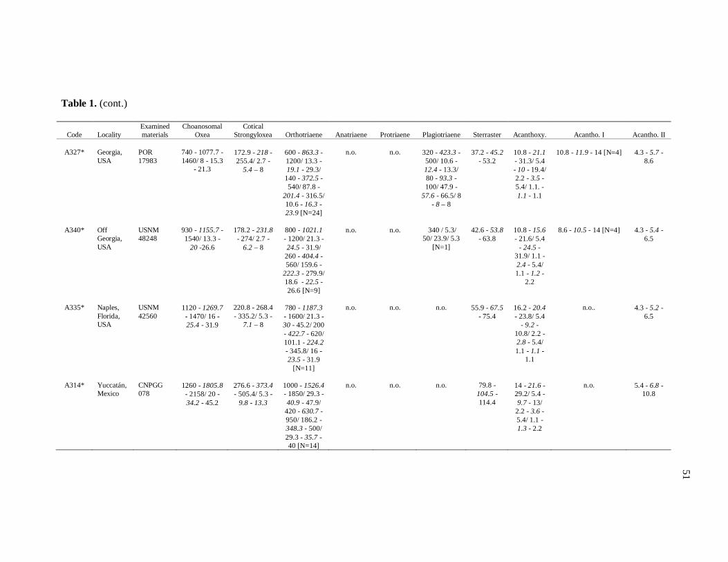
Table 1. (cont.)
Code
Locality
Examined materials
Choanosomal Oxea
Cotical Strongyloxea
Orthotriaene
Anatriaene
Protriaene
Plagiotriaene
Sterraster
Acanthoxy.
Acantho. I
Acantho. II
A327*
Georgia, USA
POR 17983
740 - 1077.7 - 1460/ 8 - 15.3
- 21.3
172.9 - 218 - 255.4/ 2.7 -
5.4 – 8
600 - 863.3 - 1200/ 13.3 - 19.1 - 29.3/
140 - 372.5 - 540/ 87.8 -
201.4 - 316.5/ 10.6 - 16.3 - 23.9 [N=24]
n.o.
n.o.
320 - 423.3 - 500/ 10.6 - 12.4 - 13.3/ 80 - 93.3 - 100/ 47.9 -
57.6 - 66.5/ 8 - 8 – 8
37.2 - 45.2
- 53.2
10.8 - 21.1 - 31.3/ 5.4 - 10 - 19.4/ 2.2 - 3.5 - 5.4/ 1.1. - 1.1 - 1.1
10.8 - 11.9 - 14 [N=4]
4.3 - 5.7 -
8.6
A340*
Off Georgia, USA
USNM 48248
930 - 1155.7 - 1540/ 13.3 -
20 -26.6
178.2 - 231.8 - 274/ 2.7 -
6.2 – 8
800 - 1021.1 - 1200/ 21.3 - 24.5 - 31.9/
260 - 404.4 - 560/ 159.6 -
222.3 - 279.9/ 18.6 - 22.5 - 26.6 [N=9]
n.o.
n.o.
340 / 5.3/
50/ 23.9/ 5.3 [N=1]
42.6 - 53.8
- 63.8
10.8 - 15.6 - 21.6/ 5.4
- 24.5 - 31.9/ 1.1 - 2.4 - 5.4/ 1.1 - 1.2 -
2.2
8.6 - 10.5 - 14 [N=4]
4.3 - 5.4 -
6.5
A335*
Naples, Florida, USA
USNM 42560
1120 - 1269.7 - 1470/ 16 - 25.4 - 31.9
220.8 - 268.4 - 335.2/ 5.3 -
7.1 – 8
780 - 1187.3 - 1600/ 21.3 - 30 - 45.2/ 200 - 422.7 - 620/ 101.1 - 224.2 - 345.8/ 16 - 23.5 - 31.9
[N=11]
n.o.
n.o.
n.o.
55.9 - 67.5
- 75.4
16.2 - 20.4 - 23.8/ 5.4
- 9.2 - 10.8/ 2.2 - 2.8 - 5.4/ 1.1 - 1.1 -
1.1
n.o..
4.3 - 5.2 -
6.5
A314*
Yuccatán, Mexico
CNPGG 078
1260 - 1805.8 - 2158/ 20 - 34.2 - 45.2
276.6 - 373.4 - 505.4/ 5.3 -
9.8 - 13.3
1000 - 1526.4 - 1850/ 29.3 - 40.9 - 47.9/
420 - 630.7 - 950/ 186.2 - 348.3 - 500/ 29.3 - 35.7 - 40 [N=14]
n.o.
n.o.
n.o.
79.8 -
104.5 - 114.4
14 - 21.6 - 29.2/ 5.4 - 9.7 - 13/
2.2 - 3.6 - 5.4/ 1.1 - 1.3 - 2.2
n.o.
5.4 - 6.8 -
10.8
51
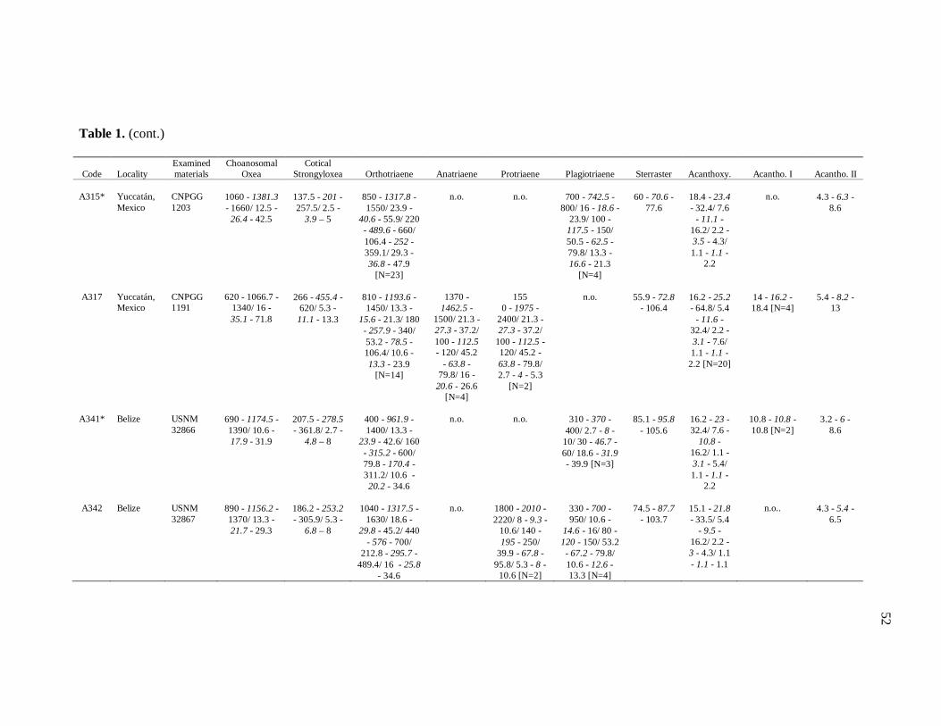
Table 1. (cont.)
Code
Locality
Examined materials
Choanosomal Oxea
Cotical Strongyloxea
Orthotriaene
Anatriaene
Protriaene
Plagiotriaene
Sterraster
Acanthoxy.
Acantho. I
Acantho. II
A315*
Yuccatán, Mexico
CNPGG 1203
1060 - 1381.3 - 1660/ 12.5 -
26.4 - 42.5
137.5 - 201 - 257.5/ 2.5 -
3.9 – 5
850 - 1317.8 - 1550/ 23.9 -
40.6 - 55.9/ 220 - 489.6 - 660/ 106.4 - 252 - 359.1/ 29.3 - 36.8 - 47.9
[N=23]
n.o.
n.o.
700 - 742.5 -
800/ 16 - 18.6 - 23.9/ 100 -
117.5 - 150/ 50.5 - 62.5 - 79.8/ 13.3 - 16.6 - 21.3
[N=4]
60 - 70.6 -
77.6
18.4 - 23.4 - 32.4/ 7.6
- 11.1 - 16.2/ 2.2 - 3.5 - 4.3/ 1.1 - 1.1 -
2.2
n.o.
4.3 - 6.3 -
8.6
A317
Yuccatán, Mexico
CNPGG 1191
620 - 1066.7 -
1340/ 16 - 35.1 - 71.8
266 - 455.4 -
620/ 5.3 - 11.1 - 13.3
810 - 1193.6 - 1450/ 13.3 -
15.6 - 21.3/ 180 - 257.9 - 340/ 53.2 - 78.5 - 106.4/ 10.6 - 13.3 - 23.9
[N=14]
1370 -
1462.5 - 1500/ 21.3 - 27.3 - 37.2/ 100 - 112.5 - 120/ 45.2
- 63.8 - 79.8/ 16 -
20.6 - 26.6 [N=4]
155
0 - 1975 - 2400/ 21.3 - 27.3 - 37.2/
100 - 112.5 - 120/ 45.2 - 63.8 - 79.8/ 2.7 - 4 - 5.3
[N=2]
n.o.
55.9 - 72.8
- 106.4
16.2 - 25.2 - 64.8/ 5.4
- 11.6 - 32.4/ 2.2 - 3.1 - 7.6/ 1.1 - 1.1 -
2.2 [N=20]
14 - 16.2 - 18.4 [N=4]
5.4 - 8.2 -
13
A341*
Belize
USNM 32866
690 - 1174.5 - 1390/ 10.6 - 17.9 - 31.9
207.5 - 278.5 - 361.8/ 2.7 -
4.8 – 8
400 - 961.9 - 1400/ 13.3 -
23.9 - 42.6/ 160 - 315.2 - 600/ 79.8 - 170.4 - 311.2/ 10.6 - 20.2 - 34.6
n.o.
n.o.
310 - 370 -
400/ 2.7 - 8 - 10/ 30 - 46.7 - 60/ 18.6 - 31.9 - 39.9 [N=3]
85.1 - 95.8
- 105.6
16.2 - 23 - 32.4/ 7.6 -
10.8 - 16.2/ 1.1 - 3.1 - 5.4/ 1.1 - 1.1 -
2.2
10.8 - 10.8 - 10.8 [N=2]
3.2 - 6 -
8.6
A342
Belize
USNM 32867
890 - 1156.2 - 1370/ 13.3 - 21.7 - 29.3
186.2 - 253.2 - 305.9/ 5.3 -
6.8 – 8
1040 - 1317.5 -
1630/ 18.6 - 29.8 - 45.2/ 440
- 576 - 700/ 212.8 - 295.7 -
489.4/ 16 - 25.8 - 34.6
n.o.
1800 - 2010 - 2220/ 8 - 9.3 -
10.6/ 140 - 195 - 250/
39.9 - 67.8 - 95.8/ 5.3 - 8 -
10.6 [N=2]
330 - 700 - 950/ 10.6 -
14.6 - 16/ 80 - 120 - 150/ 53.2 - 67.2 - 79.8/ 10.6 - 12.6 - 13.3 [N=4]
74.5 - 87.7
- 103.7
15.1 - 21.8 - 33.5/ 5.4
- 9.5 - 16.2/ 2.2 - 3 - 4.3/ 1.1 - 1.1 - 1.1
n.o..
4.3 - 5.4 -
6.5
52
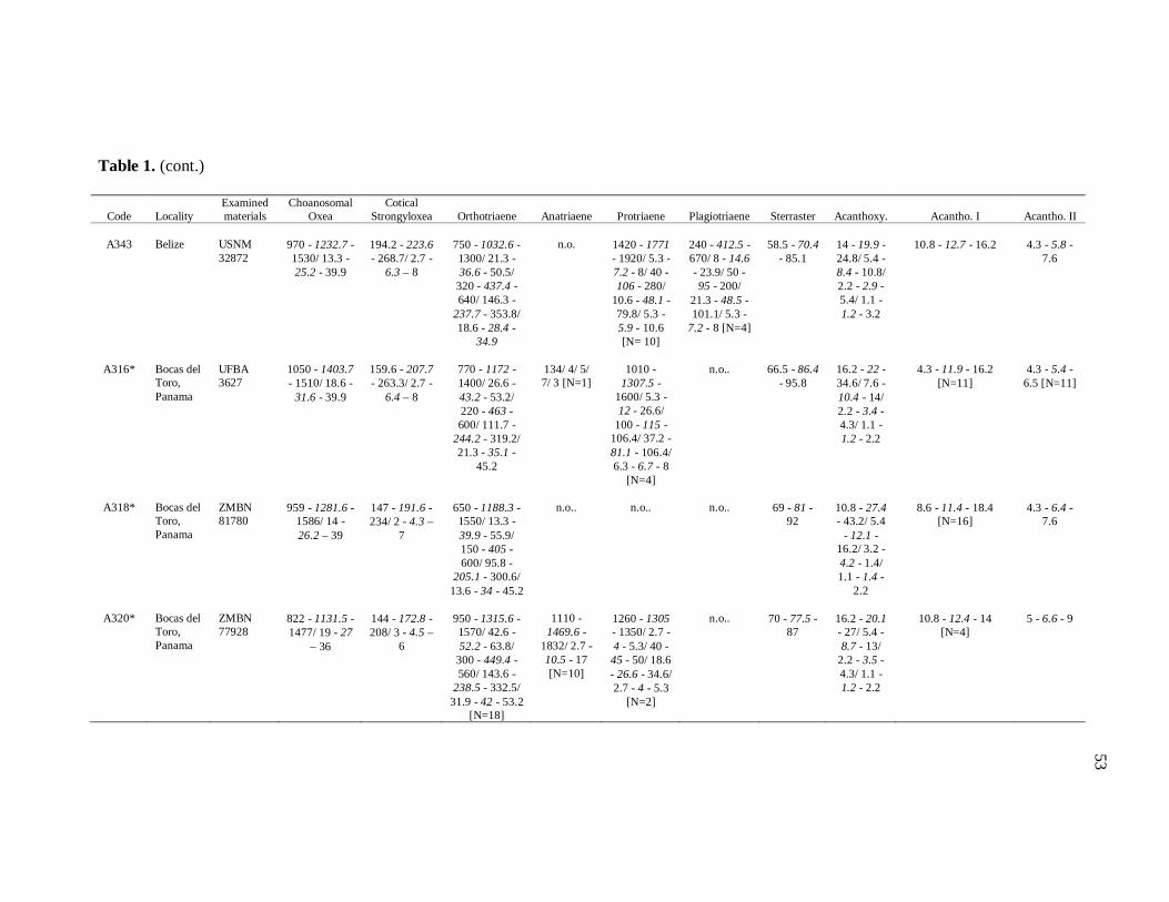
Table 1. (cont.)
Code
Locality
Examined materials
Choanosomal Oxea
Cotical Strongyloxea
Orthotriaene
Anatriaene
Protriaene
Plagiotriaene
Sterraster
Acanthoxy.
Acantho. I
Acantho. II
A343
Belize
USNM 32872
970 - 1232.7 - 1530/ 13.3 - 25.2 - 39.9
194.2 - 223.6 - 268.7/ 2.7 -
6.3 – 8
750 - 1032.6 - 1300/ 21.3 - 36.6 - 50.5/
320 - 437.4 - 640/ 146.3 -
237.7 - 353.8/ 18.6 - 28.4 -
34.9
n.o.
1420 - 1771 - 1920/ 5.3 - 7.2 - 8/ 40 - 106 - 280/
10.6 - 48.1 - 79.8/ 5.3 - 5.9 - 10.6 [N= 10]
240 - 412.5 - 670/ 8 - 14.6 - 23.9/ 50 - 95 - 200/
21.3 - 48.5 - 101.1/ 5.3 -
7.2 - 8 [N=4]
58.5 - 70.4
- 85.1
14 - 19.9 - 24.8/ 5.4 - 8.4 - 10.8/ 2.2 - 2.9 - 5.4/ 1.1 - 1.2 - 3.2
10.8 - 12.7 - 16.2
4.3 - 5.8 -
7.6
A316*
Bocas del Toro, Panama
UFBA 3627
1050 - 1403.7 - 1510/ 18.6 -
31.6 - 39.9
159.6 - 207.7 - 263.3/ 2.7 -
6.4 – 8
770 - 1172 - 1400/ 26.6 - 43.2 - 53.2/ 220 - 463 - 600/ 111.7 -
244.2 - 319.2/ 21.3 - 35.1 -
45.2
134/ 4/ 5/ 7/ 3 [N=1]
1010 -
1307.5 - 1600/ 5.3 - 12 - 26.6/
100 - 115 - 106.4/ 37.2 - 81.1 - 106.4/ 6.3 - 6.7 - 8
[N=4]
n.o..
66.5 - 86.4
- 95.8
16.2 - 22 - 34.6/ 7.6 - 10.4 - 14/ 2.2 - 3.4 - 4.3/ 1.1 - 1.2 - 2.2
4.3 - 11.9 - 16.2
[N=11]
4.3 - 5.4 -
6.5 [N=11]
A318*
Bocas del Toro, Panama
ZMBN 81780
959 - 1281.6 -
1586/ 14 - 26.2 – 39
147 - 191.6 - 234/ 2 - 4.3 –
7
650 - 1188.3 - 1550/ 13.3 - 39.9 - 55.9/ 150 - 405 - 600/ 95.8 -
205.1 - 300.6/ 13.6 - 34 - 45.2
n.o..
n.o..
n.o..
69 - 81 -
92
10.8 - 27.4 - 43.2/ 5.4
- 12.1 - 16.2/ 3.2 - 4.2 - 1.4/ 1.1 - 1.4 -
2.2
8.6 - 11.4 - 18.4
[N=16]
4.3 - 6.4 -
7.6
A320*
Bocas del Toro, Panama
ZMBN 77928
822 - 1131.5 - 1477/ 19 - 27
– 36
144 - 172.8 - 208/ 3 - 4.5 –
6
950 - 1315.6 - 1570/ 42.6 - 52.2 - 63.8/
300 - 449.4 - 560/ 143.6 -
238.5 - 332.5/ 31.9 - 42 - 53.2
[N=18]
1110 -
1469.6 - 1832/ 2.7 - 10.5 - 17 [N=10]
1260 - 1305 - 1350/ 2.7 - 4 - 5.3/ 40 - 45 - 50/ 18.6 - 26.6 - 34.6/ 2.7 - 4 - 5.3
[N=2]
n.o..
70 - 77.5 -
87
16.2 - 20.1 - 27/ 5.4 - 8.7 - 13/
2.2 - 3.5 - 4.3/ 1.1 - 1.2 - 2.2
10.8 - 12.4 - 14
[N=4]
5 - 6.6 - 9
53

Table 1. (cont.)
Code
Locality
Examined materials
Choanosomal Oxea
Cotical Strongyloxea
Orthotriaene
Anatriaene
Protriaene
Plagiotriaene
Sterraster
Acanthoxy.
Acantho. I
Acantho. II
A300
Archipelago Sabana, Cuba
UFBA 3061
974 -1246.6 - 1487/ 14 - 19
– 25
91 - 115.9 -
214/ 2 - 3.4 – 5
750 - 1058.7 - 1250/ 13.3 - 21.2 - 31.9/ 200 - 475 - 750/ 106.4 -
256.9 - 404.3/ 10.6 - 18 -
26.6
n.o..
n.o..
n.o..
30 - 39.6 -
54
11 - 17.3 - 22/ 4 - 7.8 - 11/ 2 -
2.8 - 4/ 1 - 1 – 1
9 - 9 - 9 [N=2]
4 - 5 – 6
A301
Archipelago Sabana, Cuba
UFBA 3062
980 - 1120 - 1308/ 14 -21.1 – 30
213 - 277.6 - 340/ 3 - 4.6 -
7
770 - 962.7 - 1280/ 21.3 - 26.2 - 37.2/ 300 - 415 - 640/ 133 -
220.4 - 324.5/ 13.3 - 20 -
23.9
n.o.
n.o.
n.o.
36 - 46.3 -
55
12 - 17.3 - 22/ 4 - 7.8 - 11/ 2 -
2.8 - 4/ 1 - 1 – 1
n.o.
4 - 5.2 – 6
A308
Cayeria de San Felipe, Cuba
UFBA 3071
994 - 1224.7 -
1551/ 19 - 27.2 – 39
228 - 284 -
328/ 5 - 6.7 – 10
820 - 1138.8 - 1310/ 31.9 - 40.2 - 50.5/
320 - 473.8 - 640/ 172.9 -
256.4 - 324.5/ 26.6 - 28.2 - 50.5 [N=16]
n.o.
n.o.
680/ 18.6/ 120/ 69.2/ 10.6 [N=1]
50 - 58.9 -
70
11 - 18.3 - 25/ 5 - 8.1 - 12/ 3 -
3.4 - 5/ 0.5 - 1 – 1
n.o.
4 - 5.8 – 8
A312*
Guadalupe (France)
UFBA 3622
910 - 1213.3 - 1450/ 13.3 - 22.4 - 37.2
133 - 203.8 - 242.1/ 2.7 -
2.9 - 5.3
770 -1022.3 - 1390/ 12.5 - 25.9 - 54.6/
150 - 305.3 - 460/ 80 -
160.1 - 288.6/ 10 - 18.1 -
31.2
n.o..
n.o..
n.o..
58.5 - 69.1
- 79.8
17 - 22 -
31/ 7 - 10.1 - 16/ 2 -
2.5 - 4/ 1 - 1 – 1
n.o.
4 - 5 – 7
54
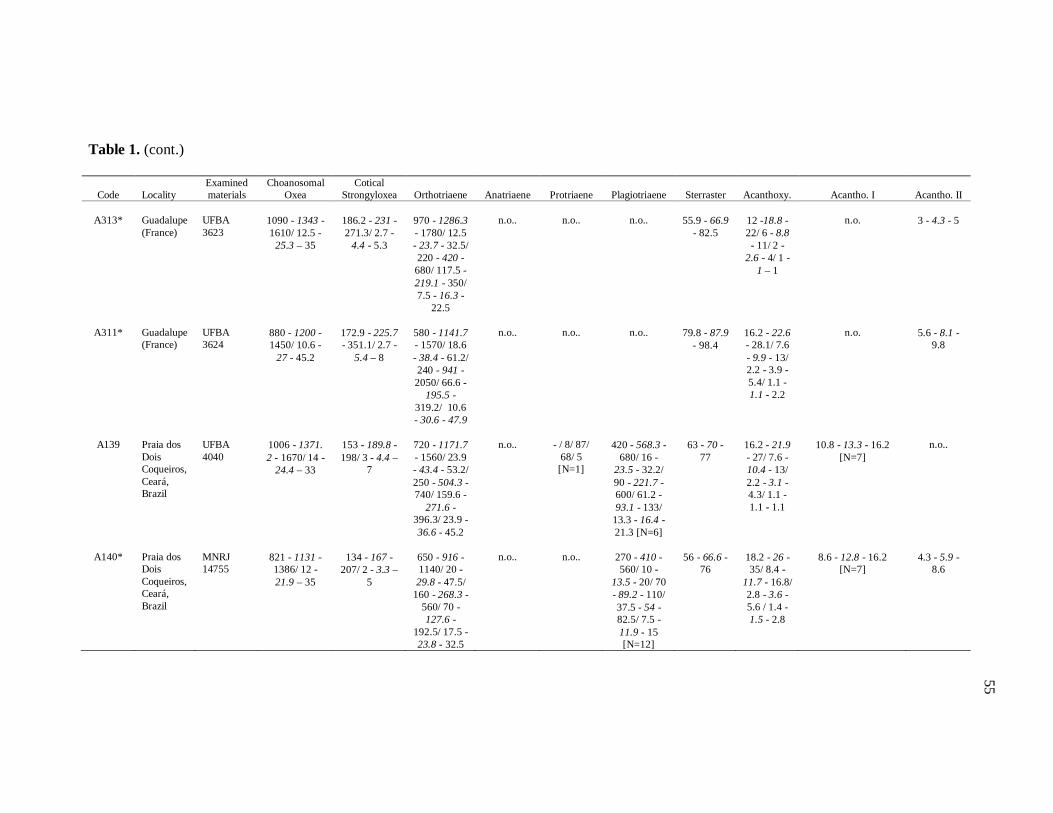
Table 1. (cont.)
Code
Locality
Examined materials
Choanosomal Oxea
Cotical Strongyloxea
Orthotriaene
Anatriaene
Protriaene
Plagiotriaene
Sterraster
Acanthoxy.
Acantho. I
Acantho. II
A313*
Guadalupe (France)
UFBA 3623
1090 - 1343 - 1610/ 12.5 -
25.3 – 35
186.2 - 231 - 271.3/ 2.7 -
4.4 - 5.3
970 - 1286.3 - 1780/ 12.5 - 23.7 - 32.5/ 220 - 420 - 680/ 117.5 - 219.1 - 350/ 7.5 - 16.3 -
22.5
n.o..
n.o..
n.o..
55.9 - 66.9
- 82.5
12 -18.8 - 22/ 6 - 8.8 - 11/ 2 -
2.6 - 4/ 1 - 1 – 1
n.o.
3 - 4.3 - 5
A311*
Guadalupe (France)
UFBA 3624
880 - 1200 - 1450/ 10.6 -
27 - 45.2
172.9 - 225.7 - 351.1/ 2.7 -
5.4 – 8
580 - 1141.7 - 1570/ 18.6 - 38.4 - 61.2/ 240 - 941 - 2050/ 66.6 -
195.5 - 319.2/ 10.6 - 30.6 - 47.9
n.o..
n.o..
n.o..
79.8 - 87.9
- 98.4
16.2 - 22.6 - 28.1/ 7.6 - 9.9 - 13/ 2.2 - 3.9 - 5.4/ 1.1 - 1.1 - 2.2
n.o.
5.6 - 8.1 -
9.8
A139
Praia dos Dois Coqueiros, Ceará, Brazil
UFBA 4040
1006 - 1371. 2 - 1670/ 14 -
24.4 – 33
153 - 189.8 - 198/ 3 - 4.4 –
7
720 - 1171.7 - 1560/ 23.9 - 43.4 - 53.2/ 250 - 504.3 - 740/ 159.6 -
271.6 - 396.3/ 23.9 - 36.6 - 45.2
n.o..
- / 8/ 87/
68/ 5 [N=1]
420 - 568.3 -
680/ 16 - 23.5 - 32.2/ 90 - 221.7 - 600/ 61.2 - 93.1 - 133/
13.3 - 16.4 - 21.3 [N=6]
63 - 70 -
77
16.2 - 21.9 - 27/ 7.6 - 10.4 - 13/ 2.2 - 3.1 - 4.3/ 1.1 - 1.1 - 1.1
10.8 - 13.3 - 16.2
[N=7]
n.o..
A140*
Praia dos Dois Coqueiros, Ceará, Brazil
MNRJ 14755
821 - 1131 - 1386/ 12 - 21.9 – 35
134 - 167 -
207/ 2 - 3.3 – 5
650 - 916 - 1140/ 20 - 29.8 - 47.5/
160 - 268.3 - 560/ 70 - 127.6 -
192.5/ 17.5 - 23.8 - 32.5
n.o..
n.o..
270 - 410 - 560/ 10 -
13.5 - 20/ 70 - 89.2 - 110/ 37.5 - 54 - 82.5/ 7.5 - 11.9 - 15 [N=12]
56 - 66.6 -
76
18.2 - 26 - 35/ 8.4 -
11.7 - 16.8/ 2.8 - 3.6 - 5.6 / 1.4 - 1.5 - 2.8
8.6 - 12.8 - 16.2
[N=7]
4.3 - 5.9 -
8.6
55
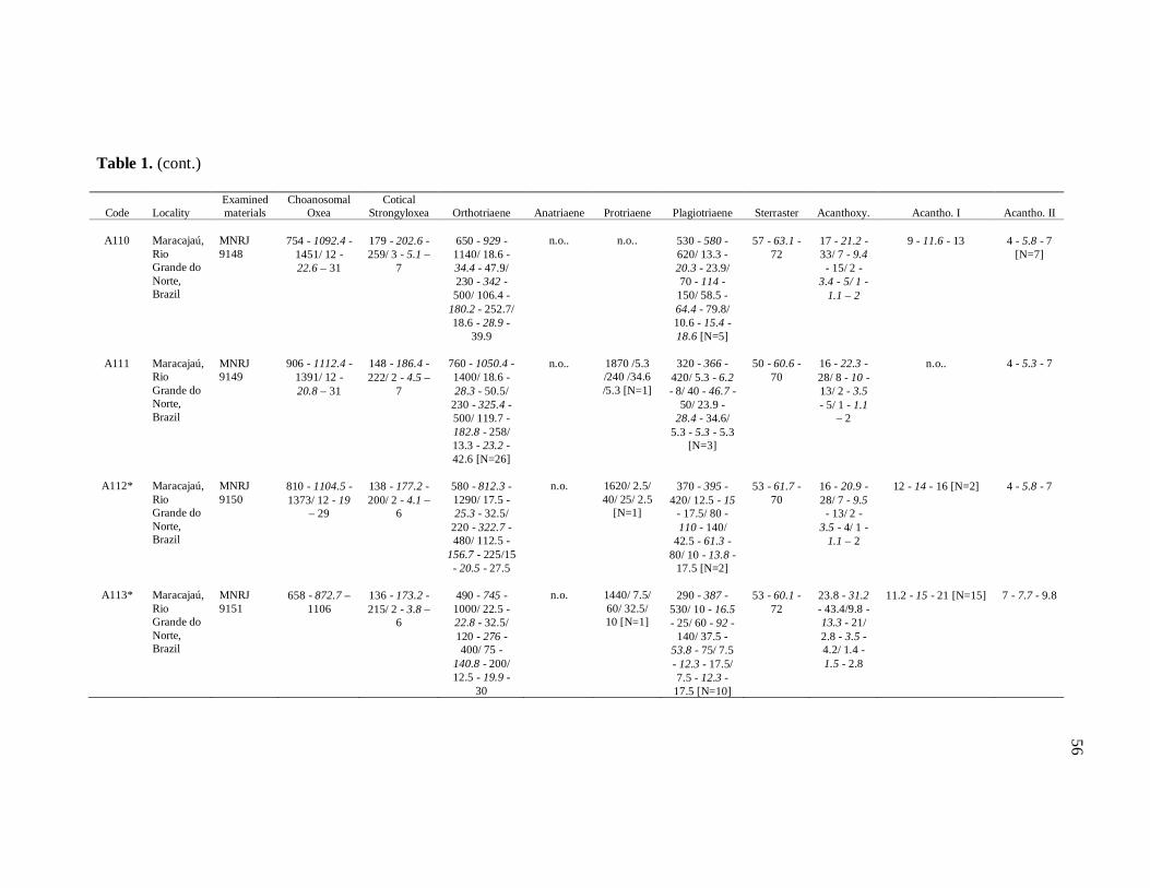
Table 1. (cont.)
Code
Locality
Examined materials
Choanosomal Oxea
Cotical Strongyloxea
Orthotriaene
Anatriaene
Protriaene
Plagiotriaene
Sterraster
Acanthoxy.
Acantho. I
Acantho. II
A110
Maracajaú, Rio Grande do Norte, Brazil
MNRJ 9148
754 - 1092.4 -
1451/ 12 - 22.6 – 31
179 - 202.6 - 259/ 3 - 5.1 –
7
650 - 929 - 1140/ 18.6 - 34.4 - 47.9/ 230 - 342 - 500/ 106.4 -
180.2 - 252.7/ 18.6 - 28.9 -
39.9
n.o..
n.o..
530 - 580 - 620/ 13.3 - 20.3 - 23.9/ 70 - 114 - 150/ 58.5 - 64.4 - 79.8/ 10.6 - 15.4 - 18.6 [N=5]
57 - 63.1 -
72
17 - 21.2 - 33/ 7 - 9.4 - 15/ 2 -
3.4 - 5/ 1 - 1.1 – 2
9 - 11.6 - 13
4 - 5.8 - 7
[N=7]
A111
Maracajaú, Rio Grande do Norte, Brazil
MNRJ 9149
906 - 1112.4 -
1391/ 12 - 20.8 – 31
148 - 186.4 - 222/ 2 - 4.5 –
7
760 - 1050.4 - 1400/ 18.6 - 28.3 - 50.5/
230 - 325.4 - 500/ 119.7 - 182.8 - 258/ 13.3 - 23.2 - 42.6 [N=26]
n.o..
1870 /5.3 /240 /34.6 /5.3 [N=1]
320 - 366 -
420/ 5.3 - 6.2 - 8/ 40 - 46.7 -
50/ 23.9 - 28.4 - 34.6/
5.3 - 5.3 - 5.3 [N=3]
50 - 60.6 -
70
16 - 22.3 - 28/ 8 - 10 - 13/ 2 - 3.5 - 5/ 1 - 1.1
– 2
n.o..
4 - 5.3 - 7
A112*
Maracajaú, Rio Grande do Norte, Brazil
MNRJ 9150
810 - 1104.5 - 1373/ 12 - 19
– 29
138 - 177.2 - 200/ 2 - 4.1 –
6
580 - 812.3 - 1290/ 17.5 - 25.3 - 32.5/
220 - 322.7 - 480/ 112.5 -
156.7 - 225/15 - 20.5 - 27.5
n.o.
1620/ 2.5/ 40/ 25/ 2.5
[N=1]
370 - 395 -
420/ 12.5 - 15 - 17.5/ 80 - 110 - 140/
42.5 - 61.3 - 80/ 10 - 13.8 -
17.5 [N=2]
53 - 61.7 -
70
16 - 20.9 - 28/ 7 - 9.5 - 13/ 2 -
3.5 - 4/ 1 - 1.1 – 2
12 - 14 - 16 [N=2]
4 - 5.8 - 7
A113*
Maracajaú, Rio Grande do Norte, Brazil
MNRJ 9151
658 - 872.7 –
1106
136 - 173.2 - 215/ 2 - 3.8 –
6
490 - 745 - 1000/ 22.5 - 22.8 - 32.5/ 120 - 276 - 400/ 75 -
140.8 - 200/ 12.5 - 19.9 -
30
n.o.
1440/ 7.5/ 60/ 32.5/ 10 [N=1]
290 - 387 -
530/ 10 - 16.5 - 25/ 60 - 92 -
140/ 37.5 - 53.8 - 75/ 7.5 - 12.3 - 17.5/ 7.5 - 12.3 -
17.5 [N=10]
53 - 60.1 -
72
23.8 - 31.2 - 43.4/9.8 - 13.3 - 21/ 2.8 - 3.5 - 4.2/ 1.4 - 1.5 - 2.8
11.2 - 15 - 21 [N=15]
7 - 7.7 - 9.8
56
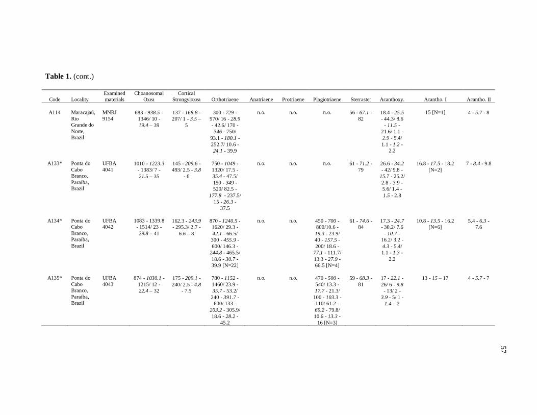
Table 1. (cont.)
Code
Locality
Examined materials
Choanosomal Oxea
Cortical Strongyloxea
Orthotriaene
Anatriaene
Protriaene
Plagiotriaene
Sterraster
Acanthoxy.
Acantho. I
Acantho. II
A114
Maracajaú, Rio Grande do Norte, Brazil
MNRJ 9154
683 - 938.5 -
1346/ 10 - 19.4 – 39
137 - 168.8 - 207/ 1 - 3.5 –
5
300 - 729 -
970/ 16 - 28.9 - 42.6/ 170 - 346 - 750/
93.1 - 180.1 - 252.7/ 10.6 - 24.1 - 39.9
n.o.
n.o.
n.o.
56 - 67.1 -
82
18.4 - 25.5 - 44.3/ 8.6
- 11.5 - 21.6/ 1.1 - 2.9 - 5.4/ 1.1 - 1.2 -
2.2
15 [N=1]
4 - 5.7 - 8
A133*
Ponta do Cabo Branco, Paraíba, Brazil
UFBA 4041
1010 - 1223.3
- 1383/ 7 - 21.5 – 35
145 - 209.6 - 493/ 2.5 - 3.8
- 6
750 - 1049 - 1320/ 17.5 - 35.4 - 47.5/ 150 - 349 - 520/ 82.5 -
177.8 - 237.5/ 15 - 26.3 -
37.5
n.o.
n.o.
n.o.
61 - 71.2 -
79
26.6 - 34.2 - 42/ 9.8 -
15.7 - 25.2/ 2.8 - 3.9 - 5.6/ 1.4 - 1.5 - 2.8
16.8 - 17.5 - 18.2
[N=2]
7 - 8.4 - 9.8
A134*
Ponta do Cabo Branco, Paraíba, Brazil
UFBA 4042
1083 - 1339.8 - 1514/ 23 - 29.8 – 41
162.3 - 243.9 - 295.3/ 2.7 -
6.6 – 8
870 - 1240.5 - 1620/ 29.3 - 42.1 - 66.5/
300 - 455.9 - 600/ 146.3 -
244.8 - 465.5/ 18.6 - 30.7 - 39.9 [N=22]
n.o.
n.o.
450 - 700 - 800/10.6 - 19.3 - 23.9/ 40 - 157.5 - 200/ 18.6 -
77.1 - 111.7/ 13.3 - 27.9 - 66.5 [N=4]
61 - 74.6 -
84
17.3 - 24.7 - 30.2/ 7.6
- 10.7 - 16.2/ 3.2 - 4.3 - 5.4/ 1.1 - 1.3 -
2.2
10.8 - 13.5 - 16.2
[N=6]
5.4 - 6.3 -
7.6
A135*
Ponta do Cabo Branco, Paraíba, Brazil
UFBA 4043
874 - 1030.1 -
1215/ 12 - 22.4 – 32
175 - 209.1 - 240/ 2.5 - 4.8
- 7.5
780 - 1152 - 1460/ 23.9 - 35.7 - 53.2/
240 - 391.7 - 600/ 133 -
203.2 - 305.9/ 18.6 - 28.2 -
45.2
n.o.
n.o.
470 - 500 - 540/ 13.3 - 17.7 - 21.3/
100 - 103.3 - 110/ 61.2 - 69.2 - 79.8/ 10.6 - 13.3 -
16 [N=3]
59 - 68.3 -
81
17 - 22.1 - 26/ 6 - 9.8 - 13/ 2 -
3.9 - 5/ 1 - 1.4 – 2
13 - 15 – 17
4 - 5.7 - 7
57

Table 1. (cont.)
Code
Locality
Examined materials
Choanosomal Oxea
Cotical Strongyloxea
Orthotriaene
Anatriaene
Protriaene
Plagiotriaene
Sterraster
Acanthoxy.
Acantho. I
Acantho. II
A138*
Ponta do Cabo Branco, Paraíba, Brazil
UFBA 4044
940 - 1169.3 - 1900/ 18.6 - 29.6 - 42.6
940 - 252.3 - 292.6/ 2.7 -
5.3 – 8
750 - 1124 - 1600/ 26.6 - 38.7 - 53.2/ 249 - 368 - 500/ 93.1 -
199.4 - 279.3/ 18.6 - 31.2 - 50.5
n.o. n.o. 790 /18.6 /110 /58.5 / 13.3 [N=1]
53.2 - 76.9 - 101.1
16.2 - 29.8 - 48.6/ 9.7
- 14.7 - 23.8/ 2.2 - 4.9 - 6.5/ 1.1 - 2 -
3.2
n.o. 4.3 - 5.9 - 7.6
A127
São Miguel dos Milagres, Alagoas, Brazil
UFBA 3273
886 -1092-
1428/ 8 - 21.7 – 34
157 - 221.3 - 398/ 3 - 5.4 –
8
900 - 1197.6 - 1430/ 26.6 - 37.1 - 55.9/ 300 - 444.8 -
660/ 149 - 243.2 -
337.8/ 13.3 - 30.8 - 47.9
n.o.
202/ 4/ 14/
32/ 3 [N=1]
450 - 710 - 930/ 5.3 -
19.5 - 26.6/ 50 - 138.9 - 230/ 31.9 -
89 - 133/ 5.3 - 13.3 - 18.6
43 - 62.7 -
76
17.3 - 23.4 - 34.6/ 5.4 - 10.5 - 13/
2.2 - 4 - 10.8/ 1.1 - 1.3 - 2.2
9.7 - 11.9 - 14 [N=7]
4.3 - 6 - 8.6
A142*
Maceió, Alagoas, Brazil
UFBA 4045
813 - 1059.9 -
1298/ 15 - 24.2 – 34
155 - 190.8 - 233/ 3 - 5.1 –
8
600 - 902.2 -
1170/ 17 - 31.1 - 57.5/
170 - 280.9 - 480/ 81 - 144.1 -
212.5/ 14 - 26.9 - 45
n.o.
1230 - 1340 -
1450/ 7.5 - 8.1 - 10/ 50
- 67.5 - 100/ 50 -
55.6 - 62.5/ 2.5 - 5.6 - 7.5 [N=4]
560 - 630 - 730/ 15 -
17.5 - 20/100 - 136.7 - 160/
50 - 70.8 - 92.5/7.5 - 12.5 - 15
[N=3]
54- 69.6 -
81
17 - 34.2 - 58.8/ 7 -
16.3 - 25.2/ 2 - 4.3 - 7/
1 - 1.9 - 2.8
n.o.
2 - 5.9 - 8.4
A143*
Pituba, Bahia, Brazil
UFBA 4046
783 - 953.1 - 1189/ 7 - 15.1
– 24
132 - 162.2 - 198/ 3 - 4.2 –
6
500 - 877.7 - 1150/ 13.3 - 17.8 - 26.6/ 170 - 386 - 560/ 9.8 -
200.9 - 292.6/ 10.6 - 16.2 - 23.9
n.o.
n.o.
157 - 368 -
457/ 7 - 9.3 - 14/ 60 - 134 -
280/ 28 - 74.5 - 159/ 5
- 9.5 - 15
42 - 46 -
51
10.8 - 16.5 - 22.7/ 4.3
- 7.7 - 10.8/ 1.1 - 2.1 - 4.3/ 1.1 - 1.1 -
1.1
7.6 - 9.1 - 10.8
3.2 - 5.3 -
7.6
58
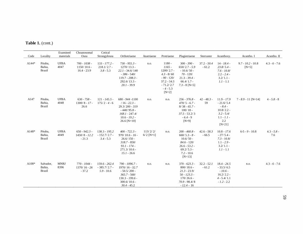
Table 1. (cont.)
Code
Locality
Examined materials
Choanosomal Oxea
Cotical Strongyloxea
Orthotriaene
Anatriaene
Protriaene
Plagiotriaene
Sterraster
Acanthoxy.
Acantho. I
Acantho. II
A144*
Pituba, Bahia, Brazil
UFBA 4047
700 - 1038 - 1150/ 10.6 - 16.4 - 23.9
133 - 177.2 - 218.1/ 2.7 -
3.8 - 5.3
730 - 955.3 - 1270/ 13.3 -
22.1 - 34.6/ 140 - 386 - 540/
119.7 - 208.3 - 292.6/ 13.3 - 20.1 - 39.9
n.o.
1180 - 1165 -
1200/ 2.7 - 4.3 - 8/ 60 - 90 - 120/ 37.2 - 54.5 - 71.2/ 2.7 - 4 - 5.3 [N=2]
300 - 390 -
650/ 2.7 - 5.9 - 10.6/ 50 - 70 - 120/
21.3 - 39.4 - 66.4/ 1.7 -
7.3 - 8 [N=5]
37.2 - 50.4
- 61.2
14 - 18.4 - 23.8/ 5.4 - 7.6 - 10.8/ 2.2 - 2.4 - 3.2/ 1.1 - 1.1 - 1.1
9.7 - 10.2 - 10.8
[N=5]
4.3 - 6 - 7.6
A147
Pituba, Bahia, Brazil
UFBA 4048
630 - 758 -
1300/ 8 - 17 - 26.6
121 - 145.5 - 175/ 2 - 4 - 6
680 - 944 -1100
/ 16 - 22.3 - 29.3/ 200 - 319
- 440/ 95.8 - 168.1 - 247.4/ 10.6 - 19.2 - 26.6 [N=10]
n.o.
n.o.
256 - 376.8 - 470/ 5 - 6.7 - 8/ 38 - 65.7 -
100/ 18 - 37.5 - 53.2/ 3
- 6.4 - 9 [N=9]
42 - 48.3 -
59
11.9 - 17.9 - 21.6/ 5.4
- 8.4 - 10.8/ 2.2 - 3.2 - 5.4/ 1.1 - 1.1 -
2.2 [N=21]
7 - 8.9 - 11 [N=14]
4 - 5.8 - 8
A148*
Pituba, Bahia, Brazil
UFBA 4049
650 - 942.3 - 1450/ 8 - 12.2
- 21.3
138.3 - 195.2 - 152.7/ 2.7 -
3.4 - 5.3
400 - 722.3 -
970/ 10.6 - 16 - 26.6/ 150 -
318.7 - 850/ 93.1 - 174 - 271.3/ 10.6 - 15.1 - 26.6
113/ 2/ 2/ 6/ 2 [N=1]
n.o.
200 - 460.8 - 660/ 5.3 - 8 -
10.6/ 50 - 84.6 - 120/
26.6 - 53.2 - 69.2/ 5.3 - 7.2 - 10.6 [N=13]
42.6 - 58.3
- 66.5
10.8 - 17.6 - 27/ 5.4 - 7.3 - 10.8/ 1.1 - 2.9 - 3.2/ 1.1 - 1.1 - 1.1
6-5 - 9 - 10.8
4.3 - 5.8 -
7.6
A108*
Salvador, Bahia, Brazil
MNRJ 8396
770 - 1044 - 1370/ 16 - 26
- 37.2
159.6 - 262.4 - 385.7/ 2.7 -
5.9 - 10.6
790 - 1096.7 - 1970/ 16 - 32.7
- 50.5/ 200 - 365.7 - 560/
130.3 - 199.6 - 300.6/ 10.6 - 30.4 - 45.2
n.o.
n.o.
370 - 623.3 - 800/ 18.6 - 21.3 - 23.9/ 50 - 123.3 - 170/ 26.6 -
70.9 - 98.4/ 8 - 12.4 – 16
32.2 - 52.1
- 61.2
18.4 - 24.5 - 33.5/ 6.5
- 10.6 - 16.2/ 2.2 - 4 - 5.4/ 1.1 - 1.2 - 2.2
n.o.
4.3 - 6 - 7.6
59
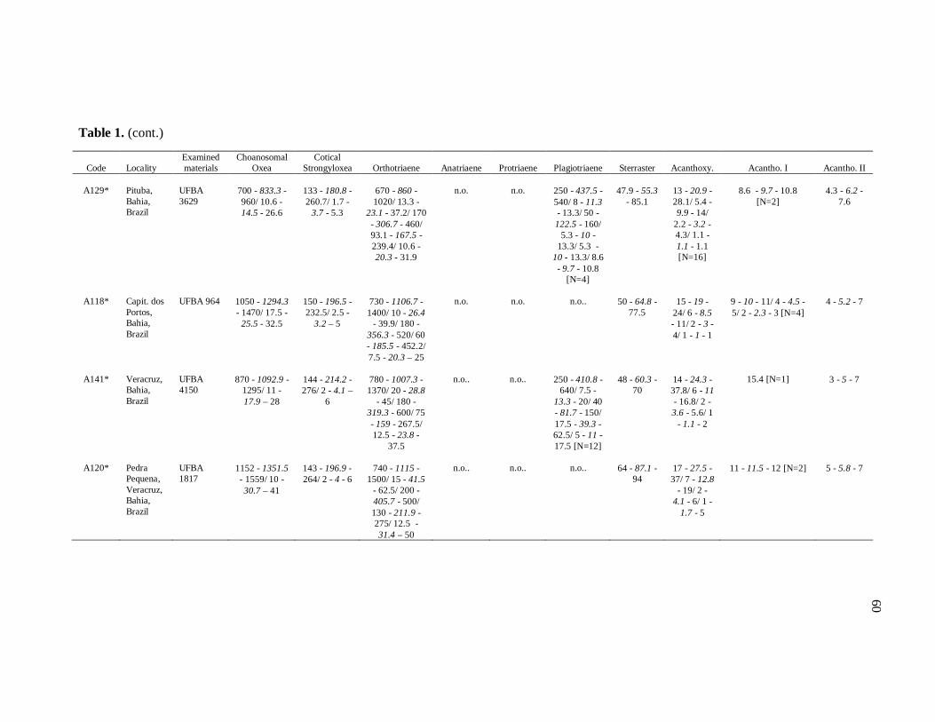
Table 1. (cont.)
Code
Locality
Examined materials
Choanosomal Oxea
Cotical Strongyloxea
Orthotriaene
Anatriaene
Protriaene
Plagiotriaene
Sterraster
Acanthoxy.
Acantho. I
Acantho. II
A129*
Pituba, Bahia, Brazil
UFBA 3629
700 - 833.3 - 960/ 10.6 - 14.5 - 26.6
133 - 180.8 - 260.7/ 1.7 -
3.7 - 5.3
670 - 860 - 1020/ 13.3 -
23.1 - 37.2/ 170 - 306.7 - 460/ 93.1 - 167.5 - 239.4/ 10.6 - 20.3 - 31.9
n.o.
n.o.
250 - 437.5 - 540/ 8 - 11.3 - 13.3/ 50 - 122.5 - 160/
5.3 - 10 - 13.3/ 5.3 -
10 - 13.3/ 8.6 - 9.7 - 10.8
[N=4]
47.9 - 55.3
- 85.1
13 - 20.9 - 28.1/ 5.4 - 9.9 - 14/
2.2 - 3.2 - 4.3/ 1.1 - 1.1 - 1.1 [N=16]
8.6 - 9.7 - 10.8
[N=2]
4.3 - 6.2 -
7.6
A118*
Capit. dos Portos, Bahia, Brazil
UFBA 964
1050 - 1294.3 - 1470/ 17.5 -
25.5 - 32.5
150 - 196.5 - 232.5/ 2.5 -
3.2 – 5
730 - 1106.7 - 1400/ 10 - 26.4
- 39.9/ 180 - 356.3 - 520/ 60 - 185.5 - 452.2/ 7.5 - 20.3 – 25
n.o.
n.o.
n.o..
50 - 64.8 -
77.5
15 - 19 -
24/ 6 - 8.5 - 11/ 2 - 3 - 4/ 1 - 1 - 1
9 - 10 - 11/ 4 - 4.5 - 5/ 2 - 2.3 - 3 [N=4]
4 - 5.2 - 7
A141*
Veracruz, Bahia, Brazil
UFBA 4150
870 - 1092.9 -
1295/ 11 - 17.9 – 28
144 - 214.2 - 276/ 2 - 4.1 –
6
780 - 1007.3 - 1370/ 20 - 28.8
- 45/ 180 - 319.3 - 600/ 75 - 159 - 267.5/ 12.5 - 23.8 -
37.5
n.o..
n.o..
250 - 410.8 -
640/ 7.5 - 13.3 - 20/ 40 - 81.7 - 150/ 17.5 - 39.3 - 62.5/ 5 - 11 - 17.5 [N=12]
48 - 60.3 -
70
14 - 24.3 - 37.8/ 6 - 11 - 16.8/ 2 - 3.6 - 5.6/ 1
- 1.1 - 2
15.4 [N=1]
3 - 5 - 7
A120*
Pedra Pequena, Veracruz, Bahia, Brazil
UFBA 1817
1152 - 1351.5 - 1559/ 10 - 30.7 – 41
143 - 196.9 - 264/ 2 - 4 - 6
740 - 1115 -
1500/ 15 - 41.5 - 62.5/ 200 - 405.7 - 500/ 130 - 211.9 - 275/ 12.5 - 31.4 – 50
n.o..
n.o..
n.o..
64 - 87.1 -
94
17 - 27.5 - 37/ 7 - 12.8
- 19/ 2 - 4.1 - 6/ 1 -
1.7 - 5
11 - 11.5 - 12 [N=2]
5 - 5.8 - 7
60
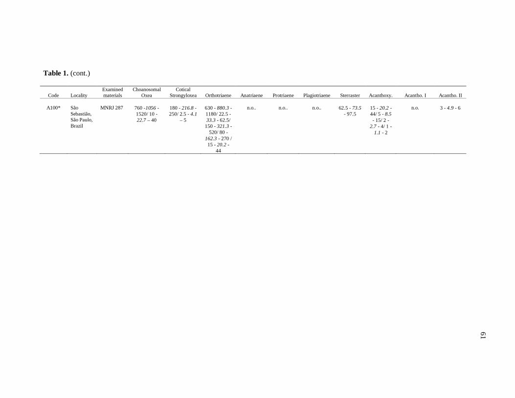
Table 1. (cont.)
Code
Locality
Examined materials
Choanosomal Oxea
Cotical Strongyloxea
Orthotriaene
Anatriaene
Protriaene
Plagiotriaene
Sterraster
Acanthoxy.
Acantho. I
Acantho. II
A100*
São Sebastião, São Paulo, Brazil
MNRJ 287
760 -1056 - 1520/ 10 - 22.7 – 40
180 - 216.8 - 250/ 2.5 - 4.1
– 5
630 - 880.3 - 1180/ 22.5 - 33.3 - 62.5/
150 - 321.3 - 520/ 80 -
162.3 - 270 / 15 - 20.2 -
44
n.o..
n.o..
n.o..
62.5 - 73.5
- 97.5
15 - 20.2 - 44/ 5 - 8.5 - 15/ 2 -
2.7 - 4/ 1 - 1.1 - 2
n.o.
3 - 4.9 - 6
61
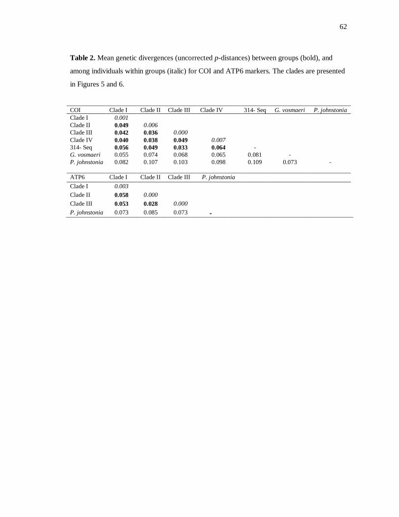
62
Table 2. Mean genetic divergences (uncorrected p-distances) between groups (bold), and
among individuals within groups (italic) for COI and ATP6 markers. The clades are presented
in Figures 5 and 6.
COI Clade I Clade II Clade III Clade IV 314- Seq G. vosmaeri P. johnstonia Clade I 0.001 Clade II 0.049 0.006 Clade III 0.042 0.036 0.000 Clade IV 0.040 0.038 0.049 0.007 314- Seq 0.056 0.049 0.033 0.064 - G. vosmaeri 0.055 0.074 0.068 0.065 0.081 - P. johnstonia 0.082 0.107 0.103 0.098 0.109 0.073 - ATP6 Clade I Clade II Clade III P. johnstonia Clade I 0.003 Clade II 0.058 0.000 Clade III 0.053 0.028 0.000 P. johnstonia 0.073 0.085 0.073 -
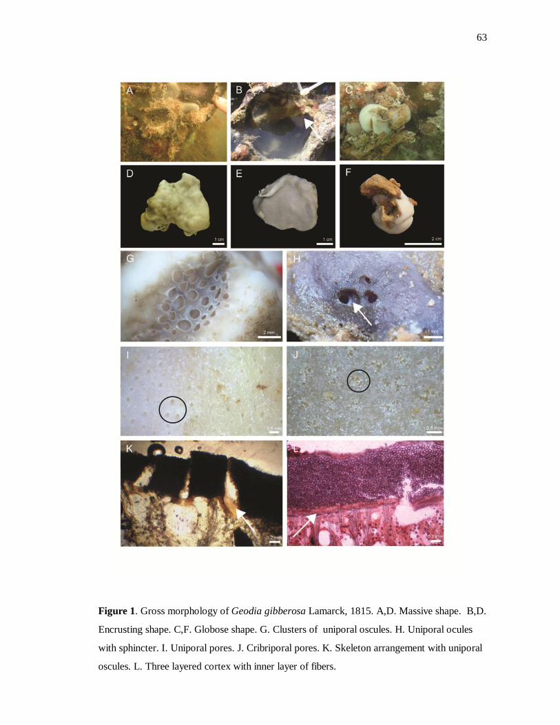
63
Figure 1. Gross morphology of Geodia gibberosa Lamarck, 1815. A,D. Massive shape. B,D.
Encrusting shape. C,F. Globose shape. G. Clusters of uniporal oscules. H. Uniporal ocules
with sphincter. I. Uniporal pores. J. Cribriporal pores. K. Skeleton arrangement with uniporal
oscules. L. Three layered cortex with inner layer of fibers.

64
Figure 2. Megascleres of Geodia gibberosa Lamarck, 1815. A. Choanosomal oxea. B-D.
Tips of oxea (B. Hastate. C. Acerate. D. Blunt). E. Cortical strongyloxea. F. Orthotriaene. G.
Cladome of orthotriaene with bent clad. H. Anatriaene (with broken rhabdome). I. Protriaene.
J. Plagiotriaene.

65
Figure 3. Microscleres of Geodia gibberosa Lamarck, 1815. Sterraster A-C (A. Mature
sterraster. C. Sterraster in an intermediate stage of development. B. Young sterraster). D.
Acanthoxyaster. E. Acanthospheroxyaster I. F. Variation of acanthospheroxyasters II.
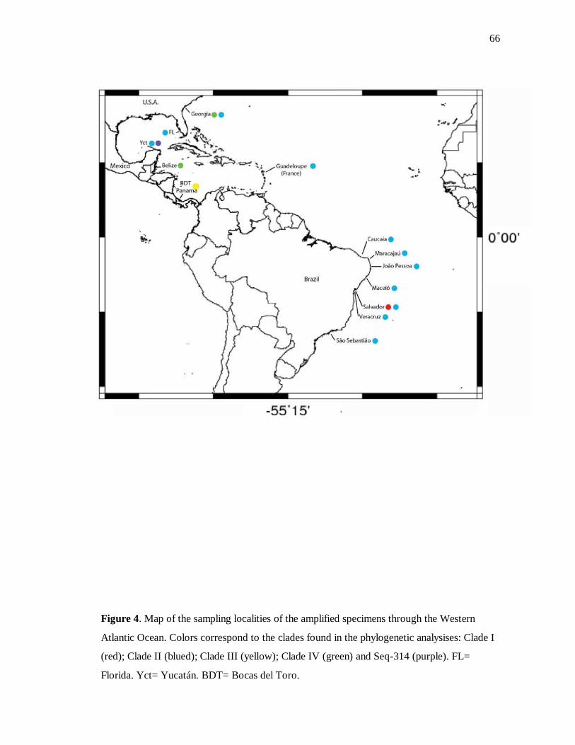
66
Figure 4. Map of the sampling localities of the amplified specimens through the Western
Atlantic Ocean. Colors correspond to the clades found in the phylogenetic analysises: Clade I
(red); Clade II (blued); Clade III (yellow); Clade IV (green) and Seq-314 (purple). FL=
Florida. Yct= Yucatán. BDT= Bocas del Toro.
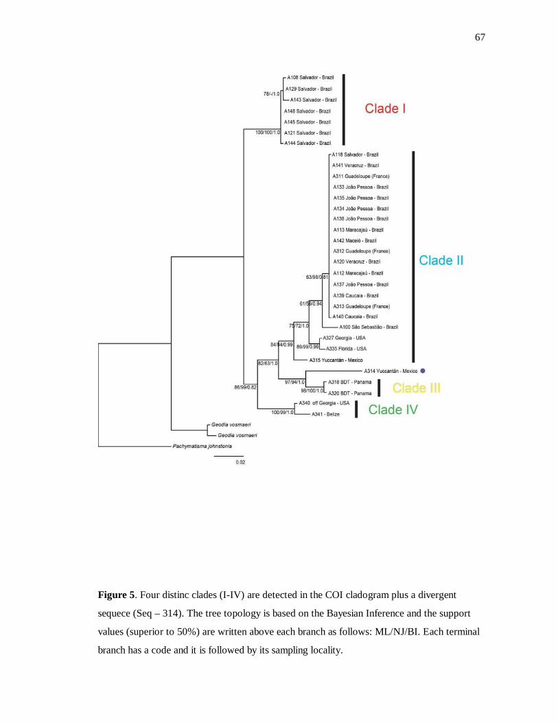
67
Figure 5. Four distinc clades (I-IV) are detected in the COI cladogram plus a divergent
sequece (Seq – 314). The tree topology is based on the Bayesian Inference and the support
values (superior to 50%) are written above each branch as follows: ML/NJ/BI. Each terminal
branch has a code and it is followed by its sampling locality.
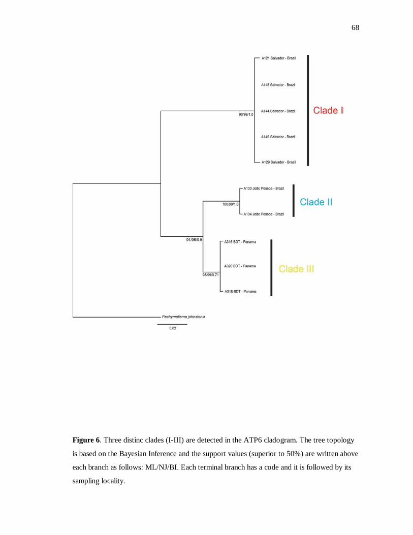
68
Figure 6. Three distinc clades (I-III) are detected in the ATP6 cladogram. The tree topology
is based on the Bayesian Inference and the support values (superior to 50%) are written above
each branch as follows: ML/NJ/BI. Each terminal branch has a code and it is followed by its
sampling locality.
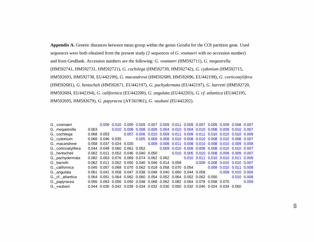
Appendix A. Genetic distances between mean group within the genus Geodia for the COI partition gene. Used
sequences were both obtained from the present study (2 sequences of G. vosmaeri with no accession number)
and from GenBank. Accession numbers are the following: G. vosmaeri (HM592711), G. megastrella
(HM592741, HM592731, HM592721), G. cochilega (HM592739, HM592742), G. cydonium (HM592715,
HM592693, HM592738, EU442199), G. macandrewi (HM592689, HM592696, EU442198), G. corticostylifera
(HM592681), G. hentscheli (HM592671, EU442197), G. pachydermata (EU442197), G. barretti (HM592720,
HM592684, EU442194), G. californica (EU442200), G. angulata (EU442203), G. cf. atlantica (EU442195,
HM592695, HM592679), G. papyracea (AY561961), G. vaubani (EU442202).
G._vosmaeri 0.009 0.010 0.009 0.009 0.007 0.009 0.011 0.009 0.007 0.009 0.009 0.008 0.007 G._megastrella 0.063 0.010 0.008 0.008 0.009 0.004 0.010 0.004 0.010 0.008 0.009 0.010 0.007 G._cochilega 0.068 0.053 0.007 0.006 0.010 0.009 0.011 0.009 0.011 0.010 0.010 0.010 0.009 G._cydonium 0.068 0.046 0.035 0.005 0.009 0.008 0.010 0.008 0.010 0.008 0.010 0.008 0.007 G._macandrewi 0.058 0.037 0.024 0.020 0.009 0.008 0.011 0.008 0.010 0.008 0.010 0.009 0.008 G._corticostylifera 0.044 0.049 0.060 0.061 0.052 0.009 0.010 0.009 0.006 0.008 0.010 0.010 0.007 G._hentscheli 0.062 0.011 0.052 0.046 0.040 0.050 0.010 0.005 0.010 0.008 0.009 0.009 0.007 G._pachydermata 0.082 0.063 0.076 0.069 0.074 0.062 0.062 0.010 0.011 0.010 0.010 0.011 0.009 G._barretti 0.062 0.011 0.052 0.050 0.040 0.046 0.014 0.058 0.009 0.008 0.010 0.010 0.007 G._californica 0.045 0.057 0.068 0.070 0.062 0.018 0.058 0.070 0.054 0.009 0.010 0.011 0.008 G._angulata 0.061 0.041 0.058 0.047 0.038 0.048 0.040 0.060 0.044 0.058 0.009 0.010 0.006 G._cf._atlantica 0.064 0.051 0.064 0.062 0.060 0.054 0.052 0.064 0.052 0.062 0.050 0.010 0.008 G._papyracea 0.056 0.063 0.056 0.050 0.048 0.068 0.062 0.082 0.064 0.078 0.058 0.070 0.009 G._vaubani 0.044 0.030 0.042 0.039 0.034 0.032 0.030 0.050 0.032 0.040 0.024 0.034 0.050
69
Capítulo 2
A NEW DESCRIPTION AND RECORD OF GEODIIDAE GRAY, 1867 (ASTROPHORIDA, DEMOSPONGIAE) SPECIES FROM THE
CARIBBEAN ZONE
Ueslei Lopes, Patrícia Domingues de Freitas & Carla Menegola
Artigo a ser submetido no periódico Zootaxa
71
Description of a new species of Geodia and new record of Pachymatisma (Astrophorida, Demospongiae) from the Caribbean zone
UESLEI LOPES1, PATRÍCIA DOMINGUES DE FREITAS2, CARLA MENEGOLA1
1 Instituto de Biologia, Departamento de Zoologia, Universidade Federal da Bahia, Campus de Ondina, Rua Barão de Jeremoabo, s/n, 40170-115, Salvador, Bahia, Brazil. E-mail: [email protected], [email protected]. 2 Departamento de Genética e Evolução, Centro de Ciências Biológicas e da Saúde, Universidade Federal de São Carlos, Via Washington Luis, Km 235, Monjolinho, 13565-905 - São Carlos, São Paulo – Brazil. E-mail: [email protected].
Abstract
In the present paper, we describe a new species of Geodia from the Gulf of Paria, Venezuela,
and the new record of the genus Pachymatisma from Curaçao. Geodia leispheroxyastera sp.
nov. is distinguished from other species of the genus by having a remarkable category of
spheroxyaster. Pachymatisma johnstonia represents the first occurrence of the genus in the
Caribbean waters. Additionally to the new species described here, we also provided an
identification key to species of Geodia occurring in the Western Atlantic Ocean.
Key words: Geodia, Pachymatisma, taxonomy, morphology, DNA barcode, Western
Atlantic ocean.
Résumé
Dans ce travail, nous décrivons une nouvelle espèce de Geodia du Golfe de Paria, au
Venezuela, et nous enregistrons la présence du genre Pachymatisma de Curaçao. Geodia
leispheroxyastera sp. nov. se difere des autres espèces du genre par une remarquable
catégorie de spheroxyaster. Pachymatisma johnstonia représente un registre nouveaux de ce
genre dans les eaux des Caraïbes. En plus de décrire la nouvelle espèce, nous fournissons une
clé d’identification pour les espèces de Geodia qui se trouvent présentes dans l'ouest de
l'océan Atlantique.
Mots-clés: Geodia, Pachymatisma, la taxonomie, la morphologie, code-barres moléculaires,
l'Ouest Atlantique.

72
1. Introduction
With approximately 220 extant species, Geodiidae is one of the largest families
within the Order Astrophorida Sollas, 1888 and one of the oldest in the Class Demospongiae
Sollas, 1885, with fossil records going back to the Early Cambrian (Gruber & Reitner, 1991).
The family Geodiidae Gray, 1867 is generally characterized by massive or thickly
encrusting poriferans. The skeleton is radially arranged at the periphery, and pores and oscules
may be organized in a sieve. Sterrasters and different types of triaenes are the main spicules,
but microrhabds and euasters are also present (Uriz, 2002).
Recently, a study based on molecular markers suggested the resurrection of the
subdivision Erylinae/ Geodinae sensu Sollas (1888) and the synonimization of the genera Isops
Sollas 1880 and Sidonops Sollas, 1889 (Cárdenas et al., 2010). The subfamily Erilinae would
be composed of the genera Erylus Gray 1867, Caminus Schmidt 1862, Penares Gray 1867
and Pachymatisma Bowerbank in Johnston, 1842 and the second one would be then only
consisted of Depressiogeodia Cárdenas, Rapp, Schander & Tendal, 2010, Geodia Lamarck,
1815 and Cydonium Fleming, 1828 (Cárdenas et al., 2010). The genus Geodia is hitherto the
most representative with 145 species and geographically and bathymetrically widely
distributed. Pachymatisma is composed of the species Pachymatisma johnstonia Bowerbank
in Johnston, 1842, Pachymatisma normani Sollas, 1888 (restricted to the Northeastern
Atlantic Ocean), Pachymatisma bifida Burton, 1959 and Pachymatisma aerolata Bowerbank
1972 (occurring in the Indian Ocean) (Silva, 2002; van Soest et al., 2012).
The specimens analyzed in this paper were previously identified as Geodia gibberosa
Lamarck 1815 and were formerly destined to a morphologic and molecular study of this
species (unpublished data). After morphologic analysis we could, then, confirm that these
samples actually represented a new species of Geodia and the first record of the genus
Pachymatisma for the Western Atlantic.

73
2. Materials and methods
2.1. Sampling
Geodiidae specimens were collected during different scientific expeditions. The
sample of Geodia was obtained by L. J. K. Klein from the Gulf of Paria, Venezuela
(10°22’30”N, 62°20’38.4”W) at 72 m depth within the “Calamar Expedition” between 1968
and 1969, under “The Caribbean Fisheries Development Project” developed by UNDP/FAO.
The sample of Pachymatisma was collected under a biodiversity project in Curaçao
(12°7’26.4” N, 68° 58’37.2”W) by N. van der Hal in 2005, at 20 m depth by scuba-diving.
The sampling localities are shown in the Figure 1. Fragments of these samples were donated
by NCB Naturalis (then Zoölogisch Museum van Amsterdam) for morphologic and molecular
analysis and they were kept in ethanol 96%. The entire specimens are deposited at the same
institution.
2.2. Morphologic analysis
A small fragment of the sponges (previously cut perpendicularly to their surface) was
kept out of the ethanol until getting slightly dried. The tissue was then deposited in a small
Petri dish and entirely embedded with distilled water. The cortex was held facing the base dish
and kept at -20 °C for 2-3 hours. The ice was removed from the dish and very thin slices were
promptly made (from the cortex to the choanosome) before the ice melting. The slices were
transferred to a slide and dried in a hotplate. For clarifying, few drops of xylene (2-3) covered
each cut and the slide rested in a laminar flow until the evaporation. Balsam of Canada was
used to cover the slide. Dissociated spicules mounts and SEM preparation followed Silva
(2002). Thirty spicules per spicule type were measured unless stated otherwise (N= number of
spicules measured). Spicule measurements were made with a Zeiss Axioplan 2 Imaging and
Zeiss Axiovision 4.5 software, and are given in μm as follows: minimum – mean – maximum.
The measures of triaenes’ are given in the ensuing order: rhabdome length/ rhabdome width;
cladome length/clad length/clad width. The base of both rhabdome and clad were used to
obtain width measures. The microscleres’ measures are: total diameter/center diameter/rays
length/ rays width. For better observing the details of these types of spicules, they were coated
with gold in the Denton Vacuum LLC Desk IV and later photographed with JEOL-JSM
74
6390LV scanning electronic microscopy at Centro de Pesquisas Gonçalo Muniz (Fundação
Oswaldo Cruz, Salvador). Photos of microscleres were treated with Adobe® Photoshop® CS
and draws of megascleres and plates were made using Adobe® Illustrator® CS5 software.
2.3. DNA extraction, PCR amplification and sequencing
The choanosomal region of the sponges was separated to perform the DNA
extraction, which followed a modified Sambrook et al., protocol (1989) using
phenol:chloroform:isoamyl alcohol (25:24:1). It was used 50 - 100 ng of DNA template, 1
unit of Platinum® Taq polymerase (Invitrogen), 2,5μl of 10x PCR buffer (200 mM Tris-HCl,
pH 8.4; 500 mM KCl), 2 mM of MgCl2, 200 μM of dNTP, 0.5μM, and 15 μl of sterile
distilled water for a 25μl PCR reaction. The universal primers described by Folmer et
al.(1994) (LCO 1490: 5’-GGT CAA CAA ATC ATA AAG ATA TTG G-3'; HCO 2198: 5'-
TAA ACT TCA GGG TGA CCA AAA AAT CA-3') were used to amplify a 650 bp partial
region of COI under the PCR thermal condition: 1 cycle [94 °C/5 min], 5 cycles [94 °C/30
bios, 46 °C/45 s, 72 °C/1 min], 35 cycles [94 °C/30 s, 50 °C/45 s, 72 °C/1 min], 1 cycle [72
°C/10 min]. Two internal primers designed by Lopes et al. (in preparation) were used to
amplify short fragments of approximately 350 pb each of COI. The primers and thermal
regimes are the following: PorF1 (5’-CGG GTA TGA TAG GTA CAG GGT T-3’) and
PorR1 (5’-TGA ATG TGC TTG TAC GCT CG-3’), 1 cycle [94 °C/5 min]; 35 cycles [94
°C/1 min, 60 °C/45 s, 72 °C/30 s]; 1 cycle [72 °C/10 min]. Second pair: PorF2 (5’-TCA GCT
TTT GTT GAA CAA GG-3’) and PorR2 (5`-CTT CTG GGT GTC CAA ARA AYC A-3’), 1
cycle [94 °C/5 min]; 35 cycles [94 °C/1 min, 48 °C/45 s, 72 °C/30 s]; 1 cycle [72 °C/10 min].
All PCR thermal regimes were ran in an Applied Biosystems Veriti® Thermal Cycler and
unincorporated primers and excess dNTPS were removed from the PCR products with PEG,
based on the protocol described by Lis & Schleif (1975). Sequencing was performed by
Macrogen, Inc., in South Korea (www.macrogen.com), and the sequences generated were
submitted to BLAST at NCBI (http:// www.ncbi.nlm.nih.gov/BLAST/).
2.4. Sequence analysis
The edition of the sequences was performed using BioEdit v. 7.0.9.0. (Hall, 1999)
and posteriorly compared to additional ones obtained from the GenBank
(www.ncbi.nlm.nih.gov). Uncorrected p-distance was estimated by 1000 bootstrap replicates

75
with MEGA5 (Tamura et al., 2011). The sequences used for this purpose are the following:
Pachymatisma normani (EF 564329, EF564327, EF564325, EF564323, EF564328,
EF564326, EF564324, EF564322); Pachymatisma johnstonia (EF654340, EF654338,
EF654335, EF654333, EF654331, EF654341, EF654339, EF654337, EF654334, EF654332,
EF654330).
3. Results
3.1. Systematic description and spicules nomenclature
The classification and the spicules nomenclature adopted in this work follows,
respectively, the Systema Porifera (Hooper & van Soest, 2002) and the Thesaurus of Sponge
Morphology (Boury-Esnault & Rützler, 1997).
Class: Demospongiae Sollas, 1885
Order: Astrophorida Sollas, 1888
Family: Geodiidae Gray, 1867
Genus: Geodia Lamarck, 1815
Geodia leispheroxyastera sp. nov.
(Figure 2 A-L)
Holotype. ZMA-POR 03558, Gulf of Paria, Venezuela, 10°22’30”N, 62°20’38.4”W.
Additional materials examined. Geodia gibberosa Lamarck, 1815. Guiana. Schizoholotype
MNHN-DT 608 (slide MCN 3639): collecting data not referred; Yucatán, CNPGG 1203: J.
Sierra coll., 25.IV.1985. Geodia pachydermata (Sollas, 1886). Bermuda. Schizoholotype of
BMNH 1889.1.1.91 (slide MCN 4050): off Bermuda, 32°8’45”N/64°59’35”W (Sta. 56), H.
M. S. “Challenger” coll., 29.V.1873, 1.935 m. Geodia cumulus Schmidt, 1870. Florida.
Schizotype BMNH 1870:5:3:85 (slide of spicular set): no collecting data (Schmidt, 1870 as
G. cumulus); Schizotype ZMB 6663 (slide of spicules identified by Oscar Schmidt): no
collecting data (Schimidt, 1870 como G. cumulus). Geodia apiarium (Schmidt, 1870).
Florida. Schizotype BMNH 1870.5.3.84: 275.4 m (slide of spicules). Geodia tuberculosa
Bowerbank, 1872. Mexico. Schizoholotype BMNH without number (slides of spicules and
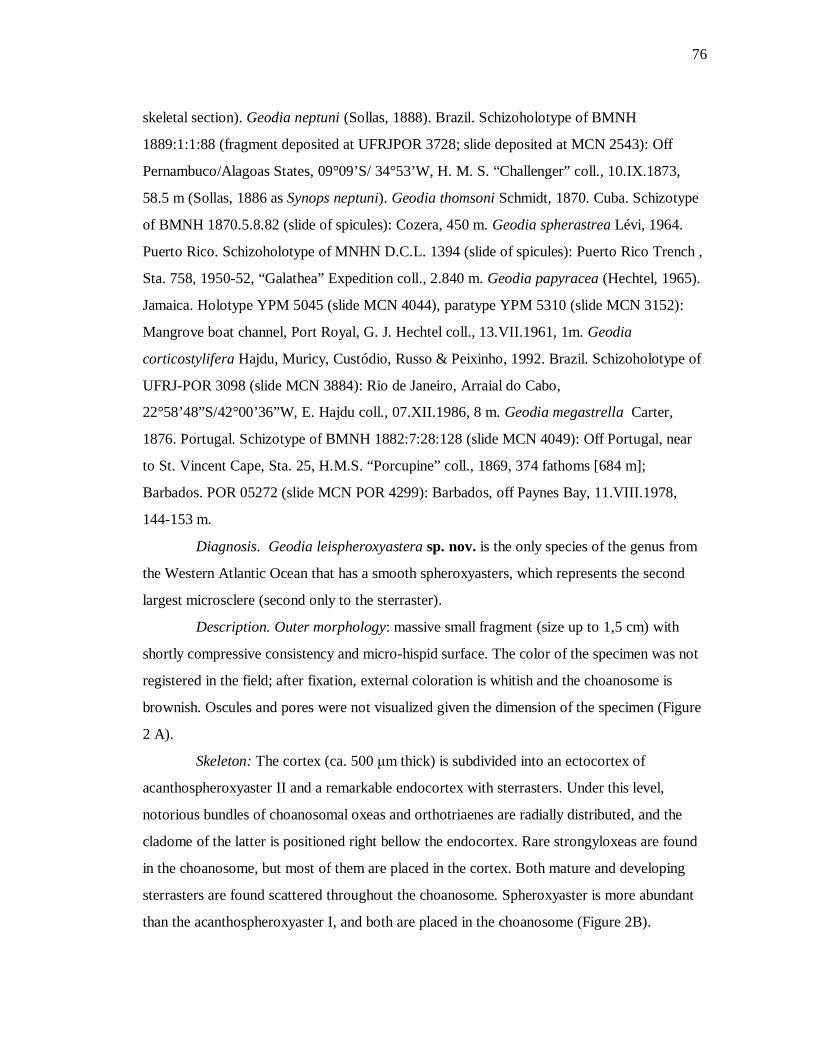
76
skeletal section). Geodia neptuni (Sollas, 1888). Brazil. Schizoholotype of BMNH
1889:1:1:88 (fragment deposited at UFRJPOR 3728; slide deposited at MCN 2543): Off
Pernambuco/Alagoas States, 09°09’S/ 34°53’W, H. M. S. “Challenger” coll., 10.IX.1873,
58.5 m (Sollas, 1886 as Synops neptuni). Geodia thomsoni Schmidt, 1870. Cuba. Schizotype
of BMNH 1870.5.8.82 (slide of spicules): Cozera, 450 m. Geodia spherastrea Lévi, 1964.
Puerto Rico. Schizoholotype of MNHN D.C.L. 1394 (slide of spicules): Puerto Rico Trench ,
Sta. 758, 1950-52, “Galathea” Expedition coll., 2.840 m. Geodia papyracea (Hechtel, 1965).
Jamaica. Holotype YPM 5045 (slide MCN 4044), paratype YPM 5310 (slide MCN 3152):
Mangrove boat channel, Port Royal, G. J. Hechtel coll., 13.VII.1961, 1m. Geodia
corticostylifera Hajdu, Muricy, Custódio, Russo & Peixinho, 1992. Brazil. Schizoholotype of
UFRJ-POR 3098 (slide MCN 3884): Rio de Janeiro, Arraial do Cabo,
22°58’48”S/42°00’36”W, E. Hajdu coll., 07.XII.1986, 8 m. Geodia megastrella Carter,
1876. Portugal. Schizotype of BMNH 1882:7:28:128 (slide MCN 4049): Off Portugal, near
to St. Vincent Cape, Sta. 25, H.M.S. “Porcupine” coll., 1869, 374 fathoms [684 m];
Barbados. POR 05272 (slide MCN POR 4299): Barbados, off Paynes Bay, 11.VIII.1978,
144-153 m.
Diagnosis. Geodia leispheroxyastera sp. nov. is the only species of the genus from
the Western Atlantic Ocean that has a smooth spheroxyasters, which represents the second
largest microsclere (second only to the sterraster).
Description. Outer morphology: massive small fragment (size up to 1,5 cm) with
shortly compressive consistency and micro-hispid surface. The color of the specimen was not
registered in the field; after fixation, external coloration is whitish and the choanosome is
brownish. Oscules and pores were not visualized given the dimension of the specimen (Figure
2 A).
Skeleton: The cortex (ca. 500 μm thick) is subdivided into an ectocortex of
acanthospheroxyaster II and a remarkable endocortex with sterrasters. Under this level,
notorious bundles of choanosomal oxeas and orthotriaenes are radially distributed, and the
cladome of the latter is positioned right bellow the endocortex. Rare strongyloxeas are found
in the choanosome, but most of them are placed in the cortex. Both mature and developing
sterrasters are found scattered throughout the choanosome. Spheroxyaster is more abundant
than the acanthospheroxyaster I, and both are placed in the choanosome (Figure 2B).
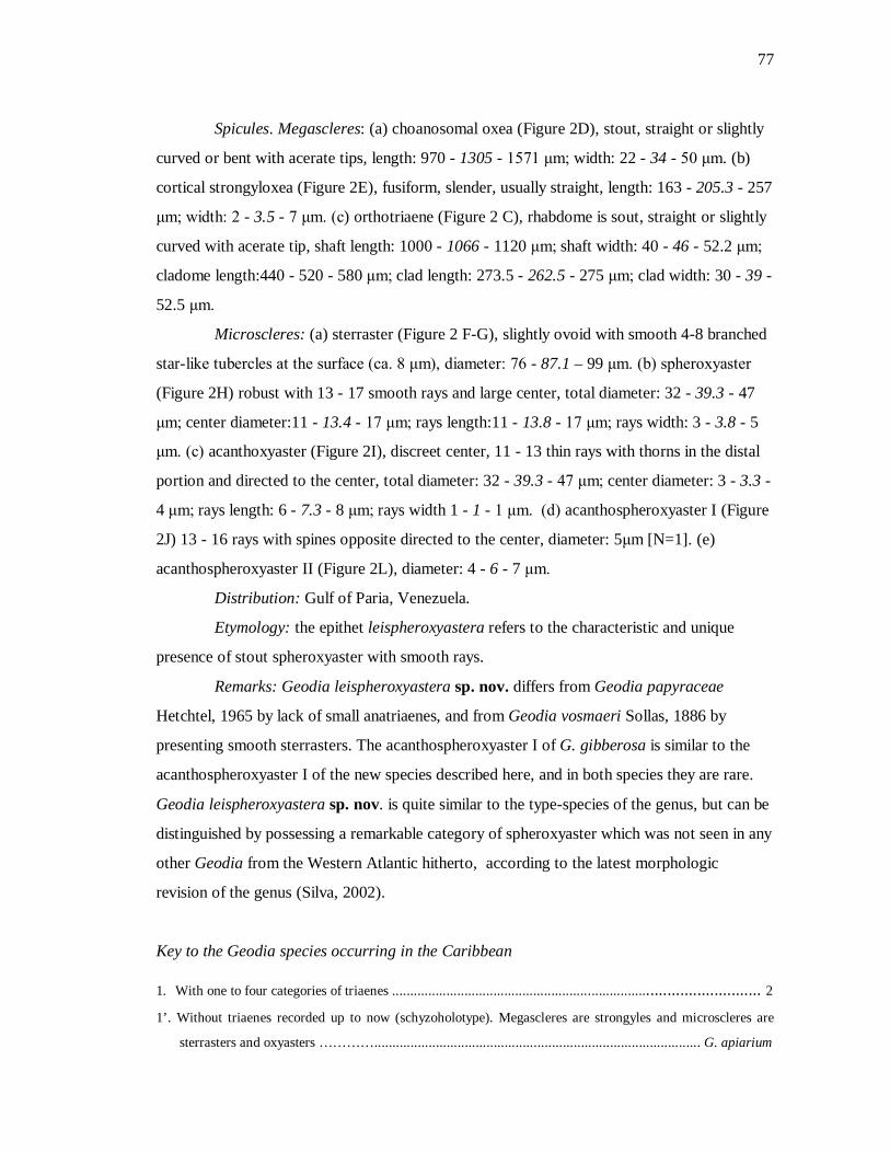
77
Spicules. Megascleres: (a) choanosomal oxea (Figure 2D), stout, straight or slightly
curved or bent with acerate tips, length: 970 - 1305 - 1571 μm; width: 22 - 34 - 50 μm. (b)
cortical strongyloxea (Figure 2E), fusiform, slender, usually straight, length: 163 - 205.3 - 257
μm; width: 2 - 3.5 - 7 μm. (c) orthotriaene (Figure 2 C), rhabdome is sout, straight or slightly
curved with acerate tip, shaft length: 1000 - 1066 - 1120 μm; shaft width: 40 - 46 - 52.2 μm;
cladome length:440 - 520 - 580 μm; clad length: 273.5 - 262.5 - 275 μm; clad width: 30 - 39 -
52.5 μm.
Microscleres: (a) sterraster (Figure 2 F-G), slightly ovoid with smooth 4-8 branched
star-like tubercles at the surface (ca. 8 μm), diameter: 76 - 87.1 – 99 μm. (b) spheroxyaster
(Figure 2H) robust with 13 - 17 smooth rays and large center, total diameter: 32 - 39.3 - 47
μm; center diameter:11 - 13.4 - 17 μm; rays length:11 - 13.8 - 17 μm; rays width: 3 - 3.8 - 5
μm. (c) acanthoxyaster (Figure 2I), discreet center, 11 - 13 thin rays with thorns in the distal
portion and directed to the center, total diameter: 32 - 39.3 - 47 μm; center diameter: 3 - 3.3 -
4 μm; rays length: 6 - 7.3 - 8 μm; rays width 1 - 1 - 1 μm. (d) acanthospheroxyaster I (Figure
2J) 13 - 16 rays with spines opposite directed to the center, diameter: 5μm [N=1]. (e)
acanthospheroxyaster II (Figure 2L), diameter: 4 - 6 - 7 μm.
Distribution: Gulf of Paria, Venezuela.
Etymology: the epithet leispheroxyastera refers to the characteristic and unique
presence of stout spheroxyaster with smooth rays.
Remarks: Geodia leispheroxyastera sp. nov. differs from Geodia papyraceae
Hetchtel, 1965 by lack of small anatriaenes, and from Geodia vosmaeri Sollas, 1886 by
presenting smooth sterrasters. The acanthospheroxyaster I of G. gibberosa is similar to the
acanthospheroxyaster I of the new species described here, and in both species they are rare.
Geodia leispheroxyastera sp. nov. is quite similar to the type-species of the genus, but can be
distinguished by possessing a remarkable category of spheroxyaster which was not seen in any
other Geodia from the Western Atlantic hitherto, according to the latest morphologic
revision of the genus (Silva, 2002).
Key to the Geodia species occurring in the Caribbean 1. With one to four categories of triaenes ................................................................................................. 2
1’. Without triaenes recorded up to now (schyzoholotype). Megascleres are strongyles and microscleres are
sterrasters and oxyasters ………….......................................................................................... G. apiarium
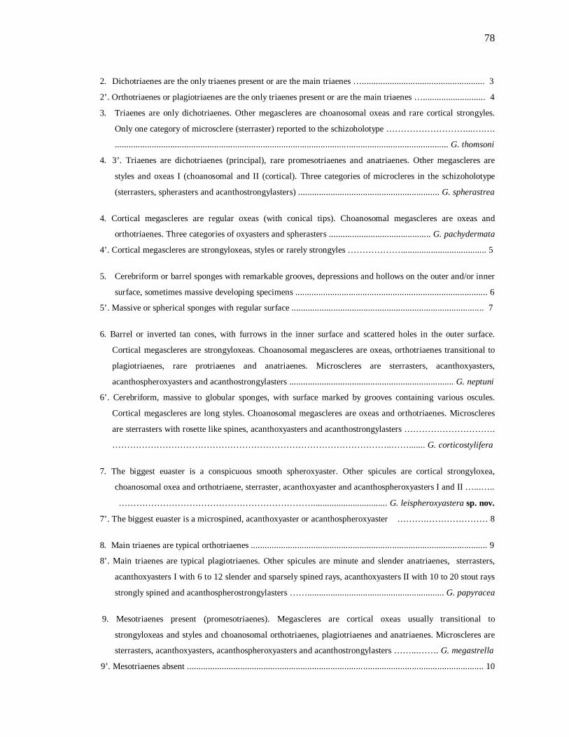
78
2. Dichotriaenes are the only triaenes present or are the main triaenes …..................................................... 3
2’. Orthotriaenes or plagiotriaenes are the only triaenes present or are the main triaenes …........................... 4
3. Triaenes are only dichotriaenes. Other megascleres are choanosomal oxeas and rare cortical strongyles.
Only one category of microsclere (sterraster) reported to the schizoholotype ………………………...….….
................................................................................................................................................ G. thomsoni
4. 3’. Triaenes are dichotriaenes (principal), rare promesotriaenes and anatriaenes. Other megascleres are
styles and oxeas I (choanosomal and II (cortical). Three categories of microcleres in the schizoholotype
(sterrasters, spherasters and acanthostrongylasters) ............................................................. G. spherastrea
4. Cortical megascleres are regular oxeas (with conical tips). Choanosomal megascleres are oxeas and
orthotriaenes. Three categories of oxyasters and spherasters ............................................ G. pachydermata
4’. Cortical megascleres are strongyloxeas, styles or rarely strongyles ………………..................................... 5
5. Cerebriform or barrel sponges with remarkable grooves, depressions and hollows on the outer and/or inner
surface, sometimes massive developing specimens ................................................................................... 6
5’. Massive or spherical sponges with regular surface ................................................................................... 7
6. Barrel or inverted tan cones, with furrows in the inner surface and scattered holes in the outer surface.
Cortical megascleres are strongyloxeas. Choanosomal megascleres are oxeas, orthotriaenes transitional to
plagiotriaenes, rare protriaenes and anatriaenes. Microscleres are sterrasters, acanthoxyasters,
acanthospheroxyasters and acanthostrongylasters ....................................................................... G. neptuni
6’. Cerebriform, massive to globular sponges, with surface marked by grooves containing various oscules.
Cortical megascleres are long styles. Choanosomal megascleres are oxeas and orthotriaenes. Microscleres
are sterrasters with rosette like spines, acanthoxyasters and acanthostrongylasters ………………………….
…………………………………………………………………………………..……....... G. corticostylifera
7. The biggest euaster is a conspicuous smooth spheroxyaster. Other spicules are cortical strongyloxea,
choanosomal oxea and orthotriaene, sterraster, acanthoxyaster and acanthospheroxyasters I and II …...…..
…………………………………………………………................................ G. leispheroxyastera sp. nov.
7’. The biggest euaster is a microspined, acanthoxyaster or acanthospheroxyaster ……….………………… 8
8. Main triaenes are typical orthotriaenes ...................................................................................................... 9
8’. Main triaenes are typical plagiotriaenes. Other spicules are minute and slender anatriaenes, sterrasters,
acanthoxyasters I with 6 to 12 slender and sparsely spined rays, acanthoxyasters II with 10 to 20 stout rays
strongly spined and acanthospherostrongylasters ……........................................................... G. papyracea
9. Mesotriaenes present (promesotriaenes). Megascleres are cortical oxeas usually transitional to
strongyloxeas and styles and choanosomal orthotriaenes, plagiotriaenes and anatriaenes. Microscleres are
sterrasters, acanthoxyasters, acanthospheroxyasters and acanthostrongylasters ……...……. G. megastrella
9’. Mesotriaenes absent ............................................................................ .................................................... 10

79
10. Triaenes with rhabdome exhibiting typical round tips. Cortical sterrasters and strongyloxeas rarely
transitional to oxeas. Choanosomal strongyloxeas, strongyles, oxeas, ortho-/ plagiotriaenes, anatriaenes,
acanthoxyasters and acanthospherostrongylasters …............................................................... G. cumulus
10’. Triaenes with rhabdome exhibiting conical tips ………………………….……………………………… 11
11. Massive sponges with irregular surface, with deep depressions and nodular projections. Cortical sterrasters
and strongyloxeas. Choanosomal oxeas (some transitional to styles), ortho-, ana- and protriaenes, spherasters,
acanthoxyasters and acanthostrongylasters ………………………….……….... G. tuberculosa
11’. Massive or spherical sponges with regular surface. Cortical spicules are sterrasters and strongyloxeas.
Choanosomal spicules are oxeas, long and stout orthotriaenes frequently transitional to plagiotriaenes,
additional rare triaenes (anatriaenes, protriaenes and remarkable minute slender plagiotriaenes),
acanthoxyasters, acanthospheroxyaster I and acanthospheroxyaster II ................................. G. gibberosa.
Genus: Pachymatisma
Pachymatisma johnstonia Bowerbank, 1842
(Figure 3 A-L)
Examined material. ZMAPOR19064, Curaçao, 12°7’26.4” N, 68° 58’37.2”W.
Description. Outer morphology: Massive specimen (up to 3 cm in size), hardly
compressive, with smooth surface. Dark brown externally and ranging from brownish to
whitish in the choanosome, after fixation. Both oscules (ca. 2 mm in length) and pores (0.1 –
0.3 mm) are uniporal.
Skeleton: Slender ectocortex is made of microhabds. Sterrasters form a quite
developed endocortex, and are spread throughout the choanosome, as well as some
microhabds. Oxeas and orthotriaenes are somewhat organized radially under the layer of
sterrasters. Acanthoxyaster II is much less abundant then the acanthoxyaster I, and both seem
not have a specific position in the choanosome.
Spicules. Megascleres:(a) choanosomal oxea (Figure 3C), stout, straight or slightly
curved with acerate tips, length: 560 - 814.3 - 970 µm; width: 7.5 - 14 - 17.5 µm. (b)
orthotriaene (Figure 3D-E), cladome with clades perfectly directed at right angle to the
rhabdome or bent and slightly straight robust rhabdome with acerate tip, rhabdome length:
340 - 498 - 650 µm; rhabdome width: 10 - 15.8 - 22.5 µm; cladome length: 200 - 417.7 - 680
µm; clad length: 87.5 - 214.8 - 375 µm; clad width 7.5 - 13.9 - 20 µm.
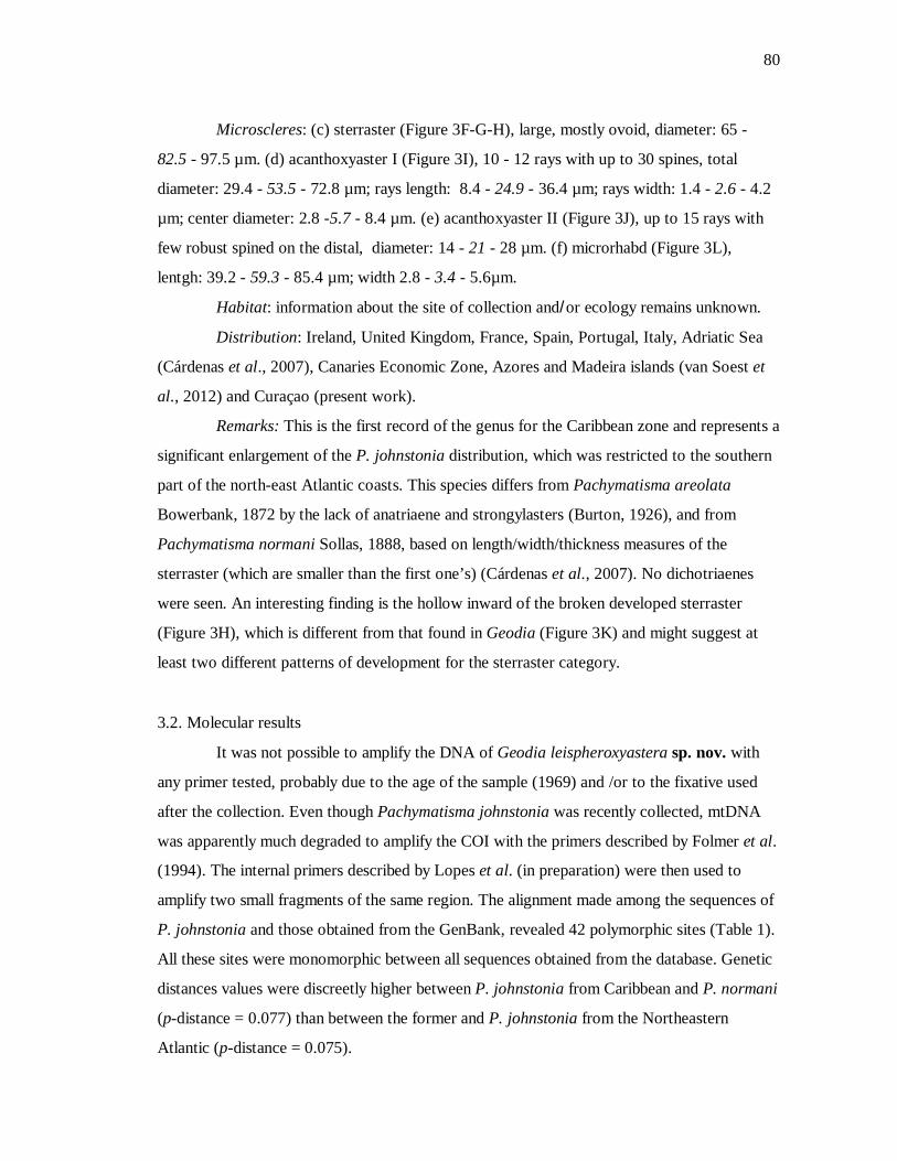
80
Microscleres: (c) sterraster (Figure 3F-G-H), large, mostly ovoid, diameter: 65 -
82.5 - 97.5 µm. (d) acanthoxyaster I (Figure 3I), 10 - 12 rays with up to 30 spines, total
diameter: 29.4 - 53.5 - 72.8 µm; rays length: 8.4 - 24.9 - 36.4 µm; rays width: 1.4 - 2.6 - 4.2
µm; center diameter: 2.8 -5.7 - 8.4 µm. (e) acanthoxyaster II (Figure 3J), up to 15 rays with
few robust spined on the distal, diameter: 14 - 21 - 28 µm. (f) microrhabd (Figure 3L),
lentgh: 39.2 - 59.3 - 85.4 µm; width 2.8 - 3.4 - 5.6µm.
Habitat: information about the site of collection and/or ecology remains unknown.
Distribution: Ireland, United Kingdom, France, Spain, Portugal, Italy, Adriatic Sea
(Cárdenas et al., 2007), Canaries Economic Zone, Azores and Madeira islands (van Soest et
al., 2012) and Curaçao (present work).
Remarks: This is the first record of the genus for the Caribbean zone and represents a
significant enlargement of the P. johnstonia distribution, which was restricted to the southern
part of the north-east Atlantic coasts. This species differs from Pachymatisma areolata
Bowerbank, 1872 by the lack of anatriaene and strongylasters (Burton, 1926), and from
Pachymatisma normani Sollas, 1888, based on length/width/thickness measures of the
sterraster (which are smaller than the first one’s) (Cárdenas et al., 2007). No dichotriaenes
were seen. An interesting finding is the hollow inward of the broken developed sterraster
(Figure 3H), which is different from that found in Geodia (Figure 3K) and might suggest at
least two different patterns of development for the sterraster category.
3.2. Molecular results
It was not possible to amplify the DNA of Geodia leispheroxyastera sp. nov. with
any primer tested, probably due to the age of the sample (1969) and /or to the fixative used
after the collection. Even though Pachymatisma johnstonia was recently collected, mtDNA
was apparently much degraded to amplify the COI with the primers described by Folmer et al.
(1994). The internal primers described by Lopes et al. (in preparation) were then used to
amplify two small fragments of the same region. The alignment made among the sequences of
P. johnstonia and those obtained from the GenBank, revealed 42 polymorphic sites (Table 1).
All these sites were monomorphic between all sequences obtained from the database. Genetic
distances values were discreetly higher between P. johnstonia from Caribbean and P. normani
(p-distance = 0.077) than between the former and P. johnstonia from the Northeastern
Atlantic (p-distance = 0.075).
81
4. Discussion and considerations
Given the well documented morphological plasticity of poriferans in the literature
(Hill & Hill, 2002; López-Legentil et al., 2010) we encourage and usually prioritize the
description of new species based on a larger type-series. However, we chose to bring this to
light based on (1) the difficulty of obtaining more samples from the collection sites and (2) the
remarkable diagnostic characteristic of Geodia leispheroxyastera sp. nov., which does not
occur in other species of Geodia described so far and does not seem to be a variation of any
other spicule. Regarding the description of this new species, we enhance the number Geodia
from the Caribbean zone to 12 (and 18 from the entire Western Atlantic), and also enlarge the
distribution of P. johnstonia.
Molecular results showed higher values of genetic distance between the sample from
Caribbean and Europe than those found between the P. normani and P. johnstonia from
Northeastern Atlantic (which presented just one polymorphic site). Since all the
morphological characteristics were in accordance with the typical P. johnstonia (Cárdenas et
al., 2007), and we did not have more samples from the same place to compare, we chose to
keep its identification this way. Nevertheless, given the low capability of the sponge larvae to
disperse, it is also known and highly debated in the academy that species with such huge
distribution could be a reflex of an overconservative taxonomy (Klautau et al., 1999; Plotkin
& Boury-Esnault, 2004). It cannot be affirmed or denied in the present work because of our
small sampling, but gives the suggestion that the distribution of this species must be at least
further investigated.
Acknowledgements We specially thank Elly Beglinger and Rob van Soest (NCB Naturalis), Patricia Gomez
(Universidad Autónoma de México, México D.F.) and Hans Tore Rapp (Universitetet i
Bergen) for providing the specimens. Adriana Rangel, Cláudio Figueira and Maria Lúcia
Moreno (Fundação Oswaldo Cruz - Centro de Pesquisa Gonçalo Moniz, Salvador) are
thanked for the help with the SEM. Luciana Martins are also thanked for the valuable tips on
vector graphics software. The authors are grateful to Armando Vieira, Luis Sartori and Inessa
82
Lacativa from the Laboratório de Ficologia (Universidade Federal de São Carlos) for allowing
us to use the light microscope and make the measurements in São Carlos. We acknowledge
Pedro Galetti and the crew of the Laboratório de Biodiversidade Molecular e Conservação
(Universidade Federal de São Carlos) for the precious assistance with the molecular part of
this work. This research was partially funded by Conselho Nacional de Desenvolvimento
Científico (CNPq, Brazil; grants nr. 133909/2010-7, fellowship to Ueslei Lopes) and
Coordenação de Aperfeiçoamento de Pessoal de Nível Superior (CAPES, Brazil; Programa de
Apoio à Pós-graduação – PROAP).
References cited Boury-Esnault, N. & Rützler, K., (1997) Thesaurus of sponge morphology. Smithsonian
Contribution to Zoology. 596, 1-55 pp.
Bowerbank, J.S. (1872) Contributions to a General History of the Spongiadae. Part III.
Proceedings of the Zoological Society of London, 1872, 626-635, pls XLVI-XLIX.
Burton, M. (1926) Description of South African sponges collected in the South African
Marine Survey. Part I. Myxospongia and Astrotetraxonida. Fisheries Bulletin. Fisheries
and Marine Biological Survey Division, Union of South Africa Rept. 4 (Special Report
9), 1-29, 6 pls.
Burton, M. (1959) Sponges. In: Scientific Reports. John Murray Expedition, 1933-34. 10,
151-281. London: British Museum (Natural History).
Cárdenas, P.; Xavier, J.; Tendal, O. S.; Schander, C. & Rapp, H. T. (2007) Redescription and
resurrection of Pachymatisma normani (Demospongiae, Geodiidae), with remarks on
the genus Pachymatisma. Journal of the Marine Biological Association of the United
Kingdom, 87, 1511-1525.

83
Cárdenas, P., Rapp, H. T., Schander, C. & Tendal, O. S. (2010) Molecular taxonomy and
phylogeny of the Geodiidae (Porifera, Demospongiae, Astrophorida) – combining
phylogenetic and Linnaean classification. Zoologica Scripta, 39, 86–106.
Carter, H.J. (1876). Descriptions and Figures of Deep-Sea Sponges and their Spicules, from
the Atlantic Ocean, dredged up on board H.M.S.‘Porcupine’, chiefly in 1869
(concluded). Annals and Magazine of Natural History, 18,: 397-40; (106): 307-324;
(107): 388-410;(108): 458-479, pls XII-XVI.
Fleming, J. (1828) Spongiadae. In: A History of British Animals, Exhibiting the Descriptive
Characters and Systematical Arrangement of the Genera and Species of Quadrupeds,
Birds, Reptiles, Fishes, Mollusca, and Radiata of the United Kingdom; including the
Indigenous, Extirpated, and Extinct Kinds, together with Periodical and Occasional
Visitants. Bell and Bradfute, Edinburgh: i-xxiii, 518-527.
Folmer, O., Black, M., Hoeh, W., Lutz, R. & Vrijenhoek, R. (1994) DNA primers for
amplification of mitochondrial cytochrome c oxidase subunit I from diverse metazoan
invertebrates. Molecular Marine Biology and Biotechnology, 3, 294–299.
Gray, J.E. (1867) Notes on the arrangement of sponges, with the descriptions of some new
genera. Proceedings of the Zoological Society of London, 2, 492–558, pls XXVII–
XXVIII.
Gruber, G. & Reitner, J. (1991) Isolierte Mikro-und Megaskleren von Porifera aus dem
Untercampan von Höver (Norddeutschland) und Bemerkungen zur Phylogenie der
Geodiidae (Demospongiae). Berliner Geowiss. Abh.: 134, 107-117.
Hajdu, E., Muricy, G., Custódio, M., Russo, C. & Peixinho, S. (1992), Geodia
corticostylifera (Demospongiae, Porifera) new Astrophorid from the Brazilian coast
(Southwestern Atlantic). Bulletin of Marine Science, 51, 204-217.
Hall, T.A. (1999) BioEdit: a user-friendly biological sequence alignment editor and analysis
program for Windows 95/98/NT. Nucleic Acids Symposium Series, 41, 95-98.

84
Hill, M. S. & Hill, A. L. (2002) Morphological Plasticity in the Tropical Sponge
Anthosigmella varians: Responses to Predators and Wave Energy. Biological Bulletin,
202, 86-96.
Hechtel, G. J. (1965) A systematic study of the Demospongiae of Port Royal, Jamaica.
Bulletin of the Peabody Museum of Natural History, 20, 1-94.
Hooper, J.N.A. & van Soest, R.W.M. (2002) Systema Porifera. A guide to the classification
of sponges. Kluwer Academic / Plenum Publishers, New York, 1708 pp.
Johnston, G. (1842) A History of British Sponges and Lithophytes.(W.H. Lizars: Edinburgh):
i-xii, 1-264, pls I-XXV.
Lamarck, J.B.P. (1815) Suite des polypiers empâtés. Mémoires du Muséum d’Histoire
Naturelle, Paris, 1, 69–80, 162–168, 331–340.
Lévi, C. (1964) Spongiaires des zones bathyale, abyssale et hadale. Galathea Report.
Scientific Results of The Danish Deep-Sea Expedition Round the World, 7, 63-112, pls
II-XI.
Lis, J.T. & Schleif, R. (1975) Size fractionation of double-stranded DNA by precipitation with
polyethylene glycol. Nucleic Acids Research, 2, 383-390.
López-Legentil, S., Erwin, P. M., Henkel, T. P., Loh, T.S. & Pawlik, J. R. (2010) Phenotypic
plasticity in the Caribbean sponge Callyspongia vaginalis (Porifera: Haplosclerida),
Scientia Marina, 74, 445-453.
Klautau, M., Russo, C., Lazoski, C., Boury-Esnault, N., Thorpe, J. P. & Solé-Cava A. M.
(1999) Does cosmopolitanism in morphologically simple species result from
overconservative systematics? A case study using the marine sponge Chondrilla nucula.
Evolution 53: 1414-1422.
85
Plotkin, A. & Boury-Esnault, N. (2004) Alleged cosmopolitanism in sponges: the example of
a common Arctic Polymastia (Porifera, Demospongiae, Hadromerida). Zoosystema, 26,
13-20.
Sambrook, J., Fritsch, E. F. & Maniatis, T. (1989) Molecular cloning: A laboratory manual.
Cold Spring Harbor Laboratory Press, Cold Spring Harbor, New York, 2344 pp.
Schmidt, O. (1862) Die Spongien des adriatischen Meeres. (Wilhelm Engelmann: Leipzig), i–
viii, 1–88, pls 1–7.
Schmidt, O. (1870). Grundzüge einer Spongien-Fauna des Atlantischen Gebietes. Leipzig,
Engelmann, iv + 88 p.
Silva, C.M.M. (2002) Revisão das espécies de Geodia Lamarck, 1815 (Porifera,
Astrophorida, Geodiidae) do Atlântico Ocidental e Pacífico Oriental. Ph.D. Instituto de
Biociências da Universidade de São Paulo, São Paulo, 305 pp.
van Soest, R.W.M, Boury-Esnault, N., Hooper, J.N.A., Rützler, K, de Voogd, N.J., Alvarez
de Glasby, B., Hajdu, E., Pisera, A.B., Manconi, R., Schoenberg, C., Janussen, D.,
Tabachnick, K.R., Klautau, M., Picton, B., Kelly, M. & Vacelet, J. (2012) World
Porifera database. Available at http://www.marinespecies.org/Porifera/ (access on
April 2012).
Sollas, W.J. (1880) The sponge-fauna of Norway; a Report on the Rev. A.M. Norman’s
Collection of Sponges from the Norwegian Coast. Annals and Magazine of Natural
History, 5, 396-409, pl. XVII.
Sollas, W.J. (1885) A Classification of the Sponges. Scientific Proceedings of the Royal
Dublin Society (new series), 5, 112.
Sollas, W. J. (1886) Preliminary account of the tetractinellid sponges dredged by H.M.S.
Challenger, 1872-1876. Part I. The Choristida. Scientific Proceedings of the Royal
Dublin Society, 5, 177-199.
86
Sollas, W.J. (1888) Report on the Tetractinellida collected by H.M.S. Challenger, during the
years 1873–1876. Report on the Scientific Results of the Voyage of H.M.S. Challenger,
1873–1876. Zoology, 25, 1–458, pls I–XLIV, 1 map.
Sollas, W.J. (1889) On the Geodine Genera, Synops, Vosm., and Sidonops. A Correction.
Scientific Proceedings of the Royal DublinSociety, 6, 276-277.
Tamura, K., Peterson, D., Peterson, N., Stecher, G., Nei, M. & Kumar, S. (2011) MEGA5:
Molecular Evolutionary Genetics Analysis using Maximum Likelihood, Evolutionary
Distance, and Maximum Parsimony Methods. Molecular Biology and Evolution, 28,
2731-2739.
Uriz, M.J. (2002) Family Geodiidae Gray, 1867. In: Hooper, J.N.A. & van Soest, R.W.M.
(Eds.) Systema Porifera. A Guide to the classification of Sponges. Kluwer Academic /
Plenum Publishers, New York, pp. 134–140.
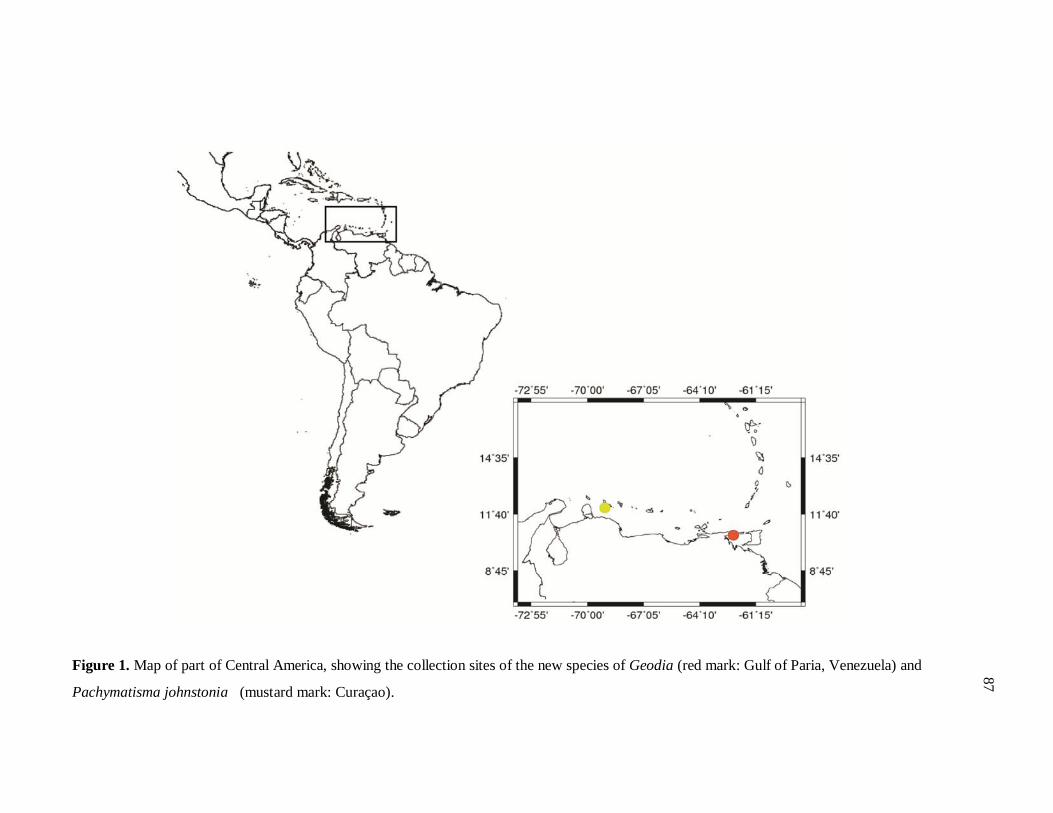
Figure 1. Map of part of Central America, showing the collection sites of the new species of Geodia (red mark: Gulf of Paria, Venezuela) and
Pachymatisma johnstonia (mustard mark: Curaçao).
87
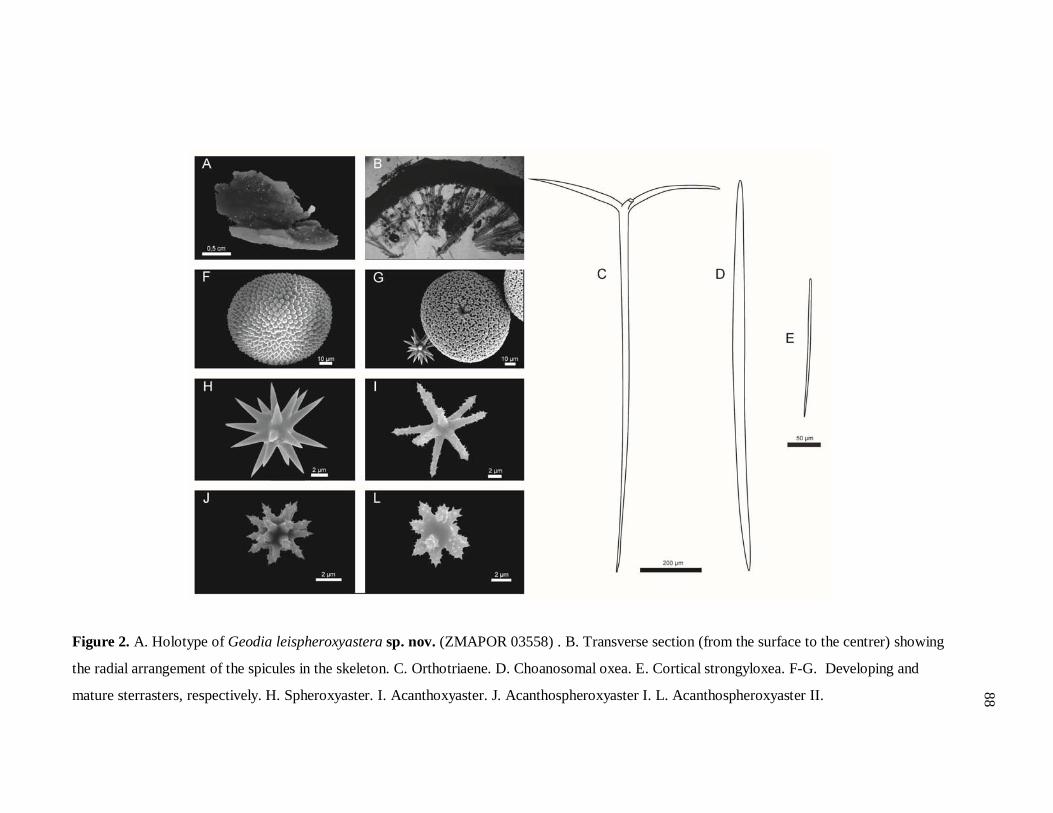
Figure 2. A. Holotype of Geodia leispheroxyastera sp. nov. (ZMAPOR 03558) . B. Transverse section (from the surface to the centrer) showing
the radial arrangement of the spicules in the skeleton. C. Orthotriaene. D. Choanosomal oxea. E. Cortical strongyloxea. F-G. Developing and
mature sterrasters, respectively. H. Spheroxyaster. I. Acanthoxyaster. J. Acanthospheroxyaster I. L. Acanthospheroxyaster II. 88
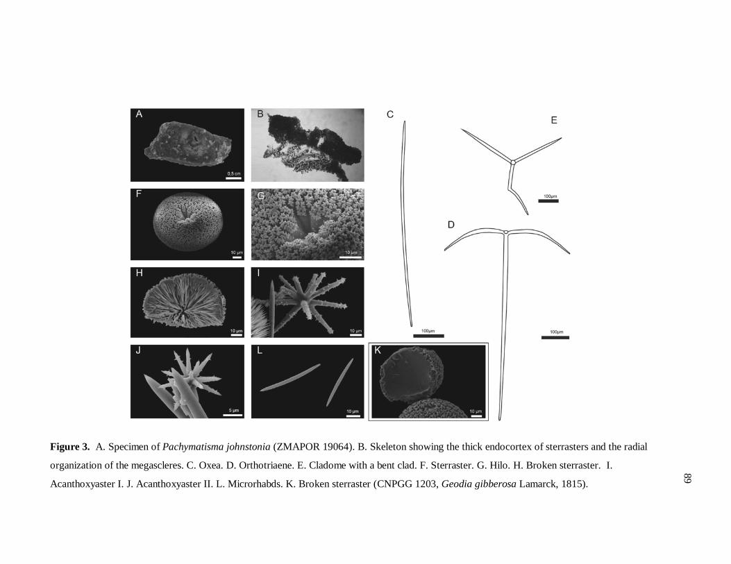
Figure 3. A. Specimen of Pachymatisma johnstonia (ZMAPOR 19064). B. Skeleton showing the thick endocortex of sterrasters and the radial
organization of the megascleres. C. Oxea. D. Orthotriaene. E. Cladome with a bent clad. F. Sterraster. G. Hilo. H. Broken sterraster. I.
Acanthoxyaster I. J. Acanthoxyaster II. L. Microrhabds. K. Broken sterraster (CNPGG 1203, Geodia gibberosa Lamarck, 1815).
89
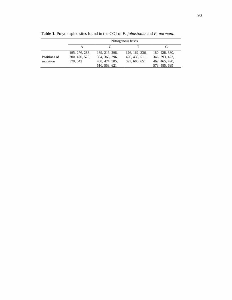
90
Table 1. Polymorphic sites found in the COI of P. johnstonia and P. normani.
Nitrogenous bases A C T G
Positions of mutation
195, 276, 288, 300, 420, 525, 579, 642
189, 219, 298, 354, 366, 396, 468, 474, 505, 510, 553, 621
126, 162, 336, 426, 435, 511, 597, 606, 651
180, 228, 330, 346, 393, 423, 462, 465, 490, 573, 585, 639
91
Considerações finais
Embora possuam significativa riqueza em termos de conjunto espicular, muitas
espécies do gênero Geodia não tem uma taxonomia fácil, dada à variação da morfologia
externa, à infrequência de determinadas categorias de espículas, ou ainda, à variação de
formas que as mesmas apresentam. Tais fatores levam taxonomistas à identificações errôneas
e acabam por provocar implicações em outros tipos de estudos.
Além de corroborar o polimorfismo (outrora mencionado na literatura) para a
morfologia externa de Geodia gibberosa, o presente estudo também detectou categorias raras
de triênios (ana- e plagiotriênios) que jamais haviam sido vistas no holótipo, e propõe algumas
hipóteses para explicar a infrequência de algumas espículas.
Os marcadores mitocondriais COI e ATP6, mesmo mostrando-se relativamente
conservados, foram eficientes em revelar a existência três de espécies crípticas dentro de
G. gibberosa. Contudo, mais amostras precisariam ter sido amplificadas para o segundo gene,
em especial aquelas que constituem o Clado IV (evidenciado apenas com o COI). Por meio
dos métodos de taxonomia clássica, no entanto, não foi possível detectar padrões que
permitissem separar espécies morfologicamente, e talvez outras análises (e.g. análises
estatísticas aplicadas à morfometria, estudos de desenvolvimento, etc.) devessem ser
empregadas para esse fim.
O presente estudo, assim como diversas pesquisas já realizadas com poríferos
considerados cosmopolitas ou com ampla distribuição (KLAUTAU et al., 1999; DE PAULA et
al., 2012), conseguiu detectar a ocorrência de espécies crípticas, e é o primeiro de sua
natureza com um representante da família Geodiidae (que é uma das maiores e mais antigas
dentro de sua Ordem). Seus resultados só vem reforçar quão subestimado é o conhecimento
acerca da biodiversidade em esponjas, e reforçam a necessidade da utilização de diferentes
fontes de informação/ferramentas em estudos de taxonomia, com o intuito de se verificar o
status de espécies cujos limites não são muito claros. Nesses termos, a taxonomia integrativa
tem se mostrado muito eficiente e vem se consolidando cada vez mais como uma prática
comum na academia.
92
Referências bibliográficas gerais
BLOCH, M. E. & SCHNEIDER, J. G. M.E. Blochii, Systema Ichthyologiae iconibus cx
illustratum. Post obitum auctoris opus inchoatum absolvit, correxit, interpolavit Jo. Gottlob
Schneider, Saxo. Berolini. Sumtibus Austoris Impressum et Bibliopolio Sanderiano
Commissum. Pp i-lx + 1–584, Pls. 1–110, 1801.
BORTOLOUS, A. Error Cascades in the Biological Sciences: The Unwanted Consequences
of Using Bad Taxonomy in Ecology. Ambio, v. 37, n. 2, p. 114–118, 2008.
BOWERBANK, J. S. Contributions to a general history of the Spongiadae. Proceedings of
the Zoological Society of London, pt. III, p. 626–635, 1872.
______.______. Proceedings of the Zoological Society of London, pt. IV, p. 3–25, 1873a.
______.______. Proceedings of the Zoological Society of London, pt. V, p. 319–333, 1873b.
______.______. Proceedings of the Zoological Society of London, pt. VI, p. 298–305, 1874.
CARBALLEIRA, N. M.; RODRIGUEZ, J. Two novel phospholipid fatty-acids from the
Caribbean sponge Geodia gibberosa. Lipids, v. 4, p. 324–326, 1991.
CÁRDENAS, P. et al. Morphological description and DNA barcodes of shallow-water
Tetractinellida (Porifera: Demospongiae) from Bocas del Toro, Panama, with description of a
new species. Zootaxa, v. 2276, p. 1–39, 2009a.
CÁRDENAS, P. et al. Molecular taxonomy and phylogeny of the Geodiidae (Porifera,
Demospongiae, Astrophorida) - combining phylogenetic and Linnaean classification.
Zoologica Scripta, v. 39, n. 1, p. 89–106, 2009b.
CÁRDENAS, P. Phylogeny, taxonomy and evolution of the Astrophorida (Porifera,
Demospongiae) 79 f. Tese (Degree of philosophy doctor - PhD) – University of Bergen,
Bergen, 2010.

93
CARTER, H. J. Some sponges from the West Indies and Acapulco in the Liverpool Free
Museum described, with general and classificatory remarks. Annals and Magazine of Natural
History, v. 9, n. 52, p. 266–301, 1882.
CHANAS, B.; PAWLIK, J. R. Defenses of Caribbean sponges against predatory reef fish. II.
Spicules, tissue toughness, and nutritional quality. Marine Ecology Progress Series, v. 127,
p. 195–211, 1995.
DUCHASSAING DE FONBRESSIN, P.; MICHELOTTI, G. Spongiaires de la mer Caraïbe.
Natuurkundige verhandelingen van de Hollandsche maatschappij der wetenschappen te
Haarlem, v. 21, n. 2, p. 1–124, 1864.
DUNLAP, M.; PAWLIK, J. R. Video-monitored predation by Caribbean reef fishes on an
array of mangrove and reefsponges. Marine Biology, v. 126, p. 117–123, 1996.
______. Spongivory by Parrotfish in Florida Mangrove and Reef Habitats. Marine Ecology,
v. 19, n. 4, p. 325–337, 1998.
ENGEL, M.; PAWLIK, J. R. Allelopathic activities of sponge extracts. Marine Ecology
Progress Series, v. 207, p. 273–281, 2000.
______. Interactions among Florida sponges. II. Mangrove habitats. Marine Ecology
Progress Series, v. 303, p. 145–152, 2005.
FLEMING, J. Spongiadae. In: A history of British animals, exhibiting the descriptive
characters and systematical arrangement of the genera and species of quadrupeds, birds,
reptiles, fishes, Mollusca, and Radiata of the United Kingdom; including the indigenous,
extirpated, and extinct kinds, together with periodical and occasional visitants. Edinburgh:
Bell and Bradfute, 1828, v. 1, n. 23, p. 1–565, 1828.
FOLMER, O. et al. DNA primers for amplification of mitochondrial cytochrome c oxidase
subunit I from diverse metazoan invertebrates. Molecular marine biology and biotechnology,
v. 3, p. 294–299, 1994.
GEORGE, W. C.; WILSON, H. V. Sponges of Beaufort (N.C.) Harbor and Vicinity. Bulletin
of the Bureau of Fisheries. Washington, v. 36, p. 129–179, 1919.

94
GRAY, J. E. Notes on the arrangement of sponges, with the description of some new genera.
Proceedings of the Zoological Society of London, v. 2, p. 492–558, 1867.
HAJDU, E. et al. Geodia corticostylifera (Demospongiae, Porifera) new Astrophorid from
the Brazilian coast (Southwestern Atlantic). Bulletin of Marine Science, v. 51, n. 2, p. 204–
217, 1992.
HECHTEL, G. J. A systematic study of the Demospongiae of Port Royal, Jamaica. Bulletin of
the Peabody Museum of Natural History, v. 20, p. 1–94, 1965.
JOHNSTON, G. A History of British Sponges and Lithophytes. W. H. Lizars: Edinburgh, i-
xii, p. 1–264, 1842.
KLAUTAU, M. et al. Does cosmopolitanism result from overconservative systematics? A
case study using the marine sponge Chondrilla nucula. Evolution, v. 53, n. 5, p. 1414–1422,
1999.
KNOWLTON, N. Molecular genetic analyses of species boundaries in the sea.
Hydrobiologia, v. 420, p. 73-90, 2000.
LAMARCK, J. B. P. A. Suite des polypiers empâtés. Histoire naturelle des animaux sans
vertébres. 2ème ed. Paris: Verdiére, p. 1-568, 1815.
______. Histoire des polypes. Histoire naturelle des animaux sans vertébres. 3ème ed.
Bruxelles: Meline, Cans et Compagnie, p. 1-677, 1837.
DE LAUBENFELS, M. W. A comparison of the shallow-water sponges near the Pacific end
of the Panama canal with those at the Caribbean end. Proceedings of the United States
National Museum, v. 83, p. 441–446, 1936a.
______. A discussion of the sponge fauna of the Dry Tortugas in particular, and the West
Indies in general, with material for a revision of the families and orders of the Porifera. Papers
Tortugas Lab. 30: i - iii + 1-225, 1936b.
VON LENDENFELS, R. Tetraxonia. In: SCHUZLE, FRANZ EILHARD (ed.), Das
Tierreich. Berlin: FriedlÄnder, p. 1–167, 1903.
95
LÉVI, C. Résultats scientifiques des Campagnes de la ‘Calypso’. Campagne de la ‘Calypso’
dans le Golfe de Guinée et aux îles Principe, São Tomé et Annobon. 5. Spongiaires. Annales
de l’Institut océanographique v. 37, n. 4, p. 115–141, 1959.
LINNAEUS, C. Systema naturae per regna tria naturae, secundum classes, ordines, genera,
species, cum characteribus, differentiis, synonymis, locis. Tomus I. Editio duodecima,
reformata. Holmiae. (Laurentii Salvii).: 1-532, 1766.
LÔBO-HAJDU, G. et al. Intra- and interspecific variation in the rDNA ITS of Porifera
revealed by PCR-single-strand conformation polymorphism (PCR-SSCP). Bollettino dei
Musei e degli Istituti Biologici dell' Università di Genova, v. 68, p.413–423, 2004.
LÔBO-HAJDU, G. Dados morfológicos versus dados moleculares em sistemática:
controvérsia ou congruência? In: GALLO, V. et al. (Ed.). Paleontologia de Vertebrados:
grandes temas e contribuições científicas. Rio de Janeiro: Editora Interciência Ltda., v. 1, p.
197–218, 2006.
LOCKE, J. M. & COATES, K. A. What are the costs of bad taxonomic practices: and what is
Madracis mirabilis? Proceedings of the 11th International Coral Reef Symposium, n. 26, p.
1348–1351, 2008.
LÓPEZ-LEGENTIL, S. et al. Phenotypic plasticity in the Caribbean sponge Callyspongia
vaginalis (Porifera: Haplosclerida). Scientia Marina, v. 74, n. 3, p. 445–453, 2010.
MEIER, R. DNA sequence in taxonomy: opportunities and challenges. In: WHELEER, Q. D.
The New Taxonomy. Boca Raton: CRC Press, 2008. cap.7, p. 95–127. (The Systematics
Association Special Volume Series 76).
MENDOLA, D. Aquaculture of three phyla of marine invertebrates to yield bioactive
metabolites: process developments and economics. Biomolecular Engineering, v. 20, n. 4, p.
441–458, 2003.
MURICY, G. et al. Biodiversidade marinha da Bacia Potiguar: Porifera. Rio de Janeiro:
Museu Nacional, p. 1–156, 2008. (Série Livros 29).
96
NAKANISHI, K. et al. Cholesterol and Companions. V. Microdetermination of 7-Stenols.
Journal of the American Chemical Society, v. 75, n. 18, p. 4415–4417, 1953.
PADIAL, M. et al. The integrative future of taxonomy. Frontiers in Zoology, v. 7, n. 16, p.
1–14, 2010.
DE PAULA, T.S., ZILBERBERG, C., HAJDU, E. & LOBO-HAJDU, G. Morphology and
molecules on opposite sides of the diversity gradient: Four cryptic species of the Cliona celata
(Porifera, Demospongiae) complex in South America revealed by mitochondrial and nuclear
markers. Molecular Phylogenetics and Evolution, v. 62, p. 529-541, 2012.
PAWLIK, J. R. et al. Defenses of Caribbean sponges against predatory reef fish. I. Chemical
deterrency. Marine Ecology Progress Series, v. 127, p.183–194, 1995.
PULITZER-FINALI, G. A collection of West Indian Demospongiae (Porifera). In appendix, a
list of the Demospongiae hitherto recorded from the West Indies. Annali del Museo Civico di
Storia Naturale Giacomo Doria, vol. 86, p. 65–216, 1986.
RANGEL, M. et al. Cytotoxic and neurotoxic activities I extracts of marine sponges
(Porifera) from southeastern Brazilian coast. Journal of Experimental Marine Biology and
Ecology, v. 262, p. 31–40, 2001.
RIGBY, J. K.; SMITH, C. C. Microscleres of a Paleocene Geodia from Western Alabama.
Journal of Paleontology, v. 66, n. 3, p. 406–413, 1992.
ROD’KINA, S. A. Fatty Acids and Other Lipids of Marine Sponges. Russian Journal of
Marine Biology, v. 31, p. 49–60, 2005.
SCHMIDT, O. Die Spongien des Adriatischen Meeres. Wilhelm Engelmann Leipzig, i-vii,
p. 1–88, 1862.
______.Grundzüge einer spongien-fauna des Atlantischen gebietes. Wilhelm Engelmann.
Leipzig, iii-iv, p. 1–88, 1870.
97
SILVA, C. M. M. Revisão das espécies de Geodia Lamarck, 1815 (Porifera, Astrophorida,
Geodiidae) do Atlântico Ocidental e Pacífico Oriental. 2002. 306 f. Tese (Doutorado em
Ciências, Zoologia) - Universidade de São Paulo, São Paulo, 2002.
VAN SOEST, R. W. M. Demosponge distribution patterns. In: VAN SOEST, R. W. M. et al.
(ed.). Sponges in Time and Space. Proceedings IV International Porifera Congress: Balkema,
Rotterdam, i-xviii, p. 213–232, 1994.
VAN SOEST, R. W. M. et al. World Porifera database. Disponível em
http://www.marinespecies.org/Porifera/. Acesso em 12 jun. 2012.
SOLLAS, W. J. The sponge-fauna of Norway; a Report on the Rev. A.M. Norman’s
Collection of Sponges from the Norwegian Coast. Annals and Magazine of Natural History,
v. 5, n. 27, p. 130–144, 1880.
______. Report on the Tetractinellida collected by H.M.S. Challenger, during the years 1873-
1876. Report on the Scientific Results of the Voyage of H. M. S. Challenger 1873-1876.
Zoology, 25, p. 1–458, 1888.
______. On the geodine genera, Synops,Vosm., and Sidonops. A correction.Scientific
Proceedings of the Royal Dublin Society, v. 6, n. 5, p. 276–27, 1889.
TAUTZ, D. et al. A plea for DNA taxonomy. TRENDS in Ecology and Evolution, v.18, n. 2,
p. 70–74, 2003.
TOPSENT, E. Éponges de San Thomé. Essai sur les genres Spirastrella, Donatia et
Chondrilla. Archives de Zoologie Expérimentale et Générale, v. 57, n. 6, p. 535–618, 1918.
ULICZKA, E. Die tetraxonen Schwämme Westindiens (auf Grund der Ergenbnisse der Reise
Kükenthal-Hartmayer). Zoologische Jahrbücher. Abteilung für Systematik, Geographie und
Biologie der Thiere, p. 35–62, 1929. (Supplement 16).
URIZ, M. J. Family Geodiidae Gray, 1867. In: HOOPER, J. N. A. & VAN SOEST, R. W. M.
(ed). Systema Porifera: A Guide to the Classification of Sponges, New York: Kluwer
Academic/Plenum Publishers, p. 134–140, 2002.
98
VALENCIENNES, M. A. Scarus aurofrenatus In: COUVIER, M. LE B. ON &
VALENCIENNES, M. A. Histoire Naturelle des Poissons, Libraire de la Société Géologique
de France, Tome 22, Paris, 1840.
WIEDENMAYER, F. Shallow-water sponges of the Western Bahamas. Experientia
Supplementum, v. 28, p. 1–287, 1977.
WULFF, J. L. Sponge predators may determine differences in sponge fauna between two sets
of mangrove cays, Belize barrier reef. Atoll Research Bulletin, v. 477, p. 249–263, 2000.
99
Apêndice 1. Protocolos de extração de DNA genômico total
1.1. Fenol:Clorofórmio:Álcool Isoamílico (Sambrook et al., 1989 – adaptado)
1. Colocar em microtubos (1,5ml) as amostras (0,05g) e adicionar 550 µL de tampão de lise
(50mM de Tris HCl, 50mM de EDTA, 100mM de NaCl).
2. Adicionar 40 µL de SDS 20% (dodecil sulfato de sódio) e 15 µL de proteinase K
(20mg/mL).
3. Vortexar as amostras e em seguida incubar em termo bloco a 55°C overnight (ou por 2hs
na mesma temperatura).
4. Retirar o tubo do Termo bloco e adicionar 550 µL de fenol/clorofórmio/álcool isoamilico
(ou somente 275 µL de fenol e 275 µL de clorofórmio).
5. Fechar muito bem os tubos e misturar as fases por inversão de tubos por 10 minutos.
6. Centrifugar por 30 minutos a 14000 rpm (enquanto isso nomear novos tubos de 2 mL)
7. Transferir o sobrenadante para outro tubo (muito cuidado para não encostar a ponteira na
fase abaixo ao sobrenadante).
8. Repetir os passos 4 a 7.
9. O mesmo volume dado de sobrenadante adicionar de Clorofórmio.
10 Misturar novamente por inversão de tubos por 10 minutos.
11. Centrifugar por 10 minutos a 14000rpm (enquanto isso nomear novos tubos).
12. Transferir o sobrenadante para um novo tubo.
13. Adicionar 10% de Acetato de Sódio 3M (pH 7,0) e 2,5x do volume do sobrenadante de
etanol absoluto gelado.
14. Misturar as fases por inversão de tubos cuidadosamente por 10 minutos.
15. Incubar as amostras em freezer a -20°C por 2 horas ou overnight.
16. Centrifugar as amostras por 15 minutos a 14000 rpm.
17. Reverter cuidadosamente para retirar o etanol.
18. Adicionar 600 µL de etanol 70%, agitar cuidadosamente o tubo e centrifugar por mais 10
minutos a 14000 rpm, depois reverter o tubo para descartar o etanol.
19. Adicionar 600 µL de etanol 70% (ou 100%), agitar cuidadosamente o tubo e centrifugar
por mais 10 minutos a 14000 rpm, depois reverter o tubo para descartar o etanol.
100
* Referência ALJANABI, S.M. & MARTINEZ, I. Universal and rapid salt-extraction of high quality genomic DNA for PCR-based techniques. Nucl. Acid. Reseach. v. 25, n. 22: p. 4692-4693, 1997.
20. Secar o DNA em temperatura ambiente ou em estufa a 37°C.
21. Com o DNA sem resíduo algum de etanol adicionar TE (10mM de Tris pH 8,0 e 1mM de
EDTA) ou água MilliQ de acordo com o tamanho do pellet.
22. Adicionar 30 µL de RNAse (5 µL).
23. Deixar o DNA em termo bloco a 37°C por 40 minutos e depois conservar a
-20°C até a utilização.
1.2. Tampão Salino (Aljanabi & Martinez, 1997* - adaptado) 1. Homogeneizar o tecido (50 – 100 mg ou 1 cm2) em 400 µl de tampão salino (NaCl
0,4 M; Tris HCl 10 mM pH 8,0 e EDTA pH 8,0 a 0,2 mM).
2. Adicionar 40 µl de SDS 20% e 8,0 µl de Proteinase K 20 mg/ml e misturar bem.
3. Incubar as amostras a 55 - 65°C por pelo menos 2h ou overnight.
4. Adicionar 300 µl de NaCl 6 M.
5. Vortexar as amostras por 30 segundos a velocidade máxima e centrifugar por 30
min. a 10000 rpm.
6. Transferir o sobrenadante para outro tubo (2,0 ml).
7. Adicionar igual volume de isopropanol e misturar bem.
8. Incubar a -20°C por 1h.
9. Centrifugar por 20 min. a 10000 rpm.
10. Lavar o pellet com 300 µl de etanol 70% e centrifugar por 5 minutos.
11. Lavar o pellet novamente com 300 µl de etanol 100% e centrifugar por mais 5
min.
12. Secar o DNA em estufa e ressuspender em 50 µl de H2O. Caso haja necessidade,
colocar RNAse (2,0 µl a uma concentração de 10 ng/µl).

101
* Referências CHOMABARD, C. et al. Reassessment of Homology of Morphological Characters in Tetractinellid Sponges Based on Molecular Data. Systematic Biology, v. 47, n. 3, p. 351–366, 1998. LÔBO-HAJDU, G. et al. Intra- and interspecific variation in the rdna its of Porifera revealed by PCR-single-strand conformation polymorphism (PCR-SSCP). Bollettino dei Musei e degli Istituti Biologici dell' Università di Genova, v. 68, p.413-423, 2004. MEUSNIER, I. et al. A universal DNA mini-barcode for biodiversity analysis. Bio Med Central Genomics, v. 9, n. 214, p. 1-4, 2008. ROT, C., et al. Putative cross-kingdom horizontal gene transfer in sponge (Porifera) mitochondria. Bio Med Central Evolutionary Biology. v. 6, n. 71, p. 1-11, 2006.
Apêndice 2. Primers adicionais utilizados no trabalho
Tabela A. Relação de primers adicionais utilizados no presente trabalho, mas que não funcionaram. Região Primers Sequência Referência*
COI
cox1-D2
AATACTGCTTTTTTTGATCCTGTTGG
Rot et al., 2006
cox1-R1 TGTTGRGGGAAAAARGTTAAATT
COI Uni-minibarF1 TCCACTAATCACAARGATATTGGTAC' Meusnier et al., 2008
Uni-minibarR1 GAAAATCATAATGAAGGCATGAGC
28S C2 GAAAAGAACTTTGRARAGAGAGT Chombard et al., 1998
D2 TCCGTGTTTCAAGACGGG
ITS 18S TCATTTAGAGGAAGTAAAAGTCG Lobo-Hajdu et al., 2004
28S GTTAGTTTCTTTTCCTCCGCTT
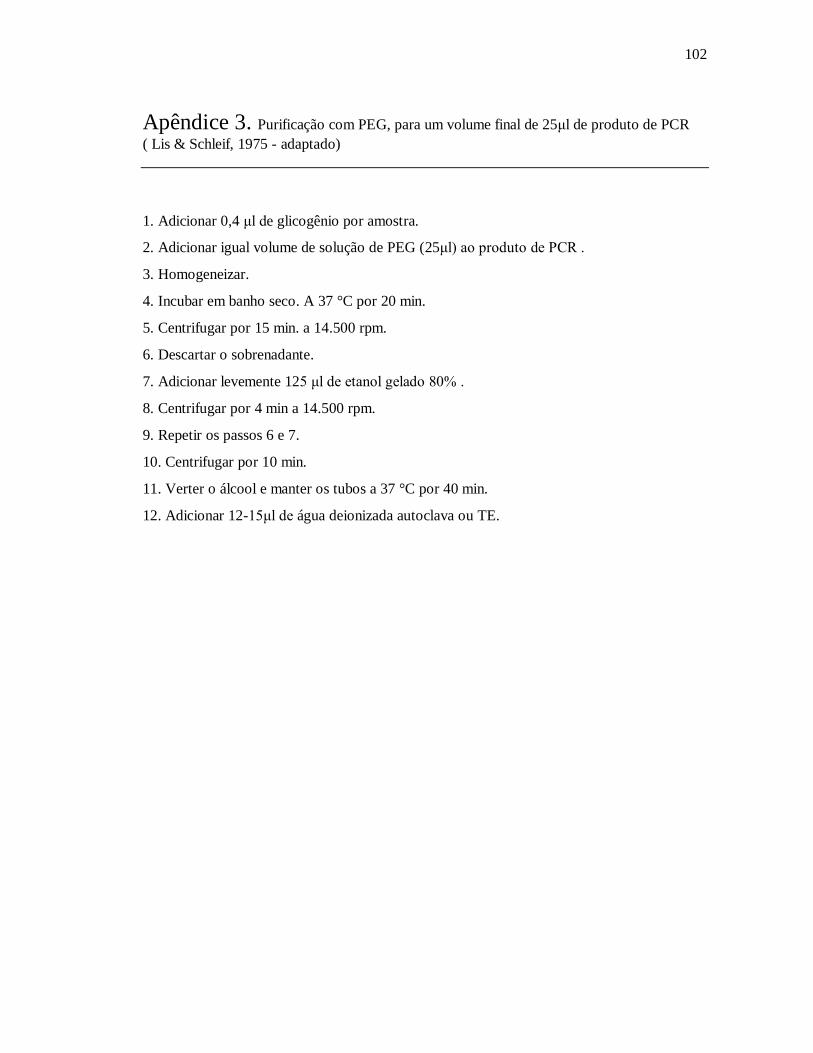
102
Apêndice 3. Purificação com PEG, para um volume final de 25μl de produto de PCR ( Lis & Schleif, 1975 - adaptado)
1. Adicionar 0,4 μl de glicogênio por amostra.
2. Adicionar igual volume de solução de PEG (25μl) ao produto de PCR .
3. Homogeneizar.
4. Incubar em banho seco. A 37 °C por 20 min.
5. Centrifugar por 15 min. a 14.500 rpm.
6. Descartar o sobrenadante.
7. Adicionar levemente 125 μl de etanol gelado 80% .
8. Centrifugar por 4 min a 14.500 rpm.
9. Repetir os passos 6 e 7.
10. Centrifugar por 10 min.
11. Verter o álcool e manter os tubos a 37 °C por 40 min.
12. Adicionar 12-15μl de água deionizada autoclava ou TE.
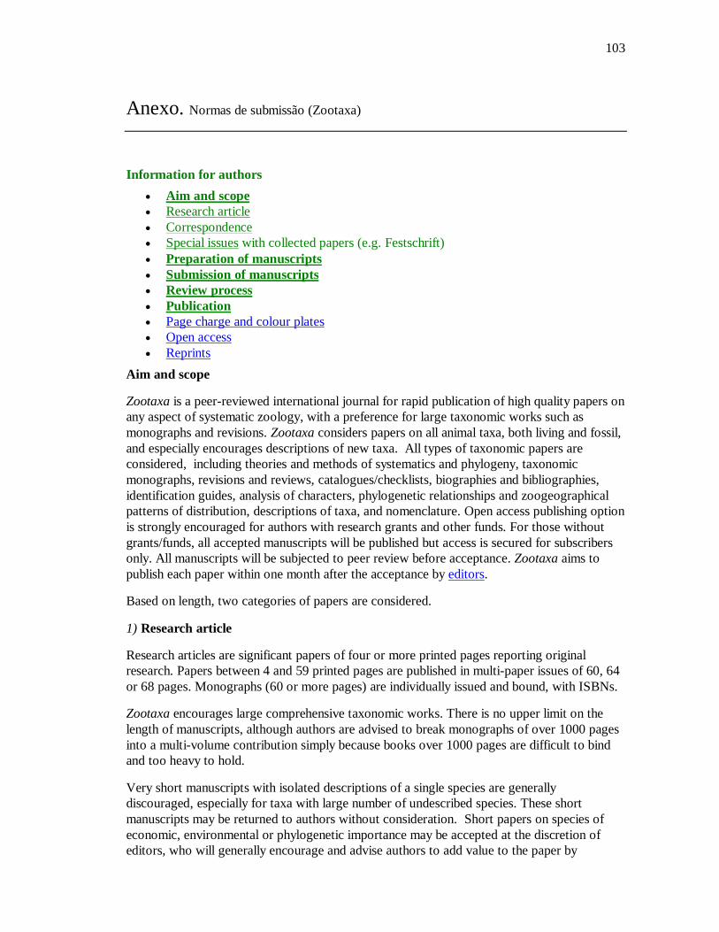
103
Anexo. Normas de submissão (Zootaxa)
Information for authors Aim and scope Research article Correspondence Special issues with collected papers (e.g. Festschrift) Preparation of manuscripts Submission of manuscripts Review process Publication Page charge and colour plates Open access Reprints
Aim and scope
Zootaxa is a peer-reviewed international journal for rapid publication of high quality papers on any aspect of systematic zoology, with a preference for large taxonomic works such as monographs and revisions. Zootaxa considers papers on all animal taxa, both living and fossil, and especially encourages descriptions of new taxa. All types of taxonomic papers are considered, including theories and methods of systematics and phylogeny, taxonomic monographs, revisions and reviews, catalogues/checklists, biographies and bibliographies, identification guides, analysis of characters, phylogenetic relationships and zoogeographical patterns of distribution, descriptions of taxa, and nomenclature. Open access publishing option is strongly encouraged for authors with research grants and other funds. For those without grants/funds, all accepted manuscripts will be published but access is secured for subscribers only. All manuscripts will be subjected to peer review before acceptance. Zootaxa aims to publish each paper within one month after the acceptance by editors.
Based on length, two categories of papers are considered.
1) Research article
Research articles are significant papers of four or more printed pages reporting original research. Papers between 4 and 59 printed pages are published in multi-paper issues of 60, 64 or 68 pages. Monographs (60 or more pages) are individually issued and bound, with ISBNs.
Zootaxa encourages large comprehensive taxonomic works. There is no upper limit on the length of manuscripts, although authors are advised to break monographs of over 1000 pages into a multi-volume contribution simply because books over 1000 pages are difficult to bind and too heavy to hold.
Very short manuscripts with isolated descriptions of a single species are generally discouraged, especially for taxa with large number of undescribed species. These short manuscripts may be returned to authors without consideration. Short papers on species of economic, environmental or phylogenetic importance may be accepted at the discretion of editors, who will generally encourage and advise authors to add value to the paper by
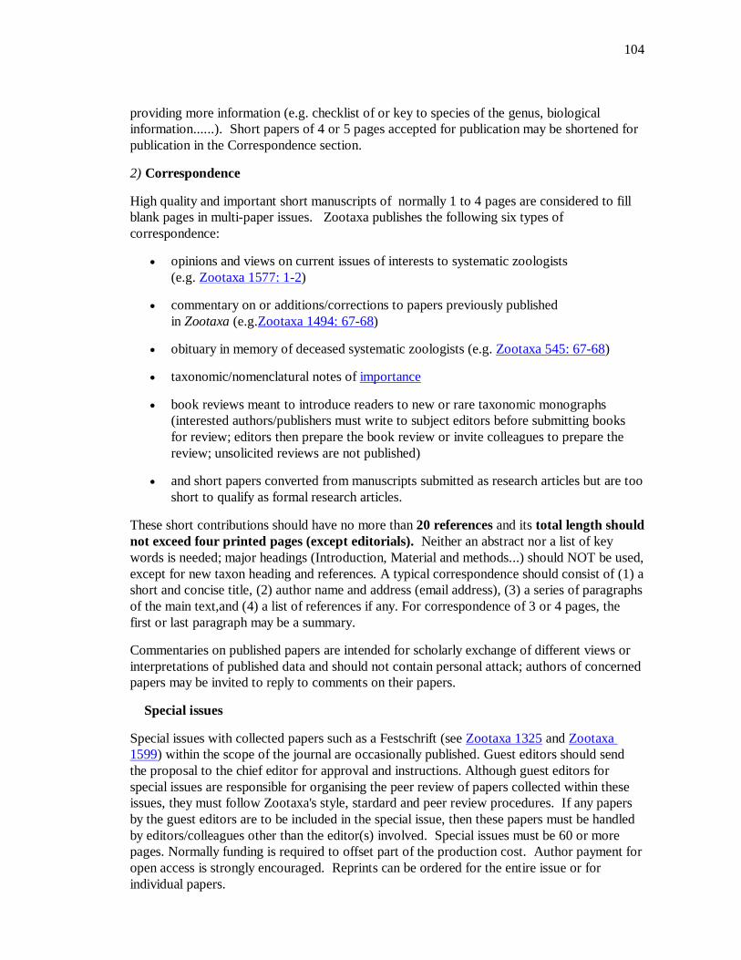
104
providing more information (e.g. checklist of or key to species of the genus, biological information......). Short papers of 4 or 5 pages accepted for publication may be shortened for publication in the Correspondence section.
2) Correspondence
High quality and important short manuscripts of normally 1 to 4 pages are considered to fill blank pages in multi-paper issues. Zootaxa publishes the following six types of correspondence:
opinions and views on current issues of interests to systematic zoologists (e.g. Zootaxa 1577: 1-2)
commentary on or additions/corrections to papers previously published in Zootaxa (e.g.Zootaxa 1494: 67-68)
obituary in memory of deceased systematic zoologists (e.g. Zootaxa 545: 67-68)
taxonomic/nomenclatural notes of importance
book reviews meant to introduce readers to new or rare taxonomic monographs (interested authors/publishers must write to subject editors before submitting books for review; editors then prepare the book review or invite colleagues to prepare the review; unsolicited reviews are not published)
and short papers converted from manuscripts submitted as research articles but are too short to qualify as formal research articles.
These short contributions should have no more than 20 references and its total length should not exceed four printed pages (except editorials). Neither an abstract nor a list of key words is needed; major headings (Introduction, Material and methods...) should NOT be used, except for new taxon heading and references. A typical correspondence should consist of (1) a short and concise title, (2) author name and address (email address), (3) a series of paragraphs of the main text,and (4) a list of references if any. For correspondence of 3 or 4 pages, the first or last paragraph may be a summary.
Commentaries on published papers are intended for scholarly exchange of different views or interpretations of published data and should not contain personal attack; authors of concerned papers may be invited to reply to comments on their papers.
Special issues
Special issues with collected papers such as a Festschrift (see Zootaxa 1325 and Zootaxa 1599) within the scope of the journal are occasionally published. Guest editors should send the proposal to the chief editor for approval and instructions. Although guest editors for special issues are responsible for organising the peer review of papers collected within these issues, they must follow Zootaxa's style, stardard and peer review procedures. If any papers by the guest editors are to be included in the special issue, then these papers must be handled by editors/colleagues other than the editor(s) involved. Special issues must be 60 or more pages. Normally funding is required to offset part of the production cost. Author payment for open access is strongly encouraged. Reprints can be ordered for the entire issue or for individual papers.
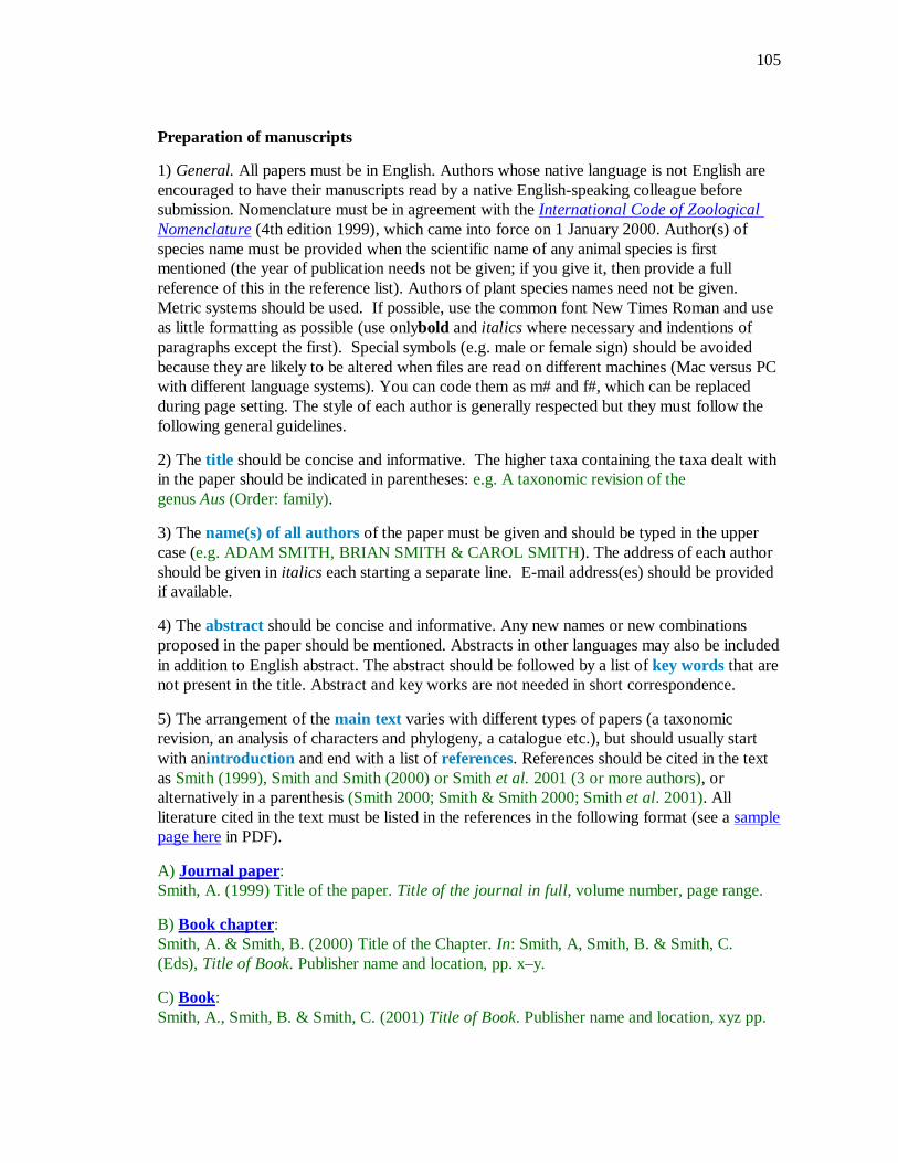
105
Preparation of manuscripts
1) General. All papers must be in English. Authors whose native language is not English are encouraged to have their manuscripts read by a native English-speaking colleague before submission. Nomenclature must be in agreement with the International Code of Zoological Nomenclature (4th edition 1999), which came into force on 1 January 2000. Author(s) of species name must be provided when the scientific name of any animal species is first mentioned (the year of publication needs not be given; if you give it, then provide a full reference of this in the reference list). Authors of plant species names need not be given. Metric systems should be used. If possible, use the common font New Times Roman and use as little formatting as possible (use onlybold and italics where necessary and indentions of paragraphs except the first). Special symbols (e.g. male or female sign) should be avoided because they are likely to be altered when files are read on different machines (Mac versus PC with different language systems). You can code them as m# and f#, which can be replaced during page setting. The style of each author is generally respected but they must follow the following general guidelines.
2) The title should be concise and informative. The higher taxa containing the taxa dealt with in the paper should be indicated in parentheses: e.g. A taxonomic revision of the genus Aus (Order: family).
3) The name(s) of all authors of the paper must be given and should be typed in the upper case (e.g. ADAM SMITH, BRIAN SMITH & CAROL SMITH). The address of each author should be given in italics each starting a separate line. E-mail address(es) should be provided if available.
4) The abstract should be concise and informative. Any new names or new combinations proposed in the paper should be mentioned. Abstracts in other languages may also be included in addition to English abstract. The abstract should be followed by a list of key words that are not present in the title. Abstract and key works are not needed in short correspondence.
5) The arrangement of the main text varies with different types of papers (a taxonomic revision, an analysis of characters and phylogeny, a catalogue etc.), but should usually start with anintroduction and end with a list of references. References should be cited in the text as Smith (1999), Smith and Smith (2000) or Smith et al. 2001 (3 or more authors), or alternatively in a parenthesis (Smith 2000; Smith & Smith 2000; Smith et al. 2001). All literature cited in the text must be listed in the references in the following format (see a sample page here in PDF).
A) Journal paper: Smith, A. (1999) Title of the paper. Title of the journal in full, volume number, page range.
B) Book chapter: Smith, A. & Smith, B. (2000) Title of the Chapter. In: Smith, A, Smith, B. & Smith, C. (Eds), Title of Book. Publisher name and location, pp. x–y.
C) Book: Smith, A., Smith, B. & Smith, C. (2001) Title of Book. Publisher name and location, xyz pp.

106
C) Internet resources Author (2002) Title of website, database or other resources, Publisher name and location (if indicated), number of pages (if known). Available from: http://xxx.xxx.xxx/ (Date of access).
Dissertations resulting from graduate studies and non-serial proceedings of conferences/symposia are to be treated as books and cited as such. Papers not cited must not be listed in the references.
Please note that (1) journal titles must be written in full (not abbreviated); (2) journal titles and volume numbers are followed by a ","; (3) page ranges are connected by "n dash", not hyphen "-", which is used to connect two words. For websites, it is important to include the last date when you see that site, as it can be moved or deleted from that address in the future.
On the use of dashes: (1) Hyphens are used to link words such as personal names, some prefixes and compound adjectives (the last of which vary depending on the style manual in use). (2) En-dash or en-rule (the length of an ‘n’) is used to link spans. In the context of our journal that means numerals mainly, most frequently sizes, dates and page numbers (e.g. 1977–1981; figs 5–7) and also geographic or name associations (Murray–Darling River; a Federal–State agreement). (3) Em-dash or em-rule (the length of an ‘m’) are used far more infrequently, and are used for breaks in the text or subject, often used much as we used parentheses. In contrast to parentheses an em-dash can be used alone; e.g. What could these results mean—that Niel had discovered the meaning of life? En-dashes and em-dashes should not be spaced.
6) Legends of illustrations should be listed after the list of references. Small illustrations should be grouped into plates. When preparing illustrations, authors should bear in mind that the journal has a matter size of 25 cm by 17 cm and is printed on A4 paper. For species illustration, line drawings are preferred, although good quality B&W or colour photographs are also acceptable. See a guide here for detailed information on preparing plates for publication.
7) Tables, if any, should be given at the end of the manuscript. Please use the table function in your word processor to build tables so that the cells, rows and columns can remain aligned when font size and width of the table are changed. Please do not use Tab key or space bar to type tables.
8) Keys are not easy to typeset. In a typical dichotomous key, each lead of a couplet should be typed simply as a paragraph as in the box below:
1 Seven setae present on tarsus I ; four setae present on tibia I; leg I longer than the body; legs black in color ... Genus A - Six setae present on tarsus I; three setae present on tibia I; leg I shorter than the body; legs brown in color ... 2 2 Leg II longer than leg I ... Genus B - Leg II shorter than leg I ... Genus C
Our typesetters can easily convert this to a proper format as in this PDF file.
Deposition of specimens Whenever possible, authors are advised to deposit type specimens in national or international

107
public museums or collections. Authors are also advised to request registration numbers of deposited material in advance of the acceptance of papers to avoid unnecessary delay of publication. Some countries (e.g. Australia) require that primary type specimens be deposited in collections of the country of origin; authors are advised to take this into consideration.
Submission Please follow the above basic guidelines and check if your manuscript has been prepared according to the style and format of the journal. Authors are encouraged to submit manuscripts by e-mail as attachments to the subject Editors responsible for your taxa or subject areas; manuscripts on small insect orders without subject editors should be submitted to Dr Ernest Bernard ([email protected]); manuscripts on other invertebrate taxa without subject editors should be submitted to the Chief editor.
Prior to submitting a manuscript and figures to an editor, please check our website if there are two or more editors per subject, and then contact one of these to announce your intention to submit a manuscript for review. Please indicate the size of the manuscript, the number of figures and the format of these files. Your editor can then respond with special instructions, especially for the submission of many image files. When you submit your manuscript to your editor, it will be more expedient to the review process if you offer the names of three or more potential reviewers with their complete postal and email addresses. It is also important to include the following statements in your cover letter:
1) All authors agree to its submission and the Corresponding author has been authorized by co-authors; 2) This Article has not been published before and is not concurrently being considered for publication elsewhere (including another editor at Zootaxa); 3) This Article does not violate any copyright or other personal proprietary right of any person or entity and it contains no abusive, defamatory, obscene or fraudulent statements, nor any other statements that are unlawful in any way.
Otherwise, your manuscript will not be processed. For manuscripts with numerous illustrations, which might be saved as separate TIFF or JPG files, for the purpose of review, it will be easier and more efficient for the subject editors and reviewers to have the figures converted into one larger PDF (Portable Document Format) file, instead of requiring the subject editor to save many files, cutting and copying these into a string of messages/files to the reviewers. You should retain the original figures in a higher resolution format for the final production of the accepted paper. For the text, PDF file along with RTF (Rich Text format) files are preferred. The advantage of submitting a rtf file for the text part of the manuscript is that the reviewers can emend the manuscript electronically. If you can not prepare PDF files, then submit text in RTF and the figures in TIFF (line drawing scanned at 600 dpi and half tone at 300 dpi; please use LZW compression, if you can, to reduce the size of e-files for easy transmission); if halftone TIFF files are too big (exceeding 2 MB), then submit them in jpeg. Seehere for detailed information on preparing plates for publication.
Vector files (charts, maps etc) are best submitted as EMF.
108
If you do not have access to e-mail, you can send three copies of the manuscript by post. Please double space your ms and leave ample margins for printed manuscripts.
Authors of accepted papers will be asked to submit an electronic version of the manuscript so that the publisher needs not to re-key or scan the ms. At this stage, the text part of the ms must be submitted as RTF or MS Word files and figures as TIFF files. Authors please be aware that line drawings must be scanned at 600 or 900 dpi as line art (=1 bit); they must NOT be scanned as 8 bit or full colour images. Please read details here.
In submitting the final version of revised manuscript to editors, authors are asked to provide the following information to all proper typesetting and indexing of the manuscript:
1) Corresponding author name and email 2) Author last name and running title (<40 characters; to be used in footer) 3) Number of plates and cited references 4) High taxon name (i.e. taxon section in Zootaxa website) and number of new taxa described in the paper
Authors need to complete and return an Assignment of Copyright form when paper is accepted for publication. Authors of institutions that do not allow transfer of copyrights to publishers (e.g. government institutions such as USDA, CSIRO) should attach a copyright waiver or similar documents.
Review process When a manuscript is received by the Editor, he/she will have it reviewed by at least two peers qualified to evaluate the manuscript and he/she normally asks the reviewers to complete the review in one month. However, the reviewing process will normally take longer, depending on the length of the manuscript and reviewer's responses.
Publication Once the manuscript is accepted by your subject editor, final files, produced according to Zootaxa requirement, will be forwarded by your subject editor to the chief editor, who will then link with author and the printer to ensure that the paper is published without unnecessary delay. Normally the proof will be sent to the author for checking 1 to 3 weeks after the final files are accepted. The paper will usually be published with two weeks (for larger papers it will take longer) once the corrections to the proof are received.
Page charge and colour plates. There is no page charge for publishing with Zootaxa. Publication of colour figures/photographs in online edition is also free of charge (print version in black and white). If colour plates in the print edition are desired, authors will be asked to contribute towards the full cost. Current rates: 300 USD for the first colour page; 200 USD for each additional colour page.
Open access. Zootaxa endorses the open access of taxonomic information and has published more open access taxonomic papers than any other journal. Authors who have funds to publish are strongly encouraged to pay a fee of 20 US$ per printed page to give free online access of their papers to all readers at this site or their own site. Open access papers are read by more people and are expected to have higher citation rates.

109
Reprints. Each author will be given a free e-reprint (PDF) for personal use (printing a copy for own use or exchange with other researchers, but not for deposition in a library/website/ftp-site for public access).
Printed copies of each paper/monograph in the form of the regular reprint can also be produced by the Publisher for purchase by authors at cost to authors, with a discount based on the number of copies ordered.