Campus de São José do Rio Preto Programa de Pós-Graduação em Genética
Márcia Cristina Duarte
Expressão de genes relacionados ao ciclo celular e proteção da mucosa gástrica em metaplasia intestinal e úlcera gástrica em
comparação com câncer gástrico.
Orientadora: Profa. Dra. Ana Elizabete Silva
Tese apresentada para obtenção do Título de Doutor em Genética.
São José do Rio Preto – SP 2009

Livros Grátis
http://www.livrosgratis.com.br
Milhares de livros grátis para download.
MARCIA CRISTINA DUARTE
Expressão de genes relacionados ao ciclo celular e proteção da mucosa gástrica em metaplasia intestinal e úlcera gástrica em comparação com câncer gástrico.
Tese apresentada para obtenção do título de Doutor em Genética, junto ao Programa de Pós-Graduação em Genética do Instituto de Biociências, Letras e Ciências Exatas da Universidade Estadual Paulista “Júlio de Mesquita Filho”, Campus de São José do Rio Preto.
BANCA EXAMINADORA
Profa. Dra. Ana Elizabete Silva Professor Adjunto Livre-Docente Orientadora
Profa. Dra. Cláudia Regina Bonini Domingos Professor Doutor UNESP – São José do Rio Preto
Profa. Dra. Dértia Villalba Freire Maia Professor Titular UNIFESP – Universidade Federal de São Paulo Profa. Dra. Eny Maria Goloni Bertollo
Professor Adjunto Faculdade de Medicina de São José do Rio Preto
Profa. Dra. Cláudia Aparecida Rainho Professor Doutor UNESP - Botucatu
São José do Rio Preto, 09 de outubro de 2009.

O presente trabalho foi realizado no Laboratório de Citogenética e Biologia
Molecular Humana e no Laboratório de Estudos Genômicos, Departamento de Biologia
do Instituto de Biociências, Letras e Ciências Exatas de São José do Rio Preto – SP, da
Universidade Estadual Paulista “Julio de Mesquita Filho” – UNESP e no Laboratório de
Patologia Cirúrgica e Molecular, Hospital Sírio-Libanês, São Paulo – SP. O projeto foi
desenvolvido com bolsa FAPESP, processo nº 05/51935-3, no período de dezembro de
2005 a setembro de 2006 e auxílio FAPESP, processo nº 07/58661-1.

Dedicatória
À minha mãe Ilza e ao Marlon
por tantas razões
e pela maior das emoções: o amor.

Agradecimentos
AGRADECIMENTOS
A realização deste trabalho contou com a colaboração de várias pessoas. A
todos, meus sinceros agradecimentos.
À minha orientadora, Profa. Dra. Ana Elizabete Silva, pelo investimento,
incentivo, orientação e amizade ao longo desses anos. Desde o primeiro momento da
acolhida deste projeto, sua sabedoria, dedicação e interesse me permitiram uma
aprendizagem constante e tornaram possível a realização deste trabalho. Por tão valiosa
seriedade profissional e amizade, a minha eterna gratidão.
À banca examinadora, Profa. Dra. Dértia Villalba Freire Maia, Profa. Dra.
Cláudia Aparecida Rainho, Profa. Dra. Eny Maria Goloni Bertollo e Profa Dra. Cláudia
Regina Bonini Domingos, por terem aceitado participar da análise e enriquecimento do
trabalho.
Ao Dr. Kenji Miyazaki e toda a equipe do Setor de Endoscopia do Hospital
Base, São José do Rio Preto, pela coleta das biópsias de estômago e pelos momentos de
aprendizagem e descontração.
Ao Dr. Aldenis Albaneze Borim por ter possibilitado o acesso à coleta das
amostras do Hospital de Base de São José do Rio Preto.
Às Dras. Kátia Ramos Moreira Leite e Mabel Tatty de Medeiros Fracassi,
Hospital Sírio Libanês, SP, pelo suporte nas análises imuno-histoquímicas.
À Profa. Dra. Paula Rahal, pelo acolhimento e disponibilização do laboratório
para realização da técnica de PCR em tempo real.
À Ms. Érica Babeto, pela sua disponibilidade em me ensinar à técnica de PCR
em tempo real. Agradeço também a Ms. Marina, que me auxiliou na retirada das placas
e finalização das reações.
Ao Prof. Dr. Sebastião Roberto Taboga pela ajuda na foto-documentação.
Ao Prof. Dr. José Antonio Manzato pela valiosa orientação na análise estatística.
Ao pessoal do Setor de Patologia do Hospital de Base pela ajuda na seleção dos
blocos e preparo das lâminas para imuno-histoquímica.
Ao Isaque Santana, pela amizade e disponibilidade em auxiliar com as reações
de imuno-histoquímica, e a todos os colegas do Laboratório de Patologia do Hospital
Sírio-Libanês, que dividiram comigo seu espaço e simpatia.
Ao técnico Josué Rodrigues dos Santos (agora aposentado!!) que me
acompanhou desde os primeiros passos, pela amizade construída e apoio oferecido.
A todos os pacientes que voluntariamente e desinteressadamente concordaram
em ceder amostras de seus tecidos e tornaram este trabalho possível.
Aos Professores do Programa de Pós-Graduação em Genética, pelos
ensinamentos e conhecimento compartilhado.
À Seção de Pós-Graduação e a todos os funcionários do IBILCE que de algum
modo contribuíram para a realização deste trabalho.
À direção do Instituto de Biociências, Letras e Ciências Exatas – IBILCE,
Universidade Estadual Paulista “Júlio de Mesquita Filho” – UNESP, e ao Programa de
Pós-Graduação em Genética, pela infra-estrutura de ensino e pesquisa e oportunidade de
concluir essa etapa tão importante de minha formação acadêmica.
Aos órgãos de fomento FAPESP, CNPq, CAPES e FUNDUNESP pelo apoio
financeiro.
Aos colegas do Laboratório de Citogenética e Biologia Molecular Humana,
Juliana, Yvana, Daniela, Isabella, Hector, Fernanda, Marilanda e Cidinha por tão
agradável convivência e conhecimentos compartilhados.
Aos colegas do Banco Nossa Caixa, pelos agradáveis momentos vividos durante
o trabalho e fora dele, nas festinhas da agência, e pelo apoio a mim conferido.
Aos meus amigos, pela amizade e pelos momentos de descontração, que
tornaram menos árduo este trabalho. Em especial para Ana Flávia, Nathália, Fabiana e
Joana.
À minha família que sempre me apoiou.
À minha mãe Ilza, por todo amor, dedicação e apoio a mim conferido e pela
grande amizade que construímos.
Ao Marlon que me acompanhou nas etapas finais deste trabalho, pelo amor,
carinho, paciência e principalmente, incentivo.
A Deus pelo mais sublime dos dons.... a vida!

Epígrafe

“A mente que se abre a uma nova idéia jamais volta ao seu tamanho
original.”
Albert Einstein
A Pedra
O distraído nela tropeçou... O bruto a usou como projétil. O empreendedor, usando-a, construiu. O camponês, cansado da lida, dela fez assento. Para meninos, foi brinquedo. Drummond a poetizou. Já, Davi, matou Golias, e Michelangelo extraiu-lhe a mais bela escultura... E em todos esses casos, a diferença não esteve na pedra, mas no homem! Não existe "pedra" no seu caminho que você não possa aproveitá-la para o seu próprio crescimento.
Autor desconhecido
“É preciso amar as pessoas como se não houvesse amanhã...”
Renato Russo

Lista de abreviaturas
Lista de abreviaturas de genes
AHR Aryl Hydrocarbon Receptor
APC Adenomatous Polyposis Coli
BCL-2 B-cell CLL/lymphoma 2
c-ERBB2 v-erb-b2 erythroblastic leukemia viral oncogene homolog 2
c-FOS FBJ murine osteosarcoma viral oncogene homolog
c-JUN Jun oncogene
CD44 CD44 molecule
CDKN1A Cyclin-Dependent Kinase Inhibitor 1A
CDKN1C Cyclin-Dependent Kinase Inhibitor 1C
CGRP Calcitonin-Related Polypeptide alpha
CLDN18 Claudin 18
COL4A1 Collagen, type IV, alpha 1
COX-1 Cyclooxygenase 1
COX-2 Cyclooxygenase 2
CTSB Cathepsin B
DCC Deleted in Colorectal Carcinoma
EGFR Epidermal Growth Factor Receptor
EGR-1 Early Growth Response 1
EPHB1 Ephrin Receptor EphB1
FAS Fas (TNF receptor superfamily, member 6)
GNK1 Gastrokine 1
HER2-neu c-erb B2/neu protein
HGF Hepatocyte Growth Factor
KRAS v-Ki-Ras2 Kirsten rat sarcoma viral oncogene homolog
MET Met proto-oncogene (hepatocyte growth factor receptor
MMP2 Matrix Metallopeptidase 2
MSH2 MutS homolog 2
MUC Mucin 1, cell surface associated
MYC v-myc myelocytomatosis viral oncogene homolog
NOS2 Nitric Oxide Synthase 2
OCT4 Ooctamer-binding transcription factor 4
P16 Cyclin-Dependent Kinase Inhibitor 2A
P27 Interferon, alpha-inducible protein 27
PDX1 Pancreatic and Duodenal Homeobox 1
PTEN Phosphatase and Tensin Homolog
RARβ Retinoic Acid Receptor, beta
SOX2 SRY (sex determining region Y)-box 2
SP-1 Sp1 transcription factor
SPK2 Protein Kinase 2
SRF Serum Response Factor
TERC Telomerase RNA component
TFF1 Trefoil Factor 1
TFF2 Trefoil Factor 2
TFF3 Trefoil Factor 3
TFIZ1/GNK2 Gastrokine 2
TGFβ Transforming Growth Factor, beta 1
TP53 Tumor Protein p53
TP73 Tumor Protein p73

VIM Vimentin
XAF1 XIAP Associated Factor 1

Sumário
Sumário
I. Introdução...................................................................................................................25 I.1. Carcinogênese do estômago...................................................................................25 I.2. Alterações genéticas e carcinogênese gástrica.......................................................30 I.3. Alterações da expressão gênica e carcinogênese do estômago..............................33
II. Objetivos....................................................................................................................46 III. Artigos……………………………………………………………………....…….48
Artigo I: Expression of TERT and COX-2 in precancerous gastric lesions compared to gastric cancer…………………………………………………………………………50
Artigo II: Expression of NOS2, HGF, MET, KRAS, TFF1 and CLDN18 in intestinal metaplasia and gastric cancer: a real-time PCR and immunohistochemical analysis……………………………………………………………………………….71 Artigo III: NOS2, HGF, MET, KRAS, TFF1 and CLDN18 expression in gastric ulcer and gastric cancer…………………………………………………………………...106
IV. Discussão…………………………………………………………………………138 V. Conclusões...............................................................................................................146 VI. Referências Bibliogáficas......................................................................................149 Apêndice.......................................................................................................................164
Apêndice 1: Questionário...........................................................................................164 Apêndice 2: Caracterização das amostras de paciente com metaplasia intestinal......165 Apêndice 3: Caracterização das amostras de pacientes com úlcera gástrica..............166 Apêndice 4: Caracterização das amostras de pacientes com câncer gástrico.............167 Apêndice 5: Material e Métodos................................................................................168
Anexo............................................................................................................................177
Anexo 1: Parecer consubstanciado do Comitê de Ética em Pesquisa Institucional...177 Anexo 2: Comprovante de submissão do artigo “Expression of TERT and COX-2 in precancerous gastric lesions compared to gastric cancer” ao periódico World Journal of Gastroenterology…………………………………………………………………178

Resumo
Resumo A carcinogênese gástrica apresenta um modelo de múltiplas etapas, que pode iniciar a partir de uma gastrite crônica, frequentemente associada à infecção pela bactéria Helicobacter pylori, e progredir para atrofia gástrica, metaplasia intestinal, displasia e câncer gástrico. Outra via, trata do surgimento do câncer gástrico a partir de um sítio de úlcera péptica benigna. Há relatos de algumas alterações genéticas bem estabelecidas nos estágios iniciais e avançados da carcinogênese gástrica, mas em lesões benignas precursoras como a metaplasia intestinal e a úlcera gástrica, relativamente pouco é conhecido. Deste modo, estudos genéticos destas lesões poderão fornecer informações importantes sobre os eventos iniciais da carcinogênese do estômago e contribuir para estratégias de diagnóstico precoce e prevenção. A partir de dados da literatura foram selecionados genes envolvidos com a carcinogênese do estômago como TERT, COX-2, NOS2, HGF, MET, KRAS, TFF1 e CLDN18, que atuam na manutenção dos telômeros, processos celulares e proteção da mucosa gástrica. Diante do exposto, este trabalho teve por objetivos avaliar mudanças de expressão gênica e protéica destes genes selecionados, em metaplasia intestinal (MI - 37 casos) e úlcera gástrica (UG - 30 casos), comparadas com suas respectivas mucosas normais (MN) e com adenocarcinoma gástrico (CG - 22 casos) e verificar possíveis correlações entre a expressão destes genes nos três grupos estudados, bem como associações entre os níveis de expressão gênica e protéica e fatores como infecção pela H. pylori e tipo histológico de MI e CG. A expressão relativa do RNAm dos referidos genes foi analisada pela técnica de PCR em tempo real, enquanto a expressão das respectivas proteínas foi avaliada por imuno-histoquímica. A avaliação da expressão gênica revelou níveis médios relativos do RNAm aumentados em CG comparado com MN para TERT (17,3 x), COX-2 (27,6 x), NOS2 (12,8 x), HGF (1,8 x), MET (3,5 x) e KRAS (1,7 x). Para TFF1 não foi observada variação significante no nível médio de RNAm (0,968) em comparação a MN, apesar de 68,2% dos casos de CG apresentarem expressão reduzida do RNAm. Enquanto a expressão de CLDN18 apresentou uma redução de 1,7 x, com nível médio de RNAm de 0,605, comparado com MN. A análise das proteínas mostrou positividade (moderada/forte) em 54,5%, 81,8%, 30% e 70% das amostras de CG para TERT, COX-2, NOS2 e KRAS, respectivamente, enquanto perda de expressão ou marcação fraca de TFF1 e CLDN18 foi observada em 100% e 90% dos casos de CG em comparação com MN. Não foram obtidos resultados satisfatórios da imuno-histoquímica para as proteínas HGF e MET nos grupos avaliados, após vários testes. No grupo de MI, os níveis de expressão do RNAm não apresentaram diferenças significantes aos do grupo de CG para TERT (2 x), NOS2 (13,6 x), HGF (2,6 x) e MET (2,8 x). Contudo, foi verificada uma diferença estatisticamente significante entre MI e CG para a expressão gênica de COX-2 (2,063 vs. 27,594, P=0,0290), TFF1 (1,842 vs. 0,968, P=0,0060) e CLDN18 (1,639 vs. 0,605, P=0,0065), enquanto a expressão de KRAS não diferiu significativamente da MN (1,146). A expressão das proteínas TERT, COX-2 e NOS2 foi positiva em 38,2%, 79,4% e 92,6% dos casos de MI, respectivamente, enquanto que para KRAS, TFF1 e CLDN18 a expressão mostrou-se fraca ou ausente em 100%, 62,9% e 100% dos casos, respectivamente. Os níveis de expressão do RNAm em UG foram significativamente aumentados para TERT (2,7 x), COX-2 (2,5 x), NOS2 (4,5 x), MET (2,8 x) e KRAS (2,6 x) e diminuídos para TFF1 (1,9 x, média=0,531) comparado com MN, enquanto HGF (1,051) e CLDN18 (0,934) não apresentaram variações significantes nos níveis médios do RNAm. Comparações entre UG e CG revelaram diferenças significantes apenas para os níveis médios de expressão de MET (P=0,0001), que foi mais expresso em CG. A análise protéica
revelou positividade (moderada/forte) em 34,8%, 17,4%, 61,5%, 38,5% e 100% das amostras de UG para TERT, COX-2, NOS2, KRAS e TFF1, respectivamente, enquanto que para CLDN18 a marcação foi fraca ou ausente em 100% dos casos. Comparado com CG, houve uma diferença estatisticamente significante apenas para as proteínas COX-2 (P=0,0030) e TFF1 (P=0,0001), sendo COX-2 imunomarcada principalmente em CG e TFF1 em UG. A análise de correlação mostrou associações entre os níveis de expressões gênicas de HGF, MET e KRAS no grupo de MI; NOS2, HGF e KRAS no grupo de UG e NOS2 e HGF no grupo de CG. Enquanto a análise de associação entre expressão gênica e protéica com fatores de risco e clinicopatológicos revelou apenas associação entre tabagismo e expressão reduzida do RNAm de TFF1 no grupo de MI, e entre infecção por H. pylori e níveis de expressão do RNAm reduzidos para NOS2 e HGF e aumentados para CLDN18 no grupo CG. Para as demais variáveis não foram verificadas associações com os níveis de expressão gênica e protéica. Considerando estes resultados, sugerimos que MI e UG compartilham alterações de expressão gênica em comum com CG, evidenciada principalmente nos níveis de expressão gênica e protéica de TERT, COX-2, NOS2, HGF, MET e CLDN18. A expressão gênica de KRAS não parece ser alterada em MI, mas em UG apresentou expressão elevada, que também foi observada para a proteína TFF1. Tais alterações em úlcera sugerem um papel de proteção e reparação da mucosa danificada, não podendo desconsiderar a hipótese de iniciação do CG a partir de UG, pois as mudanças de expressão desses genes também estão associadas com os mesmos processos envolvidos na progressão maligna. Portanto, alterações de expressão de genes com atuação em diferentes processos celulares podem contribuir para maior risco de câncer gástrico a partir de lesões precursoras como a metaplasia intestinal e úlcera gástrica. Palavras-chave: metaplasia intestinal, úlcera gástrica, câncer gástrico, H. pylori, expressão gênica, expressão protéica.

Abstract
Abstract
Gastric carcinogenesis presents a model of multiple steps, which can be triggered by a chronic gastritis, often associated with infection caused by the bacterium Helicobacter pylori, and progress to gastric atrophy, intestinal metaplasia, dysplasia and gastric cancer. However, another pathway has attracted interest in recent decades and refers to origin of gastric cancer from one site of benign peptic ulcer. There are reports of some well-established genetic alterations in the early stages and advanced gastric carcinogenesis, however, in precursor benign lesions as intestinal metaplasia and gastric ulcer, relatively little is known. Thus, genetic studies of these lesions may provide important information regarding the initial events of carcinogenesis of the stomach and contribute to strategies for early diagnosis and prevention. The genes selected for this study TERT, COX-2, NOS2, HGF, MET, KRAS, TFF1 and CLDN18, act, usually, in cell cycle processes, telomere maintenance and protection of the gastric mucosa. So, this study aimed to evaluate changes in gene and protein expression of these genes, altered in intestinal metaplasia (IM- 37 cases) and gastric ulcer (GU- 30 cases), compared with their corresponding normal mucosa (NM) and gastric cancer (GC - 22 cases) and to verify possible correlations between the expressions of these genes among the three groups studied, and also examine associations between gene and protein expression levels and factors such as H. pylori infection and histological type of IM and GC. The relative mRNA expression of these genes was analyzed by real time PCR, while the expression of respective proteins was assessed by immunohistochemistry. Evaluation of gene expression showed mRNA relative mean levels, increased in GC compared to NM to TERT (17.3-fold), COX-2 (27.6-fold), NOS2 (12.8-fold), HGF (1.8-fold), MET (3.5-fold) and KRAS (1.7-fold). For TFF1, there was no significant change in the mRNA mean level (0.968) compared to NM, despite the fact that 68.2% of GC cases had a low mRNA expression, whereas CLDN18 expression was 1.7-fold decreased, with mRNA mean level of 0.605, compared to NM. Protein analysis showed positivity (moderate or strong) in 54.5%, 81.8%, 30% and 70% of GC samples for TERT, COX-2, NOS2 and KRAS, respectively, while loss of expression or weak staining for TFF1 and CLDN18 was observed in 100% and 90% of GC compared to NM. No satisfactory results were obtained by immunohistochemistry for HGF and MET proteins after several tests. In the IM group, the gene expression levels were not significantly different from the GC group for TERT (2-fold), NOS2 (13.6-fold), HGF (2.6-fold) and MET (2.8-fold). However, there was a statistically significant difference between IM and GC for COX-2 (2.063 vs. 27.594, P = 0.0290), TFF1 (1.842 vs. 0.968, P = 0.0060) and CLDN18 (1.639 vs. 0.605, P = 0.0065) gene expressions, whereas the KRAS expression did not significantly differ of the NM (1.145). The protein expression of TERT, COX-2 and NOS2 was positive in 38.2%, 79.4% and 92.6% of IM cases, respectively, while KRAS, TFF1 and CLDN18 imunostaining was weak or absent in 100%, 62.9% and 100% of cases, respectively. The gene expression levels in GU were significantly increased for TERT (2.7-fold), COX-2 (2.5-fold), NOS2 (4.5-fold), MET (2.8-fold) and KRAS (2.6-fold) and decreased for TFF1 (1.9-fold) compared to NM, while HGF (1.051) and CLDN18 (0.934) showed no significant changes in the mRNA mean levels. Comparisons between GU and GC revealed significant differences only for the mean levels of MET expression (P = 0.0001) that was more expressed in GC. The protein analysis was positive (moderate or strong) in 34.8%, 17.4%, 61.5%, 38.5% and 100% of GU samples for TERT, COX-2, NOS2, KRAS and TFF1, respectively, while to CLDN18 weak or absent staining was
observed in 100% of the cases. Compared to GC, there was a statistically significant difference only for the proteins COX-2 (P = 0.0030) and TFF1 (P = 0.0001), with COX-2 and TFF1 immunostaining mainly in GC and GU, respectively. Correlation analysis showed associations between the gene expressions levels of HGF, MET and KRAS in the IM group, NOS2, HGF and KRAS in the GU group and NOS2 and HGF in the GC group. The association analysis between gene and protein expressions with clinicopathological and environmental factors showed only an association between smoking and reduced mRNA expression of TFF1 in the IM group, and between H. pylori infection and decreased mRNA expression levels to NOS2 and HGF and increased to CLDN18 in GC group. For the other variables, associations with gene and protein expression levels were not found. These results suggest that IM and GU share gene expression changes with GC, mainly observed in the gene and protein expression levels of TERT, COX-2, NOS2, HGF, MET and CLDN18. KRAS does not seem to be altered in IM, but in GU, it showed increased expression that also was observed for TFF1 protein. These alterations in GU, suggest a role in the protection and repair of damaged mucosa, however, it should be considered the hypothesis of initiation and progression of gastric cancer from GU, because the expression changes of these genes are also associated with the same processes involved in malignant progression. Therefore, imbalances in cell signaling pathways, where there is interaction of several genes, may contribute to increased risk of gastric cancer development from precursor lesions such as intestinal metaplasia and gastric ulcer.
Keywords: intestinal metaplasia, gastric ulcer, gastric cancer, H. pylori, gene expression, protein expression.

Introdução
25
I – Introdução
I.1- Carcinogênese do estômago
O câncer gástrico se destaca no modelo de múltiplas etapas da carcinogênese,
que pode iniciar a partir de uma gastrite crônica, geralmente acompanhada de infecção
pela bactéria Helicobacter pylori, e progredir para atrofia gástrica, metaplasia intestinal,
displasia e carcinoma (CORREA, 2004). Outra via da carcinogênese gástrica, também
induzida pela H. pylori, pode ser por meio da úlcera péptica, que constitui uma lesão
pré-cancerosa importante do estômago (TODD et al., 2004).
A metaplasia intestinal, mudança potencialmente reversível de um tipo
de célula diferenciada para outro, é frequentemente encontrada no trato gastrintestinal,
principalmente no estômago e esôfago, como resultado da infecção por H. pylori,
refluxo biliar crônico, ou induzida experimentalmente por irradiação e agentes
mutagênicos. No estômago, é caracterizada pela substituição da mucosa gástrica por
epitélio semelhante ao do intestino delgado, geralmente iniciada pela irritação
persistente da mucosa provocada pela infecção da H. pylori. Esta lesão pode aumentar a
suscetibilidade a carcinogênese gástrica em até 10 vezes, via seqüência metaplasia-
displasia-carcinoma, sendo considerada uma lesão pré-neoplásica (FILIPE et al., 1994).
A metaplasia intestinal pode ser classificada em tipo completo e incompleto. O
tipo completo ou tipo I é caracterizado pela presença de células absortivas, células de
Paneth, com grânulos eosinofílicos no seu citoplasma, encontradas na base das
glândulas, células secretoras de sialomucinas e células neuroendócrinas, que
corresponde ao fenótipo do intestino delgado. O tipo incompleto compreende os tipos II
e III e é caracterizado por células colunares e células secretoras de sialomucinas e/ou
sulfomucinas (LEUNG; SUNG, 2002; GUTIÉRREZ-GONZÁLEZ; WRIGHT, 2008). A
26
metaplasia intestinal tipo I está associada com um risco menor de câncer gástrico,
enquanto o tipo III, também denominado colônico, apresenta uma associação mais forte,
pois indivíduos com este tipo apresentam um risco quatro vezes maior de desenvolver
câncer que aqueles com o tipo I (FILIPE et al., 1994).
A úlcera péptica é uma doença heterogênea que afeta cerca de 10% da população
(CARVALHO, 2000), devido um defeito na parede gastrintestinal envolvendo toda a
espessura da mucosa e penetrando através da parede muscular. É resultante de necrose
do tecido, originada por isquemia da mucosa, formação de radicais livres e cessação de
liberação de nutrientes, todos causados por dano vascular ou microvascular como
trombos, constrição e outras oclusões (TARNAWSKI, 2005). Histologicamente, a
úlcera consiste de duas estruturas principais: uma margem distinta formada por mucosa
não-necrótica adjacente (componente epitelial) e o tecido de granulação na base da
úlcera (componente de tecido conectivo). Este consiste de fibroblastos, macrófagos e
células endoteliais proliferativas formando microvasos (CRAWFORD, 2005).
Na maioria dos países, a úlcera duodenal é a forma mais comum de úlcera
péptica. Nos Estados Unidos, 8 a 10 % da população sofrem de úlcera duodenal e 1 %
de úlcera gástrica (SZABO et al., 2007). Pacientes com úlcera gástrica com redução no
nível de ácido gástrico e pangastrite podem ter um risco de 1,8 vezes maior de
desenvolver câncer do estômago que indivíduos com úlcera duodenal com níveis
elevados de ácido gástrico e gastrite, predominantemente na região antral (HANSSON
et al., 1996). É importante salientar que a ressecção gástrica como tratamento da úlcera
não diminui este risco (SAFATLE-RIBEIRO; RIBEIRO; REYNOLDS, 1998). Vários
fatores ambientais foram relacionados com sua gênese, como o consumo de álcool e
cigarro, ácido acetilsalicílico, outros anti-inflamatórios não-esteróides e
corticoesteróides, além de fatores de fundo emocional (TAM, 1987; SZABO et al.,
27
2007). Porém, o principal fator ambiental envolvido na etiologia desta doença é a
infecção pela H. pylori, que afeta cerca de 90% dos pacientes com úlcera duodenal e 70
a 90% daqueles com úlcera gástrica (TYTGAT et al., 1993).
O estudo de lesões pré-cancerosas como a metaplasia intestinal e a úlcera
gástrica podem fornecer informações importantes a respeito das fases iniciais da
carcinogênese do estômago, assim contribuindo para estratégias de prevenção e
diminuição de sua incidência. Mesmo com o declínio no número de casos novos, o
câncer gástrico encontra-se como a quarta causa mais comum, e a segunda causa de
óbitos por câncer mundialmente. De modo geral, é cerca de duas e três vezes mais
freqüente em países em desenvolvimento, com as maiores taxas de incidência em países
asiáticos como Coréia, Japão e China, além da Europa Oriental e parte da América
Latina (SMITH et al., 2006; NITTI et al., 2008; INCA, 2009). No Brasil, segundo
estimativas do Instituto Nacional de Câncer (INCA), ele ocupa o sexto lugar em
incidência com a previsão de 22 mil casos novos (INCA, 2009).
Aproximadamente 90% dos cânceres gástricos são adenocarcinomas, que podem
ser classificados em duas entidades histomorfológicas denominadas intestinal e difuso
(LAURÉN, 1965). O tipo intestinal é caracterizado por tumores bem diferenciados, que
formam glândulas, frequentemente associados com gastrite atrófica e metaplasia
intestinal pré-existentes, sendo comuns na porção distal do estômago, e ocorrem em
pacientes mais velhos e em regiões geográficas de alto risco (JOHNSON; EVERS,
2008). O tipo intestinal de câncer gástrico afeta mais os homens em idade avançada do
que as mulheres na razão de 2:1. O tipo difuso ou indiferenciado não apresenta lesões
precursoras e pode levar a metástase precoce, apresentando um curso clínico mais
agressivo (SMITH et al., 2006; JOHNSON; EVERS, 2008). Ele compreende células
que perderam a coesão e que não são mais capazes de exercer a função gástrica,
28
apresentando aspecto típico de células em anel de sinete, afetando grandes porções do
estômago. Esta neoplasia surge em indivíduos mais jovens, afetando homens e mulheres
na mesma proporção e apresenta um forte componente genético (SMITH et al., 2006;
VAUHKONEN; VAUHKONEN; SIPPONEN, 2006; JOHNSON; EVERS, 2008).
A carcinogênese do estômago apresenta etiologia complexa, na qual fatores
ambientais e genéticos estão envolvidos. Dentre os fatores ambientais destaca-se a dieta
(alimentos salgados, conservados e defumados, peixes e carnes secas e carboidratos
refinados), os hábitos tabagista e etilista, acloridria, além da infecção por patógenos
como a bactéria Helicobacter pylori (SMITH et al., 2006; ROCCO; NARDONE, 2007;
LA TORRE et al., 2009). O consumo concomitante de álcool e tabaco está associado
com aumento do risco de câncer gástrico em 2,9 % na população japonesa (YAMAJI et
al., 2009), enquanto o consumo de fibras, frutas e vegetais frescos, antioxidantes como
o ácido ascórbico e a vitamina E, carotenóides e selênio na dieta parecem exercer um
papel importante na prevenção desta doença (KELLEY; DUGGAN, 2003; ROCCO;
NARDONE, 2007).
A bactéria Helicobacter pylori, considerada pela Agência Internacional de
Pesquisa em Câncer, como causa definitiva de câncer gástrico (IARC, 1994), aumenta o
risco para esta neoplasia em até nove vezes (KUIPERS, 1999). Esta bactéria possui
vários fatores que permitem colonizar o ambiente inóspito do estômago e evadir as
defesas do hospedeiro, incluindo a resposta imune. A enzima urease produzida pela
bactéria hidrolisa a uréia gástrica em amônia e dióxido de carbono, assim mantendo um
pH neutro, mesmo na presença de altas concentrações de H+ externo (SMITH et al.,
2006; AMIEVA; EL-OMAR, 2008).
A infecção por esta bactéria causa gastrite crônica em praticamente todos os
indivíduos colonizados e pode levar, posteriormente, à perda de glândulas gástricas. A
29
gastrite atrófica resultante e a metaplasia intestinal aumentam o risco de displasia e
câncer gástrico (KUIPERS; SIERSEMA, 2004). Vários mecanismos foram propostos
para a carcinogênese associada à H. pylori, dentre os quais estão a desregulação do ciclo
celular, formação de adutos de DNA, geração de radicais livres e compostos N-nitrosos,
alterações na secreção de fatores de crescimento e citocinas e diminuição das secreções
gástricas (HEAVEY; ROWLAND, 2004). Os diferentes tipos de cepa e fatores de
virulência da bactéria, além de fatores genéticos do hospedeiro, também foram
indicados como responsáveis pelo desenvolvimento de neoplasia gástrica (CORREA,
2004; AMIEVA; EL-OMAR, 2008).
O fator de virulência melhor caracterizado é a ilha de patogenicidade cag (cag-
PAI). Ela contém vários genes que codificam citotoxinas que são injetadas nas células
epiteliais gástricas e são fosforiladas, induzindo a secreção de IL-8, um potente fator
quimiotático e ativador de neutrófilos, produzindo várias transformações fisiológicas,
incluindo a diminuição do pH do estômago. Outros fatores de virulência incluem os dos
genes vacA, que codifica uma citotoxina vacuolizante que induz a formação de
vacúolos nas células e que pode levar à apoptose, além de estimular a formação de
poros transmembrana que permeabilizam o epitélio gástrico à uréia. Os fatores babA
que codifica a proteína BabA que promove uma maior adesão entre a bactéria e a célula
gástrica; iceA que é induzido pelo contato com o epitélio e, em algumas populações,
está associado com úlcera péptica; e oipA que parece ser importante para a liberação de
IL-8 mesmo em cepas cag-negativas, sendo relacionado com gravidade da gastrite,
úlcera péptica e metaplasia intestinal. (DE LUCA; IAQUINTO, 2004; GILLEN;
McCOLL, 2005; PRINZ; SCHWENDY; VOLAND, 2006; SMITH et al., 2006;
AMIEVA; EL-OMAR, 2008).
30
I.2. Alterações genéticas e carcinogênese gástrica.
Alguns modelos de alterações genéticas têm sido propostos acompanhando a
progressão do adenocarcinoma gástrico, inclusive com o envolvimento de vias
moleculares distintas entre os tipos intestinal e difuso (TAHARA, 2004). Hasegawa et
al. (2002) descreveram a expressão aumentada de genes envolvidos na transdução de
sinais, coagulação sanguínea (PROCR, SERPINGI e HRG) e transcrição gênica (NFL3,
LHX1 e HOXB7), enquanto genes que contribuem para o metabolismo de energia,
barreira epitelial (TFF1 e TFF2) e absorção apresentaram uma expressão diminuída.
Instabilidade genética e hiperplasia de células tronco positivas para TERT (transcriptase
reversa da telomerase) precedem erros de replicação no lócus DS191, hipermetilação no
lócus D17S5, perda de pS2, RARβ e RUNX3, transcritos anormais de CD44 e mutação
de TP53, todos os quais acumulam em 30 % das metaplasias intestinais e são eventos
comuns nos cânceres tipo intestinal. Cerca de 20 % dos adenomas com mutações no
gene APC progridem para carcinoma e durante esta progressão observam-se mutações
ou perda de heterozigosidade (LOH) de TP53, expressão reduzida de p73, perda de
RUNX3, superexpressão de ciclina-E e transcrição anormal de MET. O carcinoma
gástrico tipo intestinal resultante exibe frequentemente perda de DCC e da proteína p27,
expressão reduzida do receptor TGFβ e amplificação do gene c-ERBB2. A via envolvida
na formação de câncer gástrico tipo difuso envolve LOH no cromossomo 17p,
expressão anormal de p73, mutação ou LOH em TP53 e p16, desacetilação de histonas,
expressão de TERT, expressão aumentada de TGFβ, transcritos anormais de CD44,
perda de RUNX3 e mutação ou perda de E-caderina, entre outros (STOCK; OTTO,
2005; SMITH et al., 2006; JOHNSON; EVERS, 2008).
Estudos moleculares sobre expressão gênica em tumores gástricos têm destacado
uma expressão variável em genes relacionados com o ciclo celular, apoptose, reparo do
31
DNA, metástase e manutenção dos telômeros. Por exemplo, há relatos de expressão
aumentada dos genes ciclina E1, AXIN2 (CHEN et al., 2003), TERT (HU et al., 2004),
COX-2 (ZHANG et al., 2005), NOS2 (AUGUSTO et al., 2008), HER2-neu (GRAVALOS;
JIMENO, 2008), OCT4 (CHEN et al., 2009) e AHR (PENG et al., 2009) e uma baixa
expressão dos genes MUC, TTF1, CDKN1C (CHEN et al., 2003) e XAF1 (TU et al., 2009).
Da mesma forma também tem sido relatado aumento de expressão de algumas proteínas
como SPK2 (MA et al., 2005), TERT (GULMANN et al., 2005), NOS2 (WANG et al.,
2005), EGFR (KIM et al., 2008), COX-2 (CHEN et al., 2006; YAMAC et al., 2008) e
survivina (SONG et al., 2009), assim como redução de outras como E-caderina, β-catenina
(CHAN et al., 2003), p27, PTEN (MA et al., 2005), c-ERBB-2 (SATEROGLU-TUFAN;
BIR; CALLI-DEMISKAR, 2006), EPHB1 (WANG et al., 2007), GNK1, TFIZ1/GNK2
(MOSS et al., 2008) entre outras.
A mudança fenotípica presente na metaplasia intestinal ocorre como resultado da
combinação de expressão alterada de fatores genéticos, silenciamento epigenético,
fatores de transcrição, vias de sinalização e fatores de crescimento (GUTIÉRREZ-
GONZÁLEZ; WRIGHT, 2008). Os eventos genéticos envolvidos no desenvolvimento
da metaplasia intestinal são pouco compreendidos, mas destaca-se a expressão do gene
homeobox CDX2, cuja proteína é mais expressa em metaplasia intestinal do estômago e
no esôfago de Barrett (KUIPERS; SIERSEMA, 2004; GUTIÉRREZ-GONZÁLEZ;
WRIGHT, 2008), os genes HOX e ParaHOX envolvidos no desenvolvimento do tubo
disgestório, a família de fatores trefoil, principalmente TFF3 expresso no intestino
normal, SHH e IHH, a família de genes SOX e POU, relacionadas com a embriogênese
do trato gastrintestinal, além dos genes OCT-1 e RUNX3 (TSUKAMOTO et al., 2004;
GUTIÉRREZ-GONZÁLEZ; WRIGHT, 2008).
32
Estudos prévios em metaplasia intestinal também relataram outras alterações
genéticas como LOH do gene de reparo hMSH2 e dos supressores de tumor APC e TP53
(KIM et al., 2001), expressão aumentada de TERT com ativação de telomerase (TAHARA,
2004; GULMANN et al., 2005) e mutações no oncogene KRAS (TAHARA, 2004).
Também há relatos de expressões aumentadas das proteínas Ciclina D2 (YU et al., 2001),
p53 (CÉSAR et al., 2004), BCL-2 (ANAGNOSTOPOULOS et al., 2005), SPK2 (MA et
al., 2005), COX-2 (SUN et al., 2006), CDX2 (TSUKAMOTO et al., 2004; KIM et al.,
2006) e AHR (PENG et al., 2009) e expressão diminuída para as proteínas RUNX3
(OSAKI et al., 2004), p27, PTEN (MA et al., 2005) e BAX (ANAGNOSTOPOULOS et
al., 2005).
Meireles et al. (2004), avaliando arrays de c-DNA verificaram que o perfil de
expressão de metaplasia intestinal era mais semelhante com o tecido tumoral, em que
níveis elevados de MMP2, COL4A1, FNH1, CTSB, COLIA2 e VIM eram expressos em
maior quantidade no tecido tumoral que na metaplasia intestinal, enquanto amostras de
gastrite eram mais semelhantes com tecido normal, ocorrendo a expressão aumentada do
gene MYC e baixa expressão de CDKN1A, como característica do grupo.
Em úlcera gástrica, os estudos genéticos são mais escassos e têm mostrado a
participação de vários fatores de crescimento e dos fatores de transcrição c-FOS, c-JUN,
EGR-1, SP-1 e SRF, dentre outros (TARNAWSKI, 2005). Uma maior expressão da
proteína CGRP (TANI et al., 1999), TFF1 (REN et al., 2005; SHI; CAI; YANG, 2006) e
TFF2 (SHI; CAI; YANG, 2006) e dos genes HGF (HORI et al., 2000), TGF-β e TGF-βR2
(SHIH et al., 2005) e COX-2 (GUO et al., 2006) foi verificada na mucosa de pacientes com
úlcera gástrica em cicatrização.
Alguns estudos desenvolvidos em nosso laboratório nestes tipos de lesões pré-
cancerosas, pela técnica FISH, detectaram trissomias dos cromossomos 7, 8 e 9 em 71%
33
dos casos de metaplasia intestinal em associação com a infecção pela H. pylori, assim
como deleção do gene TP53 e expressão aumentada da proteína em 60% e 12% dos casos,
respectivamente (CÉSAR et. al., 2004). Em úlcera gástrica foram detectadas
principalmente trissomias dos cromossomos 7 e 17 frequentemente nos casos H. pylori
positivos, mas não foram observadas deleções do gene TP53, apesar de apenas 12% dos
casos expressarem imunorreatividade positiva para a proteína p53 (CÉSAR et. al., 2006).
Estes achados evidenciaram a ocorrência de instabilidade cromossômica em lesões
gástricas consideradas ainda benignas associadas com a infecção pela H. pylori, que
participam do processo de múltiplas etapas da carcinogênese gástrica.
I.3 – Alterações da expressão gênica e carcinogênese do estômago
Alterações genéticas e epigenéticas que alteram os padrões de expressão de
genes que participam da regulação do ciclo celular, reparo do DNA e manutenção dos
telômeros têm sido estudadas em uma variedade de neoplasias (TAHARA, 2004, CHEN
et. al., 2006; SMITH et. al., 2006; NITTI et al., 2008; JOHNSON; EVERS, 2008).
Esses estudos estão sendo incrementados nas últimas décadas e possibilitado a
caracterização de tumores em nível genômico, transcriptômico e proteômico. Várias
mudanças genômicas observadas em diferentes tumores têm sido relacionadas com o
início do processo carcinogênico, enquanto alterações dinâmicas na expressão gênica
em nível de RNAm e proteínas podem determinar a progressão da doença
(PAGLIARULO et al., 2004; NITTI et al., 2008; JOHNSON; EVERS, 2008).
No processo de carcinogênese do estômago diversos genes têm sido descritos
com expressão desregulada como TERT, HGF, MET, COX-2, NOS2, KRAS, TFF1 e
CLDN18, os quais desempenham importantes funções na manutenção dos telômeros,
processos celulares, como proliferação e apoptose, cascatas de sinalização, integridade
34
da mucosa gástrica dentre outras (HU et al., 2004; INOUE et al., 2004; SHI; CAI;
YANG, 2006; SANADA et al., 2006; AUGUSTO et al., 2007).
O gene TERT (Telomerase Reverse Transcriptase), mapeado em 5p15.33
(MEYERSON et al., 1997) codifica uma holoenzima, na qual duas subunidades são
essenciais para o desempenho de sua função no processo de manutenção dos telômeros:
a subunidade catalítica da enzima telomerase, TERT, que confere sua atividade de
transcriptase reversa, adicionando seqüências repetidas as extremidades teloméricas
(LINGNER et al. 1997); e o componente RNA, TERC, que consiste de uma seqüência
complementar à repetição telomérica, atuando como molde para a extensão dos
telômeros (FENG et al. 1995).
Os telômeros são essenciais para a manutenção da integridade do genoma, assim
garantindo a estabilidade cromossômica. Essas estruturas compostas de repetições
curtas (5’-TTAGGG-3’) ligam-se a proteínas específicas e são replicadas a cada ciclo
celular pela transcriptase reversa da telomerase (BELGIOVONE; CHIODI,
MONDELLO, 2008). Em seres humanos, os telômeros são encurtados a cada ciclo
celular, limitando o potencial proliferativo da célula, e levando a um estágio de
senescência replicativa (CAMPISI; D’ADDA DI FAGAGNA, 2007). A expressão do
gene TERT está intimamente associada com a atividade da telomerase, com função na
adição de repetições nos telômeros para compensar a perda de seqüências durante a
replicação do DNA, participando da imortalização celular pela estabilização da estrutura
cromossômica (BLACKBURN, 1992).
A atividade da telomerase é mantida nas células germinativas, mas nos tecidos
somáticos normais exibem níveis de atividade muito baixos ou ausentes. De modo
contrário é detectada em cerca de 90% das amostras de câncer humano e está associada
com a transformação maligna (SHAY; WRITE, 2005). Alguns estudos têm relatado
35
expressão aumentada de TERT em adenocarcinoma gástrico e lesões pré-cancerosas,
como a metaplasia intestinal, comparado com tecido gástrico normal (HU et al., 2004,
GULMANN et al., 2005). A expressão aumentada da proteína telomerase também foi
observada em tumores de estroma gastrintestinal (SABAH et al., 2004), carcinoma de
células escamosas de esôfago (HSU et al., 2005), carcinoma de ovário (BRUSTMANN,
2005), câncer de pulmão de células não-pequenas (MAVROGIANNOU et al., 2007),
carcinoma oral (CHEN et al., 2007) e astrocitoma pediátrico (WONG; MA;
HAWKINS, 2009), e também no tecido e no plasma de pacientes com câncer colorretal
(TERRIN et al., 2008). A inativação funcional de TERT com inibidores da telomerase
(MITTAL et al., 2004; SUN et al., 2007), e mais recentemente, a vacinação baseada na
telomerase em células cancerosas (MENNUNI et al., 2008; CHEN; LI; TOLLESFBOl,
2009) consiste em um alvo atrativo para novas estratégias terapêuticas. Vários estudos
têm confirmado que a perda de sua atividade resulta no encurtamento progressivo dos
telômeros, levando a parada de crescimento e/ou morte celular por apoptose (revisão em
CHEN; LI; TOLLESFBOl, 2009).
O gene HGF mapeado em 7q21.1 (FUKUYAMA et al., 1991), responsável pelo
fator de crescimento do hepatócito, é um fator multifuncional de origem mesenquimal,
que atua como mitógeno, morfógeno, motógeno e fator angiogênico, dependendo da
célula alvo e do contexto celular (MATSUMOTO; NAKAMURA, 1996). HGF também
exerce um papel importante no processo de reparo nos rins, mucosa gástrica, pulmão,
fígado, tecido miocárdico e retina (CONWAY et al., 2007). Em uma grande variedade
de células tumorais, HGF estimula a proliferação, dissociação, migração e invasão,
além de ser um potente fator angiogênico (JIANG et al., 1999). O receptor de HGF é
codificado pelo proto-oncogene MET (7q31) e consiste de uma glicoproteína
transmembrana com atividade de tirosina quinase (PARK et al., 1987). Após auto-
36
fosforilação dos resíduos de tirosina quando ligada à proteína HGF inicia-se uma
cascata de transdução de sinal (CHRISTENSEN; BURROWS; SALGIA, 2005;
CIPRIANI et al., 2009). O receptor MET é expresso no epitélio normal da maioria dos
tecidos, onde é primariamente localizado nas junções intercelulares junto com
moléculas de adesão celular como as E-caderinas (CONWAY et al., 2007).
Considerando a grande diversidade de funções biológicas, existe uma variedade
de mecanismos pelos quais HGF e MET influenciam a tumorigênese. Por exemplo,
tanto HGF quanto MET aparecem com expressão aumentada em tecidos neoplásicos em
comparação com o tecido normal adjacente e a intensidade da expressão está
relacionada à gravidade e prognóstico da doença. Outros mecanismos incluem
mutações, amplificação de MET, mecanismos epigenéticos, dentre outros (CIPRIANI et
al., 2009). A expressão aumentada de HGF e MET foram relatadas na carcinogênese
gástrica (KONTUREK et al., 2001; INOUE et al., 2004), assim como em outras
neoplasias como meningiomas em associação com o índice de proliferação celular e
recorrência (MARTÍNEZ-RUMAYOR et al., 2004), tumores de mama (PARR et al.,
2004; LINDEMANN et al., 2007), em carcinoma de hipofaringe correlacionado com
metástases de linfonodos (KIM et al., 2006), câncer de esôfago (ANDERSON et al.,
2006), nos estágios iniciais da carcinogênese pancreática (YU et al., 2006) e
rabdomiossarcoma (CHEN et al., 2007). Ainda há relatos em câncer de tiróide papilar,
no qual também se revelou um potencial alvo terapêutico (SIRAJ et al., 2007), câncer
de pulmão (NAKAMURA et al., 2007) e câncer de bexiga (MIYATA et al., 2009).
O aumento de expressão de ambos os genes também foi observado durante o
processo de cicatrização de úlceras gástricas (HORI et al., 2000), em úlceras crônicas de
pele (NAYERI et al., 2005; CONWAY et al., 2007) e em esofagite erosiva (LUO et al.,
2008). Transcritos de MET no sangue periférico foram detectados em 61,5% dos
37
pacientes com câncer gástrico (UEN et al., 2006) mostrando ser uma ferramenta
promissora na detecção de células tumorais circulantes micro-metastáticas. Devido a
sua participação em diversas vias metabólicas e interação com vários receptores de
membrana como integrinas, plexinas, CD44, FAS e outros, o receptor MET tem sido
indicado como alvo terapêutico para dificultar o processo tumorigênico e metastático
(CORSO; COMOGLIO; GIORDANO, 2005; TOSCHI; JÄNNE, 2008). Da mesma
forma, a terapia molecular tendo MET como alvo terapêutico tem mostrado resultados
eficazes em testes pré-clínicos para o tratamento de câncer de pulmão (CIPRIANI et al.,
2009).
As cicloxigenases (COX) são enzimas com atividades de oxidase e peroxidase
que catalisam a conversão de ácido aracdônico a prostaglandinas. Dentre suas funções
em vários processos biológicos, encontra-se a integridade da mucosa gastrintestinal
(STACK, 2001). Existem duas isoformas de COX, uma codificada pelo gene COX-1
(9q32-q33.3) e outra pelo gene COX-2 (1q25.2-q25.3). O gene COX-1 é expresso
constitutivamente em condições basais na maioria das células, participando da
regulação homeostática de vários órgãos. Entretanto, os níveis de COX-2 são baixos ou
indetectáveis em tecidos normais, mas altos em estado inflamatório (MILLER, 2006).
Durante a inflamação e estímulo mitogênico, é observado um aumento de 10 a 20 vezes
na expressão de COX-2 e produção de prostaglandinas (BROOKS et al., 1999), que
aumentam a infiltração de células inflamatórias, exudação, inchaço e dor nos tecidos
afetados.
A expressão de COX-2 em tumores humanos pode ser induzida por vários
fatores de crescimento, citocinas, oncogenes, dentre outros. Em células transformadas, a
produção de prostaglandinas via COX-2 aumenta a proliferação e angiogênese, inibe a
apoptose e permite que escapem da vigilância do sistema imune (TSATSANIS et al.,
38
2006). Alguns estudos têm mostrado uma expressão elevada de COX-2 em tumores
como próstata (UOTILA et al., 2001), esôfago (MÖBUS et al., 2005), tireóide (LO et
al., 2005), mama (McCARTHY et al., 2006; LUCCI et al., 2008), oral (PANDEY et al.,
2008), estômago (YAMAC et al., 2008), com associação positiva com infecção pela H.
pylori (ZHANG et al., 2005), e glândula salivar, em que mostrou associação também
com a expressão de HGF (AOKI et al., 2006). Além do mais, as prostaglandinas
derivadas de COX-2 são importantes para a cicatrização de úlceras gástricas e níveis
elevados de expressão podem ser determinados nas bordas e base de úlceras gástricas
em cicatrização e em tecidos com gastrite relacionada à H. pylori (JACKSON et al.,
2000; GUO et al., 2003).
O gene NOS (Óxido Nítrico Sintase), mapeado em 17q11.2-q12 (MARSDEN et
al., 1994) é membro da superfamília de monoxigenases que incluem a bem
caracterizada família do citocromo P450 (CYP) e apresenta três isoformas: NOS
neuronal (nNOS), NOS endotelial (eNOS) e NOS induzível (iNOS ou NOS2). As duas
primeiras são constitutivamente expressas e dependentes de Ca+2, atuando como
neurotransmissor nos tecidos neurais e regulando a pressão sanguínea nos tecidos
endoteliais, respectivamente. NOS2 é independente de Ca+2 e é induzido em vários tipos
de células por lipopolissacarídeos (LPS), endotoxinas, citoquinas inflamatórias tais
como interleucina-1 (IL-1), fator de necrose tumoral alfa (TNF-α), interferon gama
(IFN-γ), hipóxia e outros estímulos (WANG et al., 2005; KEKLIKOGLU et al., 2008).
A enzima NOS é responsável pela conversão de L-arginina para L-citrulina para a
produção de óxido nítrico (NO). Devido a não dependência de níveis de cálcio locais,
NOS2 continua a produzir NO durante horas ou mesmo dias após sua indução, diferente
das duas formas constitutivas que produzem NO de maneira transiente (BRENNAN et
al., 2002). Vários estudos têm mostrado que as três isoformas podem ser detectadas em
39
vários tipos de células cancerosas e podem estar envolvidas na promoção ou inibição da
patologia e fisiologia do câncer (DONCKIER et al., 2006).
O óxido nítrico desempenha várias funções biológicas, destacando seu papel
como molécula sinalizadora nos mecanismos de defesa imunológica e carcinogênese
(LECHNER; LIRK; RIEDER, 2005). Durante o processo carcinogênico, NO está
relacionado à transformação de células normais pela ativação de oncogenes e perda de
atividade de supressores de tumor; crescimento de células transformadas
(desdiferenciação, proliferação, progressão do tumor de lesões pré-neoplásicas);
angiogênese; invasão local e metástase. Como um radical livre, NO pode reagir para
produzir peroxidonitritos que podem causar dano direto ou indireto no DNA e, sua
produção por longo período de tempo pode levar a mutações e finalmente ao câncer
(LIRK; HOFFMANN; RIEDER, 2002; NOMELINE et al., 2008). De modo contrário, a
produção de NO também pode exercer um papel anti-tumorigênico, pelo qual o
endotélio ativado pode causar lise de células tumorais. Alguns estudos ainda sugerem
que NO é citotóxico e citostático contra células malignas e microorganismos, causando
apoptose (NOMELINE, et al., 2008).
Estudos sobre a expressão gênica de NOS2 em tecidos tumorais têm encontrado
resultados contraditórios (KEKLIKOGLU et al., 2008). A expressão aumentada de
NOS2 foi observada em câncer de mama (VAKKALA et al., 2000), pulmão
(MARROGI et al., 2000), próstata (UOTILA et al., 2001), bexiga (HAYASHI et al.,
2001), displasia oral (BRENNAN et al., 2002), linfoma de células-B (ATIK et al. 2006),
câncer colorretal (YU et al., 2006), tireóide (DONCKIER et al., 2006), fibrose
submucosa oral (REJENDRAN; VARKEY, 2007), câncer de ovário (NOMELINI, et
al., 2008), tumores de cabeça e pescoço (BRENNAN et al., 2008) e câncer gástrico
(WANG et al., 2005; CHEN et al., 2006). Neste último caso, os autores observaram
40
interação com COX-2 e uma correlação com invasão, metástase nos linfonodos,
infecção com H. pylori e invasão vascular. Estudo recente na população brasileira,
mostrou associação entre expressão aumentada de NOS2 em câncer gástrico e gastrite
crônica em comparação com a mucosa normal e úlcera gástrica (AUGUSTO, et al.,
2007). De modo contrário, estudos prévios evidenciaram uma expressão diminuída da
proteína NOS2 em tumores gástricos e de cólon, assim levantando a hipótese de uma
relação entre perda de NO e carcinogênese (RAJNAKOVA et al., 1997; AMBS et al.,
1998).
A família Ras de proteínas G monoméricas, denominadas HRAS, NRAS e
KRAS atua como “interruptores moleculares” ligando sinais extracelulares, por meio de
receptores de membrana, a sinais intracelulares. Estas proteínas alternam de um estado
inativo, quando ligada ao GDP, a um estado ativo, quando ligada ao GTP, em resposta a
ativação de vários receptores (PATRA, 2008). As proteínas RAS regulam diversas vias
celulares que são importantes para o crescimento e dispersão de células malignas,
incluindo proliferação celular, regulação do ciclo celular, sobrevida da célula,
angiogênese e migração celular. Sob a influencia de EGF e EGFR estimula a
proliferação celular pela via de sinalização RAS para promover o reparo de tecidos e
cicatrização (FRIDAY; ADJEI, 2005).
Várias mutações de ponto no gene KRAS mapeado em 12p12.1 (POPESCU et
al., 1985) foram identificadas e resultam em ativação constitutiva do gene em uma
grande variedade de tumores como cólon (SAMOWITZ et al., 2000), pâncreas (WANG
et al., 2002), tireóide, pulmão (AVIEL-RONEN et al., 2006) e uma forma rara de câncer
de pâncreas, o carcinossarcoma pancreático (NAKANO et al., 2008). Recentemente,
Watari et al. (2007) correlacionaram a presença de mutações no gene KRAS com
infecção pela bactéria H. pylori em indivíduos com metaplasia intestinal e gastrite
41
crônica, mostrando que alterações neste gene estão presentes nos estágios iniciais da
carcinogênese gástrica e que a erradicação da bactéria diminui a ocorrência das
mutações.
Na ausência de mutações ativadoras, KRAS pode ter sua função oncogênica
ativada por amplificação e expressão aumentada do gene (GALIANA et al., 1995; VON
LINTIG et al., 2000; QIAN et al., 2005). Em câncer gástrico a expressão aumentada da
proteína KRAS foi correlacionada com metástases nos linfonodos e prognóstico
reservado (LI et al., 2006). Devido a sua importância na regulação do ciclo celular,
sobrevida, angiogênese e migração celular, KRAS tem sido um dos alvos atrativos para
estudos de terapia molecular do câncer (FRIDAY; ADJEI, 2005; PATRA, 2008).
A família dos genes TFF (Trefoil Factor) humanos é composta por TFF1, TFF2
e TFF3 que são expressos predominantemente no trato gastrintestinal e interagem com
mucinas para estabilizar a barreira de muco e proteger a mucosa contra danos. Também
contribui para o reparo da mucosa, promovendo a migração de células epiteliais e
reconstituição após dano (SANDS; PODOLSKY, 1996; KORNPRAT et al., 2005). O
papel destes fatores no desenvolvimento e progressão tumoral tem sido investigado em
vários estudos, que sugeriram funções como fatores de dispersão, agentes pró-invasivos,
anti-apoptóticos e angiogênicos (ABDOU; AIAD; SULTAN, 2008). O gene TFF1,
localizado em 21q22.3 (MOISAN et al., 1985) e também conhecido como pS2, atua
como um supressor de tumor específico da mucosa gastrintestinal, prevenindo a entrada
na fase S do ciclo celular, pelo aumento da expressão da proteína pRb e subseqüente
diminuição da atividade de E2F (TOMASETTO; RIO, 2005).
A proteína TFF1 também atua como um fator anti-apoptótico, diminuindo a
atividade das caspases-3, 6, 8 e 9 e na proteção da mucosa gástrica, onde interage com a
mucina gástrica solúvel MUC5AC (LEFEBVRE et al., 1996; RUCHAUD-
42
SPARAGANO; WESTLEY; MAY, 2004). A inativação do gene TFF1 está associada
com a ocorrência de displasia, adenoma e adenocarcinoma de estômago em ratos
(LEFEBVRE et al., 1996). Em humanos, a expressão da proteína TFF1 é perdida em
40-60% dos tumores gástricos, enquanto os tecidos normais adjacentes permanecem
positivos (MACHADO et al., 2000; LEUNG et al., 2002; SHI; CAI; YANG, 2006). Os
mecanismos envolvidos com a ausência de expressão de TFF1 em câncer gástrico
incluem perda alélica em 28-50% dos casos, metilação do promotor e mutações de
ponto que promovem perda da atividade supressora de tumor e ganho de invasividade
(YIO et al., 2006). De forma semelhante, os níveis de RNAm de TFF1 diminuem
consideravelmente da mucosa gástrica normal para os adenocarcinomas gástricos
(BECKLER et al. 2003). Em lesões pré-cancerosas, como a metaplasia intestinal, a
expressão da proteína é menor que no tecido normal, diminuindo consideravelmente do
tipo completo para incompleto (KIM et al., 2004), o mesmo ocorrendo para gastrite
superficial crônica, gastrite atrófica e úlcera gástrica (SHI; CAI; YANG, 2006).
Saitoh et al. (2000) encontraram níveis aumentados do RNAm de TFF1 em
úlceras gástricas em cicatrização comparados com as úlceras ativas, sugerindo que este
gene pode estar relacionado a proteção e diferenciação celular das áreas não-ulceradas
da mucosa gástrica. De modo contrário, o gene TFF1 pode ter atividade oncogênica,
atuando como fator de crescimento, pois é altamente expresso em tecidos malignos que
normalmente não o expressam, incluindo os tumores de bexiga (KORNPRAT et al.,
2005), mama, pâncreas, intestino grosso, pulmão, esôfago, ovário e metástases
associadas, e câncer de próstata (TOMASETTO; RIO, 2005; VESTERGAARD et al.
2006; ABDOU; AIAD; SULTAN, 2008).
As claudinas fazem parte de uma família grande de proteínas compreendendo 24
membros que apresentam quatro domínios transmembrana e são expressas nas células
43
epiteliais de vários tecidos. Estas proteínas interagem entre si formando uma rede que
ajuda a criar as junções tight das células epiteliais, assim exercendo um papel crítico na
polaridade celular (KARANJAWALA et al., 2008). As claudinas também estão
envolvidas em vários processos fisiológicos como a permeabilidade e condutância
paracelular (SANADA et al., 2006), atuando como uma barreira difusora para o
movimento de proteínas e lipídeos através da membrana celular e uma barreira primária
para o transporte paracelular de solutos através das células (SEMBA et al., 2008). Deste
modo, as quebras das junções tight, devido à expressão alterada das claudinas podem
resultar na liberação de fatores de crescimento, que pode fornecer o estímulo autócrino e
parácrino para a tumorigênise das células epiteliais (VERMEER et al., 2003).
Em humanos, as claudinas exibem padrões específicos de expressão no trato
gastrintestinal: na mucosa gástrica a expressão da claudina 18 (CLDN18) é
normalmente detectada e na mucosa intestinal a expressão das claudinas 3 e 4 é
obrigatoriamente observada. É interessante notar que no estágio de gastrite crônica
seguida de metaplasia intestinal, as características das claudinas gástricas transformam-
se naquelas do tipo intestinal, mostrando-se como bons biomarcadores para determinar
a diferenciação do epitélio gástrico (MATSUDA et al., 2007).
O gene CLDN18, mapeado em 3q22.3, foi primeiramente descrito como um alvo
dowstream ao fator de transcrição T/EBP/NKX e, em ratos, foram descritas duas
variantes: CLDN18a1 expressa no pulmão e CLDN18a2 expressa no estômago. Cada
isoforma apresenta uma variante de splicing alternativo faltando o domínio C-terminal
(CLDN18a1.2 e CLDN18a2.2) (NIIMI et al., 2001). A função biológica da claudina 18
é pouco compreendida, do mesmo modo que pouco se sabe sobre o papel das claudinas
na tumorigênise humana. Como a variante CLDN18a2 é expressa somente no estômago
normal e células de Paneth do duodeno, as disfunções das junções tight causada por
44
diminuição da expressão de CLDN18 pode levar a um influxo anormal de fatores de
crescimento relacionados ao estômago e células de Paneth. Alguns estudos têm
mostrado esta relação, associando a expressão reduzida de CLDN18 e câncer gástrico
(SANADA et al., 2006; SEMBA et al., 2008). Outros estudos têm mostrado o aumento
de expressão de CLDN18 durante a colite experimental e em pacientes com colite
ulcerativa (ZWIERS et al., 2008). A expressão aumentada da proteína CLDN18 foi
verificada em carcinoma bem diferenciado de pâncreas (KARANJAWALA et al., 2008)
e no epitélio colunar especializado do esôfago de Barrett, no qual contribui para a maior
resistência ácida deste tipo de lesão (JOVOV et al., 2007).
Considerando o exposto quanto a carcinogênese gástrica e a participação de
lesões benignas, e ainda a escassez de estudos genéticos nestas lesões, torna-se
necessário à busca de possíveis alterações genéticas, como mudanças na expressão
gênica, que possam ser indicadas como biomarcadores para o diagnóstico precoce do
câncer de estômago.

Objetivos
46
II - Objetivos
Devido ao importante papel das lesões pré-malignas no desenvolvimento do
câncer gástrico, há necessidade de estudos para avaliação de mudanças nos níveis de
expressão gênica e protéica de genes relevantes que participam de processos celulares
como proliferação e apoptose, proteção e reparo do epitélio gástrico, nas etapas iniciais
que possam desencadear o processo carcinogênico. Deste modo, este trabalho teve
como objetivos:
1. Avaliar os níveis de expressão dos genes TERT, COX-2, NOS2, HGF, MET, KRAS,
TFF1 e CLDN18 em metaplasia intestinal e úlcera gástrica, em comparação com a
respectiva mucosa normal e com o câncer gástrico;
2. Avaliar a expressão das proteínas TERT, COX-2, NOS2, HGF, MET, KRAS, TFF1 e
CLDN18 nestes mesmos grupos;
3. Verificar a ocorrência de interação entre os genes NOS-2, HGF, MET e KRAS,
através da correlação entre os níveis de RNA mensageiro nas amostras de metaplasia
intestinal, ulcera gástrica e câncer gástrico.
4. Investigar a ocorrência de associação de variáveis demográficas, fatores de risco e
clinicopatológicas, tais como sexo, idade, tabagismo, etilismo, infecção pela
Helicobacter pylori e tipo histológico com os níveis de expressão gênica e protéica de
TERT, COX-2, NOS2, HGF, MET, KRAS, TFF1 e CLDN18 em metaplasia intestinal,
úlcera gástrica e câncer gástrico.

Artigos
48
Os resultados referentes aos objetivos desta tese serão apresentados, a seguir, na forma
de três artigos científicos, conforme as normas de publicações específicas de cada
periódico.
Artigo 1
Título: Expression of TERT and COX-2 in precancerous gastric lesions compared to
gastric cancer.
Autores: Márcia Cristina Duarte, Érica Babeto, Kátia Ramos Moreira Leite, Kenji
Miyazaki, Aldenis Albanese Borim, Paula Rahal, Ana Elizabete Silva
Periódico: World Journal of Gastroenterology, submetido em 27/08/2009.
Artigo 2
Título: Expression of NOS2, HGF, MET, KRAS, TFF1 and CLDN18 in intestinal
metaplasia and gastric cancer: a real-time PCR and immunohistochemical analysis.
Autores: Márcia Cristina Duarte, Mabel Tatty de Medeiros Fracassi, Kenji Miyazaki,
Ana Elizabete Silva
Periódico: BMC Gastroenterology, artigo a ser submetido à publicação.
Artigo 3
Título: NOS2, HGF, MET, KRAS, TFF1 and CLDN18 expression in gastric ulcer and
gastric cancer.
Autores: Márcia Cristina Duarte, Mabel Tatty de Medeiros Fracassi, Kenji Miyazaki,
Ana Elizabete Silva.
Periódico: Journal of Gastroenterology and Hepatology, artigo a ser submetido à
publicação.

Artigo I
50
Artigo I: Expression of TERT and COX-2 in precancerous gastric lesions
compared to gastric cancer.
Running title: TERT and COX-2 expression in gastric lesions.
Authorship: Márcia Cristina Duarte, Érica Babeto, Kátia Ramos Moreira Leite,
Kenji Miyazaki, Aldenis Albanese Borim, Paula Rahal, Ana Elizabete Silva
Institution: Márcia Cristina Duarte, Érica Babeto, Paula Rahal, Ana Elizabete
Silva, UNESP - São Paulo State University, Department of Biology, São José do
Rio Preto, SP, Brazil. Kátia Ramos Moreira Leite, Laboratory of Surgery and
Molecular Pathology, Hospital Sírio-Libanês, São Paulo, SP, Brazil. Kenji
Miyazaki, Service of Endoscopy, Hospital de Base, São José do Rio Preto, SP,
Brazil. Aldenis Albanese Borim, FAMERP – Medical School, São José do Rio
Preto, SP, Brazil.
Author contributions: Duarte MC and Silva AE designed the research. Duarte
MC performed the research. Rahal P and Babeto E provided the technical support
and assistance for Real Time PCR assays. Miyazaki K and Borim AA were
responsible for the endoscopic exam and collection of the gastric biopsies. Leite KRM
performed immunohistochemical analysis. Duarte MC and Silva AE analyzed
the data and wrote the manuscript.
Supportive foundations: Sponsored by the São Paulo State Research
Foundation (FAPESP), Nº 2007/58661-1.
Correspondence to: Ana Elizabete Silva, PhD, Professor of Genetic, Biology
Department, UNESP- São Paulo State University, Cristóvão Colombo Street,
2265, São José do Rio Preto, 15054-000, SP, Brazil. [email protected]
Telephone: +55 17 3221-2384 Fax: +55 17 3221-2390
51
ABSTRACT
AIM: To investigate TERT and COX-2 mRNA and protein expression in
stomach precancerous lesions such as intestinal metaplasia (IM) and gastric
ulcer (UG) in comparison to gastric cancer.
MATERIALS AND METHODS: Real-time PCR were performed to detected
TERT and COX-2 mRNA expression in 35 biopsies of IM, 30 of GU and 22 of
gastric cancer (GC) and their respective normal mucosa. TERT and COX-2
proteins were detected with immunohistochemistry in 68 samples, 34 of IM, 23
of GU and 11 of GC.
RESULTS: The increased mRNA expression levels of TERT and COX-2 were
observed respectively in 45.7% and 40% of IM; 50% and 51.7% of GU and 78.9%
and 61.9% of GC. Considering all samples (up and down regulated) the relative
means levels of TERT and COX-2 mRNA after normalization with
housekeeping β-actin gene and comparison with respective adjacent normal
mucosa, in the groups of IM, GU and GC were respectively: 2.008±2.605,
2.730±4.120 and 17.271±33.852 for TERT, and 2.063±3.729, 2.496±4.132 and
27.594±58.952 for COX-2. The TERT and COX-2 protein immunostaining was
observed, respectively, in 38.2% and 79.4% of IM, 39.1% and 26.1% of GU, and
54.5% and 81.8% of GC. This study showed for the first time an overexpression
of TERT mRNA and protein in GU. There were no significant differences
between the groups regarding TERT mRNA and protein expression, whereas
for COX-2 there were significant differences in mRNA (P=0.0291 for IM vs. GC)
and protein (P=0.0001 for IM vs. GU; P=0.003 for GU vs. GC). No association
between TERT and COX-2 mRNA and protein expression with H. pylori
infection and other clinicopathological variables was found.
CONCLUSION: This study suggests that TERT and COX-2 may be deregulated
expression in intestinal metaplasia and gastric ulcer and play a role in the early
events of gastric carcinogenesis.
Key words: intestinal metaplasia, gastric ulcer, gastric cancer, TERT, COX-2,
gene expression, protein expression
52
INTRODUCTION
Gastric precancerous lesions such as intestinal metaplasia have been associated
with the multistep process of well-differentiated gastric or intestinal-type
adenocarcinoma that develops from active gastritis, frequently associated with
Helicobacter pylori infection, to gastric atrophy, intestinal metaplasia, dysplasia,
and finally to gastric cancer[1]. Intestinal metaplasia is characterized as the
transformation of the gastric epithelium and glands from secretory to
absorptive cells, which closely resemble the mature intestinal epithelium[2]. It is
a well-established pre-malignant condition of the stomach and can produce a
10-fold increase in the risk of this neoplasia[3]. Another pathway of gastric
carcinogenesis includes peptic ulcer, which increases the gastric cancer risk 1.8
times[4]. Genetic studies in precancerous gastric lesions are still limited, so a
better understanding of the mechanisms involved in gene expression in the
premalignant steps, which lead to the development of cancer, is necessary.
Among the genes with changed gene expression in gastric carcinogenesis, can
be detached TERT and COX2, which can also be overexpressed in premalignant
lesions, so participating of early progression of disease.
The TERT (human telomerase reverse transcriptase) gene encodes the
catalytic subunit of telomerase, which elongates the telomere ends using the
RNA subunit hTERC as a template[5]. The stabilization of the telomere size is a
prerequisite for malignant cells to erase the senescence checkpoint and acquire
the capacity to proliferate unlimitedly. So, telomerase reactivation is an
obligatory event in carcinogenesis and, in fact, increased telomerase activity or
TERT mRNA expression has been detectable in up to 90% of human cancers[6]
including gastric cancer[7, 8].
COX-2 (Cyclooxygenase-2) is the rate-limiting enzyme for prostaglandin
synthesis[9], and can be induced by proinflammatory cytokines, growth factors,
mitogens and oncoproteins. During inflammation and mitogenic stimulation, a
10 to 20-fold increase in COX-2 mRNA expression and overproduction of
prostaglandins has been observed, which enhance inflammatory cells
infiltration, exudation, swelling and pain in the wound tissues[10]. COX-2 is
53
involved in the regulation of a broad range of cellular processes, including
angiogenesis, apoptosis and cell proliferation. Recent studies have
demonstrated overexpression of COX-2, which plays a role in carcinogenesis
and tumor progression in many epithelial tumors including colon[11], lung[12],
breast[13], oral[14], esophagus[9], and stomach[15].
Thus, the aim of this study was to investigate changes in the levels of
expression of TERT and COX-2 mRNA and protein in intestinal metaplasia and
gastric ulcer in comparison to normal mucosa and gastric cancer, and to
investigate their potential relationship with H. pylori infection and other
clinicopathological variables. The findings may indicate changed expression of
these genes in precursor lesions, which may confer increased risk of gastric
cancer.
MATERIAL AND METHODS
Samples
A total of 87 specimens were evaluated, obtained from 30 patients with gastric
ulcer (GU) (mean age 54.90+12.37 years), 35 patients with intestinal metaplasia
(IM) (mean age 61.05+ 12.45 years), all were gastric cancer free, and 22 patients
with gastric adenocarcinoma (GC) (mean age 62.45±14.07 years). For each
patient were collected three biopsies from lesion area and other three biopsies
from their normal gastric mucosa adjacent to lesion, during endoscopic
evaluation at the period between March 2006 and March 2008, at the Hospital
de Base (São José do Rio Preto, SP, Brazil). The biopsies were collected mainly
from the antrum and corpus regions of the stomach. Immediately after, they
were stored in RNA later reagent (Ambion) at –20ºC until RNA extraction. All
specimens were histopathologically diagnosed. Archival paraffin-embedded,
formalin-fixed tissues were used for immunohistochemical staining of the TERT
and COX-2 proteins. Information about age, gender, smoking and drinking
status and the written informed consent was obtained from all patients, and the
study was approved by the National Research Ethics Committee (CONEP).
54
RNA extraction and reverse transcription
Total cellular RNA was extracted using the Trizol reagent (Invitrogen)
according to the manufacturer’s protocol. RNA concentration was determined
in a NanoDrop® ND1000 spectrophotometer, and its integrity was visualized on
1% agarose gel. RNA samples were stored in freezers at -80ºC and used for
reverse transcription. cDNA was synthesized from 5 μg of total RNA using
random primers and a High Capacity cDNA Archive Kit (Applied Biosystems),
according to the manufacturer’s instructions. The integrity of all cDNA
preparations was tested by PCR assay of the β-actin gene and visualized on 2%
agarose gel.
Quantitative Real Time PCR
The expression of TERT and COX-2 mRNA was measured by real time PCR,
based on the SYBR Green methodology, using an ABI Prism® 7300 (Applied
Biosystems). The primer sequences used in the study were as follows: for TERT,
5’ CGGAAGAGTGTCTGGAGCAA 3’ and 5’ GGATGAAGCGGAGTCTGGA 3’;
and for COX-2, 5’ CATCCTGAATGGGGTGATG 3’ and 5’
GATAGCCACTCAAGTGTTGCAC 3’. The real time PCR assays were
performed in 10 μL of SYBRTM Green Master Mix (Applied Biosystems), 25 ng
of cDNA and 0.9 μM of TERT primers or 0.8 μM of COX-2 primers. Thermal
cycling conditions for TERT and COX-2 were: 2 min at 50ºC and 10 min at 95ºC
for initial denaturation, followed by 40 cycles at 95°C for 15 s and 60°C for 1
min, and a dissociation step at 95ºC for 15 s, 60ºC for 30 s and 95ºC for 15 s.
Triplicates of the samples were assayed in each run. The relative expressions of
TERT and COX-2 were analyzed according to the description by Livak and
Schmittgen[16] and normalized with the housekeeping β-actin gene and
corresponding normal gastric mucosa.
Immunohistochemistry
The immunohischemical analysis was performed in 34 samples with IM, 23
with UG and 11 with GC. Tissue sections of 4 μm were cut from paraffin-
embedded tissue blocks and mounted on glass slides pretreated with 3-
aminopropyl-triethoxysilane/acetone solution and dried overnight at 60ºC.
55
After deparaffinization and rehydration, antigen retrieval was performed in 10
mM citrate buffer (pH 6.0) for 15 min at 120ºC, followed by treatment with 3%
H2O2 for 20 minutes to block the endogenous peroxidase. Then, the sections
were incubated for 1 hour at room temperature with specific antibodies: COX-2
mouse monoclonal antibody (clone 4H12, Novocastra, 1:100) or TERT mouse
monoclonal antibody (clone 2D8, ABR - Affinity BioReagents, 1:100). After
rinsing with Tris-HCl buffer (pH 7.6), the slides were incubated with
biotinylated secondary antibody and incubated with streptavidin-biotin
peroxidase, following the manufacturer’s instructions (Histostain Bulk Kit,
Zymed). The immunostain was visualized with 3,3’-diaminobenzidine
tetrahydrochoride (DAB) and counterstained with Mayer’s hematoxylin.
Negative controls were established by replacing the primary antibody with
buffer solution. Colon carcinoma and amygdale were used as positive controls
for COX-2 and TERT antibodies, respectively. A single pathologist examined all
specimens. All analyses were done under a light microscope (x400
magnification), and the whole tissue extension of all samples was examined.
Immunostain for proteins COX-2 (brown cytoplasm staining) and TERT (brown
nuclear staining) was graded by staining intensity as negative (-) (absent brown
staining) or positive: +1 - weakly stained, +2 - moderately stained, and +3 -
strongly stained, as observed in at least 10% of cells.
Statistical Analysis
mRNA relative expression levels were described using the mean as a point
estimator and the range of values. Non-parametric Mann-Whitney U test were
used for comparisons between the groups and mRNA expression level and
clinicopathological variables. Fisher’s exact test was used to evaluate the
protein levels between the groups and the relationship between protein levels
and clinicopathological variables. All statistical tests were performed using the
GraphPad Instat Software. The level of significance was set at P < 0.05.
56
RESULTS
TERT and COX-2 mRNA relative expression
Real time PCR analysis was performed for all 87 samples and their
corresponding adjacent normal gastric mucosa and the results of mRNA
expression are summarized in Table I. In the GU and GC groups, no TERT
expression was detected in 8/30 (26.7%) and 3/22 (13.6%) samples,
respectively, while only one sample did not express COX-2 in both groups.
After normalization with housekeeping β-actin gene and comparison with
respective adjacent normal mucosa, the relative expression of TERT mRNA in
the groups was increased in 16 (45.7%) out of 35 IM samples, in 11 (50%) out of
22 GU specimens, and in 15 (78.9%) out of 19 GC samples. To COX-2 the
relative expression of mRNA in the IM, GU and GC groups, was increased in 14
(40%) out of 35, in 15 (51.7%) out of 29, and in 13 (61.9%) out of 21 samples,
respectively. Considering all samples (up and down regulated), the mean
expression levels of TERT and COX-2 mRNA in the groups of IM, GU and GC
were respectively: 2.008±2.605, 2.730±4.120 and 17.271±33.852 for TERT, and
2.063±3.729, 2.496±4.132 and 27.594±58.952 for COX-2. But when we considering
only the cases up-regulated the mean expression levels of TERT and COX-2
mRNA in the groups of IM, GU and GC were respectively: 3.833±2.954,
5.304±4.852 and 21.821±36.987 for TERT and 4.908±5.030, 4.579±5.099 and
44.356±70.663 for COX-2. No significant differences were found between the
groups for TERT mRNA expression, while for COX-2 there was a significant
difference between IM and GC (P=0.0164) and GU and GC (P=0.0066). The
combined analyses of the two genes in each group revealed that 11/35 (31.4%)
of cases of IM, 7/22 (31.8%) of GU, and 10/19 (52.6%) of GC showed
simultaneous increased levels for TERT and COX-2 mRNA (data no shown).
Immunostaining for TERT and COX-2 proteins
Figure I show representative results of immunohistochemistry for TERT and
COX-2 proteins in normal gastric mucosa showing negative expression (Figure
57
IA and IE) and positive immunostaining in the lesions evaluated. Positive
nuclear staining for the TERT protein (Figure 1B-D) was observed in 13 (38.2%)
out of 34 IM specimens, in 9 (39.1%) out of 23 GU samples, and in 6 (54.5%) out
of 11 GC samples, ranging from weak (+1) to moderate/strong intensity
(+2/+3), that was the most common staining (Table II). Both GC and GU
showed diffuse staining distribution in most of the cases, while around 50% of
the IM cases displayed a focal staining distribution, i.e., exclusively in the
metaplastic glandules (globet and non-globet cells). For the COX-2 protein, 27
(79.4%) out of 34, 6 (26.1%) out of 23, and 9 (81.8%) out of 11 cases of IM, GU
and GC, respectively, showed positive brown cytoplasmatic staining (Figure
1F-H), with mainly moderate/strong staining intensity. A diffuse distribution
was observed in most of the GC and IM samples for COX-2, whereas the GU
group showed focal distribution mainly in the regenerative areas. There were
no significant differences in the TERT protein expression among the groups, but
for the COX-2 protein a significantly increased frequency of positive cases in the
GC and IM groups compared to GU (P=0.003 and P=0.0001, respectively) was
found. In addition, when we compared the cases that showed overexpression
simultaneously to both proteins, we observed that 11 out of 34 (32.4%) of IM, 3
out of 23 (13%) of GU and 5 out of 11 (45.5%) of GC expressed TERT and COX-2
proteins.
In general for both genes the frequency of cases with overexpression of
the mRNA was higher than the frequency of positive cases by
immunohistochemistry, except for the COX-2 expression in the IM and GC
groups, in which the positive protein immunostaining (79% and 82% of the
cases, respectively) was more frequent than the mRNA expression (40% and
62% of the cases, respectively).
Relationship between demographic and clinical variables and TERT and
COX-2 expression
The association between demographic and clinical variables such as age,
gender, smoking, H. pylori infection or the histological type of gastric cancer
58
with TERT and COX-2 mRNA and protein expression were evaluated, but no
association was found (data no shown).
DISCUSSION
In this study, we used real time PCR and immunohistochemistry to examine the
TERT and COX-2 mRNA and protein expression in precancerous lesions such
as intestinal metaplasia and gastric ulcer and their relationship with expression
in gastric cancer. Our findings demonstrated elevated levels of TERT and COX-
2 mRNA expression around 40-50% of specimens of IM and GU in comparison
with 62-79% of the cases of gastric adenocarcinoma. To our knowledge, there
are no studies available in the literature about TERT expression in gastric ulcer
and our results showed mRNA overexpression in 50% of cases of gastric ulcer
with mean level of 5.3 times higher than corresponding normal mucosa. These
findings are the first evidence of deregulated expression of TERT in this lesion,
and together with other studies suggest that both metaplasia and ulcer may
present changed expression of genes that participate of initial steps of gastric
carcinogenesis.
It has been stated that telomere maintenance due to telomerase activation
contributes to cancer cell formation by increasing the cell’s life span. This allows
the accumulation of additional genetic alterations required for cancer
development[17]. Thus, telomerase activity or TERT mRNA expression may play
an important role as markers for diagnosis and prognosis in different types of
neoplasia[7].
Recently, increased telomerase activity or mRNA expression has been
demonstrated in cancers of the oral cavity[18], ovaries[19], uterine cervix[20],
lung[21] as well as gastric cancer, and there are also some reports about intestinal
metaplasia and chronic gastritis[5, 7, 8, 22, 23, 24]. Earlier clinical studies indicated
that telomerase activity and TERT are valuable biomarkers for discriminating
between normal and malignant gastric tissues[8].
59
In gastric cancer telomerase re-expression has been demonstrated in 61 to
90% of cases[5, 25]. Furthermore, chronic gastritis and intestinal metaplasia have
been linked to a higher telomerase activity than that found in normal tissues
(23% and 10 to 79%, respectively), albeit lower than that observed in dysplasia
or cancer[5, 23, 24]. Gulmann et al.[5] found that TERT protein expression in IM and
normal mucosa adjacent to gastric cancer was similar (~38% of cases), whereas
in carcinoma the TERT expression rates were higher (~50% of cases), as also
shown by our results. These authors suggest that IM may harbor molecular
abnormalities similar to those in gastric mucosa close to cancer and the early
genetic instability require telomerase re-expression in order to overcome
telomeres shortening[5].
Overexpression of COX-2 mRNA or protein has been reported in 43-
100% of gastric cancer cases, but is almost undetectable in normal gastric
mucosa[26, 27, 28]. Previous studies have proposed that COX-2 overexpression acts
in cellular proliferation, induction of a number of angiogenic factors and
resistance to apoptosis, which are important events in the carcinogenic process,
rather than in promoting invasion and metastasis[13, 15]. Our results also
revealed expression of COX-2 mRNA and protein in most of the gastric cancer
cases (62% and 82% of cases, respectively). Moreover, overexpression of COX-2
mRNA and protein was also observed in GU (52% and 26% of cases,
respectively) and IM (40% and 79% of cases, respectively), although with mean
levels of relative expression of mRNA lower than in the GC group.
A few studies detected COX-2 mRNA expression in premalignant gastric
lesions such as intestinal metaplasia as compared to paired non-metaplastic
tissues[26, 27], in the margin and base of gastric ulcer tissues during healing[29].
Differently from our study, that found COX-2 protein expression in 79% of the
IM samples, a recent report showed COX-2 expression in only 23% of the IM
cases in patients without cancer[28]. However, the same study reported a
frequency similar to our data in IM tissue distant from cancer areas (82%). In
gastric ulcer, some studies in experimental animals and humans have
evidenced the expression of COX-2 with an important event to healing, because
60
your role in the induction of angiogenic factors[30, 31]. In our study the positive
immunostaining for the COX-2 protein occurred mainly in the regenerative
areas, thus not discarding their participation also in the process of healing of the
ulcer.
Interestingly, we also observed that around 31% of cases of IM and GU in
comparison with 53% of cases of GC evaluated showed overexpression
simultaneously of both TERT and COX-2 mRNA, thus suggesting that these
precancerous lesions can share different genes with deregulated expression,
which together may confer more risk of malignant progression.
No relationship was found in our study between TERT and COX-2
mRNA and protein expression and demographic and clinical variables such as
age, gender, smoking, drinking, H. pylori infection and histological type of
gastric cancer. It is well established that H. pylori infection is associate to many
gastric lesions, including gastric cancer, so being considerated a carcinogen of
the stomach[32]. Previous studies have also rated the relationship between H.
pylori infection and COX-2 expression[33]. Although some of them reported a
positive correlation in patients with gastric cancer[34, 35] and intestinal
metaplasia[28], others[15, 36] obtained results similar to our findings, not
indicating any association between COX-2 expression and H. pylori infection.
According of Yamach et al.[15] the bacterium may have only a minor role in
COX-2 expression in gastric carcinogenesis. In addition, the small number of
patients infected by H. pylori in the present study also may have had an impact
on the results.
In conclusion, our data show that TERT and COX-2 may be up-regulated
in precursor lesions such as intestinal metaplasia and gastric ulcer in cancer-free
patients in comparison with normal mucosa and can, therefore, act as early
events in gastric carcinogenesis. These findings also bring additional
information about changed gene expression in gastric ulcer, since this is the first
time that TERT mRNA and protein expression level are described in this lesion.
Thus supporting the need to further investigations in precancerous lesions of
61
expression levels of other genes involved in initial steps of gastric
carcinogenesis.
Acknowledgements
The authors are grateful the Brazilian Agencies CNPq and FUNDUNESP.
REFERENCES
1 Correa P. The biological model of gastric carcinogenesis. In: Buffer P,
Rice J, Bird M, Boffetta P: Mechanisms of carcinogenesis: Contribution of
molecular epidemiology. Lyon: IARC Scientific Publications 2004, nº157
[PMID: 15055303]
2 Gutiérrez-González L; Wright NA. Biology of intestinal metaplasia in
2008: More than a simple phenotypic alteration. Dig Liver Dis 2008; 40:
510-522 [PMID: 18400571]
3 Filipe MI, Muñoz N, Matko I, Kato I, Pompe-Kirn V, Jutersek A,
Teuchmann S, Benz M, Prijon T. Intestinal metaplasia types and risk of
gastric cancer: a cohort study in Slovenia. Int J Cancer 1994; 57: 324-329
[PMID: 8168991]
4 Hanson LE, Nyrén O, Hsing AW, Bergström R, Josefsson S, Chow WH,
Fraumeni JF Jr, Adami HO. The risk of stomach cancer in patients with
gastric or duodenal ulcer disease. N Engl J Med 1996; 335: 242-249 [PMID:
8657240]
5 Gulmann C, Lantuejoul S, Grace A, Leader M, Patchett S, Kay E.
Telomerase activity in proximal and distal gastric cancer neoplastic and
preneoplastic lesions using immunohistochemical detection of hTERT.
Dig Liver Dis 2005; 37: 439-445 [PMID: 15893283]
6 Shay JW, Wright WE. Senescence and immortalization: role of telomeres
and telomerase. Carcinogenesis 2005; 26: 867-874 [PMID: 15471900]
7 Li W, Li L, Liu Z, Liu C, Liu Z, Straat K, Björkholm M, Jia J, Xu D.
Expression of the full-length telomerase reverse transcriptase (hTERT)
62
transcript in both malignant and normal gastric tissues. Cancer Lett 2008;
260: 28-36 [PMID: 18037230]
8 Hu LH, Chen FH, Li YR, Wang L. Real-time determination of telomerase
reverse transcriptase mRNA in gastric cancer. World J Gastroenterol 2004;
10: 3514-3517 [PMID: 15526376]
9 Xi H, Baldus SE, Warnecke-Eberz U, Brabender J, Neiss S, Metzger R,
Ling FC, Dienes HP, Bollschweiler E, Moenig S, Mueller RP, Hoelscher
AH, Schneider PM. High cyclooxygenase-2 expression following
neoadjuvant radiochemotherapy is associated with minor
histopathologic response and poor prognosis in esophageal cancer. Clin
Cancer Res 2005; 11: 8341-8347 [PMID: 16322294]
10 Brooks P, Emery P, Evans JF, Fenner H, Hawkey CJ, Patrono C, Smolen J,
Breedveld F, Day R, Dougados M, Ehrich EW, Gijon-Baños J, Kvien TK,
Van Rijswijk MH, Warner T, Zeidler H. Interpreting the clinical
significance of the differential inhibition of cyxlooxygenase-1 and
cyclooxygenase-2. Rheumatology 1999; 38: 779-788 [PMID: 10501435]
11 Kargman SL, O’Neill GP, Vickers PJ, Evans JF, Mancini JA, Jothy S.
Expression of prostaglandin G/H synthase-1 and -2 protein in human
colon cancer. Cancer Res 1995; 55: 2556-2559 [PMID: 7780968]
12 Hida T, Yatabe Y, Achiwa H, Muramatsu H, Kozaki K, Nakamura S,
Ogawa M, Mitsudomi T, Sugiura T, Takahashi T. Increased expression of
cyclooxygenase-2 occurs frequently in human lung cancers, specially in
adenocarcinomas. Cancer Res 1998; 58: 3761-3764 [PMID: 9731479]
13 Visscher DW, Pankratz VS, Santisteban M, Reynolds C, Ristimaski A,
Vierkant RA, Lingle WL, Frost MH, Hartmann LC. Association between
cyclooxygenase-2 expression in atypical hyperplasia and risk of breast
cancer. J Natl Cancer Inst 2008; 100: 421-427 [PMID: 18334709]
14 Pandey M, Prakash O, Santhi S, Soumithran CS, Pillai RM.
Overexpression of COX-2 gene in oral cancer is independent of stage of
disease and degree of differentiation. Int J Oral Maxillofac Surg 2008; 37:
379-383 [PMID: 18356024]
63
15 Yamach D, Ayyildiz T, Coskun U, Akyurek N, Dursun A, Seckin S,
Koybasioglu F. Cyclooxygenase-2 expression and its association with
angiogenesis, Helicobacter pylori, and clinicopathplogic characteristics of
gastric carcinoma. Pathol Res Practice 2008; 204(8): 527-536 [PMID:
18462890]
16 Livak KJ and Schmittgen TD. Analysis of relative gene expression data
using Real-Time Quantitative PCR and the 2-ΔΔCt method. Methods 2001;
25: 402-408 [PMID: 11846609]
17 Baykal A, Rosen D, Zhou C, Liu J, Sahin AA. Telomerase in breast
cancer. Adv Anat Pathol 2004; 11: 262-268 [PMID: 15322492]
18 Chen HH, Yu CH, Wang JT, Liu BY, Wang YP, Sun A, Tsai TC, Chiang
CP. Expression of human telomerase reverse transcriptase (hTERT)
protein is significantly associated with the progression, recurrence and
prognosis of oral squamous cell carcinoma in Taiwan. Oral Oncol 2007;
43:1212-129 [PMID: 16798059]
19 Brustman H. Immunohistochemical detection of human telomerase
reverse transcriptase (hTERT) and c-kit in serous ovarian carcinoma: A
clinicopathologic study. Gynecol Oncol 2005; 98:396-402 [PMID: 16005054]
20 Frost M, Bobak JB, Gianani R, Kim N, Weinrich S, Spalding DC, Cass
LG, Thompson LC, Enomoto T, Uribe-Lopez D, Shroyer KR. Localization
of telomerase hTERT protein and hTR in benign mucosa, dysplasia, and
squamous cell carcinoma of the cervix. Am J Clin Pathol 2000; 114:726-734
[PMID: 11068546]
21 Lantuejoul S, Soria JC, Moro-Sibilot D, Morat L, Veyrenc S, Lorimier P,
Brichon PY, Sabatier L, Brambilla C, Brambilla E. Differential expression
of telomerase reverse transcriptase (hTERT) in lung tumours. Br J Cancer
2004; 90:1222-1229 [PMID: 15026805]
22 Yoo J, Park SY, Kang SJ, Kim BK, Shim, SI, Kang CK. Expression of
telomerase activity, human telomerase RNA, and telomerase reverse
transcriptase in gastric adenocarcinomas. Mod Pathol 2003; 16(7):700-707
[PMID: 12861067]
64
23 Suzuki K, Kashimura H, Ohkawa J, Itabashi M, Watanab T, Sawahata T,
Nakahara A, Muto H, Tanaka N. Expression of human telomerase
catalytic subunit gene in cancerous and precancerous gastric conditions. J
Gastroenterol Hepatol 2000; 15: 744-751 [PMID: 10937679]
24 Yang SM, Fang DC, Luo YH, Lu R, Battle PD, Liu WW. Alterations of
telomerase activity and terminal restriction fragment in gastric cancer
and its premalignant lesions. J Gastroenterol Hepatol 2001; 16: 876-882
[PMID: 11555101]
25 Sabah M, Cummins R, Leader M, Kay E. Expression of human
telomerase reverse transcriptase in gastrointestinal stromal tumors
occurs preferentially in malignant neoplasms. Human Pathol 2004; 35:
1231-1235 [PMID: 15492990]
26 Saukkonen K, Rintahaka J, Sivula A, Buskens CJ, Van Rees BP, Rio MC,
Haglund C, Van Lanschot JJB, Offerhaus GJA, Ristmäki A.
Cyclooxygenase-2 and gastric carcinogenesis. APMIS 2003; 111: 915-925
[PMID: 14616542]
27 Sun WH, Yu Q, Shen H, Ou XL, Cao DZ, Yu T, Qian C, Zhu F, Sun YL,
Fu XL, Su H. Roles of Helicobacter pylori infection and cyclooxygenase-2
expression in gastric carcinogenesis. World J Gastroenterol 2004; 1019:
2809-2813 [PMID: 15334675]
28 Sun JH, Das KK, Amenta PS, Yokota K, Watari J, Sato T, Kohgo Y, Das
KM. Preferential expression of cyclooxygenase-2 in colonic-phenotype of
gastric intestinal metaplasia: Association with Helicobacter pylori and
gastric carcinoma. J Clin Gastroenterol 2006; 40: 122-128 [PMID: 16394872]
29 Guo JS, Cho CH, Wang WP, Shen XZ, Chen CL, Koo MWL. Expression
and activities of the three inducible enzymes in the healing of gastric
ulcers in rats. World J Gastroenterol 2003; 9: 1767-1771 [PMID: 12918117]
30 Guo JS, Cho CH, Wang JY, Koo MW. Differential effects of selective and
non-selective inhibition of nitric oxide synthase on the expression and
65
activity of ciclooxygenase-2 during gastric ulcer healing. Eur J Pharmacol
2006; 53: 301-308 [PMID: 16600210]
31 Hatazawa R, Tanaka A, Tanigami M, Amagase K, Kato S, Ashida Y,
Takeuchi K. Ciclooxygenase-2/prostaglandin E2 accelerates the healing
of gastric ulcers via EP4 receptors. Am J Physiol Gastrointest Liver Physiol
2007; 293: G788-G797 [PMID: 17673547]
32 Stoicov C, Saffari R, Cai X, Hasyagar C, Houghton JM. Molecular
biology of gastric cancer: Helicobacter infection and gastric
adenocrcinoma and host factors responsible for altered growth signaling.
Gene 2004; 134:1-17 [PMID: 15474284]
33 Konturek PC, Konturek SJ, Brzozowski, T. Gastric cancer and
Helicobacter pylori infection. J Phisiol Pharmacol 2006; 57(Suppl 3):51-65
[PMID: 17033105]
34 Chen CN, Hsieh FJ, Cheng YM, Chang KJ, Lee PH. Expression of
inducible oxide synthase and cyclooxygenase-2 in angiogenesis and
clinical outcome of human gastric cancer. J Surg Oncol 2006; 94:226-233
[PMID: 16900533]
35 Konturek SJ, Konturek PC, Hartwich A, Hahn EG. Helicobacter pylori
infection and gastrin and cyclooxygenase expression in gastric and
colorectal malignancies. Regul Pept 2000; 93:13-19 [PMID: 11033048]
36 Kawabe A, Shimada Y, Uchida S, Maeda M, Yamasaki S, Kato M,
Hashimoto Y, Ohshio G, Matsumoto M, Iamamura M. Expression of
cyclooxygenase-2 in primary and remnant gastric carcinoma: comparing
it with p53 accumulation, Helicobacter pylori, and endothelial growth
factor expression. J Surg Oncol 2002; 80:79-88 [PMID: 12173384]
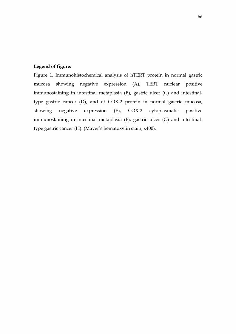
66
Legend of figure:
Figure 1. Immunohistochemical analysis of hTERT protein in normal gastric
mucosa showing negative expression (A), TERT nuclear positive
immunostaining in intestinal metaplasia (B), gastric ulcer (C) and intestinal-
type gastric cancer (D), and of COX-2 protein in normal gastric mucosa,
showing negative expression (E), COX-2 cytoplasmatic positive
immunostaining in intestinal metaplasia (F), gastric ulcer (G) and intestinal-
type gastric cancer (H). (Mayer’s hematoxylin stain, x400).
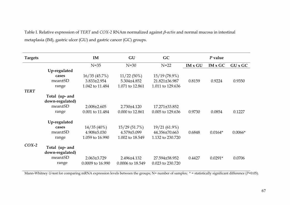
Table I. Relative expression of TERT and COX-2 RNAm normalized against β-actin and normal mucosa in intestinal
metaplasia (IM), gastric ulcer (GU) and gastric cancer (GC) groups.
Targets IM GU GC P value
N=35 N=30 N=22 IM x GU IM x GC GU x GC Up-regulated
cases mean±SD
range
16/35 (45.7%)
3.833±2.954 1.042 to 11.484
11/22 (50%) 5.304±4.852
1.071 to 12.861
15/19 (78.9%) 21.821±36.987
1.011 to 129.636
0.8159
0.9224
0.9350
TERT
Total (up- and
down-regulated) mean±SD
range
2.008±2.605 0.001 to 11.484
2.730±4.120 0.000 to 12.861
17.271±33.852 0.005 to 129.636
0.9730
0.0854
0.1227
COX-2
Up-regulated cases
mean±SD range
Total (up- and
down-regulated) mean±SD
range
14/35 (40%) 4.908±5.030
1.059 to 16.990
2.063±3.729 0.0009 to 16.990
15/29 (51.7%)
4.579±5.099 1.002 to 18.549
2.496±4.132 0.0006 to 18.549
19/21 (61.9%) 44.356±70.663
1.132 to 230.720
27.594±58.952 0.023 to 230.720
0.6848
0.4427
0.0164*
0.0291*
0.0066*
0.0706
Mann-Whitney U-test for comparing mRNA expression levels between the groups; N= number of samples; * = statistically significant difference (P<0.05).
67
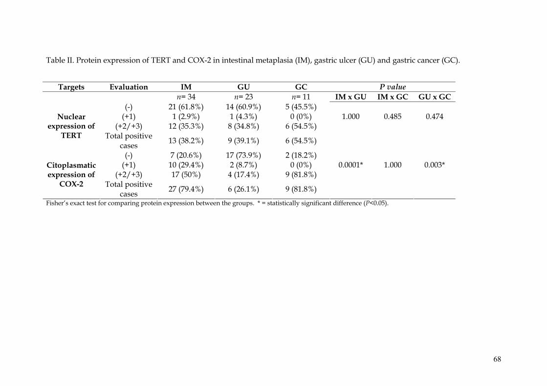
68
Table II. Protein expression of TERT and COX-2 in intestinal metaplasia (IM), gastric ulcer (GU) and gastric cancer (GC).
Targets Evaluation IM GU GC P value n= 34 n= 23 n= 11 IM x GU IM x GC GU x GC
(-) 21 (61.8%) 14 (60.9%) 5 (45.5%) (+1) 1 (2.9%) 1 (4.3%) 0 (0%) 1.000 0.485 0.474
(+2/+3) 12 (35.3%) 8 (34.8%) 6 (54.5%) Nuclear
expression of TERT Total positive
cases 13 (38.2%) 9 (39.1%) 6 (54.5%)
(-) 7 (20.6%) 17 (73.9%) 2 (18.2%) (+1) 10 (29.4%) 2 (8.7%) 0 (0%) 0.0001* 1.000 0.003*
(+2/+3) 17 (50%) 4 (17.4%) 9 (81.8%) Citoplasmatic expression of
COX-2 Total positive cases 27 (79.4%) 6 (26.1%) 9 (81.8%)
Fisher’s exact test for comparing protein expression between the groups. * = statistically significant difference (P<0.05).
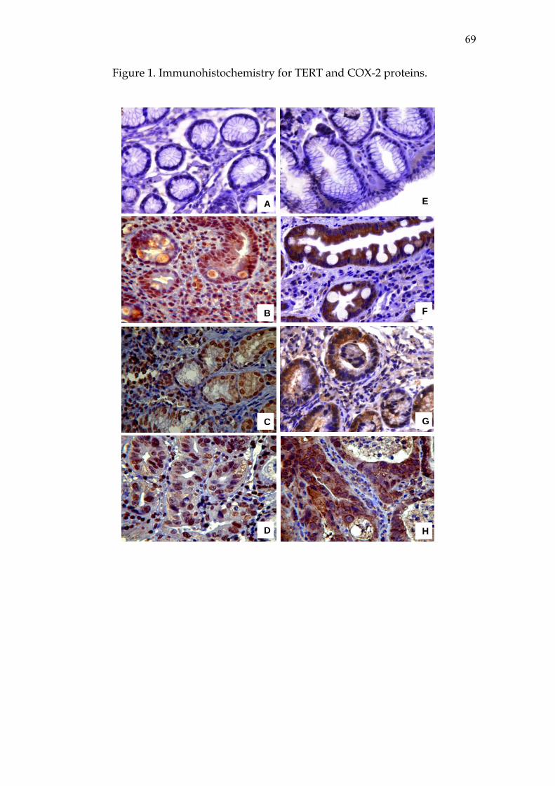
69
Figure 1. Immunohistochemistry for TERT and COX-2 proteins.
E A
F B
G C
D H

Artigo II
71
Artigo II. Expression of NOS2, HGF, MET, KRAS, TFF1 and CLDN18 in
intestinal metaplasia and gastric cancer: a real-time PCR and
immunohistochemical analysis.
Márcia Cristina Duarte1, Mabel Tatty de Medeiros Fracassi2, Kenji Miyazaki3, Ana
Elizabete Silva*1
1. UNESP - São Paulo State University, Biology Department, Rua Cristóvão Colombo,
2265, São José do Rio Preto, São Paulo State, Brazil. Telephone: +55 17-3 Laboratory,
Sírio-Libanes Hospital, Rua Dona Adma Jafet, 91, São Paulo, São Paulo State, Brazil.
Telephone: +55, 11-3155-0249
3. Endoscope Service, Base Hospital, Av. Brigadeiro Faria Lima, 5416, São José do Rio
Preto, São Paulo State, Brazil. Telephone: +55, 17-3201-5066
* Corresponding author. 221-2384
Fax: +55 17-3221-2390
2. Pathology
Email addresses:
MCD: [email protected]
MTMF: [email protected]
AES: [email protected]
72
Abstract
Background: Molecular studies concerning gastric pre-cancerous lesions are scarce and
can be important to get a best comprehension about carcinogenic process and to perform
prevention strategies. Intestinal metaplasia (IM) is a premalignat lesion of gastric cancer
(GC). Thus, changes of expression levels in genes with important roles to cellular cycle
progression, cell migration and, gastric mucosa maintenance and protection, can be
present in IM, increasing the stomach cancer risk.
Methods: Biopsies from 37 patients with IM (free-cancer) and 22 with GC and their
respective normal mucosal were used to RNA extraction and posterior real-time PCR
analysis so as to evaluate the mRNA expression level of NOS2, HGF, MET, KRAS,
TFF1 and CLDN18. β-actin was used as reference. Protein expression of these genes
was assessed by immunohistochemical tests.
Results: Up-regulated mRNA expression were found for NOS2, HGF and MET in
61.1%, 48.6% and 37.8% of the IM cases (13.6-, 2.6- and 2.8-fold), respectively. In GC,
such genes were 12.8-, 1.8- and 3.5-fold up-regulated in 59.1%, 27.3% and 38.1% of
the cases, respectively. KRAS was up-regulated in 26.7% and 28.6% of IM and GC
cases, but the expression mean was not significant in IM (1.1-fold), while in GC, it was
1.7-fold increased. Fifty-four percent and 68.2% of both IM and GC cases were down-
regulated to TFF1 and CLDN18 mRNA. Expression mean levels were increased 1.8-
and 1.6-fold in IM, and decreased to 1.1- and 1.7-fold in GC, respectively.
Immunohistochemical analysis for NOS2 and KRAS proteins showed positive
immunostaining (moderate/strong) in 92.6% and 0%, respectively of IM cases and 30%
and 70%, respectively of GC cases. While for TFF1 and CLDN18 proteins, it was
observed absent or weak immunostaining in 62.9% and 100% of IM cases, respectively,
and in 100% and 90% of GC cases. Associations between clinicopathological variables
73
and mRNA expression were found for H. pylori-positive and NOS2 and HGF (low
expression) and CLDN18 (high expression) in the GC group, and for smokers and TFF1
(low expression) in IM group.
Conclusions: Our data suggest that intestinal metaplasia shares changes in expression
of genes altered in gastric cancer, implying that they may have a key role to the
development of this lesion and also in the initial steps of gastric carcinogenesis, so may
confer increased risk of malignancy.
Key words: Intestinal metaplasia, gastric cancer, gene and protein expression, NOS2,
HGF, MET, KRAS, TFF1 and CLDN18.
Background
Intestinal metaplasia is a pre-cancerous condition leading to stomach carcinogenesis,
specifically well-differentiated adenocarcinoma or intestinal type [1]. It is defined as a
potentially reversible change from a fully differentiated cell type to another, implying
adaptation to environmental stimuli [2]. At this stage, the original glands and the
foveolar epithelium are replaced by cells with intestinal phenotype. The type I or
complete type of intestinal metaplasia resembles the small intestinal mucosa, with
absorptive enterocytes, alternating with mucin-filled globet cells. Whereas the
incomplete type includes types II and III, being characterized by cells that acquire
morphologic features of the large intestine, and are lined only by globet cells of
different sizes and shapes [3].
Intestinal metaplasia is involved in the multi-step model of intestinal type of
gastric cancer that is triggered after a chronic inflammation of the gastric mucosa, which
progresses throughout the years into the premalignant stages of chronic gastritis, leading
74
to atrophy, intestinal metaplasia, dysplasia and eventually gastric adenocarcinoma [4].
Furthermore, the major risk factors for gastric carcinogenesis consist in H. pylori
virulence factors, host genetics, and environmental factors, such as diet and smoking
[5]. The progression rates to gastric cancer in Western countries are very limited,
however, for intestinal metaplasia the progression rates to gastric cancer have varied
from 0% to 73% per year [6,7].
The gastric cancer development and progression is driven by an accumulation of
genetic alterations that have also been observed in precancerous lesions, such as
gastritis, ulcer and intestinal metaplasia [8-12]. The phenotypic shift that appears in
intestinal metaplasia occurs as a result of a combination of altered expression of genetic
factors, epigenetic silencing, transcription factors, signalling pathways and growth
factors [2]. Nevertheless, relatively little is known regarding which key genetic changes
in intestinal metaplasia are important in order to initiate tumor progression. Some
studies have reported expression changes as in TERT [13,14], PDX1, OCT1, TFF3,
SOX2, SHH, RAR-β and RUNX3 [1,2,15] and KRAS mutations [13]. Hence, studies of
gene deregulation of important genes in the cellular cycle control, growth factors,
inflammation and mucosal protection in intestinal metaplasia can help understand and
prevent the cancerous process of the stomach. Thus, the aim of this study was to
investigate the levels of mRNA and protein expression the genes NOS2, HGF, MET,
KRAS, TFF1 and CLDN18 in intestinal metaplasia compared to adjacent normal tissue
and gastric cancer, which has been reported with deregulated expression in this
neoplasm, and to investigate their potential relationship with H. pylori infection and
other clinicopathological variables.
75
Methods
Patients
This study consisted of 37 patients with intestinal metaplasia (mean age 61.05+12.46
years), all were gastric cancer-free, and 22 patients with gastric adenocarcinoma (mean
age 62.45±14.07 years), who had undergone endoscopic evaluation between March
2006 and March 2008, at the Base Hospital (São José do Rio Preto, SP, Brazil). Of each
patient, three biopsies of lesion region and other three biopsies of apparently normal
region were collected, mainly in the antrum and corpus of the stomach. Immediately
after this procedure, they were stored in RNA-later reagent (Ambion) at –20ºC until
RNA and DNA extraction. All specimens were histopathologically diagnosed. Archival
paraffin-embedded formalin-fixed tissues were used for immunohistochemical staining
of the proteins. Information about age, gender, smoking and drinking habits and the
written informed consent was obtained from all patients, and the study was approved by
the National Research Ethics Committee (CONEP).
RNA and DNA extraction and reverse transcription
The extraction of total cellular RNA and DNA was carried out by using the Trizol
reagent (Invitrogen), according to the manufacturer’s protocol. RNA and DNA
concentration was determined in a NanoDrop® ND1000 spectrophotometer, and RNA
integrity was visualized on 1% agarose gel with 2 bands of 18S and 28S rRNA. DNA
samples were maintained in freezers at -20ºC and used for molecular diagnosis of H.
pylori. RNA samples were stored in freezers at -80ºC and used for reverse transcription.
cDNA was synthesized from 5 μg of total RNA using random primers and a High
Capacity cDNA Archive Kit (Applied Biosystems), according to the manufacturer’s
instructions. The integrity of all cDNA preparations was tested by PCR assay of the β-
actin gene and visualized on 2% agarose gel.
76
Quantitative Real Time PCR
The relative expression of NOS2, HGF, MET, KRAS, TFF1 and CLDN18 mRNA was
measured by real time PCR, based on the SYBR Green methodology, using an ABI
Prism® 7300 (Applied Biosystems). The primer sequences used in the study were
obtained using Primer3 software (http://frodo.wi.mit.edu/cgi-
bin/primer3/primer3_www.cgi) with exception of CLDN18 sequence, obtained of
Sanada et al. [16] and they are shown in Table 1. An initial validation step with 10-fold
dilution series was performed to obtain the amplification efficiencies of the primers (E),
calculated according to the equation E = 10[-1/slope]. The efficiencies of amplification
ranged between 90% and 97%. After the validation step, the real time PCR assays were
performed in 10 μL of SYBRTM Green Master Mix (Applied Biosystems), 25 ng of
cDNA and 0.6 μM of β-actin, TFF1 and CLDN18 primers, 0.7 μM of KRAS primer and
0.8 μM of NOS2, HGF and MET primers. Thermal cycling conditions for all the genes
were: 2 min at 50ºC and 10 min at 95ºC for initial denaturation, followed by 40 cycles
at 95°C for 15 s and 60°C for 1 min, and a dissociation step at 95ºC for 15 s, 60ºC for
30 s and 95ºC for 15 s. Triplicates of the samples were assayed in each run. β-actin, α-
tubulin and β2-microglobulin housekeeping genes were evaluated to case and control
samples, to verify which contained the lowest variation of amplification between the
samples. After this, β-actin gene was used as reference because it showed the lowest
variation. The relative expressions of the genes were analyzed using the 2-ΔΔCt method,
according to the description by Livak and Schimittgen [17]. First, the mean of cycle
number at the threshold level of fluorescence (Ct) for each sample was determined.
Next, the ΔCt value was calculated as being the difference between the Ct value of
target gene and the Ct value of β-actin: ΔCt = Ct (target gene) - Ct (β-actin). Finally,
both the ΔΔCt value and the normalized target gene expression were calculated: ΔΔCt =
77
ΔCt (IM or GC) - ΔCt (normal adjacent mucosa). Normalized expression in a sample =
2-ΔΔCt.
PCR for H. pylori diagnosis
DNA samples extracted from the adjacent normal gastric mucosa were used for
molecular diagnosis of the H. pylori bacterium, because it is a more efficient method
than histology and urease test [18]. The bacterium genes urease and tsaA were
amplified by Multiplex PCR, using the specific primers F- 5’
TTCCTGATGGGACCAAACTC 3’ and R- 5’ TTACCGCCAATGTCAATCAA 3’,
and (F- 5’ CCTGCCGTTTTAGGAAACAA 3’ and R- 5’
TCCGCATTCCTACCTAATGG 3’) in order to amplify, respectively, segments of 316
pb and 413 pb. The housekeeping human gene CYP1A1 (F-
5’CTCACCCCTGATGGTGCTAT 3’ and R- 5’ TTTGGAAGTGCTCACAGCAG 3’)
that amplifies a 226 pb segment was used as quality control DNA. Positive and negative
controls were used in all experiments. The PCR assay solution consisted of 4 mM
MgCl2, 0.3 mM dNTPs, 0.6 μM of each primer, and 1.75 U Taq DNA polymerase in 1
X buffer. The thermal cycling conditions were: 5 min at 95ºC, followed by 35 cycles at
95ºC for 30 s, 60ºC for 30 s, and 72ºC for 1 min, and a final extension step at 72ºC for
10 min. The PCR products were separated on 2% 1000 agarose gel (Invitrogen) and
stained with ethidium bromide. The assay was considered positive when at least one of
the bacterial PCR products was present.
Immunohistochemistry
The immunohistochemical analysis was performed in 34 samples with IM, 24 with UG
and 11 with GC. Tissue sections of 4 μm were cut from paraffin-embedded tissue
blocks and mounted on glass slides pretreated with 3-aminopropyl-
triethoxysilane/acetone solution and dried overnight at 60ºC. After deparaffinization at
78
37º overnight, and rehydratation step, antigen retrieval was performed in 10 mM citrate
buffer (pH 6.0) for NOS2, HGF, MET, TFF1 and CLDN18, and in 10mM EDTA-TRIS
buffer (pH 9.0) for KRAS for 30 minutes at 95ºC in a steam-pan (Exotic, Arno). The
endogenous peroxidase activity was blocked with 3% H2O2 treatment for 10 minutes.
Then, the sections were incubated for 1 hour at room temperature with specific antibody
to TFF1 (pS2 mouse monoclonal antibody, clone BC04, Invitrogen, 1:50), HGF
(Monoclonal anti hepatocyte growth factor antibody, clone 24612.111, Sigma, 1:50)
and MET (Anti hepatocyte growth factor receptor (cMet) antibody, Sigma, 1:50) and
overnight at 4ºC with specific antibodies for NOS2 (Rabbit polyclonal to iNOS, abcam,
1:200), KRAS (Mouse monoclonal anti-KRAS, clone 9.13, Invitrogen, 1:50) e CLDN18
(Rabbit polyclonal anti-claudin-18 -c-Term, Zymed, Invitrogen, 1:200). After being
rinsed with Tris-HCl buffer (pH 7.6), the slides were incubated, for 20 minutes each,
with biotinylated secondary antibody and incubated with streptavidin-biotin peroxidase,
following the manufacturer’s instructions (Histostain Bulk Kit, Zymed). The
immunostain was visualized with 3,3’-diaminobenzidine tetrahydrochoride (DAB) and
counterstained with Mayer’s hematoxylin. Negative controls were established by
replacing the primary antibody with buffer solution. Stomach normal mucosa was used
as positive control for TFF1 and CLDN18 antibodies, and liver, breast carcinoma, lung
carcinoma and epidermoid lung carcinoma tissues were used as positive controls for
HGF, MET, NOS2 and KRAS antibodies, respectively. A single pathologist examined
all specimens. All analyses were performed under a light microscope, and the whole
tissue extension of all samples was examined. Immunostaining for protein NOS2
(brown cytoplasm staining) was evaluated by staining presence as negative (-) (absent
brown staining) or positive (+) (presence brown staining), whereas for KRAS (brown
cytoplasm staining), TFF1 (brown cytoplasm staining) and CLDN18 (brown membrane
79
staining) proteins, the immunostaining was evaluated as negative (-) or positive: (+1)
weakly stained, (+2) moderately stained, and (+3) strongly stained, as observed in at
least 10% of cells. A significant protein expression was considered only for (+2) and
(+3) staining, while (–) and (+1) showed no significant expression. Immunostaining
analysis for HGF and MET proteins was not performed because antibodies reaction did
not produce satisfactory results, after various tests.
Statistical Analysis
Data about RNAm relative expression were described using the mean + SD as a point
estimator and the range of values. Nonparametric Mann-Whitney test was used in the
comparisons between the groups, and nonparametric Spearman rank correlation was
used to evaluate interactions among mRNA expression in the groups. Protein expression
was described according to number of positive and negative cases and Fisher’s exact
test was used to compare the groups. The relationship between mRNA and protein
levels and clinicopathological variables was evaluated by Fisher’s exact test. All the
tests were accomplished using the GraphPad Instat Software. The level of significance
was set at P < 0.05.
Results
Clinical features
Characteristics about gender, age, smoking and drinking habits, histological type and H.
pylori infection are shown in Table 2. In the IM group, only five cases (13.5%) were
incomplete type of IM and most of the cases were found to be smokers and non-
drinkers. To GC group, 57.9% of the cases were intestinal type of GC and 62.5% were
smokers and drinkers. H. pylori infection was detected by multiplex PCR and we
80
verified that 37.8% and 52.4% of IM and GC cases, respectively, were positive to the
bacterium.
NOS2, HGF, MET, KRAS, TFF1 and CLDN18 mRNA expression in intestinal
metaplasia (IM) and gastric cancer (GC)
The results for the gene expression of NOS2, HGF, MET, KRAS, TFF1 and CLDN18 in
IM and GC are summarized in Table 3. Relative gene expression was calculated using
normal adjacent mucosa as reference. Mean levels of expression around 1,5-fold
increased or decreased were considered significant. NOS2 mRNA expression was
increased about 13-fold in both IM and GC in about 60% of the cases. HGF, MET and
KRAS mRNA relative expression was increased in IM (48.6%, 37.8% and 26.7% of the
cases) and GC (27.3%, 38.1% and 28.6% of the cases), respectively, as follow: 2.6- and
1.8-fold, 2.8- and 3.5- fold and 1.1- and 1.7-fold. mRNA expression for TFF1 and
CLDN18 was decreased in 54% of IM cases (but the expression mean levels was 1.8-
and 1.6-fold higher than normal, respectively) and 68% of GC cases (1.1 and 1.7-fold
lower than normal tissue, respectively). Figure 1 shows mean levels of expression for
NOS2, HGF, MET, KRAS, TFF1 and CLDN18 in IM and GC groups. The statistical
analysis between the groups showed a significant difference between the mRNA
expression mean levels only for TFF1 and CLDN18 (P= 0.006, Table 3).
The correlation between mRNA expression levels of genes that participate in the
same signal pathway was evaluated. To the IM group, we found that KRAS expression
was correlated to HGF and MET expression (r = 0.53, P=0.0001) and it has been
verified that such correlation was stronger between KRAS and HGF (r=0.70, P=0.0001).
A significant correlation between mRNA expression levels of NOS2 and HGF was
found in GC (r=0.80, P<0.0001).
81
Immunostaining for NOS2, KRAS, TFF1 and CLDN18 proteins in intestinal metaplasia
(IM) and gastric cancer (GC).
Immunostaining-positive (ranging from weak to strong) for NOS2, KRAS, TFF1 and
CLDN18 proteins were found in 92.6%, 89.3%, 85.2% and 11.5% of the IM cases and
30%, 100%, 50% and 60% of the GC cases (Table 4). To IM, cytoplasmatic staining for
NOS2 protein was only found in the metaplastic region, while the normal foveolar
epithelium had no staining. In GC, NOS2 showed positive immunostaining in cancer
cells and glandular epithelium, but, in general, the foveolar epithelium had no staining
for NOS2 (Figure 2. A.1, B.1 and C.1). KRAS protein exhibited a focal staining in the
cytoplasm of the normal gastric epithelium of IM and GC cases, with a weak staining in
metaplastic areas (89.3% of cases) and moderate or strong staining in cancer areas (70%
of cases) (Figure 2. A.2, B.2 and C.2). Redarding TFF1 protein, all the normal areas in
IM and GC showed moderate to strong cytoplasmatic immunostaining-positive status
while 62.9% and 100% of the areas with IM and GC cells, respectively, showed absent
or weak positive staining in relation with normal areas (Figure 2. A.3, B.3 and C.3). The
membrane of the normal epithelium showed strong staining for CLDN18 protein in all
the cases of IM and GC, but in metaplastic and cancer cells CLDN18 staining was
absent or weaker (100% and 90%, respectively), than that observed in the normal
adjacent mucosa (Figure 2. A.4, B.4 and C.4). Scores ≤ 1 (absent and weak staining)
and ≥ 2 (moderate and strong staining) were used to compare protein expression
between the IM and GC groups (Table 4), and a significant difference was found for
NOS2, KRAS and TFF1 proteins (P<0.05).
82
Expression of NOS2, HGF, MET, KRAS, TFF1 and CLDN18 and variables such as
smoking, drinking, H. pylori infection and histological type.
In GC group, there was a significant association between NOS2 and HGF expression
and H. pylori infection, with decreased mRNA mean levels in patients H. pylori-
positive (2.29 vs. 28.8, P= 0.0203, and 0.93 vs. 2.96, P=0.0430, respectively). While
increased CLDN18 mRNA level were associated with H. pylori- positive cases of the
GC group (0.90 vs. 0.28, P=0.0435). To IM group, TFF1 expression and smoking was
associated with decreased mRNA levels in smokers (1.08 vs. 3.92, P= 0.0453). KRAS
and MET were not associated with any variable studied. Comparisons between mRNA
relative expression of evaluated genes and histological parameters as incomplete and
complete metaplasia and gastric cancer type (intestinal vs. diffuse) did not show any
association. Likewise, analysis between protein expression and variables did not
produce significant data.
Discussion
We analyzed the mRNA and protein expression of NOS2, HGF, MET, KRAS, TFF1 and
CLDN18 in IM in comparison to GC and adjacent normal mucosal. Our results
indicated increased mRNA relative expression for genes NOS2, HGF, MET and KRAS,
with mRNA mean levels among 1.15 to 13.62, in both IM and GC groups, and
decreased means levels of mRNA for genes TFF1 and CLDN18 (0.97 and 0.60,
respectively) in GC, but increased in IM (1.84 and 1.64, respectively). In addition, we
did not observe precise relationship between mRNA and protein expression for most of
the evaluated genes.
This divergence between mRNA and protein expression might be explained by the
fact that mRNA changes are more diverse than stable. In proteins, changes are slower,
83
such as in as post-transcriptional regulation, translational control and protein stability.
However, levels of mRNA and protein are not always correlated [19].
NOS2 gene (nitric oxide synthase 2), also known as iNOS (inducible oxide nitric
synthase), encodes an enzyme responsible for producing nitric oxide (NO), induced by
bacterial endotoxins or cytokines mainly in leukocytes and epithelial cells, resulting in
high concentrations of NO [20]. NO contributes to mucosal defense through its
cytotoxic properties, against ingested bacteria and parasites [21], for example, as part of
the host response to the H. pylori infection. However, in circumstances in which the
mucosa is inflamed or damaged, some studies suggest that NO contributes to tissue
injury and it can lead to cell cycle disorders, cell death caused by apoptosis, and DNA
damage [22, 23]. In the current study, high levels of NOS2 mRNA expression (about
13-fold) observed in about 60% of cases of both IM and GC might contribute to genetic
changes associated with gastric carcinogenesis. Our findings are similar to other studies
that also observed NOS2 protein expression in 13% cases of IM [24]. To GC, high
levels of NOS2 mRNA expression also was corroborated by some previous studies [25-
28], and NOS2 protein expression was related to tumor angiogenesis, tumor
progression, and patient survival [29]. Therefore, NOS2 expression can participate in the
initial events of the gastric carcinogenesis through tissue injuries and genetic changes,
as observed in pre-cancerous lesions.
HGF, MET and KRAS genes share the same signaling pathway resulting in
proliferation, angiogenesis, migration and morphogenesis. Both MET and HGF are
often up-regulated in gastric cancer [15], while KRAS is activated by mutation [30, 31].
Hepatocyte Growth Factor (HGF) acts as a potent mitogen and pro-motility agent for
epithelial cells. HGF is produced for the mesenchymal cells to act on its receptor, MET,
84
in the epithelial cells in a paracrine fashion. Together, they have key roles in
gastrointestinal development, mucosal healing and epithelial cell migration [32].
There is accumulating evidence that the transformation of the normal gastric
mucosal cells into malignant cells and gastric cancer may be modulated by transforming
growth factors including HGF and their receptor, MET. We find 2.6-fold up-regulation
of HGF in IM (48.6% of cases), while in GC tissues (27.3%), there was about 2-fold
high expression of HGF. MET mRNA was up-regulated in about 38% of both IM and
GC cases, with 2.8- and 3.5-fold higher than adjacent normal gastric mucosa,
respectively. These data could not be verified by immunohistochemistry because the
antibody used did not produce satisfactory results.
Our findings are similar to Konturek et al. [33] who observed a 2-fold increase
in the mRNA expression of HGF (52% of the cases) and TGF-α in gastric cancer tissue
compared to adjacent mucosa by densitometric analysis. Inoue et al. [34] verified higher
expression of MET protein in tissues of gastric adenocarcinoma than in normal mucosa.
These data provide evidence that up-regulation of HGF and MET is a frequently
occurring event in gastric carcinogenesis and it was in accordance with previous studies
showing an increased expression and higher serum levels of these factors in gastric
cancer [35, 36]. However, little is known about HGF and MET expression in intestinal
metaplasia. Suzuki et al. [37] observed a slight decrease in HGF content after
eradication therapy against H. pylori in patients with upper gastrointestinal disease
through ELISA method. However, the level of antral HGF was significantly decreased
in patients with intestinal metaplasia, but not in those without it. Thus, HGF and MET
up-regulation can be important to early steps of gastric carcinogenesis through
promotion of cellular proliferation, as observed in IM, considered a pre-malignant
stomach lesion.
85
On the KRAS expression, we did not observe an important increase in mRNA
relative expression levels (about 1.15- and 1.70-fold) in IM and GC groups, where only
about 28% of cases had up-regulation. However, we found a significant proportion of
positive cases for KRAS protein in both groups (89.3% for IM and 100% for GC), with
a weak staining in IM and a moderate or strong staining in 70% of GC-positive cases.
Once again, evidencing that there is no exact relationship between mRNA and protein
expression.
The KRAS gene encodes a guanosine triphosphatase protein (p21ras) that
functions as a “molecular switch”, transmitting signals from the extracellular to
intracellular environmental, mediating signaling events that regulates cell proliferation,
cell survival, angiogenesis and cell migration [38]. Our findings suggest that KRAS
expression is not significantly altered in IM. But, in GC, the high levels of protein
expression suggest an important role of the gastric carcinogenesis. KRAS protein can be
activated by mutations, leading to increased cell growth. However, in gastric
adenocarcinoma, the incidence of KRAS mutation seems to vary among populations,
with rates of about 10%, possibly related to H. pylori infection [30, 31]. In addition,
KRAS protein can also be activated by gene amplification in gastric cancer. So,
immunohistochemistry has shown that this protein is preferentially expressed in gastric
cells with KRAS amplification [39]. Moreover, increased expression of KRAS protein
has been associated with progression of gastric carcinogenesis, lymph nodes metastases
and poor prognosis [40].
Trefoil peptide 1 (TFF1 or pS2) located in gastric mucosa cells interacts directly
with mucins to stabilize the mucous layer, protecting the epithelium against injury and
maintaining the integrity of the mucosal barrier. Furthermore, TFF1, known as tumor
suppressor gene has an important role in regulating the balance between gastrointestinal
86
cell proliferation, death and differentiation [41, 42]. Some studies have reported a loss
of TFF1 protein expression in 40 to 60% of gastric cancer cases [11, 41, 43]. Our results
are in accordance with these reports because we found a decrease or loss of TFF1
protein expression in 100% of GC areas and 62.9% of IM compared to normal
epithelium. Similarly, the TFF1 mRNA expression levels were reduced in 54% of cases
of IM, with an expression mean level of 1.84, but with a significantly lower mean level
in GC (68%, mean=0.96) compared to IM.
Fujimoto et al. [44] demonstrated that reduced expression of TFF1 at mRNA
and protein levels, by hypermethylation in the promoter region, frequently occurred in
adenomas, intestinal metaplasia and gastric adenocarcinomas. Moreover, Kim et al. [45]
reported TFF1 protein was expressed in IM, although the immunoreactivity was weaker
than in the adjacent normal gastric epithelium, as observed in the present study. Then,
progressive loss of TFF1 expression from normal gastric mucosa into intestinal
metaplasia may increase susceptibility to carcinogens and finally progression to gastric
cancer. In addition, we observed that the TFF1 mRNA expression levels were lower in
smokers than in nonsmokers in IM group, suggesting that substances of the tobacco can
decrease TFF1 expression levels in the gastric mucosa, and consequently change their
protective role. We did not find literature data about this interaction in gastric tissues,
but in breast cancer cells high TFF1 expression was reported to be significant (2-fold),
induced by tobacco smoke condensate [46]. This discrepancy might be explained by the
fact that, in the stomach, TFF1 acts as a tumor-suppressor gene, while in breast tissue, it
acts as an oncogene. So, it may hypothesized that the carcinogenic properties of the
several substances of the tobacco can modulate TFF1 expression, contributing to DNA
damage and cell transformation, and icreased malignant risk.
87
Claudins (CLDNs) are a large family of proteins which interact with one another
to form a branching network that helps create the tight junctions of epithelial cells [47].
CLDN18 protein is normally detected in the normal gastric mucosa and plays an
important role in various biophysiological processes, such as regulation of paracelular
permeability and conductance. Disruption of tight junctions can cause loss of cell
polarity, resulting in an abnormal influx of growth factors, which could provide auto
and paracrine stimulation to tumourigenic epithelial cell [16, 48]. Our results showed
down-regulation at CLDN18 mRNA in 68% of GC cases and 54% of IM cases, but with
significantly reduced mRNA mean levels (P=0.0065) in GC group (0.605) in
comparison to IM group (1.639). Similarly, a decrease or loss of CLDN18 protein
expression was observed in 90% of GC cases and 100% of IM cases when compared to
adjacent normal mucosa.
Since claudin-18 is expressed only in normal stomach, dysfunction of tight
junctions caused by down-regulation of CLDN18 mRNA may lead to an abnormal
influx of stomach-related growth factors, which can promote an increase of cell
proliferation and increased risk of gastric cancer. Similarly to our results, Sanada et al.
[16] showed that CLDN18 mRNA expression was down-regulated in 56.5% of GC and
no expression of CLDN18 protein was observed in 57% of the cases of gastric cancer
associated with poor survival, as in 90% of gastric adenoma and some of the intestinal
metaplasia. According to these authors, down-regulation of CLDN18 may be an early
event in gastric carcinogenesis. Moreover, Matsuda et al. [48] found no expression of
CLDN18 protein in IM and GC, suggesting a close correlation with carcinoma
progression and subsequent metastatic events. However, in other cancer types such as
pancreatic, CLDN18 is frequently up-regulated [47].
88
Literature data show an intrinsic network that includes NOS2, HGF, MET, and
KRAS proteins. In IM, we found a significant correlation between KRAS and
HGF/MET mRNA expression levels, and, yet, a stronger correlation between KRAS and
HGF (r=0.70). HGF action can be started through its binding to MET receptor that
activates several intracellular messengers. One of them is KRAS that has been shown to
mediate the HGF-induced migration of gastric cells and promotion of cell proliferation
[49, 50]. In GC, it was observed a correlation between NOS2 and HGF expression.
HGF/MET binding activates KRAS expression and, on the other hand, KRAS seems to
activate AP-1 (Activator Protein-1), via ERK (extracellular signal-regulated kinase)
signaling pathway that induces the activation of NOS2 and COX-2 expression,
increasing inflammation, apoptosis and oxidative damage in gastric cells [51]. So, these
proteins can interact in order to increase cellular proliferation and damage the gastric
cells, leading to key changes in cell homeostasis, contributing to the initial steps of
carcinogenesis, as in premalignant lesions.
H. pylori infection is frequently associated with gastric carcinogenesis. So, we
investigated the relationship between H. pylori infection and mRNA expression levels
of NOS2, HGF, MET, KRAS, TFF1 and CLDN18 in the IM and GC groups. We found
that in the GC group H. pylori-positive individuals had lower mRNA expression levels
for NOS2 and HGF and higher expression levels for CLDN18 than that H. pylori-
negative.
Concerning NOS2, the data were discordant with NO role in the mucosal defense
through its cytotoxic properties, acting as primary defense against bacterium infection
[22]. Wang et al. [27] found higher levels of NO and NOS expression in the H. pylori-
positive group when compared to negative group. However, Augusto et al. [28] did not
find association between NOS2 expression and virulence factor of H. pylori. There is
89
evidence that generation of polyamines by ornithine decarboxylase (ODC) and also
induced by H. pylori, results in inhibition of NOS2 [52, 53]. These biochemical
pathways that limit NO production may exist to protect macrophages from potential
toxic effects of overproduction of NO in response to other pathogens, but in the case of
H. pylori, the bacterium appears to be reaping the benefits of these host responses [53].
When it comes to the relationship between HGF expression and H pylori infection,
some studies found an increased expression of HGF in gastric mucosa colonized by H.
pylori, suggesting a possible link between this factor and H. pylori-associated gastritis
and gastric cancer [37, 54]. Moreover, CagA virulence factor of H. pylori, have been
shown to interact with HGF receptor, MET, thereby stimulating cell growth, motility
and invasiveness [55]. However, the results are controversial, because Konturek et al.
[33] did not find significant correlation between CagA status and genetic alterations
concerning gene and protein expression for growth factors. Moreover, similar gene
changes were also observed in patients without H. pylori infection, suggesting that
overexpression of HGF is not necessarily affected by the H. pylori status. In addition, a
recent study has shown that H. pylori induces AGS gastric adenocarcinoma cells
motility independently of MET expression [56]. Thus, to clarify these divergent results,
more studies of H. pylori interaction with growth factors are necessary. In the present
study, we should also consider the small number of cases evaluated in GC group.
To our knowledge, there are no reports about H. pylori infection and CLDN18
expression. However, it is known that H. pylori recruits and modifies the distribution of
zonula occludens-1 (ZO-1) and junctional adhesion molecule (JAM), which are
normally located at cell tight junctions. CagA alters the structure and function of the
apical-junctional complex may result in loss of control over cytoskeletal architecture,
cell polarity, proliferation and differentiation, which are characteristics of oncogenic
90
transformation [55]. As CLDN18 participate of the tight junctions network, the higher
levels of mRNA expression in H. pylori- positive observed in this study, can suggest an
interaction between bacterium infection and CLDN18 in GC group. However, further
studies about role of H pylori infection on CLDN18 expression are required to explain
this question.
Our data did not show any association regarding other comparisons between
mRNA and protein of genes evaluated and parameters as age, gender, smoking, alcohol
drinking, histological type of metaplasia (complete vs. incomplete) and histological type
of gastric cancer (intestinal vs. diffuse).
Conclusion
In conclusion, our findings on deregulated expression in important genes related
to cycle cellular and maintenance of gastric mucosa integrity in intestinal metaplasia,
mainly NOS2, HGF and MET, suggest that this preneoplastic lesion share changes of
gene expression similar to the GC, which may have a key role both to the development
of this lesion as to the initial steps of gastric carcinogenesis. So, these genetic changes
along with others may bring greater risk of malignancy. However, further studies are
needed in premalignat gastric lesions to assess deregulation of different genes that lead
to progression of gastric cancer.
Competing interests
The author(s) declare that they have no competing interests.
Authors’ contributions
MCD implemented the experimental design, carried out the assays and drafted the
manuscript. MTMF performed the pathological assessment of gastric specimens and
91
helps in the immunohistochemical analysis. KM was responsible for endoscope exam
and collection of the gastric biopsies. AES was responsible for study conception, design
and revision of manuscript draft for intellectual content. All authors have read and
approved the final version of the manuscript.
Acknowledgements
We want to thank Prof. Dr. Antonio José Manzato for the statistical analysis help and
the Brazilian Agencies, FAPESP and CAPES, for financial support.
References
1. Busuttil RA, Boussioutas A: Intestinal metaplasia: A premalignant lesion
involved in gastric carcinogenesis. J Gastroenterol Hepatol 2009, 24:193–201
2. Gutiérrez-González L, Wright, NA: Biology of intestinal metaplasia in 2008: more
than a simple phenotypic alteration. Diges Liver Dis 2008, 40:510-522.
3. Correa P, Houghton J: Carcinogenesis of Helicobacter pylori. Gastroenterology
2007, 133:659-672.
4. Correa P: The biological model of gastric carcinogenesis. In Mechanisms of
carcinogenesis: Contribution of molecular epidemiology nº157. Edited by Buffer P,
Rice J, Bird M, Boffetta P. Lyon: IARC Scientific Publications, 2004.
5. Wu MS, Lin YS, Chang YT, Shun CT, Lin MT, Lin JT: Gene expression profiling
of gastric cancer by microarray combined with laser capture microdissection.
World J Gastroenterol 2005, 11(47):7405-12.
6. de Vries AC, Meijer GA, Looman CW, Casparie MK, Hansen BE, van Grieken NC,
Kuipers EJ: Epidemiological trends of pre-malignant gastric lesions: a long-term
nationwide study in the Netherlands. Gut 2007, 56(12):1665-70.
92
7. de Vries AC, van Grieken NC, Looman CW, Casparie MK, de Vries E, Meijer GA,
Kuipers EJ: Gastric cancer risk in patients with premalignant gastric lesions: a
nationwide cohort study in the Netherlands. Gastroenterology 2008, 134(4):945-52.
8. César ACG, Borim AA, Caetano A, Cury PM, Silva AE: Aneuploidies, deletion,
and overexpression of TP53 gene in intestinal metaplasia of patients without
gastric cancer. Cancer Genet Cytogen 2004, 153:127-132.
9. Zhang KL, Ma JX, Chen XY, Sun Y, Kong QY, Liu J, Li H: Frequent CYP1A1
expression in gastric cancers and their related lesions. Oncol Rep 2004, 12(6):1335-
40.
10. César ACG, Calmon MF, Cury PM, Caetano A, Borim, AA, Silva AE. Genetic
alterations in benign lesions: chronic gastritis and gastric ulcer. World J. Gastroenterol.
2006; 12(4): 625-9.
11. Shi S-Q, Cai J-T, Yang J-M: Expression of trefoil factors 1 and 2 in
precancerous condition and gastric cancer. World J Gastroenterol 2006;
12(19):3119-22.
12. Zaky AH, Watari J, Tanabe H, Sato R, Moriichi K, Tanaka A, Maemoto A, Fujiya
M, Ashida T, Kohgo Y: Clinicopathologic implications of genetic instability in
intestinal-type gastric cancer and intestinal metaplasia as a precancerous lesion.
Am J Clin Pathol 2008, 129:613-621.
13. Tahara E: Genetic pathways of two types of gastric cancer. In: Mechanisms of
carcinogenesis: Contribution of molecular epidemiology nº 157. Edited by Buffer P,
Rice J, Bird M, Boffetta P. Lyon: IARC Scientific Publications, 2004.
14. Gulmann C, Lantuejoul S, Grace A, Leader M, Patchett S, Kay E: Telomerase
activity in proximal and distal gastric cancer neoplastic and preneoplastic lesions
using immunohistochemical detection of hTERT. Dig Liver Dis 2005, 37:439-445.
93
15. Stock M, Otto F: Gene deregulation in gastric cancer. Gene, 2005, 360(1):1-19.
16. Sanada Y, Oue N, Mitani Y, Yoshida K, Nakayama H, Yasui W: Down-regulation
of the claudin-18 gene, identified through serial analysis of gene expression data
analysis, in gastric cancer with an intestinal phenotype. J Pathol 2006, 208:633-42.
17. Livak KJ, Schmittgen TD: Analysis of relative gene expression data using Real
Time Quantitative PCR and the 2-ΔΔCt method. Methods 2001 25:402-408.
18. César ACG, Cury PM, Payão SLM, Liberatore PH, Silva AE. Comparison of
histological and molecular diagnosis of Helicobacter pylori in benign lesions and
gastric adenocarcinoma. Braz. J. Microbiol. 2005; 36: 12-6.20.
19. Conway K, Ruge F, Price P, Harding KG, Jiang WG: Hepatocyte growth factor
regulation: An integral part of why wounds become chronic. Wound Rep Reg 2007,
15:683-92.
20. Nomelini, R S: Production of nitric oxide and expression of inducible nitric
oxide synthase in ovarian cystic tumors. Mediators Inflamm 2008, 2008:186584.
21. Marcinkiewicz J, Chain B, Nowak B, Grabowska A, Bryniarski K, Baran J:
Antimicrobial and cytotoxic activity of hypochlorous acid: interactions with
taurine and nitrite. Inflamm Res 2000, 49(6):280-9.
22. Martin GR, Wallace JL: Gastrointestinal inflammation: a central component of
mucosal defense and repair. Exp Biol Med 2006, 231(2):130-7.
23. Keklikoglu N, Koray M, Kocaelli H, Akinci S: iNOS expression in oral and
gastrointestinal tract mucosa. Dig Dis Sci 2008, 53(6):1437-42.
24. Feng CW, Wang LD, Jiao LH, Liu B, Zheng S, Xie XJ: Expression of p53,
inducible nitric oxide synthase and vascular endothelial growth factor in gastric
precancerous and cancerous lesions: correlation with clinical features. BMC
Cancer 2002, 2:8
94
25. Koh E, Noh SH, Lee YD, Lee HY, Han JW, Lee HW, Hong S: Differential
expression of nitric synthase in human stomach cancer. Cancer Lett 1999, 146:173-
80.
26. Rajnakova A, Moochhala S, Goh PMY, Ngoi S-S: Expression of nitric oxide
synthase, cyclooxygenase, and p53 in different stages of human gastric cancer.
Cancer Lett 2001, 172:177-85.
27. Wang Y-F, Guo C-L, Zhao L-Z, Yang G-A, Chen P, Wang H-K: Effect of
Helicobacter pylori infection on gastric mucosal pathologic change and level of
nitric oxide and nitric oxide synthase. World J Gastroenterol 2005, 11:5029-31.
28. Augusto AC, Miguel F, Mendonça S, Pedrazzoli Jr J, Gurgueira SA: Oxidative
stress expression status associated to Helicobacter pylori virulence in gastric
diseases. Clin Biochem 2007, 40:615-22.
29. Chen C-N, Hsieh FJ, Cheng YM, Chang KJ, Lee PH: Expression of inducible
nitric oxide synthase and cyclooxygenase-2 in angiogenesis and clinical outcome of
human gastric cancer. J Surg Oncol 2006, 94:226-33.
30. Yoo J, Park SY, Robinson RA, Kang SJ, Ahn WS, Kang CS: Ras gene mutations
and expression of Ras signal transduction mediators in gastric adenocarcinomas.
Arch Pathol Lab Med 2002, 126(9):1096-100.
31. Watari J, Tanaka A, Tanabe H, Sato R, Moriichi K, Zaky A, Okamoto K, Maemoto
A, Fujiya M, Ashida T, Das KM, Kohgo Y: K-ras mutations and cell kinetics in
Helicobacter pylori associated gastric intestinal metaplasia: a comparison before
and after eradication in patients with chronic gastritis and gastric cancer. J Clin
Pathol 2007, 60(8):921-6.
95
32. Anderson MR, Harrison R, Atherfold PA, Campbell MJ, Darnton SJ, Obszynska J,
Jankowski JA: Met receptor signaling: a key effector in esophageal
adenocarcinoma. Clin Cancer Res. 2006, 12(20):5936-43.
33. Konturek PC, Konturek SJ, Sulekova Z et al: Expression of hepatocyte growth
factor, transforming growth factor alpha, apoptosis related proteins Bax and Bcl-
2, and gastrin in human gastric cancer. Aliment Pharmacol Ther 2001, 15:989-99.
34. Inoue T, Kataoka H, Goto K, Nagaike K, Igami K, Naka D, Kitamura N, Miyazawa
K: Activation of c-Met (hepatocyte growth factor receptor) in human gastric
cancer tissue. Cancer Sci 2004, 95(10):803-8.
35. Han SU, Lee JH, Kim WH, Cho YK, Kim MW: Significant correlation between
serum levels of hepatocyte growth factor and progression of gastric cancer. World J
Surg 1999, 23:1176-1180.
36. Uen YH, Lin SR, Wu CH, Hsieh JS, Lu CY, Yu FJ, Huang TJ, Wang JY: Clinical
significance of MUC1 and c-Met RT-PCR detection of circulating tumor cells in
patients with gastric carcinoma. Clin Chim Acta 2006 367(1-2):55-61.
37. Suzuki M, Suzuki H, Masaoka T, Tanaka S, Suzuki K, Ishii H: Helicobacter pylori
eradication treatment modulates epithelial cell proliferation and tissue content of
hepatocyte growth factor in the gastric mucosa. Aliment Pharmacol Ther 2004, 20
Suppl 1:158-64.
38. Friday BB, Adjei AA. K-ras as a target for cancer therapy. Biochim. Biophys. Acta
2005; 1756: 127-44.
39. Mita H, Toyota M, Aoki F, Akashi H, Maruyama R, Sasaki Y, Suzuki H, Idogawa
M, Kashima L, Yanagihara K, Fujita M, Hosokawa M, Kusano M, Sabau SV, Tatsumi
H, Imai K, Shinomura Y, Tokino T: A novel method, digital genome scanning detects
KRAS gene amplification in gastric cancers: involvement of overexpressed wild-
96
type KRAS in downstream signaling and cancer cell growth. BMC Cancer 2009,
9:198.
40. Li M, Liu W, Zhu YF, Chen YL, Zhang BZ, Wang R: Correlation of COX-2 and
K-ras expression to clinical outcome in gastric cancer. Acta Oncology 2006,
45(8):1115-9.
41. Leung WK, Yu J, Chan FK, To KF, Chan MW, Ebert MP, Ng EK, Chung SC,
Malfertheiner P, Sung JJ: Expression of trefoil peptides (TFF1, TFF2, and TFF3) in
gastric carcinomas, intestinal metaplasia, and non-neoplastic gastric tissues. J
Pathol 2002, 197:582-8.
42. Kornprat P, Rehak P, Lemmerer M, Gogg-Kamerer M, Langner C: Analysis of
trefoil factor family protein 1 (TFF1, pS2) expression in chronic cholecystitis and
gallbladder carcinoma. Virchows Arch 2005, 446(5):505-10.
43. Machado JC, Nogueira AM, Carneiro F, Reis CA, Sobrinho-Simões M: Gastric
carcinoma exhibits distinct types of cell differentiation: an immunohistochemical
study of trefoil peptides (TFF1 and TFF2 and mucins (MUC1, MUC2, MUC5AC
and MUC6). J Pathol 2000, 190:437-443.
44. Fujimoto J, Yasui W, Tahara H, Tahara E, Kudo Y, Yokozaki H, Tahara E: DNA
hypermetilation at the pS2 promoter region is associated with early stage of
stomach carcinogenesis. Cancer Lett 2000, 149:125-134.
45. Kim BW, Kim KM, Lee BI, Maeng LS, Choi H, Cho SH, Chae HS, Kim JK, Choi
KY, Chung IS: Expression of trefoil peptides in the subtypes of intestinal
metaplasia. Peptides 2004, 25(5):779-83.
46. Martin MB, Reiter R, Johnson M, Shah MS, Iann MC, Singh B, Richards JK, Wang
A, Stoica A: Effects of tobacco smoke condensate on estrogen receptor-alpha gene
expression and activity. Endocrinology 2007, 148(10):4676-86.
97
47. Karanjawala ZE, Illei PB, Ashfaq R, Infante JR, Murphy K, Pandey A, Schulick R,
Winter J, Sharma R, Maitra A, Goggins M, Hruban RH: New markers of pancreatic
cancer identified through differential gene expression analyses: Claudin 18 and
Annexin A8. Am J Sur Pathol 2008, 32:188-96.
48. Matsuda Y, Semba S, Ueda J, Fuku T, Hasuo T, Chiba H, Sawada N, Kuroda Y,
Yokozaki H: Gastric and intestinal claudin expression at the invasive front of
gastric carcinoma. Cancer Sci 2007, 98:1014-9.
49. Ridley AJ, Comoglio PM, Hall A: Regulation of scatter factor/hepatocyte growth
factor responses by Ras, Rac, and Rho in MDCK cells. Mol Cell Biol 1995, 15:1110-
22.
50. Nylander-Koski O, Kiviluoto, T, Puolakkainen P, Kivilaakso E, Mustonen H: The
effect of nitric oxide, growth factors, and estrogen on gastric cell migration. J Surg
Res 2007, 143:230-7.
51. Cho SO, Lim JW, Kim KH, Kim H: Involvement of Ras and AP-1 in
Helicobacter pylori-Induced Expression of COX-2 and iNOS in Gastric Epithelial
AGS Cells. Dig Dis Sci 2009, in press.
52. Bussière FI, Chaturvedi R, Cheng Y, Gobert AP, Asim M, Blumberg DR, Xu H,
Kim PY, Hacker A, Casero RA Jr, Wilson KT: Spermine causes loss of innate
immune response to Helicobacter pylori by inhibition of inducible nitric-oxide
synthase translation. J Biol Chem 2005, 280:2409–12.
53. Wilson KT, Crabtree, JE: Immunology of Helicobacter pylori: Insights Into the
Failure of the Immune Response and Perspectives on Vaccine Studies.
Gastroenterology 2007, 133:288-308.
54. Kondo S, Shinomura Y, Kanayama S, Higashimoto Y, Kiyohara T, Yasunaga Y,
Kitamura S, Ueyama H, Imamura I, Fukui H, Matsuzawa Y: Helicobacter pylori
98
increases gene expression of hepatocyte growth factor in human gastric mucosa.
Biochem Biophys Res Commun 1995, 210:960-5.
55. Nguyen LT, Uchida T, Murakami K, Fujioka T, Moriyama M: Helicobacter pylori
virulence and the diversity of gastric cancer in Asia. J Med Microbiol 2008,
57:1445–53.
56. Snider JL, Cardelli JA: Helicobacter pylori induces cancer cell motility
independent of the c-Met receptor. J Carcinogen 2009, 8:1-11.
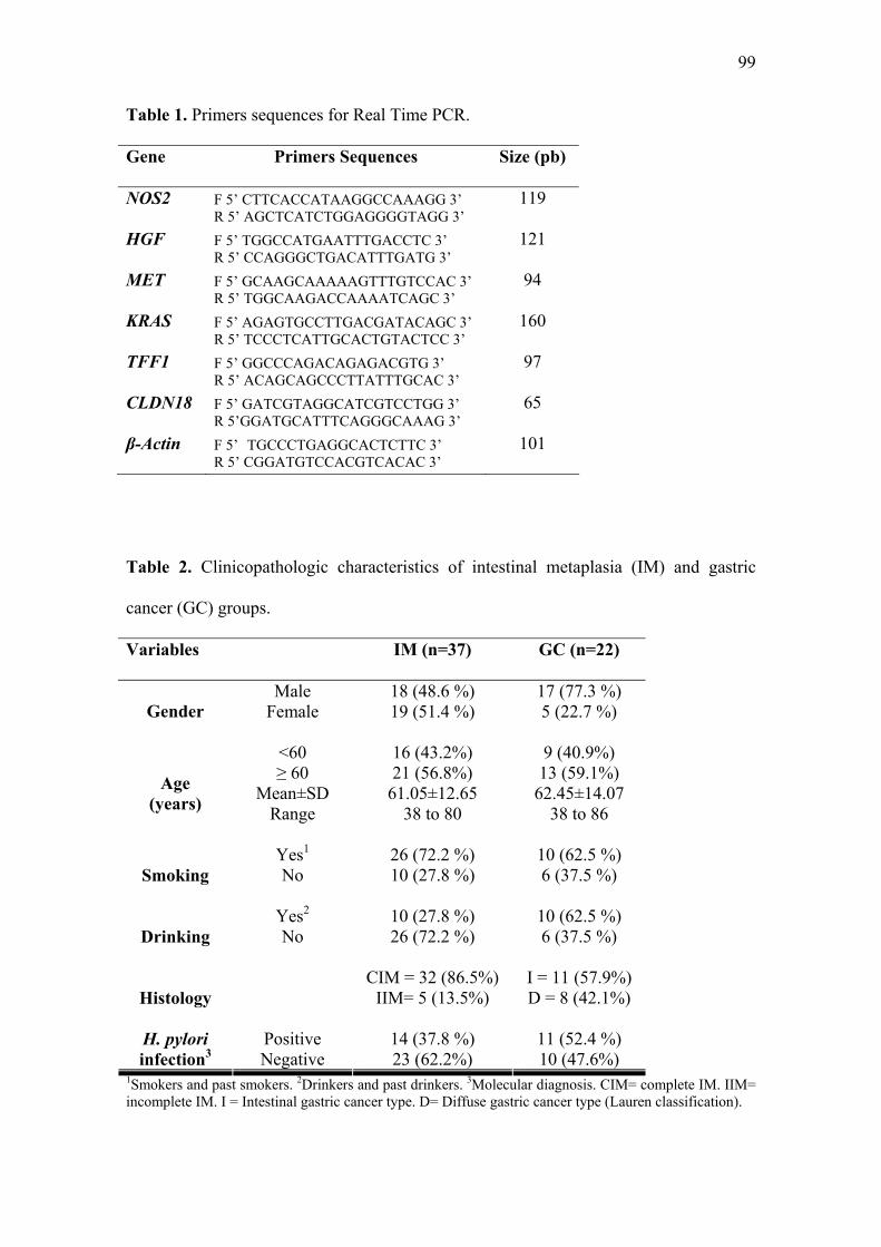
99
Table 1. Primers sequences for Real Time PCR.
Gene Primers Sequences Size (pb)
NOS2 F 5’ CTTCACCATAAGGCCAAAGG 3’ R 5’ AGCTCATCTGGAGGGGTAGG 3’
119
HGF F 5’ TGGCCATGAATTTGACCTC 3’ R 5’ CCAGGGCTGACATTTGATG 3’
121
MET F 5’ GCAAGCAAAAAGTTTGTCCAC 3’ R 5’ TGGCAAGACCAAAATCAGC 3’
94
KRAS F 5’ AGAGTGCCTTGACGATACAGC 3’ R 5’ TCCCTCATTGCACTGTACTCC 3’
160
TFF1 F 5’ GGCCCAGACAGAGACGTG 3’ R 5’ ACAGCAGCCCTTATTTGCAC 3’
97
CLDN18 F 5’ GATCGTAGGCATCGTCCTGG 3’ R 5’GGATGCATTTCAGGGCAAAG 3’
65
β-Actin F 5’ TGCCCTGAGGCACTCTTC 3’ R 5’ CGGATGTCCACGTCACAC 3’
101
Table 2. Clinicopathologic characteristics of intestinal metaplasia (IM) and gastric
cancer (GC) groups.
Variables IM (n=37) GC (n=22)
Male 18 (48.6 %) 17 (77.3 %) Gender Female 19 (51.4 %) 5 (22.7 %)
<60 16 (43.2%) 9 (40.9%) ≥ 60 21 (56.8%) 13 (59.1%)
Mean±SD 61.05±12.65 62.45±14.07 Age (years) Range 38 to 80 38 to 86
Yes1 26 (72.2 %) 10 (62.5 %)
Smoking No 10 (27.8 %) 6 (37.5 %)
Yes2 10 (27.8 %) 10 (62.5 %) No 26 (72.2 %) 6 (37.5 %) Drinking
CIM = 32 (86.5%) I = 11 (57.9%)
Histology IIM= 5 (13.5%) D = 8 (42.1%)
Positive 14 (37.8 %) 11 (52.4 %) H. pylori infection3 Negative 23 (62.2%) 10 (47.6%)
1Smokers and past smokers. 2Drinkers and past drinkers. 3Molecular diagnosis. CIM= complete IM. IIM= incomplete IM. I = Intestinal gastric cancer type. D= Diffuse gastric cancer type (Lauren classification).
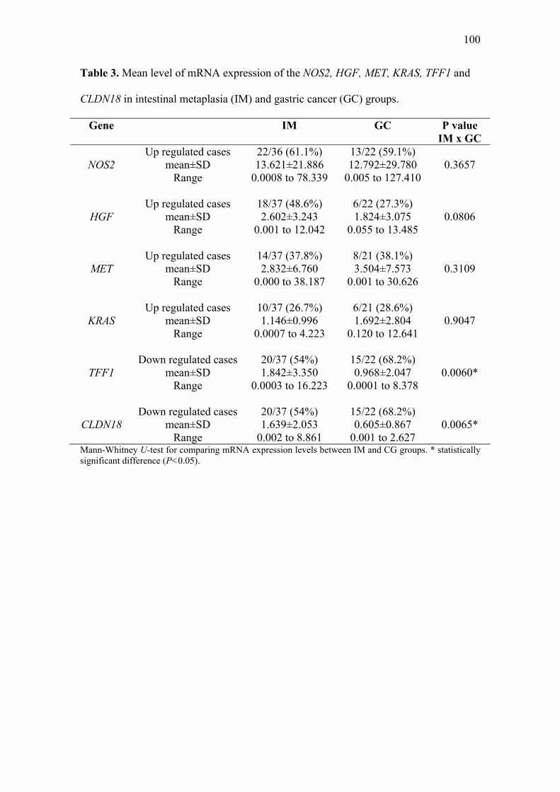
100
Table 3. Mean level of mRNA expression of the NOS2, HGF, MET, KRAS, TFF1 and
CLDN18 in intestinal metaplasia (IM) and gastric cancer (GC) groups.
Gene IM GC P value IM x GC
Up regulated cases 22/36 (61.1%) 13/22 (59.1%) NOS2 mean±SD 13.621±21.886 12.792±29.780 0.3657
Range 0.0008 to 78.339 0.005 to 127.410 Up regulated cases 18/37 (48.6%) 6/22 (27.3%)
HGF mean±SD 2.602±3.243 1.824±3.075 0.0806 Range 0.001 to 12.042 0.055 to 13.485 Up regulated cases 14/37 (37.8%) 8/21 (38.1%)
MET mean±SD 2.832±6.760 3.504±7.573 0.3109 Range 0.000 to 38.187 0.001 to 30.626 Up regulated cases 10/37 (26.7%) 6/21 (28.6%)
KRAS mean±SD 1.146±0.996 1.692±2.804 0.9047 Range 0.0007 to 4.223 0.120 to 12.641 Down regulated cases 20/37 (54%) 15/22 (68.2%)
TFF1 mean±SD 1.842±3.350 0.968±2.047 0.0060* Range 0.0003 to 16.223 0.0001 to 8.378 Down regulated cases 20/37 (54%) 15/22 (68.2%)
CLDN18 mean±SD 1.639±2.053 0.605±0.867 0.0065* Range 0.002 to 8.861 0.001 to 2.627
Mann-Whitney U-test for comparing mRNA expression levels between IM and CG groups. * statistically significant difference (P<0.05).
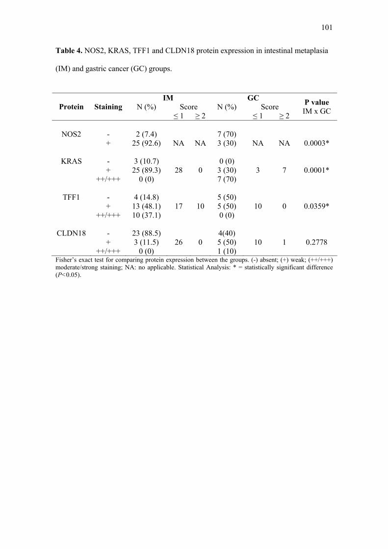
101
Table 4. NOS2, KRAS, TFF1 and CLDN18 protein expression in intestinal metaplasia
(IM) and gastric cancer (GC) groups.
Fisher’s exact test for comparing protein expression between the groups. (-) absent; (+) weak; (++/+++) moderate/strong staining; NA: no applicable. Statistical Analysis: * = statistically significant difference (P<0.05).
IM GC N (%) Score N (%) Score Protein Staining
≤ 1 ≥ 2 ≤ 1 ≥ 2
P value IM x GC
NOS2
-
2 (7.4)
7 (70)
+ 25 (92.6) NA NA 3 (30)
NA NA 0.0003*
KRAS - 3 (10.7) 0 (0) + 25 (89.3) 28 0 3 (30) 3 7 0.0001* ++/+++ 0 (0) 7 (70)
TFF1 - 4 (14.8) 5 (50) + 13 (48.1) 17 10 5 (50) 10 0 0.0359* ++/+++ 10 (37.1) 0 (0)
CLDN18 - 23 (88.5) 4(40) + 3 (11.5) 26 0 5 (50) 10 1 0.2778 ++/+++ 0 (0) 1 (10)
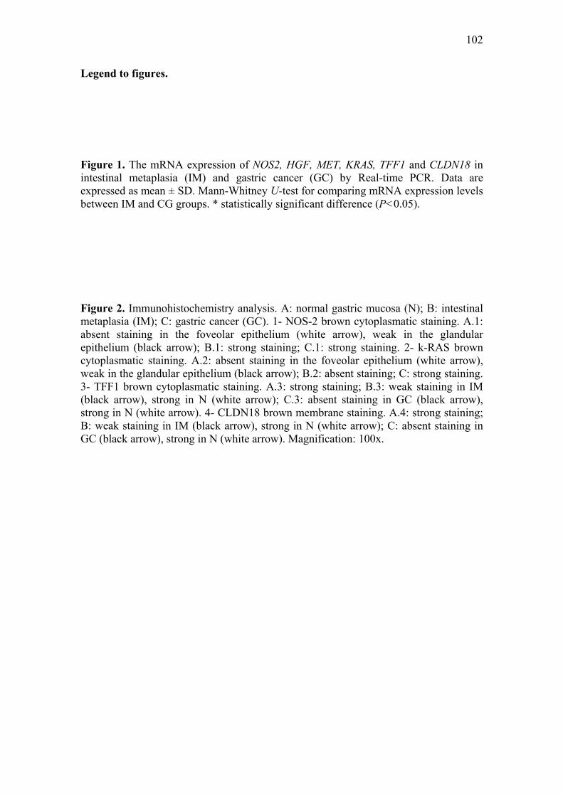
102
Legend to figures. Figure 1. The mRNA expression of NOS2, HGF, MET, KRAS, TFF1 and CLDN18 in intestinal metaplasia (IM) and gastric cancer (GC) by Real-time PCR. Data are expressed as mean ± SD. Mann-Whitney U-test for comparing mRNA expression levels between IM and CG groups. * statistically significant difference (P<0.05). Figure 2. Immunohistochemistry analysis. A: normal gastric mucosa (N); B: intestinal metaplasia (IM); C: gastric cancer (GC). 1- NOS-2 brown cytoplasmatic staining. A.1: absent staining in the foveolar epithelium (white arrow), weak in the glandular epithelium (black arrow); B.1: strong staining; C.1: strong staining. 2- k-RAS brown cytoplasmatic staining. A.2: absent staining in the foveolar epithelium (white arrow), weak in the glandular epithelium (black arrow); B.2: absent staining; C: strong staining. 3- TFF1 brown cytoplasmatic staining. A.3: strong staining; B.3: weak staining in IM (black arrow), strong in N (white arrow); C.3: absent staining in GC (black arrow), strong in N (white arrow). 4- CLDN18 brown membrane staining. A.4: strong staining; B: weak staining in IM (black arrow), strong in N (white arrow); C: absent staining in GC (black arrow), strong in N (white arrow). Magnification: 100x.
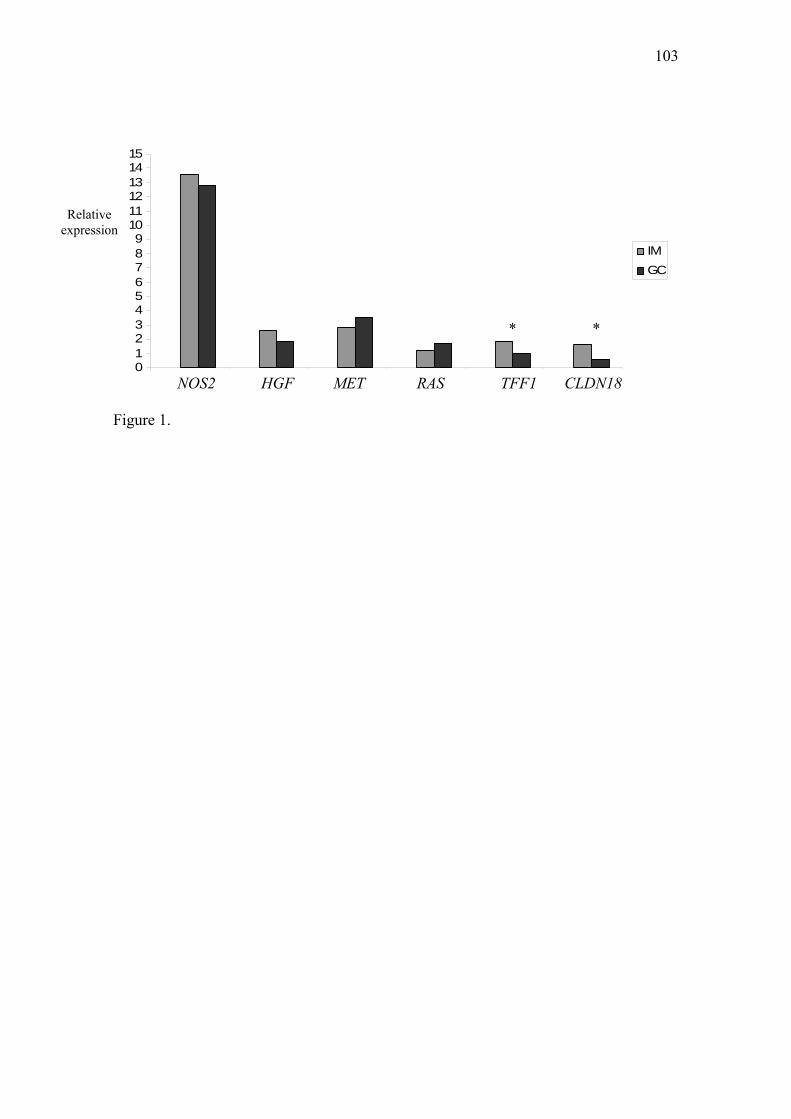
103
0123456789
101112131415
1 2 3 4 5 6
IMGC
Relative expression
NOS2 TFF1 HGF RAS MET CLDN18
* *
Figure 1.
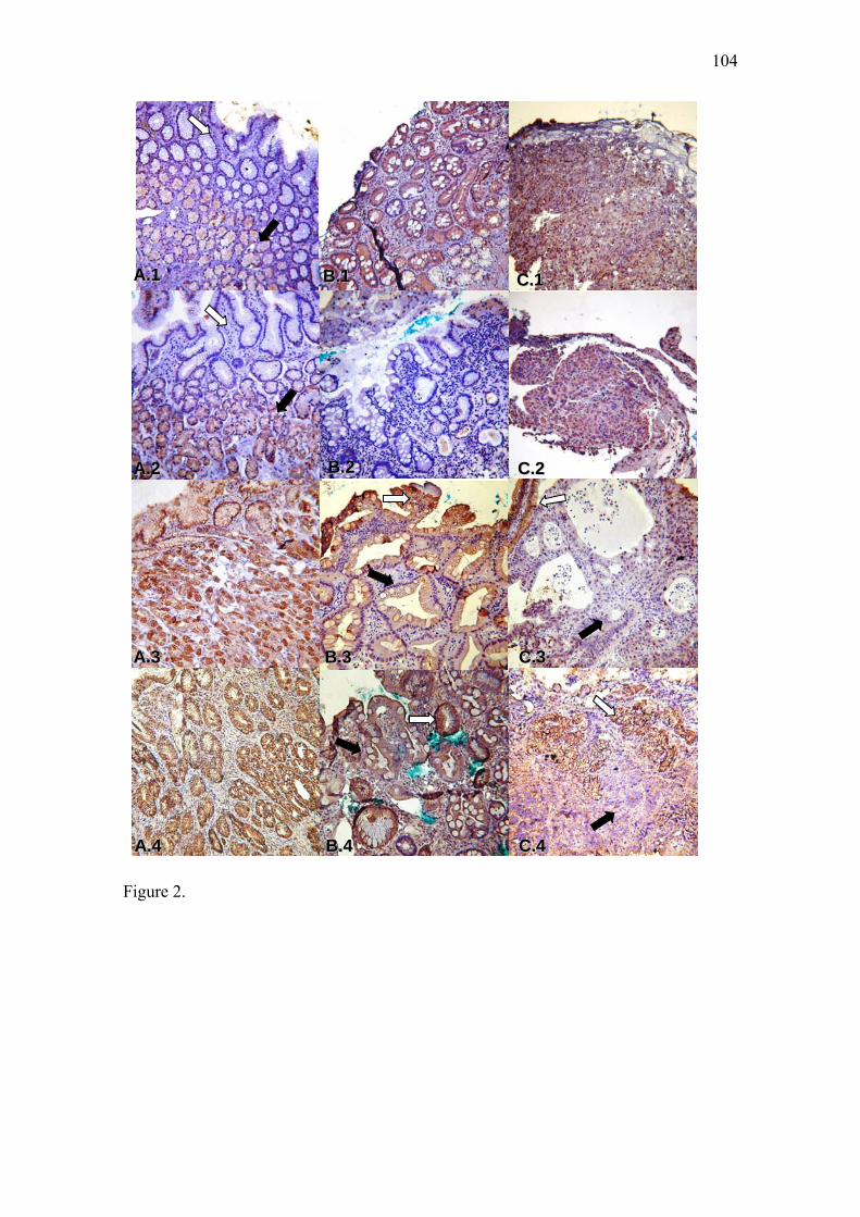
104
AA..11
AA..22
AA..33
BB..11
BB..22
BB..33
AA..44 BB..44
CC..11
CC..22
CC..33
CC..44
Figure 2.

Artigo III
106
Artigo III: NOS2, HGF, MET, KRAS, TFF1 and CLDN18 expression in gastric
ulcer and gastric cancer.
Márcia Cristina Duarte1, Mabel Tatty de Medeiros Fracassi2, Kenji Miyazaki3, Ana
Elizabete Silva1.
1. UNESP - São Paulo State University, Biology Department, Cristóvão Colombo
Street, 2265, São José do Rio Preto, São Paulo State, Brazil.
2. Pathology Laboratory, Sírio-Libanes Hospital, Dona Adma Jafet Street, 91, São
Paulo, São Paulo State, Brazil.
3. Endoscope Service, Base Hospital, Brigadeiro Faria Lima Av., 5416, São José do Rio
Preto, São Paulo State, Brazil.
Correspondence to: Ana Elizabete Silva, Cytogenetic and Molecular Biology
Laboratory, Biology Department, Cristóvão Colombo Street, 2265, São José do Rio
Preto, São Paulo State, Brazil. Telephone: +55 17-3221-2384, Fax: +55 17-3221-2390.
E-mail: [email protected]
Interest conflicts: The authors declare that they have no competing interests.
Funding Source: We want to thank Brazilian Agencies FAPESP and CAPES, for
financial support.
107
Abstract
Background: Gastric ulcer (GU) has been pointed as a pre-neoplastic condition of the
stomach cancer. However, there are few studies regarding gene expression changes in
this lesion, as well as its relationship to gastric carcinogenesis. We examined mRNA
and protein expressions of important genes involved in cell mechanisms and gastric
mucosa protection in GU and gastric cancer (GC).
Methods: Gastric mucosal biopsies specimens were taken from 30 patients with GU
and 22 with GC and their respective normal mucosa. mRNA was extracted and used in
Real Time PCR assay to evaluate mRNA expression for NOS2, HGF, MET, KRAS,
TFF1 and CLDN18. Immunohistochemistry was performed in order to evaluate the
staining of these proteins in the respective tissues.
Results: Up-regulation was found respectively in NOS2, HGF, MET and KRAS in
43.3%, 26.7%, 17.2% and 26.7% of GU cases (4.4-, 1.05-, 2.8- and 2.6-fold,
respectively). Concerning GC, the same genes were up-regulated about 12.8-, 1.8-, 3.5-
and 1.7-fold in 59.1%, 27.3%, 38.1% and 28.6% of the cases, respectively. TFF1 and
CLDN18 were down-regulated in 73.3% and 58.6% of GU (mRNA mean levels of 0.53
and 0.93, respectively) and 68.2% of GC cases (mRNA mean levels of 0.97 and 0.60,
respectively). Despite the absence of an exact relationship between mRNA and protein
expression, in general, immunohistochemistry in NOS2, KRAS, TFF1 and CLDN18
proteins evidenced positive staining (weak to strong) in GU and GC specimens.
Conclusions: These results suggest that ulcer and gastric cancer share changes in the
expression of key genes that participate in important cell mechanisms, which can both
be involved in the process of ulcer healing and also act in the initiation and progression
of gastric carcinogenesis.
108
Introduction
Gastric cancer development is a multistep process that involves H. pylori
infection, chronic gastritis, atrophic gastritis, intestinal metaplasia and dysplasia1.
However, the theory that gastric carcinoma arises within the site of a benign peptic ulcer
has been increasing in the past decades2. Studies have verified a coexisting gastric
cancer in two percent of patients with a diagnosis of gastric ulcers3. Subsequently, it
was estimated that gastric ulcer patients had a 1.8 times higher risk of developing
gastric carcinoma, which was attributed to presence of atrophic gastritis due to H. pylori
as a common primary factor to both gastric cancer and gastric ulcer patients4. Moreover,
the surgery performed to reduce the gastric acid secretion in patients with gastric ulcer
history, did not decrease the gastric cancer risk5.
Gastroduodenal peptic ulcer is characterized by circumscribed loss of tissue that
occurs in portions of the digestive tract exposed to chlorohydro-peptic secretion,
occurring in 5 to 10% of population. Among the etiological factors, those who break the
balance between aggressive factors (acid and pepsin) and protective factors from gastric
mucosa barrier, stand out acetylsalicylic acid, biliary acids, nonsteroidal anti-
inflammatories, corticosteroids, alcohol and tobacco6. Nevertheless, the most important
environmental factor is H. pylori infection, affecting over 90% adults with duodenal
ulcer and 70% to 90% of those with gastric ulcer7.
To date, very little is known about genetic alterations and gene expression
changes in gastric ulcer. In a recent report of our group, trissomies of the chromosomes
7 and 17 in patients with gastric ulcer have been detected, mainly in the positive H.
pylori cases, besides immunostaining to p538. Furthermore, it is well known that, during
the process of ulcer healing, complex biological responses occur, including cell
proliferation, migration, differentiation, regeneration, active angiogenesis, and
109
extracellular matrix deposition9,10, many of them also participating in the carcinogenic
process. In addition, complex interaction of many proteins at the injury site such as
growth factors (EGF, TGF-α, TGF-β, FGF and HGF/MET), peptides trefoil (TFF),
COX-2, among others are also required9,10. Thus, several studies have reported
increased expression of the proteins CGRP11, TFF1 and TFF212,13, and of the genes
HGF14, TGF-β and TGF-βR215 and COX-216 in the mucosa of patients with gastric ulcer
in healing.
Taking into account the potential of gastric ulcer as a precancerous lesion of the
stomach, and that gene expression deregulation occurs in initial steps of carcinogenesis,
genetic studies about important genes involved in stomach carcinogenesis in patients
with gastric ulcer can be useful to understand if these lesions have genetic changes in
common. In the presence of these facts, we analyzed the expression levels of the mRNA
and protein of key genes deregulated in the gastric cancer, such as NOS2, HGF, MET,
KRAS, TFF1 and CLDN18, in patients with gastric ulcer in comparison with normal
gastric mucosa and patients with gastric cancer and their potential relation with
clinicopathological variables such as H. pylori infection.
Material and Methods
Samples
We analyzed samples for 30 patients with gastric ulcer and 22 with gastric
adenocarcinoma, who had undergone upper endoscopy from March 2006 to March
2008 at the Base Hospital, São José do Rio Preto, SP, Brazil. Three biopsies of lesion
region and other three biopsies of apparently normal region were collected of each
patient, mainly in the antrum and corpus of the stomach. Immediately after this
procedure, they were put in RNA-later reagent (Ambion) and stored at 4ºC for one day,
110
and then, at –20ºC until RNA and DNA extraction. Archival paraffin-embedded,
formalin-fixed tissues were used for immunohistochemical staining of the proteins.
Information about age, gender, smoking and drinking habits and the written informed
consent was obtained from all patients, and the study was approved by the National
Research Ethics Committee (CONEP).
The gastric ulcer group (GU) was composed of 22 men and 8 women, with a
mean age of 54.90±12.37 years (range 30 to 75 years), mainly smokers (64.3%) and non
drinking (60.7%), while the gastric cancer group (GC) was composed of 17 men and 5
women, with a mean age of 62.45±42.07 years (range 38 to 86 years), which the
majority (62.5%) was smokers and alcoholics. All specimens were histopathologically
diagnosed. Gastric adenocarcinoma was classified as intestinal (57.9%) and diffuse
(42.1%), according to the Laurén classification17.
RNA and DNA extraction and reverse transcription
Total cellular RNA and DNA were extracted from gastric biopsies using the
Trizol reagent (Invitrogen) according to the manufacturer’s protocol. RNA and DNA
concentration was determined in a NanoDrop® ND1000 spectrophotometer, and RNA
integrity was visualized on 1% agarose gel with 2 fragments of 18S and 28S rRNA.
DNA samples were maintained in freezers at -20ºC and used for molecular diagnosis of
H. pylori. RNA samples were stored in freezers at -80ºC and used for reverse
transcription. cDNA was synthesized from 5 μg of total RNA using random primers and
High Capacity cDNA Archive Kit (Applied Biosystems), according to the
manufacturer’s instructions. The integrity of all cDNA preparations was tested by PCR
assay of the β-actin gene. Briefly, for total volume of 20μL, have been used 1 μL of c-
DNA, 1.5mM MgCl2, 0.2mM dNTPs, 0.5μM each primer (F-
5’GAGGCACTCTTCCAGCCTTC3’ and R-5’GTTGGCGTACAGGTCTTGC3’) and
111
1.5 U of Taq DNA polymerase in buffer 1 X. The cycling thermal conditions were as
follow: a initial step at 94°C for 3 minutes, follow by 35 amplification cycles at 94 °C
for 45 seconds, 61ºC for 30 seconds and 72 ºC for 1 minute and a final extension at
72°C for 10 minutes. A 114 bp band was visualized on 2% agarose gel stained with
ethidium bromide.
Quantitative Real Time PCR
The expression of NOS2, HGF, MET, KRAS, TFF1 and CLDN18 mRNA was
measured by real time PCR, based on the SYBR Green methodology, using an ABI
Prism® 7300 (Applied Biosystems). β-actin gene was used as reference. The primer
sequences used in the study were obtained using Primer3 software
(http://frodo.wi.mit.edu/cgi-bin/primer3/primer3_www.cgi) with exception of CLDN18
sequence, obtained of Sanada et al18. The primer sequences were: NOS2 F-
5’CTTCACCATAAGGCCAAAGG3’ and R-5’AGCTCATCTGGAGGGGTAGG3’,
HGF F-5’TGGCCATGAATTTGACCTC3’ and R 5’CCAGGGCTGACATTTGATG3’,
MET F-5’GCAAGCAAAAAGTTTGTCCAC3’ and R-5’TGGCAAGACCAAAA
TCAGC3’, KRAS F-5’AGAGTGCCTTGACGATACAGC3’ and R-
5’TCCCTCATTGCACTGTACTCC3’, TFF1 F-5’GGCCCAGACAGAGACGTG3’
and R-5’ACAGCAGCCCTTATTTGCAC3’, CLDN18 F-5’GATCGTAGGCATCGTC
CTGG3’ and R-5’GGATGCATTTCAGGGCAAAG 3’and β-actin F-5’TGCCCT
GAGGCACTCTTC3’ and R-5’CGGATGTCCACGTCACAC3’.
An initial validation step with 10-fold dilution series was performed to obtain
the amplification efficiencies of the primers (E), calculated according to the equation E
= 10[-1/slope]. The efficiencies of amplification ranged between 90% and 97%. After the
validation step, the real time PCR assays were performed in 10 μL of SYBRTM Green
Master Mix (Applied Biosystems), 25 ng of cDNA and 0.6 μM of β-actin, TFF1 and
112
CLDN18 primers, 0.7 μM of KRAS primer and 0.8 μM of NOS2, HGF and MET
primers. Thermal cycling conditions for all the genes were: 2 min at 50ºC and 10 min at
95ºC for initial denaturation, followed by 40 cycles at 95°C for 15 s and 60°C for 1 min,
and a dissociation step at 95ºC for 15 s, 60ºC for 30 s and 95ºC for 15 s. Triplicates of
the samples were assayed in each run. β-actin, α-tubulin and β2-microglobulin
housekeeping genes were evaluated to case and control samples, to verify which
contained the lowest variation of amplification between the samples. After this, β-actin
gene was used as reference because it showed the lowest variation. The relative
expressions of the genes were analyzed using the 2-ΔΔCt method, according to the
description by Livak and Schimittgen19. First, the mean of cycle number at the threshold
level of fluorescence (Ct) for each sample was determined. Next, the ΔCt value was
calculated as being the difference between the Ct value of target gene and the Ct value
of β-actin: ΔCt = Ct (target gene) - Ct (β-actin). Finally, the ΔΔCt value and the
normalized target gene expression were calculated: ΔΔCt = ΔCt (ulcer or cancer tissues)
- ΔCt (normal adjacent mucosa). Normalized expression in a sample = 2-ΔΔCt.
PCR for H. pylori diagnosis
DNA samples extracted from the adjacent normal gastric mucosa were used for
molecular diagnosis of the H. pylori bacterium, because it is a more efficient method
than histology and urease test20. The bacterium genes urease and tsaA were amplified
by Multiplex PCR, using the specific primers F- 5’ TTCCTGATGGGACCAAACTC 3’
and R- 5’ TTACCGCCAATGTCAATCAA 3’, and (F- 5’
CCTGCCGTTTTAGGAAACAA 3’ and R- 5’ TCCGCATTCCTACCTAATGG 3’) in
order to amplify, respectively, segments of 316 pb and 413 pb. The housekeeping
human gene CYP1A1 (F- 5’CTCACCCCTGATGGTGCTAT 3’ and R- 5’
TTTGGAAGTGCTCACAGCAG 3’) that amplifies a 226 pb segment was used as
113
quality control DNA. Positive and negative controls were used in all experiments. The
PCR assay solution consisted of 4 mM MgCl2, 0.3 mM dNTPs, 0.6 μM of each primer,
and 1.75 U Taq DNA polymerase in 1 X buffer. The thermal cycling conditions were: 5
min at 95ºC, followed by 35 cycles at 95ºC for 30 s, 60ºC for 30 s, and 72ºC for 1 min,
and a final extension step at 72ºC for 10 min. The PCR products were separated on 2%
1000 agarose gel (Invitrogen) and stained with ethidium bromide. The assay was
considered positive when at least one of the bacterial PCR products was present.
Immunohistochemical Analysis
Four-micron-thick tissue sections were heat-fixed on glass slides at 37°C
overnight. Slides were then rehydrated through sequential solutions of xylene for 5 min
(4X), four baths in 100% ethanol (3X), 95 % ethanol (1X), 80% ethanol (1X), 50%
ethanol (1X) and distilled water (2X). Freshly prepared 10mM EDTA-TRIS buffer (pH
9.0) for KRAS and 10mM Citrate buffer (pH 6.0) for NOS2, HGF, MET, TFF1 and
CLDN18 were pre-heated for 40 min in a stem-pan (Exotic, Arno) until 95ºC and then,
slides were put in the buffer for 30 min for antigen retrieval. After cooled for 20 min at
ambient temperature, the endogenous peroxidase activity was blocked with 3% H2O2
treatment for 10 minutes. Then, the sections were incubated for 1 hour at room
temperature with specific antibody for TFF1 (mouse monoclonal anti-pS2, clone BC04,
Invitrogen, 1:50), HGF (Monoclonal anti hepatocyte growth factor antibody, clone
24612.111, Sigma, 1:50) and MET (Anti hepatocyte growth factor receptor -cMet
antibody, Sigma, 1:50) and overnight at 4ºC with specific antibodies for NOS2 (Rabbit
polyclonal to iNOS, Abcam, 1:200), KRAS (Mouse monoclonal anti-KRAS, clone 9.13,
Invitrogen, 1:50) e CLDN18 (Rabbit polyclonal anti-claudin-18 -c-Term, Zymed,
Invitrogen, 1:200). After being rinsed with Tris-HCl buffer (pH 7.6), the slides were
incubated, for 20 minute each, with biotinylated secondary antibody and incubated with
114
streptavidin-biotin peroxidase, following the manufacturer’s instructions (Histostain
Bulk Kit, Zymed). The immunostain was visualized with 3,3’-diaminobenzidine
tetrahydrochoride (DAB) and counterstained with Mayer’s hematoxylin. Negative
controls were established by replacing the primary antibody with buffer solution.
Normal stomach was used as positive control for TFF1 and CLDN18 antibodies,
and liver, breast carcinoma, lung carcinoma and epidermoid lung carcinoma tissues
were used as positive controls for HGF, MET, NOS2 and KRAS antibodies,
respectively. A single pathologist examined all specimens. All analyses were performed
under a light microscope, and the whole tissue extension of all samples was examined.
Immunostain for protein NOS2 (brown cytoplasm staining) was evaluated by staining
presence as negative (-, absent brown staining) or positive (+, presence brown staining),
whereas for KRAS (brown cytoplasm staining), TFF1 (brown cytoplasm staining) and
CLDN18 (brown membrane staining) proteins, the immunostain was evaluated as
negative (-) or positive: +1 (weakly stained), +2 (moderately stained), and +3 (strongly
stained), as observed in at least 10% of cells. A significant protein expression was
considered only for (+2) and (+3) staining, while (–) and (+1) showed no significant
expression. Immunostaining analysis for HGF and MET proteins was not performed
because antibodies reaction did not produce satisfactory results, after various tests.
Statistical Analysis
Data about RNAm relative expression were described using mean + SD as a
point estimator and the range of values. Nonparametric Mann-Whitney test was used in
the comparisons between the groups, and nonparametric Spearman rank correlation was
used to evaluate interactions among mRNA expression in the groups. Protein expression
was described according to number of positive and negative cases and Fisher’s exact
test was used to compare the groups. The relationship between mRNA and protein
115
levels and clinicopathological variables was evaluated by Fisher’s exact test. All the
tests were accomplished using the GraphPad Instat Software. The level of significance
was set at P < 0.05.
Results
mRNA expression in gastric ulcer and gastric cancer
Table 1 shows the results for the gene expression of NOS2, HGF, MET, KRAS,
TFF1 and CLDN18 in gastric ulcer (GU) and gastric cancer (GC). Relative gene
expression was calculated using normal adjacent gastric mucosa (NM) as reference, and
a mean level of expression around 1.5-fold was considered significant, whether
increased or decreased. Comparisons between mRNA expression levels in GU and GC
were illustrated in Figure 1. NOS2 expression was up-regulated in 43.3% and 59.1% of
GU and GC cases with an increase of 4.5- and 12.8-fold in comparison with normal
mucosa, respectively. HGF and MET mRNA expressions were up-regulated only for
26.7% and 17.2% of GU cases and 27.3% and 38.1% of GC cases, respectively. HGF
mRNA mean level was not observed increased in UG group (1.05-fold), but both HGF
and MET were increased 1.8- and 3.5-fold, in GC group, and MET was increased 2.8-
fold in GU group. Regarding KRAS, 26.7% and 28.6% of cases of GU and GC,
respectively, showed up-regulated, with respective mRNA relative mean levels
increased in 2.6- and 1.7-fold. While for both TFF1 and CLDN18, most of GU (73.3%
and 58.6%, respectively) and GC (68.2% for both genes) cases were down-regulated,
with decreased expression mean levels in both groups, as follows: 0.531 and 0.934 for
GU and 0.968 and 0.605 for GC groups, respectively.
When we compared relative expression mean levels between GU and GC
groups, there was a significant difference only for MET (P= 0.0001, Figure 1).
116
Correlation analysis to compare mRNA expression interaction among genes that
participate in the same signaling pathway, showed in GU group that NOS2 mRNA
expression is related to KRAS mRNA expression (r=0.57, P=0.0009), and with HGF
expression (r=0.64, P=0.0001). In GC group, a correlation between mRNA expression
levels of NOS2 and HGF (r=0.80, P=0.0001) has been observed.
Immunohistochemical analysis for NOS2, KRAS, TFF1 and CLDN18.
Immunohistochemical analyses results in gastric ulcer and gastric cancer
specimens were listed in Table 2. Cytoplasmatic brown-staining for NOS2 was detected
in 61.5% and 30% of ulcer and cancer area, respectively. While NOS2 expression was
visualized in the normal glandular epithelium in 100% and 75% of GU and GC cases
(Figure 2. A.1, B.1 and C.1). In the normal foveolar epithelium, we observed negative
immunostaining in 57 % of UG and 100% of GC cases. Regarding KRAS protein, ulcer
and cancer areas were positive in 76.9% and 100%, respectively, with KRAS showing
moderate (++) and strong (+++) cytoplasmatic staining in 38.5% of GU cases and 70%
of GC specimens. But, most of the normal epithelium of both GU and GC specimens
showed a weak focal staining (+) (Figure 2. A.2, B.2 and C.2). With regards to TFF1
protein, in the UG group, 100% of the specimens had similar patterns with
moderate/strong cytoplasmatic staining (++/+++) between normal epithelium and ulcer
area (Figure 2. A.3 and B.3). But in the GC group only 50% of specimens showed a
weak staining in the cancer cells and the remaining 50% did not express TFF1 protein in
the cancerous areas, despite the fact that the normal epithelium showed moderate/strong
staining for TFF1 (Figure 2. C.3). Positive brown-staining for CLDN18 protein was
visualized in the membrane of gastric normal epithelium in both UG and GC groups.
The ulcer area was positive for CLDN18 in 88.2% of the specimens with a pattern of
weak staining. However, CLDN18 was not expressed in 40% of cancer cells and
117
showed a weak membrane staining (+) in the other 50% of cases, in comparison with
normal epithelium (+++) (Figure 2. A.4, B.4 and C.4). Scores ≤ 1 (absent and weak
staining) and ≥ 2 (moderate and strong staining) were used to compare protein
expression between the GU and GC groups, and a significant difference was found only
for TFF1 protein (P=0.0001).
mRNA and protein expression and clinicopathological variables
Association between NOS2, KRAS, HGF, MET, TFF1 and CLDN18 expression
and variables as age, gender, tobacco and alcohol habits and H. pylori infection was
measured in gastric ulcer and gastric cancer groups. Molecular diagnosis of H. pylori
bacterium showed that 15 of the 26 (57.7%) cases of GU and 11 of the 21 (52.4%) cases
of GC were H. pylori-positive (data not shown). Lower mRNA levels of NOS2 and
HGF and higher levels of CLDN18 expression were significantly associated with H.
pylori infection in GC group (2.29 vs. 28.82, 0.93 vs. 2.96 and 0.90 vs. 0.28,
respectively). For the remaining genes, the comparisons between mRNA and protein
expression and variables studied did not result in any significant association (data not
shown).
Discussion
In this study, we performed the analysis of mRNA and protein expression of
NOS2, HGF, MET, KRAS, TFF1 and CLDN18 in gastric ulcer and gastric cancer
biopsies. Our current results showed similar mRNA expression pattern in GU and GC
groups with increased mRNA mean levels for the genes NOS2, HGF, and KRAS, and
reduced for the genes TFF1 and CLDN18, but significantly different only for MET, due
to higher mRNA expression in GC group. Immunohistochemical analysis of NOS2,
KRAS and CLDN18 protein expressions did not show significant differences between
118
groups, except for the TFF1 protein, where 100% of GC cases showed negative/weak
staining, whereas 100% of GU cases the staining was moderate/strong, in both normal
epithelium and ulcer area.
Although these genes are known to be deregulated in gastric cancer, very little is
known about its expression pattern in gastric ulcer. For example, various studies have
found elevated levels of NOS2 in about 70 to 80% of cases of gastric cancer21-25. NOS2
or iNOS (inducible nitric oxide synthase) is transcriptionally induced in inflammatory
cells by proinflammatory agents and inflammatory cytokines for the production of nitric
oxide (NO)26. High concentration and sustained release of NO by NOS2 during
inflammation can result in the formation of highly toxic oxidizing species such as
peroxynitrites and tissue aggravation and DNA damages26,27.
We found that NOS2 mRNA was 4.5-fold increased in GU and about 13-fold
increased in GC. Immunohistochemistry for NOS2 protein showed that 61.5% of GU
cases were positive, but staining-positive was observed only in 30% of cases of GC.
Previous studies have also shown little correlation between NOS2 mRNA and protein,
indicating a considerable degree of post-transcriptional regulation28,29. Also, our results
are in accordance with the report of Guo et al.16, who found high levels of NOS2
activity during ulcer healing. This study verified a beneficial role of selective NOS2
inhibitor in gastric ulcer healing, reinforcing the suggestion that NO can aggravate
tissue damage. However, there are also observations that NO may benefit ulcer healing
through the promotion of apoptosis in inflammatory cells30. With regards to NOS2
protein expression, Rajnakove et al.31 found that the expression of all isoforms of NOS
in human stomach tumor tissue was significantly lower than normal mucosa, which is
similar to our results, where we found positive staining for NOS2 protein in normal
glandular epithelium of most GU and GC specimens.
119
HGF (Hepatopcyte Growth Factor) and its specific receptor, MET, are
frequently found up-regulated in various types of cancer, as esophagus, salivary gland,
breast, thyroid and bladder cancers32-36. HGF is a cytokine that induces many biological
functions as promoting cell migration, cell proliferation, angiogenesis, apoptosis
inhibition, and morphogenesis in hepatocytes and many epithelial cells type33.
Similarly, its receptor MET is expressed in the normal epithelium of the majority of
tissues, where it is primarily located at the intercellular junctions together with cell
adhesions molecules. HGF has also been shown to have an important role in repairing
the kidney, gastrointestinal, lung, liver, myocardial tissue, and retinal epithelium37. Our
data showed that about 27% of both GU and GC cases were up-regulated to HGF, with
normal mRNA expression mean levels in GU (1.05) and increased in GC (1.82), but
without significant differences between of the groups. However, for MET, we observed
a significant increase in the mRNA mean level in GC group in comparison with GU
group (3.50 vs. 2.77, respectively), where only 17.2% of GU cases were up-regulated in
relation to 38.1% of GC cases.
Immunohistochemistry analysis was not performed because antibodies for HGF
and MET proteins did not yield satisfactory results. Previous reports associated with
high expression of HGF and MET in gastric ulcer healing9,14. Similarly, in leg ulcers, it
was observed an increased expression of MET protein in chronic ulcer compared with
controls, however HGF protein expression did not reach statistical significance between
patients with chronic ulcers and controls38.
Growth factors activate epithelial cell migration and proliferation and accelerate
wound/ulcer healing in vivo and in vitro by binding to their specific receptors on the cell
surface, a process that triggers a number of intracellular signaling events that culminate
in the cell migration and proliferation9. The lower HGF mRNA relative expression
120
observed in our study, can be due to technical variation because mRNA was extracted
from biopsies of gastric ulcer containing areas with normal epithelium, so it can mask
the results. Also, it is known that expression of HGF transcripts in normal skin is
greater than in leg ulcers areas37.
Aberrant expression of KRAS, such as overexpression of wild type or point
mutations, is frequent in a variety of human cancer such as pancreatic adenocarcinoma,
lung carcinoma, gastric cancer and pancreatic carcinosarcoma39-42. These mutations are
capable of transducing a strong mitogenic signal to stimulate cell proliferation, which is
directly involved in the initiation and development of human neoplasia43. We found an
up-regulation of KRAS of 2.6- and 1.7-fold in GU and GC, respectively and a
moderate/strong protein expression in 38.5% of GU and 70% of GC cases. The
overexpression of KRAS in UG may be a result of its role in the cellular proliferation to
repair the injured area of gastric mucosa, while in gastric cancer, it might be related to
an increase in the proliferation of gastric cancer cells.
It is noteworthy that target genes of KRAS/ERK signaling pathway have been
overexpressed in gastric cancer, indicating that KRAS overexpression and activation of
RAS/ERK are key events in gastric cancer development, as well as acute myelogenous
leukemia and acute lymphocytic leukemia, breast and prostate cancers44,45. Additionally,
in gastric cancer, the increased expression of KRAS protein was correlated with lymph
nodes metastases and poor prognosis41. To the best of our knowledge, there are only
studies about KRAS protein expression in experimental gastric healing, suggesting a
role in the restoration of the epithelial components during gastric ulcer healing9. Thus,
our data can be the first to show KRAS mRNA and protein expression in human gastric
ulcer, and supporting the experimental data, suggest a role in the ulceration sites
through the cell proliferation promotion.
121
Literature data show an intrinsic network which includes HGF, MET, KRAS and
NOS2 proteins. High NO concentration is observed in injured gastric mucosa, probably
due to a protective role that NO exerts on epithelial cells, mediated by increased gastric
mucosal blood flow, increased mucus and bicarbonate secretion, stabilization of
mucosal mast cells and facilitation of the formation of mucoid cap over the injured
mucosa46. Despite this protective role, NO retards cell migration and induces apoptosis,
presumably by generating reactive metabolites such as peroxynitrites and superoxyde
radicals. This destructive role of NO seems to be inhibited by the expression of HGF
and its up-regulation can be used by gastric cells to cancel the destructive effects of NO.
HGF action can be started through its binding to MET receptor that activates several
intracellular messengers, one of them is KRAS that has been shown to mediate the
HGF-induced migration of gastric cells46,47. In GU group, our data showed high
expression of both NOS2 mRNA and protein, and although HGF has shown mRNA
normal mean levels, a high expression of HGF-receptor (MET) and KRAS were
observed. These results reinforce that all these genes are overexpressed in gastric ulcer
as part of ulcer healing process, which involves epithelial cell proliferation, migration,
re-epithelialization, and reconstruction of gastric glands9. In GC, this pathway
eventually leads to accelerated proliferation, cell morphological and actin filament
changes, and other processes that are important to gastric cancer progression48.
The gastric ulcer healing involves a great number of factors that have stimulated
expression at lesions sites, such as EGF, TNF-α, TNF-β and trefoil peptides (TFFs).
TFF, a group of small molecule polypeptide mainly secreted by gastrointestinal mucous
cells is also a factor that has stimulated expression during gastric ulcer healing9,10. TFF1
(pS2) is stomach specific and has the function of mucosal protection, epithelial
restitution after injury, carcinoma suppression, and apoptosis adjustment10. We found
122
that 100% of gastric cancer cases showed absent or weak staining for TFF1 protein by
immunohistochemistry, inversely, in GU 100% of cases showed moderate/strong
staining. Conversely, TFF1 mRNA mean levels were more reduced in GU (0.53) that in
GC (0.97). Despite the decreased levels of TFF1 mRNA in GU group, the
immunohistochemistry analysis of protein showed increased levels of TFF1 at the ulcer
area, reinforcing its role in the repair and healing of the damaged gastric mucosal. This
finding is in accordance with other studies10,13,49, for example, Shi et al.13 found
decreased levels of TFF1 protein expression from UG (mean score = 73.7%) to GC
(mean score = 57%).
This apparent discordance between mRNA and protein expression in the present
study can be related to higher levels of TFF1 expression in normal adjacent mucosa
than in the ulcer area, which suggests a cytoprotective role in non-ulcerated gastric
mucosa. Similarly, Saitoh et al.49 found that TFF1 mRNA at the non-ulcerated sites in
live human stomach with gastric ulcer was increased during the healing stage and,
consequently, these sites might escape ulceration. In addition, similar to our results in
gastric cancer, several studies have shown a decreased expression of TFF1 protein in
this cancer type13,50-52, and reinforced the findings that deregulation of the TFF1 leading
to decreased protein levels is a critical and common event in human gastric
tumorigenesis.
The other gene evaluated in our study was CLDN18, which belongs to a class of
proteins named claudins that interact with one another to form the tight junctions (TJs)
of epithelial cells53. It is known to function in cell-cell adhesion, membrane polarity,
and regulation of ion and uncharged (aqueous) molecule passage through the
paracellular pathway. Disruption of TJs can cause loss of cell polarity, resulting in an
abnormal influx of growth factors, which could provide auto and paracrine stimulation
123
to tumourigenic epithelial cell18,54. In the present study, CLDN18 mRNA was down-
regulated in 68.2% of GC cases and the protein showed absent or weak staining in 90%
of the specimens. No significant changes of the mRNA expression levels were observed
in GU, despite that, about 59% of GU cases showed reduced mRNA levels in relation to
adjacent normal mucosa. Immunohistochemistry analysis confirmed these findings,
since CLDN18 protein staining was weak in 88% of GU cases and absent/weak in 90%
of GC cases. In gastric cancer, absence or loss of expression of CLDN18 has suggested
a close correlation with carcinoma progression and subsequent metastatic events18,54.
However, in other cancer types such as pancreatic, CLDN18 is frequently up-
regulated53.
To our knowledge, there are no reports about CLDN18 expression in gastric
ulcer. So, our study showed for the first time, weak expression of CLDN18 protein in
ulcer tissues in comparison to normal gastric epithelium, which showed strong
membrane staining in normal areas of both ulcer and gastric cancer specimens. Thus,
this result suggests that a decrease or a loss of CLDN18 expression can also occur in
precancerous lesions such as gastric ulcer and may have a key role in the progression
and maintenance of gastric cancer.
In the present study, we did not observe associations between status of NOS2,
HGF, MET, KRAS, TFF1 and CLDN18 expression with clinicopathological features,
such as age, gender, tobacco and alcohol habits, except that individuals H. pylori-
positive of gastric cancer group had lower expression levels for NOS2 and HGF and
higher expression levels for CLDN18. Conversely, Wang et al.23 found higher levels of
NO and NOS expression in H. pylori- positive group, while Augusto et al.25 did not find
association between NOS2 expression and virulence factor of H. pylori. The
relationship between HGF expression and H. pylori infection is contradictory. Some
124
previous studies found an increased expression of HGF in gastric mucosa colonized by
H. pylori 55. Similarly, CagA virulence factor of H. pylori has been shown to interact
with HGF receptor, MET, thereby stimulating cell growth, motility and invasiveness56,
suggesting a possible link between this growth factor and H. pylori-associated gastritis
and gastric cancer. However, Konturek et al.57 did not find significant correlation
between CagA status and gene and protein expression of growth factors. They also
observed similar gene changes in patients without H. pylori infection. So, to clarify
these divergent results, further studies of H. pylori interaction with growth factors are
necessary.
To our knowledge, there are no reports about interactions between H. pylori
infection and CLDN18 expression. However, it is known that H. pylori recruits and
modifies the distribution of zonula occludens-1 (ZO-1) and junctional adhesion
molecule (JAM), which are normally located at cell tight junctions altering the structure
and function of the apical-junctional complex. This dysfunction may result in loss of
control over cytoskeletal architecture, cell polarity, proliferation and differentiation,
which is characteristic of oncogenic transformation56. Thus, virulence factors of H.
pylori are likely to interact with CLDN18, leading to changes in TJs structure and
function, suggesting that H. pylori infection may change CLDN18 expression.
In conclusion, we verified that gastric ulcer and gastric cancer share changes in
the expression of key genes that participate in important mechanisms as cell
proliferation, apoptosis, maintenance and integrity of gastric mucosa, namely NOS2,
HGF, MET, KRAS, TFF1 and CLDN18. Despite that, in ulcer, these expression changes
appear to be mainly related to the response of the tissue in the healing process. In gastric
cancer, it must act in initiation and progression of gastric carcinogenesis. However, we
cannot rule out the possibility that these alterations in the gene/protein expressions
125
together with other genetic alterations may provide a greater risk of malignant
progression in a percentage of gastric ulcer in the progress to gastric cancer. To clarify
these aspects, further studies should be conducted involving different gene classes
implicated in initial steps and progression of gastric cancer.
Acknowledgements
We want to thank Prof. Dr. Antonio José Manzato for the statistical analysis help and
the Brazilian Agencies, FAPESP and CAPES, for financial support.
References
1 Correa P. The biological model of gastric carcinogenesis. In: Buffer P, Rice J, Bird M,
Boffetta P, editors. Mechanisms of carcinogenesis: Contribution of molecular
epidemiology. Lyon: IARC Scientific Publications, 2004; nº157.
2 Aoki T, Takayama S, Nimura H, Tsutsumi J. Effects of mediacal treatment on gastric
mucosal abnormalities in gastroduodenal ulcer disease. World J. Surg. 2000; 24: 321-7.
3 Yamagata S, Hisamichi S. Precancerous lesions of the stomach. World J. Surg. 1979;
3: 671-3.
4 Hanson LE. The risk of stomach cancer in patients with gastric or duodenal ulcer
disease. N. Engl. J. Med. 1996; 335: 242-9.
5 Lundegardh G, Ekbom A, McLaughlin JK, Nyren O. Gastric cancer risk after
vagotomy. Gut 1994; 35: 946-9.
6 Carvalho AST. Peptic ulcer. J. Pediatr. 2000; 76(Supl.2): S127-S134.
7 Tytgat GNJ, Lee A, Graham DJ, Dixon MF, Rokkas T. The role of infection agents in
peptic ulcer disease. Gastroenterol. Int. 1993; 6: 76-89.
126
8 César ACG, Calmon MF, Cury PM, Caetano A, Borim, AA, Silva AE. Genetic
alterations in benign lesions: chronic gastritis and gastric ulcer. World J. Gastroenterol.
2006; 12(4): 625-9.
9 Tarnawski, AS. Cellular and molecular mechanisms of gastrointestinal ulcer healing.
Dig. Dis. Sci. 2005; 50(S1): S24-S33.
10 Oh TY, Ahn BO, Jang EJ, Park JS, Park SJ, Baik HW, Hahm K-B. Accelerated ulcer
healing and resistance to ulcer recurrence with gastroprotectants in rat model of acetic
acid-induced gastric ulcer. J. Clin. Biochem. Nutr. 2008; 42: 204-14.
11 Tani N, Miyazawa M, Miwa T, Shibata M, Yamaura T. Immunohistochemical
localization of calcitocin gene-related peptide in the human gastric mucosa. Digestion
1999; 60(4): 338-43.
12 Ren JL, Luo JY, Lu YP, Wang L, Shi HX. Relationship between trefoil factor 1
expression and gastric mucosa injuries and gastric cancer. World J. Gastroenterol.
2005; 11(17): 2674-7.
13 Shi S-Q, Cai J-T, Yang J-M. Expression of trefoil factors 1 and 2 in precancerous
condition and gastric cancer. World J. Gastroenterol. 2006; 12(19): 3119-22.
14 Hori K, Shiota G, Kawasaki H. Expression of hepatocyte growth factor and c-met
receptor in gastric mucosa during gastric ulcer healing. Scan. J. Gastroenterol. 2000;
35(1): 23-31.
15 Shih S-C, Tseng KW, Lin SC et al. Expression patterns of transforming growth
factor-beta and its receptors in gastric mucosa of patients with refractory gastric ulcers.
World J. Gastroenterol. 2005; 11(1): 136-41.
16 Guo JS, Cho CH, Wang JY, Koo MWL. Differential effects of selective and non-
selective inhibition of nitric oxide synthase on the expression and activity of
ciclooxygenase-2 during gastric ulcer healing. Eur. J. Pharmacol. 2006; 53: 301-8.
127
17 Laurén, P. The two histological main types of gastric carcinoma: diffuse and so-
called intestinal-type carcinoma: an attempt at a histo-clinical classification. Acta
Pathol. Microbiol. Scand. 1965; 64: 31-49.
18 Sanada Y, Oue N, Mitani Y, Yoshida K, Nakayama H, Yasui W. Down-regulation of
the claudin-18 gene, identified through serial analysis of gene expression data analysis,
in gastric cancer with an intestinal phenotype. J. Pathol. 2006; 208: 633-42.
19 Livak KJ, Schmittgen TD. Analysis of relative gene expression data using Real Time
Quantitative PCR and the 2-ΔΔCt method. Methods 2001; 25: 402-408.
20 César ACG, Cury PM, Payão SLM, Liberatore PH, Silva AE. Comparison of
histological and molecular diagnosis of Helicobacter pylori in benign lesions and
gastric adenocarcinoma. Braz. J. Microbiol. 2005; 36: 12-6.
21 Koh E, Noh SH, Lee YD et al. Differential expression of nitric synthase in human
stomach cancer. Cancer Lett. 1999; 146: 173-80.
22 Rajnakova A, Moochhala S, Goh PMY, Ngoi S-S. Expression of nitric oxide
synthase, cyclooxygenase, and p53 in different stages of human gastric cancer. Cancer
Lett. 2001; 172: 177-85.
23 Wang Y-F, Guo C-L, Zhao L-Z, Yang G-A, Chen P, Wang H-K. Effect of
Helicobacter pylori infection on gastric mucosal pathologic change and level of nitric
oxide and nitric oxide synthase. World J. Gastroenterol. 2005; 11: 5029-31.
24 Chen C-N, Hsieh FJ, Cheng YM, Chang KJ, Lee PH. Expression of inducible nitric
oxide synthase and cyclooxygenase-2 in angiogenesis and clinical outcome of human
gastric cancer. J. Surg. Oncol. 2006; 94: 226-33.
25 Augusto AC, Miguel F, Mendonça S, Pedrazzoli Jr J, Gurgueira SA. Oxidative stress
expression status associated to Helicobacter pylori virulence in gastric diseases. Clin.
Biochem. 2007; 40: 615-22.
128
26 Keklikoglu N, Koray M, Kocaelli H, Akinci S. iNOS expression in oral and
gastrointestinal tract mucosa. Dig. Dis. Sci. 2008; 53(6): 1437-42.
27 Lechner M, Lirk P, Rieder J. Inducible nitric oxide synthase (iNOS) in tumor
biology: the two sides of the same coin. Semin. Cancer Biol. 2005; 15(4): 277-89.
28 Jaiswal M, Larusso NF, Gores GJ. Nitric oxide in gastrointestinal epithelial cell
carcinogenesis: linking inflammation to oncogenesis. Am. J. Physiol. Gastrointest. Liver
Physiol. 2001; 281: 6626–34
29 Donckier JE, Michel L, Delos M, Havaux X, Van Beneden R. Interrelated
overexpression of endothelial and inducible nitric oxide synthases, endothelin-1 and
angiogenic factors in human papillary thyroid carcinoma. Clin. Endocrinol. 2006; 64:
703-10.
30 Akiba Y, Nakamura M, Mori M et al. Inhibition of inducible nitric oxide synthase
delays gastric ulcer healing in the rat. J. Clin. Gastroenterol. 1998; 27: S64-S73.
31 Rajnakove A, Goh PMY, Chan STF, Ngoi SS, Alponat A, Moochala S. Expression
of differential nitric oxide synthase isoforms in human normal gastric mucosa and
gastric cancer tissue. Carcinogenesis 1997; 18: 1841-5.
32 Anderson MR, Harrison R, Atherfold PA, Campbell MJ, Darnton SJ, Obszynska J,
Janowski JAZ. Met receptor signalling: a key effector in esophageal adenocarcinoma.
Clin. Cancer Res. 2006; 12(20): 5936-43.
33 Aoki T, Tsukinoki K, Kurabayashi H et al. Hepatocyte growth factor expression
correlates with cyclooxygenase-2 pathway in human salivary gland tumors. Oral Oncol.
2006; 42: 51-6.
34 Lindemann K, Resau J, Nahrig J et al. Differential expression of c-Met, its ligand
HGF/SF and HER2/neu in DCIS and adjacent normal breast tissue. Histopathology
2007; 51: 54-62.
129
35 Siraj A K, Bavi P, Abubaker J. et al. Genome-wide expression analysis of middle
eastern papillary thyroid cancer reveals c-Met as a novel target for cancer therapy. J.
Pathol. 2007; 213(2): 190-9.
36 Miyata, Y. et al. Phosphorylated hepatocyte growth factor receptor/c-Met is
associated with tumor growth and prognosis in patients with bladder cancer: correlation
with matrix metalloproteinase–2 and –7 and E-cadherin. Hum. Pathol. 2009; 40: 496-
504.
37 Conway K, Ruge F, Price P, Harding KG, Jiang WG. Hepatocyte growth factor
regulation: An integral part of why wounds become chronic. Wound Rep. Reg. 2007;
15: 683-92.
38 Nayeri F, Olsson H, Peterson C, Sundqvist T. Hepatocyte growth factor; expression,
concentration and biological activity in chronic leg ulcers. J. Dermatol. Sci. 2005; 37:
75-85.
39 Wang JY, Lian ST, Chen YF et al. Unique K-ras mutational pattern in pancreatic
adenocarcinoma from Taiwanese patients. Cancer Lett. 2002; 180: 153-8.
40 Aviel-Ronen S, Blackhall FH, Shepherd FA, Tsao MS. K-ras mutations in non-
small-cell lung carcinoma: a review. Clin. Lung Cancer 2006; 8(1): 30-8.
41 Li M, Liu W, Zhu YF, Chen YL, Zhang BZ, Wang R. Correlation of COX-2 and K-
ras expression to clinical outcome in gastric cancer. Acta Oncology 2006; 45(8):1115-9
42 Nakano T, Sonobe H, Usui T et al. Immunohistochemistry and K-ras sequence of
pancreatic carcinosarcoma. Pathol. Int. 2008; 58: 672-7.
43 Song JJ, Lee H, Kim E et al. Transduction effect of antisense K-ras on malignant
phenotypes in gastric cancer cells. Cancer Lett. 2000; 157: 1-7.
130
44 Kornblau SM, Womble M, Qiu YH, et al. Simultaneous activation of multiple signal
transduction pathways confers poor prognosis in acute myelogenous leukemia. Blood
2006; 108: 2358–65.
45 Song IS, Oh NS, Kim H-T et al. Human ZNF312b Promotes the progression of
gastric cancer by transcriptional activation of the K-ras gene. Cancer Res. 2009; 69(7):
3131-9.
46 Nylander-Koski O, Kiviluoto, T, Puolakkainen P, Kivilaakso E, Mustonen H. The
effect of nitric oxide, growth factors, and estrogen on gastric cell migration. J. Surg.
Res. 2007; 143: 230-7.
47 Ridley AJ, Comoglio PM, Hall A. Regulation of scatter factor/hepatocyte growth
factor responses by Ras, Rac, and Rho in MDCK cells. Mol. Cell. Biol. 1995; 15: 1110-
22.
48 Stock M, Otto F. Gene deregulation in gastric cancer. Gene 2005; 360(1): 1-19.
49 Saitoh T, Mochizuki T, Suda T et al. Elevation of TFF1 gene expression during
healing of gastic ulcer at non-ulcerated sites in the stomach: Semiquantification using
the single tube method of polymerase chain reaction. J. Gastroenterol.Hepatol. 2000;
15: 604-9.
50 Machado JC, Nogueira AM, Carneiro F, Reis CA, Sobrinho-Simões M. Gastric
carcinoma exhibits distinct types of cell differentiation: an immunohistochemical study
of trefoil peptides (TFF1 and TFF2 and mucins (MUC1, MUC2, MUC5AC and
MUC6). J. Pathol. 2000, 190: 437-43.
51 Leung WK, Yu J, Chan FKL et al. Expression of trefoil peptides (TFF1, TFF2, and
TFF3) in gastric carcinomas, intestinal metaplasia, and non-neoplastic gastric tissues. J.
Pathol. 2002; 197: 582-8.
131
52 Beckler AD, Roche JK, Harper JC et al. Decreased abundance of trefoil factor 1
transcript in the majority of gastric cancer. Cancer 2003; 98: 2184-91.
53 Karanjawala ZE, IIIei PB, Ashfaq R et al. New markers of pancreatic cancer
identified through differential gene expression analyses: Claudin 18 and Annexin A8.
Am. J. Sur. Pathol. 2008; 32: 188-96.
54 Matsuda Y, Semba S, Ueda J et al. Gastric and intestinal claudin expression at the
invasive front of gastric carcinoma. Cancer Sci. 2007; 98: 1014-9.
55 Kondo S, Shinomura Y, Kanayama S et al. Helicobacter pylori increases gene
expression of hepatocyte growth factor in human gastric mucosa. Biochem. Biophys.
Res. Commun. 1995; 210: 960-5.
56 Nguyen LT, Uchida T, Murakami K, Fujioka T, Moriyama M. Helicobacter pylori
virulence and the diversity of gastric cancer in Asia. J. Med. Microbiol. 2008; 57:
1445–53.
57 Konturek PC, Konturek SJ, Sulekova Z et al. Expression of hepatocyte growth
factor, transforming growth factor alpha, apoptosis related proteins Bax and Bcl-2, and
gastrin in human gastric cancer. Aliment Pharmacol. Ther. 2001; 15: 989-99.
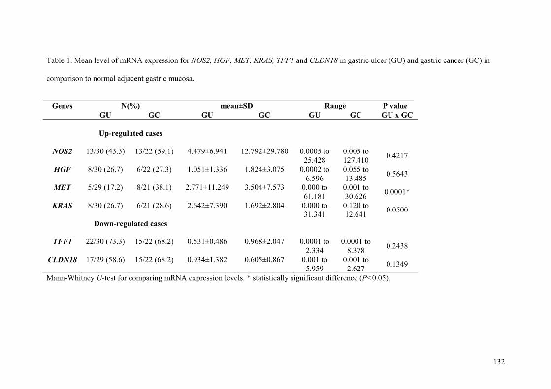
132
Table 1. Mean level of mRNA expression for NOS2, HGF, MET, KRAS, TFF1 and CLDN18 in gastric ulcer (GU) and gastric cancer (GC) in
comparison to normal adjacent gastric mucosa.
Genes N(%) mean±SD Range P value
GU GC GU GC GU GC GU x GC
Up-regulated cases
NOS2
13/30 (43.3)
13/22 (59.1)
4.479±6.941
12.792±29.780
0.0005 to
25.428
0.005 to 127.410
0.4217
HGF 8/30 (26.7) 6/22 (27.3) 1.051±1.336
1.824±3.075
0.0002 to 6.596
0.055 to 13.485 0.5643
MET 5/29 (17.2) 8/21 (38.1) 2.771±11.249
3.504±7.573
0.000 to 61.181
0.001 to 30.626 0.0001*
KRAS 8/30 (26.7) 6/21 (28.6) 2.642±7.390
1.692±2.804
0.000 to 31.341
0.120 to 12.641 0.0500
Down-regulated cases
TFF1 22/30 (73.3) 15/22 (68.2) 0.531±0.486
0.968±2.047
0.0001 to 2.334
0.0001 to 8.378 0.2438
CLDN18 17/29 (58.6) 15/22 (68.2) 0.934±1.382
0.605±0.867
0.001 to 5.959
0.001 to 2.627 0.1349
Mann-Whitney U-test for comparing mRNA expression levels. * statistically significant difference (P<0.05).
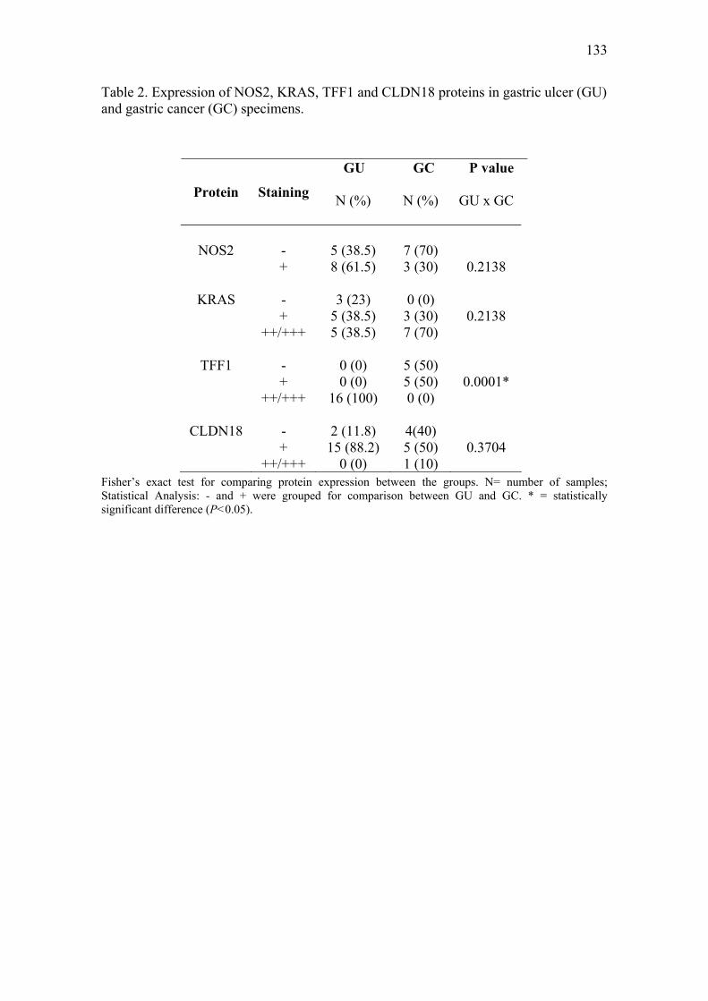
133
Table 2. Expression of NOS2, KRAS, TFF1 and CLDN18 proteins in gastric ulcer (GU) and gastric cancer (GC) specimens.
GU GC P value
N (%) N (%) Protein Staining
GU x GC
NOS2
-
5 (38.5)
7 (70)
+ 8 (61.5) 3 (30)
0.2138
KRAS - 3 (23) 0 (0) + 5 (38.5) 3 (30) 0.2138 ++/+++ 5 (38.5) 7 (70)
TFF1 - 0 (0) 5 (50) + 0 (0) 5 (50) 0.0001* ++/+++ 16 (100) 0 (0)
CLDN18 - 2 (11.8) 4(40) + 15 (88.2) 5 (50) 0.3704 ++/+++ 0 (0) 1 (10)
Fisher’s exact test for comparing protein expression between the groups. N= number of samples; Statistical Analysis: - and + were grouped for comparison between GU and GC. * = statistically significant difference (P<0.05).
134
Legend to figures Figure 1. mRNA expression of NOS2, HGF, MET, KRAS, TFF1 and CLDN18 in gastric ulcer (GU) and gastric cancer (GC) by Real-time PCR. Data are expressed as mean ± SD. Mann-Whitney U-test was used to compare mRNA expression levels between GU and CG groups. * statistically significant difference (P<0.05). Figure 2. Immunohistochemistry analysis in normal gastric mucosa (A), gastric ulcer (B) and gastric cancer (C) tissues. 1- NOS2 brown cytoplasmatic staining. A.1: absent in the foveolar epithelium (black arrow) and weak in glandular epithelium (white arrow); B.1: moderate staining; C.1: strong staining. 2- KRAS brown cytoplasmatic staining. A:2. absent in the foveolar epithelium (black arrow) and weak in glandular epithelium (white arrow); B.2: strong staining; C.2: strong staining. 3- TFF1 brown cytoplasmatic staining. A.3: strong staining; B.3: strong staining C.3: absent staining. 4- CLDN18 brown membrane staining. A.4: strong staining; B.4: strong staining in normal epithelium (white arrow) and absent in the ulcer (black arrow); C.4: strong staining in normal epithelium (white arrow) and absent in tumor cells (black arrow). Magnification 100x.
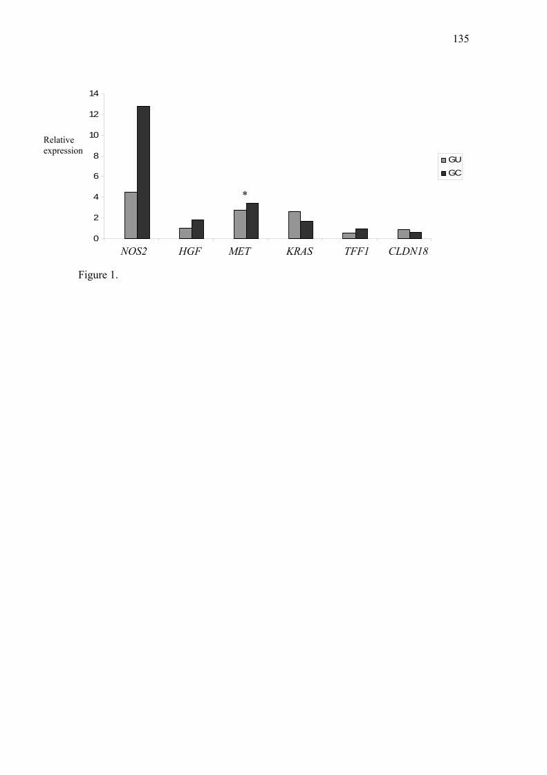
135
0
2
4
6
8
10
12
14
1 2 3 4 5 6
GUGC
*
Relative expression
NOS2 TFF1 HGF KRAS MET CLDN18
Figure 1.
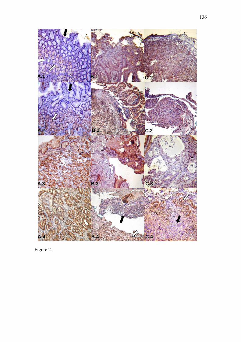
136
Figure 2.
AA..11
AA..22
AA..33
BB..11 CC..11
BB..22 CC..22
BB..33 CC..33
AA..44 BB..44 CC..44

Discussão
138
IV - Discussão
O presente estudo investigou a expressão de RNAm e proteína dos genes TERT,
COX-2, NOS2, HGF, MET, KRAS, TFF1 e CLDN18, com funções relevantes em
processos do ciclo celular e proteção da mucosa gástrica em biópsias de lesões pré-
cancerosas como metaplasia intestinal (MI) e úlcera gástrica (UG) em comparação com
suas respectivas mucosas normais e câncer gástrico (CG). Destaca-se a importância do
estudo de lesões pré-neoplásicas para melhor compreensão das etapas iniciais do
processo de carcinogênese do estômago e possível indicação de alvos para estratégias de
diagnóstico precoce e prevenção futuras.
TERT codifica a enzima transcriptase reversa da telomerase e atua na elongação
dos telômeros durante a divisão celular, sendo fundamental para a senescência e
imortalização celular (BELGIOVONE; CHIODI, MONDELLO, 2008). COX-2 e NOS2
codificam enzimas induzíveis durante o processo inflamatório, exercendo papeis tanto
na defesa da mucosa gástrica quanto na progressão carcinogênica, por meio de suas
atividades proliferativa e angiogênica (STACK; DUBOIS, 2001; SAUKKONEN et al.,
2003; KEKLIKOGLU et al., 2008). HGF, MET e KRAS participam das vias de
sinalização celular, atuando como fatores de promoção da proliferação, migração e
angiogênese (FRIDEY; ADJEI, 2005; TOSCHI; JÄNNE, 2008). TFF1 e CLDN18 são
fatores específicos que atuam na proteção da mucosa gástrica, aumentando as barreiras
existentes contra substâncias nocivas ao organismo (TOMASETTO; RIO, 2005;
SEMBA et al., 2006).
Corroborando dados prévios da literatura, nossos resultados mostraram um
aumento de expressão dos níveis de RNAm para os genes TERT, COX-2, NOS2, HGF,
MET e KRAS nas amostras de CG. A expressão das respectivas proteínas, em geral,
mostrou-se elevada em comparação com a mucosa normal, com exceção das proteínas
139
HGF e MET, para as quais a análise imunohistoquímica não produziu resultados
satisfatórios e, portanto, não foram avaliadas. De modo geral, TFF1 e CLDN18
apresentaram níveis reduzidos de expressão para ambos mRNA e proteínas.
Em câncer gástrico, a reativação da telomerase tem sido descrita em 61 a 90%
dos casos (HU et al., 2004; SABAH et al., 2004; GULMANN et al., 2005). A expressão
aumentada da proteína COX-2 foi relacionada à proliferação celular, angiogênese e
resistência a apoptose (YAMACH et al., 2008). Níveis elevados de expressão gênica e
protéica para NOS2 foram associados com angiogênese, sobrevida e progressão tumoral
(WANG et al., 2005; CHEN et al., 2006; AUGUSTO et al., 2007). HGF e MET são
freqüentemente encontrados com expressão aumentada em câncer gástrico
(KONTUREK et al., 2001; INOUE et al., 2004; STOCK; OTTO, 2005), enquanto
KRAS é preferencialmente ativado por mutação (WATARI et al., 2007), e a expressão
aumentada de sua proteína já foi correlacionada com progressão da carcinogênese
gástrica e metástases em linfonodos (LI et al., 2006).
No presente estudo, a proteína TFF1 estava ausente ou com marcação fraca nas
amostras de CG, embora o nível médio de expressão relativa do mRNA não mostrasse
uma redução significante, enquanto CLDN18 apresentou expressão gênica e protéica
reduzida em CG comparado com a mucosa normal. Alguns estudos têm descrito uma
perda de expressão da proteína TFF1 em 40 a 60% dos casos de câncer gástrico
(MACHADO et al., 2000; LEUNG et al., 2002; SHI; CAI; YANG, 2006) e da proteína
CLDN18 em 57,5% dos casos, correlacionada com baixa sobrevida (SANADA et al.;
2006).
No grupo de metaplasia intestinal, nossos resultados mostraram uma expressão
elevada dos níveis médios de mRNA para TERT, COX-2, NOS2, HGF, MET, TFF1 e
CLDN18, apesar que para TFF1 e CLDN18 54% dos casos de MI tinham baixa
140
expressão em comparação com a mucosa normal. Esses dados foram concordantes com
a expressão das proteínas TFF1 e CLDN18 que apresentaram coloração fraca ou perda
de expressão na maioria dos casos de metaplasia. Para as demais proteínas, a expressão
protéica mostrou-se frequentemente concordante com os resultados de expressão gênica.
Quanto à expressão gênica de KRAS, não foi observada variação significante em relação
à mucosa normal e a proteína mostrou-se fracamente marcada na maioria das amostras.
Estudos genéticos sobre expressão gênica e protéica em metaplasia intestinal são
bastante restritos, mas nossos resultados são concordantes com alguns deles. Por
exemplo, Gulmann et al. (2006) e Sun et al. (2006) verificaram a expressão das
proteínas TERT e COX-2, respectivamente, em 38% e 23% dos casos de metaplasia. A
expressão reduzida de TFF1, tanto RNAm quanto proteína, foi demonstrada por
Fujimoto et al. (2000) em adenomas, MI e CG, enquanto, Kim et al. (2004) observaram
positividade imuno-histoquímica mais fraca em MI que na mucosa normal adjacente.
Alguns autores também demonstraram redução na expressão da proteína CLDN18 em
MI e CG, e sugeriram que esta mudança no nível de expressão é um evento precoce na
carcinogênese gástrica, podendo estar relacionada à progressão e metástase (SANADA
et al., 2006; MATSUDA et al., 2007). Entretanto muito pouco se conhece sobre os
padrões de expressão gênica e protéica de HGF, MET e KRAS em metaplasia intestinal.
Para HGF, Suzuki et al. (2004) verificaram uma redução dos níveis da proteína em
indivíduos com metaplasia intestinal após o tratamento de erradicação da bactéria H.
pylori.
Diante dos resultados obtidos no presente estudo há indicações de que HGF e
MET, associados com aumento da proliferação celular, podem apresentar expressão
desregulada em metaplasia intestinal, assim podendo também participar das etapas
iniciais da carcinogênese gástrica. Ainda neste estudo, foi constatado que KRAS não
141
apresenta mudanças importantes nos níveis de mRNA e proteína em metaplasia
intestinal.
No grupo de úlcera gástrica foi verificado um aumento de expressão relativa do
mRNA para os genes TERT, COX-2, NOS2, MET e KRAS, e expressão diminuída para
TFF1 em comparação com a mucosa normal. HGF e CLDN18 não apresentaram
alterações significantes em seus níveis de expressão gênica, embora cerca de 27% dos
casos de UG tenham apresentado níveis aumentados do mRNA de HGF e 59% dos
casos mostraram baixa expressão gênica de CLDN18. De modo geral, as proteínas
TERT, COX-2, NOS2, KRAS e TFF1 apresentaram marcação positiva em UG,
enquanto CLDN18 apresentou expressão fraca ou ausente em comparação com o
epitélio normal.
Em úlcera gástrica alguns estudos têm avaliado mudanças na expressão gênica e
protéica associadas com o processo de cicatrização. Portanto, nossos resultados estão de
acordo com outros quanto às expressões de COX-2 (GUO et al., 2006; HATAZAWA et
al., 2007), NOS2 (AKIBA et al., 1998; GUO et al., 2006) e TFF1 (SAITOH et al., 2000;
SHI et al., 2006; OH et al., 2008). Os dados da literatura também mostram uma
expressão aumentada de HGF, MET e também COX-2 nos sítios de úlcera gástrica em
cicatrização (HORI et al., 2000; OH et al., 2008; TARNAWSKI, 2005).
No presente estudo, 26,7%, 17,2% e 51,7% dos casos de UG respectivamente,
apresentaram aumento de expressão para HGF, MET e COX-2, apesar do nível médio
de RNAm de HGF ter-se apresentado normal. Portanto, nossos resultados também
corroboram que esses genes devem atuar como fatores importantes no processo de
cicatrização da úlcera, por meio da promoção da proliferação e migração celular e
reconstrução das glândulas gástricas (TARNAWSKI, 2005). Do mesmo modo, COX-2,
NOS2 e TFF1 também estão envolvidos no processo de reparação da mucosa gástrica
142
pela indução de fatores angiogênicos, aumento de fluxo sanguíneo e formação da capa
de muco sobre a lesão (GUO et al., 2006; NYLANDER-KOSKI et al., 2007).
Em ulceras gástricas experimentais, KRAS geralmente apresenta-se com
expressão aumentada, sugerindo seu papel como ativador da proliferação e restauração
dos componentes epiteliais danificados. No entanto, até o presente, não há relatos sobre
o padrão de expressão deste gene em úlceras gástricas humanas. A expressão aumentada
do RNAm e da proteína de KRAS verificada neste estudo, pode corroborar os dados
encontrados para as úlceras experimentais. Da mesma forma, não conhecemos relatos
sobre expressão gênica e protéica de TERT e CLDN18 em ulcera gástrica. Deste modo,
nossos dados indicam uma expressão aumentada de TERT e redução de expressão da
proteína CLDN18 em ulcera gástrica, ambas mudanças que participam do processo
carcinogênico do estômago.
As proteínas avaliadas neste estudo como HGF, MET, KRAS, NOS2, COX-2 e
TFF1 participam de vias de sinalização em comum, responsáveis pela ativação de
fatores importantes associadas com processos de proliferação e migração celular,
apoptose, inflamação, danos oxidativos e reparação da mucosa gástrica (HOFFMANN,
2005; NYLANDER-KOSKI, 2007; CHO et al., 2009). Dessa forma, foi investigada a
ocorrência de correlação entre a expressão desses genes nos grupos de metaplasia,
úlcera e câncer gástrico. Os dados evidenciaram uma correlação direta entre a expressão
gênica de KRAS, HGF e MET em metaplasia intestinal, entre NOS2, RAS e HGF em
ulcera gástrica, e entre NOS2 e HGF em câncer gástrico. Esta via de sinalização pode
iniciar-se com a ligação da proteína HGF ao seu receptor MET, que ativa KRAS e
promove a proliferação e migração celular. KRAS por sua vez, pode induzir a ativação
de NOS2 e COX-2, aumentando a inflamação, apoptose e dano oxidativo nas células
143
gástricas, que pode alterar a homeostase celular e contribuir para o início e progressão
da carcinogênese gástrica (NYLANDER-KOSKI, 2007; CHO et al., 2009).
Em ulcera, contudo, HGF e MET parecem ser induzidos em resposta aos danos
produzidos pelo acúmulo de óxido nítrico nas células epiteliais gástricas, e através de
KRAS, podem promover a reestruturação do tecido ulcerado pela proliferação e
migração de células epiteliais para as áreas danificadas (TARNAWSKY, 2005;
NYLANDER-KOSKI, 2007).
Portanto, os resultados do presente estudo sugerem que mudanças nos padrões
de expressão dos genes ora avaliados podem estar associadas com alterações
importantes envolvidas tanto nas etapas inicias como na progressão maligna, como
observado em metaplasia e câncer gástrico. Dessa forma, evidenciando que metaplasia
intestinal apresenta mudanças de expressão gênica semelhante ao câncer gástrico que
podem conferir maior risco para esta neoplasia. Porém, em úlcera tais alterações devem
estar associadas principalmente com o processo de reparo do epitélio gástrico. Contudo,
ainda deve ser considerado que essas mudanças de expressão envolvendo genes que
também participam do processo carcinogênico, juntamente com outras alterações
genéticas e epigenéticas podem conferir maior risco para progressão da úlcera a
malignização.
Neste trabalho procurou-se verificar a existência de associações entre os níveis
de expressão gênica e protéica com variáveis ambientais e clinicopatológicas, como
sexo, idade, tabagismo, etilismo, infecção por H. pylori e tipo histológico de MI e CG.
Foi observada associação significante entre baixa expressão de TFF1 e tabagismo no
grupo de MI. Este dado sugere que a expressão de TFF1 pode ser modulada por
carcinógenos presentes no tabaco, que enfraquece as defesas da mucosa gástrica,
144
proporcionadas pela expressão de TFF1, e possibilita maior risco de danos ao DNA e
progressão para o fenótipo maligno (TOMASETTO; RIO, 2005; MARTIN et al., 2007).
Também foi verificada uma associação entre indivíduos H. pylori positivos e
expressão reduzida de NOS2 e HGF e expressão aumentada de CLDN18 no grupo de
CG. Há indicações de que a infecção pela H. pylori e fatores de virulência da bactéria
podem alterar o padrão de expressão de alguns genes como o da gastrina, COX-2,
ANXA2, IL-1 e NOS2 (STOICOV et al., 2004; STOCK; OTTO, 2005; WANG et al.,
2005), contudo os resultados são contraditórios. Por exemplo, há relatos de aumento de
expressão de NOS2 em indivíduos infectados por H. pylori comparados com os não
infectados (WANG et al., 2005), como também da falta de associação entre esses
fatores (AUGUSTO et al., 2007). Da mesma forma, para HGF, alguns estudos têm
verificado tanto expressão aumentada em indivíduos H. pylori positivos (SUZUKI et
al., 2004; NGUYEN et al., 2008), como falta de associação (KONTUREK et al., 2004).
Quanto à associação entre expressão de CLDN18 e infecção por H. pylori não
foi encontrado nenhum estudo na literatura, mas com base em outros relatos sobre a
interação de proteínas derivadas de H. pylori com a ZO-1 (Zonula Occludens-1), que
participa das junções “tight” (NGUYEN et al., 2008), pode-se sugerir que a infecção
por H. pylori pode participar na modulação da expressão de CLDN18 em câncer
gástrico. Deve-se ainda se considerar, no presente estudo, o número relativamente
reduzido de amostras das lesões avaliadas para este tipo de associação, deste modo mais
estudos com número maior de casos são necessários para esclarecer estes dados.

Conclusões
146
V - Conclusões
No presente estudo, considerando-se as lesões avaliadas e as técnicas empregadas, é
possível obter-se as seguintes conclusões:
a) Em geral, metaplasia intestinal exibe padrões similares de expressão gênica ao grupo
de câncer gástrico, referente aos genes TERT, NOS2, HGF e MET, mas menor para o
gene COX-2 e mais elevada para TFF1 e CLDN18. KRAS não apresenta mudanças de
expressão significantes nas amostras de metaplasia intestinal;
b) Úlcera gástrica apresenta mudanças de expressão dos genes TERT, NOS2, KRAS,
TFF1 e CLDN18 similares as do câncer gástrico, porém MET é mais expresso no câncer
em relação à úlcera. Tais alterações devem estar relacionadas ao processo de reparo da
mucosa gástrica, como também podem conferir maior risco de malignização;
c) Em câncer gástrico a expressão de TERT, COX-2, NOS2, HGF, MET e KRAS é
aumentada em comparação com a mucosa normal adjacente, enquanto TFF1 e CLDN18
exibem perda de expressão;
d) Não há uma relação precisa entre os níveis de mRNA e expressão das proteínas nos
grupos avaliados. Porém, em geral, a análise imuno-histoquímica confirma expressão
aumentada para as proteínas TERT, COX-2 e NOS2 e diminuída para CLDN18 nos três
grupos avaliados. KRAS está expressa em úlcera e câncer gástrico, enquanto TFF1
exibe perda de expressão em metaplasia intestinal e câncer gástrico;
147
e) Há evidências de correlação entre os níveis de expressão gênica para NOS2, HGF,
MET e KRAS nos três grupos avaliados;
f) De modo geral, não há associação evidente entre os níveis de expressão gênica e
protéica dos genes avaliados com variáveis clinicopatológicas, porém o hábito tabagista
pode estar associado com expressão diminuída de TFF1 no grupo de metaplasia
intestinal e a infecção por H. pylori, pode modular a expressão de NOS2, HGF e
CLDN18, como observado no grupo de câncer gástrico.

Referências Bibliográficas
149
VI - Referencias bibliográficas ABDOU, A. G.; AIAD, H. A. S.; SULTAN, S. M. pS2 (TFF1) expression in prostate carcinoma: correlation with steroid receptor status. APMIS, v. 116, p. 961-971, 2008. AKIBA, Y. et al. Inhibition of inducible nitric oxide synthase delays gastric ulcer healing in the rat. Journal of Clinical Gastroenterology, v. 27; p. S64-S73, 1998. AMBS, S. et al. Frequent nitric oxide synthase 2 expression in human colon adenomas: implication for tumor angiogenesis and colon cancer progression. Cancer Research, v. 58, p. 334-341, 1998. AMIEVA, M. R.; EL-OMAR, E. M. Host-bacterial interactions in Helicobacter pylori infection. Gastroenterology, v.134, p. 306-323, 2008. ANAGNOSTOPOULOS, G. K. et al. Bax, Bcl-2 protein expression in gastric lesions: Immunohistochemical study. Journal of Gastroenterology and Hepatology, v. 20, p. 1674-1678, 2005. ANDERSON, M. R. et al. Met receptor signaling: a key effector in esophageal adenocarcinoma. Clinical Cancer Research, v. 12(20); p. 5936-5943, 2006. AOKI, T. et al. Hepatocyte growth factor expression correlates with cyclooxygenase-2 pathway in human salivary gland tumors. Oral Oncology, v. 42, p. 51-56, 2006. ATIK, E. et al. Inducible nitric oxide synthase and apoptosis in human B cell lymphomas. Molecular and Cellular Biochemistry, v. 290, p. 205-209, 2006. AUGUSTO, A. C. et al. Oxidative stress expression status associated to Helicobacter pylori virulence in gastric diseases. Clinical Biochemistry, v. 40, p.615-622, 2007. AVIEL-RONEN, S. et al. K-ras mutations in non-small-cell lung carcinoma: a review. Clinical Lung Cancer, v. 8(1), p. 30-38, 2006. BECKLER, A. D. et al. Decreased abundance of trefoil factor 1 transcript in the majority of gastric carcinomas. Cancer, v. 98, p. 2184-2191, 2003. BELGIOVINE, C.; CHIODI, I.; MONDELLO, C. Telomerase: cellular immortalization and neoplastic transformation. Multiple functions of a multifaceted complex. Cytogenetic and Genome Research, v. 122; p. 255-262, 2008. BLACKBURN, E. H. Telomerases. Annual Ver Biochemistry, v. 61, p. 113-129, 1992. BRENNAN, P. A. et al. Inducible nitric oxide synthase : correlation with extracapsular spread and enhancement of tumor cell invasion in head and neck squamous cell carcinoma. Head & Neck, DOI 101002/head.20675, p. 208-214, 2008.
150
BRENNAN, P. A. et al. Expression of type 2 nitric oxide synthase and vascular endothelial growth factor in oral dysplasia. Journal of Oral and Maxillofacial Surgery, v.60, p. 1455-1460, 2002. BROOKS, P. et al. Interpreting the clinical significance of the differential inhibition of cyxlooxygenase-2. Rheumatology, v. 38, p. 779-788, 1999. BRUSTMANN, H. Immunohistochemical detection of human telomerase reverse transcriptase (hTERT) and c-kit in serous ovarian carcinoma: A clinicopathologic study. Gynecology Oncology, v. 98, p. 396-402, 2005. CAMPISI, J.; D’ADDA DI FAGAGNA, F. Cellular senescence : when bad things happen to good cells. Nature Reviews: Molecular Cell Biology, v.8; p.729-740, 2007. CARVALHO, A. S. T. Peptic ulcer. Jornal de Pediatria, v. 76(2), p. S127-134, 2000. CÉSAR, A. C. G. et al. Genetic alterations in benign lesions: chronic gastritis and gastric ulcer. World Journal of Gastroenterology, v. 12(4), p. 625-629, 2006. CÉSAR, A. C. G. et al. Aneuploidies, deletion, and overexpression of TP53 gene in intestinal metaplasia of patients without gastric cancer. Cancer Genetics and Cytogenetics, v. 153, p. 127-132, 2004. CHAN, A. O-O. et al. Deregulation of E-cadherin-catenin complex in precancerous lesions of gastric adenocarcinoma. Journal of Gastroenterology and Hepatology, v. 18, p. 534-539, 2003. CHEN, X. et al. Variation in gene expression patterns in human gastric cancers. Molecular Biology of Cell, v. 14, p. 3208-3215, 2003. CHEN, C-N. et al. Expression of inducible nitric oxide synthase and cyclooxygenase-2 in angiogenesis and clinical outcome of human gastric cancer. Journal of Surgery Oncology, v. 94, p. 226-233, 2006. CHEN, H. H. et al. Expression of human telomerase reverse transcriptase (hTERT) protein is significantly associated with the progression, recurrence and prognosis of oral squamous cell carcinoma in Taiwan. Oral Oncology, v.43, p.121-129, 2007. CHEN, H.; LI. Y.; TOLLEFSBOL, T. O. Strategies Targeting Telomerase Inhibition. Molecular Biotechnology, v. 41; p. 194–199, 2009. CHEN, Z. et al. Oct4, a novel marker for human gastric cancer. Journal of Surgical Oncology, v. 99(7); p. 414-419, 2009. CHO, S. O. et al. Involvement of Ras and AP-1 in Helicobacter pylori-Induced Expression of COX-2 and iNOS in Gastric Epithelial AGS Cells. Digestive Disease Science, 2009, in press. CIPRIANI, N. A. et al. MET as a target for treatment of chest tumors. Lung Cancer, v. 63, p.169-179, 2009.
151
CONWAY, K. et al. Hepatocyte growth factor regulation: An integral part of why wounds become chronic. Wound Repair and Regeneration, v. 15, p. 683-692, 2007. CORREA, P. The biological model of gastric carcinogenesis. In: BUFFER, P., RICE, J., BIRD, M., BOFFETTA, P. (eds). Mechanisms of carcinogenesis: Contribution of molecular epidemiology. Lyon: IARC Scientific Publications, nº 157, 2004. CORSO, S.; COMOGLIO, P. M.; GIORDANO, S. Cancer therapy: can the challenge be MET? TRENDS in Molecular Medicine, v.11(6), p.284-92, 2005. CRAWFORD, J. N. The gastrointestinal tract. In: COTRAN R. S.; KUMAR, V.; COLLINS, T. Pathologic basis of disease. 7ª ed. Philadelphia: WB Saunders Company, pp. 775-787, 2005. CHRISTENSEN, J.G.; BURROWS, J.; SALGIA, R. c-Met as a target for human cancer and characterization of inhibitors for therapeutic intervention. Cancer Letters, v. 225, p. 1-26, 2005. DE LUCA, A.; IAQUINTO, G. Helicobacter pylori and gastric diseases: a dangerous association. Cancer Letters, v.213, p.1-10, 2004. DONCKIER, J. E. et al. Interrelated overexpression of endothelial and inducible nitric oxide synthases, endothelin-1 and angiogenic factors in human papillary thyroid carcinoma. Clinical Endocrinology, v. 64, p. 703-710, 2006. FENG, J. et al. The RNA component of human telomerase. Science, v. 269, p. 1236-1241, 1995. FILIPE, M. I. et al. Intestinal metaplasia types and risk of gastric cancer: a cohort study in Slovenia. International Journal of Cancer, v. 57, p. 324-329, 1994. FRIDAY, B. B.; ADJEI, A. A. K-ras as a target for cancer therapy. Biochemica et Biophysica Acta, v. 1756, p. 127-144, 2005. FUJIMOTO, J. et al. DNA hypermetilation at the pS2 promoter region is associated with early stage of stomach carcinogenesis. Cancer Letters, v. 149; p. 125-134, 2000. FUKUYAMA, R. et al. Regional localization of the hepatocyte growth factor (HGF) gene to human chromosome 7 band q21.1. Genomics, v. 11, p. 410-415, 1991. GALIANA, C. et al. High frequency of Ki-ras amplification and p53 gene mutations in adenocarcinomas of the human esophagus. Molecular Carcinogenesis, v. 14, p. 286-293, 1995. GILLEN, D.; McCOLL, K. E. L. Gastroduodenal disease, Helicobacter pylori, and genetic polymorphisms. Clinical Gastroenterology and Hepatology, v. 3, p. 1180-1186, 2005. GRAVALOS, C.; JIMENO, A. HER2 in gastric cancer: a new prognostic factor and a novel therapeutic target. Annals of Oncology, v. 19, p. 1523-1529, 2008.
152
GULMANN, C. et al. Telomerase activity in proximal and distal gastric cancer neoplastic and preneoplastic lesions using immunohistochemical detection of hTERT. Digestive and Liver Disease, v. 37, p. 439-445, 2005. GUO, J. S. et al. Differential effects of selective and non-selective inhibition of nitric oxide synthase on the expression and activity of cyclooxygenase-2 during gastric ulcer healing. European Journal of Pharmacology, v.536, p.301-308, 2006. GUO, J. S. et al. Expression and activities of the three inducible enzymes in the healing of gastric ulcers in rats. World Journal of Gastroenterology, v. 9, p. 1767-1771, 2003. GUTIÉRREZ-GONZÁLEZ, L.; WRIGHT, N. A. Biology of intestinal metaplasia in 2008: more than a simple phenotypic alteration. Digestive and Liver Disease, v. 40, p. 510-522, 2008. HANSSON, L. E. et al. The risk of stomach cancer in patients with gastric or duodenal ulcer disease. New England Journal of Medicine, v. 335, p. 242-249, 1996. HASEGAWA, S. et al. Genome-wide analysis of gene expression in intestinal-type gastric cancers using a complementary DNA microarray representing 23040 genes. Cancer Research, v.62, p. 7012-7017, 2002. HATAZAWA, R. et al. Ciclooxygenase-2/prostaglandin E2 accelerates the healing of gastric ulcers via EP4 receptors. American Journal of Physiology and Gastrointestinal and Liver Physiology, v. 293; p. G788-G797, 2007. HAYASHI, H. et al. Immunohistochemical findings of nitric oxide synthase expression in urothelial transitional cell carcinoma including dysplasia. Oncology Reports, v. 8(6), p. 1275-1279, 2001. HEAVEY, P.M.; ROWLAND, I.R. Gastrointestinal cancer. Best Practice & Res Clinical Gastroenterol, v. 18(2), p.323-36, 2004. HOFFMANN, W. TFF (trefoil factor family) peptide-triggered signals promoting mucosal restitution. Cell Molecular Life Science, v. 62; p. 2932-2938, 2005. HORI, K.; SHIOTA, G.; KAWASAKI, H. Expression of hepatocyte growth factor and c-met receptor in gastric mucosa during gastric ulcer healing. Scandinavian Journal of Gastroenterology, v. 35(1), p. 23-31, 2000. HSU, C. P. et al. Clinical significance of telomerase and its associate genes expression in the maintenance of telomere length in squamous cell carcinoma of the esophagus. World Journal of Gastroenterology, v.11(44), p.6941-7, 2005. HU, L-H. et al. Real-time determination of telomerase reverse transcriptase mRNA in gastric cancer. World Journal of Gastroenterology, v. 10(23), p. 3514-3517, 2004. INOUE, T. et al. Activation of c-Met (hepatocyte growth factor receptor) in human gastric cancer tissue. Cancer Science, v. 95(10), p. 803-808, 2004.
153
INTERNATIONAL AGENCY FOR RESEARCH ON CANCER (IARC). Schistossomes, liver flukes and Helicobacter pylori. Lyon: IARC, 1994. INSTITUTO NACIONAL DO CÂNCER (INCA). Estimativa da incidência e mortalidade por câncer no Brasil. Rio de Janeiro, 2008. Disponível em <www.inca.org.br>. Acesso em: 15 de setembro de 2009. JACKSON, L. M. et al. Cyclooxygenase (COX) 1 and 2 in normal, inflamed, and ulcerated human gastric mucosa. Gut, v. 47, p. 762-770, 2000. JIANG, W. G. et al. Hepatocyte growth factor/scatter factor/scatter factor, the molecular, cellular and clinical implications in cancer. Critical Review of Oncology Hematology, v. 29, p. 209–248, 1999. JOHNSON, S. M.; EVERS, B. M. Translational research in gastric malignancy. Surgical Oncology Clinics of North America, v. 17, p. 323-340, 2008 JOJOV, B. et al. Claudin-18: a dominant tight junction protein in Barrett’s esophagus and likely contributor to its acid resistance. American Journal of Physiology: Gastrointestinal and Liver Physiology, v.293, p.G1106-G1113, 2007. KARANJAWALA, Z. E. et al. New markers of pancreatic cancer identified through differential gene expression analyses: Claudin 18 and Annexin A8. American Journal of Surgical Pathology, v. 32, p. 188-196, 2008. KEKLILOGLU, N. et al. iNOS expression in oral and gastrointestinal tract mucosa. Digestive Disease Science, v. 53, p. 1437-1442, 2008. KELLEY, J. R.; DUGGAN, J. M. Gastric cancer epidemiology and risk factors. Journal of Clinical Epidemiology, v.56, p.1-9, 2003. KIM, B-W. et al. Expression of trefoil peptides in the subtypes of intestinal metaplasia. Peptides, v. 25, p. 779-783, 2004. KIM, C. H. et al. Expression of hepatocyte growth factor and c-Met in hypopharyngeal squamous cell carcinoma. Acta Otolaryngology, v. 126(1), p. 88-94, 2006. KIM, H. S. et al. CDX-2 homeobox gene expression in human gastric carcinoma and precursor lesions. Journal of Gastroenterology and Hepatology, v. 21, p. 436-442, 2006. KIM, M. A. et al. EGFR in gastric carcinomas: prognostic significance of protein overexpression and high gene copy number. Histopathology, v.52(6), p.738-746, 2008. KIM, Y. H. et al. Chromosomal alterations in paired gastric adenomas and carcinomas. American Journal and Pathology, v. 158, p. 655-662, 2001.
154
KONTUREK, P. C. et al. Expression of hepatocyte growth factor, transforming growth factor alpha, apoptosis related proteins Bax and Bcl-2, and gastrin in human gastric cancer. Aliment Pharmacology and Therapy, v. 15, p. 989-999, 2001. KORNPRAT, P. et al. Analysis of trefoil factor family protein 1 (TFF1, pS2) expression in chronic cholecystitis and gallbladder carcinoma. Virchows Archive, v. 446(5); p. 505-510, 2005. KUIPERS, E. J. Exploring the link between Helicobacter pylori and gastric cancer. Aliment Pharmacology and Therapy, v. 13, p. 3-11, 1999. KUIPERS, E. J.; SIERSEMA, P. D. The aetiology and clinical relevance of gastric intestinal metaplasia. Digestive and Liver Disease, v. 36, p. 501-504, 2004. LA TORRE, G. et al. Smoking status and gastric cancer risk: an update meta-analysis of case-control studies published in the past ten years. Tumori, v. 95, p. 13-22, 2009. LAUREN, P. The two histological main types of gastric carcinoma: diffuse and so-called intestinal-type carcinoma: an attempt at a histo-clinical classification. Acta Pathology and Microbiology Scandinavian, v. 64, p. 31-49, 1965. LECHNER, M.; LIRK, P.; RIEDER, J. Inducible nitric oxide synthase (iNOS) in tumor biology: The two sides of the same coin. Seminars in Cancer Biology, v. 15, p. 277-289, 2005. LEFEBVRE, O. et al. Gastric mucosa abnormalities and tumorigenesis in mice lacking the pS2 trefoil protein. Science, v. 274, p .259-262, 1996. LEUNG, W. K.; SUNG, J. J. Y. Review article: intestinal metaplasia and gastric carcinogenesis. Aliment Pharmacology Therapy, v. 16, p. 1209-1216, 2002. LI, M. et al. Correlation of COX-2 and K-ras expression to clinical outcome in gastric cancer. Acta Oncology, v. 45(8), p. 1115-1119, 2006. LINDEMANN, K. et al. Differential expression of c-Met, its ligand HGF/SF and HER2/neu in DCIS and adjacent normal breast tissue. Histopathology, v. 51; p. 54-62, 2007. LINGNER, J. et al. Reverse transcriptase motifs in the catalytic subunit of telomerase. Science, v. 276, p. 561-567, 1997. LIRK, P.; HOFFMANN, G.; RIEDER, J. Inducible nitric oxide synthase – time for reappraisal. Current Drug Targets Inflammatory Allergy, v. 1, p.89-108, 2002. LO, C. Y. et al. High prevalence of cyclooxygenase 2 expression in papillary thyroid carcinoma. European Journal of Endocrinology, v.152, p.545-550, 2005. LUCCI, A. et al. Cyclooxygenase-2 expression in primary breast cancers predicts dissemination of cancer cells to the bone marrow. Breast Cancer Research and Treatment, DOI 10.1007/s10549-008-0135-x, 2008.
155
MA, X-M. et al. Relation of overexpression of S phase kinase-associated protein 2 with reduced expression of p27 e PTEN in human gastric carcinoma. World Journal of Gastroenterology, v. 11(42), p. 6716-6721, 2005. MACHADO, J. C. et al. Gastric carcinoma exhibits distinct types of cell differentiation: an immunohistochemical study of trefoil peptides (TFF1 and TFF2 and mucins (MUC1, MUC2, MUC5AC and MUC6). Journal of Pathology, v. 190, p. 437-443, 2000. MARROGI, A. J. et al. Nitric oxide synthase, cyclooxigenase, and vascular endothelial growth factor in the angiogenesis of non-small cell lung carcinoma. Clinical Cancer Research, v. 6(12), p. 4739-4744, 2000. MARSDEN, P. A. et al. Localization of the human gene for inducible nitric oxide synthase (NOS2) to chromosome 17q11.2-q12. Genomics, v. 19, p. 183-185, 1994. MARTÍNEZ-RUMAYOR, A. et al. Coexpression of hepatocyte growth factor/scatter factor (HGF/SF) and its receptor cMET predict recurrence of meningiomas. Cancer Letters, v. 213, p. 117-124, 2004. MARTIN, M. B. et al. Effects of tobacco smoke condensate on estrogen receptor-alpha gene expression and activity. Endocrinology, v. 148(10); p. 4676-4686, 2007. MATSUDA, Y. et al. Gastric and intestinal claudin expression at the invasive front of gastric carcinoma. Cancer Science, v. 98, p. 1014-1019, 2007. MATSUMOTO, K.; NAKAMURA, T. Emerging multipoint aspects of hepatocyte growth factor. Journal of Biochemistry, v. 119, p. 591–600, 1996. MAVROGIANNOU. E. et al. Real time RT-PCR quantification of human telomerase reverse transcriptase splice variants in tumor cell lines and non-small cell lung cancer. Clinical Chemistry, v.53, p.53-61, 2007. McCARTHY, K. et al. Cyclo-oxigenase-2 (COX-2) mRNA expression and hormone receptor status in breast cancer. EJSO, v. 32, p.707-709, 2006. MEIRELES, S. I. Molecular classifiers for gastric cancer and nonmalignante diseases of the gastric mucosa. Cancer Research, v. 64, p. 1255-1265, 2004. MENNUNI, C. et al. Preventive vaccination with telomerase controls tumor growth in genetically engineered and carcinogen-induced mouse models of cancer. Cancer Research, v. 68, p. 9865-9874, 2008. MEYERSON, M. et al. hEST2, the putative human telomerase catalytic subunit gene, is up-regulated in tumor cells and during immortalization. Cell, v. 90, p. 785-795, 1997. MILLER, S. B. Prostaglandins in health and disease: an overview. Seminars Arthritis and Rheumatism, v. 36, p. 37-49, 2006.
156
MITTAL, A. et al. EGCG dow-regulates telomerase in human breast carcinoma MCF-7 cells, leding to suppression of cell viability and induction of apoptosis. International Journal of Oncology, v. 24, p. 703-710, 2004. MIYATA, Y. et al. Phosphorylated hepatocyte growth factor receptor/c-Met is associated with tumor growth and prognosis in patients with bladder cancer: correlation with matrix metalloproteinase–2 and –7 and E-cadherin. Human Pathology, v. 40, p. 496-504, 2009. MÖBIUS, C. et al. COX2 expression, angiogenesis, proliferation ans survival in Barrett’s cancer. EJSO, v. 31, p. 755-759, 2005. MOISAN, J. P. et al. A gene expressed in human mammary tumor cells under estrogen control (BCEI) is located in 21q223 and defines an RFLP. Cytogenetic Cell Genetics, v. 40, p. 701-702, 1985. MOSS, S. F. et al. Decreased expression of gastrokine 1 and the trefoil factor interacting protein TFIZ1/GNK2in gastric cancer: influence of tumor histology and relationship to prognosis. Clinical Cancer Research, v. 14(13), p. 4161-4167, 2008. NAKAMURA, Y. et al. c-Met activation in lung adenocarcinoma tissues: An immunohistochemical analysis. Cancer Science, v. 98, p. 1006-1013, 2007. NAKANO, T. et al. Immunohistochemistry and K-ras sequence of pancreatic carcinosarcoma. Pathology International, v. 58; p. 672-677, 2008. NAYERI, F. et al. Hepatocyte growth factor; expression, concentration and biological activity in chronic leg ulcers. Journal of Dermatology Science, v. 37, p. 75-85, 2005. NGUYEN, L. T. et al. Helicobacter pylori virulence and the diversity of gastric cancer in Asia. Journal of Medical Microbiology, v. 57; p, 1445–1453, 2008. NIIMI, T. et al. Claudin-18, a novel downstream target gene for the T/EBP/NKX2.1 homeodomain transcription factor, encodes lung- and stomach-specific isoforms through alternative splicing. Molecular Cell Biology, v. 21, p. 7380-7390, 2001. NITTI, D. et al. Recent advances in conventional and molecular prognostic factors for gastric carcinoma. Surgical Oncology Clinics of North America, v. 17, p. 467-483, 2008. NOMELINI, R. S. Production of nitric oxide and expression of inducible nitric oxide synthase in ovarian cystic tumors. Mediators of Inflammation, v. 2008, article ID 186584, p. 1-7, 2008. NYLANDER-KOSKI, O. et al. The effect of nitric oxide, growth factors, and estrogen on gastric cell migration. Journal of Surgical Research, v. 143; p. 230-237, 2007. OH, T. Y. et al. Accelerated ulcer healing and resistance to ulcer recurrence with gastroprotectants in rat model of acetic acid-induced gastric ulcer. Journal of Clininical Biochemistry and Nutrition, v. 42; p. 204-14, 2008.
157
OSAKI, M. et al. Expression of RUNX3 protein in human gastric mucosa, intestinal metaplasia and carcinoma. European Journal of Clinical Investigation, v. 34, p. 605-612, 2004. PAGLIARULO, V. et al. Sensitivity and reproducibility of standardized-competitive RT-PCR for transcript quantification and its comparison with real time RT-PCR. Molecular Cancer, v. 3(5), p. 1-11, 2004. PARK, M. et al. Sequence of MET protooncogene cDNA has features characteristic of tyrosine kinase family of growth-factor receptors. Proceeding of the National Academy of Science of USA, v.84, p.6379-6383, 1987. PARR, C. et al. The hepatocyte growth factor regulatory factors in human breast cancer. Clinical Cancer Research, v. 10, p. 202-211, 2004. PATRA, S. K. Ras regulation of DNA-methylation and cancer. Experimental Cell Research, v. 314, p. 1193-1201, 2008. PENG, T-L. et al. Potential therapeutic significance of increased expression of aryl hydrocarbon receptor in human gastric cancer. World Journal of Gastroenterology, v.15(14), p. 1719-1729, 2009. POPESCU, N. C. et al. Chromosomal localization of three human ras genes by in situ molecular hybridization. Somatic Cell Molecular Genetics, v. 11, p. 149-155, 1985. PRINZ, C.; SCHWENDY, S.; VOLAND, P. H pylori and gastric cancer: Shifting the global burden. World Journal of Gastroenterology, v.12(34), p.5458-5464, 2006. QIAN, J. et al. In vitro modeling of human pancreatic duct epithelial cell transformation defines gene expression changes induced by k-Ras oncogenic activation in pancreatic carcinogenesis. Cancer Research, v. 65(12), p. 5045-5053, 2005. RAJENDRAN, R.; VARKEY, S. Inducible nitric oxide synthase expression is upregulated in oral submucous fibrosis. Indian Journal of Dental Research, v. 18, p. 94-100, 2007. RAJNAKOVA, A. et al. Expression of differential nitric oxide synthase isoforms in human normal gastric mucosa and gastric cancer tissue. Carcinogenesis, v. 18, p. 1841-1845, 1997. REN, J. L. et al. Relationship between trefoil factor 1 expression and gastric mucosa injuries and gastric cancer. World Journal of Gastroenterology, v. 11(17), p. 2674-2677, 2005. ROCCO, A.; NARDONE, G. Diet, H pylori infection and gastric cancer: Evidence and controversies. World Journal of Gastroenterology, v.13(21), p.2901-2912, 2007. RUCHAUD-SPARAGANO, M. H.; WESTLEY, B. R.; MAY, F. E. B. The trefoil protein TFF1 is bound to MUC5AC in human gastric mucosa. Cell Molecular Life Science, v. 61, p. 1946-1954, 2004.
158
SABAH, M. et al. Expression of human telomerase reverse transcriptase in gastrointestinal stromal tumors occurs preferentially in malignant neoplasms. Human Pathology, v.35, p.1231-1235, 2004. SAFATLE-RIBEIRO, A.; RIBEIRO, U.; REYNOLDS, J. C. Gastric stump cancer: What is the risk? Digest Disease, v. 16, p. 159-168, 1998. SAITOH. T. et al. Elevation of TFF1 gene expression during healing of gastic ulcer at non-ulcerated sites in the stomach: Semiquantification using the single tube method of polymerase chain reaction. Journal of Gastroenterology and Hepatology, v. 15; p. 604-609, 2000. SAMOWITZ, W. S. et al. Relationship of Ki-ras in colon cancers to tumor lacation, stage, and survival: a population based stud . Cancer Epidemiology, Biomarkers & Prevention, v. 9, p. 1193-1197, 2000. SANADA, Y. et al. Down-regulation of the claudin-18 gene, identified through serial analysis of gene expression data analysis, in gastric cancer with an intestinal phenotype. Journal of Pathology, v.208, p. 633-642, 2006. SANDS, B. E.; PODOLSKY, D. K. The trefoil peptide family. Annual Reviews of Physiology, v. 58, p. 253-273, 1996. SATEROGLU-TUFAN, N. L.; BIR, F.; CALLI-DEMISKAN, N. Investigation of Her-2 codon 655 single nucleotide polymorphism frequency and c-ErbB-2 protein expression alterations in gastric cancer. World Journal of Gastroenterology, v. 12(20), p. 3283-3287, 2006. SAUKKONEN, K. et al. Cyclooxygenase-2 and gastric carcinogenesis. APMIS, v. 111(10); p. 915-25, 2003. SEMBA, S. et al. Prognostic significance of intestinal claudins in high-risk synchronous and metachronous multiple gastric epithelial neoplasias after initial endoscopic submucosal dissection. Pathology International, v. 58, p. 371-377, 2008. SHAY, J. W.; WRIGHT, W. E. Senescence and immortalization: role of telomeres and telomerase. Carcinogenesis, v.26, p.867-874, 2005. SHI, S-Q.; CAI, J-T.; YANG, J-M. Expression of trefoil factors 1 and 2 in precancerous condition and gastric cancer. World Journal of Gastroenterology, v. 12(19), p. 3119-3122, 2006. SHIH, S-C. et al. Expression patterns of transforming growth factor-beta and its receptors in gastric mucosa of patients with refractory gastric ulcers. World Journal of Gastroenterology, v. 11(1), p. 136-141, 2005. SIRAJ, A. K. et al. Genome-wide expression analysis of middle eastern papillary thyroid cancer reveals c-Met as a novel target for cancer therapy. Journal of Pathology, v. 213(2), p. 190-199, 2007.
159
SMITH, M. G. et al. Cellular and molecular aspects of gastric cancer. World Journal of Gastroenterology, v. 12(19), p. 2979-2990, 2006. SONG, K. Y. et al. expression of the antiapoptosis gene survivin predicts poor prognosis of stage III gastric adenocarcinoma. Japanese Journal of Clinical Oncology, 2009. DOI: 10.1093/jjco/hyp020 STACK, E.; DUBOIS, R. N. Regulation of cyclooxygenase-2. Best Practices in Research Clinical Gastroenterology, v. 15(5), p. 787-800, 2001. STACK, G. et al. Structure and function of the pS2 gene and estrogen receptor in human breast cancer cells. In: LIPPMAN, M. E.; DICKSON, R. B. eds. Breast Cancer. Cellular and Molecular Biology. Boston: Kluwer, p. 185-206, 1988. STOCK, M.; OTTO, F. Gene deregulation in gastric cancer. Gene, v.360, p.1-19, 2005. STOICOV, C. et al. Molecular biology of gastric cancer: Helicobacter pylori infection and gastric adenocarcinoma: bacterisl and host factors responsible for altered growth signaling. Gene, v.341; p. 1-17, 2004. SUN, P-M. et al. The telomerase activity and expression of hTERT gene can serve as indicators in the anti-cancer treatment of human ovarian cancer. European Journal of Obstetrics Gynecology and Reprodutive Biology, v. 130, p. 249-257, 2007. SUN, J. H. et al. Preferential expression of cyclooxygenase-2 in colonic-phenotype of gastric intestinal metaplasia: Association with Helicobacter pylori and gastric carcinoma. Journal of Clinical Gastroenterology, v.40, p.122-128, 2006. SUZUKI, M. et al. Helicobacter pylori eradication treatment modulates epithelial cell proliferation and tissue content of hepatocyte growth factor in the gastric mucosa. Aliment Pharmacology Therapy, v. 20(S1); p. 158-164, 2004 SZABO, S. et al. New molecular mechanisms of duodenal ulceration. Annals of New York Academy of Sciences, v. 1113, p. 238-255, 2007. TAHARA, E. Genetic pathways of two types of gastric cancer. In: BUFFER, P., RICE, J., BIRD, M., BOFFETTA, P. (eds). Mechanisms of carcinogenesis: Contribution of molecular epidemiology. Lyon: IARC Scientific Publications, nº 157, 2004. TAM, P. K. H. Pathophysiology of childhood duodenal ulcer. Gut, v. 28, A 1357, 1987. TANI, N. et al. Immunohistochemical localization of calcitocin gene-related peptide in the human gastric mucosa. Digestion, v. 60(4), p. 338-343, 1999. TARNAWSKI, A. S. Cellular and molecular mechanisms of gastrointestinal ulcer healing. Digestive Disease Science, v.50(Suppl 1), p.S24-S33, 2005. TERRIN, L. et al. Relationship between tumor and plasma levels of hTERT mRNA in patients with colorectal cancer: implications for monitoring of neoplastic disease. Clinical Cancer Research, v. 14, p. 7444-7451, 2008.
160
TODD, J.A. et al. Gastric ulcer and malignancy – is there a need for follow-up endoscopy? Aliment Pharmacology and Therapy, v. 19, p. 989-991, 2004. TOMASETTO, C.; RIO, M-C. Pleiotropic effects of trefoil factor 1 deficiency. Cell Molecular and Life Science, v. 62, p. 2916-2920, 2005. TOSCHI, L.; JÄNNE, P. A. Single-agent and combination therapeutic strategies to inhibit hepatocyte growth factor/MET signaling in cancer. Clinical Cancer Research, v. 14; p. 5941-5946, 2008. TSATSANIS, C. et al. Signalling networks regulating cyclooxygenase-2. International Journal of Biochemical and Cell Biology, v. 38, p. 1654-1661, 2006. TSUKAMOTO, T. et al. Down-regulation of a gastric transcription factor, Sox2, and ectopic expression of intestinal homeobox genes, Cdx1 and Cdx2: inverse correlation during progression from gastric/intestinal-mixed to complete intestinal metaplasia. Journal of Cancer Research Clinical Oncology, v. 130, p. 135-145, 2004. TU, S. P. et al. Restoration of XAF1 expression induces apoptosis and inhibits tumor growth in gastric cancer. International Journal of Cancer, v. 125(3), p. 688-697, 2009. TYTGAT, G. N. J. et al. The role of infection agents in peptic ulcer disease. Gastroenterology International, v. 6, p. 76-89, 1993. UEN, Y. H. et al. Clinical significance of MUC1 and c-Met RT-PCR detection of circulating tumor cells in patients with gastric carcinoma. Clinical and Chimica Acta, v. 367(1-2), p. 55-61, 2006. UOTILA, P. et al. Increased expression of cyclooxygenase-2 and nitric oxide synthase-2 in human prostate cancer. Urological Research, v. 29, p. 25-28, 2001. VAKKALA, M. et al. Inducible nitric oxide synthase expression, apoptosis, and angiogenesis in in situ and invasine breast carcinomas. Clinical Cancer Research, v. 6(6), p. 2408-2416, 2000. VAUHKONEN, M.; VAUHKONEN, H.; SIPPONEN, P. Pathology and molecular biology of gastric cancer. Best Practices & Research Clinical Gastroenterology, v. 20 (4), p. 651-674, 2006. VERMEER, P. D. et al. Segregation of receptor and ligand regulates activation of epithelial growth factor receptor. Nature, v. 422, p. 322-326, 2003. VESTERGAARD, E. M. et al. Plasma levels of trefoil factors are increased in patients with advanced prostate cancer. Clinical Cancer Research, v. 12(3), p. 807-812, 2006. VON LINTIG, F. C. et al. Ras activation in human breast cancer. Breast Cancer Research and Treatment, v. 62, p. 51-62, 2000.
161
WANG, J. D. et al. Loss of expression of EphB1 protein in gastric carcinoma associated with invasion and metastasis. Oncology, v.73, p.238-245, 2007. WANG, J. Y. et al. Unique K-ras mutational pattern in pancreatic adenocarcinoma from Taiwanese patients. Cancer Letters, v. 180, p. 153-158, 2002. WANG, Y-F. et al. Effect of Helicobacter pylori infection on gastric mucosal pathologic change and level of nitric oxide and nitric oxide synthase. World Journal of Gastroenterology, v. 11, p. 5029-5031, 2005. WANG, Y-Z. et al. Expression of nitric oxide sintase in human gastric carcinoma and its relation to p53, PCNA. World Journal of Gastroenterology, v. 11(1), p. 46-50, 2005. WATARI, J. et al. k-Ras mutations and cell kinectics in Helicobacter pylori associated gastric intestinal metaplasia: a comparison before and after eradication in patients with chronic gastritis and gastric cancer. Journal Clinical Pathology, v. 60, p. 921-926, 2007. WONG, V. C. H.; MA, J.; HAWKINS, C. E. Telomerase inhibition induces acute ATM-dependent growth arrest in human astrocytomas. Cancer Letters, v. 274, p. 151-159, 2009. YAMACH, D. et al. Cyclooxygenase-2 expression and its association with angiogenesis, Helicobacter pylori, and clinicopathplogic characteristics of gastric carcinoma. Pathology Research Practice, 2008, doi:10.1016/j.prp.2008.01.002 YAMAJI, Y. et al. High-risk population for gastric cancer development based on serum pepsinogen status and lifestyle factors. Helicobacter, v.14, p. 81-86, 2009. YIO, X. et al. Trefoil factor family-1 mutations enhance gastric cancer cell invasion through distinct signaling pathways. Gastroenterology, v. 130, p. 1696-1706, 2006. YU, J. et al. Effect of H. pylori eradication on expression of cyclin D2 and p27 in gastric intestinal metaplasia. Aliment Pharmacology and Therapy, v. 15, p. 1505-1512, 2001. YU, J. et al. Overexpression of c-met in the early stage of pancreatic carcinogenesis; altered expression is not sufficient for progression from chronic pancreatitis to pancreatic cancer. World Journal of Gastroenterology, v. 12(24), p. 3878-3882, 2006. YU, J-X. et al. Expression of NOS and HIF-1α in human colorectal carcinoma and implication in tumor angiogenesis. World Journal of Gastroenterology, v.12(29); p.4660-4, 2006. ZHANG, J-T. et al. Increased expression of cyclooxygenase-2 in first-degree relatives of gastric cancer patients. World Journal of Gastroenterology, v. 11(31), p. 4918-4922, 2005.

162
ZWIERS, A. et al. Increased expression of the tight junctions molecule claudin-18 A1 in both experimental colitis and ulcerative colitis. Inflammatory Bowel Disease, v. 14, p. 1652-1659, 2008.

Apêndice
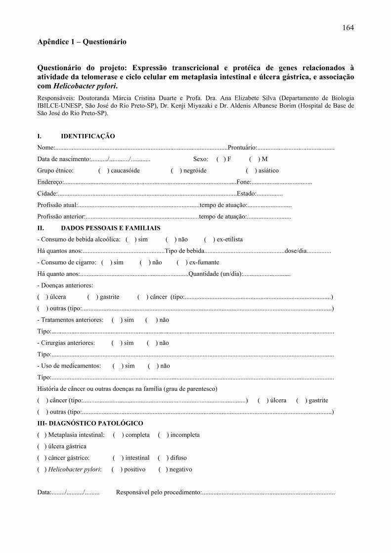
164
Apêndice 1 – Questionário
Questionário do projeto: Expressão transcricional e protéica de genes relacionados à atividade da telomerase e ciclo celular em metaplasia intestinal e úlcera gástrica, e associação com Helicobacter pylori. Responsáveis: Doutoranda Márcia Cristina Duarte e Profa. Dra. Ana Elizabete Silva (Departamento de Biologia IBILCE-UNESP, São José do Rio Preto-SP), Dr. Kenji Miyazaki e Dr. Aldenis Albanese Borim (Hospital de Base de São José do Rio Preto-SP).
I. IDENTIFICAÇÃO
Nome:.........................................................................................................Prontuário:...............................................
Data de nascimento:........../............/............ Sexo: ( ) F ( ) M
Grupo étnico: ( ) caucasóide ( ) negróide ( ) asiático
Endereço:.........................................................................................................Fone:.....................................
Cidade:.............................................................................................................Estado:................
Profissão atual:..........................................................................tempo de atuação:...........................
Profissão anterior:.....................................................................tempo de atuação:...........................
II. DADOS PESSOAIS E FAMILIAIS
- Consumo de bebida alcoólica: ( ) sim ( ) não ( ) ex-etilista
Há quantos anos:..................................................Tipo de bebida.................................................dose/dia...............
- Consumo de cigarro: ( ) sim ( ) não ( ) ex-fumante
Há quanto anos:..................................................................Quantidade (un/dia):.............................
- Doenças anteriores:
( ) úlcera ( ) gastrite ( ) câncer (tipo:.........................................................................................)
( ) outras (tipo:........................................................................................................................................................)
- Tratamentos anteriores: ( ) sim ( ) não
Tipo:............................................................................................................................................................................
- Cirurgias anteriores: ( ) sim ( ) não
Tipo:............................................................................................................................................................................
- Uso de medicamentos: ( ) sim ( ) não
Tipo:............................................................................................................................................................................
História de câncer ou outras doenças na família (grau de parentesco)
( ) câncer (tipo:...................................................................................................) ( ) úlcera ( ) gastrite
( ) outras (tipo:........................................................................................................................................................)
III- DIAGNÓSTICO PATOLÓGICO
( ) Metaplasia intestinal: ( ) completa ( ) incompleta
( ) úlcera gástrica
( ) câncer gástrico: ( ) intestinal ( ) difuso
( ) Helicobacter pylori: ( ) positivo ( ) negativo
Data:......../........../......... Responsável pelo procedimento:.................................................................................
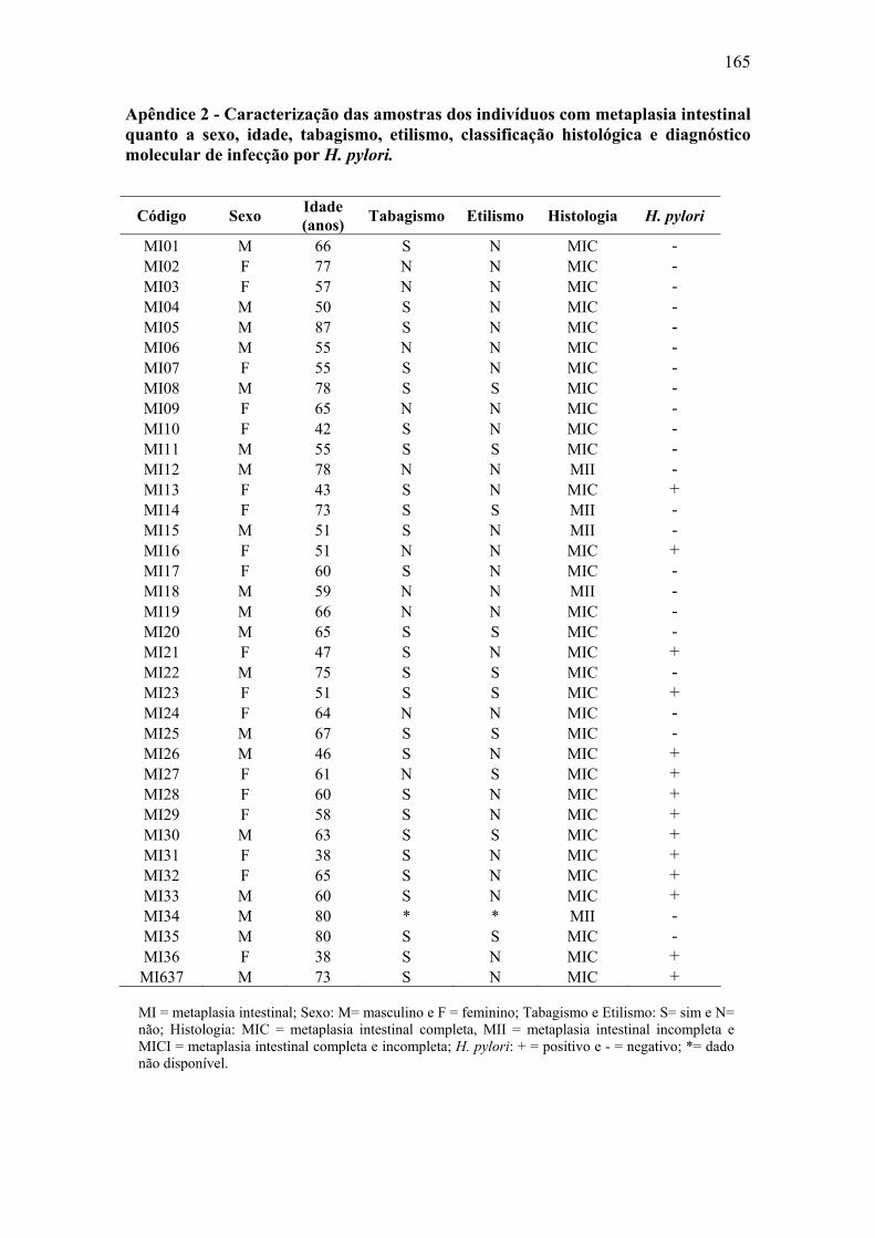
165
Apêndice 2 - Caracterização das amostras dos indivíduos com metaplasia intestinal quanto a sexo, idade, tabagismo, etilismo, classificação histológica e diagnóstico molecular de infecção por H. pylori.
Código Sexo Idade (anos) Tabagismo Etilismo Histologia H. pylori
MI01 M 66 S N MIC - MI02 F 77 N N MIC - MI03 F 57 N N MIC - MI04 M 50 S N MIC - MI05 M 87 S N MIC - MI06 M 55 N N MIC - MI07 F 55 S N MIC - MI08 M 78 S S MIC - MI09 F 65 N N MIC - MI10 F 42 S N MIC - MI11 M 55 S S MIC - MI12 M 78 N N MII - MI13 F 43 S N MIC + MI14 F 73 S S MII - MI15 M 51 S N MII - MI16 F 51 N N MIC + MI17 F 60 S N MIC - MI18 M 59 N N MII - MI19 M 66 N N MIC - MI20 M 65 S S MIC - MI21 F 47 S N MIC + MI22 M 75 S S MIC - MI23 F 51 S S MIC + MI24 F 64 N N MIC - MI25 M 67 S S MIC - MI26 M 46 S N MIC + MI27 F 61 N S MIC + MI28 F 60 S N MIC + MI29 F 58 S N MIC + MI30 M 63 S S MIC + MI31 F 38 S N MIC + MI32 F 65 S N MIC + MI33 M 60 S N MIC + MI34 M 80 * * MII - MI35 M 80 S S MIC - MI36 F 38 S N MIC +
MI637 M 73 S N MIC +
MI = metaplasia intestinal; Sexo: M= masculino e F = feminino; Tabagismo e Etilismo: S= sim e N= não; Histologia: MIC = metaplasia intestinal completa, MII = metaplasia intestinal incompleta e MICI = metaplasia intestinal completa e incompleta; H. pylori: + = positivo e - = negativo; *= dado não disponível.
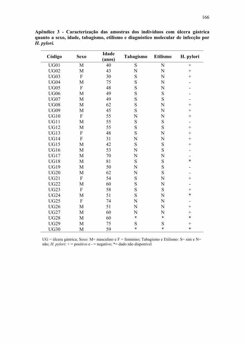
166
Apêndice 3 - Caracterização das amostras dos indivíduos com úlcera gástrica quanto a sexo, idade, tabagismo, etilismo e diagnóstico molecular de infecção por H. pylori.
Código Sexo Idade (anos) Tabagismo Etilismo H. pylori
UG01 M 40 S N + UG02 M 43 N N + UG03 F 30 S N + UG04 M 75 S N - UG05 F 48 S N - UG06 M 49 S S - UG07 M 49 S S - UG08 M 62 S N + UG09 M 45 S N + UG10 F 55 N N + UG11 M 55 S S - UG12 M 55 S S + UG13 F 48 S N + UG14 F 31 N N + UG15 M 42 S S + UG16 M 53 N S - UG17 M 70 N N - UG18 M 81 S S * UG19 M 50 N S - UG20 M 62 N S - UG21 F 54 S N + UG22 M 60 S N - UG23 F 58 S S + UG24 M 51 S N * UG25 F 74 N N - UG26 M 51 N N + UG27 M 60 N N + UG28 M 60 * * * UG29 M 75 S S + UG30 M 59 * * *
UG = úlcera gástrica; Sexo: M= masculino e F = feminino; Tabagismo e Etilismo: S= sim e N= não; H. pylori: + = positivo e - = negativo; *= dado não disponível
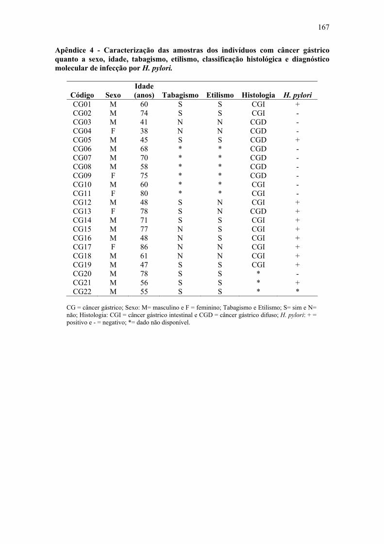
167
Apêndice 4 - Caracterização das amostras dos indivíduos com câncer gástrico quanto a sexo, idade, tabagismo, etilismo, classificação histológica e diagnóstico molecular de infecção por H. pylori.
Código Sexo Idade (anos) Tabagismo Etilismo Histologia H. pylori
CG01 M 60 S S CGI + CG02 M 74 S S CGI - CG03 M 41 N N CGD - CG04 F 38 N N CGD - CG05 M 45 S S CGD + CG06 M 68 * * CGD - CG07 M 70 * * CGD - CG08 M 58 * * CGD - CG09 F 75 * * CGD - CG10 M 60 * * CGI - CG11 F 80 * * CGI - CG12 M 48 S N CGI + CG13 F 78 S N CGD + CG14 M 71 S S CGI + CG15 M 77 N S CGI + CG16 M 48 N S CGI + CG17 F 86 N N CGI + CG18 M 61 N N CGI + CG19 M 47 S S CGI + CG20 M 78 S S * - CG21 M 56 S S * + CG22 M 55 S S * *
CG = câncer gástrico; Sexo: M= masculino e F = feminino; Tabagismo e Etilismo; S= sim e N= não; Histologia: CGI = câncer gástrico intestinal e CGD = câncer gástrico difuso; H. pylori: + = positivo e - = negativo; *= dado não disponível.

168
Apêndice 5 - Material e Métodos
1. Caracterização das amostras
Todas as amostras foram coletadas por profissional qualificado no Centro de
Endoscopia do Hospital de Base de São José do Rio Preto e tiveram seu diagnóstico
patológico confirmado pelo Serviço de Patologia do mesmo hospital. De cada paciente
com diagnóstico endoscópico de úlcera gástrica, metaplasia intestinal e câncer gástrico
foram coletadas três biópsias da lesão e três de uma região aparentemente normal do
estômago, número este de acordo com as normas do Serviço de Patologia. As biópsias
foram acondicionadas em uma solução para preservação do RNA (RNA later, Ambion)
e estocadas em freezer a -20 °C para a posterior extração de RNA e DNA. Todos os
pacientes foram devidamente informados e consultados sobre sua participação no
projeto e de todos foi obtido o Termo de Consentimento Livre e Esclarecido, além de
um questionário com informações sobre o estilo de vida e história prévia de problemas
de saúde (Apêndice 1), sendo todo o procedimento realizado pelo próprio pesquisador.
2. Extração de RNA e DNA
Para todas as amostras com diagnóstico histopatológico confirmado foi extraído
o RNA e o DNA total utilizando o protocolo do reagente Trizol. Inicialmente, duas
biópsias de cada amostra foram fragmentadas mecanicamente e colocadas em
microtubos contendo 1mL do reagente Trizol para a quebra das membranas celulares,
desagregação das proteínas e liberação do material genético (a biópsia remanescente foi
estocada em freezer -20ºC para extrações posteriores caso a primeira não tenha obtido
quantidade e/ou qualidade de RNA suficiente). Após 5 minutos em temperatura
ambiente, adicionou-se 200μL de clorofórmio e o material foi mantido à temperatura
ambiente por 3 minutos e depois centrifugado por 15 minutos a 2°C. Após a
169
centrifugação, ocorre a separação do material em três fases: uma vermelha inferior,
contendo proteínas e DNA, uma intermediária, contendo restos celulares e DNA e uma
superior, contendo RNA. A fase superior foi transferida para outro microtubo no qual
adicionou-se 500μL de álcool isopropílico. O material foi mantido à temperatura
ambiente por 10 minutos e, posteriormente, centrifugado por 10 minutos a 2°C para a
precipitação do RNA total. Após a centrifugação, o sobrenadante foi descartado e o
pellet de RNA foi ressuspendido e lavado com etanol 75%, centrifugado novamente por
5 minutos a 2°C e seco por cerca de 10 minutos à temperatura ambiente. Finalmente, foi
adicionado 30μL de água DEPEC para a diluição do RNA em banho-maria a 60ºC por
10 minutos. Logo depois, foi realizada a medida da concentração do RNA em
espectrofotômetro (NanoDrop) e este foi rapidamente estocado em freezer -80 °C para
evitar a sua degradação.
As fases inferior e intermediária foram reservadas para a posterior extração de
DNA, às quais adicionou-se 300μL de etanol absoluto gelado que foi incubado por 3
minutos à temperatura ambiente e, posteriormente, centrifugado a 2 °C por 5 minutos
para a precipitação do DNA. O pellet de DNA foi lavado duas vezes com solução de
citrato de sódio 0,1 M em etanol 10%. Para cada lavagem, o material foi mantido sob
agitação por 30 minutos à temperatura ambiente, e depois centrifugado à 2ºC por 5
minutos. Após a última lavagem, o pellet de DNA foi ressuspendido em etanol 75%,
mantido à temperatura ambiente por 15 minutos e centrifugado por 5 minutos a 2°C. O
sobrenadante foi descartado, o pellet foi seco à temperatura ambiente por 10 minutos e
foi adicionado 50μL de água ultra-pura autoclavada para a diluição do DNA em banho-
maria à 37°C por aproximadamente três dias. Foi realizada a leitura da concentração do
DNA de todas as amostras no espectrofotômetro (NanoDrop) e estas foram estocadas
em freezer -20°C, para a posterior avaliação molecular da bactéria H. pylori.
170
3. Eletroforese do RNA
Todas as amostras de RNA foram submetidas à eletroforese em gel de agarose
1% para verificação da integridade do RNA. De cada amostra foi utilizado 1μg de RNA.
As amostras de boa qualidade apresentaram duas bandas correspondentes às duas
subunidades do RNA ribossômico e foram utilizadas para a síntese de cDNA total.
Aquelas que não apresentaram RNA de boa qualidade foram submetidas à nova
extração de RNA da biópsia remanescente.
4. RT-PCR
Cerca de 5 μg de RNA foi utilizado para a síntese de c-DNA com o kit High
Capacity (Applied Biosystems). Para uma reação de 50μL, foram adicionados 5 μL de
random primer, 2,5 μL de mix dNTP 10 mM, 5 μL de tampão 5 X, 80U de RNAOut
(Invitrogen), 5μL de Transcriptase Reversa e água DEPEC para completar o volume
final. No termociclador, as condições da reação foram 25°C por 10 minutos e,
posteriormente, 37°C por 120 minutos. As amostras de cDNA obtidas foram
armazenadas em freezer -20 °C.
5. PCR do gene β-actina
Todas as amostras de cDNA foram testadas através de PCR para amplificar o
gene da β-actina, com o intuito de confirmar o sucesso da síntese do cDNA. Para uma
reação de 20μL, foi utilizado 1μL de cDNA, 1,5mM de MgCl2, 0,2mM de dNTPs,
0,5μM de cada primer (F-5’GAGGCACTCTTCCAGCCTTC3’ and R-
5’GTTGGCGTACAGGTCTTGC3’) e 1,5U de Taq DNA polimerase em tampão 1X.
As condições da reação foram as seguintes: 94°C por 3 minutos, seguidos de 35 ciclos
de amplificação a 94°C por 45 segundos, 61ºC por 30 segundos e 72ºC por 1 minuto e
extensão final a 72°C por 10 minutos. A banda amplificada de 114 pb foi visualizada
em gel de agarose 2% corado com brometo de etídeo.
171
6. Desenho das seqüências de primers
As seqüências de RNAm dos genes TERT1, COX-2, NOS2, HGF, MET, RAS e
TFF1 bem como dos genes endógenos β-actina, β2-microglobulina e α-tubulina foram
obtidas do banco de dados do NCBI e utilizadas para a confecção dos primers por meio
do programa Primer3 (disponível em http://frodo.wi.mit.edu/cgi-
bin/primer3/primer3_www.cgi). Todos apresentaram temperatura de anelamento de
60ºC e amplificam fragmentos que variam de 83 a 160 pb (Tabela 1) pertencendo a dois
ou mais éxons consecutivos, para evitar a amplificação de fragmentos de DNA
contaminante. A seqüência do gene CLDN18 foi obtida conforme descrito por Sanada et
al., 2006.
7. Amplificação por PCR em tempo real
A reação de PCR em tempo real foi realizada utilizando o kit comercial SYBR
Green (Applied Biosystems), com primers específicos para os genes TERT, COX-2,
NOS2, HGF, MET, RAS, TFF1 e CLDN18. Os genes housekeeping β-actina, β-2-
microglobulina e α-tubulina foram testados como controles internos da reação.
Resumidamente, a reação de 20 μL foi composta de 10 μL de SYBR Green,
concentração específica de cada primer obtida na etapa de otimização e 25 ng de cDNA.
As condições no equipamento de PCR em tempo real ABI Prism® 7300 (Applied
Biosystems) compreenderam uma etapa inicial de 2 min a 50ºC, seguida de uma etapa a
95ºC por 10 min e 40 ciclos de amplificação a 95ºC por 15 seg e 60ºC por 1 min. No
final, foi acrescentado um passo de dissociação que compreende duas etapas de 15 seg a
95ºC, intercaladas por uma etapa de 30 seg a 60ºC. Esta etapa tem por objetivo a
construção de uma curva padrão para verificar a ocorrência de primer-dimer, produtos
inespecíficos e contaminações. Na etapa de otimização, para cada gene proposto para
este estudo, foi realizada uma reação contendo primers com diferentes concentrações,
172
variando de 0,25 a 1,0 μM. De acordo com as curvas de amplificação geradas pelo
equipamento de PCR em tempo real, foi determinada a melhor concentração a ser
utilizada para cada par de primer nas reações futuras. A melhor concentração foi
estipulada como aquela que primeiro amplifica a amostra em determinado ciclo de
amplificação (Ct – Threshold Cycle). Este ciclo não deve mudar conforme aumenta a
concentração dos primers. Desde modo, foram obtidas concentrações de primer que
amplificam otimamente a amostra, sem o risco de formação de produtos inespecíficos e
primer-dimer.
Nesta etapa também observou-se a qualidade dos primers, sendo que aqueles
com curvas de amplificação ruins foram novamente desenhados. Após a etapa de
otimização, os primers foram validados para um gradiente de concentrações de
amostras. O objetivo foi verificar se determinado primer é capaz de amplificar com a
mesma eficiência amostras com diferentes concentrações. Esta eficiência é medida pela
fórmula E = 10(1/-s) – 1, onde s é o slope da curva padrão, que corresponde à inclinação
da reta obtida quando se analisa a variação do Ct dos genes estudados em função do Log
de diferentes quantidades de cDNA. A eficiência dos primers ficou entre 90 e 100 %.
Para os genes housekeeping e os genes supressores de tumor TFF1 e CLDN18 foram
utilizadas amostras com contrações de 100, 10, 1, 0,1 e 0,01 ng de cDNA, e para os
demais genes as concentrações foram de 50, 25, 12,5, 6,25 e 3,12 ng. Para as etapas de
otimização e validação foi utilizado um pool de amostras da mucosa normal dos
indivíduos deste estudo e para cada primer e/ou amostra a reação foi realizada em
triplicata.
Após a análise dos genes housekeeping, β-actina foi escolhido como referência
por apresentar menor variação de Ct nas diferentes amostras e grupos estudados. A
expressão relativa do RNAm foi calculada de acordo com o método 2-ΔΔCt descrita por
173
Livak e Schimittgen (2001). Primeiro, a média dos Ct para cada amostra foi
determinada. Depois, o valor de ΔCt foi calculado como sendo a diferença entre o valor
de Ct do gene alvo e o valor de Ct da β-actina: ΔCt = Ct (gene alvo) - Ct (β-actina).
Finalmente, o valor de ΔΔCt foi calculado: ΔΔCt = ΔCt (metaplasia, úlcera ou câncer) -
ΔCt (mucosa normal adjacente).
8. Diagnóstico molecular da H. pylori
As amostras de DNA obtidas foram utilizadas para o diagnóstico molecular da
bactéria H. pylori e, nesta etapa, apenas as amostras referentes às mucosas normais dos
pacientes foram avaliadas. Foram utilizados os primers específicos para a H. pylori
referentes aos genes urease A (F- 5’ TTCCTGATGGGACCAAACTC 3’ e R- 5’
TTACCGCCAATGTCAATCAA 3’) e tsaA - alquil hidroperóxido redutase (F- 5’
CCTGCCGTTTTAGGAAACAA 3’ e R- 5’ TCCGCATTCCTACCTAATGG 3’), além
do gene constitutivo humano CYP1A1 (F- 5’CTCACCCCTGATGGTGCTAT 3’ e R- 5’
TTTGGAAGTGCTCACAGCAG 3’) para verificar a integridade do DNA e eficiência
da reação de PCR, que amplificam fragmentos de 316, 413 e 226, respectivamente. A
reação multiplex foi realizada em volume final de 25 μL contendo 4 mM de MgCl2, 0,3
mM de dNTPs, 0,6 μM de cada par de primer, 1,75 U de Taq DNA polimerase em
tampão 1 X. Os produtos da reação foram submetidos à eletroforese em gel de agarose
1000 (Invitrogen) a 2 % e visualizados com coloração de brometo de etídeo.
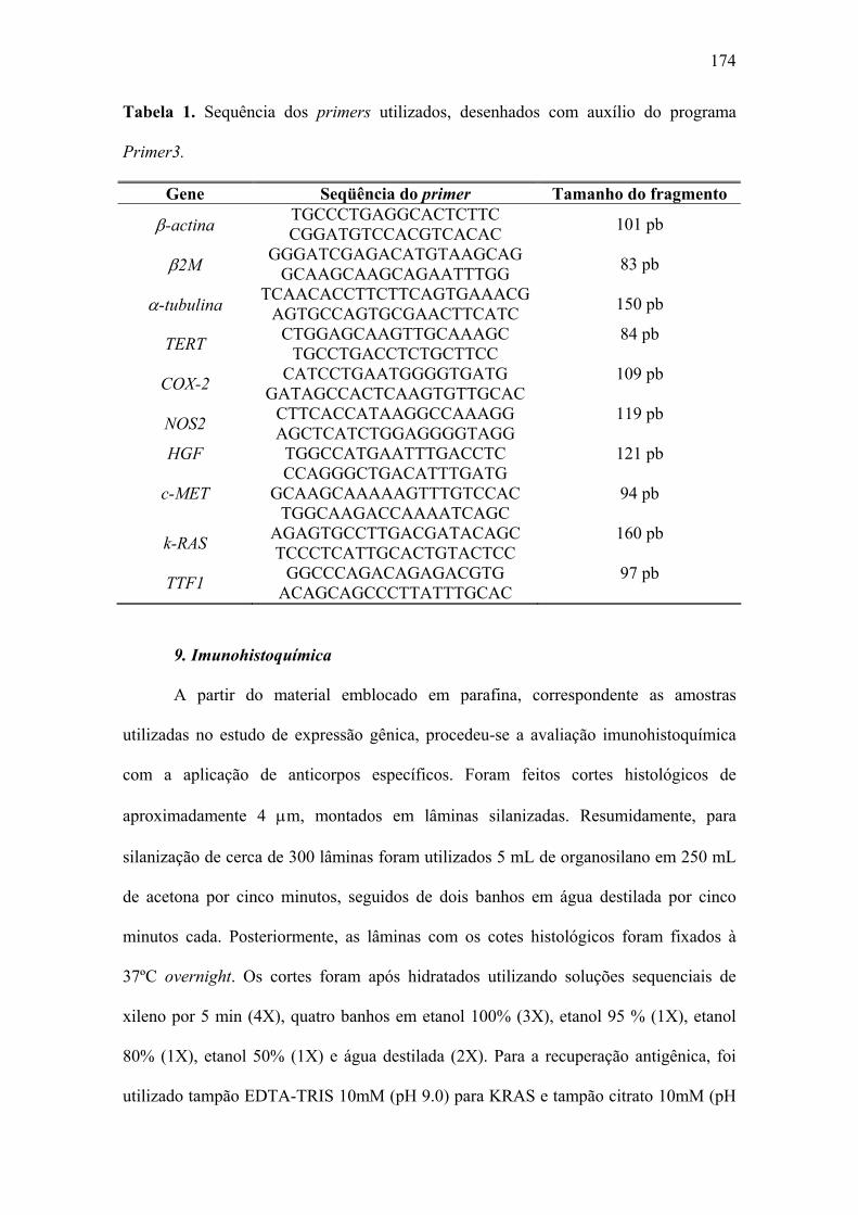
174
Tabela 1. Sequência dos primers utilizados, desenhados com auxílio do programa
Primer3.
Gene Seqüência do primer Tamanho do fragmento
β-actina TGCCCTGAGGCACTCTTC CGGATGTCCACGTCACAC 101 pb
β2M GGGATCGAGACATGTAAGCAG GCAAGCAAGCAGAATTTGG 83 pb
α-tubulina TCAACACCTTCTTCAGTGAAACG AGTGCCAGTGCGAACTTCATC 150 pb
TERT CTGGAGCAAGTTGCAAAGC TGCCTGACCTCTGCTTCC
84 pb
COX-2 CATCCTGAATGGGGTGATG GATAGCCACTCAAGTGTTGCAC
109 pb
NOS2 CTTCACCATAAGGCCAAAGG AGCTCATCTGGAGGGGTAGG
119 pb
HGF TGGCCATGAATTTGACCTC CCAGGGCTGACATTTGATG
121 pb
c-MET GCAAGCAAAAAGTTTGTCCAC TGGCAAGACCAAAATCAGC
94 pb
k-RAS AGAGTGCCTTGACGATACAGC TCCCTCATTGCACTGTACTCC
160 pb
TTF1 GGCCCAGACAGAGACGTG ACAGCAGCCCTTATTTGCAC
97 pb
9. Imunohistoquímica
A partir do material emblocado em parafina, correspondente as amostras
utilizadas no estudo de expressão gênica, procedeu-se a avaliação imunohistoquímica
com a aplicação de anticorpos específicos. Foram feitos cortes histológicos de
aproximadamente 4 μm, montados em lâminas silanizadas. Resumidamente, para
silanização de cerca de 300 lâminas foram utilizados 5 mL de organosilano em 250 mL
de acetona por cinco minutos, seguidos de dois banhos em água destilada por cinco
minutos cada. Posteriormente, as lâminas com os cotes histológicos foram fixados à
37ºC overnight. Os cortes foram após hidratados utilizando soluções sequenciais de
xileno por 5 min (4X), quatro banhos em etanol 100% (3X), etanol 95 % (1X), etanol
80% (1X), etanol 50% (1X) e água destilada (2X). Para a recuperação antigênica, foi
utilizado tampão EDTA-TRIS 10mM (pH 9.0) para KRAS e tampão citrato 10mM (pH
175
6.0) para TERT, COX-2, NOS2, HGF, MET, TFF1 e CLDN18. O tampão foi pré-
aquecido por 40 min em uma panela de vapor (Exotic, Arno) até 95ºC e então as
lâminas foram colocadas por 30 min, com exceção de TERT e COX-2. Para estes
anticorpos, a recuperação antigênica foi realizada em autoclave à 120ºC por 15 min.
Após esfriar em temperatura ambiente por cerca de 20 min, a atividade da peroxidase
endógena foi bloqueada com 3% H2O2 por 20 min.
Em seguida as lâminas foram incubadas por 1 hora em temperatura ambiente
com anticorpos específicos para TERT (mouse monoclonal antibody, clone 2D8, ABR -
Affinity BioReagents, 1:100), COX-2 (mouse monoclonal antibody, clone 4H12,
Novocastra, 1:100) TFF1 (mouse monoclonal anti-pS2, clone BC04, Invitrogen, 1:50),
HGF (Monoclonal anti hepatocyte growth factor antibody, clone 24612.111, Sigma,
1:50) e MET (Anti hepatocyte growth factor receptor -cMet antibody, Sigma, 1:50) e
overnight a 4ºC com anticorpos específicos para NOS2 (Rabbit polyclonal to iNOS,
Abcam, 1:200), KRAS (Mouse monoclonal anti-KRAS, clone 9.13, Invitrogen, 1:50) e
CLDN18 (Rabbit polyclonal anti-claudin-18 -c-Term, Zymed, Invitrogen, 1:200). Após
a lavagem com tampão Tris-HCl (pH 7.6), as laminas foram incubadas, por 20 min
cada, com anticorpo secundário biotinilado e com peroxidase estreptoavidina-biotina,
seguindo as especificações do fabricante (Histostain Bulk Kit, Zymed).
A marcação foi visualizada com 3,3’-diaminobenzidina (DAB) e contra-corado
com hematoxilina de Mayer. Os controles negativos foram estabelecidos substituindo-se
o anticorpo primário por solução tampão. Estômago normal foi utilizado como controle
positivo para TFF1 e CLDN18, e amídala, câncer de cólon, fígado, carcinoma de mama,
carcinoma de pulmão e carcinoma epidermóide de pulmão foram utilizados como
controles positivos para TERT, COX-2, HGF, MET, NOS2 e KRAS, respectivamente.

Anexo
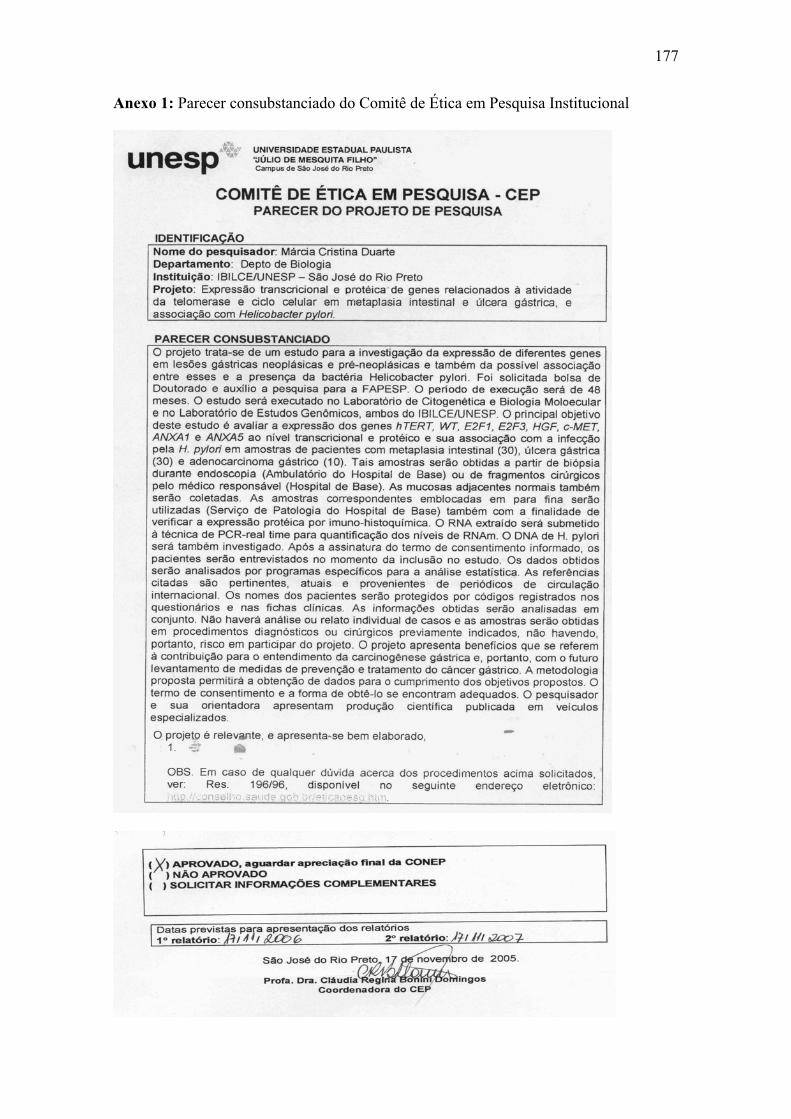
177
Anexo 1: Parecer consubstanciado do Comitê de Ética em Pesquisa Institucional
178
Anexo 2: Comprovante de submissão do artigo “Expression of TERT and COX-2 in
precancerous gastric lesions compared to gastric cancer” ao periódico World Journal of
Gastroenterology .
Livros Grátis( http://www.livrosgratis.com.br )
Milhares de Livros para Download: Baixar livros de AdministraçãoBaixar livros de AgronomiaBaixar livros de ArquiteturaBaixar livros de ArtesBaixar livros de AstronomiaBaixar livros de Biologia GeralBaixar livros de Ciência da ComputaçãoBaixar livros de Ciência da InformaçãoBaixar livros de Ciência PolíticaBaixar livros de Ciências da SaúdeBaixar livros de ComunicaçãoBaixar livros do Conselho Nacional de Educação - CNEBaixar livros de Defesa civilBaixar livros de DireitoBaixar livros de Direitos humanosBaixar livros de EconomiaBaixar livros de Economia DomésticaBaixar livros de EducaçãoBaixar livros de Educação - TrânsitoBaixar livros de Educação FísicaBaixar livros de Engenharia AeroespacialBaixar livros de FarmáciaBaixar livros de FilosofiaBaixar livros de FísicaBaixar livros de GeociênciasBaixar livros de GeografiaBaixar livros de HistóriaBaixar livros de Línguas
Baixar livros de LiteraturaBaixar livros de Literatura de CordelBaixar livros de Literatura InfantilBaixar livros de MatemáticaBaixar livros de MedicinaBaixar livros de Medicina VeterináriaBaixar livros de Meio AmbienteBaixar livros de MeteorologiaBaixar Monografias e TCCBaixar livros MultidisciplinarBaixar livros de MúsicaBaixar livros de PsicologiaBaixar livros de QuímicaBaixar livros de Saúde ColetivaBaixar livros de Serviço SocialBaixar livros de SociologiaBaixar livros de TeologiaBaixar livros de TrabalhoBaixar livros de Turismo


