descriptive study of onychomycosis in a hospital in são paulo
TRANSCRIPT

Descriptive study of onychomycosis in a hospital in São Paulo
Clarissa Santos de Carvalho Ribeiro1, Clarisse Zaitz1, Valéria Maria de Souza Framil1,
Thaíssa Santos de Carvalho Ottoboni2, Melissa Santos de Carvalho Tonoli3,
Renata Pinto Ribeiro4
1Departamento de Dermatologia, Faculdade de Ciências Médicas, Santa Casa de São Paulo,
São Paulo, SP, Brazil.2Faculdade de Medicina de Itajubá, Itajubá, MG, Brazil.
3Universidade Estadual Paulista, São José dos Campos, SP, Brazil.4Universidade Federal de Alfenas, Alfenas, MG, Brazil.
Submitted: June 17, 2013; Approved: August 19, 2014.
Abstract
Onychomychosis, a nail fungus infection is the most frequent nail ailment, constituting about half of
all nail disorders. It can be caused by dermatophytes, non-dermatophytes, yeasts and Prothoteca spp.
Methods include 5407 samples of patients with suspected onychomycosis, studied from January
2002 to December 2006, by direct mycological examination and fungi culture. The diagnosis of
onychomycosis was confirmed in samples from 3822 direct mycological and/or culture positive. The
diagnosis was established by culture for fungi. Among the 1.428 identified agents, the dermatophytes
were responsible for 68.6% (N = 980) of cases, followed by yeasts with 27.6% (N = 394),
non-dermatophytes fungi with 2.2% (N = 31), Prothoteca spp with 0.1% (N = 2), and associations
with 1.5% (N = 22). Females were more affected, with 66% (N = 2527) of cases, and the most af-
fected age group ranged from 31 to 60 years of age (median 47 years). Fungal microbiota is often
changed in the world, both quantitatively and qualitatively, and is affected by several environmental
factors. Thus, the periodic review of the composition of this microbiota is important to evaluate the
epidemiology and thus proportion a better therapeutic response.
Key words: onychomycosis, dermatophytes, yeasts, non-dermatophytes fungi, Prothoteca spp.
Introduction
Onychomycosis is a fungal nail infection which can
be caused by dermatophytes, yeast, non-dermatophyte fila-
mentous fungi (NDFF) and currently Prototheca spp, af-
fecting approximately 8% of the world population (Effendy
I et al., 2005; Zaitz C et al., 2006).
This nail ailment represents 30% of the fungal surface
and around 50% of nail diseases (Ghannoum MA et al.,
2000). The main onychomycosis etiologic agents are
dermatophytes, which are isolated in 75% of cases. Can-
dida yeasts occur in around 18-20% of cases and NDFF
reach percentages ranging from one to five percent of cases
(Agarwalla A et al., 2006).
Epidemiological studies of onychomycosis can pro-
vide divergent results in the literature due to several factors,
such as environmental factors and related to the host ones.
Key environmental factors considered include: urban de-
velopment, industrialization, geographical location and cli-
matic conditions such as temperature and exposure to ultra-
violet rays. Those factors related to the host are cited in the
literature as being: age, lifestyle, occupation, sex, color,
and chronic diseases (Sigurgeirsson B et al., 2000).
Onychomycosis is considered a public health prob-
lem due to high prevalence associated to morbidities such
as diabetes, impaired peripheral circulation, ungueal trau-
ma repetition, immunodeficiencies (Elewski BE, 1998). It
is a disease which does not demand compulsory notifica-
tion, thus making it difficult to access the extent of the
Brazilian Journal of Microbiology 46, 2, 485-492 (2015) Copyright © 2015, Sociedade Brasileira de Microbiologia
ISSN 1678-4405 www.sbmicrobiologia.org.br
DOI: http://dx.doi.org/10.1590/S1517-838246220130541
Send correspondence to C.S.C. Ribeiro. Departamento de Dermatologia, Faculdade de Ciências Médicas, Santa Casa de São Paulo, São Paulo, SP, Brazil.
E-mail: [email protected].
Research Paper
problem in our midst. Thus, onychomycosis affects the
quality of a patient’s life and causes esthetic losses, reflect-
ing directly on self-esteem, vanity, social discrimination
and the patient’s working potential (Lopes JO et al., 1999).
Although nail infections are common, nowadays they
are associated with difficult treatment and are reported as
having high rates of recurrence and therapeutic inefficacy.
The knowledge of ecology, etiology and distribution of the
biota of the main etiological agents allows for a better com-
prehension of natural history, evolution and may contribute
in the future for new therapeutic modalities (Schroeff J et
al., 1992).
In this retrospective study, an analysis of all positive
mycological examinations from patients with clinical ony-
chomycosis diagnosis from the São Paulo Hospital Derma-
tology Department’s Mycology Laboratory was per-
formed, covering a period from January 2002 to December
2006
The objectives were: 1) To analyze the sensitivity of a
diagnostic laboratory for onychomycosis in the São Paulo
Hospital Dermatology Department’s Medical Mycology
Laboratory. 2) To analyze the distribution of cases of ony-
chomycosis in relation to demographic data: age and sex. 3)
To analyze the distribution of onychomycosis cases ac-
cording to the main etiological agents: dermatophytes, non
dermatophyte filamentous fungi (NDFF), protists and
yeasts.
Material and Methods
This paper includes a descriptive study conducted at
the São Paulo Hospital Dermatology Department, by re-
viewing the results of mycological examinations performed
from January 2002 to December 2006.
All patients seen at the dermatology ambulatory that
presented, among the possible diagnoses, a clinical suspi-
cion of onychomycosis, were submitted to mycological ex-
amination in order to confirm the onychomycosis diagno-
sis.
Reagents, culture media, equipment and consumable
material are routinely used in the medical mycology labora-
tory of the Dermatology Department in Hospital in São
Paulo. The study was approved by the Ethics and Research
Committee of the São Paulo Hospital.
Laboratory procedures
Direct microscopic examination
The material was obtained by scraping with a dental
explorer, a dental curette nail and examined between a slide
and coverslip after clarification with potassium hydroxide
with 20% dimethyl sulphoxide added (Lacaz CS et al.,
2002; Sidrim JJC and Rocha MFG, 2004).
Culture
Macroscopic aspects: The culture media for isolation
and analysis of morphological species of Dermatophytes
are Sabouraud’s medium with chloramphenicol and Sabo-
uraud medium with cycloheximide. Non-dermatophyte fil-
amentous fungi, yeast and Prototheca spp are grown on
Sabouraud cloranfenicol. In our laboratory, the material
was inoculated into three tubes of the means mentioned:
first, collection of clinical material and isolation of agents;
second, collection of clinical material after the first seven
days from the initial collection and isolation of the agent;
the third collection of clinical material after seven days
from the second collection and isolation of agents. The
growth period to occur during the full maturity of fungal
structures is: dermatophytes and NDFF observed around 15
days, Prototheca spp and yeast about seven days (Lacaz CS
et al., 2002; Sidrim JJC and Rocha MFG, 2004).
Microscopic aspects: The technique of microculture
on slides with a culture medium with agar-cornmeal for the
identification and determination of genus and species of
dermatophytes, yeasts and NDFF was used. In Prototheca
spp, an inoculum of the culture was withdrawn to be identi-
fied and was placed on a slide with lacto-phenol blue dye
cotton. It was possible to observe the shaped structures of
morula. Species identification is performed through bio-
chemical tests. (Lacaz CS et al., 2002; Sidrim JJC and
Rocha MFG, 2004).
Statisticalanalysis
To analyze the results, the chi-square test in
MINITAB 14 and tables in EXCEL 2007 were used for
characterization of the Clinical sample material obtained
from nails of patients with suspected onychomycosis. This
was done to evaluate the association between the variables
of age, gender, test result, the affected site and agent. In all
tests, the 0.05 significance level was set.
Results
Characterization of the samples studied
Direct microscopic examination and culture
Between January 2002 and December 2006, 5407
mycological tests were carried out with clinical suspicion
of onychomycosis in 3541 patients of any age, sex and race,
which were analyzed by direct microscopic examination
and/or culture. Of the total amount tested, 71%
(3.822/5.407) were positive for laboratory diagnosis of
onychomycosis. Of the remaining mycological examina-
tions that were analyzed, 29% (1585/5407) were negative.
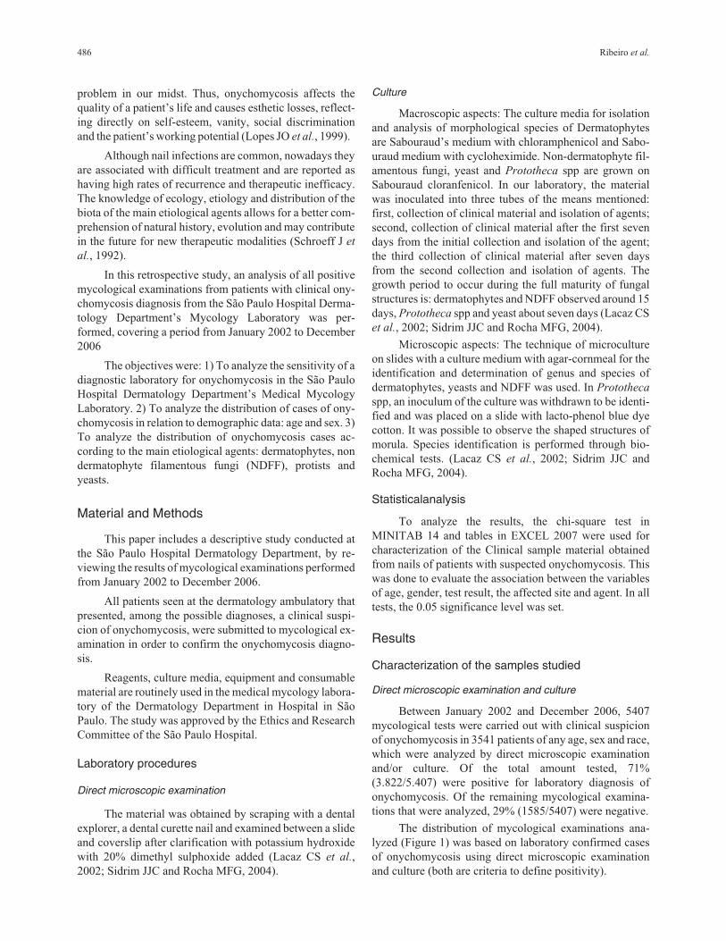
The distribution of mycological examinations ana-
lyzed (Figure 1) was based on laboratory confirmed cases
of onychomycosis using direct microscopic examination
and culture (both are criteria to define positivity).
486 Ribeiro et al.
Onychomycosis age and gender
Regarding age, it was observed in the table below that
among the 3822 samples with a diagnosis of onycho-
mycosis, 60% (2.287/3.822) presented data on age, with a
minimum age of three months (0.25 years) and the maxi-
mum age of 98 years. The mean age was 46.9 years, stan-
dard deviation � 17.02; observing a coefficient of variation
of 36.3%, concluding that the most representative of the
age of this population would be the median: 47 years old.
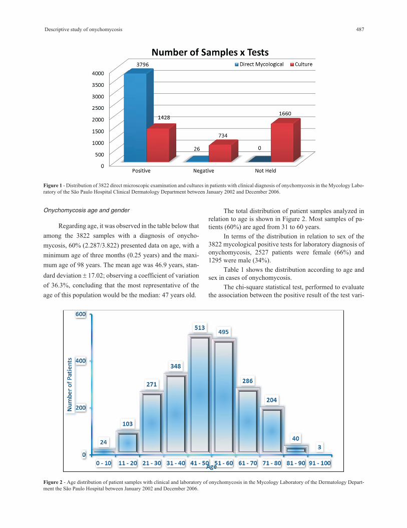
The total distribution of patient samples analyzed in
relation to age is shown in Figure 2. Most samples of pa-
tients (60%) are aged from 31 to 60 years.
In terms of the distribution in relation to sex of the
3822 mycological positive tests for laboratory diagnosis of
onychomycosis, 2527 patients were female (66%) and
1295 were male (34%).
Table 1 shows the distribution according to age and
sex in cases of onychomycosis.
The chi-square statistical test, performed to evaluate
the association between the positive result of the test vari-
Descriptive study of onychomycosis 487
Figure 1 - Distribution of 3822 direct microscopic examination and cultures in patients with clinical diagnosis of onychomycosis in the Mycology Labo-
ratory of the São Paulo Hospital Clinical Dermatology Department between January 2002 and December 2006.
Figure 2 - Age distribution of patient samples with clinical and laboratory of onychomycosis in the Mycology Laboratory of the Dermatology Depart-
ment the São Paulo Hospital between January 2002 and December 2006.
ables and age of the patients by sex, showed a significant
difference (p < 0.0001) between the percentage of positive
results observed in relation to patients age by sex, for the
level of significance; that is, the highest incidence of posi-
tive results was in the range of 31 to 60 years for females.
Identification of the etiologic agents of onychomycosis
The percentage of culture isolation and identification
of agents etiology was 66% (1.428/2.162) of the samples.
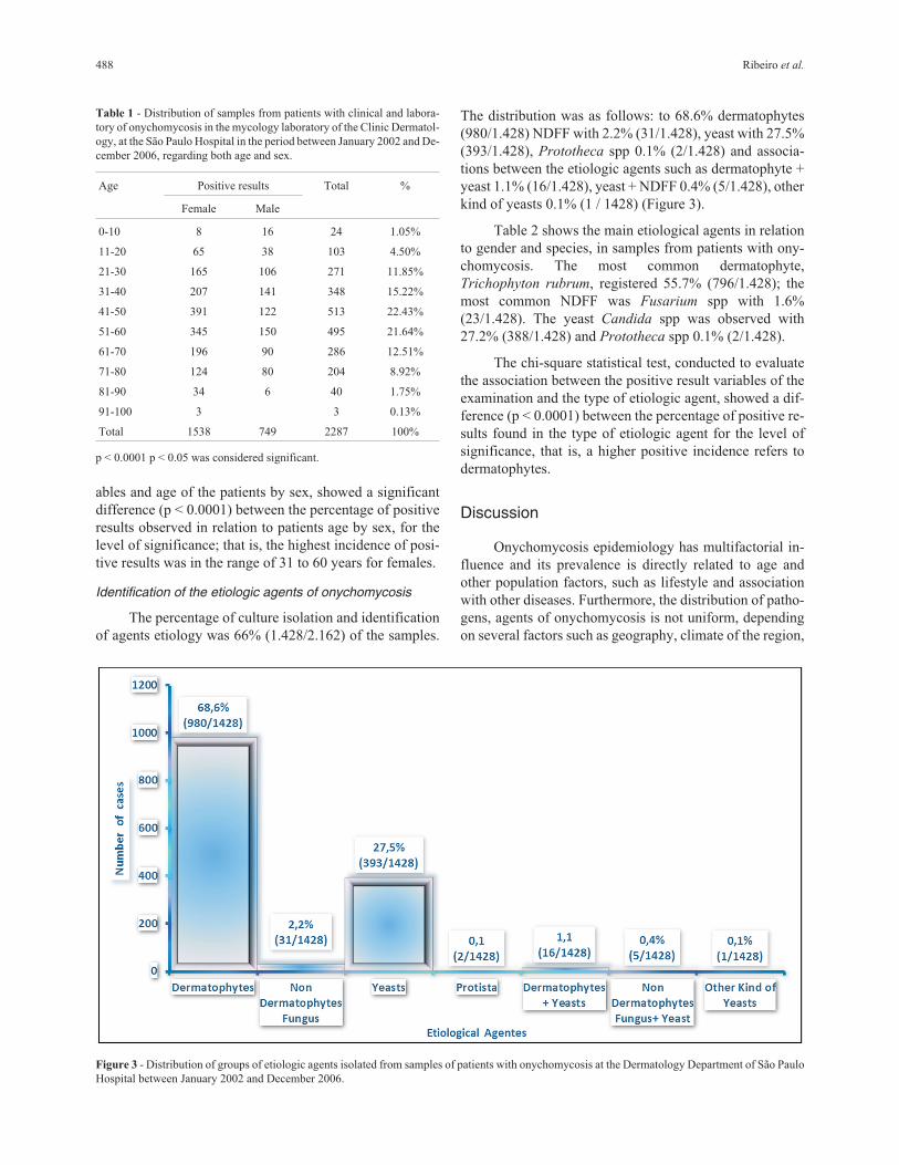
The distribution was as follows: to 68.6% dermatophytes
(980/1.428) NDFF with 2.2% (31/1.428), yeast with 27.5%
(393/1.428), Prototheca spp 0.1% (2/1.428) and associa-
tions between the etiologic agents such as dermatophyte +
yeast 1.1% (16/1.428), yeast + NDFF 0.4% (5/1.428), other
kind of yeasts 0.1% (1 / 1428) (Figure 3).
Table 2 shows the main etiological agents in relation
to gender and species, in samples from patients with ony-
chomycosis. The most common dermatophyte,
Trichophyton rubrum, registered 55.7% (796/1.428); the
most common NDFF was Fusarium spp with 1.6%
(23/1.428). The yeast Candida spp was observed with
27.2% (388/1.428) and Prototheca spp 0.1% (2/1.428).
The chi-square statistical test, conducted to evaluate
the association between the positive result variables of the
examination and the type of etiologic agent, showed a dif-
ference (p < 0.0001) between the percentage of positive re-
sults found in the type of etiologic agent for the level of
significance, that is, a higher positive incidence refers to
dermatophytes.
Discussion
Onychomycosis epidemiology has multifactorial in-
fluence and its prevalence is directly related to age and
other population factors, such as lifestyle and association
with other diseases. Furthermore, the distribution of patho-
gens, agents of onychomycosis is not uniform, depending
on several factors such as geography, climate of the region,
488 Ribeiro et al.
Table 1 - Distribution of samples from patients with clinical and labora-
tory of onychomycosis in the mycology laboratory of the Clinic Dermatol-
ogy, at the São Paulo Hospital in the period between January 2002 and De-
cember 2006, regarding both age and sex.
Age Positive results Total %
Female Male
0-10 8 16 24 1.05%
11-20 65 38 103 4.50%
21-30 165 106 271 11.85%
31-40 207 141 348 15.22%
41-50 391 122 513 22.43%
51-60 345 150 495 21.64%
61-70 196 90 286 12.51%
71-80 124 80 204 8.92%
81-90 34 6 40 1.75%
91-100 3 3 0.13%
Total 1538 749 2287 100%
p < 0.0001 p < 0.05 was considered significant.
Figure 3 - Distribution of groups of etiologic agents isolated from samples of patients with onychomycosis at the Dermatology Department of São Paulo
Hospital between January 2002 and December 2006.

and population migration (Faergemann J, Baran R, 2003;
Brito A, 1992).
In the São Paulo Hospital Dermatology Department’s
Mycology Laboratory, from January 2002 to December
2006, 5407 patients with clinical suspicion of onycho-
mycosis were evaluated. The laboratory diagnosis (myco-
logical and / or culture positive) was confirmed in 71%
(3.822/5.407) of the samples analyzed. Elewski BE et al. in
1997 observed that, in 2065 clinical samples with suspected
onychomycosis, 82% were diagnosed cases (1.707/2.065).
Gupta et al., in a multicenter study in 2000, found that of
the 15.000 patients who had nails with some sort of abnor-
mality, only 8% of cases (1.199/15.000) onychomycosis
were confirmed by laboratory tests. In a study carried out
by Brilhante et al. in 2005, of the 976 patients with sus-
pected onychomycosis, 52% (512/976) were confirmed as
having the disease. In Belgium, 36.04% of tests confirmed
presence of onychomysis in the studied patients
(1.290/3.579) (Arrese JE et al., 2005). A study conducted in
India shows that of the 302 patients, 42.4% (128/302) had
the diagnosis confirmed by laboratory examination (Sarma
S et al., 2008).
Of the 3822 clinical samples with direct examination
and / or positive cultures analyzed in our study, the direct
microscopic examination was positive in 99.3%
(3.796/3.822) of them, while the culture was positive in
66% (1.428/2.162) of the total samples. Studies in the liter-
ature reported 82.45% (94/114) positivity for the direct mi-
croscopic examination, and 17.5% (20/114) for culture
(Brito A, 1992). The author explains the difference was due
to the difficulties that the mycology laboratory went
through some time, resulting in damage to research. In
1997, a positivity of 82% (1.707/2.065) for direct micro-
scopic examination and 52% (1.069/2.065) for culture was
observed (Elewski BE et al., 1997).
Culture was performed in 56.6% (2.162/3.822) of all
samples studied; it was positive in 66% (1.428/2.162). In
the studied laboratory, due to the large number of surveys
collected in the daily routine, all positive culture direct my-
cological examination are not made. The cultivation limita-
tions of all samples are the cost of the test and the period
necessary for fungus growth.
Comparing our results with the literature, it can be
stated that the studied laboratory is efficiently achieving re-
sults similar to the Mycological reference laboratories. Be-
sides the difficulty related to culture in all samples studied,
it was observed that the positivity obtained by direct exami-
nation is significantly higher than that one obtained by cul-
ture. This fact can be explained by the uneven distribution
of fungi in lesions, the difficulty to collect material properly
(especially in the subungueal region) and the ease by which
the contamination by airborne fungi and bacterial micro-
biota, thus hindering the identification of the true etiologic
agent, due to limitations inherent in the examination, such
as the intense keratinization of nails, which makes micro-
scopic observation of microorganisms becomes difficult;
for fungal viability, which can result in false negative cul-
tures and the use of antifungal medications by the patient
prior to collection (Carvalho MTF, 1990; Martelozzo IC et
al., 2005).
The age group with confirmed onychomycosis
ranged from three months to 98 years of age, with a median
age of 47 years, the age group 31-60 years, considered the
economically active population, was involved in 60% of
our sample. Our results are consistent with those of Martins
et al., in 2007, who observed in a study with 184 patients,
the mean age of onset was 36 to 64 years (62%). Already, in
a study with 302 patients, it was found that onychomycosis
occurred more in the age group between 21 and 30 years
(36%) (Sarma S, 2008).
Descriptive study of onychomycosis 489
Table 2 - Distribution of genera and species isolated from samples of pa-
tients with onychomycosis at the Dermatology Department of the São
Paulo Hospital between January 2002 and December 2006.
Etiological agent Number of
samples
Percentage
Dermatophyte 980 68.6%
Epidermophyton floccosum 2 0.1%
Microsporum gypseum 3 0.2%
Trichophyton raubistscheckii 1 0.1%
Trichophyton mentagrophytes 97 6.8%
Trichophyton rubrum 796 55.7%
Trichophyton spp 45 3.2%
Trichophyton tonsurans 36 2.5%
Dermatophyte + Yeasts 16 1.1%
Trichophyton tonsurans e Candida spp 5 0.4%
Trichophyton mentagrophytes e Candida
spp
1 0.1%
Trichophyton rubrum e Candida spp 9 0.6%
Trichophyton spp e Candida spp 1 0.1%
NDFF 31 2.2%
Acremonium spp 1 0.1%
Aspergillus spp 1 0.1%
Fusarium spp 23 1.6%
Scytalidium hyalinum 6 0.4%
NDFF + Yeasts 5 0.4%
Fusarium spp e Candida spp 5 0.4%
Yeasts 394 27.6%
Candida spp 388 27.2%
Trichosporon spp 5 0.4%
Trichosporon spp e Candida spp 1 0.1%
Protista 2 0.1%
Prototheca spp 2 0.1%
Total 1428 100.0%
p < 0.0001 p < 0.05 was considered significant.

Of the 3822 positive mycological examinations for
laboratory diagnosis of onychomycosis, 2527 were from
female patients (66%) and 1295 male (34%).
According to statistical analysis by chi-square test,
there is a significant difference between positive tests in
both sexes. These data are consistent with the literature, as
some authors have observed that the frequency of ony-
chomycosis ranging from 67% to 74% in females (Souza
EAF et al., 2007; Effendy I et al., 2005; Alvarez MI et al.,
2005; Koussidou T et al., 2002; Vélez A et al., 1997;
Kemna ME and Elewski BE, 1996; Sais G et al., 1995;
Schroeff J et al., 1992). Other authors found exactly the op-
posite, with about 64% of cases occurring in males (Sarma
S et al., 2008; Ghannoum MA et al., 2000, Sigurgeirsson B
and Steingrimsson O, 2000; Perea S et al., 2000). The rea-
son for these discrepancies may lie in the composition of
the population studied, since the fungal infection depends
on cultural habits and ecology, as previously described.
Similarly the sample shows the predominance of fe-
male patients. In this sampling, the most affected age group
was between 31 and 60 years, where greater and more sig-
nificant (p < 0.0001) incidence of positive results was seen
in the range of 31 to 60 years for females. In the literature,
studies like those of Ghannoum et al. in 2000 reported a
higher incidence of onychomycosis in the active age group
with a mean age of 57 years mainly in males (58%)
(1063/1832). Patients with onychomycosis were in the
range of 40-50 years old (67.4%) of which 71% were fe-
male (179/252) (Martins EA et al., 2007; Martelozzo IC et
al., 2005).
Some authors explain the increased prevalence of
onychomycosis with aging due to some factors, such as pe-
ripheral circulation slower, inactivity, inability to cut and
care for nails, presence of comorbidities (diabetes, repeated
nail trauma, longer exposure to pathogenic fungi, lower im-
munity). Moreover, the reason why the prevalence of ony-
chomycosis was lower in children, can be justified by
quicker nail growth, less exposure to the etiologic agents, a
lower prevalence of tinea pedis and to a lesser extent nail
invasion (Tosti A et al., 2005; Elewski BE, 1998).
Dermatophytes are the etiological agents responsible
for most onychomycosis, representing approximately 75%
of these infections (Afsaneh AMD et al., 2008; Seebacher
CJ et al., 2007; Summerbell RC, 2005; Araújo A et al.,
2003; Rodriguez JMT and Jodra OL, 2000; Gupta AK et
al., 2000; Weitzman I and Summerbell RC, 1995; Gupta
AK, 1997). Some authors found different proportions:
33.85% and 41% for dermatophytes; 13.97% and 13% for
non-dermatophyte filamentous fungi and 52.17% and 46%
for yeast, respectively (Martelozzo et al., 2005; Kemna et
al., 1996). There are few reports of onychomycosis “sim-
ile” caused by Prototheca spp (Zaitz C et al., 2006; Mager-
man K, 1991; Marcano C and Feo M, 1981).
In this study, dermatophytes were the main etiologic
agents isolated (68.6% 980/1.428) followed by yeast in 393
patients (27.5%), with the NDFF (2.2%); the two
Prototheca spp 0.1% cases, and finally etiological agents
associations with 1.6% (22/1428). It was observed that
when the dermatophytes species was analyzed, the T.
rubrum was isolated 81.2% (796/980) of the time, followed
by T. mentagrophytes in 9.9% (97/980), T. tonsurans in
3.6% (36/980), M. gypseum in 0.2% (3/980), E. floccosum
in 0.1% (2/980) and T. raubistscheckii in 0.1% (1/980).
On reviewing the literature, it was found that the main
agents of onychomycosis are: T. rubrum, T.
mentagrophytes and T. tonsurans (Ghannoum MA et al.,
2000; Rodriguez JMT and Jodra OL, 2000; Brito A, 1992).
T. raubistscheckii is considered by many mycologists a
variant of the T. rubrum and it is rarely isolated in the nail
(Brasch J, 2007, Papini M et al., 2004).
Furthermore, non-dermatophyte filamentous fungi
(NDFF) are cited in literature as etiologic agents of
onychomycosis. In this study, Fusarium spp was the most
isolated NDFF with 74.2% (23/31). In following came the
Scytalidium hyalinum with 19.3% (6/31), Acremonium spp
and Aspergillus spp, both with 3.2% (1/31).
It was found that 13.6% of onychomycosis were
caused by NDFF. The main etiologic agent identified by the
authors was Fusarium spp, 21.2% (28/132) (Brasch J,
2007).
For yeast isolates, Candida spp was found in 98.5%
(388/394) (Souza EAF et al., 2007; Martelozzo IC et al.,
2005). It was observed that the yeast Candida was most fre-
quently isolated from cases of onychomycosis. The
Trichosporum beigelli was isolated in 1.2% (5/394) of our
cases as the etiological agent of onychomycosis by yeast
and some studies also have similar results (Brasch, 2007;
Papini M et al., 2004). Associations and fungi such as
onychomycosis agents were also found.
Dermatophyte and yeast occurred in 1.1% of cases
(16/1.428) NDFF with yeast at 0.4% of cases (5/1.428) and
yeasts from other genera (not Candida) in 0.1% of cases
(1/1.428). There are few studies which mention mixed
onychomycosis. Studies reported association of onycho-
mycosis caused by Candida spp and other fungi in
immunosuppressed patients and reported possible associa-
tions of fungi in onychomycosis in a study of 2766 pa-
tients(Koussidou T et al., 2002). Onychomycosis “like”
caused by Prototheca spp was a peculiarity observed in our
study. In respect to this peculiarity, two patients were ob-
served (patients with the same two nails affected), which
correspond to 0.1% of the causative agents of onycho-
mycosis. This case was published, the first case in Brazil of
onychomycosis caused by Prototheca spp and 3rd case in
the world (Zaitz C et al., 2006).
Conclusions
From January 2002 to December 2006, 3822 samples
were analyzed with clinically suspected onychomycosis
490 Ribeiro et al.
which were confirmed in the laboratory and allowed for the
following conclusions:
1 - The direct microscopic examination method is
sensitive, rapid and inexpensive for general diagnosis of
onychomycosis. In spite of its culture high specificity, it is
difficult to perform, more costly and dependent on several
factors such as: collection, culture medium and the skills of
the professional who performs it.
2 - The most affected age group in the population
studied was 31 to 60 years of age and predominantly female
(66%).
3 - The main groups of etiologic agents were isolated:
dermatophytes (68.6%), NDFF (2.2%), yeast (27.5%),
Prototheca spp (0.1%) and associations fungi (1.6%). The
distribution of species most often found in different groups
of agents was: a Dermatophytes: T. rubrum (81.2%), T.
mentagrophytes (9.9%) and T. tonsurans (3.6%), b. NDFF:
Fusarium spp (74.2%) and Scytalidium hyalinum (19.3%)
c. Yeasts: Candida spp (98.5%).
References
Afsaneh AMD, Woo KY, Sibbald RG (2008) Common fungal in-
fections of the nail. Can J Diagnosis 26:54-62.
Agarwalla A, Agrawal S, Khanal B (2006) Onychomycosis in
eastern Nepal. Nepal Med Coll J 8:22-25.
Alvarez MI, González LA, Castro LA (2005) Onychomycosis in
Cali, Colômbia. Mycopathologia 158:181-186.
Araújo A, Bastos O, Souza M (2003) Occurence of onycho-
mycosis among patients attend in dermatology offices in the
city of Rio de Janeiro, Brazil. An Bras Dermatol 79:225-
232.
Arrese JE, Jenny CV, Pierard GE (2005) Un nuevo enfoque sobre
la epidemiología de las onicomicosis. Ver Iberoam Micol
22:163-166.
Brasch J (2007) Var raubitschekii of Trichophyton rubrum as a
cause of tinea in Germany. Mycoses 50:11-14.
Brilhante RSN, Cordeiro RA, Medrano et al. (2005) Onycho-
mychosis in Ceará (Northeast Brazil): epidemiological and
laboratory aspects. Mem Inst Oswaldo Cruz 100:131-135.
Brito A (1992) Incidência de onicomicose no Pará entre 1980 e
1990. An Bras Dermatol 67:182-183.
Carvalho MTF (1990) Pesquisa de Fungos em Unhas de Pacientes
HIV Soropositivos. Dissertation, Escola Paulista de Medi-
cina, São Paulo.
Effendy I, Lecha M, Feuilhade M et al. (2005) Epidemiology and
clinical classification of onychomycosis. J Eur Acad
Dermatol Venereol. Suppl 1: 8-12.
Elewski BE, Charif MA (1997) Prevalence of onychomycosis in
patients attending a dermatology clinic in northeastern Ohio
for other conditions. Arch Dermatol 133:1172-1173.
Elewski BE (1998) Onychomycosis: Pathogenesis, Diagnosis,
and Management. Clin Microbiol Rev 11:415-419.
Faergemann J, Baran R (2003) Epidemiology, clinical presenta-
tion and diagnosis of onychomycosis. Br J Dermatol 149:1-
4.
Ghannoum MA, Hajjeh RA, Scher R et al. (2000) A Large -Scale
North American study of fungal isolates from nails: the fre-
quency of onychomycosis, fungal distribution, and anti-
fungal susceptibility patterns. J Am Acad Dermatol 43:641-
648.
Gupta AK, Hem CJ, Lynde CW et al. (2001) Prevalence and epi-
demiology of onychomycosis in patients visiting physicians
offices: a multicenter Canadian survey of 15000 patients. J
Am Acad Dermatol 43:244-248.
Gupta AK, Gupta MA, Summerbell MC (2000) The epidemiol-
ogy of onychomycosis possible role of smoking and periph-
eral arterial disease. J Eur Acad Dermatol Venereol 14:466-
469.
Gupta AK, Jain HC, Lynde CW et al. (1997) Prevalence and epi-
demiology of unsuspected onychomycosis in patients visit-
ing dermatologists offices in Ontario, Canada - A multi-
center survey of 2001 patients. Int J Dermatol 36:783-787.
Kemna ME, Elewski BE (1996) A U.S. epidemiologic survey of
superficial fungal diseases. J Am Acad Dermatol 35:539-
542.
Koussidou T, Devliotou-Panagiotidou D, Karakatsanis G et al.
(2002) Onychomycosis in Northern Greece during 1994-
1998. Mycoses 45:29-37.
Lacaz CS, Porto E, Martins JEC et al. (2002) Tratado de Mico-
logia Médica (9 ed.). São Paulo: Sarvier 31-58.
Lopes JO, Alves SH, Mari CRD et al. (1999) A ten-year survey of
onychomycosis in the central region of the Rio Grande do
Sul, Brazil. Rev Inst Med Trop Sao Paulo 41:147-149.
Magerman K, Gordts B, Hindryckx P et al. (1991) Isolation of
Prothoteca zopfii from a finger. Case report and review of
the literature. Acta Clin Belg 46:233-236.
Marcano C, Feo M (1981) Prothoteca zopfii colonizing the nail.
Mycopathologia 75:89-92.
Martelozzo IC, GuilhermettiI E, Svidzinski TIE (2005) Ocorrên-
cia de onicomicose em Maringá, Estado do Paraná, Brasil.
Acta Scientiarum: Health Science 27:177-182.
Martins EA, Guerrer LV, Cunha KC et al. (2007) Onicomicose:
estudo clínico, epidemiológico e micológico no município
de São José do Rio Preto. Rev Soc Bras Med Trop 40:596-
598.
Papini M, Greco C, Pileri F (2004) Onychomycosis caused by an
isolate conforming to the description of Trichophyton
raubitschekii. Med Mycol 42: 273-276.
Perea S, Ramos MJ, Garau M et al. (2000) Prevalence and risk
factors of Tinea unguium and Tinea pedis in the general pop-
ulation in Spain. J Clin Microbiol 38:3226-3230.
Rodriguez JMT, Jodra OL (2000) Epidemiology of nail infection
due to keratinophilic fungi. Rev I Am Micol 17:05-07.
Sais G, Jucglà A, Peyrí J (1995) Prevalence of dermatophyte
onychomycosis in Spain, a cross-sectional study. Br J
Dermatol 132:758-761.
Sarma S, Cappor MR, Deb M et al. (2008) Epidemiologic and
clinicomycologic profile of onychomycosis from north In-
dia. Int J Dermatol 47:584.
Schroeff J, Girkel P, Crijns M et al. (1992) A randomized treat-
ment duration finding study of terbinafine in onychomy-
cosis. Br J Dermatol 126:S36-39.
Seebacher CJ, Brash DA, Cornely O et al. (2007) Guideline of
onychomycosis. Mycoses 50:321-327.
Shoar MG, Zomorodian K, Emami M et al. (2002) Study and
Identification of the Etiological Agents of Onychomycosis
in Tehran, Capital of Iran. Iranian J Publ Health 31:01-04.
Descriptive study of onychomycosis 491

Sidrim JJC, Rocha MFG (2004) Micologia Médica a Luz de
Autores Contemporâneos. Guanabara Koogan, Rio de Ja-
neiro, pp. 97-113.
Sigurgeirsson B, Steingrimsson O (2000) Risk factors associated
with onychomycosis. J Eur Acad Dermatol Venereol
14:466-469.
Souza EAF, Mota VA, Almeida LMM et al. (2007) Frequencia de
onicomicoses por leveduras em Maringá, Paraná, Brasil. An
Bras Dermatol 82:02-06.
Tosti A, Hay R, Arenas R (2005) Patients at risk of onycho-
mycosis - risk factor identification and active prevention. J
Eur Acad Dermatol Venereol 19:13-16.
Vélez A, Linares MJ, Fernández JC et al. (1997) Study of ony-
chomycosis in Cordoba, Spain: prevailing fungi and pattern
of infection. Mycopathologia 137:1-8.
Weitzman I, Summerbell RC (1995) The dermatophytes. Clin
Microbiol Rev 8:240-259.
Zaitz C, Godoy A, Sousa V (2006) Onychoprothecosis: Report of
the first case in Brazil. The Int J Dermatol 45:1071-1073.
Associate Editor: Carlos Pelleschi Taborda
All the content of the journal, except where otherwise noted, is licensed under a
Creative Commons License CC BY-NC.
Erratum
Page 485, last author: read Renata Pinto Ribeiro instead
Renata Pinheiro Ribeiro.
492 Ribeiro et al.