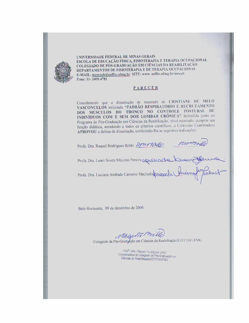
cristiane de melo vasconcelos - eeffto - ufmg · cristiane de melo vasconcelos ... você não está...
TRANSCRIPT
CRISTIANE DE MELO VASCONCELOS
PADRÃO RESPIRATÓRIO E RECRUTAMENTO DOS MÚSCULOS DO TRONCO
NO CONTROLE POSTURAL DE INDIVÍDUOS COM E SEM DOR LOMBAR
CRÔNICA
Universidade Federal de Minas Gerais
Escola de Educação Física, Fisioterapia e Terapia Ocupacional
Belo Horizonte
2009
CRISTIANE DE MELO VASCONCELOS
PADRÃO RESPIRATÓRIO E RECRUTAMENTO DOS MÚSCULOS DO TRONCO
NO CONTROLE POSTURAL DE INDIVÍDUOS COM E SEM DOR LOMBAR
CRÔNICA
Dissertação de mestrado apresentada ao Programa de Pós-Graduação em Ciências da Reabilitação da Escola de Educação Física, Fisioterapia e Terapia Ocupacional da Universidade Federal de Minas Gerais (UFMG), como requisito parcial para obtenção do título de mestre.
Área de Concentração: Desempenho Motor e Funcional Humano
Orientadora: Profa Dra. Raquel Rodrigues Britto
Co-orientador: Prof. Dr. Paulo Henrique Ferreira
Universidade Federal de Minas Gerais
Mestrado em Ciências da Reabilitação
Belo Horizonte
2009
M331p
2009
Vasconcelos, Cristiane de Melo
Padrão respiratório e recrutamento dos músculos do tronco no controle postural
de indivíduos com e sem dor lombar crônica. [manuscrito] /Cristiane de Melo
Vasconcelos – 2009.
106 f., enc.: il.
Orientadora: Raquel Rodrigues Britto
Co-orientador: Paulo Henrique Ferreira
Dissertação (Mestrado) – Universidade Federal de Minas Gerais, Escola de
Educação Física, Fisioterapia e Terapia Ocupacional.
Bibliografia: f. 46-53
1. Dor lombar. 2. Músculos abdominais. 3. Ultrassom. 4. Respiração. I.
Britto, Raquel Rodrigues. II. Ferreira, Paulo Henrique. III. Universidade Federal de
Minas Gerais. Escola de Educação Física, Fisioterapia e Terapia Ocupacional. III.
Título.
CDU: 159.943 Ficha catalográfica elaborada pela equipe de bibliotecários da Biblioteca da Escola de Educação Física, Fisioterapia e Terapia Ocupacional da Universidade Federal de Minas Gerais.

ii
Dedico este trabalho a todos aqueles
que contribuíram para sua realização.
iii
AGRADECIMENTOS A Deus, por me guiar pelas estradas da vida, me dando força, proteção e serenidade para superar cada obstáculo. Aos meus pais e ao meu irmão, pelo amor, compreensão, dedicação, exemplo de valores e princípios e por sempre me apoiarem em todas as minhas escolhas. Pai, você não está aqui agora, mas se não fosse você, eu jamais conseguiria. Ao Leo, por fazer minha vida muito mais feliz. Muito obrigada pelo amor, paciência e por estar sempre ao meu lado. A toda a minha família, pelo amor e apoio constante. Por favor, desculpem-me pelas ausências. Ao professor Paulo Henrique, por ser muito mais que um orientador: um verdadeiro pai, amigo, mestre e conselheiro. Muito obrigada, por me proporcionar diversas oportunidades na fisioterapia e na vida. A professora Raquel Britto, pela paciência na orientação, amizade, competência, ensinamentos, disponibilidade e por acreditar na realização desse sonho. A Fabianna, pela parceria nas coletas, mas principalmente pela amizade, pelos momentos de alegria, trabalho e descontração. Foi muito bom passar esses anos com você. Esse título também é seu. A Professora Manuela Ferreira, Verônica Parreira, Leani Pereira e Luciana Machado, pelas valiosas sugestões, apoio e participação no meu crescimento científico. A todos os professores e mestres do Departamento de Fisioterapia, em especial aos professores: Luci, Marcelo, Elyonara, Sérgio, João Marcos, Gisele, Anderson, Rosângela e Marisa. Minha eterna gratidão por terem contribuído para a construção de quem sou hoje. Aos colegas do LabCare que participaram das coletas de dados e das trocas de informações, em especial à Mariana Coutinho pela disponibilidade, pensamentos positivos, interesse e amizade. A todos os amigos e companheiros de trabalho da SPINE, pela amizade, substituições e por me ajudarem a realizar esse sonho. A Diva, por ser uma segunda mãe. MUITO OBRIGADA por tudo! A todos os meus amigos e companheiros do mestrado, pela troca de conhecimentos e por caminharem junto comigo. Ao Zambelli, Fabiano, Vinícius, Warley, Gui e Vânia pela amizade, sugestões e explicações valiosas. Sem vocês tudo isso seria muito mais difícil.
iv
Ao Vinicius Brasileiro, pela ajuda nas coletas e disponibilidade. Ao Tiago, irmão do Leo, pela amizade e por não medir esforços em todos os momentos em que precisei de você. A todos os meus amigos queridos, que sempre fizeram de tudo para tornar este caminho mais fácil e divertido. Amo muito vocês! A Karol, amiga e companheira de profissão, com quem tive muitos ensinamentos. Muito obrigada por todos os momentos que passamos juntas na graduação e pós-graduação. À Polly e ao Cris, pela amizade, incentivo, apoio e sugestões de verdadeiros mestres. Aos funcionários da UFMG que, sempre com muita paciência e vontade, me ofereceram todas as condições para realizar este trabalho, em especial à Marilane, Margareth, Rivamar e Richard. Aos meus estagiários, futuros companheiros de profissão, com os quais tive a oportunidade de trabalhar e aprender muitas coisas. Aos voluntários que participaram desta pesquisa, pela disponibilidade. Vocês são verdadeiros anjos! Enfim, agradeço a todos que de alguma forma passaram pela minha vida e contribuíram para a construção de quem sou hoje.

v
“A vitória de ontem deve ser apenas
um degrau na vitória de amanhã.”
Paul Brunton
vi Vasconcelos, C.M. Resumo
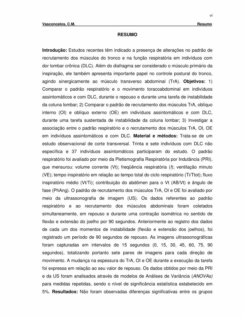
RESUMO
Introdução: Estudos recentes têm indicado a presença de alterações no padrão de
recrutamento dos músculos do tronco e na função respiratória em indivíduos com
dor lombar crônica (DLC). Além do diafragma ser considerado o músculo primário da
inspiração, ele também apresenta importante papel no controle postural do tronco,
agindo sinergicamente ao músculo transverso abdominal (TrA). Objetivos: 1)
Comparar o padrão respiratório e o movimento toracoabdominal em indivíduos
assintomáticos e com DLC, durante o repouso e durante uma tarefa de instabilidade
da coluna lombar; 2) Comparar o padrão de recrutamento dos músculos TrA, oblíquo
interno (OI) e oblíquo externo (OE) em indivíduos assintomáticos e com DLC,
durante uma tarefa sustentada de instabilidade da coluna lombar; 3) Investigar a
associação entre o padrão respiratório e o recrutamento dos músculos TrA, OI, OE
em indivíduos assintomáticos e com DLC. Material e métodos: Trata-se de um
estudo observacional de corte transversal. Trinta e sete indivíduos com DLC não
específica e 37 indivíduos assintomáticos participaram do estudo. O padrão
respiratório foi avaliado por meio da Pletismografia Respiratória por Indutância (PRI),
que mensurou: volume corrente (Vt); freqüência respiratória (f); ventilação minuto
(VE); tempo inspiratório em relação ao tempo total do ciclo respiratório (Ti/Ttot); fluxo
inspiratório médio (Vt/Ti); contribuição do abdômen para o Vt (AB/Vt) e ângulo de
fase (PhAng). O padrão de recrutamento dos músculos TrA, OI e OE foi avaliado por
meio da ultrassonografia de imagem (US). Os dados referentes ao padrão
respiratório e ao recrutamento dos músculos abdominais foram coletados
simultaneamente, em repouso e durante uma contração isométrica no sentido de
flexão e extensão do joelho por 90 segundos. Anteriormente ao registro dos dados
de cada um dos momentos de instabilidade (flexão e extensão dos joelhos), foi
registrado um período de 90 segundos de repouso. As imagens ultrassonográficas
foram capturadas em intervalos de 15 segundos (0, 15, 30, 45, 60, 75, 90
segundos), totalizando portanto sete pares de imagens para cada direção de
movimento. A mudança na espessura do TrA, OI e OE durante a execução da tarefa
foi expressa em relação ao seu valor de repouso. Os dados obtidos por meio da PRI
e da US foram analisados através de modelos de Análises de Variância (ANOVAs)
para medidas repetidas, sendo o nível de significância estatística estabelecido em
5%. Resultados: Não foram observadas diferenças significativas entre os grupos
vii Vasconcelos, C.M. Resumo
para as variáveis do padrão respiratório no repouso. No entanto, durante a tarefa de
instabilidade, os indivíduos com DLC apresentaram valores significativamente
maiores para as variáveis VE e Vt/Ti, quando comparados aos sujeitos
assintomáticos. Nas comparações entre o repouso e a instabilidade, foi observado
que somente os participantes com DLC apresentaram aumento significativo do Vt,
bem como uma diminuição da AB/Vt. Com relação ao recrutamento dos músculos
abdominais, as comparações entre os grupos indicaram que indivíduos com DLC
apresentam um recrutamento significativamente menor para o músculo TrA, quando
comparado ao grupo controle, embora não exista nenhuma diferença entre grupos
para os músculos OI e OE. As comparações entre os músculos indicaram que
indivíduos assintomáticos apresentaram um maior recrutamento do TrA em relação
ao OI e ao OE, fato não observado para o grupo com DLC. Nas comparações do
recrutamento muscular entre os intervalos de tempo, existiu diferenças significativas
para os músculos TrA e OI, embora esse resultado possa ser atribuído ao erro da
medida. Finalmente, foi observada correlação significativa, porém de baixa
magnitude, entre variáveis do padrão respiratório e o recrutamento dos músculos
abdominais nesses indivíduos. Conclusões: Indivíduos com DLC apresentam
alterações nas variáveis do padrão respiratório, assim como mudança no
recrutamento do músculo TrA, durante uma tarefa sustentada de instabilidade da
coluna lombar. A possível relação entre DLC e disfunção respiratória pode
influenciar diversos aspectos clínicos, principalmente a avaliação e a reabilitação
dos pacientes com DLC. Os clínicos e pesquisadores devem ficar atentos à
presença de alterações na função respiratória desses indivíduos.
Palavras-chave: Dor lombar; padrão respiratório; músculos abdominais;
ultrassonografia.
viii Vasconcelos, C.M. Abstract
ABSTRACT
Background and Purpose: There is mounting evidence indicating the presence of
altered trunk muscles recruitment and respiratory dysfunction in patients with chronic
low back pain (CLBP). Although the diaphragm is the principal muscle of inspiration,
it also contributes to the control of intervertebral motion by the coordination with
transversus abdominis (TrA). This study had 3 purposes: 1) To compare the
breathing pattern and thoracoabdominal motion between people with and without
CLBP, at rest and during a sustained task of lumbar spine instability; 2) To compare
the abdominal muscles recruitment throughout a sustained task of lumbar spine
instability between people with and without CLBP; 3) To investigate the association
between breathing pattern components and abdominal muscles recruitment in people
with and without CLBP. Materials and methods: It was conducted a cross-sectional
study. Thirty-seven subjects with non-specific CLBP and thirty-seven controls
participated in the study. The respiratory inductive plethysmography was used to
assess: tidal volume (Vt), respiratory frequency (f), minute ventilation (VE), fractional
inspiratory time (Ti/Ttot), mean inspiratory flow (Vt/Ti), percentage of abdomen to Vt
(AB/Vt) and Phase Angle (PhAng). In addition, the recruitment pattern of TrA,
obliquus internus (OI), and obliquus externus (OE) were assessed by
ultrasonography imaging. Plethysmography and ultrasonography recordings were
taken simultaneously at rest during 90s, and with the participant performing isometric
knee flexion and extension efforts, during an equal time of 90s. All ultrasonographic
images were made at intervals of 15s, thus, for each movement direction, seven
pairs of ultrasonographic images (at 0, 15, 30, 45, 60, 75 and 90s) were captured.
The change in TrA, OI and OE thickness was expressed as a proportion of the
thickness at rest. Plethysmography and ultrasonography data were analyzed using
repeated measure analyses of variance (ANOVAs) and alpha level was set at 0.05.
Results: There were no significant differences between groups for the variables of
breathing pattern at rest. Conversely, at the instability task, CLBP patients showed
significantly higher values for the variables of VE and Vt/Ti compared with controls.
Comparisons between rest and instability situation indicated that only participants in
the CLBP group presented an increase in Vt and a decrease in AB/Vt during the task.
Regarding the abdominal muscles recruitment, the comparisons between groups
showed that CLBP patients presented a smaller mean increase in TrA thickness
ix Vasconcelos, C.M. Abstract
compared with controls, and there were no significant differences between groups for
OI or OE recruitment. Comparisons between muscles indicated that controls had a
greater increase in TrA thickness compared with OI and OE, which did not occur in
the CLBP group. Concerning the comparisons of TrA, OI and OE recruitment
between time intervals, there were differences for the TrA and OI muscles, although
these results could be attributed to a measurement error. Finally, it was observed
that there were weak but significant inverse associations between breathing pattern
components and recruitment of the TrA, OI and OE in people with and without CLBP.
Conclusions: The current study highlighted the existence of changes in the
respiratory function, as well as in the TrA recruitment in patients with CLBP, during a
sustained task of lumbar spine instability. The possible connection of CLBP and
respiratory dysfunction may have repercussion on various clinical aspects, especially
on patient assessment and rehabilitation. Clinicians and researchers should be
aware to the presence of respiratory disorders in CLBP population.
Key-words: low back pain; breathing pattern; abdominal muscles; ultrasonography.
x Vasconcelos, C.M. Lista de abreviaturas e siglas
LISTA DE ABREVIATURAS E SIGLAS
• AB - Abdômen
• ABNT - Associação Brasileira de Normas Técnicas
• AP - Assoalho pélvico
• AB/Vt - Contribuição do abdômen para o volume corrente
• CLBP - Chronic Low Back Pain
• CNS - Central Nervous System
• CT - Caixa torácica
• CVF - Capacidade vital forçada
• CVM - Contração voluntária máxima
• d - Tamanho do efeito
• DF - Diafragma
• DLC - Dor lombar crônica
• f - Freqüência respiratória
• FEF25-75 - Fluxo expiratório forçado entre 25-75% da CVF
• IC – Intervalo de confiança
• ICC – Índice de correlação intraclasse
• IMC - Índice de massa corporal
• JPG - Joint Photographic Experts Group
• K – constante de proporcionalidade
• LabCare - Laboratório Avaliação/Pesquisa em Desempenho
Cardiorrespiratório
• MF - Multífidos
• MMII - Membros inferiores
xi Vasconcelos, C.M. Lista de abreviaturas e siglas
• Mpeg4 - Moving Picture Experts Group
• OE - Oblíquo externo
• OI - Oblíquo interno
• PFE - Pico de fluxo expiratório
• PhAng - Ângulo de fase
• PIA - Pressão intra-abdominal
• PRI - Pletismografia respiratória por indutância
• PV - Músculos paravertebrais
• QDC - Qualitative Diagnostic Calibration
• RA - Reto abdominal
• SBPT - Sociedade Brasileira de Pneumologia e Tisiologia
• TCLE - Termo de Consentimento Livre e Esclarecido
• Ti/Ttot - Tempo inspiratório em relação ao tempo total do ciclo respiratório
• TrA - Músculo transverso abdominal
• UFMG - Universidade Federal de Minas Gerais
• US - Ultrassonografia
• VE - Ventilação minuto
• VEF1 - Volume expiratório forçado no 1º segundo
• VEF1/CVF - Índice de Tiffeneau
• Vt - Volume corrente
• Vt/Ti - Fluxo inspiratório médio
xii Vasconcelos, C.M. Sumário
SUMÁRIO
DEDICATÓRIA.............................................................................................................ii
AGRADECIMENTOS...................................................................................................iii
EPÍGRAFE...................................................................................................................v
RESUMO.....................................................................................................................vi
ABSTRACT................................................................................................................viii
LISTA DE ABREVIATURAS E SIGLAS......................................................................x
Capítulo 1 – INTRODUÇÃO......................................................................................14
1.1 - Objetivos...........................................................................................................23
1.1.1 - Objetivo Geral.......................................................................................23
1.1.2 - Objetivos Específicos...........................................................................23
1.2 - Hipóteses do estudo........................................................................................23
Capítulo 2 – MATERIAIS E MÉTODOS....................................................................25
2.1 - Tipo de estudo..................................................................................................25
2.2 - Local e período de Realização........................................................................25
2.3 - Amostra.............................................................................................................25
2.3.1 - Critérios de inclusão.............................................................................26
2.3.2 - Critérios de exclusão............................................................................27
2.4 - Aspectos Éticos...............................................................................................28
2.5 - Instrumentos de medida..................................................................................28
2.5.1 - Instrumentos para caracterização da amostra.....................................28
2.5.1.1 - Questionário sócio-clínico demográfico..................................28
2.5.1.2 - Balança...................................................................................29
2.5.1.3 - Questionário Roland Morris - Brasil........................................29
2.5.1.4 - Escala Tampa para Cinesiofobia - Brasil................................30
xiii Vasconcelos, C.M. Sumário
2.5.1.5 - Espirometria............................................................................30
2.5.2 - Instrumentos de medida das variáveis dependentes...........................31
2.5.2.1 - Ultrassonografia......................................................................31
2.5.2.2 - Pletismografia Respiratória por Indutância.............................33
2.6- Procedimentos..................................................................................................37
2.7 - Procedimentos estatísticos.............................................................................44
Capítulo 3 – REFERÊNCIAS BIBLIOGRÁFICAS....................................................46
Capítulo 4 – BREATHING PATTERN AND ABDOMINAL MUSCLE RECRUITMENT
IN THE POSTURAL CONTROL OF PEOPLE WITH CHRONIC LOW BACK
PAIN...........................................................................................................................54
Capítulo 5 – CONSIDERAÇÕES FINAIS..................................................................86
Anexo I - Aprovação do Comitê de Ética em Pesquisa da UFMG........................89
Anexo II - Termo de Consentimento Livre e Esclarecido.....................................90
Anexo III - Questionário Socio-Clínico Demográfico............................................92
Anexo IV - Escala Visual Analógica........................................................................93
Anexo V - Questionário Roland Morris - Brasil.....................................................94
Anexo VI - Escala Tampa para Cinesiofobia - Brasil.............................................95
Anexo VII - Normas para a Publicação...................................................................96
14
Vasconcelos, C.M. Introdução
Capítulo 1 - INTRODUÇÃO
A dor lombar crônica (DLC) pode ser definida como dor, tensão muscular ou
rigidez localizada na região compreendida entre as últimas costelas e a linha glútea,
com duração de pelo menos três meses1. Estima-se que cerca de 80% da população
geral apresente pelo menos um episódio de dor lombar no decorrer da vida (life time
prevalence)2 e que até 50% desses indivíduos desenvolvam DLC3. Segundo a
organização Pan-Americana de saúde, a dor lombar é considerada um dos três
maiores problemas de saúde ocupacional da América Latina e é uma das doenças
sob vigilância da Organização Mundial de Saúde4. Em um estudo epidemiológico
realizado em uma amostra populacional de 2297 indivíduos na cidade de
Salvador/Brasil foi encontrada uma prevalência de DLC de 16.3%, sendo a região
lombar considerada a localização corporal de dor crônica mais frequente5. A DLC
possui impacto socioeconômico significante e está associada a elevados custos
diretos e indiretos em cuidados com a saúde, ao absenteísmo ao trabalho e a
incapacidade6, além de gerar importantes consequências para o paciente, a família,
os empregadores e a sociedade em geral1.
A DLC deve ser considerada um complexo processo multidimencional que é
influenciado na sua constituição por fatores intrínsecos ao indivíduo (doenças
inflamatórias e degenerativas, alterações congênitas, instabilidades segmentares da
coluna, desequilíbrios musculares, obesidade, morbidades psicológicas), fatores
sócio-demográficos (sexo, idade) e comportamentais (cinesiofobia, lócus de controle
da saúde)7,8,9. Ela é tipicamente classificada em específica quando os sintomas são
causados por um mecanismo patofisiológico específico, e em não específica quando
os sintomas não possuem uma causa claramente identificada, sendo essa última
15
Vasconcelos, C.M. Introdução
categoria responsável por agrupar aproximadamente 90% de todos os indivíduos
com dor lombar8. A instabilidade lombar segmentar tem sido amplamente proposta
pela literatura como um importante fator relacionado à DLC não específica10,11,12,13,14.
Panjabi12 definiu instabilidade da coluna como uma capacidade reduzida do sistema
de estabilização para manter a zona neutra intervertebral (parte da amplitude de
movimento em que há resistência mínima à movimentação intersegmentar) dentro
dos limites fisiológicos. Neste modelo, a estabilidade dos seguimentos vertebrais é
alcançada por meio da interação de três subsistemas: o subsistema passivo,
composto por ligamentos, cápsulas e estruturas ósseas; o subsistema ativo,
composto por músculos que envolvem a coluna vertebral; e o subsistema neural,
composto pelos sistemas nervoso central e periférico, responsáveis pela detecção
de demandas internas e externas e pela geração de respostas musculares
apropriadas12. Caso a integridade funcional de qualquer um desses subsistemas
esteja comprometida, a instabilidade pode acontecer e resultar em lesão e dor na
coluna lombar10,11.
Estudos recentes têm focado na contribuição funcional dos músculos do
tronco para a estabilidade da coluna lombar, bem como na relação entre suas
alterações e a presença de dor lombar aguda ou crônica10,13,14,15,16,17,18. Bermark18
dividiu o subsistema muscular do tronco em músculos superficiais ou globais e
profundos ou locais. Os músculos superficiais, tais como o oblíquo externo (OE), o
reto abdominal (RA) e os paravertebrais (PV), originam-se na pelve e se inserem no
gradil costal. Esses músculos são capazes de gerar torque, movimentar a coluna no
espaço e promover a estabilidade geral do tronco, no entanto, não possuem
influência direta no controle intersegmentar da coluna18. Por outro lado, os músculos
profundos, que incluem o transverso abdominal (TrA), diafragma (DF), assoalho
16
Vasconcelos, C.M. Introdução
pélvico (AP), multífidos (MF) e as fibras internas do músculo oblíquo interno (OI), se
inserem diretamente nos seguimentos vertebrais e possuem ação importante no
controle intersegmentar da coluna lombar18.
O recrutamento coordenado dos músculos superficiais e profundos do tronco
durante as atividades funcionais contribui para a manutenção da estabilidade
mecânica da coluna lombar19. Existem relatos na literatura de que, na presença de
disfunção do sistema muscular profundo, ocorre uma substituição compensatória
pelo sistema superficial11,19, o que pode resultar em elevadas forças de compressão
e comprometer a região lombo-pélvica17,20. Foi demonstrado que indivíduos com
DLC possuem uma atividade sustentada dos músculos eretores da coluna no final
do movimento de flexão do tronco, que em situação normal deveriam estar
inativos21. Além disso, durante uma tarefa de alcance com os membros superiores,
esses indivíduos apresentam maior atividade eletromiográfica dos músculos OE e
RA, quando comparados a sujeitos assintomáticos14. Embora muitos estudos
reportem alta variabilidade no padrão de ativação dos músculos superficiais entre os
indivíduos com DLC17, mudanças no recrutamento dos músculos profundos do
tronco são um achado consistente15,16,22.
Há evidências que indicam um padrão alterado de recrutamento do TrA,
músculo abdominal mais profundo, em indivíduos com DLC10,11,13,14,15,16. O TrA se
origina nas seis últimas costelas, crista ilíaca, processos transversos das vértebras
lombares e fáscia tóraco-lombar, e se insere na bainha do músculo RA23. Sua
contração aumenta a tensão da fáscia tóraco-lombar e a pressão intra-abdominal
(PIA)10. Em indivíduos saudáveis, o recrutamento do TrA ocorre de maneira
antecipatória e independente da direção do movimento dos membros, o que é
consistente com sua função na estabilização dos movimentos intersegmentares da
17
Vasconcelos, C.M. Introdução
coluna lombar16,24,25. No entanto, indivíduos com DLC possuem um maior limiar de
ativação do TrA16, avaliada por meio de eletromiografia, bem como uma redução na
espessura deste músculo durante a movimentação dos membros inferiores15,
avaliada por meio da ultrassonografia (US), sugerindo uma alteração no
recrutamento do TrA nestes indivíduos.
Similarmente ao TrA, o músculo DF tem sua origem no tendão central e
inserção nas três primeiras vértebras lombares, ligamento arqueado, processo
xifóide e últimas seis costelas23. Embora o DF seja considerado o músculo primário
da inspiração23, muitos estudos têm mostrado seu importante papel no controle
postural do tronco24,26,27,28,29. Quando a estabilidade do tronco é desafiada por forças
reativas decorrentes do movimento do membro superior, a atividade eletromiográfica
do DF aumenta e é antecipatória à pertubação27. Além disso, essa resposta é
simultânea à atividade do TrA, independente da fase da respiração e está
relacionada à amplitude da força reativa na coluna27,29, sugerindo uma ação
estabilizadora intersegmentar do DF. Durante o movimento repetido do membro
superior, a atividade eletromiográfica do DF é modulada para manter a respiração,
no entanto, desenvolve-se uma atividade tônica (durante a expiração) somada a
uma atividade fásica na frequência do movimento do membro28,29. Essa resposta
também é observada para o músculo TrA28. Portanto, existem pelo menos dois
estímulos (um relacionado com a respiração e outro com o movimento) para os
motoneurônios do DF durante o movimento do membro28. Quando indivíduos
saudáveis são submetidos a uma situação de instabilidade do tronco, essas duas
funções podem ser mantidas sem haver o comprometimento de uma em relação a
outra27,28,29,30. Porém, Hodges et al.31 observaram por meio da pletismografia
respiratória por indutância (PRI) e da eletromiografia que quando a demanda
18
Vasconcelos, C.M. Introdução
respiratória é aumentada em decorrência de uma hipercapnia induzida
experimentalmente, a atividade postural do DF e do TrA é reduzida. Neste
experimento, foi utilizado um longo tubo (170ml) a fim de se aumentar o espaço
morto de 13 sujeitos saudáveis que foram instruídos a movimentar o membro
superior o mais rápido possível. Os autores sugeriram que o aumento do estímulo
respiratório, que acontece durante uma situação de sobrecarga respiratória, pode
levar à atenuação da atividade postural dos músculos TrA e DF, por meio da oclusão
do estímulo postural a seus motoneurônios.
A atividade dos músculos abdominais, principalmente do TrA, e a atividade do
DF podem modular a rigidez da coluna indiretamente, por meio do aumento da PIA,
ou diretamente, como resultado da contração muscular26,32,33,34. O aumento da PIA
acontece durante muitas tarefas de vida diária e contribui de maneira isolada para
estabilização mecânica da coluna vertebral26,32,34,35. Estudos em humanos35 e em
porcos26 demostraram que a rigidez da coluna pode ser aumentada em decorrência
de uma maior PIA, sem a atividade concomitante dos músculos abdominais e
extensores do tronco. Além disso, devido a inserção do DF crural nas vértebras
lombares, sua contração pode infuenciar diretamente a rigidez da coluna
lombar26,33.
Alguns autores constataram que, em relação aos indivíduos saudáveis,
indivíduos com DLC possuem um maior deslocamento do centro de massa durante
a respiração natural36,37 e quando submetidos a uma maior demanda respiratória37.
Hodges et al.38 sugeriram que tal resposta seja devido a uma provável alteração na
coordenação do sistema nervoso central. Em um estudo recente39, foi demonstrado
por meio de estimulação elétrica transcraniana que indivíduos com dor lombar
recorrente apresentam uma reorganização da representação dos músculos do
19
Vasconcelos, C.M. Introdução
tronco no córtex motor, que está associada ao início da atividade eletromiográfica do
TrA durante o movimento do membro superior. Portanto, os autores sugeriram que
essa reorganização do córtex motor pode estar associada às alterações do controle
postural observadas nesses indivíduos.
Há evidências preliminares de uma associação entre a DLC e alterações na
função respiratória22,40,41. A função respiratória envolve diversas estruturas, em
conjunto denominadas de sistema respiratório, que proporcionam a entrada e a
saída de ar das vias aéreas42. Tal sistema é constituído pelos pulmões, que realizam
a troca gasosa, e pela parede torácica, composta por diferentes músculos
responsáveis pelo movimento de expansão e retração da mesma. Estes músculos
trabalham de maneira coordenada, bombeando ar para dentro e para fora dos
pulmões, contribuindo para a manutenção da troca gasosa em nível adequado23. O
padrão respiratório depende de variáveis relacionadas ao volume e aos tempos
respiratórios, sendo seu controle influenciado por mecanismos corticais, periféricos,
da musculatura respiratória e de membros43. A avaliação do movimento da parede
torácica é importante para a identificação do padrão respiratório. Para tal avaliação,
considera-se que a parede torácica possui dois compartimentos distintos: a caixa
torácica (CT) e o abdômen (AB), separados pelo DF44. Embora os dois
compartimentos movam-se em unidade, cada qual parece ter funções diferentes,
movendo-se como uma unidade com considerável independência de movimento44.
Em indivíduos saudáveis, durante a respiração tranquila, a CT e o AB comportam-se
da mesma forma, deslocando para fora na inspiração e para dentro na expiração,
com o mesmo ritmo45. Embora o DF seja o principal músculo da inspiração, os
músculos intercostais internos da região paraesternal e os escalenos são músculos
primários da inspiração, que quando ativados elevam as costelas e expandem o
20
Vasconcelos, C.M. Introdução
tórax superior. Ainda que o músculo esternocleidomastóideo também contribua para
a expansão da CT superior, o mesmo é considerado um músculo acessório da
inspiração23.
Em um estudo transversal, foi demonstrado que embora a obesidade e o nível
de atividade física sejam dois fatores comumente investigados na DLC, a presença
de disfunções respiratórias e/ou de incontinência urinária está mais fortemente
associada à queixa de dor lombar40. No entanto, poucos estudos avaliaram se a
presença de DLC reduz a contribuição do DF para a respiração.
O’Sullivan et al.22 observaram alteração na função respiratória e no controle
motor de indivíduos com DLC. Esses autores identificaram, por meio da espirometria
e da US, que indivíduos com dor na região sacroilíaca apresentam valores
significativamente maiores de freqüência respiratória (f) (F = 10.42; p = 0.004) e de
ventilação minuto (VE) (F = 5.4; p = 0.028) durante um teste de elevação ativa dos
membros inferiores, quando comparados a sujeitos assintomáticos. No entanto, foi
observado que esses indivíduos possuem uma diminuição de aproximadamente
79% na excursão diafragmática (F = 60.93; p < 0.001) durante a execução da tarefa.
Os autores sugeriram que o recrutamento de outros músculos inspiratórios, e não do
DF, pode ter provocado o aumento inadequado da função respiratória durante o
teste. Em um estudo posterior46, foi observado que uma intervenção voltada para o
treinamento de músculos estabilizadores profundos da coluna lombar (como o TrA e
AP) foi capaz de melhorar a cinemática alterada do DF e influenciar de maneira
positiva a função respiratória de indivíduos com DLC, aproximando sua resposta
daquela observada em indivíduos assintomáticos.
Com o intuito de avaliar o padrão respiratório em indivíduos assintomáticos e
com DLC, Roussel et al.41 observaram o movimento toracoabdominal, por meio de
21
Vasconcelos, C.M. Introdução
análise visual e palpatória, durante o repouso e durante a realização de dois testes
que avaliam o controle motor. Os autores definiram a respiração costo-diafragmática
(situação em que a CT e o AB se deslocam para fora na inspiração e para dentro na
expiração) como padrão respiratório normal e consideraram todas as demais
respirações (respiração paradoxal, com predomínio da CT superior, mista e com
pausa respiratória) como padrão respiratório alterado. De acordo com suas
observações, os autores concluíram que uma proporção maior de indivíduos com
DLC (42.5%) apresentou padrão respiratório alterado, quando comparado com
sujeitos assintomáticos (5%). Todavia, como mencionado por Benoist47, a
classificação em padrão respiratório normal e alterado por meio de análise visual e
palpatória do movimento toracoabdominal é muito subjetiva, especialmente na
ausência de teste de confiabilidade intraexaminador, o qual os autores relataram não
ter sido realizado. Em um estudo prévio48, foi demonstrado que a confiabilidade
interexaminador da avaliação do padrão respiratório da maneira realizada por esses
autores varia de ruim a moderada (k = 0.39 a 0.47) dependendo da situação
analisada. Além disso, os autores relataram que os testes de controle motor foram
mantidos por 1041 e 2048 segundos, sendo esse período muito curto para se avaliar o
padrão respiratório.
É importante ressaltar que os exercícios de controle motor, que visam
recuperar a estabilidade da coluna por meio de um treinamento específico capaz de
restabelecer os padrões de ativação muscular e estratégias posturais alteradas na
presença de DLC49,50, frequentemente utilizam o controle da respiração como
estratégia terapêutica. Tal intervenção, além de ser muito difundida na prática
clínica, é considerada efetiva na redução da dor e incapacidade de indivíduos com
DLC49,50. Somado a isso, algumas técnicas alternativas que têm ganhado grande
22
Vasconcelos, C.M. Introdução
popularidade na última década, como o Pilates e a Yoga, apresentam como um dos
focos de intervenção o controle da respiração, apesar de ainda existirem poucas
evidências científicas para tal indicação51,52,53,54.
Considerando as evidências apresentadas anteriormente, é possível observar
que os estudos apontam para a presença de alterações no padrão de recrutamento
dos músculos do tronco e na função respiratória de indivíduos com DLC. Uma falha
na coordenação das funções postural e respiratória desses músculos tem sido
proposta pela literatura como uma explicação para estes achados31,41,55. No entanto,
os estudos que avaliaram o padrão respiratório nestes indivíduos realizaram essa
análise de maneira subjetiva e durante a realização de tarefas de curta duração (até
20 segundos), não sendo encontrados na literatura pesquisada estudos que
investigaram a associação entre o padrão respiratório (variáveis de volume e tempo,
e movimento toracoabdominal) e o recrutamento dos músculos do tronco (TrA, OI e
OE) no controle postural de indivíduos com DLC.
A investigação de alterações nas variáveis do padrão respiratório e no
recrutamento dos músculos do tronco (TrA, OI e OE), bem como a associação
destes fatores em indivíduos com DLC, é uma maneira de contribuir para a melhoria
da abordagem terapêutica frente a essa patologia. Caso seja identificado alterações
nas variáveis do padrão respiratório e exista associação dessas variáveis com o
recrutamento dos músculos TrA, OI e OE, intervenções direcionadas à função
respiratória poderiam potencializar a função estabilizadora dos músculos do tronco e
consequentemente, aumentar o sucesso dos desfechos clínicos almejados para
essa população.
23
Vasconcelos, C.M. Introdução
1.1 Objetivos
1.1.1 Objetivo geral
Avaliar o padrão respiratório, por meio da pletismografia respiratória por
indutância (PRI), e o recrutamento dos músculos do tronco (TrA, OI e OE), por meio
da ultrassonografia (US), em indivíduos assintomáticos e com DLC, durante o
repouso e durante uma tarefa sustentada de instabilidade da coluna lombar.
1.1.2 Objetivos específicos
• Comparar o padrão respiratório, variáveis de volume e tempo, e movimento
toracoabdominal em indivíduos assintomáticos e com DLC, em duas situações:
repouso e instabilidade da coluna lombar;
• Comparar o padrão de recrutamento dos músculos TrA, OI, OE ao longo do
tempo (90 segundos), durante uma tarefa sustentada de instabilidade da coluna
lombar, em indivíduos assintomáticos e com DLC;
• Investigar a associação entre as variáveis do padrão respiratório (durante o
repouso e instabilidade da coluna lombar) e o recrutamento dos músculos TrA,
OI, OE em indivíduos assintomáticos e com DLC.
1.2 Hipóteses do estudo
Hipótese alternativa 1: Indivíduos com DLC, quando comparados com sujeitos
assintomáticos, apresentam alterações nas variáveis do padrão respiratório durante
uma tarefa de instabilidade da coluna lombar.
Hipótese alternativa 2: Indivíduos com DLC, quando comparados com sujeitos
assintomáticos, apresentam alteração no padrão de recrutamento dos músculos TrA,
OI, OE ao longo do tempo, durante uma tarefa sustentada de instabilidade da coluna
lombar.

24
Vasconcelos, C.M. Introdução
Hipótese alternativa 3: Existe associação entre as variáveis do padrão
respiratório e o recrutamento dos músculos TrA, OI, OE, em indivíduos
assintomáticos e com DLC.
25
Vasconcelos, C.M. Materiais e Métodos
Capítulo 2 - MATERIAIS E MÉTODOS
2.1 Tipo de estudo
Trata-se de um estudo observacional de corte transversal56, realizado com
indivíduos adultos assintomáticos e com DLC.
2.2 Local e período de Realização
O estudo foi realizado no Laboratório de Avaliação e Pesquisa em
Desempenho Cardiorrespiratório (LabCare) do Departamento de Fisioterapia, da
Escola de Educação Física, Fisioterapia e Terapia Ocupacional da Universidade
Federal de Minas Gerais (UFMG). O recrutamento dos participantes ocorreu no
período de Março de 2008 à Setembro de 2009.
2.3 Amostra
O número amostral foi calculado com base em um estudo piloto com 13
indivíduos assintomáticos (grupo controle) e 13 indivíduos com DLC, em que foram
analisadas as variáveis de volume corrente (Vt) e contribuição do abdômen para o
volume corrente (AB/Vt). Foram escolhidas estas variáveis para a realização do
cálculo amostral, por serem variáveis respiratórias que apresentam relevância clínica
e que pareciam diferenciar mais um grupo do outro. Ao realizar o cálculo do
tamanho do efeito (d) entre o grupo controle e experimental, foram encontrados
efeitos de magnitude de 0.63 e 0.61, respectivamente. Considerando esses efeitos
(d), a análise não direcional, um nível de significância α = 0.05 e um poder
estatístico de 0.80, o tamanho amostral necessário para se evidenciar estes efeitos
deveria ser de, aproximadamente, 40 indivíduos para análise do Vt e 43 indivíduos
26
Vasconcelos, C.M. Materiais e Métodos
para a análise da AB/Vt57. No entanto, cálculos posteriores considerando as coletas
realizadas no decorrer do estudo apontaram para uma amostra de 35 sujeitos.
Portanto, foram recrutados 37 indivíduos assintomáticos e 37 indivíduos com DLC,
pareados por sexo, idade e índice de massa corporal (IMC), porém a amostra final
consistiu de 35 sujeitos por grupo, pois 2 participantes com DLC foram excluídos
devido à má visualização da imagem ultrassonográfica e dois participantes
assintomáticos foram excluídos devido a uma falha no procedimento de calibração
da PRI. As características destes participantes não diferiram das características do
restante da amostra. A amostra foi alocada por conveniência e os participantes
foram recrutados de forma não aleatória entre estudantes e funcionários da UFMG e
da comunidade em geral e pacientes em fila de espera de serviços de reabilitação
física de Belo Horizonte. Não houve restrições quanto ao sexo ou nível sócio-
econômico e os indivíduos foram selecionados a partir dos seguintes critérios:
2.3.1 Critérios de inclusão
Para os dois grupos:
I) Assinar o Termo de Consentimento Livre e Esclarecido (TCLE) (Anexo II); II)
Apresentar IMC entre 18 Kg/m2 e 29.9 Kg/m2 58; III) Apresentar idade entre 18 e 60
anos; IV) Não apresentar sinais de radiculopatias (atestados por pelo menos 2
dentre 3 testes: testes de reflexos, testes sensitivos e testes de miótomos)59; V) Não
apresentar diagnóstico ou suspeita (verificada pela presença de bandeiras
vermelhas59) de doenças graves na coluna vertebral (espondiloartropatias
inflamatórias, fraturas, tumores, síndrome da cauda eqüina ou infecções); VI) Não
apresentar histórico de doenças neurológicas, neoplasias, fraturas vertebrais ou
cirurgia na coluna ou na região tóracoabdominal; VII) Não estar em período
gestacional ou puerperal; VIII) Não apresentar indicativo de doença respiratória
27
Vasconcelos, C.M. Materiais e Métodos
associada ou pregressa avaliada por meio de um questionário desenvolvido por
Pereira et al.60 (anexo II) e pelas medidas de Capacidade vital forçada (CVF),
Volume expiratório forçado no 1º segundo (VEF1) e fluxo expiratório forçado entre
25-75% da CVF (FEF25-75), de acordo com os limites de normalidade propostos
pelos mesmos autores; IX) Não ter feito uso de relaxante muscular, analgésico ou
antiinflamatório nas últimas 48 horas; X) Não apresentar deformidades graves
observáveis na região torácica ou abdominal; XI) Não apresentar contra-indicação
ao exercício físico.
Para o grupo controle:
I) Não relatar história de dor lombar que tenha provocado afastamento do trabalho
ou restringido suas atividades; II) Estar livre de quaisquer sintomas relacionados à
coluna e pélvis; III) Não estar fazendo uso de medicamentos para dor.
Para o grupo com DLC:
I) Apresentar sintomas relacionados à DLC1 (por no mínimo três meses) de origem
não específica, com ou sem dor referida para o membro inferior; II) Pontuação de no
mínimo dois pontos no Questionário Roland Morris61 (anexo V) e no mínimo de duas
unidades na escala numérica 0 – 10 de dor62 (anexo IV); III) Não estar envolvido em
treinamentos direcionados à aprendizagem motora do recrutamento da musculatura
estabilizadora intersegmentar da coluna lombar e região perineal nos últimos três
meses anteriores ao teste16,24. Observação: Caso os participantes apresentassem
osteoartrite, grau I de espondilólise ou espondilolistese ou
protusão/herniação/prolapso discal, eles seriam incluídos nesse grupo, pelo fato
dessas patologias serem classificadas na literatura como dor lombar não específica8.
2.3.2 Critérios de exclusão
28
Vasconcelos, C.M. Materiais e Métodos
I) Ser incapaz de compreender ou realizar os procedimentos propostos pelo estudo;
II) Apresentar qualquer evento agudo de dor ou piora dos sintomas durante o
procedimento de coleta do presente estudo; III) Apresentar qualquer movimento
atípico que interfira no registro dos dados da PRI ou da US.
2.4 Aspectos éticos
Este projeto de pesquisa faz parte do projeto intitulado “Análise das atividades
dos músculos profundos do tronco no controle postural de indivíduos com e sem dor
lombar crônica”, sob a coordenação do Prof. Paulo Henrique Ferreira, aprovado pelo
Comitê de Ética em Pesquisa da UFMG (ETIC 071/07) (anexo I).
Os participantes do estudo foram informados e instruídos quanto aos
procedimentos, estimativa do tempo de permanência no laboratório para a coleta de
dados, direitos, riscos e benefícios. Os voluntários que concordaram com a
participação no estudo assinaram o TCLE (anexo II).
2.5 Instrumentos de medida
2.5.1 Instrumentos para caracterização da amostra
2.5.1.1 Questionário sócio-clínico demográfico
A população foi caracterizada por meio de uma avaliação previamente
estruturada (anexo III), com os seguintes domínios: dados pessoais, diagnóstico
clínico, anamnese e exame físico. Esta avaliação continha informações sobre
presença de incontinência urinária, depressão clínica e nível de atividade física,
além de um questionário desenvolvido por Pereira et al.60, que foi utilizado com o
objetivo de triar os indivíduos que apresentassem indicativos de doença respiratória
associada ou pregressa. Caso o participante respondesse “sim” a qualquer uma das
29
Vasconcelos, C.M. Materiais e Métodos
perguntas desse último questionário, o mesmo deveria realizar a espirometria. Ainda
nesta avaliação, foi utilizada a escala numérica 0 – 10 de dor62 (anexo IV) a fim de
se medir a intensidade da dor dos participantes com DLC. Os escores dessa escala
variam de zero (nenhuma dor) a dez (pior dor possível), sendo a mesma comumente
utilizada no âmbito científico e clínico para avaliação de indivíduos com DLC,
fornecendo valores válidos e confiáveis de intensidade da dor62,63,64.
2.5.1.2 Balança
Para aferir o peso e a altura dos participantes foi utilizada uma balança
calibrada (Filizola ind. LTDA, São Paulo, SP, Brasil). Tais dados foram utilizados
para o cálculo do IMC, o qual é determinado pela divisão do peso pela altura ao
quadrado (kg/m2), e para definir a quantidade de força a ser desempenhada pelos
voluntários durante a coleta de dados da US.
2.5.1.3 Questionário Roland Morris - Brasil
Foi utilizado o questionário Roland Morris - Brasil (anexo V), previamente
adaptado e validado para a população brasileira61, a fim de se avaliar o grau de
incapacidade apresentada pelos participantes com DLC. Este questionário consiste
em 24 itens relativos à interferência das dores nas costas nas atividades de vida
diária e de vida prática de indivíduos com dor lombar. Os participantes deviam
responder “sim” ou “não” no caso da presença ou ausência, respectivamente, de
dificuldade para a realização da tarefa. O escore final foi obtido pela soma das
respostas “sim” sendo que, quanto maior o escore, maior o grau de incapacidade do
indivíduo. Este questionário apresenta confiabilidade intra e interexaminadores de r
= 0.88 e 0.86, respectivamente61, e foi aplicado no presente estudo por um
examinador previamente treinado.
30
Vasconcelos, C.M. Materiais e Métodos
2.5.1.4 Escala Tampa para Cinesiofobia - Brasil
Para a avaliação do grau de cinesiofobia (medo do movimento) dos
participantes com DLC, foi utilizada a Escala Tampa para Cinesiofobia - Brasil
(anexo VI), adaptada e validada para a população brasileira65. Ela consiste em 17
afirmações, sendo que os participantes deviam responder o tanto que concordavam
ou discordavam com cada item, utilizando uma escala de 4 pontos. Para a obtenção
do escore final, foi realizada a soma das respostas, invertendo os escores das
questões 4, 8, 12, 16. Quanto maior a pontuação adquirida, maior é o grau de
cinesiofobia, ou seja, maior é o medo que o indivíduo apresenta de se movimentar
devido à dor na coluna lombar. Esta escala apresenta confiabilidade
intraexaminadores de Índice de Correlação Intraclasse (ICC) = 0.8065, sendo
aplicada no presente estudo por um examinador previamente treinado.
2.5.1.5 Espirometria
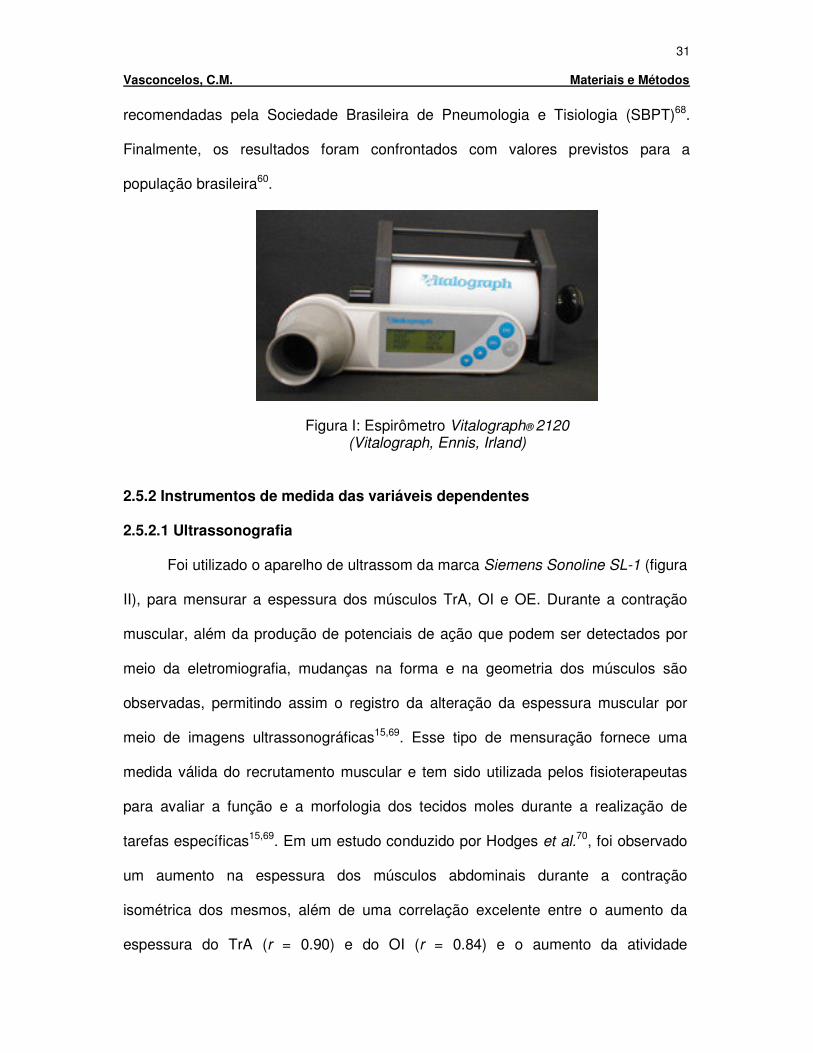
Foi utilizado o espirômetro Vitalograph® 2120 (Vitalograph, Ennis, Irland)
(figura I) com o objetivo de excluir os indivíduos que apresentassem distúrbios
ventilatórios de qualquer ordem. Um total de 11 participantes, 6 indivíduos com DLC
e 5 assintomáticos, realizaram a prova de função pulmonar. A espirometria avalia os
volumes de ar inspirado e expirado e os fluxos respiratórios, entre eles o CVF, VEF1,
FEF25-75% e o índice de Tiffeneau (VEF1/CVF), sendo um teste que permite o
diagnóstico e a quantificação dos distúrbios ventilatórios60,66. O aparelho utilizado no
presente estudo preenche os padrões mínimos de acurácia, erro, linearidade e
registros gráficos estabelecidos pela Associação Brasileira de Normas Técnicas
(ABNT)66,67. Foram realizadas manobras de CVF para medidas dos fluxos e volumes
pulmonares, sendo que as mesmas seguiram os critérios de aceitação e
reprodutibilidade, assim como a gradação da qualidade, de acordo com as normas
31
Vasconcelos, C.M. Materiais e Métodos
recomendadas pela Sociedade Brasileira de Pneumologia e Tisiologia (SBPT)68.
Finalmente, os resultados foram confrontados com valores previstos para a
população brasileira60.
2.5.2 Instrumentos de medida das variáveis dependentes
2.5.2.1 Ultrassonografia
Foi utilizado o aparelho de ultrassom da marca Siemens Sonoline SL-1 (figura
II), para mensurar a espessura dos músculos TrA, OI e OE. Durante a contração
muscular, além da produção de potenciais de ação que podem ser detectados por
meio da eletromiografia, mudanças na forma e na geometria dos músculos são
observadas, permitindo assim o registro da alteração da espessura muscular por
meio de imagens ultrassonográficas15,69. Esse tipo de mensuração fornece uma
medida válida do recrutamento muscular e tem sido utilizada pelos fisioterapeutas
para avaliar a função e a morfologia dos tecidos moles durante a realização de
tarefas específicas15,69. Em um estudo conduzido por Hodges et al.70, foi observado
um aumento na espessura dos músculos abdominais durante a contração
isométrica dos mesmos, além de uma correlação excelente entre o aumento da
espessura do TrA (r = 0.90) e do OI (r = 0.84) e o aumento da atividade
Figura I: Espirômetro Vitalograph® 2120 (Vitalograph, Ennis, Irland)
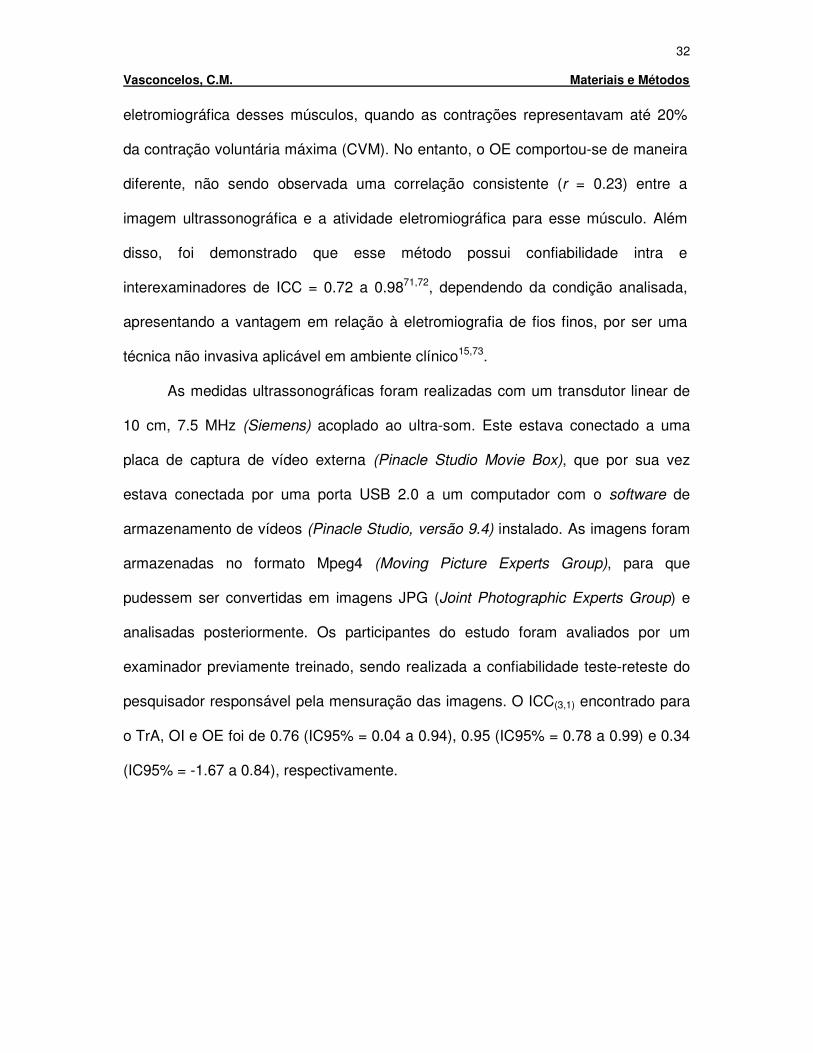
32
Vasconcelos, C.M. Materiais e Métodos
eletromiográfica desses músculos, quando as contrações representavam até 20%
da contração voluntária máxima (CVM). No entanto, o OE comportou-se de maneira
diferente, não sendo observada uma correlação consistente (r = 0.23) entre a
imagem ultrassonográfica e a atividade eletromiográfica para esse músculo. Além
disso, foi demonstrado que esse método possui confiabilidade intra e
interexaminadores de ICC = 0.72 a 0.9871,72, dependendo da condição analisada,
apresentando a vantagem em relação à eletromiografia de fios finos, por ser uma
técnica não invasiva aplicável em ambiente clínico15,73.
As medidas ultrassonográficas foram realizadas com um transdutor linear de
10 cm, 7.5 MHz (Siemens) acoplado ao ultra-som. Este estava conectado a uma
placa de captura de vídeo externa (Pinacle Studio Movie Box), que por sua vez
estava conectada por uma porta USB 2.0 a um computador com o software de
armazenamento de vídeos (Pinacle Studio, versão 9.4) instalado. As imagens foram
armazenadas no formato Mpeg4 (Moving Picture Experts Group), para que
pudessem ser convertidas em imagens JPG (Joint Photographic Experts Group) e
analisadas posteriormente. Os participantes do estudo foram avaliados por um
examinador previamente treinado, sendo realizada a confiabilidade teste-reteste do
pesquisador responsável pela mensuração das imagens. O ICC(3,1) encontrado para
o TrA, OI e OE foi de 0.76 (IC95% = 0.04 a 0.94), 0.95 (IC95% = 0.78 a 0.99) e 0.34
(IC95% = -1.67 a 0.84), respectivamente.
33
Vasconcelos, C.M. Materiais e Métodos
2.5.2.2 Pletismografia Respiratória por Indutância
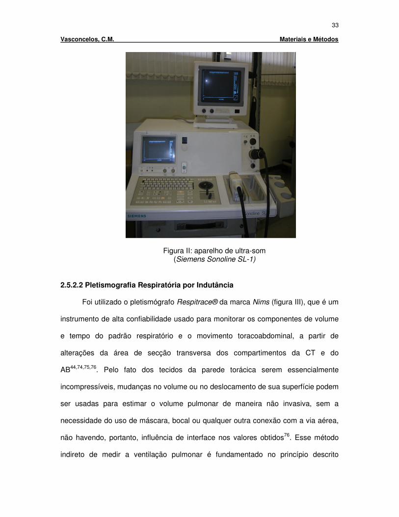
Foi utilizado o pletismógrafo Respitrace® da marca Nims (figura III), que é um
instrumento de alta confiabilidade usado para monitorar os componentes de volume
e tempo do padrão respiratório e o movimento toracoabdominal, a partir de
alterações da área de secção transversa dos compartimentos da CT e do
AB44,74,75,76. Pelo fato dos tecidos da parede torácica serem essencialmente
incompressíveis, mudanças no volume ou no deslocamento de sua superfície podem
ser usadas para estimar o volume pulmonar de maneira não invasiva, sem a
necessidade do uso de máscara, bocal ou qualquer outra conexão com a via aérea,
não havendo, portanto, influência de interface nos valores obtidos76. Esse método
indireto de medir a ventilação pulmonar é fundamentado no princípio descrito
Figura II: aparelho de ultra-som (Siemens Sonoline SL-1)
34
Vasconcelos, C.M. Materiais e Métodos
inicialmente por Konno e Mead em 196744, que presumiram que a parede torácica
possui duas partes (CT e AB) que se movem cada qual como uma unidade, havendo
considerável independência de movimento entre elas. Assim, durante a respiração
com via aérea livre, este sistema é considerado aberto, uma vez que a CT e o AB
podem acomodar volume de maneira independente (dois graus de liberdade de
movimento), de modo que o volume inspirado (Vt) seja a soma das alterações de
volume da CT e do AB. O movimento de cada compartimento pode ser medido por
sensores externos e calibrado com um espirômetro para prover medidas
volumétricas75. Foi demonstrado que esse método fornece valores consistentes de
volumes pulmonares, sendo observada diferença menor que 10% em relação à
espirometria, após diferentes métodos de calibração74,75.
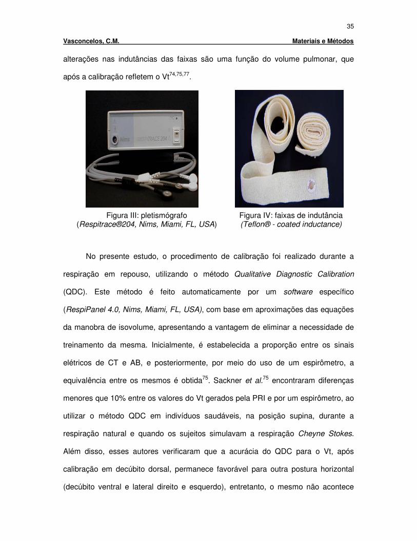
Na PRI são utilizadas duas faixas de indutância (Teflon® - coated inductance)
(figura IV). Uma das faixas é posicionada na axila e a outra na altura da cicatriz
umbilical, sendo cada uma composta por duas partes elásticas finas e aderidas, que
envolvem um fio transdutor disposto de forma sinusoidal74,77. Para aquisição dos
dados, as faixas são conectadas por meio de cabos ao equipamento, que fornece
três curvas: uma correspondente ao deslocamento da CT, outra do AB, e outra à
soma dos sinais de CT e AB, que corresponde ao Vt. O movimento da CT gera um
traçado que reflete principalmente o efeito da contração dos músculos intercostais e
acessórios da respiração. Por outro lado, o movimento do AB gera um traçado que
reflete principalmente o efeito do trabalho diafragmático78. Os transdutores das
faixas detectam a variação na área de secção transversa decorrente dos
movimentos de CT e AB, e transmitem a informação do movimento como sinal
elétrico, sendo então a indutância de cada faixa registrada pelo Respitrace. Essas
35
Vasconcelos, C.M. Materiais e Métodos
alterações nas indutâncias das faixas são uma função do volume pulmonar, que
após a calibração refletem o Vt74,75,77.
No presente estudo, o procedimento de calibração foi realizado durante a
respiração em repouso, utilizando o método Qualitative Diagnostic Calibration
(QDC). Este método é feito automaticamente por um software específico
(RespiPanel 4.0, Nims, Miami, FL, USA), com base em aproximações das equações
da manobra de isovolume, apresentando a vantagem de eliminar a necessidade de
treinamento da mesma. Inicialmente, é estabelecida a proporção entre os sinais
elétricos de CT e AB, e posteriormente, por meio do uso de um espirômetro, a
equivalência entre os mesmos é obtida75. Sackner et al.75 encontraram diferenças
menores que 10% entre os valores do Vt gerados pela PRI e por um espirômetro, ao
utilizar o método QDC em indivíduos saudáveis, na posição supina, durante a
respiração natural e quando os sujeitos simulavam a respiração Cheyne Stokes.
Além disso, esses autores verificaram que a acurácia do QDC para o Vt, após
calibração em decúbito dorsal, permanece favorável para outra postura horizontal
(decúbito ventral e lateral direito e esquerdo), entretanto, o mesmo não acontece
Figura III: pletismógrafo (Respitrace®204, Nims, Miami, FL, USA)
Figura IV: faixas de indutância (Teflon® - coated inductance)
36
Vasconcelos, C.M. Materiais e Métodos
quando se altera a postura de deitado para sentado ou de pé, ou vice-versa.
Portanto, se houver mudanças na posição corporal do indivíduo, faz-se necessária
uma recalibração para a nova postura adotada75.
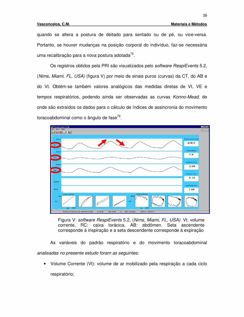
Os registros obtidos pela PRI são visualizados pelo software RespiEvents 5.2,
(Nims, Miami, FL, USA) (figura V) por meio de sinais puros (curvas) da CT, do AB e
do Vt. Obtêm-se também valores analógicos das medidas diretas de Vt, VE e
tempos respiratórios, podendo ainda ser observadas as curvas Konno-Mead, de
onde são extraídos os dados para o cálculo de índices de assincronia do movimento
toracoabdominal como o ângulo de fase79.
As variáveis do padrão respiratório e do movimento toracoabdominal
analisadas no presente estudo foram as seguintes:
• Volume Corrente (Vt): volume de ar mobilizado pela respiração a cada ciclo
respiratório;
Figura V: software RespiEvents 5.2, (Nims, Miami, FL, USA). Vt: volume corrente, RC: caixa torácica, AB: abdômen. Seta ascendente corresponde à inspiração e a seta descendente corresponde à expiração
37
Vasconcelos, C.M. Materiais e Métodos
• Freqüência Respiratória (f): número de incursões respiratórias realizadas no
intervalo de 1 minuto;
• Ventilação Minuto (VE): volume de ar mobilizado pela respiração no intervalo
de 1 minuto, sendo computada por meio do produto do Vt pela f;
• Tempo inspiratório em relação ao tempo total do ciclo respiratório (Ti/Ttot):
forma de medida indireta do timing respiratório, definido como a razão entre o
tempo inspiratório pelo tempo total do ciclo respiratório;
• Fluxo inspiratório médio (Vt/Ti): forma de medida indireta do estímulo
respiratório, definido como a razão entre o Vt pelo tempo inspiratório;
• Contribuição do abdômen para o volume corrente (AB/Vt): Participação do
compartimento abdominal na geração do Vt;
• Ângulo de fase (PhAng): variável que reflete o grau de assincronia
toracoabdominal, sendo que seus valores variam de 0° (movimento
sincrônico) a 180°(movimento completamente assincrônico).
Neste estudo, para facilitar a comparação com a literatura, as abreviaturas
das variáveis relativas ao padrão respiratório e ao movimento toracoabdominal
foram mantidas em inglês.
2.6 Procedimentos
Após a explicação dos procedimentos e assinatura do TCLE (anexo I), foi
realizada uma avaliação utilizando as escalas e os questionários referidos
previamente. Essa etapa foi realizada por um único entrevistador/examinador
treinado, em um local reservado, sendo os dados identificados por meio de códigos,
de forma que o pesquisador responsável pelas medidas ultrassonográficas não
tivesse acesso às informações, além de garantir o anonimato do voluntário.
38
Vasconcelos, C.M. Materiais e Métodos
Um total de 11 participantes, 6 indivíduos com DLC e 5 assintomáticos,
realizaram a espirometria, pois responderam “sim” a pelo menos uma das perguntas
do questionário (anexo III) utilizado para triar indivíduos que apresentassem
indicativos de doença respiratória associada ou pregressa. Inicialmente foram dadas
as orientações quanto à manobra, sendo as mesmas realizadas com os indivíduos
sentados, mantendo o tronco apoiado, cabeça na posição neutra, pés encostados no
solo e fazendo uso de clipe nasal. O aparelho foi calibrado diariamente considerando
a temperatura do dia e utilizando uma seringa de 1000ml (Vitalograph, Buckinghan,
England). Foram aceitas as manobras que apresentassem evidência de que o
participante realizou esforço máximo, com início abrupto, sem hesitação e com
duração de pelo menos 6 segundos de expiração forçada. Além disso, as manobras
deveriam apresentar um volume retroextrapolado menor que 5% da CVF ou menor
que 150ml (o maior deles), e a variação do pico de fluxo expiratório (PFE) entre as
manobras deveria ser menor ou igual a 10% ou menor ou igual a 500ml (novamente,
o maior deles). Os testes foram realizados até que fossem alcançadas três
manobras aceitáveis com pelo menos duas reprodutíveis, ou seja, os valores de
CVF e VEF1 não deveriam diferir mais que 150ml entre eles. Os maiores valores de
CVF e VEF1 foram selecionados, não necessariamente da mesma curva, sendo o
FEF25-75% retirado da curva de maior soma de CVF e VEF1. Foram realizadas no
máximo 8 manobras, sendo fornecidos intervalos de 1 minuto entre as mesmas.
Finalmente, os resultados foram confrontados com valores previstos para a
população brasileira60. Com o intuito de evitar a fadiga da musculatura respiratória,
os indivíduos que realizaram o teste de função pulmonar descansaram por pelo
menos uma hora ou os mesmos foram solicitados a retornar em um outro dia a fim
de dar continuidade ao protocolo da pesquisa.
39
Vasconcelos, C.M. Materiais e Métodos
Para a coleta das variáveis do padrão respiratório, as duas faixas da PRI
foram posicionadas, uma na axila e outra na linha umbilical, e foram levemente
esticadas em torno do indivíduo para assegurar um ajuste firme e minimizar a
distorção do sinal, sem, no entanto, limitar o movimento toracoabdominal ou causar
desconforto. Posteriormente, o indivíduo foi posicionado confortavelmente em uma
maca em supino, com os membros superiores ao lado do tronco, quadris fletidos a
50° e joelhos fletidos a 90°. Para o registro dos dados da PRI, foi realizada uma
calibração prévia, de modo que os ganhos elétricos relativos (400Mv = 100% Vt) aos
canais da CT e do AB foram obtidos utilizando o procedimento QDC, durante 5
minutos de respiração natural. Este procedimento, baseado nas equações propostas
por Sackner et al.75 para calcular a constante de proporcionalidade (K) entre os
ganhos da CT e do AB, foi utilizado para igualar os sinais dos dois compartimentos,
de modo que sua soma correspondesse ao volume pulmonar. Posteriormente, a fim
de se obter o sinal da PRI em valores absolutos (ml), foi realizada uma calibração
quantitativa com o sujeito respirando, com uso de bocal e clipe nasal, em um
espirômetro (Vitatrace, Pro Médico, Rio de Janeiro, RJ, Brazil), por
aproximadamente 60 segundos. Esse espirômetro foi calibrado utilizando uma
seringa de 1000ml (Vitalograph, Buckinghan, England) por meio do software
RespiPanel, e finalmente, os sinais elétricos foram registrados no sistema de
aquisição digital (RespiEvents 5.2, Nims). Após este procedimento, que apresentava
duração aproximada de 10 minutos, o participante teve os membros inferiores (MMII)
reposicionados para então dar início à coleta de dados da PRI, concomitante com a
coleta de dados da US.
Para a aquisição das imagens ultrassonográficas dos músculos TrA, OI e OE,
foi utilizado o protocolo proposto e validado por Ferreira et al.15. O posicionamento
40
Vasconcelos, C.M. Materiais e Métodos
dos voluntários foi mantido por um aparato adaptado de um sistema já existente15.
De acordo com esse protocolo, os MMII foram ajustados no aparato com 50° de
flexão de quadris e 90° de flexão de joelhos. As pernas ficaram suspensas por
amarras presas em torno dos joelhos e tornozelos, sendo tais amaras fixas em uma
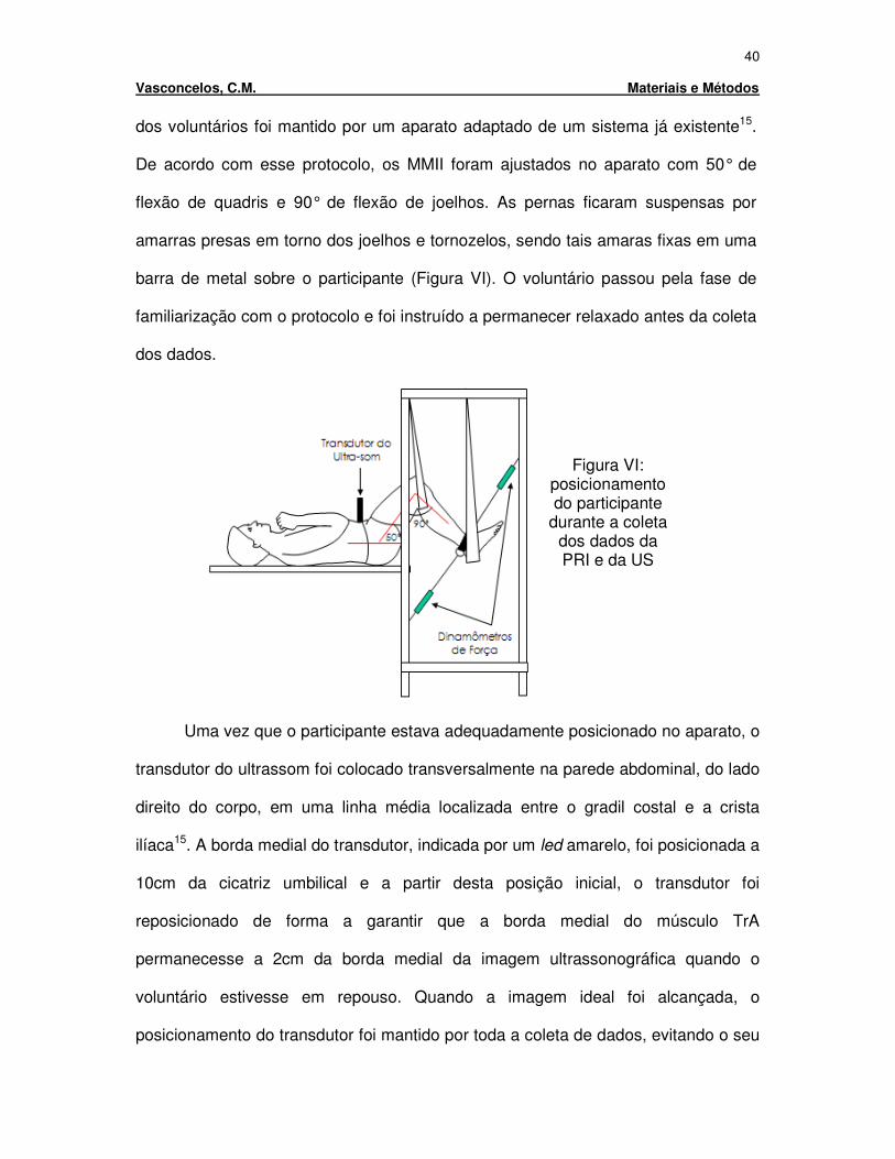
barra de metal sobre o participante (Figura VI). O voluntário passou pela fase de
familiarização com o protocolo e foi instruído a permanecer relaxado antes da coleta
dos dados.
Uma vez que o participante estava adequadamente posicionado no aparato, o
transdutor do ultrassom foi colocado transversalmente na parede abdominal, do lado
direito do corpo, em uma linha média localizada entre o gradil costal e a crista
ilíaca15. A borda medial do transdutor, indicada por um led amarelo, foi posicionada a
10cm da cicatriz umbilical e a partir desta posição inicial, o transdutor foi
reposicionado de forma a garantir que a borda medial do músculo TrA
permanecesse a 2cm da borda medial da imagem ultrassonográfica quando o
voluntário estivesse em repouso. Quando a imagem ideal foi alcançada, o
posicionamento do transdutor foi mantido por toda a coleta de dados, evitando o seu
Figura VI: posicionamento do participante
durante a coleta dos dados da PRI e da US
41
Vasconcelos, C.M. Materiais e Métodos
deslocamento crânio/caudal, medial/lateral, assim como mudanças na sua
angulação vertical. Após esse posicionamento do transdutor, os dados referentes ao
padrão respiratório e ao recrutamento dos músculos abdominais de cada indivíduo
foram coletados simultaneamente em repouso por aproximadamente 90 segundos.
Posteriormente, foi solicitado ao participante a executar uma contração isométrica no
sentido de flexão e extensão do joelho, por aproximadamente 90 segundos,
almejando uma força de 7,5% do peso corporal (tarefa sustentada de instabilidade
da coluna lombar). Foi demonstrado em um estudo prévio15 que esse nível de força
realizado durante a extensão ou flexão dos joelhos corresponde a aproximadamente
20% da CVM, mantendo assim, a correlação entre o aumento da espessura
muscular e o aumento da atividade eletromiográfica para os músculos TrA e OI.
Essa força foi mensurada por um transdutor de força fixo a um cinto que envolvia os
tornozelos e cuja armação era firmemente fixada à maca. Anteriormente ao registro
dos dados de cada um dos momentos de instabilidade (flexão e extensão dos
joelhos), foi registrado um período de 90 segundos de repouso. Em todos esses
momentos foram capturadas, além dos dados referentes ao padrão respiratório, sete
imagens ultrassonográficas em intervalos de 15 segundos (0, 15, 30, 45, 60, 75, 90
segundos). Portanto, foi armazenado um total de quatorze pares de imagens, sete
pares para flexão e sete pares para extensão, sendo que cada par consistia em uma
imagem capturada em um determinado intervalo de tempo com o individuo em
repouso (imagem de repouso), somado a uma outra imagem capturada durante a
execução da tarefa com os MMII (imagem de contração), no intervalo de tempo
correspondente ao do repouso. As imagens de repouso e de contração foram
coletadas no final da expiração e a escolha da ordem de execução das tarefas de
flexão e extensão dos joelhos foi de forma aleatória. Observou-se em estudo
42
Vasconcelos, C.M. Materiais e Métodos
prévio15 que a captura de dados durante as duas direções do movimento dos joelhos
é importante para eliminar possível viés de indivíduos com preferência de
recrutamento muscular em uma direção específica. Os comandos para execução e
graduação da contração foram oferecidos por um feedback verbal da força e foi
instruído que o indivíduo continuasse respirando tranquilamente, evitando mudanças
de posição durante o registro dos dados e mantendo cada contração por 90
segundos, com intervalo de 5 minutos entre a flexão e a extensão dos joelhos.
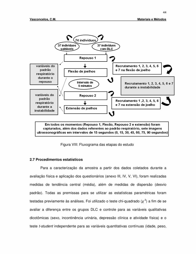
(Figura VIII)
Os dados armazenados provenientes do ultrassom foram posteriormente
convertidos em imagens no formato JPG e analisadas no software Distance® pelo
avaliador das imagens da US, que foi mantido cegado para análise dos dados. As
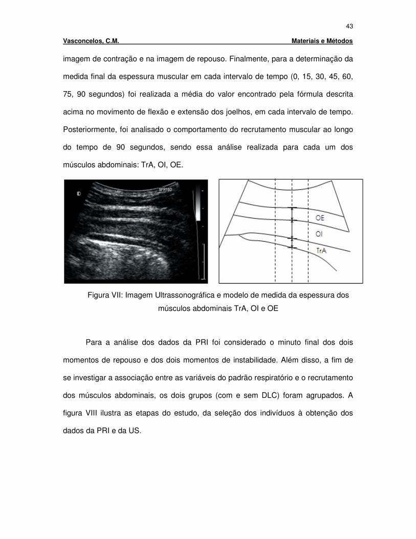
imagens foram sobrepostas por um grid e foram realizadas as medidas da
espessura dos músculos TrA, OI e OE em três pontos pré-determinados: um
localizado no ponto médio da imagem ultrassonográfica e outros dois localizados
1cm a direita e a esquerda do ponto médio, como demonstrado na figura VII. Foram
utilizadas como referência, as bordas das regiões hipoecoícas (regiões
esbranquiçadas), que refletem de maneira lenta as ondas do ultrassom e
representam as fáscias que separam os músculos15. Para calcular a porcentagem da
alteração entre o par de imagens (imagem de repouso e imagem de contração) foi
utilizada a seguinte fórmula: %100 =×−
R
RC , onde C representa a média das medidas
dos três pontos pré-determinados de uma imagem de contração, e R representa a
média das medidas dos três pontos pré-determinados de uma imagem de repouso.
O valor encontrado, obtido em porcentagem, representa o aumento da espessura do
músculo durante a execução da tarefa em relação a sua espessura em repouso.
Portanto, essa medida está condicionada a análise da espessura do músculo na
43
Vasconcelos, C.M. Materiais e Métodos
imagem de contração e na imagem de repouso. Finalmente, para a determinação da
medida final da espessura muscular em cada intervalo de tempo (0, 15, 30, 45, 60,
75, 90 segundos) foi realizada a média do valor encontrado pela fórmula descrita
acima no movimento de flexão e extensão dos joelhos, em cada intervalo de tempo.
Posteriormente, foi analisado o comportamento do recrutamento muscular ao longo
do tempo de 90 segundos, sendo essa análise realizada para cada um dos
músculos abdominais: TrA, OI, OE.
Para a análise dos dados da PRI foi considerado o minuto final dos dois
momentos de repouso e dos dois momentos de instabilidade. Além disso, a fim de
se investigar a associação entre as variáveis do padrão respiratório e o recrutamento
dos músculos abdominais, os dois grupos (com e sem DLC) foram agrupados. A
figura VIII ilustra as etapas do estudo, da seleção dos indivíduos à obtenção dos
dados da PRI e da US.
Figura VII: Imagem Ultrassonográfica e modelo de medida da espessura dos
músculos abdominais TrA, OI e OE
44
Vasconcelos, C.M. Materiais e Métodos
2.7 Procedimentos estatísticos
Para a caracterização da amostra a partir dos dados coletados durante a
avaliação física e aplicação dos questionários (anexo III, IV, V, VI), foram realizadas
medidas de tendência central (média), além de medidas de dispersão (desvio
padrão). Todas as premissas para se utilizar as estatísticas paramétricas foram
testadas previamente às análises. Foi utilizado o teste chi-quadrado (χ 2) a fim de se
avaliar a diferença entre os grupos DLC e controle para as variáveis qualitativas
dicotômicas (sexo, incontinência urinária, depressão clínica e atividade física) e o
teste t-student independente para as variáveis quantitativas contínuas (idade, peso,
Figura VIII: Fluxograma das etapas do estudo
45
Vasconcelos, C.M. Materiais e Métodos
altura, IMC e Vt). Acrescido a essas análises foi utilizado o teste ICC(3,1) para
verificar a confiabilidade teste-reteste do pesquisador responsável pela mensuração
das imagens ultrassonográficas.
Posteriormente, os dados obtidos por meio da PRI e da US foram analisados
utilizando modelos de Análises de Variância (ANOVAs) para medidas repetidas.
Para os dados de PRI, foram construídos modelos de ANOVAs two way, sendo
considerados os fatores independentes (between group factors) a condição (DLC ou
controle), e os fatores dentro dos grupos (within factors) a situação (repouso e
instabilidade). Nessa análise, foram construídos modelos de ANOVAs individuais
para cada uma das variáveis do padrão respiratório. Por outro lado, para os dados
da US foram construídos modelos de ANOVAs three way, sendo considerados os
fatores independentes (between group factors) a condição (DLC ou controle) e os
músculos (Tra, OI e OE), e os fatores dentro dos grupos (within factors) os intervalos
tempo (0, 15, 30, 45, 60, 75, 90 segundos). Como a ANOVA detectou fatores
principais (main factors) e/ou fator de interação (interaction factor), testes post hocs
de Fischer foram realizados para a análise de pares individuais. Finalmente, para se
avaliar o nível de associação entre o recrutamento muscular e as variáveis do
padrão respiratório foi utilizado o coeficiente de correlação de Pearson (r). Toda a
análise estatística foi realizada por meio do software STATISTICA (versão 7.0,
StatStoft, Inc.), sendo o nível de significância estatística estabelecido de 5%.
46
Vasconcelos, C.M. Referências Bibliográficas
Capítulo 3 - REFERÊNCIAS BIBLIOGRÁFICAS
1. WADDELL,G. The back pain revolution. 2. ed. London: Churchill Livingstone, 2004.
2. ANDERSSON,G.B. Epidemiological features of chronic low-back pain. Lancet, v. 354, n.9178, p. 581-585, 14 Aug. 1999.
3. HENSCHKE,N. et al. Prognosis in patients with recent onset low back pain in Australian primary care: inception cohort study. BMJ, v. 337, p. a171- 2008.
4. CHOI,B.C.; TENNASSEE,L.M.; EIJKEMANS,G.J. Developing regional workplace health and hazard surveillance in the Americas. Rev Panam Salud Publica, v. 10, n.6, p. 376-381, Dec. 2001.
5. SA,K. et al. Prevalence of chronic pain and associated factors in the population of Salvador, Bahia. Rev Saude Publica, v. 43, n.4, p. 622-630, Aug. 2009.
6. VAN TULDER,M.W.; KOES,B.W.; BOUTER,L.M. A cost-of-illness study of back pain in The Netherlands. Pain, v. 62, n.2, p. 233-240, Aug. 1995.
7. SILVA,M.C.; FASSA,A.G.; VALLE,N.C. Chronic low back pain in a Southern Brazilian adult population: prevalence and associated factors. Cad Saude Publica, v. 20, n.2, p. 377-385, Mar. 2004.
8. VAN,T.M.; KOES,B.; BOMBARDIER,C. Low back pain. Best Pract Res Clin Rheumatol, v. 16, n.5, p. 761-775, Dec. 2002.
9. PICAVET,H.S.; VLAEYEN,J.W.; SCHOUTEN,J.S. Pain catastrophizing and kinesiophobia: predictors of chronic low back pain. Am J Epidemiol, v. 156, n.11, p. 1028-1034, 1 Dec. 2002.
10. EBENBICHLER,G.R. et al. Sensory-motor control of the lower back: implications for rehabilitation. Med Sci Sports Exerc, v. 33, n.11, p. 1889-1898, Nov. 2001.
11. HODGES,P.W. The role of the motor system in spinal pain: implications for rehabilitation of the athlete following lower back pain. J Sci Med Sport, v. 3, n.3, p. 243-253, Sept. 2000.
47
Vasconcelos, C.M. Referências Bibliográficas
12. PANJABI,M.M. The stabilizing system of the spine. Part I. Function, dysfunction, adaptation, and enhancement. J Spinal Disord, v. 5, n.4, p. 383-389, Dec. 1992.
13. RICHARDSON,C.A.; JULL,G.A. Muscle control-pain control. What exercises would you prescribe? Man Ther, v. 1, n.1, p. 2-10, Nov. 1995.
14. SILFIES,S.P. et al. Trunk muscle recruitment patterns in specific chronic low back pain populations. Clin Biomech (Bristol , Avon ), v. 20, n.5, p. 465-473, June 2005.
15. FERREIRA,P.H.; FERREIRA,M.L.; HODGES,P.W. Changes in recruitment of the abdominal muscles in people with low back pain: ultrasound measurement of muscle activity. Spine, v. 29, n.22, p. 2560-2566, 15 Nov. 2004.
16. HODGES,P.W.; RICHARDSON,C.A. Altered trunk muscle recruitment in people with low back pain with upper limb movement at different speeds. Arch Phys Med Rehabil, v. 80, n.9, p. 1005-1012, Sept. 1999.
17. HODGES,P.W.; MOSELEY,G.L. Pain and motor control of the lumbopelvic region: effect and possible mechanisms. J Electromyogr Kinesiol, v. 13, n.4, p. 361-370, Aug. 2003.
18. BERGMARK,A. Stability of the lumbar spine. A study in mechanical engineering. Acta Orthop Scand Suppl, v. 230, p. 1-54, 1989.
19. O'SULLIVAN,P.B. Lumbar segmental 'instability': clinical presentation and specific stabilizing exercise management. Man Ther, v. 5, n.1, p. 2-12, Feb. 2000.
20. HODGES,P. et al. Changes in the mechanical properties of the trunk in low back pain may be associated with recurrence. J Biomech, v. 42, n.1, p. 61-66, 5 Jan. 2009.
21. SHIRADO,O. et al. Flexion-relaxation phenomenon in the back muscles. A comparative study between healthy subjects and patients with chronic low back pain. Am J Phys Med Rehabil, v. 74, n.2, p. 139-144, Mar. 1995.
22. O'SULLIVAN,P.B. et al. Altered motor control strategies in subjects with sacroiliac joint pain during the active straight-leg-raise test. Spine, v. 27, n.1, p. E1-E8, 1 Jan. 2002.
48
Vasconcelos, C.M. Referências Bibliográficas
23. DE,T.A.; ESTENNE,M. Functional anatomy of the respiratory muscles. Clin Chest Med, v. 9, n.2, p. 175-193, June 1988.
24. HODGES,P.W.; RICHARDSON,C.A. Feedforward contraction of transversus abdominis is not influenced by the direction of arm movement. Exp Brain Res, v. 114, n.2, p. 362-370, Apr. 1997.
25. HODGES,P.W.; RICHARDSON,C.A. Contraction of the abdominal muscles associated with movement of the lower limb. Phys Ther, v. 77, n.2, p. 132-142, Feb. 1997.
26. HODGES,P.W. et al. Intervertebral stiffness of the spine is increased by evoked contraction of transversus abdominis and the diaphragm: in vivo porcine studies. Spine, v. 28, n.23, p. 2594-2601, 1 Dec. 2003.
27. HODGES,P.W. et al. Contraction of the human diaphragm during rapid postural adjustments. J Physiol, v. 505 ( Pt 2), p. 539-548, 1 Dec. 1997.
28. HODGES,P.W.; GANDEVIA,S.C. Changes in intra-abdominal pressure during postural and respiratory activation of the human diaphragm. J Appl Physiol, v. 89, n.3, p. 967-976, Sept. 2000.
29. HODGES,P.W.; GANDEVIA,S.C. Activation of the human diaphragm during a repetitive postural task. J Physiol, v. 522 Pt 1, p. 165-175, 1 Jan. 2000.
30. BEALES,D.J.; O'SULLIVAN,P.B.; BRIFFA,N.K. Motor control patterns during an active straight leg raise in pain-free subjects. Spine (Phila Pa 1976 ), v. 34, n.1, p. E1-E8, 1 Jan. 2009.
31. HODGES,P.W.; HEIJNEN,I.; GANDEVIA,S.C. Postural activity of the diaphragm is reduced in humans when respiratory demand increases. J Physiol, v. 537, n.Pt 3, p. 999-1008, 15 Dec. 2001.
32. CHOLEWICKI,J. et al. Lumbar spine stability can be augmented with an abdominal belt and/or increased intra-abdominal pressure. Eur Spine J, v. 8, n.5, p. 388-395, 1999.
33. SHIRLEY,D. et al. Spinal stiffness changes throughout the respiratory cycle. J Appl Physiol, v. 95, n.4, p. 1467-1475, Oct. 2003.

49
Vasconcelos, C.M. Referências Bibliográficas
34. HODGES,P.W. et al. In vivo measurement of the effect of intra-abdominal pressure on the human spine. J Biomech, v. 34, n.3, p. 347-353, Mar. 2001.
35. HODGES,P.W. et al. Intra-abdominal pressure increases stiffness of the lumbar spine. J Biomech, v. 38, n.9, p. 1873-1880, Sept. 2005.
36. GRIMSTONE,S.K.; HODGES,P.W. Impaired postural compensation for respiration in people with recurrent low back pain. Exp Brain Res, v. 151, n.2, p. 218-224, July 2003.
37. HAMAOUI,A. et al. Does respiration perturb body balance more in chronic low back pain subjects than in healthy subjects? Clin Biomech (Bristol , Avon ), v. 17, n.7, p. 548-550, Aug. 2002.
38. SMITH,M.; COPPIETERS,M.W.; HODGES,P.W. Effect of experimentally induced low back pain on postural sway with breathing. Exp Brain Res, v. 166, n.1, p. 109-117, Sept. 2005.
39. TSAO,H.; GALEA,M.P.; HODGES,P.W. Reorganization of the motor cortex is associated with postural control deficits in recurrent low back pain. Brain, v. 131, n.Pt 8, p. 2161-2171, Aug. 2008.
40. SMITH,M.D.; RUSSELL,A.; HODGES,P.W. Disorders of breathing and continence have a stronger association with back pain than obesity and physical activity. Aust J Physiother, v. 52, n.1, p. 11-16, 2006.
41. ROUSSEL,N. et al. Altered breathing patterns during lumbopelvic motor control tests in chronic low back pain: a case-control study. Eur Spine J, v. 18, n.7, p. 1066-1073, July 2009.
42. GUYTON AC Ventilação pulmonar. In: GUYTON AC Tratado de fisiologia médica. Rio de Janeiro (RJ): Guanabara Koogan, 1992. p. 352-362.
43. LOPES R.B.; BRITTO,R.R.; PARREIRA,V.F. Padrão Respiratório durante o exercício: revisão literária. Rev Bras Cin e Movimento, v. 13, n.2, p. 153-160, 2005.
44. KONNO,K.; MEAD,J. Measurement of the separate volume changes of rib cage and abdomen during breathing. J Appl Physiol, v. 22, n.3, p. 407-422, Mar. 1967.
50
Vasconcelos, C.M. Referências Bibliográficas
45. VERSCHAKELEN,J.A.; DEMEDTS,M.G. Normal thoracoabdominal motions. Influence of sex, age, posture, and breath size. Am J Respir Crit Care Med, v. 151, n.2 Pt 1, p. 399-405, Feb. 1995.
46. O'SULLIVAN,P.B.; BEALES,D.J. Changes in pelvic floor and diaphragm kinematics and respiratory patterns in subjects with sacroiliac joint pain following a motor learning intervention: a case series. Man Ther, v. 12, n.3, p. 209-218, Aug. 2007.
47. BENOIST,M. Reviewer's comments concerning "Altered breathing patterns during lumbopelvic motor control tests in chronic low back pain: a case-control study" (J. Nijs et al., ESJO-D-08-00264 R2). Eur Spine J, v. 18, n.7, p. 1074-1075, July 2009.
48. ROUSSEL,N.A. et al. Low back pain: clinimetric properties of the Trendelenburg test, active straight leg raise test, and breathing pattern during active straight leg raising. J Manipulative Physiol Ther, v. 30, n.4, p. 270-278, May 2007.
49. FERREIRA,M.L. et al. Comparison of general exercise, motor control exercise and spinal manipulative therapy for chronic low back pain: A randomized trial. Pain, v. 131, n.1-2, p. 31-37, Sept. 2007.
50. FERREIRA,P.H. et al. Specific stabilisation exercise for spinal and pelvic pain: a systematic review. Aust J Physiother, v. 52, n.2, p. 79-88, 2006.
51. DONZELLI,S. et al. Two different techniques in the rehabilitation treatment of low back pain: a randomized controlled trial. Eura Medicophys, v. 42, n.3, p. 205-210, Sept. 2006.
52. RYDEARD,R.; LEGER,A.; SMITH,D. Pilates-based therapeutic exercise: effect on subjects with nonspecific chronic low back pain and functional disability: a randomized controlled trial. J Orthop Sports Phys Ther, v. 36, n.7, p. 472-484, July 2006.
53. SOROSKY,S.; STILP,S.; AKUTHOTA,V. Yoga and pilates in the management of low back pain. Curr Rev Musculoskelet Med, v. 1, n.1, p. 39-47, Mar. 2008.
54. GLADWELL V et al. Does a program of pilates improve chronic no-specific low back pain? J Sport Rehabil, v. 15, p. 338-350, 2006.
51
Vasconcelos, C.M. Referências Bibliográficas
55. WHITTAKER,J.L. Ultrasound imaging of the lateral abdominal wall muscles in individuals with lumbopelvic pain and signs of concurrent hypocapnia. Man Ther, v. 13, n.5, p. 404-410, Oct. 2008.
56. PORTNEY LG; WATKINS MP. Foundations of clinical research: applications to practice. 3. ed. Upper Saddle River (NJ): Prentice Hall, 2009.
57. COHEN,J. Statistical Power Analysis for the Behavioral Sciences. 2. ed. Hillsdale (NJ): Lawrence Erlbaum Associates, 1988.
58. ASSOCIAÇÃO BRASILEIRA PARA O ESTUDO DA OBESIDADE E DA SÍNDROME METABÓLICA. Consenso Latino Americano sobre obesidade. Disponível em: <www.abeso.org.br>. Acesso em: 16 nov 2009v. 2009.
59. MAGEE DJ. Coluna Lombar. In: MAGEE DJ Avaliação Musculoesquelética. Barueri (SP): Manole, 2005. p. 463-563.
60. PEREIRA CAC; SATO T; RODRIGUES SC. Novos valores de referência para espirometria forçada em brasileiros adultos de raça branca. J Bras Penumol, v. 33, n.4, p. 397-406, 2007.
61. NUSBAUM,L. et al. Translation, adaptation and validation of the Roland-Morris questionnaire--Brazil Roland-Morris. Braz J Med Biol Res, v. 34, n.2, p. 203-210, Feb. 2001.
62. ROSS RLP. Clinical assessment of pain. In: VAN DEUSEN JH Assessment in occupational therapy and physical therapy. Philadelphia: Elsevier Health Sciences, 1997. p. 122-133.
63. HO,K.; SPENCE,J.; MURPHY,M.F. Review of pain-measurement tools. Ann Emerg Med, v. 27, n.4, p. 427-432, Apr. 1996.
64. MADER,T.J. et al. How reliable are pain scores? A pilot study of 20 healthy volunteers. J Emerg Nurs, v. 29, n.4, p. 322-325, Aug. 2003.
65. SIQUEIRA,F.B.; TEIXERA-SALMELA,L.F.; MAGALHÃES,L.C. Análise das propriedades psicométricas da versão brasileira da escala tampa de cinesiofobia. Acta Ortop Bras, v. 15, n.1, p. 19-24, 2007.
66. PEREIRA CAC. Diretrizes para teste de função pulmonar. J Pneumol, v. 28, n.Suppl 3, p. 1-241, 2002.
52
Vasconcelos, C.M. Referências Bibliográficas
67. 2120 Spirometer user Manual. Vitalograph, v. p. 1-72, 1998.
68. SOCIEDADE BRASILEIRA DE PNEUMOLOGIA E TISIOLOGIA (SBPT). I Consenso brasileiro sobre espirometria. J Pneumol, v. 22, p. 105-164, 1996.
69. TEYHEN,D.S. et al. Rehabilitative ultrasound imaging of the abdominal muscles. J Orthop Sports Phys Ther, v. 37, n.8, p. 450-466, Aug. 2007.
70. HODGES,P.W. et al. Measurement of muscle contraction with ultrasound imaging. Muscle Nerve, v. 27, n.6, p. 682-692, June 2003.
71. KOPPENHAVER,S.L. et al. Reliability of rehabilitative ultrasound imaging of the transversus abdominis and lumbar multifidus muscles. Arch Phys Med Rehabil, v. 90, n.1, p. 87-94, Jan. 2009.
72. COSTA,L.O. et al. An investigation of the reproducibility of ultrasound measures of abdominal muscle activation in patients with chronic non-specific low back pain. Eur Spine J, v. 18, n.7, p. 1059-1065, July 2009.
73. TEYHEN,D.S. et al. Changes in deep abdominal muscle thickness during common trunk-strengthening exercises using ultrasound imaging. J Orthop Sports Phys Ther, v. 38, n.10, p. 596-605, Oct. 2008.
74. CHADHA,T.S. et al. Validation of respiratory inductive plethysmography using different calibration procedures. Am Rev Respir Dis, v. 125, n.6, p. 644-649, June 1982.
75. SACKNER,M.A. et al. Calibration of respiratory inductive plethysmograph during natural breathing. J Appl Physiol, v. 66, n.1, p. 410-420, Jan. 1989.
76. ATS/ERSStatment no respiratory muscle testing. Am J Respir Crit Care Med, v. 166, n.4, p. 518-624, 2002.
77. TOBIN,M.J. et al. Breathing patterns. 1. Normal subjects. Chest, v. 84, n.2, p. 202-205, Aug. 1983.
78. GILBERT,R.; AUCHINCLOSS,J.H., JR.; PEPPI,D. Relationship of rib cage and abdomen motion to diaphragm function during quiet breathing. Chest, v. 80, n.5, p. 607-612, Nov. 1981.

53
Vasconcelos, C.M. Referências Bibliográficas
79. SACKNER,M.A. et al. Assessment of asynchronous and paradoxic motion between rib cage and abdomen in normal subjects and in patients with chronic obstructive pulmonary disease. Am Rev Respir Dis, v. 130, n.4, p. 588-593, Oct. 1984.
54
Vasconcelos, C.M. Artigo
Capítulo 4 - BREATHING PATTERN AND ABDOMINAL MUSCLE
RECRUITMENT IN THE POSTURAL CONTROL OF PEOPLE WITH CHRONIC
LOW BACK PAIN
ABSTRACT 5
Background: There is evidence indicating the presence of altered trunk muscles recruitment and
respiratory dysfunction in patients with chronic low back pain (CLBP). Objective: 1) To
compare the breathing pattern and thoracoabdominal motion between people with and without
CLBP, at rest and during a sustained task of lumbar spine instability; 2) To compare the
abdominal muscles recruitment throughout a sustained task of lumbar spine instability between 10
people with and without CLBP; 3) To investigate the association between breathing pattern
components and abdominal muscles recruitment in people with and without CLBP. Design:
Cross-sectional study. Methods: Thirty-seven subjects with CLBP and thirty-seven controls
participated in the study. The respiratory inductive plethysmography was used to assess the
breathing pattern components, and the ultrasonography was used to assess the recruitment 15
pattern of transversus abdominis (TrA), obliquus internus (OI), and obliquus externus (OE)
muscles. Recordings were taken at rest and with the participant performing isometric knee
flexion and extension efforts. The data were analyzed using repeated measure analyses of
variance. Results: There were no differences between groups for the variables of breathing
pattern at rest. At the instability task, CLBP patients showed higher values for the variables of 20
minute ventilation (VE) and mean inspiratory flow (Vt/Ti) compared with controls. Only
participants in the CLBP group presented an increase in tidal volume (Vt) and a decrease in
percentage of abdomen to Vt (AB/Vt) during the task. CLBP patients presented a smaller mean
increase in TrA thickness compared with controls, and there were no differences between groups
for OI or OE recruitment. Controls had a greater increase in TrA thickness compared with OI 25
55
Vasconcelos, C.M. Artigo
and OE, which did not occur in the CLBP group. There were weak inverse associations between
breathing pattern components and TrA, OI and OE recruitment in people with and without
CLBP. Conclusions: The current study highlighted the existence of changes in the respiratory
function, as well as in the TrA recruitment in patients with CLBP during a sustained task of
lumbar spine instability. Clinicians and researchers should be aware to the presence of respiratory 30
disorders in CLBP population.
35
40
45
50
56
Vasconcelos, C.M. Artigo
INTRODUCTION
Non-specific chronic low back pain (CLBP), defined as pain and disability persisting for more
than 3 months without clear specific cause1, is a prevalent and costly condition in industrialized
societies2, 3. Lifetime prevalence of low back pain is as high as 80%4 and approximately 50% of
these patients develop persistent symptoms5. Lumbar spine instability has been widely proposed 55
in the literature as an important factor related to non-specific CLBP6-8 and many studies have
focused on the functional contribution and changes of trunk muscles recruitment in the presence
of this condition6-12. Although it has been proposed that there is high variability among patients
with CLBP in the recruitment pattern of superficial trunk muscles such as obliquus externus
(OE)12, changes in the recruitment of the deep stabilizing muscles, i.e., transversus abdominis 60
(TrA), obliquus internus (OI), lumbar multifidus, diaphragm and pelvic floor muscles, are a
consistent finding10-13.
TrA is the deepest abdominal muscle and its contraction increases the tension of the
thoracolumbar fascia and the intra-abdominal pressure6,14. In healthy people, TrA recruitment 65
occurs in a feedforward manner and its activity is irrespective of limb movement direction, which
is consistent with its contribution to the control of intervertebral motion11,15,16. However, in
people with CLBP, the electromyography activity of TrA is delayed and the thickness of this
muscle is reduced during voluntary tasks with limbs, suggesting a change in the recruitment of
TrA in patients with CLBP10, 11. 70
Although the diaphragm is the principal muscle of inspiration17, it also contributes to spinal
stiffness by increasing intra-abdominal pressure or through the direct mechanical effects of the
diaphragm crurae in lumbar vertebrae14, 18-22. When stability of the trunk is challenged by reactive
forces from movements of the limbs, electromyography activity of the diaphragm increases 75

57
Vasconcelos, C.M. Artigo
before the disturbance and regardless of the phase of respiration18, 20. Furthermore, its activity is
simultaneous to TrA’s activity and is related to the amplitude of the forces that perturb the spine,
suggesting a role for the diaphragm in the control of intervertebral motion18, 20. During a
sustained challenge to the posture of the trunk by repetitive movement of an upper limb,
diaphragm electromyography shows this muscle is still modulated to maintain respiration, but its 80
activity also develops tonically (during expiration) with superimposed modulation at the
frequency of limb movement19, 20. This response is also observed for the TrA muscle19. Thus,
there are at least two drives to diaphragm motoneurons during limb movement, one related to
inspiration and another to movement control19, so that when trunk stability is challenged in
healthy people, this dual task (trunk stability and respiration) can be maintained without 85
interference between them18-20, 23. However, the postural activity of the diaphragm and TrA is
reduced when the respiratory demand is increased, which suggests that increased respiratory drive
may attenuate the postural activity of these muscles24.
Studies have found that respiratory function and the coordination of the central nervous system 90
(CNS) are altered in the presence of CLBP13, 25-28. For example, the displacement of the center of
pressure associated with respiration is increased in patients with CLBP25-27. Additionaly, during an
active straight leg raise maneuver, people with CLBP exhibit an increased respiratory frequency
(f) and minute ventilation (VE), which are associated with a reduction of approximately 80% in
diaphragmatic excursion compared with asymptomatic participants13. The authors suggested that 95
the recruitment of other respiratory muscles, not the diaphragm, may have secured the
inadequate increase in respiratory function. Finally, the thoracoabdominal motion, measured by
visual inspection and manual palpation, has also been found to be altered in patients with CLBP
compared to healthy subjects28. However, breathing patterns and diaphragmatic kinematics are
normalized following an individualized motor learning intervention29. 100
58
Vasconcelos, C.M. Artigo
In this way, it is noteworthy that the evidence supports the presence of altered trunk muscles
recruitment and respiratory dysfunction in patients with CLBP. A non-optimal coordination of
postural and respiratory functions of these muscles has been proposed as an explanation for
these findings24, 28, 30. However, studies that evaluated the association between breathing pattern
and abdominal muscles recruitment in CLBP are either scarce or used subjective measures such 105
as visual inspection and palpation of thoracoabdominal motion during brief tasks28, 31. If there are
changes in breathing pattern components, as well as an association between these components
and the trunk muscles recruitment, interventions targeted at the respiratory function could
enhance the stabilizing function of the trunk muscles, consequently increasing the clinical
outcomes success desired for this population. 110
Therefore, the aims of the present study were:
1) To compare the breathing pattern as measured by volume and time components, as well as the
thoracoabdominal motion between people with and without CLBP, in two situations: at rest and
during a sustained task of lumbar spine instability; 115
2) To compare the recruitment pattern of the muscles TrA, OI and OE throughout a sustained
task of lumbar spine instability between people with and without CLBP;
3) To investigate the association between the breathing pattern components (at rest and during a
sustained task of lumbar spine instability) and the recruitment of the TrA, OI and OE in people
with and without CLBP. 120
MATERIALS AND METHODS
Participants
A sample of 74 participants of both genders (37 with CLBP and 37 asymptomatic) was recruited
from the community according to the following inclusion criteria: age ranging from 18 to 60 125
59
Vasconcelos, C.M. Artigo
years; body mass index (BMI) between 18 and 29.9 Kg/m2 32, no history of thoracoabdominal or
spine surgery , absence of respiratory disorders33, malignancies or neuromuscular diseases, no
signs of nerve root compromise34 or serious spine pathology (inflammatory
spondyloarthropathies, fracture, tumor, cauda equina syndrome or infection)34. Furthermore, the
participants should not be in pregnancy or postpartum, have made use of muscle relaxant, 130
analgesic or anti-inflammatory in the last 48 hours preceding data collection. Participants in the
control group should not have had a history of low back pain that would restrict function or
caused them to take time off work, as well as they should not have pain elsewhere in the spine or
pelvis. Participants were included in the CLBP group if they presented non-specific CLBP,
defined as pain lasting for at least 3 months without signs of serious spinal pathologies or nerve 135
root compromise1. Patients could not have received motor control exercises in the last 3 months
before participating in the study. If participants (with or without CLBP) presented alteration in
respiratory function detected by a pulmonary function test (Spirometry)33 or if they reported
worsening of symptoms or acute pain during the study period, they would be withdrawn from
the study. 140
The groups were matched by age, gender, and BMI. All volunteers signed a consent form to
participate in the study, which was approved by the Human Research Ethics Committee of the
Federal University of Minas Gerais – Brazil (#071/07).
145
Instruments
Participants’ demographics, pain, disability and kinesiophobia levels were assessed using a clinic-
social-demographic questionnaire, the 0-10 numerical pain scale35 and the Brazilian versions of
the Roland Morris Disability Questionnaire 36 and of the Tampa Scale37, respectively. Descriptive
information was also collected on clinical depression and physical activity levels as well as present 150
60
Vasconcelos, C.M. Artigo
* Vitalograph Inc, 8347 Quivira Rd, Lenexa, Kansas, USA 66215. † Siemens LTDA, 3800 Mutinga Av., Vila Puritinga, SP, Brazil 05110-000. ‡ Non-Invasive monitoring Systems Inc, 1840 West Av, Miami Beach, Florida 33139. § SensorMedics Corp, Yorba Linda, CA 92887.
history of conditions affecting the function of trunk muscles such as urinary incontinence. In
addition, in order to screen for individuals with previous or current respiratory disease, we used a
questionnaire developed by Pereira et al.33. If the participant answered "yes" to any question of
this respiratory questionnaire, they should perform a pulmonary function test using a Spirometer*
(Vitalograph). The Spirometry examinations followed the recommendations of the Brazilian 155
Thoracic Society38 and the values obtained for the participants were compared with predicted
values for the Brazilian population33.
TrA, OI and OE thickness were measured with an Ultrasound† equipment (Siemens Sonoline SL-1)
and a 10cm, 7.5MHz linear transducer (Siemens). Changes in thickness of trunk muscles measured 160
with ultrasonography have been shown to have a linear relationship with electromyography
changes for contractions of less than 20% of the maximum voluntary contraction39 and provide a
valid clinical measure of recruitment10, 39. In the present study, Intra Class Correlation Coefficients
(ICC3,1) for the test-retest reliability of the ultrasonography measures for TrA, OI and OE were
0.76 (95% CI=0.04 to 0.94), 0.95 (95% CI=0.78 to 0.99) and 0.34 (95% CI=-1.67 to 0.84), 165
respectively.
The respiratory inductive plethysmography‡ (Respitrace® 204), was used to assess the volume and
time components of breathing pattern, as well as the thoracoabdominal motion40-43. This method
provides consistent values of lung volumes, i.e., less than a ± 10 percent deviation of tidal volume 170
(Vt) from spirometry after different calibration procedures are implemented41, 43. This non-
invasive system consists of two coils that are sewn onto elastic bands§ (Teflon®-coated inductance)
encircling the rib cage and the abdomen and connected to an oscillator module. Changes in
cross-sectional areas of the rib cage and abdomen compartments alter the self-inductance of the
coils and the frequency of their oscillators, which after appropriate calibration, reflect Vt175
61 Vasconcelos, C.M. Artigo
measured by spirometry41, 43, 44. The variables of breathing pattern and thoracoabdominal motion
analyzed in this study were as follows: Vt, f, VE, fractional inspiratory time (Ti/Ttot - reflects the
respiratory timing), mean inspiratory flow (Vt/Ti - reflects the respiratory center drive),
percentage of abdomen to Vt (AB/Vt - reflects the abdominal contribution to Vt) and Phase
Angle (PhAng - reflects the thoracoabdominal asynchrony). 180
Procedure and Data Collection
Initially, anthropometric data and information regarding the presence of urinary incontinence,
clinical depression and physical activity level, as well as measures of pain, disability and
kinesiophobia were collected from all participants. A total of 11 participants (6 with CLBP and 5 185
controls) performed the spirometry, which followed the Brazilian Thoracic Society
recommendations38, and the values obtained were compared with predicted values for the
Brazilian population33. If participants presented values of lung function below reference values,
they would be withdrawn from the study. In order to avoid respiratory muscle fatigue,
participants who performed the pulmonary function test rested for at least one hour or were 190
requested to return at a later date to complete the research protocol.
To obtain breathing pattern data, the two plethysmography bands were placed around the rib
cage and abdomen (the upper edge of the rib cage band was placed at armpit and the abdomen
band at umbilicus level). Participants were comfortably positioned in supine on a plinth with 195
arms along the body, hips flexed to 50° and knees flexed to 90°. Plethysmography calibration was
performed before data acquisition, so that the signals of the rib cage and abdomen channels were
calibrated using qualitative diagnostic calibration43. A detailed description of the calibration procedure
has been published elsewhere45. After this procedure participants had their lower limbs
repositioned to initiate the plethysmography and ultrasonography data acquisition. 200
62
Vasconcelos, C.M. Artigo
The ultrasonography analyses of the TrA, OI and OE muscles recruitment were performed using
a protocol proposed and validated by Ferreira et al.12. Participants’ lower limbs were positioned at
an apparatus with hips flexed to 50° and knees flexed to 90°, and they were instructed to remain
relaxed before the test. Once participants were properly positioned, the transducer was placed
transversely across the abdominal wall along a line midway between the inferior angle of the rib 205
cage and the iliac crest. We attempted to position the medial edge of the transducer 10cm from
the midline, although it had to be adjusted to ensure that the medial edge of the TrA was
approximately 2cm from the medial edge of the ultrasonographic image when the participant was
relaxed10. Once the ideal image was generated, diligent care was taken to ensure that the
cranial/caudal and medial/lateral position of the probe, as well as its angle with respect to 210
vertical, remained constant throughout data collection.
Plethysmography and ultrasonography recordings for each participant were taken at rest during
90s and with the participant performing isometric knee flexion and extension efforts to target
forces (sustained task of lumbar spine instability) based on 7.5% of their body weight10, during an 215
equal time of 90s. The load was measured with a force transducer attached to a belt strapped
around the ankles and a frame that was positioned at the end of the bed.
Plethysmography data were collected continuously during each 90s period. All ultrasonographic
images were made at intervals of 15s and at the end of expiration, measured with the 220
plethysmography. Therefore, for each movement direction (flexion and extension), seven pairs of
ultrasonographic images (at 0, 15, 30, 45, 60, 75 and 90s) were captured. The pair of images
consisted of one image captured in a given period of time at rest and another image captured
during the isometric knee flexion or extension, at the corresponding time. The order of directions
of movement was randomized and participants were provided with an auditory feedback of force. 225
During data recording, study participants were instructed to remain breathing quietly, avoid
63
Vasconcelos, C.M. Artigo
posture changes and keep each contraction for 90s with an interval of 5 minutes between knee
flexion and extension directions.
Ultrasonographic images were analyzed by a blinded researcher using a custom designed software 230
(Distance®). The images were placed over a grid and measures of muscle thickness of TrA, OI
and OE were carried out in three predetermined sites: at the middle point of the image, and at 1
cm (calibrated to the image scale) from either side of the midline. The average of the three
measures was recorded for analysis, and the change in thickness was expressed as a proportion of
the thickness at rest. Finally, to determine the final measure of muscle thickness for each time 235
interval (0, 15, 30, 45, 60, 75, 90s) the average of the value obtained in flexion and extension
movement in each time interval was used. Data were analyzed separately for TrA, OI and OE,
and to investigate the association between breathing pattern components and abdominal muscles
recruitment both groups were pooled. Figure1 illustrates the phases of the study.
240
Role of the Funding Source
This study was supported by the Brazilian (CNPq) and Minas Gerais State (FAPEMIG)
foundations.
Statistical analysis 245
Means and standard deviation (SD) were used to describe sample characteristics. All underlying
assumptions to use parametric statistics were tested. In order to assess the differences between
CLBP and control group, chi-squared test (χ 2) was used for dichotomous data (gender, urinary
incontinence, clinical depression and physical activity), and independent t tests were used for
continuous data (age, height, weight, BMI and Vt). The sample size (35 per group) was based on 250
the difference in VE during the sustained task of lumbar spine instability between people with
and without CLBP from a pilot study (effect size: 0.70, alpha: 0.05, power: 80%).
64
Vasconcelos, C.M. Artigo
Plethysmography and ultrasonography data were analyzed using repeated measure analyses of
variance (ANOVAs).
255
For plethysmography data, 2-way ANOVAs were built with between factors being group (CLBP
and control) and within factors being situation (rest and instability). Individual ANOVAs were
conducted for each breathing pattern variables.
For the ultrasonography data, 3-way ANOVAs were built with between factors being group 260
(CLBP and control) and muscles (TrA, OI e OE) while within factor was time intervals (0, 15, 30, 45,
60, 75, 90s).
As main or interaction significant effects were identified, post hoc testing was undertaken with
Fischer’s multiple range tests. Finally, in order to determine the association between the breathing 265
pattern components (at rest and during a sustained task of lumbar spine instability) and the
recruitment of the TrA, OI and OE, Pearson correlation coefficients (r) were used. Statistical
analysis was performed using STATISTICA software (version 7.0, StatStoft, Inc.) and alpha level
was set at 0.05.
270
RESULTS
Participants’ characteristics
Of the initial sample, two participants from the control group and two from the CLBP group
were excluded due to a failure in the respiratory inductive plethysmography calibration and poor
ultrasonography image visualization, respectively. The excluded participants had similar 275
characteristics to the rest of the sample. The 11 participants who performed the pulmonary
function test were included in the study since all of them showed spirometric values within the
normal parameters for the Brazilian population33. Therefore, the final sample consisted of 35
65
Vasconcelos, C.M. Artigo
patients with CLBP and 35 controls. Characteristics of the participants are presented in table 1.
Patients with CLBP had similar levels of disability, kinesiophobia and pain severity compared to 280
previous studies of Brazilian CLBP population37, 46-48. As shown in table 1, there was no difference
between the groups for any of the descriptive parameters, except for a higher proportion of
participants in the CLBP group reporting impaired bladder control.
Outcome measures 285
Breathing pattern in people with and without CLBP, at rest and during the sustained task of lumbar spine
instability
Results from the ANOVA models identified main effects for groups for the variables of f
(F=4.81; p=0.03), VE (F=5.06; p=0.03), Vt/Ti (F=4.81; p=0.03), and situations for Vt (F=19.46;
p<0.001), f (F=111.76; p<0.001), VE (F=110.16; p<0.001), Ti/Ttot (F=21.83; p<0.001), Vt/Ti 290
(F=110.37; p<0.001), and AB/Vt (F=5.36; p=0.02). Interaction effects between groups and
situations were also identified for Vt (F=8.31; p=0.01), VE (F=7.71; p=0.01), Vt/Ti (F=7.34;
p=0.01) and AB/Vt (F=4.28; p=0.04). Therefore, post hoc analyses were undertaken for all
variables, except for PhAng (Table 2).
Means and the 95%CI of Vt, VE, Vt/Ti and AB/Vt for controls and CLBP, at rest and during 295
the sustained task of lumbar spine instability, are shown in figure 2.
• Comparisons between CLBP and control groups
No significant differences between groups were identified for the variables of breathing pattern at
rest. Conversely, when the groups were compared in the sustained task of lumbar spine
instability, patients with CLBP showed significantly higher values for the variables of VE and 300
Vt/Ti compared with controls. There were no significant differences between groups for the
other variables of breathing pattern in the instability situation. (Table 2).
• Comparisons between rest and instability situation
66
Vasconcelos, C.M. Artigo
During the sustained task of lumbar spine instability, both groups demonstrated significantly
higher values for the variables of f, VE, Vt/Ti and Ti/Ttot, when compared with the rest 305
situation. However, only participants in the CLBP group presented an increase in Vt and a
decrease in AB/Vt. (Table 2).
Abdominal muscles recruitment during the sustained task of lumbar spine instability in people with and without
CLBP 310
Results from the ANOVA models demonstrated significant main effect for groups (F=9.43;
p=0.00), muscles (F=36.06; p<0.001), and time intervals (F=11.19; p<0.001). Interaction effects
between groups and muscles (F=20.56; p<0.001), time intervals and muscles (F=4.10; p<0.001) were
also identified. Hence, post hoc analyses were conducted.
Means and the 95%CI of TrA, OI and OE recruitment during the sustained task of lumbar spine 315
instability at 0, 15, 30, 45, 60, 75, 90s, expressed as a proportion of the muscle thickness at rest
for controls and CLBP, are shown in figure 3.
• Comparisons between CLBP and control groups
TrA thickness mean increase was smaller for patients with CLBP at all time intervals compared
with controls (table 3 and figure 3). There were no significant differences between groups for OI 320
or OE recruitment compared across time intervals.
• Comparisons between TrA, OI and OE muscles
In controls the mean increase in thickness of TrA was greater than OI and OE for all time
intervals, although the recruitment of OI and OE was similar (table 4 and figure 3). However,
these findings were not observed on patients with CLBP, which exhibited no significant 325
differences between abdominal muscles recruitment during the task.
• Comparisons between time intervals
Both groups demonstrated significantly smaller mean increase of TrA thickness at 0s and 15s
compared to the other time intervals (p≤0.02), except in the CLBP group which was observed
67
Vasconcelos, C.M. Artigo
similar TrA recruitment at 0s and 45s (p=0.08). There were no significant differences when 330
comparing the OI and OE recruitment between time intervals for both groups (p≥0.06),
although participants with CLBP presented higher OI recruitment at 45s when compared to 0s
(p=0.04). (Figure 3).
Relationship between breathing pattern components and abdominal muscles recruitment 335
A weak but significant inverse association was observed between TrA recruitment and VE and
Vt/Ti at rest (r=-0.33, p=0.01; r=-0.37, p=0.00, respectively) and during the sustained task of
lumbar spine instability (r=-0.31, p=0.01; r=-0.35, p=0.00, respectively). TrA recruitment was also
associated with Vt during the instability task (r=-0.28; p=0.02). In addition, OI and OE muscles
recruitment were also inversely associated with Vt (r=-0.26; p=0.03; r=-0.27, p=0.03, 340
respectively), even though these findings occurred just at rest. The other correlations were not
statistically significant (r ranging from -0.24 to 0.23, and p values from 0.05 to 0.98).
DISCUSSION
The major finding of the present study was that, when compared with asymptomatic subjects, 345
patients with CLBP exhibited alterations of breathing pattern components and thoracoabdominal
motion, as well as changes in the recruitment of the deepest abdominal muscle, TrA, during a
sustained task of lumbar spine instability. To the best of our knowledge, the current study was
the first to compare the breathing pattern as measured by volume and time components, as well
as the thoracoabdominal motion between people with and without CLBP using the respiratory 350
inductive plethysmography.
In our study, the data indicated that the presence of CLBP did not affect the mean values of the
various breathing pattern components at rest; moreover, both groups presented similar values to
those described by Tobin et al. for healthy subjects44. However, the results of the current study 355

68
Vasconcelos, C.M. Artigo
highlighted the presence of changes in respiratory function of patients with CLBP when trunk
stability is challenged by an isometric leg task. During this situation, only participants with CLBP
showed an increase in Vt and a decrease in %AB/Vt, with patients in the CLBP group
demonstrating greater values of VE and Vt/Ti than asymptomatic subjects. As VE=Vt x f and
the mean increase in f was similar between groups during instability, the higher VE demonstrated 360
for the CLBP group should be related to the Vt increase44.
Although the present study cannot allude to the mechanism of the CNS related to the differences
in breathing pattern variables shown by patients with CLBP, a psychological phenomena has
been suggested as an influencing factor in the modulation of breathing pattern at the beginning 365
of exercise49. To confirm this fact, Tobin et al.49 used the respiratory inductive plethysmography
to evaluate the breathing pattern of 15 participants who were requested to begin a cycle
ergometer exercise in 5 minutes. At this point, a countdown was initiated and they were informed
about the time to beginning of exercise at 30s intervals. At the end of this 5min-period the
participants were informed that the exercise test was no longer needed. Similar to our results for 370
the CLBP group, these individuals exhibited an increase in VE and in Vt/Ti solely due to an
increase in Vt, even though they did not performed the exercise. The authors suggested that a
cortical stimulation of the autonomic nervous system with the release of catecholamines is a
possible mechanism for these findings49. It is known that higher levels of kinesiophobia, which is
a common psychosocial factor investigated in people with CLBP, are associated with higher 375
levels of disability, as well as with worse low back pain prognosis50, 51. Although a cut-off score on
the Tampa scale has not been identified, studies have considered a score above 37 as an
indicative of substantial fear of movement or (re)injury52, 53. In the present study, the participants
had a mean score of 37.44 and consequently, might believe that physical activity is directly related
to their pain, which may result in an avoidance behavior, physical and psychological 380
disorders37,50,51. Therefore, it is possible that these patients with CLBP exhibited a greater increase
69
Vasconcelos, C.M. Artigo
in the Vt, VE and Vt/TI, compared with asymptomatic people, as a compensatory strategy to
minimize a probable discomfort. Although it is difficult to attribute this result only to these
psychological phenomena, such mechanism could be partially responsible for our findings.
385
As mentioned before, the CNS co-ordinates the motor activities of all trunk muscles during both
postural and respiratory tasks24, 28, 30. The respiratory center drive to the inspiratory motoneurons
must be integrated with other non-respiratory functions such as the control of intra-abdominal
pressure, a crucial factor in trunk stability. This integration depends on the coordination of the
diaphragm, abdominal and pelvic floor muscles 24. While respiration may be interrupted during 390
brief activities (e.g., vomiting and coughing), other sustained tasks such as the one used in this
study must be undertaken concurrently with respiration24. To investigate this co-ordination,
Hodges et al.24 used a long tube to increase the dead-space and induce hypercapnoea in 13
participants, who were instructed to move their arm as fast as possible. They observed that after
only 60s of hypercapnoea, the tonic activity and phasic modulation with arm movement of the 395
diaphragm and TrA were reduced or absent, moreover, the associated changes in intra-abdominal
pressure were reduced simultaneously. The authors suggested that increased descending
respiratory drive from the pontomedullary respiratory centres may attenuate the postural activity
of the diaphragm and TrA by ‘gating’ or ‘occluding’ the postural inputs to their motoneurons. As
in the current study the variable that reflects the respiratory drive (Vt/TI) showed a greater 400
increase in the CLBP group, probably the diaphragm and TrA postural function attenuation is
greater in people with CLBP and thus, the stability of the trunk may be compromised, leading
them to an increased physiological vulnerability of the spine to injury.
If both postural and respiratory tasks are to occur concurrently, then the activity of the 405
diaphragm must modulate intrathoracic and intra-abdominal pressure either for respiration or
trunk stability19. This dual function must involve coordination of the diaphragm and other
70
Vasconcelos, C.M. Artigo
muscles surrounding the abdominal cavity and may compromise the respiratory motion of the rib
cage and abdomen19. The present data showed that people with CLBP had a decrease in AB/Vt
during the sustained task of lumbar spine instability, which was not observed in healthy subjects. 410
This reduced contribution of abdominal motion to inspiratory volume in patients with CLBP
may be caused by a reduced activation of the diaphragm or an increased activity of the
intercostals, parasternal, scalene or other accessory respiratory muscles, which increases the
relative contribution of the rib cage motion17, 19, 24, 54. Our data indicated that TrA recruitment was
lower in the CLBP group than in healthy people. Patients with CLBP also reported more 415
frequently a history of impaired bladder control attested by reports of stress incontinence and
urinary frequency. Therefore, the changes in TrA recruitment and, probably, in the pelvic floor
muscles, may have resulted in a disrupted coordination between these muscles and the
diaphragm, and hence, restricted diaphragm’s activation. So, presumably, the increased
respiratory function observed in these patients may have been achieved via the recruitment of 420
other inspiratory muscles. These findings are in accordance with those described by O’Sullivan et
al.13, who observed greater VE and impaired kinematics of diaphragm and pelvic floor muscles
during an active straight leg raise maneuver in people with CLBP.
Regarding the abdominal muscles recruitment during a sustained task of lumbar spine instability, 425
the present data contribute to the increasing evidence that TrA plays an important role in spine
stability6, 7, 14, 21, 55-57. The main difference between groups was a larger mean increase in thickness
of TrA for control group, when trunk stability was challenged by a sustained leg task. This data is
consistent with previous studies that have shown a reduction in TrA recruitment in association
with CLBP10-12, 58-62. For instance, asymptomatic subjects had a greater increase in TrA thickness 430
in comparison with OI and OE, which did not occur in patients with CLBP. It is important to
mention that, when analyzing the standard error of measurement (0.04≤SEM≤0.11) the authors
could be 95% confident that these changes were due to an actual change in the phenomenon
71
Vasconcelos, C.M. Artigo
being measured and not to a measurement error. The data also indicated that there was no
difference between groups for OI or OE recruitment throughout testing time. This suggests that, 435
even though individual differences may be present in the more superficial abdominal muscles,
these were not consistent across the groups. Although some studies report increased activity of
OE in people with CLBP63, others have shown great individual variation12. Finally, to the best of
our knowledge, the present study has been the first to analyze the recruitment pattern of these
abdominal muscles during a sustained task of lumbar spine instability, and data showed that both 440
groups presented similar TrA, OI and OE recruitment, although patients with CLBP presented
some variation in the TrA and OI recruitment during the task. However, this outcome may be
limited because, as showed by the standard error of measurement (0.06≤SEM≤0.07;
0.07≤SEM≤0.08, CLBP and controls, respectively) all the differences found between time
intervals for TrA and OI muscles could be attributed to a measurement error. 445
Concerning the relationship between breathing pattern components and abdominal muscles
recruitment, this study has shown that higher values of VE and Vt/Ti at rest and VE, Vt/Ti and
Vt during lumbar spine instability were associated with lower TrA recruitment. Furthermore,
smaller recruitment of OI and OE was associated with greater Vt at rest. However, the 450
magnitude of the association was weak, which may be explained by the individual differences
preventing consistency in the findings. This is in agreement with numerous descriptions of
individual variations in motor control studies examining the ability of the neuromuscular system
to balance respiratory and stability demands23, 28, 64, 65. It is important to mention that comparison
with other studies is limited because of the lack of information about the influence of low back 455
pain on breathing patterns, not to mention, about the association between breathing pattern and
abdominal muscles recruitment.
72
Vasconcelos, C.M. Artigo
Methodological considerations and implication for clinical practice and research 460
It could be argued that the alterations of breathing pattern components and thoracoabdominal
motion observed in patients with CLBP might be attributed to their general deconditioning
rather than having a specific relationship to CLBP. However, it is difficult to attribute the present
results to this factor, since no significant differences in the physical activity level reported by the
participants with and without CLBP were found. In addition, even though the presence of pain is 465
an important feature that might influence the breathing pattern, it is unlikely that the presence of
pain have been directly responsible for the changes observed in the respiratory function of these
patients with CLBP, as no significant differences between groups for the variables of breathing
pattern at rest were found. Moreover, none of the participants (with and without CLBP)
mentioned any signs of pain or worsening of symptoms for any cause during the study period, as 470
it was considered an exclusion criterion for the study. Finally, regarding the ultrasonography
measurement of abdominal muscles activities it is noteworthy that, according to previous
research39, 66, little association between electromyography activity and changes in muscle thickness
has been shown for the OE muscle. Therefore, care must be taken to limit the conclusions of the
OE results for the parameter measured (thickness), and it is important to acknowledge that 475
conclusions regarding this muscle activation or the mechanisms behind OE thickness changes are
an extrapolation of the findings.
The possible connection of CLBP and respiratory dysfunction may have repercussion on various
clinical aspects, especially on patient assessment and rehabilitation. In order to gather a 480
representative wholesome perspective of patients’ problems, the evaluation of the different
components of respiratory function should be included in the assessment of patients with CLBP.
Besides, the motor control exercises, which aim to restore coordination and control of trunk
muscle recruitment pattern and postural strategies frequently altered in CLBP67, 68, often employ
the control of respiration as a therapeutic strategy. Such intervention is widespread in clinical 485
73
Vasconcelos, C.M. Artigo
practice and it is considered effective to reduce pain, disability and recurrence in CLBP67-69. In
addition, some alternative therapies that have been gaining popularity over the last decade, such
as Pilates and Yoga, present the control of respiration as a focus of intervention, although there is
still little scientific evidence for this statement70,71. As the responsibilities of TrA, diaphragm and
pelvic floor muscles are varied, their dysfunction can manifest in a variety of ways (i.e., low back 490
pain, respiratory disorder, urinary incontinence) or perhaps as a multi-factorial conditon72, 73.The
challenge is to determine where to concentrate the initial focus of the treatment. A realistic
approach would support addressing the physiological priority, respiration, as it is probable that if
it is disrupted, altered motor control will perpetuate, and thus, response to traditional treatment
approaches aimed at restoring postural control will be diminished30. Therefore, clinicians and 495
researchers should be aware to the presence of changes in the respiratory function of patients
with CLBP. As it is possible that psychosocial changes have influence on the respiratory response
of these patients, cognitive-behavioral approaches coupled with breathing exercises could
optimize management of patients with CLBP.
500 REFERENCES
(1) Waddell G. The back pain revolution. 2 ed. London: Churchill Livingstone; 2004.
(2) Silva MC, Fassa AG, Valle NC. Chronic low back pain in a Southern Brazilian adult population: prevalence and associated factors. Cad Saude Publica 2004 March;20(2):377-85. 505
(3) van Tulder MW, Koes BW, Bouter LM. A cost-of-illness study of back pain in The Netherlands. Pain 1995 August;62(2):233-40.
(4) Andersson GB. Epidemiological features of chronic low-back pain. Lancet 1999 August 14;354(9178):581-5.
(5) Henschke N, Maher CG, Refshauge KM et al. Prognosis in patients with recent onset low 510 back pain in Australian primary care: inception cohort study. BMJ 2008;337:a171.
(6) Ebenbichler GR, Oddsson LI, Kollmitzer J, Erim Z. Sensory-motor control of the lower
back: implications for rehabilitation. Med Sci Sports Exerc 2001 November;33(11):1889-98.
(7) Hodges PW. The role of the motor system in spinal pain: implications for rehabilitation 515 of the athlete following lower back pain. J Sci Med Sport 2000 September;3(3):243-53.
74
Vasconcelos, C.M. Artigo
(8) Panjabi MM. The stabilizing system of the spine. Part I. Function, dysfunction, adaptation, and enhancement. J Spinal Disord 1992 December;5(4):383-9.
(9) Bergmark A. Stability of the lumbar spine. A study in mechanical engineering. Acta Orthop Scand Suppl 1989;230:1-54. 520
(10) Ferreira PH, Ferreira ML, Hodges PW. Changes in recruitment of the abdominal muscles in people with low back pain: ultrasound measurement of muscle activity. Spine 2004 November 15;29(22):2560-6.
(11) Hodges PW, Richardson CA. Altered trunk muscle recruitment in people with low back pain with upper limb movement at different speeds. Arch Phys Med Rehabil 1999 525 September;80(9):1005-12.
(12) Hodges PW, Moseley GL. Pain and motor control of the lumbopelvic region: effect and possible mechanisms. J Electromyogr Kinesiol 2003 August;13(4):361-70.
(13) O'Sullivan PB, Beales DJ, Beetham JA et al. Altered motor control strategies in subjects with sacroiliac joint pain during the active straight-leg-raise test. Spine 2002 January 530 1;27(1):E1-E8.
(14) Hodges P, Kaigle HA, Holm S et al. Intervertebral stiffness of the spine is increased by evoked contraction of transversus abdominis and the diaphragm: in vivo porcine studies. Spine 2003 December 1;28(23):2594-601.
(15) Hodges PW, Richardson CA. Feedforward contraction of transversus abdominis is not 535 influenced by the direction of arm movement. Exp Brain Res 1997 April;114(2):362-70.
(16) Hodges PW, Richardson CA. Contraction of the abdominal muscles associated with movement of the lower limb. Phys Ther 1997 February;77(2):132-42.
(17) De TA, Estenne M. Functional anatomy of the respiratory muscles. Clin Chest Med 1988 June;9(2):175-93. 540
(18) Hodges PW, Butler JE, McKenzie DK, Gandevia SC. Contraction of the human diaphragm during rapid postural adjustments. J Physiol 1997 December 1;505 ( Pt 2):539-48.
(19) Hodges PW, Gandevia SC. Changes in intra-abdominal pressure during postural and respiratory activation of the human diaphragm. J Appl Physiol 2000 September;89(3):967-545 76.
(20) Hodges PW, Gandevia SC. Activation of the human diaphragm during a repetitive postural task. J Physiol 2000 January 1;522 Pt 1:165-75.
(21) Hodges PW, Eriksson AE, Shirley D, Gandevia SC. Intra-abdominal pressure increases stiffness of the lumbar spine. J Biomech 2005 September;38(9):1873-80. 550
(22) Shirley D, Hodges PW, Eriksson AE, Gandevia SC. Spinal stiffness changes throughout the respiratory cycle. J Appl Physiol 2003 October;95(4):1467-75.
(23) Beales DJ, O'Sullivan PB, Briffa NK. Motor control patterns during an active straight leg raise in pain-free subjects. Spine (Phila Pa 1976 ) 2009 January 1;34(1):E1-E8.
75
Vasconcelos, C.M. Artigo
(24) Hodges PW, Heijnen I, Gandevia SC. Postural activity of the diaphragm is reduced in 555 humans when respiratory demand increases. J Physiol 2001 December 15;537(Pt 3):999-1008.
(25) Grimstone SK, Hodges PW. Impaired postural compensation for respiration in people with recurrent low back pain. Exp Brain Res 2003 July;151(2):218-24.
(26) Hamaoui A, Do M, Poupard L, Bouisset S. Does respiration perturb body balance more 560 in chronic low back pain subjects than in healthy subjects? Clin Biomech (Bristol , Avon ) 2002 August;17(7):548-50.
(27) Smith M, Coppieters MW, Hodges PW. Effect of experimentally induced low back pain on postural sway with breathing. Exp Brain Res 2005 September;166(1):109-17.
(28) Roussel N, Nijs J, Truijen S, Vervecken L, Mottram S, Stassijns G. Altered breathing 565 patterns during lumbopelvic motor control tests in chronic low back pain: a case-control study. Eur Spine J 2009 July;18(7):1066-73.
(29) O'Sullivan PB, Beales DJ. Changes in pelvic floor and diaphragm kinematics and respiratory patterns in subjects with sacroiliac joint pain following a motor learning intervention: a case series. Man Ther 2007 August;12(3):209-18. 570
(30) Whittaker JL. Ultrasound imaging of the lateral abdominal wall muscles in individuals with lumbopelvic pain and signs of concurrent hypocapnia. Man Ther 2008 October;13(5):404-10.
(31) Roussel NA, Nijs J, Truijen S, Smeuninx L, Stassijns G. Low back pain: clinimetric properties of the Trendelenburg test, active straight leg raise test, and breathing pattern 575 during active straight leg raising. J Manipulative Physiol Ther 2007 May;30(4):270-8.
(32) Associação Brasileira para o estudo da obesidade e da síndrome metabólica. Consenso Latino Americano sobre obesidade. www abeso org br 2008.
(33) Pereira CAC, Sato T, Rodrigues SC. Novos valores de referência para espirometria forçada em brasileiros adultos de raça branca. J Bras Penumol 2007;33(4):397-406. 580
(34) Magee DJ. Coluna Lombar. In: Magee DJ, editor. Avaliação Musculoesquelética. 4 ed. Barueri (SP): Manole; 2005. p. 463-563.
(35) Ross RLP. Clinical assessment of pain. In: Van Deusen JH, editor. Assessment in occupational therapy and physical therapy. 1 ed. Philadelphia: Elsevier Health Sciences; 1997. p. 122-33. 585
(36) Nusbaum L, Natour J, Ferraz MB, Goldenberg J. Translation, adaptation and validation of the Roland-Morris questionnaire--Brazil Roland-Morris. Braz J Med Biol Res 2001 February;34(2):203-10.
(37) Siqueira FB, Teixera-Salmela LF, Magalhães LC. Análise das propriedades psicométricas da versão brasileira da escala tampa de cinesiofobia. Acta Ortop Bras 2007;15(1):19-24. 590
(38) Sociedade Brasileira de Pneumologia e Tisiologia (SBPT). I Consenso brasileiro sobre espirometria. J Pneumol 1996;22:105-64.
76
Vasconcelos, C.M. Artigo
(39) Hodges PW, Pengel LH, Herbert RD, Gandevia SC. Measurement of muscle contraction with ultrasound imaging. Muscle Nerve 2003 June;27(6):682-92.
(40) ATS/ERS. Statment no respiratory muscle testing. Am J Respir Crit Care Med 595 2002;166(4):518-624.
(41) Chadha TS, Watson H, Birch S et al. Validation of respiratory inductive plethysmography using different calibration procedures. Am Rev Respir Dis 1982 June;125(6):644-9.
(42) Konno K, Mead J. Measurement of the separate volume changes of rib cage and abdomen during breathing. J Appl Physiol 1967 March;22(3):407-22. 600
(43) Sackner MA, Watson H, Belsito AS et al. Calibration of respiratory inductive plethysmograph during natural breathing. J Appl Physiol 1989 January;66(1):410-20.
(44) Tobin MJ, Chadha TS, Jenouri G, Birch SJ, Gazeroglu HB, Sackner MA. Breathing patterns. 1. Normal subjects. Chest 1983 August;84(2):202-5.
(45) Parreira VF, Tomich GM, Britto RR, Sampaio RF. Assessment of tidal volume and 605 thoracoabdominal motion using volume and flow-oriented incentive spirometers in healthy subjects. Braz J Med Biol Res 2005 July;38(7):1105-12.
(46) Oliveira WM. Análise da associação entre fatores psicossociais e o padrão de recrutamento dos músculos abdominais medidos por meio de ultrasonografia em
indivíduos com dor lombar crônica não-específica.[dissertação]. UFMG; 2007:12-74. 610
(47) Oliveira VC. Associação entre Lócus de Controle da Saúde e Benefício Mínimo Válido
para Tratamento em Pacientes com Dor Lombar não Específica.[dissertação]. UFMG; 2008:11-58.
(48) Zambelli RAP. A posição da coluna lombar altera o padrão de recrutamento dos músculos abdominais em pacientes portadores de dor lombar crônica e indivíduos 615
saudáveis?[dissertação]. UFMG; 2008:9-64.
(49) Tobin MJ, Perez W, Guenther SM, D'Alonzo G, Dantzker DR. Breathing pattern and metabolic behavior during anticipation of exercise. J Appl Physiol 1986 April;60(4):1306-12.
(50) Picavet HS, Vlaeyen JW, Schouten JS. Pain catastrophizing and kinesiophobia: predictors 620
of chronic low back pain. Am J Epidemiol 2002 December 1;156(11):1028-34.
(51) Vlaeyen JW, Kole-Snijders AM, Boeren RG, van EH. Fear of movement/(re)injury in chronic low back pain and its relation to behavioral performance. Pain 1995 September;62(3):363-72.
(52) Nijs J, De MK, Duquet W. Kinesiophobia in chronic fatigue syndrome: assessment and 625 associations with disability. Arch Phys Med Rehabil 2004 October;85(10):1586-92.
(53) Vlaeyen JW, Kole-Snijders AM, Boeren RG, van EH. Fear of movement/(re)injury in chronic low back pain and its relation to behavioral performance. Pain 1995 September;62(3):363-72.
77
Vasconcelos, C.M. Artigo
(54) Gilbert R, Auchincloss JH, Jr., Peppi D. Relationship of rib cage and abdomen motion to 630 diaphragm function during quiet breathing. Chest 1981 November;80(5):607-12.
(55) O'Sullivan PB. Lumbar segmental 'instability': clinical presentation and specific stabilizing exercise management. Man Ther 2000 February;5(1):2-12.
(56) Richardson CA, Snijders CJ, Hides JA, Damen L, Pas MS, Storm J. The relation between the transversus abdominis muscles, sacroiliac joint mechanics, and low back pain. Spine 635 (Phila Pa 1976 ) 2002 February 15;27(4):399-405.
(57) Daggfeldt K, Thorstensson A. The role of intra-abdominal pressure in spinal unloading. J Biomech 1997 November;30(11-12):1149-55.
(58) Hodges PW, Richardson CA. Inefficient muscular stabilization of the lumbar spine associated with low back pain. A motor control evaluation of transversus abdominis. 640 Spine 1996 November 15;21(22):2640-50.
(59) Hodges PW, Richardson CA. Delayed postural contraction of transversus abdominis in low back pain associated with movement of the lower limb. J Spinal Disord 1998 February;11(1):46-56.
(60) Hodges PW. Changes in motor planning of feedforward postural responses of the trunk 645 muscles in low back pain. Exp Brain Res 2001 November;141(2):261-6.
(61) Tsao H, Hodges PW. Immediate changes in feedforward postural adjustments following voluntary motor training. Exp Brain Res 2007 August;181(4):537-46.
(62) Tsao H, Galea MP, Hodges PW. Reorganization of the motor cortex is associated with postural control deficits in recurrent low back pain. Brain 2008 August;131(Pt 8):2161-71. 650
(63) Ng JK, Richardson CA, Parnianpour M, Kippers V. EMG activity of trunk muscles and torque output during isometric axial rotation exertion: a comparison between back pain patients and matched controls. J Orthop Res 2002 January;20(1):112-21.
(64) Beales DJ, O'Sullivan PB, Briffa NK. Motor control patterns during an active straight leg raise in chronic pelvic girdle pain subjects. Spine (Phila Pa 1976 ) 2009 April 20;34(9):861-655 70.
(65) Beales DJ, O'Sullivan PB, Briffa NK. The effect of resisted inspiration during an active straight leg raise in pain-free subjects. J Electromyogr Kinesiol 2009 June 19.
(66) Ferreira PH. Effectiveness of specific stabilization exercise for Chronic Low Back Pain. Sydney: School of Physiotherapy; 2005. 660
(67) Ferreira ML, Ferreira PH, Latimer J et al. Comparison of general exercise, motor control exercise and spinal manipulative therapy for chronic low back pain: A randomized trial. Pain 2007 September;131(1-2):31-7.
(68) Ferreira PH, Ferreira ML, Maher CG, Herbert RD, Refshauge K. Specific stabilisation exercise for spinal and pelvic pain: a systematic review. Aust J Physiother 2006;52(2):79-88. 665
(69) Macedo LG, Maher CG, Latimer J, McAuley JH. Motor control exercise for persistent, nonspecific low back pain: a systematic review. Phys Ther 2009 January;89(1):9-25.
78
Vasconcelos, C.M. Artigo
(70) Sorosky S, Stilp S, Akuthota V. Yoga and pilates in the management of low back pain.
Curr Rev Musculoskelet Med 2008 March;1(1):39-47. 670
(71) Rydeard R, Leger A, Smith D. Pilates-based therapeutic exercise: effect on subjects with nonspecific chronic low back pain and functional disability: a randomized controlled trial. J Orthop Sports Phys Ther 2006 July;36(7):472-84.
(72) Smith MD, Russell A, Hodges PW. Disorders of breathing and continence have a stronger association with back pain than obesity and physical activity. Aust J Physiother 675 2006;52(1):11-6.
(73) Hestbaek L, Leboeuf-Yde C, Manniche C. Is low back pain part of a general health pattern or is it a separate and distinctive entity? A critical literature review of comorbidity with low back pain. J Manipulative Physiol Ther 2003 May;26(4):243-52.
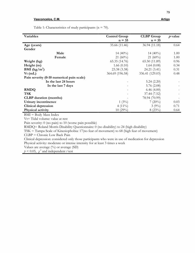
79
Vasconcelos, C.M. Artigo
Table 1: Characteristics of study participants (n = 70).
Variables Control Group n = 35
CLBP Group n = 35
p value
Age (years) 35.66 (11.46) 36.94 (11.18) 0.64 Gender
Male 14 (40%) 14 (40%) 1.00 Female 21 (60%) 21 (60%) 1.00
Weight (kg) 65.35 (14.76) 65.50 (11.89) 0.96 Height (m) 1.66 (0.10) 1.64 (0.08) 0.34 BMI (kg/m2) 23.38 (3.38) 24.21 (3.41) 0.31 Vt (mL) 364.69 (196.58) 336.41 (129.03) 0.48 Pain severity (0-10 numerical pain scale)
In the last 24 hours - 5.24 (2.20) - In the last 7 days - 5.76 (2.08) -
RMDQ - 6.46 (4.00) - TSK - 37.44 (7.52) - CLBP duration (months) - 78.94 (70.99) - Urinary incontinence 1 (3%) 7 (20%) 0.03 Clinical depression 4 (11%) 3 (9%) 0.71 Physical activity 10 (29%) 8 (23%) 0.64
BMI = Body Mass Index Vt= Tidal volume: value at rest Pain severity: 0 (no pain) to 10 (worse pain possible) RMDQ= Roland Morris Disability Questionnaire: 0 (no disability) to 24 (high disability) TSK = Tampa Scale of Kinesiophobia: 17(no fear of movement) to 68 (high fear of movement) CLBP = Chronic Low Back Pain Clinical depression: considered only those participants who were in use of medication for depression Physical activity: moderate or intense intensity for at least 3 times a week Values are average (%) or average (SD) p < 0.05, χ 2 and independent t test
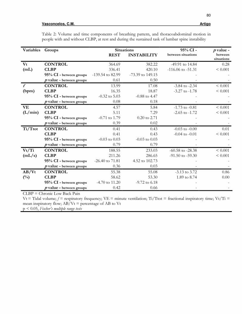
80
Vasconcelos, C.M. Artigo
Table 2: Volume and time components of breathing pattern, and thoracoabdominal motion in people with and without CLBP, at rest and during the sustained task of lumbar spine instability
Variables Groups Situations 95% CI - p value - REST INSTABILITY between situations between
situations
CONTROL 364.69 382.22 -49.91 to 14.84 0.28 CLBP 336.41 420.10 -116.06 to -51.31 < 0.001 95% CI - between groups -139.54 to 82.99 -73.39 to 149.15 - -
Vt (mL)
p value - between groups 0.61 0.50 - -
CONTROL 13.99 17.08 -3.84 to -2.34 < 0.001 CLBP 16.35 18.87 -3.27 to -1.78 < 0.001 95% CI - between groups -0.32 to 5.03 -0.88 to 4.47 - -
f (bpm)
p value - between groups 0.08 0.18 - -
CONTROL 4.57 5.84 -1.73 to -0.81 < 0.001 CLBP 5.11 7.29 -2.65 to -1.72 < 0.001 95% CI - between groups -0.71 to 1.79 0.20 to 2.71 - -
VE (L/min)
p value - between groups 0.39 0.02 - -
CONTROL 0.41 0.43 -0.03 to -0.00 0.01 CLBP 0.41 0.43 -0.04 to -0.01 < 0.001 95% CI - between groups -0.03 to 0.03 -0.03 to 0.03 - -
Ti/Ttot
p value - between groups 0.79 0.79 - -
CONTROL 188.55 233.03 -60.58 to -28.38 < 0.001 CLBP 211.26 286.65 -91.50 to -59.30 < 0.001 95% CI - between groups -26.40 to 71.81 4.52 to 102.73 - -
Vt/Ti (mL/s)
p value - between groups 0.36 0.03 - -
CONTROL 55.38 55.08 -3.13 to 3.72 0.86 CLBP 58.62 53.30 1.89 to 8.74 0.00 95% CI - between groups -4.70 to 11.20 -9.72 to 6.18 - -
AB/Vt (%)
p value - between groups 0.42 0.66 - -
CLBP = Chronic Low Back Pain Vt = Tidal volume; f = respiratory frequency; VE = minute ventilation; Ti/Ttot = fractional inspiratory time; Vt/Ti = mean inspiratory flow; AB/Vt = percentage of AB to Vt p < 0.05, Fischer’s multiple range tests
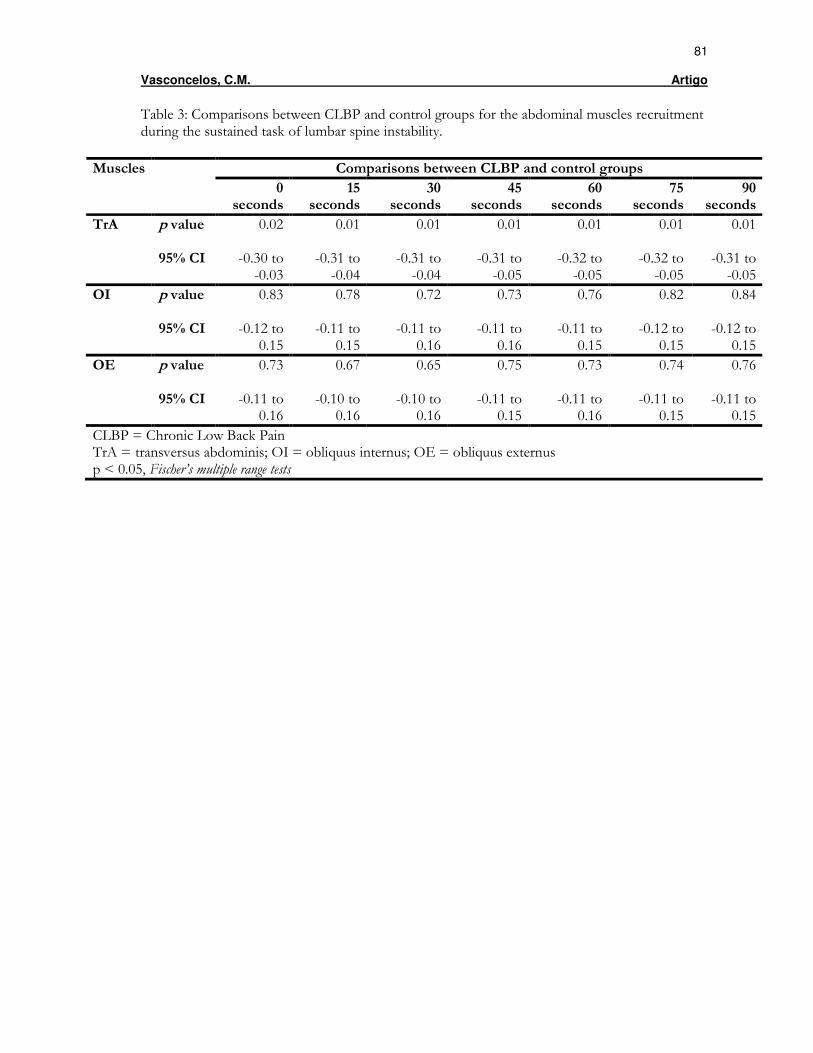
81
Vasconcelos, C.M. Artigo
Table 3: Comparisons between CLBP and control groups for the abdominal muscles recruitment during the sustained task of lumbar spine instability.
Comparisons between CLBP and control groups Muscles
0 seconds
15 seconds
30 seconds
45 seconds
60 seconds
75 seconds
90 seconds
TrA p value 0.02
0.01 0.01 0.01 0.01 0.01 0.01
95% CI -0.30 to -0.03
-0.31 to -0.04
-0.31 to -0.04
-0.31 to -0.05
-0.32 to -0.05
-0.32 to -0.05
-0.31 to -0.05
OI p value
0.83 0.78 0.72 0.73 0.76 0.82 0.84
95% CI -0.12 to 0.15
-0.11 to 0.15
-0.11 to 0.16
-0.11 to 0.16
-0.11 to 0.15
-0.12 to 0.15
-0.12 to 0.15
OE p value
0.73 0.67 0.65 0.75 0.73 0.74 0.76
95% CI -0.11 to 0.16
-0.10 to 0.16
-0.10 to 0.16
-0.11 to 0.15
-0.11 to 0.16
-0.11 to 0.15
-0.11 to 0.15
CLBP = Chronic Low Back Pain TrA = transversus abdominis; OI = obliquus internus; OE = obliquus externus p < 0.05, Fischer’s multiple range tests

82
Vasconcelos, C.M. Artigo
Table 4: Comparisons between abdominal muscles recruitment during the sustained task of lumbar spine instability for CLBP and control groups.
0 seconds
15 seconds
30 seconds
45 seconds
60 seconds
75 seconds
90 seconds
Groups
Comparisons between Tra and OI
Control p value
0.01 0.01 0.00 0.01 0.00 0.00 0.00
95% CI 0.03 to 0.30
0.04 to 0.31
0.06 to 0.32
0.05 to 0.32
0.08 to 0.34
0.07 to 0.34
0.07 to 0.33
CLBP p value
0.86 0.77 0.92 0.82 0.94 0.91 0.96
95% CI -0.14 to 0.12
-0.15 to 0.11
-0.14 to 0.13
-0.15 to 0.12
-0.13 to 0.14
-0.12 to 0.14
-0.13 to 0.14
Comparisons between Tra and OE
Control p value
<0.001 <0.001 <0.001 <0.001 <0.001 <0.001 <0.001
95% CI 0.09 to 0.35
0.10 to 0.36
0.12 to 0.39
0.11 to 0.38
0.14 to 0.40
0.14 to 0.40
0.13 to 0.40
CLBP p value
0.63 0.66 0.44 0.49 0.36 0.33 0.35
95% CI -0.10 to 0.16
-0.10 to 0.16
-0.08 to 0.18
-0.09 to 0.18
-0.07 to 0.19
-0.07 to 0.20
-0.07 to 0.19
Comparisons between OI and OE
Control p value
0.43 0.37 0.34 0.37 0.38 0.32 0.32
95% CI -0.08 to 0.18
-0.07 to 0.19
-0.07 to 0.20
-0.07 to 0.19
-0.07 to 0.19
-0.07 to 0.20
-0.07 to 0.20
CLBP p value
0.51 0.46 0.38 0.36 0.40 0.38 0.38
95% CI -0.09 to 0.18
-0.08 to 0.18
-0.07 to 0.19
-0.07 to 0.19
-0.08 to 0.19
-0.07 to 0.19
-0.07 to 0.19
CLBP = Chronic Low Back Pain TrA = transversus abdominis; OI = obliquus internus; OE = obliquus externus p < 0.05, Fischer’s multiple range tests
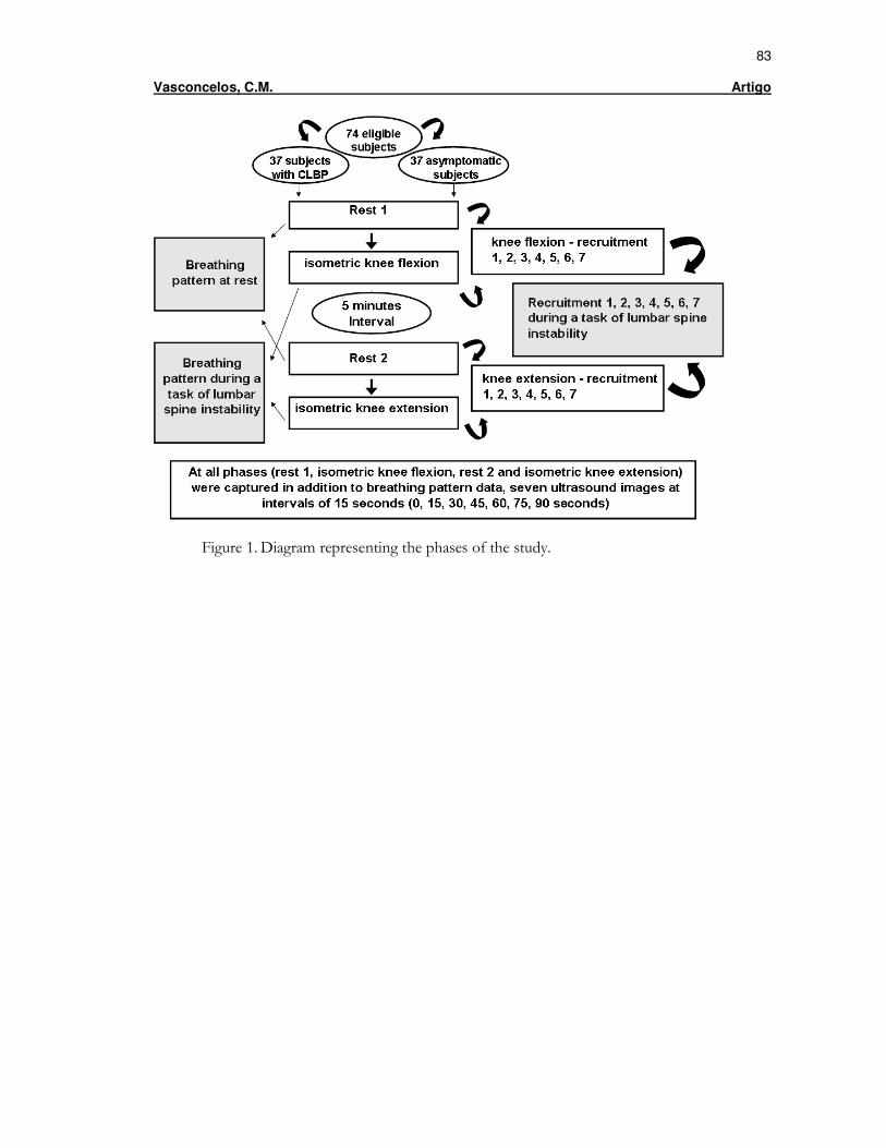
83
Vasconcelos, C.M. Artigo
Figure 1. Diagram representing the phases of the study.
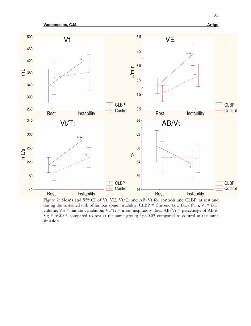
84
Vasconcelos, C.M. Artigo
Figure 2: Means and 95%CI of Vt, VE, Vt/Ti and AB/Vt for controls and CLBP, at rest and during the sustained task of lumbar spine instability. CLBP = Chronic Low Back Pain; Vt = tidal volume; VE = minute ventilation; Vt/Ti = mean inspiratory flow; AB/Vt = percentage of AB to Vt; * p<0.05 compared to rest at the same group; † p<0.05 compared to control at the same situation.
CLBP ControlRest Instability
260
300
340
380
420
460
500
mL
CLBP ControlRest Instability
3,5
4,5
5,5
6,5
7,5
8,5
L/m
in
CLBP Control
Rest Instability140
180
220
260
300
340
mL/
s
CLBP Control
Rest Instability46
50
54
58
62
66
%
*
* †
*
*
Vt VE
Vt/Ti AB/Vt
* †
*
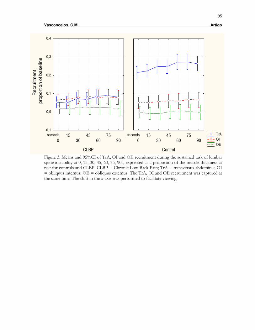
85
Vasconcelos, C.M. Artigo
TrA OI OE
CLBP
seconds
015
3045
6075
90
-0,1
0,0
0,1
0,2
0,3
0,4R
ecr
uitm
en
t p
rop
ort
ion
of b
ase
line
Control
seconds
015
3045
6075
90
Figure 3: Means and 95%CI of TrA, OI and OE recruitment during the sustained task of lumbar spine instability at 0, 15, 30, 45, 60, 75, 90s, expressed as a proportion of the muscle thickness at rest for controls and CLBP. CLBP = Chronic Low Back Pain; TrA = transversus abdominis; OI = obliquus internus; OE = obliquus externus. The TrA, OI and OE recruitment was captured at the same time. The shift in the x-axis was performed to facilitate viewing.
86 Vasconcelos, C.M. Considerações Finais
Capítulo 5 - CONSIDERAÇÕES FINAIS
Estima-se que cerca de 80% da população geral apresente pelo menos um
episódio de dor lombar no decorrer da vida e aproximadamente 10-20% dos
pacientes com dor lombar desenvolvam sintomas persistentes. A DLC possui
impacto socioeconômico significante e está associada a elevados custos diretos e
indiretos em cuidados com a saúde, com o absenteísmo e a incapacidade, além de
gerar importantes consequências para o paciente, a família, os empregadores e a
sociedade em geral. Existem relatos na literatura da associação entre DLC e
disfunção respiratória. Embora o DF seja considerado o músculo primário da
inspiração, muitos estudos têm mostrado seu importante papel no controle postural
do tronco, agindo sinergicamente ao TrA nas funções respiratória e estabilização
lombar. Desta forma, o presente estudo teve como objetivo investigar o padrão
respiratório e o recrutamento dos músculos do tronco (TrA, OI e OE) em indivíduos
assintomáticos e com DLC, durante o repouso e uma tarefa sustentada de
instabilidade da coluna lombar e, secundariamente, investigar a associação entre
essas variáveis.
A principal contribuição desse estudo para o corpo de conhecimento da
fisioterapia foi identificar que, quando comparados com indivíduos saudáveis,
individuos com DLC apresentam alterações nas variáveis do padrão respiratório e no
movimento toracoabdominal, assim como mudança no recrutamento do músculo
TrA, durante uma tarefa sustentada de instabilidade da coluna lombar. Até onde vai
o nosso conhecimento, este foi o primeiro estudo que comparou tanto as variáveis
de volume e tempo do padrão respiratório, quanto o movimento toracoabdominal em
indivíduos assintomáticos e com DLC, por meio da PRI, um instrumento válido e
87
Vasconcelos, C.M. Considerações Finais
confiável. Os demais estudos que investigaram a respiração nesses indivíduos
realizaram essa análise de maneira subjetiva e durante a execução de tarefas de
curta duração (até 20 segundos), não sendo encontrados na literatura pesquisada,
estudos que avaliaram o recrutamento dos músculos do tronco durante tarefas
sustentadas, bem como a associação de variáveis do padrão respiratório e do
recrutamento muscular no controle postural desses indivíduos.
É importante mencionar que neste estudo, o recrutamento dos músculos
abdominais foi avaliado por meio de medidas ultrassonográficas, de acordo com um
protocolo previamente validado na literatura, que nos permite associar o aumento da
espessura muscular com o aumento da atividade eletromiográfica, para os músculos
TrA e OI. No entanto, pelo fato da mudança da espessura do músculo OE possuir
baixa correlação com os dados da eletromiografia desse músculo, cuidados devem
ser tomados para limitar as informações referentes a esse músculo às mudanças do
parâmetro medido (espessura muscular), sendo necessário reconhecer que
quaisquer conclusões relativas a sua ativação muscular ou aos mecanismos por
detrás dessas mudanças são uma extrapolação dos achados.
A possível relação entre DLC e disfunção respiratória pode influenciar
diversos aspectos clínicos, especialmente a avaliação e a reabilitação dos pacientes
com dor lombar. A fim de se obter uma perspectiva integral que represente todas as
disfunções apresentadas por esses pacientes, a avaliação dos indivíduos com DLC
deve incluir uma investigação dos diversos componentes da função respiratória.
Com relação à reabilitação, os exercícios de controle motor, intervenção muito
difundida na prática clínica e considerada efetiva na redução da dor e incapacidade
de indivíduos com dor lombar, frequentemente utilizam o controle da respiração
como estratégia terapêutica. Somado a isso, algumas técnicas alternativas que têm
88
Vasconcelos, C.M. Considerações Finais
ganhado grande popularidade na última década, como o Pilates e a Yoga,
apresentam como um dos focos de intervenção o controle da respiração, apesar de
ainda existirem poucas evidências científicas para tal indicação.
Como as responsabilidades do TrA, do DF e dos músculos do AP são
variadas, a disfunção desses músculos pode se manifestar de formas variadas
(lombalgia, disfunção respiratória, incontinência urinária) ou até mesmo apresentar-
se de forma multi-fatorial. Portanto, o nosso desafio é determinar onde concentrar o
foco inicial do tratamento. Uma abordagem pragmática apoiaria intervir inicialmente
na prioridade fisiológica, ou seja, na respiração, pois é provável que se a mesma
estiver alterada, os déficits de controle motor irão perpetuar, e consequentemente,
as respostas aos tratamentos tradicionais destinados a restabelecer o controle
postural serão limitadas. Portanto, os clínicos e pesquisadores devem ficar atentos à
presença de alterações na função respiratória de indivíduos com DLC. Como é
possível que fatores psicossociais, como a cinesiofobia, tenham influência sobre a
função respiratória destes indivíduos, abordagens cognitivo-comportamentais
juntamente com exercícios respiratórios podem ser estratégias terapêuticas que
otimizam o manejo desses pacientes. Este trabalho fornece evidências que devem
ser incorporadas ao corpo de conhecimento das abordagens terapêuticas, que
utilizam de estratégias específicas para assegurar ou promover estabilidade
adequada à coluna lombar. No contexto atual, em que a fisioterapia se confronta
com uma variedade de técnicas sem o devido respaldo científico, trabalhos como
este são de extrema importância.
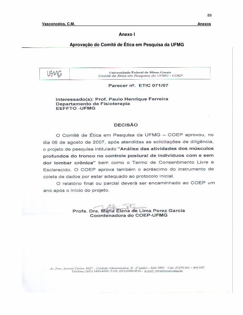
89 Vasconcelos, C.M. Anexos
Anexo I
Aprovação do Comitê de Ética em Pesquisa da UFMG
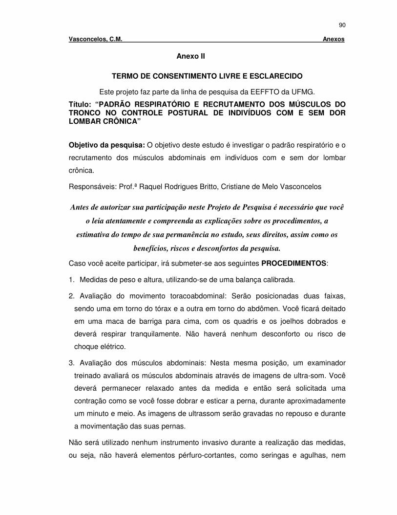
90
Vasconcelos, C.M. Anexos
Anexo II
TERMO DE CONSENTIMENTO LIVRE E ESCLARECIDO
Este projeto faz parte da linha de pesquisa da EEFFTO da UFMG.
Título: “PADRÃO RESPIRATÓRIO E RECRUTAMENTO DOS MÚSCULOS DO TRONCO NO CONTROLE POSTURAL DE INDIVÍDUOS COM E SEM DOR LOMBAR CRÔNICA”
Objetivo da pesquisa: O objetivo deste estudo é investigar o padrão respiratório e o
recrutamento dos músculos abdominais em indivíduos com e sem dor lombar
crônica.
Responsáveis: Prof.ª Raquel Rodrigues Britto, Cristiane de Melo Vasconcelos
Antes de autorizar sua participação neste Projeto de Pesquisa é necessário que você
o leia atentamente e compreenda as explicações sobre os procedimentos, a
estimativa do tempo de sua permanência no estudo, seus direitos, assim como os
benefícios, riscos e desconfortos da pesquisa.
Caso você aceite participar, irá submeter-se aos seguintes PROCEDIMENTOS:
1. Medidas de peso e altura, utilizando-se de uma balança calibrada.
2. Avaliação do movimento toracoabdominal: Serão posicionadas duas faixas,
sendo uma em torno do tórax e a outra em torno do abdômen. Você ficará deitado
em uma maca de barriga para cima, com os quadris e os joelhos dobrados e
deverá respirar tranquilamente. Não haverá nenhum desconforto ou risco de
choque elétrico.
3. Avaliação dos músculos abdominais: Nesta mesma posição, um examinador
treinado avaliará os músculos abdominais através de imagens de ultra-som. Você
deverá permanecer relaxado antes da medida e então será solicitada uma
contração como se você fosse dobrar e esticar a perna, durante aproximadamente
um minuto e meio. As imagens de ultrassom serão gravadas no repouso e durante
a movimentação das suas pernas.
Não será utilizado nenhum instrumento invasivo durante a realização das medidas,
ou seja, não haverá elementos pérfuro-cortantes, como seringas e agulhas, nem
91
Vasconcelos, C.M. Anexos
haverá risco de choque elétrico. Além dessas explicações, você tem o direito de
solicitar outros esclarecimentos a qualquer momento.
Benefícios: As medidas obtidas nesse estudo contribuirão com fundamentação
científica para um maior entendimento da respiração e dos músculos abdominais em
indivíduos com e sem dor lombar.
Possíveis Riscos: O estudo não oferece riscos significativos, visto que não há
nenhum procedimento invasivo ou desgastante para os participantes. As faixas
utilizadas para analisar sua respiração e o ultrassom não oferecem risco à sua
saúde. Você poderá apenas se cansar durante a realização dos testes ou apresentar
um leve desconforto abdominal relacionada aos exercícios, que apresentará melhora
rápida.
A PESQUISA NÃO REVELARÁ A IDENTIDADE DOS PARTICIPANTES.
Sua participação é voluntária e não lhe trará nenhum gasto financeiro nem lhe será
paga nenhuma remuneração. Você está livre para recusar participar ou interromper
a sua participação a qualquer momento durante a coleta de dados, sem qualquer
penalização ou prejuízo.
Li e entendi as informações acima, sendo que minhas dúvidas foram
satisfatoriamente respondidas. Este formulário está sendo assinado voluntariamente
por mim, indicando meu consentimento em participar do estudo.
Belo Horizonte, _______de __________________de 2008.
(Participante)
(Pesquisador)
Telefones para contato:
Orientadora: Prof.ª Raquel Rodrigues Britto (031) 3409-4793
Pesquisadora: Cristiane de Melo Vasconcelos (031) 8784-9757
Comitê de Ética em Pesquisa da UFMG: (031) 3409-4592.
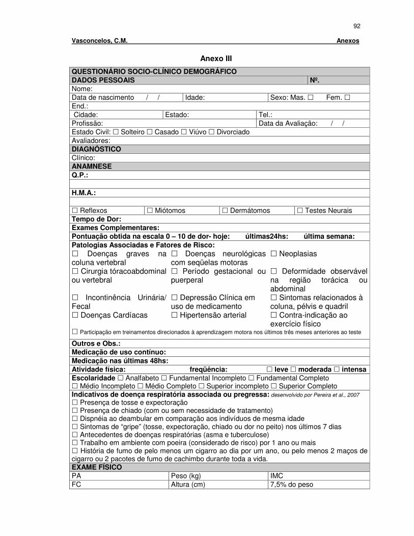
92
Vasconcelos, C.M. Anexos
Anexo III
QUESTIONÁRIO SOCIO-CLÍNICO DEMOGRÁFICO DADOS PESSOAIS Nº. Nome: Data de nascimento / / Idade: Sexo: Mas. � Fem. � End.: Cidade: Estado: Tel.: Profissão: Data da Avaliação: / / Estado Civil: � Solteiro � Casado � Viúvo � Divorciado Avaliadores: DIAGNÓSTICO Clínico: ANAMNESE Q.P.: H.M.A.: � Reflexos � Miótomos � Dermátomos � Testes Neurais Tempo de Dor: Exames Complementares: Pontuação obtida na escala 0 – 10 de dor- hoje: últimas24hs: última semana: Patologias Associadas e Fatores de Risco: � Doenças graves na coluna vertebral
� Doenças neurológicas com seqüelas motoras
� Neoplasias
� Cirurgia tóracoabdominal ou vertebral
� Período gestacional ou puerperal
� Deformidade observável na região torácica ou abdominal
� Incontinência Urinária/ Fecal
� Depressão Clínica em uso de medicamento
� Sintomas relacionados à coluna, pélvis e quadril
� Doenças Cardíacas � Hipertensão arterial � Contra-indicação ao exercício físico
� Participação em treinamentos direcionados à aprendizagem motora nos últimos três meses anteriores ao teste
Outros e Obs.: Medicação de uso contínuo: Medicação nas últimas 48hs: Atividade física: freqüência: � leve � moderada � intensa Escolaridade � Analfabeto � Fundamental Incompleto � Fundamental Completo � Médio Incompleto � Médio Completo � Superior incompleto � Superior Completo Indicativos de doença respiratória associada ou pregressa: desenvolvido por Pereira et al., 2007 � Presença de tosse e expectoração � Presença de chiado (com ou sem necessidade de tratamento) � Dispnéia ao deambular em comparação aos indivíduos de mesma idade � Sintomas de “gripe” (tosse, expectoração, chiado ou dor no peito) nos últimos 7 dias � Antecedentes de doenças respiratórias (asma e tuberculose) � Trabalho em ambiente com poeira (considerado de risco) por 1 ano ou mais � História de fumo de pelo menos um cigarro ao dia por um ano, ou pelo menos 2 maços de cigarro ou 2 pacotes de fumo de cachimbo durante toda a vida. EXAME FÍSICO PA Peso (kg) IMC FC Altura (cm) 7,5% do peso

93
Vasconcelos, C.M. Anexos
Anexo IV
Escala Visual Analógica
Eu gostaria que você desse uma nota para sua dor numa escala de 0 a 10,
onde 0 seria nenhuma dor e 10 seria a pior dor possível. Por favor, dê um número para descrever sua média de dor nas últimas 24hs.
0 1 2 3 4 5 6 7 8 9 10
NENHUMA DOR PIOR DOR POSSÍVEL
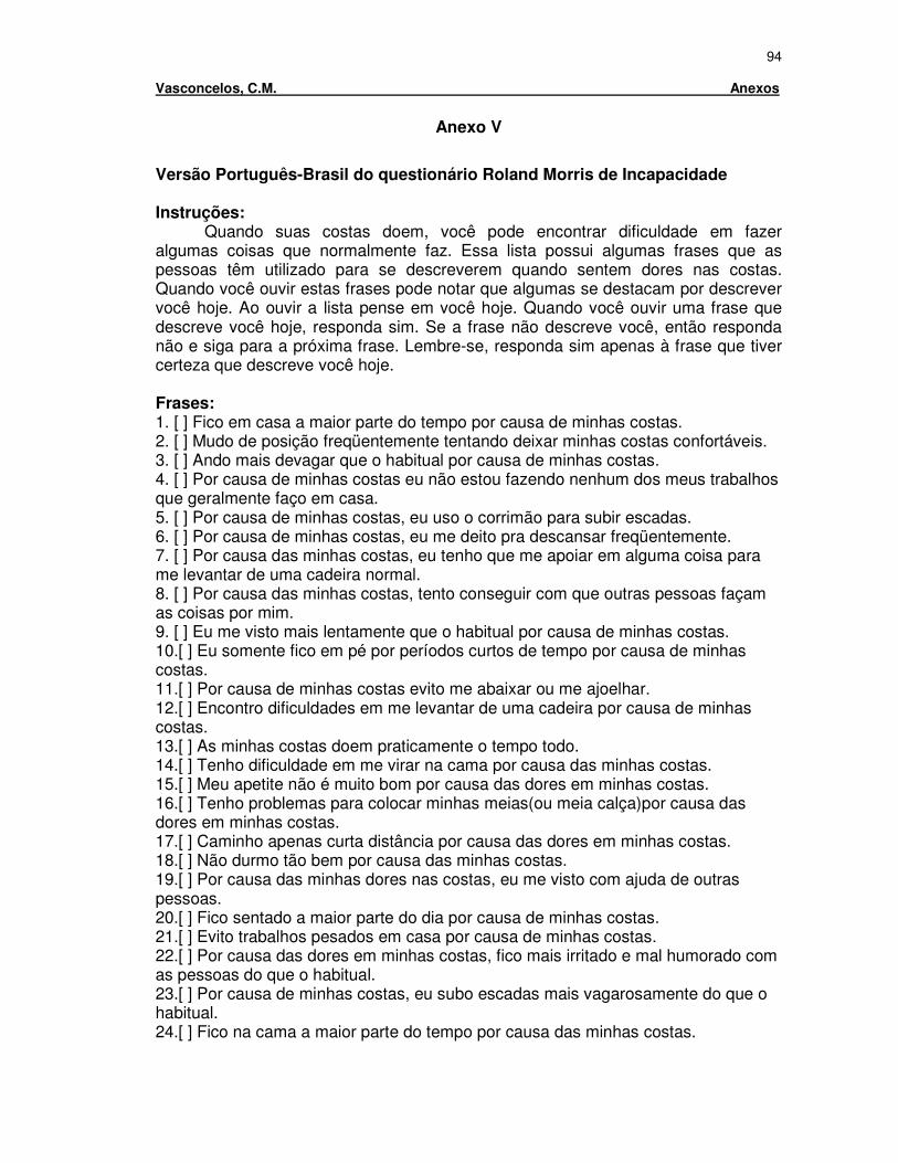
94
Vasconcelos, C.M. Anexos
Anexo V
Versão Português-Brasil do questionário Roland Morris de Incapacidade Instruções:
Quando suas costas doem, você pode encontrar dificuldade em fazer algumas coisas que normalmente faz. Essa lista possui algumas frases que as pessoas têm utilizado para se descreverem quando sentem dores nas costas. Quando você ouvir estas frases pode notar que algumas se destacam por descrever você hoje. Ao ouvir a lista pense em você hoje. Quando você ouvir uma frase que descreve você hoje, responda sim. Se a frase não descreve você, então responda não e siga para a próxima frase. Lembre-se, responda sim apenas à frase que tiver certeza que descreve você hoje. Frases: 1. [ ] Fico em casa a maior parte do tempo por causa de minhas costas. 2. [ ] Mudo de posição freqüentemente tentando deixar minhas costas confortáveis. 3. [ ] Ando mais devagar que o habitual por causa de minhas costas. 4. [ ] Por causa de minhas costas eu não estou fazendo nenhum dos meus trabalhos que geralmente faço em casa. 5. [ ] Por causa de minhas costas, eu uso o corrimão para subir escadas. 6. [ ] Por causa de minhas costas, eu me deito pra descansar freqüentemente. 7. [ ] Por causa das minhas costas, eu tenho que me apoiar em alguma coisa para me levantar de uma cadeira normal. 8. [ ] Por causa das minhas costas, tento conseguir com que outras pessoas façam as coisas por mim. 9. [ ] Eu me visto mais lentamente que o habitual por causa de minhas costas. 10.[ ] Eu somente fico em pé por períodos curtos de tempo por causa de minhas costas. 11.[ ] Por causa de minhas costas evito me abaixar ou me ajoelhar. 12.[ ] Encontro dificuldades em me levantar de uma cadeira por causa de minhas costas. 13.[ ] As minhas costas doem praticamente o tempo todo. 14.[ ] Tenho dificuldade em me virar na cama por causa das minhas costas. 15.[ ] Meu apetite não é muito bom por causa das dores em minhas costas. 16.[ ] Tenho problemas para colocar minhas meias(ou meia calça)por causa das dores em minhas costas. 17.[ ] Caminho apenas curta distância por causa das dores em minhas costas. 18.[ ] Não durmo tão bem por causa das minhas costas. 19.[ ] Por causa das minhas dores nas costas, eu me visto com ajuda de outras pessoas. 20.[ ] Fico sentado a maior parte do dia por causa de minhas costas. 21.[ ] Evito trabalhos pesados em casa por causa de minhas costas. 22.[ ] Por causa das dores em minhas costas, fico mais irritado e mal humorado com as pessoas do que o habitual. 23.[ ] Por causa de minhas costas, eu subo escadas mais vagarosamente do que o habitual. 24.[ ] Fico na cama a maior parte do tempo por causa das minhas costas.
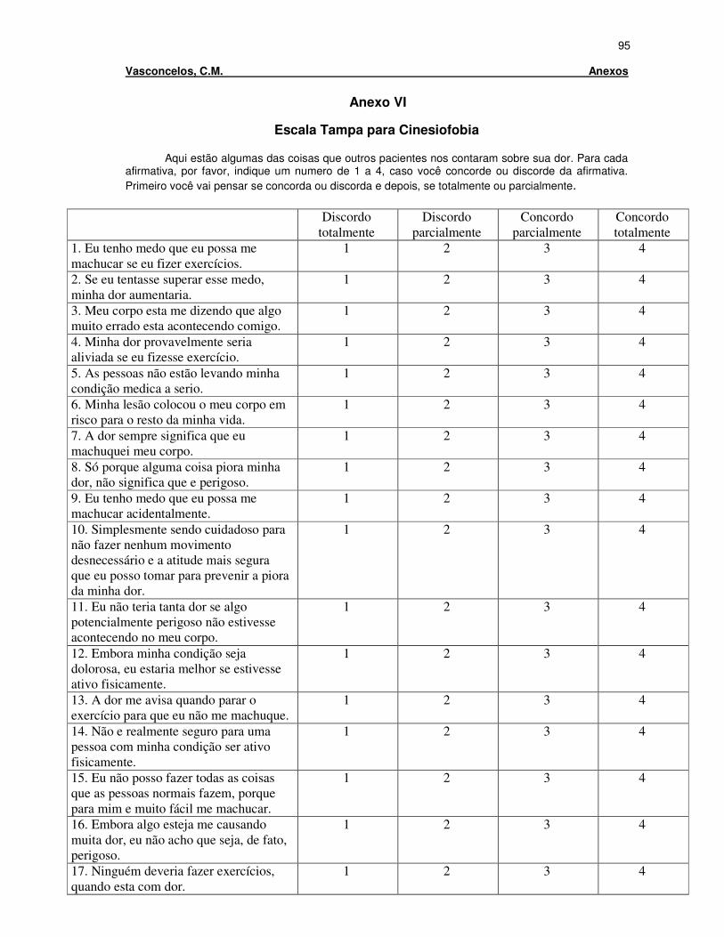
95
Vasconcelos, C.M. Anexos
Anexo VI
Escala Tampa para Cinesiofobia
Aqui estão algumas das coisas que outros pacientes nos contaram sobre sua dor. Para cada afirmativa, por favor, indique um numero de 1 a 4, caso você concorde ou discorde da afirmativa. Primeiro você vai pensar se concorda ou discorda e depois, se totalmente ou parcialmente.
Discordo
totalmente
Discordo
parcialmente
Concordo
parcialmente
Concordo
totalmente
1. Eu tenho medo que eu possa me
machucar se eu fizer exercícios.
1 2 3 4
2. Se eu tentasse superar esse medo,
minha dor aumentaria.
1 2 3 4
3. Meu corpo esta me dizendo que algo
muito errado esta acontecendo comigo.
1 2 3 4
4. Minha dor provavelmente seria
aliviada se eu fizesse exercício.
1 2 3 4
5. As pessoas não estão levando minha
condição medica a serio.
1 2 3 4
6. Minha lesão colocou o meu corpo em
risco para o resto da minha vida.
1 2 3 4
7. A dor sempre significa que eu
machuquei meu corpo.
1 2 3 4
8. Só porque alguma coisa piora minha
dor, não significa que e perigoso.
1 2 3 4
9. Eu tenho medo que eu possa me
machucar acidentalmente.
1 2 3 4
10. Simplesmente sendo cuidadoso para
não fazer nenhum movimento
desnecessário e a atitude mais segura
que eu posso tomar para prevenir a piora
da minha dor.
1 2 3 4
11. Eu não teria tanta dor se algo
potencialmente perigoso não estivesse
acontecendo no meu corpo.
1 2 3 4
12. Embora minha condição seja
dolorosa, eu estaria melhor se estivesse
ativo fisicamente.
1 2 3 4
13. A dor me avisa quando parar o
exercício para que eu não me machuque.
1 2 3 4
14. Não e realmente seguro para uma
pessoa com minha condição ser ativo
fisicamente.
1 2 3 4
15. Eu não posso fazer todas as coisas
que as pessoas normais fazem, porque
para mim e muito fácil me machucar.
1 2 3 4
16. Embora algo esteja me causando
muita dor, eu não acho que seja, de fato,
perigoso.
1 2 3 4
17. Ninguém deveria fazer exercícios,
quando esta com dor.
1 2 3 4
96
Vasconcelos, C.M. Anexos
Anexo VII
Normas para a Publicação
PHYSICAL THERAPY INFORMATION FOR AUTHORS
http://www.ptjournal.org/
Physical Therapy Physical Therapy (PTJ) engages and inspires an international readership on topics related to physical therapy. As the leading international journal for research in physical therapy and related fields, PTJ publishes innovative and highly relevant content for both clinicians and scientists and uses a variety of interactive approaches to communicate that content, with the expressed purpose of improving patient care.
PTJ's circulation in 2008 is more than 72,000. Its 2008 impact factor was 2.190. The mean time from submission to first decision is 58 days. Time from acceptance to publication online is less than or equal to 3 months and from acceptance to publication in print is less than or equal to 5 months. The acceptance rate is 30%.
These submission guidelines, first posted in March 2007, are MANDATORY for all manuscripts submitted on January 1, 2008, or thereafter. For more about the formulation or these guidelines, refer to the Editor's Note by Craik and Riddle and the Editor's Note by Fitzgerald.
PTJ endorses the Uniform Requirements for Manuscripts Submitted to Biomedical Journals put forth by the International Committee of Medical Journal Editors (ICMJE).
Editorial Policies
All submissions accepted for peer review are privileged communications. Author identity is kept confidential from reviewers, unless otherwise indicated. All correspondence is sent to the author who submits the article.
Immediate Decisions
The Editor in Chief and Editorial Board reserve the right to reject, without full review, any manuscript that does not meet Journal criteria. Manuscripts are prescreened by an Editor and/or Editorial Board member. Manuscripts are immediately rejected by PTJ when:
* Study participants are able bodied, without a compelling justification for relevance to physical therapist practice. * Reliability study does not include a discussion of the impact that the error will have on clinical decision making. * Study describes a new test without a sound comparison to current tests. * Qualitative study is purely descriptive, without analysis or interpretation of findings. * Study has fatal flaws in the methods section. * Study does not have a theoretical or evidence-based argument for the relevance of the work to physical therapist practice.
97
Vasconcelos, C.M. Anexos
Fast-Track Review
Manuscripts that have the potential to make a strong and immediate impact on physical therapist practice are considered for fast-track peer review (30 days from submission to first decision). Only original research papers, particularly clinical trials, are considered. If you believe that your manuscript meets the criterion for high impact, please contact the managing editor at [email protected] , and include an abstract and your rationale for why the paper should be fast tracked.
Redundant, Duplicate, or Simultaneous Publication
PTJ reviews and considers a manuscript for exclusive publication with the understanding that the manuscript, or any substantial portion of the manuscript (as judged by the Editor in Chief), has not been published previously and is not under consideration for publication elsewhere, whether in print or electronic form. This policy does not usually preclude consideration of (1) a manuscript that has been rejected by another journal or (2) a complete report that follows publication of a preliminary report or pilot study. Press reports on papers presented at a scientific meeting usually will not be considered to constitute prior publication, but such reports should not be amplified by additional data or copies of tables and illustrations (see also Prior Disclosure and Media Embargo Policy).
Redundancy in publications occurs when the work of 2 or more papers from the same author overlaps substantially. Authors submitting manuscripts to PTJ, are asked to include in their cover letter an explanation of any prior submission or publication (eg, published article, article in press, manuscript under review, posting of results in registries, published abstract) that includes data from the same subjects studied in the submitted manuscript. Previous publication of a small fraction of the content of a manuscript does not necessarily preclude its being published in PTJ, but the editors need information about previous publication. Secondary analyses based on previous publications can be important scientific contributions, but the editors need to be able to judge potential redundancy. If the submitted manuscript is a report of secondary analyses, the source of the data should be properly referenced and the rationale for the secondary analysis must be provided.
In addition to the cover letter explanation, authors must provide a copy of related papers-that is, submitted or published papers that deal with the same data, in part or in full, that are reported in the manuscript being submitted to PTJ. These materials will be confidential and will be viewed only by the editors.
Duplicate publication is the publication of the same paper or substantially similar papers in the same journal or in more than one journal. PTJ's Editorial Board holds that publication more than once of the same study results, regardless of whether the wording is the same or different, is rarely justified. Articles previously published in another language will not be considered for publication in PTJ.
The Editorial Board reserves the right to consult with other journals about the content of the papers in question. Furthermore, PTJ may decide to take one or more of the following actions: (1) Immediately reject the manuscript without review. (2) Decline to consider any manuscripts from the author(s) for a period of time. (3) Announce publicly, within PTJ, that the authors have submitted a previously published article. If a paper is accepted and published before evidence of duplication is discovered, PTJ probably will publish a notification of redundant or duplicate publication, with or without the author's explanation or approval. Typically, PTJ will request that
98
Vasconcelos, C.M. Anexos
the authors write a letter acknowledging the duplicate publication. The Editorial Board may notify appropriate institutions, ranging from national databases to the authors' academic departments or university administrators, at its discretion. The Editorial Board may decide not to consider submissions from the author(s) for a period of time.
Simultaneous Submission. Authors may not submit the same manuscript simultaneously to more than one journal. If the Editorial Board learns of possible simultaneous submission, it reserves the right to consult with the other journal to which the manuscript was submitted. Furthermore, the Editorial Board may return the manuscript without review or may reject it without regard to peer reviewer recommendations and may decide not to consider any submissions from the author(s) for a period of time.
Prior Disclosure and Media Embargo Policy
New Submissions: As described in detail above, prior disclosure of any part of the contents of any manuscript in a widespread and substantive form, print or electronic, may make the manuscript ineligible for consideration by or publication in PTJ. Publication of abstracts and presentations at meetings do not constitute prior disclosure. Media coverage of presentations at scientific meetings will not constitute prior disclosure, as long as such coverage is not amplified by additional data or copies of tables and illustrations; however, direct release of information through press releases or news media briefings may jeopardize consideration of the submission. During the submission process, you will be prompted to indicate whether your manuscript has been presented orally at a scientific meeting or at a professional forum. Authors who need clarification of this policy are encouraged to contact the Managing Editor before releasing or distributing information from the manuscript that they want to submit.
Accepted Papers: All information regarding the content and publication date of accepted manuscripts is strictly confidential. Information contained in or about accepted articles must not appear in print, audio, video, or digital form or be released by the news media until the day before its publication date (or other specified embargo release date in the case of articles that are released early or published online ahead of print). PTJ is willing to work with authors who wish to present their accepted papers at conferences prior to publication. Authors are allowed to alert their university media office at the rapid proof stage but must inform PTJ and must ensure that the university media office observes PTJ's media embargo policy.
Copyright and Authorship
Authors agree to execute copyright transfer as requested during the submission process. Authors will be prompted to upload a signed copyright release, authorship, and financial disclosure form. (Please upload one form per author.) Manuscripts published in PTJ become the property of the American Physical Therapy Association (APTA) and may not be published elsewhere, in whole or in part, in print or electronic form, without the written permission of APTA, which has the right to use, reproduce, transmit, derivate, publish, and distribute the contribution, in PTJ or otherwise, in any form or medium.
Commercial/Financial Associations and Conflict of Interest
All funding sources supporting the work should be acknowledged. During the submission process, authors will be required to enter this information. They also will be prompted to upload a disclosure statement, signed by all authors. This information will be held in confidence by the
99
Vasconcelos, C.M. Anexos
Editor in Chief during the review process and, if the paper is accepted for publication, will be shared with readers as appropriate.
Protection of Participants
In the cover letter that is submitted with the manuscript, authors of Research Reports should provide the name of the institutional review board (IRB), institutional animal care and use committee, or other similar body that approved the study. For those authors who do not have formal ethics review committees, the principles outlined in the Declaration of Helsinki should be followed, and authors should include a statement within the manuscript (eg, in the “Participants” section) confirming that these principles were followed. Authors also should submit patient consent forms for photographs or videos. Within the manuscript, authors must include a statement in the “Method” section that they obtained informed consent of participants, when required for protection of human subjects. Along with their signed copyright release forms, authors of Case Reports should submit a signed patient consent form. Case Report authors who practice in the United States should also include a statement about meeting the HIPAA (Health Insurance, Portability, and Accountability Act) requirements of the institution for disclosure of protected health information.
PubMed Central Deposits
In January 2008, the National Institutes of Health (NIH) announced that its voluntary Public Access Policy (NOT-OD-05-022) was now mandatory. The law states:
The Director of the National Institutes of Health shall require that all investigators funded by the NIH submit or have submitted for them to the National Library of Medicine’s PubMed Central an electronic version of their final, peer-reviewed manuscripts upon acceptance for publication, to be made publicly available no later than 12 months after the official date of publication: Provided, That the NIH shall implement the public access policy in a manner consistent with copyright law.
This policy applies to all articles, including all graphics and supplemental materials generated by the authors, that (1) are accepted for publication on or after April 7, 2008, and (2) arise in whole or in part from direct costs funded by NIH or from NIH staff. The policy also may apply to systematic reviews of randomized controlled trials that were funded by NIH. More information about the policy is available at http://publicaccess.nih.gov/policy.htm and http://grants.nih.gov/grants/guide/notice-files/NOT-OD-08-033.html.
To assist authors with this funding requirement, PTJ will make the direct deposit to the National Library of Medicine’s PubMed Central (PMC) on their behalf. If an author received financial support from NIH to conduct the research that is reported in an article that PTJ accepts and publishes, PTJ will submit an electronic copy of the final, published version to PMC before or soon after the time of publication (at most, within 4 weeks after publication). The National Institutes of Health Manuscript System will extract the necessary identifying information from the article and transfer it to NIH’s grants management system to fulfill the authors’ responsibility for providing publications as part of progress reports. The “bottom line”: PTJ authors do not have to deposit their accepted manuscripts. In fact, because PTJ has entered into an agreement with NIH to provide direct deposits, authors will be blocked from depositing manuscripts accepted by PTJ.
100
Vasconcelos, C.M. Anexos
To be consistent with the spirit of NIH’s public access policy, PTJ has reduced its customary 12-month embargo to allow PMC to make these articles available to the public at 6 months following publication.
PTJ began making PMC deposits with articles from the May 2008 issue.
Upon submitting articles to PTJ via ScholarOne (PTJ Manuscript Central), NIH-funded authors are now asked to indicate the specific NIH funding agency. Authors also are asked to provide funding information in the copyright release form that they submit with their manuscript.
Important note for NIH-funded authors: NIH states that:
Beginning May 25, 2008, anyone submitting an application, proposal or progress report to the NIH must include the PMC or NIH Manuscript Submission reference number when citing applicable articles that arise from their NIH funded research. This policy includes applications submitted to the NIH for the May 25, 2008 due date and subsequent due dates. [NOT-OD-08-033]
PTJ recognizes that other funders, such as the Wellcome Trust in the United Kingdom and the European Research Council, require making articles available to the public within 6 months. PTJ will work with authors whose manuscripts report on research funded in whole or in part by these agencies to meet their obligations regarding deposit requirements.
If you have any questions regarding PTJ’s policy on direct deposits, please contact the managing editor at [email protected] .
CONSORT
PTJ adopted the CONSORT (Consolidated Standards of Reporting Trials) Statement in March 2007 and the CONSORT Statement extension for nonpharmacologic treatment (NPT) interventions in February 2008. Click here for requirements.
Clinical Trials Registration
Effective January 1, 2009, PTJ requires clinical trial registration. Click here for details.
Photograph and Video Release
Authors must obtain and submit written permission to publish photographs or post video clips in which subjects are recognizable. This statement must be signed by the subject, parent, or guardian. You will be prompted to upload this statement during the submission process.
Reprinted Tables and Figures
Authors must obtain and submit written permission from the original sources, in the name of APTA, to publish illustrations, photographs, figures, or tables taken from those sources. Authors will be prompted to upload these permissions during the submission process.
101
Vasconcelos, C.M. Anexos
General Instructions
The following are general instructions for preparing manuscripts for PTJ. Please also see specific types of manuscripts.
Related Articles
Are there other articles using the same data set or otherwise related to this manuscript that have been published or are under review by other journals? If so, please submit a masked copy of the article(s) along with your manuscript.
People-First Language
PTJ promotes "people-first" language. That is, patients and subjects should not be referred to by disability or condition (eg, use "patients who have had a stroke" or "patients with stroke," rather than "stroke patients" or "stroke survivors"). Terms that could be considered biasing or discriminatory in any way should not be used.
Scientific Writing Style
PTJ follows the American Medical Association [AMA] Manual of Style, 10th ed, published by Williams & Wilkins (Baltimore, Md).
Measurements
Please use the International System of Units. (English units may be given in parentheses.)
Manufacturer Footnotes
For all equipment and products mentioned in the text, place a footnote containing the manufacturer's full address with ZIP code at bottom of the page on which the item is mentioned, and use consecutive symbols (*, †, ‡, §, ||, #, **, ††, ‡‡, §§, ||||, ##).
Formatting
All manuscripts must be formatted double-spaced, with pages AND lines numbered. Please use 12-point font. Most manuscripts undergo a masked review process, so you will submit both a masked copy and an unmasked copy. In the masked version, please remove author names and any affiliations within the article.
Sections, in order of appearance: (1) Title page, (2) Abstract, (3) Body of article, (4) Acknowledgments, (5) References, (6) Tables, (7) Figure legends, (8) Figures, (9) Legends for supplemental materials, (10) Appendixes.
Different article types have different requirements for word count, headings in the body of the manuscript, and number of tables and figures, please see the section on the article type for this information.
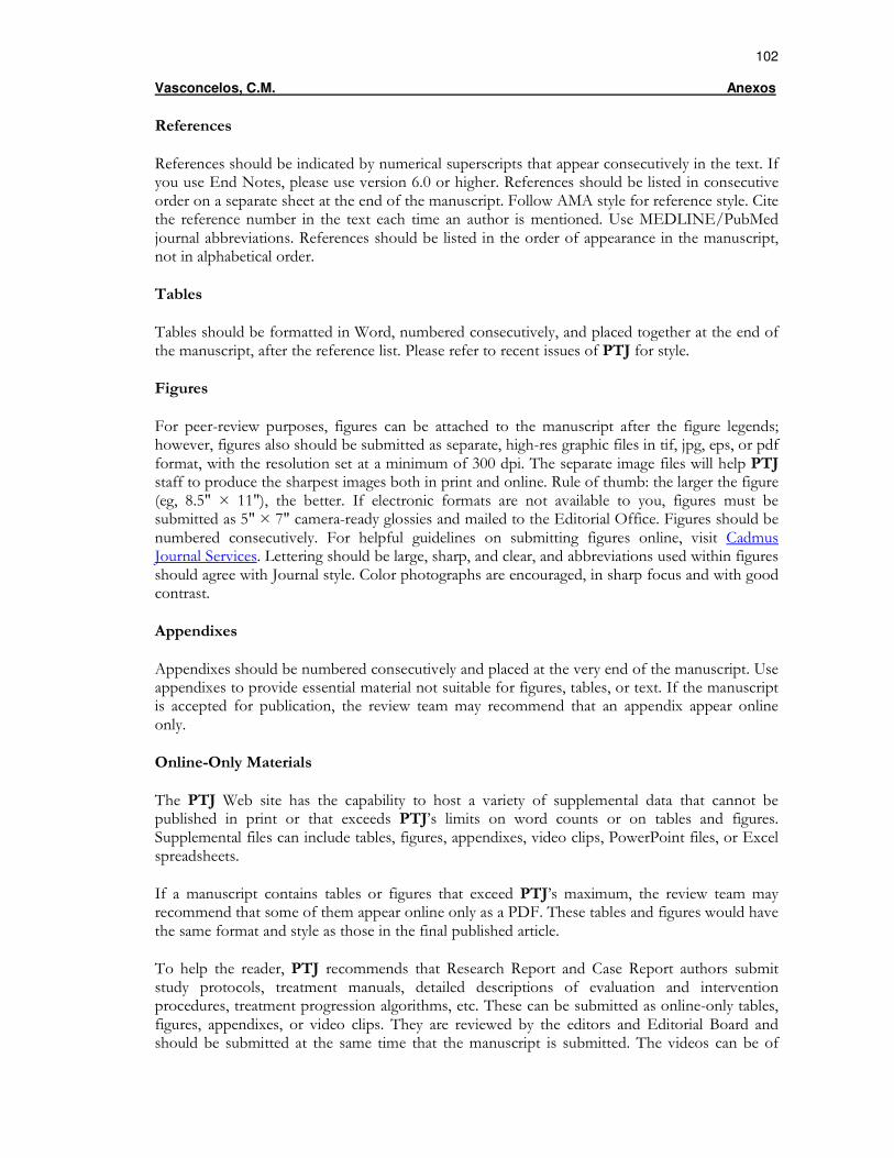
102
Vasconcelos, C.M. Anexos
References
References should be indicated by numerical superscripts that appear consecutively in the text. If you use End Notes, please use version 6.0 or higher. References should be listed in consecutive order on a separate sheet at the end of the manuscript. Follow AMA style for reference style. Cite the reference number in the text each time an author is mentioned. Use MEDLINE/PubMed journal abbreviations. References should be listed in the order of appearance in the manuscript, not in alphabetical order.
Tables
Tables should be formatted in Word, numbered consecutively, and placed together at the end of the manuscript, after the reference list. Please refer to recent issues of PTJ for style.
Figures
For peer-review purposes, figures can be attached to the manuscript after the figure legends; however, figures also should be submitted as separate, high-res graphic files in tif, jpg, eps, or pdf format, with the resolution set at a minimum of 300 dpi. The separate image files will help PTJ staff to produce the sharpest images both in print and online. Rule of thumb: the larger the figure (eg, 8.5" × 11"), the better. If electronic formats are not available to you, figures must be submitted as 5" × 7" camera-ready glossies and mailed to the Editorial Office. Figures should be numbered consecutively. For helpful guidelines on submitting figures online, visit Cadmus Journal Services. Lettering should be large, sharp, and clear, and abbreviations used within figures should agree with Journal style. Color photographs are encouraged, in sharp focus and with good contrast.
Appendixes
Appendixes should be numbered consecutively and placed at the very end of the manuscript. Use appendixes to provide essential material not suitable for figures, tables, or text. If the manuscript is accepted for publication, the review team may recommend that an appendix appear online only.
Online-Only Materials
The PTJ Web site has the capability to host a variety of supplemental data that cannot be published in print or that exceeds PTJ’s limits on word counts or on tables and figures. Supplemental files can include tables, figures, appendixes, video clips, PowerPoint files, or Excel spreadsheets.
If a manuscript contains tables or figures that exceed PTJ’s maximum, the review team may recommend that some of them appear online only as a PDF. These tables and figures would have the same format and style as those in the final published article.
To help the reader, PTJ recommends that Research Report and Case Report authors submit study protocols, treatment manuals, detailed descriptions of evaluation and intervention procedures, treatment progression algorithms, etc. These can be submitted as online-only tables, figures, appendixes, or video clips. They are reviewed by the editors and Editorial Board and should be submitted at the same time that the manuscript is submitted. The videos can be of
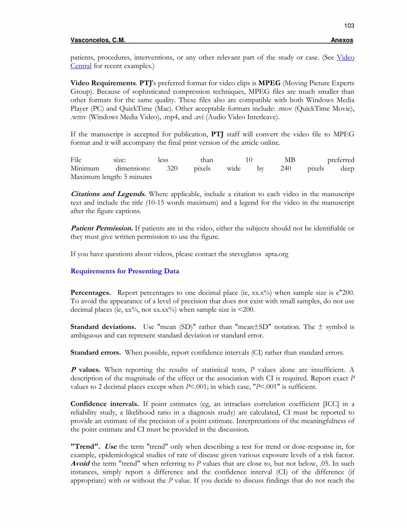
103
Vasconcelos, C.M. Anexos
patients, procedures, interventions, or any other relevant part of the study or case. (See Video Central for recent examples.)
Video Requirements. PTJ's preferred format for video clips is MPEG (Moving Picture Experts Group). Because of sophisticated compression techniques, MPEG files are much smaller than other formats for the same quality. These files also are compatible with both Windows Media Player (PC) and QuickTime (Mac). Other acceptable formats include: .mov (QuickTime Movie), .wmv (Windows Media Video), .mp4, and .avi (Audio Video Interleave).
If the manuscript is accepted for publication, PTJ staff will convert the video file to MPEG format and it will accompany the final print version of the article online.
File size: less than 10 MB preferred Minimum dimensions: 320 pixels wide by 240 pixels deep Maximum length: 5 minutes
Citations and Legends. Where applicable, include a citation to each video in the manuscript text and include the title (10-15 words maximum) and a legend for the video in the manuscript after the figure captions.
Patient Permission. If patients are in the video, either the subjects should not be identifiable or they must give written permission to use the figure.
If you have questions about videos, please contact the steveglaros apta.org
Requirements for Presenting Data
Percentages. Report percentages to one decimal place (ie, xx.x%) when sample size is e"200. To avoid the appearance of a level of precision that does not exist with small samples, do not use decimal places (ie, xx%, not xx.xx%) when sample size is <200.
Standard deviations. Use "mean (SD)" rather than "mean±SD" notation. The ± symbol is ambiguous and can represent standard deviation or standard error.
Standard errors. When possible, report confidence intervals (CI) rather than standard errors.
P values. When reporting the results of statistical tests, P values alone are insufficient. A description of the magnitude of the effect or the association with CI is required. Report exact P values to 2 decimal places except when P<.001; in which case, "P<.001" is sufficient.
Confidence intervals. If point estimates (eg, an intraclass correlation coefficient [ICC] in a reliability study, a likelihood ratio in a diagnosis study) are calculated, CI must be reported to provide an estimate of the precision of a point estimate. Interpretations of the meaningfulness of the point estimate and CI must be provided in the discussion.
"Trend". Use the term "trend" only when describing a test for trend or dose-response in, for example, epidemiological studies of rate of disease given various exposure levels of a risk factor. Avoid the term "trend" when referring to P values that are close to, but not below, .05. In such instances, simply report a difference and the confidence interval (CI) of the difference (if appropriate) with or without the P value. If you decide to discuss findings that do not reach the
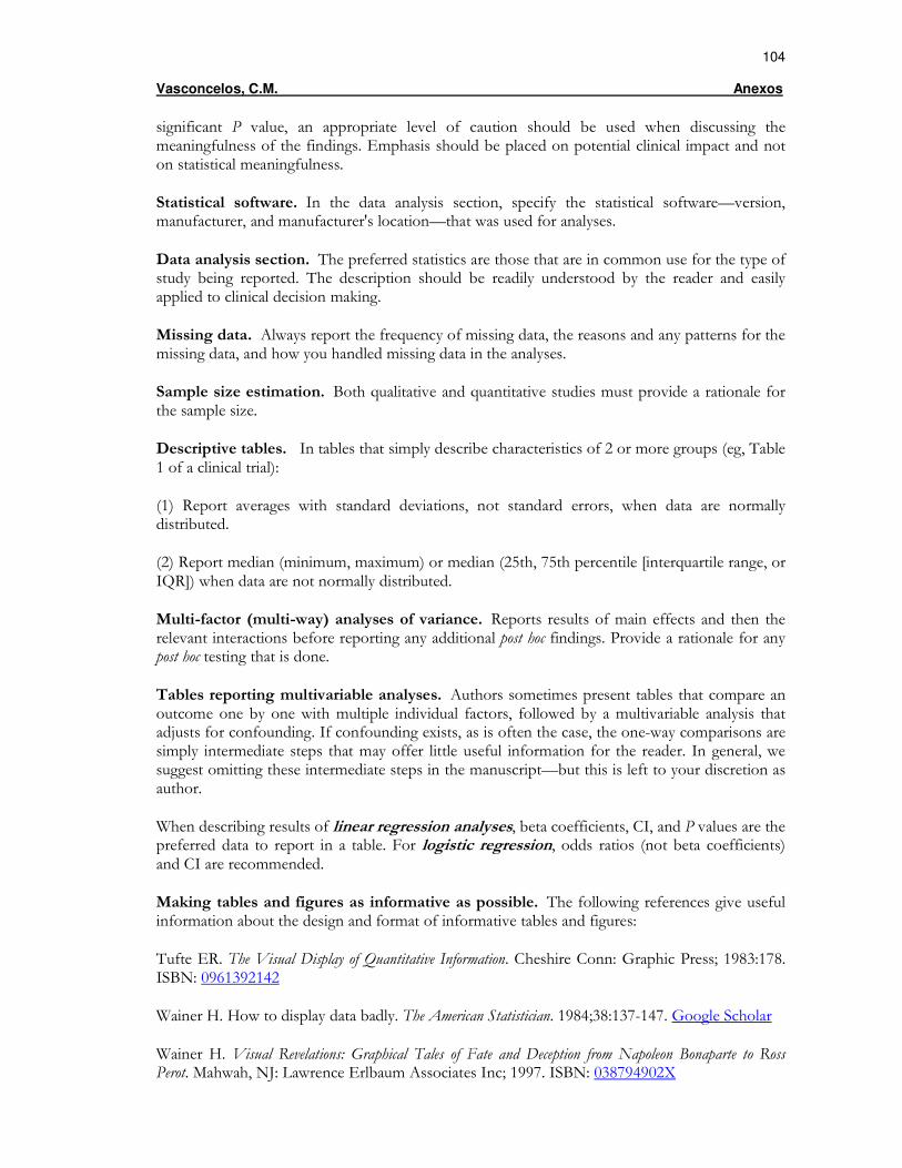
104
Vasconcelos, C.M. Anexos
significant P value, an appropriate level of caution should be used when discussing the meaningfulness of the findings. Emphasis should be placed on potential clinical impact and not on statistical meaningfulness.
Statistical software. In the data analysis section, specify the statistical software—version, manufacturer, and manufacturer's location—that was used for analyses.
Data analysis section. The preferred statistics are those that are in common use for the type of study being reported. The description should be readily understood by the reader and easily applied to clinical decision making.
Missing data. Always report the frequency of missing data, the reasons and any patterns for the missing data, and how you handled missing data in the analyses.
Sample size estimation. Both qualitative and quantitative studies must provide a rationale for the sample size.
Descriptive tables. In tables that simply describe characteristics of 2 or more groups (eg, Table 1 of a clinical trial):
(1) Report averages with standard deviations, not standard errors, when data are normally distributed.
(2) Report median (minimum, maximum) or median (25th, 75th percentile [interquartile range, or IQR]) when data are not normally distributed.
Multi-factor (multi-way) analyses of variance. Reports results of main effects and then the relevant interactions before reporting any additional post hoc findings. Provide a rationale for any post hoc testing that is done.
Tables reporting multivariable analyses. Authors sometimes present tables that compare an outcome one by one with multiple individual factors, followed by a multivariable analysis that adjusts for confounding. If confounding exists, as is often the case, the one-way comparisons are simply intermediate steps that may offer little useful information for the reader. In general, we suggest omitting these intermediate steps in the manuscript—but this is left to your discretion as author.
When describing results of linear regression analyses, beta coefficients, CI, and P values are the preferred data to report in a table. For logistic regression, odds ratios (not beta coefficients) and CI are recommended.
Making tables and figures as informative as possible. The following references give useful information about the design and format of informative tables and figures:
Tufte ER. The Visual Display of Quantitative Information. Cheshire Conn: Graphic Press; 1983:178. ISBN: 0961392142
Wainer H. How to display data badly. The American Statistician. 1984;38:137-147. Google Scholar
Wainer H. Visual Revelations: Graphical Tales of Fate and Deception from Napoleon Bonaparte to Ross Perot. Mahwah, NJ: Lawrence Erlbaum Associates Inc; 1997. ISBN: 038794902X
105
Vasconcelos, C.M. Anexos
Pocock SJ, Clayton TC, Altman DG. Survival plots of time-to-event outcomes in clinical trials: good practice and pitfalls. Lancet. 2002; 359:1686-89. PMID: 12020548
You also can follow a few simple rules of thumb:
1. Avoid pie charts. 2. Avoid simple bar plots or histograms that do not present measurements of variability. 3. Consider using box-plots to illustrate non-normally distributed data. 4. Provide raw data (numerators and denominators) in the margins of meta-analysis forest plots.
Observational Studies
Reports of cohort, case-control, and cross-sectional studies of the prevalence, causes, mechanisms, course, treatment, and prevention of disease. Authors who are reporting data on several patients and want to perform data analyses in an attempt to identify correlations, test hypotheses, or determine cause-and-effect relationships should submit the manuscript following the Observational Studies format.
Guidelines and Checklists
STROBE statement and checklist
Title
Titles should not be vague and should reflect measured variables. For instance, instead of using "physical therapy" to refer to intervention, state specific interventions (eg, "strengthening exercises").
Titles (including subtitles) should be no longer than 150 characters (including punctuation and spaces). Titles in excess of this limit will be edited, subject to author approval.
Abstracts
Word limit. 275 words
Structure. Background, Objective, Design, Methods, Results, Limitations, Conclusions (see Haynes).
Body of Manuscript
Word limit. 4,500 words (excluding abstract and references). Please provide the manuscript word count on the abstract page of your manuscript. Additional materials may be submitted in the form of an appendix, which would appear only on the PTJ Web site if the paper is accepted.
Sections. Introduction, Methods, Results, and Discussion
References. 75 or fewer (See here for more information about formatting)
Tables and figures. Maximum of 6 (See here for more information on formatting)
106
Vasconcelos, C.M. Anexos
Comments. Always end the Introduction section with a clear statement of the study's objectives or hypotheses.
For studies that examine the reliability of a measurement or series of measurements, the patient and clinician sample sizes must be of adequate size to be generalizable. In addition, the study must make a clear and compelling argument for how the findings would have an impact on clinical practice. Issues such as single score interpretation and the interpretation of change scores for the measures under study should be addressed.
Identify the funding source for the study and its role in the study's design, conduct, and reporting. Put this information last in the Methods section and title the subhead "Role of the Funding Source."
In the Methods section, state (if correct) that the study was approved by an Institutional Review Board (IRB). If the study was not submitted to an IRB, provide documentation that not seeking IRB review for this type of study was in accordance with the policy of your institution.
Protocol. PTJ encourages submission of the original study protocol. If you are using an existing database, indicate the source of the data and permission to use the data. Describe methods used to maximize completeness, validity, and quality of the data.
Statistical analysis. Describe primary and secondary analyses, including summary measures used, and methods of analysis. Detail how adjustments are made for confounders and any planned subgroup analyses. Account for missing/incomplete data or loss to follow-up.
Data. PTJ works to maintain the highest levels of integrity and accountability. The Editors therefore reserve the right to ask researchers to provide the raw data for their studies during review or at any time up to 5 years after publication in PTJ. This would likely happen only in rare instances, when credibility of the research is brought into serious question.