bruno da silveira colombo o significado prognÓstico dos
TRANSCRIPT

1
Bruno da Silveira Colombo
O SIGNIFICADO PROGNÓSTICO DOS VALORES SÉRICOS
DO FATOR DE CRESCIMENTO DO TIPO INSULINA 1 EM
PACIENTES ADMITIDOS POR DESCOMPENSAÇÃO
AGUDA DA CIRROSE
Trabalho apresentado à Universidade
Federal de Santa Catarina, como requisito
para a obtenção do título de Mestre
Profissional em Cuidados Intensivos e
Paliativos
Florianópolis
2014

2
3
Bruno da Silveira Colombo
O SIGNIFICADO PROGNÓSTICO DOS VALORES SÉRICOS
DO FATOR DE CRESCIMENTO DO TIPO INSULINA 1 EM
PACIENTES ADMITIDOS POR DESCOMPENSAÇÃO
AGUDA DA CIRROSE
Trabalho apresentado à Universidade
Federal de Santa Catarina, como requisito
para a obtenção do título de Mestre
Profissional em Cuidados Intensivos e
Paliativos
Coordenadora do Mestrado Profissional: Profª Drª Rachel Duarte
Moritz
Professor Orientador: Prof. Dr. Leonardo de Lucca Schiavon
Florianópolis
2014

4

5

6

7

8

9
AGRADECIMENTOS
Ao meu orientador Prof. Dr Leonardo de Lucca Schiavon,
agradeço pelos ensinamentos, pela dedicação, pela presença, pela
confiança e por todo o incentivo fornecido durante toda a execução
desse projeto.
Aos colegas do Núcleo de Estudos em Gastroenterologia e
Hepatologia da UFSC (NEGH), pelo apoio e pelo trabalho em conjunto
realizado durante esse um ano e meio. Aos professores do Mestrado
Profissional pelo aprendizado dividido e pelo auxílio prestado nessa
etapa. Aos colegas e preceptores do Serviço de Endocrinologia do
Hospital Universitário (HU-UFSC), pela dedicação ao ensino durante os
dois anos da minha formação em Endocrinologia e Metabologia. O
apoio e a amizade foram fundamentais.
Ao Laboratório de Análises, Biologia Molecular e Microbactérias,
do HU – UFSC, liderado pela Profª. Drª. Maria Luiza Bazzo, pela
dedicação no armazenamento e interpretação das amostras coletadas
durantes todo o projeto. Aos pacientes e aos seus familiares que
consentiram em participar da pesquisa. Aos colegas e amigos, Marcelo
Fernando Ronsoni, Mariana Costa Silva e Carina Gabriela Correa, pelo
companheirismo, pelo apoio e incentivo durante toda a minha formação.
À minha família, em especial a meu pai, a minha mãe e a meu
irmão, que sempre estiveram ao meu lado durante toda a minha
caminhada profissional e me incentivaram a sempre seguir em busca
dos meus objetivos. Obrigado por todo o incentivo e a presença
constante, mesmo com a distância desses últimos dois anos de minha
formação.

10
11
RESUMO
Introdução: Níveis reduzidos do fator de crescimento do tipo
insulina 1 (IGF-1) tem sido relatados em pacientes com cirrose hepática
e parecem se associar com a intensidade da disfunção hepática. No
entanto, ainda faltam dados sobre o significado prognóstico dessas
alterações. Objetivo: Investigar a relação entre os valores séricos de
IGF-1 e o prognóstico em curto prazo em pacientes admitidos por
descompensação aguda de cirrose. Métodos: Estudo de coorte
prospectivo onde os pacientes foram acompanhados durante a
internação e a mortalidade em 90 dias foi avaliada por contato
telefônico em caso de alta hospitalar. Todos os pacientes realizaram
exames laboratoriais na admissão e os níveis de IGF-1 foram avaliados
por ELISA. Vinte e um pacientes foram também avaliados no
seguimento ambulatorial após a alta hospitalar e comparados em dois
momentos (hospitalização e avaliação ambulatorial). Resultados: Entre
Dezembro de 2011 e Novembro de 2013, 103 pacientes foram
incluídos, com média de idade de 54,2 ± 11,3 anos, com uma
predominância do sexo masculino (69,9%). IGF-1 se correlacionou
positivamente com a albumina e negativamente com relação
normalizada internacional (RNI), proteína C reativa (PCR), bilirrubina
total e a valores de MELD (Model for End-Stage Liver Disease). A
mortalidade em 90 dias foi de 26,2 % e, na análise multivariada, foi
associada de forma independente a maiores valores médios de MELD, a
presença de ascite e a níveis reduzidos de IGF-1. A estimativa da
probabilidade de sobrevida de Kaplan-Meier no nonagésimo dia foi de
94,3% em pacientes com IGF-1 ≥ 13 ng/mL e 63,2% para os indivíduos
com IGF-1 < 13 ng/mL (P = 0,001). No seguimento ambulatorial pós-
alta hospitalar, níveis significativamente mais elevados de IGF-1 foram
encontrados quando comparados com o momento da descompensação
aguda. Conclusão: Os níveis séricos de IGF-1 diminuem durante a
descompensação aguda da cirrose e são independentemente associados
com a mortalidade em curto prazo. Estes resultados sugerem que esse
biomarcador pode ser utilizado como uma ferramenta prognóstica em
pacientes com cirrose admitidos por complicações agudas da doença.
Palavras-chave: cirrose hepática; prognóstico; IGF-I

12

13
ABSTRACT
Background & Aims: Decreased IGF-1 serum levels have been
reported in patients with cirrhosis and seem to correlate with the
intensity of hepatic dysfunction. However, data about the prognostic
significance of decreased IGF-1 serum levels are still lacking. We
investigated the relationship between serum IGF-1 levels and short-term
prognosis in patients admitted for acute decompensation of cirrhosis.
Methods: Patients were followed during their hospital stay and 90-day
mortality was evaluated. All subjects underwent a laboratory evaluation
at admission and IGF-1 was measured by ELISA. Twenty-one patients
were also evaluated in the outpatient clinic after discharge and were
compared at two time points. Results: Between December 2011 and
November 2013, 103 patients were included, with a mean age of 54.2 ±
11.3 years. IGF-1 was positively correlated with albumin and negatively
correlated with international normalized ratio (INR), C-reactive protein
(CRP), total bilirubin and the Model for End-Stage Liver Disease
(MELD). Ninety-day mortality was 26.2% and it was independently
associated with MELD, ascites and IGF-1 levels in multivariate
analysis. The Kaplan-Meier survival probability at 90 days was 94.3%
in patients with IGF-1 ≥ 13 ng/mL and 63.2% for patients with IGF-1 <
13 ng/mL (P = 0.001). In the outpatient evaluation, significantly higher
levels of IGF-1 were found compared with acute decompensation.
Conclusions: IGF-1 levels decrease during acute decompensation of
cirrhosis and were independently associated with short-term mortality.
These findings suggest that this biomarker can be used as a prognostic
tool in patients with cirrhosis admitted for acute complications of the
disease.
Key words: liver cirrhosis; prognostic; insulin-like growth factor 1

14

15
LISTA DE TABELAS E FIGURAS
TABELA 1 - Características da Casuística 31
TABELA 2 -
Fatores associados com a mortalidade em 90 dias nos pacientes
hospitalizados por descompensação aguda da cirrose 34
TABELA 3 - Análise de regressão logística múltipla dos fatores associados com a
mortalidade em 90 dias 35
FIGURA 1 -
Taxa cumulativa de sobrevida em 90 dias de pacientes cirróticos de
acordo com as categorias de IGF-1 __ _36
FIGURA 2 - Box plot de IGF-1 de acordo com o momento de avaliação
(hospitalar ou ambulatorial). Os níveis de IGF-1 foram
significativamente mais elevados na avaliação ambulatorial (P <
0,001) 37

16

17
LISTA DE ABREVIATURAS
ACLF Acute-on-Chronic Liber Failure
DA Descompensação Aguda
DP Desvio Padrão
EASL-CLIF European Association for the Study of the Liver – Chronic Liver Failure
GH Hormônio de Crescimento
HBV Vírus da Hepatite B
HCV Vírus da Hepatite C
HDA Hemorragia Digestiva Alta
HIV Vírus da Imunodeficiência Humana
IC Intervalo de Confiança
IGF Fator de Crescimento do tipo Insulina
IGF-1 Fator de Crescimento do tipo Insulina 1
IGFBP Proteína de Ligação do Fator de
Crescimento do Tipo Insulina
IGFBP-3 Proteína-3 de Ligação do Fator de
Crescimento do Tipo Insulina
MELD Model for End-Stage Liver Disease
OR Odds Ratio
PBE Peritonite Bacteriana Espontânea
PCR Proteína C Reativa
RNI Relação Normalizada Internacional
SPSS Statistical Package for the Social
Sciences TCLE Termo de Consentimento Livre e
Esclarecido
TTPA Tempo de Tromboplastina Parcial
Ativado

18

19
SUMÁRIO
1. ARTIGO CIENTÍFICO 21
1.1 PÁGINA DE TÍTULO 23
1.2 INTRODUÇÃO 25
1.3 MATERIAIS E MÉTODOS 27
1.4 RESULTADOS 31
1.5 DISCUSSÃO 39
1.6 REFERÊNCIAS 43
2. ANEXO A
Parecer Consubstanciado Comitê de Ética
em Pesquisa 47
3. ANEXO B
Normas para Publicação na Revista Liver
International 49
4. APÊNDICE A
Instrumento de Coleta de Dados 60
5. APÊNDICE B
Termo de Consentimento Livre e
Esclarecido 62
6. APÊNDICE C
Artigo em Inglês 64

20

21
ARTIGO CIENTÍFICO
TÍTULO: O Significado Prognóstico dos Valores Séricos do Fator de
Crescimento do Tipo Insulina 1 em Pacientes Admitidos por
Descompensação Aguda da Cirrose
Artigo submetido à revista LIVER INTERNATIONAL (Official
Journal Of The International Association For The Study Of The
Liver)

22

23
Página de Título
Título:
O significado prognóstico dos valores séricos do fator de crescimento do
tipo insulina 1 em pacientes admitidos por descompensação aguda da
cirrose.
Autores:
Bruno da Silveira Colombo1, Marcelo Fernando Ronsoni
1, Pedro Eduardo
Soares e Silva1, Leonardo Fayad
1, Letícia Muraro Wildner
2, Maria Luiza
Bazzo2, Esther Buzaglo Dantas-Correa
1, Janaína Luz Narciso-Schiavon
1,
Leonardo de Lucca Schiavon1
Afiliações: 1Serviço de Gastroenterologia, Universidade Federal de Santa Catarina
2Departamento de Análises Clínicas, Universidade Federal de Santa
Catarina
Auxílio Financeiro: Conselho Nacional de Desenvolvimento Científico e Tecnológico (CNPq)

24

25
INTRODUÇÃO
A cirrose hepática é caracterizada patologicamente pelo
desenvolvimento de nódulos regenerativos circundados por faixas fibrosas
em resposta à lesão hepática crônica, que levam à hipertensão portal e
doença hepática terminal [1]. O curso da cirrose é tipicamente variável,
dependendo de vários fatores, incluindo a etiologia da cirrose e a reserva
funcional hepática [2]. No geral, a evolução natural da cirrose é
caracterizada por uma longa fase assintomática, também conhecida como
fase compensada, seguida por uma fase rapidamente progressiva
caracterizada por complicações específicas [2]. Esta transição de cirrose
compensada para descompensada ocorre em uma taxa de 5-7% ao ano e
está associada à diminuição acentuada da expectativa de vida [3, 4].
Mudanças no eixo somatotrófico, observadas como resultado da
doença hepática, tem sido relatadas em pacientes cirróticos e podem ser
responsáveis, pelo menos em parte, por vários distúrbios metabólicos
caracteristicamente observados no curso da doença hepática, entre eles, a
desnutrição, resistência insulínica, osteopenia, e hipogonadismo [5-7]. O
GH é um hormônio peptídico liberado na hipófise anterior que estimula o
crescimento, a reprodução celular e a regeneração. Além disso, exerce
efeitos metabólicos sobre o osso, a cartilagem, a gordura, o músculo, o
coração, o sistema imunológico e outros [8, 9]. No fígado, o GH estimula
receptores específicos, induzi a transcrição do gene do fator de crescimento
do tipo insulina 1 (IGF-1) e, consequentemente, a síntese e liberação do
IGF-1 para o sangue [10].
Embora vários tecidos secretem IGF-1, no período pós-natal, o
fígado é a principal fonte de IGF-1 circulante [10]. O IGF-1 exerce efeitos
anabólicos sobre o metabolismo de aminoácidos e carboidratos, aumenta a
massa muscular, melhora o conteúdo mineral ósseo e a função da barreira
intestinal [10]. A maioria do IGF-1 circulante está ligado às proteínas do
IGF tipo 3 (IGFBP-3), que, por conseguinte, reduz a biodisponibilidade do
IGF-1 e limita o seu acesso ao receptor de IGF-1. As IGFBP’s regulam
fortemente a atividade biológica do IGF-1 e a atividade é ajustada
adicionalmente por meio dos níveis individuais dos subtipos de IGFBP por
proteases e moduladores de afinidade [11, 12].

26
Alterações no eixo GH/IGF/IGFBP já foram previamente bem
documentadas na cirrose, e os peptídeos envolvidos tem sido propostos
como marcadores de disfunção hepatocelular, desnutrição e prognóstico
[8, 9]. Menores níveis de IGF-1 foram relatados em pacientes com cirrose
quando comparados com controles saudáveis, o que reflete a diminuição
da função de síntese hepática [9, 13]. Além disso, o IGF-1 circulante
parece se correlacionar com o grau de disfunção hepática, já que
diminuições dos níveis foram relatadas em fases mais avançadas da
cirrose [9, 13, 14]. Apesar destes resultados indicarem que os valores de
IGF-1 podem ser de relevância clínica em pacientes cirróticos, existem
poucos dados sobre o significado prognóstico desse biomarcador neste
contexto. Desta forma, os objetivos do presente estudo foram investigar a
relação entre os valores séricos de IGF-1 e o prognóstico em curto prazo
em pacientes hospitalizados por descompensação aguda da cirrose e
compará-los aos níveis séricos no ambulatório.
27
2. MATERIAIS E MÉTODOS
Pacientes
Entre dezembro de 2011 e novembro de 2013 foi realizado um
estudo de coorte prospectivo que incluiu, consecutivamente, indivíduos
admitidos em um hospital terciário brasileiro devido a descompensação
aguda (DA) de cirrose hepática. Os pacientes nas seguintes situações foram
excluídos: internação para procedimentos eletivos, admissões não
relacionadas a complicações da cirrose hepática, carcinoma hepatocelular
fora dos critérios de Milão (uma lesão < 5 cm, até três lesões < 3 cm, sem
invasão vascular e sem envolvimento extra-hepático) [15] e diagnóstico
duvidoso de cirrose hepática. Todos os pacientes foram inicialmente
admitidos na emergência do hospital. A decisão de transferir o paciente
para a enfermaria ou unidade de terapia intensiva foi feita a critério do
médico assistente de acordo com a gravidade da DA.
O diagnóstico de cirrose foi estabelecido histologicamente
(quando disponível) ou pela combinação de dados clínicos, de imagem e
achados laboratoriais em pacientes com evidências de hipertensão portal. O
protocolo do estudo foi aprovado pelo Comitê de Ética em Pesquisa com
Seres Humanos da Universidade Federal de Santa Catarina (ANEXO A).
Métodos
DA foi definida como o desenvolvimento de encefalopatia hepática,
ascite volumosa (definida como ascite presente ao exame físico da
admissão), hemorragia gastrointestinal, infecção bacteriana ou qualquer
combinação destes. Todos os pacientes hospitalizados por DA foram
avaliados dentro das primeiras 24 horas da admissão por um dos
pesquisadores envolvidos no estudo, e as seguintes variáveis clínicas foram
coletados: idade, sexo, raça, etiologia da cirrose, história de
descompensação anterior, complicações atuais da cirrose, alcoolismo ativo
e uso regular de propanolol. Todos os indivíduos foram submetidos à
avaliação laboratorial na admissão, e os seguintes testes foram realizados
para este estudo: leucócitos totais, contagem de plaquetas, sódio sérico,
creatinina, relação normalizada internacional (RNI), albumina, proteína
C reativa (PCR), bilirrubina total e IGF-1.
Alcoolismo ativo foi definido como um consumo médio

28
global de 21 ou mais doses por semana para homens e 14 ou mais doses
por semana para as mulheres, durante as quatro semanas anteriores à
admissão (uma dose padrão equivale a 12 g de álcool absoluto) [16].
Indivíduos com suspeita de infecção bacteriana na admissão
hospitalar foram submetidos a exame clínico para confirmar esse
diagnóstico e estabelecer a principal fonte primária da infecção. O
diagnóstico da infecção foi realizado de acordo com os critérios do
Centro de Controle de Doenças [17]. Paracentese diagnóstica foi
realizada em todos os pacientes com ascite no momento da internação.
Peritonite bacteriana espontânea (PBE) foi diagnosticada quando a
contagem de neutrófilos do líquido ascítico foi ≥ 250 neutrófilos/mm3 na
ausência de fonte intra-abdominal de infecção, independentemente de
cultura negativa [18].
Todos os pacientes com PBE receberam ceftriaxone e albumina
endovenosa ajustada ao peso, no primeiro e terceiro dia após o
diagnóstico. Encefalopatia hepática foi graduada conforme os critérios de
West-Haven [19] e, caso presente, um fator precipitante era investigado.
Lactulose era iniciada com doses ajustadas conforme a necessidade.
Todos os pacientes com sangramento varicoso agudo receberam
octreotide endovenoso, um antibiótico (norfloxacino via oral ou
ceftriaxone endovenoso) e foram submetidos à terapêutica endoscópica
de urgência após estabilização. A gravidade da doença hepática foi
estimada pela classificação de Child-Pugh [20] e pelo Model for End-
Stage Liver Disease (MELD) [21], calculados com base em testes de
laboratório realizados na admissão. Acute-on-chronic liver failure
(ACLF) foi definido como proposto pelo Consórcio EASL – CLIF [22].
Os pacientes foram acompanhados durante a internação e a
mortalidade em trinta e noventa dias foi avaliada por telefonema em caso
de alta hospitalar. As taxas de mortalidade em 90 dias não incluíram
pacientes que realizaram transplante hepático (pacientes que receberam
transplante hepático foram excluídos do acompanhamento).
Níveis Séricos de IGF-1
Os níveis de IGF-1 foram avaliados em amostras de soro colhidas
dentro das primeiras 24 horas após a admissão e armazenadas a -80 °C até
à sua utilização. Os níveis de IGF-1 foram mensurados por ensaio imuno-
enzimático (ELISA Quantikine® - IGF-1 Human, R&D Systems,
Minneapolis, EUA). A sensibilidade analítica relatada deste ensaio é de

29
0,026 ng/mL.
Análise estatística
As variáveis contínuas foram descritas como média e desvio
padrão e as categóricas em números absolutos e proporções. Aplicou-se o
teste de Kolmogorov-Smirnov para avaliação da distribuição das variáveis
quanto à normalidade. A correlação entre as variáveis numéricas foi
avaliada pelo coeficiente de correlação de Spearman. As variáveis
contínuas foram comparadas pelo teste t de Student, no caso de
distribuição normal ou teste de Mann-Whitney, nos demais casos. As
variáveis categóricas foram avaliadas pelo teste qui-quadrado ou teste
exato de Fisher, conforme apropriado. A análise de regressão logística
múltipla (forward stepwise) foi utilizada para investigar os fatores
associados de forma independente à mortalidade em 90 dias. Com base na
curva ROC, o melhor ponto de corte do IGF-1 para prever a mortalidade
em 90 dias foi escolhido. A curva de sobrevida foi calculada usando o
método de Kaplan-Meier e diferenças entre os grupos foram comparadas
pelo teste de log-rank. Teste t pareado ou teste de Wilcoxon foram usados
para comparar as variáveis em dois momentos (internação e avaliação
ambulatorial). Todos os testes foram realizados pelo software SPSS, versão
17.0 (SPSS , Chicago, IL , EUA). Um valor de P inferior a 0,05 foi
considerado estatisticamente significativo.

30
31
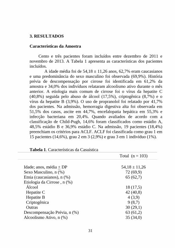
3. RESULTADOS
Características da Amostra
Cento e três pacientes foram incluídos entre dezembro de 2011 e
novembro de 2013. A Tabela 1 apresenta as características dos pacientes
incluídos.
A idade média foi de 54,18 ± 11,26 anos, 62,7% eram caucasianos
e uma predominância do sexo masculino foi observada (69,9%). História
prévia de descompensação por cirrose foi identificada em 61,2% da
amostra e 34,0% dos indivíduos relataram alcoolismo ativo durante o mês
anterior. A etiologia mais comum de cirrose foi o vírus da hepatite C
(40,8%) seguida pelo abuso de álcool (17,5%), criptogênica (8,7%) e o
vírus da hepatite B (3,9%). O uso de propranolol foi relatado por 41,7%
dos pacientes. Na admissão, hemorragia digestiva alta foi observada em
51,5% dos casos, ascite em 44,7%, encefalopatia hepática em 55,3% e
infecção bacteriana em 20,4%. Quando avaliados de acordo com a
classificação de Child-Pugh, 14,6% foram classificados como estádio A,
48,5% estádio B e 36,9% estádio C. Na admissão, 19 pacientes (18,4%)
preenchiam os critérios para ACLF. ACLF foi classificada como grau 1 em
15 pacientes (14,6%), grau 2 em 3 (2,9%) e grau 3 em 1 indivíduo (1%).
Tabela 1. Características da Casuística
Total (n = 103)
Idade; anos, média + DP
54,18 ± 11,26
Sexo Masculino, n (%) 72 (69,9)
Etnia (caucasianos), n (%) 65 (62,7)
Etiologia da Cirrose , n (%)
Álcool 18 (17,5)
Hepatite C 42 (40,8)
Hepatite B 4 (3,9)
Criptogênica 9 (8,7)
Outras 30 (29,1)
Descompensação Prévia, n (%) 63 (61,2)
Alcoolismo Ativo, n (%) 35 (34,0)
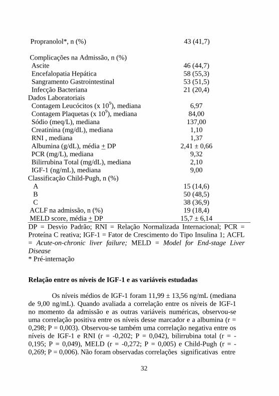
32
Propranolol*, n (%) 43 (41,7)
Complicações na Admissão, n (%)
Ascite 46 (44,7)
Encefalopatia Hepática 58 (55,3)
Sangramento Gastrointestinal 53 (51,5)
Infecção Bacteriana 21 (20,4)
Dados Laboratoriais
Contagem Leucócitos (x 109), mediana 6,97
Contagem Plaquetas (x 109), mediana 84,00
Sódio (meq/L), mediana 137,00
Creatinina (mg/dL), mediana 1,10
RNI , mediana 1,37
Albumina (g/dL), média + DP 2,41 ± 0,66
PCR (mg/L), mediana 9,32
Bilirrubina Total (mg/dL), mediana 2,10
IGF-1 (ng/mL), mediana 9,00
Classificação Child-Pugh, n (%)
A 15 (14,6)
B 50 (48,5)
C 38 (36,9)
ACLF na admissão, n (%) 19 (18,4)
MELD score, média + DP 15,7 ± 6,14
DP = Desvio Padrão; RNI = Relação Normalizada Internacional; PCR =
Proteína C reativa; IGF-1 = Fator de Crescimento do Tipo Insulina 1; ACFL
= Acute-on-chronic liver failure; MELD = Model for End-stage Liver
Disease * Pré-internação
Relação entre os níveis de IGF-1 e as variáveis estudadas
Os níveis médios de IGF-1 foram 11,99 ± 13,56 ng/mL (mediana
de 9,00 ng/mL). Quando avaliada a correlação entre os níveis de IGF-1
no momento da admissão e as outras variáveis numéricas, observou-se
uma correlação positiva entre os níveis desse marcador e a albumina (r =
0,298; P = 0,003). Observou-se também uma correlação negativa entre os
níveis de IGF-1 e RNI (r = -0,202; P = 0,042), bilirrubina total (r = -
0,195; P = 0,049), MELD (r = -0,272; P = 0,005) e Child-Pugh (r = -
0,269; P = 0,006). Não foram observadas correlações significativas entre

33
os níveis de IGF-1 e idade, contagem de leucócitos,
contagem de plaquetas, PCR, sódio ou creatinina.
Quando os níveis de IGF-1 foram avaliados de acordo com as
complicações específicas da cirrose observadas no momento da
admissão, não foram encontradas associações com a presença de ascite,
encefalopatia hepática e infecção bacteriana (P > 0,05). Aqueles
indivíduos com hemorragia digestiva alta na admissão
apresentaram menores níveis de
IGF-1 em comparação com os demais pacientes (6,30 ng/mL vs. 11,60
ng/mL, P = 0,038). Pacientes Child-Pugh C também apresentaram
menores níveis de IGF-1 do que os com Child-Pugh A/B (5,80 ng/mL
vs. 11,60 ng/mL, P < 0,001). Não foram observadas diferenças
significativas nos níveis de IGF-1 entre aqueles com ou sem ACLF na
admissão (7,10 ng/mL vs. 10,80 ng/mL, P = 0,222).
Significado Prognóstico do IGF-1 em Pacientes Cirrótios
Hospitalizados
A mortalidade global em 90 dias foi de 26,2% e associou-se na
análise bivariada (tabela 2) com idade mais avançada (58,74 ± 11,44
anos vs. 52,57 ± 10,82 anos; P = 0,014), ascite (85,2% vs. 30,3%; P <
0,001), encefalopatia hepática (74,1% vs. 50,0%; P = 0,030), infecção
bacteriana (37,0% vs. 14,5%; P = 0,012), Child-Pugh C (77,8% vs.
22,4%; P < 0,001), maior pontuação no MELD (21,44 ± 6,31 vs. 13,66
± 4,63; P < 0,001) e ACLF na admissão (51,0% vs. 6,6%; P < 0,001). A
mortalidade em noventa dias também foi relacionada a maior mediana
de creatinina (1,50 mg/dL vs. 1,00 mg/dL; P < 0,001), RNI (1,56 vs. 1,35; P = 0,003), PCR (23,40 mg/L vs. 7,80 mg/L; P = 0,004),
bilirrubina total (3,60 mg/dL vs. 1,40 mg/dL; P < 0,001) e menor média
de albumina (1,99 ± 0,51 g/dL vs. 2,56 ± 0,65 g/dL; P < 0,001), menor
mediana de sódio (133,00 mEq/L vs. 137,00 mEq/L; P = 0,002), e
menores níveis de IGF-1 (5,50 ng/mL vs. 11,60 ng/mL , P = 0,003).
Análise de regressão logística múltipla (tabela 3), incluindo variáveis
com P < 0,010 na análise bivariada mostrou que o escore MELD (OR
1,220; IC 95% 1,097-1,356; P < 0,001), ascite no momento da
internação (OR 10,706; IC 95% 2,526-45,384; P = 0,001) e níveis de
IGF-1 (OR 0,907; IC 95% 0,833-0,988; P = 0,026), foram
independentemente associados à mortalidade em 90 dias. Com base na
curva ROC, um melhor ponto de corte do IGF-1 (13 ng/mL) foi

34
escolhido para prever a mortalidade de 90 dias.
Tabela 2. Fatores associados com a mortalidade em 90 dias nos
pacientes hospitalizados por descompensação aguda da cirrose Sobreviventes
(n = 76)
Óbitos
(n = 27) P
Idade (anos), média ± DP
52,57 ± 10.82
58,74 ± 11.44
0,014
Sexo Masculino, n (%) 53 (69,7) 19 (70,4) 0,951
Etiologia da Cirrose, n (%)
Álcool 11 (14,5) 7 (25,9) 0,237
Hepatite C 33 (43,4) 9 (33,3) 0,360
Hepatite B 3 (3,9) 1 (3,7) 1,000
Criptogênica 7 (9,2) 2 (7,4) 1,000
Descompensação Prévia, n (%) 46 (60,5) 17 (63,0) 0,82
Alcoolismo Ativo, n (%) 11 (14,5) 7 (25,9) 0,237
Propranolol*, n (%) 36 (47,4) 7 (25,9) 0,052
Complicação na Admissão, n (%)
Ascite 23 (30,3) 23 (85,2) < 0,001
Encefalopatia Hepática 38 (50,0) 20 (74,1) 0,030
Sangramento Gastrointestinal 47 (61,8) 6 (22,2) < 0,001
Infecção Bacteriana 11 (14,5) 10 (37,0) 0,012
Dados laboratoriais
Contagem Leucócitos (x 109), mediana 6,63 7,25 0,680
Contagem Plaquetas (x 109), mediana 84,00 94,00 0,406
Sódio (meq/L), mediana 137,00 133,00 0,002
Creatinina (mg/dL), mediana 1,00 1,50 < 0,001
RNI, mediana 1,35 1,56 0,003
Albumina (g/dL), média ± DP 2,56 ± 0,65 1,99 ± 0,51 < 0,001
PCR (mg/L), mediana 7,80 23,40 0,004
Bilirrubina Total (mg/dL), mediana 1,40 3,60 < 0,001

35
IGF-1 (ng/mL), mediana 11,60 5,50 0,003
Child-Pugh C, n (%) 17 (22,4) 21 (77,8) < 0,001
MELD score, média ± DP 13,66 ±4,63 21,44 ± 6,31 < 0,001
ACLF, n (%) 5 (6,6) 14 (51,0) < 0,001
DP = Desvio Padrão; Relação Normalizada Internacional; PCR = Proteína
C reativa; IGF-1 = Fator de Crescimento do Tipo Insulina 1; MELD =
Model for End-stage Liver Disease; ACFL = Acute-on-chronic liver failure
* Pré-internação
Tabela 3. Análise de regressão logística dos fatores associados com
A mortalidade em 90 dias ( variáveis com p < 0,01 na análise
bivariada foram incluídas)
Fatores Odds Ratio IC 95% P
MELD Score 1,220 1,097 – 1,356 < 0,001
Ascite 10,706 2,526 – 45,384 0,001
Níveis IFG-1 0,907 0,833 – 0,988 0,026
IGF-1 = Fator de Crescimento do Tipo Insulina 1; MELD = Model for
End-stage Liver Disease; IC = Intervalo de Confiança
A Figura 1 mostra as curvas de Kaplan-Meier para a
mortalidade durante o período de acompanhamento de acordo com as
categorias de IGF-1. A probabilidade de sobrevida de Kaplan-Meier em
90 dias foi de 94,3% em pacientes com IGF-1 ≥ 13 ng/mL e 63,2% para
aqueles com IGF-1 < 13 ng/mL (P = 0,001). Para prever mortalidade em
90 dias, o IGF-1 em um ponto de corte de 13 ng/mL mostrou
sensibilidade de 93% e especificidade de 43%, com valor preditivo
negativo de 94%, mas um valor preditivo positivo de apenas 37%. A
razão de verossimilhança positiva foi de 1,637 e a razão de
verossimilhança negativa foi de 0,171.

36
Fig. 1. Taxa cumulativa de sobrevida em 90 dias de pacientes cirróticos
de acordo com as categorias de IGF-1. A sobrevida foi significativamente
menor nos pacientes com IGF-1 < 13 ng/mL, em comparação com
aqueles com valores ≥ 13 ng/mL (P = 0,001, teste de long-rank).
Níveis de IGF-1 Após a Alta Hospitalar
Um subgrupo de vinte e um pacientes que haviam sido
estudados durante a internação hospitalar foram submetidos à avaliação
laboratorial em uma mediana de seguimento de 105 dias após a alta, e
foram comparados em dois momentos para investigar o impacto da
descompensação aguda sobre os níveis de IGF-1. Na avaliação
ambulatorial, nove pacientes foram classificados como Child-Pugh A e
12 como Child-Pugh B, com uma mediana de MELD de 10,19. Quando
comparados à avaliação durante a hospitalização, valores
significativamente mais elevados de níveis de IGF-1 foram observados
no momento da avaliação ambulatorial (21,9 ± 23,3 ng/mL vs. 49,3 ±
33,3 ng/mL; P < 0,001) (Figura 2). Um incremento dos níveis de IGF-1
na avaliação ambulatorial foi observado em 17 dos 21 pacientes
incluídos nesta análise (81%). Como esperado, a avaliação ambulatorial

37
também foi associada com menores valores de MELD (10,59 ± 2,26
vs. 13,58 ± 2,99; P < 0,001), RNI (1,27 ± 0,14 vs. 1,44 ± 0,19; P <
0,001), bilirrubina total (1,28 ± 0,74 mg/dL vs. 1,75 ± 1,29 mg/dL; P
= 0,058), mediana de PCR (3,50 mg/L vs. 6,31 mg/L; P = 0,016) e
maior valores de albumina (3,27 ± 0,49 g/dL vs. 2,62 ± 0,56 g/dL; P
< 0,001).
Fig. 2. Box plot de IGF-1 de acordo com o momento de avaliação
(hospitalar ou ambulatorial). Os níveis de IGF-1 foram significativamente
mais elevados na avaliação ambulatorial (P < 0,001).

38
39
4. DISCUSSÃO
Complicações agudas da cirrose são causas comuns de internação
hospitalar e estão associadas com significativa morbidade e
mortalidade. Apesar dos grandes avanços alcançados ao longo dos
últimos anos, ainda existe uma necessidade de se identificar novos
marcadores prognósticos em pacientes com descompensação aguda da
cirrose. Previamente, demonstrou-se que os níveis de IGF-1 na cirrose
estão relacionados com a gravidade da disfunção hepática e que sofrem
pouca influência de outros fatores não relacionados com a capacidade
de síntese hepática, o que representa, assim, uma ferramenta promissora
como um marcador de prognóstico na cirrose [9, 13, 14].
No presente estudo, os níveis de IGF-1 correlacionaram-se com
outras variáveis, direta ou indiretamente associadas à intensidade da
disfunção hepática, tais como albumina, RNI, bilirrubina e MELD.
Além disso, os pacientes Child-Pugh C apresentaram menores níveis de
IGF-1 do que os Child-Pugh A/B. Estes resultados estão de acordo com
estudos anteriores que demonstraram uma associação entre níveis mais
baixos de IGF-1 e a gravidade da doença hepática [9, 13, 14, 23, 24].
Em um estudo recente, que também incluiu pacientes internados por
descompensação aguda da cirrose, encontramos menores níveis de IGF-
1 em pacientes Child-Pugh B/C e uma correlação do IGF-1 com
diversas variáveis relacionadas à gravidade da cirrose, sem interferência
de outros parâmetros como sexo, etiologia da cirrose e comorbidades
[14]. Além disso, dados anteriores indicam que os níveis baixos de IGF-
1, observados em pacientes com cirrose avançada, são prontamente
corrigidos após um transplante hepático bem sucedido. Estes dados
apoiam a investigação do IGF-1 como um biomarcador potencial para a
avaliação da função hepática [25, 27].
Neste estudo, a mortalidade em 90 dias foi de 26,2% e associou-se
na análise de regressão logística com maior pontuação MELD, com a
presença de ascite e com níveis mais baixos de IGF-1. Se por um lado, o
escore MELD e a presença de ascite são fatores prognósticos clássicos
na cirrose [28, 29]; por outro lado, existem ainda poucos dados sobre o
significado dos níveis de IGF-1 em pacientes com doenças hepáticas
crônicas. Um pequeno estudo que envolveu 36 pacientes com cirrose
alcoólica relatou taxas de sobrevida significativamente menores entre os
pacientes com IGF-1 inferior a 3 nmol/L (equivalente a 22,9 ng/mL)
[30]. Um estudo posterior do mesmo grupo avaliou os níveis de IGF-1

40
em 354 pacientes com doença hepática induzida pelo álcool a partir de
um grande estudo multicêntrico sobre o efeito do malotilato na sobrevida
[31]. O período médio de acompanhamento foi de 569 dias e os baixos
níveis de IGF-1 foram associados com pior prognóstico, especialmente
em um ponto de corte de 56 ng/mL [31]. É importante notar que este
estudo incluiu pacientes com e sem cirrose e nenhuma análise detalhada
daqueles com apenas cirrose foi fornecida [31]. No entanto, os autores
observaram uma relação significativa entre os níveis de IGF-1 e a
intensidade da disfunção hepática e também indicaram o potencial dos
seus níveis como um marcador de prognóstico em doenças hepáticas.
A probabilidade de sobrevida de Kaplan-Meier em 90 dias foi de
94,3% em pacientes com IGF-1 ≥ 13 ng/mL e 63,2% para aqueles com
IGF-1 < 13 ng/mL. Embora o ponto de corte proposto tenha mostrado boa
sensibilidade (93%) e valor preditivo negativo (94%), resultados menos
expressivos foram observados em relação a especificidade e o valor
preditivo positivo. Isso indica que o IGF-1 pode ser um bom biomarcador
para identificar os pacientes cirróticos com baixo risco de morte, mas
provavelmente é menos útil para predizer mortalidade. Esta hipótese é
reforçada pela expressivamente baixa razão de verossimilhança negativa
observada (0,171) em relação à modesta razão de verossimilhança
positiva (1,637). O corte de 13 ng/mL sugerido aqui é significativamente
mais baixo do que os valores relatados nos outros dois estudos
mencionados [30, 31]. De fato, os níveis médios de IGF-1 em pacientes
com cirrose são consideravelmente diferentes entre os estudos, variando
de 25 ng/mL a 152 ng/mL [9, 13, 14, 23, 24]. Esta discrepância pode ser
explicada por diferenças metodológias usadas na medição do IGF-1 ou
pelos distintos contextos clínicos e critérios de inclusão dos estudos.
Como no presente estudo foram incluídos apenas os pacientes internados
por complicações da cirrose, é possível que os fatores relacionados com o
insulto agudo ou, mais importante ainda, uma rápida diminuição da
função hepática, possam justificar os baixos níveis de IGF-1 observados.
Quando os indivíduos com descompensação aguda foram avaliados
após a alta, foi observado um aumento significativo nos níveis de IGF-1
em 17 dos 21 pacientes. Os níveis médios de IGF-1 neste subgrupo de
indivíduos foram de 21,9 ± 23,3 ng/mL durante a internação hospitalar e
49,3 ± 33,3 ng/mL na avaliação ambulatorial. Este incremento no IGF-1
circulante após a estabilização clínica foi acompanhado pela melhoria em
vários outros parâmetros relacionados com a gravidade da doença
hepática, tais como MELD, RNI, bilirrubina total e albumina. Embora os

41
dados em relação a mudanças dinâmicas no IGF-1 sejam escassos, o já
mencionado pequeno estudo dinamarquês avaliou o IGF-1 logo antes da
alta hospitalar em 18 dos 36 pacientes incluídos [30]. Em conformidade
com os nossos resultados, os autores relataram um aumento de 23,7
ng/mL para 48,8 ng/mL nos níveis de IGF-1 após a melhora clínica
[30]. Estes resultados sugerem que os baixos níveis de IGF-1 na cirrose
podem se tornar ainda mais suprimidos durante as complicações agudas,
provavelmente refletindo uma piora na função hepática. No entanto,
uma vez que o insulto agudo é removido e disfunção hepática melhora,
os níveis de IGF-1 tendem a voltar para os seus valores basais. Esta
hipótese é corroborada pela rápida recuperação dos níveis de IGF-1
observada após o transplante hepático, como mencionado anteriormente
[25, 27].
Algumas limitações do presente estudo merem ser discutidas. Primeiro,
o número relativamente pequeno de pacientes avaliados poderia limitar
a capacidade de generalizar estes resultados para outras populações. De
fato, existe ainda uma necessidade de validação dos resultados aqui
apresentados em coortes maiores antes da incorporação desse
biomarcador na prática clínica. No entanto, este estudo pode representar
um importante ponto de partida para pesquisas que visam avaliar IGF-1
em diferentes cenários clínicos, como cirróticos ambulatoriais em lista
de espera para transplante de fígado e na insuficiência hepática aguda.
Em segundo lugar, os pacientes foram admitidos no serviço de
emergência de um hospital geral e, apesar do serviço seguir protocolos
gerais para complicações da cirrose, estas orientações não foram criadas
especificamente para este estudo e variações na abordagem de casos
específicos são esperadas. No entanto, este problema é comum a quase
todos os estudos que investigam biomarcadores em cenários clínicos,
especialmente em pacientes que necessitam de atendimento de urgência.
A heterogeneidade dos pacientes incluídos foi antecipada e,
provavelmente, reflete as características dos pacientes com cirrose que
buscam atendimento médico em situações de emergência.
É possível concluir que, em pacientes admitidos por descompensação
aguda da cirrose, níves circulantes de IGF-1 estão relacionados com a
gravidade da doença hepática e foram associados de forma
independente ao prognóstico em curto prazo. O IGF-1 circulante
aumenta após a estabilização da doença, sugerindo um impacto da piora
transitória da função hepática em seus níveis. Estes resultados reforçam
o potencial papel do IGF-1 na avaliação da gravidade da disfunção

42
hepática e indicam que esse marcador pode ser utilizado como uma
ferramenta para avaliação do prognóstico de pacientes com cirrose
admitidos com as complicações agudas da doença.

43
REFERÊNCIAS
1. Schuppan D, Afdhal NH. Liver cirrhosis. Lancet 2008;371:838-
851.
2. Durand F, Valla D. Assessment of prognosis of cirrhosis. Semin
Liver Dis 2008;28:110-122.
3. D'Amico G, Garcia-Tsao G, Pagliaro L. Natural history and
prognostic indicators of survival in cirrhosis: a systematic review of 118
studies. J Hepatol 2006;44:217-231.
4. Asrani SK, Kamath PS. Natural history of cirrhosis. Curr
Gastroenterol Rep 2013;15:308.
5. Huisman EJ, Trip EJ, Siersema PD, van Hoek B, van Erpecum KJ.
Protein energy malnutrition predicts complications in liver cirrhosis.
Eur J Gastroenterol Hepatol 2011;23:982-989.
6. Berzigotti A, Abraldes JG. Impact of obesity and insulin-resistance
on cirrhosis and portal hypertension. Gastroenterol Hepatol
2013;36:527-533.
7. Luxon BA. Bone disorders in chronic liver diseases. Curr
Gastroenterol Rep 2011;13:40-48.
8. Perrini S, Laviola L, Carreira MC, Cignarelli A, Natalicchio A,
Giorgino F. The GH/IGF1 axis and signaling pathways in the muscle
and bone: mechanisms underlying age-related skeletal muscle wasting
and osteoporosis. J Endocrinol 2010;205:201-210.
9. Colakoglu O, Taskiran B, Colakoglu G, Kizildag S, Ari Ozcan F,
Unsal B. Serum insulin like growth factor-1 (IGF-1) and insulin like
growth factor binding protein-3 (IGFBP-3) levels in liver cirrhosis.
Turk J Gastroenterol 2007;18:245-249.
10. Juul A. Serum levels of insulin-like growth factor I and its binding
proteins in health and disease. Growth Horm IGF Res 2003;13:113-170.
11. Ferry RJ, Jr., Cerri RW, Cohen P. Insulin-like growth factor
binding proteins: new proteins, new functions. Horm Res 1999;51:53-
67.
12. Hwa V, Oh Y, Rosenfeld RG. The insulin-like growth factor-
binding protein (IGFBP) superfamily. Endocr Rev 1999;20:761-787.
13. Wu YL, Ye J, Zhang S, Zhong J, Xi RP. Clinical significance of
serum IGF-I, IGF-II and IGFBP-3 in liver cirrhosis. World J
Gastroenterol 2004;10:2740-2743.
14. Ronsoni MF, Lazzarotto C, Fayad L, Silva MC, Nogueira CL,
Bazzo ML, Narciso-Schiavon JL, et al. IGF-I and IGFBP-3 serum levels

44
in patients hospitalized for complications of liver cirrhosis. Ann Hepatol
2013;12:456-463.
15. Mazzaferro V, Regalia E, Doci R, et al. Liver transplantation for
the treatment of small hepatocellular carcinomas in patients with cirrhosis. N
Engl J Med 1996; 334:693.
16. Addolorato G, Leggio L, Ferrulli A, Cardone S, Vonghia L, Mirijello
A, Abenavoli L, et al. Effectiveness and safety of baclofen for
maintenance of alcohol abstinence in alcohol-dependent patients with
liver cirrhosis: randomised, double-blind controlled study. Lancet
2007;370:1915-1922.
17. Garner JS, Jarvis WR, Emori TG, Horan TC, Hughes JM. CDC
definitions for nosocomial infections, 1988. Am J Infect Control
1988;16:128-140.
18. Runyon BA. Management of adult patients with ascites due to
cirrhosis: an update. Hepatology 2009;49:2087-2107.
19. Bajaj JS. Review article: the modern management of hepatic
encephalopathy. Aliment Pharmacol Ther 2010;31:537-547.
20. Angermayr B, Cejna M, Karnel F, Gschwantler M, Koenig F, Pidlich
J, Mendel H, et al. Child-Pugh versus MELD score in predicting survival
in patients undergoing transjugular intrahepatic portosystemic shunt. Gut
2003;52:879-885.
21. Kamath PS, Wiesner RH, Malinchoc M, Kremers W, Therneau TM,
Kosberg CL, D'Amico G, et al. A model to predict survival in patients
with end-stage liver disease. Hepatology 2001;33:464-470.
22. Moreau R, Jalan R, Gines P, Pavesi M, Angeli P, Cordoba J, Durand
F, et al. Acute-on-chronic liver failure is a distinct syndrome that
develops in patients with acute decompensation of cirrhosis.
Gastroenterology 2013;144:1426-1437, 1437 e1421-1429.
23. Donaghy A, Ross R, Gimson A, Hughes SC, Holly J, Williams R.
Growth hormone, insulinlike growth factor-1, and insulinlike growth
factor binding proteins 1 and 3 in chronic liver disease. Hepatology
1995;21:680-688.
24. Assy N, Pruzansky Y, Gaitini D, Shen Orr Z, Hochberg Z, Baruch Y.
Growth hormone-stimulated IGF-1 generation in cirrhosis reflects
hepatocellular dysfunction. J Hepatol 2008;49:34-42.
25. Castro GR, Coelho JC, Parolin MB, Matias JE, de Freitas AC. Insulin-
like growth factor I correlates with MELD and returns to normal level
after liver transplantation. Ann Transplant 2013;18:57-62.
26. Weber MM, Auernhammer CJ, Lee PD, Engelhardt D, Zachoval R.
Insulin-like growth factors and insulin-like growth factor binding proteins
45
in adult patients with severe liver disease before and after
orthotopic liver transplantation. Horm Res 2002;57:105-112.
27. Bassanello M, De Palo EF, Lancerin F, Vitale A, Gatti R,
Montin U, Ciarleglio FA, et al. Growth hormone/insulin-like
growth factor 1 axis recovery after liver transplantation: a
preliminary prospective study. Liver Transpl 2004;10:692-698.
28. Silva PE, Fayad L, Lazzarotto C, Ronsoni MF, Bazzo ML,
Colombo BS, Dantas-Correa EB, et al. Single-centre validation of
the EASL-CLIF Consortium definition of acute-on-chronic liver
failure and CLIF-SOFA for prediction of mortality in cirrhosis.
Liver Int 2014.
29. Kamath PS, Kim WR. The model for end-stage liver disease
(MELD). Hepatology 2007;45:797-805.
30. Moller S, Gronbaek M, Main K, Becker U, Skakkebaek NE.
Urinary growth hormone (U-GH) excretion and serum insulin-like
growth factor 1 (IGF-1) in patients with alcoholic cirrhosis. J
Hepatol 1993;17:315-320.
31. Moller S, Becker U, Juul A, Skakkebaek NE, Christensen E.
Prognostic value of insulinlike growth factor I and its binding
protein in patients with alcohol-induced liver disease. EMALD
group. Hepatology 1996;23:1073-1078.

46

47
ANEXO A - Parecer Consubstanciado do CEP

48
49
ANEXO B – Normas para Publicação na Revista Liver
International
Author Guidelines
Updated 23 October 2014 From 2015 Liver International will be
published in an online-only format.
TYPES OF MANUSCRIPTS
Original Manuscripts: Liver International publishes both
clinical and experimental research in all areas of normal and abnormal
liver function and disease. Purely descriptive research or methodology
papers will not be considered for publication. Basic science manuscripts
will be considered for publication only if they have translational
significance. Manuscript length should not exceed 5,000 words
including tables, figures and references. Manuscripts should contain no
more than 5 figures or tables. Each figure should have a maximum of 4
panels.
Additional supporting information can be submitted along with the
original manuscript. Authors preparing supporting information for
publication should carefully read the guidelines at:
https://authorservices.wiley.com/bauthor/suppinfo.asp.
Abstract:
• The abstract must not exceed 250 words.
• The title must not exceed 130 characters.
• Key points must be organized in a box with 4 bullet points which
highlight your paper’s
originality. Must not exceed 100 words.
• The abstract must be organized as follows:
- Background & Aims
- Methods
- Results
- Conclusions
Do not use abbreviations, footnotes or references in the
abstract. An electronic word count of the abstract must be included.3-5
key words at the end of the abstract must be provided.

50
The manuscript must be arranged as follows:
• Title page
• Abstract in the Liver International format
• Key points box
• Introduction
• Materials and methods (or Patients and methods)
• Results
• Discussion
• Acknowledgements
• References
• Tables
• Figure legends
As a rule, original manuscripts will be evaluated by two
independent reviewers and by the Editors. The Editors reserve the right of
early rejection without further external review if the manuscript is judged
unlikely to be accepted. Manuscripts requiring extensive revision will be
at a disadvantage for publication and will be rejected. Authors shall be
responsible for the quality of language and style and are strongly advised
against submitting a manuscript which is not written in grammatically
correct English. The Editors reserve the right to reject poorly written
manuscripts even if their scientific content is qualitatively suitable for
publication. Manuscripts are submitted with the understanding that they
are original contributions and do not contain data that have been
published elsewhere or are under consideration by another journal.
Meeting abstracts do not constitute prior publication. Revised
manuscripts should be accompanied by a point-by-point reply to the
critiques, specifying the changes made in the revised version, which
should be highlighted.
Rapid Communications: will be considered for important and
timely scientific contributions; authors should explain in their
accompanying letter why they wish to submit their paper as a rapid
communication. Rapid communications will undergo regular peer-review
as original manuscripts but will be granted fast-track processing. Such
papers should not exceed 3,000 words, including no more than 2 tables or
figures and 20 references. Additional supporting information can be
submitted along with the original manuscript. Authors preparing
supporting information for publication should carefully read the
guidelines at: https://authorservices.wiley.com/bauthor/suppinfo.asp.

51
MANUSCRIPT SUBMISSION
Manuscripts, including tables and figures, should be submitted
online at: ScholarOne Manuscripts:
http://mc.manuscriptcentral.com/liverint.
Authors are kindly asked NOT to send their manuscripts by fax
or mail to the Editorial Office.
Ithenticate Liver International employs a plagiarism detection system.
By submitting your manuscript to this journal you accept that your
manuscript may be screened for plagiarism against previously published
material. Copyright - If your paper is accepted, the author identified as
the formal corresponding author for the paper will receive an email
prompting them to login into Author Services; where via the Wiley
Author Licensing Service (WALS) they will be able to complete the
license agreement on behalf of all authors on the paper.
For authors signing the copyright transfer agreement
If the OnlineOpen option is not selected the corresponding
author will be presented with the copyright transfer agreement (CTA) to
sign. The terms and conditions of the CTA can be previewed in the
samples associated with the Copyright FAQs below:
CTA Terms and Conditionshttp://exchanges.wiley.com/authors/faqs---
copyright-_301.html
For authors choosing OnlineOpen
If the OnlineOpen option is selected the corresponding author
will have a choice of the following Creative Commons License Open
Access Agreements (OAA):
• Creative Commons Attribution Non-Commercial License OAA.
• Creative Commons Attribution Non-Commercial -NoDerivs
License OAA.
To preview the terms and conditions of these open access
agreements please visit the Copyright FAQs hosted on Wiley Author
Services http://exchanges.wiley.com/authors/faqs---copyright-
_301.html and visit
http://www.wileyopenaccess.com/details/content/12f25db4c87/Copyrig
ht--License.html. See the OnlineOpen section for more information.
If you select the OnlineOpen option and your research is
52
funded by certain funders [e.g. The Wellcome Trust and
members of the Research Councils UK (RCUK) or the Austrian Science
Fund (FWF)] you will be given the opportunity to publish your article
under a CC-BY license supporting you in complying your Funder
requirements . For more information on this policy and the Journal’s
compliant self-archiving policy please visit:
http://www.wiley.com/go/funderstatement.
For RCUK, Wellcome Trust, FWF authors click on the link
below to preview the terms and conditions of this license:
• Creative Commons Attribution License OAA
To preview the terms and conditions of these open access
agreements please visit the Copyright FAQs hosted on Wiley Author
Services http://exchanges.wiley.com/authors/faqs---copyright-_301.html
and visit
http://www.wileyopenaccess.com/details/content/12f25db4c87/Copyright
--License.html.
OnlineOpen - OnlineOpen is available to authors of primary research
articles who wish to make their article available to non-subscribers on
publication, or whose funding agency requires grantees to archive the
final version of their article. With OnlineOpen, the author, the author's
funding agency, or the author's institution pays a fee to ensure that the
article is made available to non-subscribers upon publication via Wiley
Online Library, as well as deposited in the funding agency's preferred
archive.
To preview the terms and conditions of these open access
agreements please visit the Copyright FAQs hosted on Wiley Author
Services http://authorservices.wiley.com/bauthor/faqs_copyright.asp and
visit
http://www.wileyopenaccess.com/details/content/12f25db4c87/Copyright
--License.html. All OnlineOpen articles are treated in the same way as
any other article. They go through the journal's standard peer-review
process and will be accepted or rejected based on their own merit.
ORGANIZATION OF THE MANUSCRIPT
Submitted manuscripts must be typed double-spaced throughout,
preferably using a "standard" font (we prefer Times/Arial 12). Tables and
figures must be numbered. For mathematical symbols, Greek letters, and
other special characters, use normal text. The references must be in

53
accordance with Liver International reference style (see
References).
Approved nomenclature for gene and protein names and
symbols should be used, including appropriate use of italics (all gene
symbols and loci should be in italics) and capitalization as it applies for
each organism's standard nomenclature format, in text, tables, and
figures. Full gene names are generally not in italics and Greek symbols
are not used. Proteins should not be italicized.
Improperly prepared manuscripts will not be entered into the
peer review process and will be sent back to the author for correction. A
letter of submission must be uploaded with all manuscripts. The letter
may be used to outline the strengths of the manuscript. All commercial
relationships (i.e. consultancies, patent-licensing agreements) that might
pose a conflict of interest in connection with the submitted manuscript
must be included in the letter. In case of possible conflicts of interest,
the letter must include a detailed description of the nature of the conflict
of interest, the full name of the entity with which there is a conflict, as
well as address, telephone number, webpage address, a detailed
financial disclosure, and any other important, relevant details.
The Title page must contain:
a. A title of no more than 130 characters.
b. Names of the Authors including the first names of all the Authors in
full.
c. Names of department(s) and institution(s) where the work was done.
d. Name, address, telephone and fax numbers, and electronic mail
address of the corresponding Author.
e. Electronic word count for main body of manuscript.
f. Number of figures and tables.
g. List of abbreviations in the order of appearance.
h. Conflict of interest.
i. Financial support.
j. Trial registration number, if applicable (see below).
Animal trials – Manuscripts reporting experiments using
animals must include a statement giving assurance that all animals
received human care and that study protocols comply with the
institution's guidelines. Statistical methods used should be outlined.
54
Human trials – Manuscripts reporting data from research conducted on
humans must include a statement of assurance in the methods section of
the manuscript reading that: (1) informed consent was obtained from each
patient included in the study and (2) the study protocol conforms to the
ethical guidelines of the 1975 Declaration of Helsinki as reflected in a
priori approval by the institution's human research committee.
Randomised controlled trials – Any paper that is a randomised control
trial should adhere to the guidelines that can be found at the following
web-site: www.consort-statement.org. The checklist should be
downloaded, completed and uploaded with your submission. The trial
registration number must be included on the title page of the manuscript
reporting a registered clinical trial. Failure to do so will prevent entry to
the peer review process.
Registration of clinical trials – Liver International endorses the policy of
the WHO and the International Committee of Medical Journal Editors
(ICMJE) on the registration of clinical trials. Any trial that starts
recruiting on or after July 1, 2005 should be registered in a publicly
owned, publicly accessible registry and should satisfy a minimal standard
dataset. Trials that started recruiting before that date will be considered
for publication if registered before September 13, 2005. More detailed
information regarding the definition of clinical trial, the minimal
registration data set, and the requirements for an acceptable trial registry
can be found in New Engl J Med 2004, 351:1250-1251 and New Engl J
Med 2005, 352:2437-2438.
Drugs and chemicals – Drugs and chemicals should be used by generic
name. If trademarks are mentioned, the manufacturer's name and city
should be given. All funding sources supporting the work, either public or
private, especially those from pharmaceutical companies, must be
provided.
Genetic Sequence data – In papers reporting a novel DNA or amino
sequence, verification that the data have been or will be submitted either
to Gen-Bank or EMBL is required. Please provide this verification and
the accession number in the covering letter.

55
References – References must be in accordance with the Liver
International reference style. References are ordered as they appear in
the text and citation numbers for references are placed between
"brackets" ("[ ]") in the text as well as in the reference list.
Authors should be listed surname first, followed by the initials of given
names (e.g. Bolognesi M). If there are more than six authors, the names
of the first six authors followed by et al. should appear. Titles of all
cited articles are required. Titles of articles cited in reference list should
be in upright, not italic text; the first word of the title is capitalized, the
title written exactly as it appears in the work cited, ending with a full
stop. Journal titles are abbreviated according to common usage,
followed by Journal years, semicolon (;) before volume and colon (:)
before full page range (see examples below).
All articles in the list of references should be cited in the text
and, conversely, all references cited in the text must be included in the
list. Personal communications and unpublished data should be cited
directly in the text by the first Author, without being numbered.
An example of how references should look within the text:
HVPG was measured by hepatic vein catheterization using a balloon
catheter according to a procedure described elsewhere [14, 15] and used
as an index of portal hypertension [16].
An example of how the reference list should look:
[14] Merkel C, Bolognesi M, Bellon S, Zuin R, Noventa F, Finucci G,
et al. Prognostic usefulness of hepatic vein catheterization in patients
with cirrhosis and esophageal varices. Gastroenterology 1992;102:973-
979.
[15] Groszmann RJ, Wongcharatrawee S. The hepatic venous pressure
gradient: anything worth doing should be done right. Hepatology
2004;39:280-282.
Abbreviations, symbols and nomenclature - should be
standardised and in accordance with ELLIS G (ed.). Units, symbols and
abbreviations. The Royal Society of Medicine, 1 Wimpole Street,
London Wl M 8AE, 1975.
Tables – Tables should be provided as Word files (*.doc),
Excel (.xls) or Illustrator (*.eps) compatible files. TIFF and JPG files
are not acceptable for table submission. Tables should contain a
maximum of 10 columns. Tables submitted in landscape orientation will
not be accepted. Tables should include a title, table legend.

56
Figures –All graphics submitted to Liver International should be sent at
their actual size, which is 100% of their print dimension and in portrait
orientation.
Two standard widths are used and figures should fit in one (8.5 x
23.5 cm) or two (17.5 x 23.5 cm) columns (see Figure and Table
Guidelines).
Figure files should be provided in high resolution .eps format,
minimum 800dpi (for graphs and charts) or .tiff format, minimum 300dpi
(for photographs or a combination of images and text). Figures with
multiple parts (A, B, C) should be provided as separate files. Panel
lettering should be in Arial bold 16 pt, capitalized and no full stop (A)
while lettering in figures (axes, conditions) should be in Arial 14 pt,
lower case type with the first letter capitalized and no full stop. Do not
copy and paste figure files into the manuscript word document.
Figures can be in grayscale or CMYK. All photomicrographs
should have a scale on the photograph. Photographs of identifiable
patients should be accompanied by written permission to publish from
patient(s).
If you no longer have the original data to improve/recreate
graphs, charts or combination figures to high resolution, please crop the
graph portion in Microsoft PowerPoint and re-type any text in
Arial/Times New Roman in minimum 14pts. This will ensure that at least
the text is clear. Any lines in the figures must be at least 1.5 or 2pts thick.
We will accept the revised .ppt file. For more information on file
requirements, please refer to http:
//authorservices.wiley.com/prep_illust.asp.
ENGLISH
Authors may be asked to contact professionals regarding the
correction of the English content of manuscripts either before or after
acceptance. We recommend the Wiley English Language Editing
Services: go to www.wileyeditingservices.com. The expense will be the
responsibility of the Authors.
REVIEW PROCESS
Authors should be aware that manuscripts will be screened upon
submission. Only the manuscripts which fully comply with the

57
submission requirements outlined and in which the level of English
is of an acceptable standard will enter the peer review process.
First submission – Once successful submission of a manuscript
has taken place, an acknowledgement will be sent by e-mail to the
Corresponding Author on the manuscript, with a copy to all named co-
authors. All subsequent correspondence will be with the designated
Corresponding Author. The reference number of the manuscript should
be used by the Authors in all communications with the Editorial Office.
All the manuscripts will be reviewed by the Editors and, and in some
cases, by external expert reviewers. After review, the Corresponding
Author will be notified by email of the decision taken by the Editor(s).
This email will be accompanied by the comments of the reviewers,
where a paper has been sent for external review.
Resubmission of manuscripts – In some cases, Authors will be
invited to submit a revised version of the manuscript for further review.
This invitation does not imply, in any case, that the revised version will
be accepted for publication. In general, revised manuscripts must be
received in the Editorial Office within four months of the date of the
first decision. Authors should submit the resubmitted manuscript with
all changes underlined. The resubmitted manuscript should be
accompanied by a cover letter stating that the manuscript has been
revised according to the comments made by the Editor and the
Reviewers. Figures and tables must be uploaded. Please ensure that a
separate point by point response to the reviewers is included with the
covering letter. Please do not send revised manuscripts to the Editorial
Office via e-mail. Revised manuscripts should be uploaded on the
ScholarOne website.
PROOFS
When proofs are ready for checking, the corresponding author
will receive an email alert containing a link to a web site. A working e-
mail address must therefore be provided for the corresponding author.
The proof can be downloaded as a PDF (portable document format) file
from this site. Acrobat Reader will be required in order to read this file.
This software can be downloaded (free of charge) from the following
web site: http://get.adobe.com/reader/.This will enable the file to be
opened, read and corrected on screen. Further instructions will be sent
with the proof.
Offprints - A PDF offprint of the online published article will be
provided via Author Services. Additional paper offprints may be
58
ordered online at http://offprint.cosprinters.com/blackwell.
If you have queries about offprints please email
Accepted Articles – ‘Accepted Articles' have been accepted for
publication and undergone full peer review but have not been through the
copyediting, typesetting, pagination and proofreading process. Accepted
Articles are published online a few days after final acceptance, appear in
PDF format only, are given a Digital Object Identifier (DOI), which
allows them to be cited and tracked, and are indexed by PubMed. A
completed copyright form is required before a manuscript can be
processed as an Accepted Article.
Early View - Liver International is covered by Wiley Blackwell's
Early View service. Early View articles are complete full-text articles
published online in advance of their publication in a printed issue.
Articles are therefore available as soon as they are ready, rather than
having to wait for the next scheduled print issue. Early View articles are
complete and final. They have been fully reviewed, revised and edited for
publication, and the authors’ final corrections have been incorporated.
Because they are in final form, no changes can be made after online
publication. The nature of Early View articles mean that they do not yet
have volume, issue or page numbers, so Early View articles cannot be
cited in the traditional way. They are therefore given a Digital Object
Identifier (DOI), which allows the article to be cited and tracked before it
is allocated to an issue. After print publication, the DOI remains valid and
can continue to be used to cite and access the article.
Author material archive policy - Please note that unless
specifically requested, Wiley Blackwell will dispose of all hardcopy or
electronic material submitted 2 months after publication. If you require
the return of any material submitted, please inform the editorial office or
production editor as soon as possible.
Disclaimer - The Publisher, the International Association for the
Study of the Liver and the Editors cannot be held responsible for errors or
any consequences arising from the use of information contained in this
journal; the views and opinions expressed do not necessarily reflect those
of the Publisher, the International Association for the Study of the Liver
and the Editors; neither does the publication of advertisements constitute
any endorsement by the Publisher, the International Association for the
Study of the Liver and the Editors of the products advertised.
59
Author Services - Author Services enables authors to track their article
– once it has been accepted – through the production process to
publication online and in print. Authors can check the status of their
articles online and choose to receive automated e-mails at key stages of
production. The author will receive an e-mail with a unique link that
enables them to register and have their article automatically added to the
system. Please ensure that a complete e-mail address is provided when
submitting the manuscript. Visit http://authorservices.wiley.com/ for
more details on online production tracking and for a wealth of resources
including FAQs and tips on article preparation, submission and more.
Note to NIH Grantees
Pursuant to NIH mandate, Wiley-Blackwell will post the accepted
version of contributions authored by NIH grant-holders to PubMed
Central upon acceptance. This accepted version will be made publicly
available 12 months after publication. For further information, see
www.wiley.com/go/nihmandate.

60
APÊNDICE A - INSTRUMENTO DE COLETA DE DADOS

61

62
APÊNDICE B - TERMO DE CONSENTIMENTO LIVRE E
ESCLARECIDO
TERMO DE CONSENTIMENTO LIVRE E ESCLARECIDO
AVALIAÇÃO DE MARCADORES PROGNÓSTICOS EM
PORTADORES DE CIRROSE HEPÁTICA DESCOMPENSADA
Você está sendo convidado para participar de um projeto de
pesquisa: Avaliação de marcadores prognósticos em portadores de
cirrose hepática descompensada. Este projeto tem o objetivo de
identificar dados da avaliação médica ou de exames laboratoriais que
possam nos ajudar a identificar os pacientes com doença mais grave. Os
resultados desta pesquisa poderão permitir a criação de ferramentas para a
identificação rápida dos pacientes com doença mais grave, permitindo
assim um tratamento mais adequado.
Caso você concorde em participar deste estudo, será feita uma
avaliação clínica (entrevista e exame físico) no primeiro dia da sua
internação e 48 horas depois. Além disso, uma coleta de sangue será
realizada por punção periférica na veia do antebraço também nestes dois
momentos. Parte do material será destinada aos exames de rotina (que são
necessários para avaliação do seu caso durante a internação) e uma outra
porção será armazenadas em freezer a -80º C para a posterior dosagem
dos exames referentes a este estudo, que são: Anti-HEV IgG,
procalcitonina, GST-α, neoepítopos da CK-18 e queratina 18 solúvel.
Não existem riscos importantes relacionados a tal procedimento,
podendo ocorrer, como consequência da coleta de sangue, dor no local da
punção e/ou formação de hematoma local. Não há benefício direto para o
participante. Em qualquer etapa do estudo, você terá acesso aos
profissionais responsáveis pela pesquisa para esclarecimento de eventuais
dúvidas. Os principais investigadores envolvidos são o Dr. Bruno da
Silveira Colombo, o Dr. Pedro Silva e o Dr. Leonardo de Lucca Schiavon
que podem ser encontrados no endereço: Departamento de Clínica
Médica, Hospital Universitário/Campus Universitário – Trindade - Cep
88040-970 - Florianópolis – SC Fone (48) 37219149/37219014; e-mail:
[email protected] ou [email protected]. Se você tiver alguma
consideração ou dúvida sobre a ética da pesquisa, entre em contato com o
Comitê de Ética em Pesquisa (CEP): Universidade Federal de Santa
Catarina; Pró-Reitoria de Pesquisa e Extensão - Campus Universitário –
63
Trindade - Florianópolis/SC; Tel: (48) 3721-9206.
É garantida a liberdade da retirada de consentimento a qualquer
momento e deixar de participar do estudo, sem qualquer prejuízo à
continuidade de seu tratamento na Instituição. As informações obtidas
serão analisadas em conjunto com outros pacientes, não sendo
divulgada a identificação de nenhum paciente. Você tem o direito de ser
mantido atualizado sobre os resultados parciais das pesquisas, assim que
os mesmos forem de conhecimento dos pesquisadores. Não há despesas
pessoais para o participante em qualquer fase do estudo, incluindo
exames e consultas. Também não há compensação financeira
relacionada à sua participação. Se existir qualquer despesa adicional, ela
será absorvida pelo orçamento da pesquisa.
Em caso de dano pessoal, diretamente causado pelos
procedimentos propostos neste estudo (nexo causal comprovado), o
participante tem direito a tratamento médico na Instituição, bem como
às indenizações legalmente estabelecidas.
Segue abaixo os termos da declaração para poder participar do
estudo:
Acredito ter sido suficientemente informado a respeito do estudo
“Avaliação de marcadores prognósticos em portadores de cirrose
hepática descompensada”.
Eu discuti com os pesquisadores responsáveis sobre a minha
decisão em participar nesse estudo. Ficaram claros para mim quais são
os propósitos do estudo, os procedimentos a serem realizados, seus
desconfortos e riscos, as garantias de confidencialidade e de
esclarecimentos permanentes. Ficou claro também que minha
participação é isenta de despesas e que tenho garantia do acesso a
tratamento hospitalar, caso seja necessário. Concordo voluntariamente
em participar deste estudo e poderei retirar o meu consentimento a
qualquer momento: antes ou durante o mesmo, sem penalidades,
prejuízo, perda de qualquer benefício que eu possa ter adquirido, ou no
meu atendimento neste Serviço.

64
APÊNDICE C – ARTIGO EM INGLÊS
TITLE PAGE
Manuscript title: Prognostic significance of Insulin-like growth factor-1 (IGF-1) serum
levels in patients admitted for acute decompensation of cirrhosis
Running Head:
IGF-1 in acute decompensation of cirrhosis
Author’s names:
Bruno da Silveira Colombo1, Marcelo Fernando Ronsoni
1, Pedro Eduardo
Soares e Silva1, Leonardo Fayad
1, Letícia Muraro Wildner
2, Maria Luiza
Bazzo2, Esther Buzaglo Dantas-Correa
1, Janaína Luz Narciso-Schiavon
1,
Leonardo de Lucca Schiavon1
Affiliations: 1Division of Gastroenterology, Federal University of Santa Catarina
2Department of Clinical Analysis, Federal University of Santa Catarina
Financial support:
Conselho Nacional de Desenvolvimento Científico e Tecnológico (CNPq)
65
Abstract
Background & Aims: Decreased IGF-1 serum levels have been
reported in patients with cirrhosis and seem to correlate with the
intensity of hepatic dysfunction. However, data about the prognostic
significance of decreased IGF-1 serum levels are still lacking. We
investigated the relationship between serum IGF-1 levels and short-term
prognosis in patients admitted for acute decompensation of cirrhosis.
Methods: Patients were followed during their hospital stay and 90-day
mortality was evaluated. All subjects underwent a laboratory evaluation
at admission and IGF-1 was measured by ELISA. Twenty-one patients
were also evaluated in the outpatient clinic after discharge and were
compared at two time points. Results: Between December 2011 and
November 2013, 103 patients were included, with a mean age of 54.2 ±
11.3 years. IGF-1 was positively correlated with albumin and negatively
correlated with international normalized ratio, C-reactive protein, total
bilirubin and the Model for End-Stage Liver Disease (MELD). Ninety-
day mortality was 26.2% and it was independently associated with
MELD, ascites and IGF-1 levels in multivariate analysis. The Kaplan-
Meier survival probability at 90 days was 94.3% in patients with IGF-1
≥ 13 ng/mL and 63.2% for patients with IGF-1 < 13 ng/mL (P = 0.001).
In the outpatient evaluation, significantly higher levels of IGF-1 were
found compared with acute decompensation. Conclusions: IGF-1 levels
decrease during acute decompensation of cirrhosis and were
independently associated with short-term mortality. These findings
suggest that this biomarker can be used as a prognostic tool in patients
with cirrhosis admitted for acute complications of the disease.
Key words: liver cirrhosis; acute decompensation; insulin-like growth
factor 1
Key points
IGF-1 levels are decreased in cirrhosis and seem to correlate with
the intensity of hepatic dysfunction
In this article, IGF-1 levels, measured at admission in patients with
acute decompensation, were correlated with variables associated
with the intensity of liver dysfunction and were associated with 90-
day mortality in multivariate analysis
Patients with IGF-1 levels ≥ 13 ng/mL at admission exhibited
66
excellent survival (94.3%) in comparison to those with levels
below this limit (63.2%)
A significant increase in circulating IGF-1 was observed in a
subset of patients evaluated after discharge, suggesting an
impact of transient worsening in liver function on this
biomarker
Introduction
Liver cirrhosis is a pathologically defined entity characterized by the
development of regenerative nodules surrounded by fibrous bands in
response to chronic liver injury, which leads to portal hypertension and
end-stage liver disease [1]. The course of cirrhosis is typically variable,
and depends on several factors, including the etiology of cirrhosis and the
hepatic synthetic function [2]. In general, the natural course of cirrhosis is
characterized by a longstanding asymptomatic phase, also known as the
compensated phase, followed by a rapidly progressive phase
characterized by specific complications [2]. This transition from
compensated to decompensated cirrhosis occurs at a rate 5–7% per year
and is associated with a marked decrease in life expectancy [3, 4].
Changes in the growth hormone-insulin-like growth factor (GH-IGF)
axis observed as a result of liver disease has been reported in cirrhosis
and may be responsible, at least in part, for several metabolic disorders
characteristically observed in the course of the disease, including
malnutrition, insulin resistance, osteopenia and hypogonadism [5-7]. GH
is a peptide hormone released from the anterior pituitary that stimulates
growth, cell reproduction and regeneration, exerting metabolic effects on
bone, cartilage, fat, muscles, the heart, the immune system and other
organs [8, 9]. In the liver, GH activation of GH receptors induces IGF-1
gene transcription, and subsequently the synthesis and release of IGF-1 to
the plasma [10]. Even though various tissues secrete IGF-1, in the post-
natal period the liver is the main source of circulating IGF-1 [10]. IGF-1
exerts anabolic effects on amino acid and carbohydrate metabolism,
increases muscle mass and improves bone mineral content and intestinal
barrier function [10]. The majority of circulating IGF-1 is bound to IGF
binding protein-3 (IGFBP-3), which, therefore, lowers the bioavailability
of IGF-1 and limits its access to IGF-1 receptor. The IGFBPs strongly
regulate the biological activity of IGF-1, which is additionally adjusted
via individual levels of the IGFBP subtypes and by affinity-modulating
67
proteases [11, 12].
Changes in the GH/IGF/IGFBP axis are well documented in
cirrhosis, and the peptides involved have been proposed as markers of
hepatocellular dysfunction, malnutrition and survival [8, 9]. Lower IGF-1
serum levels have been reported in patients with cirrhosis compared with
healthy controls, likely reflecting decreased hepatic synthetic function [9,
13]. In addition, circulating IGF-1 appears to correlate with the degree of
hepatic dysfunction as decreased levels were reported in more advanced
stages of cirrhosis [9, 13, 14]. Although these results indicate that
determining IGF-1 serum levels may be of clinical relevance in patients
with cirrhosis, there are very few data on the prognostic significance of
this biomarker in this context. Therefore, we sought to investigate the
relationship between serum levels of IGF-1 and short-term prognosis in
patients hospitalized for acute decompensation (AD) of cirrhosis.
Materials and Methods
Patients
This is a cohort study that included consecutive subjects admitted
to the emergency room of a Brazilian tertiary hospital due to AD of liver
cirrhosis between December 2011 and November 2013. The following
exclusion criteria were adopted: hospitalization for elective procedures,
admissions not related to complications of liver cirrhosis, hepatocellular
carcinoma outside Milan criteria (defined as single lesion ≤5 cm, up to
three separate lesions, none larger than 3 cm, no evidence of gross
vascular invasion, and no regional nodal or distant metastases) [15] and a
doubtful diagnosis of liver cirrhosis. All patients were initially admitted
in the emergency room. The decision to transfer the patient to the ward or
the intensive care unit was made at the discretion of the attending
physician according to the severity of the AD.
The diagnosis of cirrhosis was established either histologically
(when available) or by the combination of clinical, imaging and
laboratory findings in patients with evidence of portal hypertension.
The study protocol complies with the ethical principles of the
Declaration of Helsinki and was approved by the Ethics Committee on
Human Research of the Federal University of Santa Catarina.

68
Methods
All patients admitted for AD, as defined by the acute development of
hepatic encephalopathy, large ascites, gastrointestinal bleeding, bacterial
infection or any combination of these, were screened. Patients were
evaluated within 24 hours of admission by one of the researchers
involved in the study, and the following clinical variables were collected:
age, gender, race, etiology of cirrhosis, history of previous
decompensation, current complications of cirrhosis, active alcoholism and
regular propranolol use. All subjects underwent laboratory evaluation at
admission, and the following tests were performed for this study: total
leukocytes, platelet count, serum sodium, creatinine, international
normalized ratio (INR), albumin, C-reactive protein (CRP) and total
bilirubin.
Active alcoholism was defined as an average overall
consumption of 21 or more drinks per week for men and 14 or more
drinks per week for women during the 4 weeks before enrollment (one
standard drink is equal to 12 g absolute alcohol) [16]. Patients were
followed during their hospital stay and 30- and 90-day mortality was
evaluated by phone call, in case of hospital discharge. Ninety-day
mortality rates were estimated as transplant-free mortality (patients who
received a liver transplant were considered lost to follow-up).
Individuals with a suspected infection at hospital admission
received a clinical examination to confirm this diagnosis and to establish
the primary source of infection. The diagnosis of infection was made
according to the criteria of the Center for Disease Control [17]. A
diagnostic paracentesis was performed in all patients with ascites at
admission. Spontaneous bacterial peritonitis (SBP) was diagnosed when
the neutrophil count of the ascitic fluid was ≥ 250 neutrophils/mm3 in the
absence of intra-abdominal source of infection, regardless of negative
culture [18]. All patients with SBP received ceftriaxone plus weight-
based intravenous albumin in the first and third day after the diagnosis.
Hepatic encephalopathy was graded according to the West-Haven criteria
[19] and, if it was present, a precipitant event was actively investigated
and lactulose was initiated and the dose was adjusted as needed. All
subjects with acute variceal bleeding received intravenous octreotide, an
antibiotic (either oral quinolone or intravenous ceftriaxone), and
underwent urgent therapeutic endoscopy after stabilization. The severity

69
of liver disease was estimated by the Child-Pugh classification system
[20] and Model for End-Stage Liver Disease (MELD) [21] calculated
based on laboratory tests performed at admission. Acute-on-chronic liver
failure (ACLF) was defined as proposed by the EASL-CLIF Consortium
[22].
IGF-1 serum levels
IGF-1 levels were measured in serum samples collected within 24
hours of admission and stored at -80°C until use. IGF-1 levels were
measured by an enzyme-linked immunosorbent assay (Quantikine®
ELISA – Human IGF-1, R&D Systems, Minneapolis, MN, USA). The
reported analytic sensitivity of this assay was 0.026 ng/mL.
Statistical analysis
The normality of the variable distribution was determined using
the Kolmogorov-Smirnov test. The correlation between the numerical
variables was evaluated using Spearman’s correlation coefficient.
Continuous variables were compared using the Student's t-test in the case
of a normal distribution or the Mann-Whitney test in remaining cases.
Categorical variables were evaluated by chi-square test or Fisher's exact
test, as appropriate. Multiple logistic regression analysis (forward
stepwise regression) was used to investigate the factors independently
associated with 90-day mortality. Based on the receiver operating
characteristics (ROC) curve, the best cutoff point for IGF-1 to predict 90-
day mortality was chosen. The survival curve was calculated using the
Kaplan-Meier method and survival differences between groups were
compared using the log-rank test. Paired samples t-test or Wilcoxon
signed rank-test were used for comparing variables at two times
(hospitalization and outpatient assessment). All tests were performed
using SPSS software, version 17.0 (SPSS, Chicago, IL, USA). A P value
of less than 0.05 was considered statistically significant.

70
Results
Characteristics of the sample
One hundred and three patients were included between December
2011 and November 2013. Table 1 lists the characteristics of the included
patients. The mean age was 54.2 ± 11.3 years, 62.7% were caucasians,
and a male predominance was observed (69.9%). A previous history of
cirrhosis decompensation was observed in 61.2% of the sample and
34.0% of the subjects reported active alcoholism during the past month.
The most common etiology of cirrhosis was hepatitis C (40.8%),
followed by alcohol abuse (17.5%), cryptogenic (8.7%) and hepatitis B
(3.9%). Regular propranolol use was reported by 41.7% of the patients.
Upon admission, upper gastrointestinal bleeding was observed in 51.5%
of cases, ascites in 44.7%, hepatic encephalopathy in 55.3% and bacterial
infections in 20.4%.
When evaluated according to the Child-Pugh classification,
14.6% of patients were classified as stage A, 48.5% were classified as
stage B and 36.9% were classified as stage C. At admission, 19 patients
(18.4%) fulfilled the criteria for ACLF. ACLF was classified as grade 1
in 15 patients (14.6%), grade 2 in 3 patients (2.9%) and grade 3 in 1
patient (1.0%).
Relationship between IGF-1 levels and the studied variables
Mean IGF-1 levels were 11.99 ± 13.56 ng/mL (median: 9.00
ng/mL). IGF-1 levels positively correlated with albumin (r = 0.298, P =
0.003). A negative correlation was observed between IGF-1 levels and
INR (r = -0.202, P = 0.042), total bilirubin (r = -0.195, P = 0.049), MELD
(r = -0.272, P = 0.005) and Child-Pugh score (r = -0.269, P = 0.006). No
significant correlations were observed between IGF-1 levels and age,
leukocyte count, platelet count, CRP, sodium or creatinine.
When IGF-1 levels were evaluated according to specific
complications of cirrhosis observed at admission, no associations with the
presence of ascites, hepatic encephalopathy or bacterial infection were
observed (P > 0.05). Those individuals with upper gastrointestinal
bleeding at admission had lower IGF-1 levels compared to the other
patients (6.30 ng/mL vs. 11.60 ng/mL, P = 0.038). Child-Pugh C patients
also exhibited lower IGF-1 levels than Child-Pugh A or B patients (5.80

71
ng/mL vs. 11.60 ng/mL, P < 0.001). No significant differences in
IGF-1 levels were observed between subjects with or without ACLF
at admission (7.10 ng/mL vs. 10.80 ng/mL, P = 0.222).
Prognostic significance of IGF-1 in hospitalized patients with cirrhosis
The overall 90-day mortality was 26.2% and it was associated in
the bivariate analysis (Table 2) with older age (58.74 ± 11.44 years vs.
52.57 ± 10.82 years, P = 0.014), ascites (85.2% vs. 30.3%, P < 0.001),
hepatic encephalopathy (74.1% vs. 50.0%, P = 0.030), bacterial infection
(37.0% vs. 14.5%, P = 0.012), Child-Pugh C (77.8% vs. 22.4%, P <
0.001), higher MELD scores (21.44 ± 6.31 vs. 13.66 ± 4.63, P < 0.001)
and ACLF at admission (51.0% vs. 6.6%, P < 0.001). Ninety-day
mortality was also related to higher median creatinine (1.50 mg/dL vs.
1.00 mg/dL, P < 0.001), INR (1.56 vs. 1.35, P = 0.003), CRP (23.40 mg/L
vs. 7.80 mg/L, P = 0.004), total bilirubin (3.60 mg/dL vs. 1.40 mg/dL, P <
0.001) and lower mean albumin (1.99 ± 0.51 g/dL vs. 2.56 ± 0.65 g/dL, P
< 0.001), lower median sodium (133.00 mEq/L vs. 137.00 mEq/L, P =
0.002), and IGF-1 levels (5.50 ng/mL vs. 11.60 ng/mL, P = 0.003).
A stepwise forward logistic regression analysis (Table 3)
including variables with P < 0.010 in the bivariate analysis showed that
MELD score (OR 1.220, 95% CI 1.097–1.356, P < 0.001), ascites at
admission (OR 10.706, 95% CI 2.526–45.384, P = 0.001) and IGF-1
levels (OR 0.907, 95% CI 0.833–0.988, P = 0.026) were independently
associated with 90-day mortality. Based on the ROC curve, the best IGF-
1 cutoff (13 ng/mL) was chosen to predict 90-day mortality. Figure 1
shows the Kaplan-Meier curves for mortality during the follow-up period,
according to the IGF-1 categories. The Kaplan-Meier survival probability
at 90 days was 94.3% for patients with IGF-1 ≥ 13 ng/mL and 63.2% for
those with IGF-1 < 13 ng/mL (P = 0.001). For the prediction of 90-day
mortality, IGF-1 at a cutoff of 13 ng/mL showed a sensitivity of 93% and
a specificity of 43%, and a negative predictive value of 94% but a
positive predictive value of only 37%. The positive likelihood ratio was
1.637 and the negative likelihood ratio was 0.171.
72
IGF-1 levels after hospital discharge
Twenty-one patients that had been included in the above analysis
underwent laboratory evaluation within a median 105 days after discharge
and were compared at two time points to investigate the impact of AD on
IGF-1 levels. At outpatient evaluation, 9 patients were classified as Child-
Pugh A and 12 subjects as Child-Pugh B, with a median MELD score of
10.19. As compared to inpatient assessment, significantly higher IGF-1
levels were observed at outpatient evaluation (21.9 ± 23.3 ng/mL vs. 49.3
± 33.3 ng/mL, P < 0.001) (Figure 2). Likewise, an increase in IGF-1
levels at outpatient evaluation was observed in 17 out of 21 patients
included in this analysis (81%). As expected, outpatient assessment was
also associated with lower MELD (10.59 ± 2.26 vs. 13.58 ± 2.99, P <
0.001), INR (1.27 ± 0.14 vs.1.44 ± 0.19, P < 0.001), total bilirubin (1.28 ±
0.74 mg/dL vs.1.75 ± 1.29 mg/dL, P = 0.058), CRP median (3.50 mg/L
vs. 6.31 mg/L, P =0.016) and higher albumin levels (3.27 ± 0.49 g/dL vs.
2.62 ± 0.56 g/dL, P < 0.001).
Discussion
Acute complications of cirrhosis are common causes of hospital
admission and are associated with significant morbidity and mortality.
Although major advances in the knowledge of natural history of cirrhosis
were achieved over the last few years, there is still a need for new
markers of prognosis in cirrhotics with AD. It was previously shown that
IGF-1 levels in cirrhosis are related to the severity of liver dysfunction
and that this marker undergoes little influence from other factors not
related to liver synthesis capacity. As a result, IGF-1 levels represent a
promising tool as a prognostic marker in liver cirrhosis [9, 13, 14].
In the present study, IGF-1 levels were correlated with other variables
directly or indirectly associated with the intensity of liver dysfunction,
including albumin, INR, bilirubin and MELD score. In addition, Child-
Pugh C patients exhibited lower IGF-1 than Child-Pugh A or B ones.
These findings are in agreement with previous studies that demonstrated
an association between lower IGF-1 levels and the severity of liver
disease [9, 13, 14, 23, 24]. In a recent study, also including patients
admitted for AD of cirrhosis, we found lower IGF-1 in Child-Pugh B or C
patients and a correlation of IGF-1 with several variables related to
severity of cirrhosis, with no interference of other parameters such as
73
gender, etiology of cirrhosis and comorbidities [14]. In addition, previous
data indicate that the low levels of IGF-1 observed in patients with
advanced cirrhosis are promptly corrected by successful orthotopic liver
transplantation, supporting the investigation of IGF-1 as a potential
biomarker for the assessment of liver function [25-27].
In this study, 90-day mortality was 26.2% and was associated in
the logistic regression analysis with higher MELD scores, the presence of
ascites and lower IGF-1 levels. On the one hand, the MELD score and the
presence of ascites are classical prognostic factors in cirrhosis [28, 29];
on the other hand, there are very few data about the significance of
circulating IGF-1 in patients with chronic liver diseases. A small study of
36 patients with alcoholic cirrhosis reported significantly lower survival
among subjects with IGF-1 below 3 nmol/L (equivalent to 22.9 ng/mL)
[30]. A subsequent study from the same group evaluated IGF-1 levels in
354 patients with alcohol-induced liver disease from a large multicenter
trial of the effect of malotilate on survival [31]. The mean follow-up
period was 569 days and low IGF-1 levels were associated with poor
prognosis, especially at a cutoff of 56 ng/mL [31]. It is important to note
that this study included both patients with and without cirrhosis and no
detailed analysis of those with only cirrhosis was provided [31].
Nevertheless, the authors found a significant relationship between IGF-1
and the intensity of liver dysfunction and also indicated the potential of
its levels as a prognostic marker on liver diseases.
The Kaplan-Meier survival probability at 90 days was 94.3% in
patients with IGF-1 ≥ 13 ng/mL and 63.2% in those with IGF-1 < 13
ng/mL. Although the proposed cutoff showed a good sensitivity (93%)
and negative predictive value (94%), less impressive results were
observed regarding specificity and positive predictive values. This fact
indicates that IGF-1 may be a good biomarker to identify those cirrhotics
with low risk of death, but it is likely less useful for predicting mortality.
This hypothesis is reinforced by the significantly low negative likelihood
ratio observed (0.171) in relation to a modest positive likelihood ratio
(1.637). The cutoff of 13 ng/mL suggested here is significantly lower than
the values reported in the two other studies discussed above [30, 31]. In
fact, mean IGF-1 levels in patients with cirrhosis vary significantly across
studies, ranging from 25 ng/mL to 152 ng/mL [9, 13, 14, 23, 24]. This
discrepancy may be explained by methodological differences in IGF-1
measurement or by the distinct clinical contexts and inclusion criteria
across the studies. Since we included only patients hospitalized for

74
complications of cirrhosis, it is possible that factors related the acute
insult or, more importantly, a rapid decrease in hepatic function might
justify the lower IGF-1 levels observed.
When patients with AD were evaluated after discharge, a significant
increase was observed in IGF-1 levels in 17 out of 21 patients. The mean
IGF-1 levels in this subgroup of subjects were 21.9 ± 23.3 ng/mL during
hospitalization and 49.3 ± 33.3 ng/mL at outpatient evaluation. This
increase of IGF-1 after stabilization was accompanied by an improvement
in various other parameters related to the severity of liver disease, such as
MELD, INR, total bilirubin and albumin. Although data regarding
dynamic changes in IGF-1 are scarce, a previously discussed small
Danish study evaluated IGF-1 just before discharge in 18 out of 36
patients included [30]. In agreement with our results, the authors reported
a significant increase in IGF-1 levels, from 23.7 ng/mL to 48.8 ng/mL
[30]. These findings suggest that the initially low circulating IGF-1 levels
in cirrhosis can become even more suppressed during acute
complications, likely reflecting a worsening in liver function. However,
once the acute insult is removed and liver dysfunction improves, IGF-1
levels tend to return to their basal values. This hypothesis is corroborated
by the rapid recovery of IGF-1 levels observed after liver transplantation,
as discussed above [25-27].
We acknowledge some limitations to our analysis. First, the relatively
small number of patients evaluated could limit our ability to generalize
these findings to other populations. In fact, there is still a need for
validation of our results in larger cohorts before this biomarker is
incorporated into clinical practice. However, we believe that our study
may represent an important starting point for research aimed at evaluating
IGF-1 in different clinical scenarios, such as acute liver failure and
outpatient cirrhotics on waiting list for liver transplant. Secondly, patients
were admitted to the emergency department of a general hospital and,
although we followed protocols for specific complications of cirrhosis,
these guidelines were not specifically created for the purpose of this study
and some variation in the approach of specific cases is to be expected.
Nevertheless, this issue is common to almost all studies investigating
biomarkers in clinical settings, especially in acutely ill patients. The
heterogeneity of the included subjects was anticipated and likely reflects
the characteristics of patients with cirrhosis that seek medical care in
emergency situations.
75
In conclusion, in patients admitted for AD of cirrhosis, circulating IGF-1
is related to the severity of liver disease and is independently associated
with short-term prognosis. IGF-1 increases after stabilization of liver
disease, suggesting an impact of transient worsening in liver function on
its levels. These findings reinforce the potential role of IGF-1 in assessing
the severity of liver dysfunction and indicate that this marker can be used
as a prognostic tool for patients with cirrhosis admitted for acute
complications of the disease.
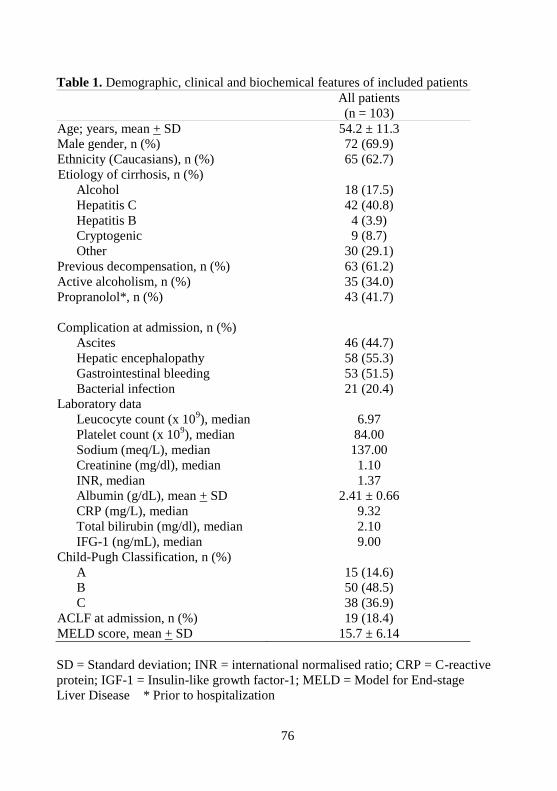
76
Table 1. Demographic, clinical and biochemical features of included patients
All patients
(n = 103)
Age; years, mean + SD 54.2 ± 11.3
Male gender, n (%) 72 (69.9)
Ethnicity (Caucasians), n (%) 65 (62.7)
Etiology of cirrhosis, n (%)
Alcohol 18 (17.5)
Hepatitis C 42 (40.8)
Hepatitis B 4 (3.9)
Cryptogenic 9 (8.7)
Other 30 (29.1)
Previous decompensation, n (%) 63 (61.2)
Active alcoholism, n (%) 35 (34.0)
Propranolol*, n (%) 43 (41.7)
Complication at admission, n (%)
Ascites 46 (44.7)
Hepatic encephalopathy 58 (55.3)
Gastrointestinal bleeding 53 (51.5)
Bacterial infection 21 (20.4)
Laboratory data
Leucocyte count (x 109), median 6.97
Platelet count (x 109), median 84.00
Sodium (meq/L), median 137.00
Creatinine (mg/dl), median 1.10
INR, median 1.37
Albumin (g/dL), mean + SD 2.41 ± 0.66
CRP (mg/L), median 9.32
Total bilirubin (mg/dl), median 2.10
IFG-1 (ng/mL), median 9.00
Child-Pugh Classification, n (%)
A 15 (14.6)
B 50 (48.5)
C 38 (36.9)
ACLF at admission, n (%) 19 (18.4)
MELD score, mean + SD 15.7 ± 6.14
SD = Standard deviation; INR = international normalised ratio; CRP = C-reactive
protein; IGF-1 = Insulin-like growth factor-1; MELD = Model for End-stage
Liver Disease * Prior to hospitalization
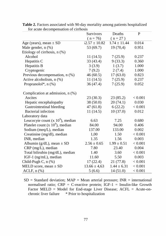
77
Table 2. Factors associated with 90-day mortality among patients hospitalized
for acute decompensation of cirrhosis
Survivors
( n = 76)
Deaths
( n = 27 )
P
Age (years), mean ± SD 52.57 ± 10.82 58.74 ± 11.44 0.014
Male gender, n (%) 53 (69.7) 19 (70,4) 0.951
Etiology of cirrhosis, n (%)
Alcohol 11 (14.5) 7 (25.9) 0.237
Hepatitis C 33 (43.4) 9 (33.3) 0.360
Hepatitis B 3 (3.9) 1 (3.7) 1.000
Cryptogenic 7 (9.2) 2 (7.4) 1.000
Previous decompensation, n (%) 46 (60.5) 17 (63.0) 0.823
Active alcoholism, n (%) 11 (14.5) 7 (25.9) 0.237
Propranolol*, n (%) 36 (47.4) 7 (25.9) 0.052
Complication at admission, n (%)
Ascites 23 (30.3) 23 (85.2) < 0.001
Hepatic encephalopathy 38 (50.0) 20 (74.1) 0.030
Gastrointestinal bleeding 47 (61.8) 6 (22.2) < 0.001
Bacterial infection 11 (14.5) 10 (37.0) 0.012
Laboratory data
Leucocyte count (x 109), median 6.63 7.25 0.680
Platelet count (x 109), median 84.00 94.00 0.406
Sodium (meq/L), median 137.00 133.00 0.002
Creatinine (mg/dl), median 1,00 1.50 < 0.001
INR, median 1.35 1.56 0.003
Albumin (g/dL), mean ± SD 2.56 ± 0.65 1.99 ± 0.51 < 0.001
CRP (mg/L), median 7.80 23.40 0.004
Total bilirubin (mg/dL), median 1.40 3.60 < 0.001
IGF-1 (ng/mL), median 11.60 5.50 0.003
Child-Pugh C, n (%) 17 (22.4) 21 (77.8) < 0.001
MELD score, mean ± SD 13.66 ± 4,63 21.44 ± 6.31 < 0.001
ACLF, n (%) 5 (6.6) 14 (51.0) < 0.001
SD = Standard deviation; MAP = Mean arterial pressure; INR = international
normalised ratio; CRP = C-reactive protein; IGF-1 = Insulin-like Growth
Factor MELD = Model for End-stage Liver Disease; ACFL = Acute-on-
chronic liver failure * Prior to hospitalization

78
Table 3. Stepwise forward logistic regression analysis of factors associated
with 90-day mortality (variables with P <0.010 in bivariate analysis were
included).
Factors Odds Ratio CI 95% P
MELD
Score 1.220 1.097 – 1.356 < 0.001
Ascites 10.706 2.526 –
45.384 0.001
IFG-1
levels 0.907 0.833 – 0.988 26
CI = Confidence interval; IGF-1 = Insulin-like Growth Factor 1; MELD =
Model for End-stage Liver Disease
Fig. 1. Cumulative 90-day survival of patients with cirrhosis according to
the IGF-1 categories. Survival was significantly lower in patients with
IGF-1 < 13 ng/mL as compared to those with values ≥ 13 ng/mL (P =
0.001, long-rank test).

79
Fig. 2. Box plot of IGF-1 according to the moment of evaluation
(inpatient or outpatient). The line across the box indicates the median
value; the box contains the 25% to 75% interquartile range; and the
whiskers represent the highest and the lower values. IGF-1 levels were
significantly higher at outpatient assessment (P < 0.001).

80
References
1. Schuppan D, Afdhal NH. Liver cirrhosis. Lancet 2008;371:838-851.
2. Durand F, Valla D. Assessment of prognosis of cirrhosis. Semin Liver
Dis 2008;28:110-122.
3. D'Amico G, Garcia-Tsao G, Pagliaro L. Natural history and
prognostic indicators of survival in cirrhosis: a systematic review of 118
studies. J Hepatol 2006;44:217-231.
4. Asrani SK, Kamath PS. Natural history of cirrhosis. Curr
Gastroenterol Rep 2013;15:308.
5. Huisman EJ, Trip EJ, Siersema PD, van Hoek B, van Erpecum KJ.
Protein energy malnutrition predicts complications in liver cirrhosis. Eur
J Gastroenterol Hepatol 2011;23:982-989.
6. Berzigotti A, Abraldes JG. Impact of obesity and insulin-resistance on
cirrhosis and portal hypertension. Gastroenterol Hepatol 2013;36:527-
533.
7. Luxon BA. Bone disorders in chronic liver diseases. Curr
Gastroenterol Rep 2011;13:40-48.
8. Perrini S, Laviola L, Carreira MC, Cignarelli A, Natalicchio A,
Giorgino F. The GH/IGF1 axis and signaling pathways in the muscle and
bone: mechanisms underlying age-related skeletal muscle wasting and
osteoporosis. J Endocrinol 2010;205:201-210.
9. Colakoglu O, Taskiran B, Colakoglu G, Kizildag S, Ari Ozcan F,
Unsal B. Serum insulin like growth factor-1 (IGF-1) and insulin like
growth factor binding protein-3 (IGFBP-3) levels in liver cirrhosis. Turk J
Gastroenterol 2007;18:245-249.
10. Juul A. Serum levels of insulin-like growth factor I and its binding
proteins in health and disease. Growth Horm IGF Res 2003;13:113-170.
11. Ferry RJ, Jr., Cerri RW, Cohen P. Insulin-like growth factor binding
proteins: new proteins, new functions. Horm Res 1999;51:53-67.
12. Hwa V, Oh Y, Rosenfeld RG. The insulin-like growth factor-binding
protein (IGFBP) superfamily. Endocr Rev 1999;20:761-787.
13. Wu YL, Ye J, Zhang S, Zhong J, Xi RP. Clinical significance of
serum IGF-I, IGF-II and IGFBP-3 in liver cirrhosis. World J
Gastroenterol 2004;10:2740-2743.
14. Ronsoni MF, Lazzarotto C, Fayad L, Silva MC, Nogueira CL, Bazzo
ML, et al. IGF-I and IGFBP-3 serum levels in patients hospitalized for
complications of liver cirrhosis. Ann Hepatol 2013;12:456-463.
81
15. Mazzaferro V, Regalia E, Doci R, et al. Liver transplantation for the
treatment of small hepatocellular carcinomas in patients with cirrhosis. N
Engl J Med 1996; 334:693.
16. Addolorato G, Leggio L, Ferrulli A, Cardone S, Vonghia L,
Mirijello A, et al. Effectiveness and safety of baclofen for maintenance
of alcohol abstinence in alcohol-dependent patients with liver cirrhosis:
randomised, double-blind controlled study. Lancet 2007;370:1915-
1922.
17. Garner JS, Jarvis WR, Emori TG, Horan TC, Hughes JM. CDC
definitions for nosocomial infections, 1988. Am J Infect Control
1988;16:128-140.
18. Runyon BA. Management of adult patients with ascites due to
cirrhosis: an update. Hepatology 2009;49:2087-2107.
19. Bajaj JS. Review article: the modern management of hepatic
encephalopathy. Aliment Pharmacol Ther 2010;31:537-547.
20. Angermayr B, Cejna M, Karnel F, Gschwantler M, Koenig F,
Pidlich J, et al. Child-Pugh versus MELD score in predicting survival in
patients undergoing transjugular intrahepatic portosystemic shunt. Gut
2003;52:879-885.
21. Kamath PS, Wiesner RH, Malinchoc M, Kremers W, Therneau
TM, Kosberg CL, et al. A model to predict survival in patients with
end-stage liver disease. Hepatology 2001;33:464-470.
22. Moreau R, Jalan R, Gines P, Pavesi M, Angeli P, Cordoba J, et al.
Acute-on-chronic liver failure is a distinct syndrome that develops in
patients with acute decompensation of cirrhosis. Gastroenterology
2013;144:1426-1437, 1437 e1421-1429.
23. Donaghy A, Ross R, Gimson A, Hughes SC, Holly J, Williams R.
Growth hormone, insulinlike growth factor-1, and insulinlike growth
factor binding proteins 1 and 3 in chronic liver disease. Hepatology
1995;21:680-688.
24. Assy N, Pruzansky Y, Gaitini D, Shen Orr Z, Hochberg Z, Baruch
Y. Growth hormone-stimulated IGF-1 generation in cirrhosis reflects
hepatocellular dysfunction. J Hepatol 2008;49:34-42.
25. Castro GR, Coelho JC, Parolin MB, Matias JE, de Freitas AC.
Insulin-like growth factor I correlates with MELD and returns to normal
level after liver transplantation. Ann Transplant 2013;18:57-62.
26. Weber MM, Auernhammer CJ, Lee PD, Engelhardt D, Zachoval
R. Insulin-like growth factors and insulin-like growth factor binding
proteins in adult patients with severe liver disease before and after
orthotopic liver transplantation. Horm Res 2002;57:105-112.

82
27. Bassanello M, De Palo EF, Lancerin F, Vitale A, Gatti R, Montin U,
et al. Growth hormone/insulin-like growth factor 1 axis recovery after
liver transplantation: a preliminary prospective study. Liver Transpl
2004;10:692-698.
28. Silva PE, Fayad L, Lazzarotto C, Ronsoni MF, Bazzo ML, Colombo
BS, et al. Single-centre validation of the EASL-CLIF Consortium
definition of acute-on-chronic liver failure and CLIF-SOFA for
prediction of mortality in cirrhosis. Liver Int 2014.
29. Kamath PS, Kim WR. The model for end-stage liver disease (MELD).
Hepatology 2007;45:797-805.
30. Moller S, Gronbaek M, Main K, Becker U, Skakkebaek NE. Urinary
growth hormone (U-GH) excretion and serum insulin-like growth
factor 1 (IGF-1) in patients with alcoholic cirrhosis. J Hepatol
1993;17:315-320.
31. Moller S, Becker U, Juul A, Skakkebaek NE, Christensen E.
Prognostic value of insulinlike growth factor I and its binding protein
in patients with alcohol-induced liver disease. EMALD group.
Hepatology 1996;23:1073-1078