“avaliaÇÃo das propriedades fÍsicas do...
TRANSCRIPT
UNIVERSIDADE ESTADUAL DE CAMPINAS
FACULDADE DE ODONTOLOGIA DE PIRACICABA
BRUNA GUERRA SILVA
“AVALIAÇÃO DAS PROPRIEDADES FÍSICAS DO ESMALTE,
QUANDO SUBMETIDO AO CLAREAMENTO DENTAL
CASEIRO COM DIFERENTES ESPESSANTES: ESTUDO IN
SITU”
"EVALUATION OF THE PHYSICAL PROPERTIES OF
ENAMEL, WHEN SUBMITTED TO AT-HOME BLEACHING
WITH DIFFERENT THICKENERS: AN IN SITU STUDY”
Piracicaba
2017
BRUNA GUERRA SILVA
“AVALIAÇÃO DAS PROPRIEDADES FÍSICAS DO ESMALTE, QUANDO
SUBMETIDO AO CLAREAMENTO DENTAL CASEIRO COM DIFERENTES
ESPESSANTES: ESTUDO IN SITU”
"EVALUATION OF THE PHYSICAL PROPERTIES OF ENAMEL SUBMITTED TO
AT-HOME BLEACHING WITH DIFFERENT THICKENERS: AN IN SITU STUDY”
Dissertação apresentada à Faculdade de
Odontologia de Piracicaba da
Universidade Estadual de Campinas como
parte dos requisitos exigidos para
obtenção do título de Mestra em Clínica
Odontológica, na Área de Dentística.
Dissertation presented to the Piracicaba
Dental School of the University of
Campinas in partial fulfillment of the
requirements for the degree of Master in
Dental Clinic in Operative Dentistry Area.
Orientadora: Profa. Dra. Débora Alves Nunes Leite Lima
ESTE EXEMPLAR CORRESPONDE À
VERSÃO FINAL DA DISSERTAÇÃO
DEFENDIDA PELA ALUNA BRUNA
GUERRA SILVA E ORIENTADA PELA
PROFA. DRA. DÉBORA ALVES NUNES
LEITE LIMA.
Piracicaba
2017

DEDICATÓRIA
À minha mãe, Elizabeth Guerra, por estar sempre presente em todos os momentos da minha
vida, por sonhar os meus sonhos e tornar possível à realização de todos eles, pelo carinho e
amor com suas filhas, pela compreensão e amizade. Por ser meu porto seguro, minha
inspiração. Sem a senhora não seria possível chegar aonde cheguei e conquistar tudo que já
conquistei até hoje. Meu amor pela senhora não tem dimensão!
Ao meu pai, Vicente Silva, que por mais distante sempre esteve a postos para me ajudar,
sempre me tirou boas risadas e que me faz compreender que não importa a distancia o amor
sempre nos traz para perto um do outro.
Ao meu padrasto, Sérgio de Siqueira, por toda a dedicação à família que formamos. Por todo
carinho, amor, por trazer tanta alegria para nossa família e por estar presente em todos os
momentos da minha vida.

Agradecimentos
À Deus...
...meu guia
Pela ajuda e força em todos os momentos da minha vida.
Agradecimentos
À minha orientadora
Professora Débora Alves Nunes Leite Lima, pela oportunidade de orientação. Pelo convívio e
aprendizado, pelos ensinamentos diários, por acreditar e confiar no meu trabalho e na minha
pessoa. O nosso convívio me trouxe uma enorme admiração por você e agregou muitas coisas
em minha vida, não só profissional como também pessoal. Agradeço pelas inúmeras vezes
que você foi como uma mãe, me aconselhando e me guiando! Você é incrível!

Agradecimentos
À minha família...
As minhas fantásticas irmãs Aline e Camila, por me apoiarem durante todos os anos, sempre
me impulsionando a seguir em frente e a conquistar os meus sonhos. Por todo amor, carinho e
amizade. Amo vocês e sou eternamente grata a Deus por me conceder vocês. Vocês são
minha vida.
Aos meus avós Lucia e Guerrinha, por todos os exemplos de vida que me passaram, por todo
amor e carinho e por compreenderem minha ausência frente aos percalços da vida. Vocês são
especiais e estarão sempre em meus pensamentos e em meu coração.
Agradecimentos
Às minhas amigas...
Queridas amigas: Marília, Heloisa e Marina, que com certeza serão amigas para a vida toda.
A amizade de vocês conforta meu coração e torna os fardos mais leves. Cada uma de vocês
me completa de forma especial e única.
À Ana Eliza Faria. Você foi peça fundamental em minha vida durante os cinco anos que
moramos juntas. Queria eu poder ser um pedacinho de você, que é cheia de qualidades e que
eu me espelho e tenho tanto orgulho de chamar de amiga. Agradeço por me permitir entrar na
sua vida, por compartilhá-la comigo e por sempre oferecer seu ombro amigo e me confortar
com suas sábias palavras. Por me trazer o chão quando ele se vai e por sempre me tirar um
sorriso quando não estou bem. Você foi meu alento, meu suporte. E você na verdade não é só
amiga, você é irmã e você se tornou parte da minha família!
À Renata Pereira, você que tem um coração maior que o mundo, um caráter e uma
inteligência admirável, que sempre compartilha dos meus pensamentos, que nunca coloca
obstáculos para me ajudar quando preciso e que sempre estende as mãos para mim. Você
tornou esses dois anos de Pós-Graduação muito mais leves. Obrigada por estar comigo em
todos os momentos. Sem palavras para agradecer à sua amizade e ao seu carinho.
À Luisa, minha amiga desde a graduação. Sem palavras para expressar o quando sua amizade
é importante para mim. Compartilhamos descobertas, novas experiências, medos, alegrias,
conhecimento, anseios e conquistas. Você é uma amiga incrível e mesmo com a distância
você se faz sempre presente. Minha saudade diária.
Aos meus amigos de pós-graduação...
À Thayla, por me conduzir durante o Mestrado. Você foi como um anjo nesses dois anos,
sempre me guiando, me orientando e me acrescentando conhecimento e experiências.
Obrigada por dedicar tanto do seu tempo à mim, e por sempre me fazer acreditar que sou
capaz. Torço muito por você!
Aos amigos da turma do mestrado, pela convivência durante esses anos, pela cumplicidade,
partilha de sonhos, anseios e conquistas. Cada um, com seu jeitinho especial, me
conquistaram e serão sempre lembrados com muito carinho.

Ao Waldemir e Jéssica por sempre estares dispostos a me ajudar quando precisei. Sem
palavras para agradecê-los.
Aos meus amigos...
... voluntários de pesquisa
Aos amigos que se dispuseram a participar da minha pesquisa, com o único objetivo de
enriquecer o meu trabalho. Sem palavras para agradecer por terem me ajudado, pela paciência
com o meu estudo e por se empenharem para que ele desse certo. Muito obrigada!

Agradecimentos
Ao meu namorado...
A você, Rafael, pelo companheirismo, pela nossa amizade, pelo apoio incondicional, pela
cumplicidade, atenção e pelo amor que tem por mim. Você, que chegou quando eu menos
esperava, que em tão pouco tempo me trouxe tanta felicidade e que me transborda. Você que
é calmaria em meio à tempestade. Sou muito grata por me permitir caminhar ao seu lado e por
ser tão presente em minha vida. É muito bom ter alguém para compartilhar os sonhos, para
planejar um futuro e para amar!
Agradecimentos
Aos mestres...
À minha co-orientadora de iniciação científica, Vanessa Cavalli Gobbo, que me inspirou com
sua inteligência, paciência, dedicação e amor pelo que faz, a iniciar a Pós-graduação. A você,
toda minha admiração e respeito.
Ao professor Marcelo Gianinni pela oportunidade de trabalhar com você e pela experiência
adquirida durante a iniciação científica.
Aos professores Luiz Alexandre Maffei Sartini Paulillo, Giselle Maria Marchi e Flávio
Henrique Baggio Aguiar, pelo conhecimento agregado ao meu trabalho no exame de
qualificação.
À todos os docentes do Programa de Pós-Graduação em Clínica Odontológica da FOP-
UNICAMP.
À professora Gláucia Maria Bovi Ambrosano, pelos conhecimentos de estatística
compartilhados.
Agradecimentos
À Faculdade de Odontologia de Piracicaba...
À Faculdade de Odontologia de Piracicaba da Universidade Estadual de Campinas, na pessoa
de seu Diretor Prof. Dr. Guilherme Elias Pessanha Henriques.
À Coordenadoria Geral da Pós-Graduação da FOP/UNICAMP.
RESUMO
O objetivo deste estudo foi avaliar in situ a influência do peróxido de carbamida
10%(PC) contendo diferentes espessantes (carbopol, carbopol+glicerina e natrosol) sobre as
características físicas do esmalte dental, como: cor, microdureza e rugosidade. Oitenta e
quatro blocos de esmalte e dentina (4 × 4 × 2,5 mm) obtidos a partir de dentes bovinos foram
manchados com chá preto, divididos em sete grupos (n=12) e fixados em dispositivos intra-
bucais palatinos utilizados por 12 voluntários. Os espécimes foram submetidos a diferentes
tratamentos: PC+carbopol (FGM), PC+carbopol+glicerina (Ultradent), PC+natrosol (Gel
experimental), apenas com o gel de carbopol, apenas com o gel de glicerina+carbopol, apenas
com gel de natrosol e sem nenhum tratamento (grupo controle). As propriedades físicas
testadas foram cor (ΔE), rugosidade média (Ra) e microdureza Knoop (KHN). A morfologia
da superfície de esmalte foi avaliada por microscopia de força atômica (AFM). Os dados
foram submetidos à análise de variância (ANOVA) em esquema fatorial com tratamento
adicional (2 x 3 +1), teste de Tukey e teste Dunnet. Como todos os pacientes receberam todos
os tratamentos, o efeito “voluntário” foi inserido na análise de variância. Foi considerado o
nível de significância de 5%. Entre as propriedades físicas avaliadas, quando avaliada a
alteração de cor, todos os grupos apresentaram alteração, exceto o grupo não clareado com
espessante carbopol, o qual foi semelhante ao grupo controle. Os grupos tratados com o
espessante natrosol tiveram uma maior alteração na cor, apresentando-se mais claro que os
demais. Com relação à microdureza, houve diminuição da mesma para os grupos tratados
apenas com o gel de carbopol e com o gel de carbopol+glicerina, os quais diferiram do grupo
controle. Para a rugosidade, houve aumento em todos os grupos clareados quando comparado
com o grupo controle, porém o grupo PC+natrosol apresentou menor aumento. Dessa forma,
pode-se concluir que a utilização de géis de clareadores contendo espessante natrosol
promoveu menores alterações no esmalte do que géis clareadores com espessante carbopol e
carbopol+glicerina, além de permitir um clareamento eficaz.
Palavras chave: Estética. Clareamento dental. Espessante. Carbopol. Natrosol.
ABSTRACT
The objective of this study was to evaluate in situ the influence of carbamide peroxide 10%
(PC) containing different thickeners on the physical characteristics of dental enamel, such as:
color, microhardness and enamel roughness. Eighty-four blocks of enamel and dentin (4 × 4 ×
2.5 mm) obtained from bovine teeth were divided into seven groups (n = 12) fixed in palatal
intraoral devices used by 12 volunteers. The specimens were submitted to different
treatments: PC + carbopol (FGM), PC + carbopol + glycerin (Ultradent), PC + natrosol
(experimental gel), with carbopol gel only, with glycerin + carbopol gel, only with Natrosol
gel and without any treatment (control group). The physical properties tested were color (ΔE),
roughness (Ra) and Knoop microhardness (KHN). The morphology of the enamel surface was
evaluated with atomic force microscopy (AFM). The data were submitted to analysis of
variance (ANOVA) in a factorial scheme with additional treatment (2 x 3 +1), Tukey's test
and Dunnet test. As all patients received all treatments, the "voluntary" effect was inserted in
the analysis of variance. The level of significance was set at 5%. Among the physical
properties evaluated, when the color change was evaluated, all the groups presented alteration,
except the group treated only with carbopol thickener, which was similar to the control group.
The groups treated with the thickener natrosol had a greater color change, being clearer than
the others. Regarding the microhardness, there was a decrease in the same for the groups
treated with carbopol gel and carbopol + glycerin gel, which differed from the control group.
For roughness, there was an increase in all bleached groups when compared to the control
group, but the PC + natrosol group presented a smaller increase. In this way it can be
concluded that the use of bleaching gels containing natrosol thickener was less deleterious to
the enamel than bleaching gels with carbopol thickeners and carbopol + glycerin, in addition
to allowing an effective bleaching.
Keywords: Aesthetics. Dental bleaching. Thickener. Carbopol. Natrosol.
Sumário
1. INTRODUÇÃO……………………………………………………………………..……..17
2. ARTIGO……………………………………………………………………………………20
3. CONCLUSÃO……………………………………………………………………………..34
REFERÊNCIAS*……………………………………………………………………………..35
APÊNDICE…………………………………………………………………………………...38
ANEXO 1 …………………………………………………………………………………….50
ANEXO 2……………………………………………………………………………………..51
17
1.INTRODUÇÃO
O desenvolvimento de novos produtos clareadores e o aperfeiçoamento
das técnicas de clareamento permitiram que o tratamento clareador se tornasse
comprovadamente eficaz (Nutter et al., 2013; Joiner A., 2006). O tratamento clareador
consiste em tornar os dentes mais claros, podendo se dar a partir da técnica de
clareamento de consultório, com géis de alta concentração de peróxido de hidrogênio,
na qual o Cirurgião-dentista realiza o procedimento no seu consultório, ou de
clareamento caseiro, com géis de baixa concentração de peróxido de hidrogênio, na qual
o paciente utiliza uma moldeira personalizada e pode realizar o procedimento em casa
sob a orientação do Cirurgião-dentista. Porém, alguns estudos indicam que o
clareamento pode causar alterações nos tecidos dentais, ), o que o torna relativamente
seguro (Marson FC et al., 2015; Tredwin CJ et al., 2006), como: alteração na superfície
do esmalte, podendo ter aumento da rugosidade e diminuição da microdureza
(Zeczkowski M et al., 2015; Sa Y et al., 2013; Basting RT et al., 2005; Potocnik I et al.,
2000; Bistey T et al., 2007) e da dentina (Jiang T et al., 2007; Kawamoto e Tsujimoto,
2004; Chng et al., 2005).
O mecanismo de ação dos géis clareadores ainda não é um consenso na
literatura; porém, a teoria dominante é a teoria da oxidação das moléculas cromógenas,
que se dá através de um radical livre de oxigênio (Eimar et al, 2012), que é liberado na
dissociação do peróxido de hidrogênio. O peróxido de hidrogênio está presente tanto no
gel clareador de consultório quanto no caseiro. Já que, mesmo o gel de clareamento
caseiro sendo à base de peróxido de carbamida, quando em contato com os tecidos
moles ou saliva em temperatura bucal, dissocia-se e um dos produtos da sua dissociação
é o peróxido de hidrogênio (Ben-Amar A et al., 1995).
O peróxido de hidrogênio é capaz de penetrar profundamente nos primas de
esmalte, alcançando a dentina devido ao seu baixo peso molecular (Lima, DA et al.,
2011), onde se degrada liberando radicais livres de oxigênio (Haywood & Heymann,
1991) que interage com os pigmentos que causam a coloração no dente (Kawamoto &
Tsujimoto, 2004). Esses pigmentos são cadeias de alto peso molecular, que através de
reações de oxirredução são quebradas, decompondo-se em moléculas menores e menos
pigmentadas (Plotino et al., 2008; Toledano et al., 2011). A alteração no tamanho e
18
pigmentação das moléculas cromógenas após a oxidação produzida pelos radicais livres
altera a absorção de luz do espectro visível pela molécula, de maior para menor
comprimento de onda, resultando em compostos mais claros (Haywood & Heymann,
1991; So Ran Kwon & Philip W. Wertz, 2015, Sulieman M 2004)
As alterações de microdureza do esmalte e da dentina são provocadas
devido à reação dos radicais livres de peróxido de hidrogênio, que por não serem
específicos e serem altamente instáveis, podem reagir não só com as moléculas
cromógenas dos pigmentos, os anéis de carbono pigmentados, como também com as
moléculas orgânicas dos tecidos dentais, para atingir estabilidade (Basting et al., 2005;
Kawamoto & Tsujimoto, 2004). Contudo, estudos vêm mostrando que as alterações na
microdureza do esmalte e da dentina podem não estar relacionadas somente aos radicais
livres de peróxido, mas com outros constituintes presentes na formulação do produto
clareador, como os espessantes (Basting et al., 2003).
Os espessantes dos géis clareadores são substâncias químicas que têm como
função aumentar sua viscosidade, permitindo assim uma maior retenção do gel na
superfície dentária e com isso um tempo prolongado para a liberação de íons de
oxigênio (McCracken e Haywood, 1996; Rodrigues JA et al., 2007). Um dos
espessantes mais comumente utilizado na composição dos géis clareadores caseiros é o
carbopol, um polímero carboxipolimetileno, iônico, de natureza ácida, derivado de um
ácido carboxílico. Porém, tem sido relatado que o carbopol pode ser capaz de reduzir a
microdureza do esmalte e da dentina, devido a sua elevada capacidade de se ligar ao
cálcio (van der Reijden et al., 1997), que leva a uma inibição da incorporação dos
cristais de hidroxiapatita.
Além do carbopol, outro espessante utilizado é a glicerina, que, no entanto,
devido à capacidade de atuar como barreira para adsorção de saliva e consequente
remineralização do esmalte, pode também diminuir a microdureza do esmalte (Basting
RT et al, 2005).
Sendo assim, esse estudo se propôs a substituir os espessantes presentes no
mercado por um espessante bastante utilizado na indústria farmacêutica e cosmética,
denominado natrosol (hidroxietil celulose), que apresenta características opostas ao
carbopol, principalmente por ser não iônico; além disso, o natrosol pode ser utilizado
com substâncias ácidas, como o gel clareador, por apresentar estabilidade na variação de
19
pH (Gouveia TH et. al., 2016). Estudos que avaliem os efeitos dos géis clareadores
sobre a estrutura dental são extremamente necessários, visto que, apesar de ser um
tratamento conservador, o clareamento pode causar danos na estrutura dental. Assim, o
objetivo desse estudo in situ foi avaliar o efeito de géis clareadores contendo diferentes
espessantes nas propriedades físicas do esmalte dental, como cor, microdureza e
rugosidade superficial.

20
2. ARTIGO
"Evaluation of the physical properties of enamel submitted to at-home bleaching
with different thickeners: an in situ study”
ABSTRACT
Objective: of this in situ study was to evaluate the influence of carbamide
peroxide 10% (CP) in combination with different thickeners, on the physical
characteristics of dental enamel. Methodology: Eighty-four blocks of enamel and
dentin (4 × 4 × 2.5 mm) obtained from bovine teeth were divided into seven groups (n =
12), fixed in palatal intraoral devices, and submitted to different treatments: CP +
carbopol (FGM), CP + carbopol + glycerin (Ultradent), CP + natrosol (experimental
gel), carbopol gel, carbopol +glycerin gel, Natrosol gel, whitout treatment (control
group). The physical properties tested were color (ΔE), average roughness (Ra) and
Knoop microhardness (KHN). Additionally, the enamel surface was evaluated by
atomic force microscopy (AFM). The data were submitted to analysis of variance
(ANOVA), Tukey's test and Dunnet test (α=0.05). Results: All groups presented
significant color variation, except the not bleached group with carbopol thickener. The
groups treated with the natrosol and carbopol+glycerin thickener had a satisfactory
color change, being clearer than the others. Groups not bleached with carbopol and
carbopol + glycerin presented decreased values of Microhardness when compared to
control group. Roughness was increased in all bleached groups when compared to the
control group, but the CP +natrosol group had a statistical increase lower than
PC+carbopol. Conclusion: the use of bleaching gel with natrosol thickener allowed
effective bleaching and was less deleterious to enamel. Clinical significance: enamel
physical characteristics alteration during at-home bleaching with differents thickners.
INTRODUCTION
The development of new products and the improvement of whitening
techniques have promoted the effectiveness of bleaching treatments [1,2]. However,
21
some studies report that bleaching treatment may cause alterations in dental tissues
which make it relatively safety [3,4], for instance changes in enamel surface involving
reduction of microhardness and increased roughness [5-9].
The mechanism of action of bleaching gels relies on oxygen free radicals
[10] released from the decomposition of hydrogen peroxide. Hydrogen peroxide is
present in the office bleaching gel and in at-home bleaching gels. Although some home-
bleaching gel are based on carbamide peroxide, when in contact with saliva at mouth
temperature, the carbamide peroxide dissociates and one of the products of its
dissociation is hydrogen peroxide [11].
The free radicals responsible for the whitening effect are also accountable
for the changes in dental physical properties, such as reduction of enamel
microhardness. Because of their highly unstable nature and non-specific reactivity, they
can interact not only with chromogenic molecules, but also with other organic and
inorganic molecules of dental tissues, to achieve stability [7,12]. The acidic pH of
bleaching gels can also cause changes in tooth structure [13]. Nevertheless, studies have
shown that in addition to free radicals and pH, changes in tooth structure may also be
related to other constituents present in the bleaching product formulation, such as
thickeners [14].
Thickeners are chemical substances that have the function of increasing the
viscosity of bleaching gels, thus allowing a greater retention on the dental surface and
prolonging the exposure time to active compounds [15,16]. The most commonly used
thickeners in the composition of at-home bleaching gels are carbopol and glycerin.
However, these thickeners are capable of interacting with the dental structure, leading to
structural changes such as reduced microhardness and altered roughness [7]. In this
context, the present study evaluates currently marketed thickeners in bleaching gels in
comparison to Natrosol, a thickener widely used in the pharmaceutical and cosmetic
industry. Natrosol has the advantages of being non-ionic and having wide-range pH
stability (2.0-12.0) [17], which means it can be combined to acidic substances like
bleaching formulations. As this thickener is less viscous than carbopol, gels containing
natrosol were manipulated with addition of silica-based filler particles (SiO) to increase
the viscosity of the material, as well as to improve the thermal resistance, since
22
according to the manufacturer these products are to be kept refrigerated and will
experience frequent temperature changes with the cycles of use.
The present in situ study investigated the effects of an experimental
Natrosol-containing bleaching gel on the properties and morphology of enamel,
evaluating its color, microhardness and surface roughness. The hypotheses tested were:
1) that the experimental bleaching gel with natrosol thickener would show the same
efficacy of the commercial bleaching agents with carbopol and glycerin, and 2) that the
experimental bleaching gel would not change the microhardness and roughness of the
enamel.
MATERIALS AND METHODS
This study was approved by the Institutional Ethics Committee
(53165116.1.0000.5418)
Sample Preparation
Newly collected bovine incisors that did not have stains or enamel fractures
were selected and stored in 0.1% Thymol at 4° C until use. Enamel/dentin blocks
measuring 4 x 4 x 2.5 mm, with 1 mm of enamel and 1.5 mm of dentin were obtained
from the middle third of the vestibular region of the crown, using a diamond disk (Extec
Dia. Wafer Blade 102 x 0.3 x 12.7 mm) coupled to a metallographic cutter (Isomet
1000, Buehler Ltda. Lake Buff, IL, USA). For grinding, regularization and polishing,
decanting silicon carbide sanding discs were used (# 1200, # 2500 and # p4000 -
Isomet, Buehler Ltd, Lake Bluff, IL, USA) to level the height of the blocks; then felts
with metallographic diamond pastes and specific lubricant (6 μm - TOP, 3 μm - RAM, 1
μm - SUPRA - Arotec, Cotia, São Paulo, SP, Brazil) were used for polishing. Between
polishing steps and at the end of the procedure, all samples were placed for 10 minutes
on ultrasound (Marconi, Piracicaba, São Paulo, Brazil) to remove residual smear
particles. Each specimen was labeled on the side face with diamond drill bit # 1012 (KG
Sorensen), to standardize sample position for spectrophotometry. The samples were
stained for 6 days by immersion in black tea solution (Lion Junior SA, Curitiba, PR,
Brazil), which was changed every 24 hours. The stain was stabilized by placing the
specimens in artificial saliva, exchanged every 24 hours, for 14 days. The specimens
were sterilized with ethylene oxide and stored in water at 4 ° C until use.
23
In situ settings
Twelve volunteers (six males and six females), aged 24-28 years,
participated in the study after signing a free and informed consent form. All of these
volunteers were selected by the inclusion criteriA (normal salivary flow, measured
through sialometry, and absence of caries and/or periodontal disease), without violating
the exclusion criterion (use of medicine that interfere with salivary flow, smoking
habits, and the presence of orthodontic device, fixed or removable prosthesis). The use
of fluoride by the volunteers was not permitted during the whole timespan of the study.
For each volunteer, a molding in alginate (Hydrogum - Zhermack, Badia Polesine, Italy)
of the upper arch was obtained, from which the palatal devices were made of acrylic
resin containing 7 reservoirs (25 mm2 wide and 3 mm deep). Seven specimens
(enamel/dentin blocks) were attached to each palatal device using sticky wax. The
specimens were randomly placed in the twelve palatal intraoral devices, taking into
account the position relative to the parotid salivary gland. The intraoral devices
remained for one day in the volunteers’ mouths before the beginning of the bleaching
sessions, for the formation of acquired film.
Treatment / Gel Treatment Application
A total of 14 daily treatment sessions were carried out as follows: devices
were removed from the volunteers’ mouths and dried with absorbent paper; six of the
seven specimens contained in each of the twelve devices were submitted to gel
application (according to the assigned experimental group), whereas the seventh, control
specimens did not receive any treatment. The volunteers were instructed about gels
handling and application, which were applied according to the manufacturer's
recommendations on the enamel surface, remaining for 4 hours at room temperature. To
prevent dehydration of specimens, devices were kept in a humidified environment,
however carefully avoiding contact of the gel-containing surfaces with water. After each
session, the gel was removed with cotton-tipped flexible rods, specimens were washed
with water, dried with absorbent paper, and the devices returned to the volunteers’
mouth.
Color Analysis

24
The color reading of each specimen was performed in ambient light
condition (GTI MiniMatcher MM 1, GTI Technology, New York, NY, USA) using a
spectrophotometer (CM 700D, Minolta, Osaka, Japan). The spectral distribution was
measured based on the CIE system L * a * b *, used in Color software (Konica
Minolta), in which the L * axis represents the brightness intensity of the specimen and
ranges from 0 (black) to 100 (white); the coordinate a * represents the intensity of green
/ red color; and the coordinate b * represents the intensity of blue / yellow color. Prior to
measurements, the spectrophotometer was calibrated using white and black reflectance
standards, according to the manufacturer's protocol.
The difference between initial (baseline) and final (24 hours after
completion of the treatment) L *, a *, and b * values were obtained (ΔL, Δa, and Δb)
and any color change was calculated using the following equation: ΔE = [(ΔL *) 2 +
(Δa *) 2 + (Δb *) 2] 1/2.
Microhardness Analysis (KHN)
The enamel surface microhardness (KHN) was analyzed using a Knoop
penetrator with a 50 g load and a 5-second time in a microhardness tester (HMV-2000,
Shimadzu, Tokyo, Japan). After the treatment, five penetrations were made in each
specimen at 100 μm each, and the mean was calculated to determine the Knoop
hardness value (KHN). The post-treatment surface microhardness values of
experimental groups were compared to each other and to the control group.
Roughness Analysis (Ra)
Surface roughness (Ra) was analyzed using a perfilometer (Surf-Corder
1700, Kosaka, Tokyo, Japan) at two time-points: initial (baseline) and 24 hours after
completion of treatment. Three different and equidistant directions were measured on
the surface of each sample, with a cut-off point of 0.25 mm, a reading length of 1.25
mm and a velocity of 0.1 mm / s. Results are expressed as the difference between the
two time-points (ΔRa).
Atomic Force Microscopy (AFM)
For quantitative analysis of the surface, two samples from each group were
randomly chosen and analyzed by AFM after the treatment. Three-dimensional
25
topographic images (20 μm x 20 μm) were obtained using an atomic force microscope
(MultiMode 3; Digital Instruments Bruker, Santa Barbara, CA, USA) with a rectangular
silicon cantilever, a constant of approximately 70 N / m, and resonance frequency of
320 KHz. The microscope was operated in intermittent contact mode (touch mode),
with a scanning frequency of 0.7 Hz per line and a 2 micron scale. The images were
processed using Nanoscope III 5.12b43 software.
Statistical Analyses
After exploratory analysis, the data were submitted to analysis of variance
(ANOVA) in a factorial scheme with additional treatment (2 x 3 +1), and Tukey test.
The comparisons with the control group were performed by the Dunnet test. Because all
volunteers received all treatments, the "volunteer effect" was accounted for during these
analyses. The level of significance was set at 5%.
RESULTS
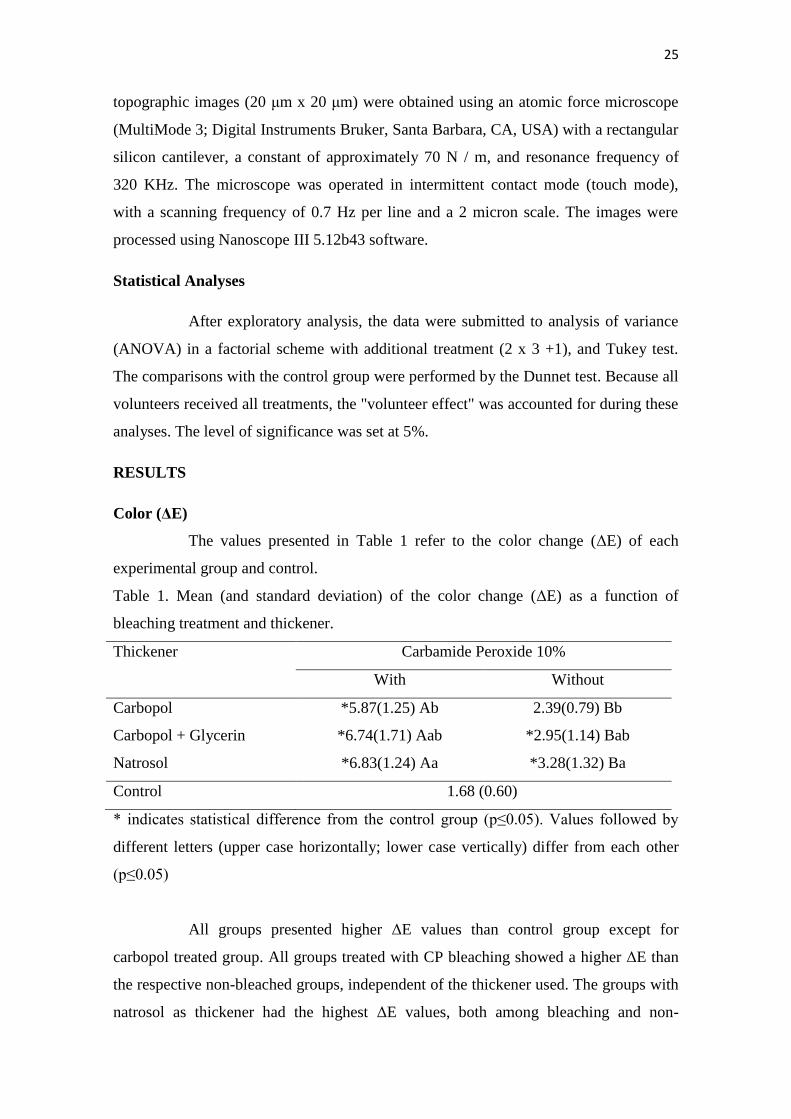
Color (ΔE)
The values presented in Table 1 refer to the color change (ΔE) of each
experimental group and control.
Table 1. Mean (and standard deviation) of the color change (ΔE) as a function of
bleaching treatment and thickener.
Thickener Carbamide Peroxide 10%
With Without
Carbopol *5.87(1.25) Ab 2.39(0.79) Bb
Carbopol + Glycerin *6.74(1.71) Aab *2.95(1.14) Bab
Natrosol *6.83(1.24) Aa *3.28(1.32) Ba
Control 1.68 (0.60)
* indicates statistical difference from the control group (p≤0.05). Values followed by
different letters (upper case horizontally; lower case vertically) differ from each other
(p≤0.05)
All groups presented higher ΔE values than control group except for
carbopol treated group. All groups treated with CP bleaching showed a higher ΔE than
the respective non-bleached groups, independent of the thickener used. The groups with
natrosol as thickener had the highest ΔE values, both among bleaching and non-
26
bleaching treatments, while those with carbopol thickener presented the lowest ΔE
values; differences between natrosol and carbopol groups were statistically relevant.
Groups treated with carbopol + glycerin gel (both in the presence or absence of CP)
yielded results which were intermediate, being statistically equivalent to both natrosol-
and carbopol-treated groups.
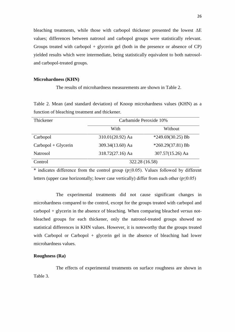
Microhardness (KHN)
The results of microhardness measurements are shown in Table 2.
Table 2. Mean (and standard deviation) of Knoop microhardness values (KHN) as a
function of bleaching treatment and thickener.
Thickener Carbamide Peroxide 10%
With Without
Carbopol 310.01(20.92) Aa *249.69(30.25) Bb
Carbopol + Glycerin 309.34(13.60) Aa *260.29(37.81) Bb
Natrosol 318.72(27.16) Aa 307.57(15.26) Aa
Control 322.28 (16.58)
* indicates difference from the control group (p≤0.05). Values followed by different
letters (upper case horizontally; lower case vertically) differ from each other (p≤0.05)
The experimental treatments did not cause significant changes in
microhardness compared to the control, except for the groups treated with carbopol and
carbopol + glycerin in the absence of bleaching. When comparing bleached versus not-
bleached groups for each thickener, only the natrosol-treated groups showed no
statistical differences in KHN values. However, it is noteworthy that the groups treated
with Carbopol or Carbopol + glycerin gel in the absence of bleaching had lower
microhardness values.
Roughness (Ra)
The effects of experimental treatments on surface roughness are shown in
Table 3.
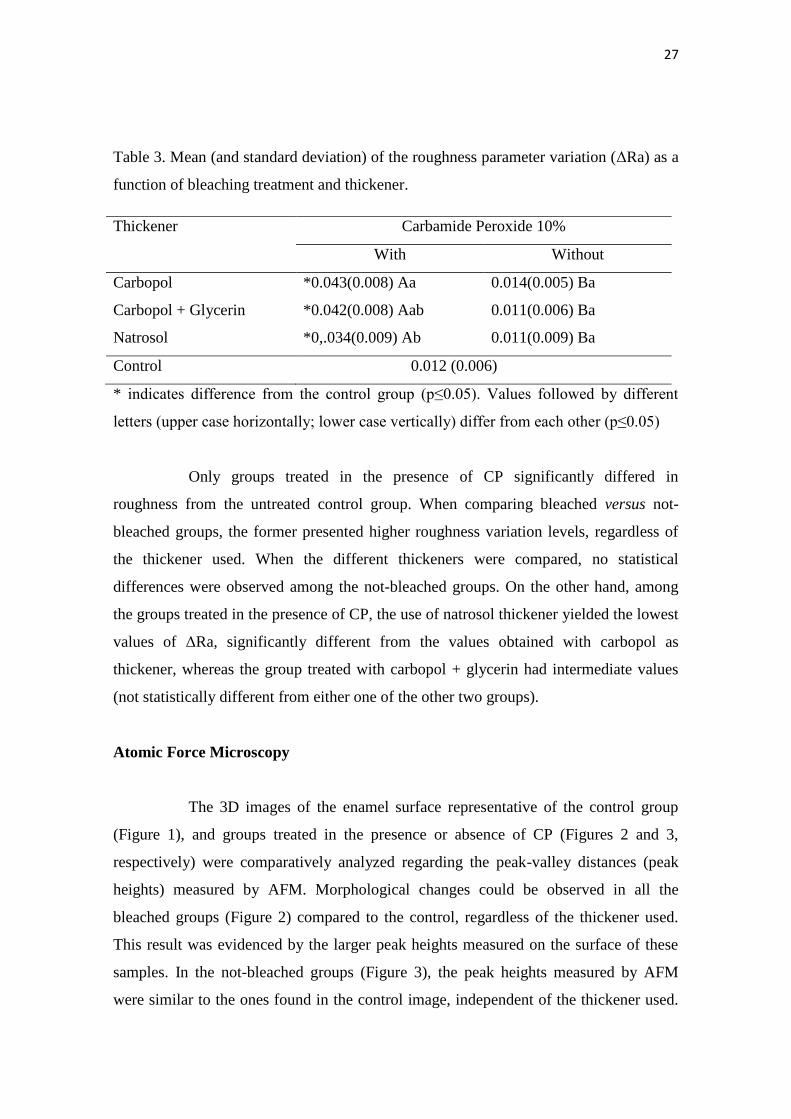
27
Table 3. Mean (and standard deviation) of the roughness parameter variation (ΔRa) as a
function of bleaching treatment and thickener.
Thickener Carbamide Peroxide 10%
With Without
Carbopol *0.043(0.008) Aa 0.014(0.005) Ba
Carbopol + Glycerin *0.042(0.008) Aab 0.011(0.006) Ba
Natrosol *0,.034(0.009) Ab 0.011(0.009) Ba
Control 0.012 (0.006)
* indicates difference from the control group (p≤0.05). Values followed by different
letters (upper case horizontally; lower case vertically) differ from each other (p≤0.05)
Only groups treated in the presence of CP significantly differed in
roughness from the untreated control group. When comparing bleached versus not-
bleached groups, the former presented higher roughness variation levels, regardless of
the thickener used. When the different thickeners were compared, no statistical
differences were observed among the not-bleached groups. On the other hand, among
the groups treated in the presence of CP, the use of natrosol thickener yielded the lowest
values of ΔRa, significantly different from the values obtained with carbopol as
thickener, whereas the group treated with carbopol + glycerin had intermediate values
(not statistically different from either one of the other two groups).
Atomic Force Microscopy
The 3D images of the enamel surface representative of the control group
(Figure 1), and groups treated in the presence or absence of CP (Figures 2 and 3,
respectively) were comparatively analyzed regarding the peak-valley distances (peak
heights) measured by AFM. Morphological changes could be observed in all the
bleached groups (Figure 2) compared to the control, regardless of the thickener used.
This result was evidenced by the larger peak heights measured on the surface of these
samples. In the not-bleached groups (Figure 3), the peak heights measured by AFM
were similar to the ones found in the control image, independent of the thickener used.
28
Importantly, within the bleached specimens, the group treated with natrosol as a
thickener (Figure 2.C) presented a less pronounced surface alteration when compared
with the group treated with carbopol (Figure 2.A), as demonstrated by the smaller
difference in peak-valley measurements on the enamel surface. The group bleached
using carbopol + glycerin as thickener (Figure 2.B) presented intermediate
morphological changes.

29
(A) (B) (C)
Figure 1: Image obtained by atomic force microscopy representative of the untreated control group
Figure 2: Images obtained by atomic force microscopy representing the groups treated in the presence of bleaching and different thickeners A)
CP + carbopol, B) CP + carbopol + glycerin, and C) CP + natrosol
(C) (A) (B)
Figure 3: Images obtained by atomic force microscopy representing groups treated in the absence of bleaching A) carbopol, B) carbopol +
glycerin, and C) natrosol
30
DISCUSSION
The results presented in this study allow a comparative assessment of different
thickeners in the formulation of gels for at-home bleaching treatment. The color variation of
specimens was evaluated in accordance with the CIELab system, which measures color
variation (ΔE). When ΔE = 0-2, the color variation is not clinically noticeable; ΔE = 2-3 color
variation is slightly noticeable; ΔE> 3-8 color variation is moderately noticeable and ΔE> 8,
color variation is highly noticeable [18]. The data showed that, whereas the control group
presented a clinically unnoticeable color variation, all groups treated with natrosol as a
thickener presented color variation classified as mildly detectable (ΔE>3). However, for
groups treated with carbopol and carbopol + glycerin in the absence of bleaching the variation
was less noticeable. Color analyses were performed before the beginning of treatment and 24
hours after the process was completed, always with hydrated samples to avoid interference. It
stands out that, in addition to the bleaching activity of CP in whitening gels via interaction of
oxygen free radicals [10] with chromogenic molecules on the tooth structure, the natrosol and
glycerin seem to have also interacted with the enamel surface, possibly removing pigments
and thereby justifying the increased color change effect when compared with carbopol-only
thickener. However, such hypothesis demands further studies in order to be properly verified
and better understood.
Natrosol (Hidroxietilcelulose) is a cellulose-based polymer, highly used in a
variety of materials such as latex emulsions, water-soluble resins, surface-active agents and
detergents, anti-caking agents, plasticizers agents and organic solvents. It has an excellent
compatibility with different types of composites and differs from carbopol mainly due to its
non-ionic nature [17]. In addition, natrosol can be associated to acidic substances in whitening
gels, due to its remarkable stability to pH variation (2,0-12,0). Carbopol, in turn, a thickener
commonly used in at-home bleaching gels [19], is a carboxypolymethylene polymer, ionic
and acidic in nature, being a carboxylic acid derivative. Due to its strong calcium binding
ability [20], it has been reported that carbopol can reduce enamel microhardness and dentin,
since it causes inhibition of hydroxyapatite crystals incorporation. In addition to carbopol,
other thickener also already used in whitening gels is glycerin, which because of its ability to
act as a barrier for adsorption of saliva and consequent remineralization of enamel, can also
affect the microhardness of enamel surfaces [7].
Previous studies have reported a decrease in enamel microhardness after
whitening treatment with CP [8,21], often associated to demineralization effect of free

31
radicals resulting from dissociation of CP [15], and low pH [22]. In the present study, only the
groups treated with carbopol carbopol + glycerin in the absence of CP presented a decreased
microhardness. This suggests that interaction of oxygen free radicals with enamel may not be
the only cause for decreased microhardness in dental structures [14,23]. Interestingly, natrosol
used either alone or in association with CP, did not cause changes in microhardness of
enamel, since no statistically significant differences were found in comparison to the control
group.
The in situ approach of the present study proposed a controlled analysis of
whitening treatments, aiming to simulate a clinic protocol considering that human saliva can
influence the effectiveness of bleaching, in contrast to other types of setups in vitro [5]. When
in buccal cavity, human saliva acts in the formation of acquired film that can prevent
reductions in enamel microhardness by avoiding direct contact of teeth with acidic substances,
such as the whitening gel, acting as a barrier for the diffusion of calcium and phosphate [24].
Also, the human saliva has a variety of proteins such as proline-rich proteins, histatins,
phosphatases, and statherin, which participate in calcium and phosphate homeostasis by
controlling deposition of these minerals on the teeth [25,26], and can account for a diminished
mineral loss when compared with studies in vitro.
Regarding roughness, the changes in physical properties can occur due to effects
of demineralization and remineralization. Demineralization can be caused by the diffusion of
hydrogen peroxide after the dissociation of CP, and by the acidic pH of whitening products
[27,28] and thickeners. Remineralization of the treated surface takes place when bleached
enamel is in contact with human saliva, because of deposition of minerals such as calcium and
phosphate [29]. However, this process takes place in an irregular way inducing a
reorganization of the enamel prisms that can cause an increase of roughness, as observed in
the present study. Given the different pH values of the different thickeners, more acidic
compounds can be responsible for greater demineralization effect. Consequently, during
remineralization, there would be a more extensive repositioning of enamel prisms due
to mineral deposition on demineralized surface. This is in accordance to our observation that
groups treated with carbopol, a more acidic compound than natrosol, presented a greater
increase in roughness.
Hence, in the current state of knowledge, the results of the present study highlight
the demand for new approaches to redesign the composition of at-home dental whitening
products, carefully considering the effects of carbamide peroxide, as well as different
components of the gels, on the physical properties of enamel [14,15].
32
CONCLUSION
The changes on physical properties of enamel by bleaching treatment varied
depending on the thickener used. The replacement of carbopol or carbopol+ glycerin by
natrosol allowed the preservation of enamel microhardness caused lesser changes on surface
roughness surface, at the same time yielding an effective whitening result.
Acknowledgement
The collaboration between Drogal manipulation, FGM dentistry products and ACECIL
sterilization.
REFERENCE
[1] Nutter BJ, Sharif MO, Smith AB, Brunton PA. A clinical study comparing
the efficacy of light activated in-surgery whitening versus in-surgery whitening without
light activation. Journal of Dentistry 2013 Nov;41 Suppl 5:e3-7. doi:
10.1016/j.jdent.2013.03.004.
[2] Joiner A. The bleaching of teeth: a review of the literature. Journal of Dentistry 2006;
34(7):412-9.
[3] Marson FC, Gonçalves RS, Silva CO, Cintra LT, Pascotto RC, Santos PH, Briso AL.
Penetration of hydrogen peroxide and degradation rate of different bleaching products.
Operative Dentistry 2015; 40(1):72-9.
[4] Tredwin CJ, Naik S, Lewis NJ, Scully C. Hydrogen peroxide tooth-whitening
(bleaching) products: review of adverse effects and safety issue. British Dental
Journal 2006; 200(7):371-6.
[5] Zeczkowski M, Tenuta LM, Ambrosano GM, Aguiar FH, Lima DA. Effect of different
storage conditions on the physical properties of bleached enamel: An in vitro vs. in situ
study. Journal of Dentistry 2015; 43(9):1154-61.
[6] Sa Y, Sun L, Wang Z, Ma X, Liang S, Xing W, Jiang T, Wang Y. Effects of two in-
office bleaching agents with different pH on the structure of human enamel: an
in situ and in vitro study. Operative Dentistry 2013; 38(1):100-10.
[7] Basting RT, Rodrigues AL Jr, Serra MC. The effect 10% carbamide peroxide, carbopol
and/or glycerin on enamel and dentin microhardness. Operative Dentistry 2005;
30(5):6008-16.
33
[8] Potocnik I, Kosec I, Gaspersic D. Effect of 10% carbamide peroxide bleaching gel on
enamel Microhardness microstructure, and mineral content. Journal of Endodontics
2000; 26(4):2003-6.
[9] Bistey T, Nagy IP, Simó A, Hegedus C. In vitro FT-IR study of the effects of hydrogen
peroxide on superficial tooth enamel. Journal of Dentistry 2007; 35(4):325-30.
[10] Eimar H, Siciliano R, Abdallah MN, Nader SA, Amin WM, Martinez PP, Celemin
A, Cerruti M, Tamimi F. Hydrogen peroxide whitens teeth by oxidizing the organic
structure. Journal of Dentistry 2012; 40 Suppl 2:e25-33. doi:
10.1016/j.jdent.2012.08.008.
[11] Ben-Amar A, Lieberman R, Gorfil C, Bernstein Y. Effect of mouthguard bleaching on
enamel surface. American Journal of Dentistry 1995; 8(1):29-32.
[12] Kawamoto K, Tsujimoto Y. Effects of the hydroxyl radical and hydrogen peroxide on
tooth bleaching. Journal of Endodontics 2004; 30(1):45-50.
[13] Sun L, Liang S, Sa Y, Wang Z, Ma X, Jiang T, & Wang Y. Surface alteration of human
tooth enamel subjected to acidic and neutral 30% hydrogen peroxide Journal of
Dentistry 2011; 39(10) 686-692.
[14] Basting RT, Rodrigues AL Jr, Serra MC. The effects of seven carbamide peroxide
bleaching agents on enamel microhardness over time. Journal of the American Dental
Association 2003; 134(10):1335-42.
[15] McCracken MS, Haywood VB. Demineralization effects of 10 percent carbamide
peroxide. Journal of Dentistry 1996; 24(6):395-8.
[16] Rodrigues JA, Oliveira GP, Amaral CM. Effect of thickener agents on dental enamel
microhardness submitted to at-home bleaching. Brazilian Oral Research 2007;
21(2):170-5.
[17] Gouveia TH, Públio JC, Ambrosano GM, Paulillo LA, Aguiar FH, Lima DA.
Evaluation of physical properties of a nanocomposite after aging, bleaching and
staining. Journal of Applied Biomaterials & Functional Materials 2016; 14(3):e256-65.
[18] Janda R, Roulet JF, Latta M, Steffin G, Rüttermann S. Color stability of resin-based
filling materials after aging when cured with plasma or halogenlight. European Journal
of Oral Sciences 2005; 113(3):251-7.
[19] Soares JC, Silva NR, Quagliatto PS, Campos RE. Tooth bleaching clinical evaluation
with industrialized and drugstore manipulated carbamide peroxide gel. Revista de
Odontologia da UNESP 2006;35:69-74.

34
[20] van der Reijden WA, Buijs MJ, Damen JJ, Veerman EC, ten Cate JM, Nieuw
Amerongen AV. Influence of polymers for use in saliva substitutes on de-
and remineralization of enamel in vitro. Caries Research 1997; 31(3):216-23.
[21] Shannon H, Spencer P, Gross K, Tira D. Characterization of enamel exposed to 10%
carbamide peroxide bleaching agents. Quintessence International 1993; 24(1):39-44.
[22] Sa Y, Chen D, Liu Y, Wen W, Xu M, Jiang T, Wang Y. Effects of two in-office
bleaching agents with different pH values on enamel surface structureand color: an
in situ vs. in vitro study. Journal of Dentistry 2012; 40 Suppl 1:e26-34. doi:
10.1016/j.jdent.2012.02.010
[23] Rodrigues JA, Marchi GM, Ambrosano GM, Heymann HO, Pimenta LA.
Microhardness evaluation of in situ vital bleaching on human dental enamel using a
novelstudy desing. Dental Materials 2005; 21(11):1059-67.
[24] Vukosavljevic D, Custodio W, Buzalaf MA, Hara AT, Siqueira L. Acquired pellicle as
a modulator for dental erosion. Archives of Oral Biology 2014; 59(6):631-8.
[25] Schenkels LC, Veerman EC, Nieuw Amerongen AV. EP-GP and
the lipocalin VEGh, two different human salivary 20-kDa proteins. Journal of Dental
Research 1995; 74(9):1543-50.
[26] Gibson J, Beeley JA. Natural and synthetic saliva: a stimulating subject. Biotechnology
& Genetic Engineering Reviws 1994; 12:39-61.
[27] Tezel H, Ertaş OS, Ozata F, Dalgar H, Korkut ZO. Effect of bleaching
agents on calcium loss from the enamel surface. Quintessence International 2007;
38(4):339-47.
[28] Kleinberg I. SensiStat. A new saliva-based composition for simple and effective
treatment of dentinal sensitivity pain. Dentistry Today 2002; 21(12):42-7.
[29] Humphrey & Williamson. A review of saliva: normal composition, flow and function.
The Journal of Prosthetic Dentistry 2001; 85(2):162-9.
3. CONCLUSÃO
1. A substituição do carbopol ou carbopol+glicerina por natrosol proporcionou a
manutenção da microdureza do esmalte após o clareamento, menor alteração da rugosidade
superficial.
2. O espessante natrosol permitiu um resultado clareador eficaz.
35
* De acordo com as normas da UNICAMP/FOP, baseadas na padronização do International Committee of Medical Journal Editors - Vancouver Group. Abreviatura dos periódicos em conformidade com o PubMed.
REFERÊNCIAS*
Basting RT, Rodrigues AL Jr, Serra MC. The effects of seven carbamide peroxide bleaching
agents on enamel microhardness over time. J Am Dent Assoc. 2003 Oct;134(10):1335-42.
Basting RT, Rodrigues AL Jr, Serra MC. The effect 10% carbamide peroxide, carbopol and/or
glycerin on enamel and dentin microhardness. Oper Dent 2005 Sep-Oct;30(5):6008-16.
Ben-Amar A, Lieberman R, Gorfil C, Bernstein Y. Effect of mouth guard bleaching on
enamel surface. Am J Dent. 1995;8(1):29-32.
Bistey T, Nagy IP, Simó A, Hegedus C. In vitro FT-IR study of the effects of hydrogen
peroxide on superficial tooth enamel. J Dent. 2007 Apr;35(4):325-30.
Chng HK, Ramli HN, Yap AU, Lim CT. Effect of hydrogen peroxide on intertubular dentine.
J Dent. 2005 May;33(5):363-9.
Eimar H, Siciliano R, Abdallah MN, Nader SA, Amin WM, Martinez PP, Celemin A, Cerruti
M, Tamimi F. Hydrogen peroxide whitens teeth by oxidizing the organic structure. J
Dent. 2012 Dec;40 Suppl 2:e25-33. doi: 10.1016/j.jdent.2012.08.008.
Gouveia TH, Públio JC, Ambrosano GM, Paulillo LA, Aguiar FH, Lima DA. J Appl
Biomater Funct Mater. 2016 Jul 26;14(3):e256-65. doi: 10.5301/jabfm.5000294. Evaluation
of physical properties of a nanocomposite after aging, bleaching and staining.
Haywood VB, Heymann HO. Nightguard vital bleaching: how safe is it? Quintessence
Int. 1991 Jul;22(7):515-23.
Jiang T et al., 2007: Jiang T, Ma X, Wang Y, Zhu Z, Tong H, Hu J. Effects of hydrogen
peroxide on human dentin structure. J Dent Res. 2007 Nov;86(11):1040-5.

36
Joiner A. The bleaching of teeth: a review of the literature. J Dent. 2006 Aug;34(7):412-9.
Kawamoto K, Tsujimoto Y. Effects of the hydroxyl radical and hydrogen peroxide on
tooth bleaching. J Endod. 2004 Jan;30(1):45-50
Lima DA, Aguiar FH, Liporoni PC, Munin E, Ambrosano GM, Lovadino JR. Influence of
chemical or physical catalysts on high concentration bleaching agents. Eur J Esthet
Dent. 2011 Winter;6(4):454-66.
Marson FC, Gonçalves RS, Silva CO, Cintra LT, Pascotto RC, Santos PH, Briso AL.
Penetration of hydrogen peroxide and degradation rate of different bleaching products. Oper
Dent. 2015 Jan-Feb;40(1):72-9. doi: 10.2341/13-270-L.
McCracken MS, Haywood VB. Demineralization effects of 10 percent carbamide peroxide. J
Dent. 1996 Nov;24(6):395-8.
Nutter BJ, Sharif MO, Smith AB, Brunton PA. A clinical study comparing the efficacy of
light activated in-surgery whitening versus in-surgery whitening without light activation. J
Dent. 2013 Nov;41 Suppl 5:e3-7. doi: 10.1016/j.jdent.2013.03.004.
Plotino G, Buono L, Grande NM, Pameijer CH, Somma F. Nonvital tooth bleaching: a review
of the literature and clinical procedures. J Endod. 2008 Apr;34(4):394-407. doi:
10.1016/j.joen.2007.12.020.
Potocnik I, Kosec I, Gaspersic D. Effect of 10% carbamide peroxide bleaching gel on enamel
Microhardness microstructure, and mineral content. J Endod 2000 Apr: 26(4):2003-6
Rodrigues JA, Oliveira GP, Amaral CM. Effect of thickener agents on dental enamel
microhardness submitted to at-home bleaching. Braz Oral Res. 2007 Apr-Jun;21(2):170-5.
Sa Y, Sun L, Wang Z, Ma X, Liang S, Xing W, Jiang T, Wang Y. Effects of two in-
office bleaching agents with different pH on the structure of human enamel: an in situ and
in vitro study. Oper Dent. 2013 Jan-Feb;38(1):100-10. doi: 10.2341/11-173-L.

37
So Ran Kwon & Philip W. Wertz, 2015: Kwon SR, Wertz PW. Review of the Mechanism of
Tooth Whitening. J Esthet Restor Dent. 2015 Sep-Oct;27(5):240-57. doi: 10.1111/jerd.12152.
Sulieman M. An overview of bleaching techniques: I. History, chemistry, safety and legal
aspects.Dent Update. 2004 Dec;31(10):608-10, 612-4, 616.
Toledano M, Yamauti M, Osorio E, Osorio R. Bleaching agents increase metalloproteinases-
mediated collagen degradation in dentin. J. Endod. 2011 Dec;37(12):1668-72. doi:
10.1016/j.joen.2011.08.003.
Tredwin CJ, Naik S, Lewis NJ, Scully C. Hydrogen peroxide tooth-whitening (bleaching)
products: review of adverse effects and safety issues. Chng HK Br Dent J. 2006 Apr
8;200(7):371-6.
van der Reijden WA, Buijs MJ, Damen JJ, Veerman EC, ten Cate JM, Nieuw Amerongen
AV. Influence of polymers for use in saliva substitutes on de-
and remineralization of enamel in vitro. Caries Res. 1997;31(3):216-23.
Zeczkowski M, Tenuta LM, Ambrosano GM, Aguiar FH, Lima DA.
Effect of different storage conditions on the physical properties of bleached enamel: An in
vitro vs. in situ study. J Dent. 2015. Sep;43(9):1154-61. doi: 10.1016/j.jdent.2015.06.004.
38
APÊNDICE Detalhamento das metologias
1. Delineamento Experimental
Unidades experimentais: 84 fragmentos de dentes bovinos
Fatores em estudo: Agentes clareadores/espessantes
-> Clareamento Caseiro (Peróxido de Carbamida 10% - FGM)
->Clareamento Caseiro (Peróxido de Carbamida 10% - Ultradent)
-> Clareamento Caseiro (Peróxido de Carbamida 10% + Natrosol – (Gel
experimental – Drogal))
-> Carbopol (Drogal)
-> Carbopol+Glicerina (Drogal)
-> Natrosol (Drogal)
-> Sem Tratamento
Variável de resposta:
->Cor
->Rugosidade superficial
->Microdureza
->Microscopia de Força Atômica
39
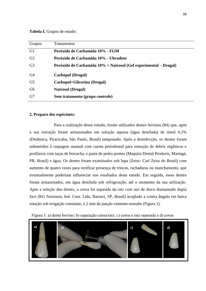
Tabela I. Grupos de estudo:
Grupos Tratamentos
G1 Peróxido de Carbamida 10% - FGM
G2 Peróxido de Carbamida 10% - Ultradent
G3 Peróxido de Carbamida 10% + Natrosol (Gel experimental – Drogal)
G4 Carbopol (Drogal)
G5 Carbopol+Glicerina (Drogal)
G6 Natrosol (Drogal)
G7 Sem tratamento (grupo controle)
2. Preparo dos espécimes:
Para a realização desse estudo, foram utilizados dentes bovinos (84) que, após
a sua extração foram armazenados em solução aquosa (água destilada) de timol 0,1%
(Dinâmica, Piracicaba, São Paulo, Brasil) tamponado. Após a desinfecção, os dentes foram
submetidos à raspagem manual com cureta periodontal para remoção de debris orgânicos e
profilaxia com taças de borracha, e pasta de pedra-pomes (Maquira Dental Products, Maringá,
PR, Brasil) e água. Os dentes foram examinados sob lupa (Zeiss- Carl Zeiss do Brasil) com
aumento de quatro vezes para verificar presença de trincas, rachaduras ou manchamento, que
eventualmente poderiam influenciar nos resultados deste estudo. Em seguida, esses dentes
foram armazenados, em água destilada sob refrigeração, até o momento da sua utilização.
Após a seleção dos dentes, a coroa foi separada da raiz com uso de disco diamantado dupla
face (KG Sorensen, Ind. Com. Ltda, Barueri, SP, Brasil) acoplado a contra ângulo em baixa
rotação sob irrigação constante, à 2 mm da junção cemento-esmalte (Figura 1).
a) b) c) d)
Figura 1: a) dente bovino; b) separação coroa/raiz; c) coroa e raiz separada e d) coroa
bovina.
40
3.Confecção dos espécimes:
Para obtenção dos espécimes, a porção coronária foi fixada na placa de acrílico
com cola quente (Figura 2.a) e foram obtidos blocos de dentes através de um disco de corte
diamantado (Extec Dia. Wafer Blade 102 x 0,3 x 12,7mm) acoplado em uma Cortadeira
Metalográfica (Isomet 1000, Buehler Ltda. Lake Buff, IL, USA) (Figura 2.b). As amostras
foram seccionadas em dois cortes no sentido mésio-distal (Figura 2.c) e dois cortes no sentido
cérvicoincisal (Figura 2.d), obtendo-se 1 bloco de cada dente da região mesio-cervical da
coroa (Figura 2.e). Sendo assim, foram obtidos, das superfícies vestibulares dos dentes
bovinos, blocos com 16 mm2.
Para planificação, regularização e polimento da superfície de esmalte, os
espécimes foram posicionados em discos de acrílico de forma que a superfície do esmalte
ficou paralela à base do disco, permitindo assim, o posicionamento do conjunto (espécime e
disco de acrílico) paralelamente à superfície da lixa ou feltro utilizado.
Para essa etapa, foram utilizadas lixas de carbeto de silício de granulação
decrescente (#1200, #2500 e #p4000 – Carborundum Abrasivos, São Paulo, SP, Brasil)
(Figura 3.b) acoplados à politriz (Arotec Ind. Com., Cotia, SP, Brasil) (Figura 3.a), sob
a) b)
c)
d)
e)
Figura 2: a) coroa fixada em placa de acrílico; b)
cortadeira; c) corte mesiodistal; d)corte
cervicoincisal e e) bloco 4x4mm confeccionado.
41
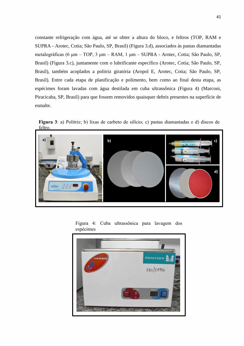
constante refrigeração com água, até se obter a altura do bloco, e feltros (TOP, RAM e
SUPRA - Arotec, Cotia; São Paulo, SP, Brasil) (Figura 3.d), associados às pastas diamantadas
metalográficas (6 μm – TOP, 3 μm – RAM, 1 μm – SUPRA - Arotec, Cotia; São Paulo, SP,
Brasil) (Figura 3.c), juntamente com o lubrificante específico (Arotec, Cotia; São Paulo, SP,
Brasil), também acoplados a politriz giratória (Aropol E, Arotec, Cotia; São Paulo, SP,
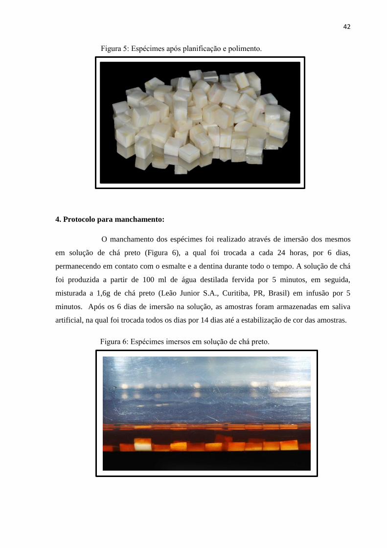
Brasil). Entre cada etapa de planificação e polimento, bem como ao final desta etapa, as
espécimes foram lavadas com água destilada em cuba ultrassônica (Figura 4) (Marconi,
Piracicaba, SP, Brasil) para que fossem removidos quaisquer debris presentes na superfície de
esmalte.
a) b) c)
d)
Figura 3: a) Politriz; b) lixas de carbeto de silício; c) pastas diamantadas e d) discos de
feltro.
Figura 4: Cuba ultrassônica para lavagem dos
espécimes
42
4. Protocolo para manchamento:
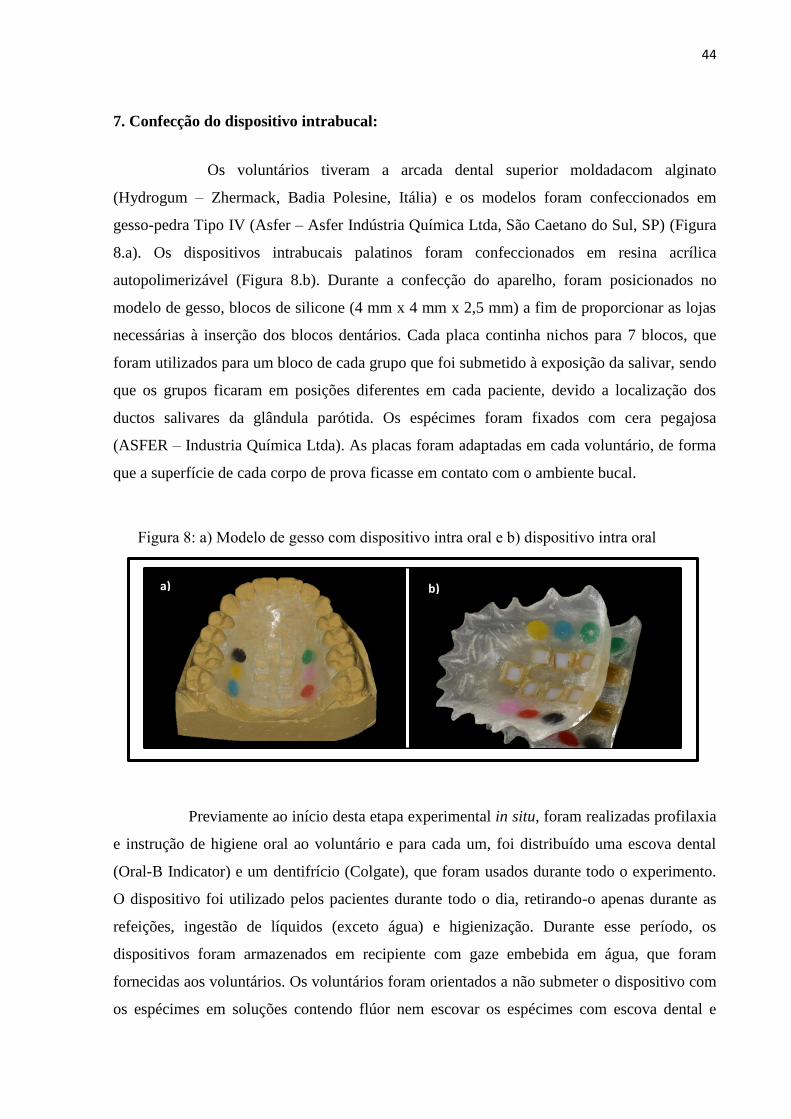
O manchamento dos espécimes foi realizado através de imersão dos mesmos
em solução de chá preto (Figura 6), a qual foi trocada a cada 24 horas, por 6 dias,
permanecendo em contato com o esmalte e a dentina durante todo o tempo. A solução de chá
foi produzida a partir de 100 ml de água destilada fervida por 5 minutos, em seguida,
misturada a 1,6g de chá preto (Leão Junior S.A., Curitiba, PR, Brasil) em infusão por 5
minutos. Após os 6 dias de imersão na solução, as amostras foram armazenadas em saliva
artificial, na qual foi trocada todos os dias por 14 dias até a estabilização de cor das amostras.
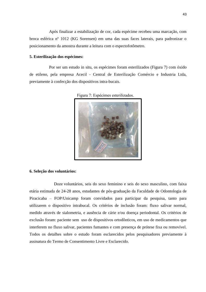
Figura 5: Espécimes após planificação e polimento.
Figura 6: Espécimes imersos em solução de chá preto.
43
Após finalizar a estabilização de cor, cada espécime recebeu uma marcação, com
broca esférica nº 1012 (KG Sorensen) em uma das suas faces laterais, para padronizar o
posicionamento da amostra durante a leitura com o espectofotômetro.
5. Esterilização dos espécimes:
Por ser um estudo in situ, os espécimes foram esterilizados (Figura 7) com óxido
de etileno, pela empresa Acecil - Central de Esterilização Comércio e Industria Ltda,
previamente à confecção dos dispositivos intra-bucais.
6. Seleção dos voluntários:
Doze voluntários, seis do sexo feminino e seis do sexo masculino, com faixa
etária estimada de 24-28 anos, estudantes de pós-graduação da Faculdade de Odontologia de
Piracicaba – FOP/Unicamp foram convidados para participar da pesquisa, tanto para
utilizarem o dispositivo intrabucal. Os critérios de inclusão foram: fluxo salivar normal,
medido através de sialometria, e ausência de cárie e/ou doença periodontal. Os critérios de
exclusão foram: paciente sem uso de dispositivos ortodônticos, em uso de medicamentos que
interferem no fluxo salivar, pacientes fumantes e com presença de prótese fixa ou removível.
Todos os detalhes sobre o estudo foram esclarecidos pelos pesquisadores previamente à
assinatura do Termo de Consentimento Livre e Esclarecido.
Figura 7: Espécimes esterilizados.
44
7. Confecção do dispositivo intrabucal:
Os voluntários tiveram a arcada dental superior moldadacom alginato
(Hydrogum – Zhermack, Badia Polesine, Itália) e os modelos foram confeccionados em
gesso-pedra Tipo IV (Asfer – Asfer Indústria Química Ltda, São Caetano do Sul, SP) (Figura
8.a). Os dispositivos intrabucais palatinos foram confeccionados em resina acrílica
autopolimerizável (Figura 8.b). Durante a confecção do aparelho, foram posicionados no
modelo de gesso, blocos de silicone (4 mm x 4 mm x 2,5 mm) a fim de proporcionar as lojas
necessárias à inserção dos blocos dentários. Cada placa continha nichos para 7 blocos, que
foram utilizados para um bloco de cada grupo que foi submetido à exposição da salivar, sendo
que os grupos ficaram em posições diferentes em cada paciente, devido a localização dos
ductos salivares da glândula parótida. Os espécimes foram fixados com cera pegajosa
(ASFER – Industria Química Ltda). As placas foram adaptadas em cada voluntário, de forma
que a superfície de cada corpo de prova ficasse em contato com o ambiente bucal.
Previamente ao início desta etapa experimental in situ, foram realizadas profilaxia
e instrução de higiene oral ao voluntário e para cada um, foi distribuído uma escova dental
(Oral-B Indicator) e um dentifrício (Colgate), que foram usados durante todo o experimento.
O dispositivo foi utilizado pelos pacientes durante todo o dia, retirando-o apenas durante as
refeições, ingestão de líquidos (exceto água) e higienização. Durante esse período, os
dispositivos foram armazenados em recipiente com gaze embebida em água, que foram
fornecidas aos voluntários. Os voluntários foram orientados a não submeter o dispositivo com
os espécimes em soluções contendo flúor nem escovar os espécimes com escova dental e
Figura 8: a) Modelo de gesso com dispositivo intra oral e b) dispositivo intra oral
a) b)

45
dentifrício. Os dispositivos foram higienizados a cada 12 horas, durante todo o experimento,
com água e escova dental sem dentifrício.
8. Protocolo do clareamento dental:
O dispositivo intrabucal permaneceu durante um dia na boca do voluntário,
previamente aos inicios dos tratamentos clareadores, para a formação de película adquirida.
Removido o dispositivo da boca do voluntário e seco com papel absorvente, os
espécimes foram submetidos ao tratamento clareador à base de peróxido de carbamida a 10%
com carbopol (FGM), peróxido de carbamida 10% com carbopol e glicerina (Ultradent),
peróxido de carbamida a 10% com natrosol (Gel experimental), apenas com o gel de
carbopol, com o gel de carbopol+glicerina e apenas com gel de natrosol (Figura 9) e sem
nenhum tratamento.Foi realizada uma sessão de tratamento clareador por dia, durante 14 dias.
Os voluntários foram calibrados para a aplicação do gel, e o mesmo foi aplicado, de acordo
com as recomendações do fabricante, na superfície de esmalte (Figura 10.a) e permaneceu em
contato com o mesmo por 4 horas, em temperatura ambiente. O dispositivo foi mantido em
um aparato contendo água, durante o tratamento, para os espécimes não sofreram
desidratação, porém, sem contato com a superfície que contém o gel. Após cada aplicação, foi
feita a remoção do gel com hastes flexíveis com ponta de algodão (Figura 10.b), e em seguida
eles foram lavados com água (Figura 10.c) e o dispositivo retornou a boca do paciente (Figura
11).
Figura 9: Géis utilizados para aplicação nos espécimes
46
9. Análises:
9.1. Cor:
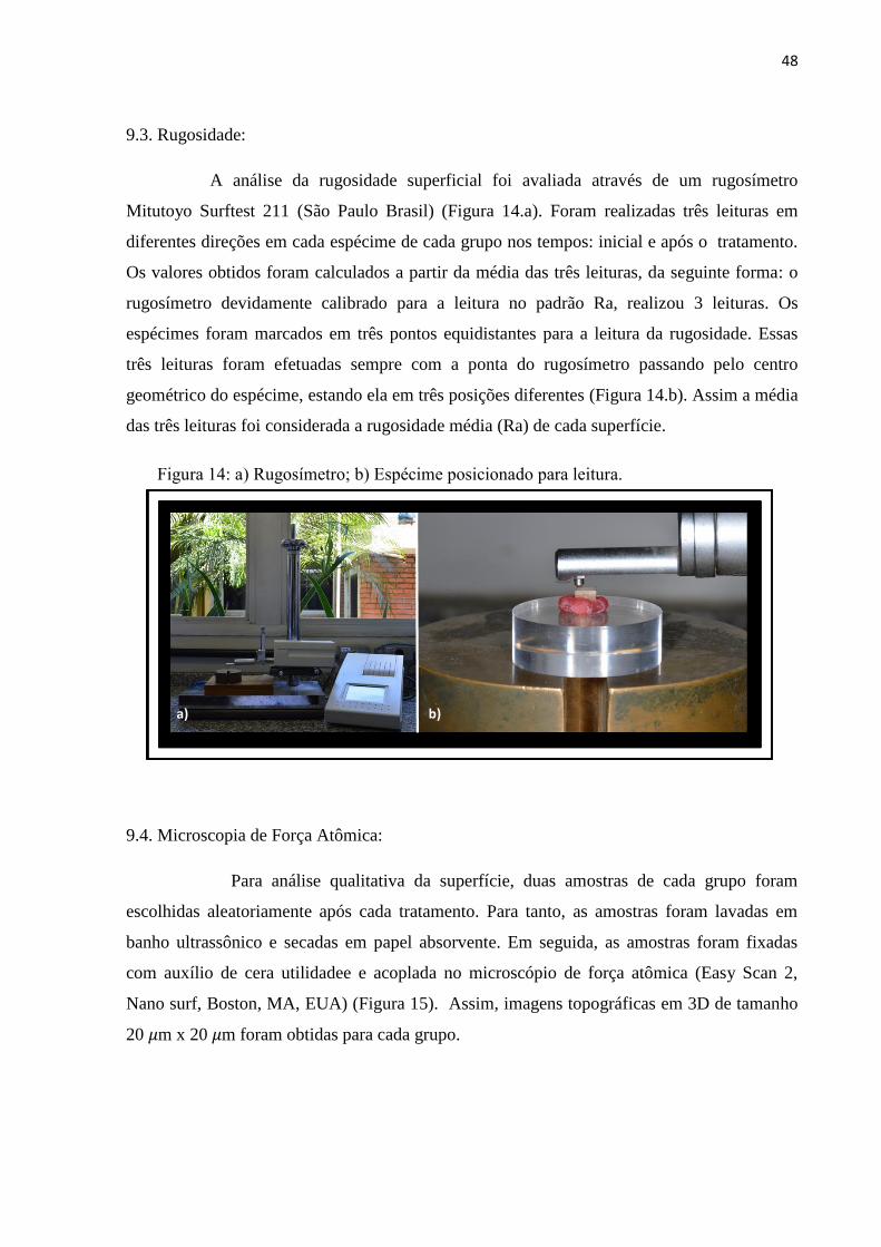
Para a realização das leituras de cor, os espécimes foram posicionados em um
dispositivo de teflon (porta amostra) (Figura 12.c) dentro de uma Câmara de luz (GTI,
Newburgh, NY, USA) (Figura 12.b), com o intuito de padronização do ambiente. O
equipamento utilizado para a leitura de cor foi um espectrofotômetro (Konica Minolta CM-
700d) (Figura 12.a) previamente calibrado. As leituras foram realizadas nos tempos: inicial e
24 horas após o final do tratamento.
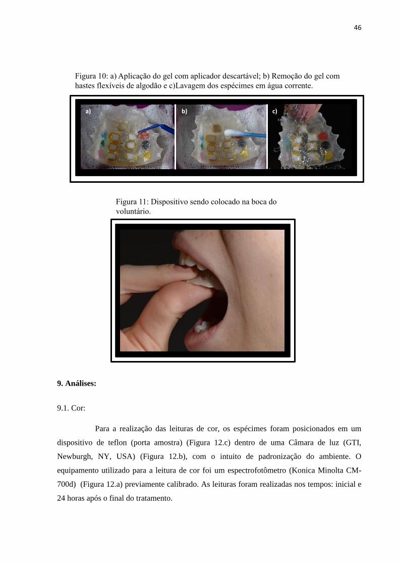
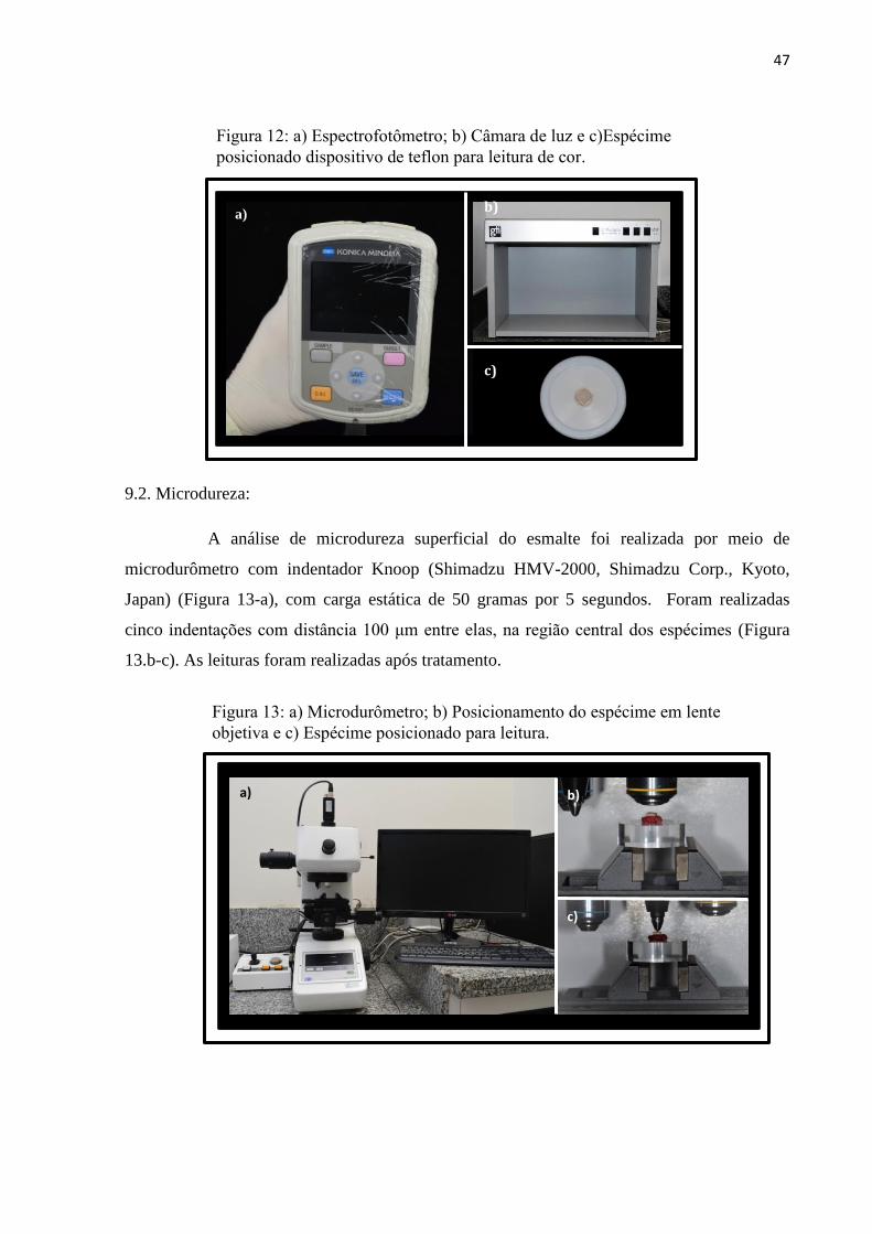
Figura 11: Dispositivo sendo colocado na boca do
voluntário.
Figura 10: a) Aplicação do gel com aplicador descartável; b) Remoção do gel com
hastes flexíveis de algodão e c)Lavagem dos espécimes em água corrente.
a) b) c)
47
9.2. Microdureza:
A análise de microdureza superficial do esmalte foi realizada por meio de
microdurômetro com indentador Knoop (Shimadzu HMV-2000, Shimadzu Corp., Kyoto,
Japan) (Figura 13-a), com carga estática de 50 gramas por 5 segundos. Foram realizadas
cinco indentações com distância 100 μm entre elas, na região central dos espécimes (Figura
13.b-c). As leituras foram realizadas após tratamento.
Figura 13: a) Microdurômetro; b) Posicionamento do espécime em lente
objetiva e c) Espécime posicionado para leitura.
a) b)
c)
b)
c)
a)
Figura 12: a) Espectrofotômetro; b) Câmara de luz e c)Espécime
posicionado dispositivo de teflon para leitura de cor.
48
9.3. Rugosidade:
A análise da rugosidade superficial foi avaliada através de um rugosímetro
Mitutoyo Surftest 211 (São Paulo Brasil) (Figura 14.a). Foram realizadas três leituras em
diferentes direções em cada espécime de cada grupo nos tempos: inicial e após o tratamento.
Os valores obtidos foram calculados a partir da média das três leituras, da seguinte forma: o
rugosímetro devidamente calibrado para a leitura no padrão Ra, realizou 3 leituras. Os
espécimes foram marcados em três pontos equidistantes para a leitura da rugosidade. Essas
três leituras foram efetuadas sempre com a ponta do rugosímetro passando pelo centro
geométrico do espécime, estando ela em três posições diferentes (Figura 14.b). Assim a média
das três leituras foi considerada a rugosidade média (Ra) de cada superfície.
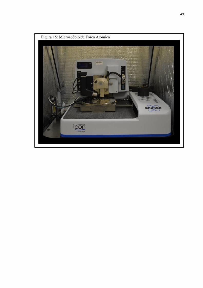
9.4. Microscopia de Força Atômica:
Para análise qualitativa da superfície, duas amostras de cada grupo foram
escolhidas aleatoriamente após cada tratamento. Para tanto, as amostras foram lavadas em
banho ultrassônico e secadas em papel absorvente. Em seguida, as amostras foram fixadas
com auxílio de cera utilidadee e acoplada no microscópio de força atômica (Easy Scan 2,
Nano surf, Boston, MA, EUA) (Figura 15). Assim, imagens topográficas em 3D de tamanho
20 𝜇m x 20 𝜇m foram obtidas para cada grupo.
Figura 14: a) Rugosímetro; b) Espécime posicionado para leitura.
a) b)
49
Figura 15: Microscópio de Força Atômica
50
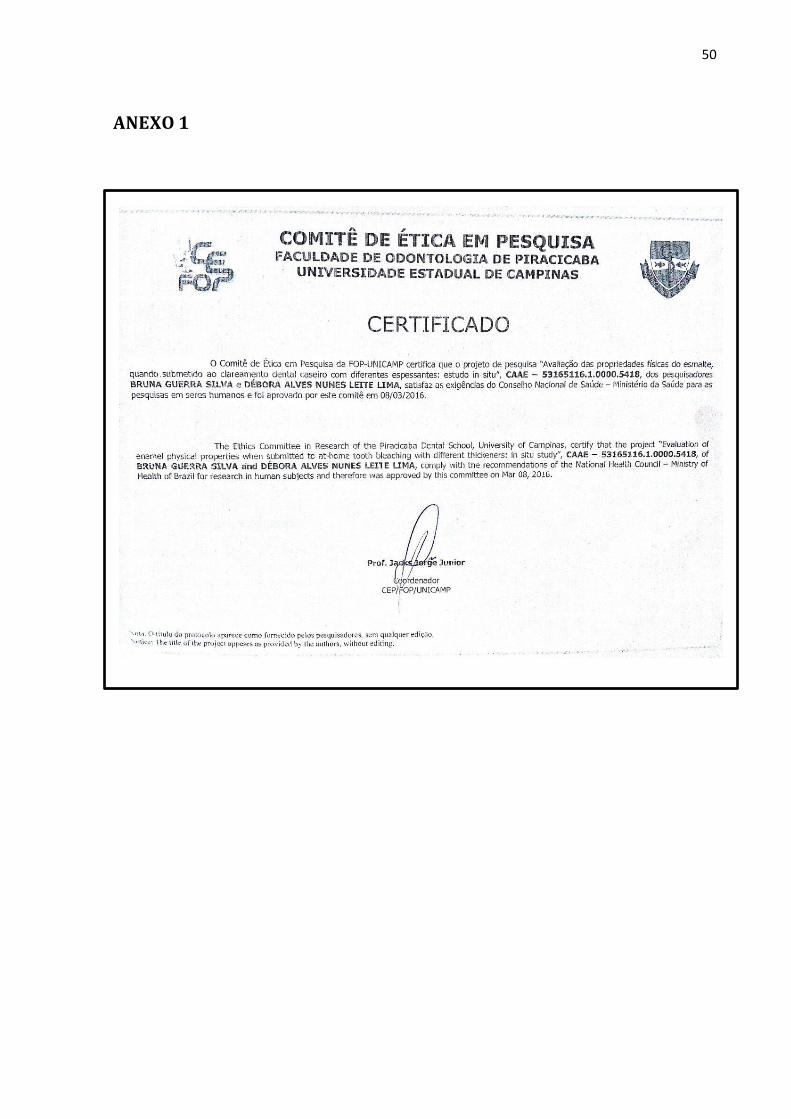
ANEXO 1
51
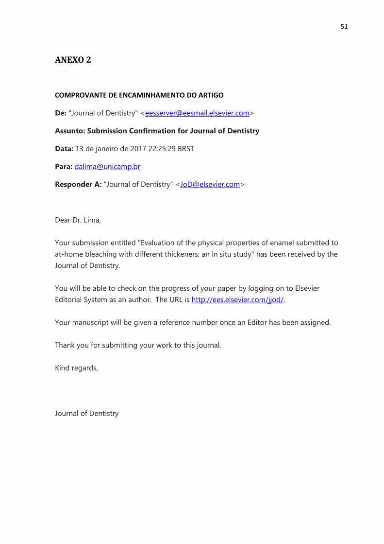
ANEXO 2
COMPROVANTE DE ENCAMINHAMENTO DO ARTIGO
De: "Journal of Dentistry" <[email protected]>
Assunto: Submission Confirmation for Journal of Dentistry
Data: 13 de janeiro de 2017 22:25:29 BRST
Para: [email protected]
Responder A: "Journal of Dentistry" <[email protected]>
Dear Dr. Lima,
Your submission entitled "Evaluation of the physical properties of enamel submitted to
at-home bleaching with different thickeners: an in situ study" has been received by the
Journal of Dentistry.
You will be able to check on the progress of your paper by logging on to Elsevier
Editorial System as an author. The URL is http://ees.elsevier.com/jjod/.
Your manuscript will be given a reference number once an Editor has been assigned.
Thank you for submitting your work to this journal.
Kind regards,
Journal of Dentistry