1 pontifÍcia universidade catÓlica do rio grande … · ... genotóxicos e carcinogênicos este...
TRANSCRIPT

1
PONTIFÍCIA UNIVERSIDADE CATÓLICA DO RIO GRANDE DO SUL
FACULDADE DE ODONTOLOGIA DOUTORADO EM ORTODONTIA E ORTOPEDIA FACIAL
GRAZIELA HENRIQUES WESTPHALEN
CITOTOXICIDADE E GENOTOXICIDADE DE MATERIAIS ORTODÔNTICOS
Porto Alegre 2009

2
GRAZIELA HENRIQUES WESTPHALEN
CITOTOXICIDADE E GENOTOXICIDADE DE MATERIAIS ORTODÔNTICOS
Tese apresentada como parte dos requisitos
obrigatórios para obtenção do grau de Doutor na
área de Ortodontia e Ortopedia Facial pelo
Programa de Pós-Graduação da Faculdade de
Odontologia da Pontifícia Universidade Católica do
Rio Grande do Sul.
Orientadora: Profa. Dra Luciane Macedo de Menezes
Co-Orientadora: Profa. Dra Renata Medina-Silva
Porto Alegre 2009
DADOS INTERNACIONAIS DE CATALOGAÇÃO NA PUBLICAÇÃO (CIP)
Alessandra Pinto Fagundes Bibliotecária CRB10/1244
W537t
Westphalen, Graziela Henriques
Citotoxicidade e genotoxicidade de materiais ortodônticos / Graziela Henriques Westphalen. – Porto Alegre, 2009.
78 f : il.
Tese (Doutorado) – Fac. de Odontologia. PUCRS, 2009. Orientador: Profa. Dra Luciane Macedo de Menezes. Co-Orientador: Profa. Dra Renata Medina-Silva.
1. Odontologia. 2. Ortodontia. 3. Materiais Ortodônticos - Toxicidade. I. Menezes, Luciane Macedo de II. Silva, Renata Medina. III. Título.
CDD: 617.695

3
GRAZIELA HENRIQUES WESTPHALEN
CITOTOXICIDADE E GENOTOXICIDADE DE MATERIAIS ORTODÔNTICOS
Tese apresentada como parte dos requisitos
obrigatórios para obtenção do grau de Doutor na
área de Ortodontia e Ortopedia Facial pelo
Programa de Pós-Graduação da Faculdade de
Odontologia da Pontifícia Universidade Católica do
Rio Grande do Sul.
BANCA EXAMINADORA:
____________________________________________
Profa Dra. Luciane Macedo de Menezes
____________________________________________
Prof. Dr. Arno Locks
____________________________________________
Profa. Dra. Cátia Cardoso Abdo Quintão
____________________________________________
Prof. Dr. Eduardo Gonçalves Mota
___________________________________________
Profa. Dra Maria Antonieta Lopes de Souza
4
DEDICATÓRIA
A Deus, inteligência suprema, causa primária de todas as coisas.
A toda minha família amada, em especial aos meus pais, Pedro e Lorene, pelo
exemplo de amor, carinho, dedicação, apoio e por sempre se esforçarem ao máximo
para me proporcionar o melhor. A vocês muito obrigada pela realização de todos
meus sonhos.
Aos meus grandes irmãos, Mário e Camila, pelo amor, amizade, carinho e alegria
sempre presentes.
Ao meu querido namorado, Marco. Agradeço a Deus pela oportunidade de encontrar
um pessoa tão repleta de amor, carinho, respeito e generosidade. Tenho a certeza
de que nada é por acaso (Thanks for you to exist!).
Aos meus amados amigos que, meu muito obrigado por todo apoio, carinho,
companheirismo e alegria demonstrados em todos os momentos da minha vida.
Amo todos vocês!

5
AGRADECIMENTOS
À Pontifícia Universidade Católica do Rio Grande do Sul, pela qual tenho uma
admiração grande e tive a excelente oportunidade de ingressar nessa instituição no
curso de Mestrado em Ortodontia e prosseguir no Doutorado.
À CAPES, pela bolsa de estudo fornecida durante o Curso de Doutorado e para
Pesquisa no Commonwealth Scientific and Industrial Research Organisation
(CSIRO) em Adelaide- Austrália e também pelo apoio e incentivo aos estudantes
brasileiros para realizarem suas pesquisas no exterior.
Ao Coordenador do Programa de Pós Graduação em Odontologia, José A. Poli de
Figueiredo, pela sua competência, dinamismo e apoio aos alunos, o que resulta na
excelência e evolução do nosso Programa de Pós Graduação.
Ao diretor da Faculdade de Odontologia, Marcos Túlio M. Carvalho, pela seriedade e
competência no desempenho do seu cargo.
À minha orientadora, Luciane Menezes, pelas orientações competentes desde o
Mestrado até o Doutorado, por ter me incentivado a pesquisar com interação de
áreas conexas, e principalmente pela confiança, amizade e apoio nutridos, há quase
10 anos, desde meu estágio em Ortodontia na UFSC.
À minha co-orientadora, Renata Medina-Silva, pelo brilhantismo das suas
orientações, desde o Mestrado até o Doutorado, fazendo com que assuntos tão
diferentes se tornassem compreensíveis. Minha admiração pela sua dedicação,
personalidade, amizade e inteligência.
Aos professores do Programa de Pós-Graduação em Odontologia, Luciana Hirakata,
Hugo Oshima, Eduardo Mota, Rosemary Shinkai, Márcio Grossi, Eduardo Martinelli,
Susana Rizzatto, Ernani Marchioro e Telmo Berthold, pela dedicação, competência e
incentivo pelo aprendizado e pesquisa.
Aos meus Professores de Ortodontia da UFSM, UFSC, UERJ que muito me
ensinaram com competência e me incentivaram nessa caminhada.

6
Aos Professores da área conexa, João Antônio P. Henriques (UFRGS), Daniel Prá
(UCPEL), pelo seguimento de suas colaborações no Doutorado, sempre de uma
forma competente, dedicada e amiga.
A toda equipe do CSIRO, pela excelente receptividade, amizade, respeito, carinho
e ensinamentos transmitidos durante meu período de pesquisa no exterior. Em
especial, aos cientistas responsáveis pela minha orientação no CSIRO, Michael
Fenech e Phil Thomas, pelos seus brilhantes ensinamentos, orientações e vínculos
de amizade firmados.
A todas as pessoas maravilhosas que encontrei em Adelaide, que se tornaram
grandes amigos e, principalmente ao Marco Lucchetta, que contribuiu para que esse
período fosse um dos mais especiais da minha vida.
À Professora Judith Scliar pela competência, seriedade, apoio e amizade nas aulas
preparatórias para os testes TOEFL e IELTS.
Aos meus queridos colegas e amigos, Micéli Blaya, Marcel Farret, Gustavo Frainer
Barbosa, Carlos Alberto Woitchunas e Rogério Rosa, pela formação de uma turma
unida, amiga e com uma ótima convivência.
A todos os pacientes que participaram voluntariamente dessa pesquisa.
À estudante de Especialização em Ortodontia, Fabiane Azeredo, por ajudar
dedicadamente no recrutamento dos pacientes e pela nossa amizade
A todos os funcionários da Faculdade de Odontologia da PUCRS, especialmente a
“turma” (Ana, Davenir, Marcos e Carlos) da Secretaria da Pós Graduação, que
sempre mostrou dedicação, carinho e amizade.À secretária Clarissa Belarmino da
PRPPG, por sempre estar disposta a sanar todas as dúvidas da bolsa para o
exterior da CAPES.
A todas as pessoas amadas, que de alguma forma, colaboraram para a conquista de
mais esse sonho.

7
Embora ninguém possa voltar atrás e fazer um novo começo, qualquer
um pode começar agora e fazer um novo fim.
(Francisco Cândido Xavier - Chico Xavier)

8
RESUMO
A avaliação da biocompatibilidade dos materiais ortodônticos é de extrema
importância, uma vez que elementos constituintes desses materiais estão
relacionados a efeitos citotóxicos, genotóxicos e carcinogênicos Este estudo
objetivou verificar a citotoxicidade e genotoxicidade de materiais ortodônticos. A
citotoxicidade de materiais ortodônticos (bráquetes, fios, resinas compostas, soldas
de prata e elásticos) foi testada por meio de duas exposições de sobrevivência ao
Saccharomyces cerevisiae (S. Cerevisiae) de forma direta e indireta (produtos
químicos liberados pelos materiais em saliva artificial) (Artigo 1). A avaliação da
genotoxicidade foi realizada em grupos de pacientes, empregando-se os testes:
Ensaio citoma bucal de micronúcleos para avaliação de aparelhos ortodônticos fixos
e Hyrax (Artigo 2); Teste de micronúcleos para aparelhos expansores maxilares de
Haas e Hyrax (Artigo 3). No artigo 1 foram realizadas comparações de ocorrência de
sobrevivência de S. Cerevisiae em curvas semi-log entre os materiais ortodônticos e
controle. Já nos artigos 2 e 3 foram empregados testes estatísticos não paramétricos
diferentes (α≤0,05) para comparações entre grupos de pacientes e controle. Foi
observado que a solda de prata foi o único material que apresentou citotoxicidade.
Todos os aparelhos testados não mostraram genotoxicidade nos grupos de
pacientes estudados.
Palavras chaves: Materiais dentários, Ortodontia, Toxicidade, Gentoxicidade,
Testes Mutagênicos, Teste de micronúcleos, Saccharomyces cerevisiae, Metais,
Mucosa bucal, Célula basal.

9
ABSTRACT
Biocompatibility evaluation of orthodontic materials is very important, since some
constituent elements of these materials are related to citotoxic, genotoxic and
carcinogenic effects. This study aimed to assess citotoxicity and genotoxicity of
orthodontic materials. Citotoxicity of orthodontic materials (brackets, wires, resin
composites, solder silver and elastomers) was tested by two survival exposure to
Saccharomyces cerevisiae (S. Cerevisiae), through direct and indirect (chemical
products liberated by these materials in artificial saliva) forms (Article 1). Assessment
of genotoxicity was carried out in patient groups, using the following tests: Cytome
buccal micronucleus assay for evaluating orthodontic fixed appliances and Hyrax
(Article 2), Micronucleus tests for Hyrax and Haas appliances (Article 3). For Article
1, occurrence of S. Cerevisiae survival was compared in a semi-log curve between
orthodontic materials and control groups. For articles 2 and 3, non parametric test
was used (α≤0.05%) for comparison among patients and control groups. Silver solder
was the only material that showed citotoxicity. All appliances tested did not present
genotoxicity in studied patients.
Key words: Dental Materials, Orthodontics, Toxicity, Genotoxicity, Mutagenic tests,
micronucleus test, Saccharomyces cerevisiae, Metals, Buccal mucosa, Basal cell.
10
SUMÁRIO
1 INTRODUÇÃO
11
2 ARTIGO 1
13
3 ARTIGO 2
27
4 ARTIGO 3
41
5 DISCUSSÃO GERAL
49
6 REFERÊNCIAS
53
7 ANEXOS
59
8 APÊNDICES
75
11
1 INTRODUÇÃO
O aumento de pesquisas na área de biocompatibilidade dos materiais
odontológicos é uma necessidade em função da capacidade dos mesmos
provocarem alterações da atividade biológica nos tecidos envolvidos1. Esse fato está
de acordo com uma tendência observada na ciência dos materiais dentários, de ser
gradativamente substituída pela ciência dos biomateriais em Odontologia2.
Determinados materiais odontológicos empregados na Ortodontia (ligas de
aço inoxidáveis, solda de prata, resinas acrílicas) são constituídos de elementos
químicos que estão relacionados a efeitos biológicos adversos no organismo
humano3-11.
Um dos aspectos da biocompatibilidade dos materiais odontológicos é a ação
citotóxica dos mesmos. Muitos estudos2,12-19 verificaram esse efeito, por meio de
utilização de células de cultura humanas. Os resultados de tais estudos geraram
conclusões diversas e conflitantes.
Uma metodologia que avalie com sucesso o potencial citotóxico se baseia no
emprego de diversos organismos-modelo como Saccharomyces cerevisiae (S.
Cerevisiae) que possibilita a realização de experimentos controlados e com um
grande número amostral. No entanto, são escassas as pesquisas que utilizam esse
método para avaliação dos efeitos citotóxicos relacionados aos materiais
ortodônticos.
Outro aspecto, de fundamental importância na avaliação da
biocompatibilidade de materiais ortodônticos, é o seu potencial genotóxico, uma vez
que elementos metálicos como níquel, cromo, prata e cádmio apresentam resultados
positivos relacionados à toxicidade genética4,6,7,20-23. Estudos in vivo mostraram que
metais como o níquel e o cobalto, quando liberados dos aparelhos ortodônticos,
podem causar quebras no DNA de células da mucosa bucal3. Apesar disso, ainda
existem poucos estudos que abordem essa área de conhecimento.
A genotoxicidade pode ser avaliada por inúmeros experimentos, sendo
abordada eficazmente pelos testes: teste de micronúcleos e ensaio citoma bucal de

12
micronúcleos. O teste de micronúcleos detecta danos genéticos tardios do DNA, por
meio da verificação da freqüência de micronúcleos, que são estruturas extra-
nucleares, compostas por cromossomos ou fragmentos dos mesmos que, durante a
mitose, não foram incorporados ao núcleo principal26,27. O citoma bucal de
micronúcleos possibilita uma avaliação mais abrangente, uma vez que além da
verificação da freqüência de micronúcleos, avalia a presença de outros
biomarcadores como: morte celular (cromatina condensada, cariorexe, cariólise e
picnose), proliferação celular (núcleo duplo) e outros danos do DNA (nuclear
“BUDS”) 28-30.
De acordo com o exposto, é de suma importância o conhecimento da
capacidade que os materiais possuem de afetar o ambiente biológico, a fim de
garantir um tratamento seguro e eficaz aos pacientes. Dessa forma, este estudo
objetivou verificar a biocompatibilidade de alguns materiais ortodônticos,
empregando teste de citotoxicidade in vitro utilizando S. Cerevisiae, como organismo
modelo, bem como testes para avaliação da genotoxicidade como, ensaio citoma
bucal de micronúcleos e teste de micronúcleos em um grupo de pacientes sob
tratamento ortodôntico.

13
2 ARTIGO 1
Cytotoxicity of orthodontic materials assessed by survival tests in
Saccharomyces cerevisiae
Authors: Karen M. Limberger 1, Graziela H. Westphalen 2, Luciane M.
Menezes 2 , Renata Medina-Silva 1
1 Laboratório de Imunologia e Microbiologia, Faculdade de Biociências, Pontifícia
Universidade Católica do Rio Grande do Sul, Porto Alegre, Rio Grande do Sul, Brazil.
2 Departamento de Ortodontia, Pontifícia Universidade Católica do Rio Grande do Sul, Porto
Alegre, Rio Grande do Sul, Brazil.
Abstract
The aim of this study was to assess the cytotoxicity of orthodontic materials
(brackets, wires, resin, elastomers and silver solder) using Saccharomyces
cerevisiae as a model organism. The induction of cytotoxicity was assessed by two
different tests using the wild-type S. cerevisiae strain FF18733: 1) direct exposure to
orthodontic materials in YPD broth, and 2) exposure to artificial commercial saliva
pre-treated with orthodontic materials. Only the silver solder was tested in mutant S.
cerevisiae strains to investigate the origin of the observed cytotoxicity. Colony
forming units per mL counts were carried out in all experiments and compared to
control groups to detect significant survival differences. The results showed that only
the silver solder induced significant cytotoxicity, which might have occurred via
oxidative stress, although this mechanism is not completely understood.
14
Introduction
The biocompatibility of dental materials has been extensively studied [1, 2],
since this property is essential to ensure the safe treatment of patients [3]. Some
orthodontic materials, such as brackets, wires, solder and resins, have compounds
known to have allergic, cytotoxic, mutagenic and/or carcinogenic potential [4-6].
These materials remain within the oral cavity for long periods and are subject to
corrosion, which provokes the release of substances [7, 8] that might interact with
patients’ tissues [9].
The toxicity of some metals (iron, cooper, chromium, vanadium, cobalt,
mercury, cadmium, nickel) might result from the generation of high levels of nitrogen
and/or oxygen reactive species.[6]. Oxidative stress may cause oxidative damage in
proteins, lipids and or/DNA and may trigger signalling cascades that stimulate cell
growth [4].
Currently, many in vitro tests exist that can be used to assess the cytotoxicity
of orthodontic materials, and various cell cultures are used that yield some similar
and also some opposing findings [10-15]. Cytotoxicity induced by harmful agents can
be assessed successfully by in vitro experiments using model microorganisms like
the yeast Saccharomyces cerevisiae [16-18]. The use of this microorganism offers
some advantages, since it is easy to cultivate and manipulate, and it provides
quantitative and well-controlled experiments with large sample numbers and quick
results. Moreover, as it is a eukaryotic microorganism, it is very similar to human and
animal cells. However, few dental studies have used this microorganism for this
purpose [19, 20].
The aim of this study was to evaluate the induction of cytotoxicity by
orthodontic materials (brackets, wires, resin, elastomers and silver solder) using a
wild-type S. cerevisiae strain as a model organism. Moreover, S. cerevisiae mutant
strains were also used to investigate the origin of observed cytotoxicity induced by
some of the orthodontic materials.
15
Materials and Methods
This cytotoxicity study was approved by the ethics Committee from
Pontifícia Universidade Católica do Rio Grande do Sul, Brazil. The evaluation was
performed using the orthodontic materials described in Table 1. These materials
were all available for testing at the Clínica de Ortodontia, Pontifícia Universidade
Católica do Rio Grande do Sul, Brazil.
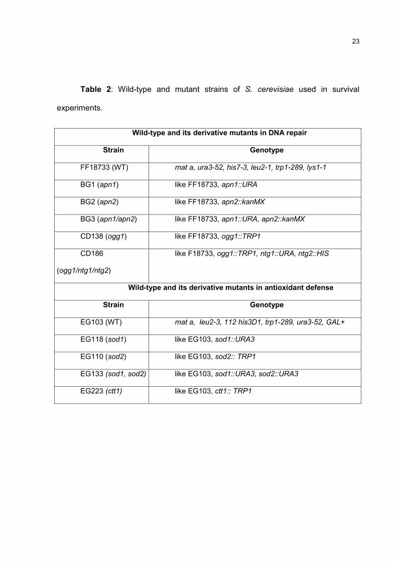
S. cerevisiae strain, media and cultures
The S. cerevisiae strains used in this work and their respective genotypes are
listed in Table 2. Included are the wild-type (WT) strain EG103 and its derivatives,
which are single or double mutants in genes that encode the antioxidant defense
enzymes Sod1, Sod2, and Cat1. Also included are the WT strain FF18733 and its
derivatives, which are single, double or triple mutants in genes that encode the DNA
base excision repair proteins Ogg1, Apn1, Apn2, Ntg1 and Ntg2.
The first experiments tested all the orthodontic materials but used only the WT
strain FF18733. The other S. cerevisiae strains were only used to test the cytotoxicity
of the solder fragments.
To cultivate all S. cerevisiae strains, YPD medium (1% yeast extract, 2%
peptone, 2% glucose) was used, either in broth or solid (with agar at 2%) form. In all
survival experiments, S. cerevisiae pre-cultures were prepared in 10 mL YPD broth
and grown overnight to exponential phase (~107 cells/ mL) at 30°C.
Survival experiments for cytotoxicity analysis
The cytotoxicity analysis was performed via two types of survival experiments:
1- Direct exposure of S. cerevisiae cells to the orthodontic materials in YPD broth; 2 -
Exposure to artificial commercial saliva (Salivan, Apsen Farmacêutica SA, Brazil)
pre-treated with orthodontic materials.
For the direct exposure experiments, new inocula were made from the pre-
cultures in 5 mL YPD, with each one containing pieces of different orthodontic
materials, and a control without any material. These cultures were incubated at 30°C
16
to exponential phase (~10-7 cells/ mL). Aliquots from each culture were diluted in
0.9% sterile saline solution and 5-µL drops from each dilution (from 10-2 to 10-5) were
plated on YPD-agar and incubated at 30°C for two days for the emergence of small
colonies, which allowed a qualitative approach. For quantitative analyses, 100 µL of
the final dilutions were plated on YPD-agar (two plates for each dilution) for colony
(CFU/mL) counting after two days at 30oC.
In saliva exposure experiments, the different orthodontic materials were
immersed in 500 µL of artificial saliva for seven or twenty days. A total of 500 µL of
the pre-inoculum was used for each treatment, which was centrifuged (2 min at 2000
g) and resuspended at 100% of saliva pre-exposed to the orthodontic materials. The
cells were then treated for 60 minutes, diluted and plated in YPD-agar as described
above, for both qualitative and quantitative analyses. A control with unexposed saliva
was also done. At least three direct and three indirect experiments were performed
with each type of orthodontic material.
Data analyses
The average Colony forming units per mL (CFU/mL) counts from three trials
of each treatment were compared to their specific controls to verify the occurrence of
significant survival differences in a semi-log curve (based on their standard
deviation), which is an indication of cellular toxicity in S. cerevisiae.
Results
Direct exposure of S. cerevisiae strain FF18733 to orthodontic materials
showed that the silver solder was the only material capable of inducing cytotoxicity,
both qualitatively (Figure 1) and quantitatively (Figure 2). Silver solder promoted a
significant reduction in CFU/ml values relative to the controls, and did so in a dose-
dependent manner. Moreover, a complete inhibition of colony emergence was
observed (Figure 3) in qualitative and quantitative tests with artificial saliva pre-
exposed to three solder fragments (indirect), which confirms the high cytotoxic
potential of the metals released from this orthodontic material. The other materials
tested did not present any difference relative to the control groups.
17
The sensitivity to silver solder was next tested using S. cerevisiae strains
deficient in genes that code for antioxidant enzymes and proteins that repair
oxidative DNA damage; the results are summarized in Table 3. In tests of direct
exposure to three, four and five solder fragments, no differences in terms of cell
viability were observed for the mutant strains relative to the WT. In saliva exposure
experiments, the EG133 strain, defective in superoxide dismutases 1 (CuZnSod,
cytosolic) and 2 (MnSod, mitochondrial), presented modest sensitivity (compared to
WT strain EG103 in three experiments) to metals released by three, four or five
solder fragments (Figure 4). The same occurred with strain BG1 (compared to WT
strain FF18733 in three experiments), which is mutant for the AP-endonuclease 1
(Apn1) DNA repair protein. Nevertheless, the average values from three quantitative
experiments with each mutant strain did not show significant differences compared to
their respective wild-type strains (data not shown).
Discussion
In this study, several S. cerevisiae strains were used. First, one WT strain was
used in an initial screen to assess the cytotoxic potential of all available orthodontic
materials. Second, mutant strains deficient in DNA repair or antioxidant enzymes
were used to investigate the basis of the cellular toxicity previously observed for the
silver solder in the WT strain.
The results showed that brackets and wires (stainless steel alloys) did not
induce any reduction in S. cerevisiae cell viability in either type of exposure. These
findings are in line with several in vitro studies that evaluated the cytotoxicity of
stainless steel orthodontic materials with different cell cultures [13-15]. However,
some in vivo studies showed that orthodontic appliances composed of stainless steel
alloys induced DNA damage in oral mucosa cells [21], and also increased the
frequency of chromosomal breakage, as assessed via the micronucleus test [22].
This difference could be explained by the fact that in vivo experiments are difficult to
control, since factors such as local irritants and adverse reactors might influence their
results [23].
18
Moreover, an absence of cytotoxicity was also observed for the elastomers.
Most studies have stated that these materials are mainly associated with allergic
reactions [24, 25]. The same occurred for the light-cure resin, which corroborates the
suggestion that this resin shows less cytotoxicity than auto-cure resin [26]. The data
from most orthodontic materials that have been tested (brackets, wires, resin and
elastomers) suggest that they bear considerable biocompatibility with S. cerevisiae
cells and that they may be indicated for clinical use.
Significant cytotoxicity induced by silver solder was observed in this study,
mainly in treatments with commercial saliva pre-exposed to solder fragments, with
which a substantial decrease in S. cerevisiae cell viability was observed. Thus, this
indicates that the artificial saliva promoted an intense metal release from the solder
fragments during the seven- and twenty-day periods of exposure. In addition, this
suggests that the use of silver solder may be injurious in the oral cavity environment.
The cytotoxic effects of silver solder have been clearly demonstrated by recent
studies [11, 12, 27-30]. One aspect of cytotoxicity from silver solder can be attributed
to its components (silver, copper, zinc, cadmium, nickel), which are known to have
cytotoxic, genotoxic and carcinogenic effects [4-6]. Based on this information, several
studies have presented laser solder as an alternative material with more
biocompatibility for routine clinical use [27-29].
The experiments with the mutant strains suggested that oxidative stress may
be involved in the cytotoxicity of silver solder. Two mutant strains defective in
enzymes that defend cellular integrity during oxidative stress events (EG133 and
BG1) showed differential sensitivity compared to WT strains when treated with saliva
pre-exposed to three silver solder fragments. The EG133 strain is deficient in its two
superoxide dismutases (CuZnSod and MnSod), which makes it extremely sensitive to
the superoxide ions that are released at high levels during oxidative stress events
[31]. The BG1 strain lacks the enzyme AP-endonuclease 1, which is involved in
repairing oxidative damage in yeast nuclear and mitochondrial DNA. Thus, it is
sensitive to environmental conditions that promote high levels of reactive oxygen
species production [32]. These results agree with statements that recognize certain
19
metals, such as cadmium and cooper, as important promoters of oxidative stress [33,
34]. Since the mutant strains did not show any significant differences from the WT
strains, further studies are necessary to better understand the basis of the cellular
toxicity of silver solder.
Acknowledgements
This study is based on a thesis submitted to the Dentistry Faculty, Pontifícia
Universidade Católica do Rio Grande do Sul, Brazil, in partial fulfillment of the
requirements for a Dentistry PhD degree. This investigation was supported by a
scholarship from Coordenação de Aperfeiçoamento de Pessoal de Nível Superior
(CAPES), Brazil.
References
1. Wataha, J.C., Principles of biocompatibility for dental practitioners. J Prosthet Dent, 2001. 86(2): p. 203-9.
2. St John, K.R., Biocompatibility of dental materials. Dent Clin North Am, 2007. 51(3): p. 747-60, viii.
3. Montanaro, L., et al., No genotoxicity of a new nickel-free stainless steel. Int J Artif Organs, 2005. 28(1): p. 58-65.
4. Beyersmann, D. and A. Hartwig, Carcinogenic metal compounds: recent insight into molecular and cellular mechanisms. Arch Toxicol, 2008. 82(8): p. 493-512.
5. Sethi, P.K. and D. Khandelwal, Cadmium exposure: health hazards of silver cottage industry in developing countries. J Med Toxicol, 2006. 2(1): p. 14-5.
6. Valko, M., H. Morris, and M.T. Cronin, Metals, toxicity and oxidative stress. Curr Med Chem, 2005. 12(10): p. 1161-208.
7. Amini, F., et al., In vivo study of metal content of oral mucosa cells in patients with and without fixed orthodontic appliances. Orthod Craniofac Res, 2008. 11(1): p. 51-6.
8. Matos de Souza, R. and L. Macedo de Menezes, Nickel, chromium and iron levels in the saliva of patients with simulated fixed orthodontic appliances. Angle Orthod, 2008. 78(2): p. 345-50.
9. Noort, R.V., Introdução aos materiais dentários. Vol. 2 edition. 2004, São Paulo: Artmed editora. 344.
20
10. Kao, C.T., et al., The cytotoxicity of orthodontic metal bracket immersion media. Eur J Orthod, 2007. 29(2): p. 198-203.
11. Freitas, M.P., et al., Cytotoxicity of silver solder employed in orthodontics. Angle Orthod, 2009. 79(5): p. 939-44.
12. Mockers, O., D. Deroze, and J. Camps, Cytotoxicity of orthodontic bands, brackets and archwires in vitro. Dent Mater, 2002. 18(4): p. 311-7.
13. Assad, M., et al., A new porous titanium-nickel alloy: Part 1. Cytotoxicity and genotoxicity evaluation. Biomed Mater Eng, 2002. 12(3): p. 225-37.
14. Assad, M., et al., A new porous titanium-nickel alloy: part 2. Sensitization, irritation and acute systemic toxicity evaluation. Biomed Mater Eng, 2002. 12(4): p. 339-46.
15. Wever, D.J., et al., Cytotoxic, allergic and genotoxic activity of a nickel-titanium alloy. Biomaterials, 1997. 18(16): p. 1115-20.
16. Perego, P. and S.B. Howell, Molecular mechanisms controlling sensitivity to toxic metal ions in yeast. Toxicol Appl Pharmacol, 1997. 147(2): p. 312-8.
17. Avery, S.V., Metal toxicity in yeasts and the role of oxidative stress. Adv Appl Microbiol, 2001. 49: p. 111-42.
18. De Freitas, J., et al., Yeast, a model organism for iron and copper metabolism studies. Biometals, 2003. 16(1): p. 185-97.
19. Tanaka, H., et al., Basic properties of an alginate impression material supplemented with chlorhexidine. I. Disinfectant effects on oral microbes. J Nihon Univ Sch Dent, 1994. 36(2): p. 135-8.
20. Williams, D.W., et al., A novel technique for assessment of adherence of Candida albicans to solid surfaces. J Clin Pathol, 1998. 51(5): p. 390-1.
21. Faccioni, F., et al., In vivo study on metal release from fixed orthodontic appliances and DNA damage in oral mucosa cells. Am J Orthod Dentofacial Orthop, 2003. 124(6): p. 687-93; discussion 693-4.
22. Westphalen, G.H., et al., In vivo determination of genotoxicity induced by metals from orthodontic appliances using micronucleus and comet assays. Genet Mol Res, 2008. 7(4): p. 1259-66.
23. Brune, D., Metal release from dental biomaterials. Biomaterials, 1986. 7(3): p. 163-75.
24. Levy, D.A., et al., Allergy to latex. Allergy, 1992. 47(6): p. 579-87.
25. Conde-Salazar, L., et al., Type IV allergy to rubber additives: a 10-year study of 686 cases. J Am Acad Dermatol, 1993. 29(2 Pt 1): p. 176-80.

21
26. Nocca, G., et al., In vitro comparison of the cytotoxicity of two orthodontic composite resins. Minerva Stomatol, 2006. 55(5): p. 297-305.
27. Vande Vannet, B., J.L. Hanssens, and H. Wehrbein, The use of three-dimensional oral mucosa cell cultures to assess the toxicity of soldered and welded wires. Eur J Orthod, 2007. 29(1): p. 60-6.
28. Sestini, S., et al., In vitro toxicity evaluation of silver soldering, electrical resistance, and laser welding of orthodontic wires. Eur J Orthod, 2006. 28(6): p. 567-72.
29. Solmi, R., et al., Interactions of fibroblasts with soldered and laser-welded joints. Biomaterials, 2004. 25(4): p. 735-40.
30. Grimsdottir, M.R., A. Hensten-Pettersen, and A. Kullmann, Cytotoxic effect of orthodontic appliances. Eur J Orthod, 1992. 14(1): p. 47-53.
31. Gralla, E.B. and J.S. Valentine, Null mutants of Saccharomyces cerevisiae Cu,Zn superoxide dismutase: characterization and spontaneous mutation rates. J Bacteriol, 1991. 173(18): p. 5918-20.
32. Ramotar, D., et al., Cellular role of yeast Apn1 apurinic endonuclease/3'-diesterase: repair of oxidative and alkylation DNA damage and control of spontaneous mutation. Mol Cell Biol, 1991. 11(9): p. 4537-44.
33. Sinha, M., P. Manna, and P.C. Sil, Induction of necrosis in cadmium-induced hepatic oxidative stress and its prevention by the prophylactic properties of taurine. J Trace Elem Med Biol, 2009. 23(4): p. 300-13.
34. Miller, C.D., et al., Copper and cadmium: responses in Pseudomonas putida KT2440. Lett Appl Microbiol, 2009.

22
Table 1: Description of orthodontic materials used.
Orthodontic material Composition Fabricant Brackets and wires Cobalt, chromium, nickel, trace
amounts of molybdenum, beryllium, boron and carbon
American Orthodontics, Sheboygan, USA
Light-cure resin “Fill Magic Orthodontic”1
BisGMA, ester of methacrylic acid, glass of silicate fluoride
Vigodent, Rio de Janeiro, Brazil
Light-cure resin “Charisma”1
BisGMA, TeGMA, microglass (particle barium glass), silicon dioxide, light-cure initiators
Heraeus Kulzer, NY, USA
Elastomer Natural latex – Hevea brasiliensis, dietildtiocarbanato zinc, sulfur, zinc, silicon, cetostearyl alcohol, poly (vinyl-metril-eter)
Morelli, Sorocaba, Brazil
Silver Solder2 Silver, copper, cadmium, zinc and nickel
Morelli, Sorocaba, Brazil
1 Fragments of resins were photoactived using the same light curing units (XL 3000, 3M Unitek – USA), following the recommendation of fabricants. All of them showed standard size and weight. 2 Fragments of silver solder showed an average weight of 0.02 g each.
23
Table 2: Wild-type and mutant strains of S. cerevisiae used in survival
experiments.
Wild-type and its derivative mutants in DNA repair
Strain Genotype
FF18733 (WT) mat a, ura3-52, his7-3, leu2-1, trp1-289, lys1-1
BG1 (apn1) like FF18733, apn1::URA
BG2 (apn2) like FF18733, apn2::kanMX
BG3 (apn1/apn2) like FF18733, apn1::URA, apn2::kanMX
CD138 (ogg1) like FF18733, ogg1::TRP1
CD186
(ogg1/ntg1/ntg2)
like F18733, ogg1::TRP1, ntg1::URA, ntg2::HIS
Wild-type and its derivative mutants in antioxidant defense
Strain Genotype
EG103 (WT) mat a, leu2-3, 112 his3D1, trp1-289, ura3-52, GAL+
EG118 (sod1) like EG103, sod1::URA3
EG110 (sod2) like EG103, sod2:: TRP1
EG133 (sod1, sod2) like EG103, sod1::URA3, sod2::URA3
EG223 (ctt1) like EG103, ctt1:: TRP1

24
Table 3: Summary of results from survival experiments with mutant S.
cerevisiae strains exposed to silver solder fragments.
Experiment Mutants in antioxidant enzymes Mutants in DNA Repair enzymes
Direct
Exposure
No differences compared to WT in
any mutant strain
No differences compared to WT in any
mutant strain
Exposure
to Saliva
Some sensitivity observed for EG133
(sod1, sod2) strain
Some sensitivity observed for BG1 (apn1)
strain
25
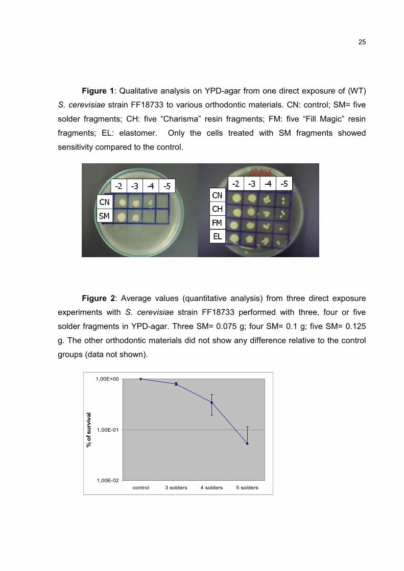
Figure 1: Qualitative analysis on YPD-agar from one direct exposure of (WT)
S. cerevisiae strain FF18733 to various orthodontic materials. CN: control; SM= five
solder fragments; CH: five “Charisma” resin fragments; FM: five “Fill Magic” resin
fragments; EL: elastomer. Only the cells treated with SM fragments showed
sensitivity compared to the control.
Figure 2: Average values (quantitative analysis) from three direct exposure
experiments with S. cerevisiae strain FF18733 performed with three, four or five
solder fragments in YPD-agar. Three SM= 0.075 g; four SM= 0.1 g; five SM= 0.125
g. The other orthodontic materials did not show any difference relative to the control
groups (data not shown).
1,00E-02
1,00E-01
1,00E+00
control 3 solders 4 solders 5 solders
% of survival

26
Figure 3: Qualitative analysis on YPD-agar from one exposure of (WT) S.
cerevisiae strain FF18733 to saliva pre-treated with the different orthodontic
materials. CN: control; SM= three solder fragments; CH: five “Charisma” resin
fragments; FM: five resin “Fill Magic” fragments; EL: elastomers; BR: brackets. The
SM fragments induced a complete inhibition of cell proliferation.
Figure 4: Qualitative analysis on YPD-agar from one exposure of S.
cerevisiae strains EG103 (WT) and EG133 (sod1/sod2) to saliva pre-treated with
three solder fragments (three SM), indicating a slight sensitivity of the mutant strain
compared to the WT strain. A similar result was observed for BG1 (apn1) compared
to the WT strain FF18733.
27
3 ARTIGO 2
Changes in Basal Cell Frequency in Orthodontic Patients
Authors: Graziela Westphalen1*, Philip Thomas2, Luciane Menezes1, João
Antônio Henriques3, Daniel Prá4, Renata Medina-Silva1, Michael Fenech2.
1 Faculdade de Odontologia, Pontifícia Universidade Católica do Rio Grande do Sul, Av.
Ipiranga, 6681 Prédio: 6, Sala: 210, Porto Alegre/RS, Brazil, Zip Code: 90619-900. Phone and fax
number:+55 51 33203538. 2 CSIRO Human Nutrition, PO Box 1004, Adelaide, Australia. S.A, 5000. 3 Departamento de Biofísica, Universidade Federal do Rio Grande do Sul, Av. Bento
Gonçalves, 9500, Prédio: 43421, Sala: 113, Porto Alegre/RS, Brazil, 91501-970. 4 Faculdade de Medicina, Universidade Católica de Pelotas, Rua: Félix da Cunha, 412,
Pelotas, Brazil, 96010-000
Abstract
The Buccal mucosa of orthodontic patients is in long term contact with metals
that might induce cytogenetic events. This study aimed to evaluate whether
orthodontic patients have higher risk for abnormal cytogenetic events in buccal cells
by investigating DNA damage, cell death and cell proliferation as determined by the
buccal micronucleus cytome assay. The relationship and subsequent correlation
between these various buccal cell types was also investigated. Buccal cell
frequencies were compared among three randomized groups: Controls (n=31);
patients wearing Hyrax appliance (n=30) and patients wearing fixed orthodontic
appliances (n=32). No significant differences for cell death and DNA damage events
were evident between control and study groups. Conversely, the frequency of basal
cells was significantly reduced in groups fitted with both orthodontic appliances. Such
differences might reflect alterations in cellular kinetics and the regenerative potential
of buccal mucosa. Significant correlations are reflective of the inter-relationships of
the various cell types.

28
Introduction
Orthodontic appliances are constantly subjected to corrosion processes
(Goncalves et al., 2008), resulting in increased exposure of the buccal mucosa to the
appliances’ metal based components (Faccioni et al., 2003). These appliances
consist of stainless steel alloys (35-65% cobalt; 20-30% chromium; 0- 30% nickel;
trace amounts of molybdenum, silica, beryllium, boron and carbon) and welding (50%
silver; 16% cadmium; 16% copper; 15% zinc; 3% nickel) and are usually worn for an
average mean time of 2 years. It is known that some of these metals such as: nickel,
chromium and cadmium exhibit both carcinogenic and mutagenic potential
(Beyersmann and Hartwig, 2008; Valko et al., 2005). Therefore, it is important to
investigate the potential cytotoxicity effects of these appliances on cells of the buccal
mucosa.
The buccal micronucleus cytome (BMCyt) assay is a minimally invasive and
established method for monitoring cytogenetic damage in human epithelial cells
(Fenech et al., 2007; Holland et al., 2008; Thomas et al., 2009). Its application
involves the assessment of biomarkers for DNA damage (micronuclei (MNi) and
nuclear bud (NBUD) frequency), Cell proliferation rates (basal and binucleated cells)
and Cell Death parameters (karyorrhexis, condensed chromatin, karyolitic and
pyknotic cells). Potential abnormal cytogenetic effects from orthodontic appliances
have not been previously assessed using the BMCyt assay. Previous studies
investigating these effects include: evaluation of MNi frequency (Westphalen et al.,
2008), comet assay sensitivity (Faccioni et al., 2003) and several in vitro cell culture
studies (Assad et al., 2002a; b; Montanaro et al., 2005; Wever et al., 1997). The aims
of this study was to investigate whether orthodontic patients have a higher risk for
abnormal cytogenetic effects in buccal cells by investigating cell proliferation rates,
DNA damage and cell death parameters as determined by the BMCyt assay and also
to evaluate the correlation and subsequent inter-relationships between these DNA
biomarkers and cells parameters.
29
Materials & methods
Approval for this study was obtained from the Pontifícia Universidade Católica
do Rio Grande do Sul, Brazil review board. The participant’s rights have been
explained and informed consent obtained. Frequency of BMCyt assay parameters
was measured on buccal cells of individuals, at University cited, from three groups
(randomized), by one operator: 1) Control (n=32, age: 9.03± 1.04 years, gender: 18
males/14 females) consisting of individuals without metal dental fillings; 2) Hyrax
appliance (n=31, age: 11.3± 1.93 years, gender: 17 males/14 females) with patients
wearing Hyrax appliance (Composition: stainless steel alloy and welding) for six
months and 3) Fixed orthodontic appliance (n=30, age: 22.0± 9.08 years, gender: 13
males/17 females) composed of patients with fixed orthodontic appliance
(Composition: stainless steel alloy) for 24 months. Individuals with certain lifestyle
factors (i.e. smoking or excessive alcohol intake), or occupational profiles (i.e.
exposure to metal ions or metal dental fillings), which has been shown to influence
genomic stability events were excluded.
Cell sampling and preparation of slides for microscopic analysis followed the
protocol: “Buccal micronucleus cytome assay” described by Thomas, Holland et al.
2009. Slides were de-identified and scored using a microscope equipped with a
triple-band filter (Nikon E600, Tokyo, Japan) at x1000 magnification (Dapi, FITC and
Rhodamine) in blind manner. Cells containing MNi on bright field were confirmed as
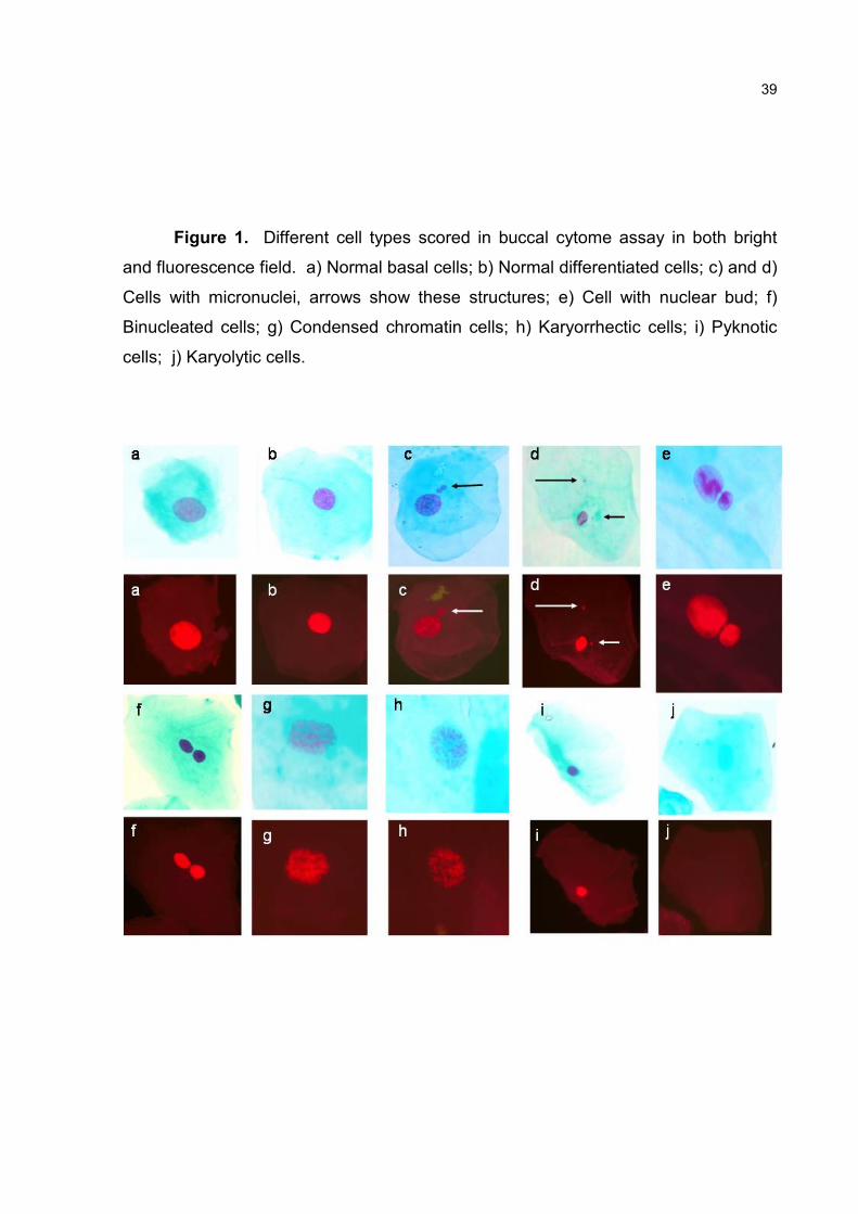
being positive by examining the cells under fluorescence (Figure 1). First, frequency
of all the various cell types was determined in 1000 cells and afterwards, frequency
of DNA damage biomarkers (MNi and NBUD) was determined in 2000 differentiated
cells and 200 basal cells. Figure 1 illustrates the different cells scored in the BMCyt
assay whilst more detailed descriptions are outlined below.
Normal basal cells (Figure 1a)
These cells are derived from the basal layer. The nuclear to cytoplasmic ratio
is larger than that in differentiated buccal cells. When compared to differentiated
cells, basal cells are smaller with a uniformly stained nucleus with more intense
green cytoplasmic stain.

30
Normal differentiated cells (Figure 1b)
These cells are derived from basal cells and have smaller nuclear to
cytoplasmic ratio relative to basal cells. The nucleus is oval or round, uniformly
stained and generally has lighter cytoplasmic green stain compared to basal cells.
Cells with micronuclei (Figure 1c and d)
These cells are characterized by the presence of one or two smaller MNi,
which are biomarkers of whole chromosome loss or breakage because they originate
from whole chromosomes or fragments that lag behind at anaphase (Fenech et al.,
1999). They range in size from 1/3 to 1/16 of the diameter of main nucleus, with a
similar staining intensity and texture to main nucleus.
Cells with Nuclear Bud (Figure 1e)
Cells containing a nuclear bud have a main nucleus with an apparent sharp
constriction indicative of a budding process. The NBUD is similar to the main nucleus
in terms of morphology and staining intensity. However, buds are smaller and range
in diameter size from half to quarter of that of the main nucleus.
Binucleated cells (Figure 1f)
These cells contain two distinct nuclei that are uniformly stained and
sometimes appear to be touching. These cells may be indicative of nuclear division
rate and/or failed cytokinesis following the last nuclear division.
Condensed chromatin cells (Figure 1g)
This cell population possesses nuclei with distinctive regions of condensed or
aggregated chromatin, exhibiting a speckled or striated pattern. Some areas of the
chromatin are darkly stained whereas others appear lighter due to loss of nuclear
material.
31
Karyorrhectic cells (Figure 1h)
Karyorrhectic cells exhibit more extensive nuclear chromatin aggregation
relative to condensed chromatin cells, leading to nuclear fragmentation and eventual
disintegration.
Pyknotic cells (Figure 1i)
These cells are characterised by a small shrunken nucleus with a diameter
usually one to two thirds that of a normal nucleus. The nuclear material contained
within pyknotic cells is uniformly and intensely stained.
Karyolytic cells (Figure 1j)
These cells when stained with Schiffs and Light Green appear negative for
nuclear material when viewed under both light and fluorescence microscopy. These
cells appear to have no nucleus although the faint outline of a membrane can
sometimes be determined.
Statistical analysis
One-way Analysis of Variance (Kruskal-Wallis test) followed by Dunn’s
Multiple Comparison test was applied to assess the significance of the cellular and
nuclear parameters measured between cohorts and Mann-Whitney test was applied
to evaluate gender and age differences. Correlations between biomarkers were
tested using Spearmann test (P≤ 0.05). These analysis values were performed using
SPSS version 10.0 (SPSS Inc, Chicago, IL) and Prism 5.0 (GraphPad Software Inc,
San Diego, CA).
Results
There were no significant differences in the frequency of DNA damage rates,
cell death and proliferation parameters among the three cohorts (Table 1).Only basal
cell frequency was significantly reduced in hyrax (19%) and fixed orthodontic (25%)
appliances relative to the control (p=0.022; Table 1; Figure 2). No age or gender

32
differences were observed for any parameter evaluated. Correlation analysis
between DNA biomarkers and cell parameters are described in Table 2.
Discussion
Previous studies have shown that the amount of metal ions liberated from
orthodontic appliances into the oral cavity due to corrosion processes was
significantly below the average dietary intake and did not reach toxic concentrations
(Agaoglu et al., 2001; Kocadereli et al., 2000). However, it can not be excluded that
even presumed non-toxic concentrations of these metals may be sufficient to induce
biological effects in vivo within oral mucosa cells over the long term chronic exposure
period (Faccioni et al., 2003). The aim of the current study was to assess the
potential cytotoxic effects which may result from orthodontic appliances using the
BMCyt assay. This is the first time that this assay has been used to investigate the
potential cytotoxic effects from orthodontic appliances. This minimally invasive assay
allows the assessment of biomarkers that are reflective of DNA damage events, cell
proliferation and cell death parameters (Fenech et al., 2007; Holland et al., 2008;
Thomas et al., 2009). Buccal cells can be easily accessed and reflect target sites that
have been shown to be sensitive biomarkers to the insults imposed by genotoxic
events, environmental and occupational exposures (Burgaz et al., 1999; Sarto et al.,
1990; Stich et al., 1984)
The results of this research showed that the use of appliances (Hyrax and
fixed orthodontic) were not related to any increase in cytotoxic events (Table 1),
which are in line with findings from several in vitro trials (Assad et al., 2002a; b;
Montanaro et al., 2005; Wever et al., 1997). In the same way, the occurrence of
these effects could not be confirmed through a trial that used both the MNi frequency
and comet assay in a group of fixed orthodontic patients, because both assays
showed opposing results (Westphalen et al., 2008). However, other studies have
shown that nickel and cobalt released from fixed orthodontic appliances in groups of
patients could induce DNA damage in oral mucosa cells (Faccioni et al., 2003).
Laboratory technicians exposed to metals (chromium, cobalt and nickel) resulting
from being in contact with dental materials showed an increase in the Micronucleus
33
(MN) frequency in peripheral lymphocytes and exfoliated nasal cells (Burgaz et al.,
2002). However, our results do not show a marked difference between the study
groups and controls for MN frequency or nuclear buds. As expression of MNi require
nuclear division, MN frequency in buccal mucosa is also dependent on the proportion
of once divided cells. It is possible that the reduced number of basal cells and
perhaps a reduced proportion of actively dividing basal cells in the appliance groups
may inhibit MNi and NBUD expression (the latter being S-phase dependent) (Shimizu
et al., 1998). The differences in these studies are probably attributable to the differing
kinetics of the basal and differentiated cells.The only parameter that was significantly
different between the control and study groups was the frequency of basal cells,
(p=0.022). This may be reflective of a reduced regenerative potential resulting from
metal alloy exposure which could lead to altered cell kinetics and changes within the
structural profile of the buccal mucosa as well as MNi and NBUD expression. The
changes in basal cell frequency between the three cohorts are also thought not to
reflect any age related changes, given the fact age was not associated to any
evaluated parameter, possibly because the age similarity between subjects. It has
been shown previously that the basal cell frequency tends to increase with normal
ageing but has been found to be significantly reduced in examples of premature
ageing syndromes such as Alzheimer’s and Down’s syndrome (Thomas and Fenech,
2007; Thomas et al., 2008).
Cross correlation analyses of biomarkers from this study (Table 2) show that
they are in agreement with the data of Thomas et al (2008) regarding the positive
association between condensed chromatin and karyorrhexis, pyknosis or basal cells.
Conversely to the study of Thomas et al (2008) with Down syndrome subjects and
young or old controls, we observed a negative correlation between condensed
chromatin and karyolysis and did not observe correlations between basal and
karyorrhexis nor between pyknosis and karyorrhexis. Based in the assumption that
the correlations are indicative of the biological association between the cell types and
their likely sequential development (Thomas et al, 2008) and that the present study is
one of the first to evaluate buccal cell dynamics, we can provide some early input
about cell dynamics in the oral mucosa. For instance, the positive correlation

34
between basal and condensed chromatin indicate the later cells might derive directly
from basal cells. The negative correlation between karyolysis and condensed
chromatin or basal cells indicate karyolitic cells are not likely to derive from basal or
condensed chromatin cells in healthy subjects. The high correlation between
karyolysis and differentiated cells indicate the former cells originate from the latter.
Further studies are needed to better understand buccal cell dynamics both in healthy
and/or pathological situations.
One of the limitations of this study was the difference in age amongst the three
groups. This occurred because each group requested specific characteristics e.g.:
for the control it was necessary to have individuals with no metal dental fillings within
the oral cavity.; Hyrax appliance is mainly used in children and young adults,
whereas the fixed orthodontic appliance is usually worn in older individuals. The
mean time of use for both appliances could not be accurately matched as each
appliance presents determined time of use. Control group also presented high
frequency of NBUD’s per 2000 differentiated cells. Although it is tentative to explain
differences in DNA damage background due to socioeconomic and lifestyles
characteristics, however both controls and individuals fitted with appliances showed
similarities, since both lived in low income areas. Another aspect of this study was
that it evaluated only the target cells, it is encouraged that surrogate tissue and
systemic metal levels are assessed (Burgaz et al., 2002), in order to provide an
overall biological effect of appliance in the individuals.
In conclusion, this study suggested a reduction in the regenerative potential of
this tissue possibly reflecting changes in cellular kinetics and the structural profile of
the buccal mucosa.
Acknowledgements
This study was supported by Coordenação de Aperfeiçoamento de Pessoal de
Nível Superior, Brazil. The authors thank the staff from: Laboratory of Human
Nutrition of CSIRO, Australia, especially Maryam Hor and Nathan O’Callaghan for
technical advice and from Pontifícia Universidade Católica do Rio Grande do Sul,

35
Brazil, especially Fabiane Azeredo for helping to recruit the individuals and to those
individuals who participated voluntarily in this study.
References
Agaoglu G, Arun T, Izgi B, Yarat A (2001). Nickel and chromium levels in the saliva and serum of patients with fixed orthodontic appliances. Angle Orthod 71:375-9.
Assad M, Chernyshov A, Leroux MA, Rivard CH (2002a). A new porous titanium-nickel alloy: part 2. Sensitization, irritation and acute systemic toxicity evaluation. Biomed Mater Eng 12:339-46.
Assad M, Chernyshov A, Leroux MA, Rivard CH (2002b). A new porous titanium-nickel alloy: Part 1. Cytotoxicity and genotoxicity evaluation. Biomed Mater Eng 12:225-37.
Beyersmann D, Hartwig A (2008). Carcinogenic metal compounds: recent insight into molecular and cellular mechanisms. Arch Toxicol 82:493-512.
Burgaz S, Karahalil B, Bayrak P, Taskin L, Yavuzaslan F, Bokesoy I, et al. (1999). Urinary cyclophosphamide excretion and micronuclei frequencies in peripheral lymphocytes and in exfoliated buccal epithelial cells of nurses handling antineoplastics. Mutat Res 439:97-104.
Burgaz S, Demircigil GC, Yilmazer M, Ertas N, Kemaloglu Y, Burgaz Y (2002). Assessment of cytogenetic damage in lymphocytes and in exfoliated nasal cells of dental laboratory technicians exposed to chromium, cobalt, and nickel. Mutat Res 521:47-56.
Faccioni F, Franceschetti P, Cerpelloni M, Fracasso ME (2003). In vivo study on metal release from fixed orthodontic appliances and DNA damage in oral mucosa cells. Am J Orthod Dentofacial Orthop 124:687-93.
Fenech M, Holland N, Chang WP, Zeiger E, Bonassi S (1999). The HUman MicroNucleus Project--An international collaborative study on the use of the micronucleus technique for measuring DNA damage in humans. Mutat Res 428:271-83.
Fenech M, Crott JW (2002). Micronuclei, nucleoplasmic bridges and nuclear buds induced in folic acid deficient human lymphocytes-evidence for breakage-fusion-bridge cycles in the cytokinesis-block micronucleus assay. Mutat Res 504:131-6.
Fenech M, Bolognesi C, Kirsch-Volders M, Bonassi S, Zeiger E, Knasmuller S, et al. (2007). Harmonisation of the micronucleus assay in human buccal cells--a Human Micronucleus (HUMN) project (www.humn.org) initiative commencing in 2007. Mutagenesis 22:3-4.
36
Gonçalves TS, de Menezes LM, Silva LE (2008). Residual monomer of autopolymerized acrylic resin according to different manipulation and polishing methods. An in situ evaluation. Angle Orthod 78:722-7.
Holland N, Bolognesi C, Kirsch-Volders M, Bonassi S, Zeiger E, Knasmuller S, et al. (2008). The micronucleus assay in human buccal cells as a tool for biomonitoring DNA damage: the HUMN project perspective on current status and knowledge gaps. Mutat Res 659:93-108.
Kocadereli L, Atac PA, Kale PS, Ozer D (2000). Salivary nickel and chromium in patients with fixed orthodontic appliances. Angle Orthod 70:431-4.
Montanaro L, Cervellati M, Campoccia D, Prati C, Breschi L, Arciola CR (2005). No genotoxicity of a new nickel-free stainless steel. Int J Artif Organs 28:58-65.
Sarto F, Tomanin R, Giacomelli L, Iannini G, Cupiraggi AR (1990). The micronucleus assay in human exfoliated cells of the nose and mouth: application to occupational exposures to chromic acid and ethylene oxide. Mutat Res 244:345-51.
Shi Q, King RW (2005). Chromosome nondisjunction yields tetraploid rather than aneuploid cells in human cell lines. Nature 437:1038-42.
Shimizu N, Itoh N, Utiyama H, Wahl GM (1998). Selective entrapment of extrachromosomally amplified DNA by nuclear budding and micronucleation during S phase. J Cell Biol 140:1307-20.
Stich HF, Rosin MP, Vallejera MO (1984). Reduction with vitamin A and beta-carotene administration of proportion of micronucleated buccal mucosal cells in Asian betal nut and tobacco chewers. Lancet 1:1204-6.
Thomas P, Fenech M (2007). A review of genome mutation and Alzheimer's disease. Mutagenesis 22:15-33.
Thomas P, O'Callaghan N, Fenech M (2008). Telomere length in white blood cells, buccal cells and brain tissue and its variation with ageing and Alzheimer's disease. Mech Ageing Dev 129:183-90.
Thomas P, Holland N, Bolognesi C, Kirsch-Volders M, Bonassi S, Zeiger E, et al. (2009). Buccal micronucleus cytome assay. Nat Protoc 4:825-37.
Valko M, Morris H, Cronin MT (2005). Metals, toxicity and oxidative stress. Curr Med Chem 12:1161-208.
Westphalen GH, Menezes LM, Pra D, Garcia GG, Schmitt VM, Henriques JA, et al. (2008). In vivo determination of genotoxicity induced by metals from orthodontic appliances using micronucleus and comet assays. Genet Mol Res 7:1259-66.
Wever DJ, Veldhuizen AG, Sanders MM, Schakenraad JM, van Horn JR (1997). Cytotoxic, allergic and genotoxic activity of a nickel-titanium alloy. Biomaterials 18:1115-20.
37
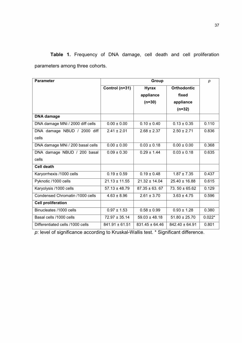
Table 1. Frequency of DNA damage, cell death and cell proliferation
parameters among three cohorts.
Group Parameter
Control (n=31) Hyrax
appliance
(n=30)
Orthodontic
fixed
appliance
(n=32)
p
DNA damage
DNA damage MNi / 2000 diff cells 0.00 ± 0.00 0.10 ± 0.40 0.13 ± 0.35 0.110
DNA damage NBUD / 2000 diff
cells
2.41 ± 2.01 2.68 ± 2.37 2.50 ± 2.71 0.836
DNA damage MNi / 200 basal cells 0.00 ± 0.00 0.03 ± 0.18 0.00 ± 0.00 0.368
DNA damage NBUD / 200 basal
cells
0.09 ± 0.30 0.29 ± 1.44 0.03 ± 0.18 0.635
Cell death
Karyorrhexis /1000 cells 0.19 ± 0.59 0.19 ± 0.48 1.87 ± 7.35 0.437
Pyknotic /1000 cells 21.13 ± 11.55 21.32 ± 14.04 25.40 ± 16.88 0.615
Karyolysis /1000 cells 57.13 ± 48.79 87.35 ± 63. 67 73. 50 ± 65.62 0.129
Condensed Chromatin /1000 cells 4.63 ± 8.96 2.61 ± 3.70 3.63 ± 4.75 0.596
Cell proliferation
Binucleates /1000 cells 0.97 ± 1.53 0.58 ± 0.99 0.93 ± 1.28 0.380
Basal cells /1000 cells 72.97 ± 35.14 59.03 ± 48.18 51.80 ± 25.70 0.022*
Differentiated cells /1000 cells 841.91 ± 61.51 831.45 ± 64.46 842.40 ± 64.91 0.801
p: level of significance according to Kruskal-Wallis test. * Significant difference.
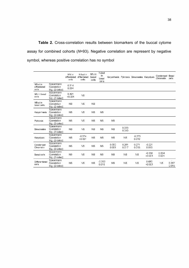
38
Table 2. Cross-correlation results between biomarkers of the buccal cytome
assay for combined cohorts (N=93). Negative correlation are represent by negative
symbol, whereas positive correlation has no symbol
39
Figure 1. Different cell types scored in buccal cytome assay in both bright
and fluorescence field. a) Normal basal cells; b) Normal differentiated cells; c) and d)
Cells with micronuclei, arrows show these structures; e) Cell with nuclear bud; f)
Binucleated cells; g) Condensed chromatin cells; h) Karyorrhectic cells; i) Pyknotic
cells; j) Karyolytic cells.

40
Figure 2. Reduction of basal cell frequency in individuals wearing appliances.
*statistical difference in relation to control according to Dunn’s Multiple Comparison
test. HA: Hyrax appliance; OFA: Orthodontic fixed appliance.
41
4 ARTIGO 3
In vivo genotoxicity assessment of maxillary expander appliances
Authors: Graziela Henriques Westphalen1, Luciane Macedo de Menezes1, Fabiane
Azeredo1, Gabriela Schmitt1, Daniel Prá2, João Antônio Pêgas Henriques3, Renata
Medina Silva1.
1 Faculdade de Odontologia, Pontifícia Universidade Católica do Rio Grande do Sul, Av.
Ipiranga, 6681 Prédio: 6, Sala: 210, Porto Alegre/RS, Brazil, Zip Code: 90619-900. Phone and fax
number:+55 51 33203538. 2 Faculdade de Medicina, Universidade Católica de Pelotas, Rua: Félix da Cunha, 412,
Pelotas, Brazil, 96010-000 3 Departamento de Biofísica, Universidade Federal do Rio Grande do Sul, Av. Bento
Gonçalves, 9500, Prédio: 43421, Sala: 113, Porto Alegre/RS, Brazil, 91501-970.
Abstract
Some components of maxillary expansor appliances (Haas and Hyrax) might
induce genotoxic effects. The aim of this study was to assess the genotoxicity related
to wearing maxillary expansor appliances (Haas and Hyrax) in group of patients.
Micronucleus assays were carried out in buccal cells, which were sampled before
(T1), 10 (T2) and 30 (T3) days after the placement of maxillary expansor appliances.
The frequency of micronuclei was compared among three periods, using the
Wilcoxon test (p<0.05). Results showed that compared with T1, the micronucleus
frequency decreased significantly at T3. Therefore, the appliances did not show any
genotoxic effects in these patients. However, further studies in long-term models
using different methodologies are necessary to better understanding the biological
effects.
42
Introduction
Negligence of biocompatible dental materials can provoke health risks to
patients, dental staff members and practitioners themselves 1. Maxillary expander
appliances might be constituted by stainless steel alloys (35-65% cobalt; 20-30%
chromium; 0- 30% nickel; small amount of molybdenum, silicon, beryllium, boron and
carbon), welding (50% silver; 16% cadmium; 16% copper; 15% zinc; 3% nickel) and
acrylic resins (methacrylates), which contain elements with known genotoxic potential 2-4. When these appliances are present in the oral cavity, their constituents elements
can be released by corrosion 5. Therefore, it is important to study the biological
effects of these appliances.
The study of genotoxicity is a field of genetics that studies the
processes that alter heredity (mutagenesis) or genetic determinism (carcinogenesis
and teratogenesis) 6. There is a wide range of assays to verify genotoxicity; one of
them is the micronucleus (MN) assay, which has been successfully used to monitor
human populations exposed to mutagenic and carcinogenic agents 7. This assay
evaluates the frequency of MN, which are extracellular structures composed of
chromosomes or chromosome fragments that are not incorporated into the main
nucleus during mitoses 8.
Concerning the importance of biocompatibility, this study used the MN assay
to assess the genotoxicity related to wearing of maxillary expansor appliances (Haas
and Hyrax) in a group of patients.
Materials and methods
This study was approved by the Pontifical Catholic University of Rio Grande
do Sul Ethics Committee. Ten patients (mean age:12.2 ± 6.9 years; 6 males and 4
females) undergoing maxillary expansion with two appliances, Haas (stainless steel
alloy and acrylic resin) (n=7) and Hyrax (stainless steel alloy and welding) (n= 3),
participated in this study. Individuals with certain lifestyle factors (i.e., smoking or
excessive alcohol intake), or certain occupational profiles (i.e., exposure to metal
43
ions or metal dental fillings) that have been shown to influence genomic stability were
excluded.
Buccal cell samples were collected by one operator from patients at three
periods: before (T1), 10 days (T2) and 30 (T3) days after placement of appliances.
The patients rinsed with distilled water for 2 minutes to remove exfoliated buccal cells 9; cells were collected using swabs and transferred to polyethylene tubes (50 ml)
containing phosphate-buffered saline (20 ml) and slides were prepared according to
the protocol established by Titenko-Holland, Moore and Smith (1994) 10.
Slides were analyzed at 1000X magnification in oil immersion with light
microscope (Axiolab, Zeiss) to determine the frequency of MN; 1000 cells were
evaluated for each patient. Only cells that were not smeared, clumped or overlapping
and that contained intact nuclei were included in the analysis. MN were identified if
they had the following characteristics: less than 1/3 diameter of the main nucleus; the
same plane of focus; the same color, texture and refraction as the main nucleus;
smooth oval or round shape, and clearly separated from the main nucleus 8.
MN frequency was compared among the three periods, using Wilcoxon test
(p<0.05 was considered statistically significant) with SPSS version 10.0 (SPSS Inc,
Chicago, USA). All values are expressed as mean ± standard deviation.
Results
There was a significant difference in MN frequency only for the comparison
between T1 and T3 (Table I), where a significant decrease of MN frequency was
observed at T3 (Figures 1 and 2).
Discussion
The scope of this study was to verify the genotoxicity of the maxillary
expander appliances, since they have constituents with known genotoxic effects and
might stay in oral cavity for a median period of 6 months, often in young patients
whose organs are developing.
The MN assay is an in vivo assay often used to detect whether agents
have caused chromosomal damage 7. MN formation is an effective biomarker of
44
illness and the process associated with induction of DNA damage 7, 8, 11, 12. MN
occurrence is detected after cellular division and after some time has passed from
exposure to a harmful agent 13. Thus, in this study, buccal cells were sampled at 10
(T2) and 30 (T3) days after placement of appliances. The type of cells used (buccal)
was successful for using the MN assay, as it requires minimally invasive procedures
for sampling 7, 12, 14, and it also constitutes the target cells, which are in direct contact
with the appliances.
This study showed that the maxillary expander appliance did not provoke
genotoxic effects in the sample of patients. These findings are in line with several in
vitro cell culture studies 15-18. Genotoxicity effects from fixed orthodontic appliances
were not confirmed by Westphalen et al. 19, however, an increase of MN was
observed after 30 days of placement of the appliance evaluated. In contrast, Faccioni
et al. 20 found that metals (nickel and cobalt) released from fixed orthodontic
appliances induced DNA damage to the buccal cells of orthodontic patients. Burgaz
et al. 21 observed that metals ions released by stainless steel alloys (chromium,
cobalt and nickel) were associated with increases in MN frequency in both
lymphocytes and nasal cells of dental technician staff.
One of the limitations of this study was the small sample size, due to patient´s
desistence of study´s step (sampling). In addition, there was evaluation of only
buccal cells. Burgaz et al. 21 suggested that beyond the target cells, other cells (e.g.,
lymphocytes) should also be analyzed and systemic measures should be carried out
to assess genotoxicity from dental materials.
Conclusion
The maxillary expander appliances (Haas and Hyrax) did not induce genotoxic
damage in the population evaluated. However, it is necessary that long-term studies
be conducted, using a wide-range of tests and systemic measures, to better assess
the full effect.

45
Acknowledgements
Authors thank Fundação de Amparo à Pesquisa do Estado do Rio Grande do
Sul (process number: 06511624) for the scholarship and the volunteers who
participated in this study.
References
1. Wataha JC. Principles of biocompatibility for dental practitioners. J Prosthet
Dent 2001;86:203-209.
2. Beyersmann D, Hartwig A. Carcinogenic metal compounds: recent insight into molecular and cellular mechanisms. Arch Toxicol 2008;82:493-512.
3. Schweikl H, Spagnuolo G, Schmalz G. Genetic and cellular toxicology of
dental resin monomers. J Dent Res 2006;85:870-877. 4. Hossain S, Liu HN, Nguyen M, Shore G, Almazan G. Cadmium exposure
induces mitochondria-dependent apoptosis in oligodendrocytes. Neurotoxicology 2009.
5. Matos de Souza R, Macedo de Menezes L. Nickel, chromium and iron
levels in the saliva of patients with simulated fixed orthodontic appliances. Angle Orthod 2008;78:345-350.
6. Ribeiro LRS, D. M. F.; Marques, E. K. . Mutagênese ambiental. Canoas:
Ulbra; 2003. 7. Holland N, Bolognesi C, Kirsch-Volders M, Bonassi S, Zeiger E,
Knasmueller S et al. The micronucleus assay in human buccal cells as a tool for biomonitoring DNA damage: the HUMN project perspective on current status and knowledge gaps. Mutat Res 2008;659:93-108.
8. Sarto F, Finotto S, Giacomelli L, Mazzotti D, Tomanin R, Levis AG. The
micronucleus assay in exfoliated cells of the human buccal mucosa. Mutagenesis 1987;2:11-17.
9. Beserati NA. Immunoperoxidase detection of polycyclic aromatic
hydrocarbon - DNA addutcts in mouth floor and buccal mucosa cells of smokers and nonsmokers. Environ Mol Mutagen. 2000;36:127-133.
10. Titenko-Holland N, Moore LE, Smith MT. Measurement and
characterization of micronuclei in exfoliated human cells by fluorescence in situ hybridization with a centromeric probe. Mutat Res 1994;312:39-50.

46
11. Fenech M, Holland N, Chang WP, Zeiger E, Bonassi S. The HUman MicroNucleus Project--An international collaborative study on the use of the micronucleus technique for measuring DNA damage in humans. Mutat Res 1999;428:271-283.
12. Thomas P, Holland N, Bolognesi C, Kirsch-Volders M, Bonassi S, Zeiger E
et al. Buccal micronucleus cytome assay. Nat Protoc 2009;4:825-837. 13. Silva JE, B.; Henriques, J. A. P. . Genética toxicológica. Porto Alegre:
Alcance; 2003. 14. Fenech M, Bolognesi C, Kirsch-Volders M, Bonassi S, Zeiger E,
Knasmuller S et al. Harmonisation of the micronucleus assay in human buccal cells--a Human Micronucleus (HUMN) project (www.humn.org) initiative commencing in 2007. Mutagenesis 2007;22:3-4.
15. Wever DJ, Veldhuizen AG, Sanders MM, Schakenraad JM, van Horn JR.
Cytotoxic, allergic and genotoxic activity of a nickel-titanium alloy. Biomaterials 1997;18:1115-1120.
16. Montanaro L, Cervellati M, Campoccia D, Prati C, Breschi L, Arciola CR.
No genotoxicity of a new nickel-free stainless steel. Int J Artif Organs 2005;28:58-65. 17. Assad M, Chernyshov A, Leroux MA, Rivard CH. A new porous titanium-
nickel alloy: part 2. Sensitization, irritation and acute systemic toxicity evaluation. Biomed Mater Eng 2002;12:339-346.
18. Assad M, Chernyshov A, Leroux MA, Rivard CH. A new porous titanium-
nickel alloy: Part 1. Cytotoxicity and genotoxicity evaluation. Biomed Mater Eng 2002;12:225-237.
19. Westphalen GH, Menezes LM, Pra D, Garcia GG, Schmitt VM, Henriques
JA et al. In vivo determination of genotoxicity induced by metals from orthodontic appliances using micronucleus and comet assays. Genet Mol Res 2008;7:1259-1266.
20. Faccioni F, Franceschetti P, Cerpelloni M, Fracasso ME. In vivo study on
metal release from fixed orthodontic appliances and DNA damage in oral mucosa cells. Am J Orthod Dentofacial Orthop 2003;124:687-693; discussion 693-684.
21. Burgaz S, Demircigil GC, Yilmazer M, Ertas N, Kemaloglu Y, Burgaz Y.
Assessment of cytogenetic damage in lymphocytes and in exfoliated nasal cells of dental laboratory technicians exposed to chromium, cobalt, and nickel. Mutat Res 2002;521:47-56.
22. Thomas P, NJ OC, Fenech M. Telomere length in white blood cells, buccal
cells and brain tissue and its variation with ageing and Alzheimer's disease. Mech Ageing Dev 2008;129:183-190.
47
23. O'Callaghan N, Dhillon V, Thomas P, Fenech M. A quantitative real-time
PCR method for absolute telomere length. Biotechniques 2008;44:807-809.

48
Table I - MN frequency comparison among each period
Periods n Average SD p
T1 10 3,00 3,09 0,284
T2 10 1,70 1,89
T1 10 3,00 3,09 0,031
T3 10 1,30 1,06
T2 10 1,70 1,89 0,522
T3 10 1,30 1,06
Figure 1 - MN frequency for each period and patient
Figure 2 - Average MN frequency for each period

49
5 DISCUSSÃO GERAL
A importância do estudo da biocompatibilidade dos materiais odontológicos é
um consenso na literatura29-32, uma vez que o emprego de materiais biocompatíveis
é essencial para garantir um tratamento biologicamente seguro aos pacientes14. A
negligência dessa propriedade pode acarretar um aumento do risco à saúde dos
pacientes, equipe odontológica e laboratorial29.
A maioria dos metais utilizados na Ortodontia apresenta composição similar
ao aço inoxidável (18% cromo e 8% níquel). Esses metais são potencialmente
relacionados a efeitos citotóxicos, genotóxicos e carcinogênicos4,6. Determinados
acessórios ortodônticos (anéis, bráquetes e máscaras faciais) contêm algum tipo de
solda, cuja composição é comumente à base de prata e cobre, sendo que também
apresentam outros metais como cádmio. A prata é considerada um metal tóxico para
humanos33, podendo alterar o metabolismo celular em exposições crônicas de baixa
dosagem22. Da mesma forma, o cádmio apresenta efeitos tóxicos relevantes
(nefrotoxicidade, neurotoxicidade) em diversos organismos7,10,21,34, sendo também
poluente e carcinógeno7.
Além dos efeitos deletérios dos metais, outros constituintes de materiais
ortodônticos apresentam efeitos adversos. A utilização de resina acrílica em
aparelhos removíveis, contenção e acessórios fixos tem sido apontada como fator
etiológico de reações de hipersensibilidade9. Efeitos citotóxicos in vitro também são
atribuídos às resinas acrílicas, especialmente as quimicamente ativadas35. A
presença de monômero não polimerizado, proveniente das resinas compostas
utilizadas para colagem ortodôntica, é relacionada a efeitos de hipersensibilidade,
citotoxicidade, genotoxicidade e alterações da reposta imune36. O uso do látex em
diversos materiais odontológicos (luvas, lençol de isolamento e acessórios
ortodônticos, como elásticos ortodônticos) é apontado com causador de reações
alérgicas importantes37-39. Tendo em vista as evidências deletérias dos constituintes
dos materiais ortodônticos, é muito importante a realização de estudos que
investiguem os efeitos biológicos desses materiais.
50
A avaliação da biocompatibilidade dos materiais odontológicos compreende a
abordagem de diferentes aspectos dos mesmos como: resistência a corrosão,
potenciais citotóxicos, genotóxicos e carcinógenos. Deve-se considerar que a
determinação da biocompatibilidade representa um processo complexo que envolve
uma série de testes in vitro e in vivo40. Nesse estudo, a biocompatibilidade de
materiais ortodônticos foi avaliada sob dois aspectos: 1) Citotoxicidade: por meio de
testes in vitro, utilizando-se Saccharomyces cerevisiae, como organismo modelo 2)
Genotoxicidade: optando-se pelo emprego dos experimentos: ensaio citoma bucal
de micronúcleos, teste de micronúcleos e ensaio cometa em pacientes sob
tratamento ortodôntico.
O emprego de Saccharomyces cerevisiae em testes de citotoxicidade é
recomendado, devido às similaridades desse microorganismo com células humanas
e animais, uma vez que o mesmo é eucarioto e unicelular. Além disso, muitas
vantagens estão associadas à sua utilização como: fácil cultivo, manipulação e
controle, obtenção de experimentos com grande quantidade amostral, rapidez e
baixo custo18,41,42. Esse microorganismo é freqüentemente utilizado para avaliação
da toxicidade de diferentes metais43-46.
Dentre os testes de avaliação da genotoxicidade, o teste de micronúcleos
identifica danos cromossômicos após a divisão celular, sendo a técnica mais
utilizada para detecção de agentes clastogênicos (que quebram cromossomos) e
aneugênicos (que induzem aneuploidia, ou segregação cromossômica anormal),
sendo extremamente sensível para o monitoramento de danos genéticos27. Por sua
vez, o ensaio citoma bucal de micronúcleos é um método minimamente invasivo,
que provê a análise de danos do DNA, instabilidade cromossômica, morte celular e
potencial regenerativo dos tecidos da mucosa bucal humana. É observado um
aumento do uso desse método em estudos epidemiológicos moleculares para
investigação do impacto de exposição à genotoxicinas, nutrição, estilo de vida e
genótipo em danos do DNA, segregação cromossômica e morte celular26,27.
Dentre os materiais ortodônticos utilizados nesse estudo, o único que
apresentou citotoxicidade foi a solda de prata (Artigo 1), que mostrou uma redução
significativa da viabilidade celular de S. Cerevisiae em ambas exposições, direta e

51
indireta (saliva artificial), a esse microorganismo. Esse achado enfatiza a ação tóxica
desse material demonstrada previamente por vários estudos10,47-49. Dessa forma,
evidencia-se a necessidade do desenvolvimento de materiais mais biocompatíveis
para substituição da solda de prata. Nesse sentido, a solda a laser apresenta-se
como uma alternativa promissora, uma vez que esse material mostrou, em estudos
anteriores, uma redução marcante da toxicidade celular em relação à solda de
prata47-49, apresentando uma tolerância biológica celular adequada48. Apesar dos
materiais (bráquetes, fios, resinas compostas e elastômeros) não mostrarem efeitos
citotóxicos nesse estudo, seus efeitos biológicos previamente relatados
(hipersensibilidade, citotoxicidade e genotoxicidade) não devem ser negligenciados
e constantemente estudados.
Em relação à genotoxicidade, este estudo mostrou que o uso dos aparelhos
tipo Haas e Hyrax e Ortodônticos fixos (liga de aço inoxidável) não demonstraram
aumento dos biomarcadores de: danos no DNA (freqüência de micronúcleos e
nuclear BUD), morte celular (cromatina condensada, cariorexe, cariólise e picnose) e
proliferação celular (núcleo duplo) indicados pelo ensaio citoma bucal de
micronúcleos (Artigo 2). Da mesma forma, os aparelhos expansores maxilares
(Hyrax e Haas) não mostraram potencial genotóxico (aumento da freqüência de
micronúcleos após 30 dias do uso dos aparelhos) em grupo de pacientes
ortodônticos jovens (Artigo 3).
Os achados indicativos de ausência de genotoxicidade associados ao uso de
aparelhos ortodônticos encontrados nesse estudo vão ao encontro de resultados de
muitos estudos in vitro12-15. De acordo com Westphalen et al. (2008)50, a
genotoxicidade de aparelhos ortodônticos fixos (liga aço inoxidável) não pode ser
totalmente evidenciada, uma vez que se observaram resultados contrastantes como,
aumento da freqüência de micronúcleos e diminuição de danos induzidos pelo teste
cometa, 30 e 10 dias, respectivamente, após a colocação de aparelho, em um grupo
de pacientes. Entretanto, Faccioni et al. (2003)3 demonstraram que metais (níquel e
cobalto), quando liberados dos aparelhos ortodônticos fixos, podem causar quebras
no DNA de células da mucosa bucal de pacientes ortodônticos. Da mesma forma, a
exposição aos metais (cromo, cobalto e níquel) mostrou um aumento da freqüência
52
de micronúcleos tanto em células nasais quanto em linfócitos periféricos de técnicos
dentários de laboratórios51. Frente a essas evidências, acredita-se que a
investigação da genotoxicidade dos aparelhos ortodônticos não esteja
complementarmente esgotada.
O entendimento das correlações celulares é extremamente importante uma
vez que as mesmas são indicativas de associação biológica entre as células e seu
desenvolvimento seqüencial52, entretanto ainda é pouco abordado. Esse estudo
(Artigo 2) foi um dos primeiros que avaliaram a dinâmica das células bucais, e
observou-se a existência de correlações positivas e negativas de biomarcadores
celulares detectados pelo uso do ensaio citoma bucal de micronúcleos. Essas
evidências fornecem dados iniciais para o entendimento das correlações celulares,
entretanto, acredita-se que mais estudos devam ser realizados para melhor
compreender o processo complexo da dinâmica celular.
53
6 REFERÊNCIAS
1.Noort RV, Introdução aos materiais dentários. São Paulo: Artmed; 2004. 2.Costa CAS, Souza PPC. Testes de citotoxicidade em cultura de células. São
Paulo: Artmed; 2005. 3.Faccioni F, Franceschetti P, Cerpelloni M, Fracasso ME. In vivo study on metal
release from fixed orthodontic appliances and DNA damage in oral mucosa cells. Am J Orthod Dentofacial Orthop. 2003 124(6):687-93.
4.Valko M, Morris H, Cronin MT. Metals, toxicity and oxidative stress. Curr Med
Chem. 2005 12(10):1161-208. 5.Sethi PK, Khandelwal D. Cadmium exposure: health hazards of silver cottage
industry in developing countries. J Med Toxicol. 2006 2(1):14-5. 6.Beyersmann D, Hartwig, A. Carcinogenic metal compounds: recent insight into
molecular and cellular mechanisms. Arch Toxicol. 2008 82(8): 493-512. 7.Hossain S, Liu HN, Nguyen M, Shore G, Almazan G. Cadmium exposure induces
mitochondria-dependent apoptosis in oligodendrocytes. Neurotoxicology. 2009 30(4):544-54.
8.Schweikl H, Spagnuolo G, Schmalz G. Genetic and cellular toxicology of dental
resin monomers. J Dent Res. 2006 85(10):870-7. 9.Goncalves TS, Morganti MA,Campos LC,Rizzatto SM, Menezes LM. Allergy to
auto-polymerized acrylic resin in an orthodontic patient. Am J Orthod Dentofacial Orthop. 2006 129(3):431-5.
10.Freitas MP, Oshima HM, Menezes LM, Machado DC, Viezzer C. Cytotoxicity of
silver solder employed in orthodontics. Angle Orthod. 2009 79(5): 939-44. 11.Menezes LM, Quintao CA, Bolognese AM. Urinary excretion levels of nickel in
orthodontic patients. Am J Orthod Dentofacial Orthop. 2007 131(5):635-8.
54
12.Assad M, Chernyshov A, Leroux MA, Rivard CH. A new porous titanium-nickel alloy: Part 1. Cytotoxicity and genotoxicity evaluation. Biomed Mater Eng. 2002 12(3):225-37.
13.Assad M, Chernyshov A, Leroux MA, Rivard CH. A new porous titanium-nickel
alloy: part 2. Sensitization, irritation and acute systemic toxicity evaluation. Biomed Mater Eng. 2002 12(4):339-46.
14.Montanaro L, Cervellati M, Campoccia D, Prati C,Breschi L, Arciola CR. No
genotoxicity of a new nickel-free stainless steel. Int J Artif Organs. 2005 28(1):58-65.
15.Wever DJ, Veldhuizen AG, Sanders MM, Schakenraad JM, Van Horn JR.
Cytotoxic, allergic and genotoxic activity of a nickel-titanium alloy. Biomaterials. 1997 18(16):1115-20.
16.Grimsdottir MR, Hensten-Pettersen A, Kullmann A. Cytotoxic effect of orthodontic
appliances. Eur J Orthod. 1992 14(1):47-53. 17.Mockers O, Deroze D, Camps J. Cytotoxicity of orthodontic bands, brackets and
archwires in vitro. Dent Mater. 2002. 18(4): p. 311-7. 18.De Freitas J, Wintz H, Kim JH, Poynton H, Fox T,Vulpe C. Yeast, a model
organism for iron and copper metabolism studies. Biometals. 2003 16(1):185-97. 19.Kao CT, Ding SJ, Min Y, Hsu TC, Chou MY,Huang TH. The cytotoxicity of
orthodontic metal bracket immersion media. Eur J Orthod. 2007 29(2):198-203. 20.Lewis JB, Wataha JC, Randol TM, McCloud VV, Lockwood PE.Metal ions alter
lipopolysaccharide-induced NF kappa B binding in monocytes. J Biomed Mater Res A. 2003 67(3):868-75.
21.Watjen W, Haase H, Biagioli M, Beyersmann D. Induction of apoptosis in
mammalian cells by cadmium and zinc. Environ Health Perspect. 2002 110 (Suppl 5):865-7.

55
22.Wataha JC, Lockwood PE, Schedle A, Noda M, Bouillaguet S. Ag, Cu, Hg and Ni ions alter the metabolism of human monocytes during extended low-dose exposures. J Oral Rehabil. 2002 29(2):133-9.
23.Issa Y, Brunton P, Waters CM, Watts DC. Cytotoxicity of metal ions to human
oligodendroglial cells and human gingival fibroblasts assessed by mitochondrial dehydrogenase activity. Dent Mater. 2008 24(2):281-7.
24.Bohrer PL. Avaliação das alterações citopatológicas da mucosa bucal
clinicamente normal exposta a carcinógenos (dissertação). Porto Alegre (RS): Universidade Federal do Rio Grande do Sul; 2003.
25.Fenech M, Holland N, Chang WP, Zeiger E, Bonassi S. The HUman
MicroNucleus Project--An international collaborative study on the use of the micronucleus technique for measuring DNA damage in humans. Mutat Res. 1999 428(1-2):271-83.
26.Thomas P, Holland N, Bolognesi C, Kirsch-Volders M, Bonassi S, Zeiger
E.Buccal micronucleus cytome assay. Nat Protoc. 2009 4(6):825-37. 27.Holland N, Bolognesi C, Kirsch-Volders M, Bonassi S, Zeiger E, Knasmueller S,
et al. The micronucleus assay in human buccal cells as a tool for biomonitoring DNA damage: the HUMN project perspective on current status and knowledge gaps. Mutat Res. 2008 659(1-2):93-108.
28.Fenech M., Holland N,Knasmueller S, Burgaz S, Bonassi S. Report on the buccal
micronucleus assay workshop organized by the International Human Micronucleus (HUMN) project--Antalya, Turkey 2007. Mutagenesis. 2009 24(2): 199-201.
29.Wataha JC. Principles of biocompatibility for dental practitioners. J Prosthet Dent.
2001 86(2): 203-9. 30.Eliades T. Orthodontic materials research and applications: part 2. Current status
and projected future developments in materials and biocompatibility. Am J Orthod Dentofacial Orthop. 2007, 131(2):253-62.
31.Murray PE, Garcia-Godoy F. Comparison of the clinical and preclinical
biocompatibility testing of dental materials: are the ISO usage tests meaningful? J Biomed Mater Res A. 2007 81(1):51-8.

56
32.St John KR. Biocompatibility of dental materials. Dent Clin North Am. 2007 51(3):747-60.
33.Pelkonen KH, Heinonen-Tanski H, Hanninen OO. Accumulation of silver from
drinking water into cerebellum and musculus soleus in mice. Toxicology. 2003 186(1-2):151-7.
34.Watjen W, Cox M, Biagioli M, Beyersmann D. Cadmium-induced apoptosis in C6
glioma cells: mediation by caspase 9-activation. Biometals. 2002 15(1): p. 15-25. 35.Rose EC, Bumann J, Jonas IE, Kappert HF. Contribution to the biological
assessment of orthodontic acrylic materials. Measurement of their residual monomer output and cytotoxicity. J Orofac Orthop. 2000 61(4):246-57.
36.Brackett MG, Bouillaguet S, Lockwood PE, Rotenberg S, Lewis JB, Messer RL,
et al. In vitro cytotoxicity of dental composites based on new and traditional polymerization chemistries. J Biomed Mater Res B Appl Biomater. 2007 81(2): 397-402.
37.Burke FJ, Wilson NH. Allergic reactions to rubber gloves in dental patients. Br
Dent J. 1992 173(4): 124. 38.Levy DA, Charpin D, Pecquet C, Leynadier F, Vervloet D. Allergy to latex. Allergy.
1992 47(6):579-87. 39.Conde-Salazar L, del-Rio E, Guimaraens D, Gonzalez Domingo A. Type IV
allergy to rubber additives: a 10-year study of 686 cases. J Am Acad Dermatol. 1993 29(21):176-80.
40.Wataha JC. Biocompatibility of dental casting alloys: a review. J Prosthet Dent.
2000 83(2): 223-34. 41.Perego P, Howell SB. Molecular mechanisms controlling sensitivity to toxic metal
ions in yeast. Toxicol Appl Pharmacol. 1997 147(2):312-8. 42.Avery SV, Metal toxicity in yeasts and the role of oxidative stress. Adv Appl
Microbiol. 2001 49:111-42.
57
43.Soares EV, Hebbelinck K, Soares, HM.Toxic effects caused by heavy metals in
the yeast Saccharomyces cerevisiae: a comparative study. Can J Microbiol. 2003 49(5): p. 336-43.
44.Viau C, Pungartnik C, Schmitt MC, Basso TS,Henriques JÁ, Brendel M. Sensitivity
to Sn2+ of the yeast Saccharomyces cerevisiae depends on general energy metabolism, metal transport, anti-oxidative defences, and DNA repair. Biometals. 2006 19(6):705-14.
45.Chen C, Wang J. Response of Saccharomyces cerevisiae to lead ion stress. Appl
Microbiol Biotechnol. 2007 74(3):683-7. 46.Liang Q, Zhou B. Copper and manganese induce yeast apoptosis via different
pathways. Mol Biol Cell. 2007 18(12):4741-9. 47.Vande Vannet B, Hanssens JL, Wehrbein H. The use of three-dimensional oral
mucosa cell cultures to assess the toxicity of soldered and welded wires. Eur J Orthod. 2007 29(1):60-6.
48.Sestini S, Notarantonio L, Cerboni B, Alessandrini C, Fimiani M, Nannelli P, et al.
In vitro toxicity evaluation of silver soldering, electrical resistance, and laser welding of orthodontic wires. Eur J Orthod. 2006 28(6): p. 567-72.
49.Solmi R, Martini D, Zanarini M, Isaza Penco S, Rimondini L, Carinci P, et al.
Interactions of fibroblasts with soldered and laser-welded joints. Biomaterials. 2004 25(4):735-40.
50.Westphalen GH, Menezes LM, Pra D, Garcia GG, Schmitt VM, Henriques JA, et
al. In vivo determination of genotoxicity induced by metals from orthodontic appliances using micronucleus and comet assays. Genet Mol Res. 2008 7:1259-66.
51.Burgaz S, Demircigil GC, Yilmazer M, Ertas N, Kemaloglu Y, Burgaz Y.
Assessment of cytogenetic damage in lymphocytes and in exfoliated nasal cells of dental laboratory technicians exposed to chromium, cobalt, and nickel. Mutat Res. 2002 521(1-2):47-56.
58
52.Thomas P, et al., The buccal cytome and micronucleus frequency is substantially altered in Down's syndrome and normal ageing compared to young healthy controls. Mutat Res. 2008 638(1-2): p. 37-47.

59
7 ANEXOS
ANEXO A - Comprovante de submissão do Artigo 1
60
ANEXO B - Comprovante de submissão do Artigo 2
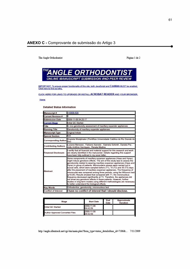
61
ANEXO C - Comprovante de submissão do Artigo 3

62
ANEXO D – Artigo publicado durante o Doutorado

63

64

65
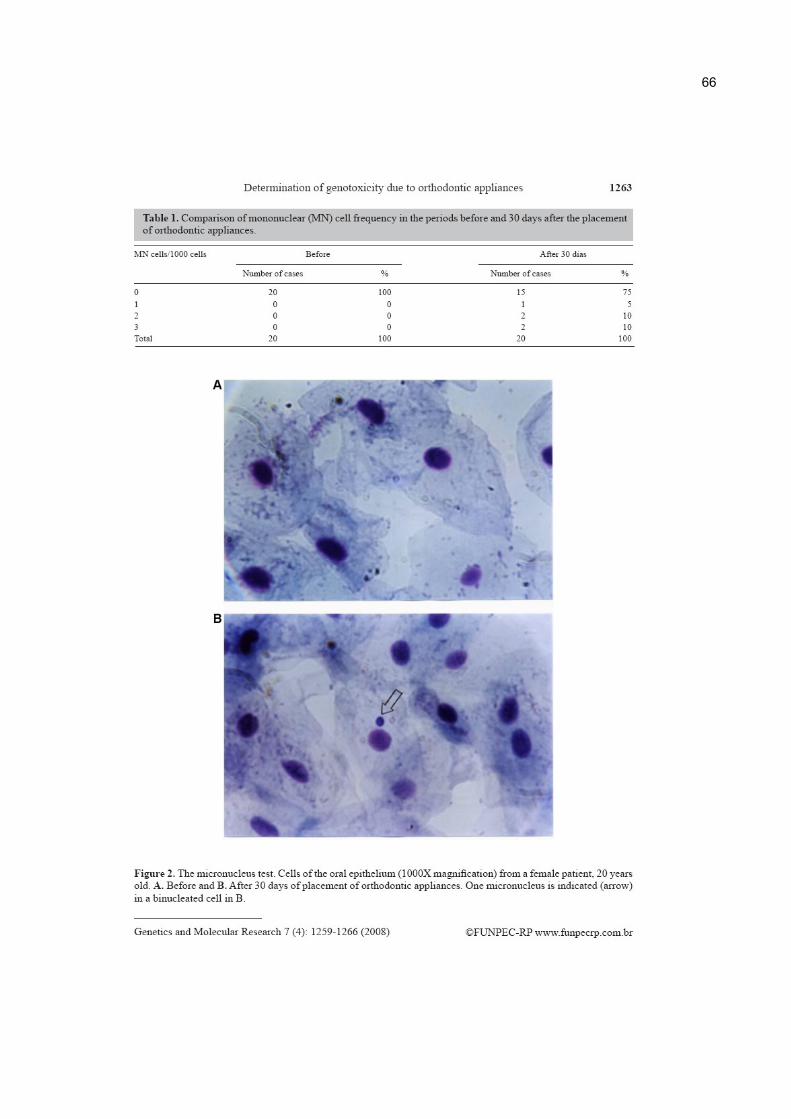
66
67
68
69
ANEXO E – Certificado de apresentação dos resultados da pesquisas em evento
internacional

70
ANEXO F – Ata de pré-defesa de tese, com aprovação do projeto de pesquisa
71
ANEXO G – Carta de Aprovação do Projeto de Pesquisa pelo Comitê de Ética em
Pesquisas da PUCRS

72
ANEXO H – Carta de concessão da bolsa de estudos do “Programa de Doutorado
no país com Estágio no Exterior” pela Coordenação de Aperfeiçoamento de Pessoal
de Nível Superior (CAPES).
73
ANEXO I - Carta convite para realização de parte da pesquisa no Commonwealth
Scientific and Industrial Research Organisation (CSIRO) em Adelaide – Austrália
74
ANEXO J - Carta com parecer sobre pesquisa desenvolvida no Commonwealth
Scientific and Industrial Research Organisation (CSIRO) em Adelaide - Austrália

75
8 APÊNDICES
APÊNDICE A - Termo de consentimento livre e esclarecido assinado pelos
pacientes como autorização de participação na pesquisa.
TERMO DE CONSENTIMENTO LIVRE E ESCLARECIDO
Título da Pesquisa: Citotoxicidade e Genotoxicidade dos materiais ortodônticos.
I. Objetivos e justificativa da pesquisa: Essa pesquisa pretende avaliar os efeitos dos
materiais que constituem os aparelhos ortodônticos no organismo humano, mais
precisamente no material genético das células da boca
II. Descrição do procedimento: Células serão coletadas da sua bochecha com uma
escova interdental pequena que será friccionada gentilmente.
III. Os desconfortos ou riscos esperados: O único desconforto esperado é a pressão
(mínima) da escova na bochecha durante a coleta das células. As escovas utilizadas serão
de uso exclusivo de cada paciente e descartadas após o uso, evitando qualquer risco de
contaminação.
IV. Os benefícios que se podem obter: A obtenção de conhecimento dos efeitos dos
materiais constituintes dos aparelhos ortodônticos no organismo humano.
V. Garantia de resposta a qualquer pergunta: Qualquer dúvida em relação à pesquisa
estamos inteiramente disponíveis a questionamentos, nesse caso entrar em contato com:
Dra. Graziela H. Westphalen ao telefone: 51 91011314 ou 3320 3538 e/ou Comitê de Ética
em Pesquisa da PUCRS ao telefone: 51 33203345.
VI. Liberdade de abandonar a pesquisa sem prejuízo para si: Caso desejar abandonar a
pesquisa, não haverá qualquer prejuízo ao participante ou modificações no tratamento
realizado.
VII. Garantia de privacidade: Ressaltamos, também, que a concordância em participar
desse estudo não implica necessariamente em qualquer modificação no tratamento que já
está sendo feito. Da mesma forma, a não concordância em participar desse estudo não irá
alterar de nenhuma maneira o tratamento já estabelecido.
76
VIII. Compromisso com informação atualizada do estudo: Os resultados da pesquisa
serão transmitidos de forma atualizada aos participantes e meios científicos.
Eu, .........................................................(paciente ou responsável) fui informado dos objetivos
da pesquisa acima de maneira clara e detalhada. Recebi informação a respeito do tratamento
recebido e esclareci minhas dúvidas. Sei que a qualquer momento poderei solicitar novas
informações e modificar minha decisão se assim eu o desejar. A Dra. Graziela Westphalen certificou-
me de que todos os dados referentes aos exames realizados serão confidenciais, bem como o
respectivo tratamento não será modificado em razão desse estudo, e terei liberdade de não mais
retirar consentir em participar da pesquisa, em face de essas informações.
Fui informado que não existirão danos à minha saúde causados diretamente pela pesquisa.
Também sei que, caso existam gastos adicionais, estes serão absorvidos pelo orçamento da
pesquisa. Caso tiverem novas perguntas sobre este estudo, posso chamar Graziela Henriques
Westphalen ao telefone: 51 91011314 ou 33203538 e/ou Comitê de Ética em Pesquisa da PUCRS ao
telefone: 51 33203345 para qualquer pergunta sobre os meus direitos como participante deste estudo
ou se penso que fui prejudicado pela minha participação.
Declaro que recebi cópia do presente Termo de Consentimento.
______________________ _____________________
Assinatura do Paciente Nome do Paciente
Data:

77
APÊNDICE B – Questionário de saúde respondido pelos pacientes participantes
desse estudo.
QUESTIONÁRIO DE SAÚDE
HISTÓRIA PESSOAL
Nome:__________________________________________________________
Data de hoje:____________ Idade:____ Data de nascimento:_____________
Sexo: ( ) Feminino ( ) Masculino
HISTÓRIA DE FUMO
Você fuma ? � SIM � NÃO
Se positivo, Há quanto tempo você fuma?___________________________
Quantas carteiras você fuma por dia? � Menos de 1/2 carteira � ½ - 1 carteira � mais
de uma carteira
HISTÓRIA DE ÁLCOOL
Você toma bebidas alcoólicas? � nunca � de vez em quando � sempre
Quantidade:____________________________________________________
HÁBITOS DIETÉTICOS
Quantas frutas você come por dia? � Nenhuma, � 1 a 2, � 3 ou mais
Quanta salada você come nas refeições? � Nenhuma, � 1/6 do prato, � 1/4 do prato ou mais
Quantos pedaços de carne você come por dia? � Nenhum, � 1 a 2 , � mais de dois
Marque abaixo quais os alimentos que você consome diariamente:
� Bolachas recheadas � Salgadinhos � Batatas fritas � Sorvetes

78
HISTÓRIA GENÉTICA
Você é portador de defeito de nascimento ou de outra desordem genética ou doença
inerente que afetou seus pais, irmãos ou filhos? � SIM � NÀO
Se SIM, favor especificar:_________________________________________________
_____________________________________________________________________
DOENÇAS
Outro tipo de doença:_____________________________________________
__________________________________________________________________________
HISTÓRIA DE EXPOSIÇÃO
Você foi exposto a algum dos elementos químicos (amianto, radiação, produtos do carvão,
pesticidas, herbicidas, produtos do petróleo, tinturas, solventes, outros químicos)? (se
positivo, responda às seguintes questões abaixo)
* Quando você foi exposto pela primeira vez? (mês e ano)_______________________
* Quando foi a última exposição? (mês e ano)_________________________________
Por quanto tempo em termos de dias, meses ou anos no total você tem sido exposto?
_____________________________________________________________________
_____________________________________________________________________
_____________________________________________________________________
OBS.:_____________________________________________________________________
__________________________________________________________________________
__________________________________________________________________________
_________________________________________________________________________