programa de pós-graduação em ciências biológicas (genética)
TRANSCRIPT

UNIVERSIDADE ESTADUAL PAULISTA
INSTITUTO DE BIOCIÊNCIAS
Programa de Pós-Graduação em Ciências Biológicas (Genética)
Diogo Cavalcanti Cabral-de-Mello
ORGANIZAÇÃO CROMOSSÔMICA DE ELEMENTOS REPETITIVOS
DE DNA EM REPRESENTANTES DA SUBFAMÍLIA SCARABAEINAE
(COLEOPTERA: SCARABAEIDAE)
Tese apresentada ao Programa de Pós-
Graduação em Ciências Biológicas (Genética)
como parte dos requisitos para obtenção do
título de Doutor
Orientador: Prof. Dr. Cesar Martins
Co-orientador: Profa. Dra. Rita de Cássia de Moura
Botucatu-SP
Fevereiro 2011

FICHA CATALOGRÁFICA ELABORADA PELA SEÇÃO DE AQUIS. E TRAT. DA INFORMAÇÃO DIVISÃO TÉCNICA DE BIBLIOTECA E DOCUMENTAÇÃO - CAMPUS DE BOTUCATU - UNESP
BIBLIOTECÁRIA RESPONSÁVEL: ROSEMEIRE APARECIDA VICENTE Cabral-de-Mello, Diogo Cavalcanti. Organização cromossômica de elementos repetitivos de DNA em representantes da subfamília Scarabaeinae (Coleoptera: Scarabaeidae) / Diogo Cavalcanti Cabral-de-Mello. - Botucatu, 2011 Tese (doutorado) - Instituto de Biociências, Universidade Estadual Paulista, 2011 Orientador: Cesar Martins Co-orientador: Rita de Cássia de Moura Capes: 20204000 1. Besouro - Evolução. 2. Citogenética animal. 3. Coleóptero. Palavras-chave: Besouro; Citogenética; DNA C0t-1; Evolução cromossômica; Famílias multigênicas; Hibridização in situ fluorescente.

Aos meus orientadores e a minha
família, em especial minha mãe e a Tatiane,
com amor dedico.

“Dias inteiros de calmaria, noites de ardentia, dedos no leme e
olhos no horizonte, descobri a alegria de transformar distâncias em tempo.
Um tempo em que aprendi a entender as coisas do mar, a conversar com
as grandes ondas e não discutir com o mau tempo. A transformar o medo
em respeito, o respeito em confiança. Descobri como é bom chegar quando
se tem paciência. E para se chegar, onde quer que seja, aprendi que não
é preciso dominar a força, mas a razão. É preciso, antes de mais nada,
querer”.
AAmyr Klink

Para cada projeto pessoal de vida que é finalizado existem inúmeras pessoas que
contribuíram para sua realização. Foram muitos anos até chegar esse momento e sem dúvida
alguma tive ajuda de muitas pessoas, algumas de forma direta e outras indireta, mas que cada
uma a sua maneira contribuiu com a minha formação profissional e pessoal; a estes sou
extremamente grato e compartilho a felicidade desta etapa cumprida. Primeiramente agradeço
a Deus pelo dom da vida e pela oportunidade por ele nos ofertada de passarmos um tempo
habitando este lugar.
Agradeço profundamente ao meu orientador Cesar Martins, pela coragem de ter
aceitado me orientar com um grupo completamente distinto do que tinha cunhado sua carreira
profissional. Agradeço também pela orientação ao longo destes anos, sempre com empenho,
fazendo as coisas acontecerem, incentivo e me dando liberdade de dar meu próprio
direcionamento ao trabalho. Além disso, por sua amizade e inúmeros momentos de
descontração durante meu doutoramento, que espero que se repitam por longos anos. A minha
co-orientadora Rita Moura, ou simplesmente Rita por ter me conduzido a este caminho, me
apresentando aos fascínios da Citogenética, mesmo quando ainda seu laboratório estava em
fase de construção. Agradeço por sua amizade, companheirismo, confiança depositada no
meu trabalho, conselhos dados ao longo de todos estes anos e reconhecimento dos meus
avanços. Levarei sua amizade pro resto da minha vida.
Ao professor Marcelo Guerra por me apresentar ao Cesar Martins, sendo decisivo no
rumo dado a minha carreira profissional. Aos professores Juan Pedro Camacho, Josefa
Cabrero e Maria Dolorez López-León por terem sido extremamente receptivos durante minha
estadia em Granada/Espanha. Além disso, em especial ao professor Juan Pedro por ter
avaliado parte do material produzido nesta tese.
Aos colegas de laboratório pelos momentos de descontração, troca de idéias, auxílio
na parte experimental e ensinamentos. Em especial aos colegas de Botucatu, Bruno, Danillo,
Éder, Érica, Guilherme, Jéssica, Juliana, Marcos, Pedro, Rafael e Sárah e aos que fiz em
Granada, Bea, Eli, Eva, Mercy e Tati. Também aos colegas do departamento de Morfologia,
assim como aos técnicos de laboratório, em especial ao Zé Eduardo, por toda ajuda e a
Luciana também por sua disponibilidade em todos os momentos. Aos encarregados pela pós-
graduação, Herivaldo e Luciana por estarem sempre dispostos a atender todas as pequenas e
grandes coisas solicitadas.

Ao meu amor Tatiane, por ter compartilhado todos os momentos bons e ruins passados
durante boa parte do tempo que estou em Botucatu. Por me dar a chance de realmente saber o
que é dividir uma vida e sonhos com alguém que se ama.
A minha família, em especial a minha mãe, minha irmã, minha avó e meu avô, por
estarem sempre preocupados comigo e por terem me auxiliado ao longo de toda essa
caminhada, desde a época do colégio.
Por fim as agencias de fomento CNPq e FAPESP, por terem concedido minha bolsa de
doutoramento e verba para a realização do projeto, respectivamente.

VI
SUMÁRIO
RESUMO VIII
ABSTRACT IX
LISTA DE FIGURAS X
LISTA DE TABELAS XV
1. INTRODUÇÃO E JUSTIFICATIVA 1
1.1. Considerações gerais sobre a família Scarabaeidae (Coleoptera) 1
1.2. Citogenética convencional de Scarabaeidae 3
1.3. DNAs repetitivos 6
1.3.1. Características gerais dos DNAs repetitivos e organização dos genomas 6
1.3.2. Heterocromatina e mapeamento cromossômico de DNAs repetitivos em
Coleoptera
11
2. OBJETIVOS 15
2.1. Objetivo Geral 15
2.2. Objetivos específicos 15
3. MATERIAIS E MÉTODOS 16
3.1. Insetos e amostragem 16
3.2. Obtenção de cromossomos e análises convencionais e diferenciais 16
3.3. Isolamento de Seqüências Repetitivas e genes mitocondriais por PCR e fração
C0t-1 DNA
18
3.4. Clonagem, seqüenciamento e Análise das Seqüências 19
3.5. Análises filogenéticas 20
3.6. Hibridação in situ fluorescente (FISH) 21
4. REFERÊNCIAS BIBLIOGRÁFICAS 23
5. RESULTADOS E DISCUSSÃO 32
5.1. Capítulo 1: Chromosomal mapping of repetitive DNAs in the beetle
Dichotomius geminatus provides the first evidence for an association of 5S
rRNA and histone H3 genes in insects, and repetitive DNA similarity
between the B chromosome and A complement
33
5.2. Capítulo 2: Evolutionary dynamics of heterochromatin in the genome of
Dichotomius beetles based on chromosomal analysis
52
5.3. Capítulo 3: Cytogenetic mapping of rRNAs and histone H3 genes in 14
species of Dichotomius (Coleoptera, Scarabaeidae, Scarabaeinae) beetles
81

VII
5.4. Capítulo 4: Chromosomal organization of 18S and 5S rRNA, and H3 histone
genes in Scarabaeinae coleopterans: insights on the evolutionary dynamics of
multigene families and heterochromatin
105
6. CONCLUSÕES GERAIS 136
7. ANEXOS 137
7.1. Anexo 1: Extração de DNA genômico de tecidos sólidos 137
7.2. Anexo2: Reações de PCR DNAr 18S, 5S e histona H3 138
7.3. Anexo 3: Isolamento de seqüências repetitivas através da técnica de C0t-1
DNA
139
7.4. Anexo 4: Clonagem dos fragmentos de PCR 140
7.5. Anexo 5: Marcação dos fragmentos de DNA para uso como sonda 143
7.6. Anexo 6: Chromosomal mapping of repetitive DNAs in the beetle
Dichotomius geminatus provides the first evidence for an association of 5S
rRNA and histone H3 genes in insects, and repetitive DNA similarity between
the B chromosome and A complement.
145
7.7. Anexo 7: Cytogenetic mapping of 5S and 18S rRNAs and H3 histone genes in
4 ancient Proscopiidae grasshopper species: Contribution to understanding the
evolutionary dynamics of multigene families
153
7.8. Anexo 8: Evolutionary dynamics of rDNAs and H3 histone sequences in the
A complement and B chromosome of Rhammathocerus brasiliensis
(Acrididae: Gomphocerinae): Chromosomal dispersion and co-localization
158
7.9. Anexo 9: Evolutionary dynamics of 5S rDNA location in acridid grasshoppers
and its relationship with histone gene and 45S rDNA location
173
7.10. Anexo 10: Evolutionary dynamics of heterochromatin in the genome of
Dichotomius beetles based on chromosomal analysis
203

VIII
RESUMO
O mapeamento cromossômico de seqüências repetitivas de DNA tem se mostrado uma
eficiente ferramenta nos estudos comparativos e evolutivos em diversos organismos. Estudos
cromossômicos com besouros da subfamília Scarabaeinae têm revelado ampla variabilidade,
entretanto a análise da organização cromossômica de DNAs repetitivos neste grupo é escassa
e direcionada unicamente ao mapeamento do DNA ribossomal (DNAr) 18S. O presente
trabalho teve como objetivo caracterizar cromossomicamente DNAs repetitivos em espécies
de Scarabaeinae, utilizando bandeamentos cromossômicos e mapeamento físico
cromossômico de seqüências repetitivas, incluindo famílias multigênicas de RNAr 18S, RNAr
5S e histona H3 e a fração de DNA C0t-1. Ampla variabilidade foi observada relacionada ao
número/localização dos sítios de DNAr 18S, aparentemente associada a diversificação da
heterocromatina. Por outro lado, os genes de RNAr 5S e histona H3, mostraram-se
amplamente conservados e co-localizados em um par cromossômico, com aparente
intercalação. Análises em representantes de Dichotomius revelaram conservação dos blocos
de heterocromatina, entretanto com aparente compartimentalização dos mesmos. O uso da
fração DNA C0t-1 confirmou o enriquecimento em DNAs repetitivos da heterocromatina, que
se apresentou diversificada entre as espécies, utilizando como referência D. geminatus. Por
outro lado, regiões terminais dos cromossomos apresentaram-se amplamente conservadas
entre as seis espécies. Além disso, a análise da fração de DNAs repetitivos em D. geminatus
indicou origem intraespecífica do cromossomo B desta espécie que possivelmente pode estar
sofrendo homogeneização com seqüências encontradas no complemento A. Os resultados
indicam distintos padrões de diversificação para o DNA repetitivo nos representantes de
Scarabaeinae, sugerindo extensiva reorganização microgenômica ao longo da cladogenese do
grupo.
Palavras-chave: besouro, citogenética, DNA C0t-1, evolução cromossômica, famílias
multigênicas, hibridização in situ fluorescente

IX
ABSTRACT
The chromosomal mapping of repeated DNAs has been used as an efficient tool in
comparative and evolutionary studies in some organism. The chromosomal studies in beetles
belonging to the subfamily Scarabaeinae have revealed wide variability, although the analysis
of chromosomal organization of repeated DNAs in this group is scarce and directed solely for
18S rDNA mapping. The present study aimed in chromosomal characterization of repeated
DNAs in Scarabaeinae species using chromosomal banding and physical chromosome
mapping of repeated sequences, including the multigene families for 18S and 5S rRNAs and
H3 histone genes and the C0t-1 DNA fraction. Wide variability was observed concerning the
number and location of 18S rDNA sites, apparently associated to the heterochromatin
diversification. On the other hand, the 5S rRNA and H3 histone genes were widely conserved
and co-located in one chromosomal pair, showing apparently interspersion. Analysis in
Dichotomius representatives revealed conservation for heterochromatic blocks, although an
apparent compartmentalization was observed. The use of C0t-1 DNA fraction confirmed the
heterochromatin repeated DNAs enrichment, which is diversified among the species, using as
reference D. geminatus. On the other hand, the terminal regions of the chromosomes were
highly conserved among the six species. Moreover, the analysis of repeated DNA fraction
from D. geminatus indicated intraspecific origin of a B chromosome in this species that
possibly could be suffering homogenization with A complement sequences. The results
indicate distinct diversification patterns for repeated DNAs in Scarabaeinae representatives,
suggesting extensive microgenomic reorganization along the cladogenesis of the group.
Key-words: beetle, cytogenetics, C0t-1 DNA, chromosomal evolution, multigene families,
fluorescent in situ hybridization

X
LISTA DE FIGURAS
Introdução e justificativa
Figura 1. Amostra da diversidade de espécies da subfamília Scarabaeinae. 2
Figura 2. Organização geral das seqüências de DNA do genoma nuclear de
eucariotos.
8
Figura 3. Organização genômica das famílias multigênicas (a) DNAr 45S, (b)
DNAr 5S, (c) genes de histonas em moscas de fruta. ETS = Espaçador transcrito
externo; ITS = espaçador transcrito interno; IGS = Espaçador intergênico.
10
Capítulo 1
Figure 1. Male meiotic cells and karyotype of Dichotomius geminatus.
Conventional staining of metaphase I chromosomes of 0B individuals (a) and 1B
individuals (b); C-banded karyotype (c), metaphases I of 0B (d) and 1B (e)
individuals and metaphases II of B-carrying individual (f). Silver nitrate staining
in inicial prophase (g). The arrows indicate the sex bivalents (Xyp), full and empty
arrowheads indicate the B chromosomes and the nucleolar organizer region
(NOR), respectively, and the (*) indicate the chromosome pairs with additional
heterochromatic blocks. Bar = 5 μm.
49
Figure 2. Fluorescent in situ hybridization with 18S rRNA, 5S rRNA and histone
H3 gene probes in 0B and 1B individuals of D. geminatus. Pachytene
chromosomes from 0B individuals hybridized using 18S (a) and 5S rDNAs (b);
double FISH with 18S (green) and 5S (red) rDNAs in metaphase I chromosomes
of 0B individuals (c); partial metaphase I chromosomes hybridized with 5S (d) and
H3 (e) probes; metaphase I chromosomes showing the distribution pattern of 18S
(f), 5S (g) and H3 (h) in 1B individuals; Note the absence of hybridization signals
on the B chromosome (f-h) and the heterochromatin highlighted after DAPI
staining (a-h). The arrows indicate the sex bivalents (Xyp), and arrowheads
indicate the B chromosome. C = centromere. Bar = 5 μm.
50
Figure 3. C0t-1 DNA fraction hybridization in metaphase I chromosomes of 0B
individuals (a) and 1B individuals (b,c). Ideogram (d) showing the hybridization
patterns described in this work. The arrows indicate the sex bivalents (Xyp),

XI
arrowheads indicate the B chromosome, and the (*) indicate the chromosome pairs
with additional heterochromatic blocks. Bar = 5 μm.
51
Capítulo 2
Figure 1. C-banding and C0t-1 DNA fraction hybridization in six species of
Dichotomius. The C0t-1 DNA fractions were isolated from each species and
hybridized against their own genomes. C-banded karyotypes from D. bos (a) and
D. laevicollis (d), and C-banded metaphase I chromosomes from D. geminatus (f),
D. nisus (h), D. sericeus (j) and D. semisquamosus (l) are shown. C0t-1 DNA
fraction hybridization are showed in metaphase II chromosomes from D. bos (b
and c), and metaphase I chromosomes from D. laevicollis (e), D. geminatus (g), D.
nisus (i), D. sericeus (k) and D. semisquamosus (m). The arrows indicate the sex
bivalents, the asterisks denote chromosome pairs containing additional
heterochromatic blocks and the arrowheads denote the terminal hybridization
signals. The insert in (m) shows terminal blocks observed in D. semisquamosus;
(t=terminal region, c=centromere). In (a-d), the X and y chromosomes are
indicated. (D1) Dichotomius bos, (D2) D. geminatus, (D3) D. laevicollis, (D4) D.
nisus, (D5) D. semisquamosus and (D6) D. sericeus. Bar = 5μm.
77
Figure 2. Fluorochrome staining (CMA3/DA/DAPI) in the six species of
Dichotomius. Karyotypes from D. bos (a) and D. laevicollis (b), respectively;
metaphase I chromosomes from D. nisus (c), D. semisquamosus (d), D. sericeus
(e) and D. geminatus (f) are showed. The arrows indicate the sex bivalents, and the
arrowheads denote the CMA3+ euchromatic blocks. The insert in (b) indicates the
conformation of the pair 2 from D. laevicollis in metaphase I. (D1) Dichotomius
bos, (D2) D. geminatus, (D3) D. laevicollis, (D4) D. nisus, (D5) D.
semisquamosus and (D6) D. sericeus. Bar = 5μm.
78
Figure 3. C0t-1 DNA fraction obtained from the genome of Dichotomius
geminatus hybridized against the chromosomes of the other five Dichotomius
species. Metaphase I chromosomes from D. semisquamosus (a), D. bos (c), D.
laevicollis (e), D. sericeus (g) and D. nisus (i); the initial pachytenes of D.
semisquamosus (b), D. bos (d), D. laevicollis (f) and D. sericeus (h), and
metaphase II of D. nisus (j) are showed. The arrows indicate the sex bivalents.
Inserts indicate the detail of the sex chromosomes in metaphase I from another cell
for each species (a,c,e,g,i) and the initial meiotic nucleus (f) showing the

XII
polarization of the hybridization signals. Note that the hybridization signals are
restricted to the terminal regions of the chromosomes. Bar = 5μm.
79
Figure 4. (a) Ideograms showing the distribution of cytogenetic markers for each
chromosome in the six species of Dichotomius studied; (b) phylogenetic
relationship of the six Dichotomius species based on COI and 16S sequences. (D1)
Dichotomius bos, (D2) D. geminatus, (D3) D. laevicollis, (D4) D. nisus, (D5) D.
semisquamosus and (D6) D. sericeus.
80
Capítulo 3
Figure 1. Fluorescent in situ hybridization using 18S (green; a-g) and 5S rDNA
(red; a-f, h, i), and H3 histone gene (green; h, i) as probes in nine representative
species of Dichotomius. Metaphases I of (a) D. prox mundus, (b) D. sericeus sp1,
(d) D. semiaeneus, (e) D. affinis, (f) D. sp, (g) D. mormon, (h) D. crinicollis, and
metaphases II of (c, i) D. depresicollis. The arrows indicate the sex bivalents, and
the inserts in (h, i) show the signals with separate probes for the 5S rRNA (red)
and H3 histone (green) genes. Note the large metacentric pair (pair one) indicated
by the number. Other chromosomes are also indicated. Scale bar = 5 μm.
102
Figure 2. Chromosomal mapping of 18S rRNA (purple), 5S rRNA (red) and H3
histone (green) multigene families in six species of Dichotomius. Karyotypes from
D. laevicollis (a) and D. sericeus (b); interphasic nucleus from D. bos (c); partial
pachytene from D. geminatus (d); metaphase II from D. geminatus (e); metaphase
I chromosomes from D. semisquamosus (f), D. nisus (g) and D. bos (h) are shown.
The arrows indicate the sex bivalents, and the inserts show the labeled
chromosomes with separate probes for the 5S rRNA (red) and H3 histone (green)
genes. Note that the 5S rRNA and H3 histone gene sites overlap in all cells,
including in interphasic nucleus (d) and less condensed chromosomes in an initial
meiotic pachytene (d), and the large metacentric pair (pair one) indicated by the
number 1. Other chromosomes are also indicated. Bar = 5μm.
103
Figure 3. (a) Ideograms showing the distribution of 18S rDNA (green) and 5S
rDNA/H3 histone (red). (D1) Dichotomius affinis, (D2) D. bos, (D3) D.
crinicollis, (D4) D. depressicollis, (D5) D. geminatus, (D6) D. laevicollis, (D7) D.
mormon, (D8) D. aff mundus, (D9) D. nisus, (D10) D. semiaeneus, (D11) D.
semisquamosus, (D12) D. sericeus, (D13) D. sericeus sp1, (D14) D. sp and (*)
chromosome shared by all species. The black lines indicate chromosomes that

XIII
harbor neither 45S/5S rDNA nor the histone cluster in any of the species. (b)
Chromosome three selected from metaphase I (1,1’,2,2’), metaphase II (3,4) and
spermatogonial metaphase (5) showing in detail the position of the 18S rDNA: (1)
terminal in the short arm, (2,5) interstitial in the short arm, (3) interstitial in the
long arm, and (4) proximal. (1) D. sericeus sp1, (2) D. affinis, (3) D.
depressicollis, (4) D. bos, (5) D. sp. 1’ and 2’ represent a graphical structure of
bivalent three in metaphase I of 1 and 2, respectively. Red arrowheads indicate the
centromere and white arrows the positions of chiasmata.
104
Capítulo 4
Figure 1. Fluorescent in situ hybridization in metaphases I using 5S rDNA (red)
and 18S rDNA (green) in nine representatives species of Scarabaeinae of three
distinct tribes (a-d) Phanaeini, (e-h) Canthonini, (i) Ateuchini. (a) Coprophanaeus
ensifer, (b) C. cyanescens, (c) Diabroctis mimas, (d) Phanaeus splendidulus, (e)
Canthon staigi, (f) Deltochilum calcaratum, (g) D. verruciferum, (h) D. elevatum,
(i) Atheuchus sp. The arrows indicate the sex bivalents. Note the co-location of the
two sequences in (e) for two chromosomes. Bar = 5μm.
130
Figure 2. Cytogenetic mapping of 5S (red) and 18S (green) rDNAs in four species
of Scarabaeinae from (a) Euristernini, (b) Onthophagini, (c-d) Coprini tribes. (a)
Eurysternus caribaeus, (b) Digitonthophagus gazella, (c) Ontherus apendiculatus,
(d) O. sulcator. The arrows indicate the sex bivalents. Note the co-location of the
two sequences in (a) and (b) in two species. Bar = 5μm.
131
Figure 3. Double-FISH for 5S rRNA and histone H3 genes in five Scarabaeinae
representatives. (a) Coprophanaeus cyanescens, (b) C. ensifer, (c) Phanaeus
splendidulus, (d) Deltochilum verruciferum, (e) Eurysternus caribaeus. The
arrows indicate the sex bivalents. Note the co-location of the two sequences in all
cells, and in (e) the presence of only on site for the two genes in the X
chromosome. Bar = 5μm.
132
Figure 4. Initial meiotic cells (a-e, h, i) and interphasic nucleus (f, g) hybridized
with 18S (green) and 5S rDNAs (red) (a-f) and 5S rRNA (red) and H3 histone
genes (green) (g-i). (a) Deltochilum elevatum, (b) Deltochilum calcaratum, (c)
Dichotomius crinicollis, (d) Coprophanaeus cyanescens, (e, f) Diabroctis mimas,
(g) Dichotomius bos (h) Dichotomius laevicollis, (i) Deltochilum verruciferum.
Note the separation of 18S and 5S rDNA signals in (a-e), in (f) two small signals

XIV
are overlapped, and note in (g-i) the overlapped configuration of 5S rRNA and H3
histone genes. In (a, b, d, e and i) in possible the observation of chromsocenter
formation by heterochromatic sequences. The scale bar is not shown.
133
Figure 5. (a) distribution of rDNA loci number in 31 Scarabaeinae species;
Compared distribution of the number of 18S rDNA with diploid number in 26
species of Scarabainae (b) and with heterochromatin distribution in 21 species (c).
The species Deltochilum calcaratum, Dichotomius semisquamosus, Gymnopleurus
sturmi, Coprophanaeus ensifer and Diabroctis mimas were considered twice due
the observation of polymorphisms related to number of sites. Each symbol below
the name of species represent the distinct tribes, (Ŧ) Euristernini, (Ф) Ateuchini,
(•) Coprini, (∆) Onthophagini, (+) Gymnopleurini, (*) Canthonini, (о) Onitini. (■)
Phaneini.
134

XV
LISTA DE TABELAS
Materiais e métodos
Tabela 1. Espécies utilizadas neste trabalho com respectivos locais de coleta e
número de indivíduos.
17
Capítulo 2
Table 1. C0t-1 DNA fraction reassociation time and chromosomal location for the
six Dichotomius species investigated in this study.
75
Capítulo 3
Table 1. Chromosome location of 18S rDNA in fourteen species of Dichotomius.
The asterisks indicate the presence of 45S rDNA site in the sex chromosomes in
which was impossible to determinate its precise position, and (†) species studied
for H3 histone mapping.
100
Capítulo 4
Table 1. Species and respective diploid numbers, heterochromatin patterns and
chromosome location of rDNA clusters and H3 histone gene in 31 Scarabaeinae
representatives. The color bars in the diploid number and heterochromatin pattern
columns represent the same chromosome characteristics described in figure 5.
126

1
1. INTRODUÇÃO E JUSTIFICATIVA
1.1. Considerações gerais sobre a família Scarabaeidae (Coleoptera)
A ordem Coleoptera compreende a mais diversa dentro da classe Insecta, apresentando
cerca de 360.000 espécies com distribuição mundial. Na região Neotropical, este grupo é
representado por aproximadamente 72.476 espécies, agrupadas em 6.703 gêneros e 127
famílias (Costa, 2000). Esta riqueza de espécies está associada à extrema diversificação
morfológica, ecológica e comportamental, sendo resultado de co-radiação de muitos grupos
com angiospermas, mamíferos e mudanças climáticas e geológicas ocorrentes desde o período
Cretáceo (Crowson, 1960; Farrell, 1998; Davis et al., 2002; Erwin, 1985). Os besouros
apareceram há cerca de 285 milhões de anos atrás e sofreram diversos eventos de radiação
adaptativa, gerando os diferentes grupos (Crowson, 1981; Grimaldi e Engel, 2005).
Atualmente, Coleoptera é dividida em quatro subordens, Archostemata, Adephaga,
Myxophaga e Polyphaga, e a monofilia do grupo é consenso entre diversos autores, apesar das
relações entre as quatro subordens permanecerem controvérsias. Dentre as quatro subordens
de Coleoptera Polyphaga é a mais diversa agrupando 90% das espécies do grupo (Crowson,
1981; Lawrence e Newton, 1995; Wheeler et al., 2001; Vanin e Ide, 2002).
Para Polyphaga são descritas 16 superfamílias, das quais Scarabaeoidea destaca-se por
apresentar ampla radiação adaptativa e alta diversidade de espécies viventes. Este grupo é
representado por cerca de 27.800 espécies agrupadas em 13 famílias, Glaressidae, Passalidae,
Lucanidae, Diphyllostomidae, Trogidae, Bolboceratidae, Plecomidae, Geotrupidae,
Hybosoridae, Ochodaidae, Ceratocanthidae, Glaphyridae e Scarabaeidae (Crowson, 1981;
Browne e Scholtz 1999; Vanin e Ide, 2002). Dentre as 13 famílias de Scarabaeiodea
Scarabaeidae é a mais diversa e possui cerca de 25.000 espécies e 2.000 gêneros agrupados
em 12 subfamílias e diversas tribos distribuídas mundialmente. No neotrópico são registrados
362 gêneros e 4.706 espécies, enquanto no Brasil ocorrem 204 gêneros e 1.777 espécies

2
(Costa, 2000; Vaz-de-Mello, 2000). São besouros de corpo robusto, ovais ou alongados,
usualmente convexos e com antenas lameladas. Variam consideravelmente em hábitos, se
alimentando de esterco, matéria vegetal, “carniça”, podendo estar associados a formigueiros e
cupinzeiros, além de se alimentarem de fungos. A maioria das espécies tem hábitos noturnos e
apresentam atração pela luz, entretanto espécies diurnas podem ser encontradas em tecidos
vegetais como flores (Lawrence e Newton 1995; Marioni et al. 2001; Ratcliffe et al. 2002).
Em Scarabaeidae, a subfamília Scarabaeinae apresenta maior quantidade de estudos
relacionados a características taxonômicas e ecológicas. É um grupo cosmopolita com ampla
diversidade, apresentando 5.000 espécies, 12 tribos e 234 gêneros descritos em todo o mundo
(Figura 1). Para região Neotropical tem-se registro de aproximadamente 1.250 espécies e 70
gêneros, enquanto para o Brasil há registro de 618 espécies, das quais 323 são endêmicas
(Hanski e Cambefort, 1991; Vaz-de-Mello, 2000). Apesar do grande número de espécies
descritas para o grupo, acredita-se que este número seja bastante superior devido à carência de
dados para diversas regiões tanto do Brasil quanto do mundo.
Figura 1. Amostra da diversidade de espécies da subfamília Scarabaeinae.

3
Os escarabeíneos são conhecidos popularmente como “rola bosta” devido ao hábito de
rolarem bolas de matéria orgânica em decomposição para realizarem ovoposição e
alimentação (Halffter e Mattheus, 1966). Devido a este hábito, os mesmos são importantes no
funcionamento de distintos ecossistemas, desde savanas Africanas a matas tropicais, atuando
como eficientes recicladores da matéria orgânica, principalmente animal, nestas regiões
(Halffter e Mattheus, 1966; Halffter e Favila, 1993). Em decorrência desta importante função
ecológica, juntamente com a ampla diversidade do grupo, facilidade de coleta em campo,
elevada densidade de indivíduos para as espécies comuns, ocorrência de grupos bem
estudados taxonomicamente e por constituírem comunidades bem estruturadas, os
escarabeíneos tem sido utilizados em estudos para indicação de qualidade ambiental (Halffter
e Edmons, 1982; Hanski e Cambefort, 1991; Halffter e Favila, 1993).
1.2. Citogenética convencional de Scarabaeidae
Embora Coleoptera apresente ampla diversidade de espécies e cromossômica, os estudos
citogenéticos são bastante escassos e apenas pouco mais de 1% dos representantes da ordem
apresentam cariótipos descritos na literatura. De maneira geral, os estudos são restritos a
descrição do número diplóide e dos mecanismo de cromossomos sexuais e, em menor escala,
morfologia cromossômica (Smith e Virkki, 1978; Vidal, 1984; Virkki e Santiago-Blay, 1993;
Moura et al., 2003; Karagyan et al., 2004; Pons, 2004; Schneider et al,. 2007; Cabral-de-
Mello et al., 2008; de Julio et al., 2010). Além disso, os estudos neste grupo são concentrados
em algumas famílias, como por exemplo, Carabidae e Cicindelidae, para subordem Adephaga
e Buprestidae, Chrysomelidae, Elateridae, Scarabaeidae e Tenebrionidae para Polyphaga,
enquanto para as subordens Myxophaga e Archostemata apenas três espécies foram estudadas
(Smith e Virkki, 1978; Mesa e Fontanetti, 1985; Galián e Lawrence, 1993; Galián et al., 2002;

4
Karagyan et al., 2004; Martinez-Navarro et al., 2004; Pons, 2004; Schneider et al,. 2007;
Cabral-de-Mello et al., 2008; de Julio et al., 2010).
Para a subordem Polyphaga, onde os estudos citogeneticos têm sido realizados com maior
freqüência, o cariótipo considerado modal e primitivo é constituído por número diplóide 2n =
20, Mecanismo sexual Xyp, cromossomos autossômicos e X com dois braços e y puntiforme
(Smith e Virkki, 1978). Apesar desta conservação, tem sido descrito para o grupo ampla
variação do cariótipo modal devido a distintos rearranjos cromossômicos, tais como fusão,
fissão e inversões. O número diplóide varia de 2n = 4 (Chalcolepidius zonatus, Elateridae) a
2n = 64 (Disonycha bicarinata, Chrysomelidae) e 11 mecanismos de cromossomos sexuais
foram descritos (Xyp, XYp, Xnyp, nXyp, Xyc, XYc, X0, Xy, XY, Xyr, nXnY). Além disso,
foram registrados a ocorrência de cromossomos com distintas morfologias (Takenouchi,
1970; Smith e Virkki, 1978; Ferreira et al., 1984; Serrano e Galián, 1998; Dutrillaux e
Dutrillaux, 2009; Cabral-de-Mello et al., 2010a).
Para a família Scarabaeidae cerca de 400 espécies foram estudadas citogeneticamente,
predominando análises em representantes das subfamílias, Cetoniinae, Dynastinae,
Melolonthinae, Rutelinae e Scarabaeinae. Assim como observado para a ordem Coleoptera
como um todo, esta família é considerada conservada cariotipicamente e apresenta cariótipo
modal e primitivo 2n = 20, Xyp e cromossomos com dois braços em mais de 50% das espécies
estudadas, embora diversas variações foram relatadas (Smith e Virkki, 1978; Yadav e Pillai,
1977, 1979; Moura et al., 2003; Bione et al., 2005a; Cabral-de-Mello et al., 2008; Dutrillaux
e Dutrillaux, 2009). Com relação ao número diplóide foi observada variação de 2n = 8 em
Eurysternus caribaeus (Scarabaeinae) a 2n = 30 em Autoserica assamensis (Melolonthinae) e
dentre os mecanismos sexuais descritos para Coleoptera sete foram observados em
Scarabaeidae, sendo quatro quiasmáticos (neo-XY, XY, Xy e Xyr) e três aquiasmáticos (Xyp,
XYp e XO) (Dasgupta, 1977; Smith e Virkki, 1978; Cabral-de-Mello et al., 2007). De acordo

5
com Yadav e Pillai (1979) e Cabral-de-Mello et al. (2008), os rearranjos cromossômicos
envolvidos na diferenciação dos cariótipos em Scarabaeidae foram as fusões autossomo-
autossomo (A-A) e autossomos e cromossomo X (A-X), perda do cromossomo y ou aumento
do mesmo, fissão de autossomos e inversões.
Dentre as cinco subfamílias de Scarabaeidae mais estudadas citogeneticamente,
Scarabaeinae apresenta a maior diversidade cariotípica, resultante de distintos rearranjos
cromossômicos, tais como fusões, fissões, inversões e aumento ou perda do cromossomo y,
assim como descrito para Scarabaeidae como um todo. Para esta subfamília, cerca de 130 das
5.000 espécies conhecidas para o grupo apresentam cariótipos descritos. Em geral os estudos
foram realizados com mais freqüência em representantes das tribos Canthonini, Coprini,
Ontophagini e Phanaeini. O menor número diplóide registrado para esta subfamília (2n = 8
em E. caribaeus) é coincidente com o menor número registrado para Scarabaeidae, enquanto
o maior número ocorre em Tiniocellus spinipes, 2n = 24 (revisado por Cabral-de-Mello et al.,
2008).
Em relação aos cromossomos B em Scarabaeinae, poucos estudos tem relatado a presença
destes elementos nos cariótipos dos representantes deste grupo. Além disso, estes estudos têm
sido concentrados basicamente apenas na descrição da presença/ausência deste polimorfismo,
relatando número de cromossomos B e seus tamanhos em populações naturais (Wilson e
Angus, 2005; Angus et al., 2007; Falahee e Angus, 2010). Exemplos de espécies de
Scarabaeinae portadoras de cromossomos B ocorrem nos representantes das tribos Onitini e
Onthophagini (Wilson e Angus, 2005; Angus et al., 2007; Falahee e Angus, 2010). O maior
número de Bs registrado para o grupo, ocorre em Bubas bubalus (Onitini), com ocorrência de
2-9 elementos supernumerários de diferentes tamanhos podendo alcançar duas vezes o
tamanho do maior par do complemento A (par 1) (Angus et al., 2007). Foram descritos
cromossomos B em cinco espécies da tribo Onthophagini: Euonthophagus amyntas,

6
Onthophagus vacca, O. similis, O. gazella e O. fracticornis. Em geral o tamanho dos Bs
nestas espécies é puntiforme, similar ao cromossomo y ou pouco maior em relação ao mesmo,
entretanto em O. vacca o cromossomo B encontrado apresenta tamanho similar ao
cromossomo 9. Além da variabilidade em relação ao número e tamanho dos cromossomos B
em Scarabaeinae, variações intraindividuais também foram descritas (Wilson e Angus, 2005;
Falahee e Angus, 2010).
1.3. DNAs repetitivos
1.3.1. Características gerais dos DNAs repetitivos e organização dos genomas
Os estudos focando tamanho de genomas em eucariotos têm revelado grande variação de
quantidade de DNA entre as diferentes espécies dos mais diversos grupos, incluindo até
mesmo espécies relacionadas. Esta variação ocorre independentemente da complexidade do
organismo e não apresenta relação com a quantidade de genes apresentados pelos organismos,
nem nível de ploidia (Gregory 2005). Esta falta de correlação entre número de genes,
complexidade do organismo e tamanho de genomas foi definida por Thomas (1971) como o
paradoxo do “valor C”, onde o “valor C” corresponde à quantidade de DNA de um núcleo
haplóide em picogramas (pg).
Para os genomas animais, uma ampla variação de quantidade de DNA tem sido
registrada, desde 0,02 pg em Pratylenchus coffeae (nemátode) a 132,83 pg em Protopterus
aethiopicus (peixe) (www.genomesize.com). Esta ampla variação tem sido atribuída a
diferentes quantidades de DNAs repetitivos nos diversos genomas, sendo a presença destes
elementos uma característica ubíqua em eucariotos. Estas seqüências são caracterizadas por
ampla variabilidade, constituindo distintas famílias e podem representar grande parte da
quantidade de DNA das células, em alguns casos excedendo mais de 80% da quantidade de
DNA da espécie (Charlesworth et al., 1994; Ridley, 1996; Gregory, 2005; Plohl et al., 2008).

7
Classicamente os DNAs repetitivos foram considerados como junk DNAs (DNA lixo) por
não apresentarem funções biológicas bem definidas relacionadas à atividade e expressão dos
mesmos (Doolittle e Sapienza, 1980; Orgel e Crick, 1980). Entretanto, o envolvimento destes
elementos na organização e funcionalidade dos centrômeros, telômeros, perfeita segregação
cromossômica, regulação gênica, reparo e replicação do DNA, diferenciação dos
cromossomos sexuais foram propostas em diversos estudos (Anleitner e Haymer, 1992;
Kraemer e Schmid, 1993; Messier et al., 1996; Martins, 2007). Além disso, análises mais
recentes têm demonstrado expressão e envolvimento dos elementos repetitivos, por exemplo,
na formação da heterocromatina e em processos de regulação gênica (Shapiro e Sternberg,
2005; Biémont e Vieira, 2006; Feschotte e Pritham, 2007). Por muito tempo os elementos
repetitivos foram considerados sem atividade transcricional, fazendo com fossem
classicamente divididos em elementos “codificadores” e “não codificadores”. Os elementos
repetitivos codificadores incluem as famílias multigênicas, enquanto os não codificadores são
representados principalmente pelos DNAs satélites e elementos transponíveis (Revisado por
Martins et al., in press). Juntamente com as seqüências únicas, moderadamente e pouco
repetitivas estes elementos constituem a estrutura básica do genoma nuclear em eucaritos
(Figura 2).

8
Figura 2. Organização geral das seqüências de DNA do genoma nuclear de eucariotos.
O termo “família multigênica” é utilizado para indicar um grupo de seqüências de
DNA (genes) com notável similaridade estrutural e funcional, sendo descendentes de um gene
ancestral comum (Nei e Rooney, 2005). Dentre estas seqüências são exemplos bastante
conhecidos os genes de RNAs ribossomais (RNAr) e genes codificadores de proteínas
histônicas. As seqüências de DNA ribossomal (DNAr) na maioria dos eucariotos são
organizadas em dois distintos grupos arranjados in tandem. O arranjo maior é formado pelos
genes que transcrevem os RNAs ribossomais 18S, 5.8S e 28S (DNAr 45S), sendo estas
seqüências separadas por espaçadores intergênicos transcritos internos (ITS-Internal
Transcribed Spacer); e cada cluster de DNAr 45S separado por espaçadores transcritos
externos (ETS-External Transcribed Spacer) e por espaçadores integênicos (IGS) (Figura 3a).
O outro arranjo é formado pelas seqüências do gene que transcrevem o RNAr 5S. Estes genes
são bastante conservados e espaçados por seqüências não transcritas (NTS-Non Transcribed
Spacer) que são extremamente variáveis em tamanho e composição nucleotídica (Figura 3b)
(Long e Dawid, 1980; Williams e Strobeck, 1985; Martins e Wasko, 2004; Eickbush e

9
Eickbush, 2007). As seqüências codificadores de proteínas histônicas podem ser organizadas
em um cluster formado por genes que apresentam poucos introns (H1, H2A, H2B, H3 e H4)
que se encontram espaçados por DNAs não codificantes. Esta estrutura genômica foi descrita,
por exemplo, em Drosophila melanogaster (Figura 3c). Por outro lado, estes mesmo genes
podem estar distribuídos isoladamente como observado em humano, rato e galinha. Além
disso, os dois tipos de organização genômica foram observados em Xenopus laevis (Lifton et
al., 1978; Engel e Dogson, 1981; Ruberti et al., 1982).
Dentre os DNAs não codificadores presentes no genoma dos eucariotos, destacam-se
os DNAs satélite (DNAsat), minisatélites, microsatélites e elementos transponíveis, devido à
grande abundancia. Os DNAsat são compostos por seqüências altamente repetitivas com
ampla variabilidade organizadas in tandem com diferentes números de cópias nos genomas
das distintas espécies, podendo variar basicamente entre 1.000 e mais de 100.000 cópias.
Estas seqüências constituem o principal componente da heterocromatina e estão
freqüentemente associadas as regiões centromérica e telomérica dos cromossomos. De uma
forma geral estes elementos são bastante diversificados entre diferentes espécies, incluindo
espécies relacionadas, resultante dos mecanismos de mutação e evolução em concerto (John e
Miklos, 1979; Charlesworth et al., 1994; Ugarković e Plohl, 2002).

10
Figura 3. Organização genômica das famílias multigênicas de DNAr e histonas: (a) DNAr
45S, (b) DNAr 5S, (c) genes de histonas em moscas de fruta. ETS = Espaçador transcrito
externo; ITS = espaçador transcrito interno; IGS = Espaçador intergênico.
Os minisatélites apresentam variação de tamanho de 10-100 pares de base (pb). Estas
seqüências encontram-se dispersas por todo o genoma com variação no número de unidades
de repetição (repeats) conhecidos como VNTR (Variable Numbers of Tandem Repeats). Os
minisatélites são encontrados nos mais distintos grupos de eucariotos, desde leveduras,
plantas e animais, sendo uma classe de DNA repetitivo comum nos genomas eucariotos
(Jeffreys et al., 1985). As menores seqüências repetidas in tandem nos eucariotos são
conhecidos como microsatélites, as quais apresentam de 1-6 nucleotídeos por unidade de
repetição. Os mesmos podem estar presentes em regiões cromossômicas eucromáticas como
podem estar associados com a heterocromatina constitutiva. Dentre os exemplos de
microsatélites, podem ser citadas as distintas seqüências de DNA telomérico (Schlötterer,
2000). Estas definições das diferentes classes de DNAs repetitivos in tandem são controversas
na literatura e diversas exceções para cada classe já foram descritas.

11
Os elementos transponíveis são diferenciados das outras seqüências repetitivas
presentes nos genomas eucariotos simplesmente por sua capacidade de
transposição/movimentação para distintas regiões. Grande parte dos diversos elementos
repetitivos são originados dos elementos transponíveis e os mesmos representam uma grande
porção dos genomas, por exemplo cerca de 40% do genoma humano. Os mesmos são
divididos em dois grupos básicos, os transposons e retrotransposons de acordo com o seu
mecanismo de transposição. Os transposons se movimentam nos genomas a partir de
moléculas de DNA, enquanto os retrotransposons se transpõem utilizando RNAs via
transcrição reversa. Diversas famílias destes elementos já foram identificadas nos genomas,
podendo ocorrer em espécies não relacionadas ou presentes apenas em grupos específicos
(Biémont e Vieira, 2006; Feschotte e Pritham, 2007; Wicker et al., 2007).
Esta ampla variedade em relação à quantidade, seqüências nucleotídicas e localização
dos elementos repetitivos de DNA se deve basicamente aos mecanismos evolutivos de
conversão gênica, transposição, crossing-over desigual, replicação slippage e ocorrência de
DNAs extracromossômicos circulares (eccDNA). Estes mecanismos podem atuar
sinergicamente nas diferentes classes de seqüências ou atuarem isoladamente ao longo do
tempo evolutivo (Charlesworth et al., 1994; Ugarković e Plohl, 2002; Cohen e Segal, 2009).
1.3.2. Heterocromatina e mapeamento cromossômico de DNAs repetitivos em Coleoptera
Diferentes técnicas citogenéticas têm sido utilizadas para análise dos DNAs repetitivos
em insetos, principalmente relacionadas à distribuição e qualificação da heterocromatina
constitutiva. Dentre estas técnicas as mais aplicadas para estudos com representantes da
ordem Coleoptera são o bandeamento C e em menor escala o uso de fluorocromos base-
específicos (CMA3 e DAPI), além de poucos estudos com mapeamento de DNAsat. Apesar da
grande diversidade em Coleoptera, tanto cromossômica como em número de espécies e da

12
facilidade e baixo custo da técnica de bandeamento C, os estudos com descrição da
localização dos blocos de heterocromatina são escassos. De acordo com Rozèk et al. (2004) o
pequeno número de estudos se deve a limitada quantidade de heterocromatina nos
cromossomos dos coleópteros, o que dificulta a análise e descrição dos segmentos
heterocromáticos, principalmente em fases onde os cromossomos se encontram bastante
condensados, tais como metáfases.
Em geral a heterocromatina nos representantes de Coleoptera encontra-se
preferencialmente localizada nas regiões pericentroméricas dos cromossomos autossômicos,
podendo em algumas espécies serem observados blocos adicionais em regiões intersticiais ou
terminais dos cromossomos. No cromossomo X, esta fração genômica encontra-se restrita a
região pericentromérica ou distribuída ao longo de toda extensão do cromossomo, enquanto o
y em geral é eucromático (Rozèk et al., 2004). Em relação à riqueza de pares de base a
heterocromatina neste grupo pode apresentar-se rica em GC, AT, AT/GC ou neutra (Juan et
al., 1993; Ugarković et al., 1996; Moura et al., 2003; Bione et al., 2005a). Na família
Scarabaeidae, assim como para Coleoptera como um todo, a heterocromatina constitutiva
ocorre principalmente nas regiões pericentroméricas dos cromossomos. Entretanto, este grupo
destaca-se por apresentar ampla variabilidade, com blocos adicionais de heterocromatina,
sendo uma característica bastante marcante a presença de cromossomos difásicos (um braço
heterocromático e outro eucromático) em alguns representantes da subfamília Scarabaeinae
(Moura et al., 2003; Bione et al., 2005a,b; Wilson e Angus, 2005; Colomba et al., 2006;
Cabral-de-Mello et al., 2010a,b; Oliveira et al., 2010). Quanto à riqueza de pares de bases,
embora poucas espécies tenham sido analisadas (cerca de 20), a heterocromatina é
heterogênea, com espécies apresentando blocos ricos em pares GC, AT, GC/AT e neutros
(Moura et al., 2003; Vitturi et al., 2003; Bione et al., 2005a,b; Wilson e Angus, 2005;
Colomba et al., 2006; Cabral-de-Mello et al., 2010b; Oliveira et al., 2010).

13
O uso de seqüências repetitivas de DNA tem se mostrado marcadores cromossômicos
informativos em estudos citogenéticos comparativos, análises de estrutura e evolução de
genomas, origem e evolução de cromossomos supernumerários e sexuais e para identificação
de rearranjos cromossômicos. Estas seqüências são bastante úteis para propósitos de
mapeamento citogenético devido a sua organização in tandem ou enriquecimento das mesmas
em algumas regiões cromossômicas, permitindo a visualização de blocos ao longo dos
cromossomos. Em insetos o mapeamento de seqüências de DNAs repetitivos é escasso. Além
disso, para maior parte das espécies estudadas as análises têm sido concentradas na descrição
do número e localização dos sítios de DNA ribossomal (DNAr), principalmente o DNAr 45S,
embora existam descrições de localização de diferentes DNAsat, DNAr 5S, transposons,
microsatélites e genes de histonas (Cabrero e Camacho, 2008; Loreto et al., 2008; Cabrero et
al., 2009; Nguyen et al., 2010; Teruel et al., 2010).
Para a ordem Coleoptera, assim como para insetos em geral, o mapeamento de
seqüências repetitivas tem sido focado na análise do DNAr 45S, embora os estudos sejam
ainda escassos (De La Rúa et al., 1996; Gómez-Zurita et al., 2004; Almeida et al., 2010;
Cabral-de-Mello et al., 2010b). Neste grupo, além do DNAr 45S, distintos DNAsat foram
mapeados em 25 espécies pertencentes as famílias Chrysomelidae, Cicindelidae e
Tenebrionidae (revisado por Palomeque e Lorite, 2008). Assim como a heterocromatina, os
DNAs satélites em Coleoptera se concentram nas regiões pericentroméricas. Em geral estas
seqüências são compartilhadas entre todos os cromossomos autossômicos e para algumas
espécies, ocorre também nos cromossomos sexuais. Além disso, as mesmas podem ser
compartilhadas entre espécies diferentes pertencentes ao mesmo gênero, tal como observado
em espécies do gênero Tribolium (Juan et al., 1993; Barceló et al., 1998) ou apresentarem
ocorrência espécie-específica como descrito em Chrysolina americana (CAMA 189-pb) e C.
carnifex (CCAH 211-pb) (Lorite et al., 2001; Palomeque et al., 2005).

14
Em Scarabaeidae o DNAr 45S foi mapeado em apenas 18 espécies, das quais nove
pertencem à subfamília Scarabaeinae, revelando ampla variabilidade quanto ao número e
localização dos clusters. Neste grupo o DNAr foi localizado restritamente em cromossomos
autossômicos, sexuais ou ambos, variando de dois clusters (um bivalente) ocorrente em
diversas espécies a 16 sítios em Coprophanaeus ensifer, correspondendo ao maior número de
sítios para a ordem Coleoptera (Colomba et al., 2000, 2006; Moura et al., 2003; Bione et al.,
2005a,b; Arcanjo et al., 2009; Silva et al., 2009; Cabral-de-Mello et al. 2010b; Oliveira et al.,
2010). Enquanto outras famílias multigênicas, como de RNAr 5S e histonas, já foram
mapeadas em outros insetos, tais genes até o momento ainda não foram mapeados em
Coleoptera.
O uso de elementos repetitivos de DNA tem se mostrado uma ferramenta
esclarecedora para diversas questões, desde o entendimento da estrutura de diferentes regiões
cromossômicas, tais como centrômeros e telômeros, a análises relacionadas à diversificação
cariotípica, incluindo cromossomos autossômicos e origem e evolução de cromossomos
sexuais e supernumerários. Além disso, o mapeamento físico cromossômico do genoma tem
contribuído no entendimento da estrutura e evolução dos genomas eucariotos, principalmente
em relação aos DNAs repetitivos. Em Scarabaeinae, o uso do mapeamento de seqüências
repetitivas de DNA apresenta-se como uma ferramenta útil no entendimento da ampla
diversidade cariotípica observada no grupo, podendo ser esclarecedora dos processos que
governam a evolução de seus cariótipos e genomas como um todo.

15
2. OBJETIVOS
2.1. Objetivo Geral
� Caracterizar a organização cromossômica de seqüências de DNAs repetitivos em
espécies pertencentes à subfamília Scarabaeinae (Coleoptera, Scarabaeidae) através de
técnicas citogenéticas clássicas e mapeamento físico cromossômico.
2.2. Objetivos específicos
� Descrever a macro-estrutura cromossômica, incluindo número diplóide, mecanismo de
determinação sexual e padrão de localização da heterocromatina constitutiva, em 23
espécies de Scarabaeinae;
� Analisar a heterocromatina constitutiva de seis espécies pertencentes ao gênero
Dichotomius (Dichotomius, D. bos, D. geminatus, D. laeveicollis, D. nisus, D.
semisquamosus e D. sericeus) através do bandeamento C, coloração com
fluorocromos base específicos (CMA3 e DAPI) e mapeamento da fração C0t-1 DNA
utilizando hibridizações espécie-especifica e entre espécies;
� Isolar e mapear as sequências nucleotídicas das familias multigênicas de RNAs
ribossomais (RNAr 5S e 18S) e codificantes da histona H3 nos cromossomos de
distintas espécies de Scarabaeinae analisando a inter-relação das mesmas;
� Analisar a presença de cromossomos B em duas populações da espécie D. geminatus e
mapear seqüências de DNAs repetitivos (DNAr 18S, 5S, histona H3 e fração C0t-1
DNA) buscando elucidar a origem deste cromossomo e sua relação com o
complemento A.

16
3. MATERIAIS E MÉTODOS
3.1. Insetos e amostragem
Foram utilizados indivíduos machos adultos de 28 espécies de Scarabaeinae (Coleoptera,
Scarabaeidae), coletados em diferentes localidades do estado de Pernambuco, Minas Gerais,
Ceará, Paraná, São Paulo e Mato Grosso (Tabela 1) ao longo dos anos de 2008-2010. As
coletas foram realizadas com auxilio de armadilhas de solo tipo pitfall iscadas com fezes
humanas e carne bovina apodrecida. O material coletado encontra-se depositado na coleção
do Laboratório de Biodiversidade e Genética de Insetos, Universidade de Pernambuco,
Recife/PE, no Laboratório de Genômica Integrativa, Universidade Estadual Paulista,
Botucatu/SP e no museu da Universidade Federal de Lavras, Lavras/MG. O material foi
coletado com autorização do IBAMA, processo número 16278-1 e identificado pelos
especialistas no grupo, Dr. Fernando Vaz-de-Mello (Universidade Federal do Mato Grosso) e
Msc. Fernando Augusto Silva (Universidade Federal de Lavras). Além disso, material
biológico de algumas espécies analisadas já se encontrava armazenado em freezer -20 oC no
Laboratório de Biodiversidade e Genética de Insetos, Universidade de Pernambuco,
Recife/PE.
3.2. Obtenção de cromossomos e análises convencionais e diferenciais
Os cromossomos foram obtidos de acordo com a técnica clássica de esmagamento de
folículos testiculares com ácido acético 45% e utilizados para o bandeamento C, coloração
com fluorocromos e hibridização in situ fluorescente (FISH). Para colorações convencionais
foi utilizado orceína lacto-acética 2%. A técnica de banda C seguiu o protocolo descrito por
Sumner (1972) e para a coloração com fluorocromos base específicos foi utilizado o protocolo
proposto por Schweizer (1983).

17
Espé
cies
Orig
em
Terr
a R
oxa,
PR
B
otuc
atu,
SP
Ald
eia,
PE
Car
uaru
, PE
Igar
assu
, PE
Salo
á, P
E Ip
ojuc
a, P
E C
arra
ncas
, MG
C
rato
, CE
Bar
ra d
o G
arça
s, M
T To
tal
Athe
uchu
s sp
25
25
C
anth
on st
aigi
20
20
Cop
roph
anae
us c
yane
scen
s
09
09
Cop
roph
anae
us e
nsife
r
03
03
Del
toch
ilum
cal
cara
tum
37
32
69
D
elto
chilu
m e
leva
tum
06
06
Del
toch
ilum
mor
billo
sum
15
15
Del
toch
ilum
ver
ruci
feru
m
04
04
D
iabr
octis
mim
as
03
03
D
icho
tom
ius a
ffini
s
15
15
Dic
hoto
miu
s bos
09
17
26
D
icho
tom
ius c
rini
colli
s
23
23
Dic
hoto
miu
s dep
resi
colli
s
11
11
Dic
hoto
miu
s gem
inat
us
43
08
51
Dic
hoto
miu
s lae
vico
llis
32
32
D
icho
tom
ius m
orm
on
11
11
D
icho
tom
ius a
ff m
undu
s
08
08
Dic
hoto
miu
s nis
us
05
09
13
04
31
Dic
hoto
miu
s sem
iane
us
14
14
D
icho
tom
ius s
emis
quam
osus
25
04
07
36
Dic
hoto
miu
s ser
iceu
s
26
26
Dic
hoto
miu
s ser
iceu
s sp1
35
35
Dic
hoto
miu
s sp
17
17
D
igito
ntho
phag
us g
azel
la
15
15
Eu
ryst
ernu
s car
ibae
us
12
12
O
nthe
rus a
ppen
dicu
latu
s 02
02
Ont
heru
s sul
cato
r 10
10
Phan
aeus
sple
ndid
ulus
15
15
Tabe
la 1
. Esp
écie
s util
izad
as n
este
trab
alho
com
resp
ectiv
os lo
cais
de
cole
ta e
núm
ero
de in
diví
duos
.

18
3.3. Isolamento de Seqüências Repetitivas e genes mitocondriais por PCR e fração C0t-1
DNA
A extração de DNA foi realizada a partir de tecido muscular localizado no pronoto e
seguiu basicamente o protocolo apresentado por Sambrook and Russel (2001) (protocolo
detalhado, anexo 1).
DNAs ribossomais e histona H3
Foi utilizado DNA genômico da espécie Dichotomius semisquamosus para amplificação
do DNAr 18S e 5S através do uso dos primers: 18S, Sca18SF 5’CCC CGT AAT CGG AAT
GAG TA), Sca18SR 5’GAG GTT TCC CGT GTT GAG TC; 5S, Sca5S1F 5’TAC CGG TTC
TCG TCC GAT CAC e Sca5S1R 5’TAC AGC GTG CTA TGG CCG TTG. Estes primers
foram deduzidos de seqüências codificadoras destes genes de diversas espécies de insetos
depositadas no GeneBank para o DNAr 18S e 5S. Para amplificação do gene de histona H3
foram utilizados os seguintes primers degenerados: ScaH3F1 5´GGC NMG NAC NAA RCA
RAC; ScaH3R1 5´TGD ATR TCY TTN GGC ATD AT, desenhados através da análise de
seqüências desta proteína depositada no GeneBank para diversos animais (protocolo
detalhado, anexo 2).
Fração C0t-1 DNA
DNAs altamente e moderadamente repetitivos (fração C0t-1 DNA) foram isolados do
genoma de seis espécies do gênero Dichotomius, D. bos, D. geminatus, D. laeveicollis, D.
nisus, D. semisquamosus e D. sericeus através da técnica descrita por Zwick et al. (1997) e
adaptada por Ferreira e Martins (2008), o qual está baseado na cinética de reassociação do
DNA, com modificações (protocolo detalhado, anexo 3). Esta fração de DNA altamente e

19
moderadamente repetitivos foi utilizada como sonda em hibridizações espécie-específicas e
entre espécies nas seis espécies do gênero Dichotomius.
Genes mitocondriais
As seqüências do Citocromo Oxidase I (COI) e do RNAr 16S foram isoladas de seis
espécies representativas do gênero Dichotomius, D. bos, D. geminatus, D. laevicollis, D.
nisus, D. semisquamosus e D. sericeus. Seqüências de COI foram amplificadas utilizando os
primers FishF2 e FishR2 (Ward et al., 2005), enquanto para amplificação do rRNA 16S foram
utilizados os primers 16SscaF- 5’CGC CTG TTT AAC AAA AAC AT e 16SscaR- 5’CTC
CGG TTT GAA CTC AGA TCA desenhados a partir de seqüências depositadas no NCBI
(AY131513-AY131516) obtidas de representantes do gênero Dichotomius.
3.4. Clonagem, seqüenciamento e Análise das Seqüências
Os fragmentos de DNA isolados através de PCR foram inseridos em vetores plasmidiais e
utilizados para transformar bactérias competentes Escherichia coli DHα65, utilizado o kit
pGEM-T (Promega) de acordo com as recomendações dos fabricantes (protocolo detalhado,
anexo 4).
Os plasmídios recombinantes (10-20 clones escolhidos aleatoriamente) foram submetidos
à sequenciamento nucleotídico através do seqüenciador automático ABI 3100 (Aplied
Biosystems). As seqüências foram processadas retirando-se as regiões dos plasmídios e
alinhadas online utilizando-se o programa ClustalW (Thompson et al., 1994), website
http://www2.ebi.ac.uk/clustalw para geração de uma seqüência consenso. Além disso, uma
busca por similaridades foi realizada através do sistema de pesquisa Blastn (Altschul et
al.,1990) do National Center for Biotechnology Information (NCBI), website
(http://www.ncbi.nlm.nih.gov/blast).

20
3.5. Análises filogenéticas
As seqüências nucleotídicas para cada espécie foram inicialmente analisadas usando o
software BioEdit 5.0.9 (Hall 1999) e uma seqüência consenso foi determinada para cada gene
de cada espécie estudada. Para checagem das seqüências dos fragmentos amplificados as
mesmas foram submetidas à pesquisa de similaridade com seqüências depositadas no NCBI
(http://www.ncbi.nlm.nih.gov/) utilizando-se a ferramenta BLAST (Altschul et al. 1990). As
seqüências consenso foram depositadas na base de dados do NCBI com os números de acesso
seguintes: HQ824533-HQ824544. Para realização do alinhamento das seqüências obtidas foi
utilizado o programa Muscle (http://www.ebi.ac.uk/Tools/muscle/index.html) (Edgar 2004).
Variações nucleotídicas e distâncias genéticas foram estimadas utilizando o MEGA 4.0
(Tamura et al. 2007). A possível saturação de substituições de nucleotídeos foi avaliada pelo
programa DAMBE plotando-se o número absoluto de transições (Ti) e transversões (Tv)
contra os valores de distancia genética estimada pelo modelo Kimura-2-parâmetros. A escolha
do melhor modelo de distância genética foi realizada com o programa Modeltest 3.06 (Posada
and Crandall 1998).
O método Bayesiano de análise filogenética (Huelsenbeck et al. 2001) foi usado para
avaliar topologias alternativas para as árvores filogenéticas através da estimativa das
probabilidades posteriores utilizando-se o programa MrBayes v.3.0 (Ronquist and
Huelsenbeck 2003) sendo gerada 3.000.000 de gerações. A árvore consenso foi produzida
utilizando-se o TreeExplorer como implementado no MEGA 4 (Tamura et al. 2007). Como
grupo externo foram utilizadas espécies pertencentes a distintos gêneros da subfamília
Scarabaeinae.

21
3.6. Hibridação in situ fluorescente (FISH)
Marcação das sondas
Para FISH as seqüências de DNAr 18S, histona H3 e fração C0t-1 DNA foram marcadas
com biotina através da reação de nick translation, utilizando o kit (Bio-Nick, Invitrogen),
enquanto a seqüência de DNAr 5S foi marcada com digoxigenina através da reação de nick
translation utilizando o kit DIG-NICK (Roche) ou através de PCR utilizando como molde
DNA plasmidial (protocolo detalhado, anexo 5).
Pré-tratamento das lâminas e hibridização in situ
As preparações cromossômicas foram pré-tratadas com RNAse (100 �g/ml) por 1 hora
em câmara úmida a 37º C, lavadas em 2xSSC por 5 min (3 lavagens); incubadas em solução
de pepsina (10 �g/ml) por 20 min em câmara úmida a 37º C, lavadas em 2xSSC por 5 min (2
lavagens); incubadas em solução de formaldeído (3.7%) por 10 min a temperatura ambiente,
lavadas em 2xSSC por 5 min (2 lavagens); em seguida desidratadas em etanol 70% e 100%
por 5 min cada.
A hibridização in situ seguiu o protocolo descrito por Pinkel et al. (1988), com
modificações. Para o mix de hibridização foram utilizados cerca de 40-120ng de sonda,
formamida (50%), sulfato de dextrano (10%) e 2xSSC. O mix de sonda foi desnaturado a 95º
C durante 10 min, adicionado a lâmina, sendo a mesma incubada a 75º C durante 3-5 min
dependendo da espécie. Por fim as preparações foram incubadas em câmara úmida a 37º C
por ao menos 18 horas.
Lavagens pós-hibridização, detecção e amplificação do sinal
As lâminas foram lavadas em solução de 2xSSC (2 lavagens), seguida de lavagens em
0,1xSSC (2 lavagens) e 1 lavagem em 2xSSC, todas a temperatura de 42º C e durante 5 min,

22
para retirada do excesso de sonda. A detecção das sondas marcadas com bitotina foi realizada
com uso do fluorocromo FITC associado à avidina (Invitrogen), enquanto as marcadas com
digoxigenina foram detectadas com Rodamina associada à anti-digoxigenina (Roche), durante
1 hora a 37º C. O sinal das sondas marcadas com biotina foi amplificado utilizando
antiavidina conjugada com biotina (Sigma) durante 10 min a 37º C, e com mais um round de
FITC conjugado a avidina durante 10 min a 37º C. Após cada passo de detecção três lavagens
foram realizadas a 45 ºC durante 5 min utilizando PBD (para cada 100,0 mL: 1,0 g de leite em
pó desnatado; 20,0 mL 20xSSC; 0,5 mL de Triton 100x; H2O q.s.p. 100,0 mL). Os
cromossomos foram contra corados e as lâminas foram montadas com meio Vectashield
(Vector) conjugado com DAPI (2 μg/mL).
Algumas preparações foram submetidas à FISH mais de uma vez, com objetivo de
mapeamento das seqüências de DNAr 18S, 5S e histona H3 na mesma célula. Para o mesmo
foi realizado uma double-FISH com as sondas de DNAs ribossomais, em seguida as lâminas
foram lavadas em 2xSSC por 30 min e re-hibridadas com as sondas de DNAr 5S e histona
H3. Todos os resultados foram certificados através do uso de double-FISH isoladas utilizando
DNAr 18S e 5S e DNAr 5S e histona H3.
Registro dos resultados e análise dos dados
Os resultados foram analisados e registrados utilizando o microscópio Olympus BX61
acoplado a câmera DP71. O contraste, brilho e sobreposição das imagens foram realizados
utilizando o programa Adobe Photoshop CS2. Além disso, o mesmo foi utilizado para
montagem das pranchas, conversão da cor dos cromossomos para cinza e modificação da cor
do sinal do DNAr 18S para violeta em algumas células com apresentação de três sinais. Para
cada seqüência analisada ao menos três indivíduos foram utilizados e vinte metáfases além de
outras fases do ciclo de divisão celular foram estudadas, para confirmação dos resultados.

23
4. REFERÊNCIAS BIBLIOGRÁFICAS
Almeida MC, Goll LG, Artoni RF, Nogaroto V, Matiello RR, Vicari MR (2010) Physical
mapping of 18S rDNA cistron in species of the Omophoita genus (Coleoptera,
Alticinae) using fluorescent in situ hybridization. Micron 41: 729-734.
Altschul SF, Gish W, Miller W, Myers EW, Lipman DJ (1990) Basic local alignment search
tool. J Mol Biol 215: 403-410.
Angus RB, Wilson CJ, Mann DJ (2007) A chromosomal analysis of 15 species of
Gymnopleurini, Scarabaeini and Coprini (Coleoptera: Scarabaeidae) Tijdsch voor
Entomo 150: 201-211.
Anleitner J, Haymer D (1992) Y enriched and Y-specific DNA sequences from the genome of
the Mediterranean fruit fly, Ceratitis capitata. Chromosoma, 101: 271-278.
Arcanjo AP, Cabral-de-Mello DC, Silva AEB, Moura RC (2009) Cytogenetic characterization
of Eurysternus caribaeus (Coleoptera: Scarabaeidae): evidence of sex-autosome fusion
and diploid number reduction prior to species dispersion. Journal of Genetics 88: 177-
182.
Barceló F, Gutiérrez F, Barjau I, Portugal J (1998) A theorical perusal of the satellite DNA
curvature in Tenebrionid beetles. J Biomol Struct Dyn 16: 41-50.
Biémont C, Vieira C (2006) Genetics: Junk DNA as an evolutionary force. Nature 443: 521-
524.
Bione E, Moura RC, Carvalho R and Souza MJ (2005a) Karyotype, C-and fluorescence
banding pattern, NOR location and FISH study of five Scarabaeidae (Coleoptera)
species. Gen Mol Biol 26: 376-381.
Bione E, Camparoto ML, Simões ZLP (2005b) A study of the constitutive heterochromatin
and nucleolus organizer regions of Isocopris inhiata and Diabroctis mimas (Coleoptera:
Scarabaeidae, Scarabaeinae) using C-banding, AgNO3 staining and FISH techniques.
Gen Mol Biol 28: 111-116.
Browne DJ, Scholtz CH (1999) A phylogeny of the families of Scarabaeoidea (Coleoptera).
Syst Entomol 24: 51-84.
Cabral-de-Mello DC, Silva FAB, Moura RC (2007) Karyotype characterization of
Eurysternus caribaeus: The smallest diploid number among Scarabaeidae (Coleoptera:
Scarabaeoidea). Micron 38: 323-325.

24
Cabral-de-Mello DC, Oliveira SG, Ramos IC, Moura RC (2008) Karyotype differentiation
patterns in species of the subfamily Scarabaeinae (Scarabaeidae: Coleoptera). Micron
39: 1243-1250.
Cabral-de-Mello DC, Moura RC, Souza MJ (2010a) Amplification of repetitive DNA and
origin of a rare chromosomal sex bivalent in Deltochilum (Calhyboma) verruciferum
(Coleoptera, Scarabaeidae). Genetica 138: 191-195.
Cabral-de-Mello DC, Moura RC, Carvalho R, Souza MJ (2010b) Cytogenetic analysis of two
related Deltochilum (Coleoptera, Scarabaeidae) species: diploid number reduction,
extensive heterochromatin addition and differentiation. Micron 41: 112-117.
Cabral-de-Mello DC, Martins C (in press) Breaking down the genome organization and
karyotype differentiation through the epifluorescence microscope lens: insects and
fishes as models. In: A Méndez-Vilas, J Díaz. (Org.). Microscopy: Science, Techology,
Application and Education. 4 ed. Badajoz: Formatex Research Center, v. 4.
Cabrero J, Camacho JPM (2008) Location and expression of ribosomal RNA genes in
grasshoppers: Abundance of silent and cryptic loci. Chromosome Res 16: 595-607.
Cabrero J, López-León MD, Teruel M, Camacho JPM (2009) Chromossome mapping of H3
and H4 histone gene clusters in 35 species of acridid grasshoppers. Chromosome Res
17: 397-404.
Charlesworth B, Snlegowskl P, Stephan W (1994) The evolution dynamics of repetitive DNA
in eukaryotes. Nature 371: 215-220.
Cohen S, Segal D (2009) Extrachromosomal circular DNA in Eukaryotes: Pssible
involvement in the plasticity of tandem repeats. Cytogenet and Genome Res 124: 327-
338.
Colomba MS, Vitturi R, Zunino M (2000) Karyotype analyzes, banding, and fluorescent in
situ hybridization in the Scarab beetle Gymnopleurus sturmi McLeady (Coleoptera,
Scarabaeoidea, Scarabaeidae). Jour Heredity 91: 260-264.
Colomba MS, Vitturi R, Libertini A, Gregorini A, Zunino M (2006) Heterochromatin of the
scarab beetles, Bubas bison (Coleoptera: Scarabaeidae) II. Evidence for AT-rich
compartmentalization and a high amount of rDNA copies. Micron 37: 47-51.
Costa C (2000) Estado de conocimiento de los Coleoptera Neotropicales. In: Martin Piera F,
Morrone JJ, Melic A (Eds.). Hacia un Proyecto CYTED para el inventario y estimación
de la diversidad Entomológica en Iberoamérica: PrIBES-2000, m3m - Monografias
Tercer Milênio v.1. Zaragoza: SEA, pp 99-114.
Crowson RA (1960) The phylogeny of Coleoptera. Ann Rev Entomol 5:111-134.

25
Crowson RA (1981) The biology of Coleoptera. London: Academic Press, xii + 802 p.
Dasgupta J (1977) Comparative cytology of seven families of Indian Coleoptera. Nucleus 20:
294-301.
Davis ALV, Scholtz CH, Philips TK (2002) Historical biogeography of Scarabaeine dung
beetles. Jour Biogeography 29: 1217-1256.
de Julio M, Fernandes FR, Costa C, Almeida MC, Cella DM (2010) Mechanisms of
karyotype differentiation in Cassidinae sensu lato (Coleoptera, Polyphaga,
Chrysomelidae) based on seven species of the Brazilian fauna and an overview of the
cytogenetic data. Micron 41: 26-38.
De La Rúa P, Serrano J, Hewitt GM and Galián J (1996) Physical mapping of rDNA genes in
the ground beetle Carabus and related genera (Coleoptera: Carabidae). J Zool Syst Evol
Res 34: 95-101.
Doolittle WF, Sapienza C (1980) Selfish genes, the phenotype paradigm and genome
evolution. Nature 284: 601-603.
Dutrillaux AM, Dutrillaux B (2009) Sex chromosome rearrangements in Polyphaga beetles.
Sex Dev 3: 43-54.
Edgar RC (2004) MUSCLE: Multiple sequence alignment with high accuracy and high
throughput. Nucleic Acids Res 32: 1792-1797.
Eickbush TH, Eickbush DG (2007) Finely orchestrated movements: evolution of the
ribosomal RNA genes. Genetics 175: 477-485.
Engel JD, Dodgson JB (1981) Histone genes are clustered but not tandemly repeated in the
chicken genome. Proc Natl Acad Sci USA 78: 2856-2860.
Erwin TL (1985) Taxonomy, phylogeny and zoogeography of beetles and ants. In Ball GE
(Ed.). W. Junk, Dordrecht, Netherlands, pp. 437–472.
Falahee SL, Angus RB (2010) Chromosomal separation of difficult species of Copris
Geoffroy, 1762 and Onthophagus Latreille, 1802 (Coleoptera, Scarabaeidae), with
discussion of O. massai Baraud as a British Pleistocene fóssil. ZooKeys 34: 17-32.
Farrell BD (1998) "Inordinate Fondness" Explained: Why Are There So Many Beetles?
Science 281: 555-559.
Ferreira A, Cella D, Tardivo JR, Virkki N (1984) Two pairs of chromosomes: a new low
record for Coleoptera. Rev Bras Genet 2: 231-239.
Ferreira IA, Martins C (2008) Physical chromosome mapping of repetitive DNA sequences in
Nile tilapia Oreochromis niloticus: evidences for a differential distribution of repetitive
elements in the sex chromosomes. Micron 39: 411-418.

26
Feschotte C, Pritham EJ (2007) DNA transposons and the evolution of eukaryotic genomes.
Ann Rev Genet 41: 331-368.
Galián J, Lawrence JF (1993) First karyotypic data on a cupedid beetle (Coleoptera:
Archostemata) showing achiasmatic meiosis. Ent News 104: 83-87.
Galián J, Hogan JE, Vogler A (2002) The origin of multiple sex chromosomes in tiger beetles.
Mol Biol Evol 19: 1792-1796.
Gómez-Zurita J, Pons J, Petitpierre E (2004) The evolutionary origin of a novel karyotype in
Timarcha (Coleoptera, Chrysomelidae) and general trends of chromosome evolution in
the genus. Zool Syst Evol Res 42: 332-341.
Gregory, T.R. 2005. Genome size evolution in animals. In Gregory TR (Ed.). The Evolution
of the Genome. Elsevier, San Diego, pp. 3-87.
Grimaldi D, Engel MS (2005) Evolution of the Insects. Cambridge Univ. Press, Cambridge,
772 p.
Halffter G, Matthews EG (1966) The Natural History of Dung Beetles of the Subfamily
Scarabaeinae (Coleoptera, Scarabaeidae). Folia Entomol Mex 12-14: 1-312.
Halffter G, Edmonds WD (1982) The nesting behavior of dung beetles (Scarabaeinae): An
ecological and evolutive approach. Man and the Biosphere Program UNESCO. México
D. F., 177 p.
Halffter G, Favila ME (1993) The Scarabaeinae (Insecta: Coleoptera) an animal group for
analyzing, inventorying and monitoring biodiversity in tropical rainforest and modified
landscapes. Biol Int 27: 15-21.
Hall TA (1999) "BioEdit: a user-friendly biological sequence alignment editor and analysis
program for Windows 95/98/NT." Nucl Acids Symp Ser 41: 95-98.
Hanski I, Cambefort Y (1991) Dung beetles ecology. Princeton, New Jersey: Princeton
University Press, 481p.
Huelsenbeck JP, Ronquist F, Nielsen R, Bollback JP (2001) Bayesian inference of phylogeny
and its impact of evolutionary biology. Science 294: 2310-2314.
Jeffreys AJ, Wilson V, Thein SL (1985) Hypervariable ‘minisatellite’ regions in human DNA.
Nature 314: 67-73.
John B, Miklos GL (1979) Functional aspects of satellite DNA and heterochromatin. In:
Bourne GH, Danielli JF (Eds.). International Review of Cytology, New York: Academic
Press, v. 58, pp. 1-114).
Juan C, Vazquez P, Rubio JM, Petitpierre E, Hewitt GM (1993) Presence of highly repetitive
DNA sequences in Tribolium flour beetles. Heredity 70: 1-8.

27
Karagyan G, Kuznetsova VG, Lachowska (2004) New cytogenetic data on Armenian
buprestid (Coleoptera, Buprestidae) with a discussion of karyotype variation within the
family. Folia Biol (Kraków) 52: 151-158.
Kraemer C, Schmidt ER (1993) The sex determination region of Chironomus thummi is
associated with highly repetitive DNA and transposable elements. Chromosoma 102:
553-562.
Lawrence JF, Newton AF Jr (1995) Families and subfamilies of Coleoptera (with selected
genera, notes, references and data on family-group names). In: Pakaluk, Slipinski (Eds.).
Biology, Phylogeny, and classification of Coleoptera: Papers Celebrating the 80th
Birthday of Roy Crowson A, pp. 779-1092.
Lifton RP, Goldberg ML, Karp RW, Hogness DS (1978) The Organization of the Histone
Genes in Drosophila melanogaster: Functional and Evolutionary Implications. Quant
Biol 42: 1047-1051.
Long EO, Dawid IB (1980) Repeated genes in eukaryotes. Ann Rev Biochem 49: 727-764.
Loreto V, Cabrero J, López-León MD, Camacho JP, Souza MJ (2008) Possible autosomal
origin of macro B chromosomes in two grasshopper species. Chromosome Res 16: 233-
241.
Lorite P, Palomeque T, Garnería I, Petitpierre E (2001) Characterization and chromosome
location of satellite DNA in the leaf beetle Chrysolina americana (Coleoptera,
Chrysomelidae). Genetica 110: 143-150.
Marioni RC, Ganho NG, Monné ML, Mermudes JRM (2001) Hábitos alimentares em
Coleoptera (Insecta). Ribeirão Preto, SP, Holos, 64p.
Martinez-Navarro EM, Serrano J, Galián J (2004) Chromosome evolution in ground beetles:
localization of the rDNA loci in the tribe Harpalini (Coleoptera, Carabidae). Zool Syst
Evol Res 42: 38-43.
Martins C (2007) Chromosomes and repetitive DNAs: a contribution to the knowledge of fish
genome. In: Pisano E, Ozouf-Costaz C, Foresti F, Kapoor BG (Eds.). Fish Cytogenetics.
Enfield, New Hampshire: Science Publisher, pp. 421-453.
Martins C, Wasko AP (2004) Organization and evolution of 5S ribosomal DNA in the fish
genome. In: Williams CR (Ed.). Focus on Genome Research. New York: Nova Science
Publishers, pp. 335-363.
Martins C, Cabral-de-Mello DC, Valente GT, Mazzuchelli J, Oliveira SG (in press).
Cytogenetic mapping and its contribution to the knowledge of animal genomes. In:

28
Kevin V Urbano. (Org.). Advances in Genetics Research. 4 ed. Hauppauge: Nova
Science, v. 4.
Mesa A, Fontanetti, CS (1985) The chromosomes of a primitive species of beetle: Ytu Zeus
(Coleoptera, Myxophaga, Torridincolidae). Proc Acad Nat Sci Philadelphia 137: 102-
105.
Messier W, Li S-H, Stewart C-B (1996) The birth of microsatellites. Nature 381:483.
Moura RC, Souza M, Mel NF, Lira-Neto AC (2003) Karyotypic characterization of
representatives from Melolonthinae (Coleoptera, Scarabaeidae): Karyotypic analysis,
banding and fluorescent in situ hybridization (FISH). Hereditas 138: 200-206.
Nei M, Rooney AP (2005) Concerted and birth-and-death evolution of multigene families.
Ann Rev Genetics 39:121-152.
Nguyen P, Sahara K, Yoshido A, Marec F (2010) Evolutionary dynamics of rDNA clusters on
chromosomes of moths and butterflies (Lepidoptera). Genetica 138: 343-354.
Oliveira SG, Moura RC, Silva AEB, Souza MJ (2010) Cytogenetic analysis of
two Coprophanaeus species (Scarabaeidae) revealing wide constitutive heterochromatin
variability and the largest number of 45S rDNA sites among Coleoptera. Micron 41:
960-965.
Orgel LE, Crick FH (1980) Selfish DNA: the ultimate parasite. Nature 284: 604-607.
Palomeque T, Lorite P (2008) Satellite DNA in insects: a review. Heredity 100:564-573.
Palomeque T, Muñoz-López M, Carrillo JA, Lorite P (2005) Characterization and
evolutionary dynamics of a complex family of satellite DNA in the leaf beetle
Chrysolina carnifex (Coleoptera, Chrysomelidae). Chromosome Res 13: 795-807.
Pinkel D, Lanlegent J, Collins C, Fuscoe J, Segraves R, Lucas J, Gray J (1988) Fluorescence
in situ hybridization with human chromosome-specific libraries: detection of trisomy 21
and translocations of chromosome 4. Proc Natl Acad Sci USA 85: 9138-42.
Plohl M, Luchetti A, Mestrovic N, Mantovani B (2008) Satellite DNAs between selfishness
and functionality: structure, genomics and evolution of tandem repeats in centromeric
(hetero) chromatin. Gene 409: 72-82.
Pons J (2004) Evolution of diploid chromosome number, sex-determining systems, and
heterochromatin in Western Mediterranean and Canarian species of genus Pimelia
(Coleoptera: Tenebrionidae). J Zool Syst Evol Research 42: 81-85.
Posada D, Crandall KA (1998) Modeltest: testing the model of DNA substitution.
Bioinformatics 1: 817-818.

29
Ratcliffe BC, Jameson ML, Smith ABT (2002) Scarabaeidae. In: Arnett JRRH, Thomas MC,
Skellei PE, Frank JH (Eds.). American Beetles, Polyphaga: Scarabaeoidea through
Curculionoidea. CRC Press LLC, v. 2, pp. 39 - 81.
Ridley M (1996) Evolution. Oxford: Oxford University Press, 458 p.
Ronquist F, Huelsenbeck JP (2003) MrBayes 3: Bayesian phylogenetic inference under mixed
models. Bioinformatics 19: 1572-1574.
Rozèk M, Lachowska D, Petitpierre E and Holecová M (2004) C-bands on chromosomes of
32 beetle species (Coleoptera: Elateridae, Cantharidae, Oedemeridae, Cerambycidae,
Chrysomelidae and Curculionidae). Hereditas 140: 1-10.
Ruberti I, Fragapane P, Pierandrei-Amaldi P, Beccari E, Amaldi F, Bozzoni I (1982)
Characterization of histone genes isolated from Xenopus laevis and Xenopus tropicalis
genomic libraries. Nucleic Acids Res 10: 1544-1550.
Sambrook J, Russel DW (2001). Molecular Cloning, A Laboratory Manual, third edn. Cold
Spring Harbor Laboratory Press: New York.
Schlötterer C (2000) Evolutionary dynamics of microsatellite DNA. Chromosoma 109: 365-
371.
Schneider MC, Rosa SP, Almeida MC, Costa C, Cella DM (2007) Strategies of karyotype
differentiation in Elateridae (Coleoptera, Polyphaga). Micron 38: 590-598.
Schweizer D, Mendelak M, White MJD, Contreras N (1983) Cytogenetics of the
parthenogenetic grasshopper Warramaba virgo and its bisexual relatives. X. Pattern of
fluorescent banding. Chromosoma 88: 227-236.
Serrano J, Galián J (1998) A review of karyotypic evolution and phylogeny of carabid beetles
(Coleoptera). In: Ball GE, Casale A, Vigna-Taglianti A (Eds.). Phylogeny and
classification of Caraboidea (Coleoptera: Adephaga). Torino: Museo Regionale di
Scienze Naturali, pp. 191-228.
Shapiro JA, Sternberg R (2005) Why repetitive DNA is essential to genome function. Biol
Rev 80: 1-24.
Silva GM, Bione EG, Cabral-de-Mello DC, Moura RC, Simões ZLP, Souza MJ (2009)
Comparative cytogenetics study in three species of the genus Dichotomius (Coleoptera:
Scarabaeidae). Gen Mol Biol 32:276-280.
Smith SG, Virkki N (1978) Coleoptera. In: John B (Ed.). Animal Cytogenetics Borntraeger,
Berlin, Stuttgard, 366 p.
Sumner AT (1972) A simple technique for demonstrating centromeric heterochromatin. Exp
Cell Res 75: 304-306.

30
Takenouchi Y (1970) Three further studies of the chromosomes of Japanese weevils
(Coleoptera: Curculionidae). Can Jour Gene Cytol 12: 273-277.
Tamura K, Dudley J, Nei M, Kumar S (2007) MEGA4: Molecular Evolutionary Genetics
Analysis (MEGA) Software Version 4.0. Mol Biol Evol 24: 1596-1599.
Teruel M, Cabrero J, Perfectti F, Camacho JPM (2010) B chromosome ancestry revealed by
histone genes in the migratory locust. Chromosoma 119:217-225.
Thomas CA Jr (1971) "The genetic organization of chromosomes". Ann Rev Genetics 5: 237-
256.
Thompson JD, Higgins DG, Gibson TJ (1994) CLUSTAL W: improving the sensitivity of
progressive multiple sequence alignment through sequence weighting, position-specific
gap penalties and weight matrix choice. Nucleic Acids Res 22: 4673-4680.
Ugarković D, Plohl M (2002) Variation in satellite DNA profiles-causes and effects. EMBO J
21: 5955-5959.
Ugarković K, Podnar M and Plohl M (1996) Satellite DNA of the red flour beetle Tribolium
castaneum - Comparative study of satellites from the genus Tribolium. Mol Biol Evol
13: 1059-1066.
Vanin AS, Ide S (2002) Classificação comentada de Coleoptera. In: Costa C, Vanin AS, Lobo
JM, Melic A (Eds.). Hacia un Proyecto CYTED para el inventario y estimación de la
diversidad Entomológica en Iberoamérica: PrIBES-2002, m3m - Monografias Tercer
Milênio v.2. Zaragoza: SEA, pp. 193-205.
Vaz-de-Mello FZ (2000) Estado atual de conhecimento dos Scarabaeidae s. str. (Coleoptera:
Scarabaeoidea) do Brasil. In: Martin Piera F, Morrone JJ, Melic A (Eds.). Hacia un
Proyecto CYTED para el inventario y estimación de la diversidad Entomológica en
Iberoamérica: PrIBES-2000, m3m - Monografias Tercer Milênio v.1. Zaragoza: SEA,
pp. 183-195.
Vidal OR (1984) Coleoptera from Argentina. Genetica 65: 235-239.
Virkki N, Santiago-Blay JA (1993) Trends of karyotype evolution in neotropical
Oedionychina (Coleoptera: Chrysomelidae: Alticinae). Hereditas 119: 263-283.
Vitturi R, Colomba MS, Volpe N, Lannino A and Zunino M (2003) Evidence for male X0 sex
chromosome system in Pentodon bidens punctatum (Coleoptera: Scarabaeoidea:
Scarabaeidae) with X-linked 18S-28S rDNA clusters. Genes Genet Syst 78: 427-432.
Ward RD, Zemlak TS, Innes BH, Last PR, Hebert PDN (2005) DNA barcoding Australia’s
fish species. Phil Trans R Soc B 360: 1847-1857.

31
Wheeler WC, Whiting M, Wheeler QD, Carpenter JM (2001) The phylogeny of extant
hexapod orders. Cladistics 17: 113-169.
Wicker T, Sabot F, Hua-Van A, Bennetzen JL, Capy P, Chalhoub B, Flavell A, Leroy P,
Morgante M, Panaud O, Paux E, SanMiguel P, Schulman A (2007) A unified
classification system for eukaryotic transposable elements. Nature Rev Genet 8: 973-
982.
Williams SM Strobeck C (1985) Sister chromatid exchange and the evolution of rDNA spacer
lenght. J Theor Biol 116: 625-636.
Wilson CJ and Angus RB (2005) A chromosomal analysis of 21 species of Oniticellini and
Onthophagini (Coleoptera: Scarabaeidae). Tijdschrift voor Entomologie 148: 63-76.
Yadav JS, Pillai RK (1977) Karyological investigations on seven species of Coprinae
(Scarabaeidae: Coleoptera). Caryologia 3: 255-263.
Yadav JS, Pillai RK (1979) Evolution of karyotypes and phylogenetic relationships in
Scarabaeidae (Coleoptera). Zool Anz Jena 202: 105-118.
Zwick MS, Hanson RE, McKnight TD, Nurul-Islam-Faridi M, Stelly DM (1997) A rapid
procedure for the isolation of C0t-1 DNA from plants. Genome 40: 138-142.

32
5. RESULTADOS E DISCUSSÃO
Os resultados da presente tese estão apresentados em quatro manuscritos a seguir, sendo
um publicado na revista Heredity (ISSN 0018-067X) (anexo 6), um aceito na revista Genetica
(ISSN 1573-6857) um submetido e em fase de revisão na Cytogenetic and Genome Research
(ISSN 1424-8581) e um a ser submetido a Chromosome Research (ISSN 0967-3849). Além
disso, também estão apresentados em anexo (anexos 7-9) trabalhos paralelos desenvolvidos
com ortópteros envolvendo o mesmo enfoque relacionado à tese, sendo um publicado na
revista Cytogenetic and Genome Research (ISSN 1424-8581), um a ser submetido à
Cytogenetic and Genome Research (ISSN 1424-8581) e um a Heredity (ISSN 0009-5915).

33
5.1. Capítulo 1:
Chromosomal mapping of repetitive DNAs in the beetle Dichotomius geminatus provides
the first evidence for an association of 5S rRNA and histone H3 genes in insects, and
repetitive DNA similarity between the B chromosome and A complement
Cabral-de-Mello DC, Moura RC and Martins C
Manuscrito publicado na revista Heredity
(2010), 104:393-400 (anexo 6).

34
Abstract
Chromosomal banding techniques and repetitive DNA mapping have been useful tools in
comparative analysis and elucidation of genome organization of several groups of eukaryotes.
In this study, we contribute to the knowledge of Coleoptera genomes by reporting the
chromosomal organization of repetitive DNA sequences and the presence and characteristics
of a B chromosome in two natural populations of Dichotomius geminatus (Coleoptera,
Scarabaeidae) by classical, chromosomal banding and molecular cytogenetic techniques. As
in other coleopteran species, the heterochromatin was mainly concentrated in pericentromeric
regions and the B chromosome was made up almost entirely of heterochromatin. Physical
mapping using double FISH was performed for the first time in Coleoptera; using DNA
probes for 5S and 18S rRNA and histone H3 genes, we revealed that ribosomal DNAs are
located in chromosomes three and four, while 5S rRNA and histone H3 genes are colocalized
in chromosomal pair two and show an apparently interspersed organization. Moreover, these
genes are not present in the B chromosome, suggesting that the B chromosome did not
originate from chromosomal pairs two, three or four. On the other hand, mapping of the C0t-1
DNA fraction revealed that the B chromosome is enriched in repetitive DNA elements, also
present in the standard complement, indicating an intraspecific origin of this element in D.
geminatus. These results will contribute to our understanding of the genome organization and
evolution of repetitive elements in Coleoptera and other insects regarding both A and B
chromosomes.
Keywords: cytogenetics, C0t-1 DNA, FISH, genome evolution, heterochromatin,
supernumerary chromosome.

35
Introduction
Repetitive DNA elements make up a large portion of eukaryotic genomes and include tandem
arrays and dispersed repeats. Tandem repeats comprise microsatellite, minisatellite and
satellite DNAs (satDNA) (Charlesworth et al., 1994) and multigenic families such as histones
and ribosomal RNAs (rRNA) (Hadjiolov, 1985; Maxon et al., 1983). Dispersed repeats are
represented by transposons and retrotransposons (Charlesworth et al., 1994). SatDNA has
been characterized as highly abundant and ubiquitous in eukaryotic genomes and is located in
heterochromatic chromosomal compartments. These sequences are more variable than the
sequences of multigenic families and, together with transposons and retrotransposons, are
responsible for the variations in the sizes of eukaryotic genomes (Cavalier-Smith, 1985; Elder
and Turner, 1995). Repetitive DNAs in particular are of great importance to molecular
cytogenetics and represent excellent chromosomal markers that are very useful in studies of
species evolution, supernumerary chromosomes, sex chromosomes and identification of
chromosomal rearrangements; these repetitive sequences are even used in applied genetics.
Probes of repeated DNA elements, such as satDNA, rDNA, and to lesser extent, histones,
have been used extensively for tracking historical and ongoing karyotype repatterning in
fishes, mammals, mollusks, insects, plants and other groups.
Repeated DNA elements have found an application in studies involving
supernumerary B chromosomes, which occur in addition to standard karyotypes and are found
in about 15 % of eukaryotic species. These elements are generally heterochromatic and are
composed of repetitive DNA, mainly satDNA. However, B chromosomes can also harbor
functional genes, such as rRNA genes (Camacho, 2005; Jones et al., 2008). Among
coleopterans, the presence of B chromosomes has been observed in several families, such as
Buprestidae (Moura et al., 2008), Cantharidae (James and Angus, 2007), Cicindelidae
(Proença et al., 2002) and Scarabaeidae (Angus et al., 2007). In Scabaraeidae, analysis of B

36
chromosomes has been restricted to polymorphism characterization using conventional
staining, and there is little information about the genomic content of these elements.
Cytogenetic studies in Coleoptera that focus on repetitive sequences are scarce and are
frequently restricted to chromosomal banding (C-banding), base-specific fluorochromes and,
to a lesser extent, fluorescent in situ hybridization (FISH) using ribosomal DNA (rDNA) or
satDNA as probes (Rożek et al., 2004; Bione et al., 2005a; Palomeque et al., 2005). With the
aim of contributing to the knowledge of coleopteran genomes, we investigated the
organization of repeated DNA elements in the karyotype of Dichotomius geminatus and
describe the association of 5S rRNA and histone H3 genes, as well as the characteristics of a
newly detected B chromosome.
Material and methods
Animals, DNA samples and chromosome preparation
Samples of Dichotomius geminatus (Arrow 1913) individuals were collected in Igarassu
(7o50’03”S:34o54’23”W) (43 males) and in Maracaípe (8o31’47”S:35o 01’71”W) (23 males),
Pernambuco State, Brazil, using pitfall traps. The genomic DNA of individuals with 0B
chromosomes was extracted using the phenol-chloroform procedure described by Sambrook
and Russell (2001).
Meiotic chromosomes were obtained from testicular cells. All individuals from
Igarassu provided usable preparations, while only eight animals from Maracaípe were useful
to this study. The rest of Maracaípe sample was not used because the individuals were not at
the appropriate stage of sexual maturity. Slides for conventional chromosome analysis were
stained with 2 % Lacto-acetic orcein. Slides used for C-banding, silver nitrate staining and

37
FISH analysis were prepared in 45 % acetic acid and coverslips were removed after freezing
the preparations by immersion in liquid nitrogen for a few seconds. C-banding was performed
by the method described by Sumner (1972) and the silver nitrate staining according Rufas et
al. (1987).
Isolation of repetitive DNA
Partial sequences of 18S rRNA, 5S rRNA, and histone H3 genes were obtained by
polymerase chain reaction (PCR) of genomic DNA from Dichotomius semisquamosus. Primer
sets were designed based on nucleotide sequences available for coleopterans and other insect
species in the nucleotide database of the National Center for Biotechnology Information
(NCBI), as follows: Sca18SF (5’CCC CGT AAT CGG AAT GAG TA), Sca18SR (5’GAG
GTT TCC CGT GTT GAG TC), Sca5SF (5’AAC GAC CAT ACC ACG CTG AA), Sca5SR
(5’AAG CGG TCC CCC ATC TAA GT), ScaH3F (5’GGC NMG NAC NAA RCA RAC),
and ScaH3R (5’TGD ATR TCY TTN GGC ATD AT). PCR products were ligated into the
plasmid pGEM-T (Promega) and the recombinant constructs were used to transform DH5α E.
coli competent cells. Positive clones were sequenced with an ABI Prism 3100 automatic DNA
sequencer (Applied Biosystems) with a Dynamic Terminator Cycle Sequencing kit (Applied
Biosystems), following the manufacturers’ instructions. Nucleic acid sequences were
subjected to BLAST (Altschul et al., 1990) searches at the NCBI website
(http://www.ncbi.nlm.nih.gov/blast) to check for similarities to other previously deposited
sequences. The sequences were deposited in the NCBI database under the accession numbers
GQ443313 (18S rRNA gene), GQ443312 (5S rRNA gene) and GQ443311 (histone H3 gene).
An enriched library with repetitive sequences of a 0B individual of D. geminatus was
constructed based on the renaturation kinetics of C0t-1 DNA (DNA enriched for highly and

38
moderately repetitive DNA sequences) (Zwick et al., 1997, Ferreira and Martins, 2008). DNA
samples (200 μL of 100–500 ng/μL genomic DNA in 0.3 M NaCl) were autoclaved for 30
minutes at 1.4 atmospheres of pressure and 120 oC and the fragmented DNA was separated by
electrophoresis with a 1 % agarose gel. Expected DNA fragments should have ranged in size
from 100 to 1000 bp. Samples of 50 μL DNA fragments were denatured at 95 oC for 10
minutes, placed on ice for 10 seconds, and transferred to a 65 oC water bath for reannealing.
After 1 minute of reannealing, the samples were incubated at 37 oC for 8 minutes with 1 U S1
nuclease to permit digestion of single-stranded DNA. The samples were immediately frozen
in liquid nitrogen and DNA was extracted with phenol–chloroform. The DNA fragments
obtained were used as probes for FISH in 0B and 1B individuals.
Fluorescence in situ hybridization (FISH)
The plasmids containing the 18S rRNA and histone H3 genes, and the C0t-1 DNA fraction
were labeled by nick translation, using biotin-14-dATP (Invitrogen). For simultaneous
hybridization (double FISH), the 5S rRNA gene clone was labeled using digoxigenin-11-
dUTP (Roche). Meiotic chromosome slides were incubated with RNase (100 μg/mL) for 1.0
hours and with pepsin (10 μg/mL) for 20 minutes in a moist chamber at 37 oC. The slides
were fixed at room temperature using 37 % formaldehyde in Phosphate buffer detergent
(PBD) solution and dehydrated in 70 % and 100 % ethanol for 5 minutes. The hybridization
mixture (100 ng denatured probe, 50 % formamide, 10 mg/mL dextran sulfate, 2× SSC), in a
final volume of 15 μL, was dropped onto slides that were previously denatured using 70 %
formamide, 2× SSC for 40-60 seconds at 70 oC. In the double FISH experiments, 15 μL
hybridization mixture of each probe was dropped onto the slides. The slides were covered
with coverslips and incubated at 75 oC for 5 minutes. Hybridization was performed overnight

39
at 37 oC in a moist chamber. The probes labeled with biotin were detected by avidin-FITC
conjugate (Sigma), and the digoxigenin-labeled probes were detected using anti-digoxigenin-
Rhodamine (Roche). All preparations were counterstained with DAPI and mounted with
Vectashield (Vector).
Results
Karyotype and chromosomal banding
The standard karyotype observed in Dichotomius geminatus was 2n = 18,Xyp, with
metacentric (1, 2, 5, 6 and 8), submetacentric (3 and 7) and subacrocentric (4) autosomes, a
subacrocentric X and a punctual y chromosome (Fig. 1a). In addition, 9 individuals among the
43 analyzed from Igarassu and 2 of 8 from Maracaípe carried 1B chromosome, corresponding
to an average prevalence of 20.93 % and 25.00 %, respectively. For each individual bearing
the B chromosome, at least 30 metaphases I were analyzed, and all of them presented one
extra chromosome, indicating mitotic stability. The B chromosome has a condensation pattern
similar to that of autosomes, but was easily recognized as a univalent element in metaphase I
and was frequently observed outside the block formed by autosomes (Fig. 1b).
Heterochromatic blocks were detected in the pericentromeric regions of all autosomes,
in the small arm of the X chromosome and in almost the entire length of the y chromosome.
Additionally, two small chromosomal pairs (six and seven) contained terminal blocks of
heterochromatin in one homologue, forming heteromorphic pairs (Fig. 1c,d). The B
chromosome was completely heterochromatic (Fig. 1e,f) and the silver nitrate staining
revealed only one active NOR (nucleolar organizer region) (Fig. 1g).

40
Multigene family sequences and repetitive DNA mapping
The nucleotide sequences obtained for 18S rRNA (822 bp), 5S rRNA (94 bp) and histone H3
(376 bp) genes were highly similar to sequences from other organisms, found in NCBI
databases. The 18S and 5S rDNAs had more than 90% and 85% identity, respectively, with
sequences from species belonging to distantly related taxa, including vertebrates. The histone
H3 gene sequence had less similarity than the other sequences, with an average of 80% to
84% similarity with sequences from species belonging to the Porifera, Cnidaria,
Echinodermata and Mollusca groups, as well as sequences from some insects and vertebrates.
FISH with rDNA probes (18S and 5S) revealed the presence of rDNA sites in distinct
chromosomes. The 18S rDNA sites were located in the short arm of autosomal pairs three and
four, while hybridization of 5S revealed one proximal site in pair two in most of the
individuals analyzed (Fig. 2a-c). In three specimens, pair two was heteromorphic for the
presence of 5S rDNA sites, possessing only one site in one of the homologous (Fig. 2c). The
histone H3 cluster colocalized to the same region as the 5S rDNA site (Fig 2d,e). The C0t-1
DNA fraction hybridization pattern was coincident with areas of heterochromatin, including
the terminal blocks of two small pairs (Fig. 3a).
In the individuals with 1B chromosomes, the hybridized probes of multigenic families
(18S and 5S rDNAs and histone H3) showed the same pattern observed in the 0B individuals,
and no hybridization was seen in the B chromosome (Fig. 2f-h). In regard to the C0t-1 DNA
fraction, the hybridization patterns of 1B individuals were similar to those of the A
chromosomal complement observed in 0B individuals. Moreover, the B chromosome was
entirely stained by C0t-1 DNA hybridization (Fig. 3b,c). All results of hybridized probes are
schematized in the figure 3d.

41
Discussion
Standard karyotype
The diploid number observed in D. geminatus differs from the most frequent and considered
primitive to Scarabaeidae and Polyphaga suborder 2n = 20 (Smith and Virkki, 1978; Yadav
and Pillai, 1979; Cabral-de-Mello et al., 2008). The karyotype is 2n = 18,Xyp and the
metacentric, submetacentric and subacrocentric chromosomes in this species are in
concordance with the previous description performed by Cabral-de-Mello et al. (2008) and
this karyotypic formula has been described for some other species of Dichotomius (Silva et
al., 2009).
The occurrence of heterochromatin mainly in the pericentromeric region of the
autosomes in D. geminatus is a common feature among eukaryotes and suggests that
repetitive DNA may be involved in centromeric function (Dawe, 2003). The hybridization of
the C0t-1 DNA fraction confirmed the heterochromatin distribution pattern, indicating the
presence of highly and moderately repetitive sequences in these areas. In the Scarabaeidae
family and in Coleoptera as a whole, the small blocks of heterochromatin in the
pericentromeres represent a conspicuous pattern that has been described in representative
organisms from distinct and unrelated families (Moura et al., 2003; Bione et al., 2005a). On
the other hand, some species had small additional blocks, as observed in D. geminatus, as was
reported in Scarabaeidae (i.e. Bubas bison) (Colomba et al., 2006) and Aphodius
representative species (Wilson and Angus, 2004). Moreover, in species belonging to the
subfamily Scarabaeinae, large heterochromatic blocks were observed, as described for
Diabroctis mimas and Isocopris inhiata (Bione et al., 2005b). These distinct patterns of
heterochromatin distribution indicate that repetitive DNA sequences in Scarabaeidae are
likely to show different dynamic processes of spreading governed by amplification and

42
dispersion, through translocation of these elements, which is favored by chromocenter
formation and ectopic heterochromatic associations.
Multigene family sequences and mapping
The different similarity indices seen in the comparative analysis of 5S rDNA, 18S rDNA and
histone H3 genes against the NCBI database reflect differences in the evolutionary rates of
these sequences in the distinct taxa. Although despite these differences, an overall similarity
index greater than 80% from the three sequences was observed when compared to the NCBI
database.
Chromosomal mapping of multigenic families is scarce in Coleoptera and is restricted
to the description of 45S rDNA locations. The most common distribution pattern described
for this insect order is the presence of one autosomal pair involved in nucleolar organization
(reviewed in Schneider et al., 2007). These results were largely observed using silver nitrate
staining, which detects only active NORs. The presence of only one NOR site detected by
silver nitrate staining in D. geminatus does not correspond to the real genome organization of
45S rDNA clusters (two clusters of 18S rDNA detected by FISH). The presence of more than
one 45S rDNA site was observed in other Scarabaeinae species, such as Bubas bison
(Colomba et al., 2006) and D. mimas (Bione et al., 2005b), and in unrelated groups of
Coleoptera, such as Cicindelidae and Scarabaeidae as a whole, indicating more than one
dispersion event of rDNA sequences.
In this paper, 5S rDNA was mapped for the first time in Coleoptera using single and
double FISH with 5S and 18S rDNAs as probes. Our results showed a single 5S rDNA site
located in a different chromosome than the 18S rDNA sites. The presence of only one 5S
rDNA site is common among eukaryotes and the distinct chromosomal locations of 5S and

43
18S rDNA sites has been frequently reported for vertebrates (Mandrioli, 2000; Sola, 2000;
Martins and Galleti, 2001). In protostome invertebrates, this kind of arrangement was
described in some mollusks (López-Piñón et al., 2005; Insua et al., 2006; Huang et al., 2007).
On the other hand, some studies have shown a different scenario for rDNA location, with
colocalization of these sequences in protostomes, as was reported for the Annelida Octodrilus
complanatus (Vitturi et al., 2002), three mollusk species (Colomba et al., 2002; Vituri et al.,
2004; Wang and Guo, 2004), and in seven calanoid copepods-Crustacea (Drouin and Moniz
de Sá, 1995), which presented an association of repeated 5S and 18S DNA sequences,
revealed by Southern blotting.
With regard to the mapping of the histone H3 sequence, there is no information related
to coleopteran species and the limited available data in protostomes have frequently shown
the presence of only one locus in the genome, similar to what has been described in D.
geminatus. Moreover, in this species, the histone cluster overlapped with 5S rDNA, showing
an apparently interspersed organization of these sequences in the D. geminatus genome. The
organization of histone and 5S rRNA genes has not been investigated in Coleoptera until now,
and in protostomes, this kind of association was observed in two species of crustaceans
(Drouin and Moniz de Sá, 1995; Barzzoti et al., 2000) and one mussel species (Eirín-López el
al., 2004). Studies using Southern blot hybridization and/or fiber-FISH experiments will be
necessary to clarify the precise organization of these multigene families, concerning the
interspersed or syntenic organization in D. geminatus and other invertebrate genomes.
Although the association and/or interspersion of multigene families has been reported
in protostome invertebrates (Drouin and Moniz de Sá, 1995; Barzzoti et al., 2000; Vitturi et
al., 2002; Eirín-López el al., 2004; Vituri et al., 2004; Cabrero et al., 2009), the significance
of such association is still unclear. According to Kaplan et al. (1993), the association of these
sequences might play a functional role in nuclear organization, whereas other researchers

44
(Dover, 1986; Liu and Fredga, 1999) agree that this association is important for the
maintenance of conserved and multiple arrays. On the other hand, specific association of 5S
rRNA and histone H3 genes cannot be explained by an advantage in the co-transcription
process, since these sequences are transcribed by different RNA polymerases, RNA
polymerases III and II, respectively. Considering that the association of 5S rRNA and histone
H3 genes was reported in crustaceans (Drouin and Moniz de Sá, 1995; Barzzoti et al., 2000)
and here detected for coleopterans, we could speculate that such association pattern could
represent an ancient characteristic that has been maintained conserved in different arthropod
groups.
The B chromosome
The presence of B chromosomes in Coleoptera has been reported in approximately 80
species, but these studies were focused on the presence or absence of this element, with no
description of frequency in populations or in relation to its molecular content (Camacho,
2005; Moura et al., 2008; Angus et al., 2007). In the family Scarabaeidae, the presence of B
chromosomes was described in representatives of the Cetoniinae and Scarabaeinae
subfamilies (Angus et al., 2007). This polymorphism in Scarabaeidae was found in more
species belonging to Scarabaeinae than to the other subfamilies. According to Cabral-de-
Mello et al. (2008), Scarabaeinae shows wide karyotypic variation, due to many different
chromosomal rearrangements. In this group, the origin of B chromosome can probably be
related to the chromosomal rearrangements that occurred along with the chromosomal
differentiation of the group. On the other hand, our results show that, at least in D. geminatus,
the origin of the B chromosome is not related to the autosomal fusion process that occurred in
this species. This information is corroborated by the absence of supernumerary elements in

45
some other species from the genus that has the same macro chromosome pair, resulting from
fusion, that was observed in D. geminatus (2n = 18,Xyp) (Silva et al., 2009).
There is a lack of information about the genomic characteristics of B chromosomes in
Coleoptera and most information is focused on the description of heterochromatin presence,
with no data about the origin and DNA composition of this particular chromosomal element.
In this paper, chromosomal banding and mapping of repetitive DNA sequences in D.
geminatus allowed the most precise characterization of this polymorphism in a beetle species.
The C0t-1 DNA hybridized fraction, obtained from individuals with 0B chromosome, showed
that this element is totally enriched in highly repetitive DNA and probably has an intraspecific
origin, due to the presence of similar sequences in both the standard complement and the B
chromosome. Moreover, the genome content similarity between the B chromosome and the A
complement indicates that homogenization mechanisms can be occurring in the
heterochromatin of D. geminatus or that this element is relatively new in this species and still
share high sequence similarity with the A complement.
Although our results indicate an intraspecific origin of the B chromosome, it is
difficult to propose the precise chromosomal A element involved in this process. The absence
of ribosomal and histone H3 clusters in the B chromosome led us to suggest that the origin of
this element is not related to chromosomal pairs two, three or four (bivalents that harbor
ribosomal and histone H3 clusters). This chromosome might be originated from one of the
small chromosomal pairs, because of the heterochromatin amplification observed in these
chromosomes, but other specific markers that are shared between these chromosomes need to
be analyzed to confirm this hypothesis. The B chromosome could have originated through the
amplification and accumulation of repeated DNAs from primordial extra chromosome
fragments that were generated from the A complement. This extra element apparently
exchanges genetic material with the A complement and could represent a repository of genetic

46
information that could be integrated into the A chromosomes, leading to the diversification of
genomes.
The variability of organization of multigene families in D. geminatus suggests the
same mechanisms of evolution of repetitive DNA proposed for other eukaryotes, DNA
duplication, non-homologous recombination, translocation, and unequal crossover. The
process of unequal crossover is likely to have occurred in individuals who do not possess a 5S
mark in chromosomal pair two. In the same way, the observed heterochromatin variations can
be related to the presence of highly repeated DNAs. The repetitive DNAs were long
considered to be junk DNA because they had no clearly identified function (Doolittle and
Sapienza, 1980; Orgel and Crick, 1980). On the other hand, their accumulation in specific
genomic areas can cause chromosomal rearrangements through chromosome breakage,
deletion, inversion and amplification (Lim and Simmons, 1994; Dimitri et al., 1997) that
possibly is involved with the B chromosome origin and can generates genome diversification.
In this way, investigation of the repetitive DNA families that are present in Coleoptera
genomes will greatly contribute to our understanding of the basal evolutionary mechanisms
involved in the chromosomal diversification of coleopterans.
The results presented in this work will contribute to elucidation of the genome
organization of repetitive elements in Coleoptera and Arthropoda as a whole. Chromosomal
mapping of repetitive sequences is a promising tool in studies of karyotypic repatterning in
insects and the origins of supernumerary elements. Moreover, the use of the C0t-1 DNA
fraction in chromosomal hybridization proved to be a valuable approach in the analysis of
genome organization and characterization of B chromosomes.

47
Acknowledgements
The study was supported by Fundação de Amparo a Pesquisa do Estado de São Paulo
(FAPESP), Coordenadoria de Aperfeiçoamento de Pessoal de Nível Superior (CAPES),
Conselho Nacional de Desenvolvimento Científico e Tecnológico (CNPq) and Fundação de
Amparo a Ciência e Tecnologia do Estado de Pernambuco (FACEPE). The authors are
grateful to Fernando Augusto Barbosa Silva and Cristiane Maria Queiroz da Costa for the
taxonomic identification of the specimens analyzed in this study.
References
As referências citadas neste capítulo encontram-se no anexo 6.
Figure legends
Figure 1. Male meiotic cells and karyotype of Dichotomius geminatus. Conventional staining
of metaphase I chromosomes of 0B individuals (a) and 1B individuals (b); C-banded
karyotype (c), metaphases I of 0B (d) and 1B (e) individuals and metaphases II of B-carrying
individual (f). Silver nitrate staining in inicial prophase (g). The arrows indicate the sex
bivalents (Xyp), full and empty arrowheads indicate the B chromosomes and the nucleolar
organizer region (NOR), respectively, and the (*) indicate the chromosome pairs with
additional heterochromatic blocks. Bar = 5 μm.
Figure 2. Fluorescent in situ hybridization with 18S rRNA, 5S rRNA and histone H3 gene
probes in 0B and 1B individuals of D. geminatus. Pachytene chromosomes from 0B
individuals hybridized using 18S (a) and 5S rDNAs (b); double FISH with 18S (green) and 5S
(red) rDNAs in metaphase I chromosomes of 0B individuals (c); partial metaphase I

48
chromosomes hybridized with 5S (d) and H3 (e) probes; metaphase I chromosomes showing
the distribution pattern of 18S (f), 5S (g) and H3 (h) in 1B individuals; Note the absence of
hybridization signals on the B chromosome (f-h) and the heterochromatin highlighted after
DAPI staining (a-h). The arrows indicate the sex bivalents (Xyp), and arrowheads indicate the
B chromosome. C = centromere. Bar = 5 μm.
Figure 3. C0t-1 DNA fraction hybridization in metaphase I chromosomes of 0B individuals
(a) and 1B individuals (b,c). Ideogram (d) showing the hybridization patterns described in this
work. The arrows indicate the sex bivalents (Xyp), arrowheads indicate the B chromosome,
and the (*) indicate the chromosome pairs with additional heterochromatic blocks. Bar = 5
μm.

49
Figure 1

50
Figure 2

51
Figure 3

52
5.2. Capítulo 2:
Evolutionary dynamics of heterochromatin in the genome of Dichotomius beetles based
on chromosomal analysis
Diogo Cavalcanti Cabral-de-Mello, Rita de Cássia de Moura, Adriana de Souza Melo, Cesar
Martins
Manuscrito aceito na revista Genetica

53
Abstract
We comparatively analyzed six Dichotomius species (Coleoptera: Scarabainae) through
cytogenetic methods and mitochondrial genes sequencing in the aim to identify patterns of
chromosomal evolution and heterochromatin differentiation in the group. The chromosomal
data were accessed through the classical analysis of heterochromatin and mapping of high and
moderately repeated DNAs (C0t-1 DNA fraction). Mitochondrial data were obtained from
nucleotide sequences of the cytochrome oxidase I (COI) and 16S rRNA genes. The
heterochromatin distribution was conserved but revealed variability in the base pair richness
and repetitive DNA content, and an intense turnover of heterochromatic associated sequences
seems to have occurred during Dichotomius speciation. Specifically for D. bos, an interesting
pattern was observed, indicating apparently the presence of heterochromatic sequences
composed of low copy-number sequences. Moreover, highly conserved terminal/sub-terminal
sequences that could act as a telomeric or telomere-associated DNA were observed. The
heterochromatin diversification patterns observed in Dichotomius were not accomplished by
the diversification of the species studied, which may be a consequence of the intense
dynamics that drive the evolution of repeated DNA clusters in the genome. Finally our
findings also suggest that the use of C0t-1 DNA fraction represents a powerful, inexpensive
and not time consuming tool to be applied in understanding heterochromatin and repetitive
DNA organization.
Key-words: C0t-1 DNA, evolution, genome, heterochromatin, repetitive DNAs

54
Introduction
Repetitive DNAs are ubiquitous components of eukaryotic genomes and are primarily
represented by tandem repeats, such as satellite DNAs (satDNA), minisatellite and
microsatellite, and some multigenic families as well as by dispersed repeats, such as
transposons and retrotransposons (Charlesworth et al. 1994). SatDNAs are normally found in
centromeric/telomeric heterochromatic regions, and often show high variability with regard to
nucleotide sequence, reiteration frequency and distribution in the genome. Transposable
elements (TEs), including DNA transposons, the elements that transpose directly through
DNA copies, and retrotransposons, which transpose through an intermediate RNA molecule
that is reverse transcribed may be arranged in clusters, thus being easily visualized in the
chromosomes by cytogenetic methods. The high dynamic molecular behavior of repeated
DNAs is promoted by concerted evolution, which causes a rapid change in repeat sequences
between species (Charlesworth et al. 1994; Ugarković and Plohl 2002).
Repetitive sequences are important cytogenetic markers that are used to study species
evolution, genome organization, sexual and supernumerary chromosomes, and the
identification of chromosomal rearrangements in diverse organism groups. For the Coleoptera
order, chromosomal analyses of repetitive DNAs are scarce, and frequently restricted to
descriptions of heterochromatin distribution and AT/GC base pair content (Moura et al. 2003;
Schneider et al. 2007; Cabral-de-Mello et al. 2010a). The organization of repeated DNAs
based on molecular cytogenetics was mostly conducted on the description of the 45S rDNA
location in several taxa (Sánchez-Gea et al. 2000; Moura et al. 2003; Bione et al. 2005a,b;
Martínez-Navarro et al. 2004), whereas studies on satDNA organization and distribution have
only been conducted primarily in species belonging to Tenebrionidae family (reviewed by
Palomeque and Lorite 2008). In Scarabaeidae, the repeated DNAs have also been primarily
analyzed only by classical methods and the chromosomal mapping of these elements is

55
concentrated in the description of 45S rRNA gene (Moura et al. 2003; Bione et al. 2005a,b;
Colomba et al. 2000, 2006; Cabral-de-Mello et al. 2010a,b). Few data are available for other
multigene families, such as the 5S rRNA and histone genes (Cabral-de-Mello et al. 2010b)
being other specific repeated sequences, like satDNAs and transposable elements, not
cytogenetically mapped until now.
Because Scarabaeinae coleopterans display extensive karyotype variability (Cabral-de-
Mello et al. 2008), molecular cytogenetics represents an interesting tool to advance our
knowledge regarding their genomes and chromosomal evolution. In addition, molecular
cytogenetics will also contribute to the characterization of the repetitive fraction of eukaryotic
genomes, which are still poorly understood, even in “completely sequenced genomes”. In this
way, we analyzed the chromosomal organization of high and moderately repetitive C0t-1
DNA fraction in six species that belong to Dichotomius (Scarabaeinae; Coleoptera), a group
of beetles with highly conserved karyotypes, and correlated the chromosomal data to a
phylogeny obtained through mtDNA analysis. The location of heterochromatic blocks was
highly conserved in the genus, although a differentiation of the heterochromatin associated
sequences was observed between D. geminatus and the other five studied species. Some
heterochromatic regions composed by high and moderately repeated sequences are present in
the six investigated species, but in D. bos heterochromatin apparently composed of low copy-
number of sequences restricted to few chromosomes was also present. Moreover, it was
revealed a high conservation of terminal/sub-terminal sequences that could act as a telomeric
or telomere-associated DNA. Additionally, our data reinforce the use of the C0t-1 DNA
fraction as a useful tool for studies of repeated DNAs in insects, contributing for
understanding heterochromatin differentiation and genome evolution.

56
Materials and methods
Animals, DNA samples, chromosome preparation and banding
Male samples from six Dichotomius species, including D. bos, D. geminatus, D. laevicollis,
D. nisus, D. semisquamosus and D. sericeus, were collected from distinct areas in
Pernambuco, São Paulo and Paraná States, Brazil, by using pitfall traps. The testis were
dissected, fixed in Carnoy (3:1 ethanol:acetic acid) and stored at -20 oC. Entire animals of
each species were frozen and stored in freezer (-20 oC) for DNA extractions. The genomic
DNA of individuals from each species was extracted from muscle tissue using the phenol-
chloroform procedure (Sambrook and Russel 2001).
Mitotic and meiotic chromosomes were obtained from fixed testicular cells, and the
slides were prepared in 45% acetic acid. Coverslips were removed after the preparations were
frozen by immersion in liquid nitrogen for a few seconds. C-banding was performed
according to the method described by Sumner (1972), and fluorochrome staining with
chromomycin A3/distamycin A/4’-6-diamindino-2-phenylindole (CMA3/DA/DAPI) was
conducted following the method of Schweizer et al. (1983).
Isolation of repetitive DNAs
Enriched samples containing repetitive DNA sequences from the six Dichotomius species
were constructed based on the renaturation kinetics of C0t-1 DNA (DNA enriched for highly
and moderately repetitive DNA sequences), according the protocol that was described by
Zwick et al. (1997) with modifications later published (Ferreira and Martins 2008; Cabral-de-
Mello et al. 2010b). DNA samples (200 μl of 100-500 ng/μl of genomic DNA in 0.3 M NaCl)
were autoclaved for 30 min at 1.4 atmospheres of pressure at 120 oC, and the fragmented
DNA was separated by 1% agarose gel electrophoresis. The expected DNA fragments ranged
in size from 100 to 1,000 base pairs (bp). The samples of 50 μl of DNA fragments were
57
denatured at 95 oC for 10 min, placed on ice for 10 sec and transferred into a 65 oC water bath
for reannealing. The distinct times for DNA reannealing were tested from 30 sec to 5 min, and
the samples were subsequently incubated at 37 oC for 8 min with 1 U of S1 nuclease to permit
the digestion of single-stranded DNA. The samples were immediately frozen in liquid
nitrogen, and the DNA was extracted using a traditional phenol–chloroform procedure. The
C0t-1 DNA fractions from each species were used as probes in Fluorescence in situ
hybridization (FISH) experiments against their own chromosomes. Moreover, the C0t-1 DNA
fraction obtained from Dichotomius geminatus was used as probe for comparative analyses
against the other five Dichotomius species.
Fluorescence in situ hybridization
The FISH procedures were performed according to Cabral-de-Mello et al. (2010b). The C0t-1
DNA fraction probes were labeled by nick translation using biotin-14-dATP (Invitrogen, San
Diego, CA, USA) and detected by avidin-FITC (fluorescein isothiocyanate) conjugate
(Sigma, St Louis, MO, USA). All of the preparations were counterstained with DAPI and the
coverslips were added after the application of Vectashield (Vector, Burlingame, CA, USA)
mounting medium. The images were captured using an Olympus BX61 microscope linked to
an Olympus DP71 digital camera. The brightness and contrast of the images were optimized
using Adobe Photoshop CS2.
Phylogenetic analysis
The sequences for the cytochrome oxidase I (COI) and 16S rRNA genes were amplified by
the polymerase chain reaction (PCR). For COI, FishF2 and FishR2 primers designed by Ward
et al. (2005) were used, and for the 16S rRNA gene sequence, the primers used (16SscaF-

58
5’CGC CTG TTT AAC AAA AAC AT and 16SscaR- 5’CTC CGG TTT GAA CTC AGA
TCA) were designed based on the 16S rRNA gene sequences of Dichotomius species
deposited in the NCBI (AY131513-AY131516). The PCR products were purified and
sequenced using an ABI Prism 3100 automatic DNA sequencer (Applied Biosystems, Foster
City, CA, USA) with a Dynamic Terminator Cycle Sequencing Kit (Applied Biosystems),
according to the manufacturer’s instructions.
Individual sequences from each species were initially analyzed using the BioEdit 5.0.9
(Hall 1999) software, and a consensus sequence was determined for each DNA segment from
each species. The nucleic acid sequences were subjected to BLAST (Altschul et al. 1990)
searches at the NCBI website (http://www.ncbi.nlm.nih.gov/) to check for similarities to other
previously deposited sequences. The sequences were deposited in the NCBI database under
the following accession numbers: HQ824533-HQ824544. All of the sequences were aligned
using Muscle software (http://www.ebi.ac.uk/Tools/muscle/index.html) (Edgar 2004).
Nucleotide variation and genetic distances were examined using MEGA 4.0 (Tamura et al.
2007). Nucleotide saturation was analyzed by plotting the numbers of observed transitions
(Ti) and transversions (Tv) against the genetic distance values that were estimated by the
Kimura-2-parameters model (Kimura 1980) using the DAMBE program (Xia and Xie 2001).
The genetic distance analyses were based on a hierarchical hypothesis test of alternative
models that was implemented using Modeltest 3.06 (Posada and Crandall 1998).
The Bayesian-likelihood method of phylogenetic analysis (Huelsenbeck et al. 2001)
was used to evaluate alternative tree topologies through the estimation of posterior
probabilities using MrBayes v.3.0 (Ronquist and Huelsenbeck 2003). Four chains were run
simultaneously for 3,000,000 generations using the MrBayes analysis. Every 100th generation
was sampled, and the asymptote of likelihood score was detected using the SUMP command.
All sampled topologies before 1,000,000 generations were discarded from the population of

59
trees that was considered in the subsequent majority-rule consensus tree. The frequency with
which a particular clade appeared in the population of retained topologies was interpreted as
the posterior probability. The posterior probabilities were interpreted as measures of the
likelihood that the clade appeared in the optimal topology, rather than the accuracy of the
node with respect to species relationships or clade stability. Consensus trees were produced
using the TreeExplorer software that was implemented in MEGA 4 (Tamura et al. 2007). The
Scarabaeinae species Canthidium rufinum, Dendropaemon bahianum, Phanaeus cambeforti
and Glyphoderus sterquilinus were included as outgroups based on the mtDNA sequences of
COI (AY131869, AY131947, AY131949, AY131891) and 16S rRNA (AY131507,
AY131606, AY131609, AY131534) genes available at NCBI.
Results
Karyotyping and chromosome banding
The six species that were analyzed in this study had a diploid number of 2n = 18 and
primarily biarmed chromosomes, with the presence of a remarkably large autosomal bivalent
(pair one) (Figure 1; 4a). All of the species showed similar patterns of heterochromatin
distribution with conspicuous heterochromatic blocks in the pericentromeric regions of all of
the autosomes (Figure 1a, d, f, h, j, l; 4a). In D. geminatus, additional blocks in the terminal
region of the sixth and seventh pairs were observed (Figure 1f; 4a). The X sex chromosome
showed heterochromatic blocks that were exclusively located in the pericentromeric regions
of D. laevicollis (Figure 1d), D. nisus (Figure 1h), D. sericeus (Figure 1j) and D.
semisquamosus (Figure 1l). However the heterochromatin was distributed in the
pericentromeric area and along the short arm of the X chromosomes in D. bos (Figure 1a) and
D. geminatus (Figure 1f). With regard to the y chromosome, the precise establishment of the
heterochromatic distribution was difficult due to its punctiform size. Nevertheless, small

60
centromeric blocks were observed in the y chromosome from D. bos (Figure 1a), D.
laevicollis (Figure 1d), D. nisus (Figure 1h), D. semisquamosus (Figure 1l) and D. sericeus
(Figure 1j). In D. geminatus, the y was completely heterochromatic (Figure 1f). For D.
geminatus, D. nisus, D. sericeus and D. semisquamosus these results are similar with previous
descriptions (Silva et al. 2009; Cabral-de-Mello et al. 2010b), while for the other species it is
the first detailed description of karyotypes and C-banding.
The fluorochrome staining combination (CMA3/DA/DAPI) revealed the presence of
GC-rich blocks (CMA3+) and the absence of AT rich regions in distinct chromosomes from all
studied species. The GC-rich blocks were concentrated in heterochromatic regions of few
chromosomes, although euchromatic GC-rich regions were also observed. The pair two had
GC-rich pericentromeric heterochromatin in all of the species (Figure 2; 4a). Additional GC-
rich blocks were observed in the terminal euchromatic region of the short arm of pair two in
Dichotomius laevicollis (Figure 2b) and D. sericeus (Figure 2e). Moreover, D. bos showed
CMA3+ blocks in the pericentromeric areas of pairs three and six (Figure 2a), while D.
geminatus had CMA3+ blocks in the heterochromatin of pairs three and four, which extended
along the short arm (Figure 2f). In D. semisquamosus, GC richness was observed in pair three
(Figure 2d). With regard to the sex chromosomes, GC-rich heterochromatin was observed in
the X chromosomes from D. bos (Figure 2a) and D. nisus (Figure 2c). Additionally, the
pericentromeric area of the y chromosome from D. nisus was CMA3+ (Figure 2c). The
remaining heterochromatic areas were neutral with regard to the CMA3 and DAPI
fluorochromes. Figure 4a summarizes all patterns of chromosomal distribution of
heterochromatin and base-pair richness among the Dichotomius species.

61
C0t-1 DNA fraction mapping
The C0t-1 DNA fractions were obtained at different reannealing times (thirty sec to
five min) from each Dichotomius species (Table 1). The hybridization of C0t-1 DNA in the
six species against their own chromosomes revealed similar patterns of heterochromatin
distribution in five species (Figure 1e, g, i, k, m; Table 1). Moreover, small signals in the
terminal regions of chromosomes were observed in some distinct cells of D. geminatus
(Figure 1g), D. nisus (Figure 1i) and D. semisquamosus (Figure 1m). In D. bos, the pairs one,
two and three did not reveal specific marks for C0t-1 DNA hybridization (Figure 1b, c), even
with the isolated C0t-1 DNA fraction that underwent 5 min of reannealing. This FISH
experiment revealed marks in euchromatic areas, but not in the heterochromatin of pairs one,
two and three (results not shown).
The hybridization of the C0t-1 DNA fraction from D. geminatus in the chromosomes
of the other five species occurred only in the terminal/sub-terminal regions of all of the
autosomes (Figure 3). Moreover, the X sex chromosome displayed hybridization in all of the
species, showing more intense labeling in D. bos (Figure 3c, d), D. nisus (Figure 3i, j) and D.
semisquamosus (Figure 3a, b). However, less intense labeling was observed in D. sericeus
(Figure 3g, h) and D. laevicollis (Figure 3e, f). The y chromosome was labeled with dots in all
of the species (Figure 3). Low intense hybridization pattern was observed in D. laevicollis
(Figure 3e, f) and D. sericeus (Figure 3g, h), and high intense hybridization was observed in
D. nisus (Figure 3i, j). At least fifteen hybridized metaphases or initial meiotic cells from each
species were analyzed to define the patterns that are described above.
Phylogeny of Dichotomius species based on mtDNA sequence analysis
All phylogenetic analyses resulted in well-resolved trees that exhibited similar topology and
maximal or near-maximal indices of support for all of the nodes (Fig. 4b). Dichotomius

62
laevicollis and D. semisquamosus appeared as a sister group to the other four species. The
four remaining species formed two clades of sister taxa. D. nisus was grouped together with
D. bos, and D. sericeus was grouped with D. geminatus. The topology of the tree was similar
independent of the species included as outgroup and considering also the inclusion of other
three Dichotomius species (D. boreus, D. pacerpunctatus and D. sp) in the analysis. In fact
the number of species included in the phylogenetic analysis represents only about 7,0% of the
representatives of Dichotomius, and a wide analysis using more species should be necessary.
Discussion
Similar macro-chromosomal structures were observed in the six Dichotomius species studied
with 2n = 18,Xyp, and biarmed chromosomes. Apparently, this karyotype pattern is highly
conserved in the Dichotomius genus, including the remarkable presence of a large autosomal
pair (pair 1) that most likely arose by a fusion event between autosomes from a 2n = 20
karyotype. Consistent with this hypothesis, 2n = 20 is the most common and ancient diploid
number for the Scarabaeidae family and Coleoptera order (Yadav and Pillai 1979; Cabral-de-
Mello et al. 2008).
The presence of heterochromatin primarily in the pericentromeric regions of all
autosomes is a common characteristic in the genus Dichotomius and also in coleopterans
(Rożek et al. 2004; Silva et al. 2009). Alternatively, the presence of heterochromatic blocks
out of pericentromeric region has been reported in D. geminatus and other Scarabaeidae
species, along with the presence of terminal blocks and diphasic autosomes (Moura et al.
2003; Cabral-de-Mello et al. 2010a,b). The diversified patterns of heterochromatin
distribution indicate that the heterochromatin diverged over evolutionary time in the various
Scarabaeidae lineages.

63
With regard to heterochromatin base pair richness, the presence of GC-rich blocks was
common in Dichotomius and in Scarabaeidae, although neutral and AT rich blocks have also
been described (Moura et al. 2003; Colomba et al. 2006). The presence of neutral and GC-rich
blocks in Dichotomius indicates that there is some level of compartmentalization of
heterochromatin in the species. Moreover, the distinct pattern of the distribution of GC-rich
blocks among the six species indicates that there is some level of dynamism in the genomic
content of heterochromatic areas that have diverged between species. Despite the variation in
GC content, some of the CMA3+ blocks in specific chromosomes are shared among the
studied species, as the GC-rich heterochromatin localized in chromosome pair two. Some
other CMA3+ blocks are species-specific. For example, the blocks located in the y
chromosome are exclusive for D. nisus, and the block in pair six was observed only in D. bos.
With regard to the sex chromosomes, the observed variability in GC richness indicates that
repeated DNAs with distinct composition are present in these chromosomes and did not
influence the ancestral structure of chromosome association (Xyp).
The use of C0t-1 DNA fractions as probes in the six species of Dichotomius revealed
the same heterochromatin distribution pattern in five of them, with the exception of D. bos,
demonstrating that highly and moderately repetitive sequences are present in the
heterochromatic areas. Moreover, the presence of weak terminal marks in some of the species,
confirms that repetitive DNAs are present in this area, although heterochromatin was not
observed in this region by C-banding. The absence of C0t-1 DNA hybridization in
chromosomes 1-3 of D. bos indicates that the heterochromatin is composed of low copy-
number of sequences or a high diversity of repetitive DNA classes are present in low copy
numbers. The presence of low copy numbers of repetitive sequences in the heterochromatin of
these three pairs was confirmed using a C0t-1 DNA fraction that was isolated using 5 min of
renaturation. This assay revealed hybridization sites in a euchromatic region, but the three

64
pairs remained without hybridization. These results are inconsistent with the classical idea
that most of the segments of constitutive heterochromatin contain high concentrations of
highly repeated DNA families (Sumner 2003).
An intense variation in the kinetics of renaturation was observed among the six species
when we obtained the C0t-1 DNA fractions. The rate at which the fragmented DNA sequences
reassociate in the C0t-1 DNA assay is proportional to the copy number in the genome
(Peterson et al. 2002); therefore, the variation in the kinetics of renaturation can be attributed
to the differences in the amount of repeated DNAs between the genomes of the six species.
Based on this parameter, the short C0t-1 DNA fractions isolation time can be attributed to the
presence of more repeated sequences in the genomes of species, such as in the cases of D.
laevicollis and D. sericeus; however, the genome of D. bos and D. semisquamosus is most
likely composed of a low quantity of repeated DNA or with more variable composition. These
results are clearly correlated with the size of heterochromatic blocks, which are larger in D.
laevicollis and D. sericeus compared to D. bos and D. semisquamosus.
The use of the C0t-1 DNA fraction from D. geminatus as probes to hybridize in the
chromosomes of the other five Dichotomius species revealed an interesting pattern of high
conservation of terminal/sub-terminal blocks, in contrast to the extensive variation in relation
to the pericentromeric heterochromatin. It is a general consensus that rapid modifications in
repeated DNAs, even among closely related species, generate species-specific sequences
(Miklos 1985; Ugarković and Plohl 2002), as observed among the Dichotomius species, at
least between D. geminatus and the other five species. The copy number variation of repeated
DNAs is governed, for example, by unequal crossing-over, replication slippage, rolling-circle
replication and other unknown mechanisms (Charlesworth et al. 1994), and the variation in
the pool of sequences is attributed to sequences modification and the principles of concerted
evolution, in which diverse mechanisms of nonreciprocal transfer induce a high turnover of

65
repeated sequences (Dover 1986). Diversification of repeated DNAs, such satDNA has been
described in other insect groups (see Palomeque and Lorite 2008). For example in D.
melanogaster, the chromosomal banding techniques differentiate heterochromatin into some
discrete regions by cytological methods, dependents of the presence and abundance of distinct
satDNAs (Brutlag and Peacock 1975; Dimitri 1991; Gatti and Pimpinelli 1992; Lohe et al.
1993). For the first time, the use of the C0t-1 DNA fraction from beetles permitted a deeper
comparison of all of the heterochromatic regions at a molecular level. The analysis of this
marker corroborates the results that were obtained using fluorochromes, which already
indicated that there was heterochromatin differentiation in the six Dichotomius species.
Several heterochromatin studies that are only based on C-banding are restricted to the
descriptions of location of this genomic component; thus, the C-banding method generated
superficial results that can led to erroneous conclusions about the similarities among animal
karyotypes and genomes. Moreover, studies of repetitive DNA sequences focus primarily on
descriptions of specific sequences in animal or plant chromosomes (for examples, see
Yamada et al. 2002; Ansari et al. 2004; Martins et al. 2006; Adega et al. 2008), thereby
limiting understanding the whole heterochromatic portion and differentiation of the genomes.
Alternatively, the C0t-1 DNA allows for a wide genomic analysis of repeated DNAs, their
relationship to heterochromatin variation and their distribution patterns among the
chromosomes without the extensive work and laboratory expenses that are required for
cloning and DNA sequencing.
The presence of signals in the terminal/sub-terminal regions of autosomes after using
the C0t-1 DNA fraction obtained from D. geminatus in five of the species indicates that the
genome portion of chromosome ends among the six species are conserved. However, the
possibility of cross-hybridization between centromeric repeated DNAs of D. geminatus and
terminal/sub-terminal regions of the autosomes from the other five species can not be
66
eliminated. This conservation may be related to structural and/or functional action of specific
DNA elements in the terminal/sub-terminal region of the chromosomes. These sequences may
play roles as telomeres, telomere-associated DNA, or they may be the telomeric sequences.
Some structures that were observed in the initial meiotic cells and in the interphasic nuclei,
such as the bouquet configuration and the polarization of the hybridized signals to one nuclear
pole to form a structure that is similar to the rabl configuration, led us to propose that these
conserved sequences most likely correspond to telomeres or telomere-associated DNA (see
Fig. 3).
The variations in pericentromeric repeat sequences that we observed in the five
Dichotomius species (comparing the C0t-1 DNA fraction obtained from D. geminatus) is a
common feature in complex eukaryotes, paradoxically with conserved function of this
chromosomal region (Schmidt and Heslop-Harrison 1998; Henikoff et al. 2001; Ugarković
and Plohl 2002). On the other hand, the telomeric sequences are highly conserved in some
groups, including vertebrates and plants (Meyne et al. 1989; Cox et al. 1993; Fuchs et al.
1995). In insects, the telomeres are more variable, but the telomeric motif (TTAGG)n is
widespread across several insect orders (Okazaki et al. 1993; Sahara et al. 1999). This high
conservation of C0t-1 DNA hybridization appears to be a common feature in Dichotomius
species, although we are not certain if the labeled regions correspond to the telomeric
sequences or to the telomere-associated DNA sequence. Conserved repeated sequences in the
telomeric regions have been reported in insects, including the transposable element TART
that is found in distantly related species of Drosophila and a complex tandem repeated DNA
family that is observed in the telomeres of the Chironomus genus (Zhang et al. 1994;
Casacuberta and Pardue 2003).
The reduction of the diploid number to 2n = 18 and the presence of a large biarmed
bivalent were proposed to have resulted from an autosomal fusion event that may be involved

67
in chromosomal differentiation in the Dichotomius species. However, the proposed autosomal
fusion event was not corroborated by interstitial hybridization signals in the first bivalent of
the five species studied when the D. geminatus C0t-1 DNA fraction that labeled the terminal
region of autosomes in the other species was used. These data can be correlated to a rapid
modification or loss of terminal sequences that occurred after the chromosomal fusion,
leading to the failure of interstitial C0t-1 DNA site detection in FISH experiments.
With regard to the sex chromosomes in the Dichotomius species analyzed in this
study, the classical cytogenetic and C0t-1 DNA hybridization analyses indicate that there are
distinct repetitive DNA differentiation patterns for the X and y chromosomes in the genus,
despite the conservation of a basic Xyp system. The X chromosome showed more variation in
comparison with the y chromosome with blocks of repeated DNAs concentrated in the
pericentromeric areas or extending along the short arm, following the heterochromatin
distribution. Using the C0t-1 DNA fraction from D. geminatus, sequence-related variability
was also observed. Some of the species had only small size blocks on the X and y
chromosomes (D. sericeus and D. laevicollis), while other species had medium size blocks
(D. bos and D. semisquamosus), and one species had large size blocks (D. nisus). Although it
was possible to identify sequence variability in the sex chromosomes, it was impossible to
determine whether or not these sequences are shared between the autosomal complement and
the sex chromosome of D. geminatus due to the use of a pool of sequences that was obtained
by the C0t-1 DNA method.
The chromosomal mapping of repeated DNAs using classical and molecular
cytogenetic approaches in the six species of Dichotomius analyzed indicates that there is
conservation of location of heterochromatic blocks as well as modification of sequences, at
least between D. geminatus and the other five species. Moreover, it was possible to identify
some conserved chromosomes within the genus based on the applied chromosomal markers,

68
such as pairs one, five and eight. However, the other chromosomes, including the sex
bivalents, have apparently experienced distinct differentiation processes, including
heterochromatin differentiation without modification of the macro-chromosomal structure.
Although we identified some conserved chromosomes in the genus, the general pattern of
organization of repeated DNAs does not reflect the relationship between the six species based
in COI and 16S rRNA genes. Repeated DNAs are subject to the action of several molecular
mechanisms and are thought to be the most rapidly evolving components in genomes (Dover
1986; Charlesworth et al. 1994; Eickbush and Eickbush 2007), displaying intense variability,
even in related species such as those in the Dichotomius genus.
Finally, the application of C0t-1 DNA fraction is a useful tool for studies of repeated
DNAs in insects, thus contributing to understanding heterochromatin differentiation among
related species. Contrary to vertebrates that posses the availability of BAC (Bacterial
Artificial Chromosomes) libraries and whole chromosomes as probes, for insects there are
few available genes or DNA sequences to be applied as probes for purposes of cytogenetic
mapping. In this way, although the C0t-1 DNA hybridization does not permit the generation of
precise information about specific chromosomes or DNA sequences, it allows for a wide
comparison of the whole repetitive portion of genomes without expensive applications of
DNA cloning and sequencing. This analysis represents an interesting approach for the
investigation of karyotype diversification and genome evolution under the focus of
cytogenetics.

69
Acknowledgments
This study was supported by Fundação de Amparo a Pesquisa do Estado de São Paulo
(FAPESP), Coordenadoria de Aperfeiçoamento de Pessoal de Nível Superior (CAPES),
Conselho Nacional de Desenvolvimento Científico e Tecnológico (CNPq) and Fundação de
Amparo a Ciência e Tecnologia do Estado de Pernambuco (FACEPE). The authors are
grateful to Fernando Augusto Barbosa Silva for the taxonomic identification of the specimens
analyzed in this study and to PIBIC/CNPq/UPE scholarship of Melo AS.
References
Adega F, Chaves R, Guedes-Pinto H (2008) Suiformes orthologous satellite DNAs as a
hallmark of Pecari tajacu and Tayassu pecari (Tayassuidae) evolutionary
rearrangements. Micron 39:1281-1287
Altschul SF, Gish W, Miller W, Myers EW, Lipman DJ (1990) Basic local alignment search
tool. J Mol Biol 215:403-410
Ansari HA, Ellison NW, Griffiths AG, Williams WM (2004) A lineage-specific centromeric
satellite sequence in the genus Trifolium. Chromosome Res 12:357-367
Bione E, Moura RC, Carvalho R, Souza MJ (2005a) Karyotype, C-and fluorescence banding
pattern, NOR location and FISH study of five Scarabaeidae (Coleoptera) species. Gen
Mol Biol 26:376-381
Bione E, Camparoto ML, Simões ZLP (2005b) A study of the constitutive heterochromatin
and nucleolus organizer regions of Isocopris inhiata and Diabroctis mimas (Coleoptera:
Scarabaeidae, Scarabaeinae) using C-banding, AgNO3 staining and FISH techniques.
Gen Mol Biol 28:111-116
Brutlag DL, Peacock, WJ (1975) The eukaryote chromosome. pp. 35-45 in Peacock WJ,
Brock RD, ed. Canberra Australia, Australian University Press

70
Cabral-de-Mello DC, Moura RC, Carvalho R, Souza MJ (2010a) Cytogenetic analysis of two
related Deltochilum (Coleoptera, Scarabaeidae) species: diploid number reduction,
extensive heterochromatin addition and differentiation. Micron 41:112-117
Cabral-de-Mello DC, Moura RC, Martins C (2010b) Chromosomal mapping of repetitive
DNAs in the beetle Dichotomius geminatus provides the first evidence for an
association of 5S rRNA and histone H3 genes in insects, and repetitive DNA similarity
between the B chromosome and A complement. Heredity 104:393-400
Cabral-de-Mello DC, Oliveira SG, Ramos IC, Moura RC (2008) Karyotype differentiation
patterns in species of the subfamily Scarabaeinae (Scarabaeidae: Coleoptera). Micron
39:1243-1250
Casacuberta E, Pardue ML (2003) Transposon telomeres are widely distributed in the
Drosophila genus: TART elements in the virilis group. Proc Natl Acad Sci USA
100:3363-3368
Charlesworth B, Sniegowski P, Stephan W (1994) The evolutionary dynamics of repetitive
DNA in eukaryotes. Nature 371:215-220
Colomba MS, Vitturi R, Libertini A, Gregorini A, Zunino M (2006) Heterochromatin of the
scarab beetles, Bubas bison (Coleoptera: Scarabaeidae) II. Evidence for AT-rich
compartmentalization and a high amount of rDNA copies. Micron 37:47-51
Colomba MS, Vitturi R, Zunino M (2000) Karyotype analyzes, banding, and fluorescent in
situ hybridization in the Scarab beetle Gymnopleurus sturmi McLeady (Coleoptera,
Scarabaeoidea, Scarabaeidae). J Heredity 91:260-264
Cox AV, Bennett ST, Parokonny AS, Kenton A, Callimassia MA, Bennett MD (1993)
Comparison of plant telomere locations using a PCR-generated synthetic probe. Ann
Botany 72:239-247

71
Dimitri P (1991) Cytogenetic analysis of the second chromosome heterochromatin of
Drosophila melanogaster. Genetics 127:553-564
Dover GA (1986) Linkage disequilibrium and molecular drive in the rDNA gene family.
Genetics 122:249-252
Edgar RC (2004) MUSCLE: Multiple sequence alignment with high accuracy and high
throughput. Nucleic Acids Res 32:1792-97
Eickbush TH, Eickbush DG (2007) Finely orchestrated movements: Evolution of the
ribosomal RNA genes. Genetics 175:477-485
Ferreira IA, Martins C (2008) Physical chromosome mapping of repetitive DNA sequences in
Nile tilapia Oreochromis niloticus: evidences for a differential distribution of repetitive
elements in the sex chromosomes. Micron 39:411-418
Fuchs J, Brandes A, Schubet I (1995) Telomere sequence localization and karyotype
evolution in higher plants. Plant Syst Evol 196:227-241
Gatti M, Pimpinelli S (1992) Functional elements in Drosophila melanogaster
heterochromatin. Annu Rev Genet 26:239-275
Hall TA (1999) "BioEdit: a user-friendly biological sequence alignment editor and analysis
program for Windows 95/98/NT." Nucl Acids Symp Ser 41:95-98
Henikoff S, Ahmad K, Malik HS (2001) The centromere paradox: stable inheritance with
rapidly evolving DNA. Science 293:1098-1102
Huelsenbeck JP, Ronquist F, Nielsen R, Bollback JP (2001) Bayesian inference of phylogeny
and its impact of evolutionary biology. Science 294:2310-2314
Kimura M (1980) A simple method for estimating evolutionary rate of base substitutions
through comparative studies of nucleotide sequences. J Mol Evol 16:111-120
Lohe AR, Hilliker AJ, Roberts PA (1993) Mapping simple repeated DNA sequences in
heterochromatin of Drosophila melanogaster. Genetics 134:1149-1174

72
Martínez-Navarro EM, Serrano J, Galián J (2004) Chromosome evolution in ground beetles:
localization of the rDNA loci in the tribe Harpalini (Coleoptera, Carabidae). J Zool Syst
Evol Res 42:38-43
Martins C, Ferreira IA, Oliveira , Foresti F, Galetti Jr PM (2006) A tandemly repetitive
centromeric DNA sequence of the fish Hoplias malabaricus (Characiformes:
Erythrinidae) is derived from 5S rDNA. Genetica 127:133-141
Meyne J, Ratliff RL, Moyzis RK (1989) Conservation of the human telomere sequence
(TTAGGG)n among vertebrates. Proc Natl Acad Sci USA 86:7049-7053
Miklos GLG (1985) Localized highly repetitive DNA sequences in vertebrate and invertebrate
genomes. pp. 241-321 in Macintyre RJ, ed. Molecular evolutionary genetics. Plenum
Press, New York
Moura RC, Souza M, Mel NF, Lira-Neto AC (2003) Karyotypic characterization of
representatives from Melolonthinae (Coleoptera, Scarabaeidae): Karyotypic analysis,
banding and fluorescent in situ hybridization (FISH). Hereditas 138:200-206
Okazaki S, Tsuchida K, Maekawa H, Ishikawa H, Fujiwara H (1993) Identification of a
pentanucleotide telomeric sequence, (TTAGG)n, in the silkworm Bombyx mori and in
other insects. Mol Cell Biol 13:1424-1432
Palomeque T, Lorite P (2008) Satellite DNA in insects: a review. Heredity 100:564-573
Peterson DG, Schulze SR, Sciara EB, Lee SA, Bowers JE, Nagel A, Jiang N, Tibbitts DC,
Wessler SR, Paterson AH (2002) Integration of Cot analysis, DNA cloning, and high-
throughput sequencing facilitates genome characterization and gene discovery. Genome
Res 12:795-807
Posada D, Crandall KA (1998) Modeltest: testing the model of DNA substitution.
Bioinformatics 1:817-818

73
Ronquist F, Huelsenbeck JP (2003) MrBayes 3: Bayesian phylogenetic inference under mixed
models. Bioinformatics 19:1572-1574
Rożek M, Lachowska D, Petitpierre E, Holecová M (2004) C-bands on chromosomes of 32
beetle species (Coleoptera: Elateridae, Cantharidae, Oedemeridae, Cerambycidae,
Chrysomelidae and Curculionidae). Hereditas 140:1-10
Sahara K, Marec F, Traut W (1999) TTAGG telomeric repeats in chromosomes of some
insects and other arthropods. Chromosome Res 7:449-460
Sambrook J, Russel DW (2001) Molecular Cloning. A Laboratory Manual, third ed. Cold
Spring Harbor Laboratory Press, New York
Sánchez-Gea JF, Serrano J, Galián J (2000) Variability in rDNA loci in Iberian species of the
genus Zabrus (Coleoptera: Carabidae) detected by fluorescence in situ hybridization.
Genome 43:22-28
Schmidt T, Heslop-Harrison JS (1998) Genomes, genes and junk: the large-scale organization
of plant chromosomes. Trends Plant Sci 3:195-199
Schneider MC, Rosa SP, Almeida MC, Costa C, Cella DM (2007) Chromosomal similarities
and differences among four Neotropical Elateridae (Conoderini and Pyrophorini) and
other related species, with comments on the NOR patterns in Coleoptera. J Zool Syst
Evol Res 45: 308-316.
Schweizer D, Mendelak M, White MJD, Contreras N (1983) Cytogenetics of the
parthenogenetic grasshopper Warramaba virgo and its bisexual relatives. X. Pattern of
fluorescent banding. Chromosoma 88:227-236
Silva GM, Bione EG, Cabral-de-Mello DC, Moura RC, Simões ZLP, Souza MJ (2009)
Comparative cytogenetics study in three species of the genus Dichotomius (Coleoptera:
Scarabaeidae). Gen Mol Biol 32:276-280

74
Sumner AT (1972) A simple technique for demonstrating centromeric heterochromatin. Exp
Cell Res 75:304-306
Sumner AT (2003) Chromosomes: organization and function (1st edition). London: Blackwell
Publishing company
Tamura K, Dudley J, Nei M, Kumar S (2007) MEGA4: Molecular Evolutionary Genetics
Analysis (MEGA) Software Version 4.0. Mol Biol Evol 24:1596-1599
Ugarković D, Plohl M (2002) Variation in satellite DNA profiles – causes and effects. The
EMBO J 21:5955-5959
Ward RD, Zemlak TS, Innes BH, Last PR, Hebert PDN (2005) DNA barcoding Australia’s
fish species. Phil Trans R Soc B 360:1847-1857
Xia X, Xie Z (2001) DAMBE: Data analysis in molecular biology and evoluiton. J Heredity
92:371-373
Yadav JS, Pillai RK (1979) Evolution of karyotypes and phylogenetic relationships in
Scarabaeidae (Coleoptera). Zool Anz 202:105-118
Yamada K, Nishida-Umehara C, Matsuda Y (2002) Characterization and chromosomal
distribution of novel satellite DNA sequences of the lesser rhea (Pterocnemia pennata)
and the greater rhea (Rhea americana). Chromosome Res 10:513-523
Zhang YJ, Kamnert I, López CC, Cohn M, Edström JE (1994) A family of complex tandem
DNA repeats in the telomeres of Chironomus pallidivittatus. Mol Cell Biol 14:8028-
8036
Zwick MS, Hanson RE, McKnight TD, Nurul-Islam-Faridi M, Stelly DM (1997) A rapid
procedure for the isolation of C0t-1 DNA from plants. Genome 40:138-142

75
Table
Table 1. C0t-1 DNA fraction reassociation time and chromosomal location for the six
Diichotomius species investigated in this study.
Species C0t-1 DNA fraction
reassociation times
C0t-1 DNA fraction chromosomal mapping
Dichotomius bos 2 min 30 sec Pericentromeric heterochromatin, except from
pairs 1-3
Dichotomius geminatus 1 min Pericentromeric heterochromatin, terminal
heterochromatic region of the pairs 6,7 and weak
marks in terminal region of other autosomes
Dichotomius laevicollis 30 sec Pericentromeric heterochromatin
Dichotomius nisus 1 min Pericentromeric heterochromatin and weak marks
in terminal region of some chromosomes
Dichotomius semisquamosus 3 min Pericentromeric heterochromatin and weak marks
in terminal region of some chromosomes
Dichotomius sericeus 30 sec Pericentromeric heterochromatin
Figure captions
Figure 1. C-banding and C0t-1 DNA fraction hybridization in six species of Dichotomius.
The C0t-1 DNA fractions were isolated from each species and hybridized against their own
chromosomes. C-banded karyotypes from D. bos (a) and D. laevicollis (d), and C-banded
metaphase I chromosomes from D. geminatus (f), D. nisus (h), D. sericeus (j) and D.
semisquamosus (l) are shown. C0t-1 DNA fraction hybridization are showed in metaphase II
chromosomes from D. bos (b and c), and metaphase I chromosomes from D. laevicollis (e),
D. geminatus (g), D. nisus (i), D. sericeus (k) and D. semisquamosus (m). The arrows indicate
the sex bivalents, the asterisks denote chromosome pairs containing additional
heterochromatic blocks and the arrowheads denote the terminal hybridization signals. The
insert in (m) shows terminal blocks observed in D. semisquamosus; (t=terminal region,

76
c=centromere). In (a-d), the X and y chromosomes are indicated. (D1) Dichotomius bos, (D2)
D. geminatus, (D3) D. laevicollis, (D4) D. nisus, (D5) D. semisquamosus and (D6) D.
sericeus. Bar = 5μm.
Figure 2. Fluorochrome staining in the six species of Dichotomius. Karyotypes from D. bos
(a) and D. laevicollis (b), respectively; metaphase I chromosomes from D. nisus (c), D.
semisquamosus (d), D. sericeus (e) and D. geminatus (f) are showed. The arrows indicate the
sex bivalents, and the arrowheads denote the CMA3+ euchromatic blocks. The insert in (b)
indicates the conformation of the pair 2 from D. laevicollis in metaphase I. (D1) Dichotomius
bos, (D2) D. geminatus, (D3) D. laevicollis, (D4) D. nisus, (D5) D. semisquamosus and (D6)
D. sericeus. Bar = 5μm.
Figure 3. C0t-1 DNA fraction obtained from the genome of Dichotomius geminatus
hybridized against the chromosomes of the other five Dichotomius species. Metaphase I
chromosomes from D. semisquamosus (a), D. bos (c), D. laevicollis (e), D. sericeus (g) and
D. nisus (i); the initial pachytenes of D. semisquamosus (b), D. bos (d), D. laevicollis (f) and
D. sericeus (h), and metaphase II of D. nisus (j) are showed. The arrows indicate the sex
bivalents. Inserts indicate the detail of the sex chromosomes in metaphase I from another cell
for each species (a,c,e,g,i) and the initial meiotic nucleus (f) showing the polarization of the
hybridization signals. Note that the hybridization signals are restricted to the terminal regions
of the chromosomes. Bar = 5μm.
Figure 4. (a) Idiograms showing the distribution of cytogenetic markers for each chromosome
in the six species of Dichotomius studied; (b) phylogenetic relationship of the six
Dichotomius species based on COI and 16S sequences. (D1) Dichotomius bos, (D2) D.
geminatus, (D3) D. laevicollis, (D4) D. nisus, (D5) D. semisquamosus and (D6) D. sericeus.

77
Figure 1

78
Figure 2

79
Figure 3

80
Figure 4

81
5.3. Capítulo 3:
Cytogenetic mapping of rRNAs and histone H3 genes in 14 species of Dichotomius
(Coleoptera, Scarabaeidae, Scarabaeinae) beetles
Diogo Cavalcanti Cabral-de-Mello, Rita de Cássia de Moura, Cesar Martins
Manuscrito submetido à revista
Cytogenetic and Genome Research, em
fase de revisão.

82
Abstract
To advance the understanding of chromosomal organization of rRNAs and H3 histone
genes in Scarabaeidae beetles and to elucidate the karyotypic differentiation patterns in
Dichotomius (Coleoptera, Scarabaeidae, Scarabaeinae) genus, classical cytogenetic analysis
and chromosomal mapping of the genes for 18S and 5S rRNAs and histone H3 genes were
performed in 14 species of the genus. Conserved karyotypes with 2n = 18 and biarmed
chromosomes were observed in all species. Moreover the presence of a large metacentric pair
(pair one) was characteristic in the studied species, evidencing a remarkable synapomorphy
for this genus, probably originated by an ancient autosomal fusion, being conserved the
ancestral sex-chromosome bivalent. FISH showed that the 5S rRNA and histone H3 genes are
located in the proximal region of pair no. 2, with the two genes co-located. However, the 18S
rDNA mapped to one to three chromosomes, being exclusively autosomal, associated with
sex elements or both. In most species, the 18S rDNA was observed in pair no. 3, and it was
frequently (in about 64.3% of the cases) located in the distal region regardless of the
chromosome. The conserved number and position of the 5S rDNA/H3 histone cluster seems
to be an ancient pattern shared by all of the studied species. In contrast, the 18S rDNA
clusters apparently have suffered distinct patterns of diversification in the karyotypes of the
species that could be associated to small inversions, ectopic recombination and transposition.
Moreover, we reinforced the association/co-localization between the 5S rRNA and histone H3
genes in this group contributing to the knowledge about the chromosomal organization and
diversification patterns of multigene families in beetles and insects.
Keywords: chromosomal evolution, fluorescence in situ hybridization, multigene family,
Scarabaeidae

83
Introduction
The ribosomal RNA (rRNA) and histone multigene families are usually arrayed in
clusters in eukaryotic genomes located on one or several chromosomes. The major ribosomal
cluster (45S rDNA) transcribes 28S, 5.8S and 18S rRNAs being separated from each other by
variable intergenic spacers (IGS) and an external transcribed spacer (ETS), whereas the 5S
rDNA cluster consists of highly conserved transcribed sequences of 120 bp separated from
each other by a variable non-transcribed spacer (NTS) (Long and Dawid, 1980). The histone
genes may be clustered in distinct chromosomal regions and among invertebrates these genes
are typically clustered as quartets (H2A, H2B, H3 and H4) or quintets (H2A, H2B, H3 and H4
plus H1), although scattered solitary genes were also reported (Lifton et al., 1977; Childs et
al., 1981; Engel and Dodgson, 1981; Maxson et al., 1983).
The clustered organization of rDNAs and histone genes makes them useful
chromosomal markers that are easily detected, and they have helped to elucidate the
karyotypic variation and genomic organization of diverse groups of eukaryotes. Among
animals, most studies have focused on the analysis of rDNAs chromosomal location, mostly
45S rDNA (for example Martínez-Navarro et al., 2004; Wang and Guo, 2004; Cabrero and
Camacho, 2008; Pisano and Ghigliotti, 2009; Nguyen et al., 2010). The 5S rRNA genes have
been primarily mapped in fish (Martins and Galetti, 1999; Martins and Wasko, 2004; Pisano
and Ghigliotti, 2009), and the histone genes have been mapped in a few species, for example
in mammals, amphibians, fish, molluscs and some insects (Graves et al., 1985; Tripputi et al.,
1986; Turner et al., 1988; Hankeln el al., 1993; Pendás et al., 1994; Zhang et al., 2007; Ranz
et al., 2003; Cabrero et al., 2009; Cabral-de-Mello et al., 2010a, 2011). These multigene
families can be organized in separated clusters or else they can be linked in the same
chromosomal region, as have been described, for example in arthropods, annelids, molluscs
and fish (Andrews et al., 1987; Drouin et al., 1987, 1992; Barzotti et al., 2000; Vitturi et al.,

84
2002; Colomba et al., 2002; Eirín-López et al., 2004; Vitturi et al., 2004; Pisano and
Ghigliotti, 2009; Cabral-de-Mello et al., 2010a, 2011).
In insects belonging to the order Coleoptera, knowledge about the chromosomal
distribution of rDNAs and histone genes is scarce. The 45S rDNAs have primarily been
mapped on the chromosomes of representatives of the families Carabidae, Cicindelidae,
Scarabaeidae and Chrysomelidae, among others (Sánchez-Gea et al., 2000; Moura et al.,
2003; Martínez-Navarro et al., 2004; Bione et al., 2005a; Almeida et al., 2010). In the family
Scarabaeidae, the 45S rDNA has been mapped in a few species (about twenty), and the
mapping of 5S rRNA and H3 histone genes has been restricted to Dichotomius geminatus,
which is the only species in the Coleoptera order in which these genes have been mapped
(Moura et al., 2003; Vitturi et al., 2003; Bione et al., 2005a, b; Silva et al., 2009; Cabral-de-
Mello et al., 2010a, b; Oliveira et al., 2010).
Dichotomius (Coleoptera, Scarabaeidae, Scarabaeinae) is endemic to America, and in
Brazil, more than 80 species have been described. Knowledge of the karyotypic organization
in this genus is restricted to six species, revealing primarily the presence of 18 chromosomes
and a large metacentric pair (pair 1) (Smith and Virkki, 1978; Vidal, 1984; Cabral-de-Mello et
al., 2008; Silva et al., 2009). The mapping of repeated multigene families, such as 45S rRNA
has been reported for three representatives, and the histone H3 and 5S rRNA genes have been
mapped in only one species. The aim of this study was to advance the understanding of the
chromosomal organization of rRNAs and H3 histone genes in Scarabaeidae beetles and
karyotypic differentiation patterns in the genus Dichotomius. To this end, 14 species of
Dichotomius were karyotyped, and their 5S and 18S RNA and histone H3 genes were
mapped. A conserved macro-chromosomal structure and stability for location of 5S rRNA and
H3 histone genes were revealed, whereas distinct degrees of variability for 45S rDNA were
observed. We also attempt to identify conserved karyological synapomorphy in this genus,
85
and the data are discussed to elucidate the possible mechanisms involved in the diversification
of karyotypes and multigene families.
Material and methods
Adult male samples from fourteen Dichotomius species were collected from distinct areas in
Ceará (CE), Mato Grosso (MT), Paraná (PR), Pernambuco (PE) and São Paulo (SP) States,
Brazil, using pitfall traps (Table 1). The testes were dissected, fixed in Carnoy (3:1
ethanol:acetic acid) and stored at -20 oC. Mitotic and meiotic chromosomes were obtained
from fixed testicular cells, and the slides were prepared in 45% acetic acid by squash
technique. Coverslips were removed after the preparations were frozen by immersion in liquid
nitrogen for a few seconds.
The FISH procedures were performed according to Cabral-de-Mello et al. (2010a).
DNA probes for the 18S and 5S rRNA and histone H3 genes were obtained from fragments
cloned from the genome of Dichotomius geminatus. The probes for 18S rDNA and histone
H3 were labeled by nick translation using biotin-14-dATP (Invitrogen, San Diego, CA, USA)
and detected by avidin-FITC (fluorescein isothiocyanate) conjugate (Sigma, St Louis, MO,
USA). The 5S rDNA was labeled with digoxigenin-11-dUTP (Roche, Mannheim, Germany)
by polymerase chain reaction (PCR) and detected by anti-digoxigenin-rhodamine (Roche,
Mannheim, Germany). Two colour FISH was performed for 18S/5S rDNA and 5S
rDNA/histone H3. To map the three multigene families in the same cell, two FISH procedures
were performed on the same slides. The probes for the 18S and 5S rRNA genes were
hybridized first. After the analysis and image captures, the slides were washed three times for
fifteen minutes each in 2× SSC at room temperature. In the second two colour FISH round,
the probes for the histone H3 and 5S rRNA genes were hybridized. The multigene family
probes were also hybridized singly to eliminate any mistake signal detection interpretation

86
errors. All of the preparations were counterstained with DAPI, and the coverslips were added
after the application of Vectashield (Vector, Burlingame, CA, USA) mounting medium. For
some images, the 18S rRNA gene green signals were pseudo-coloured to purple, and the
chromosomal images were captured in grayscale. The images were captured using an
Olympus BX61 microscope linked to an Olympus DP71 digital camera. The brightness and
contrast of the images were optimized using Adobe Photoshop CS2.
Results
All species studied showed similar karyotypes with the presence of 2n = 18 chromosomes,
Xyp sex-determining mechanism, biarmed autosomes and X chromosome, whereas the y was
punctiform. The presence of a large metacentric bivalent (pair one) almost double the size of
the second pair was a remarkable characteristic of these 14 species (Figures 1a-i, 2a-h and 3a).
The karyotypes of seven of the 14 species are described here for the first time; moreover,
other populations and distinct individuals from populations of previously analyzed species
were also karyotyped, such as D. bos, D. geminatus, D. laevicollis, D. nisus, D.
semisquamosus and D. sericeus.
The mapping of the three multigene families (18S and 5S rRNA and histone H3)
revealed distinct patterns of chromosomal distribution (Figures 1a-i, 2a-h and 3a; Table 1).
The 5S rDNA gene was highly conserved in the proximal region of chromosomal pair no. 2
(Figures 1a-f, h, i, 2a-h and 3a). Eight of the fourteen species were randomly chosen for
mapping of the histone H3 gene, and similar to 5S rDNA, this sequence was located in the
proximal region of chromosomal pair no. 2 and co-localized with the 5S rDNA (Figures 1h, i
and 2a-h). Analyses of prophase chromosomes and interphase chromatin (Figure 2c, d)
reinforced that the two genes were co-located/associated with each other. However, the 18S

87
rDNA presented higher variability in the number and location of sites ranging from one to
five sites per diploid genome and located in autosomes, sex chromosomes or both, being
proximal, interstitial or distal (see Table 1). Although variability was observed for 18S rDNA,
in seven species, this sequence was exclusively located in pair three (Figures 1b, c, e, f; 2a, h;
Table 1); in three species, it was additionally located in another chromosome: the X
chromosome for D. semisquamosus (Figure 2f) and D. crinicollis (result not shown) and pair
no. 4 in D. geminatus (Figure 2e). In D. mormon, D. aff mundus, D. nisus and D. semiaeneus,
the 18S rDNA was observed in distinct chromosomes of pair no. 3. It was exclusively
autosomal in D. aff mundus (pair no. 1) (Figure 1a), associated with sex chromosomes in D.
nisus (chromosomes X and y) (Figure 2g) and D. semiaeneus (chromosome X) (Figure 1d)
and mapped to both in D. mormon (pairs no. 6, 8 and chromosome X) that presented
polymorphic condition for the pair no. 8 (Figure 1g).
Concerning autosomes, most of the 18S rDNA sites (18 out 28 sites, corresponding to
~64.3%) were located in distal regions, six (~21.4%) were interstitial and four (~14.3%) were
proximal but not far from centromere (Table 1). Interestingly, all distal sites were located in
the short chromosomal arm, whereas the interstitial sites were located in the short arm in
Dichotomius affinis and D. sp and in the long arm in D. depressicollis. For the sex
chromosomes, it was not possible to determinate the precise position of the 18S rDNA due to the
condensation and small size of these elements. At least fifteen cells (including distinct mitotic and
meiotic cycle phases) per individual and three individuals per species of each population were
analyzed by FISH to determine the chromosomal location of the DNA markers assayed.
Figure 3b shows chromosome pair 3 in metaphases I and II and spermatogonial metaphases,
indicating the precise location of 18S rDNA.

88
Discussion
Similar macro-chromosomal structure was observed in the fourteen species analyzed here,
including distinct populations. The presence of 2n = 18 and the biarmed chromosome pair no.
1 distinct in size was previously reported for distinct populations of D. bos, D. geminatus, D.
laevicollis, D. nisus, D. semisquamosus and D. sericeus (Silva et al., 2009; Cabral-de-Mello et
al., 2010a, in press) and was observed in eight additional species studied here for the first
time as well as for two other species (unpublished results). These results indicate karyotype
stability in the genus at both intraspecific and interspecific levels, at least with regard to the
macro-chromosomal structure. Bearing in mind that 2n = 20 is considered ancient for
Coleoptera and Scarabaeidae (Smith and Virkki, 1978; Yadav and Pillai, 1979) the presence
of the diploid number 2n = 18 and a large metacentric pair (pair no. 1) in Dichotomius could
have arisen from autosomal fusion, being conserved the ancestral sex system not involved in
the rearrangement. This chromosomal fusion rearrangement likely occurred before the
diversification of Dichotomius species being the chromosome no. 1 a marker and a
remarkable synapomorphy of this genus. However, this chromosomal fusion remains to be
elucidated by more refined techniques like hybridization of telomeric sequences, that can
reveal interstitial telomeric sites (ITS) in chromosomes involved in chromosomal
rearrangements, such as Robertsonian fusions.
Recently, Cabral-de-Mello et al. (in press) hybridized a C0t-1 DNA (a pool of highly
and moderately repetitive sequences) fraction obtained from the genome of D. geminatus
against the chromosomes of five other Dichotomius species revealing the occurrence of
conspicuous and conserved terminal signals in all autosomal chromosomes (being probably
the functional telomeres or telomere-associated DNA), but interstitial sites in pair no. 1
(probably generated by chromosomal fusion) were not observed. However, the authors did not
exclude the possibility of origin by fusion of the pair no. 1, and proposed that the absence of

89
interstitial signals with C0t-1 DNA probe could be correlated to a rapid modification of
terminal sequences after the fusion, leading to the failure of interstitial C0t-1 DNA site
detection in FISH experiments.
The presence of Xy system is common in Dichotomius, occurring in all of the species
and characterized by the Xyp configuration, considered ancestral and frequently observed in
Scarabaeidae and Coleoptera (Smith and Virkki, 1978; Yadav and Pillai, 1979; Dutrillaux and
Dutrillaux 2009). In contrast, in D. sericeus, a distinct sex mechanism, Xyr (rod-shaped), was
reported by Silva et al. (2009); however, the D. sericeus individuals analyzed here presented
Xyp sex mechanism, indicating the occurrence of polymorphism related to the meiotic
behavior of the sex chromosomes, which could represent an initial step of sex chromosomes
differentiation. The Xyr sex mechanism is rare in Coleoptera, and it has only been reported in
approximately ten species of Scarabaeidae (Smith and Virkki, 1978; Yadav et al., 1979).
Another remarkable characteristic observed in all species is the presence of 5S rDNA
in pair no. 2, revealing an intense conservation of this cluster in the genus. It is impossible to
determine if this characteristic is ancient in the family Scarabaeidae and order Coleoptera or if
it represents a synapomorphy for Dichotomius; thus, the analysis of other species of this
group is necessary. In other insects, a distinct scenario has been described for 5S rDNA
organization, although until now, the results are incipient. For example, in grasshoppers the
presence of single and multiple sites for this gene have been reported (Cabrero et al., 2003;
Loreto et al., 2008; Teruel et al., 2010; Cabral-de-Mello et al., 2011). These variable results
indicate that distinct evolutionary trends are driving the chromosomal organization of 5S
rDNA in the genomes of insects.
In addition to the conservation of 5S rDNA in pair no. 2 in eight of the analyzed
species (see Table 1), the histone H3 gene was also conserved and co-located in the same
chromosome. An association between these two genes was previously reported in two

90
crustaceans (Andrews et al., 1987; Barzotti et al., 2000), one mollusc (Eirín-López el al.,
2004), four Proscopiidae grasshoppers (Cabral-de-Mello et al., 2011) and in the beetle D.
geminatus (Cabral-de-Mello et al., 2010a). Besides the association of histone genes and 5S
rDNA in Coleoptera, recently Roehrdanz and co-workers (2010) identified interspersion of
45S rDNA and the histone quintet clusters. The number of clusters and specific chromosomal
position of the histone H3 gene is apparently highly conserved among animals (see for
example Hankeln el al., 1993; Pendás et al., 1994; Cabrero et al., 2009; Cabral-de-Mello et
al., 2011), and this was also observed for the Dichotomius species analyzed here, indicating
that the chromosomal location of the histone gene clusters has been well-conserved over a
long period of evolution. In grasshoppers, for example, histone chromosome conservation was
recently described for Acrididae, with an absence of changes for the 60 million years since the
origin of the group (Cabrero et al., 2009), and for Proscopiidae (Cabral-de-Mello et al., 2011),
a more ancient grasshopper family. According to Cabrero et al. (2009), the high conservation
at the level of chromosomal location in grasshoppers suggests strong purifying selection
operating on this trait, causing most chromosomal mutations to be intolerable. This idea could
be also applicable to Dichotomius genomes, and bearing in mind this hypothesis, we can
propose that the immobility of the 5S rDNA in the chromosomes of Dichotomius beetles is a
result of its association with histone genes, leading to the same evolutionary patterns at
chromosomal level for the two sequences. The association or co-localization at molecular
level for these multigene families needs to be investigated further, although the possible
association of these sequences could be reinforced here by the observation of interphasic
chromatin and initial meiotic cells (see Figures 2c, d), in which the chromosomes are much
less condensed than in metaphase.
In contrast to the extensive conservation of the 5S rDNA/histone H3 clusters, more
variable patterns were observed for the number and chromosomal positions of the 45S rDNA

91
clusters. Variations were observed at the intraspecific and interspecific levels for the species
studied here. In Coleoptera variability for number and position of rDNA clusters have been
also reported (see Schneider et al., 2007), as observed for Dichotomius, and the plausible
explanation (other than small chromosomal rearrangements) for the variations reported in its
number and position is the occurrence of amplification/dispersion of 45S rDNA copies in this
genus. On the other hand, using silver nitrate staining the presence of one chromosome
responsible for nucleolar organization is the most common pattern in Coleoptera (reviewed by
Schneider et al., 2007) and it is possible to speculate that the genomes of the coleopteran
species have suffered selection for the activation of rDNA clusters of only one autosomal pair.
Although the presence of 45S rDNA in the pair no. 1 (resulting from fusion) was
observed in D. aff mundus, it seems that the rDNA sites in this chromosome could resultant of
a recent transposition occurring after the fusion. This same idea concerning transposition is
capable of explaining the presence of 45S rDNA in the sex chromosomes, considering that
these species present the ancient sex mechanism (Xyp) not involved in chromosomal
rearrangements. Interestingly, the presence of rDNA sites mapped by FISH in the y
chromosome as observed in D. nisus has not been extensively reported for Coleoptera; in
Scarabaeidae, it was described in a species with a derived sex system (neo-XY) that had
undergone chromosomal rearrangements that changed the position of the rDNA from
autosomes to the sex bivalent (Arcanjo et al., 2009). In Coleoptera as a whole repositioning of
rDNA clusters to sex chromosomes has been well documented, for example, in tiger beetles
(Cicindelidae) of the genus Cicindela, as result of translocations between autosomes and sex
chromosomes and fusion or fissions between X chromosomes (Galián et al., 2007).
The 45S rDNA observed in pair three of distinct species of Dichotomius presented
variability for its specific position being proximal, interstitial in the short or long arm and
distal in the short arm, and the apparent mechanism responsible for its variation may be

92
associated with small chromosomal rearrangements (such as inversions) that do not modify
the chromosomal morphology. In other groups, various mechanisms have been proposed to
explain the amplification/spreading and location of 45S rDNA sites at the intra- and
interspecific levels: (i) mobility of rDNA associated with transposable elements (Raskina et
al., 2008); (ii) equilocal dispersion of heterochromatin and rDNA (Pedersen and Linde-
Laursen, 1994); and (iii) chromosomal rearrangements. Similar to proposed here in
Dichotomius, in other insect such as grasshoppers, butterflies and moths, structural
chromosomal rearrangements, ectopic recombination and transposition have been postulated
to drive the diversification of 45S rDNA in both number and position (Cabrero and Camacho,
2008; Nguyen et al., 2010). The variation in the 45S rDNA observed in Dichotomius could be
favored by its usual distal location (64.3% of autosomal sites), that are more dynamic
chromosomal regions, which facilitates transposition events and leads to the dispersion of
these segments in the genome (Schweizer and Loidl, 1987). Similar ideas were proposed in
fish genomes for 45S rDNA in relation to its variation, as well as for the conservation of 5S
rDNA, which is interstitially located and seems to be protected from dispersion mechanisms
(Martins and Wasko, 2004).
In summary, our results demonstrate that the chromosomal stasis observed for macro-
chromosomal structure in the genus Dichotomius is not reflected at the microgenomic level, at
least concerning 45S rDNA. The other two multigene families mapped (5S rRNA and H3
histone genes) were well conserved in both their number and location, evidencing ancient
characteristics of the genus, likewise the diploid number 2n = 18 and a large metacentric pair
(pair no. 1). The next steps should be to demonstrate the specific mechanism associated with
the dispersion of the 45S rDNA and the conservation of the 5S rDNA/H3 histone sequences,
and this genus provides interesting material for this purpose. Moreover, we reinforced the
possible co-location/association of 5S rRNA and H3 histone genes in Scarabaeidae by

93
analyzing less condensed chromosomes and chromatin, and contributed to the knowledge
regarding the chromosomal organization and diversification patterns of multigene families in
beetles and insects as a whole.
Acknowledgments
This study was supported by Fundação de Amparo a Pesquisa do Estado de São Paulo
(FAPESP), Coordenadoria de Aperfeiçoamento de Pessoal de Nível Superior (CAPES) and
Conselho Nacional de Desenvolvimento Científico e Tecnológico (CNPq). The authors are
grateful to Fernando Augusto Barbosa Silva for the taxonomic identification of the specimens
analyzed in this study.
References
Almeida MC, Goll LG, Artoni RF, Nogaroto V, Matiello RR, Vicari MR: Physical mapping
of 18S rDNA cistron in species of the Omophoita genus (Coleoptera, Alticinae) using
fluorescent in situ hybridization. Micron 41:729-734 (2010).
Andrews MT, Vaughn JC, Perry BA, Bagshaw JC: Interspersion of histone and 5S RNA
genes in Artemia. Gene 51:61-67 (1987).
Arcanjo AP, Cabral-de-Mello DC, Silva AEB, Moura RC: Cytogenetic characterization of
Eurysternus caribaeus (Coleoptera: Scarabaeidae): evidence of sex-autosome fusion
and diploid number reduction prior to species dispersion. Journal of Genetics 88:177-
182 (2009).
Barzotti R, Pelliccia F, Bucciarelli E, Rocchi A: Organization, nucleotide sequence, and
chromosomal mapping of a tandemly repeated unit containing the four core histone
genes and a 5S rRNA gene in an isopod crustacean species. Genome 43:341-345
(2000).

94
Bione E, Moura RC, Carvalho R, Souza MJ: Karyotype, C-and fluorescence banding pattern,
NOR location and FISH study of five Scarabaeidae (Coleoptera) species. Gen Mol Biol
26:376-381 (2005a).
Bione E, Camparoto ML, Simões ZLP: A study of the constitutive heterochromatin and
nucleolus organizer regions of Isocopris inhiata and Diabroctis mimas (Coleoptera:
Scarabaeidae, Scarabaeinae) using C-banding, AgNO3 staining and FISH techniques.
Gen Mol Biol 28:111-116 (2005b).
Cabral-de-Mello DC, Oliveira SG, Ramos IC, Moura RC: Karyotype differentiation patterns
in species of the subfamily Scarabaeinae (Scarabaeidae: Coleoptera). Micron 39:1243-
1250 (2008).
Cabral-de-Mello DC, Moura RC, Martins C: Chromosomal mapping of repetitive DNAs in
the beetle Dichotomius geminatus provides the first evidence for an association of 5S
rRNA and histone H3 genes in insects, and repetitive DNA similarity between the B
chromosome and A complement. Heredity 104:393-400 (2010a).
Cabral-de-Mello DC, Moura RC, Carvalho R, Souza MJ: Cytogenetic analysis of two related
Deltochilum (Coleoptera, Scarabaeidae) species: diploid number reduction, extensive
heterochromatin addition and differentiation. Micron 41:112-117 (2010b).
Cabral-de-Mello DC, Martins C, Souza MJ, Moura RC: Cytogenetic mapping of 5S and 18S
rRNAs and H3 histone genes in 4 ancient Proscopiidae grasshopper species:
contribution to understanding the evolutionary dynamics of multigene families.
Cytogenet Genome Res, 132:89-93 (2011).
Cabral-de-Mello DC, Moura RC, Martins C: Evolutionary dynamics of heterochromatin in
the genome of Dichotomius beetles based on chromosomal analysis. Genetica (in press).

95
Cabrero J, Bakkali M, Bugrov A, Warchalowska-Sliwa E, López-León MD, Perfectti F,
Camacho JP: Multiregional origin of B chromosomes in the grasshopper
Eyprepocnemis plorans. Chromosoma 112:207-211 (2003).
Cabrero J, Camacho JPM: Location and expression of ribosomal RNA genes in grasshoppers:
Abundance of silent and cryptic loci. Chromosome Res 16:595-607 (2008).
Cabrero J, López-León MD, Teruel M, Camacho JPM: Chromossome mapping of H3 and H4
histone gene clusters in 35 species of acridid grasshoppers. Chromosome Res 17:397-
404 (2009).
Childs G, Maxson R, Cohn RH, Kedes L: Orphons: dispersed genetic elements derived from
tandem repetitive genes of eucaryotes. Cell 23:651-663 (1981).
Colomba MS, Vitturi R, Castriota L, Bertoni A, Libertini A: FISH mapping of 18S 28S and
5S ribosomal DNA, (GATA)n and (TTAGGG)n telomeric repeats in the periwinkle
Melarhaphe neritoides (Prosobranchia, Gastropoda, Caenogastropoda). Heredity
88:381-384 (2002).
Drouin G, Hofman JD, Doolittle WF: Unusual ribosomal RNA gene organization in copepods
of the genus Calanus. J Mol Biol 196:943-946 (1987).
Drouin G, Sévigni J-M, McLares IA, Hofman JD, Doolittle WF: Variable arrangement of 5S
ribosomal genes within the ribosomal DNA repeats of arthropods. Mol Biol Evol 9:826-
835 (1992).
Dutrillaux AM, Dutrillaux B: Sex chromosome rearrangements in Polyphaga beetles. Sex
Dev 3:43-54 (2009).
Eirín-López JM, Ruiz MF, González-Tizón AM, Martínez A, Sánchez L, Méndez J:
Molecular evolutionary characterization of the mussel Mytilus histone multigene family:
First record of a tandemly repeated unit of five histone genes containing an H1 subtype
with “orphon” features. J Mol Evol 58:131-144 (2004).

96
Engel JD, Dodgson JB: Histone genes are clustered but not tandemly repeated in the chicken
genome. Proc Natl Acad Sci USA 78:2856-2860 (1981).
Galián J, Proenca SJR, Vogler AP: Evolutionary dynamics of autosomal-heterosomal
rearrangements in a multiple-X chromosome system of tiger beetles (Cicindelidae).
BMC Evol Biol 7:158 (2007).
Graves RA, Marzluff WF, Giebelhaus DH, Schultz GA: Quantitative and qualitative changes
in histone gene expression during early mouse embryo development. Proc Natl Acad Sci
USA 82:5685-5689 (1985).
Hankeln T, Keyl HG, Ross R, Schmidt ER: Evolution of histone gene loci in chironomid
midges. Genome 36:852-862 (1993).
Lifton RP, Goldberg ML, Karp RW, Hogness DS: The organization of the histone genes in
Drosophila melanogaster: Functional and evolutionary implications. Cold Spring
Harbor Symp Quant Biol 42:1047-1051 (1977).
Long EO, Dawid ID: Repeated genes in eukaryotes. Annu Rev Biochem 49:727-764 (1980).
Loreto V, Cabrero J, López-León MD, Camacho JP, Souza MJ: Possible autosomal origin of
macho B chromosomes in two grasshopper species. Chromosome Res 16:233-241
(2008).
Martínez-Navarro EM, Serrano J, Galián J: Chromosome evolution in ground beetles:
localization of the rDNA loci in the tribe Harpalini (Coleoptera, Carabidae). J Zool Syst
Evol Res 42:38-43 (2004).
Martins C, Galetti Jr. PM: Chromosome localization of 5S rRNA genes in Leporinus
(Anostomidae, Characiformes). Chromosome Res 7:363-367 (1999).
Martins C, Wasko AP: Organization and evolution of 5S ribosomal DNA in the fish genome.
In: Editor CR Williams. (Org.). Focus on Genome Research. Hauppauge, NY, USA:
Nova Science Publishers, pp 289-318 (2004).

97
Maxson R, Cohn R, Kedes L, Mohut T: Expression and organization of histone genes. Ann
Rev Gen 17:239-277 (1983).
Moura RC, Souza M, Mel NF, Lira-Neto AC: Karyotypic characterization of representatives
from Melolonthinae (Coleoptera, Scarabaeidae): Karyotypic analysis, banding and
fluorescent in situ hybridization (FISH). Hereditas 138:200-206 (2003).
Nguyen P, Sahara K, Yoshido A, Marec F: Evolutionary dynamics of rDNA clusters on
chromosomes of moths and butterflies (Lepidoptera). Genetica 138:343-354 (2010).
Oliveira SG, Moura RC, Silva AEB, Souza MJ: Cytogenetic analysis of
two Coprophanaeus species (Scarabaeidae) revealing wide constitutive heterochromatin
variability and the largest number of 45S rDNA sites among Coleoptera. Micron
41:960-965 (2010).
Pedersen C, Linde-Laursen I: Chromosomal locations of four minor rDNA loci and a marker
microsatellite sequence in barley. Chromosome Res 2:65-71 (1994).
Pendás AM, Morán P, García-Vázquez E: Organization and chromosomal location of the
major histone cluster in brown trout, Atlantic salmon and rainbow trout. Chromosoma
103:147-152 (1994).
Pisano E, Ghigliotti L: Ribosomal genes in notothenioid fishes: Focus on the chromosomal
organization. Mar Genomics 2:75-80 (2009).
Ranz JM, González J, Casals F, Ruiz A: Low occurrence of gene transposition events during
the evolution of the genus Drosophila. Evolution 57:1325-1335 (2003).
Raskina O, Barber JC, Nevo E, Belyayev A: Repetitive DNA and chromosomal
rearrangements: speciation-related events in plant genomes. Cytogenet Genome Res
120:351-357 (2008).

98
Roehrdanz R, Heilmann L, Senechal P, Sears S, Evenson P: Histone and ribosomal RNA
repetitive gene clusters of the boll weevil are linked in a tandem array. Insect Mol Biol
19:463-471 (2010).
Sánchez-Gea JF, Serrano J, Galián J: Variability in rDNA loci in Iberian species of the genus
Zabrus (Coleoptera: Carabidae) detected by fluorescence in situ hybridization. Genome
43:22-28 (2000).
Schneider MC, Rosa SP, Almeida MC, Costa C, Cella DM: Chromosomal similarities and
differences among four Neotropical Elateridae (Conoderini and Pyrophorini) and other
related species, with comments on the NOR patterns in Coleoptera. J Zool Syst Evol
Res 45:308-316 (2007).
Schweizer D, Loidl J: A model for heterochromatin dispersion and the evolution of C banded
patterns. Chromosome Today 9:61-74 (1987).
Silva GM, Bione EG, Cabral-de-Mello DC, Moura RC, Simões ZLP, Souza MJ: Comparative
cytogenetics study in three species of the genus Dichotomius (Coleoptera:
Scarabaeidae). Gen Mol Biol 32:276-280 (2009).
Smith SG, Virkki N: Coleoptera. In: Animal Cytogenetics (John, B. ed.) Borntraeger, Berlin,
Stuttgard, 366pp (1978).
Teruel M, Cabrero J, Perfectti F, Camacho JPM: B chromosome ancestry revealed by histone
genes in the migratory locust. Chromosoma 119:217-225 (2010).
Tripputi P, Emanuel BS, Croce CM, Green LG, Stein GS, Stein JL: Human histone genes
map to multiple chromosomes. Proc Natl Acad Sci USA 83:3185-3188 (1986).
Turner PC, Bagenal EBD, Vlad MT, Woodland HR: The organization and expression of
histone genes from Xenopus borealis. Nucl Acids Res 16:3471-3485 (1988).
Vidal OR: Coleoptera from Argentina. Genetica 65:235-239 (1984).

99
Vitturi R, Colomba M, Mandrioli M, Pirrone AM: rDNA (18S-28S and 5S) co-localization
and linkage between ribosomal genes and (TTAGGG)n telomeric sequence in the
earthworm Octodrilus complanatus (Annelida: Oligochaeta: Lumbricidae) revealed by
single- and double-colour FISH. J Hered 93:279-282 (2002).
Vitturi R, Colomba MS, Volpe N, Lannino A and Zunino M: Evidence for male X0 sex
chromosome system in Pentodon bidens punctatum (Coleoptera: Scarabaeoidea:
Scarabaeidae) with X-linked 18S-28S rDNA clusters. Genes Genet Syst 78:427-432
(2003).
Vitturi R, Sineo L, Volpe N, Lannino A, Colomba M: Repetitive DNAs in the slug Milax
nigricans: association of ribosomal (18S-28S and 5S rDNA) and (TTAGGG)n
telomeric sequences) in the slug M. nigricans (Mollusca: Gastropoda: Pulmonata).
Micron 35:255-260 (2004).
Wang Y, Guo X: Chromosomal rearrangement in Pectinidae revealed by rRNA loci and
implications for bivalve evolution. Biol Bull 207:247-256 (2004).
Yadav JS and Pillai RK: Evolution of karyotypes and phylogenetic relationships in
Scarabaeidae (Coleoptera). Zool Anz Jena 202:105-118 (1979).
Yadav JS, Pillai RK and Karamjeet: Chromosome numbers of Scarabaeidae (Polyphaga:
Coleoptera). The Coleop Bull 33:309-318 (1979).
Zhang L, Bao Z, Wang S, Huang X, Hu J: Chromosome rearrangements in Pectinidae
(Bivalvia: Pteriomorphia) implied based on chromosomal localization of histone H3
gene in four scallops. Genetica 130:193-198 (2007).

100
Table
Table 1. Chromosome location of 18S rDNA in 14 species of Dichotomius. p= proximal, i=
interstitial, d= distal in respect to the centromere. The asterisks indicate the presence of 45S
rDNA site in the sex chromosomes in which was impossible to determinate its precise
position, and (†) species studied for H3 histone mapping.
Species
2n♂ Chromosomes Collecting sites in Brazil 1 2 3 4 5 6 7 8 X y
Dichotomius affinis (Felsche, 1910) 18 i Botucatu, SP (22°53’S, 48°27’W) D. bos (Blanchard, 1843) 18 p Botucatu, SP (22°53’S, 48°27’W)
Saloá, PE (8°57’S, 36°43’W) D. crinicollis† (Germar, 1824) 18 d * Carrancas, MG (21°28’S, 44°38’W) D. depresicollis† (Harold, 1867) 18 i Crato, CE (7°13’S, 39°24’W) D. geminatus† (Arrow, 1913) 18 d d Igarassu, PE (07°48’S, 34°57’W)
Maracaípe, PE (8°31’S, 35°01’W) D. laevicollis† (Felsche, 1901) 18 d Caruaru, PE (8°42’S, 35° 15’ W) D. mórmon (Ljungh, 1799) 18 d d * Botucatu, SP (22°53’S, 48°27’W) D. aff mundus (Harold, 1869) 18 p Botucatu, SP (22°53’S, 48°27’W) D. nisus† (Olivier, 1789) 18 * * Botucatu, SP (22°53’S, 48°27’W)
Igarassu, PE (07°48’S, 34°57’W) Terra Roxa, PR (24°09’S, 54°06’W)
D. semiaeneus (Germar, 1824) 18 * Botucatu, SP (22°53’S, 48°27’W) D. semisquamosus† (Curtis, 1845) 18 d * Caruaru, PE (8°42’S, 35° 15’ W)
Caruaru, PE (08°22’S, 36°05’W) Igarassu, PE (07°48’’S, 34°57’W) Saloá, PE (8°57’S, 36°43’W)
D. sericeus† (Harold, 1867) 18 d Igarassu, PE (07°48’37’’S, 34°57’25’’W)
D. sericeus sp1 (Harold, 1867) 18 d Caruaru, PE (08°22’S, 36°05’W) D. sp Hope, 1838 18 i Barra do Garças, MT (15°55’S,
52°16’W)
Figure legends
Figure 1. Fluorescent in situ hybridization using 18S (green; a-g) and 5S rDNA (red; a-f, h, i)
and the histone H3 gene (green; h, i) as probes in nine representative species of Dichotomius.
Metaphase I of (a) D. aff mundus, (b) D. sericeus sp1, (d) D. semiaeneus, (e) D. affinis, (f) D.
sp, (g) D. mormon and (h) D. crinicollis, and metaphase II of (c, i) D. depresicollis. The
arrows indicate the sex bivalents, and the inserts in (h, i) show signals from separate probes

101
for the 5S rRNA (red) and histone H3 (green) genes. Note the large metacentric pair (pair
one) indicated by the number. Other chromosomes are also indicated. Scale bar = 5 μm.
Figure 2. Chromosomal mapping of the 18S rRNA (purple), 5S rRNA (red) and histone H3
(green) multigene families in six species of Dichotomius. Karyotypes from D. laevicollis (a)
and D. sericeus (b); interphase nucleus from D. bos (c); partial pachytene from D. geminatus
(d); metaphase II from D. geminatus (e); metaphase I chromosomes from D. semisquamosus
(f), D. nisus (g) and D. bos (h) are shown. The arrows indicate the sex bivalents, and the
inserts show the chromosomes labeled with separate probes for the 5S rRNA (red) and histone
H3 (green) genes. Note that the 5S rRNA and histone H3 gene sites overlap in all cells,
including interphase nuclei (d), less condensed chromosomes in an initial meiotic pachytene
(d) and the large metacentric pair (pair one) indicated by the number 1. Other chromosomes
are also indicated. Bar = 5 μm.
Figure 3. (a) Ideograms showing the distribution of 18S rDNA (green) and 5S rDNA/H3
histone (red). (D1) Dichotomius affinis, (D2) D. bos, (D3) D. crinicollis, (D4) D.
depressicollis, (D5) D. geminatus, (D6) D. laevicollis, (D7) D. mormon, (D8) D. aff mundus,
(D9) D. nisus, (D10) D. semiaeneus, (D11) D. semisquamosus, (D12) D. sericeus, (D13) D.
sericeus sp1, (D14) D. sp and (*) chromosome shared by all species. The black lines indicate
chromosomes that harbor neither 45S/5S rDNA nor the histone cluster in any of the species.
(b) Chromosome three selected from metaphase I (1,1’,2,2’), metaphase II (3,4) and
spermatogonial metaphase (5) showing in detail the position of the 18S rDNA: (1) terminal in
the short arm, (2,5) interstitial in the short arm, (3) interstitial in the long arm, and (4)
proximal. (1) D. sericeus sp1, (2) D. affinis, (3) D. depressicollis, (4) D. bos, (5) D. sp. 1’ and
2’ represent a graphical structure of bivalent three in metaphase I of 1 and 2, respectively. Red
arrowheads indicate the centromere and white arrows the positions of chiasmata.

102
Figure 1

103
Figure 2

104
Figure 3

105
5.4. Capítulo 4:
Chromosomal organization of 18S and 5S rRNA, and H3 histone genes in Scarabaeinae
coleopterans: insights on the evolutionary dynamics of multigene families and
heterochromatin
Diogo Cavalcanti Cabral-de-Mello, Sárah Gomes de Oliveira, Rita de Cássia de Moura, Cesar
Martins
Manuscrito a ser submetido a revista
Chromosome Research

106
Abstract
We analyzed the chromosomal location of 5S and 18S rRNA and H3 histone genes in beetles
belonging to eight tribes from the subfamily Scarabaeinae (Coleoptera, Scarabaeidae). The
number of 18S rDNA sites was variable from two to 16, being located in autosomes, sex
chromosomes or both, although the presence of two clusters was the most common pattern.
On the other hand, the presence of two clusters (one bivalent) in autosomal chromosomes for
5S rRNA and H3 histone was high conserved. Moreover these genes were apparently
interspersed each other, including in initial meiosis and interphasic nucleus. Low correlation
(r=0,21) was observed between the diploid number and quantity of rDNA sites. Finally a
comparison between the number of rDNA per genome and spread of heterochromatin
revealed species with low heterochromatin quantity and few rDNA sites and species with high
heterochromatin quantity and spread of rDNA sites. These results point to distinct evolution
patterns for major rDNA in Scarabaeinae beetles, primarily non associated with macro-
chromosomal rearrangements. Although, the high variability for rDNAs is apparently
extensively related with heterochromatin spread, indicating that probably these two genomic
elements could be suffering similar evolutionary forces for spreading in the distinct lineages
of Scarabaeinae subfamily, evolving by neutral selection. Moreover the high chromosomal
variability previously reported for Scarabaeinae is also common in the microevolutionary
level, at least for major rDNA genes. On the contrary, the conservation of 5S rRNA and H3
histone gene clusters could represent a basal condition before the diversification of
Scarabaeinae and it is still maintained in the group and are under the government of different
evolutionary processes compared to the major rDNA.
Keywords: beetles, chromosomal evolution, fluorescence in situ hybridization,
heterochromatin, multigene families

107
Introduction
The use of the repetitive sequences has gain a great importance as very useful cytogenetic
markers in studies of genome organization, sexual and supernumerary chromosomes, and for
identification of chromosomal rearrangements. Among the repeated DNA sequences,
ribosomal RNAs (rRNA) and histone genes are grouped in distinct multigene families
organized in tandem of hundreds to thousands of copies. The major ribosomal DNA (rDNA)
cluster transcribes for 28S, 18S and 5.8S rRNA genes forming the 45S rDNA, while the
minor is responsible for the transcription of 5S rRNA gene (Long and David 1980).
Concerning the histone gene sequences they may be arranged in repeated clusters tamdemly
arrayed composed by intronless genes for H1, H2A, H2B, H3 and H4 histones spaced by
noncoding DNA, as observed in for example in Drosophila, although some variation have
been also reported for this organization (Lifton et al. 1977; Engel and Dodgson 1981; Maxson
et al. 1983). The clustered and repeated organization of multigene family members that codes
rRNA and histones makes these genes reliable chromosomal markers for studies of karyotypic
diversity since they generate easily visible signals in the chromosomes after in situ
hybridization. For insect chromosomes these sequences have been mapped more frequently in
representatives belonging to the orders Coleoptera, Lepdoptera and Orthoptera, although the
studies are until now incipient and concentrated in the major rDNA mapping (De la Rúa et al.
1996; Cabrero and Camacho 2008; Cabrero et al 2009; Cabral-de-Mello 2010a, submitted;
Nguyen et al 2010).
Leading in consideration the macro karyotypic structure of Scarabaeidae family
among the 13 subfamilies of this group, Scarabaeinae has presented the most chromosomal
diversity, showing variation of diploid number from 2n = 8 to 2n = 24, distinct sex
mechanism systems and chromosomal morphologies (Cabral-de-Mello et al 2008). The
heterochromatin in this group apparently has passed through intense variation, in both

108
chromosomal location and base pair richness, presenting heterochromatic blocks restrict to
centromeric region, located in terminal regions and also diphasic chromosomes that presents
entire heterochromatic arms (Colomba et al. 1996; Wilson and Angus 2005; Cabral-de-Mello
et al. 2010b,c). Moreover, the heterochromatic sequences have shown distinct base pairs
richness, with AT or GC rich blocks, besides the presence of neutral blocks (Colomba et al.
2006; Cabral-de-Mello et al. 2010b; Oliveira et al. 2010). Scarabaeidae is poorly studied
under the focus of chromosomal mapping of multigene families and the analysis were
primarily performed for the description of major rDNA clusters (Moura et al 2003; Bione et al
2005a,b; Colomba et al 2000, 2006; Arcanjo et al 2009; Silva et al 2009; Cabral-de-Mello et
al 2010b; Oliveira et al 2010). For the histone genes and 5S rDNA only seven species were
analyzed, all of them belonging to the genus Dichotomius (Cabral-de-Mello et al 2010c,
submitted).
Considering this high chromosomal diversity for Scarabaeinae beetles and in order to
understanding the chromosomal and genomic organization, and track changes along the
evolution of this group, the distribution of 18S and 5S rDNA, and H3 histone genes array
were analyzed in several species of the group. Moreover we compiled the published results on
cytogenetic mapping of multigene families location for representatives of this subfamily. Our
results showed distinct evolutionary fates for the multigene families studied, being the 5S
rRNA and H3 histone genes associated and highly conserved in number. On the other hand,
the major rDNA has shown an intense turnover of number and chromosomal location along
the Scarabaeinae karyotype diversification, non extensively associated with macro-
chromosomal changes and apparently related with heterochromatin dispersion. These data
contribute to understanding the organization of multigene families in the genome as well as
the chromosomal evolutionary history of beetles and insects as a whole.

109
Material and methods
Sample of adult males of 27 species of Scarabaeinae beetles belonging to six tribes were
collected in distinct cities of the Minas Gerais, Paraná, Pernambuco, Ceará and São Paulo
states, Brazil. The testes were fixed in Carnoy (3:1 ethanol:acetic acid) and stored in freezer -
20 oC. Slides for conventional chromosomal analysis, in order to check the male
chromosomal structure, were stained with 2% lacto-acetic orcein. The chromosome
preparations for C-banding and FISH experiments were made by squashing using a drop of
45% acetic acid and subsequently removing the coverslip with a razorblade after immersion in
liquid nitrogen. The C-banding was performed according the protocol described by Sumner
(1972).
DNA probes of the 5S and 18S rRNA, and H3 histone genes were obtained from the
genome of the beetle Dichotomius geminatus (Cabral-de-Mello et al 2010c). The 18S rRNA
and H3 histone gene probes were labeled by nick translation using biotin-11-dATP
(Invitrogen, San Diego, CA, USA), whereas the 5S rRNA gene was labeled with digoxigenin-
11-dUTP (Roche, Mannheim, Germany). The FISH procedures were performed according to
the method described by Cabral-de-Mello et al (2010c). Preparations were counterstained
with 4,6-diamidino-2-phenylindole (DAPI) and mounted in Vectashield (Vector, Burlingame,
CA, USA). Images were captured with the Olympus DP71 digital camera coupled to a BX61
Olympus microscope and were optimized for brightness and contrast using Adobe Photoshop
CS2.
A statistical analysis was performed using the Pearson rank test to analyze the
correlation degree between the number of 45S rDNA sites and diploid number. Moreover a
comparative analysis between heterochromatin distribution and number or 45S rDNA sites
was also performed.

110
Results
Karyotypes and heterochromatin distribution
The diploid number and sex mechanism system were for the first time described here for nine
species. Moreover, the karyotypic structure of other 21 species was reanalyzed (Table 1). The
heterochromatin distribution patterns were determined for Atheuchus sp, Ontherus
apendiculatus and Digitonthophagus gazella for the first time and data from previously
published works published by our group and other authors were obtained from the literature
and also re-analyzed. The heterochromatin distribution patterns were classified in three
distinct groups, (i) species presenting heterochromatin restrict to the
centromeric/pericentromeric regions (non spread pattern), (ii) species with terminal or
subterminal additional heterochromatic blocks (moderately spread pattern), and (iii) the last
one with species presenting large quantity of heterochromatin in the karyotypes, represented
primarily by diphasic chromosomes and paracentromeric blocks (highly spread pattern)
(Table 1).
Mapping of multigene families
Double-FISH performed using the probes for 18S and 5S rDNA revealed mainly conspicuous
blocks in distinct chromosomes (Figures 1, 2). Although the most common pattern for 18S
rDNA cluster was the presence of two sites (one bivalent), observed in 15 species, the number
of clusters for this repeated gene ranged from two to sixteen, with exclusively location in
autosomes and/or in sex chromosomes (Figures 1, 2, 5a, Table 1). For 5S gene almost all
species presented only two sites (one bivalent), in general located in one autosomal pair, with
distinct location of the 18S rDNA sites. Only in three species the sites of 18S and 5S rDNA
were coincident. In Diabroctis mimas the sites of 18S and 5S rDNA were coincident in one
autosomal bivalent and in the X chromosome (Figure 1c, 4). For Eurysternus caribaeus and

111
Digitonthophagus gazella these genes were collocated in the sex bivalent (Figure 2a, b),
although in the former species they were restrict to the X chromosome (Figure 2a).
The use of 5S rRNA and H3 histone genes as probes showed these two markers locate
in the same chromosomal clusters in all studied species. These two sequences were located
only in one autosome bivalent of most species (Figure 3, Table 1). In Coprophanaeus ensifer
these elements were located in the X and Y chromosomes (Figure 3b) and in E. caribaeus
exclusively in the X chromosome (Figure 3e). In D. mimas these two sequences were located
in five autosomal chromosomes plus the X sex element (result not shown). Moreover the 5S
and H3 histone sequences were apparently overlapped in the same chromosomal region
(Figure 3), independent of the chromosome condensation stage (Figure 4).
Analysis of interphasic nucleus and initial meiotic cells hybridized for 18S and 5S
rDNA, and histone H3 sequences revealed similar pattern for co-location of 5S rRNA and H3
histone genes as observed in condensed metaphasic chromosomes (Figure 4). On the other
hand the 18S rDNA were located in distinct cell regions of 5S and histone sites, and only in
the species with co-location of 5S/18S (Eurysternus caribaeus and Digitonthophagus gazella)
(Figure 2a, b), an overlapped signal was observed (Figure 4e, f).
Another characteristic for the three multigene families studied was the presence of
only one site per chromosome, without the presence of, for example, one chromosome with
more than one cluster (Figures 1-3). Besides the variability observed for 18S rDNA in the
species studied here for the first time, we identified some polymorphism concerning the
number of sites in relation to the data previously described, such as in Deltochilum
calcaratum, Dichotomius semisquamosus, Coprophanaeus ensifer, C. cyanescens and
Diabroctis mimas (Table 1).
Moreover, it was observed heteromorphism related to the cytogenetic mapping of the
three genes in several species regarding to the size and presence/absence of the clusters in the

112
homologous chromosomes, as observed for example in E. caribaeus (Figure 2a, 3e), and for
Ontherus sulcator (Figure 2d), Diabroctis mimas (Figure 1c) and Phanaeus splendidulus
(Figure 1d), concerning only the 18S rDNA sites. Although we were able to define the
number of clusters, the precise positions along the chromosomes were sometimes difficult to
define due the small size and condensation level of the chromosomes. It also difficult the
determination of the specific chromosome pair that bears the sequences studied in some
species. All patterns observed in this work are presented in the table 1, including the data
available in the literature.
The data obtained for major rDNA site numbers were compared with diploid numbers
(2n) and heterochromatin distribution patterns to evaluate the relationship among these
karyotypic characteristics. The relationship between diploid numbers and major rDNA site
numbers is showed in the figure 5b and between heterochromatin dispersion and rDNA site
numbers in the figure 5c. The variability in the diploid number and number of rDNA sites
showed apparently non relationship with low correlation (r= 0.21) (Figure 5b), while the
increase of heterochromatin was accomplished by expansion of the number of major rDNA
sites (Figure 5c).
Discussion
Here for the first time the chromosomal distribution of rRNAs and histone H3 genes were
studied in a reasonable number of species belonging to the subfamily Scarabaeinae. In fact the
mapping of 5S rRNA and histone genes were previously performed only in 14 species of
beetle belonging exclusively to the genus Dichotomius, and for insects as a whole these
elements have not been extensively mapped, with few examples most restricted to
grasshoppers (Loreto et al. 2005; Cabrero et al. 2009; Cabral-de-Mello et al. 2010a,c; Teruel
et al. 2010). Scarabaeinae exhibit a high chromosomal variability, being the most diverse

113
group of the family Scarabaeidae. According to Cabral-de-Mello et al. (2008) the
chromosomal diversity of this subfamily probably results from the occurrence of distinct
chromosome rearrangements during their evolution. This variability observed at macro-
chromosomal level is also observed concerning the major ribosomal DNA genes, with
variation of one to 16 rDNA loci per diploid genome. The variation of number of rDNA sites
in Scarabaeinae species indicates that extensive genomic reorganization has occurred during
the evolutionary history of the distinct lineages.
The 18S rDNA sites
Two main patterns of distribution of major rRNA genes were detected in
Scarabaeinae: (i) only two rDNA sites (one chromosomal bivalent) harboring these genes (15
species) as observed for example in ten Dichotomius species, Canthon staigi, Deltochilum
elevatum and Ontherus apendiculatus; (ii) more than two rDNA sites (from 3 to 16 clusters),
such as Bubas bison, Coprophanaeus ensifer, C. cyanescens, Diabroctis mimas, Ontherus
sulcator and three Deltochilum species. This might suggest that the major rRNA genes are
apparently under distinct forces that governs the spreading and movement of the copies. Such
restriction or intense spreading could be associated to the presence/absence of appropriate
molecular mechanism to move the rDNA, such as the presence of transposable elements. The
capacity of rDNA clusters movement and number variability was first observed by Schubert
(1984) in Allium, and since this description some other evidences have been accumulated
concerning the ability of rDNA to move in the genome. Recent studies have proposed that
transposable elements are a possible source of rDNA (Raskina et al. 2008; Zhang et al. 2008)
and other genes (Jiang et al. 2004; Lai et al. 2005) movements to different regions of the
genome.

114
Common patterns of major rDNA distribution could be observed for three tribes
(Canthonini, Coprini, Phanaeini - out of eight distinct tribes of Scarabaeinae studied here) that
include several species analyzed. On the other hand, for the remaining tribes with few species
analyzed, none inference could be proposed. Among Coprini species the clusters of major
rDNA have not suffered intense genomic reorganization being primarily associated to only
one chromosomal bivalent, as observed in Dichotomius species and Ontherus apendiiculatus.
Phanaeini is characterized by an intense movement of major rDNA sites that have generated
different number of clusters spread in several chromosomes. Coprophanaeus ensifer, for
example, presents the highest number of rDNA clusters (16 sites) for the subfamily and for
Coleoptera as a whole. Moreover in this tribe, besides the intense variability of sites in
interespecific level some polymorphism were observed between previous studied and the
species analyzed here, as observed for Coprophaneus ensifer, C. cyanescens, and D. mimas.
For Canthonini species an intermediate condition was observed, with species presenting no
spreading of major rDNA clusters, such as Canthon staigi and Deltochilum elevatum, or with
spread rDNA, as three Deltochilum representatives. These results indicate that the capacity of
movement of major rDNA can be distinct in these three taxonomic groups.
Apparently the ancient condition for this gene in Scarabaeinae is the occurrence of two
clusters located in only one autosomal bivalent corroborated by the large number of species
with this characteristic (15 out of 26 studied) and the presence of this pattern in sister groups.
That feature is also the most common pattern for Coleoptera as a whole, at least for Polyphaga
representatives (reviewed by Schneider et al. 2007). Besides the common pattern composed of
only one chromosomal pair harboring major rDNA clusters distributed in many species of the
group, the intense repositioning in the major rDNA in Scarabaeinae involved increase of
rDNA sites and movement to different autosomes and sex chromosomes. The occurrence of
major rDNA associated to sex chromosomes in different species apparently have two

115
evolutionary histories, one involving large chromosomal rearrangements, such as fusions,
observed for example in Deltochilum calcaratum, D. merbillosum and Eurysternus caribaeus,
and another one with apparently occurrence of transpositions, in Coprophanaeus and
Dichotomius species, Diabroctis mimas and Deltochilum verruciferum. In Dichotomius the
occurrence of a chromosomal fusion along the differentiation of the species have been
proposed, corroborated by the presence of a large metacentric chromosome and diploid
number reduction to 2n = 18. Apparently this macro-chromosomal rearrangement is not
related to the presence of major rDNA clusters in the sex bivalents of D. crinicolis, D. nisus
and D. semisquamosus, considering the conservation of primitive Xyp sex chromosomes in
these species, being the transposition the most plausible explanation. For Phanaeinae
representatives the species with major rDNA in the sex chromosomes conserved the diploid
number 2n = 20 considered ancient for Scarabaeidae and Polyphaga suborder, indicating that
fusions were not involved in the rDNA repositioning. Similar event could have occurred in
Deltochilum verruciferum. On the other hand, the fusions might have acted as an important
source for the repositioning of rDNA in the sex chromosomes in Deltochilum calcaratum, D.
morbilosum and E. caribaeus, indicated by extensive diploid number reduction in these
species involving also the formation of fusion derived sex mechanism, as neo-XY.
Although the variation in the major rDNA cluster number could be attributed to
chromosomal rearrangements in some species, there is no correlation between the variation of
rDNA sites and diploid numbers. There are examples of species with diploid number
reduction without modification of rDNA number and species with conservation of the
ancestral diploid number and extensive repositioning and expansion of major rDNA clusters.
There are evidences of “movement” and “multiplication” of major DNA clusters without the
occurrence of fusions and other chromosomal rearrangements (Dubkovsky and Dvorak 1995).
In Scarabaeinae these modification can be attributed to ectopic recombination and

116
transposition and in also to inversions and translocations. In other insects similar mechanisms
are responsible for intra- and interespecific variation as observed in Acrididae grasshoppers
(Cabrero and Camacho, 2008) and in Lepdoptera (Nguyen et al. 2010). These results indicate
distinct evolutionary trends related to the macro-chromosomal structure (diploid number,
chromosome morphology and sex chromosomes) and major rDNA organization in the insect
genomes.
Our analysis concerning heterochromatin and major rDNA dispersion revealed an
interesting relationship pattern for these two genomic components. Species with
heterochromatin restrict to centromeric regions were primarily characterized by stability of
major rDNA related to number of sites, forming a group of eight species with only two sites
(one chromosomal bivalent). Only one species Ateuchus sp presented four cluster, and D.
semisquamosus showed three clusters as a polymorphic condition. On the other hand,
extensive variability with variable numbers of rDNA sites was observed in most part of
representatives (10 out of 11) in which the heterochromatin is spread, occurring in large
quantity in the karyotype, for example whit large paracentromeric heterochromatic blocks and
diphasic chromosomes (one chromosomal arm euchromatic and another heterochromatic). In
representatives with moderate heterochromatin dispersion (centromeric and terminal blocks),
the major rDNA suffered spreading in two species and was restrict to one autosomal bivalent
in Ontherus appendiculatus. Our results indicated that possibly the same evolutionary forces
are acting in these two genomic components of Scarabaeinae genomes, leading to the
spreading of the major rDNA together with the heterochromatin. This hypothesized pattern of
evolution could be favored by ectopic paring during chromocenter formation in initial meiotic
stage. It is a common pattern in this insect group and it seems to play an important role in
nucleolar organization and chromosomal segregation (Smith and Virkki 1978; Drets et al.
1983). Association of spreading of heterochromatic sequences and rDNAs was also observed

117
in other eukaryotic groups, but this association has not been extensively studied. The most
intense variation in the number and location of 18S rDNA was observed in the tribe
Phanaeini, in which all species studied until now present high heterochromatin quantity.
Although the diploid number seems to be conserved with 2n = 20 in some representatives
(Cabral-de-Mello et al. 2008), the heterochromatin and 18S rDNA apparently have suffered
intense chromosomal reorganization, without extensive karyotypic modifications.
The 5S rRNA and histone H3 genes
Contrary to the variability for major rDNA a high conservation was observed for the
5S rRNA and histone H3 genes, concerning the number of clusters in all analyzed species.
For invertebrates the mapping of these sequences are restrict to few species: 5S and H3 were
mapped in Mollusca, in insects and in crustaceans; 5S was additionally mapped in few species
from Annelida and Echinodermata (Vitturi et al., 2002; Gornug et al. 2005; Zhang et al. 2007;
Cabral-de-Mello et al. 2010a,b,c; Cabrero et al. 2009; Teruel et al. 2010). Concerning insects,
these studies were mainly concentrated in grasshoppers (Cabrero et al. 2009; Cabral-de-Mello
et al. 2010a; Teruel et al. 2010), and for beetles only 14 species were previously studied,
belonging to the genus Dichotomius (Cabral-de-Mello et al. 2010c, submited). The presence
of only two sites (one bivalent) for these two genes in Scarabaeinae indicates that this
condition can represent the ancient organization for these sequences, and they have not been
suffered extensive changes in number since their origin [estimated in Cretaceous 92-83,5
million-year-old (Krell 2006)], along the species diversification. An intense conservation for
number of histone genes was also recently described in grasshoppers (Cabrero et al. 2009;
Cabral-de-Mello et al. 2010a) and it is also common in mollusks and was also described in
three fish species (Pendás et al., 1994; Zhang et al. 2007). These results could indicate that
strong purifying selection can be acting in the invertebrate histone clusters, against the

118
spreading of these genes in the genome, as proposed for grasshopper by Cabrero et al. (2009).
Although some variation related to specific location and also in number of clusters were also
reported, as observed in two mollusks, and some dipterans and grasshoppers (Hankeln et al.,
1993; Schienman et al., 1998; Ranz et al., 2003; Zhang et al. 2007; Cabral-de-Mello et al.
2010a).
For some studied animal groups the 5S rDNA have an intense dynamic of spreading in
the genome, including also the generation of variant copies in some species, as observed in
fish (Martins and Galetti 2001). On the contrary the number of clusters for this gene is highly
conserved in Scarabaeinae species with primarily two clusters in almost all studies species.
This conservation can be resultant of the association of this sequence to histone genes, leading
to the same purifying selection against spreading probably suffered by histone sequences in
Scarabaeinae and proposed for grasshoppers by Cabrero et al. (2009). An extreme case of
spreading of 5S rDNA sequence was observed for Scarabaeinae in Dicbroctis mimas. On the
other hand reduction of number of sites was revealed in Eurysternus caribaeus. In both cases
the change in the number of clusters in the species was also followed by the histone H3 gene
cluster. In D. mimas the 5S rDNA suffered spreading for six chromosomes, being identical
pattern observed for H3 histone. For E. caribaeus the 5S rDNA located in the sex bivalent (X
chromosome), probably translocated from an autosomal pair by chromosomal fusions along
karyotypic diversification of this species (that present an extensive diploid number reduction
from 2n = 20 to 2n = 8), apparently have been suffered unequal cross-over involved in the sex
chromosomes (neo-XY) differentiation (Arcanjo et al. 2009). The same pattern was also
observed for histone H3 gene, with this sequence restrict to X chromosome, like the 5S
rDNA.
All species studied showed 5S rRNA and H3 histone genes signals overlapped in the
same chromosomal region, indicating an interspersed organization for these two multigene

119
families. This hypothesis can also be corroborated by the cases of combined scattering in D.
mimas and unequal cross-over in E. caribaeus and the presence of overlapped signals in initial
meiosis and interphasic nucleus, with less condensed chromosomes. Although some other
molecular studies are necessary to clarify this statement, this associated organization was also
described in mollusks (Eirín-López el al., 2004), crustacean (Andrews et al 1987; Drouin and
Moniz de Sá, 1995; Barzzoti et al., 2000), and in Dichotomius coleopterans (Cabral-de-Mello
et al. submited) and Proscopiidae grasshoppers (Cabral-de-Mello et al. 2010a).
Although the histone H3 and 5S rRNA genes present co-localized organization, the
18S rDNA has shown a non linked organization with these two multigene families in the
genomes of Scarabaeinae species. It was also clear that 5S rRNA/H3 histone genes and the
18S rDNA are organized in distinct nuclear areas, as observed the interphasic nucleus and
also in initial meiosis stages. Only in few cases, Diabroctis mimas and Digitonthophagus
gazella, these sequences were co-located, that could be explained by transposition of 18S
rDNA, due its intense movement in the genome of some species. This physical separation
could be reflected by a functional advantage for these ribosomal sequences. The non
association of these two multigene families, that codes for rRNAs, is a common pattern for
Eukaryotes and in vertebrate chromosomes, including those in fishes (Sola et al. 2000;
Martins and Galetti 2001; Pisano and Ghigliotti 2009). On the other hand, in some
invertebrate species the two rRNA genes present co-located organization as related for
annelids, mollusks and crustaceans representatives, although the non co-located organization
has also been described (Drouin and Moniz de Sá 1995; Vitturi et al. 2002, 2004; Wang and
Guo 2004).
These association or co-localization of multigene families in animals is poorly
understood and the significance of these associations is still unclear. According to Dover
(1986) and Liu and Fredga (1999) this association is important for the maintenance of

120
conserved and multiple arrays. Kaplan et al. (1993) hypothesized that the association of
repetitive multigene families could play a functional role in nucleus organization. The
separation of 18S and 5S rDNA arrays could represent a functional advantage, considering
that the 18S rRNA is transcribed by the RNA polymerase I and 5S rRNA by the RNA
polymerase III. On the other hand, the association of histone H3 and 5S rRNA genes could
not be explained by a transcriptional advantage since these two sequences are transcribed by
distinct polymerases.
The high variability related to karyotype organization previously observed in
Scarabaeinae representatives (Cabral-de-Mello et al. 2008) is also observed in the
microevolutionary level, leading in consideration the high variability for the major rDNA
clusters. This high variability can be correlated to heterochromatin distribution, indicating that
probably these two genomic elements suffer similar evolutionary forces for spreading in the
distinct lineages of Scarabaeinae subfamily. Moreover our results indicates that these genomic
fractions have suffered neutral selection for dispersion in Scarabaeidae genomes. On the
contrary of this differentiation pattern, the conservation of 5S rRNA and H3 histone gene
clusters could represent a basal condition before the diversification of Scarabaeinae still
maintained in the group and are under the government of different evolutionary processes
compared to the major rDNA. Moreover they could indicate that the purifying selection and
spreading mechanism might operate differently in the distinct multigene families and for the
whole repeated DNAs in the genome. The results also reinforce this intense relationship,
concerning possible association, between these two multigene families in Scarabeinae,
although this organization need to be more studied at molecular level.

121
Acknowledgements
The authors are grateful to Fernando Augusto Barbosa Silva for taxonomic identification of
the species studied. The study was supported by Fundação de Amparo a Pesquisa do Estado
de São Paulo (FAPESP), Coordenadoria de Aperfeiçoamento de Pessoal de Nível Superior
(CAPES) and Conselho Nacional de Desenvolvimento Científico e Tecnológico (CNPq).
References
Andrews MT, Vaughn JC, Perry BA, Bagshaw JC (1987) Interspersion of histone and 5S
RNA genes in Artemia. Gene 51:61-67
Arcanjo AP, Cabral-de-Mello DC, Silva AEB, Moura RC (2009) Cytogenetic characterization
of Eurysternus caribaeus (Coleoptera: Scarabaeidae): evidence of sex-autosome fusion
and diploid number reduction prior to species dispersion. Journal of Genetics 88:177-
182
Barzotti R, Pelliccia F, Bucciarelli E, Rocchi A (2000) Organization, nucleotide sequence,
and chromosomal mapping of a tandemly repeated unit containing the four core histone
genes and a 5S rRNA gene in an isopod crustacean species. Genome 43:341-345
Bione E, Camparoto ML, Simões ZLP (2005b) A study of the constitutive heterochromatin
and nucleolus organizer regions of Isocopris inhiata and Diabroctis mimas (Coleoptera:
Scarabaeidae, Scarabaeinae) using C-banding, AgNO3 staining and FISH techniques.
Gen Mol Biol 28:111-116
Bione E, Moura RC, Carvalho R, Souza MJ (2005a) Karyotype, C-and fluorescence banding
pattern, NOR location and FISH study of five Scarabaeidae (Coleoptera) species. Gen
Mol Biol 26:376-381
Cabral-de-Mello DC, Martins C, Souza MJ, Moura RC (2010a) Cytogenetic mapping of 5S
and 18S rRNAs and H3 histone genes in 4 ancient Proscopiidae grasshopper species:
contribution to understanding the evolutionary dynamics of multigene families.
Cytogenet Genome Res, in press.
Cabral-de-Mello DC, Moura RC, Carvalho R, Souza MJ (2010b) Cytogenetic analysis of two
related Deltochilum (Coleoptera, Scarabaeidae) species: diploid number reduction,
extensive heterochromatin addition and differentiation. Micron 41:112-117

122
Cabral-de-Mello DC, Moura RC, Martins C (2010c) Chromosomal mapping of repetitive
DNAs in the beetle Dichotomius geminatus provides the first evidence for an
association of 5S rRNA and histone H3 genes in insects, and repetitive DNA similarity
between the B chromosome and A complement. Heredity 104:393-400
Cabral-de-Mello DC, Moura RC, Martins C (submited) Cytogenetic mapping of rRNAs and
histone genes in 14 species of Dichotomius beetles. Cytogenetic and Genome Research.
Cabral-de-Mello DC, Oliveira SG, Ramos IC, Moura RC (2008) Karyotype differentiation
patterns in species of the subfamily Scarabaeinae (Scarabaeidae: Coleoptera). Micron
39:1243-1250
Cabrero J, Camacho JPM (2008) Location and expression of ribosomal RNA genes in
grasshoppers: Abundance of silent and cryptic loci. Chromosome Res 16:595-607
Cabrero J, López-León MD, Teruel M, Camacho JPM (2009) Chromossome mapping of H3
and H4 histone gene clusters in 35 species of acridid grasshoppers. Chromosome Res
17:397-404
Colomba MS, Monteresino E, Vitturi R and Zunino Z (1996) Characterization of mitotic
chromosomes of the scarab beetles Glyphoderus sterquilinus (Westwood) and Bubas
bison (L.) (Coleoptera: Scarabaeidae) using conventional and banding techniques. Biol
Zent Bl 115:58-70
Colomba MS, Vitturi R, Libertini A, Gregorini A, Zunino M (2006) Heterochromatin of the
scarab beetles, Bubas bison (Coleoptera: Scarabaeidae) II. Evidence for AT-rich
compartmentalization and a high amount of rDNA copies. Micron 37:47-51
Colomba MS, Vitturi R, Zunino M (2000) Karyotype analyzes, banding, and fluorescent in
situ hybridization in the Scarab beetle Gymnopleurus sturmi McLeady (Coleoptera,
Scarabaeoidea, Scarabaeidae). J Heredity 91:260-264
De la Rúa P, Serrano J, Hewitt GM, Galiá J (1996) Physical mapping of rDNA genes in the
ground beetle Carabus and related genera (Carabidae: Coleoptera). J Zool Syst Evol
Res 34:95-101.
Dover GA (1986) Linkage disequilibrium and molecular drive in the rDNA gene family.
Genetics 122:249-252
Drets ME, Corbella E, Panzera F and Folle GA (1983) C-banding and non-homologous
association II. The “parachute” Xyp sex bivalent and the behavior of heterochromatic
segments in Epilachna paenulata. Chromosoma 88:249-255
Drouin G, Moniz de Sá M (1995) The concerted evolution of 5S ribosomal genes linked to
the repeated units of other multigene families. Mol Biol Evol 12:481-493

123
Dubkovsky J, Dvorak J (1995) Ribosomal RNA multigene loci: Nomads of the Triticeae
genomes. Genetics 140:1367-1377
Eirín-López JM, Ruiz MF, González-Tizón AM, Martínez A, Sánchez L, Méndez J (2004)
Molecular evolutionary characterization of the mussel Mytilus histone multigene family:
First record of a tandemly repeated unit of five histone genes containing an H1 subtype
with “orphon” features. J Mol Evol 58:131-144
Engel JD, Dodgson JB (1981) Histone genes are clustered but not tandemly repeated in the
chicken genome. Proc Natl Acad Sci USA 78:2856-2860
Gornung E, Kartavenko T, Kurchashova S, Kireev I, Fais D (2005) Physical mapping of the
5S rRNA in the common sea urchin, Paracentrotus lividus (Echinodermata:
Echinoidea), by in situ hybridization. Cytogenet Genome Res 111:186
Hankeln T, Keyl HG, Ross R, Schmidt ER (1993) Evolution of histone gene loci in
chironomid midges. Genome 36:852-862
Jiang N, Bao Z, Zhang X, Eddy SR, Wessler SR (2004) Pack-MULE transposable elements
mediate gene evolution in plants. Nature 431:569-573
Kaplan FS, Murray J, Sylvester JE, Gonzalez IL, O’Connor JP, Doering JL, Muenke M,
Emanuel BS, Zasloff MA (1993) The topographic organization of repetitive DNA in the
human nucleolus. Genomics 15:123-132
Krell F-T (2006) Fossil record and evolution of Scarabaeoidea (Coleoptera: Polyphaga).
Coleopterists Sciety Monograph 5:120-143
Lai Z, Nakazato T, Salmaso M, Burke JM, Tang S, Knapp SJ, Rieseberg LH (2005) Extensive
chromosomal repatterning and the evolution of sterility barriers in hybrid sunflower
species. Genetics 171:291-303
Lifton RP, Goldberg ML, Karp RW, Hogness DS (1977) The organization of the histone
genes in Drosophila melanogaster: Functional and evolutionary implications. Cold
Spring Harbor Symp Quant Biol 42:1047-1051
Liu WS, Fredga K (1999) Telomeric (TTAGGG)n sequences are associated with nucleolus
organizer regions (NORs) in the wood lemming. Chromosome Res 7:235-240
Long EO, Dawid ID (1980) Repeated genes in eukaryotes. Annu Rev Biochem 49:727-764
Loreto V, Cabrero J, López-León MD, Camacho JP, Souza MJ (2008) Possible autosomal
origin of macho B chromosomes in two grasshopper species. Chromosome Res 16:233-
241

124
Martins C, Galetti Jr PM (2001) Two rDNA arrays in Neotropical fish species: is it a general
rule for fishes? Genetica 111:439-446
Maxon R, Cohn R, Kedes L (1983) Expression and organization of histone genes. Ann Rev
Gen 17:239-277
Moura RC, Souza M, Mel NF, Lira-Neto AC (2003) Karyotypic characterization of
representatives from Melolonthinae (Coleoptera, Scarabaeidae): Karyotypic analysis,
banding and fluorescent in situ hybridization (FISH). Hereditas 138:200-206
Nguyen P, Sahara K, Yoshido A, Marec F (2010) Evolutionary dynamics of rDNA clusters on
chromosomes of moths and butterflies (Lepidoptera). Genetica 138:343-354
Oliveira SG, Moura RC, Silva AEB, Souza MJ (2010) Cytogenetic analysis of
two Coprophanaeus species (Scarabaeidae) revealing wide constitutive heterochromatin
variability and the largest number of 45S rDNA sites among Coleoptera. Micron
41:960-965
Pendás AM, Morán P, García-Vázquez E (1994) Organization and chromosomal location of
the major histone cluster in brown trout, Atlantic salmon and rainbow trout.
Chromosoma 103:147-152
Pisano E, Ghigliotti L (2009) Ribosomal genes in notothenioid fishes: Focus on the
chromosomal organization. Mar Genomics 2:75-80
Ranz JM, González J, Casals F, Ruiz A (2003) Low occurrence of gene transposition events
during the evolution of the genus Drosophila. Evolution 57:1325-1335
Raskina O, Barber JC, Nevo E, Belyayev A (2008) Repetitive DNA and chromosomal
rearrangements: speciation-related events in plant genomes. Cytogenet Genome Res
120:351-357
Schienman JE, Lozovskaya ER, Strausbaugh LD (1998) Drosophila virilis has atypical kinds
and arrangements of histone repeats. Chromosoma 107:529-539
Schneider MC, Rosa SP, Almeida MC, Costa C, Cella DM (2007) Chromosomal similarities
and differences among four Neotropical Elateridae (Conoderini and Pyrophorini) and
other related species, with comments on the NOR patterns in Coleoptera. J Zool Syst
Evol Res 45:308-316
Schubert I (1984) Mobile nucleolus organizing regions (NORs) in Allium (Liliaceae S-Lat)-
inferences from the specifity of silver staining. Plant Syst Evol 144:291-305
Silva GM, Bione EG, Cabral-de-Mello DC, Moura RC, Simões ZLP, Souza MJ (2009)
Comparative cytogenetics study in three species of the genus Dichotomius (Coleoptera:
Scarabaeidae). Gen Mol Biol 32:276-280

125
Smith SG, Virkki N (1978) Coleoptera. In: Animal Cytogenetics (John, B. ed.) Borntraeger,
Berlin, Stuttgard, 366pp
Sola L, De Innocentiis S, Gornung E, Papalia S, Rossi AR, Marino G, De Marco P,
Cataudella S (2000) Cytogenetic analysis of Epinephelus marginatus (Pisces:
Serranidae), with the chromosome localization of the 18S and 5S rRNA genes and of
the (TTAGGG)n telomeric sequence. Mar Biol 137:47-51
Sumner AT (1972) A simple technique for demonstrating centromeric heterochromatin. Exp
Cell Res 75:304-306
Teruel M, Cabrero J, Perfectti F, Camacho JPM. (2010) B chromosome ancestry revealed by
histone genes in the migratory locust. Chromosoma 119:217-225
Vitturi R, Colomba M, Mandrioli M, Pirrone AM (2002) rDNA (18S-28S and 5S) co-
localization and linkage between ribosomal genes and (TTAGGG)n telomeric sequence
in the earthworm Octodrilus complanatus (Annelida: Oligochaeta: Lumbricidae)
revealed by single- and double-colour FISH. J Hered 93:279-282
Vitturi R, Sineo L, Volpe N, Lannino A, Colomba M (2004) Repetitive DNAs in the slug
Milax nigricans: association of ribosomal (18S-28S and 5S rDNA) and (TTAGGG)n
telomeric sequences) in the slug M. nigricans (Mollusca: Gastropoda: Pulmonata).
Micron 35:255-260
Wang Y, Guo X (2004) Chromosomal rearrangement in Pectinidae revealed by rRNA loci
and implications for bivalve evolution. Biol Bull 207:247-256
Wilson CJ, Angus RB (2005) A chromosomal analysis of 21 species of Oniticellini and
Onthophagini (Coleoptera: Scarabaeidae). Tijdschrift voor Entomologie 148:63-76
Zhang L, Bao Z, Wang S, Huang X, Hu J (2007) Chromosome rearrangements in Pectinidae
(Bivalvia: Pteriomorphia) implied based on chromosomal localization of histone H3
gene in four scallops. Genetica 130:193-198
Zhang X, Eickbush MT, Eickbush TH (2008) Role of recombination in the long-term
retention of transposable elements in rRNA gene loci. Genetics 180:1617-1626

126
Ti
be
s
peci
es
Chr
omos
omal
form
ula
(mal
es)
Gen
eral
het
eroc
hrom
atin
di
strib
utio
n 45
S rD
NA
5S
rDN
A
H3
hist
one
Ref
eren
ces
Aut
Se
x A
ut
Sex
Aut
Se
x A
teuc
hini
Ath
euch
us sp
▌
16 =
7 +
Xy
▌Pe
ricen
trom
eric
blo
cks
4
2
This
wor
k C
anth
onin
i
Can
thon
stai
gi
▌18
= 8
+ X
y p
▌Pe
ricen
trom
eric
blo
cks
2
2
This
wor
k
Del
toch
ilum
cal
cara
tum
▌
14 =
6 +
neo
XY
▌
Peric
entro
mer
ic b
lock
s and
diph
asic
chr
omos
omes
4/
6*
X/ X
,Y*
2
Cab
ral-d
e-M
ello
et a
l (20
10);
This
wor
k
D. e
leva
tum
▌
20 =
9 +
Xy p
2
2
This
wor
k
D. m
orbi
llosu
m
▌14
= 6
+ n
eoX
Y
▌Pe
ricen
trom
eric
blo
cks a
nd
diph
asic
chr
omos
omes
4
X
Cab
ral-d
e-M
ello
et a
l (20
10)
D
. ver
ruci
feru
m
▌20
= 9
+X
Yp
▌Pe
ricen
trom
eric
blo
cks a
nd
diph
asic
chr
omos
omes
4
Y
2
2
This
wor
k
Cop
rini
D
icho
tom
ius a
ffini
s ▌
18 =
8 +
Xy p
2
2
Cab
ral-d
e-M
ello
et a
l (su
bmitt
ed);
This
wor
k
Dic
hoto
miu
s bos
▌
18 =
8 +
Xy p
▌
Peric
entro
mer
ic b
lock
s 2
2
2
C
abra
l-de-
Mel
lo e
t al (
subm
itted
); Th
is w
ork
D
. cri
nico
llis
▌18
= 8
+ X
y p
▌Pe
ricen
trom
eric
blo
cks
2 X
2
2
C
abra
l-de-
Mel
lo e
t al (
subm
itted
); Th
is w
ork
D
. dep
resi
colli
s ▌
18 =
8 +
Xy p
▌
Peric
entro
mer
ic b
lock
s 2
2
2
C
abra
l-de-
Mel
lo e
t al (
subm
itted
); Th
is w
ork
D
. gem
inat
us
▌18
= 8
+X
y p
▌Pe
ricen
trom
eric
and
term
inal
bl
ocks
4
2
2
C
abra
l-deM
ello
et a
l (20
10c,
su
bmitt
ed);
This
wor
k
D. l
aevi
colli
s ▌
18 =
8 +
Xy p
▌
Peric
entro
mer
ic b
lock
s 2
2
2
C
abra
l-de-
Mel
lo e
t al (
subm
itted
); Th
is w
ork
D
. mor
mon
▌
18 =
8 +
Xy p
3/4*
X
/X*
2
Cab
ral-d
e-M
ello
et a
l (su
bmitt
ed);
This
wor
k
D. a
ff m
undu
s ▌
18 =
8 +
Xy p
2
2
Cab
ral-d
e-M
ello
et a
l (su
bmitt
ed);
This
wor
k
D. n
isus
▌
18 =
8 +
Xy p
▌
Peric
entro
mer
ic b
lock
s
X,Y
2
2
C
abra
l-de-
Mel
lo e
t al (
subm
itted
); Si
lva
et a
l (20
09);
This
wor
k
D. s
emia
neus
▌
18 =
8 +
Xy p
X
2
C
abra
l-de-
Mel
lo e
t al (
subm
itted
); Th
is w
ork
D
. sem
isqu
amos
us
▌18
= 8
+ X
y p
▌Pe
ricen
trom
eric
blo
cks
2/ 2
* /
X*
2
2
Cab
ral-d
e-M
ello
et a
l (su
bmitt
ed);
Silv
a et
al (
2009
); Th
is w
ork
D
. ser
iceu
s ▌
18 =
8 +
Xy p
▌
Peric
entro
mer
ic b
lock
s 2
2
2
C
abra
l-de-
Mel
lo e
t al (
subm
itted
);

127
Si
lva
et a
l (20
09);
This
wor
k
D. s
eric
eus s
p1
▌18
= 8
+ X
y p
2
2
C
abra
l-de-
Mel
lo e
t al (
subm
itted
); Th
is w
ork
D
. sp
▌18
= 8
+ X
y p
2
2
C
abra
l-de-
Mel
lo e
t al (
subm
itted
); Th
is w
ork
O
nthe
rus a
ppen
dicu
latu
s ▌
20 =
9 +
Xy p
▌
Peric
entro
mer
ic a
nd te
rmin
al
bloc
ks
2
2
This
wor
k
O
. sul
cato
r ▌
20 =
9 +
Xy p
7
This
wor
k Eu
riste
rnin
i
Eur
yste
rnus
car
ibae
us
▌8
= 3
+ ne
oXY
▌
Peric
entro
mer
ic b
lock
s
X,Y
X
X
A
rcan
jo e
t al (
2009
) Th
is w
ork
Gym
nopl
eurin
i
Gym
nopl
euru
s stu
rmi
▌20
= 9
+X
y ▌
Peric
entro
mer
ic a
nd su
bter
min
al
bloc
ks
4/5*
Col
omba
et a
l (20
00)
Oni
tini
B
ubas
bis
on
▌20
= 9
+ X
Y
▌Pe
ricen
trom
eric
and
larg
e te
rmin
al b
lock
s
8
Col
omba
et a
l (20
06)
Ont
hoph
agin
i
Dig
itont
hoph
agus
gaz
ella
▌
20 =
8 +
Xy p
/Xy/
Xy r
▌
Peric
entro
mer
ic b
lock
s 2
2
Th
is w
ork
Phan
aein
i
Cop
roph
anae
us c
yane
scen
s ▌
20 =
9 +
XY
p ▌
Dip
hasi
c ch
rom
osom
es
5/4*
/
X*
2
2
O
livei
ra e
t al (
2010
); Th
is w
ork
C
. ens
ifer
▌20
= 9
+ X
Y
▌D
ipha
sic
chro
mos
omes
15
/ 10*
X
/ *
X, Y
X, Y
O
livei
ra e
t al (
2010
) Th
is w
ork
D
iabr
octis
mim
as
▌20
= 9
+ X
y p/X
y ▌
Para
cent
rom
eric
blo
cks a
nd
diph
asic
chr
omos
omes
4/
6*
X/ X
* 5
X
5 X
B
ione
et a
l (20
05)
This
wor
k
Iso
copr
is in
hiat
a ▌
18 =
8 +
Xy p
▌
Peri-
and
par
acen
trom
eric
bl
ocks
and
dip
hasi
c ch
rom
osom
es
2
Bio
ne e
t al (
2005
)
P
hana
eus s
plen
didu
lus
▌20
= 9
+ x
y p
7
2
2
Th
is w
ork
Tab
le 1
. Spe
cies
and
resp
ectiv
e di
ploi
d nu
mbe
rs, h
eter
ochr
omat
in p
atte
rns
and
chro
mos
ome
loca
tion
of rD
NA
clu
ster
s an
d H
3 hi
ston
e ge
ne in
31 S
cara
baei
nae
repr
esen
tativ
es. T
he c
olor
bar
s in
the
dipl
oid
num
ber
and
hete
roch
rom
atin
pat
tern
col
umns
rep
rese
nt th
e sa
me
chro
mos
ome
char
acte
ristic
s des
crib
ed in
figu
re 5
. Ast
eris
ks in
dica
te p
olym
orph
ic c
ondi
ctio
ns.

128
Figure legends
Figure 1. Fluorescent in situ hybridization in metaphases I using 5S rDNA (red) and 18S
rDNA (green) in nine representatives species of Scarabaeinae of three distinct tribes (a-d)
Phanaeini, (e-h) Canthonini, (i) Ateuchini. (a) Coprophanaeus ensifer, (b) C. cyanescens, (c)
Diabroctis mimas, (d) Phanaeus splendidulus, (e) Canthon staigi, (f) Deltochilum
calcaratum, (g) D. verruciferum, (h) D. elevatum, (i) Atheuchus sp. The arrows indicate the
sex bivalents. Note the co-location of the two sequences in (e) for two chromosomes. Bar =
5μm.
Figure 2. Cytogenetic mapping of 5S (red) and 18S (green) rDNAs in four species of
Scarabaeinae from (a) Euristernini, (b) Onthophagini, (c, d) Coprini tribes. (a) Eurysternus
caribaeus, (b) Digitonthophagus gazella, (c) Ontherus apendiculatus, (d) O. sulcator. The
arrows indicate the sex bivalents. Note the co-location of the two sequences in (a) and (b) in
two species. Bar = 5μm.
Figure 3. Double-FISH for 5S rRNA and histone H3 genes in five Scarabaeinae
representatives. (a) Coprophanaeus cyanescens, (b) C. ensifer, (c) Phanaeus splendidulus, (d)
Deltochilum verruciferum, (e) Eurysternus caribaeus. The arrows indicate the sex bivalents.
Note the co-location of the two sequences in all cells, and in (e) the presence of only on site
for the two genes in the X chromosome. Bar = 5μm.
Figure 4. Initial meiotic cells (a-e, h, i) and interphasic nucleus (f, g) hybridized with 18S
(green) and 5S rDNAs (red) (a-f) and 5S rRNA (red) and H3 histone genes (green) (g-i). (a)
Deltochilum elevatum, (b) Deltochilum calcaratum, (c) Dichotomius crinicollis, (d)
Coprophanaeus cyanescens, (e, f) Diabroctis mimas, (g) Dichotomius bos (h) Dichotomius
laevicollis, (i) Deltochilum verruciferum. Note the separation of 18S and 5S rDNA signals in
(a-e), in (f) two small signals are overlapped, and note in (g-i) the overlapped configuration of

129
5S rRNA and H3 histone genes. In (a, b, d, e and i) in possible the observation of
chromsocenter formation by heterochromatic sequences. The scale bar is not shown.
Figure 5. (a) distribution of rDNA loci number in 31 Scarabaeinae species; Compared
distribution of the number of 18S rDNA with diploid number in 31 species of Scarabainae (b)
and with heterochromatin distribution in 21 species (c). The species Deltochilum calcaratum,
Dichotomius semisquamosus, D. mormon, Gymnopleurus sturmi, Coprophanaeus ensifer and
Diabroctis mimas were considered twice due the observation of polymorphisms related to
number of sites. Each symbol below the name of species represent the distinct tribes, (Ŧ)
Euristernini, (Ф) Ateuchini, (•) Coprini, (∆) Onthophagini, (+) Gymnopleurini, (*) Canthonini,
(о) Onitini. (■) Phaneini.

130
Figure 1

131
Figure 2

132
Figure 3

133
Figure 4

134
Figu
re 5

135
Figu
re 5

136
6. CONCLUSÕES GERAIS
� As diferentes classes de elementos repetitivos têm sofrido padrões divergentes de
diferenciação ao longo da cladogenese da subfamília Scarabaeinae;
� Os genes de RNAr 5S e histona H3, devido sua co-localização possivelmente tem
sofrido os mesmos mecanismos de pressão seletiva, impedindo o espalhamento dos
mesmos no genoma dos escarabeíneos;
� A heterocromatina constitutiva e o gene de DNAr 45S apresentaram padrões
semelhantes de distribuição nos genomas de diversas espécies de Scarabaeinae,
indicando que os mecanismos evolutivos atuantes nestas seqüências podem ser
similares;
� A heterocromatina dos representantes do gênero Dichotomius, embora conservada
quanto a sua localização sofreu intensa diversificação entre as espécies, ao menos
entre D. geminatus e outras cinco espécies do gênero;
� A diversidade cariotípica previamente observada para Scarabaeinae relativa à macro-
estrutura cromossômica é também observada em nível microgenômico, levando-se em
consideração a variabilidade da heterocromatina constitutiva e do DNAr 18S;
� O uso da fração C0t-1 DNA é uma ferramenta útil na análise cromossômica em
representantes de insetos relacionadas à análise de origem e estrutura de cromossomos
Bs, diferenciação da heterocromatina e sistemas sexuais, sendo uma metodologia
rápida e barata.

137
7. ANEXOS
Extração de DNA genômico de tecidos sólidos
(Baseado em Sambrook and Russel 2001)
1. Macerar o tecido muscular obtido do pronoto dentro de um eppendoorf de 1,5mL com
auxílio de uma tesoura;
2. Adicional 430 μL de tampão de digestão (ver tabela)
Reagente C1 V1 C2 V2
NaCl 5M 10,0 μL 0,1M 500,0 μL
Tris-HCl pH 8 1M 5,0 μL 0,01M 500,0 μL
EDTA pH 8 0,5M 25,0 μL 0,025M 500,0 μL
SDS 10% 25,0 μL 0,5% 500,0 μL
Proteinase K 10mg/mL 5,0 μL 0,1mg/mL 500,0 μL
H2Od q.s.p. - 430,0 μL - 500,0 μL
3. Incubar em banho Maria a 50 oC por cerca de 1 hora e 30 min (homogeneizar
periodicamente);
4. Adicionar 500,0 μL de Fenol:Clorofórmio (1:1) e agitar com movimentos rotatórios
durante 15 min até homogeneizar os componentes;
5. Centrifugar a 15.000 RPM durante 15 min;
6. Transferir a camada superior para outro eppendorf de 1,5 mL;
7. Adicionar 0,2x o volume de NaCl 1M e 2x o volume de Etanol 100% gelado, agitar
suavemente para precipitar o DNA;
8. Centrifugar a 15.000 RPM durante 15 min;
9. Descartar o sobrenadante e acrescentar 375,0 μL de Etanol 70% (lavagem), sem
agitar;
10. Centrifugar a 15.000 RPM durante 15 min;
11. Descartar o sobrenadante e secar o pellet em estufa 37 oC;
12. Ressuspender com água mili-Q durante algumas horas;

138
Reações de PCR DNAr 18S, 5S e histona H3
1. Preparo do mix de reação
Reagente Quantidade
Tampão Taq 10x 2,5 μL
Taq polymerase (5U/μL)*Invitrogen, 11615-010 0,25 μL
MgCl2 (50mM) 0,75 μL
dNTP (8 μM) 0,8 - 1,0 μL
DNA (150,0 – 200,0 mg/μL) 1,0 μL
Pimer F (10,0 μM) 1,0 μL
Primer R (10,0 μM) 1,0 μL
H2O mili-Q (q.s.p. 25,0 μL) q.s.p. 25,0 μL
2. Ciclos para a PCR
Etapa Temperatura Tempo
1 95 oC 5 min
2 95 oC 40 seg
3* X oC 40 seg 30x
4 72 oC 1 min
5 72 oC 5 min
6 4 oC For ever
* Ciclo de anelamento do primer: X = 54 (reação DNAr 18S); X = 53 (reação DNAr 5S) X =
48 (reação histona H3).

139
Isolamento de seqüências repetitivas através da técnica de C0t-1 DNA
(Baseado em Zwick et al. 1997; Ferreira e Martins 2008)
1. Diluir o DNA genômico a 100,0 – 500,0 ng/μL em NaCl 0,3M;
2. Aliquotar 500 μL do DNA em tubo eppendorf de 1,5 mL;
3. Autoclavar durante 30 min a 1.4 atm/120 oC;
4. Checar a degradação do DNA em gel de Agarose 1 % (ideal obter fragmentos de 100 a
1.000 pb);
5. Desnaturar a 95 oC durante 10 min três alíquotas (tubos 0, 1, 5) de 50 μL de DNA
autoclavado;
6. Transferir os tubos para gelo durante 10 seg e tratar imediatamente o tubo “0” com S1
nuclease; colocar os tubos “1” e “5” para renaturar o DNA a 65 oC;
7. Após 1 min tratar com S1 nuclease o tubo “1” e após 5 min tratar o tubo “5”com S1
nuclease;
a. Para o tratamento com S1 nuclease utilizar 1U da enzima para cada 1 μg de
DNA e 5,5 μL do tampão 10x
8. Incubar a 37 oC durante 8 min;
9. Congelar imediatamente em nitrogênio líquido;
10. Adicionar igual volume de Fenol:Clorofórmio (1:1);
11. Centrifugar durante 5 min a 13.000 RPM; coletar a fase aquosa e transferir para outro
tubo eppendorf;
12. Precipitar o DNA com 2,5x do volume de Etanol 100% gelado;
13. Incubar em freezer -80 oC durante 30 min;
14. Centrifugar durante 15 min a 15.000 RPM a 4 oC;
15. Secar o pellet em estufa 37 oC e ressuspender com água mili-Q 30 - 50 μL;
16. Checar em gel de Agarose 1 % o tamanho dos fragmentos.

140
Clonagem dos fragmentos de PCR
pGEM®-T Easy Vector System (Promega, Cat. No. A1360)
1. Reação de ligação
Reagente Quantidade
2x rapid ligation buffer, T4 DNA ligase 5,0 μL
pGEM®-T Easy Vector 1,0 μL
T4 DNA ligase 1,0 μL
Produto de PCR (30,0 – 100,0 ng/μL) 1,0 – 3,0 μL
Água mili-Q autoclavada q.s.p. 10,0 μL
Incubar a 4 oC durante 18 horas;
2. Transformação bacteriana
2.1. Incubar tubos com bactérias competentes (estocadas a -80 oC com volume de 50,0
μL) em isopor com gelo até que ocorra descongelamento das mesmas;
2.2. Adicionar no tubo os 10,0 μL da reação de ligação e agitar gentilmente;
2.3. Manter no gelo por 30 min;
2.4. Incubar em banho Maria a 37 oC durante 1 min;
2.5. Incubar em gelo durante 2 min;
2.6. Adicionar 950,0 μL de maio LB líquido;
2.7. Incubar no Shaker em rotação de 225 RPM a 37 oC durante 1 hora;
� Durante este tempo preparar as placas com X-gal (20,0 mg/mL): em câmara de
fluxo laminar adicionar 20 μL de X-gal a cada placa com meio LB sólido,
espalhando-o com uma alça.
2.8. Centrifugar durante 10 – 20 seg a 10.000 RPM;
2.9. Descartar o sobrenadante e plaquear o restante da solução;
2.10. Incubar a 37 oC durante 18 horas (em câmara úmida com ventilação).

141
3. Confirmação da presença do inserto
A identificação das colônias recombinantes pode ser realizada através de amplificação
via PCR, utilizando primers específicos que fanqueiam o local da inserção do fragmento de
DNA de interesse.
Primers M13: M13F 5’ AGC GGA TAA CAA TTT CAC ACA GG 3’; m13r 5’ CCC AGT
CAC GAC GTT GTA AAA 3’
Mix de reação
Reagente Quantidade
Tampão Taq 10x 2,5 μL
Taq polymerase (5U/μL)*Invitrogen, 11615-010 0,25 μL
MgCl2 (50mM) 0,75 μL
dNTP (8 mM) 0,8 - 1,0 μL
DNA (colônia pinçada)
Pimer F (10,0 μM) 1,25 μL
Primer R (10,0 μM) 1,25 μL
H2O mili-Q (q.s.p. 25,0 μL) q.s.p. 25,0 μL
Ciclo da PCR
Etapa Temperatura Tempo
1 95 oC 5 min
2 95 oC 40 seg
3 50 oC 40 seg 30x
4 72 oC 2 min
5 72 oC 5 min
6 4 oC For ever
As colônias devem ser pinçadas com palito estéril diretamente da placa de Petri.
Pinçar preferencialmente as colônias brancas (recombinantes). Checar em gel de Agarose 1%
a amplificação do fragmento. * O vetor possui aproximadamente 250 pb.

142
Preparo de soluções
Meio LB: Líquido: 8,0 g Peptona; 8,0 g NaCl; 4,0 g de extrato de levedura; H2O q.s.p.
800,00 mL; Sólido: adicionar 12,0 g de Ágar. Autoclavar e adicionar 200,0 μL de Ampicilina
(25,0 μg/mL) para cada 100,0 mL de meio.

143
Marcação dos fragmentos de DNA para uso como sonda
Marcação através da reação de Nick translation
� Kit BiNickTM Labeling System (Invitrogen, Cat. no. 18247-015), Biotina
Mix para 2-4 lâminas
- 200-250 ng de DNA;
- 1,0 μL de dNTP mix;
- 1,0 μL de mix de enzima;
- H2O q.s.p. 9,0 μL.
1. Misturar os componentes e incubar a 16 oC durante 30 min;
2. Interromper a reação adicionando 1,0 μL de stop buffer;
3. Adicionar 1/10 do volume (1,0 μL) de Acetato de Sódio 3M e dobrar o volume de
Etanol (22,0 μL) 100% gelado;
4. Misturar gentilmente invertendo o tubo e incubar em freezer –80 oC durante 30 min;
5. Centrifugar a 15.000 RPM durante 15 min a 4 oC;
6. Descartar o sobrenadante e adicionar Etanol 70% gelado;
7. Centrifugar a 15.000 RPM durante 5 min a 4 oC;
8. Descartar o sobrenadante e secar o pellet em estufa a 37 oC;
9. Resuspender em 6,0 μL de áhua mili-Q autoclavada.
� Kit DIG-Nick translation mix (Roche, Cat. no. 11745816910), Digoxigenina
Mix para 10-20 lâminas
- 1.000-1.200 ng de DNA;
- 4,0 μL de mix de Nick;
- H2O q.s.p. 20,0 μL.
1. Misturar os componentes e incubar a 15 oC durante 90 min;
2. Interromper a reação adicionando 2,0 μL de stop buffer;

144
3. Aquecer a 65 oC durante 10 min;
4. Adicionar 2,2 μL de Acetato de Sódio 3M e 50,0 μL de Etanol 100% gelado;
5. Misturar gentilmente invertendo o tubo e incubar em freezer –80 oC durante 30 min;
6. Centrifugar a 15.000 RPM durante 15 min a 4 oC;
7. Descartar o sobrenadante e adicionar Etanol 70% gelado;
8. Centrifugar a 15.000 RPM durante 5 min a 4 oC;
9. Descartar o sobrenadante e secar o pellet em estufa a 37 oC;
10. Resuspender em 30,0 μL de áhua mili-Q autoclavada.
Marcação através da reação de PCR
Utilizar o plasmídio com inserto do gene de RNAr 5S para a reação de PCR. A reação
é semelhante a uma reação de identificação de inserto.
Mix de reação
Reagente Quantidade
Tampão Taq 10x 2,5 μL
Taq polymerase (5U/μL)*Invitrogen, 11615-010 0,25 μL
MgCl2 (50mM) 0,75 μL
dNTP (8 mM)* 0,8 - 1,0 μL
DNA (1.000 ng/μL) 0,3 μL
Pimer F (10,0 μM) 1,25 μL
Primer R (10,0 μM) 1,25 μL
H2O mili-Q (q.s.p. 25,0 μL) q.s.p. 25,0 μL
* Utilizar os dNTPs em separado (2 mM): Adicionar igual volume de dCTP, dGTP, dTTP.
Para o dTTP deve ser usado 70% do volume usado para os outros dNTPs e 30% do
Digoxigenin-11-dUTP (Roche, cat. no. 11573152910). Checar inicialmente a concentração
dos reagentes.

ORIGINAL ARTICLE
Chromosomal mapping of repetitive DNAs in thebeetle Dichotomius geminatus provides the firstevidence for an association of 5S rRNA and histoneH3 genes in insects, and repetitive DNA similaritybetween the B chromosome and A complement
DC Cabral-de-Mello1, RC Moura2 and C Martins11Departamento de Morfologia, Instituto de Biociencias, UNESP-Universidade Estadual Paulista, Botucatu, Sao Paulo, Brazil and2Departamento de Biologia, Instituto de Ciencias Biologicas, UPE-Universidade de Pernambuco, Recife, Pernambuco, Brazil
Chromosomal banding techniques and repetitive DNAmapping are useful tools in comparative analysis and in theelucidation of genome organization of several groups ofeukaryotes. In this study, we contributed to the knowledge ofColeoptera genomes by reporting the chromosomal organi-zation of repetitive DNA sequences, as well as the presenceand characteristics of a B chromosome in two naturalpopulations of Dichotomius geminatus (Coleoptera; Scar-abaeidae) using classical, chromosomal banding and mole-cular cytogenetic techniques. As in other coleopteranspecies, the heterochromatin was mainly concentrated inpericentromeric regions and the B chromosome wascomposed almost entirely of heterochromatin. Physicalmapping using double fluorescent in situ hybridization wasperformed for the first time in Coleoptera; using DNA probesfor 5S and 18S ribosomal RNA (rRNA) and histone H3
genes, we showed that ribosomal 18S rDNAs are located inchromosomes 3 and 4, whereas 5S rRNA and histone H3genes are colocalized in chromosomal pair 2 and showan apparently interspersed organization. Moreover, thesegenes are not present in the B chromosome, suggesting thatthe B chromosome did not originate from chromosomal pairs2, 3 or 4. On the other hand, mapping of the C0t-1 DNAfraction showed that the B chromosome is enriched inrepetitive DNA elements, also present in the standardcomplement, indicating an intraspecific origin of this elementin D. geminatus. These results will contribute to our under-standing of genome organization and evolution of repetitiveelements in Coleoptera and other insects regarding bothA and B chromosomes.Heredity (2010) 104, 393–400; doi:10.1038/hdy.2009.126;published online 16 September 2009
Keywords: cytogenetics; C0t-1 DNA; FISH; genome evolution; heterochromatin; supernumerary chromosome
Introduction
Repetitive DNA elements make up a large portion ofeukaryotic genomes and include tandem arrays anddispersed repeats. Tandem repeats comprise microsatel-lite, minisatellite and satellite DNAs (satDNA) (Charles-worth et al., 1994) and multigenic families, such ashistones and ribosomal RNAs (rRNAs) (Maxon et al.,1983; Hadjiolov, 1985). Dispersed repeats are representedby transposons and retrotransposons (Charlesworthet al., 1994). satDNA has been characterized as highlyabundant and ubiquitous in eukaryotic genomes and islocated in heterochromatic chromosomal compartments.These sequences are more variable than the sequences ofmultigenic families and, together with transposons and
retrotransposons, are responsible for the variations in thesizes of eukaryotic genomes (Cavalier-Smith, 1985; Elderand Turner, 1995). In particular, repetitive DNAs are ofgreat importance to molecular cytogenetics and representexcellent chromosomal markers that are very useful instudies of species evolution, supernumerary chromo-somes, sex chromosomes and for the identification ofchromosomal rearrangements; these repetitive sequencesare even used in applied genetics. Probes of repeatedDNA elements, such as satDNA, rDNA, and to lesserextent, histones, have been used extensively for trackinghistorical and ongoing karyotype repatterning in fishes,mammals, mollusks, insects, plants and other groups.Repeated DNA elements have found an application in
studies involving supernumerary B chromosomes, whichoccur in addition to standard karyotypes and are foundin B15% of eukaryotic species. These elements aregenerally heterochromatic and are composed of repeti-tive DNA, mainly satDNA. However, B chromosomescan also harbor functional genes, such as rRNA genes(Camacho, 2005; Jones et al., 2008). Among coleopterans,the presence of B chromosomes has been observed in
Received 15 June 2009; revised 5 August 2009; accepted 17 August2009; published online 16 September 2009
Correspondence: Professor C Martins, Departamento de Morfologia,Instituto de Biociencias, UNESP—Universidade Estadual Paulista,Botucatu, Sao Paulo, 1861 8-000, Brazil.E-mail: [email protected]
Heredity (2010) 104, 393–400& 2010 Macmillan Publishers Limited All rights reserved 0018-067X/10 $32.00
www.nature.com/hdy
1457.6. Anexo 6

several families, such as Buprestidae (Moura et al., 2008),Cantharidae (James and Angus, 2007), Cicindelidae(Proenca et al., 2002) and Scarabaeidae (Angus et al.,2007). In Scarabaeidae, analysis of B chromosomes hasbeen restricted to polymorphism characterization usingconventional staining, and there is little informationabout the genomic content of these elements.Cytogenetic studies in Coleoptera that focus on
repetitive sequences are scarce and are frequentlyrestricted to chromosomal banding (C-banding), base-specific fluorochromes and, to a lesser extent, fluorescentin situ hybridization (FISH) using ribosomal DNA(rDNA) or satDNA as probes (Rozek et al., 2004; Bioneet al., 2005a; Palomeque et al., 2005). With the aim ofcontributing to the knowledge of coleopteran genomes,we investigated the organization of repeated DNAelements in the karyotype of Dichotomius geminatus anddescribed the association of 5S rRNA and histone H3genes, as well as the characteristics of a newly detected Bchromosome.
Materials and methods
Animals, DNA samples and chromosome preparationSamples of D. geminatus (Arrow 1913) individuals werecollected in Igarassu (7o5000300S:34o5402300W) (43 males)and in Maracaıpe (8o3104700S:35o 0107100W) (23 males),Pernambuco State, Brazil, using pitfall traps. Thegenomic DNA of individuals with 0B chromosomeswas extracted using the phenol–chloroform proceduredescribed by Sambrook and Russel (2001).Meiotic chromosomes were obtained from testicular
cells. All individuals from Igarassu provided usablepreparations, although only eight animals from Mara-caıpe were useful to this study. The rest of the Maracaıpesample was not used because the individuals were not atthe appropriate stage of sexual maturity. Slides forconventional chromosome analysis were stained with2% Lacto-acetic orcein. Slides used for C-banding, silvernitrate staining and FISH analysis were prepared in 45%acetic acid and coverslips were removed after freezingthe preparations by immersing in liquid nitrogen for afew seconds. C-banding was performed according to themethod described by Sumner (1972) and silver nitratestaining was conducted according to Rufas et al. (1987).
Isolation of repetitive DNAPartial sequences of 18S rRNA, 5S rRNA and histone H3genes were obtained by PCR of genomic DNA fromDichotomius semisquamosus. Primer sets were designed onthe basis of nucleotide sequences available for coleopter-ans and other insect species in the nucleotide database ofthe National Center for Biotechnology Information(NCBI), as follows: Sca18SF (50CCC CGT AAT CGGAAT GAG TA), Sca18SR (50GAG GTT TCC CGT GTTGAG TC), Sca5SF (50AAC GAC CAT ACC ACG CTGAA), Sca5SR (50AAG CGG TCC CCC ATC TAA GT),ScaH3F (50GGC NMG NAC NAA RCA RAC) andScaH3R (50TGD ATR TCY TTN GGC ATD AT). PCRproducts were ligated into the plasmid pGEM-T(Promega, Madison, WI, USA), and the recombinantconstructs were used to transform DH5a Escherichia coli-competent cells. Positive clones were sequenced using anABI Prism 3100 automatic DNA sequencer (Applied
Biosystems, Foster City, CA, USA) with a DynamicTerminator Cycle Sequencing Kit (Applied Biosystems),as per the manufacturers’ instructions. Nucleic acidsequences were subjected to BLAST (Altschul et al., 1990)searches at the NCBI website (http://www.ncbi.nlm.nih.gov/blast) to check for similarities to other previouslydeposited sequences. The sequences were deposited in theNCBI database under the following accession numbers:GQ443313 (18S rRNA gene), GQ443312 (5S rRNA gene)and GQ443311 (histone H3 gene).An enriched library with repetitive sequences of a 0B
individual of D. geminatus was constructed on the basis ofrenaturation kinetics of C0t-1 DNA (DNA enriched forhighly and moderately repetitive DNA sequences) (Zwicket al., 1997, Ferreira and Martins, 2008). DNA samples(200ml of 100–500ngml�1 genomic DNA in 0.3M NaCl)were autoclaved for 30min at 1.4 atmospheres of pressureand 120 1C and the fragmented DNA was separated byelectrophoresis with a 1% agarose gel. Expected DNAfragments should have ranged in size from 100 to 1000 bp.Samples of 50ml DNA fragments were denatured at 95 1Cfor 10min, placed on ice for 10 s and transferred into a65 1C water bath for reannealing. After 1min of reanneal-ing, the samples were incubated at 37 1C for 8min with1U S1 nuclease to permit digestion of single-strandedDNA. The samples were immediately frozen in liquidnitrogen and DNA was extracted with phenol–chloro-form. The DNA fragments obtained were used as probesfor FISH in 0B and 1B individuals.
Fluorescence in situ hybridizationThe plasmids containing the 18S rRNA and histone H3genes, and the C0t-1 DNA fraction were labeled by nicktranslation, using biotin-14-dATP (Invitrogen, San Diego,CA, USA). For simultaneous hybridization (doubleFISH), the 5S rRNA gene clone was labeled usingdigoxigenin-11-dUTP (Roche, Mannheim, Germany).Meiotic chromosome slides were incubated with RNase(100 mgml�1) for 1.0 h and with pepsin (10mgml�1) for20min in a moist chamber at 37 1C. The slides were fixedat room temperature using 37% formaldehyde inphosphate buffer detergent solution and dehydrated in70 and 100% ethanol for 5min. The hybridizationmixture (100 ng denatured probe, 50% formamide,10mgml�1 dextran sulfate, 2� SSC), in a final volumeof 15 ml, was dropped onto slides that were previouslydenatured using 70% formamide, 2� SSC for 40–60 s at70 1C. In double FISH experiments, 15 ml hybridizationmixture of each probe was dropped onto the slides. Theslides were covered with coverslips and incubated at75 1C for 5min. Hybridization was performed overnightat 37 1C in a moist chamber. The probes labeled withbiotin were detected by avidin—FITC (fluoresceinisothiocyanate) conjugate (Sigma, St Louis, MO, USA),and the digoxigenin-labeled probes were detected usinganti-digoxigenin-Rhodamine (Roche). All preparationswere counterstained with DAPI and mounted withVectashield (Vector, Burlingame, CA, USA).
Results
Karyotype and chromosomal bandingThe standard karyotype observed in D. geminatuswas 2n¼ 18,Xyp, with metacentric (1, 2, 5, 6 and 8),
Mapping of repetitive DNAs in D. geminatusDC Cabral-de-Mello et al
394
Heredity
146

submetacentric (3 and 7) and subacrocentric (4) auto-somes, a subacrocentric X and a punctual y chromosome(Figure 1a). In addition, 9 individuals among the 43analyzed from Igarassu and 2 of 8 from Maracaıpecarried the 1B chromosome, corresponding to an averageprevalence rate of 20.93 and 25.00%, respectively. Foreach individual bearing the B chromosome, at least 30metaphases I were analyzed, and all of them presented 1extra chromosome, indicating mitotic stability. The Bchromosome has a condensation pattern similar to that ofautosomes, but was easily recognized as a univalentelement in metaphase I and was frequently observedoutside the block formed by autosomes (Figure 1b).Heterochromatic blocks were detected in the
pericentromeric regions of all autosomes, in the smallarm of the X chromosome and in almost the entire lengthof the y chromosome. In addition, 2 small chromosomalpairs (6 and 7) contained terminal blocks of hetero-chromatin in 1 homolog, forming heteromorphic pairs(Figures 1c and d). The B chromosome was completelyheterochromatic (Figures 1e and f), and silver nitratestaining showed only one active nucleolar organizerregion (Figure 1g).
Multigene family sequences and repetitive DNA mappingThe nucleotide sequences obtained for 18S rRNA(822 bp), 5S rRNA (94 bp) and histone H3 (376 bp) geneswere highly similar to sequences obtained from otherorganisms, found in NCBI databases. The 18S and 5SrDNAs had more than 90 and 85% identity, respectively,
with sequences from species belonging to distantlyrelated taxa, including vertebrates. The histone H3 genesequence had less similarity than the other sequences,with an average of 80–84% similarity with sequencesfrom species belonging to the Porifera, Cnidaria, Echi-nodermata and Mollusca groups, as well as sequencesobtained from some insects and vertebrates.Fluorescent in situ hybridization with rDNA probes
(18S and 5S) showed the presence of rDNA sites indistinct chromosomes. The 18S rDNA sites were locatedin the short arm of autosomal pairs 3 and 4, whereashybridization of 5S showed 1 proximal site in pair 2 inmost of the individuals analyzed (Figures 2a–c). In 3specimens, pair 2 was heteromorphic for the presence of5S rDNA sites, possessing only 1 site in 1 of thehomologs (Figure 2c). The histone H3 cluster colocalizedto the same region as the 5S rDNA site (Figures 2d ande). The C0t-1 DNA fraction hybridization pattern wascoincident with areas of heterochromatin, including theterminal blocks of two small pairs (Figure 3a).In individuals with 1B chromosome, the hybridized
probes of multigenic families (18S and 5S rDNAs andhistone H3) showed the same pattern observed in 0Bindividuals, and no hybridization was seen in the Bchromosome (Figures 2f–h). With regard to the C0t-1DNA fraction, the hybridization patterns of 1B indivi-duals were similar to those of the A chromosomalcomplement observed in 0B individuals. Moreover, the Bchromosome was entirely stained by C0t-1 DNA hybri-dization (Figures 3b and c). All results of hybridizedprobes are schematized in Figure 3d.
Figure 1 Male meiotic cells and karyotype of Dichotomius geminatus. Conventional staining of metaphase I chromosomes of 0B individuals(a) and 1B individuals (b); C-banded karyotype (c), metaphases I of 0B (d) and 1B (e) individuals and metaphases II of B-carrying individual(f). Silver nitrate staining in initial prophase (g). The arrows indicate the sex bivalents (Xyp), full and empty arrowheads indicate the Bchromosomes and the nucleolar organizer region (NOR), respectively, and the * (asterisk) indicates the chromosome pairs with additionalheterochromatic blocks. Bar¼ 5 mm.
Mapping of repetitive DNAs in D. geminatusDC Cabral-de-Mello et al
395
Heredity
147

Discussion
Standard karyotypeThe diploid number observed in D. geminatus differsfrom the most frequent and considered primitive toScarabaeidae and Polyphaga suborder 2n¼ 20 (Smithand Virkki, 1978; Yadav and Pillai, 1979; Cabral-de-Melloet al., 2008). The karyotype is 2n¼ 18,Xyp and themetacentric, submetacentric and subacrocentric chromo-somes in this species are in concordance with theprevious description by Cabral-de-Mello et al. (2008),and this karyotypic formula has been described for someother species of Dichotomius (Silva et al., 2009).
The occurrence of heterochromatin mainly in thepericentromeric region of the autosomes in D. geminatusis a common feature among eukaryotes and suggests thatrepetitive DNA may be involved in centromeric function(Dawe, 2003). The hybridization of the C0t-1 DNAfraction confirmed the heterochromatin distributionpattern, indicating the presence of highly and moder-ately repetitive sequences in these areas. In the Scar-abaeidae family and in Coleoptera as a whole, thesmall blocks of heterochromatin in the pericentromeresrepresent a conspicuous pattern that has been described
in representative organisms from distinct and unrelatedfamilies (Moura et al., 2003; Bione et al., 2005a).On the other hand, some species had small additionalblocks, as observed in D. geminatus, as was reported inScarabaeidae (that is, Bubas bison) (Colomba et al.,2006) and Aphodius representative species (Wilson andAngus, 2004). Moreover, in species belonging to thesubfamily Scarabaeinae, large heterochromatic blockswere observed, as described for Diabroctis mimas andIsocopris inhiata (Bione et al., 2005b). These distinctpatterns of heterochromatin distribution indicatethat repetitive DNA sequences in Scarabaeidae are likelyto show different dynamic processes of spreadinggoverned by amplification and dispersion, throughtranslocation of these elements, which is favored bychromocenter formation and ectopic heterochromaticassociations.
Multigene family sequences and mappingThe different similarity indices seen in the comparativeanalysis of 5S rRNA, 18S rRNA and histone H3 genesagainst the NCBI database reflect differences in theevolutionary rates of these sequences in the distinct taxa.
Figure 2 Fluorescent in situ hybridization (FISH) with 18S ribosomal RNA (rRNA), 5S rRNA and histone H3 gene probes in 0B and 1Bindividuals of Dichotomius geminatus. Pachytene chromosomes from 0B individuals hybridized using 18S (a) and 5S rDNAs (b); double FISHwith 18S (green) and 5S (red) rDNAs in metaphase I chromosomes of 0B individuals (c); partial metaphase I chromosomes hybridized with 5S(d) and H3 (e) probes; metaphase I chromosomes showing the distribution pattern of 18S (f), 5S (g) and H3 (h) in 1B individuals. The absenceof hybridization signals on the B chromosome (f–h) and the heterochromatin highlighted after DAPI staining (a–h) must be noted. The arrowsindicate the sex bivalents (Xyp), and arrowheads indicate the B chromosome. C¼ centromere. Bar¼ 5mm. A full color version of this figure isavailable at the Heredity journal online.
Mapping of repetitive DNAs in D. geminatusDC Cabral-de-Mello et al
396
Heredity
148

However, despite these differences, an overall similarityindex 480% from the three sequences was observedwhen compared with the NCBI database.Chromosomal mapping of multigenic families is scarce
in Coleoptera and is restricted to the description of 45SrDNA locations. The most common distribution patterndescribed for this insect order is the presence of oneautosomal pair involved in nucleolar organization(reviewed in the study by Schneider et al., 2007). Theseresults were largely observed using silver nitratestaining, which detects only active nucleolar organizerregions. The presence of only one nucleolar organizerregion site detected by silver nitrate staining inD. geminatus does not correspond to the real genomeorganization of 45S rDNA clusters (2 clusters of 18SrDNA detected by FISH). The presence of more than one45S rDNA site was observed in other Scarabaeinaespecies, such as Bubas bison (Colomba et al., 2006) andD. mimas (Bione et al., 2005b), and in unrelated groups ofColeoptera, such as Cicindelidae and Scarabaeidae asa whole, indicating more than one dispersion event ofrDNA sequences.In this paper, 5S rDNA was mapped for the first time
in Coleoptera using single and double FISH with 5S and
18S rDNAs as probes. Our results showed a single 5SrDNA site located in a different chromosome than the18S rDNA sites. The presence of only one 5S rDNA site iscommon among eukaryotes, and the distinct chromoso-mal locations of 5S and 18S rDNA sites has beenfrequently reported for vertebrates (Mandrioli et al.,2000; Sola et al., 2000; Martins and Galetti, 2001). Inprotostome invertebrates, this kind of arrangement wasdescribed in some mollusks (Lopez-Pinon et al., 2005;Insua et al., 2006; Huang et al., 2007). On the other hand,some studies have shown a different scenario for rDNAlocation, with colocalization of these sequences inprotostomes, as was reported for the Annelida, Octodriluscomplanatus (Vitturi et al., 2002), three mollusk species(Colomba et al., 2002; Vitturi et al., 2004; Wang and Guo,2004) and in seven calanoid copepods-Crustacea (Drouinand Moniz de Sa, 1995), which presented an associationof repeated 5S and 18S DNA sequences, as shown bySouthern blotting.With regard to mapping of the histone H3 sequence,
there is no information related to coleopteran species andthe limited available data in protostomes have frequentlyshown the presence of only one locus in the genome,similar to what has been described in D. geminatus.
Figure 3 C0t-1 DNA fraction hybridization in metaphase I chromosomes of 0B individuals (a) and 1B individuals (b,c). Ideogram (d) showingthe hybridization patterns described in this work. The arrows indicate the sex bivalents (Xyp), arrowheads indicate the B chromosome, andthe * (asterisk) indicates the chromosome pairs with additional heterochromatic blocks. Bar¼ 5mm. A full color version of this figure isavailable at the Heredity journal online.
Mapping of repetitive DNAs in D. geminatusDC Cabral-de-Mello et al
397
Heredity
149

Moreover, in this species, the histone cluster overlappedwith 5S rDNA, showing an apparently interspersedorganization of these sequences in the D. geminatusgenome. The organization of histone and 5S rRNA geneshas not been investigated in Coleoptera until now, and inprotostomes, this kind of association was observed intwo species of crustaceans (Drouin and Moniz de Sa,1995; Barzotti et al., 2000) and in one mussel species(Eirın-Lopez et al., 2004). Studies using Southern blothybridization and/or fiber-FISH experiments will benecessary to clarify the precise organization of thesemultigene families, regarding the interspersed or synte-nic organization in D. geminatus and other invertebrategenomes.Although the association and/or interspersion of
multigene families has been reported in protostomeinvertebrates (Drouin and Moniz de Sa, 1995; Barzottiet al., 2000; Vitturi et al., 2002; Eirın-Lopez et al., 2004;Vitturi et al., 2004; Cabrero et al., 2009), the significance ofsuch association is still unclear. According to Kaplanet al. (1993), the association of these sequences mighthave a functional role in nuclear organization, whereasother researchers (Dover, 1986; Liu and Fredga, 1999)agree that this association is important for the main-tenance of conserved and multiple arrays. On the otherhand, specific association of 5S rRNA and histone H3genes cannot be explained by an advantage in the co-transcription process, as these sequences are transcribedby different RNA polymerases, RNA polymerases III andII, respectively. Considering that the association of 5SrRNA and histone H3 genes was reported in crustaceans(Drouin and Moniz de Sa, 1995; Barzotti et al., 2000) andin this study was detected for coleopterans, we couldspeculate that such association pattern could representan ancient characteristic that has been maintainedconserved in different arthropod groups.
The B chromosomeThe presence of B chromosomes in Coleoptera has beenreported in B80 species, but these studies were focusedon the presence or absence of this element, with nodescription of frequency in populations or in relation toits molecular content (Camacho, 2005; Angus et al., 2007;Moura et al., 2008). In the family Scarabaeidae, thepresence of B chromosomes was described in represen-tatives of the Cetoniinae and Scarabaeinae subfamilies(Angus et al., 2007). This polymorphism in Scarabaeidaewas found in more species belonging to Scarabaeinaethan to the other subfamilies. According to Cabral-de-Mello et al. (2008), Scarabaeinae shows wide karyo-typic variation, due to many different chromosomalrearrangements. In this group, the origin of the Bchromosome can probably be related to the chromosomalrearrangements that occurred along with the chromoso-mal differentiation of the group. On the other hand, ourresults show that, at least in D. geminatus, the origin ofthe B chromosome is not related to the autosomal fusionprocess that occurred in this species. This information iscorroborated by the absence of supernumerary elementsin some other species from the genus that has the samemacro chromosome pair, resulting from fusion, that wasobserved in D. geminatus (2n¼ 18,Xyp) (Silva et al., 2009).There is a lack of information about the genomic
characteristics of B chromosomes in Coleoptera and most
information is focused on the description of heterochro-matin presence, with no data about the origin and DNAcomposition of this particular chromosomal element. Inthis paper, chromosomal banding and mapping ofrepetitive DNA sequences in D. geminatus allowed themost precise characterization of this polymorphism in abeetle species. The C0t-1 DNA hybridized fraction,obtained from individuals with 0B chromosome, showedthat this element is totally enriched in highly repetitiveDNA and probably has an intraspecific origin, because ofthe presence of similar sequences in both the standardcomplement and the B chromosome. Moreover, thegenome content similarity between the B chromosomeand the A complement indicates that homogenizationmechanisms can be occurring in the heterochromatin ofD. geminatus or that this element is relatively new in thisspecies and still share high sequence similarity with theA complement.Although our results indicate an intraspecific origin of
the B chromosome, it is difficult to propose the precisechromosomal A element involved in this process. Theabsence of ribosomal and histone H3 clusters in the Bchromosome led us to suggest that the origin of thiselement is not related to chromosomal pairs 2, 3 or 4(bivalents that harbor ribosomal and histone H3 clus-ters). This chromosome might be originated from one ofthe small chromosomal pairs, because of the hetero-chromatin amplification observed in these chromosomes,but other specific markers that are shared between thesechromosomes need to be analyzed to confirm thishypothesis. The B chromosome could have originatedthrough the amplification and accumulation of repeatedDNAs from primordial extra chromosome fragmentsthat were generated from the A complement. This extraelement apparently exchanges genetic material with theA complement and could represent a repository ofgenetic information that could be integrated into Achromosomes, leading to the diversification of genomes.The variability of organization of multigene families in
D. geminatus suggests the same mechanisms of evolutionof repetitive DNA proposed for other eukaryotes, DNAduplication, non-homologous recombination, transloca-tion and unequal crossover. The process of unequalcrossover is likely to have occurred in individuals whodo not possess a 5S mark in chromosomal pair 2. In thesame manner, the observed heterochromatin variationscan be related to the presence of highly repeated DNAs.The repetitive DNAs were long considered to be junkDNA because they had no clearly identified function(Doolitlle and Sapienza 1980; Orgel and Crick, 1980). Onthe other hand, their accumulation in specific genomicareas can cause chromosomal rearrangements throughchromosome breakage, deletion, inversion and amplifi-cation (Lim and Simmons, 1994; Dimitri et al., 1997) thatis possibly involved with the B chromosome originand can generates genome diversification. In thismanner, investigation of the repetitive DNA familiesthat are present in Coleoptera genomes will greatlycontribute to our understanding of the basalevolutionary mechanisms involved in the chromosomaldiversification of coleopterans.The results presented in this study will contribute to
the elucidation of the genome organization of repetitiveelements in Coleoptera and Arthropoda as a whole.Chromosomal mapping of repetitive sequences is a
Mapping of repetitive DNAs in D. geminatusDC Cabral-de-Mello et al
398
Heredity
150

promising tool in studies of karyotypic repatterning ininsects and the origins of supernumerary elements.Moreover, the use of the C0t-1 DNA fraction inchromosomal hybridization proved to be a valuableapproach in the analysis of genome organization andcharacterization of B chromosomes.
Acknowledgements
This study was supported by Fundacao de Amparo aPesquisa do Estado de Sao Paulo (FAPESP), Coordena-doria de Aperfeicoamento de Pessoal de Nıvel Superior(CAPES), Conselho Nacional de Desenvolvimento Cien-tıfico e Tecnologico (CNPq) and Fundacao de Amparo aCiencia e Tecnologia do Estado de Pernambuco (FA-CEPE). The authors are grateful to Fernando AugustoBarbosa Silva and Cristiane Maria Queiroz da Costa forthe taxonomic identification of the specimens analyzedin this study.
References
Altschul SF, Gish W, Miller W, Myers EW, Lipman DJ (1990).Basic local alignment search tool. J Mol Biol 215: 403–410.
Angus RB, Wilson CJ, Mann DJ (2007). A chromosomal analysisof 15 species of Gymnopleurini, Scarabaeini and Coprini(Coleoptera: Scarabaeidae). Tijdsch voor Entomol 150: 201–211.
Barzotti R, Pelliccia F, Bucciarelli E, Rocchi A (2000). Organiza-tion, nucleotide sequence, and chromosomal mapping of atandemly repeated unit containing the four core histonegenes and a 5S rRNA gene in an isopod crustacean species.Genome 43: 341–345.
Bione E, Moura RC, Carvalho R, Souza MJ (2005a). Karyotype,C-and fluorescence banding pattern, NOR location and FISHstudy of five Scarabaeidae (Coleoptera) species. Gen Mol Biol26: 376–381.
Bione E, Camparoto ML, Simoes ZLP (2005b). A study of theconstitutive heterochromatin and nucleolus organizer re-gions of Isocopris inhiata and Diabroctis mimas (Coleoptera:Scarabaeidae, Scarabaeinae) using C-banding, AgNO3 stain-ing and FISH techniques. Gen Mol Biol 28: 111–116.
Cabral-de-Mello DC, Oliveira SG, Ramos IC, Moura RC(2008). Karyotype differentiation patterns in species of thesubfamily Scarabaeinae (Scarabaeidae, Coleoptera). Micron39: 1243–1250.
Cabrero J, Lopez-Leon MD, Teruel M, Camacho JPM (2009).Chromossome mapping of H3 and H4 histone gene clustersin 35 species of acridid grasshoppers. Chrom Res 17: 397–404.
Camacho JPM (2005). B Chromosomes. In: Gregory TR (ed). TheEvolution of the Genome. San Diego: Elsevier. pp, 223–286.
Cavalier-Smith T (1985). The Evolution of Genome Size. Wiley:New York.
Charlesworth B, Sniegowski P, Stephan W (1994). The evolu-tionary dynamics of repetitive DNA in eukaryotes. Nature371: 215–220.
Colomba MS, Vitturi R, Castriota L, Bertoni A, Libertini A(2002). FISH mapping of 18S-28S and 5S ribosomal DNA,(GATA)n and (TTAGGG)n telomeric repeats in the periwin-kle Melarhaphe neritoides (Prosobranchia, Gastropoda, Caeno-gastropoda). Heredity 88: 381–384.
Colomba MS, Vitturi R, Libertini A, Gregorini A, Zunino M(2006). Heterochromatin of the scarab beetles, Bubas bison(Coleoptera: Scarabaeidae) II Evidence for AT-rich compart-mentalization and a high amount of rDNA copies. Micron 37:47–51.
Dawe RK (2003). RNA interference, transposons, and thecentromere. Plant Cell 15: 297–302.
Dimitri P, Arca B, Berghella L, Mei E (1997). High geneticinstability of heterochromatin after transposition of the
LINE-like I factor in Drosophila melanogaster. Proc Natl AcadSci USA 94: 8052–8057.
Doolitlle WF, Sapienza C (1980). Selfish genes, the phenotypeparadigm and genome evolution. Nature 284: 601–603.
Dover GA (1986). Linkage disequilibrium and molecular drivein the rDNA gene family. Genetics 122: 249–252.
Drouin G, Moniz de Sa M (1995). The concerted evolution of 5Sribosomal genes linked to the repeated units of othermultigene families. Mol Biol Evol 12: 481–493.
Eirın-Lopez JM, Ruiz MF, Gonzalez-Tizon AM, Martınez A,Sanchez L, Mendez J (2004). Molecular evolutionary char-acterization of the mussel Mytilus histone multigene family:First record of a tandemly repeated unit of five histone genescontaining an H1 subtype with ‘‘orphon’’ features. J Mol Evol58: 131–144.
Elder Jr JF, Turner BJ (1995). Concerted evolution ofrepetitive DNA sequences in eukaryotes. Quarterly Rev Biol70: 277–320.
Ferreira IA, Martins C (2008). Physical chromosomemapping of repetitive DNA sequences in Nile tilapiaOreochromis niloticus: evidences for a differential distributionof repetitive elements in the sex chromosomes. Micron 39:411–418.
Hadjiolov AA (1985). The Nucleolus and Ribosome Biogenesis.Cell Biology Monographs. Springer-Verlag: New York. vol 12,263 pp.
Huang X, Hu J, Hu X, Zhang C, Zhang L, Wang S et al. (2007).Cytogenetic characterization of the bay scallop, Argopectenirradians irradians, by multiple staining techniquesand fluorescence in situ hybridization. Genes Genet Syst 82:257–263.
Insua A, Lopez-Pinon MJ, Freire R, Mendez J (2006). Karyotypeand chromosomal location of 18S–28S and 5S ribosomalDNA in the scallops Pecten maximus and Mimachlamys varia(Bivalvia: Pectinidae). Genetica 126: 291–301.
James LV, Angus RB (2007). A chromosomal investigationof some British Cantharidae (Coleoptera). Genetica 130:293–300.
Jones RN, Gonzalez-Sanchez M, Gonzalez-Garcıa M, Vega JM,Puertas MJ (2008). Chromosomes with a life of their own.Cytog Gen Res 120: 265–280.
Kaplan FS, Murray J, Sylvester JE, Gonzalez IL, O’Connor JP,Doering JL et al. (1993). The topographic organizationof repetitive DNA in the human nucleolus. Genomics 15:123–132.
Lim JK, Simmons MJ (1994). Gross chromosome rearrange-ments mediated by transposable elements in Drosophilamelanogaster. Bioessays 16: 269–275.
Liu WS, Fredga K (1999). Telomeric (TTAGGG)n sequences areassociated with nucleolus organizer regions (NORs) in thewood lemming. Chrom Res 7: 235–240.
Lopez-Pinon MJ, Insua A, Mendez J (2005). Chromosomeanalysis and mapping of ribosomal genes by one- andtwocolor fluorescent in situ hybridization in Hinnites distortus(Bivalvia: Petinidae). J Hered 96: 1–7.
Mandrioli M, Colomba MS, Vitturi R (2000). Chromosomalanalysis of repeated DNAs in the rainbow wrasse Coris julis(Pisces Labridae). Genetica 108: 191–195.
Martins C, Galetti Jr PM (2001). Two rDNA arrays inNeotropical fish species: is it a general rule for fishes?Genetica 111: 439–446.
Maxon R, Cohn R, Kedes L (1983). Expression and organizationof histone genes. Ann Rev Gen 17: 239–277.
Moura RC, Melo NF, Souza MJ (2008). High levels ofchromosomal differentiation in Euchroma gigantean L 1735(Coleoptera, Buprestidae). Gen Mol Biol 31: 431–437.
Moura RC, Souza M.J, Melo NF, Lira-Neto AC (2003).Karyotypic characterization of representatives fromMelolonthinae (Coleoptera, Scarabaeidae): Karyotypic ana-lysis, banding and fluorescent in situ hybridization (FISH).Hereditas 138: 200–206.
Mapping of repetitive DNAs in D. geminatusDC Cabral-de-Mello et al
399
Heredity
151

Orgel LE, Crick FH (1980). Selfish DNA: the ultimate parasite.Nature 284: 604–607.
Palomeque T, Munoz-Lopez M, Carrillo JA, Lorite P (2005).Characterization and evolutionary dinamics of a complexfamily of satellite DNA in the leaf beetle Chrysolina carnifex(Coleoptera, Chrysomelidae). Chrom Res 13: 795–807.
Proenca SJR, Serrano ARM, Collares-Pereira MJ (2002). Cyto-genetic variability in genus Odontocheila (Coleoptera, Cicin-delidae): karyotypes, C-banding, NORs and localization ofribosomal genes of O confusa and O. nodicornis. Genetica114: 237–245.
Rozek M, Lachowska D, Petitpierre E, Holecova M (2004). C-bands on chromosomes of 32 beetle species (Coleoptera:Elateridae, Cantharidae, Oedemeridae, Cerambycidae, Chry-somelidae and Curculionidae). Hereditas 140: 1–10.
Rufas JS, Gimenez-Abian J, Suja JA, Garcia de la Vega C(1987). Chromosome organization in meiosis revealed bylight microscope analysis of silver-stained cores. Genome 29:706–712.
Sambrook J, Russel DW (2001). Molecular Cloning, A LaboratoryManual, third edn. Cold Spring Harbor Laboratory Press:New York.
Schneider MC, Rosa SP, Almeida MC, Costa C, Cella DM (2007).Chromosomal similarities and differences among fourNeotropical Elateridae (Conoderini and Pyrophorini) andother related species, with comments on the NOR patterns inColeoptera. J Zool Syst Evol Res 45: 308–316.
Silva GM, Bione EG, Cabral-de-Mello DC, Moura RC, SimoesZLP, Souza MJ (2009). Comparative cytogenetics study inthree species of the genus Dichotomius (Coleoptera:Scarabaeidae). Gen Mol Biol 32: 276–280.
Smith SG, Virkki N (1978). Coleoptera. In: John B (ed). AnimalCytogenetics. Borntraeger: Berlin, Stuttgard. 366 pp.
Sola L, De Innocentiis S, Gornung E, Papalia S, Rossi AR,Marino G et al. (2000). Cytogenetic analysis of Epinephelusmarginatus (Pisces: Serranidae), with the chromosome loca-lization of the 18S and 5S rRNA genes and of the (TTAGGG)ntelomeric sequence. Mar Biol 137: 47–51.
Sumner AT (1972). A simple technique for demonstratingcentromeric heterochromatin. Exp Cell Res 75: 304–306.
Vitturi R, Colomba M, Mandrioli M, Pirrone AM (2002). rDNA(18S-28S and 5S) co-localization and linkage betweenribosomal genes and (TTAGGG)n telomeric sequence in theearthworm Octodrilus complanatus (Annelida: Oligochaeta:Lumbricidae) revealed by single- and double-colour FISH.J Hered 93: 279–282.
Vitturi R, Sineo L, Volpe N, Lannino A, Colomba M (2004).Repetitive DNAs in the slug Milax nigricans: association ofribosomal (18S-28S and 5S rDNA) and (TTAGGG)n telomericsequences) in the slug M nigricans (Mollusca: Gastropoda:Pulmonata). Micron 35: 255–260.
Wang Y, Guo X (2004). Chromosomal rearrangement inPectinidae revealed by rRNA loci and implications forbivalve evolution. Biol Bull 207: 247–256.
Wilson CJ, Angus RB (2004). A chromosomal analysis of thewest European species of Aphodius Illiger, subgenus Aphodiuss. str. (Coleoptera: Aphodiidae). Tijdsch voor Entomol 147:259–264.
Yadav JS, Pillai RK (1979). Evolution of karyotypes andphylogenetic relationships in Scarabaeidae (Coleoptera). ZoolAnz Jena 202: 105–118.
Zwick MS, Hanson RE, McKnight TD, Nurul-Islam-Faridi M,Stelly DM (1997). A rapid procedure for the isolation of C0t-1DNA from plants. Genome 40: 138–142.
Mapping of repetitive DNAs in D. geminatusDC Cabral-de-Mello et al
400
Heredity
152

Fax +41 61 306 12 34E-Mail [email protected]
Original Article
Cytogenet Genome Res 2011;132:89–93 DOI: 10.1159/000317476
Cytogenetic Mapping of 5S and 18S rRNAs and H3 Histone Genes in 4 Ancient Proscopiidae Grasshopper Species: Contribution to Understanding the Evolutionary Dynamics of Multigene Families
D.C. Cabral-de-Mello a C. Martins a M.J. Souza b R.C. Moura c
a UNESP – Universidade Estadual Paulista, Instituto de Biociências/IB, Departamento de Morfologia, Botucatu , b UFPE – Universidade Federal de Pernambuco, Centro de Ciências Biológicas/CCB, Departamento de Genética, c UPE – Universidade de Pernambuco, Instituto de Ciências Biológicas/ICB, Departamento de Biologia, Recife , Brazil
sentatives of this insect group to confirm this statement. The presence of such an association of 5S rDNA and H3 histone in mussels and arthropods (beetles, grasshoppers and crus-taceans) suggests that this linked configuration could repre-sent an ancestral pattern for invertebrates. These results pro-vide new insights into the understanding of the genomeorganization and the evolution of multigene families in grasshoppers and in insects as a whole.
Copyright © 2010 S. Karger AG, Basel
Repetitive DNA elements constitute a large portion of eukaryotic genomes, including tandem arrays and dis-persed repeats. Tandem array repeats comprise mainly satellite DNAs and multigene families [Charlesworth et al., 1994]. The ribosomal RNA (rRNA) and the histone protein gene families include a variable number of copies and locations across the genomes. The use of fluorescence in situ hybridization (FISH) for rRNA and histone genes has provided useful chromosomal markers for compara-tive analysis, elucidation of genome organization and identification of chromosomal rearrangements in many organisms. Among invertebrates, for instance, mapping
Key Words Chromosomal evolution � FISH � Genome organization � Multigene families � Repetitive DNA
Abstract This paper reports on the chromosomal location of 18S rRNA, 5S rRNA and H3 histone multigene families in 4 species of a relatively ancient and diversified group of grasshoppers belonging to the family Proscopiidae. The 5S rRNA and H3 histone genes were highly conserved in the number of sites and chromosomal position in the 4th chromosome pair in all species analyzed, whereas the 18S rRNA genes showed slightly more variation because they were present on one or 2 chromosome pairs, depending on the species. The 5S and 18S rRNA gene families occurred in different chromosomes; in contrast, H3 histone and 5S rRNA genes co-localized inthe same chromosomal position, with an apparently inter-spersed organization. Considering that the Proscopiidae family is a relatively ancient group compared with the Acri-didae family, the association of the H3 histone and 5S rRNA multigene families can represent a basal condition for grass-hoppers, although more research is needed on other repre-
Accepted: March 1, 2010 by M. Schmid Published online: July 29, 2010
D.C. Cabral-de-Mello UNESP – Universidade Estadual Paulista Instituto de Biociências/IB Departamento de Morfologia Botucatu, SP 18618-000 (Brazil) Tel./Fax +55 14 3811 6264, E-Mail dccdm84 @ yahoo.com.br
© 2010 S. Karger AG, Basel1424–8581/11/1322–0089$38.00/0
Accessible online at:www.karger.com/cgr
1537.7. Anexo 7

Cabral-de-Mello /Martins /Souza /Moura
Cytogenet Genome Res 2011;132:89–9390
of rRNA genes has been performed in many groups, such as worms, insects, mollusks, echinoderms and others [Vitturi et al., 2002; Wang and Guo, 2004; Caradonna et al., 2007; Cabrero and Camacho, 2008]. On the other hand, mapping of histone genes has been restricted to 19 chironomid midges [Hankeln et al., 1993], 11 fruit flies [Schienman et al., 1998; Ranz et al., 2003], 5 mollusks [Eirín-López et al., 2004; Zhang et al., 2007], 35 acridid grasshoppers [Cabrero et al., 2009] and one beetle species [Cabral-de-Mello et al., 2010].
The family Proscopiidae comprises about 266 species that have been arranged into 3 subfamilies: Hybusinae, Xeniinae and Proscopiinae, all of which are distributed exclusively throughout Central and South America [Lia-na, 1980; Otte et al., 2003]. The taxonomic status and the phylogenetic position of this family remain controversial, although it represents a relatively ancient and diversified group if compared with the acridids, forming a quite old Acridomorpha group [Descamps, 1973; Matt et al., 2008]. Proscopiidae are characterized by diploid chromosome numbers (2n - = 15, 17, 19) lower than those observed in the Acrididae grasshoppers (most species with 2n - = 23), although both families coincide in sharing the XO/XX sex chromosome system and acrocentric chromosomes in most species [Mesa and Ferreira, 1981; Moura et al., 1996; Souza and Moura, 2000]. According to Mesa and Ferreira [1981], fusions, fissions and inversions are re-sponsible for the chromosomal diversity observed in the Proscopiidae family. Other aspects of the chromosomal organization, e.g., the location of repetitive DNA, have scarcely been studied, with only 4 species analyzed for heterochromatin and nucleolus organizer region loca-tion, through classical cytogenetic methods [Moura et al., 1996; Souza and Moura, 2000].
With the aim of contributing to the knowledge of grasshopper genomes, we investigated the chromosome location of 3 multigene families (5S rRNA, 18S rRNA and H3 histone genes) in 4 Proscopiidae species, using single and double FISH. Our results revealed the colocalization of 5S rRNA and the H3 histone genes as well as the inde-pendent location of the 18S rDNA. These data contribute to a better understanding of the genome organization of multigene families in grasshoppers and in insects as a whole.
Materials and Methods
Adult males of 4 species of Proscopiidae grasshoppers, i.e., Scleratoscopia protopeirae (7), S. spinosa (8), Stiphra robusta (12) and Tetanorhynchus silvai (6), were collected from countryside
regions in the Pernambuco State, Northeast Brazil. The testes were fixed in Carnoy (3: 1 ethanol:acetic acid), and the chromo-some preparations were made by squashing in a drop of 45% ace-tic acid and subsequently removing the coverslip with a razor-blade after immersion in liquid nitrogen.
DNA probes of the 5S rRNA, 18S rRNA and H3 histone genes were obtained from the genome of the beetle Dichotomius gemi-natus [Cabral-de-Mello et al., 2010]. The 18S rRNA and H3 his-tone gene probes were labeled by nick translation using biotin-11-dATP (Invitrogen, San Diego, CA, USA), whereas the 5S rRNA gene was labeled with digoxigenin-11-dUTP (Roche, Mannheim, Germany). The FISH procedures were performed according to the method described by Cabral-de-Mello et al. [2010]. Preparations were counterstained with 4,6-diamidino-2-phenylindole (DAPI) and mounted in Vectashield (Vector, Bur-lingame, CA, USA). Images were captured with the Olympus DP71 digital camera coupled to a BX61 Olympus microscope and were optimized for brightness and contrast using Adobe Photo-shop CS2.
Results
The 4 species studied showed 2n - = 19 chromosomes, with an XO sex-chromosome system. Acrocentric chro-mosomes were observed in Stiphra robusta and Tetano-rhynchus silvai , whereas the 2 Scleratoscopia species showed 2 submetacentric autosomal pairs (the first and 3rd, in order of decreasing size) and a submetacentric X chromosome. The karyotypes of Scleratoscopia species consist of 3 pairs of large chromosomes (L1–L3), 3 pairs of medium chromosomes (M4–M6) and 4 pairs of small chromosomes (S6–S10), the X is a medium element (M5). In S. robusta and T. silvai the chromosomes showed grad-ual decreasing size, and were not classified in distinct groups. These results for conventional analysis are in agreement with previous studies [Moura et al., 1996; Sou-za and Moura, 2000].
FISH with the 18S rDNA probe revealed the presence of sites in the 6th and 7th chromosome pairs of Stiphra robusta and Tetanorhynchus silvai , whereas it was re-stricted to the 7th pair in the 2 Scleratoscopia species ( fig. 1 ). Furthermore, the chromosome location of the 18S rDNA showed some differences among species because it was interstitial in S. robusta (see insets in fig. 1 c), but it was pericentromeric in the other 3 species ( fig. 1 ). In ad-dition, the 7th chromosome pair in S. robusta showed a heteromorphism for the size of the 18S rDNA cluster (see fig. 1 c).
All 4 species showed a similar organization pattern for the 5S rRNA and H3 histone genes. Both types of DNA sequences colocalized in the 4th chromosome pair in the 4 species, but they were located in the pericentromeric
154

Mapping of 5S and 18S rRNAs and H3 Histone Genes in Proscopiidae
Cytogenet Genome Res 2011;132:89–93 91
region in Scleratoscopia protopeirae , S. spinosa and Stiph-ra robusta and they were slightly further from the centro-mere in T. silvai ( fig. 2 ).
Discussion
These first data on the chromosome location of the multigene families in Proscopiidae, an ancient family of grasshoppers, indicate a general conservatism within this group because 18S rDNA is restricted in these 4 species from 3 different genera to one or 2 chromosome pairs, and 5S rRNA and H3 histone genes are colocalized at a single chromosome pair. Although more Proscopiidae species need to be analyzed, this family of grasshoppers seems to show less variation than Acrididae grasshop-pers, at least for the 2 classes of rDNA analyzed in this investigation.
The location of 18S rDNA sites in the 7th chromosome pair, in order of decreasing size, observed in the 4 species analyzed in this research indicates that this was probably a consensus location in this group and thus suggests that this might be an ancient placement for these genes. The site located in the 6th pair of Stiphra robusta and T. silvai , however, might represent a derived pattern. In addition, structural rearrangements, e.g., a small paracentric in-version, might have been involved in changing the 18S
a b
edc
2 3 4 5
77
7
7
7
7
8 9 X
X
X
X
X
1
6
6
6
6
a b
c d
X
44
4
4
XX
5S
5S
5S5S
H3
H3
H3H3
Fig. 1. Fluorescent in situ hybridization of 18S rDNA in the chromosomes of 4 Pro-scopiidae species. Karyotype ( a ) and meta-phase I ( b ) of Tetanorhynchus silvai ; meta-phases I of Stiphra robusta ( c ), Scleratosco-pia spinosa ( d ) and S. protopeirae ( e ). The inserts ( c ) show the precise position of the 18S rDNA cluster in chromosomes 6 and 7 in initial anaphase I and the heteromor-phism of 18S rDNA clusters between ho-mologues of pair 7. Bar = 5 � m.
Fig. 2. Double FISH using 5S rRNA (red) and H3 histone (green) genes in metaphase I ( a , c ) and metaphase II ( b , d ) cells of the spe-cies studied in this work. Tetanorhynchus silvai ( a ), Stiphra ro-busta ( b ), Scleratoscopia spinosa ( c ) and S. protopeirae ( d ). Note in the inserts the labeled chromosomes with separated probes.Bar = 5 � m.
155

Cabral-de-Mello /Martins /Souza /Moura
Cytogenet Genome Res 2011;132:89–9392
rDNA location toward an interstitial position in S. ro-busta , with no modification in the chromosome mor-phology. Moreover, the size heteromorphism observed for the 18S rDNA in the 7th chromosome pair in this last species suggests that this DNA seems to be currently sub-jected to mechanisms changing the amount of repeatsbetween homologous chromosomes. Souza and Moura [2000], studying the same S. robusta populations, report-ed the presence of 3 nucleolar organizer regions, detected by silver nitrate staining that suggests the existence of polymorphism for rDNA site number and location.
The 5S rRNA and H3 histone gene clusters were high-ly conserved in the 4 Proscopiidae species analyzed and were restricted to a single chromosome pair, i.e., the 4th in order of decreasing size. The chromosomal position of the H3 histone genes is also very conserved in representa-tives in the Acrididae family, with most species showing a single cluster in the 8th chromosome pair in species with 2n - = 23. On the other hand, the H3 histone genes changed position to chromosome 3 as a result of one of the centric fusions that decreased the chromosome num-ber to 2n - = 17 in many genera of the Gomphocerinae subfamily [Cabrero et al., 2009]. This conservatism in the number of clusters for H3 histone could be ancient in grasshoppers and there have been no changes occurring since the divergence of the Proscopiidae and Acrididae families, more than 100 million years ago [see Hewitt, 1979]. The presence of a single cluster for histone genes was also reported in 3 mollusk species [Zhang et al., 2007], 35 grasshopper species [Cabrero et al., 2009] and 3 fish species [Pendás et al., 1994]. On the other hand, more than one histone site was observed in 2 mollusk species [Eirín-López et al., 2004; Zhang et al., 2007] and some dipteran species [Hankeln et al., 1993; Schienman et al., 1998; Ranz et al., 2003].
The presence of a single site for 5S rDNA observed in the 4 Proscopiidae species analyzed here is relatively common in eukaryotes and has been reported in verte-brates and some mollusks [Mandrioli, 2000; Sola et al., 2000; Martins and Galetti, 2001; López-Piñón et al., 2005; Insua et al., 2006; Huang et al., 2007]. In grasshoppers, the 5S rDNA sequences have been previously mapped in only 2 species, Eyprepocnemis plorans from 3 populations [Cabrero et al., 2003] and Rammathocerus brasiliensis [Loreto et al., 2008]. Both species showed several clusters of 5S rDNA. Therefore, in grasshoppers, the 5S rRNA multigene family appeared to show a more dynamic evo-lution compared to H3 histone genes. But the Proscopi-idae species analyzed here also showed a conservative pattern for the 5S rRNA genes, which might result from
their ancient association with the histone genes and their submission to the same constraints that have conserved the chromosomal location of histone genes in grasshop-pers in general. Although the 5S rDNA can present high evolutionary dynamics in some genomes, this statement seems to not be applicable in the Proscopiidae family be-cause the 5S rRNA genes are restricted to a single chro-mosome locus.
The association of 5S rRNA and H3 histone genes has previously been reported in 4 other invertebrate species. In insects, Cabral-de-Mello et al. [2010] described, for the first time, the association of 5S rRNA and H3 genes in the beetle Dichotomius geminatus . Likewise, this association has also been reported in 2 crustacean species and one mussel species [Drouin and Moniz de Sá, 1995; Barzotti et al., 2000; Eirín-López et al., 2004]. In the Proscopiidae family, the H3 histone and 5S rRNA genes form a con-spicuous cluster, with both multigene families apparently interspersed among each other. However, additional studies by means of fiber-FISH, Southern blot and DNA sequencing are necessary to clarify this statement. Avail-able evidence on the interspersed arrangement of 5S rRNA and histone genes suggests the possibility that it may represent the ancestral condition for arthropods, considering that this arrangement occurs in beetles, grasshoppers and crustaceans. Alternatively, bearing in mind that the 5S rDNA shows the capability to move within the genome, the 5S rDNA repeats could have in-vaded the histone clusters several times during the evolu-tion of these groups.
The absence of an association among major and 5S rRNA genes is a widespread condition among eukary-otes. The 5S and the major rRNA genes are organized in a linked configuration in fungi but in distinct genomic arrays in higher eukaryotes [Drouin and Moniz de Sá, 1995]. According to Martins and Galetti [2001], the sepa-ration of 18S and 5S rRNA genes in distinct genomic ar-rays could represent a functional advantage, considering that the 18S rRNA is transcribed by the RNA polymerase I and 5S rRNA by the RNA polymerase III. In contrast, the specific association of 5S rRNA and H3 histone genes cannot be explained by an advantage in the co-transcrip-tion process since these sequences are also transcribed by different RNA polymerases, RNA polymerase III and II, respectively.
Considering that Proscopiidae is an ancient group of grasshoppers, the condition of association of 5S rDNA and H3 histone genes can represent a basal condition be-fore the diversification of grasshoppers. Further chromo-somal studies using these sequences in other representa-
156

Mapping of 5S and 18S rRNAs and H3 Histone Genes in Proscopiidae
Cytogenet Genome Res 2011;132:89–93 93
tives of Proscopiidae family and in other grasshopper families, such as Acrididae and Romaleidae, are neces-sary. Although the molecular organization of the asso-ciation of 5S and H3 genes is still to be elucidated, our data reinforce the previous findings concerning the pos-sible association of these genes and contribute to under-standing the dynamics of multigene families in inverte-brate genomes.
Acknowledgements
The authors are grateful to Dr. Juan Pedro M. Camacho for his valuable suggestions and critical review of this manuscript. The study was supported by Fundação de Amparo a Pesquisa do Es-tado de São Paulo (FAPESP), Coordenadoria de Aperfeiçoamento de Pessoal de Nível Superior (CAPES) and Conselho Nacional de Desenvolvimento Científico e Tecnológico (CNPq).
References
Barzotti R, Pelliccia F, Bucciarelli E, Rocchi A: Organization, nucleotide sequence, and chromosomal mapping of a tandemly re-peated unit containing the four core histone genes and a 5S rRNA gene in an isopod crus-tacean species. Genome 43: 341–345 (2000).
Cabral-de-Mello DC, Moura RC, Martins C: Chromosomal mapping of repetitive DNAs in the beetle Dichotomius geminatus pro-vides the first evidence for an association of 5S rRNA and histone H3 genes in insects, and repetitive DNA similarity between the B chromosome and A complement. Heredity 104: 393–400 (2010).
Cabrero J, Camacho JPM: Location and expres-sion of ribosomal RNA genes in grasshop-pers: Abundance of silent and cryptic loci. Chromosome Res 16: 595–607 (2008).
Cabrero J, Bugrov A, Warchalowska-Sliwa E, López-León MD, Perfectti F, Camacho JPM: Comparative FISH analysis in five species of Eyprepocnemidine grasshoppers. Heredity 90: 377–381 (2003).
Cabrero J, López-León MD, Teruel M, Camacho JPM: Chromosome mapping of H3 and H4 histone gene clusters in 35 species of acridid grasshoppers. Chromosome Res 17: 397–404 (2009).
Caradonna F, Bellavia D, Clemente AM, Sisino G, Barbieri R: Chromosomal location and molecular characterization of three different 5S ribosomal DNA clusters in the sea urchin Paracentrotus lividus . Genome 50: 867–870 (2007).
Charlesworth B, Sniegowski P, Stephan W: The evolutionary dynamics of repetitive DNA in eukaryotes. Nature 371: 215–220 (1994).
Descamps M: Révision des Eumastacoidea aux échelons des familles et des sous-familles (genitalia, répartition, phylogénie). Acrida 2: 161–298 (1973).
Drouin G, Moniz de Sá M: The concerted evolu-tion of 5S ribosomal genes linked to the re-peated units of other multigene families. Mol Biol Evol 12: 481–493 (1995).
Eirín-López JM, Ruiz MF, González-Tizón AM, Martínez A, Sánchez L, Méndez J: Molecular evolutionary characterization of the mussel Mytilus histone multigene family: First record of a tandemly repeated unit of five histone genes containing an H1 subtype with ‘orphon’ features. J Mol Evol 58: 131–144 (2004).
Hankeln T, Keyl HG, Ross R, Schmidt ER: Evo-lution of histone gene loci in chironomid midges. Genome 36: 852–862 (1993).
Hewitt GM: Grasshoppers and Crickets (Ge-brüder-Borntraeger, Berlin 1979).
Huang X, Hu J, Hu X, Zhang C, Zhang L, et al: Cytogenetic characterization of the bay scal-lop, Argopecten irradians irradians , by mul-tiple staining techniques and fluorescence in situ hybridization. Genes Genet Syst 82: 257–263 (2007).
Insua A, López-Piñón MJ, Freire R, Méndez J: Karyotype and chromosomal location of 18S–28S and 5S ribosomal DNA in the scal-lops Pecten maximus and Mimachlamysvaria (Bivalvia: Pectinidae). Genetica 126: 291–301 (2006).
Liana A: Matériaux pour la connaissance des Proscopiidae (Orthoptera). Mitteilungen Hamburg Zoolog Museum Inst 77: 229–260 (1980).
López-Piñón MJ, Insua A, Méndez J: Chromo-some analysis and mapping of ribosomal genes by one- and two-color fluorescent in situ hybridization in Hinnites distortus (Bi-valvia: Petinidae). J Hered 96: 1–7 (2005).
Loreto V, Cabrero J, López-León MD, Camacho JP, Souza MJ: Possible autosomal origin of macho B chromosomes in two grasshopper species. Chromosome Res 16: 233–241 (2008).
Mandrioli M, Colomba MS, Vitturi R: Chromo-somal analysis of repeated DNAs in the rain-bow wrasse Coris julis (Pisces Labridae). Ge-netica 108: 191–195 (2000).
Martins C, Galetti PM Jr: Two rDNA arrays in Neotropical fish species: is it a general rule for fishes? Genetica 111: 439–446 (2001).
Matt S, Flook PQ, Rowel CHF: A partial molec-ular phylogeny of the Eumastacoidea s. lat. (Orthoptera, Caelifera). J Orthop Res 17: 43–55 (2008).
Mesa A, Ferreira A: Have the Australian Mo-rambinae and the neotropical Proscopiidae evolved from a common ancestor? A cyto-logical point of view (Orthoptera, Caelifera, Eumastacoidea). Acrida 10: 205–217 (1981).
Moura RC, Souza MJ, Tashiro T: Cytogenetics characterization of the genera Scleratoscopia and Tetanorhyncus (Orthoptera, Proscopi-idae). Cytologia 61: 169–178 (1996).
Otte D, Eades DC, Naskrecki P: Orthoptera Spe-cies File Online (Version 2.1). http://osf2.or-thoptera.org (2003).
Pendás AM, Morán P, García-Vázquez E: Orga-nization and chromosomal location of the major histone cluster in brown trout, Atlan-tic salmon and rainbow trout. Chromosoma 103: 147–152 (1994).
Ranz JM, González J, Casals F, Ruiz A: Low oc-currence of gene transposition events during the evolution of the genus Drosophila . Evolu-tion 57: 1325–1335 (2003).
Schienman JE, Lozovskaya ER, Strausbaugh LD: Drosophila virilis has atypical kinds and ar-rangements of histone repeats. Chromosoma 107: 529–539 (1998).
Sola L, De Innocentiis S, Gornung E, Papalia S, Rossi AR, et al: Cytogenetic analysis of Epi-nephelus marginatus (Pisces: Serranidae), with the chromosome localization of the 18S and 5S rRNA genes and of the (TTAGGG)n telomeric sequence. Marine Biol 137: 47–51 (2000).
Souza MJ, Moura RC: Karyotypic characteriza-tion and constitutive heterochromatin in the grasshopper Stiphra robusta (Orthoptera, Proscopiidae). Cytobios 101: 137–144 (2000).
Vitturi R, Colomba M, Mandrioli M, Pirrone AM: rDNA (18S–28S and 5S) co-localization and linkage between ribosomal genes and (TTAGGG)n telomeric sequence in the earthworm Octodrilus complanatus (Anne-lida: Oligochaeta: Lumbricidae) revealed by single- and double-colour FISH. J Hered 93: 279–282 (2002).
Wang Y, Guo X: Chromosomal rearrangement in Pectinidae revealed by rRNA loci and im-plications for bivalve evolution. Biol Bull 207: 247–256 (2004).
Zhang L, Bao Z, Wang S, Huang X, Hu J: Chro-mosome rearrangements in Pectinidae (Bi-valvia: Pteriomorphia) implied based on chromosomal localization of histone H3 gene in four scallops. Genetica 130: 193–198 (2007).
157

158
Evolutionary dynamics of rDNAs and H3 histone sequences in the A complement and B
chromosome of Rhammathocerus brasiliensis (Acrididae, Gomphocerinae):
Chromosomal dispersion and co-localization.
Nathalia Lopes de Oliveira, Diogo Cavalcanti Cabral-de-Mello, Marília de França Rocha,
Vilma Loreto, Cesar Martins, Rita de Cássia de Moura
Manuscrito a ser submetido à revista Cytogenetic
and Genome Research

159
Abstract
The distribution of 18S rRNA, 5S rRNA and H3 histone genes was analyzed by fluorescence
in situ hybridization in the standard karyotype and B chromosome of three populations of the
grasshopper Rhammatocerus brasiliensis. The diploid number, chromosomal morphology and
number and location of major rDNA were coincident with the previous analysis performed for
this species. On the other hand, the 5S rDNA mapped in almost all chromosomes of the
standard complement (except in the pair 11) and in the B chromosome, showing a distinct
result from other populations previously analyzed. Besides the spreading of 5S rDNA in the
genome of R. brasiliensis it was also observed multiple sites for H3 histone genes, being
located in the same chromosomal regions of 5S rDNAs, including the presence of the H3
histone gene in the B chromosome. These findings indicate a linked organization for these
two multigene families in this grasshopper. Due to the intense multiplication of 5S rDNA
clusters, its chromosomal distribution was not informative in the clarification of the origin of
B elements, at least in the three populations studied, being the present data are in
disagreement with autosomal origin hypothesis of the B chromosome in this species, although
the sexual origin could not be evidenced. These results reinforce previous findings concerning
the association of 5S rRNA and H3 histone genes in some insects, providing new insights to
the understanding of genome organization and evolution of multigene families in
grasshoppers. Moreover we contribute in the knowledge about B chromosome composition,
origin and evolution.
Key-words: B-chromosome, FISH, grasshopper, multigene family

160
Introduction
In eukaryotes the transcribing sequences for ribosomal RNAs (rRNAs) are organized in two
multigenic families tandemly arrayed in genomes. The major one is formed by the 18S, 5.8S
and 28S rDNA (45S rDNA) and the minor one is responsible for the transcription of the 5S
rRNA (Long and David 1980). Concerning the histone gene sequences they may be arranged
in tandemly repeated clusters composed by intronless genes that codes for H1, H2A, H2B, H3
and H4 histone proteins spaced by noncoding DNA, although some variation have been
reported for this organization (Maxson et al. 1983; Nei and Rooney 2005).
Supernumerary B chromosomes occur in addition to standard karyotype and have been
described in about 15% of eukaryotes. Some studies have been conducted on B elements,
concerning their distribution, frequency in populations, structure and origin, effects in
standard complement and transmission (Camacho 2000, 2005). The B chromosomes are
relatively well studied in grasshopers, with most studies focused, for example in
Eyprepocnemis plorans, Locusta migratoria and Podisma species. In this group some
variability for B chromosomes have been reported, related to heterochromatin patterns,
satellite DNAs organization, and histone multigene families (Cabrero et al. 2003a; Camacho
2005; Bugrov et al. 2007; Loreto et al. 2008a; López-León et al. 2008; Teruel et al. 2009,
2010).
Due to the clustered organization of some repeated DNAs they have been an important
source as chromosomal marker for analysis of karyotypic evolution, genomic structure and
origin and evolution of B chromosomes in animals. In grasshoppers the mapping of repeated
elements is primarily concentrated in analysis of number and location of the major rDNA and
histone genes, and in the lesser extend satDNAs and 5S rDNA (Cabrero et al. 2003b; Cabrero
and Camacho 2008; Cabrero et al. 2009; Cabral-de-Mello et al. 2010a; Teruel et al. 2010).
Concerning multigene families in Acrididae the major rDNA was mapped in 53 species, the

161
histone genes were located in chromosomes of 39 species and the 5S rDNA distribution was
described only in three species (Cabrero et al. 2003a; Cabrero and Camacho 2008; Loreto et
al. 2008a; Cabrero et al. 2009; Teruel et al. 2010). The mapping of these sequences together
with the satDNA has been an important tool for understanding chromosomal evolution and
origin and evolution B chromosomes in some species of this family (Cabrero et al. 2003b;
Loreto et al. 2008a; Teruel et al. 2010).
The species Rhammatocerus brasiliensis (Acrididae, Gomphocerinae) presents
karyotype 2n = 23,XO and acrocentric chromosomes, and bears a distinct B chromosome
(Loreto et al. 2008a). Loreto et al. (2008a) have analyzed the occurrence of B chromosomes
in nine populations of R. brasiliensis and using the mapping of rRNA genes proposed
autosomal origin for this supernumerary element. Here we performed an analysis of three
populations of R. brasiliensis bearing B chromosomes under the focus of chromosomal
mapping of rRNAs and histone H3 multigene families. Our results indicated an intense
spreading of 5S rDNAs in this grasshopper species accompanied by dispersion of H3 histone
genes for the same chromosomal regions, including B chromosomes. These results indicated
an interspersion and linked arrangement of these two multigene families, besides the
association of the two rDNAs families in three chromosomes. Moreover our results are in
disagreement with the autosomal origin hypothesis of the B chromosome proposed by Loreto
et al. (2008a) using 5S rDNA, at least for the populations analyzed here, although the sexual
origin could not be evidenced.
Material and Methods
DNA and chromosomal spreads were obtained from ten individuals collected in Serra Talhada
(07º 59’ 31’’ S 38º 17’ 54’’ W), six from Surubim (07º 49’ 59’’ S 35º 45’ 17’’ W) and four
from Vitória de Santo Antão (08º 07’ 05’’ S 35º 17’ 29’’ W), Pernambuco State, Brazil. The

162
genomic DNA of individuals with 0B chromosomes was extracted using the phenol–
chloroform procedure described by Sambrook and Russel (2001). Meiotic chromosomes were
obtained from testicular cells. Slides used for Fluorescence in situ hybridizations (FISH)
analysis were prepared in 45% acetic acid and coverslips were removed after freezing the
preparations by immersing in liquid nitrogen for a few seconds.
Partial sequences of 5S rRNA and histone H3 genes were isolated by PCR of genomic
DNA from R. brasiliensis using the primers designated by Loreto et al. (2008) and Cabral-de-
Mello et al. (2010b), Sca5SF (5’ AAC GAC CAT ACC ACG CTG AA), Sca5SR (5’ AAG
CGG TCC CCC ATC TAA GT), ScaH3F (5’ GGC NMG NAC NAA RCA RAC) and
ScaH3R (5’ TGD ATR TCY TTN GGC ATD AT). The 18S rDNA sequence was obtained
from a cloned fragment previously isolated from the genome of Dichotomius geminatus
(Cabral-de-Mello et al. 2010b).
The plasmid containing the 18S rRNA and the histone H3 genes were labeled by nick
translation using biotin-14-dATP (Invitrogen, San Diego, CA, USA). For simultaneous
hybridization (double-FISH) the 5S rDNA was labeled using digoxigenin-11-dUTP (Roche,
Mannheim, Germany). The FISH procedures were performed according to Cabral-de-Mello et
al. (2010b). The preparations were counterstained using the 4',6-diamidine-2'-phenylindole
dihydrochloride (DAPI) and mounted in Vectashield (Vector). Images were captured with the
Olympus DP71 digital camera coupled to a BX61 Olympus microscope and were optimized
for brightness and contrast using Adobe Photoshop CS2.
Results
All populations of R. brasiliensis showed diploid number 2n =23, XO sex system and
acrocentric chromosomes, organized in three large pairs (L1-L3), five medium (M4-M8) and

163
three small (S9-S11). The X chromosome was identified as medium element and the B
element when present showed similar size to the X, as described by Loreto et al. (2008a,b).
The in situ hybridization using the probes for 18S rDNA revealed the presence of this
sequence in three autosomal chromosomes (M4, M6 and S9) located in the pericentromeric
region in all populations studied (Figure 1a). The 5S rDNA was located in the pericentromeric
region of all chromosomes of complement, including the X, except in the smallest autosome
(S11) (Figure 1b). The double-FISH indicated co-localization of 5S and 18S rRNA genes in
the pairs M4, M6 and S9 (Figure 1a,b).
The FISH for histone H3 gene in one individual from Surubim showed the presence of
only one site for this gene in the pair M7 (Figure 1c), while for the other individuals analyzed
for this population and for the other two populations studied this gene occupied all
chromosomes, except the S11. In the population from Vitória de Santo Antão and Surubim
the double-FISH using 5S rRNA and H3 histone genes as probes confirmed the patterns
obtained by single hybridization revealing loci dispersion for these sequences, being co-
located in almost all chromosomes, except in the pair S11 (Figure 1d-f).
The FISH for 5S rRNA and H3 histone genes in individuals with 1B revealed the
presence of these genes in the B chromosome (Figure 1g,h). In this same population the
double-FISH (5S rDNA and H3 histone gene) was performed, revealing co-localization for
these genes in the centromeric region of the B element (Figure 1h-inserts).
Discussion
Standard chromosomal complement
The number of sites for 18S rDNA observed in the three populations of
Rammathocerus brasiliensis is coincident with the results published by Loreto et al. (2008a,b)
for this species. The chromosomal mapping of 45S rDNA in grasshoppers has revealed an

164
intense variability related to number and chromosomal location of this gene cluster.
According to Cabrero and Camacho (2008) the variability of rDNA in grasshoppers is caused
probably by three mechanisms, structural chromosome rearrangements, ectopic recombination
and transpositions. For R. brasiliensis the most plausible explanation is the transposition or
occurrence of ectopic-recombination of major rDNA sites, since this species maintains the
ancestral diploid number for Acrididae. On the other hand, for other Gomphocerinae the
action of large chromosomal rearrangements were important in changing of rDNA location, as
described for species with 2n = 17,X0 (Cabrero and Camacho 2008).
The mapping of 5S rDNA in grasshoppers is scarce and only seven species were
analyzed under this focus, including four Proscopiidae and three Acrididae, being the number
and chromosomal location conserved in the former and variable in the later (Cabrero et al.
2003a; Loreto et al. 2008a; Cabral-de-Mello et al. 2010a; Teruel et al. 2010). In R.
brasiliensis besides the spreading of major rDNA sites it was also observed a more intense
dispersion for the 5S rDNA, for almost all chromosomes of the complement. In this work the
number of sites observed in this species for all populations was distinct of the previously
described by Loreto et al. (2008a). Our result points to a divergent condition of 5S rDNA
organization and dispersion among the distinct populations of this species. The differential
spreading for this sequence could be explained by differences in appropriate molecular
mechanisms for 5S rDNA dispersion in the distinct populations, as proposed for major rDNA
in grasshoppers as a whole (Cabrero and Camacho 2008).
The FISH using H3 histone sequence as probe also revealed dispersion for this gene
sequence, being the pattern coincident to the same location of 5S rDNA, confirmed by
double-FISH. In 35 species of Acrididae and four species belonging to Proscopiidae
grasshoppers the number and location of histone genes were extremely conserved, with the
presence of only one site (Cabrero et al. 2009; Cabral-de-Mello et al. 2010b). Cabrero et al.

165
(2009) suggested that strong purifying selection could be operating in the non-spreading of
histones in Acrididae grasshopper genomes, and Cabral-de-Mello et al. (2010b) by the
analysis of four ancient grasshoppers (Proscopiidae) proposed that the genomic association of
histones and 5S rRNA genes can be ancient for grasshoppers as a whole. Bearing in mind
these hypotheses, it can be proposed a posterior dispersion of histone H3 gene in R.
brasiliensis, being the ancestral characteristic the occurrence of only one site that occasionally
was observed in the population sampled in Surubim. The spreading of H3 histone can be
directly associated to the dispersion of 5S rDNA, indicating a linked organization of these two
multigene families in the genome of R. brasiliensis, which causes the same evolutionary
patterns for the two sequences. The linked organization of 5S rRNA and histone genes was
observed for other invertebrate species, by molecular approach in one mussel (Eirín-López et
al. 2004) and two crustaceans (Andrews et al 1987; Barzotti et al. 2000;). Moreover by
chromosomal analysis Cabral-de-Mello et al. (2010a,b) have proposed the association of these
sequences in the genomes of insect representatives, such as in one beetle (Dichotomius
geminatus) and in four ancient grasshoppers (Proscopiidae). Although molecular analysis
needs to be performed in R. brasiliensis and in other insects to confirm the genomic nature of
the association of 5S rRNA and histone H3 genes.
Besides the co-localization of 5S rDNA and H3 histone sequences it was also
observed a similar pattern for 18S and 5S rDNA in the chromosomal pairs M4, M6 and S9 of
all populations. Loreto et al. (2008a) described these two ribosomal genes located in distinct
chromosomes of R. brasiliensis, being identical the position of 18S rDNA observed here.
Considering that the location of 18S rDNA is stable among the populations of R. brasiliensis,
the co-location of ribosomal elements in the three studied populations might be explained by
invasion of 18S rDNA or a near region by the 5S rRNA genes through transposition events.
The movement of 5S rDNA throught trasnpositon has been well documented for several

166
mammals (Drouin 2000) Association of major and 5S rRNA genes was also described in
other invertebrates, such as annelids, mollusks and crustaceans and also in fungi. On the other
hand, in most eukaryotic organisms these sequences show distinct and independent genomic
arrays (Drouin and Moniz de Sá, 1995; Colomba et al. 2002; Vitturi et al. 2002, 2004; Wang
and Guo, 2004).
The B chromosome
The B chromosome of R. brasiliensis is widely distributed among all population analyzed
until the present time. Moreover a possible autosomal origin for this element using in situ
hybridization for 5S and 45S rDNAs was proposed (Loreto et al. 2008). This conclusion was
based in the presence of 5S rDNA sites restrict to autosomes and B element, and absent in the
X chromosome. On the contrary, our results can not evidence an autosomal origin of the B
chromosome in R. brasiliensis due the presence of 5S rDNA sites in almost all autosomes and
also in the X chromosome, being this chromosomal marker non informative for B origin in
this species. An alternative explanation can be the multiple origin of the B element in the
distinct populations of R. brasisiensis studied with distinct conditions of 5S rDNA location.
Multiregional origin for B chromosomes in grasshoppers was also previously proposed by
Cabrero et al. (2003b) in E. plorans, using the mapping of rDNAs and a satellite DNA with
180bp repeat sequences. Another limitation of the use of 5S rDNA sequence as an informative
marker for definition of B origin and evolution is related to the intense evolutionary dynamics
of this sequence in grasshopper genomes (as described above). In fact the presence of 5S
rDNA in the B chromosome of R. brasiliensis could be a characteristic generated after the
origin of this chromosome by transposition, and not a characteristic inherited from the
chromosome responsible for its origin.

167
Besides the presence of 5S rDNA in B chromosomes of grasshoppers, satellite DNAs
and 45S rDNA were also identified in this genomic element, as for example, in
Eyprepocnemis plorans and in some other animal groups, such as fish and mammals (Stitou et
al. 2000; Cabrero et al. 2003b; Silva and Yonenaga-Yassuda 2004; Poletto et al. 2010). On
the other hand, the presence of histone genes in B chromosomes was described only in one
species until now, Locusta migratoria, being a good marker for discrimination of ancestry of
this chromosome in this species (Teruel at al. 2010). The presence of H3 histone gene in the B
chromosome of R. brasiliensis is the second case for grasshopper and for animal as a whole,
indicating that in fact these chromosomes can harbor more gene sequences than was observed
until now. Moreover due the clustered organization of histone genes they can be a useful
marker in the investigating of B origin. On the other hand, in the case of R. brasiliensis, this
sequence, as well as the 5S rDNA, was not informative due their high dispersion. The
presence of H3 histone gene in the B chromosome can be also directly associated with the
dispersion of 5S rDNA, reinforcing the association at genomic level of these two multigene
families.
The results presented here reinforce the hypothesis of association of the 5S rDNA and
H3 histone genes in some insect groups, and other invertebrates. This association led to the
same patterns of chromosomal dispersion of these two multigene families in the genome,
including both A complement and B chromosomes. These results also contribute to a better
knowledge about B chromosomes content in animal kingdom. Moreover, we bring attention
in the definition of chromosomal markers to be applied for analysis of B origin, depending on
the mechanisms of evolution for the chosen sequence in the genomes to be investigated.
Specifically for the case of R. brasiliensis the ribosomal DNAs and histone genes apparently
are not sufficient for definition of B origin, due their intense evolutionary dynamics leading to
spread of clusters in the genome and remarkable differences among population of the species.

168
Aknowledgements
The authors are grateful to Coordenação de Aperfeiçoamento de Pesquisa de Nível Superior
(CAPES) for scholarships of N. L. Oliveira and financial support to Fundação de Amparo à
Ciência e Tecnologia do Estado de Pernambuco (FACEPE) (Grant number APQ-0583-
2.02/06 R. C. Moura) and Fundação de Amparo a Pesquisa do Estado de São Paulo (FAPESP)
(Grant number 2008/02832-4 to C. Martins), and to Celso A. Ferreira and Mônica P. dos
Santos for assistance in the animal sampling in the field.
References
Andrews MT, Vaughn JC, Perry BA, and Bagshaw JC (1987). Interspersion of histone and 5s
RNA genes in Artemia. Gene 51:6 l-67
Barzotti R, Pelliccia F, Bucciarelli E, Rocchi A (2000). Organization, nucleotide sequence,
and chromosomal mapping of a tandemly repeated unit containing the four core
histone genes and a 5S rRNA gene in an isopod crustacean species. Genome 43: 341–
345
Bugrov AG, Karamysheva TV, Perepelov EA, Elisarhenka EA, Bubtsov DN, Wachalonska-
Sliwa E, Tatsuta H, Rubtson NB (2007) DNA content of the B chromosomes in
grasshopper Podisma kanoi Storozh. (Ortopthera, Acrididae). Chromosome Research
15: 315-325
Cabral-de-Mello DC, Moura RC, Martins C (2010a) Chromosomal mapping of repetitive
DNAs in the beetle Dichotomius geminatus provides the first evidence for an
association of 5S rRNA and histone H3 genes in insects, and repetitive DNA similarity
between the B chromosome and A complement. Heredity 104: 393-400
Cabral-de-Mello DC, Martins C, Souza MJ, Moura RC (2010b) Cytogenetic mapping of 5S
and 18 rRNAs and H3 histone genes in 4 ancient Proscopiidae grasshopper species:
contribution to understanding the evolutionary dynamics of multigene families.
Cytogenetic Genome Research (in Press)

169
Cabrero J, Bakkali M, Bugrov A, Warchalosk-Sliwa E, López-León MD, Perfectti F,
Camacho JPM (2003a) Multiregional origin of B chromosome in the grasshopper
Eyprepocnemis plorans. Chromosoma 112: 207-211
Cabrero J, Perfectti F, Gómez R, Camacho JPM, López-León MD (2003b) Population
variation in the A chromosome distribution of satellite DNA and ribosomal DNA in
the grasshopper Eyprepocnemis plorans. Chromosome Research. 11: 375- 381
Cabrero J, Camacho JPM (2008) Location and expression of ribossomal RNA genes in the
grasshopper: Abundance of silent and cryptic loci. Chromosome Research 16: 595-607
Cabrero J, López-León M D, Teruel M, Camacho JPM (2009) Chromosome mapping of H3
and H4 histone gene clusters in 35 species of acridid grasshopper. Chromosome
Research 17: 397-404
Camacho JPM, Sharbel TF, Beukeboom LW (2000) B-Chromosome evolution. Phil. Trans.
R. Soc. Lond. B 355: 163-178
Camacho JPM (2005) Chromosomes. in: Ryan T. Gregory. Evolution of the genome. London:
Academic Press. p. 223-286
Colomba MS, Vitturi R, Castriota L, Bertoni A, Libertini A (2002). FISH mapping of 18S-
28S and 5S ribosomal DNA, (GATA)n and (TTAGGG)n telomeric repeats in the
periwinkle Melarhaphe neritoides (Prosobranchia, Gastropoda, Caenogastropoda).
Heredity 88: 381–384
Drouin G, Moniz de Sá M (1995). The concerted evolution of 5S ribosomal genes linked to
the repeated units of other multigene families. Mololecular and Biology Evolution 12:
481–493
Drouin D (2000). Expressed retrotransposed 5S rRNA genes in the mouse and rat genomes.
Genome 43: 213–215
Eirín-López JM, Ruiz MF, González-Tizón AM, Martínez A, Sánchez L, Méndez J (2004).
Molecular evolutionary characterization of the mussel Mytilus histone multigene family:
First record of a tandemly repeated unit of five histone genes containing an H1 subtype
with ‘‘orphon’’ features. Journal of Molecular Evolution 58: 131–144
Long EO, Dawid IB (1980) Alternative pathway in processing of ribosomal RNA precursor in
Drosophila melanogaster. Journal of Molecular Biology 138: 373- 378

170
López-León MD, Cabrero J, Dzyubenko VV, Bugrov AG, Karamysheva TV, Rubtsov NB,
Camacho JPM (2008) Differences in ribosomal DNA distribution on A and B
chromosomes between eastern and western population of the grasshopper
Eyprepocnemis plorans plorans. Cytogenetic and Genome Research 121: 260-265
Loreto V, Cabrero J, López-León MD, Camacho JPM, Souza MJ (2008a) Comparative
analysis of rDNA location in five Neotropical gomphocerine grasshopper species.
Genetica 132: 95-101
Loreto V, Cabrero J, López-León MD, Camacho JPM, Souza MJ (2008b) Possible autosomal
origin of macro B chromosomes in two grasshopper species. Chromosome Research 16:
233-241
Maxson R, Cohn R, Kedes L, Mohun T (1983) Expression and organization of histone genes.
Annual Review of Genetics 17: 239-377
Nei M, Rooney AP (2005) Concerted and birth-and-death evolution of multigene families.
Annual Review of Genetics 39: 121-152
Poletto AB, Ferreira IA, Martins C (2010) The B chromosome of the cichlid fish
Haplochromis obliquidens harbors 18S rRNA genes. BMC Genetics 11: 1.
Sambrook J, Russel DW (2001) Molecular cloning. A laboratory manual, 3rd ed. Cold Spring
Harbor Laboratory Press, New York
Silva MJ, Yonenaga-Yassuta Y (2004) B chromosome in Brazilian rodents. Cytogenetic and
Genome Research 106: 257-263
Stitou S, Díaz de La Guardia R, Jiménez R, Burgos M (2000) Inactive Ribosomal cistrons are
spread throught the B chromosome Rattus rattus (Rodentia, Muridae). Implications for
their origin and evolution. Chromosome Research 8: 305-311
Teruel M, Cabrero J, Perfectti F, Camacho J P (2009) Quantitative analysis of NOR
expression in a B chromosome of the grasshopper Eyprepocnemis plorans.
Chromossoma 118: 291-301
Teruel M, Cabrero J, Perfectti F, Camacho JP (2010) B chromosome ancestry revealed by
histone genes in the migratory locust. Chromosoma 119: 217-225
Vitturi R, Colomba M, Mandrioli M, Pirrone AM (2002). rDNA (18S-28S and 5S) co-
localization and linkage between ribosomal genes and (TTAGGG)n telomeric sequence
171
in the earthworm Octodrilus complanatus (Annelida: Oligochaeta: Lumbricidae)
revealed by single- and double-colour FISH. Journal Heredity 93: 279–282
Vitturi R, Sineo L, Volpe N, Lannino A, Colomba M (2004) Repetitive DNAs in the slung
Milax nigricans :association of ribosomal (18S-28S and 5S rDNA) and (TTAGGG) in
telomeric sequences in slung M. nigricans (Mollusca: Gastropoda: Pulmonuta). Micron
35: 255-260
Wang Y, Guo X (2004) Chromosomal rearrsngement in pectinidae revealed by rRNA loci and
implications for bivalve evolution. Biological bulletin 207: 247-256
Figure caption
Fig. 1 Fluorescence in situ hybridization of 18S rRNA, 5S rRNA and H3 histone gene
sequences in Rammathocerus brasiliensis with 0B and 1B chromosome. (a-f) Metaphases I
and (g,h) terminal diplotenes; (a) 18s rDNA; (b,e,g) 5S rDNA; (c,f,h) H3 histone; (d) DAPI
only. In (c) is showed the H3 histone location in one individual from Surubim and in the
inserts in (h) is shown the hybridization patterns of 5S and H3 in the X and B chromosomes
by double-FISH. The X and B chromosomes, and chromosomes bearing specific
hybridization signals are indicated. Bar = 5μm.

172
Figure 1

173
Evolutionary dynamics of 5S rDNA location in acridid grasshoppers and its relationship
with histone gene and 45S rDNA location
Diogo C. Cabral-de-Mello, Josefa Cabrero, Mª Dolores López-León and Juan Pedro M.
Camacho
Manuscrito submetido à revista Heredity

174
Abstract
We analyze the chromosomal location of 5S rRNA gene clusters in 29 species of
grasshoppers belonging to the family Acrididae. There was extensive variation among species
for the number and location of 5S rDNA sites. Out of 148 sites detected, 75% were
proximally located, 21.6% were interstitial, and only 3.4% were distal. The number of 5S
rDNA sites per species varied from a single chromosome pair (in six species) to all
chromosome pairs (in five species), with a range of intermediate situations. Thirteen
chromosomes from eight species carried two 5S rDNA clusters. At intraspecific level,
differences among populations were detected in Eyprepocnemis plorans, and some
heteromorphisms have also been observed in some species. Double FISH for 5S rDNA and
H3 histone gene DNA, performed on 17 of these 29 species, revealed that both markers are
sometimes placed in a same chromosome but at different location, whereas they appeared to
co-localize in five species (Calliptamus barbarus, Heteracris adpersa, Aiolopus strepens,
Oedipoda charpentieri and O. coerulescens). Double fiber-FISH in A. strepens and O.
coerulescens showed that the two DNAs are closely interspersed with variable relative
amounts of both classes of DNA. Finally, no correlation was observed between the number of
5S and 45S rDNA clusters in 23 species where this information was available. These results
are discussed in the light of possible mechanisms of spread that led to the extensive variation
in the number of clusters observed for both rDNA types in acridid grasshoppers.
Keywords: fluorescence in situ hybridization, grasshopper, histone genes, multigene families,
rDNA.

175
Introduction
Multigene families are groups of genes descended from a common ancestral gene and
showing similarity in structure and function (Nei and Rooney 2005). In eukaryote genomes,
the multigene families for ribosomal RNA (rRNA) genes are tandemly arrayed in clusters
located in one or several chromosomes. The major ribosomal DNA (45S rDNA) cluster codes
for 28S, 5.8S and 18S rRNA, whereas the minor rDNA codes for 5S rRNA (Long and Dawid
1980). The histone multigene family is organized in one or more clusters including all
intronless histone gene sequences (H1, H2A, H2B, H3 and H4), spaced by noncoding DNA
sequences, although some variation for this organization has been reported, including H1
location outside the main histone cluster (Maxson et al. 1983; Nei and Rooney 2005).
The clustered organization of rDNAs and histone genes makes them good
chromosomal markers for physical mapping, for their high value for understanding karyotypic
evolution, to elucidate genome organization and identification of specific chromosomal
rearrangements. In animals, chromosome location of 45S rDNA has been analysed in many
species of both invertebrate and vertebrate organisms. Among the former, extensive variation
has been found among adephagan beetles (Sánchez-Gea et al. 2000; Martínez-Navarro et al.
2004), mollusks (Wang and Guo 2004), acridid grasshoppers (Cabrero and Camacho 2008),
and Lepidoptera (Nguyen et al. 2010). Among vertebrates, extensive variation has been
reported mostly in fish (Pisano and Ghigliotti 2009).
In contrast, the 5S rRNA and histone genes have been mapped in only a few number
of species within a scarce number of groups. The 5S rDNA has been extensively mapped in
fish, but it has scarcely been mapped in other animals (see, for instance, Vitturi et al. 2002;
Martins and Wasko 2004; Wang and Guo 2004; Gornug et al. 2005; Cabral-de-Mello et al.
2011). This DNA sequence is generally located in one or more chromosomal sites, mostly
interstitial in amphibians (Vitelli et al. 1982; Schmid et al. 1987; Lucchini et al. 1993),

176
mammals (Mellink et al. 1996; Frederiksen et al. 1997), fish (Martins and Wasko 2004) and
mollusks (Wang and Guo 2004). The histone genes have hitherto been mapped in a few
species of mammals (Graves et al. 1985; Tripputi et al. 1986), amphibians (Turner et al.
1988), midges (Hankeln et al. 1993), fish (Pendás et al. 1994), fruit flies (Ranz et al. 2003),
mollusks (Eirín-López et al. 2004; Zhang et al. 2007), grasshoppers (Cabrero et al. 2009;
Cabral-de-Mello et al. 2011) and beetles (Cabral-de-Mello et al. 2010). In comparison with
the rDNA multigene families, the histone genes are much more conserved among relative
species concerning the number of clusters and chromosome position (Hankeln et al. 1993;
Ranz et al. 2003; Cabrero et al. 2009; Cabral-de-Mello et al. 2011).
These three multigene families can be organized in the genome as separate clusters or
else they can be linked in a same chromosome and can even be apparently syntenic.
Association between the two rDNA families has been reported, for instance, in nematodes
(Vahidi et al. 1988), crustaceans (Drouin et al. 1987, 1992), spiders (Drouin et al. 1992), fungi
(Drouin and Moniz de Sá 1995), annelids (Vitturi et al. 2002), mollusks (Colomba et al. 2002;
Vitturi et al. 2004) and fish (Pisano and Ghigliotti 2009). In addition, histone genes have been
found to be associated with 5S rDNAs in two crustaceans (Andrews et al. 1987; Barzzoti et al.
2000), one mussel (Eirín-López et al. 2004), one beetle (Cabral-de-Mello et al. 2010) and four
Proscopiidae grasshoppers (Cabral-de-Mello et al. 2011).
In insects, most studies of multigene families have focused on the major rDNA
location. In grasshoppers, however, other multigene families have also been mapped by FISH,
so that major rDNA location is known in 53 species (Cabrero and Camacho 2008; Cabral-de-
Mello et al. 2011), the H3 histone gene has been located in 39 species (Cabrero et al. 2009;
Cabral-de-Mello et al. 2011), but 5S rDNA location is known in only 7 species (Cabrero et al.
2003; Loreto et al. 2008; Teruel et al. 2010; Cabral-de-Mello et al. 2011). Whereas the 45S
rDNA shows high interspecies variation in the number of clusters per genome (Cabrero and

177
Camacho 2008), the histone genes are very conserved in respect to the number of sites (a
single cluster in all species) and chromosome location (interstitial in the eighth autosome, in
order of decreasing size, in species with 2n♂= 23, and interstitial in the short arm of the third
metacentric autosome, in species with 2n♂= 17) (Cabrero et al. 2009). The scarce information
for 5S rDNA location pointed to its close association with H3 histone genes, at least in the
four Proscopiidae grasshopper species analysed by Cabral-de-Mello et al. (2011).
Here we analyze chromosome location of 5S rDNA in 29 species of grasshoppers
belonging to the family Acrididae. In addition, in those species where 5S rDNA was located
in the same chromosome as the H3 histone gene (as reported in Cabrero et al. 2009), we
performed double-FISH experiments on mitotic or meiotic chromosomes to analyse the
relative location of both multigene families. Moreover, in two species where both markers
appeared to co-localize, we carried out the fiber-FISH technique to analyse the degree of
interspersion of both DNA sequences. Finally, we analysed the relationship between the
number of clusters of 45S and 5S rDNA families, by comparing our present results with those
previously published by Cabrero and Camacho (2008) on 45S rDNA location.
Materials and methods
Adult males of 29 species of Acrididae grasshoppers were collected in Spain, Greece and
Morocco (see localities in Cabrero and Camacho 2008; Cabrero et al. 2009). Testes were
fixed in 1:3 acetic acid - ethanol and chromosome preparations were made by squashing two
testis tubules in a drop of 50% acetic acid. The coverslip was subsequently removed with a
razorblade after immersion in liquid nitrogen. In several species (Eyprepocnemis plorans,
Chorthippus jacobsi and Stauroderus scalaris) embryos were also obtained and fixed for
cytological analysis, as described in Camacho et al. (1991).

178
Fluorescesce in situ hybridization (FISH), using the 5S rDNA sequence as probe, was
performed in the 29 species. To test the possible association of 5S rDNA with H3 histone
genes, double-FISH was performed in 17 species where both appeared to be located in the
same chromosome. In E. plorans, seven Spanish populations were analyzed for 5S rDNA
distribution, and four of them were also analysed for H3 location.
DNA probes for 5S rRNA and H3 histone genes were obtained from cloned fragments
obtained from the genomes of E. plorans and Locusta migratoria, respectively. The 5S rDNA
was labeled with digoxigenin-11-dUTP (Roche, Mannheim, Germany) by polymerase chain
reaction (PCR), while the plasmid containing H3 fragment was submitted to a nick-translation
reaction using biotin-11-dATP (Invitrogen, San Diego, CA, USA). The H3 histone probe was
detected by avidin-FITC (fluorescein isothiocyanate) conjugate (Sigma, St Louis, MO, USA),
and the 5S probe was detected using anti-digoxigenin-Rhodamine (Roche, Mannheim,
Germany). The in situ hybridization followed the protocol described in Cabral-de-Mello et al.
(2010). All preparations were counterstained with DAPI and mounted in Vectashield (Vector,
Burlingame, CA, USA). Photographs were made using an Olympus microscope equipped
with the DP70 cooled digital camera. Images were merged and optimized for brightness and
contrast with the Gimp freeware.
At least five cells per individual, and three individuals per species were analysed by
FISH in order to determine chromosome location of the DNA markers assayed. Correlation
analysis between the number of 5S clusters (present results) and 45S rDNA sites (in Cabrero
and Camacho 2008) was performed by the Gnumeric spreadsheet.
Results
We detected a total of 148 sites for 5S rDNA in the 29 species analyzed, 75% of which were
located close to centromere (proximal), 21.6% were interstitial, and only 3.4% were distally

179
located (Table 1). The number of 5S rDNA sites per species varied from a single chromosome
pair (e.g. in Pezotettix giornae, Heteracris adspersa, Chorthippus apicalis, Stauroderus
scalaris, Oedipoda coerulescens and Tropidopola graeca) to all chromosome pairs (in
Chorthippus binotatus, Ch. jacobsi, Ch. nevadensis, Omocestus bolivari and Sphingonotus
azurescens). In several species, one (Eyprepocnemis plorans, Chorthippus binotatus, Ch.
vagans, Omocestus panteli, Acrotylus patruelis and Locusta migratoria) or two (Ch. jacobsi
and Ch. nevadensis) chromosome pairs carried two 5S rDNA clusters (Figure 1). Therefore,
there was high variation for the number of 5S rDNA sites per species, averaging 5.1 sites,
3.83 of which were proximal, 1.1 interstitial and 0.17 distal (Table 1). But we also found
intraspecific variation. The analysis of seven Spanish populations of Eyprepocnemis plorans
showed the existence of variation among populations (Table 2). In all seven populations,
chromosome 5 carried two interstitial 5S rDNA sites, and chromosomes 6, 8-11 carried a
proximal site. In addition, chromosome 4 carried an interstitial site in all populations except
Fuengirola, where it was proximal, and chromosome 7 carried a proximal site of 5S rDNA in
Torrox and Fuengirola only.
In order to investigate the possible association or co-localization of the 5S rRNA and
H3 histone genes, we performed double-FISH with both DNA probes in 17 out of the 18
species where some of the 5S rDNA clusters were located on the same chromosome as the H3
histone DNA sequence reported in Cabrero et al. (2009) (this was deduced by comparing
Table 1 in the present paper with Table 1 in Cabrero et al. 2009). As shown by these authors,
most Acrididae species with n♂=11+X0 chromosomes carry a single locus for H3 genes
located in the eighth autosome in order of decreasing size, whereas most species with
n♂=8+X0 carry it in the short arm of the third autosome. As Table 1 shows, 10 species with
n♂=11+X0 and 8 with n♂=8+X0 carried a 5S rDNA cluster in the same chromosome
harboring the H3 histone cluster. The double FISH results showed that H3 genes and 5S

180
rDNA were sometimes located in the same chromosome but at clearly different sites (Figure
2a-g). For instance, in some species with n♂=8+X0, the third autosome showed H3 located in
the short arm but the 5S rDNA was sited in the centromeric region or in the long arm (Figure
3a-c). Likewise, some species with n♂=11+X0 showed the 5S rRNA and H3 genes in the
eighth autosome, but at different sites, e.g. interstitial H3 and proximal 5S rDNA (Figure 2d-
g). On the contrary, in five species (Calliptamus barbarus, Heteracris adpersa, Aiolopus
strepens, Oedipoda charpentieri and O. coerulescens) the H3 and 5S rRNA genes showed
apparently the same location and possible interspersion (Figure 2h-j). To visualize more
closely the structure of the chromosome regions where the sites for both DNAs appeared to
co-localize, we performed the fiber-FISH technique for both DNA probes simultaneously in
A. strepens and O. coerulescens. Figure 2k shows that the two DNAs are closely interspersed
in O. coerulescens, although the relative amount of both classes of DNA seems to be variable
from one site to another. Similar results were obtained for A. strepens (results not shown).
In several species, we observed conspicuous heteromorphism for the 5S rRNA gene in
both cluster size and presence/absence (Figure 3). For instance, one of the homologous
chromosomes in the fourth autosome of Oedipoda charpentieri failed to show a FISH signal
for 5S rDNA in one of the males analysed (Figure 3a). In Calliptamus barbarus, we observed
size variation for the 5S rDNA cluster located in the smallest autosome (Figure 3b).
Presence/absence heteromorphisms were also observed in Acrotylus patruelis (autosome 11),
Chorthippus jacobsi (autosome 6), C. nevadensis (autosome 6), and Euchorthippus
albolineatus (autosome 8) (Figure 3c-f).
Since Cabrero and Camacho (2008) had shown a comparably high variation for
chromosome location of 45S rDNA in acridid grasshoppers, we investigated for a possible
association between the two classes of rRNA genes. Figure 4 shows the number of sites for
5S and 45S rDNA in the 23 species where this information was available (E. plorans was not

181
included because of interpopulation variation, as shown in Table 2). Given the functional
relationship between these two classes of rRNA, by which an equimolecular amount of them
is needed in all species, it could be conceivable that the number of clusters for both rDNA
families would show positive correlation (assuming about similar number of genes per
cluster). However, no significant correlation was found (r= 0.071, P= 0.747). Figure 4 also
illustrates that the observed variation in the number of clusters for both rDNA families
follows no taxonomical pattern since, within the two most represented subfamilies, i.e.
Gomphocerinae and Oedipodinae, there are species with few or many rDNA clusters for any
family.
Discussion
Chromosome location of 5S rDNA in acridid grasshoppers
Our present results have shown extensive variation for the number of 5S rDNA sites in
Acrididae grasshoppers, with some species carrying it in a single chromosome pair (e.g. P.
giornae, H. adspersa, Ch. apicalis, S. scalaris, O. coerulescens and T. graeca) and others in
all chromosomes (e.g. Ch. binotatus, Ch. jacobsi, Ch. nevadensis, O. bolivari and S.
azurescens), with intermediate situations (see Table 1). This high variation is also found
within the two most represented subfamilies in our sample, i.e. Gomphocerinae and
Oedipodinae. In both cases, there were species with a single 5S rDNA cluster (Ch. apicalis
and S. scalaris in Gomphocerinae and O. coerulescens in Oedipodinae) and species with 5S
rDNA in all chromosomes (Ch. binotatus, Ch. jacobsi, Ch. nevadensis and O. bolivari in
Gomphocerinae and S. azurescens in Oedipodinae). High resemblance is apparent among
closely related species, suggesting possible common descent of the observed 5S rDNA
chromosome distribution (e.g. Ch. binotatus, Ch. jacobsi, Ch. nevadensis). But other species
within the same genus (e.g. Ch. apicalis and Ch. vagans) may show very few sites (1 and 3,

182
respectively). Molecular phylogenetic information suggests that Ch. jacobsi is more closely
related to Ch. binotatus than to Ch. vagans (Contreras and Chapco 2006). This would support
the possibility of common descent for the 5S distribution in Ch. binotatus, Ch. jacobsi and
Ch. nevadensis. The high similarity among species in other genera (e.g. Sphingonotus) could
also be due to common descent. In any case, the extensive variation among acridid species
suggests that 5S rDNA have spread between non-homologous chromosomes in some
genomes. The existence of a single cluster is presumably the ancestral situation, from which
arose the saturated genomes (with 5S rDNA in all chromosomes) passing through
intermediate situations as those observed in many current species (see Table 1). Although the
specific spread mechanisms remain to be shown, it seems that, in acridid grasshoppers, these
mechanisms have acted profusely, in some species, expanding 5S rDNA to many or all
chromosomes (with even two clusters in some chromosomes) whereas they have failed to
operate in species where 5S rDNA is restricted to a single chromosome pair.
It is remarkable that 75% of 5S rDNA clusters in acridid grasshoppers were proximal
to the centromeric region. This suggests that this location could be ancestral in the Acrididae.
This is also supported by the proximal location of rDNA found in the three species of
Proscopiidae grasshoppers hitherto analysed (Cabral-de-Mello et al. 2011), a family
considered relatively ancient compared to the Acrididae (Descamps 1973; Matt et al. 2008).
The analysed Proscopiidae, however, did not show variation for the number of 5S rDNA, all
four species showing a single cluster in the fourth autosome, in order of decreasing size. In
other organisms, e.g. fish, 5S rDNA clusters are predominantly interstitial, which has been
interpreted as a protection against transposition and exchange events (Martins and Wasko
2004). If 5S rDNA evolution would have had a common startpoint in the Proscopiidae and
Acrididae grasshopper families, we would expect that those acridid species conserving the
putative ancestral position for this rDNA would show a single cluster in a proximal location.

183
However, Table 1 shows that, in the Acrididae species with only one 5S rDNA site, it is
proximal in T. graeca only, whereas it is interstitial (but close to the paracentromeric
heterochromatin) in H. adspersa, Ch. apicalis, S. scalaris and O. coerulescens, and distal in
P. giornae.
Relationship between 5S rDNA and histone genes location
In five out of the 17 acridid species where 5S rRNA and H3 histone genes were located on a
same chromosome and were analyzed by double-FISH, we observed that both markers were
placed in the same chromosomal location, with apparently interspersed organization. Recently
Cabral-de-Mello et al. (2011) observed the same fact in four Proscopiidae grasshopper species
and, given the basal phylogenetic position of this family, they proposed that 5S rDNA and H3
gene DNA colocalization could be an ancestral feature, given that it has been observed in
other invertebrate groups, such as crustacean (Andrews et al. 1987; Cruces et al. 1989;
Barzotti et al. 2000), mussels (Eirín-López et al. 2004) and beetles (Cabral-de-Mello et al.
2010).
However, our present results have shown that this condition is scarcely observed in
Acrididae grasshoppers, which is a logical consequence of the extreme degree of
conservatism shown by histone gene location (Cabrero et al. 2009) and the extensive variation
shown by 5S rDNA location (this paper). If both DNAs were placed together ancestrally, we
would expect a tendency to 5S rDNA and H3 histone gene colocalization in species still
bearing a single 5S rDNA cluster. However, a comparison between Table 1 in this paper and
Table 1 in Cabrero et al. (2009) indicates that this condition is met in only two species
(Heteracris adspersa and Oedipoda coerulescens), whereas they do not colocalize in the other
three (Pezotettix giornae, Chorthippus apicalis and Stauroderus scalaris). In the two former
species, both DNA families are apparently interspersed, as shown by FISH techniques,

184
whereas in the three latter species they are located at different chromosomes. In Aiolopus
strepens and Oedipoda coerulescens the co-location of 5S rRNA and H3 histone genes was
corroborated by fiber-FISH experiments, although A. strepens carried 5S rRNA genes with no
histone genes in two other chromosomes. It is thus clear that, in acridid grasshoppers, 5S
rDNA has moved among heterologous chromosomes, whereas histone genes have mostly
conserved a single location. The analysis of 5S rDNA and histone gene location in other
grasshopper families, in combination with molecular phylogenetic analysis, could help to
trace back the origin of the present patterns of co-localization of these two repetitive DNA
families.
Relationship between 5S rDNA and 45S rDNA location
Our present analysis of 5S rDNA chromosome distribution (Table 1) included 24 species in
common with that by Cabrero and Camacho (2008) on 45S rDNA location. A comparative
analysis revealed that ten of these species (E. plorans, Ch. apicalis, Ch. binotatus, Ch.
jacobsi, Ch. vagans, D. maroccanus, O. bolivari, A. strepens, O. coerulescens and S.
coerulans) carried some 5S rDNA clusters in a similar location (i.e. proximal or interstitial;
never distal) as 45S rDNA. About one third of the 127 clusters of 5S rDNA observed in these
24 species were located in a chromosome also carrying 45S rDNA. But only 25 out of the 127
5S rDNA clusters (19.7%) were placed at the same proximal (15%) or interstitial (4.7%)
location as the 45S rDNA. The remaining 80.3% were thus located at separate sites from the
45S rDNA. In vertebrates, both rDNA types are also most frequently located on different
chromosomes (Lucchini et al. 1993; Martínez et al. 1996; Shrikulnhat et al. 2009; Henderson
et al. 1972; Steffensen and Duffey 1974).
As shown in Figure 6, grasshopper genomes show extensive variation in the number of
rDNA clusters, but this variation does not run in parallel for the 5S and 45S rRNA gene

185
families. Some species carry a small number of clusters for both rDNAs, whereas other
species carry one family in only a few chromosomes but the other family in most
chromosomes. There is even one species (O. bolivari) carrying both rDNA types in all
chromosomes. We do not know how many of the 5S rDNA clusters are functionally active in
every species, but the analysis of NOR activity by Cabrero and Camacho (2008) showed that
some of the 45S rDNA clusters are inactive, whereas some cryptic loci were active at
chromosome locations where FISH showed no evidence of 45S rDNA presence. In S.
scalaris, a species with 45S rDNA in all chromosomes, only those copies located on the third
autosome are active (López-León et al. 1999). This suggests that, in species where one or both
rDNA gene families have extensively spread over the genome, it might occur that many gene
copies of one or both families are inactive.
Independent changes of 5S and 45S rDNA families, suggesting separate evolutionary
pathways, have previously been reported in plants, (Hanson et al. 1996; Adams et al. 2000;
Pedrosa-Harand et al. 2006). The independent variation in cluster number might rest on
differences in the mechanisms by which both kinds of DNA repeats move across the genome,
so that, in some species, it is paved the way for 5S rDNA movement whereas, in others, it is
easier the pathway for 45S rDNA movement.
Mobile nature of rDNA
The mobility of nucleolus organizer regions (NORs) was first vindicated in Allium by
Schubert (1984) and Schubert and Wobus (1985). Later on, other authors have also claimed
for 45S rDNA transposition in other organisms, as a major explanation for the observed
variation in rDNA loci number and location (e.g. Castro et al. 2001; Shishido et al. 2000;
Raskina et al. 2004; Cai et al. 2006; Datson and Murray 2006; Cabrero and Camacho 2008).
In most cases, variation in rDNA location is not associated with apparent chromosome

186
structural rearrangements, on which basis, Dubcovsky and Dvorak (1995) suggested that it
may occur through the dispersion and magnification of minor rDNA loci. In grasshoppers, it
is a frequent phenomenon to find nucleolar activity at chromosome regions where FISH do
not reveals presence of 45S rDNA genes (Cabrero and Camacho 2008). These cryptic loci are
equivalent to the minor loci hypothesized by Dubcovsky and Dvorak (1995), and may
represent the germ for future changes in rDNA location, once they undergo significant
amplification.
The colonization of new chromosome domains by a single (or few) 45S rDNA copies
might be facilitated by association to transposable elements (TEs). It has been shown that
rDNA regions are common targets for TEs (Belyayev et al. 2001, 2005; Eickbush and
Eickbush 2003; Penton and Crease 2004; Averbeck and Eickbush 2005). In addition, Raskina
et al. (2004) showed that En/Spm transposons were involved in rDNA spread in Aegilops
speltoides, and Raskina et al. (2008) claimed for TE-mediated intragenomic transfer of rDNA
fragments. Some other authors have reported the association of TEs with rDNAs (see
Mandrioli 2000; Gillespie et al. 2006; Rezende-Teixeira et al. 2009). Remarkably, Kapitonov
and Jurka (2003) reported a novel class of SINE elements derived from 5S rRNA in the
zebrafish genome, and Raskina et al. (2004) found the presence of combined DNA fragments
consisting of the En/Spm transposon and 5S rDNA. In addition, the retrotransposon
Cassandra, present in all vascular plants, carries conserved 5S rDNA sequences, and its
activity would explain part of the high variation observed in the distribution of 5S rDNA loci
(Kalendar et al. 2008). Recently, Cioffi et al. (2010) have found colocalization of 5S rDNA
and the Rex3 non-LTR retrotransposon in the fish Erythrinus erythrinus. These authors found
a remarkable difference between two karyomorphs (A and D) in chromosome distribution of
5S rDNA/Rex3 sites, with only two sites (chromosome pair no. 8) in karyomorph A, but 22
sites in females and 21 in males, including chromosome 8 and the X1X2Y sex chromosomes.

187
They hypothesized that Rex3 might have inserted into 5S rDNA sequences and that the 5S
rDNA/Rex3 complex had moved and dispersed throughout the genome.
Another conceivable way for 5S movement is through extrachomosomal circular DNA
(eccDNA), which has been detected in many organisms. EccDNA usually consists of
tandemly repeated DNA and has been found carrying 5S rDNA in Drosophila (Pont et al.
1987; Degroote et al. 1989; Cohen et al. 2003), Xenopus embryos (Cohen et al. 1999), the
plants Arabidopsis thaliana and Brachycome dichromosomatica (Cohen et al. 2008) and
human beings (Cohen et al. 2010).
The extensive variation for the number and location of 5S and 45S rDNA clusters
found in acridid grasshoppers was observed at both intra- and interspecific levels, and
grasshoppers thus constitute an excellent material to investigate the possible association of
rDNA with several types of mobile elements. The huge variation in the number of clusters
among species allows predicting that the putative associations between mobile elements and
rDNA should most likely be found in species with rDNA clusters in most heterologous
chromosomes, but less likely in those showing a single cluster.
Acknowledgements
This study was supported by grants from the Spanish Ministerio de Ciencia y Tecnología
(CGL2009-11917) and Plan Andaluz de Investigación (CVI-6649), and was partially
performed by FEDER funds. The scientific scholarship of Cabral-de-Mello DC was granted
by Conselho Nacional de Desenvolvimento Científico e Tecnológico (CNPq-Brazil).

188
References
Adams SP, Leitch IJ, Bennett MD, Chase MW, Leitch AR (2000) Ribosomal DNA evolution
and phylogeny in Aloe (Asphodelaceae). Am J Bot 87: 1578-1583.
Andrews MT, Vaughn JC, Perry BA, Bagshaw JC (1987) Interspersion of histone and 5S
RNA genes in Artemia. Gene 51: 61-67.
Averbeck KT, Eickbush TH (2005) Monitoring the mode and tempo of concerted evolution in
the Drosophila melanogaster rDNA locus. Genetics 171: 1837-1846.
Barzotti R, Pelliccia F, Bucciarelli E, Rocchi A (2000) Organization, nucleotide sequence,
and chromosomal mapping of a tandemly repeated unit containing the four core histone
genes and a 5S rRNA gene in an isopod crustacean species. Genome 43: 341-345.
Belyayev A, Raskina O, Nevo E (2001) Chromosomal distribution of reverse transcriptase-
containing retroelements in two Triticeae species. Chromosome Res 9: 129-136.
Belyayev A, Raskina O, Nevo E (2005) Variability of Ty3-gypsy retrotransposons
chromosomal distribution in populations of two wild Triticeae species. Cytogenet
Genome Res 109: 43-50.
Cabral-de-Mello DC, Martins C, Souza MJ, Moura RC (2011) Cytogenetic mapping of 5S
and 18S rRNAs and H3 histone genes in four ancient Proscopiidae grasshopper species:
contribution to understanding the evolutionary dynamics of multigene families.
Cytogenet Genome Res, 132: 89-93.
Cabral-de-Mello DC, Moura RC, Martins C (2010) Chromosomal mapping of repetitive
DNAs in the beetle Dichotomius geminatus provides the first evidence for an association
of 5S rRNA and histone H3 genes in insects, and repetitive DNA similarity between the
B chromosome and A complement. Heredity 104: 393-400.
Cabrero J, Bakkali M, Bugrov A et al (2003) Multiregional origin of B chromosomes in the
grasshopper Eyprepocnemis plorans. Chromosoma 112: 207-211.
Cabrero J, Camacho JP (2008) Location and expression of ribosomal RNA genes in
grasshoppers: Abundance of silent and cryptic loci. Chromosome Res 16: 595-607.
Cabrero J, López-León MD, Teruel M, Camacho JP (2009) Chromossome mapping of H3 and
H4 histone gene clusters in 35 species of acridid grasshoppers. Chromosome Res 17:
397-404.
Cai Q, Zhang DM, Liu ZL, Wang XR (2006) Chromosomal localization of 5S and 18S rDNA
in five species of subgenus Strobus and their implications for genome evolution of Pinus.
Ann Bot 97: 715-722.

189
Camacho JPM, Cabrero J, Viseras E, López-León MD, Navas- Castillo J, Alché JD (1991) G-
Banding in two species of grasshopper and its relationship to C, N, and fluorescence
banding techniques. Genome 34: 638-643.
Castro J, Rodriguez S, Pardo BG, Sánchez L, Martínez P (2001) Population analysis of an
unusual NOR-site polymorphism in brown trout (Salmo trutta L.). Heredity 86: 291-302.
Cioffi MB, Martins C, Bertollo LAC (2010) Chromosome spreading of associated
transposable elements and ribosomal DNA in the fish Erythrinus erythrinus. Implications
for genome change and karyoevolution in fish. BMC Evol Biol 10: 271.
Cohen S, Agmon N, Sobol O, Segal D (2010) Extra chromosomal circles of satellite repeats
and 5S ribosomal DNA in human cells. Mobile DNA 1: 11.
Cohen S, Houben A, Segal D (2008) Extrachromosomal circular DNA derived from tandemly
repeated genomic sequences in plants. Plant J 53: 1027-1034.
Cohen S, Menut S, Mechali M (1999) Regulated formation of extrachromosomal circular
DNA molecules during development in Xenopus laevis. Mol Cell Biol 19: 6682-6689.
Cohen S, Yacobi K, Segal D (2003) Extrachromosomal circular DNA of tandemly repeated
genomic sequences in Drosophila. Genome Res 13: 1133-1145.
Colomba MS, Vitturi R, Castriota L, Bertoni A, Libertini A (2002) FISH mapping of 18S 28S
and 5S ribosomal DNA, (GATA)n and (TTAGGG)n telomeric repeats in the periwinkle
Melarhaphe neritoides (Prosobranchia, Gastropoda, Caenogastropoda). Heredity 88: 381-
384.
Contreras D, Chapco W (2006) Molecular phylogenetic evidence for multiple dispersal events
in gomphocerine grasshoppers. J Orth Res 15: 91-98.
Cruces J, Díaz-Guerra M, Gil I, Renart J (1989) The 5S rRNA-histone repeat in the
crustacean Artemia: Structure, polymorphism and variation of the 5S rRNA segment in
different populations. Nucleic Acids Res 17: 6283-6297.
Datson PM, Murray BG (2006) Ribosomal DNA locus evolution in Nemesia: transposition
rather than structural rearrangement as the key mechanism? Chromosome Res 14: 845-
857.
Degroote F, Pont G, Micard D, Picard G (1989) Extrachromosomal circular DNAs in
Drosophila melanogaster: comparison between embryos and Kc0% cells. Chromosoma
98: 201-206.
Descamps M (1973) Révision des Eumastacoidea aux échelons des familles et des sous-
familles (genitalia, répartition, phylogénie). Acrida 2: 161-298.

190
Drouin G, Hofman JD, Doolittle WF (1987) Unusual ribosomal RNA gene organization in
copepods of the genus Calanus. J Mol Biol 196: 943-946.
Drouin G, Moniz de Sá M (1995) The concerted evolution of 5S ribosomal genes linked to
the repeated units of other multigene families. Mol Biol Evol 12: 481-493.
Drouin G, Sévigni J-M, McLares IA, Hofman JD, Doolittle WF (1992) Variable arrangement
of 5S ribosomal genes within the ribosomal DNA repeats of arthropods. Mol Biol Evol 9:
826-835.
Dubkovsky J, Dvorak J (1995) Ribosomal RNA multigene loci: Nomads of the Triticeae
genomes. Genetics 140: 1367-1377.
Eickbush DG, Eickbush TH (2003) Transcription of endogenous and exogenous R2 elements
in the rRNA gene locus of Drosophila melanogaster. Mol Cell Biol 23: 3825-3836.
Eirín-López JM, Ruiz MF, González-Tizón AM, Martínez A, Sánchez L, Méndez J (2004)
Molecular evolutionary characterization of the mussel Mytilus histone multigene family:
First record of a tandemly repeated unit of five histone genes containing an H1 subtype
with ‘‘orphon’’ features. J Mol Evol 58: 131-144.
Frederiksen S, Cao H, Lomholt B, Levan G, Hallemberg C (1997) The rat 5S rRNA bona fide
gene repeat maps to chromosome 19q12→qter and the pseudogene repeat maps to 12q12.
Cytogenet Cell Genet 76: 101-106.
Gillespie JJ, Johnston JS, Cannone JJ, Gutell RR (2006) Characteristics of the nuclear (18 S,
5.8 S, 28 S and 5 S) and mitochondrial (12 S and 16 S) rRNA genes of Apis mellifera
(Insecta: Hymenoptera): structure, organization, and retrotransposable elements. Insect
Mol Biol 15: 657-686.
Gornung E, Kartavenko T, Kurchashova S, Kireev I, Fais D (2005) Physical mapping of the
5S rRNA in the common sea urchin, Paracentrotus lividus (Echinodermata: Echinoidea),
by in situ hybridization. Cytogenet Genome Res 111: 186.
Graves RA, Marzluff WF, Giebelhaus DH, Schultz GA (1985) Quantitative and qualitative
changes in histone gene expression during early mouse embryo development. Proc Natl
Acad Sci USA 82: 5685-5689.
Hankeln T, Keyl HG, Ross R, Schmidt ER (1993) Evolution of histone gene loci in
chironomid midges. Genome 36: 852-862.
Hanson RE, Islam-Faridi MN, Percival EA et al (1996) Distribution of 5S and 18S-28S rDNA
loci in a tetraploid cotton (Gossypium hirsutum L.) and its putative diploid ancestors.
Chromosoma 105: 55-61.

191
Henderson AS, Warburton D, Atwood KC (1972) Location of ribosomal DNA in the human
chromosome complement. Proc Natl Acad Sci USA 69: 3394-3398.
Kalendar R, Tanskanen J, Chang W et al (2008) Cassandra retrotransposons carry
independently transcribed 5S RNA. Proc Natl Acad Sci USA 105: 5833-5838.
Kapitonov VV, Jurka J (2003) A novel class of SINE elements derived from 5S rRNA. Mol
Biol Evol 20:694-702.
Long EO, Dawid ID (1980) Repeated genes in eukaryotes. Annu Rev Biochem 49: 727-764.
López-León MD, Cabrero J, Camacho JPM (1999) Unusually high amount of inactive
ribosomal DNA in the grasshopper Stauroderus scalaris. Chromosome Research 7: 83-
88.
Loreto V, Cabrero J, López-León MD, Camacho JP, Souza MJ (2008) Possible autosomal
origin of macro B chromosomes in two grasshopper species. Chromosome Res 16: 233-
241.
Lucchini S, Nardi I Barsacchi G, Batistoni R, Andronico F (1993) Molecular cytogenetics of
the ribosomal (18S + 28 S and 5S). DNA loci in primitive and advance urodele
amphibians. Genome 36: 762-773.
Mandrioli M, Colomba MS, Vitturi R (2000) Chromosomal analysis of repeated DNAs in the
rainbow wrasse Coris julis (Pisces Labridae). Genetica 108: 191-195.
Martinez JL, Morán P, García Vázquez E, Pendás AM (1996), Chromosome localization of
the major and 5S rRNA genes in the European eel (Anguilla anguilla). Cytogenet and
Cell Genet 73: 149-152.
Martínez-Navarro EM, Serrano J, Galián J (2004) Chromosome evolution in ground beetles:
localization of the rDNA loci in the tribe Harpalini (Coleoptera, Carabidae). J Zool Syst
Evol Res 42: 38-43.
Martins C, Wasko AP (2004) Organization and evolution of 5S ribosomal DNA in the fish
genome. In: Williams CR (ed) Focus on Genome Research. Hauppauge, NY, USA: Nova
Science Publishers, pp 289-318.
Matt S, Flook PQ, Rowel CHF (2008) A partial molecular phylogeny of the Eumastacoidea s.
lat. (Orthoptera, Caelifera). J Orthop Res 17: 43-55.
Maxson R, Cohn R, Kedes L (1983) Expression and organization of histone genes. Ann Rev
Gen 17: 239-277.
Mellink CHM, Bosma AA, Haan NA, Zijlstra C (1996) Physical localization of 5S rRNA
genes in the pig by fluorescence in situ hybridization. Hereditas 124: 95-97.

192
Nei M, Rooney AP (2005) Concerted and birth-and-death evolution of multigene families.
Annu Rev Genet 39: 121-152.
Nguyen P, Sahara K, Yoshido A, Marec F (2010) Evolutionary dynamics of rDNA clusters on
chromosomes of moths and butterflies (Lepidoptera). Genetica 138: 343-354.
Pedrosa-Harand A, Almeida CCS, Mosiolek M, Blair MW, Schweizer D, Guerra M (2006)
Extensive ribosomal DNA amplification during Andean common vean (Phaseolus
vulgaris L.) evolution. Theor Appl Genet 112: 924-933.
Pendás AM, Morán P, García-Vázquez E (1994) Organization and chromosomal location of
the major histone cluster in brown trout, Atlantic salmon and rainbow trout. Chromosoma
103: 147-152.
Penton EH, Crease TJ (2004) Evolution of the transposable element Pokey in the ribosomal
DNA of species in the subgenus Daphnia (Crustacea: Cladocera). Mol Biol Evol 21:
1727-1739.
Pisano E, Ghigliotti L (2009) Ribosomal genes in notothenioid fishes: Focus on the
chromosomal organisation. Marine Genomics 2: 75-80.
Pont G, Degroote F, Picard G (1987) Some extrachromosomal circular DNAs from
Drosophila embryos are homologous to tandemly repeated genes. J Mol Biol 195: 447-
451.
Ranz JM, González J, Casals F, Ruiz A (2003) Low occurrence of gene transposition events
during the evolution of the genus Drosophila. Evolution 57: 1325-1335.
Raskina O, Barber JC, Nevo E, Belyayev A (2008) Repetitive DNA and chromosomal
rearrangements: speciation-related events in plant genomes. Cytogenet Genome Res 120:
351-357.
Raskina O, Belyayev A, Nevo E (2004) Activity of the En/Spm -like transposons in meiosis
as a base for chromosome repatterning in a small, isolated, peripheral population of
Aegilops speltoides Tausch. Chromosome Res 12: 153-161.
Rezende-Teixeira P, Siviero F, Rosa MC, Machado-Santelli GM (2009) The R2 mobile
element of Rhynchosciara americana: Molecular, cytological and dynamic aspects.
Chromosome Res 17: 455-467.
Sánchez-Gea JF, Serrano J, Galián J (2000) Variability in rDNA loci in Iberian species of the
genus Zabrus (Coleoptera: Carabidae) detected by fluorescence in situ hybridization.
Genome 43: 22-28.

193
Schmid M, Vitelli L, Batistoni R (1987) Chromosome banding in Amphibia. IV. Constitutive
heterochromatin, nucleolus organizers, 18S+28S and 5S ribosomal RNA genes in
Ascaphidae, Pipidae, Discoglossidae and Pelobatidae. Chromosoma 95: 271-284.
Schubert I (1984) Mobile nucleolus organizing regions (NORs) in Allium (Liliaceae S-Lat)-
inferences from the specifity of silver staining. Plant Syst Evol 144: 291-305.
Schubert I, Wobus U (1985) In situ hybridization confirms jumping nucleolus organizing
regions in Allium. Chromosoma 92: 143-148.
Shishido R, Sano Y, Fukui K (2000) Ribosomal DNAs: an exception to the conservation of
gene order in rice genomes. Mol Gen Genet 263: 586-591.
Srikulnath K, Matsubara K, Uno Y et al (2009) Karyological characterization of the butterfly
lizard (Leiolepis reevesii rubritaeniata, Agamidae, Squamata) by molecular cytogenetic
approach. Cytogenet and Genome Res 125: 213-223.
Steffensen DM, Duffey P (1974) Localization of 5S ribosomal DNA genes on human
chromosome 1. Nature 252: 741-743.
Teruel M, Cabrero J, Perfectti F, Camacho JPM (2010) B chromosome ancestry revealed by
histone genes in the migratory locust. Chromosoma 119: 217-225.
Tripputi P, Emanuel BS, Croce CM, Green LG, Stein GS, Stein JL (1986) Human histone
genes map to multiple chromosomes. Proc Natl Acad Sci USA 83: 3185-3188.
Turner PC, Bagenal EBD, Vlad MT, Woodland HR (1988) The organization and expression
of histone genes from Xenopus borealis. Nucl Acids Res 16: 3471-3485.
Vahidi H, Curran J, Nelson DW, Webster JM, Mcclure MA, Honda BM (1988) Unusual
sequences, homologous to 5s RNA, in ribosomal DNA repeats of the nematode
Meloidogyne arenaria. J Mol Evol 27: 222-227.
Vitelli L, Batistoni R, Andronico F, Nardi I, Barsacchi-Pilone G (1982) Chromosomal
localization of 18S + 28S and 5S ribosomal RNA genes in evolutionary divergent anuran
amphibians. Chromosoma 84: 475-491.
Vitturi R, Colomba M, Mandrioli M, Pirrone AM (2002) rDNA (18S-28S and 5S) co-
localization and linkage between ribosomal genes and (TTAGGG)n telomeric sequence
in the earthworm Octodrilus complanatus (Annelida: Oligochaeta: Lumbricidae) revealed
by single- and double-colour FISH. J Hered 93: 279-282.
Vitturi R, Sineo L, Volpe N, Lannino A, Colomba M (2004). Repetitive DNAs in the slug
Milax nigricans: association of ribosomal (18S-28S and 5S rDNA) and (TTAGGG)n

194
telomeric sequences) in the slug M nigricans (Mollusca: Gastropoda: Pulmonata). Micron
35: 255-260.
Wang Y, Guo X (2004) Chromosomal rearrangement in Pectinidae revealed by rRNA loci
and implications for bivalve evolution. Biol Bull 207: 247-256.
Zhang L, Bao Z, Wang S, Huang X, Hu J (2007) Chromosome rearrangements in Pectinidae
(Bivalvia: Pteriomorphia) implied based on chromosomal localization of histone H3 gene
in four scallops. Genetica 130: 193-198.

195
Tab
le 1
. Chr
omos
omal
loca
tion
of 5
S rR
NA
gen
es in
29
Acr
idid
ae g
rass
hopp
ers.
†In
Eypr
epoc
nem
is p
lora
ns th
ere
was
var
iatio
n am
ong
popu
latio
ns fo
r aut
osom
es 4
and
7,
as s
how
n in
Tab
le 2
. p=
prox
imal
, i=
inte
rstit
ial,
d= d
ista
l in
resp
ect t
o th
e ce
ntro
mer
e. G
rey
area
s cor
resp
ond
to a
bsen
t chr
omos
ome
num
bers
in 2
n♂=
17 sp
ecie
s. A
ster
isks
indi
cate
the
spec
ies
anal
ysed
by
doub
le-F
ISH
for 5
S rD
NA
and
H3
hist
one
gene
DN
A p
robe
s.
Chr
omos
ome
no. (
in o
rder
of d
ecre
asin
g si
ze)
no
. 5S
rDN
A c
lust
ers
Subf
amily
Sp
ecie
s 1
2 3
4 5
6 7
8 9
10
11
X
p i
d To
tal
Cal
lipta
min
ae
Cal
lipta
mus
bar
baru
s*
p
p
p
p i
p p
p p
8 1
0 9
Ca
llipt
amus
wat
tenw
ylia
nus
p i
1 1
0 2
Can
tato
pina
e Pe
zote
ttix
gior
nae
d
0 0
1 1
Cyr
taca
ntha
crid
inae
An
acri
dium
aeg
yptu
m*
p
p
p
p p
p p
7 0
0 7
Eypr
epoc
nem
idin
ae
Het
erac
ris a
dper
sa*
i
0 1
0 1
Ey
prep
ocne
mis
plo
rans
i,p†
2i
p p†
p
p p
p
7 3
0 10
G
omph
ocer
inae
D
ocio
stau
rus m
aroc
canu
s
p
p
p p
p
5
0 0
5
Cho
rthi
ppus
api
calis
i
0
1 0
1
Cho
rthi
ppus
bin
otat
us
p p
p, i
p p
p p
p
p 9
1 0
10
C
hort
hipp
us ja
cobs
i*
p p
p, i
p p
p, d
p
p
p, i
9 2
1 12
Chor
thip
pus n
evad
ensi
s*
p p
p, i
p p
p, d
p
p
p, i
9 2
1 12
Cho
rthi
ppus
vag
ans*
p
p,
i
2 1
0 3
Eu
chor
thip
pus a
lbol
inea
tus
i
i
0 2
0 2
O
moc
estu
s bol
ivar
i*
p p
p p
p p
p p
p
9 0
0 9
O
moc
estu
s bur
ri*
p p
p
3 0
0 3
O
moc
estu
s pan
teli*
p,
d
p p
3
0 1
4
Stau
rode
rus s
cala
ris
i
0 1
0 1
St
enob
othr
us fe
stiv
us*
i i
i
i
d
0 4
1 5
Oed
ipod
inae
Ac
roty
lus p
atru
elis
2i
p
1 2
0 3
Ai
olop
us st
repe
ns*
i
i
i
0 3
0 3
Ai
olop
us th
alas
sinu
s*
i
p p
2 1
0 3
Lo
cust
a m
igra
tóri
a
2i
p
p
2 2
0 4
O
edip
oda
char
pent
ieri
*
i
i
i
0
3 0
3

196
O
edip
oda
coer
ules
cens
*
i
0
1 0
1
Oed
ipod
a fu
scoc
inct
a p
p 2
0 0
2
Para
cine
ma
tric
olor
* p
p p
p
p p
p p
8
0 0
8
Sphi
ngon
otus
azu
resc
ens*
p
p p
p p
p p
p p
p p
p 12
0
0 12
Sphi
ngon
otus
coe
rula
ns*
p p
p p
p p
p p
p p
p
11
0 0
11
Trop
idop
olin
ae
Trop
idop
ola
grae
ca
p
1 0
0 1
Tota
l
111
32
5 14
8 Pe
rcen
t
75
21.6
3.
4
Ave
rage
3.83
1.
1 0.
17
5.10

197
Table 2. Chromosome location of 5S rDNA in 7 Spanish populations of Eyprepocnemis plorans. The Spanish province is indicated in brackets. The asterisks indicate the populations also analyzed for histone H3 gene location. p= proximal, i= interstitial, d= distal in respect with the centromere.
Chromosome no. Population 1 2 3 4 5 6 7 8 9 10 11 X El Gallego (Albacete)* i 2i p p p p p Río Mundo (Albacete)* i 2i p p p p p Jete (Granada) i 2i p p p p p Melegí (Granada) i 2i p p p p p Salobreña (Granada)* i 2i p p p p p Torox (Málaga)* i 2i p p p p p p Fuengirola (Málaga) p 2i p p p p p p
Figure captions
Fig. 1 Fluorescent in situ hybridization using 5S rDNA as probe in nine species of
Acrididae grasshoppers. (a) Stauroderus scalaris, (b) Chorthippus jacobsi, (c)
Omocestus panteli, (d) Anacridium aegyptum, (e) Pezotettix giornae, (f) Tropidopola
graeca, (g) Sphingonotus azurescens, (h) Sphingonotus coerulans (i) Acrotylus
patruelis. Scale bar = 10 μm.
Fig. 2 Selected chromosomes of ten Acrididae species showing double FISH for 5S
rDNA (red) and H3 histone gene (green) DNA probes (a-j), and fiber-FISH for 5S
rDNA (red) and histone H3 genes (green) (k). (a) Chorthippus jacobsi, (b) Stenobothrus
festivus, (c) Chorthippus vagans, (d) Anacridium aegyptum, (e) Sphingonotus
azurescens, (f) Aiolopus thalassinus, (g) Sphingonotus coerulans, (h) Oedipoda
charpentieri, (i) Aiolopus strepens, (j, k) Oedipoda coerulescens. Arrowheads in (a-j)
indicate centromeres and in (k) indicates the difference in relative bright of both signals
in some locations. Note in (k) the colocalization of both DNAs in most places Scale bar
= 10 μm (a-j) and 1 μm (k).

198
Fig. 3 Polymorphism for 5S rDNA in six species belonging to the family Acrididae. (a)
Oedipoda charpentieri, (b) Calliptamus barbarus, (c) Acrotylus patruelis (d)
Chorthippus jacobsi, (e) C. nevadensis, (f) Euchorthippus albolineatus. Scale bar = 10
μm.
Fig. 4 Distribution of the number of clusters for 5S and 45S rDNA in 23 grasshopper
species. The species Eyprepocnemis plorans was not included due to the observed
interpopulation variation. The symbols indicate subfamilies Cantatopinae ( ▌),
Oedipodinae (●), Eyprepocneminae (▲) and Gomphocerinae (■).

199
Figure1

200
Figure 2

201
Figure 3

202
Figure 4


������������������ ����������������������������������������
����������������������������������� �������������������������������������� ����������������������
��������� �����
���������������� �������������������������������������!��� ����������������������#����$&<>
@������\�^� _�������
������������������� <�^���\�^� !�����"��"�����$�������
_�`��\�^� #�����!���� ������##�{
&�`����� &������^���������#������
|�����}����� �\��$ ���`��������$������������������������ ��������
������� ��$��������������������$����}��
�^��� ����^�����������^� �
������ <�^���\�^� !$�������������$������� ��_�`��\�^� %�����##�{
&�`����� &������^����������������$� ���`��$����^ ���
|�����}����� ������������� ��������� ���������
������� ����#��$����^ �������}��
�^���
������ <�^���\�^� &���������$������� ��_�`��\�^� '��������##�{
&�`����� &������^����������������$� ���`��$����^ ���
|�����}����� ������������� ��������� ���������
������� ����#��$����^ �������}��
�^���
������ <�^���\�^� �������$�������
_�`��\�^� !������##�{
&�`����� &������^���������#������
|�����}����� �\��$ ���`��������$������������������������ ��������
������� ��$��������������������$����}��
�^���
�������������`�� ��|��� ������
��`����

�������� ��@����������
� ������
���!�������������� ����> �����&\���`��������_���^������������^�������������`�&\��
<���������#��^�����

Author Query Form
Please ensure you fill out your response to the queries raised below and return this form along with your corrections
Dear Author During the process of typesetting your article, the following queries have arisen. Please check your typeset proof carefully against the queries listed below and mark the necessary changes either directly on the proof/online grid or in the ‘Author’s response’ area provided below
Query Details required Author’s response 1. Please check and confirm the author
names and initials are correct. Also, kindly confirm the details in the metadata are correct.
2. Please check and confirm the authors and their respective affiliations are correctly identified and amend if necessary.
3. Please confirm the section headings are correctly identified.
Journal: 10709 Article: 9551

UNCORRECTEDPROOF
12
3 Evolutionary dynamics of heterochromatin in the genome
4 of Dichotomius beetles based on chromosomal analysis
5 Diogo Cavalcanti Cabral-de-Mello •
6 Rita de Cassia de Moura • Adriana de Souza Melo •
7 Cesar Martins
8 Received: 18 October 2010 /Accepted: 13 January 20119 � Springer Science+Business Media B.V. 2011
10 Abstract We comparatively analyzed six Dichotomius
11 species (Coleoptera: Scarabainae) through cytogenetic
12 methods and mitochondrial genes sequencing in the aim to
13 identify patterns of chromosomal evolution and hetero-
14 chromatin differentiation in the group. The chromosomal
15 data were accessed through the classical analysis of het-
16 erochromatin and mapping of high and moderately repe-
17 ated DNAs (C0t-1 DNA fraction). Mitochondrial data were
18 obtained from nucleotide sequences of the cytochrome
19 oxidase I (COI) and 16S rRNA genes. The heterochromatin
20 distribution was conserved but revealed variability in the
21 base pair richness and repetitive DNA content, and an
22 intense turnover of heterochromatic associated sequences
23 seems to have occurred during Dichotomius speciation.
24 Specifically for D. bos, an interesting pattern was observed,
25 indicating apparently the presence of heterochromatic
26 sequences composed of low copy-number sequences.
27 Moreover, highly conserved terminal/sub-terminal sequen-
28 ces that could act as a telomeric or telomere-associated DNA
29 were observed. The heterochromatin diversification patterns
30 observed in Dichotomius were not accomplished by the
31 diversification of the species studied, which may be a
32consequence of the intense dynamics that drive the evolution
33of repeated DNA clusters in the genome. Finally our findings
34also suggest that the use of C0t-1 DNA fraction represents a
35powerful, inexpensive and not time consuming tool to be
36applied in understanding heterochromatin and repetitive
37DNA organization.
38
39Keywords C0t-1 DNA � Evolution � Genome �40Heterochromatin � Repetitive DNAs
41Introduction
42Repetitive DNAs are ubiquitous components of eukaryotic
43genomes and are primarily represented by tandem repeats,
44such as satellite DNAs (satDNA), minisatellite and
45microsatellite, and some multigenic families as well as by
46dispersed repeats, such as transposons and retrotransposons
47(Charlesworth et al. 1994). SatDNAs are normally found in
48centromeric/telomeric heterochromatic regions, and often
49show high variability with regard to nucleotide sequence,
50reiteration frequency and distribution in the genome.
51Transposable elements (TEs), including DNA transposons,
52the elements that transpose directly through DNA copies,
53and retrotransposons, which transpose through an inter-
54mediate RNA molecule that is reverse transcribed may be
55arranged in clusters, thus being easily visualized in the
56chromosomes by cytogenetic methods. The high dynamic
57molecular behavior of repeated DNAs is promoted by
58concerted evolution, which causes a rapid change in repeat
59sequences between species (Charlesworth et al. 1994;
60Ugarkovic and Plohl 2002).
61Repetitive sequences are important cytogenetic markers
62that are used to study species evolution, genome organi-
63zation, sexual and supernumerary chromosomes, and the
A1 D. C. Cabral-de-Mello (&) � C. Martins
A2 Departamento de Morfologia, UNESP—Univ Estadual Paulista,
A3 Instituto de Biociencias/IB, CEP 18618-000, Botucatu,
A4 SP, Brazil
A5 e-mail: [email protected]
A6 R. de Cassia de Moura � A. de Souza Melo
A7 Departamento de Biologia, UPE—Univ de Pernambuco,
A8 Instituto de Ciencias Biologicas/ICB, Recife, Pernambuco,
Brazil
123Journal : Large 10709 Dispatch : 20-1-2011 Pages : 11
Article No. : 9551h LE h TYPESET
MS Code : GENE1743 h CP h DISK4 4
Genetica
DOI 10.1007/s10709-011-9551-7
Au
tho
r P
ro
of
7. 10. Anexo 10
203

UNCORRECTEDPROOF
64 identification of chromosomal rearrangements in diverse
65 organism groups. For the Coleoptera order, chromosomal
66 analyses of repetitive DNAs are scarce, and frequently
67 restricted to descriptions of heterochromatin distribution
68 and AT/GC base pair content (Moura et al. 2003; Schneider
69 et al. 2007; Cabral-de-Mello et al. 2010a). The organiza-
70 tion of repeated DNAs based on molecular cytogenetics
71 was mostly conducted on the description of the 45S rDNA
72 location in several taxa (Sanchez-Gea et al. 2000; Moura
73 et al. 2003; Bione et al. 2005a, b; Martınez-Navarro et al.
74 2004), whereas studies on satDNA organization and dis-
75 tribution have only been conducted primarily in species
76 belonging to Tenebrionidae family (reviewed by Palome-
77 que and Lorite 2008). In Scarabaeidae, the repeated DNAs
78 have also been primarily analyzed only by classical
79 methods and the chromosomal mapping of these elements
80 is concentrated in the description of 45S rRNA gene
81 (Moura et al. 2003; Bione et al. 2005a, b; Colomba et al.
82 2000, 2006; Cabral-de-Mello et al. 2010a, b). Few data are
83 available for other multigene families, such as the 5S
84 rRNA and histone genes (Cabral-de-Mello et al. 2010b)
85 being other specific repeated sequences, like satDNAs and
86 transposable elements, not cytogenetically mapped until
87 now.
88 Because Scarabaeinae coleopterans display extensive
89 karyotype variability (Cabral-de-Mello et al. 2008),
90 molecular cytogenetics represents an interesting tool to
91 advance our knowledge regarding their genomes and
92 chromosomal evolution. In addition, molecular cytogenet-
93 ics will also contribute to the characterization of the
94 repetitive fraction of eukaryotic genomes, which are still
95 poorly understood, even in ‘‘completely sequenced gen-
96 omes’’. In this way, we analyzed the chromosomal orga-
97 nization of high and moderately repetitive C0t-1 DNA
98 fraction in six species that belong to Dichotomius (Scara-
99 baeinae; Coleoptera), a group of beetles with highly con-
100 served karyotypes, and correlated the chromosomal data to
101 a phylogeny obtained through mtDNA analysis. The loca-
102 tion of heterochromatic blocks was highly conserved in the
103 genus, although a differentiation of the heterochromatin
104 associated sequences was observed between D. geminatus
105 and the other five studied species. Some heterochromatic
106 regions composed by high and moderately repeated
107 sequences are present in the six investigated species, but in
108 D. bos heterochromatin apparently composed of low copy-
109 number of sequences restricted to few chromosomes was
110 also present. Moreover, it was revealed a high conservation
111 of terminal/sub-terminal sequences that could act as a
112 telomeric or telomere-associated DNA. Additionally, our
113 data reinforce the use of the C0t-1 DNA fraction as a useful
114 tool for studies of repeated DNAs in insects, contributing
115 for understanding heterochromatin differentiation and
116 genome evolution.
117Materials and methods
118Animals, DNA samples, chromosome preparation
119and banding
120Male samples from six Dichotomius species, including
121D. bos, D. geminatus, D. laevicollis, D. nisus, D. semi-
122squamosus and D. sericeus, were collected from distinct
123areas in Pernambuco, Sao Paulo and Parana States, Brazil,
124by using pitfall traps. The testis were dissected, fixed in
125Carnoy (3:1 ethanol:acetic acid) and stored at -20�C.
126Entire animals of each species were frozen and stored in
127freezer (-20�C) for DNA extractions. The genomic DNA
128of individuals from each species was extracted from mus-
129cle tissue using the phenol–chloroform procedure (Sam-
130brook and Russel 2001).
131Mitotic and meiotic chromosomes were obtained from
132fixed testicular cells, and the slides were prepared in 45%
133acetic acid. Coverslips were removed after the preparations
134were frozen by immersion in liquid nitrogen for a few
135seconds. C-banding was performed according to the
136method described by Sumner (1972), and fluorochrome
137staining with chromomycin A3/distamycin A/4’-6-dia-
138mindino-2-phenylindole (CMA3/DA/DAPI) was conducted
139following the method of Schweizer et al. (1983).
140Isolation of repetitive DNAs
141Enriched samples containing repetitive DNA sequences
142from the six Dichotomius species were constructed based
143on the renaturation kinetics of C0t-1 DNA (DNA enriched
144for highly and moderately repetitive DNA sequences),
145according the protocol that was described by Zwick et al.
146(1997) with modifications later published (Ferreira and
147Martins 2008; Cabral-de-Mello et al. 2010b). DNA sam-
148ples (200 ll of 100–500 ng/ll of genomic DNA in 0.3 M
149NaCl) were autoclaved for 30 min at 1.4 atmospheres of
150pressure at 120�C, and the fragmented DNA was separated
151by 1% agarose gel electrophoresis. The expected DNA
152fragments ranged in size from 100 to 1,000 base pairs (bp).
153The samples of 50 ll of DNA fragments were denatured at
15495�C for 10 min, placed on ice for 10 s and transferred into
155a 65�C water bath for reannealing. The distinct times for
156DNA reannealing were tested from 30 s to 5 min, and the
157samples were subsequently incubated at 37�C for 8 min
158with 1 U of S1 nuclease to permit the digestion of single-
159stranded DNA. The samples were immediately frozen in
160liquid nitrogen, and the DNA was extracted using a tradi-
161tional phenol–chloroform procedure. The C0t-1 DNA
162fractions from each species were used as probes in Fluo-
163rescence in situ hybridization (FISH) experiments against
164their own chromosomes. Moreover, the C0t-1 DNA
165fraction obtained from Dichotomius geminatus was used as
Genetica
123Journal : Large 10709 Dispatch : 20-1-2011 Pages : 11
Article No. : 9551h LE h TYPESET
MS Code : GENE1743 h CP h DISK4 4
Au
tho
r P
ro
of
204

UNCORRECTEDPROOF
166 probe for comparative analyses against the other five
167 Dichotomius species.
168 Fluorescence in situ hybridization
169 The FISH procedures were performed according to Cabral-
170 de-Mello et al. (2010b). The C0t-1 DNA fraction probes
171 were labeled by nick translation using biotin-14-dATP
172 (Invitrogen, San Diego, CA, USA) and detected by avidin-
173 FITC (fluorescein isothiocyanate) conjugate (Sigma, St
174 Louis, MO, USA). All of the preparations were counter-
175 stained with DAPI and the coverslips were added after the
176 application of Vectashield (Vector, Burlingame, CA, USA)
177 mounting medium. The images were captured using an
178 Olympus BX61 microscope linked to an Olympus DP71
179 digital camera. The brightness and contrast of the images
180 were optimized using Adobe Photoshop CS2.
181 Phylogenetic analysis
182 The sequences for the cytochrome oxidase I (COI) and 16S
183 rRNA genes were amplified by the polymerase chain
184 reaction (PCR). For COI, FishF2 and FishR2 primers
185 designed by Ward et al. (2005) were used, and for the 16S
186 rRNA gene sequence, the primers used (16SscaF- 50CGC187 CTG TTT AAC AAA AAC AT and 16SscaR- 50CTC CGG
188 TTT GAA CTC AGA TCA) were designed based on the
189 16S rRNA gene sequences of Dichotomius species depos-
190 ited in the NCBI (AY131513-AY131516). The PCR
191 products were purified and sequenced using an ABI Prism
192 3100 automatic DNA sequencer (Applied Biosystems,
193 Foster City, CA, USA) with a Dynamic Terminator Cycle
194 Sequencing Kit (Applied Biosystems), according to the
195 manufacturer’s instructions.
196 Individual sequences from each species were initially
197 analyzed using the BioEdit 5.0.9 (Hall 1999) software, and a
198 consensus sequence was determined for each DNA segment
199 from each species. The nucleic acid sequences were sub-
200 jected to BLAST (Altschul et al. 1990) searches at the NCBI
201 website (http://www.ncbi.nlm.nih.gov/) to check for simi-
202 larities to other previously deposited sequences. The
203 sequences were deposited in the NCBI database under the
204 following accession numbers: HQ824533-HQ824544. All of
205 the sequences were aligned using Muscle software (http://
206 www.ebi.ac.uk/Tools/muscle/index.html) (Edgar 2004).
207 Nucleotide variation and genetic distances were examined
208 usingMEGA4.0 (Tamura et al. 2007). Nucleotide saturation
209 was analyzed by plotting the numbers of observed transitions
210 (Ti) and transversions (Tv) against the genetic distance
211 values that were estimated by the Kimura-2-parameters
212 model (Kimura 1980) using the DAMBE program (Xia and
213 Xie 2001). The genetic distance analyses were based on a
214 hierarchical hypothesis test of alternative models that was
215implemented using Modeltest 3.06 (Posada and Crandall
2161998).
217The Bayesian-likelihood method of phylogenetic anal-
218ysis (Huelsenbeck et al. 2001) was used to evaluate alter-
219native tree topologies through the estimation of posterior
220probabilities using MrBayes v.3.0 (Ronquist and Huel-
221senbeck 2003). Four chains were run simultaneously for
2223,000,000 generations using the MrBayes analysis. Every
223100th generation was sampled, and the asymptote of like-
224lihood score was detected using the SUMP command. All
225sampled topologies before 1,000,000 generations were
226discarded from the population of trees that was considered
227in the subsequent majority-rule consensus tree. The fre-
228quency with which a particular clade appeared in the
229population of retained topologies was interpreted as the
230posterior probability. The posterior probabilities were
231interpreted as measures of the likelihood that the clade
232appeared in the optimal topology, rather than the accuracy
233of the node with respect to species relationships or clade
234stability. Consensus trees were produced using the Tree-
235Explorer software that was implemented in MEGA 4
236(Tamura et al. 2007). The Scarabaeinae species Canthidi-
237um rufinum, Dendropaemon bahianum, Phanaeus cambe-
238forti and Glyphoderus sterquilinus were included as
239outgroups based on the mtDNA sequences of COI
240(AY131869, AY131947, AY131949, AY131891) and 16S
241rRNA (AY131507, AY131606, AY131609, AY131534)
242genes available at NCBI.
243Results
244Karyotyping and chromosome banding
245The six species that were analyzed in this study had a diploid
246number of 2n = 18 and primarily biarmed chromosomes,
247with the presence of a remarkably large autosomal bivalent
248(pair one) (Figs. 1, 4a). All of the species showed similar
249patterns of heterochromatin distribution with conspicuous
250heterochromatic blocks in the pericentromeric regions of all
251of the autosomes (Figs. 1a, d, f, h, j, l, 4a). In D. geminatus,
252additional blocks in the terminal region of the sixth and
253seventh pairs were observed (Figs. 1f, 4a). The X sex chro-
254mosome showed heterochromatic blocks that were exclu-
255sively located in the pericentromeric regions ofD. laevicollis
256(Fig. 1d), D. nisus (Fig. 1h), D. sericeus (Fig. 1j) and
257D. semisquamosus (Fig. 1l). However the heterochromatin
258was distributed in the pericentromeric area and along the
259short arm of the X chromosomes in D. bos (Fig. 1a) and
260D. geminatus (Fig. 1f).With regard to the y chromosome, the
261precise establishment of the heterochromatic distribution
262was difficult due to its punctiform size. Nevertheless, small
263centromeric blockswere observed in the y chromosome from
Genetica
123Journal : Large 10709 Dispatch : 20-1-2011 Pages : 11
Article No. : 9551h LE h TYPESET
MS Code : GENE1743 h CP h DISK4 4
Au
tho
r P
ro
of
205
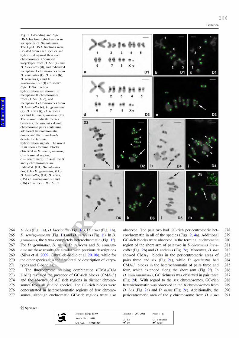
UNCORRECTEDPROOF
264 D. bos (Fig. 1a), D. laevicollis (Fig. 1d), D. nisus (Fig. 1h),
265 D. semisquamosus (Fig. 1l) and D. sericeus (Fig. 1j). In D.
266 geminatus, the y was completely heterochromatic (Fig. 1f).
267 For D. geminatus, D. nisus, D. sericeus and D. semisqu-
268 amosus these results are similar with previous descriptions
269 (Silva et al. 2009; Cabral-de-Mello et al. 2010b), while for
270 the other species it is the first detailed description of karyo-
271 types and C-banding.
272 The fluorochrome staining combination (CMA3/DA/
273 DAPI) revealed the presence of GC-rich blocks (CMA3?)
274 and the absence of AT rich regions in distinct chromo-
275 somes from all studied species. The GC-rich blocks were
276 concentrated in heterochromatic regions of few chromo-
277 somes, although euchromatic GC-rich regions were also
278observed. The pair two had GC-rich pericentromeric het-
279erochromatin in all of the species (Figs. 2, 4a). Additional
280GC-rich blocks were observed in the terminal euchromatic
281region of the short arm of pair two in Dichotomius laevi-
282collis (Fig. 2b) and D. sericeus (Fig. 2e). Moreover, D. bos
283showed CMA3? blocks in the pericentromeric areas of
284pairs three and six (Fig. 2a), while D. geminatus had
285CMA3? blocks in the heterochromatin of pairs three and
286four, which extended along the short arm (Fig. 2f). In
287D. semisquamosus, GC richness was observed in pair three
288(Fig. 2d). With regard to the sex chromosomes, GC-rich
289heterochromatin was observed in the X chromosomes from
290D. bos (Fig. 2a) and D. nisus (Fig. 2c). Additionally, the
291pericentromeric area of the y chromosome from D. nisus
Fig. 1 C-banding and C0t-1
DNA fraction hybridization in
six species of Dichotomius.
The C0t-1 DNA fractions were
isolated from each species and
hybridized against their own
chromosomes. C-banded
karyotypes from D. bos (a) and
D. laevicollis (d), and C-banded
metaphase I chromosomes from
D. geminatus (f), D. nisus (h),
D. sericeus (j) and D.
semisquamosus (l) are shown.
C0t-1 DNA fraction
hybridization are showed in
metaphase II chromosomes
from D. bos (b, c), and
metaphase I chromosomes from
D. laevicollis (e), D. geminatus
(g), D. nisus (i), D. sericeus
(k) and D. semisquamosus (m).
The arrows indicate the sex
bivalents, the asterisks denote
chromosome pairs containing
additional heterochromatic
blocks and the arrowheads
denote the terminal
hybridization signals. The insert
in m shows terminal blocks
observed in D. semisquamosus;
(t = terminal region,
c = centromere). In a–d, the X
and y chromosomes are
indicated. (D1) Dichotomius
bos, (D2) D. geminatus, (D3)
D. laevicollis, (D4) D. nisus,
(D5) D. semisquamosus and
(D6) D. sericeus. Bar 5 lm
Genetica
123Journal : Large 10709 Dispatch : 20-1-2011 Pages : 11
Article No. : 9551h LE h TYPESET
MS Code : GENE1743 h CP h DISK4 4
Au
tho
r P
ro
of
206

UNCORRECTEDPROOF
292 was CMA3? (Fig. 2c). The remaining heterochromatic
293 areas were neutral with regard to the CMA3 and DAPI
294 fluorochromes. Figure 4a summarizes all patterns of chro-
295 mosomal distribution of heterochromatin and base-pair
296 richness among the Dichotomius species.
297 C0t-1 DNA fraction mapping
298 The C0t-1 DNA fractions were obtained at different rean-
299 nealing times (30 s to 5 min) from each Dichotomius
300 species (Table 1). The hybridization of C0t-1 DNA in the
301 six species against their own chromosomes revealed simi-
302 lar patterns of heterochromatin distribution in five species
303 (Fig. 1e, g, i, k, m; Table 1). Moreover, small signals in the
304 terminal regions of chromosomes were observed in some
305 distinct cells of D. geminatus (Fig. 1g), D. nisus (Fig. 1i)
306 and D. semisquamosus (Fig. 1m). In D. bos, the pairs one,
307two and three did not reveal specific marks for C0t-1 DNA
308hybridization (Fig. 1b, c), even with the isolated C0t-1
309DNA fraction that underwent 5 min of reannealing. This
310FISH experiment revealed marks in euchromatic areas, but
311not in the heterochromatin of pairs one, two and three
312(results not shown).
313The hybridization of the C0t-1 DNA fraction from
314D. geminatus in the chromosomes of the other five species
315occurred only in the terminal/sub-terminal regions of all of
316the autosomes (Fig. 3). Moreover, the X sex chromosome
317displayed hybridization in all of the species, showing more
318intense labeling in D. bos (Fig. 3c, d), D. nisus (Fig. 3i, j)
319and D. semisquamosus (Fig. 3a, b). However, less intense
320labeling was observed in D. sericeus (Fig. 3g, h) and
321D. laevicollis (Fig. 3e, f). The y chromosome was labeled
322with dots in all of the species (Fig. 3). Low intense
323hybridization pattern was observed in D. laevicollis
Fig. 2 Fluorochrome staining
in the six species of
Dichotomius. Karyotypes from
D. bos (a) and D. laevicollis (b),
respectively; metaphase I
chromosomes from D. nisus (c),
D. semisquamosus (d),
D. sericeus (e) and
D. geminatus (f) are showed.
The arrows indicate the sex
bivalents, and the arrowheads
denote the CMA3? euchromatic
blocks. The insert in b indicates
the conformation of the pair 2
from D. laevicollis in metaphase
I. (D1) Dichotomius bos, (D2)
D. geminatus, (D3)
D. laevicollis, (D4) D. nisus,
(D5) D. semisquamosus and
(D6) D. sericeus. Bar 5 lm
Table 1 C0t-1 DNA fraction reassociation time and chromosomal location for the six Diichotomius species investigated in this study
Species C0t-1 DNA fraction
reassociation times
C0t-1 DNA fraction chromosomal mapping
Dichotomius bos 2 min 30 s Pericentromeric heterochromatin, except from pairs 1–3
Dichotomius geminatus 1 min Pericentromeric heterochromatin, terminal heterochromatic region
of the pairs 6, 7 and weak marks in terminal region of other autosomes
Dichotomius laevicollis 30 s Pericentromeric heterochromatin
Dichotomius nisus 1 min Pericentromeric heterochromatin and weak marks in terminal
region of some chromosomes
Dichotomius semisquamosus 3 min Pericentromeric heterochromatin and weak marks in terminal
region of some chromosomes
Dichotomius sericeus 30 s Pericentromeric heterochromatin
Genetica
123Journal : Large 10709 Dispatch : 20-1-2011 Pages : 11
Article No. : 9551h LE h TYPESET
MS Code : GENE1743 h CP h DISK4 4
Au
tho
r P
ro
of
207

UNCORRECTEDPROOF
324 (Fig. 3e, f) and D. sericeus (Fig. 3g, h), and high intense
325 hybridization was observed in D. nisus (Fig. 3i, j). At least
326 fifteen hybridized metaphases or initial meiotic cells from
327 each species were analyzed to define the patterns that are
328 described above.
329 Phylogeny of Dichotomius species based on mtDNA
330 sequence analysis
331 All phylogenetic analyses resulted in well-resolved trees
332 that exhibited similar topology and maximal or near-
333maximal indices of support for all of the nodes (Fig. 4b).
334Dichotomius laevicollis and D. semisquamosus appeared as
335a sister group to the other four species. The four remaining
336species formed two clades of sister taxa. D. nisus was
337grouped together with D. bos, and D. sericeus was grouped
338with D. geminatus. The topology of the tree was similar
339independent of the species included as outgroup. In fact the
340number of species included in the phylogenetic analysis
341represents only about 7.0% of the representatives of
342Dichotomius, and a wide analysis using more species
343should be necessary.
Fig. 3 C0t-1 DNA fraction
obtained from the genome of
Dichotomius geminatus
hybridized against the
chromosomes of the other five
Dichotomius species.
Metaphase I chromosomes from
D. semisquamosus (a), D. bos
(c), D. laevicollis (e),
D. sericeus (g) and D. nisus (i);
the initial pachytenes of
D. semisquamosus (b), D. bos
(d), D. laevicollis (f) and
D. sericeus (h), and metaphase
II of D. nisus (j) are showed.
The arrows indicate the sex
bivalents. Inserts indicate the
detail of the sex chromosomes
in metaphase I from another cell
for each species (a, c, e, g,
i) and the initial meiotic nucleus
(f) showing the polarization of
the hybridization signals. Note
that the hybridization signals are
restricted to the terminal regions
of the chromosomes. Bar 5 lm
Genetica
123Journal : Large 10709 Dispatch : 20-1-2011 Pages : 11
Article No. : 9551h LE h TYPESET
MS Code : GENE1743 h CP h DISK4 4
Au
tho
r P
ro
of
208

UNCORRECTEDPROOF
344 Discussion
345 Similar macro-chromosomal structures were observed in
346 the six Dichotomius species studied with 2n = 18,Xyp, and
347 biarmed chromosomes. Apparently, this karyotype pattern
348 is highly conserved in the Dichotomius genus, including
349 the remarkable presence of a large autosomal pair (pair 1)
350 that most likely arose by a fusion event between autosomes
351 from a 2n = 20 karyotype. Consistent with this hypothesis,
352 2n = 20 is the most common and ancient diploid number
353 for the Scarabaeidae family and Coleoptera order (Yadav
354 and Pillai 1979; Cabral-de-Mello et al. 2008).
355 The presence of heterochromatin primarily in the peri-
356 centromeric regions of all autosomes is a common char-
357 acteristic in the genus Dichotomius and also in
358 coleopterans (Ro _zek et al. 2004; Silva et al. 2009). Alter-
359 natively, the presence of heterochromatic blocks out of
360 pericentromeric region has been reported in D. geminatus
361 and other Scarabaeidae species, along with the presence of
362 terminal blocks and diphasic autosomes (Moura et al. 2003;
363 Cabral-de-Mello et al. 2010a, b). The diversified patterns of
364heterochromatin distribution indicate that the heterochro-
365matin diverged over evolutionary time in the various
366Scarabaeidae lineages.
367With regard to heterochromatin base pair richness, the
368presence of GC-rich blocks was common in Dichotomius
369and in Scarabaeidae, although neutral and AT rich blocks
370have also been described (Moura et al. 2003; Colomba
371et al. 2006). The presence of neutral and GC-rich blocks in
372Dichotomius indicates that there is some level of com-
373partmentalization of heterochromatin in the species.
374Moreover, the distinct pattern of the distribution of GC-rich
375blocks among the six species indicates that there is some
376level of dynamism in the genomic content of heterochro-
377matic areas that have diverged between species. Despite
378the variation in GC content, some of the CMA3? blocks in
379specific chromosomes are shared among the studied
380species, as the GC-rich heterochromatin localized in
381chromosome pair two. Some other CMA3? blocks are
382species-specific. For example, the blocks located in the y
383chromosome are exclusive for D. nisus, and the block in
384pair six was observed only in D. bos. With regard to the sex
Fig. 4 a Idiograms showing the distribution of cytogenetic markers
for each chromosome in the six species of Dichotomius studied;
b phylogenetic relationship of the six Dichotomius species based on
COI and 16S sequences. (D1) Dichotomius bos, (D2) D. geminatus,
(D3) D. laevicollis, (D4) D. nisus, (D5) D. semisquamosus and (D6)
D. sericeus
Genetica
123Journal : Large 10709 Dispatch : 20-1-2011 Pages : 11
Article No. : 9551h LE h TYPESET
MS Code : GENE1743 h CP h DISK4 4
Au
tho
r P
ro
of
209

UNCORRECTEDPROOF
385 chromosomes, the observed variability in GC richness
386 indicates that repeated DNAs with distinct composition are
387 present in these chromosomes and did not influence the
388 ancestral structure of chromosome association (Xyp).
389 The use of C0t-1 DNA fractions as probes in the six
390 species of Dichotomius revealed the same heterochromatin
391 distribution pattern in five of them, with the exception of
392 D. bos, demonstrating that highly and moderately repetitive
393 sequences are present in the heterochromatic areas.
394 Moreover, the presence of weak terminal marks in some of
395 the species, confirms that repetitive DNAs are present in
396 this area, although heterochromatin was not observed in
397 this region by C-banding. The absence of C0t-1 DNA
398 hybridization in chromosomes 1–3 of D. bos indicates that
399 the heterochromatin is composed of low copy-number of
400 sequences or a high diversity of repetitive DNA classes are
401 present in low copy numbers. The presence of low copy
402 numbers of repetitive sequences in the heterochromatin of
403 these three pairs was confirmed using a C0t-1 DNA fraction
404 that was isolated using 5 min of renaturation. This assay
405 revealed hybridization sites in a euchromatic region, but
406 the three pairs remained without hybridization. These
407 results are inconsistent with the classical idea that most of
408 the segments of constitutive heterochromatin contain high
409 concentrations of highly repeated DNA families (Sumner
410 2003).
411 An intense variation in the kinetics of renaturation was
412 observed among the six species when we obtained the C0t-
413 1 DNA fractions. The rate at which the fragmented DNA
414 sequences reassociate in the C0t-1 DNA assay is propor-
415 tional to the copy number in the genome (Peterson et al.
416 2002); therefore, the variation in the kinetics of renatur-
417 ation can be attributed to the differences in the amount of
418 repeated DNAs between the genomes of the six species.
419 Based on this parameter, the short C0t-1 DNA fractions
420 isolation time can be attributed to the presence of more
421 repeated sequences in the genomes of species, such as in
422 the cases of D. laevicollis and D. sericeus; however, the
423 genome of D. bos and D. semisquamosus is most likely
424 composed of a low quantity of repeated DNA or with more
425 variable composition. These results are clearly correlated
426 with the size of heterochromatic blocks, which are larger in
427 D. laevicollis and D. sericeus compared to D. bos and
428 D. semisquamosus.
429 The use of the C0t-1 DNA fraction from D. geminatus as
430 probes to hybridize in the chromosomes of the other five
431 Dichotomius species revealed an interesting pattern of high
432 conservation of terminal/sub-terminal blocks, in contrast to
433 the extensive variation in relation to the pericentromeric
434 heterochromatin. It is a general consensus that rapid mod-
435 ifications in repeated DNAs, even among closely related
436 species, generate species-specific sequences (Miklos 1985;
437 Ugarkovic and Plohl 2002), as observed among the
438Dichotomius species, at least between D. geminatus and the
439other five species. The copy number variation of repeated
440DNAs is governed, for example, by unequal crossing-over,
441replication slippage, rolling-circle replication and other
442unknown mechanisms (Charlesworth et al. 1994), and the
443variation in the pool of sequences is attributed to sequences
444modification and the principles of concerted evolution, in
445which diverse mechanisms of nonreciprocal transfer induce
446a high turnover of repeated sequences (Dover 1986).
447Diversification of repeated DNAs, such satDNA has been
448described in other insect groups (see Palomeque and Lorite
4492008). For example in D. melanogaster, the chromosomal
450banding techniques differentiate heterochromatin into some
451discrete regions by cytological methods, dependents of the
452presence and abundance of distinct satDNAs (Brutlag and
453Peacock 1975; Dimitri 1991; Gatti and Pimpinelli 1992;
454Lohe et al. 1993). For the first time, the use of the C0t-1
455DNA fraction from beetles permitted a deeper comparison
456of all of the heterochromatic regions at a molecular level.
457The analysis of this marker corroborates the results that
458were obtained using fluorochromes, which already indi-
459cated that there was heterochromatin differentiation in the
460six Dichotomius species. Several heterochromatin studies
461that are only based on C-banding are restricted to the
462descriptions of location of this genomic component; thus,
463the C-banding method generated superficial results that can
464led to erroneous conclusions about the similarities among
465animal karyotypes and genomes. Moreover, studies of
466repetitive DNA sequences focus primarily on descriptions
467of specific sequences in animal or plant chromosomes (for
468examples, see Yamada et al. 2002; Ansari et al. 2004;
469Martins et al. 2006; Adega et al. 2008), thereby limiting
470understanding the whole heterochromatic portion and dif-
471ferentiation of the genomes. Alternatively, the C0t-1 DNA
472allows for a wide genomic analysis of repeated DNAs, their
473relationship to heterochromatin variation and their distri-
474bution patterns among the chromosomes without the
475extensive work and laboratory expenses that are required
476for cloning and DNA sequencing.
477The presence of signals in the terminal/sub-terminal
478regions of autosomes after using the C0t-1 DNA fraction
479obtained from D. geminatus in five of the species indicates
480that the genome portion of chromosome ends among the
481six species are conserved. However, the possibility of
482cross-hybridization between centromeric repeated DNAs of
483D. geminatus and terminal/sub-terminal regions of the
484autosomes from the other five species can not be elimi-
485nated. This conservation may be related to structural and/or
486functional action of specific DNA elements in the terminal/
487sub-terminal region of the chromosomes. These sequences
488may play roles as telomeres, telomere-associated DNA, or
489they may be the telomeric sequences. Some structures that
490were observed in the initial meiotic cells and in the
Genetica
123Journal : Large 10709 Dispatch : 20-1-2011 Pages : 11
Article No. : 9551h LE h TYPESET
MS Code : GENE1743 h CP h DISK4 4
Au
tho
r P
ro
of
210

UNCORRECTEDPROOF
491 interphasic nuclei, such as the bouquet configuration and
492 the polarization of the hybridized signals to one nuclear
493 pole to form a structure that is similar to the rabl config-
494 uration, led us to propose that these conserved sequences
495 most likely correspond to telomeres or telomere-associated
496 DNA (see Fig. 3).
497 The variations in pericentromeric repeat sequences that
498 we observed in the fiveDichotomius species (comparing the
499 C0t-1 DNA fraction obtained from D. geminatus) is a com-
500 mon feature in complex eukaryotes, paradoxically with
501 conserved function of this chromosomal region (Schmidt
502 and Heslop-Harrison 1998; Henikoff et al. 2001; Ugarkovic
503 and Plohl 2002). On the other hand, the telomeric sequences
504 are highly conserved in some groups, including vertebrates
505 and plants (Meyne et al. 1989; Cox et al. 1993; Fuchs et al.
506 1995). In insects, the telomeres are more variable, but the
507 telomeric motif (TTAGG)n is widespread across several
508 insect orders (Okazaki et al. 1993; Sahara et al. 1999). This
509 high conservation of C0t-1 DNA hybridization appears to be
510 a common feature in Dichotomius species, although we are
511 not certain if the labeled regions correspond to the telomeric
512 sequences or to the telomere-associated DNA sequence.
513 Conserved repeated sequences in the telomeric regions have
514 been reported in insects, including the transposable element
515 TART that is found in distantly related species ofDrosophila
516 and a complex tandem repeatedDNA family that is observed
517 in the telomeres of theChironomus genus (Zhang et al. 1994;
518 Casacuberta and Pardue 2003).
519 The reduction of the diploid number to 2n = 18 and the
520 presence of a large biarmed bivalent were proposed to have
521 resulted from an autosomal fusion event that may be
522 involved in chromosomal differentiation in the Dichotom-
523 ius species. However, the proposed autosomal fusion event
524 was not corroborated by interstitial hybridization signals in
525 the first bivalent of the five species studied when the
526 D. geminatus C0t-1 DNA fraction that labeled the terminal
527 region of autosomes in the other species was used. These
528 data can be correlated to a rapid modification or loss of
529 terminal sequences that occurred after the chromosomal
530 fusion, leading to the failure of interstitial C0t-1 DNA site
531 detection in FISH experiments.
532 With regard to the sex chromosomes in the Dichotomius
533 species analyzed in this study, the classical cytogenetic and
534 C0t-1 DNA hybridization analyses indicate that there are
535 distinct repetitive DNA differentiation patterns for the X
536 and y chromosomes in the genus, despite the conservation
537 of a basic Xyp system. The X chromosome showed more
538 variation in comparison with the y chromosome with
539 blocks of repeated DNAs concentrated in the pericentro-
540 meric areas or extending along the short arm, following the
541 heterochromatin distribution. Using the C0t-1 DNA frac-
542 tion from D. geminatus, sequence-related variability was
543 also observed. Some of the species had only small size
544blocks on the X and y chromosomes (D. sericeus and
545D. laevicollis), while other species had medium size blocks
546(D. bos and D. semisquamosus), and one species had large
547size blocks (D. nisus). Although it was possible to identify
548sequence variability in the sex chromosomes, it was
549impossible to determine whether or not these sequences are
550shared between the autosomal complement and the sex
551chromosome of D. geminatus due to the use of a pool of
552sequences that was obtained by the C0t-1 DNA method.
553The chromosomal mapping of repeated DNAs using
554classical and molecular cytogenetic approaches in the six
555species of Dichotomius analyzed indicates that there is
556conservation of location of heterochromatic blocks as well
557as modification of sequences, at least between D. gemin-
558atus and the other five species. Moreover, it was possible to
559identify some conserved chromosomes within the genus
560based on the applied chromosomal markers, such as pairs
561one, five and eight. However, the other chromosomes,
562including the sex bivalents, have apparently experienced
563distinct differentiation processes, including heterochroma-
564tin differentiation without modification of the macro-
565chromosomal structure. Although we identified some con-
566served chromosomes in the genus, the general pattern of
567organization of repeated DNAs does not reflect the rela-
568tionship between the six species based in COI and 16S
569rRNA genes. Repeated DNAs are subject to the action of
570several molecular mechanisms and are thought to be the
571most rapidly evolving components in genomes (Dover
5721986; Charlesworth et al. 1994; Eickbush and Eickbush
5732007), displaying intense variability, even in related spe-
574cies such as those in the Dichotomius genus.
575Finally, the application of C0t-1 DNA fraction is a
576useful tool for studies of repeated DNAs in insects, thus
577contributing to understanding heterochromatin differentia-
578tion among related species. Contrary to vertebrates that
579posses the availability of BAC (Bacterial Artificial Chro-
580mosomes) libraries and whole chromosomes as probes, for
581insects there are few available genes or DNA sequences to
582be applied as probes for purposes of cytogenetic mapping.
583In this way, although the C0t-1 DNA hybridization does not
584permit the generation of precise information about specific
585chromosomes or DNA sequences, it allows for a wide
586comparison of the whole repetitive portion of genomes
587without expensive applications of DNA cloning and
588sequencing. This analysis represents an interesting
589approach for the investigation of karyotype diversification
590and genome evolution under the focus of cytogenetics.
591Acknowledgments This study was supported by Fundacao de592Amparo a Pesquisa do Estado de Sao Paulo (FAPESP), Coordenad-593oria de Aperfeicoamento de Pessoal de Nıvel Superior (CAPES),594Conselho Nacional de Desenvolvimento Cientıfico e Tecnologico595(CNPq) and Fundacao de Amparo a Ciencia e Tecnologia do Estado596de Pernambuco (FACEPE). The authors are grateful to Fernando
Genetica
123Journal : Large 10709 Dispatch : 20-1-2011 Pages : 11
Article No. : 9551h LE h TYPESET
MS Code : GENE1743 h CP h DISK4 4
Au
tho
r P
ro
of
211

UNCORRECTEDPROOF
597 Augusto Barbosa Silva for the taxonomic identification of the spec-598 imens analyzed in this study and to PIBIC/CNPq/UPE scholarship of599 Melo AS.
600 References
601 Adega F, Chaves R, Guedes-Pinto H (2008) Suiformes orthologous602 satellite DNAs as a hallmark of Pecari tajacu and Tayassu
603 pecari (Tayassuidae) evolutionary rearrangements. Micron604 39:1281–1287605 Altschul SF, Gish W, Miller W, Myers EW, Lipman DJ (1990) Basic606 local alignment search tool. J Mol Biol 215:403–410607 Ansari HA, Ellison NW, Griffiths AG, Williams WM (2004)608 A lineage-specific centromeric satellite sequence in the609 genus Trifolium. Chromosome Res 12:357–367610 Bione E, Moura RC, Carvalho R, Souza MJ (2005a) Karyotype,611 C-and fluorescence banding pattern, NOR location and FISH612 study of five Scarabaeidae (Coleoptera) species. Gen Mol Biol613 26:376–381614 Bione E, Camparoto ML, Simoes ZLP (2005b) A study of the615 constitutive heterochromatin and nucleolus organizer regions of616 Isocopris inhiata and Diabroctis mimas (Coleoptera: Scarabaei-617 dae, Scarabaeinae) using C-banding, AgNO3 staining and FISH618 techniques. Gen Mol Biol 28:111–116619 Brutlag DL, Peacock, WJ (1975) The eukaryote chromosome. In620 Peacock WJ, Brock RD (eds) Canberra Australia, Australian621 University Press, pp 35–45622 Cabral-de-Mello DC, Moura RC, Carvalho R, Souza MJ (2010a)623 Cytogenetic analysis of two related Deltochilum (Coleoptera,624 Scarabaeidae) species: diploid number reduction, extensive625 heterochromatin addition and differentiation. Micron 41:626 112–117627 Cabral-de-Mello DC, Moura RC, Martins C (2010b) Chromosomal628 mapping of repetitive DNAs in the beetle Dichotomius gemin-629 atus provides the first evidence for an association of 5S rRNA630 and histone H3 genes in insects, and repetitive DNA similarity631 between the B chromosome and A complement. Heredity632 104:393–400633 Cabral-de-Mello DC, Oliveira SG, Ramos IC, Moura RC (2008)634 Karyotype differentiation patterns in species of the subfamily635 Scarabaeinae (Scarabaeidae: Coleoptera). Micron 39:1243–1250636 Casacuberta E, Pardue ML (2003) Transposon telomeres are widely637 distributed in the Drosophila genus: TART elements in the638 virilis group. Proc Natl Acad Sci USA 100:3363–3368639 Charlesworth B, Sniegowski P, Stephan W (1994) The evolutionary640 dynamics of repetitive DNA in eukaryotes. Nature 371:215–220641 Colomba MS, Vitturi R, Libertini A, Gregorini A, Zunino M (2006)642 Heterochromatin of the scarab beetles, Bubas bison (Coleoptera:643 Scarabaeidae) II. Evidence for AT-rich compartmentalization644 and a high amount of rDNA copies. Micron 37:47–51645 Colomba MS, Vitturi R, Zunino M (2000) Karyotype analyzes,646 banding, and fluorescent in situ hybridization in the Scarab647 beetle Gymnopleurus sturmi McLeady (Coleoptera, Scarabaeoi-648 dea, Scarabaeidae). J Heredity 91:260–264649 Cox AV, Bennett ST, Parokonny AS, Kenton A, Callimassia MA,650 Bennett MD (1993) Comparison of plant telomere locations651 using a PCR-generated synthetic probe. Ann Botany 72:239–247652 Dimitri P (1991) Cytogenetic analysis of the second chromosome653 heterochromatin of Drosophila melanogaster. Genetics 127:654 553–564655 Dover GA (1986) Linkage disequilibrium and molecular drive in the656 rDNA gene family. Genetics 122:249–252657 Edgar RC (2004) MUSCLE: Multiple sequence alignment with high658 accuracy and high throughput. Nucleic Acids Res 32:1792–1797
659Eickbush TH, Eickbush DG (2007) Finely orchestrated movements:660Evolution of the ribosomal RNA genes. Genetics 175:477–485661Ferreira IA, Martins C (2008) Physical chromosome mapping of662repetitive DNA sequences in Nile tilapia Oreochromis niloticus:663evidences for a differential distribution of repetitive elements in664the sex chromosomes. Micron 39:411–418665Fuchs J, Brandes A, Schubet I (1995) Telomere sequence localization666and karyotype evolution in higher plants. Plant Syst Evol 196:667227–241668Gatti M, Pimpinelli S (1992) Functional elements in Drosophila
669melanogaster heterochromatin. Annu Rev Genet 26:239–275670Hall TA (1999) BioEdit: a user-friendly biological sequence align-671ment editor and analysis program for Windows 95/98/NT. Nucl672Acids Symp Ser 41:95–98673Henikoff S, Ahmad K, Malik HS (2001) The centromere paradox:674stable inheritance with rapidly evolving DNA. Science 293:6751098–1102676Huelsenbeck JP, Ronquist F, Nielsen R, Bollback JP (2001) Bayesian677inference of phylogeny and its impact of evolutionary biology.678Science 294:2310–2314679Kimura M (1980) A simple method for estimating evolutionary rate680of base substitutions through comparative studies of nucleotide681sequences. J Mol Evol 16:111–120682Lohe AR, Hilliker AJ, Roberts PA (1993) Mapping simple repeated683DNA sequences in heterochromatin of Drosophila melanogaster.684Genetics 134:1149–1174685Martınez-Navarro EM, Serrano J, Galian J (2004) Chromosome evolu-686tion in ground beetles: localization of the rDNA loci in the tribe687Harpalini (Coleoptera, Carabidae). J Zool Syst Evol Res 42:38–43688Martins C, Ferreira IA, Oliveira C, Foresti F, Galetti PM Jr (2006) A689tandemly repetitive centromeric DNA sequence of the fish690Hoplias malabaricus (Characiformes: Erythrinidae) is derived691from 5S rDNA. Genetica 127:133–141692Meyne J, Ratliff RL, Moyzis RK (1989) Conservation of the human693telomere sequence (TTAGGG)n among vertebrates. Proc Natl694Acad Sci USA 86:7049–7053695Miklos GLG (1985) Localized highly repetitive DNA sequences in696vertebrate and invertebrate genomes. In: Macintyre RJ (ed)697Molecular evolutionary genetics. Plenum Press, New York,698pp 241–321699Moura RC, Souza M, Mel NF, Lira-Neto AC (2003) Karyotypic700characterization of representatives from Melolonthinae (Cole-701optera, Scarabaeidae): Karyotypic analysis, banding and fluo-702rescent in situ hybridization (FISH). Hereditas 138:200–206703Okazaki S, Tsuchida K, Maekawa H, Ishikawa H, Fujiwara H (1993)704Identification of a pentanucleotide telomeric sequence,705(TTAGG)n, in the silkworm Bombyx mori and in other insects.706Mol Cell Biol 13:1424–1432707Palomeque T, Lorite P (2008) Satellite DNA in insects: a review.708Heredity 100:564–573709Peterson DG, Schulze SR, Sciara EB, Lee SA, Bowers JE, Nagel A,710Jiang N, Tibbitts DC, Wessler SR, Paterson AH (2002)711Integration of Cot analysis, DNA cloning, and high-throughput712sequencing facilitates genome characterization and gene discov-713ery. Genome Res 12:795–807714Posada D, Crandall KA (1998) Modeltest: testing the model of DNA715substitution. Bioinformatics 1:817–818716Ronquist F, Huelsenbeck JP (2003) MrBayes 3: Bayesian phylogenetic717inference under mixed models. Bioinformatics 19:1572–1574718Ro _zek M, Lachowska D, Petitpierre E, Holecova M (2004) C-bands719on chromosomes of 32 beetle species (Coleoptera: Elateridae,720Cantharidae, Oedemeridae, Cerambycidae, Chrysomelidae and721Curculionidae). Hereditas 140:1–10722Sahara K, Marec F, Traut W (1999) TTAGG telomeric repeats in723chromosomes of some insects and other arthropods. Chromo-724some Res 7:449–460
Genetica
123Journal : Large 10709 Dispatch : 20-1-2011 Pages : 11
Article No. : 9551h LE h TYPESET
MS Code : GENE1743 h CP h DISK4 4
Au
tho
r P
ro
of
212

UNCORRECTEDPROOF
725 Sambrook J, Russel DW (2001) Molecular cloning. A laboratory726 manual, 3rd edn. Cold Spring Harbor Laboratory Press, New727 York728 Sanchez-Gea JF, Serrano J, Galian J (2000) Variability in rDNA loci729 in Iberian species of the genus Zabrus (Coleoptera: Carabidae)730 detected by fluorescence in situ hybridization. Genome 43:22–28731 Schmidt T, Heslop-Harrison JS (1998) Genomes, genes and junk: the732 large-scale organization of plant chromosomes. Trends Plant Sci733 3:195–199734 Schneider MC, Rosa SP, Almeida MC, Costa C, Cella DM (2007)735 Chromosomal similarities and differences among four Neotrop-736 ical Elateridae (Conoderini and Pyrophorini) and other related737 species, with comments on the NOR patterns in Coleoptera.738 J Zool Syst Evol Res 45:308–316739 Schweizer D, Mendelak M, White MJD, Contreras N (1983)740 Cytogenetics of the parthenogenetic grasshopper Warramaba741 virgo and its bisexual relatives. X. Pattern of fluorescent742 banding. Chromosoma 88:227–236743 Silva GM, Bione EG, Cabral-de-Mello DC, Moura RC, Simoes ZLP,744 Souza MJ (2009) Comparative cytogenetics study in three745 species of the genus Dichotomius (Coleoptera: Scarabaeidae).746 Gen Mol Biol 32:276–280747 Sumner AT (1972) A simple technique for demonstrating centromeric748 heterochromatin. Exp Cell Res 75:304–306749 Sumner AT (2003) Chromosomes: organization and function, 1st edn.750 Blackwell Publishing company, London
751Tamura K, Dudley J, Nei M, Kumar S (2007) MEGA4: molecular752evolutionary genetics analysis (MEGA) software version 4.0.753Mol Biol Evol 24:1596–1599754Ugarkovic D, Plohl M (2002) Variation in satellite DNA profiles–755causes and effects. The EMBO J 21:5955–5959756Ward RD, Zemlak TS, Innes BH, Last PR, Hebert PDN (2005) DNA757barcoding Australia’s fish species. Phil Trans R Soc B 360:7581847–1857759Xia X, Xie Z (2001) DAMBE: Data analysis in molecular biology and760evoluiton. J Heredity 92:371–373761Yadav JS, Pillai RK (1979) Evolution of karyotypes and phylogenetic762relationships in Scarabaeidae (Coleoptera). Zool Anz 202:763105–118764Yamada K, Nishida-Umehara C, Matsuda Y (2002) Characterization765and chromosomal distribution of novel satellite DNA sequences766of the lesser rhea (Pterocnemia pennata) and the greater rhea767(Rhea americana). Chromosome Res 10:513–523768Zhang YJ, Kamnert I, Lopez CC, Cohn M, Edstrom JE (1994)769A family of complex tandem DNA repeats in the telomeres of770Chironomus pallidivittatus. Mol Cell Biol 14:8028–8036771Zwick MS, Hanson RE, McKnight TD, Nurul-Islam-Faridi M, Stelly772DM (1997) A rapid procedure for the isolation of C0t–1 DNA773from plants. Genome 40:138–142
774
Genetica
123Journal : Large 10709 Dispatch : 20-1-2011 Pages : 11
Article No. : 9551h LE h TYPESET
MS Code : GENE1743 h CP h DISK4 4
Au
tho
r P
ro
of
213