parosteal aneurysmal bone cyst - scielo filer ev bras ortop. 2015;50(5):601–606 case report...
TRANSCRIPT

r e v b r a s o r t o p . 2 0 1 5;5 0(5):601–606
C
P
WL
I
a
A
R
A
A
K
A
C
C
I
P
C
C
C
I
h2
www.rbo.org .br
ase report
arosteal aneurysmal bone cyst�
alter Meohas, Ana Cristina de Sá Lopes, João Victor da Silveira Möller ∗,uma Duarte Barbosa, Marcelo Braganca dos Reis Oliveira
nstituto Nacional de Traumatologia e Ortopedia (Into), Rio de Janeiro, RJ, Brazil
r t i c l e i n f o
rticle history:
eceived 4 June 2014
ccepted 23 October 2014
vailable online 1 September 2015
eywords:
neurysmal bone cyst
alcitonin
orticosteroids
nfiltration
a b s t r a c t
The incidence of aneurysmal bone cysts is 0.14 cases per 100,000 individuals. Parosteal
aneurysmal bone cysts are the least prevalent subtype and represent 7% of all aneurysmal
bone cysts. We present the case of a 38-year-old male patient with pain and bulging in
his right arm for eight months. He had previously been diagnosed as presenting giant-
cell tumor, but his slides were reviewed and his condition was then diagnosed as parosteal
aneurysmal bone cyst. The patient was treated with corticosteroid and calcitonin infiltration
into the lesion and evolved with clinical and radiological improvement within the first five
weeks after the operation.
© 2015 Sociedade Brasileira de Ortopedia e Traumatologia. Published by Elsevier Editora
Ltda. All rights reserved.
Cisto ósseo aneurismático parosteal
alavras-chave:
isto ósseo aneurismático
alcitonina
orticosteroides
nfiltracão
r e s u m o
O cisto ósseo aneurismático tem uma incidência de 0,14 a cada 100 mil indivíduos. O subtipo
parosteal é o menos prevalente, representa 7% de todos. Apresentamos um paciente mas-
culino, 38 anos, com dor e abaulamento em braco direito havia oito meses. Diagnosticado
previamente como tumor de células gigantes, teve sua lâmina revisada e então foi feito o
diagnóstico de cisto ósseo aneurismático parosteal. O paciente foi tratado com infiltracão
intralesional de corticosteroide e calcitonina e evoluiu com melhoria clínica e radiológica já
nas primeiras cinco semanas pós-operatórias.
© 2015 Sociedade Brasileira de Ortopedia e Traumatologia. Publicado por Elsevier Editora
Ltda. Todos os direitos reservados.
� Work performed at the Instituto Nacional de Traumatologia e Ortop∗ Corresponding author.
E-mails: joao [email protected], [email protected] (J.V. da Sttp://dx.doi.org/10.1016/j.rboe.2015.08.008255-4971/© 2015 Sociedade Brasileira de Ortopedia e Traumatologia. P
edia (INTO), Rio de Janeiro, RJ, Brazil.
ilveira Möller).
ublished by Elsevier Editora Ltda. All rights reserved.

p . 2 0
602 r e v b r a s o r t oIntroduction
Aneurysmal bone cysts were first described by Jaffe and Lich-tenstein in 1942.1 They account for 1–2% of all primary bonetumors and affect the metaphyseal region of long bones inchildren, adolescents and young adults.2,3
This lesion typically develops inside bones.4 Cysts locatedin the cortical bone are rare and account for 7–9.3% of allaneurysmal bone cysts.5,6
Few cases have been reported in the literature. Theapproach used is individualized and varies according to theexperience of each service. We present a case of parostealaneurysmal bone cyst that was treated in accordance with ourexperience.
Case report
The patient was a 38-year-old man of mixed race with a com-plaint of pain and bulging in his right arm. It was of progressivenature and the patient had had the complaint for at leasteight months. He said that he had not suffered any traumaor undergone previous surgery.
The patient, who had been attended previously at anotherservice, underwent a biopsy from which the histopathologicaldiagnosis was compatible with a giant-cell tumor. When he
arrived at our service, because of the clinical and radiologicalcharacteristics of the slides (Figs. 1–4), a review of them wasrequested.Fig. 1 – Radiographs of the right humerus
1 5;5 0(5):601–606
This review showed the presence of a lesion formed bycyst membranes that sometimes showed complete septationconstituted by fusiform and multinucleated giant cells. Bonetrabeculae dissociated by connective tissue were noted, alongwith neoformed bone trabeculae of reactive pattern, which ledto the diagnosis of parosteal aneurysmal bone cyst.
Infiltration into the lesion using calcitonin and corticos-teroid was indicated after reaching a group decision. In thefifth postoperative week, the lesion was already seen to beundergoing an ossification process (Fig. 5).
Discussion
Aneurysmal bone cysts were first described by Jaffe and Lich-tenstein in 1942. According to the World Health Organization,they are characterized as benign cystic bone lesions composedof bone voids that are filled with blood and separated by septaof connective tissue containing fibroblasts, osteoclastic giantcells and reactive bone tissue.1–3,7
These cysts account for 1–2% of all primary bone tumorsand their incidence is 0.14 per 100,000 individuals.8 Thelesions affect the metaphyseal region of the long bones ofchildren, adolescents and young adults.2,3
The lesions generally develop inside bones and causethinning of the cortex and possibly bone protrusion.4 Local-ized cysts in the cortical bone are rare and were previously
in anteroposterior and lateral view.
named subperiosteal giant cells or subperiosteal osteoclasis.4
In 1950, Lichtenstein9 published an article that elucidatedand differentiated parosteal aneurysmal bone cysts from

r e v b r a s o r t o p . 2 0 1 5;5 0(5):601–606 603
grap
sc
cpf
Fig. 2 – Tomo
ubperiosteal giant cells, hemangiomas and osteogenic sar-omas.
In 1957, Sherman and Soong5 classified aneurysmal bone
ysts into three types: eccentric, parosteal and central. Thearosteal subtype is the least frequent subtype, accountingor 7–9.3% of all aneurysmal bone cysts.5,6hic features.
Pain is the most prevalent symptom, and its duration maybe weeks to months.2 Radiographically, these cysts present assingle eccentric and insufflative lesions that reach the perios-
2,3
teum and have well-defined margins. Their presence may beassociated with onion-skin periosteal reactions and Codman’striangle.2,6
604 r e v b r a s o r t o p . 2 0 1 5;5 0(5):601–606
Fig. 3 – Magnetic resonance.
Tomography helps in making the differential diagnosisof these lesions. They show liquid density and may clearlydemonstrate the liquid levels.2,6 Scintigraphy shows that thereis greater uptake at the periphery of the lesion.2 In magneticresonance imaging, the lesion is well defined, with lobulatedoutlines and liquid levels.2
The histology of aneurysmal bone cysts is character-ized by voids filled with blood. These voids are coveredby a single layer of undifferentiated cells. The solid tissuesurrounding the lesion is composed of richly vascularizedfibrosis.2 Diagnostic differentiation between giant-cell tumorsand osteosarcoma with telangiectasia is anatomopathologi-
cally complex.2Because these are aggressive lesions, the treatment con-sists of curettage, with or without subsequent adjuvants such
as bone grafts, bone marrow aspirate, cryotherapy, argon,phenol or calcitonin with corticosteroid injection into thelesion.7,10 In our service, use of corticosteroids in associa-tion with calcitonin, injected into the lesion, is the preferredmethod for treating this type of lesion. Cases of resolutionof lesions after an episode of fracturing or after a biopsy, oreven spontaneously, have been described.7,8 Lesion recurrenceis associated with young patients, previous aneurysmal bonecysts, location adjacent to a joint or growth plate, low mitoticcount and presence of other open growth plates.8
Here, we presented a rare case of parosteal aneurysmalbone cyst in which the clinical, radiological and anato-
mopathological findings and presence of a multidisciplinaryteam were essential in order to completely elucidate thediagnosis.
r e v b r a s o r t o p . 2 0 1 5;5 0(5):601–606 605
Fig. 4 – Bone sc
intigraphy.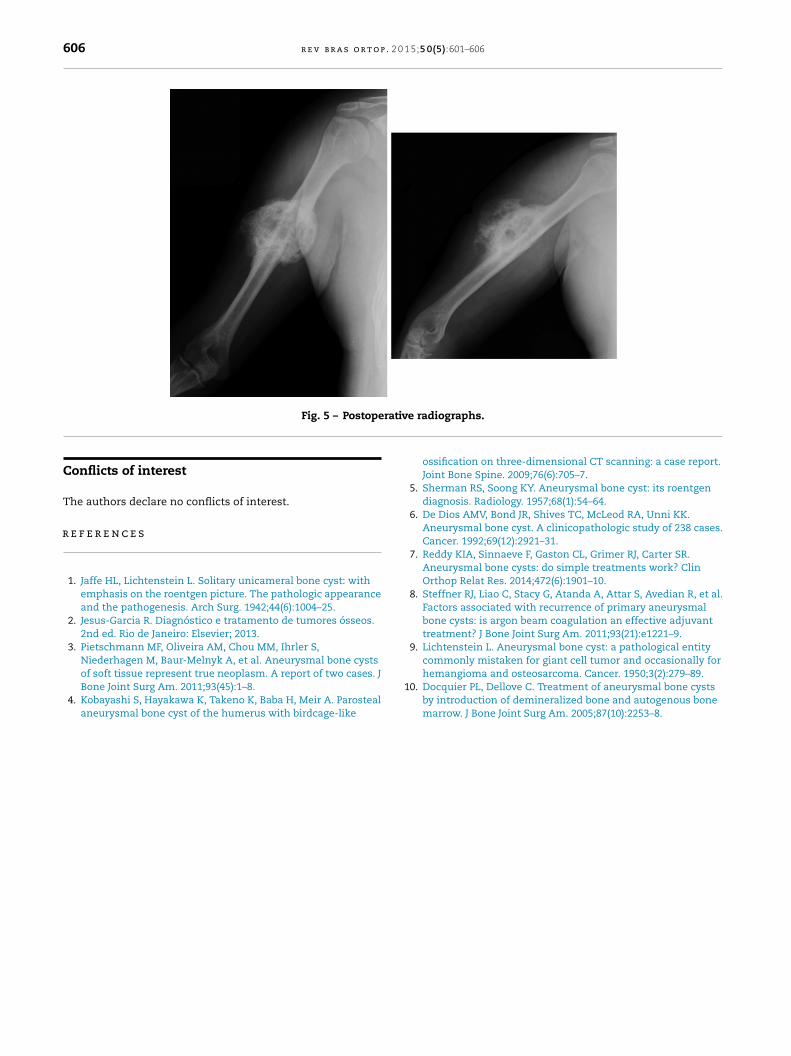
606 r e v b r a s o r t o p . 2 0 1 5;5 0(5):601–606
rativ
r
Fig. 5 – Postope
Conflicts of interest
The authors declare no conflicts of interest.
e f e r e n c e s
1. Jaffe HL, Lichtenstein L. Solitary unicameral bone cyst: withemphasis on the roentgen picture. The pathologic appearanceand the pathogenesis. Arch Surg. 1942;44(6):1004–25.
2. Jesus-Garcia R. Diagnóstico e tratamento de tumores ósseos.2nd ed. Rio de Janeiro: Elsevier; 2013.
3. Pietschmann MF, Oliveira AM, Chou MM, Ihrler S,Niederhagen M, Baur-Melnyk A, et al. Aneurysmal bone cysts
of soft tissue represent true neoplasm. A report of two cases. JBone Joint Surg Am. 2011;93(45):1–8.4. Kobayashi S, Hayakawa K, Takeno K, Baba H, Meir A. Parostealaneurysmal bone cyst of the humerus with birdcage-like
1
e radiographs.
ossification on three-dimensional CT scanning: a case report.Joint Bone Spine. 2009;76(6):705–7.
5. Sherman RS, Soong KY. Aneurysmal bone cyst: its roentgendiagnosis. Radiology. 1957;68(1):54–64.
6. De Dios AMV, Bond JR, Shives TC, McLeod RA, Unni KK.Aneurysmal bone cyst. A clinicopathologic study of 238 cases.Cancer. 1992;69(12):2921–31.
7. Reddy KIA, Sinnaeve F, Gaston CL, Grimer RJ, Carter SR.Aneurysmal bone cysts: do simple treatments work? ClinOrthop Relat Res. 2014;472(6):1901–10.
8. Steffner RJ, Liao C, Stacy G, Atanda A, Attar S, Avedian R, et al.Factors associated with recurrence of primary aneurysmalbone cysts: is argon beam coagulation an effective adjuvanttreatment? J Bone Joint Surg Am. 2011;93(21):e1221–9.
9. Lichtenstein L. Aneurysmal bone cyst: a pathological entitycommonly mistaken for giant cell tumor and occasionally for
hemangioma and osteosarcoma. Cancer. 1950;3(2):279–89.0. Docquier PL, Dellove C. Treatment of aneurysmal bone cystsby introduction of demineralized bone and autogenous bonemarrow. J Bone Joint Surg Am. 2005;87(10):2253–8.