novidades sobre a nanotecnologia do concreto e sua ... · novidades sobre a nanotecnologia do...
TRANSCRIPT

Novidades Sobre a
Nanotecnologia do Concreto
e sua Influência na
Construção Civil do Futuro
Paulo J.M. Monteiro
University of California
Berkeley

Por que??
• Somos engenheiros civis, e qual é
a importancia da nanotecnologia
para o concreto?

Condição insustentável

Novas Alternativas
Italcementi cimento branco auto-
limpável
Uso de nano-argilas para CSC
Geopolímeros
NanoCem
Calera

Meta:
• Como reduzir 120 anos de
experiência com cimento portland
em 3-5 anos para um novo
cimento?

Geopolimeros (sem cimento)
10 M NaOH solution

Processo
da
Calera

Pesquisa Integrada
Advanced Light SourceSoft x-rays microscopy, Small Angle Scattering,
High-Pressure, Microdiffraction, Microtomography,
Ambient XPS.
Advanced Photon SourceTotal scattering methods (pdf), Nanotomography,
Small Angle Scattering.
BESSY
Nanotomography
… more at Los Alamos

Tradição brasileira de
excelência no concreto…
• Vários recordes mundiais em
pontes e barragens.
• Renome internacional em
tecnologia do concreto e projeto
estrutural.

O Brasil está em otima fase
• Economia em expansão.
• Construção em alta.
• Previsão de crescimento
constante.

Um desafio e um apoio…
• Por que não fazer do Brasil uma
potência na area de
nanotecnologia do concreto?

O mais difícil já tem em
Campinas…

Advanced Light Source
the world's first
third-
generation
synchrotron
light source in
its energy
range
Location

Advanced Light Source at Berkeley

Testes Mecânicos

Pergunta:
• Como medir a propriedades
mecânicas dos cristais
pequenos?

High pressure generating device: Diamond Anvil Cell
Strongest Material Ko = ~440GPa Transparent to electromagnetic spectrum
Theoretically, possible over 500GPa, but usually ~50GPa
Extremely small sample size
Hydrostatic pressure Medium 4:1 Methanol/Ethanol solution Up to ~20GPa, nearly hydrostatic pressure
Ruby fluorescence technique Measurement of pressure inside of the cell
load
Figure. Schematic of Diamond Anvil Cell
sample
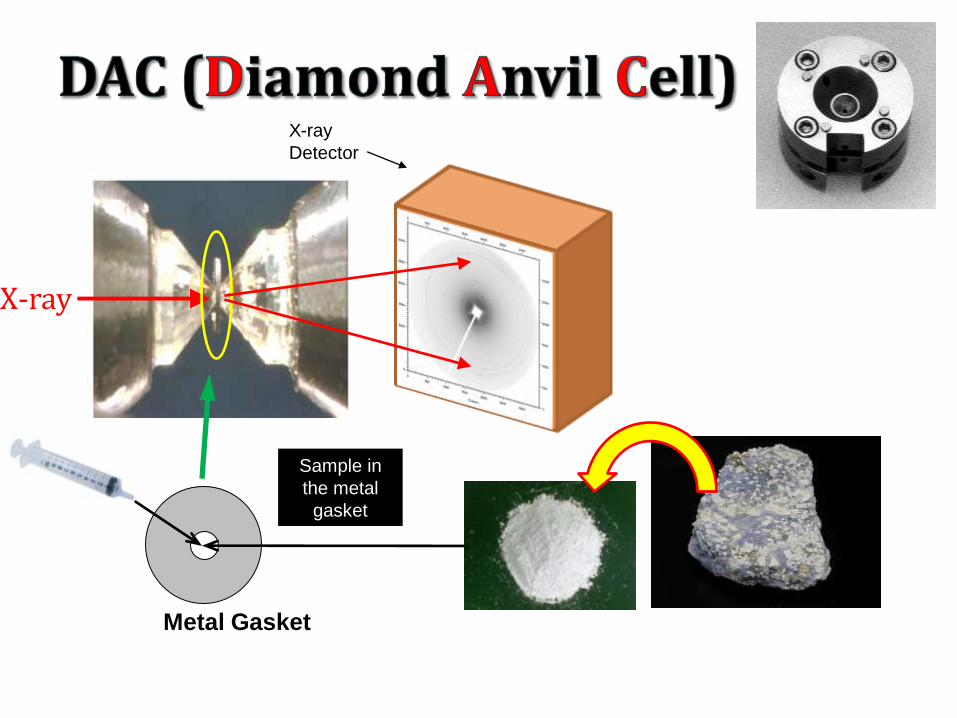
X-ray
Detector
X-ray
Metal Gasket
Sample in
the metal
gasket

As the pressure increases, the unit cell shrinks.
Unit cell dimensions (a, b, c, α, β, γ) at a certain pressure can be calculated from X-ray diffraction pattern
P(V/Vo) can be obtained
Bulk modulus =
Figure. X-ray diffraction Pattern in beamline 12.2.2 (tobermorite)
T
dPK V
dV
At low pressure At high pressure

Estrutura da etringita
Ca atoms are displayed as blue circles, oxygen atoms in red,
aluminum atoms in light blue, sulfate tetrahedral in yellow and
hydrogen atoms in grey.

Resultado experimental
Isothermal bulk modulus of ettringite: 27(7) GPa

O que acontece com as propriedades mecânicasquando o Si e’ substituido porAl?

Al substitution: The synthetic C-S-H (I) does not contain Al in its structure
The alkali-activated slag C-S-H (I) contains Al in its structure
Ca-O Layer
Bridging SiO4
Al substitution for the bridging
tetrahedron
(a) 14Å tobermorite [100] (b) 11Å tobermorite [100] (c) 9Å tobermorite [100]
- most hydrated form - moderately hydrated form - least hydrated form
11
Å
14
Å
9.3
Å

Não tem diferença nenhuma!
0.0
0.5
1.0
1.5
2.0
2.5
3.0
3.5
4.0
4.5
5.0
0.9000 0.9200 0.9400 0.9600 0.9800 1.0000
V/Vo
P(GPa)
AAS C-S-H (I)
B-M AAS C-S-H (I) (Ko'=4.00)
SYN C-S-H (I)
B-M SYN C-S-H (I) (Ko'=4.00)

Spectro eletromagnético

Dois tipos de Microscópios

250 to 900 eV
Resolution: 25 nm
Magnification: 1600 to 2400 times

Fresnel Zone Plate Lens
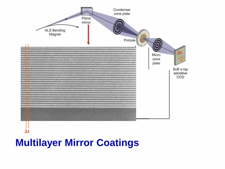
Multilayer Mirror Coatings
Dt

Preparação da Amostra
Restriction: sample thickness (less than 10 mm)
Silicon nitride
windows
Highly diluted samples
(water/cement is 5 before
centrifugation)
Imaging as soon as 6 minutes
after mixing

Early hydrates forming during
the pre-induction period
C3S hydrated for 34 min. in
saturated lime and calcium
sulfate at w/c=5, 1s
exposure time, 516eV, scale
bar 1mm.
grain
Early hydrates
(Sheaf of wheat)
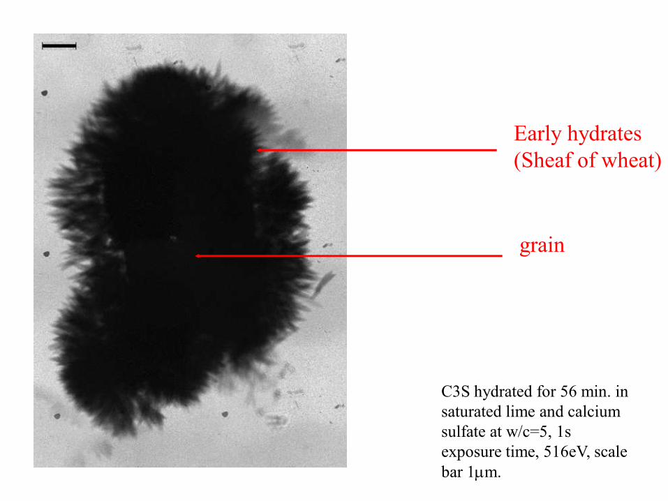
C3S hydrated for 56 min. in
saturated lime and calcium
sulfate at w/c=5, 1s
exposure time, 516eV, scale
bar 1mm.
grain
Early hydrates
(Sheaf of wheat)

8h 30min.
In-situ Massive precipitation

8h 36min.
In-situ Massive precipitation

9h 16min.
Spot

Adições Químicas

Reação álcali-agregado
California, 1936
Vertical cracks
Map cracks

Gel de
FurnasDissolução em NaOH

Na presença de Ca(OH)2

Uso de raios-x para
imagens
Wilhelm Conrad Roentgen
first Nobel prize in physics (1901)

Compressão da terceira
dimensão

Desenvolvimento da tomografia
Sir Godfrey N. Hounsfield
Allan M. Cormack
Nobel Prize in Physiology or Medicine 1979

Tomografia: Configuração

Exemplos
Radiografia (esquerda) and tomografia (direita) de cp de concreto com fibras

Exemplos
Radiografia (esquerda) e tomografia (direita) de cilindros de concreto com fibras

Localização das barras
Radiografia (esquerda) and e tomografia (direita).

Concreto
armado

Engenheiros preferem
maiores resoluções

Synchrotron XMT
XCMT
XCMT 2D Detector
1. Superbend
Polychromatic beam from
the storage ring
2. Monochromator:
isolates, then sends off
monochromatic beam
3. Monochromatic Beam
4. Sample on rotating
stage, partial beam
absorbance
5. Scintillator – converts
X-rays to visible light
6. Visible light
7. A CCD captures the raw
image, to be processed,
reconstructed, and
rendered on a PC
Image courtesy of A.Macdowell

Fibras
Image courtesy of S. Brisard
xy plane tomogram yz plane
•Smaller sample size used (20 mm x 20 mm)
•Scanned volume (approximately 20mm x
20mm x 25 mm)
•White light absorption mode with filtered x-
rays (E>30keV) using metal filters
•11.55 x 2 = 23 µm/pixel resolution


Fiber isolation
Image courtesy of S. Brisard

Nanotomografia
• Objetivo: Obter imagens em 3-d
com resoluçao superior a 20 nm

Centros de excelência
• BESSY (soft x-rays)
• APS (hard x-rays)
• Stanford (hard x-rays)
• Berkeley (soft x-rays, under
development with the KAUST
project)

Desafios
• Alinhamento das imagens
• Estabilidade do sistema
• Tomografia com ângulo limitado

BESSY x-ray
microscope
Courtesy from G. Schneider

3D structure of mammalian cells
Courtesy from G. Schneider

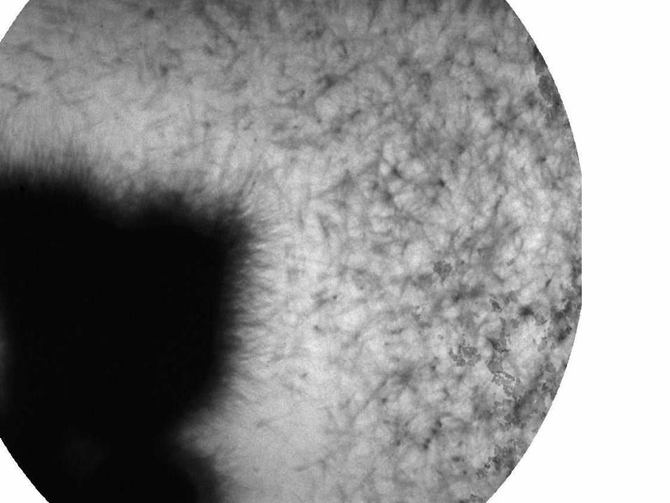
Por que nanotomografia?
• This transmission image seems to show that the
“sheet of wheat” (or “stars”) have a core which acts
as a nucleation point (see arrows)Work with Brisard, Levitz and Chae
Em BESSY,
Berlin
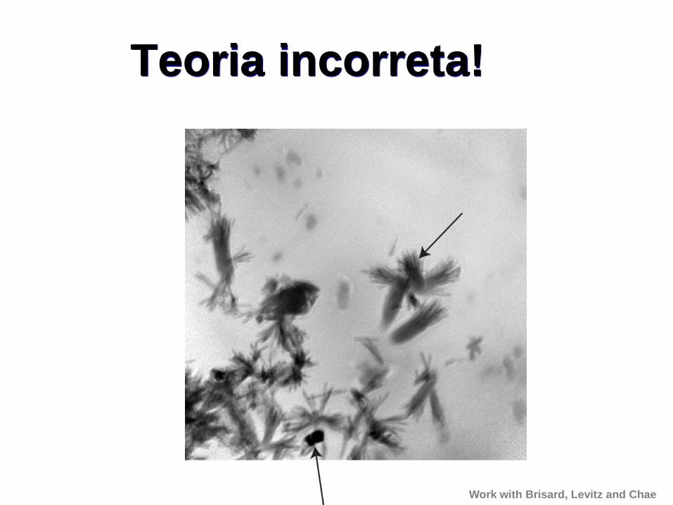
Teoria incorreta!
Work with Brisard, Levitz and Chae

Comparação
Work with Brisard, Levitz and Chae

Comparação
Work with Brisard, Levitz and Chae

Work with Brisard, Levitz and Chae

Novo Projeto
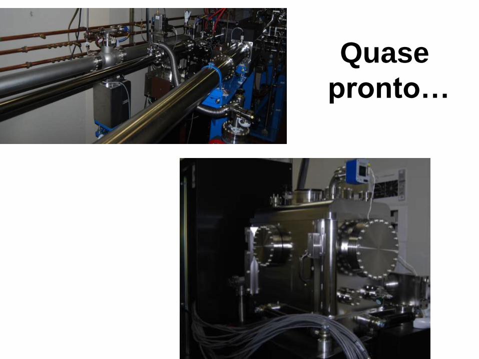
Quase
pronto…

Pesquisas possíveis
• 1) Efeito de adições químicas nos produtos de hidratação:– Plastificantes e superpastificantes– Modificadores de viscosidade
• 2) Carbonatação do C-S-H
• 3) Interação do cloretos com etringita, C-S-H etc.
• 4) Incorporadores de ar
• 5) Localização dos sulfatos na DEF–

Examplo: Efeitos de Polímeros no CSH
• Hexadecyltrimethylammonium (HDTMA) and polyethylene glycol (PEG200).
Chemical Name Chemical
Formula
Molecular
Weight
(g/mol)
Structure
Polyethylene glycol (PEG) H(OCH2CH2)nOH 200
Hexadecyltrimethylammonium,
bromide (HDTMA) C19H42BrN 354.46

Efeitos de Polimeros no CSH
• Resultados: HDTMA-CSH
– STXM images contrast (i.e., image map) of HDTMA-CSH sample taken at (A) C K-edge; (B) Ca LII,III-edge, and (C) Si K-edge.
– Strong spatial correlation observed for carbon and calcium, confirming HDTMA interaction with CSH
– HDTMA is likely to be adsorbed to the Ca but less likely to Si, suggesting that HDTMA is likely to be adsorbed to the edges or defect sites of the layer structure of CSH

Results: HDTMA-CSH Samples
– (A) image taken at C K-edge; (B) image of smaller area outlined in (A) magnifying fine structures of HDTMA-CSH sample; and (C) NEXAFS spectra at C K-edge, Ca LII,III-edge, and Si K-edge. Numbers indicate the locations from where the spectra are taken as shown in (A) and (B).
– Difference in carbon and calcium NEXAFS spectra observed– Spatial heterogeneity observed
5 mm
1 mm
12
3
4
5
(A)
(B)
(C)
6

Conclusões
As novas técnicas de Microscopia de
raios-X, Microscopia a baixas
temperaturas, Microtomografia e
Nanotomografia permitem um novo
entendimento da estrutura do
concreto.