natiele carla da silva ferreira identificação de novos
TRANSCRIPT

INSTITUTO OSWALDO CRUZ
Pós-Graduação em Biologia Celular e Molecular
NATIELE CARLA DA SILVA FERREIRA
Identificação de novos antagonistas para receptores purinérgicos P2
Tese apresentada ao Instituto Oswaldo Cruz como parte
dos requisitos para obtenção do título de Doutora em
Biologia Celular e Molecular.
Orientador: Prof. Dr. Luiz Anastacio Alves
RIO DE JANEIRO
2019

ii
INSTITUTO OSWALDO CRUZ
Pós-Graduação em Biologia Celular e Molecular
NATIELE CARLA DA SILVA FERREIRA
Identificação de novos antagonistas para receptores purinérgicos P2
ORIENTADOR: Prof. Dr. Luiz Anastacio Alves
Aprovada em: 30/04/2019
EXAMINADORES:
Prof.ª Dra. Patrícia Machado Rodrigues e Silva Martins (IOC/Fiocruz) - presidente
Prof.ª Dra. Ana Lucia Marques Ventura (EGB/UFF) – membro titular
Prof. Dr. Alcides José Monteiro da Silva (IPPN/UFRJ) – membro titular
Prof.ª Dra. Carmen Monteiro Penido (CDTS/Fiocruz) - revisora e suplente
Prof. Dr. Vinícius Cotta Almeida (IOC/Fiocruz) - suplente
Rio de Janeiro, 30 de abril de 2019.

iii
Dedico esse trabalho, fruto de quatro anos de
estudo e dedicação, aos meus orientadores,
familiares e amigos que sempre
acreditaram em mim.
Essa conquista também é de vocês!
iv
AGRADECIMENTOS
Agradeço em primeiro lugar a Deus, que me permitiu chegar até aqui, mesmo diante de
tantas preocupações e tribulações. Sou grata por ter me sustentado, por ter me permitido viver
tanta coisa boa e conhecer tantas pessoas maravilhosas ao longo desse caminho. Agradeço
também aos meus intercessores celestiais, a quem tanto recorri nas aflições.
Agradeço aos meus pais Robson e Carla que tanto se esforçaram para que eu tivesse uma
boa educação. Lembro-me dos trabalhos aos domingos, nas madrugadas, e também da
preocupação com o meu desempenho na escola, no curso de inglês... Sim, vocês semearam e
talvez jamais sonhassem que a sua plantinha chegasse ao doutorado. E aqui estou. Essa vitória
em primeiro lugar é de vocês. Amo vocês pai e mãe!
Agradeço também a todos os meus familiares: meu irmão Mateus, meus avós, tios, primos
e sogros que sempre me apoiarem e sonharam junto comigo, e que mesmo indiretamente
tiveram uma participação importante durante toda a minha trajetória de estudo, desde o
remoto jardim de infância até o doutorado. Vocês ajudaram a criar as condições propícias para
que eu chegasse aqui: seja com ajuda financeira, seja com ajuda no deslocamento casa x
escola, seja com ajuda nos mais variados trabalhos e lições, seja com ajuda motivacional. A
vocês minha eterna gratidão!
“No meio do caminho tinha um amor.” Agradeço de forma especial ao meu companheiro,
parceiro, amigo e marido Cassiano. Você foi o raio de sol em dias difíceis. Quando chegava
em casa chateada, você me oferecia o seu ombro amigo para afagar o meu choro e dizer que
tudo ia ficar bem. Sei que você mais do que ninguém observou o quão difícil foi essa
caminhada. Agradeço por compreender as minhas ausências e adiar alguns dos seus sonhos,
por causa do meu sonho. Agradeço a você por ser quem e o que você é em minha vida e
agradeço a Deus por ter abençoado nossas vidas e a nossa família que está à espera de mais
uma integrante. Eu amo vocês!
“Se eu vi mais longe, foi por estar sobre ombro de gigantes (Isaac Newton).” É com esse
pensamento que demonstro a minha gratidão aos meus orientadores, os doutores Luiz
Anastacio e Rômulo Bezerra.
Ao Luiz, gostaria de agradecer por acreditar em mim e no meu futuro desde quando
ingressei no Laboratório de Comunicação Celular. Talvez eu mesma nem acreditasse que
pudesse ir tão longe. Agradeço aos ensinamentos e, sobretudo à máxima de “aprender-a-
aprender”, que me fez enxergar as coisas de modo diferente. Obrigada pelos conselhos que
me ajudaram a melhorar as minhas apresentações e oratória. Obrigada por me ensinar a
analisar e avaliar artigos de forma crítica. E, sobretudo obrigada por me ensinar que ciência
v
deve ser feita sempre com responsabilidade e qualidade. Agradeço por ajudar a me tornar a
profissional que hoje eu sou.
Ao Rômulo, gostaria de agradecer pela amizade desde sempre. Sei que não estamos tão
próximos fisicamente, mas estamos sempre conversando e estou sempre atenta aos seus
conselhos. Agradeço por me ensinar quase tudo que sei de bancada, pela paciência em ensinar
novas técnicas e ajudar a desenhar os protocolos. Agradeço pela ajuda financeira para a
compra de reagentes em todas as vezes que houve necessidade e por toda ajuda com os
experimentos. Agradeço por estar sempre presente em minha trajetória e ela não seria a
mesma sem você.
Um doutor não se faz sozinho, ele é fruto do trabalho de outros doutores. Essa é a grande
verdade quando se está situado dentro de um laboratório e de uma instituição de pesquisa:
conviver com doutores. É um privilégio que poucos têm acesso e eu agradeço todos os dias
por tê-lo. Gostaria de agradecer aos doutores do Laboratório de Comunicação Celular, Dra.
Cristina Souza, Dr. Renato Lopes, Dr. Anael Viana e Dr. Rodrigo Bisaggio, por todos os
conselhos enriquecedores e por toda ajuda a mim disponibilizada. Agradeço também aos
meus companheiros de doutorado Carla Oliveira, Dinarte Ferreira, Alejandra Carreño, Miriam
Salles, Liana Monteiro e aos demais integrantes do Laboratório de Comunicação Celular pela
ajuda do dia-a-dia na bancada com os reagentes, células, animais e protocolos.
Agradeço de forma especial às minhas queridas alunas Rebeca Silveira e Monique Pontes.
Agradeço a vocês pela dedicação, por me ajudarem com os experimentos sempre com atenção
e capricho, e, sobretudo pela amizade e companheirismo. Desejo a vocês um futuro de muito
sucesso e conquistas.
Agradeço também à Dra. Andrea Surrage do Laboratório de Imunofarmacologia (IOC -
Fiocruz) pela preciosa ajuda e ensinamentos com os experimentos in vivo e também pela
amizade.
Agradeço às pesquisadoras Dra. Maria Raquel e Ana Luíza, bem como todos os integrantes
do Laboratório de Produtos Naturais 3 do Instituto de Tecnologia em Fármacos
(FarManguinhos - Fiocruz), por me inserirem intensivamente no mundo dos produtos
naturais. Obrigada por me ajudarem e me ensinarem com inigualável bondade e paciência as
diferentes técnicas desde a preparação até o fracionamento dos extratos.
Agradeço ao Dr. José Augusto do Laboratório de Avaliação e Promoção da Saúde
Ambiental (IOC – Fiocruz) que me ensinou com muita bondade e paciência na prática o
processo de fracionamento dos extratos.
Agradeço aos meus queridos amigos botânicos George Queiroz, Davi Nepomuceno e
Fernanda Costa por me auxiliarem com a coleta vegetal. É muito bom perceber que mesmo
vi
que atuando em linhas de pesquisa diferentes, nossos conhecimentos se complementam e a
amizade se fortalece.
Agradeço à Dra. Tânia Alves e ao Dr. Carlos Zani do Laboratório de Química de Produtos
Naturais do Centro de Pesquisa René Rachou (Fiocruz) pela parceria de muitos anos com os
extratos.
Agradeço à Plataforma de Bioprospecção da Fiocruz (RPT10A) e à técnica Fátima Marques
pelo envio das amostras.
Agradeço à Plataforma de Bioensaios e Triagem de Fármacos (RPT11I) e às técnicas Elid
Fernandes e Mônica Alcon pelo uso do espaço e dos equipamentos, bem como pela amizade
construída.
Agradeço a toda equipe do Centro de Experimentação Animal situado no prédio Hélio e
Peggy Pereira por todo auxílio no manejo com os animais.
Agradeço ao Instituto Oswaldo Cruz e à Pós-Graduação em Biologia Celular e Molecular
pela oportunidade de realizar o curso de doutorado.
Agradeço também ao IOC e à Faperj pela concessão das bolsas de estudo.
Agradeço aos professores componentes da banca examinadora pelo aceite do convite para
participar da conclusão desse trabalho.
Agradeço também aos meus amigos que sempre estiveram na torcida para que o doutorado
fosse concluído com sucesso e me deram muita força durante todo esse processo. Vocês tem
um espaço muito especial em meu coração!
Por fim, agradeço a todos que contribuíram direta ou indiretamente para a realização desse
trabalho, a todos vocês: MINHA ETERNA GRATIDÃO!

vii
“Tempo rei, ó tempo rei, ó tempo rei
Transformai as velhas formas do viver
Ensinai-me, ó Pai, o que eu ainda não sei
Mãe Senhora do Perpétuo socorrei”
(Tempo Rei, Gilberto Gil)
Que os conhecimentos e os
títulos adquiridos jamais
removam de meu coração
a humildade e a consciência
de que a felicidade
reside nas coisas mais
simples da vida.
viii
INSTITUTO OSWALDO CRUZ
IDENTIFICAÇÃO DE NOVOS ANTAGONISTAS PARA RECEPTORES
PURINÉRGICOS P2
RESUMO
TESE DE DOUTORADO
Natiele Carla da Silva Ferreira
Os receptores purinérgicos P2 são receptores expressos na membrana plasmática de diversos
tipos celulares humanos, nos quais exercem importantes funções fisiológicas. Eles são
divididos em duas classes, de acordo com a estrutura apresentada. A classe P2X compreende
os subtipos de receptores ionotrópicos que são ativados fisiologicamente pelo ATP. Dentre os
receptores P2X, o P2X7 destaca-se por suas funções associadas à dor e à inflamação. Já a
classe P2Y compreende os subtipos de receptores metabotrópicos, os quais são ativados por
diferentes nucleotídeos extracelulares. Nessa última classe, o subtipo P2Y2 destaca-se pelos
seus papeis desempenhados na inflamação e no câncer, enquanto o P2Y4 se sobressai pela
escassez de informações acerca de suas funções fisiológicas devido a limitações de caráter
farmacológico. Apesar de serem considerados importantes alvos-terapêuticos, ainda não
existem fármacos com atuação sobre esses receptores que sejam aprovados para uso clínico, o
que encoraja a busca por novas moléculas com atividade antagonista. Nesse contexto, o
objetivo deste estudo foi identificar novos antagonistas para receptores P2 (P2X7, P2Y2 e
P2Y4) e averiguar se eles exercem algum papel na atenuação da dor e inflamação. Na
primeira parte desse trabalho foi identificada a atividade antagonista do extrato galhos de
Joannesia princeps Vell. após a realização de uma mini-campanha de triagem. Utilizando a
técnica de mensuração de cálcio intracelular, foi demonstrado que esse extrato foi capaz de
inibir a mobilização de cálcio induzida por UTP de forma concentração-dependente, sendo
que esse efeito não foi provocado por citotoxicidade ou fenômeno quenching. Além disso,
esse extrato também foi capaz de inibir parcialmente a mobilização de cálcio induzida por
UDP. Com isso, os resultados sugerem uma possível seletividade quanto à ação antagonista
desse extrato sobre os receptores P2Y ativados por nucleotídeos derivados da uridina.
Enquanto isso, na segunda parte do trabalho foi identificada a atividade antagonista da
molécula CS-15 sobre o receptor P2X7 durante a realização de uma mini-campanha de
triagem. A molécula CS-15, um triterpeno isolado da planta Clusia studartiana C. M. Vieira
& Gomes da Silva, foi capaz de inibir a captação do corante YO-PRO-1 em ensaios de
permeabilização celular de forma concentração-dependente e apresentou um IC50 na faixa de
nanomolar. Ademais, essa molécula não demonstrou toxicidade in vitro e ainda foi capaz de
aliviar os sintomas de nocicepção em modelo experimental animal. Coletivamente, os
resultados apontam para a descoberta de novos antagonistas para receptores P2 de origem
natural, os quais podem contribuir no futuro para o escasso campo da terapia associada aos
receptores purinérgicos.

ix
INSTITUTO OSWALDO CRUZ
IDENTIFICATION OF NEW ANTAGONISTS FOR P2 PURINERGIC RECEPTORS
ABSTRACT
TESE DE DOUTORADO
Natiele Carla da Silva Ferreira
P2 purinergic receptors are receptors expressed on the plasma membrane of several human
cell types, in which they exert important physiological functions. They are divided into two
classes, according to the structure presented. The P2X class comprises the ionotropic
receptors subtypes that are physiologically activated by ATP. Among P2X receptors, P2X7
stands out due to its functions associated with inflammation and pain. The P2Y class
comprises the metabotropic receptors subtypes, which are activated by different extracellular
nucleotides. In this latter class, the P2Y2 subtype stands out for its roles in inflammation and
cancer, while P2Y4 stands out because of the scarce information about its physiological
functions due to pharmacological limitations. Despite being considered important therapeutic
targets, there are still no approved drugs for clinical use acting on these receptors, which
encourages the search for new molecules with antagonistic activity. In this context, the
objective of this study was to identify new antagonists for P2 receptors (P2X7, P2Y2, and
P2Y4) and to verify if they play any role in inflammation and pain attenuation. In the first part
of this work the antagonistic activity of Joannesia princeps Vell. stem extract was identified
after we carried out a mini-screening campaign. Using the intracellular calcium measurement
technique, it was demonstrated that this extract was able to inhibit UTP-induced calcium
mobilization in a concentration-dependent manner and that this effect was not triggered by
cytotoxicity or quenching phenomenon. In addition, this extract partially inhibited UDP-
induced calcium mobilization. Therefore, the results suggest a possible selectivity for the
antagonistic action of this extract on the P2Y receptors activated by uridine-derived
nucleotides. Meanwhile, in the second part of the work, the antagonist activity of the
compound CS-15 on the P2X7 receptor was identified during a mini-screening campaign. The
CS-15 compound, a triterpene isolated from the Clusia studartiana C. M. Vieira & Gomes da
Silva plant, inhibited the YO-PRO-1 dye uptake in a concentration-dependent manner on cell
permeabilization assays and showed an IC50 in the nanomolar range. In addition, this
compound did not demonstrate toxicity in vitro and was able to alleviate nociception
symptoms in an experimental animal model. Collectively, the results point to the discovery of
new antagonists for P2 receptors from natural origin, which may contribute in the future to the
scarce field of therapy associated with purinergic receptors.

x
LISTA DE ABREVIATURAS
2-MeSADP - 2-metiltioadenosina difosfato
2-thio-UTP - 2-thiouridina-5'-trifosfato
α-SMA - α-actina do músculo liso
AC - adenilato ciclase
ADA - adenosina deaminase
ADP - adenosina 5’-difosfato
AdeR - receptor ativado por adenina
AM - acetoximetil
AMP - adenosina 5’-monofosfato
ANVISA - Agência Nacional de Vigilância Sanitária
ATP - adenosina 5’-trifosfato
BAPTA - 1,2-bis(2-aminofenóxi)etano-N,N,N′,N′-ácido tetra acético
BBG - Brilliant Blue G
BzATP - 2′(3′)-O-(4-benzoilbenzoil) adenosina 5′-trifosfato
CaCC - canal de cloro ativado por cálcio
cAMP - adenosina 3’,5’-monofosfato cíclico
CFTR - condutibilidade transmembrana da fibrose cística
COX-2 - ciclooxigenase-2
DAG - diacilglicerol
EC50 - half maximal effective concentration
EGFR - receptor do fator de crescimento epidérmico
EGTA - etileno glicol-bis(2-aminoetiléter)-N,N,N′,N′-ácido tetra acético
ELISA - enzyme-linked immunosorbent assay
EMA - European Medical Agency
ENaC - canal de sódio epitelial
EP3 - receptor de prostaglandina E3
EROS - espécies reativas de oxigênio
FDA - Food and Drug Administration
FRET - transferência de energia de ressonância Förster
HDL - lipoproteína de alta densidade (high density lipoprotein)
HIV - vírus da imunodeficiência humana
HMGB1 - proteína de alta mobilidade do grupo de caixa 1
HPLC - cromatografia líquida de alta performance (high performance liquid
chromatography)

xi
HTS - triagem de alto desempenho (high-throughput screening)
IC50 - half maximal inhibitory concentration
IL-1β - interleucina-1β
IL-18 - interleucina-18
iNOS - óxido nítrico sintase induzida
IP3 - inositol trifosfato
IP3R - receptor para o inositol trifosfato
IUPHAR - União Internacional de Farmacologia Básica e Clínica
KO – nocaute (knockout)
MAPK - proteínas quinases ativadas por mitógenos
NCE - novas entidades químicas (new chemical entities)
oATP - ATP oxidado
OMS - Organização Mundial de Saúde
ORCC - canal de cloro retificador externo
oxLDL - lipoproteína de baixa densidade oxidada
PCR - reação em cadeia de polimerase
PGE2 - prostaglandina E2
PIP2 - fosfatidilinositol 4,5-bifosfato
PLC - fosfolipase C
PLCβ - fosfolipase C β
PPA - proteína precursora amilóide
PPADS - piridoxal fosfato-6-azo(benzeno-2,4-ácido disulfônico)
RB2 - Reactive Blue-2
TNF-α - fator de necrose tumoral-α
TNP-ATP - 2′,3′-O-(2,4,6-trinitrofenil) adenosina 5′-trifosfato
UDP - uridina5’-difosfato
UTP - uridina5’-trifosfato
WT - wild-type
VCAM-1 - proteína de adesão celular vascular 1

xii
SUMÁRIO
I. INTRODUÇÃO.............................................................................................................. 01
1. Breve histórico sobre a descoberta dos receptores purinérgicos.............................. 01
2. Receptores purinérgicos................................................................................................ 03
Quadro 2.1 – Classificação dos receptores purinérgicos em humanos................ 03
2.1 – Receptores P1.............................................................................................. 04
Figura 2.1.1– Receptores P1 e suas proteínas G associadas................................ 04
Quadro 2.1.1 – Agonistas e antagonistas dos receptores P1 aplicados na terapia. 05
2.2 – Receptores P2Y........................................................................................... 06
Figura 2.2.1 – Topologia básica dos receptores P2Y.......................................... 06
Quadro 2.2.1 – Principais características farmacológicas dos receptores P2Y... 07
2.3 – Receptores P2X........................................................................................... 09
Figura 2.3.1 – Topologia básica dos receptores P2X.......................................... 10
Quadro 2.3.1 - Principais características farmacológicas dos receptores P2X.... 12
3. Desenvolvimento de fármacos com atividade sobre os receptores P2.................... 14
Quadro 3.1 – Antagonistas do receptor P2Y12 aprovados para uso clínico...... 16
3.1 – O receptor P2Y2 como alvo terapêutico.................................................... 16
Figura 3.1.1 – Transporte iônico promovido pelo receptor P2Y2 em células
epiteliais das vias aéreas................................................................................
17
Figura 3.1.2 – Fármaco com ação sobre o receptor P2Y2............................. 19
Figura 3.1.3 – Principais efeitos da ativação do P2Y2................................. 21
3.2 – O receptor P2Y4 como alvo terapêutico................................................. 21
Figura 3.2.1 – Principais efeitos da ativação do P2Y4.................................. 23
3.3 – O receptor P2X7 como alvo terapêutico................................................. 23
Figura 3.3.1 – Principais efeitos da ativação do P2X7.................................. 27
4- Os produtos naturais como uma fonte para a descoberta de novos antagonistas. 27
Quadro 4.1 - Estimativa acerca da riqueza biológica mundial e brasileira.... 28
4.1 – Receptores P2 e produtos naturais..........................................................
Quadro 4.1.1 - Produtos naturais com ação antagonista sobre os receptores
P2Y2 e P2X7................................................................................................
30
31
5- Triagem de alto-desempenho aplicada à descoberta de novos antagonistas........... 33
5.1 – Ensaios para mensuração dos níveis de cálcio intracelular.................. 34

xiii
Quadro 5.1.1 – Principais ferramentas de mensuração dos níveis de cálcio
intracelular...................................................................................................
35
5.2 – Ensaios de patch-clamp............................................................................. 36
Figura 5.2.1 – Configurações da técnica de patch-clamp........................... 38
5.3 – Ensaios de permeabilização celular......................................................... 39
Figura 5.3.1 – Canal e poro membranar do P2X7............................................ 39
Quadro 5.3.1 – Principais características dos corantes mais populares no es-
tudo da formação do poro membranar associado ao receptor P2X7...............
40
5.4 – Descobertas recentes através de campanhas de HTS e perspectivas......... 40
II. OBJETIVOS................................................................................................................. 42
III. RESULTADOS........................................................................................................... 43
ARTIGO 1........................................................................................................................... 44
ARTIGO 2........................................................................................................................... 59
ARTIGO 3........................................................................................................................... 73
ARTIGO 4........................................................................................................................... 104
IV. DISCUSSÃO................................................................................................................ 123
V. CONCLUSÃO............................................................................................................... 131
VI. REFERÊNCIAS.......................................................................................................... 132
APÊNDICE 1..................................................................................................................... 151
APÊNDICE 2..................................................................................................................... 153
APÊNDICE 3..................................................................................................................... 156
INTRODUÇÃO
1
I. INTRODUÇÃO
1. Breve histórico sobre a descoberta dos receptores purinérgicos
A história dos receptores purinérgicos iniciou-se com a descoberta de seus principais
ligantes fisiológicos, as purinas e pirimidinas por Ludwig Kossel no fim do século XIX. Em
1878, Kossel publicou um de seus primeiros trabalhos, no qual ele descreve o isolamento das
bases hipoxantina e xantina do conteúdo nuclear de leveduras [1]. Já no ano de 1885, Kossel
descreveu pela primeira vez o isolamento da adenina em leveduras e no pâncreas humano,
bem como delineou a transformação química dessa base em hipoxantina [1]. Em 1887, ele
isolou e determinou a estrutura química da teofilina extraída de folhas de chá e determinou a
sua relação com a cafeína, a qual é um reconhecido antagonista natural dos receptores
purinérgicos P1. Finalmente, entre os anos de 1891 e 1901, Kossel publicou os seus
resultados sobre o isolamento do ácido fosfórico e das bases adenina, guanina, timina, citosina
e uracila [1]. Simultaneamente, dois químicos alemães, Emil Fischer e Adolf Pinner, se
dedicaram ao estudo das estruturas químicas dessas bases nitrogenadas recém-descobertas e
denominaram-nas purinas e pirimidinas [2]. Posteriormente, Phoebus Levene descreveu o
açúcar que era conjugado a essas bases, caracterizando os nucleosídeos (bases nitrogenadas
contendo açúcar) e nucleotídeos (bases nitrogenadas contendo açúcar e fosfato(s)) [2,3].
Até esse momento da história, acreditava-se que as atividades dessas moléculas
restringiam-se ao núcleo celular. Entretanto, em 1929, Alan Drury e Albert Szent-Györgyi ao
isolarem substâncias do tecido cardíaco identificaram que a adenosina 5’-monofosfato (AMP)
era a responsável por alterar o ritmo cardíaco, diminuir a pressão arterial e inibir movimentos
intestinais em cobaias. Eles também observaram que a adenosina era mais potente do que o
AMP em produzir um bloqueio cardíaco [2,4]. Esses experimentos foram repetidos por
diversos autores como Gillespie (1934), que apontou os diferentes efeitos biológicos
promovidos pela adenosina e seus derivados. Ele observou que a adenosina 5’-trifosfato
(ATP) era mais potente em aumentar a pressão arterial e o tônus uterino em cobaias, enquanto
que o AMP e a adenosina produziam efeitos antagônicos. Ele descreveu ainda que a perda da
pentose causava um detrimento da atividade biológica das moléculas [5].
Posteriormente, no final da década de 1940, surgiram os primeiros estudos
relacionando os efeitos da adenosina e seus derivados no sistema nervoso. Buhthal et al.
observaram que a injeção de ATP na medula espinhal de cobaias produzia contrações
musculares [2]. Durante a década de 1950, Holton e Holton caracterizaram pela primeira vez
o papel do ATP na neurotransmissão. No experimento de 1954, eles observaram que a injeção
de ATP produzia vasodilatação na orelha desnervada de coelhos [6]. Já no experimento de

INTRODUÇÃO
2
1959, Pamela Holton estudando ainda o papel do ATP na orelha de coelhos observou que este
era liberado após a estimulação do nervo auricular ou da pele do animal, confirmando assim, a
sua participação na neurotransmissão [7]. Nos anos seguintes, diversos trabalhos que
demonstravam os efeitos do ATP e seus derivados no sistema nervoso foram publicados. Até
que em 1970, Geoffrey Burnstock ao observar que o estímulo dos nervos causava a liberação
do ATP em órgãos do sistema digestório de cobaias, propôs que o mesmo poderia atuar como
um neurotransmissor, além de co-transmissor [8,9].
Assim, com a evidente descoberta da atividade do ATP e seus derivados em sistemas
fisiológicos, Burnstock propôs em 1978 a primeira classificação para os “purinoceptores”, isto
é, receptores ativados por purinas e pirimidinas. Com isso, os receptores ativados pela
adenosina foram nomeados de P1, enquanto os receptores ativados por ATP/ADP foram
denominados P2 [10]. Nos anos seguintes, foram realizados diversos estudos farmacológicos
e em 1979, van Calker et al. propuseram uma subdivisão para os receptores P1 em A1 e A2,
uma vez que eles mediavam a inibição ou estimulação do AMP cíclico, respectivamente [11].
Já na década de 1980, Burnstock e Kennedy também propuseram uma divisão para os
receptores P2 em duas classes: P2X e P2Y de acordo com critérios farmacológicos e efeitos
biológicos que apresentavam [12].
Durante o início da década de 1990, diversos trabalhos acerca de clonagem dos
subtipos de receptores purinérgicos foram publicados. Em 1994, um grupo de pesquisadores
se reuniu e oficialmente estabeleceu uma nomenclatura e classificação para os receptores
purinérgicos, a qual foi reconhecida pelo Comitê da União Internacional de Farmacologia
Básica e Clínica (IUPHAR) sobre Nomenclatura de Receptores e Classificação de
Medicamentos. Essa classificação seguiu critérios farmacológicos e estruturais e manteve a
distinção das duas famílias: P1 e P2. A família dos receptores P1 apresentava quatro subtipos
(A1, A2A, A2B e A3). Enquanto isso, a família dos receptores P2 foi dividida em cinco
subtipos P2X, P2Y, P2Z, P2U e P2T [13]. Contudo, nesse mesmo ano, Abbracchio e
Burnstock propuseram manter a classificação em P2X e P2Y e agruparam os subtipos P2U
(atual P2Y2) e P2T (extinto P2Y3) na classe P2Y, e o P2Z (atual P2X7) na classe P2X [14].
Nos anos seguintes, finalmente foram reconhecidos e caracterizados os sete subtipos de
receptors P2X (P2X1-P2X7) e os oito subtipos de receptores P2Y (P2Y1,2,4,6,11-14) [15,16].
Recentemente, foram descobertos um grupo de receptores são ativados por adenina
(AdeR), os quais foram nomeados de receptores P0. No entanto, esses receptores ainda não
foram caracterizados em humanos. Apenas se tem registro sobre a sua expressão em ratos,
camundongos e hamsters [17].
INTRODUÇÃO
3
2. Receptores purinérgicos
Os receptores purinérgicos são receptores conhecidos pela sua expressão na membrana
plasmática em diferentes tipos celulares em mamíferos, onde são ativados por purinas e
pirimidinas extracelulares. Todavia, já se tem informações na literatura sobre a expressão
desses receptores na membrana de organelas celulares, tais como mitocôndria e núcleo celular
[18,19], embora esses achados não tenham sido aprofundados pelos demais grupos de
pesquisa da área.
A expressão de formas primitivas desses receptores já foi observada em outros seres
vivos, tais como os seguintes invertebrados: o helminto Schistossoma mansoni que possui um
receptor ativado fisiologicamente por ATP denominado SchP2X; o caracol de lagoa Lymnaea
stagnalis que expressa o receptor LymP2X; o carrapato Boophilus microplus que possui o
receptor BmP2X; e o tardígrado Hypsibius dujardini que expressa o receptor HdP2X. Esses
receptores também estão presentes em protistas como o Dictyostelium discoideum (ameba),
que expressa cinco tipos de receptores P2X (DdP2XA-E), e Ostreococcus tauri (alga), a qual
possui um receptor capaz de ser ativado por ATP denominado OtP2X [20].
Os receptores purinérgicos expressos em humanos são divididos em duas famílias: a
família P1, que agrupa os membros ativados fisiologicamente por adenosina, e a família P2
que abriga os receptores ativados por diferentes nucleotídeos. Além disso, essa última família
é subdivida em duas classes, de acordo com a estrutura apresentada. Portanto, existe a classe
P2X, que agrupa os receptores que formam canais iônicos cátion-seletivos, e a classe P2Y que
abriga os receptores acoplados à proteína G [14]. No quadro 2.1 estão descritos os subtipos de
receptores purinérgicos de acordo com a classe a que pertencem.
Quadro 2.1 – Classificação dos receptores purinérgicos em humanos
Famílias Classes Receptores Agonista(s) fisiológico(s)
P1 - A1, A2A, A2B, A3 Adenosina
P2
P2X P2X1, P2X2, P2X3, P2X4,
P2X5, P2X6, P2X7
ATP
P2Y
P2Y1, P2Y2, P2Y4, P2Y6,
P2Y11, P2Y12, P2Y13,
P2Y14*
ADP, ATP, UDP, UDP-
glicose ou UTP
*O intervalo na sequência dos números desses receptores se deve ao fato de que alguns foram
caracterizados incorretamente, e após investigação, ficou constatado que eles não pertenciam
a essa classe.
INTRODUÇÃO
4
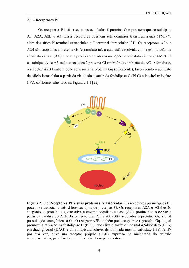
2.1 – Receptores P1
Os receptores P1 são receptores acoplados à proteína G e possuem quatro subtipos:
A1, A2A, A2B e A3. Esses receptores possuem sete domínios transmembranas (TM1-7),
além dos sítios N-terminal extracelular e C-terminal intracelular [21]. Os receptores A2A e
A2B são acoplados à proteína Gs (estimulatória), a qual está envolvida com a estimulação da
adenilato ciclase (AC) e com a produção de adenosina 3’,5’-monofosfato cíclico (cAMP). Já
os subtipos A1 e A3 estão associados à proteína Gi (inibitória) e inibição da AC. Além disso,
o receptor A2B também pode se associar à proteína Gq (quiescente), favorecendo o aumento
de cálcio intracelular a partir da via de sinalização da fosfolipase C (PLC) e inositol trifosfato
(IP3), conforme salientado na Figura 2.1.1 [22].
Figura 2.1.1: Receptores P1 e suas proteínas G associadas. Os receptores purinérgicos P1
podem se associar a três diferentes tipos de proteínas G. Os receptores A2A e A2B estão
acoplados a proteína Gs, que ativa a enzima adenilato ciclase (AC), produzindo o cAMP a
partir da catálise do ATP. Já os receptores A1 e A3 estão acoplados à proteína Gi, a qual
possui ações antagônicas à Gs. O receptor A2B também pode acoplar-se à proteína Gq, a qual
promove a ativação da fosfolipase C (PLC), que cliva o fosfatidilinositol 4,5-bifosfato (PIP2)
em diacilglicerol (DAG) e uma molécula solúvel denominada inositol trifosfato (IP3). A IP3
por sua vez, ativa um receptor próprio (IP3R) expresso na membrana do retículo
endoplasmático, permitindo um influxo de cálcio para o citosol.
INTRODUÇÃO
5
Esses receptores são expressos em diferentes órgãos em humanos, tais como cérebro,
medula espinhal, testículos, coração, terminais nervosos autonômicos, pulmões, baço,
intestino, bexiga e fígado [21]. Esses receptores são ativados fisiologicamente pela adenosina.
Entre os seus antagonistas não seletivos encontram-se a cafeína, a teofilina, entre outras
xantinas de ocorrência natural. Alguns fármacos similares à adenosina ou à teofilina já se
encontram aprovados para uso terapêutico no tratamento da taquicardia e asma,
respectivamente [22]. Apesar disso, já foram desenvolvidos diversos agonistas e antagonistas
seletivos (sintéticos e semissintéticos) para cada um dos subtipos desses receptores, sendo que
alguns deles já se encontram em diferentes fases de ensaios clínicos. O subtipo A2A, por sua
vez, é o único dentre os receptores P1 que apresenta agonista e antagonista seletivo aprovado
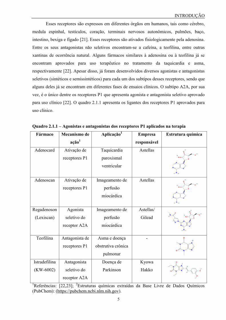
para uso clínico [22]. O quadro 2.1.1 apresenta os ligantes dos receptores P1 aprovados para
uso clínico.
Quadro 2.1.1 – Agonistas e antagonistas dos receptores P1 aplicados na terapia
Fármaco Mecanismo de
ação1
Aplicação1 Empresa
responsável
Estrutura química
Adenocard Ativação de
receptores P1
Taquicardia
paroxismal
ventricular
Astellas
Adenoscan Ativação de
receptores P1
Imageamento de
perfusão
miocárdica
Astellas
Regadenoson
(Lexiscan)
Agonista
seletivo do
receptor A2A
Imageamento de
perfusão
miocárdica
Astellas/
Gilead
Teofilina Antagonista de
receptores P1
Asma e doença
obstrutiva crônica
pulmonar
-
Istradefilina
(KW-6002)
Antagonista
seletivo do
receptor A2A
Doença de
Parkinson
Kyowa
Hakko
1Referências: [22,23];
2Estruturas químicas extraídas da Base Livre de Dados Químicos
(PubChem): (https://pubchem.ncbi.nlm.nih.gov).
INTRODUÇÃO
6
2.2 – Receptores P2Y
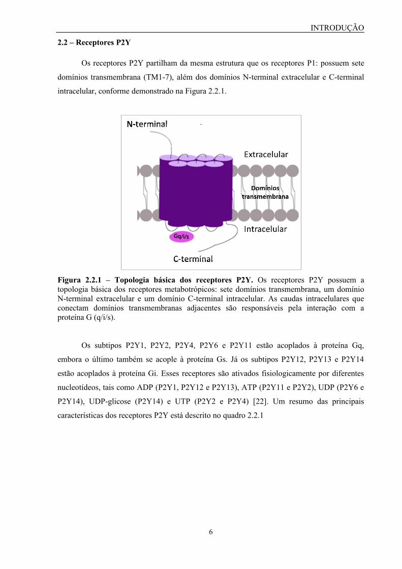
Os receptores P2Y partilham da mesma estrutura que os receptores P1: possuem sete
domínios transmembrana (TM1-7), além dos domínios N-terminal extracelular e C-terminal
intracelular, conforme demonstrado na Figura 2.2.1.
Figura 2.2.1 – Topologia básica dos receptores P2Y. Os receptores P2Y possuem a
topologia básica dos receptores metabotrópicos: sete domínios transmembrana, um domínio
N-terminal extracelular e um domínio C-terminal intracelular. As caudas intracelulares que
conectam domínios transmembranas adjacentes são responsáveis pela interação com a
proteína G (q/i/s).
Os subtipos P2Y1, P2Y2, P2Y4, P2Y6 e P2Y11 estão acoplados à proteína Gq,
embora o último também se acople à proteína Gs. Já os subtipos P2Y12, P2Y13 e P2Y14
estão acoplados à proteína Gi. Esses receptores são ativados fisiologicamente por diferentes
nucleotídeos, tais como ADP (P2Y1, P2Y12 e P2Y13), ATP (P2Y11 e P2Y2), UDP (P2Y6 e
P2Y14), UDP-glicose (P2Y14) e UTP (P2Y2 e P2Y4) [22]. Um resumo das principais
características dos receptores P2Y está descrito no quadro 2.2.1

INTRODUÇÃO
7
Quadro 2.2.1 – Principais características farmacológicas dos receptores P2Y
Receptor
P2Y
Proteína G
associada
Principais
efetores
Agonista
fisiológico
Agonistas
seletivos
Antagonistas
seletivos
P2Y1 Gq Ativação da
PLC, Rac e
Rho
ADP MRS2365 MRS2179
MRS2279
MRS2500
MRS2950
P2Y2 Gq Ativação da
PLC, Rac e
Rho
UTP e ATP MRS2768
2-thio-UTP
ARC-118925
P2Y4 Gq Ativação da
PLC
UTP MRS4062 -
P2Y6 Gq Ativação da
PLC e Rho
UDP PSB-0474
MRS2693
MRS2782
INS48823
5-O-metil-UDP
MRS2578
P2Y11 Gq; Gs Ativação da
PLC e AC
ATP NF546
AR-C67085
NF340
P2Y12 Gi Inibição da
AC e
ativação da
PLC e Rho
ADP
-
PSB-0739
ticlopidina
clopidogrel
prasugrel
ticagrelor
elinogrel
P2Y13 Gi Inibição da
AC e
ativação da
PLC e Rho
ADP CT1007900 MRS2211
P2Y14 Gi Inibição da
AC e
ativação da
PLC
UDP e UDP-
glicose
MRS2690
MRS2802
PPTN
PLC: fosfolipase C; AC: adenilato ciclase.
INTRODUÇÃO
8
O receptor P2Y1 é expresso em células epiteliais, endoteliais, imunes, plaquetas e
osteoclastos [21]. Apesar de o ADP ser o seu agonista fisiológico, o 2-MeSADP é um dos
agonistas mais potentes e o MRS2365 é o agonista seletivo. Já existem antagonistas
caracterizados para esse receptor, tais como o MRS2179, MRS2279, MRS2500 e MRS2950
[21,24]. Esses antagonistas vêm sendo utilizados em diversos trabalhos para a elucidação de
funções associadas a esse receptor, como nocicepção, hiperalgesia e dor inflamatória [25–27],
além da hiper-reatividade de astrócitos na doença de Alzheimer [28], quimiotaxia [29] e
agregação plaquetária [30–35].
O receptor P2Y2 é expresso em células do sistema imune, células epiteliais e
endoteliais, além de túbulos renais e osteoblastos [21]. O P2Y2 é ativado fisiologicamente
pelo UTP e ATP, mas também apresenta agonistas seletivos como o MRS2768 e o 2-thio-
UTP. Entretanto, apenas um antagonista seletivo é descrito para esse receptor, o ARC-118925
[22]. O P2Y2 demonstrou estar envolvido no desenvolvimento da dor neuropática [36,37], no
câncer [38–42] e na inflamação [43–48].
O receptor P2Y4 é expresso no intestino, cérebro, pulmão, coração, entre outros tipos
celulares [49]. O P2Y4 é ativado fisiologicamente pelo UTP e seletivamente pela molécula
MRS4062 [22]. Devido à inexistência de antagonistas seletivos para esse receptor, poucas
funções fisiológicas são de fato reconhecidas como provenientes de sua ativação.
O receptor P2Y6 é expresso em células epiteliais, linfócitos T, placenta e timo [21].
Fisiologicamente, esse receptor é ativado pelo UDP, entretanto, já foram desenvolvidos
diversos agonistas seletivos para esse receptor, tais como PSB-0474, MRS2693, MRS2782,
INS48823 e 5-O-metil-UDP, assim como o antagonista seletivo MRS2578 [22,24]. Trabalhos
recentes vêm demonstrando a participação desse receptor na dor [27,50–52], no câncer [53] e
na inflamação [54–58], sendo que o tratamento com o antagonista MRS2578 é capaz de
atenuar esses efeitos. Todavia, essa molécula ainda não avançou para estudos em seres
humanos devido às suas limitações quanto à reatividade e hidrofobicidade [59].
O receptor P2Y11 é expresso no baço, no intestino e em granulócitos [21]. O P2Y11 é
ativado fisiologicamente pelo ATP e os seus agonistas seletivos são NF546 e AR-C67085. O
NF-340 é o antagonista seletivo para esse receptor, uma vez que não interage com outros
receptores P2Y e P2X. Devido à inexistência de sequência para o receptor P2Y11 em
camundongos, muitas funções fisiológicas desse receptor ainda não foram completamente
caracterizadas [24,60]. Apesar disso, alguns trabalhos apontam a participação desse receptor
no desenvolvimento da dor em modelos animais, porém, a existência de possíveis efeitos
colaterais que possam ser responsáveis por essa atividade deve ser investigada [27,51].

INTRODUÇÃO
9
O receptor P2Y12 é expresso principalmente em plaquetas e células da glia [21]. O
P2Y12 é ativado fisiologicamente pelo ADP, e até o momento, não possui nenhum agonista
seletivo [22]. Contudo, esse é o primeiro e único subtipo de receptor da família P2 que possui
antagonistas empregados na terapia na prevenção da agregação plaquetária. Entre os
antagonistas seletivos para o P2Y12 encontram-se o clopidogrel, o prasugrel, o ticagrelor, o
cangrelor, o PSB-0739 e a ticlopidina, sendo os quatro primeiros aprovados para uso clínico
[24]. Além do importante papel desempenhado na agregação plaquetária, estudos recentes
demonstraram a participação do P2Y12 na dor [24], no câncer [61,62] e na inflamação
[63,64].
O receptor P2Y13 é expresso no cérebro, no baço, nos linfonodos e na medula óssea
[21]. Esse receptor é ativado fisiologicamente pelo ADP, e recentemente foi descrita a ação
agonista de uma molécula, a CT1007900, em animais [65]. O MRS2211 é o único antagonista
seletivo para o P2Y13. Apesar disso, existem poucos estudos mencionando papéis
fisiológicos associados a esse receptor. Entre eles encontra-se um estudo que mostra a
importância da ativação do P2Y13 no aprimoramento do metabolismo do colesterol HDL
[65], além de estudos descrevendo a participação do receptor na produção de citocinas
inflamatórias no fígado [66] e no desenvolvimento da dor neuropática [50].
O P2Y14 é expresso na placenta, no tecido adiposo, no estômago, no intestino e em
algumas regiões do cérebro [21]. O P2Y14 é ativado fisiologicamente pelo UDP e UDP-
glicose e as moléculas MRS2690, MRS2802 e α,β-metileno-2-thio-UDP são os seus agonistas
seletivos [24]. Recentemente, foi descrito um antagonista seletivo para esse receptor
denominado PPTN, o qual foi importante para a realização de estudos que demonstram a
participação do P2Y14 na inflamação [67,68].
2.3 – Receptores P2X
Os receptores P2X são canais iônicos que quando ativados por ligantes, permitem a
passagem seletiva dos cátions Na+, Ca
2+ e K
+, de acordo com o gradiente eletroquímico. O
P2X5 é o único subtipo que permite a passagem do íon cloreto (Cl-), além dos cátions citados
[15].
Atualmente, existem sete subtipos de receptores pertencentes a essa classe: P2X1-
P2X7. Esses receptores são triméricos, isto é, são formados por três subunidades, as quais
possuem domínios N e C-terminais intracelulares, sendo que esse último apresenta sítios de
ligação para proteínas quinases. Eles também possuem dois domínios transmembranas (TM1

INTRODUÇÃO
10
e TM2), os quais estão envolvidos com a formação do canal iônico e do poro,
respectivamente, além de uma cauda extracelular que possui resíduos de cisteína que atuam
na formação de pontes dissulfeto [21]. A Figura 2.3.1 demonstra a topologia dos receptores
P2X.
Figura 2.3.1 – Topologia básica dos receptores P2X. Cada subunidade do receptor P2X (à
esquerda) é composta por uma cauda extracelular com domínios ricos em cisteína, que se
unem para formar pontes dissulfeto, além de dois domínios transmembrana (TM1 e TM2) que
são responsáveis pela formação do canal iônico, bem como os sítios N e C-terminais
intracelulares. Três subunidades de iguais (representadas pela mesma cor) ou diferentes
(representadas por cores diferentes) subtipos de receptor P2X se agrupam para formar um
receptor homo ou heteromultimérico, respectivamente (à direita). Imagem adaptada de: Drug
Design Team da Universidade de Camerino (https://sites.google.com/a/unicam.it/diego-dal-
ben/activities-and-collaborations).
Os receptores P2X podem ser homomultiméricos, isto é, todas as subunidades
formadas correspondem à sequência de um único receptor (ex: P2X1, P2X2, P2X3, P2X4,
PX5, P2X6 e P2X7), ou heteromultiméricos, quando um canal iônico é formado pela
associação entre subunidades de diferentes P2X (ex: P2X1/2, P2X1/4, P2X1/5, P2X2/3,
P2X2/6, P2X4/6). O P2X6 é um exemplo de receptor que funciona apenas na forma de
heteromultímero, enquanto o P2X7 só funciona como homomultímero [21].
O agonista fisiológico dos receptores P2X é o ATP. Todos os membros dessa classe
são ativados pelo ATP entre 0,1 a 10 μM, com exceção do P2X7, cuja ativação ocorre em
concentrações superiores a 100 μM [15]. Além dessa particularidade, o P2X7 também é o
único que possui a habilidade de formar um poro membranar, o qual permite a passagem de

INTRODUÇÃO
11
moléculas de até 900 Da, incluindo íons, água, ATP, corantes, entre outros [69]. Já está
descrito na literatura que outros subtipos de receptores P2X, como o P2X2 e o P2X4, também
possuiriam essa habilidade de formar poros membranares similares ao P2X7 [70,71], contudo,
há controvérsias sobre a formação desses poros. Apesar disso, a origem do poro membranar
ainda é uma incógnita. Alguns autores acreditam que a abertura do poro membranar esteja
relacionada com a dilatação do canal iônico, enquanto outros acreditam que a hipótese mais
plausível seja de que outra proteína membranar funcione como um poro, sendo essa ativada
através de segundos mensageiros derivados da ativação dos receptores P2X [72–74]. O
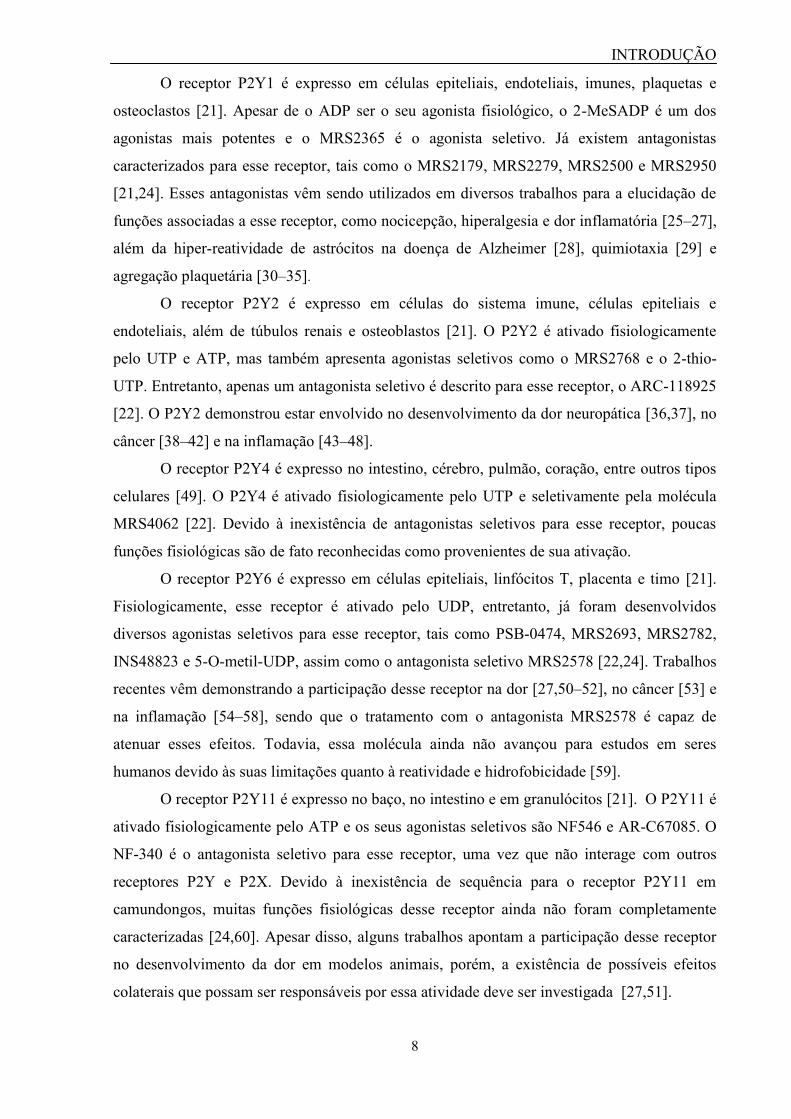
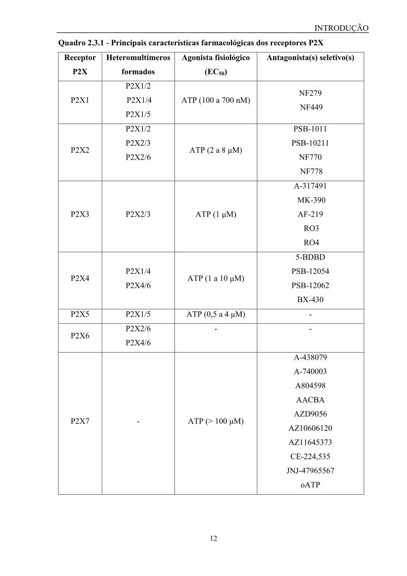
quadro 2.3.1 resume as principais características dos receptores P2X.
INTRODUÇÃO
12
Quadro 2.3.1 - Principais características farmacológicas dos receptores P2X
Receptor
P2X
Heteromultímeros
formados
Agonista fisiológico
(EC50)
Antagonista(s) seletivo(s)
P2X1
P2X1/2
P2X1/4
P2X1/5
ATP (100 a 700 nM) NF279
NF449
P2X2
P2X1/2
P2X2/3
P2X2/6 ATP (2 a 8 μM)
PSB-1011
PSB-10211
NF770
NF778
P2X3 P2X2/3 ATP (1 μM)
A-317491
MK-390
AF-219
RO3
RO4
P2X4 P2X1/4
P2X4/6 ATP (1 a 10 μM)
5-BDBD
PSB-12054
PSB-12062
BX-430
P2X5 P2X1/5 ATP (0,5 a 4 μM) -
P2X6 P2X2/6
P2X4/6
- -
P2X7 - ATP (> 100 μM)
A-438079
A-740003
A804598
AACBA
AZD9056
AZ10606120
AZ11645373
CE-224,535
JNJ-47965567
oATP
INTRODUÇÃO
13
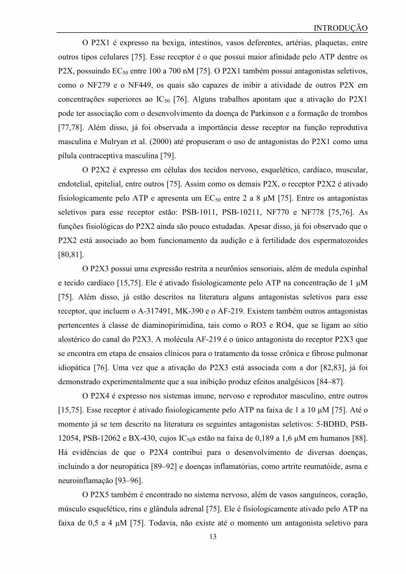
O P2X1 é expresso na bexiga, intestinos, vasos deferentes, artérias, plaquetas, entre
outros tipos celulares [75]. Esse receptor é o que possui maior afinidade pelo ATP dentre os
P2X, possuindo EC50 entre 100 a 700 nM [75]. O P2X1 também possui antagonistas seletivos,
como o NF279 e o NF449, os quais são capazes de inibir a atividade de outros P2X em
concentrações superiores ao IC50 [76]. Alguns trabalhos apontam que a ativação do P2X1
pode ter associação com o desenvolvimento da doença de Parkinson e a formação de trombos
[77,78]. Além disso, já foi observada a importância desse receptor na função reprodutiva
masculina e Mulryan et al. (2000) até propuseram o uso de antagonistas do P2X1 como uma
pílula contraceptiva masculina [79].
O P2X2 é expresso em células dos tecidos nervoso, esquelético, cardíaco, muscular,
endotelial, epitelial, entre outros [75]. Assim como os demais P2X, o receptor P2X2 é ativado
fisiologicamente pelo ATP e apresenta um EC50 entre 2 a 8 μM [75]. Entre os antagonistas
seletivos para esse receptor estão: PSB-1011, PSB-10211, NF770 e NF778 [75,76]. As
funções fisiológicas do P2X2 ainda são pouco estudadas. Apesar disso, já foi observado que o
P2X2 está associado ao bom funcionamento da audição e à fertilidade dos espermatozoides
[80,81].
O P2X3 possui uma expressão restrita a neurônios sensoriais, além de medula espinhal
e tecido cardíaco [15,75]. Ele é ativado fisiologicamente pelo ATP na concentração de 1 μM
[75]. Além disso, já estão descritos na literatura alguns antagonistas seletivos para esse
receptor, que incluem o A-317491, MK-390 e o AF-219. Existem também outros antagonistas
pertencentes à classe de diaminopirimidina, tais como o RO3 e RO4, que se ligam ao sítio
alostérico do canal do P2X3. A molécula AF-219 é o único antagonista do receptor P2X3 que
se encontra em etapa de ensaios clínicos para o tratamento da tosse crônica e fibrose pulmonar
idiopática [76]. Uma vez que a ativação do P2X3 está associada com a dor [82,83], já foi
demonstrado experimentalmente que a sua inibição produz efeitos analgésicos [84–87].
O P2X4 é expresso nos sistemas imune, nervoso e reprodutor masculino, entre outros
[15,75]. Esse receptor é ativado fisiologicamente pelo ATP na faixa de 1 a 10 μM [75]. Até o
momento já se tem descrito na literatura os seguintes antagonistas seletivos: 5-BDBD, PSB-
12054, PSB-12062 e BX-430, cujos IC50s estão na faixa de 0,189 a 1,6 μM em humanos [88].
Há evidências de que o P2X4 contribui para o desenvolvimento de diversas doenças,
incluindo a dor neuropática [89–92] e doenças inflamatórias, como artrite reumatóide, asma e
neuroinflamação [93–96].
O P2X5 também é encontrado no sistema nervoso, além de vasos sanguíneos, coração,
músculo esquelético, rins e glândula adrenal [75]. Ele é fisiologicamente ativado pelo ATP na
faixa de 0,5 a 4 μM [75]. Todavia, não existe até o momento um antagonista seletivo para

INTRODUÇÃO
14
esse receptor e enquanto isso, antagonistas não seletivos de receptores P2 como Suramina,
PPADS, TNP-ATP e Brilliant Blue G (BBG) são utilizados em experimentos para prevenir a
sua ativação [97]. Apesar das suas funções serem pouco conhecidas, recentemente Kim et al.
(2017) demonstraram a sua participação na diferenciação de osteoclastos [98].
O P2X6 está presente em algumas células do sistema nervoso, além de células do
útero, ovário, epitélio brônquico, timo e glândula salivar [15,75]. Uma vez que o P2X6 não
funciona como um homomultímero, dados referentes à sua farmacologia e funções
fisiológicas são escassos [15,21].
Já o P2X7 é vastamente expresso em células do sistema imune, bem como em células
epiteliais e glandulares, glia, fibroblastos, osteoblastos e hepatócitos [15,75]. Como
mencionado anteriormente, o receptor P2X7 é o que possui menor afinidade pelo ATP, sendo
ativado em concentrações superiores a 100 μM [15]. Em diversos trabalhos, o P2X7 é
estimulado com o BzATP, um agonista análogo ao ATP e mais potente que este. Entretanto, o
BzATP também possui atividade agonista sobre o P2X4 e funciona como agonista parcial dos
receptores P2X1, P2X2, P2X3 e P2X5 [75]. O P2X7 possui diversos antagonistas seletivos
descritos na literatura. Entre eles encontram-se: AACBA, A-438079, A-740003, A804598,
AZ10606120, AZ11645373, AZD9056, CE-224,535, JNJ-47965567 e ATP oxidado (oATP)
[99–101]. Além do papel na inflamação [102], o P2X7 também participa no desenvolvimento
de doenças neurológicas e neurodegenerativas [99–101,103–112], câncer [113–117] e dor
[118–122].
3. Desenvolvimento de fármacos com atividade sobre os receptores P2
Apesar da descoberta dos receptores P2 ter ocorrido durante as décadas de 1980 e
1990, seus papéis fisiológicos começaram a ser caracterizados a partir do fim dos anos 1990.
Embora tenha aumentado de modo expressivo o conhecimento dos receptores P2 no contexto
patológico, o grande desafio dos últimos anos é desenvolver fármacos com atividade sobre
esses receptores que sejam eficazes. Nesse cenário, diversos antagonistas seletivos para os
receptores P2 foram desenvolvidos, todavia, a maioria deles apresenta alguma limitação que
não os permitiu evoluir para a fase de terapia em humanos.
Até o momento, o receptor P2Y12 é o primeiro caso de sucesso entre os receptores P2
com relação à aplicação clínica. Ele possui um antagonista, o clopidogrel, que é aplicado na
terapia desde a década de 1990. O clopidogrel é antagonista irreversível do P2Y12 em
plaquetas. Esse fármaco funciona como uma pró-droga, que necessita do metabolismo da
enzima hepática CYP450 para produzir o seu metabólito ativo, um tiol reativo que apresenta

INTRODUÇÃO
15
propriedades anti-agregantes e antitrombóticas. Atualmente, ele é administrado em conjunto
com a aspirina na prevenção de trombose em pacientes com síndrome aguda coronária ou que
foram submetidos a uma intervenção coronária percutânea [123]. Um estudo realizado com
mais de 12 mil pacientes demonstrou que essa associação foi responsável pela diminuição de
2,1% de mortes em decorrência de eventos cardiovasculares durante o período avaliado [124].
Além do clopidogrel, outros fármacos com ação antagonista sobre o P2Y12 foram
desenvolvidos. Entre eles, o prasugrel, que possui estrutura similar ao clopidogrel,
funcionando também como uma pró-droga. Ele demonstrou ser mais eficaz que o seu
antecessor sobre a diminuição de mortes em decorrência de eventos cardiovasculares, no
entanto, ele está associado a um maior risco de hemorragias [125,126].
Posteriormente foi desenvolvido o ticagrelor, o primeiro antagonista reversível e
seletivo do P2Y12 com atividade intrínseca. Apesar de ter auxiliado na sobrevivência de
90,2% de pacientes com risco de morte em decorrência de eventos cardiovasculares, esse
antagonista provocou um número maior de efeitos colaterais do que o clopidogrel na
população estudada [127,128].
O mais recente antagonista aprovado para uso clínico é o cangrelor. Ele é um
antagonista do P2Y12 que está disponível apenas na forma intravenosa, sendo utilizado,
portanto, em emergências médicas [129]. Entretanto, o cangrelor não é seletivo para o P2Y12,
uma vez que também inibe o receptor P2Y13 [24]. O quadro 3.1 fornece um resumo dos
fármacos com atividade sobre o receptor P2Y12 que são aprovados para uso terapêutico.

INTRODUÇÃO
16
Quadro 3.1 – Antagonistas do receptor P2Y12 aprovados para uso clínico
1Informações extraídas do website do Departamento de Administração de Alimentos e
Fármacos dos Estados Unidos (FDA): (https://www.fda.gov); 2Estruturas químicas extraídas
da Base Livre de Dados Químicos (PubChem): (https://pubchem.ncbi.nlm.nih.gov).
3.1 – O receptor P2Y2 como alvo terapêutico
O P2Y2 é um receptor fisiologicamente ativado pelo UTP e ATP extracelular. Ele é
expresso em diversos órgãos tais como pulmão, coração, baço e rins, além de células do
sistema imune como linfócitos e macrófagos [24,130]. Esse receptor possui importantes
funções relacionadas ao fluxo de íons. O P2Y2 expresso nas células epiteliais da córnea,
conjuntiva e vias aéreas estimula a secreção de cloro através da ativação do canal de cloro
retificador externo (ORCC), promovendo a lubrificação das superfícies e hidratação do muco.
Além disso, esse receptor é capaz de inibir a absorção de sódio e estimular o batimento ciliar,
bem como a secreção de muco [131]. A Figura 3.1.1 demonstra as principais funções do
receptor P2Y2 sobre o fluxo iônico em células epiteliais.
Fármaco Ano de aprovação1 Empresa responsável
1 Estrutura química
2
Clopidogrel 1997 Sanofi
Prasugrel 2009 Eli Lilly and Co
Ticagrelor 2011 AstraZeneca
Cangrelor 2015 The Medicines
Company

INTRODUÇÃO
17
Figura 3.1.1 – Transporte iônico promovido pelo receptor P2Y2 em células epiteliais das
vias aéreas. O P2Y2 é ativado fisiologicamente pelo ATP e UTP extracelular. Uma vez
ativado, a proteína Gq associada estimula a enzima fosfolipase C a clivar o PIP2 em
diacilglicerol e inositol trifosfato (IP3). Este último ativa o receptor IP3R no retículo
endoplasmático, promovendo a liberação do Ca2+
dos estoques intracelulares. Esse aumento
de cálcio promove a abertura dos canais de cloro ativados por cálcio (CaCC), favorecendo o
efluxo de Cl- e o transporte de água. Além disso, a hidrólise do PIP2 inibe os canais de sódio
epiteliais (ENaC), diminuindo a reabsorção do Na+. Adaptado de [132].
Essas importantes funções relacionadas ao fluxo de íons, podem contribuir para a
lubrificação e hidratação de tecidos epiteliais, especialmente em doenças em que essas
atividades estejam comprometidas, como a fibrose cística e a doença do olho seco.
A fibrose cística é uma doença genética, caracterizada pela mutação do gene que
codifica a proteína reguladora da condutância transmembrana na fibrose cística (CFTR), a
qual está envolvida na regulação do cloro, sódio e bicarbonato na superfície aérea. Com o
fluxo iônico comprometido e o líquido superficial reduzido, os pacientes sofrem com um
aumento da suscetibilidade de infecções e inflamações e com danos progressivos nas vias
aéreas devido à limitação da depuração mucociliar [133].
Tendo em vista que a ativação do P2Y2 poderia corrigir o transporte iônico de forma
independente do genótipo CFTR, a molécula Denufosol (INS37217) foi desenvolvida pela
empresa Inspire Pharmaceuticals no início dos anos 2000. Essa molécula foi formulada como
uma solução inalatória que ativa o receptor P2Y2, mimetizando a ação dos nucleotídeos
ATP/UTP. Ela é um dinucleotídeo derivado de uridina, que estimula a secreção de cloro e
mucina, o batimento ciliar, além de ser resistente à ação das ectonucleotidases (embora seja
capaz de ativar outros receptores P2Y, como o P2Y4 e o P2Y6) [134]. O Denufosol foi
INTRODUÇÃO
18
testado em 178 pacientes em ensaios clínicos de fase 3. Esses pacientes receberam três doses
diárias de 60 mg de Denufosol ao longo de 24 semanas. Apesar de ter sido bem tolerado, a
molécula não apresentou eficácia significativa em promover uma melhora da função
pulmonar e redução de incidências de exacerbações pulmonares. Mesmo em uma segunda
etapa dentro da fase 3, que incluía um número maior de pacientes (n= 233) e tempo mais
longo de tratamento (48 semanas), ela também falhou em demonstrar eficácia [132,133,135].
Essa molécula chegou a ser estudada para o tratamento de descolamento de retina, edema
macular e uveíte, entretanto, esses estudos foram terminados ou abandonados desde 2006, de
acordo com informações depositadas no banco de dados Clinical Trials (clinicaltrials.gov).
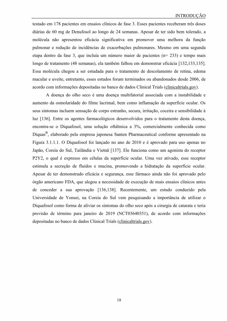
A doença do olho seco é uma doença multifatorial associada com a instabilidade e
aumento da osmolaridade do filme lacrimal, bem como inflamação da superfície ocular. Os
seus sintomas incluem sensação de corpo estranho, secura, irritação, coceira e sensibilidade à
luz [136]. Entre os agentes farmacológicos desenvolvidos para o tratamento desta doença,
encontra-se o Diquafosol, uma solução oftálmica a 3%, comercialmente conhecida como
Diquas®, elaborado pela empresa japonesa Santen Pharmaceutical conforme apresentado na
Figura 3.1.1.1. O Diquafosol foi lançado no ano de 2010 e é aprovado para uso apenas no
Japão, Coreia do Sul, Tailândia e Vietnã [137]. Ele funciona como um agonista do receptor
P2Y2, o qual é expresso em células da superfície ocular. Uma vez ativado, esse receptor
estimula a secreção de fluidos e mucina, promovendo a hidratação da superfície ocular.
Apesar de ter demonstrado eficácia e segurança, esse fármaco ainda não foi aprovado pelo
órgão americano FDA, que alegou a necessidade de execução de mais ensaios clínicos antes
de conceder a sua aprovação [136,138]. Recentemente, um estudo conduzido pela
Universidade de Yonsei, na Coreia do Sul vem pesquisando a importância de utilizar o
Diquafosol como forma de aliviar os sintomas do olho seco após a cirurgia de catarata e teria
previsão de término para janeiro de 2019 (NCT03640351), de acordo com informações
depositadas no banco de dados Clinical Trials (clinicaltrials.gov).

INTRODUÇÃO
19
Figura 3.1.2 – Fármaco com ação sobre o receptor P2Y2. O fármaco Diquas® é um
agonista do receptor P2Y2 aprovado para o tratamento da doença do olho seco em alguns
países asiáticos. À esquerda observa-se a sua formulação farmacêutica e à direita, a sua
estrutura química. Imagens extraídas do site do fabricante, Santen Pharmaceuticals e da Base
Livre de Dados Químicos (PubChem): (https://pubchem.ncbi.nlm.nih.gov).
Além dessas funções que são importantes para o tratamento da fibrose cística e doença
do olho seco, outros efeitos promovidos pelo receptor P2Y2 foram capazes de melhorar a
reconstituição da glândula salivar e a cicatrização, tratar a doença crônica renal e infecção por
leishmaniose, além de conferir neuroproteção. A ativação do P2Y2 promoveu a agregação das
células epiteliais salivares, além da proliferação e migração in vitro de células progenitoras
cardíacas humanas [139,140]. Em modelo experimental de cicatrização, Jin et al. (2014)
observaram que os animais P2Y2-/-
levaram mais tempo para que a ferida cicatrizasse,
possivelmente devido a uma diminuição da expressão de proteínas de matriz extracelular
[141]. O P2Y2 também parece exercer papeis benéficos no controle da progressão da doença
crônica renal, uma vez que animais P2Y2-/-
submetidos ao procedimento de nefrectomia
sobreviveram menos tempo que os animais selvagens e apresentaram aumento da pressão
sistólica e de ureia no soro após a cirurgia [142]. A ativação do P2Y2 confere proteção contra
agentes infecciosos como a Leishmania amazonensis uma vez que o tratamento intralesional
com o UTP (lesão na pata) diminuiu a carga parasitária nos sítios de infecção nos animais
[143]. Em doenças neurodegenerativas o receptor P2Y2 executa ações protetoras. Na doença
de Alzheimer, já foi observado que o P2Y2 contribuiu com o processamento da proteína
precursora amilóide (PPA), além de induzir a sua fagocitose e degradação [144–147]. Ajit et
al. (2014) também observaram que a deleção do P2Y2 promoveu um acúmulo da placa de β-
amilóide no córtex cerebral e hipocampo em camundongos, bem como provocou um aumento
do déficit neurológico [148].
INTRODUÇÃO
20
Apesar da ativação do P2Y2 conferir benefícios para o tratamento de várias doenças, a
sua inibição também pode ser útil para tratar doenças inflamatórias, dor e câncer. O P2Y2
pode ser considerado um alvo terapêutico para o tratamento de doenças inflamatórias, uma
vez que a sua ativação estimulou a produção de mediadores inflamatórios, bem como a
migração de células inflamatórias para o sítio de lesão [48,149], contribuindo para a
inflamação intestinal, pulmonar e hepática [43–45,47]. A ativação do P2Y2 também causou
recrutamento de macrófagos e hiperplasia arterial, contribuindo para o desenvolvimento de
lesões arteriais [150].
Recentemente, Merz et al. (2018) demonstraram a possível participação do P2Y2 no
controle de doenças metabólicas. Animais P2Y2-/-
apresentavam um número reduzido de
macrófagos no tecido adiposo, menor ganho de peso após 15 semanas, ausência de sinal de
esteatose hepática e diminuição dos níveis de colesterol plasmático [151]. Já foi observado
também que animais P2Y2-/-
parecem apresentar proteção contra a inflamação [152–154].
Além disso, a inibição desse receptor promoveu analgesia em modelo experimental de dor
neuropática no nervo trigêmeo [37].
Os antagonistas do receptor P2Y2 também podem funcionar como agentes
antimetastáticos. Uma vez que os nucleotídeos podem ser encontrados em altas concentrações
no sítio tumoral [155], já foi observado que o P2Y2, ao ser ativado pelo ATP ou UTP, pode
promover proliferação, migração, invasão e metástase de linhagens tumorais. Entre os tipos de
câncer analisados estão tumores de mama e próstata, carcinoma hepatocelular humano e
células epiteliais cancerosas do ducto pancreático [38–42,156]. O aumento da expressão do
receptor P2Y2 ainda está relacionado com um mau prognóstico no adenocarcinoma do ducto
pancreático [157].
Apesar de serem bastante promissores, muitos estudos referentes à função do P2Y2
precisam ser avaliados com cautela. Primeiramente, na maioria dos trabalhos o P2Y2 é
estimulado apenas com ATP e UTP, sendo que ambos são capazes de ativar outros receptores
P2. Além disso, esses nucleotídeos podem ser degradados pelas ectonucleotidases, gerando
novas moléculas, como ADP e UDP, por exemplo, que são capazes de ativar diferentes
subtipos de receptores P2, ocasionalmente afetando a caracterização do efeito biológico
estudado. Somado a isso, são raros os trabalhos que utilizam agonistas seletivos para esse
receptor, como o 2-thio-UTP, MRS2698 e o MRS768, apesar desse último apresentar
estabilidade limitada em meio biológico [49].
O antagonista seletivo do P2Y2 (AR-C118925), o qual é utilizado com frequência na
execução de experimentos, também possui algumas limitações. O AR-C118925 foi
desenvolvido pela empresa AstraZeneca e chegou a ser avaliado em formulação tópica para o

INTRODUÇÃO
21
tratamento da psoríase crônica, embora tenha demonstrado ineficácia. Apesar de ter sido uma
ferramenta muito útil para a execução de experimentos in vitro e in vivo, essa molécula não
avançou em testes com humanos, devido a algumas propriedades farmacológicas indesejadas,
que incluem alta polaridade e baixa biodisponibilidade através da via oral [49]. Portanto, essa
lacuna na farmacologia do receptor P2Y2 abre margem para a descoberta e/ou
desenvolvimento de novas moléculas com ação antagonista sobre esse receptor. A Figura
3.1.3 resume os principais papeis do P2Y2 e a sua associação a diferentes contextos
fisiológicos.
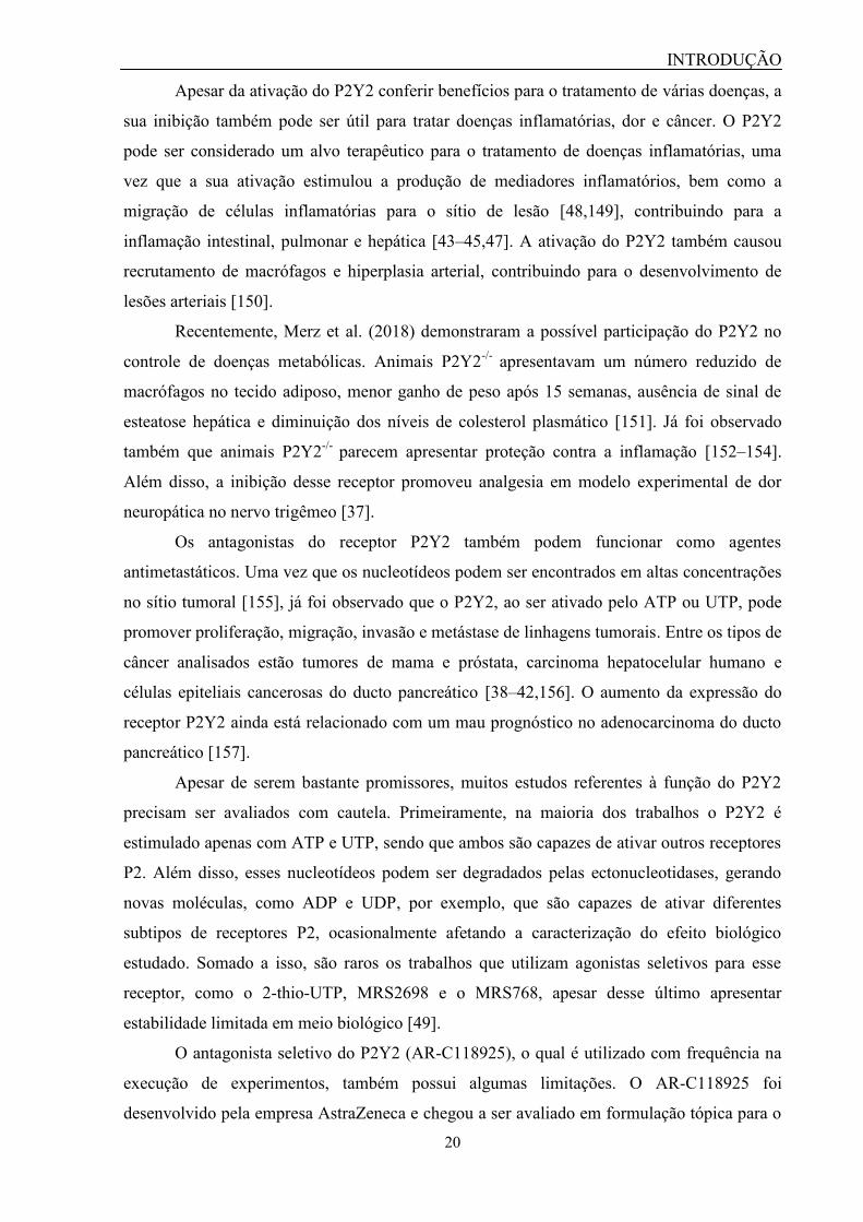
Figura 3.1.3 – Principais efeitos da ativação do P2Y2. A figura resume os principais efeitos
da ativação do receptor P2Y2 e a sua implicação em diversos contextos fisiológicos, os quais
podem ser aprimorados através do uso de agonistas desse receptor (redigidos em preto) ou
ainda atenuados através do uso de seus antagonistas (redigidos em vermelho).
3.2 - O receptor P2Y4 como alvo terapêutico
O receptor P2Y4 é ativado fisiologicamente pelo UTP extracelular. Em ratos e
camundongos, ele também pode ser ativado pelo ATP extracelular [130]. O P2Y4 é expresso
em diversos órgãos tais como intestino, cérebro, pulmão, coração, próstata, pele e baço [49].
Por ser semelhante estruturalmente e farmacologicamente ao receptor P2Y2 e por vezes, ser
expresso nos mesmos tipos celulares que ele, a caracterização dos efeitos fisiológicos do

INTRODUÇÃO
22
P2Y4 tem se tornado um grande desafio [49]. O desenvolvimento de agonistas seletivos para
cada um desses receptores tem permitido certo avanço nesse campo da Farmacologia dos
receptores P2. Assim, já se tem registros na literatura de que três moléculas são capazes de
ativar seletivamente o receptor P2Y4. São eles: MRS2927, MRS4062 e N(4)-(feniletóxi)-
CTP, sendo que essas duas últimas foram desenvolvidas inicialmente como versões
aprimoradas de agonistas para o receptor P2Y2 [24,49].
Apesar das limitações farmacológicas, algumas funções fisiológicas do P2Y4 já foram
elucidadas. O P2Y4 assim como o P2Y2 exerce um papel na regulação do transporte de íons.
Ghanem et al. (2005) demonstraram que o P2Y4 promove a secreção de Cl- na mucosa
intestinal, efeito esse que foi significativamente reduzido em animais P2Y4-/-
[158]. O P2Y4
também é expresso em células epiteliais na membrana de Reissner (situada na cóclea), onde
controla a homeostase do Na+ na endolinfa a partir da inibição de canais ENaC [159].
O P2Y4 exerceu um papel neuroprotetor em modelo experimental de Alzheimer, no
qual ele está envolvido na indução da pinocitose do peptídeo β-amilóide [160]. Ward et al.
(2008) demonstraram que a expressão do P2Y4 em células cones e bastonetes na retina sofre
alteração após a adaptação à luz e ao escuro, sugerindo que esse receptor estaria envolvido na
modulação da atividade interna da retina [161]. O P2Y4 ainda exerce importantes funções
cardíacas que podem conferir cardioproteção. Horckmans et al. (2012) demonstraram que
animais P2Y4-/-
apresentavam microcardia após 12 semanas de vida [162] e que a deleção
desse receptor também está associada a uma menor resistência durante o exercício físico
[163]. Lemaire et al. (2017) observaram que animais P2Y4-/-
apresentaram menor tamanho da
área de infarto comparado aos animais selvagens [164]. Por outro lado, em modelo
experimental de isquemia a deleção desse receptor conferiu cardioproteção. Horckmans et al.
(2015) observaram que animais P2Y4-/-
apresentavam menor área de infarto, necrose, fibrose
e diminuição do recrutamento de neutrófilos [165]. Zizzo et al. (2012) demonstraram que um
antagonista para o P2Y4 poderia ser útil no tratamento da constipação, uma vez que esse
receptor parece estar envolvido na inibição da atividade contrátil espontânea do músculo
longitudinal do íleo através da via de sinalização da fosfolipase C/IP3 [166].
Apesar disso, a ausência de antagonistas seletivos para esse receptor ainda dificulta a
caracterização do papel do P2Y4 no desenvolvimento das doenças relacionadas à sua
ativação. Nesse sentido, a molécula RB-2 vem sendo utilizada como um antagonista para esse
receptor. Entretanto, devido a sua capacidade de inibir outros receptores P2, deve-se tomar
cautela na análise dos resultados. Contudo, ela constitui um modelo estrutural para a síntese e
desenvolvimento de novos antagonistas seletivos para o P2Y4 [49]. Nesse contexto, a
descoberta de novos antagonistas para o receptor P2Y4 permitirá uma caracterização mais

INTRODUÇÃO
23
fidedigna de funções fisiológicas associadas a esse receptor in vitro e in vivo, além de
possibilitar a sua implementação na terapia clínica. A Figura 3.2.1 resume as principais
funções do receptor P2Y4 e a sua associação a diferentes contextos fisiológicos.
Figura 3.2.1 – Principais efeitos da ativação do P2Y4. A figura resume os principais efeitos
da ativação do receptor P2Y4 e a sua implicação em diversos contextos fisiológicos, os quais
podem ser aprimorados através do uso de agonistas desse receptor (redigidos em preto) ou
ainda atenuados através do uso de seus antagonistas (redigidos em vermelho).
3.3 – O receptor P2X7 como alvo terapêutico
O receptor P2X7 é expresso em células do sistema imune, tais como mastócitos,
eritrócitos, monócitos, macrófagos, células dendríticas e linfócitos, além de células da glia
[167]. Ele é ativado fisiologicamente pelo ATP extracelular em uma concentração em torno
de 100 μM, a qual é aproximadamente 10 vezes maior do que a necessária para a ativação dos
demais receptores P2X [15]. Por essa razão, alguns autores consideram que a sua ativação
seja um indicativo de “sinal de perigo”, uma vez que esses altos níveis de ATP extracelular
são alcançados fisiologicamente, por exemplo, através de citólise [102]. Esse receptor
apresenta a peculiar capacidade de formar um poro membranar em situações de exposição
prolongada ao ATP. Esse poro membranar permite a passagem de moléculas de até 900 Da,
incluindo íons (Na+, Ca
2+, K
+), água, ATP e corantes como iodeto de propídeo (668 Da),

INTRODUÇÃO
24
brometo de etídeo (394 Da) e YO-PRO-1 (629 Da), que são comumente utilizados para o
estudo da atividade do P2X7 [69,168].
A ativação do P2X7 incita uma série de respostas celulares com perfil pró-
inflamatório, que incluem a formação do inflamassomo, a ativação de caspases, fosfolipases
(A2 e D), proteínas quinases ativadas por mitógenos (MAPK) e fatores de transcrição como o
NF-κB, bem como a transcrição de genes pró-inflamatórios como COX-2 e iNOS. A sua
ativação ainda promove a liberação de citocinas como IL-1β, IL-18 e TNF-α, a geração de
espécies reativas de oxigênio (EROS), além de morte celular [102,169–172]. Devido a essas
funções desempenhadas pelo P2X7, diversos trabalhos da literatura já descreveram a sua
participação no desenvolvimento da inflamação, dor, neurodegeneração, isquemia e hipóxia,
câncer e infecção.
Existem diversos trabalhos na literatura demonstrando que os antagonistas do P2X7
apresentam efeitos anti-inflamatórios. O tratamento prévio de animais com A-438079 e BBG
(antagonistas do P2X7) foi capaz de suprimir a ativação do inflamassomo e a liberação de
citocinas, diminuindo a severidade de nefrotoxicidade renal induzida por cisplatina e nefrite
[173,174]. Os antagonistas BBG, A-740003 e A-438079 foram capazes de prevenir a colite
em ratos e inibir a infiltração de neutrófilos na orelha de camundongos [175–177]. Em
modelo experimental de lesão hepática e fibrose, o antagonista A-438079 diminuiu a necrose,
a inflamação, o acúmulo de colágeno, bem como a liberação de citocinas pró-inflamatórias
[178]. Além disso, o P2X7 é expresso no tecido sinovial inflamado e já foi observado que o
tratamento com antagonistas foi capaz de aliviar a inflamação nas articulações de animais
[119,179,180]. Recentemente, foi desenvolvida uma espécie de anticorpos em escala de
nanômetros, chamados de “nanocorpos” com reconhecimento do P2X7. Eles possuem
administração sistêmica e podem atenuar a ativação do P2X7 em células T e macrófagos,
aliviando os sintomas da glomerulonefrite experimental e dermatite de contato [181]. A
ativação do P2X7 também está associada com a hiperalgesia inflamatória, a qual pode ser
atenuada com o tratamento prévio com antagonistas desse receptor [118–122].
Em modelos experimentais de doenças neurodegenerativas já foi observado um
aumento da expressão do P2X7 em alguns tipos celulares no cérebro, o que geralmente está
associado com o agravamento dessas enfermidades. Os antagonistas do P2X7 foram capazes
de aliviar a severidade das convulsões e os danos neuronais em camundongos epiléticos [99–
101]. Em modelos de esclerose lateral amiotrófica, foi observado que os antagonistas BBG,
oATP e KN-62 reduziram a morte de neurônios motores e a gliose, resultando em um
aprimoramento do desempenho motor dos animais [109–112]. O P2X7 também está
relacionado com a diminuição da atividade da enzima α-secretase, a qual é responsável pela

INTRODUÇÃO
25
clivagem da proteína precursora amilóide (PPA) [182]. Essa atividade produz um acúmulo de
placas amilóides em modelos in vivo de doença de Alzheimer, o que foi revertido através do
tratamento prévio com antagonistas desse receptor, os quais também auxiliaram na prevenção
do déficit cognitivo [106,183].
A ativação do P2X7 na isquemia parece ser prejudicial. Estudos de privação de
oxigênio-glicose in vitro demonstraram que o uso de antagonistas do P2X7 reduziram os
danos mitocondriais, a produção de EROS e a viabilidade celular após o estímulo isquêmico
[184–186]. Já em modelos in vivo de isquemia, os antagonistas do P2X7 aumentaram a
sobrevivência de neurônios, além de reduzirem a mortalidade dos animais, a ativação da glia e
a transcrição de citocinas [187,188]. Por outro lado, alguns trabalhos demonstraram que a
inibição do P2X7 também pode ser danosa. Anagisawa et al. (2008) observaram que o
tratamento com o oATP resultou na perda de neurônios e no desempenho motor dos animais
[189]. Brinda et al. (2014) demonstraram que o BBG aboliu o efeitos neuroprotetores
produzidos pelo pós-condicionamento como a memória e o desempenho motor [190]. Já
Kaiser et al. (2015) observaram que animais P2X7-/-
apresentaram maior área de edema
cerebral comparado aos animais controles [191]. Esses achados controversos sugerem que as
ações do P2X7 na isquemia podem ser resultado da severidade e duração do evento
isquêmico, influenciando assim, o papel do receptor para um perfil de proteção ou de agressão
[192].
No câncer, assim como na isquemia, a ativação do P2X7 demonstra um duplo papel,
podendo favorecer o crescimento das células tumorais ou mesmo a sua morte. Em
determinados tipos de câncer como neuroblastoma e glioma, a ativação do P2X7 está
envolvida na proliferação celular, o que foi atenuado através da administração intravenosa de
BBG [193]. A ativação do P2X7 também estimula a proliferação, migração e invasão de
células de adenocarcinoma ductal pancreático e de algumas linhagens de câncer de próstata,
pulmão, mama e melanoma, sendo essas atividades revertidas com o tratamento de
antagonistas desse receptor [113–117]. Apesar disso, curiosamente, Fang et al. (2013)
observaram que tanto o tratamento com o BBG quanto o silenciamento do P2X7 através de
RNA de interferência (RNAi) aumentaram o crescimento tumoral e a angiogênese em células
de glioma C6 [194].
Nas doenças infecciosas, o P2X7 também possui uma função dupla. A sua ativação
pode exercer papéis protetores ou deletérios para o hospedeiro. De acordo com Savio et al.
(2018), o favorecimento de um desses dois efeitos depende do tipo de patógeno, da virulência
e da severidade da infecção [195]. A participação do P2X7 já foi caracterizada em diversas
doenças infecciosas virais, bacterianas, fúngicas, bem como em protozooses. Na tuberculose,

INTRODUÇÃO
26
a ativação do P2X7 está envolvida com a apoptose de células infectadas com a cepa H37Rv
de Mycobacterium tuberculosis [196]. Na toxoplasmose, a ativação do P2X7 promove a
eliminação do Toxoplasma gondii através da produção de EROS [197]. Já na infecção por
clamídia, a eliminação acontece através da ativação da fosfolipase D, uma enzima com
atividade antimicrobicida [198]. Recentemente, Graziano et al. (2015) demonstraram a
participação do P2X7 na liberação de virions do HIV em macrófagos humanos de pacientes
soropositivos e células D-U1 infectadas, sendo que essa atividade foi inibida pelo antagonista
oATP [199]. Em modelo experimental de sepse, Santana et al. (2015), demonstraram que os
animais P2X7-/-
apresentaram maior taxa de sobrevivência e menores números de unidades
formadoras de colônia, leucócitos e citocinas pró-inflamatórias no peritônio comparados aos
animais controle [200].
Apesar dos resultados serem bastante promissores, alguns cuidados devem ser
tomados ao se avaliar os estudos com o receptor P2X7. Primeiramente, muitos estudos
utilizam o ATP exógeno para estimular o receptor P2X7, contudo, como a concentração
necessária para ativar esse receptor é alta (>100 μM), outros receptores P2X e P2Y podem ser
ativados, provocando algum tipo de interferência nas respostas. Além disso, a expressão de
ectonucleotidases no tipo celular ou tecido estudado ainda pode direcionar parte da resposta
para receptores P2Y e P1 [195]. Em alguns trabalhos, os autores optam por utilizar o BzATP,
entretanto, ele não é seletivo para o P2X7, uma vez que possui afinidade para os receptores
P2X1 e P2X3 [59]. O BBG, utilizado em muitos experimentos como antagonista do P2X7,
também é capaz de inibir o receptor P2X4 e canais de sódio ativados por voltagem [76]. O
ATP oxidado, outro antagonista do P2X7, também possui outros alvos moleculares além
desse receptor [201]. Além disso, os animais P2X7-/-
produzidos pelas empresas
GlaxoSmithKline e Pfizer, expressam o receptor em células T, embora ele seja funcional
apenas nos camundongos da primeira empresa [195].
Apesar da perspectiva quanto ao uso de antagonistas do P2X7 na terapia, ainda não
existem fármacos na clínica que sejam utilizados para o tratamento de doenças relacionadas a
esse receptor. Baseado nos resultados observados em modelos animais de artrite reumatoide,
alguns grupos de pesquisa estudaram a aplicação de antagonistas do P2X7 nessa doença.
Utilizando os parâmetros do Colégio Americano de Reumatologia sobre o alívio dos
sintomas, os autores observaram que o tratamento de pacientes com o AZD9056 e CE-
224,535 não demonstrou eficácia superior em relação ao grupo placebo. Além disso, os
pacientes tratados com esses antagonistas relataram diversos efeitos colaterais, que incluíam
náusea, dor abdominal, diarreia e dor de cabeça [202–204]. Devido a essas falhas na eficácia,
as pesquisas com ambos as moléculas se encerraram na fase 2 de ensaios clínicos. Assim, essa

INTRODUÇÃO
27
lacuna na terapia associada ao receptor P2X7 estimula a pesquisa e o desenvolvimento de
fármacos com atuação sobre este promissor alvo-terapêutico. A Figura 3.3.1 resume as
principais funções do receptor P2X7 e a sua associação a diferentes contextos fisiológicos.
Figura 3.3.1 – Principais efeitos da ativação do P2X7. A figura resume os principais efeitos
da ativação do receptor P2X7 e a sua implicação em diversos contextos fisiológicos, os quais
podem ser aprimorados através do uso de agonistas desse receptor (redigidos em preto) ou
ainda atenuados através do uso de seus antagonistas (redigidos em vermelho ou marcados
com um asterisco vermelho).
4- Os produtos naturais como uma fonte para a descoberta de novos antagonistas
“Presentes esplêndidos: A natureza produz constantemente moléculas que não foram
pensadas pelos seres humanos.” (Satoshi Omura)
Atualmente no campo de desenvolvimento de fármacos, o maior desafio é encontrar
candidatos que sejam capazes de tratar uma determinada doença com eficácia e apresente um
mínimo de efeitos colaterais [205]. Nesse contexto, os produtos naturais emergem como uma
importante fonte para a descoberta inovadora de novos fármacos devido à sua enorme
diversidade estrutural e química. Essas características foram adquiridas ao longo de milhares
de anos de evolução e ainda continuam inspirando novas descobertas nas áreas de Química,
Biologia e Medicina [205,206].

INTRODUÇÃO
28
Tradicionalmente, os fármacos à base de produtos naturais eram oriundos de plantas,
minerais e animais, mas hoje já são estudadas moléculas presentes em seres marinhos, fungos,
bactérias, entre outros [207]. Esses seres vivos estão agrupados dentro de cinco reinos
biológicos: Animalia, Plantae, Fungi, Protista e Monera, que juntos abrigam uma imensa
biodiversidade de espécies, representando assim, um grande número de amostras para testes.
Mora et al. (2011) analisaram bancos de dados nos quais estavam depositadas
informações sobre o número de espécies pertencentes a um determinado reino biológico, e
chegaram a uma estimativa de mais de 10 milhões de espécies eucarióticas habitantes da terra
e dos oceanos. Apesar disso, se tem dados catalogados referentes à apenas cerca de 10%
dessas espécies, que estão agrupadas dentro de quatro dos cinco reinos dos seres vivos
(Animalia, Plantae, Fungi e Protista). Além desses, o reino Monera abriga aproximadamente
11 mil espécies de bactérias procariontes [208]. No Brasil, um estudo similar foi realizado por
Lewinsohn e Prado (2005), no qual eles estimaram que existissem mais de 200 mil espécies
classificadas dentro desses cinco reinos [209]. No quadro 4.1 estão especificados os números
de espécies catalogadas e estimadas para cada um dos reinos de seres vivos.
Quadro 4.1 - Estimativa acerca da riqueza biológica mundial e brasileira
Reino
Mundo1 Brasil
2
Número de espécies
catalogadas
Número de espécies
estimadas
Número de espécies
estimadas
Animalia 1.124.516 9.920.000 103.780–136.990
Plantae 224.244 314.600 43.020–49.520
Fungi 44.368 616.320 13.090–14.510
Protista 34.128 107.700 7.650–10.320
Monera 11.513 11.420 800-900
Total 1.438.769 10.970.040 168.340-212.240
1Dados extraídos do trabalho de [208].
2Dados extraídos do trabalho de [209].
Apesar dessa riqueza, apenas 20% das plantas foram investigadas quanto ao seu uso
medicinal em todo o mundo. No Brasil, por exemplo, que abriga cerca de 10% da flora
mundial, menos de 1% das espécies já foram investigadas nos campos químico e
farmacológico [210]. Apesar da baixa exploração, o mercado de fitoterápicos atual apresenta
um faturamento anual de aproximadamente 14 bilhões de dólares, correspondendo a cerca de
5% do mercado farmacêutico [210].
Além disso, Newman e Cragg (2016) realizaram uma pesquisa no banco de dados do
FDA para investigar a quantidade de novas entidades químicas (new chemical entities -
INTRODUÇÃO
29
NCEs) baseadas em produtos naturais que surgiram entre os anos de 1981 e 2014. Eles
observaram que das 1562 NCEs: a) 16% eram de origem biológica, isto é, consistiam em
peptídeos (> 50 resíduos) ou proteínas isoladas ou sintetizadas de um organismo ou linhagem
celular; b) 4% eram oriundas de produtos naturais que não sofreram alteração em sua
estrutura, embora possam ser produzidos de forma sintética ou semissintética; c) 1% eram
oriundas de espécies botânicas, isto é, consistem em medicamentos contendo material vegetal,
cujo princípio ativo ainda não foi identificado; d) 21% eram derivados de produtos naturais,
isto é, sofreram algum tipo de modificação semissintética; e) e 4% foram produzidos através
de síntese, porém apresentavam farmacóforo similar a um produto natural. Esses fármacos
possuem vasta aplicação na terapia, incluindo o tratamento de doenças neurodegenerativas,
cardíacas, metabólicas, infecciosas, inflamatórias, entre outras [211]. Cabe ressaltar que até o
ano de 2007, ao menos 91 moléculas derivadas de plantas encontravam-se em etapa de
ensaios clínicos em todo o mundo para o tratamento de diversas enfermidades [212].
Nesse contexto, a Organização Mundial de Saúde (OMS) estima que mais de 80% da
população mundial depende da medicina tradicional para as suas necessidades em cuidados
primários. Deve-se salientar que estão descritas aproximadamente 200 mil plantas medicinais,
as quais são utilizadas em 91 países, incluindo Brasil, China, França, Alemanha e Reino
Unido [213,214].
A descoberta de moléculas oriundas de produtos naturais que apresentam importante
aplicação na terapia já premiou pesquisadores com o Prêmio Nobel, como Alexander
Fleming, Ernest Chain e Howard Florey em 1945 pela descoberta da penicilina; Selman
Waksman em 1952 pela descoberta da estreptomicina; William Campbell e Satoshi Omura
em 2015 pela descoberta da avermectina; e Youyou Tu em 2015 pela descoberta da
artemisinina [215].
Apesar das vantagens oferecidas, algumas pesquisas em desenvolvimento de fármacos
a partir de produtos naturais foram substituídas por estudos com bibliotecas sintéticas [216].
Esse fato se deve a algumas razões. A maior delas é em decorrência da necessidade de se
realizar diversos processos até a determinação da molécula ativa, uma vez que as amostras
para teste muitas vezes consistem em extratos ou frações. Muitas pesquisas estacionam na
etapa anterior à purificação da molécula ativa. Isto se deve à alta complexidade das misturas
moleculares, sendo que algumas vezes, a atividade terapêutica encontrada em extratos pode
ser oriunda de uma ação sinérgica e simultânea de diversas moléculas. Além disso, moléculas
isoladas muitas vezes não estão disponíveis em quantidades suficientes para uso durante
processos de triagem de alto desempenho. A falta de seletividade também pode ser
considerada um obstáculo à pesquisa devido à capacidade de diferentes moléculas presentes

INTRODUÇÃO
30
nos extratos serem capazes de se ligar a diversos alvos celulares [205,207,215]. Por fim,
limitações de caráter legal ainda podem impactar a pesquisa com produtos naturais, tais como
a certificação necessária para utilizar informações a respeito da biodiversidade em pesquisa,
bem como a dificuldade de gerar patentes com produtos naturais em alguns países [216].
Todavia, a evolução da bioinformática em conjunto com tecnologias analíticas tem
revolucionado o campo de pesquisa com produtos naturais, ao permitir uma rápida detecção
de hits através de screening virtual, além de facilitar o isolamento e a elucidação estrutural de
moléculas ativas [215]. Entre as tecnologias que mais tem contribuído para o avanço da
química medicinal, encontram-se: a cromatografia líquida de alta-desempenho, a ressonância
magnética nuclear espectroscópica, a espectrometria de massas, a microfluídica e os
algoritmos computacionais. Técnicas de pré-fracionamento e procedimentos de extração mais
eficientes, como os que utilizam tecnologia de separação por membrana, extração fluída, bem
como a extração assistida por enzima, ultrassom ou micro-ondas também podem colaborar
para tornar mais fácil e ágil o processo de screening e caracterização da molécula ativa [205].
Outras áreas das Ciências Médicas podem contribuir significativamente para oferecer
maior qualidade para a pesquisa com produtos naturais. A Genômica pode auxiliar na correta
identificação das espécies utilizadas, conferindo ainda uma autenticação aos produtos à base
de plantas. A Transcriptômica pode auxiliar na observação dos genes que estão sendo
expressos, enquanto a Proteômica pode colaborar para o estudo das moléculas presentes nos
produtos naturais. Já a Metabolômica pode auxiliar na identificação e quantificação das
moléculas associadas ao metabolismo desses materiais. Além disso, as ferramentas de
bioinformática podem permitir o uso dos dados obtidos através dessas técnicas na elucidação
dos efeitos patofisiológicos, na especificidade ao alvo, nos efeitos moleculares, bem como na
determinação dos parâmetros farmacodinâmicos, farmacocinéticos e toxicológicos [205].
4.1 – Receptores P2 e produtos naturais
Devido ao vasto campo de aplicação dos produtos naturais e à necessidade de se
buscar novas moléculas com ação antagonista sobre receptores P2, alguns grupos de pesquisa
empenharam esforços para encontrar novas moléculas de origem natural que apresentem essa
característica. Faria et al. (2012) realizaram um levantamento na literatura para investigar as
pesquisas com produtos naturais com atuação sobre os receptores P2 e encontraram mais de
30 moléculas com ação agonista, antagonista ou moduladora sobre esses receptores [206]. O
quadro 4.1.1 resume as principais características de algumas moléculas com ação antagonista
sobre os receptores P2Y2 e P2X7.

INTRODUÇÃO
31
Quadro 4.1.1 - Produtos naturais com ação antagonista sobre os receptores P2Y2 e
P2X7
Receptor
P2
Espécie
biológica
Molécula Peso
molecular
Estrutura Referência
P2Y2 Citrus
reticulata Tangeretina
372.37
g/mol
[217]
P2Y2/
P2Y4
Joannesia
princeps Vell. ND ND ND [218]
P2Y2/
P2Y4
Peixotoa A.
Juss ND ND ND [218]
P2X7 Rheum
officinale Baill Emodina
270,24
g/mol
[219–221]
P2X7
Rheedia
longifolia
Planch &
Triana
Amentoflavona ND
[222,223]
P2X7 Colchicum
autumnale Colchicina
399.443
g/mol
[224]
P2X7
Stylissa
flabellata
Ridley &
Dendy 1886
Estalissadinas A
e B ND
[225]
P2X7 Callyspongia
sp. Nifatoxina C ND
[226]
P2X7 Vishniacozyma
victoriae ND ND ND [227]
P2X7 Metschnikowia
australis ND ND ND [227]
P2X7 Ascomycota
sp. ND ND ND [227]
(ND) significa não determinada.
INTRODUÇÃO
32
A tangeretina é uma flavona extraída da tangerina (Citrus reticulata) que foi capaz de
inibir a mobilização de cálcio intracelular induzida pelo UTP de forma concentração-
dependente. Ela foi descoberta através da realização de ensaios de screening de 42
substâncias isoladas. Após a execução de novos experimentos, os pesquisadores observaram
que ela funciona como um antagonista não competitivo do receptor P2Y2 [217].
Recentemente, Ferreira et al. (2018) demonstraram a atividade antagonista do extrato
de galhos de Joannesia princeps Vell. e extratos de folhas e flores de Peixotoa A. Juss, após a
realização de uma campanha de triagem com a finalidade de descobrir novos antagonistas
para os receptores P2Y2 e P2Y4. Essas espécies vegetais, que são encontradas no Brasil,
inibiram de forma concentração-dependente a mobilização de cálcio intracelular induzida pelo
UTP sem promover citotoxicidade. Entretanto, as moléculas responsáveis por essa atividade
ainda não foram caracterizadas [218].
A emodina é uma antraquinona derivada do ruibarbo Rheum officinale Baill. No
trabalho de Liu et al. (2010) a emodina demonstrou capacidade de inibir a morte celular
causada pela ativação do P2X7 de forma concentração-dependente, além de prevenir a
captação do corante brometo de etídeo através do poro membranar formado por esse receptor.
A emodina também inibiu a mobilização de cálcio intracelular bem como a corrente iônica
promovida pelo P2X7 [219]. Além disso, a emodina ainda foi capaz de inibir a proliferação de
linhagem tumoral de câncer de mama, assim como a ativação do inflamassomo NLRP3
[220,221].
O extrato bruto e frações das folhas da planta arbórea Rheedia longifolia Planch &
Triana apresentaram atividade antagonista sobre o receptor P2X7. Santos et al. (2011)
demonstraram que essas amostras foram capazes de inibir a captação do iodeto de propídeo
através do poro membranar formado pelo P2X7, bem como as suas correntes iônicas [223].
Além disso, as frações desse extrato também apresentaram atividade analgésica e anti-
inflamatória, e a molécula ativa isolada foi uma amentoflavona [222].
A colchicina, um alcaloide extraído da planta Colchicum autumnale, que é utilizada no
tratamento de doenças inflamatórias, também inibe as funções do P2X7, como a captação do
YO-PRO-1 e brometo de etídeo. Essa molécula também diminuiu a formação de EROS,
nitrito e a liberação da IL-1β [224].
Buchanan et al. (2007) também identificaram a atividade antagonista de dois
alcaloides da esponja do mar australiana Stylissa flabellata Ridley & Dendy 1886 após a
execução de uma campanha de triagem de alto desempenho em parceria com a empresa
AstraZeneca. Essas moléculas inibiram a formação do poro membranar associado ao receptor
P2X7 de forma concentração-dependente [225]. Durante essa campanha de screening, que

INTRODUÇÃO
33
testou mais de 62 mil amostras, o grupo também observou uma atividade antagonista de mais
um alcaloide presente no extrato de outra esponja do mar australiana, a Callyspongia sp. Esse
alcaloide, a nifatoxina C, também inibiu a captação de corante via poro associado ao receptor
P2X7 de forma concentração-dependente [226].
A atividade antagonista de espécies fúngicas também foi relatada sobre o receptor
P2X7. Soares-Bezerra et al. (in press) demonstraram que os fungos antárticos Vishniacozyma
victoriae, Metschnikowia australis, bem como o fungo algícola Ascomycota sp. foram capazes
de inibir a captação de corantes associado ao poro membranar do P2X7 e sua corrente iônica,
além de prevenir a morte celular induzida pela ativação do receptor, a liberação da citocina
IL-1β e a formação de nitrito e EROS. Contudo, a caracterização das moléculas ativas ainda
não foi realizada [227].
5- Triagem de alto desempenho aplicada à descoberta de novos antagonistas
A procura por novos fármacos é uma questão centenária, que se fortificou a partir da
evolução da Química e o estabelecimento da Farmacologia como Ciência. A primeira
campanha de screening foi realizada por Paul Ehrlich no início do século XX, quando este
buscava moléculas para o tratamento da sífilis. Com isso, ele encontrou a molécula número
606, denominada Salvarsan (diaminodióxi-arsenobenzeno), o qual foi lançada no ano de
1910, causando uma grande euforia e sendo reconhecida como uma “droga milagrosa” [228].
Com a evolução da Bioquímica ao longo do século XX, chegou-se a conclusão de que
as enzimas e os receptores eram os alvos farmacológicos. Atualmente, já se tem conhecimento
de que os fármacos atuam sobre várias biomoléculas, incluindo enzimas, transportadores,
receptores acoplados à proteína G (como os receptores P2Y), bem como canais iônicos (como
os receptores P2X) [228].
O processo de triagem de alto desempenho (high throughput screening - HTS) como é
conhecido atualmente, surgiu no início dos anos 1990 e ainda é empregado rotineiramente,
especialmente pela indústria farmacêutica e centros de pesquisa, como uma importante
tecnologia para a descoberta de novos fármacos. Ele funciona como um método eficiente para
a obtenção de dados acerca da atividade biológica de um grande número de amostras testadas
a partir ensaios de alta vazão [228]. O processo intermediário entre a triagem e a descoberta
de uma molécula ativa é denominado “hit-to-lead”, em que a primeira etapa consiste na
identificação das moléculas que apresentam a atividade almejada (denominados “hits”),
enquanto que as etapas posteriores consistem na confirmação do efeito e determinação da
potência (“leads”), eliminando assim possíveis falsos positivos [228].

INTRODUÇÃO
34
Atualmente, existem diversas estratégias para a execução dos ensaios de screening.
Uma delas envolve a realização de ensaios bioquímicos, que geralmente consistem em
reações de enzimas/substratos, ligação a receptor, interações entre proteínas, entre outros.
Alguns exemplos de ensaios que adotam essa estratégia são: cintilação, transferência de
energia de ressonância Förster (FRET), ensaio de imunoabsorção enzimática (ELISA) e
reação em cadeia de polimerase (PCR) quantitativo. Outra estratégia envolve os ensaios
baseados em células que mimetizam o contexto fisiológico. Eles são utilizados para mensurar
diversos parâmetros celulares, que incluem crescimento celular, transcrição, morfologia,
metabolismo, ativação de mensageiros intracelulares e mudança no potencial de membrana.
Alguns ensaios que estão alinhados a essa estratégia são: ensaios de mensuração de cálcio
intracelular, eletrofisiologia, análise do potencial de membrana, entre outros. Essa última
estratégia é capaz de classificar a molécula testada como agonista, antagonista, agonista
inverso e modulador alostérico [228].
O tipo de ensaio mais comum para analisar a resposta de ativação/ inibição dos
receptores P2Y ativados por UTP (P2Y2 e P2Y4) é a mensuração dos níveis de cálcio
intracelular ([Ca2+
]i), uma vez que o Ca2+
funciona como um segundo mensageiro destes
receptores, cuja liberação no citoplasma é mediada pela via da PLC/IP3. Já para o receptor
P2X7 são relatados ao menos três tipos de ensaios possíveis: a) mensuração de [Ca2+
]i, uma
vez que a ativação desse receptor está associada ao influxo desse íon; b) ensaios de patch-
clamp, os quais tem por objetivo a mensuração das correntes geradas a partir da abertura do
canal iônico; c) ensaios de permeabilização celular que consistem análise da captação de
corante através do poro membranar após a estimulação do P2X7 com altas concentrações de
ATP.
5.1 – Ensaios para mensuração dos níveis de cálcio intracelular
A mensuração da mobilização intracelular de cálcio é uma técnica comumente
utilizada para estudar a atividade dos receptores P2Y, mas também pode ser empregada para o
estudo de receptores P2X. Atualmente, existem diversas ferramentas para o estudo do cálcio
intracelular, empregando métodos físicos, biológicos ou químicos para a detecção desse íon.
Os microeletrodos sensíveis ao cálcio são um exemplo de método físico de detecção
do íon. Eles consistem em micropipetas de vidro com tamanho inferior a 2 μm que podem ser
inseridas no interior da célula ou no meio extracelular para mensurar os níveis de cálcio intra
ou extracelular, respectivamente. Esse sistema mede os níveis de cálcio quando acoplado a
um aparato de eletrofisiologia [229,230].
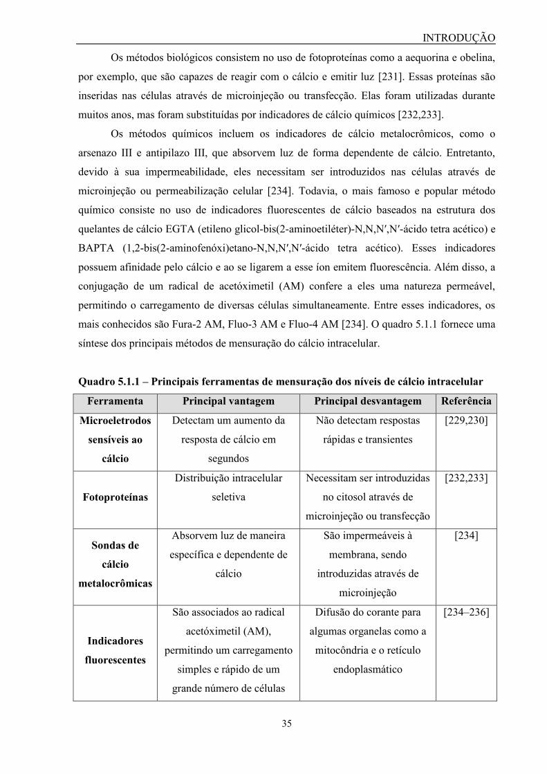
INTRODUÇÃO
35
Os métodos biológicos consistem no uso de fotoproteínas como a aequorina e obelina,
por exemplo, que são capazes de reagir com o cálcio e emitir luz [231]. Essas proteínas são
inseridas nas células através de microinjeção ou transfecção. Elas foram utilizadas durante
muitos anos, mas foram substituídas por indicadores de cálcio químicos [232,233].
Os métodos químicos incluem os indicadores de cálcio metalocrômicos, como o
arsenazo III e antipilazo III, que absorvem luz de forma dependente de cálcio. Entretanto,
devido à sua impermeabilidade, eles necessitam ser introduzidos nas células através de
microinjeção ou permeabilização celular [234]. Todavia, o mais famoso e popular método
químico consiste no uso de indicadores fluorescentes de cálcio baseados na estrutura dos
quelantes de cálcio EGTA (etileno glicol-bis(2-aminoetiléter)-N,N,N′,N′-ácido tetra acético) e
BAPTA (1,2-bis(2-aminofenóxi)etano-N,N,N′,N′-ácido tetra acético). Esses indicadores
possuem afinidade pelo cálcio e ao se ligarem a esse íon emitem fluorescência. Além disso, a
conjugação de um radical de acetóximetil (AM) confere a eles uma natureza permeável,
permitindo o carregamento de diversas células simultaneamente. Entre esses indicadores, os
mais conhecidos são Fura-2 AM, Fluo-3 AM e Fluo-4 AM [234]. O quadro 5.1.1 fornece uma
síntese dos principais métodos de mensuração do cálcio intracelular.
Quadro 5.1.1 – Principais ferramentas de mensuração dos níveis de cálcio intracelular
Ferramenta Principal vantagem Principal desvantagem Referência
Microeletrodos
sensíveis ao
cálcio
Detectam um aumento da
resposta de cálcio em
segundos
Não detectam respostas
rápidas e transientes
[229,230]
Fotoproteínas
Distribuição intracelular
seletiva
Necessitam ser introduzidas
no citosol através de
microinjeção ou transfecção
[232,233]
Sondas de
cálcio
metalocrômicas
Absorvem luz de maneira
específica e dependente de
cálcio
São impermeáveis à
membrana, sendo
introduzidas através de
microinjeção
[234]
Indicadores
fluorescentes
São associados ao radical
acetóximetil (AM),
permitindo um carregamento
simples e rápido de um
grande número de células
Difusão do corante para
algumas organelas como a
mitocôndria e o retículo
endoplasmático
[234–236]
INTRODUÇÃO
36
Os principais equipamentos utilizados no monitoramento do sinal de cálcio incluem
microscópios confocal e de fluorescência, bem como fluorímetro e citômetro de fluxo. Apesar
do uso de microscopia permitir a visualização da fluorescência de um determinado grupo de
células, o que é conhecido como imageamento de cálcio, essa técnica só permite a análise de
uma amostra por vez, tornando-a inviável para a execução de screening em larga escala.
Contudo, foram desenvolvidos microscópios automatizados que são capazes de realizar a
análise de placas de 6 a 384 poços que é conhecido como triagem de alto conteúdo (high
content screening - HCS) [237]. Na citometria de fluxo limitação similar quanto à aquisição
das amostras também existiu, no entanto, com a modernização e automatização dos
citômetros de fluxo, atualmente é possível realizar o procedimento de triagem em alto
desempenho utilizando esses equipamentos.
Em todo caso, os instrumentos denominados fluorímetros são os mais populares na
execução de triagem de alto desempenho. Eles são capazes de analisar um grande número de
amostras simultaneamente preparadas em placas de 96, 384 ou até 1536 poços, de forma
eficiente através ensaios baseados na fisiologia celular. Esses equipamentos, que por vezes
funcionam como leitores multimodais, tornaram possível a implementação de ensaios de
cálcio como protocolo para screening em larga escala [237]. Leitores de microplacas
multimodais como o FlexStation III (Molecular Devices), o FLUOstar Omega (BMG
Labtechnologies) e o Synergy (Biotek), por exemplo, atuam como plataformas que integram
as funções de leitura e adição de reagentes na placa de ensaio. Eles possuem um sistema de
pipetagem automatizado que permite a adição dos reagentes e a leitura dos sinais em tempo
real [238]. Essa habilidade é importante em ensaios de cálcio, por exemplo, uma vez que a
resposta referente à mobilização desse íon é dependente do tempo, podendo ser observada
entre 10 e 30 segundos após a ativação dos receptores metabotrópicos [239]. Além dessas
vantagens, o usuário pode ajustar vários parâmetros para otimizar o teste, tais como: uso de
placas de 96, 384 ou 1536 poços, tipo de leitura (fluorescência, absorbância ou
luminescência), tempo de leitura, tempo e velocidade em que os reagentes serão adicionados,
número de reagentes a serem adicionados, seleção de ponteiras e placas para a leitura, dentre
outras. Além disso, esses leitores multimodais podem ser integrados a uma variedade de
sistemas robóticos para realizar novas funções [238].
5.2 – Ensaios de patch-clamp
Uma segunda técnica que é utilizada para investigar a ativação de receptores P2,
especificamente os ionotrópicos (P2X), é o patch-clamp. O patch-clamp é a técnica
INTRODUÇÃO
37
considerada padrão-ouro na investigação de canais iônicos. Ela foi desenvolvida na década de
1950 e permite o estudo de um grupo ou mesmo de um único canal iônico individualmente.
Através do patch-clamp (“patch”= fragmento; “(to) clamp”= prender, fixar) é possível fixar o
potencial da membrana em uma faixa predeterminada e assim, mensurar a corrente iônica
promovida pelo fluxo de íons através dos canais [240,241].
Apesar de um único canal iônico permitir a passagem de até 10 milhões de íons por
segundo, a corrente iônica gerada encontra-se na faixa de picoampères. Para realizar a
mensuração dos canais iônicos de um fragmento de membrana, são utilizadas pipetas de vidro
ou de quartzo contendo um microeletrodo, cuja ponta mede em torno de 1 μm. Elas também
são utilizadas para “fazer contato” (selagem) com a membrana plasmática [265].
Primeiramente, uma leve sucção é aplicada para a obtenção de um selamento firme entre a
pipeta e a membrana. Essa configuração, conhecida como cell-attached, permite que a
membrana plasmática permaneça intacta, sendo muito utilizada para o registro da atividade de
um único ou de um pequeno grupo de canais iônicos (Figura 5.2.1.A). Se a pipeta for retraída,
um fragmento da membrana poderá ser separado da célula, permitindo que a face
citoplasmática da membrana fique exposta. Essa configuração, conhecida como inside-out, é
utilizada para estudar a atividade de um único canal (Figura 5.2.1B). Caso seja aplicada uma
segunda e forte sucção, a membrana plasmática poderá ser rompida e com isso, a pipeta ganha
acesso ao citoplasma. Essa configuração é conhecida como whole-cell e é a mais utilizada,
sendo empregada no estudo do potencial elétrico ou de correntes iônicas da célula por inteiro.
Dentro dessa modalidade, ainda é possível optar por um dos dois modos de registro o voltage-
clamp ou o current-clamp, sendo a voltagem mantida constante para a mensuração da
corrente, ou vice-versa (Figura 5.2.1C). Dentro da configuração de whole-cell, ainda existe a
variação perforated-cell, na qual a segunda sucção necessária para romper as células é
substituída pelo uso de antibióticos ou antifúngicos (ex: anfotericina B, nistatina e
gramicidina), os quais produzem poros na membrana celular. Essa configuração vem sendo
empregada na mensuração de potenciais de ação. Por fim, se a pipeta for retraída, dois
fragmentos de membrana que foram deslocados podem se reconectar e formar uma espécie de
vesícula, deixando a superfície extracelular exposta. Essa configuração, conhecida como
outside-out, é empregada na análise de estímulos extracelulares sobre o canal iônico (Figura
5.2.1D) [240–242]. A Figura 5.2.1 ilustra as quatro configurações da técnica de patch-clamp.

INTRODUÇÃO
38
Figura 5.2.1 – Configurações da técnica de patch-clamp. Nesta técnica, uma pipeta de
vidro contendo um microeletrodo é aproximada da célula e uma sucção leve, mas suficiente
para “selar” a célula, é promovida definindo a configuração cell-attached (A). A retirada da
pipeta pode promover o deslocamento de um pequeno fragmento de membrana, definindo a
configuração inside-out (B). Caso uma segunda sucção mais forte seja promovida, a pipeta
poderá ser contínua à célula, configurando a modalidade whole-cell (C). Após o
estabelecimento desta configuração, a pipeta também poderá ser retirada, carregando consigo
dois fragmentos de membrana que podem se unir e formar uma vesícula, modalidade
conhecida como outside-out (D). Imagem adaptada de [243].
No que tange aos ensaios de patch-clamp relacionados à atividade do receptor P2X7,
duas configurações já foram utilizadas. Na configuração cell-attached, já foi reportado uma
corrente de aproximadamente 10 pS, entretanto, a maioria dos estudos utilizam a configuração
whole-cell. Nessa configuração, as correntes macroscópicas são dividas em dois componentes.
O primeiro componente está relacionado com a abertura do canal iônico, enquanto que o
segundo está relacionado a um estado de alta condutância, o qual pode caracterizar a
formação do poro membranar [69].
A técnica de patch-clamp pode ser considerada a melhor técnica para avaliação dos
receptores P2X, uma vez que apresenta sensibilidade e acuracidade. Entretanto, ela é
laboriosa, uma vez que só permite a análise de uma célula por vez, além de não poder ser

INTRODUÇÃO
39
aplicada a todos os tipos celulares e exigir habilidade técnica e treinamento do operador [244].
Esses desafios quanto à aplicação da técnica de patch-clamp como metodologia de screening
tem sido superados com o desenvolvimento de aparato de eletrofisiologia automatizado, como
o equipamento IonFlux da empresa Molecular Devices.
5.3 – Ensaios de permeabilização celular
Os ensaios de permeabilização celular ou de captação de corantes avaliam a função do
poro associado ao P2X7 [169,172]. Esse fenômeno é considerado como o segundo estágio de
permeabilização membranar relacionado à ativação desse receptor. A formação do poro ocorre
após a exposição prolongada a altas concentrações de ATP (< 100 μM) e tende a aumentar ao
longo do tempo. Apesar da origem desse poro ainda ser debatida, já se sabe que ele permite a
passagem de íons (Na+, Ca
2+, K
+), corantes, água e ATP, conforme mostrado na Figura 5.3.1
[168].
Figura 5.3.1 – Canal e poro membranar do P2X7. A ativação do receptor P2X7 promove a
abertura do canal cátion-seletivo, permitindo o fluxo dos íons Na+, Ca
2+ e K
+ de acordo com o
gradiente eletroquímico. Em situações de ativação prolongada ou na presença de altas
concentrações do ATP (faixa de milimolar), esse receptor induz a formação de um poro
membranar que permite a passagem de moléculas de até 900 Da, incluindo os íons Na+, Ca
2+ e
K+, ATP (simbolizada pela elipse laranja) e corantes (simbolizados pela estrela amarela) como o
YO-PRO-1, iodeto de propídeo, brometo de etídeo, amarelo de lúcifer, entre outros que
apresentam peso molecular inferior ao cut-off descrito. (Ext.) significa meio extracelular e (Int.)
significa meio intracelular. Imagem adaptada de [192].
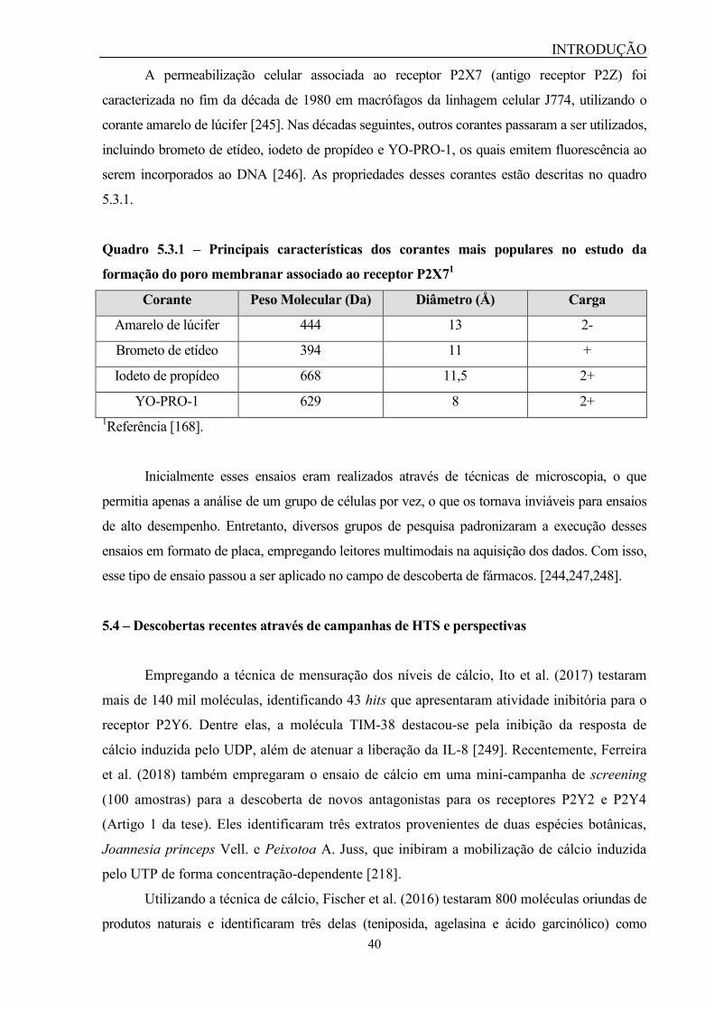
INTRODUÇÃO
40
A permeabilização celular associada ao receptor P2X7 (antigo receptor P2Z) foi
caracterizada no fim da década de 1980 em macrófagos da linhagem celular J774, utilizando o
corante amarelo de lúcifer [245]. Nas décadas seguintes, outros corantes passaram a ser utilizados,
incluindo brometo de etídeo, iodeto de propídeo e YO-PRO-1, os quais emitem fluorescência ao
serem incorporados ao DNA [246]. As propriedades desses corantes estão descritas no quadro
5.3.1.
Quadro 5.3.1 – Principais características dos corantes mais populares no estudo da
formação do poro membranar associado ao receptor P2X71
Corante Peso Molecular (Da) Diâmetro (Å) Carga
Amarelo de lúcifer 444 13 2-
Brometo de etídeo 394 11 +
Iodeto de propídeo 668 11,5 2+
YO-PRO-1 629 8 2+
1Referência [168].
Inicialmente esses ensaios eram realizados através de técnicas de microscopia, o que
permitia apenas a análise de um grupo de células por vez, o que os tornava inviáveis para ensaios
de alto desempenho. Entretanto, diversos grupos de pesquisa padronizaram a execução desses
ensaios em formato de placa, empregando leitores multimodais na aquisição dos dados. Com isso,
esse tipo de ensaio passou a ser aplicado no campo de descoberta de fármacos. [244,247,248].
5.4 – Descobertas recentes através de campanhas de HTS e perspectivas
Empregando a técnica de mensuração dos níveis de cálcio, Ito et al. (2017) testaram
mais de 140 mil moléculas, identificando 43 hits que apresentaram atividade inibitória para o
receptor P2Y6. Dentre elas, a molécula TIM-38 destacou-se pela inibição da resposta de
cálcio induzida pelo UDP, além de atenuar a liberação da IL-8 [249]. Recentemente, Ferreira
et al. (2018) também empregaram o ensaio de cálcio em uma mini-campanha de screening
(100 amostras) para a descoberta de novos antagonistas para os receptores P2Y2 e P2Y4
(Artigo 1 da tese). Eles identificaram três extratos provenientes de duas espécies botânicas,
Joannesia princeps Vell. e Peixotoa A. Juss, que inibiram a mobilização de cálcio induzida
pelo UTP de forma concentração-dependente [218].
Utilizando a técnica de cálcio, Fischer et al. (2016) testaram 800 moléculas oriundas de
produtos naturais e identificaram três delas (teniposida, agelasina e ácido garcinólico) como

INTRODUÇÃO
41
moduladores da atividade do P2X7 [250]. Enquanto isso, Soares-Bezerra et al. (in press)
empregaram a técnica de permeabilização celular para testar cerca de 1800 amostras oriundas de
produtos naturais. Como resultado, eles identificaram a atividade antagonista de três extratos
fúngicos sobre o receptor P2X7 [227].
Outra metodologia que vem ganhando espaço no cenário de screening de novos
antagonistas para os receptores P2 é a análise in silico de moléculas adquiridas de bancos de
dados virtuais através da técnica de docking molecular. Entre as vantagens dessa última
metodologia podem ser listadas a economia de tempo, uma vez que é possível realizar o teste
de um grande número de amostras em um curto espaço temporal, a redução de gastos com o
uso de reagentes e materiais consumíveis de laboratório, além de permitir uma análise seletiva
quanto à interação da molécula-teste e o alvo-farmacológico [251]. Além disso, já existem
diversas bibliotecas virtuais em que se encontram cadastradas milhares de moléculas de
origem sintética e natural. Alguns bancos de dados ofertam inclusive, moléculas provenientes
de plantas utilizadas na Medicina Tradicional Chinesa ou Africana, como por exemplo, o The
Traditional Chinese Medicine Integrated Database (TCMID) e African medicinal plants
database (AfroDb) [207].

OBJETIVOS
42
II. OBJETIVOS
Objetivo Geral
Realizar a triagem de alto desempenho de extratos e moléculas isoladas de plantas
pertencentes à flora brasileira a fim de identificar novos candidatos a antagonistas de
receptores P2.
Objetivos específicos
1. Identificar candidatos a antagonistas de receptores P2 através de triagem de alto
desempenho;
2. Avaliar a citotoxicidade desses candidatos;
3. Identificar o IC50 dos extratos e moléculas candidatas e caracterizá-los com parâmetros
farmacológicos;
4. Avaliar a atividade dos extratos e moléculas candidatas na atenuação da inflamação e
da dor em modelos in vivo;
5. Realizar a caracterização química dos extratos e moléculas candidatas.

RESULTADOS
43
III. RESULTADOS
Os resultados da tese estão contidos nos seguintes trabalhos, os quais serão
apresentados a seguir:
1. New insights in purinergic therapy: Novel antagonists for uridine 5’-triphosphate-
activated P2Y receptors from Brazilian flora;
2. The antagonistic effect of Joannesia princeps Vell. stem on UTP-activated
receptors;
3. Discovery of antagonistic activity of CS-15 compound on P2X7 receptor;
4. Potential therapeutic applications of P2 receptor antagonists: from bench to
clinical trials.
ARTIGO 1
44
Título do artigo 1: New insights in purinergic therapy: Novel antagonists for uridine
5’-triphosphate-activated P2Y receptors from Brazilian flora
Referência bibliográfica: Ferreira NCS; Soares-Bezerra RJ; da Silveira RFC; da Silva
CM; de Oliveira CS; Calheiros AS; Alves TM; Zani CL; Alves LA. New insights in
purinergic therapy: Novel antagonists for uridine 5’-triphosphate-activated P2Y receptors
from Brazilian flora. J Med Food. 2019;22(2):211-224.
Situação: Publicado
Descrição: O presente artigo foi desenvolvido parcialmente durante o meu Mestrado,
contudo, a sua conclusão só ocorreu ao longo do Doutorado. Nesse artigo, apresentamos
o passo-a-passo para a otimização do ensaio de cálcio no leitor multimodal FlexStation
III. O objetivo dessa padronização era realizar a triagem de extratos vegetais pertencentes
a diferentes biomas brasileiros como Amazônia, Cerrado, Caatinga, Pantanal, Mata
Atlântica e Pampa, com o intuito de identificar potenciais antagonistas para os receptores
P2Y ativados por UTP, isto é, P2Y2 e P2Y4. Para isso, utilizamos células da linhagem
J774.G8, as quais expressam esses receptores, como um modelo para a execução do
screening. Posteriormente, os extratos considerados promissores “hits” foram testados
novamente em células primárias (macrófagos peritoneais) a fim de validarmos o modelo
previamente estabelecido. A partir do teste com 100 extratos, descobrimos que as
espécies Joannesia princeps Vell. e Peixotoa A. Juss apresentaram um efeito inibitório
concentração-dependente sobre a mobilização de cálcio induzida pelo UTP. Cabe
ressaltar que esse efeito inibitório não foi causado pela citotoxicidade do extrato de
galhos de J. princeps Vell. e dos extratos de folhas e flores de Peixotoa A. Juss. Esse
trabalho atende aos objetivos específicos 1, 2 e 3 da tese.

New Insights in Purinergic Therapy:Novel Antagonists for Uridine 50-Triphosphate-Activated
P2Y Receptors from Brazilian Flora
Natiele Carla da Silva Ferreira,1 Romulo Jose Soares-Bezerra,1 Rebeca Ferreira Couto da Silveira,1
Clayton Menezes da Silva,1 Carla Santos de Oliveira,1 Andrea Surrage Calheiros,2
Tania Maria Alves,3 Carlos Leomar Zani,3 and Luiz Anastacio Alves1
1Laboratory of Cellular Communication, 2Laboratory of Immunopharmacology,Oswaldo Cruz Institute, Oswaldo Cruz Foundation, Rio de Janeiro, Brazil.
3Laboratory of Chemistry of Natural Products, Rene Rachou Research Center,Oswaldo Cruz Foundation, Belo Horizonte, Brazil.
ABSTRACT P2Y2 and P2Y4 receptors are physiologically activated by uridine 50-triphosphate (UTP) and are widely
expressed in many cell types in humans. P2Y2 plays an important role in inflammation and proliferation of tumor cells, which
could be attenuated with the use of antagonists. However, little is known about the physiological functions related to P2Y4,
due to the lack of selective ligands for these receptors. This can be solved through the search for novel compounds with
antagonistic activity. The aim of this study was to discover new potential antagonist candidates for P2Y2 and P2Y4 receptors
from natural products. We applied a calcium measurement methodology to identify new antagonist candidates for these
receptors. First, we established optimal conditions for the calcium assay using J774.G8, a murine macrophage cell line, which
expresses functional P2Y2 and P2Y4 receptors and then, we performed the screening of plant extracts at a cutoff concentration
of 50 lg/mL. ATP and ionomycin, known intracellular calcium inductors, were used to stimulate cells. The calculated EC50
were 11 lM and 103 nM, respectively. These cells also responded to the UTP stimulation with an EC50 of 1.021 lM. Screening
assays were performed and a total of 100 extracts from Brazilian plants were tested. Joannesia princeps Vell. (stem) and
Peixotoa A. Juss (flower and leaf) extracts stood out due to their ability to inhibit UTP-induced responses without causing
cytotoxicity, and presented an IC50 of 32.32, 14.99, and 12.98 lg/mL, respectively. Collectively, our results point to the
discovery of potential antagonist candidates from Brazilian flora for UTP-activated receptors.
KEYWORDS: � calcium � natural products � P2Y2 � P2Y4 � screening
INTRODUCTION
P2YR are G protein-coupled receptors expressed inmany cell types in humans. Currently, eight P2Y sub-
types have been cloned in mammals: P2Y1, P2Y2, P2Y4,P2Y6, P2Y11, P2Y12, P2Y13, and P2Y14.1 These subtypesare divided into two groups according to the signalingpathway promoted by G protein. P2Y1, P2Y2, P2Y4, P2Y6,and P2Y11 subtypes are coupled to Gq protein and activatephospholipase Cb (PLCb) that forms diacylglycerol (DAG)and inositol 1,4,5,triphosphate (IP3). IP3 induces an increaseof intracellular calcium through IP3 receptor activation inthe endoplasmic reticulum. Meanwhile, P2Y12, P2Y13, andP2Y14 subtypes are coupled to Gi protein and inhibit ade-
nylyl cyclase, (AC) causing a decrease in cyclic adenosinemonophosphate (cAMP) formation.1,2
P2Y2 and P2Y4 receptors are physiologically activatedby uridine 50-triphosphate (UTP) and promote an increase inintracellular calcium through PLC/IP3.2 They are expressedin many cell types, such as epithelial cells, monocytes,macrophages, neutrophils, cardiomyocytes, and organs, in-cluding the brain, heart, kidneys, liver, spleen, and muscle.3
They have effects on chloride secretion in epithelial cellsfrom the airway and eyes, and also induce water secretion inbowel epithelial cells. These functions are important topromote surface lubrification and mucus hydration, whichcan improve disease treatment, such as dry eye disease andchronic constipation.2,4
P2Y2R also plays a role in neuroprotection, chemokin-esis, and in the proliferation of hepatocytes and tumorcells.5–8 P2Y2 is also involved in the production of inflam-matory modulators, such as cyclooxygenase-2 (COX-2) andprostaglandin E2 (PGE2), and neutrophil accumulation in
Manuscript received 18 June 2018. Revision accepted 18 October 2018.
Address correspondence to: Luiz Anastacio Alves, MD, PhD, Laboratory of CellularCommunication, Oswaldo Cruz Institute, Oswaldo Cruz Foundation, Rio de Janeiro21040-360, Brazil, E-mail: [email protected]
JOURNAL OF MEDICINAL FOODJ Med Food 00 (0) 2018, 1–14# Mary Ann Liebert, Inc., and Korean Society of Food Science and NutritionDOI: 10.1089/jmf.2018.0087
1
Dow
nloa
ded
by A
ustr
alia
n N
atio
nal U
nive
rsity
fro
m w
ww
.lieb
ertp
ub.c
om a
t 12/
14/1
8. F
or p
erso
nal u
se o
nly.

lungs and liver during inflammation.9–13 Thus, P2Y2 an-tagonists might alleviate inflammation signals.
P2Y4R participates in Na+, K+, and Cl- regulation pro-cesses, but its functions are not completely characterized, asit is expressed in the same tissues and cell types as P2Y2R,and activated by the same physiological ligand, UTP.14–17
Therefore, the lack of selective ligands impairs the dis-covery of new functions associated with these receptors.However, this problem can be solved by the search for novelcompounds with antagonistic activity on P2Y2 and P2Y4receptors.
Natural products are an important source for the discov-ery of new molecules with antagonistic activity. They offerthe advantage of millenary use in the treatment of severaldiseases, such as inflammation, parasitoses, pain, and re-cently, cancer, constituting the basis of traditional orientalmedicines.18–20 Among the 13 drugs approved for globalmarketing between 2005 and 2007, five were classified asnatural products, whereas the others were semisynthetic orderived, which reinforces natural product importance forclinical use.21 In addition, the literature reports that somebiological species express compounds with antagonisticactivity on P2 receptors, such as emodin from Rheum offi-cinale Bail and amentoflavone from Rheedia longifoliaPlanch & Triana on P2X7R.22,23 However, due to thelarge number of samples for testing, implementation ofhigh-throughput screening (HTS) methodologies is nec-essary to meet this demand, since they have the advantageof evaluating many compounds in a short period oftime.24,25
In this scenario, intracellular calcium measurement tech-niques emerge as a tool for the screening of new antagonists,since P2Y2 and P2Y4 receptors are coupled to the calciumsignaling pathway.26 Kaulich et al. tested a series of 40flavonoids using calcium assays to discover novel lead an-tagonists for P2Y2R. They found that certain flavonoidscould inhibit P2Y2R and suggest using their structures todevelop new compounds with antagonistic activity.27 Re-cently, Ito et al. applied an intracellular calcium measure-ment protocol through a HTS campaign to search for novelantagonists for the P2Y6 receptor and found one compound,TIM-38, with potential activity.28
Thus, the aim of this study was to improve a protocolbased on intracellular calcium measurements and apply it inthe discovery of natural product compounds with antagonistactivity on P2Y2 and P2Y4 receptors.
MATERIALS AND METHODS
Reagents
Chemicals were purchased from Sigma Chemical Co.(St. Louis, MO, USA), including ATP, UTP, ADP, UDP,ab-meATP, Suramin, DMEM, and RPMI culture medi-ums, NaCl, KCl, Na2HPO4, KH2PO4, MgCl2, CaCl2,Glucose, DMSO, MTT ((3-(4,5-dimethylthiazol-2-yl)-2,5-diphenyltetrazolium bromide) tetrazolium, Probenecid, andIonomycin. Fetal bovine serum was obtained from Cultilab
(Campinas, SP, Brazil) and Fluo-4 AM was obtained fromLife Technologies (CA, USA).
Extracts
Extracts used in this study were collected from Brazilianbiomes (Amazon, Cerrado, Caatinga, Atlantic Forest, Pan-tanal, and Pampas), obtained through methanol extraction,and deposited at Bioprospecting Platform from the Chem-istry of Natural Products Laboratory.
The stems from Joannesia princeps Vell. (blind code:JA2) were collected in the Atlantic biome, in Linhares, in thestate of Espırito Santo, Brazil. The stems were crushed andmixed with a 1:1 mixture of CH2Cl2-CH3OH to obtain theextract deposited at voucher number EX294. Peixotoa A.Juss were collected in the Cerrado biome, in Tiradentes, lo-cated in the state of Minas Gerais, Brazil. Peixotoa A. Jussflower (blind code: RA3) and leaf (blind code: RB3) extractsare deposited at voucher numbers EX9355 and EX9356,respectively. A botanical specimen was deposited at theFederal University of Sao Joao del Rei Herbarium under codeHUFSJ1103. The flowers and leaves were crushed and ho-mogenized with ethanol to obtain the crude extracts. For theexperiments, they were resuspended in a 1 mL Dulbecco’sPBS solution containing 0.5% DMSO. The extracts weretested using a single-blind method, that is, the professionalresponsible for performing screening experiments did notknow the name of the biological species that originated thesamples.29 Because of this, they were identified by a code.
Cell culture
J774.G8 is a murine macrophage cell line that expressesP2 receptors. The J774.G8 cell line was kindly provided byDr. Vinıcius Cotta from the Laboratory on Thymus Re-search at the Oswaldo Cruz Institute, Rio de Janeiro, Brazil.The cells were routinely maintained in culture with Dul-becco’s modified Eagle’s medium (DMEM) supplementedwith 10% fetal bovine serum, at 37�C in a 5% CO2 atmo-sphere. The medium was changed every 3 days and the cellswere adjusted to a concentration of 2 · 106 cells per 150-cm2
cell culture flask (Corning, NY, USA). Cell viability wasanalyzed by the Trypan Blue assay. For this, 10 lL of cellsample was diluted 1:100 in 0.04% Trypan Blue solution.After dilution, 10 lL of the sample were added into theNeubauer chamber and observed under optical microscopeNikon Eclipse TE 2000-S (Nikon, Tokyo, Japan) to countviable cells. Calcium assays were conducted only when theviability range was above 90%.
Calcium assays
J774.G8 cells were plated at a concentration of 2 · 105
cells/well in a 96-well black-wall, clear-bottom plate(Corning, NY, USA), and maintained in culture withDMEM supplemented with 10% fetal bovine serum, at 37�Cin a 5% CO2 atmosphere for 24 h. Subsequently, the mediumwas replaced by 100 lL of Dulbecco’s PBS (DPBS)(136 mM NaCl, 2.68 mM KCl, 8 mM Na2HPO4, 1.46 mM
2 FERREIRA ET AL.
Dow
nloa
ded
by A
ustr
alia
n N
atio
nal U
nive
rsity
fro
m w
ww
.lieb
ertp
ub.c
om a
t 12/
14/1
8. F
or p
erso
nal u
se o
nly.

KH2PO4, 0.5 mM MgCl2, 0.9 mM CaCl2, 5.55 mM Glucose,and 2.5 mM Probenecid–pH 7.4) containing Fluo-4 AM[2 lM] and the cells were incubated for 60 min at 37�C in a5% CO2 atmosphere. Extracellular Fluo-4AM was then re-moved by three consecutive washes with 200 lL DPBS andthe cells received a final volume of 150 lL DPBS. Subse-quently, cells were treated with extracts [50 lg/mL] andPPADS [300 lM], Suramin [300 lM], and Reactive Blue-2(RB-2) [20 lM] antagonists for 30 min. Finally, the assayplate was placed into the FlexStation III equipment (Mo-lecular Devices, CA, USA), which performed the agonistaddition to each well, diluted in 50 lL DPBS. The mobili-zation of intracellular calcium was measured in real-timeprior, during, and after the addition of the agonists at k(excitation) of 485 nm and k (emission) of 525 nm. Totalmeasurement runtime was 90 sec and agonists were added atthe 20-sec mark. Readings were carried out every 1.52 secand a total of 60 readings were obtained for each well. TheDFU, that is, the difference between the fluorescence peakassociated with the maximum concentration of intracellularcalcium ([Ca2+]i) and basal fluorescence of calcium wasdetermined according to the protocol of Hansen andBrauner-Osborne (2009).26 The wells whose signal did notreturn to approximately 30% of the baseline after stimula-tion were discarded as quality control.
Cell loading observation undera fluorescence microscope
J774.G8 cells were loaded with Fluo-4AM as describedpreviously and observed under a Nikon Eclipse TE 2000-Sfluorescence microscope (Nikon, Tokyo, Japan), using20 · objectives. Fluo-4 was excited using a mercury lampand a set of filters: exciter: 480/30 and barrier: 535/40. Io-nomycin was added after 40 sec of observation and pictureswere captured with an Infinity 3 Microscope Digital Camera(Lumenera, Ontario, Canada).
RNA extraction, Reverse transcriptase polymerasechain reaction, and electrophoresis
mRNA was isolated from the J774.G8 cell line from aconcentration of 5 · 106 cells using TRIzol (Life Technol-ogies, CA, USA), according to the manufacturer’s specifi-cations. Then, 20 lL of mRNA was reverse transcribed at42�C for 1 h and at 70�C for 5 min using 1 lL of reversetranscriptase enzyme. Next, 4 lL of cDNA samples wereused with 4 lL of a primer sequence specific for each P2receptor subtype, as shown in Table 1. These samples wereused for the RT-PCR analysis in a 50 lL reaction volumecontaining 5 lL of Buffer 10 · , 4 lL Mg2+, 0.4 lL dNTP,0.2 lL Taq polymerase, and 4 lL of water. Cycling condi-tions were: 94�C for 45 sec, followed by 35 cycles at 54�C(P2Y11), 55�C (P2Y6), 57�C (P2X1), 58�C (P2X3, P2Y14,and P2Y13), 60�C (P2Y2, P2X2, P2X5, P2X6, P2Y12,P2Y1, P2Y4, and GAPDH) or 65�C (P2X4 and P2X7) for1 min and 72�C for 45 sec. PCR products were submittedto electrophoresis on a 2% agarose gel and visualized byethidium bromide staining under a transilluminator.
Animals
The experiments with animals were approved by OswaldoCruz Institute Ethics Committee on the use of Animals, underidentification code L-037/2017, approved on November 24,2017, and followed the ethical principles in animal experi-mentation from the Brazilian College of Animal Experi-mentation. Healthy male C57BL/6 mice weighing between25 and 30 g were obtained from the Oswaldo Cruz CentralBioterium. The animals were housed under 12-h light/12-hdark cycle conditions with free access to food and water.
Peritoneal macrophage isolation
Animals were euthanized by CO2 asphyxiation. Sub-sequently, 5 mL of RPMI medium was injected into the peri-toneal cavity. Peritoneal cells were collected and macrophageswere isolated by centrifugation at 378 g for 10 min. Cell via-bility was analyzed by the Trypan Blue assay. Peritonealmacrophages were plated at a concentration of 4 · 105 cells/well in a 96-well black-wall, clear-bottom plate and maintainedin RPMI culture supplemented with 10% fetal bovine serum, at37�C in a 5% CO2 atmosphere for 24 h. Calcium assays werecarried out using the same conditions described previously.
Table 1. Primer Sequence of P2 Receptors Used in RNA
Extraction, Reverse Transcriptase Polymerase Chain
Reaction, and Electrophoresis Experiments
P2 receptorsubtype Primer sequence
P2X1 Forward: ATGTCCTCGGCATATTTGAAReverse: ATTGTGCAGAGAACCCAGAA
P2X2 Forward: TTCATCGTGCAGAAAAGCTACCReverse: TGGATGCTGTTCTTGATGAGGA
P2X3 Forward: CTTCACCTATGAGACCACCAAGReverse: CGGTATTTCTCCTCACTCTCTG
P2X4 Forward: TGCTCATCCGCAGCCGCAAAGTReverse: AGTGGTCGCATCTGGAATCTCGG
P2X5 Forward: ACTTCCCTGCAGAGTGCTGTReverse: GGCAGCTTTATCAAGGTCACA
P2X6 Forward: TATGTGATGACCAGGAACTGReverse: CTCCAGATCTCACAGGTCCT
P2X7 Forward: GGCAGTTCAGGGAGGAATCATGGReverse: AAGCGCCAGGTGGCATAGCTC
P2Y1 Forward: CTGTTGAGACTTGCTAGACCTCReverse: ATGTTCAATTTGGCTCTGGC
P2Y2 Forward: GTCGCTTCAACGAGGACTTCReverse: GCCAGGAAGTAGAGCACAGG
P2Y4 Forward: TGGCATTGTCAGACACCTTGReverse: AAGCAGACAGCAAAGACAGTC
P2Y6 Forward: CGCTTCCTCTTCTATGCCAAReverse: GTAGGCTGTCTTGGTGATGTG
P2Y11 Forward: CAGCGTCATCTTCATCACCReverse: GCTATACGCTCTGTAGGC
P2Y12 Forward: CTGCCAAGTCATTTTCTGGAReverse: AGCATGCTCATCAAGGAATTT
P2Y13 Forward: AGGGGTTTTGTGTGCACTTTReverse: CTGACTGCTGTGGTGCTCAT
P2Y14 Forward: GCTGTCCCCAACATCATTCTReverse: GTTTTGGGGTAACTCGCAGA
GAPDH Forward: ATCACCATCTTCCAGGAGCGReverse: CCTGCTTCACCACCTTCTTG
NOVEL POTENTIAL ANTAGONISTS FOR P2Y RECEPTORS 3
Dow
nloa
ded
by A
ustr
alia
n N
atio
nal U
nive
rsity
fro
m w
ww
.lieb
ertp
ub.c
om a
t 12/
14/1
8. F
or p
erso
nal u
se o
nly.
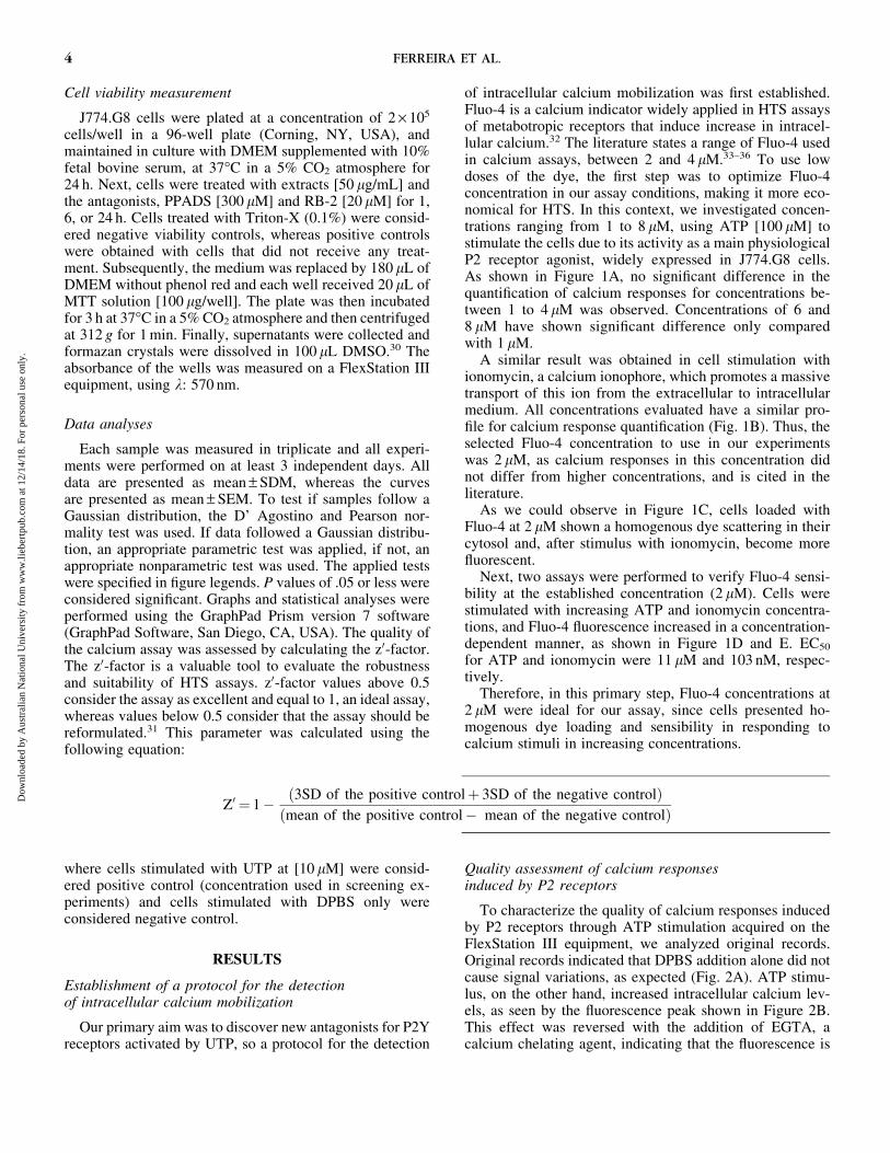
Cell viability measurement
J774.G8 cells were plated at a concentration of 2 · 105
cells/well in a 96-well plate (Corning, NY, USA), andmaintained in culture with DMEM supplemented with 10%fetal bovine serum, at 37�C in a 5% CO2 atmosphere for24 h. Next, cells were treated with extracts [50 lg/mL] andthe antagonists, PPADS [300 lM] and RB-2 [20 lM] for 1,6, or 24 h. Cells treated with Triton-X (0.1%) were consid-ered negative viability controls, whereas positive controlswere obtained with cells that did not receive any treat-ment. Subsequently, the medium was replaced by 180 lL ofDMEM without phenol red and each well received 20 lL ofMTT solution [100 lg/well]. The plate was then incubatedfor 3 h at 37�C in a 5% CO2 atmosphere and then centrifugedat 312 g for 1 min. Finally, supernatants were collected andformazan crystals were dissolved in 100 lL DMSO.30 Theabsorbance of the wells was measured on a FlexStation IIIequipment, using k: 570 nm.
Data analyses
Each sample was measured in triplicate and all experi-ments were performed on at least 3 independent days. Alldata are presented as mean – SDM, whereas the curvesare presented as mean – SEM. To test if samples follow aGaussian distribution, the D’ Agostino and Pearson nor-mality test was used. If data followed a Gaussian distribu-tion, an appropriate parametric test was applied, if not, anappropriate nonparametric test was used. The applied testswere specified in figure legends. P values of .05 or less wereconsidered significant. Graphs and statistical analyses wereperformed using the GraphPad Prism version 7 software(GraphPad Software, San Diego, CA, USA). The quality ofthe calcium assay was assessed by calculating the z0-factor.The z0-factor is a valuable tool to evaluate the robustnessand suitability of HTS assays. z0-factor values above 0.5consider the assay as excellent and equal to 1, an ideal assay,whereas values below 0.5 consider that the assay should bereformulated.31 This parameter was calculated using thefollowing equation:
where cells stimulated with UTP at [10 lM] were consid-ered positive control (concentration used in screening ex-periments) and cells stimulated with DPBS only wereconsidered negative control.
RESULTS
Establishment of a protocol for the detectionof intracellular calcium mobilization
Our primary aim was to discover new antagonists for P2Yreceptors activated by UTP, so a protocol for the detection
of intracellular calcium mobilization was first established.Fluo-4 is a calcium indicator widely applied in HTS assaysof metabotropic receptors that induce increase in intracel-lular calcium.32 The literature states a range of Fluo-4 usedin calcium assays, between 2 and 4 lM.33–36 To use lowdoses of the dye, the first step was to optimize Fluo-4concentration in our assay conditions, making it more eco-nomical for HTS. In this context, we investigated concen-trations ranging from 1 to 8 lM, using ATP [100 lM] tostimulate the cells due to its activity as a main physiologicalP2 receptor agonist, widely expressed in J774.G8 cells.As shown in Figure 1A, no significant difference in thequantification of calcium responses for concentrations be-tween 1 to 4 lM was observed. Concentrations of 6 and8 lM have shown significant difference only comparedwith 1 lM.
A similar result was obtained in cell stimulation withionomycin, a calcium ionophore, which promotes a massivetransport of this ion from the extracellular to intracellularmedium. All concentrations evaluated have a similar pro-file for calcium response quantification (Fig. 1B). Thus, theselected Fluo-4 concentration to use in our experimentswas 2 lM, as calcium responses in this concentration didnot differ from higher concentrations, and is cited in theliterature.
As we could observe in Figure 1C, cells loaded withFluo-4 at 2 lM shown a homogenous dye scattering in theircytosol and, after stimulus with ionomycin, become morefluorescent.
Next, two assays were performed to verify Fluo-4 sensi-bility at the established concentration (2 lM). Cells werestimulated with increasing ATP and ionomycin concentra-tions, and Fluo-4 fluorescence increased in a concentration-dependent manner, as shown in Figure 1D and E. EC50
for ATP and ionomycin were 11 lM and 103 nM, respec-tively.
Therefore, in this primary step, Fluo-4 concentrations at2 lM were ideal for our assay, since cells presented ho-mogenous dye loading and sensibility in responding tocalcium stimuli in increasing concentrations.
Quality assessment of calcium responsesinduced by P2 receptors
To characterize the quality of calcium responses inducedby P2 receptors through ATP stimulation acquired on theFlexStation III equipment, we analyzed original records.Original records indicated that DPBS addition alone did notcause signal variations, as expected (Fig. 2A). ATP stimu-lus, on the other hand, increased intracellular calcium lev-els, as seen by the fluorescence peak shown in Figure 2B.This effect was reversed with the addition of EGTA, acalcium chelating agent, indicating that the fluorescence is
Z0 ¼ 1� 3SD of the positive controlþ 3SD of the negative controlð Þmean of the positive control� mean of the negative controlð Þ
4 FERREIRA ET AL.
Dow
nloa
ded
by A
ustr
alia
n N
atio
nal U
nive
rsity
fro
m w
ww
.lieb
ertp
ub.c
om a
t 12/
14/1
8. F
or p
erso
nal u
se o
nly.
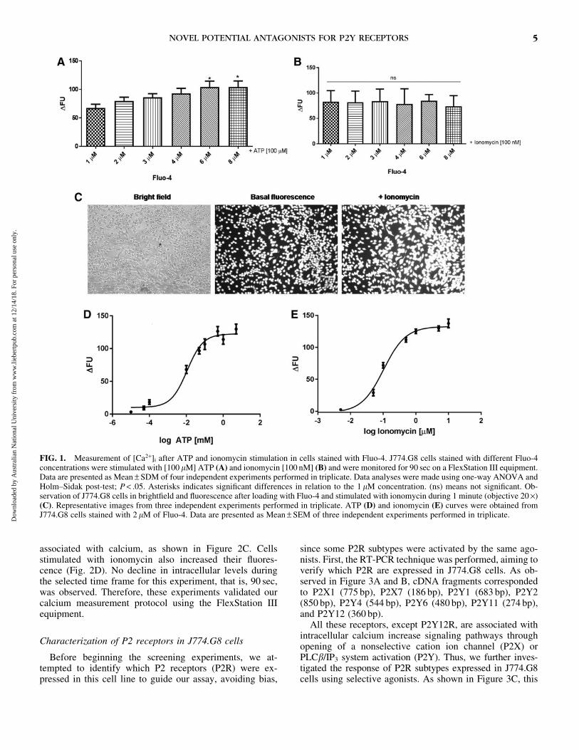
associated with calcium, as shown in Figure 2C. Cellsstimulated with ionomycin also increased their fluores-cence (Fig. 2D). No decline in intracellular levels duringthe selected time frame for this experiment, that is, 90 sec,was observed. Therefore, these experiments validated ourcalcium measurement protocol using the FlexStation IIIequipment.
Characterization of P2 receptors in J774.G8 cells
Before beginning the screening experiments, we at-tempted to identify which P2 receptors (P2R) were ex-pressed in this cell line to guide our assay, avoiding bias,
since some P2R subtypes were activated by the same ago-nists. First, the RT-PCR technique was performed, aiming toverify which P2R are expressed in J774.G8 cells. As ob-served in Figure 3A and B, cDNA fragments correspondedto P2X1 (775 bp), P2X7 (186 bp), P2Y1 (683 bp), P2Y2(850 bp), P2Y4 (544 bp), P2Y6 (480 bp), P2Y11 (274 bp),and P2Y12 (360 bp).
All these receptors, except P2Y12R, are associated withintracellular calcium increase signaling pathways throughopening of a nonselective cation ion channel (P2X) orPLCb/IP3 system activation (P2Y). Thus, we further inves-tigated the response of P2R subtypes expressed in J774.G8cells using selective agonists. As shown in Figure 3C, this
FIG. 1. Measurement of [Ca2+]i after ATP and ionomycin stimulation in cells stained with Fluo-4. J774.G8 cells stained with different Fluo-4concentrations were stimulated with [100 lM] ATP (A) and ionomycin [100 nM] (B) and were monitored for 90 sec on a FlexStation III equipment.Data are presented as Mean – SDM of four independent experiments performed in triplicate. Data analyses were made using one-way ANOVA andHolm–Sidak post-test; P < .05. Asterisks indicates significant differences in relation to the 1 lM concentration. (ns) means not significant. Ob-servation of J774.G8 cells in brightfield and fluorescence after loading with Fluo-4 and stimulated with ionomycin during 1 minute (objective 20 ·)(C). Representative images from three independent experiments performed in triplicate. ATP (D) and ionomycin (E) curves were obtained fromJ774.G8 cells stained with 2 lM of Fluo-4. Data are presented as Mean – SEM of three independent experiments performed in triplicate.
NOVEL POTENTIAL ANTAGONISTS FOR P2Y RECEPTORS 5
Dow
nloa
ded
by A
ustr
alia
n N
atio
nal U
nive
rsity
fro
m w
ww
.lieb
ertp
ub.c
om a
t 12/
14/1
8. F
or p
erso
nal u
se o
nly.

FIG. 2. Original records of calcium signals obtained on a FlexStation III. Original records of calcium signals obtained on a FlexStationIII equipment after addition of 50 lL of DPBS (A), ATP [100 lM] (B, C) at 25 sec, or Ionomycin [100 lM] (D) at 20 sec and EGTA [20 mM](54 sec) (C) (indicated by the arrows) in J774.G8 cells stained with 2 lM of Fluo-4. Representative images from three independent experimentsperformed in triplicate. Color images available online at www.liebertpub.com/jmf
6
Dow
nloa
ded
by A
ustr
alia
n N
atio
nal U
nive
rsity
fro
m w
ww
.lieb
ertp
ub.c
om a
t 12/
14/1
8. F
or p
erso
nal u
se o
nly.
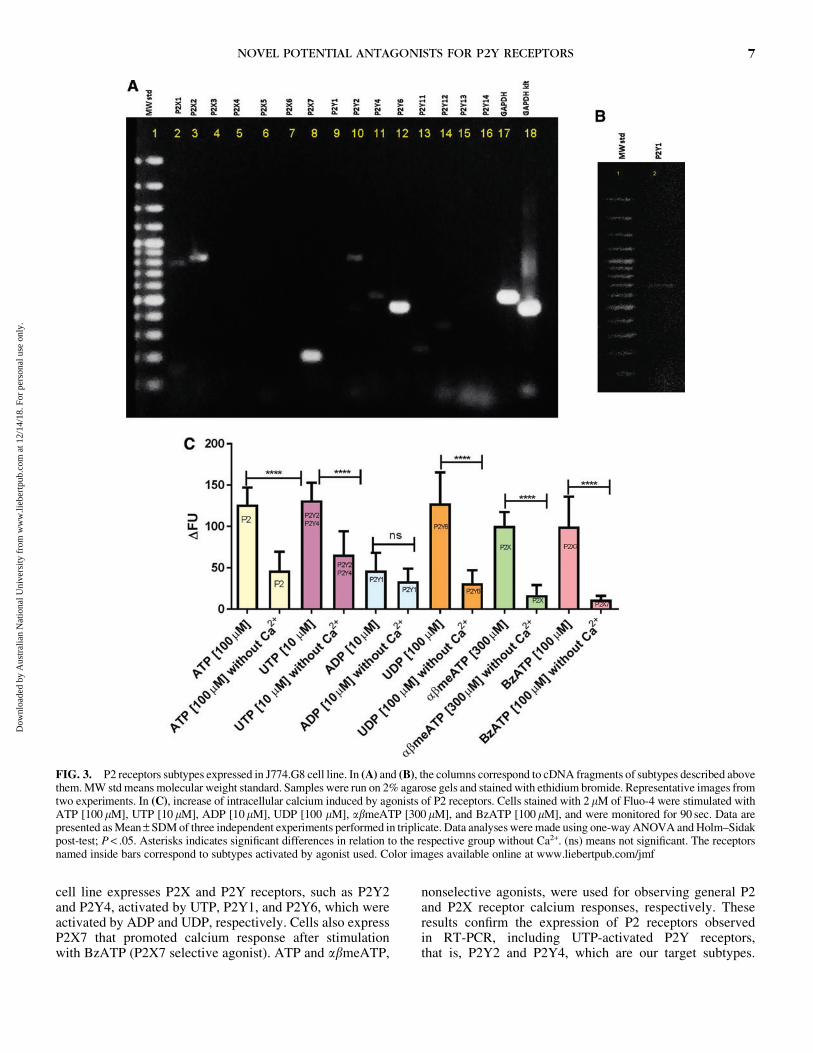
cell line expresses P2X and P2Y receptors, such as P2Y2and P2Y4, activated by UTP, P2Y1, and P2Y6, which wereactivated by ADP and UDP, respectively. Cells also expressP2X7 that promoted calcium response after stimulationwith BzATP (P2X7 selective agonist). ATP and abmeATP,
nonselective agonists, were used for observing general P2and P2X receptor calcium responses, respectively. Theseresults confirm the expression of P2 receptors observedin RT-PCR, including UTP-activated P2Y receptors,that is, P2Y2 and P2Y4, which are our target subtypes.
FIG. 3. P2 receptors subtypes expressed in J774.G8 cell line. In (A) and (B), the columns correspond to cDNA fragments of subtypes described abovethem. MW std means molecular weight standard. Samples were run on 2% agarose gels and stained with ethidium bromide. Representative images fromtwo experiments. In (C), increase of intracellular calcium induced by agonists of P2 receptors. Cells stained with 2 lM of Fluo-4 were stimulated withATP [100 lM], UTP [10 lM], ADP [10lM], UDP [100 lM], abmeATP [300 lM], and BzATP [100 lM], and were monitored for 90 sec. Data arepresented as Mean – SDM of three independent experiments performed in triplicate. Data analyses were made using one-way ANOVA and Holm–Sidakpost-test; P < .05. Asterisks indicates significant differences in relation to the respective group without Ca2+. (ns) means not significant. The receptorsnamed inside bars correspond to subtypes activated by agonist used. Color images available online at www.liebertpub.com/jmf
NOVEL POTENTIAL ANTAGONISTS FOR P2Y RECEPTORS 7
Dow
nloa
ded
by A
ustr
alia
n N
atio
nal U
nive
rsity
fro
m w
ww
.lieb
ertp
ub.c
om a
t 12/
14/1
8. F
or p
erso
nal u
se o
nly.

Interestingly, we observed that all agonists, with the exceptionof ADP, presented lower responses without extracellular cal-cium, possibly indicating the participation of store-operatedcalcium channels. In the case of ATP and abmeATP, thisresponse could be regarding ionotropic channels.
Characterization of UTP-induced calcium responses
Since our cells express UTP-activated receptors, thatis, P2Y2 and P2Y4, they were stimulated with increasingconcentrations of this agonist, to observe UTP responseprofile (Fig. 4A). The determined EC50 was of 1.021 lM.Next, we obtained the P2Y2 and P2Y4 antagonist profiles byperforming an antagonism assay using Reactive-Blue 2(RB2), Suramin, and PPADS.2 As shown in Figure 4B, RB2,Suramin, and PPADS partly inhibited UTP-induced intra-cellular calcium mobilization. To observe the antagonisticprofile of these inhibitors of P2Y2 and P2Y4 receptors, we
pretreated cells with different Suramin, RB2, and PPADSconcentrations and obtained IC50 of 173.3, 22.98, and74.24 lM, respectively (Fig. 4C–E). We also observed thatSuramin and RB2 partially block intracellular UTP-inducedcalcium mobilization.
After characterization of UTP calcium responses and P2Y2and P2Y4 antagonist inhibition profile, we calculated the z0-factor of our assay. The z0-factor is a statistical parameter usedto measure assay robustness. After applying the mathematicalz0-factor equation, we obtained a value of 0.645. Therefore, thez0-factor of our assay, which is above 0.5, is within the optimalrange (z0 ‡ 0.5). Taken together, these evaluated charac-teristics suggest that our protocol was appropriate for use.
Mini HTS application
Finally, we performed the screening of 100 naturalproduct extracts, summarized in Table 2. Nine extracts
FIG. 4. Calcium mobilization inducedby UTP can be partially inhibited byP2Y2 and P2Y4 nonselective an-tagonists. UTP concentration–responsecurve was obtained from J774.G8 cellsstained with 2 lM of Fluo-4. Data arepresented as Mean – SEM of three in-dependent experiments performed intriplicate (A). J774.G8 cells stainedwith 2 lM of Fluo-4 were pretreatedwith Suramin [300 lM], RB2 [20 lM],and PPADS [300 lM] for 30 min.Cells were then stimulated with UTP[10 lM] and monitored for 90 sec.Data are presented as Mean – SDM ofthree independent experiments performedin triplicate. Data analyses were madeusing one-way ANOVA and Holm–Sidak post-test; P < .05. Asterisks in-dicates significant differences in rela-tion to UTP (B). J774.G8 cells werepretreated with increasing concentra-tions of antagonists Suramin (C), RB2(D), and PPADS (E) during 30 min,then were stimulated with UTP[10 lM]. Data are presented asMean – SEM of four independent ex-periments performed in triplicate.
8 FERREIRA ET AL.
Dow
nloa
ded
by A
ustr
alia
n N
atio
nal U
nive
rsity
fro
m w
ww
.lieb
ertp
ub.c
om a
t 12/
14/1
8. F
or p
erso
nal u
se o
nly.

Table 2. Mini High-Throughput Screening Application with Extracts from Brazilian Plants: Inhibition
Effect on Calcium Mobilization Induced by UTP through P2Y2 and P2Y4 Receptors
Sample DFU mean – SDMDFU of control(UTP) – SDM Sample DFU mean – SDM
DFU of control(UTP) – SDM
A2 84.00 – 39.67 136.3 – 22.41 F6 140.1 – 15.32 131.1 – 17.41A3 96.11 – 34.83 136.3 – 22.41 F9 131.4 – 19.00 131.1 – 17.41A4 93.64 – 23.63 136.3 – 22.41 G4 125.9 – 22.93 131.1 – 17.41A5 121.50 – 26.98 136.3 – 22.41 G6 116.6 – 34.57 131.1 – 17.41A6 114.70 – 25.68 136.3 – 22.41 H2 99.88 – 26.96 131.1 – 17.41A7 118.20 – 25.61 136.3 – 22.41 H7 120.60 – 26.54 131.1 – 17.41A8 114.60 – 28.77 136.3 – 22.41 H8 128.60 – 30.45 131.1 – 17.41A9 103.80 – 19.04 136.3 – 22.41 H9 122.50 – 24.30 131.1 – 17.41A10 102.00 – 37.68 136.3 – 22.41 H10 121.00 – 22.07 131.1 – 17.41A11 90.91 – 23.84 136.3 – 22.41 H11 118.80 – 25.45 131.1 – 17.41B2 103.20 – 31.93 136.3 – 22.41 JA2a 52.97 – 29.48*** 117.2 – 21.81B3 96.54 – 30.47 136.3 – 22.41 JA4 87.34 – 17.22 117.2 – 21.81B4 98.59 – 32.42 136.3 – 22.41 JA5 69.73 – 12.95** 117.2 – 21.81B5 104.40 – 31.61 136.3 – 22.41 JA8 88.61 – 17.55 117.2 – 21.81B6 112.00 – 32.86 136.3 – 22.41 JA9 101.4 – 16.28 117.2 – 21.81B7 101.30 – 42.64 136.3 – 22.41 JA10 106.8 – 20.23 117.2 – 21.81B8 101.10 – 45.63 136.3 – 22.41 JA11 113.6 – 21.24 117.2 – 21.81B9 79.80 – 32.15 136.3 – 22.41 JB2 68.70 – 25.67** 117.2 – 21.81B10 83.52 – 38.49 136.3 – 22.41 JB3 103.3 – 22.15 117.2 – 21.81B11 81.18 – 39.85 136.3 – 22.41 JB4 83.31 – 15.39 117.2 – 21.81C2 97.98 – 28.62 136.3 – 22.41 JB5 95.08 – 23.12 117.2 – 21.81C3 86.42 – 33.81 136.3 – 22.41 JB7 72.98 – 27.89* 117.2 – 21.81C4 84.02 – 32.69 136.3 – 22.41 JB9 94.51 – 15.21 117.2 – 21.81C5 89.70 – 32.30 136.3 – 22.41 JC4 80.44 – 21.36 117.2 – 21.81C7 128.40 – 35.47 136.3 – 22.41 JC7 104.4 – 23.61 117.2 – 21.81C8 114.90 – 34.39 136.3 – 22.41 P2B6 88.47 – 7.95** 133.0 – 24.81C9 123.10 – 23.62 136.3 – 22.41 P5C1 90.14 – 19.28* 133.0 – 24.81C10 126.30 – 16.57 136.3 – 22.41 P5F3 94.78 – 26.75* 133.0 – 24.81C11 129.20 – 16.79 136.3 – 22.41 RA2 47.18 – 72.71 99.71 – 62.55D2 130.90 – 16.78 136.3 – 22.41 RA3b 11.15 – 10.11** 99.71 – 62.55D3 121.60 – 21.87 136.3 – 22.41 RA4 126.00 – 41.12 99.71 – 62.55D4 128.00 – 20.63 136.3 – 22.41 RA5 121.10 – 47.06 99.71 – 62.55D5 119.90 – 26.90 136.3 – 22.41 RA6 92.82 – 38.43 99.71 – 62.55D6 101.00 – 36.11 136.3 – 22.41 RA7 64.31 – 19.61 99.71 – 62.55D7 98.50 – 21.90 136.3 – 22.41 RA8 101.40 – 34.38 99.71 – 62.55D8 111.70 – 28.66 136.3 – 22.41 RA9 105.90 – 26.27 99.71 – 62.55D9 123.80 – 11.76 136.3 – 22.41 RA10 100.80 – 39.60 99.71 – 62.55D10 109.60 – 21.55 136.3 – 22.41 RA11 117.30 – 30.47 99.71 – 62.55D11 127.00 – 36.18 136.3 – 22.41 RB2 51.19 – 27.55 99.71 – 62.55E2 117.40 – 20.61 142.0 – 18.04 RB3c 19.96 – 31.03** 99.71 – 62.55E3 103.20 – 18.34 142.0 – 18.04 RB4 106.90 – 32.81 99.71 – 62.55E4 102.30 – 47.03 142.0 – 18.04 RB5 85.30 – 15.44 99.71 – 62.55E5 100.10 – 36.62 142.0 – 18.04 RB6 110.50 – 37.21 99.71 – 62.55E6 95.00 – 36.32 142.0 – 18.04 RB7 110.80 – 36.12 99.71 – 62.55E7 91.00 – 41.39 142.0 – 18.04 RB8 97.02 – 29.97 99.71 – 62.55E8 115.30 – 41.97 142.0 – 18.04 RB9 114.70 – 37.33 99.71 – 62.55E9 108.00 – 45.52 142.0 – 18.04 RB10 91.47 – 35.80 99.71 – 62.55E10 129.80 – 45.93 142.0 – 18.04 RB11 99.84 – 51.68 99.71 – 62.55E11 91.27 – 30.64 142.0 – 18.04 RC2 60.97 – 23.75 99.71 – 62.55F2 132.1 – 23.55 131.1 – 17.41 RC3 59.46 – 21.70 99.71 – 62.55
J774.G8 cells labeled with 2 lM Fluo-4 were pretreated with extracts [50 lg/mL] for 30 min. Then, cells were stimulated with UTP [10 lM] and were monitored
for 90 sec. Mean – SDM of three independent experiments. Data analyses were made using one-way ANOVA and Tukey post-test; P < .05.
Asterisks Indicates significant differences in relation to UTP.aJoannesia princeps Vell. stem extract.bPeixotoa A. Juss. flower extract.cPeixotoa A. Juss. leaf extract.
9
Dow
nloa
ded
by A
ustr
alia
n N
atio
nal U
nive
rsity
fro
m w
ww
.lieb
ertp
ub.c
om a
t 12/
14/1
8. F
or p
erso
nal u
se o
nly.
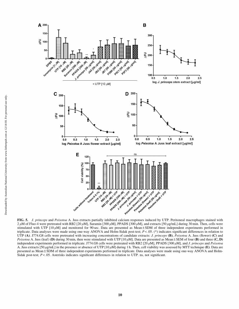
FIG. 5. J. princeps and Peixotoa A. Juss extracts partially inhibited calcium responses induced by UTP. Peritoneal macrophages stained with2 lM of Fluo-4 were pretreated with RB2 [20 lM], Suramin [300 lM], PPADS [300 lM], and extracts [50 lg/mL] during 30 min. Then, cells werestimulated with UTP [10 lM] and monitored for 90 sec. Data are presented as Mean – SDM of three independent experiments performed intriplicate. Data analyses were made using one-way ANOVA and Holm-Sidak post-test; P < .05. (*) indicates significant differences in relation toUTP (A). J774.G8 cells were pretreated with increasing concentrations of candidate extracts: J. princeps (B), Peixotoa A. Juss (flower) (C) andPeixotoa A. Juss (leaf) (D) during 30 min, then were stimulated with UTP [10 lM]. Data are presented as Mean – SEM of four (B) and three (C, D)independent experiments performed in triplicate. J774.G8 cells were pretreated with RB2 [20 lM], PPADS [300 lM], and J. princeps and PeixotoaA. Juss extracts [50 lg/mL] in the presence or absence of UTP [10 lM] during 1 h. Then, cell viability was assessed by MTT technique (E). Data arepresented as Mean – SDM of three independent experiments performed in triplicate. Data analyses were made using one-way ANOVA and Holm-Sidak post-test; P < .05. Asterisks indicates significant differences in relation to UTP. ns, not significant.
10
Dow
nloa
ded
by A
ustr
alia
n N
atio
nal U
nive
rsity
fro
m w
ww
.lieb
ertp
ub.c
om a
t 12/
14/1
8. F
or p
erso
nal u
se o
nly.

partially inhibited calcium mobilization induced by UTP,namely: JA2 (Joannesia princeps Vell. - stem), JA5, JB2,JB7, P2B6, P5C1, P5F3, RA3 (Peixotoa A. Juss - flower),and RB3 (Peixotoa A. Juss - leaf). The same experiment wasrepeated using peritoneal macrophages from mice, a pri-mary cell that expresses P2R (Fig. 5A). When using thesecells, we observed that only J. princeps Vell. extract wasable to significantly inhibit UTP-calcium responses. Themean of J. princeps Vell in peritoneal macrophages was lessthan in J774.G8 cells (Table 2) (21.07 – 18.24 comparedwith 52.97 – 29.48), which could reflect differences betweencell lines and primary cells. Peixotoa A. Juss extracts (RA3and RB3) were not been tested in peritoneal macrophagesdue to sample scarcity. Subsequently, cells were treated withincreasing concentrations of J. princeps Vell. and PeixotoaA. Juss (flower and leaf) extracts, and the obtained IC50
were equal to 32.32, 14.99, and 12.98 lg/mL, respectively(Fig. 5B–D). Aiming to verify if calcium response inhibitionwas the result of possible extract cytotoxicity, viability testswere performed using the MTT technique. As observed inFigure 5E, only leaf extract caused a slight decrease in cellviability, but although nonsignificant. Taken together, wecharacterized J. princeps Vell. and Peixotoa A. Juss ex-tracts as potential antagonist candidates for UTP-stimulatedreceptors.
DISCUSSION
P2Y2 and P2Y4 are purinergic receptors physiologicallyactivated by UTP, which promote an increase of intracel-lular calcium through PLCb/IP3 when activated.2 They areexpressed in many cell types, such as epithelial cells andleukocytes, and even in organs, such as the brain, heart,kidneys, liver, spleen, and muscle,3 where they play im-portant roles. Likewise, these P2Y receptors are associatedwith certain diseases, such as cystic fibrosis, dry eye disease,Alzheimer, and cancer.2,4,5 Thus, pharmaceutical companieshave been developing drugs that act on these P2Y receptors,especially P2Y2, to treat these related diseases.37–39 How-ever, the lack of selective ligands that act on P2Y2 and P2Y4receptors compromises the discovery of new functions as-sociated with these receptors, which can be solved by thesearch for new antagonist compounds.17 Therefore, the aimof this study was to improve a methodology for the detec-tion of intracellular calcium mobilization to discover newmolecules with antagonistic activity on UTP-activated P2Yreceptors. For this reason, we established a protocol toevaluate intracellular calcium mobilization.
First, the calcium indicator concentration was optimized.Fluo-4 is widely applied in HTS assays of metabotropicreceptors that induce an increase of intracellular calcium.32
We chose 2 lM as the final concentration, due to the ap-parent homogenous cellular loading in cells and the fact thatno significant differences were observed compared withother concentrations. In addition, it is important to use thelowest concentration that results in an adequate signal,thereby reducing reagent consumption and expenses duringscreening.31 Gee et al. also demonstrated that the use of the
lowest concentrations of calcium indicators can reduce thebuffer effect on calcium, as well as minimize the levels oftoxic products, such as formaldehyde and acetic acid, whichare produced through the hydrolysis of acetoxymethylesters.40 Furthermore, using this concentration, we demon-strated that cells were able to react to different stimuli,which increase intracellular calcium levels in a concentration-dependent manner, such as P2R agonists, including ATPand UTP. The EC50 found for ATP (11 lM) is slightlyhigher than that reported by Li et al. when they performedcalcium experiments using the FlexStation III (1.1 lM and58 nM for HEK293 and CHO-K1 cells, respectively).32 Thiscould be a sum of responses from various ATP-activatedP2R, such as P2Y2, P2Y11, P2X1, P2X2, and P2X7 sub-types. The latter could have displaced the EC50, since itsactivation occurs at concentrations higher than 100 lM.Meanwhile, the EC50 found for ionomycin (103 nM) waslower than that reported by Valentin et al. (2011), that is,1.5 lM when they performed a calcium assay using a mi-croplate reader with human endothelial cells from the um-bilical vein (HUVEC).41
We also observed some expected calcium signal charac-teristics, such as the uniformity of the basal signal, a signalpeak after the ATP stimulus, indicating an increasing of[Ca2+]i and its return to basal levels after a response time,which, in calcium events, usually occurs in seconds. In ad-dition, a decrease of calcium responses in the presenceof EGTA, a calcium chelating agent, was also observed.A similar response profile was obtained through calciumimaging (data not shown). Ionomycin stimulation also in-duced an increase in intracellular calcium levels, althoughwith no return to basal levels during the experiment period(90 sec). However, a longer measurement time concerningthe ionomycin response could be required to observe thisreturn. However, increasing the well-measurement timecould lead to a decrease in the number of samples to assessin an established period of time. Therefore, we opted tomaintain the set time at 90 sec.
Moreover, we calculated the z0-factor, a measure for assayrobustness, obtaining a value of 0.645, indicating that ourprotocol was adequate for use. Ito et al. performed an HTScampaign to identify novel P2Y6R antagonists using P2Y6-1321N1 cells, and obtained a z0-factor of 0.80,28 whereasValentin et al. (2011) used primary cells (HUVEC) to per-form a HTS based on calcium measurement and obtained az0-factor of 0.60.41 These findings suggest that the use oftransfected cells could improve the performance of the as-say. However, the use of transfected cells can add costs tothe screening. Thus, we suggest that it be applied in a secondresearch moment, to verify hit responses.
J774.G8 murine macrophage cell line expresses P2Y ac-tivated by UTP, targets of the screening assay proposedherein. Using the RT-PCR technique, we detected cDNA forP2X1, P2X2, P2X7, P2Y1, P2Y2, P2Y4, P2Y6, P2Y11, andP2Y12 subtypes. These P2R subtypes have been describedin macrophages in many studies applying molecular andpharmacological methods.42–45 Furthermore, all are associ-ated with intracellular calcium increase signaling pathways
NOVEL POTENTIAL ANTAGONISTS FOR P2Y RECEPTORS 11
Dow
nloa
ded
by A
ustr
alia
n N
atio
nal U
nive
rsity
fro
m w
ww
.lieb
ertp
ub.c
om a
t 12/
14/1
8. F
or p
erso
nal u
se o
nly.
through the opening of a nonselective cation ion channel(P2X) or PLCb/IP3 (P2Y), with the exception of P2Y12.Therefore, we characterized these subtypes using the fol-lowing agonists: ATP, a nonselective P2 receptor agonist;UTP, a P2Y2 and P2Y4 receptor agonist; ADP for the P2Y1receptor; UDP for the P2Y6 receptor; BzATP, a selectiveP2X7 agonist; and abmeATP for P2X receptors. These datashowed the functional expression of these receptors. Cal-cium responses decreased in the absence of extracellularcalcium, suggesting the participation of store-operated cal-cium channels and ionotropic receptors in the amplificationof intracellular calcium mobilization.46
We also obtained the profiles of P2Y2 and P2Y4 agonistand antagonists. J774.G8 cells were stimulated with in-creasing concentrations of UTP and the EC50 obtained was1.021 lM. It was similar to that cited by King et al.: 1.1 lMfor P2Y2 and 0.20 lM for P2Y4 receptors.47 In relation tothe antagonists, we observed that Suramin and RB2 par-tially block intracellular UTP-induced calcium mobiliza-tion, whereas PPADS was more effective. King et al. citedthat PPADS (100 lM) and RB2 (100 lM) inhibited ap-proximately 30% of P2Y4R, whereas Suramin is ineffec-tive.47,48 von Kugelgen cited that P2Y2 is blocked bySuramin and RB2, but not by PPADS.48 We also hypothe-size that the participation of store-operated calcium chan-nels could have influenced their inhibition at some level.
It is worth mentioning that our study is one of the pioneersin the search for novel antagonists using natural products,which present significant diversity concerning number ofspecies and chemical structures, both acquired over millionsof years of evolution. Thus, we tested 100 extracts froma natural library at a cutoff concentration established at50 lg/mL. First, nine extracts demonstrated partial inhibi-tion of UTP-induced calcium responses in J774.G8 cells.After a repetition of this experiment with a primary cell, thatis, peritoneal macrophages, which express P2Y2 and P2Y4receptors,44 we found that only J. princeps Vell. extractinhibited this activity. Two other extracts (Peixotoa A. Jussleaf and flower) were not tested in these cells due to samplescarcity. However, aiming at observing the occurrence ofthis inhibition at higher doses, we treated J774.G8 cellswith increasing concentrations of the assessed extracts,and observed that calcium responses were inhibited in aconcentration-dependent manner, suggesting potential abilityto block UTP-activated P2Y receptors. We then asked ifinhibition could be due to cell toxicity. To answer thisquestion, we performed a cell viability assay applying theMTT technique and observed no cytotoxicity.
J. princeps Vell. is a tree approximately 20 m in heightbelonging to the Euphorbiaceae family. In Brazil, it is foundin the Atlantic Rainforest and Caatinga biomes, and can alsobe found in some regions of Africa and Asia. It is popularlyknown as ‘‘cotieira’’ in Brazil, with economic value inlandscaping, wood extraction, and medicinal applications.49,50
The only part of the plant used for medicinal purposes is itsseed. The seed oil is used as a laxative, whereas its extractdisplays antihelminthic activity.50 Recently, Donato-Trancosoet al. demonstrated that J. princeps Vell. seed oil can im-
prove cutaneous wound closure in an experimental micemodel.51 Sousa et al. cite that the use of J. princeps Vell. infolk medicine is indicated to treat menstrual and digestivedisorders, pernicious fever, microbial diseases, syphilis,scrofulous, and swelling.50 In relation to chemical profile,Achenbach and Benirschke, in their work using J. princepsVell. root bark, stem bark, and leaves, demonstrated thatthis species presents compounds belonging to the followingclasses: sesquiterpenes, bis-sesquiterpenes, diterpenes, tri-terpenes, and steroids.52 Waibel et al. demonstrated that themethanol extract of the seeds presents secondary metabo-lites, such as lignans, neolignans, and sesquineolignans.53
Recently, Camero et al. isolated 28 secondary metabolitesfrom J. princeps Vell. leaves for the first time, includinggallic acid derivatives, gallotannins, flavonoids, a-ionones,glycosylated monoterpenes, lignan, and other phenolic de-rivatives.54 However, scarce reports in the literature areavailable concerning J. princeps Vell. stem phytochemistry,which was used in this work.
Peixotoa A. Juss is a bush belonging to the botanicalfamily Malpighiaceae, which, in Brazil is represented by 32genera and 300 species. The Peixotoa A. Juss genus com-prises shrubs, subshrubs, and creepers, which grow on rockyand earthy substrates. It is a native plant presenting wide-spread distribution in Brazil.55 Only one species belongingto this botanical genus presents folk medicinal use reportedin the literature. The roots of Peixotoa cordistipula A Juss,popularly known as ‘‘joao-da-costa’’ are used to treat spinedisorders in the west-central region of Brazil.56 The scarcityof information about this botanical genus is also reflected inthe phytochemistry field, since no studies are available in theliterature concerning the characterization of its metabolites.Some information about the chemical profile of other Mal-pighiaceae family members is, however, available. For ex-ample, De Frias and collaborators evaluated the chemicalprofile of leaves of Banisteriopsis anisandra A. Juss., be-longing to the Banisteriopsis genus, and found alkaloids,anthraquinones, flavonoids, and tannins.57 Andrade et al.analyzed the chemical profile of another Malpighiaceaefamily member, Byrsonima crassifolia, which belongs to theByrsonima genus, and found that barks produce the fol-lowing secondary metabolites: flavonoids, anthraquinones,reducing sugars, phenolic compounds, tannins, and car-diotonic glycosides.58 Therefore, based on the presence ofmetabolites in these two genera, it is possible that PeixotoaA. Juss may also synthesize some of these substances.
Despite this information, scarce studies demonstrating thepotential medicinal use of other plant parts, such as leaf,stem, root, and flower, are available. In the present study, wedemonstrate the potential antagonistic activity of a stemextract from J. princeps Vell. and flower and leaf extractsfrom Peixotoa A. Juss on P2Y2 and P2Y4 receptors, inhibitingUTP-induced calcium responses in a dose–concentrationmanner without causing cytotoxic effects. However, it isimportant to understand and characterize the active mole-cules from these extracts responsible for the observed in-hibitory effect, which will be carried out in further studiesby our group.
12 FERREIRA ET AL.
Dow
nloa
ded
by A
ustr
alia
n N
atio
nal U
nive
rsity
fro
m w
ww
.lieb
ertp
ub.c
om a
t 12/
14/1
8. F
or p
erso
nal u
se o
nly.
In conclusion, our results point to the discovery of po-tential antagonists for P2Y2 and P2Y4 receptors from Bra-zilian botanical species J. princeps Vell. and Peixotoa A.Juss. This discovery may significantly contribute to pur-inergic pharmacology and the application of P2 receptorantagonists in clinical therapy.
ACKNOWLEDGMENTS
This work was supported by grants from Fundacao deAmparo a Pesquisa do Estado do Rio de Janeiro (FAPERJ),Conselho Nacional de Desenvolvimento Cientıfico e Tec-nologico (CNPq), and Instituto Oswaldo Cruz (IOC). Theauthors thank Dr. Vinıcius Cotta, who kindly donated theJ774.G8 cell line, Fatima Regina Marques for technicalassistance with extracts, and the Program for TechnologicalDevelopment in Tools for Health PDTIS-FIOCRUZ/RPT-FIOCRUZ for the use of its facilities.
This article is based on the author’s dissertation andthesis, which is available in a repository in Brazil. The au-thors thank the Oswaldo Cruz Institute and the PostgraduateProgram in Cellular and Molecular Biology for providing allthe necessary conditions to complete this work.
AUTHOR DISCLOSURE STATEMENT
No competing financial interests exist.
REFERENCES
1. Ralevic V, Burnstock G: Receptors for purines and pyrimidines.
Pharmacol Rev 1998;50:413–492.
2. Jacobson KA, Boeynaems J-M: P2Y nucleotide receptors: Pro-
mise of therapeutic applications. Drug Discov Today 2010;15:
570–578.
3. Peterson TS, Camden JM, Wang Y, et al.: P2Y2 nucleotide
receptor-mediated responses in brain cells. Mol Neurobiol 2010;
41:356–366.
4. Burnstock G: Pathophysiology and therapeutic potential of pur-
inergic signalling. Pharmacol Rev 2006;58:58–86.
5. Weisman GA, Wang M, Kong Q, et al.: Molecular determinants
of P2Y2 nucleotide receptor function: Implications for prolifer-
ative and inflammatory pathways in astrocytes. Mol Neurobiol
2005;31:169–184.
6. Jacob F, Perez Novo C, Bachert C, Van Crombruggen K: Pur-
inergic signaling in inflammatory cells: P2 receptor expression,
functional effects, and modulation of inflammatory responses.
Purinergic Signal 2013;9:285–306.
7. Thevananther S, Sun H, Li D, et al.: Extracellular ATP activates
c-jun N-terminal kinase signaling and cell cycle progression in
hepatocytes. Hepatology 2004;39:393–402.
8. Tackett BC, Sun H, Mei Y, et al.: P2Y2 purinergic receptor
activation is essential for efficient hepatocyte proliferation in
response to partial hepatectomy. Am J Physiol Gastrointest Liver
Physiol 2014;307:G1073–G1087.
9. Degagne E, Grbic DM, Dupuis A-A, et al.: P2Y2 recep-
tor transcription is increased by NF-kappa B and stimulates
cyclooxygenase-2 expression and PGE2 released by intestinal
epithelial cells. J Immunol 2009;183:4521–4529.
10. Zhang Y, Kohan DE, Nelson RD, Carlson NG, Kishore BK:
Potential involvement of P2Y2 receptor in diuresis of post-
obstructive uropathy in rats. Am J Physiol Renal Physiol 2010;
298:F634–F642.
11. Vanderstocken G, Bondue B, Horckmans M, et al.: P2Y2 re-
ceptor regulates VCAM-1 membrane and soluble forms and
eosinophil accumulation during lung inflammation. J Immunol
2010;185:3702–3707.
12. Ayata CK, Ganal SC, Hockenjos B, et al.: Purinergic P2Y2 re-
ceptors promote neutrophil infiltration and hepatocyte death in
mice with acute liver injury. Gastroenterology 2012;143:1620–
1629.e4.
13. Inoue Y, Chen Y, Hirsh MI, Yip L, Junger WG: A3 and P2Y2
receptors control the recruitment of neutrophils to the lungs in a
mouse model of sepsis. Shock 2008;30:1.
14. Marcus DC, Scofield MA: Apical P2Y4 purinergic receptor
controls K+ secretion by vestibular dark cell epithelium. Am J
Physiol Cell Physiol 2001;281:C282–C289.
15. Ghanem E, Robaye B, Leal T, et al.: The role of epithelial P2Y2
and P2Y4 receptors in the regulation of intestinal chloride se-
cretion. Br J Pharmacol 2005;146:364–369.
16. Kim C-H, Kim H-Y, Lee HS, Chang SO, Oh S-H, Lee JH:
P2Y4-Mediated Regulation of Na+ Absorption in the Reissner’s
Membrane of the Cochlea. J Neurosci 2010;30:3762–3769.
17. Maruoka H, Jayasekara MPS, Barrett MO, et al.: Pyrimidine
nucleotides with 4-alkyloxyimino and terminal tetraphosphate
d-ester modifications as selective agonists of the P2Y(4) receptor.
J Med Chem 2011;54:4018–4033.
18. Koehn FE, Carter GT: The evolving role of natural products in
drug discovery. Nat Rev Drug Discov 2005;4:206–220.
19. Balunas MJ, Kinghorn AD: Drug discovery from medicinal
plants. Life Sci 2005;78:431–441.
20. Itokawa H, Morris-Natschke SL, Akiyama T, Lee K-H: Plant-
derived natural product research aimed at new drug discovery.
J Nat Med 2008;62:263–280.
21. Galvez-Llompart M, Zanni R, Garcıa-Domenech R: Modeling
natural anti-inflammatory compounds by molecular topology. Int
J Mol Sci 2011;12:9481–9503.
22. Liu L, Zou J, Liu X, Jiang L-H, Li J: Inhibition of ATP-induced
macrophage death by emodin via antagonizing P2X7 receptor.
Eur J Pharmacol 2010;640:15–19.
23. Santos JAA, Fidalgo-Neto AA, Faria RX, et al.: Effect of
Rheedia longifolia leaf extract and fractions on the P2X7 receptor
in vitro: Novel antagonists? J Med Food 2011;14:920–929.
24. Sullivan E, Tucker EM, Dale IL: Measurement of [Ca2+] using
the Fluorometric Imaging Plate Reader (FLIPR). Methods Mol
Biol 1999;114:125–133.
25. Hansen KB, Brauner-Osborne H: FLIPR assays of intracellular
calcium in GPCR drug discovery. Methods Mol Biol 2009;552:
269–278.
26. Arkin MR, Connor PR, Emkey R, et al.: FLIPRTM Assays for
GPCR and Ion Channel Targets. In: Assay Guidance Manual. Eli
Lilly & Company and the National Center for Advancing
Translational Sciences, Bethesda, 2004, pp.433–464.
27. Kaulich M, Streicher F, Mayer R, Muller I, Muller CE: Flavo-
noids - novel lead compounds for the development of P2Y2 re-
ceptor antagonists. Drug Dev Res 2003;59:72–81.
28. Ito M, Egashira S, Yoshida K, et al.: Identification of novel
selective P2Y6 receptor antagonists by high-throughput screen-
ing assay. Life Sci 2017;180:137–142.
NOVEL POTENTIAL ANTAGONISTS FOR P2Y RECEPTORS 13
Dow
nloa
ded
by A
ustr
alia
n N
atio
nal U
nive
rsity
fro
m w
ww
.lieb
ertp
ub.c
om a
t 12/
14/1
8. F
or p
erso
nal u
se o
nly.
29. Salkind N: Single-Blind Study. In: Encyclopedia of Research
Design. www.methods.sagepub.com/reference/encyc-of-research-
design/n423.xml (accessed January 2018).
30. Huang TT, Ojcius DM, Young JD, et al.: The anti-tumorigenic
mushroom agaricus blazei murill enhances IL-1b production and
activates the NLRP3 inflammasome in human macrophages.
PLoS One 2012;7:1–11.
31. Zhang J-H, Chung, Oldenburg: A simple statistical parameter for
use in evaluation and validation of high throughput screening
assays. J Biomol Screen 1999;4:67–73.
32. Li X, Llorente I, Brasch M: Improvements in live cell analysis of
G protein coupled receptors using second generation BD calcium
assay kits. Curr Chem Genomics 2008;2:10–15.
33. Molecular Devices: Measuring Intracellular Calcium with the
FLIPR I and FLIPR 384 Fluorometric Imaging Plate Reader
Systems. www.moleculardevices.com/resources/application-notes/
measuring-intracellular-calcium-flipr-i-and-flipr-384-fluorometric
(accessed July 2017).
34. Luo J, Zhu Y, Zhu MX, Hu H: Cell-based calcium assay for
medium to high throughput screening of TRP channel functions
using FlexStation 3. J Vis Exp 2011;54:pii:3149.
35. Camarda V, Fischetti C, Anzellotti N, et al.: Pharmacological
profile of NOP receptors coupled with calcium signaling via the
chimeric protein Gaqi5. Naunyn Schmiedebergs Arch Pharmacol
2009;379:599–607.
36. Namovic MT, Jarvis MF, Donnelly-Roberts D: High Throughput
Functional Assays for P2X Receptors. In: Current Protocols in
Pharmacology. John Wiley & Sons, New Jersey, 2012, pp. Unit
9.15.1-9.15.21.
37. Accurso FJ, Moss RB, Wilmott RW, et al.: Denufosol tetrasodium
in patients with cystic fibrosis and normal to mildly impaired lung
function. Am J Respir Crit Care Med 2011;183:627–634.
38. Nakamura M, Imanaka T, Sakamoto A: Diquafosol Ophthalmic
solution for dry eye treatment. Adv Ther 2012;29:579–589.
39. Lau OCF, Samarawickrama C, Skalicky SE: P2Y2 receptor
agonists for the treatment of dry eye disease: A review. Clin
Ophthalmol 2014;8:327–334.
40. Gee KR, Brown KA, Chen W-NU, Bishop-Stewart J, Gray D,
Johnson I: Chemical and physiological characterization of fluo-4
Ca2+-indicator dyes. Cell Calcium 2000;27:97–106.
41. Valentin S, Ortmann B, Atze K, et al.: New Tools for Drug
Discovery: Monitoring Intracellular Ca 2+ Fluxes in Primary
Cell Types with High-Throughput Formats. www.bio.lonza.com/
uploads/tx_mwaxmarketingmaterial/Lonza_Posters_New_Tools_
for_Drug_Discovery_Monitoring_Intracellular_Ca2_Fluxes_in_
Primary_Cell_Types_with_High-Throughput_Formats.pdf (accessed
October 2017).
42. Buell G, Michel A, Lewis C, Collo G, Humphrey P, Surprenant
A: P2X1 receptor activation in HL60 cells. Blood 1996;87:2659–
2664.
43. Adrian K, Bernhard MK, Breitinger HG, Ogilvie A: Expression
of purinergic receptors (ionotropic P2X1-7 and metabotropic
P2Y1-11) during myeloid differentiation of HL60 cells. Biochim
Biophys Acta 2000;1492:127–138.
44. Coutinho-Silva R, Ojcius DM, Gorecki DC, et al.: Multiple P2X
and P2Y receptor subtypes in mouse J774, spleen and peritoneal
macrophages. Biochem Pharmacol 2005;69:641–655.
45. Sakaki H, Tsukimoto M, Harada H, Moriyama Y, Kojima S:
Autocrine regulation of macrophage activation via exocytosis of
ATP and activation of P2Y11 receptor. PLoS One 2013;8:e59778.
46. Azimi I, Roberts-Thomson SJ, Monteith GR: Calcium influx
pathways in breast cancer: Opportunities for pharmacological
intervention. Br J Pharmacol 2014;171:945–960.
47. King BF, Townsend-Nicholson A, Burnstock G, King BF,
Townsend-Nicholson A, Burnstock G: Metabotropic receptors
for ATP and UTP: Exploring the correspondence between native
and recombinant nucleotide receptors. Trends Pharmacol Sci
1998;19:506–514.
48. von Kugelgen I: Pharmacological profiles of cloned mammalian
P2Y-receptor subtypes. Pharmacol Ther 2006;110:415–432.
49. Centro Nacional de Conservacao da Flora: Joannesia princeps.
In: Lista Vermelha da flora brasileira versao 2012.2. www.cncflora
.jbrj.gov.br/portal/pt-br/profile/Joannesia princeps (accessed May
2018).
50. Sousa OV de, Fioravante IA, Yamamoto CH, Alves MS, Del-
Vechio-Vieira G, Araujo ALA: Biological properties of the seeds
of Joannesia princeps Vellozo. HU Rev 2007;33:23–27.
51. Donato-Trancoso A, Goncalves L, Monte-Alto-Costa A, Silva
FA, Romana-Souza B: Seed oil of Joannesia princeps improves
cutaneous wound closure in experimental mice. Acta Histochem
2014;116:1169–1177.
52. Achenbach H, Benirschke G: Joannesialactone and other com-
pounds from Joannesia princeps. Phytochemistry 1997;45:149–
157.
53. Waibel R, Benirschke G, Benirschke M, Achenbach H: Sesqui-
neolignans and other constituents from the seeds of Joannesia
princeps. Phytochemistry 2003;62:805–811.
54. Camero CM, Temraz A, Braca A, De Leo M: Phytochemical
study of Joannesia princeps Vell. (Euphorbiaceae) leaves. Bio-
chem Syst Ecol 2017;70:69–72.
55. Flora do Brasil 2020: Peixotoa A Juss www.reflora.jbrj.gov.br/
reflora/floradobrasil/FB8933 (accessed May 2018).
56. Carneiro MRB: The medicinal flora in the center-west of Brazil:
a case study with an ethnobotanical approach in Campo Limpo of
Goias. UniEvangelica University Center 2009; 1–243.
57. Amancio de Frias U, Mendes Costa MC, Aparecida Takahashi J:
Phytochemical characterization and evaluation of antibacterial
and anticholinesterase activities of extracts of Banisteriopsis
anisandra A. Juss. (Malpighiaceae). Cuban Journal of Medicinal
Plants 2011;16:60–71.
58. Andrade BS, Matias R, Correa BO, Oliveira AKM, Guidolin
DGF, Roel AR: Phytochemistry, antioxidant potential and anti-
fungal of Byrsonima crassifolia on soil phytopathogen control.
Brazilian J Biol 2017;78:140–146.
14 FERREIRA ET AL.
Dow
nloa
ded
by A
ustr
alia
n N
atio
nal U
nive
rsity
fro
m w
ww
.lieb
ertp
ub.c
om a
t 12/
14/1
8. F
or p
erso
nal u
se o
nly.
ARTIGO 2
59
Título do artigo 2: The antagonistic effect of Joannesia princeps Vell. stem on UTP-
activated receptors
Referência bibliográfica: Ferreira, NCS; Pontes, MC; Berenger, ALR; Figueiredo,
MR; Queiroz, GA; Machado, DNS; Alves, TM; Zani, CL; Soares-Bezerra, RJ; Alves,
LA. The antagonistic effect of Joannesia princeps Vell. stem on UTP-activated receptors.
Situação: Submetido para a Revista Phytotherapy (FI 3,349)
Descrição: Nesse artigo foi caracterizada a atividade antagonista do extrato de galhos de
Joannesia princeps Vell. através da técnica de mensuração de cálcio intracelular. Foi
demonstrado que o extrato inibiu a resposta de cálcio proveniente da ativação dos
receptores P2Y2 e P2Y4, apesar de o mesmo ter sido capaz de diminuir parcialmente a
mobilização de cálcio induzida pelo UDP. Observamos também que a atividade inibitória
não foi causada por um efeito quenching, e parece não ter sido afetada por canais de
cálcio operados em estoque. Por fim, demonstramos que ambos os extratos, metanólico e
etanólico, de J. princeps Vell. apresentaram efeitos similares sobre a inibição da
mobilização de cálcio, bem como não afetaram a viabilidade das células após 24h de
tratamento. Esses dados sugerindo a existência de uma possível seletividade entre os
receptores P2 e podem nortear o desenvolvimento de novos antagonistas para os
receptores P2Y2 e P2Y4. Esse trabalho atende aos objetivos específicos 2 e 3 da tese.
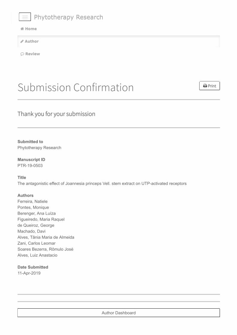
Phytotherapy ResearchPhytotherapy Research
Print Print
Submission Confirmation
Thank you for your submission
Submitted toPhytotherapy Research Manuscript IDPTR-19-0503 TitleThe antagonistic effect of Joannesia princeps Vell. stem extract on UTP-activated receptors AuthorsFerreira, Natiele Pontes, Monique Berenger, Ana Luíza Figueiredo, Maria Raquel de Queiroz, George Machado, Davi Alves, Tânia Maria de Almeida Zani, Carlos Leomar Soares Bezerra, Rômulo José Alves, Luiz Anastacio Date Submitted11-Apr-2019
Author DashboardAuthor Dashboard
Home Home
Author Author
Review Review

The antagonistic effect of Joannesia princeps Vell. stem extract onUTP-activated receptors
Natiele Carla da Silva Ferreira1, Monique de Castro Pontes1, Ana Luíza Rangel Berenger2,Maria Raquel Figueiredo2, George Azevedo de Queiroz3, Davi Nepomuceno da
Silva Machado4, Tânia Maria Alves5, Carlos Leomar Zani5, Rômulo José Soares Bezerra6 andLuiz Anastacio Alves1
1Oswaldo Cruz Institute, Laboratory of Cellular Communication, Oswaldo Cruz Foundation2Institute of Technology in Drugs, Laboratory of Chemistry of Natural Products, Oswaldo Cruz Foundation
3National Museum, Federal University of Rio de Janeiro4Group of Interdisciplinary Studies of the Environment, University of the State of Rio de Janeiro
5René Rachou Research Center, Laboratory of Chemistry of Natural Products, Oswaldo Cruz Foundation6Oswaldo Cruz Institute, Laboratory of Technological Development in Virology, Oswaldo Cruz Foundation
AbstractP2Y2 and P2Y4 are purinergic receptors physiologicallyactivated by UTP. They are expressed in organs such aslung, heart, skeletal muscle, spleen, and kidneys, as wellas in immune system cells. Once activated, they promotecalcium mobilization and act on ion flux regulation. Thesereceptors are associated with several diseases, such as in-flammation and cancer, making them important therapeutictargets. The scarcity of selective antagonists for these re-ceptors limits their application in clinical therapy, encour-aging the search for new molecules presenting antagonisticactivity. In the present study, we characterized the antag-onistic activity of the Joannesia princeps Vell. stem ex-tract. Using calcium measurement technique, we demon-strated that the extract inhibits calcium response for P2Y2and P2Y4, but also P2Y6 receptor suggesting the existenceof selectivity among P2 receptors. We also observed thatinhibitory activity was not due to the quenching effect, andwas not affected by stock-operated calcium channels. Fi-nally, we demonstrated that both methanolic and ethanolicJ. princeps Vell. extracts are able to reduce UTP-inducedcalcium responses and do not cause cytotoxicity. Our datasuggest that J. princeps Vell stem extracts act as an UTP-activated receptors receptor antagonist.
Key words: P2Y2, P2Y4, UTP, antagonist, calcium, natu-ral products
1 Introduction
Purinergic receptors are plasma membrane receptors acti-vated by extracellular nucleotides. They are classified into
two families: P1 and P2 receptors (P1R and P2R), whichare activated by adenosine and its derivated-nucleotides,respectively. P2R is also subdivided into two classes:ionotropic P2X and metabotropic P2Y (Abbracchio &Burnstock, 1994).
P2Y receptors comprise seven transmembrane domains(TM1-7), in addition to an intracellular C-terminal and ex-tracellular N-terminal. Currently, eight P2Y receptors sub-types are described. P2Y1, P2Y2, P2Y4, P2Y6, P2Y11 arecoupled to Gq protein, although the latter can also coupleto the Gs protein, while PY12-P2Y14 are coupled to theGi protein. These receptors can be activated by differentextracellular nucleotides, including ADP, ATP, UDP, UDP-glucose and UTP, which will be addressed in this study (Ja-cobson et al., 2012).
P2Y2 is a receptor physiologically activated by UTP andATP and has aroused much therapeutic interest, first, dueto its expression in a variety of biological tissues, includ-ing the lung, heart, skeletal muscle, spleen and kidneys,as well as immune system cells, such as lymphocytes andmacrophages (von Kügelgen, 2006; Von Kügelgen & Hoff-mann, 2016). Second, this receptor plays important roles re-lated to the ion flow in epithelial cells, which can contributesignificantly to the treatment of cystic fibrosis and dry eyedisease (Jacobson & Boeynaems, 2010). In view of this, anophthalmic solution that mimics UTP has been developedand is able to attenuate the dry eye disease symptoms. Thisdrug, called Diquafosol, is marketed only in Japan, SouthKorea, Thailand and Vietnam (Santen, 2017). In addition,several studies have indicated that P2Y2 is involved in thedevelopment of certain diseases, such as neuropathic pain(Li et al., 2014; Magni et al., 2015), cancer (Chadet et al.,2014; Choi et al., 2013; Jin et al., 2014; Li et al., 2013; Xieet al., 2014) and inflammation (Ayata et al., 2012; Degagné
1

J. princeps stem extract inhibits UTP-activated receptors 2
et al., 2009; Eun et al., 2014; Inoue et al., 2008; Vander-stocken et al., 2010; Zhang et al., 2010), thus, constitutingan adequate therapeutic target.
P2Y4 is another UTP-activated receptor. It is expressedin the gut, brain, lung, heart, among other cell types (Rafehi& Müller, 2018). Because it is structurally and pharma-cologically similar to the P2Y2 receptor and sometimesexpressed in the same cell types, the characterization ofP2Y4 physiological effects has become a significant chal-lenge (Rafehi & Müller, 2018; von Kügelgen, 2006). How-ever, P2Y4 is known to also act on ion regulation in theintestine and cochlea, besides promoting the inhibition ofspontaneous longitudinal ileum muscle contractile activity(Ghanem et al., 2005; Kim et al., 2010; Zizzo et al., 2012).
The scarcity of selective antagonists for these receptorslimits their application in clinical therapy, encouraging thesearch for new molecules with antagonistic activity. Nat-ural products are noteworthy in the scenario for the searchof new drugs, due to their enormous structural and chemi-cal diversity, acquired over thousands of years of evolution.These characteristics inspire new discoveries to this day, inthe chemistry, biology, and medicine fields, making them animportant source for the discovery of novel drugs. In addi-tion, natural products and their derivatives present higher hitrates during the high throughput screening (HTS) processthan compounds obtained from synthetic libraries (Chen etal., 2017).
In a previous study, we demonstrated for the first time thepotential antagonistic activity of the crude extracts of Joan-nesia princeps Vell. stems after a mini-screening campaignto discover new P2Y2 and P2Y4 receptor antagonists. J.princeps Vell. extracts were able to inhibit concentration-dependent UTP-induced intracellular calcium mobilizationwithout promoting cytotoxicity (Ferreira et al., 2018). J.princeps Vell. is a botanical species belonging to theEuphorbiaceae family, popularly known as “cotieira”. Thisspecies is found in Brazil and in some African and Asiancontinent regions. Some literature reports have indicatedthat this species presents laxative, anti-parasitic and anti-microbicide activities (Donato-Trancoso et al., 2014; Sousaet al., 2007). In this present study, we demonstrate that cal-cium inhibition is selective for UTP-activated receptors, andwas not caused by quenching effects or cell death.
2 Material and Methods
2.1 ReagentsADP, ATP, UDP, UTP, PPADS, RB2, Suramin, Triton-X,
DMEM and RPMI culture mediums, NaCl, KCl, Na2HPO4,KH2PO4, MgCl2, CaCl2, Hepes, Glucose, DMSO, MTT ((3-(4,5-dimethylthiazol-2-yl)-2,5-diphenyltetrazolium bro-mide) tetrazolium, Probenecid and Ionomycin were pur-
chased from Sigma Chemical Co. (St. Louis, Missouri,USA). 2-thio-UTP and MRS 4062 were purchased fromTocris (Bristol, UK). Fetal bovine serum was obtained fromGibco (Oklahoma, USA). Fluo-4 AM was obtained fromLife Technologies (California, USA). YO-PRO-1 was ob-tained from Molecular Probes (Oregon, USA).
2.2 ExtractsJ. princeps Vell. stems were collected at Linhares, in the
state of Espírito Santo, Brazil. The samples were crushedand extracted with a 1:1 mixture of dichloromethane-methanol. The crude extract was deposited under vouchernumber EX294 at the Natural Product Chemistry Labora-tory Bioprospecting Platform. Other J. princeps Vell. stemand leaf samples were obtained from Maricá, in the stateof Rio de Janeiro, Brazil. The plant was oven dried (40◦C)for one weekin an air circulation oven and reduced to smallfragments. The dried plant materials were submitted to astatic extraction with ethanol for about seven days and driedunder reduced pressure. They were resuspended in a Dul-becco’s PBS solution containing 0.5% DMSO for the exper-iments. All collections of botanical materials were autho-rized by the Chico Mendes Institute for Biodiversity Con-servation, an environmental agency of the Brazilian govern-ment.
2.3 Cell cultureJ774.G8 cells were cultivated with Dulbecco’s Modified
Eagle’s Medium (DMEM) supplemented with 10% fetalbovine serum, at 37◦C in a 5% CO2 atmosphere. The cel-lular medium was changed twice a week and cell densitywas adjusted to 2x10⁶ cells per 150 cm2 cell culture flask(Corning, New York, USA). Cell viability was measured bytrypan blue exclusion and experiments were conducted onlywhen viability was above 90%.
2.4 Calcium assaysJ774.G8 cells were plated on a 96-well black-wall and
clear-bottom plate (Corning, New York, USA), at a con-centration of 2x10⁵ cells/well. They were maintained inculture with DMEM supplemented with 10% fetal bovineserum, at 37◦C in a 5% CO2 atmosphere for 24h. Then,the medium was replaced by 100 µl of Dulbecco’s PBS(DPBS) (136 mM NaCl, 2.68 mM KCl, 8 mM Na2HPO4,1.46 mM KH2PO4, 0.5 mM MgCl2, 0.9 mM CaCl2, 5.55mM Glucose and 2.5 mM Probenecid – pH 7.4) contain-ing [2 µM] Fluo-4 AM and cells were incubated for 60minutes at 37ºC in a 5% CO2 atmosphere. Subsequently,the wells were washed three times with 200 µl DPBS andeach well received a final volume of 150 µl DPBS. After-wards, cells were treated with PPADS [300 µM], Suramin[300 µM], Reactive Blue-2 (RB2) [20 µM] and J. princepsVell. extracts [50 µg/mL] for 30 minutes. Intracellular cal-cium mobilization was determined using a FlexStation III
J. princeps stem extract inhibits UTP-activated receptors 3
(Molecular Devices, California, USA) in real time during90 seconds, at 485 nm and 525 nm wavelengths for exci-tation and emission, respectively. The agonists were addedat 20 seconds and readings were performed every 1.52 sec-onds, totaling 60 readings/well. Calcium mobilization wasquantified as ∆FU (the difference between the fluorescencepeak and basal fluorescence) (Hansen & Bräuner-Osborne,2009).
2.5 Dye uptake assayJ774.G8 cells were plated on a 96-well black plate
(Corning, New York, USA), at a concentration of 2x10⁵
cells/well. They were maintained in culture with DMEMsupplemented with 10% fetal bovine serum, at 37◦C in a 5%CO2 atmosphere for 24h. Then, the medium was replacedby an extracellular saline solution (150 mM NaCl, 5 mMKCl, 1 mM MgCl2, 1 mM CaCl2, and 10 mM HEPES, pH7.4). Subsequently, cells were treated with Brilliant BlueG (BBG) [200 nM] and the J. princeps Vell. extract [50µg/mL] for 30 minutes. Then, cells were stimulated withATP [5 mM] for 15 minutes, in order to allow the openingof the membrane pore associated with the P2X7 receptor.Next, the cationic dye YO-PRO-1 was added to each well ata concentration of 2 µM, and after 3 minutes, fluorescencesignals were determined using a FlexStation III equipment(Molecular Devices, California, USA) at 488 nm and 509nm for excitation and emission, respectively. Cells treatedwith Triton-X [0.1%] were considered the maximum dyeuptake control.
2.6 AnimalsAnimal experiments followed all animal experimentation
ethical principles as determined by the Brazilian College ofAnimal Experimentation. All experiments were approvedby the Ethics Committee on the use of Animals from the Os-waldo Cruz, Institute under identification code L-037/2017(approved on November 24, 2017). Healthy male Swissmice weighing between 30 and 40g were obtained from theOswaldo Cruz Central Bioterium. The animals were housedunder a 12h/12h light and dark cycle conditions with freeaccess to food and water.
2.7 Peritoneal macrophage isolationAnimals were euthanized by CO2 asphyxiation. Sub-
sequently, 10 mL of cold PBS were injected into theperitoneal cavity. Peritoneal cells were collected andmacrophages were isolated by centrifugation at 1,500 RPMfor 10 minutes. Cell viability was analyzed by the trypanblue exclusion assay. Cells were plated on a 96-well black-wall and clear-bottom plate at a density of 4x10⁵ cells/welland maintained in culture with RPMI supplemented with10% fetal bovine serum, at 37◦C in a 5% CO2 atmospherefor 1h, to allow a pre-adhesion. Then, each well was washedwith 200 µl of RPMI medium and peritoneal macrophageswere maintained in culture with RPMI supplemented with
10% fetal bovine serum, at 37◦C in a 5% CO2 atmospherefor 24h. Calcium assays were performed as previously de-scribed.
2.8 Cell viability measurementJ774.G8 cells were plated on a 96-well plate (Corning,
New York, USA) at a concentration of 2x10⁵ cells/well.They were maintained in culture with DMEM without phe-nol red supplemented with 10% fetal bovine serum, at 37◦Cin a 5% CO2 atmosphere for 24h. Next, cells were treatedwith extracts [50 µg/mL] and the RB2 [20 µM] antago-nist for 1 or 24h. Untreated cells were considered as apositive viability control, while cells treated with Triton-X [0.1%] were considered as the negative control. Afterthe treatments, the medium was replaced by 200 µL ofDMEM without phenol red containing an MTT solution[100 µg/well]. The plate was incubated for 3h at 37◦C ina 5% CO2 atmosphere, and was then centrifuged at 1,500RPM for 1 minute. Supernatants were collected and for-mazan crystals were dissolved in 100 µl of DMSO. Wellabsorbance was determined measured using a Spectramax190 equipment (Molecular Devices, California, USA) at570 nm.
2.9 High Performance Liquid Chromatography(HPLC) analysis
The qualitative analyses were carried out using an HPLCsystem consisting of a Shimadzu Liquid Chromatograph,LC-20ADXR pump equipped with a quaternary gradientvalve, Shimadzu SPD-M20A diode array detector (DAD),Shimadzu SIL-30AC autosampler, CTO-20AC oven, DGU-20A5R degasser and a Shimadzu CBM 20-A communica-tions bus module.
Methanol was added to the sample until reaching 20mg/mL. The sample was then vortexed and sonicated for 10minutes Subsequently, the sample was membrane filteredPVDF 0.22 µm and 20 µL were injected.
The Joannesia princeps chemical profile fingerprint wasobtained using a Supelcosil C-18 column (5µm particlesize, 250 mm X 4,6mm internal diameter) and oven tem-perature of 30o C. Solvent system conditions were trifluo-roacetic acid pH 2.5 and acetonitrile (95:5 v/v to 95:5 v/v,90 minutes) with a mobile flow rate of 1.0mL/min withHPLC effluent monitoring at 254-365 nm by a DAD.
2.10 Data analysisEach sample was analyzed in triplicate and all experi-
ments were performed on at least three independent days.All data are presented as means ± standard deviation (SD).The D’ Agostino and Pearson normality test was used inorder to test for a Gaussian distribution. If data followeda Gaussian distribution, an appropriate parametric test wasapplied and, if not, an appropriate non-parametric test wasused. Tests are specified in figure legends. P values of 0.05
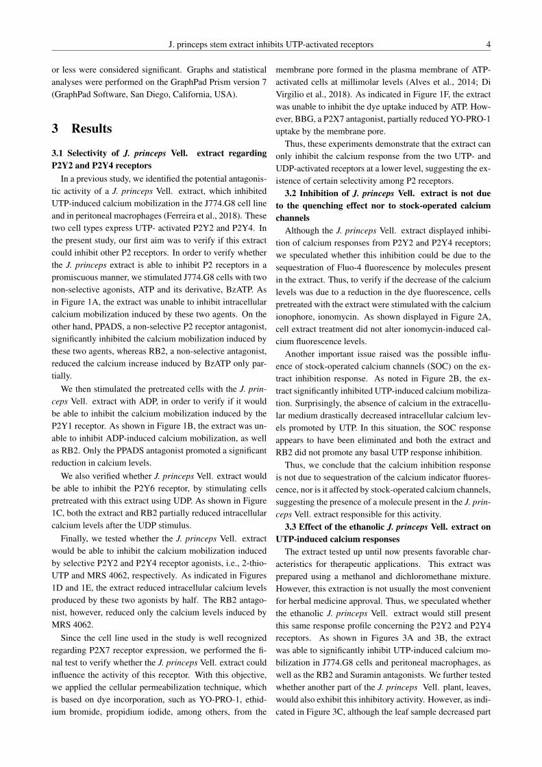
J. princeps stem extract inhibits UTP-activated receptors 4
or less were considered significant. Graphs and statisticalanalyses were performed on the GraphPad Prism version 7(GraphPad Software, San Diego, California, USA).
3 Results
3.1 Selectivity of J. princeps Vell. extract regardingP2Y2 and P2Y4 receptors
In a previous study, we identified the potential antagonis-tic activity of a J. princeps Vell. extract, which inhibitedUTP-induced calcium mobilization in the J774.G8 cell lineand in peritoneal macrophages (Ferreira et al., 2018). Thesetwo cell types express UTP- activated P2Y2 and P2Y4. Inthe present study, our first aim was to verify if this extractcould inhibit other P2 receptors. In order to verify whetherthe J. princeps extract is able to inhibit P2 receptors in apromiscuous manner, we stimulated J774.G8 cells with twonon-selective agonists, ATP and its derivative, BzATP. Asin Figure 1A, the extract was unable to inhibit intracellularcalcium mobilization induced by these two agents. On theother hand, PPADS, a non-selective P2 receptor antagonist,significantly inhibited the calcium mobilization induced bythese two agents, whereas RB2, a non-selective antagonist,reduced the calcium increase induced by BzATP only par-tially.
We then stimulated the pretreated cells with the J. prin-ceps Vell. extract with ADP, in order to verify if it wouldbe able to inhibit the calcium mobilization induced by theP2Y1 receptor. As shown in Figure 1B, the extract was un-able to inhibit ADP-induced calcium mobilization, as wellas RB2. Only the PPADS antagonist promoted a significantreduction in calcium levels.
We also verified whether J. princeps Vell. extract wouldbe able to inhibit the P2Y6 receptor, by stimulating cellspretreated with this extract using UDP. As shown in Figure1C, both the extract and RB2 partially reduced intracellularcalcium levels after the UDP stimulus.
Finally, we tested whether the J. princeps Vell. extractwould be able to inhibit the calcium mobilization inducedby selective P2Y2 and P2Y4 receptor agonists, i.e., 2-thio-UTP and MRS 4062, respectively. As indicated in Figures1D and 1E, the extract reduced intracellular calcium levelsproduced by these two agonists by half. The RB2 antago-nist, however, reduced only the calcium levels induced byMRS 4062.
Since the cell line used in the study is well recognizedregarding P2X7 receptor expression, we performed the fi-nal test to verify whether the J. princeps Vell. extract couldinfluence the activity of this receptor. With this objective,we applied the cellular permeabilization technique, whichis based on dye incorporation, such as YO-PRO-1, ethid-ium bromide, propidium iodide, among others, from the
membrane pore formed in the plasma membrane of ATP-activated cells at millimolar levels (Alves et al., 2014; DiVirgilio et al., 2018). As indicated in Figure 1F, the extractwas unable to inhibit the dye uptake induced by ATP. How-ever, BBG, a P2X7 antagonist, partially reduced YO-PRO-1uptake by the membrane pore.
Thus, these experiments demonstrate that the extract canonly inhibit the calcium response from the two UTP- andUDP-activated receptors at a lower level, suggesting the ex-istence of certain selectivity among P2 receptors.
3.2 Inhibition of J. princeps Vell. extract is not dueto the quenching effect nor to stock-operated calciumchannels
Although the J. princeps Vell. extract displayed inhibi-tion of calcium responses from P2Y2 and P2Y4 receptors;we speculated whether this inhibition could be due to thesequestration of Fluo-4 fluorescence by molecules presentin the extract. Thus, to verify if the decrease of the calciumlevels was due to a reduction in the dye fluorescence, cellspretreated with the extract were stimulated with the calciumionophore, ionomycin. As shown displayed in Figure 2A,cell extract treatment did not alter ionomycin-induced cal-cium fluorescence levels.
Another important issue raised was the possible influ-ence of stock-operated calcium channels (SOC) on the ex-tract inhibition response. As noted in Figure 2B, the ex-tract significantly inhibited UTP-induced calcium mobiliza-tion. Surprisingly, the absence of calcium in the extracellu-lar medium drastically decreased intracellular calcium lev-els promoted by UTP. In this situation, the SOC responseappears to have been eliminated and both the extract andRB2 did not promote any basal UTP response inhibition.
Thus, we conclude that the calcium inhibition responseis not due to sequestration of the calcium indicator fluores-cence, nor is it affected by stock-operated calcium channels,suggesting the presence of a molecule present in the J. prin-ceps Vell. extract responsible for this activity.
3.3 Effect of the ethanolic J. princeps Vell. extract onUTP-induced calcium responses
The extract tested up until now presents favorable char-acteristics for therapeutic applications. This extract wasprepared using a methanol and dichloromethane mixture.However, this extraction is not usually the most convenientfor herbal medicine approval. Thus, we speculated whetherthe ethanolic J. princeps Vell. extract would still presentthis same response profile concerning the P2Y2 and P2Y4receptors. As shown in Figures 3A and 3B, the extractwas able to significantly inhibit UTP-induced calcium mo-bilization in J774.G8 cells and peritoneal macrophages, aswell as the RB2 and Suramin antagonists. We further testedwhether another part of the J. princeps Vell. plant, leaves,would also exhibit this inhibitory activity. However, as indi-cated in Figure 3C, although the leaf sample decreased part

J. princeps stem extract inhibits UTP-activated receptors 5
FIGURE 1: The J. princeps Vell. extract only inhibits calcium mobilization induced by P2Y2 and P2Y4 agonists.J774.G8 cells stained with 2 µM of Fluo-4 were pretreated with J. princeps Vell. stem extract [50 µg/mL], RB2 [20µM] and PPADS [300 µM] for 30 minutes and then stimulated with agonists. Cells were stimulated with ATP [100µM] or BzATP [100 µM] and monitored for 90 seconds. Data are presented as the means ± SD of three independentexperiments performed in triplicate. The data analysis was performed by a one-way ANOVA and Holm-Sidak post-test; p<0.05. Asterisks indicate significant differences in relation to ATP or BzATP controls. (ns) non-significant means(A). Cells were stimulated with ADP [10 µM] and monitored for 90 seconds. Data are presented as the means ± SDof three independent experiments performed in triplicate. The data analysis was performed by a Kruskal-Wallis andDunn’s post-test; p<0.05. Asterisks indicate a significant difference in relation to ADP and (ns) non-significant means(B). Cells were stimulated with UDP [100 µM] and monitored for 90 seconds. Data are presented as the means ± SDof four independent experiments performed in triplicate. The data analysis was performed by a one-way ANOVA andHolm-Sidak post-test;p<0.05. Asterisks indicate a significant difference in relation to UDP (C). Cells were stimulatedwith 2-thio-UTP [10 µM] and monitored for 90 seconds. Data are presented as the means ± SD of three independentexperiments performed in triplicate. The data analysis was performed by a one-way ANOVA and Holm-Sidakpost-test;p<0.05. Asterisks indicate a significant difference in relation to 2-thio-UTP and (ns) non-significant means (D). Cellswere stimulated with MRS 4062 [10 µM] and monitored for 90 seconds. Data are presented as the means ± SD of threeindependent experiments performed in triplicate. The data analysis was performed by a one-way ANOVA and Holm-Sidakpost-test;p<0.05. Asterisks indicate a significant difference in relation to MRS 4062(E). J774.G8 cells were pretreatedwith J. princeps Vell. stem extract [50 µg/mL] and BBG [200 nM] during 30 minutes. Then, cells were stimulated withATP [5 mM] for a further 15 minutes and the YO-PRO-1 dye [2 µM] was added to the wells and fluorescence of cellswas measured. Data are presented as the means ± SD of three independent experiments performed in triplicate. The dataanalysis was performed by a one-way ANOVA and Holm-Sidak post-test; p<0.05. Data were normalized as a percentagein function of the mean of units of arbitrary fluorescence (RFU) obtained by Triton-X, the maximum fluorescence control.Asterisks indicate a significant difference in relation to ATP and (ns) non-signficant means (F).
of the UTP-induced calcium response, this reduction wasnot statistically significant.
Therefore, we conclude that both J. princeps Vell. ex-tracts, methanolic and ethanolic are able to reduce the cal-cium response from P2Y2 and P2Y4 receptor the activation.
3.4 J. princeps Vell. extract did not present cytotoxic-ity
A final criterion for the analysis of the J. princeps Vell.extract inhibition was to test whether this activity could be
due to cell death. In the case of cell death, cell fluorescencewould tend to decrease, causing an artifact in the measure-ment of calcium levels. In order to test this hypothesis, wetreated the cells with the J. princeps Vell. methanolic andethanolic extracts for 1h, an estimated period for the dura-tion of the calcium recordings. As indicated in Figures 4Aand 4B, both extracts did not cause cell death compared tountreated cell control. Finally, we also tested the cell via-bility profile after 24 hours of treatment with the extracts.

J. princeps stem extract inhibits UTP-activated receptors 6
FIGURE 2: The J. princeps Vell. extract did not present the quenching effect and did not inhibit stock-operatedcalcium channels. J774.G8 cells stained with 2 µM of Fluo-4 were pretreated with J. princeps Vell. stem extract [50µg/mL] for 30 minutes. Cells were then stimulated with Ionomycin [1 µM] and monitored for 90 seconds. Data arepresented as the means ± SD of three independent experiments performed in triplicate. The data analysis was performedby a one-way ANOVA and Holm-Sidak post-test; p<0.05.(ns) non-significant means (A). Cells loaded with Fluo-4 andbathed in DPBS with or without calcium were pretreated with J. princeps Vell. stem extract [50 µg/mL] for 30 minutes,and stimulated with UTP [10 µM], were then monitored for 90 seconds. Data are presented as the means ± SD of threeindependent experiments performed in triplicate. The data analysis was performed by a one-way ANOVA and Holm-Sidakpost-test; p<0.05. Asterisks indicate a significant difference in relation to UTP and (ns) non-significant means (B).
FIGURE 3: Effects of J. princeps Vell. ethanolic extracts on UTP-induced calcium responses. J774.G8 cells stainedwith 2 µM of Fluo-4 were pretreated with J. princeps Vell. stem extract [50 µg/mL], RB2 [20 µM], and Suramin [300µM] for 30 minutes. Cells were then stimulated with UTP [10 µM] and monitored for 90 seconds. Data are presented asthe means ± SD of four independent experiments performed in triplicate. The data analysis was performed by a one-wayANOVA and Holm-Sidak post-test; p<0.05. Asterisks indicate a significant difference in relation to UTP (A). Peritonealmacrophages loaded with Fluo-4 were pretreated with J. princeps Vell. stem extract [50 µg/mL], RB2 [20 µM], andSuramin [300 µM] for 30minutes, and stimulated with UTP [10 µM], were then monitored for 90 seconds. Data arepresented as the means ± SD of one independent experiment performed in triplicate (B). J774.G8 cells stained with 2 µMof Fluo-4 were pretreated with J. princeps Vell. leave extract [50 µg/mL], RB2 [20 µM], and Suramin [300 µM] for 30minutes. Cells were then stimulated with UTP [10 µM] and monitored for 90 seconds. Data are presented as the means± SD of four independent experiments performed in triplicate. The data analysis was performed by a one-way ANOVAand Holm-Sidak post-test; p<0.05. Asterisks indicate a significant difference in relation to UTP and (ns) non-significantmeans (C).

J. princeps stem extract inhibits UTP-activated receptors 7
As shown in Figures 4C and 4D, none of the extracts re-duced cell viability, similarly to untreated cell controls andthe RB2 antagonist.
Therefore, we discarded the hypothesis that extract in-hibition was due to cell death and concluded that they donot affect cell viability after 24h of treatment at the testedconcentration.
3.5 Chemical J. princeps profile determined by HPLCBoth J. princeps methanolic and ethanolic extracts were
submitted to the HPLC technique for chemical profile deter-minations as shown in Figures 5A and 5B, the methanolicand ethanolic extracts presented between 31-45 compoundsat 254 and 365 nm, even though the method was not ef-ficient regarding substance separation. The peak times ofboth samples was observed at approximately at 15 minutes.
4 Discussion
In a previous study, we described the potential antagonisticactivity of J. princeps Vell. extract on UTP-activated recep-tors for the first time after holding a mini high throughputscreening campaign. We observed that the extract inhib-ited calcium responses in a dependent-concentration man-ner and found an IC50 of 32.32 µg/mL (Ferreira et al.,2018). However, some questions remained open for theconclusion that the extract could be considered a P2Y2 andP2Y4 receptor antagonist.
Herein, we first performed a pharmacological characteri-zation of the J. princeps Vell. extract on P2 receptors asso-ciated with calcium signaling, i.e., P2X and P2Y1, P2Y2,P2Y4, P2Y6 and P2Y11 using physiological or selectiveagonists (Ralevic & Burnstock, 1998). To stimulate P2Xreceptors, we used ATP and BzATP, but did not observe anyinhibitory response. ATP is a non-selective agonist for P2Xreceptors and is also capable of activating the P2Y2 andP2Y11 receptors. The EC50 range for the activation of thesereceptors ranges from 0.1 to 10 µM, with the exception ofP2X7, whose activation occurs at concentrations over 100µM (Ralevic & Burnstock, 1998). However, the J774.G8line expresses several of these receptors (Coutinho-Silva etal., 2005; Ferreira et al., 2018), and the calcium responseobserved after ATP stimulation may be a kind of summationof the activation of these receptors simultaneously. Even ifit were able to inhibit only P2Y2, by adding the responseof the other receptors, it would become imperceptible. Inan attempt to select a small portion of this response for theP2X receptors, an ATP derivative was used. BzATP func-tions as a potent agonist for P2X7 as well as a total agonistfor P2X4 and a partial agonist for P2X1, P2X2, P2X3 andP2X5 (Coddou et al., 2011). Despite this, the response pro-file was similar to that observed for ATP.
Another technique used to verify if the extract is able
to exert antagonistic activity on P2X7, which is widelyexpressed in this cell line, was cellular permeabilization,which consists of cell stimulation at high ATP concentra-tions (above 1 millimolar) in order to observe the formationof a membrane pore. This membrane pore allows the pas-sage of molecules up to 900 Da, including ions (Na+, Ca2+,K+), water molecules, ATP and dyes such as propidium io-dide (668 Da), ethidium bromide (394 Da), and YO-PRO-1(629 Da), which are widely used for the study of P2X7 ac-tivity (Alves et al., 2014; Di Virgilio et al., 2018). However,no inhibitory response of the extract to the P2X7 receptorwas observed.
We also used ADP and UDP, physiological P2Y1 andP2Y6 receptor agonists, respectively, and observe a slightinhibition response of P2Y6. Interestingly, UDP is a prod-uct of UTP degradation by ectonucleotidases, and, by dis-playing a structure similar to UTP, could be easily tar-geted for inhibition by the extract. However, some stud-ies have recently demonstrated that UTP binding to P2Y2and P2Y4 receptors do not result in conformational modi-fications, whereas UDP binding to P2Y6 receptor results inconformational modifications at the quaternary level of theprotein (D’Ambrosi et al., 2007; Kotevic et al., 2005). Aswe still could rule out the possibility of the extract func-tioning as a negative allosteric modulator, these conforma-tional modifications may continue to occur even, althoughat a lower level, but sufficient for the P2Y6 receptor to re-main active.
Since UTP is able to activate both P2Y2 and P2Y4 sub-types, we use selective agonists for these receptors in or-der to try to identify whether the extract is able to inhibita single specific subtype or both subtypes (Jacobson et al.,2012). However, the data suggest that the extract can in-hibit the response of both P2Y2 and P2Y4 receptors. Al-though they are considered selective agonists, it is knownthat, depending on the concentration, they may present acertain loss of specificity. According to the manufacturer,both agonists, 2-thio-UTP (P2Y2) and MRS 4062 (P2Y4),are capable of activating other P2Y receptors in the submi-cromolar range. With all these challenges, the experimentthat can be performed in attempting to conclude this phar-macological characterization is the use of cells transfectedwith a single P2Y receptor subtype that is activated by UTPat a time.
Another important point questioned was whether the in-hibition caused by the J. princeps Vell. extract could bedue to a quenching effect. It is known that some plant sub-stances, such as quercetin or vitexin, are able to fluoresce ordisplay a quenching effect, both of which can result in arti-facts in experiments using fluorescence (Zou et al., 2002).In this sense, we performed a test using ionomycin, a cal-cium ionophore that displays the ability to increase the in-tensity of calcium-related fluorescence. With this, we ob-
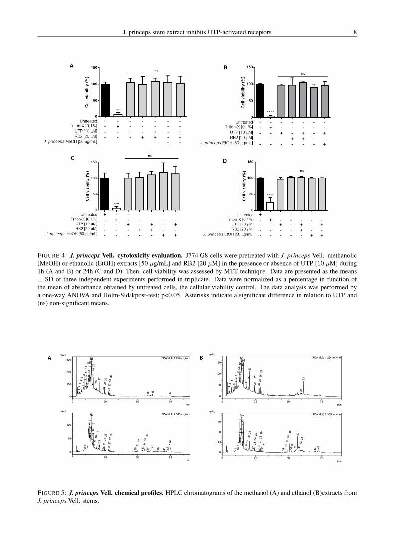
J. princeps stem extract inhibits UTP-activated receptors 8
FIGURE 4: J. princeps Vell. cytotoxicity evaluation. J774.G8 cells were pretreated with J. princeps Vell. methanolic(MeOH) or ethanolic (EtOH) extracts [50 µg/mL] and RB2 [20 µM] in the presence or absence of UTP [10 µM] during1h (A and B) or 24h (C and D). Then, cell viability was assessed by MTT technique. Data are presented as the means± SD of three independent experiments performed in triplicate. Data were normalized as a percentage in function ofthe mean of absorbance obtained by untreated cells, the cellular viability control. The data analysis was performed bya one-way ANOVA and Holm-Sidakpost-test; p<0.05. Asterisks indicate a significant difference in relation to UTP and(ns) non-significant means.
FIGURE 5: J. princeps Vell. chemical profiles. HPLC chromatograms of the methanol (A) and ethanol (B)extracts fromJ. princeps Vell. stems.

J. princeps stem extract inhibits UTP-activated receptors 9
served that the presence of the extract did not result in anychanges in the ionomycin response profile, thus ruling outthe possibility of a quenching effect.
We also speculated whether the opening of stock-operated calcium channels (SOC) could influence the ex-tract response, i.e., the opening of these channels couldbe raising intracellular calcium levels (Azimi et al., 2014).This increase in calcium levels could make it difficult toobserve calcium mobilization via the phospholipase C/IP3
pathway. Thus, by removing the extracellular calcium fromthe buffer solution, we expected to continue to observe in-hibition of the UTP response. Surprisingly, calcium mo-bilization reduced drastically and the extract response wassimilar to baseline UTP. This led us to question two facts.The first, whether it would be possible that the extract couldinhibit the SOC response rather than the receptors, and thesecond whether, in fact, the extract inhibits SOC, would itnot inhibit, even partially, the calcium mobilization inducedby ATP, ADP, BzATP and UDP? Another hypothesis is thatthe absence of extracellular calcium could generate experi-mental artifacts or unclarified phenomena that could affectthe observed response.
Another concern was with regard to the applicability ofthe extract as a possible phytotherapic. The first sampleused in the present study was extracted with a mixtureof methanol and dichloromethane. However, according toFood and Drug Administration (FDA) regulations, for abotanical drug to be approved as a phytotherapic it must beextracted in either an aqueous solution or ethanol (Centerfor Drug Evaluation and Research, 2016). Thus, we per-formed a new extraction in ethanol and similar to what wasobserved for the methanolic extract, this new ethanolic ex-tract was also able to inhibit the UTP response in J774.G8cells and peritoneal macrophages. It is noteworthy that thisinhibition was not caused by cell death and that both ex-tracts did not present cytotoxicity within 24 hours of treat-ment.
J. princeps Vell. is a botanical species found in Braziland in some African and Asian regions. It belongs to theEuphorbiaceae family and each individual measures around20 m in height. In Brazil, it is popularly known as “cotieira”and its seeds present laxative and healing properties, as wellas anti-parasitic and anti-microbicide activities (Donato-Trancoso et al., 2014; Sousa et al., 2007). J. princepsVell. presents compounds belonging to the sesquiterpene,bis-sesquiterpene, diterpene, triterpene and steroid classes(Achenbach & Benirschke, 1997). However, the few stud-ies that describe the phytochemistry and medicinal prop-erties of J. princeps Vell. focus specifically on seeds andleaves. In this sense, the study of the chemical compositionand medicinal application of stem extracts is still scarce,and our group is a pioneer in this scenario. However, thecharacterization of the active molecule, responsible for the
antagonistic activity, is indispensable and is the next aim ofour research group.
The characterization of the active molecule responsiblefor the inhibition of P2Y2 and P2Y4 receptors will certainlycontribute to the pharmacology of these receptors, whichparticipate in the development of various inflammatory dis-eases (Ayata et al., 2012; Inoue et al., 2008; Vanderstockenet al., 2010), cancer (Chadet et al., 2014; Choi et al., 2013;Jin et al., 2014; Li et al., 2013; Qiu et al., 2018; Xie et al.,2014), and contribute to cardiac pathologies (Horckmans etal., 2015). Although P2Y2 possesses a single selective an-tagonist (AR-C118925), it has failed to advance to the clin-ical application due to certain undesired pharmacologicalproperties, which include high polarity and low bioavail-ability through the oral route (Rafehi & Müller, 2018). TheP2Y4 receptor, however, still lacks selective antagonists,which makes it difficult to characterize associated physio-logical functions. In this sense, the RB2 antagonist is oftenused to obtain references regarding the expected antagonis-tic effect. However, due to its ability to inhibit other P2receptors, the results should be evaluated at with caution.In this scenario, the description of an antagonist that acts onthese two receptors, even without selectivity, will certainlycontribute to the development of several in vitro and in vivostudies, and may serve as basis for the synthesis and de-velopment of new selective antagonists for these receptors(Rafehi & Müller, 2018).
5 Conclusions
This study characterized the stem J. princeps Vell. ex-tract as a potential antagonist for P2Y2 and P2Y4 recep-tors. Both methanolic and ethanolic extracts are capable ofinhibiting calcium responses promoted by these receptorswithout causing cytotoxicity and quenching effects. Themethanolic extract also inhibits calcium mobilization in-duced by P2Y6 receptor, but not P2X and P2Y1.
Acknowledgements
This study was supported by Fundação de Amparo àPesquisa do Estado do Rio de Janeiro (FAPERJ), ConselhoNacional de Desenvolvimento Científico e Tecnológico(CNPq), and Instituto Oswaldo Cruz (IOC). The authorsthank Fátima Regina Marques for technical assistancewith the extract; Dr. Vinícius Cotta, who kindly do-nated the J774.G8 cell line; and the PDTIS-FIOCRUZ/RPT-FIOCRUZ Program for Technological Development inTools for Health for the use of its facilities.
Conflict of interestThe authors declare no conflicts of interest.
Abbracchio and Burnstock (1994); Achenbach and

J. princeps stem extract inhibits UTP-activated receptors 10
Benirschke (1997); Alves et al. (2014); Ayata et al. (2012);Azimi et al. (2014); for Drug Evaluation and Research.(2016); Chadet et al. (2014); Chen et al. (2017); Choiet al. (2013); Coddou et al. (2011); Coutinho-Silva et al.(2005); D’Ambrosi et al. (2007); Degagné et al. (2009);Virgilio et al. (2018); Donato-Trancoso et al. (2014); Eunet al. (2014); Ferreira et al. (2018); Ghanem et al. (2005);Hansen and Bräuner-Osborne (2009); Horckmans et al.(2015); Inoue et al. (2008); Jacobson et al. (2012); Jacob-son and Boeynaems (2010); Jin et al. (2014); Kim et al.(2010); Kotevic et al. (2005); Li et al. (2014, 2013); Magniet al. (2015); Qiu et al. (2018); Rafehi and Müller (2018);Ralevic and Burnstock (1998); Santen (2017); de Sousaet al. (2007); Vanderstocken et al. (2010); Kügelgen (2006);Kügelgen and Hoffmann (2016); Xie et al. (2014); Zhanget al. (2010); Zizzo et al. (2012); Zou et al. (2002)
References
Abbracchio, M. P. and Burnstock, G. (1994). Purinocep-tors: Are there families, volume 64.
Achenbach, H. and Benirschke, G. (1997). Joannesialac-tone and other compounds from Joannesia princeps. Phy-tochemistry, 45(1):149–157.
Alves, L. A., Reis, R. A. M., Souza, C. A. M., Fre-itas, M. S., Teixeira, P. C. N., Ferreira, D. N. M., andXavier, R. F. (2014). The P2X7 receptor: Shifting froma low- to a high-conductance channel — An enigmaticphenomenon? Biochimica et Biophysica Acta (BBA) -.Biomembranes, 1838(10):2578–2587.
Ayata, C. K., Ganal, S. C., Hockenjos, B., Willim, K.,Vieira, R. P., Grimm, M., and Boeynaems, J. M. (2012).Purinergic P2Y2 receptors promote neutrophil infiltra-tion and hepatocyte death in mice with acute liver injury.Gastroenterology, 143(6):1620–1629.
Azimi, I., Roberts-Thomson, S. J., and Monteith, G. R.(2014). Calcium influx pathways in breast cancer: oppor-tunities for pharmacological intervention. British Journalof Pharmacology, 171(4):945–960.
Chadet, S., Jelassi, B., Wannous, R., Angoulvant, D.,Chevalier, S., Besson, P., and Roger, S. (2014). The ac-tivation of P2Y2 receptors increases MCF-7 breast can-cer cells migration through the MEK-ERK1/2 signallingpathway. Carcinogenesis, 35(6):1238–1247.
Chen, Y., Kops, C. D. B., and Kirchmair, J. (2017). Dataresources for the computer-guided discovery of bioactivenatural products. Journal of Chemical Information andModeling, 57(9):2099–2111.
Choi, J. H., Ji, Y. G., and Lee, D. H. (2013). Uridinetriphosphate increases proliferation of human cancerouspancreatic duct epithelial cells by activating P2Y2 recep-tor. Pancreas, 42(4):680–686.
Coddou, C., Yan, Z., Obsil, T., Huidobro-Toro, J. P., andStojilkovic, S. S. (2011). Activation and regulation ofpurinergic P2X receptor channels. Pharmacological Re-views, 63(3):641–683.
Coutinho-Silva, R., Ojcius, D. M., Górecki, D. C., Perse-chini, P. M., Bisaggio, R. C., Mendes, A. N., and Dunn,P. M. (2005). Multiple P2X and P2Y receptor subtypesin mouse J774, spleen and peritoneal macrophages. Bio-chemical Pharmacology, 69(4):641–655.
D’Ambrosi, N., Iafrate, M., Saba, E., Rosa, P., and Volonté,C. (2007). Comparative analysis of P2Y4 and P2Y6 re-ceptor architecture in native and transfected neuronal sys-tems. Biochimica et Biophysica Acta (BBA) -. Biomem-branes, 1768(6):1592–1599.
de Sousa, O. V., Fioravante, I. A., Yamamoto, C. H., Alves,M. S., Del-Vechio-Vieira, G., and Araújo, A. (2007). Pro-priedades biológicas das sementes de Joannesia princepsVellozo. HU Revista, 33(1):23–27.
Degagné, E., Grbic, D. M., Dupuis, A. A., Lavoie, E. G.,Langlois, C., Jain, N., and Gendron, F. P. (2009). P2Y2receptor transcription is increased by NF-kappa B andstimulates cyclooxygenase-2 expression and PGE2 re-leased by intestinal epithelial cells. Journal of Immunol-ogy, 1950(183):4521–9.
Donato-Trancoso, A., Gonçalves, L., Monte-Alto-Costa,A., Silva, F. A., and Romana-Souza, B. (2014). Seed oilof Joannesia princeps improves cutaneous wound closurein experimental mice. Acta Histochemica, 116(7):1169–1177.
Eun, S. Y., Park, S. W., Lee, J. H., Chang, K. C., andKim, H. J. (2014). P2Y2R activation by nucleotides re-leased from oxLDL-treated endothelial cells (ECs) me-diates the interaction between ECs and immune cellsthrough RAGE expression and reactive oxygen speciesproduction. Free Radical Biology and Medicine, 69:157–166.
Ferreira, N. C., Soares-Bezerra, R. J., da Silveira, R.,da Silva, C., de Oliveira, C., Calheiros, A. S., Alves,T. M., Zani, C. L., and Alves, L. A. (2018). New insightsin purinergic therapy: Novel antagonists for uridine5′-triphosphate-activated P2Y receptors from Brazilianflora. Journal of Medicinal Food, 22(2):211–224.
for Drug Evaluation, C. and Research. (2016). BotanicalDrug Development: Guidance for Industry.

J. princeps stem extract inhibits UTP-activated receptors 11
Ghanem, E., Robaye, B., Leal, T., Leipziger, J., Driessche,W. V., Beauwens, R., and Boeynaems, J. M. (2005). Therole of epithelial P2Y2 and P2Y4 receptors in the regu-lation of intestinal chloride secretion. British Journal ofPharmacology, 146(3):364–9.
Hansen, K. B. and Bräuner-Osborne, H. (2009). FLIPR®
Assays of Intracellular Calcium in GPCR Drug Discov-ery. In Methods in molecular biology.
Horckmans, M., Esfahani, H., Beauloye, C., Clouet, S.,Pietrantonio, L., Robaye, B., and Communi, D. (2015).Loss of mouse P2Y4 nucleotide receptor protects againstmyocardial infarction through endothelin-1 downregula-tion. The Journal of Immunology, 194(4):1874–1881.
Inoue, Y., Chen, Y., Hirsh, M. I., Yip, L., and Junger, W. G.(2008). A3 and P2Y2 receptors control the recruitmentof neutrophils to the lungs in a mouse model of sepsis.Shock, 30(2):1.
Jacobson, K. A., Balasubramanian, R., Deflorian, F., andGao, Z. G. (2012). G protein-coupled adenosine (P1) andP2Y receptors: ligand design and receptor interactions.Purinergic Signalling, 8:419–436.
Jacobson, K. A. and Boeynaems, J. M. (2010). P2Y nu-cleotide receptors: promise of therapeutic applications.Drug Discovery Today, 15(13–14):570–8.
Jin, H., Eun, S. Y., Lee, J. S. J. H., Park, S. W., Lee, J. S.J. H., Chang, K. C., and Kim, H. J. (2014). P2Y2 receptoractivation by nucleotides released from highly metastaticbreast cancer cells increases tumor growth and invasionvia crosstalk with endothelial cells. Breast Cancer Re-search. BCR, 16(5):77.
Kim, C. H., Kim, H. Y., Lee, H. S., Chang, S. O., Oh, S. H.,and Lee, J. H. (2010). P2Y4-mediated regulation of Na+absorption in the Reissner’s membrane of the cochlea.Journal of Neuroscience, 30(10):3762–3769.
Kotevic, I., Kirschner, K. M., Porzig, H., and Baltensperger,K. (2005). Constitutive interaction of the P2Y2 recep-tor with the hematopoietic cell-specific G protein Gα16and evidence for receptor oligomers. Cellular Signalling,17(7):869–880.
Kügelgen, I. V. (2006). Pharmacological profiles of clonedmammalian P2Y-receptor subtypes. Pharmacology andTherapeutics, 110(3):415–432.
Kügelgen, I. V. and Hoffmann, K. (2016). Pharmacologyand structure of P2Y receptors. Neuropharmacology,104:50–61.
Li, N., Lu, Z., Yu, L., Burnstock, G., Deng, X., and Ma, B.(2014). Inhibition of G protein-coupled P2Y2 receptor-induced analgesia in a rat model of trigeminal neuro-pathic pain. Molecular Pain, 10(1):21.
Li, W. H., Qiu, Y., Zhang, H. Q., Liu, Y., You, J. F.,Tian, X. X., and Fang, W. G. (2013). P2Y2 receptorpromotes cell invasion and metastasis in prostate cancercells. British Journal of Cancer, 109(6):1666–1675.
Magni, G., Merli, D., Verderio, C., Abbracchio, M. P., andCeruti, S. (2015). P2Y2 receptor antagonists as anti-allodynic agents in acute and sub-chronic trigeminal sen-sitization: Role of satellite glial cells. Glia, 63(7):1256–1269.
Qiu, Y., Liu, Y., Li, W. H., Zhang, H. Q., Tian, X. X.,and Fang, W. G. (2018). P2Y2 receptor promotes themigration and invasion of breast cancer cells via EMT-related genes Snail and E-cadherin. Oncology Reports,39(1):138–150.
Rafehi, M. and Müller, C. E. (2018). Tools and drugs foruracil nucleotide-activated P2Y receptors. PharmacolTher, 190:24–80.
Ralevic, V. and Burnstock, G. (1998). Receptors for purinesand pyrimidines. Pharmacological Reviews, 50(3):413–492.
Santen (2017). Diquas®.
Vanderstocken, G., Bondue, B., Horckmans, M., Pietran-tonio, L. D., Robaye, B., Boeynaems, J. M., and Com-muni, D. (2010). P2Y2 receptor regulates VCAM-1membrane and soluble forms and eosinophil accumula-tion during lung inflammation. Journal of Immunology,1950(185):3702–3707.
Virgilio, F. D., Schmalzing, G., and Markwardt, F. (2018).The elusive P2X7 macropore. Trends in Cell Biology,28(5):392–404.
Xie, R., Xu, J., Wen, G., Jin, H., Liu, X., Yang, Y., and Tuo,B. (2014). The P2Y2 nucleotide receptor mediates theproliferation and migration of human hepatocellular car-cinoma cells induced by ATP. The Journal of BiologicalChemistry, 289(27):19137–49.
Zhang, Y., Kohan, D. E., Nelson, R. D., Carlson, N. G.,and Kishore, B. K. (2010). Potential involvement ofP2Y2 receptor in diuresis of postobstructive uropathy inrats. American Journal of Physiology. Renal Physiology,298(3):634–42.
Zizzo, M. G., Mastropaolo, M., Grählert, J., Mulè, F., andSerio, R. (2012). Pharmacological characterization ofuracil nucleotide-preferring P2Y receptors modulating

J. princeps stem extract inhibits UTP-activated receptors 12
intestinal motility: a study on mouse ileum. PurinergicSignalling, 8(2):275–85.
Zou, L., Harkey, M. R., and Henderson, G. L. (2002).Effects of intrinsic fluorescence and quenching onfluorescence-based screening of natural products. Phy-tomedicine, 9(3):263–267.

ARTIGO 3
73
Título do artigo 3: Discovery of antagonistic activity of CS-15 compound on P2X7
receptor
Referência bibliográfica: Ferreira, NCS; Pontes, MC; Brito, LC; Figueiredo, MR;
Calheiros, AS; Alves, LA; Soares-Bezerra, RJ. Discovery of antagonistic activity of CS-
15 compound on P2X7 receptor.
Situação: Submetido para a Revista Scientific Reports (FI 4,122)
Descrição: Nesse artigo foi descrito um potencial antagonista para o receptor P2X7, o
qual era o terceiro receptor purinérgico estudado na tese. Com o intuito de identificarmos
novos antagonistas, aplicamos uma metodologia de permeabilização celular previamente
padronizada pelo nosso grupo de pesquisa realizando algumas modificações, sendo a
principal delas o uso do corante YO-PRO-1. Com isso, realizamos uma mini-campanha
de screening e identificamos o efeito inibitório da molécula CS-15, um triterpeno isolado
da planta Clusia studartiana C.M. Vieira & Gomes da Silva. Essa molécula foi capaz de
inibir a captação do YO-PRO-1 em células das linhagens J774.G8 e U937, uma função
relacionada com a ativação do P2X7, bem como apresentou um valor de IC50 na faixa de
nanomolar. Além disso, a referida molécula não inibiu a mobilização de cálcio
intracelular induzida pelo ATP e pelo UTP, assim como não reduziu a viabilidade celular
após 24h de tratamento. Essa molécula ainda aliviou os sintomas da nocicepção em
modelo experimental in vivo. Cabe ressaltar, que esse trabalho foi o primeiro a descrever
a atividade antagonista de um triterpeno sobre o receptor P2X7, bem como o pioneiro no
sentido de avaliar uma propriedade medicinal da referida espécie botânica. Esse trabalho
atende aos objetivos específicos 1 a 5 da tese.
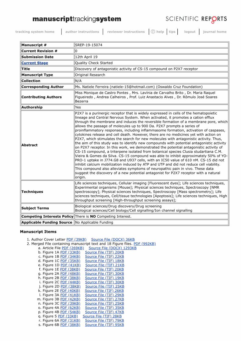
tracking system home author instructions reviewer instructions help tips logout journal home
Manuscript # SREP-19-15074
Current Revision # 0
Submission Date 12th April 19
Current Stage Quality Check Started
Title Discovery of antagonistic activity of CS-15 compound on P2X7 receptor
Manuscript Type Original Research
Collection N/A
Corresponding Author Ms. Natiele Ferreira ([email protected]) (Oswaldo Cruz Foundation)
Contributing AuthorsMiss Monique de Castro Pontes , Mrs. Lavínia de Carvalho Brito , Dr. Maria RaquelFigueiredo , Andrea Calheiros , Prof. Luiz Anastacio Alves , Dr. Rômulo José SoaresBezerra
Authorship Yes
Abstract
P2X7 is a purinergic receptor that is widely expressed in cells of the hematopoieticlineage and Central Nervous System. When activated, it promotes a cation effluxthrough the membrane and induces the reversible formation of a membrane pore, whichallows the passage of molecules up to 900 Da. P2X7 prompts a series ofproinflammatory responses, including inflammasome formation, activation of caspases,cytokines release and cell death. However, there are no medicines yet with action onP2X7, which stimulates the search for new molecules with antagonistic activity. Thus,the aim of this study was to identify new compounds with potential antagonistic activityon P2X7 receptor. In this work, we demonstrated the potential antagonistic activity ofCS-15 compound, a triterpene isolated from botanical species Clusia studartiana C.M.Vieira & Gomes da Silva. CS-15 compound was able to inhibit approximately 50% of YO-PRO-1 uptake in J774.G8 and U937 cells, with an IC50 value of 610 nM. CS-15 did notinhibit calcium mobilization induced by ATP and UTP and did not reduce cell viability.This compound also alleviates symptoms of neuropathic pain in vivo. These datasuggest the discovery of a new potential antagonist for P2X7 receptor with a naturalorigin.
Techniques
Life sciences techniques, Cellular imaging [Fluorescent dyes]; Life sciences techniques,Experimental organisms [Mouse]; Physical sciences techniques, Spectroscopy [NMRspectroscopy]; Physical sciences techniques, Spectroscopy [Mass spectrometry]; Lifesciences techniques, Cell/tissue technologies [Apoptosis]; Life sciences techniques, Highthroughput screening [High-throughput screening assays];
Subject Terms Biological sciences/Drug discovery/Drug screening Biological sciences/Cell biology/Cell signalling/Ion channel signalling
Competing Interests Policy There is NO Competing Interest.
Applicable Funding Source No Applicable Funding
Manuscript Items
1. Author Cover Letter PDF (39KB) Source File (DOCX) 36KB 2. Merged File containing manuscript text and 18 Figure files. PDF (992KB)
a. Article File PDF (269KB) Source File (DOCX) 1293KB b. Figure 1A PDF (33KB) Source File (TIF) 20KB c. Figure 1B PDF (34KB) Source File (TIF) 22KB d. Figure 1C PDF (35KB) Source File (TIF) 18KB e. Figure 1D PDF (41KB) Source File (TIF) 21KB f. Figure 1E PDF (38KB) Source File (TIF) 20KB g. Figure 2A PDF (48KB) Source File (TIF) 30KB h. Figure 2B PDF (38KB) Source File (TIF) 19KB i. Figure 2C PDF (44KB) Source File (TIF) 30KB j. Figure 2D PDF (38KB) Source File (TIF) 25KB k. Figure 2E PDF (40KB) Source File (TIF) 26KB l. Figure 3A PDF (41KB) Source File (TIF) 29KB
m. Figure 3B PDF (42KB) Source File (TIF) 27KB n. Figure 3C PDF (39KB) Source File (TIF) 25KB o. Figure 4A PDF (62KB) Source File (TIF) 35KB p. Figure 4B PDF (54KB) Source File (TIF) 47KB q. Figure 5 PDF (33KB) Source File (TIF) 28KB r. Figure 6A PDF (31KB) Source File (TIF) 79KB s. Figure 6B PDF (38KB) Source File (TIF) 95KB
Discovery of antagonistic activity of CS-15 compound onP2X7 receptor
Natiele Carla da Silva Ferreira1, Monique de Castro Pontes1, Lavınia de Carvalho Brito2,3, Maria
Raquel Figueiredo2, Andrea Surrage Calheiros4, Luiz Anastacio Alves & Romulo Jose Soares
Bezerra5,∗
1Oswaldo Cruz Institute, Laboratory of Cellular Communication, Oswaldo Cruz Foundation
2Institute of Technology in Drugs, Laboratory of Chemistry of Natural Products, Oswaldo Cruz
Foundation
3Institute of Chemistry, Central Analytical Fernanda Coutinho, State University of Rio de Janeiro
4Oswaldo Cruz Institute, Laboratory of Immunopharmacology, Oswaldo Cruz Foundation
5Oswaldo Cruz Institute, Laboratory of Technological Development in Virology, Oswaldo Cruz
Foundation
ABSTRACT
P2X7 is a purinergic receptor that is widely expressed in cells of the hematopoietic lineage and
Central Nervous System. When activated, it promotes a cation efflux through the membrane and
induces the reversible formation of a membrane pore, which allows the passage of molecules up to
900 Da. P2X7 prompts a series of proinflammatory responses, including inflammasome formation,
activation of caspases, cytokines release and cell death. However, there are no medicines yet with
action on P2X7, which stimulates the search for new molecules with antagonistic activity. Thus,
the aim of this study was to identify new compounds with potential antagonistic activity on P2X7
receptor. In this work, we demonstrated the potential antagonistic activity of CS-15 compound, a
triterpene isolated from botanical species Clusiastudartiana C.M. Vieira & Gomes da Silva. CS-
15 compound was able to inhibit approximately 50% of YO-PRO-1 uptake in J774.G8 and U937
1

cells, with an IC50 value of 610 nM. CS-15 did not inhibit calcium mobilization induced by ATP
and UTP and did not reduce cell viability. This compound also alleviates symptoms of neuropathic
pain in vivo. These data suggest the discovery of a new potential antagonist for P2X7 receptor with
a natural origin.
Keywords: P2X7, antagonist, natural products
Introduction
Purinergic receptors are plasma membrane receptors, which are activated by extracellular purines
and pyrimidines. In humans, these receptors are divided into two families: adenosine-activated P1
receptors and nucleotide-activated P2 receptors. P2 receptors are also grouped into two classes:
G-coupled P2Y receptors and ionotropic P2X receptors1. Among P2X receptors, P2X7 stands out
by its biophysical characteristics and physiological roles in inflammation, pain, and neurodegen-
eration.
P2X7 is expressed mainly in cells of the hematopoietic lineage, such as mast cells, ery-
throcytes, monocytes, macrophages, dendritic cells, and lymphocytes, as well as glial cells2. Once
activated, this receptor promotes the flow of Ca2+, Na+, and K+ cations through the cell membrane
according to the electrochemical gradient. However, in cases of prolonged activation, it induces
the reversible formation of a pore, which allows the passage of molecules up to 900 Da, such as
dyes propidium iodide (668 Da), ethidium bromide (394 Da) and YO- PRO-1 (629 Da), which are
widely used for the study of P2X7 activity3.
In addition, activation of P2X7 prompts a series of proinflammatory responses, including in-
flammasome formation, activation of caspases, phospholipases, mitogen-activated protein kinases
2
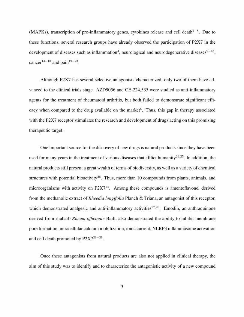
(MAPKs), transcription of pro-inflammatory genes, cytokines release and cell death4−8. Due to
these functions, several research groups have already observed the participation of P2X7 in the
development of diseases such as inflammation4, neurological and neurodegenerative diseases9−13,
cancer14−18 and pain19−23.
Although P2X7 has several selective antagonists characterized, only two of them have ad-
vanced to the clinical trials stage. AZD9056 and CE-224,535 were studied as anti-inflammatory
agents for the treatment of rheumatoid arthritis, but both failed to demonstrate significant effi-
cacy when compared to the drug available on the market6. Thus, this gap in therapy associated
with the P2X7 receptor stimulates the research and development of drugs acting on this promising
therapeutic target.
One important source for the discovery of new drugs is natural products since they have been
used for many years in the treatment of various diseases that afflict humanity24,25. In addition, the
natural products still present a great wealth of terms of biodiversity, as well as a variety of chemical
structures with potential bioactivity26. Thus, more than 10 compounds from plants, animals, and
microorganisms with activity on P2X724. Among these compounds is amentoflavone, derived
from the methanolic extract of Rheedia longifolia Planch & Triana, an antagonist of this receptor,
which demonstrated analgesic and anti-inflammatory activities27,28. Emodin, an anthraquinone
derived from rhubarb Rheum officinale Baill, also demonstrated the ability to inhibit membrane
pore formation, intracellular calcium mobilization, ionic current, NLRP3 inflammasome activation
and cell death promoted by P2X729−31.
Once these antagonists from natural products are also not applied in clinical therapy, the
aim of this study was to identify and to characterize the antagonistic activity of a new compound
3
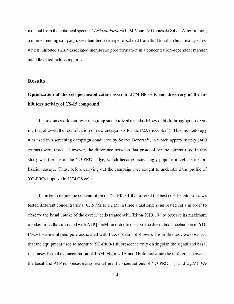
isolated from the botanical species Clusiastudartiana C.M.Vieira & Gomes da Silva. After running
a mini-screening campaign, we identified a triterpene isolated from this Brazilian botanical species,
which inhibited P2X7-associated membrane pore formation in a concentration-dependent manner
and alleviated pain symptoms.
Results
Optimization of the cell permeabilization assay in J774.G8 cells and discovery of the in-
hibitory activity of CS-15 compound
In previous work, our research group standardized a methodology of high-throughput screen-
ing that allowed the identification of new antagonists for the P2X7 receptor32. This methodology
was used in a screening campaign conducted by Soares Bezerra33, in which approximately 1800
extracts were tested. However, the difference between that protocol for the current used in this
study was the use of the YO-PRO-1 dye, which became increasingly popular in cell permeabi-
lization assays. Thus, before carrying out the campaign, we sought to understand the profile of
YO-PRO-1 uptake in J774.G8 cells.
In order to define the concentration of YO-PRO-1 that offered the best cost-benefit ratio, we
tested different concentrations (62.5 nM to 8 µM) in three situations: i) untreated cells in order to
observe the basal uptake of the dye; ii) cells treated with Triton-X [0.1%] to observe its maximum
uptake; iii) cells stimulated with ATP [5 mM] in order to observe the dye uptake mechanism of YO-
PRO-1 via membrane pore associated with P2X7 (data not shown). From this test, we observed
that the equipment used to measure YO-PRO-1 fluorescence only distinguish the signal and basal
responses from the concentration of 1 µM. Figures 1A and 1B demonstrate the difference between
the basal and ATP responses using two different concentrations of YO-PRO-1 (1 and 2 µM). We
4

observed that there was a significant difference between the basal and ATP responses, and the
signal-background ratio was 1.42 and 1.65 for the concentrations of 1 and 2 µM, respectively.
Respecting the concentration in which we obtained the best signal-background ratio, we chose to
use the concentration of 2 µM.
Using this concentration of YO-PRO-1, we obtained the P2X7 agonist and antagonist’s pro-
files. J774.G8 cells were stimulated with increasing concentrations of ATP and the EC50 found
was 2.84 mM, as shown in Figure 1C. To obtain an inhibition profile, we selected two P2X7
antagonists, one selective (A-740003) and another non-selective (Brilliant Blue G - BBG). Cells
were treated with increasing concentrations of these two antagonists and then stimulated with ATP.
Thus, we obtained an IC50 of 511 nM for A-740003 (Figure 1D) and 106 nM for BBG (Figure 1E).
In addition, we used the formula for calculating z’, a statistical parameter that measures how robust
the test is, and we obtained a value of 0.59. Since the value found was greater than 0.5, this means
that the conditions of our trial are optimal for the execution of the screening.
Finally, we conducted our mini-screening campaign and tested 134 extracts and substances
isolated from natural products. Among these samples, the CS-15 compound was distinguished by
its ability to reduce by almost half the uptake of YO-PRO-1 after stimulation with ATP.
CS-15 compound significantly inhibited dye uptake in murine and human cell lines, but
not calcium mobilization
During the screening campaign, we observed that the CS-15 compound reduced 52% of
the YO-PRO-1 uptake in J774.G8 cells stimulated with ATP, as shown in Figure 2A. We asked if
this compound could inhibit this activity in lower concentrations. For this, we treated cells with
increasing concentrations of the compound and then stimulated with ATP. As demonstrated in
5
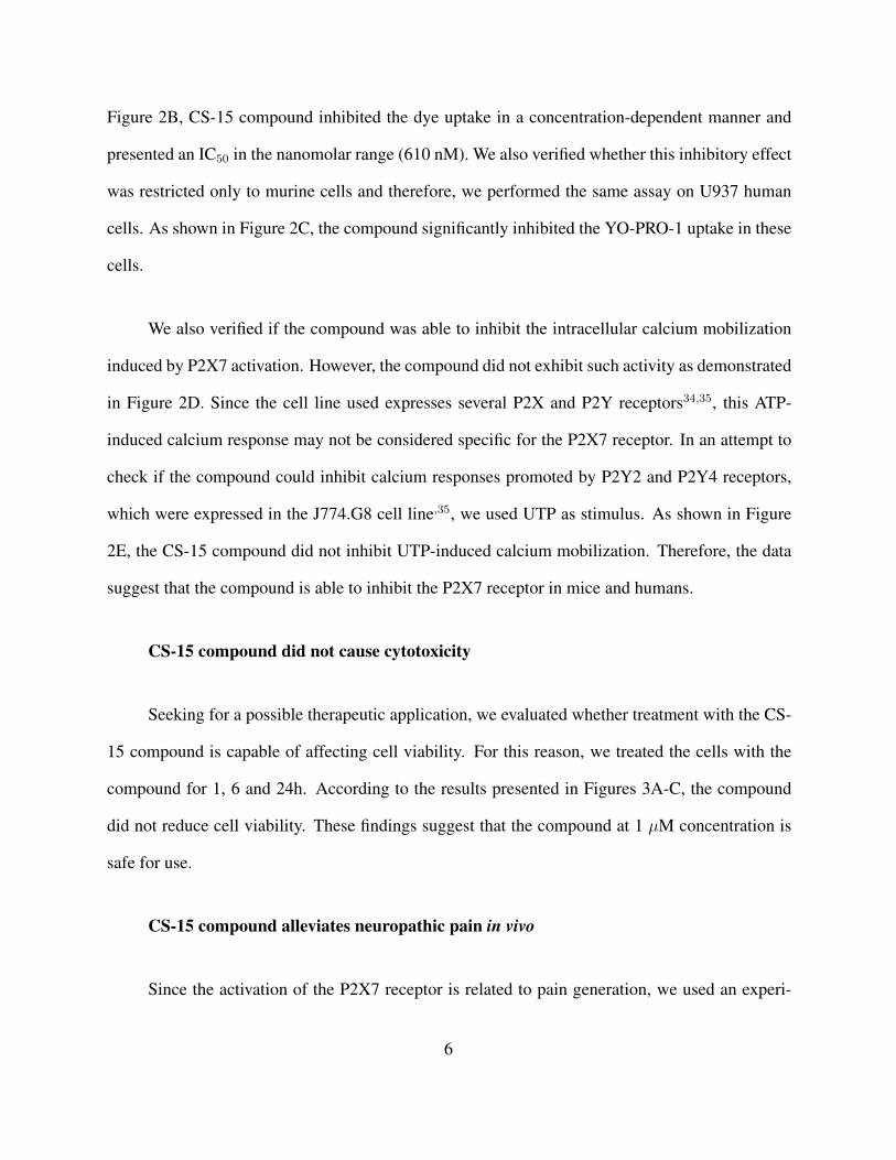
Figure 2B, CS-15 compound inhibited the dye uptake in a concentration-dependent manner and
presented an IC50 in the nanomolar range (610 nM). We also verified whether this inhibitory effect
was restricted only to murine cells and therefore, we performed the same assay on U937 human
cells. As shown in Figure 2C, the compound significantly inhibited the YO-PRO-1 uptake in these
cells.
We also verified if the compound was able to inhibit the intracellular calcium mobilization
induced by P2X7 activation. However, the compound did not exhibit such activity as demonstrated
in Figure 2D. Since the cell line used expresses several P2X and P2Y receptors34,35, this ATP-
induced calcium response may not be considered specific for the P2X7 receptor. In an attempt to
check if the compound could inhibit calcium responses promoted by P2Y2 and P2Y4 receptors,
which were expressed in the J774.G8 cell line,35, we used UTP as stimulus. As shown in Figure
2E, the CS-15 compound did not inhibit UTP-induced calcium mobilization. Therefore, the data
suggest that the compound is able to inhibit the P2X7 receptor in mice and humans.
CS-15 compound did not cause cytotoxicity
Seeking for a possible therapeutic application, we evaluated whether treatment with the CS-
15 compound is capable of affecting cell viability. For this reason, we treated the cells with the
compound for 1, 6 and 24h. According to the results presented in Figures 3A-C, the compound
did not reduce cell viability. These findings suggest that the compound at 1 µM concentration is
safe for use.
CS-15 compound alleviates neuropathic pain in vivo
Since the activation of the P2X7 receptor is related to pain generation, we used an experi-
6
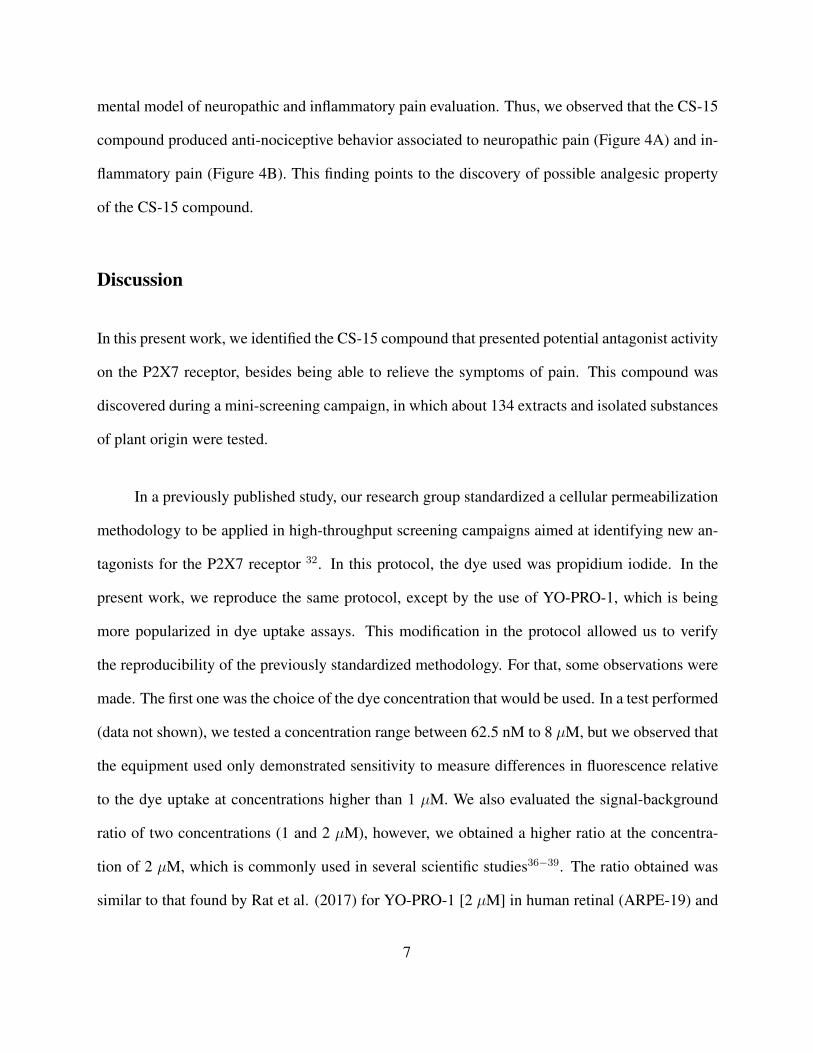
mental model of neuropathic and inflammatory pain evaluation. Thus, we observed that the CS-15
compound produced anti-nociceptive behavior associated to neuropathic pain (Figure 4A) and in-
flammatory pain (Figure 4B). This finding points to the discovery of possible analgesic property
of the CS-15 compound.
Discussion
In this present work, we identified the CS-15 compound that presented potential antagonist activity
on the P2X7 receptor, besides being able to relieve the symptoms of pain. This compound was
discovered during a mini-screening campaign, in which about 134 extracts and isolated substances
of plant origin were tested.
In a previously published study, our research group standardized a cellular permeabilization
methodology to be applied in high-throughput screening campaigns aimed at identifying new an-
tagonists for the P2X7 receptor 32. In this protocol, the dye used was propidium iodide. In the
present work, we reproduce the same protocol, except by the use of YO-PRO-1, which is being
more popularized in dye uptake assays. This modification in the protocol allowed us to verify
the reproducibility of the previously standardized methodology. For that, some observations were
made. The first one was the choice of the dye concentration that would be used. In a test performed
(data not shown), we tested a concentration range between 62.5 nM to 8 µM, but we observed that
the equipment used only demonstrated sensitivity to measure differences in fluorescence relative
to the dye uptake at concentrations higher than 1 µM. We also evaluated the signal-background
ratio of two concentrations (1 and 2 µM), however, we obtained a higher ratio at the concentra-
tion of 2 µM, which is commonly used in several scientific studies36−39. The ratio obtained was
similar to that found by Rat et al. (2017) for YO-PRO-1 [2 µM] in human retinal (ARPE-19) and
7
keratinocyte (HaCat) cell lines (1.7 and 1.45, respectively) in the presence of 1 and 10 mM ATP36.
With the definition of the YO-PRO-1 concentration, we continue with our experiments in
the spectrophotometer, evaluating the profiles of dye uptake in the presence of the agonist and
antagonists. The EC50 detected for ATP was 2.18 mM, and thus was within the EC50 range for
P2X7 (2 to 4 mM) described in the work of Coddou et al. (2011)40. However, in our previous work,
we found an EC50 between 0.7114 and 0.7663 mM32. This difference could be due to variations
in the sensitivity of the equipment or dye used. We cannot rule out that there may be differences
in the J774.G8 subclones, which could for some reason be expressing the P2X7 receptor in lower
density or a different splice variant, resistance in forming the membrane pore associated with that
receptor41. Moreover, we calculated z’-factor, a measure of robustness of assay, and we found a
value of 0.59, which indicates that our protocol was adequate for use. This value was similar to that
found in our protocol with propidium iodide (0.635) but was smaller than that found using Lucifer
yellow (0.876), indicating perhaps that there are variations in sensitivity between the dyes32.
The IC50 detected for the BBG antagonist was 106 nM and also falls within the range of IC50
described in the literature (ranging from 15 to 250 nM)40. On the other hand, it was lower than
that found in our protocol with propidium iodide, which was 1.3 to 2.6 µM32. We also tested the
P2X7 selective antagonist, A-740003, and obtained an IC50 of 511 nM, which is also within the
range described in the literature (ranging from 20 to 700 nM)40.
After the characterization of YO-PRO-1 profile applied to cell permeability assays in J774.G8,
we performed a mini-screening campaign and identified CS-15 compound as a promising hit. It
blocked around 50% YO-PRO-1 uptake in J774.G8 and U937 cell lines. In addition, it was able to
inhibit the uptake of YO-PRO-1 via a P2X7-associated pore in a concentration-dependent manner,
8
presenting an IC50 at submicromolar concentration. This effect is interesting because most hits
have the desired response of 1 to 10 µM, and what the data suggest, it is active in the nanomolar
range42. We further tested whether it could produce any toxicity to the cells, and it has been shown
safe for use within 24 hours of treatment using the MTT technique.
We also tried to verify whether this compound would be able to inhibit ATP-induced calcium
mobilization. In order to activate the P2X7 receptor, we need to use a high ATP concentration
(100 µM); however, it was high sufficient to activate other P2 receptors. Since the J774.G8 cell
expresses several P2 receptor subtypes including P2X and P2Y34,35, we are unable to observe the
expected inhibition using this model. An alternative to achieving a higher success rate in this
experiment would be the use of cells transfected with P2X7 or the use of BzATP, a more potent
but not selective agonist40. We further tested another nucleotide, UTP, in order to try to observe
whether this inhibition extended to P2Y receptors, but the compound was not able to inhibit the
calcium response. In any case, new assays with agonists of other P2 receptors must be made in
order to confirm if there is selectivity for the P2X7 receptor.
Finally, we tested the effect of the compound in alleviating the pain sensation induced by
intraplantar injection of formalin. The compound significantly reduced the sensation of pain orig-
inating from the activation of neuropathic and inflammatory pathways. This finding has already
been observed in experiments with other P2X7 antagonists20,28, and support the pharmacological
potential of this compound.
CS-15 compound is a triterpene isolated from botanical species Clusia studartiana C.M.
Vieira & Gomes da Silva, which belongs to Clusiaceae family. It is a tree of terrestrial habit, but
occasionally it can appear hemiepiphyte form. This species is endemic in Brazil, although other
9
individuals of this family can be found in the tropics. This species is native to the Atlantic Forest
in Southwest Brazil43. This species was discovered in the early 1990s and to date, there is no
information in the literature about its medicinal properties, which makes our study a pioneer in
this regard. Despite this, there is some information about the medicinal properties of the botanical
family Clusiaceae. The latex present in the fruits of Clusia sp., for example, are popularly used
in the treatment of rheumatic diseases and infant oral candidiasis, while the C. grandiflora seed
contains compounds with antimicrobial activity44−47.
We do not yet know whether these effects are dependent or independent of P2X7 receptor
activation. For this, new experiments in which the analysis of cytokines such as IL-1β and other
inflammatory mediators related to P2X7 need to be investigated in vitro and invivo. Besides that,
experiments using hP2X7-transfected cells, whole cell patch-clamp, and selective agonists could
significantly contribute to characterize the activity of CS-15 compound on P2X7. Despite these
limitations, which are within the future perspectives of our group, this study demonstrated for the
first time a medicinal effect of the C. studartiana compound on inflammation and pain possibly
caused by the P2X7 receptor blockade.
Thus, in this work, we characterized and standardized a protocol for dye uptake assay in
J774.G8 cells using YO-PRO-1 dye and applied it in a mini-screening campaign. We discovered
the inhibitory activity of the CS-15 compound on pore formation associated with P2X7 activation.
This compound also alleviates the symptoms of neuropathic pain and it did not cause cytotoxicity
in vitro. These data suggest the discovery of a new potential antagonist for P2X7 receptor that has
a natural origin.
10

Methods
Reagents
ATP, BBG, A-740003, Triton-X, DMEM and RPMI culture mediums, NaCl, KCl, MgCl2,
CaCl2, Na2HPO4, KH2PO4, Glucose, Hepes, DMSO, MTT (3-(4,5-dimethylthiazol-2-yl)- 2,5-
diphenyltetrazolium bromide) tetrazolium, Probenecid and Ionomycin were purchased from Sigma
Chemical Co. (St. Louis, Missouri, USA). Fetal bovine serum was obtained from Gibco (Okla-
homa, USA). Fluo-4 AM was obtained from Life Technologies (California, USA). YO-PRO-1
was obtained from Molecular Probes (Oregon, USA). Morphine was acquired from Cristalia (Sao
Paulo, Brazil) and Sodium diclofenac was obtained from Medley (Sao Paulo, Brazil).
Plant material
Aerial parts of C. studartiana C.M. Vieira & Gomes da Silva (Clusiaceae) were collected in
the Ecological Reserve of Macae de Cima, Atlantic Forest, Nova Friburgo, Rio de Janeiro, RJ. The
plant material was identified by Dr. Marcos Nadruz and a voucher specimen was deposited at the
Herbarium of the Botanical Garden of Rio de Janeiro under the number RB 336999. The species
is authorized by the CGEN at the register number AB5D582.
Extraction, isolation, purification and methylation of 3-oxo-olean-12-en-28-oic-acid (CS-
15 compound)
The collected material of C. studartiana was dried at 40ºC in an oven with air circulation and
reduced to small fragments. The extract was prepared by dynamic maceration at room temperature
with hexane to exhaustion, and then, filtered and evaporated under reduced pressure. During the
concentration of the extract, an amorphous solid was obtained and was chromatographed on silica
11
gel using solvent mixtures with increasing polarities (cyclopentane, cyclopentane/ethyl acetate,
ethyl acetate, and methanol). The resulting fractions were analyzed by thin layer chromatogra-
phy (CCD) and visualized with Godin’s detection reagent. After successive recrystallizations,
the procedure resulted in the isolation of a pentacyclic triterpene (3-oxo-olean-12-en-28-oic acid).
The molecular formula of this acid was determined by direct insertion in a Bruker micrOTOF-
QII mass spectrometer by precise mass and algorithmic analysis of its isotope standard (Bruker
SmartFormula 3D True Isotopic Pattern). Methylation of this acid with diazomethane48 yielded
methyl 3-oxo-olean-12-en-28-oate (Figure 5), confirmed by GC-MS data (Figures 6A and 6B).
The triterpenes were characterized by uni and bidimensional methods in 1H (500 MHz) and 13C
NMR (125MHz) and confirmed by comparison with spectral literature data49.
Methyl 3-oxo-olean-12-en-28-oate
13C NMR, δ ppm (125 MHz; CDCl3): 39.14 (C-1), 34.16 (C-2), 217,60 (C-3), 47,46 (C-
4), 55,36 (C-5), 19,59 (C-6), 32,21 (C-7), 39,32 (C-8), 46,90 (C-9), 36,83 (C-10), 22,97 (C-11),
122,40 (C-12), 143,66 (C-13), 41,79 (C-14), 27,72 (C-15), 23,51 (C-16), 46,60 (C-17), 41,12 (C-
18), 45,86 (C-19), 30,70 (C-20), 33,83 (C-21), 32,43 (C-22), 26,48 (C-23), 21,45 (C-24), 15,03
(C-25), 17,03 (C-26), 25,84 (C-27), 176,27 (C-28), 33,06 (C-29), 23,58 (C-30), 51,4 (OCH3).
Cell culture
J774.G8 and U937 cells were cultivated with Dulbecco’s Modified Eagle’s Medium (DMEM)
or RPPMI medium, respectively, supplemented with 10% fetal bovine serum, at 37◦C in a 5% CO2
atmosphere. The cellular medium was changed twice a week and the cells density were adjusted to
2x10⁶ cells per 150 cm2 cell culture flask (Corning, New York, USA). Cell viability was measured
by trypan blue exclusion and the experiments were conducted only when viability was above 90%.
12
Dye uptake assay
J774.G8 and U937 cells were plated in a 96-well black plate (Corning, New York, USA), at
a concentration of 2x10⁵ cells/well. They were maintained in culture with appropriated medium
supplemented with 10% fetal bovine serum, at 37◦C in a 5% CO2 atmosphere for 24h. Then, the
medium was replaced by extracellular saline solution (150 mM NaCl, 5 mM KCl, 1 mM MgCl2,
1 mM CaCl2, and 10 mM HEPES, pH 7.4). Subsequently, cells were treated with Brilliant Blue
G (BBG) [1 µM] and CS-15 compound [1 µM] for 30 minutes. Then, cells were stimulated with
ATP [5 mM] for 15 minutes, in order to allow the opening of the membrane pore associated with
the P2X7 receptor. Next, the cationic dye YO-PRO-1 was added on each well at a concentration of
2 µM, and after 3 minutes, its fluorescence was measured using FlexStation III equipment (Molec-
ular Devices, California, USA) at wavelengths of 488 nm and 509 nm for excitation and emission,
respectively. Cells treated with Triton-X [0.1%] were considered as the control of maximum dye
uptake.
Calcium assays
J774.G8 cells were plated in a 96-well black-wall and clear-bottom plate (Corning, New
York, USA), at a concentration of 2x10⁵ cells/well. They were maintained in culture with DMEM
supplemented with 10% fetal bovine serum, at 37◦C in a 5% CO2 atmosphere for 24h. Then,
the medium was replaced by 100 µl of Dulbecco’s PBS (DPBS) (136 mM NaCl, 2.68 mM KCl,
8 mM Na2HPO4, 1.46 mM KH2PO4, 0.5 mM MgCl2, 0.9 mM CaCl2, 5.55 mM Glucose and
2.5 mM Probenecid – pH 7.4) containing [2 µM] Fluo-4 AM and cells were incubated for 60
minutes at 37ºC in a 5% CO2 atmosphere. Next, the wells were washed three times with 200 µl
of DPBS and then, each well received a final volume of 150 µl of DPBS. Subsequently, cells were
13

treated with BBG [1 µM], Reactive Blue-2 (RB2) [20 µM] and CS-15 compound [1 µM] for 30
minutes. The mobilization of intracellular calcium was measured in FlexStation III (Molecular
Devices, California, USA) in real time during 90 seconds, at wavelengths of 485 nm and 525 nm
for excitation and emission, respectively. The agonists (ATP and UTP) were added at the time of 20
seconds and the readings were conducted every 1.52 seconds, totaling a value of 60 readings/well.
The calcium mobilization was quantified as ∆FU (the difference between fluorescence peak and
basal fluorescence)50.
Cell viability measurement
J774.G8 cells were plated in a 96-well plate (Corning, New York, USA) at a concentration of
2x10⁵ cells/well. They were maintained in culture with DMEM without phenol red supplemented
with 10% fetal bovine serum, at 37◦C in a 5% CO2 atmosphere for 24h. Next, cells were treated
with BBG [1 µM] and CS-15 compound [1 µM] for 1, 6 or 24h. Untreated cells were considered
our positive control of viability, while cells treated with Triton-X [0.1%] were considered our
negative control. After the treatments, the medium was replaced by 200 µL of DMEM without
phenol red containing MTT solution [100 µg/well]. The plate was incubated for 3h at 37◦C in a
5% CO2 atmosphere, and then, was centrifuged at 1500 RPM for 1 minute. Supernatants were
collected and formazan crystals were dissolved in 100 µl of DMSO. The absorbance of the wells
was measured in Spectramax 190 equipment (Molecular Devices, California, USA), using λ: 570
nm.
Animals
The experiments with animals follow ethical principles in animal experimentation of the
Brazilian College of Animal Experimentation. They were approved by the Ethics Committee on
14

the use of Animals from Oswaldo Cruz Institute on November 24, 2017 (L-037/2017). Healthy
male Swiss mice with a weight between 30 and 50g were obtained from Oswaldo Cruz Central
Bioterium. The animals were housed under conditions of 12h/12h light and dark cycle with free
access to food and water.
Formalin test
At the day of the test, animals were deprived of food and water for 1h before the experiment.
Subsequently, the animals were separated by groups with six animals each: (1) vehicle group,
which received 200 µL of saline; (2) positive control groups, which received anti-inflammatory or
analgesic drugs: sodium diclofenac [50 mg/kg] and morphine [10 mg/kg], respectively; (3) P2X7
reference antagonist group, which received BBG [100 mg/kg]; and (4) test groups, which received
CS-15 compound [100 or 200 µg/kg]. All drugs were orally administered with the exception
of morphine, which was administered via intraperitoneally. After one hour of treatment (or 30
minutes, in the case of morphine-treated group), all animals received a formalin injection (the pain
inductor agent) [2.5%] into the hind paw. The time that each animal spent licking the hind paw
was counted at 0 to 5 minutes to analyze the activity of the compound on neuropathic pain, and
later, between 15 to 30 minutes to evaluate inflammatory pain.
Data analysis
Each sample was measured in triplicate and all experiments were performed on at least three
independent days. All data are presented as mean ± S.D. The D’ Agostino and Pearson normality
test was used in order to test if samples follow a Gaussian distribution. If data follow a Gaussian
distribution, an appropriate parametric test was applied, if not, an appropriate non-parametric test
was applied. Used tests were specified in figure legends. P values of 0.05 or less were considered
15

significant. Graphs and statistical analyses were performed by GraphPad Prism version 5 (Graph-
Pad Software, San Diego, California, USA). We also assessed the quality of our dye uptake assay
by calculating the z’-factor. z’-factor is a valuable tool to evaluate the robustness and suitability of
HTS assays. z’-factor values above 0.5 mean that the assay is considered excellent and equal to 1,
an ideal assay. However, values below 0.5, mean that the assay needs to be reformulated51. This
parameter was calculated using the following equation:
z′ = 1− (3SD of positive control + 3SD of negative control)(mean of positive control – mean of negative control)
where cells stimulated with ATP at [10 mM] were considered as a positive control, while
cells without treatment were considered as a negative control.
The signal-background ratio between the positive and negative permeabilization controls
represented by ATP and untreated cells, respectively, was obtained by the following calculation51:
Signal − background ratio = positive control mean (ATP )negative control mean (untreated cells)
References
1–51
Authors’ contributions
N.C.S.F., L.A.A. and R.J.S.B. designed experiments, N.C.S.F., M.C.P. and A.S.C. performed ex-
periments, N.C.S.F., L.C.B and L.A.A. performed data analysis, M.R.F. and L.C.B performed the
16
extraction, isolation and purification of chemical compound, N.C.S.F., M.R.F., L.C.B and R.J.S.B.
wrote the paper.
Acknowledgements
This work was supported by Fundacao de Amparo a Pesquisa do Estado do Rio de Janeiro (FAPERJ),
Conselho Nacional de Desenvolvimento Cientıfico e Tecnologico (CNPq) and Instituto Oswaldo
Cruz (IOC). The authors thank Dr. Vinıcius Cotta, who kindly donated the J774.G8 and U937 cell
lines and the Program for Technological Development in Tools for Health RPT-FIOCRUZ for the
use of its facilities.
References
1. Abbracchio, M. P. & Burnstock, G. Purinoceptors: Are there families of P2X and P2Y
purinoceptors? Pharmac Ther 64, 445–475 (1994).
2. Skaper, S. D., Debetto, P. & Giusti, P. The P2X7 purinergic receptor: from physiology to
neurological disorders. FASEB J 24, 337–345 (2010).
3. Alves, L. A. et al. The P2X7 receptor: Shifting from a low- to a high-conductance channel —
An enigmatic phenomenon? Biochim. Biophys. Acta - Biomembr 1838, 2578–2587 (2014).
4. Lister, M. F. et al. The role of the purinergic P2X7 receptor in inflammation. J. Inflamm 4, 5
(2007).
5. Friedle, S. A., Curet, M. A. & Watters, J. J. Recent patents on novel P2X(7) receptor antag-
onists and their potential for reducing central nervous system inflammation. Recent Pat. CNS
Drug Discov 5, 35–45 (2010).
17

6. Bartlett, R., Stokes, L. & Sluyter, R. The P2X7 receptor channel: Recent developments and
the use of P2X7 antagonists in models of disease. Pharmacol. Rev 66, 638–675 (2014).
7. Riteau, N. et al. ATP release and purinergic signaling: a common pathway for particle-
mediated inflammasome activation. Cell Death Dis 3, e403 (2012).
8. Latz, E., Ts, X. & Stutz, A. Activation and regulation of the inflammasomes. Nat Rev Immunol
13, 397–411 (2013).
9. Mesuret, G. et al. P2X7 receptor inhibition interrupts the progression of seizures in immature
rats and reduces hippocampal damage. CNS Neurosci. Ther 20, 556–564 (2014).
10. Jimenez-Pacheco, A. et al. Transient P2X7 receptor antagonism produces lasting reductions
in spontaneous seizures and gliosis in experimental temporal lobe epilepsy. J. Neurosci 36,
5920–32 (2016).
11. Diaz-Hernandez, J. I. et al. In vivo P2X7 inhibition reduces amyloid plaques in Alzheimer’s
disease through GSK3B and secretases. Neurobiol. Aging 33, 1816–1828 (2012).
12. Carmo, M. R. S. et al. The P2X7 receptor antagonist Brilliant Blue G attenuates contralat-
eral rotations in a rat model of Parkinsonism through a combined control of synaptotoxicity,
neurotoxicity and gliosis. Neuropharmacology 81, 142–152 (2014).
13. Gandelman, M., Levy, M., Cassina, P., Barbeito, L. & Beckman, J. S. P2X7 receptor-induced
death of motor neurons by a peroxynitrite/FAS-dependent pathway. J. Neurochem 126, 382–
388 (2013).
14. Hattori, F. et al. Feasibility study of B16 melanoma therapy using oxidized ATP to target
purinergic receptor P2X7. Eur. J. Pharmacol 695, 20–26 (2012).
18
15. Qiu, Y. et al. P2X7 mediates ATP-driven invasiveness in prostate cancer cells. PLoS One 9
(2014).
16. Takai, E., Tsukimoto, M., Harada, H. & Kojima, S. Autocrine signaling via release of ATP and
activation of P2X7 receptor influences motile activity of human lung cancer cells. Purinergic
Signal 1–11 (2014).
17. Giannuzzo, A., Pedersen, S. F. & Novak, I. The P2X7 receptor regulates cell survival, migra-
tion and invasion of pancreatic ductal adenocarcinoma cells. Mol. Cancer 14, 203 (2015).
18. Xia, J., Yu, X., Tang, L., Li, G. & He, T. P2X7 receptor stimulates breast cancer cell invasion
and migration via the AKT pathway. Oncol. Rep 34, 103–110 (2015).
19. Honore, P. et al. A-740003 [N-(1-{[(cyanoimino)(5-quinolinylamino) methyl]amino}-2,2-
dimethylpropyl)-2-(3,4-dimethoxyphenyl)acetamide], a novel and selective P2X7 receptor an-
tagonist, dose-dependently reduces neuropathic pain in the rat. J. Pharmacol. Exp. Ther 319,
1376–85 (2006).
20. Broom, D. C. et al. Characterization of N-(adamantan-1-ylmethyl)-5-[(3R-amino-pyrrolidin-
1-yl)methyl]-2-chloro-benzamide, a P2X7 antagonist in animal models of pain and inflamma-
tion. J. Pharmacol. Exp. Ther 327, 620–33 (2008).
21. Ochi-ishi, R. et al. Involvement of the chemokine CCL3 and the purinoceptor P2X7 in the
spinal cord in paclitaxel-induced mechanical allodynia. Mol. Pain 10, 53 (2014).
22. Teixeira, J. M., Oliveira-Fusaro, M. C. G., Parada, C. A. & Tambeli, C. H. Peripheral P2X7
receptor-induced mechanical hyperalgesia is mediated by bradykinin. Neuroscience 277, 163–
173 (2014).
19
23. Ying, Y. L. et al. Over-expression of P2X7 receptors in spinal glial cells contributes to the de-
velopment of chronic postsurgical pain induced by skin/muscle incision and retraction (SMIR)
in rats. Exp. Neurol 261, 836–843 (2014).
24. Faria, R., Ferreira, L., Bezerra, R., Frutuoso, V. & Alves, L. Action of natural products on P2
receptors: A reinvented era for drug discovery. Molecules 17, 13009–13025 (2012).
25. Chin, Y. W., Balunas, M. J., Chai, H. B. & Kinghorn, A. D. Drug discovery from natural
sources. AAPS J 8, E239–E253 (2006).
26. Gu, J. et al. Use of natural products as chemical library for drug discovery and network
pharmacology. PLoS One 8, e62839 (2013).
27. Santos, J. A. A. et al. Effect of Rheedia longifolia leaf extract and fractions on the P2X7
receptor in vitro: novel antagonists? J. Med. Food 14, 920–9 (2011).
28. Santos, J. A. A. et al. Chemical profile and antinociceptive efficacy of Rheedia longifolia leaf
extract. J. Med. Food 14, 1039–45 (2011).
29. Liu, L., Zou, J., Liu, X., Jiang, L. H. & Li, J. Inhibition of ATP-induced macrophage death by
emodin via antagonizing P2X7 receptor. Eur. J. Pharmacol 640, 15–19 (2010).
30. Jelassi, B. et al. Anthraquinone emodin inhibits human cancer cell invasiveness by antagoniz-
ing P2X7 receptors. Carcinogenesis 34, 1487–1496 (2013).
31. Han, J. W. et al. Anti-inflammatory effect of emodin via attenuation of NLRP3 inflammasome
activation. Int. Int. J. Mol. Sci 16, 8102–9 (2015).
32. Soares-Bezerra, R. J. et al. An improved method for P2X7R antagonist screening. . PLoS One
10 (2015).
20

33. Bezerra, R. S. & Instituto Oswaldo Cruz. Triagem de extratos vegetais e fungicos de diferentes
biomas para identificacao de antagonistas do receptor P2X7 (2012).
34. Coutinho-Silva, R. et al. Multiple P2X and P2Y receptor subtypes in mouse J774, spleen and
peritoneal macrophages. Biochem. Pharmacol 69, 641–655 (2005).
35. Ferreira, N. C. et al. New insights in purinergic therapy: Novel antagonists for uridine 5′-
triphosphate-activated P2Y receptors from Brazilian flora. J. Med. Food 22, 211–224 (2018).
36. Rat, P., Olivier, E., Tanter, C., Wakx, A. & Dutot, M. A fast and reproducible cell- and
96-well plate-based method for the evaluation of P2X7 receptor activation using YO-PRO-1
fluorescent dye. J. Biol. Methods 4, 64 (2017).
37. Jiang, L. H. et al. N-methyl-D-glucamine and propidium dyes utilize different permeation
pathways at rat P2X(7) receptors. Am. J. Physiol. Cell Physiol 289, C1295–302 (2005).
38. Cankurtaran-Sayar, S., Sayar, K. & Ugur, M. P2X7 receptor activates multiple selective dye-
permeation pathways in RAW 264.7 and Human Embryonic Kidney 293 cells. Mol. Pharma-
col 76, 1323–1332 (2009).
39. Browne, L. E., Compan, V., Bragg, L. & North, R. A. P2X7 receptor channels allow direct
permeation of nanometer-sized dyes. J. Neurosci 33, 3557–3566 (2013).
40. Coddou, C., Yan, Z., Obsil, T., Huidobro-Toro, J. P. & Stojilkovic, S. S. Activation and
regulation of purinergic P2X receptor channels. Pharmacol. Rev 63, 641–683 (2011).
41. Rissiek, B., Haag, F., Boyer, O., Koch-Nolte, F. & Adriouch, S. P2X7 on mouse T cells: One
channel, many functions. Front. Immunol 6, 204 (2015).
42. Hughes, J. P., Rees, S., Kalindjian, S. B. & Philpott, K. L. Principles of early drug discovery.
Br. J. Pharmacol 162, 1239–49 (2011).
21
43. do Rio de Janeiro, J. B. Clusia in Flora do Brasil 2020 em construcao (2019). URL http:
//reflora.jbrj.gov.br/reflora/floradobrasil/FB6849.
44. Camara, C. G., Marsaioli, A. & Bittrich, V. Chemical constituents of apolar fractions from
fruit latex of twelve Clusia species (Clusiaceae). . An. Acad. Bras. Cienc 90, 1919–1927
(2018).
45. Sanz-Biset, J., de-la Cruz, J. C., Epiquien-Rivera, M. A. & Canigueral, S. A first survey on the
medicinal plants of the Chazuta valley (Peruvian Amazon). J. Ethnopharmacol 122, 333–362
(2009).
46. Barbosa, W. L. R. & Pinto, L. N. Documentacao e valorizacao da fitoterapia tradicional
Kayapo nas aldeias A’Ukre e Pykanu-sudeste do Para. Rev. Bras. Farmacogn 13 (2003).
47. Lokvam, J., Braddock, J. F., Reichardt, P. B. & Clausen, T. P. Two polyisoprenylated ben-
zophenones from the trunk latex of Clusia grandiflora (Clusiaceae). Phytochemistry 55, 29–34
(2000).
48. Vogel, A., Mendham, J., Denney, R., Barnes, J. & Thomas, M. Analise Quımica Quantitativa
(LTC, 2002).
49. Mahato, S. B. & Kundu, A. P. 13C NMR Spectra of pentacyclic triterpenoids—a compilation
and some salient features. Phytochemistry 37, 1517–1575 (1994).
50. Hansen, K. B. & Brauner-Osborne, H. FLIPR assays of intracellular calcium in GPCR drug
discovery. . In Methods in Molecular Biology, vol. 552, 269–278 (Clifton, 2009).
51. Zhang, J.-H., Chung, T. & Oldenburg, K. R. A simple statistical parameter for use in evaluation
and validation of high throughput screening assays. J. Biomol. Screen. 4, 67–73 (1999).
22

Competing interests
The authors declare no conflicts of interest.
23

Figure 1: Profile of YO-PRO-1 uptake assay via P2X7 pore with J774.G8 cells. J774.G8 cellswere stimulated with ATP [5 mM] for 15 minutes and then, received 1 µM (A) or 2 µM(B) ofYO-PRO-1. Data are presented as mean ± SD of three independent experiments performed intriplicate. Data analysis were made using Paired t-Test; p= 0.0099 (A) and p=0.0132 (B). Datawere normalized as a percentage in function of the mean of units of arbitrary fluorescence (RFU)obtained by Triton-X, the maximum fluorescence control. Asterisks indicate a significant differ-ence between means. J774.G8 cells were stimulated with increasing concentrations of ATP [0.078to 20 mM] for15 minutes and then, received YO-PRO-1 [2 µM] (C). J774.G8 cells were pretreatedwith increasing concentrations of A740003 [0.078 to 10 µM] (D) and BBG [0.078 to 10 µM] (E)for 30 minutes. Then, cells were stimulated with ATP [5 mM] for 15 minutes and received YO-PRO-1 [2 µM]. Data are presented as mean ±SE of three independent experiments performed intriplicate.
24

Figure 2: Compound CS-15 inhibits P2X7 pore formation. J774.G8 cells were pretreated withCS-15 compound [1 µM] and BBG [1 µM] during 30 minutes and were stimulated with ATP [5mM] for further 15 minutes. Then, YO-PRO-1 dye [2 µM]was added to the wells and fluores-cence of cells was measured. Data are presented as mean ± SD of three independent experimentsperformed in triplicate. Data analysis were made using one-way ANOVA and Tukey post-test;p<0.0001. Data were normalized as a percentage in function of the mean of units of arbitraryfluorescence (RFU) obtained by Triton-X, the maximum fluorescence control. Asterisks indicatea significant difference in relation to ATP (A). J774.G8 cells were pretreated with increasing con-centrations of CS-15 compound [0.0625 to 8 µM]for 30 minutes. Then, cells were stimulated withATP [5 mM] for 15 minutes and received YO-PRO-1 [2 µM]. Data are presented as mean ± SE ofthree independent experiments performed in triplicate(B). U937 cells were pretreated with CS-15compound [1 µM] and BBG [1 µM] during 30 minutes and were stimulated with ATP [5 mM] forfurther 15 minutes. Then, YO-PRO-1 dye [2 µM] was added to the wells and fluorescence of cellswas measured. Data are presented as mean ± SD of three independent experiments performedin triplicate. Data analysis were made using one-way ANOVA and Tukey post-test; p<0.0001.Data were normalized as a percentage in function of the mean of units of arbitrary fluorescence(RFU) obtained by Triton-X, the maximum fluorescence control. Asterisks indicate a significantdifference in relation to ATP (C). J774.G8 cells stained with 2 µM of Fluo-4 were pretreated withCS-15 compound [1 µM], BBG [1 µM], RB2 [20 µM] for 30 minutes and then, were stimulatedwith ATP [100 µM] (D) or UTP [10µM] (E), and monitored for 90 seconds. Data are presentedas mean ± SD of three independent experiments performed in triplicate. Data analysis were madeusing one-way ANOVA and Tukey post-test; p<0.0001. Asterisks indicate significant differencesin relation to ATP or UTP controls. (ns) means not significant.
25

Figure 3: CS-15 compound did not cause cytotoxicity. J774.G8 cells were pretreated with CS-15 compound [1 µM] and BBG [1 µM] during 1h (A), 6h (B) or 24h (C). Then, cell viabilitywas assessed by MTT technique. Data are presented as mean ± SD of three independent exper-iments performed in triplicate. Data were normalized as a percentage in function of the mean ofabsorbance obtained by untreated cells, the cellular viability control. Data analysis were madeusing one-way ANOVA and Tukey post-test; p<0.0001. Asterisks indicate a significant differencein relation to untreated cells and (ns) mean not significant.
26

Figure 4: CS-15 compound attenuated paw licking behavior. Mice were treated with sodiumdiclofenac [50 mg/kg] or morphine [10 mg/kg] as positive controls, CS-15 compound [100 and200 µg/kg] or BBG [100 mg/kg] as test treatments, or saline as vehicle control. After 1h, formalin[2.5%] was injected into the hind paw. The time spent at licking paw was counting in the first5 minutes (neuropathic pain) (A) and then, between 15 and 30 minutes (inflammatory pain) (B).Data are presented as the median of three independent experiment (n= 17-19 animals per group).Data analysis were made using one-way ANOVA and Tukey post-test; p< 0.0001 in A and 0.002in B. Asterisks indicate a significant difference in relation to saline control group.
27
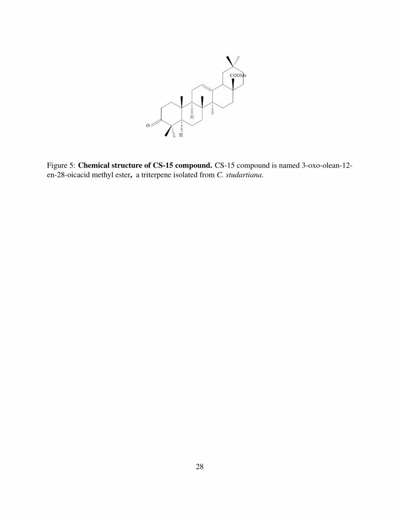
Figure 5: Chemical structure of CS-15 compound. CS-15 compound is named 3-oxo-olean-12-en-28-oicacid methyl ester, a triterpene isolated from C. studartiana.
28
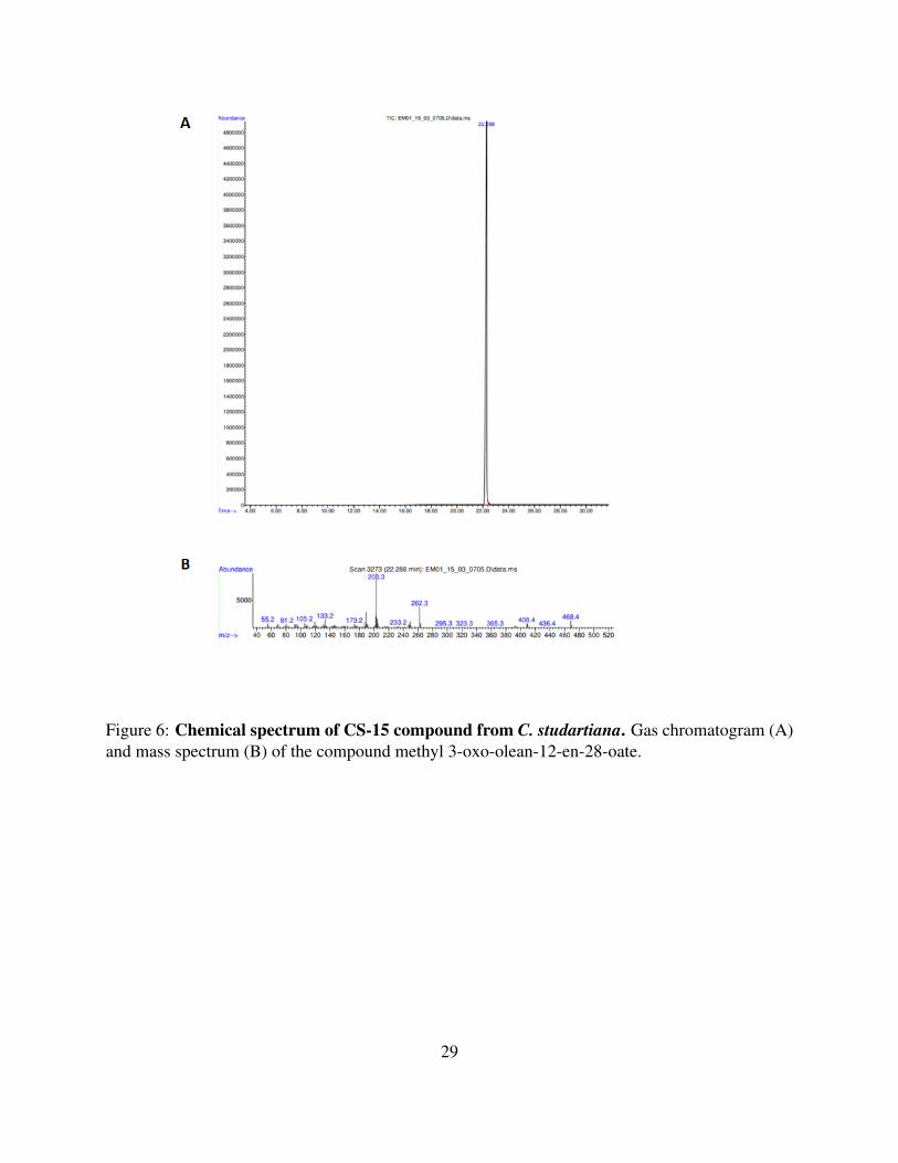
Figure 6: Chemical spectrum of CS-15 compound from C. studartiana. Gas chromatogram (A)and mass spectrum (B) of the compound methyl 3-oxo-olean-12-en-28-oate.
29

ARTIGO 4
104
Título do artigo 4: Potential therapeutic applications of P2 receptor antagonists:
from bench to clinical trials
Referência bibliográfica: Ferreira NCS; Alves LA; Soares-Bezerra RJ. Potential
therapeutic applications of P2 receptors antagonists: from bench to clinical trials. Curr
Drug Targets. 2019;20(9):1-18.
Situação: Publicado na Revista Current Drug Targets.
Descrição: Esse artigo de revisão foi redigido durante o meu doutorado e aborda o
desenvolvimento de fármacos com atuação antagonista sobre diferentes subtipos de
receptores P2 na terapia clínica. O trabalho discute os principais achados sobre o uso dos
antagonistas em modelos de dor, neurodegeneração, câncer, inflamação e trombose em
fase pré-clínica. Em seguida, aponta os resultados e os status dos antagonistas mais
promissores que avançaram para as etapas 2, 3 e 4 da fase clínica, isto é, ligantes
seletivos dos receptores P2X3, P2X7 e P2Y12. O artigo ainda apresenta uma pequena
discussão sobre os antagonistas do receptor P2Y12, os quais são até o momento o único
caso de sucesso da terapia purinérgica e encerra-se tecendo alguns comentários sobre os
principais desafios para o desenvolvimento de fármacos nessa área de atuação.

Send Orders for Reprints to [email protected]
Current Drug Targets, 2019, 20, 000-000 1
REVIEW ARTICLE
1389-4501/19 $58.00+.00 ©2019 Bentham Science Publishers
Potential Therapeutic Applications of P2 Receptor Antagonists: From Bench to Clinical Trials
Natiele Carla da Silva Ferreira1, Luiz Anastacio Alves1 and Rômulo José Soares-Bezerra2,*
1Laboratory of Cellular Communication, Oswaldo Cruz Institute, Oswaldo Cruz Foundation, Rio de Janeiro, 21040-360, Brazil; 2Laboratory of Technological Development in Virology, Oswaldo Cruz Institute, Oswaldo Cruz Foundation, Rio de Janeiro, 21040-360, Brazil
A R T I C L E H I S T O R Y
Received: November 26, 2018 Revised: January 30, 2019 Accepted: February 06, 2019 DOI: 10.2174/1389450120666190213095923
Abstract: Background: Extracellular purines and pyrimidines have important physiological functions in mammals. Purines and pyrimidines act on P1 and P2 purinergic receptors, which are widely ex-pressed in the plasma membrane in various cell types. P2 receptors act as important therapeutic targets and are associated with several disorders, such as pain, neurodegeneration, cancer, inflammation, and thrombosis. However, the use of antagonists for P2 receptors in clinical therapy, with the exception of P2Y12, is a great challenge. Currently, many research groups and pharmaceutical companies are working on the development of specific antagonist molecules for each receptor subtype that could be used as new medicines to treat their respective disorders. Objective: The present review compiles some interesting findings on the application of P2 receptor an-tagonists in different in vitro and in vivo experimental models as well as the progress of advanced clinical trials with these compounds. Conclusion: Despite all of the exciting results obtained on the bench, few antagonists of P2 receptors advanced to the clinical trials, and once they reach this stage, the effectiveness of the therapy is not guaranteed, as in the example of P2X7 antagonists. Despite this, P2Y12 receptor antagonists have a history of success and have been used in therapy for at least two decades to prevent thrombosis in pa-tients at risk for myocardial infarctions. This breakthrough is the motivation for scientists to develop new drugs with antagonistic activity for the other P2 receptors; thus, in a matter of years, we will have an evolution in the field of purinergic therapy.
Keywords: P2X receptors, P2Y receptors, antagonists, therapy, thrombosis, pain, inflammation, clinical trials.
1. INTRODUCTION
Purinergic receptors are a family of receptors expressed in the membrane surface of vertebrates, invertebrates, and protists [1, 2]. These receptors are activated by extracellular purines and pyrimidines, which initiate intracellular signal-ing that culminates in physiological responses, including inflammation [3], neurotransmission [4], platelet aggregation [5], and pain [6], among others. In humans, 19 subtypes of purinergic receptors have been cloned since the 1990s. In 1994, a group of researchers pro-posed a classification for purinergic receptors into two classes according to their natural ligand and effector system. Thus, purinergic receptors were classified as either adeno-sine-activated P1 receptors (P1R) or nucleotide-activated P2 receptors (P2R) [7]. Recently, Thimm and collaborators (2013) identified a new family of adenine receptors in
*Address correspondence to this author at the Laboratory of Technological Development in Virology, Oswaldo Cruz Institute, Oswaldo Cruz Founda-tion, Rio de Janeiro, 21040-360, Brazil; Tel: +55-21-4111-9627; E-mail: [email protected]
rodents that were named the P0 receptors. Naturally, the inclusion of P0 receptors in the traditional classification of purinergic receptors could occur in the future after the char-acterization of these receptors in humans [8]. P1 receptors are associated with the Gs/i protein and thus act on the protein-effector adenylyl cyclase to activate or inhibit the formation of cyclic adenosine 5’-monophosphate (cAMP), respectively. P1 receptors exhibit four subtypes: A1, A2A, A2B and A3, and their major physiological ligand is adenosine [7]. P2 receptors are divided into two major classes (P2X and P2Y) according to their pharmacological responses [7]. P2X receptors are ionotropic cation nonselective ion channels that allow for rapid Ca2+, Na+, and K+ flux according to the electrochemical gradient of each ion. Currently, seven sub-types of these receptors are described: P2X1-P2X7, and adenosine 5’-triphosphate (ATP) is their physiological ligand. Among all P2X receptors, P2X7 has the unique abil-ity to form membrane pores, i.e., when activated by micro-molar concentrations of ATP, P2X7 forms a membrane pore that allows for the passage of molecules up to 900 Da [9]. It

2 Current Drug Targets, 2019, Vol. 20, No. 9 Ferreira et al.
has been described that other subtypes of P2X receptors, P2X2 and P2X4, also demonstrate the ability of membrane pore formation similar to P2X7 [10, 11]. Despite this, the origin of the membrane pore remains unclear. Some authors believe that the opening of the membrane pore is related to dilation of the ion channel, while others support the hypothe-sis that another membrane protein could function as a pore and could be activated by second messengers derived from activated-P2X receptors [12-14]. P2Y receptors have eight subtypes: P2Y1, P2Y2, P2Y4, P2Y6, and P2Y11-P2Y14. These receptors are Gq/s/i pro-tein-coupled, and their major effectors are the phospholipase C/inositol triphosphate system and adenylyl cyclase, which act in the regulation of intracellular calcium stores and cAMP formation, respectively. In humans, P2Y receptors are activated by different nucleotides, including ATP (P2Y2, P2Y11), adenosine 5’-diphosphate (ADP) (P2Y1, P2Y12, P2Y13), uridine 5’-triphosphate (UTP) (P2Y2, P2Y4), uridine 5’-diphosphate (UDP) (P2Y6, P2Y14) and UDP-glucose (P2Y14) [15, 16]. P2 receptors are widely expressed in many cell types and tissues in mammals, including hematopoietic-derived cells, platelets, skeletal muscle, glial cells, some neuronal cells, he-patocytes and some digestive tract cells [16, 17]. The wide expression of P2 receptors in numerous cell types, especially in humans, mice and rats, allowed the conduction of several in vitro and in vivo experiments aiming to discover and under-stand important physiological roles of P2 receptors. These findings also demonstrated the participation of P2 receptors in the development of various pathologies and noted that the use of antagonists could relieve the symptoms of these diseases.
2. ON THE BENCH: PROMISING EFFECTS OF P2 RECEPTOR ANTAGONISTS IN DIFFERENT EX-PERIMENTAL MODELS OF DISEASES
There are many selective and nonselective antagonists described for P2 receptors, although the establishment of their use in a therapeutic context is still very difficult. Sev-eral groups have demonstrated that the use of antagonists for P2 could attenuate the effects of some disorders associated with P2R activation, such as pain, neurodegeneration, and inflammation. Here, we briefly discuss some findings on the use of P2R antagonists in pain, neurodegeneration, cancer, inflammation, and thrombosis.
2.1. Pain
The participation of P2 receptors in pain is well-investigated through the use of several animal models. In terms of P2X subtypes, the participation of P2X3, P2X4, and P2X7 in pain development has been demonstrated. P2X3 expression is upregulated in the dorsal ganglion neurons of animals with bone cancer and is associated with cancer-induced pain behavior [18] and visceral pain trans-duction from the ureter, bladder, gut, and uterus [19]. In animal models of visceral inflammation pain, the use of A317491 (a P2X3-selective antagonist), pyridoxal phos-phate-6-azophenyl-2',4'-disulfonic acid (PPADS) and Suramin (P2R-nonselective antagonist) attenuated pain sen-sation [20-23].
Meanwhile, P2X4 is upregulated in microglia after nerve injury, and it was suggested that P2X4 could be implicated in neuropathic pain [24, 25]. This upregulation in microglia after nerve injury is dependent on lyn tyrosine kinase activ-ity, which is a member of the src kinase family (SFK) [26]. Tsuda et al. (2009) demonstrated that P2X4-/- mice exhibited a reduction in pain responses in models of chronic pain [27]. P2X7 is associated with inflammatory hyperalgesia since its activation stimulates the release of inflammatory media-tors, such as IL-1β, TNF-α, IL-6 and cytokine-induced neu-trophil chemoattractant-1 (CINC-1). It has already been demonstrated that selective antagonists of P2X7, A438079, AACBA, and A740003 could attenuate hyperalgesia and allodynia in animal models of pain [28-32]. Recently, Munoz et al. (2017) demonstrated that P2X7-induced nociception could be related to the production of reactive oxygen species (ROS) since the administration of 2′(3′)-O-(4-benzoylbenzoyl)adenosine 5′-triphosphate (BzATP) in mice induced ROS production in the spinal cord and nociceptive behavior, which were attenuated with the pretreatment of ROS scavengers [33]. It has already been observed that all P2Y subtypes, with the exception of P2Y4, partially contribute to pain develop-ment. MRS2179, a selective antagonist for P2Y1, relieves nociception transmission in cancer-induced bone pain and thrombus-induced thermal hyperalgesia in rats [34, 35]. P2Y2 plays a role in trigeminal neuropathic pain, and the use of antisense oligonucleotides, Suramin and ARC-118925 (a selective P2Y2 antagonists) promoted an anti-allodynic ef-fect [36, 37]. It has been observed that nerve injury in animals upregu-lates the expression of P2Y6 and P2Y11 receptors and that tactile allodynia could be inhibited by P2Y6 and P2Y11 an-tagonists MRS2578 and NF340, respectively [38]. MRS2578 and MRS2211 (P2Y6 and P2Y13 antagonists, respectively) and P2Y14 anti-sense nucleic acids also attenuated neuro-pathic pain in an animal model of nerve injury [39]. Bar-ragán-Iglesias and collaborators (2015) also observed an increase of 0.5% in formalin-induced flinching behavior af-ter treatment with P2Y1, P2Y6 and P2Y11 agonists; on the other hand, selective antagonists of these receptors signifi-cantly reduced inflammatory pain [40]. Recently, Huang et al. (2018) demonstrated P2Y6 expression in the spinal cord of rats and that treatment with UDP increased mechanical allodynia and thermal hyperalgesia in a model of chronic constriction injury of the sciatic nerve, whereas MRS2578 alleviated these symptoms [41]. P2Y12 antagonists also demonstrated analgesic effects. In animal models of neuropathic and inflammatory pain, and acute thermal nociception, P2Y12 antagonists attenuated hyperalgesia and the expression of pro-inflammatory cytoki-nes [42]. Recently, Jia et al. (2018) showed that nanoparti-cle-encapsulated curcumin, a metabolite with antagonistic activity against P2Y12, reduced P2Y12 expression, the re-lease of pro-inflammatory cytokines, and thermal hyperalge-sia in type 2 diabetic rats [43]. Therefore, these findings demonstrate that P2 receptor antagonists are able to attenuate different pain mechanisms, including inflammatory and neuropathic pain. Thus, P2 re-

P2 Receptor Antagonists in Therapy Current Drug Targets, 2019, Vol. 20, No. 9 3
ceptor antagonists could contribute to the development of a new class of analgesic agents. Although promising, the re-sults should be evaluated with caution because some of the antagonists used in the studies present some problems. PPADS and Suramin are nonselective antagonists, and the latter is also an inhibitor of G-protein receptors. MRS2578, a P2Y6 selective antagonist, exhibited reactivity and hydro-phobicity limitations. A-317491 also demonstrated low bioavailability. BzATP, a selective P2X7 agonist, has more affinity for the P2X1 receptor [44]. Curcumin is a molecule from the plant Curcuma longa that has several molecular targets, including those that are antioxidants and involved in anti-inflammatory activities [45].
2.2. Neurodegenerative Diseases
In animal models of neurodegenerative diseases, it was observed that P2X7 was upregulated in some cells in the brain and contributed to the aggravation of neurodegenera-tive diseases. In fact, P2X7 is the most studied subtype in neurodegenerative diseases. Little is known about the contri-bution of the other P2 receptors. As will be discussed below, several works demonstrated that its antagonists could allevi-ate the symptoms of different types of neurodegenerative diseases. P2X7 antagonists, A438079 and JNJ-47965567, allevi-ated seizures in epileptic mice. A438079 also had a neuro-protector role in the animal model of kainic acid-induced seizures by decreasing seizure severity and neuronal death in the hippocampus [46, 47]. P2X7 antagonists also amelio-rated neuroinflammation, neuronal damage, and glial activa-tion thereby alleviating seizure severity [48]. P2X7 expression is upregulated in lateral amyotrophic sclerosis, and activation of the P2X7 receptor is associated with motor neuronal death, which was inhibited by Brilliant Blue G (BBG), oxidized ATP (oATP) and KN-62. Treatment with these antagonists also decreased the expression of mi-crogliosis and inflammatory markers, such as NF-κB, NADPH oxidase 2 and IL-1β, and improved motor perform-ance in mice [49-51]. BBG treatment blocked preterm birth in an animal model of intrauterine inflammation induced by lipopolysaccharide (LPS) and ameliorated fetal cortical injury, increased the number of dendrites and preserved the architectural mor-phology of cortical neurons [52]. In an experimental model of Parkinson disease, BBG ameliorated rotational behavior, improved memory, increased dopamine levels in the striatum and mesencephalon, and attenuated gliosis [53]. In Alzheimer’s disease, P2X7 stimulates amyloid precur-sor protein (APP) release and decreases α-secretase activity, which cleaves APP, increasing amyloid plaque accumulation. P2X7 also produces reactive oxygen species, which leads to oxidative stress in Alzheimer’s disease. P2X7 antagonists in-hibit APP release and enhance APP processing via α-secretase, thereby preventing amyloid plaque accumulation. P2X7 knockdown also stimulated microglial phagocytosis of amyloid β (Aβ). Additionally, BBG prevented cognitive defi-cits and improved learning and memory [54-60]. Delekate et al. (2014) observed the participation of the P2Y1 receptor in an animal model of Alzheimer’s disease.
They showed that astrocytes near Aβ were hyperactive and that astrocyte activity diminished in the presence of P2Y1 antagonists [61]. These observations illustrate the potential use of antago-nists to alleviate symptoms of neurodegenerative diseases to help improve patients' quality of life. However, the major antagonist used in the experiments, BBG, is not selective for P2X7. Thus, since BBG inhibits the P2X4 receptor and volt-age-gated sodium channels, it would be important to verify whether these other two molecular targets are actually unaf-fected in these experimental models [62]. In 2003, Beigi et al. [63] also described that oATP could have other molecular targets in addition to P2X7.
2.3. Cancer
The role of ATP in cancer is contradictory. ATP could activate P2 receptors to induce cellular proliferation or apop-tosis. Sometimes, only one subtype of P2R could play this dual role. Pellegatti et al. (2008) established a protocol to quantify ATP concentration in the tumor in vivo, and found that the ATP concentration is above 100 µM [64]. This level of extracellular ATP is sufficient to activate all P2Rs, includ-ing P2X7, which is activated at concentrations above 100 µM. In neuroblastoma and glioma, P2X7 is involved in cellu-lar proliferation, which was blocked with intravenous ad-ministrations of BBG [65]. Activation of P2X7 by extracel-lular ATP also stimulates cellular proliferation, migration, and invasion in pancreatic ductal adenocarcinoma cells and in some cell lines of prostate, lung, melanoma and breast cancer. These activities could be prevented by treatment with receptor antagonists [66-70]. Amoroso et al. (2016) observed the dual role of P2X7 in malignant pleural mesothelioma cell lines (MMP89, IST-MES2, and MSTO-211H). They observed that P2X7 antago-nists (AZ10606120 and oATP), as well as BzATP (P2X7 selective agonist), inhibited cell proliferation. However, this inhibitory effect was caused by cell death. Indeed, treatment with AZ10606120 in animals inoculated with tumor cells reduced the tumor volume after two weeks [71]. Similarly, systemic administration of ATPγS, a nonhydrolyzable ATP analog, reduced the tumor growth of MDA-MB-231 and Py8119 mammary cells in vivo, possibly by acting on P2X receptors [72]. BBG and P2X7 shRNA also increased tumor growth and angiogenesis in glioma C6 cells [73]. Extracellular ATP also activates P2Y2, which is associ-ated with cellular proliferation, migration, invasion, and me-tastasis in some cell lines from prostate and breast cancer, as well as human hepatocellular carcinoma and human cancer-ous pancreatic duct epithelial cells [74-78]. P2Y6 can contribute to the development of colorectal tumors since P2Y6-/- animals presented a lower number of cancer cells and tumors with small volumes [79]. Recently, Khalid et al. (2017) demonstrated that ATP and NF546 (a selective agonist of P2Y11) induced migration of the human hepatocellular carcinoma lineages Huh-7 and HepG2. Migration of these cell lines, as well as calcium mo-bilization induced by ATP, were inhibited by NF340, a P2Y11 antagonist [80].
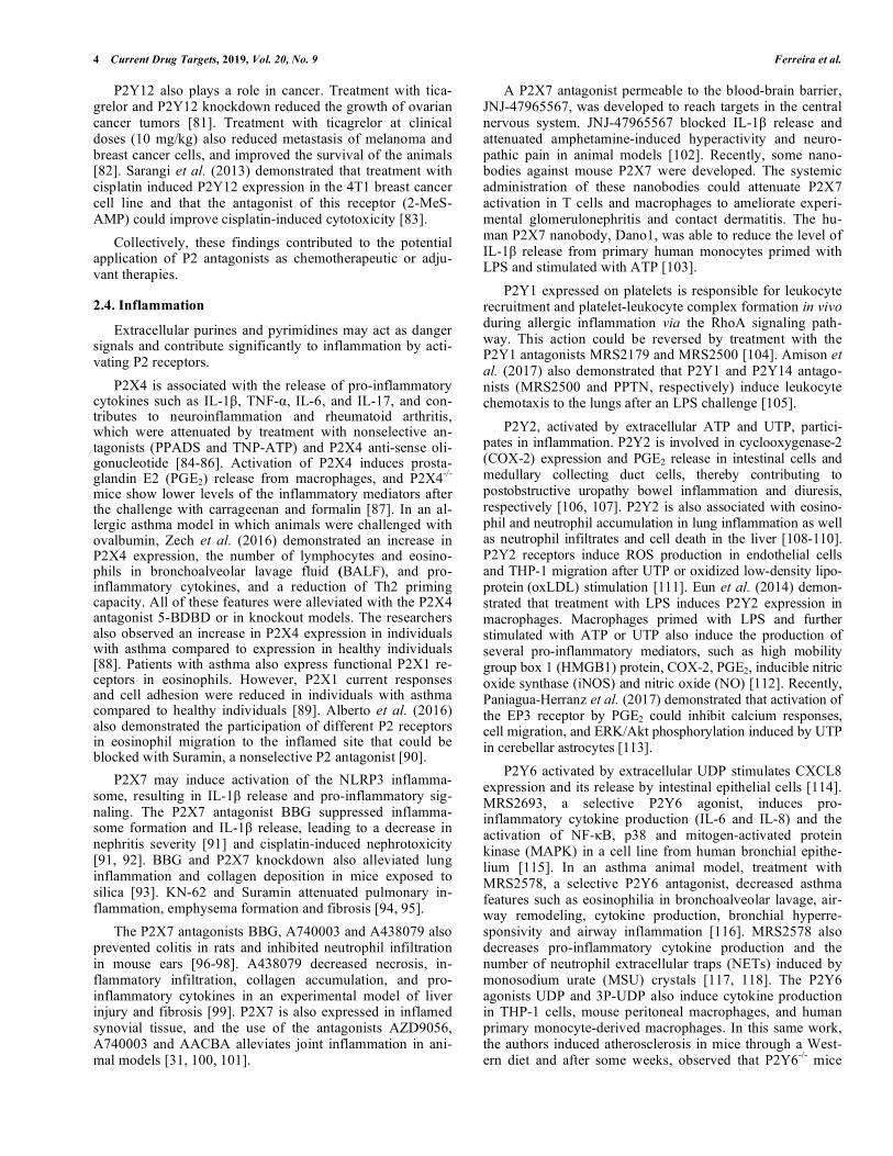
4 Current Drug Targets, 2019, Vol. 20, No. 9 Ferreira et al.
P2Y12 also plays a role in cancer. Treatment with tica-grelor and P2Y12 knockdown reduced the growth of ovarian cancer tumors [81]. Treatment with ticagrelor at clinical doses (10 mg/kg) also reduced metastasis of melanoma and breast cancer cells, and improved the survival of the animals [82]. Sarangi et al. (2013) demonstrated that treatment with cisplatin induced P2Y12 expression in the 4T1 breast cancer cell line and that the antagonist of this receptor (2-MeS-AMP) could improve cisplatin-induced cytotoxicity [83]. Collectively, these findings contributed to the potential application of P2 antagonists as chemotherapeutic or adju-vant therapies.
2.4. Inflammation
Extracellular purines and pyrimidines may act as danger signals and contribute significantly to inflammation by acti-vating P2 receptors. P2X4 is associated with the release of pro-inflammatory cytokines such as IL-1β, TNF-α, IL-6, and IL-17, and con-tributes to neuroinflammation and rheumatoid arthritis, which were attenuated by treatment with nonselective an-tagonists (PPADS and TNP-ATP) and P2X4 anti-sense oli-gonucleotide [84-86]. Activation of P2X4 induces prosta-glandin E2 (PGE2) release from macrophages, and P2X4-/- mice show lower levels of the inflammatory mediators after the challenge with carrageenan and formalin [87]. In an al-lergic asthma model in which animals were challenged with ovalbumin, Zech et al. (2016) demonstrated an increase in P2X4 expression, the number of lymphocytes and eosino-phils in bronchoalveolar lavage fluid (BALF), and pro-inflammatory cytokines, and a reduction of Th2 priming capacity. All of these features were alleviated with the P2X4 antagonist 5-BDBD or in knockout models. The researchers also observed an increase in P2X4 expression in individuals with asthma compared to expression in healthy individuals [88]. Patients with asthma also express functional P2X1 re-ceptors in eosinophils. However, P2X1 current responses and cell adhesion were reduced in individuals with asthma compared to healthy individuals [89]. Alberto et al. (2016) also demonstrated the participation of different P2 receptors in eosinophil migration to the inflamed site that could be blocked with Suramin, a nonselective P2 antagonist [90]. P2X7 may induce activation of the NLRP3 inflamma-some, resulting in IL-1β release and pro-inflammatory sig-naling. The P2X7 antagonist BBG suppressed inflamma-some formation and IL-1β release, leading to a decrease in nephritis severity [91] and cisplatin-induced nephrotoxicity [91, 92]. BBG and P2X7 knockdown also alleviated lung inflammation and collagen deposition in mice exposed to silica [93]. KN-62 and Suramin attenuated pulmonary in-flammation, emphysema formation and fibrosis [94, 95]. The P2X7 antagonists BBG, A740003 and A438079 also prevented colitis in rats and inhibited neutrophil infiltration in mouse ears [96-98]. A438079 decreased necrosis, in-flammatory infiltration, collagen accumulation, and pro-inflammatory cytokines in an experimental model of liver injury and fibrosis [99]. P2X7 is also expressed in inflamed synovial tissue, and the use of the antagonists AZD9056, A740003 and AACBA alleviates joint inflammation in ani-mal models [31, 100, 101].
A P2X7 antagonist permeable to the blood-brain barrier, JNJ-47965567, was developed to reach targets in the central nervous system. JNJ-47965567 blocked IL-1β release and attenuated amphetamine-induced hyperactivity and neuro-pathic pain in animal models [102]. Recently, some nano-bodies against mouse P2X7 were developed. The systemic administration of these nanobodies could attenuate P2X7 activation in T cells and macrophages to ameliorate experi-mental glomerulonephritis and contact dermatitis. The hu-man P2X7 nanobody, Dano1, was able to reduce the level of IL-1β release from primary human monocytes primed with LPS and stimulated with ATP [103]. P2Y1 expressed on platelets is responsible for leukocyte recruitment and platelet-leukocyte complex formation in vivo during allergic inflammation via the RhoA signaling path-way. This action could be reversed by treatment with the P2Y1 antagonists MRS2179 and MRS2500 [104]. Amison et al. (2017) also demonstrated that P2Y1 and P2Y14 antago-nists (MRS2500 and PPTN, respectively) induce leukocyte chemotaxis to the lungs after an LPS challenge [105]. P2Y2, activated by extracellular ATP and UTP, partici-pates in inflammation. P2Y2 is involved in cyclooxygenase-2 (COX-2) expression and PGE2 release in intestinal cells and medullary collecting duct cells, thereby contributing to postobstructive uropathy bowel inflammation and diuresis, respectively [106, 107]. P2Y2 is also associated with eosino-phil and neutrophil accumulation in lung inflammation as well as neutrophil infiltrates and cell death in the liver [108-110]. P2Y2 receptors induce ROS production in endothelial cells and THP-1 migration after UTP or oxidized low-density lipo-protein (oxLDL) stimulation [111]. Eun et al. (2014) demon-strated that treatment with LPS induces P2Y2 expression in macrophages. Macrophages primed with LPS and further stimulated with ATP or UTP also induce the production of several pro-inflammatory mediators, such as high mobility group box 1 (HMGB1) protein, COX-2, PGE2, inducible nitric oxide synthase (iNOS) and nitric oxide (NO) [112]. Recently, Paniagua-Herranz et al. (2017) demonstrated that activation of the EP3 receptor by PGE2 could inhibit calcium responses, cell migration, and ERK/Akt phosphorylation induced by UTP in cerebellar astrocytes [113]. P2Y6 activated by extracellular UDP stimulates CXCL8 expression and its release by intestinal epithelial cells [114]. MRS2693, a selective P2Y6 agonist, induces pro-inflammatory cytokine production (IL-6 and IL-8) and the activation of NF-κB, p38 and mitogen-activated protein kinase (MAPK) in a cell line from human bronchial epithe-lium [115]. In an asthma animal model, treatment with MRS2578, a selective P2Y6 antagonist, decreased asthma features such as eosinophilia in bronchoalveolar lavage, air-way remodeling, cytokine production, bronchial hyperre-sponsivity and airway inflammation [116]. MRS2578 also decreases pro-inflammatory cytokine production and the number of neutrophil extracellular traps (NETs) induced by monosodium urate (MSU) crystals [117, 118]. The P2Y6 agonists UDP and 3P-UDP also induce cytokine production in THP-1 cells, mouse peritoneal macrophages, and human primary monocyte-derived macrophages. In this same work, the authors induced atherosclerosis in mice through a West-ern diet and after some weeks, observed that P2Y6-/- mice

P2 Receptor Antagonists in Therapy Current Drug Targets, 2019, Vol. 20, No. 9 5
presented a significant reduction in the plaque area on the aortic arch [119]. The P2Y12 receptor also plays a role in inflammation. In a model of intra-abdominal sepsis and acute lung injury, animals treated with clopidogrel and animals that were P2Y12-null presented with reduced levels of circulating white blood cells, platelet activation, cytokine release, and lung injury compared with the control animals [120]. Sato-naka et al. (2015) also observed P2Y12 participation in vas-cular wall inflammation. They observed that treatment with ADP increased the expression and release of monocyte chemoattractant protein-1 (MCP-1), which is involved in monocyte migration to the vascular wall, in a time-dependent manner, and these features were inhibited by R-138727 (the active metabolite of prasugrel) and P2Y12-siRNA [121]. The P2Y13-selective antagonist MRS2211 and P2Y13-knockout significantly suppressed IL-6 production in Kupffer cells, alleviating liver inflammation [122]. Recently, PPTN was identified as an antagonist for P2Y14. PPTN inhibited neutrophil chemotaxis induced by UDP-glucose [123]. In collecting duct intercalated cells, P2Y14 activated by extracellular UDP-glucose induces pro-inflammatory chemokine expression and MAPK activation to mediate kidney inflammation; this effect was attenuated by PPTN [124]. Collectively, these findings endorse the role of P2R in inflammation, pointing to the possible use of P2R antago-nists as anti-inflammatory agents to attenuate inflammatory events associated with the activation of these receptors.
2.5. Thrombosis
P2 antagonists can also be applied in thrombosis preven-tion. Thrombosis is generally associated with the P2Y12 subtype, but there is increasing evidence of a significant con-tribution from other subtypes, such as P2X1 and P2Y1, in thrombosis prevention. Animals that are P2X1-/- presented with smaller thrombus formations compared to wild-type animals, and P2X1-/- ani-mals survived longer during the time interval studied than the control group [125]. P2Y1 and P2Y12 are expressed in platelets, and the inhi-bition of P2Y1 and P2Y12 is associated with a decline in platelet aggregation, thus preventing thrombosis. The P2Y1 antagonists MRS2179 and MRS2500 prevented platelet ag-gregation and thrombus formation in humans and animal models [126-131]. Using MRS2500 in combination with clopidogrel increased the anti-thrombotic effect compared to using these compounds alone [130]. It is important to note that new drugs have been developed with simultaneous ac-tion on P2Y1 and P2Y12 due to their expression in platelets, activation by extracellular ADP and contribution in platelet aggregation [132].
3. P2 ANTAGONISTS IN CLINICAL TRIALS
3.1. Antagonists for P2X-related Diseases
Based on the results observed in vitro and in animal models of different diseases, some P2 antagonists have ad-
vanced to the clinical trial level. Based on the website of clinical trials (clinicaltrials.gov) from the US National Insti-tutes of Health, we found approximately 15 studies in clini-cal phases involving P2X receptors worldwide. Among these results, only 5 studies were in phase 2 or later, and the results are shown in Table 1. The P2X3 receptor, a P2X subtype, is represented in three records of clinical trials in phase 2 or later. AF-219, a P2X3 selective antagonist, was studied for the first time in 2011 for the treatment of chronic cough by the Afferent Pharmaceuticals Company. P2X3 is expressed in the vagal nerve, and the hypothesis is that P2X3 could mediate cough reflex sensibilization. In the P2X3 study, 24 patients re-ceived treatment with AF-219 for 2 months, and 75% of the patients had decreased cough frequency. However, 6 patients presented adverse effects, such as taste disturbance, and abandoned the treatment. Some related adverse effects were dysgeusia (88%), hypogeusia (54%), nausea (38%) and oro-pharyngeal pain (21%), among others [133]. In 2015, AF-219 was studied in patients with idiopathic pulmonary fibro-sis to assess its tolerability in relation to blood pressure since P2X3 is expressed in the carotid chemosensory afferent nerve and could influence sympathetic activity. However, before and after AF-219 use, the patients did not present a difference in systolic (means: 122 vs 122, respectively) and diastolic (means: 77 vs 76, respectively) blood pressure. In the same year, Afferent Pharmaceuticals began to study the application of AF-219 in patients with idiopathic pulmonary fibrosis with a persistent cough, but the obtained results were not disclosed. Interestingly, AF-219 was studied in 74 women with interstitial cystitis and/or bladder pain syn-drome; AF-219 was administered for 4 weeks, and after this period, compared to the placebo, AF-219 reduced daily pain-ful bladder or interstitial cystitis symptoms (13.3 vs 15.3, respectively) and the genitourinary pain index (20.8 vs 25.5, respectively). However, the authors did not mention if these effects were mediated by P2X3. The major related adverse events were dysgeusia (72.22%), headache (13.89%), ageusia (8.33%) and hypogeusia (8.33%); however, there were no related serious adverse effects [150]. Another P2X subtype that presents antagonists in clinical studies is P2X7. Based on the results observed in animal models of rheumatoid arthritis, many investigators studied the application of P2X7 antagonists in this disease. Using the parameters of the American College of Rheumatology of 20%, 50% and 70% improvement responses (ACR20, ACR50, and ACR70, respectively), the authors observed that treatment with AZD9056 did not show any significant bene-fit in patients with rheumatoid arthritis compared to the pla-cebo, with patients in both groups continuing treatment with methotrexate. Among the 64 patients treated with AZD9056 at the maximal dose (400 mg), only 5 patients fell within the ACR70 criteria, compared to 3 of the 65 patients from the placebo group who fell inside this analyzed parameter. The major adverse effects were nausea, upper abdominal pain, diarrhea, and headache. The AZD9056 compound was also studied in patients presenting with acute Crohn’s disease at moderate and severe levels. Patients mentioned that the AZD9056 compound alleviated their pain sensation and in-creased their well-being. However, the levels of biomarkers, such as reactive C protein, in the serum and fecal calpro-tectin did not decrease [134, 151].

6 Current Drug Targets, 2019, Vol. 20, No. 9 Ferreira et al.
Table 1. P2 antagonists in clinical trials at phase 2 or later.
P2 Antagonist(s) Disease(s) Aim Number of Patients
Phase Start Year
Sponsor and NCT Number
Status The Major Result(s)
P2X3 AF-219 Chronic cough Evaluate the
effects of AF-
219 on chronic cough treatment
n= 34;
random-
ized and double-
blind
2 2011 Afferent Pharma-
ceuticals, United
Kingdom - NCT01432730
Completed AF-219 reduced cough
frequency [133].
P2X3 AF-219 Idiopathic
pulmonary fibrosis (IPF)
Evaluate AF-
219 tolerability in patients with
IPF
n= 6 2 2015 Afferent Pharma-
ceuticals United States -
NCT02477709
Completed AF-219 did not alter
blood pressure.*
P2X3 AF-219 Idiopathic pulmonary
fibrosis in
patients with a
persistent
cough
Evaluate changes in
cough frequency
n= (51); random-
ized and
double-
blind
2 2015 Afferent Pharma-ceuticals, United
States -
NCT02502097
Completed ND
P2X7 AZD9056 Rheumatoid
arthritis (RA)
Confirm the
efficacy of
treatment with
the drug in RA
patients
n= 385;
random-
ized and
double-
blind
2 2007 AstraZeneca,
United States -
NCT00520572
Completed AZD9056 did not have
efficacy compared to
placebo [134].
P2X7 CE-224,535 Rheumatoid arthritis
Improve signals and symptoms
of RA in pa-
tients
n= 100; random-
ized and
double-
blind
2 and 3
2008 Pfizer, United States -
NCT00628095
Completed It did not have efficacy compared to placebo
[135].
P2Y12 Clopidogrel and prasu-
grel
Coronary artery
disease (CAD) and diabetes
mellitus
Optimize anti-
platelet therapy in patients with
diabetes melli-
tus
n= 35;
random-ized and
double-
blinded
2 2008 Eli Lilly and
Company, United States -
NCT00642174
Completed Prasugrel treatment
was superior to clopi-dogrel treatment [136].
P2Y12 Ticagrelor and prasugrel Acute coro-nary syndrome
(ACS)
Evaluate the effects of aspirin
and P2Y12
antagonists on
platelet reactiv-
ity
n= (29); random-
ized and
double-
blinded
4 2015 Sheba Medical Center, Israel -
NCT02049762
Completed ND
P2Y12 Clopidogrel, ticagrelor,
and prasugrel
ACS and
coronary
angioplasty
Compare the
impact of con-
ventional ther-
apy to genotype-guided therapy
with P2Y12
antagonists
n= (4000);
random-
ized
4 2012 Italian Society of
Invasive Cardiol-
ogy, Italy -
NCT01477775
Unknown ND
P2Y12 Clopidogrel and ticagre-lor
ACS, stents, and atheroscle-
rosis
Compare the efficacy of
mono- and dual-
therapy with
antiplatelets
n= (1520); random-
ized
4 2017 Samsung Medi-cal Center, Korea
-
NCT03119012
In progress (recruiting)
ND
(Table 1) contd….
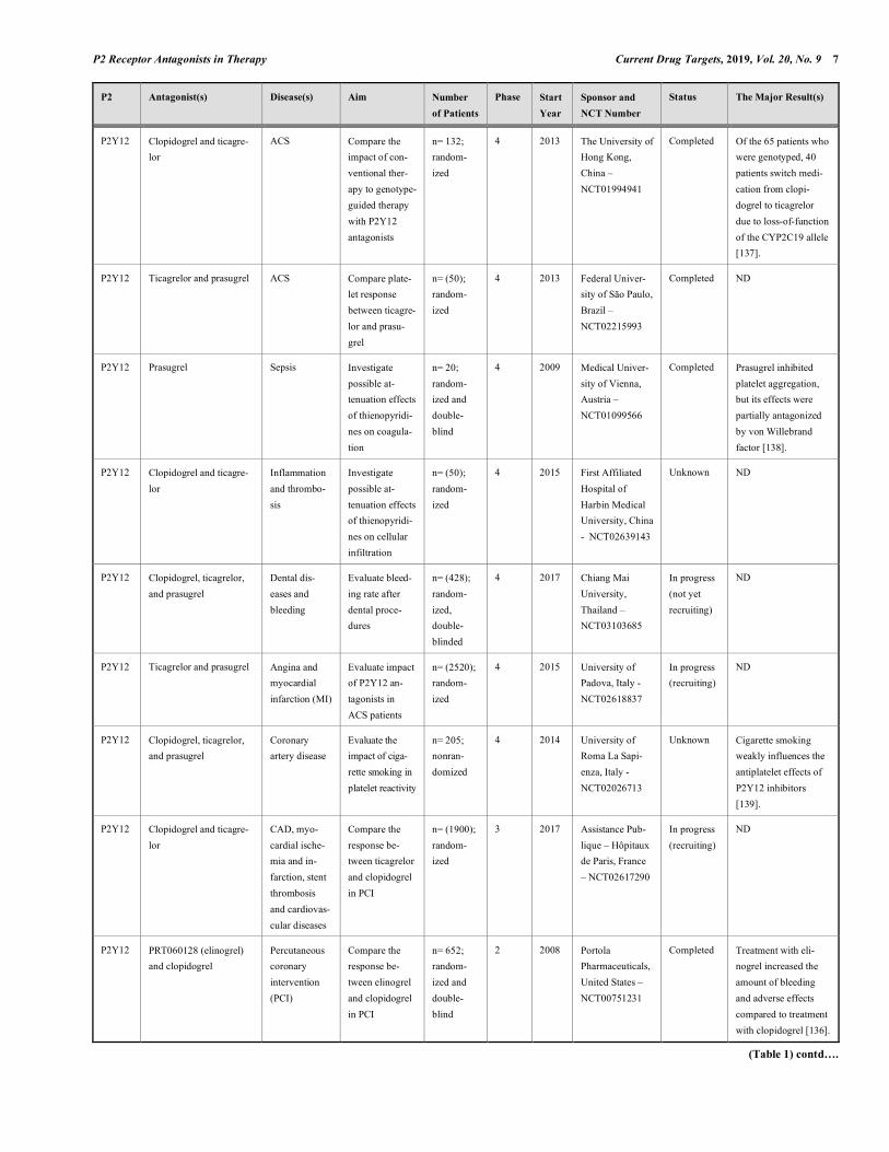
P2 Receptor Antagonists in Therapy Current Drug Targets, 2019, Vol. 20, No. 9 7
P2 Antagonist(s) Disease(s) Aim Number of Patients
Phase Start Year
Sponsor and NCT Number
Status The Major Result(s)
P2Y12 Clopidogrel and ticagre-lor
ACS Compare the impact of con-ventional ther-apy to genotype-guided therapy with P2Y12 antagonists
n= 132; random-ized
4 2013 The University of Hong Kong, China – NCT01994941
Completed Of the 65 patients who were genotyped, 40 patients switch medi-cation from clopi-dogrel to ticagrelor due to loss-of-function of the CYP2C19 allele [137].
P2Y12 Ticagrelor and prasugrel ACS Compare plate-let response between ticagre-lor and prasu-grel
n= (50); random-ized
4 2013 Federal Univer-sity of São Paulo, Brazil – NCT02215993
Completed ND
P2Y12 Prasugrel Sepsis Investigate possible at-tenuation effects of thienopyridi-nes on coagula-tion
n= 20; random-ized and double-blind
4 2009 Medical Univer-sity of Vienna, Austria – NCT01099566
Completed Prasugrel inhibited platelet aggregation, but its effects were partially antagonized by von Willebrand factor [138].
P2Y12 Clopidogrel and ticagre-lor
Inflammation and thrombo-sis
Investigate possible at-tenuation effects of thienopyridi-nes on cellular infiltration
n= (50); random-ized
4 2015 First Affiliated Hospital of Harbin Medical University, China - NCT02639143
Unknown ND
P2Y12 Clopidogrel, ticagrelor, and prasugrel
Dental dis-eases and bleeding
Evaluate bleed-ing rate after dental proce-dures
n= (428); random-ized, double-blinded
4 2017 Chiang Mai University, Thailand – NCT03103685
In progress (not yet recruiting)
ND
P2Y12 Ticagrelor and prasugrel Angina and myocardial infarction (MI)
Evaluate impact of P2Y12 an-tagonists in ACS patients
n= (2520); random-ized
4 2015 University of Padova, Italy - NCT02618837
In progress (recruiting)
ND
P2Y12 Clopidogrel, ticagrelor, and prasugrel
Coronary artery disease
Evaluate the impact of ciga-rette smoking in platelet reactivity
n= 205; nonran-domized
4 2014 University of Roma La Sapi-enza, Italy - NCT02026713
Unknown Cigarette smoking weakly influences the antiplatelet effects of P2Y12 inhibitors [139].
P2Y12 Clopidogrel and ticagre-lor
CAD, myo-cardial ische-mia and in-farction, stent thrombosis and cardiovas-cular diseases
Compare the response be-tween ticagrelor and clopidogrel in PCI
n= (1900); random-ized
3 2017 Assistance Pub-lique – Hôpitaux de Paris, France – NCT02617290
In progress (recruiting)
ND
P2Y12 PRT060128 (elinogrel) and clopidogrel
Percutaneous coronary intervention (PCI)
Compare the response be-tween elinogrel and clopidogrel in PCI
n= 652; random-ized and double-blind
2 2008 Portola Pharmaceuticals, United States – NCT00751231
Completed Treatment with eli-nogrel increased the amount of bleeding and adverse effects compared to treatment with clopidogrel [136].
(Table 1) contd….
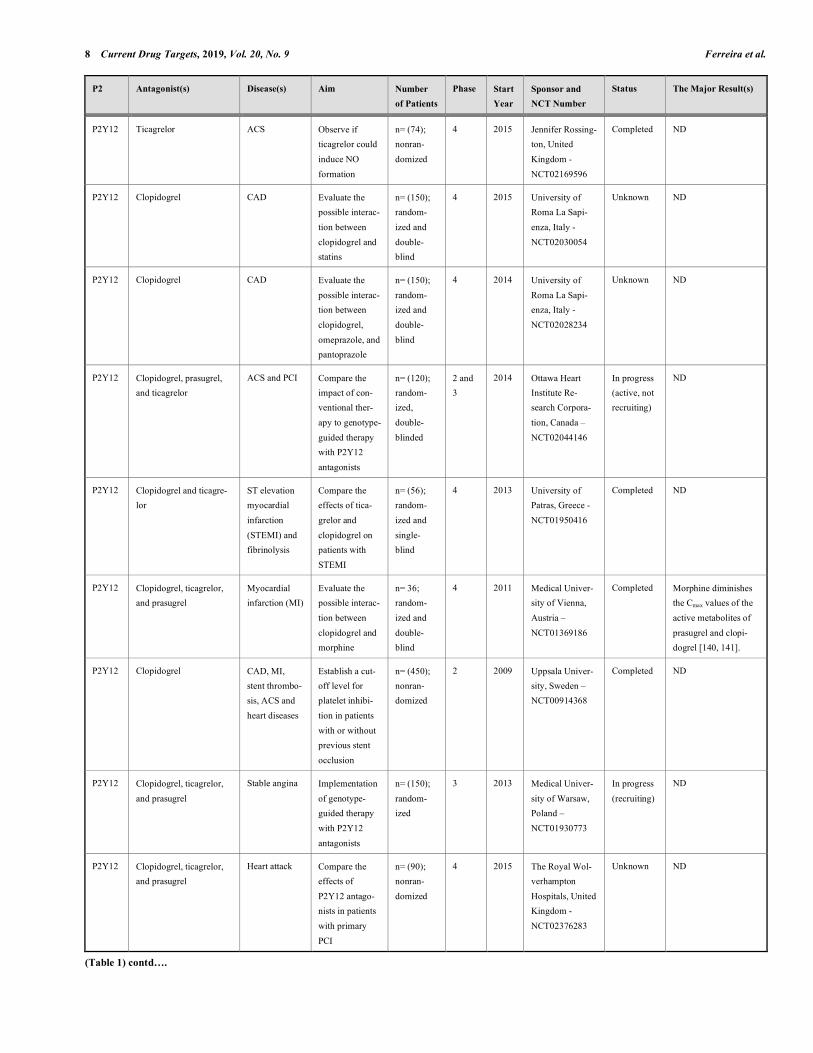
8 Current Drug Targets, 2019, Vol. 20, No. 9 Ferreira et al.
P2 Antagonist(s) Disease(s) Aim Number of Patients
Phase Start Year
Sponsor and NCT Number
Status The Major Result(s)
P2Y12 Ticagrelor ACS Observe if ticagrelor could induce NO formation
n= (74); nonran-domized
4 2015 Jennifer Rossing-ton, United Kingdom - NCT02169596
Completed ND
P2Y12 Clopidogrel CAD Evaluate the possible interac-tion between clopidogrel and statins
n= (150); random-ized and double-blind
4 2015 University of Roma La Sapi-enza, Italy - NCT02030054
Unknown ND
P2Y12 Clopidogrel CAD Evaluate the possible interac-tion between clopidogrel, omeprazole, and pantoprazole
n= (150); random-ized and double-blind
4 2014 University of Roma La Sapi-enza, Italy - NCT02028234
Unknown ND
P2Y12 Clopidogrel, prasugrel, and ticagrelor
ACS and PCI Compare the impact of con-ventional ther-apy to genotype-guided therapy with P2Y12 antagonists
n= (120); random-ized, double-blinded
2 and 3
2014 Ottawa Heart Institute Re-search Corpora-tion, Canada – NCT02044146
In progress (active, not recruiting)
ND
P2Y12 Clopidogrel and ticagre-lor
ST elevation myocardial infarction (STEMI) and fibrinolysis
Compare the effects of tica-grelor and clopidogrel on patients with STEMI
n= (56); random-ized and single-blind
4 2013 University of Patras, Greece - NCT01950416
Completed ND
P2Y12 Clopidogrel, ticagrelor, and prasugrel
Myocardial infarction (MI)
Evaluate the possible interac-tion between clopidogrel and morphine
n= 36; random-ized and double-blind
4 2011 Medical Univer-sity of Vienna, Austria – NCT01369186
Completed Morphine diminishes the Cmax values of the active metabolites of prasugrel and clopi-dogrel [140, 141].
P2Y12 Clopidogrel CAD, MI, stent thrombo-sis, ACS and heart diseases
Establish a cut-off level for platelet inhibi-tion in patients with or without previous stent occlusion
n= (450); nonran-domized
2 2009 Uppsala Univer-sity, Sweden – NCT00914368
Completed ND
P2Y12 Clopidogrel, ticagrelor, and prasugrel
Stable angina Implementation of genotype-guided therapy with P2Y12 antagonists
n= (150); random-ized
3 2013 Medical Univer-sity of Warsaw, Poland – NCT01930773
In progress (recruiting)
ND
P2Y12 Clopidogrel, ticagrelor, and prasugrel
Heart attack Compare the effects of P2Y12 antago-nists in patients with primary PCI
n= (90); nonran-domized
4 2015 The Royal Wol-verhampton Hospitals, United Kingdom - NCT02376283
Unknown ND
(Table 1) contd….

P2 Receptor Antagonists in Therapy Current Drug Targets, 2019, Vol. 20, No. 9 9
P2 Antagonist(s) Disease(s) Aim Number of Patients
Phase Start Year
Sponsor and NCT Number
Status The Major Result(s)
P2Y12 Ticagrelor and prasugrel MI Evaluate the addition of vorapaxar in dual-antiplatelet therapy
n= (126); random-ized
4 2016 University of Florida, United States – NCT02545933
In progress (recruiting)
ND
P2Y12 Clopidogrel Pancreatic cancer
Evaluate the possible interac-tion between clopidogrel and chemotherapeu-tic agents
n= (39); random-ized and double-blind
3 2016 Assistance Pub-lique – Hôpitaux de Paris, France – NCT02404363
In progress (active, not recruiting)
ND
P2Y12 PRT060128 (elinogrel) MI Evaluate the safety and efficacy of elinogrel before PCI in dual-antiplatelet therapy
n= 70; random-ized and double-blind
2 2007 Portola Pharmaceuticals, United States – NCT00546260
Terminated by adminis-trative reasons
Treatment of patients with elinogrel is well-tolerated [142].
P2Y12 Clopidogrel, ticagrelor, and prasugrel
ACS Evaluate the safety and efficacy of a ½ dose of P2Y12 inhibitors in patients with ACS
n= (100); random-ized
3 2016 Dong-A Univer-sity, Korea – NCT02944123
In progress (recruiting)
ND
P2Y12 Clopidogrel, ticagrelor, and prasugrel
ACS, unstable angina and MI
Compare the effects of P2Y12 antago-nists in elderly patients (≥ 70 years)
n= (1000); random-ized
4 2013 St. Antonius Hospital, The Netherlands - NCT02317198
In progress (recruiting)
ND
P2Y12 Cangrelor and ticagrelor STEMI Compare the effects of can-grelor and ticagrelor in patients with STEMI
n= (30); random-ized and single-blind
4 2017 Attikon Hospital, Greece - NCT02943369
Completed ND
P2Y12 Prasugrel Chronic asthma
Evaluate prasu-grel treatment in bronchial hyper-reactivity
n= (26); random-ized and single-blind
4 2011 University of Milan, Italy – NCT01305369
Completed ND
P2Y12 Prasugrel ACS Investigate the effects of prasu-grel pretreat-ment in MI
n= 4033; random-ized and double-blind
3 2009 Eli Lilly and Company, United States –
NCT01015287
Completed Prasugrel did not reduce ischemia, but led to an increased rate of bleeding complica-tions [143-145].
P2Y12 Clopidogrel, ticagrelor, and prasugrel
Cardiovascular diseases
Evaluate if ticagrelor im-proves endothe-lial function
n= (54); random-ized
4 2015 Erasmus Medical Center, The Netherlands - NCT02587260
Completed Ticagrelor did not improve endothelial functions in stabilized patients who suffered an ACS [146].
(Table 1) contd….

10 Current Drug Targets, 2019, Vol. 20, No. 9 Ferreira et al.
P2 Antagonist(s) Disease(s) Aim Number of Patients
Phase Start Year
Sponsor and NCT Number
Status The Major Result(s)
P2Y12 Ticagrelor ACS Compare the
antiplatelet
effects between ticagrelor and tirofiban
n= 95;
random-ized
3 2012 Pusan National
University Yang-
san Hospital, Korea - NCT01660373
Unknown Tirofiban demon-
strated a greater anti-
platelet effect than ticagrelor [147].
P2Y12 Prasugrel MI Compare the
antiplatelet effects between
ticagrelor and tirofiban
n= 100;
random-ized and
single-blind
3 2010 Università degli
Studi di Ferrara, Italy - NCT01336348
Completed Tirofiban demon-
strated a greater anti-platelet effect than prasugrel [148].
P2Y12 Clopidogrel Coronary
artery stenosis
and platelet aggregation
Compare the
antiplatelet
effects of clopi-dogrel plus cilostazol
n= (80);
random-ized
3 2009 Gyeongsang
National Univer-
sity Hospital, Korea - NCT00891670
Unknown ND
P2Y12 Ticagrelor and prasugrel CAD Compare the
pharmacody-
namic effects of ticagrelor sub-
stituted with prasugrel
n= 110;
random-ized
4 2012 Daiichi Sankyo,
United States - NCT01587651
Completed Prasugrel increased the
platelet reactivity
index and presented a similar profile to that
of ticagrelor on inhibi-tion percentage of platelet reactivity.*
P2Y12 Clopidogrel and prasu-grel
CAD Compare the
effectiveness of clopidogrel and
prasugrel in reducing the
number of cardiovascular
adverse effects in patients with CAD
n= 423;
random-ized and
double-blind
2 2009 Eli Lilly and
Company, United States - NCT00910299
Terminated
due to the low rate of
primary endpoint
events experienced
Clopidogrel substitu-
tion with prasugrel offered effective
platelet inhibition, but slightly increased the risk of bleeding [149].
P2Y12 Ticagrelor Peripheral artery disease
Compare tica-
grelor and aspirin effects on walking
n= 40;
random-ized,
double-blinded
2 2014 AstraZeneca,
United States - NCT02227368
Terminated
due to slow recruitment
There was a 0.1 log of
difference between ticagrelor and aspirin
treatment in seconds at peak walking time in 26 weeks.*
P2Y12 Clopidogrel, ticagrelor and prasugrel
MI and STEMI
Implementation
of genotype-guided therapy
with P2Y12 antagonists
n= (2700);
random-ized
4 2011 Vera HM
Deneer, The Netherlands - NCT01761786
In progress
(active, not recruiting)
ND
P2Y12 Clopidogrel and ticagre-lor
CAD Compare tica-
grelor and
clopidogrel effects in pa-tients with CAD
n= (28);
random-ized
4 2012 Rapid City
Regional Hospi-
tal, United States - NCT01706510
Completed ND
ND: not disclosed; ACS: acute coronary syndrome; CAD: coronary artery disease; Cmax: maximal plasma concentrations; IPF: idiopathic pulmonary fibrosis; MI: myocardial infarction; NO: nitric oxide; RA:
rheumatoid arthritis; STEMI: ST elevation myocardial infarction.
The number of estimated patients in the study is provided between parentheses due to a lack of results being disclosed. NCT: ClinicalTrials.gov Identifier. *Information about the results are available at the site
(clinicaltrials.gov)

P2 Receptor Antagonists in Therapy Current Drug Targets, 2019, Vol. 20, No. 9 11
CE-224,535, another compound with antagonistic activ-ity on P2X7, was studied in patients with rheumatoid arthri-tis treated with methotrexate. However, CE-224,535 did not demonstrate efficacy compared with the placebo, since 34% versus 36.2% of the patients treated with CE-224,535 and the placebo, respectively, fell inside the ACR20 criteria after 12 weeks. Approximately 62.2% of the patients treated with this P2X7 antagonist presented adverse effects compared to 55.3% of the patients in the placebo group. The major ad-verse effects were nausea and diarrhea. Moreover, 9.4% of the patients treated with CE-224,535 discontinued treatment compared to 6.4% of the patients in the placebo group [135]. A summary of these results is described in Table 1.
3.2. Antagonists for P2Y-related Diseases
The website of clinical trials (clinicaltrials.gov) has more than 300 records on clinical studies related to P2Y receptors, of which 206 studies were in phase 2 or later and involved only P2Y12 receptor antagonists. Of these 206 studies, 61 studies were active, 119 studies were finalized, and 26 study statuses were unknown. The majority of the studies com-pared P2Y12 antagonists (clopidogrel, prasugrel, ticagrelor, cangrelor, elinogrel, and ACT-‐246475) in pharmacodynamic parameters, efficacy and number of adverse effects in the aspect of cardiovascular diseases; some of these studies are listed in Table 1. Among the studies with disclosed results, some studies reported the effects of prasugrel in patients. One study with 110 participants aimed to observe the effects of substituting ticagrelor with prasugrel and showed that prasugrel-treated groups presented an increase in the platelet reactivity index (PRU) compared to the ticagrelor-treated groups. However, both ticagrelor and prasugrel maintained the same P2Y12 inhibition profile and adverse effects at similar rates. Trenk et al. (2012) compared the efficacy between prasugrel and clopidogrel in patients with high platelet reactivity on clopi-dogrel after coronary stent implantations and demonstrated the superiority of prasugrel in reducing the PRU [149]. In 212 patients treated with prasugrel, the PRU fell from 245 to 80, while in the clopidogrel-treated group (211 patients), this index fell from 249 to 241. However, after 90 days from the beginning of treatments, the PRU means did not differ be-tween groups. Therefore, a possible substitution of clopi-dogrel for prasugrel offers effective platelet inhibition but slightly increases the risk of bleeding (1.4% observed in the prasugrel group compared to 0.5% in the clopidogrel group) in a period of 6 months. Moreover, adverse effects were ob-served in 52.38% of patients treated with prasugrel com-pared to 34.29% of patients treated with clopidogrel [149]. In another study that compared elinogrel and clopidogrel in a sample size of 652 patients with nonurgent percutaneous coronary intervention (PCI), the obtained results demon-strated the safety and tolerability of both drugs. However, patients treated with elinogrel at a lower dose (100 mg oral) presented an increase in bleeding in 120 days (10.9%) com-pared to patients treated with clopidogrel (6.7%). In addition, elinogrel-treated patients presented more adverse effects than clopidogrel-treated patients that include respiratory disorders (24.4% versus 9.6%, respectively) and skin and subcutane-ous tissue disorders (8.5% versus 6.3%, respectively) [136].
Furthermore, Kim et al. (2016) compared the actions of prasugrel and tirofiban, an inhibitor of glycoprotein IIb/IIIa that is used in antiplatelet therapy. The researchers observed that treatment with tirofiban increase its inhibition of platelet aggregation from 83 to 92.9% compared to treatment with ticagrelor (from 56.2 to 91.3%) in a period between 2h and 24 h, after ADP stimulus. Similarly, after thrombin receptor activating peptide (TRAP) stimulation, tirofiban increased its inhibition of platelet aggregation from 61.3 to 76.5% com-pared to the increase from 21.2 to 58.3% of ticagrelor. How-ever, the high on-treatment platelet reactivity did not differ between the two groups [147]. Nevertheless, Valgimigli et al. (2012) demonstrated that concomitant administration of tirofiban and prasugrel in patients inhibited almost 100% of the platelet aggregation induced by ADP and TRAP [148]. Other records from P2Y12 clinical trials available at the site (clinicaltrials.gov) aim to implement a personalized choice of platelet P2Y12R inhibitor according to the patient's genotype. The hypothesis is that CYP2C19, an important enzyme for thienopyridine metabolism, can present some loss-of-function polymorphisms that have an association with clopidogrel resistance and ischemic events. Thus, through genotyping, it would be possible to guide the treat-ment of patients according to two groups: group one presents a high risk of resistance due to polymorphisms, and group two presents a lower risk. Patients who fit the first group may be treated with more potent drugs such as ticagrelor and prasugrel, while other patients may be treated with clopi-dogrel. This strategy aims to reduce the risk of heart attacks, deaths from cardiovascular causes and the risk of complica-tions from bleeding. Recently, Tam et al. (2017) conducted a study on the personalized choice of P2Y12 antagonist based on genotypes in 65 individuals from China. They found that 40 patients presented loss-of-function on the CYP2C19 al-lele, and treatment with clopidogrel was substituted with ticagrelor, which was effective in reducing platelet reactivity [137]. These results could also be an example of a pharma-cogenomics application. Finally, other studies have been dedicated to verifying the possible pharmacological interaction effects of P2Y12R inhibitors. One study of 250 patients with coronary artery disease (CAD) demonstrated that cigarette smoking weakly influences the antiplatelet effects of P2Y12R antagonists [139]. Another study showed the interaction between mor-phine and antiplatelets. The researchers observed that mor-phine diminishes the maximal plasma concentrations (Cmax) of active metabolites from prasugrel and clopidogrel in healthy volunteers. In addition, morphine also delayed the absorption and effects of clopidogrel [140, 141]. There are still other studies that evaluate possible pharmacological interactions between P2Y12 inhibitors and drugs such as omeprazole, pantoprazole (proton pump inhibitors), vora-paxar (protease-activated receptor inhibitor), and rosuvas-tatin and atorvastatin (statins), along with chemotherapeu-tics, but the results have not been disclosed. Through the presented general scenario, we observed that only P2Y12 receptor antagonists are applied in clinical ther-apy. P2X3 and P2X7 receptor antagonists are closer to reaching this stage in the future compared to other P2X re-ceptors, as studies on P2X3 and P2X7 receptor antagonists
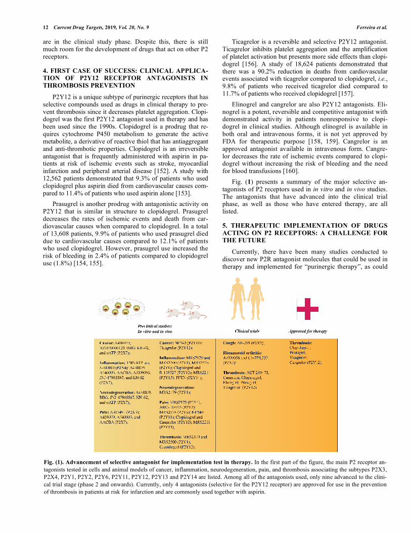
12 Current Drug Targets, 2019, Vol. 20, No. 9 Ferreira et al.
are in the clinical study phase. Despite this, there is still much room for the development of drugs that act on other P2 receptors.
4. FIRST CASE OF SUCCESS: CLINICAL APPLICA-TION OF P2Y12 RECEPTOR ANTAGONISTS IN THROMBOSIS PREVENTION
P2Y12 is a unique subtype of purinergic receptors that has selective compounds used as drugs in clinical therapy to pre-vent thrombosis since it decreases platelet aggregation. Clopi-dogrel was the first P2Y12 antagonist used in therapy and has been used since the 1990s. Clopidogrel is a prodrug that re-quires cytochrome P450 metabolism to generate the active metabolite, a derivative of reactive thiol that has antiaggregant and anti-thrombotic properties. Clopidogrel is an irreversible antagonist that is frequently administered with aspirin in pa-tients at risk of ischemic events such as stroke, myocardial infarction and peripheral arterial disease [152]. A study with 12,562 patients demonstrated that 9.3% of patients who used clopidogrel plus aspirin died from cardiovascular causes com-pared to 11.4% of patients who used aspirin alone [153]. Prasugrel is another prodrug with antagonistic activity on P2Y12 that is similar in structure to clopidogrel. Prasugrel decreases the rates of ischemic events and death from car-diovascular causes when compared to clopidogrel. In a total of 13,608 patients, 9.9% of patients who used prasugrel died due to cardiovascular causes compared to 12.1% of patients who used clopidogrel. However, prasugrel use increased the risk of bleeding in 2.4% of patients compared to clopidogrel use (1.8%) [154, 155].
Ticagrelor is a reversible and selective P2Y12 antagonist. Ticagrelor inhibits platelet aggregation and the amplification of platelet activation but presents more side effects than clopi-dogrel [156]. A study of 18,624 patients demonstrated that there was a 90.2% reduction in deaths from cardiovascular events associated with ticagrelor compared to clopidogrel, i.e., 9.8% of patients who received ticagrelor died compared to 11.7% of patients who received clopidogrel [157]. Elinogrel and cangrelor are also P2Y12 antagonists. Eli-nogrel is a potent, reversible and competitive antagonist with demonstrated activity in patients nonresponsive to clopi-dogrel in clinical studies. Although elinogrel is available in both oral and intravenous forms, it is not yet approved by FDA for therapeutic purpose [158, 159]. Cangrelor is an approved antagonist available in intravenous form. Cangre-lor decreases the rate of ischemic events compared to clopi-dogrel without increasing the risk of bleeding and the need for blood transfusions [160]. Fig. (1) presents a summary of the major selective an-tagonists of P2 receptors used in in vitro and in vivo studies. The antagonists that have advanced into the clinical trial phase, as well as those who have entered therapy, are all listed.
5. THERAPEUTIC IMPLEMENTATION OF DRUGS ACTING ON P2 RECEPTORS: A CHALLENGE FOR THE FUTURE
Currently, there have been many studies conducted to discover new P2R antagonist molecules that could be used in therapy and implemented for “purinergic therapy”, as could
Fig. (1). Advancement of selective antagonist for implementation test in therapy. In the first part of the figure, the main P2 receptor an-tagonists tested in cells and animal models of cancer, inflammation, neurodegeneration, pain, and thrombosis associating the subtypes P2X3, P2X4, P2Y1, P2Y2, P2Y6, P2Y11, P2Y12, P2Y13 and P2Y14 are listed. Among all of the antagonists used, only nine advanced to the clini-cal trial stage (phase 2 and onwards). Currently, only 4 antagonists (selective for the P2Y12 receptor) are approved for use in the prevention of thrombosis in patients at risk for infarction and are commonly used together with aspirin.

P2 Receptor Antagonists in Therapy Current Drug Targets, 2019, Vol. 20, No. 9 13
be seen in this work. Many research groups worldwide are studying new clinical therapies focusing on diseases related to P2R. Studies relating P2 antagonist applications to HIV have become very promising despite toxic effects and no curative efficacy related to the actual medicines [161]. As discussed above, the correlation between the purinergic sys-tem and pain mechanisms is well-established as well as the generation of potential therapeutic approaches [162], which could also be seen in the cancer research context seeking anti-tumor growth therapy [163]. The most important feature in “purinergic therapy” research is the correlation with the immune system, which could be useful for immunotherapeu-tic treatments for ischemia, organ transplantation, autoim-munity, and cancer [164]. Unfortunately, despite the therapeutic potential of P2 receptors, only the P2Y12 subtype has antagonists that have been applied in therapy. Other antagonists have been ex-plored by pharmaceutical companies to treat diseases related to P2X receptors. Antagonists of the P2X7 receptor, for ex-ample, have been studied in the clinical phase to treat in-flammatory diseases, such as rheumatoid arthritis, but have failed to demonstrate efficacy [134, 135, 151]. These failures could be related to the polymorphisms of P2X7 since this receptor is highly polymorphic [165]. Sorge et al. (2012) demonstrated that a single-nucleotide polymorphism (SNP) with a substitution of a proline to leucine at amino acid 451 (P451L) in the cytosolic domain of P2X7 is associated with less allodynia in mice. The researchers also observed that a missense SNP rs7958311 reduced the amount of chronic pain in patients suffering from osteoarthritis [166]. There is more evidence of these polymorphism effects on P2X7 due to the role of this receptor in tuberculosis. Extracellular ATP that acts on P2X7 is important in the killing of Mycobacte-rium tuberculosis but depends on a functional receptor. However, a variant of P2X7 with a 1513A>C loss-of-function polymorphism increases tuberculosis susceptibility [165]. Thus, a lack of therapy remains. The absence of selective antagonists for the P2X5 and P2Y4 receptors impairs the characterization of physiological roles associated with these receptors. In this scenario, the search for new molecules with antagonistic activity on these purinergic receptors is very important to achieve therapeutic applications of drugs that act on these molecular targets. Two important sources to discover new molecules with antagonistic activity on P2 receptors are natural products and synthetic compound libraries. However, these sources re-quire a great demand for testing, the first because of biologi-cal richness and the latter due to a large number of com-pounds deposited. Thus, it is necessary to increase the num-ber of available techniques that are able to solve the screen-ing problem related to evaluating these samples. Therefore, high-throughput screening techniques may facilitate the dis-covery of new molecules with selective actions for P2 recep-tors subtypes since they allow for quick and efficient analy-ses of samples [167-169].
CONCLUSION
P2 purinergic receptors play roles in many biological systems due to their wide expression in mammals. Many therapies have been studied, and molecules with action
against P2R have been discovered to seek new possibilities of treatments. Despite all the exciting results obtained on the bench, few antagonists of P2 receptors advanced to the clini-cal trials; even when an antagonist reaches this stage, the effectiveness of the therapy is not guaranteed, as in the ex-ample of P2X7 antagonists. Thus, the scarcity of ligands (agonists and antagonists) that could be useful in humans prevents the clinical applicability of purinergic therapy. Therefore, the search for new compounds with activity for P2 receptors has been growing, but only the P2Y12 subtype has antagonists with applications as medicines in humans. P2Y12 receptor antagonists have a history of success and have been used in therapy for at least two decades to prevent thrombosis in patients at risk for myocardial infarctions. This success story should be the motivation for scientists to de-velop new drugs with antagonistic activity for the other P2 receptors; thus, in a matter of years, we will have an evolu-tion in the field of purinergic therapy.
LIST OF ABBREVIATIONS
Aβ = Amyloid β ACR20 = American College of Rheumatology of
20% improvement response ACR50 = American College of Rheumatology of
50% improvement response ACR70 = American College of Rheumatology of
70% improvement response ADP = Adenosine 5’-diphosphate APP = Amyloid precursor protein ATP = Adenosine 5’-triphosphate BALF = Bronchoalveolar lavage fluid BBG = Brilliant Blue G CAD = Coronary artery disease cAMP = Cyclic adenosine 5’-monophosphate CINC-1 = Cytokine-induced neutrophil chemoattrac-
tant-1 Cmax = Maximal plasma concentration COX-2 = Cyclooxygenase-2 HMGB1 = High mobility group box 1 IPF = Idiopathic pulmonary fibrosis iNOS = Inducible nitric oxide synthase LPS = Lipopolysaccharide MAPK = Mitogen-activated protein kinase MCP-1 = Monocyte chemoattractant protein-1 MI = Myocardial infarction MSU = Monosodium urate NETs = Neutrophil extracellular traps NO = Nitric oxide oATP = Oxidized adenosine 5’-triphosphate oxLDL = Oxidized low-density lipoprotein P1R = P1 receptors P2R = P2 receptors PCI = Percutaneous coronary intervention PGE2 = Prostaglandin E2 PPADS = Pyridoxalphosphate-6-azophenyl-2',4'-
disulfonic acid PRU = Platelet reactivity index RA = Rheumatoid arthritis ROS = Reactive oxygen species

14 Current Drug Targets, 2019, Vol. 20, No. 9 Ferreira et al.
STEMI = ST elevation myocardial infarction TRAP = Thrombin receptor activating peptide UDP = Uridine 5’-diphosphate UTP = Uridine 5’-triphosphate
CONSENT FOR PUBLICATION
Not applicable.
CONFLICT OF INTEREST
The authors declare no conflict of interest, financial or otherwise.
ACKNOWLEDGEMENTS
This work was supported by Fundação de Amparo à Pes-quisa do Estado do Rio de Janeiro (FAPERJ), Conselho Na-cional de Desenvolvimento Científico e Tecnológico (CNPq) and Instituto Oswaldo Cruz (Fiocruz).
REFERENCES [1] Ralevic V, Burnstock G. Receptors for purines and pyrimidines.
Pharmacol Rev. 1998;50(3):413–92. [2] Fountain SJ. Primitive ATP-activated P2X receptors: discovery,
function and pharmacology. Front Cell Neurosci. 2013;7:247. [3] Jacob F, Pérez Novo C, Bachert C, Van Crombruggen K.
Purinergic signaling in inflammatory cells: P2 receptor expression, functional effects, and modulation of inflammatory responses. Purinergic Signal. 2013;9(3):285–306.
[4] Mutafova-Yambolieva VN, Durnin L. The purinergic neurotransmitter revisited: a single substance or multiple players? Pharmacol Ther. 2014;144(2):162–91.
[5] Hechler B, Gachet C. P2 receptors and platelet function. Purinergic Signal. 2011;7(3):293–303.
[6] Burnstock G. Purinergic receptors and pain. Curr Pharm Des. 2009;15(15):1717–35.
[7] Fredholm BB, Abbracchio MP, Burnstock G, Daly JW, Harden TK, Jacobson KA, et al. Nomenclature and classification of purinoceptors. Pharmacol Rev. 1994;46(2):143–56.
[8] Thimm D, Knospe M, Abdelrahman A, Moutinho M, Alsdorf BBA, von Kügelgen I, et al. Characterization of new G protein-coupled adenine receptors in mouse and hamster. Purinergic Signal. 2013;9(3):415–26.
[9] Alves LA, de Melo Reis RA, de Souza CAM, de Freitas MS, Teixeira PCN, Neto Moreira Ferreira D, et al. The P2X7 receptor: Shifting from a low- to a high-conductance channel — An enigmatic phenomenon? Biochim Biophys Acta - Biomembr. 2014;1838(10):2578–87.
[10] Chaumont S, Khakh BS. Patch-clamp coordinated spectroscopy shows P2X2 receptor permeability dynamics require cytosolic domain rearrangements but not Panx-1 channels. Proc Natl Acad Sci U S A. 2008;105(33):12063–8.
[11] Bernier LP, Ase AR, Boué-Grabot É, Séguéla P. P2X4 receptor channels form large noncytolytic pores in resting and activated microglia. Glia. 2012;60(5):728–37.
[12] Pelegrín P. Many ways to dilate the P2X7 receptor pore. Br J Pharmacol. 2011;163(5):908–11.
[13] Pelegrin P, Surprenant A. Pannexin-1 mediates large pore formation and interleukin-1beta release by the ATP-gated P2X7 receptor. EMBO J. 2006;25(21):5071–82.
[14] Faria RX, De Farias, Alves LA. Are second messengers crucial for opening the pore associated with P2X7 receptor? Am J Physiol Cell Physiol. 2005;288(2):C260–71.
[15] Jacobson KA, Boeynaems J-M. P2Y nucleotide receptors: promise of therapeutic applications. Drug Discov Today. 2010;15(13–14):570–8.
[16] von Kügelgen I. Pharmacological profiles of cloned mammalian P2Y-receptor subtypes. Pharmacol Ther. 2006;110(3):415–32.
[17] Coddou C, Yan Z, Obsil T, Huidobro-Toro JP, Stojilkovic SS. Activation and regulation of purinergic P2X receptor channels. Pharmacol Rev. 2011;63(3):641–83.
[18] Wu JX, Xu MY, Miao XR, Lu ZJ, Yuan XM, Li XQ, et al. Functional up-regulation of P2X3 receptors in dorsal root ganglion in a rat model of bone cancer pain. Eur J Pain (United Kingdom). 2012;16(10):1378–88.
[19] Burnstock G. Targeting the visceral purinergic system for pain control. Curr Opin Pharmacol. 2012;12(1):80–6.
[20] Deiteren A, Van Der Linden L, De Wit A, Ceuleers H, Buckinx R, Timmermans JP, et al. P2X3 receptors mediate visceral hypersensitivity during acute chemically-induced colitis and in the post-inflammatory phase via different mechanisms of sensitization. PLoS One. 2015;10(4):1–17.
[21] Wang S, Zhu H-Y, Jin Y, Zhou Y, Hu S, Liu T, et al. Adrenergic signaling mediates mechanical hyperalgesia through activation of P2X3 receptors in primary sensory neurons of rats with chronic pancreatitis. Am J Physiol Gastrointest Liver Physiol. 2015;308(8):G710-9.
[22] Zhang HP, Li CL, Lu P, Zheng JC, Yu LL, Yang WM, et al. The function of P2X3 receptor and NK1 receptor antagonists on cyclophosphamide-induced cystitis in rats. World J Urol. 2014;32(1):91–7.
[23] Ito K, Iwami A, Katsura H, Ikeda M. Therapeutic effects of the putative P2X3/P2X2/3 antagonist A-317491 on cyclophosphamide-induced cystitis in rats. Naunyn Schmiedebergs Arch Pharmacol. 2008;377(4–6):483–90.
[24] Tsuda M, Shigemoto-Mogami Y, Koizumi S, Mizokoshi A, Kohsaka S, Salter MW, et al. P2X4 receptors induced in spinal microglia gate tactile allodynia after nerve injury. Nature. 2003;424(6950):778–83.
[25] Masuda T, Iwamoto S, Yoshinaga R, Tozaki-Saitoh H, Nishiyama A, Mak TW, et al. Transcription factor IRF5 drives P2X4R+-reactive microglia gating neuropathic pain. Nat Commun. 2014; 5: 3771.
[26] Tsuda M, Tozaki-Saitoh H, Masuda T, Toyomitsu E, Tezuka T, Yamamoto T, et al. Lyn tyrosine kinase is required for P2X4 receptor upregulation and neuropathic pain after peripheral nerve injury. Glia. 2008;56(1):50–8.
[27] Tsuda M, Kuboyama K, Inoue T, Nagata K, Tozaki-Saitoh H, Inoue K. Behavioral phenotypes of mice lacking purinergic P2X4 receptors in acute and chronic pain assays. Mol Pain. 2009;5:28.
[28] Teixeira JM, de Oliveira-Fusaro MCG, Parada CA, Tambeli CH. Peripheral P2X7 receptor-induced mechanical hyperalgesia is mediated by bradykinin. Neuroscience. 2014;277:163–73.
[29] Ochi-ishi R, Nagata K, Inoue T, Tozaki-Saitoh H, Tsuda M, Inoue K. Involvement of the chemokine CCL3 and the purinoceptor P2X7 in the spinal cord in paclitaxel-induced mechanical allodynia. Mol Pain. 2014;10(1):53.
[30] Ying YL, Wei XH, Xu XB, She SZ, Zhou LJ, Lv J, et al. Over-expression of P2X7 receptors in spinal glial cells contributes to the development of chronic postsurgical pain induced by skin/muscle incision and retraction (SMIR) in rats. Exp Neurol. 2014;261:836–43.
[31] Broom DC, Matson DJ, Bradshaw E, Buck ME, Meade R, Coombs S, et al. Characterization of N-(adamantan-1-ylmethyl)-5-[(3R-amino-pyrrolidin-1-yl)methyl]-2-chloro-benzamide, a P2X7 antagonist in animal models of pain and inflammation. J Pharmacol Exp Ther. 2008;327(3):620–33.
[32] Honore P, Donnelly-Roberts D, Namovic MT, Hsieh G, Zhu CZ, Mikusa JP, et al. A-740003 [N-(1-{[(cyanoimino)(5-quinolinylamino) methyl]amino}-2,2-dimethylpropyl)-2-(3,4-dimethoxyphenyl) aceta mide], a novel and selective P2X7 receptor antagonist, dose-dependently reduces neuropathic pain in the rat. J Pharmacol Exp Ther. 2006; 319(3): 1376–85.
[33] Munoz FM, Gao R, Tian Y, Henstenburg BA, Barrett JE, Hu H. Neuronal P2X7 receptor-induced reactive oxygen species production contributes to nociceptive behavior in mice. Sci Rep. 2017;7(1):3539.
[34] Chen J, Wang L, Zhang Y, Yang J. P2Y1 purinoceptor inhibition reduces extracellular signal-regulated protein kinase 1/2 phosphorylation in spinal cord and dorsal root ganglia: implications for cancer-induced bone pain. Acta Biochim Biophys Sin (Shanghai). 2012;44(4):367–72.
[35] Kwon S-G, Roh D-H, Yoon S-Y, Moon J-Y, Choi S-R, Choi H-S, et al. Acid evoked thermal hyperalgesia involves peripheral P2Y1 receptor mediated TRPV1 phosphorylation in a rodent model of thrombus induced ischemic pain. Mol Pain. 2014;10(1):2.
[36] Li N, Lu Z, Yu L, Burnstock G, Deng X, Ma B. Inhibition of G
P2 Receptor Antagonists in Therapy Current Drug Targets, 2019, Vol. 20, No. 9 15
protein-coupled P2Y2 receptor induced analgesia in a rat model of trigeminal neuropathic pain. Mol Pain. 2014;10(1):21.
[37] Magni G, Merli D, Verderio C, Abbracchio MP, Ceruti S. P2Y2 receptor antagonists as anti-allodynic agents in acute and sub-chronic trigeminal sensitization: Role of satellite glial cells. Glia. 2015;63(7):1256–69.
[38] Barragán-Iglesias P, Pineda-Farias JB, Cervantes-Durán C, Bravo-Hernández M, Rocha-González HI, Murbartián J, et al. Role of spinal P2Y6 and P2Y11 receptors in neuropathic pain in rats: possible involvement of glial cells. Mol Pain. 2014;10:29.
[39] Kobayashi K, Yamanaka H, Yanamoto F, Okubo M, Noguchi K. Multiple P2Y subtypes in spinal microglia are involved in neuropathic pain after peripheral nerve injury. Glia. 2012; 60(10):1529–39.
[40] Barragán-Iglesias P, Mendoza-Garcés L, Pineda-Farias JB, Solano-Olivares V, Rodríguez-Silverio J, Flores-Murrieta FJ, et al. Participation of peripheral P2Y1, P2Y6 and P2Y11 receptors in formalin-induced inflammatory pain in rats. Pharmacol Biochem Behav. 2015;128:23–32.
[41] Huang D, Yang J, Liu X, He L, Luo X, Tian H, et al. P2Y6 receptor activation is involved in the development of neuropathic pain induced by chronic constriction injury of the sciatic nerve in rats. J Clin Neurosci. 2018;56:156–62.
[42] Horváth G, Gölöncsér F, Csölle C, Király K, Andó RD, Baranyi M, et al. Central P2Y12 receptor blockade alleviates inflammatory and neuropathic pain and cytokine production in rodents. Neurobiol Dis. 2014;70:162–78.
[43] Jia T, Rao J, Zou L, Zhao S, Yi Z, Wu B, et al. Nanoparticle-encapsulated curcumin inhibits diabetic neuropathic pain involving the P2Y12 receptor in the dorsal root ganglia. Front Neurosci. 2017;11:755.
[44] Jacobson KA, Costanzi S, Joshi B V, Besada P, Shin DH, Ko H, et al. Agonists and antagonists for P2 receptors. Novartis Found Symp. 2006;276:58-68-72, 107–12, 275–81.
[45] Chauhan PS, Singh DK, Dash D, Singh R. Intranasal curcumin regulates chronic asthma in mice by modulating NF-ĸB activation and MAPK signaling. Phytomedicine. 2018;51:29–38.
[46] Mesuret G, Engel T, Hessel E V., Sanz-Rodriguez A, Jimenez-Pacheco A, Miras-Portugal MT, et al. P2X7 receptor inhibition interrupts the progression of seizures in immature rats and reduces hippocampal damage. CNS Neurosci Ther. 2014;20(6):556–64.
[47] Jimenez-Pacheco A, Diaz-Hernandez M, Arribas-Blázquez M, Sanz-Rodriguez A, Olivos-Oré LA, Artalejo AR, et al. Transient P2X7 receptor antagonism produces lasting reductions in spontaneous seizures and gliosis in experimental temporal lobe epilepsy. J Neurosci. 2016;36(22):5920–32.
[48] Huang C, Chi X, Li R, Hu X, Xu H, Li J, et al. Inhibition of P2X7 receptor ameliorates nuclear factor-kappa B mediated neuroinflammation induced by status epilepticus in rat hippocampus. J Mol Neurosci. 2017;63(2):173–84.
[49] Gandelman M, Levy M, Cassina P, Barbeito L, Beckman JS. P2X7 receptor-induced death of motor neurons by a peroxynitrite/FAS-dependent pathway. J Neurochem. 2013;126(3):382–8.
[50] Apolloni S, Amadio S, Parisi C, Matteucci A, Potenza RL, Armida M, et al. Spinal cord pathology is ameliorated by P2X7 antagonism in a SOD1-mutant mouse model of amyotrophic lateral sclerosis. Dis Model Mech. 2014;7(9):1101–9.
[51] Gandelman M, Peluffo H, Beckman JS, Cassina P, Barbeito L. Extracellular ATP and the P2X7 receptor in astrocyte-mediated motor neuron death: implications for amyotrophic lateral sclerosis. J Neuroinflammation. 2010;7:33.
[52] Tsimis ME, Lei J, Rosenzweig JM, Arif H, Shabi Y, Alshehri W, et al. P2X7 receptor blockade prevents preterm birth and perinatal brain injury in a mouse model of intrauterine inflammation. Biol Reprod. 2017;97(2):230–9.
[53] Carmo MRS, Menezes APF, Nunes ACL, Pliássova A, Rolo AP, Palmeira CM, et al. The P2X7 receptor antagonist Brilliant Blue G attenuates contralateral rotations in a rat model of Parkinsonism through a combined control of synaptotoxicity, neurotoxicity and gliosis. Neuropharmacology. 2014;81:142–52.
[54] Ni J, Wang P, Zhang J, Chen W, Gu L. Silencing of the P2X7 receptor enhances amyloid-B phagocytosis by microglia. Biochem Biophys Res Commun. 2013;434(2):363–9.
[55] Delarasse C, Auger R, Gonnord P, Fontaine B, Kanellopoulos JM. The purinergic receptor P2X7 triggers α-secretase-dependent processing of the amyloid precursor protein. J Biol Chem.
2011;286(4):2596–606. [56] Lee HG, Won SM, Gwag BJ, Lee YB. Microglial P2X7 receptor
expression is accompanied by neuronal damage in the cerebral cortex of the APPswe/PS1dE9 mouse model of Alzheimer’s disease. Exp Mol Med. 2011;43(1):7–14.
[57] León-Otegui M, Gómez-Villafuertes R, Díaz-Hernández J, Díaz-Hernández M, Miras-Portugal M, Gualix J. Opposite effects of P2X7 and P2Y2 nucleotide receptors on α-secretase-dependent APP processing in Neuro-2a cells. FEBS Lett. 2011;585(14):2255–62.
[58] Diaz-Hernandez JI, Gomez-Villafuertes R, León-Otegui M, Hontecillas-Prieto L, del Puerto A, Trejo JL, et al. In vivo P2X7 inhibition reduces amyloid plaques in Alzheimer’s disease through GSK3β and secretases. Neurobiol Aging. 2012;33(8):1816–28.
[59] Sanz JM, Chiozzi P, Ferrari D, Colaianna M, Idzko M, Falzoni S, et al. Activation of microglia by amyloid B requires P2X7 receptor expression. J Immunol. 2009;182(7):4378–85.
[60] Chen X, Hu J, Jiang L, Xu S, Zheng B, Wang C, et al. Brilliant Blue G improves cognition in an animal model of Alzheimer’s disease and inhibits amyloid-β-induced loss of filopodia and dendrite spines in hippocampal neurons. Neuroscience. 2014;279:94–101.
[61] Delekate A, Füchtemeier M, Schumacher T, Ulbrich C, Foddis M, Petzold GC. Metabotropic P2Y1 receptor signalling mediates astrocytic hyperactivity in vivo in an Alzheimer’s disease mouse model. Nat Commun. 2014;5:5422.
[62] North RA, Jarvis MF. P2X receptors as drug targets. Mol Pharmacol. 2013;83(4):759–69.
[63] Beigi RD, Kertesy SB, Aquilina G, Dubyak GR. Oxidized ATP (oATP) attenuates proinflammatory signaling via P2 receptor-independent mechanisms. Br J Pharmacol. 2003;140(3):507–19.
[64] Pellegatti P, Raffaghello L, Bianchi G, Piccardi F, Pistoia V, Di Virgilio F. Increased level of extracellular ATP at tumor sites: in vivo imaging with plasma membrane luciferase. PLoS One. 2008;3(7):e2599.
[65] Ryu JK, Jantaratnotai N, Serrano-Perez MC, McGeer PL, McLarnon JG. Block of purinergic P2X7R inhibits tumor growth in a C6 glioma brain tumor animal model. J Neuropathol Exp Neurol. 2011;70(1):13–22.
[66] Qiu Y, Li WH, Zhang HQ, Liu Y, Tian XX, Fang WG. P2X7 mediates ATP-driven invasiveness in prostate cancer cells. PLoS One. 2014;9(12).
[67] Giannuzzo A, Pedersen SF, Novak I. The P2X7 receptor regulates cell survival, migration and invasion of pancreatic ductal adenocarcinoma cells. Mol Cancer. 2015;14(1):203.
[68] Takai E, Tsukimoto M, Harada H, Kojima S. Autocrine signaling via release of ATP and activation of P2X7 receptor influences motile activity of human lung cancer cells. Purinergic Signal. 2014;1–11.
[69] Xia J, Yu X, Tang L, Li G, He T. P2X7 receptor stimulates breast cancer cell invasion and migration via the AKT pathway. Oncol Rep. 2015;34(1):103–10.
[70] Hattori F, Ohshima Y, Seki S, Tsukimoto M, Sato M, Takenouchi T, et al. Feasibility study of B16 melanoma therapy using oxidized ATP to target purinergic receptor P2X7. Eur J Pharmacol. 2012;695(1–3):20–6.
[71] Amoroso F, Salaro E, Falzoni S, Chiozzi P, Giuliani AL, Cavallesco G, et al. P2X7 targeting inhibits growth of human mesothelioma. Oncotarget. 2016; 7(31): 49664-76.
[72] Zhou JZ, Riquelme MA, Gao X, Ellies LG, Sun LZ, Jiang JX. Differential impact of adenosine nucleotides released by osteocytes on breast cancer growth and bone metastasis. Oncogene. 2015;34(14):1831–42.
[73] Fang J, Chen X, Zhang L, Chen J, Liang Y, Li X, et al. P2X7R suppression promotes glioma growth through epidermal growth factor receptor signal pathway. Int J Biochem Cell Biol. 2013; 45(6):1109–20.
[74] Xie R, Xu J, Wen G, Jin H, Liu X, Yang Y, et al. The P2Y2 nucleotide receptor mediates the proliferation and migration of human hepatocellular carcinoma cells induced by ATP. J Biol Chem. 2014;289(27):19137–49.
[75] Jin H, Eun SY, Lee JSJH, Park SW, Lee JSJH, Chang KC, et al. P2Y2 receptor activation by nucleotides released from highly metastatic breast cancer cells increases tumor growth and invasion via crosstalk with endothelial cells. Breast Cancer Res. 2014;16(5):R77.

16 Current Drug Targets, 2019, Vol. 20, No. 9 Ferreira et al.
[76] Li W-H, Qiu Y, Zhang H-Q, Liu Y, You J-F, Tian X-X, et al. P2Y2 receptor promotes cell invasion and metastasis in prostate cancer cells. Br J Cancer. 2013;109(6):1666–75.
[77] Chadet S, Jelassi B, Wannous R, Angoulvant D, Chevalier S, Besson P, et al. The activation of P2Y2 receptors increases MCF-7 breast cancer cells migration through the MEK-ERK1/2 signalling pathway. Carcinogenesis. 2014;35(6):1238–47.
[78] Choi JH, Ji YG, Lee DH. Uridine triphosphate increases proliferation of human cancerous pancreatic duct epithelial cells by activating P2Y2 receptor. Pancreas. 2013; 42(4): 680–6.
[79] Placet M, Arguin G, Molle CM, Babeu J-P, Jones C, Carrier JC, et al. The G protein-coupled P2Y6 receptor promotes colorectal cancer tumorigenesis by inhibiting apoptosis. Biochim Biophys Acta. 2018;1864(5):1539–51.
[80] Khalid M, Brisson L, Tariq M, Hao Y, Guibon R, Fromont G, et al. Carcinoma-specific expression of P2Y11 receptor and its contribution in ATP-induced purinergic signalling and cell migration in human hepatocellular carcinoma cells. Oncotarget. 2017;8:37278–90.
[81] Cho MS, Noh K, Haemmerle M, Li D, Park H, Hu Q, et al. Role of ADP receptors on platelets in the growth of ovarian cancer. Blood. 2017;130(10):1235–42.
[82] Gebremeskel S, LeVatte T, Liwski RS, Johnston B, Bezuhly M. The reversible P2Y12 inhibitor ticagrelor inhibits metastasis and improves survival in mouse models of cancer. Int J Cancer. 2015;136(1):234–40.
[83] Sarangi S, Pandey A, Papa A-L, Sengupta P, Kopparam J, Dadwal U, et al. P2Y12 receptor inhibition augments cytotoxic effects of cisplatin in breast cancer. Med Oncol. 2013;30(2):567.
[84] Li F, Guo N, Ma Y, Ning B, Wang Y, Kou L. Inhibition of P2X4 suppresses joint inflammation and damage in collagen-induced arthritis. Inflammation. 2014;37(1):146–53.
[85] Li F, Wang L, Li J-W, Gong M, He L, Feng R, et al. Hypoxia induced amoeboid microglial cell activation in postnatal rat brain is mediated by ATP receptor P2X4. BMC Neurosci. 2011;12(1):111.
[86] Vázquez-Villoldo N, Domercq M, Martín A, Llop J, Gómez-Vallejo V, Matute C. P2X4 receptors control the fate and survival of activated microglia. Glia. 2014;62(2):171–84.
[87] Ulmann L, Hirbec H, Rassendren F. P2X4 receptors mediate PGE2 release by tissue-resident macrophages and initiate inflammatory pain. EMBO J. 2010;29(14):2290–300.
[88] Zech A, Wiesler B, Ayata CK, Schlaich T, Dürk T, Hoßfeld M, et al. P2rx4 deficiency in mice alleviates allergen-induced airway inflammation. Oncotarget. 2016;7(49):80288–97.
[89] Wright A, Mahaut-Smith M, Symon F, Sylvius N, Ran S, Bafadhel M, et al. Impaired P2X1 receptor-mediated adhesion in eosinophils from asthmatic patients. J Immunol. 2016;196(12):4877–84.
[90] Alberto AVP, Faria RX, de Menezes JRL, Surrage A, da Rocha NC, Ferreira LGB, et al. Role of P2 receptors as modulators of rat eosinophil recruitment in allergic inflammation. PLoS One. 2016;11(1):e0145392.
[91] Zhao J, Wang H, Dai C, Wang H, Zhang H, Huang Y, et al. P2X7 blockade attenuates murine lupus nephritis by inhibiting activation of the NLRP3/ASC/Caspase 1 pathway. Arthritis Rheum. 2013;65(12):3176–85.
[92] Zhang Y, Yuan F, Cao X, Du X, Wang Y, Zhang J, et al. P2X7 receptor blockade protects against cisplatin-induced nephrotoxicity in mice by decreasing the activities of inflammasome components, oxidative stress and caspase-3. Toxicol Appl Pharmacol. 2014;281(1):1–10.
[93] Monção-Ribeiro LC, Faffe DS, Santana PT, Vieira FS, da Graça CL, Marques-da-Silva C, et al. P2X7 receptor modulates inflammatory and functional pulmonary changes induced by silica. PLoS One. 2014;9(10):e110185.
[94] Lucattelli M, Cicko S, Müller T, Lommatzsch M, De Cunto G, Cardini S, et al. P2X7 receptor signaling in the pathogenesis of smoke-induced lung inflammation and emphysema. Am J Respir Cell Mol Biol. 2011;44(3):423–9.
[95] Riteau N, Gasse P, Fauconnier L, Gombault A, Couegnat M, Fick L, et al. Extracellular ATP is a danger signal activating P2X7 receptor in lung inflammation and fibrosis. Am J Respir Crit Care Med. 2010;182(6):774–83.
[96] Wan P, Liu X, Xiong Y, Ren Y, Chen J, Lu N, et al. Extracellular ATP mediates inflammatory responses in colitis via P2X7 receptor signaling. Sci Rep. 2016;6:19108.
[97] Marques CC, Castelo-Branco MT, Pacheco RG, Buongusto F, do
Rosário A, Schanaider A, et al. Prophylactic systemic P2X7 receptor blockade prevents experimental colitis. Biochim Biophys Acta - Mol Basis Dis. 2014;1842(1):65–78.
[98] da Silva GL, Sperotto NDM, Borges TJ, Bonorino C, Takyia CM, Coutinho-Silva R, et al. P2X7 receptor is required for neutrophil accumulation in a mouse model of irritant contact dermatitis. Exp Dermatol. 2013;22(3):184–8.
[99] Huang C, Yu W, Cui H, Wang Y, Zhang L, Han F, et al. P2X7 blockade attenuates mouse liver fibrosis. Mol Med Rep. 2014;9(1):57–62.
[100] McInnes IB, Cruwys S, Bowers K, Braddock M. Targeting the P2X7 receptor in rheumatoid arthritis: biological rationale for P2X7 antagonism. Clin Exp Rheumatol. 2014;32(6):878–82.
[101] Teixeira JM, Dias EV, Parada CA, Tambeli CH. Intra-articular blockade of P2X7 receptor reduces the articular hyperalgesia and inflammation in the knee joint synovitis especially in female rats. J Pain. 2017 Feb;18(2):132–43.
[102] Bhattacharya A, Wang Q, Ao H, Shoblock JR, Lord B, Aluisio L, et al. Pharmacological characterization of a novel centrally permeable P2X7 receptor antagonist: JNJ-47965567. Br J Pharmacol. 2013;170(3):624–40.
[103] Danquah W, Meyer-Schwesinger C, Rissiek B, Pinto C, Serracant-Prat A, Amadi M, et al. Nanobodies that block gating of the P2X7 ion channel ameliorate inflammation. Sci Transl Med. 2016;8(366):366ra162.
[104] Amison RT, Momi S, Morris A, Manni G, Keir S, Gresele P, et al. RhoA signaling through platelet P2Y1 receptor controls leukocyte recruitment in allergic mice. J Allergy Clin Immunol. 2015;135(2):528–38.
[105] Amison RT, Arnold S, O’Shaughnessy BG, Cleary SJ, Ofoedu J, Idzko M, et al. Lipopolysaccharide (LPS) induced pulmonary neutrophil recruitment and platelet activation is mediated via the P2Y1 and P2Y14 receptors in mice. Pulm Pharmacol Ther. 2017;45:62–8.
[106] Degagné E, Grbic DM, Dupuis A-A, Lavoie EG, Langlois C, Jain N, et al. P2Y2 receptor transcription is increased by NF-kappa B and stimulates cyclooxygenase-2 expression and PGE2 released by intestinal epithelial cells. J Immunol. 2009;183(7):4521–9.
[107] Zhang Y, Kohan DE, Nelson RD, Carlson NG, Kishore BK. Potential involvement of P2Y2 receptor in diuresis of postobstructive uropathy in rats. Am J Physiol Renal Physiol. 2010;298(3):F634--42.
[108] Vanderstocken G, Bondue B, Horckmans M, Di Pietrantonio L, Robaye B, Boeynaems J-M, et al. P2Y2 receptor regulates VCAM-1 membrane and soluble forms and eosinophil accumulation during lung inflammation. J Immunol. 2010;185(6):3702–7.
[109] Ayata CK, Ganal SC, Hockenjos B, Willim K, Vieira RP, Grimm M, et al. Purinergic P2Y2 receptors promote neutrophil infiltration and hepatocyte death in mice with acute liver injury. Gastroenterology. 2012;143(6):1620–1629.e4.
[110] Inoue Y, Chen Y, Hirsh MI, Yip L, Junger WG. A3 and P2Y2 receptors control the recruitment of neutrophils to the lungs in a mouse model of sepsis. Shock. 2008;30(2):1.
[111] Eun SY, Park SW, Lee JH, Chang KC, Kim HJ. P2Y2R activation by nucleotides released from oxLDL-treated endothelial cells (ECs) mediates the interaction between ECs and immune cells through RAGE expression and reactive oxygen species production. Free Radic Biol Med. 2014;69:157–66.
[112] Eun SY, Seo J, Park SW, Lee JH, Chang KC, Kim HJ. LPS potentiates nucleotide-induced inflammatory gene expression in macrophages via the upregulation of P2Y2 receptor. Int Immunopharmacol. 2014;18(2):270–6.
[113] Paniagua-Herranz L, Gil-Redondo JC, Queipo MJ, González-Ramos S, Boscá L, Pérez-Sen R, et al. Prostaglandin E2 impairs P2Y2/P2Y4 receptor signaling in cerebellar astrocytes via EP3 receptors. Front Pharmacol. 2017;8:937.
[114] Grbic DM, Degagné É, Larrivée J-F, Bilodeau MS, Vinette V, Arguin G, et al. P2Y6 receptor contributes to neutrophil recruitment to inflamed intestinal mucosa by increasing CXC chemokine ligand 8 expression in an AP-1-dependent manner in epithelial cells. Inflamm Bowel Dis. 2012;18(8):1456–69.
[115] Hao Y, Liang JF, Chow AW, Cheung W, Ko W. P2Y6 receptor-mediated proinflammatory signaling in human bronchial epithelia. PLoS One. 2014;9(9):e106235.
[116] Vieira RP, Müller T, Grimm M, Von Gernler V, Vetter B, Dürk T, et al. Purinergic receptor type 6 contributes to airway inflammation
P2 Receptor Antagonists in Therapy Current Drug Targets, 2019, Vol. 20, No. 9 17
and remodeling in experimental allergic airway inflammation. Am J Respir Crit Care Med. 2011;184(2):215–23.
[117] Uratsuji H, Tada Y, Kawashima T, Kamata M, Hau CS, Asano Y, et al. P2Y6 receptor signaling pathway mediates inflammatory responses induced by monosodium urate crystals. J Immunol. 2012;188(1):436–44.
[118] Sil P, Hayes CP, Reaves BJ, Breen P, Quinn S, Sokolove J, et al. P2Y6 receptor antagonist MRS2578 inhibits neutrophil activation and aggregated neutrophil extracellular trap formation induced by gout-associated monosodium urate crystals. J Immunol. 2017;198(1):428–42.
[119] Garcia RA, Yan M, Search D, Zhang R, Carson NL, Ryan CS, et al. P2Y6 receptor potentiates pro-inflammatory responses in macrophages and exhibits differential roles in atherosclerotic lesion development. PLoS One. 2014;9(10).
[120] Liverani E, Rico MC, Tsygankov AY, Kilpatrick LE, Kunapuli SP. P2Y12 receptor modulates sepsis-induced inflammation. Arterioscler Thromb Vasc Biol. 2016;36(5):961–71.
[121] Satonaka H, Nagata D, Takahashi M, Kiyosue A, Myojo M, Fujita D, et al. Involvement of P2Y12 receptor in vascular smooth muscle inflammatory changes via MCP-1 upregulation and monocyte adhesion. Am J Physiol Circ Physiol. 2015;308(8):H853–61.
[122] Ishimaru M, Yusuke N, Tsukimoto M, Harada H, Takenouchi T, Kitani H, et al. Purinergic signaling via P2Y receptors up-mediates IL-6 production by liver macrophages/Kupffer cells. J Toxicol Sci. 2014;39(3):413–23.
[123] Barrett MO, Sesma JI, Ball CB, Jayasekara PS, Jacobson KA, Lazarowski ER, et al. A selective high-affinity antagonist of the P2Y14 receptor inhibits UDP-glucose-stimulated chemotaxis of human neutrophils. Mol Pharmacol. 2013;84(1):41–9.
[124] Azroyan A, Cortez-Retamozo V, Bouley R, Liberman R, Ruan YC, Kiselev E, et al. Renal intercalated cells sense and mediate inflammation via the P2Y14 receptor. PLoS One. 2015;10(3):1–24.
[125] Hechler B, Lenain N, Marchese P, Vial C, Heim V, Freund M, et al. A role of the fast ATP-gated P2X1 cation channel in thrombosis of small arteries in vivo. J Exp Med. 2003;198(4):661–7.
[126] Labarthe B, Babin J, Bryckaert M, Théroux P, Bonnefoy A. Effects of P2Y(1) receptor antagonism on the reactivity of platelets from patients with stable coronary artery disease using aspirin and clopidogrel. Br J Pharmacol. 2012;166(1):221–31.
[127] Lenain N, Freund M, Léon C, Cazenave J-P, Gachet C. Inhibition of localized thrombosis in P2Y1-deficient mice and rodents treated with MRS2179, a P2Y1 receptor antagonist. J Thromb Haemost. 2003;1(6):1144–9.
[128] Léon C, Freund M, Ravanat C, Baurand A, Cazenave JP, Gachet C. Key role of the P2Y(1) receptor in tissue factor-induced thrombin-dependent acute thromboembolism: studies in P2Y(1)-knockout mice and mice treated with a P2Y(1) antagonist. Circulation. 2001;103(5):718–23.
[129] Léon C, Ravanat C, Freund M, Cazenave JP, Gachet C. Differential involvement of the P2Y1 and P2Y12 receptors in platelet procoagulant activity. Arterioscler Thromb Vasc Biol. 2003;23(10):1941–7.
[130] Hechler B, Nonne C, Roh EJ, Cattaneo M, Cazenave J-P, Lanza F, et al. MRS2500 [2-iodo-N6-methyl-(N)-methanocarba-2’-deoxyadenosine-3’,5’-bisphosphate], a potent, selective, and stable antagonist of the platelet P2Y1 receptor with strong antithrombotic activity in mice. J Pharmacol Exp Ther. 2006;316(2):556–63.
[131] Wong PC, Watson C, Crain EJ. The P2Y1 receptor antagonist MRS2500 prevents carotid artery thrombosis in cynomolgus monkeys. J Thromb Thrombolysis. 2016;41(3):514–21.
[132] Yanachkov IB, Chang H, Yanachkova MI, Dix EJ, Berny-Lang MA, Gremmel T, et al. New highly active antiplatelet agents with dual specificity for platelet P2Y1 and P2Y12 adenosine diphosphate receptors. Eur J Med Chem. 2016;107:204–18.
[133] Abdulqawi R, Dockry R, Holt K, Layton G, Mccarthy BG, Ford AP, et al. P2X3 receptor antagonist (AF-219) in refractory chronic cough: A randomised, double-blind, placebo-controlled phase 2 study. Lancet. 2015;385(9974):1198–205.
[134] Keystone EC, Wang MM, Layton M, Hollis S, McInnes IB, Team on behalf of the DS. Clinical evaluation of the efficacy of the P2X7 purinergic receptor antagonist AZD9056 on the signs and symptoms of rheumatoid arthritis in patients with active disease despite treatment with methotrexate or sulphasalazine. Ann Rheum Dis. 2012;71(10):1630–5.
[135] Stock TC, Bloom BJ, Wei N, Ishaq S, Park W, Wang X, et al.
Efficacy and safety of CE-224,535, an antagonist of P2X7 receptor, in treatment of patients with rheumatoid arthritis inadequately controlled by methotrexate. J Rheumatol. 2012;39(4):720–7.
[136] Welsh RC, Rao S V., Zeymer U, Thompson VP, Huber K, Kochman J, et al. A randomized, double-blind, active-controlled phase 2 trial to evaluate a novel selective and reversible intravenous and oral P2Y12 inhibitor elinogrel versus clopidogrel in patients undergoing nonurgent percutaneous coronary intervention: The INNOVATE-P. Circ Cardiovasc Interv. 2012; 5(3):336–46.
[137] Tam CC, Kwok J, Wong A, Yung A, Shea C, Kong SL, et al. Genotyping-guided approach versus the conventional approach in selection of oral P2Y12 receptor blockers in Chinese patients suffering from acute coronary syndrome. J Int Med Res. 2017;45(1):134–46.
[138] Spiel AO, Derhaschnig U, Schwameis M, Bartko J, Siller‑Matula JM, Jilma B. Effects of prasugrel on platelet inhibition during systemic endotoxaemia: a randomized controlled trial. Clin Sci. 2012;123(10):591–600.
[139] Patti G, Polacco M, Taurino E, Gaudio C, Greco C. Effects of cigarette smoking on platelet reactivity during P2Y12 inhibition in patients with myocardial infarction undergoing drug-eluting stent implantation: results from the prospective cigarette smoking on platelet reactivity (COPTER) study. J Thromb Thrombolysis. 2016;41(4):648–53.
[140] Hobl EL, Stimpfl T, Ebner J, Schoergenhofer C, Derhaschnig U, Sunder-Plassmann R, et al. Morphine decreases clopidogrel concentrations and effects: A randomized, double-blind, placebo-controlled trial. J Am Coll Cardiol. 2014;63(7):630–5.
[141] Hobl EL, Reiter B, Schoergenhofer C, Schwameis M, Derhaschnig U, Lang IM, et al. Morphine interaction with prasugrel: a double-blind, cross-over trial in healthy volunteers. Clin Res Cardiol. 2016;105(4):349–55.
[142] Berger JS, Roe MT, Gibson CM, Kilaru R, Green CL, Melton L, et al. Safety and feasibility of adjunctive antiplatelet therapy with intravenous elinogrel, a direct-acting and reversible P2Y12 ADP-receptor antagonist, before primary percutaneous intervention in patients with ST-elevation myocardial infarction: the Early Rap. Am Heart J. 2009;158(6):998–1004.
[143] Dudek D, Widimsky P, Bolognese L, Goldstein P, Hamm C, Tanguay J-F, et al. Impact of prasugrel pretreatment and timing of coronary artery bypass grafting on clinical outcomes of patients with non-st-segment elevation myocardial infarction: from the accoast study. Circulation. 2014;130(5):1025–1032.e2.
[144] Montalescot G, Collet JP, Ecollan P, Bolognese L, Ten Berg J, Dudek D, et al. Effect of prasugrel pre-treatment strategy in patients undergoing percutaneous coronary intervention for NSTEMI: The ACCOAST-PCI study. J Am Coll Cardiol. 2014;64(24):2563–71.
[145] Montalescot G, Bolognese L, Dudek D, Goldstein P, Hamm C, Tanguay JF, et al. Pretreatment with prasugrel in non-ST-segment elevation acute coronary syndromes. N Engl J Med. 2013; 369(11):999–1010.
[146] Ariotti S, Ortega-Paz L, van Leeuwen M, Brugaletta S, Leonardi S, Akkerhuis KM, et al. Effects of ticagrelor, prasugrel, or clopidogrel on endothelial function and other vascular biomarkers. JACC Cardiovasc Interv. 2018;11(16):1576–86.
[147] Kim JS, Han DC, Jeong YH, Park DW, Sohn CB, Hwang KW, et al. Antiplatelet effect of ticagrelor compared to tirofiban in non-ST-segment elevation ACS patients undergoing PCI: The result of the TE-CLOT trial. Thromb Haemost. 2016;115(1):213–21.
[148] Valgimigli M, Tebaldi M, Campo G, Gambetti S, Bristot L, Monti M, et al. Prasugrel versus tirofiban bolus with or without short post-bolus infusion with or without concomitant prasugrel administration in patients with myocardial infarction undergoing coronary stenting. JACC Cardiovasc Interv. 2012;5(3):268–77.
[149] Trenk D, Stone GW, Gawaz M, Kastrati A, Angiolillo DJ, Müller U, et al. A randomized trial of prasugrel versus clopidogrel in patients with high platelet reactivity on clopidogrel after elective percutaneous coronary intervention with implantation of drug-eluting stents: results of the TRIGGER-PCI (Testing Platelet Reactivity In Patients Undergoing Elective Stent Placement on Clopidogrel to Guide Alternative Therapy With Prasugrel) study. J Am Coll Cardiol. 2012;59(24):2159–64.
[150] The safety and efficacy of gefapixant (af-219/mk-7264) in female subjects with interstitial cystitis /bladder pain syndrome - Study
18 Current Drug Targets, 2019, Vol. 20, No. 9 Ferreira et al.
Results - ClinicalTrials.gov. Available at: https://www.clinical trials.gov/ct2/show/results/NCT01569438?term=af-219&phase= 123&rank=8§=X6430125#more [accessed Aug 23, 2018]
[151] Eser A, Colombel J-F, Rutgeerts P, Vermeire S, Vogelsang H, Braddock M, et al. Safety and efficacy of an oral inhibitor of the purinergic receptor P2X7 in adult patients with moderately to severely active Crohn’s disease: A randomized placebo-controlled, double-blind, phase IIa study. Inflamm Bowel Dis. 2015; 21(10):2247–53.
[152] Savi P, Herbert J-M. Clopidogrel and ticlopidine: P2Y12 adenosine diphosphate-receptor antagonists for the prevention of atherothrombosis. Semin Thromb Hemost. 2005;31(2):174–83.
[153] Yusuf S, Zhao F, Mehta SR, Chrolavicius S, Tognoni G, Fox KK, et al. Effects of clopidogrel in addition to aspirin in patients with acute coronary syndromes without ST-segment elevation. N Engl J Med. 2001;345(7):494–502.
[154] Scott DM, Norwood RM, Parra D. P2Y12 inhibitors in cardiovascular disease: focus on prasugrel. Ann Pharmacother. 2009;43(1):64–76.
[155] Wiviott SD, Braunwald E, McCabe CH, Montalescot G, Ruzyllo W, Gottlieb S, et al. Prasugrel versus clopidogrel in patients with acute coronary syndromes. N Engl J Med. 2007;357(20):2001–15.
[156] Anderson SD, Shah NK, Yim J, Epstein BJ. Efficacy and safety of ticagrelor: a reversible P2Y12 receptor antagonist. Ann Pharmacother. 2010;44(3):524–37.
[157] Wallentin L, Becker RC, Budaj A, Cannon CP, Emanuelsson H, Held C, et al. Ticagrelor versus clopidogrel in patients with acute coronary syndromes. N Engl J Med. 2009;361(11):1045–57.
[158] Ueno M, Rao S V, Angiolillo DJ. Elinogrel: pharmacological principles, preclinical and early phase clinical testing. Future
Cardiol. 2010;6(4):445–53. [159] Oestreich JH. Elinogrel, a reversible P2Y12 receptor antagonist for
the treatment of acute coronary syndrome and prevention of secondary thrombotic events. Curr Opin Investig Drugs. 2010; 11(3): 340–8.
[160] Walsh JA, Price MJ. Cangrelor for treatment of arterial thrombosis. Expert Opin Pharmacother. 2014;15(4):565–72.
[161] Swartz TH, Dubyak GR, Chen BK. Purinergic receptors: Key mediators of HIV-1 infection and inflammation. Front Immunol. 2015; 6: 1-9.
[162] Burnstock G. Purinergic Mechanisms and Pain. In: Barrett J, editor. Pharmacological Mechanisms and the Modulation of Pain. 1st ed. Academic Press; 2016. p. 91–137.
[163] Di Virgilio F, Adinolfi E. Extracellular purines, purinergic receptors and tumor growth. Oncogene. 2017;36:293–303.
[164] Cekic C, Linden J. Purinergic regulation of the immune system. Nat Rev Immunol. 2016;16:177–92.
[165] Fuller SJ, Stokes L, Skarratt KK, Gu BJ, Wiley JS. Genetics of the P2X7 receptor and human disease. Purinergic Signal. 2009; 5(2):257–62.
[166] Sorge RE, Trang T, Dorfman R, Smith SB, Beggs S, Ritchie J, et al. Genetically determined P2X7 receptor pore formation regulates variability in chronic pain sensitivity. Nat Med. 2012;18(4):595–9.
[167] Balunas MJ, Kinghorn AD. Drug discovery from medicinal plants. Life Sci. 2005;78(5):431–41.
[168] Saklani A, Kutty SK. Plant-derived compounds in clinical trials. Drug Discov Today. 2008;13(3–4):161–71.
[169] Galvez-Llompart M, Zanni R, García-Domenech R. Modeling natural anti-inflammatory compounds by molecular topology. Int J Mol Sci. 2011;12(12):9481–503.
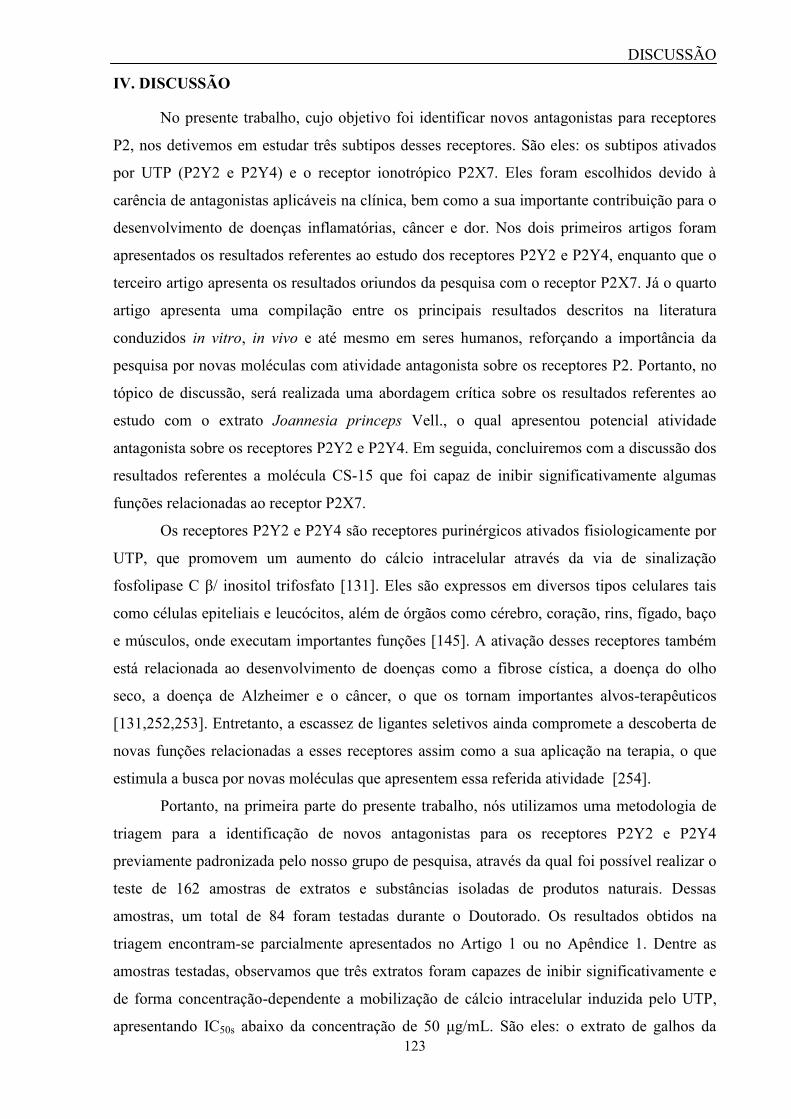
DISCUSSÃO
123
IV. DISCUSSÃO
No presente trabalho, cujo objetivo foi identificar novos antagonistas para receptores
P2, nos detivemos em estudar três subtipos desses receptores. São eles: os subtipos ativados
por UTP (P2Y2 e P2Y4) e o receptor ionotrópico P2X7. Eles foram escolhidos devido à
carência de antagonistas aplicáveis na clínica, bem como a sua importante contribuição para o
desenvolvimento de doenças inflamatórias, câncer e dor. Nos dois primeiros artigos foram
apresentados os resultados referentes ao estudo dos receptores P2Y2 e P2Y4, enquanto que o
terceiro artigo apresenta os resultados oriundos da pesquisa com o receptor P2X7. Já o quarto
artigo apresenta uma compilação entre os principais resultados descritos na literatura
conduzidos in vitro, in vivo e até mesmo em seres humanos, reforçando a importância da
pesquisa por novas moléculas com atividade antagonista sobre os receptores P2. Portanto, no
tópico de discussão, será realizada uma abordagem crítica sobre os resultados referentes ao
estudo com o extrato Joannesia princeps Vell., o qual apresentou potencial atividade
antagonista sobre os receptores P2Y2 e P2Y4. Em seguida, concluiremos com a discussão dos
resultados referentes a molécula CS-15 que foi capaz de inibir significativamente algumas
funções relacionadas ao receptor P2X7.
Os receptores P2Y2 e P2Y4 são receptores purinérgicos ativados fisiologicamente por
UTP, que promovem um aumento do cálcio intracelular através da via de sinalização
fosfolipase C β/ inositol trifosfato [131]. Eles são expressos em diversos tipos celulares tais
como células epiteliais e leucócitos, além de órgãos como cérebro, coração, rins, fígado, baço
e músculos, onde executam importantes funções [145]. A ativação desses receptores também
está relacionada ao desenvolvimento de doenças como a fibrose cística, a doença do olho
seco, a doença de Alzheimer e o câncer, o que os tornam importantes alvos-terapêuticos
[131,252,253]. Entretanto, a escassez de ligantes seletivos ainda compromete a descoberta de
novas funções relacionadas a esses receptores assim como a sua aplicação na terapia, o que
estimula a busca por novas moléculas que apresentem essa referida atividade [254].
Portanto, na primeira parte do presente trabalho, nós utilizamos uma metodologia de
triagem para a identificação de novos antagonistas para os receptores P2Y2 e P2Y4
previamente padronizada pelo nosso grupo de pesquisa, através da qual foi possível realizar o
teste de 162 amostras de extratos e substâncias isoladas de produtos naturais. Dessas
amostras, um total de 84 foram testadas durante o Doutorado. Os resultados obtidos na
triagem encontram-se parcialmente apresentados no Artigo 1 ou no Apêndice 1. Dentre as
amostras testadas, observamos que três extratos foram capazes de inibir significativamente e
de forma concentração-dependente a mobilização de cálcio intracelular induzida pelo UTP,
apresentando IC50s abaixo da concentração de 50 μg/mL. São eles: o extrato de galhos da
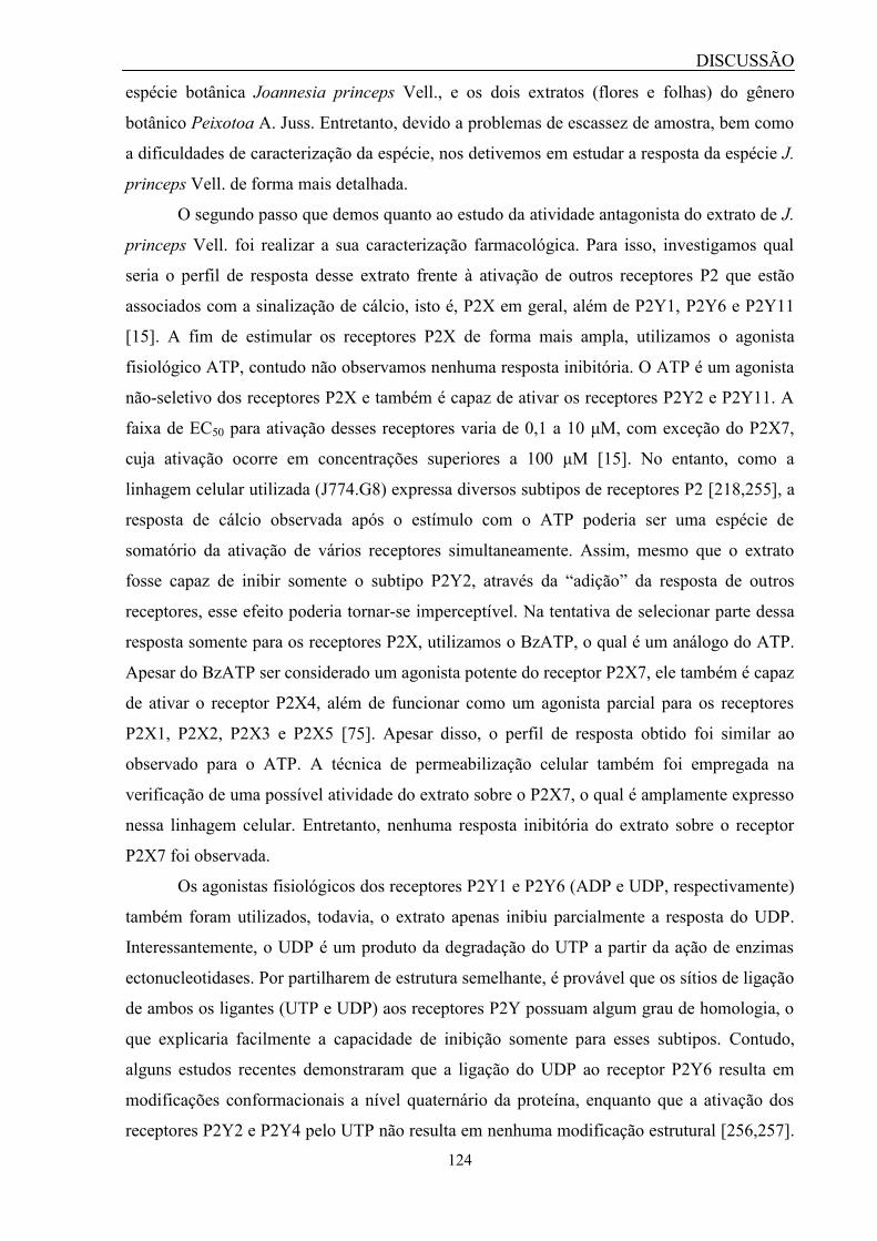
DISCUSSÃO
124
espécie botânica Joannesia princeps Vell., e os dois extratos (flores e folhas) do gênero
botânico Peixotoa A. Juss. Entretanto, devido a problemas de escassez de amostra, bem como
a dificuldades de caracterização da espécie, nos detivemos em estudar a resposta da espécie J.
princeps Vell. de forma mais detalhada.
O segundo passo que demos quanto ao estudo da atividade antagonista do extrato de J.
princeps Vell. foi realizar a sua caracterização farmacológica. Para isso, investigamos qual
seria o perfil de resposta desse extrato frente à ativação de outros receptores P2 que estão
associados com a sinalização de cálcio, isto é, P2X em geral, além de P2Y1, P2Y6 e P2Y11
[15]. A fim de estimular os receptores P2X de forma mais ampla, utilizamos o agonista
fisiológico ATP, contudo não observamos nenhuma resposta inibitória. O ATP é um agonista
não-seletivo dos receptores P2X e também é capaz de ativar os receptores P2Y2 e P2Y11. A
faixa de EC50 para ativação desses receptores varia de 0,1 a 10 μM, com exceção do P2X7,
cuja ativação ocorre em concentrações superiores a 100 μM [15]. No entanto, como a
linhagem celular utilizada (J774.G8) expressa diversos subtipos de receptores P2 [218,255], a
resposta de cálcio observada após o estímulo com o ATP poderia ser uma espécie de
somatório da ativação de vários receptores simultaneamente. Assim, mesmo que o extrato
fosse capaz de inibir somente o subtipo P2Y2, através da “adição” da resposta de outros
receptores, esse efeito poderia tornar-se imperceptível. Na tentativa de selecionar parte dessa
resposta somente para os receptores P2X, utilizamos o BzATP, o qual é um análogo do ATP.
Apesar do BzATP ser considerado um agonista potente do receptor P2X7, ele também é capaz
de ativar o receptor P2X4, além de funcionar como um agonista parcial para os receptores
P2X1, P2X2, P2X3 e P2X5 [75]. Apesar disso, o perfil de resposta obtido foi similar ao
observado para o ATP. A técnica de permeabilização celular também foi empregada na
verificação de uma possível atividade do extrato sobre o P2X7, o qual é amplamente expresso
nessa linhagem celular. Entretanto, nenhuma resposta inibitória do extrato sobre o receptor
P2X7 foi observada.
Os agonistas fisiológicos dos receptores P2Y1 e P2Y6 (ADP e UDP, respectivamente)
também foram utilizados, todavia, o extrato apenas inibiu parcialmente a resposta do UDP.
Interessantemente, o UDP é um produto da degradação do UTP a partir da ação de enzimas
ectonucleotidases. Por partilharem de estrutura semelhante, é provável que os sítios de ligação
de ambos os ligantes (UTP e UDP) aos receptores P2Y possuam algum grau de homologia, o
que explicaria facilmente a capacidade de inibição somente para esses subtipos. Contudo,
alguns estudos recentes demonstraram que a ligação do UDP ao receptor P2Y6 resulta em
modificações conformacionais a nível quaternário da proteína, enquanto que a ativação dos
receptores P2Y2 e P2Y4 pelo UTP não resulta em nenhuma modificação estrutural [256,257].
DISCUSSÃO
125
Uma vez que não podemos descartar a possibilidade do extrato funcionar como um
modulador alostérico negativo, essas modificações conformacionais poderiam continuar
ocorrendo mesmo em um nível menor, mas suficiente para que o receptor P2Y6 ainda
permaneça em estado ativo.
Visto que o UTP é capaz de ativar tanto o P2Y2 quanto o P2Y4, foram utilizados
agonistas seletivos para cada um desses receptores com o objetivo de tentar identificar se o
extrato conseguiria inibir um único subtipo ou mesmo ambos [22]. Contudo, os dados
sugerem que o extrato é capaz de inibir a resposta de ambos receptores. Embora eles sejam
considerados agonistas seletivos, sabe-se que dependendo da concentração utilizada (faixa de
submicromolar), eles podem apresentar uma determinada perda de seletividade [258,259].
Com todos esses desafios, o experimento que deve ser feito na tentativa de concluir essa
caracterização farmacológica é a utilização de células transfectadas com um único subtipo de
receptor P2Y a fim de se obter uma resposta mais seletiva.
Outro ponto importante que nos questionamos foi se a inibição causada pelo extrato
poderia ser decorrente de um efeito quenching. Uma vez que algumas substâncias oriundas de
plantas são capazes de emitir fluorescência ou apresentar efeito quenching (ex: quercetina e
vitexina), deve-se analisar os resultados obtidos com cautela a fim de verificar se eles não são
frutos de artefatos experimentais [260]. Nesse sentido, foi realizado um teste com a
ionomicina, um ionóforo de cálcio que promove uma elevação dos níveis intracelulares desse
íon, resultando em um aumento da intensidade de fluorescência proporcional a esse efeito.
Com isso, foi observado que a presença do extrato de J. princeps Vell. não resultou em
nenhuma mudança no perfil de resposta da ionomicina, eliminando assim, a possibilidade de
um efeito quenching.
Perguntamo-nos ainda se a abertura dos canais de cálcio operados por estoque (SOC)
poderia influenciar a resposta do extrato [261]. Essa elevação dos níveis de cálcio mediante a
abertura de canais SOC poderia estar dificultando a observação da mobilização de cálcio
através da via da fosfolipase C/ IP3. Portanto, através da remoção do cálcio extracelular da
solução tampão, esperávamos continuar observando a resposta inibitória do UTP.
Surpreendentemente, a mobilização de cálcio reduziu drasticamente e a resposta do extrato foi
similar à resposta basal do UTP. Isso nos levou a questionar dois fatos. O primeiro foi a
possibilidade de o extrato estar inibindo a resposta de canais SOC ao invés de inibir os
receptores. A segunda questão foi se de fato, o extrato inibisse canais SOC, não
observaríamos uma resposta de inibição mesmo que parcial sobre a mobilização de cálcio
induzida pelos demais agonistas como ATP, ADP, BzATP e UDP? Outra hipótese é de que a

DISCUSSÃO
126
ausência de cálcio extracelular poderia ter produzido algum tipo de artefato experimental ou
algum fenômeno não esclarecido que poderia ter afetado a resposta observada.
Outra preocupação que tivemos foi quanto à aplicabilidade do extrato como possível
fitoterápico. A primeira amostra de extrato utilizada no presente trabalho foi extraída a partir
de uma mistura dos solventes metanol e diclorometano. Entretanto, de acordo com as
regulamentações brasileira (Agência Nacional de Vigilância Sanitária – ANVISA) e
americana (Food and Drug Administration - FDA), para que um fármaco de origem vegetal
seja aprovado como um fitoterápico, ele necessita ter sido extraído em solução aquosa ou
etanólica [214,262]. Com isso, foi realizada uma nova extração utilizando o etanol como
solvente. Semelhantemente ao que já havia sido observado com o extrato metanólico, esse
novo extrato etanólico também foi capaz de inibir a resposta do UTP em células J774.G8 e
macrófagos peritoneais, além de não produzir citotoxicidade em até 24h de tratamento.
J. princeps Vell. é uma espécie botânica encontrada na Mata Atlântica e Caatinga no
Brasil, bem como em algumas regiões nos continentes Africano e Asiático. Ela pertence à
família Euphorbiaceae, cujos indivíduos medem em torno de 20 m de altura. No Brasil, ela é
popularmente conhecida como “cotieira” e possui valor econômico no paisagismo e extração
de madeira [263,264]. Essa espécie também possui uso medicinal, contudo, a única parte da
planta que é utilizada para essa finalidade são as sementes. O óleo presente nas sementes é
utilizado como laxante e cicatrizante, enquanto que o extrato dessa parte da planta possui
atividades anti-helmínticas [264,265]. Por outo lado, segundo relatos da medicina popular, a
J. princeps Vell. ainda é utilizada para o tratamento de desordens menstruais e digestivas,
febre perniciosa, doenças microbianas, sífilis e inchaço [264]. Apesar disso, há uma escassez
de estudos que demonstram o potencial uso medicinal de outras partes dessa planta, como
folha, caule, raiz e flor. J. princeps Vell. apresenta moléculas pertencentes às classes de
sesquiterpenos, bis-sesquiterpenos, diterpenos, triterpenos e esteróides [266]. No entanto, os
poucos trabalhos que descrevem a fitoquímica e as propriedades medicinais dessa espécie
detêm-se apenas nas sementes e folhas. Nesse sentido, o estudo da composição química e
aplicação medicinal de extratos dos galhos ainda são escassos, sendo o nosso grupo pioneiro
nesse cenário. Como resultado preliminar acerca da obtenção do perfil químico da J. princeps
Vell., observamos a presença de pelo menos 45 substâncias no extrato através de um ensaio
de cromatografia líquida de alta performance (high performance liquid chromatography –
HPLC). Entretanto, a caracterização da molécula ativa, responsável pela atividade
antagonista, encontra-se entre um dos objetivos do nosso grupo de pesquisa, uma vez que é
imprescindível para o desenvolvimento de um novo fármaco, embora não seja necessária para
a elaboração de um medicamento fitoterápico [267].

DISCUSSÃO
127
A caracterização da molécula ativa responsável pela inibição do P2Y2 e P2Y4
certamente contribuirá para a farmacologia desses receptores. Apesar do P2Y2 já possuir um
antagonista seletivo descrito, este não conseguiu avançar para a aplicação clínica devido a
algumas propriedades farmacológicas desfavoráveis, como alta polaridade e baixa
disponibilidade pela via oral [49]. O P2Y4, por outro lado, ainda não possui antagonistas
seletivos, o que dificulta a caracterização de suas funções fisiológicas. Nesse sentido, o
antagonista RB2 geralmente é utilizado na obtenção de alguma referência acerca de um
possível efeito inibitório. Todavia, devido à sua capacidade de inibir outros receptores P2, os
resultados devem ser avaliados com cautela. Nesse cenário, a descrição de um antagonista que
atue sobre ambos receptores, mesmo que sem seletividade, poderá contribuir para o
desenvolvimento de diversos estudos in vitro e in vivo, e ainda pode servir como um modelo
estrutural para a síntese e desenvolvimento de novos antagonistas seletivos para esses
receptores [49].
Enquanto isso, a segunda parte desse trabalho se deteve na pesquisa por novos
candidatos a antagonistas para o receptor P2X7, cuja ativação além de promover um fluxo de
cátions através da membrana, está relacionada com a formação de um poro membranar e a
indução de uma série de respostas com perfil pró-inflamatório [69,102,167,169–172]. Além
disso, diversos estudos vêm demonstrando a participação desse receptor no desenvolvimento
de doenças inflamatórias, neurodegenerativas e infecciosas, o que o torna um importante alvo
terapêutico [172,268]. Embora o uso de antagonistas em modelos in vivo de inflamação e dor
tenham demonstrado efeitos anti-inflamatórios e antinociceptivos significativos, as moléculas
que avançaram para a etapa de ensaios clínicos foram reprovados nessa fase [172]. Assim,
essa lacuna na terapia associada ao P2X7 estimula a pesquisa por novas moléculas com
atuação sobre este receptor e que possam ser usadas na clínica para o tratamento dessas
doenças.
Nesse contexto, executamos uma mini-campanha de triagem com o objetivo de
identificar novos antagonistas para o receptor P2X7 a partir de produtos naturais. Ao longo
dessa campanha, identificamos que a molécula CS-15 apresentou-se como um hit promissor
devido à sua habilidade de inibir as funções do P2X7 in vitro e in vivo.
Entretanto, para realizarmos essa triagem utilizamos uma metodologia de
permeabilização celular previamente padronizada pelo nosso grupo de pesquisa para ser
aplicada à descoberta de novos antagonistas para o receptor P2X7 [247]. O corante utilizado
na padronização desse protocolo foi o iodeto de propídeo. Já no presente trabalho, nós
reproduzimos o mesmo protocolo, com exceção do corante que foi substituído pelo YO-PRO-

DISCUSSÃO
128
1, cujo uso vem se popularizando em ensaios de permeabilização celular. Essa modificação no
protocolo nos permitiu verificar a reprodutibilidade da metodologia previamente padronizada.
Uma vez que realizamos uma pequena modificação no protocolo, algumas
observações precisaram ser feitas antes da sua implementação no processo de triagem. A
primeira foi em relação à escolha da concentração do corante que seria utilizada. Em um teste
realizado preliminarmente foram avaliadas diferentes concentrações do corante (YO-PRO-1)
na faixa de 62,5 nM a 8 μM (dados não mostrados). Contudo, observamos que o equipamento
utilizado era capaz somente de distinguir diferenças entre a fluorescência basal e a relacionada
à captação do corante via ativação do P2X7 a partir da concentração de 1 μM. Assim,
adotamos essa concentração como ponto de partida para estudo da relação sinal-ruído. Com
isso, observamos que a relação sinal-ruído para a concentração de 1 μM de corante era de
1,42, enquanto que com o dobro dessa concentração (2 μM), a relação aumentava para 1,65.
Apesar de haver pouca diferença entre a relação sinal-ruído das duas concentrações estudadas,
a concentração de 2 μM foi adotada para uso por possuir respaldo da literatura, uma vez que é
utilizada em diversos trabalhos científicos [248,269–271]. Além disso, o valor obtido para a
relação sinal-ruído a 2 μM foi similar ao detectado por Rat et al. (2017) para o YO-PRO-1 (2 μM)
em linhagem celular de retina humana (ARPE-19) e queratinócitos (HaCat) (1,7 e 1,45,
respectivamente) na presença de 1 e 10 mM de ATP [248]. Outra vantagem de ter escolhido
essa concentração foi a relação custo-benefício, ou seja, optar por uma concentração maior do que
essa (2 μM), certamente iria gerar um aumento do consumo e, consequentemente, maior gasto
com reagentes.
Com a concentração do YO-PRO-1 definida, os experimentos em espectrofotômetro
prosseguiram com a avaliação do perfil de captação do corante na presença do ATP. O EC50
detectado para o ATP foi de 2,18 mM, o qual se encontra dentro da faixa do EC50 descrito para o
receptor P2X7 (2 a 4 mM) [75]. Contudo, em nosso trabalho prévio em que foi utilizado o
iodeto de propídeo, o EC50 encontrado ficou entre 0,7114 e 0,7663 mM [247]. Essa variação
pode ser decorrente da diferença de sensibilidade entre os equipamentos, os corante ou
mesmo os subclones de células J774.G8 utilizados. Um subclone distinto de uma mesma
linhagem celular pode por alguma razão, expressar o receptor P2X7 em menor densidade ou
ainda apresentar um variante de splice diferente, que pode possuir uma determinada
resistência em formar o poro membranar [272]. Apesar disso, o fator z’, uma medida de
robustez do ensaio, foi calculado e o valor encontrado foi de 0,59, indicando que o protocolo
estava adequado para uso. Esse valor foi semelhante ao encontrado em nosso protocolo com o
iodeto de propídeo (0,635), porém menor do que o identificado para o corante amarelo de
DISCUSSÃO
129
lúcifer (0,876), sugerindo que possam existir variações quanto à sensibilidade dos corantes
[247].
O perfil de inibição foi avaliado utilizando-se dois antagonistas do P2X7: o BBG e o
A-740003. O IC50 detectado para o antagonista BBG foi de 106 nM e encontra-se dentro da
faixa de IC50 descrita na literatura, a qual varia entre 15 a 250 nM [75]. Por outro lado, esse
valor foi menor do que o identificado em nosso protocolo com o iodeto de propídeo, que
variou entre 1,3 a 2,6 μM [247]. Enquanto isso, o antagonista seletivo do receptor P2X7, o A-
740003, apresentou um IC50 de 511 nM, o qual também encontra-se dentro da faixa de IC50
descrita na literatura (entre 20 a 700 nM) [75].
Com a caracterização do perfil do YO-PRO-1 aplicado a ensaios de permeabilização
celular, foi realizada uma mini-campanha de triagem, na qual 134 amostras foram testadas,
cujos resultados estão apresentados no Apêndice 2. Durante essa campanha, a molécula CS-
15, um triterpeno isolado da planta Clusia studartiana C. M. Vieira & Gomes da Silva, foi
identificada como principal hit, uma vez que ela foi capaz de inibir a captação do YO-PRO-1
de forma concentração-dependente. Além disso, ela exibiu um IC50 na faixa de nanomolar,
enquanto que a maioria dos hits apresenta a atividade almejada entre 1 a 10 μM [273]. O
referida molécula também não afetou a viabilidade celular mesmo após 24h de tratamento.
Nós ainda tentamos verificar se a molécula seria capaz de inibir a mobilização de
cálcio induzida por ATP. Entretanto, com a finalidade de ativar o receptor P2X7, foi
necessário utilizar uma alta concentração de ATP (100 μM), a qual é capaz de ativar outros
receptores P2. Uma vez que as células J774.G8 expressam diversos subtipos de receptores P2,
incluindo P2X e P2Y [218,255], não fomos capazes de observar a inibição esperada utilizando
esse modelo. Uma alternativa para alcançar uma maior taxa de sucesso nesse experimento
seria o uso de células transfectadas com o P2X7 ou o uso do BzATP, um agonista mais
potente, porém não seletivo para esse receptor [75]. Por fim, também avaliamos outro
nucleotídeo, o UTP, a fim de observar se essa inibição se estenderia para receptores P2Y,
porém a molécula não foi capaz de bloquear a sua resposta de cálcio. Ainda assim, novos
ensaios com agonistas seletivos de outros receptores P2 e com células transfectadas com o
receptor P2X7 humano (hP2X7) devem ser feitos para confirmar se de fato a molécula possui
seletividade para o esse receptor. Além disso, experimentos utilizando a técnica de patch-
clamp na modalidade whole cell, que atualmente é considerada o padrão-ouro na investigação
de canais iônicos, podem contribuir significantemente para caracterizar a atividade da
molécula CS-15 sobre o P2X7.
Finalmente, o efeito da molécula foi testado em modelo experimental de dor
produzido pela injeção intraplantar de formalina. Esse modelo experimental foi desenvolvido
DISCUSSÃO
130
na década de 1970 e ainda hoje é útil para a triagem de novas moléculas com atividade
analgésica e anti-inflamatória [274,275]. Através da aplicação desse modelo, é possível
avaliar o comportamento nociceptivo dos animais em duas fases distintas. A primeira fase
inicia-se logo após a injeção da formalina e dura cerca de 5 minutos, sendo caracterizada pela
ativação química dos nociceptores. Já a segunda fase inicia-se entre 15 a 30 minutos após a
injeção da formalina e é caracterizada pelo envolvimento do processo inflamatório no
desenvolvimento da dor [275]. Utilizando esse modelo, alguns grupos de pesquisa
observaram o efeito analgésico e anti-inflamatório de algumas plantas medicinais, inclusive
com ação sobre o P2X7 [222,274]. Apesar disso, em nossos ensaios, a molécula CS-15
reduziu significativamente os sintomas da dor nociceptiva e inflamatória. Ainda não sabemos
se esse efeito é dependente ou independente da ativação do P2X7. Para isso, novos
experimentos nos quais a análise de citocinas como a IL-1β e outros medidadores
inflamatórios relacionados à ativação do P2X7 necessitam ser investigados in vivo. Em todo
caso, resultados similares a esses já foram observados em experimentos com outros
antagonistas do P2X7, o que demonstra o potencial farmacológico dessa molécula [119,222].
C. studartiana C.M. Vieira & Gomes da Silva é uma espécie botânica pertencente à
família Clusiaceae. Ela é uma árvore de hábito terrestre, que ocasionalmente pode apresentar
forma hemiepífita. Essa espécie é endêmica da Mata Atlântica brasileira na região Sudeste do
país [276]. Embora essa espécie tenha sido descrita no início dos anos 1990, até o momento
não se tem nenhuma informação na literatura acerca de suas propriedades medicinais, o que
torna o nosso trabalho pioneiro nesse sentido. Apesar disso, a família Clusiaceae possui
alguns indivíduos com conhecidas propriedades medicinais. O látex presente nos frutos de
Clusia sp. são popularmente utilizados no tratamento de doenças reumáticas e candidíase oral
infantil, enquanto que as sementes de C. grandiflora apresentam moléculas com atividade
antimicrobiana [277–280]. Em todo o caso, o nosso estudo demonstrou pela primeira vez o
efeito analgésico de uma molécula isolada da planta C. studartiana, possivelmente causado
pela inibição do receptor P2X7. Com isso, acreditamos que esses achados contribuirão para o
escasso campo da terapia purinérgica no futuro assim para o conhecimento de propriedades
medicinais oriundas de produtos naturais.

CONCLUSÃO
131
V. CONCLUSÃO
Os resultados apontaram que o extrato de galhos da espécie botânica Joannesia
princeps Vell. foi capaz de inibir a mobilização de cálcio intracelular induzida por UTP de
forma concentração-dependente, sendo que esse efeito não foi provocado por citotoxicidade
ou fenômeno quenching. Em virtude de o extrato ter sido capaz de inibir parcialmente a
mobilização de cálcio induzida por UDP, os resultados sugerem uma possível seletividade
quanto à sua ação antagonista sobre os receptores P2Y ativados por nucleotídeos derivados da
uridina.
Enquanto isso, os resultados obtidos com a pesquisa de novos antagonistas para o
receptor P2X7 apontaram para a descoberta de um novo candidato a antagonista para esse
receptor. A molécula CS-15, um triterpeno isolado da planta Clusia studartiana C. M. Vieira
& Gomes da Silva, foi capaz de inibir a captação do YO-PRO-1, função relacionada com a
ativação do P2X7 de forma concentração-dependente, apresentando atividade na faixa de
nanomolar. Além disso, essa molécula não demonstrou toxicidade in vitro e foi capaz de
aliviar os sintomas de nocicepção em modelo experimental animal.
REFERÊNCIAS
132
VI. REFERÊNCIAS
1. Jones ME. Albrecht Kossel, a biographical sketch. Yale J Biol Med. 1953;26:80–97.
2. Burnstock G, Verkhratsky A. Purinergic signalling and the nervous system. Purinergic
Signal. Nerv. Syst. 2012.
3. Levene PA, Tipson RS. The ring structure of adenosine. Science (80). 1931;74:521.
4. Drury AN, Szent-Györgyi A. The physiological activity of adenine compounds with
especial reference to their action upon the mammalian heart. J Physiol. 1929;68:213–37.
5. Gillespie JH. The biological significance of the linkages in adenosine triphosphoric acid. J
Physiol. 1934;80:345–59.
6. Holton FA, Holton P. The capillary dilator substances in dry powders of spinal roots; a
possible role of adenosine triphosphate in chemical transmission from nerve endings. J
Physiol. 1954;126:124–140.
7. Holton P. The liberation of adenosine triphosphate on antidromic stimulation of sensory
nerves. J Physiol . 1959;145:494–504.
8. Burnstock G, Campbell G, Satchell D, Smythe A. Evidence that adenosine triphosphate or
a related nucleotide is the transmitter substance released by non-adrenergic inhibitory nerves
in the gut. Br J Pharmac. 1970;40:668–88.
9. Burnstock G. Do some nerve cells release more than one transmitter? Neuroscience.
1976;1:239–48.
10. Burnstock G. Introduction: P2 Receptors. Curr Top Med Chem. 2004;4:793–803.
11. van Calker D, Müller M, Hamprecht B. Adenosine regulates via two different types of
receptors, the accumulation of cyclic AMP in cultured brain cells. J Neurochem.
1979;33:999–1005.
12. Burnstock G, Kennedy C. Is there a basis for distinguishing two types of P2-purinoceptor?
Gen Pharmacol Vasc Syst. 1985;16:433–40.
13. Fredholm BB, Abbracchio MP, Burnstock G, Daly JW, Harden TK, Jacobson KA, et al.
Nomenclature and classification of purinoceptors. Pharmacol Rev. 1994;46:143–56.
14. Abbracchio MP, Burnstock G. Purinoceptors: Are there families of P2X and P2Y
purinoceptors? Pharmac Ther. 1994;64:445–75.
15. Ralevic V, Burnstock G. Receptors for purines and pyrimidines. Pharmacol Rev.
1998;50:413–92.
16. von Kügelgen I, Wetter A. Molecular pharmacology of P2Y-receptors. Naunyn
Schmiedebergs Arch Pharmacol. 2000;362:310–23.
17. Thimm D, Knospe M, Abdelrahman A, Moutinho M, Alsdorf BBA, von Kügelgen I, et al.
Characterization of new G protein-coupled adenine receptors in mouse and hamster.
Purinergic Signal. 2013;9:415–26.
18. Menzies J, Paul A, Kennedy C. P2X7subunit-like immunoreactivity in the nucleus of
visceral smooth muscle cells of the guinea pig. Auton Neurosci Basic Clin. 2003;106:103–9.

REFERÊNCIAS
133
19. Deban SM, Olson WM, Buckley NJ, Deuchars J. An ATP-gated ion channel at the cell
nucleus. 2002;420:2002.
20. Fountain SJ. Primitive ATP-activated P2X receptors: discovery, function and
pharmacology. Front Cell Neurosci. 2013;7:247.
21. Burnstock G. Purine and pyrimidine receptors. Cell Mol Life Sci. 2007;64:1471–83.
22. Jacobson KA, Balasubramanian R, Deflorian F, Gao Z-G. G protein-coupled adenosine
(P1) and P2Y receptors: ligand design and receptor interactions. Purinergic Signal.
2012;8:419–36.
23. Franco R, Navarro G. Adenosine A2A Receptor Antagonists in Neurodegenerative
Diseases: Huge Potential and Huge Challenges. Front Psychiatry. 2018;9:68.
24. Von Kügelgen I, Hoffmann K. Pharmacology and structure of P2Y receptors.
Neuropharmacology. 2016;104:50–61.
25. Chen J, Wang L, Zhang Y, Yang J. P2Y1 purinoceptor inhibition reduces extracellular
signal-regulated protein kinase 1/2 phosphorylation in spinal cord and dorsal root ganglia:
implications for cancer-induced bone pain. Acta Biochim Biophys Sin (Shanghai).
2012;44:367–72.
26. Kwon S-G, Roh D-H, Yoon S-Y, Moon J-Y, Choi S-R, Choi H-S, et al. Acid evoked
thermal hyperalgesia involves peripheral P2Y1 receptor mediated TRPV1 phosphorylation in
a rodent model of thrombus induced ischemic pain. Mol Pain. 2014;10:2.
27. Barragán-Iglesias P, Mendoza-Garcés L, Pineda-Farias JB, Solano-Olivares V,
Rodríguez-Silverio J, Flores-Murrieta FJ, et al. Participation of peripheral P2Y1, P2Y6 and
P2Y11 receptors in formalin-induced inflammatory pain in rats. Pharmacol Biochem Behav.
2015;128:23–32.
28. Delekate A, Füchtemeier M, Schumacher T, Ulbrich C, Foddis M, Petzold GC.
Metabotropic P2Y1 receptor signalling mediates astrocytic hyperactivity in vivo in an
Alzheimer’s disease mouse model. Nat Commun. 2014;5:5422.
29. Amison RT, Momi S, Morris A, Manni G, Keir S, Gresele P, et al. RhoA signaling
through platelet P2Y1 receptor controls leukocyte recruitment in allergic mice. J Allergy Clin
Immunol. 2015;135:528–38.
30. Leon C, Freund M, Ravanat C, Baurand A, Cazenave J-P, Gachet C. Key role of the P2Y1
receptor in tissue factor-induced thrombin-dependent acute thromboembolism : studies in
P2Y1-knockout mice and mice treated with a P2Y1 antagonist. Circulation. 2001;103:718–
23.
31. Léon C, Ravanat C, Freund M, Cazenave JP, Gachet C. Differential involvement of the
P2Y1 and P2Y12 receptors in platelet procoagulant activity. Arterioscler Thromb Vasc Biol.
2003;23:1941–7.
32. Lenain N, Freund M, Léon C, Cazenave J-P, Gachet C. Inhibition of localized thrombosis
in P2Y1-deficient mice and rodents treated with MRS2179, a P2Y1 receptor antagonist. J
Thromb Haemost. 2003;1:1144–9.
33. Hechler B, Nonne C, Roh EJ, Cattaneo M, Cazenave J-P, Lanza F, et al. MRS2500 [2-
Iodo-N6-methyl-(N)-methanocarba-2′-deoxyadenosine-3′,5′-bisphosphate], a potent,
selective, and stable antagonist of the platelet P2Y1 receptor with strong antithrombotic
REFERÊNCIAS
134
activity in mice. J Pharmacol Exp Ther. 2006;316:556–63.
34. Labarthe B, Babin J, Bryckaert M, Théroux P, Bonnefoy A. Effects of P2Y(1) receptor
antagonism on the reactivity of platelets from patients with stable coronary artery disease
using aspirin and clopidogrel. Br J Pharmacol. 2012;166:221–31.
35. Wong PC, Watson C, Crain EJ. The P2Y1 receptor antagonist MRS2500 prevents carotid
artery thrombosis in cynomolgus monkeys. J Thromb Thrombolysis. 2016;41:514–21.
36. Li N, Lu Z, Yu L, Burnstock G, Deng X, Ma B. Inhibition of G protein-coupled P2Y2
receptor induced analgesia in a rat model of trigeminal neuropathic pain. Mol Pain.
2014;10:21.
37. Magni G, Merli D, Verderio C, Abbracchio MP, Ceruti S. P2Y2 receptor antagonists as
anti-allodynic agents in acute and sub-chronic trigeminal sensitization: Role of satellite glial
cells. Glia. 2015;63:1256–69.
38. Choi JH, Ji YG, Lee DH. Uridine triphosphate increases proliferation of human cancerous
pancreatic duct epithelial cells by activating P2Y2 receptor. Pancreas. 2013;42:680–6.
39. Li W-H, Qiu Y, Zhang H-Q, Liu Y, You J-F, Tian X-X, et al. P2Y2 receptor promotes
cell invasion and metastasis in prostate cancer cells. Br J Cancer. 2013;109:1666–75.
40. Jin H, Eun SY, Lee JSJH, Park SW, Lee JSJH, Chang KC, et al. P2Y2 receptor activation
by nucleotides released from highly metastatic breast cancer cells increases tumor growth and
invasion via crosstalk with endothelial cells. Breast Cancer Res. 2014;16:R77.
41. Chadet S, Jelassi B, Wannous R, Angoulvant D, Chevalier S, Besson P, et al. The
activation of P2Y2 receptors increases MCF-7 breast cancer cells migration through the
MEK-ERK1/2 signalling pathway. Carcinogenesis. 2014;35:1238–47.
42. Xie R, Xu J, Wen G, Jin H, Liu X, Yang Y, et al. The P2Y2 nucleotide receptor mediates
the proliferation and migration of human hepatocellular carcinoma cells induced by ATP. J
Biol Chem. 2014;289:19137–49.
43. Inoue Y, Chen Y, Hirsh MI, Yip L, Junger WG. A3 and P2Y2 receptors control the
recruitment of neutrophils to the lungs in a mouse model of sepsis. Shock. 2008;30:1.
44. Degagné E, Grbic DM, Dupuis A-A, Lavoie EG, Langlois C, Jain N, et al. P2Y2 receptor
transcription is increased by NF-kappa B and stimulates cyclooxygenase-2 expression and
PGE2 released by intestinal epithelial cells. J Immunol. 2009;183:4521–9.
45. Vanderstocken G, Bondue B, Horckmans M, Di Pietrantonio L, Robaye B, Boeynaems J-
M, et al. P2Y2 receptor regulates VCAM-1 membrane and soluble forms and eosinophil
accumulation during lung inflammation. J Immunol. 2010;185:3702–7.
46. Zhang Y, Kohan DE, Nelson RD, Carlson NG, Kishore BK. Potential involvement of
P2Y2 receptor in diuresis of postobstructive uropathy in rats. Am J Physiol Renal Physiol.
2010;298:F634-42.
47. Ayata CK, Ganal SC, Hockenjos B, Willim K, Vieira RP, Grimm M, et al. Purinergic
P2Y2 receptors promote neutrophil infiltration and hepatocyte death in mice with acute liver
injury. Gastroenterology. 2012;143:1620–1629.e4.
48. Eun SY, Park SW, Lee JH, Chang KC, Kim HJ. P2Y2R activation by nucleotides released
from oxLDL-treated endothelial cells (ECs) mediates the interaction between ECs and
REFERÊNCIAS
135
immune cells through RAGE expression and reactive oxygen species production. Free Radic
Biol Med. 2014;69:157–66.
49. Rafehi M, Müller CE. Tools and drugs for uracil nucleotide-activated P2Y receptors.
Pharmacol Ther. Elsevier Inc; 2018;190:24–80.
50. Kobayashi K, Yamanaka H, Yanamoto F, Okubo M, Noguchi K. Multiple P2Y subtypes
in spinal microglia are involved in neuropathic pain after peripheral nerve injury. Glia.
2012;60:1529–39.
51. Barragán-Iglesias P, Pineda-Farias JB, Cervantes-Durán C, Bravo-Hernández M, Rocha-
González HI, Murbartián J, et al. Role of spinal P2Y6 and P2Y11 receptors in neuropathic
pain in rats: possible involvement of glial cells. Mol Pain. 2014;10:29.
52. Huang D, Yang J, Liu X, He L, Luo X, Tian H, et al. P2Y6 receptor activation is involved
in the development of neuropathic pain induced by chronic constriction injury of the sciatic
nerve in rats. J Clin Neurosci. 2018;56:156–62.
53. Placet M, Arguin G, Molle CM, Babeu J-P, Jones C, Carrier JC, et al. The G protein-
coupled P2Y6 receptor promotes colorectal cancer tumorigenesis by inhibiting apoptosis.
Biochim Biophys Acta. 2018;1864:1539–51.
54. Vieira RP, Müller T, Grimm M, Von Gernler V, Vetter B, Dürk T, et al. Purinergic
receptor type 6 contributes to airway inflammation and remodeling in experimental allergic
airway inflammation. Am J Respir Crit Care Med. 2011;184:215–23.
55. Grbic DM, Degagné É, Larrivée J-F, Bilodeau MS, Vinette V, Arguin G, et al. P2Y6
receptor contributes to neutrophil recruitment to inflamed intestinal mucosa by increasing
CXC chemokine ligand 8 expression in an AP-1-dependent manner in epithelial cells.
Inflamm Bowel Dis. 2012;18:1456–69.
56. Uratsuji H, Tada Y, Kawashima T, Kamata M, Hau CS, Asano Y, et al. P2Y6 receptor
signaling pathway mediates inflammatory responses induced by monosodium urate crystals. J
Immunol. 2012;188:436–44.
57. Hao Y, Liang JF, Chow AW, Cheung W, Ko W. P2Y6 receptor-mediated
proinflammatory signaling in human bronchial epithelia. PLoS One. 2014;9:e106235.
58. Sil P, Hayes CP, Reaves BJ, Breen P, Quinn S, Sokolove J, et al. P2Y6 receptor
antagonist MRS2578 inhibits neutrophil activation and aggregated neutrophil extracellular
trap formation induced by gout-associated monosodium urate crystals. J Immunol.
2017;198:428–42.
59. Jacobson KA, Costanzi S, Joshi B V, Besada P, Shin DH, Ko H, et al. Agonists and
antagonists for P2 receptors. Novartis Found Symp. 2006;276:58-68-72, 107–12, 275–81.
60. Dreisig K, Kornum BR. A critical look at the function of the P2Y11 receptor. Purinergic
Signal. Springer; 2016;12:427–37.
61. Gebremeskel S, LeVatte T, Liwski RS, Johnston B, Bezuhly M. The reversible P2Y12
inhibitor ticagrelor inhibits metastasis and improves survival in mouse models of cancer. Int J
Cancer. 2015;136:234–40.
62. Cho MS, Noh K, Haemmerle M, Li D, Park H, Hu Q, et al. Role of ADP receptors on
platelets in the growth of ovarian cancer. Blood. 2017;130:1235–42.

REFERÊNCIAS
136
63. Satonaka H, Nagata D, Takahashi M, Kiyosue A, Myojo M, Fujita D, et al. Involvement
of P2Y12 receptor in vascular smooth muscle inflammatory changes via MCP-1 upregulation
and monocyte adhesion. Am J Physiol Circ Physiol. 2015;308:H853–61.
64. Liverani E, Rico MC, Tsygankov AY, Kilpatrick LE, Kunapuli SP. P2Y12 receptor
modulates sepsis-induced inflammation. Arterioscler Thromb Vasc Biol. 2016;36:961–71.
65. Goffinet M, Tardy C, Boubekeur N, Cholez G, Bluteau A, Oniciu DC, et al. P2Y13
receptor regulates HDL metabolism and atherosclerosis in vivo. PLoS One. 2014;9:e95807.
66. Ishimaru M, Yusuke N, Tsukimoto M, Harada H, Takenouchi T, Kitani H, et al.
Purinergic signaling via P2Y receptors up-mediates IL-6 production by liver
macrophages/Kupffer cells. J Toxicol Sci. 2014;39:413–23.
67. Barrett MO, Sesma JI, Ball CB, Jayasekara PS, Jacobson KA, Lazarowski ER, et al. A
selective high-affinity antagonist of the P2Y14 receptor inhibits UDP-glucose-stimulated
chemotaxis of human neutrophils. Mol Pharmacol. 2013;84:41–9.
68. Azroyan A, Cortez-Retamozo V, Bouley R, Liberman R, Ruan YC, Kiselev E, et al. Renal
intercalated cells sense and mediate inflammation via the P2Y14 receptor. PLoS One.
2015;10:1–24.
69. Alves LA, de Melo Reis RA, de Souza CAM, de Freitas MS, Teixeira PCN, Neto Moreira
Ferreira D, et al. The P2X7 receptor: Shifting from a low to a high-conductance channel —
An enigmatic phenomenon? Biochim Biophys Acta - Biomembr. 2014;1838:2578–87.
70. Chaumont S, Khakh BS. Patch-clamp coordinated spectroscopy shows P2X2 receptor
permeability dynamics require cytosolic domain rearrangements but not Panx-1 channels.
Proc Natl Acad Sci U S A. 2008;105:12063–8.
71. Bernier LP, Ase AR, Boué-Grabot É, Séguéla P. P2X4 receptor channels form large
noncytolytic pores in resting and activated microglia. Glia. 2012;60:728–37.
72. Faria RX, De Farias FP, Alves LA. Are second messengers crucial for opening the pore
associated with P2X7 receptor? Am J Physiol Cell Physiol. 2005;288:C260–71.
73. Pelegrin P, Surprenant A. Pannexin-1 mediates large pore formation and interleukin-1beta
release by the ATP-gated P2X7 receptor. EMBO J. 2006;25:5071–82.
74. Pelegrín P. Many ways to dilate the P2X7 receptor pore. Br J Pharmacol. 2011;163:908–
11.
75. Coddou C, Yan Z, Obsil T, Huidobro-Toro JP, Stojilkovic SS. Activation and regulation
of purinergic P2X receptor channels. Pharmacol Rev. 2011;63:641–83.
76. North RA, Jarvis MF. P2X receptors as drug targets. Mol Pharmacol. 2013;83:759–69.
77. Hechler B, Lenain N, Marchese P, Vial C, Heim V, Freund M, et al. A role of the fast
ATP-gated P2X1 cation channel in thrombosis of small arteries in vivo. J Exp Med.
2003;198:661–7.
78. Gan M, Moussaud S, Jiang P, McLean PJ. Extracellular ATP induces intracellular alpha-
synuclein accumulation via P2X1 receptor-mediated lysosomal dysfunction. Neurobiol
Aging. 2015;36:1209–20.
79. Mulryan K, Gitterman DP, Lewis CJ, Vial C, Leckie BJ, Cobb AL, et al. Reduced vas

REFERÊNCIAS
137
deferens contraction and male infertility in mice lacking P2X1 receptors. Nature.
2000;403:86–9.
80. Yan D, Zhu Y, Walsh T, Xie D, Yuan H, Sirmaci A, et al. Mutation of the ATP-gated
P2X(2) receptor leads to progressive hearing loss and increased susceptibility to noise. Proc
Natl Acad Sci U S A. 2013;110:2228–33.
81. Navarro B, Miki K, Clapham DE. ATP-activated P2X2 current in mouse spermatozoa.
Proc Natl Acad Sci U S A. 2011;108:14342–7.
82. Wu JX, Xu MY, Miao XR, Lu ZJ, Yuan XM, Li XQ, et al. Functional up-regulation of
P2X3 receptors in dorsal root ganglion in a rat model of bone cancer pain. Eur J Pain.
2012;16:1378–88.
83. Burnstock G. Targeting the visceral purinergic system for pain control. Curr Opin
Pharmacol. 2012;12:80–6.
84. Ito K, Iwami A, Katsura H, Ikeda M. Therapeutic effects of the putative P2X3/P2X2/3
antagonist A-317491 on cyclophosphamide-induced cystitis in rats. Naunyn Schmiedebergs
Arch Pharmacol. 2008;377:483–90.
85. Zhang HP, Li CL, Lu P, Zheng JC, Yu LL, Yang WM, et al. The function of P2X3
receptor and NK1 receptor antagonists on cyclophosphamide-induced cystitis in rats. World J
Urol. 2014;32:91–7.
86. Deiteren A, Van Der Linden L, De Wit A, Ceuleers H, Buckinx R, Timmermans JP, et al.
P2X3 receptors mediate visceral hypersensitivity during acute chemically-induced colitis and
in the post-inflammatory phase via different mechanisms of sensitization. PLoS One.
2015;10:1–17.
87. Wang Q, Zhu H, Zou K, Yuan B, Zhou Y-L, Jiang X, et al. Sensitization of P2X3
receptors by cystathionine β-synthetase mediates persistent pain hypersensitivity in a rat
model of lumbar disc herniation. Mol Pain. 2015;11:1–13.
88. Stokes L, Layhadi JA, Bibic L, Dhuna K, Fountain SJ. P2X4 receptor function in the
nervous system and current breakthroughs in Pharmacology. Front Pharmacol. 2017;8:291.
89. Tsuda M, Shigemoto-Mogami Y, Koizumi S, Mizokoshi A, Kohsaka S, Salter MW, et al.
P2X4 receptors induced in spinal microglia gate tactile allodynia after nerve injury. Nature.
2003;424:778–83.
90. Tsuda M, Tozaki-Saitoh H, Masuda T, Toyomitsu E, Tezuka T, Yamamoto T, et al. Lyn
tyrosine kinase is required for P2X4 receptor upregulation and neuropathic pain after
peripheral nerve injury. Glia. 2008;56:50–8.
91. Tsuda M, Kuboyama K, Inoue T, Nagata K, Tozaki-Saitoh H, Inoue K. Behavioral
phenotypes of mice lacking purinergic P2X4 receptors in acute and chronic pain assays. Mol
Pain. 2009;5:28.
92. Masuda T, Iwamoto S, Yoshinaga R, Tozaki-Saitoh H, Nishiyama A, Mak TW, et al.
Transcription factor IRF5 drives P2X4R+-reactive microglia gating neuropathic pain. Nat
Commun. 2014;5:3771.
93. Li F, Wang L, Li J-W, Gong M, He L, Feng R, et al. Hypoxia induced amoeboid
microglial cell activation in postnatal rat brain is mediated by ATP receptor P2X4. BMC
Neurosci. 2011;12:111.
REFERÊNCIAS
138
94. Li F, Guo N, Ma Y, Ning B, Wang Y, Kou L. Inhibition of P2X4 suppresses joint
inflammation and damage in collagen-induced arthritis. Inflammation. 2014;37:146–53.
95. Vázquez-Villoldo N, Domercq M, Martín A, Llop J, Gómez-Vallejo V, Matute C. P2X4
receptors control the fate and survival of activated microglia. Glia. 2014;62:171–84.
96. Zech A, Wiesler B, Ayata CK, Schlaich T, Dürk T, Hoßfeld M, et al. P2rx4 deficiency in
mice alleviates allergen-induced airway inflammation. Oncotarget. 2016;7:80288–97.
97. Gever JR, Cockayne DA, Dillon MP, Burnstock G, Ford APD. Pharmacology of P2X
channels. Pflugers Arch-Eur J Physiol. 2006;452:513–37.
98. Kim H, Walsh MC, Takegahara N, Middleton SA, Shin H-I, Kim J, et al. The purinergic
receptor P2X5 regulates inflammasome activity and hyper-multinucleation of murine
osteoclasts. Sci Rep. 2017;7:196.
99. Mesuret G, Engel T, Hessel E V., Sanz-Rodriguez A, Jimenez-Pacheco A, Miras-Portugal
MT, et al. P2X7 receptor inhibition interrupts the progression of seizures in immature rats and
reduces hippocampal damage. CNS Neurosci Ther. 2014;20:556–64.
100. Jimenez-Pacheco A, Diaz-Hernandez M, Arribas-Blázquez M, Sanz-Rodriguez A,
Olivos-Oré LA, Artalejo AR, et al. Transient P2X7 receptor antagonism produces lasting
reductions in spontaneous seizures and gliosis in experimental temporal lobe epilepsy. J
Neurosci. 2016;36:5920–32.
101. Huang C, Chi X, Li R, Hu X, Xu H, Li J, et al. Inhibition of P2X7 receptor ameliorates
nuclear factor-kappa B mediated neuroinflammation induced by status epilepticus in rat
hippocampus. J Mol Neurosci. 2017;63:173–84.
102. Lister MF, Sharkey J, Sawatzky D a, Hodgkiss JP, Davidson DJ, Rossi AG, et al. The
role of the purinergic P2X7 receptor in inflammation. J Inflamm (Lond). 2007;4:5.
103. Sanz JM, Chiozzi P, Ferrari D, Colaianna M, Idzko M, Falzoni S, et al. Activation of
microglia by amyloid B requires P2X7 receptor expression. J Immunol. 2009;182:4378–85.
104. Delarasse C, Auger R, Gonnord P, Fontaine B, Kanellopoulos JM. The purinergic
receptor P2X7 triggers alpha-secretase-dependent processing of the amyloid precursor
protein. J Biol Chem. 2011;286:2596–606.
105. Lee HG, Won SM, Gwag BJ, Lee YB. Microglial P2X7 receptor expression is
accompanied by neuronal damage in the cerebral cortex of the APPswe/PS1dE9 mouse model
of Alzheimer’s disease. Exp Mol Med. 2011;43:7–14.
106. Diaz-Hernandez JI, Gomez-Villafuertes R, León-Otegui M, Hontecillas-Prieto L, del
Puerto A, Trejo JL, et al. In vivo P2X7 inhibition reduces amyloid plaques in Alzheimer’s
disease through GSK3B and secretases. Neurobiol Aging. 2012;33:1816–28.
107. Ni J, Wang P, Zhang J, Chen W, Gu L. Silencing of the P2X7 receptor enhances
amyloid-B phagocytosis by microglia. Biochem Biophys Res Commun. 2013;434:363–9.
108. Chen X, Hu J, Jiang L, Xu S, Zheng B, Wang C, et al. Brilliant Blue G improves
cognition in an animal model of Alzheimer’s disease and inhibits amyloid-B-induced loss of
filopodia and dendrite spines in hippocampal neurons. Neuroscience. 2014;279:94–101.
109. Carmo MRS, Menezes APF, Nunes ACL, Pliássova A, Rolo AP, Palmeira CM, et al.
The P2X7 receptor antagonist Brilliant Blue G attenuates contralateral rotations in a rat model
REFERÊNCIAS
139
of Parkinsonism through a combined control of synaptotoxicity, neurotoxicity and gliosis.
Neuropharmacology. 2014;81:142–52.
110. Gandelman M, Peluffo H, Beckman JS, Cassina P, Barbeito L. Extracellular ATP and
the P2X7 receptor in astrocyte-mediated motor neuron death: implications for amyotrophic
lateral sclerosis. J Neuroinflammation. 2010;7:33.
111. Gandelman M, Levy M, Cassina P, Barbeito L, Beckman JS. P2X7 receptor-induced
death of motor neurons by a peroxynitrite/FAS-dependent pathway. J Neurochem.
2013;126:382–8.
112. Apolloni S, Amadio S, Parisi C, Matteucci A, Potenza RL, Armida M, et al. Spinal cord
pathology is ameliorated by P2X7 antagonism in a SOD1-mutant mouse model of
amyotrophic lateral sclerosis. Dis Model Mech. 2014;7:1101–9.
113. Hattori F, Ohshima Y, Seki S, Tsukimoto M, Sato M, Takenouchi T, et al. Feasibility
study of B16 melanoma therapy using oxidized ATP to target purinergic receptor P2X7. Eur J
Pharmacol. 2012;695:20–6.
114. Qiu Y, Li WH, Zhang HQ, Liu Y, Tian XX, Fang WG. P2X7 mediates ATP-driven
invasiveness in prostate cancer cells. PLoS One. 2014;9.
115. Takai E, Tsukimoto M, Harada H, Kojima S. Autocrine signaling via release of ATP and
activation of P2X7 receptor influences motile activity of human lung cancer cells. Purinergic
Signal. 2014;1–11.
116. Giannuzzo A, Pedersen SF, Novak I. The P2X7 receptor regulates cell survival,
migration and invasion of pancreatic ductal adenocarcinoma cells. Mol Cancer. 2015;14:203.
117. Xia J, Yu X, Tang L, Li G, He T. P2X7 receptor stimulates breast cancer cell invasion
and migration via the AKT pathway. Oncol Rep. 2015;34:103–10.
118. Honore P, Donnelly-Roberts D, Namovic MT, Hsieh G, Zhu CZ, Mikusa JP, et al. A-
740003 [N-(1-{[(cyanoimino)(5-quinolinylamino) methyl]amino}-2,2-dimethylpropyl)-2-
(3,4-dimethoxyphenyl)acetamide], a novel and selective P2X7 receptor antagonist, dose-
dependently reduces neuropathic pain in the rat. J Pharmacol Exp Ther. 2006;319:1376–85.
119. Broom DC, Matson DJ, Bradshaw E, Buck ME, Meade R, Coombs S, et al.
Characterization of N-(adamantan-1-ylmethyl)-5-[(3R-amino-pyrrolidin-1-yl)methyl]-2-
chloro-benzamide, a P2X7 antagonist in animal models of pain and inflammation. J
Pharmacol Exp Ther. 2008;327:620–33.
120. Ochi-ishi R, Nagata K, Inoue T, Tozaki-Saitoh H, Tsuda M, Inoue K. Involvement of the
chemokine CCL3 and the purinoceptor P2X7 in the spinal cord in paclitaxel-induced
mechanical allodynia. Mol Pain. 2014;10:53.
121. Teixeira JM, de Oliveira-Fusaro MCG, Parada CA, Tambeli CH. Peripheral P2X7
receptor-induced mechanical hyperalgesia is mediated by bradykinin. Neuroscience.
2014;277:163–73.
122. Ying YL, Wei XH, Xu XB, She SZ, Zhou LJ, Lv J, et al. Over-expression of P2X7
receptors in spinal glial cells contributes to the development of chronic postsurgical pain
induced by skin/muscle incision and retraction (SMIR) in rats. Exp Neurol. 2014;261:836–43.
123. Savi P, Herbert J-M. Clopidogrel and ticlopidine: P2Y12 adenosine diphosphate-receptor
antagonists for the prevention of atherothrombosis. Semin Thromb Hemost. 2005;31:174–83.
REFERÊNCIAS
140
124. Yusuf S, Zhao F, Mehta SR, Chrolavicius S, Tognoni G, Fox KK, et al. Effects of
clopidogrel in addition to aspirin in patients with acute coronary syndromes without ST-
segment elevation. N Engl J Med. 2001;345:494–502.
125. Wiviott SD, Braunwald E, McCabe CH, Montalescot G, Ruzyllo W, Gottlieb S, et al.
Prasugrel versus clopidogrel in patients with acute coronary syndromes. N Engl J Med.
2007;357:2001–15.
126. Scott DM, Norwood RM, Parra D. P2Y12 inhibitors in cardiovascular disease: focus on
prasugrel. Ann Pharmacother. 2009;43:64–76.
127. Wallentin L, Becker RC, Budaj A, Cannon CP, Emanuelsson H, Held C, et al. Ticagrelor
versus clopidogrel in patients with acute coronary syndromes. N Engl J Med. 2009;361:1045–
57.
128. Anderson SD, Shah NK, Yim J, Epstein BJ. Efficacy and safety of ticagrelor: a
reversible P2Y12 receptor antagonist. Ann Pharmacother. 2010;44:524–37.
129. Walsh JA, Price MJ. Cangrelor for treatment of arterial thrombosis. Expert Opin
Pharmacother. 2014;15:565–72.
130. von Kügelgen I. Pharmacological profiles of cloned mammalian P2Y-receptor subtypes.
Pharmacol Ther. 2006;110:415–32.
131. Jacobson KA, Boeynaems J-M. P2Y nucleotide receptors: promise of therapeutic
applications. Drug Discov Today. 2010;15:570–8.
132. Goralski JL, Boucher RC, Button B. Osmolytes and ion transport modulators: New
strategies for airway surface rehydration. Curr Opin Pharmacol. 2010;10:294–9.
133. Accurso FJ, Moss RB, Wilmott RW, Anbar RD, Schaberg AE, Durham TA, et al.
Denufosol tetrasodium in patients with cystic fibrosis and normal to mildly impaired lung
function. Am J Respir Crit Care Med. 2011;183:627–34.
134. Yerxa BR. Pharmacology of INS37217 [P1-(Uridine 5’)-P4- (2’-deoxycytidine
5’)tetraphosphate, tetrasodium Salt], a next-generation P2Y2 receptor agonist for the
treatment of cystic fibrosis. J Pharmacol Exp Ther. 2002;302:871–80.
135. Ratjen F, Durham T, Navratil T, Schaberg A, Accurso FJ, Wainwright C, et al. Long
term effects of denufosol tetrasodium in patients with cystic fibrosis. J Cyst Fibros.
2012;11:539–49.
136. Lau OCF, Samarawickrama C, Skalicky SE. P2Y2 receptor agonists for the treatment of
dry eye disease: a review. Clin Ophthalmol. 2014;8:327–34.
137. Santen. Diquas®. 2017. Disponível em: https://www.santen.com/en/therapeutic-
areas/asia/dryeye/diquas/. Acessado em 17 Jan 2019.
138. Koh S. Clinical utility of 3% diquafosol ophthalmic solution in the treatment of dry eyes.
Clin Ophthalmol. 2015;9:865–72.
139. El-Sayed FG, Camden JM, Woods LT, Khalafalla MG, Petris MJ, Erb L, et al. P2Y2
nucleotide receptor activation enhances the aggregation and self-organization of dispersed
salivary epithelial cells. Am J Physiol Cell Physiol. 2014;83–96.
140. Khalafalla FG, Greene S, Khan H, Ilves K, Monsanto MM, Alvarez R, et al. P2Y2

REFERÊNCIAS
141
nucleotide receptor prompts human cardiac progenitor cell activation by modulating hippo
signaling. Circ Res. 2017;121:1224–36.
141. Jin H, Seo J, Eun SY, Joo YN, Park SW, Lee JH, et al. P2Y2R activation by nucleotides
promotes skin wound-healing process. Exp Dermatol. 2014;23:480–5.
142. Potthoff SA, Stegbauer J, Becker J, Wagenhaeuser PJ, Duvnjak B, Rump LC, et al.
P2Y2 receptor deficiency aggravates chronic kidney disease progression. Front Physiol.
2013;4 SEP:1–9.
143. Marques-da-Silva C, Chaves MM, Thorstenberg ML, Figliuolo VR, Vieira FS, Chaves
SP, et al. Intralesional uridine-5’-triphosphate (UTP) treatment induced resistance to
Leishmania amazonensis infection by boosting Th1 immune responses and reactive oxygen
species production. Purinergic Signal. 2018;14:201–11.
144. Kong Q, Peterson TS, Baker O, Stanley E, Camden J, Seye CI, et al. Interleukin-1beta
enhances nucleotide-induced and alpha-secretase-dependent amyloid precursor protein
processing in rat primary cortical neurons via up-regulation of the P2Y(2) receptor. J
Neurochem. 2009;109:1300–10.
145. Peterson TS, Camden JM, Wang Y, Seye CI, Wood WG, Sun GY, et al. P2Y2 nucleotide
receptor-mediated responses in brain cells. Mol Neurobiol. 2010;41:356–66.
146. Peterson TS, Thebeau CN, Ajit D, Camden JM, Woods LT, Wood WG, et al. Up-
regulation and activation of the P2Y2 nucleotide receptor mediate neurite extension in IL-1β-
treated mouse primary cortical neurons. J Neurochem. 2013;125:885–96.
147. Kim HJ, Ajit D, Peterson TS, Wang Y, Camden JM, Gibson Wood W, et al. Nucleotides
released from Aβ1-42-treated microglial cells increase cell migration and Aβ1-42 uptake
through P2Y2 receptor activation. J Neurochem. 2012;121:228–38.
148. Ajit D, Woods LT, Camden JM, Thebeau CN, El-Sayed FG, Greeson GW, et al. Loss of
P2Y2 nucleotide receptors enhances early pathology in the TgCRND8 mouse model of
Alzheimer’s disease. Mol Neurobiol. 2014;49:1031–42.
149. Eun SY, Seo J, Park SW, Lee JH, Chang KC, Kim HJ. LPS potentiates nucleotide-
induced inflammatory gene expression in macrophages via the upregulation of P2Y2 receptor.
Int Immunopharmacol. 2014;18:270–6.
150. Seye CI, Kong Q, Erb L, Garrad RC, Krugh B, Wang M, et al. Functional P2Y2
nucleotide receptors mediate uridine 5′-triphosphate-induced intimal hyperplasia in collared
rabbit carotid arteries. Circulation. 2002;106:2720–6.
151. Merz J, Albrecht P, von Garlen S, Ahmed I, Dimanski D, Wolf D, et al. Purinergic
receptor Y2 (P2Y2)- dependent VCAM-1 expression promotes immune cell infiltration in
metabolic syndrome. Basic Res Cardiol. 2018;113.
152. Adamson SE, Montgomery G, Seaman SA, Peirce-Cottler SM, Leitinger N. Myeloid
P2Y2 receptor promotes acute inflammation but is dispensable for chronic high-fat diet-
induced metabolic dysfunction. Purinergic Signal. 2018;14:19–26.
153. Woods LT, Camden JM, Khalafalla MG, Petris MJ, Erb L, Ambrus Jr JL, et al. P2Y2R
deletion ameliorates sialadenitis in IL-14α-transgenic mice. Oral Dis. 2018;24:761–71.
154. Rennert L, Zschiedrich S, Sandner L, Hartleben B, Cicko S, Ayata CK, et al. P2Y2R
signaling is involved in the onset of glomerulonephritis. Front Immunol. 2018;9:1589.

REFERÊNCIAS
142
155. Di Virgilio F, Adinolfi E. Extracellular purines, purinergic receptors and tumor growth.
Oncogene. 2017;36:293–303.
156. Qiu Y, Liu Y, Li W-H, Zhang H-Q, Tian X-X, Fang W-G. P2Y2 receptor promotes the
migration and invasion of breast cancer cells via EMT-related genes Snail and E-cadherin.
Oncol Rep. 2018;39:138–50.
157. Hu L-P, Zhang X-X, Jiang S-H, Tao L-Y, Li Q, Zhu L-L, et al. Targeting purinergic
receptor P2Y2 prevents the growth of pancreatic ductal adenocarcinoma by inhibiting cancer
cell glycolysis. Clin Cancer Res. 2019;25(4)1318-1330.
158. Ghanem E, Robaye B, Leal T, Leipziger J, Van Driessche W, Beauwens R, et al. The
role of epithelial P2Y2 and P2Y4 receptors in the regulation of intestinal chloride secretion.
Br J Pharmacol. 2005;146:364–9.
159. Kim C-H, Kim H-Y, Lee HS, Chang SO, Oh S-H, Lee JH. P2Y4-mediated regulation of
Na+ absorption in the Reissner’s membrane of the cochlea. J Neurosci. 2010;30:3762–9.
160. Li H-Q., Chen C, Dou Y, Wu H-J., Liu Y-J, Lou H-F, et al. P2Y4 receptor-mediated
pinocytosis contributes to amyloid beta-induced self-uptake by microglia. Mol Cell Biol.
2013;33:4282–93.
161. Ward MM, Puthussery T, Fletcher EL. Localization and possible function of P2Y4
receptors in the rodent retina. Neuroscience. 2008;155:1262–74.
162. Horckmans M, Robaye B, Léon-Gómez E, Lantz N, Unger P, Dol-Gleizes F, et al. P2Y4
nucleotide receptor: A novel actor in post-natal cardiac development. Angiogenesis.
2012;15:349–60.
163. Horckmans M, Léon-Gómez E, Robaye B, Balligand J-L, Boeynaems J-M, Dessy C, et
al. Gene deletion of P2Y4 receptor lowers exercise capacity and reduces myocardial
hypertrophy with swimming exercise. Am J Physiol Heart Circ Physiol. 2012;303:H835-43.
164. Lemaire A, Vanorlé M, Horckmans M, di Pietrantonio L, Clouet S, Robaye B, et al.
Mouse P2Y4 nucleotide receptor is a negative regulator of cardiac adipose-derived stem cell
differentiation and cardiac fat formation. Stem Cells Dev. 2017;26:363–73.
165. Horckmans M, Esfahani H, Beauloye C, Clouet S, di Pietrantonio L, Robaye B, et al.
Loss of mouse P2Y4 nucleotide receptor protects against myocardial infarction through
endothelin-1 downregulation. J Immunol. 2015;194:1874–81.
166. Zizzo MG, Mastropaolo M, Grählert J, Mulè F, Serio R. Pharmacological
characterization of uracil nucleotide-preferring P2Y receptors modulating intestinal motility: a
study on mouse ileum. Purinergic Signal. 2012;8:275–85.
167. Skaper SD, Debetto P, Giusti P. The P2X7 purinergic receptor: from physiology to
neurological disorders. FASEB J. 2010;24:337–45.
168. Di Virgilio F, Schmalzing G, Markwardt F. The elusive P2X7 macropore. Trends Cell
Biol. 2018;28(5):392-404.
169. Friedle SA, Curet MA, Watters JJ. Recent patents on novel P2X(7) receptor antagonists
and their potential for reducing central nervous system inflammation. Recent Pat CNS Drug
Discov. 2010;5:35–45.
170. Riteau N, Baron L, Villeret B, Guillou N, Savigny F, Ryffel B, et al. ATP release and

REFERÊNCIAS
143
purinergic signaling: a common pathway for particle-mediated inflammasome activation. Cell
Death Dis. Nature Publishing Group; 2012;3:e403.
171. Latz E, Xiao TS, Stutz A. Activation and regulation of the inflammasomes. Nat Rev
Immunol. 2013;13:397–411.
172. Bartlett R, Stokes L, Sluyter R. The P2X7 receptor channel: Recent developments and
the use of P2X7 antagonists in models of disease. Pharmacol Rev. 2014;66:638–75.
173. Zhang Y, Yuan F, Cao X, Du X, Wang Y, Zhang J, et al. P2X7 receptor blockade
protects against cisplatin-induced nephrotoxicity in mice by decreasing the activities of
inflammasome components, oxidative stress and caspase-3. Toxicol Appl Pharmacol.
2014;281:1–10.
174. Zhao J, Wang H, Dai C, Wang H, Zhang H, Huang Y, et al. P2X7 blockade attenuates
murine lupus nephritis by inhibiting activation of the NLRP3/ASC/Caspase 1 pathway.
Arthritis Rheum. 2013;65:3176–85.
175. da Silva GL, Sperotto NDM, Borges TJ, Bonorino C, Takyia CM, Coutinho-Silva R, et
al. P2X7 receptor is required for neutrophil accumulation in a mouse model of irritant contact
dermatitis. Exp Dermatol. 2013;22:184–8.
176. Marques CC, Castelo-Branco MT, Pacheco RG, Buongusto F, do Rosário A, Schanaider
A, et al. Prophylactic systemic P2X7 receptor blockade prevents experimental colitis.
Biochim Biophys Acta - Mol Basis Dis. 2014;1842:65–78.
177. Wan P, Liu X, Xiong Y, Ren Y, Chen J, Lu N, et al. Extracellular ATP mediates
inflammatory responses in colitis via P2X7 receptor signaling. Sci Rep. 2016;6:19108.
178. Huang C, Yu W, Cui H, Wang Y, Zhang L, Han F, et al. P2X7 blockade attenuates
mouse liver fibrosis. Mol Med Rep. 2014;9:57–62.
179. McInnes IB, Cruwys S, Bowers K, Braddock M. Targeting the P2X7 receptor in
rheumatoid arthritis: biological rationale for P2X7 antagonism. Clin Exp Rheumatol.
2014;32:878–82.
180. Teixeira JM, Dias EV, Parada CA, Tambeli CH. Intra-articular blockade of P2X7
receptor reduces the articular hyperalgesia and inflammation in the knee joint synovitis
especially in female rats. J Pain. 2017;18:132–43.
181. Danquah W, Meyer-Schwesinger C, Rissiek B, Pinto C, Serracant-Prat A, Amadi M, et
al. Nanobodies that block gating of the P2X7 ion channel ameliorate inflammation. Sci Transl
Med. 2016;8:366ra162.
182. León-Otegui M, Gómez-Villafuertes R, Díaz-Hernández J, Díaz-Hernández M, Miras-
Portugal M, Gualix J. Opposite effects of P2X7 and P2Y2 nucleotide receptors on α-
secretase-dependent APP processing in Neuro-2a cells. FEBS Lett. 2011;585:2255–62.
183. Simmler C, Napolitano JG, McAlpine JB, Chen SN, Pauli GF. Universal quantitative
NMR analysis of complex natural samples. Curr Opin Biotechnol. 2014;25:51–9.
184. Wang L, Cai W, Chen P, Deng Q, Zhao C. Downregulation of P2X7 receptor expression
in rat oligodendrocyte precursor cells after hypoxia ischemia. Glia. 2009;57:307–19.
185. Domercq M, Perez-samartin A, Aparicio D, Alberdi E, Pampliega O, Matute C. P2X7
receptors mediate ischemic damage to oligodendrocytes. Glia. 2010;740:730–40.
REFERÊNCIAS
144
186. Arbeloa J, Pérez-Samartín A, Gottlieb M, Matute C. P2X7 receptor blockade prevents
ATP excitotoxicity in neurons and reduces brain damage after ischemia. Neurobiol Dis.
2012;45:954–61.
187. Chu K, Yin B, Wang J, Peng G, Liang H, Xu Z, et al. Inhibition of P2X7 receptor
ameliorates transient global cerebral ischemia/reperfusion injury via modulating inflammatory
responses in the rat hippocampus. J Neuroinflammation. 2012;9:1–10.
188. Yu Q, Guo Z, Liu X, Ouyang Q. Block of P2X7 receptors could partly reverse the
delayed neuronal death in area CA1 of the hippocampus after transient global cerebral
ischemia. Purinergic Signal. 2013;9:663–75.
189. Anagisawa DY, Itamura YK, Akata KT, Ide IH, Akata YN. Possible involvement of
P2X7 receptor activation in microglial neuroprotection against focal cerebral ischemia in rats.
Biol Phamaceutical Bull. 2008;31:1121–30.
190. Bindra CS, Jaggi AS, Singh N. Role of P2X7 purinoceptors in neuroprotective
mechanism of ischemic postconditioning in mice. Mol Cell Biochem. 2014;390:161–73.
191. Kaiser M, Penk A, Franke H, Krügel U, Nörenberg W, Huster D, et al. Lack of
functional P2X7 receptor aggravates brain edema development after middle cerebral artery
occlusion. Purinergic Signal. 2016;12:453–63.
192. de Freitas PHM, da Silva Ferreira NC, Fioravante-Rezende JG, de Menezes Santos L,
Alves LA, Rozental R. Dispelling myths about connexins, pannexins and P2X7 in hypoxic-
ischemic central nervous system. Neurosci Lett. 2017;S0304-3940:30953–9.
193. Ryu JK, Jantaratnotai N, Serrano-Perez MC, McGeer PL, McLarnon JG. Block of
purinergic P2X7R inhibits tumor growth in a C6 glioma brain tumor animal model. J
Neuropathol Exp Neurol. 2011;70:13–22.
194. Fang J, Chen X, Zhang L, Chen J, Liang Y, Li X, et al. P2X7R suppression promotes
glioma growth through epidermal growth factor receptor signal pathway. Int J Biochem Cell
Biol. 2013;45:1109–20.
195. Savio LEB, de Andrade Mello P, da Silva CG, Coutinho-Silva R. The P2X7 receptor in
inflammatory diseases: angel or demon? Front Pharmacol. 2018;9:52.
196. Placido R, Auricchio G, Falzoni S, Battistini L, Colizzi V, Brunetti E, et al. P2X7
purinergic receptors and extracellular ATP mediate apoptosis of human
monocytes/macrophages infected with Mycobacterium tuberculosis reducing the intracellular
bacterial viability. Cell Immunol. 2006;244:10–8.
197. Corrêa G, Marques da Silva C, de Abreu Moreira-Souza AC, Vommaro RC, Coutinho-
Silva R. Activation of the P2X7 receptor triggers the elimination of Toxoplasma gondii
tachyzoites from infected macrophages. Microbes Infect. 2010;12:497–504.
198. Coutinho-Silva R, Stahl L, Raymond M-N, Jungas T, Verbeke P, Burnstock G, et al.
Inhibition of chlamydial infectious activity due to P2X7R-dependent phospholipase D
activation. Immunity. 2003;19:403–12.
199. Graziano F, Desdouits M, Garzetti L, Podini P, Alfano M, Rubartelli A, et al.
Extracellular ATP induces the rapid release of HIV-1 from virus containing compartments of
human macrophages. Proc Natl Acad Sci. 2015;112:E3265-73.
200. Santana PT, Benjamim CF, Martinez CG, Kurtenbach E, Takiya CM, Coutinho-Silva R.
REFERÊNCIAS
145
The P2X7 receptor contributes to the development of the exacerbated inflammatory response
associated with sepsis. J Innate Immun. 2015;7:417–27.
201. Beigi RD, Kertesy SB, Aquilina G, Dubyak GR. Oxidized ATP (oATP) attenuates
proinflammatory signaling via P2 receptor-independent mechanisms. Br J Pharmacol.
2003;140:507–19.
202. Keystone EC, Wang MM, Layton M, Hollis S, McInnes IB, Team on behalf of the DS.
Clinical evaluation of the efficacy of the P2X7 purinergic receptor antagonist AZD9056 on
the signs and symptoms of rheumatoid arthritis in patients with active disease despite
treatment with methotrexate or sulphasalazine. Ann Rheum Dis. 2012;71:1630–5.
203. Eser A, Colombel J-F, Rutgeerts P, Vermeire S, Vogelsang H, Braddock M, et al. Safety
and efficacy of an oral inhibitor of the purinergic receptor P2X7 in adult patients with
moderately to severely active Crohn’s disease: A randomized placebo-controlled, double-
blind, phase IIa study. Inflamm Bowel Dis. 2015;21:2247–53.
204. Stock TC, Bloom BJ, Wei N, Ishaq S, Park W, Wang X, et al. Efficacy and safety of CE-
224,535, an antagonist of P2X7 receptor, in treatment of patients with rheumatoid arthritis
inadequately controlled by methotrexate. J Rheumatol. 2012;39:720–7.
205. Thomford NE, Senthebane DA, Rowe A, Munro D, Seele P, Maroyi A, et al. Natural
products for drug discovery in the 21st century: innovations for novel drug discovery. Int J
Mol Sci. 2018;19:1578.
206. Faria R, Ferreira L, Bezerra R, Frutuoso V, Alves L. Action of natural products on P2
receptors: A reinvented era for drug discovery. Molecules. 2012;17:13009–25.
207. Chen Y, De Bruyn Kops C, Kirchmair J. Data resources for the computer-guided
discovery of bioactive natural products. J Chem Inf Model. 2017;57:2099–111.
208. Mora C, Tittensor DP, Adl S, Simpson AGB, Worm B. How many species are there on
earth and in the ocean? PLoS Biol. 2011;9:e1001127.
209. Lewinsohn TM, Prado PI. How many species are there in Brazil? Conserv Biol.
2005;19:619–24.
210. Soares-Bezerra RJ, Calheiros AS, Ferreira NC da S, Frutuoso V da S, Alves LA. Natural
products as a source for new anti-inflammatory and analgesic compounds through the
inhibition of purinergic P2X receptors. Pharmaceuticals. 2013;6:650–8.
211. Newman DJ, Cragg GM. Natural products as sources of new drugs from 1981 to 2014. J
Nat Prod. 2016;79:629–61.
212. Saklani A, Kutty SK. Plant-derived compounds in clinical trials. Drug Discov Today.
2008;13:161–71.
213. Sasidharan S, Chen Y, Saravanan D, Sundram KM, Yoga Latha L. Extraction, isolation
and characterization of bioactive compounds from plants’ extracts. African J Tradit
Complement Altern Med. 2011;8:1–10.
214. Ministério da Saúde - Agência Nacional de Vigilância Sanitária. RESOLUÇÃO - RDC
No 18, DE 3 DE ABRIL DE 2013. 2013. Disponível em:
http://bvsms.saude.gov.br/bvs/saudelegis/anvisa/2013/rdc0018_03_04_2013.html. Acessado
em 12 Abr 2019.

REFERÊNCIAS
146
215. Shen B. A new golden age of natural products drug discovery. Cell. 2015;163:1297–300.
216. Siddiqui AA, Iram F, Siddiqui S, Sahu K. Role of natural products in drug discovery
process. Int J Drug Dev Res. 2014;6:172–204.
217. Kaulich M, Streicher F, Mayer R, Müller I, Müller CE. Flavonoids - Novel lead
compounds for the development of P2Y2 receptor antagonists. Drug Dev Res. 2003;59:72–
81.
218. Ferreira NC da S, Soares-Bezerra RJ, da Silveira RFC, da Silva CM, de Oliveira CS,
Calheiros AS, et al. New insights in purinergic therapy: Novel antagonists for uridine 5′-
triphosphate-activated P2Y receptors from Brazilian flora. J Med Food. 2019;22(2):211-224.
219. Liu L, Zou J, Liu X, Jiang L-H, Li J. Inhibition of ATP-induced macrophage death by
emodin via antagonizing P2X7 receptor. Eur J Pharmacol. 2010;640:15–9.
220. Jelassi B, Anchelin M, Chamouton J, Cayuela ML, Clarysse L, Li J, et al. Anthraquinone
emodin inhibits human cancer cell invasiveness by antagonizing P2X7 receptors.
Carcinogenesis. 2013;34:1487–96.
221. Han J-W, Shim D-W, Shin W-Y, Heo K-H, Kwak S-B, Sim E-J, et al. Anti-
inflammatory effect of emodin via attenuation of NLRP3 inflammasome activation. Int J Mol
Sci. 2015;16:8102–9.
222. Santos JAA, Calheiros A, Nascimento DD, Bérenger ALR, Amendoeira FC, Castro-
Faria-Neto HC, et al. Chemical profile and antinociceptive efficacy of Rheedia longifolia leaf
extract. J Med Food. 2011;14:1039–45.
223. Santos JAA, Fidalgo-Neto AA, Faria RX, Simões A, Calheiros AS, Bérenger AL, et al.
Effect of Rheedia longifolia leaf extract and fractions on the P2X7 receptor in vitro: novel
antagonists? J Med Food. 2011;14:920–9.
224. Marques-da-Silva C, Chaves MM, Castro NG, Coutinho-Silva R, Guimaraes MZP.
Colchicine inhibits cationic dye uptake induced by ATP in P2X2 and P2X7 receptor-
expressing cells: implications for its therapeutic action. Br J Pharmacol. 2011;163:912–26.
225. Buchanan MS, Carroll AR, Addepalli R, Avery VM, Hooper JNA, Quinn RJ. Natural
products, stylissadines A and B, specific antagonists of the P2X7 receptor, an important
inflammatory target. J Org Chem. 2007;72:2309–17.
226. Buchanan MS, Carroll AR, Addepalli R, Avery VM, Hooper JNA, Quinn RJ. Niphatoxin
C, a cytotoxic tripyridine alkaloid from Callyspongia sp. J Nat Prod. 2007;70:2040–1.
227. Soares-Bezerra RJ, Ferreira NC da S, Alves TMA, Zani CL, Rosa LH, Faria RX, et al. A
new insight into purinergic pharmacology: three fungal species as natural P2X7R antagonists.
Phytotherapy (in press).
228. Cronk D. High-throughput screening. In: Drug Discov Dev Technol Transit. 2nd ed.
London: Elsevier; 2013. p. 95–117.
229. Thomas RC. Intracellular calcium-sensitive microelectrodes. In: Verkhratsky A, Petersen
OH, editors. Calcium Meas Methods. Humana Press; 2010. p. 119–25.
230. Thomas RC, Bers DM. Calcium-sensitive mini- and microelectrodes. Cold Spring Harb
Protoc. 2013;2013:305–9.

REFERÊNCIAS
147
231. Malikova NP, Burakova LP, Markova S V., Vysotski ES. Characterization of
hydromedusan Ca2+-regulated photoproteins as a tool for measurement of Ca2
+ concentration.
Anal Bioanal Chem. 2014;406:5715–26.
232. Brini M. Calcium-sensitive photoproteins. Methods. 2008;46:160–6.
233. Bonora M, Giorgi C, Bononi A, Marchi S, Patergnani S, Rimessi A, et al. Subcellular
calcium measurements in mammalian cells using jellyfish photoprotein aequorin-based
probes. Nat Protoc. 2013;8:2105–18.
234. Bruton JD, Cheng AJ, Westerblad H. Methods to detect Ca(2+) in living cells. Adv Exp
Med Biol. 2012;740:27–43.
235. Bootman MD, Rietdorf K, Collins T, Walker S, Sanderson M. Ca2+-sensitive fluorescent
dyes and intracellular Ca2+ imaging. Cold Spring Harb Protoc. 2013;8:83–99.
236. Takahashi A, Camacho P, Lechleiter JD, Herman B. Measurement of intracellular
calcium. Physiol Rev. 1999;79:1089–125.
237. Sullivan E, Tucker EM, Dale IL. Measurement of [Ca2+
] using the Fluorometric Imaging
Plate Reader (FLIPR). Methods Mol Biol. 1999;114:125–33.
238. Molecular Devices. Monitoring receptor mediated changes in [Ca2+]i using single- and
dual-wavelength indicators on the FlexStation 3 reader. Appl. Note. 2010. Disponível em:
https://www.moleculardevices.com/resources/application-notes/monitoring-receptor-
mediated-changes-ca2i-using-single-and-dual. Acessado em: 7 Jul 2016.
239. Hansen KB, Bräuner-Osborne H. FLIPR assays of intracellular calcium in GPCR drug
discovery. Methods Mol Biol. 2009;552:269–78.
240. Cheffer A. Mecanismo de inibição do receptor nicotínico de acetilcolina alfa3beta4 pela
tacrina. Universidade de São Paulo; 2008.
241. Veitinger S. The Patch-Clamp Technique. 2011. Disponível em: https://www.leica-
microsystems.com/science-lab/the-patch-clamp-technique/. Acessado em: 12 Abr 2019.
242. iCell ® Cardiomyocytes application protocol measuring cardiac electrical activity: Field
potential detection with multielectrode array. 2013.Disponível em:
https://fujifilmcdi.com/assets/CDI_iCellCM_MEA_AP-CMCMEA131101.pdf. Acessado em:
12 Abr 2019.
243. Chen C-Y. Patch Clamp Technique and Applications. Mod Tools Biophys. New York:
Springer; 2017. p. 49–64.
244. YaQin J, Qi Y, ShiYou L. Cellular fluorescent high-throughput screening assays of the
ATP-gated P2X7 receptor. Chin Sci Bull. 2013;58:2812–9.
245. Silverstein SC, Steinberg TH, Newman a S, Swanson J a. ATP4- permeabilites the
plasma menbraneof mouse macrophages to fluorescent dyes. J Biol Chem. 1987;262:8884–8.
246. Volonté C, Apolloni S, Skaper SD, Burnstock G. P2X7 receptors: channels, pores and
more. CNS Neurol Disord Drug Targets. 2012;11:705–21.
247. Soares-Bezerra RJ, Ferreira NCS, Alberto AVP, Bonavita AG, Fidalgo-Neto AA,
Calheiros AS, et al. An improved method for P2X7R antagonist screening. PLoS One.
2015;10(5): e0123089.
REFERÊNCIAS
148
248. Rat P, Olivier E, Tanter C, Wakx A, Dutot M. A fast and reproducible cell- and 96-well
plate-based method for the evaluation of P2X7 receptor activation using YO-PRO-1
fluorescent dye. J Biol Methods. 2017;4:64.
249. Ito M, Egashira S, Yoshida K, Mineno T, Kumagai K, Kojima H, et al. Identification of
novel selective P2Y6 receptor antagonists by high-throughput screening assay. Life Sci.
2017;180:137–42.
250. Fischer W, Urban N, Immig K, Franke H, Schaefer M. Natural compounds with P2X7
receptor-modulating properties. Purinergic Signal. 2014;10:313–26.
251. Vyas V, Jain A, Jain A, Gupta A. Virtual screening: A fast tool for drug design. Sci
Pharm. 2008;76:333–60.
252. Weisman GA, Wang M, Kong Q, Chorna NE, Neary JT, Sun GY, et al. Molecular
determinants of P2Y2 nucleotide receptor function: Implications for proliferative and
inflammatory pathways in astrocytes. Mol Neurobiol. 2005;31:169–84.
253. Burnstock G. Pathophysiology and Therapeutic Potential of Purinergic Signalling.
Pharmacol Rev. 2006;58:58–86.
254. Maruoka H, Jayasekara MPS, Barrett MO, Franklin DA, de Castro S, Kim N, et al.
Pyrimidine nucleotides with 4-alkyloxyimino and terminal tetraphosphate δ-ester
modifications as selective agonists of the P2Y(4) receptor. J Med Chem. 2011;54:4018–33.
255. Coutinho-Silva R, Ojcius DM, Górecki DC, Persechini PM, Bisaggio RC, Mendes AN,
et al. Multiple P2X and P2Y receptor subtypes in mouse J774, spleen and peritoneal
macrophages. Biochem Pharmacol. 2005;69:641–55.
256. D’Ambrosi N, Iafrate M, Saba E, Rosa P, Volonté C. Comparative analysis of P2Y4 and
P2Y6 receptor architecture in native and transfected neuronal systems. Biochim Biophys Acta
- Biomembr. 2007;1768:1592–9.
257. Kotevic I, Kirschner KM, Porzig H, Baltensperger K. Constitutive interaction of the
P2Y2 receptor with the hematopoietic cell-specific G protein Gα16 and evidence for receptor
oligomers. Cell Signal. 2005;17:869–80.
258. Weisman GA, Woods LT, Erb L, Seye CI. P2Y receptors in the mammalian nervous
system: pharmacology, ligands and therapeutic potential. CNS Neurol Disord Drug Targets.
2012;11:722–38.
259. El-Tayeb A, Qi A, Müller CE. Synthesis and structure−activity relationships of uracil
nucleotide derivatives and analogues as agonists at human P2Y2, P2Y4, and P2Y6 receptors.
Jounal Med Chem. 2006;49:7076–87.
260. Zou L, Harkey MR, Henderson GL. Effects of intrinsic fluorescence and quenching on
fluorescence-based screening of natural products. Phytomedicine. 2002;9:263–7.
261. Azimi I, Roberts-Thomson SJ, Monteith GR. Calcium influx pathways in breast cancer:
opportunities for pharmacological intervention. Br J Pharmacol. 2014;171:945–60.
262. Center for Drug Evaluation and Research. Botanical Drug Development: Guidance for
Industry. 2016 p. 1–30. Disponível em:
http://www.fda.gov/Drugs/GuidanceComplianceRegulatoryInformation/Guidances/default.ht
m. Acessado em 12 Abr 2019.

REFERÊNCIAS
149
263. CNCFlora. Joannesia princeps in Lista Vermelha da flora brasileira versão 2012. Cent.
Nac. Conserv. da Flora. 2012. Disponível em: http://cncflora.jbrj.gov.br/portal/pt-
br/profile/Joannesia princeps. Acessado em: 3 Mai 2018.
264. Sousa OV de, Fioravante IA, Yamamoto CH, Alves MS, Del-Vechio-Vieira G, Araújo A
da LA de. Propriedades biológicas das sementes de Joannesia princeps Vellozo. HU Rev.
2007;33:23–7.
265. Donato-Trancoso A, Gonçalves L, Monte-Alto-Costa A, Silva F de A da, Romana-Souza
B. Seed oil of Joannesia princeps improves cutaneous wound closure in experimental mice.
Acta Histochem. 2014;116:1169–77.
266. Achenbach H, Benirschke G. Joannesialactone and other compounds from Joannesia
princeps. Phytochemistry. 1997;45:149–57.
267. Ministério da Saúde AN de VS. RESOLUÇÃO DA DIRETORIA COLEGIADA-RDC
N° 26, DE 13 DE MAIO DE 2014. 2014. Disponível em:
http://bvsms.saude.gov.br/bvs/saudelegis/anvisa/2014/rdc0026_13_05_2014.pdf. Acessado
em 12 Abr 2019.
268. Alves L, Bezerra R, Faria R, Ferreira L, da Silva Frutuoso V. Physiological roles and
potential therapeutic applications of the P2X7 receptor in inflammation and pain. Molecules.
2013;18:10953–72.
269. Jiang L-H, Rassendren F, Mackenzie A, Zhang Y-H, Surprenant A, North RA. N-
methyl-D-glucamine and propidium dyes utilize different permeation pathways at rat P2X(7)
receptors. Am J Physiol Cell Physiol. 2005;289:C1295-302.
270. Cankurtaran-Sayar S, Sayar K, Ugur M. P2X7 receptor activates multiple selective dye-
permeation pathways in RAW 264.7 and Human Embryonic Kidney 293 cells. Mol
Pharmacol. 2009;76:1323–32.
271. Browne LE, Compan V, Bragg L, North RA. P2X7 receptor channels allow direct
permeation of nanometer-sized dyes. J Neurosci. 2013;33:3557–66.
272. Rissiek B, Haag F, Boyer O, Koch-Nolte F, Adriouch S. P2X7 on mouse T cells: One
channel, many functions. Front Immunol. 2015;6:204.
273. Hughes JP, Rees S, Kalindjian SB, Philpott KL. Principles of early drug discovery. Br J
Pharmacol. 2011;162:1239–49.
274. Dzoyem JP, McGaw LJ, Kuete V, Bakowsky U. Anti-inflammatory and anti-nociceptive
activities of African medicinal spices and vegetables. In: Med. Spices Veg. from Africa.
Elsevier; 2017.
275. Tjølsen A, Berge O, Hunskaar S, Rosland J, Hole K. The formalin test: an evaluation of
the method. Pain. 1992;51:5–17.
276. Jardim Botânico do Rio de Janeiro. Clusia in Flora do Brasil 2020 em construção. 2019.
Disponível em: http://reflora.jbrj.gov.br/reflora/floradobrasil/FB6849. Acessado em: 1 Abr
2019.
277. Camara CG, Marsaioli A, Bittrich V. Chemical constituents of apolar fractions from fruit
latex of twelve Clusia species (Clusiaceae). An Acad Bras Cienc. 2018;90:1919–27.
278. Sanz-Biset J, Campos-de-la-Cruz J, Epiquién-Rivera MA, Cañigueral S. A first survey

REFERÊNCIAS
150
on the medicinal plants of the Chazuta valley (Peruvian Amazon). J Ethnopharmacol.
2009;122:333–62.
279. Barbosa WLR, Pinto LN. Documentação e valorização da fitoterapia tradicional Kayapó
nas aldeias A’Ukre e Pykanu-sudeste do Pará. Rev. Bras. Farm. 2003.
280. Lokvam J, Braddock JF, Reichardt PB, Clausen TP. Two polyisoprenylated
benzophenones from the trunk latex of Clusia grandiflora (Clusiaceae). Phytochemistry.
2000;55:29–34.
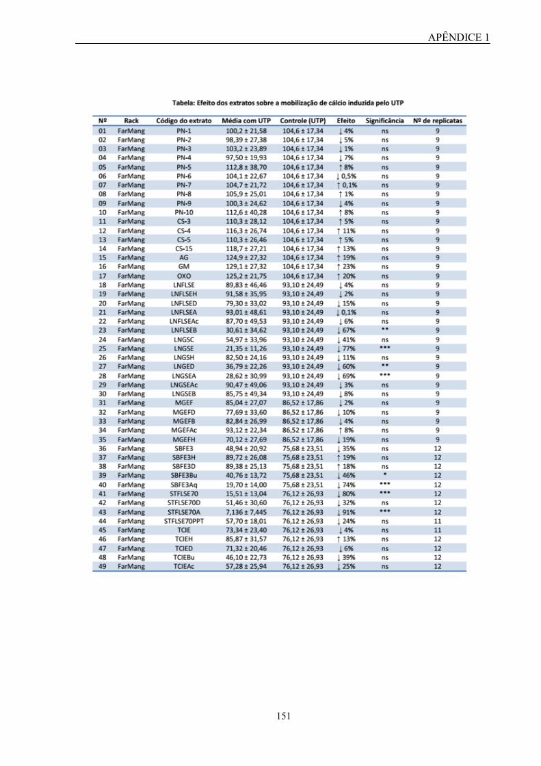
APÊNDICE 1
151

APÊNDICE 1
152
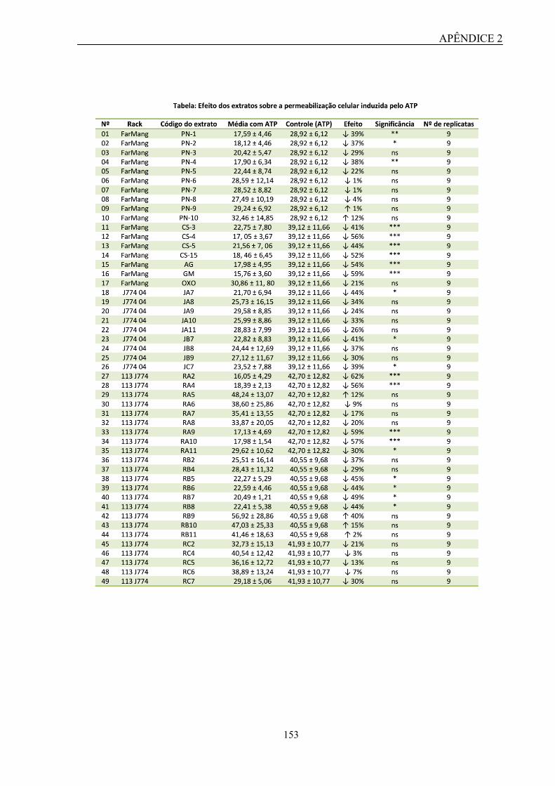
APÊNDICE 2
153
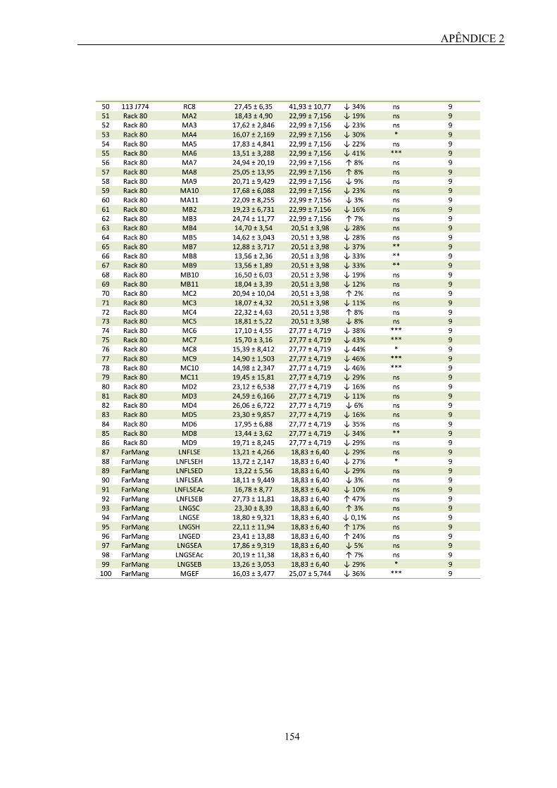
APÊNDICE 2
154

APÊNDICE 2
155

APÊNDICE 3
156