molecular mechanisms underlying in vitro cerebral ischemia ... · agradeço ao centro de...
TRANSCRIPT

Imagem
Marta Isabel Dias da Mota Vieira
Molecular Mechanisms Underlying in vitro Cerebral Ischemia: Multiple Neuronal Death Pathways
Tese de Doutoramento em Ciências e Tecnologias da Saúde, orientada por Ana Luísa Carvalho e Armanda Emanuela Castro e Santos
e apresentada à Faculdade de Farmácia da Universidade de Coimbra
Setembro/2013

Molecular Mechanisms Underlying in vitro Cerebral Ischemia: Multiple Neuronal Death
Pathways
Mecanismos Moleculares Subjacentes à Isquemia Cerebral in vitro: Múltiplas Vias de
Morte Neuronal
Marta Isabel Dias da Mota Vieira
2013
Dissertação apresentada à Faculdade de Farmácia da Universidade de Coimbra para
prestação de provas de Doutoramento em Ciências e Tecnologias da Saúde, especialidade de Biologia Celular e Molecular.

ii
Agradeço ao Centro de Neurociências e Biologia Celular e à Universidade de Coimbra a oportunidade de trabalhar no seu espaço, e à Fundação para a Ciência e Tecnologia que financiou o meu trabalho (bolsa de douoramento – SHFR/BD/47739/2008). Este trabalho foi financiado pela FEDER via Programa Operacional Factores de Competitividade – COMPETE e pela FCT no âmbito dos seguintes projectos: PTDC/SAU-NEU/099440/2008, PTDC/NEU-NMC/0750/2012, PTDC/SAU-NMC/12144/2010, PTDC/NEU-SCC/1351/2012 e PEst-C/SAU/LA0001/2013-2014. Os ensaios in vivo foram financiados pela Junta de Castilla y León (Ref. LE184A12-2).

iii
Agradecimentos
Deste doutoramento, como da vida, o melhor que levo comigo são as pessoas e as suas lições. Como alguém sábio uma vez disse “my friends have always been the best of me”, e por isso gostaria de agradecer às pessoas mais importantes da minha vida e àquelas que de alguma forma contribuíram para o meu crescimento pessoal durante estes, por vezes longos, por vezes demasiado curtos, quatro anos de trabalho.
Gostaria de começar por agradecer a uma pessoa sem a qual esta tese não teria sido possível e sem a qual esta aventura não teria sido a mesma. A minha companheira de armas de doutoramento, Joana Fernandes. Gostaria de te agradecer, naturalmente, pela ajuda no laboratório nomeadamente pela cedência das famosas listas dos microarrays, sem as quais não teria direccionado o trabalho no sentido deste formato final. Muito obrigada por isso, mas mais importante do que tudo, muito obrigada pela tua amizade. Obrigada por estares sempre do meu lado, sempre disposta a ouvir-me. Obrigada por tornares tua missão pessoal fazer-me rir! Sem isso este doutoramento não teria tido, apesar de todos os momentos de desilusão e lágrimas, todos os momentos de diversão e alegria que teve. Não há ninguém com quem teria gostado tanto de partilhar este doutoramento como tu.
Gostaria também de agradecer às minhas orientadoras, Professora Ana Luísa Carvalho e Doutora Armanda Santos por me terem aceitado como aluna de doutoramento. Obrigada pela ajuda e orientação. Obrigada pelos ensinamentos, os quais estarão sempre presentes no meu trabalho e na minha forma de encarar a ciência. À professora Ana Luísa gostaria de agradecer o entusiasmo contagiante, que foi muito precioso para mim durante estes quatro anos. A sua visão optimista da ciência é algo que levarei sempre comigo. Obrigada pela confiança em mim depositada. Um obrigado muito especial ao Professor Carlos Duarte, por todos os ensinamentos e ajuda. Foi um prazer trabalhar consigo.
À Joana Ferreira gostaria de fazer um agradecimento muito especial. Nestes já largos anos de amizade, a tua presença foi sempre e continuará sempre a ser um porto de abrigo para mim. Obrigada por toda a ajuda e apoio incondicional, no laboratório e fora dele.
À Aninhas e à Ritinha gostaria de dizer que a vossa amizade é um dos bens mais preciosos que levo comigo deste laboratório. Obrigada por todos os momentos de carinho e amizade, e também pelos momentos de palhaçada que muito me ajudam a ter energia para me levantar todas as manhãs para continuar a fazer o meu caminho. As memórias de todos os momentos por que passei com estas quatro grandes amigas, Ritinha, Aninhas, Joaninha e Fernandinhas hão-de acompanhar-me sempre. A vossa amizade faz de mim uma pessoa melhor. Enquanto investigadora olho para vocês com admiração e espero um dia poder chegar ao vosso nível.
Às “meninas” Joana, Dani, Lena e Paulo gostaria de agradecer a incondicional amizade. Mesmo que estejamos cada um num canto diferente do mundo, a amizade que nos une é algo que nunca me abandona, vocês estão sempre comigo, onde quer que eu esteja. Estes quatro anos foram marcados por muitas mudanças nas vidas de todos, alegra-me muito ter podido celebrar e partilhar com vocês tantos momentos importantes.

iv
Aos meus colegas de laboratório, obrigada por todos os momentos de amizade e galhofa que marcaram estes seis anos da minha passagem pelo laboratório de sinapses glutamatérgicas. Cada uma das pessoas que por lá passaram me ensinou alguma coisa. Um agradecimento especial:
Aos gémeos, Aninhas, Grace e Tat obrigada por serem gemini! Obrigada pela amizade e pela parvoíce partilhada. Ajudou-me mais do que possam algum dia imaginar a “sobreviver” ao doutoramento. Ao Rui e à Raquel gostaria de agradecer a boa disposição constante e dizer que foi um prazer conhecer-vos e ter a oportunidade de partilhar convosco algumas das aventuras e desventuras deste doutoramento.
À Miri, o terceiro elemento do gang da OGD, gostaria de agradecer a sua doçura e paciência. Obrigada por toda a ajuda e disponibilidade. Foi um prazer partilhar contigo as “aventuras” da OGD! Ao Rui Costa, colega dos tempos de licenciatura, obrigada pelo companheirismo e ajuda! Um obrigado ao Márcio pela amizade, apesar da distância.
Ao colega Carlos, escute, obrigada pelos momentos de galhofa, muitas vezes por si motivados e iniciados. Foi um prazer trabalhar consigo ao nível do laboratório. Aos preferiti, Pedro Isabel e Diogo, e também ao Ivan e à Domi gostaria de agradecer o humor e a boa disposição que trazem ou trouxeram sempre para o laboratório. Gostei muito de trabalhar e rir com vocês. À Joaninha Pedro gostaria ainda de agradecer a amizade e companheirismo.
À Mãezinha Andrea, ao Paizinho Johnny e ao Costini, foi um prazer conhecer-vos e ter a oportunidade de trabalhar convosco. Obrigada pela amizade. Um viva ao gang da excitotoxicidade.
À Vindy gostaria de agradecer pela amizade, pela boa disposição e, claro, pelo amadrinhamento partilhado da Tuala. Um enorme obrigado a ti e à Fernandinhas por me terem apresentado ao maravilhoso mundo do Doutor.
À D. Céu e à Beta gostaria de agradecer por toda a ajuda no laboratório, sem vocês o caos seria total. Obrigada por ser como uma mãe para todos nós, D. Céu.
Gostaria de agradecer aos meus pais, Rita e Zé, por serem as pessoas maravilhosas que são. Os valores que me transmitiram com tanta naturalidade e sem esforço são os meus maiores bens. O orgulho e amor incondicional que têm por mim fazem-me acreditar que cada pequena vitória minha tem sempre importância e sei que estarão sempre lá para apoiar qualquer rumo que resolva tomar. Por isso gostaria de vos agradecer, por me aceitaram, com todas as minhas qualidades e defeitos sem qualquer tipo de pressão ou julgamento. Se algum dia puder ser uma fracção daquilo que vocês são e me ensinaram, aí serei uma pessoa com missão cumprida.
À minha irmã gostaria de agradecer pela pessoa que ela é e por ser, antes de tudo, minha amiga. O companheirismo e compreensão que sempre partilhámos são muito queridos para mim. À Ana Sofia, que é para mim como uma irmã mais nova, um agradecimento pela sua simplicidade e carinho.
Não podia também deixar de reservar um lugar especial a uma das minhas mais antigas amigas, a Carole. Optámos por rumos semelhantes, por amor à ciência. Por isso e por muito mais que temos partilhado nestes anos de amizade, um enorme obrigado. Obrigada também por aceitares as minhas manias dos concertos e afins e por seres a minha parceira nessas aventuras que já nos deram tanto que falar e rir.

v
Onde quer que esteja, sei que posso contar sempre com a tua amizade, que é um dos bens mais valiosos que alguma vez poderia ter.
Por fim, gostaria de relembrar a minha avó, que infelizmente já nos deixou. Gostaria de agradecer por ser tudo aquilo que uma criança pode desejar numa avó! A minha infância nunca teria sido a mesma sem o que vivi consigo. Gostaria também de recordar a Marta Filipa. Gostaria muito de poder partilhar este tipo de conquista contigo. A tua prematura partida deste mundo marcou muito a minha personalidade e deixou para sempre uma saudade que nunca poderá ser colmatada. Mas apesar disso, a tua lembrança, trago-a sempre junto a mim.
Não posso deixar de aproveitar a ocasião para referir os meus gatinhos, Pretinho, Branquinha e claro, Ervilhinha. O vosso carinho descomprometido é muito importante para mim, pois é algo inteiramente puro e desinteressado. Obrigado por isso.
Gostaria de dedicar esta tese à minha família e amigos por acreditarem sempre em mim e por estarem sempre do meu lado. Muito obrigada.
“In the dark, see past our eyes Pursuit of truth no matter where it lies
Gazing up to the breeze of the heavens On a quest, meaning, reason”
(Through the Never, Hetfield, Ulrich & Hammet)

vi

vii
Table of Contents
Abbreviations ............................................................................................................... xi
Resumo ...................................................................................................................... xvii
Abstract ...................................................................................................................... xix
Chapter I – Introduction ............................................................................................... 1
Cerebral Ischemia ....................................................................................................... 3
Excitotoxicity ............................................................................................................ 5
Glutamatergic Neurotransmission ............................................................................... 7
NMDA Receptors .................................................................................................... 8
NMDAR Subunits ................................................................................................ 9
NMDAR Regulation by Phosphorylation ................................................ 11
Influence of GluN2 Subunits on Neuronal Fate ..................................... 12
NMDAR Membrane Localization ....................................................................... 13
Role of NMDAR Localization in Excitotoxic Mechanisms ...................... 14
NMDAR Interactors ........................................................................................... 16
NMDAR Internalization ...................................................................................... 17
Death Receptor Signaling and Cell Fate ................................................................... 17
Death Receptors ................................................................................................... 19
Survival Signaling .............................................................................................. 22
Apoptosis .......................................................................................................... 23
Necroptosis ....................................................................................................... 28
Necroptosis in Disease Context ............................................................ 34
MAP Kinase Pathways .............................................................................................. 36
Extracellular Signal-Regulated Kinase .................................................................. 36
ERK Activation in the Context of Global Ischemia ............................................ 37
p38 MAP Kinase .................................................................................................... 38
The Role of p38 in Disease ............................................................................... 40
p38 MAPK Activation in Cerebral Ischemia ...................................................... 41
c-Jun N-terminal Kinase ........................................................................................ 42

viii
JNK Signaling and Disease ............................................................................... 46
JNK and Global Ischemia .................................................................................. 46
Objectives ................................................................................................................. 49
Chapter II – Diverse Domains in the C-terminus of the GluN2B Subunit of NMDARs Contribute to Neuronal Death upon in vitro Ischemia ............................ 51
Abstract ..................................................................................................................... 53
Introduction ............................................................................................................... 53
Materials & Methods ................................................................................................. 55
Materials ................................................................................................................ 55
Cell Culture and Transfection ................................................................................ 55
Oxygen-Glucose Deprivation (OGD) Challenge .................................................... 56
Immunocytochemistry ........................................................................................... 57
Nuclear Morphology .............................................................................................. 57
LDH Release Assay .............................................................................................. 58
Statistical Analysis ................................................................................................. 58
Results ...................................................................................................................... 58
The GluN2B Subunit is Determinant for OGD-induced Neuronal Death .............. 58
C-terminal-specific Interactions are Determinant for GluN2B-mediated Toxicity .. 61
Discussion ................................................................................................................. 65
Chapter III – Up-regulation of Endogenous RIP3 Contributes to Necroptotic Neuronal Death in Cerebral Ischemia ....................................................................... 73
Abstract ..................................................................................................................... 75
Introduction ............................................................................................................... 75
Materials & Methods ................................................................................................. 77
Materials ................................................................................................................ 77
Cell Culture and Transfection ................................................................................ 78
Oxygen-Glucose Deprivation (OGD) Challenge .................................................... 79
RNA Extraction ...................................................................................................... 79
qPCR ..................................................................................................................... 80
Nuclear Morphology .............................................................................................. 80

ix
Lactate Dehydrogenase (LDH) Release Assay ..................................................... 81
Protein Extracts, SDS-PAGE and Western Blotting .............................................. 81
Transient Global Cerebral Ischemia in the Rat ..................................................... 82
Statistical Analysis ................................................................................................. 83
Results ...................................................................................................................... 83
OGD Induces a Component of Necroptotic Neuronal Death ................................. 83
Necroptosis in OGD-challenged Neurons is Promoted by Up-regulation of RIP3 . 84
RIP1 Contributes to Necroptotic Neuronal Death Following OGD ........................ 87
RIP3 is Upregulated Following Global Cerebral Ischemia in vivo ......................... 88
Discussion ................................................................................................................. 89
Chapter IV – Activation of MAPK Signaling Following in vitro Ischemic Insults . 97
Abstract ..................................................................................................................... 99
Introduction ............................................................................................................... 99
Materials & Methods ............................................................................................... 101
Materials .............................................................................................................. 101
Cell Culture .......................................................................................................... 101
Oxygen-Glucose Deprivation (OGD) Challenge .................................................. 102
RNA Extraction .................................................................................................... 102
RT-PCR ............................................................................................................... 102
Nuclear Morphology ............................................................................................ 103
LDH Release Assay ............................................................................................ 103
Protein Extracts, SDS-PAGE and Western Blotting ............................................ 104
Statistical Analysis ............................................................................................... 104
Results .................................................................................................................... 105
OGD Induces Up-regulation of the JNK signaling Pathway ................................ 105
JNK Inhibition Protects Against Neuronal Death ................................................. 107
OGD Elicits Activation of the p38 MAPK ............................................................. 107
Discussion ............................................................................................................... 109
Chapter V – General Conclusions and Future Directions ..................................... 115

x
Chapter VI – References .......................................................................................... 121

xi
Abbreviations
24S-OHC 24(S)-Hydroxycholesterol
Actb β-Actin
AD Alzheimer’s Disease
AIF Apoptosis-inducing Factor
AIP Apaf-1 Interacting Protein
ALS Amyotrophic Lateral Sclerosis
AMPAR α-Amino-3-hydroxy-5-methyl-4-isoxazole-propionic acid Receptor
ANOVA One-way Analysis of Variance
AP-1 Activator Protein-1
AP2 Clathrin Adaptor Protein
Apaf-1 Apoptotic Peptidase Activating Factor 1
ASIC Acid-sensing Ion Channel
ASK Apoptosis Signal Regulating Kinase
ATD Amino Terminal Domain
ATF Activating Transcription Factor
Bcl B-cell Lymphoma
BDNF Brain-derived Neurotrophic Factor
BID BH3 Interacting-domain Death Agonist
BNip3 BCL2/adenovirus E1B 19 kDa Interacting Protein 3
BSA Bovine Serum Albumin
CA Cornu Ammonis
CAD Caspase Activated DNase
CaMK Ca2+-Calmodulin Kinase
CBP CREB Binding Protein
CD95 Cluster of Differentiation 95
Cdk5 Cyclin-dependent Kinase 5
cIAP Cellular Inhibitor of Apoptosis Protein
CK2 Casein Kinase 2
CLAP Chymostatin, Leupeptin, Antipain and Pepstatin
CNS Central Nervous System
CP-AMPAR Ca2+-permeable AMPAR
CREB cAMP Response Element-binding Protein
Ct Threshold Cycle
CTD Carboxyl Terminal Domain
Ctx Cerebral Cortex

xii
CYLD Cylindromatosis
DD Death Domain
DED Death Effector Domain
DG Dentate Gyrus
DISC Death Inducing Signaling Complex
DIV Days in vitro
DL Death Ligand
DN Dominant Negative
DR Death Receptor
Drp-1 Dynamin Related Protein 1
DUB Deubiquitinase
E18 Embryonic Day 18
ECF Enhanced Chemifluorescence
EDD E3 Ubiquitin-protein Ligase UBR5
ER Endoplasmic Reticulum
ERK Extracellular-signal Regulating Kinase
FADD Fas-associated Protein with Death Domain
FasL Fas Ligand
FL Full Length
FLIP FLICE-like Inhibitory Protein
FOXO Forkhead O
GADD45 Growth Arrest and DNA Damage 45
GAPDH Glyceraldehyde 3-phosphate Dehydrogenase
GLUD1 Glutamate Dehydrogenase 1
GLUL Glutamate-ammonia Ligase
GluR Glutamate Receptor
H2AX H2A Histone Family, Member X
HBSS Hank’s Balanced Salt Solution
HD Huntington’s Disease
HtrA2 High Temperature Requirement Protein A2
ICAD Inhibitor of CAD
ID Intermediate Domain
IFNγ Interferon Gamma
iGluR Ionotropic GluR
IκB Inhibitor of Kappa B
IKK IkB Kinase

xiii
IL Interleukin
iNOS Inducible Nitric Oxide Synthase
JBD JNK Binding Domain
JIP JNK-interacting Protein
JNK c-Jun N-terminal Kinase
KAR Kainate Receptor
KD Knock-down
KO Knock-out
LBD Ligand Binding Domain
LDH Lactate Dehydrogenase
LTD Long-term Depression
LTP Long-term Potentiation
LUBAC Linear Ub Chain Assembly Complex
MADD MAP-kinase Activating Death Domain
MAGUK Membrane Associated Guanylate Kinase
MAPK Mitogen-activated Protein Kinase
MAPKAPK2 MAPK-activated Protein Kinase 2
MAPKK MAPK Kinase
MAPKKK MAPK Kinase Kinase
MCAO Middle Cerebral Artery Occlusion
MCMV Mouse Cytomegalovirus
MEF Mouse Embryonic Fibroblast
MEK MAPK/ERK Kinase
MEKK MEK Kinase
MEM Minimal Essential Medium
mGluR Metabotropic GluR
MK-801 (5S,10R)-(+)-5-Methyl-10,11-dihydro-5H-dibenzo[a,d]cyclohepten-5,10-
imine Maleate
MKP MAPK Phosphatase
MLK Mixed-lineage Kinase
MLKL Mixed-lineage Kinase Domain-like
MNK MAPK Interacting Kinase
MS Multiple Sclerosis
MSK Mitogen and Stress Activated Kinase
MW Multiwell
NCX Na+/Ca2+ Exchanger
xiv
Nec-1 Necrostatin-1
NEMO NF-kB Essential Modulator
NF-kB Nuclear Factor Kappa B
NGF Nerve Growth Factor
NHE1 Na+/H+ Exchanger 1
NMDAR N-methyl-D-aspartate Receptor
nNOS Neuronal Nitric Oxide Synthase
NO Nitric Oxide
NOS1AP NOS1 Associated Protein
OGD Oxygen and Glucose Deprivation
OSM Osmosensing Scaffold for MEKK3
PARP Poly-ADP Ribose Polymerase
PBS Phosphate-buffered Saline
PD Parkinson’s Disease
PDL Poly-D-lysine
PDZ PSD95/Dlg/ZO-1
PGAM5 Serine/threonine-protein Phosphatase
PI Propidium Iodide
PI3K Phosphoinositide 3-kinase
PK Protein Kinase
PMSF Phenylmethanesulfonyl Fluoride
POSH Plenty of SH3s
PP Protein Phosphatase
PSD Post-synaptic Density
PVDF Polyvinylidene Fluoride
PYGL Phosphorylase, Glycogen, Liver
qRT-PCR Quantitative Real-time PCR
RHIM RIP Homotypic Interaction Motif
RIP Receptor-interacting Protein Kinase
ROS Reactive Oxygen Species
RSK p90 Ribosomal S6 Kinase
SAP Synapse-associated Protein
SAPK Stress-activated Protein Kinase
SBDP α-Spectrin Break-down Product
shRNA Short Hairpin RNA
SIRT2 Sirtuin2

xv
Smac/
DIABLO
Second Mitochondria-derived Activator of Caspase/
Direct Inhibitor of Apoptosis-binding Protein with Low pI
Sp-1 Specificity Protein 1
STEP Striatal-Enriched Tyrosine Phosphatase
TAB TAK1 Binding Protein
TACE TNFα Converting Enzyme
TAK1 Transforming Growth Factor-β Activated Kinase-1
TAOK TAO Kinase
TBI Traumatic Brain Injury
TBS-T Tris-buffered Saline-Tween
TLR Toll-like Receptor
TNFR1 Tumor Necrosis Factor Receptor 1
TNFα Tumor Necrosis Factor α
TORC Transducer of Regulated CREB Activity
tPA Tissue Plasminogen Activator
TPL2 Tumor Progression Locus 2
TRADD TNFR-associated via Death Domain
TRAF2 TNFR Associated Factor 2
TRAIL TNF-related Apoptosis-inducing Ligand
TRP Transient Receptor Potential
TWEAK TNF-related Weak Inducer of Apoptosis
VO Vessel Occlusion
WT Wild-type
zVAD.fmk N-Benzyloxycarbonyl-Val-Ala-Asp(O-Me)-fluoromethyl Ketone

xvi

xvii
Resumo
A isquemia cerebral induz neurodegeneração de populações específicas de
neurónios, nomeadamente na área CA1 do hipocampo. Apesar da sua elevada
prevalência e dos esforços na investigação, não existem actualmente tratamentos
eficazes para prevenir a neurodegeneração associada à isquemia global. Esta
patologia pode ser estudada in vitro submetendo os neurónios a privação de oxigénio
e glicose (OGD, do inglês “oxygen and glucose deprivation”). O principal objectivo
deste trabalho foi o de estudar os mecanismos moleculares associados à morte
neuronal iniciada pela OGD em culturas primárias de neurónios de hipocampo. Para
este propósito pesquisámos diferentes aspectos da morte celular. Assim, estudámos o
fenómeno de excitotoxicidade mediada por receptores de glutamato do tipo NMDA
(NMDARs), analisámos a activação de um novo mecanismo de morte celular,
necroptose, e verificámos a activação de vias efectoras da morte celular, em particular,
as vias das MAPKs.
Começámos por estudar a influência da subunidade GluN2B dos NMDARs na
morte neuronal induzida por OGD. Os NMDARs têm um papel de relevo no excesso
de Ca2+ intracelular característico da excitotoxicidade; contudo, o papel das
subunidades GluN2 tem permanecido controverso. Há evidências que mostram o
envolvimento das subunidades GluN2A e GluN2B no processo de morte celular
enquanto outras mostram o papel tóxico do GluN2B e uma função neuroprotectora do
GluN2A. Para clarificar esta questão recorremos a um sistema de cultura neuronal de
murganhos GluN2B-/- e observámos um papel determinante da subunidade GluN2B na
indução de morte após OGD em neurónios de córtex. Verificámos que a ausência do
GluN2B eliminou a toxicidade induzida por OGD, que foi recuperada com a
reintrodução da subunidade nos neurónios GluN2B-/-. Demonstrámos ainda o papel
preponderante do domínio C-terminal (CTD) da subunidade na toxicidade mediada por
GluN2B e mapeámos alguns locais de interacção no CTD de GluN2B responsáveis
por esta função tóxica. O domínio PDZ é responsável pela interacção com a PSD95,
que faz o acoplamento à nNOS. A interferência com esta interacção teve um efeito
neuroprotector. Identificámos também dois determinantes moleculares no CTD de
GluN2B relevantes para este processo, o local de ligação às proteínas AP2 e CaMKII.
A mutação destes locais eliminou a toxicidade induzida pela subunidade GluN2B.
Estas evidências apoiam o papel determinante dos NMDARs contendo GluN2B num
contexto de isquemia cerebral in vitro. O nosso estudo é particularmente relevante na

xviii
medida em que os trabalhos anteriores utilizavam, na sua maioria, estímulos
excitotóxicos.
De seguida, investigámos a indução de morte neuronal por necroptose, um
novo mecanismo de necrose programada, em neurónios de hipocampo submetidos a
OGD. Este tipo de morte celular ocorre na sequência da activação de “death
receptors” (DRs). Em determinadas condições, ocorre a formação de um complexo
que medeia a apoptose e inibe a necroptose através da clivagem das proteínas
RIP1/3. Contudo, se ocorrer inibição da caspase8, a RIP3 é recrutada para a RIP1 e
juntas formam um complexo chamado necrosoma, que activa a necroptose. A OGD
induziu um componente de morte celular que foi revertido pelo inibidor de necroptose
Nec-1, mas não pelo inibidor da apoptose zVAD.fmk. A especificidade do efeito
protector da Nec-1 sobre a necroptose foi comprovada pela ausência de efeito da Nec-
1 no componente apoptótico. Além disso, a OGD induziu a expressão das proteínas
RIP1 e RIP3. Confirmámos o papel tóxico da RIP3 através de ensaios de
sobreexpressão e silenciamento. A sobreexpressão das proteínas RIP1 e RIP3
aumentou a morte neuronal, enquanto o silenciamento da RIP3 reduziu a toxicidade
induzida por OGD. O efeito tóxico da OGD foi recuperado com a reintrodução da
proteína RIP3 em neurónios sem RIP3 endógena. Por fim, relacionámos os
mecanismos observados in vitro com o modelo in vivo, ao observarmos que a
isquemia global induz o aumento da expressão da RIP3 na área CA1 do hipocampo.
Estudámos por fim a activação de MAPKs em neurónios de hipocampo
submetidos a OGD. Estas cinases são responsáveis pela resposta ao stress celular e
estão envolvidas em diversos paradigmas de morte neuronal. Verificámos a activação
das cinases p38 e JNK após OGD, às 2h e 6h de recuperação, respectivamente.
Verificámos ainda que a inibição destas cinases teve um efeito protector, o que sugere
um papel citotóxico. Curiosamente, a cinase JNK parece ter um duplo papel, já que a
sua inibição só foi protectora quando efectuada às 4h após o estímulo de OGD.
Concluindo, os nossos resultados demonstram que a OGD afecta os neurónios
a vários níveis, incluindo na indução de mecanismos que contribuem para a
neurodegeneração. Este modelo in vitro apresenta-se assim como uma ferramenta
importante para a dissecção de mecanismos moleculares subjacentes à isquemia
cerebral, o que poderá contribuir para o desenvolvimento de estratégias terapêuticas
para esta patologia.
xix
Abstract
Cerebral global ischemia induces selective neurodegeneration of specific
subsets of neurons throughout the brain, namely in the CA1 region of the
hippocampus. Despite its high prevalence and intensive research, there is still need of
effective treatments to reduce the neurodegeneration associated with global ischemia.
This pathology can be studied in vitro by depriving neurons of oxygen and glucose
(OGD). Our main goal was to study the molecular mechanisms of neuronal death
activated by OGD in primary cultures of hippocampal neurons. For this purpose we
targeted distinct aspects of cell death. We studied the excitotoxic component of cell
death mediated by NMDARs, we addressed the activation of a novel mechanism of
programmed cell death, necroptosis, and we analyzed the activation of effector
signaling cascades, in particular MAPKs.
We started by studying the influence of the GluN2B subunit of NMDARs to
OGD-induced neuronal demise. NMDARs are major contributors to the overload of
intracellular Ca2+ characteristic of excitotoxicity and the role of GluN2 subunits has
remained controversial. This is due to a variety of conflicting evidence showing that
either both GluN2A and GluN2B contribute to neuronal death or that GluN2B is mostly
pro-death and GluN2A pro-survival. To clarify this question we used cultured cortical
neurons from GluN2B-/- mice and wild-type littermates, and observed that GluN2B is
determinant for induction of excitotoxic neuronal death following OGD. We observed
that the absence of this subunit blocked neuronal death induced by OGD and that the
toxicity was rescued when we reintroduced the subunit in the KO neurons. Moreover,
we demonstrated that the C-terminal domain (CTD) had a preponderant role in
GluN2B-induced toxicity, and we identified molecular determinants in the CTD of
GluN2B responsible for this function. We confirmed that the PDZ-binding domain was
partly responsible for NMDAR toxicity. This domain is responsible for the interaction
with PSD95 that couples to nNOS, and interfering with this interaction was
neuroprotective. Additionally, we identified two other regions on the GluN2B CTD that
are required for OGD-induced cell death, the AP2- and the CaMKII-binding domains.
Mutations in either of these sites blocked GluN2B-mediated toxicity. These findings
confirmed the crucial role of GluN2B-containing NMDARs in a context of in vitro
ischemia, and our study is particularly relevant since most previous work was
performed under excitotoxic conditions.

xx
Next, we investigated whether OGD induced necroptosis, a novel type of
programmed necrosis, in hippocampal neurons. This type of cell death has been
recently described to occur following death receptor (DR) signaling. In certain
conditions a complex called DISC is formed. DISC induces apoptosis and
downregulates necroptosis via caspase8-mediated cleavage of the proteins RIP1/RIP3.
However, when caspase8 is inhibited, RIP3 is recruited to RIP1 and together they form
a complex called the necrosome, and activate necroptosis. We observed that OGD
induced a component of cell death that was reversed by the necroptotic inhibitor Nec-1
but not by zVAD.fmk, an apoptotic inhibitor. Notably, we observed that Nec-1 had no
effect on the apoptotic component of neuronal death. Additionally, OGD induced the
expression of RIP1 and RIP3. We confirmed the toxic role of RIP3 by performing
overexpression and knock-down experiments. We observed that overexpression of
both RIP1 and RIP3 exacerbated neuronal death induced by OGD whereas knockdown
of RIP3 significantly reduced OGD-mediated toxicity. The damaging effect of the OGD
challenge was rescued by reintroducing RIP3 in neurons where endogenous RIP3 was
knocked-down, confirming the specificity of the requirement for RIP3. Finally, we
correlated these in vitro events with the in vivo challenge, by confirming that global
cerebral ischemia in the rat also induces RIP3 expression in the CA1 area of the
hippocampus.
Lastly, we studied the activation of MAPKs in hippocampal neurons submitted
to OGD. These kinases are responsible for the majority of the cellular response to
stress and are involved in several paradigms of cell death, including in neurons. We
determined that both p38 and JNK are activated following OGD, at 2h and 6h of
reoxygenation, respectively. Furthermore, inhibition of the activity of these MAPKs has
a neuroprotective effect, suggesting a cytotoxic function. Interestingly, JNK seems to
have biphasic function since neuroprotection was only achieved when we inhibited JNK
at 4h reoxygenation.
Overall, our results demonstrate that OGD induces a variety of changes in
neurons, including several mechanisms that contribute to neurodegeneration. This in
vitro model is thus a powerful tool to address the molecular mechanisms underlying
cerebral ischemia, which may provide useful insights into the development of
therapeutic strategies to this pathology.

Chapter I
Introduction

Chapter I
2

Introduction
3
Cerebral Ischemia
Cerebral ischemia is a leading cause of disability and death in the western
world (Flynn et al., 2008). This pathology lacks effective treatments, a direct
consequence of a limited time window for intervention before major neurodegeneration
ensues as well as of the lack of specificity of some of the treatments developed so far.
Additionally, the inhibition of some molecular targets proves inefficient due to
secondary effects. To date, the only treatment administered to cerebral ischemic
patients is tissue plasminogen activator (tPA) (Fonarow et al., 2011; Iadecola and
Anrather, 2011b). Cerebral ischemia is broadly divided in two types: global cerebral
ischemia and focal ischemia (commonly referred to as stroke) (Flynn et al., 2008).
Transient global ischemia results from a transient interruption of blood supply to
the entire brain, due to a cardiac arrest or to a near-drowning situation. This type of
pathology causes selective degeneration of specific subsets of neurons throughout the
brain, namely the neurons of the Cornu Ammonis 1 (CA1) region of the hippocampus,
the cortical neurons of layers II, V and VI, the Purkinje cells of the cerebellum and the
dorsolateral striatal neurons (Lo et al., 2003; Zukin et al., 2004).
Focal ischemia arises from the occlusion of a blood vessel in the brain
(ischemic stroke) or from the rupture of a brain vessel (hemorrhagic stroke) leading to
the deprivation of blood flow on the region of the brain supplied by that vessel. The
area most severely affected by hypoperfusion constitutes the infarct core, in which
most neurons degenerate due to an overwhelming stress, and the surrounding area,
the penumbra, has a gradient of perfusion and mixed neuronal phenotypes, with some
cells dying whilst others are able to survive (Kunz and Iadecola, 2009).
The interruption in the blood supply to the brain causes a deprivation of oxygen
and glucose, leading to a failure in ATP production, which is pernicious due to the brain
high metabolic demand (Moskowitz et al., 2010). This results in neuronal depolarization
and excessive glutamate release, accompanied by dysfunction of the glutamate
reuptake mechanisms in glia cells and neurons, as a result of the dissipation of the Na+
gradient, due to the depletion of ATP. Thus, an excessive glutamate accumulation at
the synapse causes the overactivation of glutamate receptors leading to an intracellular
Ca2+ overload that may trigger cytotoxicity, a phenomenon called excitotoxicity.
Notably, depending on the duration and intensity of the insult, other non-excitotoxic
mechanisms are activated, contributing to Ca2+ overload (Besancon et al., 2008;
Szydlowska and Tymianski, 2010). Deregulation of Ca2+ homeostasis activates

Chapter I
4
deleterious intracellular mechanisms, including mitochondrial dysfunction and oxidative
and nitrosative stress, that induce inflammation and ultimately neuronal death (Figure
1.1) (Moskowitz et al., 2010; Iadecola and Anrather, 2011a). In global ischemia and in
the penumbra area, neuronal degeneration ensues within 24-72h after the ischemic
insult, a process called delayed neuronal death. This delay in neuronal demise onset
suggests the reliance on transcriptional changes, leading to programmed cell death
mechanisms.
Figure 1.1. Cerebral ischemia induces neuronal demise via activation of deleterious mechanisms. Upon interruption of blood flow, distinct damaging mechanisms are
activated that contribute to the injury. In an early phase, energy imbalance and
excitotoxicity are activated contributing to mounting oxidative and nitrosative stress.
These contribute to inflammatory signaling and ultimately programmed cell death
mechanisms are activated. Concomitantly, several neuroprotective mechanisms are
activated, to counteract the effect of these damaging signals. (Iadecola and Anrather,
2011b)

Introduction
5
Cerebral ischemia can be addressed experimentally, using several research
models, both in vitro and in vivo. The most common in vivo models to study transient
global ischemia are the four vessel occlusion (4-VO) and the 2-VO combined with
hypotension, both in the rat. As for focal ischemia, the middle cerebral artery occlusion
(MCAO) model is generally used. Regarding the in vitro models, the closest to in vivo
ischemia is the oxygen and glucose deprivation (OGD) challenge, which is most
commonly used either in primary cultures of hippocampal or cortical neurons or in
forebrain or organotypic hippocampal slices. Due to its characteristics, the OGD
challenge of hippocampal neurons is a good model for global ischemia and allows for
the dissection of molecular pathways underlying cerebral ischemia. This challenge
consists of placing the neurons or slices in a glucose-free medium inside an anaerobic
chamber, thereby combining the deprivation of these two factors which mimics what
happens in the brain during the interruption of the blood flow, but in a simplified system
(Zukin et al., 2004). This model also has advantage over other in vitro models [for
example, application of high concentrations of glutamate receptor (GluR) agonists]
since it accounts for most changes observed during blood supply deprivation, like
activation of non-excitotoxic mechanisms, such as Ca2+ entry via acid-sensing ion
channels (ASICs), transient receptor potential (TRP) channels or Na+/Ca2+ exchangers
(NCXs) (Szydlowska and Tymianski, 2010), instead of accounting only for excitotoxic
mechanisms.
EEXXCCIITTOOTTOOXXIICCIITTYY
The term excitotoxicity was first used by Olney (1969) to describe
neurodegeneration associated with the activation of excitatory amino acid receptors
(Olney, 1969). This phenomenon is a hallmark of several neurodegenerative disorders,
such as Alzheimer’s disease (AD), Huntington’s disease (HD) and cerebral ischemia,
and it results from excessive glutamate release from the presynaptic terminal, due to
the specific disease-associated insult (Figure 1.2). This abnormal glutamate release
combined with the dysfunction of the mechanisms responsible for removing this
neurotransmitter from the synapse result in the accumulation of glutamate in the
synaptic cleft, for longer periods of time. The immediate consequence of this
accumulation is the overactivation of GluRs, present at the postsynaptic membrane,
leading to a massive influx of ions, namely Ca2+ (Choi, 1987; Choi et al., 1987; Mehta
et al., 2013).

Chapter I
6
Calcium acts physiologically as a second messenger in neurons, which possess
highly specialized mechanisms of Ca2+ buffering to maintain this ion at low
concentrations. Thus, localized increases in Ca2+ concentration lead to the activation of
specific intracellular signaling pathways. Upon an excitotoxic insult, the Ca2+ overload
induces the deregulation of Ca2+ buffering mechanisms, for exceeding their capacity,
contributing to the activation of damaging Ca2+-dependent processes that may
ultimately lead to neuronal death (Choi, 1988; Tymianski and Tator, 1996; Arundine
and Tymianski, 2003; Mehta et al., 2013). Evidence linking excessive Ca2+ influx to
neuronal damage lead to the formulation of the “Calcium Hypothesis” which states that
“neuronal calcium overload contributes to neurodegeneration” (Arundine and
Tymianski, 2003).
Figure 1.2. Mechanisms of excitotoxicity. The excessive glutamate release from the
presynaptic terminal combined with the dysfunction of the glial mechanisms of glutamate
clearance result in excessive concentrations of glutamate in the synapse. This leads to
overactivation of GluRs located postsynaptically and activation of pro-death signaling
pathways that promote neuronal demise. (Popoli et al., 2012)

Introduction
7
The understanding of the paramount role exerted by Ca2+ to the mechanisms of
excitotoxicity lead to initial efforts aiming at the blockade of this Ca2+ overload, to
prevent neurodegeneration. This strategy, however, proved ineffective since GluR
antagonists also blocked their physiological functions, which had adverse side effects
(Lees, 1998). Thus, researchers started focusing on the intracellular pathways that are
activated as a consequence of Ca2+ overload. Notably, the route of Ca2+ entry seems to
be determinant for the activation of specific downstream signaling pathways that have
distinct contributions to neuronal fate (“Source-specificity Hypothesis”) (Bading et al.,
1993; Ghosh and Greenberg, 1995; Sattler and Tymianski, 2001). Due to their role as
primary gateways for Ca2+ entry in neurons, ionotropic GluRs (iGluRs) are one of the
main sources of neuronal Ca2+, in particular N-methyl-D-aspartate receptors
(NMDARs), which are highly permeable to this ion (Mehta et al., 2013).
Glutamatergic Neurotransmission
Glutamate is the major excitatory neurotransmitter in the mammalian central
nervous system (CNS). Glutamate is released from the presynaptic terminal, during
excitatory neurotransmission, leading to its diffusion across the synaptic cleft and
consequent activation of GluRs present at the postsynaptic membrane (Catarzi et al.,
2007; Greger et al., 2007; Hansen et al., 2007). Glutamatergic synapses undergo
activity-dependent long-lasting changes in synaptic strength, such as long-term
potentiation (LTP) and long-term depression (LTD), mechanisms that are necessary for
learning and memory (Malenka and Nicoll, 1999; Catarzi et al., 2007; Greger et al.,
2007).
Glutamate activates two major classes of receptors: metabotropic GluRs
(mGluRs) and ionotropic GluRs. The mGluRs mediate slow synaptic responses, due to
their coupling to intracellular G proteins. To date, eight subtypes of mGluR family
members have been identified, mGluR1-8, and classified into three groups (groups I–
III), based on sequence similarity, pharmacology and transduction mechanism
(Friedman, 2006; Catarzi et al., 2007). The iGluRs are responsible for the majority of
fast excitatory neurotransmission in the mammalian brain. These are heterotetrameric
cation channels, comprising three functionally distinct subtypes: α-amino-3-hydroxy-5-
methyl-4-isoxazole-propionic acid (AMPA), kainate (KA) and NMDA receptors. These
receptors are mainly concentrated at postsynaptic sites, where they fulfill a variety of

Chapter I
8
different functions (Cull-Candy et al., 2006; Greger et al., 2007). The NMDARs mediate
the slow component of excitatory postsynaptic currents. NMDARs are highly permeable
to Ca2+, Na+ and K+ and can be fully activated under membrane depolarization, which
is necessary to abolish the inhibition of the receptor by Mg2+. The membrane
depolarization is mainly afforded by AMPAR activation, which mediates the fast
component of excitatory neurotransmission. Most AMPARs, as well as the KARs, are
permeable to Na+ and K+, but impermeable to Ca2+. The permeability of AMPARs to
Ca2+ is dependent on their subunit comsition and is regulated by the GluA2 subunit
(Won et al., 2002; Hazell, 2007).
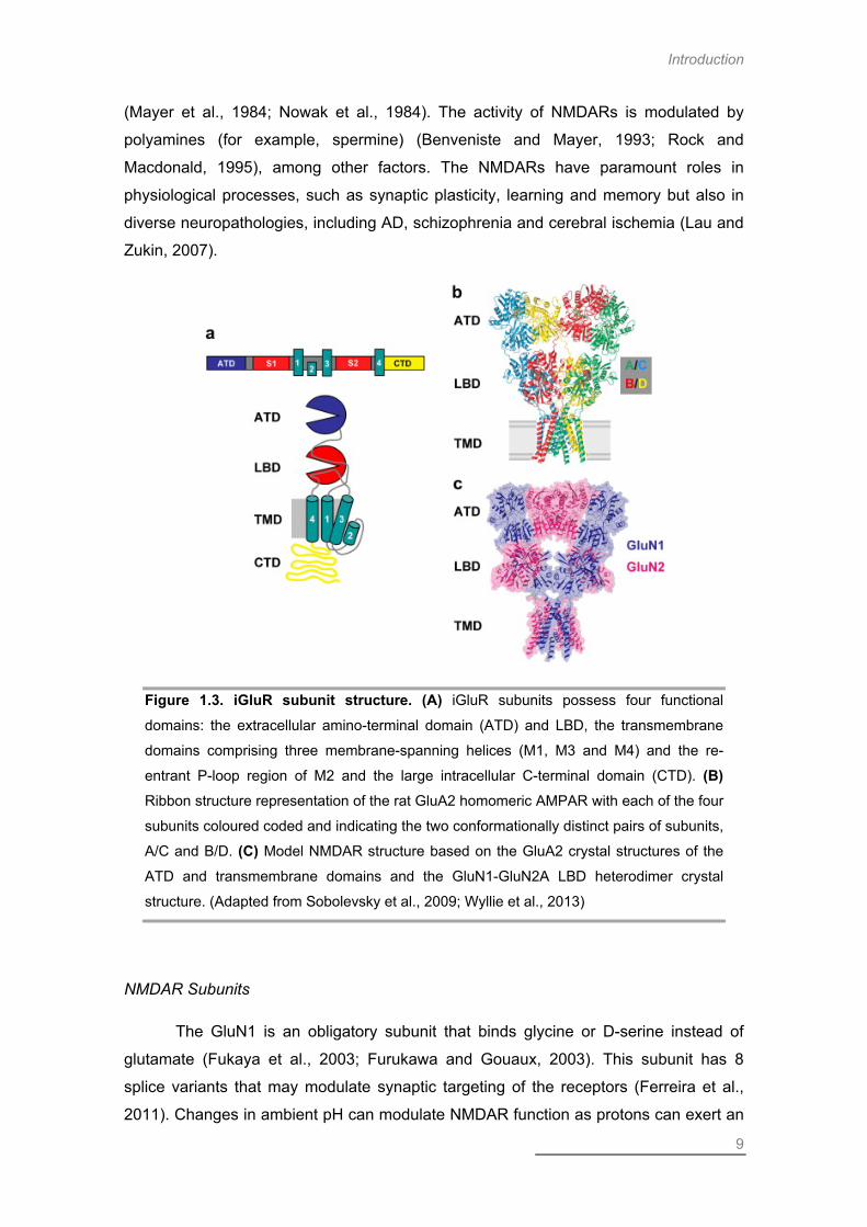
Eukaryotic iGluR subunits are each composed of an extracellular amino-
terminal domain, a ligand binding domain (LBD), a transmembrane domain, and an
intracellular carboxy-terminal domain (Figure 1.3). The transmembrane domain
contains three membrane-spanning segments (M1, M3 and M4) and a ‘‘re-entrant
loop’’, termed M2, which is located at the pore-lining region of the receptor, as
determined by crystallography (Sobolevsky et al., 2009). Two extracellular segments,
S1 and S2, have been shown to constitute the LBD which is responsible for the binding
of both the neurotransmitter and the competitive agonists/antagonists. Glutamate
binding to the extracellular LBD segment of the receptor triggers a series of
conformational changes that lead to activation of the receptor (Catarzi et al., 2007).
One of the goals of this work consisted of studying the contribution of NMDAR
subunits for OGD-induced death. For that reason, this introductory section will be
mainly focused on NMDARs.
NNMMDDAA RREECCEEPPTTOORRSS
The NMDARs are heterotetrameric structures composed of different
combinations of three types of subunits: GluN1, GluN2 and GluN3 (Hollmann and
Heinemann, 1994; Wenthold et al., 2003). These receptors are most commonly
assembled from two GluN1 subunits and two GluN2 subunits. NMDARs containing
GluN1 and GluN2 subunits are ion channels with high permeability to Ca2+
(MacDermott et al., 1986; Mayer and Westbrook, 1987), which contributes to their chief
role in the mechanisms of excitotoxicity. In order to be activated, NMDARs require
binding of glutamate and also glycine (or D-serine), which are mandatory cofactors
(Johnson and Ascher, 1987; Labrie and Roder, 2010). Agonist binding combined with
membrane depolarization contributes to relief of the Mg2+ block and channel opening
Introduction
9
(Mayer et al., 1984; Nowak et al., 1984). The activity of NMDARs is modulated by
polyamines (for example, spermine) (Benveniste and Mayer, 1993; Rock and
Macdonald, 1995), among other factors. The NMDARs have paramount roles in
physiological processes, such as synaptic plasticity, learning and memory but also in
diverse neuropathologies, including AD, schizophrenia and cerebral ischemia (Lau and
Zukin, 2007).
Figure 1.3. iGluR subunit structure. (A) iGluR subunits possess four functional
domains: the extracellular amino-terminal domain (ATD) and LBD, the transmembrane
domains comprising three membrane-spanning helices (M1, M3 and M4) and the re-
entrant P-loop region of M2 and the large intracellular C-terminal domain (CTD). (B) Ribbon structure representation of the rat GluA2 homomeric AMPAR with each of the four
subunits coloured coded and indicating the two conformationally distinct pairs of subunits,
A/C and B/D. (C) Model NMDAR structure based on the GluA2 crystal structures of the
ATD and transmembrane domains and the GluN1-GluN2A LBD heterodimer crystal
structure. (Adapted from Sobolevsky et al., 2009; Wyllie et al., 2013)
NMDAR Subunits
The GluN1 is an obligatory subunit that binds glycine or D-serine instead of
glutamate (Fukaya et al., 2003; Furukawa and Gouaux, 2003). This subunit has 8
splice variants that may modulate synaptic targeting of the receptors (Ferreira et al.,
2011). Changes in ambient pH can modulate NMDAR function as protons can exert an

Chapter I
10
inhibitory effect on NMDARs via direct interaction with a Lys residue on GluN1 subunits
(Traynelis and Cull-Candy, 1990). GluN1 subunits and the level of neuronal activity are
two contributing factors that regulate the exit of NMDARs from the endoplasmic
reticulum (ER). These subunits are produced in large excess relatively to GluN2
subunits (Huh and Wenthold, 1999; Mu et al., 2003) and are retained in the ER until
they are assembled with other subunits (Wenthold et al., 2003). Upon assembly of
functional receptors, vesicles containing NMDARs are transported to dendrites. This is
a type of active transport process, relying on motor proteins that carry the vesicles
along microtubules (Hirokawa and Takemura, 2004).
The GluN2 subunits can be divided in four subtypes, GluN2A-D. These four
subunits have distinct expression in neurons: GluN2A is widely distributed throughout
the brain, while GluN2B is predominantly expressed in the forebrain, GluN2C in the
cerebellum and GluN2D is mostly found in the thalamus (Buller et al., 1994). Thus, the
most common assemblies of NMDARs consist of GluN1-GluN2A receptors or GluN1-
GluN2B (Al-Hallaq et al., 2007), but triheteromeric receptors also exist (Sheng et al.,
1994; Cull-Candy and Leszkiewicz, 2004; Brothwell et al., 2008). Different subunit
compositions fine-tune the receptors, namely in terms of intracellular interactions,
localization in the membrane, and also channel properties (Figure 1.4). Indeed,
relatively to GluN2B, the GluN2A subunit confers the receptor faster kinetics, lower
glutamate affinity, greater channel open probability and more prominent Ca2+-
dependent desensitization. Moreover, the GluN2C/D subunits confer reduced
sensitivity to the Mg2+ block and lower conductance opening (Cull-Candy and
Leszkiewicz, 2004; Wyllie et al., 2013).
There are two subtypes of GluN3 subunits, GluN3A and GluN3B. The former
are expressed throughout the CNS, while the latter are found mostly in motor neurons.
Like GluN1, these subunits bind glycine (Yao et al., 2008). When present in the final
receptor assembly, GluN3 subunits confer lower Ca2+ permeability and reduced surface
expression (Cull-Candy and Leszkiewicz, 2004).
The GluN2 subunits share high degree of similarity in their extracellular and
transmembrane domains, suggesting a common function for these domains in all
subunits. Notably, the C-terminal domain of NMDARs is the most divergent region
among the subunits. In fact, the GluN2A and GluN2B subunits share only 29% identity
in their C-terminal domains. This region enables the NMDARs to interact with diverse
intracellular proteins and links the receptor to downstream signaling pathways. The

Introduction
11
divergent C-terminus among the different subunits suggests functional adaptations in
intracellular signaling, specific for each GluN2 subunit (Ryan et al., 2008).
Figure 1.4. The subunit composition of NMDARs influences channel properties. The
four di-heteromeric NMDARs (GluN1 subunits indicated in grey) display different features,
such as agonist and co-agonist potency (lowest – GluN1/GluN2A NMDARs, highest –
GluN1/GluN2D NMDARs); deactivation rates (fastest – GluN2A-containing NMDARs,
slowest – GluN2D-containing NMDARs); voltage-dependent block by Mg2+; permeability
to Ca2+ and unitary conductance. (Wyllie et al., 2013)
NMDAR Regulation by Phosphorylation
One of the key mechanisms that regulate iGluR function and localization is
phosphorylation (Lee, 2006). Accordingly, the C-terminal domain of NMDARs,
particularly of the GluN2A and GluN2B subunits, has diverse sites of phosphorylation
(Chen and Roche, 2007). Several kinases, including Ca2+-calmodulin kinase II
(CaMKII), cyclin-dependent kinase 5 (Cdk5), Src, protein kinase A (Tararuk et al.), PKC
and casein kinase 2 (CK2) have the ability to phosphorylate NMDAR subunits, with
different impact on channel properties and trafficking of the receptors (Chen and
Roche, 2007). The protein CaMKII is a Ca2+-dependent kinase that autophosphorylates
at Thr286 as a consequence of NMDAR activation, promoting its translocation to the
spines where it interacts with GluN2B subunits (Shen and Meyer, 1999; Bayer et al.,
Chapter I
12
2006). This kinase shows higher affinity for GluN2B subunits than GluN2A (Strack and
Colbran, 1998). CaMKII activity regulates synaptic plasticity phenomena via
phosphorylation of AMPARs (Lisman et al., 2002) and modulation of NMDAR function
(Gardoni et al., 2001; Gardoni et al., 2003; Chung et al., 2004).
Influence of GluN2 Subunits on Neuronal Fate
GluN2A and GluN2B are believed to have differential contributions to excitotoxic
neuronal death (Wyllie et al., 2013). Indeed, while GluN2A is considered to have a
prosurvival role (Liu et al., 2007; Terasaki et al., 2010), GluN2B overactivation is
thought to exert a detrimental effect (Aarts et al., 2002; Soriano et al., 2008; Martel et
al., 2012). However, there are also some reports that both subunits may contribute to
neuronal death (Graham et al., 1992; Stanika et al., 2009; Zhou et al., 2013),
suggesting that the regulation of these mechanisms implies an interplay of factors,
such as subunit composition and receptor localization (Lai et al., 2011). Indeed,
although both GluN2A and GluN2B are located at synaptic and extrasynaptic sites
(Thomas et al., 2006; Harris and Pettit, 2007; Zhou et al., 2013), GluN2A is thought to
preferentially locate in the synapse and GluN2B is believed to concentrate
extrasynaptically (Kew et al., 1998; Li et al., 1998b; Scimemi et al., 2004; Groc et al.,
2006; Zhang and Diamond, 2009).
The C-terminal domain of GluN2 subunits seems to be determinant for NMDAR-
mediated toxicity (Martel et al., 2012). This report elegantly demonstrates that the
influence of chimeric GluN2 subunits to neuronal death relies on the identity of the C-
terminal domain. Swapping the C-terminal domain of GluN2A for that of GluN2B
promotes neuronal demise in an excitotoxic context (Figure 1.5). The complementary
experiment, swapping the C-terminal domain of GluN2B for that of GluN2A does the
opposite, promoting neuronal survival (Cepeda and Levine, 2012; Martel et al., 2012).
Additional support of the concept of a detrimental role of the GluN2B subunit is
advanced by several reports demonstrating a neuroprotective effect of interfering with
activation of downstream signaling pathways, specifically neuronal nitric oxide
synthase (nNOS) (Aarts et al., 2002; Cui et al., 2007a; Cook et al., 2012).

Introduction
13
Figure 1.5. Differential contribution of GluN2A and GluN2B subunits to excitotoxic neuronal death. The C-terminal domain of GluN2B subunits is more lethal than that of
GluN2A. The extent of cell death upon excitotoxic stimulation depends in part on the C-
terminal domain of NMDAR subunits. Swapping the C-terminal domain of GluN2A for
GluN2B produces more cell death. Conversely, swapping the C-terminal from GluN2B for
GluN2A reduces cell death, due in part to increased phosphorylation of CREB. The C-
terminal domain of GluN2B displays stronger coupling to the PSD95/nNOS pathway,
which suppresses CREB activation. This indicates that the GluN2B subunit, regardless of
location at synaptic or extrasynaptic sites, is more lethal than the GluN2A subunit.
(Cepeda and Levine, 2012)
NMDAR Membrane Localization
The majority of NMDARs localizes to highly specialized structures called
postsynaptic densities (PSDs). However, they can also be targeted to extrasynaptic
sites (Brickley et al., 2003; Thomas et al., 2006; Petralia et al., 2010), and also
perisynaptic (Zhang and Diamond, 2009) and presynaptic (Bidoret et al., 2009) sites.
The PSDs possess a complex web of scaffold, adaptor and downstream signaling
proteins. Two scaffold proteins, PSD95 and synapse-associated protein 102 (SAP102),
are abundant at the PSD strucutures, and contain PSD95/Dlg/ZO-1 (PDZ) domains
that have the ability to interact with the NMDARs and control their trafficking and
synaptic delivery (Kim and Sheng, 2004). Receptors enter and exit the synapse via
lateral diffusion mechanisms. In fact, NMDARs have been shown to undergo rapid
synaptic exchange (Tovar and Westbrook, 2002) and to diffuse between synaptic and

Chapter I
14
extrasynaptic sites (Groc et al., 2004). Extrasynaptic NMDARs may function as a highly
mobile pool of receptors that can rapidly diffuse to synaptic sites upon mechanisms of
activity-induced plasticity. Interestingly, PKC activation seems to regulate both lateral
mobility and synapse dispersal of NMDARs (Fong et al., 2002; Groc et al., 2004),
which may imply the existence of membrane microdomains that could also be
associated with endocytic zones (Blanpied et al., 2002). Furthermore, subunit
composition may also contribute to the control of NMDAR surface targeting.
Role of NMDAR Localization in Excitotoxic Mechanisms
Localization of NMDARs at the membrane is thought to have distinct impact on
neuronal fate, upon excitotoxic stimulation. Indeed, synaptic NMDARs are considered
to induce neuroprotection, whilst NMDARs located extrasynaptically are thought to
promote pro-death signaling (Figure 1.6) (reviewed in Hardingham and Bading, 2010).
Figure 1.6. Differential contribution of synaptic and extrasynaptic NMDAR activation to excitotoxic neuronal death. Synaptic NMDARs activate CREB-dependent
transcription. CREB is phosphorylated at Ser133 by CaMK (A) or ERK1/2 (B), in order to
recruit its coactivator CREB binding protein (CBP). CBP is phosphorylated at Ser301 by
the nuclear Ca2+-dependent CaMKIV (C). Synaptic NMDAR-induced Ca2+ signals
promote transducer of regulated CREB activity (TORC) import into the nucleus via

Introduction
15
calcineurin-dependent dephosphorylation (D). In contrast to these CREB-activating
signals of synaptic NMDARs, extrasynaptic NMDARs suppress CREB activity through
inactivation of the Ras-ERK1/2 pathway (E) and the nuclear translocation of Jacob, which
promotes CREB dephosphorylation (F). (Adapted from Hardingham and Bading, 2010)
Synaptic NMDAR signaling is thought to induce neuronal survival via several
mechanisms. One of the most prominent pathways of neuroprotection occurs through
nuclear Ca2+ signaling, which induces gene expression (Zhang et al., 2007; Zhang et
al., 2009b; Hardingham and Bading, 2010). Among these is the protein CaMKIV, which
regulates the activity of the prosurvival transcription factor cAMP response element-
binding protein (CREB) (Chrivia et al., 1993; Enslen et al., 1994; Matthews et al., 1994;
Hardingham et al., 1997; Mayr and Montminy, 2001; Lonze et al., 2002). Also, the
activity of the mitogen-activated protein kinase (MAPK) extracellular-signal regulating
kinase 1/2 (ERK1/2), which leads to the activation of CREB, is stimulated by synaptic
NMDAR signaling (Chandler et al., 2001; Papadia et al., 2005; Hetman and Kharebava,
2006). Increased CREB transcriptional activity induces the expression of pro-survival
genes (for example Atf3, Gadd45b, Serpinb2) (Zhang et al., 2007; Zhang et al., 2009b;
Zhang et al., 2011). CREB also stimulates Bdnf expression, a known prosurvival
neurotrophin (Thoenen et al., 1987; Hansen et al., 2004; Almeida et al., 2005).
Concomitantly with induction of prosurvival signaling, synaptic NMDARs also promote
neuroprotection by suppressing prodeath signaling. These receptors trigger signaling
that suppresses the expression of pro-apoptotic proteins, like Puma (Lau and Bading,
2009; Leveille et al., 2010) and inhibit the activity of pro-death transcription factors such
as forkhead O (FOXO) proteins (Lehtinen et al., 2006; Salih and Brunet, 2008; Dick
and Bading, 2010). Additionally, synaptic NMDAR activity also seems to improve the
anti-oxidant defenses, thereby further contributing to neuronal survival (Papadia et al.,
2008).
Activation of extrasynaptic NMDARs antagonizes the effect of synaptic
receptors by inducing CREB shut-off. This occurs via coupling the extrasynaptic
NMDARs with a dominant CREB-dephosphorylating pathway, performed by the protein
Jacob (Hardingham and Bading, 2002; Hardingham et al., 2002; Dieterich et al., 2008;
Karpova et al., 2013). Additionally, upon activation of extrasynaptic NMDARs, ERK1/2
activity is rapidly decreased (Bading and Greenberg, 1991; Chandler et al., 2001; Kim
et al., 2005; Ivanov et al., 2006; Leveille et al., 2008). Also, extrasynaptic NMDAR
signaling leads to FOXO transcription factor activation, contrary to the effect of synaptic

Chapter I
16
NMDARs (Dick and Bading, 2010). Extrasynaptic NMDARs also induce the activation
of other deleterious proteins, such as calpains, as confirmed by cleavage of calpain-
specific substrates (Xu et al., 2009). For example, NCX3, which is responsible for
removal of Ca2+ from the cytoplasm, upon cleavage by calpains accelerates the
disruption of Ca2+ homeostasis which is highly damaging to neurons (Bano et al., 2005;
Araujo et al., 2007). Calpains also promote cleavage of striatal-enriched tyrosine
phosphatase (STEP), which leads to disinhibition of the pro-death p38 MAPK, allowing
this kinase to be activated and phosphorylate its substrates (Kawasaki et al., 1997; Xu
et al., 2009). Overall, extrasynaptic NMDARs contribute to neuronal death by
concomitantly suppressing pro-survival pathways and activating deleterious signaling
cascades. Despite the plethora of evidence showing a pro-survival role of synaptic
NMDARs, these have also been shown to be capable of inducing excitotoxic neuronal
death (Papouin et al., 2012).
NMDAR Interactors
Some of the best studied interactors of NMDAR subunits are proteins of the
membrane associated guanylate kinase (MAGUK) family (Gardoni et al., 2009). These
proteins function as NMDAR scaffolds and as organizers of the PSD, coupling
signaling complexes to receptor activity. The members of the MAGUK family, which
comprises PSD93, PSD95, SAP97 and SAP102, interact with GluN1 and GluN2
subunits (Zheng et al., 2011). Interestingly, GluN2A was shown to have higher affinity
for PSD95 while GluN2B interacts preferentially with SAP102, which may impact on
receptor localization. Additionally, the complex between NMDARs and the scaffolds
may occur early in the secretory pathway, for example in the ER (Sans et al., 2005).
Notably, the interaction of GluN2B-containing NMDARs with PDZ-containing proteins
seems to be essential for synaptic localization (Prybylowski et al., 2005; Yi et al., 2007)
but not for their targeting to extrasynaptic sites (Sans et al., 2005). This protein family is
responsible for coupling NMDARs to downstream signaling pathways. One of the most
remarkable examples is the coupling to nNOS (Sattler et al., 1999), which has been
shown to have detrimental effects upon excitotoxic injury (Aarts et al., 2002; Cao et al.,
2005; Zhou et al., 2010; Cook et al., 2012).

Introduction
17
NMDAR Internalization
Contrary to AMPARs, which are rapidly internalized in a synaptic activity-
regulated manner (Ehlers, 2000; Lin et al., 2000), NMDARs are considered to be
relatively stable in the synapse, even during synaptic plasticity phenomena, in mature
neurons (Roche et al., 2001). There are, however, certain stimuli that lead to NMDAR
internalization (Snyder et al., 2001; Nong et al., 2003). The GluN2B subunit has two C-
terminal domains that regulate the synaptic localization of NMDARs: the PDZ-binding
domain and the clathrin adaptor protein 2 (AP2)-binding motif, corresponding to the
amino acid sequence YEKL. This is a consensus internalization motif, through which
GluN2B interacts with the µ2 subunit of AP2, thus linking the receptor to clathrin-coated
pits. GluN2A subunits also possess an AP2 binding motif (LL) on its C-terminus
(Lavezzari et al., 2004). Interestingly, there is preferential interaction of GluN2B
subunits with AP2, relatively to GluN2A. This fact may justify the decrease in the
internalization rate of NMDARs in mature neurons, since GluN2A subunit expression
increases with development (Groc and Choquet, 2006). Notably, subunit composition
influences the sorting of the endosomes: GluN2B-containing receptors are associated
with recycling endosomes and are probably later reinserted to the plasma membrane,
while GluN2A-containing receptors are sorted to late endosomes and degraded
(Lavezzari et al., 2004; Scott et al., 2004). Both subunits possess a membrane-
proximal endocytic motif that targets the receptors for late endosomes (Scott et al.,
2004).
Cerebral ischemia induces a plethora of mechanisms that contribute to neuronal
demise, beyond excitotoxicity. Thus, we proposed to study an additional mechanism
related to induction of a novel programmed cell death process, necroptosis, that is
mediated by death receptor (DR) signaling.
Death Receptor Signaling and Cell Fate
Tumor necrosis factor α (TNFα) is a pleiotropic cytokine involved in the initiation
and regulation of proinflammatory and immune responses (Gruen and Weissman,
1997; Makhatadze, 1998). It induces the expression of adhesion molecules on the
vascular endothelium allowing the recruitment of leukocytes and immune cells to areas

Chapter I
18
of tissue damage and infection (Gamble et al., 1985; Barbara et al., 1996). This
cytokine is produced as a membrane-bound precursor molecule of 26 kDa that is
processed by the TNFα converting enzyme (TACE) to produce a 17 kDa active
cytokine and binds TNF receptors (TNFRs), which are constitutively expressed in
neurons and glia (Benveniste and Benos, 1995). The TNFα receptors include TNFR1
and TNFR2. The latter does not possess a death domain (DD), thus TNFR1 is thought
to play a major role in TNFα-induced intracellular signaling.
In the brain, TNFα is produced in its majority by activated microglia and
astrocytes, along with other proinflammatory mediators such as the cytokines
interleukin-1 (IL-1) and IL-6 and the chemokine interferon gamma (IFNγ), to promote
neuroinflammation. Secretion of these cytokines by glial cells induces their autocrine
production, further enhancing the expression of TNFα and astrogliosis (Rao et al.,
2012). To a lesser extent, TNFα has also been shown to be secreted by neurons
(Breder et al., 1993). Increased levels of TNFα have been shown to occur in several
neurological disorders, such as AD (Rubio-Perez and Morillas-Ruiz, 2012), multiple
sclerosis (MS) (Rieckmann et al., 1995) and brain ischemia (Tuttolomondo et al.,
2012).
In a context of in vitro global cerebral ischemia, the neurotoxic effect of TNFα
has been demonstrated. Indeed, cortical cultures of TNF-/- animals display enhanced
protection against the OGD challenge. Additionally, treating wild-type (WT) cultures
with neutralizing antibodies against TNF has similar prosurvival effects. This
neuroprotection has also been shown in the mouse model of focal ischemia, MCAO
(Martin-Villalba et al., 2001). Also, if TNFα is exogenously added after the insult, it
exacerbates OGD-mediated injury (Wilde et al., 2000). OGD induces increased TACE
activity and expression, leading to increased TNFα secretion, which seems to
contribute to the observed inducible NOS (iNOS) activity following OGD (Hurtado et al.,
2001). The increased TACE expression seems to occur in microglia and astrocytes,
both following OGD and glutamate exposure (Hurtado et al., 2001; Hurtado et al.,
2002). Additionally, OGD induces mRNA and protein expression of TNFα and TNFR1
in cortical neurons and microglia, but not in astrocytes (Badiola et al., 2009).
Interestingly, OGD has been shown to down-regulate miR-181c, which controls
microglia-induced neuronal apoptosis by reducing TNFα expression (Zhang et al.,
2012). Notably, if TNFα is present prior to the OGD challenge, it exerts a pro-survival
effect, via nuclear factor kappa B (NF-κB) transcriptional activity (Wilde et al., 2000;
Romera et al., 2004).
Introduction
19
DDEEAATTHH RREECCEEPPTTOORRSS
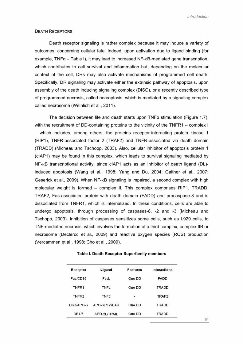
Death receptor signaling is rather complex because it may induce a variety of
outcomes, concerning cellular fate. Indeed, upon activation due to ligand binding (for
example, TNFα – Table I), it may lead to increased NF-κB-mediated gene transcription,
which contributes to cell survival and inflammation but, depending on the molecular
context of the cell, DRs may also activate mechanisms of programmed cell death.
Specifically, DR signaling may activate either the extrinsic pathway of apoptosis, upon
assembly of the death inducing signaling complex (DISC), or a recently described type
of programmed necrosis, called necroptosis, which is mediated by a signaling complex
called necrosome (Weinlich et al., 2011).
The decision between life and death starts upon TNFα stimulation (Figure 1.7),
with the recruitment of DD-containing proteins to the vicinity of the TNFR1 – complex I
– which includes, among others, the proteins receptor-interacting protein kinase 1
(RIP1), TNFR-associated factor 2 (TRAF2) and TNFR-associated via death domain
(TRADD) (Micheau and Tschopp, 2003). Also, cellular inhibitor of apoptosis protein 1
(cIAP1) may be found in this complex, which leads to survival signaling mediated by
NF-κB transcriptional activity, since cIAP1 acts as an inhibitor of death ligand (DL)-
induced apoptosis (Wang et al., 1998; Yang and Du, 2004; Gaither et al., 2007;
Geserick et al., 2009). When NF-κB signaling is impaired, a second complex with high
molecular weight is formed – complex II. This complex comprises RIP1, TRADD,
TRAF2, Fas-associated protein with death domain (FADD) and procaspase-8 and is
dissociated from TNFR1, which is internalized. In these conditions, cells are able to
undergo apoptosis, through processing of caspases-8, -2 and -3 (Micheau and
Tschopp, 2003). Inhibition of caspases sensitizes some cells, such as L929 cells, to
TNF-mediated necrosis, which involves the formation of a third complex, complex IIB or
necrosome (Declercq et al., 2009) and reactive oxygen species (ROS) production
(Vercammen et al., 1998; Cho et al., 2009).
Table I. Death Receptor Superfamily members

Chapter I
20
The ubiquitination state of RIP1 seems to be determinant for induction of
complex II assembly. This post-translational modification of RIP1 is highly regulated by
both ubiquitinases and deubiquitinases (DUBs). Indeed, cIAP1 and cIAP2 are able to
ubiquitinate RIP1 and upon inhibition of these enzymes the recruitment of RIP1 to
complex II is increased (Geserick et al., 2009). The DUB Cezanne leads to
suppression of NF-κB signaling in response to TNFα activation by removing Lys63
polyubiquitin chains from RIP1, in complex I. This contributes to increased stability of
the inhibitor of kappa B (IκB) complex, which retains NF-κB on the cytoplasmatic
compartment (Enesa et al., 2008). Inhibition of both cIAP1 and transforming growth
factor-β activated kinase-1 (TAK1) induces the formation of the necrosome and
subsequent ROS production, via increased recruitment of RIP3 and RIP1 kinase
activity (Vanlangenakker et al., 2011b). Interestingly, TAK1 seems to have a function
as an adaptor molecule, independently of its kinase activity, preventing premature
dissociation of RIP1 from complex I and induction of necroptosis (Arslan and
Scheidereit, 2011). Although RIP1 is generally thought to be necessary for canonical
NF-κB activation (Ting et al., 1996; Kelliher et al., 1998; Ea et al., 2006; Li et al., 2006),
there evidence shows that it may not be essential. Indeed, in RIP1-/- mouse embryonic
fibroblasts (MEFs), the cells are still able to induce NF-κB transcriptional activity,
through increased degradation of IκBα (Wong et al., 2010). Interestingly, HeLa cells
treated with etoposide to induce DNA damage were shown to induce biphasic NF-κB
activation, with differential regulation. RIP1 kinase activity was shown to be exclusively
necessary for the second phase of NF-κB activity. Furthermore, this type of insult was
shown to induce the recruitment of FADD and caspase-8 to a RIP1-NF-κB essential
modulator (NEMO) complex, thereby initiating apoptotic signaling, which is prevented
by knock-down (Tia et al.) of either RIP1 or caspase-8 (Biton and Ashkenazi, 2011).
The association between RIP1 and caspase-8 in complex II seems to be highly
dependent on FADD availability, since treatment of cells with a dominant negative form
of FADD (FADD-DN) renders cells resistant to TNF-induced apoptosis and impairs this
interaction. This effect can be rescued by expression of WT FADD (Micheau and
Tschopp, 2003). The protein FADD can, in fact, have a deep impact on the phenotype
of cell death being activated following TNFR1 signaling. Different domains of this
protein have the ability to induce caspase-dependent apoptosis (death effector domain
– DED) or necrosis (DD), in a caspase-independent manner. This seems to be
regulated by their ability to selectively interact with caspase-8 or RIP-1, respectively
(Vanden Berghe et al., 2004).

Introduction
21
Figure 1.7. DR signaling complexes. Upon DL binding, the DR recruits a complex of
DD-containing proteins to its vicinity (complex I). In this complex, RIP1 is ubiquitinated by
cIAPs, initiating signaling to NF-κB activation. This contributes to cell survival via
transcription of survival genes. Upon deubiquitination of RIP1, the complex dissociates
from the receptor and recruits FADD and procaspase-8, forming a complex called DISC.
Caspase-8 auto-activates, cleaves RIP1 inhibiting necroptosis, and initiates apoptosis. In
conditions of low caspase-8 activity, RIP3 is recruited to interact with RIP1, assembling
the necrosome, and together they activate necroptosis.
Apoptosis and necroptosis are complementary cell death pathways, as
demonstrated by occlusion of the lethality observed in caspase-8-/- mice when RIP3 is
concomitantly depleted, in caspase8-/-/RIP3-/- double knock-out (KO) mice (Kaiser et
al., 2011; Oberst et al., 2011), which emphasizes the role of caspase-8 as a negative
regulator of necroptosis. This regulation is achieved by the formation of catalytically
active dimers of caspase-8 and FLICE-like inhibitory protein long (FLIPL), which inhibit
the assembly of the necrosome, without inducing apoptosis (Micheau et al., 2002;
Oberst et al., 2011). Interestingly, RIP3 overexpression is not lethal per se, but
sensitizes MEFs to TNFα treatment, in a RIP1 and caspase-8 independent manner
(Moujalled et al., 2013).

Chapter I
22
Although TNFα and its receptor TNFR1 are the most commonly studied in
necroptosis research, other DLs and their respective DRs might have a role in
induction of cell death signaling (reviewed in Han et al., 2011). Indeed, TNF-related
weak inducer of apoptosis (TWEAK), which binds DR3, has been shown to induce the
formation of a DISC comprising RIP1, FADD and caspase-8, but lacking TRADD, and
to induce apoptosis. Interestingly, this DL does not seem to induce activation of
necroptosis (Ikner and Ashkenazi, 2011). Also, Fas ligand (FasL) and TNF-related
apoptosis-inducing ligand (TRAIL) were shown to induce a form of caspase-
independent necrotic cell death that is dependent on RIP1 kinase activity (Holler et al.,
2000). The receptor cluster of differentiation 95 (CD95) is also known to induce
apoptotic cell death in T cells. Upon caspase inhibition, however, the type of cell death
is shifted to necrosis and proinflammatory signaling via ERK and p38. This receptor
was also shown to induce NF-κB activity, which lead to increased cytokine production
(Scheller et al., 2002). Toll-like receptor 3 (TLR3) signaling has also been shown to
activate necroptotic mechanisms of cell death (Kalai et al., 2002).
Survival Signaling
Upon TNFα binding to TNFR1, complex I is recruited to the receptor (Figure
1.8) and RIP1 is ubiquitinated by cIAPs (Bertrand et al., 2008; Varfolomeev et al.,
2008; Bianchi and Meier, 2009). The Lys63 chains linked to RIP1 then recruit TAK1
binding protein 2 (TAB2) and NEMO, with the associated kinases (TAK1 and IκB
kinase – IKK, respectively) (Kanayama et al., 2004; Ea et al., 2006; Li et al., 2006; Wu
et al., 2006a). Linear Ub chain assembly complex (LUBAC) is also recruited to complex
I, in an Ub-dependent manner, and was shown to stabilize this complex (Haas et al.,
2009). LUBAC is responsible for ubiquitinating NEMO. Linear ubiquitination of this
protein was shown to be necessary for TNFR1-induced NF-κB activity (Tokunaga et
al., 2009; Tokunaga et al., 2012; Verhelst et al., 2012). Once in this complex, TAK1
phosphorylates IKKβ. Upon activation, IKKβ phosphorylates IκB, targeting it for
degradation. This results in the relief of NF-κB inhibition, which is then able to
translocate to the nucleus and initiate gene transcription (Karin and Ben-Neriah, 2000).
Activation of NF-κB signaling has been shown to be counteracted by the action
DUBs on RIP1, such as A20 (He and Ting, 2002; Wertz et al., 2004) and
cylindromatosis (CYLD) (Wright et al., 2007). The A20 ubiquitin-editing enzyme
removes Lys63 polyubiquitin chains from RIP1 and concurrently links Lys48

Introduction
23
polyubiquitin chains to this protein, thus targeting it for degradation by the proteasome
system. This results in concomitant inhibition of NF-κB survival signaling and inhibition
of TNF-induced cell death by either apoptosis or necrosis (He and Ting, 2002; Wertz et
al., 2004; Vanlangenakker et al., 2011a). On the other hand, CYLD removes Lys63
chains from RIP1, promoting necroptosis (Welz et al., 2011). This DUB is rapidly
cleaved by active caspase-8 (O'Donnell et al., 2011) thus, it is only stabilized and
capable of deubiquitinating RIP1 upon insufficient activity of caspase-8, which may
sensitize the cells to necroptotic death. Contrary to its ubiquitination state, the kinase
activity of RIP1 does not regulate NF-κB activation (Lee et al., 2004; Wong et al.,
2010).
Besides the known NF-κB signaling regulation, activation of TNFRs also leads
to activation of MAPKs. The kinase RIP1 was shown to be necessary for TNFα-induced
MAPK activation (ERK, p38 and c-Jun N-terminal kinase – JNK), as observed by
reduction of MAPK phosphorylation in RIP1-/- MEFs. The kinase activity of RIP1,
however, seems to be only required for ERK activation (Devin et al., 2003; Lee et al.,
2003). JNK is thought to contribute to the build-up of oxidative stress, promoting
mitochondrial dysfunction, downstream of RIP1 and TRAF2, and leading to necrotic
cell death (Davis, 2000; Shen et al., 2004; Xu et al., 2006).
Apoptosis
Apoptosis is generally described as a type of programmed cell death, due to its
reliance on specific and sequential cellular mechanisms, in order to be activated. This
type of cell death induces several cytoarquitectural alterations, including membrane
blebbing, with formation of apoptotic bodies, chromatin condensation, DNA laddering
and fragmentation of the nucleus, organelle fragmentation and release of protein
content from the mitochondria to the cytoplasm. After cellular deconstruction, the debris
are removed by phagocytes, avoiding activation of inflammatory responses and
damage to the neighboring cells (Taylor et al., 2008).
There are two major pathways of apoptosis: the extrinsic (or DR pathway) and
the intrinsic (or mitochondrial pathway). These pathways are thought to cross-talk and
influence one another (Igney and Krammer, 2002) (Figure 1.9).

Chapter I
24
Figure 1.8. Assembly of DR-associated complex I. Upon activation, TRADD, TRAF2
and RIP1 are recruited to interact with the TNFR1, through their DDs. cIAPs ubiquitinate
RIP1 and TRADD and RIP1-bound polyubiquitin chains recruit TAB2/3 and NEMO. TAK1
becomes active and phosphorylates IKKβ, which becomes active. This kinase then
phosphorylates IκB, which relieves the inhibition exerted on NF-κB dimmers. LUBAC is
also recruited to this complex in an Ub-dependent manner and it stabilizes the complex
by ubiquitinating NEMO. Upon relief of the inhibition, NF-κB dimmers translocate to the
nucleus and initiate gene transcription, promoting cell survival and inflammation.
The extrinsic pathway of apoptosis is initiated by DR signaling. Under
appropriate conditions, complex II or DISC is assembled and auto-catalytic activation of
caspase-8 occurs (Kischkel et al., 1995). The activation of caspase-8 can be inhibited
by cFLIP isoforms (FLIPL, FLIPS and FLIPR) that bind procaspase-8 keeping it in an
inactive state (Kataoka et al., 1998; Scaffidi et al., 1999). This process is regulated by
the activity of complex I. Upon NF-κB activation via complex I, the transcription factor

Introduction
25
induces, among other antiapoptotic genes, the expression of FLIPL, which then exerts
a negative regulatory effect on complex II, through inhibition of caspase-8. However, if
complex I displays limited activity, then the expression of FLIPL will be lower and
complex II will have the ability to initiate apoptotic signaling (Micheau and Tschopp,
2003). The FLIPL protein possesses caspase-like domains, but has no catalytic activity
(Tschopp et al., 1998), thus it interacts with procaspase-8, allowing its allosteric
activation and partial processing. This event inhibits induction of apoptosis due to
caspase-8 limited activity, but allows this enzyme to process local substrates, like RIP1
(Krueger et al., 2001; Micheau et al., 2002; Kavuri et al., 2011). Therefore it facilitates
concomitant inhibition of apoptosis and necroptosis (Oberst et al., 2011). The FLIPS
isoform, on the other hand, inhibits caspase-8 auto-activation (Krueger et al., 2001;
Golks et al., 2005), thereby limiting the cleavage of RIP1, which is then capable of
initiating necroptosis signaling. In conditions where caspase-8 is able to auto-activate,
it triggers the execution phase of apoptosis. The extrinsic pathway of apoptosis cross-
talks with the intrinsic pathway via caspase-8 mediated truncation of BH3 interacting-
domain death agonist (Bidoret et al.). This proapoptotic protein is then able to
translocate to the mitochondria, activate the proapoptotic proteins Bax/Bak and
promote the formation of outer membrane pores and the release of apoptotic
mitochondrial proteins to the cytoplasm (Li et al., 1998a; Esposti, 2002; Kaufmann et
al., 2012).
The intrinsic pathway of apoptosis is characterized by mitochondrial
dysfunction, resulting in the opening of the mitochondrial pores. This induces loss of
mitochondrial membrane potential and release of proapoptotic proteins to the
cytoplasm (Saelens et al., 2004). A first group of proteins to be released includes
cytochrome c, second mitochondria-derived activator of caspase/direct inhibitor of
apoptosis-binding protein with low pI (Smac/DIABLO) and high temperature
requirement protein A2 (HtrA2/Omi) (Du et al., 2000; van Loo et al., 2002; Garrido et
al., 2006). Once in the cytoplasm, cytochrome c interacts with apoptotic peptidase
activating factor 1 (Apaf-1) and procaspase-9, forming the so-called apoptosome and
initiating the caspase-dependent mitochondrial pathway (Chinnaiyan, 1999; Hill et al.,
2004). At a later stage, a second group of proteins is released from the mitochondria,
after the cell has committed to the death program. This includes apoptosis-inducing
factor (AIF) and endonuclease G, which translocate to the nucleus to induce DNA
fragmentation and nuclear condensation (Joza et al., 2001; Li et al., 2001). In the
cytoplasm, the caspase activated DNase (CAD) is kept in an inactive state by its
inhibitor, ICAD. Upon caspase-3 cleavage of ICAD, the active endonuclease is

Chapter I
26
released and further exacerbates chromatin condensation (Enari et al., 1998; Sakahira
et al., 1998).
The regulation of the mitochondrial pathway of apoptosis is mediated by a
family of proteins, the B-cell lymphoma-2 (Bcl-2) family, which includes both pro-
apoptotic (for example, Bax, Bak, Bim, BID, Bad) and anti-apoptotic members
(including Bcl-2, Bcl-XL, Bcl-x, BAG) (Cory and Adams, 2002). Several other pro- and
anti-apoptotic proteins, as well as additional signaling pathways, like MAPK signaling
cascades, participate in apoptotic mechanisms, contributing to a very complex program
to induce cellular demise.
These two pathways of apoptosis converge to the execution phase, which is
performed by active caspases. The executioner caspases (caspase-3, -6 and -7) are
activated by the initiator caspases (caspase-8, -9 and -10) by cleavage of the
procaspase, and exert their effects via proteolysis of a plethora of substrates. Among
these are poly-ADP ribose polymerase (PARP), α-fodrin, endonucleases, ICAD and
gelsolin (Slee et al., 2001). The latter is cleaved by caspase-3 causing disruption of the
cytoskeleton (Kothakota et al., 1997), which contributes to the cellular demolition
characteristic of apoptosis (Taylor et al., 2008).
The final stage of apoptosis is the phagocytic removal of cellular debris. This
process is preceded by externalization of phosphatidylserine on the cell surface, which
facilitates the recognition of the cells to be phagocytosed, without activation of
inflammatory mechanisms (Fadok et al., 2001). This feature, however, is not exclusive
of apoptosis (Krysko et al., 2004).
Global ischemic insults have been shown to induce apoptotic neuronal death in
the CA1 region of the hippocampus, where the cells display shrunk and condensed
nuclei, along with other features related to this type of cell demise (Ozawa et al., 1999).
Global ischemia also induces translocation of the pro-apoptotic protein Bad, to the
mitochondria, where it assembles with other pro-apoptotic proteins, such as Bcl-XL.
The latter is cleaved to produce a pro-death fragment that promotes the dissipation of
mitochondrial membrane potential. This then leads to release of proapoptotic proteins
from the mitochondria to the cytosol, such as cytochrome c and Smac/DIABLO. These
proteins promote the formation of the apoptosome, which induces activation of
caspases, namely the executioner caspase-3. The activity of this caspase is increased
after global ischemia, in a region-specific manner, since its activity is observed in CA1
neurons, but not in the CA3/dentate gyrus (DG) area. Caspase-3 then promotes DNA
fragmentation and subsequent neuronal death in CA1 neurons (Miyawaki et al., 2008).

Introduction
27
Figure 1.9. Apoptotic Pathways. Apoptosis is elicited in cells via two distinct pathways
that cross-talk. Extrinsic pathway: Upon deubiquitination of RIP1, complex I dissociates
from TNFR1 and recruits FADD and procaspase8. In the presence of low levels of cFLIP
isoforms, caspase8 is able to autoactivate and cleave RIP1, thereby inhibiting
necroptosis. Caspase8 truncates BID and processes caspase3, which contributes to the
intrinsic pathway of apoptosis. Intrinsic pathway: Apoptotic stimulation of cells leads to
mitochondrial dysfunction, with formation of membrane pores. This contributes to the
release of pro-apoptotic mitochondrial proteins such as cytochrome C and
Smac/DIABLO. This process is regulated by members of the Bcl-2 family, which includes
a variety of pro- and anti-apoptotic proteins. In the cytoplasm, cytochrome C interacts with
Apaf-1 and procaspase9, assembling the apoptosome. Caspase9 becomes catalytically
active and caspase3, a major executioner caspase, is processed, becoming activated.
This enzyme exerts its effects by cleaving a variety of intracellular substrates, contributing
to apoptotic cell death.
Accordingly, the OGD challenge also induces apoptosis, as demonstrated by
typical morphological alterations, as well as activation of specific molecular pathways.
Morphologically, there are evidences of DNA laddering and chromatin condensation
(Kalda and Zharkovsky, 1999). The OGD challenge also induces depolarization of the

Chapter I
28
mitochondria, as measured by dissipation of mitochondrial membrane potential (He et
al., 2009; Liu et al., 2011), which occurs during apoptotic cell death. Upon induction of
OGD in cerebrocortical cultures, both caspases and calpains become activated. This
has been shown through the increased formation of spectrin cleavage products of 120
kDa and 145 kDa, which are specific for caspases and calpains, respectively, and also
by neuroprotection afforded by caspase (Nath et al., 1998) and calpain inhibitors
against OGD-induced death (Newcomb-Fernandez et al., 2001).
The OGD challenge appears to also activate the extrinsic apoptotic pathway.
Treatment with an anti-TNFα antibody reduced the number of apoptotic neurons, in
cortical cultures submitted to OGD. Additionally, OGD enhances caspase-8 activation
mediated by TNFR1 and subsequent caspase-3 activation (Badiola et al., 2009). The
OGD challenge also induces cleavage of the pro-apoptotic protein BID, a caspase-8
substrate, before the onset of neuronal death. Furthermore, the cell death elicited by
OGD is reduced when BID is inhibited (Culmsee et al., 2005) and in BID-/- cortical
neurons, compared to WT neurons. These effects are also observed in vivo, using the
focal ischemia model MCAO, where BID-/- animals display reduced infarct volumes as
well as reduced cytochrome c release from the mitochondria (Plesnila et al., 2001).
On the other hand, members of the anti-apoptotic Bcl-2 family have been shown
to have neuroprotective effects against ischemic stimuli. Indeed, cells overexpressing
Bcl-2 display increased resistance to the OGD challenge and reduced activation of
caspase-3 and cytochrome c release from the mitochondria, as well as diminished loss
of mitochondrial membrane potential (Koubi et al., 2005). Moreover, the Bcl-XL family
member was shown to reduce nuclear translocation of AIF as well as the extent of cell
death induced by OGD (Cao et al., 2003).
Futhermore, the anti-apoptotic protein Apaf-1 interacting protein (AIP), which
acts by preventing apoptosome formation, has been demonstrated to be up-regulated
in the regions of the hippocampus that are resistant to OGD-induced damage, but not
in the CA1 area. Furthermore, overexpressing AIP in CA1 neurons significantly
reduces OGD- or global ischemia-induced neuronal death and caspase-3 activation
(Cao et al., 2004a).
Necroptosis
Caspase-independent cell death that occurs as a consequence of DR-mediated
signaling is considered to be a type of regulated necrotic cell death because it displays
necrotic features, such as plasma membrane damage, swollen mitochondria and slow
mitochondrial membrane depolarization, subsequent to nuclear and cytoplasmatic

Introduction
29
morphological changes. Also, mitochondrial apoptotic markers like cytochrome C
release or caspase-3-dependent cleavage of PARP, as well as chromatin condensation
or oligonucleosomal DNA degradation, typical features of apoptosis, are not observed
in this type of cell death (Holler et al., 2000; He et al., 2009; Vandenabeele et al.,
2010), hence its classification as programmed necrosis (necroptosis).
Necroptotic stimulation, such as a combination of Smac mimetics (which
contribute to cIAP1/2 degradation) and caspase inhibitors, such as N-
Benzyloxycarbonyl-Val-Ala-Asp(O-Me)-fluoromethyl ketone (zVAD.fmk), leads to a
necrotic phenotype, in a DR-dependent manner, as demonstrated by KD of TNFR1 or
treatment with a TNFα neutralizing antibody (He et al., 2009). RIP1 was shown to
regulate autocrine TNFα production, in a NF-κB-independent manner. In certain
contexts, such as caspase inhibition (with zVAD.fmk), cIAP1/2 depletion (with Smac
mimetics) or absence of TRAF2, RIP1 associates with the protein E3 ubiquitin-protein
ligase UBR5 (EDD) and together they activate the JNK signaling pathway. JNK then
phosphorylates its substrates activator protein-1 (AP-1) and specificity protein 1 (Sp-1),
which become transcriptionally active, inducing TNFα expression and subsequent
inflammation and cell death by necrosis (Christofferson et al., 2012).
In an effort to elucidate the mechanism of necroptosis that is activated in
response to DR signaling, three independent groups identified RIP3 as a protein that is
specifically associated with necroptotic signaling, using different approaches (Cho et
al., 2009; He et al., 2009; Zhang et al., 2009a). The failure of a RIP3-DN to prevent
FADD-induced apoptosis (Feng et al., 2006) is in accordance with these reports that
relate RIP3 specifically to necroptosis.
Cho and colleagues (2009) performed a siRNA screen in FADD-deficient Jurkat
cells, which are known to die by necroptosis in response to TNFα. They identified both
RIP1 and RIP3 as crucial mediators of this type of cell death. Interestingly, they also
found that RIP3 KD had no effect on TNF- and FasL-induced apoptosis or NF-κB
activation. This emphasizes the specific role of RIP3 on the necroptotic mechanism.
This protein was shown to be specifically recruited to complex II but, unlike RIP1, was
not present in complex I (Cho et al., 2009).
He et al. (2009) adopted a similar strategy. They performed a genome-wide
siRNA screen and transfected HT-29 cells with different siRNA pools to assess which
of these were protective against necroptotic stimulation. Besides RIP1, which had been
previously identified, they found RIP3 to be important for this process. Importantly, this

Chapter I
30
protein was specifically correlated with necroptosis since it did not interfere with
induction of apoptosis. In fact, the ability of different cell lines to undergo necroptosis
seems to be correlated with RIP3 expression. Accordingly, cells that express RIP3
have the ability to induce necroptosis (for example Jurkat, U937, L929 and MEFs)
while cells with no observable expression of RIP3, are resistant to necroptotic
stimulation, including HEK293T, MCF-7 and U2OS. Interestingly, when they expressed
RIP3 in “resistant” cells, like T98G cells, they rendered the cells susceptible to
necroptosis (He et al., 2009).
Zhang et al. (2009) studied the mechanism of necroptosis in two lines of
NIH3T3 cells, N and A cells, which are, respectively, susceptible and resistant to
necroptosis. They performed a microarray analysis to detect changes in genes that
might be responsible for this differential vulnerability and found that RIP3 was
responsible for induction of necroptosis in N cells, while A cells lack expression of this
protein. Specifically, they demonstrated that KD of RIP3 abolishes the component of
necroptotic death in N cells, whereas expression of RIP3 in A cells renders them
susceptible to TNF-induced necroptosis (Zhang et al., 2009a).
The human RIP3 gene has been shown to produce a full length (FL-RIP3)
protein and two splice variants – RIP3β and RIP3γ. The FL-RIP3 is widely expressed
throughout the organism, including the heart, brain, lung and spleen. While the RIP3γ
variant displays comparable mRNA expression to that of the FL-RIP3, in most tissues,
the RIP3β variant is expressed at very low levels (Yang et al., 2005). Unlike FL-RIP3
(Yang et al., 2004), both RIP3γ and RIP3β seem to be unable to shuttle between the
cytoplasm and the nucleus, which may be related to their inability to induce cell death
(Yang et al., 2005). The RIP3 kinase activity is required for nuclear localization of the
protein and to the induction of caspase-independent cell death. In fact, RIP3 seems to
translocate to the nucleus upon induction of cell death (Feng et al., 2007).
RIP1 and RIP3 where demonstrated to associate through their RIP homotypic
interaction motif (RHIM) domains (Sun et al., 2002), forming an amyloid complex upon
induction of necroptosis (Li et al., 2012) (Figure 1.10). Additionally, RIP3 was shown to
autophosphorylate and phosphorylate RIP1. Phosphorylation of RIP1 seems to act as
a cue to shut-off NF-κB signaling, which contributes to cell death (Sun et al., 2002) and
it may contribute to the stabilization of the association of RIP1 within complex II (Cho et
al., 2009). Also, the kinase activity and the RHIM domains of RIP1/3 are crucial for
induction of necroptosis (Cho et al., 2009; He et al., 2009; Zhang et al., 2009a; Li et al.,
2012). Mutations in the RHIM domain of RIP3 abolished its ability to induce necroptosis

Introduction
31
(He et al., 2009). Necrostatin-1 (Nec-1) a pharmacological inhibitor of necroptosis, was
found to inhibit RIP1 kinase activity, by inhibiting, for example, RIP1
autophosphorylation. In fact, four putative autophosphorylation sites were identified
(Ser14/15, Ser20, Ser161 and Ser166), all in the N-terminal domain, which is also the
kinase domain of RIP1 (Degterev et al., 2008). The pronecrotic complex II, called
necrosome, seems to be distinct from the apoptotic one (DISC) since the interaction
between RIP1 and RIP3 appears to occur specifically under necrotic-inducing
conditions (Cho et al., 2009) (Figure 1.11). Interestingly, RIP1-independent (but RIP3-
dependent) necroptosis has been shown to occur upon KD of both RIP1 and caspase-
8, evidencing once again the negative regulation exerted by caspase-8 and suggesting
that RIP3 may be sufficient to induce necroptosis in certain cellular contexts
(Vanlangenakker et al., 2011a). The intermediate domain (ID) of RIP1 seems to be
important for induction of necroptosis, as deletion of this domain in the protein shifts the
type of cell death from necroptosis to apoptosis, due to increased recruitment of RIP1
and caspase-8 to FADD (Duprez et al., 2012).
Figure 1.10. Struture of the human RIP1/3 proteins. RIP1 and RIP3 share partial
homology in their structure. They both contain a N-terminal kinase domain and a RHIM
domain, through which they interact upon necroptosis induction. RIP1 also possesses a
C-terminal DD, which allows this protein to interact with other DD-containing proteins (for
example, TNFR1) in complex I and II.
This pronecrotic interaction between RIP1 and RIP3 was recently shown to be
regulated by the acetylation state of RIP1. Under basal conditions, RIP3 interacts with
the deacetylase sirtuin2 (SIRT2). Upon induction of necrosis, the RIP3-SIRT2 complex
is recruited to RIP1, thereby facilitating SIRT2-dependent deacetylation of RIP1 on

Chapter I
32
Lys530. Due to the close proximity of this residue to the RHIM domain of RIP1, which
comprises residues 531-547 of RIP1, acetylation on Lys530 possibly disrupts the
interaction with RIP3. Upon deacetylation of this residue, RIP1 and RIP3 are able to
interact and necroptosis ensues. This evidence is further supported by the fact that
SIRT2-/- MEFs are protected from necrosis-inducing insults (Narayan et al., 2012).
Mixed lineage kinase domain-like (MLKL) was shown to be a molecular target of
the necrosome (Sun et al., 2012; Zhao et al., 2012). Its role in necroptosis is
emphasized by complete abrogation of necroptosis upon MLKL KD or in the presence
of necrosulfonamide, a MLKL inhibitor. The mitochondrial protein Ser/Thr-protein
phosphatase (PGAM5) is also phosphorylated by RIP3 and associates with the RIP1/3-
MLKL complex. The short isoform of PGAM5 (PGAM5S) is proposed to be an effector
downstream of the necrosome since its RIP3-mediated phosphorylation is abrogated
by necrosulfonamide. PGAM5S then dephosphorylates the mitochondrial protein
dynamin related protein 1 (Drp-1), resulting in its activation, which contributes to
mitochondrial fragmentation (Wang et al., 2012).
The cellular signaling network involved in the necroptotic process was shown to
be regulated by several genes (Hitomi et al., 2008) and requires de novo protein
synthesis (Yu et al., 2004). Mitochondrial dysfunction, ROS accumulation and ATP
depletion have been shown to contribute to the detrimental effect of necroptosis, which
is prevented in RIP1-/- cells (Cho et al., 2009; He et al., 2009; Irrinki et al., 2011). The
RIP3 kinase promotes mitochondrial ROS generation via a group of metabolism-related
enzymes, known to associate with RIP3: phosphorylase-glycogen-liver (PYGL),
glutamate-ammonia ligase (GLUL) and glutamate dehydrogenase 1 (GLUD1). These
enzymes are involved in mitochondrial metabolism. The protein PYLG is the rate-
limiting enzyme in glycogen degradation, while GLUL and GLUD1 are determinant
enzymes for oxidative phosphorylation (Zhang et al., 2009a). This seems to contribute
to the insertion of the proapoptotic protein BCL2/adenovirus E1B 19 kDa interacting
protein 3 (BNip3) in the mitochondria, contributing to mitochondrial damage.
Interestingly, BNip3 is induced following TNFα stimulation in alveolar lung cells, but it is
not involved in the extrinsic pathway of apoptosis but instead in induction of
necroptosis (Kim et al., 2011). Another Bcl-2 family member, Bmf, was also shown to
be important for necroptosis induction as KD of this protein inhibits necroptotic cell
death (Hitomi et al., 2008). These reports emphasize the complex interplay and
common players found between these two distinct programs of cell death: apoptosis
and necroptosis.

Introduction
33
Figure 1.11. Assembly of the necrosome. When caspase8 is downregulated, RIP1 is
capable of recruiting RIP3. This process is regulated by the acetylation state of RIP1.
Upon deacetylation of RIP1 by SIRT2, RIP1 and RIP3 interact through their RHIM
domains and autoactivate. The necrosome then phosphorylates its substrates, including
PGAM5 and MLKL. PGAM5S then dephosphorylates Drp-1, which becomes active and
promotes mitochondrial fragmentation.
On the other hand, spontaneous formation of a protein module similar to
complex II has been described (Feoktistova et al., 2011; Tenev et al., 2011). Due to its
independence from DR signaling, this high molecular weight complex comprising RIP1,
FADD and caspase-8, among other proteins, was called ripoptosome. The formation of
this complex occurs following genotoxic stress (Tenev et al., 2011) or TLR3 stimulation
(Feoktistova et al., 2011), both in conditions of cIAP depletion, suggesting that these

Chapter I
34
proteins inhibit the assembly of the ripoptosome. The FLIPL isoform is also recruited to
the complex, exerting an inhibitory regulation of the ripoptosome. Accordingly, KD of
FLIPL enhanced the formation of the ripoptosome. This complex seems to have the
ability to induce both apoptosis and necroptosis, depending on the cellular context
(Tenev et al., 2011). Interestingly, cFLIPS seems to specifically inhibit ripoptosome-
induced apoptosis and to promote necroptosis (Feoktistova et al., 2011).
Necroptosis in Disease Context
Necroptosis might be a relevant mechanism in several paradigms of cell death,
namely, to fight virus infections. Viruses such as the Vaccinia virus express apoptotic
inhibitors, delaying the activation of cell death programs in infected T cells, which
contributes to disseminate the infection. Thus, if cells are able to activate alternative
death programs, the organism may more efficiently fight the infection. Accordingly,
Vaccinia virus infected T cells are sensitized to TNF-induced necroptotic cell death
(Cho et al., 2009). Mouse cytomegaloviruses (MCMV) express cell death suppressors
that specifically inhibit RIP3 activity, in a RHIM-dependent manner (Upton et al., 2010).
This aspect emphasizes the relevance of necroptosis as a physiological programmed
cell death mechanism.
In another disease context, an animal model of cerulean-induced pancreatitis,
much of the cell death in the pancreas seems to be attributable to RIP3-dependent
necroptosis. This protein is significantly induced specifically in these cells and RIP3-/-
animals have a much lesser extent of pancreatic cell death (He et al., 2009).
Necroptosis is also involved in Crohn’s disease pathogenesis. Indeed, RIP3
seems to be specifically expressed in the cells that are most affected by the disease,
the Paneth cells of the terminal ileum, which might confer increased susceptibility to
necroptotic insults. Additionally, intestinal epithelial cells lacking caspase-8 are
sensitized to necroptotic cell death, as observed by rescue of this effect with Nec-1.
This provides further evidence for a role of caspase-8 in the inhibition of necrosome
formation and subsequent activation of necroptosis (Gunther et al., 2011).
Moreover, in a mouse model of cardiac ischemia, Nec-1 provided significant
neuroprotection, reducing the infarct size, the phosphorylation of RIP1/3 and the
inflammation, while having no impact on the extent of apoptotic cell death (Oerlemans
et al., 2012). Likewise, necroptosis was shown to occur in retinal neurons following
ischemic damage. Necrostatin-1 was effective in reducing specifically this component

Introduction
35
of cell death, without affecting apoptotic neuronal death and in improving the functional
outcome in the ischemia challenged retina (Rosenbaum et al., 2010). Also, in a mouse
model of retinal detachment, both inhibition of RIP1 as well as RIP3 ablation were
shown to be neuroprotective, demonstrating the activation of necroptotic mechanisms
upon apoptosis inhibition (Trichonas et al., 2010).
In vitro cerebral ischemia has also been shown to induce a component of
necroptotic neuronal death (Xu et al., 2010; Meloni et al., 2011) which was also
observed to occur in vivo, following neonatal hypoxia/ischemia (Northington et al.,
2011) and MCAO (Degterev et al., 2005; Xu et al., 2010). These reports showing a role
for necroptosis following ischemic insults, either in vitro or in vivo, used a specific
inhibitor of necroptosis, Nec-1, to support this finding. This inhibitor acts on the kinase
activity of RIP1 (Degterev et al., 2008), however, this protein has also been correlated
to the apoptotic signaling, mainly as a scaffold for complex II formation. Thus, the
specificity of Nec-1 may be argued against, which emphasizes the need to develop
novel strategies to clearly dissect the contribution of necroptosis, in order to better
clarify the role of this mechanism on neuronal death, following ischemic insults.
Additional neuronal death paradigms have been shown to induce necroptosis.
Indeed, following treatment with 24(S)-hydroxycholesterol (24S-OHC), which is
cytotoxic, both human neuroblastoma SH-SY5Y cells and cortical neurons were shown
to die by necroptosis (Yamanaka et al., 2011). Additionally, menadione, a compound
that induces superoxide production, with concomitant mitochondrial dysfunction and
ATP depletion, was shown to induce caspase-independent necroptotic like death in
HT-22 cells, a cell line of immortalized mouse hippocampal neurons (Fukui et al.,
2012). In a mouse model of traumatic brain injury (TBI), Nec-1 was shown to be
effective in reducing the damage associated with the injury, suggesting the occurrence
of necroptosis (You et al., 2008). Interestingly, necroptosis was also proposed to have
a neuroprotective effect by being activated in inflamed microglia, which results in less
damage to neurons. Upon inflammatory stimulation, inhibition of caspase-8 activity is
beneficial to neurons by promoting necroptosis in microglia, whereas Nec-1 rescues
neurodegeneration. This implies that caspase inhibitors could promote neuronal
survival against certain insults by promoting microglial demise (Fricker et al., 2013).
Beyond the study of the NMDARs and of the necroptotic neuronal death, we
also addressed the activation of effector signaling pathways, such as the MAPKs,
which are known to be involved in several neuronal death paradigms.

Chapter I
36
MAP Kinase Pathways
The MAPKs are a family of stress-responsive proteins that regulate an array of
functions in the cell. They exert their effects by phosphorylating a plethora of
substrates, including transcription factors, enzymes and cytoskeletal proteins impacting
on cell division, proliferation, metabolism and cell fate (Davis, 2000; Qi and Elion, 2005;
Burke, 2007).
These proteins have a typical three-tiered activation cascade. Following an
appropriate stimulus a MAPK kinase kinase (MAPKKK) autophosphorylates, leading to
relief of autoinhibition and oligomerization. The MAPKKK then activates a MAPK
kinase (MAPKK), by dual phosphorylation of Ser and Thr residues within the activation
loop of the catalytic domain (Songyang et al., 1996; Chen et al., 2001). Finally, the
activated MAPKK phosphorylates the MAPK, on conserved Thr and Tyr residues within
a conserved Thr-X-Tyr motif on the activation loop (Silva et al., 2005; Burke, 2007).
The MAPKKs are highly specific and typically phosphorylate only one MAPK, but are
regulated by many, less specific, MAPKKKs. The MAPKs phosphorylate a wide range
of substrates on Ser and Thr residues on a consensus PXT/SP motif. The pathway
specificity may be increased by the organization of different signaling complexes by
distinct scaffold proteins, which have been shown to have many functions, beyond
bringing the elements of the pathway together. Scaffold proteins have the ability of
binding, sequestering, and fostering the specific components of the pathway. This
organization in a multiprotein complex allows for specific types of stimuli to produce
unique and coordinated MAPK signaling responses (Burack and Shaw, 2000;
Yoshioka, 2004; Xu et al., 2007). The inactivation of the MAPKs is performed by dual-
specificity MAPK phosphatases (MKPs) (Keyse, 2000), among other phosphatases.
MAPKs may be found in many subcellular locations, namely in the ER, endosomes,
microtubules and may translocate to the nucleus to phosphorylate nuclear substrates.
In mammals, three major groups of MAPKs have been defined: ERK, p38 and
JNK (Figure 1.12) (Davis, 2000; Johnson and Lapadat, 2002).
EEXXTTRRAACCEELLLLUULLAARR SSIIGGNNAALL--RREEGGUULLAATTEEDD KKIINNAASSEE
The ERK subfamily of MAPKs comprises two members, ERK1 and ERK2, with
44 and 42 kDa that are activated by mitogenic stimuli like growth factors and cytokines.
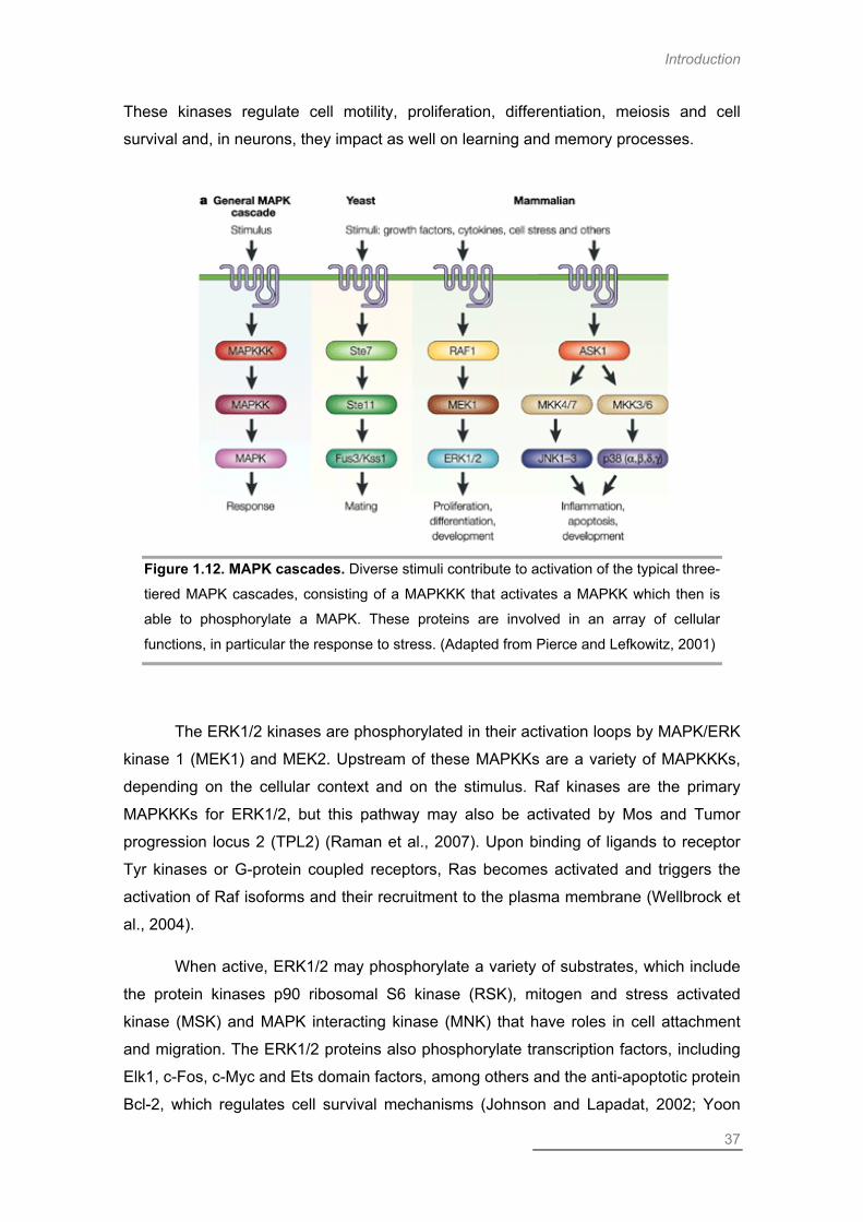
Introduction
37
These kinases regulate cell motility, proliferation, differentiation, meiosis and cell
survival and, in neurons, they impact as well on learning and memory processes.
Figure 1.12. MAPK cascades. Diverse stimuli contribute to activation of the typical three-
tiered MAPK cascades, consisting of a MAPKKK that activates a MAPKK which then is
able to phosphorylate a MAPK. These proteins are involved in an array of cellular
functions, in particular the response to stress. (Adapted from Pierce and Lefkowitz, 2001)
The ERK1/2 kinases are phosphorylated in their activation loops by MAPK/ERK
kinase 1 (MEK1) and MEK2. Upstream of these MAPKKs are a variety of MAPKKKs,
depending on the cellular context and on the stimulus. Raf kinases are the primary
MAPKKKs for ERK1/2, but this pathway may also be activated by Mos and Tumor
progression locus 2 (TPL2) (Raman et al., 2007). Upon binding of ligands to receptor
Tyr kinases or G-protein coupled receptors, Ras becomes activated and triggers the
activation of Raf isoforms and their recruitment to the plasma membrane (Wellbrock et
al., 2004).
When active, ERK1/2 may phosphorylate a variety of substrates, which include
the protein kinases p90 ribosomal S6 kinase (RSK), mitogen and stress activated
kinase (MSK) and MAPK interacting kinase (MNK) that have roles in cell attachment
and migration. The ERK1/2 proteins also phosphorylate transcription factors, including
Elk1, c-Fos, c-Myc and Ets domain factors, among others and the anti-apoptotic protein
Bcl-2, which regulates cell survival mechanisms (Johnson and Lapadat, 2002; Yoon

Chapter I
38
and Seger, 2006). Additionally, ERK phosphorylates the proapoptotic protein Bim,
leading to its proteasomal degradation (Luciano et al., 2003; Ley et al., 2004).
The ERK1/2 kinases are distributed throughout the cell, and are associated with
plasma membrane receptors and transporters. Association with scaffold proteins is
thought to direct ERK1/2 to specific cell compartments, such as the Golgi apparatus
and endosomes (Raman et al., 2007).
ERK Activation in the Context of Global Ischemia
Global ischemic insults lead to a reduction in phospho-ERK levels that increase
again in the hippocampus, neocortex and striatum, after a short period of reperfusion.
The same pattern of phosphorylation is observed for JNK however, after reperfusion,
phospho-JNK levels only increase in the hippocampus and striatum (Shackelford and
Yeh, 2006). Phospho-ERK1/2 levels appear to be upregulated in the resistant areas of
the hippocampus, but not the CA1, which is particularly vulnerable to ischemic insults
(Sugino et al., 2000; Zablocka et al., 2003). In vitro ischemia also seems to induce ERK
phosphorylation in the hippocampus (Bernardi et al., 2010). Additionally, ERK
activation seems to be relevant for induction of LTP by in vitro ischemia in
corticostriatal synapses, in a PKC and mGluR1 dependent manner (Calabresi et al.,
2001). The activation of ERK mediated by PKC is thought to mediate neuroprotection
in preconditioning models (Jia et al., 2007).
Although ERK signaling is most commonly associated with neuronal survival,
some reports describe a role of this signaling pathway in the induction of cell death
following OGD. In fact, the ERK1/2-RSK pathway was shown to be activated following
OGD and to promote Na+/H+ exchanger 1 (NHE1) phosphorylation, which increased its
activity and contributed to neuronal death. The toxic role of this cascade was further
demonstrated by the neuroprotection afforded by the MEK inhibitor U0126, which is an
ERK upstream kinase (Luo et al., 2007).
PP3388 MMAAPP KKIINNAASSEE
Along with JNK, p38 MAPKs are described as stress-activated protein kinases
(SAPKs) because they are frequently activated by a wide range of physical and
chemical stresses, namely UV radiation, hormones and cytokines (Figure 1.13). These
kinases are involved in diverse cellular processes, such as regulation of the cell cycle,

Introduction
39
induction of cell death, differentiation and senescence (Coulthard et al., 2009). This
group comprises four isoforms: p38α, β, γ and δ, with significant differences in tissue
expression and signaling cascade elements. While p38α and p38β have a ubiquitous
expression, the γ and δ isoforms have a more restricted distribution (Raman et al.,
2007). The specific function of each p38 isoform is still not completely understood.
Figure 1.13. p38 MAPK signaling pathway. p38 MAPKs are activated by environmental
stress, inflammatory cytokines and virus infection and are dually phosphorylated at the
Thr-Gly-Tyr motif by MKK3/6.These are in turn activated by MEKKs, MLKs and ASK1.
P38 phosphorylates transcription factors, including Stat1, Elk-1 ATF-2 and CHOP.
(Aurelian, 2005)
The p38 MAPKs are regulated by a vast group of MAPKKKs, including MEK
kinase (MEKK)1-4, apoptosis signal regulating kinase 1 (ASK1), mixed-lineage kinase
(MLK)2/3, TAK1 and TAO kinase (Kataoka et al.)1/2. These MAPKKKs phosphorylate
MKK3 and MKK6 and these two MAPKKs are then responsible for dual-
phosphorylation of p38 protein on Thr-Gly-Tyr motifs located within the regulatory loop
between subdomains VII and VIII (Raman et al., 2007). The kinetics of p38
phosphorylation may be determinant for its functions and sustained p38 activation has

Chapter I
40
been correlated with induction of apoptosis (Tobiume et al., 2001; Murphy and Blenis,
2006). A role of MKK4 (a JNK pathway MAPKK) in activating p38 was demonstrated in
vitro and in conditions in which MKK3 and MKK6 are absent (Brancho et al., 2003).
Similar to what happens with the other MAPKs, the phosphorylation of p38 is canceled
out by phosphatases, including MKPs, protein phosphatase (PP)1 or PP2A in a
negative feedback loop that allows tight regulation of p38 activity (Salojin et al., 2006;
Hu et al., 2007; Owens and Keyse, 2007).
In line with its diverse functions in cellular function and response to a variety of
stress insults, p38 has a wide range of substrates for phosphorylation. These include
transcription factors, other kinases and cytoskeletal proteins. Interestingly, the
substrate specificities of p38α/β differ from that of p38γ/δ. For example, MAPK-
activated protein kinase 2 (MAPKAPK2) is specifically phosphorylated by the α/β
isoforms, but not by p38γ/δ (Raman et al., 2007) and stathmin, a cytoplasmic protein
involved in microtubule dynamics, is specifically phosphorylated by the p38δ isoform
(Parker et al., 1998).
This pathway may also be regulated by scaffold proteins, such as JIP2/4
(Buchsbaum et al., 2002; Kelkar et al., 2005), TAB1 (in a MAPKK-independent
manner) (Ge et al., 2002) and the osmosensing scaffold for MEKK3 (OSM) (Uhlik et al.,
2003), although the impact of scaffold proteins on p38 signaling is much less studied
than their role on the JNK pathway.
The Role of p38 in Disease
The p38 MAPK has prominent roles in diverse cell death paradigms and is
important in the etiology of several human disorders (Coulthard et al., 2009), including
those with an inflammatory content (Kumar et al., 2003). These include rheumatoid
arthritis (Korb et al., 2006; Diarra et al., 2007), Crohn’s disease (Bantel et al., 2002;
Waetzig et al., 2003) and psoriasis (Johansen et al., 2005; Yu et al., 2007), all of which
display increased p38 activity in the affected cells. The p38 MAPK has also been
implicated in a plethora of neurological disorders, including AD, HD, Parkinson’s
disease (PD), amyotrophic lateral sclerosis (Ivanov et al.) and MS (Harper and Wilkie,
2003; Johnson and Bailey, 2003). Additionally, some p38 substrates, like MAPKAPK2
(Culbert et al., 2006; Thomas et al., 2008b) and activator transcription factor 2 (ATF2)
(Johnson and Bailey, 2003), have been associated with the development of

Introduction
41
neurodegenerative disorders. Inhibition of p38 MAPK is thought of as a promising
avenue towards treatment to some of these disorders (Bendotti et al., 2006).
p38 MAPK Activation in Cerebral Ischemia
Activation of p38 MAPK has been reported to correlate with induction of cell
death following global ischemia since administration of SB203580, a specific p38
inhibitor, had a neuroprotective effect, while inhibiting phosphorylation of known p38
substrates, such as the transcription factor ATF2 (Sugino et al., 2000). This suggests
that sustained p38 activation can have a pro-death role, thereby contributing to the
selective vulnerability of the CA1 area of the hippocampus.
The OGD challenge also activates the p38 signaling pathway (Guo and Bhat,
2007; Shinozaki et al., 2007; Xu et al., 2009; Lu et al., 2011), which has death
promoting effects in organotypic hippocampal slices (Barone et al., 2001; Strassburger
et al., 2008; Lu et al., 2011) and in forebrain cultures (Legos et al., 2002), since
neuronal death is reversed in the presence of the p38 inhibitors, SB239063 or
SB203580. Pharmacological inhibition of this kinase also reduces the extent of
neuronal death following in vivo focal ischemia (Barone et al., 2001). Knock-down of
p38 also has a neuroprotective effect, both in organotypic hippocampal slices, reducing
the formation of superoxide induced by OGD (Lu et al., 2011), as well as in a motor
neuron cell line (Guo and Bhat, 2007). The p38 MAPK mediates the inflammatory
response, as SB239063 reduces the extent of activated microglia following OGD, but
also the IL-1β mRNA levels (Strassburger et al., 2008). The p38 MAPK was also found
to lead to activation of NADPH oxidase, thereby contributing to oxidative stress and
subsequent neuronal death induced by OGD and by an in vivo model of neonatal
hypoxia-ischemia (Lu et al., 2012). Additionally, inhibition of the interaction between
nNOS and NOS1 associated protein (NOS1AP), which leads to p38 activation, confers
neuroprotection against neonatal hypoxia-ischemia (Li et al., 2013).
The p38 signaling pathway can be regulated by calpains, which are Ca2+-
dependent proteases with a determinant role in ischemic conditions, as previously
mentioned. Calpains cleave a plethora of substrates, as is the case of the phosphatase
STEP. The FL STEP61 is cleaved to a smaller isoform, STEP33, in a NMDAR-
dependent manner. Upon extrasynaptic NMDAR activation, p-p38 levels are increased
with concomitant cleavage of STEP61 by calpain, in a process that is facilitated by
Cdk5. Thus, STEP seems to counteract the toxic effects of NMDAR overactivation by

Chapter I
42
dephosphorylating key kinases such as p38, thereby improving neuronal survival.
Accordingly, a TAT peptide with the ability to inhibit STEP cleavage had
neuroprotective effects against the OGD challenge in corticostriatal slices. This
neuroprotective effect is mediated by a decrease in p38 phosphorylation induced by
OGD (Xu et al., 2009). Global ischemic insults induce strong expression of p25 in CA1
neurons, which is sufficient to activate Cdk5 in this area and induce subsequent
neuronal death. The Cdk5-mediated induction of neuronal death seems to derive from
phosphorylation of GluN2A subunits of NMDARs (Ser1232), which increases NMDAR
electrophysiological properties. Additionally, AMPARs seem to be the route of Ca2+
entry, leading to calpain activation which, in turn, cleaves p35 to produce p25 that then
activates Cdk5. This effect is specific to the CA1, as the CA3 and the DG do not
display such induction (Wang et al., 2003; Liu et al., 2004).
CC--JJUUNN NN--TTEERRMMIINNAALL KKIINNAASSEE
The JNK MAPKs, also known as SAPKs, are activated by a wide range of
environmental stresses (Kyriakis and Avruch, 1990; Kyriakis et al., 1994; Davis, 2000;
Weston and Davis, 2002), but are also involved in physiological functions (Waetzig et
al., 2006). Accordingly, JNK is activated in response to cytokines (Shirakabe et al.,
1997; Yao et al., 1999), DNA damage and oxidative stress (Finkel and Holbrook, 2000;
Kamata et al., 2005), UV radiation (Derijard et al., 1994) and excitotoxicity (Yang et al.,
1997; Maroney et al., 1999; Vieira et al., 2010), but is also important during embryonic
development (Boylan and Gruppuso, 1996; Sun et al., 2013) and for neurite outgrowth
(Yao et al., 1997; Tararuk et al., 2006; Feltrin et al., 2012; Tezuka et al., 2013).
Three genes encode mammalian JNK isoforms, Jnk1, Jnk2 and Jnk3. These
genes are alternatively spliced to produce ten different isoforms of 46 and 54 kDa
(Gupta et al., 1996) and while JNK1 and JNK2 are ubiquitously expressed, JNK3
expression is restricted to the brain, heart and testis (Mohit et al., 1995; Martin et al.,
1996). Some studies using Jnk gene disruption in mice demonstrated that there is an
extensive complementation between Jnk genes and that individual tissues may
express specific isoforms of JNK (Tournier et al., 2000).
The signaling through the JNK group is highly complex, due to a variety of
MAPKKKs that regulate JNK activity, including MLKs, ASK and MEKK1-4. These
MAPKKKs activate two MAPKKs, MKK4 and MKK7, which are then responsible for
phosphorylating JNK isoforms (Figure 1.14) (Davis, 2000; Weston and Davis, 2002;

Introduction
43
Weston and Davis, 2007). In general, MKK7 is primarily activated by cytokines (such
as TNFα and IL-1) and MKK4 by environmental stress. Although both MAPKKs can act
alone to phosphorylate JNK on Tyr and Thr, and therefore activate it, they can act
synergistically to activate JNK in response to certain stimuli (Davis, 2000; Burke, 2007).
In general, MKK4 preferentially targets JNK Tyr-185, whereas MKK7 usually
phosphorylates Thr-183 (Silva et al., 2005).
Figure 1.14. JNK signaling pathway. JNK is activated by stress signals, namely heat
shock, osmotic stress and pro-inflammatory cytokines. MKK4 and MKK7 phosphorylate
JNK (at Thr-Pro-Tyr motif). MEKKs 1-4, MLK3 and ASK-1, in turn, phosphorylate
MKK4/7. Activated JNKs dimerize and translocate to the nucleus where they
phosphorylate transcription factors including c-Jun, ATF-2, Elk-1 and p53. Activation of
the JNK signaling cascade generally results in apoptosis and is involved in inflammation.
(Aurelian, 2005)
The existence of a large number of MAPKKKs that activate the JNK pathway
suggests that different MAPKKKs respond to distinct stimuli, thereby conferring
specificity to JNK signaling (Weston and Davis, 2007). Accordingly, whilst TAK1 was

Chapter I
44
shown to activate JNK in response to inflammatory cytokines and TLR activity (Sato et
al., 2005; Shim et al., 2005; Wan et al., 2006), MEKK3 is determinant for TLR-8-
induced JNK activation (Qin et al., 2006) and MLK3 and TPL2 were demonstrated to
contribute to TNF-mediated JNK signaling in embryonic fibroblasts (Brancho et al.,
2005; Das et al., 2005). The kinase MLK3 is also important for the response to
metabolic stress induced by free fatty acids (Jaeschke and Davis, 2007) and, in
neurons, for nerve growth factor (NGF) withdrawal-induced cell death (Xu et al., 2001).
The JNK cascade may be regulated by a variety of scaffold proteins (Morrison
and Davis, 2003), including members of the JNK-interacting protein (JIP) family
(Whitmarsh et al., 1998; Yasuda et al., 1999; Kennedy et al., 2007), β-arrestin2
(McDonald et al., 2000) and plenty of SH3s (POSH) (Xu et al., 2003; Xu et al., 2005;
Wilhelm et al., 2007). These scaffolds may transport the signaling module to specific
locations in the cell, which may confer a higher specificity to signaling via this pathway.
The kinase JNK has the ability to phosphorylate a plethora of substrates in a
stimulus-dependent manner. The archetypal JNK substrate is the transcription factor c-
Jun, which is dual-phosphorylated by JNK in its N-terminal domain (Ser63 and Ser73),
increasing its transcriptional activity (Pulverer et al., 1991; Derijard et al., 1994; Minden
et al., 1994; Watson et al., 1998). Besides c-Jun, JNK also phosphorylates other
transcription factors, namely of the Jun, Fos and ATF families (for example, JunB,
JunD, c-Fos and ATF2). Members of the Jun and Fos families assemble the AP-1
complex (Bogoyevitch and Kobe, 2006), which is responsible for the transcription of
several stress responsive genes. Furthermore, JNK contributes to the mitochondrial
pathway of apoptosis by inducing the gene expression of mitochondrial proteins like
Bim (Harris and Johnson, 2001; Putcha et al., 2001; Whitfield et al., 2001; Gilley et al.,
2003). This kinase has been shown to phosphorylate H2A histone family, member X
(H2AX), which is determinant for DNA fragmentation induced by apoptotic stimulation.
Phosphorylation mutants for Ser139 resulted in an absence of DNA fragmentation
upon UV radiation (Lu et al., 2006; Sluss and Davis, 2006). Despite its major role in
gene transcription regulation, JNK can also mediate apoptotic mechanisms via non-
transcriptional effects (Tournier et al., 2000; Bogoyevitch and Kobe, 2006).
Accordingly, several anti- and pro-apoptotic proteins of the Bcl-2 family are JNK
substrates and their phosphorylation by this kinase regulates their activity in the
apoptotic signaling (Maundrell et al., 1997; Yamamoto et al., 1999; Kharbanda et al.,
2000; Donovan et al., 2002; Lei and Davis, 2003; Liu and Lin, 2005). JNK also has
some neuron-specific substrates, namely two AMPAR subunits – GluA4 and GluA2L.
Phosphorylation by JNK contributes to the regulation of AMPARs containing these

Introduction
45
subunits, which are constitutively phosphorylated by JNK at GluA4-Thr855 and
GluR2L-Thr912. This phosphorylation is proposed to play a role in controlling activity-
dependent GluR2L trafficking (Thomas et al., 2008a). JNK mediated phosphorylation of
GluA4 on Thr855 is increased upon excitotoxic stimulation however, the role of this
phosphorylation in this context is not fully understood (Vieira et al., 2010).
The activity of JNK is counteracted by MKPs, as demonstrated by increased
JNK activity upon ablation of either MKP1 (Chi et al., 2006; Wu et al., 2006b; Zhao et
al., 2006) or MKP5 (Zhang et al., 2004). Interestingly, the association of the MKPs may
occur directly with JNK or via interaction with the JNK scaffold proteins JIP1 and β-
arrestin2 (Willoughby et al., 2003; Willoughby and Collins, 2005). The phosphatase
MKP1 is part of a negative feedback loop in a NGF withdrawal paradigm of cell death
in sympathetic neurons. Activation of JNK signaling promotes MKP1 expression via c-
Jun/ATF2 transcriptional activity (Kristiansen et al., 2010).
The role of JNK in apoptosis is well established (Eilers et al., 1998; Tournier et
al., 2000; Tournier et al., 2001), however it seems to be highly dependent on the type
of cell and on the specific stimulus (Davis, 2000; Weston and Davis, 2002). Notably,
the requirement for JNK and p38, in neuronal death paradigms, seems to be highly
stimulus-specific. For example, whilst caspase-independent glutamate-induced
neuronal demise relies on p38 activity, trophic factor withdrawal is solely dependent on
JNK signaling (Cao et al., 2004b). Additionally, JNK seems to have dual roles in the
decision between life and death. In fact, JNK has been reported to have two phases of
activity (Hirt et al., 2004; Ventura et al., 2006; Vieira et al., 2010), which may have
differential impact on the cellular fate. An initial, transient increase in JNK activity was
shown to contribute to cell survival whereas a later, sustained phase of JNK activity
reportedly contributed to cell death (Ventura et al., 2006). Interestingly, neurons were
shown to have distinct pools of JNK, performing individual functions. A pool located
close to the neurites was proposed to be responsible for physiological functions while a
nuclear pool of JNK, more readily available to phosphorylate c-Jun, was responsible for
the cell death mechanisms (Coffey et al., 2000; Bjorkblom et al., 2008). Furthermore,
neurons have a high constitutive JNK activity, attributed in its majority to JNK1 and,
upon stressful insults, JNK2/3 is activated and is responsible for the damaging effects
of c-Jun (Coffey et al., 2002). Furthermore, although JNK clearly contributes to
apoptotic cell death, it has also been shown to be involved in other cell death
mechanisms, namely necrosis (Ventura et al., 2004).

Chapter I
46
JNK Signaling and Disease
As a hallmark signaling pathway in stress responses, JNK activity has been
associated with a variety of pathologies, especially those with a strong apoptotic
component (Cui et al., 2007b). Indeed, the JNK signaling pathway is involved in the
etiology of Type 2 diabetes (Hirosumi et al., 2002; Jaeschke et al., 2005; Kaneto et al.,
2006), cancer and inflammatory diseases. Furthermore, JNK activity is critically
involved in several neurological disorders, including PD (Saporito et al., 2000; Hunot et
al., 2004; Silva et al., 2005), polyglutamine diseases (Liu, 1998; Taylor et al., 2013), AD
(Savage et al., 2002; Ferrer et al., 2005) and cerebral ischemia (Herdegen et al., 1998;
Kuan et al., 2003; White et al., 2012).
In order to counteract the toxic effects of JNK, several compounds have been
developed, with different specificities. One of the most promising is a peptide inhibitor,
which consists of a small peptide corresponding to the JNK binding domain (JBD) of
the JNK scaffold JIP-1, attached to the cell permeable TAT sequence (D-JNKI-1). This
peptide acts as a competitive inhibitor, preventing the binding of JNK to its substrates
that possess a similar JBD. This peptide has been shown to have protective effects
against IL-1β-induced apoptosis in pancreatic β-cells (Bonny et al., 2001) and in
several models of focal ischemia (Borsello et al., 2003). Notably, inhibition of JNK, and
also p38, is a promising avenue for treatment against a wide range of neurological
disorders (Bendotti et al., 2006).
JNK and Global Ischemia
Global ischemic insults lead to the activation of MAPK signaling pathways: JNK,
p38 and ERK (Ozawa et al., 1999; Sugino et al., 2000). The pattern of MAPK activation
seems to be dependent on the intensity and duration of the stimulus and also on the
animal model used. Hence, the timing of MAPK activation varies in different reports.
Indeed, while Sugino and coworkers observed activation of JNK and p38 both in the
CA1 and CA3 areas of the hippocampus, although with differences in terms of kinetics
(Sugino et al., 2000), other authors have reported a specific activation of JNK in the
CA1 area. These authors describe a biphasic activation of the JNK pathway in CA1,
accompanied by c-Jun phosphorylation (Zablocka et al., 2003), involving the upstream
kinases ASK1 and MKK4, whereas the CA3 and DG do not display JNK activation.
Interestingly, only the second phase of JNK pathway activation seems to be specifically
mediated by the signaling complex formed by ASK1-MKK4-JNK, which may influence

Introduction
47
the role of JNK on the neuronal outcome. Also, this second phase seems to be
mediated by Ca2+-permeable AMPARs (CP-AMPARs) and oxidative stress (Zhang et
al., 2003). The AS601245 compound, a specific JNK inhibitor, was shown to be
neuroprotective against neuronal death in the hippocampus after global ischemia in
gerbils and focal ischemia in rats. Moreover, this inhibitor reverses the increased c-Jun
phosphorylation observed in the CA1 area after the insult (Carboni et al., 2004). In view
with the involvement of CP-AMPARs in the activation of JNK, we have shown that
overactivation of homomeric GluA4-containing AMPARs induced JNK activation, in a
biphasic and Ca2+-dependent manner (Vieira et al., 2010). Moreover endogenous
GluA4 of the rat brain hippocampus associates with JIP1 upon excitotoxic stimulation in
vivo, suggesting that the JIP1 scaffold couples CP-AMPARs to JNK activation (Vieira et
al., 2010) and to increased AP-1 binding activity (Santos et al., 2006) propagating the
excitotoxic signal from the cell surface to the nucleus. The OGD-induced death has
also been shown to be mediated by JNK signaling. In fact, there are reports of
increased c-Jun phosphorylation, following OGD stimulation (Thevenet et al., 2009) as
well as of JNK itself, which corresponds to increased activation of this kinase
(Shinozaki et al., 2007). This increase in c-Jun phosphorylation seems to be related
with induction of CA1 hippocampal neuron death. Additionally, some reports show
significant neuroprotection afforded by the JNK inhibitor D-JNKI-1, against the OGD
challenge (Hirt et al., 2004; Benakis et al., 2010). In fact, D-JNKI-1 has elicited
promising results, since administration 6h after OGD had a neuroprotective effect and
improved neuronal functional recovery. Interestingly, administration at 3h after OGD
had no significant effect, which points to dual functions performed by JNK: a pro-
survival, and a pro-death role. In vivo, D-JNKI-1 also proved neuroprotective. When
administered 3h after permanent MCAO, it reduced the infarct volumes and improved
neurological scores (Hirt et al., 2004) and reduced c-Jun phosphorylation (Repici et al.,
2007). This peptide was also effective in preventing NMDA-induced excitotoxicity
(Centeno et al., 2007). Another JNK inhibitor peptide, TAT-TIJIP, was also found to be
protective against OGD and to prevent caspase-3 activation in this paradigm of
neuronal death (Arthur et al., 2007). In JNK3-/- hippocampal neurons, there is increased
resistance to OGD-induced death and they display reduced cytoplasmic staining for
cytochrome c. JNK3-/- animals also display reduced vulnerability to excitotoxicity (Yang
et al., 1997) and hypoxia-ishemia accompanied by decreased levels of apoptotic
markers, such as Bim and Fas (Kuan et al., 2003).
Some JNK upstream kinases, such as TAK1, have been shown to be activated
in neurons, in response to OGD. This MAPKKK is related to neuronal death as its
Chapter I
48
acute inhibition protects cortical neurons from OGD-induced apoptosis, reduces the
infarct volume and improves the sensorimotor outcome following MCAO. Activation of
TAK1 results in phosphorylation and activation of the MAPKs JNK and p38 following
OGD, which then induce neuronal death, through the transcriptional activity of AP-1
and NF-κB, which contributes to oxidative stress generation. Interestingly, prolonged
TAK1 inhibition/deletion is not neuroprotective as other MAPKKKs, such as ASK1,
compensate for TAK1 absence (Neubert et al., 2011).
JNK also seems to be correlated to the toxic effects of KAR activation during in
vitro or in vivo ischemia. Indeed, the signaling module GluK2-PSD95-MLK3 seems to
induce toxicity in rat hippocampal neurons following OGD and 4-VO (Hu et al., 2009) or
MCAO (Yu et al., 2009). This signaling module leads to downstream JNK3 activation,
with subsequent c-Jun phosphorylation and activation of the apoptotic machinery. By
interfering with the formation of this module, increased neuroprotection is observed
following in vitro and in vivo ischemia (Pei et al., 2006; Hu et al., 2009; Yu et al., 2009).
Although many reports demonstrate the importance of MAPKs to the death
promoting effects following OGD or in vivo ischemia, some reports challenge this view
by showing no significant protection afforded by MAPK inhibitors. Zhou et al. describe a
reduction in ERK1/2, p38 and JNK phosphorylation following OGD, and no significant
effect of the MEK inhibitor U0126 or SB203580, a p38 inhibitor, both in young (P10)
and adult (2 month old) rat hippocampal slices (Zhou et al., 2007). These discrepancies
in the reports may be justified by the timing of application of the inhibitors, which varies
in the different reports, as well as the intensity of the stimuli.
The transcription factor AP-1 is composed of dimers of proteins of the Jun and
Fos families, among others, that are regulated by phosphorylation mediated by MAPK
protein members. The c-Jun transcription factor has a role in the induction of neuronal
death following OGD. In fact, KD of this protein significantly protects neurons in this
model of neuronal death (Cardoso et al., 2010). Excitotoxic stimulation of CP-AMPARs
leads to increased DNA binding activity of AP-1, and the inhibition of c-Jun
transcriptional activity was cytoprotective (Santos et al., 2006). Similar to the JNK
inhibitory peptides, AP-1 inhibition using TAT peptides significantly protects cortical
neurons against an excitotoxic paradigm (Meade et al., 2010).
Overall, both JNK and p38 MAPKs have an established role in the induction of
neuronal death following cerebral ischemic insults and targeting these pathways is a
promising avenue for the development of novel strategic therapies for this type of
pathology.

Introduction
49
Objectives
In this work, we aimed to study the mechanisms of toxicity underlying the
neuronal death induced by OGD in rat primary cultures of hippocampal neurons, an in
vitro global ischemia model, in order to advance the knowledge of the molecular
mechanisms of cerebral ischemia, thus contributing to identify new molecular targets
that may be useful for the development of effective therapeutic strategies to this
pathology.
To achieve our main goal, we targeted different steps in the mechanisms that
lead to neuronal death. Hence, we investigated the activation of NMDARs and the
specific contribution of the GluN2B subunit to the toxicity induced by the OGD
challenge. For this purpose, we used a GluN2B-/- culture system and addressed the
contribution of the C-terminal domain of this subunit by introducing different mutant
GluN2B subunits in these neurons. With this, we intended to clarify the domains in the
C-terminus of GluN2B responsible for the toxicity of this subunit upon OGD (Chapter
II). We also investigated the activation of a novel type of programmed cell death called
necroptosis upon OGD stimulation, in order to establish the underlying mechanisms
that lead to the activation of this cell death program under ischemic stimulation. For this
we analysed the neuroprotective effect of Nec-1 on the OGD-induced death, using a
few cell death assays (LDH, PI uptake and analysis of the nuclear morphology with
Hoechst 33342). We also studied the contribution of the necrosome proteins RIP1 and
RIP3 to the neuronal death process following OGD (Chapter III). Additionally, we
addressed the study of signaling pathways commonly associated with ischemic
neuronal death, specifically the MAPK signaling pathways JNK and p38. We analysed
the activation of both pathways and observed the effect of the inhibition of either p38 or
JNK to the hippocampal neuronal death elicited by OGD: Additionally, we assessed the
mRNA levels of some MAPK substrates and upstream kinases using qPCR (Chapter
IV).
Our approach aimed to clarify the events associated with different players of the
neuronal death process under ischemic stimulation so as to pinpoint molecular targets
involved in hippocampal neuronal death. Ultimately, this will contribute to a better
understanding of the molecular mechanisms that contribute to neurodegeneration in
cerebral ischemia and, hopefully, will be valuable in the development of therapeutics to
improve patient outcome.

Chapter I
50

Chapter II
Diverse Domains in the C-terminus of the GluN2B Subunit of NMDARs Contribute to Neuronal Death
upon in vitro Ischemia

Chapter II
52

GluN2B & Neuronal Death
53
Abstract
Global cerebral ischemia induces selective neurodegeneration of specific
subsets of neurons throughout the brain, namely in the Cornu Ammonis 1 (CA1) region
of the hippocampus and in the cortical layers II, V and VI. One of the major hallmarks
of cerebral ischemia is a phenomenon called excitotoxicity. It consists in the
overactivation of glutamate receptors (GluRs) at the postsynaptic membrane with
consequent overload of Ca2+. This leads to deregulation of Ca2+ homeostasis and
neuronal demise. N-methyl-D-aspartate receptors (NMDARs) are considered to be
largely responsible for excitotoxic mechanisms due to their high Ca2+ permeability. The
NMDARs are most prominently composed of combinations of two GluN1 subunits and
two GluN2A or GluN2B subunits. While GluN2A is commonly believed to be located at
the synapse and to mediate prosurvival signaling, GluN2B is thought to be most
abundant at extrasynaptic sites and its activation is believed to mediate neurotoxicity.
We investigated whether GluN2B is in fact responsible for activation of prodeath
signaling using an in vitro model of global ischemia by submitting primary cultures of
hippocampal neurons to oxygen and glucose deprivation (OGD). We observed that
OGD induces a component of excitotoxic cell death that is prevented upon GluN2B
antagonism or in the absence of this subunit. We also describe a crucial role of the C-
terminal domain of GluN2B that goes beyond the PSD95/Dlg/ZO-1 (PDZ) domain,
which has been previously established. Indeed, expression of YFP-GluN2B in GluN2B-
/- mouse cortical neurons rescued neuronal death upon OGD, whereas expression of
the YFP-GluN2B C-terminus mutants for the binding sites to postsynaptic density 95
(PSD95), Ca2+-calmodulin kinase II (CaMKII) or clathrin adaptor protein 2 (AP2) failed
to induce neuronal death in OGD conditions. Taken together, our results show a crucial
role of the C-terminal domain of the GluN2B subunits in promoting neuronal death in
ischemic conditions, which seems to be responsible for the divergent roles of the
GluN2A and GluN2B subunits to neuronal fate.
Introduction
Transient cerebral global ischemia is one of the most common causes of
disability and mortality in the western world. It results from a lack of blood supply to the
entire brain that may arise due to cardiac arrest, leading to the delayed death of
specific subpopulations of neurons. The hippocampus and some cerebral cortical
Chapter II
54
layers are prominently affected following global cerebral ischemia in patients (Petito et
al., 1987) and also in animal models (Kirino, 1982; Zukin et al., 2004). Global cerebral
ischemic insults can be reproduced in vitro by performing oxygen and glucose
deprivation (OGD) on primary neuronal cultures or brain slices, typically from the
hippocampus or the cerebral cortex (Goldberg and Choi, 1993; Martin et al., 1994;
Calderone et al., 2003).
Excitotoxicity is a major hallmark of several neurological disorders, including
cerebral ischemia, and is characterized by the accumulation of glutamate in the
extracellular space, leading to overactivation of glutamate receptors (GluRs) and the
consequent induction of neuronal death. One key factor in excitotoxic cell death is the
Ca2+ overload (Arundine and Tymianski, 2003; Forder and Tymianski, 2009). Among
GluRs, the N-methyl-D-aspartate receptors (NMDARs) have long been implicated in
excitotoxic phenomena, due to their high Ca2+ permeability (Goldberg and Choi, 1993;
Gwag et al., 1995; Wang and Qin, 2010). The NMDARs are ionotropic GluRs (iGluRs)
that form tetrameric structures assembled from two obligatory GluN1 subunits and two
GluN2 (A-D) or GluN3 (A, B) subunits (Traynelis et al., 2010). Most NMDAR complexes
are composed by GluN2A or GluN2B in combination with GluN1 subunits. In rodents,
the GluN2 subunits are differentially regulated throughout development, with GluN2B
being highly expressed during embryonic development and GluN2A expression
increasing around the second post-natal week (Watanabe et al., 1992; Monyer et al.,
1994). The role of these two subunits in excitotoxicity has remained controversial, as
some reports suggest that both subunits contribute to neuronal death (Stanika et al.,
2009; Zhou et al., 2013), while others imply a differential contribution of the two
subunits (Wyllie et al., 2013). Specifically, GluN2A is thought to be related with
activation of survival signaling (Liu et al., 2007; Terasaki et al., 2010), while GluN2B
activates deleterious pathways (Aarts et al., 2002; Soriano et al., 2008; Martel et al.,
2012).
In this work we investigated the impact of the GluN2B subunit to OGD-induced
neuronal death. We chose this insult because most reports use excitotoxic treatments
(usually high NMDA concentrations), which do not correlate as accurately to in vivo
cerebral ischemia as the OGD challenge. We observed a specific role of this subunit in
OGD-induced death, in particular of its C-terminal domain. While GluN2B expression in
GluN2B-/- cortical neurons rescued neuronal death in ischemic conditions, the GluN2B
subunit with its C-terminal tail swapped with that of GluN2A failed to produce cell death
upon OGD. We confirmed previous evidence showing a neuroprotective effect of
interfering with the interaction between GluN2B and postsynaptic density 95 (PSD95)
GluN2B & Neuronal Death
55
but we went further to observe a determinant role of two additional domains in the C-
terminus of the GluN2B subunit. Indeed, we report the absence of neuronal death in
the presence of GluN2B mutant subunits for the Ca2+-calmodulin kinase II (CaMKII)-
and clathrin adaptor protein (AP2)-binding sites.
Materials & Methods
Materials
The NMDAR antagonist (5S,10R)-(+)-5-Methyl-10,11-dihydro-5H-
dibenzo[a,d]cyclohepten-5,10-imine maleate (MK-801, #0924) and the GluN2B
selective antagonist ifenprodil (#2892) were purchased from Tocris Bioscience (Bristol,
UK).
The plasmids pll-synapsin-GluN2B-YFP, encoding the wild-type (WT) GluN2B
subunit of NMDARs, pll-synapsin-GluN2B swap tail-YFP, encoding the GluN2B subunit
with the entire tail swapped with that of GluN2A, pll-synapsin-GluN2B PDZ mutant-
YFP, encoding the GluN2B subunit with a mutation on the PDZ domain (ESDV →
EAAV), pll-synapsin-GluN2B CaMKII mutant-YFP, encoding the GluN2B subunit with a
mutation on the CaMKII binding domain (RQHS → QQHD) and pll-synapsin-GluN2B
AP2 mutant-YFP, encoding the GluN2B subunit with a mutation on the AP2 binding
domain (YEKL → ASKL) (Figure 2.2-B) were generous gifts from Ann Marie Craig,
University of British Columbia, Vancouver, Canada.
The TAT cell permeable peptides L-TAT and L-TAT-NR2B9c were generous
gifts from Michael Courtney, Kuopio University, Kuopio, Finland.
Cell Culture and Transfection
Two distinct types of neuronal cultures were used in this study. We used
cultured cortical neurons from GluN2B-/- and WT littermates (due to limiting numbers of
GluN2B-/- hippocampal neurons), or primary cultures of rat hippocampal neurons (for
the studies with NMDAR antagonists).
For the GluN2B-/- and WT littermate cultures, cortical neurons were prepared
from embryonic day 17-18 (E17-18) mice, as previously described (Ferreira et al.,

Chapter II
56
2011). Briefly, cortices were dissected and maintained in Hibernate E (Brain Bits,
Springfield, IL) supplemented with NeuroCult™ SM1 Neuronal Supplement
(StemCell™ Technologies, Grenoble, France) at 4°C overnight, while genotyping was
performed. Tissues from the same genotype were pooled together and dissociated with
papain (20 units/ml, 10 min, 37°C) and deoxyribonuclease I (0.20 mg/ml).
Cortical neurons were electroporated on the day of the culture using the
AMAXA system (Lonza, Basel, Switzerland). Briefly, 0.75 million cells were centrifuged
and the cell pellet resuspended in 50 µl of Ingenio™ Electroporation Solution
(MIR50111 – Mirus Bio LLC, Madison, WI) and 2.5 µg of each plasmid was added.
After electroporation, neurons were transferred to coverslips coated with poly-D-lysine
(PDL) in 12 multiwell (MW) plates containing pre-warmed minimal essential medium
(MEM) supplemented with 10% horse serum. Once the neurons attached to the
substrate, the plating medium was removed and neurons were placed in Neurobasal®
Medium (Gibco®, Life Technologies™, Paisley, UK) supplemented with 2%
NeuroCult® SM1, 0.5 mM glutamine, 0.125 μg/ml gentamicin, and insulin (20 μg/ml).
Neurons were maintained at 37°C in a humidified incubator with 5% CO2/95% air for 15
days in vitro (DIV).
For neuroprotection studies with NMDAR antagonists, primary cultures of
hippocampal neurons were prepared from E18 rats (Santos and Duarte, 2008). Briefly,
hippocampi were dissected and dissociated with trypsin (1.5 mg/ml Hank’s Balanced
Salt Solution – HBSS). Neurons were cultured in MW plates with or without glass
coverslips, coated with PDL, at a density of 85,000 cells/cm2, in Neurobasal® medium
supplemented with 2% NeuroCult™ SM1 Neuronal Supplement, 0.5 mM glutamine,
0.125 µg/ml gentamicin and 25 µM glutamate. Neuronal cultures were maintained at
37ºC in a humidified incubator with 5% CO2/95% air up to 15 DIV.
Oxygen-Glucose Deprivation (OGD) Challenge
At DIV 15, hippocampal neurons were challenged with an in vitro ischemic
insult. For that purpose, neurons were placed in a glucose-free deoxygenated buffer
medium (OGD medium – 10 mM HEPES, 116 mM NaCl, 5.4 mM KCl, 0.8 mM
NaH2PO4, 25 mM sodium bicarbonate, 25 mM sucrose, 1.8 mM CaCl2, 0.04% phenol
red, pH 7.3), inside an anaerobic chamber with a mixture of 5% CO2, 7.5% H2 and
87.5% N2 (OGD chamber, Thermo Forma 1029, Thermo Fisher Scientific, Waltham,
MA), at 37ºC for 2h. Control neurons were placed in a similar buffer, containing 25 mM

GluN2B & Neuronal Death
57
glucose, instead of sucrose (sham medium) and kept for 2h in an oxygenated
incubator. After the insult, neurons were placed in their conditioned medium and
returned to the normoxic incubator for an additional period of recovery that lasted up to
24h.
When indicated, MK-801 or ifenprodil were added to the medium 15 min prior to
stimulation and kept during the stimulation and post-stimulation periods, at the
indicated concentrations. The cell-permeable peptides, L-TAT and L-TAT-NR2B9c were
pre-incubated 1h prior to stimulation and kept for the remainder of the experiment.
Immunocytochemistry
Cortical mouse neurons were labeled with an anti-GFP antibody (1:500 - #598,
MBL International, Woburn, MA), to detect transfected neurons. Briefly, following
methanol fixation (10 min at -20°C), the coverslips were incubated with 10% bovine
serum albumin (BSA) in phosphate-buffered saline (PBS) for 1 hour at 37°C to prevent
nonspecific antibody binding, and incubated overnight with the primary antibody in 3%
BSA at 4ºC. Secondary antibody anti-rabbit-Alexa 488 (Jackson ImmunoResearch,
Suffolk, UK) was also prepared in 3% BSA and incubated at 37°C for 1 hour. The
coverslips were extensively washed with PBS after antibody incubations and were then
mounted in fluorescence mounting medium from DAKO (Glostrup, Denmark), after the
staining with Hoechst 33342 in order to visualize cell nuclei.
Nuclear Morphology
Neuronal death was assessed 24h following the OGD insult by analysis of the
nuclear morphology. For that purpose, fixed neurons were stained with the nuclear dye
Hoechst 33342 (2 µg/ml) for 15 min at RT and then mounted in florescence mounting
medium. Neurons were analyzed under fluorescence microscopy for chromatin
condensation, a feature of apoptotic cells. At least 200 cells were counted in each
coverslip. When using transfected cells, GFP-positive neurons were detected using the
green fluorescence filter and nuclear morphology was analyzed using the blue
fluorescence filter. The totality of transfected cells in each coverslip was counted. The
results were expressed as the percentage of cell death (dead cells/total number of
cells).

Chapter II
58
LDH release assay
At 24h of recovery after the OGD challenge, the lactate dehydrogenase (LDH)
release to the extracellular medium was evaluated by a colorimetric assay using the
CytoTox96 Non-Radioactive assay kit (Promega, Madison, WI), according to the
manufacturer’s instructions. The percentage of LDH release is a measure of the extent
of neuronal death and was determined as the ratio between LDH activity in the
extracellular medium and total LDH activity obtained after complete cell lysis with Triton
X-100. All experiments were carried out in duplicate or triplicate, for each independent
experiment, as previously described (Vieira et al., 2010).
Statistical Analysis
Results are presented as means ± S.E.M. of the indicated number of
experiments. Statistical significance was assessed by one-way analysis of variance
(ANOVA) followed by the Dunnett’s or the Bonferroni’s test. These statistical analyses
were performed using the software package GraphPad Prism 5.
Results
The GluN2B Subunit is Determinant for OGD-induced Neuronal Death
Excitotoxicity is one of the major hallmarks of brain ischemic injury, as a
consequence of excessive glutamate release to the synapse and dysfunction of
glutamate buffering mechanisms (Olney, 1969; Lipton and Rosenberg, 1994; Wang
and Qin, 2010), with resultant Ca2+ entry in neurons via GluRs (Arundine and
Tymianski, 2003; Forder and Tymianski, 2009). Evidence in the literature suggests that
both NMDAR (Hardingham et al., 2002; Arundine and Tymianski, 2004) and α-amino-3-
hydroxy-5-methyl-4-isoxazole-propionic acid receptors (AMPARs), specifically Ca2+-
permeable (CP)-AMPARs (Gorter et al., 1997; Pellegrini-Giampietro et al., 1997) are
responsible for the excitotoxic component of ischemic insults. In this study we used
OGD in hippocampal and cortical cultured neurons as an in vitro model of global
ischemia. To validate this model, we verified whether it induced an excitotoxic
component of neuronal death in primary cultures of hippocampal neurons. For that
purpose, we deprived hippocampal neurons from both oxygen and glucose for 2h and

GluN2B & Neuronal Death
59
then placed them back in glucose-containing and oxygenated conditions for an
additional period of 24h. To address the role of NMDARs in this system we added
NMDAR antagonists 15 min prior to OGD stimulation and kept the compounds for the
remainder of the experiment (Fig 2.1-A). Using the LDH activity assay, we observed
that the NMDAR antagonist MK-801 (10 µM) was effective in reducing neuronal death
induced by OGD (from 37.1±4.5% to 19.7±2.9% of LDH release). Interestingly, we also
observed significant neuroprotection by the selective GluN2B antagonist ifenprodil (3
µM reduced LDH release to 20.5%±5.3). This suggests that receptors containing this
subunit in their composition are responsible, at least partially, for the neuronal death
induced by excitotoxicity in this system. These results demonstrate the occurrence of a
NMDAR-mediated excitotoxic component in the OGD model.
The neuroprotective effect of ifenprodil (Fig 2.1-A), along with the evidence in
the literature of a relevant role of the GluN2B subunit in excitotoxic events (Kapin et al.,
1999; Aarts et al., 2002), prompted us to investigate the actual role of GluN2B under
OGD conditions. Thus, we took advantage of a GluN2B-/- system to address this issue.
To confirm that the GluN2B subunit is determinant for the excitotoxic component of
OGD-induced damage, we compared the neuronal death elicited by OGD in GluN2B-/-
neurons and in WT neurons (Fig 2.1-B, C). In WT mouse cortical neurons, OGD
induced cell death (35.12±3.4%), as determined by analyzing the nuclear morphology.
On the contrary, the extent of cell death in GluN2B-/- neurons was much restricted in
this assay and OGD-elicited death was not significantly different from basal neuronal
death (Fig 2.1-B). This result suggests that, in the absence of GluN2B expression,
neurons become more resistant to ischemic insults, similarly to what has been
observed for NMDA-induced excitotoxic neuronal damage (Liu et al., 2007).
To confirm that this increased resistance is a result of the absence of GluN2B
excitotoxic signaling, and not only an effect of reduced levels of NMDARs at the
surface, we reintroduced the GluN2B subunit in cortical neuronal cultures and analyzed
cell death elicited by OGD in transfected cells. We observed that expression of GluN2B
rescues the toxic effect of the OGD challenge (from 11.1% to 33.0% of cell death) (Fig
2.2-C). The GluN2B and GluN2A subunits present low homology at the C-terminal
intracellular domain (Fig. 2.2.-A), and their differential role in cell death mechanisms
could be a consequence of C-terminal-mediated processes. To test this idea we
introduced a chimeric construct comprising YFP-GluN2B with the GluN2B C-terminal
domain swapped by that of the GluN2A subunit (Fig. 2.2-B). In fact, introduction of this
construct in GluN2B-/- neurons failed to rescue the neuronal death that we observed for
the WT GluN2B subunit (5.7% of neuronal death) (Fig 2.2-C). Importantly, this chimeric

Chapter II
60
subunit is still targeted to the membrane (Ann Marie Craig personal information, data
not shown), further supporting the notion that the WT GluN2B subunit expression is
determinant for NMDAR-dependent toxicity and that the C-terminal domain of this
subunit is crucial for induction of excitotoxic mechanisms. This is in accordance with a
previous report where similar chimeric subunits were used, that demonstrated that
these subunits did not alter NMDA currents and the proportion of extrasynaptic
receptors, suggesting that these subunits form functional NMDARs that are trafficked to
the cell membrane.
Figure 2.1. OGD induces excitotoxic neuronal death that is prevented in GluN2B-/- neurons. (A) Mature cultures of hippocampal neurons (15 DIV) were submitted to OGD.

GluN2B & Neuronal Death
61
For that purpose, neurons were placed in a glucose–free saline buffer and deprived of
oxygen for 2h. Control neurons were placed in a similar saline buffer containing glucose
and kept in an air/CO2 chamber for the same period of time. The cultures were then
transferred to the conditioned medium and returned to the air/CO2 incubator for 24h. The
NMDAR antagonist – MK-801: 10 µM, n=7; and the GluN2B antagonist – ifenprodil: 3 µM,
n=4 were added 15 min prior to stimulation and kept during the stimulation and post-
stimulation periods. The LDH activity was assayed as indicated in the manufacturer’s
protocol. Bars represent the mean ± SEM of the indicated number of experiments.
*Significantly different from control (***p<0.001 (compared to Control); #p<0.05, ##p<0.01
(compared to OGD), One way ANOVA followed by Bonferroni’s multiple comparison test).
(B) Mature cultures of WT or GluN2B-/- mouse cortical neurons (15 DIV) were stimulated
as rat hippocampal neurons, and 24h after the insult nuclear morphology of cortical
neurons was analyzed. The results were expressed as the percentage of dead cells
relatively to the total number of cells. Bars represent the mean ± SEM of 4 independent
experiments. *Significantly different from control (*p<0.05, Student’s t test). (C) Representative images from B. Scale bar – 20 µm.
C-terminal-specific Interactions are Determinant for GluN2B-mediated Toxicity
Recently, a specific role of the C-terminal domains of GluN2B and GluN2A was
suggested to determine the activation of pro-death or pro-survival signaling pathways,
respectively (Martel et al., 2012). This is further supported by the fact that the C-
terminus is the most divergent region between GluN2A and GluN2B subunits (Fig 2.2-
A). Thus, we studied the C-terminal domain of GluN2B, using diverse mutant forms of
the subunit (Fig 2.2-B), to identify molecular determinants required for cell death. In
order to identify relevant interactions established between the C-terminal domain of the
GluN2B subunit and intracellular proteins that are relevant for mediating a toxic
pathway, we transfected GluN2B-/- cortical neurons with C-terminus mutants of GluN2B
subunits, for known interaction sites. Of note, all of the mutants used were targeted to
the cell surface (Ann Marie Craig personal information, unpublished results), thus, the
effects observed are specific to the mutation and not a consequence of reduced levels
of NMDARs at the surface.
The PDZ-binding domain of GluN2B has been shown to be important for activation of
excitotoxic mechanisms (Sattler et al., 1999; Aarts et al., 2002; Cao et al., 2005). This
domain was reported to be involved in the interaction with PSD95 which, upon NMDAR
overactivation, leads to the recruitment of the Ca2+-dependent neuronal nitric oxide
synthase (nNOS) to the vicinity of NMDARs, thereby contributing to neuronal death

Chapter II
62
through nitric oxide (NO) production (Aarts et al., 2002; Zhou et al., 2010). Accordingly,
we found that expression of the GluN2B subunit with a mutation in the PDZ-binding
domain in GluN2B-/- neurons fails to rescue OGD toxicity (Fig 2.3-A). Furthermore, to
determine whether we could reproduce the neuroprotective effect of disrupting the
coupling between GluN2B and PSD95, we used a cell permeable peptide (L-TAT-
NR2B9c), which targets this interaction (Aarts et al., 2002). We observed that this
peptide significantly protected rat hippocampal neurons from the OGD-induced death
(Fig 2.3-C).
Figure 2.2. The C-terminal domain of the GluN2B subunit determines its toxic effect following OGD. (A) Alignment of the C-terminal domains of the mouse GluN2A

GluN2B & Neuronal Death
63
(NP_032196) and GluN2B (NP_032197) subunits. There is a high level of divergence
between the C-terminus of GluN2A and GluN2B, which may contribute to their differential
roles in excitotoxic mechanisms. (B) Diagram of the different mutant GluN2B subunits
used in this work. A mutant consisting of a substitution of the C-terminal domain for that
of the GluN2A subunit (swap tail) was developed. Additionally, the C-terminal domain of
the GluN2B subunit was mutated in different domains, to produce an AP2 binding mutant
(YEKL → ASKL), a CaMKII binding mutant (RQHS → QQHD) and a PDZ domain mutant
(ESDV → EAAV). (C) Reintroduction of the GluN2B subunit in GluN2B-/- neurons rescues
OGD-induced toxicity. Cortical neurons from GluN2B-/- animals were electroporated (5
µg/1500000 cells) with constructs encoding the YFP tag (n=4), the YFP-GluN2B subunit
(n=4) or the YFP-GluN2B-swap tail (n=3) using the AMAXA system. After 15 DIV, the
cultures were submitted to OGD. Twenty-four hours after the insult, cells were fixed with
methanol. The nuclear morphology of transfected cells was analyzed and expressed as
the number of dead (apoptotic-like cells) relative to the total number of transfected cells.
(*p<0.05, relative to OGD-YFP; #p<0.05, relative to OGD-GluN2B, One way ANOVA
followed by Bonferroni’s multiple comparison test). (D) Representative images from C.
Scale bar – 20 µm; insert – 10 µm. Inserts show nuclear morphology of a transfected cell.
The PDZ-binding domain of GluN2B has been shown to be important for
activation of excitotoxic mechanisms (Sattler et al., 1999; Aarts et al., 2002; Cao et al.,
2005). This domain was reported to be involved in the interaction with PSD95 which,
upon NMDAR overactivation, leads to the recruitment of the Ca2+-dependent neuronal
nitric oxide synthase (nNOS) to the vicinity of NMDARs, thereby contributing to
neuronal death through nitric oxide (NO) production (Aarts et al., 2002; Zhou et al.,
2010). Accordingly, we found that expression of the GluN2B subunit with a mutation in
the PDZ-binding domain in GluN2B-/- neurons fails to rescue OGD toxicity (Fig 2.3-A).
Furthermore, to determine whether we could reproduce the neuroprotective effect of
disrupting the coupling between GluN2B and PSD95, we used a cell permeable
peptide (L-TAT-NR2B9c), which targets this interaction (Aarts et al., 2002). We
observed that this peptide significantly protected rat hippocampal neurons from the
OGD-induced death (Fig 2.3-C).
To address a putative role of other known domains of the C-terminus of
GluN2B, we expressed two additional GluN2B mutants in the GluN2B-/- neurons. Both
mutants – one for the CaMKII binding site and the other for the AP2 interaction domain
– were unable to rescue OGD-induced toxicity (Fig 2.4-A). These results demonstrate
that GluN2B subunits induce neuronal death by a variety of mechanisms and

Chapter II
64
pathways, not only through Ca2+ influx, but also by activating diverse intracellular
signaling pathways that have deleterious effects.
Figure 2.3. The PDZ-binding domain of the C-terminus of the GluN2B subunit is determinant for the OGD-induced neuronal death. For the OGD challenge, mature
cultures of GluN2B-/- mouse cortical neurons (15 DIV) transfected with the indicated
constructs or untransfected rat hippocampal neurons were placed in a glucose–free
saline buffer and deprived of oxygen for 2h. Control neurons were placed in a similar
saline buffer containing glucose and kept in an air/CO2 chamber for the same period of
time. The cultures were then transferred to the conditioned medium and returned to the
air/CO2 incubator for 24h. (A) After fixation with methanol, mouse cortical neurons were
probed with an anti-GFP antibody to identify transfected neurons and with Hoechst 33342
in order to analyze the nuclear morphology of transfected neurons. The results were
expressed as the percentage of dead cells relatively to the total number of transfected
GluN2B & Neuronal Death
65
cells. Bars represent the mean ± SEM of the indicated number of experiments (YFP –
n=4; YFP-GluN2B – n=3; YFP-GluN2B-PDZ mutant – n=2). *Significantly different from
OGD-YFP (*p<0.05, One way ANOVA followed by Bonferroni’s multiple comparison test).
(B) Representative images from A. Scale bar – 20 µm; insert – 10 µm. Inserts show
nuclear morphology of a transfected cell. (C) One hour prior to the OGD challenge, rat
hippocampal neurons were treated with the cell-permeating peptides (control TAT and
TAT-NR2B9c) which were kept throughout the experiment. Twenty four hours after OGD
the LDH activity of the extracellular medium was assayed. Bars represent the mean ±
SEM of 5 independent experiments. *Significantly different from control (*p<0.05, relative
to control, #p<0.05, relative to OGD, §p<0.05, relative to OGD-TAT, One way ANOVA
followed by Bonferroni’s multiple comparison test).
Discussion
The determinant contribution of the GluN2B subunit to excitotoxic neuronal
death has been widely studied. Using a GluN2B-/- system, we intended to clarify the
role of this subunit and, in particular, of its C-terminal domain, to the neuronal death
induced by OGD, an in vitro insult more similar to cerebral ischemia than the exposure
to NMDA or other GluR agonists. We observed that OGD-induced cell death is
dependent on the expression of the GluN2B subunit, since it is abolished in GluN2B-/-
neurons. Expression of YFP-GluN2B in these cultures promoted a component of
excitotoxicity upon the OGD challenge while the expression of a chimera consisting of
the N-terminus and transmembrane domains of GluN2B attached to the C-terminal
domain of GluN2A was not capable of restoring cortical neuronal death in the GluN2B-/-
cultures. Interestingly, we observed that several domains in the C-terminus of GluN2B
are responsible for its function in excitotoxic phenomena. Indeed, we observed that not
only is the PDZ-binding domain crucial for OGD-induced death, but also the interaction
sites for both CaMKII and the clathrin adaptor protein AP2 contribute to the activation
of cell death mechanisms in these conditions. Thus, this report provides new
mechanisms of GluN2B-mediated excitotoxicity following in vitro ischemia.
OGD insults elicit excitotoxic cell death dependent on NMDA and AMPA/kainate
receptors (KARs) (Goldberg and Choi, 1993; Gwag et al., 1995). This in vitro ischemic
challenge induces long-lasting potentiation of NMDA responses in the vulnerable CA1
region of the hippocampus, while transiently depressing these responses in the more
resistant CA3 area (Gee et al., 2006). In this report we confirm the role of NMDARs to

Chapter II
66
the damaging effect of the OGD challenge, by showing the neuroprotective effect of
NMDAR antagonists.
Figure 2.4. Several domains in the C-terminus of GluN2B are responsible for the subunit-specific toxicity. For the OGD challenge, mature cultures of GluN2B-/- mouse
cortical neurons (15 DIV) transfected with the indicated constructs were placed in a
glucose–free saline buffer and deprived of oxygen for 2h. Control neurons were placed in
a similar saline buffer containing glucose and kept in an air/CO2 chamber for the same
period of time. The cultures were then transferred to the conditioned medium and
returned to the air/CO2 incubator for 24h. After fixation with methanol, neurons were
probed with an anti-GFP antibody to identify transfected neurons and with Hoechst 33342
in order to analyze the nuclear morphology of transfected neurons. (A) The results were
expressed as the percentage of dead cells relatively to the total number of transfected
cells. Bars represent the mean ± SEM of the indicated number of experiments (YFP –
n=4; YFP-GluN2B – n=3; YFP-GluN2B-CaMKII mutant – n=2; YFPGluN2B-AP2 mutant –
n=2). *Significantly different from control (*p<0.05, One way ANOVA followed Dunnett’s
multiple comparison test). (B) Representative images from A. Scale bar – 20 µm; insert –
10 µm. Inserts show nuclear morphology of a transfected cell.
Synaptic and extrasynaptic NMDARs have been shown to be differentially
regulated (Li et al., 2002) and to have opposing contribution to the neuronal death
associated with excitotoxic phenomena (Hardingham et al., 2002; Hardingham and

GluN2B & Neuronal Death
67
Bading, 2010). Synaptic NMDAR activation leads to increased activity of
phosphoinositide 3-kinase (PI3K)/Akt, phosphorylation of extracellular-signal regulated
kinase (ERK) and transcriptional activity of cAMP-response-element-binding protein
(CREB), which contributes to neuronal survival (Chandler et al., 2001; Hardingham et
al., 2002; Papadia et al., 2005; Zhang et al., 2011). On the contrary extrasynaptic
NMDARs are thought to contribute to activation of deleterious signaling pathways, such
as p38 mitogen-activated protein kinase (MAPK), and to induce CREB shut-off, thereby
promoting neuronal demise (Hardingham et al., 2002; Ivanov et al., 2006; Leveille et
al., 2008; Wahl et al., 2009; Xu et al., 2009).
Interestingly, the GluN2A and GluN2B subunits of NMDARs have also been
shown to exert opposing effects against excitotoxic insults (Wyllie et al., 2013). Indeed,
while GluN2B antagonism or knock-down (KD) prevents neuronal death induced by in
vitro and in vivo insults, GluN2A inhibition does not display any neuroprotective effect.
In fact, selective activation of GluN2A-containing receptors has a prosurvival effect (Liu
et al., 2007). The role of GluN2A subunits in survival signaling is further supported by
reports showing its involvement in ischemic preconditioning phenomena, leading to
CREB phosphorylation and transcriptional activity (Terasaki et al., 2010).
This prosurvival role of the GluN2A subunit has been challenged in several
reports, demonstrating a lack of specificity of GluN2A inhibitors (Weitlauf et al., 2005;
Frizelle et al., 2006; Neyton and Paoletti, 2006; Stanika et al., 2009), and in some
reports pointing out a protective effect of interfering with the interaction between
GluN2A subunits with PSD95 and Src, contributing to decreased GluN2A
phosphorylation and decreased activation of nNOS (Zhang et al., 2007; Wang et al.,
2010). In these reports, however, the authors did not address a putative effect on the
GluN2B subunit. Furthermore, recent evidence reported a role for both subunits in
either the survival as well as the toxic function of NMDARs. Indeed, both subunits are
shown to contribute to activation of the CREB and brain-derived neurotrophic factor
(BDNF) survival signaling, as well as for the shut-off of these pathways, supporting the
hypothesis that synaptic NMDARs activate prosurvival signaling whilst the
extrasynaptic ones activate prodeath mechanisms. Interestingly, they found that
inhibition or KD of GluN2B subunits was more effective in rescuing neurons from
excitotoxicity induced by NMDA or OGD, which was more evident with increasing
intensity of the insults (Zhou et al., 2013). These findings complicate the interpretation
of NMDAR function, since both location and subunit composition seem to contribute to
the opposing effects of NMDARs. Interestingly, although neither GluN2A nor GluN2B
are exclusively present in either site, they were found to be most abundant in the

Chapter II
68
synapse or in extrasynaptic locations, respectively (Li et al., 1998; Tovar and
Westbrook, 1999; Groc et al., 2006).
In this report we did not address the role of the GluN2A subunit directly. Using a
KO culture system for the GluN2B subunit we determined that this subunit is crucial for
the excitotoxic events induced by OGD. Indeed, we observed that antagonism or
absence of this subunit abrogates neuronal death induced by OGD. Ifenprodil, which is
considered a highly selective GluN2B antagonist (Williams, 1993; Karakas et al., 2011;
Ogden and Traynelis, 2011) abrogated OGD-induced toxicity in rat hippocampal
neurons and the results with this antagonist are further supported by the absence of
OGD-induced toxicity, relatively to the WT, in GluN2B-/- mouse cerebral cortical
neurons. We confirmed the specificity of this effect by rescuing the toxicity of OGD
when we introduced the GluN2B subunit in the GluN2B-/- neurons.
Whilst sharing high degree of similarity in their extracellular and transmembrane
domains (69%), the mouse NMDAR subunits GluN2A and GluN2B share only 29% of
identity in their C-terminal domains [Fig. 2.2-A, (Ryan et al., 2008)]. This suggests that
the C-terminal domains of these two subunits are the main determinants of their
specific functions in the cells, and their proposed differential role under excitotoxic
conditions (Wyllie et al., 2013). Interestingly, a recent report by Martel and colleagues
supports this notion. They describe that the C-terminal domain of GluN2B is the region
of the subunit responsible for mediating its toxic effect. Indeed, when they used
chimeric subunits, comprising the GluN2B subunit attached to the C-terminus of
GluN2A, they failed to induce neuronal death following excitotoxic treatment with
NMDA (Martel et al., 2012). It should be noted that the tail swapping does not seem to
alter the gating properties of the receptors (Maki et al., 2012; Punnakkal et al., 2012),
nor their targeting to the cell membrane (Ann Marie Craig personal communication,
unpublished results) conferring specificity to the findings with this type of chimeric
subunit. In this report we describe a comparable finding. Using a similar chimeric
subunit, we observed that GluN2B swap tail failed to rescue the toxicity induced by
OGD, unlike the WT GluN2B subunit. Importantly, not only does this result support a
prominent role of the GluN2B subunit to the excitotoxic mechanisms, in detriment of the
GluN2A subunit, specifically of its C-terminal domain, we also demonstrate the
relevance of this effect by using an insult that more closely models in vivo ischemia.
One of the suggested mechanisms regulating the specific neurotoxic effect of
GluN2B is the interaction between this subunit with PSD95, through its PDZ domain.
The scaffold PSD95 couples to nNOS upon activation of NMDARs, contributing to

GluN2B & Neuronal Death
69
deleterious production of NO and mounting oxidative and nitrosative stress, as well as
p38 activation (Sattler et al., 1999; Aarts et al., 2002; Cao et al., 2005; Zhou et al.,
2010). To prevent the toxic effects of GluN2B-containing NMDARs, a peptide targeting
the interaction between GluN2B and PSD95 (TAT-NR2B9c) has been developed, in
order to uncouple NMDARs containing the GluN2B subunit from nNOS activation.
Several reports have shown a significant neuroprotective effect when disrupting this
interaction (Aarts et al., 2002; Cui et al., 2007; Soriano et al., 2008; Martel et al.,
2009a; Cook et al., 2012), which has the advantage of not interfering with the PSD
apparatus. Similar strategies using cell permeable peptides targeting specific protein
interactions have been adopted and were found to be similarly neuroprotective (Tu et
al., 2010; Li et al., 2013). In this report we also demonstrate a neuroprotective effect of
the peptide TAT-NR2B9c, which is in accordance with these previous reports.
Specifically, we demonstrate the relevance of the PDZ-binding domain by introducing a
PDZ-binding domain mutant subunit, which is unable to bind PSD95 and fails to induce
toxicity following OGD, as observed for the C-terminal domain chimera, relatively to the
WT subunit. Again, our results gain functional significance due to the type of insult
chosen, which correlates better with in vivo cerebral ischemia, while most reports use
only excitotoxic insults.
Although the role of the PDZ-binding domain of the GluN2B subunit, and the
interaction with PSD95, have been extensively associated with excitotoxic phenomena,
a role for the remainder of the C-terminal domain of the subunit cannot be excluded in
the neuronal demise that ensues in these conditions. However, to date, these
mechanisms are still not completely elucidated. One possible additional mechanism of
regulation of excitotoxicity is the phosphorylation of the subunit by CaMKII. The
interaction between GluN2B and CaMKII was shown to be important for neuronal
development and function (Wang et al., 2011). This kinase phosphorylates GluN2B at
Ser1303 and this phosphorylation was found to regulate the interaction between
CaMKII and GluN2B (Raveendran et al., 2009; O'Leary et al., 2011). The OGD
challenge induces increased CaMKIIα autophosphorylation (Thr286) and CaMKIIα-
dependent phosphorylation of GluN2B subunits. Phosphorylation of this residue causes
increased Ca2+ influx, thereby contributing to neuronal demise. Moreover, GluN2B
activation contributes to CREB shut-off, which is prevented by CaMKII inhibition
(Farinelli et al., 2012). In this report we describe, for the first time, the abrogation of
neuronal death induced by an ischemic stimulus elicited by a GluN2B mutant for the
CaMKII binding site. Unlike the WT GluN2B, this mutant subunit failed to elicit neuronal
death following the OGD challenge. This suggests that the interaction between GluN2B
Chapter II
70
and CaMKII is a determinant event in the extent of toxicity induced by in vitro ischemia.
This mechanism may account for the differences observed between GluN2A and
GluN2B subunits, since CaMKII was shown to bind with higher affinity to GluN2B
subunits (Strack and Colbran, 1998).
Synaptic NMDAR localization has been shown to be regulated by tyrosine
phosphorylation of the C-terminus of GluN2 subunits, which inhibits the binding of the
clathrin adaptor protein AP2 (Roche et al., 2001; Vissel et al., 2001). The endocytosis
of synaptic NMDARs seems to be differentially regulated from that of extrasynaptic
NMDARs (Li et al., 2002). Interestingly, the PDZ-binding domain of the GluN2B subunit
was shown to be also important for this regulation, counteracting the internalization of
NMDARs (Roche et al., 2001; Prybylowski et al., 2005). Thus, an increase in the
number of synaptic NMDARs was observed upon mutation of the AP2 binding site
(Tyr1472) on GluN2B subunits (Prybylowski et al., 2005). We found a curious effect of
the AP2-binding site mutant (YEKL). We observed that this mutant GluN2B subunit
failed to rescue the damaging effect of the OGD challenge in GluN2B-/- neurons. This
result may imply that, in the KO background used in this work, mutations in the AP2
binding site may target most receptors to synaptic sites, where they will most likely
participate in prosurvival signaling, as suggested by several reports (Papadia et al.,
2008; Al-Mubarak et al., 2009; Martel et al., 2009b). If this is the case, this evidence
supports the notion of an intricate interplay between receptor subunit composition and
localization in the prosurvival versus prodeath function of NMDARs. However, it should
be noted that the AP2-binding site is in close proximity to the PDZ-binding domain, thus
we cannot exclude a possible interference of the mutation of the former site with
binding to the latter. This, however, seems unlikely, as mutants for the two distinct sites
display differential effects on receptor localization and properties (Prybylowski et al.,
2005).
The several mechanisms of regulation of NMDAR-dependent toxicity described
seem to have one common player, Ca2+. Indeed, not only is the activation of nNOS
(downstream of the interaction between GluN2B and PSD95) dependent on Ca2+ entry,
but also CaMKII is activated by this cation. Additionally, a mechanism of regulation of
NMDAR endocytosis during LTD has been demonstrated to be dependent on Ca2+, by
regulation via a Ca2+ sensor protein – hippocalcin. This protein binds to PSD95 and
promotes dissociation of this protein from the receptor on sensing Ca2+, thereby
allowing the interaction of AP2 with NMDAR subunits, which promotes receptor
internalization (Jo et al., 2010). Thus, upon overactivation of NMDARs, influx of Ca2+
may contribute to the internalization of synaptic NMDARs via a mechanism dependent

GluN2B & Neuronal Death
71
on AP2 and, simultaneously leads to activation of CaMKII and nNOs, favoring neuronal
demise.
Overall, our results support the notion that the GluN2B subunit of NMDARs has
a determinant role in the induction of toxicity, in ischemic conditions. Specifically, we
provide evidence for the crucial role of its C-terminal domain for much of the toxicity of
GluN2B, distinguishing it from the GluN2A subunit. Additionally, we also identified
several mechanisms of regulation of this subunit, correlatable with GluN2B subunit
toxicity. In conclusion, our results suggest that the toxicity mediated by GluN2B-
containing NMDARs is the result of a complex interplay of factors, namely subunit
phosphorylation, protein interactions and recycling. We identify, for the first time, a
mechanism of regulation by the clathrin adaptor protein AP2 which may contribute to
toxicity by promoting endocytosis of synaptic NMDARs.

Chapter II
72

Chapter III
Up-regulation of Endogenous RIP3 Contributes to Necroptotic Neuronal Death in Cerebral Ischemia

Chapter III
74

Ischemia-induced Necroptosis
75
Abstract
Global cerebral ischemia induces selective neurodegeneration of the CA1
region of the hippocampus. The type of cell death that ensues may include different
programmed cell death mechanisms namely apoptosis and necroptosis, a recently
described type of programmed necrosis. We investigated whether necroptosis
contributes to hippocampal neuronal death following oxygen-glucose deprivation
(OGD), an in vitro model of global ischemia. We observed that OGD induced a death
receptor (DR)-dependent component of necroptotic cell death in primary cultures of
hippocampal neurons. Additionally, we found that this ischemic challenge upregulated
the receptor-interacting protein kinase 3 (RIP3) mRNA and protein levels, with a
concomitant increase of the RIP1 protein. Together, these two related proteins form the
necrosome, the complex responsible for induction of necroptotic cell death.
Interestingly, we found that caspase-8 mRNA, a known negative regulator of
necroptosis, was transiently decreased following OGD. Importantly, we observed that
the OGD-induced increase in the RIP3 protein was paralleled in an in vivo model of
transient global cerebral ischemia, specifically in the CA1 area of the hippocampus.
Moreover, we show that the induction of endogenous RIP3 protein levels influenced
neuronal toxicity since we found that RIP3 knock-down (KD) abrogated the component
of OGD-induced necrotic neuronal death while RIP3 overexpression exacerbated
neuronal death following OGD. Overexpression of RIP1 also had deleterious effects
following the OGD challenge. Taken together, our results highlight that cerebral
ischemia activates transcriptional changes that lead to an increase in the endogenous
RIP3 protein level which might contribute to the formation of the necrosome complex
and to the subsequent component of necroptotic neuronal death that follows ischemic
injury.
Introduction
The brain damage due to cerebral ischemia is one of the major causes of
disability in the western world. Transient global cerebral ischemia that results from a
lack of blood supply to the whole brain in consequence of cardiac arrest, leads to the
selective and delayed death of certain populations of neurons. The hippocampus is one
of the most severely affected areas in patients (Petito et al., 1987) and also in animal
models of global ischemia (Kirino, 1982; Zukin et al., 2004). Global cerebral ischemic

Chapter III
76
insults can be simulated in vitro by performing oxygen-glucose deprivation (OGD) on
primary neuronal cultures or slices, typically from the hippocampus or cortex (Goldberg
and Choi, 1993; Martin et al., 1994; Calderone et al., 2003).
Cerebral ischemic insults both in vivo and in vitro induce necrotic as well as
apoptotic neuronal death (Gwag et al., 1995; Martinez-Sanchez et al., 2004;
Malagelada et al., 2005). In recent years, however, a novel type of cell death, called
necroptosis, has been shown to contribute to ischemic injury (Degterev et al., 2005; Xu
et al., 2010; Meloni et al., 2011; Northington et al., 2011). This type of regulated
necrotic cell death was described to occur as a consequence of death receptor (DR)
signaling, in conditions where apoptosis is inhibited or downregulated (Fiers et al.,
1995; Vercammen et al., 1998; Van Herreweghe et al., 2010; Han et al., 2011).
Although the necrotic component of cell death was so far considered to be unregulated
and thus irreversible, a complete understanding of the mechanisms of regulated
necrosis might provide new targets for the therapy of cerebral ischemia.
Tumor necrosis factor receptor 1 (TNFR1) signaling is complex and might have
distinct outcomes. Upon tumor necrosis factor α (TNFα) binding to TNFR1, the receptor
becomes activated and recruits a complex of proteins (complex I) to its vicinity that
comprises the receptor-interacting protein kinase 1 (RIP1) and the TNFR associated
factor 2 (TRAF2). This leads to nuclear factor kappa B (NF-κB) activation followed by
expression of anti-apoptotic proteins, such as cellular inhibitor of apoptosis proteins
(cIAPs) (Micheau and Tschopp, 2003), among others. The NF-κB activation
downstream TNFR1 signaling is regulated by the post-translational modifications of
RIP1 and TRAF2. When RIP1 is deubiquitinated by cylindromatosis (CYLD) or cIAP
proteins are inhibited, the proteins of complex I dissociate from the receptor allowing
the association, in the cytoplasm, of the so-called complex II or death inducing
signaling complex (DISC), that recruits proteins such as Fas-associated protein with
death domain (FADD), procaspase-8 and RIP1 (Micheau and Tschopp, 2003).
Caspase-8 then becomes active and initiates the extrinsic apoptotic pathway. In cells
with high expression of RIP3, this kinase might enter complex II due to the interaction
with RIP1. Caspase-8 acts as a negative regulator of necroptosis, by promoting
cleavage of RIP1 and RIP3 in complex II (Feng et al., 2007; Cho et al., 2009). In
conditions where apoptosis is inhibited, RIP1 and RIP3 are able to induce necroptosis,
by forming the complex IIb, or necrosome (Vercammen et al., 1998; Holler et al., 2000).
While the precise mechanism by which RIP1 and RIP3 induce necroptosis is not fully
understood it is known that their kinase activity is important for this process (Cho et al.,
2009; He et al., 2009; Zhang et al., 2009) and recently it was shown that their
Ischemia-induced Necroptosis
77
interaction is regulated by sirtuin2 (SIRT2)-dependent RIP1 deacetylation (Narayan et
al., 2012). Nevertheless, the events that trigger the assembly of the necrosome in the
context of cerebral ischemia are not yet known.
In this work we examined the neuroprotective effect of the necroptosis inhibitor
necrostatin-1 (Nec-1) on OGD-challenged hippocampal neurons and we investigated
the molecular determinants underlying OGD-induced necroptosis in hippocampal
neurons, namely the role of RIP3 in this process. We show that ischemic insults
induced an upregulation of the RIP3 mRNA and protein levels, accompanied by a
transient caspase-8 mRNA downregulation. The changes in RIP3 protein level
correlated with increased hippocampal neuronal death following OGD. Importantly, we
also observed an increased RIP3 protein level in the CA1 region of the hippocampus of
rats submitted to global cerebral ischemia in vivo. These results contribute to the
elucidation of the mechanism of cerebral ischemia-induced necroptosis and therefore
may pave the way to novel therapeutic targets for cerebral ischemia.
Materials & Methods
Materials
The antibodies against RIP1 (#610458) and RIP3 (#ab62344) were purchased
from BD Biosciences (Franklin Lakes, NJ) and Abcam (Cambridge, MA), respectively.
The antibody against α-Spectrin (MAB1622) was from Millipore (Billerica, MA) and the
antibody against β-tubulin was purchased from Sigma-Aldrich (St. Louis, MO).
The neutralizing antibody against TNFα (NBP1-43235) was purchased from
Novus Biologicals (Cambridge, UK). The necroptosis inhibitor Nec-1 (#036SC-200142)
and its inactive analog (SC-204815) were purchased from Santa Cruz Biotechnology
(Santa Cruz, CA), and the broad caspase inhibitor N-benzyloxycarbonyl-Val-Ala-
Asp(O-Me)-fluoromethyl ketone (zVAD.fmk, ab120382) was from Abcam. The N-
methyl-D-aspartate receptor (NMDAR) antagonist (5S,10R)-(+)-5-methyl-10,11-
dihydro-5H-dibenzo[a,d]cyclohepten-5,10-imine maleate (MK-801, #0924) was
purchased from Tocris Bioscience (Bristol, UK).
The plasmids pBOB-mRIP3, encoding mouse RIP3, pBOB-mRIP1, encoding
mouse RIP1, pBOB (empty vector) and the short hairpin RNA (shRNA) plasmids pLV-

Chapter III
78
H1-EF1a-puro-mRIP3-1465 (GCTCTCGTCTTCAACAACT) and pLV-H1-EF1a-puro-
LacZ (control shRNA: GCAGTTATCTGGAAGATCA) were generous gifts from Duan-
Wu Zhang and Jiahuai Han, Xiamen University, Xiamen, China. The plasmid pEGFP-
N1-hRIP3 (#41387), encoding human RIP3 was obtained from Addgene (Cambridge,
MA), provided by Francis Chan, University of Massachusetts (Worcester, MA) (Cho et
al., 2009).
Cell Culture and Transfection
Primary cultures of hippocampal neurons were prepared from embryonic day 18
(E18) rats, as previously described (Santos and Duarte, 2008). Briefly, hippocampi
were dissected and dissociated with trypsin (1.5 mg/ml) in Hank’s Balanced Salt
Solution (HBSS). Neurons were cultured in multiwell (MW) plates with or without glass
coverslips, coated with poly-D-lysine (PDL), at a density of 85,000 cells/cm2, in
Neurobasal® medium (Gibco®, Life Technologies™, Paisley, UK) supplemented with
2% NeuroCult™ SM1 Neuronal Supplement (StemCell™ Technologies, Grenoble,
France), 0.5 mM glutamine, 0.125 µg/ml gentamicin and 25 µM glutamate. Neuronal
cultures were maintained at 37ºC in a humidified incubator with 5% CO2 up to 15 days
in vitro (DIV). At DIV 7, the cultures were fed with fresh supplemented neurobasal
medium without glutamate.
For overexpression studies, neurons cultured in 12MW plates were co-
transfected at DIV 7 using the calcium phosphate precipitation method (Dahm et al.,
2008). Briefly, precipitates containing 1.5 µg of GFP and 3 µg of RIP3, RIP1 or pBOB
(empty vector) plasmids were prepared using the following solutions: TE (1 mM Tris-
HCl pH 7.3, 1 mM EDTA), 2x HEBS (12 mM Dextrose, 50 mM HEPES, 10 mM KCl,
280 mM NaCl and 1.5 mM Na2HPO4•2H2O, pH 7.2) and CaCl2 (2.5M CaCl2 in 10 mM
HEPES, pH 7.2), and allowed to develop for 30 min. The precipitates were added to
the neurons, in the presence of 2 mM kynurenic acid (Sigma-Aldrich, St. Louis, MO),
and incubated for 3h at 37ºC. After the transfection period, the cells were washed with
Neurobasal medium containing 2 mM kynurenic acid (HCl was added to keep an acidic
pH to dissolve the precipitates) and then placed in conditioned medium until DIV 15.
For knock-down (KD) experiments, hippocampal cultures were prepared from
E18 mice similarly to rat neuronal cultures, but papain was used for tissue dissociation
(20 units/ml, 10 min, 37°C), instead of trypsin, as described (Ferreira et al., 2011).
Hippocampal mouse neurons were maintained in supplemented Neurobasal Medium

Ischemia-induced Necroptosis
79
(2% SM1, 0.5 mM Glutamine, 0.125 μg/ml gentamicin and 20 µg/ml insulin) at 37°C, in
a humidified incubator with 5% CO2. At DIV 7, mouse neurons (cultured in 24MW
plates) were co-transfected as described for rat cultures, using 0.75 µg of GFP and 1.5
µg of the shRNAs for LacZ (control) or RIP3. For rescue experiments, 1.5 µg of hRIP3
were used instead of the GFP plasmid.
Oxygen-Glucose Deprivation (OGD) Challenge
At DIV 15, hippocampal neurons were challenged with an in vitro ischemic
stimulus. For that purpose, neurons were placed in a glucose-free deoxygenated buffer
medium (OGD medium – 10 mM HEPES, 116 mM NaCl, 5.4 mM KCl, 0.8 mM
NaH2PO4, 25 mM sodium bicarbonate, 25 mM sucrose, 1.8 mM CaCl2, 0.04% phenol
red, pH 7.3), inside an anaerobic chamber with 5% CO2 (OGD chamber, Thermo
Forma 1029, Thermo Fisher Scientific, Waltham, MA), at 37ºC for 2h. Control neurons
were placed in a similar buffer, containing 25 mM glucose, instead of sucrose (sham
medium), and kept for 2h in a humidified incubator with 5% CO2/95% air, at 37ºC. After
incubation, the neurons were placed in their conditioned medium and returned to the
normoxic incubator for a period of recovery that lasted up to 24h.
When indicated, chemical compounds such as receptor antagonists, Nec-1,
zVAD.fmk or the TNFα neutralizing antibody were added to the medium 1h prior to
stimulation and kept during the stimulation and post-stimulation periods, at the
indicated concentrations.
RNA extraction
Total RNA from DIV 15 hippocampal neurons was extracted with TRIzol®
Reagent (Invitrogen, Madrid, Spain), following the manufacturers’ specifications. The
total amount of RNA was quantified spectrophotometrically (Thermo Nanodrop 2000,
Thermo Fisher Scientific, Wilmington, DE) and the RNA quality and integrity evaluated
using the Experion automated gel electrophoresis system (BioRad, Hercules, CA).

Chapter III
80
qPCR
For cDNA synthesis, 1 µg of total RNA was used with the iScript™ cDNA
Synthesis Kit (BioRad), according to the manufacturers’ instructions. For quantitative
real-time PCR (qRT-PCR) 20 µl reactions were prepared with 2 µl of 1:10 diluted
cDNA, 10 µl of 2x iQ™ SYBR® Green Supermix (BioRad) and specific primers at 250
nM, as follows: β-Actin (Actb) forward – 5’-CGTCACCTACTCTAACCG-3’, reverse – 5’-
CTTGTGCTATCTGCTCATC-3’; Caspase-8 forward – 5’-GGAACCAACTATGA
TGAAGAG-3’, reverse – 5’-GCAATCACTGAAGGACAC-3’; Glyceraldehyde 3-
phosphate dehydrogenase (Gapdh) forward – 5’- AACCTGCCAAGTATGATG-3’,
reverse – 5’-GGAGTTGCTGTTGAAGTC-3’; Rip3 forward – 5’-TTCCAAGACTGTG
AATCAA-3’, reverse 5’-CACTGCTTCTGTACTGAG-3’.
The fluorescent signal was measured after each elongation step of the PCR
reaction in the iQ5 Multicolor Real-Time PCR Detection System (BioRad), and was
used to determine the threshold cycle (Ct) as previously described (Manadas et al.,
2009). Melting curves were performed in order to detect non-specific amplification
products, a non-template control was included in all assays, and for each set of primers
a standard curve was performed to assess primer efficiency. Reactions were run in
duplicate. The level of expression of each gene was analyzed at 7 and 24h of recovery
following OGD and normalized to the level of expression of the gene in the
corresponding control conditions. All Ct values were normalized relatively to two
internal control genes, Actb and Gapdh, using the GenEx software (MultiD Analyses).
Nuclear Morphology
Neuronal death was assessed by analysis of the nuclear morphology 24h
following the OGD insult. For that purpose, neurons were stained with the nuclear dyes
propidium iodide (PI – 2 µg/ml) and Hoechst 33342 (2 µg/ml) for 15 min at 37ºC, and
immediately analyzed under fluorescence microscopy for the incorporation of PI and
chromatin condensation. The cellular membranes are impermeable to PI, thus this dye
only penetrates the cells upon membrane rupture. Hence, PI is considered as a
necrotic marker, since membrane rupture is one of the main features of necrotic cell
death. When using a primary culture system, we should take into account that some
cells that activate apoptosis undergo secondary necrosis. For that reason, we could not
attribute PI uptake solely to primary necrosis. On the other hand, Hoechst 33342 is cell
permeable, thus we analyzed the nuclear morphology and extent of chromatin
Ischemia-induced Necroptosis
81
condensation and pyknosis to classify cells as apoptotic. For the necrotic-like neuronal
death, we considered cells that are positive for PI staining, but in the absence of
pyknosis. At least 500 cells were counted in each coverslip. The results were
expressed as the percentage of necrotic cell death (PI positive without pyknosis/total
number of cells) or percentage of apoptotic-like cell death (apoptotic-like nuclei/total
number of cells). When using transfected cells, GFP-positive neurons were detected
using the green fluorescence filter and PI incorporation confirmed using the red
fluorescence filter. The totality of transfected cells in each coverslip was counted. The
results were expressed as the percentage of necrotic cell death (PI positive/total
number of transfected cells).
Lactate Dehydrogenase (LDH) Release Assay
At 24h of recovery after the OGD challenge, the LDH release to the extracellular
medium was evaluated by a colorimetric assay using the CytoTox96 Non-Radioactive
assay kit (Promega, Madison, WI), according to the manufacturer’s instructions. The
percentage of LDH release was determined as the ratio between LDH activity in the
extracellular medium and total LDH activity obtained after complete cell lysis with Triton
X-100. All experiments were carried out in duplicate or triplicate, for each independent
experiment, as previously described (Vieira et al., 2010).
Protein extracts, SDS-PAGE and Western Blotting
Twenty four hours after OGD, cells were washed with ice-cold phosphate-
buffered saline (PBS) and 1x sample buffer (125 mM Tris, pH 6.8, 2% SDS, 100 mM
DTT, 20% glycerol and 0.005% bromophenol blue diluted in 50 mM HEPES, 150 mM
NaCl, 2 mM EGTA, 2 mM EDTA, 2 mM Na3VO4, 50 mM NaF, 1% Triton X-100, 10%
glycerol, pH 7.4) was added. Cells were scraped, sonicated and denatured at 95ºC, for
10 min. Fifty µl of each protein extract were separated by SDS-PAGE, using 10%
polyacrilamide gels, and then transferred to a polyvinylidene fluoride (PVDF)
membrane. The membranes were blocked with Tris-buffered saline-Tween20 (TBS-T)
(20 mM Tris, 137 mM NaCl, pH 7.6, 0.1% Tween20) with 5% non-fat milk, for 1h at
room temperature, and then incubated with the primary antibody in TBS-T 5% milk, for
1h at room temperature. Alternatively, the incubation with the primary antibody was
performed overnight at 4°C. After extensive washing, membranes were incubated with
the secondary antibody conjugated with alkaline phosphatase for 1h at room

Chapter III
82
temperature. After additional washes, the membranes were developed using the
enhanced chemifluorescence (ECF) substrate, and scanned on the Storm 860 Gel and
Blot Imaging System (Amersham Biosciences, Buckinghamshire, UK). The density of
the bands was analyzed with ImageQuant 5.0 software. For subsequent reprobing, the
membranes were stripped of antibody with NaOH 0.2 M, blocked again and incubated
with the appropriate antibodies.
Transient Global Cerebral Ischemia in the Rat
Experiments were carried out in accordance with the Guidelines of the
European Union Council (86/609/EU), following Spanish regulations (RD 1201/2005,
BOE 252/34367–91, 2005) for the use of laboratory animals, and were approved by the
Scientific Committee of the University of León. All efforts were made to minimize
animal suffering and to reduce the number of animals used. Global cerebral ischemia
was performed as previously described (Montori et al., 2010), with slight modifications.
Eight 3-month-old Sprague-Dawley male rats weighing 350–450 g were housed
under standard temperature (22 ± 1ºC) in a 12h light/dark controlled environment with
free access to food and water. The animals were divided randomly into ischemic and
sham groups. Rats were placed in the anesthesia induction box supplied with 4%
isoflurane (IsoFlo, Abbott Laboratories Ltd) at 3 L/min in 100% oxygen. Animals were
maintained under anesthesia with a flux of 1.5–2.5% isoflurane at 800 mL/min in 100%
oxygen, delivered through a face mask. Both common carotid arteries were exposed
through a midline incision and transient global ischemia was induced by bilateral
common carotid artery occlusion for 15 min with atraumatic aneurysm clips and a
moderate hypotension (40–50 mm Hg). Hypotension was carried out by
exsanguination. The femoral artery was exposed and catheterized thus allowing
continuous recording of arterial blood pressure. Heparin was supplied to the animal at
50 UI/kg through this catheter and 50 UI heparin were maintained in 3 ml saline in the
syringe. Then, about 8 ml of blood were slowly extracted (1 ml/min) through the
catheter and collected in the syringe until the desired hypotension was reached that
was maintained introducing or extracting blood in the artery. Rectal temperature was
controlled at 36 ± 1ºC during surgery with a feedback regulated heating pad. After
ischemia, blood was returned to the animal at 1 ml/min until the animal arterial blood
pressure recovered. The catheter was then removed and the animal was sutured. After
regaining consciousness, animals were maintained in an air-conditioned room at 22 ±

Ischemia-induced Necroptosis
83
1ºC during 48h. For sham-operated rats, all procedures were performed exactly as for
ischemic animals with the exception that the carotid arteries were not clamped.
Two days after the ischemic insult, the animals were decapitated and their
brains were rapidly removed. The CA1, the CA3, the dentate gyrus (DG) and the
cerebral cortex (Ctx) were obtained using a dissecting microscope, frozen in liquid
nitrogen and stored at -80ºC until use. On the day of the experiment, tissue sections
were weighted and 1.5 mg of tissue from each area of the hippocampus and 5 mg from
the cortex were homogenized with 125 µl (for the hippocampus) or 250 µl (for the
cortex) of 1x sample buffer [125 mM Tris, pH 6.8, 2% SDS, 100 mM DTT, 20% glycerol
and 0.005% bromophenol blue diluted in 20 mM Tris-HCl, pH 7.4, 150 mM NaCl, 1%
Triton, 1 mM EDTA, 1 mM NaF, 10% glycerol, 150 mM NaOH and supplemented with
protease inhibitors 1 mM phenylmethanesulfonyl fluoride (PMSF) and 1 µg/ml CLAP
(chymostatin, leupeptin, antipain and pepstatin)]. Brain extracts were then sonicated
and denatured at 95ºC for 5 min, and 50 µl of each sample were applied on SDS-
PAGE.
Statistical Analysis
Results are presented as means ± S.E.M. of the indicated number of
independent experiments. The normality of the data was assessed using the
Kolmogorov-Smirnov test. Statistical significance was assessed by one-way analysis of
variance (ANOVA) followed by the Bonferroni’s test. Alternatively, Student’s t-test was
applied on log-transformed data (for qPCR analysis and in vivo western blot analysis).
These statistical analyses were performed using the software package GraphPad
Prism 5.
Results
OGD Induces a Component of Necroptotic Neuronal Death
Emerging evidence suggests that necroptosis contributes to ischemic brain
injury in vivo (Degterev et al., 2005; Xu et al., 2010; Northington et al., 2011). This
evidence is mostly based on the neuroprotective effect of Nec-1, an inhibitor of
necroptosis, against this type of insult. We investigated whether necroptotic neuronal

Chapter III
84
death occurs when hippocampal neurons are submitted to OGD, an in vitro model of
global ischemia, more amenable to the molecular dissection of cell death mechanisms.
The OGD challenge consists of combining the deprivation of both oxygen and glucose,
thereby mimicking the lack of blood supply that occurs during ischemia. To study the
putative contribution of necroptosis for the OGD-induced neuronal death, we incubated
primary cultures of rat hippocampal neurons with Nec-1 or its inactive analog. Using
different cell death assays we confirmed that Nec-1 had a neuroprotective effect
against OGD-induced hippocampal neuronal death suggesting a component of
necroptosis (Fig 3.1). In fact, using the nucleic acid dyes PI and Hoechst 33342, we
observed that Nec-1 (20 µM) significantly reduced necrotic neuronal death from 12.4 ±
1.0% to 8.9 ± 0.4% (Fig 3.1-A) without having an effect on the apoptotic-like neuronal
death component (30.4 ± 6.2% cell death on the OGD condition and 32.7 ± 4.7% with
Nec-1) (Fig 3.1-C). We performed this analysis by counting the number of PI positive
nuclei that do not present pyknosis, which correspond to necrotic cells, while the
apoptotic-like cells presented chromatin condensation with Hoechst staining. When we
used the LDH assay, which is an indirect measure of membrane rupture, we also
detected a neuroprotective effect of Nec-1 since we observed a reduction from 38.6 ±
2.2% to 26.5 ± 2.1% of the LDH release (Fig 3.1-E). Moreover, we observed that
zVAD.fmk (20 µM), a broad caspase inhibitor, affected the apoptotic component of cell
death reducing the number of apoptotic-like nuclei from 30.4 ± 6.2% to 21.1 ± 2.9%
(Fig 3.1-C), but had no significant neuroprotective effect when we analyzed necrotic
neuronal death (12.4 ± 1.0% of necrotic neurons in OGD compared to 16.5 ± 1.2% for
OGD in the presence of zVAD.fmk) (Fig 3.1-A).
Recently, events of DR-independent necroptosis were described (Feoktistova et
al., 2011; Tenev et al., 2011), so we tested whether a TNFα neutralizing antibody could
influence cell death induced by OGD. We observed that by inhibiting TNFα signaling
we were able to significantly protect neurons from OGD-induced death (LDH release
was reduced from 24.2 ± 1.9% to 16.3 ± 1.4%) (Fig 3.1-F). This suggests that in
neurons submitted to OGD, DR signaling may mediate the activation of necroptotic cell
death.
Necroptosis in OGD-challenged Neurons is Promoted by Up-regulation of RIP3
In recent years many efforts have been made to clarify the mechanisms
underlying necroptosis (Chan and Baehrecke, 2012). However, in neurons, the
mechanism by which necroptosis is activated is not known. In order to investigate the

Ischemia-induced Necroptosis
85
mechanism underlying the necroptotic component of neuronal death induced by OGD
we analyzed the mRNA levels of two known regulators of necroptosis: RIP3, a known
specific necroptosis player upon adequate stimuli (Cho et al., 2009; He et al., 2009;
Zhang et al., 2009) (Fig 3.2-A) and caspase-8, which is a negative regulator of
necroptosis (Oberst et al., 2011) (Fig 3.2-B). Interestingly, we found that RIP3 is
significantly upregulated at 7 and 24h following OGD while caspase-8 is transiently
downregulated 7h after OGD. These results suggest that a subset of neurons may
downregulate apoptosis, while upregulating RIP3 expression, which may become
afterwards more available for activation of necroptotic signaling.
To confirm that RIP3 mRNA levels translate to increased protein expression
following OGD, we analyzed total RIP3 levels by western blotting. Indeed, at 24h after
OGD we observed a significant increase in RIP3 protein expression, to about 1.5 fold
of the control level (Fig 3.2-C, D), indicating that after OGD there is an increased
availability of RIP3 that may facilitate its inclusion in the necrosome complex.
Since NMDARs are generally considered to be responsible for much of the
neurodegeneration resultant from ischemic insults due to excitotoxic events mediated
by these receptors (Deupree et al., 1996; Gee et al., 2006), we investigated the effect
of NMDAR inhibition on the induction of RIP3 following OGD. We observed that the
increase in RIP3 protein levels observed 24h after OGD was completely blocked by
treatment with MK-801 (Fig 3.2-C, D), suggesting that activation of NMDARs during an
OGD challenge contributes to the induction of necroptotic neuronal death. Indeed, MK-
801 prevented hippocampal neuron death induced by OGD as assessed by the LDH
release (data not shown).
To determine whether RIP3 upregulation following OGD affects the
susceptibility of hippocampal neurons to the ischemic insult, we studied the effect of
RIP3 overexpression as well as the effect of RIP3 KD on hippocampal neuron survival
(Fig 3.3). To do so, we co-transfected rat hippocampal neurons with GFP plus RIP3 or
the empty vector pBOB (Fig 3.3-A, B), and analyzed the PI positive nuclei in neurons
submitted to OGD under fluorescence microscopy, to assess necrotic cell death in
transfected neurons. We observed that overexpression of RIP3 significantly potentiated
the damaging effect of the OGD challenge (Fig 3.3-A). Interestingly, overexpression of
RIP3 did not induce toxicity per se, which implies that RIP3-mediated necrosis is only
activated upon specific insults.
On the other hand, to confirm the contribution of RIP3 endogenous expression
to cell death we performed KD experiments in mouse hippocampal neurons transfected

Chapter III
86
with GFP and a previously described shRNA targeting mRIP3 (Zhang et al., 2011) or a
control shRNA against LacZ (Fig 3.3-C, E). We observed that RIP3 KD significantly
protected hippocampal neurons against the OGD insult (Fig 3.3-C). The specificity of
this effect was addressed through a rescue experiment, by co-transfecting neurons
with the shRNAs plus a hRIP3 construct, which is not recognized by the mouse shRNA
used in this work (Fig 3.3-D, E). The expression of hRIP3 rescues the toxic effect of the
OGD challenge (Fig 3.3-D), thereby confirming the specificity of the protective effect of
endogenous RIP3 KD. These results highlight that the up-regulation of RIP3 mRNA
and protein following OGD has a detrimental effect, contributing to neuronal death by
necrosis.
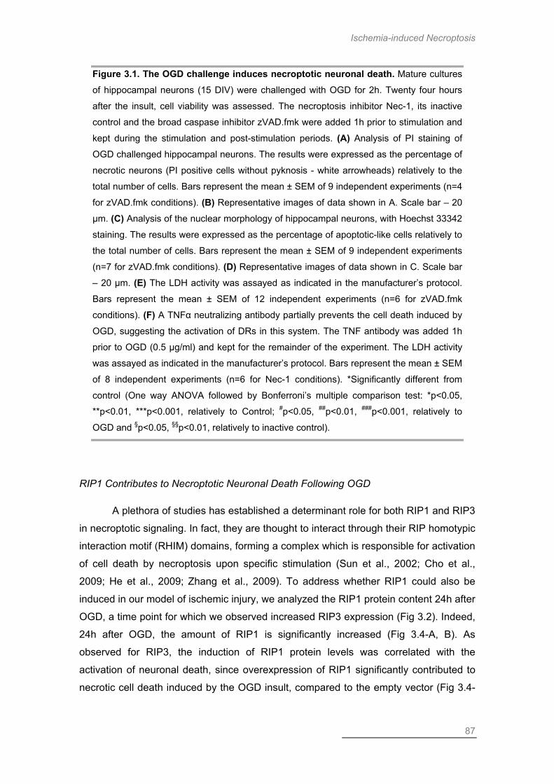
Ischemia-induced Necroptosis
87
Figure 3.1. The OGD challenge induces necroptotic neuronal death. Mature cultures
of hippocampal neurons (15 DIV) were challenged with OGD for 2h. Twenty four hours
after the insult, cell viability was assessed. The necroptosis inhibitor Nec-1, its inactive
control and the broad caspase inhibitor zVAD.fmk were added 1h prior to stimulation and
kept during the stimulation and post-stimulation periods. (A) Analysis of PI staining of
OGD challenged hippocampal neurons. The results were expressed as the percentage of
necrotic neurons (PI positive cells without pyknosis - white arrowheads) relatively to the
total number of cells. Bars represent the mean ± SEM of 9 independent experiments (n=4
for zVAD.fmk conditions). (B) Representative images of data shown in A. Scale bar – 20
µm. (C) Analysis of the nuclear morphology of hippocampal neurons, with Hoechst 33342
staining. The results were expressed as the percentage of apoptotic-like cells relatively to
the total number of cells. Bars represent the mean ± SEM of 9 independent experiments
(n=7 for zVAD.fmk conditions). (D) Representative images of data shown in C. Scale bar
– 20 µm. (E) The LDH activity was assayed as indicated in the manufacturer’s protocol.
Bars represent the mean ± SEM of 12 independent experiments (n=6 for zVAD.fmk
conditions). (F) A TNFα neutralizing antibody partially prevents the cell death induced by
OGD, suggesting the activation of DRs in this system. The TNF antibody was added 1h
prior to OGD (0.5 µg/ml) and kept for the remainder of the experiment. The LDH activity
was assayed as indicated in the manufacturer’s protocol. Bars represent the mean ± SEM
of 8 independent experiments (n=6 for Nec-1 conditions). *Significantly different from
control (One way ANOVA followed by Bonferroni’s multiple comparison test: *p<0.05,
**p<0.01, ***p<0.001, relatively to Control; #p<0.05, ##p<0.01, ###p<0.001, relatively to
OGD and §p<0.05, §§p<0.01, relatively to inactive control).
RIP1 Contributes to Necroptotic Neuronal Death Following OGD
A plethora of studies has established a determinant role for both RIP1 and RIP3
in necroptotic signaling. In fact, they are thought to interact through their RIP homotypic
interaction motif (RHIM) domains, forming a complex which is responsible for activation
of cell death by necroptosis upon specific stimulation (Sun et al., 2002; Cho et al.,
2009; He et al., 2009; Zhang et al., 2009). To address whether RIP1 could also be
induced in our model of ischemic injury, we analyzed the RIP1 protein content 24h after
OGD, a time point for which we observed increased RIP3 expression (Fig 3.2). Indeed,
24h after OGD, the amount of RIP1 is significantly increased (Fig 3.4-A, B). As
observed for RIP3, the induction of RIP1 protein levels was correlated with the
activation of neuronal death, since overexpression of RIP1 significantly contributed to
necrotic cell death induced by the OGD insult, compared to the empty vector (Fig 3.4-
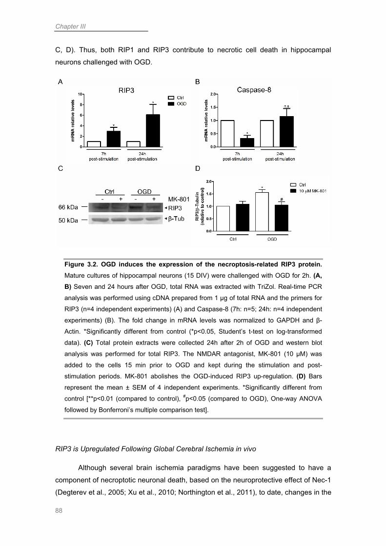
Chapter III
88
C, D). Thus, both RIP1 and RIP3 contribute to necrotic cell death in hippocampal
neurons challenged with OGD.
Figure 3.2. OGD induces the expression of the necroptosis-related RIP3 protein. Mature cultures of hippocampal neurons (15 DIV) were challenged with OGD for 2h. (A, B) Seven and 24 hours after OGD, total RNA was extracted with TriZol. Real-time PCR
analysis was performed using cDNA prepared from 1 µg of total RNA and the primers for
RIP3 (n=4 independent experiments) (A) and Caspase-8 (7h: n=5; 24h: n=4 independent
experiments) (B). The fold change in mRNA levels was normalized to GAPDH and β-
Actin. *Significantly different from control (*p<0.05, Student’s t-test on log-transformed
data). (C) Total protein extracts were collected 24h after 2h of OGD and western blot
analysis was performed for total RIP3. The NMDAR antagonist, MK-801 (10 µM) was
added to the cells 15 min prior to OGD and kept during the stimulation and post-
stimulation periods. MK-801 abolishes the OGD-induced RIP3 up-regulation. (D) Bars
represent the mean ± SEM of 4 independent experiments. *Significantly different from
control [**p<0.01 (compared to control), #p<0.05 (compared to OGD), One-way ANOVA
followed by Bonferroni’s multiple comparison test].
RIP3 is Upregulated Following Global Cerebral Ischemia in vivo
Although several brain ischemia paradigms have been suggested to have a
component of necroptotic neuronal death, based on the neuroprotective effect of Nec-1
(Degterev et al., 2005; Xu et al., 2010; Northington et al., 2011), to date, changes in the

Ischemia-induced Necroptosis
89
protein levels of the necrosome components have not been reported. To investigate
whether the changes in RIP1 and RIP3 observed in our in vitro model of ischemic injury
reflect modifications that might occur in vivo, we submitted adult rats to transient global
cerebral ischemia (Fig 3.5). We observed that RIP3 is indeed upregulated 48h after
global ischemia, specifically in the CA1 region of the hippocampus, while it remained
constant in the CA3 and DG areas, and in the cerebral cortex (Fig 3.5-A, B). RIP1, on
the other hand, displayed a trend to a slight increase in the CA1, and DG areas of the
hippocampus, although without statistical significance (Fig 3.5-A, C). We also observed
that the upregulation of RIP3 in the CA1 area correlated with increased formation of α-
Spectrin break-down products (SBDPs) of 145 and 150 kDa, demonstrating the
activation of calpains, which are proteases involved in cell death, namely in neuronal
death induced by ischemic insults (Siman et al., 1989; Vosler et al., 2011). These
results suggest that global ischemia induces the upregulation of necrosome
components in CA1 hippocampal neurons, which is probably related to the activation of
necroptotic neuronal death in this paradigm.
Discussion
Recently, necroptosis has been extensively studied as a novel type of
programmed cell death and it has been implicated in a wide range of pathologies,
including cerebral ischemia. Using an in vitro model of global cerebral ischemia, OGD,
we showed that this challenge induces necroptotic neuronal death. Interestingly, we
found that OGD upregulated both RIP1 and RIP3 protein levels, which we found to
occur also in vivo, following a transient global cerebral ischemic insult, at least for the
RIP3 protein. Furthermore, we were able to correlate the upregulation of endogenous
RIP3 with an increase in necrosis of hippocampal neurons following OGD.
Apoptosis is widely accepted as one of the major forms of programmed cell
death, requiring the sequential activation of biochemical reactions, namely those
leading to the formation of the apoptosome and the activation of caspases, as a result
of the release of proapoptotic proteins from the mitochondria (reviewed in Taylor et al.,
2008; Andersen and Kornbluth, 2013). On the contrary, necrosis is generally thought of
as an “accidental” type of cell death that occurs when the cell is submitted to an
overwhelming stress that afterwards leads to the activation of an inflammatory
response (Kanduc et al., 2002). This view of the necrotic cell death as a systematic

Chapter III
90
unregulated event has been challenged by studies showing that apoptotic inhibitors
(such as broad caspase inhibitors like zVAD.fmk) do not always protect but instead
they may sensitize the cells to a mechanism of programmed necrosis (Vercammen et
al., 1998; Holler et al., 2000). Additionally, it was found that in caspase-8-/- mice, which
die around E10.5, cells are sensitized to necrotic cell death, in response to DR activity
(Oberst et al., 2011), uncovering a role of caspase-8 as a negative regulator of
programmed necrosis or necroptosis. The role of DR signaling in activation of
programmed cell death mechanisms, including necroptosis, has been extensively
studied (reviewed in Dickens et al., 2012).
Figure 3.3. The levels of the RIP3 protein influence the extent of OGD-induced neuronal death. (A-B) Primary cultures of rat hippocampal neurons were co-transfected
at DIV 7 with the indicated constructs (3 µg) plus a GFP plasmid (1.5 µg), using the
calcium phosphate protocol. At DIV 15 cells were challenged with OGD for 2h. Twenty
four hours after stimulation cells were stained with the nuclear dyes Hoechst 33342 and
PI and the number of PI positive transfected cells was counted. The results were
expressed as the percentage of PI positive cells relatively to the total number of
Ischemia-induced Necroptosis
91
transfected cells. (A) Overexpression of RIP3 significantly enhances OGD toxicity in
hippocampal neurons. (B) Representative images of data shown in A. Scale bar – 20 µm;
Insert – 10 µm. (C-E) Primary cultures of mouse hippocampal neurons were co-
transfected at DIV 7 with the indicated constructs (1.5 µg) plus a GFP plasmid (0.75 µg)
or hRIP3 (1.5 µg), using the calcium phosphate protocol. At DIV 15 cells were challenged
with OGD for 2h. Twenty four hours after stimulation cells were stained as in (A) and the
number of PI positive transfected cells was counted. The results were expressed as the
percentage of PI positive cells relatively to the total number of transfected cells. (C) Knock-down of RIP3 significantly protects neurons against OGD-induced death (n=5). (D) In OGD-challenged neurons, the neuroprotective effect of RIP3 KD is reversed by
expression of hRIP3, which is not recognized by the mRIP3 shRNA. (E) Representative
images of data shown in C and D. Scale bar – 20 µm; Insert – 10 µm. Bars represent the
mean ± SEM of the indicated number of independent experiments. *Significantly different
from control [One way ANOVA followed by Bonferronni’s multiple comparison test:
*p<0.05, ***p<0.001 relative to control; #p<0.05, ##p<0.01, ###p<0.001 relative to OGD
condition (A) or OGD-LacZ (C and D)].
In an effort to elucidate the involvement of necroptosis in pathologies with broad
relevance, Nec-1 was identified as a specific inhibitor of necroptosis (Degterev et al.,
2005). More recently, Nec-1 was found to target the RIP1 kinase activity (Degterev et
al., 2008). This inhibitor has been shown to be protective against a variety of cellular
stresses, both in neurons (Li et al., 2008; Wang et al., 2012) as well as in non-neuronal
cells (Upton et al., 2010; Narayan et al., 2012; Simenc and Lipnik-Stangelj, 2012).
Moreover, this inhibitor of necroptosis was shown to be protective against focal
ischemic injury in vivo (Degterev et al., 2005; Northington et al., 2011; Chavez-Valdez
et al., 2012) and OGD in vitro (Xu et al., 2010; Meloni et al., 2011). Accordingly, we
also observed a component of neuronal death that was inhibited by Nec-1 in our in vitro
model of global cerebral ischemia. Additionally, we demonstrated that Nec-1 protected
neurons from OGD-induced necrosis, without affecting the component of apoptotic-like
cell death. Furthermore, the broad caspase inhibitor zVAD.fmk was only effective in
reducing apoptotic-like neuronal death, whilst having no protective effect against
necrotic neuronal death evaluated with PI staining. In this case, zVAD.fmk actually
exacerbated the neuronal death induced by OGD, which is in accordance with previous
reports showing that inhibition of caspases favors necroptotic cell death (Vercammen
et al., 1998). We also observed that TNFα signaling contributes to hippocampal
neuronal death induced by OGD, probably contributing to the activation of the extrinsic
pathway of apoptosis as well as necroptosis. We were not able to detect a significant
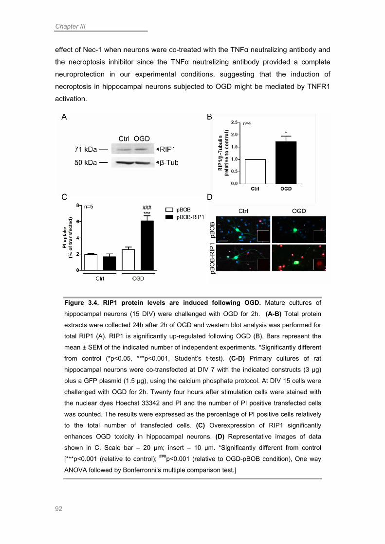
Chapter III
92
effect of Nec-1 when neurons were co-treated with the TNFα neutralizing antibody and
the necroptosis inhibitor since the TNFα neutralizing antibody provided a complete
neuroprotection in our experimental conditions, suggesting that the induction of
necroptosis in hippocampal neurons subjected to OGD might be mediated by TNFR1
activation.
Figure 3.4. RIP1 protein levels are induced following OGD. Mature cultures of
hippocampal neurons (15 DIV) were challenged with OGD for 2h. (A-B) Total protein
extracts were collected 24h after 2h of OGD and western blot analysis was performed for
total RIP1 (A). RIP1 is significantly up-regulated following OGD (B). Bars represent the
mean ± SEM of the indicated number of independent experiments. *Significantly different
from control (*p<0.05, ***p<0.001, Student’s t-test). (C-D) Primary cultures of rat
hippocampal neurons were co-transfected at DIV 7 with the indicated constructs (3 µg)
plus a GFP plasmid (1.5 µg), using the calcium phosphate protocol. At DIV 15 cells were
challenged with OGD for 2h. Twenty four hours after stimulation cells were stained with
the nuclear dyes Hoechst 33342 and PI and the number of PI positive transfected cells
was counted. The results were expressed as the percentage of PI positive cells relatively
to the total number of transfected cells. (C) Overexpression of RIP1 significantly
enhances OGD toxicity in hippocampal neurons. (D) Representative images of data
shown in C. Scale bar – 20 µm; insert – 10 µm. *Significantly different from control
[***p<0.001 (relative to control); ###p<0.001 (relative to OGD-pBOB condition), One way
ANOVA followed by Bonferronni’s multiple comparison test.]

Ischemia-induced Necroptosis
93
The identification of a necroptotic cell death program in a subpopulation of
hippocampal neurons submitted to OGD prompted us to investigate the mechanisms
that might regulate necroptosis upon an ischemic stimulus, since the events underlying
neuronal necroptotic death have not been elucidated so far. In non-neuronal systems,
the interaction between the related proteins RIP1 and RIP3 as well as their kinase
activity has been shown to be crucial for necroptotic cell death (Cho et al., 2009; He et
al., 2009; Zhang et al., 2009; Li et al., 2012), while caspase-8 may act as a negative
regulator possibly by promoting the cleavage of both RIP1 and RIP3 (Feng et al., 2007;
Cho et al., 2009; Oberst et al., 2011). Our results indicate that in a subset of neurons
OGD induces a transient down-regulation of caspase-8 mRNA while increasing RIP3
mRNA levels. The transcriptional changes induced by OGD are paralleled by an
increase in the endogenous RIP3 level. The endogenous RIP1 protein level is
increased as well, suggesting these events may underlie the formation of the
necrosome upon a toxic stimulus. Furthermore, we identified NMDAR signaling as an
important regulator of RIP3 levels, which may explain the occurrence of necroptosis
following NMDAR activation (Li et al., 2008). Importantly, we also observed increased
levels of RIP3 in the CA1 region of the hippocampus of rats submitted to transient
global cerebral ischemia in vivo. This region of the hippocampus is considered to be
selectively vulnerable to this type of insult (Kirino, 1982; Zukin et al., 2004) and our
results suggest that necroptosis contributes to the neurodegeneration that ensues in
that area upon global cerebral ischemia. Moreover, our results demonstrate the
relevance of the induction of endogenous RIP3 protein to the toxicity elicited by global
cerebral ischemia, both in vitro and in vivo. This is, to our knowledge, the first report
describing increased RIP3 expression following cerebral ischemic paradigms.
To more accurately establish the role of the RIP3 and RIP1 proteins in the
induction of necroptosis in hippocampal neurons submitted to OGD, we overexpressed
both proteins in this system. Overexpression of either RIP1 or RIP3 resulted in the
exacerbation of OGD-induced neuronal death. Importantly, we observed that the
increase in the extent of cell death in transfected neurons occurred specifically upon
OGD challenge since control neurons overexpressing RIP3 or RIP1 did not present
significant changes in cell viability. This implies that regulated necrosis requires specific
stimuli to be activated and that overexpression of RIP3 or RIP1 by themselves does not
induce cell death. This is in accordance with a previous report, that demonstrated that
overexpression of RIP3 and RIP1 specifically induces cell death only upon necroptotic
stimulation (Zhang et al., 2009).

Chapter III
94
The determinant role of RIP3 in necroptosis has been demonstrated by
complete abrogation of necrotic cell death in RIP3-depleted cells (Cho et al., 2009; He
et al., 2009; Zhang et al., 2009). Similarly, we observed that RIP3 KD in hippocampal
neurons abolished necrotic death induced by OGD (Fig 3). Importantly, we observed
that by reintroducing RIP3 in KD neurons we rescued the necrotic component of cell
death elicited by OGD. These results highlight the crucial impact of RIP3 expression
levels on OGD-induced necrosis and support evidence showing an increase in necrotic
cell death due to the expression of RIP3 in cells normally resistant to necroptotic stimuli
(Zhang et al., 2009).

Ischemia-induced Necroptosis
95
Figure 3.5. Global cerebral ischemia induces expression of the RIP3 protein. Adult
rats were challenged with in vivo transient global cerebral ischemia (GCI) for 15 min.
Forty-eight hours after GCI, the rats were sacrificed and the hippocampus and cortex Ctx
were dissected. The hippocampus was further dissected to separate the CA1, CA3 and
DG regions. (A) Total protein extracts were collected and western blot analysis was
performed for RIP3, RIP1, SBDP (to demonstrate activation of cell death pathways), and
β-tubulin. (B) GCI induced an upregulation of RIP3 in the CA1 region of the
hippocampus. Bars represent the mean ± SEM of 3 independent experiments (n=4 for the
Ctx). *Significantly different from control (*p<0.05, Student’s t-test on log-transformed
data). (C) The RIP1 protein was not changed following GCI. Bars represent the mean ±
SEM of 3 independent experiments (n=2 for the DG).
Overall, our results support the view that ischemic insults induce a plethora of
cell death mechanisms, including apoptosis and programmed necrosis (Meloni et al.,
2011). The understanding of the diverse death mechanisms that occur in ischemia may
prove beneficial to the identification of novel molecular targets relevant to the
development of new treatments for this pathology. Additionally, our results demonstrate
the induction of both RIP1 and RIP3 proteins following OGD, as well as of RIP3 mRNA.
The relevance of this upregulation of RIP3 is further demonstrated by an increase in
RIP3 protein levels in the CA1 region of the hippocampus, following an in vivo global
ischemic insult. Further studies concerning the mechanisms of necroptosis in neurons,
namely the identification of putative RIP1/3 substrates, possibly neuron-specific, may
provide important insights to clarify the mechanisms of cell death underlying global
ischemic insults.

Chapter III
96

Chapter IV
Activation of MAPK Signaling Following in vitro Ischemic Insults

Chapter IV
98

MAPKs & Neurodegeneration
99
Abstract
Global cerebral ischemia, that results from a lack of blood supply to the entire
brain, is characterized by selective neurodegeneration of specific subsets of neurons,
namely in the hippocampus. This type of insult can be mimicked in vitro by combined
oxygen and glucose deprivation (OGD), in primary neuronal cultures or slices. Mitogen-
activated protein kinases (MAPKs) have been widely studied for their role in the
response to cellular stress and were found to be involved in a variety of cell death
paradigms, namely in neurons. In this work we addressed whether OGD induces the
activation of the MAPKs c-Jun N-terminal kinase (JNK) and p38. We observed that
indeed, we were able to detect increased activation of both kinases, at distinct time
points after OGD. We observed increased p38 phosphorylation at 2h of recovery and
higher levels of the classical JNK substrate c-Jun at 6h post-stimulation. Additionally,
we also observed altered mRNA levels of the JNK and p38 pathways substrates Madd,
Atf3 and Gadd45a and upstream kinases Ask2 and Tpl2. To confirm their role in
neuronal death induced by OGD, we used the p38 inhibitor, SB203580 and the JNK
cascade inhibitors, SP600125 and CEP11004 and observed that all these compounds
conferred neuroprotection against OGD. Interestingly, we observed that the JNK
signaling inhibitors were only neuroprotective when applied 4h after the end of the
OGD challenge but not when present throughout the stimulation and post-stimulation
periods. This effect suggests that JNK may have a dual role in the regulation of
neuronal fate under OGD conditions. Overall our results highlight the role of MAPKs in
the context of cerebral ischemia. Inhibition of these kinases may provide important
benefits for the treatment of this type of insult.
Introduction
Cerebral ischemia is one of the most prevalent neuropathologies worldwide
(Flynn et al., 2008). Transient global ischemia results from a lack of blood supply to the
entire brain, commonly due to a cardiac arrest. This blood deprivation induces the
selective death of specific subsets of neurons in the brain, of which the CA1 region of
the hippocampus is one of the most prominently affected (Zukin et al., 2004). This type
of insult induces delayed neuronal death that occurs 24-72h following the insult, for
which there are to date, no effective treatments. Currently, research in this area aims at
reducing the amount of neurodegeneration that ensues as a consequence of ischemic

Chapter IV
100
insults. Thus, there is great interest on the understanding of the toxic intracellular
signaling pathways that are activated in these conditions, in order to identify new
molecular targets useful for therapy.
The mitogen-activated protein kinases (MAPKs) have a variety of functions in
cells, including neurons, namely a crucial impact on the life and death balance. These
signaling pathways have a typical three-tiered cascade, comprising a MAPK kinase
kinase (MAPKKK), a MAPK kinase (MAPKK) and a MAPK. Each sequential kinase is
activated by phosphorylation and they exert their role in the cell by phosphorylating a
variety of substrates, depending on the cellular context. There are three groups of
mammalian MAPKs - extracellular signal-regulated kinase (ERK), p38 and c-Jun N-
terminal kinase (JNK) – and they have been all correlated to the response to cellular
stress (Qi and Elion, 2005). However, while ERK1/2 is commonly associated with cell
survival (Liu et al., 1998; Liu et al., 2013), the MAPKs p38 and JNK are thought to
contribute to cell death, namely in neurons (Ham et al., 1995; Yang et al., 1997;
Borsello et al., 2003; Cao et al., 2004; Semenova et al., 2007; Lee et al., 2013). In
particular, both kinases have been correlated with neurodegeneration induced by
ischemic insults, both in vitro (Behrens et al., 1999; Hirt et al., 2004) and in vivo (Ferrer
et al., 1997; Sugino et al., 2000; Borsello et al., 2003; Kuan et al., 2003; Hirt et al.,
2004; Okuno et al., 2004). One of the preferential JNK substrates is the transcription
factor c-Jun (Gupta et al., 1996). This transcription factor homodimerizes or
heterodimerizes with other members of the Jun, activating transcription factor (ATF)
and Fos families, assembling activator protein-1 (AP-1) transcription factor complexes,
which mediate the transcription-dependent toxic effects of JNK activation (Ham et al.,
1995; Davis, 2000; Hess et al., 2004; Silva et al., 2005; Santos et al., 2006; Burke,
2007).
In this work, we aimed to study the activation of p38 and JNK in hippocampal
neurons submitted to oxygen and glucose deprivation (OGD), an in vitro model of
cerebral ischemia. We observed that both pathways are activated following OGD, but
at different time points. Interestingly, our results suggest that JNK may be activated in a
biphasic manner, since we observed that we were only able to confer neuroprotection
with JNK inhibitors when the compounds were added at 4h of reoxygenation, but no
neuroprotection was observed when the compounds were present during the
stimulation and post-stimulation periods. This suggests that prior to a toxic activation
that we detected at 6h of recovery, JNK may be involved in a prosurvival mechanism.
This hypothesis supports previous observations concerning JNK activation mediated by
tumor necrosis factor receptor 1 (TNFR1) signaling (Ventura et al., 2006) or induced by

MAPKs & Neurodegeneration
101
cerebral ischemia injury (Hirt et al., 2004) and excitotoxic signaling (Vieira et al., 2010).
Moreover, we show that this biphasic activation mediates differential effects on the
neuronal fate following OGD.
Materials & Methods
Materials
The antibodies for phospho-c-Jun (Ser63) (#9261) and total p38 (#9212) were
purchased from Cell Signaling Technology (Boston, MA). The antibodies for c-Jun/AP-
1 (sc-45-G) and p-p38 (Thr 180/Tyr 182-R) (sc-17852-R) were purchased from Santa
Cruz Biotechnology (Santa Cruz, CA).
The JNK pathway inhibitor CEP11004 was a generous gift from from Cephalon,
Inc. (West Chester, PA) and SP600125 was purchased from Sigma-Aldrich (St. Louis,
MO). The p38 inhibitor SB203580 was purchased from Calbiochem (Merck-Millipore,
Billerica, MA).
Cell Culture
Primary cultures of hippocampal neurons were prepared from embryonic day 18
(E18) rats, as previously described (Santos and Duarte, 2008). Briefly, hippocampi
were dissected and dissociated with trypsin (1.5 mg/ml) in Hank’s Balanced Salt
Solution (HBSS). Neurons were cultured in multiwell (MW) plates with or without glass
coverslips, coated with poly-D-lysine (PDL), at a density of 85,000 cells/cm2, in
Neurobasal® medium (Gibco®, Life Technologies™, Paisley, UK) supplemented with
2% NeuroCult™ SM1 Neuronal Supplement (StemCell™ Technologies, Grenoble,
France), 0.5 mM glutamine, 0.125 µg/ml gentamicin and 25 µM glutamate. Neuronal
cultures were maintained at 37ºC in a humidified incubator with 5% CO2 up to 15 days
in vitro (DIV). At DIV 7, the cultures were fed with fresh supplemented neurobasal
medium without glutamate.
Chapter IV
102
Oxygen-Glucose Deprivation (OGD) Challenge
At DIV 15, hippocampal neurons were challenged with an in vitro ischemic stimulus.
For that purpose, neurons were placed in a glucose-free deoxygenated buffer medium
(OGD medium – 10 mM HEPES, 116 mM NaCl, 5.4 mM KCl, 0.8 mM NaH2PO4, 25
mM sodium bicarbonate, 25 mM sucrose, 1.8 mM CaCl2, 0.04% phenol red, pH 7.3),
inside an anaerobic chamber with 5% CO2 (OGD chamber, Thermo Forma 1029,
Thermo Fisher Scientific, Waltham, MA), at 37ºC for 2h. Control neurons were placed
in a similar buffer, containing 25 mM glucose, instead of sucrose (sham medium), and
kept for 2h in an oxygenated incubator, at 37ºC. After incubation, the neurons were
placed in their conditioned medium and returned to the normoxic incubator for a period
of recovery that lasted up to 24h.
When indicated, JNK or p38 inhibitors were added to the cell medium.
SB203580 was pre-incubated for 1h and then kept during the stimulation and post-
stimulation periods. For inhibition of the JNK signaling pathway, CEP11004 and
SP600125 were added 1h prior to stimulation and then kept during the stimulation and
post-stimulation periods or, alternatively, the inhibitors were added 4h after the OGD
insult and kept for the remainder of the post-stimulation period.
RNA extraction
Total RNA from DIV 15 hippocampal neurons was extracted with TRIzol®
Reagent (Invitrogen, Madrid, Spain), following the manufacturers’ specifications. The
total amount of RNA was quantified spectrophotometrically (Thermo Nanodrop 2000,
Thermo Fisher Scientific, Wilmington, DE) and the RNA quality and integrity evaluated
using the Experion automated gel electrophoresis system (BioRad, Hercules, CA).
qPCR
For cDNA synthesis, 1 µg of total RNA was used with the iScript™ cDNA
Synthesis Kit (BioRad), according to the manufacturers’ instructions. For quantitative
real-time PCR (qRT-PCR) 20 µl reactions were prepared with 2 µl of 1:10 diluted
cDNA, 10 µl of 2x iQ™ SYBR® Green Supermix (BioRad) and specific primers at 250
nM. Primer sequences are indicated in Table II.

MAPKs & Neurodegeneration
103
Table II. Primers used for qPCR analysis.
The fluorescent signal was measured after each elongation step of the PCR
reaction, in the iQ5 Multicolor Real-Time PCR Detection System (BioRad), and was
used to determine the threshold cycle (Ct), as previously described (Manadas et al.,
2009). Melting curves were performed in order to detect non-specific amplification
products, a non-template control was included in all assays, and for each set of primers
a standard curve was performed to assess primer efficiency. Reactions were run in
duplicate. The level of expression of each gene was analyzed at 7 and 24h of recovery
following OGD and normalized to the level of expression of the gene in the
corresponding control conditions. All Ct values were normalized to two internal control
genes, ActB and Gapdh, using the GenEx software (MultiD Analyses).
Nuclear morphology
Neuronal death was assessed by analysis of the nuclear morphology 24h
following the OGD insult. For that purpose, neurons were stained with the nuclear dye
Hoechst 33342 (2 µg/ml) for 15 min at RT and then mounted in fluorescent mounting
medium. Neurons were analyzed under fluorescence microscopy for chromatin
condensation and nuclear pyknosis, a feature of apoptotic-like cells. The results were
expressed as the percentage of apoptotic-like cell death (apoptotic-like nuclei/total
number of cells). At least 200 cells were counted in each coverslip.
Lactate Dehydrogenase (LDH) release assay
At 24h of recovery after the OGD challenge, the LDH release to the extracellular
medium was evaluated by a colorimetric assay using the CytoTox96 Non-Radioactive
assay kit (Promega, Madison, WI), according to the manufacturer’s instructions. The
Chapter IV
104
percentage of LDH release was determined as the ratio between LDH activity in the
extracellular medium and total LDH activity obtained after complete cell lysis with Triton
X-100. All experiments were carried out in duplicate or triplicate, for each independent
experiment, as previously described (Vieira et al., 2010).
Protein Extracts, SDS-PAGE and Western Blotting
Twenty four hours after OGD, cells were washed with ice-cold phosphate-
buffered saline (PBS) and lysis buffer [50 mM HEPES, 150 mM NaCl, 2 mM EGTA, 2
mM EDTA, 2 mM Na3VO4, 50 mM NaF, 1% Triton X-100, 10% glycerol, pH 7.4
supplemented with 1 mM phenylmethanesulphonylfluoride (PMSF) and 1 µg/ml of
CLAP (cocktail containing chymostatin, leupeptin, antipain and pepstatin)] was added.
Cell extracts were then sonicated and centrifuged for 20 min at 12000 x g, at 4ºC.
Supernatants were recovered and the protein quantified by the BCA method. Samples
were denatured at 95ºC, for 10 min and 80 µg of each protein extract were separated
by SDS-PAGE, using 10% polyacrilamide gels, and then transferred to a polyvinylidene
fluoride (PVDF) membrane. The membranes were blocked with Tris-buffered saline-
Tween20 (TBS-T) (20 mM Tris, 137 mM NaCl, pH 7.6, 0.1 % Tween20) with 5% non-
fat milk, for 1h at room temperature, and then incubated with the primary antibody in
TBS-T 5% milk, for 1h at room temperature. Alternatively, the incubation with the
primary antibody was performed overnight at 4°C. After extensive washing,
membranes were incubated with the secondary antibody conjugated with alkaline
phosphatase for 1h at room temperature. After additional washes, the membranes
were developed using the enhanced chemifluorescence (ECF) substrate, and scanned
on the Storm 860 Gel and Blot Imaging System (Amersham Biosciences,
Buckinghamshire, UK). The density of the bands was analyzed with ImageQuant 5.0
software. For subsequent reprobing, the membranes were stripped of antibody with
NaOH 0.2 M, blocked again and incubated with the appropriate antibodies.
Statistical Analysis
Results are presented as means ± S.E.M. of the indicated number of
experiments. Statistical significance was assessed by Student’s t-test or by one-way
variance analysis (ANOVA) followed by the Dunnett’s test or the Bonferroni’s test.
These statistical analyses were performed using the software package GraphPad
Prism 5.
MAPKs & Neurodegeneration
105
Results
OGD Induces Up-regulation of the JNK signaling Pathway
The MAPK JNK has been shown to have a prominent role in the activation of
cell death upon several stress insults (Eilers et al., 1998; Tournier et al., 2000; Cao et
al., 2004; Hunot et al., 2004; Kukekov et al., 2006; Ventura et al., 2006; Wang et al.,
2013), including excitotoxicity (Yang et al., 1997; Borsello et al., 2003; Centeno et al.,
2007; Vieira et al., 2010) and cerebral ischemia (Ferrer et al., 1997; Kuan et al., 2003;
Hirt et al., 2004; Okuno et al., 2004; Gao et al., 2005). Thus, we addressed whether our
in vitro model of global ischemia, OGD, could induce the activation of the JNK signaling
pathway. To address this question, we looked at changes on c-Jun phosphorylation, a
classical JNK substrate, which is considered to be a measure of altered JNK activity.
We tested different times of OGD and recovery and observed that c-Jun
phosphorylation appeared to be up-regulated following 6h of reoxygenation after OGD
(Fig 4.1-A). Since this was the only time point for which we observed an apparent
increase in c-Jun phosphorylation we quantified the blots for 6h of recovery and
confirmed that c-Jun was significantly up-regulated after 6h of recovery following 2h of
OGD to 1.29 ± 0.09 times of the control (Fig 4.1-B). The phosphorylation of c-Jun is
known to up-regulate its transcriptional activity, as c-Jun complexes with other
members of the Jun and Fos families to form the AP-1 transcription factor (Bohmann et
al., 1987; Santos et al., 2006), and to regulate transcription of genes related to cell
death (Herdegen and Waetzig, 2001; Hess et al., 2004). To confirm that the increase in
p-c-Jun observed following OGD correlates with an increased transcriptional activity of
this protein, we analyzed changes in the mRNA level of some of the genes it may
regulate, namely the Atf3, which is known to be induced by c-Jun/AP-1 (Liang et al.,
1996; Mei et al., 2008) (Fig 4.1-C), the growth arrest and DNA damage 45a (Gadd45a,
Fig 4.1-D), also known to be up-regulated by JNK activity (Yin et al., 2004), and the
MAP-kinase activating death domain (Madd, Fig 4.1-E), which is down-regulated upon
activation of JNK (Del Villar and Miller, 2004). As observed by qRT-PCR analysis, both
the Atf3 and Gadd45a genes had a higher expression 7h and 24h after OGD,
respectively, while Madd expression was decreased at both time points. These results
suggest that c-Jun might be indeed activating gene transcription following OGD.
Since JNK activity may be initiated by a variety of MAPKKKs, depending on the
specific stimulus (Davis, 2000), we also analyzed the changes induced by OGD in the
expression of a few MAPKKKs, by qRT-PCR. We observed that both apoptosis signal-
regulating kinase 2 (Ask2) (Fig 4.2-A) and tumor progression locus 2 (Tpl2) (Fig 4.2-B)

Chapter IV
106
had a higher expression, especially at 24h after OGD. On the contrary, we did not
observe changes in the mRNA levels of TAO kinase 3 (Taok3) (Fig 4.2-C). These
results suggest that ASK2 and TPL2 may be involved in hippocampal neuronal death
induced by OGD.
Figure 4.1. OGD induces changes in JNK pathway targets. (A) Mature cultures of
hippocampal neurons (15 DIV) were challenged with OGD for different periods. At the
end of the insult, protein extracts were immediately prepared or, alternatively, The
cultures were transferred to the conditioned medium and returned to the air/CO2
incubator for a period of reoxygenation that lasted up to 24h and total protein extracts
were prepared. Eighty µg of total protein were used for immunoblot analysis with an anti-
phospho-c-Jun antibody. The membranes were reprobed with an anti-total c-Jun
antibody. The figure is representative of 2 (30 min OGD and 2h OGD + 24h recovery), 1
(1h OGD, 2h OGD and 2h OGD + 2h of recovery) or 5 (2h OGD + 6h recovery) assays
performed with extracts from independent experiments. (B) Quantification of the ratio
between phospho-c-Jun and total levels of c-Jun at 6h of recovery, normalized to the
control. Bars represent the mean ± SEM of the indicated number of experiments
(*p<0,05, Student’s t test). (C-E) Seven and 24 hours after OGD, total RNA was extracted

MAPKs & Neurodegeneration
107
with TriZol. Real-time PCR analysis was performed using cDNA prepared from 1 µg of
total RNA and the primers for ATF3 (C), GADD45a (D) and MADD (E). Fold change in
mRNA levels was normalized to GAPDH and β-Actin. Bars represent the mean ± SEM of
5 independent experiments. *Significantly different from control (*p<0.05, Student’s t-test
on log-transformed data).
JNK Inhibition Protects Against Neuronal Death
To confirm that the increase of JNK activity in our system is indeed correlated
with neuronal death, we used two JNK cascade inhibitors – CEP11004 [a Mixed-
lineage kinase (MLK) inhibitor] and SP600125 (a specific JNK inhibitor). Surprisingly,
we observed that if we inhibited JNK signaling throughout the entire experiment
(including both the OGD insult as well as the 24h of recovery), we could not detect any
neuroprotection against the OGD challenge. However, if we added the compounds to
the cells 4h after the end of OGD, we were able to significantly reverse the majority of
cell death elicited by this challenge [from about 39.7 ± 4.7% to 18.2 ± 1.9% of cell
death for CEP1104 and from about 38.6 ± 5.1% to 21.6 ± 2.5% of cell death for
SP600125 (Fig 4.3)]. These results suggest that OGD may elicit two distinct phases of
JNK activity, with different contributions to hippocampal neurons cell fate. This is in line
with our previous work (Vieira et al., 2010), where we reported a biphasic JNK
activation induced by excitotoxicity, a deleterious phenomenon also present in cerebral
ischemia. We propose that the initial activation of JNK (prior to 4h of reoxygenation)
may have a prosurvival effect, while the second phase of JNK activation results in
increased c-Jun phosphorylation within 6h of recovery and contributes to neuronal
death. Additionally, the neuroprotective effect afforded by CEP11004 suggests that
MLK3 may be one of the MAPKKKs to induce JNK activation in this context.~
OGD Elicits Activation of the p38 MAPK
Another MAPK, p38, has been extensively studied for its role in induction of
neuronal death following ischemia (Behrens et al., 1999; Sugino et al., 2000; Li et al.,
2013). Thus, we addressed the question of whether this pathway is also activated in
hippocampal neurons following OGD. We tested several times of OGD and recovery
and observed a significant increase in p-p38 levels following OGD, to about 1.4 times
of the control (Fig 4.4-A, B), at 2h of recovery following 2h of OGD. We did not detect
increased c-Jun phosphorylation at this time point (Fig 4.1-A). We were also able to
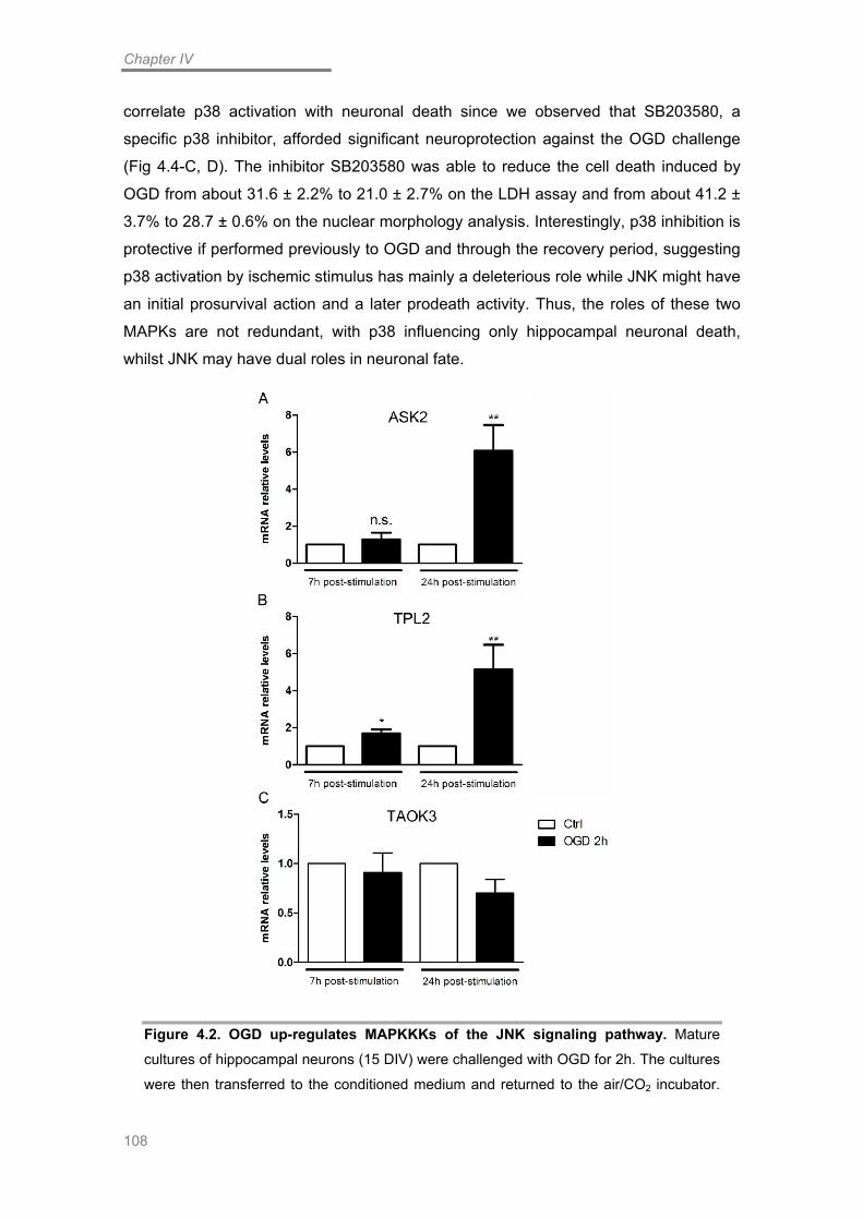
Chapter IV
108
correlate p38 activation with neuronal death since we observed that SB203580, a
specific p38 inhibitor, afforded significant neuroprotection against the OGD challenge
(Fig 4.4-C, D). The inhibitor SB203580 was able to reduce the cell death induced by
OGD from about 31.6 ± 2.2% to 21.0 ± 2.7% on the LDH assay and from about 41.2 ±
3.7% to 28.7 ± 0.6% on the nuclear morphology analysis. Interestingly, p38 inhibition is
protective if performed previously to OGD and through the recovery period, suggesting
p38 activation by ischemic stimulus has mainly a deleterious role while JNK might have
an initial prosurvival action and a later prodeath activity. Thus, the roles of these two
MAPKs are not redundant, with p38 influencing only hippocampal neuronal death,
whilst JNK may have dual roles in neuronal fate.
Figure 4.2. OGD up-regulates MAPKKKs of the JNK signaling pathway. Mature
cultures of hippocampal neurons (15 DIV) were challenged with OGD for 2h. The cultures
were then transferred to the conditioned medium and returned to the air/CO2 incubator.

MAPKs & Neurodegeneration
109
Seven and 24 hours after OGD, total RNA was extracted with TriZol. Real-time PCR
analysis was performed using cDNA prepared from 1 µg of total RNA and the primers for
ASK2 (A), TPL2 (B) and TAOK3 (C). Fold change in mRNA levels was normalized to
GAPDH and β-Actin. Bars represent the mean ± SEM of 5 independent experiments.
*Significantly different from control (*p<0.05, **p<0.001, Student’s t test on log-
transformed data).
Discussion
The MAP kinases are hallmark signaling pathways in stress response, namely
in cerebral ischemia insults. We tested whether these kinases, specifically p38 and
JNK, were activated by an in vitro model of cerebral global ischemia, OGD. We
observed that OGD induced an increase in the phosphorylation of p38 and of the
classical JNK substrate, c-Jun. We also detected changes in the mRNA levels of c-Jun
target genes and other JNK substrates as well as in a few MAPKKKs. Notably, we also
report significant neuroprotection afforded by a p38 inhibitor. Interestingly, we were
also able to protect neurons from OGD-induce damage but only when we applied the
inhibitors of JNK signaling 4h after the insult. This result suggests that JNK may have a
dual effect, contributing both to neuron survival as well as demise. Overall our results
highlight the importance of these signaling pathways in the neuronal response to in
vitro ischemia.
The MAPK signaling, mostly through p38 and JNK MAPKs, has been
extensively studied in recent years for its role in cell fate upon a plethora of stress
insults, including cerebral ischemia (Ferrer et al., 1997; Behrens et al., 1999; Sugino et
al., 2000; Harper and LoGrasso, 2001; Borsello et al., 2003; Kuan et al., 2003; Hirt et
al., 2004; Okuno et al., 2004). These kinases have a typical three-tiered signaling
cascade regulated by phosphorylation. Upon induction of a specific stimulus, a
MAPKKK is activated, usually by autophosphorylation, giving rise to the
phosphorylation of a more specific MAPKK. This protein then phosphorylates a specific
MAPK that may target a wide variety of substrates (Weston and Davis, 2007). JNK has
been correlated with a plethora of apoptotic cell death paradigms, both in neurons
(Ham et al., 1995; Yang et al., 1997; Coffey et al., 2002; Borsello et al., 2003; Cao et
al., 2004; Centeno et al., 2007) and in non-neuronal cells (Tournier et al., 2000; Bonny
et al., 2001; Lei and Davis, 2003). Although all JNK isoforms can induce cell death

Chapter IV
110
(Bjorkblom et al., 2008), JNK3, a brain specific isoform has been demonstrated to be
essential for the neurodegeneration that ensues following excitotoxic and ischemic
insults (Yang et al., 1997; Kuan et al., 2003). On the other hand, the p38 MAPK also
mediates relevant signaling pathways of neuronal death (Kawasaki et al., 1997;
Behrens et al., 1999; Sugino et al., 2000; Cao et al., 2004; Segura Torres et al., 2006;
Semenova et al., 2007; Lee et al., 2013). Nevertheless, JNK and p38 are argued to
mediate distinct apoptotic paradigms (Cao et al., 2004). In this work we observed that
both kinases are activated following OGD, in hippocampal neurons. Notably, the peak
of activation of each kinase was distinct. However, we report a concomitant role of both
kinases upon in vitro ischemia, as previously reported for in vivo insults (Ozawa et al.,
1999). This may imply a redundant mechanism, to ensure neuronal death upon
extensive cellular damage.
Figure 4.3. JNK has dual effects on neuronal fate following the OGD challenge. Mature cultures of hippocampal neurons (15 DIV) were challenged with OGD for 2h.
Twenty four hours after OGD, cell viability was assessed. The JNK pathway inhibitors
CEP11004 (A) and SP600125 (B) were added 1h prior to stimulation and kept during the
stimulation and post-stimulation periods or, alternatively, were added 4h after the OGD
insult and kept for the remainder of the reoxygenation period. The LDH activity was

MAPKs & Neurodegeneration
111
assayed as indicated in the manufacturer’s protocol. Bars represent the mean ± SEM of
10 independent experiments ( n=4; n=7). *Significantly different from control
(*p<0.05, **p<0.01, ***p<0.001, One way ANOVA followed by Bonferroni’s multiple
comparison test).
The MAPK signaling, mostly through p38 and JNK MAPKs, has been
extensively studied in recent years for its role in cell fate upon a plethora of stress
insults, including cerebral ischemia (Ferrer et al., 1997; Behrens et al., 1999; Sugino et
al., 2000; Harper and LoGrasso, 2001; Borsello et al., 2003; Kuan et al., 2003; Hirt et
al., 2004; Okuno et al., 2004). These kinases have a typical three-tiered signaling
cascade regulated by phosphorylation. Upon induction of a specific stimulus, a
MAPKKK is activated, usually by autophosphorylation, giving rise to the
phosphorylation of a more specific MAPKK. This protein then phosphorylates a specific
MAPK that may target a wide variety of substrates (Weston and Davis, 2007). JNK has
been correlated with a plethora of apoptotic cell death paradigms, both in neurons
(Ham et al., 1995; Yang et al., 1997; Coffey et al., 2002; Borsello et al., 2003; Cao et
al., 2004; Centeno et al., 2007) and in non-neuronal cells (Tournier et al., 2000; Bonny
et al., 2001; Lei and Davis, 2003). Although all JNK isoforms can induce cell death
(Bjorkblom et al., 2008), JNK3, a brain specific isoform has been demonstrated to be
essential for the neurodegeneration that ensues following excitotoxic and ischemic
insults (Yang et al., 1997; Kuan et al., 2003). On the other hand, the p38 MAPK also
mediates relevant signaling pathways of neuronal death (Kawasaki et al., 1997;
Behrens et al., 1999; Sugino et al., 2000; Cao et al., 2004; Segura Torres et al., 2006;
Semenova et al., 2007; Lee et al., 2013). Nevertheless, JNK and p38 are argued to
mediate distinct apoptotic paradigms (Cao et al., 2004). In this work we observed that
both kinases are activated following OGD, in hippocampal neurons. Notably, the peak
of activation of each kinase was distinct. However, we report a concomitant role of both
kinases upon in vitro ischemia, as previously reported for in vivo insults (Ozawa et al.,
1999). This may imply a redundant mechanism, to ensure neuronal death upon
extensive cellular damage.
Interestingly, we observed that, whilst inhibition of p38 during the OGD
challenge and the whole recovery period is neuroprotective, JNK signaling seems to be
more complex since neuroprotection was achieved only when the JNK inhibitor was
added 4h after the stimulus. Indeed, JNK inhibition continuously during OGD and the
recovery period abrogated any possible neuroprotective effect. A similar effect was

Chapter IV
112
reported by Hirt et al. (2004) in organotypic hippocampal slices submitted to OGD.
These authors observed that applying D-JNKI-1 at 3h or 9h after OGD had no
protective effect, but administration 6h after the insult was neuroprotective (Hirt et al.,
2004). Thus, our results suggest the occurrence of a dual role of JNK, with two distinct
phases of activity. Accordingly, JNK has been suggested to have a role both in
neuronal survival as well as neuronal death (Waetzig et al., 2006; Weston and Davis,
2007), depending on its cellular localization (Coffey et al., 2000; Bjorkblom et al.,
2008). Accordingly, in our OGD model, the cytoplasmic pool of JNK may be activated
in an initial phase during the insult or early after the end of OGD, contributing to
neuronal survival while the activation of the nuclear JNK pool, contributing to neuronal
death, may only occur at later time points. In fact, there are evidences that some cell
death paradigms induce a biphasic JNK activation (Vieira et al., 2010) with distinct
impact on cellular fate (Ventura et al., 2006; Kenchappa et al., 2010), as suggested in
our study.

MAPKs & Neurodegeneration
113
Figure 4.4. OGD induces p38 activation. (A) Mature cultures of hippocampal neurons
(15 DIV) were challenged with OGD for 15 min or 2h. The cultures challenged with 2h of
OGD were then transferred to the conditioned medium and returned to the air/CO2
incubator for a period of time that lasted up to 24h. At the indicated times, total cell
extracts were prepared. Eighty µg of total protein were used for immunoblot analysis with
an anti-phospho-p38 antibody. The membranes were reprobed with an anti-pan-p38
antibody. The figure is representative of 2 (15min, 4h and 24h) or 5 (2h of recovery)
assays performed with extracts from independent experiments. (B) Quantification of the
ratio between phospho-p38 and total levels of p38, normalized to the control. Bars
represent the mean ± SEM of the indicated number of experiments (p<0,05). (C) The
LDH activity was assayed at 24h post-stimulation as indicated in the manufacturer’s
protocol. Bars represent the mean ± SEM of 7 independent experiments. (D) Analysis of
the nuclear morphology of hippocampal neurons. The results were expressed as the
percentage of apoptotic cells relatively to the total number of cells. Bars represent the
mean ± SEM of 7 independent experiments. *Significantly different from control (*p<0.05,
**p<0.01, One way ANOVA followed by Bonferroni’s multiple comparison test).
We also investigated changes in the expression of putative MAPKKKs that
might be responsible for activation of the JNK and p38 signaling pathways, to better
clarify the specific cascade that contributes to neuronal death. ASK2 is a MAPKKK that
forms heteromeric complexes with the related protein ASK1, leading to stress-induced
JNK activation (Wang et al., 1998; Takeda et al., 2007). To date there are no
evidences for a role of ASK2 in ischemia-induced neuronal death, but our results
suggest a significant expression of the Ask2 gene. TPL2 is a common MAPKKK to p38,
JNK and ERK (Salmeron et al., 1996; Chiariello et al., 2000), but seems to be more
correlated with physiological functions of these kinases (Hagemann et al., 1999). In
fact, TPL2 seems to have a prosurvival function, by targeting Bim for degradation upon
phosphorylation by ERK (Banerjee et al., 2008). Although we observed the
upregulation of the mRNA of both ASK2 and TPL2, it remains to be clarified whether
this translates into increased protein level and kinase activation. On the other hand, the
neuroprotective effect of CEP11004 suggests that MLKs may be responsible for the
activation of JNK in our ischemic paradigm, which correlates with evidence in the
literature for a role of this MAPKKK in neurons (Xu et al., 2001; Tian et al., 2003; Zhu et
al., 2012).
The classical effector of the transcription-dependent cell death mediated by
JNK, the transcription factor c-Jun, activates the transcription of deleterious genes,
Chapter IV
114
such as caspase-3 (Song et al., 2011) and Dp5 (Ma et al., 2007; Towers et al., 2009).
Additionally to an increase in c-Jun phosphorylation, we observed an increase of Atf3
gene expression, which has been shown to have ATF and AP-1 binding sites and to be
regulated by the transcription factor c-Jun (Liang et al., 1996), which keeps in view with
the idea of an increase of c-Jun activity upon the OGD challenge. Moreover, JNK has
also been shown to up-regulate the mRNA and protein expression of GADD45α (Yin et
al., 2004), which was also observed in our system for the mRNA, thus suggesting JNK
activation following OGD. In fact, gadd45a mRNA has been shown to be altered
following excitotoxic stimulation and cerebral ischemia (Hou et al., 1997; Zhu et al.,
1997; Laabich et al., 2001). We also observed a decrease in MADD mRNA following
the OGD challenge, which agrees with the decrease observed for the MADD protein as
a consequence of NMDA excitotoxicity (Centeno et al., 2007) or in Alzheimer’s disease
(AD) (Del Villar and Miller, 2004). Both in neurons insulted with Aβ1-42 or in AD
transgenic mice, MADD mRNA and protein levels are decreased and this effect is
correlated with neuronal death. MADD interacts, in physiological conditions, with
TNFR1 and JNK3 and the decrease in its protein levels is speculated to be a
mechanism that allows TRADD interaction with TNFR1 and subsequent neuronal
apoptosis (Del Villar and Miller, 2004). This protein is a known JNK substrate that
translocates to the nucleus upon ischemic insults (Zhang et al., 1998).
Overall, our results demonstrate that both JNK and p38 are activated following
the OGD insult. While p38 activation seems to contribute to neuronal death, the
activation of JNK seems to have dual effects. This hypothesis is supported by the lack
of neuroprotection observed when JNK inhibitors are present throughout the
stimulation, implying that an initial activation of JNK may contribute to neuronal survival
probably through the phosphorylation of endogenous substrates other than the nuclear
pool of c-Jun. The activation of c-Jun within 6h of recovery with concurrent
neuroprotection afforded by the JNK inhibitors when applied during the post-stimulation
period suggests that after an initial period JNK activity is pro-survival, hence the duality
of JNK function within the scope of cell fate upon ischemic stimulation.

Chapter V
General Conclusions and Future Directions

Chapter V
116

Conclusions
117
Conclusions
Cerebral ischemia is a major cause of disability and morbidity in the world
(Flynn et al., 2008). Global cerebral ischemia, resultant from a lack of blood supply to
the entire brain, induces selective neurodegeneration of the CA1 region of the
hippocampus. Research efforts aim at the understanding of the molecular mechanisms
underlying the neurodegeneration associated with cerebral ischemia. However, to date,
effective treatments to global cerebral ischemia are lacking and the mechanisms
underlying this pathology are still not completely understood. Thus, the goal of this
work was the identification of the molecular mechanisms that induce
neurodegeneration of hippocampal neurons following global ischemia using an in vitro
model, OGD. For that purpose, we studied the neuronal demise induced by OGD by
addressing different steps of the death process.
First, we studied the mechanism of excitotoxicity mediated by NMDARs, in
particular the role of the GluN2B subunit in this process. We observed that OGD
induces a component of excitotoxic neuronal death that is prevented by antagonism of
NMDARs with the general inhibitor MK-801 and with ifenprodil, an antagonist of
GluN2B-containing NMDARs. The determinant role of this subunit was further
evidenced by the absence of toxicity in GluN2B-/- neurons that was rescued upon
reintroduction of the subunit in these neurons. Although these receptors have long
been recognized as important contributors to neuronal toxicity, the cause for the
distinct effects of the GluN2A and GluN2B subunits remains elusive. While GluN2A is
proposed by several reports to have a role in neuronal survival, GluN2B is most
commonly correlated with neuronal death upon excitotoxic insults. One of the
suggested regulators of the function of GluN2 subunits to neuronal response to
excitotoxic challenges is the C-terminal domain, which is the most distinct region
between the two subunits. To address this question we introduced a GluN2B subunit
with the C-terminal domain swapped for that of GluN2A and observed that this subunit
did not have the ability to rescue the toxicity induced by OGD. This effect corroborates
a recent report that adopted a similar strategy to uncover the distinct roles of the GluN2
subunit in a context of excitotoxicity (Martel et al., 2012). In this work, we used a more
physiological model, OGD that resembles more closely the effects of in vivo ischemia,
thus demonstrating the crucial role of the C-terminus of the GluN2B subunit to ischemic
damage. To further characterize the C-terminal domain of the GluN2B subunit, we
treated WT neurons with a cell-permeable peptide that targets the interaction of the C-
terminal domain of GluN2B with PSD95. This peptide significantly reduced the

Chapter V
118
neuronal death induced by OGD. This peptide has already been shown to have a
neuroprotective effect against in vivo ischemia models (Aarts et al., 2002). Notably, we
achieved a similar effect by expressing a GluN2B subunit with a mutation in the PDZ
binding site in GluN2B-/- neurons, which is the domain responsible for this interaction.
Interestingly, our findings demonstrate that other domains besides the PDZ binding site
are important for GluN2B toxicity. We observed that subunits with mutations in the
CaMKII-and the AP2-binding domains fail to evoke the toxicity of the WT GluN2B
subunit. Ultimately, our results support the view that the GluN2B subunit has a
determinant role in induction of neuronal death induced by ischemic insults. The
selectivity of this subunit seems to reside on its C-terminal domain, which is highly
divergent from that of the GluN2A subunit namely in respect to the established
interactions with intracellular proteins.
Next, we studied a novel type of cell death, called necroptosis, in a context of
OGD. Although some studies point to the occurrence of a component of necroptosis in
the neurodegeneration induced by cerebral ischemia (Degterev et al., 2005; Xu et al.,
2010; Northington et al., 2011), this evidence is mostly based on the neuroprotective
effect of Nec-1, a necroptotic inhibitor. Thus, the specific molecular mechanism of
necroptosis in neurons is yet to be clarified. We observed that Nec-1 was
neuroprotective in our system, suggesting that OGD induces a component of
necroptotic neuronal death. This mechanism relies on DR signaling and is activated in
conditions where caspase-8, a negative regulator of necroptosis, is down-regulated or
inhibited (Weinlich et al., 2011). When these conditions are gathered, RIP3 is recruited
to a complex containing RIP1 and together they assemble the necrosome. We
observed that, in hippocampal neurons submitted to OGD, the mRNA levels of
caspase-8 are transiently decreased and concomitantly, RIP3 mRNA levels are
induced. Furthermore, we observed that both necrosome proteins, RIP1 and RIP3 are
upregulated 24h following OGD. This suggests that OGD may induce necroptosis by
upregulating the proteins responsible for this cell death mechanism. We confirmed the
toxic role of RIP3 induction in neurons by KD of the endogenous RIP3. This resulted in
significant neuroprotection against the OGD challenge. Notably, we were able to
rescue the damaging effect of OGD by expressing the human protein that is resistant to
mouse shRNA against RIP3. Furthermore, we observed that overexpression of both
RIP1 and RIP3 exacerbated the necrotic component of neuronal death induced by
OGD. Our findings demonstrate the paramount role of RIP3 induction to the neuronal
death initiated by OGD. These results gain relevance by the fact that RIP3 is also
induced in the CA1 area of rats submitted to in vivo transient global ischemia. This

Conclusions
119
region of the hippocampus is considered to be selectively vulnerable to global ischemic
insults and our results suggest that necroptosis might be one of the programmed cell
death mechanisms that contribute to neurodegeneration in this area.
Finally, we studied the activation of death effector signaling pathways, in
particular MAPKs. These proteins are widely associated with stress responses and are
involved in several paradigms of cell death, including cerebral ischemia. In this work we
addressed the activation of p38 and JNK and found that both pathways are activated.
Accordingly, we observed changes in the mRNA levels of Atf3, Madd and Gadd45a,
known substrates of JNK whose transcription may be mediated by c-Jun. Additionally,
we observed changes in the mRNA levels of Ask2 and Tpl2, which are MAPKKKs that
may activate both JNK and p38 signaling pathways. Interestingly, whilst p38 activation
appears to have a deleterious effect, JNK is suggested to have a dual effect in
neuronal fate. We observed that inhibition of JNK was only neuroprotective if the
compounds were added 4h after OGD, but not before. This implies that JNK may have
an initial prosurvival role and a second phase of activation may be responsible for its
role in neurodegeneration. Our previous work demonstrated a biphasic activation of
JNK following excitotoxic stimulation of AMPARs (Vieira et al., 2010), and other studies
support the notion of a dual role of JNK in response to certain stimuli (Hirt et al., 2004;
Ventura et al., 2006). These results demonstrate that MAPKs are activated following in
vitro ischemia, contributing to neuronal demise.
Overall, the work presented here demonstrates that ischemic insults activate a
plethora of intracellular mechanisms that contribute to neuronal death. The multitude of
effects that characterize this type of insult is evidenced by the fact that diverse sources
of Ca2+ contribute to excitotoxic mechanisms (Arundine and Tymianski, 2003) and also
that distinct mechanisms of cell death may occur, including apoptosis and necroptosis.
Also, several effector pathways, including MAPKs and beyond, are activated
culminating in neuronal demise. These facts highlight the importance of a broad
understanding of the diverse mechanisms that are induced by ischemic insults and the
value of the development of therapeutical strategies combining the inhibition of several
deleterious intracellular mechanisms. Finally, this work demonstrates the avail of OGD
as an in vitro ischemic model, since it can reproduce many of the molecular events
underlying cerebral ischemia.
In the future, it will be important to validate some of the present findings in vivo
and to further study some of the mechanisms implicated in neuronal death. For

Chapter V
120
example, regarding the NMDARs and the contribution of the GluN2B subunit, we
describe three distinct domains in the C-terminus of this subunit that ablate neuronal
death induced by OGD when mutated. Further work should be performed in order to
better characterize the C-terminal domain of GluN2B and for a better understanding of
the signaling pathways associated with these domains. Regarding the types of cell
death that are activated following OGD, a combined action that prevents both
apoptosis and necroptosis may prove beneficial and for that, a better understanding of
the neuronal mechanisms of necroptosis is necessary, namely regarding the events
downstream of the necrosome. This work focused on the understanding of the
molecular mechanisms underlying in vitro ischemia, with the ultimate goal of preventing
the neurodegeneration that ensues. Thus, in the future, these findings may contribute
to the development of novel therapeutic strategies to cerebral ischemia.

Chapter VI
References

References
29

References
123
Aarts, M., Y. Liu, L. Liu, S. Besshoh, M. Arundine, J. W. Gurd, Y. T. Wang, M. W.
Salter and M. Tymianski (2002). "Treatment of ischemic brain damage by
perturbing NMDA receptor- PSD-95 protein interactions." Science 298(5594):
846-50.
Al-Hallaq, R. A., T. P. Conrads, T. D. Veenstra and R. J. Wenthold (2007). "NMDA di-
heteromeric receptor populations and associated proteins in rat hippocampus."
J Neurosci 27(31): 8334-43.
Al-Mubarak, B., F. X. Soriano and G. E. Hardingham (2009). "Synaptic NMDAR activity
suppresses FOXO1 expression via a cis-acting FOXO binding site: FOXO1 is a
FOXO target gene." Channels (Austin) 3(4): 233-8.
Almeida, R. D., B. J. Manadas, C. V. Melo, J. R. Gomes, C. S. Mendes, M. M. Graos,
R. F. Carvalho, A. P. Carvalho and C. B. Duarte (2005). "Neuroprotection by
BDNF against glutamate-induced apoptotic cell death is mediated by ERK and
PI3-kinase pathways." Cell Death Differ 12(10): 1329-43.
Andersen, J. L. and S. Kornbluth (2013). "The tangled circuitry of metabolism and
apoptosis." Mol Cell 49(3): 399-410.
Araujo, I. M., B. P. Carreira, T. Pereira, P. F. Santos, D. Soulet, A. Inacio, B. A. Bahr,
A. P. Carvalho, A. F. Ambrosio and C. M. Carvalho (2007). "Changes in calcium
dynamics following the reversal of the sodium-calcium exchanger have a key
role in AMPA receptor-mediated neurodegeneration via calpain activation in
hippocampal neurons." Cell Death Differ 14(9): 1635-46.
Arslan, S. C. and C. Scheidereit (2011). "The prevalence of TNFalpha-induced
necrosis over apoptosis is determined by TAK1-RIP1 interplay." PLoS One
6(10): e26069.
Arthur, P. G., G. P. Matich, W. W. Pang, D. Y. Yu and M. A. Bogoyevitch (2007).
"Necrotic death of neurons following an excitotoxic insult is prevented by a
peptide inhibitor of c-jun N-terminal kinase." J Neurochem 102(1): 65-76.
Arundine, M. and M. Tymianski (2003). "Molecular mechanisms of calcium-dependent
neurodegeneration in excitotoxicity." Cell Calcium 34(4-5): 325-37.
Arundine, M. and M. Tymianski (2004). "Molecular mechanisms of glutamate-
dependent neurodegeneration in ischemia and traumatic brain injury." Cell Mol
Life Sci 61(6): 657-68.
Aurelian, L. (2005). "Cross talk of signaling and apoptotic cascades in the CNS: target
for virus modulation." Front Biosci 10: 2776-87.
Bading, H., D. D. Ginty and M. E. Greenberg (1993). "Regulation of gene expression in
hippocampal neurons by distinct calcium signaling pathways." Science
260(5105): 181-6.

Chapter VI
124
Bading, H. and M. E. Greenberg (1991). "Stimulation of protein tyrosine
phosphorylation by NMDA receptor activation." Science 253(5022): 912-4.
Badiola, N., C. Malagelada, N. Llecha, J. Hidalgo, J. X. Comella, J. Sabria and J.
Rodriguez-Alvarez (2009). "Activation of caspase-8 by tumour necrosis factor
receptor 1 is necessary for caspase-3 activation and apoptosis in oxygen-
glucose deprived cultured cortical cells." Neurobiol Dis 35(3): 438-47.
Banerjee, A., R. Grumont, R. Gugasyan, C. White, A. Strasser and S. Gerondakis
(2008). "NF-kappaB1 and c-Rel cooperate to promote the survival of TLR4-
activated B cells by neutralizing Bim via distinct mechanisms." Blood 112(13):
5063-73.
Bano, D., K. W. Young, C. J. Guerin, R. Lefeuvre, N. J. Rothwell, L. Naldini, R. Rizzuto,
E. Carafoli and P. Nicotera (2005). "Cleavage of the plasma membrane
Na+/Ca2+ exchanger in excitotoxicity." Cell 120(2): 275-85.
Bantel, H., M. L. Schmitz, A. Raible, M. Gregor and K. Schulze-Osthoff (2002). "Critical
role of NF-kappaB and stress-activated protein kinases in steroid
unresponsiveness." FASEB J 16(13): 1832-4.
Barbara, J. A., X. Van ostade and A. Lopez (1996). "Tumour necrosis factor-alpha
(TNF-alpha): the good, the bad and potentially very effective." Immunol Cell Biol
74(5): 434-43.
Barone, F. C., E. A. Irving, A. M. Ray, J. C. Lee, S. Kassis, S. Kumar, A. M. Badger, R.
F. White, M. J. McVey, J. J. Legos, J. A. Erhardt, A. H. Nelson, E. H. Ohlstein,
A. J. Hunter, K. Ward, B. R. Smith, J. L. Adams and A. A. Parsons (2001). "SB
239063, a second-generation p38 mitogen-activated protein kinase inhibitor,
reduces brain injury and neurological deficits in cerebral focal ischemia." J
Pharmacol Exp Ther 296(2): 312-21.
Bayer, K. U., E. LeBel, G. L. McDonald, H. O'Leary, H. Schulman and P. De Koninck
(2006). "Transition from reversible to persistent binding of CaMKII to
postsynaptic sites and NR2B." J Neurosci 26(4): 1164-74.
Behrens, M. M., U. Strasser, J. Y. Koh, B. J. Gwag and D. W. Choi (1999). "Prevention
of neuronal apoptosis by phorbol ester-induced activation of protein kinase C:
blockade of p38 mitogen-activated protein kinase." Neuroscience 94(3): 917-27.
Benakis, C., C. Bonny and L. Hirt (2010). "JNK inhibition and inflammation after
cerebral ischemia." Brain Behav Immun 24(5): 800-11.
Bendotti, C., M. Tortarolo and T. Borsello (2006). "Targeting Stress Activated Protein
Kinases, JNK and p38, as New Therapeutic Approach for Neurodegenerative
Diseases." Central Nervous System Agents in Medicinal Chemistry 6(2): 109-
17.

References
125
Benveniste, E. N. and D. J. Benos (1995). "TNF-alpha- and IFN-gamma-mediated
signal transduction pathways: effects on glial cell gene expression and
function." FASEB J 9(15): 1577-84.
Benveniste, M. and M. L. Mayer (1993). "Multiple effects of spermine on N-methyl-D-
aspartic acid receptor responses of rat cultured hippocampal neurones." J
Physiol 464: 131-63.
Bernardi, A., R. L. Frozza, A. P. Horn, M. M. Campos, J. B. Calixto, C. Salbego, A. R.
Pohlmann, S. S. Guterres and A. M. Battastini (2010). "Protective effects of
indomethacin-loaded nanocapsules against oxygen-glucose deprivation in
organotypic hippocampal slice cultures: involvement of neuroinflammation."
Neurochem Int 57(6): 629-36.
Bertrand, M. J., S. Milutinovic, K. M. Dickson, W. C. Ho, A. Boudreault, J. Durkin, J. W.
Gillard, J. B. Jaquith, S. J. Morris and P. A. Barker (2008). "cIAP1 and cIAP2
facilitate cancer cell survival by functioning as E3 ligases that promote RIP1
ubiquitination." Mol Cell 30(6): 689-700.
Besancon, E., S. Guo, J. Lok, M. Tymianski and E. H. Lo (2008). "Beyond NMDA and
AMPA glutamate receptors: emerging mechanisms for ionic imbalance and cell
death in stroke." Trends Pharmacol Sci 29(5): 268-75.
Bianchi, K. and P. Meier (2009). "A tangled web of ubiquitin chains: breaking news in
TNF-R1 signaling." Mol Cell 36(5): 736-42.
Bidoret, C., A. Ayon, B. Barbour and M. Casado (2009). "Presynaptic NR2A-containing
NMDA receptors implement a high-pass filter synaptic plasticity rule." Proc Natl
Acad Sci U S A 106(33): 14126-31.
Biton, S. and A. Ashkenazi (2011). "NEMO and RIP1 control cell fate in response to
extensive DNA damage via TNF-alpha feedforward signaling." Cell 145(1): 92-
103.
Bjorkblom, B., J. C. Vainio, V. Hongisto, T. Herdegen, M. J. Courtney and E. T. Coffey
(2008). "All JNKs can kill, but nuclear localization is critical for neuronal death."
J Biol Chem 283(28): 19704-13.
Blanpied, T. A., D. B. Scott and M. D. Ehlers (2002). "Dynamics and regulation of
clathrin coats at specialized endocytic zones of dendrites and spines." Neuron
36(3): 435-49.
Bogoyevitch, M. A. and B. Kobe (2006). "Uses for JNK: the many and varied substrates
of the c-Jun N-terminal kinases." Microbiol Mol Biol Rev 70(4): 1061-95.
Bohmann, D., T. J. Bos, A. Admon, T. Nishimura, P. K. Vogt and R. Tjian (1987).
"Human proto-oncogene c-jun encodes a DNA binding protein with structural

Chapter VI
126
and functional properties of transcription factor AP-1." Science 238(4832):
1386-92.
Bonny, C., A. Oberson, S. Negri, C. Sauser and D. F. Schorderet (2001). "Cell-
permeable peptide inhibitors of JNK: novel blockers of beta-cell death."
Diabetes 50(1): 77-82.
Borsello, T., P. G. Clarke, L. Hirt, A. Vercelli, M. Repici, D. F. Schorderet, J.
Bogousslavsky and C. Bonny (2003). "A peptide inhibitor of c-Jun N-terminal
kinase protects against excitotoxicity and cerebral ischemia." Nat Med 9(9):
1180-6.
Boylan, J. M. and P. A. Gruppuso (1996). "A comparative study of the hepatic mitogen-
activated protein kinase and Jun-NH2-terminal kinase pathways in the late-
gestation fetal rat." Cell Growth Differ 7(9): 1261-9.
Brancho, D., N. Tanaka, A. Jaeschke, J. J. Ventura, N. Kelkar, Y. Tanaka, M. Kyuuma,
T. Takeshita, R. A. Flavell and R. J. Davis (2003). "Mechanism of p38 MAP
kinase activation in vivo." Genes Dev 17(16): 1969-78.
Brancho, D., J. J. Ventura, A. Jaeschke, B. Doran, R. A. Flavell and R. J. Davis (2005).
"Role of MLK3 in the regulation of mitogen-activated protein kinase signaling
cascades." Mol Cell Biol 25(9): 3670-81.
Breder, C. D., M. Tsujimoto, Y. Terano, D. W. Scott and C. B. Saper (1993).
"Distribution and characterization of tumor necrosis factor-alpha-like
immunoreactivity in the murine central nervous system." J Comp Neurol 337(4):
543-67.
Brickley, S. G., C. Misra, M. H. Mok, M. Mishina and S. G. Cull-Candy (2003). "NR2B
and NR2D subunits coassemble in cerebellar Golgi cells to form a distinct
NMDA receptor subtype restricted to extrasynaptic sites." J Neurosci 23(12):
4958-66.
Brothwell, S. L., J. L. Barber, D. T. Monaghan, D. E. Jane, A. J. Gibb and S. Jones
(2008). "NR2B- and NR2D-containing synaptic NMDA receptors in developing
rat substantia nigra pars compacta dopaminergic neurones." J Physiol 586(3):
739-50.
Buchsbaum, R. J., B. A. Connolly and L. A. Feig (2002). "Interaction of Rac exchange
factors Tiam1 and Ras-GRF1 with a scaffold for the p38 mitogen-activated
protein kinase cascade." Mol Cell Biol 22(12): 4073-85.
Buller, A. L., H. C. Larson, B. E. Schneider, J. A. Beaton, R. A. Morrisett and D. T.
Monaghan (1994). "The molecular basis of NMDA receptor subtypes: native
receptor diversity is predicted by subunit composition." J Neurosci 14(9): 5471-
84.

References
127
Burack, W. R. and A. S. Shaw (2000). "Signal transduction: hanging on a scaffold."
Curr Opin Cell Biol 12(2): 211-6.
Burke, R. E. (2007). "Inhibition of mitogen-activated protein kinase and stimulation of
Akt kinase signaling pathways: Two approaches with therapeutic potential in the
treatment of neurodegenerative disease." Pharmacol Ther 114(3): 261-77.
Calabresi, P., E. Saulle, G. A. Marfia, D. Centonze, R. Mulloy, B. Picconi, R. A.
Hipskind, F. Conquet and G. Bernardi (2001). "Activation of metabotropic
glutamate receptor subtype 1/protein kinase C/mitogen-activated protein kinase
pathway is required for postischemic long-term potentiation in the striatum." Mol
Pharmacol 60(4): 808-15.
Calderone, A., T. Jover, K. M. Noh, H. Tanaka, H. Yokota, Y. Lin, S. Y. Grooms, R.
Regis, M. V. Bennett and R. S. Zukin (2003). "Ischemic insults derepress the
gene silencer REST in neurons destined to die." J Neurosci 23(6): 2112-21.
Cao, G., R. S. Clark, W. Pei, W. Yin, F. Zhang, F. Y. Sun, S. H. Graham and J. Chen
(2003). "Translocation of apoptosis-inducing factor in vulnerable neurons after
transient cerebral ischemia and in neuronal cultures after oxygen-glucose
deprivation." J Cereb Blood Flow Metab 23(10): 1137-50.
Cao, G., M. Xiao, F. Sun, X. Xiao, W. Pei, J. Li, S. H. Graham, R. P. Simon and J.
Chen (2004a). "Cloning of a novel Apaf-1-interacting protein: a potent
suppressor of apoptosis and ischemic neuronal cell death." J Neurosci 24(27):
6189-201.
Cao, J., M. M. Semenova, V. T. Solovyan, J. Han, E. T. Coffey and M. J. Courtney
(2004b). "Distinct requirements for p38alpha and c-Jun N-terminal kinase
stress-activated protein kinases in different forms of apoptotic neuronal death."
J Biol Chem 279(34): 35903-13.
Cao, J., J. I. Viholainen, C. Dart, H. K. Warwick, M. L. Leyland and M. J. Courtney
(2005). "The PSD95-nNOS interface: a target for inhibition of excitotoxic p38
stress-activated protein kinase activation and cell death." J Cell Biol 168(1):
117-26.
Carboni, S., A. Hiver, C. Szyndralewiez, P. Gaillard, J. P. Gotteland and P. A. Vitte
(2004). "AS601245 (1,3-benzothiazol-2-yl (2-[[2-(3-pyridinyl) ethyl] amino]-4
pyrimidinyl) acetonitrile): a c-Jun NH2-terminal protein kinase inhibitor with
neuroprotective properties." J Pharmacol Exp Ther 310(1): 25-32.
Cardoso, A. L., P. Costa, L. P. de Almeida, S. Simoes, N. Plesnila, C. Culmsee, E.
Wagner and M. C. de Lima (2010). "Tf-lipoplex-mediated c-Jun silencing
improves neuronal survival following excitotoxic damage in vivo." J Control
Release 142(3): 392-403.

Chapter VI
128
Catarzi, D., V. Colotta and F. Varano (2007). "Competitive AMPA receptor
antagonists." Med Res Rev 27(2): 239-78.
Centeno, C., M. Repici, J. Y. Chatton, B. M. Riederer, C. Bonny, P. Nicod, M. Price, P.
G. Clarke, S. Papa, G. Franzoso and T. Borsello (2007). "Role of the JNK
pathway in NMDA-mediated excitotoxicity of cortical neurons." Cell Death Differ
14(2): 240-53.
Cepeda, C. and M. S. Levine (2012). "2B or not 2B: a tail of two NMDA receptor
subunits." Neuron 74(3): 426-8.
Chan, F. K. and E. H. Baehrecke (2012). "RIP3 finds partners in crime." Cell 148(1-2):
17-8.
Chandler, L. J., G. Sutton, N. R. Dorairaj and D. Norwood (2001). "N-methyl D-
aspartate receptor-mediated bidirectional control of extracellular signal-
regulated kinase activity in cortical neuronal cultures." J Biol Chem 276(4):
2627-36.
Chavez-Valdez, R., L. J. Martin, D. L. Flock and F. J. Northington (2012). "Necrostatin-
1 attenuates mitochondrial dysfunction in neurons and astrocytes following
neonatal hypoxia-ischemia." Neuroscience 219: 192-203.
Chen, B. S. and K. W. Roche (2007). "Regulation of NMDA receptors by
phosphorylation." Neuropharmacology 53(3): 362-8.
Chen, Z., T. B. Gibson, F. Robinson, L. Silvestro, G. Pearson, B. Xu, A. Wright, C.
Vanderbilt and M. H. Cobb (2001). "MAP kinases." Chem Rev 101(8): 2449-76.
Chi, H., S. P. Barry, R. J. Roth, J. J. Wu, E. A. Jones, A. M. Bennett and R. A. Flavell
(2006). "Dynamic regulation of pro- and anti-inflammatory cytokines by MAPK
phosphatase 1 (MKP-1) in innate immune responses." Proc Natl Acad Sci U S
A 103(7): 2274-9.
Chiariello, M., M. J. Marinissen and J. S. Gutkind (2000). "Multiple mitogen-activated
protein kinase signaling pathways connect the cot oncoprotein to the c-jun
promoter and to cellular transformation." Mol Cell Biol 20(5): 1747-58.
Chinnaiyan, A. M. (1999). "The apoptosome: heart and soul of the cell death machine."
Neoplasia 1(1): 5-15.
Cho, Y. S., S. Challa, D. Moquin, R. Genga, T. D. Ray, M. Guildford and F. K. Chan
(2009). "Phosphorylation-driven assembly of the RIP1-RIP3 complex regulates
programmed necrosis and virus-induced inflammation." Cell 137(6): 1112-23.
Choi, D. W. (1987). "Ionic dependence of glutamate neurotoxicity." J Neurosci 7(2):
369-79.
Choi, D. W. (1988). "Glutamate neurotoxicity and diseases of the nervous system."
Neuron 1(8): 623-34.

References
129
Choi, D. W., M. Maulucci-Gedde and A. R. Kriegstein (1987). "Glutamate neurotoxicity
in cortical cell culture." J Neurosci 7(2): 357-68.
Christofferson, D. E., Y. Li, J. Hitomi, W. Zhou, C. Upperman, H. Zhu, S. A. Gerber, S.
Gygi and J. Yuan (2012). "A novel role for RIP1 kinase in mediating TNFalpha
production." Cell Death Dis 3: e320.
Chrivia, J. C., R. P. Kwok, N. Lamb, M. Hagiwara, M. R. Montminy and R. H. Goodman
(1993). "Phosphorylated CREB binds specifically to the nuclear protein CBP."
Nature 365(6449): 855-9.
Chung, H. J., Y. H. Huang, L. F. Lau and R. L. Huganir (2004). "Regulation of the
NMDA receptor complex and trafficking by activity-dependent phosphorylation
of the NR2B subunit PDZ ligand." J Neurosci 24(45): 10248-59.
Coffey, E. T., V. Hongisto, M. Dickens, R. J. Davis and M. J. Courtney (2000). "Dual
roles for c-Jun N-terminal kinase in developmental and stress responses in
cerebellar granule neurons." J Neurosci 20(20): 7602-13.
Coffey, E. T., G. Smiciene, V. Hongisto, J. Cao, S. Brecht, T. Herdegen and M. J.
Courtney (2002). "c-Jun N-terminal protein kinase (JNK) 2/3 is specifically
activated by stress, mediating c-Jun activation, in the presence of constitutive
JNK1 activity in cerebellar neurons." J Neurosci 22(11): 4335-45.
Cook, D. J., L. Teves and M. Tymianski (2012). "Treatment of stroke with a PSD-95
inhibitor in the gyrencephalic primate brain." Nature 483(7388): 213-7.
Cory, S. and J. M. Adams (2002). "The Bcl2 family: regulators of the cellular life-or-
death switch." Nat Rev Cancer 2(9): 647-56.
Coulthard, L. R., D. E. White, D. L. Jones, M. F. McDermott and S. A. Burchill (2009).
"p38(MAPK): stress responses from molecular mechanisms to therapeutics."
Trends Mol Med 15(8): 369-79.
Cui, H., A. Hayashi, H. S. Sun, M. P. Belmares, C. Cobey, T. Phan, J. Schweizer, M.
W. Salter, Y. T. Wang, R. A. Tasker, D. Garman, J. Rabinowitz, P. S. Lu and M.
Tymianski (2007a). "PDZ protein interactions underlying NMDA receptor-
mediated excitotoxicity and neuroprotection by PSD-95 inhibitors." J Neurosci
27(37): 9901-15.
Cui, J., M. Zhang, Y. Q. Zhang and Z. H. Xu (2007b). "JNK pathway: diseases and
therapeutic potential." Acta Pharmacol Sin 28(5): 601-8.
Culbert, A. A., S. D. Skaper, D. R. Howlett, N. A. Evans, L. Facci, P. E. Soden, Z. M.
Seymour, F. Guillot, M. Gaestel and J. C. Richardson (2006). "MAPK-activated
protein kinase 2 deficiency in microglia inhibits pro-inflammatory mediator
release and resultant neurotoxicity. Relevance to neuroinflammation in a

Chapter VI
130
transgenic mouse model of Alzheimer disease." J Biol Chem 281(33): 23658-
67.
Cull-Candy, S., L. Kelly and M. Farrant (2006). "Regulation of Ca2+-permeable AMPA
receptors: synaptic plasticity and beyond." Curr Opin Neurobiol 16(3): 288-97.
Cull-Candy, S. G. and D. N. Leszkiewicz (2004). "Role of distinct NMDA receptor
subtypes at central synapses." Sci STKE 2004(255): re16.
Culmsee, C., C. Zhu, S. Landshamer, B. Becattini, E. Wagner, M. Pellecchia, K.
Blomgren and N. Plesnila (2005). "Apoptosis-inducing factor triggered by
poly(ADP-ribose) polymerase and Bid mediates neuronal cell death after
oxygen-glucose deprivation and focal cerebral ischemia." J Neurosci 25(44):
10262-72.
Dahm, R., M. Zeitelhofer, B. Gotze, M. A. Kiebler and P. Macchi (2008). "Visualizing
mRNA localization and local protein translation in neurons." Methods Cell Biol
85: 293-327.
Das, S., J. Cho, I. Lambertz, M. A. Kelliher, A. G. Eliopoulos, K. Du and P. N. Tsichlis
(2005). "Tpl2/cot signals activate ERK, JNK, and NF-kappaB in a cell-type and
stimulus-specific manner." J Biol Chem 280(25): 23748-57.
Davis, R. J. (2000). "Signal transduction by the JNK group of MAP kinases." Cell
103(2): 239-52.
Declercq, W., T. Vanden Berghe and P. Vandenabeele (2009). "RIP kinases at the
crossroads of cell death and survival." Cell 138(2): 229-32.
Degterev, A., J. Hitomi, M. Germscheid, I. L. Ch'en, O. Korkina, X. Teng, D. Abbott, G.
D. Cuny, C. Yuan, G. Wagner, S. M. Hedrick, S. A. Gerber, A. Lugovskoy and
J. Yuan (2008). "Identification of RIP1 kinase as a specific cellular target of
necrostatins." Nat Chem Biol 4(5): 313-21.
Degterev, A., Z. Huang, M. Boyce, Y. Li, P. Jagtap, N. Mizushima, G. D. Cuny, T. J.
Mitchison, M. A. Moskowitz and J. Yuan (2005). "Chemical inhibitor of
nonapoptotic cell death with therapeutic potential for ischemic brain injury." Nat
Chem Biol 1(2): 112-9.
Del Villar, K. and C. A. Miller (2004). "Down-regulation of DENN/MADD, a TNF
receptor binding protein, correlates with neuronal cell death in Alzheimer's
disease brain and hippocampal neurons." Proc Natl Acad Sci U S A 101(12):
4210-5.
Derijard, B., M. Hibi, I. H. Wu, T. Barrett, B. Su, T. Deng, M. Karin and R. J. Davis
(1994). "JNK1: a protein kinase stimulated by UV light and Ha-Ras that binds
and phosphorylates the c-Jun activation domain." Cell 76(6): 1025-37.

References
131
Deupree, D. L., X. W. Tang, M. Yarom, E. Dickman, R. D. Kirch, J. V. Schloss and J. Y.
Wu (1996). "Studies of NMDA- and non-NMDA-mediated neurotoxicity in
cultured neurons." Neurochem Int 29(3): 255-61.
Devin, A., Y. Lin and Z. G. Liu (2003). "The role of the death-domain kinase RIP in
tumour-necrosis-factor-induced activation of mitogen-activated protein kinases."
EMBO Rep 4(6): 623-7.
Diarra, D., M. Stolina, K. Polzer, J. Zwerina, M. S. Ominsky, D. Dwyer, A. Korb, J.
Smolen, M. Hoffmann, C. Scheinecker, D. van der Heide, R. Landewe, D.
Lacey, W. G. Richards and G. Schett (2007). "Dickkopf-1 is a master regulator
of joint remodeling." Nat Med 13(2): 156-63.
Dick, O. and H. Bading (2010). "Synaptic activity and nuclear calcium signaling protect
hippocampal neurons from death signal-associated nuclear translocation of
FoxO3a induced by extrasynaptic N-methyl-D-aspartate receptors." J Biol
Chem 285(25): 19354-61.
Dickens, L. S., I. R. Powley, M. A. Hughes and M. MacFarlane (2012). "The
'complexities' of life and death: death receptor signalling platforms." Exp Cell
Res 318(11): 1269-77.
Dieterich, D. C., A. Karpova, M. Mikhaylova, I. Zdobnova, I. Konig, M. Landwehr, M.
Kreutz, K. H. Smalla, K. Richter, P. Landgraf, C. Reissner, T. M. Boeckers, W.
Zuschratter, C. Spilker, C. I. Seidenbecher, C. C. Garner, E. D. Gundelfinger
and M. R. Kreutz (2008). "Caldendrin-Jacob: a protein liaison that couples
NMDA receptor signalling to the nucleus." PLoS Biol 6(2): e34.
Donovan, N., E. B. Becker, Y. Konishi and A. Bonni (2002). "JNK phosphorylation and
activation of BAD couples the stress-activated signaling pathway to the cell
death machinery." J Biol Chem 277(43): 40944-9.
Du, C., M. Fang, Y. Li, L. Li and X. Wang (2000). "Smac, a mitochondrial protein that
promotes cytochrome c-dependent caspase activation by eliminating IAP
inhibition." Cell 102(1): 33-42.
Duprez, L., M. J. Bertrand, T. Vanden Berghe, Y. Dondelinger, N. Festjens and P.
Vandenabeele (2012). "Intermediate domain of receptor-interacting protein
kinase 1 (RIPK1) determines switch between necroptosis and RIPK1 kinase-
dependent apoptosis." J Biol Chem 287(18): 14863-72.
Ea, C. K., L. Deng, Z. P. Xia, G. Pineda and Z. J. Chen (2006). "Activation of IKK by
TNFalpha requires site-specific ubiquitination of RIP1 and polyubiquitin binding
by NEMO." Mol Cell 22: 245-57.
Ehlers, M. D. (2000). "Reinsertion or degradation of AMPA receptors determined by
activity-dependent endocytic sorting." Neuron 28(2): 511-25.
Chapter VI
132
Eilers, A., J. Whitfield, C. Babij, L. L. Rubin and J. Ham (1998). "Role of the Jun kinase
pathway in the regulation of c-Jun expression and apoptosis in sympathetic
neurons." J Neurosci 18(5): 1713-24.
Enari, M., H. Sakahira, H. Yokoyama, K. Okawa, A. Iwamatsu and S. Nagata (1998).
"A caspase-activated DNase that degrades DNA during apoptosis, and its
inhibitor ICAD." Nature 391(6662): 43-50.
Enesa, K., M. Zakkar, H. Chaudhury, A. Luong le, L. Rawlinson, J. C. Mason, D. O.
Haskard, J. L. Dean and P. C. Evans (2008). "NF-kappaB suppression by the
deubiquitinating enzyme Cezanne: a novel negative feedback loop in pro-
inflammatory signaling." J Biol Chem 283(11): 7036-45.
Enslen, H., P. Sun, D. Brickey, S. H. Soderling, E. Klamo and T. R. Soderling (1994).
"Characterization of Ca2+/calmodulin-dependent protein kinase IV. Role in
transcriptional regulation." J Biol Chem 269(22): 15520-7.
Esposti, M. D. (2002). "The roles of Bid." Apoptosis 7(5): 433-40.
Fadok, V. A., A. de Cathelineau, D. L. Daleke, P. M. Henson and D. L. Bratton (2001).
"Loss of phospholipid asymmetry and surface exposure of phosphatidylserine is
required for phagocytosis of apoptotic cells by macrophages and fibroblasts." J
Biol Chem 276(2): 1071-7.
Farinelli, M., F. D. Heitz, B. F. Grewe, S. K. Tyagarajan, F. Helmchen and I. M. Mansuy
(2012). "Selective regulation of NR2B by protein phosphatase-1 for the control
of the NMDA receptor in neuroprotection." PLoS One 7(3): e34047.
Feltrin, D., L. Fusco, H. Witte, F. Moretti, K. Martin, M. Letzelter, E. Fluri, P. Scheiffele
and O. Pertz (2012). "Growth cone MKK7 mRNA targeting regulates MAP1b-
dependent microtubule bundling to control neurite elongation." PLoS Biol
10(12): e1001439.
Feng, S., L. Ma, Y. Yang and M. Wu (2006). "Truncated RIP3 (tRIP3) acts upstream of
FADD to induce apoptosis in the human hepatocellular carcinoma cell line
QGY-7703." Biochem Biophys Res Commun 347(3): 558-65.
Feng, S., Y. Yang, Y. Mei, L. Ma, D. E. Zhu, N. Hoti, M. Castanares and M. Wu (2007).
"Cleavage of RIP3 inactivates its caspase-independent apoptosis pathway by
removal of kinase domain." Cell Signal 19(10): 2056-67.
Feoktistova, M., P. Geserick, B. Kellert, D. P. Dimitrova, C. Langlais, M. Hupe, K. Cain,
M. MacFarlane, G. Hacker and M. Leverkus (2011). "cIAPs block Ripoptosome
formation, a RIP1/caspase-8 containing intracellular cell death complex
differentially regulated by cFLIP isoforms." Mol Cell 43(3): 449-63.
Ferreira, J. S., A. Rooyakkers, K. She, L. Ribeiro, A. L. Carvalho and A. M. Craig
(2011). "Activity and protein kinase C regulate synaptic accumulation of N-

References
133
methyl-D-aspartate (NMDA) receptors independently of GluN1 splice variant." J
Biol Chem 286(32): 28331-42.
Ferrer, I., J. Ballabriga and E. Pozas (1997). "Transient forebrain ischemia in the adult
gerbil is associated with a complex c-Jun response." Neuroreport 8(11): 2483-7.
Ferrer, I., T. Gomez-Isla, B. Puig, M. Freixes, E. Ribe, E. Dalfo and J. Avila (2005).
"Current advances on different kinases involved in tau phosphorylation, and
implications in Alzheimer's disease and tauopathies." Curr Alzheimer Res 2(1):
3-18.
Fiers, W., R. Beyaert, E. Boone, S. Cornelis, W. Declercq, E. Decoster, G. Denecker,
B. Depuydt, D. De Valck, G. De Wilde, V. Goossens, J. Grooten, G. Haegeman,
K. Heyninck, L. Penning, S. Plaisance, K. Vancompernolle, W. Van Criekinge,
P. Vandenabeele, W. Vanden Berghe, M. Van de Craen, V. Vandevoorde and
D. Vercammen (1995). "TNF-induced intracellular signaling leading to gene
induction or to cytotoxicity by necrosis or by apoptosis." J Inflamm 47(1-2): 67-
75.
Finkel, T. and N. J. Holbrook (2000). "Oxidants, oxidative stress and the biology of
ageing." Nature 408(6809): 239-47.
Flynn, R. W., R. S. MacWalter and A. S. Doney (2008). "The cost of cerebral
ischaemia." Neuropharmacology 55(3): 250-6.
Fonarow, G. C., E. E. Smith, J. L. Saver, M. J. Reeves, D. L. Bhatt, M. V. Grau-
Sepulveda, D. M. Olson, A. F. Hernandez, E. D. Peterson and L. H. Schwamm
(2011). "Timeliness of tissue-type plasminogen activator therapy in acute
ischemic stroke: patient characteristics, hospital factors, and outcomes
associated with door-to-needle times within 60 minutes." Circulation 123(7):
750-8.
Fong, D. K., A. Rao, F. T. Crump and A. M. Craig (2002). "Rapid synaptic remodeling
by protein kinase C: reciprocal translocation of NMDA receptors and
calcium/calmodulin-dependent kinase II." J Neurosci 22(6): 2153-64.
Forder, J. P. and M. Tymianski (2009). "Postsynaptic mechanisms of excitotoxicity:
Involvement of postsynaptic density proteins, radicals, and oxidant molecules."
Neuroscience 158(1): 293-300.
Fricker, M., A. Vilalta, A. M. Tolkovsky and G. C. Brown (2013). "Caspase inhibitors
protect neurons by enabling selective necroptosis of inflamed microglia." J Biol
Chem 288(13): 9145-52.
Friedman, L. K. (2006). "Calcium: a role for neuroprotection and sustained adaptation."
Mol Interv 6(6): 315-29.

Chapter VI
134
Frizelle, P. A., P. E. Chen and D. J. Wyllie (2006). "Equilibrium constants for (R)-[(S)-1-
(4-bromo-phenyl)-ethylamino]-(2,3-dioxo-1,2,3,4-tetrahydroquinoxalin-5 -yl)-
methyl]-phosphonic acid (NVP-AAM077) acting at recombinant NR1/NR2A and
NR1/NR2B N-methyl-D-aspartate receptors: Implications for studies of synaptic
transmission." Mol Pharmacol 70(3): 1022-32.
Fukaya, M., A. Kato, C. Lovett, S. Tonegawa and M. Watanabe (2003). "Retention of
NMDA receptor NR2 subunits in the lumen of endoplasmic reticulum in targeted
NR1 knockout mice." Proc Natl Acad Sci U S A 100(8): 4855-60.
Fukui, M., H. J. Choi and B. T. Zhu (2012). "Rapid generation of mitochondrial
superoxide induces mitochondrion-dependent but caspase-independent cell
death in hippocampal neuronal cells that morphologically resembles
necroptosis." Toxicol Appl Pharmacol 262(2): 156-66.
Furukawa, H. and E. Gouaux (2003). "Mechanisms of activation, inhibition and
specificity: crystal structures of the NMDA receptor NR1 ligand-binding core."
EMBO J 22(12): 2873-85.
Gaither, A., D. Porter, Y. Yao, J. Borawski, G. Yang, J. Donovan, D. Sage, J. Slisz, M.
Tran, C. Straub, T. Ramsey, V. Iourgenko, A. Huang, Y. Chen, R. Schlegel, M.
Labow, S. Fawell, W. R. Sellers and L. Zawel (2007). "A Smac mimetic rescue
screen reveals roles for inhibitor of apoptosis proteins in tumor necrosis factor-
alpha signaling." Cancer Res 67(24): 11493-8.
Gamble, J. R., J. M. Harlan, S. J. Klebanoff and M. A. Vadas (1985). "Stimulation of the
adherence of neutrophils to umbilical vein endothelium by human recombinant
tumor necrosis factor." Proc Natl Acad Sci U S A 82(24): 8667-71.
Gao, Y., A. P. Signore, W. Yin, G. Cao, X. M. Yin, F. Sun, Y. Luo, S. H. Graham and J.
Chen (2005). "Neuroprotection against focal ischemic brain injury by inhibition
of c-Jun N-terminal kinase and attenuation of the mitochondrial apoptosis-
signaling pathway." J Cereb Blood Flow Metab 25(6): 694-712.
Gardoni, F., C. Bellone, F. Cattabeni and M. Di Luca (2001). "Protein kinase C
activation modulates alpha-calmodulin kinase II binding to NR2A subunit of N-
methyl-D-aspartate receptor complex." J Biol Chem 276(10): 7609-13.
Gardoni, F., E. Marcello and M. Di Luca (2009). "Postsynaptic density-membrane
associated guanylate kinase proteins (PSD-MAGUKs) and their role in CNS
disorders." Neuroscience 158(1): 324-33.
Gardoni, F., D. Mauceri, C. Fiorentini, C. Bellone, C. Missale, F. Cattabeni and M. Di
Luca (2003). "CaMKII-dependent phosphorylation regulates SAP97/NR2A
interaction." J Biol Chem 278(45): 44745-52.

References
135
Garrido, C., L. Galluzzi, M. Brunet, P. E. Puig, C. Didelot and G. Kroemer (2006).
"Mechanisms of cytochrome c release from mitochondria." Cell Death Differ
13(9): 1423-33.
Ge, B., H. Gram, F. Di Padova, B. Huang, L. New, R. J. Ulevitch, Y. Luo and J. Han
(2002). "MAPKK-independent activation of p38alpha mediated by TAB1-
dependent autophosphorylation of p38alpha." Science 295(5558): 1291-4.
Gee, C. E., P. Benquet, O. Raineteau, L. Rietschin, S. W. Kirbach and U. Gerber
(2006). "NMDA receptors and the differential ischemic vulnerability of
hippocampal neurons." Eur J Neurosci 23(10): 2595-603.
Geserick, P., M. Hupe, M. Moulin, W. W. Wong, M. Feoktistova, B. Kellert, H. Gollnick,
J. Silke and M. Leverkus (2009). "Cellular IAPs inhibit a cryptic CD95-induced
cell death by limiting RIP1 kinase recruitment." J Cell Biol 187(7): 1037-54.
Ghosh, A. and M. E. Greenberg (1995). "Calcium signaling in neurons: molecular
mechanisms and cellular consequences." Science 268(5208): 239-47.
Gilley, J., P. J. Coffer and J. Ham (2003). "FOXO transcription factors directly activate
bim gene expression and promote apoptosis in sympathetic neurons." J Cell
Biol 162(4): 613-22.
Goldberg, M. P. and D. W. Choi (1993). "Combined oxygen and glucose deprivation in
cortical cell culture: calcium-dependent and calcium-independent mechanisms
of neuronal injury." J Neurosci 13(8): 3510-24.
Golks, A., D. Brenner, C. Fritsch, P. H. Krammer and I. N. Lavrik (2005). "c-FLIPR, a
new regulator of death receptor-induced apoptosis." J Biol Chem 280(15):
14507-13.
Gorter, J. A., J. J. Petrozzino, E. M. Aronica, D. M. Rosenbaum, T. Opitz, M. V.
Bennett, J. A. Connor and R. S. Zukin (1997). "Global ischemia induces
downregulation of Glur2 mRNA and increases AMPA receptor-mediated Ca2+
influx in hippocampal CA1 neurons of gerbil." J Neurosci 17(16): 6179-88.
Graham, D., G. Darles and S. Z. Langer (1992). "The neuroprotective properties of
ifenprodil, a novel NMDA receptor antagonist, in neuronal cell culture toxicity
studies." Eur J Pharmacol 226(4): 373-6.
Greger, I. H., E. B. Ziff and A. C. Penn (2007). "Molecular determinants of AMPA
receptor subunit assembly." Trends Neurosci 30(8): 407-16.
Groc, L. and D. Choquet (2006). "AMPA and NMDA glutamate receptor trafficking:
multiple roads for reaching and leaving the synapse." Cell Tissue Res 326(2):
423-38.

Chapter VI
136
Groc, L., M. Heine, L. Cognet, K. Brickley, F. A. Stephenson, B. Lounis and D. Choquet
(2004). "Differential activity-dependent regulation of the lateral mobilities of
AMPA and NMDA receptors." Nat Neurosci 7(7): 695-6.
Groc, L., M. Heine, S. L. Cousins, F. A. Stephenson, B. Lounis, L. Cognet and D.
Choquet (2006). "NMDA receptor surface mobility depends on NR2A-2B
subunits." Proc Natl Acad Sci U S A 103(49): 18769-74.
Gruen, J. R. and S. M. Weissman (1997). "Evolving views of the major
histocompatibility complex." Blood 90(11): 4252-65.
Gunther, C., E. Martini, N. Wittkopf, K. Amann, B. Weigmann, H. Neumann, M. J.
Waldner, S. M. Hedrick, S. Tenzer, M. F. Neurath and C. Becker (2011).
"Caspase-8 regulates TNF-alpha-induced epithelial necroptosis and terminal
ileitis." Nature 477(7364): 335-9.
Guo, G. and N. R. Bhat (2007). "p38alpha MAP kinase mediates hypoxia-induced
motor neuron cell death: a potential target of minocycline's neuroprotective
action." Neurochem Res 32(12): 2160-6.
Gupta, S., T. Barrett, A. J. Whitmarsh, J. Cavanagh, H. K. Sluss, B. Derijard and R. J.
Davis (1996). "Selective interaction of JNK protein kinase isoforms with
transcription factors." EMBO J 15(11): 2760-70.
Gwag, B. J., D. Lobner, J. Y. Koh, M. B. Wie and D. W. Choi (1995). "Blockade of
glutamate receptors unmasks neuronal apoptosis after oxygen-glucose
deprivation in vitro." Neuroscience 68(3): 615-9.
Haas, T. L., C. H. Emmerich, B. Gerlach, A. C. Schmukle, S. M. Cordier, E. Rieser, R.
Feltham, J. Vince, U. Warnken, T. Wenger, R. Koschny, D. Komander, J. Silke
and H. Walczak (2009). "Recruitment of the linear ubiquitin chain assembly
complex stabilizes the TNF-R1 signaling complex and is required for TNF-
mediated gene induction." Mol Cell 36(5): 831-44.
Hagemann, D., J. Troppmair and U. R. Rapp (1999). "Cot protooncoprotein activates
the dual specificity kinases MEK-1 and SEK-1 and induces differentiation of
PC12 cells." Oncogene 18(7): 1391-400.
Ham, J., C. Babij, J. Whitfield, C. M. Pfarr, D. Lallemand, M. Yaniv and L. L. Rubin
(1995). "A c-Jun dominant negative mutant protects sympathetic neurons
against programmed cell death." Neuron 14(5): 927-39.
Han, J., C. Q. Zhong and D. W. Zhang (2011). "Programmed necrosis: backup to and
competitor with apoptosis in the immune system." Nat Immunol 12(12): 1143-9.
Hansen, H. H., T. Briem, M. Dzietko, M. Sifringer, A. Voss, W. Rzeski, B. Zdzisinska, F.
Thor, R. Heumann, A. Stepulak, P. Bittigau and C. Ikonomidou (2004).

References
137
"Mechanisms leading to disseminated apoptosis following NMDA receptor
blockade in the developing rat brain." Neurobiol Dis 16(2): 440-53.
Hansen, K. B., H. Yuan and S. F. Traynelis (2007). "Structural aspects of AMPA
receptor activation, desensitization and deactivation." Curr Opin Neurobiol
17(3): 281-8.
Hardingham, G. E. and H. Bading (2002). "Coupling of extrasynaptic NMDA receptors
to a CREB shut-off pathway is developmentally regulated." Biochim Biophys
Acta 1600(1-2): 148-53.
Hardingham, G. E. and H. Bading (2010). "Synaptic versus extrasynaptic NMDA
receptor signalling: implications for neurodegenerative disorders." Nat Rev
Neurosci 11(10): 682-96.
Hardingham, G. E., S. Chawla, C. M. Johnson and H. Bading (1997). "Distinct functions
of nuclear and cytoplasmic calcium in the control of gene expression." Nature
385(6613): 260-5.
Hardingham, G. E., Y. Fukunaga and H. Bading (2002). "Extrasynaptic NMDARs
oppose synaptic NMDARs by triggering CREB shut-off and cell death
pathways." Nat Neurosci 5(5): 405-14.
Harper, S. J. and P. LoGrasso (2001). "Signalling for survival and death in neurones:
the role of stress-activated kinases, JNK and p38." Cell Signal 13(5): 299-310.
Harper, S. J. and N. Wilkie (2003). "MAPKs: new targets for neurodegeneration."
Expert Opin Ther Targets 7(2): 187-200.
Harris, A. Z. and D. L. Pettit (2007). "Extrasynaptic and synaptic NMDA receptors form
stable and uniform pools in rat hippocampal slices." J Physiol 584(Pt 2): 509-19.
Harris, C. A. and E. M. Johnson, Jr. (2001). "BH3-only Bcl-2 family members are
coordinately regulated by the JNK pathway and require Bax to induce apoptosis
in neurons." J Biol Chem 276(41): 37754-60.
Hazell, A. S. (2007). "Excitotoxic mechanisms in stroke: an update of concepts and
treatment strategies." Neurochem Int 50(7-8): 941-53.
He, K. L. and A. T. Ting (2002). "A20 inhibits tumor necrosis factor (TNF) alpha-
induced apoptosis by disrupting recruitment of TRADD and RIP to the TNF
receptor 1 complex in Jurkat T cells." Mol Cell Biol 22(17): 6034-45.
He, S., L. Wang, L. Miao, T. Wang, F. Du, L. Zhao and X. Wang (2009). "Receptor
interacting protein kinase-3 determines cellular necrotic response to TNF-
alpha." Cell 137(6): 1100-11.
Herdegen, T., F. X. Claret, T. Kallunki, A. Martin-Villalba, C. Winter, T. Hunter and M.
Karin (1998). "Lasting N-terminal phosphorylation of c-Jun and activation of c-
Jun N-terminal kinases after neuronal injury." J Neurosci 18(14): 5124-35.

Chapter VI
138
Herdegen, T. and V. Waetzig (2001). "AP-1 proteins in the adult brain: facts and fiction
about effectors of neuroprotection and neurodegeneration." Oncogene 20(19):
2424-37.
Hess, J., P. Angel and M. Schorpp-Kistner (2004). "AP-1 subunits: quarrel and
harmony among siblings." J Cell Sci 117(Pt 25): 5965-73.
Hetman, M. and G. Kharebava (2006). "Survival signaling pathways activated by
NMDA receptors." Curr Top Med Chem 6(8): 787-99.
Hill, M. M., C. Adrain, P. J. Duriez, E. M. Creagh and S. J. Martin (2004). "Analysis of
the composition, assembly kinetics and activity of native Apaf-1 apoptosomes."
EMBO J 23(10): 2134-45.
Hirokawa, N. and R. Takemura (2004). "Molecular motors in neuronal development,
intracellular transport and diseases." Curr Opin Neurobiol 14(5): 564-73.
Hirosumi, J., G. Tuncman, L. Chang, C. Z. Gorgun, K. T. Uysal, K. Maeda, M. Karin
and G. S. Hotamisligil (2002). "A central role for JNK in obesity and insulin
resistance." Nature 420(6913): 333-6.
Hirt, L., J. Badaut, J. Thevenet, C. Granziera, L. Regli, F. Maurer, C. Bonny and J.
Bogousslavsky (2004). "D-JNKI1, a cell-penetrating c-Jun-N-terminal kinase
inhibitor, protects against cell death in severe cerebral ischemia." Stroke 35(7):
1738-43.
Hitomi, J., D. E. Christofferson, A. Ng, J. Yao, A. Degterev, R. J. Xavier and J. Yuan
(2008). "Identification of a molecular signaling network that regulates a cellular
necrotic cell death pathway." Cell 135(7): 1311-23.
Holler, N., R. Zaru, O. Micheau, M. Thome, A. Attinger, S. Valitutti, J. L. Bodmer, P.
Schneider, B. Seed and J. Tschopp (2000). "Fas triggers an alternative,
caspase-8-independent cell death pathway using the kinase RIP as effector
molecule." Nat Immunol 1(6): 489-95.
Hollmann, M. and S. Heinemann (1994). "Cloned glutamate receptors." Annu Rev
Neurosci 17: 31-108.
Hou, S. T., Y. Tu, A. M. Buchan, Z. Huang, E. Preston, I. Rasquinha, G. S. Robertson
and J. P. MacManus (1997). "Increases in DNA lesions and the DNA damage
indicator Gadd45 following transient cerebral ischemia." Biochem Cell Biol
75(4): 383-92.
Hu, J. H., T. Chen, Z. H. Zhuang, L. Kong, M. C. Yu, Y. Liu, J. W. Zang and B. X. Ge
(2007). "Feedback control of MKP-1 expression by p38." Cell Signal 19(2): 393-
400.
Hu, S. Q., J. Zhu, D. S. Pei, Y. Y. Zong, J. Z. Yan, X. Y. Hou and G. Y. Zhang (2009).
"Overexpression of the PDZ1 domain of PSD-95 diminishes ischemic brain

References
139
injury via inhibition of the GluR6.PSD-95.MLK3 pathway." J Neurosci Res
87(16): 3626-38.
Huh, K. H. and R. J. Wenthold (1999). "Turnover analysis of glutamate receptors
identifies a rapidly degraded pool of the N-methyl-D-aspartate receptor subunit,
NR1, in cultured cerebellar granule cells." J Biol Chem 274(1): 151-7.
Hunot, S., M. Vila, P. Teismann, R. J. Davis, E. C. Hirsch, S. Przedborski, P. Rakic and
R. A. Flavell (2004). "JNK-mediated induction of cyclooxygenase 2 is required
for neurodegeneration in a mouse model of Parkinson's disease." Proc Natl
Acad Sci U S A 101(2): 665-70.
Hurtado, O., A. Cardenas, I. Lizasoain, L. Bosca, J. C. Leza, P. Lorenzo and M. A.
Moro (2001). "Up-regulation of TNF-alpha convertase (TACE/ADAM17) after
oxygen-glucose deprivation in rat forebrain slices." Neuropharmacology 40(8):
1094-102.
Hurtado, O., I. Lizasoain, P. Fernandez-Tome, A. Alvarez-Barrientos, J. C. Leza, P.
Lorenzo and M. A. Moro (2002). "TACE/ADAM17-TNF-alpha pathway in rat
cortical cultures after exposure to oxygen-glucose deprivation or glutamate." J
Cereb Blood Flow Metab 22(5): 576-85.
Iadecola, C. and J. Anrather (2011a). "The immunology of stroke: from mechanisms to
translation." Nat Med 17(7): 796-808.
Iadecola, C. and J. Anrather (2011b). "Stroke research at a crossroad: asking the brain
for directions." Nat Neurosci 14(11): 1363-8.
Igney, F. H. and P. H. Krammer (2002). "Death and anti-death: tumour resistance to
apoptosis." Nat Rev Cancer 2(4): 277-88.
Ikner, A. and A. Ashkenazi (2011). "TWEAK induces apoptosis through a death-
signaling complex comprising receptor-interacting protein 1 (RIP1), Fas-
associated death domain (FADD), and caspase-8." J Biol Chem 286(24):
21546-54.
Irrinki, K. M., K. Mallilankaraman, R. J. Thapa, H. C. Chandramoorthy, F. J. Smith, N.
R. Jog, R. K. Gandhirajan, S. G. Kelsen, S. R. Houser, M. J. May, S.
Balachandran and M. Madesh (2011). "Requirement of FADD, NEMO, and
BAX/BAK for aberrant mitochondrial function in tumor necrosis factor alpha-
induced necrosis." Mol Cell Biol 31(18): 3745-58.
Ivanov, A., C. Pellegrino, S. Rama, I. Dumalska, Y. Salyha, Y. Ben-Ari and I. Medina
(2006). "Opposing role of synaptic and extrasynaptic NMDA receptors in
regulation of the extracellular signal-regulated kinases (ERK) activity in cultured
rat hippocampal neurons." J Physiol 572(Pt 3): 789-98.

Chapter VI
140
Jaeschke, A. and R. J. Davis (2007). "Metabolic stress signaling mediated by mixed-
lineage kinases." Mol Cell 27(3): 498-508.
Jaeschke, A., M. Rincon, B. Doran, J. Reilly, D. Neuberg, D. L. Greiner, L. D. Shultz, A.
A. Rossini, R. A. Flavell and R. J. Davis (2005). "Disruption of the Jnk2 (Mapk9)
gene reduces destructive insulitis and diabetes in a mouse model of type I
diabetes." Proc Natl Acad Sci U S A 102(19): 6931-5.
Jia, J., X. Wang, H. Li, S. Han, P. Zu and J. Li (2007). "Activations of nPKCepsilon and
ERK1/2 were involved in oxygen-glucose deprivation-induced neuroprotection
via NMDA receptors in hippocampal slices of mice." J Neurosurg Anesthesiol
19(1): 18-24.
Jo, J., G. H. Son, B. L. Winters, M. J. Kim, D. J. Whitcomb, B. A. Dickinson, Y. B. Lee,
K. Futai, M. Amici, M. Sheng, G. L. Collingridge and K. Cho (2010). "Muscarinic
receptors induce LTD of NMDAR EPSCs via a mechanism involving
hippocalcin, AP2 and PSD-95." Nat Neurosci 13(10): 1216-24.
Johansen, C., K. Kragballe, M. Westergaard, J. Henningsen, K. Kristiansen and L.
Iversen (2005). "The mitogen-activated protein kinases p38 and ERK1/2 are
increased in lesional psoriatic skin." Br J Dermatol 152(1): 37-42.
Johnson, G. L. and R. Lapadat (2002). "Mitogen-activated protein kinase pathways
mediated by ERK, JNK, and p38 protein kinases." Science 298(5600): 1911-2.
Johnson, G. V. and C. D. Bailey (2003). "The p38 MAP kinase signaling pathway in
Alzheimer's disease." Exp Neurol 183(2): 263-8.
Johnson, J. W. and P. Ascher (1987). "Glycine potentiates the NMDA response in
cultured mouse brain neurons." Nature 325(6104): 529-31.
Joza, N., S. A. Susin, E. Daugas, W. L. Stanford, S. K. Cho, C. Y. Li, T. Sasaki, A. J.
Elia, H. Y. Cheng, L. Ravagnan, K. F. Ferri, N. Zamzami, A. Wakeham, R.
Hakem, H. Yoshida, Y. Y. Kong, T. W. Mak, J. C. Zuniga-Pflucker, G. Kroemer
and J. M. Penninger (2001). "Essential role of the mitochondrial apoptosis-
inducing factor in programmed cell death." Nature 410(6828): 549-54.
Kaiser, W. J., J. W. Upton, A. B. Long, D. Livingston-Rosanoff, L. P. Daley-Bauer, R.
Hakem, T. Caspary and E. S. Mocarski (2011). "RIP3 mediates the embryonic
lethality of caspase-8-deficient mice." Nature 471(7338): 368-72.
Kalai, M., G. Van Loo, T. Vanden Berghe, A. Meeus, W. Burm, X. Saelens and P.
Vandenabeele (2002). "Tipping the balance between necrosis and apoptosis in
human and murine cells treated with interferon and dsRNA." Cell Death Differ
9(9): 981-94.

References
141
Kalda, A. and A. Zharkovsky (1999). "Metabotropic glutamate receptor agonists protect
from oxygen-glucose deprivation- and colchicine-induced apoptosis in primary
cultures of cerebellar granule cells." Neuroscience 92(1): 7-14.
Kamata, H., S. Honda, S. Maeda, L. Chang, H. Hirata and M. Karin (2005). "Reactive
oxygen species promote TNFalpha-induced death and sustained JNK activation
by inhibiting MAP kinase phosphatases." Cell 120(5): 649-61.
Kanayama, A., R. B. Seth, L. Sun, C. K. Ea, M. Hong, A. Shaito, Y. H. Chiu, L. Deng
and Z. J. Chen (2004). "TAB2 and TAB3 activate the NF-kappaB pathway
through binding to polyubiquitin chains." Mol Cell 15(4): 535-48.
Kanduc, D., A. Mittelman, R. Serpico, E. Sinigaglia, A. A. Sinha, C. Natale, R.
Santacroce, M. G. Di Corcia, A. Lucchese, L. Dini, P. Pani, S. Santacroce, S.
Simone, R. Bucci and E. Farber (2002). "Cell death: apoptosis versus necrosis
(review)." Int J Oncol 21(1): 165-70.
Kaneto, H., Y. Nakatani, D. Kawamori, T. Miyatsuka, T. A. Matsuoka, M. Matsuhisa
and Y. Yamasaki (2006). "Role of oxidative stress, endoplasmic reticulum
stress, and c-Jun N-terminal kinase in pancreatic beta-cell dysfunction and
insulin resistance." Int J Biochem Cell Biol 38(5-6): 782-93.
Kapin, M. A., R. Doshi, B. Scatton, L. M. DeSantis and M. L. Chandler (1999).
"Neuroprotective effects of eliprodil in retinal excitotoxicity and ischemia." Invest
Ophthalmol Vis Sci 40(6): 1177-82.
Karakas, E., N. Simorowski and H. Furukawa (2011). "Subunit arrangement and
phenylethanolamine binding in GluN1/GluN2B NMDA receptors." Nature
475(7355): 249-53.
Karin, M. and Y. Ben-Neriah (2000). "Phosphorylation meets ubiquitination: the control
of NF-[kappa]B activity." Annu Rev Immunol 18: 621-63.
Karpova, A., M. Mikhaylova, S. Bera, J. Bar, P. P. Reddy, T. Behnisch, V. Rankovic, C.
Spilker, P. Bethge, J. Sahin, R. Kaushik, W. Zuschratter, T. Kahne, M.
Naumann, E. D. Gundelfinger and M. R. Kreutz (2013). "Encoding and
transducing the synaptic or extrasynaptic origin of NMDA receptor signals to the
nucleus." Cell 152(5): 1119-33.
Kataoka, T., M. Schroter, M. Hahne, P. Schneider, M. Irmler, M. Thome, C. J. Froelich
and J. Tschopp (1998). "FLIP prevents apoptosis induced by death receptors
but not by perforin/granzyme B, chemotherapeutic drugs, and gamma
irradiation." J Immunol 161(8): 3936-42.
Kaufmann, T., A. Strasser and P. J. Jost (2012). "Fas death receptor signalling: roles of
Bid and XIAP." Cell Death Differ 19(1): 42-50.

Chapter VI
142
Kavuri, S. M., P. Geserick, D. Berg, D. P. Dimitrova, M. Feoktistova, D. Siegmund, H.
Gollnick, M. Neumann, H. Wajant and M. Leverkus (2011). "Cellular FLICE-
inhibitory protein (cFLIP) isoforms block CD95- and TRAIL death receptor-
induced gene induction irrespective of processing of caspase-8 or cFLIP in the
death-inducing signaling complex." J Biol Chem 286(19): 16631-46.
Kawasaki, H., T. Morooka, S. Shimohama, J. Kimura, T. Hirano, Y. Gotoh and E.
Nishida (1997). "Activation and involvement of p38 mitogen-activated protein
kinase in glutamate-induced apoptosis in rat cerebellar granule cells." J Biol
Chem 272(30): 18518-21.
Kelkar, N., C. L. Standen and R. J. Davis (2005). "Role of the JIP4 scaffold protein in
the regulation of mitogen-activated protein kinase signaling pathways." Mol Cell
Biol 25(7): 2733-43.
Kelliher, M. A., S. Grimm, Y. Ishida, F. Kuo, B. Z. Stanger and P. Leder (1998). "The
death domain kinase RIP mediates the TNF-induced NF-kappaB signal."
Immunity 8(3): 297-303.
Kenchappa, R. S., C. Tep, Z. Korade, S. Urra, F. C. Bronfman, S. O. Yoon and B. D.
Carter (2010). "p75 neurotrophin receptor-mediated apoptosis in sympathetic
neurons involves a biphasic activation of JNK and up-regulation of tumor
necrosis factor-alpha-converting enzyme/ADAM17." J Biol Chem 285(26):
20358-68.
Kennedy, N. J., G. Martin, A. G. Ehrhardt, J. Cavanagh-Kyros, C. Y. Kuan, P. Rakic, R.
A. Flavell, S. N. Treistman and R. J. Davis (2007). "Requirement of JIP scaffold
proteins for NMDA-mediated signal transduction." Genes Dev 21(18): 2336-46.
Kew, J. N., J. G. Richards, V. Mutel and J. A. Kemp (1998). "Developmental changes in
NMDA receptor glycine affinity and ifenprodil sensitivity reveal three distinct
populations of NMDA receptors in individual rat cortical neurons." J Neurosci
18(6): 1935-43.
Keyse, S. M. (2000). "Protein phosphatases and the regulation of mitogen-activated
protein kinase signalling." Curr Opin Cell Biol 12(2): 186-92.
Kharbanda, S., S. Saxena, K. Yoshida, P. Pandey, M. Kaneki, Q. Wang, K. Cheng, Y.
N. Chen, A. Campbell, T. Sudha, Z. M. Yuan, J. Narula, R. Weichselbaum, C.
Nalin and D. Kufe (2000). "Translocation of SAPK/JNK to mitochondria and
interaction with Bcl-x(L) in response to DNA damage." J Biol Chem 275(1): 322-
7.
Kim, E. and M. Sheng (2004). "PDZ domain proteins of synapses." Nat Rev Neurosci
5(10): 771-81.

References
143
Kim, J. Y., Y. J. Kim, S. Lee and J. H. Park (2011). "BNip3 is a mediator of TNF-
induced necrotic cell death." Apoptosis 16(2): 114-26.
Kim, M. J., A. W. Dunah, Y. T. Wang and M. Sheng (2005). "Differential roles of NR2A-
and NR2B-containing NMDA receptors in Ras-ERK signaling and AMPA
receptor trafficking." Neuron 46(5): 745-60.
Kirino, T. (1982). "Delayed neuronal death in the gerbil hippocampus following
ischemia." Brain Res 239(1): 57-69.
Kischkel, F. C., S. Hellbardt, I. Behrmann, M. Germer, M. Pawlita, P. H. Krammer and
M. E. Peter (1995). "Cytotoxicity-dependent APO-1 (Fas/CD95)-associated
proteins form a death-inducing signaling complex (DISC) with the receptor."
EMBO J 14(22): 5579-88.
Korb, A., M. Tohidast-Akrad, E. Cetin, R. Axmann, J. Smolen and G. Schett (2006).
"Differential tissue expression and activation of p38 MAPK alpha, beta, gamma,
and delta isoforms in rheumatoid arthritis." Arthritis Rheum 54(9): 2745-56.
Kothakota, S., T. Azuma, C. Reinhard, A. Klippel, J. Tang, K. Chu, T. J. McGarry, M.
W. Kirschner, K. Koths, D. J. Kwiatkowski and L. T. Williams (1997). "Caspase-
3-generated fragment of gelsolin: effector of morphological change in
apoptosis." Science 278(5336): 294-8.
Koubi, D., H. Jiang, L. Zhang, W. Tang, J. Kuo, A. I. Rodriguez, T. J. Hunter, M. D.
Seidman, G. B. Corcoran and R. A. Levine (2005). "Role of Bcl-2 family of
proteins in mediating apoptotic death of PC12 cells exposed to oxygen and
glucose deprivation." Neurochem Int 46(1): 73-81.
Kristiansen, M., R. Hughes, P. Patel, T. S. Jacques, A. R. Clark and J. Ham (2010).
"Mkp1 is a c-Jun target gene that antagonizes JNK-dependent apoptosis in
sympathetic neurons." J Neurosci 30(32): 10820-32.
Krueger, A., I. Schmitz, S. Baumann, P. H. Krammer and S. Kirchhoff (2001). "Cellular
FLICE-inhibitory protein splice variants inhibit different steps of caspase-8
activation at the CD95 death-inducing signaling complex." J Biol Chem 276(23):
20633-40.
Krysko, O., L. De Ridder and M. Cornelissen (2004). "Phosphatidylserine exposure
during early primary necrosis (oncosis) in JB6 cells as evidenced by
immunogold labeling technique." Apoptosis 9(4): 495-500.
Kuan, C. Y., A. J. Whitmarsh, D. D. Yang, G. Liao, A. J. Schloemer, C. Dong, J. Bao,
K. J. Banasiak, G. G. Haddad, R. A. Flavell, R. J. Davis and P. Rakic (2003). "A
critical role of neural-specific JNK3 for ischemic apoptosis." Proc Natl Acad Sci
U S A 100(25): 15184-9.

Chapter VI
144
Kukekov, N. V., Z. Xu and L. A. Greene (2006). "Direct interaction of the molecular
scaffolds POSH and JIP is required for apoptotic activation of JNKs." J Biol
Chem 281(22): 15517-24.
Kumar, S., J. Boehm and J. C. Lee (2003). "p38 MAP kinases: key signalling molecules
as therapeutic targets for inflammatory diseases." Nat Rev Drug Discov 2(9):
717-26.
Kunz, A. and C. Iadecola (2009). "Cerebral vascular dysregulation in the ischemic
brain." Handb Clin Neurol 92: 283-305.
Kyriakis, J. M. and J. Avruch (1990). "pp54 microtubule-associated protein 2 kinase. A
novel serine/threonine protein kinase regulated by phosphorylation and
stimulated by poly-L-lysine." J Biol Chem 265(28): 17355-63.
Kyriakis, J. M., P. Banerjee, E. Nikolakaki, T. Dai, E. A. Rubie, M. F. Ahmad, J. Avruch
and J. R. Woodgett (1994). "The stress-activated protein kinase subfamily of c-
Jun kinases." Nature 369(6476): 156-60.
Laabich, A., G. Li and N. G. Cooper (2001). "Characterization of apoptosis-genes
associated with NMDA mediated cell death in the adult rat retina." Brain Res
Mol Brain Res 91(1-2): 34-42.
Labrie, V. and J. C. Roder (2010). "The involvement of the NMDA receptor D-
serine/glycine site in the pathophysiology and treatment of schizophrenia."
Neurosci Biobehav Rev 34(3): 351-72.
Lai, T. W., W. C. Shyu and Y. T. Wang (2011). "Stroke intervention pathways: NMDA
receptors and beyond." Trends Mol Med 17(5): 266-75.
Lau, C. G. and R. S. Zukin (2007). "NMDA receptor trafficking in synaptic plasticity and
neuropsychiatric disorders." Nat Rev Neurosci 8(6): 413-26.
Lau, D. and H. Bading (2009). "Synaptic activity-mediated suppression of p53 and
induction of nuclear calcium-regulated neuroprotective genes promote survival
through inhibition of mitochondrial permeability transition." J Neurosci 29(14):
4420-9.
Lavezzari, G., J. McCallum, C. M. Dewey and K. W. Roche (2004). "Subunit-specific
regulation of NMDA receptor endocytosis." J Neurosci 24(28): 6383-91.
Lee, H. K. (2006). "Synaptic plasticity and phosphorylation." Pharmacol Ther 112(3):
810-32.
Lee, J. K., J. H. Shin, S. G. Hwang, B. J. Gwag, A. C. McKee, J. Lee, N. W. Kowall, H.
Ryu, D. S. Lim and E. J. Choi (2013). "MST1 functions as a key modulator of
neurodegeneration in a mouse model of ALS." Proc Natl Acad Sci U S A.
Lee, T. H., Q. Huang, S. Oikemus, J. Shank, J. J. Ventura, N. Cusson, R. R.
Vaillancourt, B. Su, R. J. Davis and M. A. Kelliher (2003). "The death domain

References
145
kinase RIP1 is essential for tumor necrosis factor alpha signaling to p38
mitogen-activated protein kinase." Mol Cell Biol 23(22): 8377-85.
Lee, T. H., J. Shank, N. Cusson and M. A. Kelliher (2004). "The kinase activity of Rip1
is not required for tumor necrosis factor-alpha-induced IkappaB kinase or p38
MAP kinase activation or for the ubiquitination of Rip1 by Traf2." J Biol Chem
279(32): 33185-91.
Lees, K. R. (1998). "Does neuroprotection improve stroke outcome?" Lancet
351(9114): 1447-8.
Legos, J. J., B. McLaughlin, S. D. Skaper, P. J. Strijbos, A. A. Parsons, E. Aizenman,
G. A. Herin, F. C. Barone and J. A. Erhardt (2002). "The selective p38 inhibitor
SB-239063 protects primary neurons from mild to moderate excitotoxic injury."
Eur J Pharmacol 447(1): 37-42.
Lehtinen, M. K., Z. Yuan, P. R. Boag, Y. Yang, J. Villen, E. B. Becker, S. DiBacco, N.
de la Iglesia, S. Gygi, T. K. Blackwell and A. Bonni (2006). "A conserved MST-
FOXO signaling pathway mediates oxidative-stress responses and extends life
span." Cell 125(5): 987-1001.
Lei, K. and R. J. Davis (2003). "JNK phosphorylation of Bim-related members of the
Bcl2 family induces Bax-dependent apoptosis." Proc Natl Acad Sci U S A
100(5): 2432-7.
Leveille, F., F. El Gaamouch, E. Gouix, M. Lecocq, D. Lobner, O. Nicole and A.
Buisson (2008). "Neuronal viability is controlled by a functional relation between
synaptic and extrasynaptic NMDA receptors." FASEB J 22(12): 4258-71.
Leveille, F., S. Papadia, M. Fricker, K. F. Bell, F. X. Soriano, M. A. Martel, C. Puddifoot,
M. Habel, D. J. Wyllie, C. Ikonomidou, A. M. Tolkovsky and G. E. Hardingham
(2010). "Suppression of the intrinsic apoptosis pathway by synaptic activity." J
Neurosci 30(7): 2623-35.
Ley, R., K. E. Ewings, K. Hadfield, E. Howes, K. Balmanno and S. J. Cook (2004).
"Extracellular signal-regulated kinases 1/2 are serum-stimulated "Bim(EL)
kinases" that bind to the BH3-only protein Bim(EL) causing its phosphorylation
and turnover." J Biol Chem 279(10): 8837-47.
Li, B., N. Chen, T. Luo, Y. Otsu, T. H. Murphy and L. A. Raymond (2002). "Differential
regulation of synaptic and extra-synaptic NMDA receptors." Nat Neurosci 5(9):
833-4.
Li, H., M. Kobayashi, M. Blonska, Y. You and X. Lin (2006). "Ubiquitination of RIP is
required for tumor necrosis factor alpha-induced NF-kappaB activation." J Biol
Chem 281(19): 13636-43.

Chapter VI
146
Li, H., H. Zhu, C. J. Xu and J. Yuan (1998a). "Cleavage of BID by caspase 8 mediates
the mitochondrial damage in the Fas pathway of apoptosis." Cell 94(4): 491-
501.
Li, J., T. McQuade, A. B. Siemer, J. Napetschnig, K. Moriwaki, Y. S. Hsiao, E. Damko,
D. Moquin, T. Walz, A. McDermott, F. K. Chan and H. Wu (2012). "The
RIP1/RIP3 necrosome forms a functional amyloid signaling complex required
for programmed necrosis." Cell 150(2): 339-50.
Li, J. H., Y. H. Wang, B. B. Wolfe, K. E. Krueger, L. Corsi, G. Stocca and S. Vicini
(1998b). "Developmental changes in localization of NMDA receptor subunits in
primary cultures of cortical neurons." Eur J Neurosci 10(5): 1704-15.
Li, L. L., V. Ginet, X. Liu, O. Vergun, M. Tuittila, M. Mathieu, C. Bonny, J. Puyal, A. C.
Truttmann and M. J. Courtney (2013). "The nNOS-p38MAPK pathway is
mediated by NOS1AP during neuronal death." J Neurosci 33(19): 8185-201.
Li, L. Y., X. Luo and X. Wang (2001). "Endonuclease G is an apoptotic DNase when
released from mitochondria." Nature 412(6842): 95-9.
Li, Y., X. Yang, C. Ma, J. Qiao and C. Zhang (2008). "Necroptosis contributes to the
NMDA-induced excitotoxicity in rat's cultured cortical neurons." Neurosci Lett
447(2-3): 120-3.
Liang, G., C. D. Wolfgang, B. P. Chen, T. H. Chen and T. Hai (1996). "ATF3 gene.
Genomic organization, promoter, and regulation." J Biol Chem 271(3): 1695-
701.
Lin, J. W., W. Ju, K. Foster, S. H. Lee, G. Ahmadian, M. Wyszynski, Y. T. Wang and M.
Sheng (2000). "Distinct molecular mechanisms and divergent endocytotic
pathways of AMPA receptor internalization." Nat Neurosci 3(12): 1282-90.
Lipton, S. A. and P. A. Rosenberg (1994). "Excitatory amino acids as a final common
pathway for neurologic disorders." N Engl J Med 330(9): 613-22.
Lisman, J., H. Schulman and H. Cline (2002). "The molecular basis of CaMKII function
in synaptic and behavioural memory." Nat Rev Neurosci 3(3): 175-90.
Liu, C., M. Peng, A. M. Laties and R. Wen (1998). "Preconditioning with bright light
evokes a protective response against light damage in the rat retina." J Neurosci
18(4): 1337-44.
Liu, J. and A. Lin (2005). "Role of JNK activation in apoptosis: a double-edged sword."
Cell Res 15(1): 36-42.
Liu, R., L. Zhang, X. Lan, L. Li, T. T. Zhang, J. H. Sun and G. H. Du (2011). "Protection
by borneol on cortical neurons against oxygen-glucose deprivation/reperfusion:
involvement of anti-oxidation and anti-inflammation through nuclear
transcription factor kappaappaB signaling pathway." Neuroscience 176: 408-19.

References
147
Liu, S., L. Lau, J. Wei, D. Zhu, S. Zou, H. S. Sun, Y. Fu, F. Liu and Y. Lu (2004).
"Expression of Ca(2+)-permeable AMPA receptor channels primes cell death in
transient forebrain ischemia." Neuron 43(1): 43-55.
Liu, Y., L. Tao, X. Fu, Y. Zhao and X. Xu (2013). "BDNF protects retinal neurons from
hyperglycemia through the TrkB/ERK/MAPK pathway." Mol Med Rep 7(6):
1773-8.
Liu, Y., T. P. Wong, M. Aarts, A. Rooyakkers, L. Liu, T. W. Lai, D. C. Wu, J. Lu, M.
Tymianski, A. M. Craig and Y. T. Wang (2007). "NMDA receptor subunits have
differential roles in mediating excitotoxic neuronal death both in vitro and in
vivo." J Neurosci 27(11): 2846-57.
Liu, Y. F. (1998). "Expression of polyglutamine-expanded Huntingtin activates the
SEK1-JNK pathway and induces apoptosis in a hippocampal neuronal cell line."
J Biol Chem 273(44): 28873-7.
Lo, E. H., T. Dalkara and M. A. Moskowitz (2003). "Mechanisms, challenges and
opportunities in stroke." Nat Rev Neurosci 4(5): 399-415.
Lonze, B. E., A. Riccio, S. Cohen and D. D. Ginty (2002). "Apoptosis, axonal growth
defects, and degeneration of peripheral neurons in mice lacking CREB." Neuron
34(3): 371-85.
Lu, C., F. Zhu, Y. Y. Cho, F. Tang, T. Zykova, W. Y. Ma, A. M. Bode and Z. Dong
(2006). "Cell apoptosis: requirement of H2AX in DNA ladder formation, but not
for the activation of caspase-3." Mol Cell 23(1): 121-32.
Lu, Q., T. F. Rau, V. Harris, M. Johnson, D. J. Poulsen and S. M. Black (2011).
"Increased p38 mitogen-activated protein kinase signaling is involved in the
oxidative stress associated with oxygen and glucose deprivation in neonatal
hippocampal slice cultures." Eur J Neurosci 34(7): 1093-101.
Lu, Q., M. S. Wainwright, V. A. Harris, S. Aggarwal, Y. Hou, T. Rau, D. J. Poulsen and
S. M. Black (2012). "Increased NADPH oxidase-derived superoxide is involved
in the neuronal cell death induced by hypoxia-ischemia in neonatal
hippocampal slice cultures." Free Radic Biol Med 53(5): 1139-51.
Luciano, F., A. Jacquel, P. Colosetti, M. Herrant, S. Cagnol, G. Pages and P. Auberger
(2003). "Phosphorylation of Bim-EL by Erk1/2 on serine 69 promotes its
degradation via the proteasome pathway and regulates its proapoptotic
function." Oncogene 22(43): 6785-93.
Luo, J., D. B. Kintner, G. E. Shull and D. Sun (2007). "ERK1/2-p90RSK-mediated
phosphorylation of Na+/H+ exchanger isoform 1. A role in ischemic neuronal
death." J Biol Chem 282(38): 28274-84.

Chapter VI
148
Ma, C., C. Ying, Z. Yuan, B. Song, D. Li, Y. Liu, B. Lai, W. Li, R. Chen, Y. P. Ching and
M. Li (2007). "dp5/HRK is a c-Jun target gene and required for apoptosis
induced by potassium deprivation in cerebellar granule neurons." J Biol Chem
282(42): 30901-9.
MacDermott, A. B., M. L. Mayer, G. L. Westbrook, S. J. Smith and J. L. Barker (1986).
"NMDA-receptor activation increases cytoplasmic calcium concentration in
cultured spinal cord neurones." Nature 321(6069): 519-22.
Makhatadze, N. J. (1998). "Tumor necrosis factor locus: genetic organisation and
biological implications." Hum Immunol 59(9): 571-9.
Maki, B. A., T. K. Aman, S. A. Amico-Ruvio, C. L. Kussius and G. K. Popescu (2012).
"C-terminal domains of N-methyl-D-aspartic acid receptor modulate unitary
channel conductance and gating." J Biol Chem 287(43): 36071-80.
Malagelada, C., X. Xifro, A. Minano, J. Sabria and J. Rodriguez-Alvarez (2005).
"Contribution of caspase-mediated apoptosis to the cell death caused by
oxygen-glucose deprivation in cortical cell cultures." Neurobiol Dis 20(1): 27-37.
Malenka, R. C. and R. A. Nicoll (1999). "Long-term potentiation--a decade of
progress?" Science 285(5435): 1870-4.
Manadas, B., A. R. Santos, K. Szabadfi, J. R. Gomes, S. D. Garbis, M. Fountoulakis
and C. B. Duarte (2009). "BDNF-induced changes in the expression of the
translation machinery in hippocampal neurons: protein levels and dendritic
mRNA." J Proteome Res 8(10): 4536-52.
Maroney, A. C., J. P. Finn, D. Bozyczko-Coyne, T. M. O'Kane, N. T. Neff, A. M.
Tolkovsky, D. S. Park, C. Y. Yan, C. M. Troy and L. A. Greene (1999). "CEP-
1347 (KT7515), an inhibitor of JNK activation, rescues sympathetic neurons
and neuronally differentiated PC12 cells from death evoked by three distinct
insults." J Neurochem 73(5): 1901-12.
Martel, M. A., T. J. Ryan, K. F. Bell, J. H. Fowler, A. McMahon, B. Al-Mubarak, N. H.
Komiyama, K. Horsburgh, P. C. Kind, S. G. Grant, D. J. Wyllie and G. E.
Hardingham (2012). "The subtype of GluN2 C-terminal domain determines the
response to excitotoxic insults." Neuron 74(3): 543-56.
Martel, M. A., F. X. Soriano, P. Baxter, C. Rickman, R. Duncan, D. J. Wyllie and G. E.
Hardingham (2009a). "Inhibiting pro-death NMDA receptor signaling dependent
on the NR2 PDZ ligand may not affect synaptic function or synaptic NMDA
receptor signaling to gene expression." Channels (Austin) 3(1): 12-5.
Martel, M. A., D. J. Wyllie and G. E. Hardingham (2009b). "In developing hippocampal
neurons, NR2B-containing N-methyl-D-aspartate receptors (NMDARs) can

References
149
mediate signaling to neuronal survival and synaptic potentiation, as well as
neuronal death." Neuroscience 158(1): 334-43.
Martin-Villalba, A., M. Hahne, S. Kleber, J. Vogel, W. Falk, J. Schenkel and P. H.
Krammer (2001). "Therapeutic neutralization of CD95-ligand and TNF
attenuates brain damage in stroke." Cell Death Differ 8(7): 679-86.
Martin, J. H., A. A. Mohit and C. A. Miller (1996). "Developmental expression in the
mouse nervous system of the p493F12 SAP kinase." Brain Res Mol Brain Res
35(1-2): 47-57.
Martin, R. L., H. G. Lloyd and A. I. Cowan (1994). "The early events of oxygen and
glucose deprivation: setting the scene for neuronal death?" Trends Neurosci
17(6): 251-7.
Martinez-Sanchez, M., F. Striggow, U. H. Schroder, S. Kahlert, K. G. Reymann and G.
Reiser (2004). "Na(+) and Ca(2+) homeostasis pathways, cell death and
protection after oxygen-glucose-deprivation in organotypic hippocampal slice
cultures." Neuroscience 128(4): 729-40.
Matthews, R. P., C. R. Guthrie, L. M. Wailes, X. Zhao, A. R. Means and G. S. McKnight
(1994). "Calcium/calmodulin-dependent protein kinase types II and IV
differentially regulate CREB-dependent gene expression." Mol Cell Biol 14(9):
6107-16.
Maundrell, K., B. Antonsson, E. Magnenat, M. Camps, M. Muda, C. Chabert, C.
Gillieron, U. Boschert, E. Vial-Knecht, J. C. Martinou and S. Arkinstall (1997).
"Bcl-2 undergoes phosphorylation by c-Jun N-terminal kinase/stress-activated
protein kinases in the presence of the constitutively active GTP-binding protein
Rac1." J Biol Chem 272(40): 25238-42.
Mayer, M. L. and G. L. Westbrook (1987). "Permeation and block of N-methyl-D-
aspartic acid receptor channels by divalent cations in mouse cultured central
neurones." J Physiol 394: 501-27.
Mayer, M. L., G. L. Westbrook and P. B. Guthrie (1984). "Voltage-dependent block by
Mg2+ of NMDA responses in spinal cord neurones." Nature 309(5965): 261-3.
Mayr, B. and M. Montminy (2001). "Transcriptional regulation by the phosphorylation-
dependent factor CREB." Nat Rev Mol Cell Biol 2(8): 599-609.
McDonald, P. H., C. W. Chow, W. E. Miller, S. A. Laporte, M. E. Field, F. T. Lin, R. J.
Davis and R. J. Lefkowitz (2000). "Beta-arrestin 2: a receptor-regulated MAPK
scaffold for the activation of JNK3." Science 290(5496): 1574-7.
Meade, A. J., B. P. Meloni, J. Cross, A. J. Bakker, M. W. Fear, F. L. Mastaglia, P. M.
Watt and N. W. Knuckey (2010). "AP-1 inhibitory peptides are neuroprotective

Chapter VI
150
following acute glutamate excitotoxicity in primary cortical neuronal cultures." J
Neurochem 112(1): 258-70.
Mehta, A., M. Prabhakar, P. Kumar, R. Deshmukh and P. L. Sharma (2013).
"Excitotoxicity: bridge to various triggers in neurodegenerative disorders." Eur J
Pharmacol 698(1-3): 6-18.
Mei, Y., Z. Yuan, B. Song, D. Li, C. Ma, C. Hu, Y. P. Ching and M. Li (2008). "Activating
transcription factor 3 up-regulated by c-Jun NH(2)-terminal kinase/c-Jun
contributes to apoptosis induced by potassium deprivation in cerebellar granule
neurons." Neuroscience 151(3): 771-9.
Meloni, B. P., A. J. Meade, D. Kitikomolsuk and N. W. Knuckey (2011).
"Characterisation of neuronal cell death in acute and delayed in vitro ischemia
(oxygen-glucose deprivation) models." J Neurosci Methods 195(1): 67-74.
Micheau, O., M. Thome, P. Schneider, N. Holler, J. Tschopp, D. W. Nicholson, C.
Briand and M. G. Grutter (2002). "The long form of FLIP is an activator of
caspase-8 at the Fas death-inducing signaling complex." J Biol Chem 277(47):
45162-71.
Micheau, O. and J. Tschopp (2003). "Induction of TNF receptor I-mediated apoptosis
via two sequential signaling complexes." Cell 114(2): 181-90.
Minden, A., A. Lin, T. Smeal, B. Derijard, M. Cobb, R. Davis and M. Karin (1994). "c-
Jun N-terminal phosphorylation correlates with activation of the JNK subgroup
but not the ERK subgroup of mitogen-activated protein kinases." Mol Cell Biol
14(10): 6683-8.
Miyawaki, T., T. Mashiko, D. Ofengeim, R. J. Flannery, K. M. Noh, S. Fujisawa, L.
Bonanni, M. V. Bennett, R. S. Zukin and E. A. Jonas (2008). "Ischemic
preconditioning blocks BAD translocation, Bcl-xL cleavage, and large channel
activity in mitochondria of postischemic hippocampal neurons." Proc Natl Acad
Sci U S A 105(12): 4892-7.
Mohit, A. A., J. H. Martin and C. A. Miller (1995). "p493F12 kinase: a novel MAP kinase
expressed in a subset of neurons in the human nervous system." Neuron 14(1):
67-78.
Montori, S., S. Dos-Anjos, B. Martinez-Villayandre, M. M. Regueiro-Purrinos, J. M.
Gonzalo-Orden, D. Ruano and A. Fernandez-Lopez (2010). "Age and
meloxicam attenuate the ischemia/reperfusion-induced down-regulation in the
NMDA receptor genes." Neurochem Int 56(8): 878-85.
Monyer, H., N. Burnashev, D. J. Laurie, B. Sakmann and P. H. Seeburg (1994).
"Developmental and regional expression in the rat brain and functional
properties of four NMDA receptors." Neuron 12(3): 529-40.

References
151
Morrison, D. K. and R. J. Davis (2003). "Regulation of MAP kinase signaling modules
by scaffold proteins in mammals." Annu Rev Cell Dev Biol 19: 91-118.
Moskowitz, M. A., E. H. Lo and C. Iadecola (2010). "The science of stroke:
mechanisms in search of treatments." Neuron 67(2): 181-98.
Moujalled, D. M., W. D. Cook, T. Okamoto, J. Murphy, K. E. Lawlor, J. E. Vince and D.
L. Vaux (2013). "TNF can activate RIPK3 and cause programmed necrosis in
the absence of RIPK1." Cell Death Dis 4: e465.
Mu, Y., T. Otsuka, A. C. Horton, D. B. Scott and M. D. Ehlers (2003). "Activity-
dependent mRNA splicing controls ER export and synaptic delivery of NMDA
receptors." Neuron 40(3): 581-94.
Murphy, L. O. and J. Blenis (2006). "MAPK signal specificity: the right place at the right
time." Trends Biochem Sci 31(5): 268-75.
Narayan, N., I. H. Lee, R. Borenstein, J. Sun, R. Wong, G. Tong, M. M. Fergusson, J.
Liu, Rovira, II, H. L. Cheng, G. Wang, M. Gucek, D. Lombard, F. W. Alt, M. N.
Sack, E. Murphy, L. Cao and T. Finkel (2012). "The NAD-dependent
deacetylase SIRT2 is required for programmed necrosis." Nature 492(7428):
199-204.
Nath, R., A. Probert, Jr., K. M. McGinnis and K. K. Wang (1998). "Evidence for
activation of caspase-3-like protease in excitotoxin- and hypoxia/hypoglycemia-
injured neurons." J Neurochem 71(1): 186-95.
Neubert, M., D. A. Ridder, P. Bargiotas, S. Akira and M. Schwaninger (2011). "Acute
inhibition of TAK1 protects against neuronal death in cerebral ischemia." Cell
Death Differ 18(9): 1521-30.
Newcomb-Fernandez, J. K., X. Zhao, B. R. Pike, K. K. Wang, A. Kampfl, R. Beer, S. M.
DeFord and R. L. Hayes (2001). "Concurrent assessment of calpain and
caspase-3 activation after oxygen-glucose deprivation in primary septo-
hippocampal cultures." J Cereb Blood Flow Metab 21(11): 1281-94.
Neyton, J. and P. Paoletti (2006). "Relating NMDA receptor function to receptor subunit
composition: limitations of the pharmacological approach." J Neurosci 26(5):
1331-3.
Nong, Y., Y. Q. Huang, W. Ju, L. V. Kalia, G. Ahmadian, Y. T. Wang and M. W. Salter
(2003). "Glycine binding primes NMDA receptor internalization." Nature
422(6929): 302-7.
Northington, F. J., R. Chavez-Valdez, E. M. Graham, S. Razdan, E. B. Gauda and L. J.
Martin (2011). "Necrostatin decreases oxidative damage, inflammation, and
injury after neonatal HI." J Cereb Blood Flow Metab 31(1): 178-89.

Chapter VI
152
Nowak, L., P. Bregestovski, P. Ascher, A. Herbet and A. Prochiantz (1984).
"Magnesium gates glutamate-activated channels in mouse central neurones."
Nature 307(5950): 462-5.
O'Donnell, M. A., E. Perez-Jimenez, A. Oberst, A. Ng, R. Massoumi, R. Xavier, D. R.
Green and A. T. Ting (2011). "Caspase 8 inhibits programmed necrosis by
processing CYLD." Nat Cell Biol 13(12): 1437-42.
O'Leary, H., W. H. Liu, J. M. Rorabaugh, S. J. Coultrap and K. U. Bayer (2011).
"Nucleotides and phosphorylation bi-directionally modulate Ca2+/calmodulin-
dependent protein kinase II (CaMKII) binding to the N-methyl-D-aspartate
(NMDA) receptor subunit GluN2B." J Biol Chem 286(36): 31272-81.
Oberst, A., C. P. Dillon, R. Weinlich, L. L. McCormick, P. Fitzgerald, C. Pop, R. Hakem,
G. S. Salvesen and D. R. Green (2011). "Catalytic activity of the caspase-8-
FLIP(L) complex inhibits RIPK3-dependent necrosis." Nature 471(7338): 363-7.
Oerlemans, M. I., J. Liu, F. Arslan, K. den Ouden, B. J. van Middelaar, P. A.
Doevendans and J. P. Sluijter (2012). "Inhibition of RIP1-dependent necrosis
prevents adverse cardiac remodeling after myocardial ischemia-reperfusion in
vivo." Basic Res Cardiol 107(4): 270.
Ogden, K. K. and S. F. Traynelis (2011). "New advances in NMDA receptor
pharmacology." Trends Pharmacol Sci 32(12): 726-33.
Okuno, S., A. Saito, T. Hayashi and P. H. Chan (2004). "The c-Jun N-terminal protein
kinase signaling pathway mediates Bax activation and subsequent neuronal
apoptosis through interaction with Bim after transient focal cerebral ischemia." J
Neurosci 24(36): 7879-87.
Olney, J. W. (1969). "Brain lesions, obesity, and other disturbances in mice treated with
monosodium glutamate." Science 164(3880): 719-21.
Owens, D. M. and S. M. Keyse (2007). "Differential regulation of MAP kinase signalling
by dual-specificity protein phosphatases." Oncogene 26(22): 3203-13.
Ozawa, H., S. Shioda, K. Dohi, H. Matsumoto, H. Mizushima, C. J. Zhou, H.
Funahashi, Y. Nakai, S. Nakajo and K. Matsumoto (1999). "Delayed neuronal
cell death in the rat hippocampus is mediated by the mitogen-activated protein
kinase signal transduction pathway." Neurosci Lett 262(1): 57-60.
Papadia, S., F. X. Soriano, F. Leveille, M. A. Martel, K. A. Dakin, H. H. Hansen, A.
Kaindl, M. Sifringer, J. Fowler, V. Stefovska, G. McKenzie, M. Craigon, R.
Corriveau, P. Ghazal, K. Horsburgh, B. A. Yankner, D. J. Wyllie, C. Ikonomidou
and G. E. Hardingham (2008). "Synaptic NMDA receptor activity boosts intrinsic
antioxidant defenses." Nat Neurosci 11(4): 476-87.

References
153
Papadia, S., P. Stevenson, N. R. Hardingham, H. Bading and G. E. Hardingham
(2005). "Nuclear Ca2+ and the cAMP response element-binding protein family
mediate a late phase of activity-dependent neuroprotection." J Neurosci 25(17):
4279-87.
Papouin, T., L. Ladepeche, J. Ruel, S. Sacchi, M. Labasque, M. Hanini, L. Groc, L.
Pollegioni, J. P. Mothet and S. H. Oliet (2012). "Synaptic and extrasynaptic
NMDA receptors are gated by different endogenous coagonists." Cell 150(3):
633-46.
Parker, C. G., J. Hunt, K. Diener, M. McGinley, B. Soriano, G. A. Keesler, J. Bray, Z.
Yao, X. S. Wang, T. Kohno and H. S. Lichenstein (1998). "Identification of
stathmin as a novel substrate for p38 delta." Biochem Biophys Res Commun
249(3): 791-6.
Pei, D. S., X. T. Wang, Y. Liu, Y. F. Sun, Q. H. Guan, W. Wang, J. Z. Yan, Y. Y. Zong,
T. L. Xu and G. Y. Zhang (2006). "Neuroprotection against ischaemic brain
injury by a GluR6-9c peptide containing the TAT protein transduction
sequence." Brain 129(Pt 2): 465-79.
Pellegrini-Giampietro, D. E., J. A. Gorter, M. V. Bennett and R. S. Zukin (1997). "The
GluR2 (GluR-B) hypothesis: Ca(2+)-permeable AMPA receptors in neurological
disorders." Trends Neurosci 20(10): 464-70.
Petito, C. K., E. Feldmann, W. A. Pulsinelli and F. Plum (1987). "Delayed hippocampal
damage in humans following cardiorespiratory arrest." Neurology 37(8): 1281-6.
Petralia, R. S., Y. X. Wang, F. Hua, Z. Yi, A. Zhou, L. Ge, F. A. Stephenson and R. J.
Wenthold (2010). "Organization of NMDA receptors at extrasynaptic locations."
Neuroscience 167(1): 68-87.
Pierce, K. L. and R. J. Lefkowitz (2001). "Classical and new roles of beta-arrestins in
the regulation of G-protein-coupled receptors." Nat Rev Neurosci 2(10): 727-33.
Plesnila, N., S. Zinkel, D. A. Le, S. Amin-Hanjani, Y. Wu, J. Qiu, A. Chiarugi, S. S.
Thomas, D. S. Kohane, S. J. Korsmeyer and M. A. Moskowitz (2001). "BID
mediates neuronal cell death after oxygen/ glucose deprivation and focal
cerebral ischemia." Proc Natl Acad Sci U S A 98(26): 15318-23.
Popoli, M., Z. Yan, B. S. McEwen and G. Sanacora (2012). "The stressed synapse: the
impact of stress and glucocorticoids on glutamate transmission." Nat Rev
Neurosci 13(1): 22-37.
Prybylowski, K., K. Chang, N. Sans, L. Kan, S. Vicini and R. J. Wenthold (2005). "The
synaptic localization of NR2B-containing NMDA receptors is controlled by
interactions with PDZ proteins and AP-2." Neuron 47(6): 845-57.

Chapter VI
154
Pulverer, B. J., J. M. Kyriakis, J. Avruch, E. Nikolakaki and J. R. Woodgett (1991).
"Phosphorylation of c-jun mediated by MAP kinases." Nature 353(6345): 670-4.
Punnakkal, P., P. Jendritza and G. Kohr (2012). "Influence of the intracellular GluN2 C-
terminal domain on NMDA receptor function." Neuropharmacology 62(5-6):
1985-92.
Putcha, G. V., K. L. Moulder, J. P. Golden, P. Bouillet, J. A. Adams, A. Strasser and E.
M. Johnson (2001). "Induction of BIM, a proapoptotic BH3-only BCL-2 family
member, is critical for neuronal apoptosis." Neuron 29(3): 615-28.
Qi, M. and E. A. Elion (2005). "MAP kinase pathways." J Cell Sci 118(Pt 16): 3569-72.
Qin, J., J. Yao, G. Cui, H. Xiao, T. W. Kim, J. Fraczek, P. Wightman, S. Sato, S. Akira,
A. Puel, J. L. Casanova, B. Su and X. Li (2006). "TLR8-mediated NF-kappaB
and JNK activation are TAK1-independent and MEKK3-dependent." J Biol
Chem 281(30): 21013-21.
Raman, M., W. Chen and M. H. Cobb (2007). "Differential regulation and properties of
MAPKs." Oncogene 26(22): 3100-12.
Rao, J. S., M. Kellom, H. W. Kim, S. I. Rapoport and E. A. Reese (2012).
"Neuroinflammation and synaptic loss." Neurochem Res 37(5): 903-10.
Raveendran, R., S. Devi Suma Priya, M. Mayadevi, M. Steephan, T. R.
Santhoshkumar, J. Cheriyan, R. Sanalkumar, K. K. Pradeep, J. James and R.
V. Omkumar (2009). "Phosphorylation status of the NR2B subunit of NMDA
receptor regulates its interaction with calcium/calmodulin-dependent protein
kinase II." J Neurochem 110(1): 92-105.
Rebsamen, M., L. X. Heinz, E. Meylan, M. C. Michallet, K. Schroder, K. Hofmann, J.
Vazquez, C. A. Benedict and J. Tschopp (2009). "DAI/ZBP1 recruits RIP1 and
RIP3 through RIP homotypic interaction motifs to activate NF-kappaB." EMBO
Rep 10(8): 916-22.
Repici, M., C. Centeno, S. Tomasi, G. Forloni, C. Bonny, A. Vercelli and T. Borsello
(2007). "Time-course of c-Jun N-terminal kinase activation after cerebral
ischemia and effect of D-JNKI1 on c-Jun and caspase-3 activation."
Neuroscience 150(1): 40-9.
Rieckmann, P., M. Albrecht, B. Kitze, T. Weber, H. Tumani, A. Broocks, W. Luer, A.
Helwig and S. Poser (1995). "Tumor necrosis factor-alpha messenger RNA
expression in patients with relapsing-remitting multiple sclerosis is associated
with disease activity." Ann Neurol 37(1): 82-8.
Roche, K. W., S. Standley, J. McCallum, C. Dune Ly, M. D. Ehlers and R. J. Wenthold
(2001). "Molecular determinants of NMDA receptor internalization." Nat
Neurosci 4(8): 794-802.

References
155
Rock, D. M. and R. L. Macdonald (1995). "Polyamine regulation of N-methyl-D-
aspartate receptor channels." Annu Rev Pharmacol Toxicol 35: 463-82.
Romera, C., O. Hurtado, S. H. Botella, I. Lizasoain, A. Cardenas, P. Fernandez-Tome,
J. C. Leza, P. Lorenzo and M. A. Moro (2004). "In vitro ischemic tolerance
involves upregulation of glutamate transport partly mediated by the
TACE/ADAM17-tumor necrosis factor-alpha pathway." J Neurosci 24(6): 1350-
7.
Rosenbaum, D. M., A. Degterev, J. David, P. S. Rosenbaum, S. Roth, J. C. Grotta, G.
D. Cuny, J. Yuan and S. I. Savitz (2010). "Necroptosis, a novel form of
caspase-independent cell death, contributes to neuronal damage in a retinal
ischemia-reperfusion injury model." J Neurosci Res 88(7): 1569-76.
Rubio-Perez, J. M. and J. M. Morillas-Ruiz (2012). "A review: inflammatory process in
Alzheimer's disease, role of cytokines." ScientificWorldJournal 2012: 756357.
Ryan, T. J., R. D. Emes, S. G. Grant and N. H. Komiyama (2008). "Evolution of NMDA
receptor cytoplasmic interaction domains: implications for organisation of
synaptic signalling complexes." BMC Neurosci 9: 6.
Saelens, X., N. Festjens, L. Vande Walle, M. van Gurp, G. van Loo and P.
Vandenabeele (2004). "Toxic proteins released from mitochondria in cell
death." Oncogene 23(16): 2861-74.
Sakahira, H., M. Enari and S. Nagata (1998). "Cleavage of CAD inhibitor in CAD
activation and DNA degradation during apoptosis." Nature 391(6662): 96-9.
Salih, D. A. and A. Brunet (2008). "FoxO transcription factors in the maintenance of
cellular homeostasis during aging." Curr Opin Cell Biol 20(2): 126-36.
Salmeron, A., T. B. Ahmad, G. W. Carlile, D. Pappin, R. P. Narsimhan and S. C. Ley
(1996). "Activation of MEK-1 and SEK-1 by Tpl-2 proto-oncoprotein, a novel
MAP kinase kinase kinase." EMBO J 15(4): 817-26.
Salojin, K. V., I. B. Owusu, K. A. Millerchip, M. Potter, K. A. Platt and T. Oravecz
(2006). "Essential role of MAPK phosphatase-1 in the negative control of innate
immune responses." J Immunol 176(3): 1899-907.
Sans, N., P. Y. Wang, Q. Du, R. S. Petralia, Y. X. Wang, S. Nakka, J. B. Blumer, I. G.
Macara and R. J. Wenthold (2005). "mPins modulates PSD-95 and SAP102
trafficking and influences NMDA receptor surface expression." Nat Cell Biol
7(12): 1179-90.
Santos, A. E., C. B. Duarte, M. Iizuka, E. L. Barsoumian, J. Ham, M. C. Lopes, A. P.
Carvalho and A. L. Carvalho (2006). "Excitotoxicity mediated by Ca2+-
permeable GluR4-containing AMPA receptors involves the AP-1 transcription
factor." Cell Death Differ 13(4): 652-60.

Chapter VI
156
Santos, A. R. and C. B. Duarte (2008). "Validation of internal control genes for
expression studies: effects of the neurotrophin BDNF on hippocampal neurons."
J Neurosci Res 86(16): 3684-92.
Saporito, M. S., B. A. Thomas and R. W. Scott (2000). "MPTP activates c-Jun NH(2)-
terminal kinase (JNK) and its upstream regulatory kinase MKK4 in nigrostriatal
neurons in vivo." J Neurochem 75(3): 1200-8.
Sato, S., H. Sanjo, K. Takeda, J. Ninomiya-Tsuji, M. Yamamoto, T. Kawai, K.
Matsumoto, O. Takeuchi and S. Akira (2005). "Essential function for the kinase
TAK1 in innate and adaptive immune responses." Nat Immunol 6(11): 1087-95.
Sattler, R. and M. Tymianski (2001). "Molecular mechanisms of glutamate receptor-
mediated excitotoxic neuronal cell death." Mol Neurobiol 24(1-3): 107-29.
Sattler, R., Z. Xiong, W. Y. Lu, M. Hafner, J. F. MacDonald and M. Tymianski (1999).
"Specific coupling of NMDA receptor activation to nitric oxide neurotoxicity by
PSD-95 protein." Science 284(5421): 1845-8.
Savage, M. J., Y. G. Lin, J. R. Ciallella, D. G. Flood and R. W. Scott (2002). "Activation
of c-Jun N-terminal kinase and p38 in an Alzheimer's disease model is
associated with amyloid deposition." J Neurosci 22(9): 3376-85.
Scaffidi, C., I. Schmitz, P. H. Krammer and M. E. Peter (1999). "The role of c-FLIP in
modulation of CD95-induced apoptosis." J Biol Chem 274(3): 1541-8.
Scheller, C., S. Sopper, C. Ehrhardt, E. Flory, P. Chen, E. Koutsilieri, S. Ludwig, V. ter
Meulen and C. Jassoy (2002). "Caspase inhibitors induce a switch from
apoptotic to proinflammatory signaling in CD95-stimulated T lymphocytes." Eur
J Immunol 32(9): 2471-80.
Scimemi, A., A. Fine, D. M. Kullmann and D. A. Rusakov (2004). "NR2B-containing
receptors mediate cross talk among hippocampal synapses." J Neurosci 24(20):
4767-77.
Scott, D. B., I. Michailidis, Y. Mu, D. Logothetis and M. D. Ehlers (2004). "Endocytosis
and degradative sorting of NMDA receptors by conserved membrane-proximal
signals." J Neurosci 24(32): 7096-109.
Segura Torres, J. E., V. Chaparro-Huerta, M. C. Rivera Cervantres, R. Montes-
Gonzalez, M. E. Flores Soto and C. Beas-Zarate (2006). "Neuronal cell death
due to glutamate excitotocity is mediated by p38 activation in the rat cerebral
cortex." Neurosci Lett 403(3): 233-8.
Semenova, M. M., A. M. Maki-Hokkonen, J. Cao, V. Komarovski, K. M. Forsberg, M.
Koistinaho, E. T. Coffey and M. J. Courtney (2007). "Rho mediates calcium-
dependent activation of p38alpha and subsequent excitotoxic cell death." Nat
Neurosci 10(4): 436-43.

References
157
Shackelford, D. A. and R. Y. Yeh (2006). "Modulation of ERK and JNK activity by
transient forebrain ischemia in rats." J Neurosci Res 83(3): 476-88.
Shen, H. M., Y. Lin, S. Choksi, J. Tran, T. Jin, L. Chang, M. Karin, J. Zhang and Z. G.
Liu (2004). "Essential roles of receptor-interacting protein and TRAF2 in
oxidative stress-induced cell death." Mol Cell Biol 24(13): 5914-22.
Shen, K. and T. Meyer (1999). "Dynamic control of CaMKII translocation and
localization in hippocampal neurons by NMDA receptor stimulation." Science
284(5411): 162-6.
Sheng, M., J. Cummings, L. A. Roldan, Y. N. Jan and L. Y. Jan (1994). "Changing
subunit composition of heteromeric NMDA receptors during development of rat
cortex." Nature 368(6467): 144-7.
Shim, J. H., C. Xiao, A. E. Paschal, S. T. Bailey, P. Rao, M. S. Hayden, K. Y. Lee, C.
Bussey, M. Steckel, N. Tanaka, G. Yamada, S. Akira, K. Matsumoto and S.
Ghosh (2005). "TAK1, but not TAB1 or TAB2, plays an essential role in multiple
signaling pathways in vivo." Genes Dev 19(22): 2668-81.
Shinozaki, Y., Y. Sato, S. Koizumi, Y. Ohno, T. Nagao and K. Inoue (2007). "Retinoic
acids acting through retinoid receptors protect hippocampal neurons from
oxygen-glucose deprivation-mediated cell death by inhibition of c-jun-N-terminal
kinase and p38 mitogen-activated protein kinase." Neuroscience 147(1): 153-
63.
Shirakabe, K., K. Yamaguchi, H. Shibuya, K. Irie, S. Matsuda, T. Moriguchi, Y. Gotoh,
K. Matsumoto and E. Nishida (1997). "TAK1 mediates the ceramide signaling to
stress-activated protein kinase/c-Jun N-terminal kinase." J Biol Chem 272(13):
8141-4.
Silva, R. M., C. Y. Kuan, P. Rakic and R. E. Burke (2005). "Mixed lineage kinase-c-jun
N-terminal kinase signaling pathway: a new therapeutic target in Parkinson's
disease." Mov Disord 20(6): 653-64.
Siman, R., J. C. Noszek and C. Kegerise (1989). "Calpain I activation is specifically
related to excitatory amino acid induction of hippocampal damage." J Neurosci
9(5): 1579-90.
Simenc, J. and M. Lipnik-Stangelj (2012). "Staurosporine induces different cell death
forms in cultured rat astrocytes." Radiol Oncol 46(4): 312-20.
Slee, E. A., C. Adrain and S. J. Martin (2001). "Executioner caspase-3, -6, and -7
perform distinct, non-redundant roles during the demolition phase of apoptosis."
J Biol Chem 276(10): 7320-6.
Sluss, H. K. and R. J. Davis (2006). "H2AX is a target of the JNK signaling pathway
that is required for apoptotic DNA fragmentation." Mol Cell 23(2): 152-3.
Chapter VI
158
Snyder, E. M., B. D. Philpot, K. M. Huber, X. Dong, J. R. Fallon and M. F. Bear (2001).
"Internalization of ionotropic glutamate receptors in response to mGluR
activation." Nat Neurosci 4(11): 1079-85.
Sobolevsky, A. I., M. P. Rosconi and E. Gouaux (2009). "X-ray structure, symmetry and
mechanism of an AMPA-subtype glutamate receptor." Nature 462(7274): 745-
56.
Song, B., B. Xie, C. Wang and M. Li (2011). "Caspase-3 is a target gene of c-Jun:ATF2
heterodimers during apoptosis induced by activity deprivation in cerebellar
granule neurons." Neurosci Lett 505(2): 76-81.
Songyang, Z., K. P. Lu, Y. T. Kwon, L. H. Tsai, O. Filhol, C. Cochet, D. A. Brickey, T.
R. Soderling, C. Bartleson, D. J. Graves, A. J. DeMaggio, M. F. Hoekstra, J.
Blenis, T. Hunter and L. C. Cantley (1996). "A structural basis for substrate
specificities of protein Ser/Thr kinases: primary sequence preference of casein
kinases I and II, NIMA, phosphorylase kinase, calmodulin-dependent kinase II,
CDK5, and Erk1." Mol Cell Biol 16(11): 6486-93.
Soriano, F. X., M. A. Martel, S. Papadia, A. Vaslin, P. Baxter, C. Rickman, J. Forder, M.
Tymianski, R. Duncan, M. Aarts, P. Clarke, D. J. Wyllie and G. E. Hardingham
(2008). "Specific targeting of pro-death NMDA receptor signals with differing
reliance on the NR2B PDZ ligand." J Neurosci 28(42): 10696-710.
Stanika, R. I., N. B. Pivovarova, C. A. Brantner, C. A. Watts, C. A. Winters and S. B.
Andrews (2009). "Coupling diverse routes of calcium entry to mitochondrial
dysfunction and glutamate excitotoxicity." Proc Natl Acad Sci U S A 106(24):
9854-9.
Strack, S. and R. J. Colbran (1998). "Autophosphorylation-dependent targeting of
calcium/ calmodulin-dependent protein kinase II by the NR2B subunit of the N-
methyl- D-aspartate receptor." J Biol Chem 273(33): 20689-92.
Strassburger, M., H. Braun and K. G. Reymann (2008). "Anti-inflammatory treatment
with the p38 mitogen-activated protein kinase inhibitor SB239063 is
neuroprotective, decreases the number of activated microglia and facilitates
neurogenesis in oxygen-glucose-deprived hippocampal slice cultures." Eur J
Pharmacol 592(1-3): 55-61.
Sugino, T., K. Nozaki, Y. Takagi, I. Hattori, N. Hashimoto, T. Moriguchi and E. Nishida
(2000). "Activation of mitogen-activated protein kinases after transient forebrain
ischemia in gerbil hippocampus." J Neurosci 20(12): 4506-14.
Sun, L., H. Wang, Z. Wang, S. He, S. Chen, D. Liao, L. Wang, J. Yan, W. Liu, X. Lei
and X. Wang (2012). "Mixed lineage kinase domain-like protein mediates
necrosis signaling downstream of RIP3 kinase." Cell 148(1-2): 213-27.

References
159
Sun, T., N. Yu, L. K. Zhai, N. Li, C. Zhang, L. Zhou, Z. Huang, X. Y. Jiang, Y. Shen and
Z. Y. Chen (2013). "c-Jun NH2-terminal kinase (JNK)-interacting protein-3
(JIP3) regulates neuronal axon elongation in a kinesin- and JNK-dependent
manner." J Biol Chem 288(20): 14531-43.
Sun, X., J. Yin, M. A. Starovasnik, W. J. Fairbrother and V. M. Dixit (2002).
"Identification of a novel homotypic interaction motif required for the
phosphorylation of receptor-interacting protein (RIP) by RIP3." J Biol Chem
277(11): 9505-11.
Szydlowska, K. and M. Tymianski (2010). "Calcium, ischemia and excitotoxicity." Cell
Calcium 47(2): 122-9.
Takeda, K., R. Shimozono, T. Noguchi, T. Umeda, Y. Morimoto, I. Naguro, K. Tobiume,
M. Saitoh, A. Matsuzawa and H. Ichijo (2007). "Apoptosis signal-regulating
kinase (ASK) 2 functions as a mitogen-activated protein kinase kinase kinase in
a heteromeric complex with ASK1." J Biol Chem 282(10): 7522-31.
Tararuk, T., N. Ostman, W. Li, B. Bjorkblom, A. Padzik, J. Zdrojewska, V. Hongisto, T.
Herdegen, W. Konopka, M. J. Courtney and E. T. Coffey (2006). "JNK1
phosphorylation of SCG10 determines microtubule dynamics and axodendritic
length." J Cell Biol 173(2): 265-77.
Taylor, D. M., R. Moser, E. Regulier, L. Breuillaud, M. Dixon, A. A. Beesen, L. Elliston,
F. Silva Santos Mde, J. Kim, L. Jones, D. R. Goldstein, R. J. Ferrante and R.
Luthi-Carter (2013). "MAP kinase phosphatase 1 (MKP-1/DUSP1) is
neuroprotective in Huntington's disease via additive effects of JNK and p38
inhibition." J Neurosci 33(6): 2313-25.
Taylor, R. C., S. P. Cullen and S. J. Martin (2008). "Apoptosis: controlled demolition at
the cellular level." Nat Rev Mol Cell Biol 9(3): 231-41.
Tenev, T., K. Bianchi, M. Darding, M. Broemer, C. Langlais, F. Wallberg, A. Zachariou,
J. Lopez, M. MacFarlane, K. Cain and P. Meier (2011). "The Ripoptosome, a
signaling platform that assembles in response to genotoxic stress and loss of
IAPs." Mol Cell 43(3): 432-48.
Terasaki, Y., T. Sasaki, Y. Yagita, S. Okazaki, Y. Sugiyama, N. Oyama, E. Omura-
Matsuoka, S. Sakoda and K. Kitagawa (2010). "Activation of NR2A receptors
induces ischemic tolerance through CREB signaling." J Cereb Blood Flow
Metab 30(8): 1441-9.
Tezuka, Y., M. Okada, Y. Tada, J. Yamauchi, H. Nishigori and A. Sanbe (2013).
"Regulation of neurite growth by inorganic pyrophosphatase 1 via JNK
dephosphorylation." PLoS One 8(4): e61649.

Chapter VI
160
Thevenet, J., A. Angelillo-Scherrer, M. Price and L. Hirt (2009). "Coagulation factor Xa
activates thrombin in ischemic neural tissue." J Neurochem 111(3): 828-36.
Thoenen, H., Y. A. Barde, A. M. Davies and J. E. Johnson (1987). "Neurotrophic
factors and neuronal death." Ciba Found Symp 126: 82-95.
Thomas, C. G., A. J. Miller and G. L. Westbrook (2006). "Synaptic and extrasynaptic
NMDA receptor NR2 subunits in cultured hippocampal neurons." J
Neurophysiol 95(3): 1727-34.
Thomas, G. M., D. T. Lin, M. Nuriya and R. L. Huganir (2008a). "Rapid and bi-
directional regulation of AMPA receptor phosphorylation and trafficking by JNK."
EMBO J 27(2): 361-72.
Thomas, T., M. Timmer, K. Cesnulevicius, E. Hitti, A. Kotlyarov and M. Gaestel
(2008b). "MAPKAP kinase 2-deficiency prevents neurons from cell death by
reducing neuroinflammation--relevance in a mouse model of Parkinson's
disease." J Neurochem 105(5): 2039-52.
Tia, S., J. F. Wang, N. Kotchabhakdi and S. Vicini (1996). "Distinct deactivation and
desensitization kinetics of recombinant GABAA receptors." Neuropharmacology
35(9-10): 1375-82.
Tian, H., Q. Zhang, H. Li and G. Zhang (2003). "Antioxidant N-acetylcysteine and
AMPA/KA receptor antagonist DNQX inhibited mixed lineage kinase-3
activation following cerebral ischemia in rat hippocampus." Neurosci Res 47(1):
47-53.
Ting, A. T., F. X. Pimentel-Muinos and B. Seed (1996). "RIP mediates tumor necrosis
factor receptor 1 activation of NF-kappaB but not Fas/APO-1-initiated
apoptosis." EMBO J 15(22): 6189-96.
Tobiume, K., A. Matsuzawa, T. Takahashi, H. Nishitoh, K. Morita, K. Takeda, O.
Minowa, K. Miyazono, T. Noda and H. Ichijo (2001). "ASK1 is required for
sustained activations of JNK/p38 MAP kinases and apoptosis." EMBO Rep
2(3): 222-8.
Tokunaga, F., H. Nishimasu, R. Ishitani, E. Goto, T. Noguchi, K. Mio, K. Kamei, A. Ma,
K. Iwai and O. Nureki (2012). "Specific recognition of linear polyubiquitin by A20
zinc finger 7 is involved in NF-kappaB regulation." EMBO J 31(19): 3856-70.
Tokunaga, F., S. Sakata, Y. Saeki, Y. Satomi, T. Kirisako, K. Kamei, T. Nakagawa, M.
Kato, S. Murata, S. Yamaoka, M. Yamamoto, S. Akira, T. Takao, K. Tanaka and
K. Iwai (2009). "Involvement of linear polyubiquitylation of NEMO in NF-kappaB
activation." Nat Cell Biol 11(2): 123-32.

References
161
Tournier, C., C. Dong, T. K. Turner, S. N. Jones, R. A. Flavell and R. J. Davis (2001).
"MKK7 is an essential component of the JNK signal transduction pathway
activated by proinflammatory cytokines." Genes Dev 15(11): 1419-26.
Tournier, C., P. Hess, D. D. Yang, J. Xu, T. K. Turner, A. Nimnual, D. Bar-Sagi, S. N.
Jones, R. A. Flavell and R. J. Davis (2000). "Requirement of JNK for stress-
induced activation of the cytochrome c-mediated death pathway." Science
288(5467): 870-4.
Tovar, K. R. and G. L. Westbrook (1999). "The incorporation of NMDA receptors with a
distinct subunit composition at nascent hippocampal synapses in vitro." J
Neurosci 19(10): 4180-8.
Tovar, K. R. and G. L. Westbrook (2002). "Mobile NMDA receptors at hippocampal
synapses." Neuron 34(2): 255-64.
Towers, E., J. Gilley, R. Randall, R. Hughes, M. Kristiansen and J. Ham (2009). "The
proapoptotic dp5 gene is a direct target of the MLK-JNK-c-Jun pathway in
sympathetic neurons." Nucleic Acids Res 37(9): 3044-60.
Traynelis, S. F. and S. G. Cull-Candy (1990). "Proton inhibition of N-methyl-D-aspartate
receptors in cerebellar neurons." Nature 345(6273): 347-50.
Traynelis, S. F., L. P. Wollmuth, C. J. McBain, F. S. Menniti, K. M. Vance, K. K. Ogden,
K. B. Hansen, H. Yuan, S. J. Myers and R. Dingledine (2010). "Glutamate
receptor ion channels: structure, regulation, and function." Pharmacol Rev
62(3): 405-96.
Trichonas, G., Y. Murakami, A. Thanos, Y. Morizane, M. Kayama, C. M. Debouck, T.
Hisatomi, J. W. Miller and D. G. Vavvas (2010). "Receptor interacting protein
kinases mediate retinal detachment-induced photoreceptor necrosis and
compensate for inhibition of apoptosis." Proc Natl Acad Sci U S A 107(50):
21695-700.
Tschopp, J., M. Irmler and M. Thome (1998). "Inhibition of fas death signals by FLIPs."
Curr Opin Immunol 10(5): 552-8.
Tu, W., X. Xu, L. Peng, X. Zhong, W. Zhang, M. M. Soundarapandian, C. Balel, M.
Wang, N. Jia, F. Lew, S. L. Chan, Y. Chen and Y. Lu (2010). "DAPK1
interaction with NMDA receptor NR2B subunits mediates brain damage in
stroke." Cell 140(2): 222-34.
Tuttolomondo, A., D. Di Raimondo, R. Pecoraro, V. Arnao, A. Pinto and G. Licata
(2012). "Inflammation in ischemic stroke subtypes." Curr Pharm Des 18(28):
4289-310.

Chapter VI
162
Tymianski, M. and C. H. Tator (1996). "Normal and abnormal calcium homeostasis in
neurons: a basis for the pathophysiology of traumatic and ischemic central
nervous system injury." Neurosurgery 38(6): 1176-95.
Uhlik, M. T., A. N. Abell, N. L. Johnson, W. Sun, B. D. Cuevas, K. E. Lobel-Rice, E. A.
Horne, M. L. Dell'Acqua and G. L. Johnson (2003). "Rac-MEKK3-MKK3
scaffolding for p38 MAPK activation during hyperosmotic shock." Nat Cell Biol
5(12): 1104-10.
Upton, J. W., W. J. Kaiser and E. S. Mocarski (2010). "Virus inhibition of RIP3-
dependent necrosis." Cell Host Microbe 7(4): 302-13.
Van Herreweghe, F., N. Festjens, W. Declercq and P. Vandenabeele (2010). "Tumor
necrosis factor-mediated cell death: to break or to burst, that's the question."
Cell Mol Life Sci 67(10): 1567-79.
van Loo, G., M. van Gurp, B. Depuydt, S. M. Srinivasula, I. Rodriguez, E. S. Alnemri, K.
Gevaert, J. Vandekerckhove, W. Declercq and P. Vandenabeele (2002). "The
serine protease Omi/HtrA2 is released from mitochondria during apoptosis. Omi
interacts with caspase-inhibitor XIAP and induces enhanced caspase activity."
Cell Death Differ 9(1): 20-6.
Vanden Berghe, T., G. van Loo, X. Saelens, M. Van Gurp, G. Brouckaert, M. Kalai, W.
Declercq and P. Vandenabeele (2004). "Differential signaling to apoptotic and
necrotic cell death by Fas-associated death domain protein FADD." J Biol
Chem 279(9): 7925-33.
Vandenabeele, P., L. Galluzzi, T. Vanden Berghe and G. Kroemer (2010). "Molecular
mechanisms of necroptosis: an ordered cellular explosion." Nat Rev Mol Cell
Biol 11(10): 700-14.
Vanlangenakker, N., M. J. Bertrand, P. Bogaert, P. Vandenabeele and T. Vanden
Berghe (2011a). "TNF-induced necroptosis in L929 cells is tightly regulated by
multiple TNFR1 complex I and II members." Cell Death Dis 2: e230.
Vanlangenakker, N., T. Vanden Berghe, P. Bogaert, B. Laukens, K. Zobel, K.
Deshayes, D. Vucic, S. Fulda, P. Vandenabeele and M. J. Bertrand (2011b).
"cIAP1 and TAK1 protect cells from TNF-induced necrosis by preventing
RIP1/RIP3-dependent reactive oxygen species production." Cell Death Differ
18(4): 656-65.
Varfolomeev, E., T. Goncharov, A. V. Fedorova, J. N. Dynek, K. Zobel, K. Deshayes,
W. J. Fairbrother and D. Vucic (2008). "c-IAP1 and c-IAP2 are critical mediators
of tumor necrosis factor alpha (TNFalpha)-induced NF-kappaB activation." J
Biol Chem 283(36): 24295-9.

References
163
Ventura, J. J., P. Cogswell, R. A. Flavell, A. S. Baldwin, Jr. and R. J. Davis (2004).
"JNK potentiates TNF-stimulated necrosis by increasing the production of
cytotoxic reactive oxygen species." Genes Dev 18(23): 2905-15.
Ventura, J. J., A. Hubner, C. Zhang, R. A. Flavell, K. M. Shokat and R. J. Davis (2006).
"Chemical genetic analysis of the time course of signal transduction by JNK."
Mol Cell 21(5): 701-10.
Vercammen, D., R. Beyaert, G. Denecker, V. Goossens, G. Van Loo, W. Declercq, J.
Grooten, W. Fiers and P. Vandenabeele (1998). "Inhibition of caspases
increases the sensitivity of L929 cells to necrosis mediated by tumor necrosis
factor." J Exp Med 187(9): 1477-85.
Verhelst, K., I. Carpentier, M. Kreike, L. Meloni, L. Verstrepen, T. Kensche, I. Dikic and
R. Beyaert (2012). "A20 inhibits LUBAC-mediated NF-kappaB activation by
binding linear polyubiquitin chains via its zinc finger 7." EMBO J 31(19): 3845-
55.
Vieira, M., J. Fernandes, A. Burgeiro, G. M. Thomas, R. L. Huganir, C. B. Duarte, A. L.
Carvalho and A. E. Santos (2010). "Excitotoxicity through Ca2+-permeable
AMPA receptors requires Ca2+-dependent JNK activation." Neurobiol Dis
40(3): 645-55.
Vissel, B., J. J. Krupp, S. F. Heinemann and G. L. Westbrook (2001). "A use-
dependent tyrosine dephosphorylation of NMDA receptors is independent of ion
flux." Nat Neurosci 4(6): 587-96.
Vosler, P. S., Y. Gao, C. S. Brennan, A. Yanagiya, Y. Gan, G. Cao, F. Zhang, S. J.
Morley, N. Sonenberg, M. V. Bennett and J. Chen (2011). "Ischemia-induced
calpain activation causes eukaryotic (translation) initiation factor 4G1 (eIF4GI)
degradation, protein synthesis inhibition, and neuronal death." Proc Natl Acad
Sci U S A 108(44): 18102-7.
Waetzig, G. H., P. Rosenstiel, S. Nikolaus, D. Seegert and S. Schreiber (2003).
"Differential p38 mitogen-activated protein kinase target phosphorylation in
responders and nonresponders to infliximab." Gastroenterology 125(2): 633-4;
author reply 635-6.
Waetzig, V., Y. Zhao and T. Herdegen (2006). "The bright side of JNKs-Multitalented
mediators in neuronal sprouting, brain development and nerve fiber
regeneration." Prog Neurobiol 80(2): 84-97.
Wahl, A. S., B. Buchthal, F. Rode, S. F. Bomholt, H. E. Freitag, G. E. Hardingham, L.
C. Ronn and H. Bading (2009). "Hypoxic/ischemic conditions induce expression
of the putative pro-death gene Clca1 via activation of extrasynaptic N-methyl-D-
aspartate receptors." Neuroscience 158(1): 344-52.

Chapter VI
164
Wan, Y. Y., H. Chi, M. Xie, M. D. Schneider and R. A. Flavell (2006). "The kinase TAK1
integrates antigen and cytokine receptor signaling for T cell development,
survival and function." Nat Immunol 7(8): 851-8.
Wang, C. C., R. G. Held, S. C. Chang, L. Yang, E. Delpire, A. Ghosh and B. J. Hall
(2011). "A critical role for GluN2B-containing NMDA receptors in cortical
development and function." Neuron 72(5): 789-805.
Wang, C. Y., M. W. Mayo, R. G. Komeluk, D. V. Goeddel and A. S. Baldwin, Jr.
(1998a). "NF-kappaB antiapoptosis: induction of TRAF1 and TRAF2 and c-IAP1
and c-IAP2 to suppress caspase-8 activation." Science 281: 1680-3.
Wang, D., Q. Fu, Y. Zhou, B. Xu, Q. Shi, B. Igwe, L. Matt, J. W. Hell, E. V. Wisely, S.
Oddo and Y. K. Xiang (2013). "beta2 adrenergic receptor, protein kinase A
(PKA) and c-Jun N-terminal kinase (JNK) signaling pathways mediate tau
pathology in Alzheimer disease models." J Biol Chem 288(15): 10298-307.
Wang, J., S. Liu, Y. Fu, J. H. Wang and Y. Lu (2003). "Cdk5 activation induces
hippocampal CA1 cell death by directly phosphorylating NMDA receptors." Nat
Neurosci 6(10): 1039-47.
Wang, W. W., S. Q. Hu, C. Li, C. Zhou, S. H. Qi and G. Y. Zhang (2010). "Transduced
PDZ1 domain of PSD-95 decreases Src phosphorylation and increases nNOS
(Ser847) phosphorylation contributing to neuroprotection after cerebral
ischemia." Brain Res 1328: 162-70.
Wang, X. S., K. Diener, T. H. Tan and Z. Yao (1998b). "MAPKKK6, a novel mitogen-
activated protein kinase kinase kinase, that associates with MAPKKK5."
Biochem Biophys Res Commun 253(1): 33-7.
Wang, Y. and Z. H. Qin (2010). "Molecular and cellular mechanisms of excitotoxic
neuronal death." Apoptosis 15(11): 1382-402.
Wang, Y. Q., L. Wang, M. Y. Zhang, T. Wang, H. J. Bao, W. L. Liu, D. K. Dai, L. Zhang,
P. Chang, W. W. Dong, X. P. Chen and L. Y. Tao (2012a). "Necrostatin-1
suppresses autophagy and apoptosis in mice traumatic brain injury model."
Neurochem Res 37(9): 1849-58.
Wang, Z., H. Jiang, S. Chen, F. Du and X. Wang (2012b). "The mitochondrial
phosphatase PGAM5 functions at the convergence point of multiple necrotic
death pathways." Cell 148(1-2): 228-43.
Watanabe, M., Y. Inoue, K. Sakimura and M. Mishina (1992). "Developmental changes
in distribution of NMDA receptor channel subunit mRNAs." Neuroreport 3(12):
1138-40.

References
165
Watson, A., A. Eilers, D. Lallemand, J. Kyriakis, L. L. Rubin and J. Ham (1998).
"Phosphorylation of c-Jun is necessary for apoptosis induced by survival signal
withdrawal in cerebellar granule neurons." J Neurosci 18(2): 751-62.
Weinlich, R., C. P. Dillon and D. R. Green (2011). "Ripped to death." Trends Cell Biol
21(11): 630-7.
Weitlauf, C., Y. Honse, Y. P. Auberson, M. Mishina, D. M. Lovinger and D. G. Winder
(2005). "Activation of NR2A-containing NMDA receptors is not obligatory for
NMDA receptor-dependent long-term potentiation." J Neurosci 25(37): 8386-90.
Wellbrock, C., M. Karasarides and R. Marais (2004). "The RAF proteins take centre
stage." Nat Rev Mol Cell Biol 5(11): 875-85.
Welz, P. S., A. Wullaert, K. Vlantis, V. Kondylis, V. Fernandez-Majada, M. Ermolaeva,
P. Kirsch, A. Sterner-Kock, G. van Loo and M. Pasparakis (2011). "FADD
prevents RIP3-mediated epithelial cell necrosis and chronic intestinal
inflammation." Nature 477(7364): 330-4.
Wenthold, R. J., K. Prybylowski, S. Standley, N. Sans and R. S. Petralia (2003).
"Trafficking of NMDA receptors." Annu Rev Pharmacol Toxicol 43: 335-58.
Wertz, I. E., K. M. O'Rourke, H. Zhou, M. Eby, L. Aravind, S. Seshagiri, P. Wu, C.
Wiesmann, R. Baker, D. L. Boone, A. Ma, E. V. Koonin and V. M. Dixit (2004).
"De-ubiquitination and ubiquitin ligase domains of A20 downregulate NF-
kappaB signalling." Nature 430(7000): 694-9.
Weston, C. R. and R. J. Davis (2002). "The JNK signal transduction pathway." Curr
Opin Genet Dev 12(1): 14-21.
Weston, C. R. and R. J. Davis (2007). "The JNK signal transduction pathway." Curr
Opin Cell Biol 19(2): 142-9.
White, B. J., S. Tarabishy, V. R. Venna, B. Manwani, S. Benashski, L. D. McCullough
and J. Li (2012). "Protection from cerebral ischemia by inhibition of TGFbeta-
activated kinase." Exp Neurol 237(1): 238-45.
Whitfield, J., S. J. Neame, L. Paquet, O. Bernard and J. Ham (2001). "Dominant-
negative c-Jun promotes neuronal survival by reducing BIM expression and
inhibiting mitochondrial cytochrome c release." Neuron 29(3): 629-43.
Whitmarsh, A. J., J. Cavanagh, C. Tournier, J. Yasuda and R. J. Davis (1998). "A
mammalian scaffold complex that selectively mediates MAP kinase activation."
Science 281(5383): 1671-4.
Wilde, G. J., A. K. Pringle, L. E. Sundstrom, D. A. Mann and F. Iannotti (2000).
"Attenuation and augmentation of ischaemia-related neuronal death by tumour
necrosis factor-alpha in vitro." Eur J Neurosci 12(11): 3863-70.
Chapter VI
166
Wilhelm, M., Z. Xu, N. V. Kukekov, S. Gire and L. A. Greene (2007). "Proapoptotic Nix
activates the JNK pathway by interacting with POSH and mediates death in a
Parkinson disease model." J Biol Chem 282(2): 1288-95.
Williams, K. (1993). "Ifenprodil discriminates subtypes of the N-methyl-D-aspartate
receptor: selectivity and mechanisms at recombinant heteromeric receptors."
Mol Pharmacol 44(4): 851-9.
Willoughby, E. A. and M. K. Collins (2005). "Dynamic interaction between the dual
specificity phosphatase MKP7 and the JNK3 scaffold protein beta-arrestin 2." J
Biol Chem 280(27): 25651-8.
Willoughby, E. A., G. R. Perkins, M. K. Collins and A. J. Whitmarsh (2003). "The JNK-
interacting protein-1 scaffold protein targets MAPK phosphatase-7 to
dephosphorylate JNK." J Biol Chem 278(12): 10731-6.
Won, S. J., D. Y. Kim and B. J. Gwag (2002). "Cellular and molecular pathways of
ischemic neuronal death." J Biochem Mol Biol 35(1): 67-86.
Wong, W. W., I. E. Gentle, U. Nachbur, H. Anderton, D. L. Vaux and J. Silke (2010).
"RIPK1 is not essential for TNFR1-induced activation of NF-kappaB." Cell
Death Differ 17(3): 482-7.
Wright, A., W. W. Reiley, M. Chang, W. Jin, A. J. Lee, M. Zhang and S. C. Sun (2007).
"Regulation of early wave of germ cell apoptosis and spermatogenesis by
deubiquitinating enzyme CYLD." Dev Cell 13(5): 705-16.
Wu, C. J., D. B. Conze, T. Li, S. M. Srinivasula and J. D. Ashwell (2006a). "Sensing of
Lys 63-linked polyubiquitination by NEMO is a key event in NF-kappaB
activation [corrected]." Nat Cell Biol 8(4): 398-406.
Wu, J. J., R. J. Roth, E. J. Anderson, E. G. Hong, M. K. Lee, C. S. Choi, P. D. Neufer,
G. I. Shulman, J. K. Kim and A. M. Bennett (2006b). "Mice lacking MAP kinase
phosphatase-1 have enhanced MAP kinase activity and resistance to diet-
induced obesity." Cell Metab 4(1): 61-73.
Wyllie, D. J., M. R. Livesey and G. E. Hardingham (2013). "Influence of GluN2 subunit
identity on NMDA receptor function." Neuropharmacology.
Xu, J., P. Kurup, Y. Zhang, S. M. Goebel-Goody, P. H. Wu, A. H. Hawasli, M. L. Baum,
J. A. Bibb and P. J. Lombroso (2009). "Extrasynaptic NMDA receptors couple
preferentially to excitotoxicity via calpain-mediated cleavage of STEP." J
Neurosci 29(29): 9330-43.
Xu, X., K. W. Chua, C. C. Chua, C. F. Liu, R. C. Hamdy and B. H. Chua (2010).
"Synergistic protective effects of humanin and necrostatin-1 on hypoxia and
ischemia/reperfusion injury." Brain Res 1355: 189-94.
References
167
Xu, Y., S. Huang, Z. G. Liu and J. Han (2006). "Poly(ADP-ribose) polymerase-1
signaling to mitochondria in necrotic cell death requires RIP1/TRAF2-mediated
JNK1 activation." J Biol Chem 281(13): 8788-95.
Xu, Z., N. V. Kukekov and L. A. Greene (2003). "POSH acts as a scaffold for a
multiprotein complex that mediates JNK activation in apoptosis." EMBO J 22(2):
252-61.
Xu, Z., N. V. Kukekov and L. A. Greene (2005). "Regulation of apoptotic c-Jun N-
terminal kinase signaling by a stabilization-based feed-forward loop." Mol Cell
Biol 25(22): 9949-59.
Xu, Z., A. C. Maroney, P. Dobrzanski, N. V. Kukekov and L. A. Greene (2001). "The
MLK family mediates c-Jun N-terminal kinase activation in neuronal apoptosis."
Mol Cell Biol 21(14): 4713-24.
Xu, Z., O. L. Tavares-Sanchez, Q. Li, J. Fernando, C. M. Rodriguez, E. J. Studer, W.
M. Pandak, P. B. Hylemon and G. Gil (2007). "Activation of bile acid
biosynthesis by the p38 mitogen-activated protein kinase (MAPK): hepatocyte
nuclear factor-4alpha phosphorylation by the p38 MAPK is required for
cholesterol 7alpha-hydroxylase expression." J Biol Chem 282(34): 24607-14.
Yamamoto, K., H. Ichijo and S. J. Korsmeyer (1999). "BCL-2 is phosphorylated and
inactivated by an ASK1/Jun N-terminal protein kinase pathway normally
activated at G(2)/M." Mol Cell Biol 19(12): 8469-78.
Yamanaka, K., Y. Saito, T. Yamamori, Y. Urano and N. Noguchi (2011). "24(S)-
hydroxycholesterol induces neuronal cell death through necroptosis, a form of
programmed necrosis." J Biol Chem 286(28): 24666-73.
Yang, D. D., C. Y. Kuan, A. J. Whitmarsh, M. Rincon, T. S. Zheng, R. J. Davis, P.
Rakic and R. A. Flavell (1997). "Absence of excitotoxicity-induced apoptosis in
the hippocampus of mice lacking the Jnk3 gene." Nature 389(6653): 865-70.
Yang, Q. H. and C. Du (2004). "Smac/DIABLO selectively reduces the levels of c-IAP1
and c-IAP2 but not that of XIAP and livin in HeLa cells." J Biol Chem 279(17):
16963-70.
Yang, Y., W. Hu, S. Feng, J. Ma and M. Wu (2005). "RIP3 beta and RIP3 gamma, two
novel splice variants of receptor-interacting protein 3 (RIP3), downregulate
RIP3-induced apoptosis." Biochem Biophys Res Commun 332(1): 181-7.
Yang, Y., J. Ma, Y. Chen and M. Wu (2004). "Nucleocytoplasmic shuttling of receptor-
interacting protein 3 (RIP3): identification of novel nuclear export and import
signals in RIP3." J Biol Chem 279(37): 38820-9.

Chapter VI
168
Yao, R., M. Yoshihara and H. Osada (1997). "Specific activation of a c-Jun NH2-
terminal kinase isoform and induction of neurite outgrowth in PC-12 cells by
staurosporine." J Biol Chem 272(29): 18261-6.
Yao, Y., C. B. Harrison, P. L. Freddolino, K. Schulten and M. L. Mayer (2008).
"Molecular mechanism of ligand recognition by NR3 subtype glutamate
receptors." EMBO J 27(15): 2158-70.
Yao, Z., G. Zhou, X. S. Wang, A. Brown, K. Diener, H. Gan and T. H. Tan (1999). "A
novel human STE20-related protein kinase, HGK, that specifically activates the
c-Jun N-terminal kinase signaling pathway." J Biol Chem 274(4): 2118-25.
Yasuda, J., A. J. Whitmarsh, J. Cavanagh, M. Sharma and R. J. Davis (1999). "The JIP
group of mitogen-activated protein kinase scaffold proteins." Mol Cell Biol
19(10): 7245-54.
Yi, Z., R. S. Petralia, Z. Fu, C. C. Swanwick, Y. X. Wang, K. Prybylowski, N. Sans, S.
Vicini and R. J. Wenthold (2007). "The role of the PDZ protein GIPC in
regulating NMDA receptor trafficking." J Neurosci 27(43): 11663-75.
Yin, F., D. Bruemmer, F. Blaschke, W. A. Hsueh, R. E. Law and A. J. Herle (2004).
"Signaling pathways involved in induction of GADD45 gene expression and
apoptosis by troglitazone in human MCF-7 breast carcinoma cells." Oncogene
23(26): 4614-23.
Yoon, S. and R. Seger (2006). "The extracellular signal-regulated kinase: multiple
substrates regulate diverse cellular functions." Growth Factors 24(1): 21-44.
Yoshioka, K. (2004). "Scaffold proteins in mammalian MAP kinase cascades." J
Biochem 135(6): 657-61.
You, Z., S. I. Savitz, J. Yang, A. Degterev, J. Yuan, G. D. Cuny, M. A. Moskowitz and
M. J. Whalen (2008). "Necrostatin-1 reduces histopathology and improves
functional outcome after controlled cortical impact in mice." J Cereb Blood Flow
Metab 28(9): 1564-73.
Yu, C. Z., C. Li, D. S. Pei, Y. Y. Zong, Q. Shi, X. R. Wen, Q. H. Guan, D. Hang, X. Y.
Hou and G. Y. Zhang (2009). "Neuroprotection against transient focal cerebral
ischemia and oxygen-glucose deprivation by interference with GluR6-PSD95
protein interaction." Neurochem Res 34(11): 2008-21.
Yu, L., A. Alva, H. Su, P. Dutt, E. Freundt, S. Welsh, E. H. Baehrecke and M. J.
Lenardo (2004). "Regulation of an ATG7-beclin 1 program of autophagic cell
death by caspase-8." Science 304(5676): 1500-2.
Yu, X. J., C. Y. Li, H. Y. Dai, D. X. Cai, K. Y. Wang, Y. H. Xu, L. M. Chen and C. L.
Zhou (2007). "Expression and localization of the activated mitogen-activated
protein kinase in lesional psoriatic skin." Exp Mol Pathol 83(3): 413-8.

References
169
Zablocka, B., J. Dluzniewska, H. Zajac and K. Domanska-Janik (2003). "Opposite
reaction of ERK and JNK in ischemia vulnerable and resistant regions of
hippocampus: involvement of mitochondria." Brain Res Mol Brain Res 110(2):
245-52.
Zhang, D. W., J. Shao, J. Lin, N. Zhang, B. J. Lu, S. C. Lin, M. Q. Dong and J. Han
(2009a). "RIP3, an energy metabolism regulator that switches TNF-induced cell
death from apoptosis to necrosis." Science 325(5938): 332-6.
Zhang, D. W., M. Zheng, J. Zhao, Y. Y. Li, Z. Huang, Z. Li and J. Han (2011a).
"Multiple death pathways in TNF-treated fibroblasts: RIP3- and RIP1-dependent
and independent routes." Cell Res 21(2): 368-71.
Zhang, F., C. Li, R. Wang, D. Han, Q. G. Zhang, C. Zhou, H. M. Yu and G. Y. Zhang
(2007a). "Activation of GABA receptors attenuates neuronal apoptosis through
inhibiting the tyrosine phosphorylation of NR2A by Src after cerebral ischemia
and reperfusion." Neuroscience 150(4): 938-49.
Zhang, J. and J. S. Diamond (2009). "Subunit- and pathway-specific localization of
NMDA receptors and scaffolding proteins at ganglion cell synapses in rat
retina." J Neurosci 29(13): 4274-86.
Zhang, L., L. Y. Dong, Y. J. Li, Z. Hong and W. S. Wei (2012). "The microRNA miR-
181c controls microglia-mediated neuronal apoptosis by suppressing tumor
necrosis factor." J Neuroinflammation 9: 211.
Zhang, Q., G. Zhang, F. Meng and H. Tian (2003). "Biphasic activation of apoptosis
signal-regulating kinase 1-stress-activated protein kinase 1-c-Jun N-terminal
protein kinase pathway is selectively mediated by Ca2+-permeable alpha-
amino-3-hydroxy-5-methyl-4-isoxazolepropionate receptors involving oxidative
stress following brain ischemia in rat hippocampus." Neurosci Lett 337(1): 51-5.
Zhang, S. J., B. Buchthal, D. Lau, S. Hayer, O. Dick, M. Schwaninger, R. Veltkamp, M.
Zou, U. Weiss and H. Bading (2011b). "A signaling cascade of nuclear calcium-
CREB-ATF3 activated by synaptic NMDA receptors defines a gene repression
module that protects against extrasynaptic NMDA receptor-induced neuronal
cell death and ischemic brain damage." J Neurosci 31(13): 4978-90.
Zhang, S. J., M. N. Steijaert, D. Lau, G. Schutz, C. Delucinge-Vivier, P. Descombes
and H. Bading (2007b). "Decoding NMDA receptor signaling: identification of
genomic programs specifying neuronal survival and death." Neuron 53(4): 549-
62.
Zhang, S. J., M. Zou, L. Lu, D. Lau, D. A. Ditzel, C. Delucinge-Vivier, Y. Aso, P.
Descombes and H. Bading (2009b). "Nuclear calcium signaling controls

Chapter VI
170
expression of a large gene pool: identification of a gene program for acquired
neuroprotection induced by synaptic activity." PLoS Genet 5(8): e1000604.
Zhang, Y., J. N. Blattman, N. J. Kennedy, J. Duong, T. Nguyen, Y. Wang, R. J. Davis,
P. D. Greenberg, R. A. Flavell and C. Dong (2004). "Regulation of innate and
adaptive immune responses by MAP kinase phosphatase 5." Nature 430(7001):
793-7.
Zhang, Y., L. Zhou and C. A. Miller (1998). "A splicing variant of a death domain protein
that is regulated by a mitogen-activated kinase is a substrate for c-Jun N-
terminal kinase in the human central nervous system." Proc Natl Acad Sci U S
A 95(5): 2586-91.
Zhao, J., S. Jitkaew, Z. Cai, S. Choksi, Q. Li, J. Luo and Z. G. Liu (2012). "Mixed
lineage kinase domain-like is a key receptor interacting protein 3 downstream
component of TNF-induced necrosis." Proc Natl Acad Sci U S A 109(14): 5322-
7.
Zhao, Q., X. Wang, L. D. Nelin, Y. Yao, R. Matta, M. E. Manson, R. S. Baliga, X. Meng,
C. V. Smith, J. A. Bauer, C. H. Chang and Y. Liu (2006). "MAP kinase
phosphatase 1 controls innate immune responses and suppresses endotoxic
shock." J Exp Med 203(1): 131-40.
Zheng, C. Y., G. K. Seabold, M. Horak and R. S. Petralia (2011). "MAGUKs, synaptic
development, and synaptic plasticity." Neuroscientist 17(5): 493-512.
Zhou, L., F. Li, H. B. Xu, C. X. Luo, H. Y. Wu, M. M. Zhu, W. Lu, X. Ji, Q. G. Zhou and
D. Y. Zhu (2010). "Treatment of cerebral ischemia by disrupting ischemia-
induced interaction of nNOS with PSD-95." Nat Med 16(12): 1439-43.
Zhou, M., R. Dominguez and M. Baudry (2007). "Superoxide dismutase/catalase
mimetics but not MAP kinase inhibitors are neuroprotective against
oxygen/glucose deprivation-induced neuronal death in hippocampus." J
Neurochem 103(6): 2212-23.
Zhou, X., Q. Ding, Z. Chen, H. Yun and H. Wang (2013). "Involvement of GluN2A and
GluN2B in synaptic and extrasynaptic NMDA receptor function and neuronal
excitotoxicity." J Biol Chem.
Zhu, Q. J., Y. Xu, C. P. Du and X. Y. Hou (2012). "SUMOylation of the kainate receptor
subunit GluK2 contributes to the activation of the MLK3-JNK3 pathway following
kainate stimulation." FEBS Lett 586(9): 1259-64.
Zhu, R. L., S. H. Graham, J. Jin, R. A. Stetler, R. P. Simon and J. Chen (1997).
"Kainate induces the expression of the DNA damage-inducible gene, GADD45,
in the rat brain." Neuroscience 81(3): 707-20.

References
171
Zukin, R. S., T. Jover, H. Yokota, A. Calderone, M. Simionescu and C. G. Lau (2004).
Molecular and Cellular Mechanisms of Ischemia-Induced Neuronal Death.
Stroke: Pathophysiology, Diagnosis, and Management J. P. Mohr, D. W. Choi,
J. C. Grotta, B. Weir and P. A. Wolf. Philadelphia, Elsevier, Inc: 829-854.

Chapter VI
172