(kl), proteína morfogenética óssea 15 (bmp 15)
TRANSCRIPT
UNIVERSIDADE ESTADUAL DO CEARÁ
PRÓ-REITORIA DE PÓS-GRADUAÇÃO E PESQUISA
FACULDADE DE VETERINÁRIA
PROGRAMA DE PÓS-GRADUAÇÃO EM CIÊNCIAS VETERINÁRIAS
JULIANA JALES DE HOLLANDA CELESTINO
EEXXPPRREESSSSÃÃOO DDOO RRNNAAmm DDOO KKIITT LLIIGGAANNDD ((KKLL)),, PPRROOTTEEÍÍNNAA
MMOORRFFOOGGEENNÉÉTTIICCAA ÓÓSSSSEEAA 1155 ((BBMMPP--1155)) EE FFAATTOORR DDEE
CCRREESSCCIIMMEENNTTOO EEPPIIDDEERRMMAALL ((EEGGFF)) EE EEFFEEIITTOO DDAASS RREESSPPEECCTTIIVVAASS
PPRROOTTEEÍÍNNAASS SSOOBBRREE OO DDEESSEENNVVOOLLVVIIMMEENNTTOO IINN VVIITTRROO DDEE
FFOOLLÍÍCCUULLOOSS PPRRÉÉ--AANNTTRRAAIISS CCAAPPRRIINNOOSS
FORTALEZA
2010
1
JULIANA JALES DE HOLLANDA CELESTINO
EEXXPPRREESSSSÃÃOO DDOO RRNNAAmm DDOO KKIITT LLIIGGAANNDD ((KKLL)),, PPRROOTTEEÍÍNNAA
MMOORRFFOOGGEENNÉÉTTIICCAA ÓÓSSSSEEAA 1155 ((BBMMPP--1155)) EE FFAATTOORR DDEE CCRREESSCCIIMMEENNTTOO
EEPPIIDDEERRMMAALL ((EEGGFF)) EE EEFFEEIITTOO DDAASS RREESSPPEECCTTIIVVAASS PPRROOTTEEÍÍNNAASS SSOOBBRREE OO
DDEESSEENNVVOOLLVVIIMMEENNTTOO IINN VVIITTRROO DDEE FFOOLLÍÍCCUULLOOSS PPRRÉÉ--AANNTTRRAAIISS
CCAAPPRRIINNOOSS
FORTALEZA
2010
Tese apresentada ao Programa de Pós-Graduação em
Ciências Veterinárias da Faculdade de Veterinária da
Universidade Estadual do Ceará, como requisito parcial
para obtenção do título de Doutor em Ciências
Veterinárias.
Área de Concentração: Reprodução e Sanidade Animal.
Linha de Pesquisa: Reprodução e sanidade de pequenos
ruminantes.
Orientador: Prof. Dr. José Ricardo de Figueiredo.
2
C392e Celestino, Juliana Jales de Hollanda
Expressão do RNAm do kit ligand (KL), proteína
morfogenética óssea 15 (BMP-15) e fator de crescimento
epidermal (EGF) e efeito das respectivas proteínas sobre o
desenvolvimento in vitro de folículos pré-antrais caprinos /
Juliana Jales de Hollanda Celestino. — Fortaleza, 2010.
317 p. ; il.
Orientador: Prof. Dr. José Ricardo de Figueiredo.
Tese (Programa de Pós-Graduação em Ciências
Veterinárias – Doutorado em Ciências Veterinárias) –
Universidade Estadual do Ceará, Faculdade de Veterinária.
1. Caprino. 2. Reprodução animal. 3. Folículos
ovarianos. I. Universidade Estadual do Ceará, Faculdade de
Veterinária.
CDD: 636.08
3
JULIANA JALES DE HOLLANDA CELESTINO
EEXXPPRREESSSSÃÃOO DDOO RRNNAAmm DDOO KKIITT LLIIGGAANNDD ((KKLL)),, PPRROOTTEEÍÍNNAA
MMOORRFFOOGGEENNÉÉTTIICCAA ÓÓSSSSEEAA 1155 ((BBMMPP--1155)) EE FFAATTOORR DDEE CCRREESSCCIIMMEENNTTOO
EEPPIIDDEERRMMAALL ((EEGGFF)) EE EEFFEEIITTOO DDAASS RREESSPPEECCTTIIVVAASS PPRROOTTEEÍÍNNAASS SSOOBBRREE OO
DDEESSEENNVVOOLLVVIIMMEENNTTOO IINN VVIITTRROO DDEE FFOOLLÍÍCCUULLOOSS PPRRÉÉ--AANNTTRRAAIISS
CCAAPPRRIINNOOSS
Aprovada em: 13/12/2010 Conceito obtido: Satisfatório (aprovada com Louvor)
Nota: 10
BANCA EXAMINADORA
______________________________ __________________________________
Prof. Dr. José Ricardo de Figueiredo Profa. Dra. Maria Helena Tavares de Matos
Universidade Estadual do Ceará Universidade Federal do Vale do São Franciso
Orientador Co-orientadora/Examinadora
__________________________ __________________________________
Prof. Dr. Marcelo Bertolini Prof. Dr. Arlindo Alencar Araripe N. Moura
Universidade de Fortaleza Universidade Federal do Ceará
Examinador Examinador
______________________________ ________________________________
Prof. Dr. Claudio Cabral Campello Dr. Fabricio Sousa Martins
Universidade Estadual do Ceará Universidade Estadual do Ceará
Examinador Examinador
Tese apresentada ao Programa de Pós-Graduação em
Ciências Veterinárias da Faculdade de Veterinária da
Universidade Estadual do Ceará, como requisito
parcial para obtenção do título de Doutor em Ciências
Veterinárias.

4
Ao meu marido e filha, Ricardo
Antonio Rebouças Celestino e Júlia
de Hollanda Celestino;
Aos meus pais, José Agenor Matos
de Hollanda e Maria Auxiliadôra
Jales Cartaxo;
Dedico
5
AGRADECIMENTOS
À Universidade Estadual do Ceará (UECE), à Faculdade de Veterinária (FAVET) e ao
Programa de Pós-Graduação em Ciências Veterinárias (PPGCV), por todos os anos de ensino
e aprendizagem.
Ao Laboratório de Manipulação de Oócitos e Folículos Pré-Antrais (LAMOFOPA), da
UECE, por todo o suporte oferecido desde a minha graduação, local este de grandes
satisfações e realizações.
À Fundação Cearense de Apoio ao Desenvolvimento Científico e Tecnológico
(FUNCAP) e à Coordenação de Aperfeiçoamento do Pessoal de Nível Superior (CAPES),
muito obrigada por todo o apoio financeiro, inclusive concedido na forma de bolsa de estudo.
Ao Conselho Nacional de Desenvolvimento Científico e Tecnológico (CNPq), à Rede
Nordeste de Biotecnologia (RENORBIO) e à Financiadora de Estudos e Projetos (FINEP),
pelo suporte finaceiro.
A DEUS, fonte que sempre guia e ilumina os meus caminhos, sem a fé nele não teria
conseguido ultrapassar todos os obstáculos e chegado até aqui.
Aos meus pais, José Agenor Matos de Hollanda e Maria Auxiliadôra Jales Cartaxo,
por toda dedicação aos filhos, carinho, amor incondicional, por sempre estarem ao meu lado
me incentivando, e terem proporcionado que eu chegasse aqui aonde eu cheguei.
Aos meus irmãos, Wendel Jales Cartaxo de Hollanda e Talita Jales Cartaxo de
Hollanda, pelo companheirismo, admiração e carinho.
Ao Ricardo Antonio Rebouças Celestino, meu marido, pelo amor, carinho,
companheirismo e compreensão, sempre me apoiando e incentivando, dando forças para
continuar. É muito difícil amor expressar aqui o quanto eu o amo e o quanto você é
importante para mim, embora eu tenha certeza que você já saiba disso.
À minha filha linda e maravilhosa, meu orgulho, Júlia de Hollanda Celestino, pelo
grande sentido que dá em minha vida, sendo a principal razão do meu despertar todas as
manhãs, capaz de tornar os momentos mais difíceis em momentos fáceis de serem
ultrapassados, apenas pelo fato de eu saber que ela faz parte da minha vida. Te amo muito
filha!
Ao meu sogro, Joaquim Celestino Júnior, por toda a admiração e o incentivo dado para
eu trabalhar com pesquisa, para eu fazer uma pós-graduação, que foi o responsável por eu ter
ingressado e feito parte da equipe do LAMOFOPA. Gostaria de agradecer ainda à minha
6
sogra, Luiza de Marillac Rebouças Celestino, por todo o apoio prestado desde o momento em
que eu passei a fazer parte da sua família.
Ao meu cunhado e cunhada, Matheus Rebouças Celestino e Renata Rebouças
Celestino, pelo incentivo e momentos bons.
A todos os meus outros familiares, pelos bons momentos de encontros compartilhados,
apoio e incentivo.
Ao meu orientador, Prof. Dr. José Ricardo de Figueiredo, por tudo que tem me
ensinado desde a iniciação científica, mostrando-se como um exemplo não só de um
profissional competente, mas também de uma pessoa ética. Eu o agradeço professor pelo
incentivo, paciência, e acima de tudo, amizade e orientação durante todos os anos de
convivência.
À minha co-orientadora, Profa. Dra. Maria Helena Tavares de Matos, pela amizade
que temos, pelo exemplo de profissional e competência, bem como por todo o
acompanhamento bem próximo durante todas as fases do meu doutorado, sempre me
ajudando quando eu mais precisei, e estimulando para que eu alcançasse meus objetivos.
Realmente sem a sua ajuda, não teria chegado até aqui!
Ao Prof. Dr. José Roberto Viana Silva, pela co-orientação, pelo grande incentivo e
ajuda para realização do trabalho, além dos conhecimentos prestados, especialmente com a
parte de biologia molecular.
Ao Prof. Dr. Claudio Cabral Campello, pessoa a qual eu conheço desde a defesa da
minha monografia, sempre disposto a ajudar nos momentos que eu mais precisei, inclusive
me aconselhando. Eu o agradeço pelo apoio concedido nas análises estatísticas, e ainda com
seus conhecimentos em histologia. Obrigada professor pela paciência e dedicação.
Aos membros da banca examinadora, por terem aceitado o convite e pela disposição
para analisar este trabalho.
À Dra. Regiane Rodrigues dos Santos, por todo o apoio dado mesmo distante, sempre
me ajudando e aconselhando, sem contar por todos os ensinamentos prestados durante a
minha pós-graduação.
À Profa. Dra. Liliam Mara Trevisan Tavares, por todos os conhecimentos
compartilhados e bons momentos.
À amiga, Profa. Dra. Isabel Bezerra Lima-Verde, por toda a amizade, auxílio durante o
doutorado, além dos ótimos momentos compartilhados.

7
À amiga Sanely Lorenço Caliman da Costa, pela verdadeira amizade embora em
pouco tempo, mas que me traz momentos de alegrias, muitos ensinamentos e palavras de
conforto quando necessário.
À grande família que é a equipe do LAMOFOPA. À Profa. Dra. Ana Paula Ribeiro
Rodrigues, por todo o estímulo e apoio concedido durante a minha pós-graduação. Aos
amigos doutores, Fabricio Sousa Martins e Claudio Afonso Pinho Lopes, pela amizade
verdadeira, estando sempre dispostos a ajudar quando eu mais precisei. Às minhas queridas
amigas doutorandas, Jamily Bezerra Bruno, Márcia Viviane Alves Saraiva e Roberta
Nogueira Chaves, pela grande amizade, bons momentos vividos, além dos momentos difíceis
os quais se tornaram mais fáceis de serem encarados devido à amizade verdadeira de vocês.
Jamily Bruno, companheira durante todo o doutorado, agradeço pela companhia e amizade
que foi fundamental para mim durante essa jornada, em que mais do que nunca, durante esse
período, descobri uma amizade que eu vou levar por toda a vida. Viviane Saraiva, obrigada
por você ter me mostrado o verdadeiro sentido da amizade, capaz de lutar contra tudo e contra
todos a favor de um amigo, além de sempre ter me apoiado, principalmente nos momentos
que eu mais precisei, sendo um exemplo de amizade verdadeira. Agradeço ainda à Roberta
Chaves, pela ajuda e conselhos dados durante toda a pós-graduação. Agradeço também aos
amigos doutorandos, Valdevane Rocha Araújo, Valesca Barreto Luz, Rafael Rossetto, Ana
Beatriz Graça Duarte, Luciana Rocha Faustino, Cleidson Manoel Gomes da Silva, Isadora
Machado Teixeira Lima, Giovanna Quintino Rodrigues, Deborah de Melo Magalhães Padilha,
Anderson Pinto Almeida, Adeline de Andrade Carvalho, Leonardo Correia Pinto e Marcella
Moreira Clemente de Mello-Pinto, pela amizade, apoio, incentivo, troca de conhecimentos,
momentos de descontrações, enfim, por todos os momentos convividos. Aos mestrandos e
amigos Ívina Rocha Brito, Hiédely Kenia Machado Luz, Simone Vieira Castro, Lívia Schell
Wanderley, Gerlane Modesto da Silva, Francieli Osmarini Lunardi e Raphael Fernando Braga
Gonçalves, por todo o apoio, ajuda e alegrias, em especial às mestrandas Rebeca Magalhães
Pedrosa Rocha, Laritza Ferreira de Lima e Anelise Maria Costa Vasconcelos Alves, com as
quais eu tive um maior convívio e apoio durante o doutorado, sem contar vários momentos de
descontrações. Gostaria ainda de agradecer aos alunos de iniciação científica, Patrícia
Magalhães de Andrade, Tatiana Gois Soares, Anderson Henrique Castro Cordeiro e Mirlla
Baracho Ferreira, pelo convívio e ajuda, em especial àqueles alunos de iniciação científica
que me ajudaram com a tese, como o Márcio Breno Sampaio Mororó, Emmanuel Teles Sales
e Aglailson Silva Pinheiro.
8
Às minhas ex-alunas de iniciação científica do LAMOFOPA, Mônica Aline Parente
Melo e Priscilla Gillian Uchôa, pela ajuda, conhecimentos e momentos bons compartilhados.
Ao ex-técnico que prestava serviço para o LAMOFOPA, José Leandro da Silva Neto,
por todo o suporte técnico na histologia clássica e pelos momentos de descontrações.
Aos integrantes de outros laboratórios do PPGCV, pelo apoio.
Ao coordenador Marcos Fábio Gadelha Rocha e vice-coordenador Vicente José de
Figueirêdo Freitas, pelo excelente trabalho realizado na coordenação do PPGCV.
Aos professores do PPGCV, pelo conhecimento e experiência compartilhados.
À dedicação e competência das secretárias, Adriana Maria Sales Albuquerque e Ana
Cristina Sabóia Nascimento, ao servente Antônio César Camelo e demais funcionários do
PPGCV, cujo auxílio e cooperação foram de grande valia para a realização deste trabalho,
merecendo aqui o devido reconhecimento.
À Profa. Dra. Maria Fátima da Silva Teixeira, por colocar o seu Laboratório de
Virologia (LABOVIR) da UECE à disposição, especialmente para utilização do microscópio
de fluorescência.
Ao Prof. Rodrigo Maranguape e sua equipe de trabalho, colaborando com as técnicas
de biologia molecular e colocando à disposição o Núcleo de Biotecnologia de Sobral
(NUBIS), da Universidade Federal do Ceará (UFC).
À Dra. Sônia Nair Báo, e a sua aluna Khesller Patrícia Olázia Name, por sempre
estarem contribuindo para a utilização do Laboratório de Microscopia Eletrônica, da
Universidade de Brasília (UnB), colocando-o sempre à disposição da equipe do
LAMOFOPA.
À Dra. Christina Alves Peixoto e sua aluna Mariana Aragão Matos Donato, pela nova
parceria e colaboração, tendo sido colocado à disposição o Centro de Tecnologias Estratégicas
do Nordeste (CETENE) e o Laboratório de Ultraestrutura do Centro de Pesquisa Aggeu
Magalhães/Fundação Oswaldo Cruz (FIOCRUZ), de Pernambuco.
A todos meus amigos da graduação, em especial às minhas amigas Andreia Farias
Evangelista e Elainne Cristine Félix Vasconcelos, por me ajudarem a crescer pessoal e
profissionalmente, e pelos ótimos momentos que a gente compartilhou juntas.
Enfim, agradeço a todos que direta ou indiretamente ajudaram a seguir minha carreira
acadêmica e pessoal, concretizando mais esta etapa da minha vida.
9
RESUMO
Os objetivos deste estudo foram: 1) quantificar os níveis de RNAm para o kit ligand (KL),
proteína morfogenética óssea 15 (BMP-15) e fator de crescimento epidermal (EGF) em
ovários caprinos através da técnica de RT-PCR em tempo real; 2) avaliar o efeito da adição de
diferentes concentrações destas substâncias sobre a sobrevivência, ativação e crescimento in
vitro de folículos pré-antrais caprinos cultivados in situ e 3) investigar os efeitos do EGF
sozinho ou associado ao hormônio folículo estimulante (FSH) sobre a sobrevivência,
formação de antro e crescimento de folículos secundários caprinos isolados, bem como sobre
os níveis de RNAm para o EGF e receptor de FSH (FSH-R). Para o cultivo in situ, fragmentos
de córtex ovariano foram cultivados in vitro por um ou sete dias em MEM+ adicionado de
diferentes concentrações (0, 1, 10, 50, 100 ou 200 ng/mL) de KL, BMP-15 e EGF. Antes e
após cultivo, os fragmentos foram fixados e analisados por histologia, microscopia eletrônica
de transmissão e/ou de fluorescência, e os folículos foram classificados em primordiais,
transição, primários e secundários, bem como em normais ou atrésicos. Além disso, os
diâmetros oocitário e folicular também foram avaliados. Com relação ao cultivo de folículos
isolados, folículos secundários foram microdissecados e cultivados por seis dias em α-MEM+
contendo ou não FSH (100 ng/mL) e suplementado ou não com EGF (10 ng/mL). Os
resultados mostraram que os níveis de RNAm para KL, BMP-15 e EGF aumentaram com o
desenvolvimento folicular, sendo significativamente superiores em folículos secundários.
Além disso, os complexos cumulus-oócito de pequenos e grandes folículos antrais
apresentaram maiores níveis de RNAm para BMP-15 e EGF do que as suas respectivas
células granulosa/teca, acontecendo o mesmo para o KL somente nos grandes folículos
antrais. Após sete dias de cultivo, 50 ng/mL de KL promoveu a manutenção da sobrevivência
folicular, o crescimento e a transição para folículos primários. A adição de 100 ng/mL de
BMP-15 ao meio manteve a viabilidade, promoveu a ativação e o crescimento in vitro, além
do aumento do percentual de folículos secundários. E ainda, a utilização de 1 ou 10 ng/mL de
EGF promoveu a sobrevivência e aumentou as taxas de folículos primários, mantendo a
integridade ultraestrutural folicular. Já após cultivo dos folículos isolados, observou-se que o
EGF sozinho ou associado ao FSH promoveu significativa formação de antro e crescimento
folicular. Além disso, FSH, EGF ou ambos reduziram os níveis de RNAm para EGF,
enquanto o EGF reduziu os níveis de RNAm para FSH-R. Concluindo, os resultados deste
estudo mostraram que os RNAm para KL, BMP-15 e EGF foram detectados em todas as

10
categorias foliculares e tipos celulares investigados. A utilização de KL (50 ng/mL), BMP-15
(100 ng/mL) e EGF (1 ou 10 ng/mL) promoveu a manutenção da sobrevivência folicular, a
ativação e o desenvolvimento dos folículos pré-antrais caprinos. Além disso, o EGF e o FSH
promoveram o crescimento de folículos secundários caprinos, reduziram os níveis de RNAm
para o EGF, e ainda, o EGF diminuiu os níveis de RNAm para FSH-R em folículos
secundários caprinos cultivados.
Palavras-chave: KL. BMP-15. EGF. Cultivo in vitro. Folículos pré-antrais caprinos.
11
ABSTRACT
The objectives of this study were: 1) to quantify the mRNA levels of kit ligand (KL), bone
morphogenetic protein 15 (BMP-15) and epidermal growth factor (EGF) in goat ovaries by
real-time RT-PCR; 2) to evaluate the effect of adding different concentrations of those
substances on the survival, activation and in vitro growth of caprine preantral follicles
cultured in situ and 3) to investigate the effects of EGF alone or in combination with follicle
stimulating hormone (FSH) on the survival, antrum formation and growth of isolated
secondary follicles from goats, as well as on the EGF and FSH receptor (FSH-R) mRNA
levels. To the in situ culture, fragments of ovarian cortex were cultured in vitro for one or
seven days in MEM+ supplemented with different concentrations (0, 1, 10, 50, 100 or 200
ng/mL) of KL, BMP-15 and EGF. Before and after culture, the fragments were fixed and
analysed by histology, transmission electron microscopy and/or fluorescence, and the follicles
were classified as primordial, intermediate, primary and secondary, as well as normal or
atretic. Moreover, the oocyte and follicle diameters were also evaluated. With regard to
isolated culture, secondary follicles were microdissected and cultured for six days in α-MEM+
with or without FSH (100 ng/mL) and supplemented or not with EGF (10 ng/mL). The results
showed that KL, BMP-15 and EGF mRNA levels increased with follicular development and
were significantly higher in secondary follicles. In addition, the cumulus-oocyte complexes
from small and large antral follicles showed higher BMP-15 and EGF mRNA levels than their
corresponding granulosa/theca cells, which also occurred for KL only in the large antral
follicles. After seven days of culture, 50 ng/mL KL promoted the maintenance of follicular
survival and growth, and the transition to primary follicles. The addition of 100 ng/mL BMP-
15 to the medium maintained the viability and promoted the activation and growth in vitro,
and also increased the percentage of secondary follicles. Moreover, the use of 1 or 10 ng/mL
EGF promoted the follicular survival and increased the rates of primary follicles, maintaining
the integrity of follicular ultrastructure. After culture of isolated follicles, it was observed that
EGF alone or in combination with FSH promoted a significant antrum formation and
follicular growth. Furthermore, FSH, EGF or both reduced the EGF mRNA levels, while EGF
reduced the FSH-R mRNA levels. In conclusion, our results showed that KL, BMP-15 and
EGF mRNA were detected in all categories and follicular cell types investigated. The use of
KL (50 ng/mL), BMP-15 (100 ng/mL) and EGF (1 or 10 ng/mL) promoted the maintenance
of survival, activation and development of caprine preantral follicles. Furthermore, EGF and

12
FSH promoted the growth of caprine secondary follicles, reduced the EGF mRNA levels, and
yet, EGF decreased the FSH-R mRNA levels in cultured goat secondary follicles.
Keywords: KL. BMP-15. EGF. In vitro culture. Caprine preantral follicles.
13
LISTA DE FIGURAS
Revisão de literatura
Figura 1. Fases da foliculogênese.............................................................................................36
Capítulo 1
Figure 1. Histological sections containing ovarian follicles after staining with PAS-
haematoxylin (400x). Preantral follicles: (A) primordial; (B) intermediate; (C) primary and
(D) secondary. Antral follicles: (E) tertiary and (F) preovulatory. Nu: oocyte nucleus; O:
oocyte; GC: granulosa cells; ZP: zona pelucida; A: antrum; TC: theca cells
……………………………………………………………………...……………....................73
Figure 2. Two apoptotic pathways: membrane receptors (extrinsic) and mitochondrial
(intrinsic). The extrinsic pathway can be induced by members of the TNF family of cytokine
receptors, such as Fas, TNFR1 and DR5. These proteins recruit adapter proteins, including
FADD, TRADD, which then binds pro-caspases. The intrinsic pathway can be induced by
release of cytochrome-c from mitochondria, induced by various stimuli, including elevations
in the levels of pore-forming pro-apoptotic Bcl-2 family proteins such as Bax. In the cytosol,
formation of apoptosome occurs. Each pathway activates its own initiator caspase (8, 9, 10)
which in turn will activate the executioner caspase 3.………………………………………..75
Figure 3. Cell death by necrosis caused by a sequence of bioquimical events
………..……………………………………………………………………………………….79
Figure 4. Electron micrograph of normal (A; 6000×; scale bar: 5 μm) and degenerated follicle
(B; 7000×; scale bar: 2μm) after culture of caprine ovarian tissue in medium containing FSH
+ FGF-2 and control medium (Minimal Essential Medium), respectively. In Figure 4A, note
the homogeneous cytoplasm with numerous rounded mitochondria and the basement
membrane integrity. In Figure 4B, note the extreme vacuolization and the great holes present
in the cytoplasm, indicative of degeneration. gc, granulosa cell; l, lipid droplet; m,
mitochondria; no, nucleolus; nu, nucleus; o, oocyte; v, vesicles..…………………………....84
14
Figure 5. DNA fragmentation detected using TUNEL technique in caprine ovarian follicles
cultured in vitro for 5 days in the presence of activin. Arrows represent DNA fragmentation in
granulosa cell (A) and oocyte (B). O, oocyte, G, granulosa cells. Bars: 25 μm……………...85
Capítulo 2
Figure 1. Several functions of this KL/c-Kit system in the ovary: 1) Establishment of
primordial germ cells; 2) Activation of primordial follicles; 3) Oocyte survival and growth; 4)
Proliferation of granulosa cells and recruitment of theca cells. PGCs: primordial germ cells;
TC: theca cells; GC: granulosa cells; O: oocyte. …………………………………………...103
Figure 2. Autocrine action of Kit Ligand, expression in the granulosa cells and its role on the
oocytes, interstitial and theca cells after binding to its receptor c-Kit, the tyrosine-kinase type.
KL: Kit Ligand; c-Kit: receptor of Kit Ligand; A: antrum; O: oocyte; IC: interstitial cells; TC:
theca cells; GC: granulosa cells……………………………………………………………..104
Figure 3. Pattern of signaling of KL/c-Kit system via Phosphoinositide 3-Kinase (PI3K)-Akt-
FKHRL1 and PTEN. The oocyte PTEN-PI3K pathway governs follicle activation through
control of initiation of oocyte growth, since it inhibits the PI3K-Akt pathway, which then
allows the FKHRL1 to keep quiescent oocytes (1). The binding of KL to its receptor c-Kit
phosphorylate serine/threonine kinase Akt group and activates Akt pathway, thus inhibiting
the activity of FKHRL1 in oocytes allowing its activation (2). It is likely that KL starts oocyte
growth, for instance, with the slow accumulation of factors required for meiosis resumption,
such as p34cd2, cyclin B1, MAPK, cdc25. KL: Kit Ligand; c-Kit: receptor of Kit Ligand;
Akt: signaling molecule; FKHRL1(Foxo3a): member of the FOXO subfamily and of forkhead
transcription factors and is a substrate of Akt; PTEN: phosphatase and tensin homolog deleted
on chromosome 10..................................................................................................................106
Figure 4. Kit Ligand/Bone Morphogenetic Protein negative feedback loop. BMP-15 produced
by the oocyte increases KL expression by granulosa cells. Therefore, KL acts through c-Kit in
the oocyte membrane to promote growth and negatively regulate BMP-15 expression, which
produces a consequent increase in FSH receptors. KL: Kit Ligand; c-Kit: receptor of Kit
15
Ligand; BMP-15: Bone Morphogenetic Protein-15; R-FSH: FSH receptor; O: oocyte; GC:
granulosa cells……...………………………………………………………………………..112
Capítulo 3
Figure 1. Signaling pathways of bone morphogenetic protein-15 (BMP-15). BMP-15
primarily binds to ALK-6 receptor, to which has more affinity, and thereafter recruits BMPR-
II receptor, constitutively phosphorylated. BMPR-II, in turn, transphosphorylates ALK-6
previously bound to BMP-15, inducing the phosphorilation of sinalization proteins, called
Smads 1, 5 and 8. Smads 1, 5 and 8 phosphorylated interact with Smad 4 and the complex is
translocated to the nucleus, where interacts with transcriptional factors and regulates the
expression of target genes, determining the action of BMP-15………………....…….….....124
Capítulo 4
Figura 1. Estrutura do receptor do fator de crescimento epidermal (EGF-R) composto por um
domínio ligante extracelular, um domínio lipofílico transmembrana e um domínio intracelular
com atividade tirosina-quinase intrínseca. O domínio citoplasmático contém uma região
tirosina-quinase e uma cauda carboxi-terminal que contém pelo menos seis sítios de
autofosforilaçãso de tirosina: Y845, Y992, Y1045, Y1068, Y1148 e Y1173........................143
Figura 2. Proliferação, diferenciação e sobrevivência por diferentes vias de sinalização,
decorrente da ligação do ligante fator de crescimento epidermal (EGF) ao seu receptor (EGF-
R).............................................................................................................................................145
Capítulo 5
Figure 1. Steady-state level of KL mRNA in goat ovarian follicles (mean±SD). A: Primordial,
primary, and secondary follicles, (B) COCs from small and large antral follicles, (C)
granulosa/theca cells from small and large antral follicles, (D) COCs and granulosa/theca cells
from small antral follicles, and (E) COCs and granulosa/theca cells from large antral follicles.
Thirty follicles per category or structure follicular.................................................................161
16
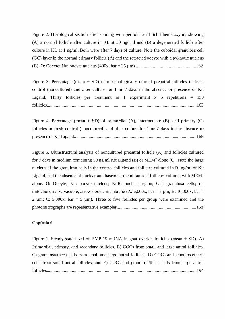
Figure 2. Histological section after staining with periodic acid Schiffhematoxylin, showing
(A) a normal follicle after culture in KL at 50 ng/ ml and (B) a degenerated follicle after
culture in KL at 1 ng/ml. Both were after 7 days of culture. Note the cuboidal granulosa cell
(GC) layer in the normal primary follicle (A) and the retracted oocyte with a pyknotic nucleus
(B). O: Oocyte; Nu: oocyte nucleus (400x, bar = 25 µm)......................................................162
Figure 3. Percentage (mean ± SD) of morphologically normal preantral follicles in fresh
control (noncultured) and after culture for 1 or 7 days in the absence or presence of Kit
Ligand. Thirty follicles per treatment in 1 experiment x 5 repetitions = 150
follicles....................................................................................................................................163
Figure 4. Percentage (mean ± SD) of primordial (A), intermediate (B), and primary (C)
follicles in fresh control (noncultured) and after culture for 1 or 7 days in the absence or
presence of Kit Ligand............................................................................................................165
Figure 5. Ultrastructural analysis of noncultured preantral follicle (A) and follicles cultured
for 7 days in medium containing 50 ng/ml Kit Ligand (B) or MEM+ alone (C). Note the large
nucleus of the granulosa cells in the control follicles and follicles cultured in 50 ng/ml of Kit
Ligand, and the absence of nuclear and basement membranes in follicles cultured with MEM+
alone. O: Oocyte; Nu: oocyte nucleus; NuR: nuclear region; GC: granulosa cells; m:
mitochondria; v: vacuole; arrow-oocyte membrane (A: 6,000x, bar = 5 µm; B: 10,000x, bar =
2 µm; C: 5,000x, bar = 5 µm). Three to five follicles per group were examined and the
photomicrographs are representative examples......................................................................168
Capítulo 6
Figure 1. Steady-state level of BMP-15 mRNA in goat ovarian follicles (mean SD). A)
Primordial, primary, and secondary follicles, B) COCs from small and large antral follicles,
C) granulosa/theca cells from small and large antral follicles, D) COCs and granulosa/theca
cells from small antral follicles, and E) COCs and granulosa/theca cells from large antral
follicles....................................................................................................................................194
17
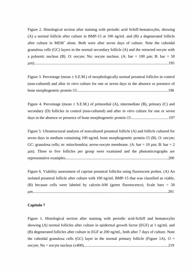
Figure 2. Histological section after staining with periodic acid Schiff-hematoxylin, showing
(A) a normal follicle after culture in BMP-15 at 100 ng/mL and (B) a degenerated follicle
after culture in MEM+ alone. Both were after seven days of culture. Note the cuboidal
granulosa cells (GC) layers in the normal secondary follicle (A) and the retracted oocyte with
a pyknotic nucleus (B). O: oocyte; Nu: oocyte nucleus. (A: bar = 100 µm; B: bar = 50
µm)..........................................................................................................................................195
Figure 3. Percentage (mean S.E.M.) of morphologically normal preantral follicles in control
(non-cultured) and after in vitro culture for one or seven days in the absence or presence of
bone morphogenetic protein-15..............................................................................................196
Figure 4. Percentage (mean S.E.M.) of primordial (A), intermediate (B), primary (C) and
secondary (D) follicles in control (non-cultured) and after in vitro culture for one or seven
days in the absence or presence of bone morphogenetic protein-15.......................................197
Figure 5. Ultrastructural analysis of noncultured preantral follicle (A) and follicle cultured for
seven days in medium containing 100 ng/mL bone morphogenetic protein-15 (B). O: oocyte;
GC: granulosa cells; m: mitochondria; arrow-oocyte membrane. (A: bar = 10 µm; B: bar = 2
µm). Three to five follicles per group were examined and the photomicrographs are
representative examples..........................................................................................................200
Figure 6. Viability assessment of caprine preantral follicles using fluorescent probes. (A) An
isolated preantral follicle after culture with 100 ng/mL BMP-15 that was classified as viable,
(B) because cells were labeled by calcein-AM (green fluorescence). Scale bars = 50
μm...........................................................................................................................................201
Capítulo 7
Figure 1. Histological section after staining with periodic acid-Schiff and hematoxylin
showing (A) normal follicles after culture in epidermal growth factor (EGF) at 1 ng/mL and
(B) degenerated follicles after culture in EGF at 200 ng/mL, both after 7 days of culture. Note
the cuboidal granulosa cells (GC) layer in the normal primary follicle (Figure 1A). O =
oocyte; Nu = oocyte nucleus (x400).......................................................................................219
18
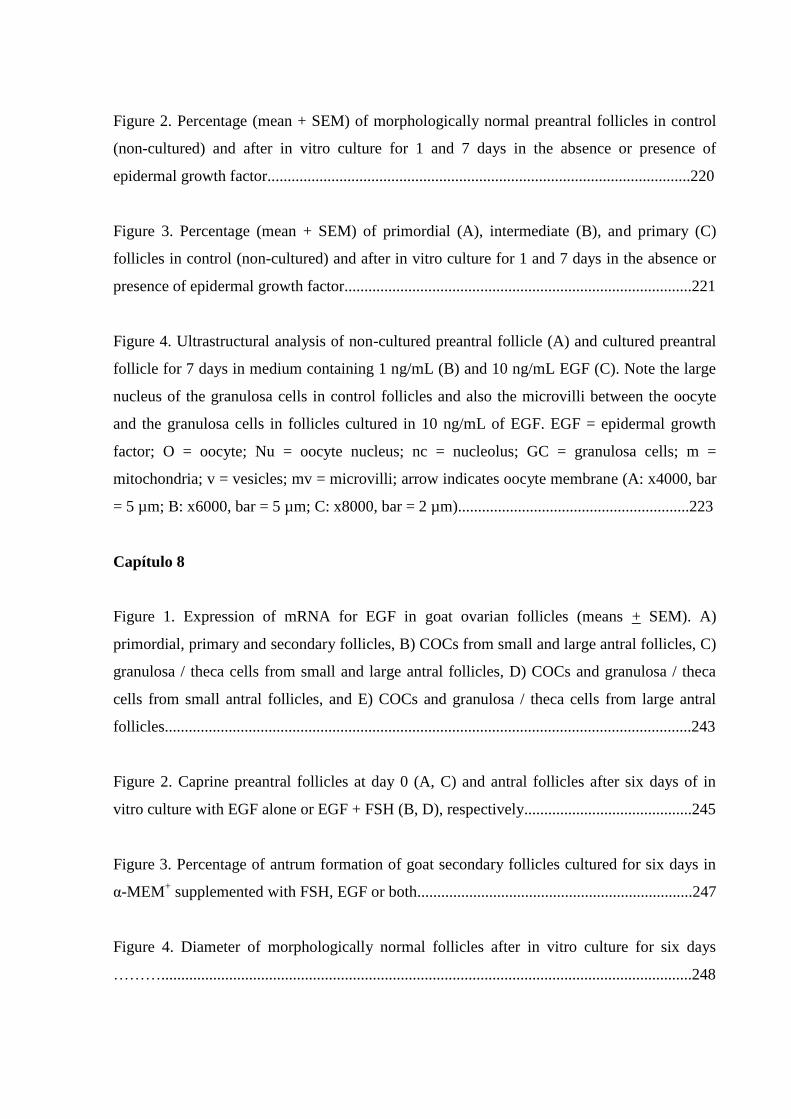
Figure 2. Percentage (mean + SEM) of morphologically normal preantral follicles in control
(non-cultured) and after in vitro culture for 1 and 7 days in the absence or presence of
epidermal growth factor..........................................................................................................220
Figure 3. Percentage (mean + SEM) of primordial (A), intermediate (B), and primary (C)
follicles in control (non-cultured) and after in vitro culture for 1 and 7 days in the absence or
presence of epidermal growth factor.......................................................................................221
Figure 4. Ultrastructural analysis of non-cultured preantral follicle (A) and cultured preantral
follicle for 7 days in medium containing 1 ng/mL (B) and 10 ng/mL EGF (C). Note the large
nucleus of the granulosa cells in control follicles and also the microvilli between the oocyte
and the granulosa cells in follicles cultured in 10 ng/mL of EGF. EGF = epidermal growth
factor; O = oocyte; Nu = oocyte nucleus; nc = nucleolus; GC = granulosa cells; m =
mitochondria; v = vesicles; mv = microvilli; arrow indicates oocyte membrane (A: x4000, bar
= 5 µm; B: x6000, bar = 5 µm; C: x8000, bar = 2 µm)..........................................................223
Capítulo 8
Figure 1. Expression of mRNA for EGF in goat ovarian follicles (means + SEM). A)
primordial, primary and secondary follicles, B) COCs from small and large antral follicles, C)
granulosa / theca cells from small and large antral follicles, D) COCs and granulosa / theca
cells from small antral follicles, and E) COCs and granulosa / theca cells from large antral
follicles....................................................................................................................................243
Figure 2. Caprine preantral follicles at day 0 (A, C) and antral follicles after six days of in
vitro culture with EGF alone or EGF + FSH (B, D), respectively..........................................245
Figure 3. Percentage of antrum formation of goat secondary follicles cultured for six days in
α-MEM+ supplemented with FSH, EGF or both.....................................................................247
Figure 4. Diameter of morphologically normal follicles after in vitro culture for six days
……….....................................................................................................................................248

19
Figure 5. Daily growth rate of morphologically normal follicles during in vitro culture for six
days …………………............................................................................................................248
Figure 6. Steady-state level of EGF mRNA in goat secondary follicles cultured for six days in
α-MEM+ supplemented with FSH, EGF or both.....................................................................249
Figure 7. Steady-state level of FSH-R mRNA in goat secondary follicles cultured for six days
in α-MEM+ supplemented with FSH, EGF or both................................................................250
20
LISTA DE TABELAS
Capítulo 3
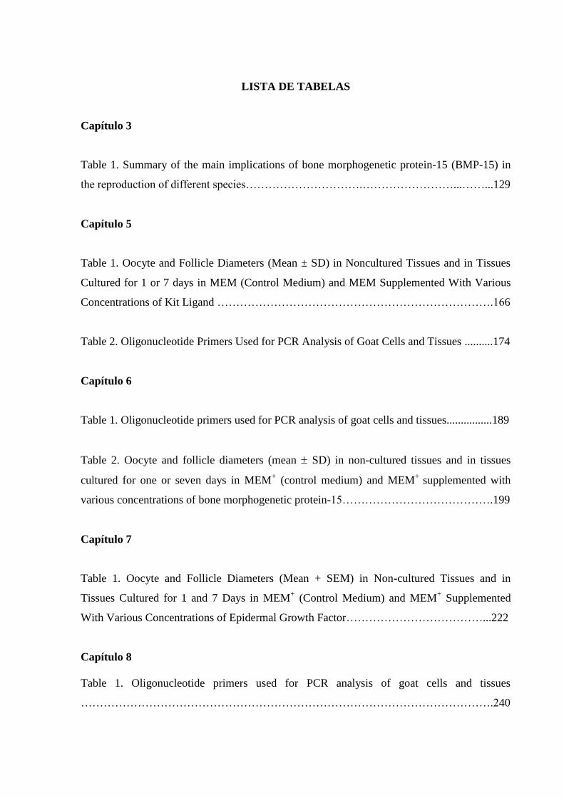
Table 1. Summary of the main implications of bone morphogenetic protein-15 (BMP-15) in
the reproduction of different species………………………….……………………...……...129
Capítulo 5
Table 1. Oocyte and Follicle Diameters (Mean ± SD) in Noncultured Tissues and in Tissues
Cultured for 1 or 7 days in MEM (Control Medium) and MEM Supplemented With Various
Concentrations of Kit Ligand ……………………………………………………………….166
Table 2. Oligonucleotide Primers Used for PCR Analysis of Goat Cells and Tissues ..........174
Capítulo 6
Table 1. Oligonucleotide primers used for PCR analysis of goat cells and tissues................189
Table 2. Oocyte and follicle diameters (mean SD) in non-cultured tissues and in tissues
cultured for one or seven days in MEM+ (control medium) and MEM
+ supplemented with
various concentrations of bone morphogenetic protein-15………………………………….199
Capítulo 7
Table 1. Oocyte and Follicle Diameters (Mean + SEM) in Non-cultured Tissues and in
Tissues Cultured for 1 and 7 Days in MEM+ (Control Medium) and MEM
+ Supplemented
With Various Concentrations of Epidermal Growth Factor………………………………...222
Capítulo 8
Table 1. Oligonucleotide primers used for PCR analysis of goat cells and tissues
……………………………………………………………………………………………….240

21
Table 2. Percentage of survival of goat secondary follicles cultured for six days in α-MEM+
supplemented with FSH, EGF or both ……………………………………………………...246
22
LISTA DE QUADROS
Revisão de literatura
Quadro 1. Principais resultados obtidos pelo LAMOFOPA com o cultivo in situ de folículos
pré-antrais caprinos...................................................................................................................54
Quadro 2. Principais resultados obtidos pelo LAMOFOPA com o cultivo isolado de folículos
pré-antrais caprinos...................................................................................................................59
23
LISTA DE ABREVIATURAS E SIGLAS
A : Antrum (Antro)
Akt : Protein kinase (Proteína kinase)
ALK-3, -6 : Receptor-like kinase-3, -6 (Receptor semelhante à kinase-3, -6)
AMH : Anti-müllerian hormone (Hormônio anti-mülleriano)
ANOVA : Analysis of variance (Análise de variância)
Apaf-1 : Apoptotic protease-activating factor (Fator ativador de protease
apoptótica)
AR : Amphiregulin (Anfiregulina)
as : antisense (anti senso)
ATP : Adenosine-5'-triphosphate (Adenosina-5‘-trifosfato)
Bak : BCL2 antagonist killer 1
Bax : BCL2 associated X protein
Bcl-2 : B-cell lymphoma protein 2
Bcl-w : BCL2 like 2 protein
Bcl-xL : BCL2 related protein, long isoform
BDNF : Brain-derived neurotrophic factor (Fator neurotrófico derivado do cérebro)
Bid : BH3 interacting domain death agonist
BMP-4, -6, -7, -15 : Bone morphogenetic protein-4, -6, -7, -15 (Proteína morfogenética
óssea-4, -6, -7, -15)
BMPR-IA, -IB, -II : Type IA, IB, II bone morphogenetic protein receptor (receptor de
proteína morfogenética óssea do tipo IA, IB, II)
BSA : Bovine serum albumin (Albumina sérica bovina)
BTC : Betacellulin (Betacelulina)
Ca2+
: Calcium ion (Íon cálcio)
CAD : Caspase-activated DNase (DNase ativada por caspase)
cAMP : Cyclic adenosine-3',5'-monophosphate (Adenosina-3',5'-
monofosfato cíclico)
CAPES : Coordenação de Aperfeiçoamento do Pessoal de Nível Superior
Caspase 1-14 : Cysteinyl aspartic acid protease 1-14
CCND2 : Cyclin D2
cdc25 : cell division cycle 25 phosphatase
24
cDNA
CETENE
: Complementary deoxyribonucleic acid (Ácido desoxirribonucléico
complementar)
: Centro de Tecnologias Estratégicas do Nordeste
CGP : Células germinativas primordiais
c-kit : kit ligand receptor (receptor para kit ligand)
CNPq : Conselho Nacional de Desenvolvimento Científico e Tecnológico
CO2 : Dióxido de carbono
COCs : Cumulus–oocyte complexes (Complexos cúmulus oócito)
CPqAM : Centro de Pesquisa Aggeu Magalhães
CT : Cycle threshold
Cx43 : Connexin 43 (Conexina 43)
CXCL12 : Chemokine (C-X-C motif) ligand 12
DABCO : 1,4-diazabicyclo[2.2.2]octane (Octano do diazabicyclo 1,4 [2.2.2])
DD : Death domain (Domínio de morte)
DISC : Death-inducing signaling complex (Complexo sinalizador indutor
de morte)
DNA : Deoxyribonucleic acid (Ácido desoxirribonucléico)
DNAse : Desoxirribonuclease
dNTP : Deoxy-nucleotide-triphophates (desoxinucleotídeo trifosfato)
DR-4, -5 : Death receptor-4, -5 (receptor de morte-4, -5)
DTT : Dithiothreitol (Ditiotreitol)
EGF : Epidermal growth factor (Fator de crescimento epidermal)
EGF-R : Receptor of epidermal growth factor (Receptor do fator de
crescimento epidermal)
ELISA : Enzyme-linked imumunosorbent assay
EPR : Epiregulin (Epiregulina)
ErbB 1/2/3/4 : EGF receptor tyrosine kinase family 1/2/3/4 (Família de receptor
EGF do tipo tirosina kinase 1/2/3/4)
Erk 1/2 : Extracellular signal-regulated kinase 1/2
F : Fluorescência
FADD : Fas-associated death domain (Domínio de morte associado ao Fas)
Fas : Fatty acid synthetase
Fas/CD95 : Fatty acid synthetase receptor
25
FasL : Fatty acid synthetase ligand
FAVET : Faculdade de Veterinária
FGF-2, -7, -10 : Fibroblast growth factor-2, -7, -10 (Fator de crescimento
fibroblástico-2, -7, -10)
Fig α : Factor in the germ-line alpha (Fator de linha germinal alfa)
Fig. : Figure (Figura)
FINEP : Financiadora de Estudos e Projetos
FIOCRUZ : Fundação Oswaldo Cruz
FOXO : Forkhead/winged helix transcription factor subfamily
FSH : Follicle stimulating hormone (Hormônio folículo estimulante)
FSH-R : Follicle stimulating hormone receptor (Receptor do hormônio
folículo estimulante)
FUNCAP : Fundação Cearense de Apoio ao Desenvolvimento Científico e
Tecnológico
g : gravidade
G : Granulosa cells (Células da granulosa)
GAPDH : Glyceraldehydes-2-phophate dehydrogenase (Gliceraldeído-2-
fosfato desidrogenase)
GC : Granulosa cells (Células da granulosa)
GDF-9, -9B : Growth diferentiation factor-9, -9B (Fator de crescimento e
diferenciação-9, -9B)
GDNF : Glial cell-derived neurotrophic factor (Fator neurotrófico derivado
da célula glial)
GFRα1 : Glial cell-derived neurotrophic factor receptor alpha1 (Receptor
alfa1 do fator neurotrófico derivado da célula glial)
GH : Growth hormone (Hormônio do crescimento)
GI : GenInfo identifier
GLM : General linear models
GT : Granulosa/theca cells (Células da granulosa e teca)
h : horas
HB-EGF : Heparin-binding EGF (Fator de crescimento semelhante ao EGF
ligado à heparina)
HC : Histologia clássica
26
HER1 1/2/3/4 : EGF receptor tyrosine kinase family 1/2/3/4 (Família de receptor
EGF do tipo tirosina kinase 1/2/3/4)
HGF : Hepatocyte growth factor (Fator de crescimento de hepatócito)
IAA : Indole-3-acetic acid (Ácido 3-indol-acético)
IC : Interstitial cells (células intersticiais)
IGF-1, -2 : Insulin-like growth factor-1, -2 (Fator de crescimento semelhante à
insulina -1, -2)
IGFBP-4 : Insulin-like growth factor-binding protein-4 (Proteína ligante 4
transportadora de fator de crescimento semelhante à insulina)
IL-1β, -6 : Interleucin-1β, -6 (interleucina-1β, -6)
I-Smads : Inhibitory-Seven mothers against dpp gene da Drosophila
ITS : Insulin, tranferrin and selenium (Insulina, transferrina e selênio)
IU : International units (Unidades internacionais)
JAK2 : Janus-activated kinase 2
JNKs : Jun NH2-terminal protein kinases
K+ : Potassium ion (Íon potássio)
KGF : Keratinocyte growth factor (Fator de crescimento keratinócito)
KL-1, -2 : Kit ligand-1, -2
kV : quilovolts
l : lipid droplets (gotas lipídicas)
L : Litro
LABOVIR : Laboratório de Virologia
LAMOFOPA : Laboratório de Manipulação de Oócitos e Folículos Pré-Antrais
LH : Luteinizing hormone (Hormônio luteinizante)
Lhx8 : LIM-homeobox protein 8
Lhx8-/-
: Lhx8-deficient (Deficiência no gene Lhx8)
LIF : Leukemia inhibitory factor (Fator inibidor de leucemia)
m : mitochondria (mitocôndria)
M : Molar
MA : Massachusetts
MAPK : Mitogen-activated protein kinase (Proteína kinase ativada por
mitógenos)
MCGF : Mast-cell growth factor (Fator de crescimento de mastócitos)
27
Mcl-1 : Myeloid cell leukemia-1
MEK : MAP Kinase/extracellular protein kinase
MEM : Minimal essential medium (Meio essencial mínimo)
MEM+ : Supplemented minimal essential medium (Meio essencial mínimo
suplementado)
MET : Microscopia eletrônica de transmissão
min. : minutos
mg : miligramas
mL : mililitros
mm
mm2
: milímetros
: milímetros quadrados
mm3 : milímetros cúbicos
mM : milimolar
MO : Missouri
MOIFOPA : Manipulação de Oócitos Inclusos em Folículos Ovarianos Pré-
Antrais
mOsm/L : miliosmol/litro
mRNA : Messenger ribonucleic acid (Ácido ribonucléico mensageiro)
mv : microvilli (microvilo)
Na+ : Sodium ion (Íon sódio)
NaCl : Cloreto de sódio
nc : nucleolus (nucléolo)
ng : nanograma
NGF : Nerve growth factor (Fator de crescimento do nervo)
nm : nanômetros
no : nucleolus (nucléolo)
NOBOX : Newborn ovary homeobox gene
NOBOX-/-
: NOBOX-deficient (Deficiência no gene NOBOX)
NRG 1-4 : Neuregulins 1-4 (Neuregulinas 1-4)
NTF5 : Neurotrophin 5 (Neurotrofina tipo 5)
nu : nucleus (núcleo)
Nu : Oocyte nucleus (Núcleo do oócito)
NUBIS : Núcleo de Biotecnologia de Sobral
28
NuR : Nuclear region (Região nuclear)
O : Oocyte (Oócito)
OCT4 : Octamer-binding transcription factor 4
P<0.05 : Probabilidade de erro menor do que 5%
P>0.05 : Probabilidade de erro maior do que 5%
p. : página
p34cd2 : Protein kinase p34 (cd2)
p38-MAPKs : p38 mitogen-activated protein kinases
p53 : 53 protein (proteína 53)
PAS-H : Periodic acid-Schiff and hematoxylin (Ácido periódico de Schiff e
hematoxilina)
PBS : Phosphate buffer saline (Tampão fosfato salino)
PCR : Polimerase chain reaction (Reação em cadeia polimerase)
pFSH : Pituitary follicle stimulating hormone (Hormônio folículo
estimulante pituitário)
PGCs : Primordial germ cells (Células germinativas primordiais)
pH : potencial hidrogeniônico
PI3K : Phosphoinositide 3-kinase (Fosfatidilinositol 3-kinase)
PK A, B, C : Protein kinase A, B, C (Proteína kinase A, B, C)
POU5F1 : Pituitary octamer neural unc domain, class 5, transcription factor 1
PPGCV
PTEN
: Programa de Pós-Graduação em Ciências Veterinárias
: Phosphatase and tensin homolog deleted on chromosome 10
(Fosfatase e tensina homóloga com deleção no cromossomo 10)
rbFSH : Recombinant bovine follicle stimulating hormone (Hormônio
folículo estimulante recombinante bovino)
RENORBIO : Rede Nordeste de Biotecnologia
RET : Ubiquitous tyrosine kinase receptor
R-FSH : Follicle stimulating hormone receptor (Receptor do hormônio
folículo estimulante)
rFSH : Recombinant follicle stimulating hormone (Hormônio folículo
estimulante recombinante)
rhEGF : Recombinant human epidermal growth factor (Fator de crescimento
epidermal recombinante humano)
29
rhKL : Recombinant human kit ligand (Kit ligand recombinante humano)
RNAm : Ribonucleic acid messenger (Ácido ribonucléico mensageiro)
RNAse : Ribonuclease
R-Smads : Receptor-Seven mothers against dpp gene da Drosophila
RT : Room temperature (Temperatura ambiente)
RT-PCR
RT-qPCR
: Reverse transcription-polimerase chain reaction (Transcrição
reversa-reação em cadeia polimerase)
: Reverse transcription-quantitative polimerase chain reaction
(Transcrição reversa-quantitativa reação em cadeia polimerase)
s : sense (senso)
SAS : Statistical analysis system
SBAC : Solução à base de água de coco
SCF : Stem cell factor (Fator de células tronco)
SD : Standard deviation
Sec. : Secunde (Segundos)
SEM : Standard error of means (Erro padrão da média)
SF : Steel factor
Smads 1/4/5/6/7/8 : Seven mothers against dpp gene da Drosophila 1/4/5/6/7/8
SNK : Student–Newman–Keuls
SP : São Paulo
Sohlh-1, -2 : Spermatogenesis and oogenesis helix-loop-helix-1, -2
STAT : Signal transducer and activator of transcription (Transdutor de sinal
e ativador de transcrição)
T 1, 2, 3 : Tratamento 1, 2, 3
TC : Theca cells (Células da teca)
TEM : Transmission electronic microscopy (Microscopia eletrônica de
transmissão)
TGF-, - α : Transforming growth factor beta, alpha (Fator de crescimento
transformante beta, alfa)
TNF : Tumor necrosis factor (Fator de necrose tumoral)
TNFR1 : Tumor necrosis factor receptor 1 (Receptor tipo 1 do fator de
necrose tumoral)
TNF-α : Tumor necrosis factor alpha (Fator de necrose tumoral alfa)
30
TRADD : TNF receptor-associated death domain
TRAIL : TNF-related apoptosis-inducing ligand
TrkB : Tyrosine kinase receptor B
TUNEL : Terminal deoxynucleotidil transferase-mediated deoxyuridine
triphosphate biotin nick end-labeling
UBQ : Ubiquitin (Ubiquitina)
UECE : Universidade Estadual do Ceará
UFC : Universidade Federal do Ceará
UnB : Universidade de Brasília
USA : United States of America (Estados Unidos da América)
v : vacuole or vesicles (vacúolo ou vesículas)
v. : volume
VEGF : Vascular endothelial growth factor (Fator de crescimento do
endotélio vascular)
VEGFR-2 : Vascular endothelial growth factor receptor-2 (Receptor 2 para fator
de crescimento do endotélio vascular)
VIP : Vasoactive intestinal peptide (Peptídeo intestinal vasoativo)
Vol. : Volume
X : Eixo das abicissas
Y : Eixo das ordenadas
ZP : Zona pellucida (Zona pelúcida)
α-MEM : Alpha minimal essential medium (Meio essencial mínimo alfa)
α-MEM+ : Supplemented alpha minimal essential medium (Meio essencial
mínimo alfa suplementado)
g : Microgramas
L : Microlitros
m : Micrômetros
µM : Micromolar
% : Percentage (Porcentagem)
~ : Aproximadamente
± : Mais ou menos
°C : Graus Celsius
31
SUMÁRIO
1 INTRODUÇÃO............................................................................................................. 33
2 REVISÃO DE LITERATURA ................................................................................... 35
2.1 Foliculogênese................................................................................................. 35
2.2 Caracterização estrutural dos folículos ovarianos e regulação da
foliculogênese........................................................................................................
36
2.2.1 Formação do folículo primordial..................................................... 36
2.2.2 Transição de folículo primordial para primário................................ 38
2.2.3 Transição de folículo primário para secundário................................ 42
2.2.4 Transição de folículo secundário para antral..................................... 44
2.3 Cultivo in vitro de folículos pré-antrais........................................................... 46
2.3.1 Cultivo in vitro de folículos pré-antrais caprinos inclusos em
fragmentos de córtex ovariano...................................................................
48
2.3.2 Cultivo in vitro de folículos pré-antrais caprinos isolados................ 55
2.4 Estado atual do cultivo in vitro de folículos pré-antrais.................................. 60
2.5 Técnicas para análise folicular durante o cultivo in vitro................................ 60
2.5.1 Histologia Clássica............................................................................ 61
2.5.2 Microscopia Eletrônica de Transmissão........................................... 61
2.5.3 Microscopia de Fluorescência........................................................... 62
2.5.4 Biologia Molecular............................................................................ 63
3 JUSTIFICATIVA......................................................................................................... 65
4 HIPÓTESES CIENTÍFICAS....................................................................................... 66
5 OBJETIVOS.................................................................................................................. 67
5.1 OBJETIVO GERAL........................................................................................ 67
5.2 OBJETIVOS ESPECÍFICOS........................................................................... 67
6 CAPÍTULO 1 - Mecanismos de atresia em folículos ovarianos................................... 69
7 CAPÍTULO 2 - Regulação da foliculogênese ovariana pelo sistema Kit Ligand e c-
Kit em mamíferos..............................................................................................................
100
8 CAPÍTULO 3 - Implicações da proteína morfogenética óssea-15 na foliculogênese
ovariana......................................................................................................................
119
9 CAPÍTULO 4 - Fator de crescimento epidermal como mediador de sobrevivência e
desenvolvimento folicular..................................................................................................
136
32
10 CAPÍTULO 5 - Níveis de RNAm para o Kit Ligand em Ovários Caprinos e o
Papel do Kit Ligand na Sobrevivência e Crescimento In Vitro de Folículos Pré-antrais..
156
11 CAPÍTULO 6 - Níveis da proteína morfogenética óssea-15 em ovários caprinos e
sua influência no desenvolvimento in vitro e sobrevivência de folículos pré-antrais.......
182
12 CAPÍTULO 7 - Fator de Crescimento Epidermal Recombinante Mantém a
Ultraestrutura Folicular e Promove a Transição para Folículos Primários em Tecido
Ovariano Caprino Cultivado In Vitro................................................................................
213
13 CAPÍTULO 8 - Níveis do fator de crescimento epidermal (EGF) e efeito do EGF
no cultivo in vitro de folículos pré-antrais caprinos..........................................................
232
14 CONCLUSÕES........................................................................................................... 261
15 PERSPECTIVAS........................................................................................................ 262
16 REFERÊNCIAS BIBLIOGRÁFICAS…………………………………………….. 263
33
1 INTRODUÇÃO
Os caprinos estão presentes em todos os continentes e são vistos comercialmente como
animais altamente atrativos, uma vez que eles têm sido utilizados para muitos propósitos, tais
como produção de leite, carne e pele. Nas últimas duas décadas, desenvolvimentos
significativos têm sido alcançados no campo da biotecnologia da reprodução assistida, tanto
em animais como em humanos. Em animais, incluindo os caprinos, essas modernas
biotecnologias estão sendo utilizadas para melhoria e preservação da genética dos animais, e
aumento da sua eficiência reprodutiva (RAHMAN; ABDULLAH; WAN KHADIJAH, 2008).
É conhecido que os ovários das diferentes espécies mamíferas, como a espécie
caprina, contêm milhares de oócitos imaturos inclusos predominantemente nos folículos pré-
antrais, representando esses folículos uma fonte potencial de gametas fertilizáveis, e com isso,
sendo de grande interesse assegurar o crescimento in vitro e permitir a aquisição da
competência dos oócitos provenientes destes folículos (MCLAUGHLIN et al., 2010).
Entretanto, mais de 99,9% dos oócitos inclusos em folículos pré-antrais não ovularão, mas
sim serão eliminados por um processo natural conhecido como atresia. Dessa forma, o resgate
dos folículos pré-antrais dos ovários, evitando assim a atresia folicular, seguido pelo
desenvolvimento de sistemas de cultivo in vitro (ovário artificial) que permitam o crescimento
e maturação de seus oócitos poderia trazer, no futuro, um maior impacto para a produção in
vitro de embriões (FIGUEIREDO et al., 2007).
Estudos in vitro com o cultivo de folículos pré-antrais de camundongas demonstraram
que é possível a obtenção de crias vivas produzidas de oócitos oriundos destes folículos
cultivados in vitro (O‘BRIEN; PENDOLA; EPPIG, 2003; HASEGAWA et al., 2006). Em
outros animais, um número limitado de embriões tem sido produzido de oócitos crescidos e
maturados in vitro (suínos - WU; EMERY; CARREL, 2001; WU; TIAN, 2007; bubalinos -
GUPTA et al., 2008; ovinos - ARUNAKUMARI; SHANMUGASUNDARAM; RAO, 2010 e
caprinos - SARAIVA et al., 2010a; MAGALHÃES et al., 2011), consistindo assim em um
grande desafio o nascimento de indivíduos vivos normais a partir de folículos pré-antrais
crescidos in vitro nestas espécies. Sabendo-se então que o crescimento dos folículos presentes
no ovário mamífero é regulado por gonadotrofinas e por fatores intra-ovarianos (FORTUNE,
2003), atualmente esforços têm sido concentrados para melhorar a identificação e
compreensão das diferentes substâncias envolvidas na promoção do desenvolvimento
folicular e no curso da atresia. Diante desse melhor conhecimento acerca da foliculogênese,
34
será possível tentar desenvolver um sistema de cultivo in vitro capaz de permitir o
desenvolvimento de um grande número de folículos pré-antrais, melhorando assim no futuro a
taxa de produção de embriões, e permitindo a obtenção de nascimentos a partir de folículos
pré-antrais destas espécies. Dentre as substâncias reguladoras da foliculogênese, merecem
destaque o kit ligand (KL), a proteína morfogenética óssea-15 (BMP-15) e o fator de
crescimento epidermal (EGF).
Para uma melhor compreensão da importância deste trabalho, a revisão de literatura a
seguir abordará aspectos relativos à regulação da foliculogênese em mamíferos, destacando a
importância dos hormônios e fatores de crescimento; cultivo in vitro de folículos pré-antrais,
especialmente na espécie caprina; estado atual do cultivo; e as principais técnicas para
avaliação dos folículos cultivados in vitro.
35
2 REVISÃO DE LITERATURA
2.1 Foliculogênese
A foliculogênese é um evento iniciado na vida pré-natal na maioria das espécies,
podendo ser definida como o processo de formação, crescimento e maturação folicular,
iniciando-se com a formação do folículo primordial e terminando no estádio de folículo pré-
ovulatório (VAN DEN HURK; ZHAO, 2005; GOUGEON, 2010).
O folículo é considerado a unidade morfológica e funcional do ovário mamífero,
proporcionando um ambiente ideal para o crescimento e maturação do oócito
(CORTVRINDT; SMITZ, 2001a), além de produzir algumas substâncias fundamentais para
sua manutenção e desenvolvimento (ADASHI, 1994). Essa estrutura é composta por um
oócito circundado por células somáticas (granulosa e/ou tecais), tendo a interação entre esses
compartimentos celulares um papel crítico no decorrer da foliculogênese (VANDERHYDEN;
TELFER; EPPIG, 1992; EPPIG; WIGGLESWORTH; PENDOLA, 2002; MATZUK et al.,
2002). Durante o processo da foliculogênese, a morfologia folicular é alterada observando-se
o crescimento oocitário, a diferenciação e a proliferação das células da granulosa e o
aparecimento das células tecais (SILVA, 2005; BRISTOL-GOULD; WOODRUFF, 2006).
Com base nessa mudança morfológica, os folículos podem ser divididos em: 1) folículos pré-
antrais ou não cavitários, que abrangem os folículos primordiais, transição, primários e
secundários e 2) folículos antrais ou cavitários, compreendendo os folículos terciários e de De
Graaf ou pré-ovulatório (SILVA et al., 2004a). Vale ressaltar que os folículos pré-antrais
representam mais de 90% da população folicular do ovário (SAUMANDE, 1981), sendo 95%
destes folículos primordiais (ERICKSON, 1986), os quais constituem o pool de reserva de
gametas femininos durante toda a vida reprodutiva (QU et al., 2000).
A foliculogênese pode ser dividida nas seguintes fases de desenvolvimento: 1) fase
pré-antral, que é subdividida em ativação dos folículos primordiais e crescimento de folículos
de transição, primários e secundários; 2) fase antral, subdividida em crescimento inicial e
terminal dos folículos terciários e formação do folículo pré-ovulatório. Além disso, a
foliculogênese pode ainda ser classificada de acordo com a dependência gonadotrófica
(MCGEE; HSUEH, 2000; CRAIG et al., 2007; MCNATTY et al., 2007; ORISAKA et al.,
2009) em: (1) fase independente e responsiva à gonadotrofina (crescimento folicular através
dos estádios primordial, transição, primário, secundário e transição de pré-antral para o
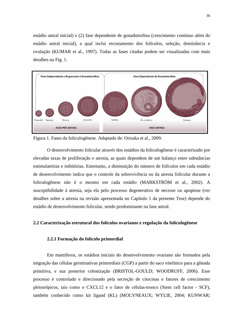
36
estádio antral inicial) e (2) fase dependente de gonadotrofina (crescimento contínuo além do
estádio antral inicial), a qual inclui recrutamento dos folículos, seleção, dominância e
ovulação (KUMAR et al., 1997). Todas as fases citadas podem ser visualizadas com mais
detalhes na Fig. 1.
Figura 1. Fases da foliculogênese. Adaptado de: Orisaka et al., 2009.
O desenvolvimento folicular através dos estádios da foliculogênese é caracterizado por
elevadas taxas de proliferação e atresia, as quais dependem de um balanço entre substâncias
estimulatórias e inibitórias. Entretanto, a diminuição do número de folículos em cada estádio
de desenvolvimento indica que o controle da sobrevivência ou da atresia folicular durante a
foliculogênese não é o mesmo em cada estádio (MARKSTRÖM et al., 2002). A
susceptibilidade à atresia, seja ela pelo processo degenerativo de necrose ou apoptose (ver
detalhes sobre a atresia na revisão apresentada no Capítulo 1 da presente Tese) depende do
estádio de desenvolvimento folicular, sendo predominante na fase antral.
2.2 Caracterização estrutural dos folículos ovarianos e regulação da foliculogênese
2.2.1 Formação do folículo primordial
Em mamíferos, os estádios iniciais do desenvolvimento ovariano são formados pela
migração das células germinativas primordiais (CGP) a partir do saco vitelínico para a gônada
primitiva, e sua posterior colonização (BRISTOL-GOULD; WOODRUFF, 2006). Esse
processo é controlado e direcionado pela secreção de citocinas e fatores de crescimento
pleiotrópicos, tais como o CXCL12 e o fator de células-tronco (Stem cell factor - SCF),
também conhecido como kit ligand (KL) (MOLYNEAUX; WYLIE, 2004; KUNWAR;
37
SIEKHAUS; LEHMANN, 2006). Com a chegada das CGP na gônada primitiva, ocorre a
formação dos cordões sexuais. A partir de então, as CGP perdem a sua motilidade e passam a
se multiplicar por mitose, morrendo, entretanto, a grande maioria delas por apoptose (KIM;
TILLY, 2004), fenômeno este responsável pela regulação do número de folículos primordiais
presentes no ovário (BAKER, 1963). As demais CGP são então diferenciadas em oogônias
(BAKER; FANCHI, 1967; SATHANANTHAN; SELVARAJ; TROUNSON, 2000) e uma
vez diferenciadas, estas irão se dividir sucessivamente por mitose e irão formar ninhos de
oogônias interligados por pontes intercelulares (PEPLING; SPRADLING, 1998, 2001;
PEPLING, 2006). Com a parada da mitose, as oogônias entram nos estádios iniciais da
meiose I diferenciando-se em oócitos primários (HIRSHFIELD, 1991). Em seguida, os
oócitos perdem suas pontes intercelulares e são circundados por uma camada de células da
pré-granulosa, as quais podem ser derivadas do mesonefron ou do epitélio da superfície
ovariana (MCNATTY et al., 2000). Uma vez que o oócito é circundando pelas células
somáticas, ocorre uma parada da meiose no estádio de diplóteno da prófase I, também
conhecido como estádio de vesícula germinativa (BAKER; FRANCHI, 1967; PICTON;
BRIGGS; GOSDEN, 1998), no qual as células da pré-granulosa param de se multiplicar e
entram num período de quiescência (SAWYER et al., 2002) juntamente com o oócito,
formando os folículos primordiais e dando início à foliculogênese. A progressão da divisão
meiótica ocorre somente na puberdade, com a liberação do pico pré-ovulatório de FSH e LH,
formação dos oócitos secundários e outra parada da meiose na fase de metáfase II (HUTT;
ALBERTINI, 2007). A meiose será retomada novamente somente após a fecundação do
oócito pelo espermatozóide, originando o oócito haplóide fecundado, e marcando assim o fim
da oogênese (FIGUEIREDO et al., 2008).
Alguns fatores transcricionais envolvidos nos padrões de sinalização da oogênese e
foliculogênese inicial têm sido identificados através de análises fenotípicas de camundongas.
Um dos primeiros fatores encontrados com papel na formação do folículo primordial foi o
fator de linha germinal α (Fig α ou Figla) (LIANG; SOYAL; DEAN, 1997), o qual é expresso
pelo oócito (SOYAL; AMLEH; DEAN, 2000). Em camundongas, esse fator é expresso no
estádio embrionário, mas sua expressão é reduzida após sete e 14 dias do nascimento;
entretanto, transcritos residuais são encontrados no ovário adulto (SOYAL; AMLEH; DEAN,
2000). Estudos mais recentes têm revelado que o Figla regula a foliculogênese inicial
aumentando diversos outros genes, incluindo o POU5F1. O POU5F1 (ou OCT4) é um fator
transcricional cujo alvo preciso não é conhecido e o papel dele na oogênese tem sido
38
recentemente descrito (PANGAS; RAJKOVIC, 2006). Ele é expresso nas CGP até elas
migrarem para a gônada primitiva e a expressão é então reprimida após o início da prófase I
meiótica no oócito, voltando a ser re-expresso pelos oócitos após o nascimento, o que
coincide com o período de crescimento destes (PARFENOV et al., 2003). Pouco foi
conhecido sobre o papel do POU5F1 no ovário pós-natal, mas análises de nocaute desse fator
nas CGP demonstraram a sua importância na sobrevivência dessas células, uma vez que na
sua ausência nenhum folículo foi encontrado no ovário, o que foi atribuído a uma apoptose
prematura das CGP antes da colonização da gônada (KEHLER et al., 2004). Outro fator que
vem demonstrando importância neste processo é o LHX8. Este gene está envolvido no padrão
de formação e sobrevivência do folículo primordial, sendo preferencialmente expresso no
oócito de ovários de camundongas. Foi observado que camundongas sem esse fator falharam
em manter os folículos primordiais e que estes desapareceram na primeira semana de vida
(CHOI et al., 2008). Este achado parece ter sido causado por uma marcada redução na
expressão do KL e de seu receptor (c-kit) em ovários de camundongas sem LHX8.
As neurotrofinas podem também estar envolvidas na sinalização entre CGP e células
somáticas no momento da formação dos folículos primordiais. Essa afirmativa foi formulada
com base nas mudanças observadas ao longo do desenvolvimento no padrão de expressão da
neurotrofina 4 e do seu receptor de alta afinidade (TrkB) em oócitos de humanos e ovários de
ratas (ANDERSON et al., 2002). Recentes estudos têm ainda sugerido que os níveis de
progesterona e estradiol fetais e maternos regulam a formação do folículo primordial
(KEZELE; SKINNER, 2003; BRITT et al., 2004; NILSSON; STANFIELD; SKINNER,
2006a; CHEN et al., 2007).
2.2.2 Transição de folículo primordial para primário
Como já relatado, os gametas femininos são estocados no ovário na forma de folículos
primordiais (FAIR, 2003). Até pouco tempo atrás, prevalecia o dogma de que o pool de
folículos primordiais representava uma reserva finita de gametas femininos (ZUCKERMAN,
1951). Entretanto, a noção de uma reserva fixa e não renovável de folículos primordiais no
ovário mamífero tem sido questionada, sendo sugerido que células-tronco da linha
germinativa extra e intraovariana poderiam reabastecer os oócitos e formar novos folículos
primordiais (JOHNSON et al., 2004, 2005). Por outro lado, diferentes estudos não
encontraram evidências para suportar a hipótese de que células progenitoras de origem
39
extragonadal possam renovar as células foliculares no ovário adulto (KERR et al., 2006;
BEGUM; PAPAIOANNOU; GOSDEN, 2008).
Independente disso, logo após a formação, alguns folículos primordiais podem ser
estimulados a crescer imediatamente ou, na maioria destes, as células da pré-granulosa param
de se multiplicar e entram num período de quiescência até receberem sinais para entrar no
pool de crescimento (MCGHEE; HSUEH, 2000). O início do desenvolvimento dos folículos
primordiais pode ocorrer dias, meses ou anos após a sua formação (VAN DEN HURK;
ZHAO, 2005), sendo considerado o maior evento biológico que controla o potencial
reprodutivo das fêmeas (MCLAUGHLIN; MCIVER, 2009). A maioria desses folículos sofre
atresia na vida pré- ou pós-natal, e nunca inicia o complexo padrão de desenvolvimento que
pode ou não culminar na ovulação (KNIGHT; GLISTER, 2006).
O início do crescimento de folículos primordiais, também conhecido como ativação, é
um processo que ocorre através da transição dos folículos do pool de reserva, ou folículos
quiescentes, para o pool de folículos em crescimento (transição, primário, secundário,
terciário e pré-ovulatório) (RÜSSE, 1983). As características morfológicas que marcam o
início do crescimento dos folículos primordiais são: aumento do diâmetro oocitário e
transformação da morfologia das células da granulosa de pavimentosa para cúbica. Durante
esta fase, os folículos que apresentam células da granulosa pavimentosas e cúbicas são
denominados folículos de transição (SILVA et al., 2004a). Quando o oócito é circundado por
uma camada completa de células da granulosa de morfologia cúbica, os folículos são
denominados primários (GOUGEON; BUSSO, 2000).
Os fatores e mecanismos responsáveis pela ativação de folículos primordiais são
pouco conhecidos. Acredita-se que a ativação dos folículos primordiais seja regulada por um
balanço entre fatores inibitórios e estimulatórios originários do ovário (VAN DEN HURK;
ZHAO, 2005). Dessa forma, a caracterização dos fatores e mecanismos envolvidos no padrão
de sinalização da ativação é fundamental para o conhecimento dos sistemas moleculares
responsáveis por assegurar o conveniente e oportuno fornecimento de oócitos aptos à
fecundação (MCLAUGHLIN; MCIVER, 2009).
As células da pré-granulosa que circundam o oócito de folículos primordiais
expressam um grande número de fatores peptídicos, incluindo o KL e o fator inibidor de
leucemia (LIF), os quais têm estimulado in vitro a transição de folículos primordiais para
primários, o crescimento do oócito e o recrutamento e proliferação das células da teca do
estroma circundante (NILSSON; KEZELE; SKINNER, 2002; NILSSON; SKINNER, 2003,
40
2004). O receptor para o KL (c-kit) está expresso no oócito e células intersticiais/tecais,
capazes então de responder ao estímulo do KL. O KL é um dos poucos fatores que possui um
papel bem definido sobre a ativação folicular. Recentes estudos têm demonstrado que este
fator de crescimento atua na ativação folicular por meio do padrão de sinalização intracelular
no oócito denominado PI3K-AKT-FKHRL1 e PTEN (LIU et al., 2007a,b; REDDY et al.,
2008). A cascata de sinalização iniciada pelo c-kit na superfície do oócito, ativado pelo KL, é
seguida por uma subsequente ativação da PI3K, podendo aumentar o crescimento do oócito e
a produção de fatores locais capazes de estimular a proliferação e diferenciação das células da
granulosa circundantes (MCLAUGHLIN; MCIVER, 2009). A revisão de literatura
apresentada no Capítulo 2 desta Tese abordará os detalhes sobre a ação do KL na
foliculogênese inicial.
Além disso, algumas das células mesenquimais que circundam os folículos
primordiais (células precursoras da teca) produzem outros peptídeos, conhecidos como fator
de crescimento de queratinócito (KGF, também chamado de fator de crescimento
fibroblástico-7, FGF-7) e o fator de crescimento do hepatócito (HGF), que podem então atuar
nas células da pré-granulosa e/ou células da granulosa aumentando a expressão do KL e
amplificando assim seus efeitos positivos no desenvolvimento folicular (KEZELE;
NILSSON; SKINNER, 2005; GUGLIELMO et al., 2010). Em adição, foi demonstrado que o
FGF-2 (também conhecido como FGF básico), expresso nos oócitos de folículos primordiais,
aumentou a expressão do KL nas células da pré-granulosa e promoveu a transição de folículo
do estádio primordial para primário no cultivo de ovários de ratas neonatais (NILSSON;
SKINNER, 2004). O KL e o FGF-2 têm efeitos estimulatórios mútuos no oócito e nas células
da granulosa e também promovem o recrutamento das células da teca a partir da população de
células do estroma/intersticial circundantes. Células do estroma/intersticiais e células da teca
secretam as proteínas morfogenéticas ósseas-4 e -7 (BMP-4 e BMP-7), as quais também
promovem a ativação e a sobrevivência folicular. O fator de crescimento e diferenciação-9
(GDF-9) e/ou a BMP-15, ambos secretados pelo oócito, promovem a proliferação das células
da granulosa, expressão do KL e a formação das células da teca (KNIGHT; GLISTER, 2006).
Análises funcionais utilizando sistemas de cultivo in vitro de ovários neonatais têm
confirmado que a taxa de ativação de folículos primordiais é diretamente proporcional ao
aumento no número de citocinas e fatores de crescimento pleiotrópicos, que incluem o KL
(HUTT; MCLAUGHLIN; HOLLAND, 2006), LIF (NILSSON; KEZELE; SKINNER, 2002),
BMP-4 e BMP-7 (LEE et al., 2001; NILSSON; SKINNER, 2003; CRAIG et al., 2007), fator
41
de crescimento derivado de plaquetas (PDGF, NILSSON; DETZEL; SKINNER, 2006b),
KGF (KEZELE; NILSSON; SKINNER, 2005), FGF-2 (NILSSON; PARROTT; SKINNER,
2001), fator neurotrópico derivado da glia (GDNF, DOLE; NILSSON; SKINNER, 2008) e as
neurotrofinas (NGF, NTF5 e BDFN; DISSEN et al., 2002; ROMERO et al., 2002; SPEARS
et al., 2003; PAREDES et al., 2004; DOLE; NILSSON; SKINNER, 2008).
A identificação de ligantes com multiplicidade de papéis na foliculogênese, como os
ligantes da superfamília de Fator de Crescimento Transformante-β (TGF-β), como por
exemplo, o TGF-β, GDF-9 e BMP-15, os quais são bem reconhecidos como proteínas
regulatórias derivadas do oócito, freqüentemente têm um papel durante o desenvolvimento do
folículo pré-antral, além do seu papel na ativação de folículos primordiais em algumas
espécies. Enquanto camundongas com nocaute para BMP-15 são subférteis (YAN et al.,
2001), ovelhas com esse tipo de mutação sofrem uma parada no desenvolvimento folicular no
estádio de folículos primários (MCNATTY et al., 2007). A BMP-15, que será discutida em
detalhes na revisão mostrada no Capítulo 3, também tem sido implicada na regulação do KL
(HUTT; ALBERTINI, 2007), cujo efeito na ativação é bem conhecido.
Outro fator que tem ação documentada sobre o desenvolvimento de folículos
primordiais é o fator de crescimento epidermal (EGF). No entanto, seu efeito sobre a ativação
tem se mostrado controverso. O EGF é importante para a formação de folículos primários em
experimentos in vitro com bovino (WANDJI; EPPIG; FORTUNE, 1996), folículos neonatais
de camundongas (EPPIG; O‘BRIEN, 1996) e ovino (ANDRADE et al., 2005). Entretanto, ele
não foi capaz de ativar os folículos primordiais em cultivo de órgãos de ratas (KEZELE;
NILSSON; SKINNER, 2002). Em caprinos, o RNAm e a proteína para o ligante EGF foram
encontrados em todas as categorias foliculares e em todos os tipos celulares (SILVA et al.,
2006), sugerindo assim uma possível importância desse fator na ativação e posterior
desenvolvimento folicular inicial. Em outras espécies, o EGF foi localizado em oócitos de
folículos unilaminares suínos (SINGH; RUTLEDGE; ARMSTRONG, 1995), de hamsters
(ROY; GREENWALD, 1990) e humanos (MARUO et al., 1993; QU et al., 2000), e seu
receptor (EGF-R/ ErbB1) em ambos, oócito e células da granulosa (SINGH; RUTLEDGE;
ARMSTRONG, 1995; QU et al., 2000). Mais detalhes acerca das funções do EGF sobre a
foliculogênese inicial serão mostrados a seguir, no Capítulo 4.
As células da granulosa de folículos em crescimento secretam o hormônio Anti-
Mulleriano (AMH) que, segundo alguns estudos, atua como um inibidor do recrutamento de
folículos primordiais (DURLINGER; VISSER; THEMMEN, 2002), embora um trabalho
42
mais recente em humanos tenha relatado que o AMH inicia o desenvolvimento folicular
(SCHMIDT et al., 2005).
Uma das questões-chave de interesse no processo de ativação é o fato de que um
folículo em particular é estimulado a crescer, enquanto outro imediatamente adjacente
permanece quiescente. Uma proposta conhecida como hipótese da ‗linha de produção‘ sugere
que os primeiros oócitos a entrarem na parada meiótica durante o desenvolvimento da gônada
embrionária são, de fato, também os primeiros a ativarem na vida adulta (HENDERSON;
EDWARDS, 1968; MCLAUGHLIN; MCIVER, 2009). O crescimento inicial pode depender
da proporção de cada tipo de célula que compõe o folículo, em que cada tipo celular pode ter
um limiar para determinar o início da ativação de cada folículo (HIRSHIFIELD, 1992). Além
disso, em humanos, estudos relacionados ao estoque de gametas sugerem que a proporção de
folículos que ativam parece depender do tamanho da reserva folicular ovariana, e existe uma
aparente correlação inversa entre a fração de folículos em crescimento e o tamanho do
estoque de folículos primordiais (PETERS, 1979; GOUGEON, 1996).
2.2.3 Transição de folículo primário para secundário
Durante o crescimento dos folículos primários, ocorre o aumento do oócito e do seu
conteúdo protéico, formação da zona pelúcida, grande proliferação das células da granulosa
para formar multicamadas, formação de uma lâmina basal e condensação das células do
estroma circundando a lâmina basal para formar a camada da teca interna (PICTON;
BRIGGS; GOSDEN, 1998; KNIGHT; GLISTER, 2006). Quando duas ou mais camadas de
células da granulosa se desenvolvem e as células da teca podem ser evidenciadas do estroma
circundante, os folículos secundários são formados. O surgimento da camada de célula da teca
no estádio de folículo secundário é um importante evento fisiológico para o desenvolvimento
folicular inicial, evidenciado por: 1) concomitante organização da camada de célula da teca,
aumento do crescimento do folículo e resposta esteroidogênica às gonadotrofinas (WANDJI;
EPPIG; FORTUNE, 1996; GUTIERREZ et al., 2000); 2) aumento no suprimento sanguíneo
pela camada da célula da teca contendo reguladores ovarianos para o desenvolvimento do
folículo (GOUGEON, 1996; BRAW-TAL; YOSSEFI, 1997); e 3) aumento na produção de
andrógenos pela aromatização tecal para biossíntese de estrógeno pelas células da granulosa, e
aumento no crescimento folicular inicial por produtos androgênicos das células da teca
(VENDOLA et al., 1998; WEIL et al., 1999; WANG et al., 2001).
43
Inevitavelmente, há diferenças entre as espécies com relação ao tempo de progressão
folicular. Em roedores, folículos adquirem uma camada da teca bem definida em um estádio
muito precoce em relação aos folículos de ruminantes ou primatas. Além disso, folículos de
roedores parecem se tornar dependentes de gonadotrofinas no estádio pré-antral tardio,
enquanto em ruminantes e primatas essa dependência não ocorre até a metade do estádio
antral. Embora existam grandes evidências que as gonadotrofinas influenciam a progressão do
desenvolvimento dos folículos pré-antrais iniciais (DUFOUR; CAHILL; MAULEON, 1979;
CORTVRINDT; SMITZ; VAN STEIRTEGHEM, 1997), seu papel não é considerado
essencial. Ao invés disso, evidências indicam que fatores locais regulam a transição de
folículos primários para secundários, além do posterior crescimento do folículo até o estádio
antral inicial. Dentre esses fatores, destacam-se o GDF-9 e a BMP-15 originários do oócito,
ativinas originárias das células da granulosa, BMP-4 e BMP-7 oriundas das células da teca, e
o TGF-β oriundo de células da granulosa e da teca. Em contraste, outros estudos apontam o
papel negativo para o AMH no desenvolvimento de folículos pré-antrais (KNIGHT;
GLISTER, 2006). Evidências fisiológicas consolidam os achados de estudos funcionais in
vivo e in vitro, em animais com deleções alvo ou mutações inativantes de alguns dos genes
relevantes para expressão desses fatores (MATZUK, 2000; MCNATTY et al., 2001, 2005). O
GDF-9 e a BMP-15 têm se mostrado essenciais para formação de folículos secundários, uma
vez que animais geneticamente deficientes para esses fatores mostraram um bloqueio do
desenvolvimento folicular além do estádio de primário (DONG et al., 1996; MCNATTY et
al., 2007). Além disso, na ausência de GDF-9, os folículos são incompetentes para emitir o
sinal que recruta os precursores das células da teca para circundar os folículos.
Outras substâncias ainda têm se mostrado importantes para essa fase. YANG e
FORTUNE (2006, 2007) mostraram após o cultivo in vitro de folículos pré-antrais bovinos,
que a testosterona e o fator de crescimento do endotélio vascular (VEGF) também podem
influenciar a transição de folículos primários para secundários. Dentre as neurotrofinas, o
NGF parece ser um bom candidato para estimular o desenvolvimento folicular inicial,
inclusive de folículos secundários. Na presença de FSH em níveis normais no soro, ovários de
camundongas deficientes para o NGF exibiram uma marcada redução no número de folículos
primários e secundários, sendo a proliferação das células da granulosa extremamente reduzida
após cultivo in vitro de biópsias ovarianas dessas camundongas (DISSEN et al., 2001).
44
2.2.4 Transição de folículo secundário para antral
Com a intensa proliferação das células da granulosa dos folículos secundários, uma
área preenchida por fluido folicular é identificada na camada granulosa e, a partir de então, os
folículos passam a ser classificados como antrais (RÜSSE, 1983). Esse fluido antral pode
servir como uma importante fonte de substâncias regulatórias ou moduladoras derivadas do
sangue ou secreções de células foliculares, como por exemplo, gonadotrofinas, esteróides,
fatores de crescimento, enzimas, dentre outras substâncias. Durante o desenvolvimento
folicular, a produção de fluido antral é intensificada pelo aumento da vascularização folicular
e permeabilidade dos vasos sanguíneos, os quais estão fortemente relacionados com o
aumento do folículo antral. Contudo, embora alguns dos mecanismos de desenvolvimento da
cavidade antral sejam conhecidos, os sinais para formação do antro ainda não foram
plenamente elucidados (VAN DEN HURK; ZHAO, 2005).
A progressão dos folículos através do estádio antral de desenvolvimento é associada
com uma contínua proliferação das células da granulosa e da teca, aumento da vascularização
tecal, crescimento adicional do oócito e um aumento relativamente rápido no diâmetro e
volume folicular. O aumento no tamanho e na complexidade histotípica do folículo irá limitar
a transferência por difusão de moléculas sinalizadoras secretadas entre células de diferentes
compartimentos intrafoliculares (KNIGHT; GLISTER, 2006). Nesta fase, as células da teca
sofrem alterações morfológicas e funcionais, e aquelas células localizadas próximas à
membrana basal passam a ser denominadas teca interna, enquanto que as localizadas
perifericamente são classificadas como teca externa.
O crescimento dos folículos secundários é primariamente controlado pelos reguladores
intraovarianos (exemplo: fatores de crescimento, citocinas e esteróides gonadais) e não
requerem gonadotrofinas (CATTANACH et al., 1977; HALPIN et al., 1986), embora estes
folículos possuam RNAm para receptores de FSH nas células da granulosa (VAN DEN
HURK; ZHAO, 2005). Estudos in vitro com folículos de roedores mostraram que a ativina
(ZHAO et al., 2001) e o KL (DRIANCOURT et al., 2000) são possíveis candidatos para
transição de folículos secundários para antrais.
Vários outros fatores derivados dos folículos secundários, isto é, EGF, TGFs, fatores
de crescimento semelhantes à insulina (IGFs), FGF-2, FGF-7, BMPs e ativina têm sido
associados com a sobrevivência e o desenvolvimento de folículos secundários em várias
espécies de roedores e animais domésticos, já que a supressão destes favorece a apoptose
45
celular e interfere na proliferação e diferenciação das células da granulosa em estudos in vitro
(VAN DEN HURK; BEVERS; BECKERS, 1997; MCNATTY et al., 1999; VAN DEN
HURK et al., 2000a; ZHAO, 2000; CAMPBELL, 2009). O EGF e o TGF-α são
estruturalmente semelhantes agindo sobre o receptor de EGF e primariamente exercem efeito
mitogênico sobre as células da granulosa de várias espécies (MULHERON; SCHOMBERG,
1993; ROY, 1999). Em hamsters, o EGF estimulou a síntese de DNA e a produção de
progesterona em folículos secundários cultivados (ROY; GREENWALD, 1991). Além disso,
trabalhos in vitro utilizando o EGF em folículos secundários bovinos mostraram um efeito
estimulatório deste fator na formação antral (GUTIERREZ et al., 2000). O neurotransmissor
peptídeo intestinal vasoativo (VIP) tem sido indicado como importante regulador do
desenvolvimento de folículos secundários de ratas, bovinos e primatas (MCGHEE; HSUEH,
2000; VAN DEN HURK et al., 2000a), e tem sido implicado na função esteroidogênica de
células da granulosa em estádios iniciais e mais avançados em roedores (DISSEN; LES DEE;
OJEDA, 1993).
Logo após a formação do antro, os folículos passam por um rápido período de
crescimento caracterizado pela alta proliferação celular devido aos altos índices mitóticos em
folículos com até 2 mm (CAHILL; MAULEON, 1980; FATET; PELLICER-RUBIO;
LEBOEUF, 2010). Com a progressão do desenvolvimento folicular em direção à fase pré-
ovulatória, os folículos tornam-se criticamente dependentes do suporte de FSH, e um grupo
de folículos antrais dependentes de FSH com diâmetro entre 2-3 mm é recrutado e entram em
seu crescimento terminal. Somente dois a três folículos alcançam 4 mm de diâmetro e são
selecionados para entrar na fase de dominância. Sob influência do LH, eles alcançam o
estádio pré-ovulatório (6-9 mm), enquanto os folículos subordinados degeneram (FATET;
PELLICER-RUBIO; LEBOEUF, 2010).
Na fase final do desenvolvimento folicular, observa-se a formação do folículo pré-
ovulatório, o qual é caracterizado por um oócito circundado por células da granulosa
especializadas, denominadas de células do cumulus. As células da granulosa de folículos pré-
ovulatórios param de se multiplicar em resposta ao hormônio luteinizante (LH) e iniciam o
programa final de diferenciação, sendo a ovulação do oócito circundado pelas células do
cumulus também resultante da ação do LH (pico pré-ovulatório). Em todas as espécies, a
formação de folículos pré-ovulatórios ocorre geralmente durante a puberdade
(DRIANCOURT, 2001).
46
No início da fase de seleção, os níveis de IGF-1 intrafoliculares estão elevados, o que
promove a inibição da produção de IGFBP-4 (proteína ligante 4 transportadora de fator de
crescimento semelhante à insulina), resultando no aumento da disponibilidade de IGFs. Esses
elevados níveis de IGF-1 e inibina estimulam a esteroidogênese, resultando em níveis
superiores de estrógeno. Além disso, os elevados níveis de IGF-1 aumentam também a
formação de receptores para FSH e LH nas células da granulosa, os quais determinam uma
maior sensibilidade dos folículos a baixos níveis de FSH e aumentam a responsividade ao LH.
Essa maior sensibilidade ao LH resulta no estímulo de fatores angiogênicos como o VEGF,
que por sua vez estimula a vascularização, fornecendo ao folículo um maior aporte de
nutrientes, oxigênio e fatores de crescimento estimulatórios que permitirão o desenvolvimento
adequado do folículo até o estádio pré-ovulatório (VAN DEN HURK; ZHAO, 2005).
Diante dessa complexidade de eventos durante a foliculogênese, em que é possível
observar a participação de diferentes substâncias, diversas pesquisas têm sido realizadas com
diferentes espécies. Estas pesquisas priorizam o desenvolvimento de meios de cultivo
eficientes que promovam a maturação de milhares de oócitos oriundos dos folículos pré-
antrais, o que possibilitaria a produção in vitro de embriões em larga escala. Nesse contexto, e
com o intuito de estudar o efeito de importantes fatores de crescimento no desenvolvimento
folicular em caprinos, experimentos com cultivo in vitro de folículos pré-antrais foram
realizados nesta espécie utilizando as substâncias KL, BMP-15 e EGF e serão abordados nos
Capítulos 5 a 8 desta tese.
2.3 Cultivo in vitro de folículos pré-antrais
O cultivo in vitro de folículos pré-antrais, também conhecido como ovário artificial, é
uma importante etapa da biotécnica de Manipulação de Oócitos Inclusos em Folículos
Ovarianos Pré-Antrais (MOIFOPA). O objetivo desta biotécnica é criar um sistema de cultivo
in vitro adequado que permita a sobrevivência, crescimento, maturação e posterior fecundação
de oócitos oriundos de folículos pré-antrais, prevenindo a atresia folicular que ocorre
abundantemente nos ovários (FIGUEIREDO et al., 2008).
O cultivo in vitro de folículos pré-antrais é uma técnica importante uma vez que
poderá fornecer um grande número de oócitos, os quais poderão ser utilizados para diversas
biotecnologias, como a produção in vitro de embriões, transferência nuclear, produção de
animais transgênicos, desenvolvimento de células-tronco embrionárias, dentre outras,
47
podendo ainda ser utilizada para auxiliar na preservação da fertilidade de mulheres jovens
sujeitas à quimioterapia. Além disso, o cultivo in vitro de folículos pré-antrais é uma
ferramenta que permite aperfeiçoar o conhecimento básico sobre os mecanismos envolvidos
na foliculogênese ovariana (SÁNCHEZ et al., 2009; ARUNAKUMARI;
SHANMUGASUNDARAM; RAO, 2010). Dessa forma, diversos sistemas de cultivo têm
sido desenvolvidos no sentido de promover o crescimento e garantir a manutenção da
viabilidade de folículos pré-antrais in vitro (VAN DEN HURK et al., 2000b). Nesses sistemas
de cultivo in vitro, os folículos ovarianos podem ser cultivados inclusos no próprio tecido
ovariano (cultivo in situ) ou na forma isolada (cultivo isolado). Aliado a isto, pode ainda ser
realizado um cultivo de dois passos associando esses dois sistemas de cultivo, em que
primeiro é realizado o cultivo in situ, e em seguida o cultivo dos folículos isolados crescidos
in vitro (O‘BRIEN; PENDOLA; EPPIG, 2003; TELFER et al., 2008). Em roedores, devido à
pequena dimensão da gônada feminina, os ovários são cultivados inteiros, permitindo estudar
os fatores que podem afetar o início do crescimento de folículos primordiais quiescentes, ou
seja, a ativação folicular (FORTUNE, 2003). Por outro lado, em animais domésticos de médio
e grande porte, devido às grandes dimensões dos ovários, alguns autores têm realizado o
cultivo de pequenos fragmentos de córtex ovariano, rico em folículos primordiais, com o
objetivo de estudar a ativação folicular e o crescimento de folículos primários (bovinos:
BRAW-TAL; YOSSEFI, 1997; humanos: ZHANG et al., 2004; caprinos: SILVA et al.,
2004a). Além da praticidade, o cultivo de fragmentos de córtex ovariano tem a vantagem de
manter o contato celular e a integridade tridimensional dos folículos (ABIR et al., 1999,
2006).
Com relação aos folículos secundários, esses são cultivados mais frequentemente na
forma isolada (PICTON et al., 2008). Desta forma, métodos mecânicos e/ou enzimáticos têm
sido desenvolvidos para isolar um grande número de folículos primários e/ou secundários
intactos de diferentes espécies (vacas: FIGUEIREDO et al., 1993; cabras: LUCCI et al., 1999;
ovelhas: CECCONI et al., 1999; ratas: ZHAO, 2000; camundongas: LENIE et al., 2004;
PESTY et al., 2007). Os folículos isolados podem ser cultivados em modelo bidimensional,
no qual são cultivados diretamente sobre o suporte de plástico (placa de cultivo) ou sobre uma
matriz extracelular como o colágeno, ou ainda em modelo tridimensional (DEMEESTERE et
al., 2005), que possui como vantagem a manutenção da integridade folicular. Esse sistema de
folículos isolados tem sido adotado sem soro (ovinos: NEWTON; PICTON; GOSDEN, 1999;
humano: TELFER et al., 2008), utilizando membranas hidrofóbicas que previnem a aderência
48
folicular (camundongo: NAYUDU; OSBORN, 1992), em gel de colágeno (murinos:
CARROL; WHITTINGHAM; WOOD, 1991; suínos: HIRAO et al,. 1994) ou de alginato
(camundongo: XU et al., 2006; WEST et al., 2007). O cultivo de folículos isolados apresenta
como vantagens permitir o acompanhamento individual dos folículos durante o cultivo, além
de favorecer a maior perfusão do meio para o folículo (ABIR et al., 2006).
A eficiência do cultivo in vitro de folículos pré-antrais pode ser afetada por diversos
fatores, incluindo a espécie animal, tipo de sistema de cultivo (bi ou tridimensional), pH,
temperatura, tensão de oxigênio e composição do meio. A composição do meio é um
importante fator para a obtenção de bons resultados durante o cultivo in vitro de folículos pré-
antrais. Alguns meios de base têm sido utilizados para o cultivo folicular de diferentes
espécies, como: Meio Essencial Mínimo (MEM - CORTVRINDT; SMITZ; VAN
STEIRTEGHEM, 1996; NEWTON; PICTON; GOSDEN, 1999; MARTINS et al., 2005);
meio Waymouth (EPPIG; O'BRIEN, 1996; MURUVI et al., 2005) e meio McCoy‘s 5a
(TELFER et al., 2008). Esses meios geralmente possuem componentes com funções vitais
para garantir a viabilidade e o crescimento dos folículos, como eletrólitos, antioxidantes,
aminoácidos, substratos energéticos e vitaminas (PICTON et al, 2008), podendo ainda serem
acrescidos de antibióticos/antifúngicos, preparações comerciais de insulina, transferrina e
selênio (WRIGHT et al., 1999), ou ainda de hormônios e/ou fatores de crescimento (EPPIG;
SCHROEDER, 1989; BOLAND; GOSDEN, 1994; FORTUNE, 2003).
Devido à importância da composição do meio, nosso grupo de pesquisa tem se focado
em estabelecer um meio de cultivo in vitro capaz de assegurar a sobrevivência e o
desenvolvimento de folículos pré-antrais caprinos. Os primeiros estudos realizados pelo nosso
grupo, e a maioria dos resultados obtidos até o presente momento, foram envolvendo o cultivo
in vitro de folículos pré-antrais caprinos inclusos em fragmentos de córtex ovariano, que será
descrito a seguir.
2.3.1 Cultivo in vitro de folículos pré-antrais caprinos inclusos em fragmentos de
córtex ovariano
Utilizando o cultivo de pequenos fragmentos de córtex ovariano (~ 3 x 3 x 1 mm), o
efeito de diversas substâncias já tem sido avaliado no cultivo in vitro de folículos pré-antrais
caprinos, incluindo a solução à base de água de coco, antioxidantes, soro, diferentes tipos de
hormônios e fatores de crescimento.
49
Nos primeiros estudos de nossa equipe, foram comparados os efeitos do Meio
Essencial Mínimo (MEM) com diferentes concentrações (5, 10, 20, 25, 50, 75, 80, 90, 95 ou
100%) da solução à base de água de coco (SBAC) sobre o crescimento de folículos
primordiais caprinos, sobrevivência e proliferação das células da granulosa. Nesses estudos,
observou-se que, após 5 dias de cultivo, as melhores taxas de sobrevivência e ativação foram
alcançadas com a utilização do meio de base MEM sozinho, ou quando utilizada a água de
coco em baixas proporções (5 ou 10%). Além disso, houve um aumento na degeneração
folicular quando utilizada a SBAC pura (SILVA et al., 2004b; MARTINS et al., 2005). Ainda
nesse estudo de SILVA et al. (2004b), foi verificada a influência da adição de suplementos
como insulina-transferrina-selênio, piruvato, glutamina, hipoxantina e albumina sérica bovina
(BSA) aos meios de base MEM ou SBAC, e constatou-se que quando adicionado tais
suplementos aos meios de cultivo, houve uma diminuição significativa da degeneração
folicular.
Em um estudo posterior, foi testado um dos componentes da água de coco, uma
auxina conhecida como Ácido 3-Indol-Acético (IAA), em diferentes concentrações (0, 10, 20,
40 ou 100 ng/mL) sobre a sobrevivência, a ativação e o crescimento de folículos pré-antrais
caprinos, visualizados através da análise histológica e ultraestrutural. Os resultados mostraram
que houve uma elevada percentagem de folículos histologicamente normais no MEM sozinho
ou MEM suplementado com IAA (20 ng/mL), quando comparados aos outros tratamentos. O
IAA, quando adicionado ao meio na concentração de 20 ou 40 ng/mL, aumentou a proporção
de folículos primordiais que entraram na fase de crescimento após 5 dias, e nas concentrações
de 10 ou 20 ng/mL aumentou o diâmetro folicular. Entretanto, os estudos ultraestruturais não
confirmaram a manutenção da integridade morfológica nos folículos caprinos cultivados por 1
ou 5 dias em MEM suplementado com IAA (20 ng/mL), mostrando que o aumento do
diâmetro observado era provavelmente devido à degeneração folicular (MATOS et al., 2006).
Também tem sido investigada a importância de alguns antioxidantes como o α-
tocoferol, a ternatina e o ácido ascórbico. Quando testados o α-tocoferol e a ternatina nas
concentrações de 0, 5, 10 ou 15 µM, verificou-se que esses antioxidantes não mantiveram a
integridade ultraestrutural de folículos pré-antrais caprinos cultivados por 5 dias, bem como
não tiveram efeito adicional sobre a ativação e o crescimento folicular (LIMA-VERDE et al.,
2009). Entretanto, o ácido ascórbico mostrou ser um importante antioxidante para o cultivo in
vitro de folículos caprinos por um longo período (14 dias). Quando testado o ácido ascórbico
em diferentes concentrações (0, 50 ou 100 µg/mL), sozinho ou associado ao FSH (50 ng/mL),
50
foi verificado que a adição de 50 µg/mL de ácido ascórbico associado ao FSH não somente
manteve a integridade folicular, mas também promoveu a ativação e o crescimento de
folículos pré-antrais caprinos (ROSSETTO et al., 2009).
Devido ao fato de o soro ser uma substância rica em nutrientes, hormônios e fatores de
crescimento, foi investigada a importância de diferentes origens (soro fetal bovino, soro de
cabra em estro e em diestro) e concentrações (0, 10 ou 20%) desta substância, no cultivo in
vitro de folículos pré-antrais caprinos. Independente da origem ou concentração, o soro não
manteve a integridade ultraestrutural de folículos pré-antrais caprinos cultivados in vitro por 7
dias, além de não ter mostrado efeitos adicionais sobre a ativação e o crescimento folicular
(BRUNO et al., 2008).
Diante da importância dos hormônios e fatores de crescimento ovarianos para a
regulação da foliculogênese, seus efeitos também foram investigados em muitos experimentos
com o cultivo in vitro de folículos pré-antrais caprinos. Uma das substâncias mais estudadas
pelo nosso grupo é a gonadotrofina FSH. No primeiro estudo, foram testadas três diferentes
concentrações do pFSH (Stimufol®) (10, 50 ou 100 ng/mL) e verificou-se que, quando
utilizado na concentração de 50 ng/mL, o pFSH manteve a integridade morfológica de
folículos pré-antrais caprinos cultivados por 7 dias, e estimulou a ativação de folículos
primordiais e o crescimento dos folículos ativados (MATOS et al., 2007a). Posteriormente,
foram avaliados os efeitos de diferentes pFSH (Stimufol® e Folltropin®) sobre a
sobrevivência e o crescimento in vitro de folículos pré-antrais caprinos cultivados também por
7 dias. Os resultados mostraram que as preparações de FSH afetaram de diferentes formas os
folículos pré-antrais caprinos cultivados in vitro. O Stimufol® foi melhor para preservar a
ultraestrutura folicular, enquanto que o Folltropin® foi mais eficiente para promover o
crescimento folicular (MAGALHÃES et al., 2009a). Em outro estudo, as diferentes origens
de FSH (pituitário e recombinante) em diferentes concentrações foram comparadas.
Verificou-se que o FSH recombinante (FSHr) foi mais eficiente do que pFSH, uma vez que
50 ng/mL de FSHr manteve a integridade ultraestrutural de folículos pré-antrais caprinos, e
ainda promoveu a ativação dos folículos primordiais e o posterior crescimento dos folículos
cultivados por 7 dias (MAGALHÃES et al., 2009b).
Outra gonadotrofina importante e que também já foi testada no cultivo in vitro de
folículos pré-antrais caprinos é o hormônio luteinizante (LH). Essa gonadotrofina foi avaliada
em diferentes concentrações (0, 1, 5, 10, 50 ou 100 ng/mL), na ausência ou presença de FSH
suíno (50 ng/mL). Nesse estudo, verificou-se que a utilização de 1 ng/mL de LH sozinho ou
51
associado ao FSH, mantém a integridade folicular e também promove o crescimento in vitro
de folículos pré-antrais caprinos (SARAIVA et al., 2008).
Além das gonadotrofinas, também já foi testada a influência do hormônio esteróide
estradiol em folículos caprinos (LIMA-VERDE et al., 2010a). Nessa pesquisa, avaliando o
estradiol (0, 1, 5, 10, 20, 40 pg/mL) sozinho ou associado ao FSH suíno, na mesma
concentração utilizada no estudo anterior, foi verificado que a utilização de estradiol (1
pg/mL) associado ao FSH, manteve a integridade morfológica e ultraestrutural após 7 dias de
cultivo, bem como estimulou a ativação e o crescimento in vitro de folículos pré-antrais.
Entretanto, quando utilizado outro hormônio esteróide, a androstenediona, também em
diferentes concentrações (0, 1, 10, 50, or 100 ng/mL), sozinha ou associada ao FSH (50
ng/mL), tal hormônio não apresentou efeito adicional sobre a sobrevivência de folículos pré-
antrais caprinos em relação ao meio de base utilizado (MEM) (LIMA-VERDE et al., 2010b).
Além dos hormônios, no nosso grupo também foi investigada a influência de fatores
intraovarianos através do cultivo de folículos pré-antrais caprinos. Muitas das moléculas
sinalizadoras implicadas na regulação do início da foliculogênese são pertencentes à
superfamília do fator de crescimento transformante β (TGF-β). Dessas moléculas, foram
estudadas a ativina, a folistatina, as BMPs - 6, - 7 e - 15, e o GDF-9.
SILVA et al. (2006a) investigaram os efeitos da ativina-A e da folistatina em
diferentes concentrações (0, 10 ou 100 ng/mL) sobre o desenvolvimento in vitro de folículos
primordiais e primários caprinos. Os resultados desse estudo mostraram que os folículos
primordiais foram ativados e se desenvolveram até estádios mais avançados, por exemplo, até
folículos de transição e primários durante o cultivo in vitro, mas nem a ativina-A nem a
folistatina afetaram o número de folículos primordiais que entraram em fase de crescimento.
O tratamento com a ativina-A (100 ng/mL) aumentou o número de folículos
morfologicamente normais e estimulou seu crescimento durante o cultivo de córtex ovariano
por 5 dias. Esses efeitos, entretanto, não foram alterados pela presença da folistatina.
Após cultivo in vitro de folículos pré-antrais caprinos em diferentes concentrações (1,
10, 50 ou 100 ng/mL), as BMPs- 6 e - 7 se comportaram de maneira diferente. No estudo
com a BMP-7, verificou-se que baixas concentrações deste fator podem promover a
sobrevivência e o crescimento de folículos pré-antrais caprinos. Além disso, a BMP-7 na
concentração de 1 ng/mL manteve a integridade ultraestrutural destes folículos pré-antrais
após 7 dias de cultivo (ARAÚJO et al., 2010a). Entretanto, a BMP-6 afetou negativamente a
sobrevivência e a ultraestrutura de folículos pré-antrais caprinos após cultivo in vitro
52
(ARAÚJO et al., 2010b). Quando a BMP-15 foi testada em diferentes concentrações (0, 1, 10,
50, 100 ou 200 ng/mL), observou-se que a utilização de 100 ng/mL de BMP-15 não somente
manteve a integridade morfológica dos folículos pré-antrais caprinos cultivados por 7 dias,
mas também promoveu o crescimento e a transição para o estádio de folículo secundário.
Mais detalhes acerca desse experimento e dos resultados obtidos serão mostrados no Capítulo
6.
Outro fator testado dessa superfamília foi o GDF-9, tendo este fator alcançado
resultados bastante satisfatórios. Quando adicionado nas mesmas concentrações utilizadas
para a BMP-15 no cultivo de folículos pré-antrais caprinos por 7 dias, o GDF-9 na
concentração de 200 ng/mL, promoveu a manutenção da ultraestrutura normal de folículos
pré-antrais, a ativação de folículos primordiais, além de ter promovido a transição de folículos
primários para secundários (MARTINS et al., 2008). O GDF-9 (200 ng/mL) também foi
testado em associação com o IGF-1 e com o hormônio do crescimento (GH), nas
concentrações de 50 e 10 ng/mL, respectivamente. Verificou-se que a associação dessas três
substâncias mantém a viabilidade de folículos pré-antrais, bem como a promove a ativação e o
crescimento folicular (MARTINS et al., 2010).
Em estudos adicionais foram investigados os efeitos de outras substâncias, dentre elas
algumas mitogênicas, como membros da família FGF (FGF-2 e -10) e o EGF, além de outras
substâncias como o KL, VEGF, VIP e a neurotrofina NGF. MATOS et al. (2007b)
demonstraram que a concentração de 50 ng/mL de FGF-2 manteve a integridade morfológica
de folículos pré-antrais caprinos cultivados por 5 dias, além de estimular a ativação de
folículos primordiais e o crescimento dos folículos ativados. Também foi observada uma
interação positiva entre o FGF-2 e o FSH em promover o início do crescimento de folículos
primordiais e o crescimento do oócito, bem como na manutenção da integridade
ultraestrutural folicular (MATOS et al., 2007c; MATOS et al., 2010). Já a utilização do FGF-
10, também na concentração de 50 ng/mL, manteve a integridade morfológica dos folículos
pré-antrais caprinos, e estimulou o crescimento dos folículos ativados (CHAVES et al.,
2010a).
Contrariamente ao que foi observado com o FGF-2, um trabalho prévio utilizando uma
única concentração de EGF (100 ng/mL) associada ao pFSH (100 ng/mL), não mostrou
interação positiva entre essas duas substâncias no que diz respeito ao crescimento folicular
(SILVA et al., 2004a). CELESTINO et al. (2009) investigaram os efeitos de diferentes
concentrações de EGF (0, 1, 10, 50, 100, ou 200 ng/mL) sobre a sobrevivência e o
53
crescimento de folículos pré-antrais caprinos. Estes autores demonstraram que as baixas
concentrações de EGF (1 ou 10 ng/mL) mantiveram a integridade folicular caprina e
promoveram a transição de folículos primordiais para primários após 7 dias de cultivo. O
capítulo 7 da presente Tese mostrará a metodologia e os resultados desse experimento mais
detalhadamente. Após testar as mesmas concentrações reportadas para o EGF, resultados
similares, que serão mostrados no Capítulo 5, foram observados utilizando KL (50 ng/mL)
(CELESTINO et al., 2010).
Fatores angiogênicos, como o VEGF, também desempenham importante papel no
desenvolvimento folicular in vitro. BRUNO et al. (2009) investigaram o efeito do VEGF
sobre a sobrevivência e o crescimento de folículos pré-antrais caprinos após cultivo in vitro, e
verificaram a expressão do receptor de VEGF (VEGFR-2) em ovários caprinos. Os estudos
imunohistoquímicos demonstraram a expressão de VEGFR-2 nos oócitos e células da
granulosa em todos os estádios foliculares, exceto nas células da granulosa de folículos
primordiais. Quanto aos resultados do cultivo in vitro, as melhores concentrações de VEGF
para promover o crescimento e a manutenção da viabilidade folicular após 7 dias de cultivo
foram 10 e 200 ng/mL, respectivamente (BRUNO et al., 2009). Em um estudo posterior, o
VIP mostrou efeitos similares quando adicionado ao meio de base na concentração de 10
ng/mL (BRUNO et al., 2010).
Utilizando a neurotrofina NGF nas concentrações 0, 1, 10, 50, 100 ou 200 ng/mL,
observou-se que a concentração mais baixa (1 ng/mL) mostrou efeitos positivos na
sobrevivência e manutenção da viabilidade folicular após 7 dias, mas nenhum efeito sobre a
ativação e o crescimento (CHAVES et al., 2010b). No Quadro 1, podem ser observados os
principais resultados obtidos por nossa equipe com o cultivo in situ de folículos pré-antrais
caprinos.
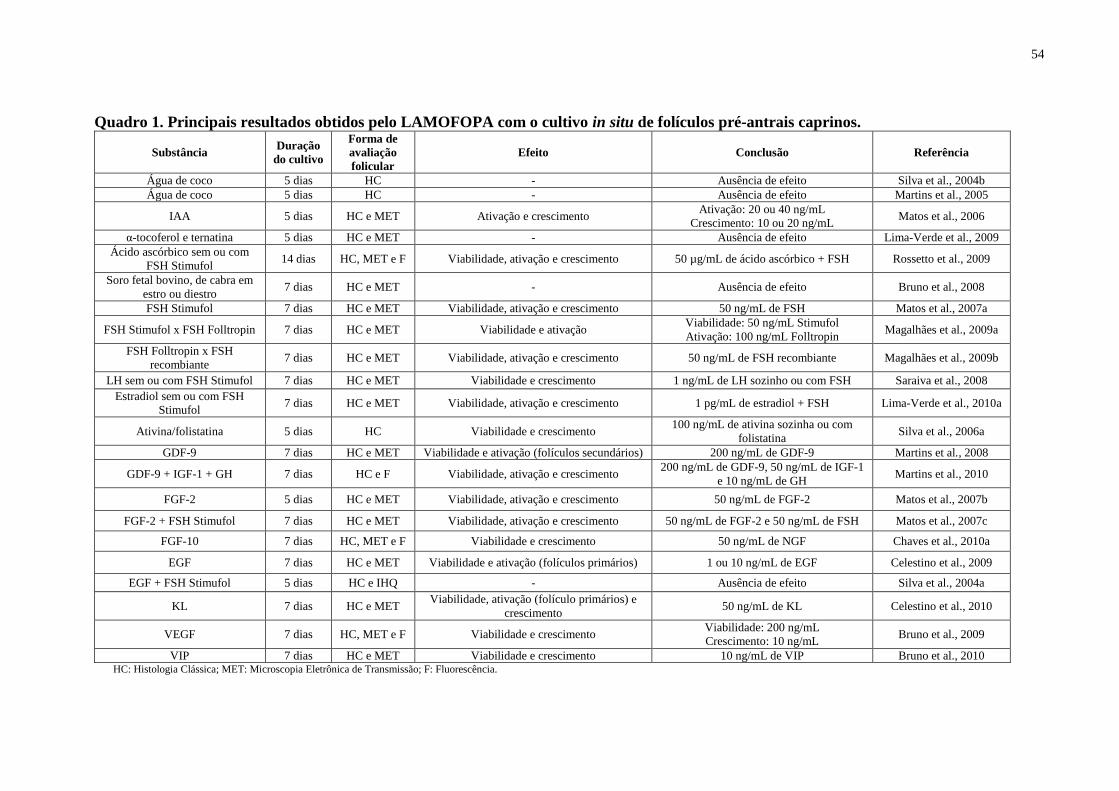
54
Quadro 1. Principais resultados obtidos pelo LAMOFOPA com o cultivo in situ de folículos pré-antrais caprinos.
Substância Duração
do cultivo
Forma de
avaliação
folicular
Efeito Conclusão Referência
Água de coco 5 dias HC - Ausência de efeito Silva et al., 2004b
Água de coco 5 dias HC - Ausência de efeito Martins et al., 2005
IAA 5 dias HC e MET Ativação e crescimento Ativação: 20 ou 40 ng/mL
Crescimento: 10 ou 20 ng/mL Matos et al., 2006
α-tocoferol e ternatina 5 dias HC e MET - Ausência de efeito Lima-Verde et al., 2009
Ácido ascórbico sem ou com
FSH Stimufol 14 dias HC, MET e F Viabilidade, ativação e crescimento 50 µg/mL de ácido ascórbico + FSH Rossetto et al., 2009
Soro fetal bovino, de cabra em
estro ou diestro 7 dias HC e MET - Ausência de efeito Bruno et al., 2008
FSH Stimufol 7 dias HC e MET Viabilidade, ativação e crescimento 50 ng/mL de FSH Matos et al., 2007a
FSH Stimufol x FSH Folltropin 7 dias HC e MET Viabilidade e ativação Viabilidade: 50 ng/mL Stimufol
Ativação: 100 ng/mL Folltropin Magalhães et al., 2009a
FSH Folltropin x FSH
recombiante 7 dias HC e MET Viabilidade, ativação e crescimento 50 ng/mL de FSH recombiante Magalhães et al., 2009b
LH sem ou com FSH Stimufol 7 dias HC e MET Viabilidade e crescimento 1 ng/mL de LH sozinho ou com FSH Saraiva et al., 2008
Estradiol sem ou com FSH
Stimufol 7 dias HC e MET Viabilidade, ativação e crescimento 1 pg/mL de estradiol + FSH Lima-Verde et al., 2010a
Ativina/folistatina 5 dias HC Viabilidade e crescimento 100 ng/mL de ativina sozinha ou com
folistatina Silva et al., 2006a
GDF-9 7 dias HC e MET Viabilidade e ativação (folículos secundários) 200 ng/mL de GDF-9 Martins et al., 2008
GDF-9 + IGF-1 + GH 7 dias HC e F Viabilidade, ativação e crescimento 200 ng/mL de GDF-9, 50 ng/mL de IGF-1
e 10 ng/mL de GH Martins et al., 2010
FGF-2 5 dias HC e MET Viabilidade, ativação e crescimento 50 ng/mL de FGF-2 Matos et al., 2007b
FGF-2 + FSH Stimufol 7 dias HC e MET Viabilidade, ativação e crescimento 50 ng/mL de FGF-2 e 50 ng/mL de FSH Matos et al., 2007c
FGF-10 7 dias HC, MET e F Viabilidade e crescimento 50 ng/mL de NGF Chaves et al., 2010a
EGF 7 dias HC e MET Viabilidade e ativação (folículos primários) 1 ou 10 ng/mL de EGF Celestino et al., 2009
EGF + FSH Stimufol 5 dias HC e IHQ - Ausência de efeito Silva et al., 2004a
KL 7 dias HC e MET Viabilidade, ativação (folículo primários) e
crescimento 50 ng/mL de KL Celestino et al., 2010
VEGF 7 dias HC, MET e F Viabilidade e crescimento Viabilidade: 200 ng/mL
Crescimento: 10 ng/mL Bruno et al., 2009
VIP 7 dias HC e MET Viabilidade e crescimento 10 ng/mL de VIP Bruno et al., 2010 HC: Histologia Clássica; MET: Microscopia Eletrônica de Transmissão; F: Fluorescência.
55
2.3.2 Cultivo in vitro de folículos pré-antrais caprinos isolados
Existem poucos trabalhos com o cultivo in vitro de folículos pré-antrais isolados em
caprinos. Embora o número de resultados obtidos até o momento com esse sistema nesta
espécie ainda seja pequeno, bons resultados têm sido alcançados, incluindo a manutenção da
sobrevivência folicular, crescimento folicular e oocitário, formação de antro, obtenção de
oócitos competentes para retomar a meiose, e ainda, a obtenção de embriões a partir de
oócitos provenientes de folículos pré-antrais cultivados in vitro.
ZHOU e ZHANG (2000) reportaram o desenvolvimento in vitro de folículos primários
e secundários caprinos isolados (50 a 150 µm de diâmetro), demonstrando que folículos
primários desenvolvem até o estádio de folículos secundários, embora poucos destes folículos
oriundos do estádio primário desenvolvam-se até antrais. Além disso, foi observado que a
sobrevivência dos folículos secundários foi maior do que a dos folículos primários, e que uma
grande quantidade dos folículos secundários atingiram o estádio antral.
RAJARAJAN et al. (2006) investigaram o efeito do TGF-α (10 ng/mL), fator de
crescimento semelhante à insulina II (IGF-II; 20 ng/mL), EGF (50 ng/mL) e FSH (1 µg/mL)
após 6 dias de cultivo in vitro de pequenos (40–60 µm) e grandes (61–100 µm) folículos pré-
antrais caprinos. Nesse estudo, eles mostraram que o EGF, TGF-α e IGF-II estimularam o
crescimento de pequenos e grandes folículos pré-antrais, enquanto o FSH foi mais eficiente
em estimular o desenvolvimento apenas de pequenos folículos pré-antrais.
Focando em oócitos, foi estudado o efeito do EGF (50 mg/L) e IGF-I (100 mg/L),
ambos individualmente ou em associação, sobre a viabilidade e o crescimento in vitro de
oócitos de folículos pré-antrais caprinos (ZHOU; ZHANG, 2005a). Os resultados mostraram
elevadas taxas de sobrevivência e crescimento do oócito quando o EGF foi associado ao IGF-
I. Além disso, ambos os fatores de crescimento, quando utilizados individualmente,
melhoraram a sobrevivência e o crescimento do oócito quando comparado ao controle. Em
outro trabalho desses mesmos autores, além de ter sido observado os efeitos benéficos da
associação do EGF e IGF-I conforme descrito anteriormente, o FGF-2 (50 ng/mL) estimulou
a sobrevivência do oócito, embora não tenha causado nenhum efeito no seu crescimento
(ZHOU; ZHANG, 2005b).
Os primeiros trabalhos do nosso grupo com o cultivo in vitro de folículos secundários
isolados foram realizados mais recentemente, envolvendo condições básicas de cultivo, como
por exemplo, tensão de oxigênio ideal, regime de troca de meio, bem como a forma do cultivo
56
de folículos isolados, individual ou em grupo, com ou sem FSH, na presença ou ausência de
folículos antrais.
Ao testar duas diferentes tensões de oxigênio (5 ou 20%) observou-se que, na
concentração de 20% de O2, os folículos pré-antrais caprinos crescidos in vitro após 30 dias
de cultivo apresentaram um maior crescimento, e um destaque para a retomada da meiose
(SILVA et al., 2010).
Em outro estudo foram avaliados os efeitos de diferentes intervalos de troca de meio
(2 ou 6 dias) sobre a viabilidade, formação da cavidade antral, crescimento e maturação in
vitro de oócitos caprinos e ovinos oriundos de folículos pré-antrais isolados e cultivados por
24 dias. Os folículos pré-antrais caprinos e ovinos comportaram-se de maneira completamente
diferente. De acordo com os resultados obtidos, para melhorar a eficiência dos sistemas de
cultivo, o meio deve ser trocado a cada 2 e 6 dias para folículos pré-antrais caprinos e ovinos,
respectivamente (MAGALHÃES et al., 2010b).
Outro estudo testou ainda a influência de diferentes regimes de troca sobre o
desenvolvimento in vitro de folículos pré-antrais caprinos cultivados por 18 dias. Nesse
estudo foi testado três diferentes regimes de troca: T1 (remoção de meio seguida da adição do
mesmo volume de meio fresco a cada troca); T2 (somente adição de meio fresco a cada troca)
e T3 (remoção de meio seguida da adição de 5 μl a mais de meio fresco a cada troca, ou seja,
aumento de 5 μl do volume final em cada troca). Como conclusão, foi observado que embora
o T3 tenha sido tão bom quanto o T2 no parâmetro de crescimento folicular, o T2, ou seja, de
adição periódica de meio foi o tratamento recomendado por ser mais prático, manter a
sobrevivência, além de promover o desenvolvimento in vitro de folículos pré-antrais caprinos,
com uma superior percentagem de oócitos destinados para MIV quando comparado aos
demais tratamentos (ARAÚJO et al., 2010c).
Também foram testadas diferentes formas de cultivo, por exemplo, individual ou em
grupo, com ou sem FSH, na presença ou na ausência de folículos antrais. Nesse estudo, na
ausência de FSH, o cultivo por 24 dias de folículos pré-antrais em grupo mostrou taxas
superiores de sobrevivência e crescimento em relação aos folículos cultivados
individualmente. Por outro lado, na presença de FSH, esses parâmetros não foram afetados,
mas foi observado um grande número de oócitos crescidos e que retomaram a meiose quando
os folículos foram cultivados em grupo. Finalmente, o co-cultivo de folículos antrais com
folículos pré-antrais afetou negativamente a sobrevivência e o crescimento folicular, enquanto
que resultados opostos foram observados quando os folículos foram cultivados em grupo
57
(DUARTE et al., 2010). Utilizando ainda esse hormônio, em outro estudo foi verificado que o
FSH na presença de 10% de soro fetal bovino teve um importante papel no crescimento de
folículos pré-antrais, não só na espécie caprina, mas também na espécie ovina, após 18 dias de
cultivo (RODRIGUES et al., 2010).
Em outro estudo realizado com FSH, em que foi verificada a influência desse
hormônio sobre o desenvolvimento in vitro de folículos caprinos isolados cultivados por 18
dias, foram testadas concentrações fixas de 100 ou 1000 ng/mL, ou adições do FSH ao meio
de cultivo de modo seqüencial, ou seja, T1: FSH 100 ng/mL (do dia 0 ao dia 6), FSH 500
ng/mL (do dia 6 ao dia 12) e FSH 1000 ng/mL (do dia 12 ao dia 18), e T2: FSH 500 ng/mL
(do dia 0 ao dia 9) e 1000 ng/mL (do dia 9 ao dia 18). Nesse estudo foi verificado que a
utilização do Tratamento 1 melhorou a sobrevivência, formação de antro e reduziu a extrusão
do oócito, e ambos os meios sequenciais promoveram uma elevada taxa de retomada da
meiose (SARAIVA et al., 2010b).
Empregando ainda esse sistema de cultivo, foi observada a influência do EGF sozinho
ou associado ao FSH sobre o desenvolvimento in vitro de folículos pré-antrais caprinos
isolados cultivados por 6 dias. Além disso, foi verificada a influência da adição dessas
substâncias ao meio de cultivo sobre a expressão do RNAm para o EGF e FSH-R. Nele
observou-se que o EGF e o FSH promoveram o crescimento de folículos secundários
caprinos, reduziram os níveis de RNAm para o EGF, e ainda, o EGF diminuiu os níveis de
RNAm para FSH-R em folículos secundários caprinos cultivados. O capítulo 8 da presente
Tese mostrará a metodologia e os resultados desse experimento mais detalhadamente.
Foi testado ainda o efeito do momento da adição de LH (100 ng/mL) ao meio de
cultivo sobre o desenvolvimento in vitro de folículos pré-antrais caprinos. Nele observou-se
que quando adicionado tal hormônio desde o início do cultivo, ou seja, é feita uma exposição
prolongada, há um detrimento na retomada da meiose desses folículos cultivados in vitro. Já
em um cultivo de 18 dias, quando este hormônio foi adicionado a partir do dia 6, houve uma
elevada sobrevivência e os oócitos foram hábeis para retomar a meiose (SILVA et al., aceito
para publicação).
Por último, os resultados mais relevantes são relativos à obtenção de embriões
caprinos a partir de oócitos oriundos de folículos pré-antrais isolados crescidos, maturados e
fecundados in vitro (SARAIVA et al., 2010a e MAGALHÃES et al., 2011). Tais resultados
foram alcançados com a adição de alguns hormônios, como o FSH de forma sequencial (100
ng/mL do dia 0 ao dia 6, 500 ng/mL do dia 6 ao dia 12 e 1000 ng/mL do dia 12 ao dia 18), LH

58
(100 ng/mL) e GH (50 ng/mL), bem como com a adição do fator de crescimento EGF (100
ng/mL). No Quadro 2, podem ser observados os principais resultados obtidos com o cultivo
de folículos pré-antrais caprinos isolados.
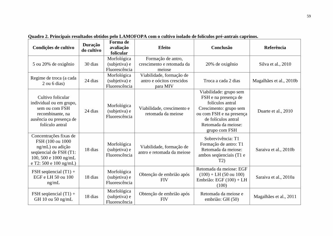
59
Quadro 2. Principais resultados obtidos pelo LAMOFOPA com o cultivo isolado de folículos pré-antrais caprinos.
Condições de cultivo Duração
do cultivo
Forma de
avaliação
folicular
Efeito Conclusão Referência
5 ou 20% de oxigênio 30 dias
Morfológica
(subjetiva) e
Fluorescência
Formação de antro,
crescimento e retomada da
meiose
20% de oxigênio Silva et al., 2010
Regime de troca (a cada
2 ou 6 dias) 24 dias
Morfológica
(subjetiva) e
Fluorescência
Viabilidade, formação de
antro e oócitos crescidos
para MIV
Troca a cada 2 dias Magalhães et al., 2010b
Cultivo folicular
individual ou em grupo,
sem ou com FSH
recombinante, na
ausência ou presença de
folículo antral
24 dias
Morfológica
(subjetiva) e
Fluorescência
Viabilidade, crescimento e
retomada da meiose
Viabilidade: grupo sem
FSH e na presença de
folículos antral
Crescimento: grupo sem
ou com FSH e na presença
de folículos antral
Retomada da meiose:
grupo com FSH
Duarte et al., 2010
Concentrações fixas de
FSH (100 ou 1000
ng/mL) ou adição
seqüencial de FSH (T1:
100, 500 e 1000 ng/mL
e T2: 500 e 100 ng/mL)
18 dias
Morfológica
(subjetiva) e
Fluorescência
Viabilidade, formação de
antro e retomada da meiose
Sobrevivência: T1
Formação de antro: T1
Retomada da meiose:
ambos seqüenciais (T1 e
T2)
Saraiva et al., 2010b
FSH seqüencial (T1) +
EGF e LH 50 ou 100
ng/mL
18 dias
Morfológica
(subjetiva) e
Fluorescência
Obtenção de embrião após
FIV
Retomada da meiose: EGF
(100) + LH (50 ou 100)
Embrião: EGF (100) + LH
(100)
Saraiva et al., 2010a
FSH seqüencial (T1) +
GH 10 ou 50 ng/mL 18 dias
Morfológica
(subjetiva) e
Fluorescência
Obtenção de embrião após
FIV
Retomada da meiose e
embrião: GH (50) Magalhães et al., 2011
60
2.4 Estado atual do cultivo in vitro de folículos pré-antrais
É indiscutível a ocorrência de grandes progressos no cultivo in vitro de folículos pré-
antrais em diferentes espécies animais. Em gatas (JEWGENOW; STOLTE, 1996), gambás
(BUTCHER; ULLMAN, 1996) e macacas (FORTUNE et al., 1998) foi observado o
crescimento de folículos ovarianos pré-antrais isolados após o cultivo in vitro, porém, sem a
formação de antro. Em humanos (ROY; TREACY, 1993; TELFER et al., 2008), bovinos
(GUTIERREZ et al., 2000; MCCAFFERY et al., 2000) e cadelas (SERAFIM et al., 2010),
folículos pré-antrais isolados foram cultivados in vitro e desenvolveram-se até o estádio
antral. Com relação aos estudos com outras espécies domésticas, os resultados mais relevantes
foram obtidos com suínos (WU; EMERY; CARREL, 2001; WU; TIAN, 2007), búfalos
(GUPTA et al., 2008), ovinos (ARUNAKUMARI; SHANMUGASUNDARAM; RAO, 2010)
e caprinos (SARAIVA et al., 2010a; MAGALHÃES et al., 2011), os quais obtiveram a
produção de embriões após o cultivo de folículos pré-antrais e posterior maturação e
fecundação in vitro dos oócitos.
Apesar do grande avanço no cultivo in vitro de folículos pré-antrais com as espécies
supracitadas, os resultados mais satisfatórios têm sido observados em animais de laboratório.
CARROLL et al. (1990) obtiveram o nascimento de camundongos in vitro após congelação e
descongelação, crescimento, maturação e fecundação in vitro de oócitos oriundos de folículos
primários. Posteriormente, EPPIG e O‘BRIEN (1996) obtiveram o nascimento de um
camundongo a partir de folículos primordiais crescidos, maturados e fecundados in vitro.
Mais recentemente, esta mesma equipe, aperfeiçoando o protocolo utilizado anteriormente,
relatou a produção de embriões e o nascimento de 59 camundongos saudáveis a partir de
folículos pré-antrais cultivados, maturados e fecundados in vitro (O‘BRIEN; PENDOLA;
EPPIG, 2003).
2.5 Técnicas para análise folicular durante o cultivo in vitro
Diferentes técnicas podem ser utilizadas para análise folicular durante o cultivo in
vitro de folículos pré-antrais, auxiliando assim no sucesso do cultivo. Dentre elas, podemos
destacar aquelas que permitem a avaliação da qualidade folicular, como por exemplo, a
histologia clássica (HC), a microscopia eletrônica de transmissão (MET) e a microscopia de
fluorescência. Além disso, existem ainda as técnicas de biologia molecular que permitem o
61
estudo da expressão de genes que codificam ligantes e/ou receptores de diferentes substâncias
importantes para a foliculogênese, contribuindo para uma melhor elucidação desse processo.
A seguir, será abordada brevemente cada uma dessas técnicas.
2.5.1 Histologia Clássica
A histologia clássica (HC) é uma técnica importante para avaliação dos folículos pré-
antrais cultivados in vitro, pois além de permitir uma análise quantitativa, ou seja, de um
grande número de folículos cultivados, permite ainda verificar o número e a mudança na
morfologia das células da granulosa de pavimentosa para cúbica, no momento da ativação
folicular, além de analisar a integridade morfológica do oócito e das células da granulosa.
Tais avaliações permitem, portanto, a classificação dos folículos quanto ao seu estádio de
desenvolvimento (primordial, transição, primário ou secundário), e ainda quanto às suas
características morfológicas (normais ou atrésicos). Entretanto, a HC possui como
desvantagens não permitir a avaliação da integridade de membranas e das organelas
citoplasmáticas. Vale salientar que tal técnica pode ser realizada tanto em folículos isolados,
como naqueles inclusos em fragmentos de córtex ovariano (MATOS et al., 2007d).
Alguns autores mostraram que, na análise histológica, as alterações indicativas de
atresia em folículos pré-antrais ocorrem primariamente no oócito, sendo a picnose nuclear o
primeiro sinal de atresia (JORIO; MARIANA; LAHLOU-KASSI, 1991; WOOD; MONTALI;
WILDT, 1997; SILVA et al., 2002). Outros estudos realizados mostraram que a HC pode dar
resultados similares aos de viabilidade folicular utilizando marcadores fluorescentes como a
calceína e o corante azul de tripan (CORTVRINDT; SMITZ, 2001b; AMORIM et al., 2003).
Além disso, outro estudo relatou ainda que a HC (coloração com hematoxilina-eosina ou
ácido períodico de Schiff-hematoxilina) pode ser utilizada para avaliar a morte da célula por
necrose, através da visualização da presença de vacúolos (MARTINEZ-MADRID et al.,
2007).
2.5.2 Microscopia Eletrônica de Transmissão
A MET é considerada uma técnica qualitativa e acurada, capaz de permitir a avaliação
da integridade de membranas celulares e organelas citoplasmáticas (SALEHNIA;
MOGHADAM; VELOJERDI, 2002). Ela se mostra como uma ferramenta valiosa para
62
detectar modificações morfológicas iniciais devido à atresia, as quais podem ser observadas
apenas em nível ultraestrutural, antes de se tornarem mais visíveis e possíveis de serem
identificadas por microscopia óptica. Tal técnica é, portanto, capaz de discernir a qualidade do
oócito e das células da granulosa (LUCCI et al., 2001; LOPES et al., 2009), sendo deste
modo, comumente utilizada como uma técnica complementar à HC. Com esta técnica é
possível ainda avaliar a atresia, ou seja, a morte do folículo tanto por apoptose
(STALDEMANN; LASSMAN, 2000) como pelo processo degenerativo de necrose
(MARTINEZ-MADRID et al., 2007).
No que se refere à utilização da MET após o cultivo, ela serve para confirmar se
realmente houve a manutenção da integridade morfológica após análise histológica. MATOS
et al. (2006) embora tenham encontrado uma elevada percentagem de folículos
histologicamente normais no MEM suplementado com IAA (20 ng/mL) após 5 dias de
cultivo, os estudos ultraestruturais não confirmaram a manutenção da integridade morfológica
desses folículos, mesmo após 1 dia de cultivo. Já em outros estudos com o cultivo in vitro de
folículos pré-antrais, os resultados da histologia clássica puderam ser confirmados por MET
(MATOS et al., 2007a,b; MARTINS et al., 2008, ROSSETTO et al., 2009; LIMA-VERDE et
al., 2010a).
2.5.3 Microscopia de Fluorescência
Na microscopia de fluorescência são utilizados marcadores fluorescentes, que quando
excitados por certos comprimentos de onda, absorvem energia e emitem luz de maior
comprimento de onda (JUNQUEIRA; CARNEIRO, 1999). A microscopia de fluorescência é
considerada uma técnica confiável, prática e rápida para analisar a viabilidade folicular
(CORTVRINDT; SMITZ, 2001b; LOPES et al., 2009), tendo sido, portanto, empregada para
avaliação da viabilidade de folículos pré-antrais após o cultivo in vitro em diversos trabalhos
(BRUNO et al., 2009; SILVA et al., 2010; MAGALHÃES et al., 2010b). Para tanto, os
folículos pré-antrais foram analisados pelo teste de viabilidade celular por fluorescência
baseado na detecção simultânea de células vivas e mortas marcadas por calceína-AM e pelo
etídio homodímero-1, respectivamente. Enquanto a primeira sonda detecta atividade da
esterase intracelular, enzima característica de células viáveis, a outra cora ácidos nucléicos em
células não-viáveis, com ruptura na membrana plasmática (LOPES et al., 2009). Dessa forma,
essa técnica pode oferecer uma nova maneira para investigações metabólicas e aspectos do
63
desenvolvimento folicular in vitro (BRUNO et al., 2009). As sondas fluorescentes calceína-
AM e etídio homodímero-1 já haviam sido utilizadas com sucesso para avaliação da
viabilidade de folículos bovinos em estádios iniciais (SCHOTANUS et al., 1997; VAN DEN
HURK et al., 1998), tendo sido a sonda calceína-AM também já empregada para determinar a
densidade e o número de folículos presentes em biópsias ovarianas humanas
(CORTVRINDT; SMITZ, 2001b).
Outro marcador que pode ser utilizado na microscopia de fluorescência para avaliar os
folículos cultivados in vitro é o Hoescht 33342. Esse marcador penetra em células vivas e
marca a cromatina, permitindo assim a avaliação do estádio meiótico folicular (JEWGENOW,
1998; MATOS et al., 2007d).
2.5.4 Biologia Molecular
As técnicas de biologia molecular podem ser utilizadas antes, durante e após o cultivo
in vitro de folículos pré-antrais, com o intuito de identificar e quantificar o local de atuação e
produção de cada substância envolvida nas diferentes etapas do desenvolvimento folicular. De
fato, muitos papéis das células foliculares ovarianas relacionados à sobrevivência, ao
crescimento e à diferenciação são refletidos na alteração dos padrões da expressão gênica.
Desta forma, a capacidade de quantificar os níveis de transcrição de genes específicos é
fundamental para garantir a completa investigação das funções foliculares (ZAMORANO;
MAHESH; BRANN, 1996). Dentre as técnicas de biologia molecular, podem ser citadas
aquelas que detectam, localizam ou identificam os ácidos nucléicos (hibridização in situ
Southern e Northern Blotting) ou proteínas (Western Blotting), as que podem efetuar a
quantificação do DNA (Reação em Cadeia de Polimerase - PCR) ou do RNA (Reação de
Transcriptase Reversa em Cadeia de Polimerase - RT-PCR), as que permitem a quantificação
da expressão do RNAm, mesmo em uma mistura complexa de RNA total (ensaios de proteção
de ribonuclease), ou que possibilitem a análise da expressão de milhares de genes
simultaneamente (Microarranjos de DNA).
No que diz respeito ao estudo da foliculogênese, as técnicas de biologia molecular
aplicadas isoladamente ou em associação podem fornecer dados essenciais para fundamentar
a base do conhecimento sobre a expressão de RNAm, tanto de ligantes quanto de receptores
associados a esse processo. Atualmente, a técnica mais utilizada para quantificar a expressão
de RNAm é a RT-PCR em tempo real (RT-qPCR) (KREUZER; MASSEY, 2002). A qPCR

64
uma variante da RT-PCR convencional, permite uma análise precisa da quantificação da
expressão gênica em determinado tecido ou amostra biológica. Esse método utiliza um
sistema fluorescente em plataforma, capaz de detectar a luz oriunda da reação de amplificação
de um determinado gene no momento real da amplificação (BUSTIN, 2002). Através da
técnica de RT-qPCR e da RT-PCR convencional, já foi possível identificar a presença de
diferentes substâncias, como hormônios e fatores de crescimento, presentes em folículos
ovarianos caprinos (SILVA et al., 2004c, 2005, 2006b,c; SARAIVA et al., 2010b).
65
3 JUSTIFICATIVA
Os folículos primordiais constituem o pool de reserva de folículos quiescentes e
compreendem cerca de 95% de toda população folicular presente no ovário mamífero. No
entanto, para que estes folículos possam entrar em fase de crescimento é preciso que sejam
ativados. Neste contexto, sabendo-se do grande valor econômico que a espécie caprina
representa em especial para o Nordeste Brasileiro, é de extrema importância o
desenvolvimento de um sistema de cultivo capaz de ativar esses folículos e assegurar seu
posterior crescimento in vitro, otimizando o aproveitamento do potencial oocitário desses
animais e incrementando a eficiência da reprodução animal. Os oócitos oriundos destes
folículos crescidos in vitro poderiam ser utilizados em programas de produção in vitro e
transferência de embriões e/ou criopreservação. Além disso, o desenvolvimento de um
sistema de cultivo eficiente poderá fornecer subsídios para uma melhor compreensão sobre os
fatores que regulam a foliculogênese na fase pré-antral, necessários para a sobrevivência, a
ativação e o início do crescimento folicular.
Estudos referentes aos fatores e mecanismos envolvidos na regulação e ativação dos
folículos primordiais são escassos, especialmente em animais de produção, como os caprinos.
Neste contexto, diversos autores têm investigado o efeito de diferentes fatores de crescimento
no cultivo de folículos pré-antrais de animais de laboratório e de animais domésticos como
vaca, ovelha e cabra. Entretanto, os efeitos de diferentes concentrações dos fatores como o
KL, BMP-15 e EGF, que são importantes reguladores da foliculogênese, ainda não haviam
sido testados no cultivo in vitro de folículos pré-antrais caprinos. Para este fim, além da
técnica de histologia clássica, foi empregada a microscopia eletrônica de transmissão e/ou
microscopia de fluorescência para determinar a qualidade de folículos pré-antrais caprinos
cultivados in vitro e, consequentemente, melhor avaliar a eficiência dos meios de cultivo
testados. Na tentativa de melhor desvendar a foliculogênese caprina, foi ainda realizado um
estudo de quantificação da expressão gênica do RNAm para o KL, BMP-15 e EGF por meio
da técnica de RT-PCR em tempo real (RT-qPCR), permitindo dessa forma, estabelecer
estratégias para adição destes componentes em momentos ideais do cultivo.
Com o desenvolvimento de um eficiente sistema de cultivo, será possível, no futuro, a
utilização dos oócitos oriundos de uma numerosa população de folículos pré-antrais crescidos
in vitro em diversas biotécnicas reprodutivas, dentre elas a fecundação in vitro, contribuindo
assim para a produção de embriões em larga escala.
66
4 HIPÓTESES CIENTÍFICAS
Diante do exposto, foram formuladas as seguintes hipóteses científicas:
1) A expressão dos níveis de RNAm para o KL, BMP-15 e EGF varia de acordo com o
estádio de desenvolvimento folicular e com os tipos celulares que compõem o folículo.
2) As substâncias KL, BMP-15 e EGF afetam positivamente o desenvolvimento in vitro de
folículos pré-antrais caprinos inclusos em fragmentos de tecido ovariano de uma maneira
concentração-dependente.
3) O EGF, associado ou não ao FSH, influencia positivamente o crescimento in vitro de
folículos secundários caprinos isolados, bem como os níveis de expressão do RNAm para o
EGF e FSH-R.
67
5 OBJETIVOS
5.1 OBJETIVO GERAL
1) Verificar a expressão do RNAm para o KL, BMP-15 e EGF em folículos ovarianos
caprinos.
2) Avaliar o efeito do KL, BMP-15 e EGF sobre o desenvolvimento de folículos pré-antrais
caprinos cultivados in vitro.
5.2 OBJETIVOS ESPECÍFICOS
1) Quantificar, através da técnica de RT-PCR em tempo real (RT-qPCR), os níveis de RNAm
para KL, BMP-15 e EGF nas diferentes categorias (folículo primordial, primário e
secundário) e compartimentos de folículos antrais (oócito, células da granulosa e da teca)
em ovários caprinos.
2) Estabelecer a curva concentração-resposta de KL, BMP-15 e EGF, tendo como parâmetros
a sobrevivência, a ativação e o crescimento de folículos pré-antrais caprinos inclusos em
tecido ovariano e cultivados in vitro por 1 ou 7 dias.
3) Analisar morfológica e ultraestruturalmente os folículos pré-antrais caprinos inclusos em
tecido ovariano e cultivados in vitro por 1 ou 7 dias com KL, BMP-15 e EGF, e ainda a
viabilidade dos folículos cultivados com BMP-15.
4) Verificar o efeito do EGF, na presença ou ausência de FSH, sobre a sobrevivência,
formação de antro e crescimento de folículos secundários caprinos isolados e cultivados in
vitro por 6 dias, bem como sobre os níveis de RNAm para o EGF e FSH-R após cultivo
folicular.
Nas páginas seguintes, serão apresentados os quatro primeiros capítulos desta Tese,
correspondendo a dois artigos de revisão que já foram publicadas em periódico incluso no

68
Qualis CAPES categoria ―B3‖, e as outras duas estão sob revisão. Além disso, em
seguida, serão apresentadas as metodologias e os resultados desta Tese na forma de quatro
capítulos referentes a quatro artigos científicos. Vale salientar que dois artigos já foram
publicados em periódico incluso no Qualis CAPES categoria ―A1‖ e outros dois
encontram-se em fase de julgamento.

69
6 CAPÍTULO 1
Mecanismos de atresia em folículos ovarianos
Mechanisms of atresia in ovarian follicles
Periódico: Animal Reproduction 6: 495-508, 2009.
70
Resumo
Existem milhares a milhões de folículos no ovário mamífero, e a grande maioria deles
(99,9%) é eliminada pelo processo conhecido como atresia. Esse fenômeno ocorre em
qualquer estádio de desenvolvimento folicular, através da apoptose ou do processo
degenerativo de necrose. Assim, um melhor conhecimento dos mecanismos envolvidos na
atresia é necessário para evitar a grande perda folicular que ocorre in vivo e maximizar o
potencial reprodutivo das fêmeas. A presente revisão foca nos aspectos relacionados à
população folicular e atresia, mecanismos de atresia (apoptose ou processo degenerativo de
necrose), técnicas utilizadas para analisar a atresia em folículos ovarianos, e a ocorrência do
processo atrésico durante os diferentes estádios foliculares.
Palavras-chave: Apoptose. Atresia. Folículo. Necrose. Ovário.
71
Mechanisms of atresia in ovarian follicles
J.J.H. Celestino1,4
, R.N. Chaves1
, M.H.T. Matos2
, M.V.A. Saraiva1
, J.B. Bruno1
, J.E. Maia-
Júnior1
, J.R.V. Silva3
, J.R. Figueiredo1
1
Faculty of Veterinary Medicine, LAMOFOPA, PPGCV, State University of Ceara,
Fortaleza, CE, Brazil. 2Nucleus of Biotechnology Applied to Ovarian Follicle Development,
Federal University of São Francisco Valley, Petrolina, PE, Brazil.
3
Biotechnology Nucleus of Sobral (NUBIS), Federal University of Ceara, Sobral, CE, Brazil.
Abstract
There are thousands to millions of follicles in the mammalian ovary, and the majority
(99.9%) are eliminated by a process known as atresia. This phenomenon occurs in any stage
of follicular development, through an apoptotic manner or the degenerative process of
necrosis. Thus, a better understanding of the mechanisms involved in atresia is necessary to
avoid the great follicular loss that occurs in vivo and to maximize female reproductive
potential. The present review focuses on aspects related to follicular population and atresia,
mechanisms of atresia (apoptosis or the degenerative process of necrosis), techniques used to
analyze atresia in ovarian follicles, and the occurrence of the atretic process during different
follicular stages.
Keywords: apoptosis, atresia, follicle, necrosis, ovary.
Introduction
The mammalian ovary represents a dynamic organ that provides an adequate
environment for the production of several substances, such as hormones and growth factors,
and releases viable female gametes (Johnson, 2003). There are thousands to millions of
ovarian follicles that are the structural and functional units of the mammalian ovary, which
support an appropriate environment for oocyte growth and maturation (Cortvrindt and Smitz,
2001). Despite the great number of follicles present in the ovary, most do not reach ovulation
72
(about 99.9%), and instead die by atresia during growth and maturation (Markström et al.,
2002).
Although atresia results in the loss of many ovarian follicles, this is a crucial event for
the maintenance of mammalian ovarian homeostasis, which assures animal cyclicity
(Amsterdam et al., 2003). In follicles, this process can occur through the apoptosis or the
degenerative process of necrosis (for details see session 3). However, apoptosis is the most
frequent form of physiologic cellular death (van Cruchten and van den Broeck, 2002) leading
to significant follicular loss. Further knowledge of the intracellular mechanism, as well as the
factors that regulate atresia, will contribute to a better comprehension of this process, which
can facilitate the development of strategies to minimize the great follicular loss that occurs in
vivo.
This review outlines the current understanding of these aspects related to follicular
population and atresia, the mechanism of atresia (apoptosis or degenerative process of
necrosis), techniques used to analyze atresia in ovarian follicles, and the occurrence of the
atretic process during different stages of follicular development.
Follicular population and atresia
Folliculogenesis is an event that begins in prenatal life for most species and can be
defined as the process of follicular assembly, growth, and maturation, beginning with the
formation of the primordial follicle and finishing with the preovulatory follicle. The ovarian
follicle is composed of an oocyte surrounded by somatic cells (granulosa and theca cells).
According to the degree of development, follicles can be classified as preantral (primordial,
intermediate, primary, and secondary) and antral follicles (tertiary and preovulatory), as
shown in Fig. 1.
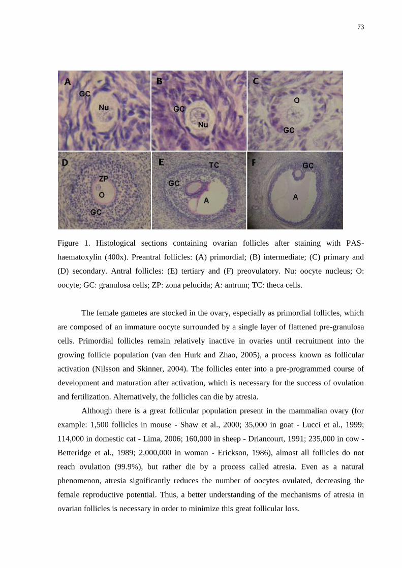
73
Figure 1. Histological sections containing ovarian follicles after staining with PAS-
haematoxylin (400x). Preantral follicles: (A) primordial; (B) intermediate; (C) primary and
(D) secondary. Antral follicles: (E) tertiary and (F) preovulatory. Nu: oocyte nucleus; O:
oocyte; GC: granulosa cells; ZP: zona pelucida; A: antrum; TC: theca cells.
The female gametes are stocked in the ovary, especially as primordial follicles, which
are composed of an immature oocyte surrounded by a single layer of flattened pre-granulosa
cells. Primordial follicles remain relatively inactive in ovaries until recruitment into the
growing follicle population (van den Hurk and Zhao, 2005), a process known as follicular
activation (Nilsson and Skinner, 2004). The follicles enter into a pre-programmed course of
development and maturation after activation, which is necessary for the success of ovulation
and fertilization. Alternatively, the follicles can die by atresia.
Although there is a great follicular population present in the mammalian ovary (for
example: 1,500 follicles in mouse - Shaw et al., 2000; 35,000 in goat - Lucci et al., 1999;
114,000 in domestic cat - Lima, 2006; 160,000 in sheep - Driancourt, 1991; 235,000 in cow -
Betteridge et al., 1989; 2,000,000 in woman - Erickson, 1986), almost all follicles do not
reach ovulation (99.9%), but rather die by a process called atresia. Even as a natural
phenomenon, atresia significantly reduces the number of oocytes ovulated, decreasing the
female reproductive potential. Thus, a better understanding of the mechanisms of atresia in
ovarian follicles is necessary in order to minimize this great follicular loss.
74
Mechanisms of atresia in ovarian follicles
Apoptosis
Apoptosis is a form of programmed cell death and has also been implicated in a
spectrum of processes associated with normal functions of the ovary and follicular
development, such as atresia and corpus luteum regression (Hussein, 2005). This process is
observed in ovarian follicles throughout fetal and adult life. Apoptosis is a genetically
determined and active event, i.e., dependent on the balance of pro-and anti-apoptotic genes
and requires energy (Hussein, 2005). Apoptosis is mediated by active intrinsic mechanisms
and extrinsic factors (Johstone et al., 2002), such as oxidative stress, irradiation, activation of
gene promoters of apoptosis, damage to DNA, cytokines, viral coat proteins, or the
withdrawal of cell growth factors (Johnson, 2003).
The initiation, execution, and regulation of apoptosis involve various biochemical
factors, and the caspase family of enzymes plays a central role in the apoptosis-signaling
network. Caspases are members of the highly conserved family of cysteine proteases with
aspartate specificity. Caspases are expressed as pro-enzymes that undergo proteolytic
processing to generate the activated form after apoptotic stimulus. There are 14 types of
caspases identified as caspase-1 to caspase-14 (Tibbets et al., 2003). Some members of the
family function specifically in cellular death by apoptosis and are subdivided into initiator
(caspases-2, -8, -9, and -10) and executor or effector caspases (caspase-3, -6, and -7; Strasser
et al., 2000). Expression of caspase-3 has been found in ovarian leukocytes and in follicular
cells of atretic follicles (Berardinelli et al., 2004; Tsai et al., 2005). Initiator caspases are
cleaved in response to apoptotic stimuli and activate the effector caspases (Green, 2003).
During apoptosis, the effector caspases cleave numerous proteins located in the cell
membrane, nucleus, and cytoplasm. The activation of caspase-activated DNase (CAD) to
facilitate DNA degradation is one of the important functions mediated by caspases in the
apoptotic process (Nagase et al., 2003).
There are two main apoptotic pathways: (1) the extrinsic or death receptor pathway
and (2) the intrinsic or mitochondrial pathway. However, there is now evidence that the two
pathways are linked and that molecules in one pathway can influence the other (Igney and
Krammer, 2002). The initial apoptotic stimulus strongly influences the pathway that is
activated. The extrinsic and intrinsic pathways converge on the same terminal, or execution
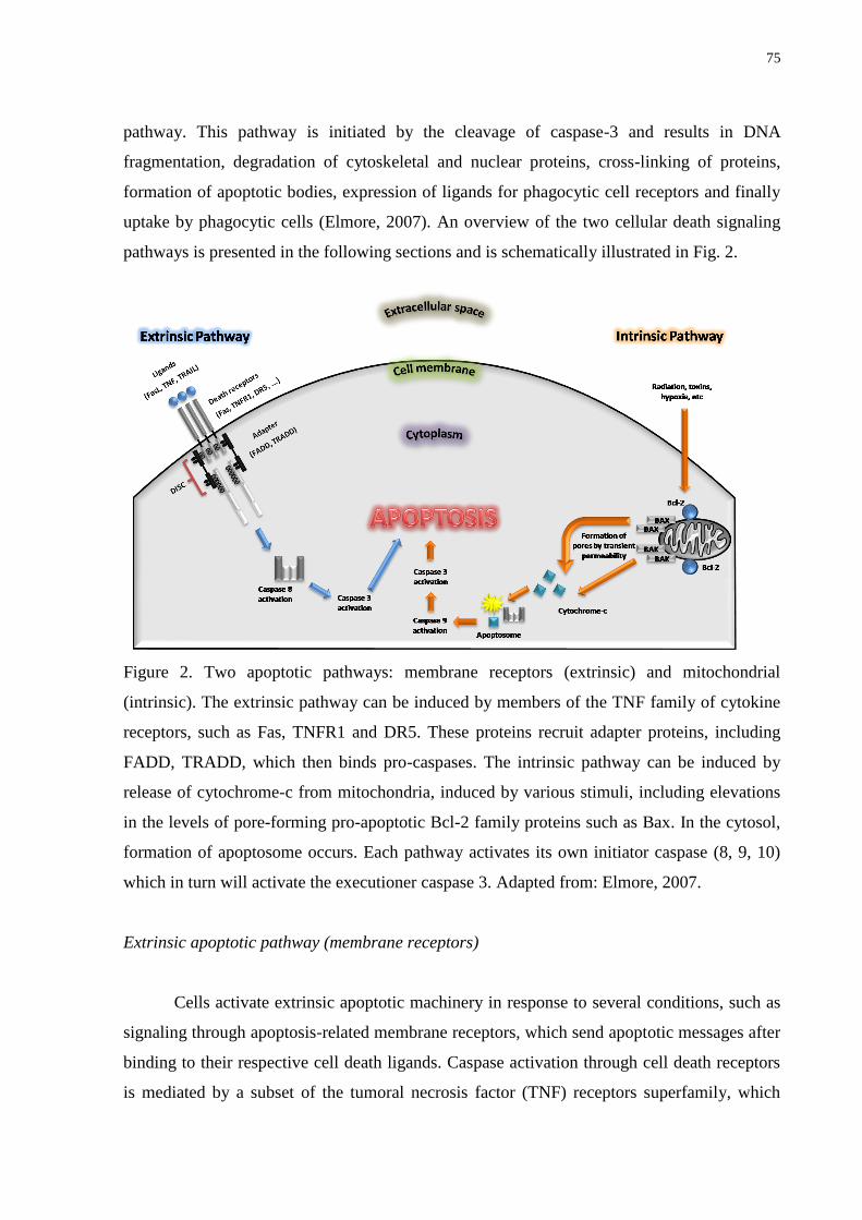
75
pathway. This pathway is initiated by the cleavage of caspase-3 and results in DNA
fragmentation, degradation of cytoskeletal and nuclear proteins, cross-linking of proteins,
formation of apoptotic bodies, expression of ligands for phagocytic cell receptors and finally
uptake by phagocytic cells (Elmore, 2007). An overview of the two cellular death signaling
pathways is presented in the following sections and is schematically illustrated in Fig. 2.
Figure 2. Two apoptotic pathways: membrane receptors (extrinsic) and mitochondrial
(intrinsic). The extrinsic pathway can be induced by members of the TNF family of cytokine
receptors, such as Fas, TNFR1 and DR5. These proteins recruit adapter proteins, including
FADD, TRADD, which then binds pro-caspases. The intrinsic pathway can be induced by
release of cytochrome-c from mitochondria, induced by various stimuli, including elevations
in the levels of pore-forming pro-apoptotic Bcl-2 family proteins such as Bax. In the cytosol,
formation of apoptosome occurs. Each pathway activates its own initiator caspase (8, 9, 10)
which in turn will activate the executioner caspase 3. Adapted from: Elmore, 2007.
Extrinsic apoptotic pathway (membrane receptors)
Cells activate extrinsic apoptotic machinery in response to several conditions, such as
signaling through apoptosis-related membrane receptors, which send apoptotic messages after
binding to their respective cell death ligands. Caspase activation through cell death receptors
is mediated by a subset of the tumoral necrosis factor (TNF) receptors superfamily, which
76
includes TNF receptor type 1 (TNFR1), Fas/CD95 (a membrane-associated polypeptide), and
the TNF-related apoptosis-inducing Ligand (TRAIL) receptors, DR-4 and DR-5 (Slot, 2004;
Contassot et al., 2007). The expression of Fas and FasL, as well as their functions in the ovary
have been shown by Fujino et al. (2008) and Manabe et al. (2008), suggesting that the Fas
system is involved in apoptosis in this organ. Additionally, Porter et al. (2001) showed higher
concentrations of Fas and FasL in bovine atretic follicular cells than in healthy follicles.
Subsequent apoptotic signaling is mediated by the cytoplasmic domain of the death
receptor, which contains a region termed the death domain (DD). Adapter molecules, like
Fas-associated protein with death domain (FADD) or TNF receptor-associated protein with
death domain (TRADD), bind to the activated death receptor, forming the death-inducing
signaling complex (DISC). When bound to the DISC, several pro-caspase-8 molecules are
recruited, resulting in cross-activation by (auto) proteolysis. Caspase-8 is a key initiator
caspase that activates the downstream caspase cascade, and initiates the apoptotic program in
the death receptor pathway (Reed, 2000), following activation in the DISC (Slot, 2004). In
some cell types, a high concentration of caspase-8 in the DISC can process downstream effect
or caspase-3 directly. This latter caspase leads the signaling of apoptosis to the nucleus, where
an endonuclease is released and degrades DNA at each 180-200 kilobase pair fragments
(Hussein, 2005).
Intrinsic pathway of apoptosis (mitochondria)
Mitochondria are not only important for amplifying extrinsic apoptotic pathway, but
also for transmittance of death signals caused by intrinsic stress stimuli and apoptosis
developmental instructions (Joza et al., 2002). Mitochondrial integrity and the release of
cytochrome-c into the cytosol are primarily under the control of the Bcl-2 family members.
The Bcl-2 family of proteins can be divided into anti-apoptotic members, such as Bcl-2, Bcl-
xL, Bcl-w, Mcl-1, Boo, survivin, and pro-apoptotic members, such as Bax, Bak, Bok, and
Diva. The Bcl-2 expression is found in the granulosa cells of both fetal and adult ovaries
(Hussein, 2005; Hussein et al., 2006). Choi et al. (2004) showed a correlation between
decreases in the level of Bcl-2 mRNA with the incidence of apoptosis in isolated granulosa
cells which were cultured under different hormonal treatment conditions. They considered
Bcl-2 to have a critical role in inhibiting the granulosa cell apoptosis pathway. Survivin is one
member of the apoptosis inhibitor protein family that has been shown to bind and inhibit the
77
cell death effectors, caspase-3 and -7 (Shin et al., 2001). In the ovary, survivin acts in
granulosa cells as a bifunctional protein associated with the regulation of the cell cycle and
inhibition of apoptosis (Johnson and Bridgham, 2002). Bax is a pro-apoptotic protein
involved in granulosa cell apoptosis (Tilly et al., 1995) and is an important regulator of
follicle growth, but is dispensable for follicle atresia in mice. In addition, a defect in
folliculogenesis was shown following Bax deletion (Greenfeld et al., 2007).
The key functions related to the cytochrome-c release from the mitochondrial
intermembrane compartment into the cytosol have not been elucidated (Suzuki et al., 2000).
One hypothesis is that Bcl-2 family members may insert inside the outer mitochondrial
membrane to form large pore channels that allow the passage of molecules (Reed and
Kroemer, 2000). Cytosolic cytochrome-c triggers the formation of the mitochondrial
apoptosome, which is indicative of apoptosis and consists of cytochrome-c, Apaf-1
(Apoptotic Protease Activating Factor), and caspase-9 (Joza et al., 2002). The caspase-9
serves as the apical caspase of the mitochondrial pathway (Reed, 2000). Cytochrome-c binds
to the Apaf-1 adapter protein, which recruits pro-caspase-9. This pro-caspase is cleaved into
the active form, caspase-9, which proteolytically activates caspase-3, resulting in cell death.
The link between the death receptor-activated caspase-8 (extrinsic pathway) and
mitochondrial cytochrome-c release (intrinsic pathway) is created by a pro-apoptotic member,
termed Bid. This member is cleaved by caspase-8 and is transferred to the mitochondria,
where Bid acts in concert with the other apoptotic members of Bcl-2 family (Bax and Bak) to
induce the release of cytochrome-c (Slot, 2004).
Another protein which is expressed in the apoptotic granulosa cells of atretic follicles
is 53 protein (p53; Tilly, 1996). This protein functions as a transcription factor in response to
DNA damage, inducing either growth, arrest or apoptosis (Slot, 2004). The p53 is known to
activate the transcription of genes as Bax, Apaf-1, Fas, as well as to repress transcription of
Bcl-2 genes (Bourdon et al., 2003). However, independent of its transcription control of these
genes, p53 has also been shown to engage the apoptotic program by directly activating Bax to
permeabilize mitochondria (Bras et al., 2005).
Morphological alterations in apoptotic cells
Independent of the particular stimulus and pathway, the morphologic features that
characterize apoptosis are pycnosis and karyorrhexis in the nucleus, as well as condensation,
78
swelling, loss of cytoplasmic detail, and fragmentation in the cytoplasm (Zeiss, 2003). The
condensed chromatin appears as crescents along the periphery of the nuclear membrane or as
spherical bodies within the nucleus. The cytoplasmic condensation induces shrinking of the
cell (Hussein, 2005). Subsequently, the nuclear and plasma membranes become convoluted,
and small masses of condensed chromatin undergo fragmentation along with condensed
cytoplasm to form ―apoptotic bodies‖. The apoptotic bodies are bound to the plasma
membrane, and often contain functional mitochondria and other organelles. The phosphatidyl
serine residues that are normally localized to the inner membrane are relocated to the outside
of the cell membrane prior to fragmentation. These residues of phosphatidyl serine on the
apoptotic bodies serve as a signal to the neighboring healthy cells to perform phagocytosis
and remove the cellular debris (Bhatia, 2004). There is essentially no inflammatory reaction
associated with the process of apoptosis nor with the removal of apoptotic cells because: (1)
apoptotic cells do not release their cellular constituents into the surrounding interstitial tissue;
(2) they are quickly phagocytosed by surrounding cells thus likely preventing secondary
necrosis; and (3) the engulfing cells do not produce anti-inflammatory cytokines (Savill and
Fadok, 2000; Kurosaka et al., 2003).
Necrosis
Generally, necrosis is initiated by non-cellular mechanisms, such as ischemia, ATP
depletion (Bhatia, 2004), and traumatic insults, which lead to irreversible cellular damage
(McCully et al., 2004). In addition to passive mechanisms, studies have suggested that
―active‖ mechanisms, such as Na+
overloading, Ca2+
accumulation, and changes in
mitochondria permeability, can also participate in the necrotic process (Barros et al., 2001b;
Padanilam, 2003).
The biochemical pathway that leads to necrotic cell death is not well known. In
ischemic or hypoxic injury, energy depletion occurs by defective ATP production associated
with the rapid consumption of ATP by Na+
-K+
pumps and through hydrolysis as well as ATP
loss. The necrotic volume increase associated with necrotic cell death is initiated by an influx
of Na+
and release of ATP due to membrane leakage (Padanilam, 2003). The increased Na+
levels in the cytosol activate the Na+
-K+
-ATPase pump, resulting in dissipation of ATP. In the
early stages of the injury, a simultaneous efflux of K+
maintains ion homeostasis. Severe
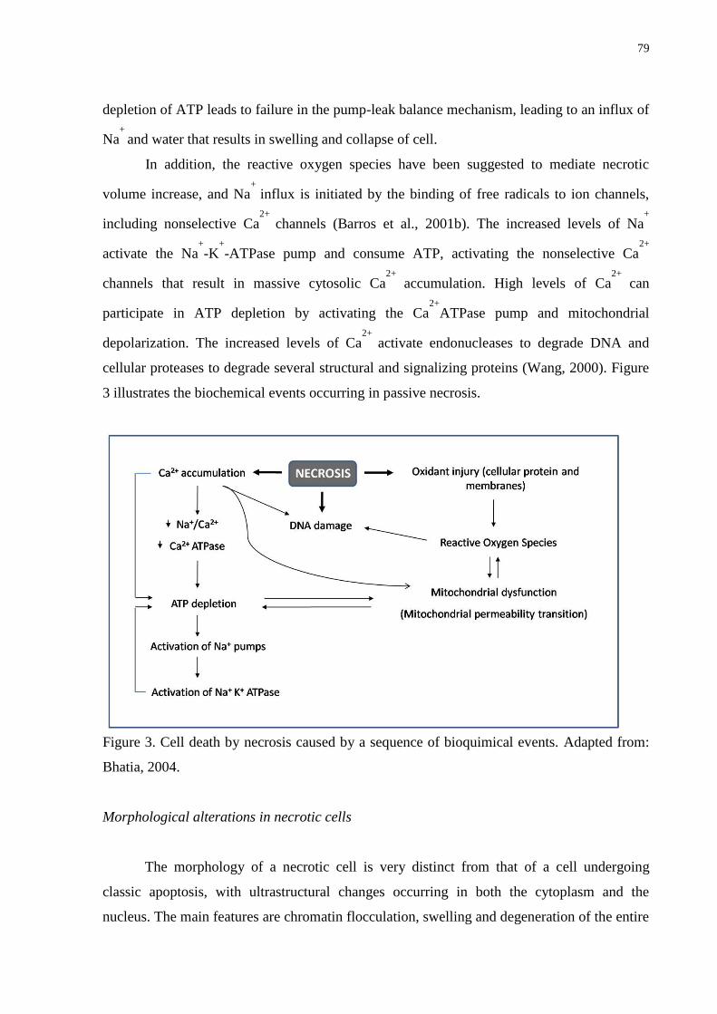
79
depletion of ATP leads to failure in the pump-leak balance mechanism, leading to an influx of
Na+
and water that results in swelling and collapse of cell.
In addition, the reactive oxygen species have been suggested to mediate necrotic
volume increase, and Na+
influx is initiated by the binding of free radicals to ion channels,
including nonselective Ca2+
channels (Barros et al., 2001b). The increased levels of Na+
activate the Na+
-K+
-ATPase pump and consume ATP, activating the nonselective Ca2+
channels that result in massive cytosolic Ca2+
accumulation. High levels of Ca2+
can
participate in ATP depletion by activating the Ca2+
ATPase pump and mitochondrial
depolarization. The increased levels of Ca2+
activate endonucleases to degrade DNA and
cellular proteases to degrade several structural and signalizing proteins (Wang, 2000). Figure
3 illustrates the biochemical events occurring in passive necrosis.
Figure 3. Cell death by necrosis caused by a sequence of bioquimical events. Adapted from:
Bhatia, 2004.
Morphological alterations in necrotic cells
The morphology of a necrotic cell is very distinct from that of a cell undergoing
classic apoptosis, with ultrastructural changes occurring in both the cytoplasm and the
nucleus. The main features are chromatin flocculation, swelling and degeneration of the entire
80
cytoplasm and the mitochondrial matrix, blebbing of the plasma membrane, and eventual
shedding of the cytoplasmic content into the extracellular space (Scaffidi et al., 2002). In
contrast to apoptosis, the chromatin is not packed into discrete membrane-bound particles, but
may form unevenly textured and irregularly shaped clumps. During necrosis, mitochondria
undergo inner membrane swelling and disintegration (Barros et al., 2001a). Polyribosomes are
dissociated and dispersed throughout the cytoplasm, imparting a dense and granular
appearance of the cytoplasmic matrix. Moreover, dilation and fragmentation of the cisterns of
the rough endoplasmic reticulum and Golgi apparatus are frequently observed (Berridge et al.,
2000). It is also important to note that cytomorphological changes like karyolysis, pyknosis
and karyorrhexis can occur in necrosis, which is not exclusive to apoptosis (Cotran et al.,
1999).
Although the mechanisms and morphologies of apoptosis and necrosis differ, there is
overlap between these two processes. Evidence indicates that necrosis and apoptosis represent
morphologic expressions of a shared biochemical network described as the ―apoptosis-
necrosis continuum‖ (Zeiss, 2003). For example, two factors that will convert an ongoing
apoptotic process into a necrotic process include a decrease in the availability of caspases and
intracellular ATP (Denecker et al., 2001). Whether a cell dies by necrosis or apoptosis
depends in part on the nature of the cell death signal, the tissue type, the developmental stage
of the tissue and the physiologic milieu (Zeiss, 2003). Table 1 illustrates the differences
between the processes of apoptosis and necrosis.
Techniques used for analysis of follicular atresia
Different techniques have been used to detect follicular atresia caused by either
apoptosis or necrosis after cryopreservation and/or in vitro culture of ovarian follicles during
the different stages of development. However, each technique has advantages and
disadvantages which may make it acceptable to use for one application but inappropriate for
another (Otsuki et al., 2003).
For apoptosis detection, different techniques may be utilized, such as: 1)
morphological analysis (laser confocal microscopy and Transmission Electronic Microscopy
– TEM; Staldemann and Lassman, 2000); 2) evaluation of DNA fragmentation (enzyme-
linked imumunosorbent assay - ELISA and terminal deoxynucleotidil transferase-mediated
deoxyuridine triphosphate biotin nick end-labeling – TUNEL; Linde et al., 2000); 3) analysis
81
of DNA content (flow cytometry); 4) evaluation of the translocation of phosphatidilserin
residues located in the inner mitochondrial membrane (Wiegele et al., 1998); 5) analysis of
gene expression and caspases involved with apoptosis (RT-PCR, northern and western blot,
and immunohistochemistry; Kiechle and Zhang, 2002). With respect to techniques for
cytomorphological alterations, the TEM is considered the gold standard to confirm apoptosis,
which better defines the subcellular changes. However, the main disadvantages of TEM are
that it is costly, time consuming, and the ability to only assay a small region at a time. The
TUNEL has been used as a principal method to identify and quantify the apoptotic cells in
atretic follicles during luteal regression (Zhang et al., 2008) because it is very sensitive and
fast (it takes about 3 hours). The disadvantages are cost and the unknown parameter of how
many DNA strand breaks are necessary for detection by this method. This method is also
subject to false positives from necrotic cells and cells in the process of DNA repair and gene
transcription (Elmore, 2007). For membrane alterations detection, the externalization of
phosphatidyl serine residues on the outer plasma membrane of apoptotic cells allows
detection via Annexin V in tissues, embryos or cultured cells (Bossy-Wetzel and Green,
2000), which can be visualized with fluorescent microscopy. The advantages are sensitivity
(can detect a single apoptotic cell) and the ability to confirm the activity of initiator caspases.
The disadvantage is that the membranes of necrotic cells are labeled as well (Elmore, 2007).
Another method for detecting apoptosis is through the detection of some factors such as the
caspase activation (Gurtu et al., 1997). The major disadvantage is that the integrity of the
sample is destroyed thereby eliminating the possibility of localizing the apoptotic event within
the tissue or determining the type of cell that is undergoing apoptosis. Another disadvantage
is that caspase activation does not necessarily indicate that apoptosis will occur (Elmore,
2007). Among these techniques, the most used for apoptosis detection is TUNEL (evaluation
of nuclear alterations, such as DNA fragmentation) and immunohistochemistry.
For evaluation of cell death by necrosis, several techniques are used, such as classical
histology (staining with hematoxilin-eosin or Periodic Schiff Acid-hematoxilin), laser
confocal microscopy, TEM (Martinez-Madrid et al., 2007), that allow for visualization of the
presence of vacuoles, especially with TEM, which can also analyze organelle damage and the
integrity of basal and nuclear membranes. Some vital fluorescent staining can also be used,
like propidium iodide and Lucifer Yellow, which penetrate damaged membranes of necrotic
follicles (Thomas et al., 2001; Choi et al., 2007).
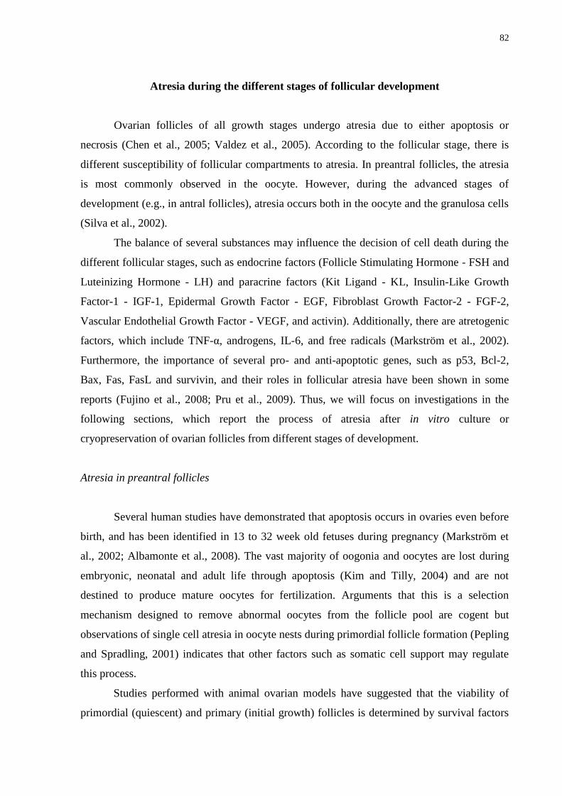
82
Atresia during the different stages of follicular development
Ovarian follicles of all growth stages undergo atresia due to either apoptosis or
necrosis (Chen et al., 2005; Valdez et al., 2005). According to the follicular stage, there is
different susceptibility of follicular compartments to atresia. In preantral follicles, the atresia
is most commonly observed in the oocyte. However, during the advanced stages of
development (e.g., in antral follicles), atresia occurs both in the oocyte and the granulosa cells
(Silva et al., 2002).
The balance of several substances may influence the decision of cell death during the
different follicular stages, such as endocrine factors (Follicle Stimulating Hormone - FSH and
Luteinizing Hormone - LH) and paracrine factors (Kit Ligand - KL, Insulin-Like Growth
Factor-1 - IGF-1, Epidermal Growth Factor - EGF, Fibroblast Growth Factor-2 - FGF-2,
Vascular Endothelial Growth Factor - VEGF, and activin). Additionally, there are atretogenic
factors, which include TNF-α, androgens, IL-6, and free radicals (Markström et al., 2002).
Furthermore, the importance of several pro- and anti-apoptotic genes, such as p53, Bcl-2,
Bax, Fas, FasL and survivin, and their roles in follicular atresia have been shown in some
reports (Fujino et al., 2008; Pru et al., 2009). Thus, we will focus on investigations in the
following sections, which report the process of atresia after in vitro culture or
cryopreservation of ovarian follicles from different stages of development.
Atresia in preantral follicles
Several human studies have demonstrated that apoptosis occurs in ovaries even before
birth, and has been identified in 13 to 32 week old fetuses during pregnancy (Markström et
al., 2002; Albamonte et al., 2008). The vast majority of oogonia and oocytes are lost during
embryonic, neonatal and adult life through apoptosis (Kim and Tilly, 2004) and are not
destined to produce mature oocytes for fertilization. Arguments that this is a selection
mechanism designed to remove abnormal oocytes from the follicle pool are cogent but
observations of single cell atresia in oocyte nests during primordial follicle formation (Pepling
and Spradling, 2001) indicates that other factors such as somatic cell support may regulate
this process.
Studies performed with animal ovarian models have suggested that the viability of
primordial (quiescent) and primary (initial growth) follicles is determined by survival factors
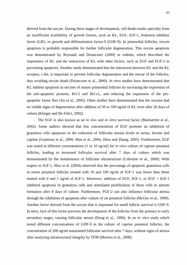
83
derived from the oocyte. During these stages of development, cell death results specially from
an insufficient availability of growth factors, such as KL, EGF, IGF-1, leukemia inhibitor
factor (LIF), or growth and differentiation factor-9 (GDF-9). In primordial follicles, oocyte
apoptosis is probably responsible for further follicular degeneration. This oocyte apoptosis
was demonstrated by Reynaud and Driancourt (2000) in rodents, which described the
importance of KL and the interaction of KL with other factors, such as EGF and FGF-2 in
preventing apoptosis. Another study demonstrated that the interaction between KL and the KL
receptor, c-kit, is important to prevent follicular degeneration and the rescue of the follicles,
thus avoiding oocyte death (Driancourt et al., 2000). In vitro studies have demonstrated that
KL inhibits apoptosis in oocytes of mouse primordial follicles by increasing the expression of
the anti-apoptotic proteins, Bcl-2 and Bcl-cL, and reducing the expression of the pro-
apoptotic factor Bax (Jin et al., 2005). Other studies have demonstrated that the oocytes had
no visible signs of degeneration after addition of 50 or 100 ng/ml of KL even after 20 days of
culture (Klinger and De Felici, 2002).
The EGF is also known as an in vivo and in vitro survival factor (Markström et al.,
2002). Some authors showed that low concentrations of EGF promote an inhibition of
granulosa cells apoptosis or the reduction of follicular atresia levels in swine, bovine and
caprine (Gutierrez et al., 2000; Mao et al., 2004, Zhou and Zhang, 2005). Furthermore, EGF
was tested at different concentrations (1 or 10 ng/ml) for in vitro culture of caprine preantral
follicles, leading to increased follicular survival after 7 days of culture which was
demonstrated by the maintenance of follicular ultrastructure (Celestino et al., 2009). With
respect to IGF-1, Mao et al. (2004) observed that the percentage of apoptotic granulosa cells
in swine preantral follicles treated with 10 and 100 ng/ml of IGF-1 was lower than those
treated with 0 and 1 ng/ml of IGF-1. Moreover, addition of EGF, IGF-1, or EGF + IGF-1
inhibited apoptosis in granulosa cells and stimulated proliferation of these cells to antrum
formation after 8 days of culture. Furthermore, FGF-2 can also influence follicular atresia
through the inhibition of apoptosis after culture of rat preantral follicles (McGee et al., 1999).
Another factor derived from the oocyte that is important for small follicle survival is GDF-9.
In mice, lack of this factor prevents the development of the follicles from the primary to early
secondary stages, causing follicular atresia (Dong et al., 1996). In an in vitro study which
tested different concentrations of GDF-9 in the culture of caprine preantral follicles, the
concentration of 200 ng/ml maintained follicular survival after 7 days, without signs of atresia
after analyzing ultrastructural integrity by TEM (Martins et al., 2008).
84
Matos et al. (2007a, b) verified that ultrastructural integrity of caprine preantral
follicles was maintained after using FSH or the association between FSH and FGF-2 in the
culture medium (Fig. 4A). Nevertheless, follicles cultured only in control medium (without
addition of any hormone or growth factor) exhibited a high rate of degeneration by necrosis,
which was characterized by a large vacuolization in the cytoplasm (Fig. 4B). However,
another study showed that in the early stage of development, FSH and its mediator cAMP did
not have any effect on apoptosis of the rat isolated preantral follicles cultured in vitro (McGee
et al., 1997). In the same way, the Anti-Müllerian Hormone (AMH) stimulated follicular
growth but did not inhibit apoptosis (McGee et al., 2001). Nevertheless, Visser et al. (2007)
recently demonstrated that AMH had an important role in follicular growth and death, serving
as a survival factor for small follicles.
Figure 4. Electron micrograph of normal (A; 6000×; scale bar: 5 μm) and degenerated follicle
(B; 7000×; scale bar: 2μm) after culture of caprine ovarian tissue in medium containing FSH
+ FGF-2 and control medium (Minimal Essential Medium), respectively. In Figure 4A, note
the homogeneous cytoplasm with numerous rounded mitochondria and the basement
membrane integrity. In Figure 4B, note the extreme vacuolization and the great holes present
in the cytoplasm, indicative of degeneration. gc, granulosa cell; l, lipid droplet; m,
mitochondria; no, nucleolus; nu, nucleus; o, oocyte; v, vesicles. (Reproduced with permission
from Matos et al., 2007a).
The susceptibility to atresia, besides the relation to the stage of follicular development,
depends on the conditions of in vitro culture, thus varying the pathway of cellular death, i.e.,
apoptosis or necrosis. After using the TUNEL technique to detect apoptosis in preantral
85
follicles cultured in vitro, Silva et al. (2006) demonstrated that addition of 100 ng/mL of
activin to the medium significantly reduced the number of atretic follicles enclosed in ovarian
tissue, but not for isolated follicles. Among the atretic follicles cultured in the ovarian cortex,
less than 30% exhibited DNA fragmentation (Fig. 5), while this phenomenon occurred
especially in granulosa cells of isolated preantral follicles. The authors suggested that this
incidence could occur most likely due to a reduced access to oxygen and nutrient in follicles
cultured in ovarian cortex, thus favoring the occurrence of necrosis as a pathway of
degeneration. Contrarily, isolated follicles which have better access to nutrients and oxygen
died via an apoptotic pathway.
Figure 5. DNA fragmentation detected using TUNEL technique in caprine ovarian follicles
cultured in vitro for 5 days in the presence of activin. Arrows represent DNA fragmentation in
granulosa cell (A) and oocyte (B). O, oocyte, G, granulosa cells. Bars: 25 μm. (Reproduced
with permission from Silva et al., 2006).
In addition to in vitro culture, cryopreservation studies have also been performed with
ovarian follicles to observe the percentage of atresia and the most predominant pathway of
cell death. Martinez-Madrid et al. (2007) showed that after cryopreservation of human ovaries
with their vascular pedicle, no primordial or primary follicle was found to be positive for
TUNEL or active caspase-3. Nevertheless, TEM revealed that some follicles exhibited
morphological alterations that were suggestive of necrosis, such as oocyte nuclear membrane
rupture and mitochondrial swelling. In another study, a high apoptosis rate was observed after
cryopreservation of mouse ovaries followed by graft, with loss of approximately half of the
number of primordial follicles present in the graft tissue (Liu et al., 2002). Apoptosis also may
be involved in follicular damage during freezing and thawing of the ovary (Rimon et al.,
2005). In addition, Tirelli et al. (2005) observed a significant increase in apoptosis of sow
granulosa cells that were frozen slowly. They suggested the increase in apoptosis was
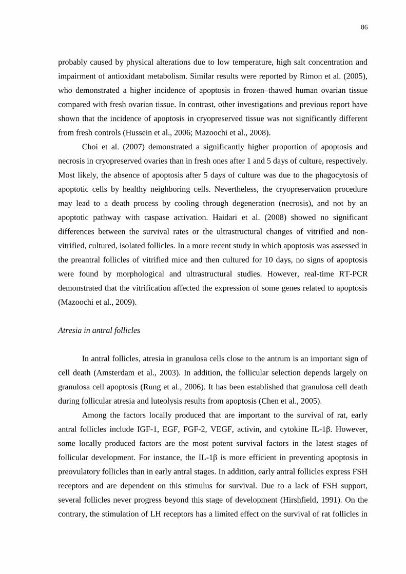
86
probably caused by physical alterations due to low temperature, high salt concentration and
impairment of antioxidant metabolism. Similar results were reported by Rimon et al. (2005),
who demonstrated a higher incidence of apoptosis in frozen–thawed human ovarian tissue
compared with fresh ovarian tissue. In contrast, other investigations and previous report have
shown that the incidence of apoptosis in cryopreserved tissue was not significantly different
from fresh controls (Hussein et al., 2006; Mazoochi et al., 2008).
Choi et al. (2007) demonstrated a significantly higher proportion of apoptosis and
necrosis in cryopreserved ovaries than in fresh ones after 1 and 5 days of culture, respectively.
Most likely, the absence of apoptosis after 5 days of culture was due to the phagocytosis of
apoptotic cells by healthy neighboring cells. Nevertheless, the cryopreservation procedure
may lead to a death process by cooling through degeneration (necrosis), and not by an
apoptotic pathway with caspase activation. Haidari et al. (2008) showed no significant
differences between the survival rates or the ultrastructural changes of vitrified and non-
vitrified, cultured, isolated follicles. In a more recent study in which apoptosis was assessed in
the preantral follicles of vitrified mice and then cultured for 10 days, no signs of apoptosis
were found by morphological and ultrastructural studies. However, real-time RT-PCR
demonstrated that the vitrification affected the expression of some genes related to apoptosis
(Mazoochi et al., 2009).
Atresia in antral follicles
In antral follicles, atresia in granulosa cells close to the antrum is an important sign of
cell death (Amsterdam et al., 2003). In addition, the follicular selection depends largely on
granulosa cell apoptosis (Rung et al., 2006). It has been established that granulosa cell death
during follicular atresia and luteolysis results from apoptosis (Chen et al., 2005).
Among the factors locally produced that are important to the survival of rat, early
antral follicles include IGF-1, EGF, FGF-2, VEGF, activin, and cytokine IL-1β. However,
some locally produced factors are the most potent survival factors in the latest stages of
follicular development. For instance, the IL-1β is more efficient in preventing apoptosis in
preovulatory follicles than in early antral stages. In addition, early antral follicles express FSH
receptors and are dependent on this stimulus for survival. Due to a lack of FSH support,
several follicles never progress beyond this stage of development (Hirshfield, 1991). On the
contrary, the stimulation of LH receptors has a limited effect on the survival of rat follicles in
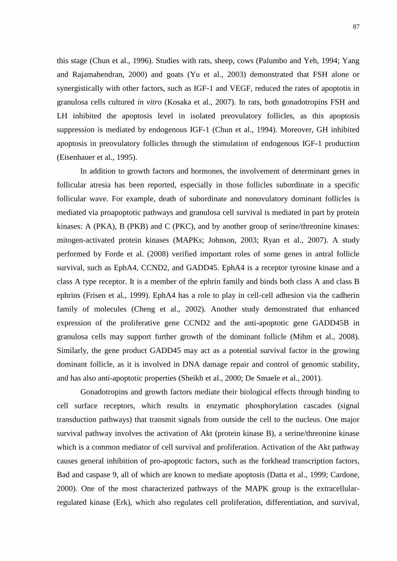
87
this stage (Chun et al., 1996). Studies with rats, sheep, cows (Palumbo and Yeh, 1994; Yang
and Rajamahendran, 2000) and goats (Yu et al., 2003) demonstrated that FSH alone or
synergistically with other factors, such as IGF-1 and VEGF, reduced the rates of apoptotis in
granulosa cells cultured in vitro (Kosaka et al., 2007). In rats, both gonadotropins FSH and
LH inhibited the apoptosis level in isolated preovulatory follicles, as this apoptosis
suppression is mediated by endogenous IGF-1 (Chun et al., 1994). Moreover, GH inhibited
apoptosis in preovulatory follicles through the stimulation of endogenous IGF-1 production
(Eisenhauer et al., 1995).
In addition to growth factors and hormones, the involvement of determinant genes in
follicular atresia has been reported, especially in those follicles subordinate in a specific
follicular wave. For example, death of subordinate and nonovulatory dominant follicles is
mediated via proapoptotic pathways and granulosa cell survival is mediated in part by protein
kinases: A (PKA), B (PKB) and C (PKC), and by another group of serine/threonine kinases:
mitogen-activated protein kinases (MAPKs; Johnson, 2003; Ryan et al., 2007). A study
performed by Forde et al. (2008) verified important roles of some genes in antral follicle
survival, such as EphA4, CCND2, and GADD45. EphA4 is a receptor tyrosine kinase and a
class A type receptor. It is a member of the ephrin family and binds both class A and class B
ephrins (Frisen et al., 1999). EphA4 has a role to play in cell-cell adhesion via the cadherin
family of molecules (Cheng et al., 2002). Another study demonstrated that enhanced
expression of the proliferative gene CCND2 and the anti-apoptotic gene GADD45B in
granulosa cells may support further growth of the dominant follicle (Mihm et al., 2008).
Similarly, the gene product GADD45 may act as a potential survival factor in the growing
dominant follicle, as it is involved in DNA damage repair and control of genomic stability,
and has also anti-apoptotic properties (Sheikh et al., 2000; De Smaele et al., 2001).
Gonadotropins and growth factors mediate their biological effects through binding to
cell surface receptors, which results in enzymatic phosphorylation cascades (signal
transduction pathways) that transmit signals from outside the cell to the nucleus. One major
survival pathway involves the activation of Akt (protein kinase B), a serine/threonine kinase
which is a common mediator of cell survival and proliferation. Activation of the Akt pathway
causes general inhibition of pro-apoptotic factors, such as the forkhead transcription factors,
Bad and caspase 9, all of which are known to mediate apoptosis (Datta et al., 1999; Cardone,
2000). One of the most characterized pathways of the MAPK group is the extracellular-
regulated kinase (Erk), which also regulates cell proliferation, differentiation, and survival,
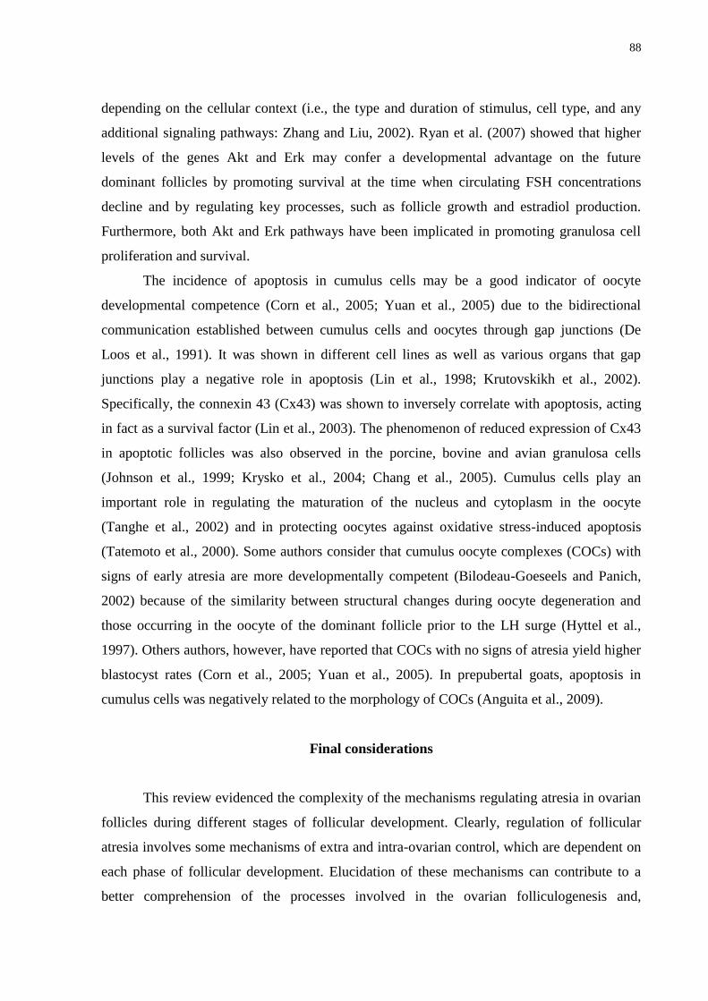
88
depending on the cellular context (i.e., the type and duration of stimulus, cell type, and any
additional signaling pathways: Zhang and Liu, 2002). Ryan et al. (2007) showed that higher
levels of the genes Akt and Erk may confer a developmental advantage on the future
dominant follicles by promoting survival at the time when circulating FSH concentrations
decline and by regulating key processes, such as follicle growth and estradiol production.
Furthermore, both Akt and Erk pathways have been implicated in promoting granulosa cell
proliferation and survival.
The incidence of apoptosis in cumulus cells may be a good indicator of oocyte
developmental competence (Corn et al., 2005; Yuan et al., 2005) due to the bidirectional
communication established between cumulus cells and oocytes through gap junctions (De
Loos et al., 1991). It was shown in different cell lines as well as various organs that gap
junctions play a negative role in apoptosis (Lin et al., 1998; Krutovskikh et al., 2002).
Specifically, the connexin 43 (Cx43) was shown to inversely correlate with apoptosis, acting
in fact as a survival factor (Lin et al., 2003). The phenomenon of reduced expression of Cx43
in apoptotic follicles was also observed in the porcine, bovine and avian granulosa cells
(Johnson et al., 1999; Krysko et al., 2004; Chang et al., 2005). Cumulus cells play an
important role in regulating the maturation of the nucleus and cytoplasm in the oocyte
(Tanghe et al., 2002) and in protecting oocytes against oxidative stress-induced apoptosis
(Tatemoto et al., 2000). Some authors consider that cumulus oocyte complexes (COCs) with
signs of early atresia are more developmentally competent (Bilodeau-Goeseels and Panich,
2002) because of the similarity between structural changes during oocyte degeneration and
those occurring in the oocyte of the dominant follicle prior to the LH surge (Hyttel et al.,
1997). Others authors, however, have reported that COCs with no signs of atresia yield higher
blastocyst rates (Corn et al., 2005; Yuan et al., 2005). In prepubertal goats, apoptosis in
cumulus cells was negatively related to the morphology of COCs (Anguita et al., 2009).
Final considerations
This review evidenced the complexity of the mechanisms regulating atresia in ovarian
follicles during different stages of follicular development. Clearly, regulation of follicular
atresia involves some mechanisms of extra and intra-ovarian control, which are dependent on
each phase of follicular development. Elucidation of these mechanisms can contribute to a
better comprehension of the processes involved in the ovarian folliculogenesis and,
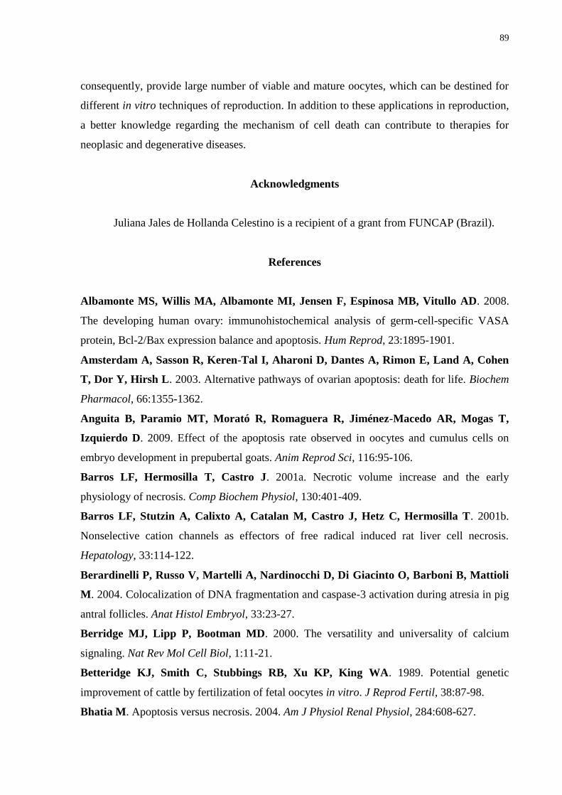
89
consequently, provide large number of viable and mature oocytes, which can be destined for
different in vitro techniques of reproduction. In addition to these applications in reproduction,
a better knowledge regarding the mechanism of cell death can contribute to therapies for
neoplasic and degenerative diseases.
Acknowledgments
Juliana Jales de Hollanda Celestino is a recipient of a grant from FUNCAP (Brazil).
References
Albamonte MS, Willis MA, Albamonte MI, Jensen F, Espinosa MB, Vitullo AD. 2008.
The developing human ovary: immunohistochemical analysis of germ-cell-specific VASA
protein, Bcl-2/Bax expression balance and apoptosis. Hum Reprod, 23:1895-1901.
Amsterdam A, Sasson R, Keren-Tal I, Aharoni D, Dantes A, Rimon E, Land A, Cohen
T, Dor Y, Hirsh L. 2003. Alternative pathways of ovarian apoptosis: death for life. Biochem
Pharmacol, 66:1355-1362.
Anguita B, Paramio MT, Morató R, Romaguera R, Jiménez-Macedo AR, Mogas T,
Izquierdo D. 2009. Effect of the apoptosis rate observed in oocytes and cumulus cells on
embryo development in prepubertal goats. Anim Reprod Sci, 116:95-106.
Barros LF, Hermosilla T, Castro J. 2001a. Necrotic volume increase and the early
physiology of necrosis. Comp Biochem Physiol, 130:401-409.
Barros LF, Stutzin A, Calixto A, Catalan M, Castro J, Hetz C, Hermosilla T. 2001b.
Nonselective cation channels as effectors of free radical induced rat liver cell necrosis.
Hepatology, 33:114-122.
Berardinelli P, Russo V, Martelli A, Nardinocchi D, Di Giacinto O, Barboni B, Mattioli
M. 2004. Colocalization of DNA fragmentation and caspase-3 activation during atresia in pig
antral follicles. Anat Histol Embryol, 33:23-27.
Berridge MJ, Lipp P, Bootman MD. 2000. The versatility and universality of calcium
signaling. Nat Rev Mol Cell Biol, 1:11-21.
Betteridge KJ, Smith C, Stubbings RB, Xu KP, King WA. 1989. Potential genetic
improvement of cattle by fertilization of fetal oocytes in vitro. J Reprod Fertil, 38:87-98.
Bhatia M. Apoptosis versus necrosis. 2004. Am J Physiol Renal Physiol, 284:608-627.
90
Bilodeau-Goeseels S, Panich P. 2002. Effects of oocyte quality on development and
transcriptional activity in early bovine embryos. Anim Reprod Sci, 71:143-155.
Bossy-Wetzel E, Green DR. 2000. Detection of apoptosis by Annexin V labeling. Methods
Enzymol, 322:15-18.
Bourdon JC, Laurenzi VD, Melino G, Lane D. 2003. P53: 25 years of research and more
questions to answer. Cell Death Differ, 10:397-399.
Bras M, Queenan B, Susin SA. 2005. Programmed cell death via mitochondria: different
modes of dying. Biochemistry, 70:231-239.
Cardone MH, Roy N, Stennicke HR, Salvesen GS, Franke TF, Stanbridge E, Frisch S,
Reed JC. 2000. Regulation of cell death protease caspase-9 by phosphorylation. Science,
282:1318-1321.
Celestino JJH, Bruno JB, Lima-Verde IB, Matos MHT, Saraiva MVA, Chaves RN,
Martins FS, Lima LF, Name KPO, Campello CC, Silva JRV, Báo SN, Figueiredo JR.
2009. Recombinant epidermal growth factor maintains follicular ultrastructure and promotes
the transition to primary follicles in caprine ovarian tissue cultured in vitro. Reprod Sci,
16:239-246.
Chang AS, Dale AN, Moley KH. 2005. Maternal diabetes adversely affects preovulatory
oocyte maturation, development, and granulosa cell apoptosis. Endocrinology, 146:2445-
2453.
Chen Q, Yano T, Matsumi H, Osuga Y, Yano N, Xu J, Wada O, Koga K, Fujiwara T,
Kugu K, Taketani Y. 2005. Cross-talk between fas/fas ligand system and nitric oxide in the
pathway subserving granulosa cell apoptosis: a possible regulatory mechanism for ovarian
follicle atresia. Endocrinology, 146:808-815.
Cheng N, Brantley DM, Chen J. 2002. The ephrins and Eph receptors in angiogenesis.
Cytokine Growth Factor Rev, 13:75-85.
Choi D, Hwang S, Lee E, Yoon S, Yoon B, Bae D. 2004. Expression of mitochondria
dependent apoptosis genes (p53, Bax, Bcl-2) in rat granulosa cells during follicular
development. J Soc Gynecol Investig, 11:311-317.
Choi J, Lee JY, Lee E, Yoon BK, Bae D, Choi D. 2007. Cryopreservation of the mouse
ovary inhibits the onset of primordial follicle development. Cryobiology, 54:55-62.
Chun SY, Billig H, Tilly JL, Furuta I, Tsafriri A, Hsueh AJ. 1994. Gonadotropin
suppression of apoptosis in cultured preovulatory follicles: mediatory role of endogenous
insulin-like growth factor I. Endocrinology, 135:1845-1853.
91
Chun SY, Eisenhauer KM, Minami S, Billig H, Perlas E, Hsueh AJ. 1996. Hormonal
regulation of apoptosis in early antral follicles: follicle stimulating hormone as a major
survival factor. Endocrinology, 137:1447-1456.
Contassot E, Gaide O, French LE. 2007. Death receptors and apoptosis. Dermatol Clin,
25:487-501.
Corn CM, Hauser-Kronberger C, Moser M, Tews G, Ebner T. 2005. Predictive value of
cumulus cell apoptosis with regard to blastocyst development of corresponding gametes.
Fertil Steril, 84:627-633.
Cortvrindt R, Smitz JEJ. 2001. In vitro follicle growth: achievements in mammalian
species. Reprod Domest Anim, 36:3-9.
Cotran RS, Kumar V, Collins T. 1999. Cellular pathology I: cell injury and cell death. In:
Cortan RS, Kumar V, Collins T (Ed.). Robbins Pathologic Basis of Disease. 6th
ed.
Philadelphina, PA: WB Saunders. pp. 1-29.
Datta SR, Brunet A, Greenberg ME. 1999. Cellular survival: a play in three Akts. Genes
Dev, 13:2905-2927.
De Loos F, Kastrop P, van Maurik P, van Beneden TH, Kruip TA. 1991. Heterologous
cell contacts and metabolic coupling in bovine cumulus oocyte complexes. Mol Reprod Dev,
28:255-259.
De Smaele E, Zazzeroni F, Papa S, Nguyen DU, Jin R, Jones J, Cong R, Franzoso G.
2001. Induction of gadd45beta by NF-kB downregulates proapoptotic JNK signaling. Nature,
414:308-313.
Denecker G, Vercammen D, Declereq W, Vandenabeele P. 2001. Apoptotic and necrotic
cell death induced by death domain receptors. Cell Mol Life Sci, 58:356-370.
Dong J, Albertini DF, Nishimori K, Kumar TR, Lu N, Matzuk MM. 1996. Growth
differentiation factor-9 is required during early ovarian folliculogenesis. Nature, 383:531-535.
Driancourt MA. 1991. Follicular dynamics in sheep and cattle. Theriogenology, 35:55-63.
Driancourt MA, Reynaud K, Cortvrindt R, Smitz J. 2000. Roles of KIT and KIT
LIGAND in ovarian function. Rev Reprod, 5:143-152.
Eisenhauer KM, Chun SY, Billig H, Hsueh AJ. 1995. Growth hormone suppression of
apoptosis in preovulatory rat follicles and partial neutralization by insulin-like growth factor
binding protein. Biol Reprod, 53:13-20.
Elmore S. 2007. Apoptosis: a review of programmed cell death. Toxicol Pathol, 35:495-516.
92
Erickson GF. 1986. An analysis of follicle development and ovum maturation. In: Seminars
in Reproductive Endocrinology, 1986, San Diego, CA. San Diego, CA: Reproductive
Endocrinology. pp. 233-254.
Forde N, Mihm M, Canty MJ, Zielak AE, Baker PJ, Park S, Lonergan P, Smith GW,
Coussens PM, Ireland JJ, Evans ACO. 2008. Differential expression of signal transduction
factors in ovarian follicle development: a functional role for betaglycan and FIBP in granulosa
cells in cattle. Physiol Genomics, 33:193-204.
Frisen J, Holmberg J, Barbacid M. 1999. Ephrins and their Eph receptors: multitalented
directors of embryonic development. EMBO J, 18:5159-5165.
Fujino K, Yamashita Y, Hayashi A, Asano M, Morishima S, Ohmichi M. 2008. Survivin
gene expression in granulosa cells from infertile patients undergoing in vitro fertilization-
embryo transfer. Fertil Steril, 89:60-65.
Green DR. 2003. Overview: apoptotic signaling pathways in the immune system. Immunol
Rev,193:5-9.
Greenfeld CR, Pepling ME, Babus JK, Furth PA, Flaws JA. 2007. BAX regulates
follicular endowment in mice. Reproduction, 133:865-876.
Gurtu V, Kain SR, Zhang G. 1997. Fluorometric and colorimetric detection of caspase
activity associated with apoptosis. Anal Biochem, 251:98-102.
Gutierrez CG, Ralph JH, Telfer EE, Wilmut I, Webb R. 2000. Growth and antrum
formation of bovine preantral follicles in long-term culture in vitro. Biol Reprod, 62:1322-
1328.
Haidari K, Salehnia M, Rezazaheh Valoujerdi M. 2008. The effect of leukemia inhibitory
factor and co-culture on the in vitro maturation and ultrastructure of vitrified and non vitrified
isolated mouse preantral follicles. Fertil Steril, 90:2389-2397.
Hirshfield AN. 1991. Development of follicles in the mammalian ovary. International Rev
Cytol, 124:43-101.
Hussein MR. 2005. Apoptosis in the ovary: molecular mechanisms. Hum Reprod Update,
11:162-178.
Hussein MR, Bedaiwy MA, Falcone T. 2006. Analysis of apoptotic cell death, Bcl-2, and
p53 protein expression in freshly fixed and cryopreserved ovarian tissue after exposure to
warm ischemia. Fertil Steril, 85:1082-1092.
Hyttel P, Fair T, Callesen H, Greve T. 1997. Oocyte growth, capacitation and final
maturation in cattle. Theriogenology, 47:23-32.
93
Igney FH, Krammer PH. 2002. Death and anti-death: tumour resistance to apoptosis. Nat
Rev Cancer, 2:277-288.
Jin X, Han CS, Yu FQ, Wei P, Hu ZY, Liu YX. 2005. Anti-apoptotic action of stem cell
factor on oocytes in primordial follicles and its signal transduction. Mol Reprod Dev, 70:82-
90.
Johnson AL, Bridgham JT. 2002. Caspase-mediated apoptosis in the vertebrate ovary.
Reproduction, 124:19-27.
Johnson AL. 2003. Intracellular mechanisms regulating cell survival in ovarian follicles.
Anim Reprod Sci, 78:185-201.
Johnson ML, Redmer DA, Reynolds LP, Grazul-Bilska AT. 1999. Expression of gap
junctional proteins connexin 43, 32, and 26 throughout follicular development and atresia in
cows. Endocrine, 10:43-51.
Johnstone RJ, Rueffi AA, Lowe SW. 2002. Apoptosis: a link between cancer genetics and
chemotherapy. Cell, 108:153-164.
Joza N, Kroemer G, Penninger JM. 2002. Genetic analysis of the mammalian cell death
machinery. Trends Genet, 18:142-149.
Kiechle FL, Zhang X. 2002. Apoptosis: biochemical aspects and clinical implications. Chim
Acta, 326:27-45.
Kim MR, Tilly JL. 2004 Current concepts in Bcl-2 family member regulation of female
germ cell development and survival. Biochim Biophys Acta, 1644:205-210.
Klinger FG, De Felici M. 2002. In vitro development of growing oocytes from fetal mouse
oocytes: stage-specific regulation by stem cell factor and granulosa cells. Dev Biol, 244:85-
95.
Kosaka N, Sudo N, Miyamoto A, Shimizu T. 2007. Vascular endothelial growth factor
(VEGF) supresses ovarian granulosa cell apoptosis in vitro. Biochem Biophys Res Commun,
363:733-737.
Krutovskikh VA, Piccoli C, Yamasaki H. 2002. Gap junction intercellular communication
propagates cell death in cancerous cells. Oncogene, 21:1989-1999.
Krysko DV, Mussche S, Leybaert L, D’Herde K. 2004. Gap junctional communication and
connexin43 expression in relation to apoptotic cell death and survival of granulosa cells. J
Histochem Cytochem, 52:1199-1207.
Kurosaka K, Takahashi M, Watanabe N, Kobayashi Y. 2003. Silent cleanup of very early
apoptotic cells by macrophages. J Immunol, 171:4672-4679.
94
Lima AKF. 2006. Determinação da população folicular, criopreservação e cultivo de
oócitos inclusos em folículos ovarianos pré-antrais de gata doméstica. Fortaleza, CE:
Universidade Estadual do Ceará. Thesis.
Lin JH, Weigel H, Cotrina ML, Liu S, Bueno E, Hansen AJ, Hansen TW, Goldman S,
Nedergaard M. 1998. Gap-junction-mediated propagation and amplification of cell injury.
Nat Neurosci, 1:494-500.
Lin JH, Yang J, Liu S, Takano T, Wang X, Gao Q, Willecke K, Nedergaard M. 2003.
Connexin mediates gap junction-independent resistance to cellular injury. J Neurosci, 23:430-
441.
Linde V, Flodgaard H, Kastrup JS, Bjorn S. 2000. Measurement of apoptosis by the
TUNEL method using scintillating microplates. Anal Biochem, 280:186-188.
Liu J, van der Elst J, van den Broecke R, Dhont M. 2002. Early massive follicle loss and
apoptosis in heterotopically grafted newborn mouse ovaries. Hum Reprod, 17:605-611.
Lucci CM, Amorim CA, Báo SN, Figueiredo JR, Rodrigues APR, Silva JR, Gonçalves
PBD. 1999. Effect of the interval of serial sections of ovarian in the tissue chopper on the
number of isolated caprine preantral follicles. Anim Reprod Sci, 56:39-49.
Manabe N, Matsuda-Minehata F, Goto Y, Maeda A, Cheng Y, Nakagawa S, Inoue N,
Wongpanit K, Jin H, Gonda H, Li J. 2008. Role of cell death ligand and receptor system on
regulation of follicular atresia in pig ovaries. Reprod Domest Anim, 43:268-272.
Mao J, Smith MF, Rucker EB, Wu GM, McCauley TC, Cantley TC, Prather RS, Didion
BA, Day BN. 2004. Effect of epidermal growth factor and insulin-like growth factor-I on
porcine preantral follicular growth, antrum formation, and stimulation of granulosa cell
proliferation and suppression of apoptosis in vitro. J Anim Sci, 82:1967-1975.
Markström E, Svensson EC, Shao R, Svanberg B, Billig H. 2002. Survival factors
regulating ovarian apoptosis – dependence on follicle differentiation. Reproduction, 123:23-
30.
Martinez-Madrid B, Camboni A, Dolmans MM, Nottola S, Langendonckt AV, Donnez
J. 2007. Apoptosis and ultrastructural assessment after cryopreservation of whole human
ovaries with their vascular pedicle. Fertil Steril, 87:1153-1165.
Martins FS, Celestino JJH, Saraiva MVA, Matos MHT, Bruno JB, Rocha-Junior CMC,
Lima-Verde IB, Lucci CM, Báo SN, Figueiredo JR. 2008. Growth and differentiation
factor-9 stimulates activation of goat primordial follicles in vitro and their progression to
secondary follicles. Reprod Fertil Dev, 20:916-924.
95
Matos MHT, Lima-Verde IB, Bruno JB, Lopes CAP, Martins FS, Santos KDB, Rocha
RMP, Silva JRV, Báo SN, Figueiredo JR. 2007a. Follicle stimulating hormone and
fibroblast growth factor-2 interact and promote goat primordial follicle development in vitro.
Reprod Fertil Dev, 19:677-684.
Matos MHT, Lima-Verde IB, Luque MC, Maia Jr JR, Silva JRV, Celestino JJH,
Martins FS, Báo SN, Lucci CM, Figueiredo JR. 2007b. Essential role of follicle stimulating
hormone in the maintenance of caprine preantral follicle viability in vitro. Zygote, 15:173-
182.
Mazoochi T, Salehnia M, Valojerdi MR, Mowla SJ. 2008. Morphologic, ultrastructural,
and biochemical identification of apoptosis in vitrified-warmed mouse ovarian tissue. Fertil
Steril, 90:1480-1486.
Mazoochi T, Salehnia M, Pourbeiranvand S, Forouzandeh M, Mowla SJ, Hajizadeh E.
2009. Analysis of apoptosis and expression of genes related to apoptosis in cultures os
follicles derived from vitrified and non-vitrified ovaries. Mol Hum Reprod, 15:155-164.
McCully JD, Wakiyama H, Hsieh YJ, Jones M, Levitsky S. 2004. Differential contribution
of necrosis and apoptosis in myocardial ischemia-reperfusion injury. Am J Physiol Heart Circ
Physiol, 286:1923-1935.
McGee EA, Perlas E, Lapolt PS, Tsafriri A, Hsueh AJ. 1997. Follicle stimulating hormone
enhances the development of preantral follicles in juvenile rats. Biol Reprod, 57:990-998.
McGee EA, Chun SY, Lai S, He Y, Hsueh AJ. 1999. Keratinocyte growth factor promotes
the survival, growth, and differentiation of preantral ovarian follicles. Fertil Steril, 71:732-
738.
McGee EA, Smith R, Spears N, Nachtigal MW, Ingraham H, Hsueh AJ. 2001. Mullerian
inhibitory substance induces growth of rat preantral ovarian follicles. Biol Reprod, 64:293-
298.
Mihm M, Baker PJ, Fleming LM, Monteiro AM, O’Shaughnessy PJ. 2008.
Differentiation of the bovine dominant follicle from the cohort upregulates mRNA expression
for new tissue development genes. Reproduction, 135:253-265.
Nagase H, Fukuyama H, Tanaka M, Kawane K, Nagata S. 2003. Mutually regulated
expression of caspase-activated DNase and its inhibitor for apoptotic DNA fragmentation.
Cell Death Differ, 10:142-143.
96
Nilsson EE, Skinner MK. 2004. Kit ligand and basic fibroblast growth factor interactions in
the induction of ovarian primordial to primary follicle transition. Mol Cell Endocrinol,
214:19-25.
Otsuki Y, Li Z, Shibata MA. 2003. Apoptotic detection methods–from morphology to gene.
Prog Histochem Cytochem, 38:275-339.
Padanilam BJ. 2003. Cell death induced by acute renal injury: a perspective on the
contributions of apoptosis and necrosis. Am J Physiol Renal Physiol, 284:608-627.
Palumbo A, Yeh J. 1994. In situ localization of apoptosis in the rat ovary during follicular
atresia. Biol Reprod, 51:888-895.
Pepling ME, Spradling AC. 2001. Mouse ovarian germ cell cysts undergo programmed
breakdown to form primordial follicles. Dev Biol, 234:339-351.
Porter DA, Harman RM, Cowan RG, Quirk SM. 2001. Relationship of Fas ligand
expression and atresia during bovine follicle development. Reproduction, 121:561-566.
Pru JK, Kaneko-Tarui T, Jurisicova A, Kashiwagi A, Selesniemi K, Tilly J. 2009.
Induction of proapoptotic gene expression and recruitment of p53 herald ovarian follicle loss
caused by polycyclic aromatic hydrocarbons. Reprod Sci, 16:347-356.
Reed JC. 2000. Mechanisms of apoptosis. Am J Pathol, 157:1415-1430.
Reed JC, Kroemer G. 2000. Mechanisms of mitochondrial membrane permeabilization. Cell
Death Differ, 7:1145.
Reynaud K, Driancourt MA. 2000. Oocyte attrition. Mol Cell Endocrinol, 163:101-108.
Rimon E, Cohen T, Dantes A, Hirsh L, Amit A, Lessing JB, Freimanns S, Amsterdam A,
Azem F. 2005. Apoptosis in cryopreserved human ovarian tissue obtained from cancer
patients: a tool for evaluating cryopreservation utility. Int J Oncol, 27:345-353.
Rung E, Friberg PA, Bergh C, Billig H. 2006. Depletion of substrates for protein
prenylation increases apoptosis in human periovulatory granulosa cells. Mol Reprod Dev,
73:1277-1283.
Ryan KE, Casey SM, Canty MJ, Crowe MA, Martin F, Evans AC. 2007. Akt and Erk
signal transduction pathways are early markers of differentiation in dominant and subordinate
ovarian follicles in cattle. Reproduction, 133:617-626.
Savill J, Fadok V. 2000. Corpse clearance defines the meaning of cell death. Nature,
407:784-788.
Scaffidi P, Misteli T, Bianchi ME. 2002. Release of chromatin protein HMGB1 by necrotic
cells triggers inflammation. Nature, 418:191-195.
97
Shaw JM, Oranratnachai JM, Trounson AO. 2000. Fundamental cryobiology of
mammalian oocytes and ovarian tissue. Theriogenology, 53:59-72.
Sheikh MS, Hollander MC, Fornance AJ Jr. 2000. Role of GADD45 in apoptosis.
Biochem Pharmacol, 59:43-45.
Shin S, Sung BJ, Cho YS, Kim HJ, Ha NC, Hwang JI, Chung CW, Jung YK, Oh BH.
2001. An anti-apoptotic protein human survivin is a direct inhibitor of caspase-3 and -7.
Biochemistry, 40:1117-1123.
Silva JRV, Ferreira MAL, Costa SHF, Santos RR, Carvalho FCA, Rodrigues APR,
Lucci CM, Báo SN, Figueiredo JR. 2002. Degeneration rate of preantral follicles in the
ovaries of goats. Small Rum Res, 43:203-209.
Silva JRV, Tharasanit T, Taverne MAM, van der Weijden GC, Santos RR, Figueiredo
JR, van den Hurk R. 2006. The activin-follistatin system and in vitro early follicle
development in goats. J Endocrinol, 189:113-125.
Slot KA. 2004. Hormonal regulation of apoptosis in the ovary under normal physiological
and pathological conditions. Utrecht: The Utrecht University. Thesis.
Stadelmann C, Lassmann H. 2000. Detection of apoptosis in tissue sections. Cell Tissue
Res, 301:19-31.
Strasser A, O’Connor L, Dixit VM. 2000. Apoptosis signaling. Annu Rev Biochem, 69:217-
245.
Suzuki M, Youle RJ, Tjandra N. 2000. Structure of Bax: coregulation of dimer formation
and intracellular localization. Cell, 103:645-654.
Tanghe S, van Soom A, Nauwynck H, Coryn M, de Kruif A. 2002. Minireview: functions
of the cumulus oophorus during oocyte maturation, ovulation, and fertilization. Mol Reprod
Dev, 61:414-424.
Tatemoto H, Sakurai N, Muto N. 2000. Protection of porcine oocytes against apoptotic cell
death caused by oxidative stress during in vitro maturation: role of cumulus cells. Biol
Reprod, 63:805-810.
Thomas FH, Leask R, Srsen V, Riley SC, Spears N, Telfer EE. 2001. Reproduction,
122:487-495.
Tibbetts MD, Zheng L, Lenardo MJ. 2003. The death effector domain protein family:
regulators of cellular homeostasis. Nat Immunol, 4:404-409.
Tilly JL, Tilly K, Kenton M, Johnson A. 1995. Expression of members of the Bcl-2 gene
family in the immature rat ovary: equine chorionic gonadotropin-mediated inhibition of
98
granulosa cell apoptosis is associated with decreased bax and constitutive bcl-2 and bcl-x long
messenger ribonucleic acid levels. Endocrinology, 136:232-241.
Tilly JL. 1996. Apoptosis and ovarian function. Rev Reprod, 1:162-172.
Tirelli M, Basini G, Grasselli F, Bianco F, Tamanini C. 2005. Cryopreservation of pig
granulosa cells: effect of FSH addition to freezing medium. Domest Anim Endocrinol, 28:17-
33.
Tsai NM, Hsieh RH, Au HK, Shieh MJ, Huang SY, Tzeng CR. 2005. Effects of
gonadotrophin-releasing hormone agonists on apoptosis of granulosa cells. Ann N Y Acad Sci,
1042:531-537.
Valdez KE, Cuneo SP, Turzillo AM. 2005. Regulation of apoptosis in the atresia of
dominant bovine follicles of the first follicular wave following ovulation. Reproduction,
130:71-81.
Van Cruchten S, van den Broeck W. 2002. Morphological and biochemical aspects of
apoptosis, oncosis and necrosis. Anat Histol Embryol, 31:214-223.
Van den Hurk R, Zhao J. 2005. Formation of mammalian oocytes and their growth,
differentiation and maturation within ovarian follicles. Theriogenology, 63:1717-1751.
Visser JA, Durlinger ALL, Peters IJJ, Heuvel ER, Rose U, Kramer P, Jong FH,
Themmen APN. 2007. Increased oocyte degeneration and follicular atresia during the estrous
cycle in anti-mullerian hormone null mice. Endocrinology, 148:2301-2308.
Wang KK. 2000. Calpain and caspase: can you tell the difference? Trends Neurosci, 23:20-
26.
Wiegele G, Brandis M, Zimmerhackl LB. 1998. Apoptosis and necrosis during ischaemia
in renal tubular cells (LLC-PK1
and MDCK). Nephrol Dial Transplant, 13:1158-1167.
Yang MY, Rajamahendran R. 2000. Morphological and biochemical identification of
apoptosis in small, medium, and large bovine follicles and the effects of follicle-stimulating
hormone and insulin-like growth factor-I on spontaneous apoptosis in cultured bovine
granulosa cells. Biol Reprod, 62:1209-1217.
Yu Y, Li W, Han Z, Luo M, Chang Z, Tan J. 2003. The effect of follicle-stimulating
hormone on follicular development, granulosa cell apoptosis and steroidogenesis and its
mediation by insulin-like growth factor-I in the goat ovary. Theriogenology, 60:1691-1704.
Yuan YQ, Van Soom A, Leroy JL, Dewulf J, van Zeveren A, de Kruif A, Peelman LJ.
2005. Apoptosis in cumulus cells, but not in oocytes, may influence bovine embryonic
developmental competence. Theriogenology, 63:2147-2163.
99
Zeiss CJ. 2003. The apoptosis-necrosis continuum: insights from genetically altered mice.
Vet Pathol, 40:481-495.
Zhang W, Liu HT. 2002. MAPK signal pathways in the regulation of cell proliferation in
mammalian cells. Cell Res, 12:9-18.
Zhang W, Huang L, Zhuang Y, Wang W. 2008. The effect of mifepristone on apoptosis
and caspase-3 activation in human ovarian luteinized granulosa cells. Eur J Obstet Gynecol
Reprod Biol, 141:131-136.
Zhou H, Zhang Y. 2005. Regulation of in vitro growth of preantral follicles by growth
factors in goats. Domest Anim Endocrinol, 28:235-242.
100
7 CAPÍTULO 2
Regulação da foliculogênese ovariana pelo sistema Kit Ligand e c-Kit em mamíferos
Regulation of ovarian folliculogenesis by Kit Ligand and the c-Kit system in mammals
Periódico: Animal Reproduction 6: 431-439, 2009.
101
Resumo
O sistema composto por Kit Ligand (KL) e seu receptor c-Kit tem mostrado desempenhar um
papel na reprodução normal de fêmeas e na fertilidade de mamíferos. Estudos de expressão de
genes têm revelado que atividades biológicas dos ligantes e receptores do sistema KL/c-Kit
são importantes no controle da apoptose e proliferação celular nos tecidos reprodutivos.
Coletivamente, esses estudos têm fornecido uma melhor compreensão da fisiologia ovariana e
fertilidade das fêmeas através do estabelecimento do conceito de que o sistema KL/c-Kit
regula a viabilidade das células germinativas primordiais e dos folículos, o início do
crescimento do folículo primordial, e posterior desenvolvimento oocitário e folicular através
de diferentes proteínas sinalizadoras. O objetivo desse artigo é revisar a importância do
sistema KL/c-Kit no desenvolvimento folicular ovariano, especialmente na fase pré-antral da
foliculogênese.
Palavras-chave: Sistema c-Kit. Folículo. Kit Ligand. Ovário. Vias de sinalização.
102
Regulation of ovarian folliculogenesis by Kit Ligand and the c-Kit system in mammals
J.J.H. Celestino1
, M.H.T. Matos, M.V.A. Saraiva, J.R. Figueiredo
Laboratory of Manipulation of Oocytes and Preantral Follicles (LAMOFOPA),
Faculty of Veterinary Medicine, State University of Ceara, Fortaleza, CE, Brazil.
Abstract
The system comprised of Kit Ligand (KL) and its receptor c-Kit has proven to play a
role in normal female reproduction and fertility in mammals. Gene expression studies have
revealed that biological activities of ligands and receptors of the KL/c-Kit system are
important in controlling apoptosis and cellular proliferation in reproductive tissues.
Collectively, these studies have provided a better understanding of ovarian physiology and
female fertility through the establishment of the concept that the KL/c-Kit system regulates
the viability of primordial germ cells and follicles, initiation of primordial follicle growth, and
further oocyte and follicular development through different signaling proteins. The purpose of
this article is to review the importance of the KL/c-Kit system in ovarian follicular
development, especially in the preantral phase of folliculogenesis.
Keywords: c-Kit system, follicle, Kit Ligand, ovary, signaling pathways.
Introduction
During the last decade, the role of growth factors in ovarian folliculogenesis has been
extensively studied in several species, including rodents, domestic animals, and humans. In
particular, Kit Ligand (KL), which was one of the first growth factors identified in the ovarian
follicle, plays a key role in mammalian oogenesis and folliculogenesis (Thomas and
Vanderhyden, 2006). Since its identification in 1990, in vivo and in vitro studies have shown
that the functions of this system in the ovary include the establishment of primordial germ
cells (PGCs), activation of primordial follicles, oocyte survival and growth, proliferation of
granulosa cells, recruitment of theca cells, and maintenance of meiotic competence (Hutt et
al., 2006b; Thomas et al., 2008), as shown in Fig. 1. This review will focus on the role of the

103
KL/c-Kit system in ovarian follicle development, especially in the preantral phase of
folliculogenesis.
Figure 1. Several functions of this KL/c-Kit system in the ovary: 1) Establishment of
primordial germ cells; 2) Activation of primordial follicles; 3) Oocyte survival and growth; 4)
Proliferation of granulosa cells and recruitment of theca cells. PGCs: primordial germ cells;
TC: theca cells; GC: granulosa cells; O: oocyte.
Expression of Kit Ligand and c-Kit in the ovary
Kit Ligand (KL), also known as Stem Cell Factor (SCF), Steel Factor (SF), and Mast
Cell Growth Factor (MCGF), is a locally produced factor that has many roles in ovarian
function from embryogenesis onward (Yoshida et al., 1997; Driancourt et al., 2000). In
follicles, the expression of the mRNA for KL has been demonstrated in the granulosa cells of
several species (rat: Ismail et al., 1996; ovine: Tisdall et al., 1999; mouse: Doneda et al.,
2002; human: Hoyer et al., 2005 and caprine: Silva et al., 2006). Depending on how the
mRNA is spliced, KL can be expressed as a soluble protein (KL-1) or as membrane-
associated protein (KL-2; Huang et al., 1992). When translated, both transcripts yield
membrane-associated products; however, KL-1 is efficiently cleaved and released as a soluble
product due to a proteolytic cleavage site encoded by an 84-base pair exon. The other form,
KL-2, lacks this cleavage site and therefore remains membrane-bound (Huang et al., 1992).
KL-2 is the main isoform required for the growth and survival of oocytes (Thomas et al.,
2008). Both isoforms are present in rodent (Ismail et al., 1997) and goat (Silva et al., 2006)
ovaries. In goats, KL protein and mRNA are expressed in granulosa cells during all stages of
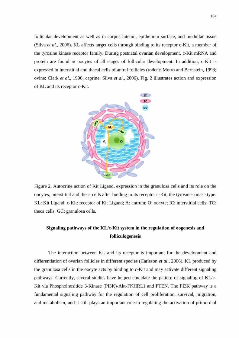
104
follicular development as well as in corpus luteum, epithelium surface, and medullar tissue
(Silva et al., 2006). KL affects target cells through binding to its receptor c-Kit, a member of
the tyrosine kinase receptor family. During postnatal ovarian development, c-Kit mRNA and
protein are found in oocytes of all stages of follicular development. In addition, c-Kit is
expressed in interstitial and thecal cells of antral follicles (rodent: Motro and Bernstein, 1993;
ovine: Clark et al., 1996; caprine: Silva et al., 2006). Fig. 2 illustrates action and expression
of KL and its receptor c-Kit.
Figure 2. Autocrine action of Kit Ligand, expression in the granulosa cells and its role on the
oocytes, interstitial and theca cells after binding to its receptor c-Kit, the tyrosine-kinase type.
KL: Kit Ligand; c-Kit: receptor of Kit Ligand; A: antrum; O: oocyte; IC: interstitial cells; TC:
theca cells; GC: granulosa cells.
Signaling pathways of the KL/c-Kit system in the regulation of oogenesis and
folliculogenesis
The interaction between KL and its receptor is important for the development and
differentiation of ovarian follicles in different species (Carlsson et al., 2006). KL produced by
the granulosa cells in the oocyte acts by binding to c-Kit and may activate different signaling
pathways. Currently, several studies have helped elucidate the pattern of signaling of KL/c-
Kit via Phosphoinositide 3-Kinase (PI3K)-Akt-FKHRL1 and PTEN. The PI3K pathway is a
fundamental signaling pathway for the regulation of cell proliferation, survival, migration,
and metabolism, and it still plays an important role in regulating the activation of primordial
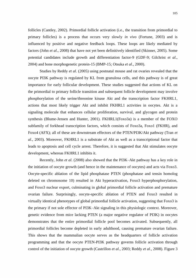
105
follicles (Cantley, 2002). Primordial follicle activation (i.e., the transition from primordial to
primary follicles) is a process that occurs very slowly in vivo (Fortune, 2003) and is
influenced by positive and negative feedback loops. These loops are likely mediated by
factors (John et al., 2008) that have not yet been definitively identified (Skinner, 2005). Some
potential candidates include growth and differentiation factor-9 (GDF-9; Gilchrist et al.,
2004) and bone morphogenetic protein-15 (BMP-15; Otsuka et al., 2000).
Studies by Reddy et al. (2005) using postnatal mouse and rat ovaries revealed that the
oocyte PI3K pathway is regulated by KL from granulosa cells, and this pathway is of great
importance for early follicular development. These studies suggested that actions of KL on
the primordial to primary follicle transition and subsequent follicle development may involve
phosphorylation of the serine/threonine kinase Akt and the transcription factor FKHRL1,
actions that most likely trigger Akt and inhibit FKHRL1 activities in oocytes. Akt is a
signaling molecule that enhances cellular proliferation, survival, and glycogen and protein
synthesis (Blume-Jensen and Hunter, 2001). FKHRL1(Foxo3a) is a member of the FOXO
subfamily of forkhead transcription factors, which consists of Foxo3a, Foxo1 (FKHR), and
Foxo4 (AFX); all of these are downstream effectors of the PTEN/PI3K/Akt pathway (Tran et
al., 2003). Moreover, FKHRL1 is a substrate of Akt as well as a transcriptional factor that
leads to apoptosis and cell cycle arrest. Therefore, it is suggested that Akt stimulates oocyte
development, whereas FKHRL1 inhibits it.
Recently, John et al. (2008) also showed that the PI3K–Akt pathway has a key role in
the initiation of oocyte growth (and hence in the maintenance of oocytes) and acts via Foxo3.
Oocyte-specific ablation of the lipid phosphatase PTEN (phosphatase and tensin homolog
deleted on chromosome 10) resulted in Akt hyperactivation, Foxo3 hyperphosphorylation,
and Foxo3 nuclear export, culminating in global primordial follicle activation and premature
ovarian failure. Surprisingly, oocyte-specific ablation of PTEN and Foxo3 resulted in
virtually identical phenotypes of global primordial follicle activation, suggesting that Foxo3 is
the primary if not sole effector of PI3K–Akt signaling in this physiologic context. Moreover,
genetic evidence from mice lacking PTEN (a major negative regulator of PI3K) in oocytes
demonstrates that the entire primordial follicle pool becomes activated. Subsequently, all
primordial follicles become depleted in early adulthood, causing premature ovarian failure.
This shows that the mammalian oocyte serves as the headquarters of follicle activation
programming and that the oocyte PTEN-PI3K pathway governs follicle activation through
control of the initiation of oocyte growth (Castrillon et al., 2003; Reddy et al., 2008). Figure 3
106
schematically illustrates the signaling pathway of KL/c-Kit via Phosphoinositide 3-Kinase
(PI3K)-Akt-FKHRL1 and PTEN.
Figure 3. Pattern of signaling of KL/c-Kit system via Phosphoinositide 3-Kinase (PI3K)-Akt-
FKHRL1 and PTEN. The oocyte PTEN-PI3K pathway governs follicle activation through
control of initiation of oocyte growth, since it inhibits the PI3K-Akt pathway, which then
allows the FKHRL1 to keep quiescent oocytes (1). The binding of KL to its receptor c-Kit
phosphorylate serine/threonine kinase Akt group and activates Akt pathway, thus inhibiting
the activity of FKHRL1 in oocytes allowing its activation (2). It is likely that KL starts oocyte
growth, for instance, with the slow accumulation of factors required for meiosis resumption,
such as p34cd2, cyclin B1, MAPK, cdc25. KL: Kit Ligand; c-Kit: receptor of Kit Ligand;
Akt: signaling molecule; FKHRL1(Foxo3a): member of the FOXO subfamily and of forkhead
transcription factors and is a substrate of Akt; PTEN: phosphatase and tensin homolog deleted
on chromosome 10.
In addition to the PI3K–Akt pathway, several transcription factors are known to affect
the regulation of oocyte-specific genes during early folliculogenesis. FIGLA (Factor in the
Germline Alpha; Joshi et al., 2007), NOBOX (Newborn Ovary Homeobox gene; Choi et al.,
2007), and Sohlh1 (Spermatogenesis and oogenesis helix-loop-helix 1; Pangas et al., 2006)
are all critical to primordial follicle formation and maintenance. FIGLA is an oocyte-specific
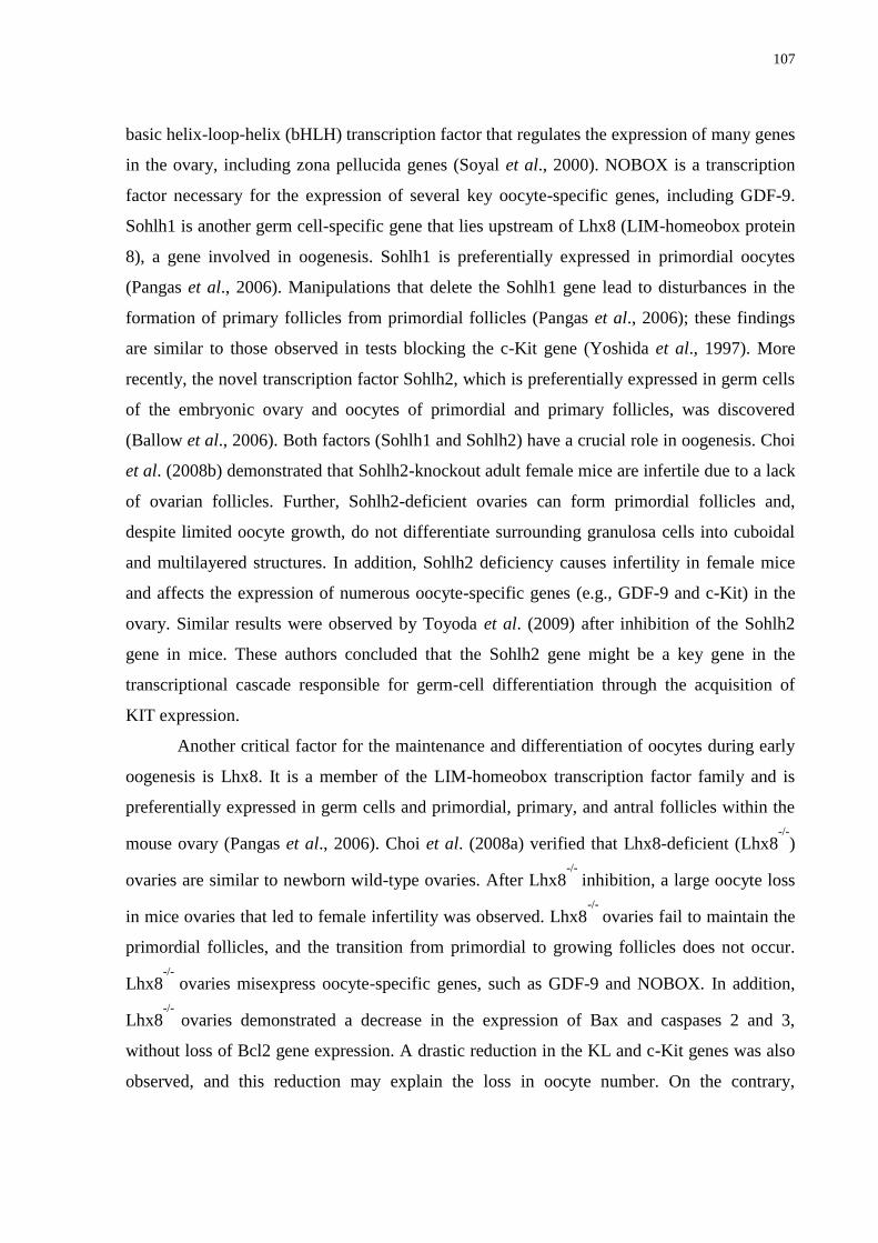
107
basic helix-loop-helix (bHLH) transcription factor that regulates the expression of many genes
in the ovary, including zona pellucida genes (Soyal et al., 2000). NOBOX is a transcription
factor necessary for the expression of several key oocyte-specific genes, including GDF-9.
Sohlh1 is another germ cell-specific gene that lies upstream of Lhx8 (LIM-homeobox protein
8), a gene involved in oogenesis. Sohlh1 is preferentially expressed in primordial oocytes
(Pangas et al., 2006). Manipulations that delete the Sohlh1 gene lead to disturbances in the
formation of primary follicles from primordial follicles (Pangas et al., 2006); these findings
are similar to those observed in tests blocking the c-Kit gene (Yoshida et al., 1997). More
recently, the novel transcription factor Sohlh2, which is preferentially expressed in germ cells
of the embryonic ovary and oocytes of primordial and primary follicles, was discovered
(Ballow et al., 2006). Both factors (Sohlh1 and Sohlh2) have a crucial role in oogenesis. Choi
et al. (2008b) demonstrated that Sohlh2-knockout adult female mice are infertile due to a lack
of ovarian follicles. Further, Sohlh2-deficient ovaries can form primordial follicles and,
despite limited oocyte growth, do not differentiate surrounding granulosa cells into cuboidal
and multilayered structures. In addition, Sohlh2 deficiency causes infertility in female mice
and affects the expression of numerous oocyte-specific genes (e.g., GDF-9 and c-Kit) in the
ovary. Similar results were observed by Toyoda et al. (2009) after inhibition of the Sohlh2
gene in mice. These authors concluded that the Sohlh2 gene might be a key gene in the
transcriptional cascade responsible for germ-cell differentiation through the acquisition of
KIT expression.
Another critical factor for the maintenance and differentiation of oocytes during early
oogenesis is Lhx8. It is a member of the LIM-homeobox transcription factor family and is
preferentially expressed in germ cells and primordial, primary, and antral follicles within the
mouse ovary (Pangas et al., 2006). Choi et al. (2008a) verified that Lhx8-deficient (Lhx8-/-
)
ovaries are similar to newborn wild-type ovaries. After Lhx8-/-
inhibition, a large oocyte loss
in mice ovaries that led to female infertility was observed. Lhx8-/-
ovaries fail to maintain the
primordial follicles, and the transition from primordial to growing follicles does not occur.
Lhx8-/-
ovaries misexpress oocyte-specific genes, such as GDF-9 and NOBOX. In addition,
Lhx8-/-
ovaries demonstrated a decrease in the expression of Bax and caspases 2 and 3,
without loss of Bcl2 gene expression. A drastic reduction in the KL and c-Kit genes was also
observed, and this reduction may explain the loss in oocyte number. On the contrary,
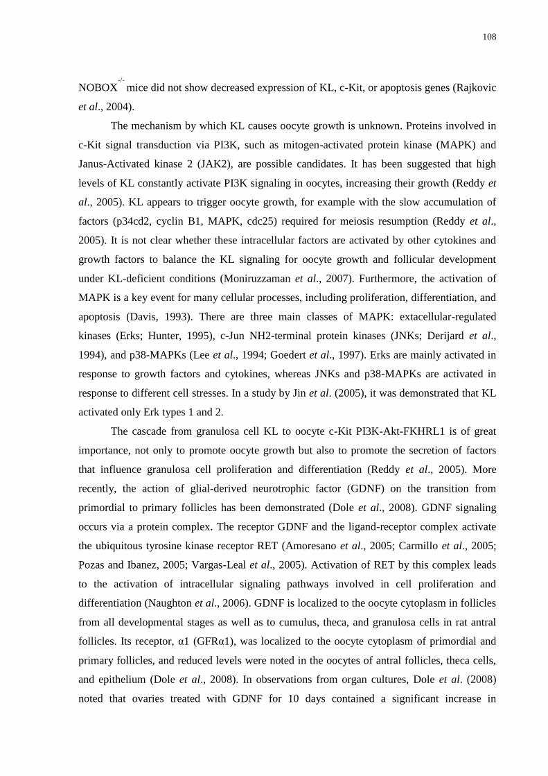
108
NOBOX-/-
mice did not show decreased expression of KL, c-Kit, or apoptosis genes (Rajkovic
et al., 2004).
The mechanism by which KL causes oocyte growth is unknown. Proteins involved in
c-Kit signal transduction via PI3K, such as mitogen-activated protein kinase (MAPK) and
Janus-Activated kinase 2 (JAK2), are possible candidates. It has been suggested that high
levels of KL constantly activate PI3K signaling in oocytes, increasing their growth (Reddy et
al., 2005). KL appears to trigger oocyte growth, for example with the slow accumulation of
factors (p34cd2, cyclin B1, MAPK, cdc25) required for meiosis resumption (Reddy et al.,
2005). It is not clear whether these intracellular factors are activated by other cytokines and
growth factors to balance the KL signaling for oocyte growth and follicular development
under KL-deficient conditions (Moniruzzaman et al., 2007). Furthermore, the activation of
MAPK is a key event for many cellular processes, including proliferation, differentiation, and
apoptosis (Davis, 1993). There are three main classes of MAPK: extacellular-regulated
kinases (Erks; Hunter, 1995), c-Jun NH2-terminal protein kinases (JNKs; Derijard et al.,
1994), and p38-MAPKs (Lee et al., 1994; Goedert et al., 1997). Erks are mainly activated in
response to growth factors and cytokines, whereas JNKs and p38-MAPKs are activated in
response to different cell stresses. In a study by Jin et al. (2005), it was demonstrated that KL
activated only Erk types 1 and 2.
The cascade from granulosa cell KL to oocyte c-Kit PI3K-Akt-FKHRL1 is of great
importance, not only to promote oocyte growth but also to promote the secretion of factors
that influence granulosa cell proliferation and differentiation (Reddy et al., 2005). More
recently, the action of glial-derived neurotrophic factor (GDNF) on the transition from
primordial to primary follicles has been demonstrated (Dole et al., 2008). GDNF signaling
occurs via a protein complex. The receptor GDNF and the ligand-receptor complex activate
the ubiquitous tyrosine kinase receptor RET (Amoresano et al., 2005; Carmillo et al., 2005;
Pozas and Ibanez, 2005; Vargas-Leal et al., 2005). Activation of RET by this complex leads
to the activation of intracellular signaling pathways involved in cell proliferation and
differentiation (Naughton et al., 2006). GDNF is localized to the oocyte cytoplasm in follicles
from all developmental stages as well as to cumulus, theca, and granulosa cells in rat antral
follicles. Its receptor, α1 (GFRα1), was localized to the oocyte cytoplasm of primordial and
primary follicles, and reduced levels were noted in the oocytes of antral follicles, theca cells,
and epithelium (Dole et al., 2008). In observations from organ cultures, Dole et al. (2008)
noted that ovaries treated with GDNF for 10 days contained a significant increase in
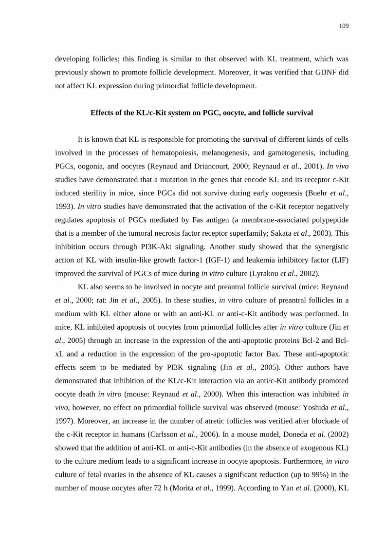
109
developing follicles; this finding is similar to that observed with KL treatment, which was
previously shown to promote follicle development. Moreover, it was verified that GDNF did
not affect KL expression during primordial follicle development.
Effects of the KL/c-Kit system on PGC, oocyte, and follicle survival
It is known that KL is responsible for promoting the survival of different kinds of cells
involved in the processes of hematopoiesis, melanogenesis, and gametogenesis, including
PGCs, oogonia, and oocytes (Reynaud and Driancourt, 2000; Reynaud et al., 2001). In vivo
studies have demonstrated that a mutation in the genes that encode KL and its receptor c-Kit
induced sterility in mice, since PGCs did not survive during early oogenesis (Buehr et al.,
1993). In vitro studies have demonstrated that the activation of the c-Kit receptor negatively
regulates apoptosis of PGCs mediated by Fas antigen (a membrane-associated polypeptide
that is a member of the tumoral necrosis factor receptor superfamily; Sakata et al., 2003). This
inhibition occurs through PI3K-Akt signaling. Another study showed that the synergistic
action of KL with insulin-like growth factor-1 (IGF-1) and leukemia inhibitory factor (LIF)
improved the survival of PGCs of mice during in vitro culture (Lyrakou et al., 2002).
KL also seems to be involved in oocyte and preantral follicle survival (mice: Reynaud
et al., 2000; rat: Jin et al., 2005). In these studies, in vitro culture of preantral follicles in a
medium with KL either alone or with an anti-KL or anti-c-Kit antibody was performed. In
mice, KL inhibited apoptosis of oocytes from primordial follicles after in vitro culture (Jin et
al., 2005) through an increase in the expression of the anti-apoptotic proteins Bcl-2 and Bcl-
xL and a reduction in the expression of the pro-apoptotic factor Bax. These anti-apoptotic
effects seem to be mediated by PI3K signaling (Jin et al., 2005). Other authors have
demonstrated that inhibition of the KL/c-Kit interaction via an anti/c-Kit antibody promoted
oocyte death in vitro (mouse: Reynaud et al., 2000). When this interaction was inhibited in
vivo, however, no effect on primordial follicle survival was observed (mouse: Yoshida et al.,
1997). Moreover, an increase in the number of atretic follicles was verified after blockade of
the c-Kit receptor in humans (Carlsson et al., 2006). In a mouse model, Doneda et al. (2002)
showed that the addition of anti-KL or anti-c-Kit antibodies (in the absence of exogenous KL)
to the culture medium leads to a significant increase in oocyte apoptosis. Furthermore, in vitro
culture of fetal ovaries in the absence of KL causes a significant reduction (up to 99%) in the
number of mouse oocytes after 72 h (Morita et al., 1999). According to Yan et al. (2000), KL
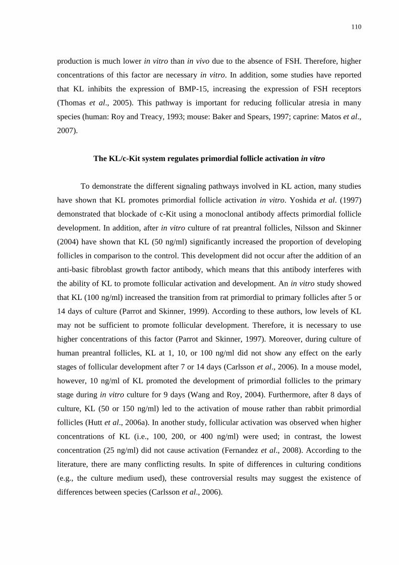
110
production is much lower in vitro than in vivo due to the absence of FSH. Therefore, higher
concentrations of this factor are necessary in vitro. In addition, some studies have reported
that KL inhibits the expression of BMP-15, increasing the expression of FSH receptors
(Thomas et al., 2005). This pathway is important for reducing follicular atresia in many
species (human: Roy and Treacy, 1993; mouse: Baker and Spears, 1997; caprine: Matos et al.,
2007).
The KL/c-Kit system regulates primordial follicle activation in vitro
To demonstrate the different signaling pathways involved in KL action, many studies
have shown that KL promotes primordial follicle activation in vitro. Yoshida et al. (1997)
demonstrated that blockade of c-Kit using a monoclonal antibody affects primordial follicle
development. In addition, after in vitro culture of rat preantral follicles, Nilsson and Skinner
(2004) have shown that KL (50 ng/ml) significantly increased the proportion of developing
follicles in comparison to the control. This development did not occur after the addition of an
anti-basic fibroblast growth factor antibody, which means that this antibody interferes with
the ability of KL to promote follicular activation and development. An in vitro study showed
that KL (100 ng/ml) increased the transition from rat primordial to primary follicles after 5 or
14 days of culture (Parrot and Skinner, 1999). According to these authors, low levels of KL
may not be sufficient to promote follicular development. Therefore, it is necessary to use
higher concentrations of this factor (Parrot and Skinner, 1997). Moreover, during culture of
human preantral follicles, KL at 1, 10, or 100 ng/ml did not show any effect on the early
stages of follicular development after 7 or 14 days (Carlsson et al., 2006). In a mouse model,
however, 10 ng/ml of KL promoted the development of primordial follicles to the primary
stage during in vitro culture for 9 days (Wang and Roy, 2004). Furthermore, after 8 days of
culture, KL (50 or 150 ng/ml) led to the activation of mouse rather than rabbit primordial
follicles (Hutt et al., 2006a). In another study, follicular activation was observed when higher
concentrations of KL (i.e., 100, 200, or 400 ng/ml) were used; in contrast, the lowest
concentration (25 ng/ml) did not cause activation (Fernandez et al., 2008). According to the
literature, there are many conflicting results. In spite of differences in culturing conditions
(e.g., the culture medium used), these controversial results may suggest the existence of
differences between species (Carlsson et al., 2006).
111
In vitro effects of the KL/c-Kit system on oocyte and follicle growth
The expression of both KL and c-Kit is consistent with the role of this system in early
oocyte growth (Manova et al., 1993), which was already demonstrated in vitro (Packer et al.,
1994; Klinger and De Felici, 2002). It is suggested that KL synthesized by granulosa cells
binds to c-Kit present in the oocyte, thereby promoting oocyte growth. Klinger and De Felici
(2002) working with mouse oocytes reported a two-fold increase in oocyte diameter
compared to controls after 4 days of culture in the presence of 50 or 100 ng/ml of KL. These
same authors reported that mouse oocyte growth is characterized by three distinct phases: the
initial stage of growth can be promoted by KL and does not require gap junctions; followed
by a growth phase which depends on KL and gap junctions; and finally, growth phase is
dependent only on gap junctions and is independent of KL (Klinger and De Felici, 2002).
Moreover, the use of 50 ng/ml of KL stimulated theca cell growth, as estimated by DNA
synthesis and the increase in androstenedione production in the absence of gonadotropins
(Parrot and Skinner, 1997). Furthermore, oocytes of prepubertal animals seemed to show the
ability to increase mRNA for KL in granulosa cells (Packer et al., 1994); this finding was
confirmed by Joyce et al. (1999). Therefore, it is suggested that the oocyte increases the
expression of KL in the surrounding granulosa cells and this increase in KL stimulates oocyte
growth (Driancourt et al., 2000). Nevertheless, Cecconi and Colonna (1996) did not observe
any effect of KL on the growth of 12-day-old mouse oocytes, suggesting that KL may have
different actions in each stage of oocyte development. In addition, after culture of whole
mouse ovaries for 9 days, KL (100 ng/ml) did not promote oocyte growth (Wang and Roy,
2004). These differences in results within the same species may be attributed to the stage of
oocyte development, different experimental techniques used for the evaluation of oocyte
growth, or differences in culture conditions cited previously.
Finally, the complex interaction between oocyte and granulosa cells is influenced by
KL/c-Kit, hormones, and growth factors. Some studies suggested that low concentrations of
FSH are necessary for the appropriate regulation of the paracrine factors that trigger oocyte
development. In this way, the correct concentration of FSH may be important for the proper
modulation of KL and BMP-15, increasing oocyte growth (Thomas et al., 2005). BMP-15
produced by the oocyte increases KL expression by granulosa cells. Therefore, KL acts
through c-Kit in the oocyte membrane to promote growth and negatively regulate BMP-15
expression, which produces a consequent increase in FSH receptors (Hutt et al., 2006b;
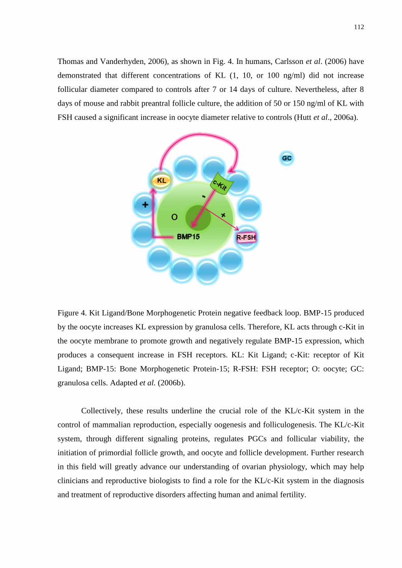
112
Thomas and Vanderhyden, 2006), as shown in Fig. 4. In humans, Carlsson et al. (2006) have
demonstrated that different concentrations of KL (1, 10, or 100 ng/ml) did not increase
follicular diameter compared to controls after 7 or 14 days of culture. Nevertheless, after 8
days of mouse and rabbit preantral follicle culture, the addition of 50 or 150 ng/ml of KL with
FSH caused a significant increase in oocyte diameter relative to controls (Hutt et al., 2006a).
Figure 4. Kit Ligand/Bone Morphogenetic Protein negative feedback loop. BMP-15 produced
by the oocyte increases KL expression by granulosa cells. Therefore, KL acts through c-Kit in
the oocyte membrane to promote growth and negatively regulate BMP-15 expression, which
produces a consequent increase in FSH receptors. KL: Kit Ligand; c-Kit: receptor of Kit
Ligand; BMP-15: Bone Morphogenetic Protein-15; R-FSH: FSH receptor; O: oocyte; GC:
granulosa cells. Adapted et al. (2006b).
Collectively, these results underline the crucial role of the KL/c-Kit system in the
control of mammalian reproduction, especially oogenesis and folliculogenesis. The KL/c-Kit
system, through different signaling proteins, regulates PGCs and follicular viability, the
initiation of primordial follicle growth, and oocyte and follicle development. Further research
in this field will greatly advance our understanding of ovarian physiology, which may help
clinicians and reproductive biologists to find a role for the KL/c-Kit system in the diagnosis
and treatment of reproductive disorders affecting human and animal fertility.

113
Acknowledgments
Juliana Jales de Hollanda Celestino is a recipient of a grant from FUNCAP (Brazil).
References
Amoresano A, Incoronato M, Monti G, Pucci P, de Franciscis V, Cerchia L. 2005 Direct
interactions among Ret, GDNF and GFRalpha1 molecules reveal new insights into the
assembly of a functional three-protein complex. Cell Signal, 17:717-727.
Ballow DJ, Xin Y, Choi Y, Pangas SA, Rajkovic A. 2006. Sohlh2 is a germ cellspecific
bHLH transcription factor. Gene Exp Patterns, 6:1014-1018.
Baker SJ, Spears N. 1997. Follicle stimulating hormone inhibits apoptosis in pre- and early-
antral murine follicles in vitro. J Reprod Fertil Abstr Ser, 19:21.
Blume-Jensen P, Hunter T. 2001. Oncogenic kinase signalling. Nature, 411:355-365.
Buehr M, McLaren A, Bartley A, Darling S. 1993. Proliferation and migration of
primordial germ cells in We/We mouse embryos. Dev Dyn, 198:182-189.
Cantley LC. 2002. The phosphoinositide 3-kinase pathway. Science, 296:1655-1657.
Carlsson IB, Laitinen MPE, Scott JE, Louhio H, Velentzis L, Tuuri T, Aaltonen J,
Ritvos O, Winston RML, Hovatta O. 2006. Kit ligand and c-Kit are expressed during early
human ovarian follicular development and their interaction is required for the survival of
follicles in long-term culture. Reproduction, 131:641-649.
Carmillo P, Dago L, Day ES, Worley DS, Rossomando A, Walus L, Orozco O, Buckley
C, Miller S, Tse A, Cate RL, Rosenblad C, Sah DW, Gronborg M, Whitty A. 2005. Glial
cell line-derived neurotrophic factor (GDNF) receptor alpha-1 (GFR alpha 1) is highly
selective for GDNF versus artemin. Biochemistry, 44:2545-2554.
Castrillon DH, Miao L, Kollipara R, Horner JW, DePinho RA. 2003. Suppression of
ovarian follicle activation in mice by the transcription factor Foxo3a. Science, 301:215-218.
Cecconi S, Colonna R. 1996. Influence of granulosa cells and of different somatic cell types
on mammalian oocyte development in vitro. Zygote, 4: 305-307.
114
Choi Y, Qin Y, Berger MF, Ballow DJ, Bulyk ML, Rajkovic A. 2007. Microarray analyses
of newborn mouse ovaries lacking Nobox. Biol Reprod, 77:312-319.
Choi Y, Ballow DJ, Xin Y, Rajkovic A. 2008a. Lim Homeobox Gene, Lhx8, is essential for
mouse oocyte differentiation and survival1. Biol Reprod, 79:442-449.
Choi Y, Yuan D, Rajkovic A. 2008b. Germ cell-specific transcriptional regulator Sohlh2 is
essential for early mouse folliculogenesis and oocyte-specific gene expression. Biol Reprod,
79:1176-1182.
Clark DE, Tisdall DJ, Fidler AE, Mcnatty KP. 1996. Localization of mRNA encoding c-
Kit during the initiation of folliculogenesis in ovine fetal ovaries. J Reprod Fertil, 106:329-
335.
Davis RJ. 1993. The mitogen-activated protein kinase signal transduction pathway. J Biol
Chem, 268:14553-14556.
Derijard B, Hibi M, Wu IH, Barrett T, Su B, Deng T, Karin M, Davis RJ. 1994. JNK1: a
protein kinase stimulated by UV light and Has-Ras that binds and phosphorylates the c-Jun
activation domain. Cell, 76:1025-1037.
Dole G, Nilsson EE, Skinner MK. 2008. Glial-derived neurotrophic factor promotes ovarian
primordial follicle development and cell–cell interactions during folliculogenesis.
Reproduction, 135:671-682.
Doneda L, Klinger FG, Larizza L, De Felici M. 2002. KL/KIT co-expression in mouse fetal
oocytes. Int J Dev Biol, 46:1015-1021.
Driancourt MA, Reynaud K, Cortvrindt R, Smitz J. 2000. Roles of Kit and Kit Ligand in
ovarian function. Rev Reprod, 5:143-152.
Fernandez SM, Keating AF, Christian PJ, Sen N, Hoying JB, Brooks HL, Hoyer PB.
2008. Involvement of the KIT/KITL signaling pathway in 4-Vinylcyclohexene Diepoxide-
induced ovarian follicle loss in rats. Biol Reprod, 79:318-327.
Fortune JE. 2003. The early stages of follicular development: activation of primordial
follicles and growth of preantral follicles. Anim Reprod Sci, 78:135-163.
Gilchrist RB, Ritter LJ, Cranfield M, Jeffery LA, Amato F, Scott SJ, Myllymaa S,
Kaivo-Oja N, Lankinen H, Mottershead DG, Groome NP, Ritvos O. 2004.
Immunoneutralization of growth differentiation factor 9 reveals it partially accounts for
mouse oocyte mitogenic activity. Biol Reprod, 71:732-739. Goedert M, Cuenda A, Craxton
M, Jakes, R, Cohen P. 1997. Activation of the novel stress-activated protein kinase SAPK4
115
by cytokines and cellular stresses in mediated by SKK3 (MKKK6); comparison of its
substrate specificity with that of other SAP kinases. EMBO J, 16:3563-3571.
Hoyer PE, Byskov AG, Mollgard K. 2005. Stem cell factor and c-Kit in human primordial
germ cells and fetal ovaries. Mol Cell Endocrinol, 234:1-10.
Huang EJ, Nocka KH, Buck J, Besmer P. 1992. Differential expression and processing of
two cell associated forms of the Kit-Ligand: KL-1 and KL-2. Mol Biol Cell, 3:349-362.
Hunter T. 1995. Protein kinases and phosphatases: the yin and yang of protein
phosphorylation and signaling. Cell, 80:225-236.
Hutt KJ, McLaughlin EA, Holland MK. 2006a. KIT/KIT ligand in mammalian oogenesis
and folliculogenesis: roles in rabbit and murine ovarian follicle activation and oocyte growth.
Biol Reprod, 75:421-433.
Hutt KJ, McLaughlin EA, Holland MK. 2006b. Kit ligand and c-Kit have diverse roles
during mammalian oogenesis and folliculogenesis. Mol Hum Reprod, 12:61-69.
Ismail RS, Okawara Y, Fryer JN, Vanderhyden BC. 1996. Hormonal regulation of the
ligand for c-Kit in the rat ovary and its effects on spontaneous oocyte meiotic maturation. Mol
Reprod Dev, 43:458-469.
Ismail RS, Dube M, Vanderhyden BC. 1997. Hormonally regulated expression and
alternative splicing of kit ligand may regulate kit-induced inhibition of meiosis in rat oocytes.
Dev Biol, 184:333-342.
Jin X, Han CS, Yu FQ, Wei P, Hu ZY, Liu YX. 2005. Anti-apoptotic action of stem cell
factor on oocytes in primordial follicles and its signal transduction. Mol Reprod Dev, 70:82-
90.
John GB, Gallardo TD, Shirley LJ, Castrillon DH. 2008. Foxo3 is a PI3K-dependent
molecular switch controlling the initiation of oocyte growth. Dev Biol, 321:197-204.
Joshi S, Davies H, Sims LP, Levy SE, Dean J. 2007. Ovarian gene expression in the
absence of FIGLA, an oocyte-specific transcription factor. BMC Dev Biol, 7:67.
Joyce IM, Pendola FL, Wigglesworth K, Eppig JJ. 1999. Oocyte regulation of kit ligand
expression in mouse ovarian follicles. Dev Biol, 214:342-353.
Klinger FG, De Felici M. 2002. In vitro development of growing oocytes from fetal mouse
oocytes: stage-specific regulation by stem cell factor and granulosa cells. Dev Biol, 244:85-
95.
Lee JC, Laydon JT, McDonnell PC, Gallagher TF, Kumar S, Green D, Mcnulty D,
Blumenthal MJ, Keys JR, Vatter SWL, Strickler JE, McLaughlin MM, Siemens IR,
116
Fisher SM, Livi GP, White JR, Adams JL, Young PR. 1994. A protein kinase involved in
the regulation of inflammatory cytokine biosynthesis. Nature, 372:739-746.
Lyrakou S, Hulten MA, Hartshome GM. 2002. Growth factors promote meiosis in mouse
fetal ovaries in vitro. Mol Hum Reprod, 8:906-911.
Manova K, Huang EJ, Angeles M, De Leon V, Sanchez S, Pronovost SM, Besmer P,
Bachvarova RF. 1993. The expression pattern of the c-Kit ligand in gonads of mice supports
a role for the c-Kit receptor in oocyte growth and in proliferation of spermatogonia. Dev Biol,
157:85-99.
Matos MHT, Lima-Verde IB, Luque MCA, Maia Jr JE, Silva JRV, Celestino JJH,
Martins FS, Báo SN, Lucci CM, Figueiredo JR. 2007. Essential role of follicle stimulating
hormone in the maintenance of caprine preantral follicle viability in vitro. Zygote, 15:173-
182.
Moniruzzaman M, Sakamaki K, Akazawa Y, Miyano T. 2007. Oocyte growth and
follicular development in KIT-deficient Fas-Knockout mice. Reproduction, 133:117-125.
Morita Y, Manganaro TF, Tao XJ, Martimbeau S, Donahoe PK, Tilly JL. 1999.
Requirement for phosphatidylinositol-3-kinase in cytokine-mediated germ cell survival during
fetal oogenesis in the mouse. Endocrinology, 140:941-949.
Motro B, Bernstein A. 1993. Dynamic changes in ovarian c-Kit and syeel expression during
the estrous reproductive cycle. Dev Dyn, 197:69-79.
Naughton CK, Jain S, Strickland AM, Gupta A, Milbrandt J. 2006. Glial cell-line derived
neurotrophic factor-mediated RET signaling regulates spermatogonial stem cell fate. Biol
Reprod, 74:314-321.
Nilsson EE, Skinner MK. 2004. Kit ligand and basic fibroblast growth factor interactions in
the induction of ovarian primordial to primary follicle transition. Mol Cell Endocrinol,
214:19-25.
Otsuka F, Yao Z, Lee T-H, Yamamoto S, Erickson GF, Shimasaki S. 2000. Bone
morphogenetic protein-15: identification of target cells and biological functions. J Biol Chem,
275:39523-39528.
Packer AI, Hsu YC, Besmer P, Bachvaroava RF. 1994. The ligand of the c-Kit receptor
promotes oocyte growth. Dev Biol, 161:194-205.
Pangas SA, Choi Y, Ballow DJ, Zhao Y, Westphal H, Matzuk MM, Rajkovic A. 2006.
Oogenesis requires germ cell-specific transcriptional regulators Sohlh1 and Lhx8. Proc Natl
Acad Sci, 103:8090-8095.
117
Parrot JA, Skinner MK. 1997. Direct actions of Kit-Ligand on theca cell growth and
differentiation during follicle development. Endocrinology, 138:3819-3827.
Parrot JA, Skinner MK. 1999. Kit-ligand/stem cell factor induces primordial follicle
development and initiates folliculogenesis. Endocrinology, 140:4262-4271.
Pozas E, Ibanez CF. 2005. GDNF and GFRalpha1 promote differentiation and tangential
migration of cortical GABAergic neurons. Neuron, 45:701-713.
Rajkovic A, Pangas SA, Ballow D, Suzumori N, Matzuk MM. 2004. NOBOX deficiency
disrupts early folliculogenesis and oocyte-specific gene expression. Science, 305:1157-1159.
Reddy P, Shen L, Ren C, Boman K, Lundin E, Ottander U, Lindgren P, Liu Y-x, Sun Q-
y, Liu K. 2005. Activation of Akt (PKB) and suppression of FKHRL1 in mouse and rat
oocytes by stem cell factor during follicular activation and development. Dev Biol, 281:160-
170.
Reddy P, Liu L, Adhikari D, Jagarlamudi K, Rajareddy S, Shen Y, Du C, Tang W,
Hämäläinen T, Peng SL, Lan Z-J, Cooney AJ, Huhtaniemi I, Liu K. 2008. Oocyte-
specific deletion of Pten causes premature activation of the primordial follicle pool. Science,
319:611-613.
Reynaud K, Cortvrindt R, Smitz J, Driancourt MA. 2000. Effects of Kit Ligand and anti-
Kit antibody on growth of cultured mouse preantral follicles. Mol Reprod Dev, 56:483-494.
Reynaud K, Driancourt MA. 2000. Oocyte attrition. Mol Cell Endocrinol, 163:101-108.
Reynaud K, Cortvrindt R, Smitz J, Bernex F, Panthier JJ, Driancourt MA. 2001.
Alterations in ovarian function of mice with reduced amounts of KIT receptor. Reproduction,
121:229-237.
Roy SK, Treacy BJ. 1993. Isolation and long-term culture of human preantral follicles. Fertil
Steril, 59:783-790.
Sakata S, Sakamaki K, Watanabe K, Nakamura N, Toyokuni S, Nishimune Y, Mori C,
Yonehara S. 2003. Involvement of death receptor Fas in germ cell degeneration in gonads of
Kit-deficient Wv/Wv mutant mice. Cell Death Differ, 10:676-686.
Silva JRV, van den Hurk R, van Tol HTA, Roelen BAJ, Figueiredo JR. 2006. The Kit
ligand/c-Kit receptor system in goat ovaries: gene expression and protein localization. Zygote,
14:317-328.
Skinner MK. 2005. Regulation of primordial follicle assembly and development. Hum
Reprod Update, 11:461-471.
118
Soyal SM, Amleh A, Dean J. 2000. FIGalpha, a germ cell-specific transcription factor
required for ovarian follicle formation. Development, 127:4645-4654.
Thomas FH, Ethier J-F, Shimasaki S, Vanderhyden BC. 2005. Follicle-stimulating
hormone regulates oocyte growth by modulation of expression of oocyte and granulosa cell
factors. Endocrinology, 146:941-949.
Thomas FH, Vanderhyden BC. 2006. Oocyte-granulosa cell interactions during mouse
follicular development regulation of kit ligand expression and its role in oocyte growth.
Reprod Biol Endocrinol, 19:1-8.
Thomas FH, Ismail RS, Jiang J-Y, Vanderhyden BC. 2008. Kit Ligand 2 promotes murine
oocyte growth in vitro. Biol Reprod, 78:167-175.
Tisdall DJ, Fidler AE, Smith P, Quirke LD, Stent VC, Heath DA, Mcnatty KP. 1999.
Stem cell factor and c-Kit gene expression and protein localization in the sheep ovary during
fetal development. J Reprod Fertil, 116:277-291.
Toyoda S, Miyazaki T, Miyazaki S, Yoshimura T, Yamamoto M, Tashiro F, Yamato E,
Miyazaki J-I. 2009. Sohlh2 affects differentiation of KIT positive oocytes and
spermatogonia. Dev Biol, 325:238-248.
Tran H, Brunet A, Griffith EC, Greenberg ME. 2003. The many forks in FOXO's road. Sci
STKE, (172):RE5.
Vargas-Leal V, Bruno R, Derfuss T, Krumbholz M, Hohlfeld R, Meinl E. 2005.
Expression and function of glial cell line-derived neurotrophic factor family ligands and their
receptors on human immune cells. J Immunol, 175:2301-2308.
Wang J, Roy SK. 2004. Growth Differentiation Factor-9 and Stem Cell Factor promote
primordial follicle formation in the hamster: modulation by follicle-stimulating hormone. Biol
Reprod, 70:577-585.
Yan W, Suominen J, Toppari J. 2000. Stem cell factor protects germ cells from apoptosis in
vitro. J Cell Sci, 113:161-168.
Yoshida H, Takakura N, Nataoka H, Kunisada T, Okamura H, Nishikawa SI. 1997.
Stepwise requirement of c-Kit tyrosine kinase in mouse ovarian follicle development. Dev
Biol, 184:122-137.
119
8 CAPÍTULO 3
Implicações da proteína morfogenética óssea-15 na foliculogênese ovariana
Implications of bone morphogenetic protein-15 on the ovarian folliculogenesis
Periódico: Animal Reproduction (Submetido em outubro de 2010).
120
Resumo
A proteína morfogenética óssea-15 (BMP-15) tem demonstrando importantes efeitos na
reprodução e fertilidade de fêmeas mamíferas das mais diversas espécies. Estudos in vivo
utilizando essa proteína têm contribuído para uma melhor compreensão da fisiologia ovariana,
demonstrando que deleções no gene da BMP-15 são capazes de causar infertilidade, com
parada no desenvolvimento folicular inicial. Além disso, estudos in vitro têm comprovado o
papel relevante da BMP-15 no ovário mamífero, destacando sua atuação na proliferação de
células da granulosa e no estímulo à expressão de alguns fatores de crescimento locais, os
quais são necessários para um adequado crescimento folicular e oocitário. O objetivo desse
artigo é revisar a importância da BMP-15 no desenvolvimento de folículos ovarianos,
especialmente nos processos de sobrevivência, ativação e crescimento de folículos pré-antrais.
Palavras-chave: BMP-15. Folículo pré-antral. Fertilidade. Ovário.
121
Implications of bone morphogenetic protein-15 on the ovarian folliculogenesis
J.J.H. Celestino1*; I.M.T. Lima
1; M.H.T. Matos
2; J.R. Figueiredo
1
1Laboratory of Manipulation of Oocytes and Preantral Follicles (LAMOFOPA), Faculty of
Veterinary, State University of Ceará, Av. Paranjana 1700, Campus Itaperi, Fortaleza, 60740-
903, CE, Brazil.
2Nucleus of Biotechnology Applied to Ovarian Follicle Development, Federal University of
São Francisco Valley, Petrolina, 48902-300, PE, Brazil
*Corresponding address:
Programa de Pós-Graduação em Ciências Veterinárias (PPGCV)
Laboratório de Manipulação de Oócitos e Folículos Pré-Antrais (LAMOFOPA)
Universidade Estadual do Ceará (UECE)
Av. Paranjana, 1700, Campus do Itaperi.
Fortaleza – CE – Brasil. CEP: 60740-903
Tel.: +55.85.3101.9852; Fax: +55.85.3101.9840
E-mail address: [email protected] (Juliana Jales de Hollanda Celestino)
Article type: review; Area: Biotechnology
Running title: BMP-15 and follicular development
122
Abstract
Bone morphogenetic protein-15 (BMP-15) has been implicated in important effects on
female reproduction and fertility in several mammalian species. In vivo studies using this
protein have contributed to a better comprehension of ovarian physiology, demonstrating that
the deletion of BMP-15 gene is capable to cause infertility, with arrest in the early follicular
development. Moreover, in vitro studies have shown the relevant role of BMP-15 in the
mammalian ovary, highlighting its action in the granulosa cell proliferation and in the
stimulation of some local growth factors expression, which are necessary for an adequate
follicular and oocyte growth. The purpose of this article is to review the importance of BMP-
15 in ovarian follicular development, especially in the processes of survival, activation and
growth of preantral follicles.
Keywords: BMP-15, preantral follicle, fertility, ovary
1. Introduction
The bone morphogenetic protein (BMP) family corresponds to the largest group of
growth factors members of TGF- superfamily (Shimasaki et al., 2004a,b). Among the BMPs,
the BMP-15 has received great distinction in the last decades, being its role widely studied in
different species, including rodents, domestic animals and humans. Such protein, codified by
a gene linked to the X chromosome and secreted by the oocyte, has been considered an
important regulator of follicular development, acting positively in the maturation of oocytes
enclosed in preovulatory follicles, in the determination of fertility and ovulation rate in
mammals, in addition to be directly involved in premature luteinization (Galloway et al.,
2000; Juengel et al., 2004a; Knight and Glister, 2006). Furthermore, BMP-15 has a significant
mitogenic effect on the granulosa cells, contributing to the growth during the different phases
of folliculogenesis, including the process of follicular activation (Otsuka et al., 2000; Juengel
et al., 2004b).
In this context, the present review will highlight the aspects related to the expression
of BMP-15 and its receptors in the ovary, cellular signaling pathways of BMP-15, and the
effects of this protein on the survival, activation and follicular growth, as well in the ovulation
and steroidogenesis.
123
2. Expression of BMP-15 and its receptors in the ovary
BMP-15, also known as growth differentiation factor-9B (GDF-9B), is one of the 35
members of the TGF- superfamily (Knight and Glister, 2006). In this superfamily, the ligand
interacts with two categories of signaling receptors, called type I and type II (Massagué and
Chen, 2000). For the BMPs, the main type I receptors are BMPR-IA or activin receptor-like
kinase-3 (ALK-3) and BMPR-IB or ALK-6, while the type II receptor is called BMPR-II
(Ten Dijike et al., 2003; Abir et al., 2008). Specifically, BMP-15 plays its biological effects
through the formation of heterodimeric complexes with a type I receptor (ALK-6) and a type
II receptor (BMPR-II) on the surface of the cell (Lebrun et al., 1997; Miyazono et al., 1997).
In all studied species, the ovary is the local with the greatest production of BMP-15
(Peng et al., 2009), being this protein also found in the pituitary, rete testis and other tissues
from some species (Fitzpatrick et al., 1998; Aaltonen et al., 1999; Galloway et al., 2000;
Eckery et al., 2002; Otsuka and Shamasaki, 2002a). Regarding the ovary, in some species,
such as marsupial (Brushtail possums – Eckery et al., 2002), ovine (Mery et al., 2007), bovine
(Bodensteiner et al., 1999) and human (Shimasaki et al., 2004a), BMP-15 is expressed in
oocytes from primordial follicles onward. In rodents, the expression of this protein is
observed only in the oocyte from primary follicles onward (Laitinen et al., 1998; Jaatinen et
al., 1999). For caprine species, BMP-15 has been found in the oocytes of all types of follicles
and granulosa cells of primary, secondary and antral follicles but not in primordial stage. In
this species, the mRNA for BMP-15 and their associated receptors were detected in
primordial, primary and secondary follicles, as well as in the oocyte and granulosa cells of
antral follicles (Silva et al., 2005). In sheep, the protein and mRNA for BMP-15 receptors
were detected both in the granulosa cells and oocyte of preantral follicles (Wilson et al., 2001;
Souza et al., 2002), thus suggesting an important role of BMP-15 in the regulation of the
function of these two cell types during the follicular development.
3. Signaling pathways of BMP-15 in the regulation of folliculogenesis
Both BMP-15 receptors, ALK-6 (type I) and BMPR-II (type II), have an extracellular
binding domain rich in cysteine, a membranary and a cytoplasmic domain of serine/treonine
quinase. First, binding of BMP-15 to the extracellular domain of type I receptor leads to the
recruitment of the type II receptor, which is constitutively phosphorylated and
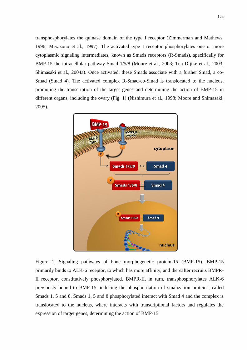
124
transphosphorylates the quinase domain of the type I receptor (Zimmerman and Mathews,
1996; Miyazono et al., 1997). The activated type I receptor phosphorylates one or more
cytoplasmic signaling intermediates, known as Smads receptors (R-Smads), specifically for
BMP-15 the intracellular pathway Smad 1/5/8 (Moore et al., 2003; Ten Dijike et al., 2003;
Shimasaki et al., 2004a). Once activated, these Smads associate with a further Smad, a co-
Smad (Smad 4). The activated complex R-Smad-co-Smad is translocated to the nucleus,
promoting the transcription of the target genes and determining the action of BMP-15 in
different organs, including the ovary (Fig. 1) (Nishimura et al., 1998; Moore and Shimasaki,
2005).
Figure 1. Signaling pathways of bone morphogenetic protein-15 (BMP-15). BMP-15
primarily binds to ALK-6 receptor, to which has more affinity, and thereafter recruits BMPR-
II receptor, constitutively phosphorylated. BMPR-II, in turn, transphosphorylates ALK-6
previously bound to BMP-15, inducing the phosphorilation of sinalization proteins, called
Smads 1, 5 and 8. Smads 1, 5 and 8 phosphorylated interact with Smad 4 and the complex is
translocated to the nucleus, where interacts with transcriptional factors and regulates the
expression of target genes, determining the action of BMP-15.
125
Additional regulation also occurs by inhibitory Smads (Smads 6 and 7), which can
attenuate the pathway of signal transduction (Miyazono et al., 1997; Knight and Glister,
2003). In addition to the cell signaling way mediated by Smads, evidences indicate that other
ways also may be involved in the BMP-15 and other BMPs action (Von Bubnoff and Cho,
2001; Nohe et al., 2002), for example, the signaling way mediated by the protein-quinase
activated by mitogens (MAPK). Studies have demonstrated that MAPK can modulate the
transduction of the BMP-15 signals through acting in common sites with the Smad way. In
fact, it has been verified that activated MAPK molecules can positively transducer the BMP-
15 signals (Su et al., 2002) or act as inhibitors of the Smad signaling (Von Bubnoff and Cho,
2001). According to Moore et al. (2003), the action of BMP-15 on the proliferation and
differentiation of the granulosa cell in dependent on the ERK1/2 molecule, which is a member
of MAPK family. This emphasizes the existence of an alternative way for the regulation of
the BMP-15 action. In addition, other works indicate that follistatin, important binding protein
expressed by the granulosa cells of growing follicles, can also bind to BMP-15, and thus
block its bioactivity (Otsuka et al., 2001a).
4. Effects of BMP-15 on the follicular viability
BMP-15, as well as others BMPs (-2, -5 and -6), acts in the granulosa cells, promoting
the survival of the follicles through the maintenance of cell proliferation and prevention of
precocious luteinization and/or atresia (Knight and Glister, 2006). Studies also have
demonstrated that BMP-15 levels in the follicular fluid seems to be a potential factor to
determine the oocyte quality and further embryo development, being demonstrated that high
BMP-15 levels in human follicular fluid warrant a higher cleavage rate and good embryo
morphology (Wu et al., 2007). In addition, BMP-15 was capable to maintain a low incidence
of apoptosis in the cumulus cells, suggesting that this may be involved in the follicular
development (Hussein et al., 2005; Wu et al., 2007). In a study with mouse in which the
specific gene for BMP-15 was deleted, the authors observed defects in the integrity of the
cumulus oocyte complexes (Varani et al., 2002). Other study showed that after neutralization
of a great portion of the gene for GDF-9 and BMP-15, there was a reduction in the oocyte
quality and in the ability of the sheep to carry pregnancies to term (Juengel et al., 2004a).
Some studies have evidenced that BMP-15 did not act by itself but interacts with other
substances, such as GDF-9, kit ligand (KL), follicle stimulating hormone (FSH) and follistatin
126
(Huang et al., 2001; Otsuka et al., 2001a,b; Yan et al., 2001; Juengel et al., 2002; Otsuka and
Shimasaki, 2002a,b; Moore et al., 2003; Shimasaki et al., 2004a; Edwards et al., 2008; Sadeu
et al., 2008), possibly in a species-specific way (Margulis et al., 2009). It is known that BMP-
15 stimulates the expression of KL in the granulosa cells (Otsuka and Shimasaki, 2002b;
Moore and Shimasaki, 2005), which is an essential factor for the early folliculogenesis
(Otsuka and Shamasaki, 2002a; Peng et al., 2009). In humans (Carlsson et al., 2006) and
caprine (Celestino et al., 2010), it has been demonstrated the importance of KL and its
receptor c-kit for the survival of preantral follicles cultured in vitro. Other studies have
demonstrated that KL inhibits apoptosis in oocytes from mouse primordial follicles through
an increase in the mRNA levels of antiapoptotic proteins, Bcl-2 and Bcl-cL, and a reduction
in the levels of the pro-apoptotic factor Bax. These antiapoptotic effects seem to be mediated
by a fosfatidilinositol 3-quinase (PI3K) signaling, which acts promoting the inhibition of
apoptosis mediated by Fas ligand, a polypeptide associated to the membrane (Jin et al., 2005).
5. Involvement of BMP-15 in the activation of primordial follicles
In the early folliculogenesis, one of the main activities of BMP-15 is to promote
granulosa cell mitosis (Moore et al., 2003). In fact, studies performed in rodents (rat: Otsuka
et al., 2000) and ruminants (sheep and bovine: McNatty et al., 2005) characterize BMP-15 as
a potent stimulator of granulosa cell proliferation, demonstrating an intense capacity to
increase the incorporation of tritiated thymidine and the number of follicular cells in vitro.
The presence of BMP-15 in primordial follicles from some species suggests that this
protein, produced by the oocyte, has a role in the recruitment of the follicles, i.e., in the
follicular activation (Otsuka et al., 2000). In caprine, not only the protein (expressed
exclusively in the oocyte) but also the mRNA for the BMP-15 ligand, as well as the mRNA
for its two receptors, were all expressed as early as from primordial follicle stage (Silva et al.,
2005), thus suggesting the possible role of BMP-15 in the follicular activation in this species.
We recently confirmed this hypothesis in a study showing that BMP-15 promotes the in vitro
transition from caprine primordial to primary follicle stage (Celestino et al., unpublished
data). The role of BMP-15 in the process of follicular activation, determined in a great part by
its action in cell proliferation, occurs mainly through its BMPR-II receptor present in the
surface of the granulosa cells (Shimasaki et al., 2004a,b).
127
In ovarian follicles, the BMP-15 action is directly related with the KL, including in
the proliferation of the granulosa cells (Moore and Shimasaki, 2005). In fact, the ability of
BMP-15 for regulating cell mitosis is controlled in part by KL. A negative feedback between
these two factors seems to exist, with BMP-15 stimulating the expression of KL in the
granulosa cells, and KL inhibiting the expression of BMP-15 in the oocyte (Juengel et al.,
2004b). In sheep and women mutations for the BMP-15 gene, this negative feedback between
BMP-15 and KL is lost, damaging the early follicular development (Moore and Shimasaki,
2005).
6. BMP-15 and follicular and oocyte growth
Evidences indicate that due to its effects on the cellular proliferation, BMP-15 has an
essential role for the ovarian follicle development (Juengel et al., 2004b). In addition, it has
been demonstrated that BMP-15 activity is independent of FSH, suggesting that BMP-15 can
stimulate the mitosis of granulosa cells in early preantral follicles in the absence of this
gonadotropin, and also inhibiting the expression of FSH receptor (FSH-R) (Otsuka et al.,
2001b).
Otsuka et al. (2000) suggests that the signaling pathway of BMP-15 leads to the
stimulation and inhibition of effector genes, as well as to biological specific responses, which
are critical for follicular growth and development. In mouse, when the gene for BMP-15 was
deleted, the animals show defects in the ovulation process and in the quality of the fertilizable
oocytes, resulting in lowest sizes of these oocytes (Yan et al., 2001). In sheep, immunization
against BMP-15 resulted in a blockage of follicular growth (Juengel et al., 2002).
Additional studies have related a high expression of the mRNA for BMP-15 and/or of
the protein in growing or completely grown oocytes (Shimasaki et al., 2004a; Juengel and
McNatty, 2005; Li et al., 2008). Moreover, the absence of BMP-15 receptor (ALK-6 and
BMPR-II) production in ovine follicles can lead to a break in their growth (Mery et al., 2007).
In mouse, it was reported an increase in the levels of mRNA expression for BMP-15 from day
7 to 10 of in vitro culture, being this increase associated to the presence of early stage follicles
with growing oocytes (Sadeu et al., 2008). In another study, BMP-15 increased the
competence for bovine oocyte development (Gilchrist et al., 2008). Similarly, it has been
demonstrated that BMP-15 in human follicular fluid is positively correlated with the potential
for the oocyte development (Wu et al., 2007). In this same species, the expression of the

128
mRNA for BMP-15 increased in a direct correlation with the follicular growth (Teixeira Filho
et al., 2002). A summary of the main implications of bone morphogenetic protein-15 (BMP-
15) in the reproduction of different species as shown in Table 1.
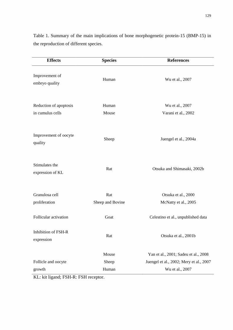
129
Table 1. Summary of the main implications of bone morphogenetic protein-15 (BMP-15) in
the reproduction of different species.
Effects Species References
Improvement of
embryo quality
Human Wu et al., 2007
Reduction of apoptosis
in cumulus cells
Human
Mouse
Wu et al., 2007
Varani et al., 2002
Improvement of oocyte
quality
Sheep Juengel et al., 2004a
Stimulates the
expression of KL
Rat Otsuka and Shimasaki, 2002b
Granulosa cell
proliferation
Rat
Sheep and Bovine
Otsuka et al., 2000
McNatty et al., 2005
Follicular activation Goat Celestino et al., unpublished data
Inhibition of FSH-R
expression
Rat Otsuka et al., 2001b
Follicle and oocyte
growth
Mouse
Sheep
Human
Yan et al., 2001; Sadeu et al., 2008
Juengel et al., 2002; Mery et al., 2007
Wu et al., 2007
KL: kit ligand; FSH-R: FSH receptor.
130
7. Final considerations
Several researches in the field of reproductive biotechnologies have been developed to
establish in vivo and in vitro systems which reveal the functions of different proteins
synthesized in the ovary, including BMP-15. This protein, highlighted in the present review,
has a crucial role on mammalian folliculogenesis and fertility, showing together with other
local factors, regulatory actions on the follicular somatic cells, which are essential for warrant
an adequate development of ovarian follicles. However, despite many researches be related to
the study of BMP-15, less is known about its specific role on the female reproduction, as well
as on its interaction with other regulatory substances. Therefore, it is of great importance the
complete elucidation of the mechanisms of BMP-15 action in the ovary because, in the future,
it can provide a correct monitoring and manipulation of the ovarian function, with a
consequent improvement of the fertility of domestic animals, endangered species and humans.
Acknowledgments
Juliana Jales de Hollanda Celestino is a recipient of a grant from FUNCAP (Brazil).
References
Aaltonen J, Laitinen MP, Vuojolainen K, Jaatinen R, Horelli-Kuitunen N, Seppa L, Louhio H,
Tuuri T, Sjoberg J, Butzow R, Hovata O, Dale L, Ritvos O. 1999. Human growth
differentiation factor 9 (GDF-9) and its novel homolog GDF-9B are expressed in oocytes
during early folliculogenesis. J Clin Endocrinol Metab, 84:2744-2750.
Abir R, Ben-Haroush A, Melamed N, Felz C, Krissi H, Fisch B. 2008. Expression of bone
morphogenetic proteins 4 and 7 and their receptors IA, IB and II in human ovaries from
fetuses and adults. Fertil Steril, 89:1430-1440.
Bodensteiner KJ, Clay CM, Moeller CL, Sawyer HR. 1999. Molecular cloning of the ovine
growth/differentiation factor-9 gene and expression of growth/differentiation factor-9 in ovine
and bovine ovaries. Biol Reprod, 60:381-386.
Carlsson IB, Laitinen MPE, Scott JE, Louhio H, Velentzis L, Tuuri T, Aaltonen J, Ritvos O,
Winston RML, Hovatta O. 2006. Kit ligand and c-Kit are expressed during early human

131
ovarian follicular development and their interaction is required for the survival of follicles in
long-term culture. Reproduction, 131:641-649.
Celestino JJH, Bruno JB, Lima-Verde IB, Matos MHT, Saraiva MVA, Chaves RN, Martins
FS, Almeida AP, Cunha RMS, Lima LF, Name KPO, Campello CC, Silva JRV, Báo SN,
Figueiredo JR. 2010. Steady-state level of Kit Ligand mRNA in goat ovaries and the role of
Kit Ligand in preantral follicle survival and growth in vitro. Mol Reprod Dev, 77:231-240.
Eckery DC, Whale LJ, Lawrence SB, Wylde KA, McNatty KP, Juengel JL. 2002. Expression
of mRNA encoding growth differentiation factor 9 and bone morphogenetic protein 15 during
follicular formation and growth in a marsupial, the brushtail possum (Trichosurus vulpecula).
Mol Cell Endocrinol, 192:115-126.
Edwards SJ, Reader KL, Lun S, Western A, Lawrence S, McNatty KP, Juengel JL. 2008. The
cooperative effect of growth and differentiation factor-9 and bone morphogenetic protein
(BMP)-15 on granulosa cell function is modulated primarily through BMP receptor II.
Endocrinology, 149:1026-1030.
Fitzpatrick SL, Sindoni DM, Shughrue PJ, Lane MV, Merchenthaler IJ, Frail DE. 1998.
Expression of growth differentiation factor-9 messenger ribonucleic acid in ovarian and
nonovarian rodent and human tissues. Endocrinology, 139:2571-2578.
Galloway SM, McNatty KP, Cambridge LM, Laitinen MP, Juengel JL, Jokiranta TS,
McLaren RJ, Luiro K, Dodds KG, Montgomery GW, Beattie AE, Davis GH, Ritvos O. 2000.
Mutations in an oocyte-derived growth factor gene (BMP15) cause increased ovulation rate
and infertility in a dosage-sensitive manner. Nat Genet, 25:279-283.
Gilchrist RB, Lane M, Thompson JG. 2008. Oocyte-secreted factors: regulators of cumulus
cell function and oocyte quality. Hum Reprod Update, 14:159-177.
Huang H-J, Wu JC, Su P, Zhirnov O, Miller WL. 2001. A novel role for bone morphogenetic
proteins in the synthesis of follicle-stimulating hormone. Endocrinology, 142:2275-2283.
Hussein TS, Froiland DA, Amato F, Thompson JG, Gilchrist RB. 2005. Oocytes prevent
cumulus cell apoptosis by maintaining a morphogenic paracrine gradient of bone
morphogenetic proteins. J Cell Sci, 118:5257-5268.
Jaatinen R, Laitinen MP, Vuojolainen K, Aaltonen J, Louhio H, Heikinheimo K, Lehtonen E,
Ritvos O. 1999. Localization of growth differentiation factor-9 (GDF-9) mRNA and protein
in rat ovaries and cDNA cloning of rat GDF-9 and its novel homolog GDF-9B. Mol Cell
Endocrinol, 156:189-193.
132
Jin X, Han CS, Yu FQ, Wei P, Hu ZY, Liu YX. 2005. Anti-apoptotic action of stem cell
factor on oocytes in primordial follicles and its signal transduction. Mol Reprod Dev, 70:82-
90.
Juengel JL, Hudson NL, Heath DA, Smith P, Reader KL, Lawrence SB, O‘Connell AR,
Laitinen MP, Cranfield M, Groome NP, Ritvos O, McNatty K.P. 2002. Growth differentiation
factor-9 and bone morphogenetic protein 15 are essential for ovarian follicular development
in sheep. Biol Reprod, 67:1777-1789.
Juengel JL, Hudson NL, Whitinig L, McNatty KP. 2004a. Effects of immunization against
bone morphogenetic protein 15 and growth differentiation factor 9 on ovulation rate,
fertilization, and pregnancy in ewes. Biol Reprod, 70:557-561.
Juengel JL, Bodensteiner KJ, Heath DA, Hudsona NL, Moeller CL, Smith P, Galloway SM,
Davis GH, Sawyer HR, McNatty KP. 2004b. Physiology of GDF9 and BMP15 signalling
molecules. Anim Reprod Sci, 82-83:447-460.
Juengel JL, McNatty KP. 2005. The role of proteins of the transforming growth factor-b
superfamily in the intraovarian regulation of follicular development. Hum Reprod Update,
11:144-161.
Knight PG, Glister C. 2003. Local roles of TGF-β superfamily members in the control of
ovarian follicle development. Anim Reprod Sci, 78:165-183.
Knight PG, Glister C. 2006. TGF-β superfamily members and ovarian follicle development.
Reproduction, 132:191-206.
Laitinen M, Vuojolainen K, Jaatinen R, Ketola I, Aaltonen J, Lehtonen E, Heikinheimo M,
Ritvos O. 1998. A novel growth differentiation factor- 9 (GDF-9) related factor is co-
expressed with GDF-9 in mouse oocytes during folliculogenesis. Mech Dev, 78:135-140.
Lebrun JJ, Chen Y, Vale WW. 1997. Receptor serine kinases and signaling by activins and
inhibins. In Aono, T., Sugino, H., Vale, W. (Ed.). Inhibin, activin and follistatin regulatory
functions in system and cell biology, Springer Serono Symposia, USA, pp.1-20.
Li HK, Kuo TY, Yang HS, Chen LR, Li SS, Huang HW. 2008. Differential gene expression
of bone morphogenetic protein 15 and growth differentiation factor 9 during in vitro
maturation of porcine oocytes and early embryos. Anim Reprod Sci, 103:312-322.
Margulis S, Abir R, Felz C, Nitke S, Krissi H, Fisch B. 2009. Bone morphogenetic protein 15
expression in human ovaries from fetuses, girls, and women. Fertil Steril, 92:1666-1673.
Massagué J, Chen YG. 2000. Controlling TGF-β signaling. Gene Dev, 14:627-644.
133
McNatty KP, Juengel JL, Reader KL, Lun S, Myllymaa S, Lawrence SB, Western A,
Meerasahib MF, Mottershead DG, Groome NP, Ritvos O, Laitinen MPE. 2005. Bone
morphogenetic protein 15 and growth differentiation factor 9 co-operate to regulate granulosa
cell function. Reproduction, 129:473-480.
Mery L, Lefevre A, Benchaib M, Demirci B, Salle B, Guerin J-F, Lornage J. 2007. Follicular
growth in vitro: Detection of growth differentiation factor 9 (GDF9) and Bone morphogenetic
protein 15 (BMP15) during in vitro culture of ovine cortical slices. Mol Reprod Dev, 74:767-
774.
Miyazono K, Tijke PT, Souchelnytskyi S, Nakao A, Imamura T, Hanai J-I, Kawabata M,
Heldin C-H. 1997. Transforming growth factor-β receptors and signal transduction, In Aono,
T., Sugino, H., Vale, W. (Ed.), Inhibin, activin and follistatin regulatory functions in system
and cell biology, Springer Serono Symposia, USA, pp.277-286.
Moore RK, Otsuka F, Shimasaki S. 2003. Molecular Basis of Bone Morphogenetic Protein-15
Signaling in Granulosa Cells. J Biol Chem, 278:304-310.
Moore RK, Shimasaki S. 2005. Molecular biology and physiological role of the oocyte factor,
BMP-15. Mol Cell Endocrinol, 234:67-73.
Nishimura R, Kato Y, Chen D, Harris SE, Mundy GR, Yoneda T. 1998. Smad5 and DPC4 are
key molecules in mediating BMP-2- induced osteoblastic differentiation of the pluripotent
mesenchymal precursor cell line C2C12. J Biol Chem, 273:1872-1879.
Nohe A, Hassel S, Ehrlich M, Neubauer F, Sebald W, Henis YI, Knaus P. 2002. The mode of
bone morphogenetic protein (BMP) receptor oligomerization determines different BMP-2
signaling pathways. J Biol Chem, 277:5330-5338.
Otsuka F, Yao Z, Lee T, Yamamoto S, Erickson GF, Shimasaki S. 2000. Bone morphogenetic
protein-15. Identification of target cells and biological functions. J Biol Chem, 275:39523-
39528.
Otsuka F, Moore RK, Iemura S, Ueno N, Shimasaki S. 2001a. Follistatin inhibits the function
of the oocyte-derived factor BMP-15. Biochem Biophys Res Commun, 289:961-966.
Otsuka F, Yamamoto S, Erickson GF, Shimasaki S. 2001b. Bone morphogenetic protein-15
inhibits follicle-stimulating hormone (FSH) action by suppressing FSH receptor expression. J
Biol Chem, 276:11387-11392.
Otsuka F, Shimasaki S. 2002a. A novel function of bone morphogenetic protein-15 in the
pituitary: selective synthesis and secretion of FSH by gonadotropes. Endocrinology,
143:4938-4941.
134
Otsuka F, Shimasaki S. 2002b. A negative feedback system between oocyte bone
morphogenetic protein 15 and granulosa cell kit ligand: its role in regulating granulosa cell
mitosis. Proc Natl Acad Sci USA, 99:8060-8065.
Peng C, Clelland E, Tan Q. 2009. Potencial role of bone morphogenetic protein-15 in
zebrafish follicle development and oocyte maturation. Comp Biochem Phys A, 153:83-87.
Sadeu JC, Adriaenssens T, Smitz J. 2008. Expression of growth differentiation factor 9, bone
morphogenetic protein 15, and anti-Mullerian hormone in cultured mouse primary follicles.
Reproduction, 136:195-203.
Shimasaki S, Moore RK, Otsuka F, Erickson GF. 2004a. The bone morphogenetic protein
system in mammalian reproduction. Endocr Rev, 25:72-101.
Shimasaki S, Moore RK, Erickson GF, Otsuka F. 2004b. Ovarian bone morphogenetic
proteins in female reproduction. Int Congress Series, 1266:241-247.
Silva JR, van den Hurk R, van Tol HT, Roelen BA, Figueiredo J.R. 2005. Expression of
growth differentiation factor 9 (GDF9), bone morphogenetic protein 15 (BMP-15), and BMP
receptors in the ovaries of goats. Mol Reprod Dev, 70:11-19.
Souza CJ, Campbell BK, McNeilly AS, Baird DT. 2002. Effect of bone morphogenetic
protein 2 (BMP2) on oestradiol and inhibin A production by sheep granulosa cells, and
localization of BMP receptors in the ovary by immunohistochemistry. Reproduction,
123:363-369.
Su Y-Q, Wigglesworth K, Pendola FL, O‘Brien MJ, Eppig JJ. 2002. Mitogen-activated
protein kinase activity in cumulus cells is essential for gonadotropin-induced oocyte meiotic
resumption and cumulus expansion in the mouse. Endocrinology, 143:2221-2232.
Teixeira Filho FL, Baracat EC, Lee TH, Suh CS, Matsui M, Chang RJ, Shimasaki S, Erickson
G.F. 2002. Aberrant expression of growth differentiation factor-9 in oocytes of women with
polycystic ovary syndrome. J Clin Endocrinol Metab, 87:1337-1344.
Ten Dijike P, Korchynskyi O, Valdimarsdottir G, Goumans MJ. 2003. Controlling cell fate by
bone morphogenetic protein receptors. Mol Cell Endocrinol, 211:105-113.
Varani S, Elvin JA, Yan C, DeMayo J, DeMayo FJ, Horton HF, Byrne MC, Matzuk MM.
2002. Knockout of pentraxin 3, a downstream target of growth differentiation factor-9, causes
female subfertility. Mol Endocrinol, 16:1154-1167.
Von Bubnoff A, Cho KWY. 2001. Intracellular BMP signaling regulation in vertebrates:
pathway or network? Dev Biol, 239:1-14.
135
Wilson T, Wu XY, Juengel JL, Ross IK, Lumsden JM, Lord EA, Dodds KG, Walling GA,
McEwan JC, O‘Connell AR, McNatty KP, Montgomery GW. 2001. Highly prolific Booroola
sheep have a mutation in the intracellular kinase domain of bone morphogenetic protein IB
receptor (ALK-6) that is expressed in both oocytes and granulosa cells. Biol Reprod, 64:1225-
1235.
Wu Y-T, Tang L, Cai J, Lu X-E, Xu J, Zhu X-M, Luo Q, Huang H-F. 2007. High bone
morphogenetic protein-15 level in follicular fluid is associated with high quality oocyte and
subsequent embryonic development. Human Reprod, 22:1526-1531.
Yan C, Wang P, DeMayo J, DeMayo FJ, Elvin JA, Carino C, Prasad SV, Skinner SS, Dunbar
BS, Dube JL, Celeste AJ, Matzuk MM. 2001. Synergistic roles of bone morphogenetic
protein 15 and growth differentiation factor 9 in ovarian function. Mol Endocrinol, 15:854-
866.
Zimmerman CM, Mathews LS. 1996. Activin receptors: cellular signaling by receptor serine
kinases. Biochem Soc Symp, 62:25-38.
136
9 CAPÍTULO 4
Fator de crescimento epidermal como mediador de sobrevivência e desenvolvimento folicular
Epidermal growth factor as mediator of survival and follicular development
Periódico: Revista Brasileira de Reprodução Animal (Submetido em outubro de 2010).
A B
C D
137
Resumo
Diversos fatores intra-ovarianos atuam no ovário dos mamíferos regulando o
desenvolvimento folicular. Dentre eles destaca-se o fator de crescimento epidermal (EGF),
considerado um potente fator mitogênico para células foliculares e luteais. Tendo em vista a
importância deste fator no âmbito do desenvolvimento folicular, a presente revisão de
literatura tem como objetivo descrever as principais implicações do EGF na foliculogênese,
destacando seu padrão de expressão no ovário, suas principais vias de sinalização celular, bem
como seu efeito como fator de sobrevivência e de desenvolvimento folicular.
Palavras-chave: EGF. Ovário. Folículo. Mamífero.
138
Fator de crescimento epidermal como mediador de sobrevivência e desenvolvimento
folicular
(Epidermal growth factor as mediator of survival and follicular development)
Juliana Jales de Hollanda Celestino*, Cleidson Manoel Gomes da Silva, Simone Vieira
Castro, José Ricardo de Figueiredo
Laboratório de Manipulação de Oócitos e Folículos Ovarianos Pré-Antrais - LAMOFOPA,
Faculdade de Veterinária, Universidade Estadual do Ceará, Fortaleza, CE, Brasil
*Corresponding address:
Programa de Pós-Graduação em Ciências Veterinárias (PPGCV)
Laboratório de Manipulação de Oócitos e Folículos Pré-Antrais (LAMOFOPA)
Universidade Estadual do Ceará (UECE)
Av. Paranjana, 1700, Campus do Itaperi.
Fortaleza – CE – Brasil. CEP: 60740-903
Tel.: 85.3101.9852; Fax: 85.3101.9840
E-mail address: [email protected] (Juliana Jales de Hollanda Celestino)
139
Resumo
Diversos fatores intra-ovarianos atuam no ovário dos mamíferos regulando o
desenvolvimento folicular. Dentre eles destaca-se o fator de crescimento epidermal (EGF),
considerado um potente fator mitogênico para células foliculares e luteais. Tendo em vista a
importância deste fator no âmbito do desenvolvimento folicular, a presente revisão de
literatura tem como objetivo descrever as principais implicações do EGF na foliculogênese,
destacando seu padrão de expressão no ovário, suas principais vias de sinalização celular, bem
como seu efeito como fator de sobrevivência e de desenvolvimento folicular.
Palavras-chave: EGF, ovário, folículo, mamífero.
Abstract
Several intra-ovarian factors act regulating the mammalian follicular development in the
ovary. Among them highlights the epidermal growth factor (EGF), considered a potent
mitogenic factor for follicular and luteal cells. Given the importance of this factor within the
follicular development, this review describes the main implications of EGF in the
folliculogenesis, focusing on its expression pattern in the ovary, the major pathways of cell
signaling and its effect as a survival and follicular development factor.
Keywords: EGF, ovary, follicle, mammal.
Introdução
A foliculogênese é uma complexa orquestra de eventos que propicia o
desenvolvimento folicular (Fair, 2003). Atualmente é bem estabelecido que o crescimento dos
folículos presentes nos ovários dos mamíferos é regulado por gonadotrofinas e por fatores
intra-ovarianos (Fortune, 2003). O papel das gonadotrofinas, Hormônio Folículo Estimulante
(FSH) e Hormônio Luteinizante (LH) tem sido bastante investigado, principalmente sobre o
desenvolvimento folicular terminal, tornando-se uma prioridade o estudo dos fatores de
crescimento, sobretudo durante o crescimento folicular inicial (Bristol-Gould e Woodruff,
2006).
O início do desenvolvimento folicular é marcado pela ativação folicular, ou seja, a
passagem de folículos primordiais do pool de reserva ou quiescentes (oócito circundado por
140
uma camada de células da pré-granulosa de morfologia plana) para o estádio de folículos de
transição (oócito circundado por uma camada de células da granulosa de morfologia plana
e/ou cúbica) (van den Hurk e Zhao, 2005). De acordo com Fortune (2003), este evento ocorre
devido a estímulos de diferentes fatores de crescimento produzidos localmente no ovário.
Além disso, Eppig (2001) demonstrou que vários fatores de crescimentos, como o fator de
crescimento epidermal (EGF) produzido pelo oócito e células da granulosa, frequentemente
atuam modulando a foliculogênese ovariana.
O EGF é considerado um fator de crescimento protéico pertencente à família EGF, a
qual consiste de, no mínimo, oito membros (Riese e Stern, 1998). Sua atividade biológica é
mediada por receptores de membrana EGF-R (ErbB1) do tipo tirosina-quinase, pertencentes à
superfamília ErbB (Conti et al., 2006). Na década de 80, vários estudos já demonstravam que
o EGF induzia o crescimento de folículos ovarianos e modulava a função das células da
granulosa (Schomberg et al., 1983). Atualmente ele é considerado um potente fator
mitogênico que estimula a proliferação de diferentes tipos celulares (Toyoda et al., 2007). No
ovário, é tido como um importante regulador da fisiologia, estando envolvido na regulação de
diversos processos, incluindo a ativação folicular (Celestino et al., 2009), proliferação e
diferenciação das células da granulosa, (Saha et al., 2000; Wang et al., 2007) esteroidogênese
e maturação oócitária (Gall et al., 2004). Além disso, tem recebido notável atenção por inibir
a apoptose, garantindo assim uma maior sobrevivência folicular em condições in vitro
(Markström et al., 2002).
Tendo em vista a grande relevância do EGF para o desenvolvimento folicular, a
presente revisão de literatura tem como objetivo descrever as principais implicações deste
fator sobre a foliculogênese ovariana, enfatizando seu padrão de expressão no ovário, as
principais vias de sinalização celular, bem como seu efeito como fator de sobrevivência e de
desenvolvimento folicular.
Expressão do EGF e EGF-R no ovário
O RNAm para proteína e receptor de EGF (EGF-R) têm sido identificados no oócito e
células da granulosa de folículos iniciais e em estádios mais avançados de desenvolvimento,
em diferentes espécies (ratas: Chabot et al., 1986; Feng et al., 1987, mulheres: Maruo et al.,
1993; Bennett et al., 1996; Qu et al., 2000, porcas: Singh et al., 1995a, vacas: Lonergan et al.,
1996, camundongas: Hill et al., 1999, hamsters: Garnett et al., 2002 e cabras: Gall et al., 2004;
141
Silva et al., 2006). Além disso, foi encontrado em células luteais de porcas (Kennedy et al.,
1993; Singh et al., 1995b), ratas (Tekpetey et al., 1995) e cabras (Silva et al., 2006).
Após análise por imunohistoquímica para localização da proteína EGF, em ovários de
hamsters, foi observada uma intensa marcação nas células da granulosa de folículos pré-
antrais (1 a 6 camadas de células da granulosa) (Roy e Greenwald, 1990). Por outro lado, Wu
e Tian (2007) verificaram que a produção e a ação do EGF foram mais significativas em
células da granulosa de folículos pré-antrais em desenvolvimento do que em folículos
quiescentes (primordiais). Em caprinos, após quantificação do RNAm para o EGF foi
observada uma expressão significativamente superior em folículos secundários quando
comparada com folículos primordiais, não diferindo dos folículos primários (Celestino et al.,
dados não publicados). No entanto, esses achados não têm sido totalmente reproduzidos por
outros estudos (Skinner e Coffey, 1988; Tamura et al., 1995; Reeka et al., 1998).
Reeka et al. (1998) e Qu et al. (2000) relataram em humanos que a expressão de
RNAm para EGF foi somente detectada em oócitos de folículos primordiais e primários, mas
não nos estádios subsequentes. Maruo et al. (1993), também em humanos, relataram que a
expressão do EGF foi encontrada em oócitos a partir do estádio primário até a fase antral. Roy
e Greenwald (1990) verificaram por imunohistoquímica, que em hamsters ocorre uma
expressão flutuante nos níveis de EGF em função das diferentes fases do ciclo estral. Skinner
e Coffey (1988) e Tamura et al. (1995), utilizando a técnica de hibridização in situ e marcação
por imunohistoquímica, não detectaram imunoreatividade para EGF em células foliculares,
luteais e células do estroma em ovários de bovinos e humanos, respectivamente. De acordo
com Lafky et al. (2008) estas discrepâncias no padrão de expressão do EGF durante o
desenvolvimento folicular podem ser atribuídas ao uso de diferentes técnicas, bem como a
espécie estudada.
Em folículos antrais, a presença de RNAm para o receptor e proteína do EGF foi
descrita em oócitos de suínos (Singh et al., 1995a), humanos (Reeka et al., 1998) e caprinos
(Silva et al., 2006), bem como nas células da granulosa de folículos suínos (Singh et al.,
1995a). Na espécie humana, uma fraca marcação através da técnica de imunohistoquímica
para a proteína EGF foi verificada em células da teca (Qu et al., 2000). Na espécie caprina,
Celestino et al. (dados não publicados) demonstraram que a expressão de RNAm para o EGF
foi significativamente superior nas células do cumulus do que nas suas respectivas células da
granulosa/teca. Após a formação da cavidade antral, a presença de receptores funcionais para
EGF no ovário de primatas não humanos parece ser regulada pelas gonadotrofinas (Fru et al.,
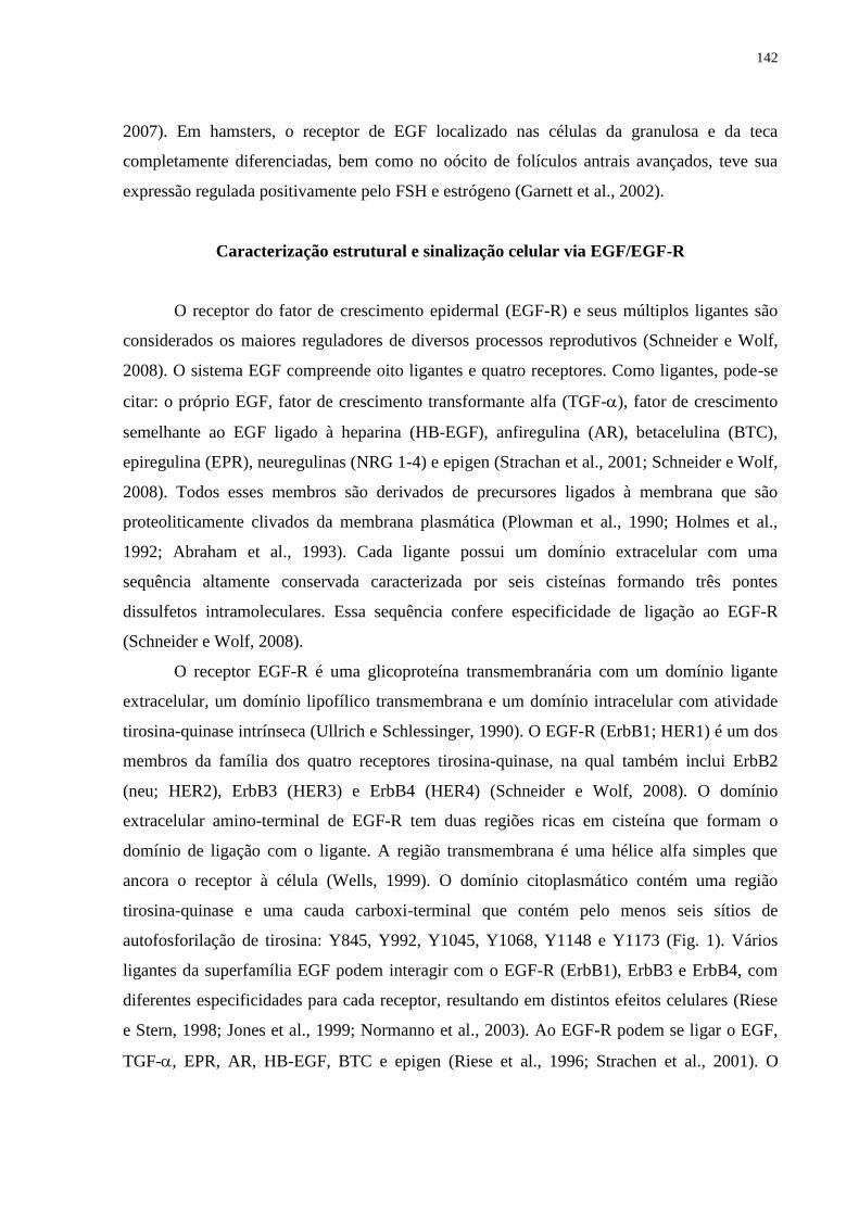
142
2007). Em hamsters, o receptor de EGF localizado nas células da granulosa e da teca
completamente diferenciadas, bem como no oócito de folículos antrais avançados, teve sua
expressão regulada positivamente pelo FSH e estrógeno (Garnett et al., 2002).
Caracterização estrutural e sinalização celular via EGF/EGF-R
O receptor do fator de crescimento epidermal (EGF-R) e seus múltiplos ligantes são
considerados os maiores reguladores de diversos processos reprodutivos (Schneider e Wolf,
2008). O sistema EGF compreende oito ligantes e quatro receptores. Como ligantes, pode-se
citar: o próprio EGF, fator de crescimento transformante alfa (TGF-), fator de crescimento
semelhante ao EGF ligado à heparina (HB-EGF), anfiregulina (AR), betacelulina (BTC),
epiregulina (EPR), neuregulinas (NRG 1-4) e epigen (Strachan et al., 2001; Schneider e Wolf,
2008). Todos esses membros são derivados de precursores ligados à membrana que são
proteoliticamente clivados da membrana plasmática (Plowman et al., 1990; Holmes et al.,
1992; Abraham et al., 1993). Cada ligante possui um domínio extracelular com uma
sequência altamente conservada caracterizada por seis cisteínas formando três pontes
dissulfetos intramoleculares. Essa sequência confere especificidade de ligação ao EGF-R
(Schneider e Wolf, 2008).
O receptor EGF-R é uma glicoproteína transmembranária com um domínio ligante
extracelular, um domínio lipofílico transmembrana e um domínio intracelular com atividade
tirosina-quinase intrínseca (Ullrich e Schlessinger, 1990). O EGF-R (ErbB1; HER1) é um dos
membros da família dos quatro receptores tirosina-quinase, na qual também inclui ErbB2
(neu; HER2), ErbB3 (HER3) e ErbB4 (HER4) (Schneider e Wolf, 2008). O domínio
extracelular amino-terminal de EGF-R tem duas regiões ricas em cisteína que formam o
domínio de ligação com o ligante. A região transmembrana é uma hélice alfa simples que
ancora o receptor à célula (Wells, 1999). O domínio citoplasmático contém uma região
tirosina-quinase e uma cauda carboxi-terminal que contém pelo menos seis sítios de
autofosforilação de tirosina: Y845, Y992, Y1045, Y1068, Y1148 e Y1173 (Fig. 1). Vários
ligantes da superfamília EGF podem interagir com o EGF-R (ErbB1), ErbB3 e ErbB4, com
diferentes especificidades para cada receptor, resultando em distintos efeitos celulares (Riese
e Stern, 1998; Jones et al., 1999; Normanno et al., 2003). Ao EGF-R podem se ligar o EGF,
TGF-, EPR, AR, HB-EGF, BTC e epigen (Riese et al., 1996; Strachen et al., 2001). O
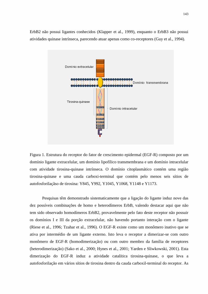
143
ErbB2 não possui ligantes conhecidos (Klapper et al., 1999), enquanto o ErbB3 não possui
atividades quinase intrínseca, parecendo atuar apenas como co-receptores (Guy et al., 1994).
Figura 1. Estrutura do receptor do fator de crescimento epidermal (EGF-R) composto por um
domínio ligante extracelular, um domínio lipofílico transmembrana e um domínio intracelular
com atividade tirosina-quinase intrínseca. O domínio citoplasmático contém uma região
tirosina-quinase e uma cauda carboxi-terminal que contém pelo menos seis sítios de
autofosforilaçãso de tirosina: Y845, Y992, Y1045, Y1068, Y1148 e Y1173.
Pesquisas têm demonstrado sistematicamente que a ligação do ligante induz nove das
dez possíveis combinações de homo e heterodímeros ErbB, valendo destacar aqui que não
tem sido observado homodímeros ErbB2, provavelmente pelo fato deste receptor não possuir
os domínios I e III da porção extracelular, não havendo portanto interação com o ligante
(Riese et al., 1996; Tzahar et al., 1996). O EGF-R existe como um monômero inativo que se
ativa por intermédio de um ligante externo. Isto leva o receptor a dimerizar-se com outro
monômero de EGF-R (homodimerização) ou com outro membro da família de receptores
(heterodimerização) (Sako et al., 2000; Hynes et al., 2001; Yarden e Sliwkowski, 2001). Esta
dimerização do EGF-R induz a atividade catalítica tirosina-quinase, o que leva a
autofosforilação em vários sítios de tirosina dentro da cauda carboxil-terminal do receptor. As
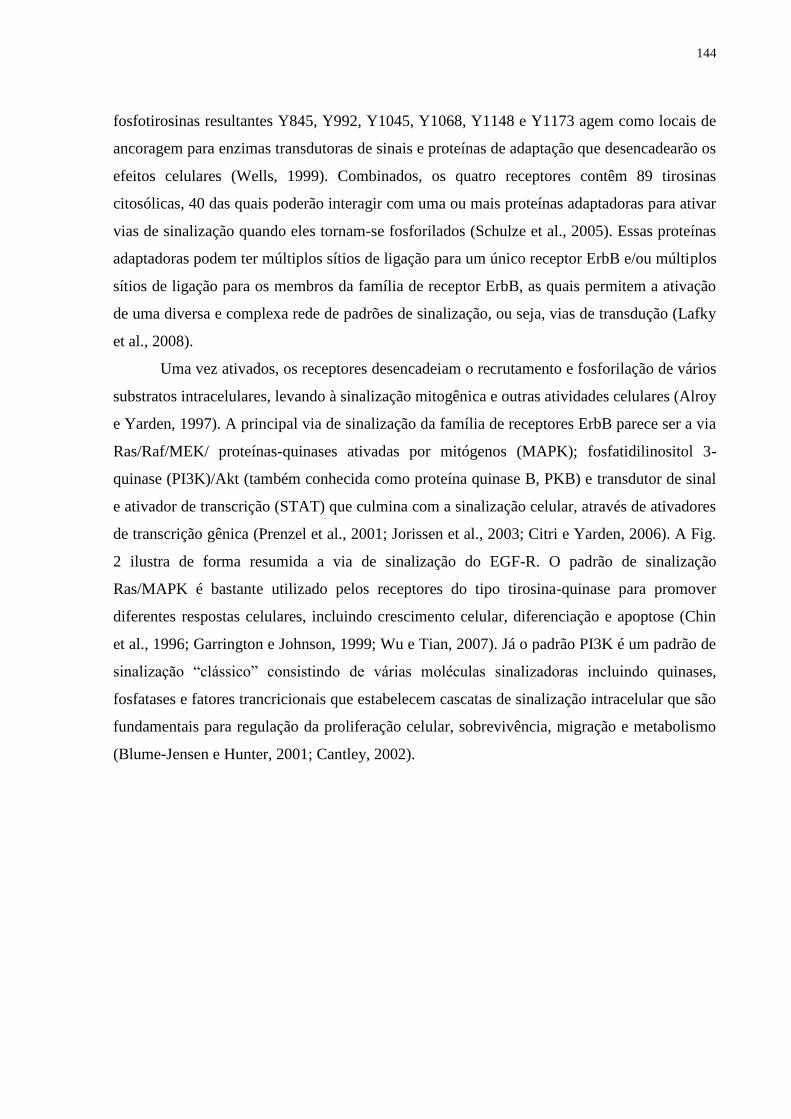
144
fosfotirosinas resultantes Y845, Y992, Y1045, Y1068, Y1148 e Y1173 agem como locais de
ancoragem para enzimas transdutoras de sinais e proteínas de adaptação que desencadearão os
efeitos celulares (Wells, 1999). Combinados, os quatro receptores contêm 89 tirosinas
citosólicas, 40 das quais poderão interagir com uma ou mais proteínas adaptadoras para ativar
vias de sinalização quando eles tornam-se fosforilados (Schulze et al., 2005). Essas proteínas
adaptadoras podem ter múltiplos sítios de ligação para um único receptor ErbB e/ou múltiplos
sítios de ligação para os membros da família de receptor ErbB, as quais permitem a ativação
de uma diversa e complexa rede de padrões de sinalização, ou seja, vias de transdução (Lafky
et al., 2008).
Uma vez ativados, os receptores desencadeiam o recrutamento e fosforilação de vários
substratos intracelulares, levando à sinalização mitogênica e outras atividades celulares (Alroy
e Yarden, 1997). A principal via de sinalização da família de receptores ErbB parece ser a via
Ras/Raf/MEK/ proteínas-quinases ativadas por mitógenos (MAPK); fosfatidilinositol 3-
quinase (PI3K)/Akt (também conhecida como proteína quinase B, PKB) e transdutor de sinal
e ativador de transcrição (STAT) que culmina com a sinalização celular, através de ativadores
de transcrição gênica (Prenzel et al., 2001; Jorissen et al., 2003; Citri e Yarden, 2006). A Fig.
2 ilustra de forma resumida a via de sinalização do EGF-R. O padrão de sinalização
Ras/MAPK é bastante utilizado pelos receptores do tipo tirosina-quinase para promover
diferentes respostas celulares, incluindo crescimento celular, diferenciação e apoptose (Chin
et al., 1996; Garrington e Johnson, 1999; Wu e Tian, 2007). Já o padrão PI3K é um padrão de
sinalização ―clássico‖ consistindo de várias moléculas sinalizadoras incluindo quinases,
fosfatases e fatores trancricionais que estabelecem cascatas de sinalização intracelular que são
fundamentais para regulação da proliferação celular, sobrevivência, migração e metabolismo
(Blume-Jensen e Hunter, 2001; Cantley, 2002).
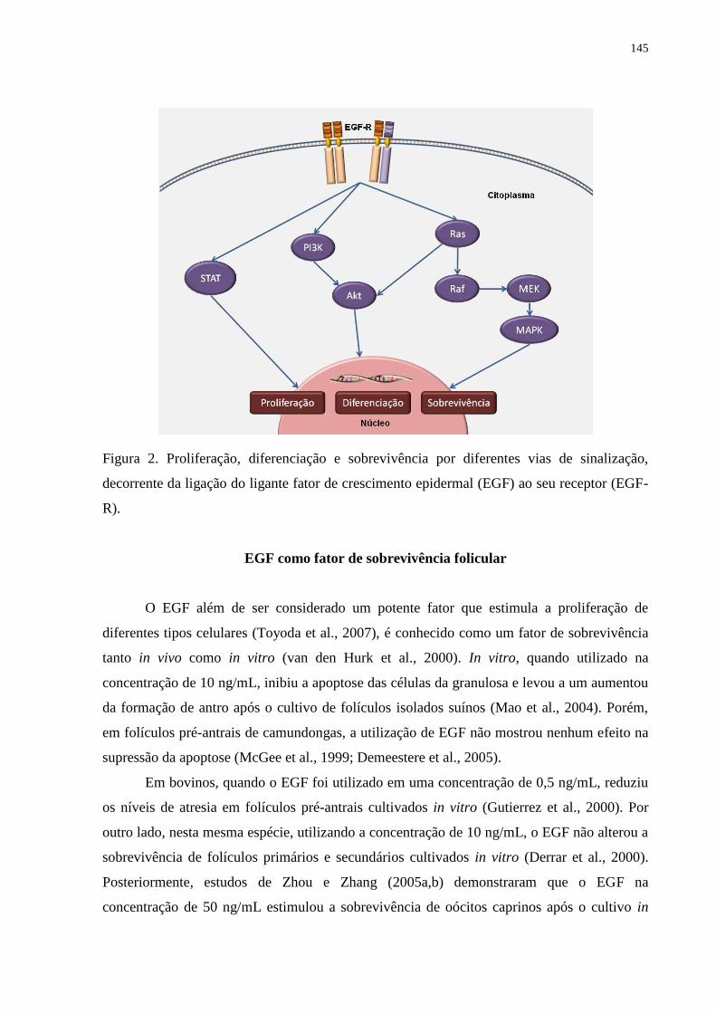
145
Figura 2. Proliferação, diferenciação e sobrevivência por diferentes vias de sinalização,
decorrente da ligação do ligante fator de crescimento epidermal (EGF) ao seu receptor (EGF-
R).
EGF como fator de sobrevivência folicular
O EGF além de ser considerado um potente fator que estimula a proliferação de
diferentes tipos celulares (Toyoda et al., 2007), é conhecido como um fator de sobrevivência
tanto in vivo como in vitro (van den Hurk et al., 2000). In vitro, quando utilizado na
concentração de 10 ng/mL, inibiu a apoptose das células da granulosa e levou a um aumentou
da formação de antro após o cultivo de folículos isolados suínos (Mao et al., 2004). Porém,
em folículos pré-antrais de camundongas, a utilização de EGF não mostrou nenhum efeito na
supressão da apoptose (McGee et al., 1999; Demeestere et al., 2005).
Em bovinos, quando o EGF foi utilizado em uma concentração de 0,5 ng/mL, reduziu
os níveis de atresia em folículos pré-antrais cultivados in vitro (Gutierrez et al., 2000). Por
outro lado, nesta mesma espécie, utilizando a concentração de 10 ng/mL, o EGF não alterou a
sobrevivência de folículos primários e secundários cultivados in vitro (Derrar et al., 2000).
Posteriormente, estudos de Zhou e Zhang (2005a,b) demonstraram que o EGF na
concentração de 50 ng/mL estimulou a sobrevivência de oócitos caprinos após o cultivo in
146
vitro. Celestino et al. (2009) verificaram que a adição de baixas concentrações de EGF (1 ou
10 ng/mL) no cultivo in vitro de folículos pré-antrais caprinos manteve a morfologia e
ultraestrutura folicular após 7 dias de cultivo. No entanto, Silva et al. (2004) não observaram
efeito significativo do EGF sobre a sobrevivência de folículos pré-antrais caprinos após 5 dias
de cultivo, provavelmente devido a elevada concentração (100 ng/mL) de EGF utilizada.
Talmimani et al. (2005) ao testar o efeito de diferentes concentrações de EGF (0, 25, 50, 75
ou 100 ng/mL) sobre folículos pré-antrais ovinos, mostrou que as maiores concentrações, ou
seja, 75 ou 100 ng/mL induziu à degeneração de todos folículos pré-antrais após cultivo de 6
dias. Achados semelhantes também foram descritos por Celestino et al. (2009) ao verificar
que folículos pré-antrais caprinos cultivados in vitro por 7 dias em altas concentrações de
EGF (100 ou 200 ng/mL) não sofreram ativação e apresentaram altas taxas de degeneração.
Papel do EGF no desenvolvimento folicular
Estudos in vitro mostraram que o EGF promove a ativação folicular (ovinos: Andrade
et al., 2005), estimula a proliferação das células da granulosa (suínos: Morbeck et al., 1993),
evidenciado pela incorporação de timidina em folículos pré-antrais (hamster: Roy, 1993).
Além disso, aumenta o diâmetro folicular (suínos: Mao et al., 2004) e promove o crescimento
de oócitos em folículos primários (caprinos: Silva et al., 2004). Ademais, o EGF tem
mostrado regular a expressão da conexina 43 (suíno: Bolamba et al., 2002; coelho: Kennedy
et al., 2003) que é uma proteína essencial para a formação das junções gaps, sendo portanto
importante para o desenvolvimento de folículos pré-antrais.
A ação do EGF sobre a ativação, diferenciação, proliferação e esteroidogênese das
células da granulosa tem se mostrado controversa na literatura e dependente do estádio de
desenvolvimento folicular. Alguns estudos têm mostrado que embora o EGF não seja
essencial para ativação de folículos primordiais (Braw-Tal e Yossefi, 1997; Fortune et al.,
1998; Wright et al., 1999), ele é importante para os estádios mais avançados de
desenvolvimento folicular (Gutierrez et al., 2000; Nayudu et al., 2002; Peng et al., 2010). Em
caprinos, Silva et al. (2004) demonstraram que o EGF (100 ng/mL) no cultivo in situ, não
promoveu a ativação de folículos primordiais, mas teve um efeito benéfico no crescimento de
oócitos de folículos primários (Silva et al., 2004). Em ovinos, sua utilização nesta mesma
concentração, promoveu a ativação de folículos primordiais in situ e manutenção da
viabilidade por até 6 dias de cultivo (Andrade et al., 2005). Já em pequenos e médios folículos
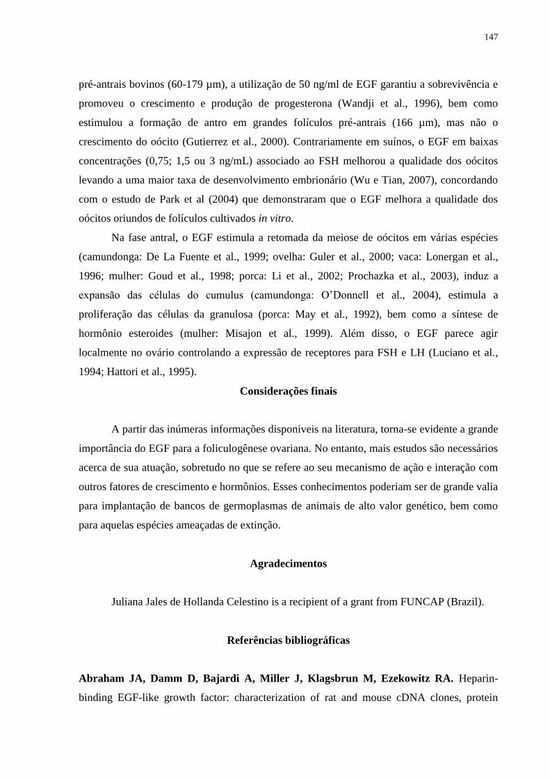
147
pré-antrais bovinos (60-179 µm), a utilização de 50 ng/ml de EGF garantiu a sobrevivência e
promoveu o crescimento e produção de progesterona (Wandji et al., 1996), bem como
estimulou a formação de antro em grandes folículos pré-antrais (166 µm), mas não o
crescimento do oócito (Gutierrez et al., 2000). Contrariamente em suínos, o EGF em baixas
concentrações (0,75; 1,5 ou 3 ng/mL) associado ao FSH melhorou a qualidade dos oócitos
levando a uma maior taxa de desenvolvimento embrionário (Wu e Tian, 2007), concordando
com o estudo de Park et al (2004) que demonstraram que o EGF melhora a qualidade dos
oócitos oriundos de folículos cultivados in vitro.
Na fase antral, o EGF estimula a retomada da meiose de oócitos em várias espécies
(camundonga: De La Fuente et al., 1999; ovelha: Guler et al., 2000; vaca: Lonergan et al.,
1996; mulher: Goud et al., 1998; porca: Li et al., 2002; Prochazka et al., 2003), induz a
expansão das células do cumulus (camundonga: O‘Donnell et al., 2004), estimula a
proliferação das células da granulosa (porca: May et al., 1992), bem como a síntese de
hormônio esteroides (mulher: Misajon et al., 1999). Além disso, o EGF parece agir
localmente no ovário controlando a expressão de receptores para FSH e LH (Luciano et al.,
1994; Hattori et al., 1995).
Considerações finais
A partir das inúmeras informações disponíveis na literatura, torna-se evidente a grande
importância do EGF para a foliculogênese ovariana. No entanto, mais estudos são necessários
acerca de sua atuação, sobretudo no que se refere ao seu mecanismo de ação e interação com
outros fatores de crescimento e hormônios. Esses conhecimentos poderiam ser de grande valia
para implantação de bancos de germoplasmas de animais de alto valor genético, bem como
para aquelas espécies ameaçadas de extinção.
Agradecimentos
Juliana Jales de Hollanda Celestino is a recipient of a grant from FUNCAP (Brazil).
Referências bibliográficas
Abraham JA, Damm D, Bajardi A, Miller J, Klagsbrun M, Ezekowitz RA. Heparin-
binding EGF-like growth factor: characterization of rat and mouse cDNA clones, protein
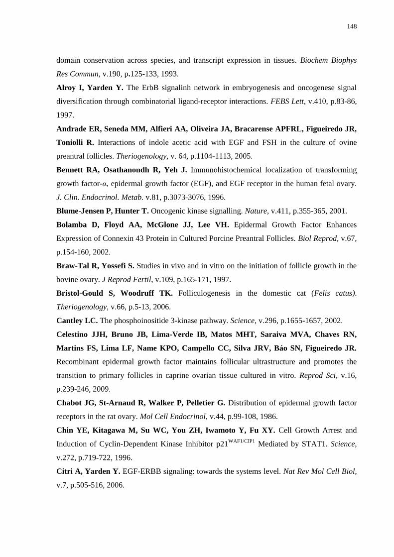
148
domain conservation across species, and transcript expression in tissues. Biochem Biophys
Res Commun, v.190, p.125-133, 1993.
Alroy I, Yarden Y. The ErbB signalinh network in embryogenesis and oncogenese signal
diversification through combinatorial ligand-receptor interactions. FEBS Lett, v.410, p.83-86,
1997.
Andrade ER, Seneda MM, Alfieri AA, Oliveira JA, Bracarense APFRL, Figueiredo JR,
Toniolli R. Interactions of indole acetic acid with EGF and FSH in the culture of ovine
preantral follicles. Theriogenology, v. 64, p.1104-1113, 2005.
Bennett RA, Osathanondh R, Yeh J. Immunohistochemical localization of transforming
growth factor-α, epidermal growth factor (EGF), and EGF receptor in the human fetal ovary.
J. Clin. Endocrinol. Metab. v.81, p.3073-3076, 1996.
Blume-Jensen P, Hunter T. Oncogenic kinase signalling. Nature, v.411, p.355-365, 2001.
Bolamba D, Floyd AA, McGlone JJ, Lee VH. Epidermal Growth Factor Enhances
Expression of Connexin 43 Protein in Cultured Porcine Preantral Follicles. Biol Reprod, v.67,
p.154-160, 2002.
Braw-Tal R, Yossefi S. Studies in vivo and in vitro on the initiation of follicle growth in the
bovine ovary. J Reprod Fertil, v.109, p.165-171, 1997.
Bristol-Gould S, Woodruff TK. Folliculogenesis in the domestic cat (Felis catus).
Theriogenology, v.66, p.5-13, 2006.
Cantley LC. The phosphoinositide 3-kinase pathway. Science, v.296, p.1655-1657, 2002.
Celestino JJH, Bruno JB, Lima-Verde IB, Matos MHT, Saraiva MVA, Chaves RN,
Martins FS, Lima LF, Name KPO, Campello CC, Silva JRV, Báo SN, Figueiredo JR.
Recombinant epidermal growth factor maintains follicular ultrastructure and promotes the
transition to primary follicles in caprine ovarian tissue cultured in vitro. Reprod Sci, v.16,
p.239-246, 2009.
Chabot JG, St-Arnaud R, Walker P, Pelletier G. Distribution of epidermal growth factor
receptors in the rat ovary. Mol Cell Endocrinol, v.44, p.99-108, 1986.
Chin YE, Kitagawa M, Su WC, You ZH, Iwamoto Y, Fu XY. Cell Growth Arrest and
Induction of Cyclin-Dependent Kinase Inhibitor p21WAF1/CIP1
Mediated by STAT1. Science,
v.272, p.719-722, 1996.
Citri A, Yarden Y. EGF-ERBB signaling: towards the systems level. Nat Rev Mol Cell Biol,
v.7, p.505-516, 2006.
149
Conti M, Hsieh M, Park J-Y, Su Y-Q. Role of the epidermal growth factor network in
ovarian follicles. Mol Endocrinol, v.20, p.715-723, 2006.
De La Fuente R, O’Brien MJ, Eppig JJ. Epidermal growth factor enhances preimplantation
developmental competence of maturing mouse oocytes. Hum Reprod, v.14, p.3060-3068,
1999.
Demeestere I, Centner J, Gervy Y, Englert Y, Delbaere A. Impact of various endocrine
and paracrine factors on in vitro culture of preantral follicles in rodents. Reproduction, v.130,
p.147-156, 2005.
Derrar N, Price CA, Sirard M-A. Effects of growth factors and co-culture with ovarian
medulla on the activation of primordial in explants of bovine ovarian cortex. Theriogenology,
v.54, p.587-598, 2000.
Eppig JJ. Oocyte control of ovarian follicular development and function in mammals
Reproduction, v.122, p.829-838, 2001.
Fair T. Follicular oocyte growth and acquisition of development competence. Anim Reprod
Sci, v.78, p.203-216, 2003.
Feng P, Knecht M, Catt K. Hormonal control of epidermal growth factor receptors by
gonadotropins during granulosa cell differentiation. Endocrinology, v.120, p.1121–1126,
1987
Fortune JE. The early stages of follicular development: activation of primordial follicles and
growth of preantral follicles. Anim Reprod Sci, v.78, p.135-163, 2003.
Fortune JE, Kito S, Wandji SA, Srsen V. Activation of bovine and baboon primordial
follicles in vitro. Theriogenology, v.49, p.441-449, 1998.
Fru KN, Cherian-Shaw M, Puttabyatappa M, VandeVoort CA, Chaffin CL. Regulation
of granulosa cell proliferation and EGF-like ligands during the periovulatory interval in
monkeys. Hum Reprod, v.22, p.1247-1252, 2007.
Gall L, Chene N, Dahirel M, Ruffini S, Boulesteix C. Expression of epidermal growth
factor receptor in the goat cumulus-oocyte complex. Mol Reprod Dev, v.67, p.439-445, 2004.
Garnett K, Wang J, Roy SK. Spatiotemporal expression of epidermal growth factor receptor
messenger RNA and protein in the hamster ovary: follicle stage specific differential
modulation by follicle-stimulating hormone, luteinizing hormone, estradiol, and progesterone.
Biol Reprod, v.67, p.1593-1604, 2002.
Garrington TP, Johnson GL. Organization and regulation of mitogen-activated protein
kinase signaling pathways. Curr Opin Cell Biol, v.11, p.211-218, 1999.
150
Goud PT, Goud AP, Qian C, Laverge H, Van der Elst J, De Sutter P, Dhont M. In vitro
maturation of human germinal vesicle stage oocytes: role of cumulus cells and EGF in the
culture medium. Hum Reprod, v.13, p.1638-1644, 1998.
Guler A, Poulin N, Mermillod P, Terqui M, Cognie Y. Effect of growth factors, EGF and
IGF-I, and estradiol on in vitro maturation of sheep oocytes. Theriogenology, v.54, p.209-218,
2000.
Gutierrez CG, Ralph JH, Telfer EE, Wilmut I, Webb R. Growth and antrum formation of
bovine preantral follicles in long-term culture in vitro. Biol Reprod, v.62, p.1322-1328, 2000.
Guy PM, Platko JV, Cantley LC, Cerione RA, Carraway III KL. Insect cell-expressed
p180erbB3 possesses an impaired tyrosine kinase activity. Proc Natl Acad Sci USA, v.91,
p.8132-8136, 1994.
Hattori MA, Yoshino E, Shinohara Y, Horiuchi R, Kojima I. A novel action of epidermal
growth factor in rat granulosa cells: its potentiation of gonadotrophin action. J Mol
Endocrinol, v.15, p.283-291, 1995.
Hill JL, Hammar K, Smith PJ, Gross DJ. Stage dependent effects of epidermal growth
factor onCa2+ efflux in mouse oocytes. Mol Reprod Dev, v.53, p.244-253, 1999.
Holmes WE, Sliwkowski MX, Akita RW, Henzel WJ, Lee J, Park JW, Yansura D,
Abadi N, Raab H, Lewis GD, Shepard HM, Kuang W-J, Wood WL, Goeddel DV,
Vandlen RL. Identification of Heregulin, a Specific Activator of p185erbB2
. Science, v.256,
p.1205-1210, 1992.
Hynes NE, Horsh K, Olayioye MA, Badache A. The ErbB receptor tyrosine family as
signal integrators. Endocr Relat Cancer, v.8, p.151-159, 2001.
Jones JT, Akita RW, Sliwkwoski MX. Bindings specificities and affinities of egf domain
for ErbB receptors. FEBS Lett, v.447, p.227-231, 1999.
Jorissen RN, Walker F, Pouliot N, Garrett TP, Ward CW, Burgess AW. Epidermal
growth factor receptor: mechanisms of activation and signaling. Exp Cell Res, v.284, p.31-53,
2003.
Kennedy TG, Brown KD, Vaughan TJ. Expression of the genes for the epidermal growth
factor receptor and its ligands in porcine corpora lutea. Endocrinology, v.132, p.1857-1859,
1993.
Kennedy KL, Floyd AA, Clarkson AM, Lee VH. Epidermal growth factor regulation of
connexin 43 in cultured granulosa cells from preantral rabbit follicles. Mol Reprod Dev, v.64,
p.61-69, 2003.
151
Klapper LN, Glathe S, Vaisman N, Hynes NE, Andrews GC, Sela M, Yarden Y. The
ErbB-2/HER2 oncoprotein of human carcinomas may function solely as a shared coreceptor
for multiple stroma-derived growth factors. Proc Natl Acad Sci USA, v.96, p.4995-5000,
1999.
Lafky JM, Wilken JA, Baron AT, Maihle N.J. Clinical implications of the ErbB/epidermal
growth factor (EGF) receptor family and its ligands in ovarian cancer. Biochim Biophys Acta,
v.1785, p.232-265, 2008.
Li YH, Liu RH, Jiao LH, Wang WH. Synergetic effects of epidermal growth factor and
estradiol on cytoplasmic maturation of porcine oocytes. Zygote, v.10, p.349-354, 2002.
Lonergan P, Carolan C, Van Langendonckt A, Donnay I, Khatir H, Mermillod, P. Role
of epidermal growth factor in bovine oocyte maturation and preimplantation embryo
development in vitro. Biol Reprod, v.54, p.1420-1429, 1996.
Luciano AM, Pappalardo A, Ray C, Peluso JJ. Epidermal growth factor inhibits large
granulosa cell apoptosis by stimulating progesterone synthesis and regulating the distribution
of intracellular free calcium. Biol Reprod, v.51, p.646-654, 1994.
Mao J, Smith MF, Rucker EB, Wu GM, McCauley TC, Cantley TC, Prather RS, Didion
BA, Day BN. Effect of epidermal growth factor and insulin-like growth factor I on porcine
preantral follicular growth, antrum formation, and stimulation of granulosal cell proliferation
and suppression of apoptosis in vitro. J Anim Sci, v.82, p.1967-1975, 2004.
Markström E, Svensson EC, Shao R, Svanberg B, Billig H. Survival factors regulating
ovarian apoptosis – dependence on follicle differentiation. Reproduction, v.123, p.23-30,
2002.
Maruo T, Ladines-Llave CA, Samoto T, Matsuo H, Manalo AS, Ito H, Mochizuki M.
Expression of epidermal growth factor and its receptor in the human ovary during follicular
growth and regression. Endocrinology, v.132, p.924-931, 1993.
May JV, Bridge AJ, Gotcher ED, Gangrade BK. The regulation of porcine theca cell
proliferation in vitro: synergistic actions of epidermal growth factor and platelet derived
growth factor. Endocrinology, v.131, p.689-697, 1992.
McGee EA, Chun S-Y, Lai S, He Y-E, Hsueh AJW. Keratinocyte growth factor promotes
the survival, growth, and differentiation of preantral ovarian follicles. Fertil Steril, v.71,
p.732-738, 1999.
152
Misajon A, Hutchinson P, Lolatgis N, Trounson AO, Almahbobi G. The mechanism of
action of epidermal growth factor and transforming growth factor alpha on aromatase activity
in granulosa cells from polycystic ovaries. Mol Hum Reprod, v.5, p.96-103, 1999.
Morbeck DE, Flowers WL, Britt JH. Response of porcine granulosa cells isolated from
primary and secondary follicles to FSH, 8-bromo-cAMP and epidermal growth factor in vitro.
J Reprod Fertil, v.99, p.577-584, 1993.
Nayudu PL, Vitt UA, De Tomasi JB, Pancharatna K, Ulloa-Aguirre A. Intact follicle
culture: what it can tell us about the roles of FSH glycoforms during follicle development.
Reprod BioMed Online, v.5, p.240-253, 2002.
Normanno N, Bianco C, De Luca A, Maiello MR, Salomon DS. Target-based agents
against ErbB receptors a their ligands: a novel approach to cancer treatment. Endocr Relat
Cancer, v.10, p.1-21, 2003.
O’Donnell JR, Hill JL, Gross DJ. Epidermal growth factor activates cytosolic [Ca2+]
elevations and subsequent membrane permeabilization in mouse cumulus–oocyte complexes.
Reproduction, v.127, p.207-220, 2004.
Park JY, Su YQ, Ariga M, Law E, Jin SLC, Conti M. EGF-like growth factors as
mediators of LH action in the ovulatory follicle. Science, v.303, p.682-684, 2004.
Peng X, Yang M, Wang L, Tong C, Guo Z. In vitro culture of sheep lamb ovarian cortical
tissue in a sequential culture medium. J Assist Reprod Genet, v.27, p.247-257, 2010.
Plowman GD, Green JM, McDonald VL, Neubauer MG, Disteche CM, Todaro GJ,
Shoyab M. The amphiregulin gene encodes a novel epidermal growth factor-related protein
with tumor-inhibitory activity. Mol Cell Biol, v.10, p.1969-1981, 1990.
Prenzel N, Fischer OM, Streit S, Hart S, Ullrich A. The epidermal growth factor receptor
family as a central element for cellular signal transduction and diversification. Endocr Relat
Cancer, v.8, p.11-31, 2001.
Prochazka R, Kalab P, Nagyova E. Epidermal growth factor-receptor tyrosine kinase
activity regulates expansion of porcine oocyte–cumulus cell complexes in vitro. Biol Reprod,
v.68, p.797-803, 2003.
Qu JP, Godin PA, Nisolle M, Donnez J. Distribution of epidermal growth factor receptor
expression of primordial follicles in human ovarian tissue before and after cryopreservation.
Hum Reprod, v.15, p.302-310, 2000.
153
Reeka N, Berg FD, Brucer C. Presence of transforming growth factor alpha and epidermal
growth factor in human ovarian tissue and follicular fluid. Hum Reprod, v.13, p.2199-2205,
1998.
Riese DJ, Kim ED, Elenius K, Buckley S, Klagsbrun M, Plowman GD, Stern DF. The
Epidermal Growth Factor Receptor Couples Transforming Growth Factor-α, Heparin-binding
Epidermal Growth Factor-like Factor, and Amphiregulin to Neu, ErbB-3, and ErbB-4. J Biol
Chem, v.271, p.20047-20052, 1996.
Riese DJ, Stern DF. Specificity within the EGF family/ErbB receptor family signaling
network. Bioessays, v.20, p.41-48, 1998.
Roy SK. Epidermal growth factor and transforming growth factor-beta modulation of follicle-
stimulating hormone-induced deoxyribonucleic acid synthesis in hamster preantral and early
antral follicles. Biol Reprod, v.48, p.552-557, 1993.
Roy SK, Greenwald GS. Immunohistochemical localisation of epidermal growth factor-like
activity in the hamster ovary with a polyclonal antibody. Endocrinology, v.126, p.1309-1317,
1990.
Saha S, Shimizu M, Geshi M, Izaike Y. In vitro culture of bovine preantral follicles. Anim
Reprod Sci, v.63, p.27-39, 2000.
Sako Y, Minoguchi S, Yanagida T. Single-molecule imaging of EGFR signaling on the
surface of living cells. Nat Cell Biol, v.2, p.168-172, 2000.
Schneider MR, Wolf E. The epidermal growth factor receptor and its ligands in female
reproduction: Insights from rodent models. Cytokine Growth Factor Rev, v.19, p.173-181,
2008.
Schomberg DW, May JV, Mondschein JS. Interactions between hormones and growth
factors in the regulation of granulosa cell differentiation in vitro. J Steroid Biochem, v.19,
p.291-295, 1983.
Schulze WX, Deng L, Mann M. Phosphotyrosine interactome of the ErbB-receptor kinase
family. Mol Syst Biol, v.1, p.2005-2008, 2005.
Silva JRV, van den Hurk R, Matos MHT, Santos RR, Pessoa C, Moraes MO, Figueiredo
JR. Influences of FSH and EGF on primordial follicles during in vitro culture of caprine
ovarian cortical tissue. Theriogenology, v.61, p.1691-1704, 2004.
Silva JRV, van den Hurk R, Figueiredo JR. Expression of mRNA and protein localization
of epidermal growth factor and its receptor in goat ovaries. Zygote, v.14, p.107-117, 2006.
154
Singh B, Rutledge JM, Armstrong DT. Epidermal growth factor and its receptor gene
expression and peptide localization in porcine ovarian follicles. Mol Reprod Dev, v.40, p.391-
399, 1995a.
Singh B, Kennedy TG, Tekpetey FR, Armstrong DT. Gene expression and peptide
localization for epidermal growth factor receptor and its ligands in porcine luteal cells. Mol
Cell Endocrinol, v.113, p.137-143, 1995b.
Skinner MK, Coffey RJ. Regulation of ovarian cell growth through the local production of
transforming growth factor-α by theca cells. Endocrinology, v.123, p.2632-2638, 1988.
Strachan L, Murison JG, Prestidge RL, Sleeman MA, Watson JD, Kumble KD. Cloning
and Biological Activity of Epigen, a Novel Member of the Epidermal Growth Factor
Superfamily. J Biol Chem, v.276, p.18265-18271, 2001.
Talmimani G, Rao BS, Vagdevi R, Amarnath D, Naik BR, Mutharao M, Rao VH.
Nuclear maturation of ovine oocytes in cultured preantral follicles. Small Rumin Res, v.60,
p.295-305, 2005.
Tamura M, Sasano H, Suzuki T, Fukaya T, Funayama Y, Takayama K, Takaya R,
Yajima A. Expression of epidermal growth factors and epidermal growth factor receptor in
normal cycling human ovaries. Hum Reprod, v.10, p.1891-1896, 1995.
Tekpetey FR, Singh B, Barbe G, Armstrong DT. Localization of epidermal growth factor
(EGF) receptor in the rat corpus luteum, and EGF and transforming growth factor-alpha
stimulation of luteal cell steroidogenesis in vitro. Mol Cell Endocrinol, v.110, p.95-102, 1995.
Toyoda T, Nakamura K, Yamada K, Thanseem I, Anitha A, Suda S, Tsujii M, Iwayama
Y, Hattori E, Toyota T, Miyachi T, Iwata Y, Suzuki K, Matsuzaki H, Kawai M, Sekine
Y, Tsuchiya K, Sugihara G-I, Ouchi Y, Sugiyama T, Takei N, Yoshikawa T, Mori N.
SNP analyses of growth factor genes EGF, TGF-β1, and HGF reveal haplotypic association of
EGF with autism. Biochem Biophys Res Commun, v.360, p.715-720, 2007.
Tzahar E, Waterman E, Chen X, Levkowitz G, Karunagaran D, Lavi S, Ratzkin BJ,
Yarden Y. A hierarchical network of interreceptor interactions determines signal transduction
by Neu differentiation factor/neuregulin and epidermal growth factor. Mol Cell Biol, v.16,
p.5276-5287, 1996.
Ullrich A, Schlessinger J. Signal transduction by receptors with tyrosine kinase activity.
Cell, v.61, p.203-212, 1990.
155
van den Hurk R, Abir R, Telfer EE, Bevers MM. Preantral and antral follicles as possible
source for fertilizable oocytes in human and bovine. Hum Reprod Update, v.2, p.457-474,
2000.
van den Hurk R, Zhao J. Formation of mammalian oocytes and their growth, differentiation
and maturation within ovarian follicles. Theriogenology, v.63, p.1717-1751, 2005.
Wandji SA, Eppig JJ, Fortune JE. FSH and growth factors affect the growth and endocrine
function in vitro of granulosa cells of bovine preantral follicles. Theriogenology, v.45, p.817-
832, 1996.
Wang Y, Li J, Wang CY, Kwok AHY, Leung FC. Epidermal growth factor (EGF) receptor
ligands in the chicken ovary: I. Evidence for heparin-binding EGF-like growth factor
(HBEGF) as a potential oocyte-derived signal to control granulosa cell proliferation and HB-
EGF and kit ligand expression. Endocrinology, v.148, p.3426-3440, 2007.
Wells A. EGF receptor. Int J Biochem Cell Biol, v.31, p.637-644, 1999.
Wright CS, Hovatta O, Margara R, Trew G, Winston RML, Franks S, Hardy K. Effects
of follicle-stimulating hormone and serum substitution on the in-vitro growth of human
ovarian follicles. Hum Reprod, v.14, p.1555-1562, 1999.
Wu J, Tian Q. Role of follicle stimulating hormone and epidermal growth factor in the
development of porcine preantral follicle in vitro. Zygote, v.15, p.233-240, 2007.
Yarden Y, Sliwkowski MX. Untangling the ErbB signaling network. Nat Rev Mol Cell Biol,
v.2, p.127-137, 2001.
Zhou H, Zhang Y. Effect of growth factors on in vitro development of caprine preantral
follicle oocytes. Anim Reprod Sci, v.90, p.265-272, 2005a.
Zhou H, Zhang Y. Regulation of in vitro growth of preantral follicles by growth factors in
goats. Domest Anim Endocrinol, v.28, p.235-242, 2005b.
156
10 CAPÍTULO 5
Níveis de RNAm para o Kit Ligand em Ovários Caprinos e o Papel do Kit Ligand na
Sobrevivência e Crescimento In Vitro de Folículos Pré-antrais
Steady-State Level of Kit Ligand mRNA in Goat Ovaries and the Role of Kit Ligand in
Preantral Follicle Survival and Growth In Vitro
Periódico: Molecular Reproduction & Development 77: 231-240, 2010.
.
A B
C D
157
Resumo
Os objetivos desse estudo foram investigar os níveis de RNAm para o Kit Ligand (KL) e os
efeitos dele na sobrevivência e crescimento de folículos pré-antrais caprinos. A RT-PCR foi
utilizada para analisar os níveis de RNAm para o KL em folículos primordiais, primários e
secundários, e em pequenos (1-3 mm) e grandes (3-6 mm) folículos antrais. Além disso,
fragmentos ovarianos foram cultivados por 1 ou 7 dias em Meio Essencial Mínimo (MEM+)
suplementado com KL (0, 1, 10, 50, 100 ou 200 ng/ml). Fragmentos não-cultivados (controle)
e cultivados foram processados para histologia e microscopia eletrônica de transmissão
(MET). A RT-PCR demonstrou um aumento nos níveis de RNAm para o KL durante a
transição de folículos primários para secundários. Pequenos folículos antrais tiveram níveis
superiores de RNAm para o KL em células da granulosa e tecais do que grandes folículos.
Após 7 dias, somente 50 ng/ml de KL manteve a percentagem de folículos normais similar ao
controle. Após 1 dia, todas as concentrações de KL reduziram a percentagem de folículos
primordiais e aumentaram a percentagem de folículos em crescimento. Após 7 dias, KL 10,
50, 100 ou 200 ng/ml aumentou o percentual de folículos primários quando comparados ao
MEM+. Um aumento no diâmetro folicular e oocitário foram observados com KL 50 ng/ml. A
MET confirmou a integridade ultraestrutural dos folículos após 7 dias de cultivo em KL 50
ng/ml. Em conclusão, os RNAm para o KL foram detectados em todas as categorias
foliculares. Além disso, 50 ng/ml de KL manteve a integridade de folículos pré-antrais
caprinos cultivados por 7 dias e estimulou a ativação de folículos primordiais e o crescimento
folicular.
Palavras-chave: Folículo ovariano. Ativação. Desenvolvimento folicular. KL. Caprino.
158
Steady-State Level of Kit Ligand mRNA in Goat Ovaries and the Role of Kit Ligand in
Preantral Follicle Survival and Growth In Vitro
JULIANA J.H. CELESTINO,1* JAMILY B. BRUNO,
1 ISABEL B. LIMA-VERDE,
1 MARIA
HELENA T. MATOS,1
MÁRCIA VIVIANE A. SARAIVA,1
ROBERTA N. CHAVES,1
FABRICIO S. MARTINS,1
ANDERSON P. ALMEIDA,1 RODRIGO M.S. CUNHA,
3
LARITZA F. LIMA,1 KHESLLER P.O. NAME,
2 CLAUDIO C. CAMPELLO,
1 JOSÉ
ROBERTO V. SILVA,3 SÔNIA N. BÁO,
2 AND JOSÉ RICARDO FIGUEIREDO
1
1 Faculty of Veterinary Medicine, LAMOFOPA, PPGCV, State University of Ceara,
Fortaleza, CE, Brazil.
2 Laboratory of Electron Microscopy, Department of Cell Biology, University of Brasilia,
Brasilia, DF, Brazil.
3 Biotecnology Nucleus of Sobral (NUBIS), Federal University of Ceara, Sobral, CE, Brazil.
SUMMARY
The aims of this study were to investigate steady-state level of Kit Ligand (KL) mRNA and
its effects on in vitro survival and growth of caprine preantral follicles. RT-PCR was used to
analyze caprine steady-state level of KL mRNA in primordial, primary, and secondary
follicles, and in small (1–3 mm) and large (3–6 mm) antral follicles. Furthermore, ovarian
fragments were cultured for 1 or 7 days in Minimal Essential Medium (MEM+) supplemented
with KL (0, 1, 10, 50, 100, or 200 ng/ml). Noncultured (control) and cultured fragments were
processed for histology and transmission electron microscopy (TEM). RT-PCR demonstrated
an increase in steady-state level of KL mRNA during the transition from primary to secondary
follicles. Small antral follicles had higher steady-state levels of KL mRNA in granulosa and
theca cells than large follicles. After 7 days, only 50 ng/ml of KL had maintained the
percentage of normal follicles similar to control. After 1 day, all KL concentrations reduced
the percentage of primordial follicles and increased the percentage of growing follicles. KL at
10, 50, 100, or 200 ng/ml increased primary follicles, compared to MEM+ after 7 days. An
increase in oocyte and follicular diameter was observed at 50 ng/ml of KL. TEM confirmed
ultrastructural integrity of follicles after 7 days at 50 ng/ml of KL. In conclusion, the KL
mRNAs were detected in all follicular categories. Furthermore, 50 ng/ml of KL maintained
159
the integrity of caprine preantral follicle cultured for 7 days and stimulated primordial follicle
activation and follicle growth.
INTRODUCTION
Female gametes are stocked in the ovary as primordial follicles, which are composed
of small, immature, quiescent oocytes surrounded by a single layer of flattened granulosa
cells. During the female reproductive life, primordial follicles are stimulated to grow each
day, a process known as follicular activation (Fortune, 2003; Choi et al., 2008a,b). After
growth begins, the oocytes enter into a pre-programmed course of maturation and
development that is necessary for the success of ovulation and fertilization. Primordial
follicles from the quiescent pool are lost by atresia without these processes (Knight and
Glister, 2006; Dole et al., 2008). The size of the ovarian follicular reserve associated with the
rate of follicular activation is a critical determinant of the female reproductive lifespan, since
the number of gametes in the ovary is supposedly finite (Choi et al., 2008a; Dole et al., 2008).
However, the mechanisms responsible for activation of primordial follicles remain unknown.
Kit Ligand (KL) is one of the first discovered of a few known factors with a recognized role
in primordial follicle activation (Hutt et al., 2006a; Thomas and Vanderhyden, 2006).
KL, also known as stem cell factor (SCF), steel factor (SF), or mast cell growth factor
(MCGF), is a locally produced growth factor with many roles in ovarian function from the
embryonic stage onwards (Yoshida et al., 1997; Driancourt et al., 2000). The presence of KL
mRNA in follicles has been detected in the granulosa cells of several species (rat: Ismail et
al., 1996; sheep: Tisdall et al., 1999; mouse: Doneda et al., 2002; human: Hoyer et al., 2005;
and goat: Silva et al., 2006). KL can be expressed as a soluble protein (KL-1) or a membrane
associated protein (KL-2) depending on how the mRNA is spliced (Huang et al., 1992). KL-2
is the main isoform required for oocyte growth, survival, and formation of germ cells
(Thomas et al., 2008). However, soluble KL-1 can promote the primordial to primary
transition (Parrot and Skinner, 1999). Both forms are present in rodent (Manova et al., 1990;
Ismail et al., 1997) and goat ovaries (Silva et al., 2006). In goats, the protein and mRNA for
KL were expressed in granulosa cells during all stages of follicular development (Silva et al.,
2006). However, quantification of the steady-state level of KL mRNA during different stages
of follicular development has not been performed yet. KL influences the target cells through
the c-kit receptor, a member of the tyrosine kinase receptor family. During postnatal ovarian
development, both the mRNA and protein for c-kit are expressed in the oocytes during all
160
stages of follicular development. Moreover, c-kit mRNA has been detected in the interstitial
and thecal cells of antral follicles (rodents: Motro and Bernstein, 1993; sheep: Clark et al.,
1996; goat: Silva et al., 2006).
In vitro studies of rodents and sheep have implicated the KL/c-kit system in the
migration, proliferation, and survival of primordial germ cells (CGP) (Zama et al., 2005),
activation of primordial follicles (Parrot and Skinner, 1999), oocyte growth and survival (Jin
et al., 2005), granulosa cell proliferation (Otsuka and Shimasaki, 2002), maintenance of
meiotic competence (Ismail et al., 1997), recruitment of theca cells, and regulation of ovarian
steroidogenesis (Hutt et al., 2006a). In addition, the KL/c-kit system is essential for human
preantral follicle survival in vitro (Carlsson et al., 2006). When tested in mouse and rabbit
ovaries at two different concentrations (50 and 150 ng/ml), KL promoted an increase in the
diameter of oocytes in preantral follicles from both species and stimulated mouse primordial
follicle activation in vitro (Hutt et al., 2006b). Although several studies have been performed,
particularly in murine and ovine models, little is known regarding the KL/c-Kit interaction
during early folliculogenesis in goats.
The present study was designed with the following aims: (1) to determine the steady-
state level of KL mRNA during different follicular stages in goat ovaries, and (2) to
investigate a possible influence of KL at different concentrations (0, 1, 10, 50, 100, or 200
ng/ml) on the survival, activation, and growth of preantral follicles enclosed in caprine
ovarian tissue cultured for 1 or 7 days.
RESULTS
Steady-State Level of KL mRNA in Goat Ovarian Follicles
Quantification of the steady-state level of mRNA demonstrated a significant increase
in the steady-state level of KL mRNA during the transition from primary to secondary follicle
stage (P < 0.05), but no significant difference was observed between primordial and primary
follicles (P > 0.05; Fig. 1A). When the steady-state level of KL mRNA was compared
between cumulus–oocyte complexes (COCs) collected from small and large antral follicles,
no significant difference was observed (P > 0.05; Fig. 1B). A significantly higher steady-state
level of KL mRNA was observed in granulosa/theca cells from small antral follicles in
comparison to large antral follicles (P < 0.05; Fig. 1C). Additionally, real-time PCR
demonstrated that granulosa/theca cells and COCs from small antral follicles exhibited a
similar steady-state level of KL mRNA (P > 0.05; Fig. 1D). However, COCs from large antral
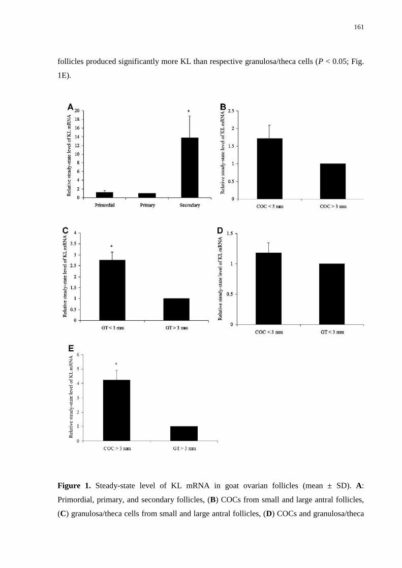
161
follicles produced significantly more KL than respective granulosa/theca cells (P < 0.05; Fig.
1E).
Figure 1. Steady-state level of KL mRNA in goat ovarian follicles (mean ± SD). A:
Primordial, primary, and secondary follicles, (B) COCs from small and large antral follicles,
(C) granulosa/theca cells from small and large antral follicles, (D) COCs and granulosa/theca
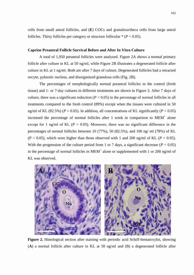
162
cells from small antral follicles, and (E) COCs and granulosa/theca cells from large antral
follicles. Thirty follicles per category or structure follicular * (P < 0.05).
Caprine Preantral Follicle Survival Before and After In Vitro Culture
A total of 1,950 preantral follicles were analyzed. Figure 2A shows a normal primary
follicle after culture in KL at 50 ng/ml, while Figure 2B illustrates a degenerated follicle after
culture in KL at 1 ng/ml. Both are after 7 days of culture. Degenerated follicles had a retracted
oocyte, pyknotic nucleus, and disorganized granulosa cells (Fig. 2B).
The percentages of morphologically normal preantral follicles in the control (fresh
tissue) and 1- or 7-day cultures in different treatments are shown in Figure 3. After 7 days of
culture, there was a significant reduction (P < 0.05) in the percentage of normal follicles in all
treatments compared to the fresh control (89%) except when the tissues were cultured in 50
ng/ml of KL (82.5%) (P > 0.05). In addition, all concentrations of KL significantly (P < 0.05)
increased the percentage of normal follicles after 1 week in comparison to MEM+ alone
except for 1 ng/ml of KL (P > 0.05). Moreover, there was no significant difference in the
percentages of normal follicles between 10 (77%), 50 (82.5%), and 100 ng/ ml (78%) of KL
(P > 0.05), which were higher than those observed with 1 and 200 ng/ml of KL (P < 0.05).
With the progression of the culture period from 1 to 7 days, a significant decrease (P < 0.05)
in the percentage of normal follicles in MEM+ alone or supplemented with 1 or 200 ng/ml of
KL was observed.
Figure 2. Histological section after staining with periodic acid Schiff-hematoxylin, showing
(A) a normal follicle after culture in KL at 50 ng/ml and (B) a degenerated follicle after
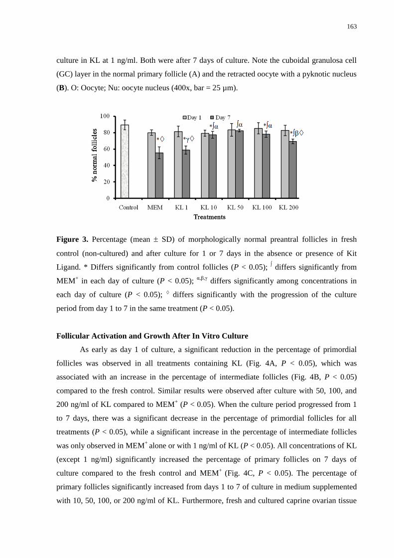
163
culture in KL at 1 ng/ml. Both were after 7 days of culture. Note the cuboidal granulosa cell
(GC) layer in the normal primary follicle (A) and the retracted oocyte with a pyknotic nucleus
(B). O: Oocyte; Nu: oocyte nucleus (400x, bar = 25 µm).
Figure 3. Percentage (mean SD) of morphologically normal preantral follicles in fresh
control (non-cultured) and after culture for 1 or 7 days in the absence or presence of Kit
Ligand. * Differs significantly from control follicles (P < 0.05); ∫ differs significantly from
MEM+ in each day of culture (P < 0.05);
α,β,γ differs significantly among concentrations in
each day of culture (P < 0.05); differs significantly with the progression of the culture
period from day 1 to 7 in the same treatment (P < 0.05).
Follicular Activation and Growth After In Vitro Culture
As early as day 1 of culture, a significant reduction in the percentage of primordial
follicles was observed in all treatments containing KL (Fig. 4A, P < 0.05), which was
associated with an increase in the percentage of intermediate follicles (Fig. 4B, P < 0.05)
compared to the fresh control. Similar results were observed after culture with 50, 100, and
200 ng/ml of KL compared to MEM+ (P < 0.05). When the culture period progressed from 1
to 7 days, there was a significant decrease in the percentage of primordial follicles for all
treatments (P < 0.05), while a significant increase in the percentage of intermediate follicles
was only observed in MEM+
alone or with 1 ng/ml of KL (P < 0.05). All concentrations of KL
(except 1 ng/ml) significantly increased the percentage of primary follicles on 7 days of
culture compared to the fresh control and MEM+
(Fig. 4C, P < 0.05). The percentage of
primary follicles significantly increased from days 1 to 7 of culture in medium supplemented
with 10, 50, 100, or 200 ng/ml of KL. Furthermore, fresh and cultured caprine ovarian tissue

164
contained a low (often zero) and variable number of secondary follicles (data not shown) that
did not permit the performance of statistical analysis.
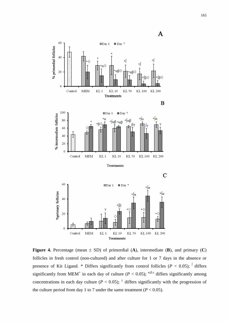
165
Figure 4. Percentage (mean SD) of primordial (A), intermediate (B), and primary (C)
follicles in fresh control (non-cultured) and after culture for 1 or 7 days in the absence or
presence of Kit Ligand. * Differs significantly from control follicles (P < 0.05); ∫ differs
significantly from MEM+ in each day of culture (P < 0.05);
α,β,γ differs significantly among
concentrations in each day culture (P < 0.05); differs significantly with the progression of
the culture period from day 1 to 7 under the same treatment (P < 0.05).
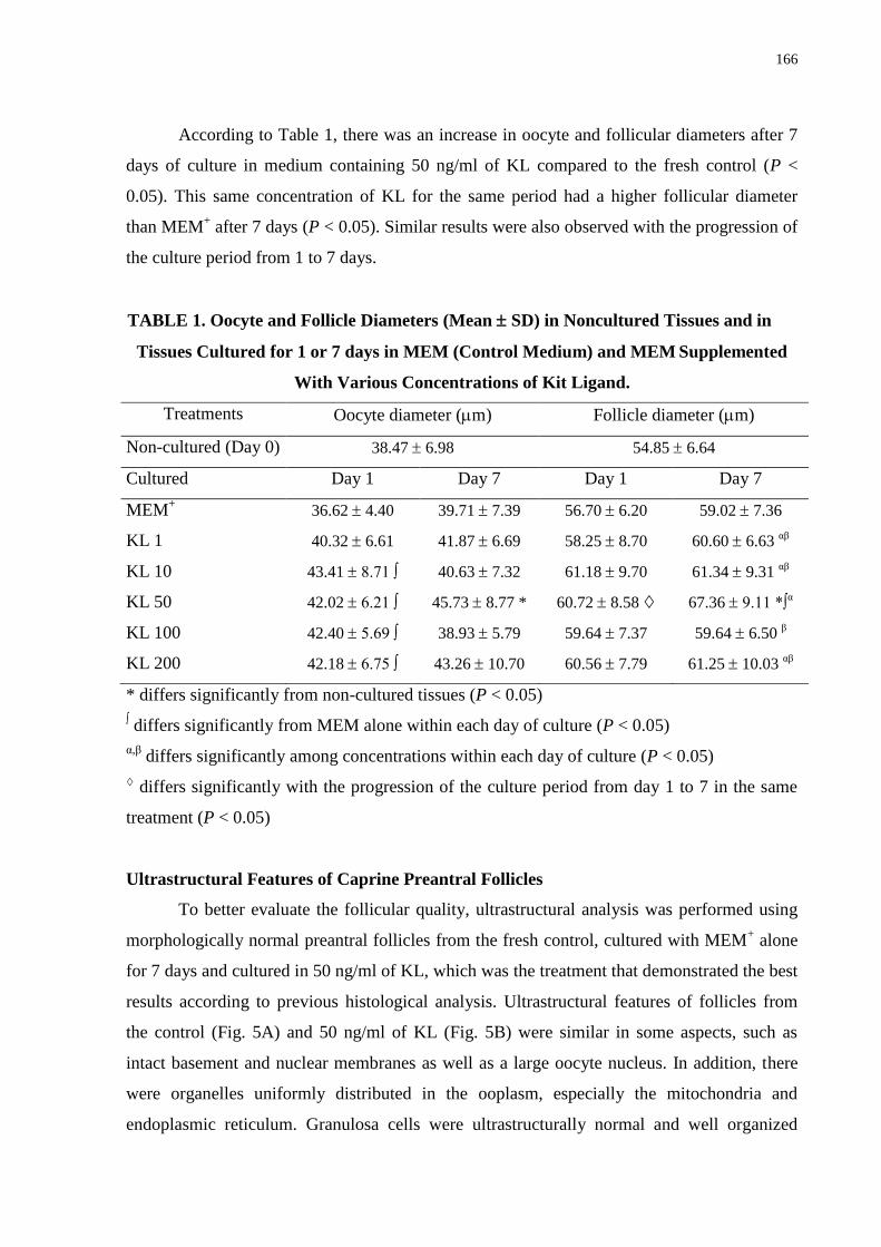
166
According to Table 1, there was an increase in oocyte and follicular diameters after 7
days of culture in medium containing 50 ng/ml of KL compared to the fresh control (P <
0.05). This same concentration of KL for the same period had a higher follicular diameter
than MEM+ after 7 days (P < 0.05). Similar results were also observed with the progression of
the culture period from 1 to 7 days.
TABLE 1. Oocyte and Follicle Diameters (Mean SD) in Noncultured Tissues and in
Tissues Cultured for 1 or 7 days in MEM (Control Medium) and MEM Supplemented
With Various Concentrations of Kit Ligand.
Treatments Oocyte diameter (m) Follicle diameter (m)
Non-cultured (Day 0) 38.47 6.98 54.85 6.64
Cultured Day 1 Day 7 Day 1 Day 7
MEM+ 36.62 4.40 39.71 7.39 56.70 6.20 59.02 7.36
KL 1 40.32 6.61 41.87 6.69 58.25 8.70 60.60 6.63
αβ
KL 10 43.41 8.71 ∫ 40.63 7.32 61.18 9.70 61.34 9.31
αβ
KL 50 42.02 6.21 ∫ 45.73 8.77 * 60.72 8.58 67.36 9.11 *∫
α
KL 100 42.40 5.69 ∫ 38.93 5.79 59.64 7.37 59.64 6.50
β
KL 200 42.18 6.75 ∫ 43.26 10.70 60.56 7.79 61.25 10.03
αβ
* differs significantly from non-cultured tissues (P < 0.05)
∫ differs significantly from MEM alone within each day of culture (P < 0.05)
α,β differs significantly among concentrations within each day of culture (P < 0.05)
differs significantly with the progression of the culture period from day 1 to 7 in the same
treatment (P < 0.05)
Ultrastructural Features of Caprine Preantral Follicles
To better evaluate the follicular quality, ultrastructural analysis was performed using
morphologically normal preantral follicles from the fresh control, cultured with MEM+ alone
for 7 days and cultured in 50 ng/ml of KL, which was the treatment that demonstrated the best
results according to previous histological analysis. Ultrastructural features of follicles from
the control (Fig. 5A) and 50 ng/ml of KL (Fig. 5B) were similar in some aspects, such as
intact basement and nuclear membranes as well as a large oocyte nucleus. In addition, there
were organelles uniformly distributed in the ooplasm, especially the mitochondria and
endoplasmic reticulum. Granulosa cells were ultrastructurally normal and well organized

167
around the oocyte, revealing an elongated and large nucleus. However, when the ovarian
tissue was cultured for 7 days in MEM+
with KL 50 ng/ml, their oocytes had irregular nuclear
membranes as well as a detachment of the granulosa cells from the oocyte. When follicles
were cultured for 7 days with MEM+ alone, there was an absence of nuclear and basement
membrane integrity, a highly vacuolated oocyte cytoplasm, low density of organelles and
disorganized and degenerated granulosa cells (Fig. 5C).
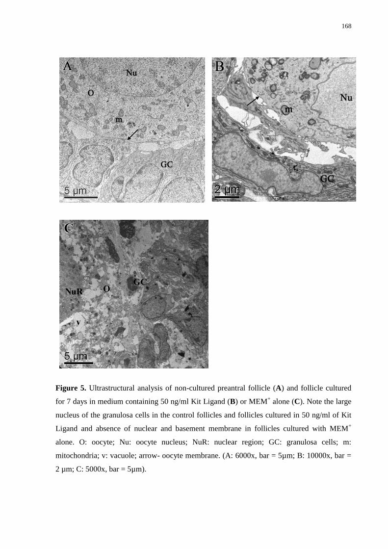
168
Figure 5. Ultrastructural analysis of non-cultured preantral follicle (A) and follicle cultured
for 7 days in medium containing 50 ng/ml Kit Ligand (B) or MEM+ alone (C). Note the large
nucleus of the granulosa cells in the control follicles and follicles cultured in 50 ng/ml of Kit
Ligand and absence of nuclear and basement membrane in follicles cultured with MEM+
alone. O: oocyte; Nu: oocyte nucleus; NuR: nuclear region; GC: granulosa cells; m:
mitochondria; v: vacuole; arrow- oocyte membrane. (A: 6000x, bar = 5µm; B: 10000x, bar =
2 µm; C: 5000x, bar = 5µm).
169
DISCUSSION
Analysis of the steady-state level of mRNA encoding KL demonstrated an increase in
the steady-state level of KL mRNA during the transition from primary to secondary follicle
stages. KL is involved in the control of early follicle development in goats, since a stimulation
of primordial follicle activation and growth of activated follicles was observed after culturing
ovarian cortical tissue in medium containing KL. In a mouse model, a higher level of KL
mRNA was demonstrated in one and two-layered growing follicles 8 days after birth, while
minimal expression was found in primordial follicles cells until this time (Doneda et al.,
2002). In addition, the steady-state level of KL mRNA was very low in primordial and
primary follicles (Manova et al., 1993; Motro and Bernstein, 1993). In humans, mRNA
encoding KL was detected in primary follicles; however, there was no effect on the stages of
follicular development after in vitro culture with KL for 7 days, which was independent of the
concentration used (Carlsson et al., 2006). In rabbit ovaries, low steady-state level of KL
mRNA in primordial follicles, high steady-state level of mRNA in growing follicles, and
variable levels in antral follicles were observed (Hutt et al., 2006b). In this study on goat
antral follicles, regarding the levels of mRNA in granulosa/theca cells, a higher steady-state
level was observed in small antral follicles in comparison to large antral follicles. In large
antral follicles, a higher steady-state level of mRNA was detected in COCs as compared with
granulosa/theca cells. In contrast, Joyce et al. (1999) demonstrated that the development of
mouse preovulatory follicles was associated with a large increase in the steady-state level of
KL mRNA in mural granulosa cells but not in cumulus cells. These differences could be due
to the use of goat follicles ranging from 3 to 6 mm in this study instead of preovulatory
follicles. In rabbit antral follicles, low levels of KL mRNA were expressed in mural granulosa
cells, and moderate to insignificant steady-state levels of mRNA were observed in the
cumulus cells of antral follicles (Hutt et al., 2006b).
This study is the first to report the positive effect of KL on the in vitro development of
caprine preantral follicles, which indicated that KL at a concentration of 50 ng/ml not only
maintained the morphological integrity of 7-day cultures of caprine preantral follicles, but
also stimulated the activation of the primordial follicles and the growth of the activated
follicles. After 7 days of culture, the follicular survival was similar to the fresh control only
with the addition of 50 ng/ml of KL to the culture medium. These results demonstrate the
importance of the KL/c-kit interaction for preantral follicle survival. According to Yoshida et
al. (1997), there was a significant reduction in normal mouse preantral follicles after in vivo
170
injection of antibodies that block the interaction between these two components. Moreover,
the inhibition of the KL/c-kit interaction by anti-c-kit antibody promoted the death of oocytes
in vitro (Reynaud et al., 2000). In vitro studies have demonstrated that KL inhibits apoptosis
in oocytes of mouse primordial follicles by increasing the steady-state level of mRNA of the
anti-apoptotic proteins, Bcl-2 and Bcl-cL, and reducing the steady-state level of mRNA of the
pro-apoptotic factor, Bax. These anti-apoptotic effects appear to be mediated through a
phosphoinositide-3 kinase pattern, which promotes the inhibition of Fas-mediated apoptosis
(Jin et al., 2005). Other studies have demonstrated that the oocytes had no visible signs of
degeneration after addition of 50 or 100 ng/ml of KL even after 20 days of culture (Klinger
and De Felici, 2002). However, KL did not have any effect on the survival of rabbit and
murine preantral follicles at 50 or 150 ng/ml (Hutt et al., 2006b). Similar to the results
observed in our study at 1 ng/ml of KL, Yan et al. (2000) concluded that low levels of KL are
not sufficient for the survival of rat male germ cells. According to this previous study, the
steady-state level of KL mRNA is lower in vitro than in vivo due to the absence of FSH, as
high concentrations of this factor are necessary in vitro. Regarding the unsatisfactory result at
200 ng/ml, this KL concentration may have interfered with the steady-state level of mRNA of
other important factors for follicular survival, such as FSH receptors (Thomas et al., 2005;
Matos et al., 2007).
After 1 day of culture, all treatments containing KL promoted follicular activation
compared to the fresh control. Additionally, all KL concentrations (except 1 ng/ml) increased
the percentage of primary follicles after 7 days of culture compared to the fresh control and
MEM+ alone. Studies have demonstrated that blockage of KL affects primordial follicle
development (Yoshida et al., 1997) and that KL increases the transition from primordial to
primary follicles in rats (Parrot and Skinner, 1999; Dole et al., 2008) and hamsters (Wang and
Roy, 2004) after in vitro culture. However, KL did not have any effect on human preantral
follicle development after 7 or 14 days of culture at 1, 10, or 100 ng/ml (Carlsson et al.,
2006). KL has been hypothesized to play a role in follicular activation and further
development through activation of Akt (a signaling molecule known to increase cellular
proliferation and survival as well as glycogen and protein synthesis) and inhibition of
FKHRL1 (the Akt substrate and transcriptional factor that leads to apoptosis and cell cycle
arrest) in the oocyte (Reddy et al., 2005). Although an elevated steady-state level of KL
mRNA in secondary follicles was verified in this study, which is similar to previous results
(human: Carlsson et al., 2006, rabbit: Hutt et al., 2006b), few follicles were observed at this
171
stage after 7 days of culture with KL. This lack of follicles most likely occurs due to the short
culture period or the absence of some hormones or other growth factors in the culture medium
that could be important for the transition from primary to secondary follicles, such as activin
(Zhao et al., 2001), bone morphogenetic protein-15 (BMP-15) (Galloway et al., 2000), or
growth and differentiation factor-9 (GDF-9) (Hayashi et al., 1999).
In relation to growth, an increase in the follicular and oocyte diameter was observed
after 7 days of culture with 50 ng/ml of KL compared to the control. The role of KL in the
promotion of early oocyte growth has already been demonstrated in vitro (Packer et al., 1994),
and the steady-state level of mRNA of both KL and c-kit is consistent with this role (Manova
et al., 1993). Previous studies have indicated that KL stimulates oocyte growth during earlier
stages of follicular development (mouse: Klinger and De Felici, 2002; rabbit: Hutt et al.,
2006b). However, other investigations did not observe any effect of KL on oocyte (mouse:
Cecconi and Colonna, 1996; hamster: Wang and Roy, 2004) or follicle (human: Carlsson et
al., 2006) growth, suggesting that KL‘s actions differ according to the stage of follicular
development. In addition, Parrot and Skinner (1997) demonstrated that 50 ng/ml of KL
stimulated theca cell growth. The mechanism by which KL results in oocyte growth is
unknown. However, proteins involved in the translation of the c-kit signal, such as PI3-
kinases, MAP kinases, and Janus-activated kinase 2 (JAK2), are possible candidates. KL is
likely to initiate oocyte growth with the slow accumulation of required factors for meiosis
resumption (e.g., p34cd2, cyclin B1, MAPK, cdc25) (Reddy et al., 2005). Moreover, based on
the ability of KL to induce oocyte growth, which requires both the presence of KL and contact
with granulosa cells (Thomas and Vanderhyden, 2006), KL actions are suggested to be
modulated by the presence of gap junctions (Klinger and De Felici, 2002) according to the
oocyte stage.
Our results on follicular viability after classical histology were confirmed by
ultrastructural analysis. This technique, which is able to detect damage to cellular membranes
and organelles that is not visible at the level of light microscopy, is considered an important
tool to discern the quality of the follicle and oocyte (Lucci et al., 2001). Important structures,
such as the mitochondria, endoplasmic reticulum, and granulosa, were preserved after
transmission electron microscopy (TEM) in addition to the basement and nuclear membranes
after 7 days of culture in the presence of 50 ng/ml of KL. However, cortical tissues cultured
with 50 ng/ml KL for 7 days demonstrated some detachment of granulosa cells from the
oocyte. This result was also observed in caprine preantral follicles cultured in vitro (Lima-
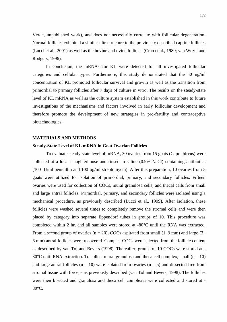
172
Verde, unpublished work), and does not necessarily correlate with follicular degeneration.
Normal follicles exhibited a similar ultrastructure to the previously described caprine follicles
(Lucci et al., 2001) as well as the bovine and ovine follicles (Cran et al., 1980; van Wezel and
Rodgers, 1996).
In conclusion, the mRNAs for KL were detected for all investigated follicular
categories and cellular types. Furthermore, this study demonstrated that the 50 ng/ml
concentration of KL promoted follicular survival and growth as well as the transition from
primordial to primary follicles after 7 days of culture in vitro. The results on the steady-state
level of KL mRNA as well as the culture system established in this work contribute to future
investigations of the mechanisms and factors involved in early follicular development and
therefore promote the development of new strategies in pro-fertility and contraceptive
biotechnologies.
MATERIALS AND METHODS
Steady-State Level of KL mRNA in Goat Ovarian Follicles
To evaluate steady-state level of mRNA, 30 ovaries from 15 goats (Capra hircus) were
collected at a local slaughterhouse and rinsed in saline (0.9% NaCl) containing antibiotics
(100 IU/ml penicillin and 100 µg/ml streptomycin). After this preparation, 10 ovaries from 5
goats were utilized for isolation of primordial, primary, and secondary follicles. Fifteen
ovaries were used for collection of COCs, mural granulosa cells, and thecal cells from small
and large antral follicles. Primordial, primary, and secondary follicles were isolated using a
mechanical procedure, as previously described (Lucci et al., 1999). After isolation, these
follicles were washed several times to completely remove the stromal cells and were then
placed by category into separate Eppendorf tubes in groups of 10. This procedure was
completed within 2 hr, and all samples were stored at -80°C until the RNA was extracted.
From a second group of ovaries (n = 20), COCs aspirated from small (1–3 mm) and large (3–
6 mm) antral follicles were recovered. Compact COCs were selected from the follicle content
as described by van Tol and Bevers (1998). Thereafter, groups of 10 COCs were stored at -
80°C until RNA extraction. To collect mural granulosa and theca cell complex, small (n = 10)
and large antral follicles (n = 10) were isolated from ovaries (n = 5) and dissected free from
stromal tissue with forceps as previously described (van Tol and Bevers, 1998). The follicles
were then bisected and granulosa and theca cell complexes were collected and stored at -
80°C.
173
Isolation of total RNA was performed using Trizol plus purification kit (Invitrogen,
São Paulo, Brazil). According to the manufacturer‘s instructions, 1 ml of Trizol solution was
added to each frozen samples and the lysate was aspirated through a 20-gauge needle before
centrifugation at 10,000g for 3 min at room temperature. Thereafter, all lysates were diluted
1:1 with 70% ethanol and subjected to a mini-column. After binding of the RNA to the
column, DNA digestion was performed using RNAse-free DNAse (340 Kunitz units/ml) for
15 min at room temperature. After washing the column three times, the RNA was eluted with
30 µl RNAse-free water.
Prior to reverse transcription, the eluted RNA samples were incubated for 5 min at
70°C, and chilled on ice. Reverse transcription was then performed in a total volume of 20 µl,
which was comprised of 10 µl of sample RNA, 4 µl 5X reverse transcriptase buffer
(Invitrogen), 8 U RNAseout, 150 U Superscript III reverse transcriptase, 0.036 U random
primers (Invitrogen), 10 mM DTT, and 0.5 mM of each dNTP. The mixture was incubated for
1 hr at 42°C, for 5 min at 80°C, and then stored at -20°C. Negative controls were prepared
under the same conditions but without the inclusion of the reverse transcriptase.
Quantification of the mRNA for KL was performed using SYBR Green. PCR
reactions were composed of 1 µl cDNA as a template in 7.5 µl of SYBR Green Master Mix
(PE Applied Biosystems, Foster City, CA), 5.5 µl of ultra-pure water, and 0.5 µM of each
primer. The primers were designed to perform amplification of mRNA for KL. The GAPDH
and b-actin (Table 2) were used as endogenous controls for normalization of steady-state level
of mRNA of gene. The thermal cycling profile for the first round of PCR was: initial
denaturation and activation of the polymerase for 15 min at 94°C, followed by 40 cycles of 15
sec at 94°C, 30 sec at 60°C, and 45 sec at 72°C. The final extension was for 10 min at 72°C.
All reactions were performed in a real-time PCR Mastercycler (Eppendorf, Germany). The
delta-delta-CT method was used to transform CT values into normalized relative steady-state
level of mRNA.
174
TABLE 2. Oligonucleotide Primers Used for PCR Analysis of Goat Cells and Tissues.
Target
gene
Primer sequence (5´ 3´) Sense Position GenBank accession nº
GAPDH TGTTTGTGATGGGCGTGAACCA s 287-309 GI: 27525390
(2005)Capra hircus
GAPDH
ATGGCGTGGACAGTGGTCATAA as 440-462
Beta-Actin ACCACTGGCATTGTCATGGACTCT s 187-211 GI: 28628620
(2003)Capra hircus
Beta-Actin
TCCTTGATGTCACGGACGATTTCC as 386-410
KL AGCGAGATGGTGGAACAACTGTCA s 211-235 GI: 16580734
(2001)Capra hircus KL GTTCTTCCATGCACTCCACAAGGT as 35-59
s,sense; as, antisense
In Vitro Culture of Ovarian Tissue and Morphological Evaluation
Ovarian cortical tissues were collected at a local slaughterhouse from five adult (1–3
years old), mixed-breed goats (two ovaries from each goat). Immediately postmortem, the
ovaries were washed in 70% alcohol for 10 sec, followed by two washes in Minimum
Essential Medium (MEM) supplemented with 100 µg/ml penicillin and 100 µg/ml
streptomycin. The pairs of ovaries were transported within 1 hr to the laboratory in MEM at
4°C.
The organ culture system utilized herein was previously described in detail (Matos et
al., 2007; Celestino et al., 2009). In the laboratory, the ovaries from each animal were stripped
of surrounding fat tissue and ligaments. Moreover, the medulla, visible growing follicles and
corpora lutea were removed. Ovarian cortex tissue samples from each ovarian pair were cut
into 13 slices (approximate size 3 x 3 mm2, with 1 mm thickness) using a needle and scalpel
under sterile conditions. The tissue pieces were then either directly fixed for histological and
ultrastructural analysis (fresh control) or placed in culture for a total of 1 or 7 days. Caprine
tissues were transferred to 24-well culture dishes containing 1 ml of culture media. The
culture was performed at 39°C in 5% CO2 in a humidified incubator and all the media were
incubated for 1 hr prior to use. The basic culture medium (culture control) consisted of MEM
(pH 7.2–7.4) supplemented with ITS (insulin 6.25 µg/ml, transferrin 6.25 µg/ml, and
selenium 6.25 ng/ml) (Sigma, St. Louis, MO), 0.23 mM pyruvate, 2 mM glutamine, 2 mM
hypoxantine, and 1.25 mg/ml bovine serum albumin (BSA), and was called MEM+. For
experimental conditions, the medium was supplemented with recombinant human KL (rhKL)
at different concentrations (0, 1, 10, 50, 100, or 200 ng/ml) (Cell Sciences, Canton, MA).
Each treatment was repeated five times, that is, five different goats were used, and the culture
media were replenished every other day.
175
Before culture (fresh control) and after 1 or 7 days in culture, all tissues were fixed in
Carnoy‘s solution for 12 hr and then dehydrated with increasing concentrations of ethanol.
After paraffin embedding (Synth, São Paulo, Brazil), the caprine tissues pieces were cut into
7-µm sections, and every section was mounted on glass slides and stained by periodic acid
Schiff–hematoxylin. Follicle stage and survival were assessed microscopically on serial
sections. Coded, anonymized slides were examined on a microscope (Nikon, Tokyo, Japan)
under 400x magnification. The follicles were carefully counted once, as performed in our
earlier studies (Matos et al., 2007; Celestino et al., 2009). Each follicle was examined in every
section that the follicle appeared in and matched with the same follicle on adjacent sections to
avoid double counting and therefore ensure that each follicle was only counted once
regardless of size.
The developmental stages of follicles have been previously defined (Silva et al., 2004)
as primordial (one layer of flattened granulosa cells around the oocyte) or growing follicles
(intermediate: one layer of flattened and cuboidal granulosa cells; primary: one layer of
cuboidal granulosa cells, and secondary: two or more layers of cuboidal granulosa cells
around the oocyte). These follicles were still individually classified as histologically normal
when an intact oocyte was present and surrounded by granulosa cells, which were well
organized into one or more layers and no pyknotic nucleus was present. Atretic follicles were
defined as follicles with a retracted oocyte, pyknotic nucleus, and/or disorganized granulosa
cells detached from the basement membrane. Overall, 150 follicles were evaluated for each
treatment (30 follicles per treatment in one repetition x 5 repetitions = 150 follicles).
To evaluate follicular activation, the percentage of healthy primordial and growing
follicles were calculated before (fresh control) and after culture in each medium. Follicle and
oocyte diameters were only measured in unilaminar (primordial, intermediate, and primary
follicles) and healthy follicles with the aid of an ocular micrometer. Both diameters, from the
basement membrane, at right angles to each other in the largest cross-section of each growing
oocyte and follicle, were measured and averaged.
Ultrastructural Analysis
To better evaluate follicular morphology, ultrastructural analysis was performed on
preantral follicles from noncultured tissue, follicles cultured in MEM+ alone, as well as on the
treatment which demonstrated the best results for morphology, activation, and growth. Small
pieces (1 mm3) of caprine ovarian tissues from treatments mentioned above were fixed in 2%
176
paraformaldehyde and 2.5% glutaraldehyde in 0.1 M sodium cacodylate buffer (pH 7.2) for 4
hr at room temperature. After fixation, fragments were post-fixed in 1% osmium tetroxide,
0.8% potassium ferricyanide, and 5 mM calcium chloride in 0.1 M sodium cacodylate buffer
for 1 hr. Subsequently, the samples were dehydrated through a gradient of acetone solutions
and the tissues were embedded in Spurr‘s resin. Semi-thin sections (3 µm) were cut on an
ultramicrotome (Reichert Supernova, Heidelberg, Germany) for light microscopy studies and
stained with toluidine blue. The ultra-thin sections (60–70 µm) were contrasted with uranyl
acetate and lead citrate, and examined under a Jeol 1011 (Jeol, Tokyo, Japan) transmission
electron microscope. Parameters were further evaluated, such as the density and integrity of
ooplasmic and granulosa cell organelles, vacuolization, and basement membrane integrity.
For ultrastructural analysis, 3–5 follicles were examined per group.
Statistical Analysis
The percentages of morphologically normal, primordial, and developing follicles after
1 or 7 days of culture were initially subjected to Smirnov–Kolmogorov and Bartlett tests to
confirm normal distribution and homogeneity of variance, respectively. Analysis of variance
(ANOVA) was then performed using the GLM procedure of SAS (1997) and the Dunnett test
was applied for comparison of each treatment tested against the control groups (Control and
MEM+) (Steel et al., 1997). The Student–Newman–Keuls (SNK) test was utilized to evaluate
the effects of different concentrations of KL and days of culture. Due to heterogeneity,
steady-state levels of mRNA in primordial, primary, secondary, and in small and large antral
follicles were analyzed by the Kruskal–Wallis nonparametric test. Differences were
considered to be significant when P <0 .05, and results were expressed as the mean ± standard
deviation (SD).
ACKNOWLEDGMENTS
This work was supported by CNPq, CAPES, FINEP, and Fundação Cearense de
Apoio à Pesquisa (FUNCAP). Juliana Jales de Hollanda Celestino is a recipient of a grant
from FUNCAP (Brazil). The authors thank José Leandro da Silva Neto for his technical
support in classical histology and all the members of the laboratory.
177
REFERENCES
Carlsson IB, Laitinen MPE, Scott JE, Louhio H, Velentzis L, Tuuri T, Aaltonen J, Ritvos O,
Winston RML, Hovatta O. 2006. Kit ligand and c-Kit are expressed during early human
ovarian follicular development and their interaction is required for the survival of follicles
in long-term culture. Reproduction 131:641–649.
Cecconi S, Colonna R. 1996. Influence of granulosa cells and of different somatic cell types
on mammalian oocyte development in vitro. Zygote 4:305–307.
Celestino JJH, Bruno JB, Lima-Verde IB, Matos MHT, Saraiva MVA, Chaves RN, Martins
FS, Lima LF, Name KPO, Campello CC, Silva JRV, Báo SN, Figueiredo JR. 2009.
Recombinant epidermal growth factor maintains follicular ultrastructure and promotes the
transition to primary follicles in caprine ovarian tissue cultured in vitro. Reprod Sci
16:239–246.
Choi Y, Ballow DJ, Xin Y, Rajkovic A. 2008a. Lim homeobox gene, Lhx8, is essential for
mouse oocyte differentiation and survival. Biol Reprod 79:442–449.
Choi Y, Yuan D, Rajkovic A. 2008b. Germ cell-specific transcriptional regulator Sohlh2 is
essential for early mouse folliculogenesis and oocyte-specific gene expression. Biol
Reprod 79: 1176–1182.
Clark DE, Tisdall DJ, Fidler AE, Mcnatty KP. 1996. Localization of mRNA encoding c-kit
during the initiation of folliculogenesis in ovine fetal ovaries. J Reprod Fertil 106:329–335.
Cran DG, Moor RM, Hay MF. 1980. Fine structure of the sheep oocyte during antral follicle
development. J Reprod Fertil 59: 125–132.
Dole G, Nilsson EE, Skinner MK. 2008. Glial-derived neurotrophic factor promotes ovarian
primordial follicle development and cell–cell interactions during folliculogenesis.
Reproduction 135: 671–682.
Doneda L, Klinger FG, Larizza L, De Felici M. 2002. KL/KIT coexpression in mouse fetal
oocytes. Int J Dev Biol 46:1015–1021.
Driancourt MA, Reynaud K, Cortvrindt R, Smitz J. 2000. Roles of Kit and Kit Ligand in
ovarian function. Rev Reprod 5:143–152.
Fortune JE. 2003. The early stages of follicular development: Activation of primordial
follicles and growth of preantral follicles. Anim Reprod Sci 78:135–163.
Galloway SM, Mcnatty KP, Cambridge LM, Laitinen MP, Juengel JL, Jokiranta TS, Mclaren
RJ, Luiro K, Dodds KG, Montgomery GW, Beattie AE, Davis GH, Ritvos O. 2000.
178
Mutations in an oocyte-derived growth factor gene (BMP15) cause increased ovulation rate
and infertility in a dosage-sensitive manner. Nat Genet 25:279–283.
Hayashi M, Mcgee EA, Min G, Klein C, Rose UM, van Duin M, Hsueh AJW. 1999.
Recombinant growth differentiation factor-9 (GDF-9) enhances growth and differentiation
of cultured early follicles. Endocrinology 140:1236–1244.
Hoyer PE, Byskov AG, Mollgard K. 2005. Stem cell factor and c-kit in human primordial
germ cells and fetal ovaries. Mol Cell Endocrinol 234:1–10.
Huang EJ, Nocka KH, Buck J, Besmer P. 1992. Differential expression and processing of two
cell associated forms of the Kit- Ligand: KL-1 and KL-2. Mol Biol Cell 3:349 362.
Hutt K, Mclaughlin EA, Holland MK. 2006a. KL and KIT have diverse role during
mammalian oogenesis. Mol Hum Reprod 12:61–69.
Hutt KJ, Mclaughlin EA, Holland MK. 2006b. KIT/KIT Ligand in mammalian oogenesis and
folliculogenesis: Roles in rabbit and murine ovarian follicle activation and oocyte growth.
Biol Reprod 75:421–433.
Ismail RS, Okawara Y, Fryer JN, Vanderhyden BC. 1996. Hormonal regulation of the ligand
for c-Kit in the rat ovary and its effects on spontaneous oocyte meiotic maturation. Mol
Reprod Dev 43: 458–469.
Ismail RS, Dube M, Vanderhyden BC. 1997. Hormonally regulated expression and alternative
splicing of kit ligand may regulate kitinduced inhibition of meiosis in rat oocytes. Dev Biol
184: 333–342.
Jin X, Han CS, Yu FQ, Wei P, Hu ZY, Liu YX. 2005. Anti-apoptotic action of stem cell
factor on oocytes in primordial follicles and its signal transduction. Mol Reprod Dev
70:82–90.
Joyce IM, Pendola FL, Wigglesworth K, Eppig JJ. 1999. Oocyte regulation of kit ligand
expression in mouse ovarian follicles. Dev Biol 214:342–353.
Klinger FG, De Felici M. 2002. In vitro development of growing oocytes from fetal mouse
oocytes: Stage-specific regulation by stem cell factor and granulosa cells. Dev Biol
244:85–95.
Knight PG, Glister C. 2006. TGF-b superfamily members and ovarian follicle development.
Reproduction 132:191–206.
Lucci CM, Amorim CA, Báo SN, Figueiredo JR, Rodrigues APR, Silva JRV, Goncalves
PBD. 1999. Effect of the interval of serial sections of ovarian in the tissue chopper on the
number of isolated caprine preantral follicles. Anim Reprod Sci 56:39–49.
179
Lucci CM, Silva JRV, Carvalho FCA, Figueiredo JR, Báo SN. 2001. Light microscopical and
ultrastrutural characterization of goat preantral follicles. Small Rumin Res 41:61–69.
Manova K, Nocka K, Besmer P, Bachvarova RF. 1990. Gonadal expression of c-kit encoded
at the W locus of the mouse. Development 110:1057–1069.
Manova K, Huang EJ, Angeles M, De Leon V, Sanchez S, Pronovost SM, Besmer P,
Bachvarova RF. 1993. The expression pattern of the c-kit ligand in gonads of mice
supports a role for the c-kit receptor in oocyte growth and in proliferation of
spermatogonia. Dev Biol 157:85–99.
Matos MHT, Lima-Verde IB, Luque MCA, Maia JE, Jr., Silva JRV, Celestino JJH, Martins
FS, Báo SN, Lucci CM, Figueiredo JR. 2007. Essential role of follicle stimulating hormone
in the maintenance of caprine preantral follicle viability in vitro. Zygote 15:173–182.
Motro B, Bernstein A. 1993. Dynamic changes in ovarian c-kit and syeel expression during
the estrous reproductive cycle. Dev Dynam 197:69–79.
Otsuka F, Shimasaki S. 2002. A negative feedback system between oocyte bone
morphogenetic protein 15 and granulosa cell kit ligand: Its role in regulating granulosa cell
mitosis. Proc Natl Acad Sci USA 99:8060–8065.
Packer AI, Hsu YC, Besmer P, Bachvaroava RF. 1994. The ligand of the c-kit receptor
promotes oocyte growth. Dev Biol 161: 194–205.
Parrot JA, Skinner MK. 1997. Direct actions of Kit-Ligand on theca cell growth and
differentiation during follicle development. Endocrinology 138:3819–3827.
Parrot JA, Skinner MK. 1999. Kit-ligand/stem cell factor induces primordial follicle
development and initiates folliculogenesis. Endocrinology 140:4262–4271.
Reddy P, Shen L, Ren C, Boman K, Lundin E, Ottander U, Lindgren P, Liu Y-x, Sun Q-y,
Liu K. 2005. Activation of Akt (PKB) and suppression of FKHRL1 in mouse and rat
oocytes by stem cell factor during follicular activation and development. Dev Biol
281:160–170.
Reynaud K, Cortvrindt R, Smitz J, Driancourt MA. 2000. Effects of Kit Ligand and anti-Kit
antibody on growth of cultured mouse preantral follicles. Mol Reprod Dev 56:483–494.
Silva JRV, van den Hurk R, Matos MHT, Santos RR, Pessoa C, Moraes MO, Figueiredo JR.
2004. Influences of FSH and EGF on primordial follicles during in vitro culture of caprine
ovarian cortical tissue. Theriogenology 61:1691–1704.
180
Silva JRV, van den Hurk R, van Tol HTA, Roelen BAJ, Figueiredo JR. 2006. The Kit
ligand/c-Kit receptor system in goat ovaries: Gene expression and protein localization.
Zygote 14:317– 328.
Steel RGD, Torrie JH, Dickey DA. 1997. Principles and procedures of statistics: A
biometrical approach, 3rd edition. New York, NY: McGraw-Hill. p 666. Thomas FH,
Vanderhyden BC. 2006. Oocyte-granulosa cell interactions during mouse follicular
development regulation of kit ligand expression and its role in oocyte growth. Reprod Biol
Endocrinol 19:1–8.
Thomas FH, Ethier J-F, Shimasaki S, Vanderhyden BC. 2005. Follicle-stimulating hormone
regulates oocyte growth by modulation of expression of oocyte and granulosa cell factors.
Endocrinology 146:941–949.
Thomas FH, Ismail RS, Jiang J-Y, Vanderhyden BC. 2008. Kit Ligand 2 promotes murine
oocyte growth in vitro. Biol Reprod 78:167–175.
Tisdall DJ, Fidler AE, Smith P, Quirke LD, Stent VC, Heath DA, Mcnatty KP. 1999. Stem
cell factor and c-Kit gene expression and protein localization in the sheep ovary during
fetal development. J Reprod Fertil 116:277–291.
Van Tol HT, Bevers MM. 1998. Theca cells and theca-cell conditioned medium inhibit the
progression of FSH-induced meiosis of bovine oocytes surrounded by cumulus cells
connected to membrane granulosa. Mol Reprod Dev 51:315–321.
Van Wezel IL, Rodgers RJ. 1996. Morphological characterization of bovine primordial
follicles and their environment in vivo. Biol Reprod 55:1003–1011.
WangJ, Roy SK. 2004. Growth differentiation factor-9 and stem cell factor promote
primordial follicle formation in the hamster: Modulation by follicle-stimulating hormone.
Biol Reprod 70: 577–585.
Yan W, Suominen J, Toppari J. 2000. Stem cell factor protects germ cells from apoptosis in
vitro. J Cell Sci 113:161–168.
Yoshida H, Takakura N, Nataoka H, Kunisada T, Okamura H, Nishikawa SI. 1997. Stepwise
requirement of c-Kit tyrosine kinase in mouse ovarian follicle development. Dev Biol 184:
122–137.
Zama AM, Hudson FP, Bedell MA. 2005. Analysis of hypomorphic KitlSI mutants suggests
different requirements for KITL in proliferation and migration of mouse primordial germ
cells. Biol Reprod 73:639–647.

181
Zhao J, Taverne MAM, van der Weijden BC, Bevers MM, van den Hurk R. 2001. Effect of
activin A on in vitro development of rat preantral follicles and localization of activin A and
activin receptor II. Biol Reprod 65:967–977.
182
11 CAPÍTULO 6
Níveis da proteína morfogenética óssea-15 em ovários caprinos e sua influência no
desenvolvimento in vitro e sobrevivência de folículos pré-antrais
Steady-state level of bone morphogenetic protein-15 in goat ovaries and its influence on in
vitro development and survival of preantral follicles
Periódico: Molecular and Cellular Endocrinology (Submetido em agosto de 2010).
A B
C D
183
Resumo
Este estudo investiga os níveis de RNAm para a proteína morfogenética óssea-15 (BMP-15)
em ovários caprinos, e os efeitos da BMP-15 no desenvolvimento in vitro de folículos pré-
antrais. Fragmentos ovarianos foram cultivados por um ou sete dias em Meio Essencial
Mínimo (MEM+) com BMP-15 (0, 1, 10, 50, 100 ou 200 ng/mL), e posteriormente foram
analisados por histologia, microscopia eletrônica de transmissão e de fluorescência. O RNAm
para BMP-15 em folículos secundários foi superior que em folículos primordiais e primários.
Após sete dias, 10, 50 ou 100 ng/mL de BMP-15 mantiveram a percentagem de folículos
normais similar ao controle (não-cultivado), e aumentou os diâmetros oócitário e folicular
quando comparado ao controle e ao MEM+. BMP-15 100 ng/mL aumentou a percentagem de
folículos secundários e manteve a integridade ultraestrutural. Em conclusão, os RNAm para
BMP-15 foram detectados em todas as categorias foliculares. BMP-15 (100 ng/mL) manteve
a integridade e promoveu o crescimento de folículos pré-antrais caprinos cultivados por sete
dias.
Palavras-chave: Caprino. Folículos pré-antrais. BMP-15. Ativação. Crescimento.
184
Steady-state level of bone morphogenetic protein-15 in goat ovaries and its influence on in
vitro development and survival of preantral follicles
J.J.H. Celestinoa*, I.B. Lima-Verdeb, J.B. Brunoa, M.H.T. Matosc, R.N. Chavesa, M.V.A. Saraivaa,
C.M.G. Silvaa, L.R. Faustinoa, R. Rossettoa, C.A.P. Lopesa, M.A.M. Donatod, C.A Peixotod, C.C.
Campelloa, J.R.V. Silvae, J.R. Figueiredoa
aFaculty of Veterinary Medicine, LAMOFOPA, PPGCV, State University of Ceara, Fortaleza-CE, Brazil
bInstitute for Research and Technology, Tiradentes University, Aracaju-SE, Brazil
cNucleus of Biotechnology Applied to Ovarian Follicle Development, Federal University of São Francisco
Valley, Petrolina-PE, Brazil
dLaboratory of Ultrastructure, CPqAM/Fiocruz, Federal University of Pernambuco, Recife-PE, Brazil
eBiotecnology Nucleus of Sobral (NUBIS), Federal University of Ceara, Sobral-CE, Brazil
*Corresponding address:
Programa de Pós-Graduação em Ciências Veterinárias (PPGCV)
Laboratório de Manipulação de Oócitos e Folículos Pré-Antrais (LAMOFOPA)
Universidade Estadual do Ceará (UECE)
Av. Paranjana, 1700, Campus do Itaperi.
Fortaleza – CE – Brasil. CEP: 60740-000
Tel.: +55.85.3101.9852; Fax: +55.85.3101.9840
E-mail address: [email protected] (Juliana Jales de Hollanda Celestino)
185
Abstract
This study investigates steady-state level of bone morphogenetic protein-15 (BMP-15) mRNA
in caprine follicles, and the effects of BMP-15 on in vitro development of preantral follicles.
Ovarian fragments were cultured for one or seven days in Minimal Essential Medium
(MEM+) with BMP-15 (0, 1, 10, 50, 100 or 200 ng/mL), and further analysed by histology,
transmission electron and fluorescent microscopy. BMP-15 mRNA in secondary follicles was
higher than in primordial and primary follicles. After seven days, 10, 50 or 100 ng/mL of
BMP-15 maintained the percentage of normal follicles similar to the control (noncultured),
and increased the oocyte and follicle diameters when compared to the control and MEM+.
BMP-15 at 100 ng/mL increased the secondary follicles and maintained their ultrastructural
integrity. In conclusion, the BMP-15 mRNAs were detected in all follicular categories. BMP-
15 (100 ng/mL) maintained the integrity and promoted the growth of caprine preantral
follicles cultured for seven days.
Keywords: Caprine; Preantral Follicles; BMP-15; Activation; Growth.
186
1. Introduction
The mechanisms that control the initiation of follicular development, i.e., the
activation of primordial follicles, and the time necessary for their growth and differentiation
remains unknown (Fortune, 2003). Therefore, the development of an efficient culture system
that allows the activation and development of early stage follicles (preantral) up to later stages
is essential. This system would provide oocytes that can be further used for the in vitro
embryo production. In addition, this system would provide a better understanding of the
factors involved in folliculogenesis. Among these factors, bone morphogenetic protein-15
(BMP-15) can be highlighted.
BMP-15, also known as growth differentiation factor-9B (GDF-9B), is a member of
the transforming growth factor- (TGF-) superfamily. The ligands of this superfamily
regulate cell proliferation, differentiation and apoptosis, and they are essential for embryo
development, organogenesis, bone formation, reproduction and other biologic processes
(Chang et al., 2002; Mazerbourg and Hsueh, 2006). BMP-15 binds through a type I receptor,
i.e., BMPR-IA (activin receptor-like kinase [ALK]3) and BMPR-IB (ALK-6), and a type II
receptor on the surface of the cell (Miyazono et al., 1997; Lebrun et al., 1997). The activated
type I receptor phosphorylates one or more cytoplasmic signaling intermediates, known as
Smads receptors (R-Smads) (Moore et al., 2003; Shimasaki et al., 2004a).
In addition to being found in other tissues (Galloway et al., 2000; Eckery et al., 2002;
Otsuka and Shimasaki, 2002a), BMP-15 is produced in the largest quantities in the ovary
(Peng et al., 2009). In marsupial (Eckery et al., 2002), ovine (Mery et al., 2007), bovine
(Bodensteiner et al., 1999) and human (Aaltonen et al., 1999), BMP-15 is expressed in
oocytes from primordial follicles onward. In rodents, the expression of this protein is
observed only in the oocyte from primary follicles onward (Laitinen et al., 1998; Jaatinen et
al., 1999). For caprine species, BMP-15 has been found in the oocytes and granulosa cells of
all types of follicles but not in granulosa cells of primordial follicles. The mRNAs for BMP-
15 and their associated receptors were detected in primordial, primary and secondary follicles
and in the oocyte and granulosa cells of antral follicles (Silva et al., 2005). However,
quantification of the steady-state level of BMP-15 mRNA during different stages of goat
follicular development has not yet been performed.
In vitro studies have demonstrated that BMP-15 promotes the proliferation of the
granulosa cells and stimulates the development of primordial and primary follicles in rodents
187
(Otsuka et al., 2000; Vitt et al., 2000a; Fortune, 2003). In vivo studies have shown that BMP-
15-mutant sheep, i.e., without BMP-15, are infertile because they do not continue their
development beyond the primary follicle stage (Dong et al., 1996; Galloway et al., 2000). In
rats, recombinant BMP-15 stimulated the proliferation of granulosa cells independent of
follicle stimulating hormone (FSH) (Otsuka et al., 2000). Furthermore, BMP-15 is able to
inhibit the expression of the FSH receptor (Otsuka et al., 2001) and stimulate the expression
of the kit ligand (KL) and epidermal growth factor (EGF) in rat granulosa cells (Otsuka and
Shimasaki, 2002b) and mouse cumulus cells (Yoshino et al., 2006), respectively. In a study
performed with sheep, BMP-15 was suggested to be required for the follicular development
up to the ovulatory stage (Juengel et al., 2002). However, the influence of BMP-15 during
caprine folliculogenesis is unknown. Results for this species are of great economic importance
due to its production of milk, meat and fibers, and its potential to provide the main source of
proteins for the population of the tropics. Therefore, the present study was designed with the
following aims: (1) to determine the steady-state level of BMP-15 mRNA during different
follicular stages in goat ovaries and (2) to investigate a possible influence of BMP-15 at
different concentrations (0, 1, 10, 50, 100 or 200 ng/mL) on the survival, activation and
further growth of preantral follicles enclosed in caprine ovarian tissue that was cultured for
one or seven days.
2. Materials and Methods
2.1. Steady-state level of BMP-15 mRNA in goat ovarian follicles
To evaluate the steady-state level of mRNA, 30 ovaries from 15 goats (Capra hircus)
were collected at a local slaughterhouse and rinsed in Minimum Essential Medium (MEM)
containing antibiotics (100 µg/mL penicillin and 100 µg/mL streptomycin). After this
preparation, 10 ovaries from five goats were used for the isolation of primordial, primary, and
secondary follicles. The remaining 20 ovaries were used for collection of the cumulus oocyte
complex (COCs), mural granulosa and thecal cells from small and large antral follicles.
Primordial and primary follicles were isolated using a mechanical procedure, as previously
described (Lucci et al., 1999). Using a tissue chopper (The Mickle Laboratory Engineering
Co., Gomshal, Surrey, UK) that was adjusted for a sectioning interval of 75 µm, samples were
cut into small pieces, placed in MEM, resuspended 40 times using a large Pasteur pipette
188
(approximate diameter of 1600 μm) and resuspended again 40 times with a smaller Pasteur
pipette (approximate diameter of 600 µm) to dissociate preantral follicles from stroma. The
material obtained was passed through 100-μm nylon mesh filters and resulted in a suspension
containing preantral follicles with less than a 100-μm diameter. This procedure was carried
out at room temperature (RT, approximately 25°C) within a 10-min time frame.
To isolate the secondary follicles, ovarian cortical slices (1 - 2 mm thickness) were cut
from the ovarian surface using a surgical blade under sterile conditions. Then, the ovarian
cortex was placed in fragmentation medium, consisting of MEM plus HEPES. Secondary
follicles of approximately 150-μm diameter were visualized under a stereomicroscope (SMZ
645 Nikon, Tokyo, Japan) and manually dissected from the strips of ovarian cortex using 26-
gauge (26-G) needles. After isolation, all follicles were washed several times to completely
remove the stromal cells and were then placed by category into separate Eppendorf tubes in
groups of 10. This procedure was completed within 2 h, and all samples were stored at -80C
until the RNA was extracted. From a second group of ovaries (n=15), COCs aspirated from
small (1–3 mm) and large (3–6 mm) antral follicles were recovered. Compact COCs were
selected from the follicle content as described by van Tol and Bevers (1998). Thereafter,
groups of 10 COCs were stored at -80C until RNA extraction. To collect mural granulosa
and theca cell complexes, small (n=10) and large antral follicles (n=10) were isolated from
ovaries (n=5) and dissected from stromal tissue with forceps as previously described (van Tol
and Bevers, 1998). The follicles were then bisected and granulosa and theca cell complexes
were collected and stored at -80C.
Isolation of total RNA was performed using Trizol and a purification kit (Invitrogen,
São Paulo, Brazil). According to the manufacturer‘s instructions, 1 mL of Trizol solution was
added to each frozen sample and the lysate was aspirated through a 20-G needle before
centrifugation at 10,000 g for 3 min at room temperature. Thereafter, all lysates were diluted
1:1 with 70% ethanol and subjected to a mini-column. After the binding of the RNA to the
column, DNA digestion was performed using RNAse-free DNAse (340 Kunitz units/mL) for
15 min at room temperature. After washing the column three times, the RNA was eluted with
30 µL RNAse-free water.
Prior to reverse transcription, the eluted RNA samples were incubated for 5 min at
70C and chilled on ice. Reverse transcription was then performed in a total volume of 20 µL,
which was comprised of 10 µL sample RNA, 4 µL 5X reverse transcriptase buffer
(Invitrogen), 8 U RNAse-out, 150 U Superscript III reverse transcriptase, 0.036 U random
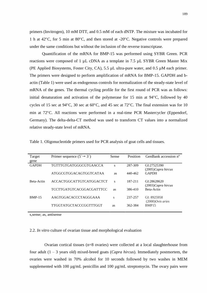
189
primers (Invitrogen), 10 mM DTT, and 0.5 mM of each dNTP. The mixture was incubated for
1 h at 42C, for 5 min at 80C, and then stored at -20C. Negative controls were prepared
under the same conditions but without the inclusion of the reverse transcriptase.
Quantification of the mRNA for BMP-15 was performed using SYBR Green. PCR
reactions were composed of 1 µL cDNA as a template in 7.5 µL SYBR Green Master Mix
(PE Applied Biosystems, Foster City, CA), 5.5 µL ultra-pure water, and 0.5 µM each primer.
The primers were designed to perform amplification of mRNA for BMP-15. GAPDH and b-
actin (Table 1) were used as endogenous controls for normalization of the steady-state level of
mRNA of the genes. The thermal cycling profile for the first round of PCR was as follows:
initial denaturation and activation of the polymerase for 15 min at 94C, followed by 40
cycles of 15 sec at 94C, 30 sec at 60C, and 45 sec at 72C. The final extension was for 10
min at 72C. All reactions were performed in a real-time PCR Mastercycler (Eppendorf,
Germany). The delta-delta-CT method was used to transform CT values into a normalized
relative steady-state level of mRNA.
Table 1. Oligonucleotide primers used for PCR analysis of goat cells and tissues.
Target
gene
Primer sequence (5´ 3´) Sense Position GenBank accession nº
GAPDH TGTTTGTGATGGGCGTGAACCA
s 287-309 GI:27525390
(2005)Capra hircus
GAPDH ATGGCGTGGACAGTGGTCATAA
as 440-462
Beta-Actin ACCACTGGCATTGTCATGGACTCT
s 187-211 GI:28628620
(2003)Capra hircus
Beta-Actin TCCTTGATGTCACGGACGATTTCC
as 386-410
BMP-15 AAGTGGACACCCTAGGGAAA
TTGGTATGCTACCCGGTTTGGT
s
as
237-257
362-384
GI: 8925958
(2000)Ovis aries
BMP15
s,sense; as, antisense
2.2. In vitro culture of ovarian tissue and morphological evaluation
Ovarian cortical tissues (n=8 ovaries) were collected at a local slaughterhouse from
four adult (1 – 3 years old) mixed-breed goats (Capra hircus). Immediately postmortem, the
ovaries were washed in 70% alcohol for 10 seconds followed by two washes in MEM
supplemented with 100 µg/mL penicillin and 100 µg/mL streptomycin. The ovary pairs were
190
transported within 1 hour to the laboratory in MEM at 4°C (Chaves et al., 2008). All
chemicals used in the present study were purchased from Sigma Chemical Co. (St. Louis,
MO, USA), unless otherwise indicated.
Our organ culture system was described in detail previously (Celestino et al., 2010).
Ovarian tissue samples from each ovarian pair were cut in 13 slices (3 mm x 3 mm x 1 mm)
using a needle and scalpel under sterile conditions. The tissue pieces were then either directly
fixed for histological and ultrastructural analysis (fresh control tissue) or placed in culture for
one or seven days. Caprine tissues were transferred to 24-well culture dishes containing 1 mL
of culture media. Culture was performed at 39°C in 5% CO2 in a humidified incubator, and
all of the media was incubated for 1 h prior to use. The basic culture medium (cultured
control tissue) consisted of MEM (pH 7.2 – 7.4) supplemented with ITS (10 µg/mL insulin,
5.5 µg/mL transferrin and 5.0 ng/mL selenium), 0.23 mM pyruvate, 2 mM glutamine, 2 mM
hypoxantine and 1.25 mg/mL bovine serum albumin (BSA), which combined was called
MEM+. For the experimental conditions, the medium was supplemented with recombinant
human BMP-15 (rhBMP-15, R&D Systems, Minneapolis, MN, USA) at different
concentrations (1, 10, 50, 100 or 200 ng/mL). Each treatment was repeated four times, and
the culture media was replenished every other day.
Before culture (fresh control tissue) and after one or seven days in culture, all of the
pieces were fixed in Carnoy‘s solution for 12 h and then dehydrated in increasing
concentrations of ethanol. After paraffin embedding (Synth, São Paulo, Brazil), the caprine
tissue pieces were cut into 7-m sections, and every section was mounted on glass slides and
stained by Periodic Acid Schiff - hematoxylin. Follicle stage and survival were assessed
microscopically on serial sections. Coded, anonymous slides were examined on a microscope
(Nikon, Japan) under 400X magnification.
The developmental stages of follicles have been defined previously (Silva et al., 2004)
as either primordial (one layer of flattened granulosa cells around the oocyte) or growing
follicles (intermediate: one layer of flattened or cuboidal granulosa cells; primary: one layer
of cuboidal granulosa cells; and secondary: two or more layers of cuboidal granulosa cells
around the oocyte). These follicles were still classified individually as histologically normal
when an intact oocyte was present and surrounded by granulosa cells, which are well
organized in one or more layers and have no pyknotic nucleus. Atretic follicles were defined
as those with a retracted oocyte, pyknotic nucleus, and/or disorganized granulosa cells
191
detached from the basement membrane. Overall, 120 follicles were evaluated for each
treatment (30 follicles per treatment for one repetition x four repetitions = 120 follicles).
To evaluate follicular activation, the percentages of healthy primordial and growing
follicles were calculated before (fresh control tissue) and after culture in each medium.
Follicle and oocyte diameters were only measured in healthy follicles with the aid of an
ocular micrometer. Both basement membrane diameters were measured at right angles to
each other in the largest cross-section of each growing oocyte and follicle were then
averaged. Care was taken to count each follicle only once as previously described (Celestino
et al., 2010). Each follicle was examined in every section it appeared and matched with the
same follicle on adjacent sections to avoid double counting, thus ensuring that each follicle
was only counted once regardless of its size.
2.3. Ultrastructural analysis of caprine preantral follicles
For a more in-depth evaluation of follicular morphology after histological analysis,
ultrastructural studies were performed on fragments of fresh control tissue and treatment
groups that maintained follicular survival and promoted growth after seven days of culture. A
portion with a maximum dimension of 1 mm3 was cut from each fragment of ovarian tissue
and fixed in Karnovsky solution (4% paraformaldehyde and 2.5% glutaraldehyde in 0.1 M
sodium cacodylate buffer at pH 7.2) for 3 h at RT. After three washes in sodium cacodylate
buffer, specimens were post-fixed in 1% osmium tetroxide, 0.8% potassium ferricyanide and
5 mM calcium chloride in 0.1 M sodium cacodylate buffer for 1 h at RT. The samples were
then dehydrated through a gradient of acetone solutions, and thereafter, they were embedded
in SPIN-PON resin (Sigma Company, St Louis, MO). Afterwards, semi-thin sections (3 µm)
were cut, stained with toluidine blue and analyzed by light microscopy at 400× magnification.
Ultra-thin sections (60–70 nm) were obtained from preantral follicles, which were classified
as morphologically normal in semi-thin sections according to the criteria adopted in histology.
Subsequently, ultra-thin sections were contrasted with uranyl acetate and lead citrate, and they
were examined under a Morgani-FEI transmission electron microscope (TEM) operating at 80
kV. The density and integrity of ooplasmic and granulosa cell organelles as well as
vacuolization and membrane integrity were evaluated.
192
2.4. Assessment of preantral follicle viability by fluorescence microscopy
Based on the results of the morphological and ultrastructural analyses, the viability of
follicles from control preparations and preparations cultured with the concentration of BMP-
15 that maintained follicular morphology and ultrastructural integrity was further analyzed
using fluorescent probes.
Additional pairs of goat ovaries (n = 2) were collected from a slaughterhouse, and cut
into fragments at the laboratory. One of these fragments was immediately processed for
follicle isolation (fresh control tissue) and the remaining fragments were cultured for seven
days with BMP-15 (100 ng/mL) as described above. After the culture period, fragments were
processed for mechanical isolation using the method described by Lucci et al. (1999).
Preantral follicles were analyzed using a two-color fluorescence cell viability assay based on
the simultaneous detection of live and dead cells with calcein-AM and ethidium homodimer-
1, respectively. While the first probe detected intracellular esterase activity in viable cells, the
latter labeled nucleic acids in non-viable cells with plasma membrane disruption. The test was
performed by adding 4 μM calcein-AM and 2 μM ethidium homodimer-1 (Molecular Probes,
Invitrogen, Eugene, OR, USA) to a suspension of isolated follicles and incubating them at
37°C for 15 min. After being labeled, follicles were washed once by centrifugation at 100× g
for 5 min and resuspended in MEM. Cells were then mounted on glass microscope slides in 5
μl anti-fading medium (DABCO, Sigma, Deisenhofen, Germany) to prevent photobleaching,
and they were examined using a fluorescence microscope (Nikon Eclipse 80i, Tokyo, Japan).
The emitted fluorescence signals of calcein-AM and ethidium homodimer-1 were collected at
488 and 568 nm, respectively. Oocytes and granulosa cells were considered viable if their
cytoplasm stained positively with calcein-AM (green) and their chromatin was not labeled
with ethidium homodimer-1 (red).
2.5. Statistical analysis
Data of mRNA expression in primordial, primary and secondary follicles were
analyzed by Kruskal-Wallis test, while t test was used for paired comparisons of mRNA
expression in small and large antral follicles (P<0.05). Percentages of morphologically
normal, primordial and growing follicles after one or seven days of culture were initially
submitted to Smirnov-Kolmogorov and Batlett tests to confirm a normal distribution and
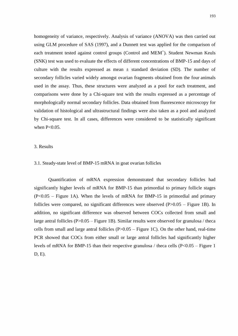
193
homogeneity of variance, respectively. Analysis of variance (ANOVA) was then carried out
using GLM procedure of SAS (1997), and a Dunnett test was applied for the comparison of
each treatment tested against control groups (Control and MEM+). Student Newman Keuls
(SNK) test was used to evaluate the effects of different concentrations of BMP-15 and days of
culture with the results expressed as mean ± standard deviation (SD). The number of
secondary follicles varied widely amongst ovarian fragments obtained from the four animals
used in the assay. Thus, these structures were analyzed as a pool for each treatment, and
comparisons were done by a Chi-square test with the results expressed as a percentage of
morphologically normal secondary follicles. Data obtained from fluorescence microscopy for
validation of histological and ultrastructural findings were also taken as a pool and analyzed
by Chi-square test. In all cases, differences were considered to be statistically significant
when P<0.05.
3. Results
3.1. Steady-state level of BMP-15 mRNA in goat ovarian follicles
Quantification of mRNA expression demonstrated that secondary follicles had
significantly higher levels of mRNA for BMP-15 than primordial to primary follicle stages
(P<0.05 – Figure 1A). When the levels of mRNA for BMP-15 in primordial and primary
follicles were compared, no significant differences were observed (P>0.05 – Figure 1B). In
addition, no significant difference was observed between COCs collected from small and
large antral follicles (P>0.05 – Figure 1B). Similar results were observed for granulosa / theca
cells from small and large antral follicles (P>0.05 – Figure 1C). On the other hand, real-time
PCR showed that COCs from either small or large antral follicles had significantly higher
levels of mRNA for BMP-15 than their respective granulosa / theca cells (P<0.05 – Figure 1
D, E).
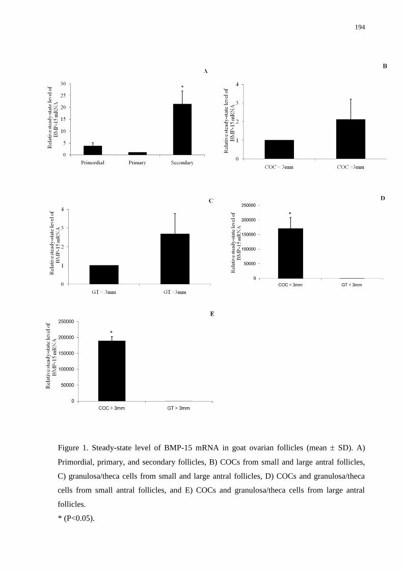
194
Figure 1. Steady-state level of BMP-15 mRNA in goat ovarian follicles (mean SD). A)
Primordial, primary, and secondary follicles, B) COCs from small and large antral follicles,
C) granulosa/theca cells from small and large antral follicles, D) COCs and granulosa/theca
cells from small antral follicles, and E) COCs and granulosa/theca cells from large antral
follicles.
* (P<0.05).
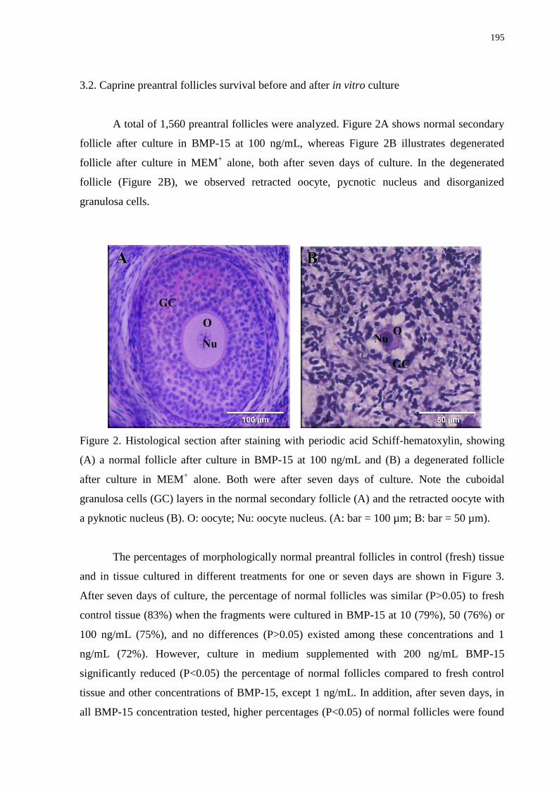
195
3.2. Caprine preantral follicles survival before and after in vitro culture
A total of 1,560 preantral follicles were analyzed. Figure 2A shows normal secondary
follicle after culture in BMP-15 at 100 ng/mL, whereas Figure 2B illustrates degenerated
follicle after culture in MEM+ alone, both after seven days of culture. In the degenerated
follicle (Figure 2B), we observed retracted oocyte, pycnotic nucleus and disorganized
granulosa cells.
Figure 2. Histological section after staining with periodic acid Schiff-hematoxylin, showing
(A) a normal follicle after culture in BMP-15 at 100 ng/mL and (B) a degenerated follicle
after culture in MEM+ alone. Both were after seven days of culture. Note the cuboidal
granulosa cells (GC) layers in the normal secondary follicle (A) and the retracted oocyte with
a pyknotic nucleus (B). O: oocyte; Nu: oocyte nucleus. (A: bar = 100 µm; B: bar = 50 µm).
The percentages of morphologically normal preantral follicles in control (fresh) tissue
and in tissue cultured in different treatments for one or seven days are shown in Figure 3.
After seven days of culture, the percentage of normal follicles was similar (P>0.05) to fresh
control tissue (83%) when the fragments were cultured in BMP-15 at 10 (79%), 50 (76%) or
100 ng/mL (75%), and no differences (P>0.05) existed among these concentrations and 1
ng/mL (72%). However, culture in medium supplemented with 200 ng/mL BMP-15
significantly reduced (P<0.05) the percentage of normal follicles compared to fresh control
tissue and other concentrations of BMP-15, except 1 ng/mL. In addition, after seven days, in
all BMP-15 concentration tested, higher percentages (P<0.05) of normal follicles were found
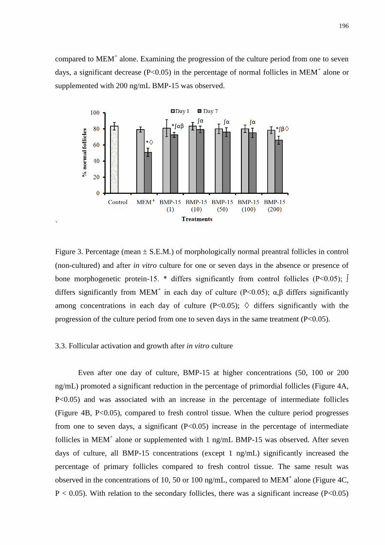
196
compared to MEM+ alone. Examining the progression of the culture period from one to seven
days, a significant decrease (P<0.05) in the percentage of normal follicles in MEM+ alone or
supplemented with 200 ng/mL BMP-15 was observed.
`
Figure 3. Percentage (mean S.E.M.) of morphologically normal preantral follicles in control
(non-cultured) and after in vitro culture for one or seven days in the absence or presence of
bone morphogenetic protein-15. * differs significantly from control follicles (P<0.05); ∫
differs significantly from MEM+ in each day of culture (P<0.05); α,β differs significantly
among concentrations in each day of culture (P<0.05); differs significantly with the
progression of the culture period from one to seven days in the same treatment (P<0.05).
3.3. Follicular activation and growth after in vitro culture
Even after one day of culture, BMP-15 at higher concentrations (50, 100 or 200
ng/mL) promoted a significant reduction in the percentage of primordial follicles (Figure 4A,
P<0.05) and was associated with an increase in the percentage of intermediate follicles
(Figure 4B, P<0.05), compared to fresh control tissue. When the culture period progresses
from one to seven days, a significant (P<0.05) increase in the percentage of intermediate
follicles in MEM+ alone or supplemented with 1 ng/mL BMP-15 was observed. After seven
days of culture, all BMP-15 concentrations (except 1 ng/mL) significantly increased the
percentage of primary follicles compared to fresh control tissue. The same result was
observed in the concentrations of 10, 50 or 100 ng/mL, compared to MEM+ alone (Figure 4C,
P < 0.05). With relation to the secondary follicles, there was a significant increase (P<0.05)
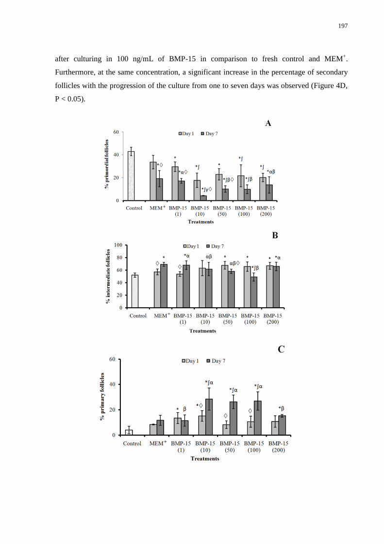
197
after culturing in 100 ng/mL of BMP-15 in comparison to fresh control and MEM+.
Furthermore, at the same concentration, a significant increase in the percentage of secondary
follicles with the progression of the culture from one to seven days was observed (Figure 4D,
P < 0.05).
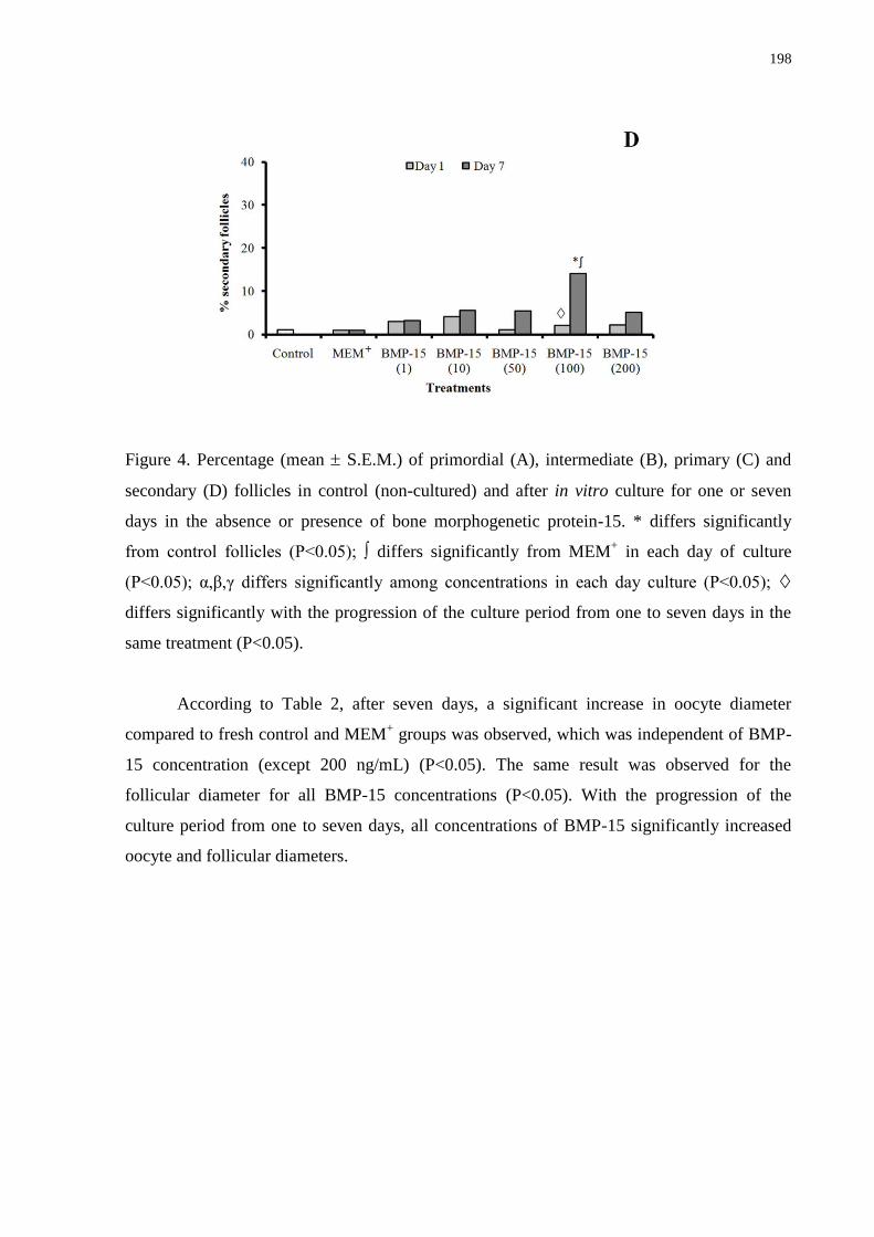
198
Figure 4. Percentage (mean S.E.M.) of primordial (A), intermediate (B), primary (C) and
secondary (D) follicles in control (non-cultured) and after in vitro culture for one or seven
days in the absence or presence of bone morphogenetic protein-15. * differs significantly
from control follicles (P<0.05); ∫ differs significantly from MEM+ in each day of culture
(P<0.05); α,β,γ differs significantly among concentrations in each day culture (P<0.05);
differs significantly with the progression of the culture period from one to seven days in the
same treatment (P<0.05).
According to Table 2, after seven days, a significant increase in oocyte diameter
compared to fresh control and MEM+ groups was observed, which was independent of BMP-
15 concentration (except 200 ng/mL) (P<0.05). The same result was observed for the
follicular diameter for all BMP-15 concentrations (P<0.05). With the progression of the
culture period from one to seven days, all concentrations of BMP-15 significantly increased
oocyte and follicular diameters.
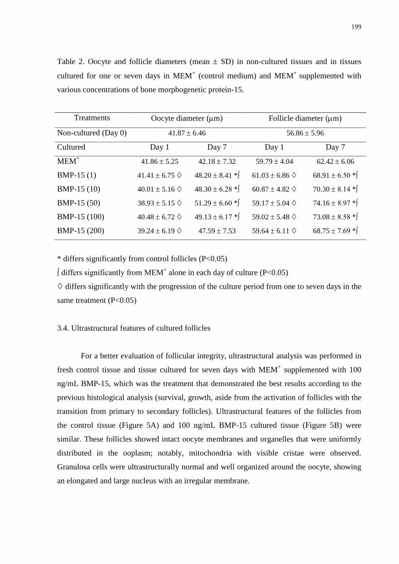
199
Table 2. Oocyte and follicle diameters (mean SD) in non-cultured tissues and in tissues
cultured for one or seven days in MEM+ (control medium) and MEM
+ supplemented with
various concentrations of bone morphogenetic protein-15.
Treatments Oocyte diameter (m) Follicle diameter (m)
Non-cultured (Day 0) 41.87 6.46 56.86 5.96
Cultured Day 1 Day 7 Day 1 Day 7
MEM+ 41.86 5.25 42.18 7.32 59.79 4.04 62.42 6.06
BMP-15 (1) 41.41 6.75 48.20 8.41 *∫ 61.03 6.86 68.91 6.50 *∫
BMP-15 (10) 40.01 5.16 48.30 6.28 *∫ 60.87 4.82 70.30 8.14 *∫
BMP-15 (50) 38.93 5.15 51.29 6.60 *∫ 59.17 5.04 74.16 8.97 *∫
BMP-15 (100) 40.48 6.72 49.13 6.17 *∫ 59.02 5.48 73.08 8.58 *∫
BMP-15 (200) 39.24 6.19 47.59 7.53 59.64 6.11 68.75 7.69 *∫
* differs significantly from control follicles (P<0.05)
∫ differs significantly from MEM+ alone in each day of culture (P<0.05)
differs significantly with the progression of the culture period from one to seven days in the
same treatment (P<0.05)
3.4. Ultrastructural features of cultured follicles
For a better evaluation of follicular integrity, ultrastructural analysis was performed in
fresh control tissue and tissue cultured for seven days with MEM+ supplemented with 100
ng/mL BMP-15, which was the treatment that demonstrated the best results according to the
previous histological analysis (survival, growth, aside from the activation of follicles with the
transition from primary to secondary follicles). Ultrastructural features of the follicles from
the control tissue (Figure 5A) and 100 ng/mL BMP-15 cultured tissue (Figure 5B) were
similar. These follicles showed intact oocyte membranes and organelles that were uniformly
distributed in the ooplasm; notably, mitochondria with visible cristae were observed.
Granulosa cells were ultrastructurally normal and well organized around the oocyte, showing
an elongated and large nucleus with an irregular membrane.
200
Figure 5. Ultrastructural analysis of noncultured preantral follicle (A) and follicle cultured for
seven days in medium containing 100 ng/mL bone morphogenetic protein-15 (B). O: oocyte;
GC: granulosa cells; m: mitochondria; arrow-oocyte membrane. (A: bar = 10 µm; B: bar = 2
µm). Three to five follicles per group were examined and the photomicrographs are
representative examples.
3.5. Viability assessment of follicles cultured with BMP-15
The results from the morphological and ultrastructural evaluation demonstrated that
culturing follicles with 100 ng/mL BMP-15 maintained the percentages of normal follicles,
which was similar to levels seen in fresh control tissue, and it preserved the ultrastructure of
the sample. Thus, a viability trial using this treatment was performed. In the present study, 30
caprine preantral follicles were analyzed after seven days of culture with MEM+
supplemented with 100 ng/mL BMP-15. After this quantitative analysis, 90.0% of follicles
remained viable, assessed by calcein-AM–ethidium homodimer assays (Figure 6), and this
percentage of viable follicles was similar to the percentage observed in the fresh control group
(96.7%).
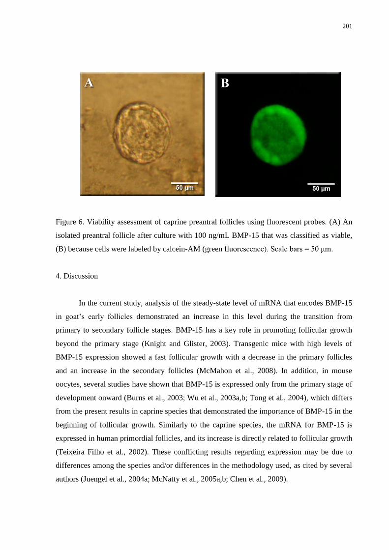
201
Figure 6. Viability assessment of caprine preantral follicles using fluorescent probes. (A) An
isolated preantral follicle after culture with 100 ng/mL BMP-15 that was classified as viable,
(B) because cells were labeled by calcein-AM (green fluorescence). Scale bars = 50 μm.
4. Discussion
In the current study, analysis of the steady-state level of mRNA that encodes BMP-15
in goat‘s early follicles demonstrated an increase in this level during the transition from
primary to secondary follicle stages. BMP-15 has a key role in promoting follicular growth
beyond the primary stage (Knight and Glister, 2003). Transgenic mice with high levels of
BMP-15 expression showed a fast follicular growth with a decrease in the primary follicles
and an increase in the secondary follicles (McMahon et al., 2008). In addition, in mouse
oocytes, several studies have shown that BMP-15 is expressed only from the primary stage of
development onward (Burns et al., 2003; Wu et al., 2003a,b; Tong et al., 2004), which differs
from the present results in caprine species that demonstrated the importance of BMP-15 in the
beginning of follicular growth. Similarly to the caprine species, the mRNA for BMP-15 is
expressed in human primordial follicles, and its increase is directly related to follicular growth
(Teixeira Filho et al., 2002). These conflicting results regarding expression may be due to
differences among the species and/or differences in the methodology used, as cited by several
authors (Juengel et al., 2004a; McNatty et al., 2005a,b; Chen et al., 2009).
202
In the current study, COCs from either small or large antral follicles had higher levels
of mRNA for BMP-15 than their respective granulosa / theca cells. These data demonstrated
that although BMP-15 was found in the follicular wall, the COC was mainly responsible for
its production. BMP-15 has been identified as an important factor in the later stages of
folliculogenesis and is related to both the inhibition of premature luteinization and cumulus
cells expansion (Gilchrist et al., 2004; Shimasaki et al., 2004b, Su et al., 2008). In mice,
mutations in the BMP-15 gene are harmful to the normal development of the COC (Yan et al.,
2001). In this same species, when the specific gene for BMP-15 was eliminated, some defects
in the process of ovulation and in the oocyte quality were observed, which resulted in a
smaller size of these oocytes (Yan et al., 2001). In swine COC obtained from antral follicles
(3-7 mm), the mRNA and protein for BMP-15 were expressed in low levels in immature
oocytes (Li et al., 2008; Zhu et al., 2008) and increased their levels within 18 h of in vitro
maturation, which was coincident with cumulus cells expansion (Li et al., 2008).
This study is the first to report the positive effect of BMP-15 on the in vitro
development of caprine preantral follicles. After seven days of culture, the follicular survival
was similar to the fresh control tissue with the addition of 10, 50 or 100 ng/mL BMP-15 to the
culture medium. In addition, all BMP-15 concentrations maintained a higher percentage of
normal follicles than MEM+ alone. Similar to our results, studies using BMP-15 have verified
its important role in preventing atresia in follicular somatic cells (Hussein et al., 2005;
Yoshino et al., 2006). Previous studies have shown that after neutralization of the gene for
BMP-15, a decrease in the integrity of ovine oocytes (Juengel et al., 2004b) and mouse COC
(Varani et al., 2002) was observed. BMP-15 can promote follicular survival through the
maintenance of granulosa cell proliferation and prevention of premature luteinization and/or
atresia (Knight and Glister, 2006). Furthermore, BMP-15 is known to stimulate KL
expression, which is an important factor for the in vitro survival of murine (Jin et al., 2005)
and caprine (Celestino et al., 2010) preantral follicles. However, as suggested by Mery et al.
(2007), the expression of both BMP-15 and its receptors may be reduced in in vitro cultured
ovaries compared with those in vivo. Thus, larger concentrations of BMP-15 may be needed
to be administered exogenously. This fact may explain the unsatisfying results obtained in our
study with the concentration of 1 ng/ml BMP-15 regarding the maintenance of follicular
survival. In contrast, the concentration of 200 ng/ml may have induced the expression of
inhibitory Smads (I-Smads), which can act as part of an autocrine negative feedback
(Miyazono, 2000), and attenuate the pattern of transduction of BMP-15 signals (Miyazono et
203
al., 1997). A recent study demonstrated that transgenic adult mice with increased BMP-15
expression showed a higher amount of atretic antral follicles compared to the non-transgenic
control mice (McMahon et al., 2008). Another explanation for our findings is that the high
concentration of BMP-15, such as 200 ng/mL, may change the expression of the follistatin
growth factor, which is expressed in granulosa cells of small growing follicles in most
species, including caprine (Silva et al., 2006). The follistatin may bind to BMP-15 and inhibit
its bioactivity (Otsuka et al., 2001).
All BMP-15 concentrations (except for 1 ng/mL) increased the percentage of primary
follicles after seven days of culture compared to the fresh control tissue and at 10, 50 or 100
ng/mL when compared to MEM+ alone. The presence of the BMP-15 protein in some oocytes
of primordial follicles (Otsuka et al., 2000) suggest that it has a role in the follicular
activation, i.e., the transition from the primordial to primary follicle stage (Shimasaki et al.,
2004a,b; Moore and Shimasaki, 2005). In this case, the BMP-15 would act through the
BMPR-II receptor (Shimasaki et al., 2004a), found on the surface of granulosa cells, to
directly stimulate their mitosis (Vitt et al., 2000b; Di Pasquale et al., 2004; McNatty et al.,
2005a,b). In rodents, BMP-15 stimulated the in vitro development of primordial and primary
follicles (Otsuka et al., 2000; Fortune, 2003). In caprine species, the protein and mRNA for
BMP-15 ligands and receptors (BMPR-IA, BMPR-IB and BMPR-II) were expressed even
from the primordial follicle stage (Silva et al., 2005), thus providing evidence for the role of
BMP-15 in follicular activation observed in the current study.
The elevated steady-state level of BMP-15 mRNA in secondary follicles found in this
study agreed with previous results (rat: Otsuka et al., 2000, human: Teixeira Filho et al.,
2002). Thus, BMP-15, at 100 ng/mL, increased the percentage of secondary follicles. Genes
that encode for BMP-15 are essential for the early stages of follicular growth, especially for
the transition from primary to secondary follicles (Mery et al., 2007). In ovine, after 7.5
months of immunization against BMP-15, the ovaries did not show normal development
beyond the primary stage (Juengel et al., 2004b). An in vitro study showed that the addition of
100 ng/mL BMP-15 to a culture of granulosa cells increased the expression of phosphorylated
Smads 1/5/8, i.e., active Smads. Furthermore, the treatment of rat granulosa cells with 30 or
200 ng/mL human rBMP-15 stimulated cell proliferation and DNA synthesis (Otsuka et al.,
2000).
In relation to growth, an increase in the follicular diameter was observed after seven
days of culture for all BMP-15 concentrations compared to the fresh control group or MEM+
204
alone. The same result was observed for the oocyte diameter, except when 200 ng/mL BMP-
15 was added to the culture medium. BMP-15 is essential for ovarian follicular growth
(Juengel et al., 2004a) because granulosa cells are the target cells for the BMP-15 ligand
(Otsuka et al., 2000). Some authors have described a high expression of the mRNA and/or
BMP-15 protein in oocytes of growing or completely grown follicles (Shimasaki et al.,
2004b; Juengel and McNatty, 2005; Li et al., 2008), which can suggest that BMP-15 plays a
role in the increase of follicle diameter. Other studies reported that a lack in the production of
BMP-15 receptors, such as BMPR-II and ALK-6 (BMPR-IB), may lead to a break in the
growth (Mery et al., 2007). In mice, an increase in the levels of BMP-15 mRNA expression
from day one to seven was associated with the presence of early follicles that contained
growing oocytes (Sadeu et al., 2008).
Our results regarding to the percentage of normal follicular morphology were
confirmed by ultrastructural analysis. TEM is considered a powerful technique for the
evaluation of cellular organelles and ultrastructural changes (Salehnia et al., 2002) that occur
during follicular atresia and is an important tool for analyzing preantral follicles after in vitro
culture (Matos et al., 2007). Using TEM, important cellular structures, such as mitochondria,
the endoplasmic reticulum, granulosa cells, and the basement and nuclear membranes, were
preserved after seven days of culture in the presence of 100 ng/mL BMP-15. Normal follicles
had ultrastructure similar to those described in caprine (Lucci et al., 2001; Celestino et al.,
2009, 2010), bovine (Cran et al., 1980) and ovine (van Wezel and Rodgers, 1996) studies. In
addition to the ultrastructural analysis, preantral follicles cultured for seven days in a medium
containing 100 ng/mL BMP-15 were further analyzed using a more accurate method based on
fluorescent probes; this method confirmed the previous results obtained with a light
microscope and TEM regarding to follicular survival. Thus, this viability assessment appears
to be a reliable, practical and fast method to analyze follicular viability (Cortvrindt and Smitz,
2001). Recently, this method has also been used successfully to evaluate preantral follicle
viability in goats (Bruno et al., 2009; Silva et al., 2010).
In conclusion, the BMP-15 mRNAs were detected for all investigated follicular
categories and cellular types. In addition, this study demonstrated that 100 ng/mL BMP-15
not only maintained the morphological integrity of caprine preantral follicles cultured for
seven days but also promoted growth and transition to the secondary follicle stage. BMP-15
seems to be an essential factor for female fertility. However, new studies are still necessary
for a better understanding of the ovarian regulatory mechanisms. These mechanisms could
205
ease the monitoring and manipulation of the ovarian function, further improving the fertility
of domestic animals, endangered species and/or humans.
Acknowledgments
This work was supported by the National Council for Scientific and Technological
Development (CNPq), and the Coordination for the Improvement of Higher Education
Personnel (CAPES), and the Brazilian Innovation Agency (FINEP) and the Ceara State
Foundation for the Support of Scientific and Technological Development (FUNCAP). Juliana
Jales de Hollanda Celestino is a recipient of a grant from FUNCAP. The authors thank José
Leandro da Silva Neto for his technical support in classical histology and all the members of
the laboratory.
References
Aaltonen, J., Laitinen, M.P., Vuojolainen, K., Jaatinen, R., Horelli-Kuitunen, N., Seppa, L.,
Louhio, H., Tuuri, T., Sjoberg, J., Butzow, R., Hovata, O., Dale, L., Ritvos, O., 1999.
Human growth differentiation factor 9 (GDF-9) and its novel homolog GDF-9B are
expressed in oocytes during early folliculogenesis. J. Clin. Endocrinol. Metab. 84, 2744-
2750.
Bodensteiner, K.J., Clay, C.M., Moeller, C.L., Sawyer, H.R., 1999. Molecular cloning of the
ovine growth/differentiation factor-9 gene and expression of growth/differentiation factor-
9 in ovine and bovine ovaries. Biol. Reprod. 60, 381-386.
Bruno, J.B., Celestino, J.J.H., Lima-Verde, I.B., Lima, L.F., Matos, M.H.T., Araújo, V.R.,
Saraiva, M.V.A., Martins, F.S., Name, K.P.O., Campello, C.C., Báo, S.N., Silva, J.R.,
Figueiredo, J.R., 2009. Expression of vascular endothelial growth factor (VEGF) receptor
in goat ovaries and improvement of in vitro caprine preantral follicle survival and growth
with VEGF. Reprod. Fertil. Dev. 21, 679-687.
Burns, K.H., Viveiros, M.M., Ren, Y., Wang, P., DeMayo, F.J., Frail, D.E., Eppig, J.J.,
Matzuk, M.M., 2003. Roles of NPM2 in chromatin and nucleolar organization in oocytes
and embryos. Science 300, 633-636.
Celestino, J.J.H., Bruno, J.B., Lima-Verde, I.B., Matos, M.H.T., Saraiva, M.V.A., Chaves,
R.N., Martins, F.S., Lima, L.F., Name, K.P.O., Campello, C.C., Silva, J.R.V., Báo, S.N.,
206
Figueiredo, J.R., 2009. Recombinant epidermal growth factor maintains follicular
ultrastructure and promotes the transition to primary follicles in caprine ovarian tissue
cultured in vitro. Reprod. Sci. 16, 239-246.
Celestino, J.J.H., Bruno, J.B., Lima-Verde, I.B., Matos, M.H.T., Saraiva, M.V.A., Chaves,
R.N., Martins, F.S., Almeida, A.P., Cunha, R.M.S., Lima, L.F., Name K.P.O., Campello,
C.C., Silva, J.R.V., Báo, S.N., Figueiredo, J.R., 2010. Steady-state level of Kit Ligand
mRNA in goat ovaries and the role of Kit Ligand in preantral follicle survival and growth
in vitro. Mol. Reprod. Dev. 77, 231-240.
Chang, H., Brown, C.W., Matzuk, M.M., 2002. Genetic analysis of the mammalian
transforming growth factor-beta superfamily. Endocr. Rev. 23, 787-823.
Chaves, R.N., Martins, F.S., Saraiva, M.V.A., Celestino, J.J.H., Lopes, C.A.P., Correia, J.C.,
Lima-Verde, I.B., Matos, M.H.T., Báo, S.N., Name, K.P.O., Campello, C.C., Silva, J.R.V.,
Figueiredo, J.R., 2008. Chilling ovarian fragments during transportation improves viability
and growth of goat preantral follicles cultured in vitro. Reprod. Fertil. Dev. 20, 640-647.
Chen, A.Q., Yu, S.D., Wang, Z.G., Xu, Z.R., Yang, Z.G., 2009. Stage-specific expression of
bone morphogenetic protein type I and type II receptor genes:Effects of follicle-stimulating
hormone on ovine antral follicles. Anim. Reprod. Sci. 111, 391-399.
Cortvrindt, R.G., Smitz, J.E.J., 2001. Fluorescent probes allow rapid and precise recording of
follicle density and staging in human ovarian cortical biopsy samples. Fertil. Steril. 75,
599-593.
Cran, D.G., Moor, R.M., Hay, M.F., 1980. Fine structure of the sheep oocyte during antral
follicle development. J. Reprod. Fertil. 59, 125-132.
Di Pasquale, E., Beck-Peccoz, P., Persani, L., 2004. Hypergonadotropic ovarian failure
associated with na inherited mutation of human bone morphogenetic protein-15 (BMP15)
gene. Am. J. Hum. Genet. 75, 106-111.
Dong, J., Albertini, D.F., Nishimori, K., Kumar, T.R., Lu, N., Matzuk, M., 1996. Growth
differentiation factor-9 is required during early ovarian folliculogenesis. Nature 383, 531-
535.
Eckery, D.C., Whale, L.J., Lawrence, S.B., Wylde, K.A., McNatty, K.P., Juengel, J.L., 2002.
Expression of mRNA encoding growth differentiation factor 9 and bone morphogenetic
protein 15 during follicular formation and growth in a marsupial, the brushtail possum
(Trichosurus vulpecula). Mol. Cell. Endocrinol. 192, 115-126.
207
Fortune, J.E., 2003. The early stages of follicular development: activation of primordial
follicles and growth of preantral follicles. Anim. Reprod. Sci. 78, 135-163.
Galloway, S.M., McNatty, K.P., Cambridge, L.M., Laitinen, M.P., Juengel, J.L., Jokiranta,
T.S., McLaren, R.J., Luiro, K., Dodds, K.G., Montgomery, G.W., Beattie, A.E., Davis,
G.H., Ritvos, O., 2000. Mutations in an oocyte-derived growth factor gene (BMP15) cause
increased ovulation rate and infertility in a dosage-sensitive manner. Nat. Genet. 25, 279-
283.
Gilchrist, R.B., Ritter, L.J., Armstrong, D.T., 2004. Oocyte-somatic cell interactions during
follicle development in mammals. Anim. Reprod. Sci. 82-83, 431-446.
Hussein, T.S., Froiland, D.A., Amato, F., Thompson, J.G., Gilchrist, R.B., 2005. Oocytes
prevent cumulus cell apoptosis by maintaining a morphogenic paracrine gradient of bone
morphogenetic proteins. J. Cell. Sci. 118, 5257-5268.
Jaatinen, R., Laitinen, M.P., Vuojolainen, K., Aaltonen, J., Louhio, H., Heikinheimo, K.,
Lehtonen, E., Ritvos, O., 1999. Localization of growth differentiation factor-9 (GDF-9)
mRNA and protein in rat ovaries and cDNA cloning of rat GDF-9 and its novel homolog
GDF-9B. Mol. Cell. Endocrinol. 156, 189-193.
Jin, X., Han, C.S., Yu, F.Q., Wei, P., Hu, Z.Y., Liu, Y.X., 2005. Anti-apoptotic action of stem
cell factor on oocytes in primordial follicles and its signal transduction. Mol. Reprod. Dev.
70, 82-90.
Juengel, J.L., Hudson, N.L., Heath, D.A., Smith, P., Reader, K.L., Lawrence, S.B.,
O‘Connell, A.R., Laitinen, M.P., Cranfield, M., Groome, N.P., Ritvos, O., McNatty, K.P.,
2002 Growth differentiation factor-9 and bone morphogenetic protein 15 are essential for
ovarian follicular development in sheep. Biol. Reprod. 67, 1777-1789.
Juengel, J.L., Bodensteiner, K.J., Heath, D.A., Hudsona, N.L., Moeller, C.L., Smith, P.,
Galloway, S.M., Davis, G.H., Sawyer, H.R., McNatty, K.P., 2004a. Physiology of GDF9
and BMP15 signalling molecules. Anim. Reprod. Sci. 82-83, 447-460.
Juengel, J.L., Hudson, N.L., Whitinig, L., McNatty, K.P., 2004b. Effects of immunization
against bone morphogenetic protein 15 and growth differentiation factor 9 on ovulation
rate, fertilization, and pregnancy in ewes. Biol. Reprod. 70, 557-561.
Juengel, J.L., McNatty, K.P., 2005. The role of proteins of the transforming growth factor-b
superfamily in the intraovarian regulation of follicular development. Hum. Reprod. Update
11, 144-161.
Knight, P.G., Glister, C., 2003. Local roles of TGF-β superfamily members in the
208
control of ovarian follicle development. Anim. Reprod. Sci. 78, 165-183.
Knight, P.G., Glister, C., 2006. TGF-β superfamily members and ovarian follicle
development. Reproduction 132, 191-206.
Laitinen, M., Vuojolainen, K., Jaatinen, R., Ketola, I., Aaltonen, J., Lehtonen, E.,
Heikinheimo, M., Ritvos, O., 1998. A novel growth differentiation factor- 9 (GDF-9)
related factor is co-expressed with GDF-9 in mouse oocytes during folliculogenesis. Mech.
Dev. 78, 135-140.
Lebrun, J.J., Chen,Y., Vale, W.W., 1997. Receptor serine kinases and signaling by activins
and inhibins, in: Aono, T., Sugino, H., Vale, W. (Eds.), Inhibin, activin and follistatin
regulatory functions in system and cell biology. Springer Serono Symposia, USA, pp. 1-
20.
Li, H.K., Kuo, T.Y., Yang, H.S., Chen, L.R., Li, S.S., Huang, H.W., 2008. Differential gene
expression of bone morphogenetic protein 15 and growth differentiation factor 9 during in
vitro maturation of porcine oocytes and early embryos. Anim. Reprod. Sci. 103, 312-322.
Lucci, C.M., Amorim, C.A., Báo, S.N., Figueiredo, J.R., Rodrigues, A.P.R., Silva, J.R.V.,
Goncalves, P.B.D., 1999. Effect of the interval of serial sections of ovarian in the tissue
chopper on the number of isolated caprine preantral follicles. Anim. Reprod. Sci. 56, 39-
49.
Lucci, C.M., Silva, J.R.V., Carvalho, F.C.A., Figueiredo, J.R., Báo, S.N., 2001. Light
microscopical and ultrastrutural characterization of goat preantral follicles. Small Rumin.
Res. 41, 61-69.
Matos, M.H.T., Silva, J.R.V., Rodrigues, A.P.R., Figueiredo, J.R., 2007. Técnicas para
avaliação da qualidade de folículos ovarianos pré-antrais cultivados in vitro. Rev. Bras.
Reprod. Anim. 31, 433-442.
Mazerbourg, S., Hsueh, A.J.W., 2006. Genomic analyses facilitate identification of receptors
and signaling pathways for growth differentiation factor 9 and related orphan bone
morphogenetic protein/growth differentiation factor ligands. Hum. Reprod. Update 12,
373-383.
McMahon, H.E., Hashimoto, O., Mellon, P.L., Shimasaki, S., 2008. Oocyte-specific
overexpression of mouse bone morphogenetic protein-15 leads to accelerated
folliculogenesis and an early onset of acyclicity in transgenic mice. Endocrinology 149,
2807-2815.
209
McNatty, K.P., Juengel, J.L., Reader, K.L., Lun, S., Myllymaa, S., Lawrence, S.B., Western,
A., Meerasahib, M.F., Mottershead, D.G., Groome, N.P., Ritvos, O., Laitinen, M.P.E.,
2005a. Bone morphogenetic protein 15 and growth differentiation factor 9 co-operate to
regulate granulosa cell function in ruminants. Reproduction 129, 481-487.
McNatty, K.P., Juengel, J.L., Reader, K.L., Lun, S., Myllymaa, S., Lawrence, S.B., Western,
A., Meerasahib, M.F., Mottershead, D.G., Groome, N.P., Ritvos, O., Laitinen, M.P.E.,
2005b. Bone morphogenetic protein 15 and growth differentiation factor 9 co-operate to
regulate granulosa cell function. Reproduction 129, 473-480.
Mery, L., Lefevre, A., Benchaib, M., Demirci, B., Salle, B., Guerin, J-F., Lornage, J., 2007.
Follicular growth in vitro: Detection of growth differentiation factor 9 (GDF9) and Bone
morphogenetic protein 15 (BMP15) during in vitro culture of ovine cortical slices. Mol.
Reprod. Dev. 74, 767-774.
Miyazono, K., Tijke, P.T., Souchelnytskyi, S., Nakao, A., Imamura, T., Hanai, J.-I.,
Kawabata, M., Heldin, C-H., 1997. Transforming growth factor-β receptors and signal
transduction, in: Aono, T., Sugino, H., Vale, W. (Eds.), Inhibin, activin and follistatin
regulatory functions in system and cell biology. Springer Serono Symposia, USA, pp. 277-
286.
Miyazono, K., 2000. TGF-β signaling by Smad proteins. Cytokine Growth F. R. 11, 15-22.
Moore, R.K., Otsuka, F., Shimasaki, S., 2003. Molecular Basis of Bone Morphogenetic
Protein-15 Signaling in Granulosa Cells. J. Biol. Chem. 278, 304-310.
Moore, R.K., Shimasaki, S., 2005. Molecular biology and physiological role of the oocyte
factor, BMP-15. Mol. Cell. Endocrinol. 234, 67-73.
Otsuka, F., Yao, Z., Lee, T., Yamamoto, S., Erickson, G.F., Shimasaki, S., 2000. Bone
morphogenetic protein-15. Identification of target cells and biological functions. J. Biol.
Chem. 275, 39523-39528.
Otsuka, F., Yamamoto, S., Erickson, G.F., Shimasaki, S., 2001. Bone morphogenetic protein-
15 inhibits follicle-stimulating hormone (FSH) action by suppressing FSH receptor
expression. J. Biol. Chem. 276, 11387-11392.
Otsuka, F., Shimasaki, S., 2002a. A novel function of bone morphogenetic protein-15 in the
pituitary: selective synthesis and secretion of FSH by gonadotropes. Endocrinology 143,
4938-4941.
210
Otsuka, F., Shimasaki, S., 2002b. A negative feedback system between oocyte bone
morphogenetic protein 15 and granulosa cell kit ligand: its role in regulating granulosa cell
mitosis. Proc. Natl. Acad. Sci. USA 99, 8060-8065.
Peng, C., Clelland, E., Tan, Q., 2009. Potencial role pf bone morphogenetic protein-15 in
zebrafish follicle development and oocyte maturation. Comp. Biochem. Phys. A 153, 83-
87.
Sadeu, J.C., Adriaenssens, T., Smitz, J., 2008. Expression of growth differentiation factor 9,
bone morphogenetic protein 15, and anti-Mullerian hormone in cultured mouse primary
follicles. Reproduction 136, 195-203.
Salehnia, M., Moghadam, E.A., Velojerdi, M.R., 2002. Ultrastructure of follicles after
vitrification of mouse ovarian tissue. Fertil. Steril. 78, 644-645.
Shimasaki, S., Moore, R.K., Otsuka, F., Erickson, G.F., 2004a. The bone morphogenetic
protein system in mammalian reproduction. Endocrinol. Rev. 25, 72-101.
Shimasaki, S., Moore, R.K., Erickson, G.F., Otsuka, F., 2004b. Ovarian bone morphogenetic
proteins in female reproduction. Int. Congress Series 1266, 241-247.
Silva, J.R.V., van den Hurk, R., Matos, M.H.T., Santos, R.R., Pessoa, C., Moraes, M.O.,
Figueiredo, J.R., 2004. Influences of FSH and EGF on primordial follicles during in vitro
culture of caprine ovarian cortical tissue. Theriogenology 61, 1691-1704.
Silva, J.R., van den Hurk, R., van Tol, H.T., Roelen, B.A., Figueiredo, J.R., 2005. Expression
of growth differentiation factor 9 (GDF9), bone morphogenetic protein 15 (BMP-15), and
BMP receptors in the ovaries of goats. Mol. Reprod. Dev. 70, 11-19.
Silva, J.R.V., Tharasanit, T., Taverne, M.A.M., van der Weijden, G.C., Santos, R.R.,
Figueiredo, J.R., van den Hurk, R., 2006. The activin-follistatin system and in vitro early
follicle development in goats. J. Endocrinol. 189, 113-125.
Silva, C.M.G., Matos, M.H.T., Rodrigues, G.Q., Faustino, L.R., Pinto, L.C., Chaves, R.N.,
Araújo, V.R., Campello, C.C., Figueiredo, J.R., 2010. In vitro survival and development of
goat preantral follicles in two different oxygen tensions. Anim. Reprod. Sci. 117, 83-89.
Steel, R.G.D., Torrie, J.H., Dickey, D., 1997. Principles and Procedures of Statistics: A
Biometrical Approach, third ed. McGraw-Hill, New York.
Su, Y.Q., Sugiura, K., Wigglesworth, K., O‘Brien, M.J., Affourtit, J.P., Pangas, S.A., Matzuk,
M.M., Eppig, J.J., 2008. Oocyte regulation of metabolic cooperativity between mouse
cumulus cells and oocytes: BMP15 and GDF9 control cholesterol biosynthesis in cumulus
cells. Development 135, 111-121.
211
Teixeira Filho, F.L., Baracat, E.C., Lee, T.H., Suh, C.S., Matsui, M., Chang, R.J., Shimasaki,
S., Erickson, G.F., 2002. Aberrant expression of growth differentiation factor-9 in oocytes
of women with polycystic ovary syndrome. J. Clin. Endocrinol. Metab. 87, 1337-1344.
Tong, Z.B., Gold, L., De Pol, A., Vanevski, K., Dorward, H., Sena, P., Palumbo, C., Bondy,
C.A., Nelson, L.M., 2004. Developmental expression and subcellular localization of mouse
MATER, an oocyte-specific protein essential for early development. Endocrinology 145,
1427-1434.
van Tol, H.T., Bevers, M.M., 1998. Theca cells and theca-cell conditioned medium inhibit the
progression of FSH-induced meiosis of bovine oocytes surrounded by cumulus cells
connected to membrane granulosa. Mol. Reprod. Dev. 51, 315-321.
van Wezel, I.L., Rodgers, R.J., 1996. Morphological characterization of bovine primordial
follicles and their environment in vivo. Biol. Reprod. 55, 1003-1011.
Varani, S., Elvin, J.A., Yan, C., DeMayo, J., DeMayo, F.J., Horton, H.F., Byrne, M.C.,
Matzuk, M.M., 2002. Knockout of pentraxin 3, a downstream target of growth
differentiation factor-9, causes female subfertility. Mol. Endocrinol. 16, 1154-1167.
Vitt, U.A., McGee, E.A., Hayashi, M., Hsueh, A.J., 2000a. In vivo treatment with GDF-9
stimulates primordial and primary follicle progression and theca cell marker CYP17 in
ovaries of immature rats. Endocrinology 141, 3814-3820.
Vitt, U.A., Hayashi, M., Klein, C., Hsueh, A.J., 2000b. Growth differentiation factor-9
stimulates proliferation but suppresses the follicle-stimulating hormone-induced
differentiation of cultured granulosa cells from small antral and preovulatory rat follicles.
Biol. Reprod. 62, 370-377.
Wu, X., Viveiros, M.M., Eppig, J.J., Bai, Y., Fitzpatrick, S.L., Matzuk, M.M., 2003a. Zygote
arrest 1 (Zar1) is a novel maternal-effect gene critical for the oocyte-to-embryo transition.
Nat. Genet. 33, 187-191.
Wu, X., Wang, P., Brown, C.A., Zilinski, C.A., Matzuk, M.M., 2003b. Zygote arrest 1 (Zar1)
is an evolutionarily conserved gene expressed in vertebrate ovaries. Biol. Reprod. 69, 861-
867.
Yan, C., Wang, P., DeMayo, J., DeMayo, F.J., Elvin, J.A., Carino, C., Prasad, S.V., Skinner,
S.S., Dunbar, B.S., Dube, J.L., Celeste, A.J., Matzuk, M.M., 2001. Synergistic roles of
bone morphogenetic protein 15 and growth differentiation factor 9 in ovarian function.
Mol. Endocrinol. 15, 854-866.

212
Yoshino, O., McMahon, H.E., Sharma, S., Shimasaki, S., 2006. A unique preovulatory
expression pattern plays a key role in the physiological functions of BMP-15 in the mouse.
PNAS 103, 10678-10683.
Zhu, G., Guob, B., Pan, D., Mu, Y., Feng, S., 2008. Expression of bone morphogenetic
proteins and receptors in porcine cumulus–oocyte complexes during in vitro maturation.
Anim. Reprod. Sci. 104, 275-283.
213
12 CAPÍTULO 7
Fator de Crescimento Epidermal Recombinante Mantém a Ultraestrutura Folicular e Promove
a Transição para Folículos Primários em Tecido Ovariano Caprino Cultivado In Vitro
Recombinant Epidermal Growth Factor Maintains Follicular Ultrastructure and Promotes the
Transition to Primary Follicles in Caprine Ovarian Tissue Cultured In Vitro
Periódico: Reproductive Sciences 16: 239-246, 2009.
A B
C D
214
Resumo
Neste estudo foram investigados os efeitos do fator de crescimento epidermal sobre a
sobrevivência e crescimento de folículos pré-antrais caprinos. Fragmentos ovarianos foram
cultivados por 1 e 7 dias em meio essencial mínimo enriquecido com fator de crescimento
epidermal (0, 1, 10, 50, 100 ou 200 ng/mL). Tecidos não-cultivados e cultivados foram
processados para estudos histológicos e ultraestruturais. Os resultados mostraram que após 7
dias, o fator de crescimento epidermal (1 e 10 ng/mL) mantiveram a percentagem de folículos
normais similar ao controle. Um aumento na percentagem de folículos primários foi
observado com 1, 10 e 50 ng/mL de fator de crescimento epidermal comparado ao meio
essencial mínimo enriquecido. Estudos ultraestruturais confirmaram a integridade folicular
após 7 dias em fator de crescimento epidermal (1 e 10 ng/mL). Concluindo, baixas
concentrações do fator de crescimento epidermal mantêm a viabilidade folicular caprina e
promove a transição de folículos primordiais para primários.
Palavras-chave: EGF. Folículos pré-antrais. Ativação. Ruminante.
215
Recombinant Epidermal Growth Factor Maintains Follicular Ultrastructure and
Promotes the Transition to Primary Follicles in Caprine Ovarian Tissue Cultured In
Vitro
J. J. H. Celestino, M.S., J. B. Bruno, M.S., I. B. Lima-Verde, M.S., M. H. T. Matos, PhD, M.
V. A. Saraiva, M.S., R. N. Chaves, M.S., F. S. Martins, M.S., L. F. Lima, Undergraduate
Student, K. P. O. Name, M.S., C. C. Campello, PhD, J. R. V. Silva, PhD, S. N. Báo, PhD, and
J. R. Figueiredo, PhD
We investigated the effects of epidermal growth factor on the survival and growth of caprine
preantral follicles. Ovarian fragments were cultured for 1 and 7 days in enriched minimal
essential medium with epidermal growth factor (0, 1, 10, 50, 100, or 200 ng/mL). Non-
cultured and cultured tissues were processed for histological and ultrastructural studies.
Results showed that after 7 days, the epidermal growth factor (1 and 10 ng/mL) maintained
the percentage of normal follicles similar to control. An increase in the percentage of primary
follicles was observed with 1, 10, and 50 ng/mL of epidermal growth factor compared to
enriched minimal essential medium. Ultrastructural studies confirmed follicular integrity
after 7 days in epidermal growth factor (1 and 10 ng/mL). In conclusion, the low
concentrations of epidermal growth factor maintain caprine follicular viability and promote
the transition from primordial to primary follicles.
KEY WORDS: EGF, preantral follicles, activation, ruminant.
INTRODUCTION
Mammalian folliculogenesis is a complex process that involves changes in follicular
morphology such as oocytes growth and differentiation of the granulosa cells around it.1 The
mechanisms responsible for the beginning of follicular growth that is primordial follicle
activation as well as the time required for follicle growth and differentiation are not yet
elucidated. It is known that the growth of follicles in the mammalian ovary is regulated by
gonadotrophins and intraovarian factors.2 Several growth factors produced by the oocyte and
granulosa cells frequently act modulating hormone effects, and folliculogenesis can be
dysregulated when a factor is absent.3
216
One of the factors is the epidermal growth factor (EGF, a mitogenic factor), which
stimulates the proliferation of different kinds of cells.4 In the ovary, the EGF is involved in
the regulation of several processes,5 including proliferation and cellular differentiation, in
addition to steroidogenesis.6,7
Molecular biologic studies have demonstrated the expression of
the protein and mRNA for EGF and its receptor in the oocyte and granulosa cells of preantral
and antral follicles from rat, bovine, human, hamster, swine, mouse, and monkey8-14
and also
in luteal cells from rat, swine, and monkey.14-16
In caprine, the EGF and its receptor are
expressed in the developmental stages of all ovarian follicles, in corpus luteum, and in the
ovarian epithelium surface.5
Studies about in vitro culture of preantral follicles showed that EGF promotes the
proliferation of granulosa cells from swine, rodent, and human;17,18
increases in follicular
diameter from swine, bovine, rodent, and human,19-22
reduces in atresia levels from bovine,
swine, and caprine; 19,20,23
and promotes ovine primordial follicle activation and maintenance
of viability for up to 6 days of culture.24
Moreover, the culture of caprine ovarian tissue in 100
ng/mL of EGF had a benefic effect in the growth of oocyte from primary follicles but did not
affect follicular viability.25
However, in most of these studies performed with preantral follicle
cultures, the EGF was tested at only one concentration, as well as the data obtained were
based only on the histological evaluation of follicles. The aim of this study is to investigate a
possible influence of different concentrations of EGF (0, 1, 10, 50, 100, or 200 ng/mL) on the
survival, activation, and further growth of preantral follicles enclosed in caprine ovarian tissue
cultured for 1 and 7 days.
MATERIALS AND METHODS
Source of Ovaries
Ovaries (n = 10) from 5 adult nonpregnant mixed-breed goats were collected at a local
slaughterhouse. All animals were cyclic and in good body condition. The ovaries were
removed, washed, and transported in minimum essential medium (MEM) supplemented with
100 µg/mL penicillin and 100 µg/mL streptomycin (Vetec, Rio de Janeiro, Brazil), within 1
hour, to the laboratory in thermo flasks containing water at 33ºC.
217
Experimental Protocol
In the laboratory, goat ovaries were collected, stripped of all fat tissue and ligaments, and cut
in half, after which the medulla, large antral follicles, and corpora lutea were removed.
Following this, the ovarian cortex was divided into 13 fragments of approximately 3 x 3 mm
(1 mm thick). The period from the collection of the ovaries up to the obtention of ovarian
fragments lasts about 1 hour and a half. One fragment was immediately fixed for classic
histological studies, while a smaller fragment (1 mm3) was randomly collected and
subsequently fixed for ultrastructural examination (non-cultured control). The other fragments
of ovarian cortex were individually cultured in vitro in 1 mL of basic culture medium, which
was MEM supplemented with ITS (insulin 6.25 g/mL, transferring 6.25 g/mL, and selenium
6.25 ng/mL), 0.23 mM pyruvate, 2 mM glutamine, 2 mM hypoxanthine, and 1.25 mg/mL
bovine serum albumin (BSA), which was called MEM+. This control medium (MEM
+) was
tested alone or supplemented with different concentrations of recombinant human EGF (1, 10,
50, 100, or 200 ng/ mL [rhEGF; Cell Sciences, Canton, Mass]). All chemicals used in the
current study were purchased from Sigma Chemical Co (St Louis, Mo), unless otherwise
indicated. The tissues were cultured for 1 and 7 days at 39ºC with 5% CO2 in air using a 24-
well culture dish. Every 2 days, the culture medium was replaced with fresh medium. Each
treatment was repeated 5 times using the ovaries of 5 different animals.
Histological Analysis and Assessment of In Vitro Follicle Growth
To evaluate caprine follicular morphology after 1 and 7 days of culture, a small part (1 mm3)
from each cultured fragment was randomly removed for transmission electron microscopy
(TEM) studies, while the remainder was fixed overnight at room temperature in 4%
paraformaldehyde in phosphate-buffered saline (PBS, pH 7.4) for histological studies. After
fixation, the tissue fragments were dehydrated in a graded series of ethanol, clarified with
xylene, and embedded in paraffin wax. For each piece of ovarian cortex, 7 µm sections were
mounted on slides and stained with periodic acid-Schiff and hematoxylin (PAS-H staining
system; Sigma, Inc). Coded anonymized slides were examined under a microscope (Zeiss,
Jena, Germany) at x100 and x400 magnification by a single observer.
The follicles were classified as primordial or developing follicles (intermediate,
primary, or secondary).26
These follicles were further classified individually as histologically
normal when an intact oocyte was present, surrounded by granulosa cells that are well
organized in one or more layers, and has no pyknotic nucleus. Degenerated follicles were
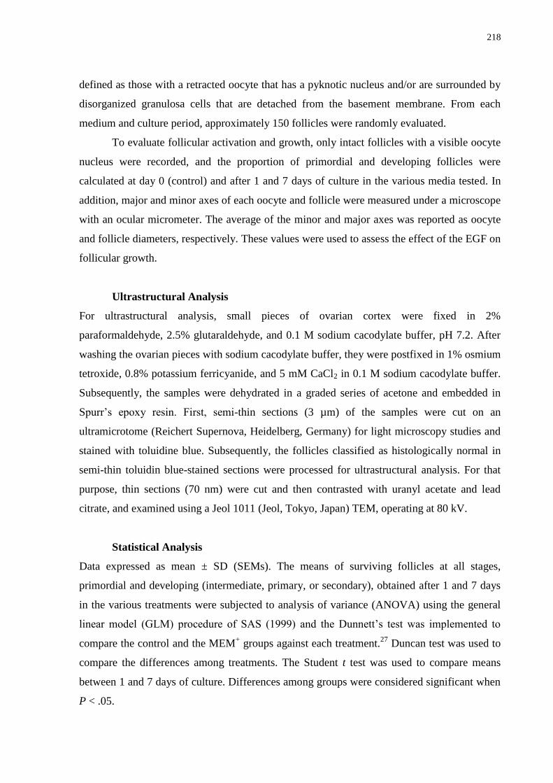
218
defined as those with a retracted oocyte that has a pyknotic nucleus and/or are surrounded by
disorganized granulosa cells that are detached from the basement membrane. From each
medium and culture period, approximately 150 follicles were randomly evaluated.
To evaluate follicular activation and growth, only intact follicles with a visible oocyte
nucleus were recorded, and the proportion of primordial and developing follicles were
calculated at day 0 (control) and after 1 and 7 days of culture in the various media tested. In
addition, major and minor axes of each oocyte and follicle were measured under a microscope
with an ocular micrometer. The average of the minor and major axes was reported as oocyte
and follicle diameters, respectively. These values were used to assess the effect of the EGF on
follicular growth.
Ultrastructural Analysis
For ultrastructural analysis, small pieces of ovarian cortex were fixed in 2%
paraformaldehyde, 2.5% glutaraldehyde, and 0.1 M sodium cacodylate buffer, pH 7.2. After
washing the ovarian pieces with sodium cacodylate buffer, they were postfixed in 1% osmium
tetroxide, 0.8% potassium ferricyanide, and 5 mM CaCl2 in 0.1 M sodium cacodylate buffer.
Subsequently, the samples were dehydrated in a graded series of acetone and embedded in
Spurr‘s epoxy resin. First, semi-thin sections (3 µm) of the samples were cut on an
ultramicrotome (Reichert Supernova, Heidelberg, Germany) for light microscopy studies and
stained with toluidine blue. Subsequently, the follicles classified as histologically normal in
semi-thin toluidin blue-stained sections were processed for ultrastructural analysis. For that
purpose, thin sections (70 nm) were cut and then contrasted with uranyl acetate and lead
citrate, and examined using a Jeol 1011 (Jeol, Tokyo, Japan) TEM, operating at 80 kV.
Statistical Analysis
Data expressed as mean ± SD (SEMs). The means of surviving follicles at all stages,
primordial and developing (intermediate, primary, or secondary), obtained after 1 and 7 days
in the various treatments were subjected to analysis of variance (ANOVA) using the general
linear model (GLM) procedure of SAS (1999) and the Dunnett‘s test was implemented to
compare the control and the MEM+ groups against each treatment.
27 Duncan test was used to
compare the differences among treatments. The Student t test was used to compare means
between 1 and 7 days of culture. Differences among groups were considered significant when
P < .05.
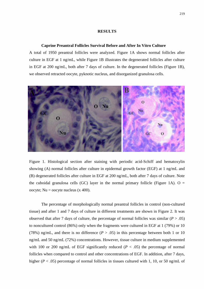
219
RESULTS
Caprine Preantral Follicles Survival Before and After In Vitro Culture
A total of 1950 preantral follicles were analyzed. Figure 1A shows normal follicles after
culture in EGF at 1 ng/mL, while Figure 1B illustrates the degenerated follicles after culture
in EGF at 200 ng/mL, both after 7 days of culture. In the degenerated follicles (Figure 1B),
we observed retracted oocyte, pyknotic nucleus, and disorganized granulosa cells.
Figure 1. Histological section after staining with periodic acid-Schiff and hematoxylin
showing (A) normal follicles after culture in epidermal growth factor (EGF) at 1 ng/mL and
(B) degenerated follicles after culture in EGF at 200 ng/mL, both after 7 days of culture. Note
the cuboidal granulosa cells (GC) layer in the normal primary follicle (Figure 1A). O =
oocyte; Nu = oocyte nucleus (x 400).
The percentage of morphologically normal preantral follicles in control (non-cultured
tissue) and after 1 and 7 days of culture in different treatments are shown in Figure 2. It was
observed that after 7 days of culture, the percentage of normal follicles was similar (P > .05)
to noncultured control (86%) only when the fragments were cultured in EGF at 1 (79%) or 10
(78%) ng/mL, and there is no difference (P > .05) in this percentage between both 1 or 10
ng/mL and 50 ng/mL (72%) concentrations. However, tissue culture in medium supplemented
with 100 or 200 ng/mL of EGF significantly reduced (P < .05) the percentage of normal
follicles when compared to control and other concentrations of EGF. In addition, after 7 days,
higher (P < .05) percentage of normal follicles in tissues cultured with 1, 10, or 50 ng/mL of
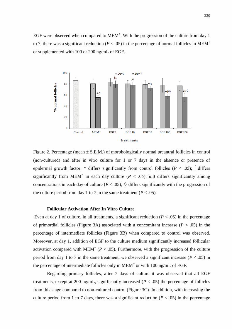
220
EGF were observed when compared to MEM+. With the progression of the culture from day 1
to 7, there was a significant reduction (P < .05) in the percentage of normal follicles in MEM+
or supplemented with 100 or 200 ng/mL of EGF.
Figure 2. Percentage (mean S.E.M.) of morphologically normal preantral follicles in control
(non-cultured) and after in vitro culture for 1 or 7 days in the absence or presence of
epidermal growth factor. * differs significantly from control follicles (P < .05); ∫ differs
significantly from MEM+ in each day culture (P < .05); α,β differs significantly among
concentrations in each day of culture (P < .05); differs significantly with the progression of
the culture period from day 1 to 7 in the same treatment (P < .05).
Follicular Activation After In Vitro Culture
Even at day 1 of culture, in all treatments, a significant reduction (P < .05) in the percentage
of primordial follicles (Figure 3A) associated with a concomitant increase (P < .05) in the
percentage of intermediate follicles (Figure 3B) when compared to control was observed.
Moreover, at day 1, addition of EGF to the culture medium significantly increased follicular
activation compared with MEM+ (P < .05). Furthermore, with the progression of the culture
period from day 1 to 7 in the same treatment, we observed a significant increase (P < .05) in
the percentage of intermediate follicles only in MEM+ or with 100 ng/mL of EGF.
Regarding primary follicles, after 7 days of culture it was observed that all EGF
treatments, except at 200 ng/mL, significantly increased (P < .05) the percentage of follicles
from this stage compared to non-cultured control (Figure 3C). In addition, with increasing the
culture period from 1 to 7 days, there was a significant reduction (P < .05) in the percentage
221
of primordial follicles and a significant increase (P < .05) in the percentage of primary
follicles after culture in MEM+ or at 1, 10, or 50 ng/mL of EGF. However, only the low EGF
concentrations (1, 10, and 50 ng/mL) significantly increased (P < .05) the rates of primary
follicles in relation to MEM+. Non-cultured and cultured caprine ovarian tissue contained a
low (often zero) and variable number of secondary follicles (data not shown), which did not
permit statistical analysis.
Figure 3. Percentage (mean S.E.M.) of primordial (A), intermediate (B) and primary (C)
follicles in control (non-cultured) and after in vitro culture for 1 or 7 days in the absence or
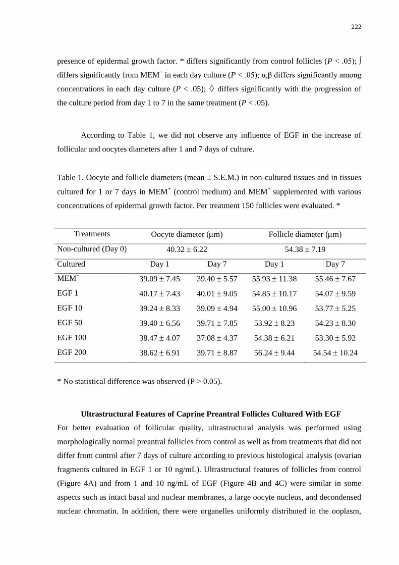
222
presence of epidermal growth factor. * differs significantly from control follicles (P < .05); ∫
differs significantly from MEM+ in each day culture (P < .05); α,β differs significantly among
concentrations in each day culture (P < .05); differs significantly with the progression of
the culture period from day 1 to 7 in the same treatment (P < .05).
According to Table 1, we did not observe any influence of EGF in the increase of
follicular and oocytes diameters after 1 and 7 days of culture.
Table 1. Oocyte and follicle diameters (mean S.E.M.) in non-cultured tissues and in tissues
cultured for 1 or 7 days in MEM+ (control medium) and MEM
+ supplemented with various
concentrations of epidermal growth factor. Per treatment 150 follicles were evaluated. *
Treatments Oocyte diameter (m) Follicle diameter (m)
Non-cultured (Day 0) 40.32 6.22 54.38 7.19
Cultured Day 1 Day 7 Day 1 Day 7
MEM+ 39.09 7.45
39.40 5.57
55.93 11.38
55.46 7.67
EGF 1 40.17 7.43 40.01 9.05
54.85 10.17
54.07 9.59
EGF 10 39.24 8.33 39.09 4.94
55.00 10.96
53.77 5.25
EGF 50 39.40 6.56 39.71 7.85
53.92 8.23
54.23 8.30
EGF 100 38.47 4.07 37.08 4.37
54.38 6.21
53.30 5.92
EGF 200 38.62 6.91 39.71 8.87
56.24 9.44
54.54 10.24
* No statistical difference was observed (P > 0.05).
Ultrastructural Features of Caprine Preantral Follicles Cultured With EGF
For better evaluation of follicular quality, ultrastructural analysis was performed using
morphologically normal preantral follicles from control as well as from treatments that did not
differ from control after 7 days of culture according to previous histological analysis (ovarian
fragments cultured in EGF 1 or 10 ng/mL). Ultrastructural features of follicles from control
(Figure 4A) and from 1 and 10 ng/mL of EGF (Figure 4B and 4C) were similar in some
aspects such as intact basal and nuclear membranes, a large oocyte nucleus, and decondensed
nuclear chromatin. In addition, there were organelles uniformly distributed in the ooplasm,
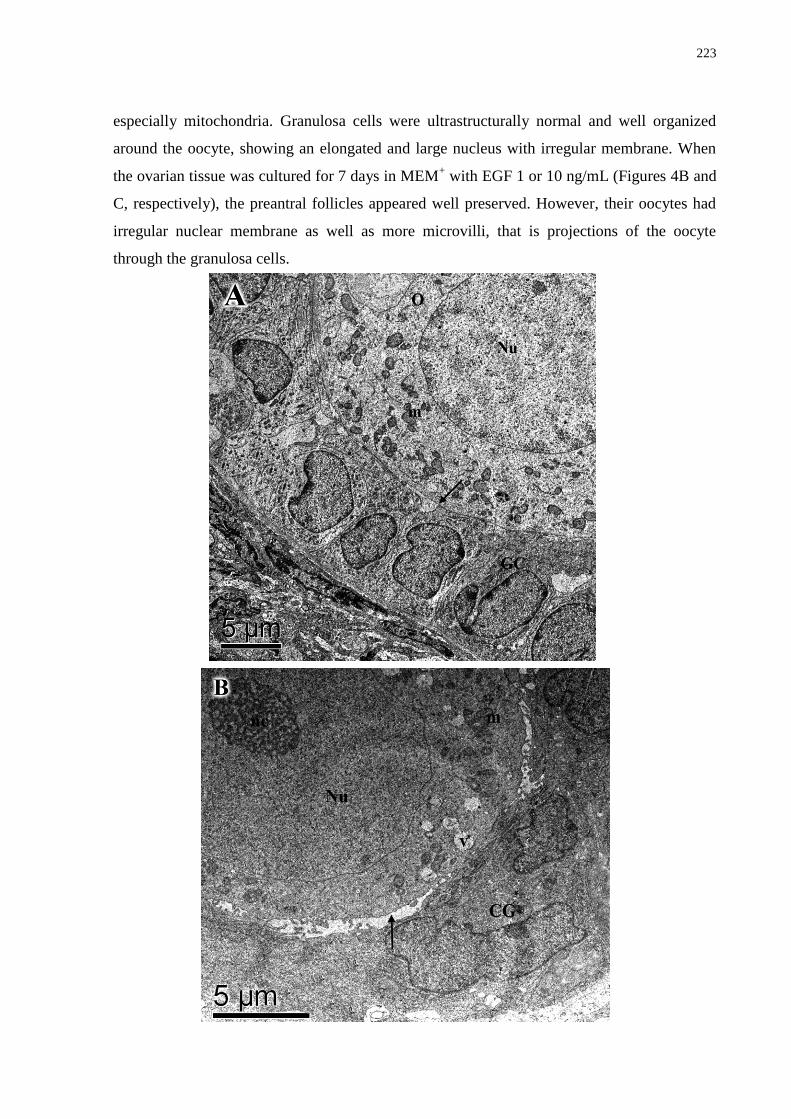
223
especially mitochondria. Granulosa cells were ultrastructurally normal and well organized
around the oocyte, showing an elongated and large nucleus with irregular membrane. When
the ovarian tissue was cultured for 7 days in MEM+ with EGF 1 or 10 ng/mL (Figures 4B and
C, respectively), the preantral follicles appeared well preserved. However, their oocytes had
irregular nuclear membrane as well as more microvilli, that is projections of the oocyte
through the granulosa cells.
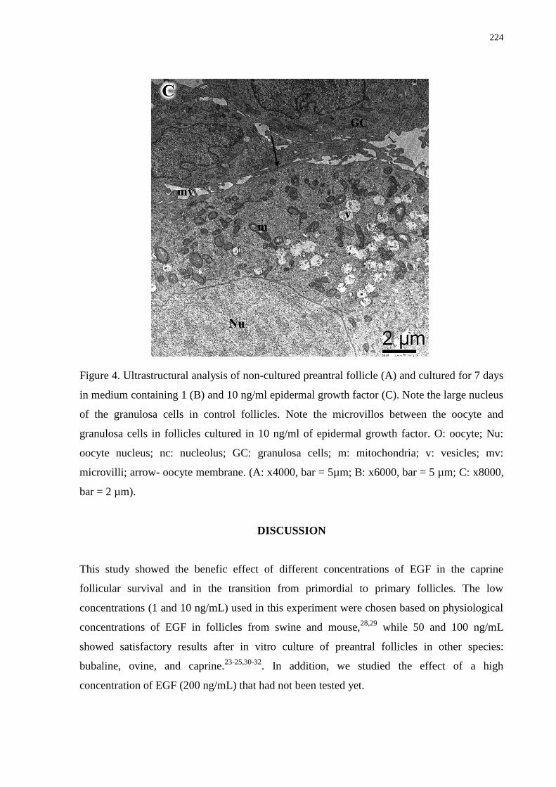
224
Figure 4. Ultrastructural analysis of non-cultured preantral follicle (A) and cultured for 7 days
in medium containing 1 (B) and 10 ng/ml epidermal growth factor (C). Note the large nucleus
of the granulosa cells in control follicles. Note the microvillos between the oocyte and
granulosa cells in follicles cultured in 10 ng/ml of epidermal growth factor. O: oocyte; Nu:
oocyte nucleus; nc: nucleolus; GC: granulosa cells; m: mitochondria; v: vesicles; mv:
microvilli; arrow- oocyte membrane. (A: x4000, bar = 5µm; B: x6000, bar = 5 µm; C: x8000,
bar = 2 µm).
DISCUSSION
This study showed the benefic effect of different concentrations of EGF in the caprine
follicular survival and in the transition from primordial to primary follicles. The low
concentrations (1 and 10 ng/mL) used in this experiment were chosen based on physiological
concentrations of EGF in follicles from swine and mouse,28,29
while 50 and 100 ng/mL
showed satisfactory results after in vitro culture of preantral follicles in other species:
bubaline, ovine, and caprine.23-25,30-32
. In addition, we studied the effect of a high
concentration of EGF (200 ng/mL) that had not been tested yet.
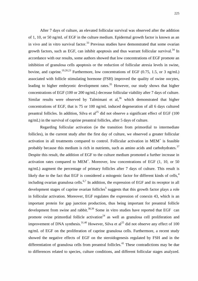
225
After 7 days of culture, an elevated follicular survival was observed after the addition
of 1, 10, or 50 ng/mL of EGF in the culture medium. Epidermal growth factor is known as an
in vivo and in vitro survival factor.33
Previous studies have demonstrated that some ovarian
growth factors, such as EGF, can inhibit apoptosis and thus warrant follicular survival.34
In
accordance with our results, some authors showed that low concentrations of EGF promote an
inhibition of granulosa cells apoptosis or the reduction of follicular atresia levels in swine,
bovine, and caprine.19,20,23
Furthermore, low concentrations of EGF (0.75, 1.5, or 3 ng/mL)
associated with follicle stimulating hormone (FSH) improved the quality of swine oocytes,
leading to higher embryonic development rates.35
However, our study shows that higher
concentrations of EGF (100 or 200 ng/mL) decrease follicular viability after 7 days of culture.
Similar results were observed by Talmimani et al,36
which demonstrated that higher
concentrations of EGF, that is 75 or 100 ng/mL induced degeneration of all 6 days cultured
preantral follicles. In addition, Silva et al25
did not observe a significant effect of EGF (100
ng/mL) in the survival of caprine preantral follicles, after 5 days of culture.
Regarding follicular activation (ie the transition from primordial to intermediate
follicles), in the current study after the first day of culture, we observed a greater follicular
activation in all treatments compared to control. Follicular activation in MEM+ is feasible
probably because this medium is rich in nutrients, such as amino acids and carbohydrates.37
Despite this result, the addition of EGF to the culture medium promoted a further increase in
activation rates compared to MEM+. Moreover, low concentrations of EGF (1, 10, or 50
ng/mL) augment the percentage of primary follicles after 7 days of culture. This result is
likely due to the fact that EGF is considered a mitogenic factor for different kinds of cells,4
including ovarian granulosa cells.6,7
In addition, the expression of EGF and its receptor in all
development stages of caprine ovarian follicles5 suggests that this growth factor plays a role
in follicular activation. Moreover, EGF regulates the expression of conexin 43, which is an
important protein for gap junction production, thus being important for preantral follicle
development from swine and rabbit.38,39
Some in vitro studies have reported that EGF can
promote ovine primordial follicle activation24
as well as granulosa cell proliferation and
improvement of DNA synthesis.31,40
However, Silva et al25
did not observe any effect of 100
ng/mL of EGF on the proliferation of caprine granulosa cells. Furthermore, a recent study
showed the negative effects of EGF on the steroidogenesis regulated by FSH and in the
differentiation of granulosa cells from preantral follicles.41
These contradictions may be due
to differences related to species, culture conditions, and different follicular stages analyzed.
226
Similar to other studies,42,43
we observed a few secondary follicles probably due to the small
period of culture and the absence of any other growth factor or hormone in the culture
medium, such as activin,44
bone morphogenetic45
protein-15, and growth differentiation46
factor-9.
Although there was an increase in the percentage of primary follicles after EGF
addition, no effect of EGF on the follicular and oocyte diameters was observed. This can
probably happen due to the fact that intermediate follicles were already large, thus follicular
growth was not perceptive but only the changes in granulosa cells morphology from flattened
to cuboidal. Similar to our results, Zhou and Zhang23,32
also observed that the use of EGF, in
concentration 50 ng/mL, showed no effect on the caprine oocyte growth. Nevertheless, Silva
et al25
showed that EGF (100 ng/mL) increased the diameter of intermediate and primary
follicles.
Our results of follicular viability after classical histology were confirmed by
ultrastructural analysis. This technique is considered an important tool because it provides
more information about the follicular and oocyte quality and is able to detect undiscernible
damage to cellular membranes and organelles.47
From the TEM studies, it was observed that
besides basal and nuclear membranes, important organelles such as mitochondria,
endoplasmic reticulum, and granulosa cells were preserved even after 7 days of culture in the
presence of 1 or 10 ng/mL of EGF. Normal follicles had their ultrastructure similar to those
described earlier in caprine47
and subsequently in bovine and ovine.37,48
In this study, we
observed microvilli and vesicles, which are important for the oocyte and granulosa cells
communication, allowing the exchange of substances and, consequently, warranting an
adequate follicular development.
In conclusion, this study showed that the low concentrations of EGF (ie 1 or 10
ng/mL) are able to promote the maintenance of follicular viability and the transition from
primordial to primary follicles after 7 days of in vitro culture.
ACKNOWLEDGMENTS
This work was supported by CNPq, CAPES, FINEP, and Fundação Cearense de Apoio à
Pesquisa (FUNCAP). JJHC is a recipient of a grant from FUNCAP (Brazil).
227
REFERENCES
1. Bristol-Gould S, Woodruff TK. Folliculogenesis in the domestic cat (Felis catus).
Theriogenology. 2006;66:5-13.
2. Fortune JE. The early stages of follicular development: activation of primordial follicles
and growth of preantral follicles. Anim Reprod Sci. 2003;78:135-163.
3. Eppig JJ. Oocyte control of ovarian follicular development and function in mammals.
Reproduction. 2001;122:829-838.
4. Toyoda T, Nakamura K, Yamada K, et al. SNP analyses of growth factor genes EGF, TGF-
β1, and HGF reveal haplotypic association of EGF with autism. Biochem Bioph Res
Commun. 2007;360:715-720.
5. Silva JRV, van den Hurk R, Figueiredo JR. Expression of mRNA and protein localization
of epidermal growth factor and its receptor in goat ovaries. Zygote. 2006;14:107-117.
6. Saha S, Shimizu M, Geshi M, Izaike Y. In vitro culture of bovine preantral follicles. Anim
Reprod Sci. 2000;63:27-39.
7. Wang Y, Li J, Wang CY, Kwok AHY, Leung FC. Epidermal growth factor (EGF) receptor
ligands in the chicken ovary: I. Evidence for heparin-binding EGF-like growth factor
(HBEGF) as a potential oocyte-derived signal to control granulose cell proliferation and
HB-EGF and kit ligand expression. Endocrinology. 2007;148:3426-3440.
8. Feng P, Knecht M, Catt K. Hormonal control of epidermal growth factor receptors by
gonadotropins during granulosa cell differentiation. Endocrinology. 1987;120:1121-1126.
9. Lonergan P, Carolan C, Van Langendonckt A, Donnay I, Khatir H, Mermillod P. Role of
epidermal growth factor in bovine oocyte maturation and preimplantation embryo
development in vitro. Biol Reprod. 1996;54:1420-1429.
10. Qu J, Godin PA, Nisolle M, Donnez J. Distribution of epidermal growth factor receptor
expression of primordial follicles in human ovarian tissue before and after
cryopreservation. Hum Reprod. 2000;15:302-310.
11. Garnett K, Wang J, Roy SK. Spatiotemporal expression of epidermal growth factor
receptor messenger RNA and protein in the hamster ovary: follicle stage-specific
differential modulation by follicle-stimulating hormone, luteinizing hormone, estradiol,
and progesterone. Biol Reprod. 2002;67:1593- 1604.
228
12. Singh B, Rutledge JM, Armstrong DT. Epidermal growth factor and its receptor gene
expression and peptide localization in porcine ovarian follicles. Mol Reprod Dev.
1995;40: 391-399.
13. Hill JL, Hammar K, Smith PJ, Gross DJ. Stage-dependent effects of epidermal growth
factor on Ca2þ efflux in mouse oocytes. Mol Reprod Dev. 1999;53:244-253.
14. Fru KN, Cherian-Shaw M, Puttabyatappa M, Vande Voort CA, Chaffin CL. Regulation of
granulose cell proliferation and EGF-like ligands during the periovulatory interval in
monkeys. Hum Reprod. 2007;22:1247-1252.
15. Tekpetey FR, Singh B, Barbe G, Armstrong DT. Localisation of epidermal growth factor
(EGF) receptor in the rat corpus luteum, and EGF and transforming growth factor-alpha
stimulation of luteal cell steroidogenesis in vitro. Mol Cell Endocrinol. 1995;110:95-102.
16. Singh B, Kennedy TG, Tekpetey FR, Armstrong DT. Gene expression and peptide
localisation for epidermal growth factor receptor and its ligands in porcine luteal cells.
Mol Cell Endocrinol. 1995;113:137-143.
17. Morbeck DE, Flowers WL, Britt JH. Response of porcine granulosa cells isolated from
primary and secondary follicles to FSH, 8-bromo-cAMP and EGF in vitro. J Reprod
Fertil. 1993;99:577-584.
18. Gospodarowicz D, Bialecki H. Fibroblast and epidermal growth factors are mitogenic
agents for cultured granulosa cells of rodent, porcine and human origin. Endocrinology.
1979;104:757-764.
19. Mao J, Smith MF, Rucker EB, et al. Effect of epidermal growth factor and insulin-like
growth factor I on porcine preantral follicular growth, antrum formation, and stimulation
of granulosal cell proliferation and suppression of apoptosis in vitro. J Anim Sci.
2004;82:1967-1975.
20. Gutierrez CG, Ralph JH, Telfer EE, Wilmut I, Webb R. Growth and antrum formation of
bovine preantral follicles in long-term culture in vitro. Biol Reprod. 000;62:1322- 1328.
21. Romano M, Kraus ER, Boland CR, Coffey RJ. Comparison between transforming growth
factor alpha and epidermal growth factor in the protection of rat gastric mucosa against
drug-induced injury. Ital J Gastroenterol. 1994;26:223-228.
22. Roy SK, Kole AR. Ovarian transforming growth factor-beta (TGF-beta) receptors: in vitro
effects of follicle stimulating hormone, epidermal growth factor and TGF beta on
receptor expression in human preantral follicles. Mol Hum Reprod. 1998;4:207-214.
229
23. Zhou H, Zhang Y. Regulation of in vitro growth of preantral follicles by growth factors in
goats. Domest Anim Endocrinol. 2005;28:235-242.
24. Andrade ER, Marcondes Seneda M, Alfieri AA, et al. Interactions of indole acetic acid
with EGF and FSH in the culture of ovine preantral follicles. Theriogenology.
2005;64:1104-1113.
25. Silva JRV, van den Hurk R, de Matos MH, et al. Influences of FSH and EGF on
primordial follicles during in vitro culture of caprine ovarian cortical tissue.
Theriogenology. 2004;61:1691-1704.
26. Silva JR, van den Hurk R, Costa SH, et al. Survival and growth of goat primordial
follicles after in vitro culture of ovarian cortical slices in media containing coconut water.
Anim Reprod Sci. 2004;81:273-286.
27. Steel RGD, Torrie JH, Dickey D. Principles and Procedures of Statistics: A Biometrical
Approach. 3rd ed. New York, NY: McGraw-Hill; 1997.
28. Hsu CJ, Holmes SD, Hammond JM. Ovarian epidermal growth factor-like activity.
Concentrations in porcine follicular fluid during follicular enlargement. Biochem
Biophys Res Commun. 1987;147:242-247.
29. Demeestere I, Centner J, Gervy Y, Englert Y, Delbaere A. Impact of various endocrine
and paracrine factors on in vitro culture of preantral follicles in rodents. Reproduction.
2005;130: 147-156.
30. Gupta PS, Nandi S, Ravindranatha BM, Sarma PV. In vitro culture of buffalo (Bubalus
bubalis) preantral follicles. Theriogenology. 2002;57:1839-1854.
31. Hemamalini NC, Rao BS, Tamilmani G, et al. Influence of transforming growth factor-a,
insulin-like growth factor-II, epidermal growth factor or follicle stimulating hormone on
in vitro development of preantral follicles in sheep. Small Rumin Res. 2003;50:11-22.
32. Zhou HM, Zhang Y. Impact of growth factors on in vitro development of caprine oocytes
at pre-antral stage. Reprod Domest Anim. 2005;40:161-165.
33. Van den Hurk R, Abir R, Telfer EE, Bevers MM. Preantral and antral follicles as possible
source for fertilizable oocytes in human and bovine. Hum Reprod Update. 2000;2: 457-
474.
34. Markstrom E, Svensson ECh, Shao R, Svanberg B, Billig H. Survival factors regulating
ovarian apoptosis—dependence on follicle differentiation. Reproduction. 2002;123:23-
30.
230
35. Wu J, Tian Q. Role of follicle stimulating hormone and epidermal growth factor in the
development of porcine preantral follicle in vitro. Zygote. 2007;15:233-240.
36. Talmimani G, Rao BS, Vagdevi R, et al. Nuclear maturation of ovine oocytes in cultured
preantral follicles. Small Rumin Res. 2005;60:295-305.
37. van Wezel IL, Rodgers RJ. Morphological characterization of bovine primordial follicles
and their environment in vivo. Biol Reprod. 1996;55:1003-1011.
38. Bolamba D, Floyd AA, McGlone JJ, Lee VH. Epidermal growth factor enhances
expression of connexin 43 protein in cultured porcine preantral follicles. Biol Reprod.
2002;67:154-160.
39. Kennedy KL, Floyd AA, Clarkson AM, Lee VH. Epidermal growth factor regulation of
connexin 43 in cultured granulose cells from preantral follicles rabbit follicles. Mol
Reprod Dev. 2003;64:61-69.
40. Rajarajan K, Rao BS, Vagdevi R, et al. Effect of various growth factors on the in vitro
development of goat preantral follicles. Small Rumin Res. 2006;63:204-212.
41. Conti M, Hsieh M, Park JY, Su YQ. Role of the epidermal growth factor network in
ovarian follicles. Mol Endocrinol. 2006;20:715-723.
42. Wandji SA, Srsen V, Nathanielsz PW, Eppig JJ, Fortune JE. Initiation of growth of
baboon primordial follicles in vitro. Hum Reprod. 1997;12:1993-2001.
43. Fortune JE, Kito S, Wandji SA, Srsen V. Activation of bovine and baboon primordial
follicles in vitro. Theriogenology. 1998;49:441-449.
44. Zhao J, Taverne MAM, van der Weijden GC, Bevers MM, van den Hurk R. Effect of
activin A on in vitro development of rat preantral follicles and localization of activin
and activin receptor II. Biol Reprod. 2001;65:967-977.
45. Galloway SM, McNatty KP, Cambridge LM, et al. Mutations in an oocyte-derived growth
factor gene (BMP15) cause increased ovulation rate and infertility in a dosage-sensitive
manner. Nat Genet. 2000;25:279-283.
46. Hayashi M, McGee EA, Min G, et al. Recombinant growth differentiation factor-9 (GDF-
9) enhances growth and differentiation of cultured early follicles. Endocrinology.
1999;140:1236-1244.
47. Lucci CM, Silva RV, Carvalho CA, Figueiredo R, Ba´o N. Light microscopical and
ultrastrutural characterization of goat preantral follicles. Small Rumin Res. 2001;41:61-
69.

231
48. Cran DG, Moor RM, Hay MF. Fine structure of the sheep oocyte during antral follicle
development. J Reprod Fertil. 1980;59:125-132.
232
13 CAPÍTULO 8
Níveis de RNAm para o fator de crescimento epidermal (EGF) e efeito do EGF no cultivo in
vitro de folículos pré-antrais caprinos
Steady-state level of mRNA for epidermal growth factor (EGF) and effect of EGF on in vitro
culture of caprine preantral follicles
Periódico: Cell and Tissue Research (Submetido em outubro de 2010).
B
C D
233
Resumo
Os objetivos desse estudo foram verificar os níveis de RNAm para o fator de crescimento
epidermal (EGF) em folículos caprinos nos diferentes estádios de desenvolvimento, e
investigar a influência do EGF na sobrevivência, formação de antro e crescimento de folículos
secundários cultivados por seis dias. Para tanto, folículos primordiais, primários e secundários
de cabras, bem como pequenos e grandes folículos antrais foram obtidos, e os níveis de
RNAm para o EGF foram quantificados pela RT-PCR em tempo real. A influência do EGF na
presença ou ausência de FSH sobre o desenvolvimento de folículos secundários, e a expressão
do RNAm para o EGF e R-FSH foram avaliadas após seis dias de cultivo. Além disso, a
sobrevivência, a formação de antro e o diâmetro folicular foram avaliados a cada dois dias. Os
níveis de RNAm para EGF em folículos secundários foram significativamente superiores que
em folículos primordiais. Em pequenos e grandes folículos antrais, os níveis de RNAm para
EGF em CCOs foram significativamente superiores que os níveis nas células da
granulosa/teca. Durante o cultivo, o EGF na presença ou na ausência de FSH aumentou a taxa
de crescimento diário folicular de folículos secundários quando comparados ao α-MEM+. Em
adição, FSH, EGF ou ambos reduziram os níveis de RNAm para o EGF, enquanto o EGF
reduziu os níveis de RNAm para o R-FSH após cultivo dos folículos por seis dias.
Concluindo, os níveis de RNAm para o EGF foram superiores em folículos secundários, e
ambos FSH e EGF promoveram o crescimento de folículos secundários caprinos. Além disso,
EGF e/ou FSH reduziram os níveis de RNAm para o EGF, e o EGF diminuiu os níveis de
RNAm para R-FSH em folículos secundários cultivados.
Palavras-chave: EGF. Folículos secundários. FSH-R. Cabra. Cultivo.
234
Steady-state level of epidermal growth factor (EGF) mRNA and the effect of EGF on the in
vitro culture of caprine preantral follicles
J.J.H. Celestinoa*, J.B. Brunoa, M.V.A. Saraivaa, R.M.P. Rochaa, I.R. Britoa, A.B.G. Duartea, V.R.
Araújoa, C.M.G. Silvaa, M.H.T. Matosb, C.C. Campelloa, J.R.V. Silvac, J.R. Figueiredoa
aFaculty of Veterinary Medicine, LAMOFOPA, PPGCV, State University of Ceara, Fortaleza-CE, Brazil,
bNucleus of Biotechnology Applied to Ovarian Follicle Development, Federal University of São Francisco
Valley, Petrolina-PE, Brazil
cBiotechnology Nucleus of Sobral (NUBIS), Federal University of Ceara, Sobral-CE, Brazil
*Corresponding address:
Programa de Pós-Graduação em Ciências Veterinárias (PPGCV)
Laboratório de Manipulação de Oócitos e Folículos Pré-Antrais (LAMOFOPA)
Universidade Estadual do Ceará (UECE)
Av. Paranjana, 1700, Campus do Itaperi.
Fortaleza – CE – Brasil. CEP: 60740-903
Tel.: +55.85.3101.9852; Fax: +55.85.3101.9840
E-mail address: [email protected] (Juliana Jales de Hollanda Celestino)
235
Abstract
The aims of this study were to verify the steady-state level of epidermal growth factor (EGF)
mRNA in goat follicles at different developmental stages and to investigate the influence of
EGF on the survival, antrum formation and growth of secondary follicles cultured for six
days. For this purpose, primordial, primary and secondary goat follicles as well as small and
large antral follicles were obtained, and EGF mRNA levels were quantified by real-time RT-
PCR. The influence of EGF in the presence or absence of FSH on the development of
secondary follicles and on the EGF and FSH-R mRNA expression was evaluated after six
days of culture. Furthermore, the survival, the antrum formation and the follicular diameter
were evaluated every other day of culture. The EGF mRNA levels in secondary follicles were
significantly higher than in primordial follicles. In small and large antral follicles, the EGF
mRNA levels in cumulus–oocyte complexes (COCs) were significantly higher than the levels
in granulosa/theca cells. During the culture, EGF in the presence or absence of FSH increased
the follicular daily growth rate of secondary follicles when compared to α-MEM+. In addition,
FSH, EGF or both reduced the EGF mRNA levels, whereas EGF reduced the FSH-R mRNA
levels after culture for six days. In conclusion, the EGF mRNA levels were higher in
secondary follicles and both FSH and EGF promoted growth of goat secondary follicles.
Furthermore, EGF and/or FSH reduced the EGF mRNA levels and EGF decreased the FSH-R
mRNA levels in cultured secondary follicles.
Keywords: EGF, secondary follicles, FSH-R, goat, culture.
236
1. Introduction
The goat preantral follicles are highly susceptible to atresia. Thus, there is an interest
in developing a culture system that supports the in vitro growth of preantral follicles to the
stage at which the oocytes were capable of being matured and fertilized in vitro. The
implementation of this system could maximize the in vitro production of embryos by
providing a large number of oocytes that are homogeneous and meiotically competent for
several biotechnologies such as in vitro fertilization, cloning and transgenesis. In some animal
species, the in vitro growth and development of preantral follicles has been successful until
the embryonic stage (pig: Wu and Tian, 2007, buffalo: Gupta et al., 2008, sheep:
Arunakumari et al., 2010 and caprine: Saraiva et al., in press). Nevertheless, the birth of live
offspring from the preantral follicles grown in vitro in these species is still a great challenge.
In addition, the maturation rates of oocytes obtained from preantral follicles are very low.
The follicular development is the result of the complex interactions between pituitary
gonadotropins and numerous intra-ovarian factors, which act as promoters of survival,
stimulating the growth and differentiation of follicular cells (Fortune, 2003; Miyoshi et al.,
2010). Among these factors, the epidermal growth factor (EGF) has been highlighted; it has
emerged as an important substance capable of inducing follicular development in vitro
(Celestino et al., 2009). The EGF is a protein belonging to the EGF family, which consists of
at least eight members (Riese and Stern, 1998). EGF is considered a mitogenic factor, and it is
involved in the regulation of several ovarian processes (Silva et al., 2006), including
proliferation and cellular differentiation, in addition to steroidogenesis (Saha et al., 2000;
Wang et al., 2007). The expression of protein and mRNA for EGF has been demonstrated in
the oocyte and granulosa cells of early and late-staged follicles (hamster: Roy and Greenwald,
1990, human: Maruo et al., 1993; Bennett et al., 1996, pig: Singh et al., 1995), while EGF
mRNA has been described only in oocyte and granulosa cells from pig antral follicles (Singh
et al., 1995). In caprine, the protein and mRNA for EGF were expressed in all developmental
stages of ovarian follicles and in the ovarian epithelium surface (Silva et al., 2006). However,
quantification of the steady-state level of EGF mRNA during different stages of follicular
development has not been performed as yet. The action of EGF in the ovary is mediated by a
membrane receptor, EGF-R (ErbB1), which belongs to the ErbB superfamily (Riese and
Stern, 1998). EGF-R mRNA and protein have been identified in the oocyte and granulosa
cells of early-and late-stage follicles (rat: Chabot et al., 1986; Feng et al., 1987, human:
237
Maruo et al., 1993; Bennett et al., 1996; Qu et al., 2000, pig: Singh et al., 1995, cattle:
Lonergan et al., 1996, mouse: Hill et al., 1999, hamster: Garnett et al., 2002, caprine: Gall et
al., 2004; Silva et al., 2006). In hamsters, it was demonstrated that the expression of the
protein and mRNA for the EGF receptor was positively regulated by gonadotropins and
steroids (Garnett et al., 2002).
In vitro studies showed that EGF promotes the proliferation of granulosa cells in the
rodent, swine, caprine and human (Gospodarowicz and Bialecki, 1979; Morbeck et al., 1993;
Rajarajan et al., 2006), increases the follicular diameter in the rodent, swine, bovine, caprine
and human (Romano et al., 1994; Roy and Kole, 1998; Gutierrez et al., 2000; Silva et al.,
2004a; Mao et al., 2004), reduces the atresia levels in the cow, swine, and caprine (Gutierrez
et al., 2000; Mao et al., 2004; Zhou and Zhang 2005a,b; Celestino et al., 2009) and promotes
ovine and caprine primordial follicle activation and maintenance of survival for up to six and
seven days of culture, respectively (Andrade et al., 2005; Celestino et al., 2009). There is
evidence that the regulation of EGF activity in granulosa cells in vitro may occur by
stimulation of FSH receptor expression (Luciano et al., 1994). When the combination of EGF
with FSH was tested in the in vitro culture of pig preantral follicles, the vast majority of
follicles grew to the antral stage, with high secretion of estradiol, and the oocytes from these
follicles could be matured, fertilized and developed until the blastocyst stage (Wu and Tian,
2007). In caprine, the interaction between EGF and FSH promoted follicular survival,
although it had no effect on growth (Zhou and Zhang, 2005a,b). Although there have been
some studies to date, little is known about the in vitro effects of EGF in the presence or
absence of FSH on the development of isolated caprine secondary follicles. Moreover, the in
vitro effects of EGF in the presence or absence of FSH on the expression of EGF and FSH
receptor (FSH-R) after culture is still unknown.
Therefore, the present study aimed (1) to verify the steady-state level of EGF mRNA
during different follicular stages in goat ovaries, (2) to investigate a possible influence of EGF
on the survival, antral cavity formation and growth of secondary follicles after culture for six
days, and (3) to evaluate the effects of EGF and/or FSH on the EGF and FSH-R mRNA levels
after six days of culture.
238
2. Material and Methods
2.1. Chemicals
Unless mentioned otherwise, the culture media and other chemicals used in the present
study were purchased from Sigma Chemical Co. (St. Louis, MO, USA).
2.2. Steady-state level of EGF mRNA in goat ovarian follicles
To evaluate the steady-state level of mRNA, 30 ovaries from 15 goats (Capra hircus)
were collected at a local slaughterhouse and rinsed in Minimum Essential Medium (MEM)
containing antibiotics (100 µg/mL penicillin and 100 µg/mL streptomycin). After this
preparation, 10 ovaries from 5 goats were utilized for the isolation of primordial, primary, and
secondary follicles. The remaining ovaries were used for the collection of cumulus–oocyte
complexes (COCs), mural granulosa cells, and thecal cells from small and large antral
follicles. Primordial, primary, and secondary follicles were isolated using a previously
described mechanical procedure (Lucci et al., 1999). After isolation, these follicles were
washed several times to completely remove the stromal cells and were then placed by
category into separate Eppendorf tubes in groups of 10. This procedure was completed within
2 h, and all samples were stored at -80C until the RNA was extracted. From a second group
of ovaries (n=15), COCs aspirated from small (1–3 mm) and large (3–6 mm) antral follicles
were recovered. Compact COCs were selected from the follicle content as described by van
Tol and Bevers (1998). Thereafter, groups of 10 COCs were stored at – 80C until RNA
extraction. To collect mural granulosa and theca cell complex, small (n=10) and large antral
follicles (n=10) were isolated from ovaries (n=5) and dissected free from stromal tissue with
forceps as previously described (van Tol and Bevers, 1998). The follicles were then bisected,
and the granulosa and theca cell complexes were collected and stored at -80C.
The isolation of total RNA was performed using a Trizol plus purification kit
(Invitrogen, São Paulo, Brazil). According to the manufacturer‘s instructions, 1 mL of Trizol
solution was added to each frozen sample, and the lysate was aspirated through a 20-gauge
needle before centrifugation at 10,000 g for 3 min at room temperature. Thereafter, all lysates
were diluted 1:1 with 70% ethanol and subjected to a mini-column. After binding of the RNA
to the column, DNA digestion was performed using RNAse-free DNAse (340 Kunitz
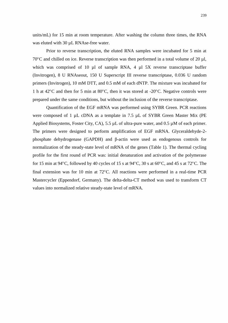
239
units/mL) for 15 min at room temperature. After washing the column three times, the RNA
was eluted with 30 µL RNAse-free water.
Prior to reverse transcription, the eluted RNA samples were incubated for 5 min at
70C and chilled on ice. Reverse transcription was then performed in a total volume of 20 µl,
which was comprised of 10 µl of sample RNA, 4 µl 5X reverse transcriptase buffer
(Invitrogen), 8 U RNAseout, 150 U Superscript III reverse transcriptase, 0.036 U random
primers (Invitrogen), 10 mM DTT, and 0.5 mM of each dNTP. The mixture was incubated for
1 h at 42C and then for 5 min at 80C, then it was stored at -20C. Negative controls were
prepared under the same conditions, but without the inclusion of the reverse transcriptase.
Quantification of the EGF mRNA was performed using SYBR Green. PCR reactions
were composed of 1 µL cDNA as a template in 7.5 µL of SYBR Green Master Mix (PE
Applied Biosystems, Foster City, CA), 5.5 µL of ultra-pure water, and 0.5 µM of each primer.
The primers were designed to perform amplification of EGF mRNA. Glyceraldehyde-2-
phosphate dehydrogenase (GAPDH) and β-actin were used as endogenous controls for
normalization of the steady-state level of mRNA of the genes (Table 1). The thermal cycling
profile for the first round of PCR was: initial denaturation and activation of the polymerase
for 15 min at 94C, followed by 40 cycles of 15 s at 94C, 30 s at 60C, and 45 s at 72C. The
final extension was for 10 min at 72C. All reactions were performed in a real-time PCR
Mastercycler (Eppendorf, Germany). The delta-delta-CT method was used to transform CT
values into normalized relative steady-state level of mRNA.
240
Table 1. Oligonucleotide primers used for PCR analysis of goat cells and tissues.
Target
gene
Primer sequence (5´ 3´) Sense Position GenBank
accession nº
GAPDH TGTTTGTGATGGGCGTGAACCA
s 287-309 GI:27525390
ATGGCGTGGACAGTGGTCATAA
as 440-462
β-actin ACCACTGGCATTGTCATGGACTCT
s 187-211 GI:28628620
TCCTTGATGTCACGGACGATTTCC
as 386-410
UBQ GAAGATGGCCGCACTCTTCTGAT
ATCCTGGATCTTGGCCTTCACGTT
s
as
607-631
756-780
GI:57163956
EGF
FSH-R
CCAGGTTCTCTTAAGTGC
ACCAAGAGCTGCTCTCTG
s
as
48-65
151-168
GI: 1706938
GI:95768228
AGGCAAATGTGTTCTCCAACCTGC
TGGAAGGCATCAGGGTCGATGTAT
s
as
250-274
316-340
s,sense; as, antisense
2.3. Effect of EGF on the survival and growth of goat secondary follicles and expression
of FSH-R and EGF
2.3.1. Isolation and selection of caprine preantral follicles
Ovaries (n=40) were collected at a local slaughterhouse from 20 adult (1 - 3 years old)
mixed-breed goats, and we made a total of 4 replicas (5 goats/replica). Immediately
postmortem, the ovaries were washed in 70% alcohol, followed by two rinses in Minimum
Essential Medium (MEM) supplemented with 100 µg/mL penicillin and 100 µg/mL
streptomycin. The ovaries were transported within 1 hour to the laboratory in MEM at 4°C
(Chaves et al., 2008).
In the laboratory, the surrounding fat tissue and ligaments were stripped off from the
ovaries. Ovarian cortical slices (1 - 2 mm in diameter) were cut from the ovarian surface using
a surgical blade under sterile conditions. Then, the ovarian cortex was placed in a
241
fragmentation medium, consisting of MEM
plus HEPES. Secondary follicles of
approximately 200 μm in diameter were visualized under a stereomicroscope (SMZ 645
Nikon, Tokyo, Japan) and manually dissected from the strips of ovarian cortex using 26 gauge
(26 G) needles. After isolation, follicles were transferred to 100 µL drops containing fresh
medium under mineral oil to further evaluate the follicular quality. Follicles with a visible
oocyte, surrounded by granulosa cells, an intact basement membrane and no antral cavity
were selected for culture.
2.3.2. Caprine preantral follicles culture
For in vitro studies, after selection, follicles were individually cultured in 100 µL
drops of culture medium in Petri dishes (60 x 15 mm, Corning, USA) under mineral oil.
Control culture medium, called α-MEM+
, consisted of α-MEM (pH 7.2 - 7.4) supplemented
with 3.0 mg/mL bovine serum albumin (BSA), ITS (insulin 10 µg/mL, transferrin 5.5 µg/mL
and selenium 5 ng/mL), 2 mM glutamine, 2 mM hypoxanthine and 50 μg/mL of ascorbic acid
under mineral oil. Incubation was conducted at 39°C, 5% CO2 in air for six days. Fresh media
were prepared and incubated for one hour prior to use. Preantral follicles obtained from each
animal were randomly distributed in the following treatments: α-MEM+ alone or
supplemented with 100 ng/mL recombinant bovine FSH (rbFSH: Nanocore, São Paulo, SP,
Brazil), 10 ng/mL recombinant human epidermal growth factor (rhEGF: Cell Sciences,
Canton, MA, USA) or both. These concentrations of rbFSH and rhEGF were those that
promoted the best results in the in vitro development of goat preantral follicles in previous
studies of our laboratory (Celestino et al., 2009; Saraiva et al., unpublished data). Every other
day, 60 μL of the culture media were replaced for fresh medium. The culture was replicated
four times, and a mean number of 37 follicles were used per treatment.
2.3.3. Morphological evaluation of follicle development
Follicles were classified according to their morphological aspect, and those showing
morphological signs of degeneration, such as darkness of oocytes and surrounding cumulus
cells or those with misshapen oocytes, were classified as degenerated. Follicular diameter was
measured only in healthy follicles in the x and y dimensions (90o) by using an ocular
micrometer (100x magnification) inserted into a stereomicroscope (SMZ 645 Nikon, Tóquio,
242
Japão) every other day of culture. Regarding the follicular growth, the mean increase in
follicular diameter was calculated as follows: the diameter of viable follicles at day 6 minus
the diameter of viable follicles at day 0 divided by the total number of viable follicles at day
6. In addition, the percentages of secondary follicles that reached the antrum formation in
vitro were determined. Antral cavity formation was defined as a visible translucent cavity
within the granulosa cell layers.
2.3.4. Steady-state level of FSH-R and EGF mRNA in goat ovarian follicles cultured in vitro
To evaluate the effect of EGF, FSH or both combined on the FSH-R and EGF mRNA
expression after a six-day culture period, for each treatment, groups of ten follicles were
collected at the end of the culture period and stored at -80°C until the extraction of total RNA.
Quantification of mRNA was performed as described previously, and the primers for EGF
and FSH-R are shown in Table 1. β-actin and ubiquitin (UBQ) were used as endogenous
controls for normalization of gene expression (Table 1).
2.4. Statistical analysis
Data of mRNA expression in primordial, primary and secondary follicles were
analyzed with the Kruskal-Wallis non-parametric test, while t test was used for paired
comparisons of mRNA expression in small and large antral follicles (P<0.05). Data from the
follicular survival and antrum formation after in vitro culture for each treatment were
compared using the Chi-square test, and the results were expressed as percentages. Follicular
diameter and growth rate, as well as EGF and FSH-R mRNA levels after culture did not show
homoscedasticity, and these parameters were analyzed using the Kruskal-Wallis non-
parametric test (SAS, 1999). The results were expressed as the mean ± standard error of the
mean (SEM), and differences were considered to be significant when P<0.05.
3. Results
3.1. Steady-state level of EGF in goat ovarian follicles
Quantification of mRNA expression demonstrated that secondary follicles had
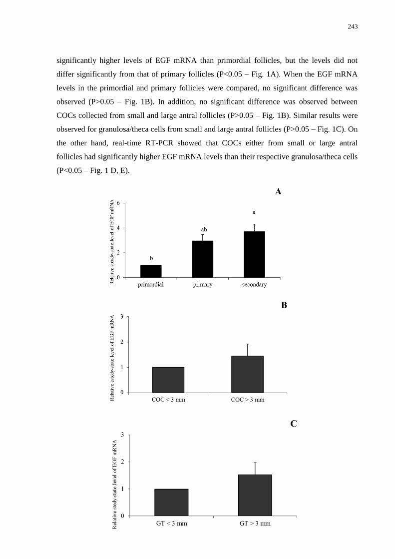
243
significantly higher levels of EGF mRNA than primordial follicles, but the levels did not
differ significantly from that of primary follicles (P<0.05 – Fig. 1A). When the EGF mRNA
levels in the primordial and primary follicles were compared, no significant difference was
observed (P>0.05 – Fig. 1B). In addition, no significant difference was observed between
COCs collected from small and large antral follicles (P>0.05 – Fig. 1B). Similar results were
observed for granulosa/theca cells from small and large antral follicles (P>0.05 – Fig. 1C). On
the other hand, real-time RT-PCR showed that COCs either from small or large antral
follicles had significantly higher EGF mRNA levels than their respective granulosa/theca cells
(P<0.05 – Fig. 1 D, E).
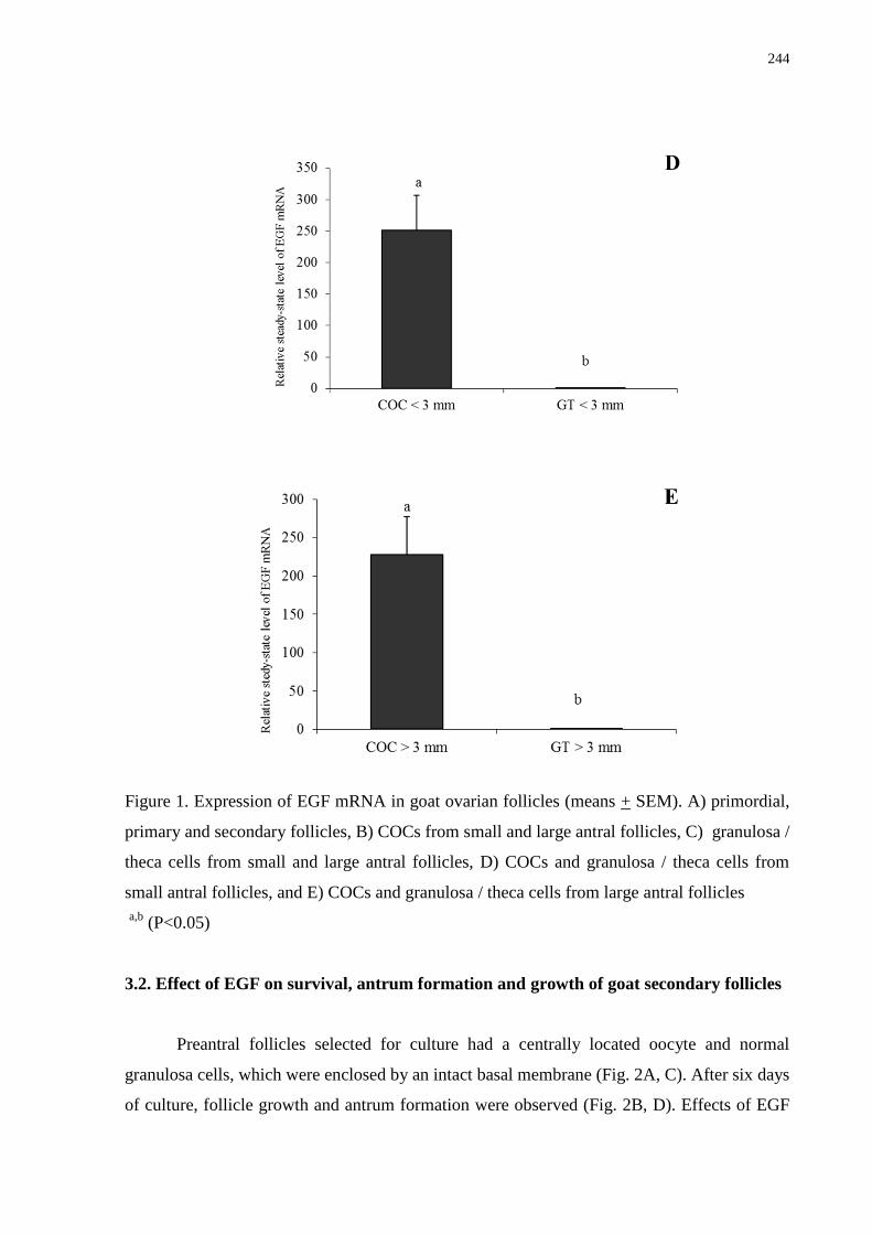
244
Figure 1. Expression of EGF mRNA in goat ovarian follicles (means + SEM). A) primordial,
primary and secondary follicles, B) COCs from small and large antral follicles, C) granulosa /
theca cells from small and large antral follicles, D) COCs and granulosa / theca cells from
small antral follicles, and E) COCs and granulosa / theca cells from large antral follicles
a,b
(P<0.05)
3.2. Effect of EGF on survival, antrum formation and growth of goat secondary follicles
Preantral follicles selected for culture had a centrally located oocyte and normal
granulosa cells, which were enclosed by an intact basal membrane (Fig. 2A, C). After six days
of culture, follicle growth and antrum formation were observed (Fig. 2B, D). Effects of EGF
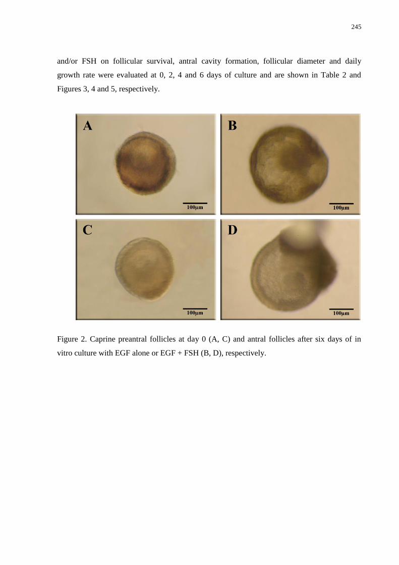
245
and/or FSH on follicular survival, antral cavity formation, follicular diameter and daily
growth rate were evaluated at 0, 2, 4 and 6 days of culture and are shown in Table 2 and
Figures 3, 4 and 5, respectively.
Figure 2. Caprine preantral follicles at day 0 (A, C) and antral follicles after six days of in
vitro culture with EGF alone or EGF + FSH (B, D), respectively.
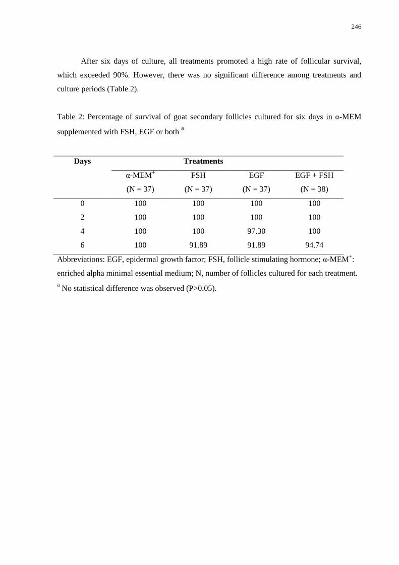
246
After six days of culture, all treatments promoted a high rate of follicular survival,
which exceeded 90%. However, there was no significant difference among treatments and
culture periods (Table 2).
Table 2: Percentage of survival of goat secondary follicles cultured for six days in α-MEM
supplemented with FSH, EGF or both a
Days Treatments
α-MEM+
(N = 37)
FSH
(N = 37)
EGF
(N = 37)
EGF + FSH
(N = 38)
0 100 100 100 100
2 100 100 100 100
4 100 100 97.30 100
6 100 91.89 91.89 94.74
Abbreviations: EGF, epidermal growth factor; FSH, follicle stimulating hormone; α-MEM+:
enriched alpha minimal essential medium; N, number of follicles cultured for each treatment.
a No statistical difference was observed (P>0.05).
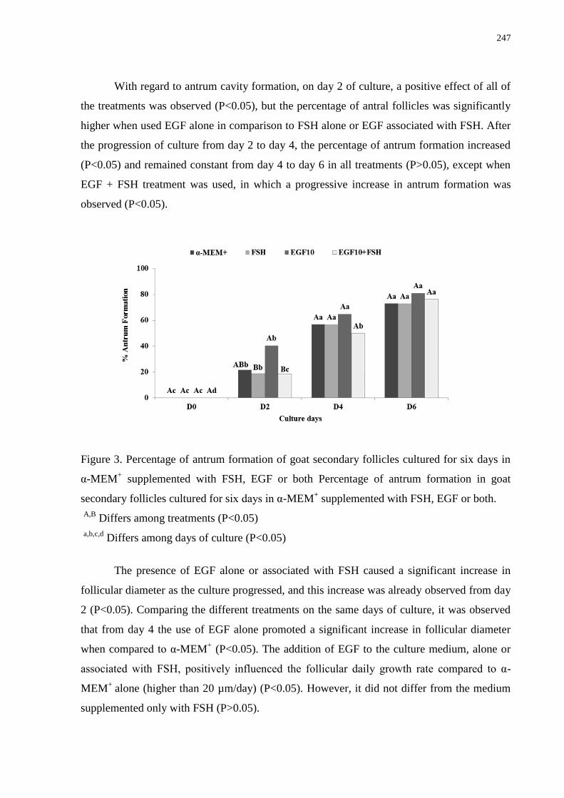
247
With regard to antrum cavity formation, on day 2 of culture, a positive effect of all of
the treatments was observed (P<0.05), but the percentage of antral follicles was significantly
higher when used EGF alone in comparison to FSH alone or EGF associated with FSH. After
the progression of culture from day 2 to day 4, the percentage of antrum formation increased
(P<0.05) and remained constant from day 4 to day 6 in all treatments (P>0.05), except when
EGF + FSH treatment was used, in which a progressive increase in antrum formation was
observed (P<0.05).
Figure 3. Percentage of antrum formation of goat secondary follicles cultured for six days in
α-MEM+ supplemented with FSH, EGF or both Percentage of antrum formation in goat
secondary follicles cultured for six days in α-MEM+ supplemented with FSH, EGF or both.
A,B
Differs among treatments (P<0.05)
a,b,c,d
Differs among days of culture (P<0.05)
The presence of EGF alone or associated with FSH caused a significant increase in
follicular diameter as the culture progressed, and this increase was already observed from day
2 (P<0.05). Comparing the different treatments on the same days of culture, it was observed
that from day 4 the use of EGF alone promoted a significant increase in follicular diameter
when compared to α-MEM+ (P<0.05). The addition of EGF to the culture medium, alone or
associated with FSH, positively influenced the follicular daily growth rate compared to α-
MEM+
alone (higher than 20 µm/day) (P<0.05). However, it did not differ from the medium
supplemented only with FSH (P>0.05).
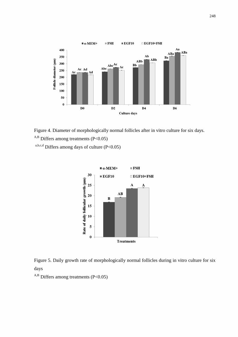
248
Figure 4. Diameter of morphologically normal follicles after in vitro culture for six days.
A,B Differs among treatments (P<0.05)
a,b,c,d
Differs among days of culture (P<0.05)
Figure 5. Daily growth rate of morphologically normal follicles during in vitro culture for six
days
A,B Differs among treatments (P<0.05)
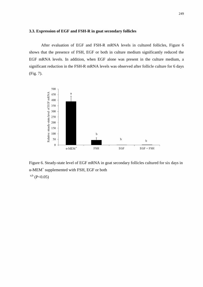
249
3.3. Expression of EGF and FSH-R in goat secondary follicles
After evaluation of EGF and FSH-R mRNA levels in cultured follicles, Figure 6
shows that the presence of FSH, EGF or both in culture medium significantly reduced the
EGF mRNA levels. In addition, when EGF alone was present in the culture medium, a
significant reduction in the FSH-R mRNA levels was observed after follicle culture for 6 days
(Fig. 7).
Figure 6. Steady-state level of EGF mRNA in goat secondary follicles cultured for six days in
α-MEM+ supplemented with FSH, EGF or both
a,b
(P<0.05)
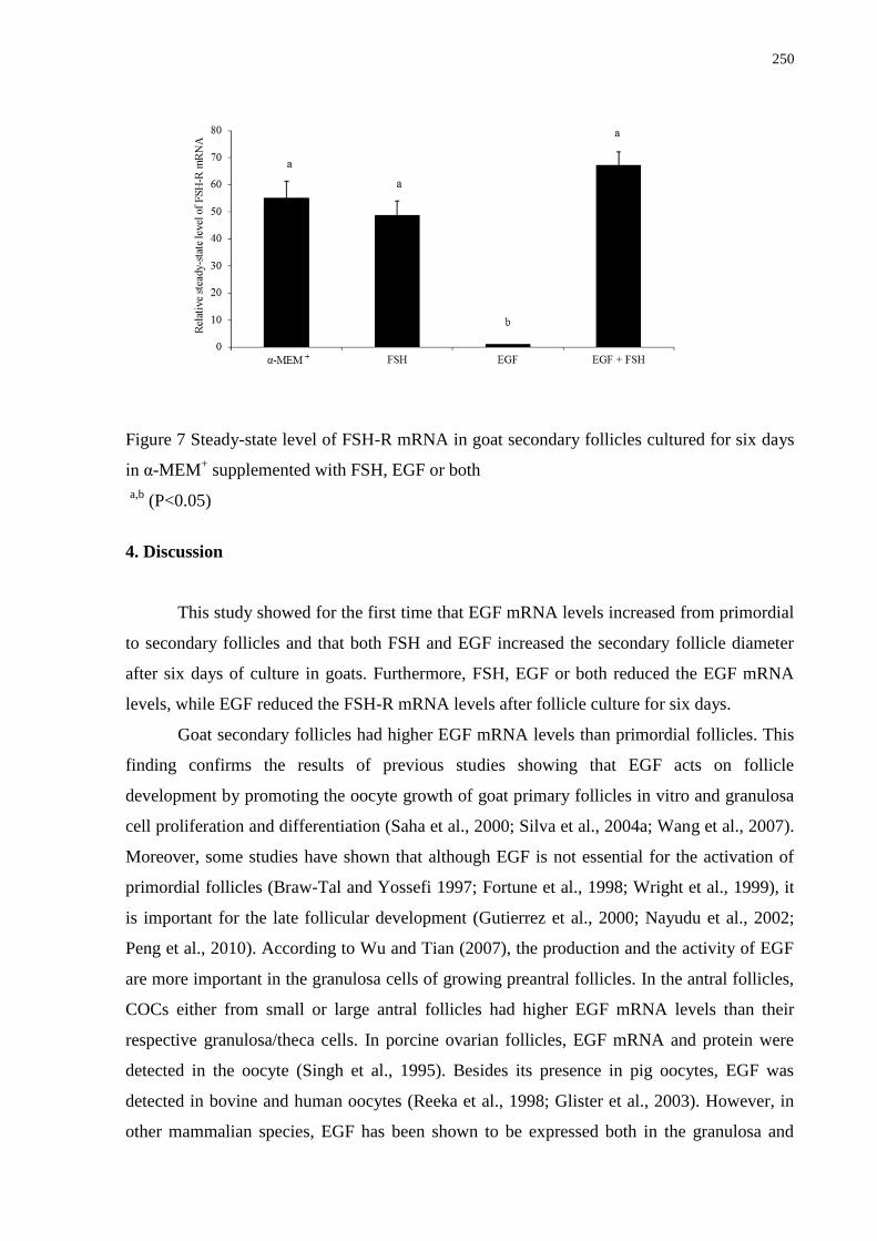
250
Figure 7 Steady-state level of FSH-R mRNA in goat secondary follicles cultured for six days
in α-MEM+ supplemented with FSH, EGF or both
a,b
(P<0.05)
4. Discussion
This study showed for the first time that EGF mRNA levels increased from primordial
to secondary follicles and that both FSH and EGF increased the secondary follicle diameter
after six days of culture in goats. Furthermore, FSH, EGF or both reduced the EGF mRNA
levels, while EGF reduced the FSH-R mRNA levels after follicle culture for six days.
Goat secondary follicles had higher EGF mRNA levels than primordial follicles. This
finding confirms the results of previous studies showing that EGF acts on follicle
development by promoting the oocyte growth of goat primary follicles in vitro and granulosa
cell proliferation and differentiation (Saha et al., 2000; Silva et al., 2004a; Wang et al., 2007).
Moreover, some studies have shown that although EGF is not essential for the activation of
primordial follicles (Braw-Tal and Yossefi 1997; Fortune et al., 1998; Wright et al., 1999), it
is important for the late follicular development (Gutierrez et al., 2000; Nayudu et al., 2002;
Peng et al., 2010). According to Wu and Tian (2007), the production and the activity of EGF
are more important in the granulosa cells of growing preantral follicles. In the antral follicles,
COCs either from small or large antral follicles had higher EGF mRNA levels than their
respective granulosa/theca cells. In porcine ovarian follicles, EGF mRNA and protein were
detected in the oocyte (Singh et al., 1995). Besides its presence in pig oocytes, EGF was
detected in bovine and human oocytes (Reeka et al., 1998; Glister et al., 2003). However, in
other mammalian species, EGF has been shown to be expressed both in the granulosa and
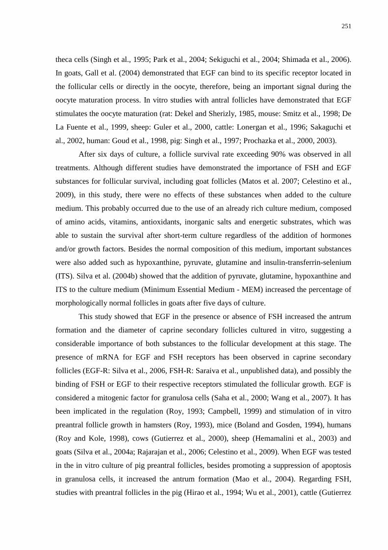
251
theca cells (Singh et al., 1995; Park et al., 2004; Sekiguchi et al., 2004; Shimada et al., 2006).
In goats, Gall et al. (2004) demonstrated that EGF can bind to its specific receptor located in
the follicular cells or directly in the oocyte, therefore, being an important signal during the
oocyte maturation process. In vitro studies with antral follicles have demonstrated that EGF
stimulates the oocyte maturation (rat: Dekel and Sherizly, 1985, mouse: Smitz et al., 1998; De
La Fuente et al., 1999, sheep: Guler et al., 2000, cattle: Lonergan et al., 1996; Sakaguchi et
al., 2002, human: Goud et al., 1998, pig: Singh et al., 1997; Prochazka et al., 2000, 2003).
After six days of culture, a follicle survival rate exceeding 90% was observed in all
treatments. Although different studies have demonstrated the importance of FSH and EGF
substances for follicular survival, including goat follicles (Matos et al. 2007; Celestino et al.,
2009), in this study, there were no effects of these substances when added to the culture
medium. This probably occurred due to the use of an already rich culture medium, composed
of amino acids, vitamins, antioxidants, inorganic salts and energetic substrates, which was
able to sustain the survival after short-term culture regardless of the addition of hormones
and/or growth factors. Besides the normal composition of this medium, important substances
were also added such as hypoxanthine, pyruvate, glutamine and insulin-transferrin-selenium
(ITS). Silva et al. (2004b) showed that the addition of pyruvate, glutamine, hypoxanthine and
ITS to the culture medium (Minimum Essential Medium - MEM) increased the percentage of
morphologically normal follicles in goats after five days of culture.
This study showed that EGF in the presence or absence of FSH increased the antrum
formation and the diameter of caprine secondary follicles cultured in vitro, suggesting a
considerable importance of both substances to the follicular development at this stage. The
presence of mRNA for EGF and FSH receptors has been observed in caprine secondary
follicles (EGF-R: Silva et al., 2006, FSH-R: Saraiva et al., unpublished data), and possibly the
binding of FSH or EGF to their respective receptors stimulated the follicular growth. EGF is
considered a mitogenic factor for granulosa cells (Saha et al., 2000; Wang et al., 2007). It has
been implicated in the regulation (Roy, 1993; Campbell, 1999) and stimulation of in vitro
preantral follicle growth in hamsters (Roy, 1993), mice (Boland and Gosden, 1994), humans
(Roy and Kole, 1998), cows (Gutierrez et al., 2000), sheep (Hemamalini et al., 2003) and
goats (Silva et al., 2004a; Rajarajan et al., 2006; Celestino et al., 2009). When EGF was tested
in the in vitro culture of pig preantral follicles, besides promoting a suppression of apoptosis
in granulosa cells, it increased the antrum formation (Mao et al., 2004). Regarding FSH,
studies with preantral follicles in the pig (Hirao et al., 1994; Wu et al., 2001), cattle (Gutierrez
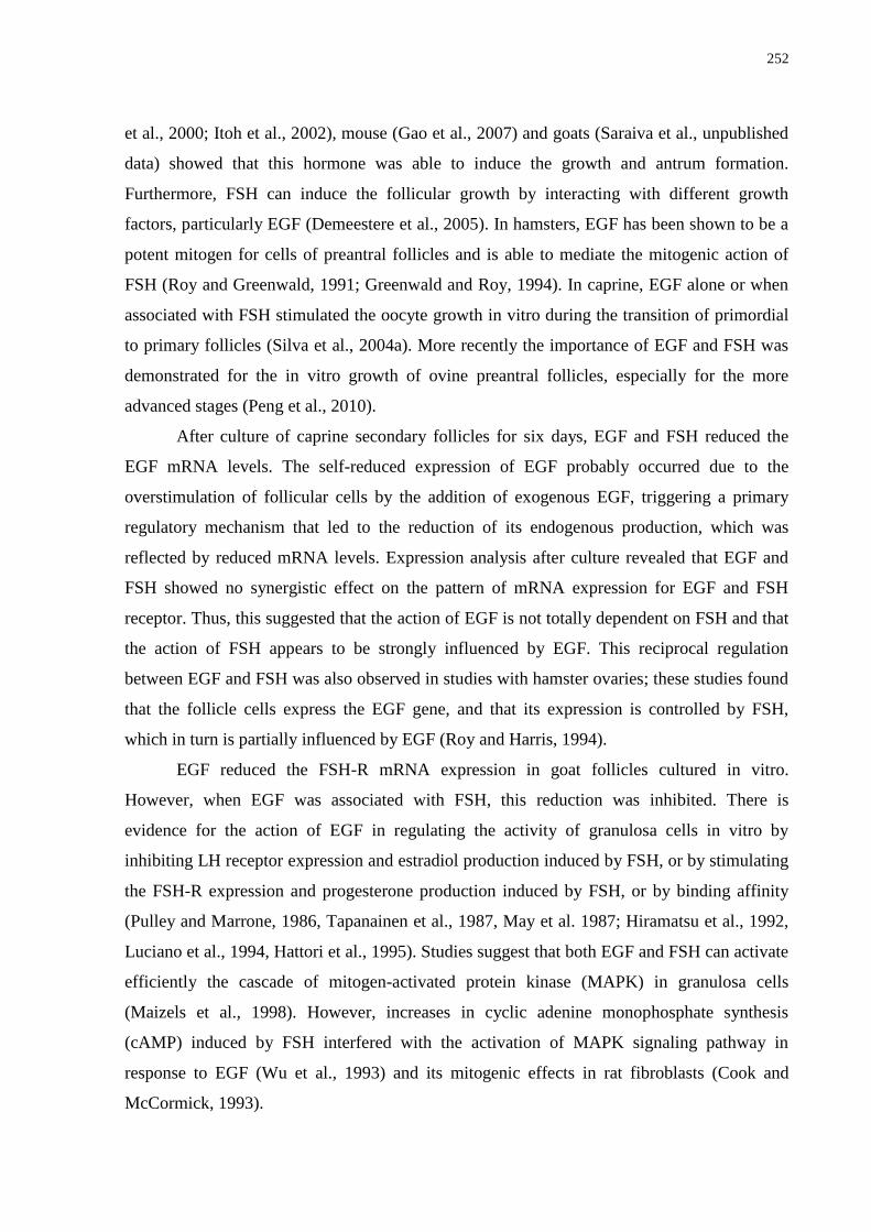
252
et al., 2000; Itoh et al., 2002), mouse (Gao et al., 2007) and goats (Saraiva et al., unpublished
data) showed that this hormone was able to induce the growth and antrum formation.
Furthermore, FSH can induce the follicular growth by interacting with different growth
factors, particularly EGF (Demeestere et al., 2005). In hamsters, EGF has been shown to be a
potent mitogen for cells of preantral follicles and is able to mediate the mitogenic action of
FSH (Roy and Greenwald, 1991; Greenwald and Roy, 1994). In caprine, EGF alone or when
associated with FSH stimulated the oocyte growth in vitro during the transition of primordial
to primary follicles (Silva et al., 2004a). More recently the importance of EGF and FSH was
demonstrated for the in vitro growth of ovine preantral follicles, especially for the more
advanced stages (Peng et al., 2010).
After culture of caprine secondary follicles for six days, EGF and FSH reduced the
EGF mRNA levels. The self-reduced expression of EGF probably occurred due to the
overstimulation of follicular cells by the addition of exogenous EGF, triggering a primary
regulatory mechanism that led to the reduction of its endogenous production, which was
reflected by reduced mRNA levels. Expression analysis after culture revealed that EGF and
FSH showed no synergistic effect on the pattern of mRNA expression for EGF and FSH
receptor. Thus, this suggested that the action of EGF is not totally dependent on FSH and that
the action of FSH appears to be strongly influenced by EGF. This reciprocal regulation
between EGF and FSH was also observed in studies with hamster ovaries; these studies found
that the follicle cells express the EGF gene, and that its expression is controlled by FSH,
which in turn is partially influenced by EGF (Roy and Harris, 1994).
EGF reduced the FSH-R mRNA expression in goat follicles cultured in vitro.
However, when EGF was associated with FSH, this reduction was inhibited. There is
evidence for the action of EGF in regulating the activity of granulosa cells in vitro by
inhibiting LH receptor expression and estradiol production induced by FSH, or by stimulating
the FSH-R expression and progesterone production induced by FSH, or by binding affinity
(Pulley and Marrone, 1986, Tapanainen et al., 1987, May et al. 1987; Hiramatsu et al., 1992,
Luciano et al., 1994, Hattori et al., 1995). Studies suggest that both EGF and FSH can activate
efficiently the cascade of mitogen-activated protein kinase (MAPK) in granulosa cells
(Maizels et al., 1998). However, increases in cyclic adenine monophosphate synthesis
(cAMP) induced by FSH interfered with the activation of MAPK signaling pathway in
response to EGF (Wu et al., 1993) and its mitogenic effects in rat fibroblasts (Cook and
McCormick, 1993).
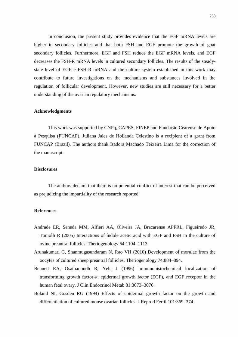
253
In conclusion, the present study provides evidence that the EGF mRNA levels are
higher in secondary follicles and that both FSH and EGF promote the growth of goat
secondary follicles. Furthermore, EGF and FSH reduce the EGF mRNA levels, and EGF
decreases the FSH-R mRNA levels in cultured secondary follicles. The results of the steady-
state level of EGF e FSH-R mRNA and the culture system established in this work may
contribute to future investigations on the mechanisms and substances involved in the
regulation of follicular development. However, new studies are still necessary for a better
understanding of the ovarian regulatory mechanisms.
Acknowledgments
This work was supported by CNPq, CAPES, FINEP and Fundação Cearense de Apoio
à Pesquisa (FUNCAP). Juliana Jales de Hollanda Celestino is a recipient of a grant from
FUNCAP (Brazil). The authors thank Isadora Machado Teixeira Lima for the correction of
the manuscript.
Disclosures
The authors declare that there is no potential conflict of interest that can be perceived
as prejudicing the impartiality of the research reported.
References
Andrade ER, Seneda MM, Alfieri AA, Oliveira JA, Bracarense APFRL, Figueiredo JR,
Toniolli R (2005) Interactions of índole acetic acid with EGF and FSH in the culture of
ovine preantral follicles. Theriogenology 64:1104–1113.
Arunakumari G, Shanmugasundaram N, Rao VH (2010) Development of morulae from the
oocytes of cultured sheep preantral follicles. Theriogenology 74:884–894.
Bennett RA, Osathanondh R, Yeh, J (1996) Immunohistochemical localization of
transforming growth factor-α, epidermal growth factor (EGF), and EGF receptor in the
human fetal ovary. J Clin Endocrinol Metab 81:3073–3076.
Boland NI, Gosden RG (1994) Effects of epidermal growth factor on the growth and
differentiation of cultured mouse ovarian follicles. J Reprod Fertil 101:369–374.
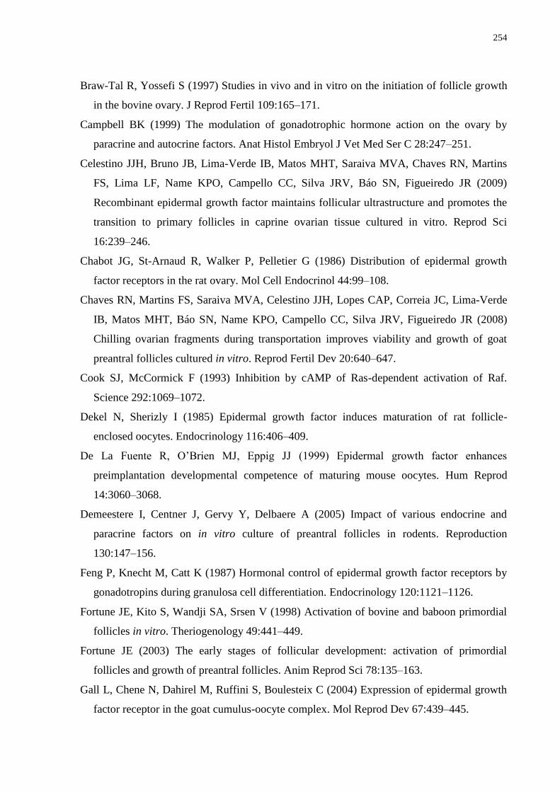
254
Braw-Tal R, Yossefi S (1997) Studies in vivo and in vitro on the initiation of follicle growth
in the bovine ovary. J Reprod Fertil 109:165–171.
Campbell BK (1999) The modulation of gonadotrophic hormone action on the ovary by
paracrine and autocrine factors. Anat Histol Embryol J Vet Med Ser C 28:247–251.
Celestino JJH, Bruno JB, Lima-Verde IB, Matos MHT, Saraiva MVA, Chaves RN, Martins
FS, Lima LF, Name KPO, Campello CC, Silva JRV, Báo SN, Figueiredo JR (2009)
Recombinant epidermal growth factor maintains follicular ultrastructure and promotes the
transition to primary follicles in caprine ovarian tissue cultured in vitro. Reprod Sci
16:239–246.
Chabot JG, St-Arnaud R, Walker P, Pelletier G (1986) Distribution of epidermal growth
factor receptors in the rat ovary. Mol Cell Endocrinol 44:99–108.
Chaves RN, Martins FS, Saraiva MVA, Celestino JJH, Lopes CAP, Correia JC, Lima-Verde
IB, Matos MHT, Báo SN, Name KPO, Campello CC, Silva JRV, Figueiredo JR (2008)
Chilling ovarian fragments during transportation improves viability and growth of goat
preantral follicles cultured in vitro. Reprod Fertil Dev 20:640–647.
Cook SJ, McCormick F (1993) Inhibition by cAMP of Ras-dependent activation of Raf.
Science 292:1069–1072.
Dekel N, Sherizly I (1985) Epidermal growth factor induces maturation of rat follicle-
enclosed oocytes. Endocrinology 116:406–409.
De La Fuente R, O‘Brien MJ, Eppig JJ (1999) Epidermal growth factor enhances
preimplantation developmental competence of maturing mouse oocytes. Hum Reprod
14:3060–3068.
Demeestere I, Centner J, Gervy Y, Delbaere A (2005) Impact of various endocrine and
paracrine factors on in vitro culture of preantral follicles in rodents. Reproduction
130:147–156.
Feng P, Knecht M, Catt K (1987) Hormonal control of epidermal growth factor receptors by
gonadotropins during granulosa cell differentiation. Endocrinology 120:1121–1126.
Fortune JE, Kito S, Wandji SA, Srsen V (1998) Activation of bovine and baboon primordial
follicles in vitro. Theriogenology 49:441–449.
Fortune JE (2003) The early stages of follicular development: activation of primordial
follicles and growth of preantral follicles. Anim Reprod Sci 78:135–163.
Gall L, Chene N, Dahirel M, Ruffini S, Boulesteix C (2004) Expression of epidermal growth
factor receptor in the goat cumulus-oocyte complex. Mol Reprod Dev 67:439–445.
255
Gao MZ, Wang Y, Wu X (2007) In-vitro maturation of immature oocytes from preantral
follicles in prepuberal mice. J Reprod Contracept 18:25–32.
Garnett K, Wang J, Roy SK (2002) Spatiotemporal expression of epidermal growth factor
receptor messenger RNA and protein in the hamster ovary: follicle stage specific
differential modulation by follicle-stimulating hormone, luteinizing hormone, estradiol,
and progesterone. Biol Reprod 67:1593–1604.
Gospodarowicz D, Bialecki H (1979) Fibroblast and epidermal growth factors are mitogenic
agents for cultured granulose cells of rodent, porcine and human origin. Endocrinology
104:757–764.
Goud PT, Goud AP, Qian C, Laverge H, Van der Elst J, De Sutter P, Dhont M (1998) In vitro
maturation of human germinal vesicle stage oocytes: role of cumulus cells and epidermal
growth factor in the culture medium. Hum Reprod 13:1638–1644.
Greenwald GS, Roy SK (1994) Follicular development and its control. In: Knobil E, Neill JD
(ed) The Physiology of Reproduction. Raven Press, New York, pp. 629–724.
Guler A, Poulin N, Mermillod P, Terqui M, Cognie Y (2000) Effect of growth factors, EGF
and IGF-I, and estradiol on in vitro maturation of sheep oocytes. Theriogenology, 54:209–
218.
Gupta PSP, Ramesh HS, Manjunatha BM, Nandi S, Ravindra JP (2008) Production of buffalo
embryos using oocytes from in vitro grown preantral follicles. Zygote 16:57–63.
Gutierrez CG, Ralph JH, Telfer EE, Wilmut I, Webb R (2000) Growth and antrum formation
of bovine preantral follicles in long-term culture in vitro. Biol Reprod 62:1322–1328.
Hattori MA, Yoshino E, Shinohara Y, Horiuchi R, Kojima I (1995) A novel action of
epidermal growth factor in rat granulosa cells: its potentiation of gonadotrophin action. J
Mol Endocrinol 15:283–291.
Hemamalini NC, Rao BS, Tamilmani G, Amarnath D, Vagdevi R, Naidu KS, Reddy KK, Rao
VH (2003) Influence of transforming growth factor-a, insulin-like growth factor-II,
epidermal growth factor or follicle stimulating hormone on in vitro development of
preantral follicles in sheep. Small Rumin Res 50:11–22.
Hill JL, Hammar K, Smith PJ, Gross DJ (1999) Stage dependent effects of epidermal growth
factor onCa2+ efflux in mouse oocytes. Mol Reprod Dev 53:244–253.
Hiramatsu S, Maruo T, Matsuo H, Mochizuki M (1992) Effects of epidermal growth factor on
the proliferation and differentiation of porcine granulosa cells cultured in vitro. Acta
Obstet Gynecol Jpn 44:55–61.
256
Hirao Y, Nagai T, Kubo M, Miyano T, Miyake M, Kato S (1994) In vitro growth and
maturation of porcine oocytes. J Reprod Fertil 100:333–339.
Itoh T, Kacchi M, Abe H, Sendai Y, Hoshi H (2002) Growth, antrum formation, and estradiol
production of bovine preantral follicles cultured in a serum-free medium. Biol Reprod
67:1099–1105.
Lonergan P, Carolan C, Van Langendonckt A, Donnay I, Khatir H, Mermillod P (1996) Role
of epidermal growth factor in bovine oocyte maturation and preimplantation embryo
development in vitro. Biol Reprod 54:1420–1429.
Luciano AM, Pappalardo A, Ray C, Peluso JJ (1994) Epidermal growth factor inhibits large
granulose cell apoptosis by stimulating progesterone synthesis and regulating the
distribution of intracellular free calcium. Biol Reprod 51:646–654.
Lucci CM, Amorim CA, Báo SN, Figueiredo JR, Rodrigues APR, Silva JRV, Goncalves PBD
(1999) Effect of the interval of serial sections of ovarian in the tissue chopper on the
number of isolated caprine preantral follicles. Anim Reprod Sci 56: 39–49.
Maizels ET, Cottom J, Jones JC, Hunzicker-Dunn M (1998) Follicle stimulating hormone
(FSH) activates the p38 mitogen-activated protein kinase pathway, inducing small heat
shock protein phosphorylation and cell rounding in immature rat ovarian granulose cells.
Endocrinology 139:3353–3356.
Mao J, Smith MF, Rucker EB, Wu GM, McCauley TC, Cantley TC, Prather RS, Didion BA,
Day BN (2004) Effect of epidermal growth factor and insulin-like growth factor I on
porcine preantral follicular growth, antrum formation, and stimulation of granulosal cell
proliferation and suppression of apoptosis in vitro. J Anim Sci 82:1967–1975.
Maruo T, Ladines-Llave CA, Samoto T, Matsuo H, Manalo AS, Ito H, Mochizuki M (1993)
Expression of epidermal growth factor and its receptor in the human ovary during
follicular growth and regression. Endocrinology 132:924–931.
Matos MHT, Lima-Verde IB, Luque MCA, Maia-Jr JE, Silva JRV, Celestino JJH, Martins
FS, Báo SN, Lucci CM, Figueiredo JR (2007) Essential role of follicle stimulating
hormone in the maintenance of caprine preantral follicle viability in vitro. Zygote 15:173–
182.
May JV, Buck PA, Schomberg DW (1987) Epidermal growth factor enhances [125
I]iodo-
follicle-stimulating hormone binding by cultured porcine granulosa cells. Endocrinology
120:2413–2420.
257
Miyoshi T, Otsuka F, Yamashita M, Inagaki K, Nakamura E, Tsukamoto N, Takeda M,
Suzuki J, Makino H (2010) Functional relationship between fibroblast growth factor-8 and
bone morphogenetic proteins in regulating steroidogenesis by rat granulosa cells. Mol Cell
Endocrinol 325:84–92.
Morbeck DE, Flowers WL, Britt JH (1993) Response of porcine granulosa cells isolated from
primary and secondary follicles to FSH, 8-bromo-cAMP and epidermal growth factor in
vitro. J Reprod Fertil 99, 577–584.
Nayudu PL, Vitt UA, De Tomasi JB, Pancharatna K, Ulloa-Aguirre A (2002) Intact follicle
culture: what it can tell us about the roles of FSH glycoforms during follicle development.
Reprod BioMed Online 5:240–253.
Park JY, Su YQ, Ariga M, Law E, Jin SL, Conti M (2004) EGF-like growth factors as
mediators of LH action in the ovulatory follicle. Science 303:682–684.
Peng X, Yang M, Wang L, Tong C, Guo Z (2010) In vitro culture of sheep lamb ovarian
cortical tissue in a sequential culture medium. J Assist Reprod Genet 27:247–257.
Prochazka R, Srsen V, Nagyova E, Miyano T, Flechon J.E (2000) Developmental regulation
of effect of epidermal growth factor on porcine oocyte-cumulus cell complexes: nuclear
maturation, expansion, and F-actin remodeling. Mol Reprod Dev 56:63–73.
Prochazka R, Kalab P, Nagyova E (2003) Epidermal growth factor-receptor tyrosine kinase
activity regulates expansion of porcine oocyte–cumulus cell complexes in vitro. Biol
Reprod 68:797–803.
Pulley DD, Marrone BL (1986) Inhibitory action of epidermal growth factor on progesterone
biosynthesis in hen granulose cells during short term culture: two sites of action.
Endocrinology 118:2284–2291.
Qu JP, Godin PA, Nisolle M, Donnez J (2000) Distribution of epidermal growth factor
receptor expression of primordial follicles in human ovarian tissue before and after
cryopreservation. Hum. Reprod 15:302–310.
Rajarajan K, Rao BS, Vagdevi R, Talmimani G, Arunakumari G, Sreenu M, Amarnath D,
Naik BR, Rao VH (2006) Effect of various growth factors on the in vitro development of
goat preantral follicles. Small Rumin Res 63:204–212.
Reeka N, Berg FD, Brucer C (1998) Presence of transforming growth factor alpha and
epidermal growth factor in human ovarian tissue and follicular fluid. Hum Reprod
13:2199–2205.
258
Riese 2nd
DJ, Stern DF (1998) Specificity within the EGF family/ErbB receptor family
signaling network. Bioessays 20:41–48.
Romano M, Kraus ER, Boland CR, Coffey RJ (1994) Comparison between transforming
growth factor alpha and epidermal growth factor in the protection of rat gastric mucosa
against drug-induced injury. Ital J Gastroenterol 26:223–228.
Roy SK (1993) Epidermal growth factor and transforming growth factor-beta modulation of
follicle-stimulating hormone-induced deoxyribonucleic acid synthesis in hamster preantral
and early antral follicles. Biol Reprod 48:552–557.
Roy SK, Greenwald GS (1990) Immunohistochemical localisation of epidermal growth
factor-like activity in the hamster ovary with a polyclonal antibody. Endocrinology
126:1309–1317.
Roy SK, Greenwald GS (1991) Mediation of follicle-stimulating hormone action on follicular
deoxyribonucleic acid synthesis by epidermal growth factor. Endocrinology 129:1903–
1908.
Roy SK, Harris SG (1994) Antisense epidermal growth factor oligodeoxynucleotides inhibit
follicle-stimulating hormone-induced in vitro DNA and progesterone synthesis in hamster
preantral follicles. Mol Endocrinol 8:1175–1181.
Roy SK, Kole AR (1998) Ovarian transforming growth factor-beta (TGF-beta) receptors: in
vitro effects of follicle stimulating hormone, epidermal growth factor and TGFbeta on
receptor expression in human preantral follicles. Mol Hum Reprod 4:207–214.
Saha S, Shimizu M, Geshi M, Izaike Y (2000) In vitro culture of bovine preantral follicles.
Anim Reprod Sci 63:27–39.
Sakaguchi M, Dominko T, Yamauchi N, Leibfried-Rutledge ML, Nagai T, First NL (2002)
Possible mechanism for acceleration of meiotic progression of bovine follicular oocytes by
growth factors in vitro. Reproduction 123:135–142.
Saraiva MVA, Rossetto R, Brito IR, Celestino JJH, Silva CMG, Faustino LR, Almeida AP,
Bruno JB, Magalhães DM, Matos MHT, Campello CC, Figueiredo J.R (in press) Dynamic
medium produces caprine embryo from preantral follicles grown in vitro. Reprod Sci.
Sekiguchi T, Mizutani T, Yamada K, Kajitani T, Yazawa T, Yoshino M, Miyamoto K (2004)
Expression of epiregulin and amphiregulin in the rat ovary. J Mol Endocrinol 33:281–291.
Shimada M, Hernandez-Gonzalez I, Gonzalez-Robayana I, Richards JS (2006) Paracrine and
autocrine regulation of epidermal growth factor-like factors in cumulus oocyte complexes
259
and granulosa cells: key roles for prostaglandin synthase 2 and progesterone receptor. Mol
Endocrinol 20:1352–1365.
Silva JRV, van den Hurk R, Matos MHT, Santos RR, Pessoa C, Moraes MO, Figueiredo JR
(2004a) Influences of FSH and EGF on primordial follicles during in vitro culture of
caprine ovarian cortical tissue. Theriogenology 61:1691–1704.
Silva JRV, van den Hurk R, Costa SHF, Andrade ER, Nunes APA, Ferreira FVA, Lôbo RNB,
Figueiredo JR (2004b) Survival and growth of goat primordial follicles after in vitro
culture of ovarian cortical slices in media containing coconut water. Anim Reprod Sci
81:273–286.
Silva JRV, van den Hurk R, Figueiredo JR (2006) Expression of mRNA and protein
localization of epidermal growth factor and its receptor in goat ovaries. Zygote 14:107–
117.
Singh B, Rutledge JM, Armstrong DT (1995) Epidermal growth factor and its receptor gene
expression and peptide localisation in porcine ovarian follicles. Mol Reprod Dev 40:391–
399.
Singh B, Meng L, Rutledge JM, Armstrong DT (1997) Effects of epidermal growth factor and
follicle-stimulating hormone during in vitro maturation on cytoplasmic maturation of
porcine oocytes. Mol Reprod Dev 46:401–407.
Smitz J, Cortvrindt R, Hu Y (1998) Epidermal growth factor combined with recombinant
human chorionic gonadotrophin improves meiotic progression in mouse follicle-enclosed
oocyte culture. Hum Reprod 13:664–669.
Tapanainen J, Leinonen PJ, Tapanainen P, Yamamoto M, Jaffee RB (1987) Regulation of
human granulosa-luteal cell progesterone production and proliferation by gonadotropins
and growth factors. Fertil Steril 48:576–579.
van Tol HT, Bevers MM (1998) Theca cells and theca-cell conditioned medium inhibit the
progression of FSH-induced meiosis of bovine oocytes surrounded by cumulus cells
connected to membrane granulosa. Mol Reprod Dev 51:315–321.
Wang Y, Li J, Wang CY, Kwok AHY, Leung FC (2007) Epidermal growth factor (EGF)
receptor ligands in the chicken ovary: I. Evidence for heparin-binding EGF-like growth
factor (HBEGF) as a potential oocyte-derived signal to control granulose cell proliferation
and HB-EGF and kit ligand expression. Endocrinology 148:3426–3440.
260
Wright CS, Hovatta O, Margara R, Trew G, Winston RML, Franks S, Hardy K (1999) Effects
of follicle-stimulating hormone and serum substitution on the in-vitro growth of human
ovarian follicles. Hum Reprod 14:1555–1562.
Wu J, Dent P, Jelinek T, Wolfman A, Weber MJ, Sturgill TW (1993) Inhibition of the EGF-
activated MAP kinase signaling pathway by adenosine 3‘, 5‘- monophosphate. Science
262:1065–1069.
Wu J, Emery BR, Carrell DT (2001) In vitro growth, maturation, fertilization, and embryonic
development of oocytes from porcine preantral follicles. Biol Reprod 64: 375–381.
Wu J, Tian Q (2007) Role of follicle stimulating hormone and epidermal growth factor in the
development of porcine preantral follicle in vitro. Zygote 15:233–240.
Zhou H, Zhang Y (2005a) Effect of growth factors on in vitro development of caprine
preantral follicle oocytes. Anim Reprod Sci 90:265–272.
Zhou H, Zhang Y (2005b) Regulation of in vitro growth of preantral follicles by growth
factors in goats. Domest Anim Endocrinol 28:235–242.
261
14 CONCLUSÕES
Os RNAm para o KL, a BMP-15 e o EGF foram expressos nos compartimentos
foliculares em todos os estádios de desenvolvimento, sugerindo assim o envolvimento
destes fatores de crescimento no processo de desenvolvimento folicular na espécie
caprina.
A adição de 50 ng/mL de KL ao meio de cultivo promoveu a manutenção da
integridade morfológica e ultraestrutural, o crescimento e a transição folicular em
caprinos para o estádio primário após 7 dias de cultivo in vitro.
A utilização de BMP-15 promoveu a sobrevivência e o crescimento folicular, e
quando adicionado na concentração de 100 ng/mL estimulou o desenvolvimento dos
folículos caprinos até o estádio secundário, assegurando ainda a manutenção da
viabilidade folicular durante 7 dias de cultivo in vitro.
A utilização de EGF promoveu a sobrevivência e a ativação folicular, e quando
adicionado nas concentrações de 1 ou 10 ng/mL, estimulou o desenvolvimento dos
folículos caprinos até o estádio primário, assegurando ainda a manutenção da
integridade ultraestrutural folicular durante 7 dias de cultivo in vitro.
A utilização de EGF promoveu o crescimento dos folículos secundários caprinos e a
diminuição dos níveis de RNAm para o EGF e para o FSH-R após 6 dias de cultivo.
262
15 PERSPECTIVAS
Por meio do cultivo in vitro de folículos pré-antrais é possível se estudar o processo de
foliculogênese, evento esse complexo e ainda não totalmente elucidado. Um conhecimento
aprofundado desse processo pode ser empregado no desenvolvimento de biotecnologias
reprodutivas, aumentando o sucesso das mesmas, inclusive em mamíferos selvagens
ameaçados de extinção, e otimizando as ferramentas já utilizadas para o melhoramento
genético de animais de interesse zootécnico, como os caprinos, ou até mesmo para o
tratamento de infertilidade humana. Existem fortes evidências, conforme demonstradas neste
trabalho, da regulação da foliculogênese inicial por diferentes substâncias, como hormônios e
fatores de crescimento, sendo a completa elucidação dessa regulação um dos maiores desafios
científicos.
Os fatores de crescimento KL, BMP-15 e EGF utilizados nesta pesquisa foram
promissores no cultivo in situ de folículos pré-antrais iniciais caprinos, podendo ainda ser
verificado efeitos positivos do EGF no cultivo de folículos secundários isolados. Entretanto,
estudos complementares poderão ser realizados visando a utilização destas substâncias sobre
o desenvolvimento in vitro de folículos pré-antrais isolados em estádios tardios, inclusive em
cultivos in vitro de longa duração, ou ainda, em cultivos de dois passos (cultivo in situ
seguido por um cultivo dos folículos isolados crescidos in vitro). Com base nos resultados
alcançados nesse estudo sugere-se então a utilização sequencial das substâncias testadas, em
que se propõe iniciar um cultivo de folículos in situ utilizando inicialmente o KL para
promover a ativação dos folículos primordiais e garantir a sobrevivência folicular. Em um
segundo momento, o emprego da BMP-15 poderá ser eficiente para a promoção da transição
dos folículos primários para secundários. Uma vez obtido um grande número de folículos
secundários, estes poderão ser isolados e cultivados in vitro na presença do EGF, o qual se
mostrou como um fator importante para o crescimento dos folículos secundários e posterior
desenvolvimento até os estádios antrais mais avançados. Além disso, são numerosas as
possibilidades de se estudar a influência de tais substâncias sobre a expressão de outras que
estejam envolvidas no controle do desenvolvimento folicular e diferentes vias de sinalização.
Diante das conclusões do trabalho, as informações obtidas poderão ser utilizadas para
aperfeiçoar a elaboração e o fornecimento de meios de cultivo capazes de propiciar ótimas
condições para um completo crescimento folicular e posterior produção de oócitos maturos a
partir de folículos pré-antrais, revolucionando assim a produção in vitro de embriões.
263
16 REFERÊNCIAS BIBLIOGRÁFICAS
AALTONEN, J.; LAITINEN, M. P.; VUOJOLAINEN, K.; JAATINEN, R.; HORELLI-
KUITUNEN, N.; SEPPA, L.; LOUHIO, H.; TUURI, T.; SJOBERG, J.; BUTZOW, R.;
HOVATA, O.; DALE, L.; RITVOS, O. Human growth differentiation factor 9 (GDF-9) and
its novel homolog GDF-9B are expressed in oocytes during early folliculogenesis. Journal of
Clinical Endocrinology & Metabolism, v. 84, p. 2744-2750, 1999.
ABIR, R.; BEN-HAROUSH, A.; MELAMED, N.; FELZ, C.; KRISSI, H.; FISCH, B.
Expression of bone morphogenetic proteins 4 and 7 and their receptors IA, IB and II in human
ovaries from fetuses and adults. Fertility and Sterility, v. 89, p. 1430-1440, 2008.
ABIR, R.; NITKE, S.; BEN-HAROUSH, A.; FISCH, B. In vitro maturation of human
primordial ovarian follicles: Clinical significance, progress in mammals, and methods for
growth evaluation. Histology and Histopathology, v. 21, p. 887-898, 2006.
ABIR, R.; ROIZMAN, P.; FISCH, B.; NITKE, S.; OKON, E.; ORVIETO, R.; BEN,
RAFAEL. Pilot study of isolated early human follicles cultured in collagen gels for 24 hours.
Human Reproduction, v.14, p. 1299-1301, 1999.
ABRAHAM, J. A.; DAMM, D.; BAJARDI, A.; MILLER, J.; KLAGSBRUN, M.;
EZEKOWITZ, R. A. Heparin-binding EGF-like growth factor: characterization of rat and
mouse cDNA clones, protein domain conservation across species, and transcript expression in
tissues. Biochemical and Biophysical Research Communications, v. 190, p. 125-133, 1993.
ADASHI, E. Y. Endocrinology of the ovary. Human Reproduction, v. 9, p. 815-827, 1994.
ALBAMONTE, M. S.; WILLIS, M. A.; ALBAMONTE, M. I. JENSEN, F.; ESPINOSA, M.
B.; VITULLO, A. D. The developing human ovary: immunohistochemical analysis of germ-
cell-specific VASA protein, Bcl-2/Bax expression balance and apoptosis. Human
Reproduction, v. 23, p. 1895-1901, 2008.
ALROY, I.; YARDEN, Y. The ErbB signalinh network in embryogenesis and oncogenese
signal diversification through combinatorial ligand-receptor interactions. Federation of
European Biochemical Societies Letters, v. 410, p. 83-86, 1997.
AMORESANO, A.; INCORONATO, M.; MONTI, G.; PUCCI, P.; DE FRANCISCIS, V,
CERCHIA L. Direct interactions among Ret, GDNF and GFRalpha1 molecules reveal new
insights into the assembly of a functional three protein complex. Cellular Signaling, v. 17, p.
717-727, 2005.
264
AMORIM, C. A.; RODRIGUES, A. P. R.; RONDINA, D.; GONÇALVES, P. B. D.;
FIGUEIREDO, J. R.; GIORGETTI, A. Cryopreservation of ovine primordial follicles using
dimethyl sulfoxide. Fertility and Sterility, v. 79, p. 682-682, 2003.
AMSTERDAM, A.; SASSON, R.; KEREN-TAL, I.; AHARONI, D.; DANTES, A.; RIMON,
E.; LAND, A.; COHEN, T.; DOR, Y.; HIRSH, L. Alternative pathways of ovarian apoptosis:
death for life. Biochemical Pharmacology, v. 66, p. 1355-1362, 2003.
ANDERSON, R. A.; ROBINSON, L. L. L.; BROOKS, J.; SPEARS, N. Neurotropins and
their receptors are expressed in the human fetal ovary. Journal Clinical Endocrinology e
Metabolism, v. 87, p. 890-897, 2002.
ANDRADE, E. R.; SENEDA, M. M.; ALFIERI, A. A.; OLIVEIRA, J. A.; BRACARENSE,
A. P. F. R. L.; FIGUEIREDO, J. R.; TONIOLLI, R. Interactions of indole acetic acid with
EGF and FSH in the culture of ovine preantral follicles. Theriogenology, v. 64, p. 1104-1113,
2005.
ANGUITA, B.; PARAMIO, M. T.; MORATÓ, R.; ROMAGUERA, R.; JIMÉNEZ-
MACEDO, A. R.; MOGAS, T.; IZQUIERDO, D. Effect of the apoptosis rate observed in
oocytes and cumulus cells on embryo development in prepubertal goats. Animal Reproduction
Science, v. 116, p. 95-106, 2009.
ARAÚJO, V. R.; CHAVES, R. N.; DUARTE, A. B. G.; CELESTINO, J. J. H.; SILVA, G.
M.; FERNANDES, D. D.; MATOS, M. H. T.; CAMPELLO, C. C.; FIGUEIREDO, J. R.
Effect of culture medium replacement protocol on the in vitro development of isolated caprine
secondary follicles. Small Ruminant Research, 2010c, [no prelo].
ARAÚJO, V. R.; LIMA-VERDE, I. B.; NAME, K. P. O.; BÁO, S. N.; CAMPELLO, C.C.;
SILVA, J. R. V.; RODRIGUES. A. P. R.; FIGUEIREDO, J. R. Bone Morphogenetic Protein-
6 (BMP-6) induced atresia in goat primordial follicles cultured in vitro. Pesquisa Veterinária
Brasileira, 2010b, [no prelo].
ARAÚJO, V. R.; SILVA, C. M. G.; MAGALHÃES, D. M.; SILVA, G.M.; BÁO, S. N.;
SILVA, J. R. V.; FIGUEIREDO, J. R.; RODRIGUES, A. P. R. Effect of Bone Morphogenetic
Protein-7 (BMP-7) on in vitro survival of caprine preantral follicles. Pesquisa Veterinária
Brasileira, v. 30, n. 4, p. 305-310, 2010a.
ARUNAKUMARI, G.; SHANMUGASUNDARAM, N.; RAO, V. H. Development of
morulae from the oocytes of cultured sheep preantral follicles. Theriogenology, v. 74, p. 884-
894, 2010.
265
BAKER, S. J.; SPEARS, N. Follicle stimulating hormone inhibits apoptosis in pre- and early-
antral murine follicles in vitro. Journal Reproduction and Fertility, Abstract Series, v. 19, p.
21, 1997.
BAKER, T. G.; A quantitative and cytological study of germ cells in human ovaries
Proceedings of the Royal Society of London. Biological Sciences, v. 158, p. 417-433, 1963.
BAKER, T. G.; FRANCHI, L. L. The fine structure of oogonia and oocytes in human ovaries.
Journal of Cell Science, v. 2, p. 213-224, 1967.
BALLOW, D. J.; XIN, Y.; CHOI, Y.; PANGAS, S. A.; RAJKOVIC, A. Sohlh2 is a germ
cellspecific bHLH transcription factor. Gene Expression Patterns, v. 6, p. 1014-1018, 2006.
BARROS, L. F.; HERMOSILLA, T.; CASTRO, J.; Necrotic volume increase and the early
physiology of necrosis. Comparative Biochemistry and Physiology, v. 130, p. 401-409, 2001a.
BARROS, L. F.; STUTZIN, A.; CALIXTO, A.; CATALAN, M.; CASTRO, J.; HETZ, C.;
HERMOSILLA, T.; Nonselective cation channels as effectors of free radical induced rat liver
cell necrosis. Hepatology, v. 33, p. 114-122, 2001b.
BEGUM, S.; PAPAIOANNOU, V. E.; GOSDEN, R.G. The oocyte population is not renewed
in transplanted or irradiated adult ovaries. Human Reproduction, v. 23, p. 2326-2330, 2008.
BENNETT, R. A.; OSATHANONDH, R.; YEH, J. Immunohistochemical localization of
transforming growth factor-α, epidermal growth factor (EGF), and EGF receptor in the human
fetal ovary. Journal of Clinical Endocrinology & Metabolism, v. 81, p. 3073-3076, 1996.
BERARDINELLI, P.; RUSSO, V.; MARTELLI, A.; NARDINOCCHI, D.; DI GIACINTO,
O.; BARBONI, B.; MATTIOLI, M. Colocalization of DNA fragmentation and caspase-3
activation during atresia in pig antral follicles. Anatomy Histology and Embryology, v. 33, p.
23-27, 2004.
BERRIDGE, M. J.; LIPP, P.; BOOTMAN, M. D.; The versatility and universality of calcium
signaling. Natural Reviews Molecular Cell Biology, v. 1, p. 11-21, 2000.
BETTERIDGE, K. J.; SMITH C, STUBBINGS, R. B.; XU K. P.; KING, W. A. Potential
genetic improvement of cattle by fertilization of fetal oocytes in vitro. Journal of
Reproduction and Fertility, v. 38, p. 87-98, 1989.
BHATIA, M. Apoptosis versus necrosis. American Journal of Physiol Renal Physiology, v.
284, p. 608-627, 2004.
266
BILODEAU-GOESEELS, S.; PANICH, P. Effects of oocyte quality on development and
transcriptional activity in early bovine embryos. Animal Reproduction Science, v. 71, p.143-
155, 2002.
BLUME-JENSEN, P.; HUNTER, T. Oncogenic kinase signalling. Nature, v. 411, p. 355-365,
2001.
BODENSTEINER, K. J.; CLAY, C. M.; MOELLER, C. L.; SAWYER, H. R. Molecular
cloning of the ovine growth/differentiation factor-9 gene and expression of
growth/differentiation factor-9 in ovine and bovine ovaries. Biology of Reprodction, v. 60, p.
381-386, 1999.
BOLAMBA, D.; FLOYD, A. A.; MCGLONE, J. J.; LEE, V. H. Epidermal growth factor
enhances expression of connexin 43 protein in cultured porcine preantral follicles. Biology of
Reproduction, v. 67, p. 154-160, 2002.
BOLAND, N. I.; GOSDEN, R. G. Effects of epidermal growth factor on the growth and
differentiation of cultured mouse ovarian follicles. Journal of Reproduction and Fertility, v.
101, p. 369-374, 1994.
BOSSY-WETZEL, E.; GREEN, D. R. Detection of apoptosis by Annexin V labeling.
Methods in Enzymology, v. 322, p. 15-18, 2000.
BOURDON, J. C.; LAURENZI, V. D.; MELINO, G.; LANE, D. P53: 25 years of research
and more questions to answer. Cell Death Differentiation, v. 10, p. 397-399, 2003.
BRAS, M.; QUEENAN, B.; SUSIN, S. A. Programmed cell death via mitochondria: different
modes of dying. Biochemistry, v. 70, p. 231-239, 2005.
BRAW-TAL, R.; YOSSEFI, S. Studies in vivo and in vitro on the initiation of follicle growth
in the bovine ovary. Journal of Reproduction and Fertility, v. 109, p. 165-171, 1997.
BRISTOL-GOULD, S.; WOODRUFF, T. K. Folliculogenesis in the domestic cat (Felis
catus). Theriogenology, v. 66, p. 5-13, 2006.
BRITT, K. L.; SAUNDERS, P. K.; MCPHERSON, S. J.; MISSO, M. L.; SIMPSON, E. R.;
FINDLAY, J. K. Estrogen actions on follicle formation and early follicle development.
Biology of Reproduction, v. 71, p. 1712-1723, 2004.
BRUNO, J. B.; CELESTINO, J. J. H.; LIMA-VERDE, I. B.; LIMA, L. F.; MATOS, M. H.
T.; ARAÚJO, V. R.; SARAIVA, M. V. A.; MARTINS, F. S.; NAME, K. P. O.;
CAMPELLO, C. C.; BÁO, S. N.; SILVA, J. R.; FIGUEIREDO, J. R. Expression of vascular
267
endothelial growth factor (VEGF) receptor in goat ovaries and improvement of in vitro
caprine preantral follicle survival and growth with VEGF. Reproduction Fertility and
Development, v. 21, p. 679-687, 2009.
BRUNO, J. B.; CELESTINO, J. J. H.; LIMA-VERDE, I. B.; MATOS, M. H. T.; LIMA, L.
F.; NAME, K. P. O.; ARAÚJO, V. R.; SARAIVA, M. V. A.; MARTINS, F. S.;
CAMPELLO, C. C.; SILVA, J. R. V.; BÁO, S. N.; FIGUEIREDO, J. R. Vasoactive Intestinal
Peptide improves the survival and development of caprine preantral follicles after in vitro
tissue culture. Cells Tissues Organs, v. 191, p. 414-421, 2010.
BRUNO, J. B.; LIMA-VERDE, I. B.; MARTINS, F. S.; MATOS, M. H. T.; LOPES, C. A. P.;
MAIA-JR, J. E.; BÁO, S. N.; NOBRE-JUNIOR, H. V.; MAIA F. D.; PESSOA, C.;
MORAES, M. O.; SILVA, J. R. V., FIGUEIREDO, J. R.; RODRIGUES, A.P.R.
Característica histológica, ultra-estrutural e produção de nitrito de folículos pré-antrais
caprinos cultivados in vitro na ausência ou presença de soro. Arquivo Brasileiro de Medicina
Veterinária e Zootecnia, v. 60, p. 1329-1337, 2008.
BUEHR, M.; MCLAREN, A.; BARTLEY, A.; DARLING, S. Proliferation and migration of
primordial germ cells in We/We mouse embryos. Developmental Dynamics, v. 198, p. 182-
189, 1993.
BURNS, K. H.; VIVEIROS, M. M.; REN, Y.; WANG, P.; DEMAYO, F. J.; FRAIL, D. E.;
EPPIG, J. J.; MATZUK, M. M. Roles of NPM2 in chromatin and nucleolar organization in
oocytes and embryos. Science, v. 300, p. 633-636, 2003.
BUSTIN, S. A. Quantification of mRNA using real-time reverse transcription PCR (RT-
PCR): trends and problems. Journal of Molecular Endocrinology, v. 29, p. 23-39, 2002.
BUTCHER, L.; ULLMANN, S. L. Culture of Preantral Ovarian Follicles in the Grey, Short-
tailed Opossum, Monodelphis domestica. Reproduction, Fertility and Development, v. 8, p.
535-539, 1996.
CAHILL, L. P.; MAULEON, P. Influences of season, cycle and breed on follicular growth
rates in sheep. Journal of Reproduction & Fertility, v. 30, p. 321-328, 1980.
CAMPBELL, B. K. The endocrine and local control of ovarian follicle development in the
ewe. Animal Reproduction, v. 6, p.159-171, 2009.
CAMPBELL, B. K. The modulation of gonadotrophic hormone action on the ovary by
paracrine and autocrine factors. Anatomia Histologia Embryologia: Journal of Veterinary
Medicine Series C, v. 28, p. 247-251, 1999.
268
CANTLEY, L. C. The phosphoinositide 3-kinase pathway. Science, v. 296, p. 1655-1657,
2002.
CARDONE, M. H.; ROY, N.; STENNICKE, H. R.; SALVESEN, G. S.; FRANKE, T. F.;
STANBRIDGE, E.; FRISCH, S.; REED, J. C. Regulation of cell death protease caspase-9 by
phosphorylation. Science, v. 282, p. 1318-1321, 2000.
CARLSSON, I. B.; LAITINEN, M. P. E.; SCOTT, J. E.; LOUHIO, H.; VELENTZIS, L.;
TUURI, T.; AALTONEN, J.; RITVOS, O.; WINSTON, R. M. L.; HOVATTA, O. Kit ligand
and c-Kit are expressed during early human ovarian follicular development and their
interaction is required for the survival of follicles in long-term culture. Reproduction, v. 131,
p. 641-649, 2006.
CARMILLO, P.; DAGO, L.; DAY, E. S.; WORLEY, D. S.; ROSSOMANDO, A.; WALUS,
L.; OROZCO, O.; BUCKLEY, C.; MILLER, S.; TSE, A.; CATE, R. L.; ROSENBLAD, C.;
SAH, D. W.; GRONBORG, M.; WHITTY, A. Glial cell line-derived neurotrophic factor
(GDNF) receptor alpha-1 (GFR alpha 1) is highly selective for GDNF versus artemin.
Biochemistry, v. 44, p. 2545-2554, 2005.
CARROLL, J.; WHITTINGHAM, D. J.; WOOD, M. J.; TELFER, E.; GOSDEN, R. G.
Extra-ovarian production of mature viable mouse oocytes from frozen primary follicles.
Journal of Reproduction and Fertility, v. 90, p. 321-327, 1990.
CASTRILLON, D. H.; MIAO, L.; KOLLIPARA, R.; HORNER, J. W.; DEPINHO, R. A.
Suppression of ovarian follicle activation in mice by the transcription factor Foxo3a. Science,
v. 301, p. 215-218, 2003.
CATTANACH, B. M.; IDDON C .A.; CHARLTON, H. M.; CHIAPPA, S. A.; FINK, G.
Gonadotrophin-releasing hormone deficiency in a mutant mouse with hypogonadism. Nature,
v. 269, p. 338-340, 1977.
CECCONI, S.; BARBONI, B.; COCCIA, M.; MATTIOLI, M. In vitro development of sheep
preantral follicles. Biology of Reproduction, v. 60, p. 594-601,1999.
CECCONI, S.; COLONNA, R. Influence of granulosa cells and of different somatic cell types
on mammalian oocyte development in vitro. Zygote, v. 4, p. 305-307, 1996.
CELESTINO, J. J. H.; BRUNO, J. B.; LIMA-VERDE I. B.; MATOS, M. H. T.; SARAIVA,
M. V. A.; CHAVES, R. N.; MARTINS, F. S.; LIMA, L. F.; NAME, K. P. O.; CAMPELLO,
C. C.; SILVA, J. R. V.; BÁO, S. N.; FIGUEIREDO; J. R. Recombinant epidermal growth
factor maintains follicular ultrastructure and promotes the transition to primary follicles in
caprine ovarian tissue cultured in vitro. Reproductive Sciences, v. 16, p. 239-246, 2009.
269
CELESTINO, J. J. H.; BRUNO, J. B.; LIMA-VERDE, I. B.; MATOS, M. H. T.; SARAIVA,
M. V. A.; CHAVES, R. N.; MARTINS, F. S.; ALMEIDA, A. P.; CUNHA, R. M. S.; LIMA,
L. F.; NAME, K. P. O.; CAMPELLO, C. C.; SILVA, J. R. V.; BÁO, S. N.; FIGUEIREDO, J.
R. Steady-state level of Kit Ligand mRNA in goat ovaries and the role of Kit Ligand in
preantral follicle survival and growth in vitro. Molecular Reproduction and Development, v.
77, p. 231-240, 2010.
CHABOT, J. G.; ST-ARNAUD, R.; WALKER, P.; PELLETIER, G. Distribution of
epidermal growth factor receptors in the rat ovary. Molecular and Cellular Endocrinology, v.
44, p. 99-108, 1986.
CHANG, A. S.; DALE, A. N.; MOLEY, K. H.2005. Maternal diabetes adversely affects
preovulatory oocyte maturation, development, and granulosa cell apoptosis. Endocrinology, v.
146, p. 2445-2453, 2005.
CHANG, H.; BROWN, C. W.; MATZUK, M. M. Genetic analysis of the mammalian
transforming growth factor-beta superfamily. Endocrine Reviews, v. 23, p. 787-823, 2002.
CHAVES, R. N.; ALVES, A. M. C. V.; DUARTE,
A. B. G.; ARAÚJO,
V. R.; CELESTINO,
J. J. H.; MATOS, M. H. T.; LOPES,
C. A. P.; CAMPELLO,
C. C.; NAME,
K. P. O.; BÁO,
S.
N.; FIGUEIREDO, J. R. Nerve growth factor promotes the survival of goat preantral follicles
cultured in vitro. Cells Tissues Organs, v. 192, p. 272-282, 2010b.
CHAVES, R. N.; MARTINS, F. S.; SARAIVA, M. V. A.; CELESTINO, J. J. H.; LOPES, C.
A. P.; CORREIA, J. C.; LIMA-VERDE, I. B.; MATOS, M. H. T.; BÁO, S. N.; NAME, K. P.
O.; CAMPELLO, C. C.; SILVA, J. R. V.; FIGUEIREDO, J. R. Chilling ovarian fragments
during transportation improves viability and growth of goat preantral follicles cultured in
vitro. Reproduction Fertility and Development, v. 20, p. 640-647, 2008.
CHAVES, R. N.; LIMA-VERDE, I. B.; CELESTINO, J. J. H.; DUARTE, A. B. G.; ALVES,
A. M. C. V.; MATOS, M. H. T.; CAMPELLO, C. C.; NAME, K. P. O.; BÁO, S. N.;
BURATINI JR, J.; FIGUEIREDO, J. R. Fibroblast growth factor-10 maintains the survival
and promotes the growth of cultured goat preantral follicles. Domestic Animal Endocrinology,
v. 39, p. 249-258, 2010a.
CHEN, A. Q.; YU, S. D.; WANG, Z. G.; XUA, Z. R.; YANG, Z. G. Stage-specific
expression of bone morphogenetic protein type I and type II receptor genes:Effects of follicle-
stimulating hormone on ovine antral follicles. Animal Reproduction Science, v. 111, p. 391-
399, 2009.
CHEN, Q.; YANO, T.; MATSUMI, H.; OSUGA, Y.; YANO, N.; XU, J.; WADA, O.;
KOGA, K.; FUJIWARA, T.; KUGU, K.; TAKETANI, Y. Cross-talk between fas/fas ligand
system and nitric oxide in the pathway subserving granulosa cell apoptosis: a possible
regulatory mechanism for ovarian follicle atresia. Endocrinology, v. 146, p. 808-815, 2005.
270
CHEN, Y.; JEFFERSON, W. N.; NEWBOLD, R. R.; PADILLA-BANKS, E.; PEPLING, M.
E. Estradiol, progesterone, and genistein inhibit oocyte nest breakdown and primordial
follicle assembly in the neonatal mouse ovary in vitro and in vivo. Endocrinology, v. 148, p.
3580-3590, 2007.
CHENG, N.; BRANTLEY, D. M.; CHEN, J. The ephrins and Eph receptors in angiogenesis.
Cytokine Growth Factor Review, v. 13, p. 75-85, 2002.
CHIN, Y. E.; KITAGAWA, M.; SU, W. C.; YOU, Z. H.; IWAMOTO, Y.; FU, X. Y. Cell
Growth Arrest and Induction of Cyclin-Dependent Kinase Inhibitor p21WAF1/CIP1
Mediated by
STAT1. Science, v. 272, p. 719-722, 1996.
CHOI, D.; HWANG, S.; LEE, E.; YOON, S.; YOON, B.; BAE, D. Expression of
mitochondria dependent apoptosis genes (p53, Bax, Bcl-2) in rat granulosa cells during
follicular development. Journal of the Society Gynecology Investigation, v. 11, p. 311-317,
2004.
CHOI, J.; LEE, J. Y.; LEE, E.; YOON, B. K.; BAE, D.; CHOI, D. Cryopreservation of the
mouse ovary inhibits the onset of primordial follicle development. Cryobiology, v. 54, p. 55-
62, 2007.
CHOI, Y.; BALLOW, D. J.; XIN, Y.; RAJKOVIC, A. Lim homeobox gene, lhx8, is essential
for mouse oocyte differentiation and survival. Biology of Reproduction, v. 79, p. 442-449,
2008.
CHOI, Y.; BALLOW, D. J.; XIN, Y.; RAJKOVIC, A. Lim homeobox gene, Lhx8, is
essential for mouse oocyte differentiation and survival. Biology of Reproduction, v. 79, p.
442-449, 2008a.
CHOI, Y.; QIN, Y.; BERGER, M. F.; BALLOW, D. J.; BULYK, M. L.; RAJKOVIC, A.
Microarray analyses of newborn mouse ovaries lacking Nobox. Biology of Reproduction, v.
77, p. 312-319, 2007.
CHOI, Y.; YUAN, D.; RAJKOVIC, A. Germ cell-specific transcriptional regulator Sohlh2 is
essential for early mouse folliculogenesis and oocyte-specific gene expression. Biology of
Reproduction, v. 79, p. 1176-1182, 2008b.
CHUN, S. Y.; BILLIG, H.; TILLY, J. L.; FURUTA, I.; TSAFRIRI, A.; HSUEH, A. J.
Gonadotropin suppression of apoptosis in cultured preovulatory follicles: mediatory role of
endogenous insulin-like growth factor I. Endocrinology, v. 135, p. 1845-1853, 1994.
271
CHUN, S. Y.; EISENHAUER, K. M.; MINAMI, S.; BILLIG, H.; PERLAS, E.; HSUEH, A.
J. Hormonal regulation of apoptosis in early antral follicles: follicle stimulating hormone as a
major survival factor. Endocrinology, v. 137, p. 1447-145, 1996.
CITRI, A.; YARDEN, Y. EGF-ERBB signaling: towards the systems level. Nat Rev
Molecular and Cellular Biology, v. 7, p. 505-516, 2006.
CLARK, D. E.; TISDALL, D. J.; FIDLER, A. E.; MCNATTY, K. P. Localization of mRNA
encoding c-Kit during the initiation of folliculogenesis in ovine fetal ovaries. Journal of
Reproduction and Fertility, v. 106, p. 329-335, 1996.
CONTASSOT, E.; GAIDE, O.; FRENCH, L. E. Death receptors and apoptosis. Dermatologic
Clinics, v. 25, p. 487-501, 2007.
CONTI, M.; HSIEH, M.; PARK, J-Y.; SU, Y-Q. Role of the epidermal growth factor network
in ovarian follicles. Molecular Endocrinology, v. 20, p. 715-723, 2006.
COOK, S. J.; MCCORMICK, F. Inhibition by cAMP of Ras-dependent activation of Raf.
Science, v. 292, p. 1069-1072, 1993.
CORN, C. M.; HAUSER-KRONBERGER, C.; MOSER, M.; TEWS, G.; EBNER, T.
Predictive value of cumulus cell apoptosis with regard to blastocyst development of
corresponding gametes. Fertility and Sterility, v. 84, p. 627-633, 2005.
CORTVRINDT, R. G.; SMITZ, J. E. Fluorescent probes allow rapid and precise recording of
follicle density and staging in human ovarian cortical biopsy samples. Fertilility and Sterility,
v. 75, p. 599-593, 2001.
CORTVRINDT, R. G.; SMITZ, J. E. Fluorescent probes allow rapid and precise recording of
follicle density and staging in human ovarian cortical biopsy samples. Fertilility and Sterility,
v. 75, p. 599-593, 2001b.
CORTVRINDT, R.; SMITZ, J. In vitro follicle growth: achievements in mammalian species.
Reproduction in Domestic Animals, v. 36, p. 3-9, 2001.
CORTVRINDT, R.; SMITZ, J. In vitro follicle growth: achievements in mammalian species.
Reproduction in Domestic Animals, v. 36, p. 3-9, 2001a.
CORTVRINDT, R.; SMITZ, J.; VAN STEIRTEGHEM, A. C. Assessment of the need for
follicle stimulating hormone in early preantral mouse follicle culture in vitro. Human
Reproduction, v. 12, p. 759-768, 1997.
272
CORTVRINDT, R.; SMITZ, J.; VAN STEIRTEGHEM, A. C. In vitro maturation,
fertilization and embryo development of immature oocytes from early preantral follicles from
prepubertal mice in a simplified culture system. Human Reproduction, v. 11, p. 2656-2666,
1996.
COTRAN, R. S.; KUMAR, V.; COLLINS, T. In: CORTAN, R. S.; KUMAR, V.; COLLINS,
T. (6th
Eds.), Cellular pathology I: cell injury and cell death. Robbins Pathologic Basis of
Disease. Philadelphina, PA: WB Saunders, 1999, p. 1-29.
CRAIG, J.; ORISAKA, M.; WANG, H.; ORISAKA, S.; THOMPSON, W.; ZHU, C.;
KOTSUJI, F.; TSANG, B. K. Gonadotropin and intra-ovarian signals regulating follicle
development and atresia: the delicate balance between life and death. Frontiers on
Biosciences, v. 12, p. 3628-3639, 2007.
CRAN, D. G.; MOOR, R. M.; HAY, M. F. Fine structure of the sheep oocyte during antral
follicle development. Journal of Reproduction and Fertility, v. 59, p. 125-132, 1980.
DATTA, S. R.; BRUNET, A.; GREENBERG, M. E. Cellular survival: a play in three Akts.
Genes & Development, v. 13, p. 2905-2927, 1999.
DAVIS, R. J. The mitogen-activated protein kinase signal transduction pathway. Journal of
Biological Chemistry, v. 268, p. 14553-14556, 1993.
DE LA FUENTE, R.; O‘BRIEN, M. J.; EPPIG, J. J. Epidermal growth factor enhances
preimplantation developmental competence of maturing mouse oocytes. Human
Reproduction, v. 14, p. 3060-3068, 1999.
DE LOOS, F.; KASTROP, P.; VAN MAURIK, P.; VAN BENEDEN, T.H.; KRUIP, T.A.
Heterologous cell contacts and metabolic coupling in bovine cumulus oocyte complexes.
Molecular Reproduction and Development, v. 28, p. 255-259, 1991.
DE SMAELE, E.; ZAZZERONI, F.; PAPA, S.; NGUYEN, D. U.; JIN, R.; JONES, J.;
CONG, R.; FRANZOSO, G. Induction of gadd45beta by NF-kB downregulates proapoptotic
JNK signaling. Nature, v. 414, p. 308-313, 2001.
DEKEL, N.; SHERIZL, Y. I. Epidermal growth factor induces maturation of rat follicle-
enclosed oocytes. Endocrinology, v. 116, p. 406-409, 1985.
DEMEESTERE, I.; CENTNER, J.; GERVY, Y.; DELBAERE, A. Impact of various
endocrine and paracrine factors on in vitro culture of preantral follicles in rodents.
Reproduction, v. 130, p. 147-156, 2005.
273
DENECKER, G.; VERCAMMEN, D.; DECLEREQ, W.; VANDENABEELE, P. Apoptotic
and necrotic cell death induced by death domain receptors. Cellular and Molecular Life
Science, v. 58, p. 356-370, 2001.
DERIJARD, B.; HIBI, M.; WU, I. H.; BARRETT, T.; SU, B.; DENG, T.; KARIN, M.;
DAVIS, R. J. JNK1: a protein kinase stimulated by UV light and Has-Ras that binds and
phosphorylates the c-Jun activation domain. Cell, v. 76, p. 1025-1037, 1994.
DERRAR, N.; PRICE, C. A.; SIRARD, M-A. Effects of growth factors and co-culture with
ovarian medulla on the activation of primordial in explants of bovine ovarian cortex.
Theriogenology, v. 54, p. 587-598, 2000.
DI PASQUALE, E., BECK-PECCOZ, P., PERSANI, L. Hypergonadotropic ovarian failure
associated with na inherited mutation of human bone morphogenetic protein-15 (BMP15)
gene. American Journal of Human Genetics, v. 75, p. 106-111, 2004.
DISSEN, G. A.; LES DEE, W.; OJEDA, S. R. Neural and neurotrophic control of ovarian
development. In: ADASHI, E. Y.; LEUNG, P. C. K. (Ed.), The Ovary. New York: Raven
Press, 1993, p. 1-19.
DISSEN, G. A.; ROMERO, C.; HIRSHFIELD, A. N.; OJEDA, S. R. Nerve growth factor is
required for early follicular development in the mammalian ovary. Endocrinology, v. 142, p.
2078-2086, 2001.
DISSEN, G. A.; ROMERO, C.; PAREDES, A.; OJEDA, S. R. Neurotrophic control of
ovarian development. Microscopy Research and Technique, v. 59, p. 509-515, 2002.
DOLE, G.; NILSSON, E. E.; SKINNER, M. K. Glial-derived neurotrophic factor promotes
ovarian primordial follicle development and cell–cell interactions during folliculogenesis.
Reproduction, v. 135, p. 671-682, 2008.
DONEDA, L.; KLINGER, F. G.; LARIZZA, L.; DE FELICI, M. KL/KIT co-expression in
mouse fetal oocytes. International Journa of Developmental Biology, v. 46, p. 1015-1021,
2002.
DONG, J.; ALBERTINI, D. F.; NISHIMORI, K.; KUMAR, T. R.; LU, N.; MATZUK, M. M.
Growth differentiation factor-9 is required during early ovarian folliculogenesis. Nature, v.
383, p. 531-535, 1996.
DRIANCOURT, M. A. Follicular dynamics in sheep and cattle. Theriogenology, v. 35, p. 55-
63, 1991.
274
DRIANCOURT, M. A. Regulation of ovarian follicular dynamics in farm animals.
Implications for manipulation of reproduction. Theriogenology, v. 55, p. 1211-1239, 2001.
DRIANCOURT, M. A.; REYNAUD, K.; CORTVRINDT, R.; SMITZ, J. Roles of KIT and
KIT LIGAND in ovarian function. Reviews of Reproduction, v. 5, p. 143-152, 2000.
DUARTE, A. B. G.; CHAVES, R. N.; ARAÚJO, V. R.; CELESTINO. J. J. H.; SILVA, G.
M.; LOPES, C. A. P.; TAVARES, L. M. T.; CAMPELLO, C. C.; FIGUEIREDO, J. R.
Follicular interactions affect the in vitro development of isolated goat preantral follicles.
Zygote, 2010, [no prelo].
DUFOUR, J.; CAHILL, L. P.; MAULEON, P. Short- and long-term effects of
hypophysectomy and unilateral ovariectomy on ovarian follicular populations in sheep.
Journal of Reproduction and Fertility, p. 57, p. 301-309, 1979.
DURLINGER, A. L;. VISSER, J. A.; THEMMEN, A. P. Regulation of ovarian function: the
role of anti-Mullerian hormone. Reproduction, v. 124, p. 601-609, 2002.
ECKERY, D. C.; WHALE, L. J.; LAWRENCE, S. B.; WYLDE, K. A.; MCNATTY, K. P.;
JUENGEL, J. L. Expression of mRNA encoding growth differentiation factor 9 and bone
morphogenetic protein 15 during follicular formation and growth in a marsupial, the brushtail
possum (Trichosurus vulpecula). Molecular and Cellular Endocrinology, v. 192, p. 115-126,
2002.
EDWARDS, S. J.; READER, K. L.; LUN, S.; WESTERN, A.; LAWRENCE, S.;
MCNATTY, K. P.; JUENGEL, J. L. The cooperative effect of growth and differentiation
factor-9 and bone morphogenetic protein (BMP)-15 on granulosa cell function is modulated
primarily through BMP receptor II. Endocrinology, v. 149, p. 1026-1030, 2008.
EISENHAUER, K. M.; CHUN, S. Y.; BILLIG, H.; HSUEH, A. J. Growth hormone
suppression of apoptosis in preovulatory rat follicles and partial neutralization by insulin-like
growth factor binding protein. Biology of Reproduction, v. 53, p. 13-20, 1995.
ELMORE, S. Apoptosis: a review of programmed cell death. Toxicologic Pathology, v. 35, p.
495-516, 2007.
EPPIG, J. J. Oocyte control of ovarian follicular development and function in mammals.
Reproduction, v. 122, p. 829-838, 2001.
EPPIG, J. J.; O‘BRIEN, M. J. Development in vitro of Mouse Oocytes from Primordial
Follicles. Biology of Reproduction, v. 54, p. 197-207, 1996.
275
EPPIG, J. J.; SCHROEDER, A. C. Capacity of mouse oocytes from preantral follicles to
undergo embryogenesis and development to live young after growth, maturation, and
fertilization in vitro. Biology of Reproduction, v. 41, p. 268-276, 1989.
EPPIG, J. J.; WIGGLESWORTH, K.; PENDOLA, F. L. The mammalian oocyte orchestrates
the rate of ovarian follicular development. Proceeding of the National Academy Science of the
United States of the America, v. 99, p. 2890-2894, 2002.
ERICKSON, G. F. An analysis of follicle development and ovum maturation. Seminars in
Reproductive Endocrinology, v. 4, p. 233-254, 1986.
FAIR, T. Follicular oocyte growth and acquisition of development competence. Animal
Reproduction Science, v. 78, p. 203-216, 2003.
FATET, A.; PELLICER-RUBIO, M-T.; LEBOEUF, B. Reproductive cycle of goats. Animal
Reproduction Science, 2010, [no prelo].
FENG, P.; KNECHT, M.; CATT, K. Hormonal control of epidermal growth factor receptors
by gonadotropins during granulosa cell differentiation. Endocrinology, v. 120, p. 1121-1126,
1987.
FERNANDEZ, S. M.; KEATING, A. F.; CHRISTIAN, P. J.; SEN, N.; HOYING, J. B.;
BROOKS, H. L.; HOYER, P. B. Involvement of the KIT/KITL signaling pathway in 4-
Vinylcyclohexene Diepoxide-induced ovarian follicle loss in rats. Biology of Reproduction, v.
79, p. 318-327, 2008.
FIGUEIREDO, J. R.; CELESTINO, J. J. H.; RODRIGUES, A. P. R.; SILVA, J. R. V.
Importância da biotécnica de MOIFOPA para o estudo da foliculogênese e produção in vitro
de embriões em larga escala. Revista Brasileira de Reprodução Animal, v. 31, p. 143-152,
2007.
FIGUEIREDO, J. R.; HULSHOF, S. C.; VAN DEN HURK, R.; BEVERS, M.M.;
NUSGENS, B.; BECKERS, J.F. Development of a new mechanical method for the isolation
of intact preantral follicles from fetal, calf and adult bovine ovaries. Theriogenology, v. 40, p.
789-799, 1993.
FIGUEIREDO, J. R.; RODRIGUES, A. P. R.; AMORIM, C. A.; SILVA, J. R. V.
Manipulação de oócitos inclusos em folículos ovarianos pré-antrais – MOIFOPA. In:
GONÇALVES, P. B. D.; FIGUEIREDO, J.R.; FREITAS, V. J. F. (Ed.), Biotécnicas
aplicadas à reprodução animal, São Paulo: Livraria Roca, 2008, p. 303-327.
FITZPATRICK, S. L.; SINDONI, D. M.; SHUGHRUE, P. J.; LANE, M. V.;
MERCHENTHALER, I. J.; FRAIL, D. E. Expression of growth differentiation factor-9
276
messenger ribonucleic acid in ovarian and nonovarian rodent and human tissues.
Endocrinology, v. 139, p. 2571-2578, 1998.
FORDE, N.; MIHM, M.; CANTY, M. J.; ZIELAK, A. E.; BAKER, P. J.; PARK, S.;
LONERGAN, P.; SMITH, G. W.; COUSSENS, P. M.; IRELAND, J. J.; EVANS, A. C. O.
Differential expression of signal transduction factors in ovarian follicle development: a
functional role for betaglycan and FIBP in granulosa cells in cattle. Physiological Genomics,
v. 33, p. 193-204, 2008.
FORTUNE, J. E. The early stages of follicular development: Activation of primordial
follicles and growth of preantral follicles. Animal Reproduction Science, v. 78, p. 135-163,
2003.
FORTUNE, J. E.; KITO, S.; WANDJI, S. A.; SRSEN, V. Activation of bovine and baboon
primordial follicles in vitro. Theriogenology, v. 49, p. 441-449, 1998.
FRISEN, J.; HOLMBERG, J.; BARBACID, M. Ephrins and their Eph receptors:
multitalented directors of embryonic development. European Molecular Biology
Organization Journal, v. 18, p. 5159-5165, 1999.
FRU, K. N.; CHERIAN-SHAW, M.; PUTTABYATAPPA, M.; VANDE VOORT, C. A.;
CHAFFIN, C. L. Regulation of granulosa cell proliferation and EGF-like ligands during the
periovulatory interval in monkeys. Human Reproduction, v. 22, p. 1247-1252, 2007.
FUJINO, K.; YAMASHITA, Y.; HAYASHI, A.; ASANO, M.; MORISHIMA, S.;
OHMICHI, M. Survivin gene expression in granulosa cells from infertile patients undergoing
in vitro fertilization-embryo transfer. Fertility and Sterility, v. 89, p. 60-65, 2008.
GALL, L.; CHENE, N.; DAHIREL, M.; RUFFINI, S.; BOULESTEIX, C. Expression of
epidermal growth factor receptor in the goat cumulus-oocyte complex. Molecular
Reproduction and Development, v. 67, p. 439-445, 2004.
GALLOWAY, S. M.; MCNATTY, K. P.; CAMBRIDGE, L. M.; LAITINEN, M. P.;
JUENGEL, J. L.; JOKIRANTA, T. S.; MCLAREN, R. J.; LUIRO, K.; DODDS, K. G.;
MONTGOMERY, G. W.; BEATTIE, A. E.; DAVIS, G. H.; RITVOS, O. Mutations in an
oocyte-derived growth factor gene (BMP15) cause increased ovulation rate and infertility in a
dosage-sensitive manner. Nature Genetics, v. 25, p. 279-283, 2000.
GAO, M. Z.; WANG, Y.; WU, X. In-vitro maturation of immature oocytes from preantral
follicles in prepuberal mice. Journal of Reproduction & Contraceptive, v. 18, p. 25-32, 2007.
GARNETT, K.; WANG, J.; ROY, S. K. Spatiotemporal expression of epidermal growth
factor receptor messenger RNA and protein in the hamster ovary: follicle stage-specific

277
differential modulation by follicle-stimulating hormone, luteinizing hormone, estradiol, and
progesterone. Biology of Reproduction, v. 67, p. 1593-1604, 2002
GARRINGTON, T. P.; JOHNSON, G. L. Organization and regulation of mitogen-activated
protein kinase signaling pathways. Current Opinion in Cell Biology, v. 11, p. 211-218, 1999.
GILCHRIST, R. B.; LANE, M.; THOMPSON, J. G. Oocyte-secreted factors: regulators of
cumulus cell function and oocyte quality. Human Reproduction Update, v. 14, p. 159-177,
2008.
GILCHRIST, R. B.; RITTER, L. J.; ARMSTRONG, D. T. Oocyte-somatic cell interactions
during follicle development in mammals. Animal Reproduction Science, v. 82-83, p. 431-446,
2004.
GILCHRIST, R. B.; RITTER, L. J.; CRANFIELD, M.; JEFFERY, L. A.; AMATO, F.;
SCOTT, S. J.; MYLLYMAA, S.; KAIVO-OJA, N.; LANKINEN, H.; MOTTERSHEAD, D.
G.; GROOME, N. P.; RITVOS, O. 2004. Immunoneutralization of growth differentiation
factor 9 reveals it partially accounts for mouse oocyte mitogenic activity. Biology of
Reproduction, v. 71, p. 732-739, 2004.
GLISTER, C.; GROOME, N. P.; KNIGHT, P. G. Oocyte-mediated suppression of follicle-
stimulating hormone-and insulin-like growth factor-induced secretion of steroids and inhibin-
related proteins by bovine granulosa cells in vitro: possible role of transforming growth factor
α. Biology of Reproduction, v. 68, p. 758-765, 2003.
GOEDERT, M.; CUENDA, A.; CRAXTON, M.; JAKES, R.; COHEN, P. Activation of the
novel stress-activated protein kinase SAPK4 by cytokines and cellular stresses in mediated by
SKK3 (MKKK6); comparison of its substrate specificity with that of other SAP kinases. The
European Molecular Biology Organization Journal, v. 16, p. 3563-3571, 1997.
GOSPODAROWICZ, D.; BIALECKI, H. Fibroblast and epidermal growth factors are
mitogenic agents for cultured granulosa cells of rodent, porcine and human origin.
Endocrinology, v. 104, p. 757-764, 1979.
GOUD, P. T.; GOUD, A. P.; QIAN, C.; LAVERGE, H.; VAN DER ELST, J.; DE SUTTER,
P.; DHONT, M. In vitro maturation of human germinal vesicle stage oocytes: role of cumulus
cells and epidermal growth factor in the culture medium. Human Reproduction, v. 13, p.
1638-1644, 1998.
GOUGEON, A. Human ovarian follicular development: From activation of resting follicles to
preovulatory maturation. Annales d’Endocrinologie, v. 71, p. 132-143, 2010.
278
GOUGEON, A. Regulation of ovarian follicular development in primates: facts and
hypotheses. Endocrine Reviews, v. 17, p. 121-155, 1996.
GOUGEON, A.; BUSSO, D. Morphologic and functional determinants of primordial and
primary follicles in the monkey ovary. Molecular and Cellular Endocrinology, v. 163, p. 33-
41, 2000.
GREEN, D. R. Overview: apoptotic signaling pathways in the immune system.
Immunological Reviews, v. 193, p. 5-9, 2003.
GREENFELD, C. R.; PEPLING, M. E.; BABUS, J.K.; FURTH, P. A.; FLAWS, J. A. BAX
regulates follicular endowment in mice. Reproduction, v. 133, p. 865-876, 2007.
GREENWALD, G. S.; ROY, S. K. In: KNOBIL, E.; NEILL, J.D. (Eds.), Follicular
development and its control. The Physiology of Reproduction. New York : Raven Press, 1994,
p. 629-724.
GUGLIELMO, M. C.; RICCI, G.; CATIZONE, A.; BARBERI, M.; GALDIERI, M.;
STEFANINI, M.; CANIPARI, R. The effect of hepatocyte growth factor on the initial stages
of mouse follicle development. Journal of Cellular Physiology, 2010, [no prelo].
GULER, A.; POULIN, N.; MERMILLOD, P.; TERQUI, M.; COGNIE, Y. Effect of growth
factors, EGF and IGF-I, and estradiol on in vitro maturation of sheep oocytes.
Theriogenology, v. 54, p. 209-218, 2000.
GUPTA, P. S.; NANDI, S.; RAVINDRANATHA, B. M.; SARMA, P. V. In vitro culture of
buffalo (Bubalus bubalis) preantral follicles. Theriogenology, v. 57, p. 1839-1854, 2002.
GUPTA, P. S.; RAMESH, H. S.; MANJUNATHA, B. M.; NANDI, S.; RAVINDRA, J. P.
Production of buffalo embryos using oocytes from in vitro grown preantral follicles. Zygote,
v. 16, p. 57-63, 2008.
GURTU, V.; KAIN, S.R.; ZHANG, G. Fluorometric and colorimetric detection of caspase
activity associated with apoptosis. Analytical Biochemistry, v. 25, p. 98-102, 1997.
GUTIERREZ, C. G.; RALPH, J. H.; TELFER, E. E.; WILMUT, I.; WEBB, R. Growth and
antrum formation of bovine preantral follicles in long-term culture in vitro. Biology of
Reproduction, v. 62, p. 1322-1328, 2000.
GUY, P. M.; PLATKO, J. V.; CANTLEY, L. C.; CERIONE, R. A.; CARRAWAY III, K. L.
Insect cell-expressed p180erbB3 possesses an impaired tyrosine kinase activity. Proceeding of
the National Academy Science of the United States of the America, v. 91, p. 8132-8136, 1994.
279
HAIDARI, K.; SALEHNIA, M.; REZAZAHEH, V. M. The effect of leukemia inhibitory
factor and co-culture on the in vitro maturation and ultrastructure of vitrified and non vitrified
isolated mouse preantral follicles. Fertility and Sterility, v. 90, p. 2389-2397, 2008.
HALPIN, D. M.; CHARLTON, H. M.; FADDY, M. J. Effects of gonadotrophin deficiency on
follicular development in hypogonadal (hpg) mice. Journal of Reproduction and Fertility, v.
78, p. 119-125, 1986.
HASEGAWA, A.; MOCHIDA, N.; OGASAWARA, T.; KOYAMA, K. Pup birth from
mouse oocytes in preantral follicles derived from vitrified and warmed ovaries followed by in
vitro growth, in vitro maturation and in vitro fertilization. Fertility and Sterility, v. 863, p.
1182-1192, 2006.
HATTORI, M. A.; YOSHINO, E.; SHINOHARA, Y.; HORIUCHI, R.; KOJIMA, I. A novel
action of epidermal growth factor in rat granulosa cells: its potentiation of gonadotrophin
action. Journal of Molecular Endocrinology, v. 15, p. 283-291, 1995.
HAYASHI, M.; MCGEE, E. A.; MIN, G.; KLEIN, C.; ROSE, U. M.; VAN DUIN, M.;
HSUEH, A. J. W. Recombinant growth differentiation factor-9 (GDF-9) enhances growth and
differentiation of cultured early follicles. Endocrinology, v. 140, p. 1236-1244, 1999.
HEMAMALINI, N. C.; RAO, B. S.; TAMILMANI, G.; AMARNATH, D.; VAGDEVI, R.;
NAIDU, K. S.; REDDY, K. K.; RAO, V. H. Influence of transforming growth factor-a,
insulin-like growth factor-II, epidermal growth factor or follicle stimulating hormone on in
vitro development of preantral follicles in sheep. Small Ruminant Research, v. 50, p. 11-22,
2003.
HENDERSON, S. A.; EDWARDS, R. G. Chiasma frequency and maternal age in mammals.
Nature, v. 218, p. 22-28, 1968.
HILL, J. L.; HAMMAR, K.; SMITH, P. J.; GROSS, D. J. Stage dependent effects of
epidermal growth factor onCa2+ efflux in mouse oocytes. Molecular Reproduction and
Development, v. 53, p. 244-253, 1999.
HIRAMATSU, S.; MARUO, T.; MATSUO, H.; MOCHIZUKI, M. Effects of epidermal
growth factor on the proliferation and differentiation of porcine granulosa cells cultured in
vitro. Nippon Sanka Fujinka Gakkai Zasshi, v. 44, p. 55-61, 1992.
HIRAO, Y.; NAGAI, T.; KUBO, M.; MIYANO, T.; MIYAKE, M.; KATO, S. In vitro
growth and maturation of pig oocytes. Journal of Reproduction and Fertility, v. 100, p. 333-
339, 1994.
280
HIRSHFIELD, A. N. Development of follicles in the mammalian ovary. International Review
of Cytology, v. 124, p. 43-101, 1991.
HIRSHFIELD, A. N. Heterogeneity of cell populations that contribute to the formation of
primordial follicles in rats. Biology of Reproduction, v. 47, p. 466-472, 1992.
HOLMES, W. E.; SLIWKOWSKI, M. X.; AKITA, R. W.; HENZEL, W. J.; LEE, J.; PARK,
J. W.; YANSURA, D.; ABADI, N.; RAAB, H.; LEWIS, G. D.; SHEPARD, H. M.; KUANG,
W-J.; WOOD, W. L.; GOEDDEL, D. V.; VANDLEN, R. L. Identification of Heregulin, a
Specific Activator of p185erbB2
. Science, v. 256, p. 1205-1210, 1992.
HOYER, P. E.; BYSKOV, A. G.; MOLLGARD, K. Stem cell factor and c-kit in human
primordial germ cells and fetal ovaries. Molecular and Cellular Endocrinology, v. 234, p. 1-
10, 2005.
HSU, C. J.; HOLMES, S. D.; HAMMOND, J. M. Ovarian epidermal growth factor-like
activity. Concentrations in porcine follicular fluid during follicular enlargement. Biochemical
and Biophysical Research Communications, v. 147, p. 242-247, 1987.
HUANG, E. J.; NOCKA, K. H.; BUCK, J.; BESMER, P. Differential expression and
processing of two cell associated forms of the Kit-Ligand: KL-1 and KL-2. Molecular
Biology of the Cell, v. 3, p. 349-362, 1992.
HUANG, H-J.; WU, J. C.; SU, P.; ZHIRNOV, O.; MILLER, W. L. A novel role for bone
morphogenetic proteins in the synthesis of follicle-stimulating hormone. Endocrinology, v.
142, p. 2275-2283, 2001.
HUNTER, T. Protein kinases and phosphatases: the yin and yang of protein phosphorylation
and signaling. Cell, v. 80, p. 225-236, 1995.
HUSSEIN, M. R. Apoptosis in the ovary: molecular mechanisms. Human Reproduction
Update, v. 11, p. 162-178, 2005.
HUSSEIN, M. R.; BEDAIWY, M. A.; FALCONE, T. Analysis of apoptotic cell death, Bcl-2,
and p53 protein expression in freshly fixed and cryopreserved ovarian tissue after exposure to
warm ischemia. Fertility and Sterility, v. 85, p. 1082-1092, 2006.
HUSSEIN, T. S.; FROILAND, D. A.; AMATO, F.; THOMPSON, J. G.; GILCHRIST, R. B.
Oocytes prevent cumulus cell apoptosis by maintaining a morphogenic paracrine gradient of
bone morphogenetic proteins. Journal of Cell Science, v. 118, p. 5257-5268, 2005.
281
HUTT, K. J.; ALBERTINI, D. F. An oocentric view of folliculogenesis and embryogenesis.
Reproductive Biomedicine Online, v. 14, p. 758-764, 2007.
HUTT, K. J.; MCLAUGHLIN, E. A.; HOLLAND, M. K. KIT/KIT ligand in mammalian
oogenesis and folliculogenesis: roles in rabbit and murine ovarian follicle activation and
oocyte growth. Biology of Reproduction, v. 75, p. 421-433, 2006.
HUTT, K. J.; MCLAUGHLIN, E. A.; HOLLAND, M. K. KIT/KIT ligand in mammalian
oogenesis and folliculogenesis: roles in rabbit and murine ovarian follicle activation and
oocyte growth. Biology of Reproduction, v. 75, p. 421-433, 2006a.
HUTT, K. J.; MCLAUGHLIN, E. A.; HOLLAND, M. K. KIT/KIT ligand in mammalian
oogenesis and folliculogenesis: roles in rabbit and murine ovarian follicle activation and
oocyte growth. Biology of Reproduction, v. 75, p. 421-433, 2006b.
HUTT, K. J.; MCLAUGHLIN, E. A.; HOLLAND, M. K. Kit ligand and c-Kit have diverse
roles during mammalian oogenesis and folliculogenesis. Molecular Human Reproduction, v.
12, p. 61-69, 2006a.
HUTT, K. J.; MCLAUGHLIN, E. A.; HOLLAND, M. K. Kit ligand and c-Kit have diverse
roles during mammalian oogenesis and folliculogenesis. Molecular Human Reproduction, v.
12, p. 61-69, 2006b.
HYNES, N. E.; HORSH, K.; OLAYIOYE, M. A.; BADACHE, A. The ErbB receptor
tyrosine family as signal integrators. Endocrine-Related Cancer, v. 8, p. 151-159, 2001.
HYTTEL, P.; FAIR, T.; CALLESEN, H.; GREVE, T. Oocyte growth, capacitation and final
maturation in cattle. Theriogenology, v. 47, p. 23-32, 1997.
IGNEY, F. H.; KRAMMER, P. H. Death and anti-death: tumour resistance to apoptosis.
Nature Reviews Cancer, v. 2, p. 277-288, 2002.
ISMAIL, R. S.; DUBE, M.; VANDERHYDEN, B. C. Hormonally regulated expression and
alternative splicing of kit ligand may regulate kitinduced inhibition of meiosis in rat oocytes.
Developmental Biology, v. 184, p. 333-342, 1997.
ISMAIL, R. S.; OKAWARA, Y.; FRYER, J. N.; VANDERHYDEN, B. C. Hormonal
regulation of the ligand for c-Kit in the rat ovary and its effects on spontaneous oocyte
meiotic maturation. Molecular Reproduction and Development, v. 43, p. 458-469, 1996.
282
ITOH, T.; KACCHI, M.; ABE, H.; SENDAI, Y.; HOSHI, H. Growth, antrum formation, and
estradiol production of bovine preantral follicles cultured in a serum-free medium. Biology of
Reproduction, v. 67, p. 1099-1105, 2002.
JAATINEN, R.; LAITINEN, M. P.; VUOJOLAINEN, K.; AALTONEN, J.; LOUHIO, H.;
HEIKINHEIMO, K.; LEHTONEN, E.; RITVOS, O. Localization of growth differentiation
factor-9 (GDF-9) mRNA and protein in rat ovaries and cDNA cloning of rat GDF-9 and its
novel homolog GDF-9B. Molecular and Cellular Endocrinology, v. 156, p. 189-193, 1999.
JEWGENOW, K. Role of media, protein and energy supplements on maintenance of
morphology and DNA-synthesis of small preantral domestic cat follicles during short-term
culture. Theriogenology, v. 49, p. 1567-1577, 1998.
JEWGENOW, K.; STOLTE, M. Isolation of preantral follicles from nondomestics cats –
viability and ultrastructural investigations. Reproduction in Domestic Animals, v. 44, p. 183-
193, 1996.
JIN, X.; HAN, C. S.; YU, F. Q.; WEI, P.; HU, Z. Y.; LIU, Y. X. Anti-apoptotic action of stem
cell factor on oocytes in primordial follicles and its signal transduction. Molecular
Reproduction and Development, v. 70 p. 82-90, 2005.
JOHN, G. B.; GALLARDO, T. D.; SHIRLEY, L. J.; CASTRILLON, D. H. Foxo3 is a PI3K-
dependent molecular switch controlling the initiation of oocyte growth. Development Biology,
v. 321, p. 197-204, 2008.
JOHNSON, A. L. Intracellular mechanisms regulating cell survival in ovarian follicles.
Animal Reproduction Science, v. 78, p. 185-201, 2003.
JOHNSON, A. L.; BRIDGHAM, J. T. Caspase-mediated apoptosis in the vertebrate ovary.
Reproduction, v. 124, p. 19-27. 2002.
JOHNSON, J.; BAGLEY, J.; SKAZNIK-WIKIEL, M.; LEE, H. J.; ADAMS, G. B.;
NIIKURA, Y.; TSCHUDY, K. S.; TILLY, J. C.; CORTES, M. L.; FORKERT, R.; SPITZER,
T.; IACOMINI, J.; SCADDEN, D. T.; TILLY, J. L. Oocyte generation in adult mammalian
ovaries by putative germ cells in bone marrow and peripheral blood. Cell, v. 122, p. 303-315,
2005.
JOHNSON, J.; CANNING, K.; KANEKO,T.; PRU, J. K.; TILLY, J. L. Germline stem cells
and follicular renewal in the postnatal mammalian ovary. Nature, v. 428 p. 145-150, 2004.
JOHNSON, M. L.; REDMER, D. A.; REYNOLDS, L. P.; GRAZUL-BILSKA, A.T.
Expression of gap junctional proteins connexin 43, 32, and 26 throughout follicular
development and atresia in cows. Endocrine, v. 10 p. 43-51, 1999.
283
JOHNSTONE, R. J.; RUEFFI, A. A.; LOWE, S. W. Apoptosis: a link between cancer
genetics and chemotherapy. Cell, v. 108, p. 153-164, 2002.
JONES, J. T.; AKITA, R. W.; SLIWKWOSKI, M. X. Bindings specificities and affinities of
egf domain for ErbB receptors. Federation of European Biochemical Societies Letters, v. 447,
p. 227-231, 1999.
JORIO, A.; MARIANA, J. C.; LAHLOU-KASSI, A. Development of the population of
ovarian follicles during the prepubertal period in D‘man and Timahdite sheep. Animal
Reproduction Science, v. 26, p. 239-250, 1991.
JORISSEN, R. N.; WALKER, F.; POULIOT, N.; GARRETT, T. P.; WARD, C. W.;
BURGESS, A. W. Epidermal growth factor receptor: mechanisms of activation and signaling.
Experimental Cell Research, v. 284, p. 31-53, 2003.
JOSHI, S.; DAVIES, H.; SIMS, L. P.; LEVY, S. E.; DEAN, J. Ovarian gene expression in the
absence of FIGLA, an oocyte-specific transcription factor. BMC Developmental Biology, v. 7,
p. 67, 2007.
JOYCE, I. M.; PENDOLA, F. L.; WIGGLESWORTH, K.; EPPIG, J. J. Oocyte regulation of
kit ligand expression in mouse ovarian follicles. Developmental Biology, v. 214, p. 342-353,
1999.
JOZA, N.; KROEMER, G.; PENNINGER; J. M. Genetic analysis of the mammalian cell
death machinery. Trends in Genetics, v. 18, p. 142-149, 2002.
JUENGEL, J. L.; BODENSTEINER, K. J.; HEATH, D. A.; HUDSONA, N. L.; MOELLER,
C. L.; SMITH, P.; GALLOWAY, S. M.; DAVIS, G. H.; SAWYER, H. R.; MCNATTY, K. P.
Physiology of GDF9 and BMP15 signalling molecules. Animal Reproduction Science, v. 82-
83, p. 447-460, 2004a.
JUENGEL, J. L.; BODENSTEINER, K. J.; HEATH, D. A.; HUDSONA, N. L.; MOELLER,
C. L.; SMITH, P.; GALLOWAY, S. M.; DAVIS, G. H.; SAWYER, H. R.; MCNATTY, K. P.
Physiology of GDF9 and BMP15 signalling molecules. Animal Reproduction Science, v. 82-
83, p. 447-460, 2004b.
JUENGEL, J. L.; HUDSON, N. L.; HEATH, D. A.; SMITH, P.; READER, K. L.;
LAWRENCE, S. B.; O‘CONNELL, A. R.; LAITINEN, M. P.; CRANFIELD, M.; GROOME,
N. P.; RITVOS, O.; MCNATTY, K. P. Growth differentiation factor-9 and bone
morphogenetic protein 15 are essential for ovarian follicular development in sheep. Biology of
Reproduction, v. 67, p. 1777-1789, 2002.
284
JUENGEL, J. L.; HUDSON, N. L.; WHITINIG, L.; MCNATTY, K. P. Effects of
immunization against bone morphogenetic protein 15 and growth differentiation factor 9 on
ovulation rate, fertilization, and pregnancy in ewes. Biology of Reproduction, v. 70, p. 557-
561, 2004a.
JUENGEL, J. L.; HUDSON, N. L.; WHITINIG, L.; MCNATTY, K. P. Effects of
immunization against bone morphogenetic protein 15 and growth differentiation factor 9 on
ovulation rate, fertilization, and pregnancy in ewes. Biology of Reproduction, v. 70, p. 557-
561, 2004b.
JUENGEL, J. L.; MCNATTY, K. P. The role of proteins of the transforming growth factor-b
superfamily in the intraovarian regulation of follicular development. Human Reproduction
Update, v. 11, p. 144-161, 2005.
JUNQUEIRA, L. C.; CARNEIRO, J. A Histologia e seus Métodos de Estudo. In:
JUNQUEIRA, L. C.; CARNEIRO, J. (Ed.), Histologia Básica. Rio de Janeiro: Guanabara
Koogan, cap. 1, 1999, p. 1-18.
KEHLER, J.; TOLKUNOVA, E.; KOSCHORZ, B.; PESCE, M.; GENTILE, L.; BOIANI,
M.; LOMELI, H.; NAGY, A.; MCLAUGHLIN, K. J.; SCHOLER, H. R. Oct4 is required for
primordial germ cell survival. European Molecular Biology Organization Reports, v. 5, p.
1078-1083, 2004.
KENNEDY, K. L.; FLOYD, A. A.; CLARKSON, A. M.; LEE, V. H. Epidermal growth
factor regulation of connexin 43 in cultured granulosa cells from preantral follicles rabbit
follicles. Molecular Reproduction and Development, v. 64, p. 61-69, 2003.
KENNEDY, T. G.; BROWN, K. D.; VAUGHAN, T. J. Expression of the genes for the
epidermal growth factor receptor and its ligands in porcine corpora lutea. Endocrinology, v.
132, p. 1857-1859, 1993.
KERR, J. B.; DUCKETT, R.; MYERS, M.; BRITT, K. L.; MLADENOVSKA, T.;
FINDLAY, J. K. Quantification of healthy follicles in the neonatal and adult mouse ovary:
evidence for maintenance of primordial follicle supply. Reproduction, v. 132, p. 95-109,
2006.
KEZELE, P.; NILSSON, E. E.; SKINNER, M. K. Insulin but not insulin-like growth factor-1
promotes the primordial to primary follicle transition. Molecular and Cellular Endocrinology,
v. 192, p. 37-43, 2002.
KEZELE, P.; NILSSON, E. E.; SKINNER, M. K. Keratinocyte growth factor acts as a
mesenchymal factor that promotes ovarian primordial to primary follicle transition. Biology of
Reproduction, v. 73, p. 967-973, 2005.
285
KEZELE, P.; SKINNER, M. K. Regulation of ovarian primordial follicle assembly and
development by estrogen and progesterone: endocrine model of follicle assembly.
Endocrinology, v. 144, p. 3329-3337, 2003.
KIECHLE, F. L.; ZHANG, X. Apoptosis: biochemical aspects and clinical implications.
Chimica Acta, v. 326, p. 27-45, 2002.
KIM, M. R.; TILLY, J. L. Current concepts in Bcl-2 family member regulation of female
germ cell development and survival. Biochimica et Biophysica Acta, v. 1644, p. 205-210,
2004.
KLAPPER, L. N.; GLATHE, S.; VAISMAN, N.; HYNES, N. E.; ANDREWS, G. C.; SELA,
M.; YARDEN, Y. The ErbB-2/HER2 oncoprotein of human carcinomas may function solely
as a shared coreceptor for multiple stroma-derived growth factors. Proceeding of the National
Academy Science of the United States of the America, v. 96, p. 4995-5000, 1999.
KLINGER, F. G.; DE FELICI, M. In vitro development of growing oocytes from fetal mouse
oocytes: stage-specific regulation by stem cell factor and granulosa cells. Developmental
Biology, v. 244, p. 85-95, 2002.
KNIGHT, P. G.; GLISTER, C. Local roles of TGF-β superfamily members in the control of
ovarian follicle development. Animal Reproduction Science, v. 78, p. 165-183, 2003.
KNIGHT, P. G.; GLISTER, C. TGF-β superfamily members and ovarian follicle
development. Reproduction, v. 132, p. 191-206, 2006.
KOSAKA, N.; SUDO, N.; MIYAMOTO, A.; SHIMIZU, T. Vascular endothelial growth
factor (VEGF) supresses ovarian granulosa cell apoptosis in vitro. Biochemical and
Biophysical Research Communications, v. 363, p. 733-737, 2007.
KREUZER, H.; MASSEY, A. (Ed.), Engenharia genética e biotecnologia. Porto Alegre:
Artmed, 2002, 434p.
KRUTOVSKIKH, V. A.; PICCOLI, C.; YAMASAKI, H. Gap junction intercellular
communication propagates cell death in cancerous cells. Oncogene, v. 2, p. 1989-1999, 2002.
KRYSKO, D. V.; MUSSCHE, S.; LEYBAERT, L.; D‘HERDE, K. Gap junctional
communication and connexin43 expression in relation to apoptotic cell death and survival of
granulosa cells. Journal of Histochemistry and Cytochemistry, v. 52, p. 1199-1207, 2004.
286
KUMAR, T. R.; WANG, Y.; LU, N.; MATZUK, M. M.; Follicle stimulating hormone is
required for ovarian follicle maturation but not male fertility. Nature Genetics, v. 15, p. 201-
204, 1997.
KUNWAR, P. S.; SIEKHAUS, D. E.; LEHMANN, R. In vivo migration: a germ cell
perspective. Annual Review of Cell and Developmental Biology, v. 22, p. 237-265, 2006.
KUROSAKA, K.; TAKAHASHI, M.; WATANABE, N.; KOBAYASHI, Y. Silent cleanup of
very early apoptotic cells by macrophages. Journal of Immunology, v. 171, p. 4672-4679,
2003.
LAFKY, J. M.; WILKEN, J. A.; BARON, A. T.; MAIHLE, N. J. Clinical implications of the
ErbB/epidermal growth factor (EGF) receptor family and its ligands in ovarian cancer.
Biochimica et Biophysica Acta, v. 1785, p. 232-265, 2008.
LAITINEN, M.; VUOJOLAINEN, K.; JAATINEN, R.; KETOLA, I.; AALTONEN, J.;
LEHTONEN, E.; HEIKINHEIMO, M.; RITVOS, O. A novel growth differentiation factor- 9
(GDF-9) related factor is co-expressed with GDF-9 in mouse oocytes during folliculogenesis.
Mechanisms of Development, v. 78, p. 135-140, 1998.
LEBRUN, J. J.; CHEN, Y.; VALE, W. W. In: AONO, T.; SUGINO, H.; VALE, W. (Eds.),
Receptor serine kinases and signaling by activins and inhibins. Inhibin, activin and follistatin
regulatory functions in system and cell biology. USA: Springer Serono Symposia, 1997, p. 1-
20.
LEE, J. C.; LAYDON, J. T.; MCDONNELL, P. C.; GALLAGHER, T. F.; KUMAR, S.;
GREEN, D.; MCNULTY, D.; BLUMENTHAL, M. J.; KEYS, J. R.; VATTER, S. W. L.;
STRICKLER, J. E.; MCLAUGHLIN, M. M.; SIEMENS, I. R.; FISHER, S. M.; LIVI, G. P.;
WHITE, J. R.; ADAMS, J. L.; YOUNG, P. R A protein kinase involved in the regulation of
inflammatory cytokine biosynthesis. Nature, v. 372, p. 739-746, 1994.
LEE, W. S.; OTSUKA, F.; MOORE, R. K.; SHIMASAKI, S. Effect of bone morphogenetic
protein-7 on folliculogenesis and ovulation in the rat. Biology of Reproduction, v. 65, p. 994-
999, 2001.
LENIE, S.; CORTVRINDT, R.; ADRIAENSSENS, T.; SMITZ, J. A. Reproducible two-step
culture system for isolated primary mouse ovarian follicles as single functional units. Biology
of Reproduction, v. 7, p. 1730-1738, 2004.
LI, H. K.; KUO, T. Y.; YANG, H. S.; CHEN, L. R.; LI, S. S.; HUANG, H. W. Differential
gene expression of bone morphogenetic protein 15 and growth differentiation factor 9 during
in vitro maturation of porcine oocytes and early embryos. Animal Reproduction Science, v.
103, p. 312-322, 2008.
287
LI, Y. H.; LIU, R. H.; JIAO, L. H.; WANG, W. H. Synergetic effects of epidermal growth
factor and estradiol on cytoplasmic maturation of porcine oocytes. Zygote, v. 10, p.3 49-354,
2002.
LIANG, L.; SOYAL, S. M.; DEAN, J. FIGalpha, a germ cell specific transcription factor
involved in the coordinate expression of the zona pellucida genes. Development, v. 124, p.
4939-4947, 1997.
LIMA, A. K. F. Determinação da população folicular, criopreservação e cultivo de oócitos
inclusos em folículos ovarianos pré-antrais de gata doméstica. Phd Thesis. Fortaleza, CE:
Universidade Estadual do Ceará, 2006.
LIMA-VERDE, I. B.; MATOS, M. H. T.; BRUNO, J. B.; MARTINS, F. S.; SANTOS, R. R.;
BÁO, S. N.; LUQUE, M. C. A.; VIEIRA, G. A. B.; SILVEIRA, E. R.; RODRIGUES, A. P.
R.; FIGUEIREDO, J. R.; OLIVEIRA, M. A. L.; LIMA, P. F. Effects of α-tocopherol and
ternatin antioxidants on morphology and activation of goat preantral follicles in vitro cultured.
Arquivo Brasileiro de Medicina Veterinária e Zootecnia, v. 61, p. 57-65, 2009.
LIMA-VERDE, I. B.; ROSSETTO, R.; MATOS, M. H. T.; CELESTINO, J. J. H.; BRUNO,
J. B.; SILVA, C. M. G.; FAUSTINO, L. R.; MORORÓ, M. B. S.; ARAÚJO, V. R.;
CAMPELLO, C. C.; FIGUEIREDO, J. R. Androstenedione and follicle stimulating hormone
involvement in the viability and development of goat prentral follicles in vitro. Animal
Reproduction, v. 7, p. 80-89, 2010b.
LIMA-VERDE, I. B.; SARAIVA, M. V. A.; MATOS, M. H. T.; BRUNO, J. B.; TENÓRIO,
S. B.; MARTINS, F. S.; CUNHA, L. D.; NAME, K. P. O.; BÁO, S. N.; CAMPELLO, C. C.;
FIGUEIREDO, J. R. Interaction between estradiol and follicle stimulating hormone promotes
in vitro survival and development of caprine preantral follicles. Cells Tissues Organs, v. 191,
p. 240-247, 2010a.
LIN, J. H.; WEIGEL, H.; COTRINA, M. L.; LIU, S.; BUENO, E.; HANSEN, A. J.;
HANSEN, T. W.; GOLDMAN, S.; NEDERGAARD, M. Gap-junction-mediated propagation
and amplification of cell injury. Nature Neuroscience, v. 1, p. 494-500, 1998.
LIN, J. H.; YANG, J.; LIU, S.; TAKANO, T.; WANG, X.; GAO, Q.; WILLECKE, K.;
NEDERGAARD, M. Connexin mediates gap junction-independent resistance to cellular
injury. Journal of Neuroscience, v. 23, p. 430-441, 2003.
LINDE, V.; FLODGAARD, H.; KASTRUP, J. S.; BJORN, S. Measurement of apoptosis by
the TUNEL method using scintillating microplates. Analytical Biochemistry, v. 280, p. 186-
188, 2000.
288
LIU, J.; VAN DER ELST, J.; VAN DEN BROECKE, R.; DHONT, M. Early massive follicle
loss and apoptosis in heterotopically grafted newborn mouse ovaries. Human Reproduction, v.
17, p. 605-611, 2002.
LIU, L.; RAJAREDDY, S.; REDDY, P.; DU, C.; JAGARLAMUDI, K.; SHEN, Y.;
GUNNARSSON, D.; SELSTAM, G.; BOMAN, .; LIU, K. Infertility caused by retardation of
follicular development in mice with oocyte-specific expression of Foxo3a. Development, v.
134, p. 199-209, 2007a.
LIU, L.; RAJAREDDY, S.; REDDY, P.; JAGARLAMUDI, K.; DU, C.; SHEN, Y.; GUO, Y.;
BOMAN, K.; LUNDIN, E.; OTTANDER, U.; GUNNAR, S.; KUI, L. Phosphorylation and
inactivation of glycogen synthase kinase-3by soluble kit ligand in mouse oocytes during early
follicular development. Journal of Molecular Endocrinology, v. 38, p. 137-146, 2007b.
LONERGAN, P.; CAROLAN, C.; VAN LANGENDONCKT, A.; DONNAY, I.; KHATIR,
H.; MERMILLOD, P. Role of epidermal growth factor in bovine oocyte maturation and
preimplantation embryo development in vitro. Biology of Reproduction, v. 54, p. 1420-1429,
1996.
LOPES, C. A. P.; SANTOS, R. R.; CELESTINO, J. J. H.; MELO, M. A.; CHAVES, R. N.;
CAMPELLO, C. C.; SILVA, J. R.; BÁO, S. N.; JEWGENOW, K.; FIGUEIREDO, J. R.
Short-term preservation of canine preantral follicles: Effects of temperature, medium and
time. Animal Reproduction Science, v. 115, p. 201-214, 2009.
LUCCI, C. M.; AMORIM, C. A.; BÁO, S. N.; FIGUEIREDO, J. R.; RODRIGUES, A. P. R.;
SILVA, J. R.; GONÇALVES, P. B. D. Effect of the interval of serial sections of ovarian in
the tissue chopper on the number of isolated caprine preantral follicles. Animal Reproduction
Science, v. 56, p. 39-49, 1999.
LUCCI, C. M.; SILVA, J. R. V.; CARVALHO, F. C. A.; FIGUEIREDO, J. R.; BÁO, S. N.
Light microscopical and ultrastrutural characterization of goat preantral follicles. Small
Ruminants Research, v. 41, p. 61-69, 2001.
LUCIANO, A. M.; PAPPALARDO, A.; RAY, C.; PELUSO, J. J. Epidermal growth factor
inhibits large granulosa cell apoptosis by stimulating progesterone synthesis and regulating
the distribution of intracellular free calcium. Biology of Reproduction, v. 51, p. 646-654,
1994.
LYRAKOU, S.; HULTEN, M. A.; HARTSHOME, G. M. Growth factors promote meiosis in
mouse fetal ovaries in vitro. Molecular Human Reproduction, v. 8, p. 906-911, 2002.
MAGALHÃES, D. M.; ARAÚJO, V. R.; LIMA-VERDE, I. B.; MATOS, M. H. T.; SILVA,
R. C.; LUCCI, C. M.; BÁO, S. N.; CAMPELLO, C. C.; FIGUEIREDO, J. R. Different
289
Follicle-Stimulating Hormone (FSH) sources influence caprine preantral follicle viability and
development in vitro. Brazilian Journal of Veterinary Research and Animal Science, v. 46, p.
378-386, 2009b.
MAGALHÃES, D. M.; ARAÚJO, V. R.; LIMA-VERDE, I. B.; MATOS, M. H. T.; SILVA,
R. C.; LUCCI, C. M.; BÁO, S. N.; CAMPELLO, C. C.; FIGUEIREDO, J. R. Impact of
pituitary FSH purification on in vitro early folliculogenesis in goats. Biocell, v. 33, p. 91-97,
2009a.
MAGALHÃES, D. M.; DUARTE, A. B. G.; ARAÚJO, V. R.; BRITO, I. R.; SOARES, T. G.;
LIMA, I. M. T.; LOPES, C. A. P.; CAMPELLO, C. C.; RODRIGUES, A. P. R.;
FIGUEIREDO, J. R. In vitro production of a caprine embryo from a preantral follicle cultured
in media supplemented with growth hormone. Theriogenology, v. 75, p. 182-188, 2011.
MAGALHÃES, D. M.; FERNANDES, D. D.; MORORÓ, M. B. S.; SILVA, C. M. G.;
RODRIGUES, G. Q.; BRUNO, J. B.; MATOS, M. H. T.; CAMPELLO, C. C.;
FIGUEIREDO, J. R. Effect of the medium replacement interval on the viability, growth and
in vitro maturation of isolated caprine and ovine pre-antral follicles. Reproduction in
Domestic Animals, 2010b. [no prelo].
MAIZELS, E. T.; COTTOM, J.; JONES, J. C.; HUNZICKER-DUNN, M. Follicle stimulating
hormone (FSH) activates the p38 mitogen-activated protein kinase pathway, inducing small
heat shock protein phosphorylation and cell rounding in immature rat ovarian granulosa cells.
Endocrinology, v. 139, p. 3353-3356, 1998.
MANABE, N.; MATSUDA-MINEHATA, F.; GOTO, Y.; MAEDA, A.; CHENG, Y.;
NAKAGAWA, S.; INOUE, N.; WONGPANIT K; JIN, H.; GONDA, H.; LI, J. Role of cell
death ligand and receptor system on regulation of follicular atresia in pig ovaries.
Reproduction in Domestic Animals, v. 43, p. 268-272, 2008.
MANOVA, K.; HUANG, E. J.; ANGELES, M.; DE LEON, V.; SANCHEZ, S.;
PRONOVOST, S. M.; BESMER, P.; BACHVAROVA, R. F. The expression pattern of the c-
kit ligand in gonads of mice supports a role for the c-kit receptor in oocyte growth and in
proliferation of spermatogonia. Developmental Biology, v. 157, p. 85-99, 1993.
MANOVA, K.; NOCKA, K.; BESMER, P.; BACHVAROVA, R. F. Gonadal expression of c-
kit encoded at the W locus of the mouse. Development, v. 110, p. 1057-1069, 1990.
MAO, J.; SMITH, M. F.; RUCKER, E. B.; WU, G. M.; MCCAULEY, T. C.; CANTLEY, T.
C.; PRATHER, R. S.; DIDION, B. A.; DAY, B. N. Effect of epidermal growth factor and
insulin-like growth factor I on porcine preantral follicular growth, antrum formation, and
stimulation of granulosa cell proliferation and suppression of apoptosis in vitro. Journal of
Animal Science, v. 82, p. 1967-1975, 2004.
290
MARGULIS, S.; ABIR, R.; FELZ, C.; NITKE, S.; KRISSI, H.; FISCH, B. Bone
morphogenetic protein 15 expression in human ovaries from fetuses, girls, and women.
Fertility and Sterility, v. 92, p. 1666-1673, 2009.
MARKSTRÖM, E.; SVENSSON, E. C.; SHAO, R.; SVANBERG, B.; BILLIG, H. Survival
factors regulating ovarian apoptosis – dependence on follicle differentiation. Reproduction, v.
123, p. 23-30, 2002.
MARTINEZ-MADRID, B.; CAMBONI, A.; DOLMANS, M. M.; NOTTOLA, S.;
LANGENDONCKT, A. V.; DONNEZ, J. Apoptosis and ultrastructural assessment after
cryopreservation of whole human ovaries with their vascular pedicle. Fertility and Sterility,
v. 87, p. 1153-1165, 2007.
MARTINS, F. S..; CELESTINO, J. J. H.; SARAIVA, M. V. A.; MATOS, M. H. T.; BRUNO,
J. B.; ROCHA-JUNIOR, C. M. C.; LIMA-VERDE, I. B.; LUCCI, C. M.; BÁO, S. N.;
FIGUEIREDO, J. R. Growth and differentiation factor-9 stimulates activation of goat
primordial follicles in vitro and their progression to secondary follicles. Reproduction,
Fertility and Development, v. 20 p. 916-924, 2008.
MARTINS, F. S.; CELESTINO, J. J. H.; SARAIVA, M. V. A.; CHAVES, R. N.;
ROSSETTO, R.; SILVA, C. M. G.; LIMA-VERDE, I. B.; LOPES, C. A. P.; CAMPELLO, C.
C.; FIGUEIREDO, J. R. Interaction between growth differentiation factor 9, insulin-like
growth factor I and growth hormone on the in vitro development and survival of goat
preantral follicles. Brazilian Journal of Medical and Biological Research, v. 43, p. 728-736,
2010.
MARTINS, F. S.; VAN DEN HURK, R.; SANTOS, R. R.; SILVA, J. R. V.; MATOS, M. H.
T.; CELESTINO, J. J. H.; RODRIGUES, A. P. R.; PESSOA, C.; FERREIRA, F. V. A.;
FIGUEIREDO, J. R. Development of goat primordial follicles after in vitro culture of ovarian
tissue in Minimal Essential Medium supplemented with coconut water. Animal Reproduction,
v. 2, p. 106-113, 2005.
MARUO, T.; LADINES-LLAVE, C. A.; SAMOTO, T.; MATSUO, H.; MANALO, A. S.;
ITO, H.; MOCHIZUKI, M. Expression of epidermal growth factor and its receptor in the
human ovary during follicular growth and regression. Endocrinology, v. 132, p. 924-931,
1993.
MASSAGUÉ, J.; CHEN, Y. G. Controlling TGF-β signaling. Genes & Development, v. 14, p.
627-644, 2000.
MATOS, M. H. T.; BRUNO, J. B.; ROCHA, R. M.; LIMA-VERDE, I. B.; SANTOS, K. D.;
SARAIVA, M. V.; SILVA, J. R. V.; MARTINS, F. S.; CHAVES, R.N.; BÁO, S. N.;
FIGUEIREDO, J. R. In vitro development of primordial follicles after long-term culture of
goat ovarian tissue. Research in Veterinary Science. 2010, [no prelo].
291
MATOS, M. H. T.; LIMA-VERDE, I. B.; BRUNO, J. B.; LOPES, C. A. P.; MARTINS, F. S.;
SANTOS, K. D. B.; ROCHA, R. M. P.; SILVA, J. R. V.; BÁO, S. N.; FIGUEIREDO, J. R.
Follicle stimulating hormone and fibroblast growth factor-2 interact and promote goat
primordial follicle development in vitro. Reproduction, Fertility and Development, v. 19, p.
677-684, 2007a.
MATOS, M. H. T.; LIMA-VERDE, I. B.; BRUNO, J. B.; LOPES, C. A. P.; MARTINS, F. S.;
SANTOS, K. D. B.; ROCHA, R. M. P.; SILVA, J. R. V.; BÁO, S. N.; FIGUEIREDO, J. R.
Follicle stimulating hormone and fibroblast growth factor-2 interact and promote goat
primordial follicle development in vitro. Reproduction, Fertility and Development, v.19, p.
677-684, 2007c.
MATOS, M. H. T.; LIMA-VERDE, I. B.; LUQUE, M. C. A, MAIA JR, J. E.; SILVA, J. R.
V.; CELESTINO, J. J. H.; MARTINS, F. S.; BÁO, S. N.; LUCCI, C. M.; FIGUEIREDO, J.
R. Essential role of follicle stimulating hormone in the maintenance of caprine preantral
follicle viability in vitro. Zygote, v.15, p. 173-182, 2007.
MATOS, M. H. T.; LIMA-VERDE, I. B.; LUQUE, M. C. A.; MAIA JR, J. E.; SILVA, J. R.
V.; CELESTINO, J. J. H.; MARTINS, F. S.; BÁO, S. N.; LUCCI, C. M.; FIGUEIREDO, J.
R. Essential role of follicle stimulating hormone in the maintenance of caprine preantral
follicle viability in vitro. Zygote, v. 15, p. 173-182, 2007a.
MATOS, M. H. T.; LIMA-VERDE, I. B.; LUQUE, M. C.; MAIA, J. R.; SILVA, J. R. V.;
CELESTINO, J. J. H.; MARTINS, F. S.; BÁO, S. N.; LUCCI, C. M.; FIGUEIREDO, J. R.
Essential role of follicle stimulating hormone in the maintenance of caprine preantral follicle
viability in vitro. Zygote, v. 15, p. 173-182, 2007b.
MATOS, M. H. T.; SILVA, J. R. V.; RODRIGUES, A. P. R.; FIGUEIREDO, J. R. Técnicas
para avaliação da qualidade de folículos ovarianos pré-antrais cultivados in vitro. Revista
Brasileira de Reprodução Animal, v. 31, p. 433-442, 2007.
MATOS, M. H. T.; SILVA, J. R. V.; RODRIGUES, A. P. R.; FIGUEIREDO, J. R. Técnicas
para avaliação da qualidade de folículos ovarianos pré-antrais cultivados in vitro. Revista
Brasileira de Reprodução Animal, v.31, p.433-442, 2007d.
MATOS, M. H. T.; VAN DEN HURK, R.; LIMA-VERDE, I. B.; LUQUE, M. C. A.;
SANTOS, K. D. B.; MARTINS, F. S.; BÁO, S. N.; LUCCI, C. M.; FIGUEIREDO, J. R.
Effects of Fibroblast Growth Factor-2 on the in vitro Culture of Caprine Preantral Follicles.
Cells Tissues Organs, v.186, p. 112-120, 2007b.
MATOS, M. H. T.; VAN DEN HURK, R.; MARTINS, F. S.; SANTOS, R. R.; LUQUE, M.
C. A.; SILVA, J. R. V.; CELESTINO, J. J. H.; BÁO, S. N.; FIGUEIREDO, J. R. Histological
and ultrastructural features of caprine preantral follicles after in vitro culture in the presence
or absence of indole-3-acetic acid. Animal Reproduction, v. 3, n. 4, p. 415-422, 2006.
292
MATZUK, M. M. Revelations of ovarian follicle biology from gene knockout mice.
Molecular and Cellular Endocrinology, v. 163, p. 61-66, 2000.
MATZUK, M. M.; BURNS, K. H.; VIVEIROS, M. M.; EPPIG, J. J. Intercellular
communication in the mammalian ovary: oocytes carry the conversation. Science, v. 296, p.
2178-2180, 2002.
MAY, J. V.; BRIDGE, A. J.; GOTCHER, E. D.; GANGRADE, B. K. The regulation of
porcine theca cell proliferation in vitro: synergistic actions of epidermal growth factor and
platelet derived growth factor. Endocrinology, v. 131, p. 689-697, 1992.
MAY, J. V.; BUCK, P. A.; SCHOMBERG, D. W. Epidermal growth factor enhances
[125
I]iodo-follicle-stimulating hormone binding by cultured porcine granulosa cells.
Endocrinology, v. 120, p. 2413-2420, 1987.
MAZERBOURG, S., HSUEH, A. J. W. Genomic analyses facilitate identification of
receptors and signaling pathways for growth differentiation factor 9 and related orphan bone
morphogenetic protein/growth differentiation factor ligands. Human Reproduction Update, v.
12, p. 373-383, 2006.
MAZOOCHI, T.; SALEHNIA, M.; POURBEIRANVAND, S.; FOROUZANDEH, M.;
MOWLA, S. J.; HAJIZADEH, E. Analysis of apoptosis and expression of genes related to
apoptosis in cultures os follicles derived from vitrified and non-vitrified ovaries. Molecular
Human Reproduction, v. 15, p. 155-164, 2009.
MAZOOCHI, T.; SALEHNIA, M.; VALOJERDI, M. R.; MOWLA, S. J. Morphologic,
ultrastructural, and biochemical identification of apoptosis in vitrified-warmed mouse ovarian
tissue. Fertility and Sterility, v. 90, p. 1480-1486, 2008.
MCCAFFERY, F. H.; LEASK, R.; RILEY, S. C.; TELFER, E. E. Culture of Bovine Preantral
Follicles in a Serum-Free System: Markers for Assessment of Growth and Development.
Biology of Reproduction, v. 63, p. 267-273, 2000.
MCCULLY, J. D.; WAKIYAMA, H.; HSIEH, Y. J.; JONES, M.; LEVITSKY, S. Differential
contribution of necrosis and apoptosis in myocardial ischemia-reperfusion injury. American
Journal of Physiology, Heart and Circulatory Physiology, v. 286, p. 1923-1935, 2004.
MCGEE, E. A.; CHUN, S. Y.; LAI, S.; HE, Y.; HSUEH, A. J. Keratinocyte growth factor
promotes the survival, growth, and differentiation of preantral ovarian follicles. Fertility and
Sterility, v. 71, p. 732-738, 1999.
MCGEE, E. A.; HSUEH, A. J. Initial and cyclic recruitment of ovarian follicles. Endocrine
Reviews, v. 21, p. 200-214, 2000.
293
MCGEE, E. A.; PERLAS, E.; LAPOLT, P. S.; TSAFRIRI, A.; HSUEH, A. J. Follicle
stimulating hormone enhances the development of preantral follicles in juvenile rats. Biology
of Reproduction, v. 57, p. 990-998, 1997.
MCGEE, E. A.; SMITH, R.; SPEARS, N.; NACHTIGAL, M. W.; INGRAHAM, H.;
HSUEH, A. J. Mullerian inhibitory substance induces growth of rat preantral ovarian follicles.
Biology of Reproduction, v. 64, p. 293-298, 2001.
MCLAUGHLIN, E. A.; MCIVER, S. C. Awakening the oocyte: controlling primordial
follicle development. Reproduction, v. 137, p. 1-11, 2009.
MCLAUGHLIN, M.; BROMFIELD, J. J.; ALBERTINI, D. F.; TELFER, E. E. Activin
Promotes Follicular Integrity and Oogenesis in Cultured Preantral Bovine Follicles.
Molecular Human Reproduction, v. 16, p. 644 - 653, 2010.
MCMAHON, H. E.; HASHIMOTO, O.; MELLON, P. L.; SHIMASAKI, S. Oocyte-specific
overexpression of mouse bone morphogenetic protein-15 leads to accelerated folliculogenesis
and an early onset of acyclicity in transgenic mice. Endocrinology, v. 149, p. 2807-2815,
2008.
MCNATTY, K. P.; FIDLER, A. E.; JUENGEL, J. L.; QUIRKE, L. D.; SMITH, P. R.;
HEATH, D. A.; LUNDY, T.; O‘CONNELL, A.; TISDALL, D. J. Growth and paracrine
factors regulating follicular formation and cellular function. Molecular and Cellular
Endocrinology, v. 163, p. 11-20, 2000.
MCNATTY, K. P.; HEATH, D. A.; LUNDY, T.; FIDLER, A. E.; QUIRKE, L.;
O'CONNELL, A.; SMITH, P.; GROOME, N.; TISDALL, D. J. Control of early ovarian
follicular development. Journal of Reproduction and Fertility, v. 54, p. 3-16, 1999.
MCNATTY, K. P.; JUENGEL, J. L.; READER, K. L.; LUN, S.; MYLLYMAA, S.;
LAWRENCE, S. B.; WESTERN, A.; MEERASAHIB, M. F.; MOTTERSHEAD, D. G.;
GROOME, N. P.; RITVOS, O.; LAITINEN, M. P. E. Bone morphogenetic protein 15 and
growth differentiation factor 9 co-operate to regulate granulosa cell function. Reproduction, v.
129, p. 473-480, 2005.
MCNATTY, K. P.; JUENGEL, J. L.; READER, K. L.; LUN, S.; MYLLYMAA, S.;
LAWRENCE, S. B.; WESTERN, A.; MEERASAHIB, M. F.; MOTTERSHEAD, D. G.;
GROOME, N. P.; RITVOS, O.; LAITINEN, M. P. E. Bone morphogenetic protein 15 and
growth differentiation factor 9 co-operate to regulate granulosa cell function in ruminants.
Reproduction, v. 129, p. 481-487, 2005a.
MCNATTY, K. P.; JUENGEL, J. L.; READER, K. L.; LUN, S.; MYLLYMAA, S.;
LAWRENCE, S. B.; WESTERN, A.; MEERASAHIB, M. F.; MOTTERSHEAD, D. G.;
294
GROOME, N. P.; RITVOS, O.; LAITINEN, M. P. E. Bone morphogenetic protein 15 and
growth differentiation factor 9 co-operate to regulate granulosa cell function. Reproduction, v.
129, p. 473-480, 2005b.
MCNATTY, K. P.; JUENGEL, J. L.; WILSON, T.; GALLOWAY, S. M.; DAVIS, G. H.
Genetic mutations influencing ovulation rate in sheep. Reproduction, Fertility and
Development, v. 13, p. 549-555, 2001.
MCNATTY, K. P.; READER, K.; SMITH, P.; HEATH, D. A.; JUENGEL, J. L. Control of
ovarian follicular development to the gonadotrophin dependent phase: a 2006 perspective.
Society for Reproduction and Fertility, v. 64, p. 55-68, 2007.
MCNATTY, K. P.; SMITH, P.; MOORE, L. G.; READER, K.; LUN, S.; HANRAHAN, J. P.;
GROOME, N. P.; LATINEN, M.; RITVOS, O.; JUENGEL, J. L. Oocyte expressed genes
affecting ovulation rate. Molecular and Cellular Endocrinology, v. 234, p. 57-66, 2005.
MERY, L.; LEFEVRE, A.; BENCHAIB, M.; DEMIRCI, B.; SALLE, B.; GUERIN, J-F.;
LORNAGE, J. Follicular growth in vitro: Detection of growth differentiation factor 9 (GDF9)
and Bone morphogenetic protein 15 (BMP15) during in vitro culture of ovine cortical slices.
Molecular Reproduction and Development, v. 74, p. 767-774, 2007.
MIHM, M.; BAKER, P. J.; FLEMING, L. M.; MONTEIRO, A. M.; O‘SHAUGHNESSY, P.
J. Differentiation of the bovine dominant follicle from the cohort upregulates mRNA
expression for new tissue development genes. Reproduction, v. 135, p. 253-265, 2008.
MISAJON, A.; HUTCHINSON, P.; LOLATGIS, N.; TROUNSON, A. O.; ALMAHBOBI, G.
The mechanism of action of epidermal growth factor and transforming growth factor alpha on
aromatase activity in granulosa cells from polycystic ovaries. Molecular Human
Reproduction, v. 5, p. 96-103, 1999.
MIYAZONO, K. TGF-β signaling by Smad proteins. Cytokine & Growth Factor Reviews, v.
11, p. 15-22, 2000.
MIYAZONO, K.; TIJKE, P. T.; SOUCHELNYTSKYI, S.; NAKAO, A.; IMAMURA, T.;
HANAI, J.-I.; KAWABATA, M.; HELDIN, C-H. In: AONO, T.; SUGINO, H.; VALE, W.
(Eds.), Transforming growth factor-β receptors and signal transduction, Inhibin, activin and
follistatin regulatory functions in system and cell biology. USA: Springer Serono Symposia,
1997, p. 277-286.
MIYOSHI, T.; OTSUKA, F.; YAMASHITA, M.; INAGAKI, K.; NAKAMURA, E.;
TSUKAMOTO, N.; TAKEDA, M.; SUZUKI, J.; MAKINO, H. Functional relationship
between fibroblast growth factor-8 and bone morphogenetic proteins in regulating
295
steroidogenesis by rat granulosa cells. Molecular and Cellular Endocrinology, v. 325, p. 84-
92, 2010.
MOLYNEAUX, K .; WYLIE, C. Primordial germ cell migration. International Journal of
Developmental Biology, v. 48, p. 537-544, 2004.
MONIRUZZAMAN, M.; SAKAMAKI, K.; AKAZAWA, Y.; MIYANO, T. Oocyte growth
and follicular development in KIT-deficient Fas-Knockout mice. Reproduction, v. 133, p.
117-125, 2007.
MOORE, R. K.; OTSUKA, F.; SHIMASAKI, S. Molecular Basis of Bone Morphogenetic
Protein-15 Signaling in Granulosa Cells. Journal of Biological Chemistry, v. 278, p. 304-310,
2003.
MOORE, R. K.; SHIMASAKI, S. Molecular biology and physiological role of the oocyte
factor, BMP-15. Molecular and Cellular Endocrinology, v. 234, p. 67-73, 2005.
MORBECK, D. E.; FLOWERS, W. L.; BRITT, J. H. Response of porcine granulosa cells
isolated from primary and secondary follicles to FSH, 8-bromo-cAMP and epidermal growth
factor in vitro. Journal of Reproduction & Fertility, v. 99, p. 577-584, 1993.
MORITA, Y.; MANGANARO, T. F.; TAO, X. J.; MARTIMBEAU, S.; DONAHOE, P. K.;
TILLY, J. L. Requirement for phosphatidylinositol-3-kinase in cytokine-mediated germ cell
survival during fetal oogenesis in the mouse. Endocrinology, v. 140, p. 941-949, 1999.
MOTRO, B.; BERNSTEIN, A. Dynamic changes in ovarian c-kit and Steel expression during
the estrous reproductive cycle. Developmental Dynamics, v.197, p. 69-79, 1993.
MULHERON, G. W.; SCHOMBERG, D. W. In: ADASHI, E. Y.; LEUNG, P. C. K.; (Eds.),
The ovary. New York: Raven Press; 1993, p. 337-361.
MURUVI, W.; PICTON, H. M.; RODWAY, R. G.; JOYCE, I. M. In vitro growth of oocytes
from primordial follicles isolated from frozen–thawed lamb ovaries. Theriogenology, v. 64, p.
1357-1370, 2005.
NAGASE, H.; FUKUYAMA, H.; TANAKA, M.; KAWANE, K.; NAGATA, S. Mutually
regulated expression of caspase-activated DNase and its inhibitor for apoptotic DNA
fragmentation. Cell Death and Differentiation, v. 10, p. 142-143, 2003.
NAUGHTON, C. K.; JAIN, S.; STRICKLAND, A. M.; GUPTA, A.; MILBRANDT, J. Glial
cell-line derived neurotrophic factor-mediated RET signaling regulates spermatogonial stem
cell fate. Biology of Reproduction, v. 74 p. 314-321, 2006.
296
NAYUDU, P. L.; VITT, U. A.; DE TOMASI, J. B.; PANCHARATNA, K.; ULLOA-
AGUIRRE, A. Intact follicle culture: what it can tell us about the roles of FSH glycoforms
during follicle development. Reproductive BioMedicine Online, v. 5, p. 240-253, 2002.
NEWTON, H.; PICTON, H. M.; GOSDEN, R. G. In vitro growth of oocyte–granulosa cell
complexes isolated from cryopreserved ovine tissue. Journal of Reproduction and Fertility, v.
115, p. 141-150, 1999.
NILSSON, E. E.; DETZEL, C.; SKINNER, M. K. Platelet-derived growth factor modulates
the primordial to primary follicle transition. Reproduction, v. 131, p. 1007-1015, 2006b.
NILSSON, E. E.; KEZELE, P.; SKINNER, M. K. Leukemia inhibitory factor (LIF) promotes
the primordial to primary follicle transition in rat ovaries. Molecular and Cellular
Endocrinology, v. 188, p. 65-73, 2002.
NILSSON, E. E.; SKINNER, M. K. Bone morphogenetic protein-4 acts as an ovarian follicle
survival factor and promotes primordial follicle development. Biology of Reproduction, v. 69,
p. 1265-1272, 2003.
NILSSON, E. E.; SKINNER, M. K. Kit ligand and basic fibroblast growth factor interactions
in the induction of ovarian primordial to primary follicle transition. Molecular and Cellular
Endocrinology, v. 214, p. 19-25, 2004.
NILSSON, E. E.; STANFIELD, J.; SKINNER, M. K. Interactions between progesterone and
tumor necrosis factor-alpha in the regulation of primordial follicle assembly. Reproduction, v.
132, p. 877-886, 2006a.
NILSSON, E.; PARROTT, J. A.; SKINNER, M. K. Basic fibroblast growth factor induces
primordial follicle development and initiates folliculogenesis. Molecular and Cellular
Endocrinology, v. 175, p. 123-130, 2001.
NISHIMURA, R.; KATO, Y.; CHEN, D.; HARRIS, S. E.; MUNDY, G. R.; YONEDA, T.
Smad5 and DPC4 are key molecules in mediating BMP-2- induced osteoblastic
differentiation of the pluripotent mesenchymal precursor cell line C2C12. Journal of
Biological Chemistry, v. 273, p. 1872-1879, 1998.
NOHE, A.; HASSEL, S.; EHRLICH, M.; NEUBAUER, F.; SEBALD, W.; HENIS, Y. I.;
KNAUS, P. The mode of bone morphogenetic protein (BMP) receptor oligomerization
determines different BMP-2 signaling pathways. Journal of Biological Chemistry, v. 277, p.
5330-5338, 2002.
297
NORMANNO, N.; BIANCO, C.; DE LUCA, A.; MAIELLO, M. R.; SALOMON, D. S.
Target-based agents against ErbB receptors a their ligands: a novel approach to cancer
treatment. Endocrine-Related Cancer, v. 10, p. 1-21, 2003.
O´BRIEN M. J.; PENDOLA J. K.; EPPIG J. J. A revised protocol for in vitro development of
mouse oocyte from primordial follicles dramatically improves their development competence.
Biology of Reproduction, v. 68, p. 1682-1686, 2003.
O‘DONNELL, J. R.; HILL, J. L.; GROSS, D. J. Epidermal growth factor activates cytosolic
[Ca2+] elevations and subsequent membrane permeabilization in mouse cumulus–oocyte
complexes. Reproduction, v. 127, p. 207-220, 2004.
ORISAKA, M.; TAJIMA, K.; TSANG, B. K.; KOTSUJI, F. Oocyte-granulosa-theca cell
interactions during preantral folliculardevelopment. Journal of Ovarian Research, v. 2, p. 9,
2009.
OTSUKA, F.; MOORE, R. K.; IEMURA, S.; UENO, N.; SHIMASAKI, S. Follistatin inhibits
the function of the oocyte-derived factor BMP-15. Biochemical and Biophysical Research
Communications, v. 289, p. 961-966, 2001a.
OTSUKA, F.; SHIMASAKI, S. A negative feedback system between oocyte bone
morphogenetic protein 15 and granulosa cell kit ligand: Its role in regulating granulosa cell
mitosis. Proceeding of the National Academy Science of the United States of the America, v.
99, p. 8060-8065, 2002.
OTSUKA, F.; SHIMASAKI, S. A negative feedback system between oocyte bone
morphogenetic protein 15 and granulosa cell kit ligand: Its role in regulating granulosa cell
mitosis. Proceeding of the National Academy Science of the United States of the America, v.
99, p. 8060-8065, 2002b.
OTSUKA, F.; SHIMASAKI, S. A novel function of bone morphogenetic protein-15 in the
pituitary: selective synthesis and secretion of FSH by gonadotropes. Endocrinology, v. 143, p.
4938-4941, 2002a.
OTSUKA, F.; YAMAMOTO, S.; ERICKSON, G. F.; SHIMASAKI, S. Bone morphogenetic
protein-15 inhibits follicle-stimulating hormone (FSH) action by suppressing FSH receptor
expression. Journal of Biological Chemistry, v. 276, p. 11387-11392, 2001.
OTSUKA, F.; YAMAMOTO, S.; ERICKSON, G.F.; SHIMASAKI, S. Bone morphogenetic
protein-15 inhibits follicle-stimulating hormone (FSH) action by suppressing FSH receptor
expression. Journal of Biological Chemistry, v. 276, p. 11387-11392, 2001b.
298
OTSUKA, F.; YAO, Z.; LEE, T.; YAMAMOTO, S.; ERICKSON, G. F.; SHIMASAKI, S.
Bone morphogenetic protein-15. Identification of target cells and biological functions.
Journal of Biological Chemistry, v. 275, p. 39523-39528, 2000.
OTSUKI, Y.; LI, Z.; SHIBATA, M. A. Apoptotic detection methods–from morphology to
gene. Progress in Histochemistry and Cytochemistry, v. 38, p. 275-339, 2003.
PACKER, A. I.; HSU, Y. C.; BESMER, P.; BACHVAROAVA, R. F. The ligand of the c-kit
receptor promotes oocyte growth. Developmental Biology, v. 161, p. 194-205, 1994.
PADANILAM, B. J. Cell death induced by acute renal injury: a perspective on the
contributions of apoptosis and necrosis. American Journal of Physiology - Renal Physiology,
v. 284, p. 608-627, 2003.
PALUMBO, A.; YEH, J. In situ localization of apoptosis in the rat ovary during follicular
atresia. Biology of Reproduction, v. 51, p. 888-895, 1994.
PANGAS, S. A.; CHOI, Y.; BALLOW, D. J.; ZHAO, Y.; WESTPHAL, H.; MATZUK, M.
M.; RAJKOVIC, A. Oogenesis requires germ cell-specific transcriptional regulators Sohlh1
and Lhx8. Proceedings of the National Academy Sciences of the United States of America, v.
103, p. 8090-8095, 2006.
PANGAS, S. A.; RAJKOVIC, A. Transcriptional regulation of early oogenesis: in search of
masters. Human Reproduction Update, v. 12, p. 65-76, 2006.
PAREDES, A.; ROMERO, C.; DISSEN, G. A.; DECHIARA, T. M.; REICHARDT, L.;
CORNEA, A.; OJEDA, S. R,. XU, B. TrkB receptors are required for follicular growth and
oocyte survival in the mammalian ovary. Developmental Biology, v. 267, p. 430-449, 2004.
PARFENOV, V. N.; POCHUKALINA, G. N.; DAVIS, D. S.; REINBOLD, R.; SCHOLER,
H. R.; MURTI, K. G. Nuclear distribution of Oct-4 transcription factor in transcriptionally
active and inactivemouse oocytes and its relation to RNApolymerase II and splicing factors.
Journal of Cellular Biochemistry, v. 89, p. 720-732, 2003.
PARK, J. Y.; SU, Y. Q.; ARIGA, M.; LAW, E.; JIN, S. L.; CONTI, M. EGF-like growth
factors as mediators of LH action in the ovulatory follicle. Science, v. 303, p. 682-684, 2004.
PARROT, J. A.; SKINNER, M. K. Direct actions of Kit-Ligand on theca cell growth and
differentiation during follicle development. Endocrinology, v. 138, p. 3819-3827, 1997.
PARROT, J. A.; SKINNER, M. K. Kit-ligand/stem cell factor induces primordial follicle
development and initiates folliculogenesis. Endocrinology, v. 140, p. 4262-4271, 1999.
299
PENG, C.; CLELLAND, E.; TAN, Q. Potencial role of bone morphogenetic protein-15 in
zebrafish follicle development and oocyte maturation. Comparative Biochemistry and
Physiology - Part A, v. 153, p. 83-87, 2009.
PENG, X.; YANG, M.; WANG, L.; TONG, C.; GUO, Z. In vitro culture of sheep lamb
ovarian cortical tissue in a sequential culture medium. Journal of Assisted Reproduction and
Genetics, v. 27, p. 247-257, 2010.
PEPLING, M. E. From primordial germ cell to primordial follicle: mammalian female germ
cell development. Genesis, v. 44, p. 622-632, 2006.
PEPLING, M. E.; SPRADLING, A. C. Female mouse germ cells form synchronously
dividing cysts. Development, v. 125, p. 3323-3328, 1998.
PEPLING, M. E.; SPRADLING, A. C. Mouse ovarian germ cell cysts undergo programmed
breakdown to form primordial follicles. Developmental Biology, v. 234, p. 339-351, 2001.
PESTY, A.; MIYARA, F.; DEBEY, P.; LEFEVRE, B.; POIROT, C. Multiparameter
assessment of mouse oogenesis during follicular growth in vitro. Molecular Human
Reproduction, v. 13, p. 3-9, 2007.
PETERS, H. Some aspects of early follicular development. In: MIDGLEY, A. R.; SADLER,
W. A. (Ed.), Ovarian Follicular Development and Function, New York: Raven Press, 1979,
pp 1-15.
PICTON, H. M.; HARRIS, S. E.; MURUVI, W.; CHAMBERS, E. L. The in vitro growth and
maturation of follicles. Reproduction, v. 136, p. 703-715, 2008.
PICTON, H.; BRIGGS, D.; GOSDEN, R. The molecular basis of oocyte growth and
development. Molecular and Cellular Endocrinology, v. 145, p. 27-37, 1998.
PLOWMAN, G. D.; GREEN, J. M.; MCDONALD, V. L.; NEUBAUER, M. G.; DISTECHE,
C. M.; TODARO, G. J.; SHOYAB, M. The amphiregulin gene encodes a novel epidermal
growth factor-related protein with tumor-inhibitory activity. Molecular and Cellular Biology,
v. 10, p. 1969-1981, 1990.
PORTER, D. A.; HARMAN, R. M.; COWAN, R. G.; QUIRK, S. M. Relationship of Fas
ligand expression and atresia during bovine follicle development. Reproduction, v. 121, p.
561-566, 2001.
POZAS, E.; IBANEZ, C. F. GDNF and GFRalpha1 promote differentiation and tangential
migration of cortical GABAergic neurons. Neuron, v. 45, p. 701-713, 2005.
300
PRENZEL, N.; FISCHER, O. M.; STREIT, S.; HART, S.; ULLRICH, A. The epidermal
growth factor receptor family as a central element for cellular signal transduction and
diversification. Endocrine-Related Cancer, v. 8, p. 11-31, 2001.
PROCHAZKA, R.; KALAB, P.; NAGYOVA, E. Epidermal growth factor-receptor tyrosine
kinase activity regulates expansion of porcine oocyte–cumulus cell complexes in vitro.
Biology of Reproduction, p. 68, v. 797-803, 2003.
PROCHAZKA, R.; SRSEN, V.; NAGYOVA, E.; MIYANO, T.; FLECHON, J. E.
Developmental regulation of effect of epidermal growth factor on porcine oocyte-cumulus
cell complexes: nuclear maturation, expansion, and F-actin remodeling. Molecular
Reproduction and Development, v. 56, p. 63-73, 2000.
PRU, J. K.; KANEKO-TARUI, T.; JURISICOVA, A.; KASHIWAGI, A.; SELESNIEMI, K.;
TILLY, J. Induction of proapoptotic gene expression and recruitment of p53 herald ovarian
follicle loss caused by polycyclic aromatic hydrocarbons. Reproductive Sciences, v. 16, p.
347-356, 2009.
PULLEY, D. D.; MARRONE, B. L. Inhibitory action of epidermal growth factor on
progesterone biosynthesis in hen granulosa cells during short term culture: two sites of action.
Endocrinology, v. 118, p. 2284-2291, 1986.
QU, J. P.; GODIN, P. A.; NISOLLE, M.; DONNEZ, J. Distribution of epidermal growth
factor receptor expression of primordial follicles in human ovarian tissue before and after
cryopreservation. Human Reproduction, v. 15, p. 302-310, 2000.
RAHMAN, A. N. M. A.; ABDULLAH, R. B.; WAN KHADIJAH, W. E. A review of
reproductive biotechnologies and their applications in goat. Biotechnology, v. 7, p. 371-384,
2008.
RAJARAJAN, K.; RAO, B. S.; VAGDEVI, R.; TAMILMANI, G.; ARUNAKUMARI, G.;
SREENU, M.; AMARNATH, D.; NAIK, B. R.; RAO, V. H. Effect of various growth factors
on the in vitro development of goat preantral follicles. Small Ruminant Research, v. 63, p.
204-212, 2006.
RAJKOVIC, A.; PANGAS, S. A.; BALLOW, D.; SUZUMORI, N.; MATZUK, M. M.
NOBOX deficiency disrupts early folliculogenesis and oocyte-specific gene expression.
Science, v. 305, p. 1157-1159, 2004.
REDDY, P.; LIU, L.; ADHIKARI, D.; JAGARLAMUDI, K.; RAJAREDDY, S.; SHEN, Y.;
DU, C.; TANG, W.; HÄMÄLÄINEN, T.; PENG, S. L.; LAN, Z-J.; COONEY, A. J.;
HUHTANIEMI, I.; LIU, K. Oocyte-specific deletion of Pten causes premature activation of
the primordial follicle pool. Science, v. 319, p. 611-613, 2008.
301
REDDY, P.; SHEN, L.; REN, C.; BOMAN, K.; LUNDIN, E.; OTTANDER, U.;
LINDGREN, P.; LIU, Y-X.; SUN, Q-Y.; LIU, K. Activation of Akt (PKB) and suppression of
FKHRL1 in mouse and rat oocytes by stem cell factor during follicular activation and
development. Developmental Biology, v. 281, p. 160-170, 2005.
REED, J. C. Mechanisms of apoptosis. American Journal of Pathology, v. 157, p. 1415-1430,
2000.
REED, J. C.; KROEMER, G. Mechanisms of mitochondrial membrane permeabilization. Cell
Death and Differentiation, v. 7, p. 1145, 2000.
REEKA, N.; BERG, F. D.; BRUCER, C. Presence of transforming growth factor alpha and
epidermal growth factor in human ovarian tissue and follicular fluid. Human Reproduction, v.
13, p. 2199-2205, 1998.
REYNAUD, K.; CORTVRINDT, R.; SMITZ, J.; BERNEX, F.; PANTHIER, J. J.;
DRIANCOURT, M. A. Alterations in ovarian function of mice with reduced amounts of KIT
receptor. Reproduction, v. 121, p. 229-237, 2001.
REYNAUD, K.; CORTVRINDT, R.; SMITZ, J.; DRIANCOURT, M. A. Effects of Kit
Ligand and anti-Kit antibody on growth of cultured mouse preantral follicles. Molecular
Reproduction and Development, v. 56, p. 483-494, 2000.
REYNAUD, K.; DRIANCOURT, M. A. Oocyte attrition. Molecular and Cellular
Endocrinology, v. 163, p. 101-108, 2000.
RIESE 2ND
, D. J.; STERN, D. F. Specificity within the EGF family/ErbB receptor family
signaling network. Bioessays, v. 20, p. 41-48, 1998.
RIESE, D. J.; KIM, E. D.; ELENIUS, K.; BUCKLEY, S.; KLAGSBRUN, M.; PLOWMAN,
G. D.; STERN, D. F. The Epidermal Growth Factor Receptor Couples Transforming Growth
Factor-α, Heparin-binding Epidermal Growth Factor-like Factor, and Amphiregulin to Neu,
ErbB-3, and ErbB-4. Journal of Biological Chemistry, v. 271, p. 20047-20052, 1996.
RIMON, E.; COHEN, T.; DANTES, A.; HIRSH, L.; AMIT, A.; LESSING, J. B.;
FREIMANNS, S.; AMSTERDAM, A.; AZEM, F. Apoptosis in cryopreserved human ovarian
tissue obtained from cancer patients: a tool for evaluating cryopreservation utility. Intional
Journal of Oncology, v. 27, p. 345-353, 2005.
RODRIGUES, G. Q.; SILVA, C. M. G.; FAUSTINO, L. R.; BRUNO, J. B.; PINTO, L. C.;
LOPES, C. A. P.; CAMPELLO, C. C.; FIGUEIREDO, J. R. Efeito de diferentes
concentrações de hormônio folículo-estimulante recombinante sobre o desenvolvimento in
302
vitro de folículos pré-antrais caprinos e ovinos isolados. Acta Veterinaria Brasilica, v.4, p.
144-152, 2010.
ROMANO, M.; KRAUS, E. R.; BOLAND, C. R.; COFFEY, R. J. Comparison between
transforming growth factor alpha and epidermal growth factor in the protection of rat gastric
mucosa against drug-induced injury. Italian Journal of Gastroenterology, v. 26, p. 223-228,
1994.
ROMERO, C.; PAREDES, A.; DISSEN, G. A.; OJEDA, S. R. Nerve growth factor induces
the expression of functional FSH receptors in newly formed follicles of the rat ovary.
Endocrinology, v. 143, p. 1485-1494, 2002.
ROSSETTO, R.; LIMA-VERDE, I. B.; MATOS, M. H. T.; SARAIVA, M. V. A.;
MARTINS, F. S.; FAUSTINO, L. R.; ARAÚJO, V. R.; SILVA, C. M. G.; NAME, K. P. O.;
BÁO, S. N.; CAMPELLO, C. C.; FIGUEIREDO, J. R.; BLUME, H. Interaction between
ascorbic acid and follicle-stimulating hormone maintains follicular viability after long-term in
vitro culture of caprine preantral follicles. Domestic Animal Endocrinology, v. 37, p. 112-123,
2009.
ROY, S. K. Epidermal growth factor and transforming growth factor-beta modulation of
follicle-stimulating hormone-induced deoxyribonucleic acid synthesis in hamster preantral
and early antral follicles. Biology of Reproduction, v. 48, p. 552-557, 1993.
ROY, S. K. In: JOY, K. P.; KRISHNA, A.; HALDAR, C. (Eds.), Comparative endocrinology
and reproduction. New Delhi: Narosa Publishing House, 1999, p. 313-328.
ROY, S. K.; GREENWALD, G. S. Immunohistochemical localization of epidermal growth
factor-like activity in the hamster ovary with a polyclonal antibody. Endocrinology, v. 126, p.
1309-1317, 1990.
ROY, S. K.; GREENWALD, G. S. In vitro effects of epidermal growth factor, insulin-like
growth factor-I, fibroblast growth factor, and follicle-stimulating hormone on hamster
follicular deoxyribonucleic acid synthesis and steroidogenesis. Biology of Reproduction, v.
44, p. 889-896, 1991.
ROY, S. K.; GREENWALD, G. S. Mediation of follicle-stimulating hormone action on
follicular deoxyribonucleic acid synthesis by epidermal growth factor. Endocrinology, v. 129,
p. 1903-1908, 1991.
ROY, S. K.; HARRIS, S. G. Antisense epidermal growth factor oligodeoxynucleotides inhibit
follicle-stimulating hormone-induced in vitro DNA and progesterone synthesis in hamster
preantral follicles. Molecular Endocrinology, v. 8, p. 1175-1181, 1994.
303
ROY, S. K.; KOLE, A. R. Ovarian transforming growth factor-beta (TGF-beta) receptors: in-
vitro effects of follicle stimulating hormone, epidermal growth factor and TGF-beta on
receptor expression in human preantral follicles. Molecular Human Reproduction, v. 4, p.
207-214, 1998.
ROY, S. K.; TREACY, B. J. Isolation and long-term culture of human preantral follicles.
Fertility and Sterility, v. 59, p. 783-790, 1993.
RÜSSE, I. Oogenesis in cattle and sheep. Bibliotheca Anatômica, v. 24, p. 77-92, 1983.
RUNG, E.; FRIBERG, P. A.; BERGH, C.; BILLIG, H. Depletion of substrates for protein
prenylation increases apoptosis in human periovulatory granulosa cells. Molecular
Reproduction and Development, v. 73, p. 1277-1283, 2006.
RYAN, K. E.; CASEY, S. M.; CANTY, M. J.; CROWE, M. A.; MARTIN, F.; EVANS, A. C.
Akt and Erk signal transduction pathways are early markers of differentiation in dominant and
subordinate ovarian follicles in cattle. Reproduction, v. 133 p. 617-626, 2007.
SADEU, J. C.; ADRIAENSSENS, T.; SMITZ, J. Expression of growth differentiation factor
9, bone morphogenetic protein 15, and anti-Mullerian hormone in cultured mouse primary
follicles. Reproduction, v. 136, p. 195-203, 2008.
SAHA, S.; SHIMIZU, M.; GESHI, M.; IZAIKE, Y. In vitro culture of bovine preantral
follicles. Animal Reproduction Science, v. 63, p. 27-3, 2000.
SAKAGUCHI, M.; DOMINKO, T.; YAMAUCHI, N.; LEIBFRIED-RUTLEDGE, M. L.;
NAGAI, T.; FIRST, N. L. Possible mechanism for acceleration of meiotic progression of
bovine follicular oocytes by growth factors in vitro. Reproduction, v. 123, p. 135-142, 2002.
SAKATA, S.; SAKAMAKI, K.; WATANABE, K.; NAKAMURA, N.; TOYOKUNI, S.;
NISHIMUNE, Y.; MORI, C.; YONEHARA, S. Involvement of death receptor Fas in germ
cell degeneration in gonads of Kit-deficient Wv/Wv mutant mice. Cell Death &
Differentiation, v. 10, p. 676-686, 2003.
SAKO, Y.; MINOGUCHI, S.; YANAGIDA, T. Single-molecule imaging of EGFR signaling
on the surface of living cells. Nature Cell Biology, v. 2, p. 168-172, 2000.
SALEHNIA, M.; MOGHADAM, E. A.; VELOJERDI, M. R. Ultrastructure of follicles after
vitrification of mouse ovarian tissue. Fertility and Sterility, v. 78, p. 644-645, 2002.
304
SÁNCHEZ, F.; ADRIAENSSENS, T.; ROMERO, S.; SMITZ, J. Quantification of oocyte-
specific transcripts in follicle-enclosed oocytes during antral development and maturation in
vitro. Molecular Human Reproduction, v. 15, p. 539-550, 2009.
SARAIVA, M. V. A.; CELESTINO, J. J. H.; ARAÚJO, V. R.; CHAVES, R. N.; ALMEIDA,
A. P.; LIMA-VERDE, I. B.; DUARTE, A. B. G.; SILVA, G. M.; MARTINS, F. S.; BRUNO,
J. B.; MATOS, M. H. T.; CAMPELLO, C. C.; SILVA, J. R. V.; FIGUEIREDO, J. R.
Expression of follicle-stimulating hormone receptor (FSH-R) in goat ovarian follicles and the
impact of sequential culture medium on in vitro development of caprine preantral follicles.
Zygote, 2010b, [no prelo].
SARAIVA, M. V. A.; CELESTINO, J. J. H.; CHAVES, R. N.; MARTINS, F. S.; BRUNO, J.
B.; LIMA-VERDE, I. B.; MATOS, M. H. T.; SILVA, G. M.; PORFIRIO, E. P.; BÁO, S. N.;
CAMPELLO, C. C.; SILVA, J. R. V.; FIGUEIREDO, J. R. Influence of different
concentrations of LH and FSH on in vitro caprine primordial ovarian follicle development.
Small Ruminant Research, v. 78, p. 87-95, 2008.
SARAIVA, M. V. A.; ROSSETTO, R.; BRITO, I. R.; CELESTINO, J. J. H.; SILVA, C. M.
G.; FAUSTINO, L. R.; ALMEIDA, A. P.; BRUNO, J. B.; MAGALHÃES, D. M.; MATOS,
M. H. T.; CAMPELLO, C. C.; FIGUEIREDO, J. R. Dynamic medium produces caprine
embryo from preantral follicles grown in vitro. Reproductive Sciences, (in press).
SARAIVA, M. V. A.; ROSSETTO, R.; BRITO, I. R.; CELESTINO, J. J. H.; SILVA, C. M.
G.; FAUSTINO. L. R.; ALMEIDA, A. P.; BRUNO, J. B.; MAGALHÃES, D. M.; MATOS,
M. H. T.; CAMPELLO, C. C.; FIGUEIREDO, J. R. Dynamic medium produces caprine
embryo from preantral follicles grown in vitro. Reproductive Sciences, v. 17, p. 1135-1143
2010a.
SATHANANTHAN, A. H.; SELVARAJ, K.; TROUNSON, A. Fine structure of human
oogonia in the foetal ovary. Molecular and Cellular Endocrinology, v. 161, p. 3-8, 2000.
SAUMANDE, J. Ovogenèse et folliculogenèse. Recueil de Médecine Vétérinaire, v. 157, p.
29-38, 1981.
SAVILL, J.; FADOK, V. Corpse clearance defines the meaning of cell death. Nature, v. 407,
p. 784-788, 2000.
SAWYER, H. T.; SMITH, P.; HEATH, D. A.; JUENGEL, J. L.; WAKEFIELD, S. J.;
MCNATTY, K. P. Formation of ovarian follicles during fetal development in sheep. Biology
of Reproduction, v. 66, p. 1134-1150, 2002.
SCAFFIDI, P.; MISTELI, T.; BIANCHI, M. E. Release of chromatin protein HMGB1 by
necrotic cells triggers inflammation. Nature, v. 418, p. 191-195, 2002.
305
SCHMIDT, K. L.; KRYGER-BAGGESEN, N.; BYSKOV, A. G.; ANDERSEN, C. Y. Anti-
Mullerian hormone initiates growth of human primordial follicles in vitro. Molecular and
Cellular Endocrinology, v. 234, p. 87-93, 2005.
SCHNEIDER, M. R.; WOLF, E. The epidermal growth factor receptor and its ligands in
female reproduction: Insights from rodent models. Cytokine & Growth Factor Reviews, v. 19,
p. 173-181, 2008.
SCHOMBERG, D. W.; MAY, J. V.; MONDSCHEIN, J. S. Interactions between hormones
and growth factors in the regulation of granulosa cell differentiation in vitro. Journal of
Steroid Biochemistry, v. 19, p. 291-295, 1983.
SCHOTANUS, K.; HAGE, W. J.; VANDERSTICHELE, H.; VAN DEN HURK, R. Effects
of conditioned media from murine granulosa cell lines on the growth of isolated bovine
preantral follicles. Theriogenology, v. 48, p. 471-483, 1997.
SCHULZE, W. X.; DENG, L.; MANN, M. Phosphotyrosine interactome of the ErbB-receptor
kinase family. Molecular Systems Biology, v. 1, p. 2005-2008, 2005.
SEKIGUCHI, T.; MIZUTANI, T.; YAMADA, K.; KAJITANI, T.; YAZAWA, T.;
YOSHINO, M.; MIYAMOTO, K. Expression of epiregulin and amphiregulin in the rat ovary.
Journal of Molecular Endocrinology, v. 33, p. 281-291, 2004.
SERAFIM, M. K.; ARAUJO, V. R.; SILVA, G. M.; DUARTE, A. B.; ALMEIDA, A. P.;
CHAVES, R. N.; CAMPELLO, C. C.; LOPES, C. A.; FIGUEIREDO, J. R.; SILVA, L. D.
Canine preantral follicles cultured with various concentrations of follicle-stimulating hormone
(FSH). Theriogenology, v. 74, p. 749-55, 2010.
SHAW, J. M.; ORANRATNACHAI, J. M.; TROUNSON, A. O. Fundamental cryobiology of
mammalian oocytes and ovarian tissue. Theriogenology, v. 53, p. 59-72, 2000.
SHEIKH, M. S.; HOLLANDER, M. C.; FORNANCE AJ, J. R. Role of GADD45 in
apoptosis. Biochemical Pharmacology, v. 59, p. 43-45, 2000.
SHIMADA, M.; HERNANDEZ-GONZALEZ, I.; GONZALEZ-ROBAYANA, I.;
RICHARDS, J. S. Paracrine and autocrine regulation of epidermal growth factor-like factors
in cumulus oocyte complexes and granulosa cells: key roles for prostaglandin synthase 2 and
progesterone receptor. Molecular Endocrinology, v. 20, p. 1352-1365, 2006.
SHIMASAKI, S.; MOORE, R. K.; ERICKSON, G. F.; OTSUKA, F. Ovarian bone
morphogenetic proteins in female reproduction. International Congress Series, v. 1266, p.
241-247, 2004b.
306
SHIMASAKI, S.; MOORE, R. K.; OTSUKA, F.; ERICKSON, G. F. The bone
morphogenetic protein system in mammalian reproduction. Endocrine Reviews, v. 25, p. 72-
101, 2004a.
SHIN, S.; SUNG, B. J.; CHO, Y. S.; KIM, H. J.; HA, N. C.; HWANG, J. I.; CHUNG, C. W.;
JUNG, Y. K.; OH, B. H. An anti-apoptotic protein human survivin is a direct inhibitor of
caspase-3 and -7. Biochemistry, v. 40, p. 1117-1123, 2001.
SILVA, C. M. G.; MATOS, M. H. T.; RODRIGUES, G. Q.; FAUSTINO, L. R.; PINTO, L.
C.; CHAVES, R. N.; ARAÚJO, V. R.; CAMPELLO, C. C.; FIGUEIREDO, J. R. In vitro
survival and development of goat preantral follicles in two different oxygen tensions. Animal
Reproduction Science, v. 117, p. 83-89, 2010.
SILVA, J. R. V. Growth factors in goat ovaries and the role of ativina-A in the development
of early-staged follicles. Phd Thesis. Utrecht University, Faculty of Veterinary Medicine,
p.142, 2005.
SILVA, J. R. V.; FERREIRA, M. A. L.; COSTA, S. H. F.; SANTOS, R. R.; CARVALHO, F.
C. A.; RODRIGUES APR, LUCCI, C. M.; BÁO, S. N.; FIGUEIREDO, J. R. Degeneration
rate of preantral follicles in the ovaries of goats. Small Ruminant Research, v. 43, p. 203-209,
2002.
SILVA, J. R. V.; THARASANIT, T.; TAVERNE, M. A. M.; VAN DER WEIJDEN, G. C.;
SANTOS, R. R.; FIGUEIREDO, J. R.; VAN DEN HURK, R. The activin-follistatin system
and in vitro early follicle development in goats. Journal of Endocrinology, v. 189, p. 113-125,
2006.
SILVA, J. R. V.; THARASANIT, T.; TAVERNE, M. A. M.; VAN DER WEIJDEN, G. C.;
SANTOS, R. R.; FIGUEIREDO, J. R.; VAN DEN HURK, R. The activin-follistatin system
and in vitro early follicle development in goats. Journal of Endocrinology, v. 189, p. 113-125,
2006a.
SILVA, J. R. V.; VAN DEN HURK, R.; COSTA, S. H. F.; ANDRADE, E. R.; NUNES, A. P.
A.; FERREIRA, F. V. A.; LÔBO, R. N. B.; FIGUEIREDO, J. R. Survival and growth of goat
primordial follicles after in vitro culture of ovarian cortical slices in media containing coconut
water. Animal Reproduction Science, v. 81, p. 273-286, 2004.
SILVA, J. R. V.; VAN DEN HURK, R.; COSTA, S. H. F.; ANDRADE, E. R.; NUNES, A. P.
A.; FERREIRA, F. V. A.; LÔBO, R. N. B.; FIGUEIREDO, J. R. Survival and growth of goat
primordial follicles after in vitro culture of ovarian cortical slices in media containing coconut
water. Animal Reproduction Science, v. 81, p. 273-286, 2004b.
307
SILVA, J. R. V.; VAN DEN HURK, R.; FIGUEIREDO, J. R. Expression of mRNA and
protein localization of epidermal growth factor and its receptor in goat ovaries. Zygote, v. 14,
p. 107-117, 2006.
SILVA, J. R. V.; VAN DEN HURK, R.; FIGUEIREDO, J. R. Expression of mRNA and
protein localization of epidermal growth factor and its receptor in goat ovaries. Zygote, v. 14,
p. 107-117, 2006b.
SILVA, J. R. V.; VAN DEN HURK, R.; MATOS, M. H. T.; SANTOS, R. R.; PESSOA, C.;
MORAES, M. O.; FIGUEIREDO, J. R. Influences of FSH and EGF on primordial follicles
during in vitro culture of caprine ovarian cortical tissue. Theriogenology, v. 61, p. 1691-1704,
2004.
SILVA, J. R. V.; VAN DEN HURK, R.; MATOS, M. H. T.; SANTOS, R. R.; PESSOA, C.;
MORAES, M. O.; FIGUEIREDO, J. R. Influences of FSH and EGF on primordial follicles
during in vitro culture of caprine ovarian cortical tissue. Theriogenology, v. 61, p. 1691-1704,
2004a.
SILVA, J. R. V.; VAN DEN HURK, R.; VAN TOL, H. T. A.; ROELEN, B. A. J.;
FIGUEIREDO, J. R. The Kit ligand/c-Kit receptor system in goat ovaries: gene expression
and protein localization. Zygote, v. 14, p. 317-328, 2006.
SILVA, J. R. V.; VAN DEN HURK, R.; VAN TOL, H. T. A.; ROELEN, B. A. J.;
FIGUEIREDO, J. R. The Kit ligand/c-Kit receptor system in goat ovaries: gene expression
and protein localization. Zygote, v. 14, p. 317-328, 2006c.
SILVA, J. R. V.; VAN DEN HURK, R.; VAN TOL, H. T. A.; ROELEN, B. A. J.;
FIGUEIREDO, J. R. Gene expression and protein localisation for activin-A, follistatin and
activin receptors in goat ovaries. Journal of Endocrinology, v. 183, p. 405-415, 2004c.
SILVA, J. R. V.; VAN DEN HURK, R.; VAN TOL, H. T.; ROELEN, B. A.; FIGUEIREDO,
J. R. Expression of growth differentiation factor 9 (GDF9), bone morphogenetic protein 15
(BMP-15), and BMP receptors in the ovaries of goats. Molecular Reproduction and
Development, v. 70, p. 11-19, 2005.
SINGH, B.; KENNEDY, T. G.; TEKPETEY, F. R.; ARMSTRONG, D. T. Gene expression
and peptide localization for epidermal growth factor receptor and its ligands in porcine luteal
cells. Molecular and Cellular Endocrinology, v. 113, p. 137-143, 1995.
SINGH, B.; KENNEDY, T. G.; TEKPETEY, F. R.; ARMSTRONG, D. T. Gene expression
and peptide localization for epidermal growth factor receptor and its ligands in porcine luteal
cells. Molecular and Cellular Endocrinology, v. 113, p. 137-143, 1995b.
308
SINGH, B.; MENG, L.; RUTLEDGE, J. M.; ARMSTRONG, D. T. Effects of epidermal
growth factor and follicle-stimulating hormone during in vitro maturation on cytoplasmic
maturation of porcine oocytes. Molecular Reproduction and Development, v. 46, p. 401-407,
1997.
SINGH, B.; RUTLEDGE, J. M.; ARMSTRONG, D. T. Epidermal growth factor and its
receptor gene expression and peptide localization in porcine ovarian follicles. Molecular
Reproduction and Development, v. 40, p. 391-399, 1995.
SINGH, B.; RUTLEDGE, J. M.; ARMSTRONG, D. T. Epidermal growth factor and its
receptor gene expression and peptide localization in porcine ovarian follicles. Molecular
Reproduction and Development, v. 40, p. 391-399, 1995a.
SKINNER, M. K. Regulation of primordial follicle assembly and development. Human
Reproduction Update, v. 11, p. 461-471, 2005.
SKINNER, M. K.; COFFEY, R. J. Regulation of ovarian cell growth through the local
production of transforming growth factor-α by theca cells. Endocrinology, v. 123, p. 2632-
2638, 1988.
SLOT, K. A. Hormonal regulation of apoptosis in the ovary under normal physiological and
pathological conditions. Phd Thesis. Utrecht: The Utrecht University, 2004.
SMITZ, J.; CORTVRINDT, R.; HU, Y. Epidermal growth factor combined with recombinant
human chorionic gonadotrophin improves meiotic progression in mouse follicle-enclosed
oocyte culture. Human Reproduction, v. 13, p. 664-669, 1998.
SOUZA, C. J.; CAMPBELL, B. K.; MCNEILLY, A. S.; BAIRD, D. T. Effect of bone
morphogenetic protein 2 (BMP2) on oestradiol and inhibin A production by sheep granulosa
cells, and localization of BMP receptors in the ovary by immunohistochemistry.
Reproduction, v. 123, p. 363-369, 2002.
SOYAL, S. M.; AMLEH, A.; DEAN, J. FIGalpha, a germ cell-specific transcription factor
required for ovarian follicle formation. Development, v. 127, p. 4645-4654, 2000.
SPEARS, N.; MOLINEK, M. D.; ROBINSON, L. L.; FULTON, N.; CAMERON, H.;
SHIMODA, K.; TELFER, E. E.; ANDERSON, R. A.; PRICE, D. J. The role of neurotrophin
receptors in female germ-cell survival in mouse and human. Development, v. 130, p. 5481-
5491, 2003.
STALDEMANN, C.; LASSMANN, H. Detection of apoptosis in tissue sections. Cell and
Tissue Research, v. 301, p. 19-31, 2000.
309
STEEL, R. G. D.; TORRIE, J. H.; DICKEY, D. A. (3rd Eds.), Principles and procedures of
statistics: A biometrical approach, 666p. New York, NY: McGraw-Hill, 1997.
STRACHAN, L.; MURISON, J. G.; PRESTIDGE, R. L.; SLEEMAN, M. A.; WATSON, J.
D.; KUMBLE, K. D. Cloning and Biological Activity of Epigen, a Novel Member of the
Epidermal Growth Factor Superfamily. Journal of Biological Chemistry, v. 276, p. 18265-
18271, 2001.
STRASSER, A.; O‘CONNOR, L.; DIXIT, V. M. Apoptosis signaling. Annual Review of
Biochemistry, v. 69, p. 217-245, 2000.
SU, Y-Q.; SUGIURA, K.; WIGGLESWORTH, K.; O‘BRIEN, M. J.; AFFOURTIT, J. P.;
PANGAS, S. A.; MATZUK, M. M.; EPPIG, J. J. Oocyte regulation of metabolic
cooperativity between mouse cumulus cells and oocytes: BMP15 and GDF9 control
cholesterol biosynthesis in cumulus cells. Development, v. 135, p. 111-121, 2008.
SU, Y-Q.; WIGGLESWORTH, K.; PENDOLA, F. L.; O‘BRIEN, M. J.; EPPIG, J. J.
Mitogen-activated protein kinase activity in cumulus cells is essential for gonadotropin-
induced oocyte meiotic resumption and cumulus expansion in the mouse. Endocrinology, v.
143, p. 2221-2232, 2002.
SUZUKI, M.; YOULE, R.J.; TJANDRA, N. Structure of Bax: coregulation of dimer
formation and intracellular localization. Cell, v. 103, p. 645-654, 2000.
TALMIMANI, G.; RAO, B. S.; VAGDEVI, R.; AMARNATH, D.; NAIK, B. R.;
MUTHARAO, M.; RAO, V. H. Nuclear maturation of ovine oocytes in cultured preantral
follicles. Small Ruminant Research, v. 60, p. 295-305, 2005.
TAMURA, M.; SASANO, H.; SUZUKI, T.; FUKAYA, T.; FUNAYAMA, Y.;
TAKAYAMA, K.; TAKAYA, R.; YAJIMA, A. Expression of epidermal growth factors and
epidermal growth factor receptor in normal cycling human ovaries. Human Reproduction, v.
10, p. 1891-1896, 1995.
TANGHE, S.; VAN SOOM, A.; NAUWYNCK, H.; CORYN, M.; DE KRUIF, A.
Minireview: functions of the cumulus oophorus during oocyte maturation, ovulation, and
fertilization. Molecular Reproduction and Development, v. 61, p. 414-424, 2002.
TAPANAINEN, J.; LEINONEN, P. J.; TAPANAINEN, P.; YAMAMOTO, M.; JAFFEE, R.
B. Regulation of human granulosa-luteal cell progesterone production and proliferation by
gonadotropins and growth factors. Fertility and Sterility, v. 48, p. 576-579, 1987.
310
TATEMOTO, H.; SAKURAI, N.; MUTO, N. Protection of porcine oocytes against apoptotic
cell death caused by oxidative stress during in vitro maturation: role of cumulus cells. Biology
of Reproduction, v. 63, p. 805-810, 2000.
TEIXEIRA FILHO, F. L.; BARACAT, E. C.; LEE, T. H.; SUH, C. S.; MATSUI, M.;
CHANG, R. J.; SHIMASAKI, S.; ERICKSON, G. F. Aberrant expression of growth
differentiation factor-9 in oocytes of women with polycystic ovary syndrome. Journal of
Clinical Endocrinology & Metabolism, v. 87, p. 1337-1344, 2002.
TEKPETEY, F. R.; SINGH, B.; BARBE, G.; ARMSTRONG, D. T. Localization of
epidermal growth factor (EGF) receptor in the rat corpus luteum, and EGF and transforming
growth factor-alpha stimulation of luteal cell steroidogenesis in vitro. Molecular and Cellular
Endocrinology, v. 110, p. 95-102, 1995.
TELFER, E. E.; MCLAUGHLIN, M.; DING, C.; THONG, K. J. A two-step serum-free
culture system supports development of human oocytes from primordial follicles in the
presence of activin. Human Reproduction, v. 23, p. 1151-1158, 2008.
TEN DIJIKE, P.; KORCHYNSKYI, O.; VALDIMARSDOTTIR, G.; GOUMANS, M. J.
Controlling cell fate by bone morphogenetic protein receptors. Molecular and Cellular
Endocrinology, v. 211, p. 105-113, 2003.
THOMAS, F. H.; ETHIER, J-F.; SHIMASAKI, S.; VANDERHYDEN, B. C. Follicle-
stimulating hormone regulates oocyte growth by modulation of expression of oocyte and
granulosa cell factors. Endocrinology, v. 146, p. 941-949, 2005.
THOMAS, F. H.; ISMAIL, R. S.; JIANG, J-Y.; VANDERHYDEN, B. C. Kit Ligand 2
promotes murine oocyte growth in vitro. Biology of Reproduction, v. 78, p. 167-175, 2008.
THOMAS, F. H.; LEASK, R.; SRSEN, V.; RILEY, S. C.; SPEARS, N.; TELFER, E. E.
Effect of ascorbic acid on health and morphology of bovine preantral follicles during long-
term culture. Reproduction, v. 122, p. 487-495, 2001.
THOMAS, F. H.; VANDERHYDEN, B. C. Oocyte-granulosa cell interactions during mouse
follicular development regulation of kit ligand expression and its role in oocyte growth.
Reproductive Biology and Endocrinology, v. 19, p. 1-8, 2006.
TIBBETTS, M. D.; ZHENG, L.; LENARDO, M. J. The death effector domain protein family:
regulators of cellular homeostasis. Nature Immunology, v. 4, p. 404-409, 2003.
TILLY, J. L. Apoptosis and ovarian function. Reviews of Reproduction, v. 1, p. 162-172,
1996.
311
TILLY, J. L.; TILLY, K.; KENTON, M.; JOHNSON, A. Expression of members of the Bcl-2
gene family in the immature rat ovary: equine chorionic gonadotropin-mediated inhibition of
granulosa cell apoptosis is associated with decreased bax and constitutive bcl-2 and bcl-x long
messenger ribonucleic acid levels. Endocrinology, v. 136, p. 232-241, 1995.
TIRELLI, M.; BASINI, G.; GRASSELLI, F.; BIANCO, F.; TAMANINI, C.
Cryopreservation of pig granulosa cells: effect of FSH addition to freezing medium. Domestic
Animal Endocrinology, v. 28, p. 7-33, 2005.
TISDALL, D. J.; FIDLER, A. E.; SMITH, P.; QUIRKE, L. D.; STENT, V. C.; HEATH, D.
A.; MCNATTY, K. P. Stem cell factor and c-Kit gene expression and protein localization in
the sheep ovary during fetal development. Journal of Reproduction and Fertility, v. 116, p.
277-291, 1999.
TONG, Z. B.; GOLD, L.; DE POL, A.; VANEVSKI, K.; DORWARD, H.; SENA, P.;
PALUMBO, C.; BONDY, C. A.; NELSON, L. M. Developmental expression and subcellular
localization of mouse MATER, an oocyte-specific protein essential for early development.
Endocrinology, v. 145, p. 1427-1434, 2004.
TOYODA, S.; MIYAZAKI, T.; MIYAZAKI, S.; YOSHIMURA, T.; YAMAMOTO, M.;
TASHIRO, F.; YAMATO, E.; MIYAZAKI, J-I. Sohlh2 affects differentiation of KIT positive
oocytes and spermatogonia. Developmental Biology, v. 325, p. 238-248, 2009.
TOYODA, T.; NAKAMURA, K.; YAMADA, K.; THANSEEM, I.; ANITHA, A.; SUDA, S.;
TSUJII, M.; IWAYAMA, Y.; HATTORI, E.; TOYOTA, T.; MIYACHI, T.; IWATA, Y.;
SUZUKI, K.; MATSUZAKI, H.; KAWAI, M.; SEKINE, Y.; TSUCHIYA, K.; SUGIHARA,
G-I.; OUCHI, Y.; SUGIYAMA, T.; TAKEI, N.; YOSHIKAWA, T.; MORI, N. SNP analyses
of growth factor genes EGF, TGF-β1, and HGF reveal haplotypic association of EGF with
autism. Biochemical and Biophysical Research Communications, v. 360, p. 715-720, 2007.
TRAN, H.; BRUNET, A.; GRIFFITH, E. C.; GREENBERG, M. E. The many forks in
FOXO's road. Science Signaling: The Signal Transduction Knowledge Environment, v. 172,
p. RE5, 2003.
TSAI, N. M.; HSIEH, R. H.; AU, H. K.; SHIEH, M. J.; HUANG, S. Y.; TZENG, C. R.
Effects of gonadotrophin-releasing hormone agonists on apoptosis of granulosa cells. Annals
of the New York Academy of Science, v. 1042, p. 531-537, 2005.
TZAHAR, E.; WATERMAN, E.; CHEN, X.; LEVKOWITZ, G.; KARUNAGARAN, D.;
LAVI, S.; RATZKIN, B. J.; YARDEN, Y. A hierarchical network of interreceptor
interactions determines signal transduction by Neu differentiation factor/neuregulin and
epidermal growth factor. Molecular and Cellular Biology, v. 16, p. 5276-5287, 1996.
312
ULLRICH, A.; SCHLESSINGER, J. Signal transduction by receptors with tyrosine kinase
activity. Cell, v. 61, p. 203-212, 1990.
VALDEZ, K. E.; CUNEO, S. P.; TURZILLO, A. M. Regulation of apoptosis in the atresia of
dominant bovine follicles of the first follicular wave following ovulation. Reproduction, v.
130, p. 71-81, 2005.
VAN CRUCHTEN, S.; VAN DEN BROECK, W. Morphological and biochemical aspects of
apoptosis, oncosis and necrosis. Anatomy, Histology and Embryology, v. 31, p. 214-223,
2002.
VAN DEN HURK, R.; ABIR, R.; TELFER, E. E.; BEVERS, M. M. Preantral and antral
follicles as possible source for fertilizable oocytes in human and bovine. Human
Reproduction Update, v. 2, p. 457-474, 2000.
VAN DEN HURK, R.; ABIR, R.; TELFER, E. E.; BEVERS, M. M. Preantral and antral
follicles as possible source for fertilizable oocytes in human and bovine. Human
Reproduction Update, v. 2, p. 457-474, 2000b.
VAN DEN HURK, R.; ABIR, R.; TELFER, E. E.; BEVERS, M. M. Primate and bovine
immature oocytes and follicles as sources of fertilizable oocytes. Human Reproduction, v. 6,
p. 457-474, 2000a.
VAN DEN HURK, R.; BEVERS, M. M.; BECKERS, J. F. In vivo and in vitro development
of preantral follicles. Theriogenology, v. 47, p. 73-82, 1997.
VAN DEN HURK, R.; SPEK, E. R.; HAGE, W. J.; FAIR, T.; RALPH, J. H.; SCHOTANUS,
K. Ultrastructure and viability of isolated bovine preantral follicles. Human Reproduction, v.
4, p. 833-841, 1998.
VAN DEN HURK, R.; ZHAO, J. Formation of mammalian oocytes and their growth,
differentiation and maturation within ovarian follicles. Theriogenology, v. 63, p. 1717-1751,
2005.
VAN TOL, H. T.; BEVERS, M. M. Theca cells and theca-cell conditioned medium inhibit the
progression of FSH-induced meiosis of bovine oocytes surrounded by cumulus cells
connected to membrane granulosa. Molecular Reproduction and Development, v. 51, p. 315-
321, 1998.
VAN WEZEL, I. L.; RODGERS, R. J. Morphological characterization of bovine primordial
follicles and their environment in vivo. Biology of Reproduction, v. 55, p. 1003-1011, 1996.
313
VANDERHYDEN, B. C.; TELFER, E. E.; EPPIG, J. J. Mouse oocytes promote proliferation
of granulosa cells from preantral and antral follicles in vitro. Biology of Reproduction, v. 46,
p. 1196-204, 1992.
VARANI, S.; ELVIN, J. A.; YAN, C.; DEMAYO, J.; DEMAYO, F. J.; HORTON, H. F.;
BYRNE, M. C.; MATZUK, M. M. Knockout of pentraxin 3, a downstream target of growth
differentiation factor-9, causes female subfertility. Molecular Endocrinology, v. 16, p. 1154-
1167, 2002.
VARGAS-LEAL, V.; BRUNO, R.; DERFUSS, T.; KRUMBHOLZ, M.; HOHLFELD, R.;
MEINL, E. Expression and function of glial cell line-derived neurotrophic factor family
ligands and their receptors on human immune cells. Journal of Immunology, v. 175, p. 2301-
2308, 2005.
VENDOLA, K. A.; ZHOU, J.; ADESANYA, O. O.; WEIL, S. J.; BONDY, C. A. Androgens
stimulate early stages of follicular growth in the primate ovary. Journal Clinical
Investigation, v. 101, p. 2622-2629, 1998.
VISSER, J. A.; DURLINGER, A. L. L.; PETERS, I. J. J.; HEUVEL, E. R.; ROSE, U.;
KRAMER, P.; JONG, F. H.; THEMMEN, A. P. N. Increased oocyte degeneration and
follicular atresia during the estrous cycle in anti-mullerian hormone null mice. Endocrinology,
v. 148, p. 2301-2308, 2007.
VITT, U. A.; HAYASHI, M.; KLEIN, C.; HSUEH, A. J. Growth differentiation factor-9
stimulates proliferation but suppresses the follicle-stimulating hormone-induced
differentiation of cultured granulosa cells from small antral and preovulatory rat follicles.
Biology of Reproduction, v. 62, p. 370-377, 2000b.
VITT, U. A.; MCGEE, E. A.; HAYASHI, M.; HSUEH, A. J. In vivo treatment with GDF-9
stimulates primordial and primary follicle progression and theca cell marker CYP17 in
ovaries of immature rats. Endocrinology, v. 141, p. 3814-3820, 2000a.
VON BUBNOFF, A.; CHO, K. W. Y. Intracellular BMP signaling regulation in vertebrates:
pathway or network? Developmental Biology, v. 239, p. 1-14, 2001.
WANDJI, S. A.; EPPIG, J. J.; FORTUNE, J. E. FSH and growth factors affect the growth and
endocrine function in vitro of granulosa cells of bovine preantral follicles. Theriogenology, v.
45, p. 817-832, 1996.
WANDJI, S. A.; SRSEN, V.; NATHANIELSZ, P. W.; EPPIG, J. J.; FORTUNE, J. E.
Initiation of growth of baboon primordial follicles in vitro. Human Reproduction, v. 12, p.
1993-2001, 1997.
314
WANG, H.; ANDOH, K.; HAGIWARA, H.; XIAOWEI, L.; KIKUCHI, N.; ABE, Y.;
YAMADA, K.; FATIMA, R.; MIZUNUMA, H. Effect of adrenal and ovarian androgens on
type 4 follicles unresponsive to FSH in immature mice. Endocrinology, v. 142, p. 4930-4936,
2001.
WANG, J.; ROY, S. K. Growth differentiation factor-9 and stem cell factor promote
primordial follicle formation in the hamster: Modulation by follicle-stimulating hormone.
Biology of Reproduction, v. 70, p. 577-585, 2004.
WANG, K. K. Calpain and caspase: can you tell the difference? Trends in Neurosciences, v.
23, p. 20-26, 2000.
WANG, Y.; LI, J.; WANG, C. Y.; KWOK, A. H. Y.; LEUNG, F. C. Epidermal growth factor
(EGF) receptor ligands in the chicken ovary: I. Evidence for heparin binding EGF-like growth
factor (HBEGF) as a potential oocyte-derived signal to control granulosa cell proliferation
and HB-EGF and kit ligand expression. Endocrinology, v. 148, p. 3426-3440, 2007.
WEIL, S.; VENDOLA, K.; ZHOU, J.; BONDY, C. A. Androgen and follicle-stimulating
hormone interactions in primate ovarian follicle development. Journal of Clinical,
Endocrinology and Metabolism, v. 84, p. 2951-2956, 1999.
WELLS, A. EGF receptor. International Journal of Biochemistry & Cell Biology, v. 31, p.
637-644, 1999.
WEST, E. R.; XU, M.; WOODRUFF, T. K.; SHEA, L. D. Physical properties of alginate
hydrogels and their effects on in vitro follicle development. Biomaterials, v. 28, p. 4439-
4448, 2007.
WIEGELE, G.; BRANDIS, M.; ZIMMERHACKL, L. B. Apoptosis and necrosis during
ischaemia in renal tubular cells (LLC-PK1
and MDCK). Nephrology Dialysis Transplantation,
v. 13, p. 1158-1167, 1998.
WILSON, T.; WU, X. Y.; JUENGEL, J. L.; ROSS, I. K.; LUMSDEN, J. M.; LORD, E. A.;
DODDS, K. G.; WALLING, G. A.; MCEWAN, J. C.; O‘CONNELL, A. R.; MCNATTY, K.
P.; MONTGOMERY, G.W. Highly prolific Booroola sheep have a mutation in the
intracellular kinase domain of bone morphogenetic protein IB receptor (ALK-6) that is
expressed in both oocytes and granulosa cells. Biology of Reproduction, v. 64, p. 1225-1235,
2001.
WOOD, T. C.; MONTALI, R. J.; WILDT, D. E. Follicle-oocyte atresia and temporal
taphonomy in cold-stored domestic cat ovaries. Molecular Reproduction Development, v. 46,
p. 190-200, 1997.
315
WRIGHT, C. S.; HOVATTA, O.; MARGARA, R.; TREW, G.; WINSTON, R. M. L.;
FRANKS, S.; HARDY, K. Effects of follicle-stimulating hormone and serum substitution on
the in-vitro growth of human ovarian follicles. Human Reproduction, v. 14, p. 1555-1562,
1999.
WU, J.; DENT, P.; JELINEK, T.; WOLFMAN, A.; WEBER, M. J.; STURGILL, T. W.
Inhibition of the EGF-activated MAP kinase signaling pathway by adenosine 3‘, 5‘-
monophosphate. Science, v. 262, p. 1065-1069, 1993.
WU, J.; EMERY, B. R.; CARRELL, D. T. In vitro growth, maturation, fertilization, and
embryonic development of oocytes from porcine preantral follicles. Biology of Reproduction,
v. 64, p. 375-381, 2001.
WU, J.; TIAN, Q. Role of follicle stimulating hormone and epidermal growth factor in the
development of porcine preantral follicle in vitro. Zygote, v. 15, p. 233-240, 2007.
WU, X.; VIVEIROS, M. M.; EPPIG, J. J.; BAI, Y.; FITZPATRICK, S. L.; MATZUK, M. M.
Zygote arrest 1 (Zar1) is a novel maternal-effect gene critical for the oocyte-to-embryo
transition. Nature Genetics, v. 33, p. 187-191, 2003a.
WU, X.; WANG, P.; BROWN, C. A.; ZILINSKI, C. A.; MATZUK, M. M. Zygote arrest 1
(Zar1) is an evolutionarily conserved gene expressed in vertebrate ovaries. Biology of
Reproduction, v. 69, p. 861-867, 2003b.
WU, Y-T.; TANG, L.; CAI, J.; LU, X-E.; XU, J.; ZHU, X-M.; LUO, Q.; HUANG, H-F. High
bone morphogenetic protein-15 level in follicular fluid is associated with high quality oocyte
and subsequent embryonic development. Human Reproduction, v. 22, p. 1526-1531, 2007.
XU, M.; WEST, E.; SHEA, L. D.; WOODRUFF, T. K. Identification of a stage specific
permissive in vitro culture environment for follicle growth and oocyte development. Biology
of Reproduction, v. 75, p. 916-923, 2006.
YAN, C.; WANG, P.; DEMAYO, J.; DEMAYO, F. J.; ELVIN, J. A.; CARINO, C.;
PRASAD, S. V.; SKINNER, S. S.; DUNBAR, B. S.; DUBE, J. L.; CELESTE, A. J.;
MATZUK, M. M. Synergistic roles of bone morphogenetic protein 15 and growth
differentiation factor 9 in ovarian function. Molecular Endocrinology, v. 15, p. 854-866,
2001.
YAN, W.; SUOMINEN, J.; TOPPARI, J. Stem cell factor protects germ cells from apoptosis
in vitro. Journal of Cell Science, v. 113, p. 161-168, 2000.
YANG, M. Y.; FORTUNE, J. E. Testosterone stimulates the primary to secondary follicle
transition in bovine follicles in vitro. Biology of Reproduction, v. 75, p. 924-932, 2006.
316
YANG, M. Y.; FORTUNE, J. E. Vascular endothelial growth factor stimulates the primary to
secondary follicle transition in bovine follicles in vitro. Molecular, Reproduction and
Development, v.74, p. 1095-1104, 2007.
YANG, M. Y.; RAJAMAHENDRAN, R. Morphological and biochemical identification of
apoptosis in small, medium, and large bovine follicles and the effects of follicle-stimulating
hormone and insulin-like growth factor-I on spontaneous apoptosis in cultured bovine
granulosa cells. Biology of Reproduction, v. 62, p. 1209-1217, 2000.
YARDEN, Y.; SLIWKOWSKI, M. X. Untangling the ErbB signaling network. Nature
Reviews Molecular Cell Biology, v. 2, p.1 27-137, 2001.
YOSHIDA, H.; TAKAKURA, N.; NATAOKA, H.; KUNISADA, T.; OKAMURA, H.;
NISHIKAWA, S. I. Stepwise requirement of c-Kit tyrosine kinase in mouse ovarian follicle
development. Developmental Biology, v. 184, p. 122-137, 1997.
YOSHINO, O.; MCMAHON, H. E.; SHARMA, S.; SHIMASAKI, S. A unique preovulatory
expression pattern plays a key role in the physiological functions of BMP-15 in the mouse.
Proceeding of the National Academy Science of the United States of the America, v. 103, p.
10678-10683, 2006.
YU, Y.; LI, W.; HAN, Z.; LUO, M.; CHANG, Z.; TAN, J. The effect of follicle-stimulating
hormone on follicular development, granulosa cell apoptosis and steroidogenesis and its
mediation by insulin-like growth factor-I in the goat ovary. Theriogenology, v. 60, p. 1691-
1704, 2003.
YUAN, Y. Q.; VAN SOOM, A.; LEROY, J. L.; DEWULF, J.; VAN ZEVEREN, A.; DE
KRUIF, A.; PEELMAN, L. J. Apoptosis in cumulus cells, but not in oocytes, may influence
bovine embryonic developmental competence. Theriogenology, v. 63, p. 2147-2163, 2005.
ZAMA, A. M.; HUDSON, F. P.; BEDELL, M. A. Analysis of hypomorphic KitlSI mutants
suggests different requirements for KITL in proliferation and migration of mouse primordial
germ cells. Biology of Reproduction, v. 73, p. 639-647, 2005.
ZAMORANO, P. L.; MAHESH, V. B.; BRANN, D. W. Quantitative RT-PCR for
neuroendocrine studies: a minireview. Neuroendocrinology, v. 63, p. 397-407, 1996.
ZEISS, C. J. The apoptosis-necrosis continuum: insights from genetically altered mice.
Veterinary Pathology, v. 40, p. 481-495, 2003.
ZHANG, P.; LOUHIO, H.; TUURI, T.; SJOBERG, J.; HREINSSON, J.; TELFER, E. E.;
HOVATTA, O. In vitro effect of cyclic adenosine 3‘, 5‘-monophosphate (cAMP) on early
human ovarian follicles. Journal of Assisted Reproduction Genetic, v. 21, p. 301-306, 2004.
317
ZHANG, W.; HUANG, L.; ZHUANG, Y.; WANG, W. The effect of mifepristone on
apoptosis and caspase-3 activation in human ovarian luteinized granulosa cells. European
Journal of Obstetrics & Gynecology and Reprodutive Biology, v. 141, p. 131-136, 2008.
ZHANG, W.; LIU, H. T. MAPK signal pathways in the regulation of cell proliferation in
mammalian cells. Cell Research, v. 12, p. 9-18, 2002.
ZHAO, J. Development of rat preantral follicles in vitro. PhD Thesis, Utrecht University, The
Netherlands, 2000.
ZHAO, J.; TAVERNE, M. A. M.; VAN DER WEIJDEN, G. C.; BEVERS, M. M.; VAN
DEN HURK, R. Effect of activin A on in vitro development of rat preantral follicles and
localization of activin A and activin receptor II. Biology of Reproduction, v. 65, p. 967-977,
2001.
ZHOU, H.; ZHANG, Y. Effect of growth factors on in vitro development of caprine preantral
follicle oocytes. Animal Reproduction Science, v. 90, p. 265-272, 2005a.
ZHOU, H.; ZHANG, Y. Impact of growth factors on in vitro development of caprine oocytes
at pre-antral stage. Reproduction in Domestic Animals, v. 40, p. 161-165, 2005.
ZHOU, H.; ZHANG, Y. In vitro development of caprine ovarian preantral follicles.
Theriogenology, v. 54, p. 641-650, 2000.
ZHOU, H.; ZHANG, Y. Regulation of in vitro growth of preantral follicles by growth factors
in goats. Domestic Animal Endocrinology, v. 28, p. 235-242, 2005.
ZHOU, H.; ZHANG, Y. Regulation of in vitro growth of preantral follicles by growth factors
in goats. Domestic Animal Endocrinology, v. 28, p. 235-242, 2005b.
ZHU, G.; GUOB, B.; PAN, D.; MU, Y.; FENG, S. Expression of bone morphogenetic
proteins and receptors in porcine cumulus–oocyte complexes during in vitro maturation.
Animal Reproduction Science, v. 104, p. 275-283, 2008.
ZIMMERMAN, C. M.; MATHEWS, L. S. Activin receptors: cellular signaling by receptor
serine kinases. Biochemical Society Symposia, v. 62, p. 25-38, 1996.
ZUCKERMAN, S. The number of oocytes in the mature ovary. Recent Progress in Hormone
Research, v. 6, p. 63-108, 1951.