fungos endofíticos de arbutus unedo l.: diversidade, propriedades ... · ajuda na parte dos...
TRANSCRIPT

Fungos endofíticos de Arbutus unedo L.: diversidade, propriedades antimicrobianas e composição volátil
Elza Borges
Dissertação apresentado ao Instituto Politécnico de Bragança e à Universidade de Salamanca para obtenção do
Grau de Mestre em Farmácia e Química de Produtos Naturais
Orientado por
Professora Doutora Paula Baptista
Professor Doutor José Alberto Pereira
Bragança 2014


II
Aos meus Pais

III

IV
Agradecimentos
Agradeço a todos aqueles que de alguma forma contribuíram para a realização
e conclusão deste trabalho.
Em primeiro lugar gostaria de agradecer à minha orientadora, Professora
Doutora Paula Baptista por todo o auxílio prestado e por todos os conhecimentos
transmitidos ao longo da realização deste trabalho, pela sua disponibilidade, por toda
a motivação, conselhos, amizade e todo o apoio fornecido. Pela sua paciência.
Agradecer também ao meu Co-orientador Professor Doutor José Alberto Pereira, por
toda a disponibilidade e por todos os conhecimentos e conselhos dados e por toda a
ajuda disponibilizada.
Ao Ricardo Malheiro por todo o apoio, ajuda e incentivo, em especial, pela
ajuda na parte dos compostos voláteis, e por todos os conhecimentos transmitidos.
Agradeço também à Fátima, pela motivação, apoio e por todos os conhecimentos
transmitidos.
À Teresa por toda a ajuda e auxílio prestado no laboratório, apoio, motivação
e por todas as explicações e esclarecimentos dados.
Ao Nuno e aos Diogos por todo o apoio, ajuda, incentivo e boa disposição.
À minha Cy (Cynthia Malhadas), ao meu Pedrocas (Pedro Costa) por todo o
carinho, grande amizade e boa disposição transmitida, pela imensa ajuda em tudo e
por todos os bons momentos vividos ao longo destes anos.
À minha Claudinha (Cláudia Ribeiro) e a minha Anocas (Ana Margarida
Fraga) por todo o apoio, carinho, amizade e boa disposição transmitida.

V
Aos meus Gémeos (Afonso e Tiago Fernandes) e á minha Tanocas (Tânia
Morgado), por todo o carinho, amizade e boa disposição transmitida e por todos os
momentos vividos ao longo destes anos. Á Diana Moutinho, por toda ajuda no
laboratório e apoio.
A todos os meus amigo (da terrinha, de Amarante e de Bragança), por todo o
carinho e todos os momentos vividos ao longo dos meus 25 anos.
À minha Alcina por todo o apoio e amizade. A todas as pessoas que
acreditavam em mim e que nunca desitiram de me apoiar.
Ao meu Ruizinho (Rui Pereira), por ser o meu pilar nesta fase, por toda a sua
ajuda nos momentos mais difíceis, paciência, compreensão, incentivo e pelo apoio
demonstrado no percurso deste trabalho. Pela sua paciência para comigo e por todos
os seus conhecimentos. Pelo seu carinho, afeto e amor de todos estes anos. Sem ele
nada disto seria possível.
Por último, mas não menos importante, agradeço à minha família. Em especial
aos meus queridos pais, e irmãos que tudo fizeram para que chegasse até aqui e
concluísse mais uma etapa do meu percurso académico, pelo seu constante apoio e
amor e amizade.
Um Muito Obrigada a Todos...

VI
Trabalho financiado pelo Projeto "RED/AGROTEC - Red transfronteriza España
Portugal de experimentación y transferencia para el desarrollo del sector agropecuario
y agroindustrial" financiado no âmbito do POCTEP - Programa Cooperação
Transfronteiriça Espanha-Portugal 2007-2013.

VII

VIII
Índice
Resumo ......................................................................................................................... X
Abstract ...................................................................................................................... XI
Capítulo 1 ...................................................................................................................... 1
Enquadramento e Objetivos ....................................................................................... 1
Capítulo 2 ...................................................................................................................... 5
Introdução .................................................................................................................... 5
2.1. Fungos endofíticos ................................................................................................. 7
2.1.1. Definição .......................................................................................................... 7
2.1.2. Importância de fungos endofíticos como produtores de compostos
antimicrobianos .......................................................................................................... 8
2.2. Bioprospeção de metabolitos com atividade antimicrobiana em fungos
endofíticos ................................................................................................................... 12
2.2.1. Seleção da planta para isolamento de fungos endofíticos .............................. 12
2.2.2. Isolamento e identificação de fungos endofíticos .......................................... 13
2.2.3. Avaliação da atividade antimicrobiana .......................................................... 15
2.2.4. Extração e identificação do metabolito bioativo ............................................ 16
2.3. Propriedades medicinais do medronheiro ........................................................ 17
Bibliografia ................................................................................................................. 19
Capítulo 3 .................................................................................................................... 29
Fungal endophyte of Arbutus unedo L.: diversity, antimicrobial proprieties and
volatile composition ................................................................................................... 29
Capítulo 4 .................................................................................................................... 68
Conclusão .................................................................................................................... 68

IX

X
Resumo As plantas medicinais têm sido reconhecidas como um repositório de fungos
endófitos com novos metabólitos de importância antimicrobiana. Arbutus unedo L. é
uma planta endémica do Mediterrâneo amplamente utilizada na medicina popular.
Este estudo tem como objetivo avaliar a diversidade de fungos endófitos associados
às partes desta planta mais frequentemente utilizadas para fins medicinais (raiz, folha,
caule e casca) e de explorar o seu potencial antimicrobiano. Os compostos voláteis
produzidos por endófitos foram identificados por cromatografia gasosa/espectrometria
de massa e ainda correlacionadas com a atividade antimicrobiana. Foi obtido um total
de 288 isolados pertencentes a 118 espécies. Cryptosporiopsis diversispora foi a
espécie mais comum, representando 26% do total dos isolados, com uma taxa de
colonização global de 3,4%. A diversidade e a frequência de colonização de fungos
endófitos foram significativamente maiores na casca, face às outras partes da planta.
A composição das espécies mostrou ser dependente do tipo de órgão. Entre as cinco
espécies selecionadas para avaliar a sua atividade antimicrobiana, apenas C.
diversispora e Penicillium sp. 3, inibiram significativamente agentes patogénicos
humanos quando comparado com os medicamentos comerciais. Cryptosporiopsis
diversispora foi o mais eficaz na inibição de leveduras (até 1,9 vezes quando
comparado com o fluconazol, 25 μg/mL) e de bactérias gram-negativas (até 2,4 vezes
quando comparado com o cloranfenicol, 30 μg/mL), enquanto Penicillium sp. 3 foi
mais eficaz na inibição de ambas as bactérias gram-negativas e gram-positivas (até
2,0 vezes e 2,5 vezes quando comparado com o cloranfenicol, 30 μg/mL,
respectivamente). A composição volátil do fungo que apresentou a maior (C.
diversispora) e menor (Penicillium glabrum) atividade antimicrobiana revelou a
presença de 22 compostos pertencentes a diferentes classes químicas (10
sesquiterpenos, 7 álcoois, 3 ésteres e 2 cetonas). A comparação do perfil volátil dessas
duas espécies associadas com a análise de componentes principais sugeriu que a
atividade antimicrobiana apresentada por C. diversispora pode ser atribuída à sua
composição volátil, em particular ao seu elevado teor em voláteis antimicrobianos 3-
metil-1-butanol e álcool fenetílico. Além de fornecer novas prespetivas sobre fungos
endófitos de A. unedo, o presente trabalho mostrou o potencial desses fungos para
produzir compostos antimicrobianos.
Palavras-chave: medronheiro, órgãos da planta, Cryptosporiopsis diversispora,
bactérias, leveduras, HS-SPME/GC/MS.

XI
Abstract
Medicinal plants have been recognized as a repository of fungal endophytes
with novel metabolites of antimicrobial importance. Arbutus unedo L. is a
Mediterranean endemic plant widely employed in folk medicine. This study aims to
assess the diversity of fungal endophytes inhabiting the parts most often used for
medicinal purposes (root, leaf, twig and bark) of this plant and to explore their
antimicrobial potential. The volatile compounds produced by endophytes were
identified by gas chromatography/mass spectrometry and further correlated with the
antimicrobial activity. A total of 288 endophyte isolates belonging to 118 fungal
species were isolated. Cryptosporiopsis diversispora was the most common specie,
accounting 26% of the total isolates, with an overall colonization rate of 3.4%. The
diversity and the colonization frequency of endophytic fungi were significantly
greater on bark in comparison to the other plant parts. The species composition was
found to be dependent on the organ type. Among the five species selected for screen
their antimicrobial activity, only C. diversispora and Penicillium sp. 3, inhibit
significantly human pathogens when compared to commercial drugs.
Cryptosporiopsis diversispora was the most effective in inhibiting yeasts (up to 1.9-
fold when compared to fluconazole, 25 μg/mL) and gram-negative bacteria (up to 2.4-
fold when compared to chloramphenicol, 30 µg/mL), whereas Penicillium sp. 3 was
most effective in inhibiting both gram-negative and gram-positive bacteria (up to 2.0
fold and up to 2.5-fold when compared to chloramphenicol, 30 µg/mL, respectively).
The volatile composition of the fungi that displayed the highest (C. diversispora) and
the lowest (Penicillium glabrum) antimicrobial activity revealed the presence of 22
compounds belonging to different chemical classes (10 sesquiterpenes, 7 alcohols, 3
esters and 2 ketones). The comparison of the volatile profile of these two species
associated to the principal component analysis suggested that the antimicrobial
activity displayed by C. diversispora may be ascribed to their volatile composition, in
particular to the high content in the antimicrobial volatiles 3-methyl-1-butanol and
phenylethyl alcohol. In addition to provide new insights into fungal endophyte of A.
unedo, the present work showed the potential of these fungi to produce antimicrobial
compounds.
Key-words: Strawberry tree, plant organs, Cryptosporiopsis diversispora, bacteria,
yeasts, HS-SPME/GC/MS.

1
Capítulo 1
Enquadramento e Objetivos

2

3
Enquadramento e Objetivos
A bioprospeção de fungos endofíticos de plantas medicinais constitui uma
atividade promissora na pesquisa e deteção de novos compostos com atividade
biológica de interesse a nível farmacêutico e medicinal. Este facto deve-se sobretudo
por estes fungos representarem uma fonte rica e diversa de compostos bioativos
naturais, contribuindo para a produção de uma vasta gama de metabolitos
biologicamente ativos, tais como antibióticos, compostos antitumorais,
imunossupressores, antivirais, agentes antiparasíticos e compostos inativadores de
enzimas (Gunatilaka 2006; Olano et al. 2008; Donnez et al. 2009). A pesquisa destes
microrganismos em plantas medicinais tem sido alvo de intensos estudos nos últimos
anos por se acreditar que algumas das propriedades terapêuticas atribuídas a estas
plantas possam estar relacionadas com a produção de metabolitos secundários pelos
fungos endofíticos.
O medronheiro (Arbutus unedo L.) é um arbusto nativo da região
mediterrânica, encontrando-se disseminado por todo o território nacional, inclusive
em Trás-os-Montes. São várias as partes desta planta que têm reconhecida aplicação
fitoterapêutica, destacando-se em especial as folhas, frutos, cascas e raízes (Oliveira
et al., 2011).
Tanto quanto é do nosso conhecimento, estudos sobre a diversidade de fungos
endofíticos associados ao medronheiro e consequentemente das suas propriedades
bioativas nunca foram realizados até ao momento. Estudos neste âmbito são de
extrema importância uma vez que os fungos endofíticos podem também ser os
responsáveis pelas propriedades medicinais atribuída ao medronheiro. O
desconhecimento da diversidade fúngica associada às suas potencialidades bioativas,
sugere a existência de uma grande quantidade de compostos bioativos naturais que se
encontram ainda por explorar. Até ao momento foram descritos mais de 20 000
compostos naturais novos e bioativos obtidos de fungos endofíticos (Ownley et al.,
2010), e 51% destes apresentam estruturas inéditas (Yang et al., 2012).
Neste contexto, o presente trabalho teve como objetivo geral isolar fungos
endofíticos de diversos órgãos do medronheiro e avaliar o seu potencial
antimicrobiano. Especificamente pretendeu-se:

4
1- Isolar e avaliar a diversidade de fungos endofíticos de folhas, ramos, raízes e
casca do medronheiro;
2- Caracterizar molecularmente os isolados fúngicos com maior abundância, pela
sequenciação da região espaçadora transcrita interna (ITS) do rDNA;
3- Avaliar a atividade antimicrobiana dos isolados fúngicos mais abundantes;
4- Identificar potenciais compostos voláteis que possam ser responsáveis pela
atividade antimicrobiana exibida pelos endófitos, por GC/MS.
Espera-se que este estudo seja um contributo para a identificação de espécies de
fungos endofíticos com propriedades antimicrobianas, que possam no futuro vir a ser
exploradas no desenvolvimento de novos antibióticos/antifúngicos.
A dissertação foi elaborada no formato de artigo científico (capítulo 3), ao qual
se incluiu um capítulo inicial introdutório (capítulo 2) e, no fim, um capítulo de
conclusões gerais (capítulo 4). Na introdução será feita referência aos fungos
endofíticos (definição e sua importância a nível medicinal, em especial ao seu
potencial antimicrobiano) e ao procedimento a seguir na bioprospeção de metabolitos
bioativos produzidos por estes fungos. São ainda referidas as propriedades medicinais
atribuídas às diferentes partes do medronheiro (fruto, folha, raiz e casca).

5
Capítulo 2
Introdução

6

7
2.1. Fungos endofíticos
2.1.1. Definição
A palavra endófito significa Endo: dentro e Fito: planta. Desta forma, os fungos
endofíticos caracterizam-se por colonizarem os tecidos internos dos órgãos das
plantas (como folhas, ramos, raízes, frutos, sementes e flores), sem aparentemente
causarem quaisquer danos no hospedeiro (Hyde e Soytong, 2008). Neste tipo de
associação o fungo recebe nutrientes e proteção da planta hospedeira. Por sua vez o
fungo confere à planta hospedeira resistência / tolerância a stresses bióticos
(patogénicos, herbívoros, entre outros) e abióticos (secura, salinidade, metais pesados,
entre outros) (Soliman et al., 2013). O carácter assintomático da interação planta –
fungo endófito resulta de um equilíbrio que varia do mutualismo ao antagonismo,
havendo sempre um grau de virulência por parte do microrganismo e, ao mesmo
tempo, defesa por parte da planta hospedeira (Schulz e Boyle, 2005). Nesta interação
ocorre a produção de metabolitos que vão desempenhar funções importantes em
ambos os intervenientes: os fungos secretam enzimas e outros metabolitos necessários
ao processo de infeção; por sua vez, a planta produz metabolitos responsáveis pela
contenção da infeção (Schulz e Boyle, 2005). Desta forma, durante a interação os
fungos endofíticos induzem alterações morfológicas, fisiológicas e bioquímicas na
planta hospedeira melhorando o seu desempenho em condições de stresse e estimulam
o seu crescimento (Schulz e Boyle, 2005).
Os fungos endofíticos são ubíquos tendo sido observada a sua presença em
todas as espécies de plantas estudadas até ao momento. Por exemplo, foi já descrita a
sua presença em plantas pertencentes ao grupo das pteridófitas (Del Olmo-Ruiz e
Arnold, 2014), angiospérmicas (Santiago et al., 2012), briófitas (U’Ren et al., 2011) e
gimnospérmicas (Soca-Chafze et al., 2011). A maioria das espécies fúngicas
endofíticas colonizadoras destas plantas pertence ao filo Ascomycota e, em menor
número, aos filos Basidiomycota e Deuteromycota.
De acordo com Kumar e Hyde (2004), os fungos endofíticos estão associados
aos mais diversos órgãos e tecidos vegetais, incluindo folhas, ramos, caules, raízes e
estruturas florais como pólen, ovário, anteras e estames. A comunidade fúngica
presente numa determinada planta hospedeira difere entre os vários tecidos e órgãos
(Moricca et al., 2012). Este facto poderá estar relacionado com diferenças de

8
composição química entre os diferentes órgãos que, ao constituírem microhabitats
distintos, permitem apenas o desenvolvimento de uma determinada espécie fúngica
endofítica.
Estima-se que a diversidade de fungos endofíticos seja enorme, cerca de 1,5
milhões de espécies. Deste total, apenas 10% foram descobertas e estudadas até ao
momento, e apenas 1% foram alvo de estudo quanto à sua capacidade de produção de
metabolitos secundários (Guo et al., 2008). Estudos mais recentes, propõem que a
diversidade de fungos seja muito superior, ou seja, de aproximadamente 5,1 milhões
de espécies (Blackwell, 2011). Normalmente, numa única planta ocorrem dezenas de
espécies fúngicas endofíticas, tendo sido verificado a ocorrência de mais do que uma
espécie por 2 mm2 de tecido foliar (Gamboa et al., 2002).
2.1.2. Importância de fungos endofíticos como produtores de compostos
antimicrobianos
Durante a interação fungo endofítico – planta hospedeira ocorre a produção de
metabolitos que favorecem o crescimento vegetativo e a competitividade do
hospedeiro. Prevê-se que a diversidade destes metabolitos seja elevada dada a
multiplicidade de vias biossintéticas que podem ocorrer durante a associação
mutualista (Carter, 2011). A variabilidade de fatores bióticos (tais como as espécies
envolvidas na interação) e abióticos (como por exemplo fatores ambientais)
contribuem também para o incremento da diversidade de metabolitos produzidos
durante a interação fungo endofítico-planta (Soliman et al., 2013). Face ao exposto, os
fungos endofíticos constituem uma importante fonte de compostos bioativos naturais
com aplicação em diversas áreas, nomeadamente na farmacêutica. A bioprospeção de
novos compostos antimicrobianos ao nível de fungos endofíticos reveste-se, neste
âmbito, de enorme importância. Na atualidade, muitos dos antibióticos e antifúngicos
disponíveis no mercado têm-se mostrado ineficazes no tratamento de infeções
causadas por bactérias e fungos, respetivamente. O aumento do aparecimento de
estirpes bacterianas multirresistentes a antibióticos, devido sobretudo ao seu uso
irracional, é a principal causa da ineficácia destes agentes terapêuticos (ECDC, 2012).
Até o momento, são considerados, pela comunidade científica internacional,
patogénicos multirresistentes as espécies Enterococcus spp., Staphylococcus spp.,
Pseudomonas aeruginosa, Acinetobacter baumanii, e Enterobactérias, destacando-se
entre estas a Klebsiella pneumoniae e as espécies de Enterobacter spp. (Boucher et

9
al., 2009). A dificuldade de eliminar infeções provocadas pelos microrganismos
Enterococcus faecium, Staphylococcus aureus, Klebsiella pneumoniae, Acinetobacter
baumanii, Pseudomonas aeruginosa e Enterobacter spp., devido aos diversos
mecanismos de escape que apresentam, fez com que se atribuísse a este grupo de
microrganismos o acrónimo de ESKAPE (Boucher et al., 2009). Este termo também
pretende alertar para a necessidade da descoberta de novos compostos
antimicrobianos capazes de eliminar as infeções por eles causadas. Apesar da
necessidade da descoberta de novos medicamentos antimicrobianos para o tratamento
de infeções resistentes, o sector farmacêutico não tem investido o suficiente nesta
área. Para fazer face a esta problemática, a Infectious Diseases Society of America
(IDSA), lançou uma iniciativa que apoia o desenvolvimento de 10 novas classes de
antibióticos até 2020 (Wright, 2012). Os fungos endofíticos constituem uma enorme
fonte de compostos naturais que poderiam ser explorados no desenvolvimento de
novos compostos antimicrobianos.
Os diversos estudos efetuados ao nível da prospeção de metabolitos
secundários em fungos endofíticos permitiram identificar, até ao momento, mais de
20 000 compostos bioativos (Ownley et al., 2010). Estes compostos pertencem a
várias classes tais como alcalóides, terpenóides, flavonóides, esteróides, terpenos,
isocumarinas, quinonas, fenilpropanóides, lignanas, ácidos fenólicos, entre outros
(Zhang et al., 2006; Yu et al., 2009). As atividades biológicas apresentadas por estes
compostos são muito diversas incluindo, por exemplo, antimicrobiana, antiparasitária,
neuroprotetiva, antioxidante, antidiabética, imunossupressora, antiviral,
anticolinesterásica, antineoplásicos e citotóxica (Ondeyka et al., 1997 ; Zhang et al.,
1999 ; Guo et al., 2000 ; Zhang et al., 2006 ; Shweta et al., 2010 ; Aly et al., 2010,
2011; Wang et al., 2012).
No que concerne especificamente aos metabolitos secundários produzidos por
fungos endofíticos e relacionados com atividade antimicrobiana, destacam-se
sobretudo os compostos fenólicos (e.g. quinonas e flavonóides), alcalóides, peptídeos,
terpenóides e esteróides (Yu et al., 2009) e, ainda, compostos voláteis tais como
ésteres, lípidos, álcoois, ácidos orgânicos, cetonas, entre outros (Banerjee et al., 2010;
Kudalkar et al., 2012).
Os compostos fenólicos são metabolitos secundários aos quais se encontram
associados propriedades bioativas, sendo as propriedades antimicrobianas um dos
exemplos. São vários os autores a evidenciar estas propriedades em metabolitos

10
produzidos por fungos endofíticos. Hoffman et al. (2008) verificaram que um grupo
de compostos fenólicos (designados por ácido úsnico, cercosporamida e Phomodione)
produzidos pelo fungo endofítico Phoma sp. isolado da planta medicinal Saurauia
scaberrinae exibia atividade antibacteriana contra as bactérias Staphylococcus aureus
e Escherichia coli. Similarmente Han et al. (2008) identificaram dois novos
compostos fenólicos com atividade antimicrobiana contra Staphylococcus aureus
resistentes a meticilina, produzidos pelo fungo endofítico Penicillium sp. isolado de
Cerbera martghas. De entre os compostos fenólicos destacam-se os flavonóides. Um
número alargado de famílias de compostos pertencentes aos flavonóides foi isolado
do fungo endofítico Nodulisporium sp. de Juniperus cedre e observada a sua ação
antimicrobiana contra Bacillus megaterium, Microbotryum violaceum, Septoria tritici
e Chlorella fusca (Dai et al., 2006). Aly et al. (2008) identificaram novos metabolitos
antimicrobianos da classe das quinonas no fungo endofítico Ampelomyces sp., isolado
da planta medicinal Urospermum picroides. Estas quinonas apresentavam atividade
antibacteriana contra Staphylococcus aureus, S. epidermidis e Enterococcus faecalis.
Os alcalóides são metabolitos secundários produzidos vulgarmente por fungos
endófitos e, alguns dos quais, apresentam atividade antimicrobiana. Alguns exemplos
de compostos alcalóides com esta propriedade incluem: chaetoglobosinas A e C,
produzido pelo endófito Chaetomium globosum isolado de Ginkgo biloba que
mostrou atividade antibacteriana contra Mucor miehei (Qin et al., 2009); pirrocidinas
A e B, produzido pelo fungo Acremonium zeae isolado de grãos de milho (Zea mays),
que apresentou atividade antifúngica contra Aspergillus flavus, Fusarium
verticillioides e Candida albicans e antibacteriana contra várias estirpes de bactérias
gram-positivas, incluindo estirpes multirresistentes a antibióticos (Wicklow et al.,
2005; Wicklow e Poling, 2009); Phomoenamide sintetizado pelo fungo Phomopsis sp.
isolado de Garcinia dulcis, que apresentou atividade antimicrobiana significativa
contra Mycobacterium tuberculosis (Rukachaisirikul et al., 2008).
Os peptídeos produzidos por fungos endófitos têm igualmente exibido atividade
antimicrobiana. A título de exemplo destaca-se a cryptocandina, lipopeptídeo
antimicótico, produzido pelo fungo Cryptosporiopsis quercina isolado de
Tripterigium wiflordii que mostrou inibir C. albicans (Strobel et al., 1999). Um grupo
de peptídeos da classe das equinocandinas (A, B, C, D, H) produzidos pelo fungo
Cryptosporiopsis sp. e Pezicula sp. isolados a partir de Pinus sylvestris e Fagus
sylvatica mostraram atividade antifúngica contra leveduras (Noble et al., 1991). A

11
partir da cultura do fungo endofítico Penicillium sp. isolado de Acrostichum aureum,
Cui et al. (2008), identificaram 2 novos peptídeos cíclicos, (Pro-Thr) e (Pro-Tyr), que
mostraram ação antimicrobiana contra Staphylococcus aureus e Candida albicans.
Os sesquiterpenos, diterpenos e triterpenóides são os principais terpenóides
isolados de fungos endófitos (Yu et al., 2009). Os quatro antibióticos diterpenóides
novos, designados por guanancastepene A, guanacastepene, periconicin A e
perieoniein B, são apenas alguns exemplos de terpenóides produzidos por fungos
endofíticos e com reconhecida atividade antimicrobiana (Brady et al., 2000, 2001;
Kim et al., 2004). Similarmente foi observada a ação antimicrobiana de cinco
sesquiterpenos (cadinenos) produzidos pelo fungo endofítico Phomopsis cassiae
isolado de Cassia spectabilis (Silva et al., 2006).
Alguns compostos produzidos por fungos endofíticos pertencentes à classe
química dos esteróides tem exibido ação antimicrobiana contra fungos e bactérias. De
entre os compostos produzidos e com atividade antimicrobiana destacam-se o
ergosterol e 5α, 8α-epidioxyergosterol isolado do fungo Nodulisporium sp.
colonizador de Juniperus cedre (Dai et al., 2006). Foi ainda identificada uma série de
esteróides novos na espécie fúngica Colletotrichum sp. isolada de Artemisia annua
que apresentou uma significativa atividade antimicrobiana contra os fitopatogénicos
Phytophthora capisici, Rhizoctonia cerealis, Gaeumannomyces graminis var. tritici e
Helminthosporium sativum (Lu et al., 2000).
No que concerne aos compostos voláteis, Mitchell et al. (2010) verificaram
que o fungo endófito Muscodor crispans isolado de Ananas ananassoides produzia
uma mistura de voláteis, a maioria ácido propanóico, 2-metil-metil éster, 2-metil-
ácido propanóico, 3-metil-1 butanol, acetato de 3-metil-1-butanol e etanol, que
inibiam o crescimento de microrganismos fitopatogénicos e patogénicos de humanos,
incluindo Yersinia pestis, Mycobacterium tuberculosis e Staphylococcus aureus. De
igual modo, foi observado que a mistura de compostos voláteis, na sua maioria
naftaleno, 2-metil-ácido propanóico e éster metílico do ácido propanóico, produzido
pelo endófito Muscodor sp. inibia diversos microrganismos fitopatogénicos e
patogénicos de humanos, como Escherichia coli (Zhang et al., 2010).
De uma maneira geral, estes estudos demonstram o potencial antimicrobiano
dos fungos endofíticos. Alguns autores demonstraram que a percentagem de isolados
endofíticos com propriedades antimicrobianas poderia ser, em determinados casos,
superiores a 30% (Mussi-Dias et al., 2012). Por exemplo, Vaz et al. (2009) avaliaram

12
a atividade antimicrobiana de fungos endofíticos associados à Orchidaceae e
verificaram que 33% das espécies isoladas inibiam pelo menos um dos
microrganismos analisados.
2.2. Bioprospeção de metabolitos com atividade antimicrobiana em
fungos endofíticos
A bioprospeção de metabolitos com atividade antimicrobiana em fungos
endofiticos envolve geralmente quatro etapas: seleção da planta hospedeira para
isolamento dos fungos endofíticos, isolamento e identificação de fungos endofíticos,
avaliação da sua atividade antimicrobiana e finalmente elucidação da natureza
química do metabolito (ou metabolitos) responsável por esta propriedade. Cada uma
destas etapas é a seguir discriminada.
2.2.1. Seleção da planta para isolamento de fungos endofíticos
Muitas propriedades que eram atribuídas à produção de substâncias ativas pelas
plantas medicinais, foram recentemente verificadas que estão na realidade
relacionadas com os endofíticos que as colonizam (Ji et al., 2005; Kusari et al., 2012).
O composto antitumoral, maytansinoid (ansamitocin), é um exemplo tendo sido
originalmente isolado de plantas pertencentes às famílias Celastraceae, Rhamnaceae
e Euphorbiaceae, mas cuja produção parece estar relacionada com um endofítico (Yu
et al., 2002). Vários estudos têm, igualmente, demonstrado a capacidade dos fungos
endofíticos de sintetizarem metabolitos secundários idênticos ou semelhantes aos
produzidos pelas plantas hospedeiras. Por exemplo, o paclitaxel (Taxol®), uma
substância isolada de plantas do género Taxus e utilizada na terapia do cancro da
mama e do útero, já foi identificado em vários géneros de fungos que colonizam
espécies vegetais produtoras desse metabolito (Zhou et al., 2010). Estas descobertas
são extremamente importantes do ponto de vista biotecnológico e ecológico uma vez
que para extrair 1 kg de Taxol a partir das cascas da planta, são necessárias cerca de
1000 árvores com 100 anos de idade, o que levou quase à extinção desta importante
planta medicinal (Stinson et al., 2003). Os fungos endofíticos, com capacidade de
sintetizar os compostos das suas plantas hospedeiras, sob condições ótimas de cultura,
pode ser um meio para a obtenção de compostos bioativos a baixo-custo, ecológico,
reprodutível e compatível com a sua exploração industrial (Kusari et al., 2012). Esta

13
capacidade do fungo endofítico em biossintetizar metabolitos originalmente
produzidos pela planta hospedeira pode dever-se à transferência de genes da planta
para o fungo ou vice-versa (Kusari e Spiteller, 2011).
O facto das atividades biológicas e os metabolitos produzidos pelos fungos
endofíticos se encontrar associado à planta hospedeira levou vários autores a adotar
uma nova abordagem na pesquisa de atividade das plantas medicinais (Kusari et al.,
2008). Nesta pesquisa, para além dos extratos vegetais, também os microrganismos
endofíticos presentes na planta medicinal deverão ser estudados (Kusari et al., 2008).
Face ao exposto, os endófitos e, em especial os colonizadores de plantas medicinais,
por sintetizarem metabolitos secundários semelhantes às suas plantas hospedeiras, são
considerados recursos naturais com grande potencial para a descoberta de novos
compostos bioativos (Kusari et al., 2008). Assim, quando se têm em vista a
exploração dos metabolitos de fungos endofíticos na área medicinal é aconselhável
que estes sejam isolados de plantas que apresentem um histórico etnobotânico, ou
seja, que sejam plantas medicinais. Strobel e colaboradores (2004), sugerem que
devem estar também incluídas nesta seleção, plantas de ambientes peculiares,
especialmente aquelas que apresentam estratégias de sobrevivência pouco comuns;
plantas endémicas de determinadas regiões que apresentam longevidade incomum e
que estão localizadas em ambientes ancestrais; plantas cujo desenvolvimento ocorre
em áreas de grande biodiversidade, como florestas temperadas e tropicais; e plantas
infetadas por fitopatogénicos e que não demonstrem sintomatologia. Estas plantas
encontram-se colonizadas por fungos endofíticos que produzem compostos
antimicrobianos. Por exemplo, Tuntiwachwuttikul et al. (2008) verificaram que o
fungo endofitico Colletotrichum musae exibia atividade antimicrobiana contra
agentes fitopatogénicos.
2.2.2. Isolamento e identificação de fungos endofíticos
Os fungos endofíticos podem ser isolados dos diferentes órgãos da planta (folha,
caule, raiz, casca, entre outros). O material vegetal, depois de colhido no campo, é
lavado em água corrente, fragmentado e submetido a um processo de esterilização
(Hallmann et al., 2006). Neste processo, o material vegetal é exposto a um agente de
desinfeção, sendo os mais vulgares etanol e lixívia, seguida por lavagens em água
destilada estéril (Qadri et al., 2013). As concentrações, bem como o tempo de

14
exposição são variáveis de acordo com o material vegetal. Os fragmentos que foram
sujeitos ao processo de desinfeção são colocados em caixas de Petri contendo meio
Batata Dextrose Agar (PDA), visto que este é o meio que normalmente se utiliza para
o isolamento de fungos, podendo também ser isolado em outros meios. No meio
pode-se ainda adicionar um antibiótico (Hallmann et al., 2006). A fim de promover o
crescimento de espécies fúngicas especificas da planta, por vezes, são adicionados ao
meio de cultura, tecidos ou extratos vegetais da planta hospedeira (Arnold et al.,
2003). Posteriormente, as caixas são incubadas numa estufa a 25ºC (no escuro),
vigiadas diariamente para avaliação do crescimento fúngico. Aquando do
desenvolvimento das colónias, estas são repicadas até se obter culturas puras, em
meio PDA (Hallmann et al., 2006).
A identificação de fungos endofíticos é feita com base na avaliação das
características morfológicas das colónias fúngicas, micélio e estruturas reprodutivas
(Devie e Prabakaran, 2014), complementado com a caracterização molecular (Huang
et al., 2009; Lu et al., 2012). O método molecular mais utilizado baseia-se na
amplificação da região espaçadora transcrita interna (ITS, Internal transcribed
spacer) do DNA que contém o conjunto de genes que codificam o RNA ribossómico
(rRNA), seguida da sua sequenciação (Huang et al., 2009; Lu et al., 2012). O
conjunto de genes de rRNA existe em múltiplas cópias e encontram-se alinhados em
tandem (Martin e Rygiewicz, 2005). Cada unidade repetitiva do rDNA é constituída
por (i) regiões codificantes conservadas, correspondentes aos três genes 18S, 5,8S e
26S; (ii) regiões não codificantes, que correspondem aos espaçadores internos
transcritos (ITS) e espaçadores intergénicos não transcritos (IGS), que separam as
diferentes unidades de transcrição (Figura 1) (Martin e Rygiewicz, 2005). De entre as
regiões codificantes, o gene 26S apresenta sequências menos conservadas face aos
genes 18S e 5,8S. O par de iniciadores nucleotídicos frequentemente usados para
amplificar a região ITS em fungos, recorrendo à reação da polimerase em cadeia
(PCR), é ITS1 (5’-TCCGTAGGTGAACCTGCGG-3′) e ITS4 (5′-
TCCTCCGCTTATTGATATGC-3) (Martin e Rygiewicz, 2005). Esta região
corresponde a um fragmento do rDNA que inclui o gene 5,8S e as regiões ITS1 e
ITS2 (Huang et al., 2009; Lu et al., 2012), e foi recentemente aceite como a região
“barcoding” (código de barras do DNA) de fungos (Schoch et al., 2012). As
diferenças na região ITS são detetadas normalmente por sequenciação dos produtos

15
amplificados, utilizando neste processo o mesmo par de iniciadores nucleotídicos
usados na amplificação.
Figura 1 - Representação esquemática da região de rDNA, que contém as unidades repetitivas dos
genes que codificam o RNA ribossómico nuclear. Os retângulos representam os genes com sequências
conservadas, enquanto as linhas representam as regiões espaçadoras mais variáveis.
2.2.3. Avaliação da atividade antimicrobiana
A composição do meio de cultura e as condições ambientais onde o fungo é
posto a crescer afeta significativamente a produção de metabolitos secundários, uma
vez que pode favorecer ou não a produção de substâncias de interesse (Berdy et al,
2005). Assim sendo, a avaliação de qualquer propriedade bioativa de fungos
endofíticos requer a otimização prévia das condições de crescimento do fungo. As
condições ótimas para o crescimento e produção de metabolitos variam muito de
espécie para espécie. A fonte de carbono e azoto do meio de cultura, assim como a
temperatura, arejamento, tempo de cultura, são exemplos de alguns factores que
influenciam o crescimento e a produção de metabolitos secundários por fungos
endofíticos (Scherlach e Hertweck, 2009; Tayung et al., 2011; VanderMolen et al.,
2013). Alguns fungos requerem ainda elicitores para poderem biossintetizar certos
compostos de interesse. Por exemplo, muitas vezes é adicionado ao meio de cultura
extratos da planta hospedeira com o intuito de incrementar a produção de metabolitos
bioativos pelo fungo endofítico (Yenn et al., 2012).
Existe uma grande variedade de métodos que podem ser utilizados para avaliar
a atividade antimicrobiana de fungos endofíticos ou dos seus extratos. De entre estes,
os mais utilizados são o método de difusão em agar e o método da microdiluição. O
primeiro método é considerado qualitativo, uma vez que normalmente é utilizado para
demonstrar a presença ou ausência de compostos com atividade antimicrobiana. Por

16
sua vez, o método da microdiluição é considerado quantitativo por permitir a
determinação da concentração mínima inibitória (CMI).
O método de difusão em agar baseia-se na deposição sobre meio de cultura
gelificado, previamente inoculado com bactérias ou leveduras, de um papel de filtro
embebido em extrato fúngico endofítico preparado a partir do meio de cultura ou do
micélio (Lu et al., 2000; Hoffman et al., 2008). Alternativamente, os extratos podem
ser depositados em poços efetuados no meio de cultura sólido, realizados com
cilindros de 6-8 mm de diâmetro (Krohn et al. 2009; Xing et al., 2011). A difusão do
extrato fúngico leva à formação de um halo de inibição de crescimento microbiano,
cujo diâmetro é indicativo da atividade antimicrobiana. O valor de halo estimado é
sempre comparado com um controlo positivo, onde se utiliza antibióticos ou
antifúngicos, de acordo com o microrganismo testado (bactéria ou fungo/levedura,
respetivamente). Como controle negativo utiliza-se o solvente utilizado para a
dissolução dos extratos, normalmente dimetilsulfóxido (DMSO).
Na microdiluição utilizam-se microplacas com 96 poços, onde é colocado um
volume de meio de cultura (entre 0,1 e 0,2 mL), e um volume do extrato a analisar de
forma a obter um gradiente crescente de concentrações de extrato (Hu et al., 2010;
Arivudainambi et al., 2011; Qadri et al., 2013). Cada poço é, em seguida, inoculado
com o microrganismo (normalmente à concentração de 104
UFC/poço). São ainda
reservados alguns poços da placa para efetuar o controlo positivo e negativo. As
placas são postas a incubar a uma temperatura controlada, normalmente a 37ºC para
bactérias e 27ºC para leveduras/fungos, durante 24 a 48h. Findo este tempo procede-
se à avaliação do crescimento microbiano num leitor de microplacas a um
determinado comprimento de onda.
2.2.4. Extração e identificação do metabolito bioativo
Para avaliação das possíveis aplicações biotecnológicas dos metabolitos que
exibiram atividade antimicrobiana, é necessário proceder ao seu isolamento e
identificação. Só desta forma se pode contribuir para a descoberta de novas moléculas
com atividade antimicrobiana.
A elucidação da natureza química dos metabolitos produzidos pelo fungo
endofítico envolve a sua extração seguida da sua identificação. Devido ao
desconhecimento da natureza química dos metabolitos bioativos produzidos pelo
17
fungo endofítico, não é possível estabelecer-se uma técnica especifica que garanta a
extração de todos os constituintes da mistura. Desta forma, a melhor abordagem
consiste na extração destes compostos do meio de cultura liquido (metabolitos
extracelulares) e/ou do micélio fúngico (metabolitos intracelulares) através do uso de
solventes orgânicos com diferentes polaridades. Os solventes mais comuns utilizados
no processo de extração, em ordem crescente de polaridade, são o hexano (Gao et al.,
2011), acetato de etilo (Tayung et al., 2011) e metanol (Rukachaisirkul et al., 2008).
Após a extração os metabolitos são concentrados, normalmente por evaporação,
e a sua atividade antimicrobiana é testada. Em caso de possuir atividade
antimicrobiana contra microrganismos teste (bactérias e/ou leveduras), o composto é
identificado e patenteado, podendo ser utilizado na forma de fármacos. No processo
de identificação os metabolitos contidos no extrato são separados por técnicas
cromatográficas, como por exemplo Sephadex ou gel de sílica (Jiao et al., 2013), ou
mais frequentemente por cromatografia líquida de alta pressão (HPLC) (Sileshig et
al., 2013) ou mesmo cromatografia gasosa (GC) (Tayung et al., 2011). Após
separação, os metabolitos são identificados, recorrendo para tal a diversas técnicas,
sendo as mais comuns a espectroscopia de infravermelho, ressonância magnética
nuclear (Wang et al., 2012) e espectrometria de massa (MS) (Tayung et al., 2011).
2.3. Propriedades medicinais do medronheiro
O Arbutus unedo L., é uma árvore de fruto que pertencente à família Ericacea,
e ao género Arbutus. Encontra-se distribuída por vários países, nomeadamente todo o
sul da Europa, norte de África e Palestina, podendo ainda ser encontrada em países
como a Irlanda e Macaronésia (Canárias) (Celikel et al., 2008). Em Portugal, esta
árvore existe a sul do rio Tejo nomeadamente na região das Serras do Caldeirão e
Monchique (Algarve), podendo estar disseminada por todo país inclusive em Trás-os-
Montes (Pedro, 1994).
Desde os tempos ancestrais que esta espécie é utilizada na medicina
tradicional em países mediterrâneos, onde infusões e decocções de diferentes partes
do medronheiro (folhas, raiz e casca) eram aplicadas contra variados problemas de
saúde (Oliveira et al., 2011). As folhas desta planta são utilizadas em infusões, devido
às suas propriedades antioxidantes, adstringentes, diuréticas, antissépticas,
antidiarreicas e depurativas. As folhas são também muito importantes para o

18
tratamento de doenças como hipertensão, diabetes e problemas inflamatórios (Ziyyat
e Boussairi, 1998; Afkir et al., 2008; Mariotto et al., 2008). Na medicina popular,
aproveitam-se também as cascas e as raízes, para o tratamento de distúrbios
gastrointestinais e urológicos e de problemas dermatológicos (Novais et al., 2004;
Leonti et al., 2009). O fruto (medronho) é também muito utilizado na medicina
popular devido às suas propriedades diuréticas, antissépticas das vias urinárias e
laxativas (Ziyyat et al., 1997; Ziyyat e Boussairi, 1998; Mariotto et al., 2008, Afkir et
al., 2008; Pallauf et al., 2008; Oliveira et al., 2011). São ainda utilizados na terapia de
diversas doenças, como por exemplo distúrbios gastrointestinais, dermatológicos,
urológicos, cardiovasculares (Leonti et al., 2009), doenças renais (El Hilaly et al.,
2003) e gastrite (Cornara et al., 2009).

19
Bibliografia
Afkir, S., Nguelefack, T.B., Aziz, M., Zoheir, J., Cuisinaud, G., Bnouham, M.,
Mekhfi, H., Legssyer, A., Lahlou, S., Ziyyat, A. (2008). Arbutus unedo prevents
cardiovascular and morphological alterations in L-NAME-induced hypertensive
rats. Part I: Cardiovascular and renal hemodynamic effects of Arbutus unedo in
L- NAME-induced hypertensive rats. Journal of Ethnopharmacology, 116: 288–
295.
Aly, A.H., Debbab, A., Kjer, J., Proksch, P. (2010). Fungal endophytes from higher
plants: a prolific source of phytochemicals and other bioactive natural products.
Fungal Divers, 41:1–16.
Aly, A.H., Edrada-Ebel, R., Wray, V., Muller, W.E.G., Kozytska S, Hentschel U, et
al. (2008). Bioactive metabolites from the endophytic fungus Ampelomyces sp.
isolated from the medicinal plant Urospermum picroides. Phytochemistry, 69:
1716–25.
Arivudainambi, U.S., Anand, T.D., Shanmugaiah, V., Karunakaran, C., Rajendran, A.
(2011). Novel bioactive metabolites producing endophytic fungus Col-
letotrichum gloeosporioides against multidrug-resistant Staphylococcus aureus.
FEMS Immunology and Medical Microbiology, 61: 340–345.
Arnold, A.E., Mejía, L.C., Kyllo, D., Rojas, E.I., Maynard, Z., Robbins, N., and
Herre, E.A. (2003). Fungal endophytes limit pathogen damage in a tropical
tree. PNAS, (100 (26)): 15649-15654.
Banerjee D., Strobel G., Geary B., Sears J., Ezra D., Liarzi O., Coombs J. (2010)
Muscodor albus strain GBA, an endophytic fungus of Ginkgo biloba from the
United States of America, produces volatile antimicrobials. Mycology, 1:179–
186.
Berdy, J. (2005). Bioactive microbial metabolites. J Antibiot (Tokyo), 58: 1–26.
Blackwell, M. (2011). The fungi: 1, 2, 3 … 5.1 Million Species? American Journal of
Botany, 98(3): 426–438.
Boucher, H.W., Talbot, G.H., Bradley, J.S., et al. (2009). Bad Bugs, No Drugs: no
ESKAPE! an update from the Infectious Diseases Society of America. Clinical
Infectious Diseases, 48(1): 1-12.
Brady, S.F., Bondi, S.M, Clardy J. (2001). The Guanacastepene: A highly diverse

20
family of secondary metabolites produced by an endophytic fungus. Journal of
the American Chemical Society, 123: 9900–1.
Brady, S.F., Singh, M.P., Janso, J.E., Clardy, J.J. (2000). Guanacastepene, a fungal
derived diterpene antibiotic with a new carbon skeleton. Journal of the
American Chemical Society, 122: 2116–7.
Carter, G.T. (2011). Natural products and Pharma 2011: strategic changes spur new
opportunities. Nat. Prod. Rep, 28: 1783.
Celikel, G., Demirsoy, L., Demirsoy, H. (2008). The strawberry tree (Arbutus unedo
L.) selection in Turkey. Scientia Horticulture, 118: 115–119.
Cornara, L., La Rocca, A., Marsili, S., Mariotti, M. (2009). Traditional uses of plants
in the Eastern Riviera (Liguria, Italy). Journal of Ethnopharmacology, 125: 16-
30.
Cui, H.B., Mei, W.L., Miao, C.D., Lin, H.P., Hong, K., Dai, H.F. (2008).
Antibacterial constituents from the endophytic fungus Penicillium sp.0935030
of mangrove plant Acrostichum aureurm. Chemical Journal of Chinese
Universities, 33: 407–10.
Dai, J.Q., Krohn,K., Flörke, U., Draeger, S., Schulz, B., Kiss-Szikszai, A., et al.
(2006). Metabolites from the endophytic fungus Nodulisporium sp. from
Juniperus cedre. European journal of organic chemistry, 3498–506.
Del Olmo-Ruiz, M., and Arnold A.E. (2014) Interannual variation and host
affiliations of endophytic fungi associated with ferns at La Selva, Costa Rica.
Mycologia, 106: 8-21.
Devi, NN., and Prabakaran, JJ. (2014). Bioactive metabolites from an endophytic
fungus Penicillium sp. isolated from Centella asiatica. Current Research in
Environmental & Applied Mycology, 4(1): 34-43.
Donnez, D., Jeandet, P., Clément, C., Courot, E. (2009). Bioproduction of resveratrol
and stilbene derivatives by plant cells and microorganisms. Trends in
Biotechnology, 27(12): 706 – 713.
ECDC - European Centre for Disease Prevention and Control (2012). Antimicrobial
resistance surveillance in Europe 2011. Annual Report of the European
Antimicrobial Resistance Surveillance Network (EARS-Net), Stockholm:
ECDC.
El-Hilaly, J., Hmammouchi, M., Lyoussi, B. (2003). Ethnobotanical studies and
economic evaluation of medicinal plants in Taounate province (Northern
21
Morocco). Journal of Ethnopharmacology, 86: 149-158.
Gamboa, M.A., Laureano, S., Bayman, P. (2002). Measuring diversity of endophytic
fungi in leaf fragments: does size. Matter Mycopathologia, 156: 41–45.
Gunatilaka, L. (2006). Natural Products from Plant-Associated Microorganisms:
Distribuition, Structural Diversity, Bioactivity and Implications of their
occurrence. Journal of Natural Products, 69: 509-526.
Guo, B., Dai J.R., Ng, S., Huang, Y., Leong, C., Ong, W., and Carte, B.K. (2000).
Cytonic acids A and B: novel tridepside inhibitors of hCMV protease from the
endophytic fungus Cytonaema species. J Nat Prod, 63: 602–604981.
Guo, B., Wang, Y., Sun, X., Tang, K. (2008). Bioactive Natural Products from
Endophytes: A Review. Applied Biochemistry and Microbiology, 44(2): 136-
142.
Hallmann, J., Berg, G., Schulz, B. (2006). Isolation Procedures for Endophytic
Microorganisms. In Schulz B., Boyle C., Sieber T.N. (Eds.) Soil Biology, 9:
299-319.
Han, Z., Mei, W.L., Cui, H.B., Zeng, Y.B., Lin, H.P., Hong, K., et al. (2008).
Antibacterial constituents from the endophytic fungus Penicillium sp. of
mangrove plant cerbera manghas. Chemical Journal of Chinese Universities,
29(4): 749–752.
Hoffman, A.M., Mayer, S.G., Strobel, G.A., Hess, W.M., Sovocool, G.W., Grange,
A.H., et al. (2008). Purification, identification and activity of phomodione, a
furandione from an endophytic Phoma species. Phytochemistry, 69: 1049–56.
Hu, Z.Y., Li, Y.Y., Lu, C.H., Lin, T., Hu, P., Shen, Y.M. (2010). Seven novel linear
polyketides from Xylaria sp. NCY2. Helvetica Chimica Acta, 93: 925–933.
Huang, W.Y., Cai, Y.Z., Surveswaran, S., Hyde, K.D., Corke, H., Sun, M. (2009).
Molecular phylogenetic identification of endophytic fungi isolated from three
Artemisia species. Fungal Diversity, (36): 69-88.
Hyde, K., Soytong, K. (2008). The fungal endophyte dilemma. Fungal Diversity, 33:
167 – 173.
Ji, Z.Q., Wu, W.J., Wang, M.A., Gu, A.G. (2005). Identification of fungicidal
compounds from endophytic fungi Fusarium proliferatum in celastrus
angulatus. Journal of North- west Sci-Tech University of Agriculture and Forest
(Nat. Sci. Ed.), 33(5): 61–4.

22
Jiao, Y., Zhang, X., Wang, L., Li, G., Zhou, J.C., Lou, H.X. (2013). Metabolites from
Penicillium sp., an endophytic fungus from the liverwort Riccardia multifida
(L.) S. Gray. Gray, Phytochemistry Letters, 6: 14-17.
Kim, S., Sin, D.S., Lee, T., Oh, J.B. (2004). Periconicins two new fusicoccane
diterpenes produced by an endophytic fungus Periconia sp. with antibacterial
activity. Journal of Natural products, 67: 448–50.
Krohn, K., Kouam, S.F., Kuigoua, G.M., Hussain, H., Cludius-Brandt, S., Florke, U.,
Kurtan, T., Pescitelli, G., Di Bari, L., Draeger, S., Schulz, B. (2009). Xanthones
and oxepino[2,3-b]chromones from three endophytic fungi. Chemistry – A
European Journal, 15: 12121–12132.
Kudalkar, P., Strobel, G., Riyaz-Ul-Hassan, S., Geary, B., Sears, J. (2012). Muscodor
sutura, a novel endophytic fungus with volatile antibiotic activities.
Mycoscience, 53: 319–325.
Kumar, D.S.S., and Hyde, KD. (2004). Biodiversity and tissue-recurrence of
endophytic fungi in Tripterygium wilfordii. Fungal Diversity, 17: 69-90.
Kusari, S., and Spiteller, M. (2011). Are we ready for industrial production of
bioactive plant secondary metabolites utilizing endophytes? Nat. Prod. Rep, 28:
1203–1207.
Kusari, S., Hertweck, C,. and Spiteller, M. (2012). Chemical Ecology of Endophytic
Fungi: Origins of Secondary Metabolites. Chemistry & Biology, 19: 792–798.
Kusari, S., Lamshoft, M., Zuhlke, S., Spiteller, M. (2008). An endophytic fungus
from Hypericun perfolatum that produces hypericin. Journal of Natural
Products, 71.
Leonti, M., Casu, L., Sanna, F., Bonsignore, L. (2009). A comparison of medicinal
plant use in Sardinia and Sicily-De Materia Medica revisited? Journal of
Ethnopharmacology, 121: 255-267.
Lu, H., Zou, W.X., Meng, J.C., Hu, J., Tan R.X. (2000) New bioactive metabolites
produced by Colletotrichum sp., an endophytic fungus in Artemisia annua.
Plant Science, 151: 67–73.
Lu, Y., Chen, C., Chen, H., Zhang, J., Chen, W. (2012). Isolation and Identification of
Endophytic Fungi from Actinidia macrosperma and Investigation of Their
Bioactivities. Evidence-Based Complementary and Alternative Medicine.
(Article ID 382742), 8 pages.
Mariotto, S., Esposito, E., Di Paola, R., Ciampa, A., Mazzon, E., Carcereri de Prati,
23
A., Darra, E., Vincenzo, S., Cucinotta, G., Caminiti, R., Suzuki, H., Cuzzocrea,
S. (2008). Protective effect of Arbutus unedo aqueous extract in
carrageenaninduced lung inflammation in mice. Pharmacological Research, 57:
110–124.
Martin, K.J., and Rygiewicz, P.T. (2005). Fungal-specific PCR primers develop for
analysis of the ITS region of environmental DNA extracts. BMC Microbiology,
5: 28.
Mitchell, A.M., Strobel, G.A., Moore, E., Robison, R., Sears, J. (2010). Volatile
antimicrobials from Muscodor crispans, a novel endophytic fungus.
Microbiology, 156: 270-277.
Moricca, S., Ginetti, B., Ragazzi, A. (2012) Species and organ specificity in
endophytes colonizing healthy and declining Mediterranean oaks.
Phytopathologia Mediterranea, 51: 587−598.
Mussi-Dias, V et al. (2012). Fungos endofíticos associados a plantas medicinais.
Sociedade Brasileira de Plantas Medicinais, 14(2): 261-266.
Noble, H.M., Langley, D., Sidebottom, P.J., Lane, S.J., Fisher, P.J. (1991). An
echinocandin from an endophytic Cryptosporiopsis sp. and Pezicula sp. in
Pinus sylvestris and Fagus sylvatica. Mycological Rearch, 95: 1439–40.
Novais, M., Santos, I., Mendesa, S., Pinto-Gomes, C. (2004). Studies on
pharmaceutical ethnobotany in Arrabida Natural Park (Portugal). Journal of
Ethnopharmacology, 93: 183-195.
Olano, C., Lombó, F., Ménez, C., Salas, J. (2008). Improving production of bioactive
secondary metabolites in actinomycetes by metabolic engineering. Metabolic
Engineering, 10(5): 281 – 292.
Oliveira, I., Batista, P., Bento, A., Pereira, J.A. (2011). Arbutus unedo L. and its
benefits on human health. Journal of Food and Nutrition Research, 50: 73-85.
Ondeyka, J.G., Helms G.L., Hensens, O.D., Goetz, M.A., Zink, D.L., Tsipouras, A.,
Shoop, W.L., Slayton, L., Dombrowski, A.W., Polishook J.D., Ostlind, D.A.,
Tsou, N.N., Ball, R. G., and Singh, S.B. (1997). Nodulisporic acid A, a novel
and potent insecticide from a Nodulisporium sp. Isolation, structure
determination, and chemical transformations. Journal of the American Chemical
Society, 119: 8809-8816.

24
Ownley, B.H., Gwinn, K.D., Vega, F.E. (2010). Endophytic fungal entomopathogens
with activity against plant pathogens: ecology and evolution. BioControl, 55:
113.
Pallauf, K., Rivas-Gonzalo, J.C., Castillo, M.D., Cano, M.P., Pascual-Teresa, S.
(2008). Characterization of the antioxidant composition of strawberry tree
(Arbutus unedo L.) fruits. Journal of Food Composition Analysis, 21: 273-281.
Pedro, J. (1994). Carta da distribuição de figueira e medronheiro – Notícia
Explicativa. Ministério do Ambiente e Recursos Naturais, Direcção Geral do
Ambiente. Lisboa.
Qadri, M., Johri, S., Shah, B.A., Khajuria, A., Sidiq, T., Lattoo, S.K., Abdin, M.Z.,
Riyaz-Ul-Hassan S. (2013). Identification and bioactive potential of endophytic
fungi isolated from selected plants of the Western Himalayas. SpringerPlus,
2(8): 2-14.
Qin, J.C., Zhang, Y.M., Gao, J.M., Bai, M.S., Yang, S.X., Laatschb, H., et al. (2009).
Bioactive metabolites produced by Chaetomium globosum, an endophytic
fungus isolated from Ginkgo biloba. Bioorganic & Medicinal Chemistry
Letters, 19(6): 1572–4.
Rukachaisirikul, V., Sommart, U., Phongpaichit, S., Sakayaroj, J., Kirtikara, K.
(2008). Metabolites from the endophytic fungus Phomopsis sp. PSU-D15.
Phytochemistry, 69: 783–7.
Santiago, I.F., Alves, T.M., Rabello, A., Sales Junior, P.A., Romanha, A.J., Zani,
C.L., Rosa, C.A., Rosa, L.H. (2012). Leishmanicidal and antitumoral activities
of endophytic fungi associated with the Antarctic angiosperms Deschampsia
antarctica Desv. and Colobanthus quitensis (Kunth) Bartl. Extremophiles, 16:
95-103.
Scherlach, K., and Hertweck, C. (2009). Triggering cryptic natural product
biosynthesis in microorganisms. . Org. Biomol. Chem, (7): 1753-1760.
Schoch, CL., Seifert, K.A., Huhndorf, S., Robert, V., Spouge, J.L., Levesque, C.A.,
Chen, W. (2012). Fungal Barcoding Consortium. Nuclear ribosomal internal
transcribed spacer (ITS) region as a universal DNA barcode marker for Fungi.
PNAS, 109(16): 6241–6246.
Schulz, B., and Boyle, C. (2005). The endophytic continuum. Mycol Res, 109: 661-
686.
Shweta, S., Züehlke, S., Ramesha, B.T., Priti, V., Kumar, P.M., Ravikanth, G.,

25
Spiteller, M., Vasudeva, R., Shaanker, R.U. (2010). Endophytic fungal strains
of Fusarium solani, from Apodytes dimidiata E. Mey. ex Arn (Icacinaceae)
produce camptothecin, 10-hydroxycamptothecin and 9-methoxycamptothecin.
Phytochemistry, 71: 117–122.
Sileshi, G.W., Schmidt, J.S., Wiese, S., and Staerk, D. (2013). Combined with HPLC-
HRMS-SPE-NMR for Identification of Potential Health-Promoting Constituents
in Sea Aster and Searocket—New Nordic Food Ingredients. J. Agric. Food
Chem, 61 (36): 8616–8623.
Silva, G.H., Teles, H.L., Zanardi, L.M. (2006). Cadinane sesquiterpenoids of
Phomopsis cassiae, an endophytic fungus associated with Cassia spectabilis
(Leguminosae). Phytochemistry, 67: 1964–9.
Soca-Chafre, G.; Rivera-Oruña, F.N.; Hidalgo-Lara, M.E.; Hernandez-Rodriguez, C.;
Marsch, R.; Flores-Cotera, L.B.(2011). Molecular phylogeny and paclitaxel
screening of fungal endophytes from Taxus globosa. Fungal Biology, 115: 143-
156.
Soliman, S.S.M., Trobacher, C.P., Tsao, R., Greenwood, J.S., Raizada, M.N. (2013).
A fungal endophyte induces transcription of genes encoding a redundant
fungicide pathway in its host plant. BMC Plant Biology, 13(93): 2-10.
Stinson, M., Ezra, D., Hess, W.M., Sears, J., and Strobel, G. (2003). An endophytic
Gliocladium sp. of Eucryphia cordifolia producing selective volatile
antimicrobial compounds, Plant Science, 165: 913-922.
Strobel, G., Daisy, B., Castillo, U., Harper, J. (2004). Natural products from
endophytic microorganisms. Journal of Natural Products, 67: 257–68.
Strobel, G.A., Miller, R.V., Martinez-Miller, C., Condron, M.M., Teplow, D.B., Hess,
WM. (1999). Cryptocandin a potent and antimycotic from the endophytic
fungus Cryptosporiopsis cf. quercina. Microbiology, 145: 1919–26.
Tayung, K., Barik, B.P., Jha, D.K., Deka, D.C. (2011). Identification and
characterization of antimicrobial metabolite from an endophytic fungus,
Fusarium solani isolated from bark of Himalayan yew. Mycosphere, 2(3): 203–
213.
Tuntiwachwuttikul, P., Taechowisan, T., Wanbanjob, A., Thadaniti, S., Taylor, W.C.
(2008). Lansai A-D, secondary metabolites from Streptomyces sp. SUC1.
Tetrahedron, 64: 7583–6.

26
U'rem, J.M., Dalling, J.W., Gallery, R.E., Maddison, D.R., Davis, E.C., Gilbsin,
C.M., Arnold, A.E. (2011). Diversity and evolutionary origins of fungi
associated with seeds of a neotropical pioneer tree: a case study for analyzing
fungal environmental samples. Mycological Research, 113: 432-449.
VanderMolen, K.M., Raja, H.A., El-Elimat, T., Oberlies, N.H. (2013). Evaluation of
culture media for the production of secondary metabolites in a natural products
screening program. AMB Express, (3): 71.
Vaz, A.B.M., Mota, R.C., Bomfim, M.R.Q., Zani, C.L; Rosa, C.A., Rosa, L.H.
(2009). Antimicrobial activity of endophytic fungi associated with Orchidaceae
in Brazil. Can. J. Microbiol, 55(12): 1381-1391.
Wang, Y.H., Xu, L., Ren, W.M., Zhao, D., Zhu, Y.P., Wu, X.M. (2012). Bioactive
metabolites from Chaetomium globosum L18, an endophytic fungus in the
medicinal plant Curcuma wenyujin. Phytomedicine, 19: 364–368.
Wicklow, D.T., and Poling, S.M. (2009). Antimicrobial Activity of Pyrrocidines from
Acremonium zeae Against Endophytes and Pathogens of Maize. Biological
Control, 99: 109-115.
Wicklow, D.T., Roth, S., Deyrup, S.T., Gloer, J.B. (2005). A protective endophyte of
maize: Acremonium zeae antibiotics inhibitory to Aspergillus flavus and
Fusarium verticillioides. Mycological Research, 109(5): 610–8.
Wright, GD. (2012) Antibiotics: A New Hope. Chemistry & Biology, 19: 3-10.
Xing, YM., Chen, J., Cui, JL., Chen, XM., Guo, SX. (2011). Antimicrobial activity
and biodiversity of endophytic fungi in Dendrobium devonianum and
Dendrobium thyrsiflorum from Vietman. Current Microbiology, 62. 1218–1224.
Yang X.-L., Zhang J.-Z., Luo D.-Q. (2012). The taxonomy, biology and chemistry of
the fungal Pestalotiopsis genus. Natural Product Reports, 29: 622–641.
Yenn, T.W., Lee, C.C., Ibrahim, D., Zakaria, L. (2012). Enhancement of anti-candidal
activity of endophytic fungus Phomopsis sp. ED2, isolated from Orthosiphon
stamineus Benth, by incorporation of host plant extract in culture medium.
Journal of Microbiology, 50: 581-585.
Yu, T.W., Bai, L., Clade, D., Hoffmann, D., Toelzer, S., Trinh, K.Q., Xu, J., Moss,
S.J., Leistner, E., and Floss, H.G. (2002). The biosynthetic gene cluster of the
maytansinoid antitumor agent ansamitocin from Actinosynnemapretiosum.
PNAS. USA, 99: 7968–7973.
Yu. H., Zhang, Z., Li, L., Zheng, C., Guo, L., Li. W., Sun, P., Qin, L. (2009). Recent

27
developments and future prospects of antimicrobial metabolites produced by
endophytes. Microbiological Research, 32(165): 437-449.
Zhang, B., Salituro, G., Szalkowski, D., Li, Z., Zhang,Y., Royo, I., Vilella, D., Dez,
M., Pelaez, F., Ruby, C., Kendall, R.L., Mao, X., Griffin, P., Calaycay. J.,
Zierath, J.R., Heck, J.V., Smith, R.G., and Moller, D.E. (1999). Discovery of
small molecule insulin mimetic with antidiabetic activity in mice. Science,
284:974.
Zhang, C.L., Wang, G.P., Mao, L.J., Komon-Zelazowska, M., Yuan, ZL., Lin, FC.,
Druzhinina, IS., Kubicek, CP. (2010). Muscodor fengyangensis sp. nov. from
southeast China: morphology, physiology and production of volatile
compounds. Fungal Biol, 114: 797-808.
Zhang, L.X., Liu, H.P., Han, J.C., Gao, J.M., Cheng, M.F. (2006). Isolation and
screening of endophytic antifungal fungi from tomato. Journal of Shanxi
Agricultural University, 25: 30–3.
Zhou, X., Zhu, H., Liu, L., Lin, J., Tang, K. (2010). A review: Recent advances and
future prospects of taxol-producing endophytic fungi. Microbiol Biotechnol, 86:
1707-17.
Ziyyat, A., and Boussairi, E. (1998). Cardiovascular effects of Arbutus unedo L. in
spontaneously hypertensive rats. Phytotherapy Research, 12: 110-113.
Ziyyat, A., Legssyer, A., Mekhfilt, A., Dassouli, A., Serhrouchni, M. and Benjelloun,
W. (1997). Phytotherapy of hypertension and diabetes in oriental Morocco.
Journal of Ethnopharmacology, 58: 45–54.

28

29
Capítulo 3
Fungal endophyte of Arbutus unedo L.:
diversity, antimicrobial proprieties and volatile
composition

30

31
Fungal endophyte of Arbutus unedo L.: diversity, antimicrobial proprieties and volatile
composition
Elza Borges, Ricardo Malheiro, José Alberto Pereira, Paula Baptista*
Manuscript in preparation.
Mountain Research Centre (CIMO) / School of Agriculture, Polytechnic Institute of Bragança, Campus
de Santa Apolónia, Apartado 1172, 5301-855 Bragança, Portugal.

32
Abstract Medicinal plants have been recognized as a repository of fungal endophytes
with novel metabolites of antimicrobial importance. Arbutus unedo L. is a
Mediterranean endemic plant widely employed in folk medicine. This study aims to
assess the diversity of fungal endophytes inhabiting the parts most often used for
medicinal purposes (root, leaf, twig and bark) of this plant and to explore their
antimicrobial potential. The volatile compounds produced by endophytes were
identified by gas chromatography/mass spectrometry and further correlated with the
antimicrobial activity. A total of 288 endophyte isolates belonging to 118 fungal
species were isolated. Cryptosporiopsis diversispora was the most common specie,
accounting 26% of the total isolates, with an overall colonization rate of 3.4%. The
diversity and the colonization frequency of endophytic fungi were significantly
greater on bark in comparison to the other plant parts. The species composition was
found to be dependent on the organ type. Among the five species selected for screen
their antimicrobial activity, only C. diversispora and Penicillium sp. 3, inhibit
significantly human pathogens when compared to commercial drugs.
Cryptosporiopsis diversispora was the most effective in inhibiting yeasts (up to 1.9-
fold when compared to fluconazole, 25 μg/mL) and gram-negative bacteria (up to 2.4-
fold when compared to chloramphenicol, 30 µg/mL), whereas Penicillium sp. 3 was
most effective in inhibiting both gram-negative and gram-positive bacteria (up to 2.0
fold and up to 2.5-fold when compared to chloramphenicol, 30 µg/mL, respectively).
The volatile composition of the fungi that displayed the highest (C. diversispora) and
the lowest (Penicillium glabrum) antimicrobial activity revealed the presence of 22
compounds belonging to different chemical classes (10 sesquiterpenes, 7 alcohols, 3
esters and 2 ketones). The comparison of the volatile profile of these two species
associated to the principal component analysis suggested that the antimicrobial
activity displayed by C. diversispora may be ascribed to their volatile composition, in
particular to the high content in the antimicrobial volatiles 3-methyl-1-butanol and
phenylethyl alcohol. In addition to provide new insights into fungal endophyte of A.
unedo, the present work showed the potential of these fungi to produce antimicrobial
compounds.
Key-words: Strawberry tree, plant organs, Cryptosporiopsis diversispora, bacteria,
yeasts, HS-SPME/GC/MS

33
Introduction
Today, one of the major threats to public health in Europe, and globally, is the
increasing resistance to antimicrobials (Carlet et al., 2012). This is particularly notice
in gram-negative rods such as Escherichia coli, Salmonella spp., Klebsiella spp.,
Pseudomonas aeruginosa and Acinetobacter spp., which are resistant to almost all
currently available antibiotics (Carlet et al., 2012). One possible option to tackle the
problem, in the longer term, is the development of new antibiotics. Keeping this in
mind, the Infectious Diseases Society of America launched the “10×20 Initiative” in
2010 with the main objective to produce 10 new antibiotics by the year 2020 (IDSA,
2010). Fungal endophytes are considered a potential source for many novel secondary
metabolites, and thus may have been interesting for the discovery of new antibiotics
(Yu et al., 2010). These fungi are a very diverse polyphyletic group of
microorganisms characterized to colonise the plant tissues internally, for at least a part
of their life cycle, without causing any visible manifestation of disease (Hyde and
Soytong, 2008). They are distributed throughout the host in all plant organs such as
leaves, stems, roots, fruits, seeds and flowers (Arnold, 2007). Fungal endophytes
confer fitness benefits to their host plants by producing a plethora of secondary
metabolites that provide protection and ultimately survival value to the plant (Strobel
et al., 2004). Some of these compounds possess unique structures and have been
shown to exhibited antimicrobial properties against a vast array of plant and human
pathogens (Yu et al., 2010; Mousa and Raizada, 2013), which reinforce the role of
fungal endophytes as an important source of compounds with potential to enter the
field of drug development. Antimicrobials metabolites of various reported fungal
endophytes are very diverse and belonging to several chemical classes, including
alkaloids, peptides, steroids, terpenoids, phenols, phenylpropanoids, aliphatic
compounds, polyketides, quinones and flavonoids (Yu et al., 2010; Mousa and
Raizada, 2013), as well as volatile organic compounds (VOCs) such as esters, lipids,
alcohols, acids, ketones, among others (Banerjee et al., 2010; Kudalkar et al., 2012).
Some fungal endophytes have been shown the ability to produce the same
and/or similar bioactive chemicals with therapeutic value as those originated from
their host plants (Kusari et al., 2008, 2009, 2012a). Gene transference from the plant
to the endophyte and vice-versa is suggests to happen allowing common secondary

34
metabolites production (Kusari and Spiteller, 2011). Motivated by this discovery,
many scientists have become increasingly interested in studying fungal endophytes
associated with traditionally used medicinal plants for the discovery of new
substances for human therapeutics, including antibiotics (Kaul et al., 2012). Such
plants, with established ethnobotanic values, would be more promising sources of
endophytes producing novel biologically active secondary metabolites (Strobel and
Daisy, 2003). Exploitation of endophytic fungi, especially those with capacity to
produce host plant compounds, for pharmaceutical purposes would be cost-effective,
reproducible and environmental friendly due to the preservation of the medicinal plant
(Kusari et al., 2012b).
Arbutus unedo L., commonly known as strawberry tree, is an evergreen shrub or
small tree, belonging to the Ericaceae family, endemic to Mediterranean region
(Celikel et al., 2008). Different parts of this plant have been used in traditional
medicine since ancient times (Oliveira et al., 2011). For instances, the leaves have
been used for their astringent, diuretic, urinary anti-septic, antidiarrheal, depurative
and in the therapy of hypertension, diabetes, and inflammatory diseases; while the
roots and barks have been used on gastrointestinal disorders, as well as for urological
and dermatologic problems (reviewed by Oliveira et al., 2011). The notable medicinal
properties mentioned make this plant as a good candidate for screening of fungal
endophytes with potential to produce novel bioactive compounds. Endemic plants,
such as A. unedo, are also more likely to lodge endophytes with active natural
products than other plants (Strobel and Daisy, 2003). To our knowledge, studies on
the diversity of endophytic fungi of A. unedo and consequently their bioactive
properties have never been made. Only, Qin et al. (2011) have isolate and identified
four new compounds, named as pestalotheols E–H, from ethyl acetate extracts of the
culture of an unidentified Ascomycete isolated from A. unedo. Those extracts have
shown strong antifungal activity and good antibacterial and antialgal activities.
Therefore, the main aim of this study was to isolate and assess fungal endophyte
diversity in different A. unedo organs (leaves, stems, roots and bark) and screen the
antimicrobial activity of the most frequent species against bacteria and yeasts
pathogens. In addition, the VOCs produced by the tested fungi were identified by gas
chromatography/mass spectrometry (GC/MS) and further correlated with the
antimicrobial activity with an attempted to identified potential components of the
antimicrobial activity. It is expected that the screening of endophytic fungi from A.

35
unedo for antimicrobial activities will serve as a good basis for discovering new
antibiotic agents.
Materials and Methods
Plant sample collection
Seven healthy trees were randomly selected in natural populations of Arbutus
unedo L. located in Bragança (Northeast of Portugal). From each tree, was randomly
harvested one branch with leaves, one sample of roots and of barks, in January 2014.
The plant material was placed in individual plastic bags, labelled, and transported to
the laboratory in an icebox. All samples were stored at 4ºC and processed within one
day.
Surface sterilization and isolation of endophytic fungi
All the plant material collected was firstly rinsed in water. After that, the
leaves were removed from the branches, and those with no obvious lesions, were
selected for assessment. Each twig, root and bark was further cut into 4 cm long
segments before surface sterilization. Five segments each of roots, twigs, barks and
five leaves per tree were randomly selected, and further surface sterilized through
sequential immersion in 70% (v/v) ethanol for 2 min, 3-5% (v/v) sodium hypochlorite
for 3 min (for leaves and twigs) or 5 min (for roots and barks), 70% (v/v) ethanol for
1 min and rinsed three times (1 min each) with sterile distillate water. After being
dried in sterile filter paper, each root and twig were cut in five segments of
approximately 4-5 mm, and for each leaf, four segments of ca. 5 x 5 mm from the
lamina and one from the petiole of approximately 4-5 mm were excised. The bark was
also cut in five segments of ca. 5 x 5 mm. These tissue segments were immediately
transferred to 9 cm diameter Petri plates, containing 10 ml of sterile potato-dextrose
agar (PDA) medium and incubated at 25 ± 2 ºC in the dark. Each plate contained 5
tissue fragments for a total of 25 fragments assay per plant organ and tree. Therefore,
in total, 700 segments (7 trees x 4 plant organs x 25 tissue segments) were used in this
study. Efficiency of the surface sterilization procedure was ascertained by imprinting
onto PDA Petri plate randomly selected surface sterilized leaves, roots, barks and
twigs segments. The fungi growing out of the tissue segments were recorded as

36
endophytic fungi and were sub-cultured on individual PDA plates to obtain pure
isolates for subsequent identification. Pure cultures of each isolate were deposited in
the culture collection of the Polytechnic Institute of Bragança (School of Agriculture).
Identification of fungal isolates
A combination of morphological and molecular approach was used to identify
fungal isolates. At the first, isolates from pure cultures were grown on PDA medium
and maintained at 25 2 ºC in the dark for 1-3 weeks, depending on their growth rate.
These fungal cultures were examined periodically, and groups of strains were formed
according to their morphological similarity. Isolates were divided based on the
morphology of the fungal culture colony (e.g. color, shape and texture) or hyphae, the
characteristics of the spores (e.g. size and shape) and reproductive structures, mainly
according to Yarrow (1998). One representative strain for each morphotype was
selected for further molecular identification using the internal transcribed spacer (ITS)
region of the nuclear ribosomal DNA (rDNA). This is the recommended genomic
region to be used as the DNA barcode marker for fungi (Schoch et al., 2012).
Genomic DNA was extract from spores or mycelial mat following the protocol
of Oliveira et al. (2012). The ITS region (ITS1, 5.8S, ITS2) was amplified using the
universal ITS1 and ITS4 primers (White et al., 1990), in a PCR protocol formerly
described by Oliveira et al. (2012). Amplified products (~600 pb) were purified using
JETQUICK PCR Product Purification Spin Kit (Genomed) following the
manufacturer´s instructions. Clean PCR products were sequenced using the
STABVida services (Oeiras, Portugal). The obtained DNA sequences were analysed
with DNASTAR v.2.58 software, and fungal identification was performed using the
NCBI database (http://www.ncbi.nlm.nih.gov) and the BLAST algorithm. Blast
results were sorted according to the higher identity score and the lowest E-value and
sequence similarity was only accepted for maximal identity values >95% and ≅ 0.0
E-values.

37
Endophyte community analysis
Species diversity
The diversity of fungal endophytes within tissues samples (root, leaf, twig,
bark and total plant organs) was evaluated at the level of their richness (number of
different taxa) and abundance (number of isolates per taxa), and also by computing
the most widely used indices of species diversity, such as Simpson's Reciprocal Index
(1/D) and Shannon–Wiener (H). Both indices combine species richness and
abundance, in different ways (Magurran, 2004). Both indices were computed in
Species Diversity and Richness v. 4.0 (Seaby and Henderson, 2006), and were
presented as the mean of seven independent experiments displaying the respective SD
values or as total number (value for all samples lumped together).
Overall colonization
Measurement of fungal occurrence in each plant organ surveyed was
established by calculating the frequency of colonization and relative abundance. The
frequency of colonization (FC, %) was calculated as the total number of plant tissue
segments colonized by each endophyte divided by the total number of plant segments
surveyed (Suryanarayanan et al., 2003). The relative abundance (RA, %) of a fungal
taxon was determined as the total number of isolates of a taxon divided by the total
number of isolates of all taxa.
Similarity
Non-metric multidimensional scaling (NMDS) was carried out to explore the
similarity of fungal endophytes community among plant organs (bark, twig, root and
leaf). This analysis ranks fungal endophytes of each plant organ (represented by
points) in ordination space in a way that the distance between two points is inversely
proportional to their similarity (Kruskal, 1964). Therefore, points closer together are
more similar in microorganism species composition than those further apart. The
correspondence of the ordination diagram to the distances is described by a stress
value (Kruskal's stress), with values less than 0.2 representing good ordination plots
and greater than 0.3 provides a poor representation (Clarke, 1993). NMDS was

38
performed using the Bray-Curtis similarity matrice (Magurran, 2004). This index
compares taxa presence or absence and abundance among samples. This coefficient
ignores cases in which the species is absent in both community samples, and is
strongly influenced by the abundant species so rare species add very little influence
(Bray and Curtis, 1957). The ordination analyse was conducted on Ln(x+1)
transformed data.
Screening of fungal endophytes for antimicrobial activity
The screening of fungal endophytes for antimicrobial activity was only
performed for those species with greatest frequency of colonization and those that are
easily grown in artificial media. According to these criteria’s, five species were
selected: Penicillium glabrum (Wehmer) Westling, Penicillium crustosum Thom,
Penicillium commune Thom and Cryptosporiopsis diversispora Robak, all isolated
from bark, and Penicillium sp. 3 isolated from roots (please see results and discussion
section).
Microbial strains
The antimicrobial activity of endophytes was evaluated against three gram-
positive bacteria (Bacillus cereus, Bacillus subtilis and Staphyloccocus aureus), two
gram-negative bacteria (Escherichia coli and Pseudomonas aeruginosa) and three
yeasts (Candida albicans, Candida glabrata and Candida parapsilosis). All the
microorganisms were obtained from the Biology Department - University of Minho
(Braga, Portugal). Yeast strains were maintained at 4°C in YEPDA medium [1%
(w/v) yeast extract, 2% (w/v) peptone, 2% (w/v) glucose and 2% (w/v) agar], and sub-
cultured periodically. Growth was promoted aerobically at 28°C. Bacterial stocks
cultures were maintained at 4ºC on LB agar [tryptone 1% (w/v), yeast extract 0.5%
(w/v), NaCl 1% (w/v) and agar 2% (w/v)], being sub-cultured periodically at 37°C.
Antimicrobial activity by bioassay method
The screening of fungal endophytes for antimicrobial activity was performed
by using the bioassay method developed by Pereira et al. (2013). This method has the
advantage of being less time consuming and allows the direct detection of

39
antimicrobial activity of growing fungi against single cell microorganisms without the
prior isolation of the active substance(s). Also, this method allows to study the
dynamics of antimicrobial compounds production by filamentous fungi.
Petri dishes (9 cm diameter) containing PDA medium were centrally
inoculated with 10 µL of a spore suspension (1x10⁶ spore/mL) of each P. glabrum, P.
crustosum, P. commune, Penicillium sp. 3 and C. diversispora fungus. Spore
suspensions were obtained by flooding fungal cultures growing on PDA medium with
2 mL of sterile aqueous solution of 0.02% (v/v) Tween 80. The spore concentration
was adjusted to 1x10⁶ spore/mL with a Neübauer haemacytometer, under light
microscope (Leica, CTR 5000). After incubation at 25°±1ºC, in the dark, for 3 (P.
glabrum, P. crustosum, P. commune and Penicillium sp. 3) and 7 days (C.
diversispora), the inoculated plates were overlaid with the sensitive indicator strain.
To prepare the sensitive microbial suspension, yeast and bacteria biomass were
scraped from a 24h YPDA or LB culture plate, respectively, and suspended in 1 ml of
NaCl 0.85% (w/v). Suspensions of the microorganisms were prepared and mixed with
melted-agar 0.8% (w/v) in order to obtain 106 CFU (colony forming units)/ml. A
volume of 3 ml of the mixture was then seeded as a lawn onto the surface of the plates
previously inoculated with endophytic fungi. The plates were incubated at 25ºC / 48h
and 32ºC / 24h for yeasts and bacteria, respectively. Standard discs of
chloramphenicol (30 μg/ml) and fluconazole (25 μg/ml) were obtained from Oxoid
Ltd. and served as positive controls for antibacterial and antifungal activity,
respectively. Antimicrobial activity was observed when the fungal inoculum was
surrounded by a clear zone of growth inhibition and evaluated by determining both
the diameter of the fungal colony and the diameter of the inhibition halo. Values were
used to calculate the areas occupied by the fungus and by the inhibition halo formed
by its antimicrobial activity. Data are presented as the mean of 3 to 4 independent
experiments, with 3 replicas each. The corresponding standard error values are
displayed.
Analysis of volatile compounds
Secondary metabolites presents in the volatile fraction of the endophytic fungus
specie that showed the highest antimicrobial activity (i.e. C. diversispora, please see
results and discussion section) were studied, as an attempt to identify compounds

40
responsible for such activity. This study was performed in fungus growing in PDA
medium, in order to get a more realistic picture of the microbial volatile organic
compounds released from the endophyte. The fungus specie that showed the lowest
antimicrobial activity (i.e. P. glabrum, please see results and discussion section) was
used as negative control. The comparison of the volatile profile of the two fungal
species will allow a more accurate identification of volatile antimicrobials
compounds.
In vitro fungal cultures
The fungi were grown in 50 mL flasks (Duran Gaines Synth, Bioblock),
containing 10 mL of PDA medium, sealed with a polypropylene cap with
PTFE/silicon septum (Duran) (Fig. S1, Supporting information). The inoculation of
culture medium was performed by transferring 10 µL of a spore suspension (1x10⁶
spore/mL) of each P. glabrum and C. diversispora fungus. Four replicates per fungal
species were performed; and flasks containing exclusively PDA medium were also
included as control. The cultures were incubated at 25 ºC in the dark, for 3 (P.
glabrum) and 7 days (C. diversispora), and further used to analyse the volatile
compounds.
Extraction of volatile compounds by headspace solid-phase microextraction
The extraction of volatile compounds from P. glabrum and C. diversispora
growing under in vitro conditions was performed by headspace solid-phase
microextraction (HS-SPME). The volatiles were extracted using a fiber coated with
divinylbenzene/carbonex/polydimethylsiloxane (DVB/CAR/PDMS 50/30 µm)
(Supelco, Bellefonte, USA). The entire procedure was carried out in a horizontal
position in a gas emission system (isolation) at 40ºC (Fig. S1, Supporting
information). The flasks with the respective fungus were placed for 10 minutes in an
oven at 40ºC for an effective release of the volatile compounds. After this period, the
SPME fiber was exposed during 60 minutes for the compounds adsorption in the
headspace. The fiber was collected and inserted into the injection port of the gas
chromatography system. The HS-SPME procedure was performed in quadruplicate.

41
The same procedure was performed with a control sample containing only PDA
medium.
Gas Chromatography-Mass Spectrometry (GC/MS) analysis
The retained compounds were eluted from the fiber by thermal adsorption for 1
minute. For cleaning and conditioning of further analyzes the fiber was maintained
during 10 minutes at 220 ºC in the injector port of the chromatography system. The
volatile compounds detection and quantification were performed in a gas
chromatographer Shimadzu GC-2010 Plus equipped with a mass spectrometer
Shimadzu GC/MS-QP2010 SE detector. A TRB-5MS (30 m × 0.25mm × 0.25μm)
column (Teknokroma, Spain) was used. The injector was set at 220 °C and the manual
injections were made in splitless mode. The mobile phase consisted in helium
(Praxair, Portugal) at a linear velocity of 30 cm/s and a total flow of 24.4 mL/min.
The oven temperature was fixed at 40 °C during 1 minute and then raised 2 °C per
minute to 220 °C, and held at this temperature during 30 minutes. The ionization
source was maintained at 250 °C with an ionization energy of 70 eV, and with a
ionization current of 0.1 kV. All mass spectra were acquired by electron ionization.
The ionization was left off during the first 3 minutes. The MS spectra fragments were
compared with those obtained from a database (NIST 11).
For quantification purposes, each sample was injected in quadruplicate, and the
areas of the chromatographic peaks were determined integrating the re-constructed
chromatogram from the full scan chromatogram using for each compound the ion
base (m/z intensity 100%). Data from each volatile (as peak area / 1000) were
respectively reported as the mean of four independent experiments with the respective
standard deviation.
Data analysis
Principal component analysis (PCA)
Principal components analysis (PCA) was applied for reducing the number of
variables regarding the volatile composition of C. diversispora and P. glabrum
growing under in vitro as well as their antimicrobial activity (diameter of the
inhibition halo) against several microorganisms (22 variables corresponding to

42
volatile components, and 8 variables corresponding to inhibition halo values; with a
total of 30 variables) to a smaller number of new derived variables (principal
component or factors) that adequately summarize the original information, i.e., the in
vitro volatile composition of both fungi in study and their capability to inhibit
microbial growth. Moreover, it allowed recognizing patterns in the data by plotting
them in a multidimensional space, using the new derived variables as dimensions
(factor scores). PCA was performed by using SPSS software, version 21.0 (IBM
Corporation, New York, U.S.A.).
Other statistical analysis
Significant differences among samples were determined by analysis of
variance (ANOVA), using SPSS software, version 21.0 (IBM Corporation, New
York, U.S.A.) and averages were compared using Tukey’s (p<0.05).
Results and discussion
The screening of antimicrobial compounds from fungal endophytes is currently
being widely studied to meet the increasing threat of drug-resistant strains of human
pathogens. Studies focusing antimicrobial compounds derived particularly from
fungal endophytes of medicinal plants have attracted much interest, since endophytes
have been shown the capacity to synthesize the same metabolites produced by the
host plant (Kusari et al., 2008, 2009, 2012a). Therefore, the current study was
undertaken to isolate endophytic fungi from the medicinal plant A. unedo, and screen
them for the presence of antimicrobial activity. Fungi were isolated from roots,
leaves, twigs and barks, because they are the plant's most used organs in folk
medicine (Oliveira et al., 2011). To our knowledge, there are no previous reports on
the isolation and cultivation of endophytes from A. unedo. The composition of the
volatile organic compounds of endophytes is also presented aiming to relate with their
antimicrobial activities.
Composition of fungal endophytes in strawberry tree
The results indicate a great diversity and richness of fungal endophytes
associated to this specie. All the seven strawberry trees sampled harbored fungal

43
endophytes, with a mean of 20.6 fungal taxa/tree and 41.1 fungal isolates/tree (Table
1). A total of 118 morphospecies were identified from 288 isolates. The high diversity
found was corroborated by the Simpson and Shannon-Wiener indices estimate (Table
1). This high diversity could be due to the greater number of rare species present and
low number of individuals. Studies in the Mediterranean region for other woody plant
species, such as Quercus cerris, Quercus pubescens (Moricca et al., 2012) and
Quercus suber (Linaldeddu et al., 2011), have reported lower number of endophyte
species. For plant species of the same family as A. unedo (Ericaceae), such as
Rhododendron (Okane et al., 1998; Purmale et al., 2012), Enkianthus perulatus and
Pieris japonica (Okane et al., 1998), was also reported a lower number of fungal
species compared to the present study. This variation may be due to differences in the
number of plant organs surveyed. In fact, in all of those studies the surveyed of
endophytic fungi was done in few plant organs, just on leaves and twigs (Moricca et
al., 2012), or twigs, branches and trunk (Linaldeddu et al., 2011) or solely on leaves
(Okane et al., 1998; Purmale et al., 2012). In the present study, the sampling of
several plant organs (roots, leaves, twigs and barks) was performed with the intention
of isolating as many endophyte species as possible from A. unedo. Another reason
could be related with differences on plant species and sampling sites, as previously
reported (Sebastianes et al., 2013). In addition, the study of culturable endophytes is
recognise to be a method-dependent process (Hyde and Soytong, 2008), being the
identity and number of isolates obtained influenced by several experimental variables
that can affect the comparability of endophytes datasets. The mean overall
colonisation of endophytes from A. unedo was 41%, which is very similar to that
reported for other woody plant species elsewhere in the Mediterranean region
(Linaldeddu et al., 2011), as well as elsewhere in the world (Guo et al., 2008; Sun et
al., 2012).
The identity of the species with greatest frequency of colonization was further
confirmed by their ITS-rDNA sequences (Table S1). Among the 33 taxa selected for
this analysis, only 15 taxa were identified to the species level (Table 2). The ITS
sequence of the remained species (18 taxa/OTUs) were not specified to the species
level or even to the genus level in the GenBank fungal sequence database to name
them to species. The species molecularly identified belonged to 10 families and 11
genera, being Penicillium the genera with greatest number of species (10) followed by
Umbelopsis (3 species). The remaining genera were represented by only one taxa. The

44
endophytic assemblages of A. unedo comprised a number of taxa belonging to
ubiquitous genera. Fungi belonging to the genera Alternaria, Cladosporium and
Penicillium (Linaldeddu et al., 2011; Sun et al., 2012; Moricca et al., 2012) as well as
some isolates identified as Umbelopsis spp. (Hoff et al., 2004), Aureobasidium
pullulans (Pancher et al., 2012) and Microsphaeropsis olivacea (Li et al., 2007) have
commonly been cited as endophytes of several woody plant species. Similarly, the
species identified as Stemphylium globuliferum (Debbab et al., 2010) and
Allantophomopsis lycopodina (Shiono et al., 2010), were previously described as
endophytes and found to be producers of new secondary metabolites with potent
antimicrobial and cytotoxic activity, respectively. Curiously, one of the isolates
identified as Discostroma sp. has been previously found to be one of the most
common fungal endophytes inhabiting ericaceous plants, namely Rhododendron
indicum, Rhododendron macrosepalum and Rhododendron obtusum (Okane et al.,
1996).
Cryptosporiopsis diversispora was the most common, accounting 26% of the
total isolates, with an overall colonization rate of 3.4% (Table 2). There are few
reports about this specie. Only Eo et al. (2014) have identified this fungus as
endophyte of the woody plants Acer tagmentosum, Larix kaempferi, Abies holophylla
and Pinus koraiensis.

Table 1 – Richness, diversity and frequency of colonization of fungal endophytes by strawberry tree organ. The results are present as the
mean value ± SD (n=7) and as total number (in brackets). Different superscript lower case letters denote statistically significant differences
(p<0.05) between plant organs.
Parameters Root Twig Leaf Bark Total
Total number of isolates 73 32 41 142 288
Average number of isolates per tree 10.4±1.9c
4.6±4.3d
5.9±3.1d 20.3±4.4
b 41.1±9.3
a
Total number of taxa 39 22 13 48 118
Average number of taxa per tree 6.1±2.5b
3.7±1.6c
3.3±1.3c
7.6±2.5b 20.6±4.8
a
Simpson's Reciprocal Index (1/D) 9.1±6.7
b
(39.8)
8.9±4.2b
(27.6)
5.2±4.7b
(6.4)
6.9±3.6b
(20.9)
18.0±8.2a
(59.1)
Shannon-Wiener index (H) 1.6± 0.5
b
(3.5)
0.9± 0.4b
(2.9)
1.0 ± 0.5b
(2.1)
1.7±0.4b
(3.4)
2.7±0.3a
(4.4)
Frequency of colonization (%) 41.7±7.6b 18.3±16.4
c 23.4±12.7
c 81.1±17.5
a 41.1±9.3
b

46
Table 2 – Frequency of colonization (FC, %) and relative abundance (RA, %) of the most common fungal endophyte isolated from roots, twigs,
barks and leaves of Arbutus unedo. The RA is shown in brackets.
Family, genera and species Root Twig Leaf Bark Total
FC (RA)% FC (RA)% FC (RA)% FC (RA)% FC (RA)%
Amphisphaeriaceae
Discostroma sp. 0 (0) 0.57 (5.0) 0 (0) 0 (0) 0.14 (1.1)
Cladosporiaceae
Cladosporium sp. 0 (0) 1.71 (15.0) 0.57 (20.0) 0 (0) 0.57 (4.3)
Dermateaceae
Cryptosporiopsis
C. diversispora Robak 0 (0) 0 (0) 0 (0) 13.7 (43.6) 3.43 (25.8)
Dothioraceae
Aureobasidium
A. pullulans (de Bary) G. Arnaud 0 (0) 0 (0) 0 (0) 0.57 (1.8) 0.14 (1.1)
Montagnulaceae
Microsphaeropsis
M. olivacea (Bonord.) Höhn. 0 (0) 0 (0) 0 (0) 0.57 (1.8) 0.14 (1.1)
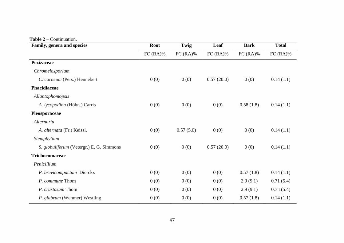
47
Table 2 – Continuation.
Family, genera and species Root Twig Leaf Bark Total
FC (RA)% FC (RA)% FC (RA)% FC (RA)% FC (RA)%
Pezizaceae
Chromelosporium
C. carneum (Pers.) Hennebert 0 (0) 0 (0) 0.57 (20.0) 0 (0) 0.14 (1.1)
Phacidiaceae
Allantophomopsis
A. lycopodina (Höhn.) Carris 0 (0) 0 (0) 0 (0) 0.58 (1.8) 0.14 (1.1)
Pleosporaceae
Alternaria
A. alternata (Fr.) Keissl. 0 (0) 0.57 (5.0) 0 (0) 0 (0) 0.14 (1.1)
Stemphylium
S. globuliferum (Vetergr.) E. G. Simmons 0 (0) 0 (0) 0.57 (20.0) 0 (0) 0.14 (1.1)
Trichocomaceae
Penicillium
P. brevicompactum Dierckx 0 (0) 0 (0) 0 (0) 0.57 (1.8) 0.14 (1.1)
P. commune Thom 0 (0) 0 (0) 0 (0) 2.9 (9.1) 0.71 (5.4)
P. crustosum Thom 0 (0) 0 (0) 0 (0) 2.9 (9.1) 0.7 1(5.4)
P. glabrum (Wehmer) Westling 0 (0) 0 (0) 0 (0) 0.57 (1.8) 0.14 (1.1)

48
Table 2 – Continuation.
Family, genera and species Root Twig Leaf Bark Total
FC (RA)% FC (RA)% FC (RA)% FC (RA)% FC (RA)%
P. griseolum G. Sm. 1.14 (15.4) 0 (0) 0 (0) 0 (0) 0.29 (2.2)
P. sanguifluum (Sopp) Biourge 0.57 (7.7) 0 (0) 0 (0) 0 (0) 0.14 (1.1)
P. thomii Maire 0 (0) 0 (0) 0 (0) 0.6 (1.8) 0.14 (1.1)
Penicillium sp. 1 1.1 (15.4) 0 (0) 0 (0) 0 (0) 0.29 (2.2)
Penicillium sp. 2 0 (0) 0.6 (5.0) 0.57 (20.0) 1.1 (3.6) 0.57 (4.3)
Penicillium sp. 3 1.71 (23.1) 0 (0) 0 (0) 0 (0) 1.7 (3.2)
Umbelopsidaceae
Umbelopsis
U. vinacea (Dixon-Stew.) Arx 0 (0) 0.57 (5.0) 0 (0) 0 (0) 0.14 (1.1)
Umbelopsis sp. 1 0.57 (7.7) 0 (0) 0 (0) 0 (0) 0.14 (1.1)
Umbelopsis sp. 2 0.67 (7.7) 0 (0) 0 (0) 0 (0) 0.14 (1.1)
Unidentified species
Fungal sp. 1 0 (0) 0.57 (5.0) 0 (0) 0 (0) 0.14 (1.1)
Fungal sp. 2 0 (0) 0 (0) 0 (0) 0.57 (1.8) 0.14 (1.1)
Uncultured Ascomycota 0 (0) 0.57 (5.0) 0.57 (20.0) 0 (0) 0.29 (2.2)
Uncultured Cladosporium clone 0 (0) 0 (0) 0 (0) 0.57 (1.8) 0.14 (1.1)
Uncultured fungus clone 1 0 (0) 5.71 (50.0) 0 (0) 0 (0) 1.41 (10.8)

49
Table 2 – Continuation.
Family, genera and species Root Twig Leaf Bark Total
FC (RA)% FC (RA)% FC (RA)% FC (RA)% FC (RA)%
Uncultured fungus clone 2 0 (0) 0 (0) 0 (0) 0.57 (1.8) 0.14 (1.1)
Uncultured Rhizoctonia 0 (0) 0 (0) 0 (0) 5.71 (18.2) 1.41 (10.8)
Helotiales sp. 0.57 (7.7) 0 (0) 0 (0) 0 (0) 0.14 (1.1)
Pezizomycetes sp. 0 (0) 0.57 (5.0) 0 (0) 0 (0) 0.14 (1.1)
Phialocephala sp. 0.57 (7.7) 0 (0) 0 (0) 0 (0) 0.14 (1.1)
Trichocomaceae sp. 0.57 (7.7) 0 (0) 0 (0) 0 (0) 0.14 (1.1)
Total 7.43 (100) 11.4 (100) 2.86 (100) 31.4 (100) 13.3 (100)
50
Endophyte community variations between plant organs
Various A. unedo parts differed significantly in the diversity and richness of
fungal endophytes. The fungal community from the bark was most diverse (48 taxa) and
rich (142 isolates), followed by the roots (39 taxa, 73 isolates), twigs (22 taxa, 32
isolates) and leaves (13 taxa, 41 isolates) (Table 1). The average number of fungal taxa
and isolates obtained from each organ per tree was also statistically significant, being
highest in barks (Table 1). Differences in the assemblages of endophytic fungal species
between tree organs have also been reported in previous studies (Fisher et al., 1993;
Moricca et al., 2012; Qi et al., 2012; Sun et al., 2012; Sebastianes et al., 2013). For
example, in Eucalyptus nitens was observed more endophytic fungal isolates in barks
than in leaves (Fisher et al., 1993). Similarly, Qi et al. (2012) recovered more fungal
endophyte taxa and isolates in barks than in annual twigs of Acer ginnala. However,
there was no statistically significant difference in Simpson and Shannon diversity
indices between the various A. unedo parts.
The four organs of A. unedo also differed significantly on endophytic colonization
rate (Table 1). Overall colonization rate were highest in barks, as reflected by the
highest mean colonization rate values (81%), followed by roots (42%), leaves (23%)
and twigs (18%). Differences in the colonization frequency of endophytes between tree
organs have also been reported for other plant species (Collado et al., 2000; Nalini et al.,
2005; Guo et al., 2008; Moricca et al., 2012; Sun et al., 2012). For example, Collado et
al. (2000) revealed much higher colonization rates of endophytic fungi from bark (65%)
than leaves (25%) of Quercus ilex and from bark (75.6%) than leaves (36.3%) of
Quercus faginea Lam., respectively. Similarly, in Pinus tabulaeformis, the colonization
frequency was recorded much higher in bark (ranging from 53-96%) than in needles
(ranging from 9-36%) (Guo et al., 2008).
Different endophyte taxa dominated the four organs of A. unedo (Table 2). In
barks, C. diversispora (43.6%), Uncultured Rhizoctonia (18.2%), Penicillium commune
(9.1%) and Penicillium crustosum (9.1%), were the dominant species and were isolated
only from this organ. The roots were dominated by species of the genus Penicillium,
namely Penicillium sp.3 (23.1%), P. griseolum and Penicillium sp.1 (15.4% each), and
were isolated only from this organ. Uncultured fungus clone 1 (50.0%) and
Cladosporium sp. (15.0%) were the most dominant species in twigs, being the first one
exclusively isolated from twigs whereas the former was also occurred in leaves. The
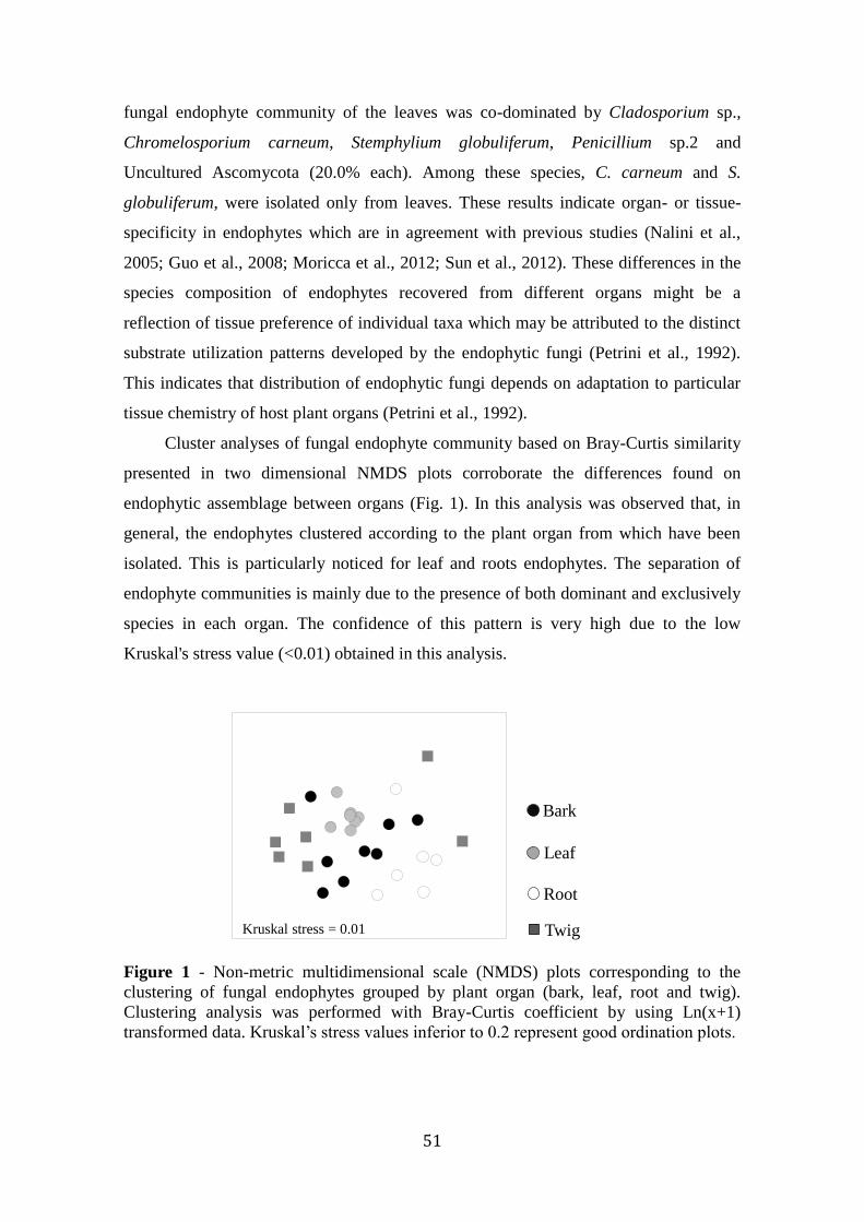
51
fungal endophyte community of the leaves was co-dominated by Cladosporium sp.,
Chromelosporium carneum, Stemphylium globuliferum, Penicillium sp.2 and
Uncultured Ascomycota (20.0% each). Among these species, C. carneum and S.
globuliferum, were isolated only from leaves. These results indicate organ- or tissue-
specificity in endophytes which are in agreement with previous studies (Nalini et al.,
2005; Guo et al., 2008; Moricca et al., 2012; Sun et al., 2012). These differences in the
species composition of endophytes recovered from different organs might be a
reflection of tissue preference of individual taxa which may be attributed to the distinct
substrate utilization patterns developed by the endophytic fungi (Petrini et al., 1992).
This indicates that distribution of endophytic fungi depends on adaptation to particular
tissue chemistry of host plant organs (Petrini et al., 1992).
Cluster analyses of fungal endophyte community based on Bray-Curtis similarity
presented in two dimensional NMDS plots corroborate the differences found on
endophytic assemblage between organs (Fig. 1). In this analysis was observed that, in
general, the endophytes clustered according to the plant organ from which have been
isolated. This is particularly noticed for leaf and roots endophytes. The separation of
endophyte communities is mainly due to the presence of both dominant and exclusively
species in each organ. The confidence of this pattern is very high due to the low
Kruskal's stress value (<0.01) obtained in this analysis.
Figure 1 - Non-metric multidimensional scale (NMDS) plots corresponding to the
clustering of fungal endophytes grouped by plant organ (bark, leaf, root and twig).
Clustering analysis was performed with Bray-Curtis coefficient by using Ln(x+1)
transformed data. Kruskal’s stress values inferior to 0.2 represent good ordination plots.
Kruskal stress = 0.01
Bark
Leaf
Root
Twig

52
Antimicrobial activity of endophytes
Five fungal endophytes, P. glabrum, P. crustosum, P. commune and C.
diversispora, all isolated from bark, and Penicillium sp. 3 isolated from roots, were
screened for their antimicrobial activity against gram-positive and gram-negative
bacteria and yeasts. These species were chosen due to their high abundance and
frequency of colonization. Results of antimicrobial activity are presented in Figure 2.
Only two endophytes, C. diversispora and Penicillium sp. 3, displayed significant
antimicrobial activity against the tested microorganisms when compared to control.
Cryptosporiopsis diversispora was the most effective in inhibiting microorganisms with
a widest spectrum. This fungus was the only one out of five endophytes with capacity to
inhibit significantly the yeasts than the control. When compared with the fluconazole,
C. diversispora reduced significantly the growth of C. glabrata (up to 1.9-fold), C.
albicans (up to 2.7-fold) and C. parapsilosis (up to 3.1-fold). This endophyte was also
the most effective in inhibiting the gram-negative bacteria E. coli and P. aeruginosa (up
to 2.4-fold and 4.5-fold, respectively when compared to chloramphenicol). There are
few reports about C. diversispora including of their antimicrobial proprieties. Only
Hwang et al. (2011) reported that C. diversispora isolated from lichen displayed
antifungal activity against C. albicans. Several bioactive metabolites, including
antimicrobial compounds, have been previously isolated from the Genus
Cryptosporiopsis and its teleomorph, Pezicula (Strobel et al., 1999; Verkley, 1999; Zilla
et al., 2013). Therefore, the present study provides, for the first time, evidences on the
antimicrobial potential of C. diversispora, which exhibited promising and broad
spectrum of antagonistic activity against clinical microorganisms, including E. coli and
P. aeruginosa. This is of much importance due to the increasing resistance to antibiotics
of these two gram-negative bacteria, including in Europe, especially in Portugal and
Italy (Nordberg et al., 2013). The distinctive feature of gram-negative bacteria is the
presence of a double membrane surrounding each bacterial cell. This outer membrane
excludes certain drugs and antibiotics from penetrating the cell, partially accounting for
why gram-negative bacteria are generally more resistant to antibiotics than are gram-
positive bacteria (Nordberg et al., 2013). The endophyte Penicillium sp.3 was also
shown to be efficient in inhibiting these two gram-negative bacteria and also one gram-
positive bacterium (Fig. 2). Therefore, this specie could also be of considerable interest
to be exploited as a source of novel antimicrobials compounds to overcome the

53
Figure 2 - Antimicrobial activity (halo of inhibition, in area) against yeasts (A), gram-
positive bacteria (B) and gram-negative bacteria (C) displayed by the endophytic fungi
Penicillium glabrum, Penicillium crustosum, Penicillium commune, Cryptosporiopsis
diversispora, and Penicillium sp. 3. Each value is expressed as mean standard error of
3 independent experiments performed in triplicate. Bars with different letters indicate
values with significant differences at p<0.05, within each microorganism.
Chloramphenicol (30 μg/ml) and fluconazole (25 μg/ml) were used as positive controls
for antibacterial and antifungal activity, respectively.
0
1
2
3
4
5
6
7
8
9
10
C. albicans C. parapsilosis C. glabrata
IHal
o o
f in
hib
itio
n (
cm2
)Penicillium crustosum
Penicillium glabrum
Penicillium sp. 3
Cryptosporiopsis diversispora
Penicillium commune
Fluconazole
0
5
10
15
20
25
30
B. subtilis B. cereus S. aureus
Hal
o o
f in
hib
itio
n (
cm2) Penicillium crustosum
Penicillium glabrum
Penicillium sp. 3
Cryptosporiopsis diversispora
Penicillium commune
Chloramphenicol
0
5
10
15
20
25
30
E. coli P. aeruginosa
Hal
o o
f in
hib
itio
n (
cm2)
Penicillium crustosum
Penicillium glabrum
Penicillium sp. 3
Cryptosporiopsis diversispora
Penicillium commune
Chloramphenicol
cc
a
c b
c
b
b
bb
a
bb
c
a
b
cc
b
c b
b
e
a
b
be d
c d
b
a
a
b a
a
b a
b
c
a
c
b
a
c
a
b a
a
b
b
b
b
b
b
A
B
C

54
resistance from time to time. When compared with the control chloramphenicol,
Penicillium sp.3 reduced significantly the growth of E. coli (up to 2.0-fold), P.
aeruginosa (up to 3.3-fold) and B. subtilis (up to 2.5-fold). Species belonging to
Penicillium genus are well known for their capacity to produce a vast array of bioactive
secondary metabolites (Nicoletti et al., 2014), including antimicrobial compounds (Yu
et al., 2010).

55
Analysis of volatile compounds
Results of antimicrobial activity showed that among the five endophytes analysed,
C. diversispora was the most effective, whereas P. glabrum was one of the fungi with
lowest activity against bacteria and yeasts. Therefore, the composition of volatile
organic compounds (VOCs) of both fungi growing in PDA medium under in vitro
conditions was analysed by GC/MS and the profiles obtained will be compared in an
attempt to identify the compounds potentially responsible for the antimicrobial activity
displayed by C. diversispora. Controls consisted of uninoculated PDA medium. The
compounds appearing in the control were subtracted from those acquired from the C.
diversispora and P. glabrum growing in PDA medium.
With the HS-SPME technique, 22 volatiles were extracted and identified by
GC/MS in the two endophytic species. The volatiles identified belong to different
chemical classes: 10 sesquiterpenes; 7 alcohols; 3 esters; and 2 ketones (corresponding
to those reported in Table 3 and represented in Figure 3). In C. diversispora was
identified a total of six volatiles compounds, being the most abundant phenylethyl
alcohol and 3-methyl-1-butanol, representing 46% and 33% of the total volatile fraction,
respectively (Table 3; Figure 3). The remaining volatile compounds were exclusively
found in this fungus and comprised two esters (acetic acid, 2-phenylethyl ester and
3,7,12-tridecatrienoic acid, methyl ester), one ketone (ochracin) and one sesquiterpene
(β-himachalene).
In P. glabrum was identified highest number of volatiles (18), being the
sesquiterpene α-guaiene the most abundant, accounting 69% of the total volatile fraction
identified in this specie (Table 3; Figure 3). This compound was exclusively identified
in this fungus. Also, present exclusively in this specie were a series of sesquiterpenes
(β-elemene, ylangene, (E)-β-farnesene, α-guaiene, valencene, α-selinene, β-selinene and
two more sesquiterpene-like compounds not identified), alcohols (juniper camphor, and
three more alcohol-like compounds not identified), one ketone-like compound and one
ester (acetic acid, 2-phenylethyl ester), altogether accounting 31% of the total volatile
fraction identified in this specie. Both the alcohols compounds, phenylethyl alcohol and
3-methyl-1-butanol, were also identified in P. glabrum, but in lower amounts when
compared to C. diversispora (0.7-fold lower, each).
In an attempted to elucidate the contribution of the several identified volatile
compounds on the antimicrobial activity displayed by the two endophytes, a PCA was

56
performed by using the data of volatile compounds of the two fungus as well as their
inhibition halos (in cm2) against bacteria and yeasts. The PCA obtained was able to
distinguish the two species (Figure 4) explaining in two principal components 85.51%
of the total variance captured. The first principal component clearly separate C.
diversispora from P. glabrum, due to the higher content of the first specie on 3-methyl-
1-butanol and phenylethyl alcohol. As already stated these two compounds were part of
the volatile fraction of both fungi, but in considerable higher amounts in C. diversispora
(p<0.001). The remaining volatiles characterized P. glabrum. An important information
retained from PCA is that 3-methyl-1-butanol and phenylethyl alcohol are associated
with C. diversispora, as well as the higher inhibition halos verified for all
microorganisms (with exception of B. subtilis). Such information suggested the
involvement of these two volatile compounds in the inhibition displayed by C.
diversispora against these microorganisms. The inhibition displayed by P. glabrum
against B. subtilis, could be similarly related with the production of volatiles
compounds as highlighted in the figure 4. These hypotheses require however
confirmation by performing bioassays with synthetic standards of the respective
identified VOCs. Despite this, previous studies have been reported the antimicrobial
potential of 3-methyl-1-butanol. For example, the VOCs produced by the endophyte
Muscodor crispans, with 3-methyl-1-butanol as the major compound, have been shown
to inhibited the growth of several phytopathogens (Mitchell et al., 2010). Similarly,
Fialho et al. (2011) have reported the high antimicrobial effect of the 3-methyl-1-
butanol against an array of phytopathogens under in vitro conditions. Phenylethyl
alcohol is a well-known bacteriostatic agent that, at low concentrations, can inhibit the
growth of gram-negative bacteria, including Salmonella, Shigella, Aerobacter,
Klebsiella, Escherichia, Pseudomonas and Proteus (Naz et al., 2013).

57
Table 3 – Relative percentage of volatile compounds of the endophytic fungi Penicillium glabrum and Cryptosporiopsis diversispora growing
under in vitro conditions (mean ± standard deviation, n=4). Compounds found in the control PDA are not included in this table. Different
superscript lower case letters denote statistically significant differences (p<0.05) between fungal species.
Compound RT
1 Characteristic ions (m/z)
QI (m/z
100%)2
Molecular
weight
Molecular
formula P. glabrum C. diversispora
1 3-Methyl-1-butanol 4.705 42/55/70 55 88 C5H12O 4.01±0.73a
32.75±1.97b
2 Butanoic acid, 2-methyl-, 3-methyllbutyl ester 23.575 43/57/70/85 70 172 C10H20O2 0.63±0.13 _
3 Phenylethyl alcohol 24.05 91/92/122 91 122 C8H10O 5.33±2.50a
45.83±2.26b
4 Acetic acid, 2-phenylethyl ester 34.065 43/104 104 164 C10H12O2 _ 2.94±0.94
5 β-elemene 43.14 68/81/93/107 81 204 C15H24 9.06±2.04 _
6 Ylangene 46.535 93/105/119/161 105 204 C15H24 0.21±0.05 _
7 (E)-β-farnesene 47.4 41/69/93 69 204 C15H24 0.20±0.05 _
8 Sesquiterpene-like compound 47.82 105 105 204 C15H24 0.33±0.10 _
9 Sesquiterpene-like compound 48.28 91/105/133/189 133 204 C15H24 0.32±0.11 _
10 α-Guaiene 49.02 93/105/107/147/189 105 204 C15H24 68.55±1.75 _
11 Valencene 49.355 79/93/107/161 161 204 C15H24 2.72±0.62 _
12 α-Selinene 49.445 81/93/107/189/204 189 204 C15H24 0.86±0.22 _
13 β-Himachalene 49.57 41/105/119/134/204 119 204 C15H24 _ 2.71±0.63
14 Ochracin 51.895 134/160/178 134 178 C10H10O3 _ 6.57±0.61
15 3,7,12-Tridecatrienoic acid, methyl ester 57.39 55/67 67 222 C14H22O2 _ 9.20±1.28
16 Alcohol-like compound 57.52 43/69/81/95/109/161 43 _ _ 0.34±0.05 _
17 β-selinene 58.51 41/81/107/161/189/204 107 204 C15H24 4.16±0.50 _
18 Juniper camphor 58.66 43/161/189/204 161 222 C15H26O 0.22±0.11 _
19 Alcohol-like compound 58.945 41/81/91/107/131/145 41 _ _ 0.11±0.03 _
20 Alcohol-like compound 62,225 43/79/105/119/147/162 162 _ _ 0.13±0.04 _
21 Unknown compound 63.995 135/150 135 _ _ 2.62±0.46 _
22 Ketone-like compound 66.43 147/161/218 218 _ _ 0.24±0.03 _ 1Retention time;
2Quantification ions.

58
Figure 3 - Full scan chromatographic profile obtained from the endophytic fungi
Penicillium glabrum and Cryptosporiopsis diversispora growing in vitro by HS-SPME
using DVB/CAR/PDMS fiber (identification numbers correspond to those compounds
reported in Table 3).
Penicillium glabrum
Cryptosporiopsis diversispora
1
1
2
3
3
4
5
67
10
1112
13
14
18
17
19
8
9
16 20
21
22

59
Figure 4 - Principal component analysis obtained using the volatile composition of
Penicillium glabrum ( ) and Cryptosporiopsis diversispora ( ) growing in vitro and
antimicrobial activity displayed by these fungi against bacteria and yeasts, evaluated as
halo inhibition (in cm2). The PCA factors explain 85.51% of the total variance. The
numbers represented correspond to those compounds presented in Table 3.
Conclusion
The results presented in this work show appreciable fungal diversity recovered
from A. unedo that are relatively unexplored as hosts for fungal endophytes. The
considerable high number of fungal endophytes observed combined with the apparent
organ specificity exhibited makes this plant specie suitable in search for novel
metabolites. The evaluation of capability of five fungal endophytes from A. unedo to
produce compounds with antibacterial or antifungal activity indicates that both C.
diversispora and Penicillium sp. 3 have great potential for exploitation in the
development of new drugs. The antimicrobial activity displayed by C. diversispora may
be ascribed to the volatile compounds 3-methyl-1-butanol and phenylethyl alcohol,
which requires confirmation. Further investigations will be planned to identify and
characterize active principles, and assess toxicity by laboratory assays.
B. subtilis
19, 21
20
17
18
16, 22
7
2
5, 6, 8-12
3
B. cereus
E. coli
1
C. parapsilosis
C. albicansS. aureus
P. aeruginosa
C. glabrata
4, 13-15
60
Acknowledgments
This work is funded by POCTEP - Cross Border Cooperation Programme Spain-
Portugal 2007-2013, project “RED/AGROTEC - Red transfronteriza España Portugal
de experimentación y transferencia para el desarrollo del sector agropecuario y
agroindustrial”.
References
Arnold, A.E. (2007). Understanding the diversity of foliar endophytic fungi: progress,
challenges, and frontiers. Fungal Biology Reviews, 21: 51–66.
Banerjee, D., Strobel, G., Geary, B., Sears, J., Ezra, D., Liarzi, O., Coombs, J. (2010).
Muscodor albus strain GBA, an endophytic fungus of Ginkgo biloba from the
United States of America, produces volatile antimicrobials. Mycology, 1:179–
186.
Bray, J.R., Curtis, J.T. (1957). An ordination of the upland forest communities of
Southern Wisconsin. Ecological Monographies, 27:325-349.
Carlet, J., Jarlier, V., Harbarth, S., Voss. A., Goossens, H., and Pittet, D. (2012). Ready
for a world without antibiotics? The Pensières Antibiotic Resistance Call to
Action. Antimicrobial Resistance and Infection Control, 1:11-13.
Celikel G, Demirsoy L, Demirsoy H. (2008). The strawberry tree (Arbutus unedo L.)
selection in Turkey. Scientia Horticulturae, 118: 115-119.
Clarke, J (1993) Non-parametric multivariate analysis of changes in community
structure. Aust J Ecol, 18:117–143.
Collado, J., Platas, G., Peláez, F. (2000). Host specificity in fungal endophytic
populations of Quercus ilex and Quercus faginea from central Spain. Nova
Hedwigia, 71: 421–430.
Debbab, A., Pretsch, A., Edrada-Ebel, R., Wray, V., Proksch, P. (2010). New antibiotic
metabolites from the fungal endophyte Stemphylium globuliferum isolated from
Mentha pulegium. Planta Med, 76 - 401.
Eo, J.K., Lee, B.H., Eom, A.H. (2014). A New Report on Oidiodendron flavum
Isolated from Field Soil in Korea. The Korean Journal of Mycology, 42: 239-242.

61
Fialho, M.B., Moraes, M.H.D.D., Tremocoldi, A.R., Pascholati, S.F. (2011). Potential
of antimicrobial volatile organic compounds to control Sclerotinia sclerotiorum in
bean seeds. Pesquisa Agropecuária Brasileira, 46(2): 137-142.
Fisher, P.J., Petrini, O., and Sutton, B.C. (1993). A comparative study of fungal
endophytes in leaves, xylem and bark of Eucalyptus nitens in Australia and
England. Sydowia, 45(2): 338-345.
Guo, L.D., Huang, G.R., Wang, Y. (2008).Seasonal and tissue age influences on
endophytic fungi of Pinus tabulaeformis (Pinaceae) in the Dongling Mountains,
Beijing. Journal of Integrative Plant Biology, 50: 997–1003.
Hoff, J.A., Klopfenstein, N.B., McDonald, G.I., Tonn, J.R., Kim, M.S., Zambino, P.J.,
Hessburg, P.F., Rogers, J.D., Peever, T.L., and Carris, L.M. (2004). Fungal
endophytes in woody roots of Douglas-fir (Pseudotsuga menziesii) and ponderosa
pine (Pinus ponderosa). For. Pathol, 34: 255.
Hwang, H.G., Kim, Y.N., Baik, K.S., Choi, S.K. (2011). Isolation and Characteristics of
Endolichenic Fungi Producing Antifungal Compound. The Korean Journal of
Microbiology, 47(1): 97-101.
Hyde, K.D., and Soytong, K. (2008). The fungal endophyte dilemma. Fungal Diversity,
33: 163 – 173.
IDSA - Infectious Diseases Society of America. (2010). The 10 × ‘20 initiative:
pursuing a global commitment to develop 10 new antibacterial drugs by 2020.
Clin Infect Dis, 50:1081-1083.
Kaul, S., Gupta, S., Ahmed, M., Dhar, M.K. (2012). Endophytic fungi from medicinal
plants: a treasure hunt for bioactive metabolites. Phytochem Rev, 11(4): 487–505.
Kruskal, J. (1964). Multidimensional scaling by optimizing goodness of fit to a
nonmetric hypothesis. Psychometrika, 29: 1–27.
Kudalkar, P., Strobel, G., Riyaz-Ul-Hassan, S., Geary, B., Sears, J. (2012). Muscodor
sutura, a novel endophytic fungus with volatile antibiotic activities. Mycoscience,
53: 319–325.
Kusari, S., and Spiteller, M. (2011). Are we ready for industrial production of bioactive
plant secondary metabolites utilizing endophytes? Nat. Prod. Rep, 28: 1203–1207.
Kusari, S., Hertweck, C., and Spiteller, M. (2012). Chemical Ecology of Endophytic
Fungi: Origins of Secondary Metabolites. Chemistry & Biology, 792-798.
Kusari, S., Lamshöft, M., and Spiteller, M. (2009). Aspergillus fumigatus Fresenius, an
endophytic fungus from Juniperus communis L. Horstmann as a novel source of

62
the anticancer pro-drug deoxypodophyllotoxin. Jounal Applied Microbiology,
107: 1019–1030.
Kusari, S., Lamshöft, M., Zühlke, S., and Spiteller, M. (2008). An endophytic fungus
from Hypericum perforatum that produces hypericin. Journal of Natural Products,
71: 159–162.
Kusari, S., Verma, VC., Lamshöft, M., and Spiteller, M. (2012). An endophytic fungus
from Azadirachta indica A. Juss. that produces azadirachtin. World J. Microbiol.
Biotechnol, 28: 1287–1294.
Li, G.H., Yu, Z.F., Li, X., Wang, X.B., Zheng, L.J., and Zhang, K.Q. (2007).
Nematicidal Metabolites Produced by the Endophytic Fungus Geotrichum sp.
Chemistry and Biodiversity, 4: 1520-1524.
Linaldeddu, B.T., Sirca, C., Spano, D., Franceschini, A. (2011). Variation of endophytic
cork oak-associated fungal communities in relation to plant health and water
stress. Forest Pathol, 41: 193–201.
Magurran AE (2004) Measuring biological diversity.
Moricca, S., Ginetti, B., Ragazzi, A. (2012). Species and organ specificity in
endophytes colonizing healthy and declining Mediterranean oaks.
Phytopathologia Mediterranea, 51: 587−598.
Mousa, W.K.; and Raizada, M.N. (2013). The diversity of anti-microbial secondary
metabolites produced by fungal endophytes: an interdisciplinary perspective.
Frontiers in microbiology, 4: 65.
Nalini, M.S., Mahesh, B., Tejesvi, M.V., Prakash, H.S., Subbaiah, V., Kini, K.R., and
Shetty, H.S. (2005). Fungal endophytes from the three-leaved caper, Crataeva
magna (Lour.) DC. (Capparidaceae). Mycopathologia, 159: 245–249.
Naz, S., Cretenet, M., & Vernoux, J. P. (2013). Current knowledge on antimicrobial
metabolites produced from aromatic amino acid metabolism in fermented
products. Microbial pathogens and strategies for combating them: science,
technology and education (A. Méndez-Vilas, Ed.) FORMATEX, 337-346.
Nicoletti, R., Fiorentino, A., and Scognamiglio, M. (2014). Endophytism of Penicillium
Species in Woody Plants. Open Mycology Journal, 8: 1-26.
Nordberg, P., Monnet, D.L., Cars, O. (2013). Background Paper, BP 6.1 Antimicrobial
resistance. Boston University.

63
Okane, I., Nagakiri, A., Ito, T. (1995).Discostroma tricellulaire, a new endophytic
ascomycete with a Seimatosporium anamorph isolated from Rhodendron. Can. J.
Bot., 74: 1338–1344.
Okane, I., Nakagiri, A., Ito, T. (1998). Endophytic fungi in leaves of ericaceous plants.
Canadian Journal of Botany, 76(4): 657-663.
Oliveira, I., Batista, P., Bento, A., Pereira, JA. (2011). Arbutus unedo L. and its benefits
on human health. Journal of Food and Nutrition Research, 50: 73-85
Oliveira, I., Pereira, J.A, Lino-Neto, T., Bento, A., Baptista, P., 2012. Fungal Diversity
Associated to the Olive Moth, Prays Oleae Bernard: A Survey for Potential
Entomopathogenic Fungi. Microbial Ecology, 63(4): 964-74.
Pancher, M., Ceol, M., Corneo, P.E., Longa, C.M.O., Yousaf, S., Pertot, I., and
Campisano, A. (2012). Fungal endophytic communities in grapevines (Vitis
vinifera L.) respond to crop management. Appl Environ Microbiol, 78: 4308–
4317.
Pereira, E., Santos, A., Reis, F,. Tavares M.R., Baptista, P., Lino-Neto, T., Almeida-
Aguiar, C. (2013). A new effective assay to detect antimicrobial activity of
filamentous fungi, Microbiological Research, 168: 1-5.
Petrini, O., Sieber, T.N., Toti, L., and Viret, O. (1992). Ecology, metabolite production
and substrate utilization in endophytic fungi. Natural Toxins, 1: 185-196.
Purmale, L., Apine, I., Nikolajev, V., Granti, L., Verkly, G., and Tomsone, S. (2012).
Endophytic fungi evergreen rhododendrons cultivated in vitro and in vivo.
Environ Exp Biol, 10: 1-7.
Qi, F., Jing, T., Zhan, Y. (2012). Characterization of Endophytic Fungi from Acer
ginnala Maxim. in an Artificial Plantation: Media Effect and Tissue-Dependent
Variation. PLOS ONE, 7(10): e46785.
Schoch, CL, Seifert, K., Huhndorf, S., Robert, V., Spoug, J.L., Levesque, C.A., Chen,
W. (2012) Nuclear ribosomal internal transcribed spacer (ITS) region as a
universal DNA barcode marker for Fungi. PNAS, 109: 6241–6.
Seaby RM, Henderson PA (2006) Species Diversity and Richness Version 4.
Sebastianes, F.L.S., Romão-Dumaresq, A.S., Lacava, P.T., Harakava, R., Azevedo, J.L.,
Soares de Melo, I., Pizzirani-Kleiner, A.A. (2013). Species diversity of culturable
endophytic fungi from Brazilian mangrove forests. Curr Genet, 59:153–166.
Shiono, Y., Yokoi, M., Koseki, T., Murayama, T., Aburai, N., and Kimura, K. (2010).
Allantopyrone A, a new α-pyrone metabolite with potent cytotoxicity from an

64
endophytic fungus, Allantophomopsis lycopodina KS-97. The Journal of
Antibiotics, 63: 251–253.
Strobel GA, Daisy B, 2003. Bioprospecting for microbial endophytes and their natural
products. Microbiology and Molecular Biology Reviews, 67: 491–502
Strobel, G., Daisy, B., Castillo, U., Harper, J. (2004). Natural products from endophytic
microorganisms. Journal of Natural Products, 67: 257-268.
Strobel, G.A., Miller, R.V., Miller, C., Condron, M., Teplow, D.B., Hess, W.M. (1999).
Cryptocandin, a potent antimycotic from the endophytic fungus Cryptosporiopsis
cf. quercina. Microbiology, 145: 1919–1926.
Sun, X., Ding, Q., Hyde, K.D., Guo, L.D. (2012). Community structure and preference
of endophytic fungi of three woody plants in a mixed forest. Fungal Ecol, 5: 624-
632.
Suryanarayanan, TS., Venkatesan, G., Murali, T.S. (2003). Endophytic fungal
communities in leaves of tropical forest trees: diversity and distribution patterns.
Current Science 85: 489-493
Verkley, G.J.M. (1999). A monograph of the genus Pezicula and its anamorphs. Studies
in mycology, 44: 180.
White, T.j., Bruns, T., Lee, S., Taylor, J. (1990) Amplification and direct sequencing of
fungal ribosomal RNA genes for phylogenetics. In: Innis, M., Gelfand, D.,
Shinsky, J., White, T.J. (eds), PCR Protocols: A Guide to Methods and
Applications, Academic Press, San Diego, 315-322.
Yu, H., Zhang, L., Li, L., Zheng, C., Guo, L., Li, W., Qin, L. (2010). Recent
developments and future prospects of antimicrobial metabolites produced by
endophytes. Microbiological research, 165(6): 437-449.
Zilla, M.K., Qadri, M., Pathania, A.S., Strobel, G.A., Nalli, Y., Kumar, S., Guru, S.K.,
Bhushan, S., Singh, S.K., Vishwakarma, R.A., Riyaz-Ul-Hassan, S., Asif Ali, A.
(2013). Bioactive metabolites from an endophytic Cryptosporiopsis sp. inhabiting
Clidemia hirta. Phytochemistry, 95: 291–297.

65
Supporting information
Table S1 - Fungal taxa isolated in this study and the summary of BLAST results,
showing the coverage of the sequences and sequence similarities with the most closely
related organisms.
Isolated (GenBank Acc. no. of the
ITS sequence)
Query
coverage
Sequence
similarity E-value*
Organism with the highest
sequence identity, GenBank
Acc. no.
Allantophomopsis lycopodina 81% 95% 3,00E
-164 JX981469
(submitted)
Alternaria alternata 86% 96% 0.0 JN867471
(submitted)
Aureobasidium pullulans 100% 99% 0.0 KC897669
(submitted)
Chromelosporium carneum 99% 88% 4,00E
-112 JF440586
(submitted)
Cladosporium sp. 99% 100% 0.0 KJ598873
(submitted)
Cryptosporiopsis diversispora 99% 98% 0.0 JF714251
(submitted)
Discostroma sp. 99% 96% 8,00E
-133 JN545795
(submitted)
Fungal sp. 100% 100% 0.0 KC867746
(submitted)
Fungal sp. 100% 99% 0.0 HM123559
(submitted)
Helotiales sp. 100% 98% 0.0 KC180699
(submitted)
Microsphaeropsis olivacea 99% 99% 0.0 JX681101
(submitted)
Penicillium brevicompactum 100% 99% 0.0 KF876834
(submitted)
Penicillium commune 80% 99% 0.0 KF938402
(submitted)
Penicillium crustosum 64% 96% 0.0 LN482509
(submitted)
Penicillium glabrum 100% 99% 0.0 AY373915
(submitted)

66
Penicillium griseolum 95% 99% 0.0 EF422848
(submitted)
Penicillium sanguifluum 100% 100% 0.0 JX140866
(submitted)
Penicillium sp 99% 95% 7,00E
-103 JF429680
(submitted)
Penicillium sp. 96% 99% 0.0 KF367506
(submitted)
Penicillium thomii 100% 100% 0.0 JX140779
(submitted)
Pezizomycetes sp. 100% 97% 0.0 JQ759165
(submitted)
Phialocephala sp. 100% 99% 0.0 KJ425309
(submitted)
Stemphylium globuliferum 93% 99% 0.0 KF871456
(submitted)
Trichocomaceae sp. 100% 100% 0.0 KC007327
(submitted)
Umbelopsis sp. 100% 100% 0.0 JQ912671
(submitted)
Umbelopsis sp. 100% 99% 0.0 KC816011
(submitted)
Umbelopsis vinacea 100% 97% 3,00E
-127 GU183114
(submitted)
Uncultured Ascomycota 96% 99% 0.0 FR682256
(submitted)
Uncultured Cladosporium clone 94% 88% 1,00E
-143 JF449738
(submitted)
Uncultured fungus clone 100% 99% 0.0 HM044625
(submitted)
Uncultured fungus clone 100% 100% 5,00E
-35 JF720038
(submitted)
Uncultured Rhizoctonia 100% 99% 0.0 DQ061931
(submitted)
*BLAST expected value represents the number of sequence matches expected by
random chance (the smaller the value, the better the match to the reported NCBI
database sequence)

67
Figure S1 – In vitro fungal culture flasks (50 mL) sealed with a cap containing a
silicone septum. This system was used to evaluate the production of volatile organic

68
Capítulo 4
Conclusão

69

70
Conclusão
A bioprospeção de metabolitos com atividade antimicrobiana em fungos
endofíticos isolados de plantas medicinais tem despertado a atenção dos investigadores,
pelo facto de sintetizarem metabolitos secundários semelhantes às suas plantas
hospedeiras. Os fungos endofíticos, com capacidade de sintetizar os compostos das suas
plantas hospedeiras, sob condições ótimas de cultura, pode ser um meio para a obtenção
de compostos bioativos a baixo-custo, ecológico, reprodutível e compatível com a sua
exploração industrial. O medronheiro (Arbutus unedo L.) é uma árvore fruteira
endémica da região mediterrânica, pertencente à família Ericacea e ao género Arbutus.
Em Portugal existe preponderantemente a sul do rio Tejo podendo contudo, encontrar-
se difundido por todo o país, inclusive em Trás-os-Montes. Desde os tempos ancestrais
que diversas partes desta árvore, em especial folhas, raízes, casca e fruto, são utilizadas
na medicina tradicional. Tanto quanto é do nosso conhecimento, a comunidade fúngica
endofítica associada a esta espécie bem como o potencial antimicrobiano destes fungos
nunca foi explorado. Assim, neste trabalho avaliou-se pela primeira vez a comunidade
fúngica endofítica associada às diversas partes desta planta utilizadas vulgarmente na
medicina tradicional (folhas, ramos, raízes e cascas) e o seu potencial antimicrobiano
contra bactérias gram-positivas e gram-negativas, e leveduras patogénicas humanas. A
composição volátil de alguns endófitos foi ainda analisada e correlacionada com a
atividade antimicrobiana exibida pelos endófitos.
A comunidade fúngica endofítica associada ao medronheiro demonstrou ser
muito abundante e diversa. Dos 700 segmentos vegetais analisados, foi obtido um total
de 288 isolados pertencentes a 118 taxa. Todas as árvores analisadas encontravam-se
colonizadas por fungos endofíticos, apresentando cada árvore uma média de 20,6 taxa e
41,1 isolados. A sequenciação da região ITS do rDNA das 33 espécies mais abundantes,
permitiu a identificação até à espécie de 15 taxa. As restantes 18 taxa não foram
identificadas até à espécie pelo fato de não se encontrarem especificadas na base de
dados GenBank. As espécies identificadas molecularmente pertencem a 10 famílias e 11
géneros, sendo o género Penicillium (10 taxa) e Umbelopsis (3 taxa) os mais
diversificados. A espécie Cryptosporiopsis diversispora foi a mais comum,
representando 26% do total de isolados e com uma frequência de colonização de 3,4%.
As diferentes partes da planta analisadas apresentaram diferenças em termos de
composição, riqueza e diversidade de fungos endofíticos. As cascas apresentaram um

71
maior número de espécies (48) e de isolados (142), comparado com as raízes (39 taxa,
73 isolados), ramos (22 taxa, 32 isolados) e folhas (13 taxa, 41 isolados). A frequência
de colonização também foi superior nas cascas (81%) face às raízes (42%), folhas
(23%) e ramos (18%). A comunidade fúngica endofítica variou ainda de acordo com o
órgão da planta, verificando-se diferenças de predominância de determinadas espécies
entre os órgãos amostrados. Este resultado reflete a preferência individual das espécies
endofiticas para um determinado órgão/tecido. Este fato pode estar relacionado com
diferenças de composição química entre os diferentes órgãos da planta permitindo o
desenvolvimento de determinada espécie fúngica.
De entre as espécies identificadas selecionaram-se as cinco mais abundantes
para avaliar a sua capacidade antimicrobiana contra bactérias gram-positiva e gram-
negativa e leveduras. As espécies selecionadas foram Penicillium glabrum, Penicillium
crustosum, Penicillium commune e C. diversispora, isoladas das cascas, e Penicillium
sp. 3 isolada da raiz. De entre estas, apenas C. diversispora e Penicillium sp. 3,
mostraram capacidade para inibir significativamente os microorganismos testados face
aos antibióticos / antifúngicos comerciais. A espécie C. diversispora apresentou um
amplo espetro, sendo a única que apresentou capacidade em inibir significativamente as
leveduras Candida glabrata, Candida albicans e Candida parapsilosis mais de 1,2
vezes face ao antifúngico fluconazole (25 μg/ml). Esta espécie também mostrou ser a
mais eficaz contra as bactérias gram-negativas Escherichia coli e Pseudomonas
aeruginosa, inibindo significativamente o seu crescimento em mais de 2,4 vezes face ao
antibiótico cloranfenicol (30 μg/ml). Por sua vez, Penicillium sp.3 demonstrou ser mais
eficaz do que os antibióticos comerciais contra bactérias gram-positiva e gram-negativa.
A avaliação dos compostos voláteis por GC/MS dos fungos que apresentaram
maior (C. diversispora) e menor (P. glabrum) atividade antimicrobiana, em crescimento
sob condições in vitro, permitiu identificar um total de 22 compostos pertencentes a
quatro classes químicas: sesquiterpenos (10), álcoois (7), éstere (3), e cetonas (2). Na
espécie C. diversispora foi identificado um total de seis voláteis sendo os mais
abundantes, o fenil-etil álcool e o 3-metil-1-butanol, representando 46% e 33% do total
da fração volátil, respectivamente. Por sua vez, na espécie P. glabrum foi identificado
um número superior de compostos voláteis (18 no total), sendo o sesquiterpeno α-
guaiene o mais abundante representando cerca de 69% do total da fração volátil.
Identificou-se, nesta espécie, a produção exclusiva de 16 compostos voláteis. A maioria
72
dos compostos voláteis identificados encontram-se descritos como possuindo
propriedades antimicrobianas o que poderá explicar, em parte, a atividade
antimicrobiana exibida pelos dois endófitos. A comparação do perfil volátil de C.
diversispora e P. glabrum associada a uma análise de componentes principais
demonstrou o envolvimento dos compostos voláteis na atividade antimicrobiana exibida
pelas espécies endófitas. Esta analise evidenciou o envolvimento de 3-metil-1-butanol e
fenil-etil álcool, produzido por C. diversispora, no processo de inibição deste fungo
contra as leveduras e as bactérias gram-negativas.
Os resultados obtidos evidenciam, pela primeira vez, o potencial antimicrobiano
de espécies fúngicas endofiticas do medronheiro. A espécie C. diversispora parece ser a
mais promissora como fonte de antibióticos/antifúngicos naturais que poderão, no
futuro, ser exploradas ao nível da indústria farmacêutica. No entanto, os resultados
obtidos abrem novas perspetivas para o estudo mais detalhado da comunidade fúngica
associada ao medronheiro e a bioprospeção de outros isolados obtidos. É ainda
necessário proceder-se à elucidação da natureza química dos metabolitos produzidos e
da avaliação da sua capacidade antimicrobiana.