efeito do peptídeo liberador de gastrina na resposta
TRANSCRIPT

Universidade Federal do Rio Grande do Sul
Instituto de Ciências Básicas da Saúde
Departamento de Bioquímica
Pós-Graduação em Ciências Biológicas: Bioquímica
Efeito do peptídeo liberador de gastrina na
resposta inflamatória local e sistêmica:
envolvimento na sinalização
do receptor Toll-like 4
FABRICIA CARDOSO PETRONILHO
Orientador:
Prof. Dr. Felipe Dal-Pizzol
Tese apresentada ao Programa de Pós-Graduação em Ciências
Biológicas: Bioquímica, como requisito parcial para a obtenção do título
de Doutor em Bioquímica.
Porto Alegre, Março de 2010

II
“Cultivem o espírito crítico. Se deixado de lado, ele não desperta idéias, nem
estimula as grandes coisas. Sem ele, nada tem valor. Ele tem sempre a última
palavra. Isso que lhes peço é o que, por sua vez, vocês devem pedir aos
discípulos que vão formar, é o que há de mais difícil para o inventor. Acreditar
que se encontrou um fato científico importante, sentir um desejo ardente de
anunciá-lo, e ser obrigado durante dias, semanas e até durante anos a
combater a si próprio, esforçando-se para desmentir suas próprias experiências
e só proclamar sua descoberta quando esgotarem todas as hipóteses
contrárias, sim, é uma tarefa árdua. Porém quando depois de tantos esforços
chega-se, finalmente a certeza, sentimos uma das maiores alegrias que a alma
humana pode experimentar...”
Louis Pasteur

III
À minha mãe Marlize, e ao meu pai, Antônio, por todo apoio, carinho e amor
em todas as etapas da minha vida. Por terem me proporcionado a
oportunidade de conquistar o mais valioso de todos os bens, a cultura.
E à minha irmã Taise minha eterna incentivadora, companheira e amiga.
Meu eterno agradecimento.
Este trabalho é dedicado a vocês.

IV
AGRADECIMENTOS
Gostaria de iniciar os meus agradecimentos ao Prof. Dr. Felipe Dal
Pizzol, não porque de praxe devemos fazê-lo ao Orientador, mas sim, porque
foi ele quem me abriu as portas para conhecer o “mundo da pesquisa
científica”. A sua sabedoria e competência aliadas a simplicidade o torna uma
das pessoas mais admiráveis que conheci. Por isso agradeço pela chance de
ser a sua orientanda desde o mestrado e poder trabalhar ao seu lado todos
esses anos, por ter acesso aos seus conhecimentos valiosos e pelo incentivo e
confiança em mim depositados. A você Felipe, minha eterna gratidão e
admiração...
Ao Prof. Dr. Emílio Luis Streck, “Xuxa” e seus orientandos por toda ajuda
e disponibilidade oferecida a este trabalho e pelo convívio no Laboratório de
Fisiopatologia Experimental - UNESC, o nosso querido “triângulo”.
À Profa. Dra. Cristiane Ritter, do Laboratório de Intensivismo
Experimental – UNESC, e seus bolsistas pela fundamental contribuição na
disponibilização de dados e amostras de sangue dos pacientes.
Ao Prof. Dr. José Claudio Fonseca Moreira do Departamento de
Bioquímica da UFRGS e seus bolsistas pela colaboração nos estudos in vitro.
Ao Prof. Dr. Francisco Garcia Soriano do Departamento de Medicina do
Hospital Universitário da USP e sua equipe para os experimentos de RT-PCR.
Ao Prof Dr. Rafael Roesler e ao Prof. Dr. Gilberto Schwartsmann do
Laboratório de Pesquisa do Câncer da UFRGS pela parceria na discussão dos
estudos envolvento a ação do peptídeo liberador de gastrina na inflamação.

V
Ao Prof Dr. João Quevedo, cujo entusiasmo pela ciência contagia a
todos que com ele convivem.
À Profa. Dra. Tatiana Barichello pela amizade e palavras de incentivo em
vários momentos e a Profa. Dra Patricia Fernanda Schuck, minha querida Pati
uma pessoa maravilhosa que Deus deu de presente para o FISIOPAT, pelas
valiosas dicas que só ela sabe dar e pela amizade, obrigada minha amiga cor-
de-rosa.
Aos colegas do Laboratório de Fisiopatologia Experimental da UNESC:
às ICs Bruna de Souza (Bruninha), que me ajudou nesse estudo até em dia de
Natal; à vencedora de todos os prêmios, Francielle Mina (Fram Mina), à
Francieli Vuolo, a minha Inha o meu anjinho; à Fabíola (Ti Bibi) minha
companheira sempre e exemplo de amizade; à Francine que esta sempre
pronta aos experimentos, à Angele, Riti e Vanessa. E as colegas de pós-
graduação à Amanda e as inseparáveis Larissa Constantino (Lara) e Cristiane
Damiani Tomasi (Cris), obrigada à todas por todo companheirismo e por
fazerem parte da minha história no lab.
Aos colegas dos outros vértices do “triângulo” obrigada pelo convívio, e
entre elas, as “Gis, Gisele, sempre disposta a ajudar e à Gislaine Tezza Rezin
“meu toquinho” que só é pequena de tamanho mais é gigante pela força e
honestidade, obrigada amiga pelo carinho, incentivo, pelo ombro consolador e
por mostrar acreditar no meu potencial durante todo o tempo que trabalhamos
juntas.
Aos colegas do NEUROLAB e LAFIBE, pelos momentos de
descontração quando dividíamos o mesmo espaço... saudades daquela

VI
época... à “Gi do Neuro” Gislaine Zilli Réus, minha amiga querida, minha
companheira de congressos, dos cafezinhos, obrigada pela alegria do convívio.
À todos os ratos que fizeram parte desse estudo e aos pacientes,
objetivo maior de toda atividade científica.
À Universidade do Extremo Sul Catarinense e ao Programa de Pós-
Graduação em Ciências da Saúde por me acolherem tão bem durante todos
esses anos.
À Universidade Federal do Rio Grande do Sul, pelo ensino gratuito e de
qualidade, que possibilitou a realização de um sonho.
À Capes, pela bolsa de Doutorado e pela confiança que dedica aos
pesquisadores e à pesquisa científica brasileira.
Aos meus pais, que propiciaram os meios e a estrutura onde me formei,
intelectualmente e moralmente. Sem seu suporte e orientação, não teria tido as
oportunidades de aprendizado que tive, nem saberia usar, de maneira ética, o
que aprendi.
À amiga, companheira e irmã, Taise, minha Tequinha pelo constante
incentivo e auxílio desmedido durante toda minha vida.
À Deus, que sempre esteve ao meu lado, nas minhas quedas,
fraquezas, nas lutas e controvérsias, vitórias e derrotas. Obrigada por tudo o
que vi, ouvi e aprendi durante esse tempo como doutoranda... obrigada pela
vida.
A todos, meu eterno e inesquecível
“Muito Obrigada”

VII
SUMÁRIO
PARTE I
Resumo .............................................................................................................. 2
Abstract .............................................................................................................. 3
Lista de Abreviaturas .......................................................................................... 4
I.1. INTRODUÇÃO ............................................................................................. 7
I.1.1. Definição e aspectos epidemiológicos da sepse ................................... 7
I.1.2. Fisiopatologia da sepse....................................................................... 10
I.1.3. Mecanismos moleculares de ação do LPS ......................................... 12
I.1.4. Migração de neutrófilos ....................................................................... 15
I.1.4.1. Aspectos gerais ....................................................................... 15
I.1.4.2. Origem e mobilização de neutrófilos ........................................ 16
I.1.4.3. Ação dos neutrófilos no foco infeccioso ................................... 18
I.1.4.4. Falência da migração de neutrófilos na sepse severa ............. 20
I.1.3. Neuropeptídeos e inflamação ............................................................. 23
I.1.3.1 Bombesina/Peptídeo liberador de gastrina ............................... 24
I.1.3.2 Importância do GRP ................................................................. 25
I.1.3.3 RC-3095- um antagonista do receptor GRP ............................. 27
I.1.4 Modelos animais de inflamação pleural e sepse .................................. 29
I.2. OBJETIVOS ............................................................................................... 33
I.2.1. Objetivo Geral ..................................................................................... 33
I.2.2. Objetivos Específicos .......................................................................... 33

VIII
PARTE II
Capítulo I - Protective effect of gastrin-releasing peptide receptor antagonist in
carrageenan-induced pleural inflammation in rats ............................................ 36
Capítulo II - Antagonist of gastrin-releasing peptide receptors induces a
protection from lethal sepsis: involvement of toll-like receptor 4 signaling ...... 44
Capítulo III - Gastrin-releasing peptide receptor as a molecular target for
inflammatory diseases ...................................................................................... 92
PARTE III
III.1. DISCUSSÃO ............................................................................................ 98
III.2. CONCLUSÕES ...................................................................................... 111
III.3. PERSPECTIVAS .................................................................................... 113
III.4. REFERÊNCIAS BIBLIOGRÁFICAS ...................................................... 115

1
PARTE I
Introdução e Objetivos
2
RESUMO
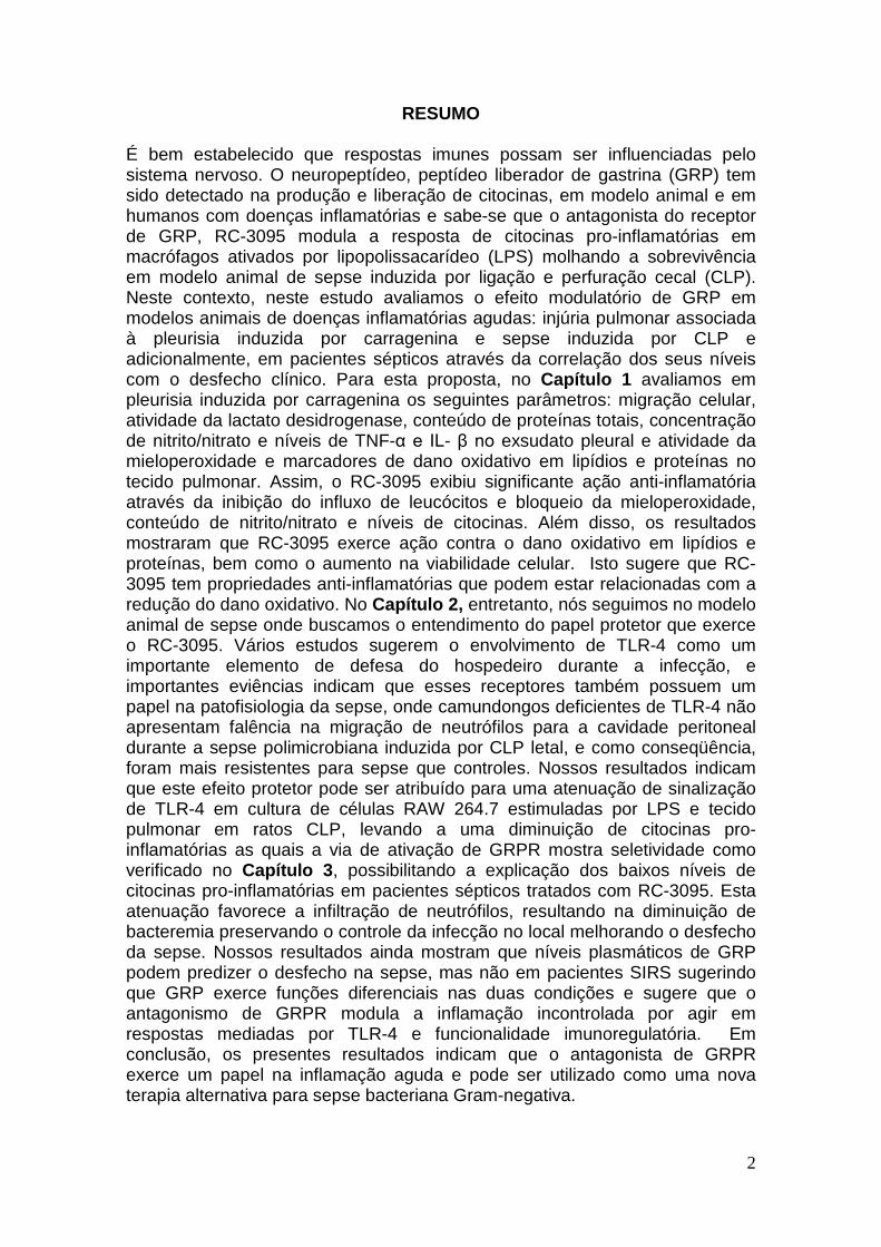
É bem estabelecido que respostas imunes possam ser influenciadas pelo sistema nervoso. O neuropeptídeo, peptídeo liberador de gastrina (GRP) tem sido detectado na produção e liberação de citocinas, em modelo animal e em humanos com doenças inflamatórias e sabe-se que o antagonista do receptor de GRP, RC-3095 modula a resposta de citocinas pro-inflamatórias em macrófagos ativados por lipopolissacarídeo (LPS) molhando a sobrevivência em modelo animal de sepse induzida por ligação e perfuração cecal (CLP). Neste contexto, neste estudo avaliamos o efeito modulatório de GRP em modelos animais de doenças inflamatórias agudas: injúria pulmonar associada à pleurisia induzida por carragenina e sepse induzida por CLP e adicionalmente, em pacientes sépticos através da correlação dos seus níveis com o desfecho clínico. Para esta proposta, no Capítulo 1 avaliamos em pleurisia induzida por carragenina os seguintes parâmetros: migração celular, atividade da lactato desidrogenase, conteúdo de proteínas totais, concentração de nitrito/nitrato e níveis de TNF-α e IL- β no exsudato pleural e atividade da mieloperoxidade e marcadores de dano oxidativo em lipídios e proteínas no tecido pulmonar. Assim, o RC-3095 exibiu significante ação anti-inflamatória através da inibição do influxo de leucócitos e bloqueio da mieloperoxidade, conteúdo de nitrito/nitrato e níveis de citocinas. Além disso, os resultados mostraram que RC-3095 exerce ação contra o dano oxidativo em lipídios e proteínas, bem como o aumento na viabilidade celular. Isto sugere que RC-3095 tem propriedades anti-inflamatórias que podem estar relacionadas com a redução do dano oxidativo. No Capítulo 2, entretanto, nós seguimos no modelo animal de sepse onde buscamos o entendimento do papel protetor que exerce o RC-3095. Vários estudos sugerem o envolvimento de TLR-4 como um importante elemento de defesa do hospedeiro durante a infecção, e importantes eviências indicam que esses receptores também possuem um papel na patofisiologia da sepse, onde camundongos deficientes de TLR-4 não apresentam falência na migração de neutrófilos para a cavidade peritoneal durante a sepse polimicrobiana induzida por CLP letal, e como conseqüência, foram mais resistentes para sepse que controles. Nossos resultados indicam que este efeito protetor pode ser atribuído para uma atenuação de sinalização de TLR-4 em cultura de células RAW 264.7 estimuladas por LPS e tecido pulmonar em ratos CLP, levando a uma diminuição de citocinas pro-inflamatórias as quais a via de ativação de GRPR mostra seletividade como verificado no Capítulo 3, possibilitando a explicação dos baixos níveis de citocinas pro-inflamatórias em pacientes sépticos tratados com RC-3095. Esta atenuação favorece a infiltração de neutrófilos, resultando na diminuição de bacteremia preservando o controle da infecção no local melhorando o desfecho da sepse. Nossos resultados ainda mostram que níveis plasmáticos de GRP podem predizer o desfecho na sepse, mas não em pacientes SIRS sugerindo que GRP exerce funções diferenciais nas duas condições e sugere que o antagonismo de GRPR modula a inflamação incontrolada por agir em respostas mediadas por TLR-4 e funcionalidade imunoregulatória. Em conclusão, os presentes resultados indicam que o antagonista de GRPR exerce um papel na inflamação aguda e pode ser utilizado como uma nova terapia alternativa para sepse bacteriana Gram-negativa.

3
ABSTRACT
It is well established that immune responses may be influenced by the nervous system. The neuropeptide gastrin-releasing peptide (GRP) has been detected on the production and release of cytokines, both in animal models and humans with inflammatory diseases and reported that the GRP receptor antagonist RC-3095 modulates the response of proinflammatory cytokines in activated macrophages by lipopolysaccharide and improved the survival in an animal model of sepsis induced by cecal ligation and puncture (CLP). Within this context, in this work we evaluate the GRP modulatory effect in animal models of acute inflammatory illnesses: lung injury associated with carrageenan-induced pleurisy, and in CLP-induced sepsis and additionally in septic patients through the correlations of its levels with the clinical outcome. For this purpose, in Chapter 1 we evaluated in carrageenan-induced pleurisy the following parameters: cell migration, lactate dehydrogenase activity, total protein content, nitrite/nitrate concentration, TNF-α and interleukin-1β (IL-1β) levels in pleural exudates; myeloperoxidase activity and lipid and protein oxidative damage markers in lung tissue. Thus, RC-3095 exhibited pronounced anti-inflammatory actions by inhibition of leukocyte influx and blockade of myeloperoxidase, nitrite/nitrate content and cytokine levels. Moreover, the results showed that RC-3095 elicits action against oxidative damage in lipids and proteins, as well as increases cell viability. These suggest that RC-3095 has anti-inflammatory properties that can be related with the reduction of oxidative damage. In Chapter 2, however, we follow the animal model of sepsis where we search the agreement of the protective role that RC-3095 exerts. Increasing evidences suggest the evolvement of TLR-4 as an important element of host defense during an infection, a growing body of evidence indicates that these receptors also may play a role in the pathophysiology of sepsis where TLR-4 defective mice did not present failure of neutrophil migration to the peritoneal cavity during polymicrobial sepsis induced by lethal CLP, and as consequence, they were more resistant to sepsis than controls Our results further indicate that this protective effect can be attributed to an attenuation of TLR-4 signaling in RAW 264.7 cell culture stimulated by LPS and lung tissue in CLP rats, leading to a decrease of release of proinflammatory cytokines which the pathway of activation of GRPR shows selectivity as verified in Chapter 3, and can additionally explain the lower levels of the proinflammatory cytokine in RC-3095-treated septic patients. This attenuation favors neutrophil infiltration, resulting in decreased bacteremia preserving the control of infection in situ improving sepsis outcome. Our finding still show that plasma GRP levels could predict outcome in sepsis but not SIRS patients suggesting that GRP plays different roles in the two conditions and suggest that GRPR antagonism modulates uncontrolled inflammation by targeting TLR-4-mediated responses and immunoregulatory functionality. Taken together, the present results indicate that a GRPR antagonist plays a role in acute inflammation and could be developed as a new alternative therapy for Gram-negative bacterial sepsis.

4
LISTA DE ABREVIATURAS
BASES - brazilian sepsis epidemiological study
BN – bombesina
BNPs – peptídeos relacionados à bombesina
C5a – quinto fragmento do sistema complemento ativado
CLP – ligação e perfuração cecal
EROs – especies reativas de oxigênio
ERK1/2 – quinase regulada por sinais extracelulares 1/2
G-CSF – fator de estimulação de colônias de granulócitos
GPI – glicosilfosfatidilinositol
GPCR – receptor acoplado a proteína G
GRK-2 – quinase 2 do receptor acoplado a proteína G
GRK-5 – quinase 5 do receptor acoplado a proteína G
GRO-α – oncogene relacionada ao crescimento - α
GRP – peptídeo liberador de gastrina
GRPR – receptor do peptídeo liberador de gastrina
HO-1 – heme oxigenase -1
ICAMs – molécula de adesão intrecelular
ICAM -2 – molécula de adesão intracelular – 2
IFN-γ – interferon-γ
IL-1β – interleucina-1β
IL-6 – interleucina – 6
IL-8 – interleucina – 8
IL-10 – interleucina -10
5
IL-12 – interleucina -12
iNOS – óxido nítrico sintase induzível
IRAK – quinase associada ao receptor de IL-1
IRF-1 – fator regulatório de interferon-1
JAK/STAT – janus quinase/transdutora de sinais e ativadora de transcrição
JNK – quinase n-terminal c-Jun
KC – quimiocina derivada de queratinócitos
LBP – proteína ligadora de LPS
LPS – lipopolissacarídeo
LTA – ácido lipoteicóico
LTB4 – leucotrieno B
MCP-1 – proteína quimiotáxica de monócitos - 1
4
MD2 – proteína de diferenciação mielóide - 2
MIP-1α – proteína inflamatória de macrófagos - 1α
MIP-1β – proteína inflamatória de macrófagos - 1β
MIP2 – proteína inflamatória de macrófagos 2
MyD88 – fator de diferenciação mieloide humano 88
NADPH – nicotinamida adenina dinucleotideo fosfato
NETs – neutrophil extracellular traps
NF-κB – fator nuclear-κB
NK – natural killer
NMB – neuromedina B
NMC – neuromedina C
NO – óxido nítrico
PAF – fator de agregação plaquetária

6
PAMPs – padrões moleculares associados a patógenos
PECAM – molécula de adesão plaquetária
PI3K – fosfatidilinositol 3 quinase
PPRs – receptores de reconhecimento de padrões
SDF-1 – fator derivado de estroma da medula óssea
SIRS – sindrome da resposta inflamatória sistêmica
SNC – sistema nervoso central
TIR – domínio intracelular Toll/interleucina -1
TIRAP – proteina adaptadora contendo o domínio TIR
TLRs – receptores toll-like
TLR-2 - receptor toll-like-2
TLR-3 – receptor toll-like-3
TLR-4 – receptor toll-like-4
TNF-α – fator de necrose tumoral – α
TRAF6 – fator 6 associado ao receptor de TNF
TRAM – molécula adaptadora relacionada ao TRIF
TRIF – domínio TIR contendo adaptador do indutor de interferon β
UTI - unidade de terapia intensiva
VCAMs – molécula de adesão vascular
VIP – peptídeo vasoativo intestinal

7
I.1. INTRODUÇÃO
I.1.1. Definição e aspectos epidemiológicos da sepse
Todo processo infeccioso desencadeia uma resposta inflamatória do
hospedeiro, cuja magnitude pode variar de indivíduo para indivíduo. A interação
complexa entre o organismo e o agente causador da infecção resulta no
processo fisiopatológico que antigamente era definido como septicemia e hoje
é denominado sepse (Vicent e Korhut, 2008).
Em 1879-80, Louis Pasteur encontrou pela primeira vez, bactérias no
sangue de uma paciente que sobreviveu à sepse e assim as diferentes etapas
desse processo, foram inicialmente definidas por Willian Osler em 1892 (Baron,
Baron e Perrella, 2006). Aproximadamente um século após a observação feita
por Pasteur é que finalmente se chegou a um consenso sobre a definição de
sepse. A relativa demora se deve principalmente à dificuldade de identificação
desta síndrome por clínicos e pesquisadores, uma vez que os sinais e sintomas
clínicos variam enormemente dependendo do estágio em que o paciente se
encontra (Annane, Bellissant e Cavaillon, 2005). E em 1991 na Conferência de
Consenso feita pela American College of Chest Physicians and the Society of
Critical Care Medicine foram estabelecidos critérios para a classificação de
sepse e doenças similares (Bone et al., 1992).
Optou-se por enquadrá-las como síndrome da resposta inflamatória
sistêmica (SIRS). Além disso, ficou estabelecido também que o termo sepse
deveria ser utilizado nos casos onde a infecção é documentada, pois a SIRS
pode ser causada por diversos outros insultos (além da infecção por bactérias,

8
vírus e fungos), considerados como “estéreis”, tais como trauma, queimaduras,
choque hemorrágico e pacreatite aguda (Beishuizen, Vermes e Haanen, 1999).
Esta medida foi necessária para adequação do tratamento à fase de evolução
da síndrome em que o paciente se encontra. Esses parâmetros vêm sendo
reavaliados nas subseqüentes conferências e hoje compõe as diretrizes da
“Surviving Sepsis Campaign” para diagnóstico precoce e tratamento adequado
em cada etapa do processo fisiopatológico da sepse, com objetivo único de
diminuir a taxa de mortalidade (Dellinger et al., 2008; Vicent e Korhut, 2008).
O desenvolvimento de alterações clínicas, hematológicas, bioquímicas e
imunológicas associadas à infecção caracteriza a sepse, a qual quando
complicada pela disfunção orgânica é denominada sepse severa. O choque
séptico se refere a um estado de falência circulatória aguda com hipotensão
arterial apesar da reposição volêmica, fazendo-se necessário a terapia
vasopressora para manutenção de uma pressão arterial a valores aceitáveis
(Vicent e Korhut, 2008).
A ocorrência de falência orgânica segue um padrão comum: a disfunção
pulmonar ocorre quase sempre e precoce, persiste durante o choque, que
também ocorre precocemente, e se resolve rapidamente ou é fatal. Sérias
anormalidades da função hepática, coagulação e manifestações neurológicas
tendem a ocorrer horas ou dias após o início da sepse e persistem por tempo
indeterminado (Bone, 1996; Nathens e Marshall, 1996; Warren, 1997;
Hotchkiss e Karl, 2003). O número de falências orgânicas, além da gravidade
destas, afeta o prognóstico do paciente, pois cada órgão adicional em falência
acrescenta de 15-20% a taxa de mortalidade (Boné, 1992; Warren, 1997,
Friedman, Silva e Vicent, 1998; Hotchkiss e Karl, 2003).

9
Apesar do desenvolvimento crescente de diversos métodos diagnósticos
e terapêuticos, as taxas de mortalidades globais da sepse permanecem
inaceitavelmente altas, variando, conforme o estudo, de 20 a 80% (Zannon et
al., 2002). Sepse e choque séptico juntos representam a causa mais importante
de morte em unidades de terapia intensiva (UTIs) de adultos, superando as
doenças cardiovasculares (Nguyen et al., 2006). Dados dos Estados Unidos
indicam que ocorrem aproximadamente 751.000 casos de sepse por ano
(Remick, 2007). No Brasil, dados do “Brazilian Sepsis Epidemiological Study”
(BASES) mostram que a sepse é o maior problema de saúde pública em UTIs
brasileiras, com uma alta incidência (cerca de 57 pacientes-dia por 1000), altas
taxas de mortalidade (33,9% para sepse, 46,9% para sepse grave e 52,3%
para choque séptico) e altos custos (Silva et al., 2004). Um estudo
observacional multicêntrico, realizado em pacientes sépticos de 21 UTIs de
hospitais públicos e privados do Brasil, no período de outubro de 2003 a março
de 2004, estimou uma média de custo de US$ 9.632 por internação, com um
custo médio diário de US$ 934 por paciente (Sogayar et al., 2008).
A análise conjunta desses dados nos dá a dimensão dos enormes
custos sociais e econômicos que a sepse representa, em vidas perdidas, em
perda de capacidade produtiva e em dispêndio direto. Ainda que nos últimos
anos, ocorreram importantes avanços no entendimento da fisiopatologia da
sepse e que estudos multicêntricos tenham identificado intervenções benéficas
no manejo da sepse, como a manutenção adequada dos parâmetros
hemodinâmicos e o uso precoce da antibioticoterapia, com a sua elevada
mortalidade torna essencial a busca de novas alternativas de tratamento
(Carvalho e Trotta, 2003).

10
I.1.2. Fisiopatologia da sepse
A fisiopatologia da sepse está relacionada com uma interação complexa
entre o hospedeiro e o microorganismo infectante. Os mecanismos de
reconhecimento específico, componentes da resposta inata, têm sido
caracterizados como uma via de controle da imunidade adquirida. Esses
mecanismos são deflagrados por receptores de membrana celular, que, por
sua vez, são ativados com o reconhecimento do microorganismo através de
estruturas conservadas, constitutivamente expressas na superfície dos
patógenos, denominadas padrões moleculares associados à patógenos
(PAMPs) (Cinel e Dellinger, 2007). Assim, os fatores desencadeadores da
ativação celular e da cascata de eventos plasmáticos são principalmente os
componentes da parede celular dos microorganismos, como o ácido
lipoteicóico (LTA) e peptideoglicados, derivados de bactérias Gram-positivas
(exotoxinas), ou o lipopolissacarídeo (LPS), no caso de bactérias Gram-
negativas (endotoxinas).
O LPS e as exotoxinas são liberados normalmente durante a replicação
da bactéria e/ou como conseqüência de sua morte, devido a lise da parede
celular. Diversos receptores foram encontrados nos últimos anos, capazes de
reconhecer essas moléculas e ativar assim, a resposta imune inata (Triantafilou
e Triantafilou, 2002). Dessa forma os PAMPs são reconhecidos por receptores
encontrados em células do sistema imune inato como neutrófilos
polimorfonucleares, macrófagos e células dendríticas. Esses receptores são
denominados de receptores de reconhecimento de padrões (PPRs). Entre os
membros mais importantes dos PPRs destacam-se os receptores Toll-like

11
(TLRs). Tais receptores são uma família de receptores transmembrana do tipo
1, codificados em linhagem germinativa e não clonais, que são caracterizados
por domínios extracelulares repetitivos ricos em leucina e um domínio
citoplasmático homólogo ao receptor de interleucina-1 (IL-1) (Weighardt e
Holzmann, 2007).
O primeiro receptor da família Toll foi identificado em Drosophila como
componente da via de sinalização que controla a polaridade dorso-ventral em
embriões, e, além disso, com importante participação na resposta imune de
moscas adultas, o que pode ser demonstrado pelo fato de moscas com
mutação que lhes confira perda da função do gene Toll, tornam-se altamente
suscetíveis a infecções fúngicas (Lemaitre et al., 1996). Atualmente, 11
homólogos humanos dos TLRs foram identificados e, se não todos, a maioria
envolvida no reconhecimento dos principais padrões microbianos (Hopkins e
Sriskanda, 2005) existindo a evidência de que o polimorfismo de proteínas dos
TLRs pode explicar em parte a grande variabilidade de respostas individuais
aos estímulos infecciosos (Lorenz et al., 2000).
Portanto, de um modo geral, após a interação inicial PAMPs-PPRs
acontece ativação da resposta imune inata com a finalidade de coordenar uma
resposta defensiva envolvendo componentes humorais e celulares. Nesse
contexto, com tal interação, células residentes têm um papel chave, liberando
uma grande variedade de moléculas sinalizadoras como prostaglandinas,
leucotrienos, citocinas e quimiocinas que desencadeiam a resposta
inflamatória, culminando no recrutamento e ativação de leucócitos para o local
da infecção, representando uma das funções mais importantes da imunidade
inata (Janeway, 2001; Janeway, 2002). No entanto, a interação de vários

12
componentes como infecciosos, imunológicos, endócrinos, hemodinâmicos,
cardiovasculares e até mesmo genéticos (De Maio, Torres e Reeves, 2005;
Remick, 2007), pode levar a uma resposta exacerbada do organismo com
produção de vários mediadores inflamatórios e consequentes alterações
fisiológicas (Annane, Bellissant e Cavaillon, 2005; Rocha, Oliveira e Farias-
Corrêa, 2006).
I.1.3. Mecanismos moleculares de ação do LPS
Diversos estudos mostram a importância do LPS como fator
desencadeador da sepse como o estudo demonstrado após a sua
administração em humanos sudáveis, com reprodução de alterações
hemodinâmicas observadas em pacientes com sepse e em modelos
experimentais sendo provavelmente a estrutura com maior atividade
imunoestimulatória entre os componentes de bactérias, incluindo a produção
de citocinas pró-inflamatórias como interleucina-1β (IL-1β), fator de necrose
tumoral-α (TNF-α), interleucina -12 (IL-12) e substâncias inflamatórias efetoras
como o óxido nítrico (NO) (Sufradini et al., 1989).
Em bactérias Gram-negativas, o LPS tem papel determinante no
reconhecimento deste grupo bacteriano. A membrana externa de Gram-
negativos é composta por uma dupla camada lipídica, separada da membrana
citoplasmática por uma camada de peptidoglicano. O LPS está embebido na
porção externa através de sua porção lipídica (o lipídio A) (Akira, Uematsu e
Takeuchi, 2006).

13
A falta de um receptor para LPS foi durante muitos anos uma barreira no
entendimento de como bactérias Gram-negativas disparavam a resposta
inflamatória que resultava no choque séptico. Porém, uma série de estudos
mostram que a ativação de macrófagos estimulados por LPS é dependente da
presença de proteínas ligadoras de LPS (LBP) e da proteína de membrana CD-
14 (Cohen, 2002; Liew, Xu e Brint, 2005). A LBP é uma proteína de fase aguda
que catalisa a transferência do LPS para CD-14 potencializando a ativação de
macrófagos induzida por LPS de 100 a 1.000 vezes. O CD-14 foi originalmente
identificado como um co-receptor essencial na ativação de monócitos por LPS
e trata-se de uma proteína ancorada à membrana através de uma âncora
glicosilfosfatidilinositol (GPI) (Cohen, 2002; Heumann e Roger, 2002; Liew, Xu
e Brint, 2005).
Embora a descoberta do CD-14 tenha representado um importante
passo na compreensão de como o hospedeiro responde ao LPS, o fato de CD-
14 não possuir um domínio intracelular tornava obscura a maneira pela qual a
interação LPS-LBP-CD-14 induzia ativação celular (Beutler e Poltorak, 2001).
Portanto como descrito anteriormente, um componente da família dos
receptores Toll se faz presente. O CD-14 em conjunto com a proteína de
diferenciação mielóide-2 (MD-2) uma pequena molécula sem região
transmembrana, participam da apresentação de LPS para o receptor similar ao
Toll e em 1998, Poltorak e colaboradores, mostraram que a sinalização pelo
LPS é transmitida pelo receptor Toll-like-4 (TLR-4) o primeiro membro da
família a ser caracterizado em mamíferos (Poltorak et al., 1998).
TLR-4 transmite um sinal de ativação onde o domínio intracelular
Toll/Interleucina-1 (TIR) interage com quatro moléculas adaptação do domínio

14
TIR. Todas compartilham uma sequência significativa de aminoácidos
similares. São elas o fator de diferenciação mieloide humano 88 (MyD88), a
proteína adaptadora contendo o domínio TIR (TIRAP), a domínio TIR contendo
o adaptador do indutor de interferon-β (TRIF) e a molécula adaptadora
relacionada ao TRIF (TRAM). Atualmente está claro que a MyD88 e TRIF
promovem uma plataforma central para a propagação de sinais complexos dos
TLRs via associação direta com quinases e fatores de transcrição (Yamamoto
et al., 2003; Honda et al, 2004). Na ativação do receptor, a MyD88 é recrutada
via seu próprio domínio TIR que então interage com o domínio TIR do TLR em
questão. Isso favorece o recrutamento e ativação da família de quinases
conhecida como a quinase associada ao receptor IL-1 (IRAKs) 1, 2 e 4. A IRAK
4 é recrutada primeiramente, torna-se ativa e fosforila a IRAK 1. Estas quinases
interagem com a MyD88 através do domínio de morte comum a ambas as
proteínas, promovendo então a ativação do fator 6 associado ao receptor de
TNF (TRAF6). Esse evento resulta na ativação de quinases situadas na
seqüência do processo de sinalização com a liberação do fator nuclear-κB (NF-
kB) que transloca para o núcleo e aumenta a expressão gênica de citocinas
inflamatórias determinando, por fim, uma resposta pró-inflamatória (Akira e
Takeda, 2004).
Dependendo de qual molécula de adaptação esteja envolvida, o
processo de resposta dos TLRs pode ser dividido em duas categorias: de
resposta precoce dependente de MyD88 onde estão envolvidos MyD88 e
TIRAP, vitais para a ativação de NF-kB pelo TLR-4, e de resposta tardia que
independe de MyD88 e tem envolvimento de TRAM e TRIF, sendo este
15
utilizado pelo receptor toll-like-3 (TLR-3) na indução da síntese de interferon do
tipo 1 (Sato et al., 2003).
I.1.4. Migração de neutrófilos
I.1.4.1. Aspectos gerais
Conforme descrito anteriormente, o recrutamento leucocitário para o
local de injúria celular é uma das etapas essenciais da defesa do organismo
contra um agente agressor. Nos estágios iniciais de variados processos
infecciosos, incluindo infecções por fungos, vírus e bactérias, o leucócito
predominante é o neutrófilo, permanecendo em geral de 12-24 horas no local
da injúria. Após esse período, o neutrófilo inicia um processo de morte
programada (apoptose), sendo em seguida fagocitado por macrófagos. A partir
da décima hora surgem progressivamente os eosinófilos, macrófagos e
linfócitos, permanecendo por cerca de uma semana no local, isto se o agente
agressor for removido, caso contrário, ocorre a cronificação do processo (Gallin
e Snyderman, 1999). Todavia, existem diferenças no tipo e cinética de
leucócitos dependendo do agente agressor. A migração de neutrófilos durante
a resposta inflamatória resulta principalmente da liberação, por células
residentes de fatores quimiotáxicos. Destacando-se os mediadores lipídicos,
como o PAF e o leucotrieno B4 (LTB4) fragmentos do complemento, como o
quinto fragmento do sistema complemento ativado (C5a), citocinas e uma
variedade de quimiocinas, incluindo interleucina-8 (IL-8), oncogene relacionada
ao crescimento – α (GRO-α), fator derivado do estroma da medula óssea (SDF-
1), quimiocina derivada de queratinócitos (KC), proteína inflamatória de

16
macrófagos-1α (MIP-1α), proteína inflamatória de macrófagos -1β (MIP-1β) e
proteína inflamatória de macrófagos-2 (MIP-2) no sítio inflamatório formando
um gradiente de concentração dessas substâncias que é fundamental para o
correto direcionamento dos leucócitos para o foco inflamatório (Harkness,
1981; Lee et al,. 2002).
I.1.4.2. Origem e mobilização de neutrófilos
O número de neutrófilos no sangue periférico é usualmente constante,
mas o hospedeiro é capaz de aumentar significativamente a quantidade. A
origem dos neutrófilos acontece a partir de células tronco da medula óssea
com a influência de fator de estimulação de colônias de granulócitos (G-CSF)
e podem ser rapidamente mobilizados durante reações inflamatórias tais como
nas primeiras fases de sepse. Essa liberação de neutrófilos da medulla óssea
resulta em um grande aumento do número de neutrófilos circulantes
disponíveis para serem recrutadas ao sítio de inflamação (Furze e Rankin,
2008). A migração dessas células do compartimento intravascular para o
extravascular ocorre predominantemente nas vênulas pós-capilares, sendo
mediadas por uma combinação de processos mecânicos, químicos e
moleculares. Tais eventos distintos estão ligados em uma sequência temporal
(Seely e Pascual, 2003). Inicia-se com a marginação ou movimento dos
neutrófilos do fluxo central para a periferia do vaso. Esse mecanismo ocorre
através da interação molecular entre a superfície celular de neutrófilos e células
endoteliais, resultando no rolamento de neutrófilos ao longo da parede do vaso.
Tal interação é dependente de forças físicas e moleculares sendo mediado por
selectinas e seus ligantes. Selectinas são uma família de moléculas de adesão

17
incluindo L-selectina, expressa constitutivamente exclusivamente em leucócitos
circulantes, E-selectina (expressa em células endoteliais), e P-selectina
(expressa em plaquetas e células endoteliais) após a ativação por quimiocinas
e outros mediadores inflamatórios. A combinação do contato de baixa afinidade
entre neutrófilos e células endoteliais e ativação de quimiocinas induzem a
expressão de moléculas de adesão que permitem a ligação de alta afinidade,
que compreende a segunda fase da migração. Essa fase é estabelecida pela
interação entre integrinas leucocitárias, principalmente β 2
O estágio final do recrutamento de neutrófilos é a passagem através da
parede endotelial para o tecido inflamado. Essa etapa também envolve as
integrinas leucocitárias e uma interação adesiva posterior, envolvendo uma
outra molécula chamada molécula de adesão plaquetária (PECAM), e molécula
de adesão intracelular-2 (ICAM-2), que são expressas tanto nos leucócitos
como nas junções intracelulares das células endoteliais. Essas interações entre
-integrinas com as
moléculas–membros da superfamília das imunoglobulinas, molécula de adesão
vascular (VCAM) e molécula de adesão intracelular (ICAM) expressas pelo
endotélio, quando ativado. As integrinas estão normalmente expressas nos
leucócitos em um estado de baixa afinidade por ligantes. No entanto, durante o
rolamento, os leucócitos passam a interagir com quimiocinas como IL-8 e as
quimiocinas da família GRO, que se encontram ancoradas na superfície das
células endoteliais, resultando na ativação dos leucócitos promovendo
alterações conformacionais nas integrinas presentes nos leucócitos em
rolamento, que aumentam a sua capacidade adesiva. Consequentemente o
rolamento é interrompido e os leucócitos aderem firmemente ao endotélio
vascular (Huttenlocher, Sandborg e Horvitz,1995; Meng et al., 2004).

18
leucócitos e células endoteliais permitem, finalmente, que o leucócito penetre
nas junções intracelulares das células endoteliais e degradem a membrana
basal com o auxílio de enzimas proteolíticas, ultrapassando finalmente a
barreira endotelial (Huttenlocher, Sandborg e Horvitz, 1995; Meng et al., 2004).
Uma vez que isso ocorre, o processo é guiado por um gradiente de
concentração de quimiocinas conhecido como quimiotaxia. Entre as várias
moléculas quimioatraentes destacam-se IL-8, quimiocinas da família Gro, bem
como os mediadores lipídicos LTB4
, PAF e C5a que são produzidos
primeiramente pelas células residentes no local de inflamação. A ligação das
moléculas quimioatraentes aos leucócitos ocorre por meio de receptores
específicos levando a uma complexa interação dos elementos do citoesqueleto
dos leucócitos, como a polimerização da actina, permitindo que os leucócitos
migrem em direção ao foco de inflamação (Reddy e Standiford, 2010).
I.1.4.3. Ação dos neutrófilos no foco infeccioso
Todos esses eventos que ocorrem, são responsáveis, portanto, pelo
papel fundamental dos neutrófilos na circunscrição e controle do foco
infeccioso. Uma vez no foco infeccioso, são capazes de engolfar os patógenos
por meio de um processo conhecido como fagocitose, resultando na formação
de vesículas citoplasmáticas formadas pela fusão dos fagossomas e dos
lisossomas (Janeway e Medzhitov, 2002). Através dessa fusão os neutrófilos
iniciam uma potente defesa antimicrobiana onde pode ser dividida em um
processo dependente e independente de oxigênio. Mecanismos dependentes
de oxigênio envolvem a transformação de oxigênio molecular em uma família
de espécies reativas de oxigênio (EROs) que são liberadas nos fagossomos ou

19
no ambiente extracelular (Hampton, Kettle e Winterbourn,1998). Esses
intermediários são altamente reativos com moléculas biológicas importantes
causando peroxidação de lipídios, modificações estruturais de proteínas e dano
ao DNA, levando a morte do microorganismo. A geração de EROs requer o
complexo enzimático conhecido como nicotinamida adenina dinucleotídeo
fostato (NADPH) oxidase na membrana fagossomal que transfere elétrons para
o NADPH no citoplasma para o oxigênio molecular dentro do fagossomo
iniciando a geração de EROs através de um processo conhecido como burst
respiratório. Produtos importantes dessa reação são ânion superóxido (O2-),
peróxido de hidrogênio (H2O2), radical hidroxil (OH-
Os efetores da defesa antimicrobiana de neutrófilos independentes de
oxigênio estão contidos em grânulos no citoplasma dos neutrófilos conhecidos
como grânulos azurófilos ou primários (Faurschou e Borregaard, 2003). Essas
estruturas contêm uma variedade de proteínas pré-formadas que são liberadas
no fagossomo durante o processo de morte microbiana como defensinas,
elastase e lactoferrinas (Marshall, 2005) . Ainda em uma descoberta recente
revelou que durante a ativação de neutrófilos, estes liberam sua cromatina para
o meio extracelular conhecido como NETs (do inglês neutrophil extracelular
traps). A cromatina extracelular forma uma espécie de “teia” que aprisiona
bactérias, ao mesmo tempo que concentra a atividade bactericida da elastase e
das histonas, sobre a bactéria liberando mediadores inflamatórios, como
citocinas e eicosanóides, com extrema importância na defesa do hospedeiro,
promovendo a ativação celular, recrutamento de mais leucócitos para o sítio
infeccioso e indução da atividade microbicida (Clark et al., 2007).
), ácido hipocloroso (HClO)
e peroxinitrito (ONOO-) ( Marshall, 2005).

20
I.1.4.4 Falência da migração de neutrófilos na sepse severa
Conforme mencionado anteriormente, neutrófilos são conhecidos por
exercer um importante papel na resposta inflamatória por uma série de funções
efetoras que representam um mecanismo central de imunidade contra
infecções (Nathan, 2006; Appelberg, 2007). Dessa forma, o recrutamento de
neutrófilos é um evento chave para o controle do processo infeccioso visto que
o desequilíbrio desse processo pode ser verificado em diferentes patologias
onde encontra-se uma redução na quimiotaxia in vitro, além da diminuição de
migração para o tecido infectado em modelos in vivo, resultando no aumento
da suscetibilidade do hospedeiro à infecção (Pereira, Sannomiva e Leme,
1987; Mastroianni et al., 1999; Fiúza, 2002). Da mesma forma, estudos
mostram que em modelo animal de sepse severa por ligação e perfuração
cecal (CLP) encontra-se a falência da migração de neutrófilos para o foco
infeccioso, associado ao número de bactérias aumentado no exsudato
peritoneal e sangue, seguido pela inflamação sistêmica caracterizada pelo
aumento dos níveis de citocinas e quimiocinas circulantes e o seqüestro de
neutrófilos para o pulmão e redução da taxa de sobrevida (Alves-Filho et al.,
2006a). Assim como em modelo animal, neutrófilos em pacientes sépticos
apresentam uma supressão da resposta quimiotáxica por diferentes estímulos
que associado ao índice de mortalidade dos pacientes (Tavares-Murta et al,
2002).
Diferentes mecanismos estão envolvidos no desequilíbrio desse sistema.
Entre as investigações no entendimento de tais mecanismos, verifica-se a
redução da expressão do receptor CXCR-2 um dos receptores da quimiocina
IL-8 que se associa a diminuição na função quimiotáxica dos neutrófilos em

21
pacientes com sepse grave além da relação entre a sua redução e a piora dos
índices prognósticos de pacientes com sepse tornando-se claro a importância
da presença de tais receptores em neutrófilos na prevenção e controle de
insultos infecciosos (Chishti et al., 2004). Embora os mecanismos envolvidos
na falência da migração de neutrófilos devido a dessensibilização e
internalização desses receptores não serem completamente entendidos, a
produção excessiva de citocinas e quimiocinas circulantes, proteínas de fase
aguda, aumento da atividade de óxido nítrico sintase induzível (iNOS), heme
oxigenase-1 (HO-1) e ativação sistêmica de TLRs são associados a esse
fenômeno (Alves-Filho et al., 2008).
Conforme mencionado, TLRs são componentes essenciais na resposta
imune inata contra a infecção, no entanto, diferentes evidências indicam que
podem desempenhar um papel importante na patofisiologia da sepse (Meng et
al., 2004). Durante uma infecção polimicrobiana onde componentes estruturais
de diferentes bactérias levam a ativação de vários TLRs, a ativação de um
deles é suficiente para o desencadeamento de uma eficiente resposta
inflamatória local. Por outro lado, a sinalização excessiva de todos os
receptores leva à geração de uma resposta inflamatória sistêmica que é
caracterizada pela excessiva produção e liberação de citocinas e quimiocinas
na circulação (Alves-Filho et al., 2008). Esse fato leva, portanto, a
dessensibilização e internalização de receptores CXCR2 por sua ativação
constante e excessiva de quimiocinas na sepse severa.
Durante a sepse, acontece um aumento de duas enzimas conhecidas
como a quinase 2 e a quinase 5 do receptor acoplado a proteína G (GRK-2 e
GRK-5, respectivamente). Essas enzimas promovem a fosforilação de

22
receptores quimiotáxicos, que pertencem a família dos receptores acoplados a
proteína G (GPCR), levando a sua dessensibilização e internalização,
resultando na redução significativa da sinalização intracelular que culmina na
redução da polimerização da actina nos neutrófilos (Fan e Malik, 2003).
Nesse sentido, neutrófilos de pacientes sépticos em comparação com
neutrófilos controle mostram um aumento da expressão de GRK2 e GRK5
(Arraes et al., 2006). Similarmente, a indução de células de pacientes controle
saudáveis com LPS levam a up-regulation na expressão de GRK2 em
neutrófilos. Esses dados sugerem que o aumento de mediadores inflamatórios
sistêmicos na sepse podem aumentar a estimulação de neutrófilos circulantes,
induzindo a expressão de GRK e a fosforilação de GPCR. Assim, a
dessensibilização de GPCR muito provavelmente induzida pelo aumento na
produção de mediadores em pacientes com sepse severa podem ser
responsável pelos neutrófilos não responderem a quimioatraentes durante a
sepse (Carvalho et al., 2008).
Recentemente, foi investigado o papel crucial de TLRs durante sepse
polimicrobiana, onde foi demonstrado que a ativação sistemica de TLR-2 e
TLR-4 prejudica a migração de neutrófilos para o sítio de infecção por down-
regulating a expressão de CXCR2 em neutrófilos circulantes (Alves-Filho et al.,
2006a; Alves-Filho et al., 2009). Além disso, essa redução de CXCR2 foi
restabelecida através da inibição genética TLR-2 ou TLR-4, bem como pela
inibição farmacológica e genética de iNOS (Rios-Santos et al., 2007).
De um modo geral portanto, a falência de neutrófilos que pode resultar
na falência orgânica e alta mortalidade estando associada à ativação sistêmica
de TLRs na sepse severa. O conjunto dos dados sugerem que a falência da

23
migração neutrofílica para o foco infeccioso compromete diversas etapas da
migração celular, incluindo a dessensibilização e internalização de receptores
de fatores quimiotáxicos redução da sinalização intracelular envolvida na
quimiotaxia e redução do rolamento e adesão dos neutrófilos ao endotélio
vascular resultando no aumento da disseminação bacteriana (Alves-Filho et al.,
2008).
Portanto, os receptores TLR como TLR-4 exercem um papel central na
patogênese de sepse, sendo um alvo no devenvolvimento de novas terapias
antisepse (Matsuda e Hattori, 2006).
I.1.3. Neuropeptídeos e inflamação
Atualmente o tratamento de sepse inclui o controle precoce do processo
infeccioso, o suporte hemodinâmico com reposição volêmica e drogas
vasoativas para otimização da oferta e consumo de oxigênio tecidual, correção
dos distúrbios da coagulação, glicocorticóides, controle à glicemia e suporte às
disfunções orgânicas como a oxigenoterapia e a ventilação mecânica
(Azevedo, Park e Schettino, 2008). Assim, o controle do processo inflamatório
é um dos objetivos mais pesquisados na biologia do paciente séptico. Nesse
sentido, interessantemente nos últimos anos tem-se dado ênfase no estudo de
neuropeptídeos como tratamento de doenças inflamatórias.
Neuropeptídeos são peptídeos liberados por neurônios que atuam como
mensageiros intracelulares, exercendo inúmeras funções modulatórias do
sistema nervoso. O espectro da sua ação é restrito, isto se dá de acordo com o
tipo de neuropeptídeo e o local de ação (Brogden et al., 2005).

24
A evidência do seu envolvimento em doenças inflamatórias tem
focalizado predominantemente no papel proinflamatório da substância P
(O'Connor et al., 2004), peptídeo liberador de gastrina (GRP) (Dal-Pizzol et al.,
2006) e nociceptina/orphanina FQ (Carvalho et al., 2008), bem como efeito
imunomodulador do peptideo Y (Bedoui et al., 2004) e atividade anti-
inflamatória do peptídeo vasoativo intestinal (VIP) (Gonzalez-Rey et al., 2007).
Dessa forma, o mecanismo de ação dos neuropeptídeos na modulação das
doenças inflamatórias é, em grande parte, pela influência na produção de
citocinas.
I.1.3.1 Bombesina/Peptídeo Liberador de Gastrina
A bombesina (BN) é um peptídeo primeiramente isolado por Anastasi e
colaboradores a partir da pele dos sapos Bombina bombina em 1971 (Anastasi,
Esparmer e Bucci, 1971). Estudos imunoquímicos mostraram a presença da
BN (imunorreatividade) no Sistema Nervoso Central (SNC) e trato
gastrointestinal em várias espécies, incluindo ratos, porcos, cachorros e
humanos. Contudo a imunoreatividade, não é devida somente a BN, como
também aos peptídeos relacionados: GRP, decapeptídeo neuromedina-B (NM-
B) e neuromedina C (NM-C). Todos esses peptídeos fazem parte da família
dos peptídeos relacionados a bombesina (BNPs) (Cornelio et al., 2007). Um
análogo mamífero da bombesina, com potente atividade liberadora de gastrina,
o GRP, foi caracterizado quimicamente em 1978 a partir do tecido gástrico de
suínos (McDonald et al., 1978). Esse peptídeo apresenta 27 aminoácidos e é
estruturalmente homólogo à BN de anfíbios, tendo em comum 9 dos 10
aminoácidos da porção carboxiterminal (McDonald et al., 1979). Estudos

25
direcionados ao GRP, têm mostrado que encontra-se em vários tecidos de
diversas espécies de mamíferos, incluindo: trato gastrointestinal (Bjornskov-
Bartholdy, Bersani e Holst, 1991), cérebro (Hernanz, 1990), glândula pituitária
(Larsen et al.,1989), medula espinhal (Fuxe et al., 1983)
O receptor de GRP (GRPR) é uma proteína de superfície celular ligante
com alta-afinidade da BN e GRP, assim como de peptídeos intimamente
relacionados, desencadeando alterações intracelulares que influenciam o
comportamento celular (Tokita et al., 2001). O GRPR, clonado no início da
década de 1990, interage com proteínas G e ativa a liberação de inositol-
trifosfato, com aumento do cálcio iônico intracelular (Battey et al., 1991). Esses
receptores apresentam uma ampla distribuição no SNC e no tecido periférico,
incluindo o trato gastrointestinal de murinos, suínos, caninos e de seres
humanos. São encontrados em altos níveis no músculo liso (intestino,
estômago e bexiga) e em glândulas secretoras (por exemplo, pâncreas).
Também são encontrados em várias linhagens celulares (por exemplo,
fibroblastos de Swiss 3T3 e carcinoma pulmonar de pequenas células (Watson,
1994).
, gânglios simpático e
sensorial (Panula et al., 1983;Helen et al, 1984), glândula adrenal (Lemaire et
al., 1986), pulmão (McKillop et al., 1990), tireóide (Conlon et al., 1988), trato
genituritário (Bjornskov-Bartholdy, Bersani e Holst, 1991; Xiao, et al., 1996) e
articulações (Chu et al., 1995).
I.1.3.2 Importância do GRP
O GRP possui importante papel em várias atividades biológicas, tais
como crescimento tecidual (McKillop et al., 1990), modulação do trato

26
gastrointestinal, SNC, trato respiratório e resposta imune. Relacionado ao
crescimento celular do tecido normal e neoplásico, os efeitos tróficos foram
estabelecidos na mucosa intestinal normal (Chu et al., 1995), pâncreas,
(Parekh et al., 1994), neoplasias do pulmão (Alexander et al.,1988), estômago
(Bold et al., 1988; Kim et al., 1996 ), cólon (Narayan et al., 1990), mama (Burns
et al., 1999) e próstata (Bologna et al., 1989; Logothetis e Hoosein, 1992
Na questão relacionada ao trato gastrointestinal, o GRP modula a
secreção gástrica e pancreática através da ativação das células G e
consequente liberação de colestocinina e gastrina, regulando a secreção do
ácido gástrico e funções motoras na musculatura estomacal (
)
através da mediação dos receptores de membrana específicos nas células-alvo
(Sunday et al., 1998).
Ghatei et
al.,1982
Finalmente, o GRP atua sobre o desenvolvimento e regulação da
resposta imune através de ações diretas sobre as células do sistema
imunológico. Linfócitos, neutrófilos, eosinófilos, macrófagos, mastócitos e
células endoteliais expressam o receptor GRP (Degan et al., 2008; Tokita et al.,
). No pâncreas, estimula a liberação de neurotensina, motilina,
peptídeo pancreático, insulina e glucagon (Minamino et al., 1983). Enquanto
no SNC, tem sido envolvido na modulação da saciedade (McCoy e Avery,
1990), termorregulação (Brown, Carver e Fisher, 1988), homeostase, ritimo
circadiano (Albers et al., 1991), metabolismo (Hill e McDonald, 1992) e na
participação em desordens psiquiátricas e neurológicas desempenhando papel
neuroimunomodulatório importante na patogenia da ansiedade, depressão,
esquizofrenia, autismo e demência (Roesler et al., 2006; Roesler, Henriques e
Schwartsmann, 2006).

27
2001). A ativação do receptor do GRP tem efeitos imunológicos, incluindo
quimioatração de macrófagos peritoneais, monócitos e linfócitos (Medina et al.,
1998a,b; Del Rio, Hermanz e de la Fuente, 1994) estímulo da produção de
linfócitos (Del Rio e De la Fuente, 1994), ativação de células natural killer (NK)
e citotoxicidade dependente de anticorpos (De la Fuente, Del Rio e Hernanz,
1993). Além disso, induz a proliferação de mastócitos e quimiotaxia in vitro,
sendo implicado na patogenia de doenças pulmonares (Subramaniam et al.,
2003) e artrite reumatóide (Green, 2005; Grimsholm, Rantapaa-Dahlqvist e
Forsgren, 2005
).
1.1.3.3. RC-3095 - um antagonista do receptor GRP
O antagonista da BN, RC-3095 foi sintetizado através de uma técnica de
fase sólida da resina benzidrilamina no laboratório de Shally (Universidade de
Tulane, New Orleans) (Liebow et al., 1994). Na posição 6 a forma D do análogo
Trp 2, 3, 4, 9 tetrahidro-1H-pirido (3, 4-b) ácido indol-3-carboxílico (Tpi) foi
introduzida no nonapeptídeo Leu 13 ψ(Ch2 -NH) Leu 14-Bombesina. A redução
da ligação peptídea entre a posição 13 e 14 foi introduzida utilizando-se uma
reação de alquilação redutiva (Cuttitta et al., 1985). O antagonista da BN foi
sintetizado como dois diferentes sais: D-21663 é um sal triflúor-acetato e D-
22213 é um sal acetato (Schwartsmann et al., 2006).
Diversas pesquisas mostram que a meia-vida de peptídeos relacionados
a BN, como o GRP, é muito curta na circulação, devido a sua rápida
metabolização. Assim, os efeitos de BN/GRP e seus antagonistas aparecem
rapidamente mais são relativamente de curta duração. Estudo in vitro mostra
que a meia-vida de vários antagonistas da BN/GRP estão entre 154 e 1.388

28
minutos (Davis et al., 1992). Experimentos em modelos animais demonstraram
que o RC-3095, em nível sanguíneo, decresce rapidamente após injeção
intravenosa ou subcutânea, decaindo rapidamente e tornando-se indetectável
após 3-5h (Groot et al., 1995
Em relação ao perfil de segurança, estudos clínicos de fase I em
pacientes com diferentes tipos de câncer em estágio avançado não tem
demonstrado nenhum efeito colateral significativo, mesmo em doses mais
elevadas, que variam entre 8-96ug/kg (Schwartsmann et al., 2006).
). Quando é administrado pela via subcutânea, os
efeitos do RC-3095 são obtidos em 15-30 minutos após a injeção terminando
em poucas horas. Observa-se inibição significativa da liberação da gastrina
diminuindo a elevação de glicose no plasma, causada pelo GRP (Pinski et al.,
1992a; Pinski J. et al., 1992b).
Como macrófagos ativados secretam GRP e estas células exercem
função importante entre as células do sistema imune no desenvolvimento de
doenças inflamatórias (Dal-Pizzol, 2004; Shasby e McCray, 2004)
recentemente foi abordado o efeito de RC-3095 como importante papel anti-
inflamatório. Em modelo experimental de artrite induzida por adjuvante
completo de Freund em ratos, RC-3095 exerceu significante inibição na
inflamação articular com diminuição nos níveis séricos de IL-1β, TNF,
interleucina-6 (IL-6) e interleucina-10 (IL-10) em relação ao grupo controle
(Oliveira et al., 2008). Um estudo recente também verificou o seu papel em
modelo animal de colite ulcerativa e sua atividade anti-inflamatória foi
associada com a redução na expressão colônica de TNF-α (Damin et al.,
2009).

29
Os efeitos do RC-3095 foram demonstrados por nosso grupo em modelo
bem estabelecido de sepse experimental de CLP e dano pulmonar agudo (Dal-
Pizzol et al., 2006). Nesses modelos propostos, o RC-3095 melhorou as taxas
de sobrevida, reduzindo a insuficiência orgânica e o infiltrado inflamatório. Além
disso, modulou a liberação de citocinas pró-inflamatórias (TNF e IL-1β) por
macrófagos ativados, bloqueando diversos processos associados a progressão
da sepse. De forma interessante, o RC-3095 não modula a liberação de IL-10,
sugerindo que os caminhos intracelulares modulados pela BN/GRP é seletivo
para citocinas pró-inflamatórias. Portanto, o RC-3095 pode representar um
possível alvo farmacológico para o controle da resposta inflamatória local e
sistêmica.
I.1.4. Modelos animais de inflamação pleural e sepse
Diversos modelos de inflamação, utilizando animais de diferentes
espécies, têm sido utilizados para avaliar as drogas usadas no tratamento de
doenças inflamatórias. Isto se deve ao fato de que apesar da maioria das
reações inflamatórias apresentarem características comuns, sua etiologia e
manifestações clínicas diferem significativamente, necessitando, portanto, de
modelos específicos que reproduzam as suas características básicas. Desta
forma é fácil compreender também porque o tratamento de doenças
inflamatórias é bastante diversificado.
Relacionado ao modelo de inflamação pleural, o modelo foi
originalmente desenvolvido em ratos (Spector, 1956) e mais tarde reproduzido

30
em cobaias (Yamamoto et al., 1975; Saleh, Calixto e Medeiros, 1996). Com o
desenvolvimento deste modelo, foi possível fazer uma avaliação não só do
extravasamento de líquido como da migração de células induzidos por diversos
agentes flogísticos (Saleh, Calixto e Medeiros,1997; Saleh, Calixto e Medeiros,
1999). A técnica de pleurisia possui vantagens, pois a partir da coleta do lavado
na cavidade pleural é possível analisar e quantificar os componentes celulares
e humorais da inflamação, sem necessitar recorrer a procedimentos
complicados de extração e quantificação. Outra vantagem adicional é a sua
fácil execução, pois diferentes agentes flogísticos (específicos e não
específicos) podem ser estudados. Caracterizada por uma primeira fase, que
ocorre 4 h após a administração da carragenina na cavidade pleural destes
animais onde evidencia-se um aumento na exsudação, bem como de
leucócitos do tipo polimorfonucleares. A segunda fase ocorre 48 h após, e é
caracterizada pelo aumento da exsudação e de leucócitos do tipo
mononucleares (Saleh, Calixto e Medeiros,1997; Frode e Medeiros, 2001;
Dalmarco, Frode e Medeiros, 2002).
Admite-se que o sítio primário da inflamação na cavidade pleural seja a
microvasculatura subpleural, local onde se inicia a exsudação e o influxo de
células inflamatórias. A partir do processo inflamatório, induzido
experimentalmente na cavidade, pode ocorrer acúmulo de líquido, o qual pode
apresentar características de exsudato, com ou sem fibrina, ou transudato. Os
mediadores envolvidos neste tipo de inflamação são liberados por células
residentes ou que migram para o local do processo inflamatório. Além disso, o
tipo, a intensidade e a duração da inflamação produzida pela injeção de um

31
agente flogístico dependerá da sua persistência na cavidade pleural e da
natureza da sua interação com fatores humorais e/ou celulares.
Relacionado à sepse, sabe-se que em humanos é uma síndrome
complexa e terapeuticamente desafiadora, na qual diversos sistemas orgânicos
estão interligados e desequilibrados. Vários modelos experimentais têm sido
desenvolvidos para os estudos dos aspectos fisiopatológicos e das
consequências sistêmicas da sepse, assim como para a investigação de
agentes potencialmente terapêuticos e seus mecanismos de ação. Para esta
proposta, deve-se utilizar um modelo animal que reproduza a vasodilatação,
hipotensão, aumento no débito cardíaco, resposta ao tratamento e mortalidade
vistos em pacientes sépticos. Tem-se utilizado para tal finalidade o modelo de
sepse abdominal, sepse cutânea, sepse induzida pela administração de LPS
ou TNF. Porém os modelos que induzem peritonite são mais amplamente
utilizados. A peritonite pode ser induzida por inoculação direta de bactérias ou
conteúdo fecal na cavidade peritoneal e, entretanto o modelo mais aceito na
literatura, e que parece simular mais adequadamente o quadro clínico de sepse
é chamado de CLP (Deitch, 2005; Hubbard et al., 2005; Rocha, Oliveira e
Farias-Corrêa, 2006; Rittirsch, Hoesel e Ward, 2007). A CLP baseia-se na
ligação do ceco logo abaixo da válvula íleo cecal, perfuração do ceco com
tamanho padronizado e liberação do conteúdo fecal para a cavidade peritoneal,
conforme classicamente descrito por Wichterman e colaboradores
(Wichterman, Baue e Chaudry, 1980). Desta maneira além da peritonite se
induz isquemia mesentérica simulando as grandes síndromes clinicas de sepse
abdominal (p. ex. apendicite, isquemia mesentérica) (Hollenberg et al., 2001).
Representa vantagens como relativa simplicidade, reprodutibilidade e

32
possibilidade de controlar o grau de contaminação bacteriana na cavidade
peritoneal, e consequentemente, a mortalidade, pela mudança do tamanho da
agulha e/ou número de perfurações realizadas no ceco. Em contrapartida,
variabilidades na mortalidade podem ser encontradas mesmo quando
protocolos iguais são usados devido a fatores aos quais, na maioria das vezes,
não são considerados como, por exemplo, diferenças no tamanho da incisão
na pele e músculo, no comprimento ligado do ceco e volume de conteúdo fecal
extravasado para a cavidade peritoneal (Deitch, 2005; Hubbard et al., 2005;
Rocha, Oliveira e Farias-Corrêa, 2006; Rittirsch, Hoesel e Ward, 2007). Por
isso, a padronização do procedimento de indução de sepse polimicrobiana por
CLP tem relevante importância na consistência e reprodutibilidade dos
resultados.

33
I.2. OBJETIVOS
I.2.1. Objetivo Geral
Determinar o efeito modulatório do peptídeo liberador de gastrina na
resposta inflamatória.
I.2.2. Objetivos Específicos
• Investigar o efeito do antagonista do peptideo liberador de gastrina nos
marcadores de inflamação e dano oxidativo em dano pulmonar
associado à pleurisia induzida por carragenina em ratos.
• Investigar o efeito do antagonista do peptídeo liberador de gastrina na
expressão genética e protéica de TLR-4 em culturas de células RAW
264.7 estimulados por LPS e em tecido pulmonar e lavado
broncoalveolar de ratos induzidos ao modelo de sepse por ligação e
perfuração cecal.
• Investigar o efeito do antagonista do peptídeo liberador de gastrina nas
vias de sinalização quinase regulada por sinais extracelulares ½
(ERK1/2), quinase n-terminal c-Jun (JNK) a Akt e a translocação nuclear
dos fatores de transcrição NK-κB e AP-1 em culturas de células RAW
264.7 estimulados por LPS.
• Investigar o efeito do antagonista do peptídeo liberador de gastrina nos
níveis de citocinas e quimiocinas em culturas de células RAW 264.7
estimulados por LPS e no soro e lavado broncoalveolar de ratos
induzidos ao modelo de sepse por ligação e perfuração cecal.

34
• Investigar os níveis plasmáticos de citocinas sob infusão contínua por
12h do antagonista do peptídeo liberador de gastrina em pacientes
sépticos.
• Investigar o efeito do antagonista do peptídeo liberador de gastrina na
migração de neutrófilos no lavado broncoalveolar e disseminação
bacteriana a nível sistêmico em ratos induzidos ao modelo de sepse por
ligação e perfuração cecal.
• Investigar os níveis plasmáticos do peptídeo liberador de gastrina em
pacientes com síndrome da resposta inflamatória sistêmica e sepse e a
correlação com o desfecho clínico.

35
PARTE II
Artigos Científicos

36
Capítulo I
Protective effect of gastrin-releasing peptide receptor antagonist in
carrageenan-induced pleural inflammation in rats
Fabricia Petronilho, Bruna de Souza, Francieli Vuolo, César A. F. Benetton,
Emilio L. Streck, Rafael Roesler, Gilberto Schwartsmann, Felipe Dal-Pizzol
Artigo científico aceito para publicação no periódico
Inflammation Research

ORIGINAL RESEARCH PAPER
Protective effect of gastrin-releasing peptide receptor antagonistin carrageenan-induced pleural inflammation in rats
Fabricia Petronilho • Bruna de Souza • Francieli Vuolo •
Cesar A. F. Benetton • Emilio L. Streck • Rafael Roesler •
Gilberto Schwartsmann • Felipe Dal-Pizzol
Received: 4 November 2009 / Revised: 14 January 2010 / Accepted: 15 March 2010
� Springer Basel AG 2010
Abstract
Objective We report the effects of the gastrin-releasing
peptide (GRP) receptor antagonist RC-3095 in an acute
inflammation model induced by carrageenan.
Methods Male Wistar rats received saline or saline
containing 2% k-carrageenan into the pleural cavity, with
some also receiving RC-3095 3 mg/kg subcutaneously,
immediately after surgery. Four hours later, the rats were
killed and pleural exudate was obtained for evaluation of
total cell count, lactate dehydrogenase activity, total pro-
tein, cytokines analysis and nitrite/nitrate concentrations;
myeloperoxidase (MPO) activity and oxidative stress were
evaluated in the lung.
Results RC-3095 exhibited pronounced anti-inflamma-
tory actions by inhibition of leukocyte influx and blockade
of MPO, nitrite/nitrate and cytokine levels. Moreover, the
results showed that RC-3095 elicits action against oxida-
tive damage in lipids and proteins, as well as increasing
cell viability.
Conclusion The present findings suggest that GRP plays
a role in acute inflammation that can be related with the
reduction of oxidative damage and that it could be effective
in therapeutic applications.
Keywords Gastrin-releasing peptide � Pleurisy �RC-3095 � Oxidative stress
Introduction
Carrageenan is a high-molecular-weight sulphated poly-
saccharide which is widely used in pharmacology to
induce local inflammation (paw oedema and pleurisy) [1]
in mice. Carrageenan-induced pleurisy is a well-charac-
terized experimental model of inflammation which permits
the quantification and correlation of both exudates and
cellular migration with changes in other inflammatory
parameters [2].
The administration of carrageenan into the pleural space
leads to pleurisy, characterized by an immediate neutrophil
infiltration followed by replacement of neutrophils by
macrophages and lung injury. Besides neutrophil infiltra-
tion, the pleurisy induced by carrageenan is characterized
by the production of neutrophil-derived reactive oxygen
species (ROS), such as hydrogen peroxide (H2O2), super-
oxide anion and hydroxyl radical, as well as the release of
other neutrophil-derived mediators [3]. Evidence from the
literature shows that the production of ROS and reactive
Responsible Editor: I. Ahnfelt-Rønne.
F. Petronilho � B. de Souza � F. Vuolo �C. A. F. Benetton � E. L. Streck � F. Dal-Pizzol (&)
Experimental Physiopathology Laboratory, Postgraduate
Program in Health Sciences, University of the Extreme-South
Catarinense, 88006-000 Criciuma, SC, Brazil
e-mail: [email protected]
F. Petronilho � F. Dal-Pizzol
Department of Biochemistry,
Institute for Basic Health Sciences, Federal University
of Rio Grande do Sul, 90046-900 Porto Alegre, RS, Brazil
R. Roesler
Cellular and Molecular Neuropharmacology Research Group,
Department of Pharmacology Institute for Basic Health
Sciences, Federal University of Rio Grande do Sul,
90046-900 Porto Alegre, RS, Brazil
G. Schwartsmann
Department of Internal Medicine, Faculty of Medicine,
Federal University of Rio Grande do Sul,
90035-003 Porto Alegre, RS, Brazil
Inflamm. Res.
DOI 10.1007/s00011-010-0190-8 Inflammation Research
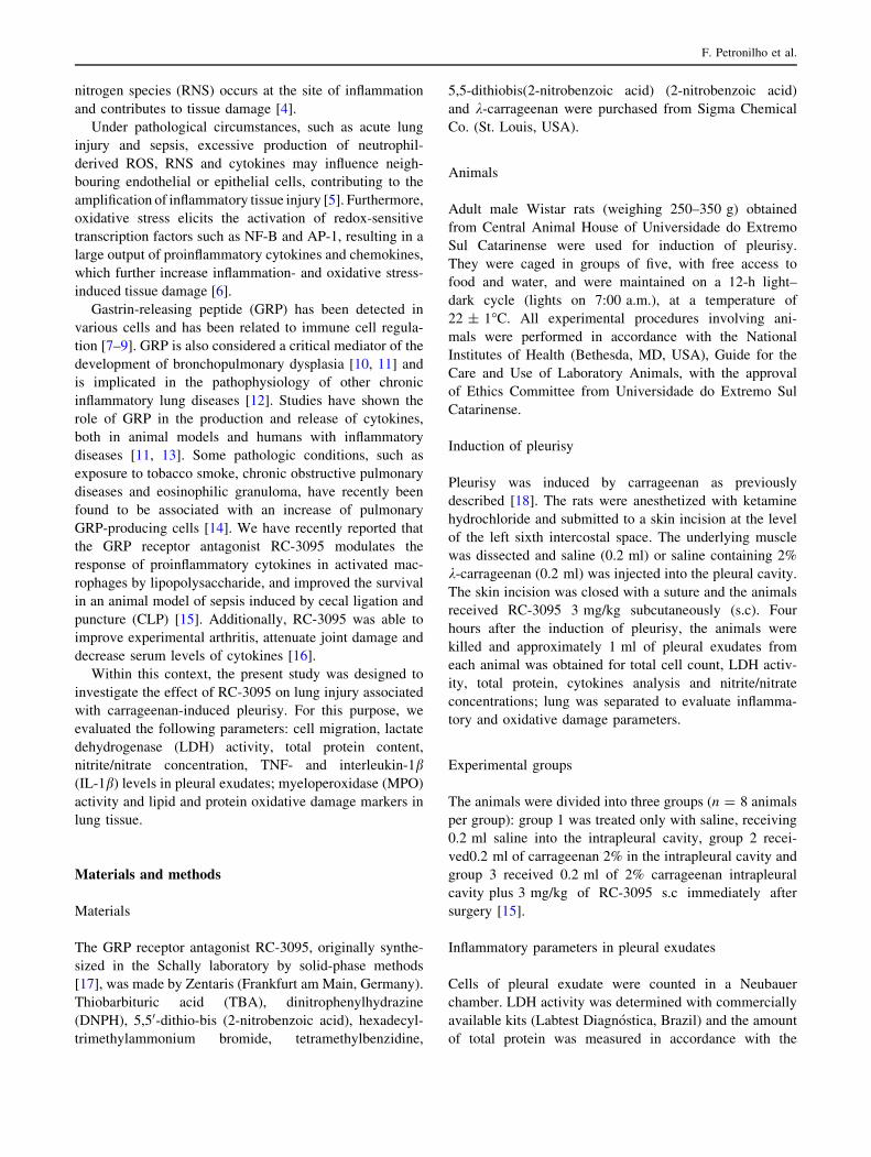
nitrogen species (RNS) occurs at the site of inflammation
and contributes to tissue damage [4].
Under pathological circumstances, such as acute lung
injury and sepsis, excessive production of neutrophil-
derived ROS, RNS and cytokines may influence neigh-
bouring endothelial or epithelial cells, contributing to the
amplification of inflammatory tissue injury [5]. Furthermore,
oxidative stress elicits the activation of redox-sensitive
transcription factors such as NF-B and AP-1, resulting in a
large output of proinflammatory cytokines and chemokines,
which further increase inflammation- and oxidative stress-
induced tissue damage [6].
Gastrin-releasing peptide (GRP) has been detected in
various cells and has been related to immune cell regula-
tion [7–9]. GRP is also considered a critical mediator of the
development of bronchopulmonary dysplasia [10, 11] and
is implicated in the pathophysiology of other chronic
inflammatory lung diseases [12]. Studies have shown the
role of GRP in the production and release of cytokines,
both in animal models and humans with inflammatory
diseases [11, 13]. Some pathologic conditions, such as
exposure to tobacco smoke, chronic obstructive pulmonary
diseases and eosinophilic granuloma, have recently been
found to be associated with an increase of pulmonary
GRP-producing cells [14]. We have recently reported that
the GRP receptor antagonist RC-3095 modulates the
response of proinflammatory cytokines in activated mac-
rophages by lipopolysaccharide, and improved the survival
in an animal model of sepsis induced by cecal ligation and
puncture (CLP) [15]. Additionally, RC-3095 was able to
improve experimental arthritis, attenuate joint damage and
decrease serum levels of cytokines [16].
Within this context, the present study was designed to
investigate the effect of RC-3095 on lung injury associated
with carrageenan-induced pleurisy. For this purpose, we
evaluated the following parameters: cell migration, lactate
dehydrogenase (LDH) activity, total protein content,
nitrite/nitrate concentration, TNF- and interleukin-1b(IL-1b) levels in pleural exudates; myeloperoxidase (MPO)
activity and lipid and protein oxidative damage markers in
lung tissue.
Materials and methods
Materials
The GRP receptor antagonist RC-3095, originally synthe-
sized in the Schally laboratory by solid-phase methods
[17], was made by Zentaris (Frankfurt am Main, Germany).
Thiobarbituric acid (TBA), dinitrophenylhydrazine
(DNPH), 5,50-dithio-bis (2-nitrobenzoic acid), hexadecyl-
trimethylammonium bromide, tetramethylbenzidine,
5,5-dithiobis(2-nitrobenzoic acid) (2-nitrobenzoic acid)
and k-carrageenan were purchased from Sigma Chemical
Co. (St. Louis, USA).
Animals
Adult male Wistar rats (weighing 250–350 g) obtained
from Central Animal House of Universidade do Extremo
Sul Catarinense were used for induction of pleurisy.
They were caged in groups of five, with free access to
food and water, and were maintained on a 12-h light–
dark cycle (lights on 7:00 a.m.), at a temperature of
22 ± 1�C. All experimental procedures involving ani-
mals were performed in accordance with the National
Institutes of Health (Bethesda, MD, USA), Guide for the
Care and Use of Laboratory Animals, with the approval
of Ethics Committee from Universidade do Extremo Sul
Catarinense.
Induction of pleurisy
Pleurisy was induced by carrageenan as previously
described [18]. The rats were anesthetized with ketamine
hydrochloride and submitted to a skin incision at the level
of the left sixth intercostal space. The underlying muscle
was dissected and saline (0.2 ml) or saline containing 2%
k-carrageenan (0.2 ml) was injected into the pleural cavity.
The skin incision was closed with a suture and the animals
received RC-3095 3 mg/kg subcutaneously (s.c). Four
hours after the induction of pleurisy, the animals were
killed and approximately 1 ml of pleural exudates from
each animal was obtained for total cell count, LDH activ-
ity, total protein, cytokines analysis and nitrite/nitrate
concentrations; lung was separated to evaluate inflamma-
tory and oxidative damage parameters.
Experimental groups
The animals were divided into three groups (n = 8 animals
per group): group 1 was treated only with saline, receiving
0.2 ml saline into the intrapleural cavity, group 2 recei-
ved0.2 ml of carrageenan 2% in the intrapleural cavity and
group 3 received 0.2 ml of 2% carrageenan intrapleural
cavity plus 3 mg/kg of RC-3095 s.c immediately after
surgery [15].
Inflammatory parameters in pleural exudates
Cells of pleural exudate were counted in a Neubauer
chamber. LDH activity was determined with commercially
available kits (Labtest Diagnostica, Brazil) and the amount
of total protein was measured in accordance with the
F. Petronilho et al.

Lowry method using bovine serum albumin as the standard
[19].
Measurement of nitrite/nitrate concentration in pleural
exudates
Total nitrite concentration in the samples was then mea-
sured using the Griess reaction, by adding 100 ll of Griess
reagent [0.1% (w/v) naphthylethylendiamide dihydrochlo-
ride in H2O and 1% (w/v) sulphanilamide in 5% (v/v)
concentrated H3PO4], vol. [1:1] to the 100 ll sample. The
optical density at 550 nm (OD550) was measured using
ELISA microplate reader [20].
Measurement of cytokines in pleural exudates
TNF-a and IL-1b concentrations were evaluated in the
pleural exudates at 4 h after intrapleural injection of
carrageenan. The assay was carried out by using a colori-
metric, commercial ELISA kit (Calbiochem-Novabiochem
Corporation, USA).
Myeloperoxidase activity in lung tissue
Tissues were homogenized (50 mg/ml) in 0.5% hex-
adecyltrimethylammonium bromide and centrifuged at
15,0009g for 40 min. The suspension was then sonicated
three times for 30 s. An aliquot of supernatant was mixed
with a solution of 1.6 mM tetramethylbenzidine and 1 mM
H2O2. Activity was measured spectrophotometrically as the
change in absorbance at 650 nm at 37�C [21].
Thiobarbituric acid reactive substances in lung tissue
The formation of thiobarbituric acid reactive substances
(TBARS) during an acid-heating reaction is widely adop-
ted as a sensitive method for measurement of lipid
peroxidation [22]. Briefly, the samples were mixed with
1 ml of 10% trichloroacetic acid and 1 ml of 0.67% TBA.
Subsequently, they were heated in a boiling water bath
for 30 min. Malondialdehyde (MDA) equivalents were
determined by absorbance at 532 nm using 1,1,3,3-tetra-
methoxypropane as an external standard. Results were
expressed as MDA equivalents (nmol/mg protein).
Protein carbonyl content in lung tissue
The oxidative damage to proteins was assessed by the
determination of carbonyl groups content, based on the
reaction with dinitrophenylhidrazine (DNPH) [23]. Briefly,
proteins were precipitated by the addition of 20% trichlo-
roacetic acid and redissolved in DNPH, and the absorbance
was monitored at 370 nm.
Sulfhydryl groups in lung tissue
Total thiol content in lungs was determined using the
5,5-dithiobis(2-nitrobenzoic acid) (2-nitrobenzoic acid)
method (DTNB). The conditions of DTNB measurement
were as described in [24] with some modifications. Briefly,
30 ll of a sample was mixed with 1 ml of PBS/1 mM
EDTA (pH 7.5). The reaction was started by the addition of
30 ll of 10 mM DTNB stock solution in PBS. Control
samples, which did not include DTNB or protein, were run
simultaneously. After 30 min of incubation at room tem-
perature, the absorbance at 412 nm was measured and
amounts of TNB formed [equivalent to the amount of
sulfhydryl (SH) groups] were calculated.
Statistical analysis
Results are expressed as mean ± SEM and P \ 0.05 was
considered to be significant. Differences between groups
were determined by one-way analysis of variance followed
by a Newman–Keuls test. All statistical analyses were
performed with SPSS 12.0 for Windows (SPSS, Chicago,
IL, USA).
Results
The effects of subcutaneous injection of RC-3095 on
inflammatory cell recruitment into the pleural cavity are
illustrated in Fig. 1a. Carrageenan-induced pleurisy dis-
played a substantial increase in the number of cells in the
pleural cavity compared with control rats, and treatment
with RC-3095 decreased cells migration in the pleural
cavity compared to the carrageenan group. RC-3095
administration reduced protein content and pleural effusion
LDH as an index of pleural cell injury (Fig. 2a, b,
respectively). Moreover, a substantial increase in TNF- and
IL-1b production was found in pleural exudates collected
from rats 4 h after carrageenan injection (Fig. 3). Pleural
exudates production of TNF- and IL-1b was significantly
reduced in carrageenan-injected rats treated with RC-3095.
A significant decrease in nitrite/nitrate accumulation in the
pleural exudates 4 h after carrageenan administration and
with RC-3095 relative to the non-treated carrageenan
group was also observed (Fig. 4).
We also investigated the effect of RC-3095 on the
presence of neutrophils by measurement of the activity of
MPO. MPO activity in the lung tissues was significantly
elevated at 4 h after carrageenan administration (Fig. 5a).
In addition, in rats treated with RC-3095, MPO activity in
the lungs was significantly reduced. The pulmonary injury
in carrageenan-treated animals was also characterized by
an increase in the tissue levels of MDA, indicative of lipid
Protective effect of gastrin-releasing peptide receptor antagonist

peroxidation (Fig. 5b). MDA levels were significantly
enhanced in lungs collected from carrageenan-treated rats
when compared to those found in the saline group and were
significantly reduced when RC-3095 was administered. In
addition, carrageenan decreased the free SH groups, and
this was partially reversed by RC-3095 treatment (Fig. 5c);
this did not occur with protein carbonyl levels (data not
shown).
Discussion
In this study, we report on the effects of the GRP receptor
antagonist RC-3095 in a well-established murine model for
acute inflammation induced by carrageenan. In the present
work, acute administration of RC-3095 exhibited pro-
nounced anti-inflammatory actions, characterized by the
inhibition of leukocyte influx into the pleural cavity, nitrite/
nitrate content, TNF-a and IL-1b levels in the acute model
Fig. 1 Cell migration into the pleural cavity in carrageenan-induced
pleurisy and treated with RC-3095. Assessment of cell migration into
the pleural cavity was performed 4 h after pleurisy induction and
treatments. Results are expressed as mean ± SD, n = 8 per group;
*P \ 0.05 compared with control group and **P \ 0.05 compared
with carrageenan group
Fig. 2 Total protein (a) and LDH activity (b) in pleural exudate of
rats submitted to carrageenan administration and treated with
RC-3095. Results are expressed as mean ± SD, n = 8 per group;
*P \ 0.05 compared with the control group and **P \ 0.05
compared with the carrageenan group
Fig. 3 TNF-a and IL-1b levels in pleural exudates of rats subjected
to carrageenan administration and treated with RC-3095. Results are
expressed as mean ± SD, n = 8 per group; *P \ 0.05 compared with
the control group and **P \ 0.05 compared with the carrageenan
group
Fig. 4 Nitrite/nitrate concentration in pleural exudates of rats
subjected to carrageenan administration and treated with RC-3095.
Results are expressed as mean ± SD, n = 8 per group; *P \ 0.05
compared with the control group and **P \ 0.05 compared with the
carrageenan group
F. Petronilho et al.

of inflammation caused by intrapleural administration of
carrageenan. Moreover, RC-3095 elicited important action
against oxidative damage in lipids and free SH groups and
increased cell viability. Taken together, these findings
suggest that the administration of RC-3095 could effec-
tively interfere with the inflammatory process in pleurisy
induced by carrageenan.
Cellular immune responses appear to be the predomi-
nant mechanisms involved in the pathogenesis of most
inflammatory pleural effusions. Recent advances in basic
research have extended our comprehension of the immu-
nological mechanisms underlying pleural inflammation.
Clearly, understanding the pleural inflammatory mecha-
nisms has direct clinical implications, although many
aspects relating to pleural inflammation and disease pro-
gression or resolution remain only partially understood
[25]. Although differential leukocyte count in pleural fluid
is of limited diagnostic value, it reflects the stage of the
inflammatory response and narrows the diagnostic possi-
bilities in exudative effusions. A pleural neutrophil
predominance is associated with the early phase of
the inflammatory response and can be found in several
infectious and non-infectious conditions, such as para-
pneumonic effusions, pulmonary embolus, viral infection,
gastrointestinal disease and acute tuberculous pleuritis
[26]. In our study, we demonstrated that RC-3095 inhibited
cell migration, LDH activity and total protein in pleural
exudates in carrageenan-induced pleurisy; this could
block several processes associated with pleural diseases
progression [27].
Studies have also shown that cell influx into the pleural
cavity occurs after the release of chemotactic cytokines by
pleural mesothelial cells in response to other cytokines
[27]. There is evidence that the pro-inflammatory cytokines
TNF-a and IL-1b help to propagate the extension of a local
or systemic inflammatory process [28–30]. We confirm
here that the inflammatory process caused by the admin-
istration of carrageenan into the pleural cavity leads to a
substantial increase in the levels of both TNF-a and IL-1bin the exudates. Interestingly, the levels of these two pro-
inflammatory cytokines are significantly lower in the exu-
date obtained from RC-3095-treated animals. Dal-Pizzol
and colleagues described that, in a sepsis animal model,
RC-3095 attenuated TNF-a and IL-1b and macrophage
stimulation with lipopolysaccharide [15]. Additionally,
another study demonstrated that RC-3095 was able to
attenuate serum levels these cytokines on complete
Freund’s adjuvant-induced arthritis in rats [16].
There is a large amount of evidence that the enhanced
formation of nitric oxide (NO) by inducible nitric oxide
synthase (NOS) may negatively contribute to the inflam-
matory process [3, 31–34]. For instance, inhibitors of NOS
activity reduce the development of carrageenan-induced
inflammation [32, 35]. In this context, we demonstrated
that the formation of nitrite and nitrate (final metabolites of
NO in water) after carrageenan administration were
reduced in pleural exudate from RC-3095-treated rats
compared to non-treated rats. The induction of iNOS by
injection of endotoxin in rodents in vivo is mediated by
endogenous TNF-a and IL-1b [36, 37]. As the levels of
TNF-a and IL-1b are significantly lower in the exudates
obtained from RC-3095-treated rats, we propose that the
Fig. 5 Myeloperoxidase activity (a), oxidative damage in lipids
through thiobarbituric acid reactive substances (TBARS) measurement
(b) and free SH groups in proteins (c) in lung tissue of rats submitted
to carrageenan administration and treated with RC-3095. Results are
expressed as mean ± SD, n = 8 per group; *P \ 0.05 compared with
the control group and **P \ 0.05 compared with the carrageenan
group
Protective effect of gastrin-releasing peptide receptor antagonist

attenuation of the formation of nitrite and nitrate observed
is secondary to reduced TNF-a and IL-1b levels.
Reduced amounts of NO lead to decreased endothelial
cell membrane permeability, thus limiting polymorphonu-
clear cell transmigration from the vascular compartment
into the lung [6]. Together with NO, MPO activity is an
indirect marker of activated leukocytes and is implicated in
exudation and cell migration [38]. The low MPO and NO
levels in our study following RC-3095 treatment showed
what had already been verified in an animal model of sepsis
by CLP [15].
ROS and primary granule constituents are secreted by
activated neutrophils, and these sometimes lead to injury of
normal tissue [39]. Several studies have demonstrated its
participation in models of inflammation, such as carra-
geenan-induced pleurisy in rats [40–43]. In accordance
with Guo and Ward [6], lipid peroxidation is believed to
play an important role in inflammatory lung diseases. As
expected, MDA concentrations increased in lung tissue in
the carrageenan group. On the other hand, when RC-3095
was administered, the levels of lipid peroxidation end-
products were substantially reduced. Other oxidative indi-
cators, SH-containing amino acid residues in protein
sulfhydryls, are susceptible targets for a variety of pro-
oxidants [44]. The increase of free SH groups in proteins
from the lung provides additional evidence for a decrease
in oxidative damage by RC-3095.
The present study showed that ROS amplify inflamma-
tory response by activating oxidant-regulated transcription
factors such as nuclear factor jB (NF-jB) and activa-
tion protein-1 (AP-1), with a subsequent increase of
pro-inflammatory cytokines [45]. NF-jB inhibitors
strongly inhibit the early phase of the pleural inflammatory
response induced by carrageenan in mice, suggesting that
the NF-jB pathway plays a relevant role in the inflam-
matory response elicited by carrageenan in the mice model
of pleurisy [46]. Levine and colleagues showed that bom-
besin stimulates NF-jB activation and expression of pro-
angiogenic factors in prostate cancer cells [47]. Therefore,
the decrease of inflammatory response and oxidative
damage in carrageenan-induced pleurisy may be attributed
to the GRP receptor; RC-3095 can be involved in the
regulation of NF-jB activation.
In conclusion, the present results support the view that
in carrageenan-induced pleurisy model, the GRP receptor
antagonist RC-3095 has anti-inflammatory properties
which can be related with the reduction of oxidative
damage.
Acknowledgments This work was supported by grants from
Universidade do Extremo Sul Catarinense (UNESC), Coordenacao de
Aperfeicoamento de Nıvel Superior (CAPES) and Conselho Nacional
de Desenvolvimento Cientıfico e Tecnologico (CNPq).
References
1. Lentsch AB, Ward PA. Regulation of experimental lung
inflammation. Respir Physiol. 2001;128:17–21.
2. Vinegar R, Truax JF, Selph JL, Voelker FA. Pathway of onset,
development, and decay of carrageenan pleurisy in the rat. Fed
Proc. 1982;41:2588–95.
3. Salvemini D, Wang ZQ, Wyatt P, Bourdon DM, Marino MH,
Manning PT, et al. Nitric oxide: a key mediator in the early and
late phase of carrageenan-induced rat paw inflammation. Br J
Pharmacol. 1996;118:829–37.
4. Salvemini D, Masferrer JL. Interactions of nitric oxide witch
cyclooxygenase: in vitro, ex vivo, and in vivo studies. Methods
Enzymol. 1996;269:12–25.
5. Fialkow L, Wang Y, Downey GP. Reactive oxygen and nitrogen
species as signaling molecules regulating neutrophil function.
Free Radic Biol Med. 2007;42:153–64.
6. Guo RF, Ward PA. Role of oxidants in lung injury during sepsis.
Antioxid Redox Signal. 2007;9:02–1991.
7. Medina S, Del Rio M, Victor VM, Hernanz A, De la Fuente M.
Changes with ageing in the modulation of murine lymphocyte
chemotaxis by CCK-8S, GRP and NPY. Mech Ageing Dev.
1998;102:249–61.
8. Medina S, Rio MD, De la Cuadra B, Fuente MD. Age-related
changes in the modulatory action of gastrin-releasing peptide,
neuropeptide Y and sulfated cholecystokinin octapeptide in the
proliferation of murine lymphocytes. Neuropeptides. 1999;33:
173–9.
9. Genton L, Kudsk KA. Interactions between the enteric nervous
system and the immune system: role of neuropeptides and
nutrition. Am J Surg. 2003;186:253–8.
10. Sunday ME, Yoder BA, Cuttitta F, Haley KJ, Emanuel RL.
Bombesin like peptide mediates lung injury in a baboon model of
bronchopulmonary dysplasia. J Clin Invest. 1998;102:584–94.
11. Subramaniam M, Sugyiama K, Coy DH, Kong Y, Miller YE,
Weller PF, et al. Bombesin-like peptides and mast cell responses:
relevance to bronchopulmonary dysplasia? Am J Respir Crit Care
Med. 2003;168:601–11.
12. Sunday ME. Neuropeptides and lung development. In:
McDonald JA, editor. Lung growth and development. New York:
Marcel Dekker; 1997. p. 401–94.
13. Grimsholm O, Rantapaa-Dahlqvist S, Forsgren S. Levels of
gastrin-releasing peptide and substance P in synovial fluid and
serum correlate with levels of cytokines in rheumatoid arthritis.
Arthritis Res Ther. 2005;7:416–26.
14. Petronilho F, Roesler R, Schwartsmann G, Dal-Pizzol F. Gastrin-
releasing peptide receptor as a molecular target for inflammatory
diseases. Inflamm Allergy Drug Targets. 2007;6:197–200.
15. Dal-Pizzol F, Di Leone LP, Ritter C, Martins MR, Reinke A,
Pens Gelain D, et al. Gastrin-releasing peptide receptor antagonist
effects on an animal model of sepsis. Am J Respir Crit Care Med.
2006;173:84–90.
16. Oliveira PG, Brenol CV, Edelweiss MI, Brenol JC, Petronilho F,
Roesler R, et al. Effects of an antagonist of the bombesin/gastrin-
releasing peptide receptor on complete Freund’s adjuvant-
induced arthritis in rats. Peptides. 2008;29:1726–31.
17. Radulovic S, Cai R-Z, Serfozo P, Groot K, Redding TW, Pinski J,
et al. Biological effects and receptor binding affinities of new
pseudononapeptide bombesin/GRP receptor antagonists with N-
terminal d-Trp or d-Tpi. Int J Pept Protein Res. 1991;38:593–600.
18. Vinegar R, Traux JF, Selph JL. Some quantitative temporal
characteristics of carrageenan-induced pleurisy in the rat. Proc
Soc Exp Biol Med. 1943;143:711–4.
19. Lowry OH, Rosebrough AL, Randal RJ. Protein measurement
with the Folin phenol reagent. J Biol Chem. 1951;193:265–75.
F. Petronilho et al.

20. Green IC, Wagner DA, Glowski J, Skipper PL, Wishnok JS,
Tannenbaum SR. Analysis of nitrate, nitrite and [15N] nitrate in
biological fluids. Anal Biochem. 1982;126:131–8.
21. De Young LM, Kheifets JB, Ballaron SJ, Young JM. Edema and
cell infiltration in the phorbol ester-treated mouse ear are tem-
porally separate and can be differentially modulated by
pharmacologic agents. Agents Actions. 1989;26:335–41.
22. Draper HH, Hadley M. Malondialdehyde determination as index
of lipid peroxidation. Methods Enzymol. 1990;186:421–31.
23. Levine RL, Garland D, Oliver CN, Amici A, Climent I, Lenz AG.
Determination of carbonyl content in oxidatively modified pro-
teins. Methods Enzymol. 1990;186:464–78.
24. Riddles PW, Blakeley RL, Zerner B. Reassessment of Ellman’s
reagent. Methods Enzymol. 1983;91:49–60.
25. Kroegel C, Antony VB. Immunobiology of pleural inflammation:
potential implications for pathogenesis, diagnosis and therapy.
Eur Respir J. 1997;10:2411–8.
26. Segura RM. Useful clinical biological markers in diagnosis of
pleural effusions in children. Paediatr Respire Rev. 2004;5:
205–12.
27. Antony VB, Godbey SW, Kunkel SL, Hott JW, Hartman DL,
Burdick MD, et al. Recruitment of inflammatory cells to the
pleura space: chemotactic cytokines IL-8, and monocyte che-
motactic peptide-1 in human pleural fluids. J Immunol. 1993;151:
7216–23.
28. Romano M, Sironi M, Toniatti C, Polentarutti N, Fruscella P,
Ghezzi P, et al. Role of IL-6 and its soluble receptor in induction
of chemokines and leukocyte recruitment. Immunity. 1997;6:
315–25.
29. Alonzi T, Fattori E, Cappelletti M, Ciliberto G, Poli V. Impaired
stat3 activation following localized inflammatory stimulus in
IL-6 deficient mice. Cytokine. 1998;10:13–8.
30. Utsunomiya I, Nagai S, Oh-ishi S. Sequential appearance of IL-1
and IL-6 activities in rat carrageenan-induced pleurisy. J Immu-
nol. 1991;147:1803–9.
31. Oh-Ishi S, Hayashi I, Hayashi M, Yamaki K, Utsunomiya I.
Pharmacological demonstration of inflammatory mediators using
experimental inflammatory models: rat pleurisy induced by car-
rageenin and phorbol myristate acetate. Dermatologica.
1989;179:68–71.
32. Tracey WR, Nakane M, Kuk J, Budzik G, Klinghofer V, Harris R.
The nitric oxide synthase inhibitor, l-NG-monomethylarginine,
reduces carrageenan-induced pleurisy in the rat. J Pharmacol Exp
Ther. 1995;273:1295–9.
33. Wei XQ, Charles IG, Smith A, Ure J, Feng GJ, Huang FP, et al.
Altered immune responses in mice lacking inducible nitric oxide
synthase. Nature. 1995;375:408–11.
34. Cuzzocrea S, Zingarelli B, Costantino G, Caputi AP. Beneficial
effects of Mn(III)tetrakis (4-benzoic acid) porphyrin (MnTBAP),
a superoxide dismutase mimetic, in carrageenan-induced pleu-
risy. Free Radic Biol Med. 1999;26:25–33.
35. Cuzzocrea S, Zingarelli B, Hake P, Salzman AL, Szabo C.
Antiinflammatory effects of mercaptoethylguanidine, a combined
inhibitor of nitric oxide synthase and peroxynitrite scavenger, in
carrageenan-induced models of inflammation. Free Radic Biol
Med. 1998;24:450–9.
36. Szabo C, Wu CC, Gross SS, Thiemermann C, Vane JR. Inter-
leukin-1 contributes to the induction of nitric oxide synthase by
endotoxin in vivo. Eur J Pharmacol. 1993;250:157–60.
37. Jin CY, Moon DO, Lee KJ, Kim MO, Lee JD, Choi YH, et al.
Piceatannol attenuates lipopolysaccharide-induced NF-kappaB
activation and NFkappaB-related proinflammatory mediators in
BV2 microglia. Pharmacol Res. 2006;54:461–7.
38. Mariotto S, Esposito E, Di Paola R, Ciampa A, Mazzon E,
de Prati AC, et al. Protective effect of Arbutus unedo aqueous
extract in carrageenan-induced lung inflammation in mice.
Pharmacol Res. 2008;57:110–24.
39. Yasui K, Baba A. Therapeutic potential of superoxide dismutase
(SOD) for resolution of inflammation. Inflamm Res. 2006;55:
359–63.
40. Cuzzocrea S, McDonald MC, Filipe HM, Costantino G, Mazzon E,
Santagati S, et al. Effects of tempol, a membrane-permeable radical
scavenger, in a rodent model of carrageenan-induced pleurisy. Eur J
Pharmacol. 2000;390:209–22.
41. Nardi GM, Siqueira Junior JM, Delle Monache F, Pizzolatti MG,
Ckless K, Ribeiro-do-Valle RM. Antioxidant and anti-inflam-
matory effects of products from Croton celtidifolius Bailon on
carrageenan-induced pleurisy in rats. Phytomedicine. 2007;14:
115–22.
42. Bhattacharyya S, Dudeja PK, Tobacman JK. Carrageenan-
induced NFkappaB activation depends on distinct pathways
mediated by reactive oxygen species and Hsp27 or by Bcl10.
Biochim Biophys Acta. 2008;1780:973–82.
43. Menegazzi M, Di Paola R, Mazzon E, Genovese T, Crisafulli C,
Dal Bosco M, et al. Glycyrrhizin attenuates the development of
carrageenan-induced lung injury in mice. Pharmacol Res. 2008;
58:22–31.
44. Aksenov MY, Markesbery WR. Changes in thiol content and
expression of glutathione redox system genes in the hippocampus
and cerebellum in Alzheimer’s disease. Neurosci Lett. 2001;302:
141–5.
45. Kirkham P, Rahman I. Oxidative stress in asthma and COPD:
antioxidants as a therapeutic strategy. Pharmacol Ther. 2006;
111:476–94.
46. Frode-Saleh TS, Calixto JB. Synergistic antiinflammatory effect
of NF-kappaB inhibitors and steroidal or non steroidal antiin-
flammatory drugs in the pleural inflammation induced by
carrageenan in mice. Inflamm Res. 2000;49:330–7.
47. Levine L, Lucci JA, Pazdrak B, Cheng JZ, Guo YS,
Townsend CM Jr, et al. Bombesin stimulates nuclear factor jB
activation and expression of proangiogenic factors in prostate
cancer cells. Cancer Res. 2003;63:3495–502.
Protective effect of gastrin-releasing peptide receptor antagonist

44
Capítulo II
Antagonist of gastrin-releasing peptide receptors induces a
protection from lethal sepsis: involvement of toll-like receptor 4 signaling
Fabricia Petronilho, Bruna de Souza, Larissa Constantino, Francieli Vuolo,
Cristiane Damiani Tomasi, Vinicius Renne Giombelli, Cláudio Teodoro de
Souza Sabrina da Silva, Denise Frediani Barbeiro, Francisco Garcia Soriano,
Emílio Luiz Streck, Cristiane Ritter, Alfeu Zanotto-Filho, Daniel Pens Gelain,
José Cláudio Fonseca Moreira, Norman L. Block, Rafael Roesler,
Gilberto
Schwartsmann, Andrew V. Schally, Felipe Dal-Pizzol
Artigo científico a ser submetido para publicação no periódico
Proceedings of the National Academy of Sciences USA (PNAS)

45
Antagonist of gastrin-releasing peptide receptors induces a
protection from lethal sepsis: involvement of toll-like receptor 4 signaling
Fabricia Petronilhoa, Bruna de Souzaa, Larissa Constantinoa, Francieli Vuoloa,
Cristiane Damiani Tomasia, Vinicius Renne Giombellia, Cláudio Teodoro de
Souzab Sabrina da Silvab, Denise Frediani Barbeiroc, Francisco Garcia
Sorianoc, Emílio Luiz Strecka, Cristiane Rittera, Alfeu Zanotto-Filhod, Daniel
Pens Gelaind, José Cláudio Fonseca Moreirad, Norman L. Blocke, Rafael
Roeslere,f,g, Gilberto Schwartsmannf,g,h, Andrew V. Schallyi, Felipe Dal-Pizzol
a,1
aExperimental Physiopathology Laboratory, Graduate Program in Health
Sciences, University of Southern Santa Catarina-UNESC, Criciúma, SC, Brazil;
bLaboratory of Exercise Biochemistry and Physiology, Graduate Program in
Health Sciences, University of Southern Santa Catarina-UNESC, Criciúma, SC,
Brazil; cDepartment of Internal Medicine, Academic Hospital, São Paulo
University, São Paulo, SP, Brazil; dCenter for Oxidative Stress, Department of
Biochemistry, Institute for Basic Health Sciences, Departments of Biochemistry
and Pharmacology, Federal University of Rio Grande do Sul, Porto Alegre, RS,
Brazil; eDepartment of Urology, University of Miami Miller School of Medicine,
Miami, Florida; eLaboratory of Molecular Neuropharmacology, Department of
Pharmacology, Institute for Basic Health Sciences, Federal University of Rio
Grande do Sul, Porto Alegre, RS, Brazil; fCancer Research Laboratory,
University Hospital Research Center (CPE-HCPA), Federal University of Rio
Grande do Sul, Porto Alegre, RS, Brazil; gNational Institute for Translational
Medicine, Porto Alegre, RS, Brazil; hDepartment of Internal Medicine, Faculty of

46
Medicine, Federal University of Rio Grande do Sul, Porto Alegre, RS, Brazil;
i
Veterans Affairs VA Medical Center and Department of Medicine, Divisions of
Endocrinology and Hematology-Oncology, University of Miami Miller School of
Medicine, and Department of Pathology, University of Miami Miller School of
Medicine, Miami, FL USA
Author contributions:
1To whom correspondence should be addressed at: Laboratório de
Fisiopatologia Experimental, Universidade do Extremo Sul Catarinense,
Avenida Universitária, 1105, 88806-000, Criciúma, SC, Brazil. E-mail:

47
Manuscript information
Number of text pages: 34
Number of figures: 5
Number of tables: 1
Abbreviations: Toll-like receptor 4 (TLR-4), gastrin-releasing peptide receptor
(GRPR), tumor necrosis factor- α (TNF-α), interleukin -1 β (IL-1β), cecal ligation
and puncture (CLP), nuclear factor-κB (NF-κB), activator protein 1 (AP-1),
lipopolysaccharide (LPS), systemic inflammatory response syndrome (SIRS),
LPS-binding protein (LBP), myeloid differentiation primary response protein 88
(MyD88), mitogen-activated protein kinases (MAPK), extracellular signal-
regulated kinase (ERK-1/2), c-Jun NH2-terminal kinase (JNK), bombesin (BN),
gastrin-releasing peptide (GRP), Toll-like receptors (TLRs), reverse
transcriptase–polymerase chain reaction (RT-PCR), monocyte chemotactic
protein 1 (MCP-1), irterleukin-6 (IL-6), vasoactive intestinal peptide (VIP),
phosphatidylinositol 3-kinase (PI3K), vasoactive intestinal peptide (VIP).

48
Abstract
In lethal polymicrobial sepsis, toll-like receptor-4 (TLR-4) signaling mediates a
critical role in impeding the migration of neutrophils to infectious foci, thereby
favoring increased bacteremia and ultimately leading to mortality. We have
previously shown that the selective gastrin-releasing peptide receptor (GRPR)
antagonist RC-3095 can reduce organ dysfunction in experimental sepsis by
modulating the release of cytokines by activated macrophages. Here we report
a novel link between GRPR and TLR-4 signaling and its relationship with
inflammatory parameters in in vitro and in vivo experimental models as well as
in sepsis patients. RC-3095 treatment inhibited expression of TLR-4 and
molecules in its downstream signaling pathway, leading to decreased activation
of nuclear factor-κB (NF-κB) and activator protein 1 (AP-1) in macrophages. In a
rat model of sepsis, RC-3095 treatment decreased lung TLR-4 content, reduced
the migration of inflammatory cells to the lung, reduced systemic cytokine
levels, and attenuated bacterial dissemination. Patients with systemic
inflammatory response syndrome (SIRS) and sepsis displayed elevated plasma
levels of GRP and GRP plasma levels correlated with clinical outcome in the
sepsis patients. These findings highlight the role of GRPR signaling in sepsis
outcome and the beneficial action of GRPR antagonism in controlling the
inflammatory response in sepsis through a mechanism involving inhibition of
TLR-4 signaling.
Keywords: gastrin releasing peptide/ toll-like receptor 4/ RC-3095/sepsis
49
Introduction
Sepsis remains a critical problem with significant morbidity and mortality despite
modern advances in critical care management. Severe sepsis (acute organ
dysfunction secondary to infection) and septic shock (severe sepsis plus
hypotension not reversed by fluid resuscitation) are major healthcare problems,
affecting millions of individuals around the world annually, killing upwards of one
in four afflicted patients, and increasing in incidence (1–5). As sepsis accounts
for >200,000 deaths and accounts for an estimated expenditure of $16 billion
per annum in the United States alone (6), it is a financial motivations for
researchers in the pharmaceutical industry to develop a therapy that can
substantially improve sepsis clinical outcomes (7). A better understanding of the
molecular mechanisms associated with development of sepsis and sepsis-
related organ injury is essential for reducing sepsis-related mortality.
Systemic inflammation is characterized by increased levels of circulating
cytokines and chemokines, and neutrophil sequestration in the lung (8).
Reduction of neutrophil chemotaxis has been associated with illness severity
and organ damage (9,10). Expansion of bacterial infection leads to systemic
toll-like receptor (TLR) activation (11). Endotoxin (lipopolysaccharide, LPS), a
major cell wall component in Gram-negative bacteria, is generally considered to
be a major pathogenic element in Gram-negative bacterial infection, and can
induce systemic inflammatory response syndrome (SIRS) (12). LPS sensing by
innate immune cells is vital for host defense against Gram-negative bacteria.
Recognition of LPS requires the accessory protein LPS-binding protein (LBP),
which converts oligomeric micelles of LPS into monomers that can interact with
CD14 for binding to the TLR-4-MD2 complex which is present on the surface of

50
many cell types including macrophages and dentritic cells (13). LPS-LBP-CD14
binding of the TLR-4-MD2 complex triggers conformational changes in TLR-4
that activate an intracellular downstream signaling pathway. Molecules involved
in the TLR-4 activated pathway include the adaptor molecule myeloid
differentiation primary response protein 88 (MyD88), interleukin (IL)-1 receptor-
associated kinases, and tumor necrosis factor (TNF) receptor-associated factor
6 (14). This pathway culminates in activation of several mitogen-activated
protein kinases (MAPKs), including extracellular signal-regulated kinase (ERK)-
1/2, c-Jun NH2-terminal kinase (JNK) and p38, as well as activation of the
transcription factors NF-κB and AP-1, resulting in the expression of numerous
genes encoding cytokines and other inflammatory molecules (6, 15). It is well
documented that NF-κB and AP-1 play prominent roles in LPS-induced
transcriptional regulation of inflammatory genes that contribute to the
development of septic shock and multiple organ failure (16). C3H/HeJ mice
bearing a mutation in the TLR-4 gene and TLR-4 knockout mice are
hyporesponsive to LPS and exhibit resistance to LPS-induced mortality,
indicating that TLR-4 signaling plays a critical role in LPS-induced sepsis (12).
However, TLR-4-defective mice exhibit neutrophil migration to the peritoneal
cavity during polymicrobial sepsis induced by lethal cecal ligation and puncture
(CLP), and as a consequence, were more resistant to sepsis than controls. The
systemic inflammatory response was also reduced in TLR-4-defective mice
because cytokine levels were lower than in wild-type TLR-4 mice submitted to
polymicrobial peritonitis. These results suggest that TLR-4 signaling may
diminish neutrophil migration to infection sites and promote an excessive
systemic inflammatory response (17). Given its central role in the pathogenesis

51
of sepsis, TLR-4 is a target of choice for the development of novel antisepsis
therapies (6).
Bombesin (BN) is a 14-amino acid peptide isolated from frog skin (18).
BN-like immunoreactivity via amphibian BN antibodies has been demonstrated
in the mammalian gut, central nervous system (CNS) and lung. Gastrin-
releasing peptide (GRP), a BN-like peptide, has been implicated in the
pathogenesis of inflammatory conditions (19-21) by studies showing that it is
involved in the control of CNS and gastrointestinal system functions (22,23),
cancer growth (24,25), and immune cell regulation (26-28). BN-like receptors
such as GRP/BN-preferring receptor (GRPR), neuromedin B receptor, and the
orphan BN receptor subtype 3, have been cloned. These receptors are seven-
transmembrane-spanning G protein-coupled receptors that are able to stimulate
various intracellular signaling pathways that are intimately involved in activation
of neutrophils and macrophages by chemokines (29), long known to attract
various inflammatory cells (30).
We recently demonstrated that the selective GRPR antagonist RC-3095
attenuates the release of proinflammatory cytokines in vitro and in vivo and
improves survival in experimental sepsis (31). In particular, we showed in a
well-established model of sepsis and acute lung injury that RC-3095 could
reduce mortality rates by alleviating organ dysfunction and inflammatory
infiltration and modulating the release of pro-inflammatory cytokines by
activated macrophages (31). These findings are consistent with the involvement
of a GRPR-stimulated inflammatory pathway in the development of sepsis.
Given that TLRs are essential components of the innate immune response to
infection and a growing body of evidence indicates that these receptors may
52
play a role in the pathophysiology of sepsis (32-34), together with the
aforementioned critical role of TLR-4 in particular in neutrophil migration (6), we
hypothesize that GRPR stimulation may exert its inflammatory effects via a
mechanism involving the TLR-4 signaling pathway.
The aims of this study were firstly investigate the effects of RC-3095 on
TLR-4 expression and its signaling pathways in LPS-stimulated peritoneal
macrophages. Additionally, TLR-4 content, cytokine/chemokine levels,
neutrophil migration levels, and bacterial dissemination levels were determined
in a CLP animal model of sepsis. These experiments in animals were
completed by an examination of cytokine levels in human septic patients. Anti-
inflammatory neuropeptides have recently emerged as endogenous factors
participating in the maintenance of immune tolerance and a loss of immune
tolerance results in the breakdown of immune homeostasis and an exacerbation
of inflammatory conditions (35). Therefore, our second aim was to examine
whether GRP levels in plasma are related to sepsis severity in humans.
Results
GRPR antagonist RC-3095 inhibits expression of TLR-4 and its signaling
pathway constituent molecules in LPS-stimulated RAW 264.7 cells.
Reverse-transcription polymerase chain reaction (RT-PCR) experiments in
RAW 264.7 cultures revealed that RC-3095 significantly reduced TLR-4 mRNA
levels in macrophages after LPS exposure (Fig. 1A). The decreased TLR-4
expression induced by RC-3095 was followed by decreased ERK1/2, JNK and
Akt phosphorylation in a sustained fashion (until 60 min after the end of RC-
3095 treatment) (Fig. 1B). Subsequent electromobility shift assay (EMSA)

53
experiments showed that the nuclear extract from LPS-stimulated RAW 264.7
cells had a significant increase in the DNA-binding activity of NF-κB and AP-1
(Fig. 1C). However this binding activity was suppressed by RC-3095 exposure,
suggesting that suppression of NF-κB and AP-1 nuclear translocation by RC-
3095 was associated with decreased gene expression of TLR-4 and MAP
kinase activation (Fig. 1A and B).
RC-3095 inhibits expression of TLR-4 in the lung in an animal model of
polymicrobial sepsis. RT-PCR using TLR-4 specific primers demonstrated
high levels of TLR-4 mRNA expression in lung tissue 6 h after sepsis and
significantly reduced expression of TLR-4 mRNA in RC-3095 treated animals
relative to that in the sepsis group (Fig. 2A). Immunoblotting experiments
showed that the decreased mRNA levels in the lung were followed by
decreased TLR-4 protein levels (Fig. 2B), and a subsequent decrease in
inflammatory cell presence in the bronchoalveolar fluid (Fig. 2C). Thus
pharmacologic blockade of the GRP-GRPR system decreased TLR-4
expression and protein content both in vitro and in vivo.
RC-3095 decreases cytokine/chemokine expression in LPS-activated RAW
264.7 cells and in severe sepsis animal model and in septic patients.
Enzyme-linked immunosorbent assays (ELISAs) revealed elevated monocyte
chemotactic protein 1 (MCP-1) levels in RAW 264.7 cells exposed to LPS (Fig.
3A) and serum and the bronchoalveolar fluid (BALF) of CLP septic rats (Fig. 3B
and Fig. 3C), relative to unexposed control cells and un-ligated control rats,
respectively. Administration of RC-3095 resulted in a significant decrease in

54
MCP-1 levels compared with the corresponding levels in LPS-exposed cells and
CLP septic rats. LPS-stimulated RAW 264.7 cells (Fig. 3D) and vehicle-injected
CLP animals showed a significant increase in serum (Fig. 3E) and BALF (Fig.
3F) interleukin-6 (IL-6) levels compared with that in unstimulated cells and sham
rats, respectively. RC-3095 administration significantly reduced IL-6 levels both
in vitro and in the animal model of sepsis (Fig. 3D-F). Continuous infusion with
RC-3095 (3 mg/kg) for 12 h decreased IL-6 plasma levels in septic patients
(Fig. 3G) but did not significantly affect IL-10 plasma levels (Fig. 3H). These
data demonstrate that TLR-4 inhibition mediated by RC-3095 decreased
inflammatory markers in sites distant to the infectious focus.
RC-3095 decreases inflammatory cell migration to the lung and bacterial
dissemination. RC-3095 decreased inflammatory cell levels in the BALF of
CLP animals compared to that in untreated CLP animals (Fig. 4A). RC-3095
administration also reduced bacterial dissemination in circulation (Fig. 4B) and
in peritoneal exudate (Fig. 4C) compared to levels in untreated CLP animals.
Plasma GRP levels can be related to outcome in septic patients. The
clinical profiles of sepsis patients (regardless of severity, n = 30) were
compared to those of patients with SIRS (n = 29). The data were further
analyzed for differences among sepsis patients according to disease severity:
sepsis (n = 9), severe sepsis (n = 10), and septic shock (n = 11) patients. The
patient groups were similar in terms of race, age, sex, ICU stay, sepsis source
and SOFA score (data not shown). The median APACHE II score of the mild to
moderate sepsis group was lower than those of the septic shock, severe sepsis

55
and SIRS groups (Table 1). Plasma GRP concentrations sampled on the
patients’ first day in the intensive care unit (ICU) were similar between the SIRS
patients and sepsis patients (sepsis, severe sepsis, and septic shock combined;
Fig. 5A). Comparing patients across sepsis gravity levels, we found that
patients with septic shock had greater GRP concentrations than their sepsis
and severe sepsis counterparts (Fig. 5B). Both SIRS and sepsis patients had
significantly higher GRP concentrations compared to healthy subjects (data not
shown). Clinical outcome measures revealed that those subjects with the
highest GRP concentrations had major mortality in the sepsis groups (Fig. 5C);
this association was not apparent in patients with SIRS (Fig. 5D). Patients with
a GRP concentration <1 pg/ml had no mortality, whereas those with a GRP
concentration ≥1 pg/ml) had a mortality rate of approximately 87% (Fig. 5 E),
with an area under the ROC curve of 0.85.
Discussion
In the present study, we demonstrated for the first time that RC-3095 treatment
can decreases TLR-4 expression and down-stream signaling activation in RAW
264.7 cells stimulated by LPS, leading to a decrease of release of chemokines
and cytokines. These results were supported by our in vivo experiments which
showed lower IL-6 and MCP-1 concentrations and bacteremia in RC-3095
treated CLP animals. We further showed that RC-3095 treatment decreased
inflammatory cell levels in the BALF of CLP animals. These findings are
noteworthy given that neutrophil function is essential to preventing bacterial
dissemination into the circulation; thus we can postulate that impairment of their
migration should contribute to bacterial clearance failure during an infection

56
process (36). Our results indicate that the RC-3095 treatment limited the spread
of infection beyond the abdominal compartment, suggesting that RC-3095 could
potentially prevent development of multiple organ dysfunction syndrome (35-
39). There are numerous factors that interact in the long chain of events from
pathogen recognition to the overwhelming of host responses (40). Our findings
provide support for the notion that TLR-4 is a particularly important element of
host defense during an infection and may play a role in the pathophysiology of
sepsis (33,34,41). This view is strongly supported by prior research showing
that TLR-4 defective mice do not exhibit failure of neutrophil migration to the
peritoneal cavity during polymicrobial sepsis induced by lethal CLP, and as
consequence, are more resistant to sepsis than controls (17). Furthermore,
increased concentration TLR-4 mRNA in lung tissue 3 h after CLP surgery has
been shown to precede and correlate with death (32).
It is well established that immune responses may be influenced by the
nervous system (42). Studies supporting the involvement of neuropeptides in
inflammatory diseases have focused predominantly on a proinflammatory role
for substance P (43,44), GRP (31,45) and nociceptin/orphanin FQ (46), as well
as an immunomodulatory role for peptide Y (47) and an anti-inflammatory role
for vasoactive intestinal peptide (VIP) (48). Furthermore neuropeptides, which
regulate the macrophage response to LPS, affect TLR-4 expression (49), and
regulate TLR-4 signaling (50). In this context, and because activated
macrophages have been shown to secrete GRP (51) and macrophages seem
to be central in the development of sepsis and septic shock (52,53), we observe
a decrease in the expression of TLR-4 mRNA in RAW 264.7 cells stimulated by
LPS following treatment with RC-3095. Our findings are consistent with recent

57
reports that increased expression of TLR-2 and TLR-4 during the early phase of
sepsis correlates with death in CLP animals (32), and that the down-regulation
of these receptors increases their survival (17). Furthermore, our observation
that RC-3095 inhibits upregulation of TLR-4 in polymicrobial sepsis in lung
tissue 6 h after CLP, resulting in a diminution of lung inflammation, fits with prior
research indicating that GRP is present in pulmonary neuroendocrine cells and
may be a mediator of acute and chronic lung injury in bronchopulmonary
dysplasia (54), as well as work showing that the GRPR antagonism can
alleviate alveolar edema and inflammatory infiltration (31).
TLR activation can be affected by perturbation of receptor complexes by
soluble TLRs or structurally related membrane proteins, ubiquitin-mediated
degradation of TLRs, and signaling proteins, sequestration of adapter proteins,
expression of dominant negative signalling inhibitors, and deactivation of
kinases through phosphatases (55,56). BN-activated G protein-coupled
receptor may phosphorylate proteins members of the Jak/STAT family (57).
RC-3096 might exert it effects on TLR-4 expression through STAT6 as
phosphorylated STAT6 down-regulates TLR-4 expression and STAT6
overexpression enhances inhibition of the TLR-4 promoter (58). TLR-4
enhances the inflammatory response through four major intracellular kinase
signaling pathways: p39 MAPK, JNK, and phosphatidylinositol 3-kinase
(PI3K)/Akt (59-63). The present results provide a clear demonstration that
GRPR antagonism influences TLR-4 induced activation of intracellular kinase
signaling pathways. TLR-4 signaling has been previously shown to activate the
ERK1/2, JNK and PI3K pathways, ultimately activating Akt (12, 64-69).

58
Activation of TLR-4 leads to increased activity of NF-κB and AP-1
transcription factors (68) while TLR-4-binding peptide inhibits LPS-induced
activation of NF-κB (12). NF-κB and AP-1 appear to play key roles in the
transcription of a number of inflammatory genes strongly involved in the
pathophysiology of sepsis (68). These observations are in accordance with our
results showing that RC-3095 exposure in RAW 264.7 cells decreased LPS-
stimulated DNA-binding activity of NF-κB and AP-1.
Neutrophil recruitment to infection sites is heavily dependent on
chemotatic cytokines (chemokines) such as MCP-1 (10,62). Moreover, because
IL-6 directly affects the adhesion and migratory functions of neutrophils, it may
promote the activity and production of important neutrophil modulators (69,79).
Wagner and Roth’s work showed that plasma MCP-1 and IL-6 are intermediate
effectors in neutrophil migration by 1-2 h after endotoxemia (71). Systemic
activation of TLR-4 results in inadequate activation of neutrophils by bacterial
products and/or chemokines induced by the expression of inducible nitric oxide
synthase, resulting mainly in CXCR2 desensitization and loss of adhesion
molecules (e.g. L-selectin). In addition, increase in acute-phase protein serum
levels induced by chemokines may also inhibit neutrophil-endothelium
interactions, thereby reducing the rolling/adhesion and chemotaxis of
neutrophils and ultimately disrupting neutrophil migration (41).
During endotoxic shock, massive numbers of neutrophils and other
leukocytes accumulate in the lung, a process entirely dependent on TLR-4.
Leukocyte accumulation in the lung is also observed in human septic patients
(72) and systemic activation of TLR-4 results in immense trapping of leukocytes
within lung capillaries (73-75). Furthermore, neuropeptides are known to

59
stimulate cytokine production in macrophages, lymphocytes, and mast cells,
and substance P is reported to influence LPS-induced production of pro-
inflammatory cytokines, which was abolished by neurokinin-1 (NK-1) receptor
blocking (76). Arranz and collaborators have showed that pro-inflammatory
cytokines can act synergistically, together with Gram-negative bacterial
components, to upregulate TLR-4 expression, and it is possible that VIP-
induced inhibition of TLR-4 up-regulation in inflammatory models occurs
indirectly via suppression of pro-inflammatory cytokine production (77). We
proposed in recent review that GRP may serve an autocrine/paracrine role in
macrophage activation during sepsis and/or LPS sitmulation, leading to a
modulation of pro-inflammatory, but not anti-inflammatory responses (44).
In this study, we demonstrated RC-3095 to be an effective blocker of
bacterial dissemination into the circulation. Our results showing that acute RC-
3095 treatment decreased cell migration in the lung fit Alves-Filho et al.’s
findings showing that TLR-4-deficient mice exhibit reduced neutrophil migration
to the infection focus and sequestration of the cells within lung tissue during
lethal polymicrobial sepsis (17). Cell sequestration is a key event in sepsis-
induced acute lung injury as it hampers bacterial clearance, leading to
bacteremia.
The presence of increased GRP concentrations in patients with
inflammatory disease and correlations of GRP concentration with erythrocyte
sedimentation rate and with TNF and IL-6, suggest that it may also be involved
in modulation of the inflammatory process in humans (78-79). The present
observation of reduced IL-6, but not IL-10, levels in the plasma of sepsis

60
patients treated with RC-3095 indicates that this GRPR antagonist may be
selective for activation of pro-inflammatory cytokines.
Our results suggest that GRP may be differentially involved in SIRS and
sepsis. Despite the presence of similar plasma levels of GRP between SIRS
and sepsis patients, GRP could only predict outcome in septic patients. Our
accompanying findings suggesting that GRP increases the inflammatory
response via TLR upregulation could, at least in part, explain the presently
observed differences between septic and SIRS patients.
Conclusions
The present experiments demonstrated that GRPR antagonism exerts an
overall protective role in LPS-activated macrophages culture and in acute
sepsis in rodents. Our results further indicate that this protective effect can be
attributed to an attenuation of TLR-4 signaling. This attenuation favors
neutrophil infiltration, resulting in decreased bacteremia and thus improving
sepsis outcome. Our finding that plasma GRP levels could predict outcome in
sepsis but not SIRS patients suggests that GRP plays different roles in the two
conditions. Our findings suggest that GRPR antagonism modulates uncontrolled
inflammation by targeting TLR-4-mediated responses and immunoregulatory
functionality. Taken together, the present results suggest that a GRPR
antagonist could be developed as a new alternative therapy for Gram-negative
bacterial sepsis.
Materials and Methods

61
Cells. RAW 264.7 macrophages were obtained from UFRJ Cell Bank, Rio de
Janeiro, Brazil. In preparation for RT-PCR analyses of TLR-4 mRNA,
immunoblotting of pERK1/2, pJNK, pAkt, and EMSA of NF-κB and AP-1, the
cells were seeded in 24 well plates (0.5 x 10⁶
cells/well) and incubated for 24 h
in RPMI-1640 media supplemented with 10% bovine fetal serum.
Animal subjects. A total of 15 male Wistar rats, 2 to 3 mos. old, were used in
this study. The rats were randomly divided, n = 5 per group, into sham
operated, CLP and CLP plus RC-3095 groups. Six hours after surgery
(described below), all rats were anesthetized with a mixture of ketamine (80
mg/kg) and xylazine (10 mg/kg), given intraperitoneally. As soon as anesthesia
was confirmed in each animal by the loss of pedal and corneal reflexes, the
animal’s thoracic cavity was opened and it was killed by cardiac puncture. Blood
was collected from the site of the cardiac puncture, BALF and peritoneal
lavages were performed, and the lung tissue was dissected out. All
experimental procedures involving animals were performed in accordance with
National Institutes of Health (Bethesda, MD) Guide for Care and Use of
Laboratory Animals with the approval of our institutional ethics committee.
Human subjects. Twelve patients, (7 males, 5 females), admitted to an adult
medical ICU with a clinical diagnosis of septic shock and failure of three or more
organs were enrolled in the RC-3095 administration experiment according to
the Consensus Conference (80). All procedures involving patients and healthy
volunteers were performed in compliance with the Declaration of Helsinki and
National Institutes of Health guidelines and were approved by the institutional

62
ethics committee (protocol number 431/2006). All patients gave their informed
consent prior to their inclusion in the study. Their mean age (±SD) was 61 ± 7 y.
Patients’ plasma samples were collected in heparin-treated vacuum tubes
before and after RC-3095 administration.
All patients consecutively admitted to an adult medical ICU between
August 2008 and December 2008 with SIRS or sepsis diagnosis were screened
for enrollment in the GRP analysis experiment. Septic patients (n = 30) were
classified as sepsis, severe sepsis, or septic shock according to the Consensus
Conference (80) by two board-certified internal medicine specialists. Septic
patients were paired to SIRS patients (n = 29) in relation to age, gender,
severity scores and mortality (demographics summarized in Table 1). Patients
were eligible if the time between sepsis diagnosis (this was done by reviewing
medical charts and/or contacting to patients assisting physician) and ICU
admission was no more than 24 h. In parallel, clinically relevant data were
recorded daily for 28 d. Exclusion criteria were: age below 18, chronic hepatic
or renal insufficiency, neoplasia receiving chemo, or radiotherapy and
immunodeficiency. Blood samples were drawn into heparin-treated vacuum
tubes for GRP quantitative analysis (described below).
CLP model. Rats were subjected to CLP as previously described (81). Briefly,
they were anesthetized with a mixture of ketamine (80 mg/kg) and xylazine 10
mg/kg, given intraperitoneally. Under aseptic conditions, a 3-cm midline
laparotomy was performed to expose the cecum and adjoining intestine. The
cecum was tightly ligated with a 3.0 silk suture at its base, below the ileocecal
valve, and was perforated once with 14-gauge needle. The cecum was then

63
squeezed gently to extrude a small amount of feces through the perforation site.
The cecum then was returned to the peritoneal cavity and the laparotomy was
closed with 4.0 silk sutures. Animals were resuscitated with normal saline (50
mL/kg subcutaneous) immediately after the operation. All animals were returned
to their home cages with free access to food and water.
Drug treatments. GRP receptor antagonist RC-3095, originally synthesized in
the Shally laboratory by solid-phase methods (82), was obtained from Zentaris
(Frankfurt am Main, Germany). Cultures were exposed to LPS (Escherichia coli
055:B5, Sigma Aldrich, St. Louis, MO) (100 ng/ml)–supplemented medium
(RPMI 1640) or RPMI 1640 alone and, 4 h later, RC-3095 (10 ng/ml) was added
for 2 h. Several times after the treatment period, cells and/or media samples
were collected for analyses in RC-3095-free media. Rats in the CLP plus RC-
3095 group were administered RC-3095 (3 mg/kg, subcutaneously) immediately
after surgery. Human patients received a continuous infusion with RC-3095 (3
mg/kg) over a period of 12 h.
RT-PCR. RNA was extracted from RAW 264.7 cells and from lung tissue
specimens collected from animals in the CLP and CLP+RC3095 groups using
TRIZOL reagent (Invitrogen®). RNA concentration was determined by
absorbance at 260 nm, and RNA integrity was confirmed by electrophoresis on
1% agarose gels and staining with 0.1 mg/L ethidium bromide. After DNAse I
digestion of the total extracted RNA, 1-μg aliquots of RNA were used to
synthesize single-strand cDNA using oligo dT primer (Promega®) and Impron II
reverse transcriptase (Promega®). PCR was carried out with 1.0 μl of RT

64
product serving as the template. The amplified products were analyzed by
ethidium bromide-stained agarose gel electrophoresis. For cell extracts, the
PCR primer sequence for TLR-4 were 5´GGCAGCAGGTGGAATTGTAT 3´
(sense) and 5´AGGCCCCAGAGTTTTGTTCT 3´(reverse) 5´
AAGAGCTGGAATACCTGGAC 3´ (sense) and
5´GAAATGCTACAGTGGCTACC 3´(reverse) for cell extracts and for animal
tissues. The gel was analyzed using Gene Flash and Software Image Gene
Tools (Syngene). TLR-4 mRNA levels were expressed as the ratio of signal
intensity for the target genes in relation to that for co-amplified glyceraldehyde-
3-phosphate dehydrogenase.
EMSA. The protein contents of RAW 264.7 cell nuclear extracts were prepared
as described previously (83). EMSA was performed using biotin-labeled
oligonucleotides to measure NF-κB or AP-1 content according to the kit
manufacturer’s protocol (Pierce).
Western blot. Phosphorylation of ERK1/2, JNK and Akt in cells was measured
by western blotting wherein whole cell lysates (20 µg, for phospho-ERK1/2,
phospho-JNK and phospho-Akt) were separated by 10% SDS-PAGE and then
electro-transferred to nitrocellulose membranes (Amersham International,
Buckinghamshire, UK). The membranes were preincubated for 1 h at room
temperature in Tris-buffered saline, pH 7.6, containing 0.05% Tween 20 and 3%
bovine serum albumin. The nitrocellulose membranes were incubated with
phosphorylated ERK1/2, phosphorylated JNK and phosphorylated Akt and the

65
immunoreactive bands were detected by incubation with HRP conjugates of
anti-rabbit IgGs and enhanced with chemoluminescence reagents (Amersham).
TLR-4 protein content in rat BALF and lung tissue was also quantified by
immunoblotting. The tissues were pooled, minced coarsely, and homogenized
immediately in extraction buffer (mM) (1% Triton-X 100, 100 Tris, pH 7.4,
containing 100 sodium pyrophosphate, 100 sodium fluoride, 10 EDTA, 10
sodium vanadate, 2 PMSF and 0.1 mg of aprotinin/ml) at 4ºC with a Polytron
PTA 20S generator (Brinkmann Instruments model PT 10/35) operated at
maximum speed for 30 s. The extracts were centrifuged at 11000 rpm and 4°C
in a Beckman 70.1 Ti rotor (Palo Alto, CA) for 40 min to remove insoluble
material, and the supernatants of these tissues were used for protein
quantification, using the Bradford method (84). Extracted proteins were
denatured by boiling in Laemmli (85) sample buffer containing 100 mM DTT,
run on SDS-PAGE and transferred to nitrocellulose membranes. The
membranes were blocked, probed with anti-TLR antibodies (Santa Cruz
Biotechnology, Inc, CA) and developed as described previously (86). The blots
were exposed to preflashed Kodak XAR film with Cronex Lightning Plus
intensifying screens at −80°C for 12–48 h. Band intensities were quantitated by
optical desitometry (Scion Image software, ScionCorp, Frederick, MD) of the
developed autoradiographs.
ELISA. Concentrations of IL-6 (in cells, rat BALF and serum, and human
plasma), IL-10 (in human plasma) and MCP-1 (in cells, rat BALF and serum)
were determined by a standard sandwich ELISA, employing commercially
available kits (R&D Systems, Minneapolis, MN). Plasma GRP concentrations

66
were determined in blood samples from healthy volunteers and septic patients
by ELISA commercial assays according to the manufacturer’s instructions
(Phoenix Pharmaceuticals, Inc., Burlingame, CA, U.S.A.).
Neutrophil migration in BALF. Cells were harvested by introducing 1.5 ml of
sterile PBS through the tracheal cannula and withdrawn to recover BALF.
Migrating neutrophil cell counts were performed as previously described (87).
The results are expressed as the number of cells/mm3
.
Bacterial counts in the peritoneal exudate and blood. Bacterial count was
determined as previously described (88). Briefly, after peritoneal lavage with
sterile PBS and blood collection performed during euthanasia (described
above), aliquots of serial dilutions of the samples were plated on Muller-Hinton
agar dishes (Difco Laboratories) and incubated at 37°C; colony-forming units
(CFUs) were analyzed after 24 h. The results were expressed as CFU/ml.
Statistical Analysis
Results are expressed as means ± SD and p < 0.05 was considered significant.
Differences between groups were determined by one-way analysis of variance
(ANOVA) followed by Tukey’s post hoc test. Differences between two groups
were determined by t test. In the human experiments, demographic and clinical
characteristics of the study groups were compared by t-test, γ2 test or ANOVA,
followed by LSD post hoc test, as appropriate, and a two-sided significance
level of 0.05 or less was considered statistically significant. The accuracy of
GRP concentrations in distinguishing between survivors and non-survivors was

67
examined separately by receiver operator characteristic (ROC) curves. Survival
curves were generated by the Kaplan-Meier curve. Differences between groups
were calculated by the log rank test. All statistical analyses were performed with
SPSS 12.0 for Windows (SPSS, Chicago, IL).
ACKNOWLEDGMENTS. This work was supported by the National Council for
Scientific and Technological Development (CNPq); the South American Office
for Anticancer Drug Development (SOAD; Porto Alegre, Brazil); and the
National Institute for Translational Medicine (INCT program).
68
1. Angus DC, et al. (2001) Epidemiology of severe sepsis in the United States:
analysis of incidence, outcome, and associated costs of care. Crit Care Med
29:1303–1310.
2. Dellinger RP (2003) Cardiovascular management of septic shock. Crit Care
Med 31: 946–955.
3. Martin GS, Mannino DM, Eaton S, Moss M (2003) The epidemiology of
sepsis in the United States from 1979 through 2000. N Engl J Med
348:1546–1554.
4. Linde-Zwirble WT, Angus DC (2004) Severe sepsis epidemiology: sampling,
selection, and society. Crit Care 8:222–226.
5. Dombrovskiy VY, Martin AA, Sunderram J, Paz HL (2007) Rapid increase in
hospitalization and mortality rates for severe sepsis in the United States: a
trend analysis from 1993 to 2003. Crit Care Med 35:1414–1415.
6. Matsuda N, Hattori Y (2006) Systemic inflammatory response syndrome
(SIRS): molecular pathophysiology and gene therapy. J Pharmacol Sci 101:
189-198.
7. Dyson A, Singer M (2009)
8. Abraham E (2003) Neutrophils and acute lung injury. Crit Care Med 31:195–
199.
Animal models of sepsis: Why does preclinical
efficacy fail to translate to the clinical setting? Supplement 37: 30-37.
9. Hedge A, Zhang H, Moochhala SM, Bhatia M (2007) Neurokinin-1 receptor
antagonist treatment protects mice against lung injury in polymicrobial
sepsis. J Leukoc Biol 82: 678-685.

69
10. Chishti AD, Shenton BK, Kirby JA, Baudouin SV (2004) Neutrophil
chemotaxis and receptor expression in clinical septic shock. Intensive Care
Med 30:605–611.
11. Alves-Filho JC, et al. (2009) Regulation of chemokine receptor by Toll-like
receptor 2 is critical to neutrophil migration and resistance to polymicrobial
sepsis. Proc Natl Acad Sci U S A 106:4018-4023.
12. Sugiyama K, Muroi M, Tanamoto K (2008) A novel TLR-4-binding peptide
that inhibits LPS-induced activation of NF-KappaB and vivo toxicity. Eur J
Pharmacol
13. Miller SI, Ernst RK, Bader MW (2005)
10: 152-156.
LPS, TLR-4 and infectious disease
diversity. Nat Rev Microbiol
14. Tsung A, et al. (2007)
3:36-46.
A novel inhibitory peptide of Toll-like receptor
signaling limits lipopolysaccharide-induced production of inflammatory
mediators and enhances survival in mice. Shock
15. Weighardt H, Holzmann B (2007)
27: 364-369.
Role of Toll-like receptor responses for
sepsis pathogenesis. Immunobiology
16. Hellmich MR, et al. (1999) Multiple protein kinase pathways are involved in
gastrin-releasing peptide receptor-regulated secretion. J Biol Chem
274:23901-23909.
212:715-722.
17. Alves-Filho JC, et al. (2006) Toll-like receptor 4 signaling leads to neutrophil
migration impairment in polymicrobial sepsis. Crit Care Med 34: 461-470.
18. Anastasi A, Erspamer V, Bucci M (1971) Isolation and structure of bombesin
and alytesin, 2 analogous active peptides from the skin of the European
amphibians Bombina and Alytes. Experientia 27: 166-167.

70
19. Subramaniam M, et al. (2003) Bombesin-like peptides and mast cell
responses: relevance to bronchopulmonary dysplasia? Am J Respir Crit
Care Med 168:601–611.
20. Grimsholm O, Rantapaa-Dahlqvist S, Forsgren S. (2005) Levels of
gastrinreleasing peptide and substance P in synovial fluid and serum
correlate with levels of cytokines in rheumatoid arthritis. Arthritis Res Ther
7:R416–R426.
21. Cornelio DB, et al (2007) Targeting the bombesin/gastrin-releasing peptide
receptor to treat sepsis. Recent Pat Antiinfect Drug Discov
22. Jensen RT, Battey JF, Spindel ER, Benya RV (2008) International Union of
Pharmacology. LXVIII. Mammalian bombesin receptors: nomenclature,
distribution, pharmacology, signaling, and functions in normal and disease
states.
2: 178-181.
23. Roesler R, Henriques JA, Schwartsmann G (2006) Gastrin-releasing
peptide receptor as a molecular target for psychiatric and neurological
disorders.
Pharmacol Rev 60: 1-42.
24. Cornelio DB, Roesler R, Schwartsmann G (2007) Gastrin-releasing peptide
receptor as a molecular target in experimental anticancer therapy.
CNS Neurol Disord Drug Targets 5: 197-204.
25. Halmos G, Schally AV (1997) Reduction in receptors for bombesin and
epidermal growth factor in xenografts of human small-cell lung cancer after
treatment with bombesin antagonist RC-3095.
Ann
Oncol 18: 1457-1466.
Proc Natl Acad Sci U S A 94:
956-960.

71
26. Genton L, Kudsk KA. (2003) Interactions between the enteric nervous
system and the immune system: role of neuropeptides and nutrition. Am J
Surg 186:253–258.
27. Medina S, et al. (1998) Changes with ageing in the modulation of murine
lymphocyte chemotaxis by CCK-8S, GRP and NPY. Mech Ageing Dev
102:249–261.
28. Medina S, et al. (1999) Age-related changes in the modulatory action of
gastrin-releasing peptide, neuropeptide Y and sulfated cholecystokinin
octapeptide in the proliferation of murine lymphocytes. Neuropeptides
33:173–179.
29. Schwartsmann G, et al. (2006) A phase I trial of the bombesin/gastrin-
releasing peptide (BN/GRP) antagonist RC3095 in patients with advanced
solid malignancies. Invest New Drugs 24: 403-412.
30. Guha M, Mackman N (2001) LPS induction of gene expression in human
monocytes. Cell Signal 13: 85-94.
31. Dal-Pizzol F, et al. (2006)
32. Williams DL, et al. (2003) Modulation of tissue Toll-like receptor 2 and 4
during the early phases of polymicrobial sepsis correlates with mortality. Crit
Care Med 31:1808-1818.
Gastrin-releasing peptide receptor antagonist
effects on an animal model of sepsis. Am J Respir Crit Care Med 173: 84-
90.
33. Ishii KJ, Akira S (2004) Toll-like receptors and sepsis. Curr Infect Dis Rep 6:
361-366.
34. Meng G, et al. (2004) Antagonistic antibody prevents Toll like receptor 2
driven lethal shock like syndromes. J Clin Invest 113: 1473-1481.

72
35. Gonzalez-Rey E, Delgado M. (2007) Anti-inflammatory neuropeptide
receptors: new therapeutic targets for immune disorders? Trends
Pharmacol Sci
36. Tavares-Murta BM, et al. (2002) Failure of neutrophil chemotactic function in
septic patients. Crit Care Med 30:1056–1061.
. 28:482-491.
37. Tsujimoto H, et al. (2005) Neutrophil elastase, MIP-2, and TLR-4 expression
during human and experimental sepsis. Shock
38. Iseri SO, et al. (2005) Oxytocin protects against sepsis-induced multiple
organ damage: role of neutrophils. J Surg Res 126:73–81.
23:39-44.
39. Aldridge AJ (2002) Role of the neutrophil in septic shock and the adult
respiratory distress syndrome. Eur J Surg Res 168:204–214.
40. Alves-Filho JC, et al. (2008) The role of neutrophils in severe sepsis. Shock
30 Suppl 1:3-9.
41. Annane D, Bellissant E, Cavaillon JM (2005) Septic shock. The Lancet 365:
63-78.
42. Bornstein SR, et al. (2004) Impaired adrenal stress response in Toll-like
receptor 2-deficient mice. Proc Nat Acad Sci U S A 101:16695–16700.
43. Brogden KA., Guthmiller JM, Salzet M, Zasloff. M (2005) The nervous
system and innate immunity: the neuropeptide connection. Nat Immunol 6:
558–564.
44. O'Connor TM, et al. (2004) The role of substance P in inflammatory disease
J Cell Physiol 201:
45. Petronilho F, Roesler R, Schwartsmann G, Dal-Pizzol F. (2007) Gastrin-
releasing peptide receptor as a molecular target for inflammatory diseases.
167-180.
Inflamm Allergy Drug Targets 6: 197-200.

73
46. Carvalho D et al. (2008) The nociceptin/orphanin FQ-NOP receptor
antagonist effects on an animal model of sepsis.
47. Bedoui S et al. (2004) More sympathy for autoimmunity with neuropeptide Y
Intensive Care Med 34:
2284-2290.
Trends Immunol. 25:
48. Gonzalez-Rey E, Varela N, Chorny A, Delgado M (2007)
508-512.
Therapeutical
approaches of vasoactive intestinal peptide as a pleiotropic
immunomodulator. Curr Pharm Des
49. Tsatsanis C, et al. (2006) Corticotropin-releasing factor and the urocortins
induce the expression of TLR-4 in macrophages via activation of the
transcription factors PU.1 and AP-1. J Immunol 176: 1869–1877.
13:1113-1139.
50. Taylor AW (2005) The immunomodulating neuropeptide alpha-
melanocytestimulating hormone (alpha-MSH) suppresses LPS-stimulated
TLR-4 with IRAK-M in macrophages. J Neuroimmunol 162: 43–50.
51. Genton L, Kudsk KA (2003) Interactions between the enteric nervous
system and the immune system: role of neuropeptides and nutrition. Am J
Surg 186:253–258.
52. Dal-Pizzol F (2004) Alternative activated macrophage: a new key for
systemic inflammatory response syndrome and sepsis treatment? Crit Care
Med 32:1971–1972.
53. Shasby DM, McCray P (2004) Sepsis and innate immunity. Am J Respir Crit
Care Med 169:144–145.
54. Degan S, Lopez GY, Kevill K, Sunday E (2008) Gastrin-releasing peptide,
immune responses, and lung disease. Ann NY Acad Sci 1144:136–147.

74
55. Liew FYD, Xu EK. Brint LAJ (2005) Negative regulation of Toll-like receptor-
mediated immune responses. Nature 5: 446-458.
56. Han J, Ulevitch RJ (2005). Limiting inflammatory responses during
activation of innate immunity. Nat Immunol 6:1198-1205.
57. Pansky A, et al. (1998) Growth effects of alpha-interferon but not of
bombesin or angiotensin II are mediated by activation of STAT proteins. Eur
J Clin Invest
58. Fiset PO, et al. (2006) Signal transducer and activator of transcription 6
down-regulates toll-like receptor-4 expression of a monocytic cell line. Clin
Exp Allergy 36:158-165.
28:398-406.
59. Herrera JL, et al. (2009) Toll-like receptor stimulation differentially regulates
vasoactive intestinal peptide type 2 receptor macrophages. J Cell Mol Med
in press.
60. Mitsuhiro F, et al. (2003) Molecular mechanisms of macrophage activation
and deactivation by lipopolysaccharide: roles of the receptor complex.
Pharmacol Ther 100:171-194.
61. Togbe D, et al. (2007) Toll-like receptor and tumour necrosis factor
dependent endotoxin-induced acute lung injury. Int J Exp Path 88: 387–
391.
62. Wrann CD, et al. (2007) The phosphatidylinositol 3-kinase signaling
pathway exerts protective effects during sepsis by controlling C5a-mediated
activation of innate immune functions.
63. Laird MH, et al. (2009) TLR-4/MyD88/P13K interactions regulate TLR-4
signaling. J Leukoc Biol
J Immunol 178: 5940-5948.
85(6):966-77.

75
64. Ojaniemi M, et al. (2003) Phosphatidylinositol 3-kinase is involved in Toll-
like receptor 4-mediated cytokine expression in mouse macrophages. Eur J
Immunol 33: 597–605.
65. Monick MM, et al. (2001) Lipopolysaccharide activates Akt in human
alveolar macrophages resulting in nuclear accumulation and transcriptional
activity of beta-catenin. J. Immunol 166: 4713–4720.
66. Jones BW, et al. (2001) Differential roles of Toll-like receptors in the
elicitation of proinflammatory responses by macrophages. Ann. Rheum Dis
60: 6 –12.
67. Akira S, Hoshino K (2003) Myeloid differentiation factor 88-dependent and -
independent pathways in Toll-like receptor signaling. J Infect Dis 187: 356–
363.
68. Chen LC, et al. (2007)
69.
Role of TLR-4 in liver macrophage and endothelial
cell responsiveness during acute endotoxemia. Exp Mol Pathol 83: 311-326.
Adams DH, Lloyd AR (1997) Chemokines: leucocyte recruitment and
activation cytokines. Lancet 349: 490-495.
70.
71. Wagner JG, Roth R (1999) Neutrophil migration during endotoxemia. J
Leukoc Biol 66: 10-24.
Johnson, JL, et al. (1998) Interleukin-6 augments neutrophil cytotoxic
potential via selective enhancement of elastase release. J Surg Res 76: 91–
94.
72. Kerfoot SM, Kubes P (2005) Local coordination verses systemic
deregulation: complexities in leukocyte recruitment revealed by local and
systemic activation of TLR-4 in vivo. J Leukoc Biol 77: 862-867.

76
73. Welbourn CR, Young Y (1992) Endotoxin, septic shock and acute lung
injury: neutrophils, macrophages and inflammatory mediators. Br J Surg 79:
998–1003.
74. Andonegui G, et al. (2003) Endothelium-derived Toll-like receptor-4 is the
key molecule in LPS-induced neutrophil sequestration into lungs. J Clin
Invest 111:1011–1020.
75. Sheridan, BC, et al. (1997) Neutrophils mediate pulmonary vasomotor
dysfunction in endotoxin-induced acute lung injury. J Trauma 42: 391–396.
76. Dickerson C, Undem B, Bullock B, Winchurch, RA (1998) Neuropeptide
regulation of proinflammatory cytokine responses. J Leukoc Biol 63:602–
605.
77. Arranz A, et al. (2008) Vasoactive intestinal peptide suppresses toll-like
receptor 4 expression in macrophages via Akt1 reducing their
responsiveness to lipopolysaccharide. Mol Immunol
78. Fagiolo U, et al. (1993) Increased cytokine production in mononuclear cells
of healthy elderly people. Eur J Immunol 23: 2375-2378.
45:2970-80.
79. Green PG. (2005) Gastrin-releasing peptide, substance P and cytokines in
rheumatoid arthritis. Arthritis Res Ther
80. Bone R C, et al. (1992) Definitions for sepsis and organ failure and
guidelines for the use of innovative therapies in Sepsis. The ACCP-SCCM
consensus conference on sepsis and organ failure. Chest 101:1481-1483.
7:111-113.
81. Ritter C, et al. (2004) Treatment with N-acetylcysteine plus deferoxamine
protects rats against oxidative stress and improves survival in sepsis. Crit
Care Med 32:342-349.

77
82. Radulovic S, et al (1991). Biological effects and receptor binding affinities of
new pseudononapeptide bombesin/GRP receptor antagonists with N-
terminal d-Trp or d-Tpi. Int J Pept Protein Res 38:593–600.
83. Chang W, Alvarez-Gonzalez R (2001) The sequence-specific DNA binding
of NF-κB is reversibly regulated by the automodification reaction of poly
(ADP-ribose) polymerase I. J Biol Chem 276: 47664-47670.
84. Bradford MM (1976) A rapid and sensitive method for the quantitation of
microgram quantities of protein utilizing the principle of protein-dye binding.
Anal Biochem 72: 248-254.
85. Laemmli, UK (1970) Cleavage of structural proteins during the assembly of
the head of bacteriophage T4. Nature 227: 680-685.
86. De Souza CT, et al. (2003). Peroxisome proliferator-activated receptor
gamma coactivator-1-dependent uncoupling protein-2 expression in
pancreatic islets of rats: a novel pathway for neural control of insulin
secretion. Diabetologia 46:1522-1531
87. Benjamim CF, Ferreira SH, Cunha FQ (2000) Role of nitric oxide in the
failure of neutrophil migration in sepsis. J Infect Dis 182: 214–223.
.
88. Godshall CJ, et al. (2002) Genetic background determines susceptibility
during murine septic peritonitis. J Surg Res 102:45–49.

78
Figure Legends
Fig. 1. RC-3095 decreased TLR-4 mRNA expression and signaling pathways in
RAW 264.7 cells exposed to LPS. (A) RT-PCR using specific primers to TLR-4
demonstrated that 2-h RC-3095 treatment of LPS-activated RAW 264.7 cells
(collected 30 min post-RC-3095 treatment) reduced TLR-4 mRNA levels
(expressed as the ratio of signal intensity to that of co-amplified GAPDH: TLR-4
mRNA/GPDH) (*p < 0.05 vs. LPS without RC-3095). (B) Western blot
experiments showed that RC-3095 treatment also resulted in a sustained (up to
60 min) reduction of phosphorylated ERK1/2, phosphorylated JNK and
phosphorylated Akt levels (p < 0.05, LPS plus RC-3095 versus LPS without RC-
3095). (C) RAW 264.7 macrophages were stimulated with LPS, as evidenced
by increased NF-κB and AP-1 DNA-binding activity measured by EMSA and
macrophage activation was attenuated by RC-3095 treatment alone or following
LPS stimulation (p < 0.05).
Fig. 2. Acute administration of RC-3095 decreased TLR-4 mRNA expression
and TLR-4 protein level in rats subject to lethal sepsis in lung and BALF. (A)
RT-PCR experiments showed that TLR-4 mRNA levels (reported as TLR-4
mRNA/GPDH) were increased in CLP rats (*p < 0.05 vs. sham) and this
increase was attenuated in CLP rats treated with RC-3095 ($p < 0.05 vs. CLP
without RC-3095). Immunoblot (IB) experiments with anti-TLR antibody (β-actin
loading control) showed that CLP produced increased TLR-4 protein levels in
lung tissue extracts (B) and lavage exudates (C) (*p < 0.05 vs. sham) and that
RC-3095 treatment attenuated this increase ($p < 0.05 vs. CLP). Scanning

79
densitometry results are expressed as arbitrary units. Bars represent means ±
SE of five rats.
Fig. 3. RC-3095 attenuated cytokine/chemokine production in vitro, in CLP
animals, and in sepsis patients. ELISA showed that RC-3095 attenuated MCP-1
increases induced by LPS stimulation in RAW 264.7 cells (A) as well as MCP-1
increases induced by CLP in serum (B) and BALF (C). ELISA also showed that
RC-3095 similarly attenuated IL-6 increases induced by LPS stimulation in
RAW 264.7 cells (D) as well as increases induced by CLP in serum (E) and
BALF (F). Results are expressed as means ± SD. (*p < 0.05 vs. LPS without
RC-3095 in A and D and vs. sham in B, C, E and F; $
p < 0.05 vs. LPS without
RC-3095 in A and D and vs. CLP without RC-3095 in B, C, E and F).
Complementary experiments in human plasma showed that 12-h infusion of
RC-3095 resulted in decreased levels of IL-6 (G) and IL-10 (H). Plasma
samples collected before (T0) and after (T12) the infusion were compared and
the data are expressed as means ± SD. (p < 0.05 vs. T0).
Fig. 4. RC-3095 protected against lung injury by impairing neutrophil migration
to BALF and preventing bacterial dissemination into the blood and peritoneal
exudate in CLP rats.
(A) Neutrophil migration into the BALF and bacterial count
was decreased in serum (B) and peritoneal exudate (C) in RC-3095 treated
animals (*p < 0.05 vs. CLP without RC-3095). Results are expressed as means
± SD.

80
Fig. 5. GRP plasma levels were elevated in human patients diagnosed with
sepsis and correlated with outcome. (A) When sepsis severity was not
accounted for, no difference between GRP levels was observed between SIRS
and sepsis patients (p < 0.05). (B) Comparison between the gravity levels of
sepsis (mild to moderate sepsis, severe sepsis and septic shock) revealed
greater plasma GRP levels in the septic shock group relative to the other two
groups (*p < 0.05). (C) Plasma GRP concentrations differed between sepsis
patients who survived and those who did not (*p < 0.01). (D) Plasma GRP
concentrations were similar, however, between the survivors and non-survivors
in the SIRS patient group (p > 0.05). (E) GRP concentration served as an
effective predictor of mortality in septic patients; a GRP concentration below a
cut-off value of 1 pg/mL was associated with greater incidence of survival (p <
0.01 vs. non-survival). Results are expressed as means ± SD.

81
Table 1. Patient characteristics. Age, race, sex, ICU stay, mortality, APACHE II,
sepsis source are quoted as median (range) or number (n).
SIRS Sepsis Severe sepsis Septic shock P value
N 29 9 10 11
Age, years, SD 59 (15) 60(14) 62(7) 56(15) p > 0.05
Race
Black 0 1 0 1 p > 0.05
White 29 8 10 10 p > 0.05
Sex
Female 11 4 3 5 p > 0.05
Male 18 5 7 6 p > 0.05
ICU stay (days) 15 17 13 10 p > 0.05
Mortality, n 9 2* 4 10 p ≤ 0.01 $
APACHE II, SD 18(9) 10(8)* 21(13) 24(10) $ p ≤ 0.01 $
Sepsis source (n)
Abdominal 0 2 3 5 p > 0.05
Respiratory 0 2 4 3 p > 0.05
Surgical cut 0 2 1 1 p > 0.05
Urosepsis 0 1 2 1 p > 0.05
Skin and
limp tissue
0 2 0 1 p > 0.05
*p ≤ 0.01 vs. non-sepsis group, $
SD = Standard deviation
p ≤ 0.01 vs. sepsis group.

82
Fig.1.

83

84
Fig. 2.

85
Fig. 3.

86
*

87
Fig. 4.

88
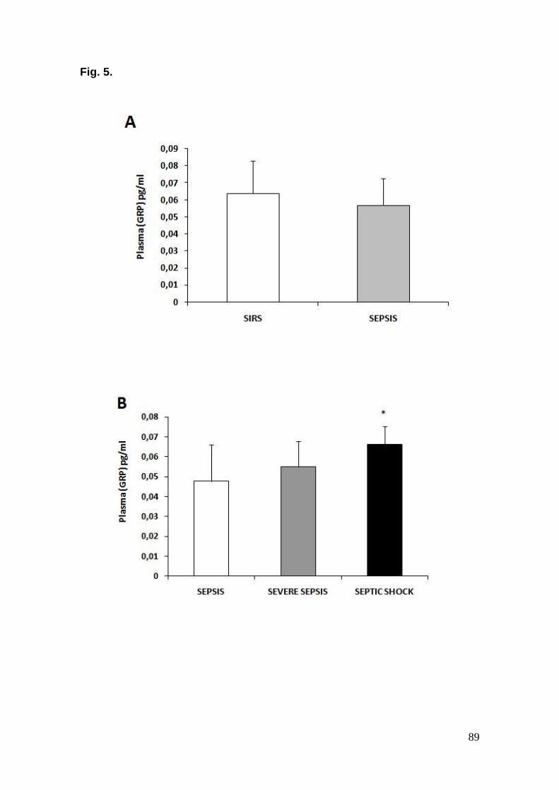
89
Fig. 5.

90

91

92
Capítulo III
Gastrin-releasing peptide receptor as a molecular target for inflammatory
diseases
Fabricia Petronilho, Rafael Roesler, Gilberto Schwartsmann, Felipe Dal-Pizzol
Artigo publicado no periódico
Inflammation & Allergy Drug Targets
6 (4): 197-200, 2007

Inflammation & Allergy - Drug Targets, 2007, 6, 197-200 197
1871-5281/07 $50.00+.00 © 2007 Bentham Science Publishers Ltd.
Gastrin-Releasing Peptide Receptor as a Molecular Target for Inflammatory Diseases
Fabricia Petronilho, Rafael Roesler, Gilberto Schwartsmann and Felipe Dal Pizzol*
Laboratório de Fisiopatologia Experimental, Programa de Pós-Graduação em Ciências da Saúde, Unidade Acadêmica
de Ciências da Saúde, Universidade do Extremo Sul Catarinense, Criciúma, SC, Brazil
Cancer Research Laboratory, Academic Hospital Research Center, Federal University of Rio Grande do Sul,
PortoAlegre, RS, Brazil
Abstract: Bombesin-like peptides (BLP) and its receptors are widely distributed in mammalian peripheral tissues and in
the central nervous system. Recently, effects of these peptides on the production and release of cytokines were described
both in animal models and humans with inflammatory diseases. Some pathological conditions such as exposure to tobacco
smoke, chronic obstructive pulmonary diseases and eosinophilic granuloma have recently been found to be associated
with an increase of pulmonary BLP-producing cells. Proinflammatory neuropeptides have a key role in the pathogenesis
and maintenance of rheumatoid arthritis and sepsis. Together, these findings support the view that the GRPR should be
considered a therapeutic target for a subset of inflammatory diseases.
Keywords: Neuropeptide, bombesin-like peptides, gastrin-releasing peptide receptor, immune system, sepsis.
IN TRODUCTION
Bombesin (BBS) is one of the active peptides purified from amphibian skin [1]. This peptide is also active in mammals and its pharmacological effect extends into various physiological aspects: hypertensive action, contractive effect on uterus, colon or ileum, stimulating action on the gastric secretion, hyperglycemic effect or increasing insulin secre-tion [2]. Many other peptides structurally related to BBS are discovered from amphibian skin and divided into three groups: BBS family that includes BBS and alytesin, ranat-ensin family that includes ranatensin, litorin and their deriva-tives and phyllolitorin family [3]. The first mammalian BBS-like peptide was isolated from porcine gastric tissue and named gastrin-releasing peptide (GRP) since its first known activity was inducing gastrin secretion from G cells in the gastric antrum [4]. Shortly thereafter, a novel bombesin-like peptide was identified in porcine spinal cord and named neu-romedin B (NMB) [5].
1. GRP Receptors
Four different receptor subtypes (GRP-R, NMB-R, BRS-3 and BB4-R) have been described for the bombesin-like peptides. GRP-stimulated signal transduction occurs with the binding of GRP to its cognate cell surface receptor, the G protein-coupled receptor (GPCR) [6]. The GRP-GPCRs have the typical structural features of G protein binding seven-transmembrane receptors. The NMB-R cDNA was then cloned from rat oesophagus [7] and from human genomic libraries [8]. In an attempt to search for novel G-protein-coupling receptors from guinea-pig uterus, Gorbulev et al. cloned a new subtype of bombesin-like peptide receptor [9] that has the highest amino acid similarity to GRP-R (52%)
*Address correspondence to this author at the Laboratório de Fisiopatologia
Experimental, Universidade do Extremo Sul Catarinense, Criciúma, SC,
Brazil, Avenida Universitária 1105, 88806-000; Tel: 55 48 4312641; Fax:
55 48 431 2750; E-mail: [email protected]
and has beendesignated as bombesin-like peptide receptors subtype-3 (BRS-3). The gene encoded for human homolog of BRS-3 was reported subsequently [10, 11]. In search of receptors for bombesin-related peptides in amphibian, Nagalla et al. isolated two clones encoding fragments highly homologous to mammalian GRP-R and one clone encoded for a novel bombesin receptor subtype and named it as BB4 [12]. This receptor has higher affinity for bombesin than GRP and shared only 56% and 70% amino acid identity to the human GRP-R and human BRS-3, respectively. Another subtype of bombesin-like peptide receptor was cloned from chick brain [13]. This subtype, called BRS-3.5, has moderate affinity for bombesin but low affinity for GRP. Distinct from chick GRP-R that is expressed in the brain and gastrointesti-nal tissues, BRS-3.5 is expressed only in the brain.
All the GRP receptors characterized to date are guanine nucleotide binding protein (G-protein)-coupled, have seven transmembrane domains and activate phospholipase C to increase intracellular concentrations of inositol phosphates, diacyl glycerol and calcium. Among the multiple intracellu-lar signaling pathways that mediate the proliferative effects of GPCRs, a family of related serine-threonine kinases, col-lectively known as ERKs or MAPKs, appeared to play a central role [14]. The effects of bombesin on cyclooxy-genase-2 expression in intestinal cell lines require an in-crease in Ca
+2; activation of extracellular signal-regulated
kinase-1 and -2 and p38MAPK; and increased activation and expression of the transcription factors Elk-1, ATF-2, c-Fos, and c-Jun [15]. We demonstrated that in the central nervous system, BBS/GRP acts at least through a mechanism involv-ing the PKC, MAPK and PKA signaling pathways [16]. Some of the actions of bombesin/GRP upon immune cells seem to involve the activation of protein kinase C [17].
2. General Function of Bombesin Related Peptides (BRP)
The BRP play many physiological roles in addition to the stimulation of gastric acid secretion. The neural effects of
Not For D
istributio
n

198 Inflammation & Allergy - Drug Targets, 2007, Vol. 6, N o. 4 Petronilho et al.
GRP-like peptides include changes in animal behavior, sup-pression of food intake and improvement of learning and memory in rodent models [16, 18-20]. Bombesin and GRP stimulate growth of the gastrointestinal mucosa and pancreas [21]. BRP are growth factors for 3T3 fibroblasts, normal airway epithelial cells [22, 23]. BRPs promote fetal lung development, including lung branching morphogenesis, epithelial and mesenchymal cell proliferation and Type II cell differentiation [24, 25]. During lung development, the first epithelial cells to differentiate are the pulmonary neu-roendocrine cells [26], which contain high levels of BLP. BLP can promote growth and maturation of developing fetal lung in humans, nonhuman primates, rats and mice [24, 25, 27]. Levels of GRP receptor (GRPR) and GRP mRNAs peak during the canalicular phase, then fall to much lower levels during normal alveolarization after birth [28].
GRP-like peptides may also provide a protective effect for the GI mucosa during periods of inflammation. In ex-perimental studies, Chu et al. demonstrated that the admini-stration of BBS greatly ameliorated the effects of methotrexate on the intestinal mucosa and increased survival of rats given methotrexate [29]. Exogenous bombesin pre-vents gastric injury induced by ethanol [30] and significantly improves the healing of the gastric mucosa of rats after ace-tic acid-induced ulcer induction [31]. Colonic damage in-duced by trinitrobenzene sulfonic acid in rats [32] and burn-induced gut malfunction were also ameliorated by bombesin treatment [33]. Bombesin-induced gastroprotection involves the release of endogenous gastrin [30]. In addition, bombesin improved intestinal barrier function and oxidative stress in rats with experimentally induced jaundice [35] or following partial hepatectomy [35]. Apart from its physiological roles, GRP has been shown to be mitogenic for a number of can-cerous tissues [36].
3. GRP and Inflammatory Disease
The evidence for the involvement of neuropeptides in inflammatory diseases has focused predominantly on a proinflammatory role for substance P (SP) [37, 38], as well as an immuno-modulator role for neuropeptide Y [39], the proinflammatory action of neuromedin U [40] and an anti-inflammatory role for VIP [41]. The mechanism of action of these neuropeptides in modulating inflammatory diseases is, at least in part, due to their ability to affect cytokine produc-tion. For example, SP increases the production of the proin-flammatory cytokines TNF- , IL-6 and IL-1! [42], whereas VIP inhibits the production of the proinflammatory cytoki-nes TNF- , IL-6 and IL-12 and stimulates the production of the anti-inflammatory cytokines IL-10 and IL-1Ra [43].
The number of BLP-producing neuroendocrine (NE) cells in adult lung is extremely low under normal conditions [44.45]. Some pathological conditions such as exposure to tobacco smoke, chronic obstructive pulmonary diseases (COPD) and eosinophilic granuloma have recently been found to be associated with an increase of pulmonary BLP-producing NE cells [46-48]. NE cell hyperplasia with BLP hyperproduction is present in chronic obstructive airway diseases, particularly among cigarette smokers [49-51]. BLP could exert a role in these disease since they are chemotactic for monocytes, increase phagocytosis and superoxide anion production and expression of procoagulant activity by
mononuclear phagocytes and modulate the release of some proinflammatory cytokines by peripheral monocytes and alveolar macrophages [52-55]. Mast cells can function in both acute and chronic inflammation, promoting both innate and acquired immune responses including leukocyte re-cruitment by chemotaxis and leading to lung injury and fi-brosis in later stages [56, 57]. An increase in mast cells in the lung in response to GRP leads to inflammatory response that may have a role in the pathogenesis of bronchopulmonar dysplasia (BPD). Proinflammatory cytokines, adhesion molecules and inflammatory cells including mast cells have been demonstrated in the lung parenchyma of infants with BPD [58, 59]. The concept that GRP-induced mast cell re-sponses might be an early event leading to lung injury in BPD represents a paradigm shift in current understandings of BPD. These observations could be of further relevance be-cause BPD is associated with bronchospasm and inflamma-tion, similar to asthma [60, 61]. In addition, bombesin dimin-ished alveolarization in C57BL/6 mice, but not in outbred Swiss-Webster mice, suggesting that this response depends on the background genes that are as yet unidentified [62]. Second, GRPRKO mice are protected in part against this inhibitory effect of bombesin on alveolarization, indicating that GRPR mediates a significant part of the reduced alveo-larization [62]. Similarly, it has been suggested that elevated GRP levels in humans might predict which adult patients will develop smoking related lung diseases, including em-physema [51]. Thus, increased GRPR signaling due to in-creased BLP levels could be a common denominator for lung diseases with decreased alveolarization, including both BPD and emphysema.
GRP is present in the synovial fluid of joints affected by arthritis and that the pattern of GRP increase differs from that of SP [63]. It appears as if the presence of GRP is par-ticularly related to the early processes of joint involvement. These observations are of interest because GRP has well-known trophic and paracrine effects and chondrocytes have been shown to produce neuropeptides such as GRP [64]. Grimsholm and colleagues [65] have hypothesized that proinflammatory neuropeptides have a key role in the patho-genesis and maintenance of rheumatoid arthritis (RA), and they report that there is a correlation between the synovial concentration of GRP and proinflammatory cytokines and RA disease activity in these patients.
In this context we report for the first time the beneficial effects of the selective GRP receptor antagonist, RC-3095, in a well-established model for experimental sepsis and LPS-induced acute lung injury [66]. RC-3095 modulates the re-lease of proinflammatory cytokines (TNF and IL-1) by acti-vated macrophages, leading to a diminution of inflammatory infiltration and organ dysfunction, thus improving mortality in a clinically relevant model of sepsis. Interestingly, RC-3095 did not modulate the release of the anti-inflammatory IL-10, suggesting that the intracellular pathway modulated by bombesin/GRP is selective to proinflammatory cytokines. Thus we proposed an autocrine/paracrine role of GRP on macrophage activation by sepsis and/or LPS (Fig. 1) leading to a modulation of proinflammatory, but not anti-inflammatory response and this is consistent with the in-volvement of a new inflammatory pathway relevant to the development of inflammatory diseases.
Not For D
istributio
n

Gastrin-Releasing Peptide Receptor Inflammation & Allergy - Drug Targets, 2007, Vol. 6, N o. 4 199
ABBREVIATION S
BLP = Bombesin-like peptides
BBS = Bombesin
GRP = Gastrin-releasing peptide
NMB = Neuromedin B
GRP-R = Gastrin-releasing peptide receptor
NMB-R = Neuromedin B receptor
BRS-3 = Bombesin-like peptide receptors subtype-3
GPCR = G protein – coupled receptor
BB4 = Bombesin receptor subtype 4
BRS-3.5 = Bombesin-like peptide receptors subtype-3.5
VIP = Vasointestinal Peptide
COPD = Chronic obstructive pulmonary disease
NE = Neuroendocrine
BPD = Bronchopulmonar dysplasia
RA = Rheumatoid arthritis
LPS = Lipopolyssacharide
REFEREN CES
[1] Anastasi, A.; Erspamer, V.; Bucci, M. Experiencia, 1971, 27, 166.
[2] Erspamer, V.; Esparmer, G.F.; Inselvini, M. J. Pharm. Pharmacol., 1970, 22, 875.
[3] Erspamer, V.; Ersparmer, G.F.; Mazzanti, G.; Endean, R. Comp.
Biochem. Physiol. C, 1984, 77, 99. [4] McDonald, T.J.; Nilsson, G.; Vagne, M.; Ghatei, M.; Bloom S.R.;
Mutt, V. Gut, 1978, 19, 767. [5] Minamino, N.; Kangawa, K.; Matsuo, H. Biochem. Biophys. Res.
Commun., 1983, 114, 541. [6] Spindel, E.R.; Giladi, E.; Brehm, P.; Goodman, R.H.; Segerson,
T.P. Mol. Endocrinol., 1990, 4, 1956. [7] Wada, E.; Way, J.; Shapira, H.; Kusano, K.; Lebacq-Verheyden,
A.M.; Coy, D.; Jensen, R.; Battery, J. N euron, 1991, 6, 421. [8] Corjay, M.H; Dobrzanski, D.J.; Way, J.M.; Viallet, J.; Shapira, H.;
Worland, P.; Sausville, E.A.; Battey, J.F. J. Biol. Chem., 1991, 226, 18771.
[9] Gorbulev, V.; Akhundova, A; Grzeschik, K.H.; Fahrenholz, F. FEBS Lett., 1994, 340, 260.
[10] Fathi, Z.; Corjay, M.H.; Shapira, H.; Wada, E.; Benya, R.; Jensen, R.; Viallet, J.; Sausville, E.A.; Battey, J.F. J. Biol. Chem., 1993,
268, 5979. [11] Gorbulev, V.; Akhundova, A.; Grzeschik, K.H.; Fahrenholtz, F.
FEBS Lett., 1994, 340, 260. [12] Nagalla, S.R.; Barry, B.J.; Creswick, K.C.; Eden, P.; Taylor, J.T.;
Spindel, E.D. Proc. N atl. Acad. Sci. USA, 1995, 92, 6205. [13] Iwabuchi, M.; Ui-Tei, K.; Yamada, K.; Matsuda, Y.; Sakai, Y.;
Tanaka, K.; Ohki-Hamazaki, H. Br. J. Pharmacol., 2003, 139, 555. [14] Hellmich, M.R.; Ives, K.L.; Udupi, V.; Soloff, M.S.; Greeley, G.H.
Jr; Christensen, B.N.; Townsend, C.M. Jr. J. Biol. Chem., 1999, 274, 23901.
[15] Guo Y.S.; Hellmich, M.R.; Wen, X.D.; Townsend, C.M. Jr. J. Biol. Chem., 2001, 276, 22941.
[16] Roesler, R.; Luft, T.; Oliveira, S.H.; Farias, C.B.; Almeida, V.R.; Quevedo, J.; Dal Pizzol, F.; Schroder, N.; Izquierdo, I.; Schwarts-
mann, G. N europharmacology, 2006, 51, 350. [17] Helmich, M.R.; Ives, K.L.; Udupi, V.; Soloff, M.S.; Greeley, G.H.
Jr.; Christensen, B.N.; Townsend, C.M. Jr. J. Biol. Chem., 1999, 274, 23901.
Fig. (1). Putative mechanism on LPS and GRP interaction in macrophages.
Not For D
istributio
n

200 Inflammation & Allergy - Drug Targets, 2007, Vol. 6, N o. 4 Petronilho et al.
[18] Yamada, K.; Wada, E.; Wada, K. Brain Res., 2000, 870, 20.
[19] Yamada, K.; Wada, E.; Wada, K. Ann. Méd., 2000, 32, 519. [20] Roesler, R.; Meller, C.A.; Kopschina, M.I., Souza, D.O.; Henri-
ques, J.A.; Schwartsmann, G. Peptides, 2003, 24, 1069. [21] Lehy, T.; Puccio, F.; Chariot, J.; Labeille, D. Gastroenterology,
1986, 90, 1942. [22] Rozengurt, E.; Sinnett-Smith. J. Proc. N atl. Acad. Sci. USA, 1983,
80, 2936. [23] Li, K.; Nagalla, S.R.; Spindel, E.R. J. Clin. Invest., 1994, 94, 1605.
[24] Sunday, M.E.; Hua, J.; Dai, H.B.; Nusrat, A.; Torday, J.S. Am. J. Respir. Cell Mol. Biol., 1990, 3, 199.
[25] Sunday, M.E.; Hua, J..; Torday, J.; Reyes, B.; Shipp, M.A. J. Clin. Invest., 1992, 90, 2517.
[26] Ten Have-Opbroek, A.A.W. Exp. Lung Res., 1991, 17, 111. [27] Sunday, M.E.; Hua, J.; Reyes, B.; Masui, H.; Torday, J.S. Anat.
Rec., 1993, 236, 25. [28] King, K.A.; Torday, J.S.; Sunday, M.E. Proc. N atl. Acad. Sci. USA,
1995, 92, 4357. [29] Chu, K.U.; Higashide, S.; Evers, B.M.; Rajaraman, S.; Ishizuka, J.;
Townsend, C.M. Jr, Thompson, J.C. Ann. Surg., 1994, 220, 570. [30] Mercer, D.W.; Cross, J.M.; Chang, L.; Lichtenberger, L.M. Dig.
Dis. Sci., 1998, 43, 826. [31] Gunal, O.; Oktar, B.K.; Ozcinar, E.; Tansuker, D.; Arbak, S.;
Yegen, B.C. Regul. Pept., 2002, 106, 81. [32] Gulluoglu, B.M.; Kurtel, H.; Gulluoglu, M.G.; Aktan, A.O.; Yegen,
B.C.; Dizdaroglu, F.; Yalin, R.; Yegen, B.C. Dig. Dis. Sci., 1999, 44, 1531.
[33] Wu, X.W.; Spies, M.; Chappell, V.L.; Herndon, D.N.; Thompson, J.C.; Wolf, S.E. Shock, 2002, 18, 518.
[34] Assimakopoulos, S.F.; Scopa, C.D.; Charonis, A.; Spiliopoulou, I.; Georgiou, C.; Nikolopoulou, V.; Vagianos, C.E. J. Am. Coll. Surg.,
2004, 198, 748. [35] Assimakopoulos, S.F.; Alexandris, I.H.; Scopa, C.D.; Mylonas,
P.G.; Thomopoulos, K.C.; Georgiou, C.D.; Nikolopoulou, V.N.; Vagianos, C.E. World J. Gastroenterol., 2005, 11, 6757.
[36] Zhou, J.; Chen, J.; Mokotoff, M.; Ball, E.D. Anticancer Drugs, 2004, 15, 921.
[37] O'Connor, T.M.; O'Connell, J.; O'Brien, D.I.; Goode, T.; Bredin, C.P.; Shanahan, F. J. Cell Physiol., 2004, 201, 167.
[38] Keeble, J.E.; Brain, S.D. N eurosci. Lett., 2004, 361, 176. [39] Bedoui, S.; Miyake, S.; Straub, R.H.; von Horsten, S.; Yamamura,
T. Trends Immunol., 2004, 25, 508. [40] Moriyama, M.; Sato, T.; Inoue, H.; Fukuyama, S.; Teranishi, H.;
Kangawa, K.; Kano, T.; Yoshimura, A.; Kojima, M. J. Exp. Med., 2005, 202, 217.
[41] Niissalo, S.; Hukkanen, M.; Imai, S.; Tornwall, J.; Konttinen, Y.T. Ann. N . Y. Acad. Sci., 2002, 966, 384.
[42] Cuesta, M.C.; Quintero, L.; Pons, H.; Suarez-Roca, H. N eurochem.
Int., 2002, 40, 301. [43] Delgado, M.; Pozo, D.; Ganea, D. Pharmacol. Rev., 2004, 56, 249.
[44] Gosney, J.R.; Sissons, M.; Allibone, R. Thorax, 1988, 43, 878. [45] Cutz, E.; Chan, W.; Track, N.S. Experientia, 1981, 37, 765.
[46] Aguayo, S.M.; Kane, M.A.; Talmadge, E.K.; Schwarz, M.I.; Grauer, L.; Miller, Y.E. J. Clin. Invest., 1989, 84, 1105.
[47] Gosney, J.R.; Sissons, M.C.J.; Allibone, R.D.; Blakey. A.F. J. Pathol., 1989, 147, 127.
[48] Aguayo, S.M.; Talmadge, E.K.; Waldron, J.A. Jr; Sherrit, K.M.; Kane, M.A.; Miller, Y.E. J. Clin. Invest., 1990, 86, 838.
[49] Tabassian, A.R.; Nylen, E.S.; Linnoila, R.I.; Snider, R.H.; Cassidy, M.M.; Becker, K.L. Am. Rev. Respir. Dis., 1989, 140, 436.
[50] Nylen, E.S.; Becker, K.L.; Joshi, P.A.; Snider, R.H.; Schuller, H.M. Am. J. Respir. Cell Mol. Biol., 1990, 2, 25.
[51] Meloni, F.; Bertoletti, R.; Corsico, A.; Di Fazio, P.; Cecchettin, M.; Grassi, G. Int. J. Tiss. Reac., 1992, 14, 195.
[52] Peck, R. Ann. N . Y. Acad. Sci., 1987, 496, 264. [53] Meloni, F.; Saporiti, A.; Ballabio, P.; Brocchieri, A.; Grignani, G.;
Gialdroni Grassi, G. Monaldi. Arch. Chest Dis., 1995, 50, 187. [54] Meloni, F.; Ballabio, P.; Bianchi, L.; Mangiarotti, P.; Grassi, G.;
Bignamini, A.; Gialdroni Grassi, G. Respiration, 1996, 63, 28. [55] Aguayo, S.M. Am. J. Respir. Crit. Care Med., 1994, 149, 1692.
[56] Warner, J.A.; Kroegel, C. Eur. Respir. J., 1994, 7: 1326. [57] Akers, I.A.; Parsons, M.; Hill, M.R.; Hollenberg, M.D.; Sanjar, S.;
Laurent, G.J.; McAnulty, R.J. Am. J. Physiol. Lung Cell Mol. Physiol., 2000, 278, L193.
[58] Sunday, M.E.; Shan, L.; Subramaniam, M. Endocr. Pathol., 2004, 15, 91.
[59] Cullen, A.; Van Marter, L.J.; Allred, E.N.; Moore, M.; Parad, R.B.; Sunday, M.E. Am. J. Respir. Crit. Care Med., 2002, 165, 1093.
[60] Subramaniam, M.; Sugiyama, K.; Coy, D.H.; Kong, Y.; Miller, Y.E.; Weller, P.F.; Wada, K.; Wada, E.; Sunday, M.E. Am. J.
Respir. Crit. Care Med., 2003, 168, 601. [61] Bader, D.; Ramos, A.D.; Lew, C.D.; Platzker, A.C.; Stabile, M.W.;
Keens, T.G. J. Pediatr., 1987, 110, 693. [62] Ashour, K.; Shan, L.; Lee, J.H.; Schlicher, W.; Wada, K.; Wada,
E.; Sunday, M.E. Am. J. Respir. Crit. Care Med., 2006, 173, 1377. [63] Westermark T, Rantapaa-Dahlqvist S, Wallberg-Jonsson S, Kjorell
U, Forsgren S. Clin. Exp. Rheumatol., 2001, 19, 715. [64] Hill DJ, McDonald TJ. Endocrinology, 1992, 130, 2811.
[65] Grimsholm, O.; Rantapaa-Dahlqvist, S.; Forsgren, S. Arthritis Res. Ther., 2005, 7, R416.
[66] Dal-Pizzol, F.; Di Leone, L.P.; Ritter, C.; Martins, M.R.; Reinke, A.; Pens Gelain, D.; Zanotto-Filho, A.; de Souza, L.F.; Andrades,
M.; Barbeiro, D.F.; Bernard, E.A.; Cammarota, M.; Bevilaqua, L.R.; Soriano, F.G.; Cláudio, J.; Moreira, F.; Roesler, R.;
Schwartsmann, G. Am. J. Respir. Crit. Care Med., 2006, 173, 84.
Received: August 26, 2007 Revised: N ovember 30, 2007 Accepted: December 4, 2007
Not For D
istributio
n

97
PARTE III
Discussão e Conclusões

98
III.1. DISCUSSÃO
Segundo observações clássicas de Lewis publicadas em 1972 no New
England Journal of Medicine “É nossa resposta à presença dos
microorganismos patogênicos que causa a doença. Nosso arsenal para
combater as bactérias é tão poderoso...que nós estamos mais em perigo do
que os próprios invasores”. A perda do direcionamento de resposta do
hospedeiro aos organismos invasores torna-se a principal característica
relacionada à sepse e sua resposta sistêmica caracterizada tradicionalmente
como pró-inflamatória e excessiva. O conceito “inflamação excessiva” encontra
respaldo em diferentes achados, o que denota a complexidade da definição
clinica e o manejo terapêutico da sepse em muitos aspectos ainda não bem
definidos o que leva ainda a um aumento na sua incidência nos últimos anos
com uma discreta redução na mortalidade (Alberti et al., 2002; Annane et al.,
2002; Annane et al., 2003; Brun-Buisson et al., 2004; Dellinger et al., 2004).
Portanto, o entendimento sobre os mecanismos celulares e moleculares que
contribuem na fisiopatologia de doenças inflamatórias e na disfunção de órgãos
e a busca de novas alternativas terapêuticas tornam-se essenciais na redução
da mortalidade (Baron et al., 2006; Vincent e Abraham, 2006).
Estudos mostram que a resposta imune pode ser influenciada pelo
sistema nervoso onde recentemente nosso grupo mostrou que o neuropeptídeo
homólogo da bombesina, o peptideo liberador de gastrina, encontra-se
envolvido na inflamação sistêmica característica da sepse, sendo que o seu
antagonista, RC-3095 modulou a liberação de citocinas pró-inflamatórias por
macrófagos ativados, levando a diminuição da infiltração inflamatória e

99
disfunção de órgãos, melhorando dessa forma a mortalidade em modelo
clinicamente relevante de sepse e dano agudo pulmonar (Dal-Pizzol et al.,
2006).
Nesse contexto, na primeira fase do nosso estudo, avaliamos o efeito
protetor de RC-3095 em modelo de inflamação aguda relacionada ao dano
pulmonar induzido por carragenina. Sabe-se que a administração aguda de
carragenina no espaço pleural leva a pleurisia, caracterizada pela infiltração
imediata de neutrófilos e dano pulmonar. Com o infiltrado de neutrófilos, a
pleurisia induzida por carragenina é caracterizada pela produção em excesso
de espécies reativas de oxigênio bem como outros mediadores derivados de
neutrófilos que segundo a literatura estão envolvidos em condições que levam
ao dano tecidual por ativarem fatores de transcrição responsáveis pela
liberação de citocinas pró-inflamatórias (Guo e Ward, 2007). Assim a resposta
inflamatória é o mecanismo predominante envolvido na patogênese no dano
pleural causado por carragenina.
Uma vez a efusão pleural é caracterizada por um exsudato, o estágio da
inflamação é caracterizado pela presença significante de neutrófilos onde a
predominância é associada já nas primeiras fases da resposta inflamatória e
progressão da doença pleural, no entanto o antagonista do GRPR, RC-3095
inibiu a migração celular, o dano tecidual com a diminuição da atividade de
lactato desidrogenase e níveis de proteínas no exsudato pleural comparando
com o grupo que não recebeu tratamento.
Trabalhos mostram que o mecanismo da migração celular para a
cavidade pleural é dependente da ação de citocinas quimiotáxicas e ocorre
após a sua liberação por células mesoteliais pleurais em resposta ao estímulo

100
por outras citocinas (Antony et al., 1993). O processo inflamatório causado com
administração de carragenina na cavidade pleural é consistente com o
aumento de citocinas pro-inflamatórias como IL-1β e TNF-α (Kroegel e Antony,
1997) o que foi demonstrado adicionalmente no estudo onde camundongos
knockout do receptor 1 de TNF-α foram resistentes a inflamação aguda
induzida por carragenina na cavidade pleural em relação aos camundongos
selvagens (Mazzon et al., 2008). Conforme mencionado anteriormente
verificamos a ação inflamatória induzida por GRP na sepse e em macrófagos
peritoneais induzidos por LPS e observamos que a via induzida por GRPR é
seletiva para a ativação de citocinas pró-inflamatórias (Dal-Pizzol et al., 2006).
E em paralelo nosso grupo verificou tal efeito em modelo de artrite em ratos
(Oliveira et al., 2008). Através destes dados verificamos os níveis de TNF-α e
IL-1β no exsudato pleural de ratos sujeitos ao modelo proposto sob tratamento
com RC-3095 onde constatamos a diminuição dos seus níveis com o
tratamento.
Outro evento positivo causado por RC-3095 sob o processo inflamatório
relacionado à atenuação da liberação de citocinas pode ser a diminuição da
concentração de nitrito/nitrato. A indução de iNOS por injeção de LPS em
roedores é mediado por TNF-α e IL-1β (Szabo et al., 1993; Jin et al., 2006) e
sabe-se que o aumento na produção de NO por iNOS pode contribuir para o
processo inflamatório (Oh-Ishi et al., 1989; Tracey et al., 1995; Cuzzocrea et
al., 1999). Evidências, confirmam que inibidores da atividade de iNOS estão
relacionados com a redução da pleurisia causada por carragenina (Cuzzocrea
et al., 1998) e constatamos uma diminuição na formação de nitrito-nitrato, um
metabólito final de NO (Guo e Ward, 2007). Ainda, verificamos a atividade de

101
RC-3095 na diminuição da enzima mieloperoxidade conhecida como um
marcador indireto de leucócitos ativados e na exudação e migração celular o
que também foi verificado em modelo de sepse (Dal-Pizzol et al., 2006).
Visto que como uma das funções dos neutrófilos quando reconhecem
um patógeno é eliminá-lo, neutrófilos ativados contam com substâncias como
EROs que agem sobre a membrana celular dos microorganismos com a
finalidade de destruí-los e consequentemente evitar a disseminação. No
entanto, as EROs também podem levar a injuria em tecidos normais (Yasui e
Baba, 2006). Este mecanismo deletério causado por tais moléculas exerce
efeito contra importantes componentes celulares devido a sua intensa
instabilidade e reatividade e encontram-se associados ao modelo de indução
de pleurisia por carragenina (Cuzzocrea et al., 2000; Nardi et al., 2007;
Bhattacharyya, Dudeja e Tobacman, 2008; Menegazzi et al., 2008) Nesse
contexto, marcadores de peroxidação de lipídios foram encontrados em
estudos que avaliam o dano pulmonar induzido por ânion superóxido (O2-),
peróxido de hidrogênio (H2O2), radical hidroxil (OH-) e peroxinitrito (ONOO-
Evidências sugerem que NF-κB exerce importante papel na resposta
inflamatória induzida por carragenina onde através da administração de seus
)
(Guo e Ward, 2007). Assim, no tecido pulmonar de ratos submetidos à
pleurisia, RC-3095 foi importante na diminuição do dano em lipídios e dano em
proteínas com o aumento de resíduos de aminoácidos contendo grupos tióis.
Essa resposta positiva está relacionada também a diminuição de citocinas
anteriormente descritas pois as EROs podem amplificar a resposta inflamatória
com a liberação de mediadores inflamatórios através da ativação de NF-κB e
AP-1 (Kirkham e Rahman, 2006; Uffort, Grimm e Ellerhorst, 2009).

102
inibidores, inibe-se a resposta inflamatória exacerbada após a administração de
carragenina na cavidade pleural em camundongos (Fröde-Saleh e Calixto,
2000). Dessa forma, segundo Levine, a bombesina estimula a ativação de NF-
κB (Levine et al., 2003), o que pode sugerir a associação da diminuição da
resposta inflamatória e a atenuação do dano oxidativo em modelo animal de
inflamação pleural com o bloqueio de GRPR envolvido na up-regulation da
ativação de NF-κB.
Os resultados até agora discutidos podem demonstrar, portanto que o
neuropeptideo GRP encontra-se envolvido na gênese da inflamação pleural e
que antagonizando o seu receptor encontramos uma diminuição em
parâmetros oxidativos e inflamatórios que segundo a literatura levam ao dano
pulmonar (Farley, Wang e Mehta, 2009). Assim o nosso próximo estudo
baseou-se na sua ação como efeito modulatório da inflamação a nível
sistêmico.
Sabe-se que a inflamação sistêmica é caracterizada por níveis
aumentados de citocinas e quimiocinas circulantes e como resultado em níveis
aumentados de neutrófilos em órgãos como o pulmão (Abraham, 2003).
Estudos mostram que o mecanismo desta relação acontece em animais
submetidos à sepse severa ou ao choque endotoxemico, onde uma das
funções principais da imunidade inata realizada por neutrófilos no tecido
infectado está alterada, com comprometimento da sua resposta quimiotáxica e
da capacidade em migrar para o foco infeccioso, evento denominado de
paralisia ou falência da migração de neutrófilos (Benjamim et al., 2002; Alves-
Filho et al., 2006b). Nesse sentido, recentemente foi observado que
camundongos deficientes de TLR-4 exibem migração de neutrófilos para a

103
cavidade peritoneal durante sepse polimicrobiana letal induzida por CLP,
interessantemente tal evento não ocorre em camundongos normais, portanto
sugeriu-se o envolvimento de TLR-4 na falência da migração de neutrófilos
para o foco infeccioso nos casos mais severos de sepse visto que esse fato
ocorre através da ativação sistêmica dos TLRs com a invasão bacteriana em
níveis relevantes (Alves-Filho et al., 2006a). Dessa forma, devido seu
importante papel na regulação de moléculas envolvidas na resposta
inflamatória, receptores TLR principalmente TLR-4 são alvos importantes para
o desenvolvimento de novas terapias contra doenças inflamatórias como a
sepse (Matsuda e Hattori, 2006).
Assim, no segundo capítulo do presente trabalho tivemos por objetivo
investigar o efeito do antagonista de GRP, RC-3095 na sinalização de TLR-4 e
esta relação com parâmetros inflamatórios em modelos experimentais in vitro e
in vivo e em pacientes com sepse, bem como avaliar os níveis plasmáticos de
GRP em pacientes com SIRS e sepse correlacionando com o seu desfecho
clinico.
O estudo das interações entre o sistema imune e nervoso central tem
despertado o interesse de inúmeros grupos de pesquisa. Esses sistemas
trocam informações, influenciando-se mutuamente, com evidentes implicações
fisiológicas e patológicas. Assim como outros neuropeptídeos GRP está ligado
a esses dois sistemas, como verificado na modulação como um estimulador ou
inibidor da função de linfócitos (Medina et al., 1998b; Medina et al., 1999),
fagócitos (De la Fuente et al., 1991) e células NK (Medina et al., 1998a).
Adicionalmente, BLPs possuem efeitos estimulatórios na mobilidade, ingestão
e produção de superóxido em macrófagos de ratos adultos, potencializando a

104
liberação de IL-1 por macrófagos alveolares ativados com LPS (Lemaire, 1991;
De la Fuente et al., 2000) e em condições inflamatórias como bronquite crônica
(Meloni et al., 1992) e fibrose pulmonar (Lemaire, Jones e Khan, 1991) além do
aumento nos níveis de GRP em células da linhagem monócito-macrófago (Del
Rio e De la Fuente, 1994).
Recentes trabalhos indicam que neuropeptídeos podem ainda regular a
resposta de macrófagos para LPS, afetando a expressão de TLR-2 e regulando
a sinalização de TLR-4 (Tsatsanis et al., 2006). Nesse caso, devido aos
macrófagos secretarem GRP (Genton e Kudsk, 2003) e tais células serem
importantes no processo de desenvolvimento de sepse e choque séptico
(Shasby e McCray, 2004), as nossas primeiras observações foram em torno da
diminuição da expressão de mRNA de TLR-4 em cultura de células RAW 264.7
estimuladas por LPS quando tratadas com RC-3095. Estudos mostram que
existe um aumento na expressão de TLR-4 durante as fases iniciais de sepse
correlacionando-se com a morte em modelo animal de sepse por CLP, portanto
a down-regulation desses receptores aumenta a sobrevida em modelo de CLP
(Williams et al., 2003) que mostra-se em concordância com os nossos
resultados apresentados através da ação de RC-3095. Ainda, considerando
que GRP induz a proliferação de mastócitos e quimiotaxia in vitro, é detectado
em células pulmonares neuroendócrinas sendo implicado na patogenese de
doenças pulmonares como um mediador de injuria pulmonar aguda e crônica
em displasia broncopulmonar (Subramaniam et al., 2003; Degan et al., 2008) e
ainda, relacionando ao seu receptor, foi visto que GRPR está implicado no
edema alveolar (Dal-Pizzol et al., 2006), tais informações remetem os
resultados observados onde o efeito inibitório de RC-3095 na up-regulation de

105
TLR-4 no tecido pulmonar nos ratos sujeitos a sepse polimicrobiana letal por
CLP resulta na atenuação da inflamação pulmonar.
Pouco se sabe ainda a respeito do mecanismo pelo qual acontece a
inibição ou estimulação de TLR-4. No entanto pesquisadores mostram que os
receptores TLRs podem ser afetados através da ubiquitilação um mecanismo
geral para a regulação de funções de receptores de membrana celulares por
promover a degradação proteolítica de proteínas, seqüestro de proteínas
adaptadoras, e a desativação de quinases através de fosfatases e pela inibição
da expressão por citocinas anti-inflamatórias (Han e Ulevitch, 2005; Liew, Xu e
Brint, 2005). Tais mecanismos podem associar-se as vias de ativação de
GRPR. Sabe-se que o receptor de BN acoplado a proteína G pode fosforilar
membros da família Janus quinase/transdutora de sinais e ativadora de
transcrição (Jak/STAT) (Pansky et al., 1998). Podemos especular que RC-3095
pode exercer este efeito na expressão de TLR-4 através de STAT6 como
STAT6 fosforilada down-regulate a expressão de TLR-4 visto que a expressão
excessiva de STAT6 aumenta a inibição de promotores de TLR-4 (Fiset et al.,
2006). Ainda, STAT6 pode inibir importantes genes inflamatórios, incluindo
fator regulatório de interferon–1 (IRF-1) (Ohmori e Hamilton, 1997), TNF-α e IL-
12p40 (Major, Fletcher e Hamilton, 2002) e nas células mononucleares, o
interferon-γ (IFN-γ), induz o aumento de mRNA e expressão na superfície da
célula de TLR-4, inibindo a redução de TLR-4 induzida por LPS. Portanto, IFN-
γ sensibiliza as células mononucleares para o efeito do LPS, justificando qu e
essa citocina aumenta a expressão de TLR-4 (Cohen, 2002), o que pode ser
um link entre a relação da inibição de GRPR e a expressão atenuada de TLR-
4.

106
Com o aumento da sinalização de TLR-4 acontece a ativação de
importantes vias inflamatórias como verificado na ativação de ERK1/2 em
monócitos/macrófagos, via JNK em células RAW (Sugiyama, Muroi e
Tanamoto, 2008) e fosfatitilinositol 3 quinase (PI3K), que ativa Akt em
macrófagos de camundongos (Kim et al., 1996; Monick et al., 2001; Bozinovski
et al., 2002; Ojaniemi et al., 2003; Wrann et al., 2007; Laird et al., 2009).
Constatamos que o estímulo de células RAW 264.7 com LPS resultou na
fosforilação de ERK 1/2, JNK e Akt em 60 minutos após o tratamento com RC-
3095. Assim, antagonizando o GRPR pode-se influenciar em vias de
sinalização de kinases intracelulares induzida por TLR-4.
Adicionalmente, é bem documentado que fatores de transcrição NF-kB e
AP-1 exercem um importante papel na regulação transcricional induzido por
LPS de muitos genes inflamatórios como de citocinas/quimiocinas que
contribuem para o desenvolvimento do choque séptico e falência múltipla de
órgãos (Guha e Mackman, 2001). Essas observações, estão de acordo com
nossos resultados onde células RAW 264.7 estimuladas por LPS a atividade de
NF-kB e AP-1 ligado ao DNA foi diminuído. Sugerimos assim, que a supressão
da translocação nuclear de NF-kB e AP-1 por RC-3095 está associada com a
diminuição da expressão de TLR-4 e ativação de MAP kinase.
Segundo Alves-Filho e colaboradores, uma significante deficiência no
recrutamento de neutrófilos durante a sepse severa é caracterizada pelo
seqüestro inapropriado dessas células para o pulmão. A migração de
neutrófilos prejudicada para os sítios de infecção implica-se na patogênese da
sepse, e a magnitude desse evento associa-se à razão da mortalidade (Alves-
Filho et al., 2006a). O recrutamento de neutrófilos para o sítio de infecção é

107
dependente de quimiocinas tais como proteína quimiotáxica de monócitos -1
(MCP-1) (Chishti et al., 2004), alem da ação de citocinas pró-inflamatórias
como IL-6 que afetam diretamente a adesão e funções migratórias de
neutrófilos (Johnson et al., 1991; Adams e Lloyd, 1997). Como demonstrado no
estudo onde MCP-1 e IL-6 são efetores na migração de neutrófilos de 1-2 h
após endotoxemia (Wagner e Roth, 1999). No entanto, a falência na migração
de neutrófilos não é uma conseqüência de níveis de mediadores quimiotáxicos
baixos, mais de altos níveis de mediadores quimiotáxicos no foco de infecção
(Benjamim, Ferreira e Cunha, 2000; Crosara-Alberto et al., 2002).
A participação de citocinas/quimiocinas na falência de migração de
neutrófilos para o foco de infecção pode ser justificada por diferentes estudos
como a alta concentração dessas moléculas na circulação de pacientes com
sepse (Ozbalkan et al., 2004; ter Meulen et al., 2004); e em modelo animal
(Benjamim, Ferreira e Cunha, 2000; Crosara-Alberto et al., 2002; Moreno et al.,
2006), a inibição da migração de neutrófilos por diferentes estímulos
inflamatórios com a administração intravenosa de TNF-α, IL-8 e IL-2 (Otsuka et
al., 1990; Hechtman et al., 1991; Tavares-Murta, Cunha e Ferreira, 1998);
aumento da sobrevida de camundongos B6 a infecção polimicrobiana letal com
aumento do recrutamento de neutrófilos e clearance bacteriano na cavidade
peritoneal comparados com camundongos deficientes dos receptores 1 e 2 de
TNF-α (Secher et al., 2009). Dados consistentes, portanto mostram que a
resposta exacerbada com a presença de mediadores inflamatórios como
citocinas/quimiocinas podem comprometer a ação controlada de neutrófilos e
propõe-se que isso se deve em parte ao fato de que tais mediadores exercem
sua ação via ligação ao GPCRs (Katanaev, 2001) onde sua ativação contínua

108
leva a um processo de dessensibilização devido a sua fosforilação por GRK
(Fan e Malik, 2003). Recentemente foi observado que a ativação de GRKs está
envolvida na dessensibilização de CXCR2 em neutrófilos de pacientes sépticos
e camundongos submetidos à sepse severa (Arraes et al., 2006; Alves-Filho et
al., 2009). Nesse sentido, ativação sistêmica de TLR-4 resulta na ativação
inadequada de neutrófilos por produtos bacterianos e/ou quimiocinas induzidas
pela expressão de iNOS, resultando principalmente na dessensibilização de
CXCR2 e perda de moléculas de adesão. Em adição, aumento em níveis de
proteína de fase aguda no soro induzida por quimiocinas podem também inibir
interações neutrófilo-endotélio, desse modo reduzindo a adesão/rolamento e
quimiotaxia de neutrófilos e finalmente comprometendo a migração levando
possivelmente a uma resposta alterada de neutrófilos contra células de
diferentes órgãos (Annane, Bellissanti e Cavaillon, 2005).
Durante o choque endotóxico, um massivo número de neutrófilos e
outros leucócitos acumulam-se no pulmão, um processo dependente de TLR-4.
Certamente, o acúmulo de células no pulmão é também observado em
pacientes sépticos (Kerfoot e Kubes, 2005). Novamente, a ativação sistêmica
de TLR-4 resulta em um significante seqüestro de leucócitos para os capilares
pulmonares (Welbourn e Young, 1992; Sheridan et al., 1997; Andonegui et al.,
2003). Ainda, neuropeptídeos são conhecidos por estimular a produção de
citocinas em macrófagos, linfócitos, mastócitos e a substância P é conhecida
por influenciar a produção de citocinas pró-inflamatórias induzida por LPS
(Dickerson et al., 1998). Arranz e colaboradores mostraram que citocinas pro-
inflamtórias podem agir sinergisticamente, com componentes de bactérias
gram-negativas, up-regulating a expressão de TLR-4 e é possível que a

109
inibição induzida por VIP na up-regulation de TLR-4 em modelos inflamatórios
ocorre indiretamente via supressão da produção de citocinas pro-inflamatórias
(Arranz et al., 2008). Essa questão vem ao encontro de informações que
propomos no Capítulo III e anteriormente mencionado, visto que o papel
autócrino/parácrino de GRP na ativação de macrófagos por sepse e/ou LPS é
seletivo para a modulação de pró-inflamatória, mas não resposta anti-
inflamatória conforme a Figura 2.
Figura 1. Mecanismo da interação LPS x GRP em macrófagos
De acordo com esses dados, em nosso estudo verificamos que RC-3095
modulou a resposta inflamatória dependente de TLR-4 produzindo uma
diminuição nos níveis de MCP-1 e IL-6 em células RAW 264.7, soro e lavado
broncoalveolar em ratos CLP. Sabe-se que o tratamento agudo com RC-3095
diminui a migração de células para o pulmão e de acordo com os estudos de

110
Alves-Filho e colaboradores, onde camundongos normais mais não os
deficientes de TLR-4 exibem diminuição na migração de neutrófilos para o foco
infeccioso e exibem significante seqüestro dessas células para o tecido
pulmonar durante sepse polimicrobiana letal e isto é conhecido por ser o
evento chave no dano agudo pulmonar induzido na sepse com diminuição da
morte bacteriana levando a bacteremia (Alves-Filho et al., 2006a).
Consequentemente percebemos que a ação de RC-3095 na atenuação de
TLR-4 em modelo animal de sepse resulta em uma migração normal de
neutrófilos, sendo efetivo na atenuação da disseminação bacteriana na
circulação e foco infeccioso.
Visto que GRP afeta vários sistemas em mamíferos, incluindo regulação
neuroendócrina, secreção gastrointestinal e proliferação celular. Esta presença
em concentrações aumentadas tem sido demonstrada em pacientes com
doenças inflamatórias e este nível correlaciona-se a razão da sedimentação de
eritrócitos e com TNF e IL-6, sugerimos, portanto que tal neuropeptídeo pode
estar envolvido na modulação de processos inflamatórios também em humanos
(Fagiolo et al., 1993). Em pacientes com sepse RC-3095 revela a deficiência de
níveis de IL-6 no plasma, entretanto este resultado não foi encontrado para
níveis de IL-10, mostrando adicionalmente a seletividade apresentada na
Figura 2. Nossos resultados sugerem ainda que GRP pode estar
diferencialmente envolvido em SIRS e sepse onde GRP pode somente predizer
o desfecho em pacientes sépticos. Finalmente, de acordo com esses
resultados obtidos na ultima fase do estudo, sugerimos que GRP aumenta a
resposta inflamatória por up-regulating TLRs e isto se deve em grande parte
explicar as diferenças entre pacientes com sepse e pacientes com SIRS.

111
III.2. CONCLUSÕES
• O antagonista do GRPR é efetivo na diminuição da inflamação aguda
em modelo animal de pleurisia induzido por carragenina e relaciona-se à
diminuiçao de parametros de dano oxidativo.
• O antagonista do GRPR diminui a expressão do receptor TLR-4 e de
uma forma secundaria é efetivo na atenuação da fosforilação de
quinases resultando na inibição da translocação dos fatores de
transcrição NF-kB e AP-1 em cultura de macrófagos estimulados por
LPS e a nível pulmonar em modelo animal de sepse.
• Níveis de IL-6 e MCP-1 parecem estar associados à diminuição da
expressão de TLR-4 através da sua atenuação em cultura de células
estimuladas por LPS e a nível sistêmico e pulmonar em modelo animal
de sepse polimicrobiana letal.
• O nível plasmático de IL-6 em pacientes com choque séptico atenuado
com a infusão de RC-3095 mostra a seletividade da via de ativação do
receptor do peptídeo liberador de gastrina para mediadores pró-
inflamatórios e não anti-inflamatórios.
• A modulação de citocinas e quimiocinas através do antagonismo de
GRPR associa-se a diminuição da inflamação em locais distantes do
foco infeccioso limitando a disseminação bacteriana a nível local em
modelo animal de sepse polimicrobiana letal estando associada a
diminuição da resposta mediada por TLR-4.
• GRP esta diferencialmente envolvido em pacientes com SIRS e sepse
onde GRP pode somente predizer o desfecho em pacientes sépticos e

112
esta diferença pode estar relacionada ao aumento da resposta
inflamatória causado por GRP por up-regulating TLRs.

113
III.3. PERSPECTIVAS Nosso propósito como continuidade para o estudo até agora apresentado,
está relacionado a investigação sobre os efeitos do antagonista do peptídeo
liberador de gastrina em camundongos deficientes de TLR-4 submetidos a
sepse polimicrobiana na migração neutrofílica para o foco infeccioso avaliando
a interação do receptor de quimiocina CXCR2 com todo esse processo.
Portanto pretendemos passar pelas seguintes etapas:
• Investigar a ação de RC-3095, em camundongos deficientes de TLR-4 e
animais controle sujeitos a sepse severa polimicrobiana, a fim de verificar a
migração neutrofílica para o foco infeccioso e em sítios distantes da
infecção.
• Testar o rolamento e adesão de leucócitos no mesentério de camundongos
deficientes de TLR-4 e animais controle sujeitos a sepse severa
polimicrobiana tratados com o antagonista do receptor do peptídeo
liberador de gastrina.
• Avaliar através da contagem de bactérias a disseminação bacteriana no
foco infeccioso e a nível circulatório em camundongos deficientes de TLR-4
e animais controle sujeitos a sepse severa polimicrobiana, tratados com o
antagonista do receptor do peptídeo liberador de gastrina.
• Determinar níveis de citocinas e quimiocinas no foco infeccioso, na
circulação sanguínea e no tecido pulmonar de camundongos deficientes de
TLR-4 e animais controle sujeitos a sepse severa polimicrobiana tratados
com o antagonista do receptor do peptídeo liberador de gastrina.

114
• Verificar a expressão de CXCR2 na superfície de neutrófilos isolados do
sangue de camundongos deficientes de TLR-4 e animais controle sujeitos a
sepse severa polimicrobiana tratados com o antagonista do receptor do
peptídeo liberador de gastrina.
• Analisar a expressão de GRK-2 em neutrófilos isolados do sangue de
camundongos deficientes de TLR-4 e animais controle sujeitos a sepse
severa polimicrobiana tratados com o antagonista do receptor do peptídeo
liberador de gastrina, a fim de verificar a migração neutrofílica para o foco
infeccioso.
Essa abordagem nos proporcionará resultados mais refinados que poderão
ser potencialmente úteis para o desenvolvimento de novas estratégias
terapêuticas para a sepse e alie-se a isto o fato de que nenhum grupo nacional
ou estrangeiro estar se dedicando ao estudo que ora propomos até o momento.

115
III.4. REFERÊNCIAS BIBLIOGRÁFICAS
Abraham E. Neutrophils and acute lung injury. Crit Care Med 31:195–199,
2003.
Adams DH, Lloyd AR. Chemokines: leucocyte recruitment and activation
cytokines. Lancet 349:490-495, 1997.
Akira S, Takeda K. Toll-like receptor signalling. Nat Rev Immunol 4:499-511,
2004.
Akira S, Uematsu S, Takeuchi O. Pathogen recognition and innate immunity.
Cell 124: 783-801, 2006.
Albers HE, Liou SY, Stopa EG, Zoeller RT. Interaction of colocalized
neuropeptides: functional significance in the circadian timing system. J
Neurosci 11: 846-851, 1991.
Alexander RW, Upp JR Jr, Poston GJ, Gupta V, Townsend CM Jr, Thompson
JC. Effects of bombesin on growth of human small cell lung carcinoma in
vivo. Cancer Res 48: 1439-1441, 1998.
Alves-Filho JC, Freitas A, Russo M, Cunha FQ. Toll-like receptor 4 signaling
leads to neutrophil migration impairment in polymicrobial sepsis. Crit Care
Med 34:461-470, 2006 a.
Alves-Filho JC, Tavares-Murta BM, Barja-Fidalgo C, Benjamim CF, Basile-Filho
A, Arraes SM, Cunha FQ. Neutrophil function in severe sepsis. Endocr
Metab Immune Disord Drug Targets 6:151-158, 2006 b.

116
Alves-Filho JC, Freitas A, Spiller F, Souto FO, Cunha FQ. The role of
neutrophils in severe sepsis. Shock Supp 1: S3-S9, 2008.
Alves-Filho JC, Freitas A, Souto FO, Spiller F, Paula-Neto H, Silva JS,
Gazzinelli RT, Teixeira MM,Ferreira SH,Cunha FQ. Regulation of
chemokine receptor by Toll-like receptor 2 is critical to neutrophil migration
and resistance to polymicrobial sepsis. Proc Natl Acad Sci USA 106:4018-
4023, 2009.
Anastasi A, Esparmer V, Bucci M. Isolation and structure of bombesin and
alytesin, two analogous active peptides from the skin of the European
amphibians Bombina and Alytes. Experimentia 27:166-167, 1971.
Andonegui G, Bonder CS, Green F, Mullaly SC, Zbytnuik L, Raharjo E, Kubes
P. Endothelium-derived Toll-like receptor-4 is the key molecule in LPS-
induced neutrophil sequestration into lungs. J Clin Invest 111:1011–1020,
2003.
Annane D, Bellissant E, Cavaillon J. Septic shock. The Lancet 365:63-78, 2005.
Antony VB, Godbey SW, Kunkel SL, Hott JW, Hartman DL, Burdick MD, Strieter
RM. Recruitment of inflammatory cells to the pleura space. Chemotactic
cytokines IL-8, and monocyte chemotactic peptide-1 in human pleural fluids.
J Immunol 151:7216-7223, 1993.
Appelberg R. Neutrophils and intracellular pathogens: beyond phagocytosis and
killing. Trends Microbiol 15:87-92, 2007.
Arraes SM, Freitas MS, da Silva SV, de Paula Neto HA, Alves-Filho JC,
Auxiliadora Martins M, Basile-Filho A, Tavares-Murta BM, Barja-Fidalgo C,

117
Cunha FQ. Impaired neutrophil chemotaxis in sepsis associates with GRK
expression and inhibition of actin assembly and tyrosine phosphorylation.
Blood 108:2906-2913, 2006.
Arranz A, Androulidaki A, Zacharioudaki V, Martinez C, Margioris AN, Gomariz
RP, Tsatsanis C. Vasoactive intestinal peptide suppresses toll-like receptor
4 expression in macrophages via Akt1 reducing their responsiveness to
lipopolysaccharide. Mol Immunol
Azevedo LCP, Park M, Schettino GPP. Novel potential therapies for septic
shock. Shock Supp 1: S60-S66, 2008.
45:2970-2980, 2008.
Baron RM, Baron MJ, Perrella MA. Pathobiology of sepsis: are we still asking
the same questions? Am J Cell Mol Biol 34:129-134, 2006.
Battey J.F, Way JM, Corjay MH, Shapira H, Kusano K, Harkins R, Wu JM,
Slattery T, Mann E, Feldman RI. Molecular cloning of the bombesin/gastrin-
releasing peptide receptor from Swiss 3T3 cells. Proc Natl Acad Sci USA
88:395-399,1991.
Bhattacharyya S, Dudeja PK, Tobacman JK. Carrageenan-induced NFkappaB
activation depends on distinct pathways mediated by reactive oxygen
species and Hsp27 or by Bcl10. Biochim Biophys Acta 1780:973-82, 2008.
Bedoui S, et al. More sympathy for autoimmunity with neuropeptide Y. Trends
Immunol 25:
Beishuizen A, Vermes I, Haanen C. Endogenous mediators in sepsis and septic
shock. Adv Clin Chem 33:55-131, 1999.
508-512, 2004.

118
Benjamim CF, Ferreira SH, Cunha FQ. Role of nitric oxide in the failure of
neutrophil migration in sepsis. J Infect Dis 182: 214-223, 2000.
Benjamim, CF, Silva JS, Fortes ZB, Oliveira MA, Ferreira SH, Cunha FQ.
Inhibition of leukocyte rolling by nitric oxide during sepsis leads to reduced
migration of active microbicidal neutrophils. Infect Immun 70:3602-3610,
2002.
Beutler B, Poltorak A. Sepsis and evolution of innate immune response. Crit
Care Med 2001, 29: S2-S6.
Bjornskov-Bartholdy, Bersani LM, Holst JJ. Identification, characterization and
release of GRF gene-associated peptides from the normal porcine and
human gastro-intestinal tract. Regul Pept 34:197-209, 1991.
Bold RJ, Kim HJ, Ishizuka J, Townsend CM Jr, Thompson JC. A human gastric
cancer cell line possesses a functional receptor for gastrin-releasing
peptide. Cancer Invest 16:12-17,1998.
Bologna M, Festuccia C, Muzi P, Biordi L, Ciomei M. Bombesin stimulates
growth of human prostatic cancer cells in vitro. Cancer 63:1714-1720, 1989.
Bone RC. Immunologic dissonance: a continuing evolution in our understanding
of the systemic inflammatory response syndrome (SIRS) and the multiple
organ dysfunction syndrome (MODS). Ann Intern Med 125:680-687, 1996.
Bone R C, Balk R A, Cerra F B, Dellinger R P, Fein A M, Knaus W A, Schein R
M, Sibbald WJ Definitions for sepsis and organ failure and guidelines for the

119
use of innovative therapies in Sepsis. The ACCP-SCCM consensus
conference on sepsis and organ failure. Chest 101:1481-1483, 1992.
Bozinovski S, Jones JE, Vlahos R, Hamilton JA, Anderson GP.
Granulocyte/macrophage-colony-stimulating factor (GM-CSF) regulates
lung innate immunity to lipopolysaccharide through Akt/ERK activation of
NF-kB and AP-1 in vivo. J Biol Chem 277: 42808-42814, 2002.
Brogden KA, Guthmiller JM, Salzet M, Zasloff M.The nervous system and
innate immunity: the neuropeptide connection. Nat Immunol 6: 558–564,
2005.
Brown MK, Carver K, Fisher LA. Bombesin: central nervous system actions to
affect the autonomic nervous system. Ann N Y Acad Sci 547:174-182,
1988.
Burns DM, Walker B, Gray J, Nelson J. Breast cancer cell-associated
endopeptidase EC 24,11 modulates proliferative response to bombesin. Br
J Cancer 79:214-220, 1999.
Carvalho D, Petronilho F, Vuolo F, Machado RA, Constantino L, Guerrini R,
Calo G, Gavioli EC, Streck EL, Dal-Pizzol F. The nociceptin/orphanin FQ-
NOP receptor antagonist effects on an animal model of sepsis.
Carvalho PRP, Trotta EA. Avanços no diagnóstico e tratamento da sepse. J
Pediatr (Rio de Janeiro) 79: S195-S205, 2003.
Intensive
Care Med 34: 2284-2290, 2008.

120
Chishti AD, Shenton BK, Kirby JA, Baudouin SV. Neutrophil chemotaxis and
receptor expression in clinical septic shock. Intensive Care Med 30:605-
611, 2004.
Chu KU, Evers BM, Ishizuka J, Townsend CM Jr, Thompson JC. Role of
bombesin on gut mucosal growth. Ann Surg 222:94-100, 1995.
Cinel I, Dellinger RP. Advances in pathogenesis and management of sepsis.
Curr Opin Infect Dis 20:345-352, 2007.
Clark SR, Ma AC, Tavener SA, McDonald B, Goodarzi Z, Kelly MM, Patel KD,
Chakrabarti S, McAvoy E, Sinclair GD, Keys EM, Allen-Vercoe E, Devinney
R, Doig CJ, Green FH, Kubes P. Platelet TLR-4 activates neutrophil
extracellular traps to ensnare bacteria in septic blood. Nat Med 13: 463-469,
2007.
Cohen J.The immunopathogenesis of sepsis. Nature 420:885-891, 2002.
Conlon JM, McGregor GP, Wallin G, Grimelius L, Thim L. Molecular forms of
katacalcin, calcitonin gene-related peptide and gastrin-releasing peptide, in
a human medullary thyroid carcinoma. Cancer Res 48:2412-2416, 1988.
Cornelio DB, Dal-Pizzol F, Roesler R, Schwartsmann G. Targeting the
bombesin/gastrin-releasing peptide receptor to treat sepsis. Recent Pat
Antiinfect Drug Discov
Cuttitta F,
2: 178-181, 2007.
Carney DN, Mulshine J, Moody TW, Fedorko J, Fischler A, Minna JD.
Bombesin-like peptides can function as autocrine growth factors in human
small cell lung cancer. Nature 316:823-836, 1985.

121
Cuzzocrea S, McDonald MC, Filipe HM, Costantino G, Mazzon E, Santagati S,
Caputi AP, Thiemermann C. Effects of tempol, a membrane-permeable
radical scavenger, in a rodent model of carrageenan-induced pleurisy. Eur J
Pharmacol 390:209-222, 2000.
Cuzzocrea S, Zingarelli B, Costantino G, Caputi AP. Beneficial effects of
Mn(III)tetrakis (4-benzoic acid) porphyrin (MnTBAP), a superoxide
dismutase mimetic, in carrageenan-induced pleurisy. Free Rad Biol Med
26:25-33, 1999.
Cuzzocrea S, Zingarelli B, Hake P, Salzman AL, Szabó C. Antiinflammatory
effects of mercaptoethylguanidine, a combined inhibitor of nitric oxide
synthase and peroxynitrite scavenger, in carrageenan-induced models of
inflammation. Free Rad Biol Med 24:450-59, 1998.
Dalmarco EM, Frode TS, Medeiros YS. Effects of methotrexate upon
inflammatory parameters induced by carrageenan in the mouse model of
pleurisy. Mediators Inflamm 11:299-306, 2002.
Dal-Pizzol F. Alternative activated macrophage: a new key for systemic
inflammatory response syndrome and sepsis treatment? Crit Care Med
32:1971-1972, 2004.
Dal-Pizzol F, Di Leone LP, Ritter C, Martins MR, Reinke A, Pens Gelain D,
Zanotto-Filho A, de Souza LF, Andrades M, Barbeiro DF, Bernard EA,
Cammarota M, Bevilaqua LR, Soriano FG, Moreira JCF, Roesler R,
Schwartsmann G. Gastrin-releasing Peptide Receptor Antagonist Effects on
an Animal Model of Sepsis. Am. J. Respir. Crit. Care Med 173:84-90, 2006.

122
Damin DC, Santos FS, Heck R, Rosito MA, Meurer L, Kliemann LM, Roesler R,
Schwartsmann G. Effects of the Gastrin-Releasing Peptide Antagonist RC-
3095 in a Rat Model of Ulcerative Colitis. Dig Dis Sci, in press, 2009.
Davis TP, Crowell S, Taylor J, Clark DL, Coy D, Staley J, Moody TW. Metabolic
stability and tumor inhibition of bombesin/GRP receptor antagonists.
Peptides 13:401-407, 1992.
Degan S, Lopez GY, Kevill K, Sunday E. Gastrin-releasing peptide, immune
responses, and lung disease. Ann NY Acad Sci 1144:136–147, 2008.
Deitch EA. Rodent models of intra-abdominal infection. Shock 24:19-23, 2005.
de la Fuente M, Del Rio M, Ferrandez MD, Hernanz A. Modulation of
phagocytic function in murine peritoneal macrophages by bombesin,
gastrinreleasing peptide and neuromedin C. Immunology 73: 205-11,1991.
de la Fuente M, Del Rio M, Hernanz A. Stimulation of natural killer and
antibody-dependent cellular cytotoxicity activities in mouse leukocytes by
bombesin, gastri-releasing peptide and neuromedin C: involvement of cyclic
AMP, inositol 1,4,5-trisphosphate and protein kinase C. J Neuroimmunol
48:143-150, 1993.
de la Fuente M, Medina S, Del Rio M, Ferrandez MD, Hernanz A. Effect of
aging on the modulation of macrophage functions by neuropeptides. Life
Sci 67:2125-35, 2000.

123
Dellinger RP, levy MM, Carlet JM, Bion J, Parker MM, Jaeschke R, Reinhart K,
Angus DC, Brun-Buisson C, Beale R, Calandra T, Dhainaut JF, Gerlach H,
Harvey MM, Marini JJ, Marshall J, Ranieri M, Ramsay G, Sevransky J,
Thompson BT, Townsend S, Vender JS, Zimmerman JL, Vicent JL.
Surviving Sepsis Campaing: international guidelines for management of
severe sepsis and septic shock. Crit Care Med 36:296-327, 2008.
De Maio A, Torres MB, Reeves RH. Genetic determinants influencing the
response to injury, inflammation, and sepsis. Shock 23:11-17, 2005.
Del Rio M, De la Fuente M. Chemoattractant capacity of bombesin, gastrin-
releasing peptide and neuromedin C is mediated through PKC activation in
murine peritoneal leukocytes. Regul Pept 49:185-193, 1994.
Del Rio M, Hernanz A, De la Fuente M. Bombesin, gastrin-releasing peptide,
and neuromedin C modulate murine lymphocyte proliferation through
adherent accessory cells and activate protein kinase C. Peptides 15:15-
22,1994.
Dickerson C, Undem B, Bullock B, Winchurch, RA. Neuropeptide regulation of
proinflammatory cytokine responses. J Leukoc Biol 63:602–605, 1998.
Fagiolo U, Cossarizza A, Scala E, Fanales-Belasio E, Ortolani C, Cozzi E,
Monti D, Franceschi C, Paganelli R. Increased cytokine production in
mononuclear cells of healthy elderly people. Eur J Immunol. 23: 2375-2378,
1993.

124
Fan J, Malik AB. Toll-like receptor-4 (TLR-4) signaling augments
hemokineinduced neutrophil migration by modulating cell surface
expression of chemokine receptors. Nat Med 9:315-321, 2003.
Farley KS, Wang L, Mehta S. Septic pulmonary microvascular endothelial cell
injury: role of alveolar macrophage NADPH oxidase. Am J Physiol Lung
Cell Mol Physiol 296:480-488, 2009.
Faurschou M, Borregaard N. Neutrophil granules and secretory vesicles in
inflammation. Microb Infect 5:1317-1327, 2003.
Fiset PO, Tulic MK, Skrablin PS, Grover SM, Létuvé S, Mazer BD, Hamid Q.
Signal transducer and activator of transcription 6 down-regulates toll-like
receptor-4 expression of a monocytic cell line. Clin Exp Allergy 36:158-165,
2006.
Fiuza C, Salcedo M, Clemente G, Tellado JM. Granulocyte colony-stimulating
factor improves deficient in vitro neutrophil transendothelial migration in
patients with advanced liver disease. Clin Diagn Lab Immunol
Friedman G, Silva E, Vicent JL. Has the mortality of septic shock change with
time? Crit Care Med 26:2078-2086, 1998.
9:433-439,
2002.
Fröde-Saleh TS, Calixto JB. Synergistic antiinflammatory effect of NF-kappaB
inhibitors and steroidal or non steroidal antiinflammatory drugs in the pleural
inflammation induced by carrageenan in mice. Inflamm Res 49:330-37,
2000.

125
Fröde TS, Medeiros YS. Myeloperoxidase and adenosine-deaminase levels in
the pleural fluid leakage induced by carrageenan in the mouse model of
pleurisy. Mediators Inflamm 10:223-227, 2001.
Furze RC, Rankin SM. Neutrophil mobilization and clearance in the bone
marrow. Immunology 125:281-288,2008.
Fuxe K,
Immunohistochemical indications of
gastrin releasing peptide--bombesin-like immunoreactivity in the nervous
system of the rat. Codistribution with substance P-like immunoreactive
nerve terminal systems and coexistence with substance P-like
immunoreactivity in dorsal root ganglion cell bodies.
Agnati LF, McDonald T, Locatelli V, Hökfelt T, Dalsgaard CJ, Battistini
N, Yanaihara N, Mutt V, Cuello AC.
Gallin JI, Snyderman R. Overview, in Inflammation - Basic Principles and
Clinical Correlates, Lippincott Willians & Wilkins, 1999.
Neurosci Lett 37:17-
22,1983.
Genton L, Kudsk KA. Interactions between the enteric nervous system and the
immune system: role of neuropeptides and nutrition. Am J Surg 186:253–
258, 2003.
Ghatei MA, Jung RT, Stevenson JC, Hillyard CJ, Adrian TE, Lee YC,
Christofides ND, Sarson DL, Mashiter K, MacIntyre I, Bloom SR. Bombesin:
action on gut hormones and calcium in man. J Clin Endocrinol Metab
54:980-985, 1982.

126
Gonzalez-Rey E, Varela N, Chorny A, Delgado M. Therapeutical approaches of
vasoactive intestinal peptide as a pleiotropic immunomodulator. Curr Pharm
Des
Green PG.
13:1113-1139, 2007.
Gastrin-releasing peptide, substance P and cytokines in rheumatoid
arthritis. Arthritis Res Ther 7:111-113, 2005.
Grimsholm OS, Rantapää-Dahlqvist S, Forsgren S.
Groot K, Horvàth JE, Cai RZ, Schally AV.
Levels of gastrin-releasing
peptide and substance P in synovial fluid and serum correlate with levels of
cytokines in rheumatoid arthritis. Arthritis Res Ther 7:416-426, 2005.
Development of a radioimmunoassay
for a pseudononapeptide bombesin/GRP antagonist with antitumor activity.
Int J Pept Protein Res
Guha M, Mackman N. LPS induction of gene expression in human monocytes.
Cel Signal 13: 85-94, 2001.
45:561-566, 1995.
Guo RF, Ward PA. Role of oxidants in lung injury during sepsis. Antioxid Redox
Signal 9:1991-2002, 2007.
Hampton MB, Kettle AJ, Winterbourn CC. Inside the neutrophil phagosome:
Oxidants, myeloperoxidase, and bacterial killing. Blood 92:3007–3017,
1998.
Han J, Ulevitch RJ. Limiting inflammatory responses during activation of innate
immunity. Nat Immunol 6:1198-1205, 2005.

127
Harkness RA. The caracteristic cell of acute inflammation. The
polymorphonuclear neutrophil leucocyte and its biochemistry. Molec
Aspects Med 4:191-207, 1981.
Helen P, Panula P, Yang HY, Hervonen A, Rapoport SI. Location of substance
P-,bombesin-gastrin-releasing peptide, [Met5] enkephalin- and [met5]
enkephalin-Arg6-Phe7-like immunoreactivities in adult human sympathetic
ganglia. Neuroscience 12:907-916, 1984.
Hernanz A. Characterization and distribution of bombesin-like peptides in the rat
brain and gastrointestinal tract. Biochem Cell Biol 68:1142-1145, 1990.
Heumann D, Roger T. Initial responses to endotoxins and Gram-negative
bacteria. Clinica Chimica Acta 323:59–72, 2002.
Hill DJ, McDonald TJ. Mitogenic action of gastrin-releasing polypeptide on
isolated epiphyseal growth plate chondrocytes from the ovine fetus.
Endocrinology 130:2811-2819, 1992.
Hollenberg SM, Dumasius A, Easington C, Colilla SA, Neumann A, Parrillo JE.
Characterization of a hyperdynamic murine model of resuscitated sepsis
using echocardiography. Am J Respir Crit Care Med 164:891-895, 2001.
Honda K, Yanai H, Mizutani T, Negishi H, Shimada N, Suzuki N, Ohba Y,
Takaoka A, Yeh WC,Taniguchi T. Role of a transductionaltranscriptional
processor complex involving MyD88 and IRF-7 in Toll-like receptor
signaling. Proc Natl Acad Sci USA 101: 15416-15421, 2004.

128
Hopkins PA, Sriskanda S. Mammalian Toll-like receptors: to immunity and
beyond. Clin Exp Immunol 140:395-407, 2005.
Hotchkiss RS, Karl IE. The pathophysiology and treatment of sepsis. New Engl
J Med 348:138-150,2003.
Hubbard WJ, Choudhry M, Schwacha MG, Kerby JD, Rue LW 3rd, Bland KI,
Chaudry IH. Cecal ligation and puncture. Shock 24: S52-S57, 2005.
Huttenlocher A, Sandborg RR, Horvitz AF. Adhesion in cell migration. Curr Opin
Cell Biol 7: 697-706, 1995.
Janeway CA Jr. How the immune system protects the host from infection.
Microbes Infect 3:1167-1171, 2001.
Janeway CA Jr. Medzhitov R. Innate immune recognition. Annu Rev Immunol
20:197-216 2002.
Jin CY, Moon DO, Lee KJ, Kim MO, Lee JD, Choi YH, Park YM, Kim GY.
Piceatannol attenuates lipopolysaccharide-induced NF-kappaB activation
and NF-kappaB- related proinflammatory mediators in BV2 microglia.
Pharmacol Res 54:461-67, 2006.
Johnson JL, Moore EE, Tamura DY, Zallen G, Biffl WL, Silliman CC.
Interleukin-6 augments neutrophil cytotoxic potential via selective
enhancement of elastase release. J Surg Res 76:91-94, 1998.
Kerfoot SM, Kubes P. Local coordination verses systemic deregulation:
complexities in leukocyte recruitment revealed by local and systemic
activation of TLR-4 in vivo. J Leukoc Biol 77: 862-867, 2005.

129
Kim HJ, Evers BM, Guo Y, Banker NA, Hellmich MR, Townsend CM Jr.
Bombesin-mediated AP-1 activation in a human gastric cancer (SIIA).
Sugery 120:130-136, 1996.
Kirkham P, Rahman I. Oxidative stress in asthma and COPD: antioxidants as a
therapeutic strategy. Pharmacol Ther 111:476-94, 2006.
Kroegel C, Antony VB. Immunobiology of pleural inflammation: potential
implications for pathogenesis, diagnosis and therapy. Eur Respir J 10:2411-
18, 1997.
Laird MH, Rhee SH, Perkins DJ, Medvedev AE, Piao W, Fenton MJ, Vogel SN.
TLR-4/MyD88/PI3K interations regulate TLR-4 signaling. J Leukoc Biol 85:
966-977, 2009.
Larsen K, O'Hare MM, Vangsted A, Mikkelsen JD. Gastrin releasing peptide
(GRP) is present in a GRP (1-27) form in anterior pituitary cells of the
guinea pig. Peptides 10:815-818, 1989.
Lee SW, Hong YS, Chun CM, Moon JD, Kim SJ, Jung IC, Yoon YH, Lee BA,
Monn SW, Choi SH, Moon CK. Anti-inflammatory effects of IL-4 and IL-10
on human polymorphonuclear leukocytes. J Korean Med Sci 17:7-14, 2002.
Lemaire I. Bombesin-related peptides modulate interleukin-1 production by
alveolar macrophages. Neuropeptides 20:217-23, 1991.
Lemaire I, Jones S, Khan MF. Bombesin-like peptides in alveolar macrophage:
increased release in pulmonary inflammation and fibrosis. Neuropeptides
20:63-72, 1991.

130
Lemaire S, Chouinard L, Mercier P, Day R. Bombesin-like immunoreactivity in
bovine adrenal medulla. Regul Pept 13:133-146, 1986.
Lemaitre B, Nicolas E, Michaut L, Reichhart JM, Hoffmann JA. The dorsoventral
regulatory gene cassette spatzle/Toll/cactus controls the potent antifungal
response in Drosophila adults. Cell 86:973-83, 1996.
Levine L, Lucci JA, Pazdrak B, Cheng JZ, Guo YS, Townsend CM Jr, Hellmich
MR. Bombesin stimulates nuclear fator κB activation and expression of
proangiogenic factors in prostate cancer cells. Cancer Res 63:3495-502,
2003.
Liebow C, Crean DH, Lee MT, Kamer AR, Mang TS, Schally AV. Synergistic
effects of bombesin and epidermal growth factor on cancers. Proc Natl
Acad Sci 91:3804-3808, 1994.
Liew FYD, Xu EK. Brint LAJ. Negative regulation of Toll-like receptor-mediated
immune responses. Nature Review Immunology 5: 446-458, 2005.
Lorenz E, Mira JP, Cornish KL, Arbour NC. And Schwartz DA. A novel
polymorphism in the toll-like receptor 2 gene and its potential association
with staphylococcal infection. Infect Immun 68:6398-401, 2000.
Logothetis C, Hoosein N. the inhibition of the paracrine progression of prostate
cancer as an approach to early therapy of prostatic carcinoma. J Cell
Biochem Suppl 16:128-134, 1992.
Mastroianni CM, Lichtner M, Mengoni F, D'Agostino C, Forcina G, D'Ettorre G,
Santopadre P, Vullo V. Improvement in neutrophil and monocyte function

131
during highly active antiretroviral treatment of HIV-1-infected patients. AIDS
13:883-890, 1999.
Marshall JC, Neutrophils in the pathogenesis of sepsis. Crit Care Med 33:
S502-S505, 2005.
Matsuda N, Hattori Y. Systemic inflammatory response syndrome (SIRS):
molecular pathophysiology and gene therapy. J Pharmacol Sci 101:189-
198, 2006.
Mazzon E, Esposito E, Di Paola R, Muià C, Crisafulli C, Genovese T. Effect of
tumour necrosis factor-α receptor 1 genetic deletion on carrageenan-
induced acute inflammation: a comparison with etanercept . Clin Exp
Immunol 153:136-49, 2008.
McCoy JG, Avery DD. Bombesin: potential integrative peptide for feeding and
satiety. Peptides 11:595-607, 1990.
McDonald TJ, Jörnvall H, Nilsson G, Vagne M, Ghatei M, Bloom SR, Mutt V.
Characterization of a gastrin releasing peptide from porcine non-antral
gastric tissue. Biochem Biophys Res Commun 90:227-233, 1979.
McDonald TJ, Nilson G, Vagne M, Bloom SR, Mutt V. A gastrin-releasing
peptide from the porcine non-antral gastric tissue. Gut 19:767-774, 1978.
McKillop JM, McCann JP, Gibbons JR, Johnston CF, Buchanan KD. Gastrin-
releasing peptide in normal and neoplastic human lung: measurement and
biochemical characterization. Int J Cancer 46:591-561, 1990.

132
Medina S, Del Rio M, De la Cuadra B, Guayerbas N, De la Fuente M. Age
related changes in the modulatory action of gastrin-releasing peptide,
neuropeptide Y and sulfated cholecystokinin octapeptide in the proliferation
of murine lymphocytes. Neuropeptides 33:173-79, 1999.
Medina S, Del Rio M, Ferrandez MD, Hernanz A, De la Fuente M. Changes with
age in the modulation of natural killer activity of murine leukocytes by
gastrin-releasing peptide, neuropeptyde Y and sulfatated cholecystokinin
octapeptide. Neuropeptides 32: 549-555, 1998 a.
Medina S, Del Rio M, Victor VM, Hernanz A, De la Fuente M. Changes with
ageing in the modulation of murine lymphocyte chemotaxis by CCK-8S,
GRP and NPY. Mech Ageing 102:249-261, 1998 b.
Meloni F, Bertoletti R, Corsico A, Di Fazio P, Cechettin M, Gialdroni-Grassi G.
Bombesin/gastrin releasing peptide levels of peripheral mononuclear cells,
monocytes and alveolar macrophages in chronic bronchitis. Int J Tissue
React 14:195-201, 1992.
Menegazzi M, Di Paola R, Mazzon E, Genovese T, Crisafulli C, Dal Bosco M,
Zou Z, Suzuki H, Cuzzocrea S. Glycyrrhizin attenuates the development of
carrageenan-induced lung injury in mice. Pharmacol Res 58:22-31, 2008.
Meng G, Rutz M, Schiemann M, Metzger J, Grabiec A, Schwandner R, Luppa
PB, Ebel F, Busch DH, Bauer S, Wagner H, Kirschning CJ. Antagonistic
antibody prevents toll-like receptor-2-driven lethal shock-like syndromes. J
Clin Invest 113:1473-1481, 2004.
133
Minamino N, Kangawa K, Matsuo H. Neuromedin B: a novel bombesin-like
peptide identified in porcine spinal cord. Biochem Biophys Res Commun
114:541-548, 1983.
Monick MM, Carter AB, Robeff PK, Flaherty DM, Peterson MW, Hunninghake
GW. Lipopolysaccharide activates Akt in human alveolar macrophages
resulting in nuclear accumulation and transcriptional activity of beta-catenin.
J Immunol 166: 4713-4720 2001.
Narayan S, Guo YS, Townsend CM Jr, Singh P. Specific binding and growth
effects of bombesin-related peptides on mouse colon cancer cells in vitro.
Cancer Res 50:6772-6778, 1990.
Nardi GM, Siqueira Junior JM, Delle Monache F, Pizzolatti MG, Ckless K,
Ribeiro-do-Valle RM. Antioxidant and anti-inflammatory effects of products
from Croton celtidifolius Bailon on carrageenan-induced pleurisy in rats.
Phytomedicine 14:115-122, 2007.
Nathan C. Neutrophils and immunity: challenges and opportunities. Nat Rev
Immunol 6:173-182, 2006.
Nathens AB, Marshall JC. Sepsis, SIRS, and MODS: wat´s in a name? World J
Surg 20:386-391, 1996.
Nguyen HB, Rivers EP, Abrahamian FM, Moran GJ, Abraham E, Trzeciak S,
Huang DT, Osborn T, Stevens D, Talan DA. Severe sepsis and septic
shock: review of the literature and emergency department management
guidelines. Ann Emerg Med 48:28-54, 2006.

134
O'Connor TM, O'Connell J, O'Brien DI, Goode T, Bredin CP, Shanahan F. The
role of substance P in inflammatory disease. J Cell Physiol 201:
Oh-Ishi S, Hayashi I, Hayashi M, Yamaki K, Utsunomiya I. Pharmacological
demonstration of inflammatory mediators using experimental inflammatory
models: rat pleurisy induced by carrageenin and phorbol myristate acetate.
Dermatologica 179:68-71, 1989.
167-180,
2004.
Ojaniemi M, Glumoff V, Harju K, Liljeroos M, Vuori K, Hallman M.
Phosphatidylinositol 3-kinase is involved in Toll-like receptor 4-mediated
cytokine expression in mouse macrophages. Eur J Immunol 33: 597-605,
2003.
Oliveira PG, Brenol CV, Edelweiss MI, Brenol JCT, Petronilho F, Roesler R,
Dal-Pizzol F, Schwartsmann G, Xavier RM. Effects of an antagonist of the
bombesin/gastrin-releasing peptide receptor on complete Freund’s
adjuvant-induced arthritis in rats. Peptides 29:1726-1731, 2008.
Pansky A, Hildebrand P, Heim MH, Eberhard M, Kissel T, Beglinger C. Growth
effects of alpha-interferon but not of bombesin or angiotensin II are
mediated by activation of STAT proteins. Eur J Clin Invest
Panula P,
28:398-406,
1998.
Hadjiconstantinou M, Yang HY, Costa E. Immunohistochemical
localization of bombesin/gastrin-releasing peptide and substance P in
primary sensory neurons. J Neurosci 3:2021-2029, 1983.

135
Parekh D, Ishizuka J, Townsend CM Jr, Haber B, Beauchamp RD, Karp G, Kim
SW, Rajaraman S, Greeley G Jr, Thompson JC. Characterization of a
human pancreatic carcinoid in vitro: morphology, amine and peptide
storage, and secretion. Pancreas 9:83-90, 1994.
Pereira MA, Sannomiva P, Leme JG. Inhibition of leukocyte chemotaxis by
factor in alloxan-induced diabetic rat plasma. Diabetes 36:1307-1314, 1987.
Pinski J, Yano T, Groot K, Cai RZ, Radulovic S, Schally AV. Endocrine effects
of new bombesin/gastrin-releasing peptide antagonists in rats. Am J Physiol
263:712-717, 1992 a.
Pinski J, Yano T, Rekasi Z, Cai RZ, Radulovic S, Schally AV. High potency of a
new bombesin antagonist (RC-3095) in inhibiting serum gastrin levels;
comparison of different routes of administration. Regul Pept 41:185-193,
1992 b.
Poltorak A, He X, Smirnova I, Liu MY, Van Huffel C, Du X, Birdwell D, Alejos E,
Silva M, Galanos C, Freudenberg M, Ricciardi-Castagnoli P, Layton B,
Beutler B. Defective LPS signaling in C3H/HeJ and C57BL/10ScCr mice:
mutations in TLR-4 gene. Science 282:2085-2088, 1998.
Reddy RC, Standiford TJ. Effects of sepsis on neutrophil chemotaxis. Current
Opinion in Hematology 17:18-24, 2010.
Remick DG. Pathophysiology of sepsis. Am J Pathol
170:1435-1444, 2007.
Rios-Santos, F, Alves-Filho JC, Souto FO, Spiller F, Freitas A, Lotufo CM,
Soares MB, Dos Santos RR, Teixeira MM, Cunha FQ. Down-regulation of

136
CXCR2 on neutrophils in severe sepsis is mediated by inducible nitric oxide
synthase-derived nitric oxide. Am J Respir Crit Care Med 175:490-497,
2007.
Rittirsch D, Hoesel LM, Ward PA. The disconnect between animal models of
sepsis and human sepsis. J Leukoc Biol 81:137-143, 2007.
Rocha MJA, Oliveira GR, Farias-Corrêa PB. Neurohypophyseal homone
secretion during septic shock. In: Chen FJ (Ed.), 75-94, New Trends in
Brain Research. New York: Nova Science Publishers, 2006.
Roesler R, Henriques JA, Schwartsmann G. Gastrin-releasing peptide receptor
as a molecular target for psychiatric and neurological disorders. CNS
Neurol Disord Drug Targets 5:197-204, 2006.
Roesler R, Luft T, Oliveira SH, Farias CB, Almeida VR, Quevedo J, Dal Pizzol
F, Schröder N, Izquierdo I, Schwartsmann G. Molecular mechanisms
mediating gastrin-releasing peptide receptor modulation of memory
consolidation in the hippocampus. Neuropharmacology 51:350-357, 2006.
Saleh TS, Calixto JB, Medeiros YS. Effects of anti-inflammatory drugs upon
nitrate and myeloperoxidase levels in the mouse pleurisy induced by
carrageenan. Peptides 20:949-956, 1999.
Saleh TSF, Calixto JB, Medeiros YS. Anti-inflammatory effects of theophylline,
cromolyn and salbutamol in a murine model of pleurisy. Brit. J. Pharmacol
118:811-819, 1996.

137
Saleh TSF, Calixto JB, Medeiros YS. Proinflammatory effects induced by
bradykinin in a murine model of pleurisy. Eur J Pharmacol 331: 43-52,
1997.
Sato S, Sugiyama M, Yamamoto M, Watanabe Y, Kawai T, Takeda K, Akira S.
Toll/IL-1 receptor domain-containing adaptor inducing IFN-beta (TRIF)
associates with TNF receptor-associated factor 6 and TANK-binding kinase
1,and activates two distinct transcription factors, NF-kappa B and IFN-
regulatory factor-3, in the Toll-like receptor signaling. J Immunol 171: 4304-
4310, 2003.
Schwartsmann G, DiLeone LP, Horowitz M, Schunemann D, Cancella A,
Pereira AS, Richter M, Souza F, da Rocha AB, Souza FH, Pohlmann P, De
Nucci G. A phase I trial of the bombesin/gastrin-releasing peptide (BN/GRP)
antagonist RC3095 in patients with advanced solid malignancies. Invest
New Drugs 24:403-412, 2006.
Secher T, Vasseur V, Poisson DM, Mitchell JA, Cunha FQ, Alves-Filho JC,
Riffel B. Crucial role of TNF receptors 1 and 2 in the controlo f polymicrobial
sepsis. J Immunol 182: 7855-7864, 2009.
Seely AJ, Pascual JL, Christou NV. Science review: Cell membrane expression
(connectivity) regulates neutrophil delivery, function and clearance. Crit
Care 7:291-307, 2003.
Shasby DM, McCray P. Sepsis and innate immunity. Am J Respir Crit Care Med
169: 144-45, 2004.

138
Sheridan BC, McIntyre RC Jr, Moore EE, Meldrum DR, Agrafojo J, Fullerton
DA. Neutrophils mediate pulmonary vasomotor dysfunction in endotoxin-
induced acute lung injury. J Trauma 42: 391-396, 1997.
Silva E, Pedro MA, Sogayar AC, Mohovic T, Silva CL, Janiszewski M, Cal RG,
de Sousa EF, Abe TP, de Andrade J, de Matos JD, Rezende E, Assunção
M, Avezum A, Rocha PC, de Matos GF, Bento AM, Corrêa AD, Vieira PC,
Knobel E. Brazilian Sepsis Epidemiological Study (BASES study). Crit Care
8:251-260, 2004.
Sogayar AM, Machado FR, Rea-Neto A, Dornas A, Grion CM, Lobo SM, Tura
BR, Silva CL, Cal RG, Beer I, Michels V, Safi J, Kayath M, Silva E. A
multicentre, prospective study to evaluate costs of septic patients in
Brazilian intensive care units. Pharmacoeconomics 26:425-434, 2008.
Spector WG. The mediation of altered capillary permeability in acute
inflammation. J Pathol Bact 72:367-380, 1956.
Subramaniam M, Sugiyama K, Coy DH, Kong Y, Miller YE, Weller PF, Wada K,
Wada E, Sunday ME. Bombesin-like peptides and mast cell responses:
relevance to bronchopulmonary dysplasia? Am J Respir Crit Care Med
168:601-611, 2003.
Sufradini AF, Fromm RE, Parker MM, Breener M, Kovacs JA, Wesley RA,
Parvilho JE. The cardiovascular response of normal humans to the
administration of endotoxin. N Engl J Med 321:280-287, 1989.

139
Sugiyama K, Muroi M, Tanamoto K. A novel TLR-4-binding peptide that inhibits
LPS-induced activation of NF-KappaB and vivo toxicity. Eur J Pharmacol
Sunday ME, Kaplan LM, Motoyama E, Chin WW, Spindel ER. Gastrin-releasing
peptide (mammalian bombesin) gene expression in health and disease. Lab
Invest 59:5-24, 1998.
10:152-156, 2008.
Szabo C, Wu CC, Gross SS, Thiemermann C, Vane JR. Interleukin-1
contributes to the induction of nitric oxide synthase by endotoxin in vivo. Eur
J Pharmacol 250:157-160, 1993.
Tavares-Murta BM, Zaparoli M, Ferreira RB, Silva-Vergara ML, Oliveira CH,
Murta EF, Ferreira SH, Cunha FQ. Failure of neutrophil chemotactic
function in septic patients. Crit Care Med 30:1056-1061, 2002.
Tokita K, Katsuno T, Hocart SJ, Coy DH, Llinares M, Martinez J, Jensen RT.
Molecular basis for selectivity of high affinity peptide antagonists for the
gastrin-releasing peptide receptor. J Biol Chem 276:36652-36663, 2001.
Tracey WR, Nakane M, Kuk J, Budzik G, Klinghofer V, Harris R. The nitric oxide
synthase inhibitor, l-NG-monomethylarginine, reduces carrageenan-induced
pleurisy in the rat. J Pharmacol Exp Ther 273:1295-1299,1995.
Triantafilou M, Triantafilou K. Lipopolysaccharide recognition: CD14, TLRs and
the LPS-activation cluster. Trends Immunol 23:301-304, 2002.
Tsatsanis C, Androulidaki A, Alissafi T, Charalampopoulos I, Dermitzaki E,
Roger T, Gravanis A, Margioris AN. Corticotropin-releasing factor and the

140
urocortins induce the expression of TLR-4 in macrophages via activation of
the transcription factors PU.1 and AP-1. J Immunol 176:1869-1877, 2006.
Uffort DG, Grimm EA, Ellerhorst JA. NF-kappaB mediates mitogen-activated
protein kinase pathway-dependent iNOS expression in human melanoma. J
Invest Dermatol 129:148-54, 2009.
Vincent JL, Abraham E. The last 100 years of sepsis. Am J Respir Crit Care
Med 173: 256-263, 2006.
Vincent JL, Korkut HA. Defining sepsis. Clin Chest Med 29:585-590, 2008
Xiao Q, Han X, Challis JR, Hill DJ, Spindel ER, Prasad CJ, Akagi K, McDonald
TJ. Gastrin-releasing peptide-like immunoreactivity is present in human
maternal and fetal placental membranes. J Clin Endocrinol Metab 81:3766-
3773, 1996.
Wagner JG, Roth R. Neutrophil migration during endotoxemia. J Leukoc Biol
66: 10-24, 1999.
Watson S. “Bombesin” The G-protein Linked Receptor. Factsbook, Academic
Press Limited, 1994.
Warren HS. Strategies for the treatmente of sepsis. N Engl J Med 336:952-953,
1997.
Weighardt H, Holzmann B. Role of Toll-like receptor responses for sepsis
pathogenesis. Immunobiology 212:715-722, 2007.

141
Welbourn CR, Young Y. Endotoxin, septic shock and acute lung injury:
neutrophils, macrophages and inflammatory mediators. Br J Surg 79:998-
1003, 1992.
Wichterman KA, Baue AE, Chaudry IH. Sepsis and septic shock-a review of
laboratory models and a proposal. J Surg Res 29:189-201, 1980.
Williams DL, Ha T, Li C, Kalbfleisch JH, Schweitzer J, Vogt W, Browder IW.
Modulation of tissue Toll-like receptor 2 and 4 during the early phases of
polymicrobial sepsis correlates with mortality. Crit Care Med 31:1808-1818,
2003.
Wrann CD, Tabriz NA, Barkhausen T, Klos A, van Griensven M, Pape HC,
Kendoff DO, Guo R, Ward PA, Krettek C, Riedemann NC. The
phosphatidylinositol 3-kinase signaling pathway exerts protective effects
during sepsis by controlling C5a-mediated activation of innate immune
functions.
Yamamoto M, Sato S, Hemmi H, Hoshino K, Kaisho T, Sanjo H, Takeuchi O,
Sugiyama M, Okabe M, Takeda K, And Akira, S. Role of adaptor TRIF in
the MyD88-independent toll-like receptor signaling pathway.Science
301:640-643, 2003.
J Immunol 178: 5940-5948, 2007.
Yamamoto S, Dunn CJ, Capasso F, Deporter DA, Willoughby DA, Huskisson
EC. Quantitative studies on cellmediated immunity in the pleural cavity of
guinea pigs. J Pathol 117: 65-73, 1975.
Yasui K, Baba A. Therapeutic potential of superoxide dismutase (SOD) for
resolution of inflammation. Inflamm Res 55:359-363, 2006.

142
Zanon F, Caovilla JJ, Michel RS, Cabeda EV, Ceretta DF, Luckemeyer GD,
Beltrame C, Posenatto N. Sepse na unidade de terapia intensive:
etiologias, fatores, prognósticos e mortalidade. RBTI 20:128-134, 2002.