1
PONTIFÍCIA UNIVERSIDADE CATÓLICA DO RIO GRANDE DO SUL
FACULDADE DE ODONTOLOGIA
PROGRAMA DE PÓS-GRADUAÇÃO EM ODONTOLOGIA
ÁREA DE CONCENTRAÇÃO EM PRÓTESE DENTAL
FÁBIO SÁ CARNEIRO SCZEPANIK
AVALIAÇÃO DA MANUTENÇÃO ÓSSEA EM IMPLANTES INSTALADOS A
PARTIR DA ABORDAGEM TRIMODAL: ACOMPANHAMENTO DE 1-4 ANOS
Porto Alegre
2015
2
FÁBIO SÁ CARNEIRO SCZEPANIK
AVALIAÇÃO DA MANUTENÇÃO ÓSSEA EM IMPLANTES INSTALADOS A
PARTIR DA ABORDAGEM TRIMODAL: ACOMPANHAMENTO DE 1-4 ANOS
Dissertação apresentada como requisito para a obtenção do grau de Mestre pelo programa de Pós-Graduação da Faculdade de Odontologia da Pontifícia Universidade Católica do Rio Grande do Sul.
Orientador: Prof. Dr. Márcio Lima Grossi
Porto Alegre
2015
3
FÁBIO SÁ CARNEIRO SCZEPANIK
AVALIAÇÃO DA MANUTENÇÃO ÓSSEA EM IMPLANTES INSTALADOS A
PARTIR DA ABORDAGEM TRIMODAL: ACOMPANHAMENTO DE 1-4 ANOS
Dissertação apresentada como requisito para a obtenção do grau de Mestre pelo programa de Pós-Graduação da Faculdade de Odontologia da Pontifícia Universidade Católica do Rio Grande do Sul.
Aprovada em: ____ de ___________________ de _______.
BANCA EXAMINADORA:
________________________________________________
Prof. Dr. Márcio Lima Grossi – PUCRS
________________________________________________
Prof. Dr. Regenio Mahfuz Herbistrith Segundo – PUCRS
________________________________________________
Profa. Dra. Vivian Chiada Maineri – UFRGS
Porto Alegre
2015

4
Dedico este trabalho à fonte de minha inspiração
diária, meus avós Lorena e Edy.
5
AGRADECIMENTOS
Aos meus pais, Lorise e Jorge, pelo companheirismo, empenho e carinho durante toda
a nossa caminhada.
Ao meu orientador, Prof. Dr. Márcio Lima Grossi, pelo grande apoio, determinação e
entusiasmo com o qual conduzimos esta pesquisa.
A Thiago Revillion Dinato, pelo companheirismo diário e de longa data.
Ao Prof. Dr. José Cícero Dinato, pela confiança e exigência depositadas em mim; pelo
aprendizado diário e enorme amor à profissão; e por me dar a oportunidade de praticar
uma Odontologia no mais alto padrão de qualidade.
À Simone Mello, pela atenção, carinho e dedicação.
Ao meu tio Sérgio Sá Carneiro, pela força e companheirismo diários.
À Janaína Miranda Pereira, pela dedicação e esforço imprimidos.
À toda a equipe da Clínica Dinato de Odontologia, por fazer da minha profissão um
aprendizado constante e saudável.

6
RESUMO
Objetivos: O objetivo desta investigação retrospectiva foi avaliar, radiograficamente
e clinicamente, um grupo de pacientes tratados com instalação imediata de implantes
unitários, provisório imediato com um pilar protético personalizado, com uma
abordagem sem descolamento de retalho mucoperiostal na região ântero superior.
Nível da crista óssea marginal e crista óssea marginal a nível dos dentes adjacentes
foram avaliados. Materiais e métodos: Uma amostra de 20 indivíduos recebeu 20
implantes cônicos com mudança de plataforma através de uma abordagem
minimamente invasiva (abordagem trimodal). A média de idade foi de 55,2 anos
(variando de 25 a 71 anos) e o período médio de acompanhamento foi de 2,2 anos
(variando de 12 a 48 meses). Resultados: A taxa total de sobrevivência dos implantes
e coroas definitivas foi de 100%. Não houve diferença estatisticamente significativa
entre MBLN nas faces mesial e distal, comparando-se baseline ao controle final, assim
como não houve entre as quatro radiografias independentemente (i.e. baseline, 6
meses, 12 meses e controle final). Houve diferença estatisticamente significativa entre
MBL nas faces mesial e distal entre baseline e o controle final (p<0,05). Conclusão:
Dentro das limitações deste estudo, demonstramos que a abordagem trimodal pode
oferecer uma vantagem em termos de níveis de crista óssea marginal, especialmente
em MBLN, durante um período médio de acompanhamento de 26 meses.
Adicionalmente, reduzido tempo total de tratamento, apenas uma etapa cirúrgica e
estética imediata podem ser obtidos com 100% de taxas de sobrevivência e sucesso,
utilizando uma abordagem minimamente invasiva.
Palavras-chave: Implante imediato. Provisório imediato. Cirurgia sem retalho.
Abordagem Trimodal. Crista óssea marginal.
7
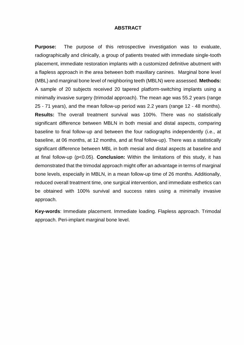
ABSTRACT
Purpose: The purpose of this retrospective investigation was to evaluate,
radiographically and clinically, a group of patients treated with immediate single-tooth
placement, immediate restoration implants with a customized definitive abutment with
a flapless approach in the area between both maxillary canines. Marginal bone level
(MBL) and marginal bone level of neighboring teeth (MBLN) were assessed. Methods:
A sample of 20 subjects received 20 tapered platform-switching implants using a
minimally invasive surgery (trimodal approach). The mean age was 55.2 years (range
25 - 71 years), and the mean follow-up period was 2.2 years (range 12 - 48 months).
Results: The overall treatment survival was 100%. There was no statistically
significant difference between MBLN in both mesial and distal aspects, comparing
baseline to final follow-up and between the four radiographs independently (i.e., at
baseline, at 06 months, at 12 months, and at final follow-up). There was a statistically
significant difference between MBL in both mesial and distal aspects at baseline and
at final follow-up (p<0.05). Conclusion: Within the limitations of this study, it has
demonstrated that the trimodal approach might offer an advantage in terms of marginal
bone levels, especially in MBLN, in a mean follow-up time of 26 months. Additionally,
reduced overall treatment time, one surgical intervention, and immediate esthetics can
be obtained with 100% survival and success rates using a minimally invasive
approach.
Key-words: Immediate placement. Immediate loading. Flapless approach. Trimodal
approach. Peri-implant marginal bone level.
8
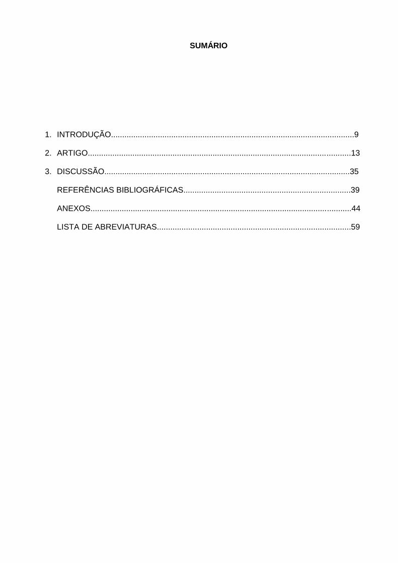
SUMÁRIO
1. INTRODUÇÃO.............................................................................................................9
2. ARTIGO......................................................................................................................13
3. DISCUSSÃO..............................................................................................................35
REFERÊNCIAS BIBLIOGRÁFICAS...........................................................................39
ANEXOS.....................................................................................................................44
LISTA DE ABREVIATURAS.......................................................................................59

9
1. INTRODUÇÃO
A atual busca pela estética em reabilitações orais modificou a forma com que
abordamos os pacientes com indicação de extração e instalação de implantes
osseointegrados, especialmente na região ântero superior. A instalação imediata de
implantes é uma técnica consolidada na literatura e tem mostrado previsibilidade muito
similar aos casos de instalação em osso cicatrizado (CABELLO et al. 2009; LANG et
al. 2011), não havendo diferença estatisticamente significativa em termos de taxa de
sobrevivência quando comparados os implantes imediatos versus tardios (CHEN et
al. 2004; KAN et al 2011; LANG et al 2011).
Ao trabalharmos na região ântero superior, a altura e a espessura da parede
óssea vestibular, presença de papila interdental e o biótipo gengival são considerados
fatores chave para atingirmos níveis de estética satisfatórios (BUSER et al. 2004;
EVANS & CHEN et al. 2007). A parede óssea vestibular dos dentes anteriores
localizados na maxila geralmente apresenta-se fina ou ausente e sofre grande
reabsorção após extração (BRAUT et al. 2011). A sua manutenção está diretamente
ligada ao posicionamento vestíbulo-palatino desses implantes, devendo os mesmos
estarem idealmente posicionados de 1-2mm palatinamente aos dentes adjacentes
(BUSER et al. 2004) e de 3-4mm abaixo da margem gengival vestibular (EVANS &
CHEN et al. 2007). O seu posicionamento mais vestibularizado ou ao nível dos dentes
vizinhos mostrou chances três vezes maiores de perda da parede vestibular e,
consequentemente, perda da arquitetura gengival (EVANS & CHEN et al. 2007). A
presença de papila interdental está majoritariamente relacionada à altura óssea
interproximal dos dentes adjacentes (BUSER et al. 2004; KAN et al. 2011). Deve ser
respeitada a distância mínima de 1.5mm entre os dentes adjacentes no momento da
instalação do implante para que não haja perda da crista alveolar (BUSER et al. 2004).
O biótipo gengival é definido pela visibilidade (fino) ou não (espesso) da sonda
periodontal milimetrada através do tecido gengival quando a sondagem periodontal é
realizada (KAN et al. 2003). Os indivíduos com biótipo gengival mais fino têm menores
chances de formação de papila interdental (BUSER et al. 2004) e maior migração dos
tecidos moles no sentido apical (45.8% versus 33.3%) (EVANS & CHEN et al. 2007),

10
apresentando maior recessão gengival quando comparados com indivíduos com
biótipo espesso (85.7% versus 66.7%). Enquanto que os pacientes com maior
espessura gengival apresentaram mudanças no aspecto da mucosa vestibular
significativamente menores seguidas da exodontia e da instalação de implantes (KAN
et al. 2003). Após a extração, a parede óssea vestibular deve apresentar ao menos
2mm de espessura para evitar a sua reabsorção, caso contrário, algum procedimento
de enxertia deve ser utilizado (HOF et al. 2013; BRAUT et al. 2011). Portanto, o
momento para realizar a instalação do implante seguida da exodontia pode ser
importante para tirar vantagem da cicatrização de tecido mole e diminuir o risco de
reabsorção óssea vestibular (CHEN et al 2004; KAN et al 2011).
O processo de cicatrização pós extração vem acompanhado de uma série de
eventos biológicos (CABELLO et al 2013) que podem influenciar negativamente o
resultado final de um tratamento com implantes na zona anterior. Recessão gengival,
perda de papila interdental e de crista óssea marginal estão intimamente ligados
(GUIRADO et al. 2009; BRAUT et al. 2011; BUSER et al. 2004) e são os principais
fatores que devem ser controlados. Extrações realizadas sem o tradicional
descolamento muco periostal apresentam menor perda óssea marginal quando
comparadas à técnica tradicional (TSOUKAKI et al. 2013), além de reduzir o tempo
de tratamento, o sangramento transoperatório e as chances de futuro
desenvolvimento de periimplantite (YOU et al. 2009). Além disso, o descolamento
muco periostal reduz a quantidade de mucosa queratinizada (BARONE et al. 2013),
alterando a arquitetura gengival pós extração e aumentando os sinais inflamatórios
clínica e histologicamente (YOU et al. 2009; BASHUTSKI et al. 2013; TSOUKAKI et
al. 2013).
A instalação de um provisório imediato proporciona o acondicionamento do
tecido mole periimplantar através da manutenção da arquitetura gengival (CANULLO
et al. 2010; KAN et al 2003), preservando o contorno e o volume dos tecidos moles
(DE CARVALHO et al. 2013). Adicionalmente, a instalação de pilares provisórios
mostrou sinais inflamatórios mais exacerbados, maior migração do epitélio juncional
no sentido apical e maior perda de crista óssea marginal (GRANDI et al. 2012),
sugerindo que a não remoção dos pilares definitivos em titânio instalados no ato
cirúrgico resultam em redução estatisticamente significativa dos níveis de perda óssea
(CANULLO et al. 2010) e, consequentemente, redução na migração dos tecidos moles
11
no sentido apical. Além disso, estudos comparativos mostraram que as taxas de
sucesso de implantes unitários instalados em zona estética com carga tardia (97%)
versus provisório imediato (98%) são similares (KAN et al. 2003; KAN et al. 2010),
fortalecendo a ideia de que a instalação de um provisório imediato é uma técnica
segura e previsível. A forma dos pilares protéticos também influencia no resultado final
(FREITAS et al 2011), devendo os mesmos respeitarem os princípios biomecânicos
dos preparos protéticos, afim de estabelecer uma relação natural e harmônica com os
tecidos moles circundantes. Coroas protéticas cimentadas na região anterior
favorecem a estética e apresentam vantagens na distribuição de carga mastigatória
quando comparadas com restaurações protéticas aparafusadas (FREITAS et al 2011).
Tarnow e colaboradores relataram que após restauração com implantes
utilizando a técnica tradicional, houve migração do tecido ósseo de 1.4 – 2.0mm a
partir da união implante-pilar no primeiro ano de função (GUIRADO, 2009,
HÜRZELER et al. 2007). Atieh e colaboradores (2010) vêm relatando que média de
reabsorção nos anos seguintes está na casa dos 0.2mm, mais ainda não há um
consenso estabelecido na literatura. Revisão sistemática da literatura realizada com
estudos em humanos e com a presença de grupo controle, no período de 2007 à 2010
e com um total de 1.239 implantes, mostrou perda óssea marginal significativamente
menor em implantes com mudança de plataforma, além de tecido duro
substancialmente mais estável (ATIEH et al. 2010). O tempo de acompanhamento
desses estudos variou de 12 aos 60 meses. Em estudo prospectivo avaliando as
alterações ósseas após um ano da instalação de implantes com mudança de
plataforma, a diferença entre baseline e follow-up foram de -0.12mm ± 0.40mm para
os implantes com mudança de plataforma, contra -0.29mm ± 0.34mm (p ≤ 0.0001)
(HÜRZELER et al. 2007). A razão para essa redução na perda de quantidade óssea
marginal pode estar relacionada ao posicionamento mais apical da junção implante-
abutment, afastando o infiltrado inflamatório da crista alveolar (ATIEH et al. 2010;
CANULLO et al. 2010; HÜRZELER et al. 2007). Além disso, há uma vantagem
biomecânica nesse tipo de conexão, relatando que a diferença entre os diâmetros do
pilar protético e da cabeça do implante reduz a concentração de stress ósseo na
região cervical (MAEDA et al. 2007), reduzindo a sua migração no sentido apical.
Canullo e colaboradores, em um ensaio clínico randomizado controlado duplo cego,
relatam que implantes restaurados com o conceito de mudança de plataforma
12
apresentaram redução significativa nos níveis de perda óssea marginal e uma
correlação negativa entre perda óssea e diferença de diâmetro entre implante e
abutment foi encontrada (CANULLO et al. 2010). Uma redução de 0.45mm no
diâmetro do pilar parece ser necessária para reduzir a perda óssea marginal
(HÜRZELER et al. 2007).
Outro fator que influencia na manutenção de tecido ósseo periimplantar é o tipo
de conexão. A interface implante-pilar é a região mais suscetível à contaminação
bacteriana e a que mais sofre com o impacto mastigatório. Em revisão sistemática da
literatura avaliando a performance de conexões do tipo cone morse, não foram
detectados micro movimentos do abutment sob forças verticais e oblíquas. Este tipo
de conexão mostrou maior resistência à perda de torque e à fratura e menor stress
sobre o parafuso quando comparado com conexões não cônicas. A geometria da
conexão cone morse distribui mais homogeneamente o stress do impacto oclusal para
o implante, melhorando o selamento, diminuindo a contaminação bacteriana e a
reabsorção óssea circundante (SCHMITT et al. 2013).
O objetivo desta investigação retrospectiva foi avaliar, radiograficamente e
clinicamente, um grupo de pacientes tratados com instalação imediata de implantes
unitários, provisório imediato com um pilar protético personalizado, com uma
abordagem sem descolamento de retalho mucoperiostal na região ântero superior.
Nível da crista óssea marginal e crista óssea marginal a nível dos dentes adjacentes
foram avaliados.
13
2. ARTIGO
EVALUATION OF BONE PRESERVATION AROUND IMPLANTS PLACED WITH THE TRIMODAL APPRACH: A 1 – 4 YEARS FOLLOW-UP
Fábio Sá Carneiro Sczepanik, DDSa
Márcio Lima Grossi, DDS, MS, PhDb
Eduardo Rolim Teixeira, DDS, PhDc
José Cícero Dinato, DDS, MS, PhDd
Thiago Revillion Dinato, DDSe
Purpose: The purpose of this retrospective investigation was to evaluate, radiographically and clinically, a group of patients treated with immediate single-tooth placement, immediate restoration implants with a customized definitive abutment with a flapless approach in the area between both maxillary canines. Marginal bone level (MBL) and marginal bone level of neighboring teeth (MBLN) were assessed, and the final MBL and MBLN were correlated with clinical variables. Methods: A sample of 20 subjects received 20 tapered platform-switching implants using a minimally invasive surgery (trimodal approach). The mean age was 55.2 years (range 25 - 71 years), and the mean follow-up period was 2.2 years ( range 12 - 48 months). Results: The overall treatment survival was 100%. There was no statistically significant difference between MBLN in both mesial and distal aspects, comparing baseline to final follow-up and between the four radiographs independently (i.e., at baseline, at 06 months, at 12 months, and at final follow-up). There was a statistically significant difference between MBL in both mesial and distal aspects at baseline and at final follow-up (p<0.05). Conclusion: Within the limitations of this study, it has demonstrated that the trimodal approach might offer an advantage in terms of marginal bone levels, especially in MBLN, in a mean follow-up time of 26 months. Additionally, reduced overall treatment time, one surgical intervention, and immediate esthetics can be obtained with 100% survival and success rates using a minimally invasive approach. Additional long-term, well-conducted, randomized-controlled trials are needed to confirm the validity of the trimodal approach.
Key-words: Immediate placement. Immediate loading. Flapless approach. Trimodal approach. Peri-implant marginal bone level.
______________________________
a Private practice, Porto Alegre, Brazil. b Prosthodontist; Master Of Science Restorative Dentistry-Occlusion; Doctor of Philosophy. Professor, Department
of Prosthodontics, Pontifícia Universidade Católica do Rio Grande do Sul, Brazil. Adress: Ipiranga, 6681 c Prosthodontist; Doctor of Philosophy in Dental Sciences. Professor, Department of Prosthodontics, Pontifícia
Universidade Católica do Rio Grande do Sul, Brazil. Adress: Ipiranga, 6681 d Implantologist; Prosthodontist; Master of Science Prosthodontics; Doctor of Philosofy in Implantology. Adress:
Furriel Luiz Antônio Vargas, 250/1501 e Implantologist; Adress: Furriel Luiz Antônio Vargas, 250/1501
Correspondence to: Dr. FSC Sczepanik, Clínica Dinato de Odontologia, CEP:90470-130, Porto Alegre, Brazil. Fax:
+555133332717. Email:[email protected]

14
INTRODUCTION
As the technique of replacing missing teeth in healed edentulous sites offered a
predictable and effective outcome1,2. Different placement protocols have evolved to
expand implant therapy applicability, starting with the first reports about immediate
implant placement into extraction sites described by Schulte and colleagues2 and
Lazzara and colleagues4. Bone preservation and soft tissue stability are one of the
benefits related to this approach5,6 as they reduce the number of surgical interventions,
overall treatment time and morbidity; and they have attracted attention in recent
years1,7, representing a clear advantage for our day-to-day practice6 and patient well-
being. Moreover, a recent systematic review, which have evaluated 2934 implants
placed into fresh sockets in 2130 patients, have showed an annual failure rate of 0.82%
and a 2-year survival rate of 98.4%1.
Additionally, immediate loading protocols have been suggested as a way to
restore aesthetic appearance2, specially in the anterior maxilla, as soft tissue contour
and gingival architecture can be maintained9,10, adding a predictability of implant
survival and papilla levels over time7. A systematic review have evaluated the survival
rate of immediately restored implants as compared to implants conventionally restored
after 1 year of implant placement, and the difference was not statistically significant
(0.75% vs 0.89%)1. Furthermore, combining a provisional crown with a definitive
abutment during the healing phase may prevent bone remodeling from happening
according to several authors10-13.
The elevation and advancement of a full thickness flap can cause post-operative
bone resorption, reduced keratinized mucosa14, loss of gingival/papillae architecture15,
higher risk of developing periimplantitis, and enhanced clinically and histologically
inflammatory signs16,17. Thus, the use of a minimally invasive approach, by means of
a flapless protocol, may reduce bone remodeling14 and improve the esthetic result18.
The complete preservation of the alveolar ridge dimension after tooth extraction is
unlikely to be attainable, even with the use of a bone substitute and collagen
membrane14.
Despite the high survival rate, the marginal bone level (MBL) around implants is
an important factor in the success criteria for evaluating implant therapy19. Laurell and
15
colleagues in a 5-year of function meta-analysis have reported a mean marginal bone
loss of 0.48 mm for platform-switched implants, and have suggested a revision of the
existing implant success criteria20. The use of a smaller diameter abutment on a larger
diameter implant collar, with a conical abutment-implant attachment system, reduces
de gap between both interfaces. This reduction shifts the inflammatory cell infiltrate to
the central axis of the implant, which limits crestal bone resorption21,22.
Additionally, a key point in maintaining interdental papillae may be the bone
level to the adjacent tooth. Preserving this bone peak throughout extraction of the tooth
as well as during implant placement may secure papilla height over time7.
Recently, few studies have been published reporting a minimally invasive
approach, which consists: a) hard tissue preservation, b) peri-implant mucosal
maintenance, c) reduced treatment time, and d) improved esthetic results. Primarily,
an immediate placement and restoration/loading protocol have led to the introduction
of a bimodal approach (BA)2, which have shown similar survival rates to the
conventional protocols and encouraging esthetic outcomes in a 5-year case-control
study23. Posteriorly, Cabello and colleagues added a third element to the BA: a flapless
surgery, leading to a trimodal approach (TA)24. This TA approach consisted of an
immediate single tooth placement and an immediate provisional loading with no
mucoperiostal flap raised.
Therefore, the aim of this retrospective study was twofold. First, to evaluate
radiographically patients treated within TA criteria, in the area comprehended between
both maxillary canines in terms of marginal bone level (MBL) and marginal bone level
of neighboring teeth (MBLN). Second, to compare final MBL and MBLN with the
clinical variables.
MATERIAL AND METHODS
Patients and study design

16
This retrospective study was based on data from 20 patients (12 females and 8
males) who were treated in a private practice in the city of Porto Alegre, in the state of
Rio Grande do Sul (Brazil), who had underwent immediate placement and
temporization of one single-tooth implant within the anterior maxilla (incisors and
canines) using a flapless technique, and who had a definitive titanium abutment. The
mean age was 55.2 ± 13.2 years (range 25 to 71 years), and the follow-up period was
at least 12 months (mean = 2.2 ± 1.03 years, range 12 to 48 months). The same
practitioner (J. C. D.) have treated all patients between 2010 and 2014, and he did not
participate in the data collection and evaluation. The study protocol was reviewed and
approved by the Research Ethics Committee (CEP, 808.513) of the Pontifical Catholic
University of Rio Grande do Sul (PUCRS), Brazil.
The inclusion criteria was: a) patients with the need of a single tooth extraction
in the anterior sector.
The exclusion criteria was: a) patients under 18 years of age, b) systemic
diseases that would otherwise contraindicate surgery (uncontrolled diabetes,
hemophilia, radiation treatment to the head and neck region, current chemotherapy,
treatment with oral bisphosphonates), c) acute infection in the treatment area, d)
absence of the adjacent tooth/presence of an adjacent implant, e) probing depth over
3mm, f) heavy smokers (more than 10 cigarettes a day), g) absence of opposite teeth,
and h) if the principles of trimodal approach could not be follow during surgery. Patients
who used less than 10 cigarettes/day were not excluded, but were instructed to stop
smoking throughout the treatment. Patients with chronic endodontic pathology and/or
buccal bone wall resorption were included in this study.
Surgical protocol
Before surgery, cone beam computerized tomography (CBCT) and periapical
radiographs were requested in order to establish the indication for tooth extraction as
well as the initial diagnosis and treatment planning. Patients agreed with the treatment
plan and consented to be treated. They underwent professional prophylaxis and
17
periodontal treatment, if necessary, prior to the implant surgery. All patients received
2g of amoxicillin 1h before the procedure, and 0.12% chlorhexidine gluconate
mouthwashes were given immediately before the surgery for oral disinfection.
Tooth removal was done as atraumatically as possible (Articaine 100, DFL, Rio
de Janeiro, Brazil) using a specific extractor (Benex Root Extraction System, Hager
and Meisinger GMbH, Neuss, Germany) without raising the mucoperiostal flap. The
socket was vigorously debrided with the help of manual curettes. A roughened surface
morse tapered implant (CM Drive, Neodent, Curitiba, Brazil) was placed at 2 to 3 mm
beneath the bone crest and 2 mm palatal to the buccal bone wall, with a minimum
initial insertion torque of 35 N/cm verified with a digital device. The gap between the
implant’s shoulder and the inner wall of the buccal aspect was filled with an anorganic
bovine bone in small particles (0.25 – 1 mm) (Bio-Oss®, Geistlich Pharma AG,
Wolhusen, Switzerland).
Clinical procedures
Implant’s platform impression was took immediately after surgery with a help of
a rubber dam (Madeitex, São José dos Campos, SP, Brazil) in order to fabricate an
acrylic provisional crown adapted to a customized definitive titanium abutment
extraorally. The abutment was digitally scanned with a laboratory device (Neo Shape
D700, 3Shape, Copenhagen, Denmark) to fabricate a zirconium oxide reinforced
coping to be placed after the osseointegration process is completed (05 months).
Within 24 hours after surgery, the abutment was tightened at 20 N/cm; and an acrylic
crown cemented provisionally (Dycal, Dentsply, York, United States) with a non-
functional loading (i.e., centric or eccentric contacts). A distance was left between the
gingival contour and the cervical aspect of the provisional crown in order to avoid soft
tissue compression. Abutments had a reduced diameter in comparison to implant’s
platform diameter and had a conical connection.
After surgery, patients were instructed to have a soft diet and to avoid chewing
in the treated area for the remaining duration of the implant healing phase. Drug
therapy consisted in antibiotics (i.e., Amoxicillin 875mg, every 12h for 7 days),

18
analgesic and anti-inflammatory drugs (i.e., Nimesulide 100mg, every 12h for 3 days)
and mouthwashes (i.e., 0.12% chlorhexidine gluconate, twice daily for 7-10 days).
Definitive crown delivery
Five months after surgery, the provisional crown was removed, the absence of
suppuration/inflammatory signs was checked, and abutment torque confirmed with a
torque wrench. A CAD/CAM machined zirconium oxide coping (InLab MC XL, Sirona,
Salzburg, Austria) was placed onto the abutment, and a coping pick-up silicone
impression was taken (Regular Body Normal Set, Elite HD+, Zhermack, Rovigo, Italy).
Feldspathic ceramic was manually applied in the final master cast and cemented
definitively with a resin-modified glass ionomer cement (Relyx Luting 2, 3M ESPE,
California, United States), with manual compression for 10 minutes. The cement
excess was then removed with the help of a sounder and a dental floss. Patients were
instructed not to chew or to floss during the first 24 hours post cementation. A periapical
radiograph was taken in order to assess the presence of any residual cement
remnants. All materials were handled according to the manufacturer instructions. The
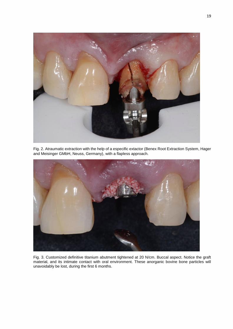
treatment sequence is shown in figures 1 - 5.
Fig. 1. Initial clinical aspect. Upper left central incisor with indication ofextraction due to root fracture.
19
Fig. 2. Atraumatic extraction with the help of a especific extactor (Benex Root Extraction System, Hager
and Meisinger GMbH, Neuss, Germany), with a flapless approach.
Fig. 3. Customized definitive titanium abutment tightened at 20 N/cm. Buccal aspect. Notice the graft material, and its intimate contact with oral environment. These anorganic bovine bone particles will unavoidably be lost, during the first 6 months.
20
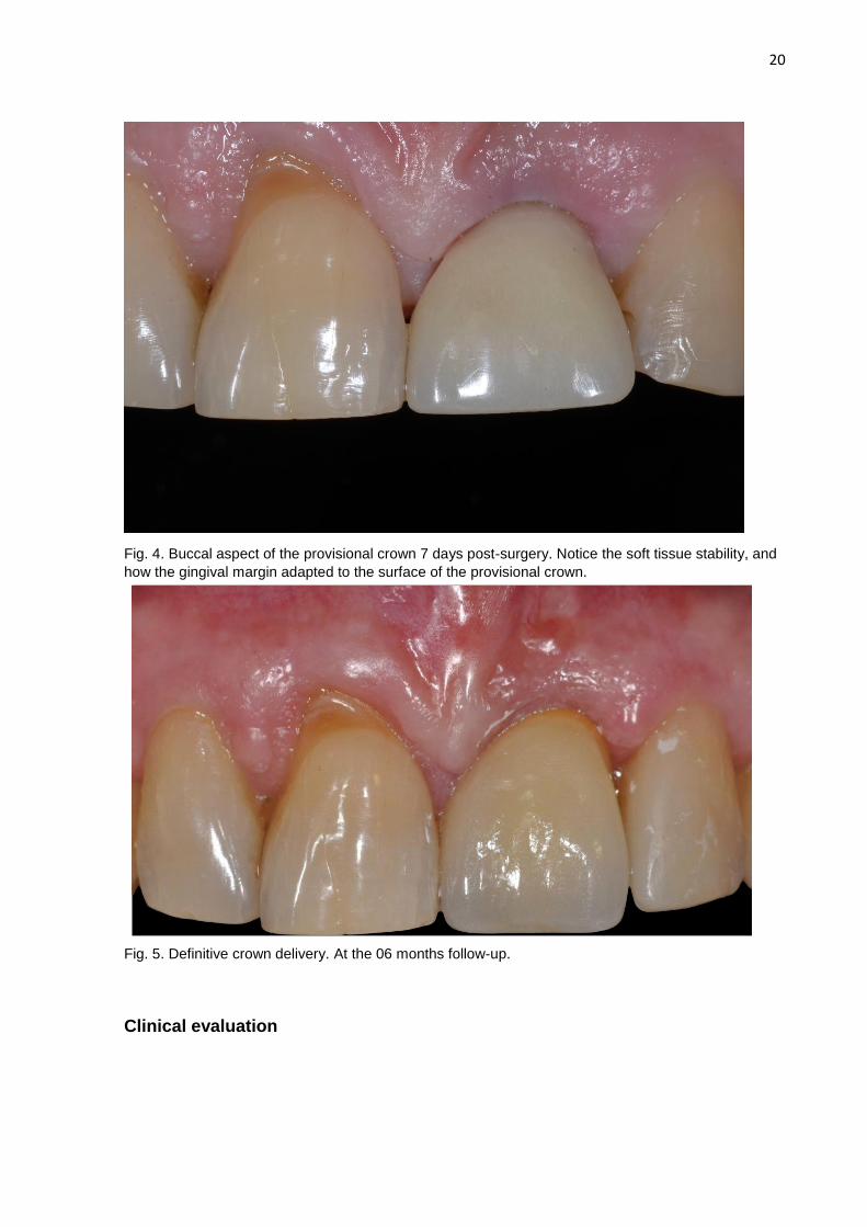
Fig. 4. Buccal aspect of the provisional crown 7 days post-surgery. Notice the soft tissue stability, and
how the gingival margin adapted to the surface of the provisional crown.
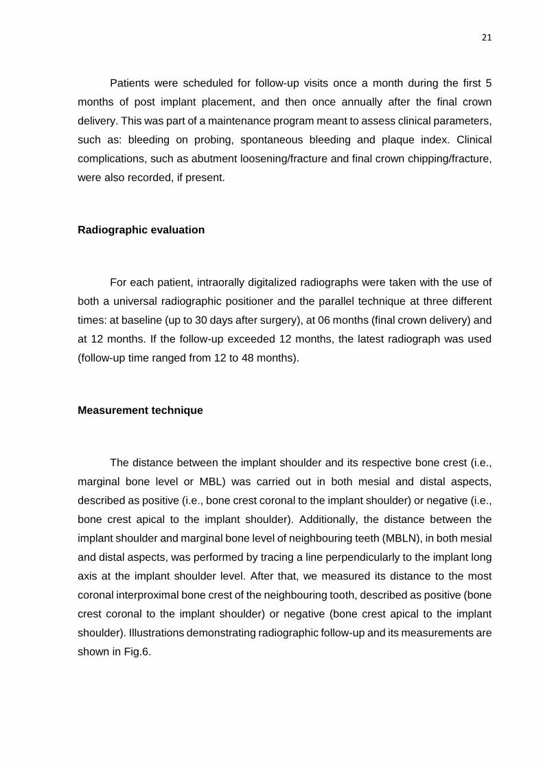
Fig. 5. Definitive crown delivery. At the 06 months follow-up.
Clinical evaluation
21
Patients were scheduled for follow-up visits once a month during the first 5
months of post implant placement, and then once annually after the final crown
delivery. This was part of a maintenance program meant to assess clinical parameters,
such as: bleeding on probing, spontaneous bleeding and plaque index. Clinical
complications, such as abutment loosening/fracture and final crown chipping/fracture,
were also recorded, if present.
Radiographic evaluation
For each patient, intraorally digitalized radiographs were taken with the use of
both a universal radiographic positioner and the parallel technique at three different
times: at baseline (up to 30 days after surgery), at 06 months (final crown delivery) and
at 12 months. If the follow-up exceeded 12 months, the latest radiograph was used
(follow-up time ranged from 12 to 48 months).
Measurement technique
The distance between the implant shoulder and its respective bone crest (i.e.,
marginal bone level or MBL) was carried out in both mesial and distal aspects,
described as positive (i.e., bone crest coronal to the implant shoulder) or negative (i.e.,
bone crest apical to the implant shoulder). Additionally, the distance between the
implant shoulder and marginal bone level of neighbouring teeth (MBLN), in both mesial
and distal aspects, was performed by tracing a line perpendicularly to the implant long
axis at the implant shoulder level. After that, we measured its distance to the most
coronal interproximal bone crest of the neighbouring tooth, described as positive (bone
crest coronal to the implant shoulder) or negative (bone crest apical to the implant
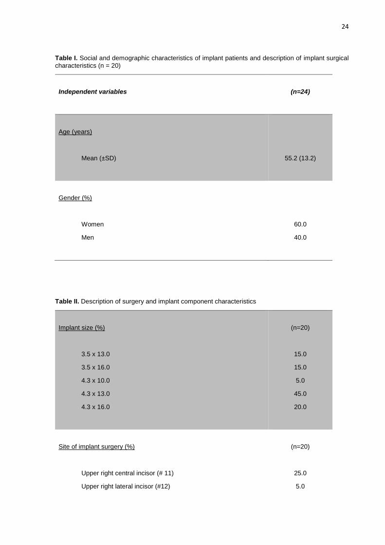
shoulder). Illustrations demonstrating radiographic follow-up and its measurements are
shown in Fig.6.
22
The radiographic implant length was measured, and compared with the true
implant length 11.5 mm, 13 mm, 16 mm), for adjusting bone level and distance
measurements, according to image magnification. The same equation was used to
determine the distance from the implant shoulder to MBL, and the distance from the
implant shoulder to MBLN. Brightness, contrast, magnification and the measurements
were controlled and standardized in all radiographs with a specific imaging software
(DBSWIN Imaging Software, Dürr Dental, Bietigheim-Bissingen, Germany).
Data collection
One independent examiner performed all examinations and data collection.
Evaluations were made at baseline (up to 30 days before implant placement), at 06,
and at 12 months. If the follow-up exceeded 12 months, the latest radiograph was used
(follow-up time ranged from 12 to 48 months). The following variables were recorded:
a) age, b) gender, c) surgery site, d) implant features, e) follow-up time, f) implant
success/failure, g) marginal bone level (MBL), and h) marginal bone level of
neighbouring teeth (MBLN). Measurements were made in both mesial and distal
aspects.
Sample size calculation and statistical analysis

23
For the reduction in marginal bone loss, repeated measures Analysis of
Variance (ANOVA) and the Tukey-b Post-Hoc test will be used for the differences
within and between groups.
The formula for the sample size calculation of hypothesis testing for population
proportions is: n = {Ζ1-α√[Po (1 - Po)] + Ζ1-β√[Pa (1 - Pa)]}2 / (Po – Pa)2; in which n =
estimated sample size, Po = proportion value test of the population under the null
hypothesis, Pa = anticipated value of the population proportion, Ζ1-α = standard
distribution value corresponding to a α level of significance (e.g. 1.96 for a one-sided
test at a 0.05 level), and Ζ1-β = value of the normal distribution corresponding to the
desired power level (e.g. 0.84 for a power of 80%). Using a success rate of 95% and
an anticipated success rate of 85%, we reach a sample size of 44 individuals25.
RESULTS
Patients who had received immediate placement/loading of one single-tooth
tapered implant in the esthetic zone until to February 2014 were selected to this
retrospective study. Twenty consecutive individuals were included for the radiographic
and clinical analysis, with a mean age at implant insertion of 55.2 years (± 13.2),
ranging from 25 to 71 years. Sixty per cent of the patients were female and forty per
cent were male (Table I). The mean follow-up time was 2.2 years (± 1.03), ranging
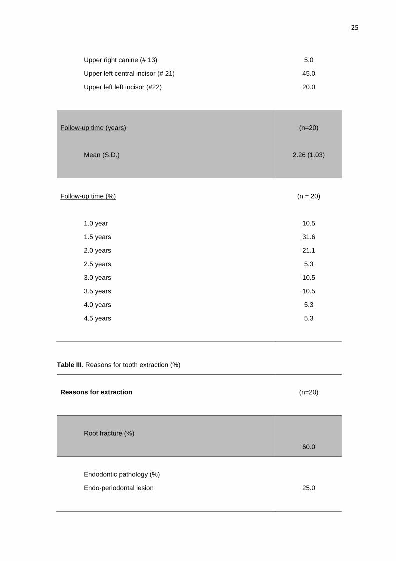
from 12 to 48 months (Table II). Sixty per cent of extractions were indicated because
of root fracture and none was indicated because of chronical periodontal disease.
Reasons for tooth extraction were in Table III.
Seventy per cent of the implants had a regular platform (4.3 mm), while a narrow
platform implant (3.5 mm) was used in only 30% of the patients. The most frequent
implant use had a regular diameter (4.3 mm) and was 13 mm length (45%). The upper
left central incisor was the most frequent tooth restored (45%), followed by its
contralateral tooth (25%). In the sample, only the upper left canine was not present.
Description of surgery and implant characteristics are listed in Table III.
24
Table I. Social and demographic characteristics of implant patients and description of implant surgical characteristics (n = 20)
Independent variables
(n=24)
Age (years)
Mean (±SD)
55.2 (13.2)
Gender (%)
Women
Men
60.0
40.0
Table II. Description of surgery and implant component characteristics
Implant size (%)
3.5 x 13.0
3.5 x 16.0
4.3 x 10.0
4.3 x 13.0
4.3 x 16.0
(n=20)
15.0
15.0
5.0
45.0
20.0
Site of implant surgery (%)
Upper right central incisor (# 11)
Upper right lateral incisor (#12)
(n=20)
25.0
5.0
25
Upper right canine (# 13)
Upper left central incisor (# 21)
Upper left left incisor (#22)
5.0
45.0
20.0
Follow-up time (years)
Mean (S.D.)
(n=20)
2.26 (1.03)
Follow-up time (%)
1.0 year
1.5 years
2.0 years
2.5 years
3.0 years
3.5 years
4.0 years
4.5 years
(n = 20)
10.5
31.6
21.1
5.3
10.5
10.5
5.3
5.3
Table III. Reasons for tooth extraction (%)
Reasons for extraction
(n=20)
Root fracture (%)
60.0
Endodontic pathology (%)
Endo-periodontal lesion
25.0

26
Root resorption (%)
15.0
Implant and definitive crown survival
All 20 implants showed an uneventful healing and integrated, presenting a
survival rate of 100%. None of the 20 definitive crowns was lost and no clinical
complications were recorded during the follow-up visits, such as: ceramic fracture,
abutment loosening, or periimplantitis.
Radiographic marginal bone level of neighbouring tooth
Comparison of mean MBLN and MBL in both mesial and distal aspects among
different follow-up time periods are shown in table IV.
The mean marginal bone level of neighbouring tooth (MBLN) in the mesial
aspect at baseline was 2.9 mm ± 1.3 mm, with a variation between 1.1 to 5.9 mm. At
the final follow-up, the MBLN was 3.2 mm ± 1.0 mm, ranging from 0.9 to 4.6 mm. There
was no statistical significant difference between MBLN in the mesial aspect, comparing
baseline to final follow-up, and between the four radiographs independently (at
baseline, at 06 months, at 12 months, and at final follow-up).
The mean MBLN in the distal aspect at baseline was 2.2 mm ± 1.1 mm, with a
variation between 0 to 4.2 mm. At the final follow-up, the MBLN remained 2.2 mm ±
1.1 mm, ranging from 0 to 3.9 mm. There was no statistical significant difference
between MBLN in the distal aspect, comparing baseline to final follow-up, and between
the four radiographs independently (at baseline, at 06 months, at 12 months, and at
final follow-up).

27
Radiographic peri-implant marginal bone level
The mean marginal bone level (MBL) in the mesial aspect at baseline was 3.6
mm ± 1.2, with a variation between 1.8 to 6.9 mm. At the final follow-up, the MBL was
1.0 mm ± 1.2, ranging from 0 to 4.1 mm. There was a statistical significant difference
between MBL in the mesial aspect at baseline and at the final follow-up (p<0.05). There
was no statistical significant difference between MBL in the mesial aspect, when we
compared the 06 month follow-up to the posterior radiographs.
The mean MBL in the distal aspect at baseline was 3.1 mm ± 1.2, with a variation
between 1.8 to 5.5 mm. At the final follow-up, the MBL was 1.0 mm ± 1.2, ranging from
0 to 3.9 mm. There was a statistical significant difference between MBL in the distal
aspect at baseline and at the final follow-up (p<0.05). There was no statistical
significant difference between MBL in the distal aspect, when we compare the 06
month follow-up to the posterior radiographs.
Table IV. Comparison of mesial and distal bone / implant shoulder distances among different follow-up time periods (n = 65)
Independent variables:
Mean (±SD)
1 month
(baseline)
6 months
12 months
+12 months
Mesial bone crest/implant
shoulder (mm)
(n = 20)
2.9 (1.3)
(n = 17)
3.1 (1.2)
(n = 14)
3.2 (1.0)
(n = 15)
3.2 (0.9) †
Distal bone crest/implant
shoulder (mm)
(n = 20)
2.2 (1.0)
(n = 17)
2.2 (1.3)
(n = 14)
2.2 (1.1)
(n = 15)
2.2 (1.0) †
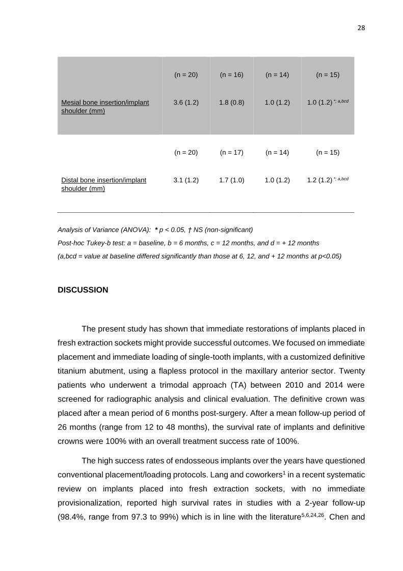
28
Mesial bone insertion/implant
shoulder (mm)
(n = 20)
3.6 (1.2)
(n = 16)
1.8 (0.8)
(n = 14)
1.0 (1.2)
(n = 15)
1.0 (1.2) *; a,bcd
Distal bone insertion/implant
shoulder (mm)
(n = 20)
3.1 (1.2)
(n = 17)
1.7 (1.0)
(n = 14)
1.0 (1.2)
(n = 15)
1.2 (1.2) *; a,bcd
Analysis of Variance (ANOVA): * p < 0.05, † NS (non-significant)
Post-hoc Tukey-b test: a = baseline, b = 6 months, c = 12 months, and d = + 12 months
(a,bcd = value at baseline differed significantly than those at 6, 12, and + 12 months at p<0.05)
DISCUSSION
The present study has shown that immediate restorations of implants placed in
fresh extraction sockets might provide successful outcomes. We focused on immediate
placement and immediate loading of single-tooth implants, with a customized definitive
titanium abutment, using a flapless protocol in the maxillary anterior sector. Twenty
patients who underwent a trimodal approach (TA) between 2010 and 2014 were
screened for radiographic analysis and clinical evaluation. The definitive crown was
placed after a mean period of 6 months post-surgery. After a mean follow-up period of
26 months (range from 12 to 48 months), the survival rate of implants and definitive
crowns were 100% with an overall treatment success rate of 100%.
The high success rates of endosseous implants over the years have questioned
conventional placement/loading protocols. Lang and coworkers1 in a recent systematic
review on implants placed into fresh extraction sockets, with no immediate
provisionalization, reported high survival rates in studies with a 2-year follow-up
(98.4%, range from 97.3 to 99%) which is in line with the literature5,6,24,26. Chen and
29
colleagues5, in a histological and clinical analysis, concluded that survival rates were
similar for both immediate and delayed placement; but recommended the implant
placement from 4 to 8 weeks after tooth extraction in order to allow soft tissue healing
and removal of an inflammatory process, if present27. In a prospective, controlled
clinical trial, there was no statistically significant difference on survival rates as well as
in clinical and radiographic parameters between immediate implant placement in
sockets exhibiting periapical pathology and healthy sockets after a careful socket
debridement28. In terms of marginal bone loss, a systematic review on immediate
placement/loading, authors have concluded that immediate loading offer more
advantages in terms of bone height preservation2.
Using the immediate loading protocol in the anterior maxilla is preferable in
order to re-establish esthetics and to avoid the use of a temporary removable
prosthesis during the healing phase23,29. There are studies reporting similar high
survival rates comparing delayed loading with conventional loading (97% versus 98%),
respectively9,23,26.
Abutment disconnection, as part of the prosthetic treatment, and its consequent
disruption of the epithelial seal, may cause: a) pronounced inflammatory signs, such
as bleeding and ulceration10, b) epithelial migration12, and c) re-establishment of
biologic width apically, which may cause initial crestal bone loss11-13. Therefore, the
one abutment at one time protocol11, with a provisional crown cemented is preferable
in order to ensure peri-implant stability as well as an optimal seal around
osseointegrated implants10. That might explain gingival health and stability during
healing follow-ups, and specially after definitive ceramic crown cementation in our
study. Although no implant system or connection has been able to provide a perfect
sealing, the use of conical connections clearly reduce bacterial leakage30. Besides, the
use of a customized titanium abutment has shown to be an effective and safe approach
for soft tissue handling. In our study, in accordance with other authors31, this adequate
emergence profile, following gingival architecture, resulted in papilla maintenance.
Formerly, it was thought that implant diameter should fill and be placed precisely
in the middle of the socket, providing support to the bone walls. Because of that, 5 mm
diameter implants were advocated. This approach has been revised11. In our study,
70% of implants used had a diameter of 4.3 mm and were placed closer to the palatal
30
wall, which left a gap between implant’s shoulder and the inner wall of the socket32.
This gap was filled with anorganic bovine bone.
The mean MBL radiographic measurement at baseline included the
reconstructive graft, which was partly lost during the following months. This might have
been due to the non-use of a collagen membrane allied to a flapless protocol, which
prevents soft tissue primary closure33,34. Covani et al have advocated the use of a
collagen membrane along with the bone graft, which showed minimal soft tissue
remodelling in a 5-year prospective single-cohort study34; however, they have
performed a conventional loading protocol.
Therefore, statistically significant difference was found between mean MBL at
baseline (i.e. 3.6 ± 1.2 at the mesial aspect, and 3.1 ± 1.2 at the distal aspect), and at
the final follow-up (i.e. 1.0 ± 1.2 at the mesial aspect, and 1.2 ± 1.2 at the distal aspect)
(p<0.05). Besides that, mean MBL at 06 months, at 12 months, and at final follow-up
remained statistically similar. In the mesial aspect, MBL remained 1.0 mm ± 1.2 mm
coronal to the implant shoulder, ranging from 0 to 4.1 mm. While in the distal aspect,
MBL remained 1.2 mm ± 1.2 mm coronal to the implant shoulder, ranging from 0 to 3.9
mm. In agreement with other findings33,35, there was a hard tissue remodelling, but the
bone remained over the implant shoulder. Radiographically, bone could be seen
growing on top of the implant shoulder, and in close contact with the abutment surface.
This observation is in accordance with a histologic and histomorphometrical study36.
This bone behaviour is probably due to both the lack of micro-motion between the
implant and the abutment, a characteristic of the Morse Cone tapered connection
implants, and also to the subcrestal positioning of the implants and platform-
switching19,37,38. In our study, implants were placed 2.5 mm subcrestally, 2.9 mm in the
mesial aspect, and 2.1 mm in the distal aspect.
There was no statistical significant difference between MBLN in the mesial and
distal aspects between baseline and final follow-up, and between the four radiographs
independently (i.e. at baseline, at 6 months, at 12 months, and at final follow-up). In
the mesial aspect, MBLN at baseline was 2.9 mm ± 1.3 mm; and at the final follow-up,
the MBLN was 3.2 mm ± 1.0 mm. This growth have indicated that restorative
procedures influenced the maintenance of MBLN the presence of papilla over time,
which is in agreement with other studies38,39.

31
Gingival stability is an unquestionable factor for satisfactory esthetic results in
single-tooth implants. Thus, the maintenance of the bone peak plays a leading role on
the presence of papilla over time40. Our study it is not in accordance with Degidi and
colleagues. They have found a mean marginal bone loss at adjacent tooth of 0.53 mm.
Additionally, in 18.18% of the radiographs, bone peak was apical to the implant
shoulder; contradicting our results. However, they used no bone substitutes40.
However is in accordance with Tarnow and colleagues41, which have recommended
the use of reconstructive graft and immediate provisionalization, in order to reduce
bone remodelling and facial/palatal dimensional change.
CONCLUSION
Within the limitations of this study, it has demonstrated that the trimodal
approach might offer an advantage in terms of marginal bone levels, specially in MBLN,
in a mean follow-up time of 26 months. Additionally, reduced overall treatment time,
one surgical intervention, and immediate esthetics can be obtained, with 100% survival
and success rates, using a minimally invasive approach. Further long-term, well-
conducted, randomized-controlled studies are needed to confirm the validity of the
trimodal approach.
Our findings in bone peak maintenance contradicts other study40, which have
reported resorption of marginal bone levels at the neighbouring tooth even apically to
the implant shoulder; thus, prospective studies comparing the maintenance of the
MBLN with peri-implant stability and bone remodelling radiographically, are needed.
ACKNOWLEDGMENTS
The authors report no conflict of interests related to this study.
REFERENCES

32
1. Lang, NP; Pun, L; LauU, KY; Wong, MCM. A Systematic Review on Survival and Success Rates of Implants Placed Immediately Into Fresh Extraction Sockets After at Least 1 Year. Clin Oral Impl Res 2012; 23: 39-66.
2. Atieh MA, Payne AGT, Duncan WJ, Cullinan MP. Immediate restoration/loading of immediately placed single implants: is it an effective bimodal approach? Clin. Oral Impl. Res. 20, 2009; 645–659.
3. Lazzara, RJ. Immediate implant placement into extraction sites: surgical and restorative advantages. The International Journal of Periodontics and Restorative Dentistry 1989; Volume 9, Number 5.
4. Romanos GE, Traini T, Johansson CB, Piatelli A. Biologic Width and Morphologic Characteristics of Soft Tissues Around Immediately Loaded Implants: Studies Performed on Human Autopsy Specimens. J Periodontol 2010;81:70-78
5. Chen, ST; Wilson, TG; Hämmerle, CHF. Immediate or Early Placement of Implants Following Tooth Extraction: Review of Biologic Basis, Clinical Procedures and Outcomes. Int J Oral Maxillofac Implants 2004; 19(suppl): 12-15.
6. Evans CDJ, Chen ST. Esthetic outcomes of immediate implant placements. Clin. Oral Impl. Res. 19, 2008; 73–80.
7. De Rouck, T; Collys, K; Cosyn, J. Single-Tooth in the Anterior Maxilla by Means of Immediate Implantation and Provisionalization: A Review. Int J Oral Maxillofac Implants 2008; 23: 897-904.
8. Canullo, L; Bignozzi, I; Cocchetto, R; Cristalli, MP; Ianello, G. Immediate Positioning of a Definitive Abutment Versus Repeated Abutment Replacements in Post-Extractive Implants: 3-Year Follow-up of a Randomised Multicentre Clinical Trial. Eur J Oral Implantol 2010; 3(4): 285-296.
9. Kan, J; Rungcharassaeng; Lozada, J. Immediate Placement and Provisionalization of Maxillary Anterior Single Implants: 1-Year Prospective Study. Int J Oral Maxillofac Implants 2003; 18: 31-39.
10. Alves CC, Munoz F, Ramos I, Neves M, Blanco J. Marginal bone and soft tissue behavior following platform switching abutment connection/disconnection a dog model study. Clin. Oral Impl. Res.00, 2014, 1–9.
11. Degidi, M; Nardi, D; Daprile, G; Piatelli, A. Nonremoval of Immediate Abutments in Cases Involving Subcrestally Placed Postextractive Tapered Single Implants: A Randomized Controlled Clinical Study. Clinical Implant Dentistry and Related Research, Volume 16, Number 6, 2014.
12. Grandi, T; Guazzi, P; Samarini, R; Maguaireh, H; Grandi G. One abutment–one time versus a provisional abutment in immediately loaded post-extractive single implants: A 1-year follow-up of a multicentre randomised controlled trial. Eur J Oral Implantol 2014;7(2):141–149.
13. Degidi M, Nardi D, Piattelli A. One abutment at one time: non-removal of an immediate abutment and its effect on bone healing around subcrestal tapered implants. Clin. Oral Impl. Res. 22, 2011; 1303–1307.
14. Barone, A; Toti, P; PiatelliI, A; Iezzi, G; Derchi, G; Covani U. Extraction Socket Healing in Humans After Ridge Preservation Techniques: A Comparison Between Flapless and Flapped Procedure in a Randomized Clinical Trial. J Periodontol 2013 Jan; 85(1):14-23.
15. You, TM; Choi, BH; Li, J; Xuan, F; Jeong, SM; Jang, SO. Morphogenesis of the Peri-Implant Mucosa: A Comparison Between Flap and Flapless Procedures in the Canine Mandible. Oral Surg Oral Med Oral Pathol Oral Radiol Endod 2009; 107: 66-70.
33
16. Bashutski, JD; Wang, HL; Rudek, I; Moreno, I; Koticha, T; Oh, TJ. The Effect of Flapless Surgery on Single-Tooth Implants in the Esthetic Zone: A Randomized Clinical Trial. J Periodontol 2013 Dec; 84(12):1747-54.
17. Tsoukaki, M; Kalpidis, CDR; Sakellair, D; Tsaliks, L; Mikrogiorgis, G; Konstandinidis, A. Clinical, Radiographic, Microbiological, and Immunological Outcomes of Flapped vs. Flapless Dental Implants: A Prospective Randomized Controlled Clinical Trial. Clin Oral Impl Res 2013; 24: 969-976.
18. De Carvalho, BCF; De Carvalho, EMOF; Consani, RLX. Flapless Single-Tooth Immediate Implant Placement. Int J Oral Maxillofac Implants 2013; 28: 783-789.
19. Strietzel FP, Neumann K, Hertel M. Impact of platform switching on marginal peri-implant bone level changes. A systematic review and meta-analysis. Clin. Oral Impl. Res.00, 2014, 1–16.
20. Laurell, L; Lundgren, D. Marginal Bone Level Changes at Dental Implants after 5 Years in Function: A Meta-Analysis. Clinical Implant Dentistry and Related Research, Volume 13, Number 1, 2011.
21. Hürzeler, M; Fick, S; Zuhr, O; Wachtel, HC. Peri-Implant Bone Level Around Implants With Platform-Switched Abutments: Preliminary Data From a Prospective Study. J Oral Maxillofac Surg 2007, 65: 33-39.
22. Schmitt, CM; Nogueira-Filho, G; Tenenbaum, HC; Lai, JY; Brito, C; Döring, H; Nonhoff, J. Performance of Conical Abutment (Morse Taper) Connection Implants: A Systematic Review. J Biomed Mater Res Part A 2013; 00A: 000-000.
23. Laviv, A; Levin, L; Usiel, Y; Schwartz-Adad, D. Survival of Immediately Provisionalized Dental Implants: A Case-Control Study with up to 5 Years Follow-Up. Clinical Implant Dentistry and Related Research, Volume 12, Supplement 1, 2010.
24. Cabello, G; Riobbo, M; Fábrega, JG. Immediate Placement and Restoration of Implants in the Aesthetic Zone with a Trimodal Approach: Soft Tissue alterations and its Relation to Gingival Biotype. Clin Oral Impl Res 2013; 24: 1094-1100.
25. Lwnga, SK; Lemeshow, S. Sample Size Determination in Health Studies: a Practical Manual. Geneve: World Health Organization, Genebra, 1991: 4, pp:3.
26. Kan, J; Rungcharassaeng; Lozada, J; Zimmerman, G. Facial Gingival Tissue Stability Following Immediate Placement and Provisionalization of Maxillary Anterior Single Implants: 2-to-8-Year Follow up. Int J Oral Maxillofac Implants 2011; 26: 179-187.
27. Buser, D; Wittneben, J; Bornstein, MM; Grütter, L; Chappuis, V; Belser, UC. Stability of Contour Augmentation and Esthetic Outcomes of Implant-Supported Single Crowns in the Esthetic Zone: 3-Year Results of a Prospective Study With Early Implant Placement Postextraction. J Periodontol 2011;82:342-349.
28. Truninger TC, Philipp AOH, Siegenthaler DW, Roos M, Hammerle CHF, Jung RE. A prospective, controlled clinical trial evaluating the clinical and radiological outcome after 3 years of immediately placed implants in sockets exhibiting periapical pathology. Clin. Oral Impl. Res. 22, 2011; 20–27.
29. Hartlev J, Kohberg P, Ahlmann S, Gotfredsen E, Andersen NT, Isidor F, Schou S. Immediate placement and provisionalization of single-tooth implants involving a definitive individual abutment: a clinical and radiographic retrospective study. Oral Impl. Res.24,2013, 652–658.
30. Canullo L, Penarrocha-Oltra D, Soldini C, Mazzocco F, Penarrocha M, Covani U. Microbiological assessment of the implant-abutment interface in different connections: crosssectional study after 5 years of functional loading. Clin. Oral Impl. Res.00, 2014, 1–9.
31. Borges T, Lima T, Carvalho, A, Dourado C, Carvalho V. The influence of customized abutments and custom metal abutments on the presence of the interproximal papilla

34
at implants inserted in single-unit gaps: a 1-year prospective clinical study. Clin. Oral Impl. Res.00, 2013, 1–6.
32. BUSER, D; MARTIN, W; BELSER, UC. Optimizing Esthetic for Implant Restorations in the Anterior Maxilla: Anatomic and Surgical Considerations. Int J Oral Maxillofac Implants 2004; 19(suppl): 43-61.
33. Barone A, Borgia V, Covani U, Ricci M, Piattelli A, Iezzi G. Flap versus flapless procedure for ridge preservation in alveolar extraction sockets: a histological evaluation in a randomized clinical trial. Clinical Oral Implants Research 00, 2014, 1–8.
34. Covani, U; Canullo, L; Toti, P; Alfonsi, F; Barone, A. Tissue Stability of Implants Placed in Fresh Extraction Sockets: A 5-Year Prospective Single-Cohort Study. J Periodontol 2014;85:e323-e332.
35. Suaid FA, Novaes AB, Queiroz AC, Muglia VA, Almeida ALG, Grisi MFM. Buccal bone plate remodeling after immediate implants with or without synthetic bone grafting and flapless surgery: a histomorphometric and fluorescence study in dogs. Clin. Oral Impl. Res. 25, 2014, e10–e21.
36. De Castro, DSM; de Araújo, MAR; Benfatti, CAM; de Araújo, CRP; Piatelli, A; Perrotti, V; Iezzi, G. Comparative Histological and Histomorphometrical Evaluation of Marginal Bone Resorption Around External Hexagon and Morse Cone Implants: An Experimental Study in Dogs. Implant Dent 2014;23:270–276.
37. Almeida, FD; Carvalho, ACP; Fontes, M; Pedrosa, A; Costa, R; Noleto, JW; Mourão, CFAB. Radiographic Evaluation of Marginal Bone Level Around Internal-Hex Implants with Switched Platform: A Clinical Case Report Series. Int J Oral Maxillofac Implants 2011;26:587–592.
38. Elian, N; Bloom, M; Dar, M; Cho, SC; Trushkowsky, RD; Tarnow, D. Effect of Interimplant Distance (2 and 3 mm) on the Height of Interimplant Bone Crest: A Histomorphometric Evaluation. J Periodontol 2011;82:1749-1756.
39. Schropp L, Isidor F. Papilla dimension and soft tissue level after early vs. delayed placement of single-tooth implants: 10-year results from a randomized controlled clinical trial. Clin. Oral Impl. Res. 26, 2015, 278–286.
40. Degidi, M; Nardi, D; Daprile, G; Piatelli, A. Buccal Bone Plate in the Immediately Placed and Restored Maxillary Single Implant: A 7-Year Retrospective Study Using Computed Tomography. Implant Dent 2012;21:62–66.
41. Tarnow DP, Chu SJ, Salama MA, Stappert CF, Salama H, Garber DA, Sarnachiaro E, Gotta SL, Saito H. Flapless postextraction socket implant placement in the esthetics zone: part 1. The effect of bone graftin and/or provisional restoration on facial-palatal ridge dimensional change – a retrospective cohort-study. Int J Periodontics Restorative Dent 2014 May-Jun;34(3):323-31.

35
3.DISCUSSÃO
O presente estudo mostrou que restaurações imediatas de implantes instalados
em alvéolos pós-extração pode proporcionar resultados satisfatórios. Nós focamos
nosso estudo em carga e função imediatas em implantes unitários na região ântero-
superior, com o uso de pilares protéticos customizados e definitivos em titânio, sem o
uso do tradicional retalho mucoperiostal. Vinte pacientes que foram submetidos à
Abordagem Trimodal (AT) entre os anos de 2010 e 2014 foram selecionados para
análise radiográfica e avaliação clínica. As coroas protéticas definitivas foram
cimentadas em uma média de 6 meses pós-cirurgia. Após um período médio de
acompanhamento de 26 meses (variando entre 12 e 48 meses), a taxa de
sobrevivência dos implantes e das coroas foi de 100%, com uma taxa de sucesso de
tratamento de 100%.
A alta taxa de sucesso dos implantes osseointegrados ao longo dos anos
questionou os protocolos convencionais de instalação e carga. Lang e colaboradores
(2012), em uma recente revisão sistemática da literatura em implantes instalados
através de carga imediata e sem o uso de provisório, relataram altas taxas de
sobrevivência em estudos com 2 anos de acompanhamento (98.4%, variação de 97.3
a 99%), corroborando com diversos estudos (CHEN et al. 2004, EVANS et al. 2008,
CABELLO et al. 2013, KAN et al. 2011). Chen et al. (2004), em análises clínica e
histológica, concluíram que taxas de sobrevivência são similares para carga imediata
e carga tardia, mas recomendaram a instalação de implantes a partir de 4 a 8 semanas
após a extração dentária, a fim de permitir a cicatrização de tecido mole e remoção
dos processos inflamatórios e/ou infecciosos, quando presentes (BUSER et al. 2011).
Em um ensaio clínico randomizado, não houve diferença estatisticamente significativa
em taxas de sobrevivência, assim como nos parâmetros clínicos e radiográficos, entre
carga imediata em alvéolos exibindo lesão periapical e alvéolos saudáveis
cuidadosamente curetados (TRÜNINGER et al. 2011). Em termos de perda óssea
marginal, em uma revisão sistemática da literatura sobre carga e função imediatas, os
autores concluíram que função imediata oferece maiores vantagens relacionada à
preservação óssea (ATIEH et al. 2009).

36
Utilizando função imediata na região anterior da maxila é preferível para
reestabelecer a estética e a fim de evitar o uso de próteses temporárias removíveis
durante o processo de Osseointegração (LAVIV et al. 2010, HARTLEV et al. 2013).
Remoção dos abutments, como parte das etapas protéticas, e o seu
consequente rompimento do vedamento epitelial, pode causar: a) sinais inflamatórios
pronunciados, como sangramento e ulceração (ALVES et al. 2010), b) migração
epitelial (GRANDI et al. 2014) e c) reestabelecimento do espaço biológico do
periodonto no sentido apical, que pode ser uma das causas para perda óssea cristal
inicial (DEGIDI et al. 2011, DEGIDI et al. 2014, GRANDI et al. 2014). Portanto, o
protocolo one abutment at one time (DEGIDI et al. 2011), com uma coroa provisória
cimentada é preferível, a fim de garantir estabilidade periimplantar, assim como um
melhor selamento ao redor do implante (ALVES et al. 2014). Isso talvez explique a
saúde e a estabilidade peri-implantar durante as consultas de acompanhamento em
nosso estudo, especialmente após cimentação das coroas definitivas, concordando
com os achados de Canullo et al. (2014). Apesar de não haver sistema de conexão
capaz de propiciar um perfeito selamento, o uso de conexões cônicas claramente
reduz a infiltração bacteriana (CANULLO et al. 2014). Além disso, o uso de pilares
personalizados em titânio se mostrou uma abordagem segura e eficaz em termos de
manuseio de tecido mole. Em nosso trabalho, em acordo com outros autores
(BORGES et al. 2013), esse perfil de emergência adequado dos abutments
personalizados, resultou em manutenção de papila.
Anteriormente, se pensava que o diâmetro do implante deveria preencher toda
a área do alvéolo, assim como deveria ser posicionado no centro do mesmo,
proporcionando suporte as paredes ósseas circundantes. Por conta disso, implantes
de plataforma larga (5 mm) eram defendidos. Essa abordagem foi revista e modificada
(DEGIDI et al. 2014). Em nosso trabalho, 70% dos implantes utilizados foram de 4,3
mm de diâmetro e foram instalados próximos à parede palatina dos alvéolos, deixando
um gap entre o ombro do implante e a parede interna do alvéolo (BUSER et al. 2004).
Esse gap foi preenchido com matriz bovina inorgânica.
A média da medida do nível ósseo marginal (Marginal bone level - MBL) no
baseline contabilizou esse enxerto xenógeno, que foi parcialmente perdido durante as
consultas de acompanhamento. Isso pode ter sido causado pela ausência de
membrana de colágeno, juntamente com o uso de uma técnica sem retalho

37
mucoperiostal, impedindo uma cicatrização da ferida por primeira intenção (BARONE
et al. 2014, COVANI et al. 2014). Covani e colaboradores (2014) advogam o uso de
uma membrana de colágeno, juntamente com enxerto ósseo, e mostram mínimo
remodelamento ósseo em um estudo prospectivo com 5 anos de acompanhamento.
Porém, eles conduziram este trabalho com um protocolo convencional de carga, sem
o uso de provisório imediato.
Portanto, diferença estatisticamente significativa foi encontrada entre a média
de MBL no baseline (3,6 mm ± 1,2 mm na face mesial e 3,1 mm ± 1,2 mm na face
distal) e o acompanhamento radiográfico final (1,0 mm ± 1,2 mm na face mesial e 1,2
mm ± 1,2 mm na face distal) (p<0,05). Além disso, a média de MBL em 6 meses, 12
meses e no acompanhamento radiográfico final permaneceu semelhante. Na face
mesial, a MBL permaneceu em 1,0 mm ± 1,2 mm coronalmente ao ombro do implante,
variando de 0 a 4,1 mm. Enquanto que na face distal, a MBL permaneceu em 1,2 mm
± 1,2 mm coronalmente ao ombro do implante, variando de 0 a 3,9 mm. Indo de
encontro com os achados de outros trabalhos (BARONE et al. 2014, SUAID et al
2014), houve remodelamento ósseo, mas o osso se manteve coronalmente ao ombro
do implante. Radiograficamente, pode ser visto osso passando pelo ombro do
implante e em um íntimo contato com a superfície do pilar personalizado. Esse achado
vai de acordo com avaliações histológicas e histomorfométricas (DE CASTRO et al.
2014). Esse comportamento ósseo é provavelmente devido à mínima micro
movimentação entre o implante e o pilar protético, uma característica das conexões
Morse. Também consequência do posicionamento apical dos implantes e do uso do
conceito de mudança de plataforma (ALMEIDA et al. 2011, ELIAN et al 2011,
STRIETZEL et al. 2014). No nosso trabalho, os implantes foram posicionados em
média 2,9 mm infra ósseo; sendo 2,5 mm apical à crista óssea mesial e 2,1 mm apical
à crista óssea distal.
Não houve diferença estatisticamente significativa entre a crista óssea marginal
nos dentes adjacentes (Marginal bone level at neighbouring tooth - MBLN) nas faces
mesial e distal, quando comparamos baseline e o acompanhamento final, e
comparando as 4 quatro radiografias de acompanhamento de forma individual (6
meses, 12 meses e acompanhamento final). Na face mesial, MBLN no baseline foi de
2,9 mm ± 1,3 mm e no controle final foi de 3,2 mm ± 1,0 mm. Esse crescimento ósseo
indica que os procedimentos de reconstrução óssea influenciaram a manutenção do
38
nível ósseo a nível dos dentes adjacentes e também a presença de papila durante as
consultas de controle, concordando com outros trabalhos (ELIAN et al 2011,
SCHROPP et al. 2015).
Estabilidade gengival é um fator inquestionável para resultados estéticos
satisfatórios em implantes unitários na região anterior de maxila. Portanto, a
manutenção da crista óssea a nível dos dentes adjacentes cumpre papel principal na
presença ou ausência de papila. Nosso estudo não está de acordo com Degidi e
colaboradores. Eles encontraram uma perda da crista óssea marginal a nível dos
dentes adjacentes de 0,53 mm em 7 anos de acompanhamento. Adicionalmente, em
18,18% das radiografias, o nível da MBLN foi medido apicalmente ao ombro dos
implantes. Eles não utilizaram substitutos ósseos (DEGIDI et al 2012). Porém vai de
acordo com Tarnow et al. (2014), que recomendam o uso de bio-material e provisório
imediato para manutenção de tecido duro (TARNOW et al. 2014).
Dentro das limitações desse estudo, foi demonstrado que a abordagem trimodal
pode oferecer vantagens em termos de níveis de crista óssea marginal, especialmente
na crista óssea a nível dos dentes adjacentes, em um período de acompanhamento
médio de 26 meses. Adicionalmente, reduzido tempo de tratamento, uma etapa
cirúrgica e estética imediata podem ser obtidas, com taxas de sobrevivência e de
sucesso de 100%. Ensaios clínicos randomizados e de longo tempo de
acompanhamento são necessários para confirmar a validade da abordagem trimodal.
Nossos resultados contradizem outros resultados (DEGIDI et al 2014) que
demonstraram reabsorção da crista óssea marginal a nível dos dentes adjacentes,
inclusive apicalmente ao ombro do implante. Portanto, estudos prospectivos
comparando a manutenção de MBLN com estabilidade periimplantar e
remodelamento ósseo do ponto de vista radiográfico são necessários.

39
REFERÊNCIAS
ALMEIDA, FD; CARVALHO, ACP; FONTES, M; PEDROSA, A; COSTA, R; NOLETO, JW; MOURAO, CFAB. Radiographic Evaluation of Marginal Bone Level Around Internal-Hex Implants with Switched Platform: A Clinical Case Report Series. Int J Oral Maxillofac Implants 2011;26:587–592.
ALVES CC, MUNOZ F, RAMOS I, NEVSS M, BLANCO J. Marginal bone and soft tissue behavior following platform switching abutment connection/disconnection a dog model study. Clin. Oral Impl. Res.00, 2014, 1–9.
ANTUNES, JLF; PERES, MA. Fundamentos de Odontologia. Epidemiologia da Saúde Bucal. Guanabara Koogan, Rio de Janeiro, 2006.
ATIEH MA, PAYNE AGT, DUNCAN WJ, CULLINAN MP. Immediate restoration/loading of immediately placed single implants: is it an effective bimodal approach? Clin. Oral Impl. Res. 20, 2009; 645–659.
ATIEH, MA; IBRAHIM HM; ATIEH AH. Platform Switching for Marginal Bone Preservation Around Dental Implants: A Systematic Review and Meta-Analysis. J Periodontol 2010 Oct; 81(10):1350-1366.
BARONE A, BORGIA V, COVANI U, RICCI M, PIATELLI A, IEZZI G. Flap versus flapless procedure for ridge preservation in alveolar extraction sockets: a histological evaluation in a randomized clinical trial. Clinical Oral Implants Research 00, 2014, 1–8.
BARONE, A; TOTI P; PIATELLI A; IEZZI G; DERCHI, G; COVANI U. Extraction Socket Healing in Humans After Ridge Preservation Techniques: A Comparison Between Flapless and Flapped Procedure in a Randomized Clinical Trial. J Periodontol 2013 Jan; 85(1):14-23.
BARONE, A; TOTI, P; PIATTELLI, A; IEZZI, G; DERCHI, G; COVANI U. Extraction Socket Healing in Humans After Ridge Preservation Techniques: A Comparison Between Flapless and Flapped Procedure in a Randomized Clinical Trial. J Periodontol 2014 Jan; 85(1):14-23.
BASHUTSKI, JD; WANG, HL; RUDEK, I; MORENO, I; KOTICHA, T; OH, TJ. The Effect of Flapless Surgery on Single-Tooth Implants in the Esthetic Zone: A Randomized Clinical Trial. J Periodontol 2013 Dec; 84(12):1747-54.
BASHUTSKI, JD; WANG, HL; RUDEK, I; MORENO, I; KOTICHA, T; OH, TJ. The Effect of Flapless Surgery on Single-Tooth Implants in the Esthetic Zone: A Randomized Clinical Trial. J Periodontol 2013 Dec; 84(12):1747-54.
BORGES T, LIMA T, CARVALHO, A, DOURADO C, CARVALHO V. The influence of customized abutments and custom metal abutments on the presence of the interproximal papilla at implants inserted in single-unit gaps: a 1-year prospective clinical study. Clin. Oral Impl. Res.00, 2013, 1–6.
BRAUT, V; BORNSTEIN, M; BELSER, U; BUSER, D. Thickness of the Anterior Maxillary Facial Bone Wall – A Retrospective Radiographic Study Using Cone Beam Computed Tomography. Int J Periodontics Restorative Dent 2011; 31: 125-131.
40
BUSER, D; MARTIN, W; BELSER, UC. Optimizing Esthetic for Implant Restorations in the Anterior Maxilla: Anatomic and Surgical Considerations. Int J Oral Maxillofac Implants 2004; 19(suppl): 43-61.
BUSER, D; WITTNEBE, J; BORNSTEIN, MM; GRUTTER, L; CHAPPUIS, V; BELSER, UC. Stability of Contour Augmentation and Esthetic Outcomes of Implant-Supported Single Crowns in the Esthetic Zone: 3-Year Results of a Prospective Study With Early Implant Placement Postextraction. J Periodontol 2011;82:342-349.
CABELLO, G; RIOBOO, M; FÁBREGA, JG. Immediate Placement and Restoration of Implants in the Aesthetic Zone with a Trimodal Approach: Soft Tissue alterations and its Relation to Gingival Biotype. Clin Oral Impl Res 2013; 24: 1094-1100.
CALVO-GUIRADO, JL; ORTIZ-RUIZ, AJ; LÓPEZ-MARÍ, L; DELGADO-RUIZ, R; MATÉ-SÁNCHEZ, J; GONZALEZ, LAB. Immediate Maxillary Restoration of Single-Tooth Implants Using Platform Switching for Crestal Bone Preservation: A 12-Month Study. Int J Oral Maxillofac Implants 2009; 24: 275-281.
CANULLO L, PENARROCHA-OLTRA D, SOLDINI C, MAZZOCO F, PENARROCHA M, COVANI U. Microbiological assessment of the implant-abutment interface in different connections: crosssectional study after 5 years of functional loading. Clin. Oral Impl. Res.00, 2014, 1–9.
CANULLO, L; BIGNOZZI, I; COCCHETTO, R; CRISTALLI, MP; IANELLO, G. Immediate Positioning of a Definitive Abutment Versus Repeated Abutment Replacements in Post-Extractive Implants: 3-Year Follow-up of a Randomised Multicentre Clinical Trial. Eur J Oral Implantol 2010; 3(4): 285-296.
CANULLO, L; FEDELE, GR; IANELLO, G; JEPSEN, S. Platform Switching and Marginal Bone-Level Alterations: The Results of a Randomized-Controlled Trial. Clin Oral Impl Res 2010; 21: 115-121.
CHEN, ST; WILSON, TG; HÄMMERLE, CHF. Immediate or Early Placement of Implants Following Tooth Extraction: Review of Biologic Basis, Clinical Procedures and Outcomes. Int J Oral Maxillofac Implants 2004; 19(suppl): 12-
COSYN, J; EGHBALI, A; DE BRUYN, H; COLLYS, K; CLEYMAET, R; DE ROUCK, T. Immediate Single-Tooth Implants in the Anterior Maxilla: 3-Year Results of a Case Series on Hard and Soft Tissue Response and Aesthetics. J Clin Periodontol 2011; 38: 746-753.
COVANI U; CANULLO, L; TOTI, P; ALFONSI, F; BARONE, A. Tissue Stability of Implants Placed in Fresh Extraction Sockets: A 5-Year Prospective Single-Cohort Study. J Periodontol 2014;85:e323-e332.
DE CARVALHO, BCF; DE CARVALHO, EMOF; CONSANI, RLX. Flapless Single-Tooth Immediate Implant Placement. Int J Oral Maxillofac Implants 2013; 28: 783-789.
DE CASTRO, DSM; DE ARAUJO, MAR; BENFATTI, CAM; DE ARAUJO, CRP; PIATELLI, A; PERROTTI, V; IEZZI, G. Comparative Histological and Histomorphometrical Evaluation of Marginal Bone Resorption Around External Hexagon and Morse Cone Implants: An Experimental Study in Dogs. Implant Dent 2014;23:270–276.
41
DE ROUCK, T; COLLYS, K; COSYN, J. Single-Tooth in the Anterior Maxilla by Means of Immediate Implantation and Provisionalization: A Review. Int J Oral Maxillofac Implants 2008; 23: 897-904.
DEGIDI M, NARDI D, PIATELLI A. One abutment at one time: non-removal of an immediate abutment and its effect on bone healing around subcrestal tapered implants. Clin. Oral Impl. Res. 22, 2011; 1303–1307.
DEGIDI, M; NARDI, D; DAPRILE, G; PIATELLI, A. Nonremoval of Immediate Abutments in Cases Involving Subcrestally Placed Postextractive Tapered Single Implants: A Randomized Controlled Clinical Study. Clinical Implant Dentistry and Related Research, Volume 16, Number 6, 2014.
DEGIDI, M; NARDI, D; DAPRILE, G; PIATELLI, A. Buccal Bone Plate in the Immediately Placed and Restored Maxillary Single Implant: A 7-Year Retrospective Study Using Computed Tomography. Implant Dent 2012;21:62–66.
ELIAN, N; BLOOM, M; DAR, M; CHO, SC; TRUSHKOWSKI, RD; TARNOW, D. Effect of Interimplant Distance (2 and 3 mm) on the Height of Interimplant Bone Crest: A Histomorphometric Evaluation. J Periodontol 2011;82:1749-1756.
EVANS CDJ, CHEN ST. Esthetic outcomes of immediate implant placements. Clin. Oral Impl. Res. 19, 2008; 73–80.
FREITAS, AC JR; BONFANTE, EA; ROCHA, EP, SILVA NRFA; MAROTTA, L; COELHO, PG. Effect of Implant Connection and Restoration Design (Screwed vs. Cemented) in reliability and Failure Modes of Anterior Crowns. Eur J Oral Sci 2011; 119: 323-330.
GRANDI, T; GUAZZI, P; SAMARINI, R; MAGUAIREH, H; GRANDI G. One abutment–one time versus a provisional abutment in immediately loaded post-extractive single implants: A 1-year follow-up of a multicentre randomised controlled trial. Eur J Oral Implantol 2014;7(2):141–149.
HARTLEV J, KOHBERG P, AHLMANN S, GOTFREDSEN E, ANDERSEN NT, ISIDOR F, SCHOU S. Immediate placement and provisionalization of single-tooth implants involving a definitive individual abutment: a clinical and radiographic retrospective study. Oral Impl. Res.24,2013, 652–658.
HOF, M; TEPPER, G; KOLLER, B; KRAINHÖFER, M; WATZEK, G; POMMER, B. Esthetic Evaluation of Single-Tooth Implants in the Anterior Mandible. Clin Oral Impl Res 2013, 1-5.
HÜRZELER, M; FICK, S; ZUHR, O; WACHTEL, HC. Peri-Implant Bone Level Around Implants With Platform-Switched Abutments: Preliminary Data From a Prospective Study. J Oral Maxillofac Surg 2007, 65: 33-39.
KAN, J; RUNGCHARASSAENG; LOZADA, J. Immediate Placement and Provisionalization of Maxillary Anterior Single Implants: 1-Year Prospective Study. Int J Oral Maxillofac Implants 2003; 18: 31-39.
KAN, J; RUNGCHARASSAENG; LOZADA, J; ZIMMERMAN, G. Facial Gingival Tissue Stability Following Immediate Placement and Provisionalization of Maxillary Anterior Single Implants: 2-to-8-Year Follow up. Int J Oral Maxillofac Implants 2011; 26: 179-187.
42
LANG, NP; PUN, L; LAU, KY; WONG, MCM. A Systematic Review on Survival and Success Rates of Implants Placed Immediately Into Fresh Extraction Sockets After at Least 1 Year. Clin Oral Impl Res 2012; 23: 39-66.
LAURELL, L; LUNDGREN, D. Marginal Bone Level Changes at Dental Implants after 5 Years in Function: A Meta-Analysis. Clinical Implant Dentistry and Related Research, Volume 13, Number 1, 2011.
LAVIV, A; LEVIN, L; USIEL, Y; SCHWATRZ-ADAD, D. Survival of Immediately Provisionalized Dental Implants: A Case-Control Study with up to 5 Years Follow-Up. Clinical Implant Dentistry and Related Research, Volume 12, Supplement 1, 2010.
LAYTON, D. Understanding Kaplan Meier and Survival Statistics. Int J Prosthodont 2013; 26: 218-226
LAZZARA, RJ. Immediate implant placement into extraction sites: surgical and restorative advantages. The International Journal of Periodontics and Restorative Dentistry 1989; Volume 9, Number 5.
LWANGA, SK; LEMESHOW, S. Sample Size Determination in Health Studies: a Practical Manual. Geneve: World Health Organization, Genebra, 1991: 4, pp:3.
MAEDA, Y; MIURA, J; TAKI, I; SOGO, M. Biomechanical Analysis on Platform Switching: Is There any Biomechanical Rationale? Clin Oral Implants Res 2007 Oct;18(5):581-4.
ROMANOS GE, TRAINI T, JOHASSON CB, PIATELLI A. Biologic Width and Morphologic Characteristics of Soft Tissues Around Immediately Loaded Implants: Studies Performed on Human Autopsy Specimens. J Periodontol 2010;81:70-78.
SCHMITT, CM; NOGUEIRA-FILHO, G; TENENBAUM, HC; LAI, JY; BRITO, C; DÖRING, H; NONHOFF, J. Performance of Conical Abutment (Morse Taper) Connection Implants: A Systematic Review. J Biomed Mater Res Part A 2013; 00A: 000-000.
SCHROP P. Delayed placement of single-tooth implants: 10-year results from a randomized controlled clinical trial. Clin. Oral Impl. Res. 26, 2015, 278–286.
STRIETZEL FP, NEUMANN K, HERTEL M. Impact of platform switching on marginal peri-implant bone level changes. A systematic review and meta-analysis. Clin. Oral Impl. Res.00, 2014, 1–16.
SUAID FA, NOVAES AB, QUEIROZ AC, MUGLIA VA, ALMEIDA ALG, GRISI MFM. Buccal bone plate remodeling after immediate implants with or without synthetic bone grafting and flapless surgery: a histomorphometric and fluorescence study in dogs. Clin. Oral Impl. Res. 25, 2014, e10–e21.
TARNOW DP, CHU SJ, SALAMA MA, STAPPERT CF, SALAMA H, GARBER DA, SARNARCHIARO E, GOTTA SL, SAITO H. Flapless postextraction socket implant placement in the esthetics zone: part 1. The effect of bone graftin and/or provisional restoration on facial-palatal ridge dimensional change – a retrospective cohort-study. Int J Periodontics Restorative Dent 2014 May-Jun;34(3):323-31.
TRUNINGER TC, PHILIPP AOH, SIEGENTHALER DW, ROOS M, HAMMERLE CHF, JUNG RE. A prospective, controlled clinical trial evaluating the clinical and radiological

43
outcome after 3 years of immediately placed implants in sockets exhibiting periapical pathology. Clin. Oral Impl. Res. 22, 2011; 20–27.
TSOUKAKI, M; KALPIDIS, CDR; SAKELLARI, D; TSALIKIS, L; MIKROGIORGIS, G; KONSTANTINIDIS, A. Clinical, Radiographic, Microbiological, and Immunological Outcomes of Flapped vs. Flapless Dental Implants: A Prospective Randomized Controlled Clinical Trial. Clin Oral Impl Res 2013; 24: 969-976.
BRAUT, V; BORNSTEIN, MM; BELSER, U; BUSER, D. Thickness of the Anterior Facial Bone Wall – A Retrospective Radiographic Study Using Cone Beam Computed Tomography. Int J Periodonics Restorative Dent 2011; 31:125-131.
YOU, TM; CHOI, BH; LI, J; XUAN, F; JEONG, SM; JANG, SO. Morphogenesis of the Peri-Implant Mucosa: A Comparison Between Flap and Flapless Procedures in the Canine Mandible. Oral Surg Oral Med Oral Pathol Oral Radiol Endod 2009; 107: 66-70.
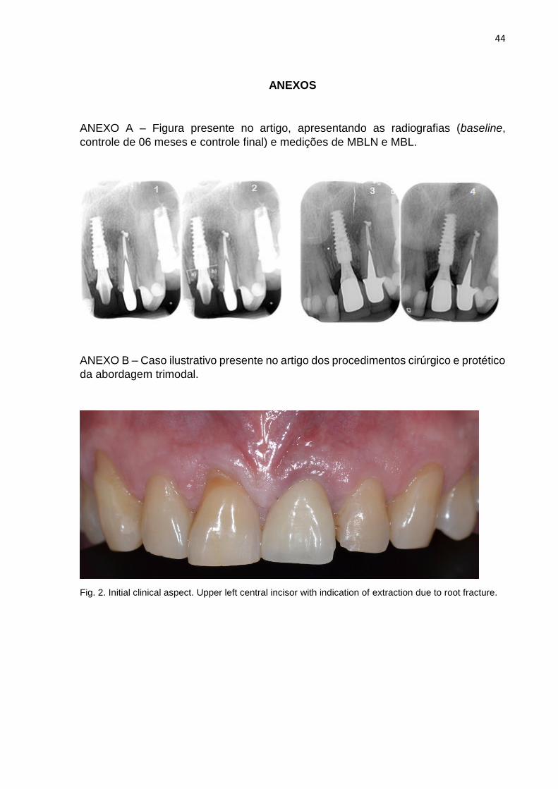
44
ANEXOS
ANEXO A – Figura presente no artigo, apresentando as radiografias (baseline,
controle de 06 meses e controle final) e medições de MBLN e MBL.
ANEXO B – Caso ilustrativo presente no artigo dos procedimentos cirúrgico e protético
da abordagem trimodal.
Fig. 2. Initial clinical aspect. Upper left central incisor with indication of extraction due to root fracture.
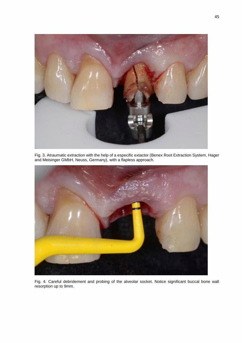
45
Fig. 3. Atraumatic extraction with the help of a especific extactor (Benex Root Extraction System, Hager and Meisinger GMbH, Neuss, Germany), with a flapless approach.
Fig. 4. Careful debridement and probing of the alveolar socket. Notice significant buccal bone wall resorption up to 9mm.
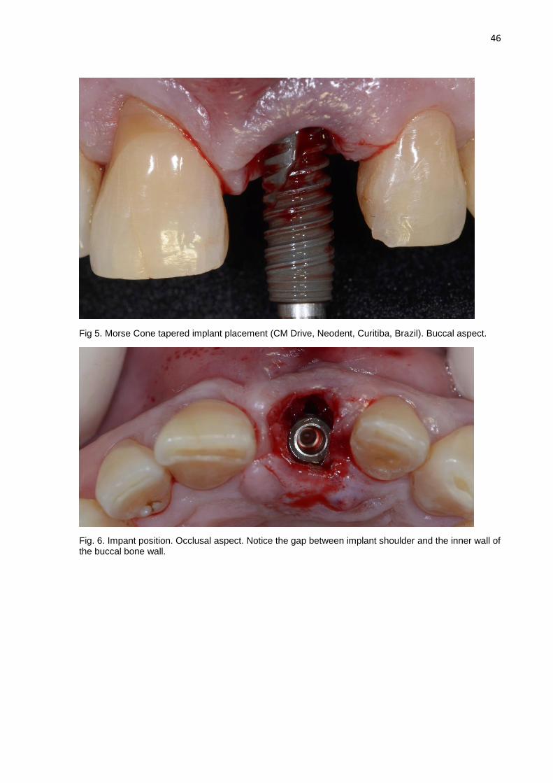
46
Fig 5. Morse Cone tapered implant placement (CM Drive, Neodent, Curitiba, Brazil). Buccal aspect.
Fig. 6. Impant position. Occlusal aspect. Notice the gap between implant shoulder and the inner wall of the buccal bone wall.

47
Fig. 7. Implant platform impression with the help of a rubber dam before gap filling with anorganic bovine bone.
Fig. 8. Gap filling with xenograft bone substitute. Buccal aspect. A narrow healing abutment is tightened into the implant platform until definitive abutment is screwed.

48
Fig. 9. Customized titanium abutment in the master cast before passing through the sterilization process.
Fig. 10. Customized definitive titanium abutment tightened at 20 N/cm. Buccal aspect. Notice the graft material, and its intimate contact with oral environment. These anorganic bovine bone particles will unavoidably be lost, during the first 6 months.

49
Fig. 11. Provisional crown cemented onto the custom abutment within 24 hours post implant placement, avoiding centric and eccentric occlusal contacts. Buccal aspect. Observe the gap between the gingival margin and cervical aspect of the temporary tooth.
Fig. 12. Buccal aspect of the provisional crown 7 days post-surgery. Notice the soft tissue stability, and
how the gingival margin adapted to the surface of the provisional crown.

50
Fig. 13. Coping pick-up silicone impression (Regular Body Normal Set, Elite HD+, Zhermack, Rovigo,
Italy), after 05 months of healing. CAD/CAM machined zirconium oxide reinforced coping.
Fig. 14. Definitive crown delivery. At the 06 months follow-up.
ANEXO C – Tabela I presente no artigo, apresentando a descrição amostral.
Table I. Social and demographic characteristics of implant patients and description of implant surgical
characteristics (n = 20)
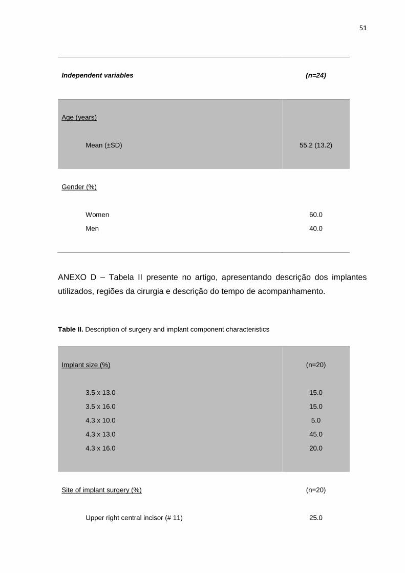
51
Independent variables
(n=24)
Age (years)
Mean (±SD)
55.2 (13.2)
Gender (%)
Women
Men
60.0
40.0
ANEXO D – Tabela II presente no artigo, apresentando descrição dos implantes
utilizados, regiões da cirurgia e descrição do tempo de acompanhamento.
Table II. Description of surgery and implant component characteristics
Implant size (%)
3.5 x 13.0
3.5 x 16.0
4.3 x 10.0
4.3 x 13.0
4.3 x 16.0
(n=20)
15.0
15.0
5.0
45.0
20.0
Site of implant surgery (%)
Upper right central incisor (# 11)
(n=20)
25.0

52
Upper right lateral incisor (#12)
Upper right canine (# 13)
Upper left central incisor (# 21)
Upper left left incisor (#22)
5.0
5.0
45.0
20.0
Follow-up time (years)
Mean (S.D.)
(n=20)
2.26 (1.03)
Follow-up time (%)
1.0 year
1.5 years
2.0 years
2.5 years
3.0 years
3.5 years
4.0 years
4.5 years
(n = 20)
10.5
31.6
21.1
5.3
10.5
10.5
5.3
5.3
ANEXO E – Tabela III presente no artigo, apresentando as razões para extração.
Table III. Reasons for tooth extraction (%)
Reasons for extraction
(n=20)
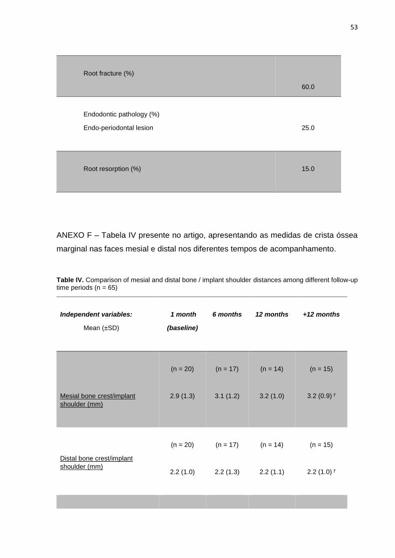
53
Root fracture (%)
60.0
Endodontic pathology (%)
Endo-periodontal lesion
25.0
Root resorption (%)
15.0
ANEXO F – Tabela IV presente no artigo, apresentando as medidas de crista óssea
marginal nas faces mesial e distal nos diferentes tempos de acompanhamento.
Table IV. Comparison of mesial and distal bone / implant shoulder distances among different follow-up time periods (n = 65)
Independent variables:
Mean (±SD)
1 month
(baseline)
6 months
12 months
+12 months
Mesial bone crest/implant
shoulder (mm)
(n = 20)
2.9 (1.3)
(n = 17)
3.1 (1.2)
(n = 14)
3.2 (1.0)
(n = 15)
3.2 (0.9) †
Distal bone crest/implant
shoulder (mm)
(n = 20)
2.2 (1.0)
(n = 17)
2.2 (1.3)
(n = 14)
2.2 (1.1)
(n = 15)
2.2 (1.0) †
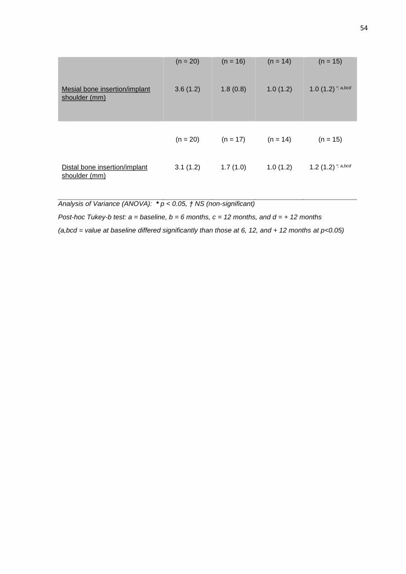
54
Mesial bone insertion/implant
shoulder (mm)
(n = 20)
3.6 (1.2)
(n = 16)
1.8 (0.8)
(n = 14)
1.0 (1.2)
(n = 15)
1.0 (1.2) *; a,bcd
Distal bone insertion/implant
shoulder (mm)
(n = 20)
3.1 (1.2)
(n = 17)
1.7 (1.0)
(n = 14)
1.0 (1.2)
(n = 15)
1.2 (1.2) *; a,bcd
Analysis of Variance (ANOVA): * p < 0.05, † NS (non-significant)
Post-hoc Tukey-b test: a = baseline, b = 6 months, c = 12 months, and d = + 12 months
(a,bcd = value at baseline differed significantly than those at 6, 12, and + 12 months at p<0.05)
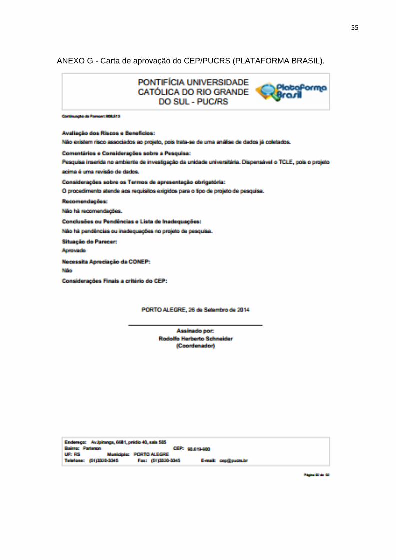
55
ANEXO G - Carta de aprovação do CEP/PUCRS (PLATAFORMA BRASIL).
56
57
ANEXO H - Carta de aprovação da comissão científica e de ética da Faculdade de
Odontologia da PUCRS.
58
ANEXO I – Autorização de uso de arquivo de dados.
59
ANEXO J – Termo de compromisso de emprego de dados.
60
LISTA DE ABREVIATURAS
BA Bimodal approach
CAD/CAM Computed aided designed/Computed aided manufactured
CBCT Cone beam computerized tomography
CEP Comitê de Ética em Pesquisa
MBL Marginal bone level
MBLN Marginal bone level at neighbouring tooth
PUCRS Pontifícia Universidade Católica do Rio Grande do Sul
TA Trimodal approach


