dissertação mestrado laura roesler nery - core.ac.uk · programa de pÓs-graduaÇÃo em biologia...
TRANSCRIPT
PONTIFÍCIA UNIVERSIDADE CATÓLICA DO RIO GRANDE DO SUL - PUCRS FACULDADE DE BIOCIÊNCIAS
PROGRAMA DE PÓS-GRADUAÇÃO EM BIOLOGIA CELULAR E MOLECULAR
LAURA ROESLER NERY
Efeito do tratamento com lítio sobre a habilidade motora e o sistema Wnt-catenina-caderina durante o desenvolvimento inicial de Zebrafish.
PORTO ALEGRE
2011

PONTIFÍCIA UNIVERSIDADE CATÓLICA DO RIO GRANDE DO SUL - PUCRS
FACULDADE DE BIOCIÊNCIAS PROGRAMA DE PÓS-GRADUAÇÃO EM BIOLOGIA CELULAR E MOLECULAR
Efeito do tratamento com lítio sobre a habilidade motora e o sistema Wnt-
catenina-caderina durante o desenvolvimento inicial de Zebrafish.
Aluna: Laura Roesler Nery Orientadora: Drª Mônica Ryff Moreira Roca Vianna
PORTO ALEGRE 2011
Dissertação de mestrado apresentada ao Programa de Pós-Graduação em Biologia Celular e Molecular da Pontifícia Universidade Católica do Rio Grande do Sul.

I
RESUMO
Lítio tem sido o principal tratamento para desordens bipolares desde 1950,
oferecendo um efeito profilático e agudo contra os episódios maníacos e depressivos, apesar
dos desafios encontrados na sua administração durante a gravidez e o período perinatal. Os
mecanismo por trás dos efeitos do lítio ainda não foram descritos completamente mas existem
evidencias de um efeito sobre a via Wnt-ß-catenina devido a sua capacidade de inibição sobre
a GSK-3ß. Neste estudo nós avaliamos os efeitos da exposição ao lítio durante o
desenvolvimento inicial de Zebrafish incluindo caracterizações comportamentais e
moleculares do sistema Wnt-ß-catenina-Caderina. Embriões foram tratados individualmente
por 72hpf com LiCl 0.05, 0.5 e 5 mM. Não foram observadas malformações significativas
nem efeitos embriotóxicos. Em 10 dpf animais tratados com 0.5 e 5 mM de lítio
demonstraram habilidades locomotoras similares a síndrome induzida por lítio em humanos
Floppy baby syndrom. Análises através da técnica de Western blot demonstraram uma
expressão aumentada nos níveis de ß-catenina e diminuída para N-caderina. Animais com
10dpf reestabeleceram seus níveis de expressão proteica de acordo com o controle. Análises
através da técnica de qPCR não demonstraram nenhum efeito sobre os níveis de ß-catenina ou
N-caderina, sugerindo a ocorrência de mudanças pós-transcricionais. Enquanto os níveis de
c-myc não foram alterados, houve uma diminuição nos níveis de Cyclin D1 em larvas de
3dpf, enquanto os níveis de CamkIV ocilaram.
II
ABSTRACT
Lithium has been the mainstream treatment for bipolar disorder since 1950s, offering
unequivocal prophylactic and acute efficacy against maniac and depressive episodes despite
challenges in managing bipolar disorder during pregnancy and the perinatal period. The
mechanisms underlying lithium effects are still elusive but exacerbation of wnt-β-catenin
signaling system due to GSK-3 inhibition is believed to be its main target. In this study we
evaluated effects of lithium exposure during zebrafish early development including behavioral
and molecular characterization of the wnt-β-catenin system. Embryos were individually
treated for 72hpf with LiCl at 0.05, 0.5 and 5mM. No significant malformation and
embryotoxic effects were observed. At 10dpf 0.5 and 5mM lithium-treated larvae showed
decreased locomotory behavior resembling lithium-induced Floppy baby syndrome
neurobehavioral symptoms. Western blot analysis of selected wnt-β-catenin system
components showed an increased β-catenin and decreased N-cadherin protein levels at the
end of treatment unaccompanied by wnt3a changes, reinforcing current view of GSK-3
inhibition as the main mechanism of lithium effects. At 10 dpf protein levels were
reestablished on control levels for all previously treated groups. qPCR analysis of β-catenin
and N-cadherin showed no effects of lithium on these genes expression suggesting protein
changes were due to post-transcriptional events. While c-myc gene expression remained
unaltered during evaluated periods, cyclin d1 was decreased at 3dpf, and CamkIV oscillated.

III
LISTA DE FIGURAS:
FIGURA 01 – Formação tubo neural Zebrafish ..........................................................7
FIGURA 02 – Via Wnt-ß-catenina ..............................................................................9
FIGURA 03 – Esquema Caderinas ............................................................................10
IV
SUMÁRIO
1. CAPÍTULO 1 ................................................................................................................................... 1 1.1 INTRODUÇÃO ...................................................................................................................................................... 2 1.1.1 Lítio ........................................................................................................................................................................... 2 1.1.2 Zebrafish ................................................................................................................................................................. 4 1.1.3 Desenvolvimento do Sistema Nervoso ....................................................................................................... 5 1.1.4 Via de Sinalização Wnt ..................................................................................................................................... 7 1.1.5 Caderinas ................................................................................................................................................................ 9
1.2 JUSTIFICATIVA .................................................................................................................................................... 11 1.3 OBJETIVOS .............................................................................................................................................................. 12 1.3.1 Objetivos Específicos ....................................................................................................................................... 12
2. CAPÍTULO 2 ................................................................................................................................ 13 2.1 ARTIGO CIENTÍFICO ................................................................................................................................................ I
3. CAPÍTULO 3 ................................................................................................................................ 36 3.1 CONSIDERAÇÕES FINAIS ................................................................................................................................. 37
REFERÊNCIAS ................................................................................................................................. 40 ANEXO 01 ........................................................................................................................................ 46

1
1. CAPÍTULO 1
2
1.1 INTRODUÇÃO
1.1.1 Lítio
O impacto de medicamentos e fármacos sobre a fisiologia vai além da
preocupação que estes estejam cumprindo seu papel terapêutico. Estudos avaliando o
potencial tóxico e efeitos secundários adversos de fármacos terapêuticos são cruciais
na avaliação da segurança de seu uso, especialmente durante fases mais vulneráveis
da vida do indivíduo. O desenvolvimento inicial humano em sua fase intrauterina é
extremamente sensível e fármacos utilizados pela gestante podem afetar não apenas a
saúde do embrião/feto momentaneamente, mas comprometê-lo de forma definitiva.
Algumas patologias requerem tratamentos constantes e cujo regime não deve ser
interrompido. Dentre estas, transtornos de humor envolvem um equilíbrio delicado
entre riscos e benefícios do tratamento tanto para as mães quanto para o embrião/feto
(KOZMA 2005).
O lítio vem sendo utilizado como principal fármaco no tratamento de
transtorno bipolar desde 1950 (SALINAS & HALL 1999). Devido à importância do
tratamento psiquiátrico para a saúde e vida das mães, muitas vezes o tratamento com
lítio é mantido, ou mesmo recomendado em substituição ao uso de outros
medicamentos com comprovado efeito teratogênico (KOZMA 2005). Nestes casos os
níveis de lítio durante a gestação e período de amamentação são monitorados.
Apesar dos comprovados efeitos sobre o humor, as ações do lítio sobre o
sistema nervoso variam de neuroproteção a neurotoxicidade (KOZMA 2005). Embora
os dados sejam ainda mais escassos, o efeito da exposição pré- e neo-natal ao lítio
sobre o sistema nervoso, contudo, parece ser mais danosa, conforme revisado por
Kozma (2005). Fazendo uma revisão da literatura de 1978 a 2004 sobre os efeitos
neurotóxicos e neurodesenvolvimentais do lítio, Kozma integrou achados de 30
pacientes que foram expostos ao lítio durante a gestação demonstrando uma série de
prejuízos de distintas etiologias, e reforçando a necessidade de se estabelecer
diretrizes sobre a administração de lítio a gestantes e adequado monitoramento dos
níveis absorvidos pelo embrião/feto.
3
Estudos sobre o potencial teratogênico do lítio são controversos,
especialmente devido à variedade de abordagens usadas e dificuldade de extrapolação
dos achados para humanos. Em humanos, poucos relatos abrangem indivíduos que
tenham sido expostos durante a gestação e analisados subsequentemente e
demonstram que as anormalidades fetais resultantes do uso de lítio pela gestante são
dependentes da frequência e da quantidade absorvida (SMITHBERG & DIXIT 1982;
KOZMA 2005). Dentre as principais alterações descritas em humanos estão padrões
comportamentais alterados, cianose, hipotonicidade, hipotireoidismo, diabetes
insípidus nefrogênica e problemas cardiovasculares (SCHOU & AMDISEN 1973;
SCHOU et al. 1973; SCHOU 1976; NARS & GIRARD 1977; ANANTH 1978;
MIZRAHI, HOBBS & GOLDSMITH 1979; JACOBSON et al. 1992; YONKERS et
al. 2004). Estudos abrangentes e com condições monitoradas são cruciais para se
estabelecer a segurança do tratamento com lítio em gestantes, especialmente devido a
relatos de efeitos teratógenos observados em invertebrados e vertebrados inferiores
(WRIGHT, HOFFMAN & DAVIES 2005). Em ratos Wistar demonstrou-se que o
lítio apresenta embriotoxicidade e teratogenicidade (MARATHE & THOMAS 1986).
A indicação atual de manutenção do tratamento com lítio pela gestante,
acompanhado de monitoramento periódico da concentração plasmática do composto,
é uma estratégia falha na prevenção de danos ao indivíduo em desenvolvimento. Esta
estratégia ainda não é capaz de prever as concentrações de lítio alcançadas durante o
desenvolvimento do novo indivíduo e portanto incapaz de estabelecer concentrações
seguras de exposição (NEWPORT et al. 2005). Devido à passagem do lítio através da
placenta, somada à imaturidade do sistema renal do embrião/feto resultando em uma
meia-vida aumentada do composto, as concentrações de lítio em indivíduos cujas
mães fazem uso do fármaco pode atingir níveis plasmáticos maiores do que aqueles
identificados nas gestantes em um mesmo dado momento (STEVENS, BURMAN &
MIDWINTER 1974).
Adicionalmente, em modelos animais este cátion também se mostrou efetivo
na melhora de quadros associados a acidente vascular cerebral, Doenças de
Alzheimer, Parkinson e Huntington, danos medulares e degeneração da retina (LENG
et al. 2008). Em roedores adultos, o lítio reforça a estabilidade das moléculas
responsáveis pela consolidação da memória, como a β-catenina (MAGUSCHAK &
RESSLER 2008).
A administração de lítio por períodos prolongados em modelos animais
4
durante os processos de desenvolvimento ocasiona anormalidades ontogenéticas no
indivíduo (KLEIN & MELTON 1996). Foi observado que em Xenopus, o lítio causa
uma expansão da mesoderme dorsal levando a uma duplicação do eixo dorsal (KAO,
MASUI & ELINSON 1986). Selderslaghs e colaboradores (2009) comprovaram que
o lítio durante o processo de desenvolvimento embrionário em Zebrafish ocasiona
retardo no desenvolvimento, demora na eclosão, deformidades no esqueleto e redução
da capacidade natatória (SELDERSLAGHS et al. 2009).
Estudos recentes demonstraram que o lítio pode modular a via da Wnt agindo
como um inibidor da GSK-3ß, evitando a fosforilação e degradação da β-catenina.
Graças à ação inibidora do lítio sobre a GSK-3ß ocorre um aumento na concentração
citoplasmática de β-catenina, favorecendo que esta se direcione ao núcleo e ative a
transcrição de genes da via Wnt responsáveis pela migração e proliferação celular
(KLEIN & MELTON 1996; STAMBOLIC, RUEL & WOODGETT 1996; SALINAS
& HALL 1999; CHEN, DING & MACCORMICK 2000; GOULD & MANJI 2005;
STUMP et al. 2006; LENG et al. 2008; DILL et al. 2008).
1.1.2 Zebrafish
Diferentes modelos animais vêm sendo adaptados para estudos que possam,
ao mesmo tempo, diminuir o custo e manter uma complexidade de sistemas que
permita a extrapolação dos achados para outros animais e humanos (LIESCHKE &
CURRIE 2007; SPENCE et al. 2008). O teleósteo Danio rerio, mundialmente
conhecido como Zebrafish foi descrito por Francis Hamilton em 1822. Pertencente à
família Cyprinidae, uma das mais ricas em representantes entre os vertebrados, esta
espécie tem distribuição geográfica endêmica no sul e sudeste da Ásia.
Proposto primeiramente como animal modelo por George Streissinger para
pesquisas em biologia do desenvolvimento, o Zebrafish vem sendo utilizado por
muitos anos, e é um dos principais modelos para estudos em genética, neurofisiologia
e biomedicina (SPENCE et al. 2008). Devido ao seu pequeno porte, chegando no
máximo de 120 milímetros de comprimento, é de fácil armazenamento, permitindo a
acomodação de inúmeros animais em um mesmo aquário. Graças a vantagens como a
5
possibilidade de estudos genéticos em grande escala enquanto mantém a
complexidade típica de um vertebrado, somadas a fácil manipulação e manutenção
quando comparado aos modelos de vertebrados tradicionais, o Zebrafish, vem se
estabelecendo como vantajoso modelo animal.
O Zebrafish se reproduz durante todo o ano, sendo capaz de originar
centenas de ovos transparentes a cada ato reprodutivo. A fertilização e
desenvolvimento exclusivamente externos de ovos relativamente grandes (cerca de
0,7 milímetros de diâmetro) e transparentes tornam a observação e a manipulação
mais fácil do que nos demais animais modelo de vertebrado. Seu desenvolvimento
embrionário completa-se em até 72 horas pós-fertilização (hpf), quando o ovo eclode
dando origem a uma larva (SPENCE et al. 2008), o que facilita o acompanhamento da
evolução dos processos em tempo real.
Kimmel et al. (1995) descreveu os estágios de desenvolvimento do
Zebrafish, desde a formação do zigoto até a eclosão da larva, os estágios larvais e a
formação do indivíduo adulto, possibilitando a identificação das distintas fases de
formação do tecido nervoso.
1.1.3 Desenvolvimento do Sistema Nervoso
Após a fertilização, o embrião passa por processos de intensa proliferação
celular, seguido de migração das novas células até seu comprometimento e
diferenciação terminal. Estes processos que tem início com as clivagens são
intensificados a partir do processo de gastrulação. Durante estes processos, as células
que formam o embrião sofrem alterações tanto em nível de sinalização celular, graças
à expressão de diferentes morfógenos, quanto em nível estrutural, em decorrência da
expressão de distintas moléculas de adesão, por exemplo. Através dessas mudanças e
da histocompatibilidade resultante, as células comprometem-se com diferentes
destinos, dando, assim, origem a três folhetos embrionários: a endoderme, a
mesoderme e a ectoderme.
A embriogênese do sistema nervoso tem início com o processo de
neurulação, no qual, parte da ectoderme de revestimento é internalizada na forma de

6
tubo enquanto células próximas às suas extremidades laterais se desprendem,
originando as células da crista neural (Figura 01), resultando na segregação dos
respectivos precursores do sistema nervoso central e periférico (GILBERT 2000;
GARCIA & GARCÍA 2001; WOLPERT et al. 2008). Durante esse processo, vias de
sinalização celular como a via Wnt, que é responsável por ativar genes responsáveis
pela proliferação e migração celular (GILBERT 2000; WOLPERT et al. 2008), assim
como a expressão de fatores de crescimento, o bloqueamento de BMP (bone
morphogenic protein), o que inibi a transformação da ectoderme em epiderme (DALE
et al. 1992; JONES et al. 1992), e a via notch-delta, responsável por manter as células
sem diferenciação neuronal (GILBERT 2000; WOLPERT et al. 2008), fazem com
que ocorra um crescimento e aumento do número de células assim como um rearranjo
na estrutura tecidual, formando a placa neural e posteriormente o tubo neural, que
dará origem ao sistema nervoso.
Além da formação das células precursoras neurogliais, um evento crucial na
formação da organização regional e periférica do sistema nervoso é o estabelecimento
de sinapses. Este processo mediado por projeções de membrana dos neurônios recém
originados é desempenhado de forma ativa pelo cone de crescimento axonal, que
estabelece os primeiros contatos com os alvos neuronais com os quais poderão ser
estabelecidas sinapses definitivas (GILBERT 2000; WOLPERT et al. 2008;
CASTAÑO, GORDON-WEEKS & KYPTA 2010).
Várias moléculas participam de forma crítica dos processos de proliferação,
migração, comprometimento e diferenciação, e que culminam com o estabelecimento
de sinapses estáveis, condição crítica para a sobrevivência neuronal (GILBERT 2000;
WOLPERT et al. 2008; CASTAÑO, GORDON-WEEKS & KYPTA 2010). Dentre os
ligantes que participam ativamente deste processo estão as moléculas da família Wnt
(TOLEDO, COLOMBRES & INESTROSA 2008; DILL et al. 2008; CHALPE et al.
2010), capazes de modular as atividades de proliferação, diferenciação e
sinaptogênese graças a ativação e repressão de eventos intracelulares específicos
através da modulação da expressão gênica (CLEVERS 2006; DILL et al. 2008;
TOLEDO, COLOMBRES & INESTROSA 2008; SHITASHIGE, HIROHASHI &
YAMADA 2008; CHALPE et al. 2010; VERHEYEN & GOTTARDI 2010).
Moléculas de adesão celular como as Caderinas, por sua vez, desempenham papel
importante no reconhecimento dos terminais sinápticos em formação. Estas proteínas
são conhecidas por serem homofílicas, ligando-se exclusivamente entre elas, o que faz

7
com que o reconhecimento do cone axonal com o alvo seja especifico para a
formação de sinapses (TAKEICHI 1988; TAKEICHI 1995; ALBERTS et al. 2004;
SHAN et al. 2004; LIEN, KLEZOVITCH & VASIOUKHIN 2006; MAGUSCHAK
& RESSLER 2008).
1.1.4 Via de Sinalização Wnt
Wnt ou Wingless são proteínas conservadas evolutivamente responsáveis por
diversos processos durante o desenvolvimento embrionário e a homeostase do
indivíduo adulto. Sabe-se que esta família de proteínas são secretadas de forma
parácrina ou autócrina, possuindo um raio de ação restrito, e regulando processos
celulares variados em seus alvos (CLEVERS 2006; TOLEDO, COLOMBRES &
INESTROSA 2008; VERHEYEN & GOTTARDI 2010).
Existem diferentes genes que codificam as proteínas desta família com
distintos efeitos nas células e tecidos. Krylova e colaboradores (2002) demonstraram
que tanto a Wnt3a quanto a Wnt7a induzem o crescimento e ramificação do cone
axonal em neurônios sensoriais da espinha dorsal em camundongos (HALL, LUCAS
& SALINAS 2000; PURRO et al. 2008; FARÍAS et al. 2010). Já a proteína Wnt4
Figura 01 – Esquema ilustrando a posição da placa neural (do inglês neural plate) e demonstrando a formação do tubo neural (do inglês Neural tube) de vertebrados, o desprendimento das células da crista neural (do inglês neural crest) e a localização da epiderme (do inglês epidermis).
FONTE: Adaptado de WOLPERT, 2008.
8
induz o desenvolvimento do sistema nervoso central, incluindo a formação dos
hemisférios cerebrais em mamiferos (LYUKSYUTOVA et al. 2003; FARÍAS et al.
2010). Wnt5a é responsável pela formação do botão pós-sinaptico e manutenção das
suas funções no Sistema Nervoso Central de camundongos adultos (FARÍAS et al.
2007; FARÍAS et al. 2010).
Essas proteínas já foram caracterizadas em diversos sistemas biológicos,
incluindo camundongo, Drosophila e o Zebrafish. Foi demonstrado que a proteína
Wnt3a é expressa em Zebrafish incluindo tecidos neurais em desenvolvimento, tecto
óptico, cordão espinhal, e partes do encéfalo (CLEMENTS, ONG & TRAVER 2009).
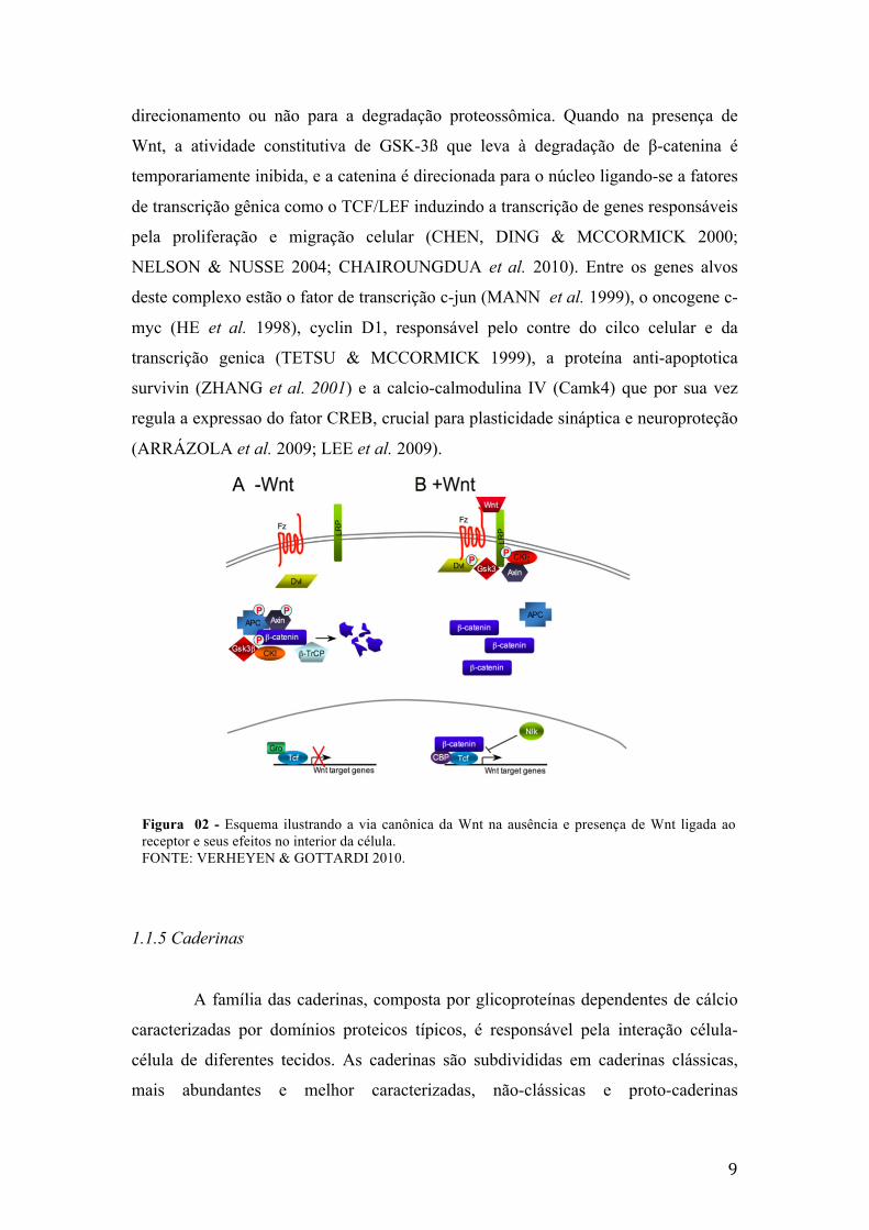
Na ausência de Wnt um complexo formado por Axin, adenomatous polyposis
coli gene product (APC) e Glicogênio Sintase Quinase 3-ß (GSK-3ß) acarreta a
fosforilacao da β-catenina citosólica (LIU et al. 2002), resultando em sua poli-
ubiquitinação e degradação pelo proteassomo (CLEVERS 2006; SHITASHIGE,
HIROHASHI & YAMADA 2008; TOLEDO, COLOMBRES & INESTROSA 2008;
CHAIROUNGDUA et al. 2010; VERHEYEN & GOTTARDI 2010). Desta forma, na
ausência de Wnt os níveis de β-catenina citosolicos são mantidos baixos (Figura 03).
A ligação de Wnt ao seu receptor Frizzled e ao receptor relacionado a
lipoproteína de baixa densidade resulta na ativação da proteína Dishevelled (Dvl) a
qual sequestra os mediadores da ligação da GSK-3ß a β-catenina, Axin e APC,
inibindo a fosforilação da mesma, ocasionando a estabilização e acumulo de β-
catenina no citoplasma (CLEVERS 2006; SHITASHIGE, HIROHASHI &
YAMADA 2008; TOLEDO, COLOMBRES & INESTROSA 2008;
CHAIROUNGDUA et al. 2010; VERHEYEN & GOTTARDI 2010) e ativando a via
canônica da Wnt (Figura 02).
A β-catenina é uma proteína que possui dois principais papéis dentro da
célula, sendo localizada em dois pools intracelulares, um próximo à membrana
plasmática e outro associado a vias de sinalização intracelular que a dirigem ao núcleo
(TOLEDO, COLOMBRES & INESTROSA 2008; FARÍAS et al. 2010). Quando
localizada próxima à membrana celular é responsável pela estabilidade de moléculas
de adesão celular, como as Caderinas, fazendo a conexão entre o domínio
intracelulares das caderinas e o citoesqueleto de actina (ALBERTS et al. 2004;
NELSON & NUSSE 2004; SALINAS & PRICE 2005; LIEN, KLEZOVITCH &
VASIOUKHIN 2006; MAGUSCHAK & RESSLER 2008). O pool
citoplasmático/nuclear varia significativamente em quantidade, dependendo de seu
9
direcionamento ou não para a degradação proteossômica. Quando na presença de
Wnt, a atividade constitutiva de GSK-3ß que leva à degradação de β-catenina é
temporariamente inibida, e a catenina é direcionada para o núcleo ligando-se a fatores
de transcrição gênica como o TCF/LEF induzindo a transcrição de genes responsáveis
pela proliferação e migração celular (CHEN, DING & MCCORMICK 2000;
NELSON & NUSSE 2004; CHAIROUNGDUA et al. 2010). Entre os genes alvos
deste complexo estão o fator de transcrição c-jun (MANN et al. 1999), o oncogene c-
myc (HE et al. 1998), cyclin D1, responsável pelo contre do cilco celular e da
transcrição genica (TETSU & MCCORMICK 1999), a proteína anti-apoptotica
survivin (ZHANG et al. 2001) e a calcio-calmodulina IV (Camk4) que por sua vez
regula a expressao do fator CREB, crucial para plasticidade sináptica e neuroproteção
(ARRÁZOLA et al. 2009; LEE et al. 2009).
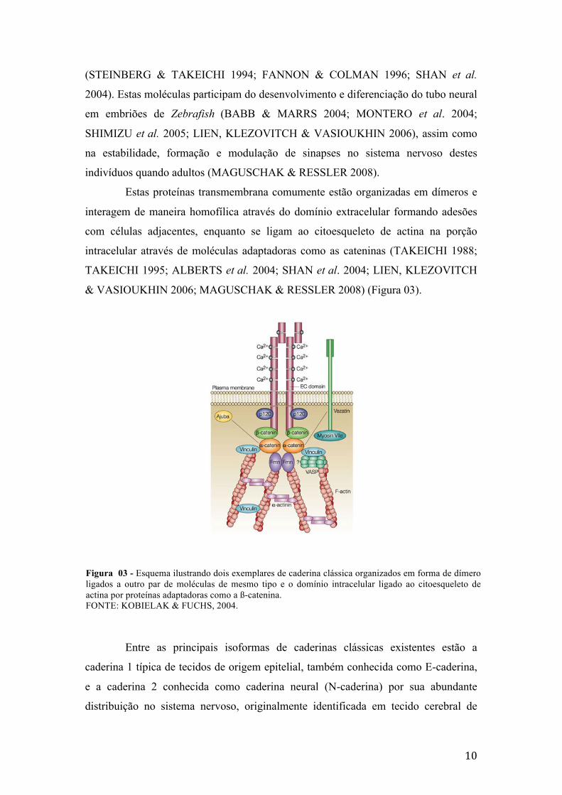
1.1.5 Caderinas
A família das caderinas, composta por glicoproteínas dependentes de cálcio
caracterizadas por domínios proteicos típicos, é responsável pela interação célula-
célula de diferentes tecidos. As caderinas são subdivididas em caderinas clássicas,
mais abundantes e melhor caracterizadas, não-clássicas e proto-caderinas
Figura 02 - Esquema ilustrando a via canônica da Wnt na ausência e presença de Wnt ligada ao receptor e seus efeitos no interior da célula. FONTE: VERHEYEN & GOTTARDI 2010.
10
(STEINBERG & TAKEICHI 1994; FANNON & COLMAN 1996; SHAN et al.
2004). Estas moléculas participam do desenvolvimento e diferenciação do tubo neural
em embriões de Zebrafish (BABB & MARRS 2004; MONTERO et al. 2004;
SHIMIZU et al. 2005; LIEN, KLEZOVITCH & VASIOUKHIN 2006), assim como
na estabilidade, formação e modulação de sinapses no sistema nervoso destes
indivíduos quando adultos (MAGUSCHAK & RESSLER 2008).
Estas proteínas transmembrana comumente estão organizadas em dímeros e
interagem de maneira homofílica através do domínio extracelular formando adesões
com células adjacentes, enquanto se ligam ao citoesqueleto de actina na porção
intracelular através de moléculas adaptadoras como as cateninas (TAKEICHI 1988;
TAKEICHI 1995; ALBERTS et al. 2004; SHAN et al. 2004; LIEN, KLEZOVITCH
& VASIOUKHIN 2006; MAGUSCHAK & RESSLER 2008) (Figura 03).
Entre as principais isoformas de caderinas clássicas existentes estão a
caderina 1 típica de tecidos de origem epitelial, também conhecida como E-caderina,
e a caderina 2 conhecida como caderina neural (N-caderina) por sua abundante
distribuição no sistema nervoso, originalmente identificada em tecido cerebral de
Figura 03 - Esquema ilustrando dois exemplares de caderina clássica organizados em forma de dímero ligados a outro par de moléculas de mesmo tipo e o domínio intracelular ligado ao citoesqueleto de actina por proteínas adaptadoras como a ß-catenina. FONTE: KOBIELAK & FUCHS, 2004.
11
roedores e pintos e que apresenta grande expressão em tecido embrionário
(SHIRAYOSHI et al. 1986; TAKEICHI 1988; HIRAI et al. 1989; NOLLET, KOOLS
& VAN ROY 2000).
Por serem responsáveis pela manutenção da estrutura sináptica, regulação na
composição e intensidade das adesões, as caderinas são candidatos à mediadores de
plasticidade sináptica, estando potencialmente envolvidos na sinaptogênese,
remodelação dos terminais e alterações na eficiência da transmissão (ARIKKATH &
REICHARDT 2008; MENDEZ et al. 2010).
1.2 JUSTIFICATIVA
Devido a capacidade de absorver as substâncias adicionadas ao seu meio, a
complexidade típica de vertebrado e a transparência dos embriões e larvas durante os
rápidos estágios de desenvolvimento do sistema nervoso, o Zebrafish tem sido
apontado como um ótimo modelo animal para estudos de desenvolvimento e
alterações estruturais e funcionais do sistema nervoso, bem como screening de efeitos
farmacológicos.
Neste trabalho propomos o uso do tratamento com lítio durante o período de
desenvolvimento inicial do Zebrafish a fim de caracterizar os efeitos deste íon na
formação do sistema nervoso mediada por caderinas e os potenciais alvos
responsáveis pelas alterações observadas no desenvolvimento.

12
1.3 OBJETIVOS
Caracterizar, em animais expostos a cloreto de lítio durante o
desenvolvimento inicial, a capacidade motora e os efeitos sobre a via Wnt-ß-catenina.
Avaliando a expressão de β-catenina, Wnt e seus genes alvos, buscando relacionar
estes achados com a expressão da molécula de adesão celular N-caderina e a
capacidade natatória dos animais após o tratamento.
1.3.1 Objetivos Específicos
• Avaliar os efeitos embriotoxicológios e teratogênicos durante as 72 horas de
tratamento e até 10 dias pós fertilização;
• Avaliar a habilidade locomotora de larvas de 10 dias submetidas ao tratamento
com lítio;
• Quantificar a expressão de RNA mensageiro, através da técnica de qRT-PCR,
dos genes alvos da Via Wnt em larvas de 3dpf e 10 dpf;
• Quantificar a expressão de proteínas participantes da Via Wnt através da
técnica de Western Blot, em larvas de 3dpf e 10pdf.

13
2. CAPÍTULO 2
i
2.1 ARTIGO CIENTÍFICO
Título: Neurobehavioral effects of early exposure to lithium on zebrafish and the
Wnt-ß-catenin signaling system.
Autores: Laura R. Nery, Talita C. Pereira, Laura D. Guerim, Lídia Martins, Mauricio
R. Bogo and Monica R. Vianna.
Revista: Developmental Biology – Elsevier Science, San Diego, CA, USA.
Número do Manuscrito: DBIO11-189
ii
Neurobehavioral effects of early exposure to lithium on zebrafish and the Wnt-β-catenin signaling system
Laura R. Nery1, Talita C. Pereira2, Laura D. Guerim1, Lídia Martins1, Maurício R.
Bogo2, Monica R. M. Vianna1, 3,4§
1Laboratório de Biologia e Desenvolvimento do Sistema Nervoso, Faculdade de
Biociências, Pontifícia Universidade Católica do Rio Grande, 90619-900, Porto
Alegre, RS, Brazil 2Laboratório de Biologia Genômica e Molecular, Faculdade de Biociências, Pontifícia
Universidade Católica do Rio Grande do Sul, 90619-900, Porto Alegre, RS, Brazil.
3National institute for Translational Medicine (INCT-TM), 90035-003, Porto Alegre,
RS, Brazil 4Zebrafish Neuroscience Research Consortium (ZNRC)
§Corresponding author
Prof. Monica R. M. Vianna, Ph.D.
Correspondence address:
Faculdade de Biociências, Pontifícia Universidade Católica do Rio Grande do Sul
Av. Ipiranga 6681, Prédio 12
90619-900, Porto Alegre, RS, Brazil
Phone: + 55 51 33534743
FAX: + 55 51 33203612
E-mail address: [email protected]
iii
Abstract
Lithium has been the mainstream treatment for bipolar disorder since 1950s, offering
unequivocal prophylactic and acute efficacy against maniac and depressive episodes.
Its use during early pregnancy and the perinatal period remain controversial due to
reports of deleterious effects on the newborn. The mechanisms underlying lithium
therapeutic action are still elusive but exacerbation of wnt-β-catenin signaling
pathway due to GSK-3 inhibition is believed to represent its main target. In this study
we evaluated the effects of lithium exposure during zebrafish embryonic and early
development including behavioral and molecular characterization of the wnt-β-
catenin system. Embryos were individually treated for 72hpf with LiCl at 0.05, 0.5
and 5mM. No significant teratogenic and embryotoxic effects were observed. Western
blot analysis at 3dpf of selected wnt-β-catenin system components showed increased
β-catenin and decreased N-cadherin protein levels at the end of treatment, without
significant changes in wnt3a, reinforcing the current view of GSK-3 inhibition as
lithium’s main target. At 10dpf 0.5 and 5mM lithium-treated larvae showed a dose-
dependent decrease in locomotion, resembling lithium-induced Floppy baby
syndrome neurobehavioral symptoms in humans. At this later period previously
altered proteins returned to control levels in all treated groups, suggesting the
neurobehavioral effects are likely a lasting consequence of lithium exposure. qPCR
analysis of β-catenin and N-cadherin gene expression showed no effects of lithium at
3 or 10dpf, suggesting that protein fluctuations were likely due to post-transcriptional
events. Other gene targets of the wnt signalling pathway were evaluated and only
discrete alterations were observed. While c-myc expression remained unaltered,
cyclin d1 was decreased at 3dpf, and CamKIV oscillated.
Keywords: lithium, motor behavior, β-catenin, N-cadherin, Wnt, zebrafish
iv
1. Introduction
Lithium has been the paradigmatic treatment for bipolar disorder since 1950s,
offering unequivocal prophylactic and acute efficacy against maniac and depressive
episodes, despite the constant monitoring of patients Li+ plasma levels required to
reach effectiveness and avoid toxicity. The management of bipolar disorder during
pregnancy and the perinatal period is specially challenging, and the conflict between
therapy interruption related-relapse and the potential deleterious effects of continued
therapy on development is accentuated by the lack of epidemiological studies
supporting definitive management guidelines (Yonkers et al., 2004).
Li+ is known to equilibrate across the chorionic membranes, and maternal and
infant lithium levels had shown to be directly correlated with higher doses associated
with more prominent perinatal complications (Newport et al., 2005). Lithium
exposure during development has been associated with higher incidence of organ
dysgenesis, in particular with cardiac malformations and Ebstein’s anomaly, as well
as renal and hepatic abnormalities (Schou et al., 1973; Schou and Amdisen, 1973;
Jacobson et al., 1992; Cohen et al. 1994). Endocrinological and metabolic conditions
such as infant diabetes insipidus and hypothyroidism have also been associated with
lithium exposure during pregnancy and breast feeding (Schou, 1976; Nars and Girard,
1977; Ananth, 1978; Mizrahi, Hobbs and Goldsmith, 1979). The relative incidences
of such effects are uncertain, and while more recent epidemiological data indicate that
the teratogenic risk of first-trimester exposure is lower than previously suggested
(Cohen et al., 1994), it is still substantial (Zegers and Andriessen, 2003). More
information regarding lithium’s effects and a better understanding of its mechanisms
are critical to establish safe clinical management guidelines for woman with bipolar
disorder of childbearing age or pregnant, especially during the critical embryological
period (Cohen et al., 1994; Zegers and Andriessen, 2003).
In addition to the morphological and physiological teratogenic potentials
described above, lithium has been associated with neurobehavioral effects in
developing individuals. Collectively described as Floppy baby syndrome, depressed
neurological function and/or neuromuscular deficiencies result in significant lethargy,
hipotonicity, slower reflexes and respiratory difficulties in neonates exposed to
lithium during development (Kozma 2005). The persistence of these effects has not
been critically evaluated, but a developmental delay may be expected and could
impact later life outcome.
v
Despite many decades of clinical use, the molecular targets involved in
lithium mood stabilizing effects are only now being defined (Phiel and Klein, 2001).
Lithium inhibitory effects on inositol monophosphatase (IMPase) and subsequent
reduction in brain inositol were primary candidates to mediate lithium therapeutic
effects, but despite efforts it has not been possible to directly demonstrate its
contribution to mood stabilization (revised in Gould and Manji, 2005). For several
decades, candidate mediators of lithium effects have been proposed, but no causal
relations were established between these targets and lithium’s effects on mood until
recently. In 1996 lithium was shown to inhibit the ubiquitous glycogen synthase
kinase-3 (GSK-3) (Klein and Melton, 1996; Stambolic et al., 1996), and therefore to
potentially affect the vast number of molecular targets this kinase has in neurons and
glia (Jope and Johnson, 2004; Gould and Manji, 2005; Wada, 2009). Subsequent
demonstrations linked lithium therapeutical doses to GSK-3 inhibition and mood
stabilization, confirming GSK-3 as the main candidate do lithium thymoleptic effects
involving modulation of brain plasticity and metabolism, monoaminergic signaling
(Wu and Pan 2009; Wada 2009) and stem cell activity and neuroprotection (Willert et
al. 2003; Toledo et al. 2008).
GSK-3 inhibition can directly mimic and exacerbate Wnt-β-catenin signaling
system activation (Wada, 2009; MacDonald et al., 2009). The Wnt family of secreted
glycoproteins is critically involved in brain development, acting as morphogens and
controlling stem cell proliferation, cell fate determination and differentiation,
migration, growth cone guidance, synaptogenesis and sustained synaptic plasticity
through adulthood (revised in MacDonald et al., 2009; Farías et al., 2010; Inestrosa &
Arenas, 2010). This wide range of effects is mostly achieved by regulation of catenin-
mediated gene expression of DNA-bound T cell factor/lymphoid enhancement factor
(TCF/LEF) family of transcriptional factors in the canonical Wnt pathway.
In this study we evaluated the effects of lithium exposure on zebrafish
development and the potential molecular targets of the wnt-β-catenin system involved
in the characteristic neurobehavioral symptoms of lithium-affected babies observed in
10dpf larvae.
vi
2. Methods
2.1 Animals breeding and maintenance
Zebrafish embryos were obtained from natural mating of adult Danio rerio
(short-fin wild type) bred and maintained in an automated re-circulating tank system
(Tecniplast, Italy). At the night before the breeding, males and females of
reproductive age were placed in a breeding tank (Tecniplast, Italy) in a 2:1 ratio,
respectively, separated by a transparent barrier. At 8 a.m., when the light went on, the
barrier was removed and after 15 min the fertilized eggs were collected, washed with
system water (reverse osmosis water equilibrated with salts) (Westerfield, 2000),
freed of debris and transferred to sterile cell culture 96 wells dishes.
Embryos were individually placed in 96 wells plates (1 embryo for well, n=60
per group in triplicate) and submitted to 72 hours post-fertilization (hpf) treatment
with system water (control groups) or lithium chloride (LiCl) (Synth) at 0.05 mM,
0.5mM and 5.0mM diluted in system water. Plates were maintained in an incubator at
28.5 oC on a 14:10 light/dark cycle and monitored daily.
At 72 hpf, 3 days post-fertilization (dpf) embryos were washed three times and
placed in petry dishes with system water in groups of 30 larvae per dish. They where
fed with paramecia twice daily starting at 4dpf and water was changed daily.
All the experiments were conducted according to the Canadian Council on
Animal Care Guidelines on care and use of fish in research, teaching and testing
(CCAC, 2005), followed Brazilian legislation (no. 11.794/08) and the Brazilian
college of animal experimentation (COBEA) Guidelines (COBEA, 2008). Protocols
were previously evaluated and approved by the Institutional Animal Care Committee
(CEUA PUCRS, protocol 09/00129).
2.2. Embryotoxicity
Relative mortality rate, hatching efficiency and larvae general morphology
were daily monitored during 10dpf, following Sederslaghs et. al. (2009). The hatching
efficiency was observed up to 96hpf. Dead embryos were discarded immediately
when detected. Morphological deformities in embryos or larvae were analyzed under
an inverted microscope (Olympus SZ 4045).
2.3 Exploratory task
vii
At 10dpf larvae were submitted to an exploratory task to evaluate their motor
behavior when placed in a new environment. Animals individually placed in cell-
culture 24 well dishes filled with 3ml of system water had their locomotion and
exploratory behavior video-recorded during 3 min using a digital webcam (Quick cam
pro 9000, Logitech). Data was analyzed using ANYmaze (Stoelting) in a designed
protocol that virtually divided each 15 mm diameter well in central area (7.5 mm
diameter) and periphery. The total travelled distance, number of head turns and
crossings between the well central area and the periphery ring were considered the
most relevant indicators.
2.4 Western Blot analysis
Whole-body tissue from 3 and 10dpf euthanized individuals (n = 15 per
group) were placed in a cooled protease inhibitor solution (Complete Mini, Roche
Applied Science) and stored at -80ºC for subsequent analysis. The protein extract was
prepared in RIPA buffer (Sigma-Aldrich). Twenty-five µg total protein was separated
on a 7.5% SDS-polyacrylamide gel and transferred electrophoretically to a
nitrocellulose membrane. Membranes were blocked with 5% nonfat dry milk in TBS
containing 0.05% Tween-20 and were incubated overnight with the following
antibodies: ß-actin (ab8224, Abcam) at 1:2000, ß–catenin (C2206, Sigma-Aldrich) at
1:5000, N-cadherin (ab12221, Abcam) at 1:2000; Wnt3a (ab28472, Abcam) at
1:2000. Goat anti-mouse (G-21040, Molecular Probes) and goat anti-rabbit (G-21234,
Molecular Probes) horseradish-conjugated secondary antibodies were used and
detected using Western Lighting Western Blot Chemiluminescence (NEL 104001EA,
Perkin Elmer). Pre-stained molecular weight protein markers (Benchmark marker,
Invitrogen) were used to determine the detected bands’ molecular weight and confirm
antibodies target specificity.
Densitometry quantification of each replicated gel (n=3) was performed using
ImageJ software (http://rsb.info.nih.gov/ij/). Total blotting protein levels of were
normalized according to each sample’s ß-actin protein levels.
2.5 Real Time PCR analysis
RNA isolation and cDNA synthesis were performed as described in
Rosemberg and collaborators (2007). In brief, whole 3 and 10 dpf (n=15 in triplicate)

viii
larvae were frozen in liquid nitrogen and RNA was isolated with TRIzol reagent
(Invitrogen) according to the manufacturer’s instruction. RNA purity was quantified
spectrophotometrically calculating the ratio between the absorbance values at 260nm
and 280nm and the samples were tested by electrophoresis in a 1.0% agarose gel with
gelRed nucleic acid stain (Biotium). cDNA species were synthesized with
SuperScriptTMIII First-Strand Synthesis SuperMix (Invitrogen, USA) (Rosemberg
2007).
For all genes, qRT-PCRs were performed using SYBR green (Tang et al.,
2007). Standard reactions (25 µl) were assembled as follows: 4 µl of SYBR green
qPCR supermix-UDG (Invitrogen), 0.25 µl of forward primer (10 µM), 0.25 µl of
reverse primer (10 µM), 0.25µl of dNTPs (10mM), 1.5µl of MgCl2 (50mM), 2.5µl of
PCR buffer (10X), 3.7 µl DEPC water, 0.05µl of Platinum TaqDNA 0.5U
(Invitrogen) and 12.5µl of template. Templates were 1:50 diluted cDNA samples, and
in the case of negative controls, cDNAs were replaced by DEPC water. The primers
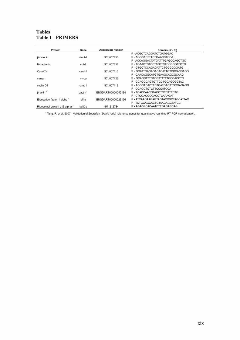
sequences were designed using the program Oligos 9.6 for the following genes:
ctnnb2, cdh2, camk4, myca and cnnd1. The primers for the constitutive genes bactin1,
ef1a and rpl13a (Table 1) were according from Tang (2007). Gene transcripts were
obtained from the NCBI (http://www.ncbi.nlm.nih.gov/Genbank) and Ensembl
databases (http://www.ensembl.org/ Danio_rerio). All real time assays were carried
out in quadruplicate using an Applied Biosystems 7500 real-time PCR system. Forty
amplification cycles were performed, with each cycle consisting of 94ºC for 15
seconds followed by 60ºC for 35 seconds. Amplification and dissociation curves
generated by the software Applied Biosystems 7500 were used for gene expression
analysis.
Cycle-temperature (Ct) values were obtained for each gene. Following the
removal of outliers, raw fluorescence data were exported to the LinRegPCR 12.x
(http://LinRegPCR.HFRC.nl) to determine the PCR amplification efficiency of each
sample. PCR efficiency of each sample, together with Ct values, was used to calculate
a relative gene expression value for each transcript, according to the equation R = (E
ref)CT sample X (E sample)-CT Sample X (E sample)CT Ref X (E ref)-CT Ref , where E refers to
PCR efficiency, Ct is the Ct value for each amplification, Ref is the value of the
reference gene and sample to the gene in question (PFAFFI 2001).
2.6 Statistical analysis
ix
Data was parametrically analyzed using one-way ANOVA followed by Tukey
post-hoc test. The level of significance was considered p<0.05.
3. Results
No significant teratogenic or embryotoxic effects were observed when animals
receiving lithium at 0.05, 0.5 or 5 mM were compared to control groups in the course
of 10 dpf (data not shown). A significant 16% hatching deficiency was observed on
animals treated with 5mM lithium when compared to controls.
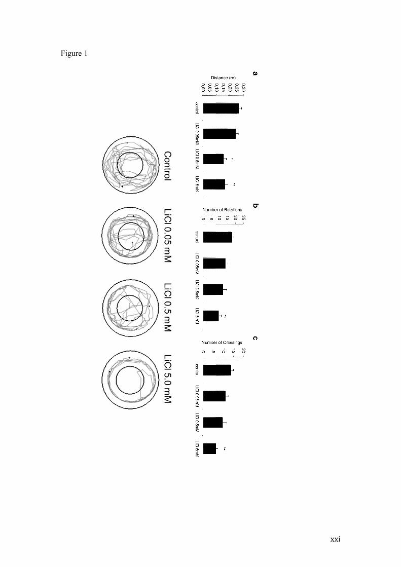
When animals that were exposed to lithium during the initial 3dpf were
behaviorally tested at 10 dpf, (i.e. after 7 days of treatment end), a significant dose-
dependent locomotor deficit was observed (Figure 1). When individually allowed to
explore a 15 mm diameter arena for 3 minutes, individuals treated with lithium at 0.5
and 5 mM travelled shorter distances when compared to untreated control (p<0.05 and
p<0.005, respectively) or 0.05mM lithium animals (p<0.05). The number of body
rotations and crossings between the inner and outer arena areas were also reduced in
animals treated with the higher lithium dose in relation to controls (p<0.005).
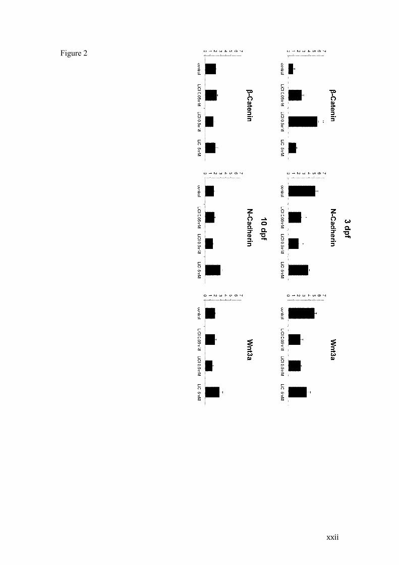
Western blot analysis of β-catenin, N-cadherin and Wnt3a proteins
immediately after the 72 hpf treatment (i.e. at 3 dpf) revealed increased β-catenin
levels on 0.5 mM lithium-treated animals (p< 0.005) when compared to controls
(Figure 2). In contrast, N-cadherin protein levels of 0.05 and 0.5mM lithium-treated
animals were decreased when compared to control group (p<0.05). The higher
treatment dose (5 mM) did not significantly alter β-catenin or N-cadherin levels. We
also evaluated Wnt3a protein levels after lithium treatment, and despite 0.05 and
0.5mM groups lower mean values, no statistically significant effect was observed
when compared to control levels at 3dpf animals.
At 10 dpf, β-catenin, N-cadherin and Wnt3a protein levels were not altered in
relation to control groups (Figure 2).
To better characterize the mechanisms underlying the altered protein levels
observed in the western blot analysis and further explore the consequences of altered
β-catenin and N-cadherin immediately after treatment, we analyzed β-catenin
(ctnnb2) and N-cadherin (cdh2) gene expression at the same time periods. We also
evaluated the transcription levels of three β-catenin target genes: c-myc (myca) and
cyclin-d1 (ccnd1) and CamKIV (camk4) (Figure 3).

x
While expression of β-catenin and N-cadherin genes, evaluated at 3 or 10 dpf,
was not affected by lithium, distinct effects were observed on the β-catenin target
genes. Whereas c-myc remained unaffected on both time periods, cyclin D1
expression was significantly decreased on groups receiving 0.5 and 5 mM lithium at 3
dpf (p<0.05), returning to control levels at 10 dpf. CamKIV expression was increased
after 0.05 mM lithium treatment at 3 dpf (p<0.05), but appeared to be generally
decreased at 10 dpf in relation to control group, being significantly reduced at 0.5 mM
(p<0.01).
4. Discussion
The lithium concentrations chosen in this study were based on a 0.5mM
concentration within the expected therapeutic maintenance blood levels, and included
a lower, mood stabilizing ineffective but potentially neuroprotective dosage (Leng et
al., 2008; Maguschak and Ressler, 2008) and higher, potentially toxic but still
representative of lithium intoxication during therapy (Newport et al., 2005).
The observed absence of significant mortality or gross teratogenic effects of
lithium exposure during early developmental stages are in accordance to lithium’s
current classification as a human low teratogenic agent (Cohen et al., 1994; Gentile,
2011). Previous studies using animal models including mice, rats and rabbits have
shown a higher incidence of lithium-induced teratogenicity than those observed in
humans, what was later attributed to excessively high doses and species specific
sensitivities (discussed by Cohen and collaborators, 1994). The only equivalent study
in zebrafish, performed by Selderslaghs et al. (2009) used broader period and
concentration spectrums of lithium exposure and found similar effects at equivalent
doses to those here presented. Importantly, they only observed significative
embryotoxic effects after 72hpf of 235.9 mM lithium exposure, a concentration that in
no circumstance would be reached on human therapy. Their observation of an 11.1%
hatching deficiency after 72hpf 3.69mM lithium treatment is similar to the 16%
deficit we observed on 5mM treated-group, reinforcing lithium’s potential delaying
development.
Despite its lack of teratogenic effects, and discrepancies in reports of other
lithium-induced malformations, neurobehavioral effects in the newborn have been
repeatedly reported (Kozma 2005). This is, to our knowledge, the first description of
persistent symptoms that resemble the neurodevelopmental deficits including

xi
hypotonicity and lethargy observed in lithium Floppy baby syndrome affected babies
in model organisms. Newport et al. (2005) showed higher rates of CNS and
neuromuscular complications and significantly lower Apgar scores in newborns
exposed to higher lithium therapeutic doses. Animals treated with 0.5 and 5mM
lithium showed impaired locomotion when compared to controls, moving slower,
travelling shorter distances and with deficits in performing body turns. Importantly,
these effects were observed in animals exposed to lithium concentrations within the
lower limit of the therapeutic dose range (approximately 0.4-1.2mM). The
neurodevelopmental deficits observed in human babies are commonly attributed to an
acute effect of lithium intoxication during delivery, and there is no significant
longitudinal studies evaluating symptoms resumption. Our results suggest that the
persistent neurobehavioral effects may be a lasting consequence of lithium chronic
exposure during the early developmental state.
GSK-3 inhibition can impact several signaling pathways, including growth
factors cascades, but its most profound and direct impacts at therapeutic range is
believed to be on Wnt-β-catenin system (Wada, 2009; MacDonald et al., 2009).
The Wnt family of secreted glycoproteins is critically involved in brain
development and function through life, fundamentally directing cell proliferation,
determination and differentiation, migration, growth cone guidance and
synaptogenesis and plasticity (revised in MacDonald et al., 2009 and Farías et al.,
2010). This wide range of effects is mostly achieved by regulation of catenin-
mediated gene expression of DNA-bound T cell factor/lymphoid enhancement factor
(TCF/LEF) family of transcriptional factors in the canonical Wnt pathway (revised in
MacDonald et al., 2009).
Wnts are evolutionary conserved, and family members have complex and
specific features not yet fully understood. Wnt3a was the first characterized Wnt
protein in mammals, mostly due to its controlled secretion (Willert et al., 2003). In
zebrafish the Wnt3 ortholog is broadly expressed in the developing nervous system
beginning at the tailbud stage with a distribution pattern that closely resembles that of
other vertebrates and capable of activating the Wnt canonical pathway (Clements et
al., 2009). As reviewed in detail by MacDonald et al. (2009) and other recent
publications (Clevers, 2006), the Wnt canonical pathway is activated when Wnt binds
to frizzled (Fz or FZd) receptor and its low-density lipoprotein receptor-related
protein co-receptor (LPR), forming a complex that recruits the scaffolding protein

xii
disheveled (Dvl) and alters the activation state of the a multiple-protein complex
called “Axin- or β-catenin destruction- complex” that includes another scaffolfing
protein, Axin, the tumor suppressor adenomatous polyposis coli gene product (APC),
the casein-kinase 1 (CK1) and GSK-3. The Wnt-induced complex recruitment
prevents GSK-3 phosphorilation of β-catenin residues and subsequent ubiquitination
and rapid proteosomal degradation (Clevers, 2006; MacDonald et al., 2009). β-catenin
has two intracellular pools, one anchoring the cell adhesion molecules Cadherins at
the membrane, and a rapid turn-over citoplasmatic pool, that is prevented from
moving to the nucleus in the absence of Wnt. β-catenin stabilization results in higher
nuclear levels, association to TCF/LEF transcription factors and activation of gene
expression. Wnt vast and constantly growing list of target genes are also diverse and
context-specific (http://www.stanford.edu/~rnusse/Wntwindow.html), including cell
cycle regulators that mediate proliferation, such as cyclin d1, oncogenes as c-myc and
proteins related to migration and synaptic plasticity such as the CamKIV which is
associated to synaptic plasticity and neuroprotection (He et al., 1998; Tetsu and
McCormick, 1999; Arrázola et al., 2009; Lee et al., 2009;).
Lithium-mediated GSK inhibition also prevents β-catenin degradation and
therefore mimics and potentially exacerbates Wnt effects during development.
Aiming to better understand the underlying molecular network involved in lithium
developmental effects, we analyzed protein and gene expression levels of the Wnt-
catenin system components at the end of the 72 hpf treatment, i.e. at 3dpf, and again
at 10 dpf, when motor deficits were observed.
As expected, at 3dpf, 0.5mM lithium treatment induced a robust increase in β-
catenin protein levels, suggesting the exacerbation of the already active proliferative
and migratory scenario expected at this early developmental stage period. This anti-
apoptotic context may disfavor synaptic adhesion, justifying the reduced N-cadherin
protein levels in 0.5mM treated animals despite β-catenin abundance. Gene
expression of β-catenin and N-cadherin measured by qPCR were not altered at this
time period, suggesting the protein level changes probably result from post-
transcriptional events that may include a reduced degradation of β-catenin due to
GSK-3 inhibition and either translation repression or active degradation of previously
existing N-cadherin. Surprisingly, the higher treatment dose (5mM lithium) did not
alter the measured parameters. Finally, despite a tendency that may reflect an
endogenous negative feedback mechanism on Wnt secretion (Chairoungdua et al.,
xiii
2010), lithium did not significantly alter total Wnt3a protein level. Interestingly, at
10dpf, when the behavioral impact of lithium treatment was evident on larvae motor
behavior, no changes in protein or gene expression of β-catenin, N-cadherin or Wnt3a
was observed in relation to control groups.
Finally, the expression of the three chosen β-catenin system target genes at
3dpf were not markedly altered, but cyclin-d1 was statistically decreased in 0.5 and
5mM Li-treated animals in relation to controls, while CamKIV was increased at the
0.05mM concentration. Cyclin d1 is considered a key sensor and integrator of
extracellular signals, modulating chromatin condensation of genes associated to cell
proliferation and differentiation. Cyclin d1 has also been shown to regulate cell-
matrix interaction independently of its effect on cycle progression reciprocally
modulating migration through integrin and growth factor signaling (reviewed in Fu et
al., 2004). Curiously, the CamKIV increased gene expression was the only effect of
0.05mM lithium concentration observed in this study, a dosage in which lithium may
be affecting more significatively other targets than GSK-3 (Phiel and Klein, 2001).
Lithium is known to have neuroprotective effects under low concentrations, and
CamKIV is known to be involved in CREB phosphorylation and neuronal survival
(Mbebi et al., 2002). Accordingly, Arrazola et al. (2009) showed that Wnt3a and
lithium increase CamKIV gene expression and protects hippocampal neurons against
Alzheimer’s amyloid peptide tocixity. The reduced CamKIV levels observed at 10dpf
in lithium-treated animals are intriguing, but may be related to a different scenario
due to the later developmental period, in which cell proliferation and survival are
probably less active in favor or other cellular events.
Other signaling systems than those related to GSK-3-inhibition may be
involved in the persistent behavioral effects observed at 10dpf and a growing list of
candidates are to be explored. It should also be considered that other targets of GSK-3
might account, at least partially, to the observed behavioral effects, and deserve to be
investigated. Finally, despite wnt signaling system proeminence in the nervous system
(Inestrosa & Arenas, 2010) the contribution of other tissues to the neurobehavioral
and molecular effects of lithium cannot be ruled out without further analysis.
Conclusions
Our study reinforced previous evidence of lithium low teratogenic and
embryotoxic effects within therapeutical ranges in humans and animal models
xiv
including zebrafish. Also in accordance to literature on the neurodevelopmental
effects of lithium exposed babies, we have observed a prominent neuromuscular
deficit in 10dpf larvae exposed to lithium during early developmental period at the
0.5mM concentration, which is within the lower levels of the therapeutic range. This
is, to our knowledge, the first description of persistent symptoms that resemble the
lithium Floppy baby syndrome in animal model organism. This effect is likely due to
GSK inhibition and corroborated by increased β-catenin protein levels at the end of
lithium treatment but the underlying cellular and molecular players affected remain to
be fully characterized. Importantly, at 10dpf when the neurobehavioral effects were
observed, lithium had already washed-out and molecular makers returned to basal
levels, suggesting a permanent detrimental effect of lithium early exposure. Further
investigations should focus both on the vast and constantly growing list of wnt- β-
catenin system targets as in other signaling systems potentially affected by lithium.
Competing interests
There are no competing interests
Acknowledgements
This work was supported by CNPq (Conselho Nacional de Pesquisa e
Desenvolvimento 567483/2008-8) and by DECIT/SCTIE-MS through CNPq and
Fundação de Amparo à Pesquisa do Estado do Rio Grande do Sul (FAPERGS, Proc.
10/0055-0 and 10/0036-5). We are thankful for received fellowships (M.R.V.: CNPq
305060/2009-0; L.M.: CNPq 504508/2007-5; L.R.N.: CAPES).

xv
References
Ananth, J., 1978. Side effects in the neonate from psychotropic agents excreted
through breast-feeding. Am. J. Psychiatry. 135, 801-805.
Arrázola, M.S., Varela-Nallar, L., Colombres, M., Toledo, E.M., Cruzat, F., Pavez,
L., Assar, R., Aravena, A., González, M., Montecino, M., Maass, A., Martínez,
S., Inestrosa, N.C., 2009. Calcium/Calmodulin-dependent protein kinase type
IV is a target gene of the Wnt/ß-catenin signaling pathway. J. Cell. Physiol. 221,
658-667.
CCAC guidelines on: the care and use of fish in research, teaching and testing.
Ottawa: Canadian Council on Animal Care, 2005, 94p.
Chairoungdua, A., Smith, D.L., Pochard, P., Hull, M., Caplan, M.J., 2010. Exosome
release of ß-catenin: a novel mechanism that antagonizes Wnt signaling. J. Cell.
Biol. 190, 1079-1091.
Clements, W.K., Ong, K.G., Traver, D., 2009. Zebrafish Wnt3 is expressed in
developing neural tissue. Dev. Dyn. 238, 1788-1795.
Clevers, H., 2006. Wnt/ß-catenin signaling in development and disease. Cell. 125,
469-480
COBEA, Colégio Brasileiro de Experimentação Animal. Estatuto.
http://www.cobea.org.br
Cohen, L.S., Friedman, J.M., Jefferson, J.W., Johnson, E.M., Weiner, M.L., 1994. A
reevaluation of rsk iof in utero exposure of lithium. JAMA 271,146-150
Farias, G.G., Godoy, J.A., Cerpa, W., Varela-Nallar, L., Inestrosa, N.C., 2010. Wnt
signaling modulates pre- and postsynaptic maturation: Therapeutic
Considerations. Dev. Dyn. 239, 94-101.
Fu, M., Wang, C., Li, Z., Sakamaki, T., Pestell, R. G., 2004. Minireview: Cyclin D1:
Normal and Abnormal Functions. Endocrinology 145, 5439-5447.
Gentile, S. 2011. Drug treatment for mood disorders in pregnancy. Curr. Opin.
Psychiatry. 24, 34-40.
Gould, T.D., Manji, H.K., 2005. Glycogen synthase kinase-3: a putative molecular
target for lithium mimetic drugs. Neuropsychopharmacology 30, 1223-1237.
He, T.C., Sparks, A.B.; Rago, C., Hermeking, H., Zawel, L., da Costa, L.T.; Morin,
P.J., Vogelstein, B., Kinzler, K.W., 1998. Identification of c-myc as a target of
the APC pathway. Science 281, 1509-1512.
xvi
Inestrosa, N.C., Arenas, E. 2010. Emerging roles of wnts inthe adult nervous system.
Nature Rev. Neurosci. 11, 77-86.
Jacobson, S.J., Jones, K., Johnson, K., Ceolin, L., Kauer, P., Sah, K., Konnenfeld,
A.E., Rieder, M., Santelli, R., Smythe, J., Pastuszak, A., Einarson, T., Koren,
G., 1992. Prospective multicentre study of pregnancy outcome after lithium
exposure during first trimester. Lancet 339, 530-533.
Jope, R.S., Johnson, G.V.W., 2004. The glamour and gloom of glycogen synthase
kinase-3. Trends Biochem. Sci. 29, 95-102.
Klein, P.S., Melton, D.A., 1996. A molecular mechanism for the effect of lithium on
development. Proc. Natl. Acad. Sci. U.S.A. 93, 8455-8459.
Kozma, C., 2005.Neonatal Toxicity and Transient Neurodevelopmental Deficits
Following Prenatal Exposure to lithium: Another Clinical Report and a Review
of the Literature American Journal of Medical Genetics. Am. J. Med. Genet.
132, 441-444.
Lee, K.H., Chatila, T.A., Ram, R.A., Thompson, R.F., 2009. Impaired memory of
eyeblink conditioning in CaMKIV KO mice. Behav. Neurosci. 123, 438-442.
Leng, Y., Liang, M., Ren, M., Marinova, Z., Leeds, P., Chuang, D., 2008. Synergistic
Neuroprotective Effects of lithium and Valproic Acid or Other Histone
Deacetylase Inhibitors in Neurons: Roles of Glycogen Synthase Kinase-3
Inhibition. J. Neurosci. 28, 2576-2588.
MacDonald, B.T., Tamai, K., He, X., 2009. Wnt/β-Catenin Signaling: Components,
Mechanisms, and Diseases. Dev. Cell 17, 9-26.
Maguschak, K.A., Ressler, K.J., 2008. ß-catenin is required for memory
consolidation. Nat. Neurosci. 11, 1319-1326.
Mbebi, C., Sée, V., Mercken, L., Pradier, L., Müller, U., Loeffler, J.P., 2002. Amyloid
precursor protein family-induced neuronal death is mediated by impairment of
the neuroprotective calcium/calmodulin protein kinase IV-dependent signaling
pathway. J. Biol. Chem. 277, 20979-20990.
Mizrahi, E.M., Hobbs, J.F., Goldsmith, D.I., 1979. Nephrogenic diabetes insipidus in
transplacental lithium intoxication. J. Pediatr. 94, 493-495.
Nars, P.W., Girard, J., 1977. Lithium carbonate intake during pregnancy leading to
large goiter in a premature infant. Am. J. of Disease Children, 131, 924-925.

xvii
Newport, J., Viguera, A.C., Beach, A.J., Ritchie, J.C., Cohen, L.S., Stowe, Z.N.,
2005. Lithium placental passage and obstetrical outcome: Implications for
clinical management during late pregnancy. Am. J. Psychiatry. 162, 2162-2170.
Pfaffi, M.W., 2001. A new mathematical model for relative quantification in real-time
PT-PCR. Nucleic Acid Res. 29, 2003-2007.
Phiel, C., Klein, P., 2001. Molecular targets of lithium action. Annu. Rev. Pharmacol.
Toxicol. 41, 789–813.
Rosemberg, D.B., Rico, E.P., Guidoti, M.R., Dias, R.D., Souza, D.O., Bonan, C.D.,
Bogo, M.R., 2007. Adenosine deaminase-release genes: Molecular
identification, tissue expression. Pattern and truncated alternative splice isoform
in adult Zebrafish (Danio rerio). Life Sci. 81, 1526-1534.
Schou, M., 1976. What happened later to the lithium babies? A follow up study of
children born without malformations. Acta Psychiatr. Scand. 54, 193-197.
Schou, M., Amdisen, A., 1973. Lithium and pregnancy, 3: lithium ingestion by
children breast-fed by women on lithium treatment. Br. Med. J. 2, 138.
Schou, M., Goldfield, M.D., Weinstein, M.R., Villeneuve, A., 1973. Lithium and
pregnancy, I: report from the Register of lithium Babies. Br. Med. J. 2, 135-136.
Sederslaghs, I.W.T., Van Rompay, A.R., De Coen, W., Witters, H.E., 2009.
Development of a screening assay to identify teratogenic and embryotoxic
chemicals using the zebrafish embryo. Repro. Toxicol. 3, 308-20.
Stambolic, V., Ruel, L., Woodget, J.R., 1996. Lithium inhbits glycogen synthase
kinase-3 activity and mimics Wingless signaling in intact cells. Curr. Biol. 6,
1664-1668.
Tang, R., Dodd, A., Lai, D., McNabb, W., Love, D., 2007. Validation of Zebrafish
(Danio rerio) reference genes for quantitative real-time RT-PCR normalization.
Acta Biochim. Biophys. Sin. 38, 384-90
Tetsu, O., McCormick, F., 1999. Β-catenin regulates expression of cyclin D1 in colon
carcinomas cells. Nature. 398, 422-426.
Toledo, E.M., Colombres, M., Inestrosa, N.C., 2008. Wnt signaling in
neuroprotection and stem cell differentiation. Prog. Neurobiol. 86, 281-296.
Wada, A., 2009. Lithium and Neuropsychiatric Therapeutics: Neuroplasticity via
Glycogen Synthase Kinase-3β, β-Catenin, and Neurotrophin Cascades. J.
Pharmacol. Sci. 110, 14-28.

xviii
Westerfield, M. 2000. The zebrafish book. A guide for the laboratory use of zebrafish
(Danio rerio). Eugene: University of Oregon Press.
Willert, K., Brown, J.D., Danenberg, E., Duncan, A.W., Weissman, I.L., Reya, T.,
Yates, J.R. 3rd., Nusse, R., 2003. Wnt proteins are lipid-modified and can act as
stem cell growth factors. Nature. 423, 448-452.
Wu, D., Pan, W., 2009 GSK3: a multifaceted kinase in Wnt signalling. Trends
Biochem. Sci. 35, Issue 3, 161-168.
Yonkers, K.A., Wisner, K.L., Stowe, Z.; Leibenluft, E., Cohen, L., Miller, L.,
Manber, R., Viguera, A., Suppes, T., Altshuler, L., 2004. Management of
bipolar disorder during pregnancy and the postpartum period. Am. J. Psychiatry.
161, 608-620.
Zegers, B., Andriessen P. Maternal lithium therapy and neonatal morbidity. Eur. J.
Pediatr. 162, 348-349.
xix
Tables Table 1 - PRIMERS
Protein Gene Accession nunber Primers (5' - 3')F - ACGCTCAGGATCTGATGGACR - AGGCACTTTCTGAACCTCCAF - ACCAGGACTATGATTTGAGCCAGCTGCR - TGAACTCTCCTATGTCTCCGGGATGTGF - GTGCTCCAGAGATTCTGCGGGGATGR - GCATTGAGAGACACATTGTCCCACCAGGF - CAACAGGCATGTGAAGCAGCGCAAGR - GCAGCTTTCTCGTTATTTGCGACCTCF - GCAGGCAGTGTTGCTGCAGCGGTACR - AGGGTCACTTCTGATGACTTGCGAGAGGF - CGAGCTGTCTTCCCATCCAR - TCACCAACGTAGCTGTCTTTCTGF - CTGGAGGCCAGCTCAAACATR - ATCAAGAAGAGTAGTACCGCTAGCATTACF - TCTGGAGGACTGTAAGAGGTATGCR - AGACGCACAATCTTGAGAGCAG
NC_007118cyclin D1
!-actin *
Elongation factor 1 alpha *
Ribosomal protein L13 alpha *
cnnd1
bactin1
ef1a
rpl13a
* Tang, R. et al. 2007 - Validation of Zebrafish (Danio rerio) reference genes for quantitative real-time RT-PCR normalization.
!-catenin ctnnb2 NC_007130
NC_007131cdh2N-cadherin
CamKIV camk4 NC_007116
NC_007126mycac-myc
NM_212784
ENSDART00000023156
ENSDART00000055194

xx
Figure legends
Figure 1 - Behavioral effects of lithium exposure at 10dpf
Locomotion and exploratory effects of lithium exposure on 10dpf larvae.
Representative track-plots and behavior quantification (mean +- SEM) of distance
traveled during 3 minutes (a); number of rotations higher than 90º degrees (b);
number of crossings between the inner and outer zones of the circular arena (c). **
p<0.01 and * p<0.05 in relation to Control group using One-way ANOVA followed
by Tukey post-hoc.
Figure 2 - Western blot analysis of β-catenin, N-cadherin and Wnt3a of 3dpf
and 10dpf larvae exposed to lithium during the initial 72 hpf
Columns indicate mean +- SEM values of β-catenin, N-cadherin and Wnt3a protein
levels in relation to ß-actin of 3dpf and 10 dpf individuals. ** indicates p<0.01 and *
p<0.05 compared to Control by One-way ANOVA followed by Tukey post-hoc.
Figure 3 - Relative mRNA expression of β-catenin (ctnnb2), N-cadherin (cdh2),
C-myc (myca), cyclin D1 (cnnd1) and CamK IV (camk4) gene expression of 3dpf
and 10dpf larvae exposed to lithium during the initial 72 hpf. mRNA amount
normalized to the average of 3 reference genes (β-actin, Ef1α and Rpl13α). Graphic
representation based on specific sample efficiency independently and in
quadruplicate. * indicates p<0.05 compared to Control by One-way ANOVA
followed by Tukey post-hoc.
xxi
Figure 1
xxii
Figure 2
xxiii
Figure 3

36
3. CAPÍTULO 3

37
3.1 CONSIDERAÇÕES FINAIS
A exposição ao lítio durante o desenvolvimento é associada à incidência de
malformações em humanos, como anomalias cardíacas e de Ebstein, renais e
hepáticas, além de alterações endocrinológicas e metabólicas como diabetes insípidus
e hipotireoidismo (SHOU et al. 1973; SHOU 1976; NARS & GIRARD 1977;
ANANTH 1978; MIZRAHI, HOBBS & GOLDSMITH 1979; JACOBSON et al.
1992). Além destes efeitos, alterações neurocomportamentais e motoras são
comumente observadas em crianças que foram expostas ao lítio durante o
desenvolvimento intrauterino. Estas alterações incluem letargia, hipotonicidade,
reflexos lentos, e dificuldades respiratórias e são comumente descritas como Floppy
baby syndrome, sendo atribuídas disfunções neurológicas e neuromusculares
(KOZMA 2005). Apesar de prevalentes pouco se sabe sobre os mecanismos destas
alterações sob ponto de vista tecidual, celular e molecular, e por consequência não é
conhecida a evolução e eventuais consequências destas manifestações no
desenvolvimento e amadurecimento neural.
Estudos anteriores com outros animais modelo incluindo ratos, camundongos
e coelhos, utilizaram doses muito acima daquelas do espectro terapêutico e
encontraram alterações teratogênicas não compatíveis com as da literatura em
humanos. Neste estudo, onde utilizamos doses dentro do espectro terapêutico
administrado em humanos, observamos uma baixa taxa de efeitos teratogênicos, o que
corrobora com os dados em humanos, que levaram à atual classificação do lítio como
um agente de baixa teratogenicidade apesar das discrepâncias nos dados
epidemiológicos disponíveis (COHEN et al. 1994; GENTILE 2011). Estes achados
reforçam o potencial do Zebrafish como animal modelo para screenings de fármacos
com potenciais efeitos teratogênicos.
O único trabalho anterior em Zebrafish estudando eventuais efeitos
teratogênicos, desenvolvido por Selderslaghs e colaboradores (2009) encontrou
resultados equivalentes aos por nós encontrados, como baixa teratogenicidade e
ausência de mortalidade nas doses até 5mM, e uma deficiência na capacidade de
eclosão de 11,1% na dose de 3.69 mM, o que corrobora os nossos achados na dose de
5mM (16%).
38
Observamos através de análises comportamentais um déficit na habilidade
locomotora dos animais tratados com 0.5 mM e 5 mM de lítio, com redução na
distância percorrida, número de rotações e cruzamentos entre o centro e periferia da
arena. É importante ressaltar que estes achados foram observados no limite mais
baixo das concentrações sanguíneas de pacientes que utilizam o lítio como
medicamento (variando entre 0.4 e 1.2 mM), demonstrando que este agente pode
afetar significativamente a capacidade natatória dos animais.
Como o lítio afeta a atividade da enzima GSK-3ß inibindo a degradação de
β-catenina, também avaliamos as alterações moleculares desta via. Como esperado
em 3 dpf, houve um aumento nos níveis da proteína β-catenina, sugerindo um
aumento na capacidade migratória e proliferativa das células neste período de
desenvolvimento. Neste cenário migratório e anti-apoptótico, foi observada uma
diminuição nos níveis de N-caderina. Chalpe e colaboradores (2010) demonstraram
que a expressão de caderinas do tipo 7 e 11 é regulada pela via canônica da Wnt em
células da crista neural de galinha quando tratadas com o ligante Wnt3a. Foi
observado um aumento na expressão destas moléculas de adesão pró-migratórias. Os
autores propõe que com a ativação de genes que aumentam a migração e proliferação
celular, estas moléculas de adesão estariam sendo mais expressas na presença de um
ativador da via canônica, como a Wnt3a, e que moléculas como a N-caderina e a E-
caderina (anti-migratórias) estariam sendo inibidas ou degradadas para a facilitação
deste processo. Foi demonstrado que a proteína Wnt3a regula a expressão de E-
caderinas em tecido epitelial em camundongos (JAMORA 2003) e também induz a
transição epitélio-mesenquimal inibindo a expressão de E-caderina, proteína
característica de tecido epitelial (BERGE et al. 2008).
Os níveis de expressão gênica, medidos através de qPCR das moléculas de β-
catenina e N-caderina não foram alterados, demonstrando que as alterações na
quantidade de proteínas são devido a modificações pós-transcricionais. Devido à
exposição ao lítio, podemos supor que o aumento da concentração citoplasmática de
β-catenina foi devido a inibição da GSK-ß3 ou degradação proteica ativa de N-
caderinas pré-existentes. Apesar dos níveis de Wnt3a apresentarem um perfil
refletindo um possível mecanismo de retro-inibição, não foi observada diferença
significativa na quantidade de proteína Wnt3a durante o tratamento com lítio.
Após 10 dpf não foi observada nenhuma diferença significativa nos níveis
proteicos desses alvos em relação ao controle, indicando que apesar das alterações na
39
capacidade motora destes animais foi afetada pela desregulação proteica durante o
período do tratamento.
Juntamente com a análise dos genes de β-catenina e N-caderina, avaliamos a
expressão de três genes alvos da via Wnt. Nenhum dos três genes teve sua expressão
alterada significativamente. Foi observada uma diminuição estatisticamente diferente
nas doses de 0.5 e 5 mM em relação ao controle para o gene de Cyclin D1. Isso pode
ser devido a atividade de regulação da matriz extracelular, independente do seu efeito
no ciclo celular, aumentando a capacidade migratória das células através de fatores de
crescimento como as integrinas, o que possivelmente estaria diminuindo a expressão
deste gene (FU et al. 2004). Ao contrário da expressão de Cyclin D1, os níveis
aumentados de CamKIV foram o único efeito deste tratamento na dose de 0.05 mM,
uma dose que potencialmente não teria nenhum efeito sobre a GSK-3ß (PHIEL &
KLEIN 2001) mas como demonstrado anteriormente, o lítio possui ação
neuroprotetora em doses baixas o que poderia estar envolvido nos efeitos da CamKIV
com a fosforilação de CREB e também na sobrevivência neuronal (MBEBI & SÉE
2002). Estudos relacionam o aumento de CamKIV com a atividade da Wnt3a e do
lítio, protegendo neurônios hipocampais adultos contra proteína β-amiloide,
causadora da doença de Alzheimer (ARRÁZOLA et al. 2009). Em outro momento
(10 dpf), onde os processos de migração e proliferação celular não estão mais tão
ativos, os níveis de CamKIV apresentaram-se diminuídos, demonstrando a capacidade
desta proteína de se relacionar com outras vias além da Wnt.
Estes dados reforçam a utilização do Zebrafish como animal modelo
adequado para avaliar eventuais potenciais deletérios de fármacos sobre o
desenvolvimento, e demonstram, pela primeira vez, a possibilidade de caracterização
de fenótipos similares aos de crianças com Floppy baby syndrome em decorrência da
exposição ao lítio. Estudos futuros que se dediquem á investigação de mais alvos
dentre as centenas de genes cuja regulação é modulada pela via Wnt são necessários
para a identificação dos mediadores dos efeitos encontrados. Adicionalmente outras
vias potencialmente afetadas pelo lítio nas concentrações terapêuticas testadas devem
ser exploradas.
40
REFERÊNCIAS
ALBERTS, B.; JOHNSON, A.; LEWIS, J.; RAFF, M.; ROBERTS, K.; WALTER, P. Biologia Molecular da Célula. Porto Alegre: Artmed, 2004. 1463p. ANANTH, J. Side effects in the neonate from psychotropic agents excreted through breast-feeding. The American Journal of Psychiatry, v. 135, p. 801-805, 1978. ARIKKATH, J.; REICHARDT, L.F. Cadherins and catenins at synapses: roles in synaptogenesis and synaptic plasticity. Trends in Neuroscience, v. 31, n. 9, p. 487-494, 2008. ARRÁZOLA, M.S.; VARELA-NALLAR, L.; COLOMBRES, M.; TOLEDO, E.M.; CRUZAT, F.; PAVEZ, L.; ASSAR, R.; ARAVENA, A.; GONZÁLEZ, M.; MONTECINO, M.; MAASS, A.; MARTÍNEZ, S.; INESTROSA, N.C. Calcium/Calmodulin-dependent protein kinase type IV is a target gene of the Wnt/ß-catenin signaling pathway. Journal of Cellular Physiology, v.221, p.658-667, 2009. BABB, S.G., MARRS, J.A. E-Cadherin Regulates Cell Movements and Tissue Formation in Early Zebrafish Embryos. Developmental Dynamics, v. 230, p. 263-277, 2004. BERGE, D.T.; KOOLE, W.; FUERER, C.; FISH, M.; EROGLU, E.; NUSSE, R. Wnt signaling mediates self-organization anda xis formation in embryoid bodies. Cell Stem Cell, v.3, n.5, p. 508-518, 2008. CASTAÑO, Z.; GORDON-WEEKS, P.R.; KYPTA, R.M. The neuron-specific isoform of glycogen synthase kinase-3ß is required for axon growth. Journal of Neurochemistry, v. 113, p. 117-130, 2010. CHAIROUNGDUA, A.; SMITH, D.L.; POCHARD, P.; HULL, M.; CAPLAN, M.J. Exosome release of ß-catenin: a novel mechanism that antagonizes Wnt signaling. Journal of Cell Biology, v. 190, n. 6, p. 1079-1091, 2010. CHALPE, A.J.; PRASAD, M.; HENKE, A.J.; PAULSON, A.F. Regulation of cadherin expression in the chicken neural crest by the Wnt/ß-catenin signaling pathway. Cell Adhesion & Migration, v.4, n. 3, p. 431-438, 2010. CHEN, R.; DING, W.V.; MACCORMICKS, F.. Wnt Signaling to ß-catenin Involves Two Interactive Components. The Journal of Biological Chemistry, v.275, n.23, p. 17894-17899, 2000. CLEMENTS, W.K.; ONG, K.G.; TRAVER, D. Zebrafish wnt3 is expressed in developing neural tissue. Developmental Dynamics, v. 238, p. 1788-1795, 2009. CLEVERS, H. Wnt/ß-catenin signaling in development and disease. Cell, v. 125, p. 469-480, 2006. COHEN, L.S.; FRIEDMAN, J.M.; JEFFERSON, J.W.; JOHNSON, E.M.; WEINER, M.L. A reevaluation of risk of in utero exposure of lithium. JAMA, v. 271, p. 146-150, 1994.
41
DALE, L.; HOWES, G.; PRICE, B.M.J.; SMITH, J.C. Bone morphogenetic protein 4: A ventralizing factor in early Xenopus development. Development, v. 115, p.573-585, 1992. DILL, J.; WANG, H.; ZHOU, F.; LI, S. Inactivation of glycogen synthase kinase 3 promotes axonal growth and recovery in the CNS. The Journal of Neuroscience, v.28, n.36, p. 8914-8928, 2008. FANNON, A.M.; COLMAN, D.R. A model for central synaptic junctional complex formation based on the differential adhesive specificities of the cadherins. Neuron, v.17, n.3, p. 423-434, 1996. FARÍAS, G.G; VALLÉS, A.S; COLOMBRES, M; GODOY J.A; TOLEDO, E.M; LUKAS, R.J; BARRANTES, F.J; INESTROSA, C.N. Wnt-7a induces pre-synaptic colocalization of alpha7-nicotinic acetylcholine receptors and adenomatous polyposis coli in hippocampal neurons. Journal of Neuroscience, v. 27, p. 5313-5325, 2007. FARÍAS, Ginny G.; GODOY, Juan A.; CERPA, Waldo; VARELA-NALLAR, Lorena; INESTROSA, Nibaldo C. Wnt signaling modulates pre- and postsynaptic maturation: Therapeutic Considerations. Developmental dynamics, v. 239, p. 94-101, 2010. FU, M.; WANG, C.; LI, Z.; SAKAMAKI, T.; PESTELL, R. G. Minireview: Cyclin D1: Normal and Abnormal Functions. Endocrinology, v. 145, p. 5439-5447, 2004. GARCIA, S.M.L.; GARCÍA, C.F. Embriologia. Porto Alegre: Artmed, 2001. 416p. GENTILE, S. Drug treatment for mood disorders in pregnancy. Curr Opin Psychiatry, v. 24, p. 34-40, 2011. GILBERT, S.F. Developmental Biology. Sunderland: Sinauer Associates, 2000. Disponível em : < http://www.ncbi.nlm.nih.gov/books> Acesso em : 20 de setembro de 2008. GOULD, T.D.; MANJI, H.K. Glycogen synthase kinase-3: a putative molecular target for lithium mimetic drugs. Neuropsychopharmacology, v. 30, p. 1223-1237, 2005. HALL, A.C; LUCAS, F.R; SALINAS, P.C. Axonal remodeling and synaptic differentiation in the cerebellum is regulated by Wnt-7a signaling. Cell, v. 100, p. 525-535, 2000. HAMILTON, F. An Account of the Fishes Found in the River Ganges and its Branches. Edinburg: Archibald constable and company, 1822. 405 pp.
HE, T.C.; SPARKS, A.B.; RAGO, C.; HERMEKING, H.; ZAWEL, L.; DA COSTA, L.T.; MORIN, P.J.; VOGELSTEIN, B.; KINZLER, K.W. Identification of c-myc as a target of the APC pathway. Science, v. 281, n. 5382, p. 1509-1512, 1998.
HIRAI, Y.; NOSE, A.; KOBAYASHI, S.; TAKEICHI, M. Expression and role of E- and P-cadherin adhesion molecules in embryonic histogenesis. Development, v. 105, p. 263-270, 1989.

42
JACOBSON, S.J.; JONES, K.; JOHNSON, K.; CEOLIN, L.; KAUER, P.; SAHN, K.; KONNENFELD, A.E.; RIEDER, M.; SANTELLI, R.; SMYTHE, J.; PASTUSZAK, A.; EINARSON, T.; KOREN, G. Prospective multicentre study of pregnancy outcome after lithium exposure during first trimester. Lancet, v. 339, p. 530-533, 1992 JAMORA, C.; DASGUPTA, R.; KOCIENIEWSKI, P.; FUCHS, E. Links between signal transduction, transcription and adhesion in epithelial bud development. Nature, v. 422, n. 6929, p. 317-322, 2003. JONES, C.M.; LYONS, K.M.; LAPAN, P.M.; WRIGHT, C.V.E.; HOGAN, B.L.M. DVR-4 (bone morphogenetic protein-4) as a posterior ventralizing factor in Xenopus mesoderm induction. Development, v. 115, p.639-647, 1992. KAO, K.R.; MASUI, Y.; ELINSON, R.P. Lithium-induced respecification of pattern in Xenopus laevis embryos. Nature, v. 322, n. 6077, p. 371-373, 1986. KIMMEL, C.B.; BALLARD, W.W.; KIMMEL, S.R.; ULLMANN, B.; SCHILLING, T.F. Stages of Embryonic Development of the Zebrafish. Developmental dynamics, v.203, p. 253-310, 1995. KLEIN, P.S.; MELTON, D.A. A molecular mechanism for the effect of lithium on development. Proccedings of the National Academy of Sciences of the United States of America, v.93, n.16, p.8455-8459, 1996. KOBIELAK, A.; FUCHS, E. α-catenin: at the junction of intercellular adhesion and actin dynamics. Nature Reviews Molecular Cell Biology, v. 5, p. 614-625, 2004. KOZMA, C. Neonatal Toxicity and Transient Neurodevelopmental Deficits Following Prenatal Exposure to Lithium: Another Clinical Report and a Review of the Literature American Journal of Medical Genetics. American journal of medical genetics, v. 132, p. 441-444, 2005. KRYLOVA, O; HERREROS, J; CLEVERLEY, K.E; EHLER, E; HENRIQUEZ, J.P; HUGHES, S.M; SALINAS, P.C. Wnt-3, expressed by motoneurons, regulates terminal arborization of neurotrophin-3-responsive spinal sensory neurons. Neuron, v. 35, p. 1043-1056, 2002. LEE, K.H.; CHATILA, T.A.; RAM, R.A.; THOMPSON, R.F. Impaired memory of eyeblink conditioning in CaMKIV KO mice. Behavior Neuroscience, v. 123, n. 2, p. 438-442, 2009. LENG, Y.; LIANG, M.; REN, M.; MARINOVA, Z., LEEDS, P.; CHUANG, D. Synergistic Neuroprotective Effects of Lithium and Valproic Acid or Other Histone Deacetylase Inhibitors in Neurons: Roles of Glycogen Synthase Kinase-3 Inhibition. The Journal of Neuroscience, v. 28, n. 10, p. 2576-2588, 2008. LIEN, W.; KLEZOVITCH, O.; VASIOUKHIN, V. Cadherin-catenin proteins in vertebrates development. Courrent Opinion in Cell Biology, v. 18, p. 499-506, 2006. LIESCHKE, G.L.; CURRIE, P.D. Animal models os human disease: zebrafish swim into view. Nature Reviews, v. 8, p. 353-367, 2007.

43
LIU, C.; LI, Y.; SEMENOV, M.; HAN, C.; BAEG, G.H.; TAN, Y.; ZHANG, Z.; LIN, X.; HE, X. Control of ß-catenin phosphorylation/degradation by a dual-kinase mechanism. Cell, v. 108, p. 837-847, 2002. LYUKSYUTOVA, A.I; LU, C.C; MILANESIO, N; KING, L.A; GUO, N; WANG, Y; NATHANS, J; TESSIER-LAVIGNE, M; ZOU, Y. Anterior-posterior guidance of commissural axons by Wnt-frizzled signaling. Science, v. 302, p. 1984-1988, 2003. MAGUSCHAK, K.A.; RESSLER, K.J. ß-catenin is requird for memory consolidation. Nature Neuroscience, v. 11, n. 11, p.1319-1326, 2008 MANN, B.; GELOS, M.; SIEDOW, A.; HANSKI, M.L.; GRATCHEV, A.; ILYAS, M.; BODMER, W.F.; MOYER, M.P.; RIECKEN, E.O.; BUHR, H.J.; HANSKI, C. Target genes of beta-catenin-T cell-factor/lymphoid-enhancer-factor signaling in human colorectal carcinomas. The Proceedings of the National Academy of Sciences USA, v.96, n. 4, p. 1603-1608, 1999. MARATHE, M.R.; THOMAS, G.P. Embryotoxicity and teratogenicity of lithium carbonate in Wistar rat. Toxicology Letters, v. 34, n. 1, p. 115-120, 1986. MBEBI, C.; SÉE, V.; MERCKEN, L.; PRADIER, L.; MÜLLER, U.; LOEFFLER, J.P. Amyloid precursor protein family-induced neuronal death is mediated by impairment of the neuroprotective calcium/calmodulin protein kinase IV-dependent signaling pathway. The Journal of Biological Chemistry, v. 23, p. 20979-90, 2002. MENDEZ, P.; DE ROO, M.; POGLIA, L.; KLAUSER, P.; MULLER, D. N-cadherin mediates plasticity-induced long-term spine stabilization. Journal of Cell Biology, v. 139, n. 3, p. 589-600, 2010. MIZRAHI, E.M.; HOBBS, J.F.; GOLDSMITH, D.I. Nephrogenic diabetes insipidus in transplacental lithium intoxication. The Journal of Pediatrics, v. 94, n. 3, p. 493-495, 1979. MONTERO, J.; CARVALHO, L.; WILSCH-BRAUNINGER, M.; KILIAN, B.; MUSTAFA, C.; HEISENBERG, C. Shield formation at the onset of zebrafish gastrulation. Development, v. 132, p.1187-1198, 2004. NARS, P.W.; GIRARD, J. Lithium carbonate intake during pregnancy leading to large goiter in a premature infant. American Journal of Disease of Children, v. 131, n. 8, p. 924-925, 1977. NELSON, W.J.; NUSSE, R. Convergence of Wnt, ß-catenin, and Cadherin Pathways. Science, v.303, p. 1483-1487, 2004. NEWPORT, J.; VIGUERA, A.C.; BEACH, A.J.; RITCHIE, J.C.; COHEN, L.S.; STOWE, Z.N. Lithium placental passage and obstetrical outcome: Implications for clinical management during late pregnancy. American Journal of Psychiatry, v.162, p.2162-2170, 2005.
44
NOLLET, F.; KOOLS, P.; VAN ROY, F. Phylogenetic analysis of the cadherin superfamilyallows identification of six major subfamilies besides several solitary members. Journal of Molecular Biology, v. 299, p. 551-572, 2000. PURRO, S.A; CIANI, L; HOYOS-FLIGHT, M; STAMATAKOU, E; SIOMOU, E; SALINAS, P.C. Wnt regulates axon behavior through changes in microtubule growth directionality: a new role for adenomatous polyposis coli. Journal of Neuroscience, v. 28, p. 8644-8654, 2008. PHIEL, C.; KLEIN, P. Molecular targets of lithium action. Annual Review of Pharmacology and Toxicology, v. 41, p. 789–813, 2001. SALINAS, P.C.; HALL, A.C. Lithium and synaptic plasticity. Bipolar Disorders, v.2, p. 87-90, 1999. SALINAS, P.C.; PRICE, S.R. Cadherins and catenins in synapse development. Courrent Opinion in Neurobiology, v. 15, p. 73-80, 2005. SCHOU, M. What happened later to the lithium babies? a followu study of children born without malformations. Acta Psychiatrica Scandinavica, v. 54, n. 3, p. 193-197, 1976. SCHOU, M.; AMDISEN, A. Lithium and pregnancy, 3: lithium ingestion by children breast-fed by women on lithium treatment. British Medical Journal, v. 2, p. 138, 1973. SCHOU, M.; GOLDFIELD, M.D.; WEINSTEIN, M.R.; VILLENEUVE, A. Lithium and pregnancy, I: report from the Register of Lithium Babies. British Medical Journal, v. 2, p. 135-136, 1973. SELDERSLAGHS, I.W.T.; VAN ROMPAY, A.R.; DE COEN, W.; WITTERS, H.E. Development of a screening assay to identify teratogenic and embryotoxic chemicals using the zebrafish embryo. Reproductive Toxicology, in press, 2009. SHAN, W.; YAGITA, Y.; WANG, Z.; KOCH, A.; SVENNINGSEN, A.F.; GRUZGLIN, E.; PEDRAZA, L.; COLMAN, D.R. The minimal essential unit for cadherin-mediated intercellular adhesion comprises extracellular domains 1 and 2. Journal of Biological Chemistry, v. 279, n. 53, p. 55014-55923, 2004. SHIMIZU, T.; YABE, T.; MURAOKA, O.; YONEMURA, S.; ARAMAKI, S.; HATTA, K.; BAE, Y.; NOJIMA, H.; HIBI, M. E-cadherin is required for gastrulation cell movements in zebrafish. Mechanisms of Development, v. 122, p. 747-763, 2005. SHIRAYOSHI, Y.; HATTA, K.; HOSODA, M.; TSUNASAWA, S.; SAKIYAMA, F.; TAKEICHI, M. Cadherin cell adhesion molecules with distinct binding specificities share a common structure. The EMBO Journal, v. 5, n. 10, p. 2485 -2488, 1986. SHITASHIGE, M.; HIROHASHI, S.; YAMADA, T. Wnt signaling inside the nucleus. Cancer Science, v. 99, n. 4, p. 631-637, 2008. SMITHBERG, M.; DIXIT, P.K. Teratogenic effects of lithium in mice. Teratology, v.26, n.3, p.239-246, 1982.
45
SPENCE, R.; GERLACH, G.; LAWRENCE, C.; SMITH, C. The behavior and ecology of the zebrafish, Danio rerio. Biological Reviews of the Cambridge Philosophical Society, v. 83, n.1, p. 13-34, 2008. STAMBOLIC, V.; RUEL, L.; WOODGET, J.R. Lithium inhbits glycogen synthase kinase-3 activity and mimics Wingless signaling in intact cells. Current Biology, v.6, n.12, p. 1664-1668, 1996. STEINBERG, M.S.; TAKEICHI, M. Experimental specification of cell sorting, tissue spreading, and specific spatial patterning by quantitative differences in cadherin expression. Proceeding of the National Academy of Sciences of the United States of America, v.91, n.1 p.206-209, 1994. STEVENS, D.; BURMAN, D.; MIDWINTER, A. Letter: Transplacental lithium poisoning. Lancet, v. 2, n. 7880, p. 595, 1974. STUMP, R.J.W.; LOVICU, F.J.; ANG, S.; PANDEY, S.L.; MCAVOY, JW. Lithium stabilizes the polarized lens epithelial phenotype and inhibits proliferation, migration, and epithelial mesenchymal transition. Journal of Pathology, v.210, p. 249-257, 2006. TAKEICHI, M. The cadherins: cell-cell adhesion molecules contrilling animal morphogenesis. Development, v. 102, p.639-655, 1988. TAKEICHI, M. Morphogenetic roles of classic cadherins. Courrent Opinion in Cell Biology, v.7, n.5, p.619-627, 1995. TETSU, O.; MCCORMICK, F. Beta-catenin regulates expression of cyclin D1 in colon carcinomas cells. Nature, v. 398, n. 6726, p. 422-426, 1999. TOLEDO, E.M.; COLOMBRES, M.; INESTROSA, N.C. Wnt signaling in neuroprotection and stem cell differentiation. Progress in Neurobiology, v. 86, p. 281-296, 2008. VERHEYEN, E.M.; GOTTARDI, C.J. Regulation of Wnt/ß-catenin signaling by protein kinases. Developmental Dynamics, v. 239, p. 34-44, 2010. WOLPERT, L.; JESSEL, T.; LAWRENCE, P.; MEYEROWITZ, E.; ROBERTSON, E.; SMITH, J. Princípios de Biologia do Desenvolvimento. Porto Alegre: Artmed, 2008. 576p. WRIGHT, T.L.; HOFFMAN, L.H.; DAVIES, J. Teratogenic effects of lithium in rats. Teratology, v. 4, n. 2, p. 151-155, 2005. YONKERS, K.A.; WISNER, K.L.; STOWE, Z.; LEIBENLUFT, E.; COHEN, L.; MILLER, L.; MANBER, R.; VIGUERA, A.; SUPPES, T.; ALTSHULER, L. Menagement of bipolar disorder during pregnancy and the postpartum period. American Journal of Psychiatry, v. 161, p. 608-620, 2004. ZHANG, T.; OTEVREL, T.; GAO, Z.; EHRLICH, S.M.; FIELDS, J.Z.; BOMAN, B.M. Evidence that APC regulates surviving expression: a possible mechanism contributing to the stem cell origin of colon cancer. Cancer Research, v. 61, n. 24, p. 8664-8667, 2001.
46
ANEXO 01