curso de pós-graduação em patologia humana e experimental · com hu não interferiu na...
TRANSCRIPT
Curso de Pós-Graduação em Patologia Humana e Experimental
TESE DE DOUTORADO
HIDROXIUREIA NA DOENÇA FALCIFORME: INFLUÊNCIA DE
POLIMORFISMOS NOS GENES MPO E SERPINA1 E IMPLICAÇÕES
NAS VIAS DOS RECEPTORES TIPO TOLL E DO INFLAMASSOMA
THASSILA NOGUEIRA PITANGA
Salvador – Bahia 2015
UNIVERSIDADE FEDERAL DA BAHIA FACULDADE DE MEDICINA
FUNDAÇÃO OSWALDO CRUZ CENTRO DE PESQUISAS GONÇALO MONIZ
Curso de Pós-Graduação em Patologia Humana e Experimental
HIDROXIUREIA NA DOENÇA FALCIFORME: INFLUÊNCIA DE
POLIMORFISMOS NOS GENES MPO E SERPINA1 E IMPLICAÇÕES
NAS VIAS DOS RECEPTORES TIPO TOLL E DO INFLAMASSOMA
THASSILA NOGUEIRA PITANGA
Orientadora: Profa Dra Marilda de Souza Gonçalves
Tese apresentada ao Curso de Pós-Graduação em Patologia Humana e Experimental para obtenção do grau de Doutora.
Salvador – Bahia 2015


Trabalho realizado no Laboratório de Hematologia, Genética e Biologia
Computacional (LHGB) - Centro de Pesquisas Gonçalo Moniz (CPqGM/Fiocruz-Ba),
após aprovação dos projetos de pesquisa no Comitê de Ética em Pesquisa (CAAE
0022.0.225.000-09/CAAE: 04733612.7.0000.0040), com auxílio financeiro do CNPq,
FAPESB, PPSUS/FAPESB e INCT do sangue.

“A adversidade tem o efeito de despertar talentos que em circunstâncias prósperas
teriam continuado adormecidos”.
Quintus Horatius Flaccus
AGRADECIMENTOS
Foi árduo chegar até aqui! Só eu sei quantas pedras encontrei no caminho.
Mas também sei que este momento só foi possível porque tive pessoas
maravilhosas que caminharam junto comigo, fazendo-me olhar sempre para frente,
não me permitindo fraquejar.
Agradeço imensamente...
A Deus......meu amigo, meu pai, meu mentor! Ao senhor devo absolutamente
tudo! Laus deo.
Aos meus pais, Abílio César e Martha Pitanga, pelos melhores ensinamentos
que uma pessoa pode receber, e por nunca me permitirem esquecer o lado mais rico
da vida: a humildade. Sempre agradecerei a vocês por terem me ensinado a ser
tudo o que sou. Amo vocês!
Aos meus irmãos, Patrícia, Viviane e Neto pela amizade, amor e cuidado!
À minha linda família, pelo amor e carinho eternos. Minha eterna gratidão à
pessoa mais linda do mundo: minha avó Lourdes! Obrigada por todas aquelas noites
de muitas leituras. Com a senhora aprendi não só o gosto pela leitura, mas a ser
questionadora, estudiosa, o que resultou nesta neta aqui que sempre quer aprender
mais e mais. Amo-te infinitamente, vó!
A Tio Luiz Almeida (in memoriam), tia Lúcia Cortizo e família. Obrigada pelo
carinho, cuidado e por acreditarem tanto em mim. Amo muito vocês!
À minha encantadora e maravilhosa orientadora, Dra. Marilda Gonçalves.
Falei várias vezes, e repito quantas vezes for preciso. Esse amor que sinto vai além
da relação aluno-professor. É amor de filha! Te amo, pró! Obrigada por acreditar
tanto em mim. Obrigada por realizar meu sonho! OBRIGADA!
A Ricardo Riccio, não apenas pelas grandes contribuições científicas e
intelectuais, mas por ter colocado toda a sua energia nesse último ano do meu
doutorado a fim de que eu continuasse acreditando que seria possível chegar até
aqui. Teria sido muito difícil sem o seu apoio! Eternamente grata a você!

À minha querida Dra. Valéria Borges, pelo eterno carinho e amizade, e pelas
discussões científicas sempre enriquecedoras.
À Dra. Tânia Barros, a pessoa que me iniciou na pesquisa, lá em 2005, e a
quem sempre serei eternamente grata por isso.
Aos amigos queridos, Carolzinha, Ray, Sânzio e Dalila! Definitivamente, sem
vocês eu não teria conseguido. Valeu por todas as madrugadas de experimentos e
discussões sobre cada resultado. Obrigada pela parceria, meus amigos!
A todos do LPBM, LHGB e LPA, em especial, Dr. Luiz Alcântara, Dr. Vitor
Maffili, Dra. Márcia Weber, Dra. Magda Seixas, Dra. Cyntia, Ms. Júnia, Ms. Milena,
Camila, Luciana e Corynne. O apoio de vocês foi muito importante.
Aos meus grandes amigos, os irmãos que escolhi, o meu mais puro e
verdadeiro amor de irmã...vocês sabem quem são!!!
Aos meus queridos parceiros, Wendell Vilas Boas, Bruno Cerqueira e Filipe
Rego. Obrigada por todos os ensinamentos, brincadeiras, discussões sobre o
trabalho (e sobre a vida) e, acima de tudo, pela amizade. Sinto uma saudade imensa
do nosso maravilhoso convívio! Amo vocês, meus amigos.
A Dr. Ronald Blanton e família, bem como à sua equipe (Lúcio, Walter, Peace,
João, Rafael e Sandya), por terem me acolhido tão bem no laboratório, em
Cleveland-Ohio! Obrigada pelos ensinamentos!
A Lúcio Barbosa, e à sua linda família que tanto amo, pela eterna amizade e
parceria! Agradecerei eternamente por ter me mostrado esse mundo da pesquisa
sem fronteiras, e por todos os dias você ter tentado me ensinar a “sorrir, respirar e ir
devagar”. Ainda estou aprendendo!
Aos meus grandes e eternos parceiros da B05! Em especial: Viviane, Luciana,
Felipe, Thayna e Rodrigo! Como sinto falta da convivência com vocês!
Aos amigos Kiyoshi Fukutani, Everton Batista, Tiago Landim, Antônio Muniz,
Jessica Petrilli, Silvana Paz e Liliane Cunha por estarem sempre dispostos a me
ajudar.
Aos parceiros Sérgio, Jane, Elze, Rita e Jurema, pela ajuda, amizade e
ótimas conversas durante todo o doutorado. O que vocês fizeram por mim vai muito

além da simples ajuda na esterilização dos materiais ou questões burocráticas da
secretaria e administração. Obrigada!
Aos colegas de trabalho do ICS/UFBA, pela torcida e apoio. Obrigada pelo
carinho!
A todos os meus alunos que torceram muito por esse momento. Vocês foram
a força motriz para a realização desse sonho! Espero um dia poder retribuir todo
esse carinho.
À Faculdade de Farmácia/UFBA, à HEMOBA, aos técnicos de enfermagem e
à Dra. Valma Lopes, a qual me permitiu trabalhar com os pacientes.
A toda equipe da biblioteca, local onde passei boa parte do meu tempo
pesquisando e desenvolvendo essa tese.
A todos os membros da Pós-graduação pela ajuda nas questões burocráticas
referentes ao Doutorado;
Ao CPqGM / FIOCRUZ, pela estrutura física e pessoal que proporcionaram a
realização deste trabalho;
A Coordenação de Aperfeiçoamento de Pessoal de Nível Superior do
Ministério da Educação – CAPES, e ao Conselho Nacional de Desenvolvimento
Científico e Tecnológico – CNPq, pelo apoio financeiro.
E, por fim, meu eterno agradecimento aos pacientes e voluntários que confiaram em mim e participaram desse estudo, permitindo, portanto, a realização de cada experimento.
Que meus resultados venham para melhorar a vida de vocês, os verdadeiros merecedores de todas as honras desse trabalho.
Muito obrigada!

PITANGA, Thassila Nogueira. Hidroxiureia na doença falciforme: influência de polimorfismos nos genes MPO e SERPINA1 e implicações nas vias dos receptores tipo Toll e do Inflamassoma 157 f. il. Tese (Doutorado) – Fundação Oswaldo Cruz, Instituto de Pesquisas Gonçalo Moniz, Salvador, 2015.
RESUMO
Introdução: A doença falciforme (DF) é uma condição inflamatória associada a crises vaso-oclusivas e hemólise intravascular. Os polimorfismos nos genes SERPINA1 e mieloperoxidase (MPO) -463G>A estão associados a complicações vasculares na DF. Pacientes com DF são tratadas com hidroxiureia (HU), que aumenta os níveis de hemoglobina fetal (HbF) e diminui a contagem de leucócitos. Os receptores do tipo Toll (TLR) desempenham papel importante na manutenção do estado inflamatório observado nestes pacientes e o inflamassoma associado à proteína 3 do receptor tipo NOD (NLRP3) poderia contribuir para essa inflamação, uma vez que o conteúdo eritróide atua como sinais de perigo (eDAMPs) para ativar esta via. Objetivos: O objetivo geral desse trabalho foi avaliar a possível interferência de polimorfismos nos genes da MPO e SERPINA1 em pacientes com DF na resposta ao tratamento com HU e investigar se esse tratamento pode interferir na expressão gênica de TLR e NOD induzida por hemácias dos pacientes com anemia falciforme (AF). Os objetivos específicos foram avaliar a associação entre esses polimorfismos e aspectos clínicos e laboratoriais em resposta ao tratamento com HU; a expressão de TLR2, TLR4, TLR5 e TLR9 em células mononucleares do sangue periférico (PBMC) de pacientes com AF e o papel que os eritrócitos desempenham na indução da expressão dos TLR, e o efeito in vitro da HU; investigar a expressão de NLRP3, caspase-1, interleucina (IL)-1β e IL-18 em PBMC de pacientes com AF e o papel que os eritrócitos exercem na expressão dessas moléculas, além do efeito in vitro da HU. Métodos: Foram analisadas amostras de sangue de 356 pacientes com DF e 100 voluntários saudáveis. Sessenta e nove pacientes foram rastreados para o polimorfismo -463G>A MPO e 129 para SERPINA1 por métodos baseados em PCR e enzimas de restrição. PBMC e hemácias de pacientes com AF (SS-RBC) e voluntários saudáveis (AA-RBC) foram obtidos. PBMCs foram estimulados com SS- ou AA-RBC na presença de HU. Expressões gênicas de TLRs e NOD foram avaliadas por qPCR. As produções de leucotrieno-B4 (LTB4), IL-1β e proteína ligadora (BP) de IL-18 foram determinadas por ELISA, e a produção de nitrito foi medida pela reação de Griess. Resultados: Entre os pacientes com DF em uso de HU, aqueles com o polimorfismo para MPO apresentaram níveis aumentados de ferritina e de HbF, ao passo que estes parâmetros não estiveram alterados em pacientes tratados e sem o polimorfismo. Em contraste, pacientes com DF com polimorfismo SERPINA1 apresentaram aumento dos níveis de ferritina e diminuição dos níveis de alfa-1 antitripsina (AAT). O tratamento com HU coincidiu com níveis elevados de ferritina em ambos os grupos, ao passo que os níveis de AAT foram reduzidos apenas no grupo mutante. Os pacientes tratados com HU sem polimorfismo SERPINA1 apresentaram redução na contagem de leucócitos e nos níveis de AAT. TLR2, TLR4 e TLR5 estiveram altamente expressos em PBMC de pacientes com AF, em comparação com voluntários saudáveis, enquanto a expressão de TLR9 foi similar em ambos os grupos. Além disso, hemácias intactas de pacientes com AF ou submetidas à lise (SS-RBC), mas não dos voluntários sadios (AA-RBC), induziram a expressão de TLR9, e tanto as AA- quanto SS-RBC lisadas induziram a expressão de TLR2, TLR4
e TLR5. Curiosamente, o tratamento com HU aumentou a expressão de TLR2 e não interferiu na expressão de outros TLRs. Embora SS-RBC induza produção de LTB4 e de nitrito, a HU não impede secreção de LTB4, mas reduz a produção de nitrito. NLRP3 e IL-1β são altamente expressos em PBMC de pacientes com AF quando em comparação com voluntários saudáveis. Adicionalmente, mostramos que SS-RBC intactas ou submetidas à lise, mas não AA-RBC, induziram a expressão de caspase-1 e IL-18, e AA-RBC lisadas induziram a expressão de NLRP3 e IL-1β. Os SS-RBCs íntegros, mas não AA-RBCs, induziram a produção de IL-1β. O tratamento com HU não interferiu na expressão de proteínas do inflamassoma associado ao NLRP-3, ao passo que induziu a produção de IL-18BP. Conclusão: Estes resultados em conjunto indicam que as alterações genéticas, tais como polimorfismos em genes de MPO e SERPINA1, interferem na resposta ao tratamento com HU. Também ficou evidente que hemácias, especialmente SS-RBC, agem como eDAMPs, estimulando a expressão de TLR e NOD, contribuindo para a inflamação. O uso de HU não impede a inflamação dependente de TLR e da plataforma do inflamassoma associada ao NLRP3. Estes conhecimentos podem levar ao desenvolvimento de novas estratégias terapêuticas que atuem em vias diferentes daquelas observadas para HU. Palavras-chave: Anemia falciforme; hidroxiureia; polimorfismo de mieloperoxidase; gene SERPINA1; inflamação via Toll; inflamassoma

PITANGA, Thassila Nogueira. Hydroxyurea in sickle cell disease: influence of MPO and SERPINA1 gene polymorphisms and implications in Toll-like receptors and Inflammasome. 157 f. il. Tese (Doutorado) – Fundação Oswaldo Cruz, Instituto de Pesquisas Gonçalo Moniz, Salvador, 2015.
ABSTRACT
Introduction: Sickle cell disease (SCD) is an inflammatory condition associated with vaso-occlusive and painful episodes intravascular hemolysis. SERPINA1 and myeloperoxidase (MPO)-463G>A gene polymorphisms are associated with vascular complications in sickle cell disease (SCD). SCD patients are treated with Hydroxyurea (HU), which increases levels of fetal hemoglobin (HbF) and decreases leukocytes. Toll-like receptors (TLR) play important role in the maintenance of the inflammatory status observed in these patients and the nod-like receptor protein 3 (NLRP3)-inflammasome platform could contribute this inflammation, since erythroid contents acts as danger signals (eDAMPs) for activating this pathway. Objectives: The aim of this study was to evaluate the possible influence of MPO and SERPINA1 genes polymorphisms in SCD patients in response to HU treatment and investigate whether this treatment can interfere with gene expression of TLR and NOD-induced red cells of patients with sickle cell anemia (SCA). The specific objectives were to evaluate the association between these polymorphisms and clinical and laboratory aspects in response to HU treatment; to evaluate the expression of TLR2, TLR4, TLR5 and TLR9 in peripheral blood mononuclear cells (PBMC) of SCA patients and the role that erythrocytes plays in the expression of these TLRs, and in vitro effect of Hydroxyurea; to investigate the expression of NLRP3, Caspase-1, interleukin (IL)-1β and IL-18 in PBMC of SCA patients and the role that erythrocytes plays in the expression of these molecules, and in vitro effect of Hydroxyurea. Methods: Blood samples from 356 SCD patients and 100 healthy volunteers were analyzed. Sixty-nine patients were screened for -463G>A MPO and 129 for SERPINA1 polymorphisms by RT-PCR-based methods. PBMC and red blood cells of SCA patients (SS-RBC) and healthy volunteers (AA-RBC) were obtained. PBMC were challenged with SS- or AA-RBC in presence of hydroxyurea (HU). TLRs and NODs gene expressions were performed by qPCR. Leukotriene-B4 (LTB4), IL-1β and IL-18 bind protein (BP) production were performed by Elisa. Nitrite production was measured by Griess reaction. Results: In the general SCD patients, independent of gene polymorphisms, HU-treated individuals exhibit increased levels of serum ferritin and HbF and reduction of leukocytes count. The MPO polymorphism was associated with reduction in platelet and leukocytes counts. Under HU treatment, patients with MPO polymorphism showed increased levels of ferritin and HbF, whereas these parameters were not changed after treatment of patients without polymorphism. In contrast, SCD patients with SERPINA1 polymorphism presented increased levels of ferritin and decreased levels of Alpha-1 antitrypsin (AAT). HU treatment significantly increased ferritin levels in both groups, whereas AAT were reduced only in the mutant group. HU-treated patients without SERPINA1 polymorphism exhibit reduction in total leukocytes count and AAT levels. TLR2, TLR4 and TLR5 are highly expressed in PBMC of SCA patients, comparing to healthy volunteers, whereas TLR9 expression was similar in both groups. Additionally, intact or lysed SS-RBC, but not AA-RBC, induces TLR9 expression, and lysed, both AA- and SS-RBC, induces expression of TLR2, TLR4 and TLR5. Interestingly, HU treatment increases expression of TLR2 and does not interfere with expression of other TLRs. Moreover,

although SS-RBC induces LTB4 and Nitrite, production, HU does not prevent LTB4 but reduces nitrite production. NLRP3 and IL-1β are highly expressed in PBMC of SCA patients when compared to healthy volunteers. In addition, we showed that intact or lysed SS-RBC, but not AA-RBC, induces Caspase-1 and IL-18 expression, and lysed AA-RBC induces expression of NLRP3 and IL-1β. Moreover, Intact SS-RBC, but not AA-RBC, induces production of IL-1β. Treatment with hydroxyurea (HU) showed no interference with the expression of NLRP-3-inflammasome proteins, whereas induced production of IL-18BP. Conclusion: These results together point out that genetic change, such as mutations in MPO and SERPINA1 genes, interfere with the response to HU treatment. In addition, there are more evidence that RBCs, especially SS-RBCs, act as eDAMPs, stimulating TLR and NOD expression and contributing to inflammation. This study highlighted that HU does not prevent TLR- and NLRP3-inflammasome-dependent inflammation. This knowledge could lead to the development of new therapeutic strategies, which act in different ways from those given by hydroxyurea.
Keywords: Sickle cell anemia; hydroxyurea; myeloperoxidase gene polymorphism; SERPINA1 gene polymorphism; toll-dependent inflammation; inflammasome
LISTA DE ILUSTRAÇÕES
Figura 1. A mutação de ponto e a origem da hemoglobina S. ................................. 20
Figura 2. Distribuição global do alelo β S. ................................................................ 22
Figura 3. Eventos fisiopatológicos presentes no desenvolvimento das crises vaso-
oclusivas na doença falciforme. ................................................................................. 24
Figura 4. Representação das moléculas envolvidas na hemólise intravascular e
bioatividade do NO na anemia falciforme. ................................................................. 25
Figura 5. Toll-like receptors (TLR) e seus ligantes (PAMPs e DAMPs). ..................... 31
Figura 6. Ativação do inflamassoma do NLRP3. ....................................................... 36

LISTA DE ABREVIATURAS E SIGLAS
AA-PBMC Células mononucleares de sangue periférico de voluntários sadios
AA-RBC Eritrócitos de voluntários sadios
AAT Alfa 1 antitripsina
AF Anemia falciforme
ALT Alanina aminotransferase
AST Aspartato aminotransferase
ASC Proteína associada a apoptose (do ingles Apoptosis-associated speck-
like protein containing a CARD)
AVC Acidente vascular cerebral
DAMPs Padrões moleculares associados a danos (do inglês Damage-
associated molecular pattern molecules)
DF Doença falciforme
eDAMP Padrões moleculares associados a danos de eritrócitos
GAPDH Glyceraldehyde 3-phosphate dehydrogenase
GTP Guanosina trifosfato
HbF Hemoglobina fetal
HbS Hemoglobina S
HMGB1 Proteína box-1 do grupo de alta mobilidade (do inglês high-mobility
group box 1)
HU Hidroxiureia
IL Interleucina
IL-18BP Proteína ligadora de IL-18 (do Inglês IL-18 bind protein)
LDH Lactato desidrogenase
MPO Mieloperoxidase
NLR Receptor do tipo NOD
NLRP3 NLR family, pyrin domain containing 3
NO Óxido nítrico
NOD Domínio de oligomerização nuclear (do inglês nucleotide
oligomerization domain)
NOS Óxido nítrico sintase (do inglês nitric oxide sintase)
PAMPs Padrões moleculares associados a patógenos (do inglês pathogen-
associated molecular patterns)

PBMC Células mononucleares de sangue periférico (do inglês peripheral blood
mononuclear cell)
PBS Salina tamponada com fosfato
PCR Proteína C reativa
PRR Receptores de reconhecimento padrão (do inglês pattern recognition
receptors)
qPCR Reação em cadeia da polimerase em tempo real (do inglês polymerase
chain reaction in real time)
RNS Espécies reativas de nitrogênio (do inglês reactive nitrogen species)
ROS Espécies reativas de oxigênio (do inglês reactive oxygen species)
RT-PCR Reação em cadeia da polimerase via transcriptase reversa
SCA Anemia Falciforme (do inglês sickle cell anemia)
SCD Doença Falciforme (do inglês sickle cell disease)
SS-PBMC Células mononucleares de sangue periférico de indivíduos com anemia
falciforme
SS-RBC Eritrócitos de indivíduos com anemia falciforme
TLR Receptor do tipo Toll

SUMÁRIO
1 INTRODUÇÃO ............................................................................................................................ 16
2 REVISÃO DE LITERATURA ...................................................................................................... 20
2.1 Doença Falciforme: Considerações Gerais e Epidemiologia ............................................... 20
2.2 Manifestações Clínicas e Fisiopatologia ................................................................................ 23
2.3 Hemólise e Óxido Nítrico .......................................................................................................... 24
2.4 Terapia Farmacológica na Doença Falciforme: Hidroxiureia ............................................... 27
2.5 Polimorfismos nos Genes da Mieloperoxidase e dea Alfa-1 Antitripsina .......................... 28
2.6 Resposta Imune Inata ............................................................................................................... 31
2.6.1 Receptores do tipo toll (tlrs) ........................................................................................................ 31
2.6.2 Receptores do tipo nod (nlrs) e o inflamassoma ........................................................................ 34
3 JUSTIFICATIVA .......................................................................................................................... 38
4 HIPÓTESES .............. ..................................................................................................................40
5 OBJETIVOS ............................................................................................................................... 41
5.1 Objetivo Geral ............................................................................................................................ 41
5.2 Objetivos Específicos ............................................................................................................... 41
6 MANUSCRITOS .......................................................................................................................... 43
6.1 Manuscrito 1 .............................................................................................................................. 43
6.2 Manuscrito 2 .............................................................................................................................. 70
6.3 Manuscrito 3 .............................................................................................................................. 98
7 DISCUSSÃO.. ...........................................................................................................................124
8 CONCLUSÃO ........................................................................................................................... 132
REFERÊNCIAS ......................................................................................................................... 133
APÊNDICE A: Artigos produzidos durante o período de doutorado que não fizeram parte da
tese ............................................................................................................................... 148
APÊNDICE B: Termo de consentimento livre e esclarecido para pacientes e controles que
participaram do manuscrito 1 ....................................................................................... 151
APÊNDICE C: Termo de consentimento livre e esclarecido para pacientes e controles que
participaram dos manuscritos 2 e 3 .............................................................................. 153
APÊNDICE D: Questionário epidemiológico para pacientes e controles ................................. 155

16
1 INTRODUÇÃO
A doença falciforme (DF) é uma desordem genética caracterizada pela
presença da hemoglobina S (HbS) e quadro inflamatório persistente. A HbS decorre
de uma mutação de ponto no sexto códon do gene beta (HBB) da globina
(GAGGTG), com substituição do ácido glutâmico por valina, na cadeia
polipeptídica beta. A DF é uma doença hereditária monogênica, cuja homozigose
para o alelo beta S (βS) (HbSS) é denominada anemia falciforme (AF), a forma mais
grave da doença. Entretanto, outros genótipos descritos para a DF são decorrentes
da combinação da HbS com outras Hb variantes ou com alterações de síntese da
globina ou talassemia. A HbS polimeriza em condições de hipóxia, modificando a
forma dos eritrócitos, no fenômeno conhecido como falcização, que contribui para a
ocorrência de hemólise precoce e infartos teciduais. O indivíduo heterozigoto para o
alelo βS, ou portador do genótipo HbAS, é assintomático (WEATHERALL e
PROVAN, 2000; STEINBERG, 2008; TAYLOR et al., 2008).
As crises vaso-oclusivas são as causas mais frequentes de hospitalização e
óbito nos indivíduos com DF. A hemólise e a liberação de moléculas associadas ao
catabolismo da Hb tais como heme, Hb livre e ferro, criam um ambiente pró-
oxidante, contribuindo para a produção de espécies reativas de oxigênio (ROS) e
nitrogênio (RNS), com ativação de leucócitos, plaquetas, eritrócitos, reticulócitos e
células endoteliais vasculares, levando a redução na biodisponibilidade do óxido
nítrico (NO), característica primária da disfunção endotelial. O aumento na
expressão de moléculas de adesão nos eritrócitos falcizados e nos reticulócitos
(principalmente CD36, CD71 e anexina V), bem como em leucócitos, plaquetas
(CD61 e CD62) e endotélio vascular, favorece os fenômenos vaso-oclusivos,
propiciando a ocorrência de crises de dor, que envolvem articulações, abdome ou
órgãos diversos. O paciente com DF possui susceptibilidade exacerbada a infecções
em decorrência da asplenia funcional, além do risco aumentado para alterações
neurológicas, ósseas, pulmonares e renais, entre outras (EBERHARDT et al., 2003;
COVAS et al., 2004; MORRIS et al., 2008; STEINBERG, 2008).
Em circunstâncias normais, a óxido nítrico sintase (NOS) produz níveis basais
de NO, que são importantes na manutenção da homeostase vascular. Entretanto,
este equilíbrio é alterado durante a hemólise intravascular ocorrida na DF,
principalmente devido ao aumento de arginase e consumo de arginina, com

17
repercussão na biodisponibilidade de NO. As hemácias falcizadas ou não e os
reticulócitos, bem como os seus produtos de lise, induzem a disfunção vascular
crônica e aumentam os níveis sistêmicos de citocinas inflamatórias, mobilizando
células mononucleares – as principais células da resposta imune inata- propiciando
sua adesão ao endotélio vascular e contribuindo para os episódios vaso-oclusivos
(SWITZER et al., 2006; CONRAN et al., 2007; WOOD, HSU e GLADWIN, 2008;
ZENNADI et al., 2008).
Os produtos do metabolismo do NO, tais como nitrito e nitrato, encontram-se
aumentados no ambiente inflamatório vascular presente na DF, implicando na
redução da quantidade de NO bioativo. Isso contribui para a disfunção vascular e
propagação da resposta inflamatória em uma reação de ativação em cascata de
hemácias, plaquetas, células endoteliais e leucócitos. Estes últimos estão
representados principalmente por monócitos e linfócitos, além de células imaturas,
como as células linfoides (ILCs) e linfócitos natural killer (NKT), que participam
ativamente da amplificação da resposta inflamatória na DF (ZENNADI et al., 2008;
NATHAN et al., 2012).
Nesse contexto, a ativação contínua das células do sistema imune inato,
principalmente monócitos e linfócitos, contribui para o aumento da secreção de
citocinas proinflamatórias, como interleucina (IL)-1β e IL-18; ativação de enzimas
pró-oxidantes e inflamatórias, tal como a mieloperoxidase (MPO); além da redução
da atividade de enzimas anti-inflamatórias, como a alfa-1 antitripsina (AAT),
reforçando seu papel na manutenção do quadro inflamatório na doença (CAJADO et
al., 2011; CERQUEIRA et al., 2011; PITANGA et al., 2013).
A MPO é uma NO oxidase produzida, principalmente, por monócitos e
neutrófilos ativados. Modula a resposta inflamatória vascular e contribui para a
redução da biodisponibilidade do NO bioativo (EISERICH et al., 2002; GALIJASEVIC
et al., 2006), e disfunção do endotélio vascular (ZHANG et al., 2013). Esta enzima e
seus produtos de ativação estão aumentados em condições inflamatórias, como em
pacientes com DF, principalmente durante os episódios de crise vaso-oclusiva ou
quadros infecciosos, sugerindo que esta enzima pode contribuir diretamente para a
resposta vascular e na proteção contra infecções (EISERICH et al., 2002; COSTA et
al., 2005; REES, WILLIAMS e GLADWIN, 2010).
A enzima AAT é sintetizada predominantemente em hepatócitos e secretada no
plasma, apresenta seus níveis séricos aumentados durante o processo inflamatório

18
ou lesão tecidual e está associada a inibição de proteases, especialmente a
elastase, uma enzima proteolítica produzida por leucócitos e relacionada à
patogênese da DF (RUDNICK e PERLMUTTER, 2005).
A resposta imune inata é a primeira linha de defesa contra patógenos, além de
ser o principal responsável por manter a função homeostática dos tecidos, como
durante o reparo tecidual após lesão estéril. As células do sistema imune inato
devem ser capazes de diferenciar o próprio do não-próprio, o que inclui diferenciar
microrganismos benéficos da microbiota normal de outros patogênicos (RICCIARDI-
CASTAGNOLI e GRANUCCI, 2002).
O sistema imune inato, além de reconhecer padrões moleculares associados a
patógenos (PAMPs, pathogen-associated molecular patterns), reconhece padrões
moleculares associados ao perigo (DAMPs, damage-associated molecular pattern
molecules), os quais são proteínas citosólicas ou nucleares liberadas em diversas
situações de “perigo”, como no caso do estresse celular constante observado em
células endoteliais de pacientes com AF. A ativação do sistema imune inato, tanto a
partir da presença de patógenos, como de origem não-infecciosa, é responsável por
iniciar e manter a resposta inflamatória enquanto houver contato com o agente
desencadeador (VANCE, ISBERG e PORTNOY, 2009; CERQUEIRA et al., 2011).
Os PAMPs e DAMPs são reconhecidos por diversas células da linha de defesa
inicial que expressam receptores de reconhecimento padrão (PRRs), como
monócitos, macrófagos, células dendríticas, neutrófilos e células epiteliais. Os
receptores do tipo Toll (TLRs) são PRRs presentes majoritariamente na superfície da
célula (mas alguns tipos, como o TLR9, podem ser encontrados em endossomos),
os quais reconhecem DAMPs e PAMPs associados a patógenos intra ou
extracelular. Os receptores do tipo NOD (NLRs) auxiliam a resposta imune inata por
meio do reconhecimento de PAMPs e DAMPs (VANCE, ISBERG e PORTNOY,
2009; SCHRODER e TSCHOPP, 2010; LATZ, XIAO e STUTZ, 2013).
O paciente com DF apresenta quadro inflamatório persistente, diretamente
relacionado às complicações clínicas mais graves dessa doença, que vão desde
acidente vascular cerebral (AVC) a hipertensão pulmonar e alterações
cardiovasculares (GLADWIN et al., 2004; SCHNOG et al., 2004; WOOD, HEBBEL e
GRANGER, 2004; KATO et al., 2009). Embora esse quadro inflamatório contribua
decisivamente para o número de hospitalizações e óbitos nesses pacientes, até o
presente momento, o único agente farmacológico mundialmente liberado para o

19
tratamento de pacientes graves com DF é o antineoplásico hidroxiureia (HU). Este
fármaco está diretamente associado à melhora clínica do paciente, pois induz
aumento nos níveis de Hb fetal (HbF), contribuindo para a redução na incidência de
eventos vaso-oclusivos; está associado a redução na contagem de leucócitos e
expressão de moléculas de adesão (PLATT, 2008). Entretanto, pouco se sabe a
respeito da resposta ao tratamento com HU e sua ação nas vias da inflamação
dependentes dos receptores tipo Toll e NOD, ou se a resposta ao tratamento sofre
interferência na presença de polimorfismos genéticos em moléculas associadas a
inflamação, como os descritos nos genes MPO e SERPINA1, uma vez que os
pacientes respondem de maneira diferenciada à HU (GREEN e BARRAL, 2011;
BARBOSA et al., 2014)
Dessa forma, na tentativa de melhor compreender os mecanismos associados
a agentes que possam estar contribuindo para o quadro inflamatório recorrente dos
pacientes com DF, torna-se essencial investigar a associação dos polimorfismos em
genes responsáveis pela síntese de MPO e AAT nestes pacientes bem como suas
associações com marcadores hematológicos e bioquímicos, e avaliar se estas
alterações genéticas afetam a resposta ao tratamento com HU. Além disso, é de
fundamental importância a compreensão dos mecanismos envolvidos na ativação da
imunidade inata nesses pacientes, mediada por TLR e NOD, bem como a possível
interferência na expressão destes receptores durante o tratamento com HU, uma vez
que estes são relacionados às duas principais vias da inflamação Portanto, a
compreensão do papel desses polimorfismos e receptores da resposta imune inata
frente a hemácias falcizadas, ou produtos da hemólise, na presença ou não de HU,
contribuirá com informações relevantes que podem melhorar o tratamento dos
pacientes com DF, uma condição patológica grave que acomete cerca de 7% da
população mundial (WEATHERALL e CLEGG, 2001).

20
2 REVISÃO DE LITERATURA
2.1 Doença Falciforme: Considerações Gerais e Epidemiologia
O termo doença falciforme (DF) é usado para denominar um grupo de
alterações genéticas com significância clínica, que apresentam a Hb S no seu perfil
de hemoglobinas. Incluem-se ao grupo das DFs, as duplas heterozigoses,
correspondendo às associações da Hb S com outras variantes, tais como as Hb C e
Hb D, as interações com talassemias (Hb S/β0 talassemia, Hb S/β+ talassemia, Hb
S/α talassemia) e a anemia falciforme (AF)(BRASIL, 2001; STEINBERG e
RODGERS, 2001; REES, WILLIAMS e GLADWIN, 2010).
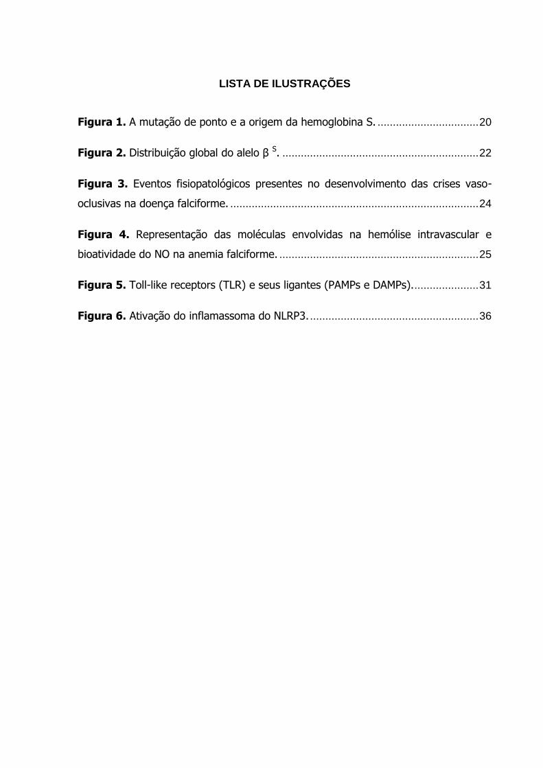
Figura 1. A mutação de ponto e a origem da hemoglobina S. A HbS é decorrente da substituição do aminoácido adenina por timina (GAG GTG), resultando na da valina em substituição ao ácido glutâmico, na posição 6 da cadeia da globina beta da hemoglobina. Figura adaptada (FRENETTE e ATWEH, 2007).
A Hb S é decorrente da mutação de ponto com substituição da base
nucleotídica adenina por timina (GAG GTG), localizada no sexto códon do gene
beta (HBB) da globina, codificando ácido glutâmico por valina na cadeia
polipeptídica beta (Figura 1). A mutação em uma das globinas beta caracteriza o
indivíduo como heterozigoto para a HbS (HbAS), que não apresenta manifestações
clínicas da doença. A mutação nas duas globinas beta implica na forma mais grave
da DF, a anemia falciforme, caracterizada pela homozigose do alelo beta S (βs), com
21
produção de HbSS (EMBURY, 1986; BRASIL, 2001; STEINBERG, 2008; ODIEVRE
et al., 2011).
Em condições de hipóxia, ocorre a polimerização da HbS com formação de
estruturas filamentosas ou poliméricas que se depositam no interior dos eritrócitos,
modificando a sua forma bicôncava para o formato alongado, semelhante ao de
foice ou meia lua. A falcização dos eritrócitos reduz a sua sobrevida, tornando-os
rígidos, favorecendo o seu catabolismo rápido, com ocorrência de hemólise
acentuada e episódios de infartos teciduais (WEATHERALL e PROVAN, 2000;
BRASIL, 2001; TAYLOR et al., 2008). A AF também apresenta complicações clínicas
heterogêneas, tais como crises de sequestro esplênico; acidente vascular cerebral
(AVC); complicações renais; priapismo; alterações pulmonares, como a síndrome
torácica aguda (STA) e a hipertensão pulmonar; crises recorrentes de dor e
fenômenos de vaso-oclusão (SCHNOG et al., 2004; GLADWIN e KATO, 2005;
TAYLOR et al., 2008).
A DF está presente em cerca de 7% da população mundial, com prevalência
elevada nos países da África, América do Sul, América Central, Arábia Saudita e
Índia (WEATHERALL e CLEGG, 2001). Esta é a doença hematológica de origem
genética mais prevalente nos Estados Unidos, acometendo cerca de 70.000
pessoas, incluindo uma média de 01 nascido vivo com a anemia falciforme para
cada 3500 afro-descentes recém-nascidos anualmente (HASSELL, 2010). Conforme
dados da Organização Mundial de Saúde (OMS), estima-se que nasçam cerca de
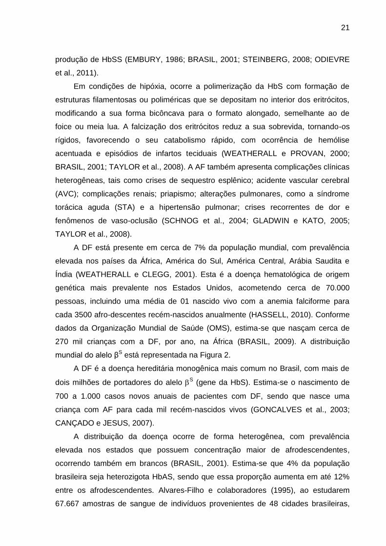
270 mil crianças com a DF, por ano, na África (BRASIL, 2009). A distribuição
mundial do alelo βS está representada na Figura 2.
A DF é a doença hereditária monogênica mais comum no Brasil, com mais de
dois milhões de portadores do alelo S (gene da HbS). Estima-se o nascimento de
700 a 1.000 casos novos anuais de pacientes com DF, sendo que nasce uma
criança com AF para cada mil recém-nascidos vivos (GONCALVES et al., 2003;
CANÇADO e JESUS, 2007).
A distribuição da doença ocorre de forma heterogênea, com prevalência
elevada nos estados que possuem concentração maior de afrodescendentes,
ocorrendo também em brancos (BRASIL, 2001). Estima-se que 4% da população
brasileira seja heterozigota HbAS, sendo que essa proporção aumenta em até 12%
entre os afrodescendentes. Alvares-Filho e colaboradores (1995), ao estudarem
67.667 amostras de sangue de indivíduos provenientes de 48 cidades brasileiras,

22
descreveram a frequência de 2,2% para os indivíduos HbAS (ALVARES FILHO et
al., 1995).
Atualmente, com base nos dados provenientes da triagem neonatal realizada
pela Associação de Pais e Amigos dos Excepcionais (APAE) – BA sabe-se que a
incidência e prevalência, na Bahia, da DF, é uma das maiores do Brasil, acometendo
um a cada 645 nascidos-vivos (SILVA et al., 2006). As regiões Norte e Nordeste
apresentam as frequências mais elevadas dos heterozigotos HbAS, correspondendo
a 6% e 10%, respectivamente, enquanto as demais regiões brasileiras não chegam
a somar 5% (CANÇADO e JESUS, 2007).
Figura 2. Distribuição global do alelo β S. Figura adaptada (REES e GIBSON, 2012).
A gravidade da DF aponta para uma contribuição elevada no número de óbitos
de jovens no Brasil, sendo que cerca de 80% das mortes ocorrem nos indivíduos
com AF antes dos 30 anos e, aproximadamente, 40% acometem menores de nove
anos. Esses números de idade baixa no óbito refletem a dificuldade no diagnóstico
precoce e no acesso às medidas de prevenção às infecções, e a falta de orientação
entre os familiares sobre como proceder aos primeiros sinais de complicações
(LOUREIRO e ROZENFELD, 2005).

23
2.2 Manifestações Clínicas e Fisiopatologia
Na HbS, a troca do ácido glutâmico, aminoácido hidrofílico, pela valina, que é
hidrofóbica, favorece, durante as fases de desoxigenação, interações hidrofóbicas,
entre moléculas vizinhas, culminando em estruturas poliméricas, as quais deformam
a hemácia. Em condições de hipóxia, acidez e desidratação celular, a polimerização
da Hb leva à deformação da estrutura da hemácia pelo fenômeno denominado
falcização. Esse fenômeno é reversível durante a oxigenação, enquanto as
hemácias não sofrerem lesões em sua membrana. Entretanto, falcizações
repetitivas, podem levar a alterações definitivas na membrana eritrocitária, tornado o
seu formato irreversivelmente falcizado (BRASIL, 2001; STEINBERG, 2008).
A cinética de falcização depende do grau de desoxigenação, concentração
intracelular de HbS e presença ou ausência de HbF. Os pacientes que possuem
concentrações elevadas de HbF têm seus sintomas clínicos reduzidos devido á
inibição desse fenômeno. Os eritrócitos falcizados possuem rigidez aumentada e
vida-média reduzida, o que contribui para a instalação de um quadro de anemia
hemolítica grave, com destruição tanto extravascular quanto intravascular, levando a
redução da vida do eritrócito e o agravamento da anemia (BRASIL, 2001; SILVA,
GONCALVES e MARTINS, 2004).
Os pacientes com AF possuem numerosas complicações que podem afetar
quase todos os órgãos e sistemas, com morbidade expressiva, redução da
capacidade de trabalho e da expectativa de vida. A interação dinâmica entre os
eritrócitos do paciente com AF e o endotélio vascular contribui para os episódios de
vaso-oclusão e isquemia, estresse vascular, expressão aumentada de citocinas
inflamatórias e moléculas de adesão. Além disso, a hemólise crônica, presente nos
quadros mais graves da AF, contribuem para a hipercoagulabilidade e vasculopatia,
entre outros (FRANCIS e HAYWOOD, 1992; TURHAN et al., 2002; WOOD, HEBBEL
e GRANGER, 2004; RAPHAEL, 2005; MORRIS, 2008).
A oclusão da microcirculação pelos eritrócitos falcizados leva à isquemia
tecidual e danos em tecidos, sendo a causa responsável pelas complicações clínicas
presentes na doença (SCHNOG et al., 2004). O fenômeno vaso-oclusivo presente
na AF envolve a obstrução de microvasos, lesão tecidual, com inflamação local
(STUART e NAGEL, 2004). Tanto as hemácias quanto reticulócitos dos pacientes
com AF expressam em sua superfície moléculas de adesão, como a integrina α4β1
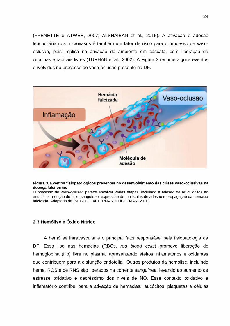
24
(FRENETTE e ATWEH, 2007; ALSHAIBAN et al., 2015). A ativação e adesão
leucocitária nos microvasos é também um fator de risco para o processo de vaso-
oclusão, pois implica na ativação do ambiente em cascata, com liberação de
citocinas e radicais livres (TURHAN et al., 2002). A Figura 3 resume alguns eventos
envolvidos no processo de vaso-oclusão presente na DF.
Figura 3. Eventos fisiopatológicos presentes no desenvolvimento das crises vaso-oclusivas na doença falciforme. O processo de vaso-oclusão parece envolver várias etapas, incluindo a adesão de reticulócitos ao endotélio, redução do fluxo sanguíneo, expressão de moléculas de adesão e propagação da hemácia falcizada. Adaptado de (SEGEL, HALTERMAN e LICHTMAN, 2010).
2.3 Hemólise e Óxido Nítrico
A hemólise intravascular é o principal fator responsável pela fisiopatologia da
DF. Essa lise nas hemácias (RBCs, red blood cells) promove liberação de
hemoglobina (Hb) livre no plasma, apresentando efeitos inflamatórios e oxidantes
que contribuem para a disfunção endotelial. Outros produtos da hemólise, incluindo
heme, ROS e de RNS são liberados na corrente sanguínea, levando ao aumento de
estresse oxidativo e decréscimo dos níveis de NO. Esse contexto oxidativo e
inflamatório contribui para a ativação de hemácias, leucócitos, plaquetas e células
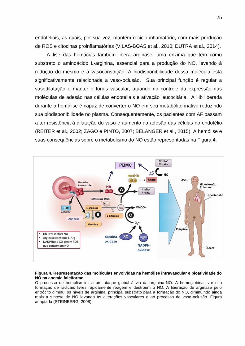
25
endoteliais, as quais, por sua vez, mantêm o ciclo inflamatório, com mais produção
de ROS e citocinas proinflamatórias (VILAS-BOAS et al., 2010; DUTRA et al., 2014).
A lise das hemácias também libera arginase, uma enzima que tem como
substrato o aminoácido L-arginina, essencial para a produção do NO, levando à
redução do mesmo e à vasoconstrição. A biodisponibilidade dessa molécula está
significativamente relacionada a vaso-oclusão. Sua principal função é regular a
vasodilatação e manter o tônus vascular, atuando no controle da expressão das
moléculas de adesão nas células endoteliais e ativação leucocitária. A Hb liberada
durante a hemólise é capaz de converter o NO em seu metabólito inativo reduzindo
sua biodisponibilidade no plasma. Consequentemente, os pacientes com AF passam
a ter resistência à dilatação do vaso e aumento da adesão das células no endotélio
(REITER et al., 2002; ZAGO e PINTO, 2007; BELANGER et al., 2015). A hemólise e
suas consequências sobre o metabolismo do NO estão representadas na Figura 4.
Figura 4. Representação das moléculas envolvidas na hemólise intravascular e bioatividade do NO na anemia falciforme. O processo de hemólise inicia um ataque global à via da arginina-NO. A hemoglobina livre e a formação de radicais livres rapidamente reagem e destroem o NO. A liberação de arginase pelo eritrócito diminui os níveis de arginina, principal substrato para a formação do NO, diminuindo ainda mais a síntese de NO levando às alterações vasculares e ao processo de vaso-oclusão. Figura adaptada (STEINBERG, 2008).

26
O NO, principal responsável pela manutenção do tônus vascular, é formado a
partir da L-arginina e do oxigênio molecular em uma reação catalisada pela enzima
NOS (ROMERO et al., 2002). O NO é constitutivamente sintetizado e liberado
principalmente pelas células endoteliais vasculares e fisiologicamente inibe diversos
componentes do processo aterogênico, como vasoconstricção, agregação
plaquetária, proliferação do músculo liso vascular e adesão de leucócitos ao
endotélio (FORGIONE, LEOPOLD e LOSCALZO, 2000).
A diminuição da biodisponibilidade do NO durante a hemólise contribui para o
desequilíbrio vascular, uma condição presente na disfunção endotelial (DE). Essa
condição leva ao aumento da expressão de moléculas como a molécula de adesão
intercelular 1 (ICAM-1), proteína de adesão celular vascular 1 (VCAM-1) e
endotelina-1, aumento da ativação plaquetária e dano na reperfusão isquêmica
(ROTHER et al., 2005; WOOD, HSU e GLADWIN, 2008; VILAS-BOAS et al., 2010).
A redução do NO pelos produtos gerados no processo inflamatório causa
também danos endoteliais via ativação transcricional de moléculas vasoconstritoras,
como a endotelina-1 (ROTHER et al., 2005).Esta molécula possui características
contrárias à do NO, uma vez que apresenta a mais potente atividade vasoconstritora
descrita em humanos, sendo que em pacientes com AF, seus níveis séricos estão
elevados durante as crises vaso-oclusivas (GRAIDO-GONZALEZ et al., 1998;
ERGUL et al., 2004).
Os pacientes com AF em estado estável apresentam concentrações
reduzidas de NO (BELHASSEN et al., 2001; VILAS-BOAS et al., 2010). Essa
redução, quando associada ao estado inflamatório crônico (KATO et al., 2009)
aumenta, provavelmente, a ativação de fatores de coagulação, em especial o fator
tecidual (FT) (SOLOVEY et al., 2010), além de modular outras moléculas como a
fosfatidilserina (FS), pelos eritrócitos, e receptor de proteína C reativa (RPCR) (GU
et al., 2000; SETTY, KULKARNI e STUART, 2002).
Portanto, o NO desempenha papel fundamental na patogênese da AF, pois,
nesta condição, suas propriedades vasodilatadora e antitrombogênica encontram-se
comprometidas, e a vasoconstricção aliada à adesão de células circulantes podem
levar à oclusão de microvasos.

27
2.4 Terapia Farmacológica na Doença Falciforme: Hidroxiureia
Os pacientes com DF em crise vaso-oclusiva necessitam de uma terapia
farmacológica a fim de aliviar as manifestações clinicas. Até o presente momento, o
único tratamento farmacológico utilizado nestes pacientes é a hidroxiureia (HU). A
terapia com HU foi aprovada, em 1998, para o uso em pacientes com DF
sintomática, pela United States Food Drug Administration (FDA), e em 2010, pelo
Ministério da Saúde (BRASIL, 2010).
Inúmeros estudos têm reportado a eficácia da HU em pessoas com DF por
conduzir à melhora clínica e hematológica pela redução da incidência de episódios
vaso-oclusivos. A HU é uma droga antineoplásica, citotóxica, mas que apresenta
vários efeitos benéficos em pacientes com DF, tais como: aumento da produção de
HbF, aumento da hidratação do glóbulo vermelho, aumento na concentração de Hb,
aumento na produção de NO e diminuição da expressão de moléculas de adesão
(BRASIL, 2010; STROUSE e HEENEY, 2012; CUNNINGHAM-MYRIE et al., 2015).
Como mecanismo de ação efetivo para a DF, a HU atua inibindo a
ribonucleotídeo redutase, uma enzima que catalisa a conversão redutiva de
ribonucleotídeos em desoxirribonucleotídeos, sendo esta a etapa limitante da
velocidade de síntese de DNA (OSTERMAN GOLKAR et al., 2013; GUAN et al.,
2015). Embora tenha efeito citotóxico, é esta propriedade que a torna importante e
singular no tratamento de pacientes com AF. Sua toxicidade está associada a
supressão de precursores eritróides com ciclagem mais rápida, favorece a maior
duração de precursores mais primitivos (Células F) e de ciclagem mais lenta,
quiescentes e contendo HbF, forma predominante no feto e como prescinde das
cadeias β globina, não tem sua estrutura afetada pela mutação nos genes das
mesmas (MORRIS et al., 2003; STEINBERG et al., 2010).
Outro benefício do tratamento com HU é seu papel de “doador de NO”. O
metabolismo desse fármaco resulta na produção de NO, o qual compensa a redução
do mesmo no processo de hemólise crônica. O NO ativa o sistema efetor guanilato
ciclase que, por sua vez, leva ao aumento dos níveis intracelulares de monofosfato
cíclico de guanosina (GMPc), o qual interage com fatores de transcrição estimulando
diretamente a produção de células F e contribuindo para o aumento da Hb fetal nas
hemácias. Esse aumento de HbF contribui, portanto, para a diminuição percentual
28
da HbS, e redução da hemólise crônica (GLADWIN et al., 2002; NAHAVANDI et al.,
2002; COKIC et al., 2008).
2.5 Polimorfismos nos Genes da Mieloperoxidase e da Alfa-1 Antitripsina
A mieloperoxidase (MPO) é uma proteína relacionada à inflamação e proteção
contra infecções, abundante em neutrófilos e monócitos. Em níveis elevados, a MPO
contribui significativamente para a amplificação da resposta inflamatória vascular,
condição constante em pacientes com DF. A enzima catalisa a formação de ROS e
RNS, que apresentam capacidade oxidante elevada e podem comprometer a
integridade do endotélio vascular (ABU-SOUD e HAZEN, 2000). A MPO modula a
resposta inflamatória vascular, sendo que seus níveis elevados são preditores da
disfunção endotelial e contribuem para a redução da biodisponibilidade do NO, já
que esta enzima é uma NO oxidase (EISERICH et al., 2002; GALIJASEVIC et al.,
2006; ZHANG et al., 2013) Uma vez que a MPO e os produtos derivados de sua
atividade enzimática estão aumentados nos sítios inflamatórios (EISERICH et al.,
2002), pacientes com DF, principalmente durante os episódios de crise (REES,
WILLIAMS e GLADWIN, 2010) ou infecção (COSTA et al., 2005), apresentam níveis
séricos elevados de MPO (VILLAESCUSA et al., 1999), sugerindo que esta enzima
pode contribuir diretamente para a resposta vascular.
Além do papel modulador no processo inflamatório, crescentes evidências
demonstram o papel da MPO como agente da resposta frente a patógenos.
Infecções tais como pneumonia, meningite, infecções urinárias e septicemia
constituem uma causa comum de hospitalização em pacientes com DF,
principalmente, em pacientes com AF (COSTA et al., 2005), condições estas
relacionadas ao aumento da contagem de leucócitos (MTATIRO et al., 2015) e dos
níveis séricos de MPO. Sabe-se que a MPO, única enzima humana capaz de gerar
hipoclorito (HOCl) em meio contendo cloreto e peróxido de hidrogênio (H2O2), é
liberada logo após ativação celular, exercendo papel essencial nos mecanismos de
defesa do sistema imune (KLEBANOFF, 1970; HANSSON, OLSSON e NAUSEEF,
2006; SHAEIB et al., 2015).
Sabe-se que a gravidade da DF varia bastante entre os indivíduos e esta
variabilidade fenotípica costuma estar relacionada a alterações genotípicas. Sendo a
MPO uma enzima altamente polimórfica, o polimorfismo localizado na região

29
promotora (-463G>A, troca de G por A) do gene MPO tem sido relacionado à
redução na transcrição gênica dessa enzima (PIEDRAFITA et al., 1996; CASCORBI
et al., 2000).
Como pacientes com DF apresentam quadro inflamatório crônico,
especialmente durante os episódios de hemólise, crises vaso-oclusivas e síndrome
torácica aguda (STA) (AMER et al., 2006; STEINBERG, 2008), e este polimorfismo é
caracterizado pela redução da transcrição gênica de MPO (COSTA et al., 2005;
REYNOLDS et al., 2006), esta mutação poderia estar associada às manifestações
clínicas da doença. A presença desse polimorfismo em pacientes com DF está
associada à ocorrência de infecções bacterianas, especialmente em indivíduos com
AF (COSTA et al., 2005; BARBOSA et al., 2014), e a deficiência de MPO tem sido
associada ao aumento da ocorrência de processos inflamatórios (CASCORBI et al.,
2000).
A distribuição desse polimorfismo mostra que cerca de 2-10% da população
geral apresentam o alelo polimórfico em homozigose (AA), cerca de 30-40% são
heterozigotos (GA) e 60-70% são homozigotos para o alelo selvagem (GG) (DALLY
et al., 2002; FEYLER et al., 2002; ZHONG et al., 2009). Indivíduos com o alelo A
(GA ou AA) apresentam expressão reduzida de MPO em até três vezes quando
comparados ao grupo com genótipo selvagem. O genótipo AA corresponde à forma
mais grave do polimorfismo, implicando em níveis séricos diminuídos e atividade
reduzida da MPO (REYNOLDS et al., 2006).
Outra proteína relacionada à inflamação e que também exibe polimorfismo
genético é a alfa-1 antitripsina (AAT). Esta glicoproteína de fase aguda é altamente
polimórfica e sua síntese é controlada pelo gene SERPINA1, localizado no locus do
inibidor de protease (Pi), no cromossomo 14q32.1, em resposta a diversas citocinas,
tais como IL-1, fator de necrose tumoral α (TNF α) e, principalmente, IL-6
(RICHARDS, GAULDIE e BAUMANN, 1991; LOMAS e MAHADEVA, 2002;
CHAPPELL et al., 2006). Após sua síntese, predominantemente em hepatócitos, a
AAT é secretada no plasma, espalhando-se por todo o corpo. Os níveis séricos da
AAT aumentam durante o processo inflamatório ou lesão tecidual e estão
relacionados com a inibição de proteases, especialmente a elastase, uma protease
de serina produzida por neutrófilos, que tem a capacidade de hidrolisar as fibras de
elastina no pulmão e relacionada à patogênese da DF (RUDNICK e PERLMUTTER,
2005; GRUBER, NADIR e HAAS, 2010). Os níveis plasmáticos de elastase estão

30
elevados em indivíduos com DF assintomáticos em relação a controles sadios, e
esses níveis aumentam significativamente em pacientes em crise vaso-oclusiva
(LARD et al., 1999). Além da inibição de proteases, estudos têm mostrado que a
AAT modula a função de células do sistema imune, tais como neutrófilos (BERGIN et
al., 2010), monócitos (JANCIAUSKIENE, NITA e STEVENS, 2007) e linfócitos (LU et
al., 2006), e contribui para a supressão da síntese de citocinas proinflamatórias
(POTT et al., 2009).
O gene codificante para AAT, SERPINA1, é altamente polimórfico com mais de
125 polimorfismos de nucleotídeo único (single nucleotide polymorphism, SNP). As
formas variantes de AAT são classificadas como inibidor de protease “Pi” (Protease
inhibitor). As formas variantes “normais” de AAT apresentam níveis séricos normais
(20-50 µM) e atividade de inibição de protease funcional, representadas pelo alelo
selvagem M. Entretanto, as variantes para “deficiência” (ou seja, redução da
concentração sérica da AAT ou presença de genótipo alterado) estão associadas
com concentrações séricas dessa proteína menores do que os valores encontrados
em indivíduos com variantes normais, representadas principalmente pelos alelos S e
Z, sendo este último associado à forma mais grave de deficiência de AAT
(GOOPTU, DICKENS e LOMAS, 2014).
A homozigose para o alelo Pi*Z tem cerca de 10-15% dos níveis normais de
AAT, sendo esta condição um fator de risco para o desenvolvimento de
complicações pulmonares, como enfisema, e, sistemicamente, para infecção.
Indivíduos heterozigotos para esse alelo (Pi*MZ) produzem cerca de 50 % dos níveis
normais de AAT, produzidos pelos indivíduos sem o alelo mutante, ou seja,
homozigotos para o alelo selvagem (Pi*MM) (STOCKLEY, 2014). O alelo Pi*S é
mais frequente que o alelo Pi*Z. Em contraste ao alelo Z, os níveis de AAT, em
indivíduos homozigotos para o alelo Pi*SS, são reduzidos cerca de 60% em relação
aos indivíduos com o genótipo Pi*MM, sendo que essa variante não está relacionada
à inibição da elastase neutrofílica. Em indivíduos heterozigotos Pi*MS, os níveis de
AAT correspondem cerca de 75% da concentração sérica dos indivíduos normais.
Entretanto, a combinação dessa variante com o alelo Z (Pi*SZ) reduz
significativamente os níveis circulantes dessa proteína, alcançando uma
concentração sérica de apenas 35% da encontrada em indivíduos P1*MM. A
presença desse genótipo (Pi*SZ) aumenta o risco de doenças, incluindo a doença
pulmonar e cirrose hepática (TURINO et al., 1996; STOCKLEY e TURNER, 2014).

31
2.6 Resposta Imune Inata
2.6.1 Receptores do Tipo Toll (TLRS)
Os receptores do tipo Toll (TLR) são uma família de proteínas conservadas
evolutivamente para reconhecerem alguns DAMPs (IMAI et al., 2008; LEFEBVRE et
al., 2011) e PAMPs associados a vírus, bactérias, parasitas ou fungos (KUMAR et
al., 2014; SMITH et al., 2014; TOMLINSON et al., 2014; WAGENER et al., 2014).
Estes receptores são amplamente distribuídos nas células do sistema imune inato,
sendo importantes não apenas na ativação da resposta inata, como principalmente
na iniciação e estabelecimento da resposta imune adaptativa (MEDZHITOV,
PRESTON-HURLBURT e JANEWAY, 1997).
Foram descritos, em humanos, 10 TLRs (TLR1 ao TLR10), cada um dos quais
apresenta certa especificidade para determinado PAMP ou DAMP (Figura 5). O
TLR10 era o único TLR encontrado em humanos que, até recentemente, não
possuía ligantes conhecidos (CHUANG e ULEVITCH, 2001). Apesar do ligante do
TLR10 ainda não ser conhecido, um estudo demonstrou forte associação deste TLR
na defesa contra infeção bacteriana na mucosa intestinal (REGAN et al., 2013).
Figura 5. Toll-like receptors (TLR) e seus ligantes (PAMPs e DAMPs). Adaptado de (TAKEDA e AKIRA, 2004).

32
Dentre os receptores do tipo Toll mais bem estudados e descritos, o TLR2,
TLR4, TLR5 e TLR9 apresentam características importantes na indução de resposta
inflamatória do hospedeiro. O TLR2 é importante para a resposta contra bactérias
Gram-positivas, já que reconhecem peptidoglicanos da sua parede celular.
Entretanto, como o TLR2 forma heterodímeros, outros PAMPs também têm sido
associados com ativação deste receptor (TAKEUCHI et al., 1999; OZINSKY et al.,
2000). Além de participar da defesa contra patógenos, o TLR2 é capaz ainda de
responder contra DAMPs endógenos, num processo de inflamação estéril. Os
principais DAMPs agonistas de TLR2 são a beta-2-Glicoproteína I e os beta-
amilóides (ALARD et al., 2010; LIU et al., 2012; VAN BERGENHENEGOUWEN et
al., 2013). Quando o TLR2 está formando heterodímero com o TLR4, este complexo
pode ser ativado por hemoglobina (WANG et al., 2014).
O TLR4 é um receptor que reconhece um dos PAMPs mais bem descritos, o
lipopolissacarídeo (LPS), além de reconhecer resíduos celulares endógenos que
atuam como DAMPs após lesão celular. Os principais DAMPs agonistas de TLR4
são HMGB1 (Proteína do Grupo 1 de Mobilidade Alta), HSP (Proteína de Choque
Térmico), β-defensina, ácido hialurônico, grupo heme, dentre outros (IMAI et al.,
2008; LEFEBVRE et al., 2011; BELCHER et al., 2014; GUPTA, 2014; CAI et al.,
2015; NAIR et al., 2015).
A ativação do TLR5 ocorre principalmente por infecção por patógenos
flagelados, como Salmonella spp. (TAKEDA e AKIRA, 2004). Este é um receptor que
reconhece apenas PAMP (flagelina), não havendo nenhum DAMP ainda descrito
capaz de ativá-lo. Entretanto, apesar do principal agonista do TLR5 ser a flagelina,
alguns estudos demonstraram associação entre a expressão deste receptor,
polimorfismos e gravidade e prognóstico de diferentes tipos de tumor de mucosa e
outras doenças inflamatórias crônicas, sugerindo que o TLR5 pode ser um
biomarcador de prognóstico destas condições (KIM et al., 2008; PIMENTEL-NUNES
et al., 2011; WANG et al., 2012; KAUPPILA et al., 2013; SHERIDAN et al., 2013).
O TLR9 é um receptor que reconhece DNA de cadeia simples associado a
motivos CpG não metilados, o qual é raro em DNA eucariótico e abundante em DNA
bacteriano (BAUER, 2013). Entretanto, alguns autores sugerem que o TLR9 seja
capaz de reconhecer DNA próprio, tanto mitocondrial quanto nuclear, estando
associado a imunopatogênese de doenças inflamatórias, como o lúpus eritematoso
sistêmico (LEADBETTER et al., 2002; BARRAT et al., 2005; ZHANG et al., 2010).

33
À exceção do TLR3, todos os outros TLRs sinalizam sua ativação por meio da
proteína adaptadora MyD88. O recrutamento desta molécula induz a ativação de
uma família de quinases (IRAK) e do fator 6 associado ao receptor do fator de
necrose tumoral (TRAF6), as quais levam à liberação e translocação do NF-B para
o núcleo da célula e, consequente, aumento da expressão gênica de citocinas
proinflamatórias (LIEW et al., 2005). Estas moléculas proinflamatórias participam da
defesa do hospedeiro contra patógenos invasores, mas podem ainda estar
envolvidas na patogênese de doenças autoimunes e inflamatórias crônicas como
diabetes (KAYSEROVA et al., 2014), lúpus eritematoso sistêmico (LYN-COOK et al.,
2014), asma (SHIKHAGAIE et al., 2014) e DF (GHOSH et al., 2013; BELCHER et al.,
2014).
Na AF, as células endoteliais, leucócitos e plaquetas estão constantemente
expostos a ação do heme e Hb provenientes da hemólise intravascular,
frequentemente observada nestes casos, o que leva a um estado de inflamação
sistêmica e fenômenos pró-trombóticos (BELCHER et al., 2003; WOOD, HEBBEL e
GRANGER, 2004). O heme é capaz de ativar diretamente a sinalização via TLR4 em
células endoteliais, levando à ativação do NF-B. Esta via induz liberação dos
constituintes dos corpos de Weibel-Palade (WPB), p-selectina e fator de von
Willebrand (VWF), para o endotélio vascular, ocasionando fenômenos de vaso-
oclusão em modelo experimental de DF (FIGUEIREDO et al., 2007; BELCHER et al.,
2014).
Por outro lado, a Hb liberada durante a hemólise intravascular, além de
interagir diretamente com TLR4, pode ainda associar-se a certos PAMPs ou DAMPs,
sendo importantes na ativação do sistema imune inato. Análise in silico demonstrou
que a molécula de Hb é capaz, por exemplo, de interagir com LPS, resultando em
possível alteração conformacional e conversão para meta-hemoglobina (BAHL et al.,
2011).
Além da heme e da Hb serem importantes na ativação da reposta imune inata,
tem sido demonstrado que hemácias íntegras também participam do processo
inflamatório observado na AF. As células endoteliais ativadas por hemácias íntegras
produzem HMGB1, um DAMP agonista de TLR4 altamente expresso na DF,
induzindo inflamação e necroptose (morte celular programada por necrose) de
células endoteliais (GUPTA, 2014; QING et al., 2014; XU et al., 2014).
34
Alguns estudos têm demonstrado que a incubação de Hb com ligantes de TLRs
e PAMPs estimula, de forma sinérgica, a produção das citocinas proinflamatórias IL-
1β, IL-6, IL-8 e TNF por macrófagos (BODET, CHANDAD e GRENIER, 2007; LIN et
al., 2010). Estes dados em conjunto, associados à presença de DAMPs durante a
hemólise intravascular e ao estado inflamatório observado na AF, sugerem que
outras vias de ativação do sistema imune inato estejam participando deste processo,
como os receptores do tipo NOD e o inflamassoma.
2.6.2 Receptores do Tipo NOD (NLRS) e o Inflamassoma
Os receptores do tipo NOD (NLRs) representam uma superfamília dos
receptores de reconhecimento padrão (PRR) que foram descritos inicialmente há
cerca de 15 anos (BERTIN et al., 1999; INOHARA et al., 1999). Há um total de 22
genes humanos relacionados com os NLRs, os quais são agrupados em cinco
subfamílias (NLRA, NLRB, NLRC, NLRP e NLRX) a depender do seu domínio efetor
N-terminal, compreendendo, normalmente, um domínio efetor pirina (PYD) ou de
recrutamento e ativação de caspase (CARD). Todas as NLRs contêm ainda um
domínio central para oligomerização e ligação de nucleotídeo (NACHT), e um
domínio de repetição rico em leucina (LRR) na sua porção C-terminal (TING et al.,
2008; YERETSSIAN, 2012).
O NLRA possui um domínio N-terminal de transativação ácida, chamado de
CIITA, atuando como regulador transcricional da apresentação de antígenos via
MHC classe II. O NLRB (NAIP) apresenta um domínio de repetição de inibição de
apoptose de baculovírus, estando associado a defesa do hospedeiro e sobrevida
celular. A subfamília NLRC apresenta cinco membros (NLRC 1 a 5), sendo o NLRC1
(NOD1) e NLRC2 (NOD2) os mais bem descritos. Estes receptores reconhecem
peptidoglicanos presentes na parede celular de bactérias, sendo fundamentais para
manutenção da homeostasia tecidual e defesa contra agentes bacterianos. O
NLRX1, único membro descrito da família dos NLRX, possui na região N-terminal
uma sequência-alvo que permite seu tráfego pela membrana mitocondrial
(NICKERSON et al., 2001; DIEZ et al., 2003; WRIGHT et al., 2003; LIGHTFIELD et
al., 2008; PHILPOTT et al., 2014).
A subfamília dos NLRPs (também conhecidos como NALPs) possui 14
membros descritos (NRLP 1 a 14), todos com um domínio PYD N-terminal. Uma das

35
principais funções destes receptores é a formação de uma plataforma proteica
inflamatória após sua oligomerização, chamada de “inflamassoma”, a qual controla a
produção de citocinas proinflamatórias como a IL-1 beta e a IL-18. Apesar de várias
plataformas de inflamassoma já terem sido descritas, o inflamassoma associado ao
NLRP3 (ou NALP3) é o mais comumente estudado (MARTINON, BURNS e
TSCHOPP, 2002; SCHRODER e TSCHOPP, 2010; LATZ, XIAO e STUTZ, 2013).
Classicamente o inflamassoma consiste de NLRP (NLRP3 é mais comum), da
protease inflamatória caspase-1 e da proteína associada a apoptose (ASC)
(MARTINON, BURNS e TSCHOPP, 2002). O NLRP3 é expresso em uma variedade
de tipos celulares, incluindo neutrófilos, células dendríticas, células epiteliais,
monócitos e linfócitos T. Diversos PAMPs de vírus, bactérias e fungos foram
descritos como sendo ativadores do NLRP3, além de alguns DAMPs, como ATP e
glicose, dentre outros (Revisado por SCHRODER e TSCHOPP, 2010). Após sua
ativação, o NLRP3 recruta ASC e pro-caspase-1, as quais são necessárias para a
síntese das citocinas proinflamatórias IL-1β e IL-18 e, consequente, morte das
células infamatórias, num processo dependente de caspase-1 chamado de piroptose
(AGOSTINI et al., 2004; MARTINON et al., 2006; BERGSBAKEN, FINK e
COOKSON, 2009).
A formação do inflamassoma é um processo de duas etapas. Como o nível
basal de NLRP3 é insuficiente para iniciar a formação do inflamassoma, é
necessário que haja a participação de NF-B proveniente da via do TLR, sendo este
o primeiro sinal para ativação do inflamassoma. Apesar de este ser o caminho
comumente percorrido para ativação do inflamassoma, existem ainda algumas
evidências científicas de ativação desta plataforma proteica independente de TLR,
em resposta, por exemplo, ao estímulo por LPS, citocinas ou ROS. Adicionalmente,
a presença de ROS parece ser fundamental para o estabelecimento do
inflamassoma (BAUERNFEIND et al., 2009; BAUERNFEIND et al., 2011; ZHOU et
al., 2011; GHONIME et al., 2014). O segundo sinal para ativação do inflamassoma é
a própria oligomerização do NLRP3, a qual leva à formação de capase-1 ativa e,
consequente, conversão de pro-IL-1β e pro-IL18 em suas formas ativas que são
secretadas no ambiente extracelular (Figura 6) (MARIATHASAN e MONACK, 2007).

36
Figura 6. Ativação do inflamassoma do NLRP3. (1) Estimulação de NLRP3 por agonistas (PAMPs e/ou DAMPs); (2) recrutamento de pro-caspase-1 e ASC, possibilitando oligomerização do NLRP3 e formação do inflamassoma; (3) processo auto catalítico gerando caspase-1 ativa; (4) clivagem dos precursores pro-IL-1β e pro-IL18 nas suas formas ativas (DOS SANTOS, KUTUZOV e RIDGE, 2012).
As interleucinas IL-1β e IL-18 pertencem à superfamília da interleucina-1, a
qual ainda compreende a citocinas mais recentes IL-33, IL-36 e IL-37. Estas estão
relacionadas entre si pela sua origem, estrutura do receptor ou via de transdução do
sinal (DINARELLO, 2009; DINARELLO et al., 2010). A IL-1β é uma citocina não
expressa constitutivamente, mas induzida pela estimulação de sinais inflamatórios.
Esta citocina existe no citoplasma celular na sua forma inativa, pro-IL-1β,
apresentando atividade apenas após sua conversão mediada pela caspase-1, sendo
um dos principais indutores de inflamação e febre (DINARELLO, 2013; JESUS e
GOLDBACH-MANSKY, 2014). A citocina IL-18, entretanto, é expressa
constitutivamente em alguns tipos de células, como em PBMC (PUREN, FANTUZZI
e DINARELLO, 1999), e está evolvida tanto na resposta imune inata quanto na
adaptativa, sendo originalmente referida como IGIF (fator indutor de IFN-) devido à
sua capacidade de estimular produção de IFN- principalmente por células Th. O
aumento na expressão e nos níveis circulantes de IL-1β e IL-18 tem sido associado

37
a diversas condições clínicas que cursam com processo inflamatório, como
infecções, esclerose múltipla, câncer, doença de Alzheimer, artrite e AF (GUMA et
al., 2010; CERQUEIRA et al., 2011; KITAZAWA et al., 2011; RAMIREZ-RAMIREZ et
al., 2013; KETELUT-CARNEIRO et al., 2015; TAS et al., 2015; VICARI et al., 2015).
A AF é caracterizada por um estado inflamatório intenso associado a
expressão aumentada de moléculas de adesão nas células edoteliais, consequência
da constante lesão destas células, além de hemólise, e concentrações plasmáticas
elevadas de citocinas e outros mediadores inflamatórios (HEBBEL, 1997; PLATT,
2000). Na AF tem sido demonstrada, inclusive em população brasileira, a associação
entre polimorfismos de IL-1β e complicações decorrentes da doença, como AVC
(ASARE et al., 2010; VICARI et al., 2015). Adicionalmente, nosso grupo demonstrou
que níveis plasmáticos de IL-18 estão correlacionados com marcadores de hemólise
e disfunção endotelial (CERQUEIRA et al., 2011). Desta forma, o tratamento
farmacológico do indivíduo com AF deve almejar, além da elevação dos níveis de
HbF, a redução do processo inflamatório crônico observado nestes pacientes,
interferindo na expressão de moléculas associadas às principais vias da inflamação,
como o Toll e o NOD.
38
3 JUSTIFICATIVA
As complicações clínicas presentes na DF, principalmente na AF, estão
diretamente relacionadas à hemólise, à disfunção do endotélio vascular, à
amplificação da resposta vaso-oclusiva, que estão sempre relacionadas à
morbimortalidade elevada desses pacientes, e ao estado ativado dos leucócitos
circulantes (GILL et al., 1995; SCHNOG et al., 2004).
Sabe-se, ainda, que os mecanismos fisiopatológicos que induzem a
variabilidade clínica apresentada por esses pacientes ainda não estão
completamente elucidados, sendo que a identificação de moléculas envolvidas em
complicações vasculares, principalmente nos quadros de oclusão vascular, pode
contribuir para o desenvolvimento de estratégias terapêuticas voltadas para o
tratamento farmacológico mais direcionado a estas moléculas.
Até o presente momento, o único tratamento farmacológico aprovado
mundialmente para o uso nesses pacientes é a hidroxiureia, um fármaco que atua
na melhora clínica desses pacientes por induzir aumento na produção de HbF.
Entretanto, não se sabe se esse fármaco poderia interferir diretamente nas vias
inflamatórias ou influenciar na produção de moléculas associadas à inflamação
naqueles pacientes com DF que apresentam polimorfismo em genes relacionados
ao processo inflamatório.
Não há dúvidas de que a gravidade da doença, principalmente no que diz
respeito à AF, é bastante divergente entre os pacientes. Uma das causas dessa
diversidade clínica poderia ser a presença de alterações genéticas, tais como
polimorfismos nos genes da mieloperoxidase (-463G>A) e SERPINA1, afetando,
respectivamente, a mieloperoxidase e alfa-1 antitripsina - moléculas coadjuvantes do
processo inflamatório apresentado pelos pacientes com DF.
Além disso, esses pacientes apresentam um quadro inflamatório persistente,
o que poderia ser explicado pela constante ativação das vias dos receptores tipo Toll
e NOD, as duas principais vias da inflamação. Vários trabalhos têm demonstrado
que esses receptores são importantes no reconhecimento DAMPs derivados da
resposta inflamatória levando a um ciclo de ativação constante da resposta imune
inata. Porém, pouco se sabe a respeito da interação entre DAMPs decorrentes do
quadro inflamatório dos pacientes com AF e os componentes formadores do
inflamassoma ou relacionados à via do Toll.

39
Visto que a presença de polimorfismos em genes relacionados à inflamação,
tais como MPO e SERPINA1, pode resultar em estados clínicos diversos nos
pacientes com DF, e que as vias de sinalização mediadas por Toll e NOD podem ser
críticas na ativação e manutenção da resposta inflamatória nesses pacientes, os
estudos sobre a participação desses componentes da inflamação nas manifestações
clínicas dos pacientes aliados à investigação sobre o possível efeito da HU nessas
vias inflamatórias podem auxiliar no desenvolvimento de estratégias terapêuticas
novas direcionadas a esses componentes, além de reforçar a participação destes
como preditores de subfenótipos clínicos específicos nestes pacientes.

40
4 HIPÓTESES
Hipótese 1
Polimorfismos nos genes MPO-463G>A e SERPINA1 interferem na resposta ao
tratamento com hidroxiureia em pacientes com doença falciforme
Hipótese 2
Hidroxiureia não reduz diretamente a expressão gênica in vitro de receptores do tipo
Toll em pacientes com anemia falciforme.
Hipótese 3
Hidroxiureia não reduz diretamente a expressão gênica in vitro de moléculas
associadas ao inflamassoma via NLRP3.
41
5 OBJETIVOS
5.1 Objetivo Geral
Avaliar a associação dos polimorfismos nos genes da MPO -463G>A e da
SERPINA1 na resposta de pacientes com DF ao tratamento com hidroxiureia e o
efeito da adição de hemácias falcizadas na expressão gênica de receptores tipo Toll
ou componentes do inflamassoma em células mononucleares, tratadas ou não com
hidroxiureia.
5.2 Objetivos Específicos
1) Avaliar parâmetros hematológicos e bioquímicos e sua associação com
polimorfismos nos genes MPO -463G>A e SERPINA1 em pacientes com doença
falciforme tratados com HU;
2) Determinar a expressão de genes relacionados aos receptores do tipo Toll em
células mononucleares (PBMC) de pacientes com anemia falciforme e de voluntários
sadios;
3) Avaliar o efeito da adição de hemácias falcizadas na expressão de genes
relacionados aos receptores do tipo Toll em cultura de PBMC de voluntários sadios,
tratada ou não com hidroxiureia (HU);
4) Avaliar moléculas inflamatórias (LTB4 e nitrito) em sobrenadante de cultura de
células mononucleares incubadas com hemácias falcizadas, lisadas ou íntegras,
tratadas ou não com HU;
5) Determinar a expressão de genes relacionados a componentes do
inflamassoma NLRP3 em PBMC de pacientes com anemia falciforme e de
voluntários sadios;

42
6) Avaliar o efeito da adição de hemácias falcizadas na expressão de genes
relacionados a componentes do inflamassoma NLRP3 em cultura de PBMC de
voluntários sadios, tratada ou não com HU;
7) Avaliar proteínas relacionadas à ativação do inflamassoma (IL-1β e proteína
ligadora de IL-18 [IL-18BP]) em sobrenadante de cultura de PBMC incubada com
hemácias falcizadas tratada ou não com HU.
43
6 MANUSCRITOS
6.1 Manuscrito 1
Título: Sickle cell disease pharmacogenomics: implications on SERPINA1 and MPO
-463G>A gene polymorphisms and Hydroxyurea response.
Autores: Pitanga, T.N.; Carvalho, M.O.S.; Souza, A.L.C.S.; Santiago, R.P.; Oliveira,
R. R.; Lopes, V.M.; Oliveira, R.R.; Gonçalves, M.S.
Situação: A ser submetido
Objetivo: (referente ao objetivo 1 da tese):
1. Avaliar parâmetros hematológicos e bioquímicos e sua associação com
polimorfismos nos genes MPO -463G>A e SERPINA1 em pacientes com doença
falciforme tratados com HU.
Principais resultados: Pacientes com DF que exibem polimorfismo para os genes
da SERPINA1 ou MPO (-463G>A) mostram alterações em marcadores
hematológicos e bioquímicos na resposta ao tratamento com HU, não sendo
observadas na ausência desses polimorfismos.

44
SICKLE CELL DISEASE PHARMACOGENOMICS: IMPLICATIONS ON SERPINA1
AND MPO -463G>A GENE POLYMORPHISMS AND HYDROXYUREA RESPONSE
SHORT TITLE:
Gene polymorphisms in SCD and HU treatment
AUTHORS:
Thassila N. Pitangaa,b*, Magda O. S. Carvalhoa,c*, André L. C. S. Souzaa; Rayra P.
Santiagoa, Valma M. Lopesd, Ricardo R. Oliveiraa, Marilda S. Gonçalvesa,e.
AUTHOR AFFILIATIONS: aCentro de Pesquisas Gonçalo Moniz, Fundação Oswaldo Cruz, FIOCRUZ/BA; bInstituto de Ciências da Saúde (ICS), Universidade Federal da Bahia (UFBA); cHospital Universitário Professor Edgard Santos (HUPES), UFBA; dFundação de
Hematologia e Hemoterapia da Bahia (HEMOBA); eFaculdade de Farmácia, UFBA,
Salvador, Bahia, Brazil. *These authors contributed equally to this work.
CORRESPONDENT AUTHOR:
Correspondence to Marilda S. Gonçalves, PhD, Centro de Pesquisas Gonçalo
Moniz, LHGB, Waldemar Falcão St, n. 121, Candeal, Salvador, Bahia, Brasil. Tel:
+55 (71) 3176-2226, e-mail: [email protected]
CONFLICT OF INTEREST:
All authors have no conflict of interest to declare.
SOURCE FUNDING:
This work was supported by grants from the Brazilian National Council of Research
(CNPq) (311888/2013-5) (M.S.G.); the Foundation of Research and Extension of
Bahia (FAPESB) (3626/2013, 1431040053063, and 9073/2007) (M.S.G.); and
PPSUS/FAPESB (020/2013 EFP_00007295), (M.S.G.); Instituto Nacional de Ciência
e Tecnologia do Sangue (CNPq) (Coordinated by S.T.O.S.), and MCD/CNPq/MS-
SCTIE-DECIT (409800/2006-6), (M.S.G.). Sponsors of this study are public or
nonprofit organizations that support science in general. They had no role in gathering
analyzing, or interpreting the data.

45
ABSTRACT Background and Objective: SERPINA1 and myeloperoxidase (MPO)-463G>A gene polymorphisms are associated with vascular complications in sickle cell disease (SCD). SCD patients are treated with Hydroxyurea (HU), which increases levels of fetal hemoglobin (HbF) and decreases leukocytes. We evaluated the association between these polymorphisms and clinical and laboratory aspects in response to HU treatment. Methods: Blood samples from 356 SCD patients and 100 healthy volunteers were analyzed. Sixty-nine patients were screened for -463G>A MPO and 129 for SERPINA1 polymorphisms by RT-PCR-based methods. Laboratory tests were performed, and epidemiological and clinical data were collected. Results: In the general SCD patients, independent of gene polymorphisms, HU-treated individuals exhibit increased levels of serum ferritin and HbF and reduction of leukocytes count. The MPO polymorphism was associated with reduction in platelet and leukocytes counts. Under HU treatment, patients with MPO polymorphism showed increased levels of ferritin and HbF, whereas these parameters were not changed after treatment of patients without polymorphism. In contrast, SCD patients with SERPINA1 polymorphism presented increased levels of ferritin and decreased levels of Alpha-1 antitrypsin (AAT). HU treatment significantly increased ferritin levels in both groups, whereas AAT were reduced only in the mutant group. HU-treated patients without SERPINA1 polymorphism exhibit reduction in total leukocytes count and AAT levels. Conclusion: These results together point out that genetic change, such as mutations in MPO and SERPINA1 genes, interfere with the response to HU treatment, and highlight the concern to better understand the influence of these polymorphisms in order to provide a more efficient therapy. Keywords: Sickle Cell Disease; hydroxyurea; myeloperoxidase; SERPINA1 gene; alpha-1 antitrypsin.
46
INTRODUCTION
Sickle cell disease (SCD) is a general term used to determine a group of
genetic disorders characterized by the predominance of hemoglobin S (HbS), such
as sickle cell anemia (SCA), the double heterozygous association of HbS and HbC
(HbSC) and interactions with thalassemia. The beta S (S) allele is characterized by a
single mutation (GAG > GTG) that occurs in the sixth codon of the beta globin gene
(HBB), which valine replace glutamic acid in the sixth amino acid of the beta (β)
globin chain of the hemoglobin (Hb) molecule (STEINBERG, 2008; PITANGA et al.,
2013). Clinically, SCD patients could be in a steady state, with no acute clinical
manifestations, or in crisis, showing systemic inflammation related with vaso-
occlusive phenomena and painful episodes, as well as susceptibility to infections
(WEATHERALL, PROVAN, 2000; TAYLOR et al., 2008). This widely variability in the
disease severity is usually associated with genotypic changes.
Myeloperoxidase (MPO) is an enzyme found in lysosomes of neutrophils and
monocytes and its deficiency is associated with susceptibility to chronic inflammatory
processes (COSTA et al., 2005; PROKOPOWICZ et al., 2012). The key feature of
this enzyme is its ability to use hydrogen peroxide (H2O2) and halides to generating
their respective hypohalous acids (PROKOPOWICZ et al., 2012; PITANGA et al.,
2014). A gene polymorphism in MPO promoter region (-463G>A) reduces MPO
transcription and is associated with higher susceptibility to infection in SCD patients
(COSTA et al., 2005; THUN et al., 2013; BARBOSA et al., 2014). Approximately 10%
of the general population have the mutant allele A, of which about 35% are
heterozygous (GA) and 65% are homozygous for the wild-type allele (GG) (DALLY et
al., 2002; FEYLER et al., 2002; ZHONG et al., 2009). Individuals with the allele (GA

47
or AA) have MPO expression reduced almost three times when compared to the wild
group. AA genotype corresponds to the most severe form of the polymorphism,
resulting in lower serum levels and lower MPO activity (REYNOLDS et al., 2006).
Another protein associated with inflammation, which also exhibit genetic
polymorphism, is the alpha-1-antitrypsin (AAT), which is secreted predominantly by
hepatocytes and its levels are increased during inflammation or tissue injury. The
AAT synthesis is controlled by the SERPINA1 gene in the locus of protease inhibitor
(Pi) on chromosome 14q32.1 in response to several cytokines such as interleukin-1
(IL-1), tumor necrosis factor α (TNF-α) and, especially, interleukin-6 (IL-6)
(RICHARDS, GAULDIE, BAUMANN, 1991; LARD et al., 1999; LOMAS, MAHADEVA,
2002; RUDNICK, PERLMUTTER, 2005; CHAPPELL et al., 2006).
SERPINA1 is highly polymorphic with over 125 SNPs (single nucleotide
polymorphisms) and its variants are classified as “Pi” (proteases inhibitor). The
presence of mutation is associated with reduction of AAT serum levels. The Z allele
interferes in both activity and concentration of AAT, which is associated with more
severity of the genetic alteration when compared to the wild type (allele M). On the
other hand, the S allele affects levels of AAT, but has no implications on the enzyme
activity. (TURINO et al., 1996; GOOPTU, DICKENS, LOMAS, 2014; STOCKLEY,
TURNER, 2014). SERPINA1 and MPO -463G>A gene polymorphisms could have a
negative effect on inflammatory diseases, such as SCD, both in clinical and
laboratory aspects and in the specific treatment of the disease.
Therapeutic alternatives that could be used for the treatment of SCD are bone
marrow transplantation and hydroxyurea (HU) therapy. However, only the treatment
with HU was approved by the Brazilian Ministry of Health since 2010 (BRASIL, 2010).
The HU is an antineoplastic drug acting as an inhibitor of ribonucleotide reductase

48
enzyme, which leads to several beneficial effects, such as improvement in levels of
fetal hemoglobin (HbF) and in the hydration of red blood cell (RBCs). Such molecular
improvements are associated with reduction of vaso-occlusive crisis and painful
events (DE MONTALEMBERT et al., 2006; PLATT, 2008).
The HU therapy has benefic effects when evaluating the entire population
affected with SCD. However, is not clear if this treatment exhibits improvements in
laboratory and clinical aspects in SCD patients with genetic polymorphisms. In this
study we evaluated clinical, hematological and biochemical markers of SCD patients
with SERPINA1 and MPO -463G>A gene polymorphisms, as well as the effect of HU
therapy in these patients.
MATERIAL AND METHODS
Study Subjects
A total of 355 SCD patients and 100 healthy volunteers were studied in this
work. After obtaining approval from the Research Ethics Committee of the Oswaldo
Cruz Foundation, we enrolled the patients from Fundação de Hematologia e
Hemoterapia da Bahia (HEMOBA) and healthy volunteers (individuals without
hematological disorder or inflammatory conditions) from of Pharmacy College of
Universidade Federal da Bahia (UFBA). We performed a cross-sectional study
composed of 235 SCA, 115 HbSC and 05 HbSβ0 patients in steady-state, which were
characterized by an absence of blood transfusion in a period of four month prior to
blood sample. In addition, patients included in this study did not show any infection,
hospitalization or vaso-occlusive event. The average age for the patients group and

49
the healthy volunteers group was 14.0 ± 9.0 years (mean ± standard deviation) and
was 8.0 ± 3.0 years respectively. Since there was statistical difference between the
mean ages of patients and healthy volunteers, all analyzes were adjusted for age by
linear regression. Sixty-nine patients were screened for the -463G>A
myeloperoxidase gene polymorphism and 129 patients for the AAT gene
polymorphism (SERPINA1). The average age of the patients and volunteers was 14
± 9 and 8 ± 3. The study was in accordance with Declaration of Helsink of 1975 as
revised in 2000, and all subjects (or their responsible) signed the informed consent
form.
Blood collection
Laboratory technicians collected a blood sample from each individual, using
EDTA anticoagulant. Samples were refrigerated at 4oC for a maximum of six hours
before the hemoglobin profile investigation.
Hemoglobin analysis
Hematologic values were analyzed using an electronic counter ABX Pentra.
The hemoglobin profile was confirmed by liquid chromatography high performance
(HPLC), automated equipment (BioRad Variant®, California, USA).
Biochemical analyses

50
Serum concentrations of biochemical and hematological markers were
determined using automated equipment A25 (Biosystems S.A, Costa Brava,
Barcelona); Access 2 and IMMAGE (Beckman Coulter, Inc., Fullerton, California,
USA).
MPO -463G>A and SERPINA1 gene polymorphisms
The genomic DNA was extracted from peripheral blood of all studied patients,
using the Flexigene DNA Kit (QIAGEN Inc., Valencia, CA, USA) in according to the
manufacturer's recommendations and quantified by a spectrophotometer
(NanoDrop® ND-1000, NanoDrop Technologies, Inc., Wilmington, NC, USA).
SERPINA1 gene variants were investigated by a duplex polymerase chain reaction
(RT-PCR), using a combination of specific primers for the detection of the variant
alleles PI*M, PI*S, and PI*Z in a single reaction, followed by digestion of RFLP-PCR
products by TaqI restriction enzyme (LUCOTTE, SESBOUE, 1999). The polymorphic
site at position -463G>A of the MPO gene was investigated by RFLP-PCR, followed
by digestion of RFLP-PCR products by AciI restriction enzyme (LONDON, LEHMAN,
TAYLOR, 1997).
Hardy-Weinberg equilibrium
We tested MPO polymorphism for Hardy-Weinberg Equilibrium (HWE) by on
line program (http://www.oege.org/software/hardy-weinberg.html) (RODRIGUEZ,
GAUNT, DAY, 2009). The locus is in HWE in the population when the relationship
between allele frequencies and genotype proportions follows the equation

51
p2+2pq+q2=1, where “p” and “q” are the frequencies for major and minor allele.
SERPINA1 polymorphism has multiple alleles, which do not allow HWE calculation.
Statistical analysis
Differences between the means values of groups were compared using an
unpaired t-test for data distributed normally and a Mann-Whitney test with Dunn for
non-normal. The Kruskal–Wallis test or Anova with Turkey were used to compare
means among two or more groups, as measured by interval variables. Genotypes
distributions were also tested for HWE by the Chi-square (χ2) test, where values of
p>0.05 were considered in HWE. Data analyses were performed using Prism 5.03
(GraphPad Software, San Diego, CA, USA) and STATA SE 10 software (StataCorp,
Texas, USA). Values to p<0.05 were considered a difference significant. All analyzes
were adjusted for age by linear regression, because the difference between the
mean ages of patients and healthy volunteers was statistical.
RESULTS
Distribution frequencies of MPO -463G>A and SERPINA1 gene polymorphisms
in sickle cell disease patients
MPO -463G>A and SERPINA1 gene polymorphisms frequencies of the 198
patients and the 100 healthy volunteers were analyzed (Table 1). Sixty-nine patients
and 100 healthy volunteers were screened for the -463G>A myeloperoxidase gene
polymorphism. Among the patients and healthy volunteers, 43.5% and 53%

52
respectively had the wild genotype (GG). The heterozygote genotype (GA) was
present in 44.9% of patient group and 41.0% of control group. The AA genotype was
observed in the 11.6% of healthy individuals and 6% of patients. MPO -463G>A
polymorphisms was in Hardy-Weinberg equilibrium both the patients (p=0.998) and
the healthy volunteers (p=0.599) groups. For the SERPINA1 gene polymorphism was
studied 129 SCD patients and 100 healthy individuals. Of these, 91.4% and 92%
respectively, had the wild genotype (MM). The SS genotype was observed only in
1.6% of patients with SCD, which was not observed in healthy volunteers group. The
MS genotype was present in 7% in the patient group and 6% in the control group.
Among the groups, the MZ genotype was observed only in the healthy individuals
(2%).
Treatment with hydroxyurea is associated with hematological and biochemical
markers changes
Serum ferritin (522.4 ± 830.1) and fetal hemoglobin (9.3 ± 6.7) levels in HU-
treated patients were higher than those observed in untreated individuals (272.5 ±
361.4 p=0.002, and 7.1 ± 6.6, p=0.006, respectively). However, the leukocyte count
in the group not treated with HU (11651 ± 12588) was higher than that observed in
the group of patients treated (9446 ± 12136, p=0.014). Treatment with HU did not
affect the other analyzed variables (Table 2).
MPO -463G>A and SERPINA1 gene polymorphisms are related with
hematological markers changes

53
Platelet (361.1 ± 137.1), lymphocytes (3999 ± 2036) and leukocytes (10736 ±
3735) counts were significantly reduced in patients with MPO gene polymorphism
(Figures 1d, 1e and 1f) when compared to the values found in the group of patients
without the polymorphism (462.4 ± 172.8, p = 0.018; 5038 ± 1888, p= 0.009; 13294 ±
3718, p=0.003). The presence of polymorphisms did not affect the other variables
analyzed in patients. Polymorphism did not affect other evaluated variables (Figures
1a, 1b, 1c, 1g). SCD patients that exhibit SERPINA1 gene polymorphism are
associated with serum ferritin and AAT change. It was found increased ferritin serum
levels in patients with mutant genotype (359.8 ± 154.8) compared with wild group
(242.5 ± 230.2, p=0.0093) (Figure 2a). However, the presence of the polymorphism
is associated with reduction of AAT serum levels (149.4 ± 16.0) when compared to
the wild group (172.1 ± 38.1, p=0.016) (Figure 2g). There was no statistical difference
dependent on the polymorphism among the other variables in the group of patients
(Figures 2b, 2c, 2d, 2e, 2f).
MPO -463G>A and SERPINA1 gene polymorphisms interferes with the
response to treatment with hydroxyurea
Patients with mutant genotype for MPO treated with HU have increased ferritin
serum levels (371.1 ± 176.8) when compared to untreated mutant group (179.1 ±
167.0, p=0.007) (Figure 3a). In the mutant group, HU treatment increases the HbF
serum levels (8.1 ± 4.1) in relation to the untreated group (5.3 ± 4.7, p=0.042) (Figure
3C). There was no statistical difference in the ferritin and HbF serum levels in the wild
group treated or not with HU (Figures 3a and 3c), suggesting that treatment effect on
these variables only in the presence of the polymorphism. Treatment with HU did not
54
affect the other analyzed variables in mutants or wild groups (Figures 3b, 3d, 3e, 3f
and 3g). In patients assessed for SERPINA1 gene polymorphism, the HU treatment
significantly increased ferritin serum levels in patients, both the mutant group (505.4
± 148.8) as the wild group (256.4 ± 160.7) compared respectively, in polymorphism
(265.0 ± 85.7, p=0.047) and wild (220.0 ± 231.4, p=0.029) groups untreated (Figure
4a). Serum levels of HbF significantly increased in the group without the mutation
treated with HU (9.4 ± 4.7) when compared to the untreated group (6.4 ± 4.9,
p=0.003) (Figure 4b). Treatment did not affect this variable in the mutant group.
Lymphocyte counts increased in the group of patients with polymorphism treated with
HU (7417 ± 2479) compared to the untreated group (4079 ±1183, p=0.016) (Figure
4d). The treatment did not affect the group without the mutation. However, the
leucocyte count was not affected by treatment in the mutant group. In patients
without polymorphism, the number of leukocytes reduced in the group treated with
HU (10384 ± 4060) in relation to the untreated group (12391 ± 3611, p=0.028)
(Figure 4F). Patients with the polymorphism and treated with HU exhibit AAT serum
levels reduced (136.7 ± 4.5) when compared to the untreated group (154.7 ± 17.4,
p=0.039). Treatment with HU did not interfere with other variables (Figure 4c and 4e).
No significant association between MPO -463G>A gene polymorphisms and
clinical conditions of SCD patients.
The analysis between wild and mutant genotype for MPO and SERPINA1 are
shown in Table 3 and Table 4 respectively. No significant association was observed
between gene polymorphisms and clinical status of the patient, independent of
adjusting for the confounding factors age, gender and HU in the multivariate analysis.

55
DISCUSSION
In the present study, we observed a prevalence of 56% and 9% of MPO -
463G>A and SERPINA1 gene polymorphisms respectively, within SCD patients.
Although MPO or SERPINA1 polymorphisms did not affected HbF levels, the
treatment with HU showed better results for this parameter in patients with mutant
allele to MPO or in wild group (GG) for SERPINA1.
The MPO polymorphism was in Hardy-Weinberg Equilibrium (HWE) in both
SCD patients and healthy volunteers in this study. Besides that, there are studies
showing lower frequency of MPO mutant genotype (GA and AA) in other
inflammatory pathologies such as cystic fibrosis and end-stage renal disease, in spite
of both studies has worked with populations in HWE (PECOITS-FILHO et al., 2003;
REYNOLDS et al., 2006). This point out that the genotype frequency depends on the
population studied as well as the population selection strategy used.
During crisis, SCD patients present severe clinical manifestations with systemic
inflammation associated to vaso-occlusive phenomena and susceptibility to infections
(WEATHERALL, PROVAN, 2000; TAYLOR et al., 2008). Since in wild type
individuals MPO and AAT are associated with homeostasis maintenance, especially
in host defense and inflammation, it is expected that the presence of polymorphism
aggravate the clinical condition, as observed in other studies with other inflammatory
diseases, such as gastric cancer, vasculitis, and systemic lupus erythematosus
(REYNOLDS, STEGEMAN, TERVAERT, 2002; BOUALI et al., 2007; JIANG et al.,
2012). However, we found no association between polymorphisms of MPO and
SERPINA1 and evaluated clinical manifestations in SCD patients. This observation

56
could also be explained by the reduced number of subjects carrying these
polymorphisms and the low frequency of each evaluated clinical markers.
Regarding hematological and biochemical parameters it is well documented that
platelets count, leucocytes count, (HbF, ferritin, and Hb are altered in SCD patients
(STEINBERG, 2005; CARVALHO-NETO, LAND, FLEURY, 2011). However, this is
the first documentation of the influence of MPO and SERPINA1 gene polymorphisms
in these laboratory parameters in this population. In all analyzes, these
polymorphisms did not affect laboratory parameters of healthy volunteer, however,
interfered in some markers of SCD patients.
The presence of mutant allele for MPO showed lower platelets, total leukocyte
and lymphocyte counts than wide type group. It is observed that SCD patients’
platelet express more adhesion molecules, which could contribute to vaso-oclusive
phenomena and endothelial activation (BROWNE et al., 1996; PROENCA-
FERREIRA et al., 2010; PROENCA-FERREIRA et al., 2014). In addition to platelets,
leukocytes and other cells express high levels of adhesion molecules also
contributing to vaso-occlusion in SCD (OKPALA, 2006). Therefore, considering the
parameters “platelets”, “lymphocytes” and “total leukocyte”, MPO polymorphism could
act as a protector factor for vaso-occlusive crisis, besides it was not showed here.
These parameters, however, did not differ between wide type and mutant groups for
SERPINA1 polymorphism.
In contrast to MPO polymorphism analyses, SERPINA1 polymorphism showed
increased levels of ferritin, and, as expected, lower levels of AAT in the mutant group
of SCD patients. Ferritin is inflammation biomarker and its levels are increased in
vaso-oclusive crisis and infection (AL-SAQLADI, BIN-GADEEM, BRABIN, 2012).
Therefore, this polymorphism could represent disease severity, especially because it
57
is associated with low levels of AAT, an anti-inflammatory enzyme (GOOPTU,
DICKENS, LOMAS, 2014; STOCKLEY, TURNER, 2014). This highlights the need for
looking more closely patients with these polymorphisms concerning specific
treatment.
HU therapy has benefic effects in SCD patients, since it is a ribonucleotide
reductase inhibitor, which increases levels of HbF, improves red cell rheology and
reduces red cell adhesion (KUMKHAEK et al., 2008; PLATT, 2008). In this study, we
showed that patients treated with HU, in general and independent of MPO or
SERPINA1 polymorphisms, had higher HbF levels. This data suggests that presence
of polymorphism is not able to affect the drug activity regarding HbF improvement.
On the order hand, MPO polymorphism affected the response to treatment in
regard to ferritin level, showing increased levels in those patients with mutation under
treatment. Additionally, in the absence of mutation, HU did not interfere in the level of
this inflammatory marker.
In this study, we showed evidence, for the first time, that polymorphism in two
genes influence levels of hematological and biochemical markers, as well as in the
response to HU treatment. Our results suggest that treatment of SCD patients should
take into account other genetic disorders, such as polymorphisms, and should
consider alternative therapy for specific situations. Since there is no safety alternative
therapy for SCD, the major contribution of this study is to call attention for clinicians
to monitor laboratorial parameters of SCD patients under HU treatment whom is
known to have any other genetic disorder. We also suggest the development of new
therapies to improve HbF levels through different pathway from what is used by HU
and that do not affect other markers associated with inflammation.

58
AUTHORS’ CONTRIBUTIONS
TNP, RRO and MSG: conceived the study design. TNP: made the graphics and data
analysis. RRO: Univariate and multivariate analysis and discussion of the results.
MOSC: made all the samples collections and set up the database. ALCSS and RPS:
performed both polymorphisms and discussion of results. VML: responsible for
patients’ follow-up. MSG was TNP supervisor during the whole work. TNP, RRO and
MSG: wrote the paper. All authors read and approved the final manuscript.
ACKNOWLEDGEMENTS
We are greatly grateful to SCD patients and all healthy volunteers for trust us and for
making possible the realization of this work. We thank the laboratory technicians for
help us in the collection of the samples. We would also like to thank Sânzio Santana
and Caroline da Guarda for excellent discussions. TNP and MOSC received a
scholarship from CAPES, RRO and MSG have productivity scholarship from CNPq.

59
REFERENCES
1. Pitanga, TN, Vilas-Boas, W, Cerqueira, BA, Seixas, MO, Barbosa, CG, Adorno, EV, et al. Cytokine profiles in sickle cell anemia: Pathways to be unreveled. Adv Biosc Biotech. 2013; 4: p. 7. 2. Steinberg, MH. Sickle cell anemia, the first molecular disease: overview of molecular etiology, pathophysiology, and therapeutic approaches. ScientificWorldJournal. 2008; 8: p. 1295-324. 3. Weatherall, DJ, Provan, AB. Red cells I: inherited anaemias. Lancet. 2000; 355: p. 1169-75. 4. Taylor, JGt, Nolan, VG, Mendelsohn, L, Kato, GJ, Gladwin, MT, Steinberg, MH. Chronic hyper-hemolysis in sickle cell anemia: association of vascular complications and mortality with less frequent vasoocclusive pain. PLoS One. 2008; 3: p. e2095. 5. Prokopowicz, Z, Marcinkiewicz, J, Katz, DR, Chain, BM. Neutrophil myeloperoxidase: soldier and statesman. Arch Immunol Ther Exp (Warsz). 2012; 60: p. 43-54. 6. Costa, RN, Conran, N, Albuquerque, DM, Soares, PH, Saad, ST, Costa, FF. Association of the G-463A myeloperoxidase polymorphism with infection in sickle cell anemia. Haematologica. 2005; 90: p. 977-9. 7. Pitanga, TN, de Aragao Franca, L, Rocha, VC, Meirelles, T, Borges, VM, Goncalves, MS, et al. Neutrophil-derived microparticles induce myeloperoxidase-mediated damage of vascular endothelial cells. BMC Cell Biol. 2014; 15: p. 21. 8. Thun, GA, Imboden, M, Ferrarotti, I, Kumar, A, Obeidat, M, Zorzetto, M, et al. Causal and synthetic associations of variants in the SERPINA gene cluster with alpha1-antitrypsin serum levels. PLoS Genet. 2013; 9: p. e1003585. 9. Barbosa, LC, Miranda-Vilela, AL, Hiragi Cde, O, Ribeiro, IF, Daldegan, MB, Grisolia, CK, et al. Haptoglobin and myeloperoxidase (- G463A) gene polymorphisms in Brazilian sickle cell patients with and without secondary iron overload. Blood Cells Mol Dis. 2014; 52: p. 95-107. 10. Dally, H, Gassner, K, Jager, B, Schmezer, P, Spiegelhalder, B, Edler, L, et al. Myeloperoxidase (MPO) genotype and lung cancer histologic types: the MPO -463 A allele is associated with reduced risk for small cell lung cancer in smokers. Int J Cancer. 2002; 102: p. 530-5. 11. Feyler, A, Voho, A, Bouchardy, C, Kuokkanen, K, Dayer, P, Hirvonen, A, et al. Point: myeloperoxidase -463G --> a polymorphism and lung cancer risk. Cancer Epidemiol Biomarkers Prev. 2002; 11: p. 1550-4. 12. Zhong, C, Quanzhong, Y, Genshan, M, Hua, Z, Ruolong, Z, Jiahong, W, et al. Myeloperoxidase gene-463G > A polymorphism and premature coronary artery disease. Genet Mol Biol. 2009; 32: p. 260-3. 13. Reynolds, WF, Sermet-Gaudelus, I, Gausson, V, Feuillet, MN, Bonnefont, JP, Lenoir, G, et al. Myeloperoxidase promoter polymorphism -463G is associated with more severe clinical expression of cystic fibrosis pulmonary disease. Mediators Inflamm. 2006; 2006: p. 36735. 14. Richards, C, Gauldie, J, Baumann, H. Cytokine control of acute phase protein expression. Eur Cytokine Netw. 1991; 2: p. 89-98. 15. Lomas, DA, Mahadeva, R. Alpha1-antitrypsin polymerization and the serpinopathies: pathobiology and prospects for therapy. J Clin Invest. 2002; 110: p. 1585-90. 16. Chappell, S, Daly, L, Morgan, K, Guetta Baranes, T, Roca, J, Rabinovich, R, et al. Cryptic haplotypes of SERPINA1 confer susceptibility to chronic obstructive pulmonary disease. Hum Mutat. 2006; 27: p. 103-9. 17. Lard, LR, Mul, FP, de Haas, M, Roos, D, Duits, AJ. Neutrophil activation in sickle cell disease. J Leukoc Biol. 1999; 66: p. 411-5. 18. Rudnick, DA, Perlmutter, DH. Alpha-1-antitrypsin deficiency: a new paradigm for hepatocellular carcinoma in genetic liver disease. Hepatology. 2005; 42: p. 514-21. 19. Gooptu, B, Dickens, JA, Lomas, DA. The molecular and cellular pathology of alpha(1)-antitrypsin deficiency. Trends Mol Med. 2014; 20: p. 116-27. 20. Turino, GM, Barker, AF, Brantly, ML, Cohen, AB, Connelly, RP, Crystal, RG, et al. Clinical features of individuals with PI*SZ phenotype of alpha 1-antitrypsin deficiency. alpha 1-Antitrypsin Deficiency Registry Study Group. Am J Respir Crit Care Med. 1996; 154: p. 1718-25.

60
21. Stockley, RA, Turner, AM. alpha-1-Antitrypsin deficiency: clinical variability, assessment, and treatment. Trends Mol Med. 2014; 20: p. 105-15. 22. BRASIL. Protocolo Clinico e Diretrizes Terapêuticas - Doença Falciforme. Ministério da Saúde. PORTARIA Nº 55. 2010. 23. de Montalembert, M, Brousse, V, Elie, C, Bernaudin, F, Shi, J, Landais, P. Long-term hydroxyurea treatment in children with sickle cell disease: tolerance and clinical outcomes. Haematologica. 2006; 91: p. 125-8. 24. Platt, OS. Hydroxyurea for the treatment of sickle cell anemia. N Engl J Med. 2008; 358: p. 1362-9. 25. Lucotte, G, Sesboue, R. Polymerase chain reaction detection of S and Z alpha-1-antitrypsin variants by duplex PCR assay. Mol Cell Probes. 1999; 13: p. 389-91. 26. London, SJ, Lehman, TA, Taylor, JA. Myeloperoxidase genetic polymorphism and lung cancer risk. Cancer Res. 1997; 57: p. 5001-3. 27. Rodriguez, S, Gaunt, TR, Day, IN. Hardy-Weinberg equilibrium testing of biological ascertainment for Mendelian randomization studies. Am J Epidemiol. 2009; 169: p. 505-14. 28. Pecoits-Filho, R, Stenvinkel, P, Marchlewska, A, Heimburger, O, Barany, P, Hoff, CM, et al. A functional variant of the myeloperoxidase gene is associated with cardiovascular disease in end-stage renal disease patients. Kidney Int Suppl. 2003: p. S172-6. 29. Bouali, H, Nietert, P, Nowling, TM, Pandey, J, Dooley, MA, Cooper, G, et al. Association of the G-463A myeloperoxidase gene polymorphism with renal disease in African Americans with systemic lupus erythematosus. J Rheumatol. 2007; 34: p. 2028-34. 30. Jiang, Z, Zhang, Z, Wang, Z, Zhang, G, Hu, K, He, B, et al. Association of the 463G-A myeloperoxidase gene polymorphism with gastric cancer risk. Hepatogastroenterology. 2012; 59: p. 757-61. 31. Reynolds, WF, Stegeman, CA, Tervaert, JW. -463 G/A myeloperoxidase promoter polymorphism is associated with clinical manifestations and the course of disease in MPO-ANCA-associated vasculitis. Clin Immunol. 2002; 103: p. 154-60. 32. Steinberg, MH. Predicting clinical severity in sickle cell anaemia. Br J Haematol. 2005; 129: p. 465-81. 33. Carvalho-Neto, A, Land, M, Fleury, M. Clinical and laboratorial aspects inj children with sickle cell disease. RBAC. 2011; 43: p. 4. 34. Browne, PV, Mosher, DF, Steinberg, MH, Hebbel, RP. Disturbance of plasma and platelet thrombospondin levels in sickle cell disease. Am J Hematol. 1996; 51: p. 296-301. 35. Proenca-Ferreira, R, Brugnerotto, AF, Garrido, VT, Dominical, VM, Vital, DM, Ribeiro Mde, F, et al. Endothelial activation by platelets from sickle cell anemia patients. PLoS One. 2014; 9: p. e89012. 36. Proenca-Ferreira, R, Franco-Penteado, CF, Traina, F, Saad, ST, Costa, FF, Conran, N. Increased adhesive properties of platelets in sickle cell disease: roles for alphaIIb beta3-mediated ligand binding, diminished cAMP signalling and increased phosphodiesterase 3A activity. Br J Haematol. 2010; 149: p. 280-8. 37. Okpala, I. Leukocyte adhesion and the pathophysiology of sickle cell disease. Curr Opin Hematol. 2006; 13: p. 40-4. 38. Al-Saqladi, AW, Bin-Gadeem, HA, Brabin, BJ. Utility of plasma transferrin receptor, ferritin and inflammatory markers in children with sickle cell disease. Paediatr Int Child Health. 2012; 32: p. 27-34. 39. Kumkhaek, C, Taylor, JGt, Zhu, J, Hoppe, C, Kato, GJ, Rodgers, GP. Fetal haemoglobin response to hydroxycarbamide treatment and sar1a promoter polymorphisms in sickle cell anaemia. Br J Haematol. 2008; 141: p. 254-9.

61
FIGURE LEGENDS
Figure 1
Ferritin (a), Hemoglobin (b) Fetal Hemoglobin (c), Platelet (d), Lymphocytes (e), Total
Leukocytes (f) and Alpha-1 antitrypsin (g) levels in Sickle Cell Disease (SCD; n=69)
patients and Healthy Volunteers (n=100), according to the presence of -463G>A
myeloperoxidase gene polymorphism. Wt, Wild Type; Mut, Mutant.
Figure 2
Ferritin (a), Hemoglobin (b) Fetal Hemoglobin (c), Platelet (d), Lymphocytes (e), Total
Leukocytes (f) and Alpha-1 antitrypsin (g) levels in Sickle Cell Disease (SCD; n=129)
patients and Healthy Volunteers (n=100), according to the presence of SERPINA1
gene polymorphism. Wt, Wild Type; Mut, Mutant.
Figure 3
Ferritin (a), Hemoglobin (b) Fetal Hemoglobin (c), Platelet (d), Lymphocytes (e), Total
Leukocytes (f) and Alpha-1 antitrypsin (g) levels in Sickle Cell Disease (SCD; n=69)
patients, according to the presence of -463G>A myeloperoxidase gene
polymorphism and hydroxyurea treatment. Wt, Wild Type; Mut, Mutant.
Figure 4
Ferritin (a), Hemoglobin (b) Fetal Hemoglobin (c), Platelet (d), Lymphocytes (e), Total
Leukocytes (f) and Alpha-1 antitrypsin (g) levels in Sickle Cell Disease (SCD; n=129)
patients, according to the presence of SERPINA1 and gene polymorphism and
hydroxyurea treatment. Wt, Wild Type; Mut, Mutant.

62
Figure 1
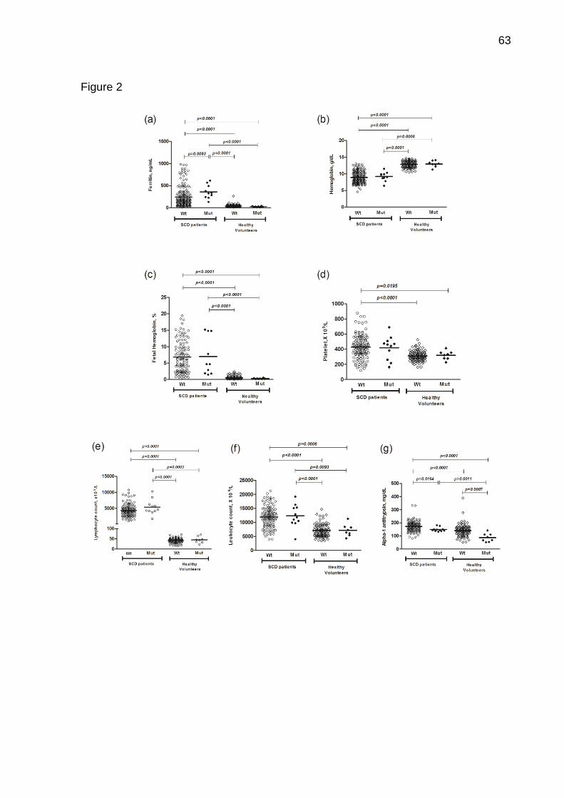
63
Figure 2

64
Figure 3
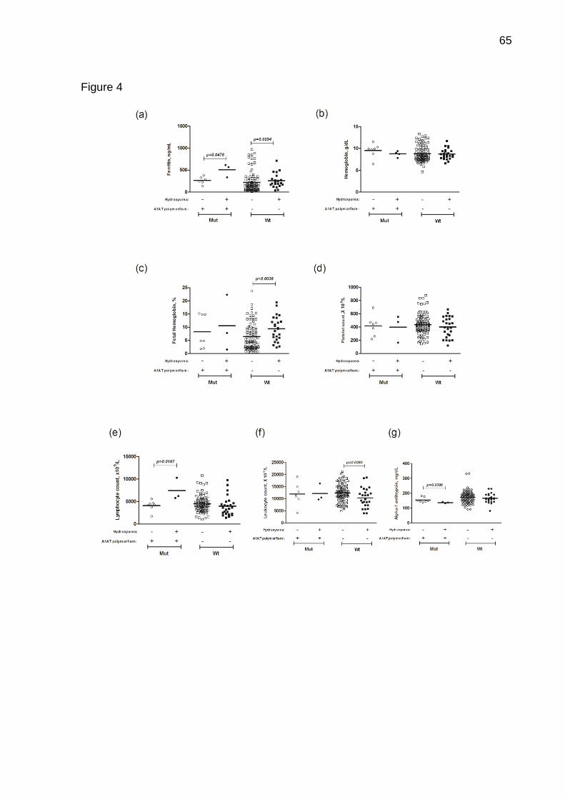
65
Figure 4

66
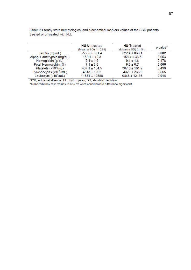
67

68

69

70
6.2 Manuscrito 2
Título: Sickle Red Cells as Danger Signals for Toll-Like Receptors Gene Expression:
Is Hydroxyurea Beneficial?
Autores: Pitanga, T.N.; Oliveira, R.R; Zanette, D.L.; Guarda, C.C.; Santiago, R.P;
Santana, S.S; Lopes, M.V.; Lima, J.B.; Carvalho, G.Q.; Carvalho, M.O.S.; Maffili,
V.V.; Alcântara, L.C.J.; Borges, V.M.; Goncalves, M.S.
Situação: A ser submetido
Objetivos: (referentes aos objetivos 2 a 4 da tese)
1. Determinar a expressão de genes relacionados aos receptores do tipo Toll células
mononucleares (PBMC) de pacientes com anemia falciforme e de voluntários
sadios;
2. Avaliar o efeito da adição de hemácias falcizadas na expressão de genes
relacionados aos receptores do tipo Toll em cultura de PBMC de voluntários
sadios, tratada ou não com hidroxiureia;
3. Avaliar moléculas inflamatórias (LTB4 e nitrito) em sobrenadante de cultura de
células mononucleares incubadas com hemácias falcizadas, lisadas ou íntegras,
tratadas ou não com hidroxiureia.
Principais resultados: Células mononucleares (PBMC) de pacientes com AF
apresentam expressão aumentada de receptores tipo Toll (TLR) 2, TLR4 e TLR5 e
hemácias AF induzem expressão gênica de TLR9 em PBMC de voluntários sadios e
aumentam secreção de leucotrieno B4. O tratamento com hidroxiureia aumenta a
expressão de TLR2 e reduz produção de nitrito em PBMC de voluntários sadios
tratados com hemácias AF.

71
Sickle Red Cells as Danger Signals for Toll-Like Receptors Gene Expression: Is
Hydroxyurea Beneficial?
AUTHORS
Thassila Nogueira Pitanga,1,2 Ricardo Riccio Oliveira,1 Dalila Lucíola Zanette,1
Caroline Conceição da Guarda,1 Rayra Pereira Santiago,1 Sânzio Silva Santana,1
Valma M. Lopes do Nascimento,3 Jonilson Berlink Lima,1 Graziele Quintela de
Carvalho,1 Magda O. S. Carvalho,4 Vitor Valério Maffili,1 Luiz Carlos Júnior
Alcântara,1 Valéria Matos Borges1 and Marilda de Souza Gonçalves1,5
AUTHOR AFFILIATIONS
1Fundação Oswaldo Cruz, Centro de Pesquisas Gonçalo Moniz –
FIOCRUZ/BA; 2Instituto de Ciências da Saúde (ICS), Universidade Federal da Bahia
(UFBA); 3Fundação de Hematologia e Hemoterapia da Bahia (HEMOBA); 4Hospital
Universitário Professor Edgard Santos (HUPES), UFBA; 5Faculdade de Farmácia,
UFBA, Salvador, Bahia, Brazil.
RUNNING TITLE: Sickle cell disease drives Toll signaling
CORRESPONDENCE: Marilda S. Gonçalves, PhD, Centro de Pesquisas Gonçalo
Moniz, LHGB, Waldemar Falcão St, n. 121, Candeal, Salvador, Bahia, Brasil. Tel:
+55 (71) 3176-2226, e-mail: [email protected]
CONFLICT OF INTEREST: All authors declare no conflicts of interest.
FUNDING: This work was approved by the Research Ethics Committee (CAAE:
04733612.7.0000.0040) and was supported by grants from the Brazilian National
Council of Research (CNPq) (311888/2013-5) (M.S.G.); the Foundation of Research
and Extension of Bahia (FAPESB) (3626/2013, 1431040053063, and 9073/2007)
(M.S.G.); and PPSUS/FAPESB (020/2013 EFP_00007295), (M.S.G.); the Instituto
Nacional de Ciência e Tecnologia do Sangue (CNPq) (Coordinated by S.T.O.S.), and
MCD/CNPq/MS-SCTIE-DECIT (409800/2006-6), (M.S.G.). Sponsors of this study are
public or nonprofit organizations that support science in general. They had no role in
gathering analyzing, or interpreting the data.
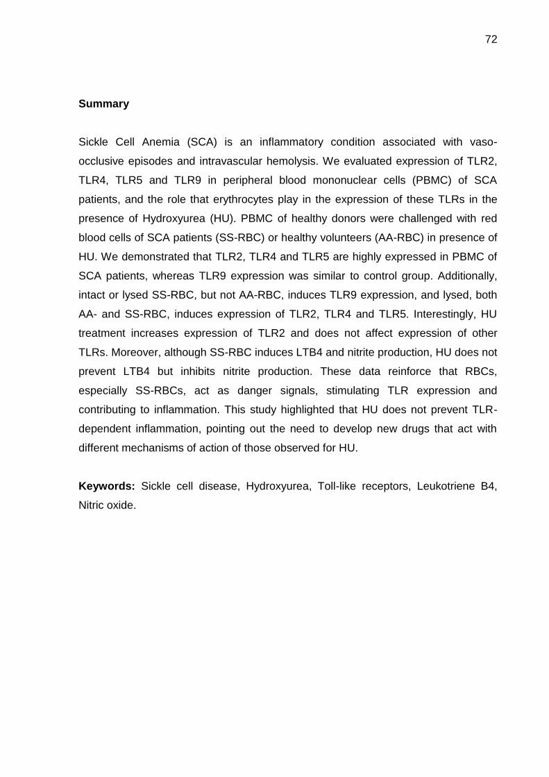
72
Summary
Sickle Cell Anemia (SCA) is an inflammatory condition associated with vaso-
occlusive episodes and intravascular hemolysis. We evaluated expression of TLR2,
TLR4, TLR5 and TLR9 in peripheral blood mononuclear cells (PBMC) of SCA
patients, and the role that erythrocytes play in the expression of these TLRs in the
presence of Hydroxyurea (HU). PBMC of healthy donors were challenged with red
blood cells of SCA patients (SS-RBC) or healthy volunteers (AA-RBC) in presence of
HU. We demonstrated that TLR2, TLR4 and TLR5 are highly expressed in PBMC of
SCA patients, whereas TLR9 expression was similar to control group. Additionally,
intact or lysed SS-RBC, but not AA-RBC, induces TLR9 expression, and lysed, both
AA- and SS-RBC, induces expression of TLR2, TLR4 and TLR5. Interestingly, HU
treatment increases expression of TLR2 and does not affect expression of other
TLRs. Moreover, although SS-RBC induces LTB4 and nitrite production, HU does not
prevent LTB4 but inhibits nitrite production. These data reinforce that RBCs,
especially SS-RBCs, act as danger signals, stimulating TLR expression and
contributing to inflammation. This study highlighted that HU does not prevent TLR-
dependent inflammation, pointing out the need to develop new drugs that act with
different mechanisms of action of those observed for HU.
Keywords: Sickle cell disease, Hydroxyurea, Toll-like receptors, Leukotriene B4,
Nitric oxide.

73
Introduction
Sickle Cell Anemia (SCA) is a genetic disorder characterized by the presence
of homozygous hemoglobin S (HbS). Clinically, SCA patients could be in a steady
state, with no clinical manifestations, or in crisis, showing systemic inflammation
coinciding with vaso-occlusive phenomena and painful episodes, as well as
susceptibility to infections and intravascular hemolysis (PLATT, 2000;
WEATHERALL, PROVAN, 2000; TAYLOR et al., 2008; CONNES et al., 2014). This
disease has been characterized as a chronic inflammatory state, coinciding with
abnormal activation and elevated number of peripheral blood mononuclear cells
(PBMCs), endothelial dysfunction and an increased level of multiple inflammatory
mediators (ZENNADI et al., 2008; VILAS-BOAS et al., 2010; CERQUEIRA et al.,
2011; PITANGA et al., 2013).
Endothelial cells, leukocytes and platelets are constantly exposed to toxic
products of sickle red cell, specially the heme group, released during intravascular
hemolysis, which are frequently observed in SCA patients, and leads to systemic
inflammation and pro-thrombotic phenomena (BELCHER et al., 2003; WOOD,
HEBBEL, GRANGER, 2004). This condition is associated with increased levels of
nitric oxide (NO) metabolites and leukotriene (LT) B4 even in steady state, which is
worsened during vaso-occlusion episodes (IBE et al., 1994; SETTY, STUART, 2002;
GLADWIN, KATO, 2005). Free heme reacts with NO, an important molecule involved
in vascular tone and the maintenance of vascular homeostasis, to form its
metabolites (nitrite and nitrate), thereby reducing its bioavailability. Moreover, heme
induces LT production, an inflammatory mediator derived from arachidonic acid
metabolism, which induces leucocytes migration and activation (GRACA-SOUZA et
al., 2002; EBERHARDT et al., 2003; MONTEIRO et al., 2011; RAAT et al., 2013).
It has been proposed that sickle red blood cells (SS-RBC) could act as an
inflammation inducer agent, stimulating production of LT and thromboxane A2,
resulting in both hemolysis and vaso-occlusive episodes. SS-RBC exhibit abnormal
adhesion to extracellular matrix proteins, such as thrombospondin, which increases
pain episodes in SCA patients (STEINBERG, BRUGNARA, 2003; TELEN, 2005;
CHAAR et al., 2010; OPENE et al., 2014). Since the presence of sickle red cell is a
typical feature in sickle cell disease (SCD) patients, more investigation is necessary

74
in order to identify its role in the amplification and maintenance of inflammatory
immune response within these conditions.
The immune response, particularly the innate immune response through
innate receptors, such as NOD-like (NLR) and Toll-like receptors (TLR), plays
important role in the initiation and maintenance of the inflammatory process observed
in SCA patients. Since TLRs are a family of evolutionary conserved proteins that
recognize pathogen- (PAMPs) and danger- (DAMPs) associated molecular patterns,
it is expected that TLR plays a critical role in SCA patients (BAHL et al., 2011;
BELCHER et al., 2014; GUPTA, 2014; XU et al., 2014; VICARI et al., 2015).
In humans, ten TLRs (TLR1 to TLR10) were reported, each of which has
certain specificity for a particular PAMP or DAMP. Several studies have shown
association between expression of these receptors and inflammatory conditions. In
SCD, for instance, TLR4 expression is 100 fold higher in patients compared to
healthy controls, and the mortality and inflammatory condition is directly associated
with high intracellular iron (VAN BEERS et al., 2015). TLR4 is a receptor that
recognizes one of the most well-known PAMP, the lipopolysaccharide (LPS).
However, TLR4 and other TLRs were recently shown to recognize cellular residues
and other endogenous molecules, acting as signals for tissue injury. Heat shock
protein (HSP), high-mobility group protein B1 (HMGB1), oxidized low-density
lipoprotein (oxLDL) and heme are DAMPs considered as TLR4 agonists (IMAI et al.,
2008; LEFEBVRE et al., 2011; BELCHER et al., 2014; GUPTA, 2014; YANG et al.,
2014).
Although TLR4 is the best well characterized TLR in SCD, other TLRs may
play a role in the immunopathogenesis of the disease, contributing to its severity and
therapeutic failure. In Brazil, hydroxyurea (HU) is the drug approved for treatment of
SCA patients. HU is a cytotoxic agent that inhibits ribonucleotide reductase enzyme
and leads to improvement in levels of fetal hemoglobin (HbF), in the hydration of
RBC and reduces SS-RBCs-endothelial interaction (PLATT, 2008; STEINBERG et
al., 2010; SHETH, LICURSI, BHATIA, 2013). Such effects are associated with
improvement of clinical status of the patient and reduction of inflammatory markers
(DE MONTALEMBERT et al., 2006; PLATT, 2008). However, it is not known whether
HU interferes with TLR signaling pathways.
The role that inflammatory receptors, such as TLRs play in the
immunopathogenesis of SCA, as well as the contribution of RBC from SCA patients

75
to the induction of inflammation and the ability of HU to revert this scenario, are still
not clear. In this study, we evaluated the expression of TLR2, TLR4, TLR5 and TLR9
in PBMC of SCA patients. Moreover, we tested the role that RBC obtained from SCA
patients play in the expression of these TLRs in PMBC from healthy donors, as well
as the in vitro effect of hydroxyurea in the expression of these inflammatory markers.
Material and methods
Study subjects
A cross sectional study was performed to include 12 sickle cell anemia (SCA)
patients (age 9.1 ± 4.7 years), recruited at the Hematology and Hemotherapy
Foundation of the State of Bahia (HEMOBA). All patients were in steady-state,
characterized by absence of blood transfusion in a period of four months prior to
blood draw. In addition, patients included in this study did not show any infection,
hospitalization or vaso-occlusive event, and were not under antibiotics,
corticosteroids or hydroxyurea treatments, but all patients were under treatment with
folic acid. The control group consisted of nine healthy volunteers (age 12.4 ± 5.6
years) recruited at the Pharmacy College (FacFAR) of Federal University of Bahia of
Bahia (UFBA). This group was characterized by absence of hematological disorders
or inflammatory conditions. This study was approved by the Research Ethics
Committee of the Oswaldo Cruz Foundation, and was developed in accordance with
the Declaration of Helsinki of 1975 as revised in 2000. All subjects (or their parents/
legal guardians) agreed to participate after reading the terms in the informed consent.
Blood samples and preparation of red blood cells
Venous blood was collected from patients and controls using the anticoagulant
ethylene-diamine-tetraacetic-acid (EDTA) in order to determine hemoglobin profile,
and to obtain red blood cells (RBC) and peripheral blood mononuclear cells (PBMC).
Additionally, all PBMCs used for cell culture (PBMC culture) experiments were
obtained from fresh leukocytes concentrates (buffy coat) of healthy blood donors of
HEMOBA. The buffy-coat samples were serological tested and negative for HIV,
Human T lymphotropic virus type 1 (HTLV), hepatitis B (HBV), hepatitis C (HCV),
syphilis and Chagas disease.
76
In order to obtain RBC, each blood sample was centrifuged at 270 g for 10 min
to discard the platelet-rich plasma (PRP). Leukocytes and PRP were removed by
aspiration, and RBC were washed 3 times in sterile PBS (pH 7.3). RBC concentrate
were assessed for leukocyte and platelet contamination by staining with trypan blue
and then analyzed by using a phase contrast microscope (Olympus CK2, Center
Valley, PA, USA). For the experiments with lysed erythrocytes, 500µL of RBC
concentrate was kept at -70°C for 3 hours and subsequently thawed to obtain lysed
RBC (BENSOUDA, LAATIRIS, 2006). The purity of erythrocyte lysis (> 90%) was
evaluated by microscopic examination of cells stained with trypan blue.
Hematological and biochemical analysis performed
Hematological and biochemical parameters from patients and controls included
in this studied was analyzed. RBC, platelets, leucocyte, reticulocyte, hemoglobin,
fetal hemoglobin, hematocrit, ferritin and C-reactive protein of the SCA patients and
controls are shown in Table 1. The mean age between patients and healthy
volunteers was similar.
Hematologic values and RBC indices were determined by using the electronic
counter ABX Pentra and morphological analysis of red blood cells was performed by
microscopic examination of blood smears stained with Wright. The hemoglobin profile
was confirmed by high-performance liquid chromatography (HPLC) in automated
equipment (Variant I - Bio-rad). Reticulocyte counts were determined using supravital
staining. Biochemical analyzes were determined by automated method and included
the C-reactive protein. These analyses were performed using A25 equipment (AS
Biosystems, Barcelona, Spain) at the Laboratory of Biochemistry, Faculty of
Pharmacy, Federal University of Bahia.
Peripheral blood mononuclear cells separation
Mononuclear cells were obtained from peripheral blood of healthy volunteers
and SCA patients and buffy coat bags of healthy blood donors. Initially, whole blood
was centrifuged at 270 g for 10 min to discard the platelet-rich plasma. The leukocyte
concentrate was diluted in 0.9% saline solution buffered with phosphate (PBS) and
centrifuged on Ficoll-Hypaque (GE Healthcare Bio-Sciences Corp. Piscataway, NJ,
USA) 500 g for 30 minutes at 25°C. The mononuclear cells ring was then centrifuged
at 500 g for 10 minutes. Pellet was collected and resuspended in RPMI-1640 medium

77
(Grand Island, NY, USA) supplemented with 10% fetal bovine serum (FBS) (Sigma-
Aldrich, Piscataway, USA) and with 100 U/ml penicillin and 100 µg/ml streptomycin
(Grand Island, NY, USA).
PBMC from buffy coat bags were counted, distributed in 24-well plate (Costar,
Corning, NY, USA) at a concentration of 3x10⁶ mononuclear cells/mL and cultured
with 3% lysed or intact RBC for 24h at 37°C and 5% CO2 cultivated with or without
100 µM hydroxyurea (HU) (Sigma-Aldrich, Saint Louis, MO, USA) (STEINBERG et
al., 2010).
Expression of mRNA for TLR2, TLR4, TLR5 and TLR9
Total mRNA extraction from PBMC was performed using Trizol Reagent
(Invitrogen, Life Technologies, CA, USA) according to the manufacturer's instructions
and measured by NanoDrop™ Lite Spectrophotometer (Thermo Fisher Scientific,
Wilmington, USA). Reverse transcription was performed with 400 ng of total RNA
using the High Capacity cDNA Reverse Transcription Kit (Applied Biosystems TM,
Foster City, CA) according to the manufacturer's instructions. The conditions used
were 25°C for 10 minutes, 37°C for 120 minutes and 85°C for 5 minutes.
Gene expression analysis was performed by quantitative real-time PCR
(qPCR) on an ABI 7500 FAST Real-Time PCR equipment (Applied BiosystemsTM,
Foster City, CA) for TLR2, TLR4, TLR5, TLR9, GAPDH and β-ACTIN genes with
Power SYBR® Green, according to the manufacturer’s instructions. The standard
qPCR conditions were as follows: 10 min at 95oC, followed 40 cycles at 95oC for 15
sec, 60oC for 60 sec. Primers used for qPCR reactions are shown in Table 2. After
amplification and dissociation curve runs, the values of threshold cycle (Ct) were
obtained by using the Operational Programme 7500™ System (Applied Biosystems,
USA). The intra-assay precision was calculated using the equation E=(-1/slope) to
confirm precision and reproducibility of qPCR. The expression levels were
normalized based on the geometric mean of GAPDH and β-ACTIN (endogenous
controls). Relative expression folds were calculated based on 2^-ddCt method
(PFAFFL, 2001).
Quantification of leukotriene-B4 and nitrite production in culture supernatants
PBMC from buffy coat bags was cultured with 3% of lysed or intact RBC for
24h at 37°C and 5% CO2, in the presence or absence of 100 µM hydroxyurea.
78
Supernatants were collected and used for the detection of leukotriene-B4 (LTB4) by
EIA in accordance with the manufacturer's instructions (Cayman Chemical Company,
MI, USA). NO production was assessed in culture supernatants by determining nitrite
accumulation, using the Griess method (DING, NATHAN, STUEHR, 1988).
Statistical analysis
Average values of quantitative variables between groups were compared using
the Student's t test for unpaired data with normal distribution and the Mann-Whitney
test for non-normally distributed data. Kruskal-Wallis test was used for comparison
tests between two or more groups, non-parametric quantitative variables. Values of
p<0.05 were considered a significant difference. All data were analyzed using Prism
5.1 software (GraphPad, San Diego, USA).
Results
TLR2, TLR4 and TLR5 are highly expressed in PBMC of SCA patients
SCA is an inflammatory condition in which the innate immune response may
play an important role, both in initiating and maintaining the pathological response.
For this reason, we first examined whether the expression of TLR2, TLR4, TLR5 and
TLR9, assessed by qPCR, differed between peripheral blood mononuclear cells
obtained from SCA patients (SS-PBMC) and healthy controls (AA-PBMC).
Expression of TLR2, TLR4 and TLR5 was significantly higher in SS-PBMC
(0.46 ± 0.17; 47.50 ± 21.46 and 0.88 ± 0.23) when compared to AA-PBMC (0.20 ±
0.07; 20.60 ± 1.75 and 0.37 ± 0.09, respectively, p < 0.05). Interestingly, gene
expression of each of these TLRs was approximately 2.3 times higher in SS-PBMC
than AA-PBMC. On the other hand, TLR9 was equally expressed in both SS-PBMC
and AA-PBMC (Fig 1).
SS-RBC induces TLR9 expression
Assuming that TLRs are highly expressed in cells of SCA patients, we next
tested whether red blood cells from these patients (SS-RBC) are able to induce
expression of TLR2, TLR4, TLR5 or TLR9 in AA-PBMC. For this purpose, we isolated
AA-PBMC from buffy coat obtained from regular blood donators and incubated these
cells in the presence of intact or lysed SS-RBC or RBC from healthy controls (AA-

79
RBC). Since intact SS-RBC are morphologically distinct from AA-RBC, and
intracellular content of RBC are able to induce inflammatory response mediated by
innate receptors (POTOKA, GLADWIN, 2015), we hypothesized that both intact and
lysed SS-RBC could promote TLRs expression in AA-PBMC.
Intact AA-RBC or SS-RBC were not able to induce expression of TLR2 (Fig 2A),
TLR4 (Fig 2C) or TLR5 (Fig 2D), when compared to basal expression of these
receptors observed in unstimulated AA-PBMC. On the other hand, lysed AA-RBC
and SS-RBC were both good inductors of TLR2, TLR4 and TLR5 expression (Figs
2B, 2D and 2F). This data suggests that intracellular content of RBC, regardless of
being from SS or AA individuals, has similar stimulatory effects on the expression of
these TLRs, which are not observed when stimulating AA-PBMC with intact RBCs
Nevertheless, it was noteworthy that both intact and lysed SS-RBC, but not AA-
RBC, were able to increase TLR9 expression, almost four times, when compared to
unstimulated AA-PBMC (p < 0.05; Fig 2G and 2H). Therefore, SS-RBC is a good
inductor of TLR9 expression, an effect that is not observed for AA-RBC. Moreover,
this peculiarity is probably mediated by a danger signal that is present in RBC
surface or actively released in the medium, as well as by intracytoplasmic molecules.
HU treatment increases expression of TLR2
Hydroxyurea (HU) is an antineoplastic drug used for treatment of SCA patients
and its main expected beneficial effect is improvement in HbF levels. However, it is
not clear if HU interferes with the inflammatory condition of these patients. In order to
answer this question we incubated AA-PBMC with intact or lysed SS-RBC, treated or
not with HU. AA-PBMC incubated with intact SS-RBC and treated with HU showed
higher TLR2 gene expression (1.03 ± 0.20) than untreated AA-PBMC stimulated with
intact SS-RBC (0.78 ± 0.33; p < 0.05; Fig 3A). This effect, however, was not
observed when AA-PBMC was incubated with lysed SS-RBC. Lysed SS-RBC induce
TLR2 expression, but treatment with HU does not down modulate TLR2 expression
(Fig 3B).
Expression of TLR4 and TLR5 were not affected by HU treatment, in both intact
and lysed SS-RBC situations (Figs 3C, 3D, 3E and 3F). However, although HU
showed no significant modulation of TLR9 expression when AA-PBMC was
incubated with intact SS-RBC (Fig 3G), a different scenario was observed for the
expression of TLR9 in AA-PBMC cultured with lysed SS-RBC (Fig 3H). Despite lysed
80
SS-RBC were able to induce TLR9 expression, treatment with HU reduced
expression of this receptor in AA-PBMC to similar levels to those seen in
unstimulated AA-PBMC.
HU does not prevent RBC-induced LTB4 but reduces nitrite production
SCA patients present increased NO metabolites and LTB4 concentrations (IBE
et al., 1994; SETTY, STUART, 2002; GLADWIN, KATO, 2005). Since LTB4 is a lipid
involved in inflammation, we tested whether AA- or SS-RBC were able to induce its
production, and if HU was able to reverse this condition. As shown in Fig 4A, intact
SS-RBC induced higher production of LTB4 when compared to AA-RBC (p<0.05).
However, when lysed SS-RBC were used, concentration of LTB4 was significantly
reduced when compared to AA-PBMC incubated with lysed AA-RBC (Fig 4B).
Interestingly, levels of LTB4 did not differ between cultures stimulated with intact or
lysed AA-RBC, whereas under SS-RBC incubation, production of LTB4 was higher
when PBMC were stimulated with intact than lysed RBC (Figs 4A and 4B). This effect
was not reversed by adding 100 µM of hydroxyurea, both in intact and lysed SS-RBC
conditions.
We also evaluated the concentration of nitrite, NO metabolism product, in the
supernatants of the PBMC cultured with RBC. Differently from what was observed for
LTB4, intact SS-RBC induced significantly higher production of nitrite compared to
AA-RBC (p<0.05; Fig 5A). However, when lysed RBCs were used, nitrite production
was equally high on both AA-RBC or SS-RBC conditions (Fig 5B). Moreover, it was
noteworthy that HU treatment was able to decrease nitrite production when AA-
PBMC was stimulated with intact or lysed SS-RBC, compared to HU-untreated
cultures (Figs 5C and 5D).
Discussion
Innate immune system acts as a first line of defense against pathogens, by
recognizing pathogen-associated molecular patterns (PAMPs), but also senses
damage associated molecular patterns (DAMPs) from sterile tissue injury. Upon its
activation by innate receptors such as Toll like receptors (TLRs), the inflammatory
response is initiated and maintained, for as long as the triggering agent is present.

81
The inflammation in sickle cell anemia is proposed to be dependent on TLR signaling
since erythrocyte or its contents released during hemolysis could be recognized as
DAMPs (VANCE, ISBERG, PORTNOY, 2009; CERQUEIRA et al., 2011; GUPTA,
2014; CHEN et al., 2015). In this study we demonstrated that TLRs expression is
activated in sickle cell anemia (SCA) and in AA-PBMC cultured with red blood cells
(RBCs).
Mononuclear cells (PBMC) of SCA patients (SS-PBMC) have high expression
of TLR2, TLR4 and TLR5. TLR2 is important for the response against Gram-positive
bacteria as well as for nonpathogenic inflammation by identifying different DAMPs.
This receptor can form heterodimers with TLR4, which are able to recognize
hemoglobin (TAKEUCHI et al., 1999; VAN BERGENHENEGOUWEN et al., 2013;
WANG et al., 2014). Although LPS is a well described PAMP for TRL4, there are
other agonists even in sterile conditions, such as high mobility group box 1 (HMGB1),
heat shock protein (HSP) and heme (LIEW et al., 2005; BELCHER et al., 2014;
GUPTA, 2014). Similarly, despite TLR5 is important for defense against pathogens,
there are evidences that this receptor is a severity and prognostic biomarker for some
inflammatory conditions (PIMENTEL-NUNES et al., 2011; WANG et al., 2012;
KAUPPILA et al., 2013; SHERIDAN et al., 2013). Therefore, the expression of these
receptors in SS-PBMC patients could be due to continuous exposure to intracellular
content of erythrocytes released during intravascular hemolysis or due to exposure to
sickle red blood cells (SS-RBCs).
To test this hypothesis, we performed an experiment where PBMC from healthy
donors was cultured with lysed or intact SS-RBCs. Interestingly, although expression
of TLR9 was similar between SS-PBMC and PBMC from healthy volunteers (AA-
PBMC) both intact and lysed SS-RBCs were able to upregulate TLR9 expression in
cultured AA-PBMC. This was not observed with TLR2, TLR4 and TLR5 gene
expression when using intact RBCs. TLR9 is a receptor that binds to CpG-DNA,
which is frequent in bacteria and rare in eukaryotic cells (BAUER, 2013). In addition,
although some authors suggest that hemozoin, a heme dimer, acts as TLR9 agonist
(COBAN et al., 2010; KALANTARI et al., 2014), in an experimental model of TLR9-/-,
it was demonstrated that heme-induced inflammation is not dependent on this
receptor (FIGUEIREDO et al., 2007). Therefore, the upregulation of TLR9 expression
observed in our study was probably not due to the presence of heme. This
hypothesis is supported by the observation that expression of TLR9 was also

82
increased when adding intact SS-RBCs, situation where there are no free heme in
the medium.
It is well known that SCA patients have increased reticulocyte counts,
cytoplasmic inclusion bodies such as the DNA-rich Howell-Jolly bodies and high
levels of fetal hemoglobin (HbF) (STEINBERG, 2005; HARROD et al., 2007;
ROGERS et al., 2011). Moreover, the 5’-globin promoter of HbF presents a high
local concentration of CpG residues, which are absent in β-like globin gene
promoters (SAUNTHARARAJAH, LAVELLE, DESIMONE, 2004; LESSARD et al.,
2015). Thus, since CpG-DNA is a TLR9 agonist, and considering that the expression
of this receptor was increased when using intact or lysed SS-RBCs, but not when
adding AA-RBC, it is plausible that CpG-DNA released from SS-RBCs could be
inducing TLR9 expression probably by a positive paracrine feedback mechanism.
Intracytoplasmic content of AA-RBCs or SS-RBCs was able to induce TLR2,
TLR4 and TLR5 expression, suggesting that there are no significant differences in
their composition. However, it is noteworthy that intravascular hemolysis does not
occur physiologically in healthy volunteers, whereas a great proportion of SCA
patients exhibit this condition (TAYLOR et al., 2008; VILAS-BOAS et al., 2010).
Hence, the inflammation observed in SCA patients could be a consequence of
constant exposure to RBCs’ intracytoplasmic content, such as heme (BELCHER et
al., 2014), during intravascular hemolysis, leading to the upregulation of toll-like
receptors’ expression. Additionally, intravascular hemolysis induces release of
HMGB1, a nuclear protein involved in regulation of transcription, which, similar to
heme, acts as a DAMP and activates TLR4 (XU et al., 2014). In this study we
showed that, in addition to TLR4, the expression of TLR2, TLR5 and TLR9 contribute
to the inflammatory condition, and could aggravate patient’s condition.
Pharmacological control of SCA patients clinical status is achieved mainly by
hydroxyurea (HU), an antineoplastic drug that increases HbF levels and reduces SS-
RBCs-endothelial interaction (PLATT, 2008; STEINBERG et al., 2010). In this study,
we showed that intact SS-RBCs are not able to induce TLR2 expression; however,
treatment with HU significantly increased expression of this receptor. TLR2 is
associated with NF-B activation through the MyD88-dependent pathway, stimulates
the production of proinflammatory cytokines such as interleukin (IL)-12 and tumor
necrosis factor (TNF) (LIEW et al., 2005). Moreover, we demonstrated that HU does
not decrease the expression of TLR2, TLR4, TLR5 or TLR9, suggesting that this

83
treatment, in addition to increasing TLR2 expression, is not able to reduce TLR-
mediated inflammation observed in SCA patients.
Leukotrienes (LT) are an important mediators of inflammation, and LTB4
promotes leukocyte migration and adhesion to the vascular endothelium (SETTY,
KULKARNI, STUART, 2002). Our observation that SS-RBC increase LTB4
production in AA-PBMC compared to AA-RBC is corroborated by the reported of LT
production are augmented in PBMC cultured with intact SS-RBC (OPENE et al.,
2014). We demonstrated here that intact SS-RBCs induced twice more the
production of LTB4 than AA-RBCs, whereas lysed SS-RBC induced three times less
this inflammatory mediator, and treatment with HU does not interfere in this
parameter. Since LTB4 is associated with SCA immunopathogenesis (SETTY,
STUART, 2002; KNIGHT-PERRY et al., 2009), our data suggest that, as observed
for TLRs, HU treatment does not reverse the inflammatory scenario.
Another mediator associated with inflammatory condition is NO, which acts
inducing vasodilation, inhibiting platelet activation, and thus controlling inflammation.
During hemolysis, RBC products deplete NO to form its metabolites, such as nitrite,
reducing NO bioavailability, which may promote vaso-occlusion episodes (GLADWIN
et al., 2003; KATO, GLADWIN, STEINBERG, 2007; ELIAS et al., 2012). In our study,
we showed that SS-RBCs/AA-PBMC system induced higher levels of nitrite when
compared to AA-RBC/AA-PBMC cultures. However, HU treatment was able to
reduce nitrite production, presumably because HU is a NO donator, improving its
bioavailability (GLADWIN et al., 2002; BURKITT, RAAFAT, 2006). Once NO is
associated with vascular homeostasis, and that the reduction of nitrite levels
implicates in higher concentration of bioactive NO (REITER, GLADWIN, 2003), our
data suggest a beneficial effect of HU treatment in regard to this parameter,
corroborating with other studies (GLADWIN et al., 2002).
SCA is an inflammatory and complex condition considered a public health
problem and associated with increased morbidity and mortality. Despite its global
relevance, mechanisms that contribute to disease severity are poorly understood. In
an attempt to contribute to the field, our data reinforce the hypothesis that red blood
cells, especially sickle RBCs, act as DAMPs, stimulating TLR expression and
contributing to inflammation. Furthermore, although HU treatment improves several
parameters in SCA patients, we highlighted here that HU does not prevent TLR-
84
dependent inflammation, pointing out the need to develop new drugs that act with
different mechanisms of action of those observed for HU.
AUTHORS’ CONTRIBUTIONS
TNP, RRO and MSG: conceived the study design. TNP: performed all the
experiments. DLZ, CCG, RPS and SSS: assisted in all experiments. VML:
responsible for patients’ follow-up. VVM, JBL and GQC: discussion and
accomplishment of some experiments. MOSC, LCJA and VMB: discussion of study
design, experiments and all results. TNP, RRO and MSG: wrote the paper. All
authors read and approved the final manuscript.
ACKNOWLEDGEMENTS
We are greatly grateful to SCA patients and all healthy volunteers for trust us and for
making possible the realization of this work. We thank the laboratory technicians for
help us in the collection of the samples. We would also like to thank Marcia Weber
and Cyntia Cajado for excellent discussions. TNP received a scholarship from
CAPES. L.C.J.A., V.M.B. and MSG have productivity scholarship from CNPq.
85
REFERENCES
BAHL, N.; DU, R.; WINARSIH, I.; HO, B.; TUCKER-KELLOGG, L.; TIDOR, B.; DING, J. L. (2011). Delineation of lipopolysaccharide (LPS)-binding sites on hemoglobin: from in silico predictions to biophysical characterization. J Biol Chem 286(43): 37793-37803. BAUER, S. (2013). Toll-like receptor 9 processing: the key event in Toll-like receptor 9 activation? Immunol Lett 149(1-2): 85-87. BELCHER, J. D.; BRYANT, C. J.; NGUYEN, J.; BOWLIN, P. R.; KIELBIK, M. C.; BISCHOF, J. C.; HEBBEL, R. P.; VERCELLOTTI, G. M. (2003). Transgenic sickle mice have vascular inflammation. Blood 101(10): 3953-3959. BELCHER, J. D.; CHEN, C.; NGUYEN, J.; MILBAUER, L.; ABDULLA, F.; ALAYASH, A. I.; SMITH, A.; NATH, K. A.; HEBBEL, R. P.; VERCELLOTTI, G. M. (2014). Heme triggers TLR4 signaling leading to endothelial cell activation and vaso-occlusion in murine sickle cell disease. Blood 123(3): 377-390. BENSOUDA, Y.; LAATIRIS, A. (2006). The lyophilization of dispersed systems: influence of freezing process, freezing time, freezing temperature and RBCs concentration on RBCs hemolysis. Drug Dev Ind Pharm 32(8): 941-945. CERQUEIRA, B. A.; BOAS, W. V.; ZANETTE, A. D.; REIS, M. G.; GONCALVES, M. S. (2011). Increased concentrations of IL-18 and uric acid in sickle cell anemia: contribution of hemolysis, endothelial activation and the inflammasome. Cytokine 56(2): 471-476. CHEN, R.; FU, S.; FAN, X. G.; LOTZE, M. T.; ZEH, H. J., 3rd; TANG, D.; KANG, R. (2015). Nuclear DAMP complex-mediated RAGE-dependent macrophage cell death. Biochem Biophys Res Commun 458(3): 650-655. COBAN, C.; IGARI, Y.; YAGI, M.; REIMER, T.; KOYAMA, S.; AOSHI, T.; OHATA, K.; TSUKUI, T.; TAKESHITA, F.; SAKURAI, K.; IKEGAMI, T.; NAKAGAWA, A.; HORII, T.; NUNEZ, G.; ISHII, K. J.; AKIRA, S. (2010). Immunogenicity of whole-parasite vaccines against Plasmodium falciparum involves malarial hemozoin and host TLR9. Cell Host Microbe 7(1): 50-61. DE MONTALEMBERT, M.; BROUSSE, V.; ELIE, C.; BERNAUDIN, F.; SHI, J.; LANDAIS, P. (2006). Long-term hydroxyurea treatment in children with sickle cell disease: tolerance and clinical outcomes. Haematologica 91(1): 125-128. DING, A. H.; NATHAN, C. F.; STUEHR, D. J. (1988). Release of reactive nitrogen intermediates and reactive oxygen intermediates from mouse peritoneal macrophages. Comparison of activating cytokines and evidence for independent production. J Immunol 141(7): 2407-2412. FIGUEIREDO, R. T.; FERNANDEZ, P. L.; MOURAO-SA, D. S.; PORTO, B. N.; DUTRA, F. F.; ALVES, L. S.; OLIVEIRA, M. F.; OLIVEIRA, P. L.; GRACA-SOUZA, A. V.; BOZZA, M. T. (2007). Characterization of heme as activator of Toll-like receptor 4. J Biol Chem 282(28): 20221-20229. GLADWIN, M. T.; SHELHAMER, J. H.; OGNIBENE, F. P.; PEASE-FYE, M. E.; NICHOLS, J. S.; LINK, B.; PATEL, D. B.; JANKOWSKI, M. A.; PANNELL, L. K.; SCHECHTER, A. N.; RODGERS, G. P. (2002). Nitric oxide donor properties of hydroxyurea in patients with sickle cell disease. Br J Haematol 116(2): 436-444.

86
GUPTA, K. (2014). HMGB1 takes a "Toll" in sickle cell disease. Blood 124(26): 3837-3838. HARROD, V. L.; HOWARD, T. A.; ZIMMERMAN, S. A.; DERTINGER, S. D.; WARE, R. E. (2007). Quantitative analysis of Howell-Jolly bodies in children with sickle cell disease. Exp Hematol 35(2): 179-183. IMAI, Y.; KUBA, K.; NEELY, G. G.; YAGHUBIAN-MALHAMI, R.; PERKMANN, T.; VAN LOO, G.; ERMOLAEVA, M.; VELDHUIZEN, R.; LEUNG, Y. H.; WANG, H.; LIU, H.; SUN, Y.; PASPARAKIS, M.; KOPF, M.; MECH, C.; BAVARI, S.; PEIRIS, J. S.; SLUTSKY, A. S.; AKIRA, S.; HULTQVIST, M.; HOLMDAHL, R.; NICHOLLS, J.; JIANG, C.; BINDER, C. J.; PENNINGER, J. M. (2008). Identification of oxidative stress and Toll-like receptor 4 signaling as a key pathway of acute lung injury. Cell 133(2): 235-249. KALANTARI, P.; DEOLIVEIRA, R. B.; CHAN, J.; CORBETT, Y.; RATHINAM, V.; STUTZ, A.; LATZ, E.; GAZZINELLI, R. T.; GOLENBOCK, D. T.; FITZGERALD, K. A. (2014). Dual engagement of the NLRP3 and AIM2 inflammasomes by plasmodium-derived hemozoin and DNA during malaria. Cell Rep 6(1): 196-210. KAUPPILA, J. H.; MATTILA, A. E.; KARTTUNEN, T. J.; SALO, T. (2013). Toll-like receptor 5 (TLR5) expression is a novel predictive marker for recurrence and survival in squamous cell carcinoma of the tongue. Br J Cancer 108(3): 638-643. KNIGHT-PERRY, J.; DEBAUN, M. R.; STRUNK, R. C.; FIELD, J. J. (2009). Leukotriene pathway in sickle cell disease: a potential target for directed therapy. Expert Rev Hematol 2(1): 57-68. LEFEBVRE, J. S.; LEVESQUE, T.; PICARD, S.; PARE, G.; GRAVEL, A.; FLAMAND, L.; BORGEAT, P. (2011). Extra domain A of fibronectin primes leukotriene biosynthesis and stimulates neutrophil migration through activation of Toll-like receptor 4. Arthritis Rheum 63(6): 1527-1533. LESSARD, S.; BEAUDOIN, M.; BENKIRANE, K.; LETTRE, G. (2015). Comparison of DNA methylation profiles in human fetal and adult red blood cell progenitors. Genome Med 7(1): 1. LIEW, F. Y.; XU, D.; BRINT, E. K.; O'NEILL, L. A. (2005). Negative regulation of toll-like receptor-mediated immune responses. Nat Rev Immunol 5(6): 446-458. PIMENTEL-NUNES, P.; AFONSO, L.; LOPES, P.; RONCON-ALBUQUERQUE, R., Jr.; GONCALVES, N.; HENRIQUE, R.; MOREIRA-DIAS, L.; LEITE-MOREIRA, A. F.; DINIS-RIBEIRO, M. (2011). Increased expression of toll-like receptors (TLR) 2, 4 and 5 in gastric dysplasia. Pathol Oncol Res 17(3): 677-683. PLATT, O. S. (2000). Sickle cell anemia as an inflammatory disease. J Clin Invest 106(3): 337-338. PLATT, O. S. (2008). Hydroxyurea for the treatment of sickle cell anemia. N Engl J Med 358(13): 1362-1369. POTOKA, K. P.; GLADWIN, M. T. (2015). Vasculopathy and pulmonary hypertension in sickle cell disease. Am J Physiol Lung Cell Mol Physiol 308(4): L314-324. REITER, C. D.; GLADWIN, M. T. (2003). An emerging role for nitric oxide in sickle cell disease vascular homeostasis and therapy. Curr Opin Hematol 10(2): 99-107. ROGERS, Z. R.; WANG, W. C.; LUO, Z.; IYER, R. V.; SHALABY-RANA, E.; DERTINGER, S. D.; SHULKIN, B. L.; MILLER, J. H.; FILES, B.; LANE, P. A.; THOMPSON, B. W.; MILLER, S. T.; WARE, R. E.; BABY, H. U. G.

87
(2011). Biomarkers of splenic function in infants with sickle cell anemia: baseline data from the BABY HUG Trial. Blood 117(9): 2614-2617. SAUNTHARARAJAH, Y.; LAVELLE, D.; DESIMONE, J. (2004). DNA hypo-methylating agents and sickle cell disease. Br J Haematol 126(5): 629-636. SETTY, B. N.; STUART, M. J. (2002). Eicosanoids in sickle cell disease: potential relevance of neutrophil leukotriene B4 to disease pathophysiology. J Lab Clin Med 139(2): 80-89. SHERIDAN, J.; MACK, D. R.; AMRE, D. K.; ISRAEL, D. M.; CHERKASOV, A.; LI, H.; GRIMARD, G.; STEINER, T. S. (2013). A non-synonymous coding variant (L616F) in the TLR5 gene is potentially associated with Crohn's disease and influences responses to bacterial flagellin. PLoS One 8(4): e61326. STEINBERG, M. H. (2005). Predicting clinical severity in sickle cell anaemia. Br J Haematol 129(4): 465-481. STEINBERG, M. H.; MCCARTHY, W. F.; CASTRO, O.; BALLAS, S. K.; ARMSTRONG, F. D.; SMITH, W.; ATAGA, K.; SWERDLOW, P.; KUTLAR, A.; DECASTRO, L.; WACLAWIW, M. A.; INVESTIGATORS OF THE MULTICENTER STUDY OF HYDROXYUREA IN SICKLE CELL, A.; FOLLOW-UP, M. S. H. P. (2010). The risks and benefits of long-term use of hydroxyurea in sickle cell anemia: A 17.5 year follow-up. Am J Hematol 85(6): 403-408. TAKEUCHI, O.; HOSHINO, K.; KAWAI, T.; SANJO, H.; TAKADA, H.; OGAWA, T.; TAKEDA, K.; AKIRA, S. (1999). Differential roles of TLR2 and TLR4 in recognition of gram-negative and gram-positive bacterial cell wall components. Immunity 11(4): 443-451. TAYLOR, J. G. t.; NOLAN, V. G.; MENDELSOHN, L.; KATO, G. J.; GLADWIN, M. T.; STEINBERG, M. H. (2008). Chronic hyper-hemolysis in sickle cell anemia: association of vascular complications and mortality with less frequent vasoocclusive pain. PLoS One 3(5): e2095. VAN BEERS, E. J.; YANG, Y.; RAGHAVACHARI, N.; TIAN, X.; ALLEN, D. T.; NICHOLS, J. S.; MENDELSOHN, L.; NEKHAI, S.; GORDEUK, V. R.; TAYLOR, J. G. t.; KATO, G. J. (2015). Iron, inflammation, and early death in adults with sickle cell disease. Circ Res 116(2): 298-306. VAN BERGENHENEGOUWEN, J.; PLANTINGA, T. S.; JOOSTEN, L. A.; NETEA, M. G.; FOLKERTS, G.; KRANEVELD, A. D.; GARSSEN, J.; VOS, A. P. (2013). TLR2 & Co: a critical analysis of the complex interactions between TLR2 and coreceptors. J Leukoc Biol 94(5): 885-902. VANCE, R. E.; ISBERG, R. R.; PORTNOY, D. A. (2009). Patterns of pathogenesis: discrimination of pathogenic and nonpathogenic microbes by the innate immune system. Cell Host Microbe 6(1): 10-21. VICARI, P.; ADEGOKE, S. A.; MAZZOTTI, D. R.; CANCADO, R. D.; NOGUTTI, M. A.; FIGUEIREDO, M. S. (2015). Interleukin-1beta and interleukin-6 gene polymorphisms are associated with manifestations of sickle cell anemia. Blood Cells Mol Dis 54(3): 244-249. VILAS-BOAS, W.; CERQUEIRA, B. A.; ZANETTE, A. M.; REIS, M. G.; BARRAL-NETTO, M.; GONCALVES, M. S. (2010). Arginase levels and their association with Th17-related cytokines, soluble adhesion molecules (sICAM-1 and sVCAM-1) and hemolysis markers among steady-state sickle cell anemia patients. Ann Hematol 89(9): 877-882.

88
WANG, R.; AHMED, J.; WANG, G.; HASSAN, I.; STRULOVICI-BAREL, Y.; SALIT, J.; MEZEY, J. G.; CRYSTAL, R. G. (2012). Airway epithelial expression of TLR5 is downregulated in healthy smokers and smokers with chronic obstructive pulmonary disease. J Immunol 189(5): 2217-2225. WANG, Y. C.; ZHOU, Y.; FANG, H.; LIN, S.; WANG, P. F.; XIONG, R. P.; CHEN, J.; XIONG, X. Y.; LV, F. L.; LIANG, Q. L.; YANG, Q. W. (2014). Toll-like receptor 2/4 heterodimer mediates inflammatory injury in intracerebral hemorrhage. Ann Neurol 75(6): 876-889. WEATHERALL, D. J.; PROVAN, A. B. (2000). Red cells I: inherited anaemias. Lancet 355(9210): 1169-1175. WOOD, K. C.; HEBBEL, R. P.; GRANGER, D. N. (2004). Endothelial cell P-selectin mediates a proinflammatory and prothrombogenic phenotype in cerebral venules of sickle cell transgenic mice. Am J Physiol Heart Circ Physiol 286(5): H1608-1614. XU, H.; WANDERSEE, N. J.; GUO, Y.; JONES, D. W.; HOLZHAUER, S. L.; HANSON, M. S.; MACHOGU, E.; BROUSSEAU, D. C.; HOGG, N.; DENSMORE, J. C.; KAUL, S.; HILLERY, C. A.; PRITCHARD, K. A., Jr. (2014). Sickle cell disease increases high mobility group box 1: a novel mechanism of inflammation. Blood 124(26): 3978-3981. YANG, K.; ZHANG, X. J.; CAO, L. J.; LIU, X. H.; LIU, Z. H.; WANG, X. Q.; CHEN, Q. J.; LU, L.; SHEN, W. F.; LIU, Y. (2014). Toll-like receptor 4 mediates inflammatory cytokine secretion in smooth muscle cells induced by oxidized low-density lipoprotein. PLoS One 9(4): e95935.

89
FIGURE LEGENDS
Figure 1
Toll-like receptor (TLR) 2 (A), TLR4 (B), TLR5 (C) and TLR9 (D) mRNA expression in
peripheral blood mononuclear cells (PBMC) of healthy volunteers (AA-PBMC; n = 9)
and sickle cell anemia (SCA) patients (SS-PBMC; n = 12). Asterisk marker above
each bar represents significant statistic difference between SS-PBMC and AA-PBMC
groups (*p<0.05).
Figure 2
Toll-like receptor (TLR) 2 (A and B), TLR4 (C and D), TLR5 (E and F) and TLR9 (G
and H) mRNA expression in peripheral blood mononuclear cells (PBMC) of healthy
volunteers challenged with intact (first column) or lysed (second column) red blood
cells of healthy volunteers (AA-RBC; n = 9) or sickle cell anemia patients (SS-RBC; n
= 12). Asterisk marker above each bar represents significant statistic difference
between this and the negative control bar (PBMC). Asterisk marker above horizontal
lines represents significant statistic difference between designated groups (*p<0.05;
**p<0.01).
Figure 3
Toll-like receptor (TLR) 2 (A and B), TLR4 (C and D), TLR5 (E and F) and TLR9 (G
and H) mRNA expression in peripheral blood mononuclear cells (PBMC) of healthy
volunteers challenged with intact (first column) or lysed (second column) red blood
cells of sickle cell anemia patients (SS-RBC; n = 12), in the presence or absence of
hydroxyurea (HU). Asterisk marker above each bar represents significant statistic
difference between this and the negative control bar (PBMC). Asterisk marker above
horizontal lines represents significant statistic difference between designated groups
(*p<0.05; **p<0.01).
Figure 4
Levels of leukotriene B4 (LTB4) in culture supernatant of peripheral blood
mononuclear cells of healthy volunteers (PBMC) incubated with intact (first column)
or lysed (second column) red blood cells of healthy volunteers (AA-RBC; n = 9) or
sickle cell anemia patients (SS-RBC; n = 12), in the presence or absence of

90
hydroxyurea (HU). Asterisk marker above each bar represents significant statistic
difference between this and the negative control bar (PBMC). Asterisk marker above
horizontal lines represents significant statistic difference between designated groups
(*p<0.05; **p<0.01).
Figure 5
Levels of Nitrite in culture supernatant of peripheral blood mononuclear cells of
healthy volunteers (PBMC) incubated with intact (first column) or lysed (second
column) red blood cells of healthy volunteers (AA-RBC; n = 9) or sickle cell anemia
patients (SS-RBC; n = 12), in the presence or absence of hydroxyurea (HU). Asterisk
marker above each bar represents significant statistic difference between this and the
negative control bar (PBMC). Asterisk marker above horizontal lines represents
significant statistic difference between designated groups (*p<0.05; **p<0.01;
***p<0.001).

91
Figure 1
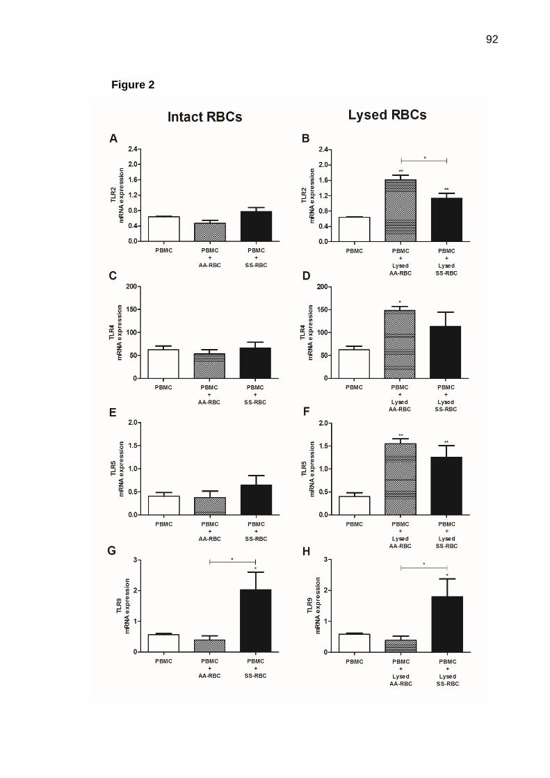
92
Figure 2

93
Figure 3

94
Figure 4

95
Figure 5

96
Table 1 Steady state hematological and biochemical markers values of the SCA patients and
healthy volunteers groups.
SCA patients
(n=12) (mean ± SD)
Healthy volunteers
(n=9) (mean ± SD) p value*
Age (years) 9.08 ± 4.72 12.44 ± 5.60 0.1864
RBC (x106/mL) 2.99 ± 0.36 4.78 ± 0.41 <0.0001
Platelets (x109/mL) 441.00 ± 118.10 307.1 ± 49.85 0.0056
Leukocyte (x109/mL) 15178 ± 4474 6767.00 ± 1716.00 0.0002
Reticulocyte (%) 7.25 ± 2.05 0.76 ± 0.17 0.0004
Hemoglobin (g/dL) 8.68 ± 1.03 13.23 ± 1.27 0.0004
Fetal Hemoglobin (%) 7.73 ± 6.26 0.48 ± 0.42 0.0007
Hematocrit (%) 25.34 ± 3.53 39.43 ± 3.18 0.0004
Ferritin (ng/mL) 246.30 ± 213.80 36.63 ± 18.53 0.0016
C-reactive protein (mg/L) 5.173 ± 0.67 1.58 ± 1.16 0.0163
SCA, sickle cell anemia; SD, standard deviation; RBC, red blood cells
* Mann-Whitney test; values to p<0.05 were considered a significant difference
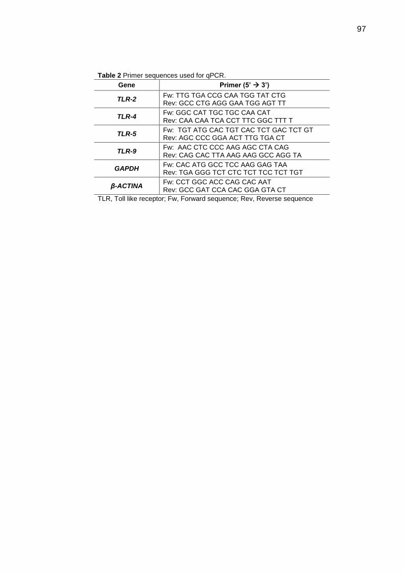
97
Table 2 Primer sequences used for qPCR.
Gene Primer (5’ 3’)
TLR-2 Fw: TTG TGA CCG CAA TGG TAT CTG Rev: GCC CTG AGG GAA TGG AGT TT
TLR-4 Fw: GGC CAT TGC TGC CAA CAT Rev: CAA CAA TCA CCT TTC GGC TTT T
TLR-5 Fw: TGT ATG CAC TGT CAC TCT GAC TCT GT Rev: AGC CCC GGA ACT TTG TGA CT
TLR-9 Fw: AAC CTC CCC AAG AGC CTA CAG Rev: CAG CAC TTA AAG AAG GCC AGG TA
GAPDH Fw: CAC ATG GCC TCC AAG GAG TAA Rev: TGA GGG TCT CTC TCT TCC TCT TGT
β-ACTINA Fw: CCT GGC ACC CAG CAC AAT Rev: GCC GAT CCA CAC GGA GTA CT
TLR, Toll like receptor; Fw, Forward sequence; Rev, Reverse sequence

98
6.3 Manuscrito 3
Título: NLRP3-inflammasome platform contributes to the inflammatory status in
sickle cell anemia regardless hydroxyurea treatment
Autores: Pitanga, T.N.; Oliveira, R. R; Zanette, D.L.; Guarda, C.C.; Santiago, R.P;
Santana, S.S; Lopes, M.V.; Lima, J.B.; Carvalho, G.Q.; Maffili, V.V.; Alcântara,
L.C.J.; Borges, V.M.; Goncalves, M.S.
Situação: A ser submetido
Objetivos: (referentes aos objetivos 5 a 7 da tese):
1. Determinar a expressão de genes relacionados a componentes do inflamassoma
NLRP3 em PBMC de pacientes com anemia falciforme e de voluntários sadios;
2. Avaliar o efeito da adição de hemácias falcizadas na expressão de genes
relacionados a componentes do inflamassoma NLRP3 em cultura de PBMC de
voluntários sadios, tratada ou não com hidroxiureia;
3. Avaliar proteínas relacionadas à ativação do inflamassoma (IL-1β e proteína
ligadora de IL-18 [IL-18BP]) em sobrenadante de cultura de PBMC incubada com
hemácias falcizadas tratada ou não com hidroxiureia.
Principais resultados: células mononucleares de pacientes com AF mostram
expressão elevada de NLRP3 e IL-18; hemácias de pacientes AF induzem
expressão gênica de caspase-1 e IL-18, e aumentam a secreção de IL-1β; o
tratamento com HU não afeta a expressão de moléculas relacionadas ao
inflamassoma e induz aumento da secreção de IL-18BP (proteína ligadora de IL-18).

99
NLRP3-inflammasome platform contributes to the inflammatory status in sickle
cell anemia regardless hydroxyurea treatment
Thassila N. Pitanga,1,2 Ricardo R. Oliveira,1 Dalila L. Zanette,1 Caroline C. Guarda,1
Rayra P. Santiago,1 Sânzio S. Santana,1 Valma M. L. Nascimento,3 Jonilson B.
Lima,1 Graziele Q. Carvalho,1 Vitor V. Maffili,1 Luiz Carlos J. Alcântara,1 Valéria M.
Borges1 and Marilda S. Gonçalves1,4
1Fundação Oswaldo Cruz, Centro de Pesquisas Gonçalo Moniz – FIOCRUZ/BA
2Instituto de Ciências da Saúde (ICS), Universidade Federal da Bahia (UFBA)
3Fundação de Hematologia e Hemoterapia da Bahia (HEMOBA)
4Faculdade de Farmácia, UFBA, Salvador, Bahia, Brazil.
RUNNING TITLE: Sickle cell anemia trigger the NLRP3-inflammasome
CORRESPONDENCE: Marilda S. Gonçalves, PhD, Centro de Pesquisas Gonçalo
Moniz, LHGB, Waldemar Falcão St, n. 121, Candeal, Salvador, Bahia, Brasil. Tel:
+55 (71) 3176-2226, e-mail: [email protected]
CONFLICT OF INTEREST: All authors declare no conflicts of interest.
FUNDING: This work was approved by the Research Ethics Committee (CAAE:
04733612.7.0000.0040) and was supported by grants from the Brazilian National
Council of Research (CNPq) (311888/2013-5) (M.S.G.); the Foundation of Research
and Extension of Bahia (FAPESB) (3626/2013, 1431040053063, and 9073/2007)
(M.S.G.); and PPSUS/FAPESB (020/2013 EFP_00007295), (M.S.G.); the Instituto
Nacional de Ciência e Tecnologia do Sangue (CNPq) (Coordinated by S.T.O.S.), and
MCD/CNPq/MS-SCTIE-DECIT (409800/2006-6), (M.S.G.). Sponsors of this study are
public or nonprofit organizations that support science in general. They had no role in
gathering analyzing, or interpreting the data.

100
Abstract
Sickle Cell Anemia (SCA) is a genetic disorder characterized by systemic
inflammation coinciding with vaso-occlusive phenomena, painful episodes and
intravascular hemolysis. The nod-like receptor protein 3 (NLRP3)-inflammasome
platform could contribute this inflammation, since erythroid contents act as danger
signals (eDAMPs) activating this pathway. Our objective was to evaluate expression
of NLRP3, Caspase-1, interleukin (IL)-1β and IL-18 in peripheral blood mononuclear
cells (PBMC) of SCA patients and the role that erythrocytes play in the expression of
these molecules, and in vitro effect of Hydroxyurea. PBMC and red blood cells of
SCA patients (SS-RBC) and healthy volunteers (AA-RBC) were obtained. PBMC
were challenged with SS- or AA-RBC in the presence of hydroxyurea (HU).
Inflammasome-related gene expressions were assessed by qPCR. IL-1β and IL-18
binding protein (BP) production were measured in culture supernatants. We found
that NLRP3 and IL-1β were highly expressed in PBMC of SCA patients when
compared to healthy volunteers. In addition, we showed that intact or lysed SS-RBC,
but not AA-RBC, induces Caspase-1 and IL-18 expression, while lysed AA-RBC
induces expression of NLRP3 and IL-1β. Moreover, Intact SS-RBC, but not AA-RBC,
induces production of IL-1β. Treatment with hydroxyurea (HU) showed no
interference with the expression of NLRP-3-inflammasome proteins, whereas
induced production of IL-18BP. Our data suggest that NLRP3-inflammasome
platform contributes to the inflammatory status observed in SCA patients and that HU
treatment does not interfere in the inflammatory pathway. This knowledge could lead
to the development of new therapeutic strategies, which act in different ways from
those given by hydroxyurea.
Keywords: Sickle cell disease; hydroxyurea; NOD-like receptors; NLRP3-
inflammasome; eDAMP.
101
Introduction
Sickle Cell Anemia (SCA) is a hereditary disease characterized by the
production of abnormal red cells, presence of homozygous hemoglobin S (HbSS)
and dysfunction on oxygen transportation. Although some patients present no
symptoms, SCA is an inflammatory condition associated with vaso-occlusive and
painful episodes, increased susceptibility to infections and intravascular hemolysis
(PLATT, 2000; WEATHERALL, PROVAN, 2000; TAYLOR et al., 2008; NEBOR et al.,
2011).
Hemoglobin and heme group released during intravascular hemolysis activate
endothelial cells, leukocytes and platelets, leading to systematic inflammation and
contributing to pro-thrombotic and vaso-occlusive phenomena observed in SCA
patients (BELCHER et al., 2003; WOOD, HEBBEL, GRANGER, 2004). The
maintenance of the inflammatory condition is mediated by the immune system,
especially the innate immune system, which is able to recognize danger-associated
molecular patters (DAMPs) by NOD-like receptors (NLRs) (CERQUEIRA et al., 2011;
VICARI et al., 2015).
Cellular residues and other endogenous molecules, released during
intravascular hemolysis, are able to act as signals for tissue injury, and are
recognized by NLRs. High-Mobility Group Protein B1 (HMGB1), and heme are the
major DAMPs considered as NLR agonists (DUTRA et al., 2014; GLADWIN, OFORI-
ACQUAH, 2014; GUPTA, 2014; XU et al., 2014).
Nod-like receptors are grouped into five subfamilies (NLRA, NLRB, NLRC,
NLRP and NLRX) depending on the composition of their N-terminal domain. Under
activation, the central domain of these receptors oligomerizes resulting in an
inflammatory multi-protein platform, named the inflammasome. Although several
inflammasome platforms have been proposed, the NLRP3-associated inflammasome
is the most widely studied (TING et al., 2008; SCHRODER, TSCHOPP, 2010;
YERETSSIAN, 2012; LATZ, XIAO, STUTZ, 2013).
Classically, inflammasome consists of NLRP (usually NLRP3, NOD-like
receptor family, pyrin domain containing 3), inflammatory protease Caspase-1 and
the Apoptosis-Associated Speck-Like Protein Containing Card (ASC). Following
activation, NLRP3 recruits ASC and pro-caspase-1 in a process that requires
participation of NF-κB, as a first-step of NLRP3 activation, and NLRP3

102
oligomerization itself as a second-step of activation, in order to convert pro-caspase-
1 into its active form. Then, active caspase-1 converts the inflammatory cytokines IL-
1β and IL-18 into active forms, contributing to pathology severity (MARTINON,
BURNS, TSCHOPP, 2002; AGOSTINI et al., 2004; MARIATHASAN, MONACK,
2007; BAUERNFEIND et al., 2009; BAUERNFEIND et al., 2011; ZHOU et al., 2011;
GHONIME et al., 2014).
IL-1β and IL-18 are pro-inflammatory cytokines engaged with the innate
immune response in both infection and aseptic inflammation. Levels of these
cytokines have been associated with different clinical inflammatory conditions, such
as multiple sclerosis, cancer, Alzheimer’s disease, arthritis and sickle cell anemia
(GUMA et al., 2010; CERQUEIRA et al., 2011; KITAZAWA et al., 2011; RAMIREZ-
RAMIREZ et al., 2013; KETELUT-CARNEIRO et al., 2015; TAS et al., 2015; VICARI
et al., 2015). Levels or polymorphisms of IL-1β and IL-18 have been correlated with
clinical complications, such as cerebrovascular accident, intravascular hemolysis and
endothelial dysfunction, contributing to the disease severity and to the therapeutic
failure (ASARE et al., 2010; CERQUEIRA et al., 2011; VICARI et al., 2015).
Therapy with hydroxyurea seems to be the best treatment available for SCA
patients. HU exhibits a cytotoxic effect by inhibiting ribonucleotide reductase enzyme
contributing for its main pharmacologic mechanism – increasing levels of HbF (DE
MONTALEMBERT et al., 2006; PLATT, 2008; LEBENSBURGER et al., 2010).
In this study, we evaluated the expression of NLRP3, Caspase-1, interleukin
(IL)-1β and IL-18 and production of IL-1β and IL-18 bind protein (IL-18BP) in
peripheral blood mononuclear cells (PBMC) of SCA patients. In addition, we
investigated the participation of RBCs obtained from SCA patients (SS-RBCs) in
modulating the expression of these genes and production of cytokines in PBMC from
healthy donors (AA-PBMCs), as well as the in vitro effect of hydroxyurea in the
expression of these inflammatory markers.
Methods
SCA patients and healthy volunteers
Patients were recruited from Hematology and Hemotherapy Foundation of the
State of Bahia (HEMOBA) and healthy volunteers (individuals without hematological
103
disorder or inflammatory conditions) from Pharmacy College (FacFAR) of Federal
University of Bahia. A cross-sectional study performed comprising 10 sickle cell
anemia (SCA) patients in steady-state (age 10.3 ± 4.6 years) and 7 healthy
volunteers (age 14.5 ± 5.2 years). No patient required blood transfusion in a period of
four months prior to blood draw, or presented any infection, hospitalization or vaso-
occlusive event. Although all patients were being treated with folic acid, none was
using antibiotics, corticosteroids or hydroxyurea. Demographic, hematological and
biochemical parameters of both groups are showed in Table 1. Although subjects
from both groups has similar age, all biochemical and hematological parameters,
except for levels of alpha-1 antitrypsin and alanine aminotransferase, were worse in
the SCA group. This study was approved by the Research Ethics Committee of the
Oswaldo Cruz Foundation, and was developed in accordance with the Declaration of
Helsinki of 1975 as revised in 2008. All subjects or their legal guardians agreed to
collect the biological sample after reading the terms in the informed consent.
Blood collection and isolation of human mononuclear and red blood cells
Venous blood was collected from patients and controls in order to determine
hemoglobin profile by high-performance liquid chromatography, and to obtain red
blood cells (RBC) for cell culture experiments and peripheral blood mononuclear cells
(PBMC) for gene expression analysis. Additionally, PBMCs used for cell culture
(PBMC culture) experiments were obtained from fresh leukocytes concentrates (buffy
coat) of healthy donors from HEMOBA by using the gradient Ficoll-Hypaque (GE
Healthcare Bio-Sciences Corp. Piscataway, NJ, USA). RBC were isolated by
centrifugation from peripheral blood of SCA patients (SS-RBC) and controls (AA-
RBC). Lysed erythrocytes was obtained by maintaining RBC concentrate at -70°C for
3 hours and subsequently thawed (purity >90%).(BENSOUDA, LAATIRIS, 2006)
PBMC from buffy coat bags were counted, distributed in 24-well plate (Costar,
Corning, NY, USA) at a concentration of 3x10⁶ mononuclear cells/mL and cultured
with 3% of lysed or intact RBC for 24h at 37°C and 5% CO2, in the presence or
absence of 100 µM hydroxyurea (HU) (Sigma-Aldrich, Saint Louis, MO, USA)
(STEINBERG et al., 2010).

104
IL-1β e IL-18 bind protein (IL-18BP) production by PBMC culture
IL-1β and IL-18BP production was assessed using IL-1β and IL-18BP (R&D
Systems, Minneapolis, USA) ELISA kits, according to the manufacturer's instructions.
In vitro IL-1β production was analyzed in supernatants harvested from the PBMC,
exposed to intact or lysed erythrocytes from AF patients for 24h.
Quantification of gene expressions
Extraction of mRNA from PBMC of SCA patients and PBMC cultures was
performed by using Trizol reagent (Invitrogen, Life Technologies, CA, USA)
according to the manufacturer's instructions. The RNA obtained was measured by
spectrophotometer NanoDrop™ Lite Spectrophotometer (Thermo Fisher Scientific,
Wilmington, USA). Reverse transcription was performed using the High Capacity
cDNA Reverse Transcription Kit (Applied BiosystemsTM, Foster City, CA) according
to the manufacturer's instructions. NLRP3, IL-1β e IL-18, Caspase-1, GAPDH and β-
ACTIN gene expression analysis by qPCR was performed on an ABI 7500 FAST
Real-Time PCR equipment (Applied BiosystemsTM, Foster City, CA). The
amplification reactions were performed with Power SYBR® Green, according to
manufacturer’s instructions. Primers used for qPCR reactions are shown in Table 2.
After amplification and dissociation curve runs, the values of threshold cycle (Ct)
were obtained with the aid of the Operational Programme 7500™ System (Applied
Biosystems, USA). Expression levels were normalized based on the geometric mean
of GAPDH and β-ACTIN, endogenous controls. Relative expression folds were
calculated based on 2^-ddCt method.(PFAFFL, 2001)
Statistical analysis
Comparison of quantitative variables between groups was performed by
unpaired Student's t test for normal distribution data and by the Mann-Whitney test
for non-normally distributed data. The non-parametric ANOVA Kruskal-Wallis was
used for comparison between three or more groups. Values of p<0.05 were
considered as significant different. All data were analyzed using Prism 5.1 software
(GraphPad, San Diego, USA).

105
Results
PBMC of SCA patients highly express NLRP3 and IL-1β genes
SCA is a genetic disorder in which an intense inflammatory response is
observed, associated with intravascular hemolysis and activation of leukocytes,
endothelial cells and platelets. We hypothesized that red cells from SCA patients, as
well as their intracytoplasmic contents, act as danger signals for the activation of
NLRP3 inflammasome. In attempt to test this hypothesis, we first evaluated the
expression of NLRP3, Caspase-1, IL-1β and IL-18 in peripheral blood mononuclear
cells obtained from SCA patients (SS-PBMC) and healthy controls (AA-PBMC).
Expression of NLRP3 and IL-1β was significantly higher in SS-PBMC (2.30 ±
1.02 and 2.84 ± 1.08) as compared to AA-PBMC (0.75 ± 0.13 and 1.24 ± 0.38,
respectively, p < 0.05). On the other hand, mRNA expression of Caspase-1 and IL-18
did not differ between PBMC from healthy volunteers and SCA patients (Figure 1).
SS-RBC induces Caspase-1 and IL-18 expression
Since it has been shown that SS-PBMC expresses mediators associated with
NLRP3 inflammasome, we decided to investigate whether red blood cells of SCA
patients act as DAMPs for the activation of NLRP3 inflammasome. For this purpose,
we isolated AA-PBMC from regular blood donors and incubated these cells in the
presence of intact or lysed SS-RBC or RBC from healthy controls (AA-RBC). Since
intact SS-RBC are morphologically distinct from AA-RBC, and intracellular contents
of RBC are able to induce inflammatory response mediated by innate receptors
(POTOKA, GLADWIN, 2015), we hypothesized that both intact and lysed SS-RBC
could promote the expression of molecules involved in NLRP3 inflammasome
activation in AA-PBMC.
We demonstrated here that intact AA-RBC were not able to induce expression
of NLRP3, Caspase-1, IL-1β or IL-18, when compared to basal expression of these
receptors observed in unstimulated AA-PBMC (Figures 2 A, C, E, G). In contrast,
intact SS-RBC induced higher expression of NLRP3, Caspase-1, IL-1β and IL-18
than unstimulated PBMC did. Lysed AA-RBC induced higher expression of NLRP3

106
and IL-β, whereas SS-RBC induced higher expression of NLRP3, Caspase-1 and IL-
18, when compared to unstimulated cell cultures.
Interestingly, intact SS-RBC induced higher expression of Caspase-1 when
compared to AA-RBC (Figure 2C). Moreover, Caspase-1 and IL-18 were more
expressed in PBMC challenged with lysed SS-RBC (3.54 ± 2.01 and 2.41 ± 1.25)
when compared to lysed AA-RBC (0.08 ± 0.02 and 0.26 ± 0.06, respectively, p<0.05,
Figure 2 D, H). These data suggest that intact and lysed SS-RBC are good inductors
of Caspase-1 or IL-18 expression, which was not observed for AA-RBC.
HU treatment does not affect expression of NLRP3 inflammasome-related
molecules
Hydroxyurea (HU) is a cytotoxic drug used for treatment of SCA patients and its
main expected beneficial effect is the improvement in HbF levels. However, it is not
clear whether HU acts controlling the inflammatory condition in these patients. We
sought to investigate this issue by incubating AA-PBMC with intact or lysed SS-RBC,
in the presence or absence of HU.
Expression of NLRP3, Caspase-1, IL-1β and IL-18 were not affected by HU
treatment, in both intact and lysed SS-RBC conditions (Figure 3). Thus, besides HU
has a positive effect on HbF production, our data suggested that this drug is not able
to interfere positively in the control of the inflammatory condition.
Intact SS-RBC, but not AA-RBC, induces production of IL-1β
In addition to assessing gene expression, we also evaluated protein production,
especially IL-1β and IL-18 binding protein (IL-18BP). IL-1β is secreted upon
activation of NLRP3-inflammasome, whereas IL-18BP is a protein that binds to
soluble IL-18, preventing the binding of IL-18 to its receptor,(DINARELLO et al.,
2013) Therefore, production of IL-18BP might be understood as a compensatory
mechanism to regulate IL-18 activity, a cytokine that is also produced after NLRP3
inflammasome activation.
It was observed that both intact and lysed SS-RBC and AA-RBC induced
production of IL-1β and IL-18BP (Figure 4). However, levels of IL-1β in response to
intact AA-RBC were similar to those was observed for unstimulated cultures, and

107
intact SS-RBC was able to induce production of higher levels of IL-1β, when
compared to intact AA-RBC stimulated cultures (Figure 4A). This data reinforces the
hypothesis that SS-RBC, as opposed to AA-RBC, activates an inflammatory
response mediated by receptors from the innate immune system.
HU induces production of IL-18BP but does not affect IL-1β production
HU showed no effect in down-regulating IL-1β production induced by intact or
lysed SS-RBC on AA-PBMC (Figures 5 A-B). However, HU up-regulated IL-18BP
production induced by lysed SS-RBC, when comparing with untreated cells (Figure
5D), suggesting that HU might play an important role controlling the inflammatory
effects of IL-18, which could be beneficial for the treatment of SCA patients.
Discussion
In this study, we showed that NLRP3-inflammasome is an important innate
immune platform for the recognition of DAMPs derived from sickle red cell or its
contents. Sickle cell anemia (SCA) patients exhibited high expression of NLRP3 and
IL-1β in mononuclear cells (PBMC) and SS-RBC induced Caspase-1 and IL-18
expression and production of IL-1β. Hydroxyurea treatment did not affect expression
of NLRP3 inflammasome-related molecules, but induced production of IL-18BP.
The most common inflammasome is associated with NLRP3, caspase-1 and
ASC. Several dangers signals has been well documented as NLRP3-inflammasome
agonists, such as extracellular ATP, glucose, hyaluronan, alum and asbestos
(SCHRODER, TSCHOPP, 2010). More recently, some authors have demonstrated
red cells or its contents as DAMPs for NLRP3 inflammasome activation, and
proposed erythroid DAMPs (eDAMPs) as a new terminology for these signals of red
cells injury.(GLADWIN, OFORI-ACQUAH, 2014; POTOKA, GLADWIN, 2015)
Following NLRP3 activation, caspase-1 becomes active and, consequently, converts
pro-IL-1β and pro-IL-18 in their active forms which are secreted in the extracellular
milieu (MARIATHASAN, MONACK, 2007).
In this study, we did not find any differences in the expressions of caspase-1
and IL-18 between AA-PBMC and SS-PBMC; however, NLRP3 and IL-1β were more
expressed in SS-PBMC. It was documented that many of the same stimuli induce

108
expression of both pro-IL-1β and NLRP3, and that IL-18 and caspase-1 are
constitutively expressed in many cells (GUARDA et al., 2011; DINARELLO et al.,
2013) corroborating our results.
Since hematological parameters, such as RBCs count, hemoglobin levels and
frequency of reticulocytes were substantially different in the SCA group when
compared to healthy volunteers, we tested the effect to RBC in the activation of the
NLRP3 inflammatory pathway. Interestingly, intact SS-RBCs were potent eDAMPs,
as they induced expression of all NLRP3-inflammsome related molecules evaluated
here. Moreover, lysed SS-RBCs increased caspase-1 and IL-18 expression whereas
lysed AA-RBCs induced higher expression of NLRP3 and IL-1β. These results
suggest that erythrocyte composition differs between AA- and SS-RBCs regarding
inflammasome pathway activation. Despite the observed contribution of lysed AA-
RBCs to IL-1β and NLRP3 expression, it is important to point out that intravascular
hemolysis does not occur physiologically in healthy volunteers, whereas a great
proportion of SCA patients exhibit this condition (TAYLOR et al., 2008; VILAS-BOAS
et al., 2010). Hence, the inflammation observed in SCA patients could be a
consequence of constant exposure to eDAMPs (intact or lysed RBCs), such as heme
(DUTRA et al., 2014), during intravascular hemolysis, which leads to upregulation of
NOD-like receptors expression, suggesting that these molecules could be therapeutic
targets for SCA.
Hydroxyurea is an important agent used to treat SCA patients that acts
increasing HbF levels and reducing SS-RBCs-endothelial interaction (PLATT, 2008;
STEINBERG et al., 2010). In this study, we demonstrated that HU does not interfere
in the expression of NLRP3, caspase-1, IL-1β and IL-18, suggesting that HU
mechanism of action is not associated with NLRP3-inflammasome pathway, thus is
not able to act controlling inflammasome-dependent inflammatory conditions
observed in SCA patients.
In addition to gene expression, we also evaluated production of IL-1β and IL-18
binding protein (IL-18BP). IL-18BP is a protein that binds to IL-18 preventing the
attachment with its receptor, contributing to decrease the inflammatory response
(CARBOTTI et al., 2013; DINARELLO et al., 2013). We showed here that both intact
and lysed SS-RBCs induced IL-1β production, whereas, only lysed AA-RBCs
demonstrated this property. On the other hand, IL-18BP was induced in all

109
conditions, suggesting that its production is not associated with specific eDAMP from
SS-RBCs.
Although plasma levels of IL-18BP are significantly increased in inflammatory
conditions (DINARELLO et al., 2003), in our in vitro model we found higher gene
expression of IL-18 in SS-RBCs-stimulated PBMC, and found no difference in the
concentration of IL-18BP in supernatant of PBMC-stimulated with AA- or SS-RBCs.
Upon HU treatment, although we did not observe any interference in IL-18
expression, this drug was able to increase the levels of its inhibitor (IL-18BP) in our
model of in vitro hemolysis, what was not observed when using intact RBCs.
Therefore, during intravascular hemolysis, HU might have beneficial effects in the
inflammatory control by reducing IL-18 attachment in IL-18 receptor.
Our data reinforce that NLRP3-inflammasome platform, an important and well-
known inflammatory pathway, could be contributing to the clinical inflammatory status
observed in SCA patients. This knowledge could lead to the development of new
therapeutic strategies targeting, preferentially, sickle cell eDAMPs or NLRP3-
inflammasome-associated molecules.
AUTHORS’ CONTRIBUTIONS
TNP, RRO and MSG: conceived the study design. TNP: performed all the
experiments. DLZ, CCG, RPS and SSS: assisted in all experiments. VML:
responsible for patients’ follow-up. VVM, JBL and GQC: discussion and
accomplishment of some experiments. LCJA and VMB: discussion of study design,
experiments and all results. TNP, RRO and MSG: wrote the paper. All authors read
and approved the final manuscript.
ACKNOWLEDGEMENTS
The authors would like to thank SCA patients and all healthy volunteers for trust
us and for making possible the realization of this work. We thank the laboratory
technicians for help us in the collection of the samples. We would also like to thank
Wendell Vilas Boas, Bruno Cerqueira, Magda Seixas and Marcia Weber for excellent

110
discussions. TNP received a scholarship from CAPES. L.C.J.A., V.M.B. and MSG
have productivity scholarship from CNPq.

111
REFERENCES
AGOSTINI, L.; MARTINON, F.; BURNS, K.; MCDERMOTT, M. F.; HAWKINS, P. N.; TSCHOPP, J. (2004). NALP3 forms an IL-1beta-processing inflammasome with increased activity in Muckle-Wells autoinflammatory disorder. Immunity 20(3): 319-325. ASARE, K.; GEE, B. E.; STILES, J. K.; WILSON, N. O.; DRISS, A.; QUARSHIE, A.; ADAMS, R. J.; KUTLAR, A.; HIBBERT, J. M. (2010). Plasma interleukin-1beta concentration is associated with stroke in sickle cell disease. Cytokine 49(1): 39-44. BAUERNFEIND, F.; BARTOK, E.; RIEGER, A.; FRANCHI, L.; NUNEZ, G.; HORNUNG, V. (2011). Cutting edge: reactive oxygen species inhibitors block priming, but not activation, of the NLRP3 inflammasome. J Immunol 187(2): 613-617. BAUERNFEIND, F. G.; HORVATH, G.; STUTZ, A.; ALNEMRI, E. S.; MACDONALD, K.; SPEERT, D.; FERNANDES-ALNEMRI, T.; WU, J.; MONKS, B. G.; FITZGERALD, K. A.; HORNUNG, V.; LATZ, E. (2009). Cutting edge: NF-kappaB activating pattern recognition and cytokine receptors license NLRP3 inflammasome activation by regulating NLRP3 expression. J Immunol 183(2): 787-791. BELCHER, J. D.; BRYANT, C. J.; NGUYEN, J.; BOWLIN, P. R.; KIELBIK, M. C.; BISCHOF, J. C.; HEBBEL, R. P.; VERCELLOTTI, G. M. (2003). Transgenic sickle mice have vascular inflammation. Blood 101(10): 3953-3959. BENSOUDA, Y.; LAATIRIS, A. (2006). The lyophilization of dispersed systems: influence of freezing process, freezing time, freezing temperature and RBCs concentration on RBCs hemolysis. Drug Dev Ind Pharm 32(8): 941-945. CARBOTTI, G.; BARISIONE, G.; ORENGO, A. M.; BRIZZOLARA, A.; AIROLDI, I.; BAGNOLI, M.; PINCIROLI, P.; MEZZANZANICA, D.; CENTURIONI, M. G.; FABBI, M.; FERRINI, S. (2013). The IL-18 antagonist IL-18-binding protein is produced in the human ovarian cancer microenvironment. Clin Cancer Res 19(17): 4611-4620. CERQUEIRA, B. A.; BOAS, W. V.; ZANETTE, A. D.; REIS, M. G.; GONCALVES, M. S. (2011). Increased concentrations of IL-18 and uric acid in sickle cell anemia: contribution of hemolysis, endothelial activation and the inflammasome. Cytokine 56(2): 471-476. DE MONTALEMBERT, M.; BROUSSE, V.; ELIE, C.; BERNAUDIN, F.; SHI, J.; LANDAIS, P. (2006). Long-term hydroxyurea treatment in children with sickle cell disease: tolerance and clinical outcomes. Haematologica 91(1): 125-128. DINARELLO, C. A.; NOVICK, D.; KIM, S.; KAPLANSKI, G. (2013). Interleukin-18 and IL-18 binding protein. Front Immunol 4: 289. DINARELLO, C. A.; NOVICK, D.; RUBINSTEIN, M.; LONNEMANN, G. (2003). Interleukin 18 and interleukin 18 binding protein: possible role in immunosuppression of chronic renal failure. Blood Purif 21(3): 258-270. DUTRA, F. F.; ALVES, L. S.; RODRIGUES, D.; FERNANDEZ, P. L.; DE OLIVEIRA, R. B.; GOLENBOCK, D. T.; ZAMBONI, D. S.; BOZZA, M. T. (2014). Hemolysis-induced lethality involves inflammasome activation by heme. Proc Natl Acad Sci U S A 111(39): E4110-4118.

112
GHONIME, M. G.; SHAMAA, O. R.; DAS, S.; ELDOMANY, R. A.; FERNANDES-ALNEMRI, T.; ALNEMRI, E. S.; GAVRILIN, M. A.; WEWERS, M. D. (2014). Inflammasome priming by lipopolysaccharide is dependent upon ERK signaling and proteasome function. J Immunol 192(8): 3881-3888. GLADWIN, M. T.; OFORI-ACQUAH, S. F. (2014). Erythroid DAMPs drive inflammation in SCD. Blood 123(24): 3689-3690. GUARDA, G.; ZENGER, M.; YAZDI, A. S.; SCHRODER, K.; FERRERO, I.; MENU, P.; TARDIVEL, A.; MATTMANN, C.; TSCHOPP, J. (2011). Differential expression of NLRP3 among hematopoietic cells. J Immunol 186(4): 2529-2534. GUMA, M.; KASHIWAKURA, J.; CRAIN, B.; KAWAKAMI, Y.; BEUTLER, B.; FIRESTEIN, G. S.; KAWAKAMI, T.; KARIN, M.; CORR, M. (2010). JNK1 controls mast cell degranulation and IL-1{beta} production in inflammatory arthritis. Proc Natl Acad Sci U S A 107(51): 22122-22127. GUPTA, K. (2014). HMGB1 takes a "Toll" in sickle cell disease. Blood 124(26): 3837-3838. KETELUT-CARNEIRO, N.; SILVA, G. K.; ROCHA, F. A.; MILANEZI, C. M.; CAVALCANTI-NETO, F. F.; ZAMBONI, D. S.; SILVA, J. S. (2015). IL-18 Triggered by the Nlrp3 Inflammasome Induces Host Innate Resistance in a Pulmonary Model of Fungal Infection. J Immunol. KITAZAWA, M.; CHENG, D.; TSUKAMOTO, M. R.; KOIKE, M. A.; WES, P. D.; VASILEVKO, V.; CRIBBS, D. H.; LAFERLA, F. M. (2011). Blocking IL-1 signaling rescues cognition, attenuates tau pathology, and restores neuronal beta-catenin pathway function in an Alzheimer's disease model. J Immunol 187(12): 6539-6549. LATZ, E.; XIAO, T. S.; STUTZ, A. (2013). Activation and regulation of the inflammasomes. Nat Rev Immunol 13(6): 397-411. LEBENSBURGER, J. D.; PESTINA, T. I.; WARE, R. E.; BOYD, K. L.; PERSONS, D. A. (2010). Hydroxyurea therapy requires HbF induction for clinical benefit in a sickle cell mouse model. Haematologica 95(9): 1599-1603. MARIATHASAN, S.; MONACK, D. M. (2007). Inflammasome adaptors and sensors: intracellular regulators of infection and inflammation. Nat Rev Immunol 7(1): 31-40. MARTINON, F.; BURNS, K.; TSCHOPP, J. (2002). The inflammasome: a molecular platform triggering activation of inflammatory caspases and processing of proIL-beta. Mol Cell 10(2): 417-426. NEBOR, D.; BOWERS, A.; HARDY-DESSOURCES, M. D.; KNIGHT-MADDEN, J.; ROMANA, M.; REID, H.; BARTHELEMY, J. C.; CUMMING, V.; HUE, O.; ELION, J.; REID, M.; CONNES, P.; GROUP, C. S. (2011). Frequency of pain crises in sickle cell anemia and its relationship with the sympatho-vagal balance, blood viscosity and inflammation. Haematologica 96(11): 1589-1594. PFAFFL, M. W. (2001). A new mathematical model for relative quantification in real-time RT-PCR. Nucleic Acids Res 29(9): e45. PLATT, O. S. (2000). Sickle cell anemia as an inflammatory disease. J Clin Invest 106(3): 337-338. PLATT, O. S. (2008). Hydroxyurea for the treatment of sickle cell anemia. N Engl J Med 358(13): 1362-1369.

113
POTOKA, K. P.; GLADWIN, M. T. (2015). Vasculopathy and pulmonary hypertension in sickle cell disease. Am J Physiol Lung Cell Mol Physiol 308(4): L314-324. RAMIREZ-RAMIREZ, V.; MACIAS-ISLAS, M. A.; ORTIZ, G. G.; PACHECO-MOISES, F.; TORRES-SANCHEZ, E. D.; SORTO-GOMEZ, T. E.; CRUZ-RAMOS, J. A.; OROZCO-AVINA, G.; CELIS DE LA ROSA, A. J. (2013). Efficacy of fish oil on serum of TNF alpha , IL-1 beta , and IL-6 oxidative stress markers in multiple sclerosis treated with interferon beta-1b. Oxid Med Cell Longev 2013: 709493. SCHRODER, K.; TSCHOPP, J. (2010). The inflammasomes. Cell 140(6): 821-832. STEINBERG, M. H.; MCCARTHY, W. F.; CASTRO, O.; BALLAS, S. K.; ARMSTRONG, F. D.; SMITH, W.; ATAGA, K.; SWERDLOW, P.; KUTLAR, A.; DECASTRO, L.; WACLAWIW, M. A.; INVESTIGATORS OF THE MULTICENTER STUDY OF HYDROXYUREA IN SICKLE CELL, A.; FOLLOW-UP, M. S. H. P. (2010). The risks and benefits of long-term use of hydroxyurea in sickle cell anemia: A 17.5 year follow-up. Am J Hematol 85(6): 403-408. TAS, F.; TILGEN YASASEVER, C.; KARABULUT, S.; TASTEKIN, D.; DURANYILDIZ, D. (2015). Clinical significance of serum interleukin-18 (IL-18) levels in patients with gastric cancer. Biomed Pharmacother 70: 19-23. TAYLOR, J. G. t.; NOLAN, V. G.; MENDELSOHN, L.; KATO, G. J.; GLADWIN, M. T.; STEINBERG, M. H. (2008). Chronic hyper-hemolysis in sickle cell anemia: association of vascular complications and mortality with less frequent vasoocclusive pain. PLoS One 3(5): e2095. TING, J. P.; LOVERING, R. C.; ALNEMRI, E. S.; BERTIN, J.; BOSS, J. M.; DAVIS, B. K.; FLAVELL, R. A.; GIRARDIN, S. E.; GODZIK, A.; HARTON, J. A.; HOFFMAN, H. M.; HUGOT, J. P.; INOHARA, N.; MACKENZIE, A.; MALTAIS, L. J.; NUNEZ, G.; OGURA, Y.; OTTEN, L. A.; PHILPOTT, D.; REED, J. C.; REITH, W.; SCHREIBER, S.; STEIMLE, V.; WARD, P. A. (2008). The NLR gene family: a standard nomenclature. Immunity 28(3): 285-287. VICARI, P.; ADEGOKE, S. A.; MAZZOTTI, D. R.; CANCADO, R. D.; NOGUTTI, M. A.; FIGUEIREDO, M. S. (2015). Interleukin-1beta and interleukin-6 gene polymorphisms are associated with manifestations of sickle cell anemia. Blood Cells Mol Dis 54(3): 244-249. VILAS-BOAS, W.; CERQUEIRA, B. A.; ZANETTE, A. M.; REIS, M. G.; BARRAL-NETTO, M.; GONCALVES, M. S. (2010). Arginase levels and their association with Th17-related cytokines, soluble adhesion molecules (sICAM-1 and sVCAM-1) and hemolysis markers among steady-state sickle cell anemia patients. Ann Hematol 89(9): 877-882. WEATHERALL, D. J.; PROVAN, A. B. (2000). Red cells I: inherited anaemias. Lancet 355(9210): 1169-1175. WOOD, K. C.; HEBBEL, R. P.; GRANGER, D. N. (2004). Endothelial cell P-selectin mediates a proinflammatory and prothrombogenic phenotype in cerebral venules of sickle cell transgenic mice. Am J Physiol Heart Circ Physiol 286(5): H1608-1614. XU, H.; WANDERSEE, N. J.; GUO, Y.; JONES, D. W.; HOLZHAUER, S. L.; HANSON, M. S.; MACHOGU, E.; BROUSSEAU, D. C.; HOGG, N.; DENSMORE, J. C.; KAUL, S.; HILLERY, C. A.; PRITCHARD, K. A., Jr. (2014). Sickle cell disease increases high mobility group box 1: a novel mechanism of inflammation. Blood 124(26): 3978-3981.

114
YERETSSIAN, G. (2012). Effector functions of NLRs in the intestine: innate sensing, cell death, and disease. Immunol Res 54(1-3): 25-36. ZHOU, R.; YAZDI, A. S.; MENU, P.; TSCHOPP, J. (2011). A role for mitochondria in NLRP3 inflammasome activation. Nature 469(7329): 221-225.

115
Figure Legends
Figure 1
Nod-like receptor protein 3 (NLRP3; A), Caspase-1 (B), Interleukin (IL) 1β (C) and IL-
18 (D) mRNA expression in peripheral blood mononuclear cells (PBMC) of healthy
volunteers (AA-PBMC; n = 7) and sickle cell anemia (SCA) patients (SS-PBMC; n =
10). Asterisk marker above each bar represents significant statistic difference
between SS-PBMC and AA-PBMC groups (*p<0.05).
Figure 2
Nod-like receptor protein 3 (NLRP3; A and B), Caspase-1 (C and D), Interleukin (IL)
1β (E and F) and IL-18 (G and H) mRNA expression in peripheral blood mononuclear
cells (PBMC) of healthy volunteers challenged with intact (first column) or lysed
(second column) red blood cells of healthy volunteers (AA-RBC; n = 7) or sickle cell
anemia patients (SS-RBC; n = 10). Asterisk marker above each bar represents
significant statistic difference between this and the negative control bar (PBMC).
Asterisk marker above horizontal lines represents significant statistic difference
between designated groups (*p<0.05; **p<0.01).
Figure 3
Nod-like receptor protein 3 (NLRP3; A and B), Caspase-1 (C and D), Interleukin (IL)
1β (E and F) and IL-18 (G and H) mRNA expression in peripheral blood mononuclear
cells (PBMC) of healthy volunteers challenged with intact (first column) or lysed
(second column) red blood cells of sickle cell anemia patients (SS-RBC; n = 10), in
the presence or absence of hydroxyurea (HU). Asterisk marker above each bar
represents significant statistic difference between this and the negative control bar
(PBMC). Asterisk marker above horizontal lines represents significant statistic
difference between designated groups (*p<0.05; **p<0.01).
Figure 4
Levels of Interleukin (IL) 1β (A and B) and IL-18 (C and D) in culture supernatant of
peripheral blood mononuclear cells of healthy volunteers (PBMC) incubated with
intact (first column) or lysed (second column) red blood cells of healthy volunteers
(AA-RBC; n = 7) or sickle cell anemia patients (SS-RBC; n = 10). Asterisk marker

116
above each bar represents significant statistic difference between this and the
negative control bar (PBMC). Asterisk marker above horizontal lines represents
significant statistic difference between designated groups (*p<0.05).
Figure 5
Levels of Interleukin (IL) 1β (A and B) and IL-18 (C and D) in culture supernatant of
peripheral blood mononuclear cells of healthy volunteers (PBMC) incubated with
intact (first column) or lysed (second column) red blood cells of sickle cell anemia
patients (SS-RBC; n = 10), in the presence or absence of hydroxyurea (HU). Asterisk
marker above each bar represents significant statistic difference between this and the
negative control bar (PBMC). Asterisk marker above horizontal lines represents
significant statistic difference between designated groups (*p<0.05).

117
Figure 1

118
Figure 2

119
Figure 3
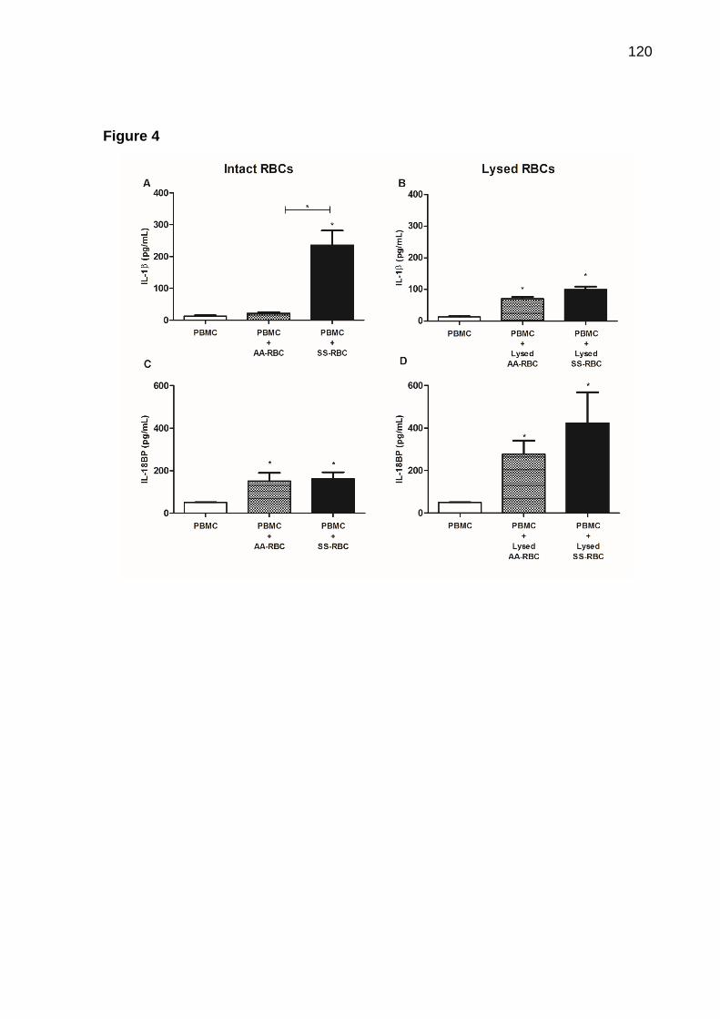
120
Figure 4

121
Figure 5
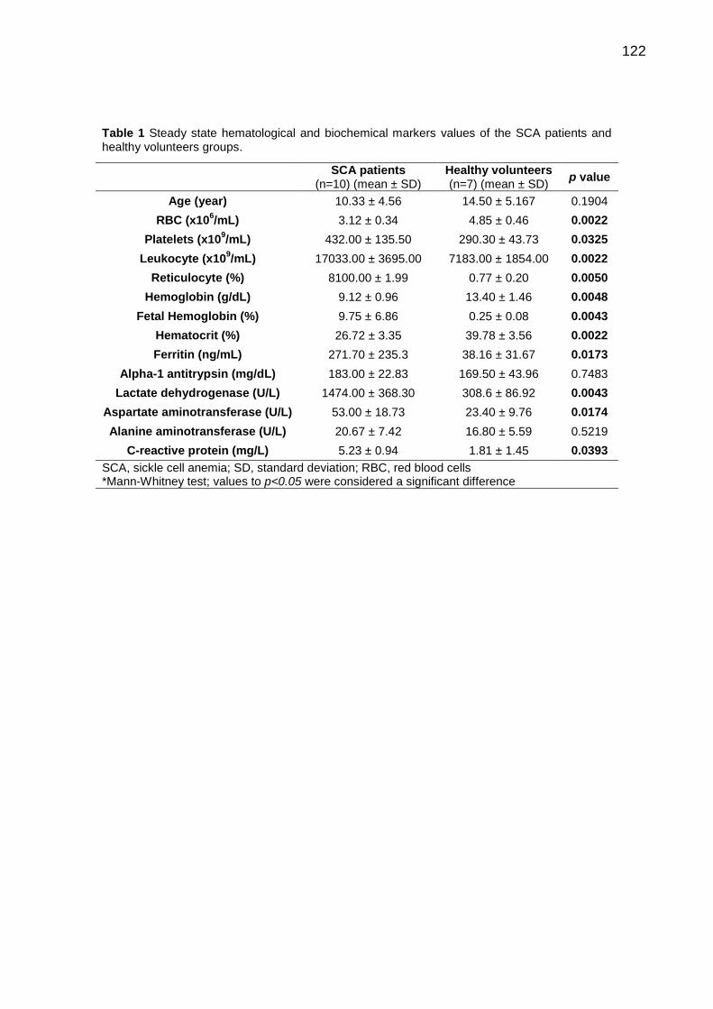
122
Table 1 Steady state hematological and biochemical markers values of the SCA patients and healthy volunteers groups.
SCA patients
(n=10) (mean ± SD) Healthy volunteers (n=7) (mean ± SD)
p value
Age (year) 10.33 ± 4.56 14.50 ± 5.167 0.1904
RBC (x106/mL) 3.12 ± 0.34 4.85 ± 0.46 0.0022
Platelets (x109/mL) 432.00 ± 135.50 290.30 ± 43.73 0.0325
Leukocyte (x109/mL) 17033.00 ± 3695.00 7183.00 ± 1854.00 0.0022
Reticulocyte (%) 8100.00 ± 1.99 0.77 ± 0.20 0.0050
Hemoglobin (g/dL) 9.12 ± 0.96 13.40 ± 1.46 0.0048
Fetal Hemoglobin (%) 9.75 ± 6.86 0.25 ± 0.08 0.0043
Hematocrit (%) 26.72 ± 3.35 39.78 ± 3.56 0.0022
Ferritin (ng/mL) 271.70 ± 235.3 38.16 ± 31.67 0.0173
Alpha-1 antitrypsin (mg/dL) 183.00 ± 22.83 169.50 ± 43.96 0.7483
Lactate dehydrogenase (U/L) 1474.00 ± 368.30 308.6 ± 86.92 0.0043
Aspartate aminotransferase (U/L) 53.00 ± 18.73 23.40 ± 9.76 0.0174
Alanine aminotransferase (U/L) 20.67 ± 7.42 16.80 ± 5.59 0.5219
C-reactive protein (mg/L) 5.23 ± 0.94 1.81 ± 1.45 0.0393
SCA, sickle cell anemia; SD, standard deviation; RBC, red blood cells *Mann-Whitney test; values to p<0.05 were considered a significant difference

123
Table 2. Primer sequences used for qPCR
Gene Primer (5’→3’)
CASPASE-1 Fw: AAA AAA TCT CAC TGC TTC GGA CAT Rev: TCT GGG CGG TGT GCA AA
IL-1β Fw: AGC TAC GAA TCT CCG ACC AC Rev: CGT TAT CCC ATG TGT CGA AGA A
IL-18 Fw: ATC GCT TCC TCT CGC AAC A Rev: TCT ACT GGT TCA GCA GCC ATC TT
NLRP3 Fw: TGC CCC GAC CCA AAC C Rev: GAA GCC GTC CAT GAG GAA GA
GAPDH Fw: CAC ATG GCC TCC AAG GAG TAA Rev: TGA GGG TCT CTC TCT TCC TCT TGT
β-ACTINA Fw: CCT GGC ACC CAG CAC AAT Rev: GCC GAT CCA CAC GGA GTA CT
Fw, Forward sequence; Rev, Reverse sequence.

124
7 DISCUSSÃO
A DF é uma desordem hemolítica, caracterizada por um processo inflamatório
crônico. A forma mais grave da DF, a AF, apresenta perfil clínico heterogêneo, com
ocorrência de crises vaso-oclusivas, AVC, hipertensão pulmonar e STA, entre outras
alterações (VILAS-BOAS et al., 2012; PITANGA et al., 2013). As complicações
clínicas associadas ao número de internações e óbitos desses pacientes
demonstram a necessidade de se investigar moléculas associadas a fisiopatologia
da doença bem como alvos terapêuticos.
Nesse sentido, dois polimorfismos genéticos vêm sendo investigados como
marcadores prognósticos da DF, e destacam-se quanto à importância na inflamação:
SERPINA1 e mieloperoxidase (MPO, -463G>A). O gene da SERPINA1 é
responsável pelo controle da síntese da AAT, um inibidor de protease produzido,
predominantemente, em hepatócitos, a qual atua controlando negativamente
citocinas inflamatórias, tais como IL-1, IL-6 e fator de necrose tumoral (TNF), e
inibindo elastase neutrofílica (RICHARDS, GAULDIE e BAUMANN, 1991; LOMAS e
MAHADEVA, 2002; CHAPPELL et al., 2006). Outra proteína altamente polimórfica é
a MPO, que é uma enzima produzida por monócitos e neutrófilos, que atua,
principalmente, no estresse oxidativo vascular e proteção do organismo contra
bactérias (PITANGA et al., 2014). A presença de polimorfismos nestes dois genes
está relacionada a complicações vasculares e aumento da susceptibilidade à
infecção nesses pacientes (COSTA et al., 2005; BARBOSA et al., 2014; CARVALHO
et al., 2014; STOCKLEY e TURNER, 2014).
No manuscrito I, observamos a prevalência de 56% e 9% do polimorfismo nos
genes de MPO e SERPINA1, respectivamente, nos pacientes com DF. Embora os
polimorfismos não tenham afetado as concentrações de hemoglobina fetal (HbF), o
tratamento com hidroxiureia (HU) mostrou melhor resultado para este parâmetro em
pacientes com o polimorfismo para MPO ou naqueles sem a mutação para
SERPINA1.
Durante a crise, pacientes com DF apresentam manifestações clínicas graves,
com inflamação sistêmica associada a fenômenos vaso-oclusivos e susceptibilidade
aumentada a infecções (WEATHERALL e PROVAN, 2000; TAYLOR et al., 2008).
Indivíduos com genótipo selvagem (sem o polimorfismo) para MPO e SERPINA1
estão associados com a manutenção da homeostasia, principalmente na defesa

125
contra patógenos e durante a inflamação. Nesse sentido, a presença do
polimorfismo pode agravar a condição clínica do paciente, como observado em
estudos com outras doenças inflamatórias como câncer gástrico e lúpus eritematoso
sistêmico (REYNOLDS, STEGEMAN e TERVAERT, 2002; BOUALI et al., 2007;
JIANG et al., 2012). Entretanto, não encontramos associação entre os polimorfismos
da MPO e SERPINA1 com as manifestações clínicas avaliadas, provavelmente pelo
número reduzido de indivíduos que carregam estes polimorfismos aliado à baixa
frequência de cada marcador clínico avaliado no grupo estudado.
Está bem documentado que contagens de plaquetas e leucócitos e
concentrações de hemoglobina (Hb), HbF e ferritina encontram-se alteradas em
pacientes com DF (STEINBERG, 2005; CARVALHO-NETO, LAND e FLEURY,
2011). Entretanto, este é o primeiro trabalho que demonstra a influência dos
polimorfismos de MPO e SERPINA1 em parâmetros laboratoriais em pacientes com
DF. A presença do polimorfismo não afetou os parâmetros laboratoriais dos
voluntários sadios, entretanto, interferiu em alguns marcadores dos pacientes com
DF.
A presença do alelo esteve associada a contagem baixa de plaquetas e ao
número elevado de leucócitos e linfócitos quando comparados ao grupo selvagem.
Plaquetas e leucócitos de pacientes com DF expressam mais moléculas de adesão,
contribuindo para a ocorrência de crises vaso-oclusivas e ativação endotelial
(BROWNE et al., 1996; OKPALA, 2006; PROENCA-FERREIRA et al., 2010;
PROENCA-FERREIRA et al., 2014). Portanto, considerando plaquetas, linfócitos e
leucócitos totais, a presença do polimorfismo no gene MPO poderia atuar como fator
protetor para eventos vaso-oclusivos, embora não tenhamos mostrado no presente
trabalho. Esses parâmetros, entretanto, não diferiram entre os grupos selvagem e
mutante para a SERPINA1.
Em contraste com as análises encontradas para o polimorfismo no gene MPO,
a mutação no gene da SERPINA1 mostrou contribuir para o aumento nos níveis de
ferritina e, como esperado, redução nos níveis de AAT no grupo de pacientes com
DF e com alelos mutantes. A ferritina é um biomarcador da inflamação e seus níveis
estão aumentados durante as crises dolorosas e infecção (AL-SAQLADI, BIN-
GADEEM e BRABIN, 2012). Sendo assim, este polimorfismo poderia representar
gravidade da doença, especialmente por estar associado a níveis reduzidos de AAT,
uma enzima que atua reduzindo a inflamação (GOOPTU, DICKENS e LOMAS,

126
2014; STOCKLEY e TURNER, 2014). Isso destaca a necessidade de se buscar
tratamentos mais específicos e individualizados para pacientes com DF que
apresentem esses polimorfismos.
A terapia com HU, o único medicamento aprovado pelo FDA norte americanos,
tem mostrado efeitos benéficos nos pacientes com DF. Seu principal mecanismo de
ação concentra-se, basicamente, em sua capacidade de inibir a enzima
ribonucleotídeo redutase, levando a proliferação eritróide anormal. Sob essa
condição, ocorre o aumento da produção de células F, as quais estimulam a síntese
de HbF e inibem a síntese de novas moléculas de HbS (FRANCO et al., 2006;
STROUSE e HEENEY, 2012). Além disso, a HU melhora o fluxo das hemácias e
reduz sua adesão às células vizinhas, reduzindo o risco de crises vaso-oclusivas
(KUMKHAEK et al., 2008; PLATT, 2008).
Neste estudo, mostramos que pacientes tratados com HU apresentam níveis
elevados de HbF, independentemente de ter ou não o polimorfismo para SERPINA1
ou MPO. Este dado sugere que a presença do polimorfismo não é capaz de afetar a
atividade do fármaco, observada na população geral, no que diz respeito à melhora
dos níveis da HbF. Por outro lado, a presença do polimorfismo da MPO afeta na
resposta ao tratamento. Pacientes com esta mutação e tratado com HU
apresentaram níveis mais elevados de ferritina do que os indivíduos nas mesmas
condições, mas sem a mutação. Neste último, a ausência da mutação implicou em
níveis reduzidos desse marcador inflamatório. Este dado sugere que o tratamento
com HU em pacientes com DF e com polimorfismo para MPO pode piorar o quadro
inflamatório, pois a ferritina é um marcador da inflamação.
Além da busca por novos biomarcadores prognósticos da doença, discutidos
no trabalho anterior, o sistema imune também vem sendo motivo de muitos estudos
(VILAS-BOAS et al., 2012; PITANGA et al., 2013) na busca por uma maior
compreensão sobre as vias relacionadas à manutenção da resposta inflamatória em
pacientes com AF e, consequentemente, por novos alvos terapêuticos. O sistema
imune inato atua como primeira linha de defesa contra patógenos, por reconhecer
PAMPs e DAMPs derivados da lesão tecidual estéril. Após sua ativação via TLR, a
resposta inflamatória é iniciada e mantida enquanto o agente causador estiver
presente. Nesse sentido, alguns estudos sugerem que a inflamação na AF pode ser
dependente da via do TLR, uma vez que hemácias, ou produtos de hemólise,
127
poderiam estar sendo reconhecidos como DAMPs (VANCE, ISBERG e PORTNOY,
2009; CERQUEIRA et al., 2011; GUPTA, 2014; CHEN et al., 2015).
No manuscrito II, demonstramos que a expressão de TLRs é ativada em
pacientes com AF e que hemácias e HU são capazes de induzir expressão destes
receptores. Nós mostramos que células mononucleares (PBMC) de pacientes com
AF (SS-PBMC) apresentam expressão aumentada de TLR2, TRLR4 e TLR5. O
receptor Toll tipo 2 é o mais importante na resposta contra bactérias Gram positivas
assim como na inflamação não patogênica, pela interação com DAMPs. Este
receptor pode formar um heterodímero com TLR4 o qual é capaz de reconhecer Hb
livre (TAKEUCHI et al., 1999; VAN BERGENHENEGOUWEN et al., 2013; WANG et
al., 2014). Embora o lipopolissacarídeo (LPS) seja o PAMP mais bem descrito, há
outros agonistas em condições estéreis, tais como proteína Box 1 do grupo de alta
mobilidade (HMGB1, high mobility group box 1), proteína de choque térmico (HSP) e
heme (LIEW et al., 2005; BELCHER et al., 2014; GUPTA, 2014).
De maneira semelhante, embora o TLR5 seja importante contra patógenos, há
evidências que a expressão aumentada deste receptor está associada a condições
inflamatórias, podendo ser um biomarcador de prognóstico ou de gravidade da
doença (PIMENTEL-NUNES et al., 2011; WANG et al., 2012; KAUPPILA et al., 2013;
SHERIDAN et al., 2013). Desta forma, a expressão destes receptores em PBMC de
pacientes com AF poderia estar associado a exposição contínua do conteúdo
eritrocitário liberado durante a hemólise intravascular ou pela exposição à própria
hemácia falcizada.
A fim de testar esta hipótese, realizamos o experimento em que PBMC de
indivíduos sadios foi cultivado com hemácias de pacientes com AF (SS-RBCs)
íntegras ou lisadas. Surpreendentemente, embora a expressão do TLR9 tenha sido
similar entre PBMC de pacientes e voluntários, as SS-RBCs, íntegras ou lisadas,
foram capazes de aumentar a expressão desse receptor em cultura de PBMC de
voluntario. As SS-RBCs intactas não induziram, em cultura de PBMC, expressão nos
demais receptores (TLR2, TLR4 e TLR5).
O TLR9 é um receptor citoplasmático que se liga ao CpG-DNA (citosina e
guanina separadas por apenas um fosfato), o qual é frequente em bactérias e raro
em células eucariotas (BAUER, 2013). Além disso, alguns autores sugerem que a
hemozoina, um dímero de heme, atua como agonista de TLR9 (COBAN et al., 2010;
KALANTARI et al., 2014). Em um modelo experimental de TLR9-/-, foi demonstrado

128
que a inflamação induzida por heme não é dependente desse receptor
(FIGUEIREDO et al., 2007). Sendo assim, o aumento da expressão de TLR9,
observado neste estudo, foi decorrente de um estímulo diferente do heme. Esta
hipótese é baseada na observação de que a expressão de TLR9 foi também
aumentada após a adição de SS-RBCs íntegras, situação em que não há heme livre
no meio.
É bem conhecido que pacientes com AF apresentam aumento na contagem de
reticulócitos, de inclusões citoplasmáticas ricas em DNA chamadas corpúsculos de
Howell-Jolly, e concentrações elevadas de HbF (STEINBERG, 2005; HARROD et
al., 2007; ROGERS et al., 2011). Além disso, a região promotora da globina 5’ da
HbF apresenta concentração elevada de resíduos de CpG-DNA, o qual está ausente
nos genes promotores da globina β (SAUNTHARARAJAH, LAVELLE e DESIMONE,
2004; LESSARD et al., 2015).
Desta forma, como CpG-DNA é um agonista TRL9 e, considerando que a
expressão deste receptor em cultura de PBMC foi aumentada na presença de SS-
RBC, íntegras ou lisadas, e ausente no sistema incubado com AA-RBCs, sugere-se
que CpG-DNA liberado de SS-RBCs induz expressão de TLR9, provavelmente por
um mecanismo parácrino de retroalimentação positiva.
O conteúdo citoplasmático de AA-RBCs ou SS-RBCs foi capaz de induzir a
expressão de TLR2, TLR4 e TLR5, sugerindo que não há diferença significativa em
sua composição no que tange esses receptores. Entretanto, a hemólise intravascular
ocorre em proporção diminuída em indivíduos sadios, ao passo que a grande
proporção desses pacientes exibe esta condição (TAYLOR et al., 2008; VILAS-
BOAS et al., 2010). Portanto, a inflamação observada em pacientes com AF poderia
ser consequência da exposição constante do conteúdo intraeritrocitário, tais como
heme (BELCHER et al., 2014), durante a hemólise intravascular, a qual leva ao
aumento da expressão de TLRs. Adicionalmente, hemólise intravascular induz
liberação de HMGB1, uma proteína nuclear envolvida na regulação da transcrição, a
qual também atua como DAMP, ativando TLR4 (XU et al., 2014). Neste estudo,
mostramos que, em adição ao TLR4, a expressão do TLR2, TLR5 e TLR9 contribui
para a condição inflamatória, significando piora na condição clínica do paciente.
Nesse estudo, mostramos que SS-RBCs não foi capaz de induzir a expressão
de TLR2; entretanto, o tratamento com HU levou ao aumento significativo desse
receptor. O TLR2 está associado à ativação do NF-B por meio de uma via

129
dependente de MyD88, o qual leva à produção de citocinas proinflamatórias tais
como IL-12 e TNF (LIEW et al., 2005). Além disso, demostramos que HU não
reduziu a expressão de TLR4, TLR5 e TLR9, sugerindo que este tratamento, aliado
ao que foi encontrado com o TLR2, não é capaz de reduzir a inflamação em
pacientes com AF mediada pelo TLR.
Outras moléculas inflamatórias envolvidas na patogênese da AF são LTB4 e
metabólitos de NO, tal como nitrito. Demostramos que SS-RBCs integras induzem
produção de LTB4 duas vezes mais do que AA-RBCs, ao passo que SS-RBCs
lisadas reduziu este mediador. O tratamento com HU não interferiu neste parâmetro.
Como o LTB4 é um eicosanoide associado com a imunopatogênese de doenças
inflamatórias tais como AF (SETTY e STUART, 2002; KNIGHT-PERRY et al., 2009),
nossos dados sugerem que, assim como observado para TLR, o tratamento com HU
não reverte o cenário inflamatório.
Por outro lado, somando à habilidade de SS-RBCs induzirem produção de
nitrito na cultura de PBMC, o tratamento com HU foi capaz de reduzir essa
produção. Alguns trabalhos vêm mostrando que a HU é um doador de NO (BURKITT
e RAAFAT, 2006; VANKAYALA, HARGIS e WOODCOCK, 2012), contribuindo para
aumentar as concentrações de NO bioativo. Além disso, sabe-se que a redução dos
níveis de nitrito implica em maior concentração de NO bioativo (REITER e
GLADWIN, 2003). Portanto, nossos dados sugerem um efeito benéfico do
tratamento com HU no que se refere à disponibilidade de NO bioativo, corroborando
com outros achados (GLADWIN et al., 2002), uma vez que, em concentrações
fisiológicas, o NO está associado à vasodilatação necessária à homeostasia
vascular, inibição plaquetária e redução da inflamação.
Outro componente-chave da inflamação é o inflamassoma, uma plataforma
molecular que tem como componentes principais os receptores do tipo NOD (NLRs).
O mais bem estudado complexo multiproteico do sistema imune inato é o
inflamassoma associado a NLRP3, caspase-1 e ASC. Vários “sinais de perigo” têm
sido bem documentados por atuar como agonistas do NLRP3-inflamassoma, tais
como ATP extracelular, glicose, ácido hialurônico, asbesto e lipoproteína de baixa
densidade (LDL) oxidada (SCHRODER e TSCHOPP, 2010; LIU et al., 2014). Mais
recentemente, alguns autores têm demonstrado hemácias, ou seu conteúdo
citoplasmático como DAMPs para a ativação do NLRP3-inflamassoma, propondo
uma nova terminologia, eDAMPs (GLADWIN e OFORI-ACQUAH, 2014; POTOKA e
130
GLADWIN, 2015). Após a ativação do NLRP3, a caspase-1 torna-se ativa,
convertendo pro-IL-1β e pro-IL-18 em suas formas ativas, as quais são secretadas
no ambiente extracelular, amplificando a resposta inflamatória (MARIATHASAN e
MONACK, 2007).
No terceiro manuscrito, mostramos que NLRP3-inflamassoma é uma
plataforma importante da imunidade inata para o reconhecimento de DAMPs
derivados de hemácias de pacientes com AF (SS-RBCs) ou do seu conteúdo
citoplasmático. O PBMC de pacientes com AF exibem expressão elevada de NLRP3
e IL-1β. Além disso, os SS-RBCs induziram a expressão de caspase-1 e IL-18 e
aumentaram a secreção de IL-1β. O tratamento com HU não afetou na expressão de
moléculas relacionadas ao NLRP3-inflamassoma, mas aumentou a produção do
inibidor de IL-18, IL-18BP (bind protein).
Nesse estudo, não encontramos diferença na expressão de caspase-1 e IL-18
entre PBMC de voluntários e de pacientes com AF. Entretanto, PBMC de pacientes
com AF expressaram significativamente NLRP3 e IL-1β. Alguns trabalhos mostraram
que muitos estímulos induzem expressão de pro-IL-1β e NLRP3, e que IL-18 e
caspase-1 são constitutivamente expressos em muitas células (GUARDA et al.,
2011; DINARELLO et al., 2013), corroborando com nossos achados.
Interessantemente, as SS-RBCs íntegras foram potentes eDAMPs, pois
induziram expressão de todas as moléculas relacionadas ao NLRP3-inflamassoma
avaliadas nesse estudo. Além disso, as SS-RBCs lisadas aumentaram a expressão
de caspase-1 e IL-18 ao passo que as AA-RBCs lisadas induziram expressão maior
de NLRP3 e IL-1β. Estes resultados demonstram que a composição da hemácia do
paciente com AF é diferente da do voluntário sadio, no que se refere a eDAMPs
agonistas da via do NLRP3- inflamassoma. Portanto, a inflamação observada em
pacientes com AF poderia ser consequência da exposição constante a eDAMPs
(RBCs íntegras ou lisadas), tais como Hb ou heme (DUTRA et al., 2014), durante a
hemólise intravascular, levando ao aumento da expressão de receptores do tipo
NOD. A expressão dessas moléculas amplificadoras da resposta inflamatória
poderia vir a ser alvo terapêutico para pacientes com AF.
Neste estudo, demonstramos que o tratamento com HU não interfere na
expressão de NLRP3, caspase-1, IL-1β e IL-18, sugerindo que o mecanismo de
ação da HU não está associado com NLRP3-inflamassoma. Portanto, a HU não é
capaz de controlar a inflamação dependente desse sistema.

131
Além da expressão gênica de moléculas do Inflamassoma, também avaliamos
a produção de IL-1β e IL-18BP. A proteína IL-18BP liga-se ao IL-18 prevenindo
interação com o seu receptor, contribuindo, dessa forma, para a redução da
inflamação (CARBOTTI et al., 2013; DINARELLO et al., 2013). Demonstramos que
ambas SS-RBCs, íntegras e lisadas, induziram expressão de IL-1β, ao passo que
somente as AA-RBCs lisadas demonstraram essa mesma propriedade. Por outro
lado, a produção de IL-18BP foi induzida em todas as condições, sugerindo que sua
produção não está associada a algum eDAMP específico de SS-RBCs. Alguns
trabalhos têm mostrado que os níveis plasmáticos de IL-18BP encontram-se
significantemente elevado em condições inflamatórias (DINARELLO et al., 2003;
NOVICK et al., 2010). Em nosso modelo in vitro, encontramos expressão gênica
aumentada de IL-18 em cultura de PBMC estimulada com SS-RBCs. Entretanto, não
encontramos diferença na concentração de IL-18BP em sobrenadante de PBMC
estimulado com AA ou SS-RBCs. Na presença de HU, embora não tenha sido
observada diferença na expressão de IL-18, esta droga foi capaz de aumentar os
níveis de IL-18BP em nosso modelo de hemólise in vitro, o que não foi observado
usando SS-RBCs íntegras. Portanto, durante a hemólise intravascular, a HU pode
ter efeito benéfico no controle da inflamação por reduzir a interação entre IL-18 e
seu receptor.

132
8 CONCLUSÃO
Neste estudo, mostramos, pela primeira vez, evidências de que polimorfismos
no gene da MPO e da SERPINA1 influenciam nos níveis de marcadores
hematológicos e bioquímicos, assim como na resposta ao tratamento com
hidroxiureia. Nossos resultados sugerem que o tratamento farmacológico de
pacientes com DF deve levar em consideração as alterações genéticas, tais como a
presença de polimorfismos gênicos, e considerar a possibilidade de falhas frente a
terapia com HU a algumas situações específicas voltadas para estas condições.
Como não há nenhuma alternativa terapêutica segura para DF, a maior contribuição
deste estudo é chamar a atenção dos médicos para a necessidade de
monitoramento de parâmetros laboratoriais, tais como níveis séricos de ferritina e
AAT além da contagem de leucócitos, nos pacientes em tratamento com HU.
Outro achado importante desse trabalho é que os resultados obtidos reforçam
a hipótese de que RBCs, especialmente, SS-RBCs, e seus produtos de hemólise,
podem atuar como eDAMPs. Portanto, nossos dados demonstram que o TLR e
NLRP3-inflamassoma podem estar contribuindo para manutenção da resposta
inflamatória presente nos pacientes com AF. Esse conhecimento pode levar ao
desenvolvimento de novas estratégias terapêuticas, as quais atuariam em diferentes
caminhos daqueles utilizados pela HU, direcionando eDAMPs como alvos
terapêuticos.

133
REFERÊNCIAS
ABU-SOUD, H. M.; HAZEN, S. L. Nitric oxide modulates the catalytic activity of myeloperoxidase. J Biol Chem. v. 275, n. 8, p. 5425-5430, 2000. AGOSTINI, L. et al. NALP3 forms an IL-1beta-processing inflammasome with increased activity in Muckle-Wells autoinflammatory disorder. Immunity. v. 20, n. 3, p. 319-325, 2004. AL-SAQLADI, A. W.; BIN-GADEEM, H. A.; BRABIN, B. J. Utility of plasma transferrin receptor, ferritin and inflammatory markers in children with sickle cell disease. Paediatr Int Child Health. v. 32, n. 1, p. 27-34, 2012. ALARD, J. E. et al. TLR2 is one of the endothelial receptors for beta 2-glycoprotein I. J Immunol. v. 185, n. 3, p. 1550-1557, 2010. ALSHAIBAN, A. et al. Modulation of Sickle Red Blood Cell Adhesion and its Associated Changes in Biomarkers by Sulfated Nonanticoagulant Heparin Derivative. Clin Appl Thromb Hemost. 2015. ALVARES FILHO, F. et al. [Age and racial geographic distribution of S hemoglobin in Brazil]. Sangre (Barc). v. 40, n. 2, p. 97-102, 1995. AMER, J. et al. Red blood cells, platelets and polymorphonuclear neutrophils of patients with sickle cell disease exhibit oxidative stress that can be ameliorated by antioxidants. Br J Haematol. v. 132, n. 1, p. 108-113, 2006. ASARE, K. et al. Plasma interleukin-1beta concentration is associated with stroke in sickle cell disease. Cytokine. v. 49, n. 1, p. 39-44, 2010. BAHL, N. et al. Delineation of lipopolysaccharide (LPS)-binding sites on hemoglobin: from in silico predictions to biophysical characterization. J Biol Chem. v. 286, n. 43, p. 37793-37803, 2011. BARBOSA, L. C. et al. Haptoglobin and myeloperoxidase (- G463A) gene polymorphisms in Brazilian sickle cell patients with and without secondary iron overload. Blood Cells Mol Dis. v. 52, n. 2-3, p. 95-107, 2014. BARRAT, F. J. et al. Nucleic acids of mammalian origin can act as endogenous ligands for Toll-like receptors and may promote systemic lupus erythematosus. J Exp Med. v. 202, n. 8, p. 1131-1139, 2005. BAUER, S. Toll-like receptor 9 processing: the key event in Toll-like receptor 9 activation? Immunol Lett. v. 149, n. 1-2, p. 85-87, 2013. BAUERNFEIND, F. et al. Cutting edge: reactive oxygen species inhibitors block priming, but not activation, of the NLRP3 inflammasome. J Immunol. v. 187, n. 2, p. 613-617, 2011. BAUERNFEIND, F. G. et al. Cutting edge: NF-kappaB activating pattern recognition and cytokine receptors license NLRP3 inflammasome activation by regulating NLRP3 expression. J Immunol. v. 183, n. 2, p. 787-791, 2009. BELANGER, A. M. et al. Effects of nitric oxide and its congeners on sickle red blood cell deformability. Transfusion. 2015.

134
BELCHER, J. D. et al. Transgenic sickle mice have vascular inflammation. Blood. v. 101, n. 10, p. 3953-3959, 2003. BELCHER, J. D. et al. Heme triggers TLR4 signaling leading to endothelial cell activation and vaso-occlusion in murine sickle cell disease. Blood. v. 123, n. 3, p. 377-390, 2014. BELHASSEN, L. et al. Endothelial dysfunction in patients with sickle cell disease is related to selective impairment of shear stress-mediated vasodilation. Blood. v. 97, n. 6, p. 1584-1589, 2001. BENSOUDA, Y.; LAATIRIS, A. The lyophilization of dispersed systems: influence of freezing process, freezing time, freezing temperature and RBCs concentration on RBCs hemolysis. Drug Dev Ind Pharm. v. 32, n. 8, p. 941-945, 2006. BERGIN, D. A. et al. alpha-1 Antitrypsin regulates human neutrophil chemotaxis induced by soluble immune complexes and IL-8. J Clin Invest. v. 120, n. 12, p. 4236-4250, 2010. BERGSBAKEN, T.; FINK, S. L.; COOKSON, B. T. Pyroptosis: host cell death and inflammation. Nat Rev Microbiol. v. 7, n. 2, p. 99-109, 2009. BERTIN, J. et al. Human CARD4 protein is a novel CED-4/Apaf-1 cell death family member that activates NF-kappaB. J Biol Chem. v. 274, n. 19, p. 12955-12958, 1999. BODET, C.; CHANDAD, F.; GRENIER, D. Hemoglobin and LPS act in synergy to amplify the inflammatory response. J Dent Res. v. 86, n. 9, p. 878-882, 2007. BOUALI, H. et al. Association of the G-463A myeloperoxidase gene polymorphism with renal disease in African Americans with systemic lupus erythematosus. J Rheumatol. v. 34, n. 10, p. 2028-2034, 2007. BRASIL. Manual de Diagnóstico e Tratamento de Doença Falciformes. Ministério da Saúde 2001. BRASIL. Secretaria de Atenção à Saúde. Departamento de Atenção Especializada. Manual de Educação em Saúde. Linha de cuidade em doença falciforme. Brasília: Ministério da Saúde. v. 2, 2009. BRASIL. Protocolo Clinico e Diretrizes Terapêuticas - Doença Falciforme. Ministério da Saúde. PORTARIA Nº 55. 2010. BROWNE, P. V. et al. Disturbance of plasma and platelet thrombospondin levels in sickle cell disease. Am J Hematol. v. 51, n. 4, p. 296-301, 1996. BURKITT, M. J.; RAAFAT, A. Nitric oxide generation from hydroxyurea: significance and implications for leukemogenesis in the management of myeloproliferative disorders. Blood. v. 107, n. 6, p. 2219-2222, 2006. CAI, J. et al. The Role HMGB1 in Cardiovascular Biology: Danger Signals. Antioxid Redox Signal. 2015. CAJADO, C. et al. TNF-alpha and IL-8: serum levels and gene polymorphisms (-308G>A and -251A>T) are associated with classical biomarkers and medical history in children with sickle cell anemia. Cytokine. v. 56, n. 2, p. 312-317, 2011.
135
CANÇADO, R. D.; JESUS, J. A. A doença falciforme no Brasil. Rev Bras Hematol Hemoter. v. 29, n. 3, p. 3, 2007. CARBOTTI, G. et al. The IL-18 antagonist IL-18-binding protein is produced in the human ovarian cancer microenvironment. Clin Cancer Res. v. 19, n. 17, p. 4611-4620, 2013. CARVALHO-NETO, A.; LAND, M.; FLEURY, M. Clinical and laboratorial aspects inj children with sickle cell disease. RBAC. v. 43, n. 2, p. 4, 2011. CARVALHO, M. O. S. et al. Alpha-1 Antitrypsin and SERPINA1 gene Mutation As New Biomarker in Sickle Cell Disease. Blood v. 124, n. 21, 2014. CASCORBI, I. et al. Substantially reduced risk of cancer of the aerodigestive tract in subjects with variant--463A of the myeloperoxidase gene. Cancer Res. v. 60, n. 3, p. 644-649, 2000. CERQUEIRA, B. A. et al. Increased concentrations of IL-18 and uric acid in sickle cell anemia: contribution of hemolysis, endothelial activation and the inflammasome. Cytokine. v. 56, n. 2, p. 471-476, 2011. CHAAR, V. et al. Aggregation of mononuclear and red blood cells through an {alpha}4{beta}1-Lu/basal cell adhesion molecule interaction in sickle cell disease. Haematologica. v. 95, n. 11, p. 1841-1848, 2010. CHAPPELL, S. et al. Cryptic haplotypes of SERPINA1 confer susceptibility to chronic obstructive pulmonary disease. Hum Mutat. v. 27, n. 1, p. 103-109, 2006. CHEN, R. et al. Nuclear DAMP complex-mediated RAGE-dependent macrophage cell death. Biochem Biophys Res Commun. v. 458, n. 3, p. 650-655, 2015. CHUANG, T.; ULEVITCH, R. J. Identification of hTLR10: a novel human Toll-like receptor preferentially expressed in immune cells. Biochim Biophys Acta. v. 1518, n. 1-2, p. 157-161, 2001. COBAN, C. et al. Immunogenicity of whole-parasite vaccines against Plasmodium falciparum involves malarial hemozoin and host TLR9. Cell Host Microbe. v. 7, n. 1, p. 50-61, 2010. COKIC, V. P. et al. Hydroxyurea nitrosylates and activates soluble guanylyl cyclase in human erythroid cells. Blood. v. 111, n. 3, p. 1117-1123, 2008. CONNES, P. et al. Haemolysis and abnormal haemorheology in sickle cell anaemia. Br J Haematol. v. 165, n. 4, p. 564-572, 2014. CONRAN, N. et al. Leukocyte numbers correlate with plasma levels of granulocyte-macrophage colony-stimulating factor in sickle cell disease. Ann Hematol. v. 86, n. 4, p. 255-261, 2007. COSTA, R. N. et al. Association of the G-463A myeloperoxidase polymorphism with infection in sickle cell anemia. Haematologica. v. 90, n. 7, p. 977-979, 2005. COVAS, D. T. et al. Effects of hydroxyurea on the membrane of erythrocytes and platelets in sickle cell anemia. Haematologica. v. 89, n. 3, p. 273-280, 2004. CUNNINGHAM-MYRIE, C. et al. Hydroxyurea use in prevention of stroke recurrence in children with sickle cell disease in a developing country: A cost effectiveness analysis. Pediatr Blood Cancer. 2015.

136
DALLY, H. et al. Myeloperoxidase (MPO) genotype and lung cancer histologic types: the MPO -463 A allele is associated with reduced risk for small cell lung cancer in smokers. Int J Cancer. v. 102, n. 5, p. 530-535, 2002. DE MONTALEMBERT, M. et al. Long-term hydroxyurea treatment in children with sickle cell disease: tolerance and clinical outcomes. Haematologica. v. 91, n. 1, p. 125-128, 2006. DIEZ, E. et al. Birc1e is the gene within the Lgn1 locus associated with resistance to Legionella pneumophila. Nat Genet. v. 33, n. 1, p. 55-60, 2003. DINARELLO, C. et al. IL-1 family nomenclature. Nat Immunol. v. 11, n. 11, p. 973, 2010. DINARELLO, C. A. Immunological and inflammatory functions of the interleukin-1 family. Annu Rev Immunol. v. 27, p. 519-550, 2009. DINARELLO, C. A. Overview of the interleukin-1 family of ligands and receptors. Semin Immunol. v. 25, n. 6, p. 389-393, 2013. DINARELLO, C. A. et al. Interleukin-18 and IL-18 binding protein. Front Immunol. v. 4, n. p. 289, 2013. DINARELLO, C. A. et al. Interleukin 18 and interleukin 18 binding protein: possible role in immunosuppression of chronic renal failure. Blood Purif. v. 21, n. 3, p. 258-270, 2003. DING, A. H.; NATHAN, C. F.; STUEHR, D. J. Release of reactive nitrogen intermediates and reactive oxygen intermediates from mouse peritoneal macrophages. Comparison of activating cytokines and evidence for independent production. J Immunol. v. 141, n. 7, p. 2407-2412, 1988. DOS SANTOS, G.; KUTUZOV, M. A.; RIDGE, K. M. The inflammasome in lung diseases. Am J Physiol Lung Cell Mol Physiol. v. 303, n. 8, p. L627-633, 2012. DUTRA, F. F. et al. Hemolysis-induced lethality involves inflammasome activation by heme. Proc Natl Acad Sci U S A. v. 111, n. 39, p. E4110-4118, 2014. EBERHARDT, R. T. et al. Sickle cell anemia is associated with reduced nitric oxide bioactivity in peripheral conduit and resistance vessels. Am J Hematol. v. 74, n. 2, p. 104-111, 2003. EISERICH, J. P. et al. Myeloperoxidase, a leukocyte-derived vascular NO oxidase. Science. v. 296, n. 5577, p. 2391-2394, 2002. ELIAS, D. B. et al. Correlation of low levels of nitrite and high levels of fetal hemoglobin in patients with sickle cell disease at baseline. Rev Bras Hematol Hemoter. v. 34, n. 4, p. 265-269, 2012. EMBURY, S. H. The clinical pathophysiology of sickle cell disease. Annu Rev Med. v. 37, p. 361-376, 1986. ERGUL, S. et al. Vasoactive factors in sickle cell disease: in vitro evidence for endothelin-1-mediated vasoconstriction. Am J Hematol. v. 76, n. 3, p. 245-251, 2004. FEYLER, A. et al. Point: myeloperoxidase -463G --> a polymorphism and lung cancer risk. Cancer Epidemiol Biomarkers Prev. v. 11, n. 12, p. 1550-1554, 2002.
137
FIGUEIREDO, R. T. et al. Characterization of heme as activator of Toll-like receptor 4. J Biol Chem. v. 282, n. 28, p. 20221-20229, 2007. FORGIONE, M. A.; LEOPOLD, J. A.; LOSCALZO, J. Roles of endothelial dysfunction in coronary artery disease. Curr Opin Cardiol. v. 15, n. 6, p. 409-415, 2000. FRANCIS, R. B., Jr.; HAYWOOD, L. J. Elevated immunoreactive tumor necrosis factor and interleukin-1 in sickle cell disease. J Natl Med Assoc. v. 84, n. 7, p. 611-615, 1992. FRANCO, R. S. et al. The effect of fetal hemoglobin on the survival characteristics of sickle cells. Blood. v. 108, n. 3, p. 1073-1076, 2006. FRENETTE, P. S.; ATWEH, G. F. Sickle cell disease: old discoveries, new concepts, and future promise. J Clin Invest. v. 117, n. 4, p. 850-858, 2007. GALIJASEVIC, S. et al. Myeloperoxidase metabolizes thiocyanate in a reaction driven by nitric oxide. Biochemistry. v. 45, n. 4, p. 1255-1262, 2006. GHONIME, M. G. et al. Inflammasome priming by lipopolysaccharide is dependent upon ERK signaling and proteasome function. J Immunol. v. 192, n. 8, p. 3881-3888, 2014. GHOSH, S. et al. Extracellular hemin crisis triggers acute chest syndrome in sickle mice. J Clin Invest. v. 123, n. 11, p. 4809-4820, 2013. GILL, F. M. et al. Clinical events in the first decade in a cohort of infants with sickle cell disease. Cooperative Study of Sickle Cell Disease. Blood. v. 86, n. 2, p. 776-783, 1995. GLADWIN, M. T.; KATO, G. J. Cardiopulmonary complications of sickle cell disease: role of nitric oxide and hemolytic anemia. Hematology Am Soc Hematol Educ Program. p. 51-57, 2005. GLADWIN, M. T.; OFORI-ACQUAH, S. F. Erythroid DAMPs drive inflammation in SCD. Blood. v. 123, n. 24, p. 3689-3690, 2014. GLADWIN, M. T. et al. Pulmonary hypertension as a risk factor for death in patients with sickle cell disease. N Engl J Med. v. 350, n. 9, p. 886-895, 2004. GLADWIN, M. T. et al. Divergent nitric oxide bioavailability in men and women with sickle cell disease. Circulation. v. 107, n. 2, p. 271-278, 2003. GLADWIN, M. T. et al. Nitric oxide donor properties of hydroxyurea in patients with sickle cell disease. Br J Haematol. v. 116, n. 2, p. 436-444, 2002. GONCALVES, M. S. et al. BetaS-haplotypes in sickle cell anemia patients from Salvador, Bahia, Northeastern Brazil. Braz J Med Biol Res. v. 36, n. 10, p. 1283-1288, 2003. GOOPTU, B.; DICKENS, J. A.; LOMAS, D. A. The molecular and cellular pathology of alpha(1)-antitrypsin deficiency. Trends Mol Med. v. 20, n. 2, p. 116-127, 2014. GRACA-SOUZA, A. V. et al. Neutrophil activation by heme: implications for inflammatory processes. Blood. v. 99, n. 11, p. 4160-4165, 2002.
138
GRAIDO-GONZALEZ, E. et al. Plasma endothelin-1, cytokine, and prostaglandin E2 levels in sickle cell disease and acute vaso-occlusive sickle crisis. Blood. v. 92, n. 7, p. 2551-2555, 1998. GREEN, N. S.; BARRAL, S. Genetic modifiers of HbF and response to hydroxyurea in sickle cell disease. Pediatr Blood Cancer. v. 56, n. 2, p. 177-181, 2011. GRUBER, R.; NADIR, J.; HAAS, R. Neutrophil elastase activity and concentrations of interleukin 1-beta in crevicular fluid after immediate replacement and immediate loading of implants. Br J Oral Maxillofac Surg. v. 48, n. 3, p. 228-231, 2010. GU, J. M. et al. Endotoxin and thrombin elevate rodent endothelial cell protein C receptor mRNA levels and increase receptor shedding in vivo. Blood. v. 95, n. 5, p. 1687-1693, 2000. GUAN, Z. et al. dNTP deficiency induced by HU via inhibiting ribonucleotide reductase affects neural tube development. Toxicology. v. 328, p. 142-151, 2015. GUARDA, G. et al. Differential expression of NLRP3 among hematopoietic cells. J Immunol. v. 186, n. 4, p. 2529-2534, 2011. GUMA, M. et al. JNK1 controls mast cell degranulation and IL-1{beta} production in inflammatory arthritis. Proc Natl Acad Sci U S A. v. 107, n. 51, p. 22122-22127, 2010. GUPTA, K. HMGB1 takes a "Toll" in sickle cell disease. Blood. v. 124, n. 26, p. 3837-3838, 2014. HANSSON, M.; OLSSON, I.; NAUSEEF, W. M. Biosynthesis, processing, and sorting of human myeloperoxidase. Arch Biochem Biophys. v. 445, n. 2, p. 214-224, 2006. HARROD, V. L. et al. Quantitative analysis of Howell-Jolly bodies in children with sickle cell disease. Exp Hematol. v. 35, n. 2, p. 179-183, 2007. HASSELL, K. L. Population estimates of sickle cell disease in the U.S. Am J Prev Med. v. 38, n. 4 Suppl, p. S512-521, 2010. HEBBEL, R. P. Perspectives series: cell adhesion in vascular biology. Adhesive interactions of sickle erythrocytes with endothelium. J Clin Invest. v. 99, n. 11, p. 2561-2564, 1997. IBE, B. O. et al. Plasma and urinary leukotrienes in sickle cell disease: possible role in the inflammatory process. Eur J Clin Invest. v. 24, n. 1, p. 57-64, 1994. IMAI, Y. et al. Identification of oxidative stress and Toll-like receptor 4 signaling as a key pathway of acute lung injury. Cell. v. 133, n. 2, p. 235-249, 2008. INOHARA, N. et al. Nod1, an Apaf-1-like activator of caspase-9 and nuclear factor-kappaB. J Biol Chem. v. 274, n. 21, p. 14560-14567, 1999. JANCIAUSKIENE, S. M.; NITA, I. M.; STEVENS, T. Alpha1-antitrypsin, old dog, new tricks. Alpha1-antitrypsin exerts in vitro anti-inflammatory activity in human monocytes by elevating cAMP. J Biol Chem. v. 282, n. 12, p. 8573-8582, 2007. JESUS, A. A.; GOLDBACH-MANSKY, R. IL-1 blockade in autoinflammatory syndromes. Annu Rev Med. v. 65, p. 223-244, 2014.

139
JIANG, Z. et al. Association of the 463G-A myeloperoxidase gene polymorphism with gastric cancer risk. Hepatogastroenterology. v. 59, n. 115, p. 757-761, 2012. KALANTARI, P. et al. Dual engagement of the NLRP3 and AIM2 inflammasomes by plasmodium-derived hemozoin and DNA during malaria. Cell Rep. v. 6, n. 1, p. 196-210, 2014. KATO, G. J.; GLADWIN, M. T.; STEINBERG, M. H. Deconstructing sickle cell disease: reappraisal of the role of hemolysis in the development of clinical subphenotypes. Blood Rev. v. 21, n. 1, p. 37-47, 2007. KATO, G. J. et al. Vasculopathy in sickle cell disease: Biology, pathophysiology, genetics, translational medicine, and new research directions. Am J Hematol. v. 84, n. 9, p. 618-625, 2009. KAUPPILA, J. H. et al. Toll-like receptor 5 (TLR5) expression is a novel predictive marker for recurrence and survival in squamous cell carcinoma of the tongue. Br J Cancer. v. 108, n. 3, p. 638-643, 2013. KAYSEROVA, J. et al. Decreased dendritic cell numbers but increased TLR9-mediated interferon-alpha production in first degree relatives of type 1 diabetes patients. Clin Immunol. v. 153, n. 1, p. 49-55, 2014. KETELUT-CARNEIRO, N. et al. IL-18 Triggered by the Nlrp3 Inflammasome Induces Host Innate Resistance in a Pulmonary Model of Fungal Infection. J Immunol. 2015. KIM, W. Y. et al. Increased expression of Toll-like receptor 5 during progression of cervical neoplasia. Int J Gynecol Cancer. v. 18, n. 2, p. 300-305, 2008. KITAZAWA, M. et al. Blocking IL-1 signaling rescues cognition, attenuates tau pathology, and restores neuronal beta-catenin pathway function in an Alzheimer's disease model. J Immunol. v. 187, n. 12, p. 6539-6549, 2011. KLEBANOFF, S. J. Myeloperoxidase: contribution to the microbicidal activity of intact leukocytes. Science. v. 169, n. 3950, p. 1095-1097, 1970. KNIGHT-PERRY, J. et al. Leukotriene pathway in sickle cell disease: a potential target for directed therapy. Expert Rev Hematol. v. 2, n. 1, p. 57-68, 2009. KUMAR, R. et al. Enhanced expression of Toll-like receptors 2 and 4, but not 9, in spleen tissue from patients with visceral leishmaniasis. Parasite Immunol. v. 36, n. 12, p. 721-725, 2014. KUMKHAEK, C. et al. Fetal haemoglobin response to hydroxycarbamide treatment and sar1a promoter polymorphisms in sickle cell anaemia. Br J Haematol. v. 141, n. 2, p. 254-259, 2008. LARD, L. R. et al. Neutrophil activation in sickle cell disease. J Leukoc Biol. v. 66, n. 3, p. 411-415, 1999. LATZ, E.; XIAO, T. S.; STUTZ, A. Activation and regulation of the inflammasomes. Nat Rev Immunol. v. 13, n. 6, p. 397-411, 2013. LEADBETTER, E. A. et al. Chromatin-IgG complexes activate B cells by dual engagement of IgM and Toll-like receptors. Nature. v. 416, n. 6881, p. 603-607, 2002.

140
LEBENSBURGER, J. D. et al. Hydroxyurea therapy requires HbF induction for clinical benefit in a sickle cell mouse model. Haematologica. v. 95, n. 9, p. 1599-1603, 2010. LEFEBVRE, J. S. et al. Extra domain A of fibronectin primes leukotriene biosynthesis and stimulates neutrophil migration through activation of Toll-like receptor 4. Arthritis Rheum. v. 63, n. 6, p. 1527-1533, 2011. LESSARD, S. et al. Comparison of DNA methylation profiles in human fetal and adult red blood cell progenitors. Genome Med. v. 7, n. 1, p. 1, 2015. LIEW, F. Y. et al. Negative regulation of toll-like receptor-mediated immune responses. Nat Rev Immunol. v. 5, n. 6, p. 446-458, 2005. LIGHTFIELD, K. L. et al. Critical function for Naip5 in inflammasome activation by a conserved carboxy-terminal domain of flagellin. Nat Immunol. v. 9, n. 10, p. 1171-1178, 2008. LIN, T. et al. Synergistic inflammation is induced by blood degradation products with microbial Toll-like receptor agonists and is blocked by hemopexin. J Infect Dis. v. 202, n. 4, p. 624-632, 2010. LIU, S. et al. TLR2 is a primary receptor for Alzheimer's amyloid beta peptide to trigger neuroinflammatory activation. J Immunol. v. 188, n. 3, p. 1098-1107, 2012. LIU, W. et al. OxLDL-induced IL-1 beta secretion promoting foam cells formation was mainly via CD36 mediated ROS production leading to NLRP3 inflammasome activation. Inflamm Res. v. 63, n. 1, p. 33-43, 2014. LOMAS, D. A.; MAHADEVA, R. Alpha1-antitrypsin polymerization and the serpinopathies: pathobiology and prospects for therapy. J Clin Invest. v. 110, n. 11, p. 1585-1590, 2002. LONDON, S. J.; LEHMAN, T. A.; TAYLOR, J. A. Myeloperoxidase genetic polymorphism and lung cancer risk. Cancer Res. v. 57, n. 22, p. 5001-5003, 1997. LOUREIRO, M. M.; ROZENFELD, S. [Epidemiology of sickle cell disease hospital admissions in Brazil]. Rev Saude Publica. v. 39, n. 6, p. 943-949, 2005. LU, Y. et al. Therapeutic level of functional human alpha 1 antitrypsin (hAAT) secreted from murine muscle transduced by adeno-associated virus (rAAV1) vector. J Gene Med. v. 8, n. 6, p. 730-735, 2006. LUCOTTE, G.; SESBOUE, R. Polymerase chain reaction detection of S and Z alpha-1-antitrypsin variants by duplex PCR assay. Mol Cell Probes. v. 13, n. 5, p. 389-391, 1999. LYN-COOK, B. D. et al. Increased expression of Toll-like receptors (TLRs) 7 and 9 and other cytokines in systemic lupus erythematosus (SLE) patients: ethnic differences and potential new targets for therapeutic drugs. Mol Immunol. v. 61, n. 1, p. 38-43, 2014. MARIATHASAN, S.; MONACK, D. M. Inflammasome adaptors and sensors: intracellular regulators of infection and inflammation. Nat Rev Immunol. v. 7, n. 1, p. 31-40, 2007. MARTINON, F.; BURNS, K.; TSCHOPP, J. The inflammasome: a molecular platform triggering activation of inflammatory caspases and processing of proIL-beta. Mol Cell. v. 10, n. 2, p. 417-426, 2002.

141
MARTINON, F. et al. Gout-associated uric acid crystals activate the NALP3 inflammasome. Nature. v. 440, n. 7081, p. 237-241, 2006. MEDZHITOV, R.; PRESTON-HURLBURT, P.; JANEWAY, C. A., Jr. A human homologue of the Drosophila Toll protein signals activation of adaptive immunity. Nature. v. 388, n. 6640, p. 394-397, 1997. MONTEIRO, A. P. et al. Leukotriene B4 mediates neutrophil migration induced by heme. J Immunol. v. 186, n. 11, p. 6562-6567, 2011. MORRIS, C. R. Mechanisms of vasculopathy in sickle cell disease and thalassemia. Hematology Am Soc Hematol Educ Program. p. 177-185, 2008. MORRIS, C. R. et al. Erythrocyte glutamine depletion, altered redox environment, and pulmonary hypertension in sickle cell disease. Blood. v. 111, n. 1, p. 402-410, 2008. MORRIS, C. R. et al. Hydroxyurea and arginine therapy: impact on nitric oxide production in sickle cell disease. J Pediatr Hematol Oncol. v. 25, n. 8, p. 629-634, 2003. MTATIRO, S. N. et al. Genetic variants at HbF-modifier loci moderate anemia and leukocytosis in sickle cell disease in Tanzania. Am J Hematol. v. 90, n. 1, p. E1-4, 2015. NAHAVANDI, M. et al. Nitric oxide and cyclic GMP levels in sickle cell patients receiving hydroxyurea. Br J Haematol. v. 119, n. 3, p. 855-857, 2002. NAIR, A. R. et al. Angiotensin II-induced hypertensive renal inflammation is mediated through HMGB1-TLR4 signaling in rat tubulo-epithelial cells. Exp Cell Res. n. 2015. NATHAN, D. G. et al. Sickle cell disease (SCD), iNKT cells, and regadenoson infusion. Trans Am Clin Climatol Assoc. v. 123, n. p. 312-317; discussion 317-318, 2012. NEBOR, D. et al. Frequency of pain crises in sickle cell anemia and its relationship with the sympatho-vagal balance, blood viscosity and inflammation. Haematologica. v. 96, n. 11, p. 1589-1594, 2011. NICKERSON, K. et al. Dendritic cell-specific MHC class II transactivator contains a caspase recruitment domain that confers potent transactivation activity. J Biol Chem. v. 276, n. 22, p. 19089-19093, 2001. NOVICK, D. et al. High circulating levels of free interleukin-18 in patients with active SLE in the presence of elevated levels of interleukin-18 binding protein. J Autoimmun. v. 34, n. 2, p. 121-126, 2010. ODIEVRE, M. H. et al. Pathophysiological insights in sickle cell disease. Indian J Med Res. v. 134, p. 532-537, 2011. OKPALA, I. Leukocyte adhesion and the pathophysiology of sickle cell disease. Curr Opin Hematol. v. 13, n. 1, p. 40-44, 2006. OPENE, M. et al. Sickle erythrocytes and platelets augment lung leukotriene synthesis with downregulation of anti-inflammatory proteins: relevance in the pathology of the acute chest syndrome. Pulm Circ. v. 4, n. 3, p. 482-495, 2014.

142
OSTERMAN GOLKAR, S. et al. Intracellular deoxyribonucleotide pool imbalance and DNA damage in cells treated with hydroxyurea, an inhibitor of ribonucleotide reductase. Mutagenesis. v. 28, n. 6, p. 653-660, 2013. OZINSKY, A. et al. The repertoire for pattern recognition of pathogens by the innate immune system is defined by cooperation between toll-like receptors. Proc Natl Acad Sci U S A. v. 97, n. 25, p. 13766-13771, 2000. PECOITS-FILHO, R. et al. A functional variant of the myeloperoxidase gene is associated with cardiovascular disease in end-stage renal disease patients. Kidney Int Suppl. n. 84, p. S172-176, 2003. PFAFFL, M. W. A new mathematical model for relative quantification in real-time RT-PCR. Nucleic Acids Res. v. 29, n. 9, p. e45, 2001. PHILPOTT, D. J. et al. NOD proteins: regulators of inflammation in health and disease. Nat Rev Immunol. v. 14, n. 1, p. 9-23, 2014. PIEDRAFITA, F. J. et al. An Alu element in the myeloperoxidase promoter contains a composite SP1-thyroid hormone-retinoic acid response element. J Biol Chem. v. 271, n. 24, p. 14412-14420, 1996. PIMENTEL-NUNES, P. et al. Increased expression of toll-like receptors (TLR) 2, 4 and 5 in gastric dysplasia. Pathol Oncol Res. v. 17, n. 3, p. 677-683, 2011. PITANGA, T. N. et al. Neutrophil-derived microparticles induce myeloperoxidase-mediated damage of vascular endothelial cells. BMC Cell Biol. v. 15, n. p. 21, 2014. PITANGA, T. N. et al. Cytokine profiles in sickle cell anemia: Pathways to be unreveled. Adv Biosc Biotech. v. 4, n. 7, p. 7, 2013. PLATT, O. S. Sickle cell anemia as an inflammatory disease. J Clin Invest. v. 106, n. 3, p. 337-338, 2000. PLATT, O. S. Hydroxyurea for the treatment of sickle cell anemia. N Engl J Med. v. 358, n. 13, p. 1362-1369, 2008. POTOKA, K. P.; GLADWIN, M. T. Vasculopathy and pulmonary hypertension in sickle cell disease. Am J Physiol Lung Cell Mol Physiol. v. 308, n. 4, p. L314-324, 2015. POTT, G. B. et al. Alpha-1-antitrypsin is an endogenous inhibitor of proinflammatory cytokine production in whole blood. J Leukoc Biol. v. 85, n. 5, p. 886-895, 2009. PROENCA-FERREIRA, R. et al. Endothelial activation by platelets from sickle cell anemia patients. PLoS One. v. 9, n. 2, p. e89012, 2014. PROENCA-FERREIRA, R. et al. Increased adhesive properties of platelets in sickle cell disease: roles for alphaIIb beta3-mediated ligand binding, diminished cAMP signalling and increased phosphodiesterase 3A activity. Br J Haematol. v. 149, n. 2, p. 280-288, 2010. PROKOPOWICZ, Z. et al. Neutrophil myeloperoxidase: soldier and statesman. Arch Immunol Ther Exp (Warsz). v. 60, n. 1, p. 43-54, 2012.

143
PUREN, A. J.; FANTUZZI, G.; DINARELLO, C. A. Gene expression, synthesis, and secretion of interleukin 18 and interleukin 1beta are differentially regulated in human blood mononuclear cells and mouse spleen cells. Proc Natl Acad Sci U S A. v. 96, n. 5, p. 2256-2261, 1999. QING, D. Y. et al. Red blood cells induce necroptosis of lung endothelial cells and increase susceptibility to lung inflammation. Am J Respir Crit Care Med. v. 190, n. 11, p. 1243-1254, 2014. RAAT, N. J. et al. Direct sGC activation bypasses NO scavenging reactions of intravascular free oxy-hemoglobin and limits vasoconstriction. Antioxid Redox Signal. v. 19, n. 18, p. 2232-2243, 2013. RAMIREZ-RAMIREZ, V. et al. Efficacy of fish oil on serum of TNF alpha , IL-1 beta , and IL-6 oxidative stress markers in multiple sclerosis treated with interferon beta-1b. Oxid Med Cell Longev. v. 2013, p. 709493, 2013. RAPHAEL, R. I. Pathophysiology and treatment of sickle cell disease. Clin Adv Hematol Oncol. v. 3, n. 6, p. 492-505, 2005. REES, D. C.; GIBSON, J. S. Biomarkers in sickle cell disease. Br J Haematol. v. 156, n. 4, p. 433-445, 2012. REES, D. C.; WILLIAMS, T. N.; GLADWIN, M. T. Sickle-cell disease. Lancet. v. 376, n. 9757, p. 2018-2031, 2010. REGAN, T. et al. Identification of TLR10 as a key mediator of the inflammatory response to Listeria monocytogenes in intestinal epithelial cells and macrophages. J Immunol. v. 191, n. 12, p. 6084-6092, 2013. REITER, C. D.; GLADWIN, M. T. An emerging role for nitric oxide in sickle cell disease vascular homeostasis and therapy. Curr Opin Hematol. v. 10, n. 2, p. 99-107, 2003. REITER, C. D. et al. Cell-free hemoglobin limits nitric oxide bioavailability in sickle-cell disease. Nat Med. v. 8, n. 12, p. 1383-1389, 2002. REYNOLDS, W. F. et al. Myeloperoxidase promoter polymorphism -463G is associated with more severe clinical expression of cystic fibrosis pulmonary disease. Mediators Inflamm. v. 2006, n. 2, p. 36735, 2006. REYNOLDS, W. F.; STEGEMAN, C. A.; TERVAERT, J. W. -463 G/A myeloperoxidase promoter polymorphism is associated with clinical manifestations and the course of disease in MPO-ANCA-associated vasculitis. Clin Immunol. v. 103, n. 2, p. 154-160, 2002. RICCIARDI-CASTAGNOLI, P.; GRANUCCI, F. Opinion: Interpretation of the complexity of innate immune responses by functional genomics. Nat Rev Immunol. v. 2, n. 11, p. 881-889, 2002. RICHARDS, C.; GAULDIE, J.; BAUMANN, H. Cytokine control of acute phase protein expression. Eur Cytokine Netw. v. 2, n. 2, p. 89-98, 1991. RODRIGUEZ, S.; GAUNT, T. R.; DAY, I. N. Hardy-Weinberg equilibrium testing of biological ascertainment for Mendelian randomization studies. Am J Epidemiol. v. 169, n. 4, p. 505-514, 2009. ROGERS, Z. R. et al. Biomarkers of splenic function in infants with sickle cell anemia: baseline data from the BABY HUG Trial. Blood. v. 117, n. 9, p. 2614-2617, 2011.

144
ROMERO, J. R. et al. Arginine supplementation of sickle transgenic mice reduces red cell density and Gardos channel activity. Blood. v. 99, n. 4, p. 1103-1108, 2002. ROTHER, R. P. et al. The clinical sequelae of intravascular hemolysis and extracellular plasma hemoglobin: a novel mechanism of human disease. JAMA. v. 293, n. 13, p. 1653-1662, 2005. RUDNICK, D. A.; PERLMUTTER, D. H. Alpha-1-antitrypsin deficiency: a new paradigm for hepatocellular carcinoma in genetic liver disease. Hepatology. v. 42, n. 3, p. 514-521, 2005. SAUNTHARARAJAH, Y.; LAVELLE, D.; DESIMONE, J. DNA hypo-methylating agents and sickle cell disease. Br J Haematol. v. 126, n. 5, p. 629-636, 2004. SCHNOG, J. B. et al. Sickle cell disease; a general overview. Neth J Med. v. 62, n. 10, p. 364-374, 2004. SCHRODER, K.; TSCHOPP, J. The inflammasomes. Cell. v. 140, n. 6, p. 821-832, 2010. SEGEL, G. B.; HALTERMAN, M. W.; LICHTMAN, M. A. The paradox of the neutrophil's role in tissue injury. J Leukoc Biol. v. 89, n. 3, p. 359-372, 2010. SETTY, B. N.; KULKARNI, S.; STUART, M. J. Role of erythrocyte phosphatidylserine in sickle red cell-endothelial adhesion. Blood. v. 99, n. 5, p. 1564-1571, 2002. SETTY, B. N.; STUART, M. J. Eicosanoids in sickle cell disease: potential relevance of neutrophil leukotriene B4 to disease pathophysiology. J Lab Clin Med. v. 139, n. 2, p. 80-89, 2002. SHAEIB, F. et al. Melatonin prevents myeloperoxidase heme destruction and the generation of free iron mediated by self-generated hypochlorous Acid. PLoS One. v. 10, n. 3, p. e0120737, 2015. SHERIDAN, J. et al. A non-synonymous coding variant (L616F) in the TLR5 gene is potentially associated with Crohn's disease and influences responses to bacterial flagellin. PLoS One. v. 8, n. 4, p. e61326, 2013. SHETH, S.; LICURSI, M.; BHATIA, M. Sickle cell disease: time for a closer look at treatment options? Br J Haematol. v. 162, n. 4, p. 455-464, 2013. SHIKHAGAIE, M. M. et al. Mapping of TLR5 and TLR7 in central and distal human airways and identification of reduced TLR expression in severe asthma. Clin Exp Allergy. v. 44, n. 2, p. 184-196, 2014. SILVA, L. B.; GONCALVES, R. P.; MARTINS, M. F. Estudo da correlação entre os níveis de hemoglobina fetal e o prognóstico dos pacientes com anemia falciforme. Rev Bras Hematol Hemoter. v. 31, n. 6, 2004. SILVA, W. S. et al. Avaliação da cobertura do programa de triagem neonatal de hemoglobinopatias em populações do Recôncavo Baiano, Brasil. Cad. Saúde Pública. v. 22, n. 12, 2006. SMITH, P. D. et al. Cytomegalovirus enhances macrophage TLR expression and MyD88-mediated signal transduction to potentiate inducible inflammatory responses. J Immunol. v. 193, n. 11, p. 5604-5612, 2014.

145
SOLOVEY, A. et al. Endothelial nitric oxide synthase and nitric oxide regulate endothelial tissue factor expression in vivo in the sickle transgenic mouse. Am J Hematol. v. 85, n. 1, p. 41-45, 2010. STEINBERG, M. H. Predicting clinical severity in sickle cell anaemia. Br J Haematol. v. 129, n. 4, p. 465-481, 2005. STEINBERG, M. H. Sickle cell anemia, the first molecular disease: overview of molecular etiology, pathophysiology, and therapeutic approaches. ScientificWorldJournal. v. 8, p. 1295-1324, 2008. STEINBERG, M. H.; BRUGNARA, C. Pathophysiological-based approaches to treatment of sickle cell disease. Annu Rev Med. v. 54, p. 89-112, 2003. STEINBERG, M. H. et al. The risks and benefits of long-term use of hydroxyurea in sickle cell anemia: A 17.5 year follow-up. Am J Hematol. v. 85, n. 6, p. 403-408, 2010. STEINBERG, M. H.; RODGERS, G. P. Pathophysiology of sickle cell disease: role of cellular and genetic modifiers. Semin Hematol. v. 38, n. 4, p. 299-306, 2001. STOCKLEY, R. A. Alpha1-antitrypsin review. Clin Chest Med. v. 35, n. 1, p. 39-50, 2014. STOCKLEY, R. A.; TURNER, A. M. alpha-1-Antitrypsin deficiency: clinical variability, assessment, and treatment. Trends Mol Med. v. 20, n. 2, p. 105-115, 2014. STROUSE, J. J.; HEENEY, M. M. Hydroxyurea for the treatment of sickle cell disease: efficacy, barriers, toxicity, and management in children. Pediatr Blood Cancer. v. 59, n. 2, p. 365-371, 2012. STUART, M. J.; NAGEL, R. L. Sickle-cell disease. Lancet. v. 364, n. 9442, p. 1343-1360, 2004. SWITZER, J. A. et al. Pathophysiology and treatment of stroke in sickle-cell disease: present and future. Lancet Neurol. v. 5, n. 6, p. 501-512, 2006. TAKEDA, K.; AKIRA, S. TLR signaling pathways. Semin Immunol. v. 16, n. 1, p. 3-9, 2004. TAKEUCHI, O. et al. Differential roles of TLR2 and TLR4 in recognition of gram-negative and gram-positive bacterial cell wall components. Immunity. v. 11, n. 4, p. 443-451, 1999. TAS, F. et al. Clinical significance of serum interleukin-18 (IL-18) levels in patients with gastric cancer. Biomed Pharmacother. v. 70, p. 19-23, 2015. TAYLOR, J. G. t. et al. Chronic hyper-hemolysis in sickle cell anemia: association of vascular complications and mortality with less frequent vasoocclusive pain. PLoS One. v. 3, n. 5, p. e2095, 2008. TELEN, M. J. Erythrocyte adhesion receptors: blood group antigens and related molecules. Transfus Med Rev. v. 19, n. 1, p. 32-44, 2005. THUN, G. A. et al. Causal and synthetic associations of variants in the SERPINA gene cluster with alpha1-antitrypsin serum levels. PLoS Genet. v. 9, n. 8, p. e1003585, 2013. TING, J. P. et al. The NLR gene family: a standard nomenclature. Immunity. v. 28, n. 3, p. 285-287, 2008.

146
TOMLINSON, G. et al. TLR-mediated inflammatory responses to Streptococcus pneumoniae are highly dependent on surface expression of bacterial lipoproteins. J Immunol. v. 193, n. 7, p. 3736-3745, 2014. TURHAN, A. et al. Primary role for adherent leukocytes in sickle cell vascular occlusion: a new paradigm. Proc Natl Acad Sci U S A. v. 99, n. 5, p. 3047-3051, 2002. TURINO, G. M. et al. Clinical features of individuals with PI*SZ phenotype of alpha 1-antitrypsin deficiency. alpha 1-Antitrypsin Deficiency Registry Study Group. Am J Respir Crit Care Med. v. 154, n. 6 Pt 1, p. 1718-1725, 1996. VAN BEERS, E. J. et al. Iron, inflammation, and early death in adults with sickle cell disease. Circ Res. v. 116, n. 2, p. 298-306, 2015. VAN BERGENHENEGOUWEN, J. et al. TLR2 & Co: a critical analysis of the complex interactions between TLR2 and coreceptors. J Leukoc Biol. v. 94, n. 5, p. 885-902, 2013. VANCE, R. E.; ISBERG, R. R.; PORTNOY, D. A. Patterns of pathogenesis: discrimination of pathogenic and nonpathogenic microbes by the innate immune system. Cell Host Microbe. v. 6, n. 1, p. 10-21, 2009. VANKAYALA, S. L.; HARGIS, J. C.; WOODCOCK, H. L. Unlocking the binding and reaction mechanism of hydroxyurea substrates as biological nitric oxide donors. J Chem Inf Model. v. 52, n. 5, p. 1288-1297, 2012. VICARI, P. et al. Interleukin-1beta and interleukin-6 gene polymorphisms are associated with manifestations of sickle cell anemia. Blood Cells Mol Dis. v. 54, n. 3, p. 244-249, 2015. VILAS-BOAS, W. et al. Sickle cell disease: Only one road, but different pathways for inflammation. Adv Biosc Biotech. v. 3, n. 4, p. 13, 2012. VILAS-BOAS, W. et al. Arginase levels and their association with Th17-related cytokines, soluble adhesion molecules (sICAM-1 and sVCAM-1) and hemolysis markers among steady-state sickle cell anemia patients. Ann Hematol. v. 89, n. 9, p. 877-882, 2010. VILLAESCUSA, R. et al. [Antineutrophil cytoplasmic antibodies in patients with sickle cell anemia]. Sangre (Barc). v. 44, n. 5, p. 383-385, 1999. WAGENER, J. et al. Fungal chitin dampens inflammation through IL-10 induction mediated by NOD2 and TLR9 activation. PLoS Pathog. v. 10, n. 4, p. e1004050, 2014. WANG, R. et al. Airway epithelial expression of TLR5 is downregulated in healthy smokers and smokers with chronic obstructive pulmonary disease. J Immunol. v. 189, n. 5, p. 2217-2225, 2012. WANG, Y. C. et al. Toll-like receptor 2/4 heterodimer mediates inflammatory injury in intracerebral hemorrhage. Ann Neurol. v. 75, n. 6, p. 876-889, 2014. WEATHERALL, D. J.; CLEGG, J. B. Inherited haemoglobin disorders: an increasing global health problem. Bull World Health Organ. v. 79, n. 8, p. 704-712, 2001. WEATHERALL, D. J.; PROVAN, A. B. Red cells I: inherited anaemias. Lancet. v. 355, n. 9210, p. 1169-1175, 2000.

147
WOOD, K. C.; HEBBEL, R. P.; GRANGER, D. N. Endothelial cell P-selectin mediates a proinflammatory and prothrombogenic phenotype in cerebral venules of sickle cell transgenic mice. Am J Physiol Heart Circ Physiol. v. 286, n. 5, p. H1608-1614, 2004. WOOD, K. C.; HSU, L. L.; GLADWIN, M. T. Sickle cell disease vasculopathy: a state of nitric oxide resistance. Free Radic Biol Med. v. 44, n. 8, p. 1506-1528, 2008. WRIGHT, E. K. et al. Naip5 affects host susceptibility to the intracellular pathogen Legionella pneumophila. Curr Biol. v. 13, n. 1, p. 27-36, 2003. XU, H. et al. Sickle cell disease increases high mobility group box 1: a novel mechanism of inflammation. Blood. v. 124, n. 26, p. 3978-3981, 2014. YANG, K. et al. Toll-like receptor 4 mediates inflammatory cytokine secretion in smooth muscle cells induced by oxidized low-density lipoprotein. PLoS One. v. 9, n. 4, p. e95935, 2014. YERETSSIAN, G. Effector functions of NLRs in the intestine: innate sensing, cell death, and disease. Immunol Res. v. 54, n. 1-3, p. 25-36, 2012. ZAGO, M. A.; PINTO, A. C. S. The pathophysiology of sickle cell disease: from the genetic mutation to multiorgan disfunction. Rev. bras. hematol. hemoter. v. 29, n. 3, p. 207-214, 2007. ZENNADI, R. et al. Sickle red cells induce adhesion of lymphocytes and monocytes to endothelium. Blood. v. 112, n. 8, p. 3474-3483, 2008. ZHANG, H. et al. Inhibition of myeloperoxidase decreases vascular oxidative stress and increases vasodilatation in sickle cell disease mice. J Lipid Res. v. 54, n. 11, p. 3009-3015, 2013. ZHANG, Q. et al. Circulating mitochondrial DAMPs cause inflammatory responses to injury. Nature. v. 464, n. 7285, p. 104-107, 2010. ZHONG, C. et al. Myeloperoxidase gene-463G > A polymorphism and premature coronary artery disease. Genet Mol Biol. v. 32, n. 2, p. 260-263, 2009. ZHOU, R. et al. A role for mitochondria in NLRP3 inflammasome activation. Nature. v. 469, n. 7329, p. 221-225, 2011.

148
APÊNDICE A: Artigos produzidos durante o período de doutorado que não
fizeram parte da tese
Artigos de revisão publicados:
Wendell Vilas-Boas, Bruno Antônio Veloso Cerqueira, Thassila Nogueira Pitanga,
Magda Oliveira Seixas, Joelma Menezes, Cyntia Cajado de Souza, Elisângela Vitória
Adorno, Marilda Souza Goncalves. Sickle cell disease: Only one road, but different
pathways for inflammation. ABB, 3:(4A), 2012.
Thassila Nogueira Pitanga, Wendell Vilas-Boas, Bruno Antônio Veloso Cerqueira,
Magda Oliveira Seixas, Cynara Gomes Barbosa, Elisangela Vitória Adorno, Marilda
Souza Goncalves. Cytokine profiles in sickle cell anemia: Pathways to be unraveled.
ABB, 4(7), 2013.
Artigos originais submetidos:
Bruno Antônio Veloso Cerqueira; Wendell Vilas Boas; Jorge Clarêncio; Daniela
Andrade; José Pereira de Moura Neto; Angela Zanette; Rayra Pereira Santiago;
Caroline Conceição da Guarda; Camylla Vilas Boas Figueiredo; Thassila Nogueira
Pitanga; Sanzio Silva Santana; Marilda de Souza Gonçalves. Surface neutrophils
molecules expression in sickle cell anemia: correlation with biomarkers of hemolysis,
inflammation and clinical profile. J Inflamm, 2015.
Wendell Vilas Boas; Bruno Antônio Veloso Cerqueira; Angela Zanette; Camylla Vilas
Boas Figueiredo; Rayra Pereira Santiago; Caroline Conceição da Guarda; Thassila
Nogueira Pitanga; Sanzio Silva Santana; Marilda de Souza Gonçalves. Association
of Homocysteine and inflammatory-related molecules in sickle cell anemia.
Hematology, 2015.

149
150

151
APÊNDICE B: Termo de consentimento livre e esclarecido para pacientes e
controles que participaram do manuscrito 1
Você está sendo convidado a consentir com a participação do
menor____________________________________________________________, em pesquisas
realizadas no Centro de Pesquisas Gonçalo Moniz – FIOCRUZ - BA, cujo título está especificado a
seguir, uma vez que oficialmente é o seu representante legal.
“MOLÉCULAS ASSOCIADAS À ATIVAÇÃO ENDOTELIAL, INTERAÇÕES CÉLULA-CÉLULA E
CÉLULAS ENDOTÉLIO: CONTRIBUIÇÕES NOS EVENTOS VASO-OCLUSIVOS DA ANEMIA
FALCIFORME”.
A doença falciforme é uma doença genética muito comum na população de Salvador, sendo que o
indivíduo doente apresenta crise de dor, decorrente da oclusão das veias pelas células vermelhas
que possuem o formato de foice, podendo também possuir infecção e outros tipos alterações clínicas,
como alteração nos olhos, rins, coração, pulmão e cérebro.
Nessa pesquisa estudaremos a doença falciforme, alteração que muda a forma das células
vermelhas que ficam rígidas, facilitando a obstrução de veias e juntamente com as células brancas
participam das crises de dor e podem contribuir para a ocorrência de derrame, problemas no coração,
nos olhos, nervos e pulmões. O sangue retirado será destinado ao estudo do DNA, RNA das células e
de algumas substâncias que ajudam na ligação das células às veias, além do estudo de fatores que
contribuem para os fenômenos de vaso-oclusão. Os resultados obtidos nesta pesquisa poderão
posteriormente servir para estudos futuros de medicamentos novos para a doença.
A sua participação é totalmente voluntária e a sua permissão para participar do estudo pode ser
retirada a qualquer momento, não resultando em punições.
O objetivo deste trabalho é investigar aspectos epidemiológicos, clínicos e laboratoriais da população
atendida na fundação HEMOBA.
Os registros da participação do menor no estudo serão mantidos confidencialmente, sendo do
conhecimento apenas da equipe participante do projeto e do médico que o acompanha. As amostras
coletadas serão identificadas por código, bem como os dados individuais dos exames e testes, que
serão do conhecimento somente dos pesquisadores envolvidos na pesquisa. Desta forma, a sua
identidade será mantida em segredo e nenhum outro grupo terá acesso às informações coletadas,
tais como seguradoras, empregadores ou superiores, de acordo com a resolução CNS 340/2004, item
V.1.e.
A permissão para que o menor participe deste estudo não implicará na retirada de sangue adicional,
de modo que será utilizada uma quantidade remanescente da mesma amostra coletada para a

152
realização dos exames solicitados pelo médico. Também queremos que você concorde que as
amostras colhidas sejam armazenadas e possam ser utilizadas em estudos futuros, desde que estes
estudos adicionais sejam analisados por um Comitê de Ética em Pesquisa em Seres Humanos e
sigam os aspectos éticos determinados nas resoluções 466/2012 e 347/05 do Conselho Nacional de
Saúde.
Comunicamos que o sangue será colhido do braço e poderá acarretar em riscos e desconfortos,
como sangramento e dor. Entretanto, a coleta de sangue será realizada por pessoal habilitado e
especializado, visando diminuir esses riscos. A realização de coletas adicionais dependerá do médico
e estará relacionada, simplesmente, ao acompanhamento clínico e avaliação periódica do menor.
A participação do menor no estudo não trará benefícios, mas possibilitará a obtenção de dados que
poderão ser utilizados futuramente no acompanhamento de indivíduos que apresentam alguma
doença conhecida e na implantação de políticas de saúde.
Assinatura do responsável _________________________________________
Data ___/___/___
Nome do responsável (letra de forma) ______________________________________________
RG:_________________________
Endereço_________________________________________________________________________
__________________________________________________________________________
Nome Testemunha 1____________________________________________________________
RG: _________________
Nome Testemunha 2 ___________________________________________________________
RG: _________________
Por favor, entre em contato com uma das pessoas abaixo caso você necessite de maiores
esclarecimentos:
Dra. Marilda de Souza Gonçalves - Coordenadora do projeto - Laboratório de Pesquisa em Anemias
da Faculdade de Farmácia/UFBA e Laboratório de Hematologia, Genética e Biologia Computacional
(LHGB)/Centro de Pesquisas Gonçalo Moniz (CPqGM) – Fiocruz-Bahia. Fone (71) 3176-2226.
Magda Oliveira Seixas – Responsável pelo desenvolvimento do projeto – LHGB/CPqGM.
Fone (71) 3176-2226.

153
APÊNDICE C: Termo de consentimento livre e esclarecido para pacientes e
controles que participaram dos manuscritos 2 e 3
Você está sendo convidado a consentir a participação do
menor______________________________________________________________ na pesquisa
vinculada ao projeto intitulado “Micropartículas circulantes na anemia falciforme: disfunção de células
endoteliais vasculares e fenômenos trombóticos” que será realizada no Centro de Pesquisas Gonçalo
Moniz – FIOCRUZ – BA.
Esta pesquisa é importante, uma vez que poderá fornecer informações sobre a anemia falciforme,
principalmente a respeito de fatores que possam influenciar nos fenômenos relacionados à oclusão
de vasos sanguíneos que são frequentes nesta doença. Cumpre ressaltar que a descoberta de
mecanismos envolvidos no fenômeno de obstrução das veias e artérias pode contribuir para a
melhoria do acompanhamento médico do paciente, podendo fornecer dados que podem servir como
base para a descoberta de tratamentos novos.
A participação é totalmente voluntária e pode ser retirada a qualquer momento, não resultando em
punições.
Os registros da participação do menor no estudo serão mantidos confidencialmente, sendo do
conhecimento apenas da equipe participante do projeto e do médico que o acompanha. As amostras
coletadas serão identificadas por código, bem como os dados individuais dos exames e testes, que
serão do conhecimento somente dos pesquisadores envolvidos na pesquisa. Desta forma, a sua
identidade será mantida em segredo e nenhum outro grupo terá acesso às informações coletadas,
tais como seguradoras, empregadores ou superiores, de acordo com a resolução CNS 340/2004, item
V.1.e.
A permissão para que o menor participe do estudo implicará na permissão para o acesso ao seu
prontuário e para a retirada de 15 mL de sangue para que possamos fazer as análises bioquímicas,
hematológicas e moleculares e obter algumas células circulantes. Também queremos que você
concorde que as amostras colhidas sejam armazenadas e possam ser utilizadas em estudos futuros,
desde que estes estudos adicionais sejam analisados por um Comitê de Ética em Pesquisa em Seres
Humanos e sigam os aspectos éticos determinados nas resoluções 196/96, 347/05 e 441/11 do
Conselho Nacional de Saúde.
Comunicamos que o sangue será colhido do braço, o que poderá acarretar em riscos pequenos e
desconfortos mínimos, com possibilidade de sangramento e dor. Entretanto, a coleta de sangue será
realizada por pessoal habilitado e especializado, visando diminuir eventuais riscos. A realização de
coletas adicionais dependerá do seu médico e estará relacionada, simplesmente, ao
acompanhamento clínico e avaliações médicas periódicas. A participação no estudo não trará
154
benefícios imediatos aos pacientes, mas possibilitará a obtenção de dados que poderão ser utilizados
futuramente no acompanhamento de indivíduos com anemia falciforme, podendo servir como base
para a descoberta de tratamentos novos, bem como para a implantação de políticas de saúde.
Aceito participar do estudo
DATA:________/______/______
________________________________________________________________ Nome Testemunha 1
RG:____________________________
________________________________________________________________ Nome Testemunha 1
RG:___________________________
Por favor, entre em contato com uma das pessoas abaixo caso você necessite de maiores
esclarecimentos:
Dra. Marilda de Souza Gonçalves - Coordenadora do projeto - Laboratório de Pesquisa em Anemias
da Faculdade de Farmácia/UFBA e Laboratório de Hematologia, Genética e Biologia Computacional
(LHGB) do CPqGM – FIOCRUZ - Fone (71) 3176-2256
Thassila Nogueira Pitanga - Laboratório de Hematologia, Genética e Biologia Computacional (LHGB)
do CPqGM/FIOCRUZ - Fone (71) 3176-2256
Comitê de Ética e Pesquisa (CEP) - CPqGM/FIOCRUZ – Fone: 3176-2285
ASSINATURA DO RESPONSÁVEL: Digital
NOME:
ENDEREÇO:

155
APÊNDICE D: Questionário epidemiológico para pacientes e controles
156

157