avaliaÇÃo histomorfomÉtrica da densidade dos … · gil, l. f. avaliação histomorfométrica da...
TRANSCRIPT

UNIVERSIDADE FEDERAL DE SANTA CATARINA CENTRO DE CIÊNCIAS DA SAÚDE
PROGRAMA DE PÓS-GRADUAÇÃO EM ODONTOLOGIA ÁREA DE CONCENTRAÇÃO: IMPLANTODONTIA
AVALIAÇÃO HISTOMORFOMÉTRICA DA DENSIDADE DOS OSTEÓCITOS AO REDOR DE 99 IMPLANTES EM FORMA DE PLATÔS REMOVIDOS DE HUMANOS EM
FUNÇÃO ATÉ 18 ANOS.
LUIZ FERNANDO GIL
FLORIANÓPOLIS
2017


Luiz Fernando Gil
AVALIAÇÃO HISTOMORFOMÉTRICA DA DENSIDADE DOS OSTEÓCITOS AO REDOR DE 99 IMPLANTES EM FORMA DE PLATÔS REMOVIDOS DE HUMANOS EM
FUNÇÃO ATÉ 18 ANOS.
Tese submetida ao Programa de Pós-graduação em Odontologia da Universidade Federal de Santa Catarina para a obtenção do Grau de Doutor em Odontologia Orientador: Prof. Dr. José Nazareno Gil Coorientador: Prof. Dr. Paulo Guilherme Coelho
Florianópolis 2017

Luiz Fernando Gil
AVALIAÇÃO HISTOMORFOMÉTRICA DA DENSIDADE DOS OSTEÓCITOS AO REDOR DE 99 IMPLANTES EM FORMA DE PLATÔS REMOVIDOS DE HUMANOS EM
FUNÇÃO ATÉ 18 ANOS.
Esta Tese foi julgada adequada para obtenção do Título de Doutor em Odontologia – Área de Concentração em Implantodontia, e aprovada em sua forma final pelo Programa de Pós-graduação em Odontologia.
Florianópolis, 01 de fevereiro de 2017.
________________________ Prof. Izabel Cristina Santos Almeida, Dra.
Coordenadora do Curso de Pós-graduação em Odontologia Banca Examinadora:
________________________ Prof. José Nazareno Gil, Dr.
Orientador Universidade Federal de Santa Catarina
________________________ Prof. Prof. César Augusto Magalhães Benfatti, Dr.
Universidade Federal de Santa Catarina

________________________
Prof. Marco Aurélio Bianchini, Dr. Universidade Federal de Santa Catarina
________________________ Prof. César Augusto Magalhães Benfatti, Dr.
Universidade Federal de Santa Catarina
________________________ Prof. Rodrigo Granato, Dr.
Universidade do Grande Rio
________________________ Prof. Ricardo Hochheim Neto, Dr.

Dedico este trabalho aos meus filhos Miguel e Lara.


AGRADECIMENTOS
Agradeço primeiramente ao Criador, por ter me brindado com saúde para chegar até aqui. Obrigado por me proporcionar tantas coisas boas!
Aos meus pais e irmão por sempre me darem todo amor e apoio necessário durante minha vida. Sem vocês nada disso seria possível.
À minha esposa Renata pela cumplicidade, amor, compreensão (muita!) em todos os momentos de nossas vidas. Obrigado por cuidar de tudo nas minhas ausências. Obrigado por me mostrar que a vida pode ser muito mais leve.
Ao meu orientador José Nazareno Gil, tio, amigo e grande exemplo. Obrigado por me orientar na vida e por ser um grande entusiasta na minha formação.
Ao meu coorientador Paulo Guilherme Coelho por ser um modelo de excelência e seriedade. Agradeço todas as oportunidades e principalmente a amizade destes anos.
Aos mestres Antônio Carlos, Bianchini, César e Magini, por todos os ensinamentos dados durante minha formação, não só na área profissional mas também na vida pessoal. Obrigado por ajudar tantos alunos a realizarem seus sonhos.
A todos os colegas de mestrado e doutorado, pela ajuda e por compartilharem comigo suas virtudes e dificuldades. Em especial aos amigos Bernardo, Maurício, Artur, Gaby, Juan e Carol.
Aos funcionários da Odontologia/UFSC Nilcéia, Fátima e Rô por toda ajuda e carinho a mim dirigidos. Em especial à salvadora da pátria Silvane, sempre empenhada a ajudar e resolver nossos problemas – muito obrigado por tudo!
Enfim, à Universidade Federal de Santa Catarina, local que respeito e amo, por me dar o alicerce de minha vida profissional.
Muito obrigado a todos!


“Quanto mais eu estudo a natureza mais fico impressionado com a obra do Criador. Nas menores de suas criaturas Deus
colocou propriedades extraordinárias.” (Louis Pasteur)


GIL, L. F. Avaliação histomorfométrica da densidade dos osteócitos ao redor de 99 implantes em forma de platôs removidos de humanos em função até 18 anos. 2017. 86p. Tese (Doutorado em Odontologia – Área de Concentração: Implantodontia) – Programa de Pós-graduação em Odontologia – Universidade Federal de Santa Catarina, Florianópolis.
RESUMO
Proposição: O objetivo deste trabalho foi avaliar a densidade dos osteócitos (DO) ao redor de implantes removidos de humanos, e associá-la ao tempo de carga funcional e a variáveis relacionadas aos pacientes e aos implantes. Materiais e Métodos: Ao longo de 17 anos, 221 implantes em forma de platôs (Bicon LLC, Boston, MA, USA) removidos de humanos por razões protéticas foram obtidos. Este estudo avaliou somente implantes unitários, funcionais por no mínimo 4 meses, e com dados relativos aos pacientes – arco (maxila e mandíbula) e região (anterior e posterior) – e aos implantes (superfície, diâmetro e comprimento) presentes. Assim, um total de 99 implantes com dados suficientes foram incluídos e preparados histologicamente de maneira não descalcificada. Análise histomorfométrica da DO foi realizada e relacionada ao tempo de carga funcional e às variáveis independentes referentes aos pacientes e aos implantes. Resultados: Nenhuma característica dos implantes ou pacientes pode ser estatisticamente relacionada à DO; houve uma discreta diminuição na DO ao longo do tempo. Conclusões: Sugere-se que as características únicas dos implantes em forma de platôs foram capazes de suprimir a influência de variáveis relativas aos implantes e pacientes sobre a DO, somado à sua discreta diminuição em função do tempo de carga funcional, demonstrando que um padrão ósseo homogêneo se desenvolve ao redor destes dispositivos. Palavras-chave: Osteócito. Implantes Dentários. Histomorfometria.


GIL, L. F. Osteocyte density around humand retrieved plateau root form dental implants: analyses of 99 cases . 2017. 86p. Thesis (PhD in Dentistry – Implant Dentistry) – Post-graduate Program in Dentistry – Federal University of Santa Catarina, Florianópolis.
ABSTRACT
Purpose: The aim of this study was to assess the effect of functional loading time and other clinical/implant design variables on osteocyte density (OD) human retrieved dental implants. Materials and Methods: Due to prosthetic reasons, 221 human plateau root form implant retrievals from the same manufacturer (Bicon LLC, Boston, MA, USA) were obtained over a period of approximately 17 years. This study analyzed single implants that were on functional loading for at least 4 months, presenting data regarding patients – arch (maxilla and mandible) and region (anterior and posterior) – and implants (surface, diameter and length). Then, 99 implants with sufficient data were included and histologically prepared in a non-decalcified protocol. DO histomorphometric analysis was performed and assessed as a function of time of functional loading and multiple patients and implants independent variables. Results: No implant and patient related characteristics were statistically related to OD; there was a slightly decrease in OD overtime. Conclusions: Results suggest that the unique characteristics of plateau root form implants were able to suppress any influence of patient and implant parameters on OD and along with its slight decrease overtime, showing that a homogeneous bone pattern develops around these devices. Keywords: Osteocyte. Dental Implant. Histomorphometry.


LISTA DE FIGURAS
Figura 1: Para determinar a densidade dos osteócitos a área óssea foi delineada (linha amarela) e os osteócitos contados (pontos amarelos)......................................................................................43 Figura 2: Histologias representativas de implantes: a) submetidos a carga até 1 ano; após este período padrão ósseo similar se desenvolve como em b) 8 anos e c) 17 anos em função...........................................................................................44 Figura 3: Variação da densidade dos osteócitos ao longo do tempo...........................................................................................46 Figure 1: To determine the osteocyte density, the bone area was delineated (yellow line) and the osteocytes (yellow dots) were counted……………………………………………………….…61 Figure 2: Optical micrographs representative of implants a) loaded up to 1 year; after this period a similiar bone pattern develops as in b) 8 years and c) 17 years in function....………………………………………………………62 Figure 3: Osteocyte density as a function of functional loading time ……………………………………………………………64


LISTA DE TABELAS
Tabela 1: Média (dp) da densidade dos osteócitos (DO) diante das variáveis independentes...............................................................45
Table 1: Mean (SD) of osteocyte density (OD) as a function of the independent variables………….……………………………63

LISTA DE ABREVIATURAS E SIGLAS
ATP – Adenosina Trifosfato DKK1 – Proteína Dickkopf-1 sFRP1 - Secreted frizzled-related protein 1 RANKL – receptor ativador do fator nuclear kapa B ligante OPG – osteoprotegerina FGF23 – Fator de Crascimento Fibroblástico 23 DO – Densidade dos Osteócitos dp – Desvio-padrão OD – Osteocyte Density SD – Standard deviation PRFI – Plateau root form implant


SUMÁRIO
Capítulo I………………………………………………………25 Introdução e Revisão da Literatura……………………........…..27 Capítulo II……………………………………………………..33 Artigo Científico…..….………………………….……………..35 Manuscript……..……………………………………………….53 Capítulo III…………………………………………………….71 Bibliografia Consultada…………………...……………………73 Capítulo IV…………………………………………………….83 Produção Científica no Doutorado……………………………..85

24

25
Capítulo I

26
27
INTRODUÇÃO E REVISÃO DA LITERATURA O tecido ósseo apresenta diversas funções, podendo-se
destacar: garantir suporte para o organismo, proteger órgãos internos, locomoção, homeostasia mineral, reservatório de cálcio, fatores de crescimento e citocinas, além de ser sítio para hematopoiese. Sendo assim, a manutenção da saúde óssea é essencial para a manutenção da vida (KNOTHE TATE et al., 2004; TAICHMAN, 2005; CLARKE, 2008; SATO e KATAYAMA, 2015).
É um tecido dinâmico cujo tamanho, forma e propriedades materiais envolvem reorganização e reconstrução constantes ao longo da vida (BUENZLI e SIMS, 2015). Sua integridade depende dos seus processos de modelação e remodelação, diante das cargas a que é submetido.
A modelação provoca mudanças na forma geral do osso, levando a ajustes graduais frente às forças que o desafiam, através de ações independentes de osteoblastos e osteoclastos. Já a remodelação compreende o processo onde o tecido ósseo é renovado para manutenção de sua força e homeostasia mineral, através da reabsorção de osso velho e conseguinte neoformação óssea, prevenindo o acúmulo de microfraturas (CLARKE, 2008).
A remodelação óssea se dá pela ação conjunta de osteoblastos e osteoclastos (unidade básica multicelular), onde há reabsorção óssea mediada por osteoclastos, seguida de uma fase de formação óssea dirigida por osteoblastos (CLARKE, 2008; ERIKSEN, 2010; SMITH et al., 2012). Embora avanços tenham sido obtidos na identificação de mecanismos envolvidos na diferenciação e atividade de osteoblastos e osteoclastos, a ativação da unidade básica multicelular e a iniciação da remodelação óssea ainda é tema controverso (GOLDRING, 2015).
Os osteócitos são as células mais abundantes no tecido ósseo maduro, sendo por muito tempo definidos mais por sua morfologia e localização embutida na matriz óssea do que propriamente por suas funções (KNOTHE TATE et al., 2004; DALLAS et al., 2013; MANOLAGAS e PARFITT, 2013). No entanto, estudos recentes colocam os osteócitos como os grandes maestros da remodelação óssea (CHEN et al., 2015; DELGADO-CALLE e BELLIDO, 2015; GOLDRING, 2015; GRUBER et al., 2016; PRIDEAUX et al., 2016). Sua responsabilidade pela

28
homeostasia mineral, bem como a atividade endócrina em órgãos distantes também tem sido propostas (TETI e ZALLONE, 2009; BONEWALD, 2011; DALLAS et al., 2013; CHEN et al., 2015; SATO e KATAYAMA, 2015).
Derivados de células osteoprogenitoras, os osteócitos nada mais são que osteoblastos enclausurados pela matriz óssea por ele formada. Embora os mecanismos que levem um osteoblasto a se diferenciar em osteócito não tenham sido elucidados, existem diferenças marcantes nas expressões gênicas destas células em sua diferenciação, desde a fase pré-osteoblástica até a formação do osteócito maduro (BONEWALD, 2011; DALLAS et al., 2013; CAPULLI et al., 2014; CHEN et al., 2015).
Durante a diferenciação há uma mudança na morfologia do osteoblasto, partindo de um formato poligonal, finalizando em um osteócito estrelado com inúmeros dendritos, apresentando uma redução de 70% em seu volume celular (PALUMBO, 1986; FRANZ-ODENDAAL et al., 2006). Embora inseridos na matriz óssea, estas células apresentam um alto grau de interconectividade.
No osso maduro, o corpo celular e os prolongamentos dos osteócitos estão contidos em espaços denominados lacunas e canalículos, respectivamente, formando o chamado sistema lacuno-canalicular. Este garante a comunicação entre os osteócitos, e destes com outras células e a vascularização. Este sistema lacuno-canalicular é preenchido por um fluido por onde substâncias são transportadas, garantindo o suporte de oxigênio e nutrientes para a célula, a comunicação intercelular pela sinalização molecular, bem como a captação de cargas mecânicas através da movimentação deste fluido intersticial (KNOTHE TATE et al., 2004; BUENZLI e SIMS, 2015; LAI et al., 2015).
Quando o osso é submetido à carga, os osteócitos como células mecanorreceptoras a detectam, possivelmente através de três mecanismos: a deformação da matriz óssea e a força de cisalhamento e/ou potenciais de ação gerados pelo fluxo do fluido canalicular. Este estímulo mecânico é transformado pelos osteócitos em resposta biológica (mecanotransdução), sendo a primeira delas o influxo de cálcio para seu interior. Em seguida, há liberação de moléculas anabólicas do tecido ósseo, como o óxido nítrico, ATP e prostaglandinas. A mecanotransdução é ainda capaz de disparar vias de sinalização e liberar moléculas

29
essenciais para a remodelação óssea (DALLAS et al., 2013; ROSA et al., 2015).
A via de sinalização wnt/β-catenina é uma das vias mais importantes do controle da formação óssea pelos osteócitos. Esta via é crítica para o recrutamento e diferenciação de células mesenquimais em osteoblastos, bem como na regulação da neoformação óssea. Os osteócitos produzem antagonistas dessa via, como a DKK1, sFRP1 e esclerostina, sendo esta última alvo de estudos recentes (GALLI et al., 2010; CHEN et al., 2015; DUAN e BONEWALD, 2016).
A esclerostina é codificada pelo gene Sost, sendo sua ativação dependente do estímulo mecânico. Sob a presença de carga, há uma diminuição da transcrição de Sost, com consequente diminuição de esclerostina, contribuindo assim para a formação óssea (BURGERS e WILLIAMS, 2013). A ação do paratormônio sobre o aumento da formação óssea é explicado, em partes, também pela supressão de Sost nos osteócitos (BELLIDO et al., 2013).
A atividade dos osteócitos sobre a remodelação óssea não se restringe somente à sua formação, mas também sobre a reabsorção. Estudo recente demonstrou que a fonte principal do receptor ativador do fator nuclear kapa B ligante (RANKL) necessária para ativação dos osteoclastos durante a remodelação óssea decorre dos osteócitos (XIONG et al., 2015). Também há evidência da ação do paratormônio sobre o aumento da produção de RANKL pelos osteócitos (BEN-AWADH et al., 2014). Ainda, a via wnt/β-catenina dos osteócitos tem influência na produção de osteoprotegerina (OPG) por estas células. A diminuição da atividade da via de sinalização diminui OPG, favorecendo a ação de RANKL e, consequentemente, a reabsorção óssea (KRAMER et al., 2010).
Os osteócitos também são capazes de induzir a remodelação óssea durante sua apoptose, através da liberação de RANKL por osteócitos viáveis vizinhos, influenciando na diferenciação dos osteoclastos. A razão RANKL/OPG fica também desfavorecida, uma vez que os osteócitos apoptóticos param de secretar OPG (KENNEDY et al., 2012; CHEN et al., 2015).
Além de comandar a remodelação óssea através da ação de osteoblastos e osteoclastos, os osteócitos são capazes de remodelar seu próprio espaço perilacunar durante períodos de alta

30
demanda de cálcio, contribuindo para a homeostasia mineral (QING et al., 2012). Controlam também a homeostasia do cálcio atuando como células endócrinas, através da liberação de FGF23. FGF23 atua na paratireoide inibindo a formação de paratormônio, que por sua vez controla o metabolismo deste mineral. Este atua ainda à distância, diminuindo a expressão dos transportadores de fosfato renais, resultando em fosfatúria e diminuição dos níveis séricos de fosfato (DALLAS et al., 2013; CHEN et al., 2015).
O número celular de um órgão, sua organização e seu tamanho afeta seu funcionamento (BUENZLI e SIMS, 2015). Segundo VASHISHTH et al. (2000) um número mínimo de osteócitos é necessário para o funcionamento da rede operacional. Um número adequado destas células é essencial no reparo de microtraumas, sendo que a densidade dos osteócitos se relaciona com a qualidade óssea (MA et al., 2008).
A densidade dos osteócitos, a razão entre o número células pela área tecidual, pode ser considerada como uma medida quantitativa desta rede celular (VASHISHTH et al., 2000). De acordo com MULLENDER et al. (2005) o estudo da densidade dos osteócitos contribui para um melhor entendimento sobre a biologia óssea. A literatura apresenta diversos estudos buscando diferenças na densidade dos osteócitos ante diversas variáveis tais como: idade, sexo, etnia, tipo de osso e sua localização, bem como sua relação com doenças sistêmicas (QIU et al., 2002b; a; 2003; HERNANDEZ et al., 2004; MULLENDER et al., 2005; BUSSE et al., 2010; ZARRINKALAM et al., 2012).
Os implantes dentários modificaram os conceitos da Odontologia em relação à reabilitação do edentulismo. Sua utilização como forma de tratamento é corroborada por uma sólida base de estudos científicos, onde dados a longo prazo mostram um elevado índice de sucesso, tornando-o previsível em reabilitações unitárias, parciais e totais (ADELL et al., 1981; ALBREKTSSON et al., 1988; BUSER et al., 1997).
Para TRIPLETT et al. (2003) o sucesso dos implantes está diretamente relacionado ao princípio da osseointegração. A criação e manutenção deste fenômeno depende do entendimento do reparo do tecido ósseo e de sua capacidade de remodelação (SAKKA e COULTHARD, 2009). Sendo assim, o sucesso dos implantes envolve a atividade continua de remodelação de sua periferia, evitando microfraturas ou repondo osso microfraturado (DEGIDI et al., 2005).

31
A osseointegração é geralmente mensurada através das análises histomorfométricas do contato osso-implante e fração de ocupação de área óssea. Entretanto, para COELHO et al. (2009) a quantidade obtida por essas mensurações seriam suplantadas pela qualidade do tecido ósseo de suporte, sendo preferível ao redor dos implantes um osso em menor quantidade e maior qualidade, do que em maior quantidade e menor qualidade. Recentemente, KUROSHIMA et al. (2015) pontuaram que o número de osteócitos é um dos fatores mais importantes na determinação da qualidade óssea, sendo que esta qualidade reflete na resistência óssea durante a função.
Dessa maneira, considerando que os osteócitos têm papel central na remodelação e qualidade óssea, e estando o sucesso dos implantes diretamente ligado a estes fatores, a literatura focada na avaliação da densidade dos osteócitos na região maxilofacial, e mais especificamente ao redor dos implantes dentários, ainda é restrita (BARROS et al., 2009; KUCHLER et al., 2013; TRAINI et al., 2013; PIATTELLI et al., 2014; MANGANO et al., 2015; DU et al., 2016; OLIVEIRA et al., 2016). Dessa maneira, esta avaliação histomorfométrica seria importante para um melhor entendimento sobre o comportamento do osso peri-implantar.
Assim, o objetivo deste trabalho foi avaliar a densidade dos osteócitos ao redor de implantes em forma de platôs, em diferentes tempos de carga funcional, relacionando-a a variáveis relativas aos implantes e aos pacientes.

32

33
Capítulo II

34

35
Artigo Científico Elaborado de acordo com as normas do periódico The International Journal of Oral & Maxillofacial Implants.

36

37
Avaliação histomorfométrica da densidade dos osteócitos ao redor de 99 implantes em forma de platôs removidos de humanos em função até 18 anos. Luiz Fernando Gil 1, Marcelo Suzuki 2, Malvin Janal 3, Nick Tovar 4, Charles Marin 1,5, Rodrigo Granato 5, Lukasz Witek 4, José Nazareno Gil 1, Paulo Guilherme Coelho 4,6
1 Departamento de Odontologia, Universidade Federal de Santa Catarina, Florianópolis, Santa Catarina, Brasil. 2 Departamento de Prótese e Dentística Operatória, Faculdade de Medicina Dental da Universidade Tufts, Boston, Massachusetts. 3 Departamento de Epidemiologia, Faculdade de Odontologia da Universidade de Nova York, Nova York, Nova York. 4 Departamento de Biomateriais e Biomimética, Faculdade de Odontologia da Universidade de Nova York, Nova York, Nova York. 5 Departamento de Odontologia, UNIGRANRIO, Duque de Caxias, Rio de Janeiro, Brasil. 6 Departamento Hansjorg Wyss de Cirurgia Plástica, Faculdade de Medicina de Nova York, Nova York, Nova York. Correspondência: Luiz Fernando Gil Tenente Silveira, 293, sl 1001 Centro – Fpolis/SC – Brazil CEP: 88010301 [email protected] Palavras-chave: densidade dos osteócitos, removido de humano, implante dentário, histologia.

38

39
RESUMO
Proposição: O objetivo deste trabalho foi avaliar a densidade dos osteócitos (DO) ao redor de implantes removidos de humanos, e associá-la ao tempo de carga funcional e a variáveis relacionadas aos pacientes e aos implantes. Materiais e Métodos: Ao longo de 17 anos, 221 implantes em forma de platôs (Bicon LLC, Boston, MA, USA) removidos de humanos por razões protéticas foram obtidos. Este estudo avaliou somente implantes unitários, funcionais por no mínimo 4 meses, e com dados relativos aos pacientes – arco (maxila e mandíbula) e região (anterior e posterior) – e aos implantes (superfície, diâmetro e comprimento) presentes. Assim, um total de 99 implantes com dados suficientes foram incluídos e preparados histologicamente de maneira não descalcificada. Análise histomorfométrica da DO foi realizada e relacionada ao tempo de carga funcional e às variáveis independentes referentes aos pacientes e aos implantes. Resultados: Nenhuma característica dos implantes ou pacientes pode ser estatisticamente relacionada à DO; houve uma discreta diminuição na DO ao longo do tempo. Conclusões: Sugere-se que as características únicas dos implantes em forma de platôs foram capazes de suprimir a influência de variáveis relativas aos implantes e pacientes sobre a DO, somado à sua discreta diminuição em função do tempo de carga funcional, demonstrando que um padrão ósseo homogêneo se desenvolve ao redor destes dispositivos.

40

41
Introdução Os implantes dentários modificaram os conceitos da
Odontologia em relação à reabilitação do edentulismo. Para Triplett, Frohberg, Sykaras and Woody (1) o sucesso dos implantes está diretamente relacionado ao princípio da osseointegração. A criação e manutenção deste fenômeno depende do entendimento do reparo do tecido ósseo e de sua capacidade de remodelação (2). Sendo assim, o sucesso dos implantes envolve a atividade continua de remodelação de sua periferia, evitando microfraturas ou repondo osso microfraturado (3).
Os osteócitos, as células mais abundantes no tecido ósseo maduro, são derivados de osteoblastos enclausurados na matriz óssea recém formada. De formato estrelado, apresentam inúmeros dendritos, que garantem um alto grau de interconectividade entre osteócitos e destes com outras células e a vascularização (4, 5). Esta rede formada e sua localização dotam estas células com capacidade mecanorreceptora e mecanotransdutora. A mecanotransdução é capaz de disparar vias de sinalização e liberar moléculas essenciais para a remodelação óssea, tornando os osteócitos maestros deste processo (5-7).
A via de sinalização wnt/β-catenina é uma das vias mais importantes do controle da formação óssea pelos osteócitos. Esta é crítica para o recrutamento e diferenciação de células mesenquimais em osteoblastos, bem como na regulação da neoformação óssea. Os osteócitos produzem antagonistas dessa via, como a DKK1, sFRP1 e esclerostina (8-10).
O controle da reabsorção óssea se dá através da coordenação do balanço entre o receptor ativador do fator nuclear kapa B ligante (RANKL) e a osteoprotegerina (OPG) do meio pelos osteócitos. Xiong, Piemontese, Onal, Campbell, Goellner, Dusevich, Bonewald, Manolagas and O'Brien (11) demonstrou recentemente que a fonte principal de RANKL necessária para ativação dos osteoclastos durante a remodelação óssea decorre dos osteócitos (11). RANKL também é produzido por osteócitos viáveis vizinhos a áreas de apoptose (10, 12). Ainda, estas células são capazes de secretar OPG pela ativação de sua própria via de sinalização wnt/β-catenina (13).
Segundo Vashishth, Verborgt, Divine, Schaffler and Fyhrie (14), um número mínimo de osteócitos é necessário para o

42
funcionamento da rede operacional, sendo que sua densidade pode ser considerada como uma medida quantitativa desta rede. Um número adequado destas células é essencial no reparo de microtraumas, sendo que a densidade dos osteócitos se relaciona com a qualidade óssea (15, 16).
Considerando que os osteócitos têm papel central na remodelação óssea, e estando o sucesso dos implantes diretamente ligado a este processo, a literatura focada na avaliação da densidade dos osteócitos na região maxilofacial, e mais especificamente ao redor dos implantes dentários, ainda é restrita (17-24). Dessa maneira, esta avaliação histomorfométrica seria importante para um melhor entendimento sobre o comportamento do osso peri-implantar.
Assim, o objetivo deste trabalho foi avaliar a densidade dos osteócitos ao redor de implantes em forma de platôs, em diferentes tempos de carga funcional, relacionando-a a variáveis relativas aos implantes e aos pacientes. Materiais e Métodos
Após aprovação do Comitê de Ética do Hospital Faulkner
(Boston, MA, EUA – NEIRB# 14-256) e obtenção de consentimento livre-esclarecido, 221 implantes em forma de platôs removidos de humanos por falha protética foram recebidos e analisados ao longo de 17 anos. Estes dispositivos estiveram em função por um período que variou de 0,3 a 24 anos. Nenhum implante com falha biológica foi considerado.
Para este estudo foram incluídos apenas implantes unitários submetidos à carga funcional por pelo menos 120 dias. Análise retrospectiva dos prontuários compreendeu coleta de dados relativos aos implantes (tratamento de superfície, comprimento e diâmetro) e aos pacientes, como arco (maxila e mandíbula) e região de instalação (anterior e posterior), bem como o tempo de carga funcional a que o implante foi submetido. Foram removidos da análise implantes com ao menos um parâmetro acima mencionado indisponível, bem como aqueles instalados em área enxertada.
Todos os implantes foram removidos em bloco utilizando uma trefina de diâmetro adequado. Os espécimes foram armazenados em solução de formalina tamponada a 10% por 24h, lavados em água corrente por adicionais 24h, e

43
gradualmente desidratados em uma série ascendente de álcool, de 70-100%. Após desidratação, as amostras foram embutidas em resina a base de metil metacrilato (Technovit 9100, Heraeus Kulzer GmbH, Wehrheim, Germany) seguindo as especificações do fabricante. Os blocos foram seccionados (~ 300 µm espessura) objetivando o centro do implante, seguindo o eixo vestíbulo- lingual com uma serra de precisão diamantada (Isomet 2000; Buehler Ltd., Lake Bluff, IL, USA), sendo após colados em lâminas acrílicas com cola a base de acrilato. Um tempo de 24h foi respeitado antes do acabamento e polimento. As secções foram então reduzidos sob irrigação a uma espessura final de ~ 50 µm por uma série de lixas abrasivas sob irrigação (400, 600, 800, 1200, e 2400) (Buehler Ltda.) em uma máquina de acabamento/polimento (Metaserv 3000; Boiler Ltda.) (25). Em seguida, as lâminas foram coradas com azul de toluidina e levadas à microscopia óptica para avaliação histomorfométrica.
A contagem dos osteócitos foi realizada manualmente, em todo perímetro dos implantes, com interesse na área entre platôs, utilizando uma ferramenta de contagem celular (ImageJ, U.S. Nacional Institutes of Health, Bethesda, Maryland, EUA). A densidade dos osteócitos foi obtida pela razão entre o número dos osteócitos e a área óssea peri-implantar mensurada (Figura 1).
Figura 1: Para determinar a densidade dos osteócitos a área óssea foi delineada (linha amarela) e os osteócitos contados (pontos amarelos).
44
Para avaliar a relação entre a densidade dos osteócitos com as variáveis independentes (superfície do implante, diâmetro, comprimento, arco e região de instalação) foi utilizado o teste ANOVA. Análise de regressão linear (IBM, Armonk, EUA) foi realizada para obter a relação entre o tempo de carga funcional e a densidade dos osteócitos. Todas inferências estatísticas foram efetuadas utilizando o software SPSS, com um nível de significância de α=0.05. Resultados
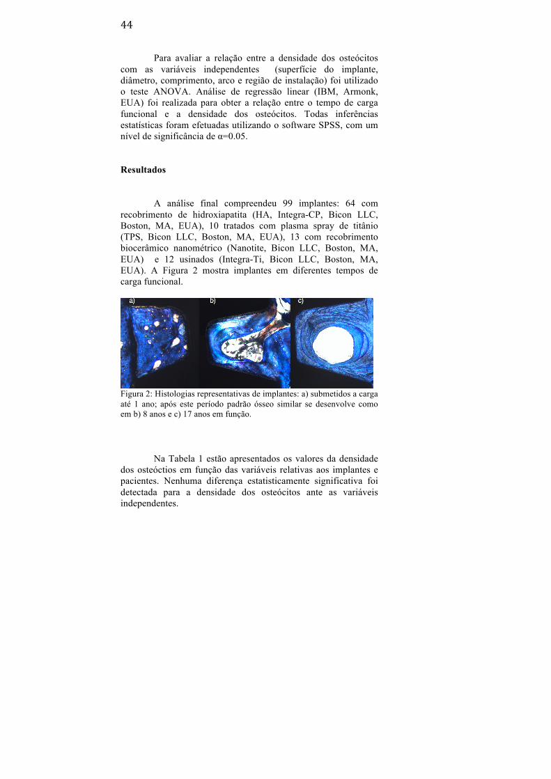
A análise final compreendeu 99 implantes: 64 com
recobrimento de hidroxiapatita (HA, Integra-CP, Bicon LLC, Boston, MA, EUA), 10 tratados com plasma spray de titânio (TPS, Bicon LLC, Boston, MA, EUA), 13 com recobrimento biocerâmico nanométrico (Nanotite, Bicon LLC, Boston, MA, EUA) e 12 usinados (Integra-Ti, Bicon LLC, Boston, MA, EUA). A Figura 2 mostra implantes em diferentes tempos de carga funcional.
Figura 2: Histologias representativas de implantes: a) submetidos a carga até 1 ano; após este período padrão ósseo similar se desenvolve como em b) 8 anos e c) 17 anos em função.
Na Tabela 1 estão apresentados os valores da densidade
dos osteóctios em função das variáveis relativas aos implantes e pacientes. Nenhuma diferença estatisticamente significativa foi detectada para a densidade dos osteócitos ante as variáveis independentes.

45
Tabela 1: Média (dp) da densidade dos osteócitos (DO) diante das variáveis independentes.
Gráfico de regressão linear mostrou que a densidade dos
osteócitos diminuiu discretamente ao longo do tempo de carga funcional (R2 = 0,012) (Figura 3).
Variáveis N DO (osteócitos/mm2)
Superfície HA 64 2050 (90) TPS 10 1940 (190) Nanotite 13 1910 (170) Usinado 12 1740 (220) Diâmetro (mm) 4,0 41 1820 (90) 4,5 26 2110 (360) 5,0 32 2100 (110) Comprimento (mm) 6,0 31 2070 (120) 8,0 36 2080 (130) 11,0 32 1800 (90) Região Anterior 30 1970 (100) Posterior 69 2010 (90) Arco Maxila 60 1990 (90) Mandíbula 39 2010 (110)

46
Figura 3: Variação da densidade dos osteócitos ao longo do tempo. Discussão
Os parâmetros histomorfométricos relacionados à
osseointegração são amplamente documentados em estudos animais, geralmente através da análise de contato osso-implante e a fração de ocupação de área óssea. No entanto, estas análises quantitativas são suplantadas quando se utiliza parâmetros que demonstrem a qualidade do tecido ósseo peri-implantar (26). Estudo recente (16) pontuou que o número de osteócitos é um dos fatores mais importantes na determinação da qualidade óssea, sendo que esta qualidade reflete na resistência óssea durante a função. A densidade dos osteócitos, sendo um tipo de análise histomorfométrica, é geralmente negligenciada pelos estudos em implantodontia. Dessa maneira, relações importantes com outras avaliações histomorfométricas comumente empregadas acabam não sendo estabelecidas, bem como há perda de informações valiosas a respeito da biologia óssea peri-implantar. O presente estudo objetivou avaliar a densidade dos osteócitos ao redor de

47
implantes removidos de humanos, onde o falta de dados é ainda mais pronunciada.
Estudos prévios (20, 24) avaliaram em humanos a densidade dos osteócitos ao redor de implantes com diferentes tratamentos de superfície, mantidos submersos por um período de 8 semanas. Os resultados mostraram diferenças significativas na densidade dos osteócitos para as superfícies testadas, diferentemente do encontrado pelo presente trabalho. Tal dissimilaridade pode ser explicada pela diferença no tempo de avaliação entre estudos, uma vez que a presente avaliação se deu em implantes submetidos à carga por pelo menos 4 meses. A este tempo, com a osseointegração estabelecida e o processo de remodelação óssea iniciado, um possível efeito na modificação da superfície do implante estaria suplantado.
A presente análise estatística também não demonstrou efeitos significativos do comprimento e diâmetro dos implantes sobre a densidade dos osteócitos. Tais fatores são reconhecidos como influenciadores da biomecânica óssea peri-implantar (27), sendo assim, poderia se esperar uma possível ação sobre a densidade dos osteócitos. Presume-se que a ausência de efeito pode ter sido determinada pela macrogeometria dos implantes analisados, que possuem dissipação de forças nos seus 3-5 mm intra-ósseos, devido ao seu protocolo infra-ósseo de instalação, bem como sua configuração endosteal circunferencial em forma de platô (27-30).
Hernandez, Majeska and Schaffler (31) demonstrou diferenças na densidade dos osteócitos entre osso cortical e medular. Como existem discrepâncias entre a proporção de osso cortical e medular entre maxila e mandíbula, bem como entre as regiões anteriores e posteriores dos arcos, poderia se esperar discrepâncias na densidade dos osteócitos entre essas variáveis, no entanto, nenhuma diferença significativa foi detectada pelo presente estudo. Especula-se que este resultado esteja relacionado à via singular de regeneração dos implantes em forma de platôs. Estes dispositivos reparam através da formação de câmaras de cicatrização geradas pela diferença entre o diâmetro final de fresagem e a configuração geométrica do implante. Desenvolve-se assim um padrão de reparo semelhante ao intramembranoso, onde o coágulo que preenche as câmaras é substituído por osso entrelaçado, que por sua vez é rapidamente reposto por osso lamelar (32-36). Estudos prévios demonstraram que este padrão

48
lamelar remodela em uma morfologia semelhante à haversiana, independente de arco e região de instalação (37-40). Este padrão ósseo regular pode justificar a homogeneidade no número de osteócitos ao redor destes implantes, considerando arco e região de instalação.
O modelo de regressão linear mostrou discreta diminuição do número dos osteóctios em função do tempo de carga funcional. Piattelli, Artese, Penitente, Iaculli, Degidi, Mangano, Shibli, Coelho, Perrotti and Iezzi (18) estudando implantes rosqueáveis removidos de humanos, encontrou uma diminuição no número de osteócitos ao comparar grupos de implantes submetidos à carga entre 1-5 anos e 14-27 anos, o que concorda com os resultados da presente investigação. Embora os estudos difiram na macro geometria dos implantes avaliados, presume-se que o tecido ósseo peri-implantar necessita de um certo tempo para remodelar até que se obtenha um melhor alinhamento para suporte das cargas a que é submetido. Nesta fase, um número maior de osteóctios se faz necessário, devido seu papel essencial na remodelação e homeostasia óssea. Uma vez adaptado, o mesmo número de osteócitos não se faz mais necessário.
Vale ressaltar que os resultados obtidos pelo presente estudo não devem ser extrapolados para implantes rosqueáveis, uma vez que diferem nos padrões de dissipação de força, reparo, modelação e remodelação ósseas.
Conclusões
Sugere-se que as características únicas dos implantes em forma de platôs foram capazes de suprimir a influência de variáveis relativas aos implantes e pacientes sobre a densidade dos osteócitos, somado à sua discreta diminuição em função do tempo de carga funcional, demonstrando que um padrão ósseo homogêneo se desenvolve ao redor destes dispositivos. Referências 1. Triplett RG, Frohberg U, Sykaras N, Woody RD. Implant materials, design, and surface topographies: their influence on

49
osseointegration of dental implants. J Long Term Eff Med Implants 2003;13:485-501. 2. Sakka S, Coulthard P. Bone quality: a reality for the process of osseointegration. Implant Dent 2009;18:480-485. 3. Degidi M, Scarano A, Piattelli M, Perrotti V, Piattelli A. Bone remodeling in immediately loaded and unloaded titanium dental implants: a histologic and histomorphometric study in humans. J Oral Implantol 2005;31:18-24. 4. Knothe Tate ML, Adamson JR, Tami AE, Bauer TW. The osteocyte. Int J Biochem Cell Biol 2004;36:1-8. 5. Dallas SL, Prideaux M, Bonewald LF. The osteocyte: an endocrine cell ... and more. Endocr Rev 2013;34:658-690. 6. Rosa N, Simoes R, Magalhaes FD, Marques AT. From mechanical stimulus to bone formation: A review. Med Eng Phys 2015;37:719-728. 7. Gruber R, Stadlinger B, Terheyden H. Cell-to-cell communication in guided bone regeneration: molecular and cellular mechanisms. Clin Oral Implants Res 2016. 8. Galli C, Passeri G, Macaluso GM. Osteocytes and WNT: the mechanical control of bone formation. J Dent Res 2010;89:331-343. 9. Duan P, Bonewald LF. The role of the wnt/beta-catenin signaling pathway in formation and maintenance of bone and teeth. Int J Biochem Cell Biol 2016;77:23-29. 10. Chen H, Senda T, Kubo KY. The osteocyte plays multiple roles in bone remodeling and mineral homeostasis. Med Mol Morphol 2015;48:61-68. 11. Xiong J, Piemontese M, Onal M, Campbell J, Goellner JJ, Dusevich V, et al. Osteocytes, not Osteoblasts or Lining Cells, are the Main Source of the RANKL Required for Osteoclast Formation in Remodeling Bone. PLoS One 2015;10:e0138189. 12. Kennedy OD, Herman BC, Laudier DM, Majeska RJ, Sun HB, Schaffler MB. Activation of resorption in fatigue-loaded bone involves both apoptosis and active pro-osteoclastogenic signaling by distinct osteocyte populations. Bone 2012;50:1115-1122. 13. Kramer I, Halleux C, Keller H, Pegurri M, Gooi JH, Weber PB, et al. Osteocyte Wnt/beta-catenin signaling is required for normal bone homeostasis. Mol Cell Biol 2010;30:3071-3085. 14. Vashishth D, Verborgt O, Divine G, Schaffler MB, Fyhrie DP. Decline in osteocyte lacunar density in human cortical bone is associated with accumulation of microcracks with age. Bone 2000;26:375-380.

50
15. Ma YL, Dai RC, Sheng ZF, Jin Y, Zhang YH, Fang LN, et al. Quantitative associations between osteocyte density and biomechanics, microcrack and microstructure in OVX rats vertebral trabeculae. J Biomech 2008;41:1324-1332. 16. Kuroshima S, Yasutake M, Tsuiki K, Nakano T, Sawase T. Structural and Qualitative Bone Remodeling Around Repetitive Loaded Implants in Rabbits. Clin Implant Dent Relat Res 2015;17 Suppl 2:e699-710. 17. Barros RR, Degidi M, Novaes AB, Piattelli A, Shibli JA, Iezzi G. Osteocyte density in the peri-implant bone of immediately loaded and submerged dental implants. J Periodontol 2009;80:499-504. 18. Piattelli A, Artese L, Penitente E, Iaculli F, Degidi M, Mangano C, et al. Osteocyte density in the peri-implant bone of implants retrieved after different time periods (4 weeks to 27 years). J Biomed Mater Res B Appl Biomater 2014;102:239-243. 19. Mangano C, Piattelli A, Mortellaro C, Mangano F, Perrotti V, Iezzi G. Evaluation of Peri-Implant Bone Response in Implants Retrieved for Fracture After More Than 20 Years of Loading: A Case Series. J Oral Implantol 2015;41:414-418. 20. Traini T, Murmura G, Piattelli M, Scarano A, Pettinicchio M, Sinjari B, et al. Effect of nanoscale topography of titanium implants on bone vessel network, osteocytes, and mineral densities. J Periodontol 2013;84:e40-47. 21. Du Z, Xiao Y, Hashimi S, Hamlet SM, Ivanovski S. The effects of implant topography on osseointegration under estrogen deficiency induced osteoporotic conditions: Histomorphometric, transcriptional and ultrastructural analysis. Acta Biomater 2016;42:351-363. 22. Kuchler U, Pfingstner G, Busenlechner D, Dobsak T, Reich K, Heimel P, et al. Osteocyte lacunar density and area in newly formed bone of the augmented sinus. Clin Oral Implants Res 2013;24:285-289. 23. Oliveira PS, Rodrigues JA, Shibli JA, Piattelli A, Iezzi G, Perrotti V. Influence of osteoporosis on the osteocyte density of human mandibular bone samples: a controlled histological human study. Clin Oral Implants Res 2016;27:325-328. 24. Shibli JA, Grassi S, Piattelli A, Pecora GE, Ferrari DS, Onuma T, et al. Histomorphometric evaluation of bioceramic molecular impregnated and dual acid-etched implant surfaces in the human posterior maxilla. Clin Implant Dent Relat Res 2010;12:281-288.

51
25. Donath K, Breuner G. A method for the study of undecalcified bones and teeth with attached soft tissues. The Sage-Schliff (sawing and grinding) technique. J Oral Pathol 1982;11:318-326. 26. Coelho PG, Granjeiro JM, Romanos GE, Suzuki M, Silva NR, Cardaropoli G, et al. Basic research methods and current trends of dental implant surfaces. J Biomed Mater Res B Appl Biomater 2009;88:579-596. 27. Baggi L, Cappelloni I, Di Girolamo M, Maceri F, Vairo G. The influence of implant diameter and length on stress distribution of osseointegrated implants related to crestal bone geometry: a three-dimensional finite element analysis. J Prosthet Dent 2008;100:422-431. 28. Bozkaya D, Muftu S, Muftu A. Evaluation of load transfer characteristics of five different implants in compact bone at different load levels by finite elements analysis. J Prosthet Dent 2004;92:523-530. 29. Qian L, Todo M, Matsushita Y, Koyano K. Effects of implant diameter, insertion depth, and loading angle on stress/strain fields in implant/jawbone systems: finite element analysis. Int J Oral Maxillofac Implants 2009;24:877-886. 30. Borie E, Orsi IA, Noritomi PY, Kemmoku DT. Three-Dimensional Finite Element Analysis of the Biomechanical Behaviors of Implants with Different Connections, Lengths, and Diameters Placed in the Maxillary Anterior Region. Int J Oral Maxillofac Implants 2016;31:101-110. 31. Hernandez CJ, Majeska RJ, Schaffler MB. Osteocyte density in woven bone. Bone 2004;35:1095-1099. 32. Berglundh T, Abrahamsson I, Lang NP, Lindhe J. De novo alveolar bone formation adjacent to endosseous implants. Clin Oral Implants Res 2003;14:251-262. 33. Coelho PG, Granato R, Marin C, Bonfante EA, Janal MN, Suzuki M. Biomechanical and bone histomorphologic evaluation of four surfaces on plateau root form implants: an experimental study in dogs. Oral Surg Oral Med Oral Pathol Oral Radiol Endod 2010;109:e39-45. 34. Suzuki M, Calasans-Maia MD, Marin C, Granato R, Gil JN, Granjeiro JM, et al. Effect of surface modifications on early bone healing around plateau root form implants: an experimental study in rabbits. J Oral Maxillofac Surg 2010;68:1631-1638. 35. Leonard G, Coelho P, Polyzois I, Stassen L, Claffey N. A study of the bone healing kinetics of plateau versus screw root design titanium dental implants. Clin Oral Implants Res 2009;20:232-239.

52
36. Coelho PG, Suzuki M, Marin C, Granato R, Gil LF, Tovar N, et al. Osseointegration of Plateau Root Form Implants: Unique Healing Pathway Leading to Haversian-Like Long-Term Morphology. Adv Exp Med Biol 2015;881:111-128. 37. Gil LF, Suzuki M, Janal MN, Tovar N, Marin C, Granato R, et al. Progressive plateau root form dental implant osseointegration: A human retrieval study. J Biomed Mater Res B Appl Biomater 2015;103:1328-1332. 38. Coelho PG, Bonfante EA, Marin C, Granato R, Giro G, Suzuki M. A human retrieval study of plasma-sprayed hydroxyapatite-coated plateau root form implants after 2 months to 13 years in function. J Long Term Eff Med Implants 2010;20:335-342. 39. Coelho PG, Marin C, Granato R, Suzuki M. Histomorphologic analysis of 30 plateau root form implants retrieved after 8 to 13 years in function. A human retrieval study. J Biomed Mater Res B Appl Biomater 2009;91:975-979. 40. Baldassarri M, Bonfante E, Suzuki M, Marin C, Granato R, Tovar N, et al. Mechanical properties of human bone surrounding plateau root form implants retrieved after 0.3-24 years of function. J Biomed Mater Res B Appl Biomater 2012;100:2015-2021.

53
Manuscript Based on The International Journal of Oral & Maxillofacial Implants guidelines.

54

55
Osteocyte density around human retrieved dental implants: analyses of 99 cases. Luiz Fernando Gil 1, Marcelo Suzuki 2, Malvin Janal 3, Nick Tovar 4, Charles Marin 1,5, Rodrigo Granato 5, Lukasz Witek 4, José Nazareno Gil 1, Paulo Guilherme Coelho 4,6
1 Department of Dentistry, Federal University of Santa Catarina, Florianópolis, Santa Catarina, Brazil. 2 Department of Prosthodontics and Operative Dentistry, Tufts University School of Dental Medicine, Boston, Massachusetts. 3 Department of Epidemiology, New York University College of Dentistry, New York, New York. 4 Department of Biomaterials and Biomimetics, New York College of Dentistry, New York, New York. 5 Department of Dentistry, UNIGRANRIO, Duque de Caxias, Rio de Janeiro, Brazil. 6 Hansjorg Wyss Department of Plastic Surgery, New york University School of Medicine, New York, New York. Corresponding Author: Luiz Fernando Gil Tenente Silveira, 293, sl 1001 Centro – Fpolis/SC – Brazil CEP: 88010301 [email protected] Keywords: osteocyte density, human retrieval, dental implant, histology.

56

57
ABSTRACT Purpose: The aim of this study was to assess the effect of functional loading time and other clinical/implant design variables on osteocyte density (OD) human retrieved dental implants. Materials and Methods: Due to prosthetic reasons, 99 human plateau root form implant retrievals from the same manufacturer (Bicon LLC, Boston, MA, USA) were obtained over a period of approximately 17 years. OD was measured in single implants were on functional loading for at least 4 months and was assessed as a function of time of functional loading and multiple independent variables: implant surface, diameter, length, jaw (maxilla and mandible), region (anterior and posterior). Results: No implant and patient related characteristics were statistically related to OD; there was a slightly decrease in OD overtime. Conclusions: Results suggest that the unique characteristics of plateau root form implants were able to suppress any influence of patient and implant parameters on OD and along with its slight decrease overtime, showing that a homogeneous bone pattern develops around these devices.

58

59
Introduction Dental implants have changed the concept of replacing
missing teeth. According to Triplett, Frohberg, Sykaras and Woody (1) the success of dental implants is related to osseointegration. The creation and maintenance of this phenomenon depends on the understanding of bone healing and remodeling (2). Long-term maintenance and success of osseointegrated implants involves continued remodeling activity at the periphery of the implant to avoid micro-fractures and to replace bone that may have sustained micro-fractures (3).
Osteocytes, the most abundant cell in mature bone, are differentiated from osteoblasts once incorporated in the mineralized bone matrix. These cells present a stellated shape with innumerous dendrites, providing a high degree of interconnectivity between osteocytes and from them with other cells and blood vessels (4, 5). This net and its location give to osteocytes the capacity for mechanosensation and mechanotransduction. Trough machanotransduction, signaling pathways and molecules involved in bone remodeling are activated and released, promoting osteocytes as the key cells in this process (4, 6, 7).
Osteocytes control bone formation through the regulation of Wnt/β-catenin signaling. This pathway is responsible for recruitment and differentiation of mesenchymal cells in osteoblasts as well as the regulation of bone formation. Osteocytes release antagonists of Wnt/β-catenin pathway as DKK1, sFRP1and sclerostin (8-10).
Bone resorption control depends on the balance of RANKL/OPG in the medium. Recent study (11) demonstrated that osteocytes are the main source of RANKL required for osteoclast formation in remodeling bone. RANKL is also released by viable osteocytes neighboring areas of apoptosis (9, 12). Also, these cells are able to liberate OPG trough the activation of their own Wnt/β- catenin pathway (13).
According to Vashishth, Verborgt, Divine, Schaffler and Fyhrie (14) a minimum number of osteocytes is necessary for the operational network, and their density is considered as a quantitative measurement of this network. Osteocyte density (OD) is related to bone quality and an adequate number of osteocytes is essential in repairing micro-traumas (15, 16).

60
Despite the fact that osteocytes play a key role in bone remodeling, and considering that the success of dental implants depends on this process, literature lacks data regarding OD in oral and maxillofacial regions, and more specifically around dental implant (17-24). Thus, this histomorphometric evaluation would be important in order to better understand peri-implant bone behavior.
The aim of this study was to evaluate the OD around human retrieved plateau root form dental implants due to prosthetic reasons. Relationships between patient and implant design variables to osteocyte density were attempted.
Materials and Methods
Over a period of ∼17 years this study retrieved 221 human samples that were under functional loading anywhere from 0.3 to 24 years due to retreatment reasons. A total of 99-retrieved plateau root form implants (PRFI) (Bicon LLC, Boston, MA, USA) presented sufficient clinical and implant design parameter data to test. Implant retrieval was conducted following approval from the New England Institutional Review Board (NEIRB# 14-256) and receipt of informed patient consent.
All implants were retrieved for prosthetic reasons. Retrospective patient chart review revealed that all retrieved implants were restored with a single crown and subjected to functional loading approximately 6 months after surgical placement. Data regarding implant design parameters (e.g., surface, length, diameter), patient related characteristics as jaw placement (maxilla vs. mandible), region (anterior vs. posterior), and functional loading time were collected. If any of the above parameters were not available, the implant was omitted from the analysis. Implants placed in grafted regions were also excluded.
All implants were removed en bloc using trephine burs of appropriate diameters. The specimens were kept in 10% buffered formalin solution for 24 h, washed in running tap water for another 24 h, and gradually dehydrated in an ascending series of ethanol solutions ranging from 70 to 100%. Following dehydration, the samples were embedded in a methacrylate-based resin (Technovit 9100, Heraeus Kulzer GmbH, Wehrheim, Germany) according to the manufacturer's instructions. The

61
blocks were then cut into slices (∼300 µm thickness) aiming the center of the implant along its bucco-lingual long axis with a precision diamond saw (Isomet 2000; Buehler, Lake Bluff, IL), and were glued to acrylic plates with an acrylate-based cement. A 24 h setting time was allowed before grinding and polishing. The sections were then reduced to a final thickness of ∼100 µm by means of a series of SiC abrasive papers (400, 600, 800, 1200, and 2400) (Buehler) in a grinding/polishing machine (Metaserv 3000; Buehler, Lake Bluff, IL) under water irrigation (25). The slides were then stained with toluidine blue and thin sections were referred to optical microscopy for OLD evaluation.
Osteocyte lacunae where manually counted on the entire perimeter of the implant with a cell counter tool (ImageJ, U. S. National Institutes of Health, Bethesda, Maryland, USA). OD was determined by the ratio of the number osteocytes to the peri-implant bone area (Figure 1).
Figure 1: To determine the osteocyte density, the bone area was delineated (yellow line) and the osteocytes (yellow dots) were counted.
The minimum time frame considered for the retrieved
samples was 4 months under functional loading. ANOVA was used to evaluate relationships between outcome measures of OD and independent variables of surface material, implant diameter, implant length, arch, and anterior/posterior location. Patients’ age and gender were excluded from analysis, as these presented no

62
significant influence in any dependent variable. Linear regression analyses (IBM SPSS, Armonk) were used to evaluate the relationship between time of functional loading and OD. All inferences were performed by SPSS software and significance levels were set to α = 0.05.
Results
The analysis considered 99 implants: 64 hydroxyapatite (HA, Integra-CP, Bicon LLC, Boston, MA, USA), 10 titanium plasma sprayed (TPS, Bicon LLC, Boston, MA, USA) 13 nano-thickness bioceramic deposition (Nanotite, Bicon LLC, Boston, MA, USA) and 12 uncoated (Integra-Ti, Bicon LLC, Boston, MA, USA). Figure 2 depicts implants in different loading times.
Figure 2: Optical micrographs representative of implants a) loaded up to 1 year; after this period a similiar bone pattern develops as in b) 8 years and c) 17 years in function.
Table 1 shows mean levels of OD as a function of implants characteristics and clinical variables. No implant and patient related characteristics were statistically related to OD.

63
Table 1: Mean (SD) of osteocyte density (OD) as a function of the independent variables.
Figure 3 shows that OD slightly decreased as a function of functional loading time (R2 = 0,012).
Variables N OD (pixels/100µm2)
Surface HA 64 2050 (90) TPS 10 1940 (190) Nanotite 13 1910 (170) Uncoated 12 1740 (220) Diameter (mm) 4.0 41 1820 (90) 4.5 26 2110 (360) 5.0 32 2100 (110) Lenght (mm) 6.0 31 2070 (120) 8.0 36 2080 (130) 11.0 32 1800 (90) Region Anterior 30 1970 (100) Posterior 69 2010 (90) Arco Maxila 60 1990 (90) Mandíbula 39 2010 (110)

64
Figure 3: Osteocyte density as a function of functional loading time.
Discussion
Histomorphometric parameters including osseointegration are greatly documented on animal studies and are based mainly on evaluation of bone-to-implant contact (BIC) and bone area fraction occupancy (BAFO). However, these quantitative analyses are supplanted when parameters of peri-implant bone quality are used (26). Recent study (16) considered osteocyte number as one of the most important factors in determining bone quality. OD as a histomorphometric parameter is commonly neglected by these studies, and thus potentially important relationships between widely used histometric parameters and OD are not usually performed and valuable information regarding bone biology may be lost or omitted. For instance, OD would possibly facilitate the determination of
65
preclinical study findings mechanisms where differences may be sight but significant between groups at early times in vivo. The present study aimed to evaluate OD around human retrieved dental implants, where the lack of information is even more pronounced.
Previous studies (18, 20) evaluated in humans the OD around implant with different surfaces, submerged for 8 weeks. Results demonstrated statistically different values of OD between tested surfaces. This is in contrast to our results, where no statistical difference in OD was detected among implant surfaces. This dissimilarity can be explained by differences on time frames between studies, as the present work evaluated implants loaded for minimum of 4 months (~10 months after placement), where a possible effect of surface modification on OD is unlikely to be detected as the process of osseointegration is established and bone remodeling around the implant has already initiated.
Statistical analysis also did not demonstrate significant influence of the evaluated implant parameters of diameter and length in OLD, factors, which are known to affect peri-implant bone biomechanics and thus differences in OD were most expected (27). We speculate that the lack of effect of implant diameter and length on OD was possibly due to the implant macro-geometry analyzed by this study, where forces have been shown to dissipate within the first 3-5mm inside bone due to its deep insertion level and plateau circumferential endosteal configuration (27-30).
Hernandez, Majeska and Schaffler (31) has demonstrated OD differences between cortical and trabecular bone, as thus discrepancies OD counts would be expected between implants retrieved from the maxilla and mandible, as well as between implants retrieved in anterior and posterior regions. Our results, once again, indicated no significant differences in OD regardless of arch and region of implant placement. We speculate that such lack of discrepancy observed between implants placed in the maxilla and mandible, anteriorly and posteriorly, may be related to the unique healing pathway, which lead to haversian-like bone configuration between plateaus over time extensively reported in the literature for PRFI. In brief, PRFIs heal through the

66
formation of healing chambers generated due to the discrepancy between the final drilling diameter and the geometric configuration of the implant, resulting in an intramembranous-like healing pathway, where the clot filling these chambers are then substituted by woven bone, which is then rapidly replaced by lamellar bone (32-36). This lamellar bone has been demonstrated to remodel towards a haversian-like bone morphology over time and that bone morphology is observed irrespective of the position of the retrieved implants in the mouth (37-40). This homogeneous bone configuration, may thus explain the homogeneity of OD observed by the present study.
The OD regression model has shown a slight decrease as a function of functional loading time. Piattelli et al. (21) in a retrieval study on screw root form implants observed a significant OLD reduction between groups of 1-5 years and 14-27 years in function, which is in agreement with the current work. Although studies differ on retrieved implant macro-geometry these suggest that peri-implant bone needs a certain amount of time to remodel in order to obtain a better alignment and consequently improved capacity to bear load. During this time a high number of osteocytes is necessary as they are responsible to control remodeling and bone homeostasis. Once bone is properly aligned, high numbers of osteocytes are no longer required.
It must be noted that these results should not be extrapolated to screw root form implants as they differ in stress distribution, bone healing, modeling and remodeling.
Conclusions
Results suggest that the unique characteristics of plateau root form implants were able to suppress any influence of patient and implant parameters on OD and along with its slight decrease overtime, showing that a homogeneous bone pattern develops around these devices.

67
References 1. Triplett RG, Frohberg U, Sykaras N, Woody RD. Implant materials, design, and surface topographies: their influence on osseointegration of dental implants. J Long Term Eff Med Implants 2003;13:485-501. 2. Sakka S, Coulthard P. Bone quality: a reality for the process of osseointegration. Implant Dent 2009;18:480-485. 3. Degidi M, Scarano A, Piattelli M, Perrotti V, Piattelli A. Bone remodeling in immediately loaded and unloaded titanium dental implants: a histologic and histomorphometric study in humans. J Oral Implantol 2005;31:18-24. 4. Dallas SL, Prideaux M, Bonewald LF. The osteocyte: an endocrine cell ... and more. Endocr Rev 2013;34:658-690. 5. Knothe Tate ML, Adamson JR, Tami AE, Bauer TW. The osteocyte. Int J Biochem Cell Biol 2004;36:1-8. 6. Rosa N, Simoes R, Magalhaes FD, Marques AT. From mechanical stimulus to bone formation: A review. Med Eng Phys 2015;37:719-728. 7. Gruber R, Stadlinger B, Terheyden H. Cell-to-cell communication in guided bone regeneration: molecular and cellular mechanisms. Clin Oral Implants Res 2016. 8. Galli C, Passeri G, Macaluso GM. Osteocytes and WNT: the mechanical control of bone formation. J Dent Res 2010;89:331-343. 9. Chen H, Senda T, Kubo KY. The osteocyte plays multiple roles in bone remodeling and mineral homeostasis. Med Mol Morphol 2015;48:61-68. 10. Duan P, Bonewald LF. The role of the wnt/beta-catenin signaling pathway in formation and maintenance of bone and teeth. Int J Biochem Cell Biol 2016;77:23-29. 11. Xiong J, Piemontese M, Onal M, Campbell J, Goellner JJ, Dusevich V, et al. Osteocytes, not Osteoblasts or Lining Cells, are the Main Source of the RANKL Required for Osteoclast Formation in Remodeling Bone. PLoS One 2015;10:e0138189. 12. Kennedy OD, Herman BC, Laudier DM, Majeska RJ, Sun HB, Schaffler MB. Activation of resorption in fatigue-loaded bone involves both apoptosis and active pro-osteoclastogenic signaling by distinct osteocyte populations. Bone 2012;50:1115-1122. 13. Kramer I, Halleux C, Keller H, Pegurri M, Gooi JH, Weber PB, et al. Osteocyte Wnt/beta-catenin signaling is required for normal bone homeostasis. Mol Cell Biol 2010;30:3071-3085.

68
14. Vashishth D, Verborgt O, Divine G, Schaffler MB, Fyhrie DP. Decline in osteocyte lacunar density in human cortical bone is associated with accumulation of microcracks with age. Bone 2000;26:375-380. 15. Ma YL, Dai RC, Sheng ZF, Jin Y, Zhang YH, Fang LN, et al. Quantitative associations between osteocyte density and biomechanics, microcrack and microstructure in OVX rats vertebral trabeculae. J Biomech 2008;41:1324-1332. 16. Kuroshima S, Yasutake M, Tsuiki K, Nakano T, Sawase T. Structural and Qualitative Bone Remodeling Around Repetitive Loaded Implants in Rabbits. Clin Implant Dent Relat Res 2015;17 Suppl 2:e699-710. 17. Barros RR, Degidi M, Novaes AB, Piattelli A, Shibli JA, Iezzi G. Osteocyte density in the peri-implant bone of immediately loaded and submerged dental implants. J Periodontol 2009;80:499-504. 18. Shibli JA, Grassi S, Piattelli A, Pecora GE, Ferrari DS, Onuma T, et al. Histomorphometric evaluation of bioceramic molecular impregnated and dual acid-etched implant surfaces in the human posterior maxilla. Clin Implant Dent Relat Res 2010;12:281-288. 19. Kuchler U, Pfingstner G, Busenlechner D, Dobsak T, Reich K, Heimel P, et al. Osteocyte lacunar density and area in newly formed bone of the augmented sinus. Clin Oral Implants Res 2013;24:285-289. 20. Traini T, Murmura G, Piattelli M, Scarano A, Pettinicchio M, Sinjari B, et al. Effect of nanoscale topography of titanium implants on bone vessel network, osteocytes, and mineral densities. J Periodontol 2013;84:e40-47. 21. Piattelli A, Artese L, Penitente E, Iaculli F, Degidi M, Mangano C, et al. Osteocyte density in the peri-implant bone of implants retrieved after different time periods (4 weeks to 27 years). J Biomed Mater Res B Appl Biomater 2014;102:239-243. 22. Mangano C, Piattelli A, Mortellaro C, Mangano F, Perrotti V, Iezzi G. Evaluation of Peri-Implant Bone Response in Implants Retrieved for Fracture After More Than 20 Years of Loading: A Case Series. J Oral Implantol 2015;41:414-418. 23. Du Z, Xiao Y, Hashimi S, Hamlet SM, Ivanovski S. The effects of implant topography on osseointegration under estrogen deficiency induced osteoporotic conditions: Histomorphometric, transcriptional and ultrastructural analysis. Acta Biomater 2016;42:351-363.

69
24. Oliveira PS, Rodrigues JA, Shibli JA, Piattelli A, Iezzi G, Perrotti V. Influence of osteoporosis on the osteocyte density of human mandibular bone samples: a controlled histological human study. Clin Oral Implants Res 2016;27:325-328. 25. Donath K, Breuner G. A method for the study of undecalcified bones and teeth with attached soft tissues. The Sage-Schliff (sawing and grinding) technique. J Oral Pathol 1982;11:318-326. 26. Coelho PG, Granjeiro JM, Romanos GE, Suzuki M, Silva NR, Cardaropoli G, et al. Basic research methods and current trends of dental implant surfaces. J Biomed Mater Res B Appl Biomater 2009;88:579-596. 27. Baggi L, Cappelloni I, Di Girolamo M, Maceri F, Vairo G. The influence of implant diameter and length on stress distribution of osseointegrated implants related to crestal bone geometry: a three-dimensional finite element analysis. J Prosthet Dent 2008;100:422-431. 28. Bozkaya D, Muftu S, Muftu A. Evaluation of load transfer characteristics of five different implants in compact bone at different load levels by finite elements analysis. J Prosthet Dent 2004;92:523-530. 29. Qian L, Todo M, Matsushita Y, Koyano K. Effects of implant diameter, insertion depth, and loading angle on stress/strain fields in implant/jawbone systems: finite element analysis. Int J Oral Maxillofac Implants 2009;24:877-886. 30. Borie E, Orsi IA, Noritomi PY, Kemmoku DT. Three-Dimensional Finite Element Analysis of the Biomechanical Behaviors of Implants with Different Connections, Lengths, and Diameters Placed in the Maxillary Anterior Region. Int J Oral Maxillofac Implants 2015. 31. Hernandez CJ, Majeska RJ, Schaffler MB. Osteocyte density in woven bone. Bone 2004;35:1095-1099. 32. Berglundh T, Abrahamsson I, Lang NP, Lindhe J. De novo alveolar bone formation adjacent to endosseous implants. Clin Oral Implants Res 2003;14:251-262. 33. Coelho PG, Granato R, Marin C, Bonfante EA, Janal MN, Suzuki M. Biomechanical and bone histomorphologic evaluation of four surfaces on plateau root form implants: an experimental study in dogs. Oral Surg Oral Med Oral Pathol Oral Radiol Endod 2010;109:e39-45. 34. Suzuki M, Calasans-Maia MD, Marin C, Granato R, Gil JN, Granjeiro JM, et al. Effect of surface modifications on early bone

70
healing around plateau root form implants: an experimental study in rabbits. J Oral Maxillofac Surg 2010;68:1631-1638. 35. Leonard G, Coelho P, Polyzois I, Stassen L, Claffey N. A study of the bone healing kinetics of plateau versus screw root design titanium dental implants. Clin Oral Implants Res 2009;20:232-239. 36. Coelho PG, Suzuki M, Marin C, Granato R, Gil LF, Tovar N, et al. Osseointegration of Plateau Root Form Implants: Unique Healing Pathway Leading to Haversian-Like Long-Term Morphology. Adv Exp Med Biol 2015;881:111-128. 37. Gil LF, Suzuki M, Janal MN, Tovar N, Marin C, Granato R, et al. Progressive plateau root form dental implant osseointegration: A human retrieval study. J Biomed Mater Res B Appl Biomater 2015;103:1328-1332. 38. Coelho PG, Bonfante EA, Marin C, Granato R, Giro G, Suzuki M. A human retrieval study of plasma-sprayed hydroxyapatite-coated plateau root form implants after 2 months to 13 years in function. J Long Term Eff Med Implants 2010;20:335-342. 39. Coelho PG, Marin C, Granato R, Suzuki M. Histomorphologic analysis of 30 plateau root form implants retrieved after 8 to 13 years in function. A human retrieval study. J Biomed Mater Res B Appl Biomater 2009;91:975-979. 40. Baldassarri M, Bonfante E, Suzuki M, Marin C, Granato R, Tovar N, et al. Mechanical properties of human bone surrounding plateau root form implants retrieved after 0.3-24 years of function. J Biomed Mater Res B Appl Biomater 2012;100:2015-2021.

71
Capítulo III

72

73
Bibliografia Consultada ADELL, R. et al. A 15-year study of osseointegrated implants in the treatment of the edentulous jaw. Int J Oral Surg, v. 10, n. 6, p. 387-416, Dec 1981. ISSN 0300-9785 (Print) 0300-9785 (Linking). Disponível em: < http://www.ncbi.nlm.nih.gov/pubmed/6809663 >. ALBREKTSSON, T. et al. Osseointegrated oral implants. A Swedish multicenter study of 8139 consecutively inserted Nobelpharma implants. J Periodontol, v. 59, n. 5, p. 287-96, May 1988. ISSN 0022-3492 (Print) 0022-3492 (Linking). Disponível em: < http://www.ncbi.nlm.nih.gov/pubmed/3290429 >. BARROS, R. R. et al. Osteocyte density in the peri-implant bone of immediately loaded and submerged dental implants. J Periodontol, v. 80, n. 3, p. 499-504, Mar 2009. ISSN 0022-3492 (Print) 0022-3492 (Linking). Disponível em: < http://www.ncbi.nlm.nih.gov/pubmed/19254134 >. BELLIDO, T.; SAINI, V.; PAJEVIC, P. D. Effects of PTH on osteocyte function. Bone, v. 54, n. 2, p. 250-7, Jun 2013. ISSN 1873-2763 (Electronic) 1873-2763 (Linking). Disponível em: < http://www.ncbi.nlm.nih.gov/pubmed/23017659 >. BEN-AWADH, A. N. et al. Parathyroid hormone receptor signaling induces bone resorption in the adult skeleton by directly regulating the RANKL gene in osteocytes. Endocrinology, v. 155, n. 8, p. 2797-809, Aug 2014. ISSN 1945-7170 (Electronic) 0013-7227 (Linking). Disponível em: < http://www.ncbi.nlm.nih.gov/pubmed/24877630 >. BONEWALD, L. F. The amazing osteocyte. J Bone Miner Res, v. 26, n. 2, p. 229-38, Feb 2011. ISSN 1523-4681 (Electronic) 0884-0431 (Linking). Disponível em: < http://www.ncbi.nlm.nih.gov/pubmed/21254230 >.

74
BUENZLI, P. R.; SIMS, N. A. Quantifying the osteocyte network in the human skeleton. Bone, v. 75, p. 144-50, Jun 2015. ISSN 1873-2763 (Electronic) 1873-2763 (Linking). Disponível em: < http://www.ncbi.nlm.nih.gov/pubmed/25708054 >. BURGERS, T. A.; WILLIAMS, B. O. Regulation of Wnt/beta-catenin signaling within and from osteocytes. Bone, v. 54, n. 2, p. 244-9, Jun 2013. ISSN 1873-2763 (Electronic) 1873-2763 (Linking). Disponível em: < http://www.ncbi.nlm.nih.gov/pubmed/23470835 >. BUSER, D. et al. Long-term evaluation of non-submerged ITI implants. Part 1: 8-year life table analysis of a prospective multi-center study with 2359 implants. Clin Oral Implants Res, v. 8, n. 3, p. 161-72, Jun 1997. ISSN 0905-7161 (Print) 0905-7161 (Linking). Disponível em: < http://www.ncbi.nlm.nih.gov/pubmed/9586460 >. BUSSE, B. et al. Decrease in the osteocyte lacunar density accompanied by hypermineralized lacunar occlusion reveals failure and delay of remodeling in aged human bone. Aging Cell, v. 9, n. 6, p. 1065-75, Dec 2010. ISSN 1474-9726 (Electronic) 1474-9718 (Linking). Disponível em: < http://www.ncbi.nlm.nih.gov/pubmed/20874757 >. CAPULLI, M.; PAONE, R.; RUCCI, N. Osteoblast and osteocyte: games without frontiers. Arch Biochem Biophys, v. 561, p. 3-12, Nov 01 2014. ISSN 1096-0384 (Electronic) 0003-9861 (Linking). Disponível em: < http://www.ncbi.nlm.nih.gov/pubmed/24832390 >. CHEN, H.; SENDA, T.; KUBO, K. Y. The osteocyte plays multiple roles in bone remodeling and mineral homeostasis. Med Mol Morphol, v. 48, n. 2, p. 61-8, Jun 2015. ISSN 1860-1499 (Electronic) 1860-1499 (Linking). Disponível em: < http://www.ncbi.nlm.nih.gov/pubmed/25791218 >.

75
CLARKE, B. Normal bone anatomy and physiology. Clin J Am Soc Nephrol, v. 3 Suppl 3, p. S131-9, Nov 2008. ISSN 1555-905X (Electronic) 1555-9041 (Linking). Disponível em: < http://www.ncbi.nlm.nih.gov/pubmed/18988698 >. COELHO, P. G. et al. Basic research methods and current trends of dental implant surfaces. J Biomed Mater Res B Appl Biomater, v. 88, n. 2, p. 579-96, Feb 2009. ISSN 1552-4981 (Electronic) 1552-4973 (Linking). Disponível em: < http://www.ncbi.nlm.nih.gov/pubmed/18973274 >. DALLAS, S. L.; PRIDEAUX, M.; BONEWALD, L. F. The osteocyte: an endocrine cell ... and more. Endocr Rev, v. 34, n. 5, p. 658-90, Oct 2013. ISSN 1945-7189 (Electronic) 0163-769X (Linking). Disponível em: < http://www.ncbi.nlm.nih.gov/pubmed/23612223 >. DEGIDI, M. et al. Bone remodeling in immediately loaded and unloaded titanium dental implants: a histologic and histomorphometric study in humans. J Oral Implantol, v. 31, n. 1, p. 18-24, 2005. ISSN 0160-6972 (Print) 0160-6972 (Linking). Disponível em: < http://www.ncbi.nlm.nih.gov/pubmed/15751384 >. DELGADO-CALLE, J.; BELLIDO, T. Osteocytes and Skeletal Pathophysiology. Curr Mol Biol Rep, v. 1, n. 4, p. 157-167, Dec 2015. ISSN 2198-6428 (Linking). Disponível em: < http://www.ncbi.nlm.nih.gov/pubmed/26693137 >. DU, Z. et al. The effects of implant topography on osseointegration under estrogen deficiency induced osteoporotic conditions: Histomorphometric, transcriptional and ultrastructural analysis. Acta Biomater, v. 42, p. 351-63, Sep 15 2016. ISSN 1878-7568 (Electronic) 1742-7061 (Linking). Disponível em: < http://www.ncbi.nlm.nih.gov/pubmed/27375286 >. DUAN, P.; BONEWALD, L. F. The role of the wnt/beta-catenin signaling pathway in formation and maintenance of bone and teeth.

76
Int J Biochem Cell Biol, v. 77, n. Pt A, p. 23-9, Aug 2016. ISSN 1878-5875 (Electronic) 1357-2725 (Linking). Disponível em: < http://www.ncbi.nlm.nih.gov/pubmed/27210503 >. ERIKSEN, E. F. Cellular mechanisms of bone remodeling. Rev Endocr Metab Disord, v. 11, n. 4, p. 219-27, Dec 2010. ISSN 1573-2606 (Electronic) 1389-9155 (Linking). Disponível em: < http://www.ncbi.nlm.nih.gov/pubmed/21188536 >. FRANZ-ODENDAAL, T. A.; HALL, B. K.; WITTEN, P. E. Buried alive: how osteoblasts become osteocytes. Dev Dyn, v. 235, n. 1, p. 176-90, Jan 2006. ISSN 1058-8388 (Print) 1058-8388 (Linking). Disponível em: < http://www.ncbi.nlm.nih.gov/pubmed/16258960 >. GALLI, C.; PASSERI, G.; MACALUSO, G. M. Osteocytes and WNT: the mechanical control of bone formation. J Dent Res, v. 89, n. 4, p. 331-43, Apr 2010. ISSN 1544-0591 (Electronic) 0022-0345 (Linking). Disponível em: < http://www.ncbi.nlm.nih.gov/pubmed/20200416 >. GOLDRING, S. R. The osteocyte: key player in regulating bone turnover. RMD Open, v. 1, n. Suppl 1, p. e000049, 2015. ISSN 2056-5933 (Linking). Disponível em: < http://www.ncbi.nlm.nih.gov/pubmed/26557372 >. GRUBER, R.; STADLINGER, B.; TERHEYDEN, H. Cell-to-cell communication in guided bone regeneration: molecular and cellular mechanisms. Clin Oral Implants Res, Aug 23 2016. ISSN 1600-0501 (Electronic) 0905-7161 (Linking). Disponível em: < http://www.ncbi.nlm.nih.gov/pubmed/27550738 >. HERNANDEZ, C. J.; MAJESKA, R. J.; SCHAFFLER, M. B. Osteocyte density in woven bone. Bone, v. 35, n. 5, p. 1095-9, Nov 2004. ISSN 8756-3282 (Print) 1873-2763 (Linking). Disponível em: < http://www.ncbi.nlm.nih.gov/pubmed/15542034 >.

77
KENNEDY, O. D. et al. Activation of resorption in fatigue-loaded bone involves both apoptosis and active pro-osteoclastogenic signaling by distinct osteocyte populations. Bone, v. 50, n. 5, p. 1115-22, May 2012. ISSN 1873-2763 (Electronic) 1873-2763 (Linking). Disponível em: < http://www.ncbi.nlm.nih.gov/pubmed/22342796 >. KNOTHE TATE, M. L. et al. The osteocyte. Int J Biochem Cell Biol, v. 36, n. 1, p. 1-8, Jan 2004. ISSN 1357-2725 (Print) 1357-2725 (Linking). Disponível em: < http://www.ncbi.nlm.nih.gov/pubmed/14592527 >. KRAMER, I. et al. Osteocyte Wnt/beta-catenin signaling is required for normal bone homeostasis. Mol Cell Biol, v. 30, n. 12, p. 3071-85, Jun 2010. ISSN 1098-5549 (Electronic) 0270-7306 (Linking). Disponível em: < http://www.ncbi.nlm.nih.gov/pubmed/20404086 >. KUCHLER, U. et al. Osteocyte lacunar density and area in newly formed bone of the augmented sinus. Clin Oral Implants Res, v. 24, n. 3, p. 285-9, Mar 2013. ISSN 1600-0501 (Electronic) 0905-7161 (Linking). Disponível em: < http://www.ncbi.nlm.nih.gov/pubmed/22784394 >. KUROSHIMA, S. et al. Structural and Qualitative Bone Remodeling Around Repetitive Loaded Implants in Rabbits. Clin Implant Dent Relat Res, v. 17 Suppl 2, p. e699-710, Oct 2015. ISSN 1708-8208 (Electronic) 1523-0899 (Linking). Disponível em: < http://www.ncbi.nlm.nih.gov/pubmed/25809916 >. LAI, X. et al. The dependences of osteocyte network on bone compartment, age, and disease. Bone Res, v. 3, 2015. ISSN 2095-4700 (Print) 2095-4700 (Linking). Disponível em: < http://www.ncbi.nlm.nih.gov/pubmed/26213632 >. MA, Y. L. et al. Quantitative associations between osteocyte density and biomechanics, microcrack and microstructure in OVX rats vertebral trabeculae. J Biomech, v. 41, n. 6, p. 1324-32, 2008. ISSN 0021-9290 (Print)

78
0021-9290 (Linking). Disponível em: < http://www.ncbi.nlm.nih.gov/pubmed/18342320 >. MANGANO, C. et al. Evaluation of Peri-Implant Bone Response in Implants Retrieved for Fracture After More Than 20 Years of Loading: A Case Series. J Oral Implantol, v. 41, n. 4, p. 414-8, Aug 2015. ISSN 1548-1336 (Electronic) 0160-6972 (Linking). Disponível em: < http://www.ncbi.nlm.nih.gov/pubmed/23964716 >. MANOLAGAS, S. C.; PARFITT, A. M. For whom the bell tolls: distress signals from long-lived osteocytes and the pathogenesis of metabolic bone diseases. Bone, v. 54, n. 2, p. 272-8, Jun 2013. ISSN 1873-2763 (Electronic) 1873-2763 (Linking). Disponível em: < http://www.ncbi.nlm.nih.gov/pubmed/23010104 >. MULLENDER, M. G. et al. Differences in osteocyte density and bone histomorphometry between men and women and between healthy and osteoporotic subjects. Calcif Tissue Int, v. 77, n. 5, p. 291-6, Nov 2005. ISSN 0171-967X (Print) 0171-967X (Linking). Disponível em: < http://www.ncbi.nlm.nih.gov/pubmed/16307389 >. OLIVEIRA, P. S. et al. Influence of osteoporosis on the osteocyte density of human mandibular bone samples: a controlled histological human study. Clin Oral Implants Res, v. 27, n. 3, p. 325-8, Mar 2016. ISSN 1600-0501 (Electronic) 0905-7161 (Linking). Disponível em: < http://www.ncbi.nlm.nih.gov/pubmed/25487068 >. PALUMBO, C. A three-dimensional ultrastructural study of osteoid-osteocytes in the tibia of chick embryos. Cell Tissue Res, v. 246, n. 1, p. 125-31, 1986. ISSN 0302-766X (Print) 0302-766X (Linking). Disponível em: < http://www.ncbi.nlm.nih.gov/pubmed/3779795 >. PIATTELLI, A. et al. Osteocyte density in the peri-implant bone of implants retrieved after different time periods (4 weeks to 27 years). J Biomed Mater Res B Appl Biomater, v. 102, n. 2, p. 239-43, Feb 2014. ISSN 1552-4981 (Electronic)

79
1552-4973 (Linking). Disponível em: < http://www.ncbi.nlm.nih.gov/pubmed/24106071 >. PRIDEAUX, M.; FINDLAY, D. M.; ATKINS, G. J. Osteocytes: The master cells in bone remodelling. Curr Opin Pharmacol, v. 28, p. 24-30, Jun 2016. ISSN 1471-4973 (Electronic) 1471-4892 (Linking). Disponível em: < http://www.ncbi.nlm.nih.gov/pubmed/26927500 >. QING, H. et al. Demonstration of osteocytic perilacunar/canalicular remodeling in mice during lactation. J Bone Miner Res, v. 27, n. 5, p. 1018-29, May 2012. ISSN 1523-4681 (Electronic) 0884-0431 (Linking). Disponível em: < http://www.ncbi.nlm.nih.gov/pubmed/22308018 >. QIU, S. et al. Age and distance from the surface but not menopause reduce osteocyte density in human cancellous bone. Bone, v. 31, n. 2, p. 313-8, Aug 2002a. ISSN 8756-3282 (Print) 1873-2763 (Linking). Disponível em: < http://www.ncbi.nlm.nih.gov/pubmed/12151084 >. ______. Relationships between osteocyte density and bone formation rate in human cancellous bone. Bone, v. 31, n. 6, p. 709-11, Dec 2002b. ISSN 8756-3282 (Print) 1873-2763 (Linking). Disponível em: < http://www.ncbi.nlm.nih.gov/pubmed/12531566 >. ______. Reduced iliac cancellous osteocyte density in patients with osteoporotic vertebral fracture. J Bone Miner Res, v. 18, n. 9, p. 1657-63, Sep 2003. ISSN 0884-0431 (Print) 0884-0431 (Linking). Disponível em: < http://www.ncbi.nlm.nih.gov/pubmed/12968675 >. ROSA, N. et al. From mechanical stimulus to bone formation: A review. Med Eng Phys, v. 37, n. 8, p. 719-28, Aug 2015. ISSN 1873-4030 (Electronic) 1350-4533 (Linking). Disponível em: < http://www.ncbi.nlm.nih.gov/pubmed/26117332 >.

80
SAKKA, S.; COULTHARD, P. Bone quality: a reality for the process of osseointegration. Implant Dent, v. 18, n. 6, p. 480-5, Dec 2009. ISSN 1538-2982 (Electronic) 1056-6163 (Linking). Disponível em: < http://www.ncbi.nlm.nih.gov/pubmed/20009601 >. SATO, M.; KATAYAMA, Y. Osteocytes and Homeostasis of Remote Organs : Bone-Buried Osteocytes Talk to Remote Organs. Curr Osteoporos Rep, v. 13, n. 4, p. 193-7, Aug 2015. ISSN 1544-2241 (Electronic) 1544-1873 (Linking). Disponível em: < http://www.ncbi.nlm.nih.gov/pubmed/25851936 >. SMITH, D. W.; GARDINER, B. S.; DUNSTAN, C. Bone balance within a cortical BMU: local controls of bone resorption and formation. PLoS One, v. 7, n. 7, p. e40268, 2012. ISSN 1932-6203 (Electronic) 1932-6203 (Linking). Disponível em: < http://www.ncbi.nlm.nih.gov/pubmed/22844401 >. TAICHMAN, R. S. Blood and bone: two tissues whose fates are intertwined to create the hematopoietic stem-cell niche. Blood, v. 105, n. 7, p. 2631-9, Apr 1 2005. ISSN 0006-4971 (Print) 0006-4971 (Linking). Disponível em: < http://www.ncbi.nlm.nih.gov/pubmed/15585658 >. TETI, A.; ZALLONE, A. Do osteocytes contribute to bone mineral homeostasis? Osteocytic osteolysis revisited. Bone, v. 44, n. 1, p. 11-6, Jan 2009. ISSN 1873-2763 (Electronic) 1873-2763 (Linking). Disponível em: < http://www.ncbi.nlm.nih.gov/pubmed/18977320 >. TRAINI, T. et al. Effect of nanoscale topography of titanium implants on bone vessel network, osteocytes, and mineral densities. J Periodontol, v. 84, n. 10, p. e40-7, Oct 2013. ISSN 1943-3670 (Electronic) 0022-3492 (Linking). Disponível em: < http://www.ncbi.nlm.nih.gov/pubmed/23517509 >. TRIPLETT, R. G. et al. Implant materials, design, and surface topographies: their influence on osseointegration of dental implants.

81
J Long Term Eff Med Implants, v. 13, n. 6, p. 485-501, 2003. ISSN 1050-6934 (Print) 1050-6934 (Linking). Disponível em: < http://www.ncbi.nlm.nih.gov/pubmed/15056066 >. VASHISHTH, D. et al. Decline in osteocyte lacunar density in human cortical bone is associated with accumulation of microcracks with age. Bone, v. 26, n. 4, p. 375-80, Apr 2000. ISSN 8756-3282 (Print) 1873-2763 (Linking). Disponível em: < http://www.ncbi.nlm.nih.gov/pubmed/10719281 >. XIONG, J. et al. Osteocytes, not Osteoblasts or Lining Cells, are the Main Source of the RANKL Required for Osteoclast Formation in Remodeling Bone. PLoS One, v. 10, n. 9, p. e0138189, 2015. ISSN 1932-6203 (Electronic) 1932-6203 (Linking). Disponível em: < http://www.ncbi.nlm.nih.gov/pubmed/26393791 >. ZARRINKALAM, M. R. et al. Changes in osteocyte density correspond with changes in osteoblast and osteoclast activity in an osteoporotic sheep model. Osteoporos Int, v. 23, n. 4, p. 1329-36, Apr 2012. ISSN 1433-2965 (Electronic) 0937-941X (Linking). Disponível em: < http://www.ncbi.nlm.nih.gov/pubmed/21626447 >.

82

83
Capítulo IV

84

85
Produção Científica no Doutorado
1: Neiva RF, Gil LF, Tovar N, Janal MN, Marao HF, Bonfante EA, Pinto N, Coelho PG. The Synergistic Effect of Leukocyte Platelet-Rich Fibrin and Micrometer/Nanometer Surface Texturing on Bone Healing around Immediately Placed Implants: An Experimental Study in Dogs. Biomed Res Int. 2016;2016:9507342. 2: Marão HF, Jimbo R, Neiva R, Gil LF, Bowers M, Bonfante EA, Tovar N, Janal MN, Coelho PG. Cortical and Trabecular Bone Healing Patterns and Quantification for Three Different Dental Implant Systems. Int J Oral Maxillofac Implants. 2016 Nov 11. 3: Patel A, Gil LF, Castellano A, Freitas G, Navarro D, Peredo AP, Tovar N, Coelho P. Effect of Simplified One-Step Drilling Protocol on Osseointegration. Int J Periodontics Restorative Dent. 2016 Sep-Oct;36(5):e82-7. 4: Neiva R, Tovar N, Jimbo R, Gil LF, Goldberg P, Barbosa JP, Lilin T, Coelho PG. The Effect of Laser-Etched Surface Design on Soft Tissue Healing of Two Different Implant Abutment Systems: An Experimental Study in Dogs. Int J Periodontics Restorative Dent. 2016 Sep-Oct;36(5):673-9. 5: Ribeiro da Silva J, Castellano A, Malta Barbosa JP, Gil LF, Marin C, Granato R, Bonfante EA, Tovar N, Janal MN, Coelho PG. Histomorphological and Histomorphometric Analyses of Grade IV Commercially Pure Titanium and Grade V Ti-6Al-4V Titanium Alloy Implant Substrates: An In Vivo Study in Dogs. Implant Dent. 2016 Oct;25(5):650-5. 6: Marin C, Bonfante E, Granato R, Neiva R, Gil LF, Marão HF, Suzuki M, Coelho PG. The Effect of Osteotomy Dimension on Implant Insertion Torque, Healing Mode, and Osseointegration Indicators: A Study in Dogs. Implant Dent. 2016 Dec;25(6):739-743 7: Neiva R, Tovar N, Jimbo R, Gil LF, Goldberg P, Barbosa JP, Lilin T, Coelho PG. Guidelines for Clinical Management of

86
Laser-Etched (Laser-Lok) Abutments in Two Different Clinical Scenarios: A Preclinical Laboratory Soft Tissue Assessment Study. Int J Periodontics Restorative Dent. 2016 May-Jun;36(3):339-45. 8: Bowers M, Yoo D, Marin C, Gil L, Shabaka N, Goldstein M, Janal M, Tovar N, Hirata R, Bonfante E, Coelho P. Surface characterization and in vivo evaluation of laser sintered and machined implants followed by resorbable-blasting media process: A study in sheep. Med Oral Patol Oral Cir Bucal. 2016 Mar 1;21(2):e206-13. 9: Coelho PG, Gil LF, Neiva R, Jimbo R, Tovar N, Lilin T, Bonfante EA. Microrobotized blasting improves the bone-to-textured implant response. A preclinical in vivo biomechanical study. J Mech Behav Biomed Mater. 2016 Mar;56:175-82. 10: Coelho PG, Suzuki M, Marin C, Granato R, Gil LF, Tovar N, Jimbo R, Neiva R, Bonfante EA. Osseointegration of Plateau Root Form Implants: Unique Healing Pathway Leading to Haversian-Like Long-Term Morphology. Adv Exp Med Biol. 2015;881:111-28. 11: Gil LF, Marin C, Teixeira H, Marão HF, Tovar N, Khan R, Bonfante EA, Janal M, Coelho PG. The effect of controlled microrobotized blasting on implant surface texturing and early osseointegration. J Biomater Appl. 2016 Feb;30(7):900-7. 12: Gil LF, Suzuki M, Janal MN, Tovar N, Marin C, Granato R, Bonfante EA, Jimbo R, Gil JN, Coelho PG. Progressive plateau root form dental implant osseointegration: A human retrieval study. J Biomed Mater Res B Appl Biomater. 2015 Aug;103(6):1328-32.