avaliaÇÃo de risco ocupacional no setor …livros01.livrosgratis.com.br/cp050658.pdf ·...
TRANSCRIPT
1
UNIVERSIDADE FEDERAL DO RIO GRANDE DO SUL - UFRGS
AVALIAÇÃO DE RISCO OCUPACIONAL NO SETOR
COUREIRO-CALÇADISTA DO RIO GRANDE DO SUL
Vanina Dahlström Heuser
Tese submetida ao Programa de Pós-
Graduação em Genética e Biologia Molecular
da UFRGS como requisito parcial para
obtenção do grau Doutor em Ciências.
Orientador: Bernardo Erdtmann
Co-orientadora: Juliana da Silva
Porto Alegre, fevereiro de 2005.

Livros Grátis
http://www.livrosgratis.com.br
Milhares de livros grátis para download.

2
Aos meus pais,
meus maiores mestres,
dedico.

3
Este trabalho foi desenvolvido no Laboratório de Citogenética e Evolução e no Laboratório de Imunogenética, ambos nas dependências do
Departamento de Genética da Universidade Federal do Rio Grande do Sul, e foi financiado pelo Conselho Nacional
de Desenvolvimento Científico e Tecnológico (CNPq).

4
AGRADECIMENTOS:
À Dra. Jacinta da Faculdade de Engenharia (UFRGS) que nos encaminhou ao
CESSTIC (Centro de Saúde e Segurança do Trabalhador das Indústrias Calçadistas da
Região de Parobé).
A todo o pessoal do CESSTIC: Verônica, Eduardo, César, Ivani e Sílvia, por nos
possibilitar as coletas de amostras dos funcionários das empresas fabricantes de
calçados, e por nos dar acesso a todos os resultados toxicológicos.
À Diolanda e Luciana do Laboratório Bom Pastor de Taquara (RS) e à Cíntia,
Líliam e especialmente à Suzi do CESSTIC, por toda a ajuda e ótima companhia durante
as coletas nas fábricas calçadistas.
Ao Tedi, Laoni e Jaime, por nos possibilitarem a obtenção de amostras em
curtumes de Estância Velha.
A toda a equipe do Laboratório Vida (Estância Velha): Dra. Vera, Poliana, Anita,
Elisa e em especial à Cíntia, por todo o apoio nas coletas, pelas análises feitas
especialmente para esse trabalho e por nos fornecerem os resultados toxicológicos dos
voluntários na indústria do couro.
Às empresas que nos depositaram confiança participando desse projeto, e a todos
os voluntários que gentilmente nos cederam amostras como grupo exposto, tanto nos
curtumes quanto nas fábricas de calçados.
A José Celso da empresa Artecola que, mesmo sem me conhecer pessoalmente,
me esclareceu muitas dúvidas por telefone, me indicou sites na internet, e enviou Fichas
de Segurança de produtos utilizados nas indústrias calçadistas.
À Liliam, do Laboratório Toxilab, por toda paciência nas negociações e pelos
descontos nos exames ocupacionais.
Ao amigo Horst, pela ajuda na interpretação de algumas informações e pela
oportunidade de divulgação da pesquisa no Senai Couros de Estância Velha através de
palestra e, indiretamente, na obtenção de voluntários para o estudo. E por responder tão
prontamente meus e-mails desesperados.
A todos aqueles que participaram como voluntários do grupo controle: meu irmão
Martin, vizinhos, conhecidos, funcionários de setores burocráticos das regiões de coleta,


6
SUMÁRIO ABREVIATURAS ................................................................................................................8
RESUMO.............................................................................................................................9
ABSTRACT.......................................................................................................................12
1. INTRODUÇÃO ..............................................................................................................14
1.1. RISCO OCUPACIONAL.............................................................................................14
1.2. CONTROLE DA EXPOSIÇÃO ...................................................................................16
1.2.1. Monitoramento Biológico ..............................................................................16
1.2.2.1. Biomarcadores de Exposição ....................................................................18
1.2.2.2. Biomarcadores de Efeito ............................................................................19
1.2.2.3. Biomarcadores De Suscetibilidade: Características Genéticas Individuais ...................................................................................................21
1.3. FATORES DE RISCO NÃO-OCUPACIONAIS ..........................................................23
1.3.1. IDADE E SEXO ................................................................................................23
1.3.2. TABAGISMO E ÁLCOOL ................................................................................24
1.4. NÍVEIS DE AÇÃO – CONTROLE NO BRASIL .........................................................25
1.5. SETOR COUREIRO-CALÇADISTA BRASILEIRO ...................................................25
1.5.1. Setor Coureiro-Calçadista X Fatores De Risco............................................26
1.5.1.1. Produção do Couro .....................................................................................26
1.5.1.2. Produção de Calçados ................................................................................30
1.6. METODOLOGIAS UTILIZADAS NESTE ESTUDO...................................................34
1.6.1. Amostra Biológica ..........................................................................................34
1.6.2. Ensaio Cometa................................................................................................35
1.6.3. Teste de Micronúcleos (MN) ..........................................................................36
1.6.4. Identificação de Polimorfismos nos Genes de Metabolização...................39
2. OBJETIVOS ..................................................................................................................41

7
3. CAPÍTULO I - EVALUATION OF GENOTOXIC EXPOSURE IN BRAZILIAN TANNERY WORKERS: COMPARISON BETWEEN DRUM WORKSHOP AND FINISHING WORKSHOP. .................................................................................................42
4. CAPÍTULO II - INFLUENCE OF METABOLIZING GENE POLYMORPHISMS ON GENOTOXICITY IN BRAZILIAN TANNERY WORKERS. ...............................................62
5. CAPÍTULO III - COMPARISON OF GENETIC DAMAGE IN BRAZILIAN FOOTWEAR-WORKERS EXPOSED TO SOLVENT-BASED OR WATER-BASED ADHESIVE ........................................................................................................................84
6. CAPÍTULO IV - EVALUATION OF GENETIC DAMAGE IN BRAZILIAN FOOTWEAR-WORKERS: CORRELATION OF CYTOGENETIC ANALYSIS AND POLYMORPHISMS IN METABOLIZING GENES GSTM1, GSTT1, GSTP1, CYP1A1, AND CYP2E1. ...................................................................................104
7. DISCUSSÃO ...............................................................................................................127
7.1. Risco Ocupacional no Curtume ............................................................................127
7.1.1. Biomarcadores de Exposição e de Efeito ..................................................127
7.1.2. Biomarcadores de Suscetibilidade .............................................................131
7.2. Risco Ocupacional na Produção de Calçados ....................................................133
7.2.1. Biomarcadores de Exposição e de Efeito ..................................................133
7.2.2. Biomarcadores de Suscetibilidade .............................................................136
8. REFERÊNCIAS BIBLIOGRÁFICAS ...........................................................................141
9. ANEXOS......................................................................................................................160
9.1. Norma Regulamentadora No. 7 (NR-7) ..................................................................160
9.2. Informações ao Voluntário – Consentimento Informado....................................160
9.3. Questionário de Saúde Pessoal ............................................................................161
9.4. Exames realizados em Laboratórios Particulares...............................................163
9.5. Resolução do Comitê de Ética em Pesquisa .......................................................163
8
ABREVIATURAS
PORTUGUÊS AC – aberrações cromossômicas
AH - ácido hipúrico
CBA – cola a base de água
CBS – cola a base de solventes
CMBMN – célula(s) de mucosa bucal com micronúcleo(s)
CI - Comprimento de imagem (Ensaio Cometa)
FD – Freqüência de dano (Ensaio Cometa)
HAP - hidrocarbonetos aromáticos policíclicos
ID – Índice de Dano (Ensaio Cometa)
LBMN – linfócito(s) binucleado(s) com micronúcleo(s)
MN – micronúcleo
NR-7 - Norma Regulamentadora N°7, da Secretaria de Segurança e Saúde no Trabalho,
que estabelece os parâmetros biológicos para o controle da exposição a agentes
químicos no Brasil
TCI – Troca de cromátides irmãs
VR - Valor de Referência da normalidade
INGLÊS BNLMN – binucleated lymphocyte with micronucleus (linfócito binucleado com
micronúcleo)
CA – cromosomal aberrations (aberrações cromossômicas)
DF – damage frequency (freqüência de danos – Ensaio cometa)
DI – damage index (índice de dano – Ensaio cometa)
DW – drum workshop (setor de curtimento e/ou recurtimento – curtume)
EBCMN – exfoliated buccal cell with micronucleus (célula bucal exfoliada com
micronúcleo)
IL – image lenght (comprimento de imagem – Ensaio cometa)
FW – finishing workshop (setor de acabamento – curtume)
NPB – nucleoplasmic bridges (pontes nucleoplasmáticas – nos LBN)
SBA – solvent based adhesive (cola a base de solventes)
SCE – sister chromatid exchanges (troca de cromátides irmãs)
WBA – water based adhesive (cola a base de água)
9
RESUMO
Várias pesquisas descrevem um aumento de mortalidade por câncer em
trabalhadores de curtumes e de fábricas de calçados, do mesmo modo que um aumento
nas freqüências de biomarcadores de danos citogenéticos. Essas indústrias são de
grande importância no sul do Brasil e, portanto, buscou-se avaliar o risco ocupacional
nesses setores em relação a danos genotóxicos.
No primeiro trabalho, foram comparados dois setores de processamento de couro
em curtumes: o recurtimento (17 indivíduos), e o acabamento (32 indivíduos). Como
controles, foram utilizadas amostras de 32 indivíduos de sexo masculino, sem exposição
ocupacional. Os biomarcadores de exposição utilizados foram as concentrações de
cromo na urina e os níveis de metahemoglobina e hemoglobina, dos quais somente a
hemoglobina demonstrou relação com a exposição, sendo encontrada em menores
quantidades nos indivíduos do setor de acabamento quando comparados ao grupo
controle (P < 0,05).
A análise do Ensaio Cometa em leucócitos e o teste de micronúcleo em células de
mucosa bucal (CMBMN) não demonstraram diferenças entre os grupos de estudo. No
entanto, os trabalhadores do setor de acabamento tiveram um aumento estatisticamente
significativo na freqüência de micronúcleos em linfócitos binucleados (LBMN) e no
número de pontes nucleoplasmáticas (PN) (P < 0,01 e P < 0,05, respectivamente). Para
os trabalhadores do recurtimento esse aumento só foi estatisticamente significativo nas
freqüências de PN em relação ao grupo não exposto (P < 0,01).
No segundo artigo são apresentados os dados obtidos com a avaliação de 45
trabalhadores de curtume, de ambos os setores (recurtimento e acabamento). Quarenta
indivíduos do sexo masculino foram utilizados como controles. Como observado no
primeiro estudo, com exceção dos níveis de hemoglobina mais baixos no grupo exposto
(P < 0,01), os demais biomarcadores de exposição (cromo na urina, metahemoglobina e
níveis de dano de DNA obtidos pelo Ensaio Cometa) não demonstraram haver diferenças
significantes entre os grupos. Com relação às freqüências de CMBMN os grupos
apresentaram resultados similares, mas para as freqüências de LBMN e PN observou-se
um aumento significativo nos trabalhadores de curtume (P < 0,05 e P < 0,001,
respectivamente). Considerando que variações genéticas individuais podem estar
envolvidas na modulação dos efeitos genotóxicos observados, foram identificados
polimorfismos nos genes GSTT1, GSTM1, GSTP1, CYP1A1 e CYP2E1, como

10
biomarcadores de suscetibilidade. Os resultados obtidos sugerem que enzimas do
sistema CYP450 podem influenciar os resultados de genotoxicidade, pois os
trabalhadores de curtume com o genótipo CYP2E1 selvagem (*1A/*1A) apresentaram
níveis maiores de danos no DNA detectados pelo Ensaio Cometa, quando comparados
ao genótipo CYP2E1 variante (*1A/*5B ou *5B/*5B) (P < 0,03). Os genótipos GSTs
investigados não influenciaram os níveis de dano citogenético entre os grupos.
Nas indústrias calçadistas, o reconhecimento do risco para a saúde associado com
o uso de colas a base de solvente (CBS), levou ao desenvolvimento de adesivos sem
essas substâncias, as colas a base de água (CBA). Deste modo, nosso terceiro artigo
mostra os resultados obtidos em relação aos trabalhadores de fábricas de calçados
expostos a CBS (29 indivíduos) e CBA (16 indivíduos); 25 indivíduos saudáveis foram
utilizados como grupo controle. Como biomarcador de exposição, foram obtidos valores
de ácido hipúrico (AH) na urina, o principal metabólito de exposição ao tolueno, o qual
apresentou concentrações significativamente mais baixas no grupo controle em relação
aos grupos expostos às CBS e CBA (P < 0,001 e P < 0,05, respectivamente). Os
resultados do Ensaio Cometa demonstraram aumento de danos de DNA no grupo
exposto a CBS em comparação ao grupo exposto a CBA (P < 0,001), bem como em
comparação com o grupo controle (P < 0,05). Em relação às freqüências de CMBMN e
LBMN não se observou diferença significativa entre os três grupos.
Portanto, neste artigo foi demonstrado que a utilização da CBA, mesmo contendo
isocianato, é uma opção menos prejudicial à saúde dos trabalhadores de fábricas de
calçados. Os resultados positivos de genotoxicidade nos trabalhadores expostos a CBS
possivelmente podem ser explicados pela presença de policloropreno na fórmula deste
adesivo.
Tendo sido encontrados esses resultados positivos, nosso quarto artigo descreve
um estudo envolvendo 39 trabalhadores da indústria calçadista (31 homens e 8
mulheres), expostos ocupacionalmente a colas contendo solvente e policloropreno. Como
grupo controle foram utilizados 55 indivíduos (44 homens e 11 mulheres) sem exposição
ocupacional. Os danos no DNA, observados com o Ensaio Cometa, e as concentrações
de AH, foram significativamente maiores nos trabalhadores expostos em comparação aos
controles (P < 0,001). Diferenças nas freqüências de LBMN, PN e CMBMN entre os
grupos não foram observadas. O hábito tabagista e o sexo não influenciaram os
resultados em nenhum dos parâmetros analisados. Com o objetivo de identificar
diferenças de sensibilidade à genotoxicidade, foram determinados polimorfismos nos
genes GSTT1, GSTM1, GSTP1, CYP1A1, e CYP2E1, com os quais foi demonstrada a
influência dos genótipos variantes dos genes GSTP1 e CYP2E1, sugerindo uma

11
associação dos mesmos com os elevados níveis de dano de DNA nos trabalhadores de
indústrias calçadistas.
Embora não tenha sido possível apontar um único agente genotóxico no ambiente
de trabalho, devido à presença de misturas complexas neste, os dados indicam
exposição genotóxica em curtumes e em fábricas de calçados. Também se observou
aumento de dano nos indivíduos com mais idade, o que nos leva a uma discussão sobre
a possibilidade desses danos apresentarem maior acúmulo em pessoas expostas. Em
todos os grupos estudados foi encontrada uma correlação positiva entre as freqüências
LBMN e idade, o que é amplamente discutido por inúmeros autores.

12
ABSTRACT
Several investigations report increased mortality from cancer in tannery and
footwear workers, as well as increase in frequency of cytogenetic biomarkers. Since these
industries are some of the most important in Southern Brazil, it was therefore considered
interesting to examine whether occupational exposure in this occupational settings could
bear genotoxic risk to the workers.
Firstly, we compared two different processing areas in tannery industry, the Drum
Workshop (DW; 17 subjects) and Finishing Workshop (FW; 32 subjects). As control, a
group of 32 healthy males was used, with no occupational exposure. As biomarkers of
exposure, we obtained data on the chromium in urine, hemoglobin and methemoglobin
levels, from which only the hemoglobin levels seem to demonstrate association with
exposure, being lower in FW compared to control group (P < 0.05). The analysis of Comet
assay in leukocytes and micronucleus in epithelial buccal cells (EBCMN) failed to show
differences between the control and test groups. The FW workers presented a statistical
significant increase in the frequency of MN in binucleated lymphocytes (BNLMN) and
nucleoplasmic bridges (NPB), while the DW workers showed a statistically significant
increase in NPB frequencies compared to control group.
Our second paper presented evaluations carried out in a group of 45 male Leather
workers, independently of section in tannery. Forty healthy males were used as controls.
As observed at the firsth study, with exception of lower hemoglobin levels in Tannery
workers, the other biomarkers of exposure (chromium in urine, methemoglobin, and
reparable DNA damage measured by Comet assay) failed to show marked differences
between the groups. However, although no difference was found between controls and
Tannery workers regarding EBCMN frequencies, the other biomarkers of early effect
used, BNLMN and NPB were significantly increased in Tannery workers (P < 0.05 and P <
0.001, respectively). Individual variations of the genes GSTT1, GSTM1, GSTP1, CYP1A1
and CYP2E1 were used as biomarkers of susceptibility, suggesting the modulation of the
genotoxicity by the enzymes of the CYP450 system, since Tannery workers with the
CYP2E1*1A/*1A (the wild type) had increased values in DNA damage measured by
Comet assay in comparison to CYP2E1 variant genotypes (*1A/*5B or *5B/*5B) (P <
0.03). The different GST genotypes investigated did not influence the level of cytogenetic
damage between groups.
At the footwear industry, the recognition of the potential health-hazards of solvent-
based adhesives (SBAs) has lead to the development of adhesives with no organic

13
solvents, the water-based adhesives (WBA). Thus, in our third paper we described the
comparison between footwear-workers (all males) exposed to SBA (n=29) and WBA
(n=16); 25 healthy subjects were used as controls. As biomarkers of exposure, we
obtained data on the concentration of hippuric acid (HA) in urine, the main metabolite of
exposure to toluene, which show a significantly lower mean values in the control group in
relation to SBA (P < 0.001) and WBA (P < 0.05) groups. The Comet assay results showed
increased levels for the SBA (P < 0.001) group in comparison to the WBA and control (P <
0.05), while in the frequencies of BNLMN and EBCMN, the three groups were not
statistically different. Therefore, in this study it was demonstrated water-based adhesives
are clearly a better option for safeguarding the health of footwear-workers, even with
possibility of isocyanate presence, while the positive results observed in SBA group might
be explained by chloroprene presence in adhesive.
Due to the positive findings in the third paper, our fourth study involved 39
Footwear-workers (31 males and 8 females), occupationally exposed to solvent-based
polychloroprene glue and solutions containing organic solvents. The control group
consisted of 55 subjects (44 males and 11 females) with no occupational exposure. The
HA concentration and Comet assay show statistical increase in footwear-workers in
relation to controls (P < 0.001), but no differences were observed in BNLMN, NPB and
EBCMN frequencies. Males and females did not show differences in any of the
parameters analyzed. To make it possible to identify differences in sensitivity to
genotoxicity, the polymorphisms in genes GSTT1, GSTM1, GSTP1, CYP1A1, and
CYP2E1 were determined, showing that GSTP1 and CYP2E1 variant polymorphisms
seem be associated with increasing in DNA damage footwear-workers.
Although it may not be possible to point out a single genotoxic agent at the
workplace our data indicate the presence of genotoxic exposure at the Tannery and
Footwear industry, with increased values in older subjects, leading to the discussion about
the possibility accumulated damage to be increased in exposed people, since a positive
correlation was found between age and BNLMN frequency in all the study groups in the
four papers, what are also widely described for several authors.
14
1. INTRODUÇÃO
1.1. Risco Ocupacional
O desenvolvimento industrial tem como conseqüência inevitável a exposição do
homem a um número crescente de substâncias químicas sintéticas ou naturais, que
incluem poeiras, fibras, compostos químicos orgânicos e inorgânicos, e que podem
causar sérios efeitos toxicológicos (Keshava & Ong, 1999; Lucas et al., 2001).
É crescente a preocupação com o efeito mutagênico e carcinogênico de agentes
genotóxicos em populações expostas ocupacionalmente, acidentalmente ou por estilo de
vida. Estas substâncias podem afetar a saúde humana por sua disseminação no
ambiente, mas os trabalhadores que manipulam rotineiramente estes agentes constituem
o maior grupo de risco, devido à constante exposição (Maluf & Erdtmann, 2003).
Programas de saúde e segurança foram implementados em diversos países do
mundo, dando maior atenção aos problemas causados por intoxicações ocupacionais
causados por agentes químicos. Os trabalhadores dos países desenvolvidos que têm
risco de exposição a esses agentes, têm sido esclarecidos sobre seu manuseio, porém,
devido a grande diversidade do parque industrial e devido a desigualdades culturais do
planeta, muitos países ainda desprezam sua importância por questões políticas e
econômicas. Apesar da existência de medidas regulatórias, trabalhadores continuam
sendo expostos a agentes genotóxicos por desconhecerem tal exposição, e o tipo e a
quantidade de substâncias potencialmente perigosas utilizadas em seu trabalho
(Keshava & Ong, 1999).
A exposição a agentes genotóxicos pode levar a alterações nas células
germinativas causando problemas reprodutivos. Estima-se que mais de 50% das mortes
fetais, 30% de retardo mental, 20% dos defeitos congênitos e 2% da infertilidade
masculina estejam associados a aberrações cromossômicas. Estudos de risco
ocupacional reprodutivo consistem de dados epidemiológicos de exposição maternal e
têm mostrado aumento nas taxas de aborto espontâneo em mulheres trabalhando com
químicos em hospitais e laboratórios. Dados indicam de que cerca de 10% de todas as
doenças crônicas, incluindo doenças cardiovasculares, podem ser resultantes de
mutações (Keshava & Ong, 1999).
O processo carcinogênico desenvolve-se após vários estágios e cada um deles
pode ser influenciado por diferentes fatores. Dados epidemiológicos, estudos com

15
animais e o uso de técnicas modernas de biologia molecular demonstraram não haver
dúvidas sobre a influência de agentes químicos em alguns destes estágios, embora
existam muitos outros fatores envolvidos. Há evidências convincentes de que o sítio de
ação destes agentes é o material genético celular, sendo que essas mutações, quando
ocorrem em proto-oncogenes ou genes supressores de tumor, que estão envolvidos no
controle do crescimento ou na diferenciação celular, podem levar ao desenvolvimento de
câncer em determinados órgãos atingidos (Au, 1991; Maluf & Erdtmann, 2003).
Evidências indicam que fatores ambientais podem ser os maiores responsáveis
pelo desenvolvimento do câncer e que o ambiente de trabalho ainda é o principal local
em que ocorre exposição ambiental a substâncias potencialmente carcinogênicas
(Keshava & Ong, 1999). O ambiente ocupacional é um dos ambientes mais propícios
para investigar a etiologia e patogênese do câncer em humanos. Até a década de 1970,
as substâncias ou circunstâncias causadoras de câncer em humanos eram encontradas
primariamente no ambiente ocupacional, e embora atualmente o número de carcinógenos
não ocupacionais esteja aumentando, carcinógenos de origem ocupacional ainda
representam uma grande fração do total, ocupando uma posição especial entre as
diferentes classes de carcinógenos humanos (Siemiatycki et al., 2004).
Estima-se que a exposição ocupacional é responsável por pelo menos 4% dos
casos de câncer em humanos (NORA, 2003). Em alguns casos, sabe-se que
determinado grupo ocupacional ou industrial apresenta risco aumentado para o
desenvolvimento de câncer, e pode-se ter alguma idéia sobre o agente causador, como
por exemplo, o câncer de testículos em limpadores de chaminés expostos à
hidrocarbonetos aromáticos policíclicos (HAP), e o câncer de pulmão em trabalhadores
de mineração de asbestos. Em outras situações, sabe-se que determinados grupos têm o
risco de desenvolvimento de câncer aumentado, mas o agente causador é desconhecido
ou de difícil comprovação, como por exemplo, o câncer de pulmão entre pintores, e de
bexiga entre trabalhadores na indústria de alumínio (Siemiatycki et al., 2004).
Entre as várias substâncias no ambiente industrial para as quais não existem
estudos com respeito à carcinogenicidade em humanos, centenas têm se mostrado
carcinogênicas para animais de laboratório e milhares têm demonstrado efeitos em testes
de mutagenicidade ou genotoxicidade (Siemiatycki et al., 2004). Apesar disso, menos de
2% dos químicos comercializados foram testados adequadamente para
carcinogenicidade. São necessários métodos melhores para testar essas substâncias
quanto ao seu potencial carcinogênico, especialmente para a avaliação de materiais que
são misturas e circunstâncias em que a exposição inclui misturas químicas (Fung et al.,

16
1995; NORA, 2003). Além disso, os efeitos das exposições podem ser sobrepostos ou
sofrer interações com outros fatores ambientais e/ou genéticos, diminuindo o risco para
certos trabalhadores e aumentando risco para outros (Siemiatycki et al., 2004).
Como a incidência de câncer e a mortalidade têm utilidade limitada para a
prevenção da doença, pois são detectáveis apenas após a exposição com o
desenvolvimento do câncer ou morte, estudos com marcadores intermediários em
trabalhadores saudáveis são importantes na avaliação do risco de carcinogenicidade,
antes que tais eventos aconteçam (NORA, 2003). São necessários mais esforços para se
detectar e identificar essas substâncias no ambiente ocupacional, bem como para se
estabelecer biomarcadores adequados relacionados com doenças quer seja em nível
químico, fisiológico, celular e sub-celular, ou molecular, para facilitar a prevenção do
câncer ocupacional (Keshava & Ong, 1999).
1.2. Controle da Exposição
A avaliação da exposição, associada aos conhecimentos relativos aos efeitos na
saúde e os limites considerados seguros, permite estabelecer as prioridades e formas de
intervenção efetiva para proteger uma população dos riscos químicos (Amorim, 2003),
podendo ser avaliada por medida da concentração do agente químico em amostras
ambientais, como ar (monitoramento ambiental), ou através da medida de parâmetros
biológicos (monitoramento biológico), denominados indicadores biológicos ou
biomarcadores.
1.2.1. Monitoramento Biológico
O monitoramento biológico da exposição aos agentes químicos, propriamente dita,
significa a medida da substância ou seus metabólitos em vários meios biológicos, como
sangue, urina, ar exalado e outros. O conceito de monitoramento biológico inclui também
a detecção precoce de efeitos não-adversos e reversíveis (monitoramento biológico de
efeito) (Dougherty, 1998; WHO, 2001; Amorim, 2003).
A detecção precoce de uma exposição perigosa pode diminuir significativamente a
ocorrência de efeitos adversos na saúde dos trabalhadores expostos às substâncias
químicas, pois as informações provenientes do monitoramento da exposição ocupacional
possibilitam a implantação de medidas de prevenção e controle apropriadas. Para isto é
necessário: (I) a definição dos níveis permissíveis de exposição, os quais, de acordo com

17
os conhecimentos atuais, são estabelecidos para não causarem efeitos adversos
decorrentes da exposição química; e (II) a avaliação regular dos possíveis riscos à saúde
associados à exposição, por comparação aos seus limites permissíveis (Amorim, 2003).
Além do monitoramento ambiental e biológico através da formação de metabólitos,
técnicas para detectar alterações nas células de trabalhadores ocupacionalmente
expostos são igualmente importantes, pois: a) se agentes genotóxicos ou condições de
genotoxicidade estão presentes, medidas preventivas podem ser introduzidas para evitar
maiores efeitos nos trabalhadores; b) em caso de exposição acidental, alguma estimativa
da dose absorvida pode ser obtida; c) em caso de problema genético de saúde
(reprodutivo ou câncer), alguma estimativa pode ser obtida, da probabilidade de que o
efeito tenha vindo da exposição (Sorsa et al., 1992).
Assim, o monitoramento biológico complementa o monitoramento ambiental,
considerando-se que determina a exposição global efetiva diretamente no indivíduo e
detecta efeitos precoces reversíveis, proporcionando melhor estimativa de risco.
Uma série de trabalhos ressalta a utilidade de biomarcadores em estudos de
monitoramento de populações. O termo “biomarcador” é usado para expressar uma
medida específica de uma interação entre determinado sistema biológico com um agente
genotóxico (Cebulska-Wasilewska, 2003). Os biomarcadores disponíveis podem ser
classificados em três categorias: a) biomarcadores de exposição; b) biomarcadores de
efeito; e c) biomarcadores de suscetibilidade (WHO, 2001; Bonassi & Au, 2002; Amorim
2003; Cebulska-Wasilewska, 2003).
Os biomarcadores, sejam de exposição ou efeito, como ferramentas para avaliar a
exposição ocupacional são também utilizados nos estudos epidemiológicos, buscando-se
estabelecer uma relação entre a exposição aos agentes químicos e os efeitos na saúde
dos indivíduos expostos. A importância do uso destes biomarcadores como parâmetros
biológicos de exposição a substâncias químicas, deve-se ao fato de eles estarem mais
diretamente relacionados aos efeitos na saúde do que os parâmetros ambientais,
podendo, assim, oferecer uma melhor estimativa do risco. Além disso, a avaliação
biológica da exposição ocupacional a substâncias químicas leva em consideração a
absorção por diferentes vias e rotas de introdução no organismo, permitindo avaliar a
exposição global do indivíduo ou população (Amorim, 2003).
Vários são os parâmetros biológicos que podem estar alterados como
conseqüência da interação entre o agente químico e o organismo. Entretanto, a
determinação quantitativa desses parâmetros será usada como indicador biológico ou
18
biomarcador somente se existir uma correlação com a intensidade de exposição e/ou
com o efeito biológico da substância ou seu produto de biotransformação, assim como
qualquer alteração bioquímica precoce, cuja determinação nos fluidos biológicos, tecidos
ou ar exalado, avalie a intensidade da exposição e o risco à saúde (WHO, 2001; Amorim,
2003).
1.2.2.1. Biomarcadores de Exposição
Segundo Lawry (1995) existem dois tipos de Biomarcadores de exposição: (1) a
medida quantitativa de uma substância química ou seus metabólitos em fluidos
biológicos, e (2) a medida de uma alteração bioquímica precoce e reversível em fluidos
biológicos que reflita exposição.
A medida de substâncias químicas ou seus metabólitos são os biomarcadores de
exposição mais utilizados. O indicador biológico de exposição estima a dose interna,
através da determinação da substância química ou seu produto de biotransformação em
fluidos biológicos, como sangue, urina, ar exalado entre outros, possibilitando quantificar
a substância no organismo, quando a toxicocinética é bem conhecida (Bonassi & Au,
2002; Amorim, 2003).
Para muitos tipos de biomarcadores a consideração mais importante é a
estabilidade dos mesmos com respeito ao tempo após a exposição, pois a tendência é de
que as concentrações dos mesmos diminuam com o passar do tempo (Bonassi & Au,
2002). Alguns biomarcadores de dose interna, como o benzeno no sangue, ácido hipúrico
e 2,5-hexanodiona na urina, refletem apenas a exposição recente ao benzeno, tolueno e
n-hexano, respectivamente, enquanto outros refletem a exposição média dos últimos
meses, como o chumbo e mercúrio no sangue, ou até mesmo em anos, como cádmio na
urina (WHO, 2001; Amorim, 2003).
O segundo tipo de biomarcador de exposição identifica mudanças bioquímicas
precoces e reversíveis que refletem exposição. Exemplos: adutos de albumina-HAP no
sangue como biomarcador de exposição a HAP; ácido s-fenilmercaptúrico no sangue
como biomarcador de exposição ao benzeno; carboxihemoglobina no sangue como
biomarcador de exposição ao monóxido de carbono e diclorometano; adutos de anilina
em hemoglobina como biomarcador de exposição a anilinas; colinesterase em eritrócitos
como biomarcador de exposição a pesticidas organofosforados e outros (Lowry, 1995).
Biomarcadores de exposição a solventes orgânicos que produzem efeitos


20
independentemente de exposição à carcinógenos (Bonassi et al., 2000).
Segundo Fenech et al. (1999) e Thomas et al. (2003) o teste de MN pode não
identificar todos os eventos de dano cromossômico, como translocações recíprocas que
não são expressas em MN, mas pode indentificar translocações assimétricas, como
cromossomos dicêntricos e seus fragmentos acêntricos associados, pontes
nucleoplasmáticas (PN) e MN. Além disso, o processo responsável pela formação de um
MN pode ser um mecanismo importante pelo qual uma célula pode perder a
heterozigosidade em um locus genético chave (Inoue et al., 1997). Deste modo, a
hipótese de associação direta entre a freqüência de MN em determinado tecido e o
desenvolvimento de câncer, é embasada em várias observações:
a) há um nítido aumento na freqüência de MN em determinados órgãos e linfócitos
periféricos de pacientes com câncer (Cheng et al., 1996; Duffaud et al., 1997);
b) indivíduos com instabilidade cromossômica congênita, como na Anemia de
Fanconi e na Síndrome de Bloom, normalmente desenvolvem câncer, mesmo sem
exposição a condições de risco (Rosin et al., 1985; Rudd et al., 1988);
c) avaliações clínicas de câncer oral têm usado MN em mucosa bucal como
biomarcador de câncer (Benner et al., 1994; Desai et al., 1996);
d) existe correlação entre carcinogenicidade e genotoxicidade para alguns agentes
capazes de aumentar a freqüência de MN em humanos e em animais (Sorsa et al.,
1992);
e) a freqüência de micronúcleos é fortemente associada com a concentração de
vitaminas e folatos no sangue, que também estão associados com um risco aumentado
de algumas formas de câncer (Fenech & Rinaldi, 1995; Blount et al., 1997; Fenech et al.,
1997b; Fenech et al., 1998).
Assim, a maioria das investigações citogenéticas em populações quimicamente
expostas é realizada através de métodos clássicos, como AC e a análise MN para
detectar efeitos aneugênicos e clastogênicos, ou TCI para detecção de alterações no
DNA (Marcon et al., 1999).
Recentemente, em função de regulamentação exigindo a redução progressiva dos
níveis de exposição na maioria das condições de risco, aumentou o interesse no
monitoramento biológico da exposição associado à baixa exposição ocupacional ou
ambiental a agentes perigosos. Embora bem conduzidos, muitos estudos nesta área têm
falhado em tornar mais clara a associação entre estes baixos níveis de exposição e

21
alterações citogenéticas. Pesquisas cuidadosas usando técnicas novas e mais sensíveis
são necessárias para uma estimativa eficiente e precisa dos efeitos citogenéticos em
níveis baixos de exposição (Marcon et al., 1999).
1.2.2.3. Biomarcadores de Suscetibilidade: Características Genéticas Individuais
A atividade dos xenobióticos, que são compostos estranhos ao organismo, não
depende apenas das suas propriedades intrínsecas e das doses recebidas, mas também
de sítios alvos nos hospedeiros, de sua biotransformação e do reparo do DNA (Omenn,
1991).
A biotransformação de xenobióticos, em especial a que se processa no fígado, é
comumente separada em duas fases. A Fase I, que envolve as enzimas microssomais e
inclui as reações de oxidação, de redução ou de hidrólise, que podem ativar, inativar ou
deixar inalteradas as atividades do xenobiótico; e a Fase II, na qual quase sempre ocorre
a inativação da substância caso esta ainda não tenha sido inativada na fase I (Kelada et
al., 2003).
As enzimas da Fase I, também chamadas de enzimas de ativação, representadas
pela superfamília citocromo P450 (CYP450), realizam o metabolismo oxidativo através da
inserção de um átomo de oxigênio em um xenobiótico, tornando-o altamente eletrofílico.
As enzimas da Fase II, chamadas também de enzimas de detoxificação, como as
glutationa S-transferases (GSTs), por exemplo, conjugam os reativos intermediários
eletrofílicos formados no metabolismo oxidativo da Fase I com substâncias endógenas
como a glutationa, tornando-as mais solúveis em água e mais facilmente elimináveis do
organismo. Dependendo do xenobiótico, a reação da Fase I pode ser suficiente para
tornar o composto mais hidrossolúvel, sem que haja necessidade de reação da Fase II
para sua eliminação do organismo (Venitt, 1994).
A regulação coordenada das enzimas das Fases I e II é um fator importante para
assegurar o metabolismo de xenobióticos, diminuindo o risco de acumular intermediários
oxigenados reativos (Hirvonen, 1994; Venitt, 1994). Assim sendo, a associação entre
alelos específicos de genes responsáveis pela metabolização de compostos químicos e o
risco aumentado para diversas neoplasias, se deve à existência de múltiplos passos
enzimáticos no biometabolismo, que podem resultar na ativação ou detoxificação de
xenobióticos (Viezzer et al., 1999).

22
A identificação de marcadores genéticos que predispõem indivíduos à indução de
cânceres oriundos de exposição ambiental é de grande importância na determinação do
risco individual a esta doença. Por esta razão, indivíduos com variações na expressão de
enzimas envolvidas nas reações de biotransformação de xenobióticos são
extensivamente estudados (Hirvonen et al., 1993), principalmente para as enzimas da
Fase I (CYP1A1, CYP2E1, CYP2D6), e de Fase II, as glutationas s-tranferases (GSTM1,
GSTT1, GSTP1) e N-acetil transferases (NAT2) (Au et al., 1998; Srám, 1998; Perera &
Weinstein, 2000; Norppa, 2003). Muitos desses genes mostram-se polimórficos dentro da
população, e os produtos desses genes podem ser enzimas inativas ou com atividade
modificada (aumentada ou diminuída). Esses polimorfismos parecem contribuir para um
aumento na suscetibilidade individual a doenças como o câncer (Raunio et al., 1995;
Rebbeck, 1997; Aynacyoglu et al., 1998; Ingelman-Sundberg, 2001; WHO, 2001).
Recentemente foram descobertos polimorfismos que afetam o reparo de DNA dos
quais se espera uma importância especial na modulação de efeitos genotóxicos, mas, até
o momento, existem poucas informações sobre o significado desses polimorfismos e
sobre o seu impacto nos biomarcadores citogenéticos (Norppa, 2004). No entanto, alguns
trabalhos sugerem a influência de polimorfismos nos genes de reparo XRCC1, XRCC2,
XRCC3, XPC, XPD e XPG, sendo que alguns parecem afetar respostas de
genotoxicidade individuais (Norppa, 2001; Tuimala et al., 2004) e incidência de câncer
(Ratnasinghe et al., 2001; Spitz et al., 2003).
Diferenças individuais nos genes polimórficos envolvidos no metabolismo de
xenobióticos e no reparo são associados com um aumento no risco de desenvolvimento
de diferentes doenças em vários estudos (IARC, 1999b). Essas diferenças individuais
podem ser importantes na estimativa de risco resultante da exposição a tóxicos
ambientais. Entender o significado desses polimorfismos genéticos na determinação de
resultados de genotoxicidade também se torna importante quando se utiliza células
humanas para esse tipo de avaliação. Algumas discrepâncias entre resultados negativos
e positivos ou variações muito grandes nos resultados podem ser atribuídas aos
diferentes genótipos encontrados nos doadores de amostras. Assim, estudos com
biomarcadores citogenéticos em populações expostas demonstram que a determinação
de polimorfismos está se tornando cada vez mais importante para tornar os testes mais
sensíveis e específicos na identificação de efeitos e de subgrupos com maior
sensibilidade (Norppa, 1997).
Apesar do risco individual associado a esses polimorfismos ser considerado

23
relativamente baixo, o risco atribuível a uma população deve ser maior, dando mérito às
investigações nesta área de pesquisa (Kelada et al., 2003).
Além dos polimorfismos genéticos, idade, sexo, fatores comportamentais como o
consumo de cigarro, álcool e estado nutricional podem influenciar a expressão dos genes
de biotransformação da Fase I e II e são, portanto também importantes para
compreender o risco de doenças ambientais (Kelada et al., 2000).
1.3. Fatores de Risco Não-ocupacionais
Alguns testes para genotoxicidade têm seus resultados alterados em função de
fatores não ocupacionais. Portanto, com a finalidade de incrementar a relevância destes
testes é de suma importância avaliar quais os fatores que contribuem significativamente
na variação encontrada entre os indivíduos estudados (Fenech et al., 1994). Fatores
como a predisposição genética, além de fatores como a idade, sexo, dieta e estilo de vida
podem influenciar/afetar a suscetibilidade de indivíduos expostos a substâncias químicas
(WHO, 1993), bem como aumentar e/ou diminuir a taxa basal de alterações citogenéticas
em uma população a ser usada como referência.
1.3.1. Idade e Sexo
As freqüências de AC, TCI e MN aumentam com a idade, e esse efeito é
particularmente claro na freqüência de MN em mulheres (Bolognesi et al., 1997; Fenech
et al., 1993; Norppa, 2004).
A maioria dos trabalhos de biomonitoramento de populações humanas descreve
que sexo e idade afetam a freqüência de MN em linfócitos em cultura, tanto para grupos
expostos quanto para grupos controles (Fenech et al., 1994; Hando et al., 1994;
Bolognesi et al., 1997; Hando et al., 1997; Barale et al., 1998; Catalán et al., 1998;
Fenech et al., 1999; Albertini et al., 2000; Crebelli et al., 2002; Ishikawa et al., 2003;
Högstedt et al., 1983; Richard et al., 1994), principalmente devido aos micronúcleos
conterem cromossomos sexuais (Hando et al., 1994; Fenech et al., 1997a; 1997b; Hando
et al., 1997; Catalán et al., 1998).
Segundo Crome (2003), com o aumento da idade aumenta a suscetibilidade de
riscos a efeitos adversos causados por drogas, substâncias químicas e alterações
ambientais. A freqüência de MN é significativamente e positivamente correlacionada com
idade em homens e mulheres, e isso é afetado por fatores como a deficiência de folato e
24
níveis plasmáticos de vitamina B12 e homocisteína (Fenech & Rinaldi, 1994; 1995;
Fenech et al., 1997a; 1998). Outros estudos demonstram a mesma relação, e sugerem
que esse aumento na freqüência de micronúcleos pode estar associado com mudanças
na ativação transcricional do gene CYP2E1 e os níveis de expressão dos seus mRNAs e
proteínas (Ishikawa et al., 2004). Além disso, o aumento do efeito da idade na freqüência
de MN pode refletir danos genéticos acumulados durante a vida dos indivíduos (Migliore
et al., 1991a; Ishiwaka et al., 2003), podendo ser, portanto, mais acentuado em grupos
expostos a substâncias genotóxicas.
1.3.2. Tabagismo e Álcool
O cigarro é uma mistura altamente complexa de mais de 3.800 componentes
químicos, alguns dos quais são diretamente ativos, enquanto outros precisam de ativação
metabólica para produzir metabólitos reativos (Au et al., 1998). Desses componentes,
pelo menos 20 são indutores tumorais, incluído o benzeno, aminas aromáticas e HAP
(Hecht, 1999), sendo que os últimos são capazes de formar adutos de DNA que são
convertidos em quebras (Jalozynski et al., 2003).
O hábito tabagista é conhecido por aumentar o nível de TCI e AC, mas sua
capacidade na indução de MN não é clara (Norppa, 2004). Vários trabalhos de
biomonitoramento de populações expostas a diferentes substâncias descrevem um
aumento nos níveis de dano citogenético em fumantes, provavelmente devido aos
hidrocarbonetos aromáticos (HA) (Tomanin et al., 1991; Warshawsky et al., 1995;
Ishikawa et al., 2003), enquanto outros não encontram essa associação (Crebelli et al.,
2002).
Do mesmo modo, a influência do consumo de bebidas alcoólicas na freqüência de
alterações citogenéticas apresenta resultados contraditórios na literatura. Em trabalhos
de biomonitoramento ocupacional poucos trabalhos sequer fazem um levantamento do
número de usuários de álcool, e muitos estudos que levam em consideração este hábito
não encontram relação entre a ingestão de bebidas alcoólicas e MN (Ishikawa et al.,
2003). No entanto, a ingestão de doses elevadas de álcool, principalmente em
associação com o tabagismo, como descrito por Maffei et al. (2002), pode levar a um
aumento estatisticamente significativo de AC e MN.

25
1.4. Níveis de ação – Controle no Brasil
A presença e a evidência de riscos químicos no ambiente de trabalho são
reconhecidas com base nos limites permissíveis no meio biológico, os quais são
propostos a partir das informações obtidas nos estudos de toxicidade, através das
relações dose-resposta, e reconhecidos como níveis de advertência.
No Brasil, os Limites Biológicos de Exposição são estabelecidos para os
Biomarcadores de Exposição e Efeito relativos a substâncias químicas presentes no
ambiente de trabalho, os quais constam no Quadro I da Norma Regulamentadora n°7
(NR-7, 1994), da Secretaria de Segurança e Saúde no Trabalho, onde constam os
respectivos valores de referência para indivíduos não expostos ocupacionalmente. Estes
limites foram atualizados em 1994, recebendo a denominação de Índice Biológico
Máximo Permitido (IBMP), e são estabelecidos para apenas 26 substâncias. Conforme
esta Portaria, todos os empregados e instituições que admitam trabalhadores como
empregados são obrigados a elaborar e implementar o Programa de Controle Médico de
Saúde Ocupacional (PCMSO) (Anexo 9.1).
Uma vez estabelecidos estes limites, a comparação com os valores evidenciados
nos trabalhadores requer a interpretação dos resultados para a tomada de decisão ou
mesmo para a comprovação de que o indivíduo está submetido a uma exposição
considerada adequada. Os resultados da Monitorizarão Biológica podem ser
interpretados com base individual ou de grupo, e a interpretação dos dados individuais só
é possível quando a variabilidade do parâmetro biológico utilizado não for muito grande e
sua especificidade for suficientemente alta (Amorim, 2003).
1.5. Setor Coureiro-calçadista Brasileiro
O setor coureiro-calçadista é de extrema importância na economia Brasileira, não
só pelo volume de exportações, mas também pela geração de empregos, atualmente em
cerca de 221 mil (Abicalçados, 2004).
No Brasil, o setor iniciou suas atividades no século XIX no Estado do Rio Grande do
Sul (RS), com o surgimento de curtumes implantados por imigrantes alemães e italianos
que aproveitaram a grande disponibilidade de peles bovinas, oriundas inicialmente das
charqueadas e, mais tarde, dos frigoríficos. A maior concentração de curtumes aconteceu
na região do Vale dos Sinos (RS), seguida pela região da cidade de Franca (São Paulo,
SP) (Corrêa, 2001).

26
Antes do final da década de 1860, a produção de calçados era desenvolvida por
uma indústria local em pequena escala, principalmente por artesãos. A produção em
fábricas teria iniciado na primeira metade da década de 1870, impulsionado pela
introdução da máquina de costura (Corrêa, 2001).
1.5.1. Setor Coureiro-Calçadista X Fatores de Risco
Embora alguns estudos tenham encontrado associação entre atividades
relacionadas ao beneficiamento do couro (que inclui o curtimento e a manufatura de
artefatos de couro) e algumas formas de câncer (bexiga e leucemia) a atividade não é
classificada como carcinógena em humanos (IARC, 1987a; 1987b).
1.5.1.1. Produção do Couro
O Brasil possuía, no ano de 2002, o segundo maior rebanho bovino do mundo,
embora seu aproveitamento para a produção de couro tenha sido sempre relativamente
baixo quando comparado àqueles dos países tradicionais e de menor rebanho, como a
Argentina, por exemplo (Santos et al., 2002a).
A indústria Brasileira do couro é constituída por aproximadamente setecentos
curtumes, sendo cerca que 80% são considerados pequenas empresas, gerando
diretamente cerca de 65 mil empregos (Gorini & Siqueira, 1999; Corrêa, 2001). Em
relação ao número de curtumes, as regiões Sul e Sudeste concentram 72% da produção
total e registram o maior número de estabelecimentos curtidores (Santos et al., 2002a).
Trinta por cento do total produzido é destinado às exportações (Gorini & Siqueira, 1999).
A produção de couro é uma das tecnologias humanas mais antigas. Os primeiros
métodos na arte de curtir utilizavam basicamente ácidos tânicos extraídos de plantas,
técnica que ainda hoje é utilizada. Entretanto, a técnica mais utilizada na indústria de
couro moderna é a que utiliza sais de cromo, introduzido nos curtumes na segunda
metade do século XIX (Covington et al., 2001). Segundo estimativas da Associação das
Indústrias de Curtumes do RS (AICSUL, 2004), 95,5% do couro curtido no Brasil é obtido
com a utilização de cromo.
Dependendo do estágio no processo de beneficiamento do couro no curtume,
podem ocorrer diferentes modos de exposição a substâncias químicas variadas (Stern,
2003). De todas as fases do processo de curtimento, quanto ao risco ocupacional a
substâncias genotóxicas, destacam-se os setores de curtimento (Figura 1) e

27
recurtimento, principalmente pela utilização de ácidos e sais de cromo, e de acabamento,
pela potencial exposição a uma ampla gama de substâncias químicas, principalmente
anilinas, tintas, lacas, solventes e fixadores, além de fragmentos de couro contendo
cromo (Sbrana et al., 1991; Stupar et al., 1999).
Figura 1. Setor de curtimento do couro.
Apesar dos trabalhadores em curtumes serem expostos a numerosas substâncias
potencialmente carcinógenas, essa atividade não é classificada como causadora de
câncer em humanos (IARC, 1987b; Stern, 2003). No entanto essa possibilidade é bem
discutida na literatura, sendo que vários estudos descrevem um aumento da mortalidade
por câncer em trabalhadores de curtume (Constantini et al., 1990; Battista et al., 1995;
Montanaro et al., 1997; Feron et al., 2001; Majer et al., 2001; Veyalkin & Milyutin, 2003),
enquanto outros não encontram essa associação ou apresentam resultados inconclusivos
a respeito do aumento na incidência de câncer e essa atividade ocupacional (Stern et al.,
1987; Mikoczy et al., 1996; Stern, 2003).
Do mesmo modo, estudos com biomarcadores descrevem um aumento de adutos
de DNA, MN em linfócitos de sangue periférico (Medeiros et al., 2003), MN em células
esfoliadas da bexiga, elevação nos níveis de AC (Cid et al., 1991; Sbrana et al., 1991),

28
indícios de citotoxicidade, genotoxicidade, e TCI em linfócitos de sangue periférico em
trabalhadores de curtume (Venier et al., 1985), enquanto outros não observam aumento
significativo de danos cromossômicos (Hamamy et al., 1987; Migliore et al., 1991). Além
disso, diferentes setores podem apresentar diferentes respostas genotóxicas, como
descrito por Sbrana et al., (1991), em um estudo com curtumes Italianos, que encontrou
aumento de aberrações cromossômicas em trabalhadores do curtimento e recurtimento,
mas não em trabalhadores do acabamento em comparação com o grupo controle. Outros
trabalhos descrevem infertilidade com diminuição na produção de espermatozóides,
possivelmente devido a exposição aos solventes (Kurinczuk & Clarke, 2001) ou Cr(III)
(ASTDR, 2000).
O emprego de sais de cromo (Cr(III)) curtentes e sua possível toxicidade têm sido
amplamente discutidos. Também é de grande importância a preocupação com a
identificação do cromo hexavalente (Cr(VI)) no ambiente, efluentes e resíduos gerados
nos curtumes (Rao et al., 2004; Tagliari et al., 2004), pois o Cr(VI) é cerca de mil vezes
mais tóxico que o Cr(III) (ASTDR, 2000). Entretanto, Cr(VI) ainda pode ser encontrado
nos curtumes, possivelmente devido a contaminações, já que o cromo utilizado nos
curtumes encontra-se em sua forma trivalente (Cr(III)) e não existem condições que
permitam sua oxidação a Cr(VI) (EPA, 1998). O EPA (Agência de Proteção Ambiental
dos EUA, 1998) determinou que o cromo VI no ar deve ser considerado um carcinógeno
humano, sendo que para o cromo III não se tem informações suficientes para determinar
uma classificação (ASTDR, 2002c).
Mesmo o Cr(III) apresentando baixa toxicidade por não conseguir atravessar a
membrana celular como o Cr(VI) (Pan et al., 1996), compostos com Cr(III) podem causar
citotoxicidade em altas concentrações e/ou combinado com outras substâncias (Bagchi et
al., 2002), podendo entrar nas células por mecanismos de endocitose (Bianchi et al.,
1984).
Trabalhadores da indústria do couro podem ser expostos a quantidades muito altas
de cromo, principalmente o cromo trivalente (Cr(III)) (ASTDR, 2002c), em soluções ou
ligado a proteínas (poeira do couro) (Stupar et al., 1999). Poucos estudos descrevem
diretamente a toxicidade do Cr(III), particularmente em exposição por inalação, e essa
falta de informações resultam na incerteza sobre o risco associado a exposição ao Cr(III)
(EPA, 1998; Medeiros et al., 2003).
O biomarcador de exposição utilizado mais comumente em avaliações de
exposição ao cromo, tanto em estudos para a investigação de exposição ocupacional
quanto ambiental, é a medida de sua concentração no sangue, plasma, urina (Rajaram et

29
al., 1995; Pan et al., 1996; EPA, 1998; Stupar et al., 1999; Medeiros et al., 2003) e, em
alguns casos, também no cabelo (Simpson & Gibson, 1992), mas estas análises não
informam sobre a valência do cromo que foi absorvido (Medeiros et al., 2003). No Brasil,
o método utilizado pelas indústrias do couro para avaliação da exposição dos
trabalhadores é também a medida de cromo na urina e/ou plasma, como determinado
pela NR-7 (1994).
Estudos com trabalhadores de curtumes que encontram alterações citogenéticas,
mas que não observam dose-resposta e/ou aumento na concentração de cromo na urina
dos indivíduos analisados atribuem os resultados a outras substâncias encontradas nos
curtumes, como a corantes a base de benzidina ou toluidina, anilina, formaldeído e outros
solventes orgânicos (Sbrana et al., 1991; Medeiros et al., 2003).
Das substâncias citadas acima, a toluidina é considerada carcinogênica em
animais, enquanto que a genotoxicidade e carcinogenicidade da benzidina em humanos
é conhecida (ASTDR, 2001a; IARC, 1987c). A produção dessas substâncias está
proibida desde a década de 1970 na maioria dos países, mas devido a problemas
econômicos, países como Brasil, México, Índia e Argentina, não cessaram
completamente a produção de alguns corantes à base de benzidinas de grande
potencialidade econômica (Guaratini & Zanoni, 2000).
A anilina não é classificada como carcinógena pela Agencia Internacional de
Pesquisas sobre o Câncer, mas como provável carcinógena em humanos pela Agência
de Proteção Ambiental Norte Americana (EPA, 1994). A exposição excessiva a anilinas
causa lesões na molécula de hemoglobina, formando a metahemoglobina (ASTDR,
2002a). Em casos de exposição crônica ou aguda a anilinas a legislação Brasileira
determina que o bioindicador de exposição utilizado seja a medida de p-aminofenol na
urina e/ou metahemoglobina (NR-7, 1994).
O formaldeído deve ser considerado um provável carcinógeno humano (ASTDR,
1999b), além de ser considerado mutagênico em vários testes de laboratório e com
populações humanas expostas (Feron et al., 1991; Ballarin et al., 1992; Norppa et al.,
1992; Titenko-Hollad et al., 1996; Ying et al., 1997; Burgaz et al., 2002; Shaham et al.,
2002). O bioindicador de exposição ao formaldeído é a medida do mesmo em ar exalado,
sangue ou plasma (ASTDR, 1999b).

30
1.5.1.2. Produção de Calçados
O setor calçadista nacional é composto por aproximadamente seis mil empresas
que geram diretamente cerca de 170 mil empregos e indiretamente cerca de 475 mil.
Apresenta capacidade de 700 milhões de pares/ano, sendo 70% destinados ao mercado
interno e 30% à exportação. Com esses números, o Brasil é atualmente o terceiro maior
produtor mundial de calçados (Abicalçados, 2004). O Vale dos Sinos (RS) é o maior pólo
produtor de calçados do Brasil e também está entre os maiores do mundo, com cerca de
mil fábricas de calçados. É responsável por aproximadamente 40% da produção nacional
e 75% das exportações totais (Gorini & Siqueira, 1999; Corrêa, 2001).
Trabalhadores de fábricas de calçados são potencialmente expostos a vários
solventes orgânicos presentes em colas, adesivos, primers e outras soluções, sendo que
os principais são o tolueno, n-hexano, acetona e metiletilcetona (Denton, 1985; Pitarque
et al., 1999; Uuksulainen et al., 2002). Nenhum desses solventes é considerado
genotóxico e/ou carcinogênico (ASTDR, 1995a; 1995b; 1999a; 2001b; EPA, 2003a;
2003b), mas o efeito de misturas orgânicas à saúde é desconhecido, e o aumentado do
risco de efeitos adversos pode ser considerado uma conseqüência dessa exposição.
Outras substâncias possivelmente perigosas incluem materiais particulados, aditivos em
materiais para calçados e produtos de degradação de materiais, como o isocianato e o
cloropreno (Uuksulainen et al., 2002).
Um dos problemas crônicos observados em fábricas de calçados é a proteção
inadequada a solventes tóxicos utilizados no processo de produção. A maioria dos
equipamentos de segurança disponíveis não é apropriada para a função a ser
desempenhada, levando os funcionários a recusar a utilização do equipamento de
segurança, como luvas e máscaras, as quais têm pouca utilidade quando se trata de
vapores de solventes. Funcionários das sessões de colagem (Figura 2) e pintura são,
portanto, especialmente vulneráveis, pois não existe proteção efetiva contra esses
vapores tóxicos (Nijem et al., 2001).
Segundo a Agência Internacional de Pesquisa sobre o Câncer (IARC, 1987a),
trabalhadores das indústrias de calçados têm aumentado o risco de câncer,
principalmente o câncer nasal e leucemia, associados à poeira do couro e a exposição ao
benzeno, respectivamente. Assim, muitos fatores de risco nas fábricas de calçados e
curtumes, como o Cr(VI) e o benzeno foram substituídos por substâncias similares mas
menos tóxicas, como o Cr(III) e o tolueno.

31
Embora controversos na literatura, vários estudos ainda descrevem associação
entre o aumento da incidência de câncer e a atividade ocupacional em fábricas de
calçados, atribuindo suas suspeitas e/ou achados às misturas de solventes, incluindo o
benzeno (Mayan et al., 1999), a poeiras de couro (Constantini et al., 1990; Battista et al.,
1995), a substâncias encontradas em colas e adesivos, como o e cloropreno (Bulbulyan
et al., 1998), e ao conjunto de vários desses elementos (Zaridze et al., 2001).
Figura 2. Setor de montagem em uma fábrica de calçados.
Do mesmo modo, estudos também descrevem imunotoxicidade (Bogadi-Sare et al.,
2000), aumento na taxa de abortos (Agnesi et al., 1997; Xiau et al., 1999), e aumento de
alterações citogenéticas como TCI (Karacicacute et al., 1995; Kasuba et al., 2000), AC
(Tunka et al., 1996; Bogadi-Sare et al., 1997) e MN em linfócitos de sangue periférico
(Pitarque et al., 2002) de trabalhadores de fábricas de calçados.
Há possibilidade de encontrar benzeno em concentrações acima do permitido por
lei como contaminantes em solventes (ASTDR, 2002b). Os principais efeitos da
exposição crônica ao benzeno estão relacionados à sua ação hematotóxica e/ou
imunotóxica e carcinogênica, principalmente através da formação de metabólitos. Sua
capacidade de provocar danos cromossômicos e à medula óssea já foi amplamente
demonstrada em humanos e animais (EPA, 2002).
No Brasil, a ação cancerígena do benzeno foi reconhecida oficialmente a partir de
1994, pela Portaria SSST N° 3, de 10 de março de 1994, sendo que a Portaria
32
Interministerial N° 775, de 28 de abril de 2004, proíbe em todo o Território Nacional a
comercialização de produtos acabados que contenham benzeno em sua composição,
admitida, porém a presença desse solvente como agente contaminante em percentual
não superior a: 1% (em volume), até 30 de junho de 2004; 0,8% a partir de 1° de julho de
2004; 0,4% a partir de 1° de dezembro de 2005; 0,1% a partir de 1° de dezembro de
2007. A NR-7 (1994) determina que nos casos de suspeita de exposição ocupacional ao
benzeno (como impureza em outros solventes), seja feito um exame completo de
hemograma e plaquetas, complementando os exames feitos em condições normais
(considerando a ausência de contaminação por benzeno).
A exposição nas fábricas de calçados ocorre principalmente por inalação e/ou
absorção dérmica (Kezic et al., 2001; Santos et al., 2002b), sendo que o agente
predominante na exposição é o tolueno, o solvente utilizado em maior quantidade.
Embora a neurotoxicidade do tolueno seja aceita (Spencer & Schaumburg, 1985;
Greenberg, 1997; ASTDR, 2001b), sua genotoxicidade é discutida. O tolueno testado
isoladamente apresenta resultados negativos na maioria dos testes (Rodrigue-Arnaiz &
Villalobos-Pietrini, 1985; MacGregor et al., 1994; Nakamura et al., 1997; Zarani et al.,
1999; SCGOS, 2002). Nos estudos de monitoramento com exposição predominante ao
tolueno em que se encontrou genotoxicidade, a influência de outros solventes ou
substâncias presentes não pode ser descartada (Bauchinger, 1982; Pelclová et al., 1990;
Nise et al., 1991; Popp et al., 1992; Tunka et al., 1996; Bogadi-Sare et al., 1997; Hammer
et al., 1998; Pelcková et al., 2000; Pitarque et al., 2002; SCGOS, 2002; Çok et al., 2004).
Alguns autores sugerem que a presença do tolueno em misturas possa aumentar a
suscetibilidade de absorção a outras substâncias ou biodisponibilizar produtos
genotóxicos (Toftgård et al.,1982; Wang et al., 1993; Nakajima & Wang, 1994; Pitarque et e t a l
. , 1 9 9 2 ; T u n k a e t e t a l. , 1 9 9 2 ; T u n k a

33
WHO, 1998) e in vivo (Basler, 1986), todos apresentando resultados negativos. Do
mesmo modo, testes de curta duração para investigação do potencial genotóxico da
metiletilcetona demonstram a ausência de mutagenicidade causada por esse solvente
tanto in vitro (Florin et al., 1980; O’Donoghue et al., 1988; Zeiger et al., 1992) quanto in
vivo (O’Donoghue et al., 1988; WHO, 1992; EPA, 1998). O marcador de exposição à
acetona pode ser a medida da concentração da mesma no ar exalado, sangue ou urina
(ASTDR, 1995a), enquanto que para a metiletilcetona o método utilizado é a medida da
concentração do solvente também em ar exalado, sangue e urina, ou de seus metabólitos
em sangue e urina (ASTDR, 1995b; NR-7, 1994).
A maioria dos estudos sobre o potencial de genotoxicidade e/ou mutagenicidade do
n-hexano descrevem resultados negativos (McCarroll et al., 1980; Ishidate & Sofuni,
1984; Mortelmans et al., 1986; Houk et al., 1989), sendo que apenas em doses
excessivamente altas algum efeito mutagênico pôde ser observado em raízes de Vicia
faba (Gomez-Arroyo et al., 1986) e células germinativas de ratos (DeMartino et al., 1987).
O marcador de exposição a esse solvente é a presença de 2,5-hexanodiona, seu
principal metabólito, na urina, sendo que este metabólito normalmente não é encontrado
em amostras de indivíduos não expostos (NR-7, 1994; ASTDR, 1999a, Santos, 2002b).
Muitos estudos descrevem a exposição ocupacional ao n-hexano e a incidência de
neuropatias, devido à formação da 2,5-hexanodiona (WHO, 1991; ASTDR, 1999a).
Entretanto, a exposição industrial normalmente ocorre durante o uso de produtos a base
de solventes, no qual os trabalhadores são expostos a uma mistura de compostos
voláteis, como por exemplo, etilacetato, metiletilcetona, tolueno, acetona e
hidrocarbonetos alifáticos (WHO, 1991). Há indícios de que interações metabólicas
podem ocorrer entre os solventes, por exemplo, a metiletilcetona potencializa o efeito
neurotóxico da exposição ao n-hexano (WHO, 1992), do mesmo modo que a acetona
(Cardona et al., 1996) e a presença do tolueno induziria a supressão do metabolismo do
n-hexano, reduzindo a neurotoxicidade causada pelo mesmo (Takeuchi, 1993; Ikeda,
1995; Karakaya et al., 1999). Estudos sugerem que o tolueno, por ser o solvente mais
frequentemente usado e em maiores quantidades, além de ter alta afinidade pelas
enzimas de metabolização de xenobióticos, seria o menos afetado por essas interações
(Ikeda, 1995; Baelum, 1998; Ali & Tardif, 1999). No entanto, de um modo geral, a maioria
dos autores descreve a exposição a misturas complexas como exponencialmente
perigosas em comparação com exposições a substâncias únicas (Shy, 1993;
Uuksulainen et al., 2002).
Um avanço verificado na indústria calçadista que contribui para a diminuição da
34
exposição é a utilização de adesivos à base de água, em substituição os adesivos à base
de outros tipos de solventes. No entanto, calçados feitos com cola à base de água podem
ter um custo de 10 a 100% maior do que os calçados feitos com colas tradicionais, pelo
preço do adesivo ser mais elevado e o tempo de secagem mais longo, além dos
investimentos necessários para sua utilização, como a construção de câmaras
aquecidas, por exemplo. Mesmo nos países mais avançados da Europa, os adesivos à
base de água ainda não atingiram um estágio tecnológico que permita a substituição total
dos adesivos à base de solventes orgânico (Abicalçados, 2004).
O maior problema descrito para as fábricas de calçados é a exposição aos
solventes. No entanto, substâncias presentes nas colas, tanto à base de água quanto à
base de solventes, como o poliuretano e policloropreno, podem ser igualmente perigosas
segundo alguns autores. Os monômeros dessas substâncias, isocianato e cloropreno,
são considerados, respectivamente, suspeito e possível carcinógeno em humanos (IARC,
1999a; 1999b).
Vários trabalhos descrevem a possibilidade de emissão de isocianato presente nos
adesivos de poliuretano quando aquecido ou em temperatura ambiente (Zhong & Siegel,
2000; Wirts & Salthammer, 2002). A exposição ao isocianato está associada a problemas
respiratórios (Skarping et al., 1996; Collins, 2002), toxicidade e/ou genotoxicidade,
mesmo depois de polimerizado (Andersen et al., 1980; Kligerman et al., 1987; Mäki-
paakkanen et al., 1987; Mori et al., 1988; Marczynski et al., 1992; Zhong & Siegel, 2000;
Collins, 2002; Bilban, 2004). Do mesmo modo, estudos com altas concentrações de
cloropreno demonstram mutagenicidade (Bartsch et al., 1979; Westphal et al., 1994), mas
os resultados são negativos com concentrações que podem ser observadas em fábricas
de calçados (Tice et al., 1988; Valentine & Himmelstein, 2001).
1.6. Metodologias Utilizadas neste estudo
1.6.1. Amostra Biológica
Muitas pesquisas são desenvolvidas para o biomonitoramento de populações
humanas expostas a mutágenos ambientais. As técnicas tradicionais utilizam células
sanguíneas, tais como linfócitos e eritrócitos, como biomarcadores dos efeitos da
exposição. Embora doenças em longo prazo não sejam esperadas a partir de células
sanguíneas afetadas, geralmente é aceito que estas possam ser utilizadas como células
sentinelas na indicação de problemas de saúde (Salama et al., 1999; Faust et al., 2004).

35
Assim, na maioria das pesquisas, linfócitos de sangue periférico são utilizados devido ao
fato de poderem ser obtidos através de procedimentos relativamente não invasivos, e por
que sua distribuição através do organismo permite sua utilização com substituto para
muitos tecidos atingidos (Marcon et al., 1999).
Com exceção das células sanguíneas, as células mais disponíveis para o
biomonitoramento de populações humanas são de mucosa bucal, mucosa nasal, células
de folículo piloso, escarro, células de lavagem broncoalveolar, células de cólon, cervicais,
uroteliais e espermáticas, dentre outras. A decisão sobre a utilização de células não
sanguíneas para o biomonitoramento é baseada na condição de exposição e no projeto
experimental. No planejamento desses estudos, os benefícios e limitações do uso de
células não sanguíneas devem ser considerados. Por exemplo, o uso de células de
mucosa nasal é apropriado para a exposição a químicos reativos presentes no ar.
Nessas células diretamente expostas espera-se encontrar maiores danos devido à
exposição quando comparado aos outros tipos de células. Além disso, deve-se levar em
consideração que a coleta dessas amostras é relativamente simples (Salama et al.,
1999).
Do mesmo modo, células de mucosa bucal são excelentes no monitoramento de
exposição humana a genotoxinas ocupacionais e ambientais, pois encontram-se na rota
direta de exposição de poluentes ingeridos e são capazes de metabolizar carcinógenos e
químicos reativos (Salama et al., 1999). Os níveis de enzimas que ativam muitos
genotóxicos presentes no ar são maiores em células epiteliais humanas, enquanto que
nos linfócitos as mesmas enzimas são pouco expressas (Vondracek et al., 2001; Faust et
al., 2004). Por isso, parece ser indicado o uso de células esfoliadas paralelamente com
linfócitos em estudos de dano em DNA devido à exposição ocupacional, pois os efeitos
podem não ser observados somente com o uso de linfócitos (Faust et al., 2004).
1.6.2. Ensaio Cometa
A técnica do Ensaio Cometa consiste em obter, a partir de células individualizadas
(colocadas em agarose, lisadas, submetidas à eletroforese e coradas), uma matriz com
um halo fluorescente, formado por DNA não danificado e que não migrou. Células com
DNA danificado apresentam o formato de um cometa, consistindo de uma cabeça (matriz
nuclear) e uma cauda (DNA quebrado). A extensão do DNA que migrou está
correlacionado com o dano ocorrido (Fairbairn et al., 1995) (Figura 3).
A determinação de quebras simples no DNA através de eletroforese em célula
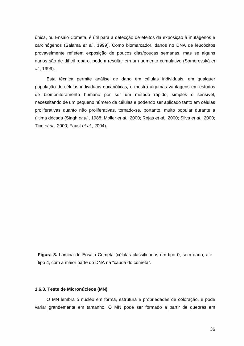
36
única, ou Ensaio Cometa, é útil para a detecção de efeitos da exposição à mutágenos e
carcinógenos (Salama et al., 1999). Como biomarcador, danos no DNA de leucócitos
provavelmente refletem exposição de poucos dias/poucas semanas, mas se alguns
danos são de difícil reparo, podem resultar em um aumento cumulativo (Somorovská et
al., 1999).
Esta técnica permite análise de dano em células individuais, em qualquer
população de células individuais eucarióticas, e mostra algumas vantagens em estudos
de biomonitoramento humano por ser um método rápido, simples e sensível,
necessitando de um pequeno número de células e podendo ser aplicado tanto em células
proliferativas quanto não proliferativas, tornado-se, portanto, muito popular durante a
última década (Singh et al., 1988; Moller et al., 2000; Rojas et al., 2000; Silva et al., 2000;
Tice et al., 2000; Faust et al., 2004).
Figura 3. Lâmina de Ensaio Cometa (células classificadas em tipo 0, sem dano, até
tipo 4, com a maior parte do DNA na “cauda do cometa”.
1.6.3. Teste de Micronúcleos (MN)
O MN lembra o núcleo em forma, estrutura e propriedades de coloração, e pode
variar grandemente em tamanho. O MN pode ser formado a partir de quebras em

37
cromossomos ou cromátides (clastogênese), ou problemas no fuso mitótico
(aneugênese). Os fragmentos ou cromossomos inteiros, que não se orientam para os
núcleos filhos de uma célula em divisão, ficam perdidos no citoplasma e formam sua
própria membrana nuclear, originando os MNs, que são detectados em células
interfásicas como pequenos corpúsculos arredondados de cromatina, separados do
núcleo principal (Schmid, 1975; Heddle et al., 1983; Heddle et al., 1991).
O MN aparece pela primeira vez no final da primeira divisão mitótica, porém MNs
adicionais podem se formar nas divisões seguintes. Por isso, para visualizar MNs, as
células precisam ter passado por um ciclo mitótico (Carrano & Natarajan, 1988).
A análise cromossômica permite identificar com maior precisão os tipos e a
localização das alterações cromossômicas. Porém em estudos populacionais, a análise
de MNs é bem mais viável, podendo-se contar com resultados estatisticamente mais
significantes (Heddle et al., 1983; Fenech et al., 1999). Além disso, o teste de MNs é bem
mais sensível que a contagem cromossômica na detecção de perda cromossômica por
lesão do fuso mitótico (Migliore et al., 1991b).
O teste de MN em linfócitos binucleados (Figura 4) desenvolvido por Fenech &
Morley (1985) é um teste eficiente para a detecção de efeitos clastogênicos decorrentes
de vários poluentes físicos e químicos (Szirmai et al., 1993; Fenech et al., 1999). Nesta
técnica se utiliza Citocalasina B, que impede a divisão do citoplasma sem inibir a mitose,
tornando as células reconhecíveis pela presença de dois núcleos após uma divisão
nuclear (Fenech et al., 1999).
Embora o Teste de MN em linfócitos binucleados seja o mais utilizado no
monitoramento de populações expostas, essa técnica também pode ser utilizada em
outros tipos de célula, sem necessidade de cultura in vitro, como células epiteliais. A
análise de MN em células de mucosa bucal (Figura 5) tem demonstrado ser um método
muito sensível no monitoramento de dano genético de populações humanas expostas
(Karahalil et al., 1999; Gattás et al., 2001; Majer et al., 2001; Burgaz et al., 2002; Pastor
et al., 2003). Nesse tipo de análise deve-se levar em conta que o período crítico de
exposição para a formação do MN ocorreu na última divisão das células coletadas, e o
tempo exato entre a divisão na camada basal e a migração das células até a superfície é
desconhecido (Albertini et al., 2000). No entanto, alguns autores estimam que seja um
período de uma a duas semanas (Tolbert et al., 1992; Burgaz et al., 2002).

38
Figura 4. Teste de Micronúcleos em linfócitos binucleados. A – linfócito binucleado normal (LB); B – linfócito binucleado com micronúcleo (LBMN); C – linfócito binucleado com dois micronúcleos (LBMN); e D – linfócito binucleado com ponte nucleoplasmática (PN) ligando os núcleos.
Figura 5. Célula de mucosa bucal com micronúcleo (CMBMN).

39
1.6.4. Identificação de Polimorfismos nos Genes de Metabolização
Muitos dos genes que participam da ativação ou desativação de compostos
potencialmente carcinogênicos encontram-se distribuídos de uma maneira polimórfica
nas populações, sendo descritos vários alelos relacionados com aumento de
suscetibilidade para desenvolver tumores, perante a exposição a determinadas drogas.
Estes polimorfismos contribuem na determinação da suscetibilidade ao câncer,
decorrente das respostas diferenciais dos indivíduos em relação à exposição a mesma
droga (Idle, 1991; Venitt, 1994; Raunio et al., 1995).
Vários sistemas polimórficos são descritos como ligados à suscetibilidade ao câncer
e aumentos de danos citogenéticos, entre eles: polimorfismos em genes do citocromo
P450 (CYP) e GST (glutationa-s-transferase). Vários trabalhos demonstram a existência
de alto risco de desenvolvimento de tumores, principalmente de pulmão, esôfago,
próstata, laringe, bexiga, mama entre outros, associados com determinados
polimorfismos de genes destas famílias e com exposição ambiental (Harries et al., 1991;
Hubert et al., 1993; Venitt, 1994; Raunio et al., 1995; Kihara et al., 1997; Nimura et al.,
1997; Ingles et al., 1998; Millikan et al., 1998).
Dois polimorfismos do gene CYP1A1 são estudados em relação a suscetibilidade:
uma substituição 462 Ile→Val (alelo CYP1A1*2C) e uma mutação 6235 T→C na região
3`não codificadora (alelo CYP1A1*A) (Hayashi, 1991). Segundo Kyohara et al. (1996), a
mutação Ile→Val ocorre na região correspondente ao sítio catalítico da enzima, e com
isso o alelo CYP1A1*2C codifica uma enzima com aumento de atividade. As freqüências
dessas mutações são diferentes entre as etnias (Aynacioglu et al., 1998; Kvitko et al.,
2000). Para o gene CYP2E1 estão descritos dois RFLPs (random fragments length
polimorphisms) em desequilíbrio de ligação na região regulatória. Devido a esse
desequilíbrio, dois arranjos principais são normalmente encontrados, CYP2E1*5B e
CYP2E1*1A. Embora seja raro em muitas populações (Hamada et al., 1995; Griese et al.,
2001), o haplótipo CYP2E1*5B é comum em asiáticos (19-30%) (Morita, 1997; Tan, 2000)
e ameríndios (25%) (Muñoz, 1998). Estudos de expressão demonstraram que a forma
mutante CYP2E1*5B resulta no aumento da transcrição do seu mRNA (Watanabe et al.,
1994). A enzima CYP2E1 é toxicologicamente importante devido a sua capacidade de
metabolizar vários xenobióticos de baixo peso molecular como N-nitrosaminas e
solventes orgânicos, tais como o benzeno, tolueno e outros, enquanto que a CYP1A1
metaboliza vários xenobióticos como aminas aromáticas e HAP gerando outras
substâncias tóxicas (Guengerich & Shimada, 1998).
40
Os polimorfismos genéticos da família GST estão relacionados com o risco de
desenvolvimento de câncer (Indulski & Lutz, 2000). Deleções nos loci GSTM1 ou GSTT1,
resultam na falta de atividade enzimática (Pemble et al., 1994; Xu et al., 1998); ambos os
genótipos GSTM1*0/*0 e GSTT1*0/*0 apresentam freqüência variável entre as etnias
(Rebbeck, 1997). Dentre os caucasianos, cerca de 20% da população apresenta
ausência da enzima GSTT1 e 50% da enzima GSTM1 (Hirvonen et al., 1993; Zhong et
al., 1993). No gene GSTP1, uma mudança do aninoácido Ile→Val no códon 105
(GSTP1*Val105), na região correspondente ao centro catalítico, parece resultar em
redução de atividade enzimática (Zimniak et al., 1994; Ali-Osmam et al., 1997; Watson et
al., 1998). Entretanto, algumas investigações sugerem que a forma GSTP1*Val105 está
associada com aumento de atividade catalítica, dependendo do substrato (Hu et al.,
1997; Sundberg et al., 1998; Watson et al., 1998). O alelo GSTP1*Val105 tem a sua
especificidade por compostos eletrofílicos, estando relacionado com a predisposição ao
câncer de pulmão, bexiga, testículos entre outros (Gaspar et al., 2002).
É bem aceito que a distribuição polimórfica das enzimas de metabolizadoras nas
diferentes populações do mundo influencia na suscetibilidade ao desenvolvimento de
doenças ambientais, sendo de grande importância o conhecimento da freqüência destes
genes nas diferentes etnias, visando gerar uma forma efetiva de prevenção,
especialmente contra o câncer (Au et al., 1999).
Estudos utilizando Biomarcadores de exposição (substâncias ou seus metabólitos
em fluidos biológicos, danos reparáveis de DNA, adutos de proteína e DNA) e de efeitos
(AC, TCI, MN e outros) demonstraram que a determinação de polimorfismos nos genes
de ativação e detoxifiação são aspectos importantes na implementação de testes mais
sensíveis e específicos, e na identificação de subgrupos com maior sensibilidade
(Norppa, 1997; WHO, 2001). Por outro lado, Pavanello & Clonfero (2000), num trabalho
de revisão sobre o assunto, relataram que em menos da metade dos estudos se
encontrou relação entre genótipo e indicador biológico de efeito e/ou exposição,
concluindo que esse efeito dos polimorfismos sobre os biomarcadores não é tão simples,
e que muito ainda deve ser elucidado através de diferentes estudos.

41
2. OBJETIVOS
Em função do que se conhece sobre os produtos químicos utilizados no setor
coureiro-calçadista, visando avaliar os possíveis riscos que os trabalhadores pudessem
estar sofrendo, e ainda possibilitar a prevenção e monitoramento, este trabalho teve
como objetivos:
• avaliar através de Biomarcadores de Efeito precoce (Ensaio Cometa e Teste de
Micronúcleos) os trabalhadores de curtumes de fábricas de calçados em relação aos
danos ao DNA que possam estar sofrendo, observando correlação com as
diferenças individuais (suscetibilidade, hábitos, idade, sexo, tempo de exposição,
setor de trabalho);
• comparar as respostas de mutagenicidade e/ou genotoxicidade encontradas nos
diferentes tipos de células e metodologias;
• identificar alguns dos genes correlacionados com suscetibilidade ao tipo de
exposição, através da genotipagem dos indivíduos, para posterior correlação com os
dados de danos ao DNA;
• relacionar os dados obtidos com a utilização de Biomarcadores de Efeito Biológico
Precoce e de Suscetibilidade com os Biomarcadores de Exposição determinados
pela Norma Regulamentadora No 7 (NR-7) para as exposições nos setores avaliados
nesse estudo.

42
3. CAPÍTULO I
Evaluation of genotoxic exposure in Brazilian Tannery workers: Comparison between Drum Workshop and Finishing Workshop.
Vanina Dahlström Heuser1, Juliana da Silva2* & Bernardo Erdtmann1,3
1Programa de Pós-Graduação em Genética e Biologia Molecular (PPGGBM),
Universidade Federal do Rio Grande do Sul (UFRGS), Porto Alegre-RS, Brazil.
2Laboratório de Genética Toxicológica, Programa de Pós-Graduação em Ensino de
Ciências e Matemática (PPGECIM), Universidade Luterana do Brasil (ULBRA), Canoas-
RS, Brazil.
3Instituto de Biotecnologia, Universidade de Caxias do Sul (UCS), Caxias do Sul-RS,
Brazil.
*Correspondence to: Juliana da Silva, Programa de Pós-Graduação em Ensino de
Ciências e Matemática (PPGECIM), Universidade Luterana do Brasil (ULBRA), Prédio 14,
Sala 230, Rua Miguel Tostes 101, Bairro São Luís, CEP 92420-280, Canoas-RS, Brazil.
E-mail: [email protected]
A SER ENVIADO PARA A REVISTA MUTAGENESIS
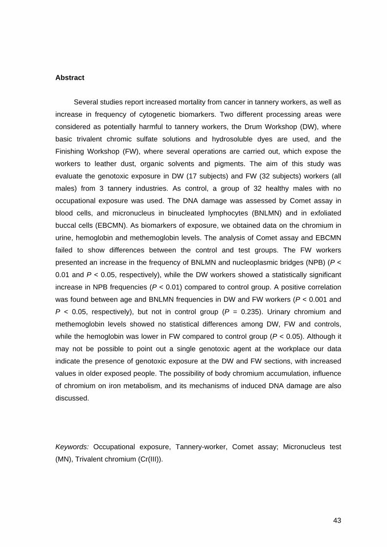
43
Abstract
Several studies report increased mortality from cancer in tannery workers, as well as
increase in frequency of cytogenetic biomarkers. Two different processing areas were
considered as potentially harmful to tannery workers, the Drum Workshop (DW), where
basic trivalent chromic sulfate solutions and hydrosoluble dyes are used, and the
Finishing Workshop (FW), where several operations are carried out, which expose the
workers to leather dust, organic solvents and pigments. The aim of this study was
evaluate the genotoxic exposure in DW (17 subjects) and FW (32 subjects) workers (all
males) from 3 tannery industries. As control, a group of 32 healthy males with no
occupational exposure was used. The DNA damage was assessed by Comet assay in
blood cells, and micronucleus in binucleated lymphocytes (BNLMN) and in exfoliated
buccal cells (EBCMN). As biomarkers of exposure, we obtained data on the chromium in
urine, hemoglobin and methemoglobin levels. The analysis of Comet assay and EBCMN
failed to show differences between the control and test groups. The FW workers
presented an increase in the frequency of BNLMN and nucleoplasmic bridges (NPB) (P <
0.01 and P < 0.05, respectively), while the DW workers showed a statistically significant
increase in NPB frequencies (P < 0.01) compared to control group. A positive correlation
was found between age and BNLMN frequencies in DW and FW workers (P < 0.001 and
P < 0.05, respectively), but not in control group (P = 0.235). Urinary chromium and
methemoglobin levels showed no statistical differences among DW, FW and controls,
while the hemoglobin was lower in FW compared to control group (P < 0.05). Although it
may not be possible to point out a single genotoxic agent at the workplace our data
indicate the presence of genotoxic exposure at the DW and FW sections, with increased
values in older exposed people. The possibility of body chromium accumulation, influence
of chromium on iron metabolism, and its mechanisms of induced DNA damage are also
discussed.
Keywords: Occupational exposure, Tannery-worker, Comet assay; Micronucleus test
(MN), Trivalent chromium (Cr(III)).

44
1. Introduction
Brazil is one of the largest producers of bovine leather in the world, with the main
concentration of producers located in States of Rio Grande do Sul and São Paulo. With
more than 700 industries, tanneries employ directly about 65 thousand workers (Santos et
al., 2002).
People employed in leather tanning industries may be exposed to higher-than-
normal level of chromium, mostly trivalent chromium (Cr(III)) (ASTDR, 2002a), either
inorganic Cr(III) compounds or Cr bound to proteins (leather dust) (Stupar et al., 1999).
Little toxic effect is attributed to Cr(III) when present in very large quantities while
hexavalent chromium (Cr(VI)) has been found biologically active (Pan et al., 1995). The
failure of Cr(III) ions to pass through the cell membrane explains the genetic inertness of
Cr(III). However, some data suggest that in mammalian cells endocytosis is a mechanism
that allows Cr(III) complexes to pass the membrane barrier and enter cells (Bianchi and
Levis, 1984). Cr(III) compounds are 1000-fold less toxic than Cr(VI) compounds (ASTDR,
2000), but Cr(III) compounds may cause toxicity at higher concentration and/or depending
on the ligant attached to it (Bagchi et al., 2002).
Relatively few studies are available in the literature that directly address the toxicity
of Cr(III), particularly by the inhalation route of exposure. This lack of data results in
considerable uncertainty regarding the hazard associated with exposures to Cr(III) (EPA,
1998; Medeiros et al., 2003). Concentrations of chromium in blood, serum, urine and hair
have long been used in biological monitoring of environmentally and occupationally
exposed populations, as biomarker of exposure (Simpson and Gibson, 1992; Rajaram et
al., 1995; Pan et al., 1996; EPA, 1998; Stupar et al., 1999; Medeiros et al., 2003).
Two different processing areas were considered as potentially harmful to tannery
workers: (a) the Drum Workshop, and (b) the Finishing Workshop. In the former
department tanning is carried out by placing the hides in a drum containing basic trivalent
chromic sulfate solutions; moreover leather is colored using hydrosoluble dyes. In the
latter department several mechanical operations are carried out (conditioning, staking,
buffing) which expose the workers to leather dust; moreover finishing is carried out using
formaldehyde as a leather preservative and as a protein fixer on glazed leather; painting is
carried out during which solvents, paints and pigments containing oxide and insoluble
salts of metals, including anilines and Cr salt are used.


46
Brazilian National Ethical Committee on Research (Comissão Nacional de Ética em
Pesquisa – CONEP) and informed written consent was obtained from each individual prior
to the start of the study.
All the individuals examined were required to answer a Portuguese version of a
questionnaire from the International Commission for Protection against Environmental
Mutagens and Carcinogens (Carrano and Natarajan, 1988) and participate in a face-to-
face questionnaire which included standard demographic data (age, gender, etc.) as well
as questions relating to medical issues (exposure to X-rays, vaccinations, medication,
etc.), life style (smoking, coffee, alcohol, diet, etc.) and their occupation (number of hours
worked per day, time of exposure, use of protective measures, etc.). In all the groups,
individuals who smoked more than 5 cigarettes/day for at least 1 year were considered
smokers. The characteristics of the three groups are presented in Table I.
Blood and urine samples were obtained from individuals in the DW and FW groups
on the same day during a normal shift during the workers periodical medical examinations
by the nurses by Vida Laboratory (laboratory of Clinic Analysis, Estância Velha, RS). For
the control group, blood and urine samples were taken at the same region. All blood
samples were collected by venipuncture using vacutainers with heparine and processed
as quickly as possible to avoid the damage associated with storage (Albertini et al., 2000),
the blood cell samples being transported to the laboratories at or below 8oC and
processed within 8 h of collection in order to minimize loss of DNA and damage due to
DNA repair processes.
2.2. Chromium in Urine, Methemoglobin, and Hemoglobin values
A spot urine sample was obtained (Control, n=21; DW, n=17; and FW, n=30) in the
last day of the working week. The chromium determination in urine was performed by
graphite furnace atomic absorption (Zeeman, Simaa 600-EUA, Perquin-Elmer).
Hemoglobin levels (Control, n=26; DW, n=10; and FW, n=16) were determined using a
hematological electronic analyzer (Pentra 60 plus, ABX, France). Methemoglobin
percentages (Control, n=20; DW, n=17; and FW, n=31) were analyzed by
spectrophotometric method. For all study groups, these analyses were performed by
Toxilab (Toxicological Laboratory, Porto Alegre, RS) and Vida Laboratory (Clinic Analysis,
Estância Velha, RS).

47
2.3. Comet Assay
The alkaline Comet assay was performed as described by Singh et al. (1988) with
the modifications suggested by Tice et al. (2000). Blood cells (5 µl) were embedded in 95
µl of 0.75% low-melting point agarose and when the agarose had solidified the slides were
placed in lysis buffer (2.5M NaCl, 100mM EDTA and 10mM Tris; pH 10.0-10.5) containing
freshly added 1% (v/v) Triton X-100 and 10% (v/v) dimethyl sulfoxide (DMSO) for a
minimum of 1 hour and a maximum of two weeks. After treatment with lysis buffer, the
slides were incubated in freshly-made alkaline buffer (300 mM NaOH and 1 mM EDTA;
pH >13) for 20 min and the DNA was electrophoresed for 20 min at 25 volts (0.90 V/cm)
and 300 mA after which the buffer was neutralized with 0.4 M Tris (pH 7.5) and the DNA
stained with ethidium bromide (2 µg/ml). The electrophoresis procedure and efficiency for
each electrophoresis run was checked using negative and positive internal controls
consisting of whole human blood collected in the laboratory, the negative control being
unmodified blood and the positive control 50 µl of blood mixed with 13 µl (8 x 10⁻⁵ M) of
methyl methanesulfonate (CAS No 66-27-3; Sigma, St. Louis, MO, USA) and incubated
for 2 hours at 37oC. Each electrophoresis run was considered valid only if the negative
and positive controls yielded the expected results.
Images of 200 randomly selected cells (100 cells from each of two replicate slides)
were analyzed for each person. Comet image lengths (IL) (i.e. the nuclear region+tail)
were measured in arbitrary units using a calibrated eyepiece micrometer (1 unit= ≈5 µm at
200X) and a fluorescence microscope equipped with a 12nm BP546 excitation filter and a
590nm barrier filter. Cells were also visually allocated to one of five classes depending on
tail size (class 0 = no tails, class 4 = longest tails) to give a single DNA damage score for
each subject and hence for each group studied, the group damage index (DI) ranging
from 0 (no tails on any cells, i.e. 200 cells x 0) to 800 (all cells with maximally long tails,
i.e. 200 cells x 4) (Collins et al., 1997; Albertini et al., 2000; Silva et al., 2000). The
damage frequency (DF (%); i.e. the proportion of cells with altered migration), was
calculated based on the number of cells with tails versus the number of cells without tails.
All the slides were scored blindly on the same day on which they were subjected to
electrophoresis.
2.4. MN in binucleated lymphocytes
For each blood sample, duplicate lymphocytes cultures were set up in culture flasks
by adding 0.3 ml of whole blood to 5 ml of RPMI 1640 medium (Nutricell, Campinas-SP,

48
Brazil) containing 1% (v/v) phytohemagglutinin and the flasks incubated at 37ºC for 44 h
before adding 5 µg/ml of cytochalasin B (Sigma) (Fenech et al., 1999) and continuing
incubation until the total incubation time reached was 72 h. After incubation the
lymphocytes were harvested by centrifugation at 800 revs/min for 8 min, recentrifuged,
fixed in 3:1 (v/v) methanol/acetic acid, placed onto a clean microscope slide and stained
with 5% (v/v) Giemsa. For each blood sample, 2000 binucleated lymphocytes (BNL) (i.e.
1000 from each of the two slides prepared from the duplicate cultures) were scored for
both micronuclei (MN) presence and nucleoplasmic bridges (NPB) between daughter
nuclei, assessment being made using bright-field optical microscopy at a magnification of
200-1000X. All sides were coded to blind analysis.
2.5. MN in Exfoliated Buccal Cells
Buccal cells were collected by swabbing the inner cheek of the individuals with a
moistened wooden tongue depressor, the tip of which was immersed in 5 ml of cold saline
(0.9% (w/v) aqueous NaCl) in a conical tube and transported under refrigeration to the
laboratory where the saline was centrifuged at 1500 revs/min for 8 min and the
sedimented buccal cells washed twice more with saline under the same centrifugation
conditions to remove bacteria and cell debris which would have complicated scoring. After
washing, a drop of cell suspension was placed onto each of two duplicate microscope
slides and dried at room temperature for 1-2 weeks and then feulgen-stained (hydrolysis
in 1N HCl at 60oC for 10 minutes followed by immersion in Schiff`s reagent for 3-4 h) and
the cytoplasm counterstained by immersion in 0.5% (w/v) fast-green for 30 seconds.
The criteria used for micronuclei analysis were those of Tolbert et al. (1992) and
Titenko-Holland et al. (1998). Only cells that were not smeared, clumped or overlapped
and that contained intact nuclei were included in the analysis. Cells undergo degenerative
processes which can produce anomalies that are difficult to distinguish from micronuclei
(binucleated cells, condensed chromatin, ‘broken egg’, pycnosis, karyorrhexis and
karyolysis) were excluded from the micronucleus analysis and all the slides were coded to
blind analysis. The quality of the slides were determined at 200X magnification using a
bright-field Zeiss microscope and the micronuclei frequency estimated based on the
number of normal exfoliated buccal cells counted using bright-field optical microscopy at a
magnification of 1000X. For each volunteer, 2000 buccal cells (i.e. 1000 from each of the
duplicate slides) were scored.

49
2.6. Statistical Analysis
The normality of the variables was evaluated using the Kolmogorov-Smirnov test. χ2
and t-test were used to compare the basic characteristic of study populations. According
with the distribution of data, the Kruskal-Wallis (Dunn`s Multiple Comparisons) and
ANOVA (Tukey's Multiple Comparison Test) were used to compare the parameters
analyzed for the three groups (control, DW, and FW). The Spearman and Pearson
correlation test were used to analyze possible correlations as appropriated. The critical
level for rejection of the null hypothesis was considered to be a P value of 5%.
3. Results
The main characteristics of the control, DW and FW groups are presented in Table
1. The mean ages of the groups showed no significant differences (t-test). The time of
exposure was statistically higher in FW in relation to DW (P < 0.01, t-test). Few volunteers
were considered smokers (smoking five or more cigarettes per day) for all study groups.
No statistical differences were found in smoking and drinking habits among the groups
(χ2-test).
Table 1 Size of sample, mean age, time of exposure, smoking and drinking status of control,
Drum Workshop (DW), and Finishing Workshop (FW).
Control DW FW
Number of subjects 32 17 32
Mean age ± S.D. (years) 31.94 ± 6.80 32.29 ± 8.23 33.61 ± 6.97
Years of exposure (mean ± S.D.) - 7.65 ± 7.32 12.70 ± 6.97*
Smokersa / non-smokers 3/29 1/16 6/26
Drinkersb / non-drinkers 2/30 4/13 2/30 asmoking 5 or more cigarettes per day; bdrinking 4 or times per week; *P < 0.01 (t-test).

50
The mean values (mean ± S.D.) of chromium in urine, methemoglobin and
hemoglobin levels are presented in Figure 1. Although the FW show a slight increase on
chromium excretion in urine, there were no statistical differences among DW, FW and
controls, as well as the methemoglobin percentages. The mean levels of hemoglobin were
not different between DW and controls, or between DW and FW, but lower in FW
compared to control group (P < 0.05; ANOVA, Tukey's Multiple Comparison Test). No
correlation was found between chromium concentration in urine and hemoglobin or
methemoglobin levels, age, and cytogenetic analysis (Spearman correlation test; data
non-shown). With exception of one subject in FW group, all volunteers presented normal
chromium concentration, hemoglobin and methemoglobin levels (up to 5.0 µg/g creatinine,
1.15 - 1.78 g/l blood, and up to 2% of total hemoglobin, respectively) (NR-7, 1994).
0.00
0.25
0.50
0.75
1.00
1.25
1.50
1.75
Chromium (µg/g creatinine)Methemoglobin (%)Hemoglobin (g/l)
Control Drum Workshop Finishing Workshop
*
Fig. 1. Mean values on total chromium in urine (µg/g creatinine), methemoglobin (%)and hemoglobin (g/l) in study groups. Error bars represent the standard deviation(S.D.) of the mean. *P < 0.05 (ANOVA, Tukey's Multiple Comparison Test).

51
The cytogenetic parameters analyzed are presented in Table 2. The Comet assay
analysis indicated a slight but not significant increase in Damage Index (DI) in DW and
FW workers in relation to control. Workers from DW and FW did not present different
results obtained by Comet assay (Kruskal-Wallis Test, Dunn’s Multiple Comparison).
Smokers in all groups showed no significant increase in Comet assay values than their
non-smoking counterparts (data non-shown). Negative (DI = 0-4) and positive control (DI
= 615-800) for electrophoresis demonstrated negative and positive results, respectively
(data non-shown).
The results of MN frequency on 2000 binucleated lymphocytes (BNLMN) and NPB
(nucleoplasmic bridges) are also presented in Table 2. The FW workers presented an
increase in the number of BNLMN and NPB (P < 0.01 and P < 0.05, respectively), while
the DW workers showed a statistically significant increase in NPB frequencies (P < 0.01)
in relation to control group (ANOVA, Tukey's Multiple Comparison Test). The analysis of
EBCMN failed to show differences between the control and test groups. No difference was
detected between smokers and their counterparts on BNLMN, NPB, and EBCMN analysis
(data non-shown).
Positive correlation was found between age and BNLMN frequencies in DW and FW
workers (r = 0.759, P < 0.001; and r = 0.361, P < 0.05, respectively). The same was not
observed for control group (r = 0.237, P = 0.235; Pearson correlation test) (Figure 2).
The effect of age on BNLMN frequency is presented in Figure 3, which shows the
individuals more than 30 years old (Control, n=14; DW, n=8; and FW, n=21) compared to
those to 30 years old (Control, n=15; DW, n=9; and FW, n=10). A significant increased
mean frequency of BNLMN in older individuals was observed in both DW and FW (P <
0.05). The correlation between BNLMN and the working time was not so clear; while the
DW presented a correlation (r = 0.679, P = 0.011), in the FW this correlation was not
significant (r = 0.265, P = 0.173; Pearson correlation test) (data non-shown).
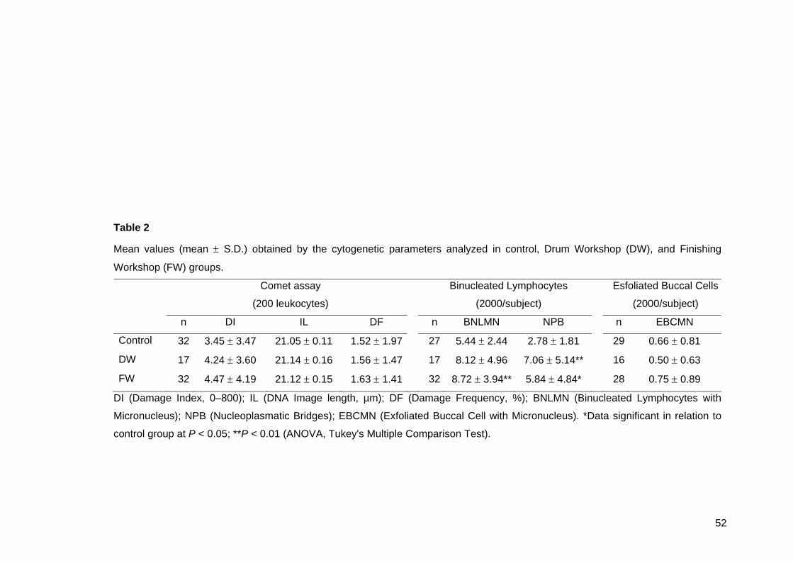
52
Table 2
Mean values (mean ± S.D.) obtained by the cytogenetic parameters analyzed in control, Drum Workshop (DW), and Finishing
Workshop (FW) groups.
Comet assay
(200 leukocytes)
Binucleated Lymphocytes
(2000/subject)
Esfoliated Buccal Cells
(2000/subject)
n DI IL DF n BNLMN NPB n EBCMN
Control 32 3.45 ± 3.47 21.05 ± 0.11 1.52 ± 1.97 27 5.44 ± 2.44 2.78 ± 1.81 29 0.66 ± 0.81
DW 17 4.24 ± 3.60 21.14 ± 0.16 1.56 ± 1.47 17 8.12 ± 4.96 7.06 ± 5.14** 16 0.50 ± 0.63
FW 32 4.47 ± 4.19 21.12 ± 0.15 1.63 ± 1.41 32 8.72 ± 3.94** 5.84 ± 4.84* 28 0.75 ± 0.89
DI (Damage Index, 0–800); IL (DNA Image length, µm); DF (Damage Frequency, %); BNLMN (Binucleated Lymphocytes with
Micronucleus); NPB (Nucleoplasmatic Bridges); EBCMN (Exfoliated Buccal Cell with Micronucleus). *Data significant in relation to
control group at P < 0.05; **P < 0.01 (ANOVA, Tukey's Multiple Comparison Test).
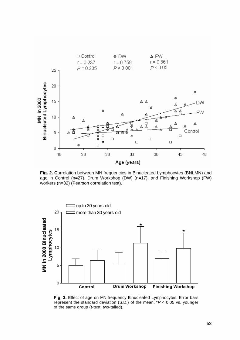
53
Fig. 2. Correlation between MN frequencies in Binucleated Lymphocytes (BNLMN) and age in Control (n=27), Drum Workshop (DW) (n=17), and Finishing Workshop (FW) workers (n=32) (Pearson correlation test).
0
5
10
15
20up to 30 years oldmore than 30 years old
**
Control Drum Workshop Finishing Workshop
Fig. 3. Effect of age on MN frequency Binucleated Lymphocytes. Error barsrepresent the standard deviation (S.D.) of the mean. *P < 0.05 vs. youngerof the same group (t-test, two-tailed).
MN
in 2
000
Bin
ucle
ated
Lym
phoc
ytes

54
4. Discussion
As soon as it was realized that Cr(VI) entails a high potential risk of cancer it was
completely replaced with the less dangerous Cr(III) in tannery industry. However, there
are still hazardous chemicals like alkalis, acids, dyes or solvents, which must be used in
several tannery departments and, although Cr(III) is considered to be of much lower
toxicity, the risk involved in chronic exposure is uncertain (EPA, 1998).
In the present study, although not statistically significant, chromium excretion in
urine from FW workers was slightly increased than in control and DW (Fig.1), probably
due to aniline and leather dust exposure in this workshop. This is in accordance with the
study made by Stupar et al. (1999), who concluded that the absorption of chromium from
leather dust may be more efficient in comparison to inorganic chromium. In addition,
hemoglobin levels were decreased in FW, possibly associated with body chromium
accumulation, causing adverse effects on iron metabolism, as suggested by Kornhauser
et al. (2002), who reported similar results in Mexican tannery workers. The occupational
exposure has shown that Cr(III) associated to certain ligants leads to cell death and/or
structural modification of proteins (Balamurugan et al., 2002; Rao et al., 2004). It is
reported that metal toxicity can be associated to oxidative tissue damage, due to the
catalytic role of metals in the oxidative deteriorating effect to biological macromolecules
(Okada, 1996). Also, studies describe that chromate promotes changes in morphology in
the red blood cells, and hemoglobin oxidation (methemoglobin formation) (Fernandes et
al., 1999), that also occur due to aniline exposure (Krishnan and Pelekis, 1995; Khan et
al., 1997; ASTDR, 2002b). Hemoglobin/methemoglobin ratio and cell shape, which are
crucial for the cell functions and survival, are irreversibly disrupted. Thus, in vivo, these
cells will be removed early from circulation, decreasing the hemoglobin in individuals
exposed to high chromate concentrations (Fernandes et al., 1999), as well as anilines.
Non-invasive urinary samples have been extensively used in biological monitoring of
chromium but its reliability is controversial. Once absorbed, Cr(III) compounds are rapidly
cleared from the blood and more slowly from the tissues (EPA, 1998). Since the half-life of
the metal in the blood stream is short, followed by a rapid urinary excretion or storage in
body tissues such as bone and liver, urinary levels, as well as plasma determinations,
may not provide an indication of low chronic exposure to chromium (Bukowski et al., 1991;
Simpson and Gibson, 1992; Christensen, 1995; Finley et al., 1996). According to Pan et
al. (1996) serum chromium concentration in exposed workers increased proportionally to
their working years, possible due to the accumulation. However, studies of chronic animal
55
exposure to various Cr(III) salts have provided no evidence of health effects consistent
with bioaccumulation (Anderson et al., 1997).
Studies using cytogenetic tests like the MN assay showed that nasal and buccal
mucosal cells, being directly exposed, are an excellent target for biomonitoring exposure
to mutagenic compounds (Salama et al., 1999; Majer et al., 2001; Faust et al., 2004). In
some of them, the frequency of MN in mucosal cells was significantly higher in individuals
exposed than in non-exposed, without an increase in micronuclei frequency in
lymphocytes (Norppa et al., 1992; Ying et al., 1997). Although the main exposure in
tanneries occurs by airborne substances, in the present study, an inverse result was
obtained: the Brazilian tannery workers did not present an increase in EBCMN, but on
other hand, an increased frequency of NPB was observed in both DW and FW workers,
as well as BNLMN, but it was statistically significant increased only in FW group.
The positive results of BNLMN but not of EBCMN were somewhat unexpected. The
increase in NPB denotes the micronucleus resulted mainly from clastogenic mechanism.
A possible explanation of the difference between the lymphocytes and exfoliated buccal
cells could be the metabolism and exposure of the tissues. It is known that blood cells
have a more complex metabolism, and are also submitted to the action of the products of
metabolism, while the epithelial cells are less affected by the metabolites. So, if one or
more metabolic products are responsible for the genotoxicity, it is expected that the target
are the blood cells. In addition, MN frequencies in lymphocytes reveal accumulated
chromosome damage (Delf et al., 1998), while MN in exfoliated cells reflect genotoxic
events that occurred in the dividing basal layer 1-3 weeks earlier (Tolbert et al., 1992).
If the clastogenicity is responsible for NPB in the lymphocytes, the absence of DNA
damage detected by Comet assay is surprising. Pitarque et al. (1999; 2002) found similar
results in shoe factory workers, with no sister chromatid exchange and DNA damage
measured by Comet assay, but increasing MN in binucleated lymphocytes. One possibility
is that the reduced DNA strand breaks can be caused by the elimination of cells with
extensive strand breaks, or the reduction in strand breaks due to incorrect rejoining of the
DNA molecules (Bonassi and Au, 2002), which could lead to MN formation, since there is
evidence that metal ions might interfere with distinct steps of diverse DNA repair systems
(Hartwig, 1998).
The effect of age on the frequency of BNLMN clearly emerges in the present study:
both DW and FW show an increase in MN frequency due to age (Fig. 2 and 3). The
BNLMN frequencies were also associated to working time, but this correlation was not so
clear. Data from most biomonitoring studies describe the positive correlation between MN

56
and age in control and in exposed groups (Bolognesi et al., 1997; Fenech et al., 1999;
Albertini et al., 2000) including tannery workers (Migliore et al., 1991). To explain these
findings, several possibilities have been suggested: a general increase of susceptibility in
older people (Crome, 2003); possible age-related changes in the transcriptional activation
xenobiotic genes (Ishikawa et al., 2004); changes in the ionic blood concentrations
(Fenech et al., 1997; 1998; Landi et al., 2000); and the possibility of cumulated exposure
and/or DNA damage (Migliore et al., 1991; Ishikawa et al., 2003). In the present study,
positive correlation was found between age and BNLMN in tannery workers, but not in
controls, suggesting that this age-associated increase in susceptibility can be higher in
exposed people, since the increasing effects of aging on MN frequency might reflect
accumulated genetic damage throughout the lifetime of the exposed people.
There was no evidence of toxic effects of Cr(III) in human dose response studies
(Campbell et al., 1999; Cefalu et al., 1999), but Cr(III) has been demonstrated to decrease
the accuracy of DNA synthesis (Snow, 1994). In vitro studies have also shown the
induction of micronuclei by Cr(III) in human diploid fibroblasts, showing a predominace of
aneugenic over clastogenic effects, since this compound should damage spindle proteins
(Seoane and Dulout, 2001). On the other hand, Blasiak and Kowalic (2000) report the
formation of DNA strand breaks as the prevalent mode of chromium damage to DNA.
These contradictory data suggest that further studies are needed to clarify this point.
However, leather processing in DW and FW also involves a considerable number of
potentially genotoxic substances, such as organic solvents, mainly formaldehyde, a
known mutagen and possibly a human carcinogen (Ballarin et al., 1992; ASTDR, 1999;
Burgaz et al., 2002; Shaham et al., 2002) which may be contributing to the cytogenetic
lesions reported in this study.
Finally, our data indicate the presence of genotoxic exposure at the tannery
workplace, with increased values in older people. It is important to have in mind that
biomarker studies are still not generating the type of reliable information needed for
precise risk assessment, mainly in complex mixture exposures, as in tannery industry,
where the distinct production processes result in different levels, as well as different types
of exposures. For instance, the amount of chromium absorbed from airborne particulate
matter depends on the combined effect of several factors, which are responsible for the
large variability of absorption rate in the tannery population, even in the same department.
Also, there are other problems as the lack of predictable dose-response relationship, as
observed in the present study, and the existence of interindividual variations in response
to exposure, as suggested by Au et al. (1998). Studies of cytogenetic biomarkers among

57
exposed humans show that the determination of polymorphisms is becoming an
increasingly important aspect that may make the assays more sensitive and more specific
in identifying the effect and the sensitive subgroups (Norppa, 1997).
Acknowledgements The authors would like to express their gratitude to all volunteers, for their
acceptation to participate in this study. We wish to thank Dr. Vera L.T. de Azevedo, Dr.
Poliana K. Hernandez, Dr. Anita Lehnen, from Vida Laboratory, especially Cíntia S. da
Silva and Dorelis M. de Souza for their valuable participation in the sampling. We also
thank Roseli O. de Cândido, Vanessa M. de Andrade, and Marcos R. Furini for their help
in questionnaire application. This work was financially supported by the CNPq (Conselho
Nacional para o Desenvolvimento Científico e Tecnológico).
References
Albertini,R., Anderson,D., Douglas,G.R., Hagmar,L., Hemminki,K., Merlo,F., Natarajan,A.T., Norppa,H., Shuker,D.E.G., Tice,R.R., Waters,M.D. and Aitio,A. (2000) IPCS guidelines for the monitoring of genotoxic effects of carcinogens in humans, International Program on Chemical Safety. Mutat. Res., 463, 111-172.
Anderson,R.A., Bryden,N.A. and Polansky,M.M. (1997) Lack of toxicity of chromium chloride and chromium picolinate in rats. J. Am. Coll. Nutr., 16, 273-270.
Agency for Toxic Substances and Disease Registry (ATSDR) (1999) Toxicological profile for formaldehyde. On line: http://www.astdr.cdc.gov/toxfaq.html
Agency for Toxic Substances and Disease Registry (ATSDR) (2000) Chromium Toxicity, Case studies in Environmental Medicine. On line: http://www.astdr.cdc.gov/HEC/CSEM/
Agency for Toxic Substances and Disease Registry (ATSDR) (2002a) Toxicological profile for chromium. On line: http://www.astdr.cdc.gov/toxfaq.html
Agency for Toxic Substances and Disease Registry (ATSDR) (2002b) Toxicological profile for anilines. On line: http://www.astdr.cdc.gov/toxfaq.html
Au,W.W., Cajas-Salazar,N. and Salama,S. (1998) Factors contributing to discrepancies in population monitoring studies. Mutat. Res., 400, 467-478.
Bagchi,D., Stohs,S.J., Downs,B.W., Bagchi,M. and Preuss,H.G. (2002) Cytotoxicity and oxidative mechanism of different forms of chromium. Toxicology, 180, 5-22.
Balamurugan,K., Rajaram,R., Ramasani,T. and Narayanan,S. (2002) Chromium(III)-induced apoptosis of lymphocytes: death decision by ROS and Src-family tyrosine kinase. Free Rad. Biol. Med., 33, 1622-1640.
Ballarin,C., Sarto,F., Giacomelli,L., Bartolucci,G.B. and Clonfero,E. (1992) Micronucleated cells in nasal mucosa of formaldehyde-exposed workers. Mutat. Res., 280, 1-7.

58
Battista,G., Comba,P., Orsi,D., Norpoth,K. and Maier,A. (1995) Nasal cancer in leather workers: an occupational disease. J. Cancer Res. Clin. Oncol., 121, 1-6.
Bianchi,V. and Levis,A.G. (1984) Mechanisms of chromium genotoxicity. Toxicol. Environ. Chem., 9, 1-25.
Blasiak,J. and Kowalik,J. (2000) A comparison of in vitro genotoxicity of tri- and hexavalent chromium. Mutat. Res., 469, 135-145.
Bolognesi,C., Abbondandolo,A., Barale,R., Casalone,R., Dalpra,L., DeFerrari,M., Degrassi,F., Forni,A., Lamberti,L., Lando,C., Migliore,L., Padovani,P., Pasquini,R., Puntoni,R., Sbrana,I., Stella,M. and Bonassi,S. (1997) Age-related increase of baseline frequencies of sister chromatid exchanges, chromosome aberrations, and micronuclei in human lymphocytes. Cancer Epidemiol. Biomarkers Prev., 6, 249-256.
Bonassi,S. and Au,W.W. (2002) Biomarkers in molecular epidemiology studies for health risk prediction. Mutat. Res., 511, 73-86.
Burgaz,S., Erdem,O., Cakmak,G., Erdem,N., Karakaya,A. and Karakaya,A.E. (2002) Cytogentic analysis of buccal cell from shoe-workers and pathology and anatomy laboratory workers exposed to n-hexane, toluene, metyl ethyl ketona and formaldehyde. Biomarkers, 7, 151-161.
Bukowski,J.A., Golstein,M.D., Korn,L.R. and Johnson,B.B. (1991) Biological markers in chromium exposure assessment: confounding variables. Arch. Environ. Health, 46, 230-236.
Campbell,W.W., Joseph,L.J., Davey,S.L., Cyr-Campbell,D., Anderson,R.A. and Evans,W.J. (1999) Effects of resistance training and chromium tripicolinate on body composition and skeletal muscle in older men. J. Appl. Physiol., 86, 29-39.
Carrano,A.V. and Natarajan,A.T. (1988) Considerations for population monitoring using cytogenetic techniques, International Commission for Protection against Environmental Mutagens and Carcinogens (ICPEMC publication 14). Mutat. Res., 204, 379-406.
Cefalu,W.T., Bell-Farrow,A.D., Stegner,J., Wang,Z.Q., King,T., Morgan,T. and Terry,J.G. (1999) Effects of chromium tripicolinate on insulin sensitivity in vivo. J. Trace Elem. Exp. Med., 12, 71- 83.
Christensen,J.M. (1995) Human exposure to toxic metals: factors influencing interpretation of biomonitoring results. Sci. Total Environ., 166, 89-135.
Cid,M.G., Loria,D., Vilensky,M., Miotti,J.L. and Matos,E. (1991) Leather tanning workers: chromossomal aberrations in peripheral lymphocytes and micronuclei in exfoliated cell in urine. Mutat. Res., 259, 197-201.
Collins,A.R., Dobson,V.L., Dusinská,M., Kennedy,G. and Stetina,R. (1997) The Comet assay: what can it really tell us? Mutat. Res., 375, 183-193.
Constantini,S.A., Merler,E. and Saracci,R. (1990) Epidemiologic studies on carcinogeneic risk and occupational activities in tanning, leather and shoe industries. Med. Lav., 81, 184-211.
Crome,P. (2003) What`s different about older people. Toxicology, 192, 49-54.
Delf,J.H., Baan,L. and Roza,R.A. (1998) Biological effect markers for exposure to carcinogenic compounds and their relevance for risk assessment. Crit. Rev. Toxicol., 28, 477-510.
Environmental Protection Agency (EPA) (1998) Toxicological review of trivalent chromium. In Support of Summary Information on the Integrated Risk Information (IRIS). On-line: http://epa.gov/iris

59
Faust,F., Kassie,F., Knasmüller,S., Kevekordes,S. and Mersch-Sundermann,V. (2004) Use of primary blood cells for the assessment of exposure to occupational genotoxicants in human biomonitoring studies. Toxicology, 198, 341-350.
Fenech,M., Dreosti,I.E. and Rinaldi,J.R. (1997) Folate, vitamin B12, homocysteine status and chromosome damage rate in lymphocytes of older man. Carcinogenesis, 18, 1329-1336.
Fenech,M., Aitken,C. and Rinaldi,J. (1998) Folate, vitamin B12, homocysteine status and DNA damage in young Australian adults. Carcinogenesis, 19, 1163- 1171.
Fenech,M., Titenko-Holland,N., Chang,W.P., Zeiger,E. and Bonassi,S. (1999) The Human Micronucleus Project – An international colaborative study on the use of the micronucleus technique for measuring DNA damage in humans. Mutat. Res., 428, 271-283.
Fernandes,M.A.S., Mota,I.M., Silva,M.T.L., Oliveira,C.R., Geraldes,C.F.G.C. and Alpoim,M.C. (1999) Human Erythrocytes are protected against chromate-induced peroxidation. Ecotoxicol. Environ. Safety, 43, 38-46.
Finley,B.L., Scott,P.K., Norton,R.L., Gasgas,M. and Paustenbach,D.J. (1996) Urinary chromium concentrations in humans following ingestion of safe doses of hexavalent and trivalent chromium: implications for biomonitoring. J. Toxicol. Environ. Health, 48, 479-499.
Hamamy,H.A., Al-Hakkak,Z.S. and Hussain,A.F. (1987) Chromosome aberrations in workers a tannery in Iraq. Mutat. Res., 189, 395-398.
Hartwig,A. (1998) Carcinogenicity of metal compounds: possible role of DNA repair inhibition. Toxicol. Lett., 102, 235-239.
International Agency for Research on Cancer (IARC) (1987) Leather tanning and processing. IARC Monographs on the evaluation of the carcinogenic risk of chemicals to humans, Vols. 25, Suppl. 7, Lyon. pp. 232. On-line: http://www.inchen.org/pages/iarc.html
Ishikawa,H., Tian,Y. and Yamauchi,T. (2003) Influence of gender, age and lifestyle factors on micronuclei frequency in healthy Japanese population. J. Occup. Health, 45, 179-181.
Ishikawa,H., Yamamoto,H., Tian,Y., Kawano,M., Yamauchi,T and Kazuhito,Y. (2004) Evidence of association of the CYP2E1 genetic polymorphism with micronuclei frequency in human peripheral blood. Mutat. Res., 546, 45-53.
Khan,M.F., Wu,X., Kaphalia,B.S., Boor,P.J. and Ansari,G.A.S. (1997) Acute hematopoietic toxicity of aniline in rats. Toxicol. Lett., 92, 31-37.
Kornhauser,C., Wrobel,K., Malacara,J.M., Nava,L.E., Gomez,L. and Gonzales,R. (2002) Possible adverse effect of chromium in ccupational exposure of tannery workers. Ind. Health, 40, 207-212.
Krishnan,K. and Pelekis,M. (1995) Hematotoxic interaction: occurrence, mechanism and predictability. Toxicology, 105, 355-364.
Landi,S., Iazzolino,E. and Barale,R. (2000) Are baseline frequencies of SCEs, CAs, and MN in human lymphocytes related to hematological values? Mutat. Res., 469, 159-166.
Majer,B.J., Laky,B., Knasmüller,S. and Kassie,F. (2001) Use of the micronucleus assay with exfoliated epithelial cells as a biomarker for monitoring individuals at elevated risk of genetic damage and in chemoprevention trials. Mutat. Res., 489, 147-172.

60
Medeiros,M.G., Rodrigues,A.S., Batoréu,M.C., Laires,A., Rueff,J. and Zhitkovich,A. (2003) Elevated levels of DNA – protein crosslinks and micronuclei peripheral lymphocytes of tannery workers exposed to trivalent chromium. Mutagenesis, 18, 19-24.
Migliore,L., Parrini,M., Sbrana,I., Biagini,C., Battaglia,A. and Loprieno,N. (1991) Micronucleated lymphocytes in people occupationally exposed to potential environmental contaminants: the age affect. Mutat. Res., 256, 13-20.
Montanaro,F., Ceppi,M., Demers,P.A., Puntoni,R. and Bonassi,S. (1997) Mortality in a cohort of tannery workers. Occup. Environ. Med., 8, 588-591.
Norma Regulamentadora N°7 (1994) Segurança e Medicina do Trabalho – NR-7 – programa de Controle Médico de Saúde Ocupacional. Portaria GM/SSSTb n. 24 – (DOU 30.12.94).
Norppa,H., Heikanen,H., Pfaffli,P. and Virtanen,H. (1992) Increased micronuclei in buccal mucosa cells but not in blood lymphocytes of formaldehyde exposed workers. Pharmacol. Toxicol., 70, 22-27.
Norppa,H. (1997) Cytogenetic markers of susceptibility: Influence of polymorphic carcinogen-metabolizing enzymes. Environ. Health Perspect., 105, 829-835.
Okada,S. (1996) Iron-induced tissue damage and cancer: the role of reactive oxygen species-free radicals. Pathol. Int., 46, 311-332.
Pan,T.C., Wu,W.J., Huang,K.C., Wei,B.L. and Ko,Y.C. (1996) Biological monitoring for chromium exposure in the workplace. Jap. J. Toxicol. Environ. Health, 42, 301-306.
Pitarque,M., Vaglenov,A., Nosko,M., Hirvonen,A., Norppa,H., Creus,A. and Marcos,R. (1999) Evaluation of DNA damage by Comet assay in shoe-workers exposed to toluene and other organic solvents. Mutat. Res., 441, 115-127.
Pitarque,M., Vaglenov,A., Nosko,M., Pavlova,S., Petkova,V., Hirvonen,A., Creus,A., Norppa,H. and Marcos,R. (2002) Sister chromatid excahanges and micronuclei in peripheral lymphocytes of shoe factory workers exposed to solvents. Environ. Health Perspect., 110, 339-404.
Rao,J.R., Kanthiamathi,M., Tahnikaivelan,P., Sreeram,K.L., Ramesh,R., Ramalingam,S., Chandrababu,N.K., Nair,B.U. and Ramasani,T. (2004) Pickle-free chrome tanning using a polymeric synthetic tanning agent for cleaner leather processing. Clean Technol. Environ. Policy, 6, 243-249.
Rajaram,R., Nair,B.U. and Ramasami,T. (1995) Chromium(III) induced abnormalities in human lymphocyte cell proliferation: evidence for apoptosis. Biochem. Biophys. Res. Commun., 210: 434-440.
Salama,S.A., Serrana,M. and Au,W.W. (1999) Biomonitoring using accessible human cells for exposure and health risk assessment. Mutat. Res., 436, 99-112.
Santos,A.M.M.M., Correa,A.R., Alexim,F.M.B. and Peixoto,G.B.T. (2002) Panorama do setor de couro no Brasil. Banco Nacional para o Desenvolvimento Social (BNDES) Setorial, Rio de Janeiro, n.16, p. 65-84.
Sbrana,I., Caretto,S. and Battaglia, A. (1991) Chromossomal aberration of workers in tannery industries. Mutat. Res., 242, 331-336.
Seoane,A.I. and Dulout,F.N. (2001) Genotoxic ability of cadmium, chromium and nickel salts by kinetochore staining in the cytokinesis-blocked micronucleus assay. Mutat. Res., 490, 99-106.
Shaham,J., Gurvich,R. and Kaufman,Z. (2002) Sister chromatid exchange in pathology staff occupationally exposed to formaldehyde. Mutat. Res., 514, 115–123.
61
Silva,J., Freitas,T.R.O., Heuser,V.D., Marinho,J.R. and Erdtmann,B. (2000) Genotoxicity Biomonitoring in Coal Regions Using Wild Rodent Ctenomys torquatus by Comet Assay and Micronucleus Test. Environ. Mol. Mutagen., 35, 270-278.
Simpson,J.R. and Gibson,R.S. (1992) Hair, serum, and urine chromium concentrations in former employed of the leather tanning industry. Biol. Trace Elem. Res., 32, 155-159.
Singh,N.P., McCoy,M.T., Tice,R.R. and Schneider,E.L. (1988) A simple technique for quantification of low levels of DNA damage in individual cells. Exp. Cell Res., 175, 184-191.
Snow,E.T. (1994) Effects of chromium on DNA replication in vitro. Environ. Health Perspect., 102, 41-44.
Stern,F.B. (2003) Mortality among chrome leather tannery workers: an update. Am. J. Ind. Med., 44, 197-206.
Stupar,J., Vrtovec,M., Kocijancic,A. and Gantar,A. (1999) Chromium status of tannery workers in relation to metabolic disorders. J. Appl. Toxicol., 19, 437-446.
Tice,R.R., Agurell,E., Anderson,D., Burlinson,B., Hartmann,A., Kobayashi,H., Miyamae,Y., Rojas,E., Ryu,J.C. and Sasaki,Y.F. (2000) Single Cell Gel/Comet Assay: Guidelines for in vitro and in vivo Genetic Toxicology Testing. Environ. Mol. Mutagen., 35, 206-221.
Titenko-Holland,N., Jacob,R., Shang,N., Balaraman,A. and Smith,M.T. (1998) Micronuclei in lymphocytes and expholiated buccal cells of postmenopausal women with dietary changes in folate. Mutat. Res., 417, 101-114.
Tolbert,P.E., Shy,C.M. and Allen,J.W. (1992) Micronuclei and other nuclear anomalies in buccal smears: method development. Mutat. Res., 271, 69-77.
Veyalkin,I.V. and Milyutin,A.A. (2003) Proportionate cancer mortality among workers in the Belarussian tanning industry. Am. J. Ind. Med., 44, 637-42.
World Health Organization (WHO) (2001) Environmental Health Criteria 222: Biomarkers in risk assessment: Validity and validation. IPCS (International Program on Chemical Safety), Geneva. On-line: http://www.inchem.org/documents/ehc/ehc/ehc222.htm
Ying,C.J., Yan,W.S., Zhao,M.Y., Ye,X.L., Xie,H., Yin,S.Y. and Zhu,X.S. (1997) Micronuclei in nasal mucosa, oral mucosa and lymphocytes in students exposed to formaldehyde vapor in anatomy class. Biomed. Environ. Sci., 10, 451-455.
62
4. CAPÍTULO II
Influence of metabolizing gene polymorphisms on genotoxicity in Brazilian Tannery workers.
Vanina Dahlström Heuser1, Juliana da Silva2*, Kátia Kvitko1, Paula Rohr1 & Bernardo
Erdtmann1,3
1Programa de Pós-Graduação em Genética e Biologia Molecular (PPGGBM),
Universidade Federal do Rio Grande do Sul (UFRGS), Porto Alegre-RS, Brazil.
2Laboratório de Genética Toxicológica, Programa de Pós-Graduação em Ensino de
Ciências e Matemática (PPGECIM), Universidade Luterana do Brasil (ULBRA), Canoas-
RS, Brazil.
3Instituto de Biotecnologia, Universidade de Caxias do Sul (UCS), Caxias do Sul-RS,
Brazil.
*Correspondence to: Juliana da Silva, Programa de Pós-Graduação em Ensino de
Ciências e Matemática (PPGECIM), Universidade Luterana do Brasil (ULBRA), Prédio 14,
Sala 230, Rua Miguel Tostes 101, Bairro São Luís, CEP 92420-280, Canoas-RS, Brazil.
E-mail: [email protected]
Grant sponsors: CNPq, FAPERGS, GENOTOX
A SER ENVIADO PARA A REVISTA TOXICOLOGY LETTERS

63
Abstract
Biomarkers have received considerable interest as tools for detecting human
genotoxic exposure, being classified as those of exposure, effects and susceptibility. The
present study was carried out in a group of 45 male Tannery workers exposed to
chromium, alkalis, acids, dyes and organic solvents. Forty healthy males were used as
controls. As biomarkers of exposure we obtained data on chromium in urine,
methemoglobin, hemoglobin and reparable DNA damage measured by Comet assay.
Micronucleus (MN) frequency in binucleated lymphocytes (BNL) and in exfoliated buccal
cells (EBC) were considered biomarkers of early effect, while individual variations of the
genes GSTT1, GSTM1, GSTP1, CYP1A1 and CYP2E1 were used as biomarkers of
susceptibility. Our results show a slight but not significant increase in chromium excretion
in urine and methemoglobin levels in Tannery workers when compared with the control
group. The mean levels of hemoglobin were lower in Tannery workers in relation to
controls (P < 0.05). The analysis indicated a slight increase in DNA damage in Tannery
workers, but a statistically significant increase was observed in BNLMN (P < 0.05) and
NPB (P < 0.001) in relation to control group. A positive correlation was also found
between age and BNLMN frequencies in Tannery workers (r = 0.514, P < 0.01). No
difference was found between controls and Tannery workers regarding EBCMN
frequencies. The results of the current study shows that Tannery workers with the
CYP2E1*1A/*1A (the wild type) had increased values in DNA damage measured by
Comet assay in comparison to CYP2E1 variant genotypes (*1A/*5B or *5B/*5B) (P <
0.03), while in the control group the CYP1A1 variant genotype (*1A/*2C or *2C/*2C)
seems to increase the baseline of nucleoplasmic bridges (NPB) when compared to
CYP1A1 wild (*1A/*1A) genotype (P < 0.05). Our biomarkers of susceptibility suggest the
modulation of the genotoxicity by the analyzed enzymes of the CYP450 system in the
Tannery workers studied, since the different GST genotypes investigated did not influence
the level of cytogenetic damage between groups.
Keywords: Occupational exposure, Tannery-worker, Comet assay; Micronucleus test
(MN), Trivalent chromium (Cr(III)), Metabolizing genes.

64
1. Introduction
In spite of employees engaged in the tanning and finishing of leather being
potentially exposed to numerous carcinogens (Stern, 2003), leather tanning and
processing entail exposures that are not classifiable as to carcinogenicity to humans
(IARC, 1987). However, there have been several reports on the increased mortality from
cancers in Tannery workers (Constantini et al., 1990; Battista et al., 1995; Montanaro et
al., 1997; Stern, 2003; Veyalkin and Milyutin, 2003) but the results of these studies are
controversial. Similarly, several studies have reported the increase in DNA-protein
crosslinks, micronuclei (MN) in peripheral blood lymphocytes (Medeiros et al., 2003),
micronucleated exfoliated bladder cells and elevated chromosomal aberration (CA) rates
in Tannery workers (Cid et al., 1991; Sbrana et al., 1991), while others have found no
significant increase in chromosome damage (Hamamy et al., 1987; Migliore et al., 1991).
People employed in leather tanning industries can be exposed to higher-than-
normal level of chromium, mostly trivalent chromium (Cr(III)) (ASTDR, 2002a), either
inorganic Cr(III) compounds or chromium bound to proteins (leather dust) (Stupar et al.,
1999). Thus, in most of studies, exposure to chromium was pointed out as the main
genotoxic agent in tanneries. Little toxic effects is attributed to Cr(III) when present in very
large quantities while hexavalent chromium (Cr(VI)) has been found biologically active
(Pan et al., 1996). As soon as it was realized that Cr(VI) entails a high potential risk of
cancer it was completely replaced with the less dangerous Cr(III) in tannery industry.
However, there are still hazardous chemicals like alkalis, acids, dyes or solvents, mainly
formaldehyde, which must be used in several tannery departments and, although Cr(III) is
considered to be of much lower toxicity, the risk involved in chronic exposure is uncertain
(EPA, 1998; Medeiros et al., 2003). Concentrations of chromium in blood, serum, urine
and hair have long been used in biological monitoring of environmentally and
occupationally exposed populations, as biomarkers of exposure (Simpson and Gibson,
1992; Rajaram et al., 1995; Pan et al., 1996; EPA, 1998; Stupar et al., 1999; Medeiros et
al., 2003).
During the last few years, genotoxicity biomarkers have received considerable
interest as tools for detecting human genotoxic exposure and effects, especially in health
surveillance programs dealing with occupational exposure to chemical carcinogens
(Pitarque et al., 2002). Increasing attention has recently been focused on genetic
polymorphisms that could modulate biomarker response to genotoxic carcinogens
(Norppa, 1997; Autrup, 2000; Pavanello and Clonfero, 2000; Norppa, 2001). Any
polymorphisms that affect xenobiotic metabolism or cellular response to DNA damage
65
could, in principle, modify individual sensitivity to genotoxins, increasing the susceptibility
(Norppa, 2003). Some of the cytogenetic biomarkers are believed to represent events in a
causal pathway to disease; their occurrence may be viewed as indicative of having
significantly increased risk for disease (Cebulska-Wasilewska, 2003). Thus, biomarkers
are classified as those of exposure, effects and susceptibility (WHO, 2001). However,
biomarker studies are still not generating the type of reliable information needed for
precise risk assessment. Some of the problems are due to inconsistent observation of
biological effects from similarly exposed populations, lack of predictable dose-response
relationship and existence of interindividual variations in response to exposure (Au et al.,
1998).
In our recent study (Heuser et al., in prep) tannery workers from both Drum
Workshop and Finishing Workshop presented increased values of cytogenetic damage
measured by MN and nucleoplasmic bridges (NPB) in peripheral lymphocytes. We also
observed a positive correlation between age and MN frequency in exposed groups, and a
decrease in hemoglobin levels of Finishing workers, so the possible influence of chromium
in iron metabolism and methemoglobin formation are discussed. However, chromium
concentration in urine was not significantly different in any of the groups, and no
correlations were found between the chromium, hemoglobin, methemoglobin and
cytogenetic parameters analyzed. Thus, although it may not be possible to point out a
single genotoxic agents at the workplace, we concluded that the presence of potentially
genotoxic substances besides chromium, such as organic solvents, mainly formaldehyde,
may have contributed to the cytogenetic lesions reported. In the present study, we
evaluated if individual variations of the genes GSTT1, GSTM1, GSTP1, CYP1A1 and
CYP2E1 and if their diverse ability to metabolize the xenobiotics could modify individual
susceptibility to the observed genotoxic effects.
2. Materials and Methods
2.1. Study subjects
This study was approved by the Brazilian National Ethical Committee on Research
(Comissão Nacional de Ética em Pesquisa – CONEP) and informed written consent was
obtained from each individual prior to the start of the study.
The study was carried out in a group of 45 male workers from 3 tannery industries
from Estância Velha, in Rio Grande do Sul State (RS). The control group consisted of 40

66
healthy males from the same area, without previous occupational exposure to any
substances able to cause mutagenic and or genotoxic effects.
All the individuals examined in the study lived in or near the city of Novo Hamburgo
and were required to answer a Portuguese version of a questionnaire from the
International Commission for Protection against Environmental Mutagens and
Carcinogens (Carrano and Natarajan, 1988) and participate in a face-to-face
questionnaire which included standard demographic data (age, gender, etc.) as well as
questions relating to medical issues (exposure to X-rays, vaccinations, medication, etc.),
life style (smoking, coffee, alcohol, diet, etc.) and their occupation (number of hours
worked per day, time exposed to chromium, acids, and organic solvents, use of protective
measures, etc.). In all the groups, individuals who smoked more than 5 cigarettes/day for
at least 1 year were considered smokers. The characteristics of the three groups are
presented in Table I.
Blood and urine samples were obtained from Tannery workers on the same day
during a normal shift during the workers periodical medical examinations by the nurses by
Vida Laboratory (laboratory of Clinic Analysis, Estância Velha, RS). For the control group,

67
2.2.2. Comet assay
The alkaline Comet assay was performed as described by Singh et al. (1988) with
the modifications suggested by Tice et al. (2000). Blood cells (5 µl) were embedded in 95
µl of 0.75% low-melting point agarose and when the agarose had solidified the slides were
placed in lysis buffer (2.5M NaCl, 100mM EDTA and 10mM Tris; pH 10.0-10.5) containing
freshly added 1% (v/v) Triton X-100 and 10% (v/v) dimethyl sulfoxide (DMSO) for a
minimum of 1 hour and a maximum of two weeks. After treatment with lysis buffer, the
slides were incubated in freshly-made alkaline buffer (300 mM NaOH and 1 mM EDTA;
pH >13) for 20 min and the DNA was electrophoresed for 20 min at 25 volts (0.90 V/cm)
and 300 mA after which the buffer was neutralized with 0.4 M Tris (pH 7.5) and the DNA
stained with ethidium bromide (2 µg/ml). The electrophoresis procedure and efficiency for
each electrophoresis run was checked using negative and positive internal controls
consisting of whole human blood collected in the laboratory, the negative control being
unmodified blood and the positive control 50 µl of blood mixed with 13 µl (8 x 10⁻⁵ M) of
methyl methanesulfonate (CAS No 66-27-3; Sigma, St. Louis, MO, USA) and incubated
for 2 hours at 37oC. Each electrophoresis run was considered valid only if the negative
and positive controls yielded the expected results.
Images of 200 randomly selected cells (100 cells from each of two replicate slides)
were analyzed for each person. Comet image lengths (IL) (i.e. the nuclear region+tail)
were measured in arbitrary units using a calibrated eyepiece micrometer (1 unit= ≈5 µm at
200X) and a fluorescence microscope equipped with a 12nm BP546 excitation filter and a
590nm barrier filter. Cells were also visually allocated to one of five classes depending on
tail size (class 0 = no tails, class 4 = longest tails) to give a single DNA damage score for
each subject and hence for each group studied, the group damage index (DI) ranging
from 0 (no tails on any cells, i.e. 200 cells x 0) to 800 (all cells with maximally long tails,
i.e. 200 cells x 4) (Collins et al., 1997; Albertini et al., 2000; Silva et al., 2000). The
damage frequency (DF (%); i.e. the proportion of cells with altered migration), was
calculated based on the number of cells with tails versus the number of cells without tails.
All the slides were scored blindly on the same day on which they were subjected to
electrophoresis.

68
2.3. Biomarkers of early effects
2.3.1. MN in binucleated lymphocytes
For each blood sample, duplicate lymphocytes cultures were set up in culture flasks
by adding 0.3 ml of whole blood to 5 ml of RPMI 1640 medium (Nutricell, Campinas-SP,
Brazil) containing 1% (v/v) phytohemagglutinin and the flasks incubated at 37ºC for 44 h
before adding 5 µg/ml of cytochalasin B (Sigma) (Fenech et al., 1999) and continuing
incubation until the total incubation time reached was 72 h. After incubation the
lymphocytes were harvested by centrifugation at 800 revs/min for 8 min, recentrifuged,
fixed in 3:1 (v/v) methanol/acetic acid, placed onto a clean microscope slide and stained
with 5% (v/v) Giemsa. For each blood sample, 2000 binucleated lymphocytes (BNL) (i.e.
1000 from each of the two slides prepared from the duplicate cultures) were scored for
both micronuclei presence and nucleoplasmic bridges (NPB) between daughter nuclei,
assessment being made using bright-field optical microscopy at a magnification of 200-
1000X. All sides were coded to blind analysis.
2.3.2. MN in exfoliated buccal cells
Buccal cells were collected by swabbing the inner cheek of the individuals with a
moistened wooden tongue depressor, the tip of which was immersed in 5 ml of cold saline
(0.9% (w/v) aqueous NaCl) in a conical tube and transported under refrigeration to the
laboratory where the saline was centrifuged at 1500 revs/min for 8 min and the
sedimented buccal cells washed twice more with saline under the same centrifugation
conditions to remove bacteria and cell debris which would have complicated scoring. After
washing, a drop of cell suspension was placed onto each of two duplicate microscope
slides and dried at room temperature for 1-2 weeks and then feulgen-stained (hydrolysis
in 1N HCl at 60oC for 10 minutes followed by immersion in Schiff`s reagent for 3-4 h) and
the cytoplasm counterstained by immersion in 0.5% (w/v) fast-green for 30 seconds.
The criteria used for micronuclei analysis were those of Tolbert et al. (1992) and
Titenko-Holland et al. (1998). Only cells that were not smeared, clumped or overlapped
and that contained intact nuclei were included in the analysis. Cells undergo degenerative
processes which can produce anomalies that are difficult to distinguish from micronuclei
(binucleated cells, condensed chromatin, ‘broken egg’, pycnosis, karyorrhexis and
karyolysis) were excluded from the micronucleus analysis and all the slides were coded to
blind analysis. The quality of the slides were determined at 200X magnification using a
bright-field Zeiss microscope and the micronuclei frequency estimated based on the

69
number of normal exfoliated buccal cells counted using bright-field optical microscopy at a
magnification of 1000X. For each volunteer, 2000 buccal cells (i.e. 1000 from each of the
duplicate slides) were scored.
2.4. Biomarkers of susceptibility
2.4.1. DNA extraction and genotyping
Genomic DNA was isolated from whole blood (collected using vacuntainers with
EDTA) by the salting out method (Miller et al., 1995). Five polymorphic markers
investigated by genotyping using the polymerase chain reaction-restriction fragment
length polymorphism (PCR-RFLP) method.
2.4.2. GST genes: GSTM1, GSTT1 and GSTP1
The GSTM1, GSTT1 and GSTP1 genes were typed by a multiplex Polymerase
Chain Reaction (PCR) method using a reaction mixture consisting of 100 ng of genomic
DNA, 15 pmol of each primer, 10 mM Tris HCl, 4,5 mM MgCl2, 50 mM KCl, 100 mM
dNTPs and 1.0 U of Taq DNA polymerase in a total volume of 50μl. The amplification
protocol consisted of initial denaturation at 94°C for 5 min, 6 touchdown cycles of 1 min at
94°C followed by 2 min at 59°C (decreasing to 54°C at a rate of 1°C per cycle) and 1 min
at 72°C, and 30 cycles at 94°C for 1 min followed by 1 min at 55°C and 1 min at 72°C,
plus a final extension of 5 min at 72°C. An aliquot of the amplification product was
subjected to horizontal agarose gel (3.5%) electrophoreses to verify the presence or
absence of GSTM and GSTT fragments, the GSTP1 product being used as a control for
this reaction. Primer sequences were those reported by Harries et al. (1991), Bell et al.
(1993) and Pemble et al. (1994). A second aliquot of the amplified GSTP1 product was
digested with BsmAI as described by Harries et al. (1991).
2.4.3. CYP450 genes: CYP1A1 and CYP2E1
The CYP1A1*2C polymorphism was genotyped using the primers and the PCR
conditions indicated by Cascorby et al. (1996). The CYP1A1*1A (wild) and CYP1A1*2C
(variant) alleles were detected after digestion with BsrDI enzyme. The CYP2E1
polymorfism was analyzed using the primers and PCR conditions described by Kato et al.

70
(1992). An aliquot of the amplified product was subjected to the PstI and RsaI enzymes to
establish the CYP2E1*1A/5B haplotypes.
2.5. Statistical analysis
The normality of variables was evaluated by the Kolmogorov-Smirnov test. χ2 and t-
test were used to compare the basic characteristic of study populations. The statistical
analysis of differences in BNLMN and NPB in individuals with different genotypes was
carried out using the t-test, with Welch`s correction when variances were different. When
it was observed a significant deviation from normality, non-parametric tests (Mann-
Whitney U test) were used for the statistical analysis. The correlations of different
variables were determined by Pearson correlation and Sperman rank correlation test as
appropriated. The critical level for rejection of the null hypothesis was considered to be a
P value of 5%, two-tailed.
3. Results
The main characteristics of the controls and Tannery workers studied are presented
in Table I. The mean ages showed no significant differences between the groups (t-test).
Few volunteers were considered smokers (smoking five or more cigarettes per day) for all
study groups. No statistical differences were found in smoking and drinking habits (χ2-test,
Mann-Whitney U test).
The mean values (mean ± S.D.) of chromium in urine, methemoglobin and
hemoglobin levels are presented in Table II. A slight but no significant increase in
chromium excretion in urine was found in Tannery workers (0.48 ± 0.34, range 0.10 -
2.50) when compared with the control group (0.36 ± 0.34, range 0.10 - 1.10 µg/g
creatinine). The same was observed in methemoglobin percentages slightly increased in
Tannery workers (0.76 ± 0.42, range 0.20 - 2.10%) in relation to controls (0.70 ± 0.39,
range 0.20 - 2.00%). The mean levels of hemoglobin were lower in Tannery workers in
relation to controls (P < 0.05, t-test). With the exception of one subject in Tannery worker
group, all volunteers presented normal chromium concentration in urine, hemoglobin and
methemoglobin levels (up to 5.0 µg/g creatinine, 1.15 – 1.78 g/l blood, and up to 2% of
total hemoglobin, respectively) (NR-7, 1994).

71
Table I General characteristics of control and Tannery workers.
Control Tannery workers
Number of subjects 40 45
Mean age ± S.D. (years) 30.15 ± 7.10 32.77 ± 7.41
Years of exposure (mean ± S.D.) - 10.92 ± 7.62
Tabacco Smoking
No. of non-smokers 36 (90.00%) 38 (84.40%)
No. of smokersa 4 (10.00%) 7 (14.29%)
Cigarettes per day (mean ± S.D.) 10.00 ± 6.94 9.71 ± 0.76
Alcohol assumption
No. of never drinker and non-habitual drinkersb 38 (95.00%) 41 (91.11%)
No. of habitual drinkerc 2 (5.00%) 4 (8.89%)
Drinks/week ± S.D. 1.72 ± 2.40 2.01 ± 2.05 asmoking 5 or more cigarettes per day; bdrinking 0-3 times per week; cdrinking 4-7
times per week).
The Comet assay data for controls and Tannery workers are also presented in Table
II. The analysis indicated a slight but not significant increase in DI (P = 0.073), and DF (P
= 0.281) in Tannery worker group in relation to control (Mann-Whitney U test). The same
was not observed for IL. Smokers in both groups showed no increase on Comet assay
values in relation to their non-smoking counterparts. Negative (DI = 0-4) and positive
control (DI = 615-800) for electrophoresis demonstrated negative and positive results,
respectively (data non-shown).
No correlation was found between chromium concentration in urine, hemoglobin,
methemoglobin, age, and cytogenetic analysis (Pearson and Spearman correlation tests;
data non-shown).
The results on MN frequency in 2000 BNL and NPB are presented in Table II.
Tannery workers showed a statistically significant increase in BNLMN (P < 0.05) and NPB
(P < 0.001; t-test) in relation to control group.
A positive correlation was found between age and BNLMN frequencies in Tannery
workers (r = 0.514, P < 0.01; Pearson correlation test; data non-show). No difference was
72
found between controls and Tannery workers regarding EBCMN frequencies (Mann-
Whitney U test) (Table II). Smokers in both groups showed no significant differences on
BNLMN, NPB, and EBCMN frequencies in relation to their non-smoking counterparts
(data non-shown).
The genotype frequencies of the different xenobiotic metabolizing enzymes studied
are listed in Table III. The GSTT1, GSTM1, GSTP1, CYP1A1, and CYP1E2 genotype
frequencies of the controls and Tannery workers were not statistically different (χ2-test),
and are similar to those observed in various healthy Caucasian populations (data non-
shown).
Table IV shows the effect of individual genotype on the level of different biomarkers
evaluated in control and Tannery workers. The results obtained suggest the modulation of
the genotoxicity by the analyzed enzymes of the CYP450 system. Evaluation in Tannery
workers show that CYP2E1 wild homozygous (*1A/*1A) genotype seems to increase DNA
damage (DI and DF), in relation to CYP2E1 variant heterozygous and/or homozygous
subjects (*1A/*5B or *5B/*5B) (P < 0.03), while in the control group the CYP1A1 variant
genotype (*1A/*2C or *2C/*2C) seems to increase the baseline of NPB when compared to
CYP1A1 wild (*1A/*1A) genotype (P < 0.05; Mann-Whitney U-test). The different GST
genotypes did not influence the level of cytogenetic damage between groups.
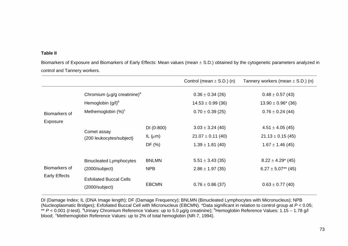
73
Table II
Biomarkers of Exposure and Biomarkers of Early Effects: Mean values (mean ± S.D.) obtained by the cytogenetic parameters analyzed in
control and Tannery workers.
Control (mean ± S.D.) (n) Tannery workers (mean ± S.D.) (n)
Chromium (μg/g creatinine)a 0.36 ± 0.34 (26) 0.48 ± 0.57 (43)
Hemoglobin (g/l)b 14.53 ± 0.99 (36) 13.90 ± 0.96* (36)
Methemoglobin (%)c 0.70 ± 0.39 (25) 0.76 ± 0.24 (44)
DI (0-800) 3.03 ± 3.24 (40) 4.51 ± 4.05 (45)
IL (μm) 21.07 ± 0.11 (40) 21.13 ± 0.15 (45)
Biomarkers of
Exposure
Comet assay (200 leukocytes/subject)
DF (%) 1.39 ± 1.81 (40) 1.67 ± 1.46 (45)
BNLMN 5.51 ± 3.43 (35) 8.22 ± 4.29* (45)
Binucleated Lymphocytes
(2000/subject) NPB 2.86 ± 1.97 (35) 6.27 ± 5.07** (45) Biomarkers of
Early Effects Esfoliated Buccal Cells
(2000/subject) EBCMN 0.76 ± 0.86 (37) 0.63 ± 0.77 (40)
DI (Damage Index; IL (DNA Image length); DF (Damage Frequency); BNLMN (Binucleated Lymphocytes with Micronucleus); NPB (Nucleoplasmatic Bridges); Exfoliated Buccal Cell with Micronucleus (EBCMN). *Data significant in relation to control group at P < 0.05; ** P < 0.001 (t-test). aUrinary Chromium Reference Values: up to 5.0 µg/g creatinine); bHemoglobin Reference Values: 1.15 – 1.78 g/l blood; cMethemoglobin Reference Values: up to 2% of total hemoglobin (NR-7, 1994).

74
Table III
Distribution of GSTM1, GSTT1, GSTP1, CYP1A1 and
CYP2E1 genotypes in control and Tannery workers.
Genotypes Controls (%) Tannery workers (%)
GSTM1 Non null 22 (55.0) 27 (60.0) Null 18 (45.0) 18 (40.0) GSTT1 Non null 32 (80.0) 38 (84.4) Null 8 (20.0) 7 (15.6) GSTP1 Ile/Ile 19 (47.5) 25 (55.6) Ile/Val 17 (42.5) 9 (20.0) Val/Val 2 (5.0) 5 (11.1) missing 2 (5.0) 6 (13.3) CYP1A1 *1A/*1A 21 (52.5) 17 (37.8) *1A/*2C 10 (25.0) 23 (51.1) *2C*/2C 8 (20.0) 2 (4.4) missing 1 (2.5) 3 (6.7) CYP2E1 *1A/*1A 30 (75.0) 31 (68.9) *1A/*5B 10 (25.0) 14 (31.1) *5B/*5B 0.0 0.0
GSTM1 and GSTT1: Non null (wild-type homozygous
or heterozygous), null (homozigous gene delection).
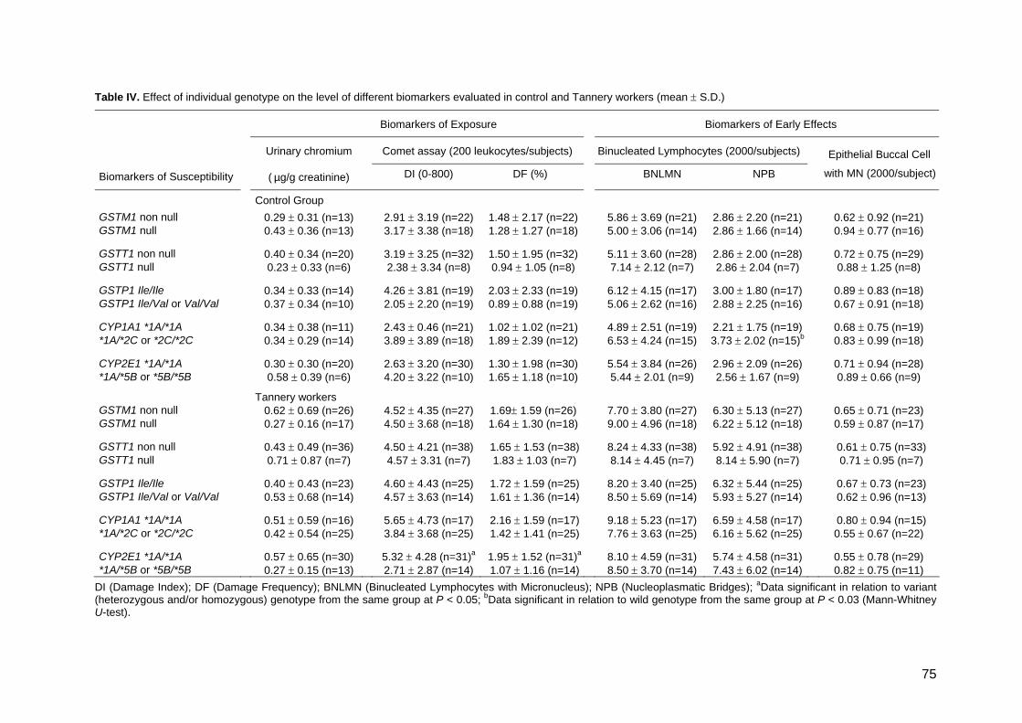
75
Table IV. Effect of individual genotype on the level of different biomarkers evaluated in control and Tannery workers (mean ± S.D.)
Biomarkers of Exposure Biomarkers of Early Effects
Comet assay (200 leukocytes/subjects) Binucleated Lymphocytes (2000/subjects)
Biomarkers of Susceptibility
Urinary chromium
( µg/g creatinine) DI (0-800) DF (%) BNLMN NPB
Epithelial Buccal Cell
with MN (2000/subject)
Control Group GSTM1 non null 0.29 ± 0.31 (n=13) 2.91 ± 3.19 (n=22) 1.48 ± 2.17 (n=22) 5.86 ± 3.69 (n=21) 2.86 ± 2.20 (n=21) 0.62 ± 0.92 (n=21) GSTM1 null 0.43 ± 0.36 (n=13) 3.17 ± 3.38 (n=18) 1.28 ± 1.27 (n=18) 5.00 ± 3.06 (n=14) 2.86 ± 1.66 (n=14) 0.94 ± 0.77 (n=16) GSTT1 non null 0.40 ± 0.34 (n=20) 3.19 ± 3.25 (n=32) 1.50 ± 1.95 (n=32) 5.11 ± 3.60 (n=28) 2.86 ± 2.00 (n=28) 0.72 ± 0.75 (n=29) GSTT1 null 0.23 ± 0.33 (n=6) 2.38 ± 3.34 (n=8) 0.94 ± 1.05 (n=8) 7.14 ± 2.12 (n=7) 2.86 ± 2.04 (n=7) 0.88 ± 1.25 (n=8) GSTP1 Ile/Ile 0.34 ± 0.33 (n=14) 4.26 ± 3.81 (n=19) 2.03 ± 2.33 (n=19) 6.12 ± 4.15 (n=17) 3.00 ± 1.80 (n=17) 0.89 ± 0.83 (n=18) GSTP1 Ile/Val or Val/Val 0.37 ± 0.34 (n=10) 2.05 ± 2.20 (n=19) 0.89 ± 0.88 (n=19) 5.06 ± 2.62 (n=16) 2.88 ± 2.25 (n=16) 0.67 ± 0.91 (n=18) CYP1A1 *1A/*1A 0.34 ± 0.38 (n=11) 2.43 ± 0.46 (n=21) 1.02 ± 1.02 (n=21) 4.89 ± 2.51 (n=19) 2.21 ± 1.75 (n=19) 0.68 ± 0.75 (n=19) *1A/*2C or *2C/*2C 0.34 ± 0.29 (n=14) 3.89 ± 3.89 (n=18) 1.89 ± 2.39 (n=12) 6.53 ± 4.24 (n=15) 3.73 ± 2.02 (n=15)b 0.83 ± 0.99 (n=18) CYP2E1 *1A/*1A 0.30 ± 0.30 (n=20) 2.63 ± 3.20 (n=30) 1.30 ± 1.98 (n=30) 5.54 ± 3.84 (n=26) 2.96 ± 2.09 (n=26) 0.71 ± 0.94 (n=28) *1A/*5B or *5B/*5B 0.58 ± 0.39 (n=6) 4.20 ± 3.22 (n=10) 1.65 ± 1.18 (n=10) 5.44 ± 2.01 (n=9) 2.56 ± 1.67 (n=9) 0.89 ± 0.66 (n=9)
Tannery workers GSTM1 non null 0.62 ± 0.69 (n=26) 4.52 ± 4.35 (n=27) 1.69± 1.59 (n=26) 7.70 ± 3.80 (n=27) 6.30 ± 5.13 (n=27) 0.65 ± 0.71 (n=23) GSTM1 null 0.27 ± 0.16 (n=17) 4.50 ± 3.68 (n=18) 1.64 ± 1.30 (n=18) 9.00 ± 4.96 (n=18) 6.22 ± 5.12 (n=18) 0.59 ± 0.87 (n=17) GSTT1 non null 0.43 ± 0.49 (n=36) 4.50 ± 4.21 (n=38) 1.65 ± 1.53 (n=38) 8.24 ± 4.33 (n=38) 5.92 ± 4.91 (n=38) 0.61 ± 0.75 (n=33) GSTT1 null 0.71 ± 0.87 (n=7) 4.57 ± 3.31 (n=7) 1.83 ± 1.03 (n=7) 8.14 ± 4.45 (n=7) 8.14 ± 5.90 (n=7) 0.71 ± 0.95 (n=7) GSTP1 Ile/Ile 0.40 ± 0.43 (n=23) 4.60 ± 4.43 (n=25) 1.72 ± 1.59 (n=25) 8.20 ± 3.40 (n=25) 6.32 ± 5.44 (n=25) 0.67 ± 0.73 (n=23) GSTP1 Ile/Val or Val/Val 0.53 ± 0.68 (n=14) 4.57 ± 3.63 (n=14) 1.61 ± 1.36 (n=14) 8.50 ± 5.69 (n=14) 5.93 ± 5.27 (n=14) 0.62 ± 0.96 (n=13) CYP1A1 *1A/*1A 0.51 ± 0.59 (n=16) 5.65 ± 4.73 (n=17) 2.16 ± 1.59 (n=17) 9.18 ± 5.23 (n=17) 6.59 ± 4.58 (n=17) 0.80 ± 0.94 (n=15) *1A/*2C or *2C/*2C 0.42 ± 0.54 (n=25) 3.84 ± 3.68 (n=25) 1.42 ± 1.41 (n=25) 7.76 ± 3.63 (n=25) 6.16 ± 5.62 (n=25) 0.55 ± 0.67 (n=22) CYP2E1 *1A/*1A 0.57 ± 0.65 (n=30) 5.32 ± 4.28 (n=31)a 1.95 ± 1.52 (n=31)a 8.10 ± 4.59 (n=31) 5.74 ± 4.58 (n=31) 0.55 ± 0.78 (n=29) *1A/*5B or *5B/*5B 0.27 ± 0.15 (n=13) 2.71 ± 2.87 (n=14) 1.07 ± 1.16 (n=14) 8.50 ± 3.70 (n=14) 7.43 ± 6.02 (n=14) 0.82 ± 0.75 (n=11)
DI (Damage Index); DF (Damage Frequency); BNLMN (Binucleated Lymphocytes with Micronucleus); NPB (Nucleoplasmatic Bridges); aData significant in relation to variant (heterozygous and/or homozygous) genotype from the same group at P < 0.05; bData significant in relation to wild genotype from the same group at P < 0.03 (Mann-Whitney U-test).
76
4. Discussion
In the present study, although not statistically significant, chromium excretion in
urine from Tannery workers was slightly increased in relation to control, as described by
several authors (Simpson and Gibson, 1992; Rajaram et al., 1995; Pan et al., 1996;
Stupar et al., 1999; Medeiros et al., 2003). In addition, hemoglobin levels were lower in
Tannery workers, possibly associated with body chromium accumulation, causing adverse
effects on iron metabolism and/or hemoglobin oxidation (methemoglobin formation), that
also occur due to aniline exposure, both leading to a decrease in hemoglobin levels, as
previously described (Krishnan and Pelekis, 1995; Khan et al., 1997; Fernandes et al.,
1999; ASTDR, 2002b; Kornhauser et al. 2002; Heuser et al., in prep).
Studies using cytogenetic tests like the micronuclei assay showed that nasal and
buccal mucosal cells, being directly exposed, are an excellent target for biomonitoring
exposure to mutagenic compounds (Salama et al., 1999; Majer et al., 2001; Faust et al.,
2004). In some of them, the frequency of MN in mucosal cells was significantly higher in
individuals exposed than in non-exposed, without an increase in MN in lymphocytes
(Norppa et al., 1992; Ying et al., 1997). Although the main exposure in tanneries occurs
by airborne substances, in the present study, Brazilian Tannery workers showed no
increase in EBCMN in relation to controls. On the other hand, an increased frequency of
BNLMN and NPB was observed in Tannery workers, although the Comet assay analysis
presented negative results. Similar results were obtained by Pitarque et al. (1999; 2002)
and our previous study (Heuser et al., in prep). Our findings indicate the presence of
genotoxic exposure at the workplace at least by one of the parameters used. It is possible
that the reduced DNA strand breaks can be caused by the elimination of cells with
extensive strand breaks, or the reduction in strand breaks due to incorrect rejoining of the
DNA molecules (Bonassi and Au, 2002), which could lead to MN formation, since there is
evidence that metal ions might interfere with distinct steps of diverse DNA repair systems
(Hartwig, 1998).
Tannery workers showed an increase in MN frequency due to age as described by
almost all biomonitoring studies (Migliore et al., 1991; Bolognesi et al., 1997; Fenech et
al., 1999; Albertini et al., 2000; Ishikawa et al., 2004). The lack of association between
age and MN frequencies in control suggest that this age-associated increase in
susceptibility can be higher in exposed people, probably due to accumulated genetic
damage in Tannery workers (Heuser et al., in prep). However, there was no evidence of
toxic effects of Cr(III) in human dose response studies (Campbell et al., 1999; Cefalu et
al., 1999), but some mechanisms by which Cr(III) can damage DNA are discussed (Snow,

77
1994; Blasiak and Kowalic, 2000; Seoane and Dulout, 2001). However, leather tanning
and processing involve a considerable number of potentially genotoxic substances, such
as organic solvents, mainly formaldehyde, a known mutagen and possibly a human
carcinogen (Ballarin et al., 1992; ASTDR, 1999; Burgaz et al., 2002; Shaham et al., 2002;
Titenko-Holland et al., 1996) which may be contributing to the cytogenetic lesions reported
in this study.
In the current study, the CYP1A1 genotype seems to modulate the baseline NPB in
the control group, since individuals porting CYP1A1 variant genotype (*1A/*2C or
*2C*/2C) presented increased frequency of this biomarker. Teixeira et al. (2002) also
describe an increase in DNA adduct levels in controls with CYP1A1 variant genotype. In
addition, several studies have implied that genetic polymorphisms can influence the level
of chromosome damage on the background levels of cytogenetic alterations (Norppa,
1997; 2001; 2003).
The different genes of the CYP system produced enzymes which metabolize the
xenobiontic products, and mutation in such genes can cause enzyme products with
abolished, reduced, altered or increased activity. As a result of this mechanisms, inactive
metabolites are sometimes formed, which are readily eliminated from the organism, but
chemically reactive agents can also be generated that can bind covalently to cellular
macromolecules, DNA, RNAs and proteins (Schoket et al., 2001). The effect of the
enzyme variants on the substrate specificity is sometimes very great in depending on the
expression system used for the enzyme variant (Ingelman-Sundberg, 2001). The results
of this study show that Tannery workers with the CYP2E1*1A/*1A (the wild type) had
increased damage, presenting higher values in DI and DF measured by Comet assay.
Very recently, it has been suggested that the CYP2E1 wild type genotype was correlated
with an increase in sister chromatid exchanges in smokers (Carere et al., 2002), and MN
frequencies in a healthy population (Ishikawa et al., 2004). Since CYP2E1 is involved in
the activation of a wide variety of xenobiotics, including organic compounds, to toxic
material (Guengerich and Shimada, 1998; Lucas et al., 2001), the differences in the levels
of CYP2E1 mRNA and protein may lead to the genotype-dependent variation in the DNA
damage detected by Comet assay in this study. The same was not observed in basal
levels of DNA damage in control group with the same genotype.
In this study, no association between the genotoxicity parameters with the chromium
in urine was observed. Chromium is normally considered the most important substance in
the monitoring of Tannery workers, but it must be taken in to consideration that these
workers are also submitted to other toxic products, as formaldehyde, which isolatedly or

78
associated with chromium can be responsible for the genotoxic effects. This study, as in
other studies, shows that the Tannery workers are still submitted to some hazardous
conditions. More studies are necessary to identify the health risks and establish better
exposure evaluation methods.
Many studies have been performed in the recent decade to explore the influence of
single genotypes and interaction of genotypes on the levels of biomarkers of genotoxic
exposure. However, this is the firth study describing the effect of polymorphic genotype on
biomarkers of exposure and effects on tanning occupation. The different GST genotypes
analyzed, GSTM1, GSTT1 and GSTP1, did not influence the level of cytogenetic damage
in Tannery workers. It was possible to identify a sensitive subgroup, since the Comet
assay parameters were increased Tannery workers with CYP2E1 (*1A/*1A) wild genotype
compared to CYP2E1 (*1A/*5B or *5B/*5B) variant genotype. It should be emphasized
that genetic susceptibility is not limited to the inheritance of polymorphic chemical
metabolizing genes but also other genes, e. g., DNA repair genes (Au et al., 1998).
Therefore, susceptible genes may have negative impact on the quality of life. Further
understanding on the impact will allow us to protect the health of the population thus
improving the quality of life.
Acknowledgements The authors would like to express their gratitude to all volunteers, for their
acceptation to participate in this study. We wish to thank Dr. Vera L. T. de Azevedo, Dr.
Poliana K. Hernandez, Dr. Anita Lehnen, from Vida Laboratory, especially Cíntia and Lisa
for their valuable participation in the sampling. We also thank Roseli O. de Cândido,
Vanessa M. de Andrade, Marcos R. Furini and Lisiane F. Leal for their help in various
stages of this study. This work was financially supported by the CNPq (Conselho Nacional
para o Desenvolvimento Científico e Tecnológico).
References
Albertini, R., Anderson, D., Douglas, G.R., Hagmar, L., Hemminki, K., Merlo, F., Natarajan, A.T., Norppa, H., Shuker, D.E.G., Tice, R.R., Waters, M.D., Aitio, A., 2000. IPCS guidelines for the monitoring of genotoxic effects of carcinogens in humans, International Program on Chemical Safety. Mutat. Res. 463, 111-172.
Agency for Toxic Substances and Disease Registry (ATSDR), 1999. Toxicological profile for formaldehyde. On line: http://www.astdr.cdc.gov/toxfaq.html
Agency for Toxic Substances and Disease Registry (ATSDR), 2002a. Toxicological profile for chromium. On line: http://www.astdr.cdc.gov/toxfaq.html

79
Agency for Toxic Substances and Disease Registry (ATSDR), 2002b. Toxicological profile for anilines. On line: http://www.astdr.cdc.gov/toxfaq.html
Au, W.W., Cajas-Salazar, N., Salama, S., 1998. Factors contributing to discrepancies in population monitoring studies. Mutat. Res. 400, 467-478.
Autrup, H., 2000. Genetic polymorphisms in human xenobiotic metabolizing enzymes as susceptibility factors in toxic response. Mutat. Res. 464:65-76.
Ballarin, C., Sarto, F., Giacomelli, L., Bartolucci, G.B., Clonfero, E., 1992. Micronucleated cells in nasal mucosa of formaldehyde-exposed workers. Mutat. Res. 280, 1-7.
Battista, G., Comba, P., Orsi, D., Norpoth, K., Maier, A., 1995. Nasal cancer in leather workers: an occupational disease. J. Cancer Res. Clin. Oncol. 121, 1-6.
Bell, D.A., Taylor, J.A., Paulson, D.F., Robertson, C.N., Mohler, J.L., Lucier, G.W., 1993. Genetic risk and carcinogen exposure: A common inherited defect of the carcinogen-metabolism gene glutathione S-transferase M1 (GSTM1) that increases susceptibility to bladder cancer. J. Natl. Cancer Inst. 85, 1159-1164.
Blasiak, J., Kowalik, J.,2000. A comparison of in vitro genotoxicity of tri- and hexavalent chromium. Mutat. Res. 469, 135-145.
Bolognesi, C., Abbondandolo, A., Barale, R., Casalone, R., Dalpra, L., De Ferrari, M., Degrassi, F., Forni, A., Lamberti, L., Lando, C., Migliore, L., Padovani, P., Pasquini, R., Puntoni, R., Sbrana, I., Stella, M., Bonassi, S., 1997. Age-related increase of baseline frequencies of sister chromatid exchanges, chromosome aberrations, and micronuclei in human lymphocytes. Cancer Epidemiol. Biomarkers Prev. 6, 249-256.
Bonassi, S., Au, W.W., 2002. Biomarkers in molecular epidemiology studies for health risk prediction. Mutat. Res. 511, 73-86.
Burgaz, S., Erdem, O., Cakmak, G., Erdem, N., Karakaya, A., Karakaya, A.E., 2002. Cytogentic analysis of buccal cell from shoe-workers and pathology and anatomy laboratoy workers exposed to n-hexane, toluene, metyl ethyl ketona and formaldehyde. Biomarkers 7, 151-161.
Campbell, W.W., Joseph, L.J., Davey, S.L., Cyr-Campbell, D., Anderson, R.A., Evans, W.J., 1999. Effects of resistance training and chromium tripicolinate on body composition and skeletal muscle in older men. J. Appl. Physiol. 86, 29-39.
Carere, A., Andreoli, C., Galati, R., Leopardi, P., Marcon, F., Rosati, M.V., Rossi, S., Tomei, F., Verdina, A., Zijno, A., Crebelli, R., 2002. Biomonitoring of exposure to urban pollutants: analysis of sister chromatid exchanges and DNA lesions in peripheral lymphocytes of traffic policemen. Mutat. Res. 518, 215-224.
Carrano, A.V., Natarajan A.T., 1988. Considerations for population monitoring using cytogenetic techniques, International Commission for Protection against Environmental Mutagens and Carcinogens (ICPEMC publication 14). Mutat. Res. 204, 379-406.
Cascorby, I., Brockmöller, J., Roots, I.A., 1996. C4887A polymorphism in exon 7 of human CYP1A1: Population frequency, mutation linkages, and impact on lung cancer susceptibility. Cancer Res. 56, 4965-4969.
Cebulska-Wasilewska, A., 2003. Response to challenging dose of X-ray as a predictive assay for molecular epidemiology. Mutat. Res. 544, 289-297.
Cefalu, W.T., Bell-Farrow, A.D., Stegner, J., Wang, Z.Q., King, T., Morgan, T., Terry, J.G.,1999. Effects of chromium tripicolinate on insulin sensitivity in vivo. J. Trace Elem. Exp. Med. 12, 71- 83.
80
Cid, M.G., Loria, D., Vilensky, M., Miotti, J.L., Matos, E., 1991. Leather tanning workers: chromossomal aberrations in peripheral lymphocytes and micronuclei in exfoliated cell in urine. Mutat. Res. 259, 197-201.
Collins, A.R., Dobson, V.L., Dusinská, M., Kennedy, G., Stetina, R., 1997. The Comet assay: what can it really tell us? Mutat. Res. 375, 183-193.
Constantini, S.A., Merler, E., Saracci, R., 1990. Epidemiologic studies on carcinogeneic risk and occupational activities in tanning, leather and shoe industries. Med. Lav. 81, 184-211.
Environmental Protection Agency (EPA), 1998. Toxicological review of trivalent chromium. In Support of Summary Information on the Integrated Risk Information (IRIS). On-line: http://epa.gov/iris
Faust, F., Kassie, F., Knasmüller, S., Kevekordes, S., Mersch-Sundermann, V., 2004. Use of primary blood cells for the assessment of exposure to occupational genotoxicants in human biomonitoring studies. Toxicology 198, 341-350.
Fenech, M., Titenko-Holland, N., Chang, W.P., Zeiger, E., Bonassi, S., 1999. The Human Micronucleus Project – An international colaborative study on the use of the micronucleus technique for measuring DNA damage in humans. Mutat. Res. 428, 271-283.
Fernandes, M.A.S., Mota, I.M., Silva, M.T.L., Oliveira, C.R., Geraldes, C.F.G.C., Alpoim, M.C., 1999. Human Erythrocytes are protected against chromate-induced peroxidation. Ecotoxicol. Environ. Safety 43, 38-46.
Guengerich, F.P., Shimada, T., 1998. Activation of procarcinogens by human cytochrome P450 enzymes. Mutat. Res. 400, 201-213.
Hamamy, H.A., Al-Hakkak, Z.S., Hussain, A.F., 1987. Chromosome aberrations in workers a tannery in Iraq. Mutat. Res. 189, 395-398.
Harries, L.W., Stubbins, M.J., Forman, D., Howard, G.C.W., Wolf, C.R., 1991. Identification of genetic polymorphisms at the glutathione S-transferase Pi locus and association with susceptibility to bladder, testicular and prostate cancer. Carcinogenesis 18, 641-644.
Hartwig, A., 1998. Carcinogenicity of metal compounds: possible role of DNA repair inhibition. Toxicol. Lett. 102, 235-239.
Heuser, V.D., Silva, J., Erdtmann, B. in prep. Evaluation of genotoxic exposure in Brazilian Tannery workers: Comparison between Drum Workshop and Finishing Workshop.
Ingelman-Sundberg, M., 2001. Genetic susceptibility to adverse effects of drugs and environmental toxicants: the role of the CYP family of enzymes. Mutat. Res. 482, 11-19.
International Agency for Research on Cancer (IARC), 1987. Leather tanning and processing. IARC Monographs on the evaluation of the carcinogenic risk of chemicals to humans, Vols. 25, Suppl. 7, Lyon, 1987, pp. 232. On-line: http://www.inchen.org/pages/iarc.html
Ishikawa, H., Yamamoto, H., Tian, Y., Kawano, M., Yamauchi, T., Kazuhito, Y., 2004. Evidence of association of the CYP2E1 genetic polymorphism with micronuclei frequency in human peripheral blood. Mutat. Res. 546, 45-53.
Kato, S., Shield, P.G., Caporoso, N.E., Hoover, R.N., Trump, B.F., Sugimura, H., Weston, A., Harris, C.C., 1992. Cytochrome P450IIE1 genetic polymorphism, racial variation, and lung cancer risk. Cancer Res. 52, 6712-6715.
81
Khan, M.F., Wu, X., Kaphalia, B.S., Boor, P.J., Ansari, G.A.S., 1997. Acute hematopoietic toxicity of aniline in rats. Toxicol. Lett. 92, 31-37.
Kornhauser, C., Wrobel, K., Malacara, J.M., Nava, L.E., Gomez, L., Gonzales, R., 2002. Possible adverse effect of chromium in ccupational exposure of tannery workers. Ind. Health 40, 207-212.
Krishnan, K., Pelekis, M., 1995. Hematotoxic interaction: occurrence, mechanism and predictability. Toxicology 105, 355-364.
Lucas, D., Ferrara, R., Gonzáles, E., Albores, A., Manno, M., Berthou, F., 2001. Cytochrome CYP2E1 phenotyping and genotyping in the evaluation of health risks from exposure to polluted environments. Toxicol. Lett. 124, 71-81.
Majer, B.J., Laky, B., Knasmüller, S., Kassie, F., 2001. Use of the micronucleus assay with exfoliated epithelial cells as a biomarker for monitoring individuals at elevated risk of genetic damage and in chemoprevention trials. Mutat. Res. 489, 147-172.
Medeiros, M.G., Rodrigues, A.S., Batoréu, M.C., Laires, A., Rueff, J., Zhitkovich, A., 2003. Elevated levels of DNA – protein crosslinks and micronuclei peripheral lymphocytes of tannery workers exposed to trivalent chromium. Mutagenesis 18, 19-24.
Migliore, L., Parrini, M., Sbrana, I., Biagini, C., Battaglia, A., Loprieno, N., 1991. Micronucleated lymphocytes in people occupationally exposed to potential environmental contaminants: the age affect. Mutat. Res. 256, 13-20.
Miller, A.S., Dykes, D.D., Polesky, H.F., 1995. A simple salting out procedure for extracting DNA from human nucleated cells. Nucleic Acids Res. 16, 1215.
Montanaro, F., Ceppi, M., Demers, P.A., Puntoni, R., Bonassi, S., 1997. Mortality in a cohort of tannery workers. Occup. Environ. Med. 8, 588-591.
Norma Regulamentadora N°7 (NR-7), 1994. Segurança e Medicina do Trabalho – NR-7 – programa de Controle Médico de Saúde Ocupacional. Portaria GM/SSSTb n. 24 – (DOU 30.12.94).
Norppa, H., Heikanen, H., Pfaffli, P., Virtanen, H., 1992. Increased micronuclei in buccal mucosa cells but not in blood lymphocytes of formaldehyde exposed workers. Pharmacol. Toxicol. 70, 22-27.
Norppa, H., 1997. Cytogenetic markers of susceptibility: Influence of polymorphic carcinogen-metabolizing enzymes. Environ. Health Perspect. 105, 829-835.
Norppa, H., 2001. Genetic polymorphisms and chromosome damage. Int. J. Environ. Health 204, 31-38.
Norppa, H., 2003. Genetic susceptibility, biomarkers response, and cancer. Mutat Res 544, 339-348.
Pan, T.C., Wu, W.J., Huang, K.C., Wei, B.L., Ko, Y.C., 1996. Biological monitoring for chromium exposure in the workplace. Jap. J. Toxicol. Environ. Health 42, 301-306.
Pavanello, S., Clonfero, E., 2000. Biological indicators of genotoxic risk and metabolic polymorfisms. Mutat. Res. 463, 285-308.
Pemble, S., Schroeder, K.R., Spencer, S.R., Meyer, D.J., Hallier, E., Bolt, H.M., Ketterer, B., Taylor, J.B., 1994. Human glutathione S-transferase theta (GSTT1): cDNA cloning and the characterization of a genetic polymorphism. Biochem. J. 300, 271- 276.
Pitarque, M., Vaglenov, A., Nosko, M., Hirvonen, A., Norppa, H., Creus, A., Marcos, R., 1999. Evaluation of DNA damage by Comet assay in footwear-workers exposed to toluene and other organic solvents. Mutat. Res. 441, 115-127.

82
Pitarque, M., Vaglenov, A., Nosko, M., Pavlova, S., Petkova, V., Hirvonen, A., Creus, A., Norppa, H., Marcos, R., 2002. Sister chromatid excahanges and micronuclei in peripheral lymphocytes of shoe factory workers exposed to solvents. Environ. Health Perspect. 110, 339-404.
Rajaram, R., Nair, B.U., Ramasami, T., 1995. Chromium(III) induced abnormalities in human lymphocyte cell proliferation: evidence for apoptosis. Biochem. Biophys. Res. Commun. 210: 434-440.
Salama, S.A., Serrana, M., Au, W.W., 1999. Biomonitoring using accessible human cells for exposure and health risk assessment. Mutat. Res. 436, 99-112.
Sbrana, I., Caretto, S., Battaglia, A., 1991. Chromossomal aberration of workers in tannery industries. Mutat. Res. 242, 331-336.
Schoket, B., Papp, G., Lévay, K., Mracková, G., Kadlubar, F.F., Vincze, I., 2001. Impact of metabolic genotypes on levels of biomarkers of genotoxic exposure. Mutat. Res. 482, 57-69.
Seoane, A.I., Dulout, F.N., 2001. Genotoxic ability of cadmium, chromium and nickel salts by kinetochore staining in the cytokinesis-blocked micronucleus assay. Mutat. Res. 490, 99-106.
Shaham, J., Gurvich, R., Kaufman, Z., 2002. Sister chromatid exchange in pathology staff occupationally exposed to formaldehyde. Mutat. Res. 514, 115–123.
Silva, J., Freitas, T.R.O., Heuser V.D., Marinho, J.R., Erdtmann, B., 2000. Genotoxicity Biomonitoring in Coal Regions Using Wild Rodent Ctenomys torquatus by Comet Assay and Micronucleus Test. Environ. Mol. Mutagen. 35, 270-278.
Simpson, J.R., Gibson, R.S., 1992. Hair, serum, and urine chromium concentrations in former employed of the leather tanning industry. Biol. Trace Elem. Res. 32, 155-159.
Singh, N.P., McCoy, M.T., Tice, R.R., Schneider, E.L., 1988. A simple technique for quantification of low levels of DNA damage in individual cells. Exp. Cell Res. 175, 184-191.
Snow, E.T., 1994. Effects of chromium on DNA replication in vitro. Environ. Health Perspect. 102, 41-44.
Stern, F.B., 2003. Mortality among chrome leather tannery workers: an update. Am. J. Ind. Med. 44, 197-206.
Stupar, J., Vrtovec, M., Kocijancic, A., Gantar, A., 1999. Chromium status of tannery workers in relation to metabolic disorders. J. Appl. Toxicol. 19, 437-446.
Teixeira, J.P., Gaspar, J., Martinho, G., Silva, S., Rodrigues, S., Mayan, O., Martin, E., Farmer, P.B., Rueff, J., 2002. Aromatic DNA adduct level in coke oven workers: correlation with polymorphisms in genes GSTP1, GSTM1, GSTT1 and CYP1A1. Mutat. Res. 517, 147-155.
Tice, R.R., Agurell, E., Anderson, D., Burlinson, B., Hartmann, A., Kobayashi, H., Miyamae, Y., Rojas, E., Ryu, J.C., Sasaki, Y.F., 2000. Single Cell Gel/Comet Assay: Guidelines for In Vitro and In Vivo Genetic Toxicology Testing. Environ. Mol. Mutagen. 35, 206-221.
Titenko-Holland, N., Levine, A.J., Smith, M.T., Quintana, P.J.E., Boeniger, M., Hayes, R., Surada, A., Sculte, P., 1996. Quantification of epithelial cell micronuclei by fluorescence in situ hybridization (FISH) in mortuary science students exposed to formaldehyde, Mutat. Res. 371, 237-248.

83
Titenko-Holland, N., Jacob, R., Shang, N., Balaraman, A., Smith, M.T., 1998. Micronuclei in lymphocytes and exfoliated buccal cells of postmenopausal women with dietary changes in folate. Mutat. Res. 417, 101-114.
Tolbert, P.E., Shy, C.M., Allen, J.W., 1992. Micronuclei and other nuclear anomalies in buccal smears: method development. Mutat. Res. 271, 69-77.
Veyalkin, I.V., Milyutin, A.A., 2003. Proportionate cancer mortality among workers in the Belarussian tanning industry. Am. J. Ind. Med. 44, 637-42.
World Health Organization (WHO), 2001. Environmental Health Criteria 222: Biomarkers in risk assessment: Validity and validation. IPCS (International Program on Chemical Safety), Geneva. On-line: http://www.inchem.org/documents/ehc/ehc/ehc222.htm
Ying, C.J., Yan, W.S., Zhao, M.Y., Ye, X.L., Xie, H., Yin, S.Y., Zhu X.S., 1997. Micronuclei in nasal mucosa, oral mucosa and lymphocytes in students exposed to formaldehyde vapor in anatomy class. Biomed. Environ. Sci. 10, 451-455.
84
5. CAPÍTULO III
Comparison of genetic damage in Brazilian footwear-workers exposed to solvent-based or water-based adhesive
Vanina Dahlström Heuser1, Vanessa Moraes de Andrade1, Juliana da Silva2* & Bernardo
Erdtmann1,3
1Programa de Pós-Graduação em Genética e Biologia Molecular (PPGGBM), Universidade
Federal do Rio Grande do Sul (UFRGS), Porto Alegre-RS, Brazil.
2Laboratório de Genética Toxicológica, Programa de Pós-Graduação em Ensino de Ciências
e Matemática (PPGECIM), Universidade Luterana do Brasil (ULBRA), Canoas-RS, Brazil.
3Instituto de Biotecnologia, Universidade de Caxias do Sul (UCS), Caxias do Sul-RS, Brazil.
*Correspondence to: Juliana da Silva, Programa de Pós-Graduação em Ensino de Ciências
e Matemática (PPGECIM), Universidade Luterana do Brasil (ULBRA), Prédio 14, Sala 230,
Rua Miguel Tostes 101, Bairro São Luís, CEP 92420-280, Canoas-RS, Brazil.
E-mail: [email protected]
Grant sponsors: CNPq, FAPERGS, GENOTOX
ARTIGO IN PRESS NA REVISTA MUTATION RESEARCH

85
ABSTRACT
Research has shown that workers employed in footwear manufacture are at increased
risk of some cancers, the strongest evidence being for nasal cancer and leukemia. Footwear-
workers are routinely exposed to complex mixtures of solvents in degreasers, cleaners,
primers and adhesives used in the production process, as toluene, n-hexane, acetone, and
possibly dust particles, additives in shoe materials and degradation products of materials.
The recognition of the potential health-hazards of solvent-based adhesives (SBAs) has lead
to the development of adhesives with no organic solvents, the water-based adhesives
(WBA). We investigated footwear-workers (all males) exposed to SBA (n=29) (for 3.98 ± 4.13
years), and WBA (n=16), which had spent the six months previous to the study employed in
an experimental section which used only water-based adhesives, although they had
previously worked in sections which used solvent-based adhesives (for 5.80 ± 4.03 years);
25 healthy subjects were used as controls. The Comet assay and the micronucleus test were
used as endpoints, while the traditional parameters for assessing exposure to toluene in
organic mixtures by measuring the concentration of urinary hippuric acid were also
assessed. Our results showed a significantly lower mean concentration of hippuric acid in the
control group than found in the SBA (P < 0.001) and WBA (P < 0.05) groups. The Comet
assay results showed that there was a significant increase in the mean damage index for the
SBA (P < 0.001) group in comparison to the WBA group and control (P < 0.05). For the
micronucleus test in binucleated lymphocytes and exfoliated buccal cell, the three groups
were not statistically different. Our study demonstrated that water-based adhesives are
clearly a better option for safeguarding the health of footwear-workers, even with possibility
of isocyanate presence, while the positive results observed in SBA group might be explained
by chloroprene presence in adhesive.
Keywords: Comet assay, micronucleus test, exfoliated buccal cells, footwear-workers,
hippuric acid, occupational exposure to glues.
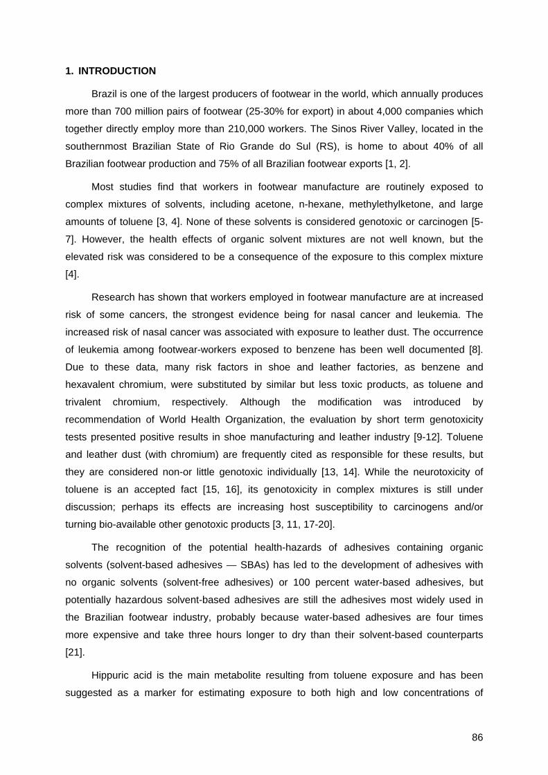
86
1. INTRODUCTION
Brazil is one of the largest producers of footwear in the world, which annually produces
more than 700 million pairs of footwear (25-30% for export) in about 4,000 companies which
together directly employ more than 210,000 workers. The Sinos River Valley, located in the
southernmost Brazilian State of Rio Grande do Sul (RS), is home to about 40% of all
Brazilian footwear production and 75% of all Brazilian footwear exports [1, 2].
Most studies find that workers in footwear manufacture are routinely exposed to
complex mixtures of solvents, including acetone, n-hexane, methylethylketone, and large
amounts of toluene [3, 4]. None of these solvents is considered genotoxic or carcinogen [5-
7]. However, the health effects of organic solvent mixtures are not well known, but the
elevated risk was considered to be a consequence of the exposure to this complex mixture
[4].
Research has shown that workers employed in footwear manufacture are at increased
risk of some cancers, the strongest evidence being for nasal cancer and leukemia. The
increased risk of nasal cancer was associated with exposure to leather dust. The occurrence
of leukemia among footwear-workers exposed to benzene has been well documented [8].
Due to these data, many risk factors in shoe and leather factories, as benzene and
hexavalent chromium, were substituted by similar but less toxic products, as toluene and
trivalent chromium, respectively. Although the modification was introduced by
recommendation of World Health Organization, the evaluation by short term genotoxicity
tests presented positive results in shoe manufacturing and leather industry [9-12]. Toluene
and leather dust (with chromium) are frequently cited as responsible for these results, but
they are considered non-or little genotoxic individually [13, 14]. While the neurotoxicity of
toluene is an accepted fact [15, 16], its genotoxicity in complex mixtures is still under
discussion; perhaps its effects are increasing host susceptibility to carcinogens and/or
turning bio-available other genotoxic products [3, 11, 17-20].
The recognition of the potential health-hazards of adhesives containing organic
solvents (solvent-based adhesives — SBAs) has led to the development of adhesives with
no organic solvents (solvent-free adhesives) or 100 percent water-based adhesives, but
potentially hazardous solvent-based adhesives are still the adhesives most widely used in
the Brazilian footwear industry, probably because water-based adhesives are four times
more expensive and take three hours longer to dry than their solvent-based counterparts
[21].
Hippuric acid is the main metabolite resulting from toluene exposure and has been
suggested as a marker for estimating exposure to both high and low concentrations of
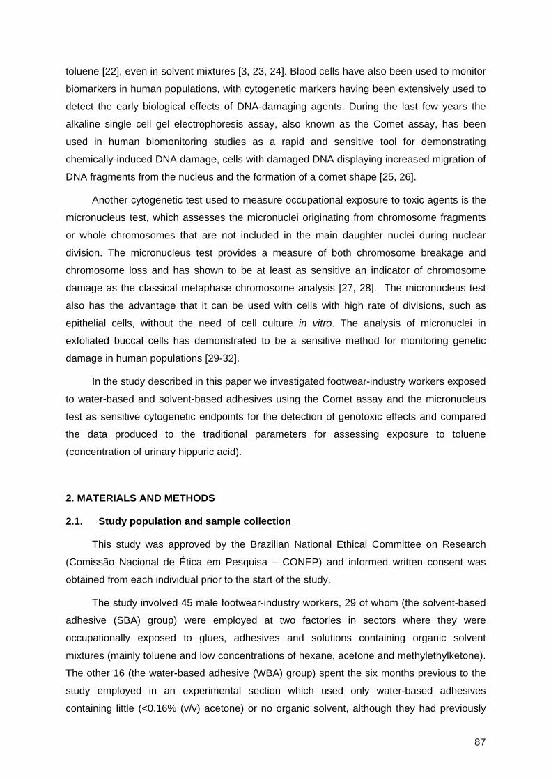
87
toluene [22], even in solvent mixtures [3, 23, 24]. Blood cells have also been used to monitor
biomarkers in human populations, with cytogenetic markers having been extensively used to
detect the early biological effects of DNA-damaging agents. During the last few years the
alkaline single cell gel electrophoresis assay, also known as the Comet assay, has been
used in human biomonitoring studies as a rapid and sensitive tool for demonstrating
chemically-induced DNA damage, cells with damaged DNA displaying increased migration of
DNA fragments from the nucleus and the formation of a comet shape [25, 26].
Another cytogenetic test used to measure occupational exposure to toxic agents is the
micronucleus test, which assesses the micronuclei originating from chromosome fragments
or whole chromosomes that are not included in the main daughter nuclei during nuclear
division. The micronucleus test provides a measure of both chromosome breakage and
chromosome loss and has shown to be at least as sensitive an indicator of chromosome
damage as the classical metaphase chromosome analysis [27, 28]. The micronucleus test
also has the advantage that it can be used with cells with high rate of divisions, such as
epithelial cells, without the need of cell culture in vitro. The analysis of micronuclei in
exfoliated buccal cells has demonstrated to be a sensitive method for monitoring genetic
damage in human populations [29-32].
In the study described in this paper we investigated footwear-industry workers exposed
to water-based and solvent-based adhesives using the Comet assay and the micronucleus
test as sensitive cytogenetic endpoints for the detection of genotoxic effects and compared
the data produced to the traditional parameters for assessing exposure to toluene
(concentration of urinary hippuric acid).
2. MATERIALS AND METHODS
2.1. Study population and sample collection
This study was approved by the Brazilian National Ethical Committee on Research
(Comissão Nacional de Ética em Pesquisa – CONEP) and informed written consent was
obtained from each individual prior to the start of the study.
The study involved 45 male footwear-industry workers, 29 of whom (the solvent-based
adhesive (SBA) group) were employed at two factories in sectors where they were
occupationally exposed to glues, adhesives and solutions containing organic solvent
mixtures (mainly toluene and low concentrations of hexane, acetone and methylethylketone).
The other 16 (the water-based adhesive (WBA) group) spent the six months previous to the
study employed in an experimental section which used only water-based adhesives
containing little (<0.16% (v/v) acetone) or no organic solvent, although they had previously

88
worked in sections which used solvent-based adhesives. Besides the difference in solvent
content in adhesives, water-based adhesives contained polyurethane, while solvent-based
adhesives contained polychroprene in their formulation. The control group consisted of 25
healthy males with no occupational exposure.
All the individuals examined in the study lived in or near the city of Novo Hamburgo-RS
and were required to answer a Portuguese version of a questionnaire from the International
Commission for Protection against Environmental Mutagens and Carcinogens [33] and
participate in a face-to-face questionnaire which included standard demographic data (age,
gender, etc.) as well as questions relating to medical issues (exposure to X-rays,
vaccinations, medication, etc.), life style (smoking, coffee, alcohol, diet, etc.) and their
occupation (number of hours worked per day, time exposed to organic solvents, use of
protective measures, etc.). Individuals were selected for the three groups (control, SBA,
WBA) in such a manner as to ensure that, except for occupational exposure to organic
solvents, there were no marked differences between the members of the groups. In all the
groups, individuals who smoked more than 5 cigarettes/day for at least 1 year were
considered smokers. The characteristics of the three groups are presented in Table I.
Blood and urine samples were obtained from individuals in the SBA and WBA groups
on the same day during a normal shift during the workers periodical medical examinations by
the nurses from a regional Health and Safety at Work Medical Center (Centro de Saúde e
Segurança do Trabalhador das Indústrias Calçadistas da Região de Parobé, Parobé-RS,
Brasil (CESSTIC) — The Footwear Industry Health and safety at work Center for the Parobé
Region, Parobé-RS, Brazil). For the control group, blood and urine samples were taken at
the same region. All blood samples were collected using venipuncture and heparinized
vacutainers and processed as quickly as possible to avoid the damage associated with
storage [34], the blood cell samples being transported to the laboratories at or below 8oC and
processed within 8 h of collection in order to minimize loss of DNA and damage due to DNA
repair processes.
2.2. Hippuric acid assay
Analysis of urinary hippuric acid was carried out using gas chromatography at a
commercial laboratory (Toxilab, Toxicological Laboratory, Porto Alegre-RS, Brazil) using a
Perkin-Elmer XL Auto System gas chromatograph (Perkin-Elmer, USA).
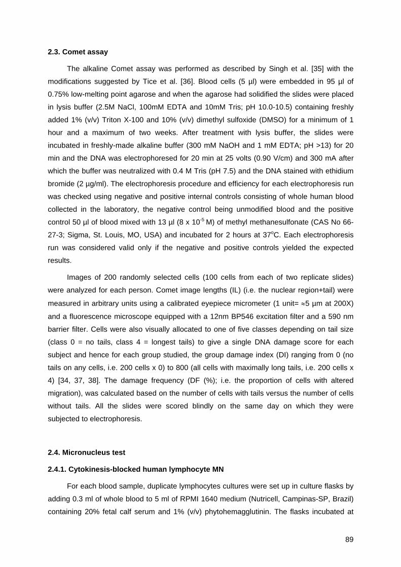
89
2.3. Comet assay
The alkaline Comet assay was performed as described by Singh et al. [35] with the
modifications suggested by Tice et al. [36]. Blood cells (5 µl) were embedded in 95 µl of
0.75% low-melting point agarose and when the agarose had solidified the slides were placed
in lysis buffer (2.5M NaCl, 100mM EDTA and 10mM Tris; pH 10.0-10.5) containing freshly
added 1% (v/v) Triton X-100 and 10% (v/v) dimethyl sulfoxide (DMSO) for a minimum of 1
hour and a maximum of two weeks. After treatment with lysis buffer, the slides were
incubated in freshly-made alkaline buffer (300 mM NaOH and 1 mM EDTA; pH >13) for 20
min and the DNA was electrophoresed for 20 min at 25 volts (0.90 V/cm) and 300 mA after
which the buffer was neutralized with 0.4 M Tris (pH 7.5) and the DNA stained with ethidium
bromide (2 µg/ml). The electrophoresis procedure and efficiency for each electrophoresis run
was checked using negative and positive internal controls consisting of whole human blood
collected in the laboratory, the negative control being unmodified blood and the positive
control 50 µl of blood mixed with 13 µl (8 x 10⁻⁵ M) of methyl methanesulfonate (CAS No 66-
27-3; Sigma, St. Louis, MO, USA) and incubated for 2 hours at 37oC. Each electrophoresis
run was considered valid only if the negative and positive controls yielded the expected
results.
Images of 200 randomly selected cells (100 cells from each of two replicate slides)
were analyzed for each person. Comet image lengths (IL) (i.e. the nuclear region+tail) were
measured in arbitrary units using a calibrated eyepiece micrometer (1 unit= ≈5 µm at 200X)
and a fluorescence microscope equipped with a 12nm BP546 excitation filter and a 590 nm
barrier filter. Cells were also visually allocated to one of five classes depending on tail size
(class 0 = no tails, class 4 = longest tails) to give a single DNA damage score for each
subject and hence for each group studied, the group damage index (DI) ranging from 0 (no
tails on any cells, i.e. 200 cells x 0) to 800 (all cells with maximally long tails, i.e. 200 cells x
4) [34, 37, 38]. The damage frequency (DF (%); i.e. the proportion of cells with altered
migration), was calculated based on the number of cells with tails versus the number of cells
without tails. All the slides were scored blindly on the same day on which they were
subjected to electrophoresis.
2.4. Micronucleus test
2.4.1. Cytokinesis-blocked human lymphocyte MN
For each blood sample, duplicate lymphocytes cultures were set up in culture flasks by
adding 0.3 ml of whole blood to 5 ml of RPMI 1640 medium (Nutricell, Campinas-SP, Brazil)
containing 20% fetal calf serum and 1% (v/v) phytohemagglutinin. The flasks incubated at

90
37ºC for 44 h before adding 5 µg/ml of cytochalasin B (Sigma) [27] and continuing incubation
until the total incubation time reached was 72 h. After incubation the lymphocytes were
harvested by centrifugation at 800 revs/min for 8 min, recentrifuged, fixed in 3:1 (v/v)
methanol/acetic acid, placed onto a clean microscope slide and stained with 5% (v/v)
Giemsa. For each blood sample, 2000 binucleated lymphocytes (i.e. 1000 from each of the
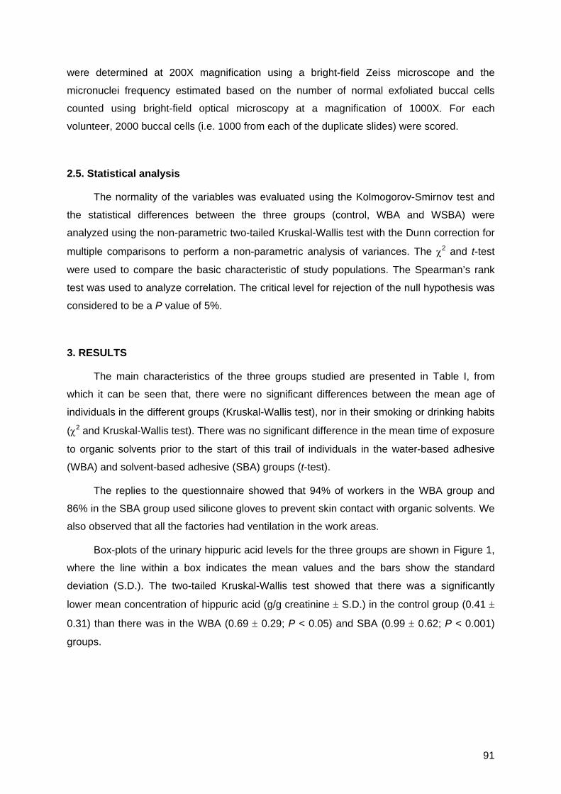
91
were determined at 200X magnification using a bright-field Zeiss microscope and the
micronuclei frequency estimated based on the number of normal exfoliated buccal cells
counted using bright-field optical microscopy at a magnification of 1000X. For each
volunteer, 2000 buccal cells (i.e. 1000 from each of the duplicate slides) were scored.
2.5. Statistical analysis
The normality of the variables was evaluated using the Kolmogorov-Smirnov test and
the statistical differences between the three groups (control, WBA and WSBA) were
analyzed using the non-parametric two-tailed Kruskal-Wallis test with the Dunn correction for
multiple comparisons to perform a non-parametric analysis of variances. The χ2 and t-test
were used to compare the basic characteristic of study populations. The Spearman’s rank
test was used to analyze correlation. The critical level for rejection of the null hypothesis was
considered to be a P value of 5%.
3. RESULTS
The main characteristics of the three groups studied are presented in Table I, from
which it can be seen that, there were no significant differences between the mean age of
individuals in the different groups (Kruskal-Wallis test), nor in their smoking or drinking habits
(χ2 and Kruskal-Wallis test). There was no significant difference in the mean time of exposure
to organic solvents prior to the start of this trail of individuals in the water-based adhesive
(WBA) and solvent-based adhesive (SBA) groups (t-test).
The replies to the questionnaire showed that 94% of workers in the WBA group and
86% in the SBA group used silicone gloves to prevent skin contact with organic solvents. We
also observed that all the factories had ventilation in the work areas.
Box-plots of the urinary hippuric acid levels for the three groups are shown in Figure 1,
where the line within a box indicates the mean values and the bars show the standard
deviation (S.D.). The two-tailed Kruskal-Wallis test showed that there was a significantly
lower mean concentration of hippuric acid (g/g creatinine ± S.D.) in the control group (0.41 ±
0.31) than there was in the WBA (0.69 ± 0.29; P < 0.05) and SBA (0.99 ± 0.62; P < 0.001)
groups.

92
Table I
Some characteristics of unexposed control individuals and footwear-industry workers
exposed to water-based adhesives (WBA) and solvent-based adhesives (SBA)
Control WBA SBA
No of subjects 25 16 29
Mean age ± S.D. (years)
Range (min-max)
30.64 ± 8.76
(19 – 44)
25.38 ± 4.40
(18 – 34)
27.10 ± 7.07
(19 – 44)
Time of exposure (mean ± S.D., years)a
Range (min-max)
- 5.80 ± 4.03
(0.42 – 14.00)
3.98 ± 4.13
(0.50 – 19.00)
Smoking status
No. of non-smokers 20 (76.00%) 13 (81.25%) 22 (75.86%)
No. of ex-smokers 2 (8.00%) 2 (12.50%) 6 (20.68% )
No. of current smokers 3 (12.00%) 1 (6.25%) 1 (3.45%)
Cigarettes/day ± S.D. 20 ± 10 10 ± 0 10 ± 0
Alcohol drinking status
No. of never drinker 5 (20.00%) 10 (62.50%) 10 (34.48%)
No. of non-habitual drinker (0-3 times/week) 19 (76.00%) 6 (37.50%) 19 (65.52%)
No. of habitual drinker (4-7 times/week) 1 (4.00%) 0 0
Drinks/week ± S.D. 1.92 ± 2.97 1.23 ± 0.58 1.38 ± 0.57
aThis data refers to exposure up to six months before blood samples were taken, the WBA
group (who had previously been working with SBA) having started working with WBA six
months before the blood samples were taken; S.D. = standard deviation.
The cytogenetic data for the three groups (Table II) was analyzed using the Kruskal-
Wallis test. For the micronucleus test in lymphocytes and buccal exfoliated cells no
significant differences were detected among the groups. The Comet assay results showed
that the mean damage index (DI) for the SBA group was significantly greater than that for the
control (P < 0.05) and WBA (P < 0.001) groups and that there was a significant increase (P <
0.05) in the mean image length (IL) of the SBA and control groups in relation to the WBA
group, while the mean damage frequency (DF) for the SBA group was significantly higher as
compared to the WBA group (P < 0.001). No differences in DI and DF values were observed
between the means of the control and WBA groups. As expected, the electrophoresis
negative internal control Comet assay blood cells gave a DI of 0-4 which indicated that these
cells were not damaged while the positive internal control blood had a DI of 615-800 which
indicates highly damaged cells.

93
With respect to the significant results observed, we used the Spearman rank test to
investigate possible correlations between hippuric acid concentration and age and DNA
damage index and found that there was a positive correlation between the urinary
concentration of hippuric acid and age for the control group (P < 0.02) and the SBA group (P
< 0.001) (Fig. 2.). There were no correlations between Comet assay results and hippuric acid
concentration.
Fig.1. Box-plot of the urinary hippuric acid levels (g/g creatinine) of unexposed control
individuals and footwear-industry workers exposed to water-based (WBA) and solvent-based
(SBA) adhesives. Line within a box indicates the mean values and the bars show the
standard deviation (S.D.). The significance of the WBA and SBA values were compared
using the two-tailed Kruskal-Wallis test with the Dunn correction (*P < 0.05; ** P < 0.001).

94
Table II
Cytogenetic parameters (mean ± S.D.) of unexposed control individuals and footwear-industry workers exposed to water-based
adhesives (WBA) and solvent-based adhesives (SBA).
Cytogenetic parameters Control (n=25) WBA (n=16) SBA (n=29)
Comet assay (200 leukocytes/subject)
Damage index (index range = 0–800) 3.44 ± 3.24 2.13 ± 2.45 8.35 ± 7.85a,b
Image length (µm) 20.57 ± 0.93c 19.89 ± 0.60 20.68 ± 1.05c
Damage frequency (%) 1.52 ± 1.31 0.78 ± 0.91 2.76 ± 1.99b
Micronucleus tests (2000 cells/subject)
Micronuclei present in binucleated lymphocytes 5.20 ± 2.33 3.88 ± 1.93 4.90 ± 2.34
Nucleoplasmatic bridges present in binucleated lymphocytes 3.00 ± 1.97 2.56 ± 2.53 3.69 ± 2.49
Micronuclei present in exfoliated buccal cells 0.62 ± 0.73 0.69 ± 0.87 1.15 ± 1.45
aSignificant in relation to the control group at P < 0.05; bSignificant in relation to the WBA group at P < 0.001; cSignificant in relation
to the WBA group at P < 0.05. All significances by the two-tailed Kruskal-Wallis, Dunn test. S.D. = standard deviation.
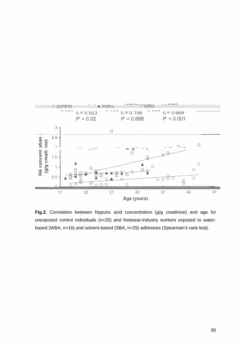
95
Fig.2. Correlation between hippuric acid concentration (g/g creatinine) and age for
unexposed control individuals (n=25) and footwear-industry workers exposed to water-
based (WBA, n=16) and solvent-based (SBA, n=29) adhesives (Spearman’s rank test).

96
4. DISCUSSION
Footwear-workers are routinely exposed to complex mixtures of solvents in
degreasers, cleaners, primers and adhesives used in the production process, the main
solvent used being toluene, with lesser amounts of n-hexane, acetone, methyethylketone
[3], and possibly dust particles (leather, polymers, and finishing materials) additives in
shoe materials and degradation products of materials (i.e. polyurethanes / isocyanates)
and additives [4].
Our data on urinary hippuric acid data (Fig. 1) indicate a relationship between
urinary hippuric acid concentration and exposure to toluene present in the mixtures of
organic solvents used, with a higher mean concentration of this toluene metabolite
appearing in the urine of the WBA and SBA groups than that of the control group. This
finding agrees with previous studies of footwear-workers that also used the hippuric acid
as a parameter to evaluate exposure to solvent mixtures [3, 23, 24], in which it was found
that the concentration of urinary hippuric acid was higher in workers exposed to toluene
and other solvents than in unexposed individuals. The metabolite of toluene, hippuric acid,
provided a good estimation of workplace exposure of workers with SBG (Fig. 1.), but it is
necessary to take into consideration other factors which increase hippuric acid, such as
age (Fig. 2.). Similar results for hippuric acid in Brazilian population were shown by
Siqueira and Paiva [42].
The Comet assay values for the SBA group were significantly higher than the values
for the control group, although there were no differences in damage index (DI) and
damage frequency (DF) values between the WBA and control groups (Table II). According
to Tice et al. [36], DF values (based on the proportion of cells with altered migration) are
less informative (especially for damaged cells) than measures related to the extent of
DNA damage, DI values (based on the length of migration and the amount of DNA in the
Comet tail) considered as being a more sensitive measure of DNA damage. There was a
higher frequency of micronuclei in buccal epithelial cells from the SBG group, although no
statistical difference was observed (Table II).
Toluene alone gives negative results in most genotoxicity tests with different
organisms [13, 43-46]. Other results were obtained in a multitude of studies with
biological monitoring of various genotoxic effects in peripheral blood lymphocytes from
workers exposed to toluene in the occupational environment [46-52], as well as glue
sniffers [53]. Some positive results have been obtained in shoe workers, but these
97
significant responses might also be due to benzene contamination [11, 54, 55]. Pitarque et
al. [3] obtained negative results by Comet test in shoe-workers exposed mainly to toluene,
but found increased values for MN in peripheral lymphocytes in the same group [11]. In
most cases confusion due to co-exposure to ink, other solvents and various genotoxic
substances in the environment cannot be excluded.
Some authors suggest that the presence of isocyanates in glues can explain
positive findings [11, 56]. Several studies describe the isocyanate emission potential of
polyurethane adhesives [57], that may spread in aerosolized or gaseous form when
heating or when vapors escape to the workplace air from open vessels at room
temperature [58, 59]. Respiratory disorders associated with isocyanate exposure [59-61],
and toxicity and/or genotoxicity, even in polymerized form, are described in a number of
assays [56, 58, 61-65] and occupational exposures [66]. The results pertaining to
isocyanate induction of MN formation were inconsistent [67, 68]. Isocyanate has been
classified as a carcinogen in animals [69, 70] and is a suspected carcinogen in humans
[69]. In our study, isocyanate was present only in water-based adhesive composition, to
which WBA workers were exposed; this group revealed no positive results in our
genotoxicity tests (Comet assay, BNLMN, and EBCMN). However, it is possible that the
use of chloroprene as a monomer in the production of the polychloroprene adhesive by
SBG workers can explain the increase in Comet assay values in SBG exposed workers.
Positive mutagenicity results of chloroprene were reported [71, 72], but at the exposure
concentrations used in the cancer inhalation studies, chloroprene did not induce SCE or
CA in mouse bone marrow cells, nor did it increase the MN frequency in peripheral blood
erythrocytes [73, 74]. Several studies also reported an increased risk of cancer among
shoe-workers exposed to chloroprene and organic solvents present in glues and
adhesives [75-79]. Chloroprene is classified as possibly carcinogenic to humans [78].
In conclusion, our study demonstrates the absence of genotoxic effects in footwear-
workers exposed to water-based adhesives and although the workers exposed to solvent-
based adhesives did not show a marked difference in relation to the control group, it is
clear that water-based adhesives are a better option for safeguarding the health of
footwear-workers. In the genotoxicity evaluations made in this study the Comet assay was
more sensitive than the micronucleus test, both performed on blood cells, which was also
observed in other studies [38, 80]. The micronucleus test in buccal epithelial cells showed
a non-significant increase in SBA group. Considering the less invasive sampling of such
types of cells, in a proper design of the test, it can present advantages to monitor human

98
populations exposed to inhaled/ingested genotoxins. The exposure to solvents estimated
by urinary hippuric acid were higher in the SBA workers in relation to WBA and controls,
but the exposure responsible for the DNA damage could not be identified with certainty,
like in many other studies about solvent mixtures exposure. The genotoxicity, although
low, detected in this study in footwear-workers, also reported by other studies [11], and
SBA glue sniffers [53] suggest the use of glue can present health risks, and the footwear
workers need more evaluations to assess the risk associated to these working conditions.
The use of WBA glue seems to be a less toxic option than the SBA glue, although more
costly.
ACKNOWLEDGMENTS
The authors express their gratitude to all the individuals who volunteered to
participate in this study. We also thank Verônica R.S. de Moraes, Silvia Brito, Eduardo
Rissi from CESSTIC (Centro de Saúde e Segurança do Trabalhador das Indústrias

99
[6] EPA (Environmental Protection Agency) - In Support of Summary Information on the Integrated Risk Information (IRIS). Toxicological Review of Methyl Ethyl Ketone (2003), On-line: http://epa.gov/iris
[7] WHO (World Health Organization) - Air quality Guidelines, second edition, Chapter 5.14. Toluene. Regional Office for Europe, Copenhagen, Denmark (2000). On-line: http://www.who.int
[8] IARC (International Agency for Research on Cancer), Boot and shoe manufacture and repair, IARC Monographs on the Evaluation of the Carcinogenic Risks of Chemicals to Humans, Summaries & Evaluations, Suppl. 7, p. 232. Lyon, France (1987). On-line: http://www.inchen.org/pages/iarc.html
[9] M. González Cid, D. Loria, M. Vilensky, J.L. Miotti, E. Matos, Leather tanning workers: chromosomal aberrations in peripheral lymphocytes and micronuclei in exfoliated cell in urine, Mutat. Res. 259 (1991) 197-201.
[10] I. Sbrana, S. Caretto, A. Battaglia, Chromosomal aberration analysis of workers in tannery industries, Mutat. Res. 242 (1991) 331-336.
[11] M. Pitarque, A. Vaglenov, M. Nosko, S. Pavlova, V. Petkova, A. Hirvonen, A. Creus, H. Norppa, R. Marcos, Sister chromatid exchanges and micronuclei in peripheral lymphocytes of shoe factory workers exposed to solvents, Environ. Health Perspect. 110 (2002) 339-404.
[12] M.G. Medeiros, A.S. Rodrigues, M.C. Batoréu, A. Laires, J. Rueff, A. Zhitkovich, Elevated levels of DNA – protein crosslinks and micronuclei peripheral lymphocytes of tannery workers exposed to trivalent chromium, Mutagenesis 18 (2003) 19-24.
[13] D. MacGregor, The genetic toxicology of toluene, Mutat. Res. 317 (1994) 213-228.
[14] EPA (Environmental Protection Agency) - In Support of Summary Information on the Integrated Risk Information (IRIS), Toxicological review of trivalent chromium (1998). On-line: http://epa.gov/iris
[15] P.S. Spencer, H.H. Schaumburg, Organic solvent neurotoxicity, facts and research needs, Scan. J. Work Environ. Health 11 (1985) 53-60.
[16] M.M. Greenberg, The central system and exposure to toluene: a risk characterization, Environ. Res. 72 (1997) 1-7.
[17] R. Toftgård, O.G. Nilsen, J-A.Gustafsson, Dose dependent induction of rat liver microsomal cytochromeP-450 and microsomal enzymatic activities after inhalationof toluene and dichloromethane, Acta Pharmac. Toxic. 51 (1982) 108-114.
[18] R-S. Wang, T. Nakajima, S.S. Park, H.V. Gelboin, N. Murayama, Monoclonal antibody-directed assessment of cytochrome P450 isozymes, Biochem. Pharmac. 46 (1993) 413-419.
[19] T. Nakajima, R-S. Wang, Induction of cytochrome P450 by toluene, Int. J. Biochem. 26 (1994) 1333-1340.
[20] D.K. Hammer, Metabolite ratio of toluene-exposed rotogravure printing plant workers reflects individual mutagenic risk by sister chromatid exchanges, Mutat. Res. 519 (2002) 171-177.
[21] Revista Química e Derivados 413 (2003). On-line: www.química.com.br/indice.html

100
[22] E. De Rosa, F. Brugnone, G.B. Bartolucci, L. Perbellini, M.L. Bellomo, G.P. Gori, M. Sigon, P.C. Corona, The validity of urinary metabolites as indicators of low exposures to toluene, Int. Arch. Environ. Health 56 (1985) 135-145.
[23] S. Burgaz, O. Erdem, G. Cakmak, N. Erdem, A. Karakaya, A.E. Karakaya, Cytogentic analysis of buccal cell from shoe-workers and pathology and anatomy laboratory workers exposed to n-hexane, toluene, metyl ethyl ketona and formaldehyde, Biomarkers 7 (2002) 151-161.
[24] I. Çok, E. Dagdelen, E. Gökçe, Determination of urinary hippuric acid and o-cresol levels as biological indicators of toluene exposure in shoe-workers and glue sniffers, Biomarkers 8 (2003) 119-127.
[25] D.W. Fairbairn, P.L. Olive, K.L. O’Neill, The Comet assay: a comprehensive review, Mutat. Res. 339 (1995) 37-59.
[26] P. Grover, K. Danadevi, M. Mahboob, R. Rozati, B.S. Banu, M.F. Rahman, Evaluation of genetic damage in workers employed in pesticide production utilizing the Comet Assay, Mutagenesis 18 (2003) 201-205.
[27] M. Fenech, N. Holland, W.P. Chang, E. Zeiger, S. Bonassi, The Human Micronucleus Project – An international colaborative study on the use of the micronucleus technique for measuring DNA damage in humans, Mutat. Res. 428 (1999) 271-283.
[28] M. Fenech, The in vitro micronucleus technique, Mutat. Res. 455 (2000) 81-95.
[29] B. Karahalil, A.E. Karakaya, S. Burgaz, The micronucleus assay in exfoliated buccal cells: application to occupational exposure to polycyclic aromatic hydrocarbons, Mutat. Res. 442 (1999) 29-35.
[30] G.J.F. Gattás, L.D.A. Cardos, M.D.A. Medrado-Faria, P.H. Saldanha, Frequency of oral mucosa micronuclei in gas station operators after introducing methanol, Occup. Med. 51 (2001) 107-113.
[31] A.J. Majer, B. Laky, S. Knasmüller, F. Kassie, Use of the micronucleus assay with exfoliated epithelial cells as a biomarker for monitoring individuals at elevated risk of genetic damage and in chemoprevention trials, Mutat. Res. 489 (2001) 147-172.
[32] S. Pastor, A. Creus, T. Parrón, A. Cebulska-Wasilewska, C. Siffel, S. Piperakis, A. Marcos, Biomonitoring of four European populations occupationally exposed to pesticides: use of micronuclei as biomarker, Mutagenesis 18 (2003) 249-258.
[33] A.V. Carrano, A.T. Natarajan, Considerations for population monitoring using cytogenetic techniques, International Commission for Protection against Environmental Mutagens and Carcinogens (ICPEMC publication 14), Mutat. Res. 204 (1988) 379-406.
[34] R. Albertini, D. Anderson, G.R. Douglas, L. Hagmar, K. Hemminki, F. Merlo, A.T. Natarajan, H. Norppa, D.E.G. Shuker, R.R. Tice, M.D. Waters, A. Aitio, IPCS guidelines for the monitoring of genotoxic effects of carcinogens in humans, International Program on Chemical Safety, Mutat. Res. 463 (2000) 111-172.
[35] N.P. Singh, M. T. McCoy, R.R. Tice, E.L. Schneider, A simple technique for quantification of low levels of DNA damage in individual cells, Exp. Cell Res. 175 (1988) 184-191.
[36] R.R. Tice, E. Agurell, D. Anderson, B. Burlinson, A. Hartmann, H. Kobayashi, Y. Miyamae, E. Rojas, J.C. Ryu, Y.F. Sasaki, Single Cell Gel/Comet Assay: Guidelines for In Vitro and In Vivo Genetic Toxicology Testing, Environ. Mol.
101
Mutagen. 35 (2000) 206-221.
[37] A.R. Collins, V.L. Dobson, M. Dusinská, G. Kennedy, R. Stetina, The Comet assay: what can it really tell us? Mutat. Res. 375 (1997) 183-193.
[38] J. Silva, T. R. O. Freitas, V. D. Heuser, J. R. Marinho, B. Erdtmann, Genotoxicity Biomonitoring in Coal Regions Using Wild Rodent Ctenomys torquatus by Comet Assay and Micronucleus Test, Environ. Mol. Mutagen. 35 (2000) 270-278.
[39] P.E. Tolbert, C.M. Shy, J.W. Allen, Micronuclei and other nuclear anomalies in buccal smears: method development, Mutat. Res. 271 (1992) 69-77.
[40] N. Titenko-Holland, R. Jacob, N. Shang, A. Balaraman, M.T. Smith, Micronuclei in lymphocytes and exfoliated buccal cells of postmenopausal women with dietary changes in folate, Mutat. Res. 417 (1998) 101-114.
[41] S. Gómez-Arroyo, Y. Díaz-Sánchez, M.A. Meneses-Pérez, R. Villalobos-Pietrini, J. León-Rodríguez, Cytogenetic biomonitoring in a Mexican floriculture worker group exposed to pesticide, Mutat. Res. 466 (2000) 117-124.
[42] M.E.P.B. Siqueira, M.J.N. Paiva, Hippuric acid in urine: reference values, Rev. Saúde Pública 36 (2002) 723-727.
[43] R. Rodrigue-Arnaiz, R. Villalobos-Pietrini, Genetic effects of thinner, benzene and toluene in Drosophila melanogaster. 1. Sex chromosome loss and non-dis-junction, Contam. Amb. 1 (1985) 35-43.
[44] S. Nakamura, Y. Oda, T. Shimata, I. Oki, K. Sugimoto, SOS-induced activity of chemical carcinogens and mutagens in Salmonella typhimurium TA1535/pSK 1002: examination with 151 chemicals, Mutat. Res. 192 (1997) 239-246.
[45] F. Zarani, P. Papazafiri, A. Kappas, Induction of micronuclei in human lymphocytes by organic solvents in vitro, J. Environ. Pathol. Toxicol. Oncol. 18 (1999) 21-28.
[46] SCGOC (Swedish Criteria Group for occupational Standards), Scientific Basis for Swedish Occupational Standards, Toluene, Arbete och Hälsa (2002). Arbetarskyddsverket, Solna. On-line: http://www.niwl.se/
[47] M. Bauchinger, E. Schmid, J. Dresp, J. Kolin-Gerresheim, R. Hauf, E. Suhr, Chromosome changes in lymphocytes after occupational exposure to toluene, Mutat. Res. 102 (1982) 439-445.
[48] D. Pelclova, P. Rossner, J. Pickova, Chromosome aberrations in rotogravure printing plant workers, Mutat. Res. 245 (1990) 299-303.
[49] G. Nise, B. Högstedt, I. Bratt, S. Skerfving, Cytogenetic effects in rotogravure workers exposed to toluene (and benzene), Mutat. Res. 261 (1991) 217-223.
[50] W. Popp, C. Vahrenholz, S. Yaman, C. Müller, G. Müller, W. Schmeiding, K. Norpoth, R. Fahnert, Investigation of the frequency of DNA strand breakage and cross-linking and sister chromatid exchange frequency in the lymphocytes of female workers exposed to benzene and toluene, Carcinogenesis 13 (1992) 57-61.
[51] D.K. Hammer, N. Mayer, E.H. Pfeiffer. Sister chromatid exchanges in rotogravure printing plant workers, Int. Arch. Occup. Environ. Health 71 (1998) 138-142.
[52] D. Pelclova, M. Cerna, A. Patorkova, V. Vrbikova, B. Prochazka, D. Hurychova, Z. Dlaskova, M. Hornychova, Study of the genotoxicity of toluene, Arch. Environ. Health 55 (2000) 268-273.
[53] I. Çok, S. Sardas, E. Kadioglu, E. Ozcagli, Assessment of DNA damage in glue sniffers by use of the alkaline comet assay, Mutat. Res. 557 (2004) 131-136.
102
[54] B.T. Tunka, Ü. Egeli, Cytogenetic Findings on Shoe Workers Exposed Long-term to Benzene, Environ. Health Perpect. 104 (1996) 1313-1317.
[55] A. Bogadi-Sare, V. Brumen, R. Turk, V. Karacicacute, M. Zavalicacute, Genotoxic effects in workers exposed to benzene: with special reference to exposure biomarkers and confounding factors, Ind. Health 35 (1997) 367-373.
[56] M. Andersen, M.L. Binderup, P. Kiel, H. Larsen, J. Maxlid, Mutagenic action of isocyanate used in the production of polyurethane, Scand. J. Work Environ. Health. 6 (1980) 221-226.
[57] M. Wirts, T. Salthammer, Isocyanate emission from PUR adhesives: influence of temperature, monomer content, and curing mechanism, Environ. Sci. Technol. 36 (2002) 1827-1832.
[58] B.Z. Zhong, P.D. Siegel, Induction of micronuclei following exposure to methylene di-phenyl diisocyanate: potential genotoxic metabolites, Toxicol. Sci. 58 (2000) 102-108.
[59] D.M. Paal, J.O. Levin, A. Östin, C. Rosenberg, M.L. Henriks-Eckerman, S. Brodsgaard, S.H. Throud, G. Fladseth, Y. Thomassen, Harmonized Nordic strategies for isocyanate monitoring on workroom atmospheres, J. Environ. Monit. 4 (2002) 685-687.
[60] G. Skarping, M. Dalene, B.G. Svensson, M. Littorin, B. Akesson, H. Welinder, S. Skerfving, Biomarkers of exposure, antibodies, and respiratory symptoms in workers heating polyurethane glue, Occup. Environ. Med. 53 (1996) 180-187.
[61] M.A. Collins, Toxicology of toluene diisoctanate, Appl. Occup. Environ. Hyg. 17 (2002) 846-855.
[62] H. Mori, N. Yoshimi, S. Sugie, H. Iwata, K. Kawai, N. Mashizu, H. Shimizu, Genotoxicity of epoxy resin hardeners in the hepatocyte primary culture/DNA repair test, Mutat. Res. 204 (1988) 683-688.
[63] A.D. Kligerman, J.A. Campbell, G. L. Erexson, J. W. Allen, M. D. Shelby, Sister chromatid exchange analysis in lung and peripheral blood lymphocytes of mice exposed to methyl isocyanate by inhalation, Environ. Mutagen. 9 (1987) 29-36.
[64] J. Mäki-paakkanen, H. Norppa, Chromosome aberrations and sister-chromatid exchanges induced by technical grade toluene isocyanate and methylenediphenyl diisocyanate in cultured human lymphocytes, Toxicol. Lett. 36 (1987) 37-43.
[65] M. Bilban, Mutagenic testing of workers exposed to toluene-diisocyanate during plastic production process, Am. J. Ind. Med. 45 (2004) 468-474.
[66] B. Marczynski, A.B. Czuppom, H.P. Hoffarth, W. Marek, X. Baur, DNA damage in human white blood cells after inhalation exposure to 4,4'-methylenediphenyl diisocyanate (MDI) - case report, Toxicol. Lett. 60 (1992) 131-138.
[67] EPA (Environmental Protection Agency) – Technology Transfer Network, Air Toxic Website: 2,4-Toluene Diisocyanate (2004). On-line: http://epa.gov/ttn/atw/nlthef/toluene2.html
[68] M.D. Shelby, J.W. Allen, W.J. Caspary, S. Haworth, J. Ivett, A. Kligerman, C.A. Luke, J.M. Mason, B. Myhr, R.R. Tice, R. Valencia, E. Zeiger, Results of in vitro and in vivo genetic toxicity tests on methyl isocyanate, Environ. Health Perspect. 72 (1987) 183-187.

103
[69] IARC (International Agency for Research on Cancer), Toluene Diisocyanate, IARC Monographs on the Evaluation of the Carcinogenic Risks of Chemicals to Humans, Vol. 39, pp. 865. Lyon, France (1999). On line: http://www.inchen.org/pages/iarc.html
[70] NTP (National Toxicology Program) - Technical report on the toxicology and carcinogensis studies of commercial grade 2,4(80%) and 2,6(20%)-toluene diisocyanate (CAS No. 26471–62–5) in F344/N rats and B6C3F1 mice (gavage studies). NIH Publication No. 86–2507 (1986). On-line: http://ntp.niehs.nih.gov
[71] H. Bartsch, C. Malaveille, A. Barbin, G. Planche, Mutagenic and alkylating metabolites of haloethylenes, chlorobutadienes and dichlorobutenes produced by rodent or human liver tissues: evidence for oxirane formation by P450-linked microssomal mono-oxigenases, Arch. Toxicol. 41 (1979) 249-277.
[72] G.A. Westphal, M. Blaszkewics, M. Leutbecher, A. Müller, E. Hallier, H.M. Bolt, Bacterial Mutagenicity of 2-Chloro-1,3-butadiene (Chloroprene) Caused by Decomposition Products, Arch. Toxicol. 68 (1994) 79-84.
[73] R. Tice, R. Boucher, C. Luke, D. Paquette, R. Melnick, M. Shelby, Chloroprene and Isoprene: Cytogenetic Studies in Mice, Mutagenesis 3 (1988) 141-146.
[74] R. Valentine, M.W. Himmelstein, Overview of the acute, subchronic, reproductive, developmental and genetic toxicology of beta-chloroprene, Chem. Biol. Interact. 35 (2001) 81-100.
[75] S. Li, Q. Dong, Y. Liu, Y. Liu, Epidemiologic study of cancer mortality among chloroprene workers, Biomed. Environ. Sci. 2 (1989) 141-149.
[76] M.A. Bulbulyan, O.V. Changuina, D.G. Zaridze, S.V. Astashevsky, D. Colin, P. Boffetta, Cancer mortality among Moskow shoe workers exposed to chloroprene (Russia), Cancer Causes & Control 9 (1998) 381-387.
[77] NTP (National Toxicology Program) - Technical Report Series No 467, Toxicology and Carcinogenesis Studies of Chloroprene (CAS No 126-99-8) in F344/N Rats and B6CF1 Mice (Inhalation Studies). NIH Publication No 98-3957 (1998). On-line: http://ntp.niehs.nih.gov
[78] IARC (International Agency for Research on Cancer), Chloroprene, IARC Monographs on the Evaluation of Carcinogenic Risks to Humans, Vol. 71, p. 227. Lyon, France (1999). On-line: http://www.inchen.org/pages/iarc.html
[79] D. Zaridze, M. Bulbulyan, O. Changuina, A. Margaryan, P. Boffetta, Cohort studies of chloroprene-exposed workers in Russia, Chem. Biol. Interact. 135 (2001) 487-503.
[80] S.W. Maluf, B. Erdtmann, Follow-up study of the genetic damage in lymphocytes of pharmacists and nurses handling antineopastic drugs evaluated by citokinesis-block micronuclei analysis and single cell gel electrophoresis assay, Mutat. Res. 471 (2000) 21-27.

104
6. CAPÍTULO IV
Evaluation of genetic damage in Brazilian Footwear-workers: correlation of cytogenetic analysis and polymorphisms in metabolizing genes GSTM1, GSTT1,
GSTP1, CYP1A1, and CYP2E1.
Vanina Dahlström Heuser1, Juliana da Silva2*, Kátia Kvitko1, Paula Rohr1 & Bernardo
Erdtmann1,3
1Programa de Pós-Graduação em Genética e Biologia Molecular (PPGGBM),
Universidade Federal do Rio Grande do Sul (UFRGS), Porto Alegre-RS, Brazil.
2Laboratório de Genética Toxicológica, Programa de Pós-Graduação em Ensino de
Ciências e Matemática (PPGECIM), Universidade Luterana do Brasil (ULBRA), Canoas-
RS, Brazil.
3Instituto de Biotecnologia, Universidade de Caxias do Sul (UCS), Caxias do Sul-RS,
Brazil.
*Correspondence to: Juliana da Silva, Programa de Pós-Graduação em Ensino de
Ciências e Matemática (PPGECIM), Universidade Luterana do Brasil (ULBRA), Prédio 14,
Sala 230, Rua Miguel Tostes 101, Bairro São Luís, CEP 92420-280, Canoas-RS, Brazil.
E-mail: [email protected]
Grant sponsors: CNPq, FAPERGS, GENOTOX
A SER ENVIADO PARA A REVISTA TOXICOLOGY

105
Abstract
People employed in the footwear manufacturing industry are routinely exposed to a
complex mixture of solvents, mainly toluene, used in several processes. The health
effects of this exposure are not well known, but the elevated risk was considered to be a
consequence of the exposure to this complex mixture. The present study involved 39
Footwear-workers (31 males and 8 females), occupationally exposed to solvent-based
polychloroprene glue and solutions containing organic solvents. The control group
consisted of 55 subjects (44 males and 11 females) with no occupational exposure. As
biomarker of exposure, we obtained data on hippuric acid (HA) in urine (g/g creatinine).
The Comet assay in blood cells and micronucleus (MN) frequency in binucleated
lymphocytes (BNL) and epithelial buccal cells (EBC) were analyzed as cytogenetic
markers. We also determined polymorphisms in genes GSTT1, GSTM1, GSTP1,
CYP1A1, and CYP2E1, to make it possible to identify differences in sensitivity to
genotoxicity. Our results on HA show statistical increase in Footwear-workers in relation to
controls (P < 0.001) as well as our data obtained by Comet assay (P < 0.001). No
differences were observed in BNLMN, NPB and EBCMN frequencies between the groups.
However, in the exposed and non-exposed subjects, we found a positive correlation
between age and BNLMN (r = 0.674, P < 0.001; and r = 0.290, P < 0.05, respectively). No
differences were observed between males and females inside the groups. The results on
genetic polymorphisms in controls indicated an increase in frequencies in NPB in CYP1A1
variant in relation to CYP1A1 wild genotype (P < 0.02), and also an increase in EBCMN
frequencies in GSTM null compared to GSTM non null (P < 0.02). In Footwear-workers,
GSTP1 variant seems to increase the DNA damage in relation to wild genotype (P <
0.02). The CYP2E1 variant also seems to be important in the increase of DNA damage
observed, even in presence of GSTP1 wild type genotype (P = 0.07).
Keywords: Occupational exposure, Footwear-workers, Comet assay; Micronucleus test
(MN), Organic solvents, Hippuric acid (HA), Metabolizing genes.
106
1. Introduction
People employed in the footwear manufacturing industry suffer an increased risk of
leukemia and nasal cancer, and an excess of mortality due to other types of cancer has
also been reported (IARC, 1987). Workers in footwear manufacture are routinely exposed
to complex mixtures of solvents, used in cleaning and as diluents in glues, primers, and
degreasers, i.e. acetone, n-hexane, methylethylketone, and large amounts of toluene
(Pitarque et al., 1999; Uuksulainen et al., 2002). Neither of these solvents is isolatedly
considered genotoxic or a carcinogenic (ASTDR, 1995a; 1995b; 1999; 2001; EPA, 2003).
However, the health effects of organic mixtures are not well known, but the elevated risk
was considered to be a consequence of the exposure to this complex mixture
(Uuksulainen et al., 2002).
The products used in footwear manufacturing contain mainly toluene, which may
increase host susceptibility to carcinogens (Nakajima and Wang, 1994). Toluene is the
main solvent used in footwear manufacturing. Urinary hippuric acid (HA) has long been
considered a biomarker of exposure (WHO, 2000), being a good marker to estimate
exposure risk, also at lower concentrations and in organic mixtures (Pitarque et al., 1999;
Burgaz et al., 2002; Çok et al., 2003).
A wide range of methods are currently used for the detection of early biological
effects of DNA-damaging agents in occupational settings. During the last few years, less
time-consuming methods are being required because it is of public interest that hazardous
chemicals are removed from the environment as soon as possible. Consequently, there is
a need for rapid and reliable tests that detect DNA damage of agents in different exposure
circumstances. Thus the Micronucleus test (MN) and the Comet assay seem to satisfy
many of these criteria, that have been used in human biomonitoring studies (Fairbairn et
al., 1995; Moller et al., 2000; Grover et al., 2003).
However, biomarker studies are still not generating the type of reliable information
needed for precise risk assessment. Some of the problems are due to inconsistent
observation of biological effects from similarly exposed populations, lack of predictable
dose–response relationship and existence of interindividual variations in response to
exposure (Au et al., 1998). The metabolism of volatile hydrocarbons, including organic
solvents, is receiving more and more attention because it is essential as a biomarker of
exposure in biological monitoring and because metabolic activation often occurs during
the metabolic process (Nakajima, 1997). Individual variations in polymorphic genes
involved in xenobiotic metabolism and DNA repair have been linked with an increased risk
107
of cancer in several case-control studies (IARC, 1999). These individual differences may
be important in the estimation of the risk to humans from exposure to environmental
toxicants. Understanding the significance of genetic polymorphisms in determining
genotoxic response will also have an important influence on requirements concerning the
use of human cells in genotoxicity testing (Norppa, 1997).
In a preliminary study of the genotoxic exposure in Footwear-workers effects of
occupational exposure to organic solvents, using the Comet assay and MN test, we
detected an increase in DNA damage in leukocytes measured by Comet assay but no
statistical significant increase in MN formation in lymphocytes and buccal cells from a
group of males employed in two footwear factories exposed to solvent-based cleaners,
primers and adhesives. In this study, besides the cytogenetic endpoints in a larger sample
of footwear workers, including males and females, we determined polymorphisms in
genes which metabolize xenobiotics: GSTT1, GSTM1, GSTP1, CYP1A1, and CYP2E1, to
make it possible to identify differences in sensitivity to genotoxicity. We also obtained data
on the HA concentration in urine, as biomarker of exposure, and the potential difference
by gender was also evaluated.
2. Materials and methods
2.1. Study population and sample collection
This study was approved by the Brazilian National Ethical Committee on Research
(Comissão Nacional de Ética em Pesquisa – CONEP) and informed written consent was
obtained from each individual prior to the start of the study.
The study involved 39 footwear industry workers (31 males and 8 females),
employed at three factories where they were occupationally exposed to adhesives
(polychloroprene) and solutions containing organic solvents. The control group consisted
of 55 subjects (44 males and 11 females) with no occupational exposure to organic
solvents or any other genotoxic substance.
All the individuals examined in the study were required to answer a Portuguese
version of a questionnaire from the International Commission for Protection against
Environmental Mutagens and Carcinogens (Carrano and Natarajan, 1988) and participate
in a face-to-face questionnaire which included standard demographic data (age, gender,
108
etc.) as well as questions relating to medical issues (exposure to X-rays, vaccinations,
medication, etc.), life style (smoking, coffee, alcohol, diet, etc.) and their occupation
(number of hours worked per day, time exposed to organic solvents, use of protective
measures, etc.). In all the groups, individuals who smoked more than 5 cigarettes/day for
at least 1 year were considered smokers. The characteristics of the three groups are
presented in Table I.
Blood and urine samples were obtained from individuals in the exposed groups on
the same day during a normal shift during the workers periodical medical examinations by
the nurses from a regional Health and Safety at Work Medical Center (Centro de Saúde e
Segurança do Trabalhador das Indústrias Calçadistas da Região de Parobé, Parobé-RS,
Brasil (CESSTIC) — The Footwear Industry Health and safety at work Center for the
Parobé Region, Parobé-RS, Brazil). For the control group, blood and urine samples were
taken at the same region. All blood samples were collected using venipuncture and
heparinized vacutainers and processed as quickly as possible to avoid the damage
associated with storage (Albertini et al., 2000), the blood cell samples being transported to
the laboratories at or below 8oC and processed within 8 h of collection in order to minimize
loss of DNA and damage due to DNA repair processes.
2.2. Hippuric acid (HA) concentrations
Analysis of urinary HA acid was carried out using gas chromatography using a
Perkin-Elmer XL Auto System gas chromatograph (Perkin-Elmer, USA) in a commercial
laboratory (Toxilab, Toxicological Laboratory, Porto Alegre-RS, Brazil).
2.3. Micronucleus test
2.3.1. Cytokinesis-blocked human lymphocyte MN
For each blood sample, duplicate lymphocytes cultures were set up in culture flasks
by adding 0.3 ml of whole blood to 5 ml of RPMI 1640 medium (Nutricell, Campinas-SP,
Brazil) containing 20% fetal calf serum and 1% (v/v) phytohemagglutinin. The flasks
incubated at 37ºC for 44h before adding 5 µg/ml of cytochalasin B (Sigma), and
continuing incubation until the total incubation time reached was 72 h as described by
Fenech et al. (1999). After incubation the lymphocytes were harvested by centrifugation at
109
800 revs/min for 8 min, recentrifuged, fixed in 3:1 (v/v) methanol/acetic acid, placed onto a
clean microscope slide and stained with 5% (v/v) Giemsa. For each blood sample, 2000
binucleated lymphocytes (i.e. 1000 from each of the two slides prepared from the
duplicate cultures) were scored for both MN presence and nucleoplasmic bridges (NPB)
between daughter nuclei, assessment being made using bright-field optical microscopy at
a magnification of 200-1000X. All sides were coded to blind analysis.
2.3.2. Buccal cells
Buccal cells were collected by swabbing the inner cheek of the individuals with a
moistened wooden tongue depressor, the tip of which was immersed in 5 ml of cold saline
(0.9% (w/v) aqueous NaCl) in a conical tube and transported under refrigeration to the
laboratory where the saline was centrifuged at 1500 revs/min for 8 min and the
sedimented buccal cells washed twice more with saline under the same centrifugation
conditions to remove bacteria and cell debris which would have complicated scoring. After
washing, a drop of cell suspension was placed onto each of two duplicate microscope
slides and dried at room temperature for 1-2 weeks and then feulgen-stained (hydrolysis
in 1N HCl at 60oC for 10 minutes followed by immersion in Schiff`s reagent for 3-4 h) and
the cytoplasm counterstained by immersion in 0.5% (w/v) fast-green for 30 seconds.
The criteria used for MN analysis were those of Tolbert et al. (1992) and Titenko-
Holland et al. (1998). The presence of leukocytes and the quality of the slides were
determined at 200X magnification using a bright-field Zeiss microscope and the MN
frequency estimated based on the number of normal exfoliated buccal cells counted using
bright-field optical microscopy at a magnification of 200-1000X. For each volunteer, 2000
buccal cells (i.e. 1000 from each of the duplicate slides) were scored.
2.4. Comet assay
The alkaline Comet assay was performed as described by Singh et al. (1988) with
the modifications suggested by Tice et al. (2000). Blood cells (5 µl) were embedded in 95
µl of 0.75% low-melting point agarose and when the agarose had solidified the slides were
placed in lysis buffer (2.5M NaCl, 100mM EDTA and 10mM Tris; pH 10.0-10.5) containing
freshly added 1% (v/v) Triton X-100 and 10% (v/v) dimethyl sulfoxide (DMSO) for a
minimum of 1 hour and a maximum of two weeks. After treatment with lysis buffer, the

110
slides were incubated in freshly-made alkaline buffer (300 mM NaOH and 1 mM EDTA;
pH >13) for 20 min and the DNA was electrophoresed for 20 min at 25 volts (0.90 V/cm)
and 300 mA after which the buffer was neutralized with 0.4 M Tris (pH 7.5) and the DNA
stained with ethidium bromide (2 µg/ml). The electrophoresis procedure and efficiency for
each electrophoresis run was checked using negative and positive internal controls
consisting of whole human blood collected in the laboratory, the negative control being
unmodified blood and the positive control 50 µl of blood mixed with 13 µl (8 x 10⁻⁵ M) of
methyl methanesulfonate (CAS No 66-27-3; Sigma, St. Louis, MO, USA) and incubated
for 2 hours at 37oC. Each electrophoresis run was considered valid only if the negative
and positive controls yielded the expected results.
Images of 200 randomly selected cells (100 cells from each of two replicate slides)
were analyzed for each person. Comet image lengths (IL) (i.e. the nuclear region+tail)
were measured in arbitrary units using a calibrated eyepiece micrometer (1 unit= ≈5 µm at
200X) and a fluorescence microscope equipped with a 12nm BP546 excitation filter and a
590nm barrier filter. Cells were also visually allocated to one of five classes depending on
tail size (class 0 = no tails, class 4 = longest tails) to give a single DNA damage score for
each subject and hence for each group studied, the group damage index (DI) ranging
from 0 (no tails on any cells, i.e. 200 cells x 0) to 800 (all cells with maximally long tails,
i.e. 200 cells x 4) (Collins et al., 1997; Albertini et al., 2000; Silva et al., 2000). The
damage frequency (DF (%); i.e. the proportion of cells with altered migration), was
calculated based on the number of cells with tails versus the number of cells without tails.
All the slides were scored blindly on the same day on which they were subjected to
electrophoresis.
2.5. DNA extraction and genotyping
Genomic DNA was isolated from whole blood (collected using vacuntainers with
EDTA) by the salting out method (Miller et al., 1995). Five polymorphic markers
investigated by genotyping using the polymerase chain reaction-restriction fragment
length polymorphism (PCR-RFLP) method.
2.5.1. GST genes: GSTM1, GSTT1, and GSTP1

111
The GSTM1, GSTT1 and GSTP1 genes were typed by a multiplex Polymerase Chain
Reaction (PCR) method using a reaction mixture consisting of 100 ng of genomic DNA, 15
pmol of each primer, 10 mM Tris HCl, 4,5 mM MgCl2, 50 mM KCl, 100 mM dNTPs and 1.0
U of Taq DNA polymerase in a total volume of 50μl. The amplification protocol consisted
of initial denaturation at 94°C for 5 min, 6 touchdown cycles of 1 min at 94°C followed by 2
min at 59°C (decreasing to 54°C at a rate of 1°C per cycle) and 1 min at 72°C, and 30
cycles at 94°C for 1 min followed by 1 min at 55°C and 1 min at 72°C, plus a final
extension of 5 min at 72°C. An aliquot of the amplification product was subjected to
horizontal agarose gel (3.5%) electrophoreses to verify the presence or absence of GSTM
and GSTT fragments, the GSTP1 product being used as a control for this reaction. Primer
sequences were those reported by Harries et al. (1991), Bell et al. (1993) and Pemble et
al. (1994). A second aliquot of the amplified GSTP1 product was digested with BsmAI as
described by Harries et al. (1991).
2.5.2. CYP450 genes: CYP1A1 and CYP2E1
The CYP1A1*2C polymorphism was genotyped using the primers and the PCR
conditions indicated by Cascorby et al. (1996). The CYP1A1*1A (wild) and CYP1A1*2C
(variant) alleles were detected after digestion with BsrDI enzyme. The CYP2E1
polymorfism was analysed using the primers and PCR conditions described by Kato et al.
(1992). An aliquot of the amplified product was subjected to the PstI and RsaI enzymes to
establish the CYP2E1*1A/5B haplotypes.
2.6. Statistical analysis
The normality of variables was evaluated by the Kolmogorov-Smirnov test. χ2 and t-
test were used to compare the basic characteristic of study populations. The statistical
analysis of differences in age between control and Footwear-workers, been a normal
distribution, was tested by Student t-test. Differences in smoking and drinking habits, DNA
damage and cytogenetic test evaluated, as well between the different genotypes into the
control and exposed groups, having a significant deviation from normality, were tested by
the non-parametric Mann-Whitney U test. The correlations of different variables were
determined by Sperman rank correlation test. Two-tailed P values are given for
significance of differences.
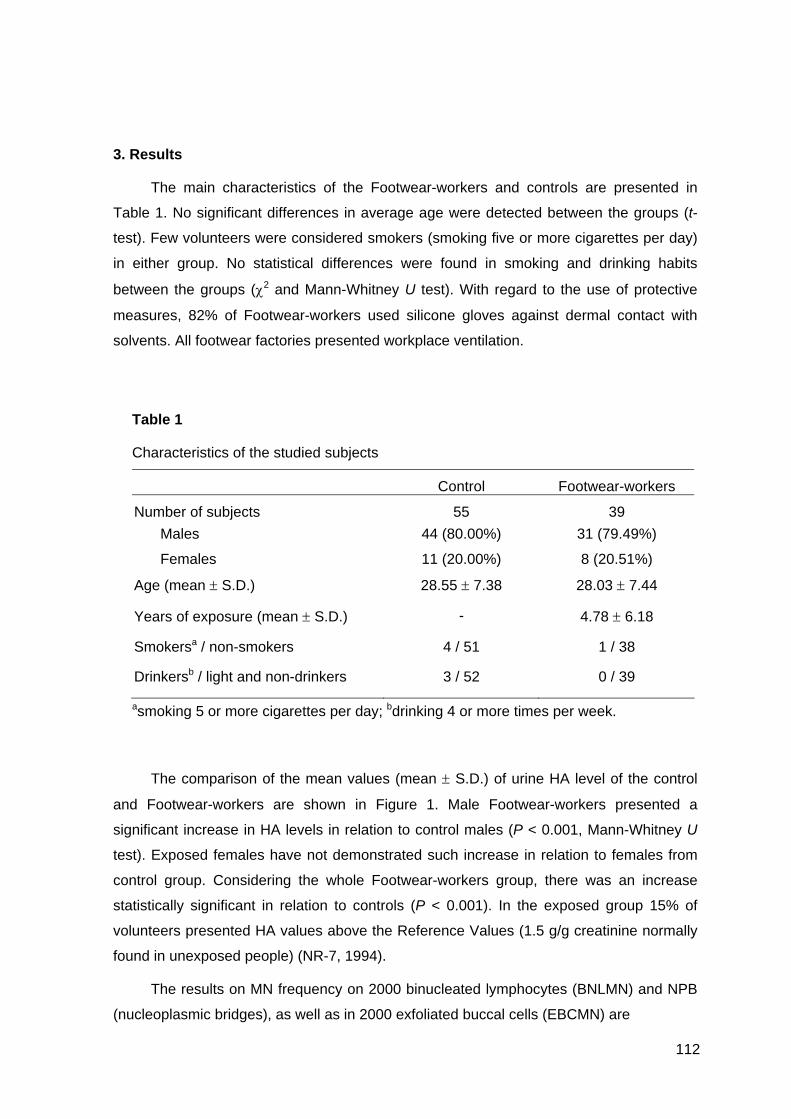
112
3. Results
The main characteristics of the Footwear-workers and controls are presented in
Table 1. No significant differences in average age were detected between the groups (t-
test). Few volunteers were considered smokers (smoking five or more cigarettes per day)
in either group. No statistical differences were found in smoking and drinking habits
between the groups (χ2 and Mann-Whitney U test). With regard to the use of protective
measures, 82% of Footwear-workers used silicone gloves against dermal contact with
solvents. All footwear factories presented workplace ventilation.
Table 1
Characteristics of the studied subjects
Control Footwear-workers
Number of subjects 55 39 Males 44 (80.00%) 31 (79.49%)
Females 11 (20.00%) 8 (20.51%)
Age (mean ± S.D.) 28.55 ± 7.38 28.03 ± 7.44
Years of exposure (mean ± S.D.) - 4.78 ± 6.18
Smokersa / non-smokers 4 / 51 1 / 38
Drinkersb / light and non-drinkers 3 / 52 0 / 39
asmoking 5 or more cigarettes per day; bdrinking 4 or more times per week.
The comparison of the mean values (mean ± S.D.) of urine HA level of the control
and Footwear-workers are shown in Figure 1. Male Footwear-workers presented a
significant increase in HA levels in relation to control males (P < 0.001, Mann-Whitney U
test). Exposed females have not demonstrated such increase in relation to females from
control group. Considering the whole Footwear-workers group, there was an increase
statistically significant in relation to controls (P < 0.001). In the exposed group 15% of
volunteers presented HA values above the Reference Values (1.5 g/g creatinine normally
found in unexposed people) (NR-7, 1994).
The results on MN frequency on 2000 binucleated lymphocytes (BNLMN) and NPB
(nucleoplasmic bridges), as well as in 2000 exfoliated buccal cells (EBCMN) are

113
presented in Table 2. The comparison of MN frequencies between control group and
exposed workers did not show a statistically significant difference (Mann-Whitney U test).

114
Table 2
Mean values (mean ± S.D.) obtained by the cytogenetic parameters analyzed
Citokinesis-bloked
(2000/subject)
Esfoliated Buccal Cells
(2000/subject)
BNLMN NPB EBCMN
Control (n = 55) 4.90 ± 2.24 3.00 ± 2.38 0.72 ± 0.84
Female (n = 11) 5.46 ± 2.66 3.73 ± 3.17 0.82 ± 0.87
Male (n = 44) 4.74 ± 2.12 2.80 ± 2.12 0.69 ± 0.84
Footwear-workers (n = 39) 4.53 ± 3.49 3.29 ± 2.30 1.05 ± 1.31
Female (n = 8) 4.14 ±3.13 2.13 ± 0.84 0.86 ± 0.90
Male (n = 31) 4.61 ± 3.61 3.65 ± 2.37 1.10 ± 1.40
BNLMN (Binucleated Lymphocytes with Micronucleus); NPB (Nucleoplasmatic Bridges); EBCMN (Exfoliated Buccal Cell with Microncleus).
Table 3
Mean values (mean ± S.D.) obtained by the comet assay analysis (200 leukocytes/subject)
Image Length (μm) Damage Index (0-
800)
Damage Frequency
(%)
Control (n = 55) 20.46 ± 0.53 2.82 ± 2.87 1.34 ± 1.66
Female (n = 11) 20.49 ± 0.36 2.00 ± 1.61 1.05 ± 1.11
Male (n = 44) 20.45 ± 0.58 3.02 ± 3.09 1.41 ± 1.77
Footwear-workers (n = 39) 20.58 ± 0.86 8.46 ± 7.79* 2.86 ± 1.98*
Female (n = 8) 20.87 ± 0.45 10.75 ± 7.80a 3.81 ± 1.69a
Male (n = 31) 20.50 ± 0.92 7.87 ± 7.81b 2.61 ± 1.20c
*Data significant in relation to control group at P < 0.001; aData significant in relation to females from control group at P < 0.01; bData significant in relation to males from control group at P < 0.001 and c P < 0.01 (Mann-Whitney U test).
The GSTT1, GSTM1, GSTP1, CYP1A1, and CYP1E2 genotype frequencies of the
controls and the Footwear-workers sample are shown in Table 4. The observed
frequencies are similar to those observed in various healthy Caucasian populations (data
non-shown).
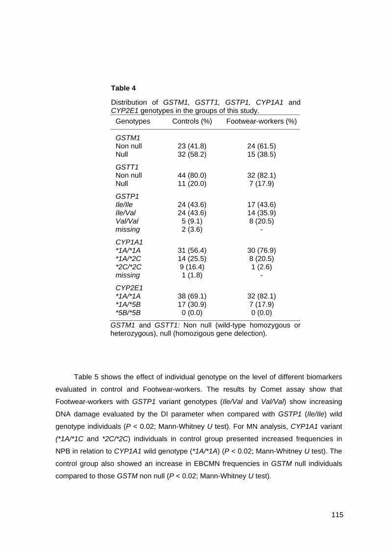
115
Table 4
Distribution of GSTM1, GSTT1, GSTP1, CYP1A1 and CYP2E1 genotypes in the groups of this study.
GSTM1 and GSTT1: Non null (wild-type homozygous or heterozygous), null (homozigous gene delection).
Table 5 shows the effect of individual genotype on the level of different biomarkers
evaluated in control and Footwear-workers. The results by Comet assay show that
Footwear-workers with GSTP1 variant genotypes (Ile/Val and Val/Val) show increasing
DNA damage evaluated by the DI parameter when compared with GSTP1 (Ile/Ile) wild
genotype individuals (P < 0.02; Mann-Whitney U test). For MN analysis, CYP1A1 variant
(*1A/*1C and *2C/*2C) individuals in control group presented increased frequencies in
NPB in relation to CYP1A1 wild genotype (*1A/*1A) (P < 0.02; Mann-Whitney U test). The
control group also showed an increase in EBCMN frequencies in GSTM null individuals
compared to those GSTM non null (P < 0.02; Mann-Whitney U test).
Genotypes Controls (%) Footwear-workers (%)
GSTM1 Non null 23 (41.8) 24 (61.5) Null 32 (58.2) 15 (38.5)
GSTT1 Non null 44 (80.0) 32 (82.1) Null 11 (20.0) 7 (17.9)
GSTP1 Ile/Ile 24 (43.6) 17 (43.6) Ile/Val 24 (43.6) 14 (35.9) Val/Val 5 (9.1) 8 (20.5) missing 2 (3.6) -
CYP1A1 *1A/*1A 31 (56.4) 30 (76.9) *1A/*2C 14 (25.5) 8 (20.5) *2C/*2C 9 (16.4) 1 (2.6) missing 1 (1.8) -
CYP2E1 *1A/*1A 38 (69.1) 32 (82.1) *1A/*5B 17 (30.9) 7 (17.9) *5B/*5B 0 (0.0) 0 (0.0)
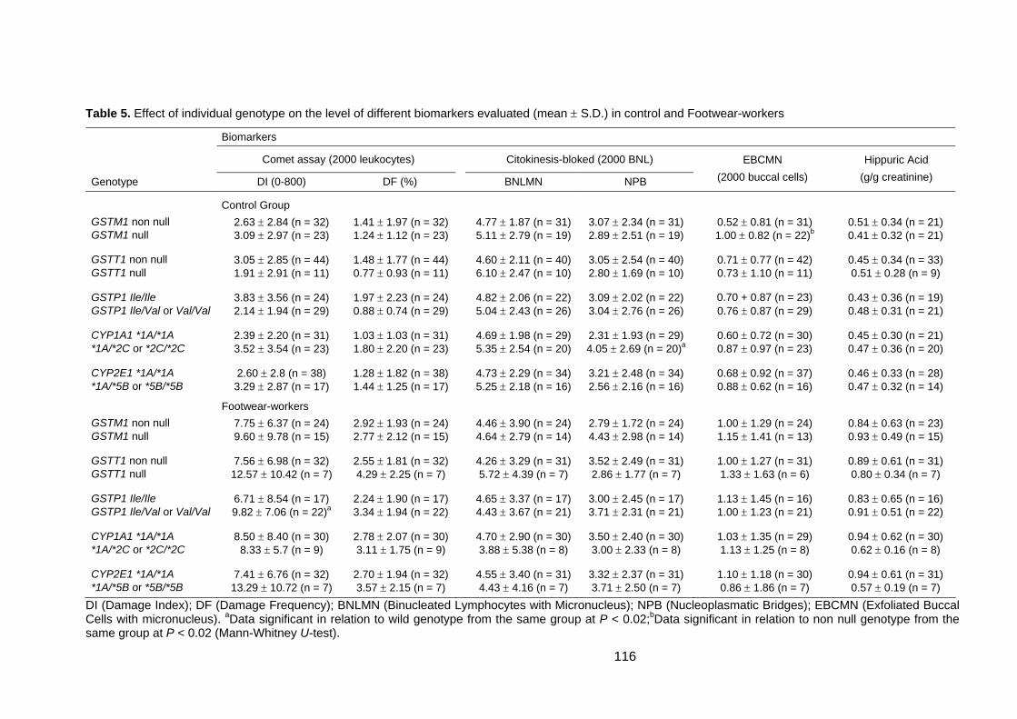
116
Table 5. Effect of individual genotype on the level of different biomarkers evaluated (mean ± S.D.) in control and Footwear-workers
Biomarkers
Comet assay (2000 leukocytes) Citokinesis-bloked (2000 BNL) Genotype DI (0-800) DF (%) BNLMN NPB
EBCMN (2000 buccal cells)
Hippuric Acid (g/g creatinine)
Control Group GSTM1 non null 2.63 ± 2.84 (n = 32) 1.41 ± 1.97 (n = 32) 4.77 ± 1.87 (n = 31) 3.07 ± 2.34 (n = 31) 0.52 ± 0.81 (n = 31) 0.51 ± 0.34 (n = 21) GSTM1 null 3.09 ± 2.97 (n = 23) 1.24 ± 1.12 (n = 23) 5.11 ± 2.79 (n = 19) 2.89 ± 2.51 (n = 19) 1.00 ± 0.82 (n = 22)b 0.41 ± 0.32 (n = 21) GSTT1 non null 3.05 ± 2.85 (n = 44) 1.48 ± 1.77 (n = 44) 4.60 ± 2.11 (n = 40) 3.05 ± 2.54 (n = 40) 0.71 ± 0.77 (n = 42) 0.45 ± 0.34 (n = 33) GSTT1 null 1.91 ± 2.91 (n = 11) 0.77 ± 0.93 (n = 11) 6.10 ± 2.47 (n = 10) 2.80 ± 1.69 (n = 10) 0.73 ± 1.10 (n = 11) 0.51 ± 0.28 (n = 9) GSTP1 Ile/Ile 3.83 ± 3.56 (n = 24) 1.97 ± 2.23 (n = 24) 4.82 ± 2.06 (n = 22) 3.09 ± 2.02 (n = 22) 0.70 + 0.87 (n = 23) 0.43 ± 0.36 (n = 19) GSTP1 Ile/Val or Val/Val 2.14 ± 1.94 (n = 29) 0.88 ± 0.74 (n = 29) 5.04 ± 2.43 (n = 26) 3.04 ± 2.76 (n = 26) 0.76 ± 0.87 (n = 29) 0.48 ± 0.31 (n = 21) CYP1A1 *1A/*1A 2.39 ± 2.20 (n = 31) 1.03 ± 1.03 (n = 31) 4.69 ± 1.98 (n = 29) 2.31 ± 1.93 (n = 29) 0.60 ± 0.72 (n = 30) 0.45 ± 0.30 (n = 21) *1A/*2C or *2C/*2C 3.52 ± 3.54 (n = 23) 1.80 ± 2.20 (n = 23) 5.35 ± 2.54 (n = 20) 4.05 ± 2.69 (n = 20)a 0.87 ± 0.97 (n = 23) 0.47 ± 0.36 (n = 20) CYP2E1 *1A/*1A 2.60 ± 2.8 (n = 38) 1.28 ± 1.82 (n = 38) 4.73 ± 2.29 (n = 34) 3.21 ± 2.48 (n = 34) 0.68 ± 0.92 (n = 37) 0.46 ± 0.33 (n = 28) *1A/*5B or *5B/*5B 3.29 ± 2.87 (n = 17) 1.44 ± 1.25 (n = 17) 5.25 ± 2.18 (n = 16) 2.56 ± 2.16 (n = 16) 0.88 ± 0.62 (n = 16) 0.47 ± 0.32 (n = 14)
Footwear-workers GSTM1 non null 7.75 ± 6.37 (n = 24) 2.92 ± 1.93 (n = 24) 4.46 ± 3.90 (n = 24) 2.79 ± 1.72 (n = 24) 1.00 ± 1.29 (n = 24) 0.84 ± 0.63 (n = 23) GSTM1 null 9.60 ± 9.78 (n = 15) 2.77 ± 2.12 (n = 15) 4.64 ± 2.79 (n = 14) 4.43 ± 2.98 (n = 14) 1.15 ± 1.41 (n = 13) 0.93 ± 0.49 (n = 15) GSTT1 non null 7.56 ± 6.98 (n = 32) 2.55 ± 1.81 (n = 32) 4.26 ± 3.29 (n = 31) 3.52 ± 2.49 (n = 31) 1.00 ± 1.27 (n = 31) 0.89 ± 0.61 (n = 31) GSTT1 null 12.57 ± 10.42 (n = 7) 4.29 ± 2.25 (n = 7) 5.72 ± 4.39 (n = 7) 2.86 ± 1.77 (n = 7) 1.33 ± 1.63 (n = 6) 0.80 ± 0.34 (n = 7) GSTP1 Ile/Ile 6.71 ± 8.54 (n = 17) 2.24 ± 1.90 (n = 17) 4.65 ± 3.37 (n = 17) 3.00 ± 2.45 (n = 17) 1.13 ± 1.45 (n = 16) 0.83 ± 0.65 (n = 16) GSTP1 Ile/Val or Val/Val 9.82 ± 7.06 (n = 22)a 3.34 ± 1.94 (n = 22) 4.43 ± 3.67 (n = 21) 3.71 ± 2.31 (n = 21) 1.00 ± 1.23 (n = 21) 0.91 ± 0.51 (n = 22) CYP1A1 *1A/*1A 8.50 ± 8.40 (n = 30) 2.78 ± 2.07 (n = 30) 4.70 ± 2.90 (n = 30) 3.50 ± 2.40 (n = 30) 1.03 ± 1.35 (n = 29) 0.94 ± 0.62 (n = 30) *1A/*2C or *2C/*2C 8.33 ± 5.7 (n = 9) 3.11 ± 1.75 (n = 9) 3.88 ± 5.38 (n = 8) 3.00 ± 2.33 (n = 8) 1.13 ± 1.25 (n = 8) 0.62 ± 0.16 (n = 8) CYP2E1 *1A/*1A 7.41 ± 6.76 (n = 32) 2.70 ± 1.94 (n = 32) 4.55 ± 3.40 (n = 31) 3.32 ± 2.37 (n = 31) 1.10 ± 1.18 (n = 30) 0.94 ± 0.61 (n = 31) *1A/*5B or *5B/*5B 13.29 ± 10.72 (n = 7) 3.57 ± 2.15 (n = 7) 4.43 ± 4.16 (n = 7) 3.71 ± 2.50 (n = 7) 0.86 ± 1.86 (n = 7) 0.57 ± 0.19 (n = 7)
DI (Damage Index); DF (Damage Frequency); BNLMN (Binucleated Lymphocytes with Micronucleus); NPB (Nucleoplasmatic Bridges); EBCMN (Exfoliated Buccal Cells with micronucleus). aData significant in relation to wild genotype from the same group at P < 0.02;bData significant in relation to non null genotype from the same group at P < 0.02 (Mann-Whitney U-test).
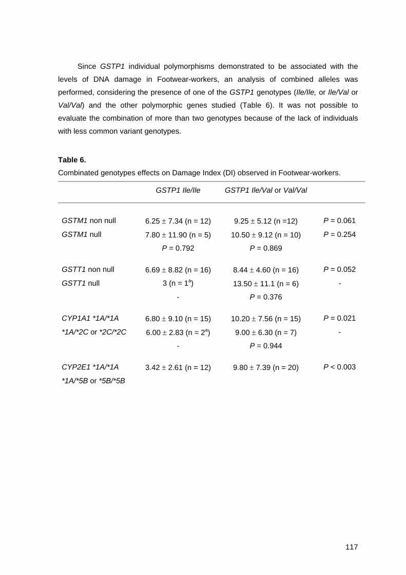
118
4. Discussion
Workers in footwear manufacture are routinely exposed to complex mixture of
solvents, and although neither of these solvents is considered a genotoxic or a
carcinogen, the health effects of organic mixtures are not well known (Uuksulainen et al.,
2002). Several studies used the HA, the main metabolite resulting from toluene, as a
marker for estimating individual exposure in shoe and footwear industry (Pitarque et al.,
1999; Burgaz et al., 2002; Çok et al., 2003).
Our urinary HA data (Fig. 1) indicate a higher mean concentration of this toluene
metabolite appearing in the urine of the Footwear-workers than that of the control group.
This finding agrees with previous studies of Footwear-workers that also used the HA as a
indicator of exposure to solvent mixtures and found the concentration of urinary HA higher
in workers exposed to toluene and other solvents than in unexposed individuals (Pitarque
et al., 1999, Burgaz et al., 2002; Çok et al., 2003; Heuser et al., in press). The HA
concentration observed in controls of this study was similar to the other Brazilian
population study (Siqueira and Paiva, 2002). It is suggested that polymorphisms in
CYP1A1 and CYP2E1 genes could modulate the HA concentration in urine (Hayashi et
al., 1991; Kawamoto et al., 1995). In the present study, this association was not
significant, but we observed that the variant genotypes in exposed group presented lower
HA values than the wild genotypes, similar to the control groups (Table 5).
In this study, regarding MN frequency in lymphocytes, no differences were observed
between control and Footwear-workers for BNLMN and NPB frequencies. However, in the
exposed and non-exposed subjects, we found a positive correlation between age and
BNLMN. Data from most biomonitoring studies describe the positive correlation between
MN and age in control and in exposed groups (Bolognesi et al., 1997; Fenech et al., 1999;
Albertini et al., 2000). To explain these findings, several possibilities have been
suggested: a general increase of susceptibility in older people (Crome, 2003); possible
age-related changes in the transcriptional activation xenobiotic genes (Ishikawa et al.,
2004); changes in the ionic blood concentrations (Fenech et al., 1997; 1998; Landi et al.,
2000); and the possibility of cumulated exposure and/or DNA damage (Ishikawa et al.,
2003).
Since the levels of enzymes that activate many airborne genotoxicants are more
expressed in human epithelial cells than in lymphocytes, Faust et al. (2004) suggest that it
seems necessary to use exfoliated cells in parallel with lymphocytes to study DNA
damaging effects of occupational exposure in populations, because the effects can
119
probably not be observed with the use of lymphocytes alone. In our study, lymphocyte MN
test did not show any difference between control and Footwear-workers, and MN levels in
EBC from Footwear-workers, although higher than controls, were also unable to
demonstrate any statistical difference (Table 2). On the other hand, our Comet assay
analyses showed that Footwear-workers presented significantly higher DI and DF in
relation to control (Table 3), as observed by Çok et al. (2004) in the study of DNA damage
measured by Comet assay in glue sniffers. Several authors have demonstrated the
difference between sensitivity of Comet assay and MN test (Maluf and Erdtman, 2000;
Silva et al., 2000; Heuser et al., 2002; Pitarque et al., 2002)
Some discrepancies between negative and positive findings or dramatically different
active concentration ranges will be explained by different genotypes of the donors.
Studies of cytogenetic biomarkers among exposed humans show that the determination of
polymorphisms is becoming an increasingly important aspect that may turn the assays
more sensitive and more specific in identifying the effect and the sensitive subgroups
(Norppa, 1997). Genotypes responsible for interindividual differences in ability to activate
or detoxify genotoxic agents are recognized as biomarkers of susceptibility (Autrup, 2000).
As a result of this mechanisms, inactive metabolites are sometimes formed, which are
readily eliminated from the organism, but chemically reactive agents can also be
generated that can bind covalently to cellular macromolecules, DNA, RNAs and proteins
(Schoket et al., 2001).
GSTP1 has particular importance in the detoxification of inhaled toxicants since it is
the most abundant GST isoform in the lung (Saarikoski et al., 1998; Teixeira et al., 2004).
The polymorphism of this gene provide enzymes with different thermal stability and
substrate affinity (Sarmanová et al., 2000). In this case the differences in activity of the
GSTP1 towards environmental genotoxicants might also account for the different level of
DNA damage observed in exposed population. In the present study, GSTP1 seems to
modulate the DNA damage measured by Comet assay (DI), since Footwear-workers with
heterozygous or homozygous form (Ile/Val or Val/Val) presented increased values in
relation to individuals with wild genotype (Ile/Ile) (Table 5).
When a GSTM1 null genotype is detected, it is impossible to metabolize some
activated carcinogens, which increase the risk of DNA damage and can lead to
development of cancers (Wu et al., 1997). Our study suggests that the baseline level of
EBCMN is increased in GSTM1 null subsets, while Falk et al. (1999) suggest the contrary
for the baseline MN in peripheral blood lymphocytes, which in our study were not
associated with the genetic polymorphisms studied. According to some authors, epithelial

120
cells are more likely to show some positive genotoxic results, especially in airborne
mutagen exposure (Salama et al., 1999). However, there are numerous molecular
epidemiological studies examining whether a variety of metabolic enzyme gene
polymorphisms play an important role as explanatory factors for a dispersion of MN
distributions, but little evidence has been presented (Pavanello and Clonfero, 2000).
Many studies have been performed in the recent decade to explore the influence of
single genotypes and interaction of genotypes on the levels of biomarkers of genotoxic
exposure. Although several studies describe the association between GSTT1 null
genotype and increased baseline frequency of sister chromatic exchanges (Kesley et al.,
1995; Norppa et al., 1995; Schröder et al., 1995; Wiencke et al., 1995), and possibly also
chromosome aberrations (Norppa, 1997; Srám et al., 1998; Landi et al., 1998), in the
present study such association was not observed for Comet assay, BNLMN, NPB or
EBCMN data. The possible influence of genetic polymorphisms in exposed group, similar
to our results were found by Pitarque et al. (1999; 2002), in the study of Bulgarian
Footwear-workers exposed to acetone, gasoline and toluene, where it was not found any
significant relationship between GSTM1 and GSTT1 polymorphisms and the level of DNA
damage as detected by the Comet assay, SCE and MN in peripheral lymphocytes.
The mutation in the CYP genes can cause enzyme products with abolished,
reduced, altered or increased enzymatic activity (Ingelman-Sundberg, 2001). The
CYP2E1*5B allele polymorphism is located in the 5’- transcription regulatory region. The
allele has been reported to show altered gene expression, associated with elevated
protein levels compared to homozygous CYP2E1*1A/*1A genotype (Tsutsumi et al.,
1994). Since CYP2E1 is involved in the activation of a wide variety of xenobiotics,
including organic compounds, to toxic material (Guengerich and Shimada, 1998; Lucas et
al., 2001), the differences in the levels of CYP2E1 mRNA and protein may lead to the
genotype-dependent variation in the DNA damage suggested by Comet assay in this
study. In our study, the CYP1A1 genotype seems to modulate the baseline NPB in the
control group, since individuals porting CYP1A1 variant genotype (*1A/*2C or *2C*/2C)
presented increased frequency of this biomarker. Teixeira et al. (2002) also describe an
increase in DNA adduct levels in controls with CYP1A1 variant genotype. In addition,
several studies have implied that genetic polymorphisms can influence the level of
chromosome damage on the background levels of cytogenetic alterations (Norppa, 1997;
2001; 2003).
In this study the Comet assay in leukocytes was more sensitive than MN test in
binucleated lymphocytes and exfoliated buccal cells to detect genotoxic effect in

121
Footwear-workers. The genotypes GSTP1 (Ile/Ile) and CYP2E1 (*1A/*1A), seem to give
some protection to their porters, by reducing the DNA damage. However, due to the low
number of subjects, other studies are needed to evaluate the influence of these
metabolizing genes in individual response to environmental genotoxins.
Acknowledgements The authors express their gratitude to all the individuals who volunteered to
participate in this study. We also thank Verônica R.S. de Moraes, Silvia Brito, Eduardo
Rissi from CESSTIC (Centro de Saúde e Segurança do Trabalhador das Indústrias
Calçadistas da Região de Parobé, RS) and especially to Susi N. da Silva for her valuable
help during the sampling. We also thank Luciana R. Somonet and Diolanda Barros (Bom
Pastor Laboratory), Roseli O. de Cândido, Vanessa M. de Andrade and Lisiane F. Leal.
This work was financially supported by the Brazilian agency Conselho Nacional para o
Desenvolvimento Científico e Tecnológico (CNPq).
References
Albertini, R., Anderson, D., Douglas, G.R., Hagmar, L., Hemminki, K., Merlo, F., Natarajan, A.T., Norppa, H., Shuker, D.E.G., Tice, R. R., Waters, M.D., Aitio, A., 2000. IPCS guidelines for the monitoring of genotoxic effects of carcinogens in humans. International Program on Chemical Safety. Mutat. Res. 463, 111-172.
Agency for Toxic Substances and Disease Registry (ATSDR), 1995a. Toxicological profile for Acetone. GA: U.S. Department of Health and Human Services, Public Health Service. http://www.astdr.cdc.gov/toxfaq.html
Agency for Toxic Substances and Disease Registry (ATSDR), 1995b. Toxicological profile for 2-butanone. GA: U.S. Department of Health and Human Services, Public Health Service. http://www.astdr.cdc.gov/toxfaq.html
Agency for Toxic Substances and Disease Registry (ATSDR), 1999. Toxicological profile for n-hexane. GA: U.S. Department of Health and Human Services, Public Health Service. http://www.astdr.cdc.gov/toxfaq.html
Agency for Toxic Substances and Disease Registry (ATSDR), 2001. Toxicological profile for Toluene. GA: U.S. Department of Health and Human Services, Public Health Service. http://www.astdr.cdc.gov/toxfaq.html
Au, W.W., Cajas-Salazar, N., Salama, S., 1998. Factors contributing to discrepancies in population monitoring studies. Mutat. Res. 400, 467-478.
Autrup, H., 2000. Genetic polymorphisms in human xenobiotic metabolizing enzymes as susceptibility factors in toxic response. Mutat. Res. 464:65-76.
Bell, D.A., Taylor, J.A., Paulson, D.F., Robertson, C.N., Mohler, J.L., Lucier, G.W., 1993. Genetic risk and carcinogen exposure: A common inherited defect of the carcinogen-

122
metabolism gene glutathione S-transferase M1 (GSTM1) that increases susceptibility to bladder cancer. J. Natl. Cancer Inst. 85, 1159-1164.
Bolognesi, C., Abbondandolo, A., Barale, R., Casalone, R., Dalpra, L., De Ferrari, M., Degrassi, F., Forni, A., Lamberti, L., Lando, C., Migliore, L., Padovani, P., Pasquini, R., Puntoni, R., Sbrana, I., Stella, M., Bonassi, S., 1997. Age-related increase of baseline frequencies of sister chromatid exchanges, chromosome aberrations, and micronuclei in human lymphocytes. Cancer Epidemiol. Biomarkers Prev. 6, 249-256.
Burgaz, S., Erdem, O., Cakmak, G., Erdem, N., Karakaya, A., Karakaya, A.E., 2002. Cytogentic analysis of buccal cell from footwear-workers and pathology and anatomy laboratoy workers exposed to n-hexane, toluene, metyl ethyl ketona and formaldehyde. Biomarkers 7, 151-161.
Carrano, A.V., Natarajan, A.T., 1988. Considerations for population monitoring using cytogenetic techniques, International Commission for Protection against Environmental Mutagens and Carcinogens (ICPEMC publication 14). Mutat. Res. 204, 379-406.
Cascorby, I., Brockmöller, J., Roots, I.A., 1996. C4887A polymorphism in exon 7 of human CYP1A1: Population frequency, mutation linkages, and impact on lung cancer susceptibility. Cancer Res. 56: 4965-4969.
Collins, A.R., Dobson, V.L., Dusinská, M., Kennedy, G., Stetina, R., 1997. The Comet assay: what can it really tell us? Mutat. Res. 375, 183-193.
Çok, A., Dagdelen, E., Gökçe, E., 2003. Determination of urinary hippuric acid and o-cresol levels as biological indicators of toluene exposure in footwear-workers and glue sniffers. Biomarkers 2, 119-127.
Çok, I., Sardas, S., Kadioglu, E., Ozcagli, E., 2004. Assessmente of DNA damage in glue sniffers by use of the alkaline comet assay. Mutat. Res. 557, 131-136.
Crome, P., 2003. What`s different about older people. Toxicology 192, 49-54.
Environmental Protection Agency (EPA), 2003. Toxicological Review of Methyl Ethyl Ketone (MEK). In Support of Summary Information on the Integrated Risk Information (IRIS). On line: www.epa.gov/iris.
Fairbairn, D.W., Olive, P.L., O’Neill, K.L., 1995. The Comet assay: a comprehensive review. Mutat. Res. 339, 37-59.
Falk, G., Hirvonen, A., Scarpato, R., Saarikoski, S.T., Migliore, L., Norppa, H., 1999. Micronuclei in blood lymphocytes and genetic polymorpisms for GSTM1, GSTT1 and NAT2 in pesticide-exposed greenhouse workers. Mutat. Res. 44, 225-237.
Faust, F., Kassie, F., Knasmüller, S., Kevekordes, S., Mersch-Sundermann, V., 2004. Use of primary blood cells for the assessment of exposure to occupational genotoxicants in human biomonitoring studies. Toxicology 198, 341-350.
Fenech, M., Dreosti, I.E., Rinaldi, J.R., 1997. Folate, vitamin B12, homocysteine status and chromosome damage rate in lymphocytes of older man. Carcinogenesis 18, 1329-1336.
Fenech, M., Aitken, C., Rinaldi, J., 1998. Folate, vitamin B12, homocysteine status and DNA damage in young Australian adults. Carcinogenesis 19, 1163- 1171.
Fenech, M., Holland, N., Chang, W.P., Zeiger, E., Bonassi, S., 1999. The Human Micronucleus Project – An international colaborative study on the use of the micronucleus technique for measuring DNA damage in humans. Mutat. Res. 428, 271-283.

124
human lymphocytes: influence of glutatione S-transferase M1, P1 and T1 genotypes. Pharmacogenetics 8, 461-471.
Landi, S., Iazzolino, E., Barale, R., 2000. Are baseline frequencies of SCEs, CAs, and MN in human lymphocytes related to hematological values? Mutat. Res. 469, 159-166.
Lucas, D., Ferrara, R., Gonzáles, E., Albores, A., Manno, M., Berthou, F., 2001. Cytochrome CYP2E1 phenotyping and genotyping in the evaluation of health risks from exposure to polluted environments. Toxicol. Lett. 124, 71-81.
Maluf, S.W., Erdtmann, B., 2000. Follow-up study of the genetic damage in lymphocytes of pharmacists and nurses handling antineopastic drugs evaluated by citokinesis-block micronuclei analysis and single cell gel electrophoresis assay, Mutat. Res. 471, 21-27.
Miller, A.S., Dykes, D.D., Polesky, H.F., 1995. A simple salting out procedure for extracting DNA from human nucleated cells. Nucleic Acids Res. 16, 1215.
Moller, P., Knudsen, L.E., Loft, S., Wallin, H., 2000. The comet assay as a rapid test in bimonitoring occupational exposure to DNA-damaging agents and effect of confounding factors. Cancer Epidemiol. Biomarkers Prev. 9, 1005-1015.
Nakajima, T., Wang, R.S., 1994. Induction of cytochrome P450 by toluene, Int. J. Biochem. 26, 1333-1340.
Nakajima, T., 1997. Cytochrome P450 isoforms and the metabolism of volatile hydrocarbons of low relative molecular mass. J. Occup. Health 39, 83-91.
Norma Regulamentadora N°7 (NR-7), 1994. Segurança e Medicina do Trabalho – NR-7 – programa de Controle Médico de Saúde Ocupacional. Portaria GM/SSSTb n. 24 – (DOU 30.12.94).
Norppa, H., Hirvonen, A., Järventaus, H., Uusküla, M., Tasa, G., Ojajärvi, A., Sorsa, M., 1995. Role of GSTT1 and GSTM1 genotypes in determining individual sensibility to sister chromatid exchange induction by diepoxybutane in cultured human lymphocytes. Cacinogenesis 16, 1261-1264.
Norppa, H., 1997. Cytogenetic markers of susceptibility: Influence of polymorphic carcinogen-metabolizing enzymes. Environ. Health Perspect. 105, 829-835.
Norppa, H., 2001. Genetic polymorphisms and chromosome damage. Int. J. Environ. Health 204, 31-38.
Norppa, H., 2003. Genetic susceptibility, biomarkers response, and cancer. Mutat. Res. 544, 339-348.
Pavanello, S., Clonfero, E., 2000. Biological indicators of genotoxic risk and metabolic polymorfisms. Mutat. Res. 463, 285-308.
Pemble, S., Schroeder, K.R., Spencer, S.R., Meyer, D.J., Hallier, E., Bolt, H.M., Ketterer, B., Taylor, J.B., 1994. Human glutathione S-transferase theta (GSTT1): cDNA cloning and the characterization of a genetic polymorphism. Biochem. J. 300, 271- 276.
Pitarque, M., Vaglenov, A., Nosko, M., Hirvonen, A., Norppa, H., Creus, A., Marcos, R., 1999. Evaluation of DNA damage by Comet assay in footwear-workers exposed to toluene and other organic solvents. Mutat. Res. 441, 115-127.
Pitarque, M., Vaglenov, A., Nosko, M., Pavlova, S., Petkova, V., Hirvonen, A., Creus, A., Norppa, H., Marcos, R., 2002. Sister chromatid excahanges and micronuclei in peripheral lymphocytes of shoe factory workers exposed to solvents. Environ Health Perspect.110, 339-404.
Saarikoski, S.T., Voho, A., Reinkainen, M., Antilla, S., Karjalainen, A., Malaveille, C., Vainio, H., Husgavfel-Pursianinen, K., Hirvonen, A., 1998. Combined effect of
125
polymorphic GST genes on individual susceptibility to lung cancer. Int. J. Cancer 77, 516-521.
Salama, S.A., Salama, M., Au, W.W., 1999. Biomonitoring using accessible human cells for exposure and health risk assessment. Mutat. Res. 436, 99-112.
Sarmanová, J., Týnková, L., Süsová, S., Gut, I., Sucek, P., 2000. Genetic polymorphisms of biotransformation enzymes: allele frequencies in the population of the Czech Republic. Pharmacogenetics 10, 781-788.
Schoket, B., Papp, G., Lévay, K., Mracková, G., Kadlubar, F.F., Vincze, I., 2001. Impact of metabolic genotypes on levels of biomarkers of genotoxic exposure. Mutat. Res. 482, 57-69.
Schröder, R., Wiebel, F.A., Reich, S., Dannappel, D., Bolt, H.M., Hallier, E., 1995. Glutatione S-tranferase (GST) theta polymorphism influences background SCE rate. Arch. Toxicol. 89, 505-507.
Silva, J., Freitas, T. R. O., Heuser, V. D., Marinho, J. R., Erdtmann, B., 2000. Genotoxicity Biomonitoring in Coal Regions Using Wild Rodent Ctenomys torquatus by Comet Assay and Micronucleus Test. Environ. Mol. Mutagen. 35, 270-278.
Singh, N.P., McCoy, M. T., Tice, R.R., Schneider, E.L., 1988. A simple technique for quantification of low levels of DNA damage in individual cells. Exp. Cell Res. 175, 184-191.
Siqueira, M.E.P.B., Paiva, M.J.N., 2002. Hippuric acid in urine: reference values. Rev. Saúde Pública 36, 723-727.
Srám, R.J., 1998. Effect of Glutathione S-transferase M1 Polymorphisms on biomarkers of exposure and effects. Environ. Heath Perspect. 106, 231-239.
Teixeira, J.P., Gaspar, J., Martinho, G., Silva, S., Rodrigues, S., Mayan, O., Martin, E., Farmer, P.B., Rueff, J., 2002. Aromatic DNA adduct level in coke oven workers: correlation with polymorphisms in genes GSTP1, GSTM1, GSTT1 and CYP1A1. Mutat. Res. 517, 147-155.
Teixeira, J. P., Gaspar, J., Silva, S., Torres, J., Silva, S.N., Azevedo, M.C., Neves, P., Laffon, B., Méndez, J., Gonçalves, C., Mayan, O., Farmer, P.B., Rueff, J., 2004. Occuaptional exposure to styrene: modulation of cytogenetic damage and levels of urinary metabolites of styrene by polymorphisms in genes CYP2E1, EPHX1, GSTM1, GSTT1 and GSTP1. Toxicology 195, 231-242.
Tice, R.R., Agurell, E., Anderson, D., Burlinson, B., Hartmann, A., Kobayashi, H., Miyamae, Y., Rojas, E., Ryu, J.C., Sasaki, Y.F., 2000. Single Cell Gel/Comet Assay: Guidelines for In Vitro and In Vivo Genetic Toxicology Testing. Environ. Mol. Mutagen. 35, 206-221.
Titenko-Holland, N., Jacob, R., Shang, N., Balaraman, A., Smith, M.T., 1998. Micronuclei in lymphocytes and exfoliated buccal cells of postmenopausal women with dietary changes in folate. Mutat. Res. 417, 101-114.
Tolbert, P.E., Shy, C.M., Allen, J.W., 1992. Micronuclei and other nuclear anomalies in buccal smears: method development. Mutat. Res. 271, 69-77.
Tsutsumi, M., Wang, J.S., Takase, Takada, S.A., 1994. Hepatic messenger RNA contents of cytochrome P4502E1 in patients with different P450 2E1 genotypes. Alcohol 29, 29-32.
Uuksulainen, S.O., Heikkila, P.R., Olkinuora, P.S., Kiilunen, M., 2002. Self-reported occupational health hazards and measured exposures to airborne impurities and noise in footwear repair work. Int. J. Occup. Environ. Health 6, 320-327.

126
World Health Organization (WHO), 2000. Air quality Guidelines, second eddition. Chapter 5.14. Toluene. Regional Office for Europe, Copenhagen, Denmark.
Wiencke, J.K., Pemble, B., Ketterer, B., Kelsey, K.T., 1995. Gene delection of glutathione S-tranferase Ө: Correlation with induced genetic damage and potential role in endogenous mutagenesis. Cancer Epidemiol. Biomarkers Prev. 4, 253-259.
Wu, X., Shi, H., Jiang, H., Kemp, B., Hong, W.K., Delclos, G.L., Spitz, M.R., 1997. Association between cytochrome P4502E1 genotype, mutagen sensibility, cigarette smoking and susceptibility to lung cancer. Carcinogenesis 18, 967-973.

127
7. DISCUSSÃO
7.1. Risco Ocupacional no Curtume
7.1.1. Biomarcadores de exposição e de efeito
O processo de curtimento envolve diversas substâncias em seus diferentes
estágios, expondo os trabalhadores a químicos potencialmente carcinógenos (Stern,
2003). Apesar disto, essa atividade não é classificada como causadora de câncer em
humanos (IARC, 1987b). No entanto essa possibilidade é bem discutida na literatura
(Stern et al., 1987; Constantini et al., 1990; Battista et al., 1995; Mikoczy et al., 1996;
Montanaro et al., 1997; Feron et al., 2001; Majer et al., 2001; Stern, 2003; Veyalkin &
Milyutin, 2003).
Trabalhadores da indústria do couro podem ser expostos a quantidades muito altas
de cromo, principalmente o cromo trivalente (Cr(III)) (ASTDR, 2002c), considerado cerca
de mil vezes menos tóxico que o cromo hexavalente Cr(VI) (ASTDR, 2000), que
raramente é encontrado nos curtumes, exceto nos casos de contaminação. No entanto,
algumas evidências sugerem que a toxicidade do Cr(III) pode aumentar em situações de
exposição a altas concentrações deste metal ou quando associado a outros elementos
(Bagchi et al., 2002). Poucos estudos descrevem diretamente a toxicidade do Cr(III),
particularmente em exposição por inalação, e essa falta de informações resulta na
incerteza sobre o risco associado a exposição ao Cr(III) (EPA, 1998; Medeiros et al.,
2003). Além disso, outras substâncias potencialmente perigosas também são utilizadas,
como álcalis, ácidos, e solventes. Na entanto, na maioria dos trabalhos com curtumes, o
Cr(III) é descrito como o principal biomarcador de exposição, sendo dosado mais
comumente na urina dos trabalhadores. Por ser a forma mais estável desse metal, todo o
cromo excretado é trivalente (Cr(III)), mesmo que tenha havido exposição ao Cr(VI)
(Rajaram et al., 1995; Pan et al., 1996; EPA, 1998; Stupar et al., 1999; Medeiros et al.,
2003).
Dependendo do estágio no processo de beneficiamento do couro no curtume,
podem ocorrer diferentes modos de exposição a substâncias químicas variadas. Quanto
ao risco ocupacional a substâncias genotóxicas, das diversas fases do processo de
curtimento, destacam-se os setores de curtimento e recurtimento, pela utilização de
ácidos e sais de cromo, e de acabamento, pela potencial exposição a uma ampla gama
de substâncias químicas, como anilinas, tintas, lacas, solventes e fixadores, além de
128
fragmentos de couro contendo cromo (Sbrana et al., 1991; Stupar et al., 1999).
Considerando que estes setores podem apresentar diferentes respostas
genotóxicas, este estudo avaliou a possível exposição genotóxica em três curtumes do
Rio Grande do Sul, comparando os resultados entre os trabalhadores dos setores de
recurtimento e de acabamento. O biomarcadores de exposição utilizados foram o cromo
na urina, e os níveis de hemoglobina e metahemoglobina, levando em consideração a
possível influência do cromo sobre a hemoglobina. Como marcadores citogenéticos,
foram utilizados o Ensaio Cometa (biomarcador de exposição) em sangue periférico e o
Teste de Micronúcleos (MN) (biomarcador de efeito) em linfócitos binucleados (LBMN) e
Células de Mucosa Bucal (CMBMN).
Os resultados indicaram um aumento não estatisticamente significante na excreção
de cromo na urina e também nos níveis de metahemoglobina nos trabalhadores do setor
de Acabamento em comparação aos trabalhadores do Recurtimento e aos controles.
Segundo Stupar et al. (1999), a exposição ao cromo por inalação de poeiras de couro,
como acontece no setor de acabamento, é um modo mais eficiente de absorção desse
metal se comparado com a exposição do cromo em soluções, como acontece no setor de
recurtimento. Além disso, os níveis de hemoglobina também foram estatisticamente
menores nos trabalhadores do setor de acabamento (P < 0,05), o que, de acordo com
Kornhauser et al. (2002), pode estar associado com o acúmulo de cromo no organismo.
A exposição ao Cr(III) pode levar a morte celular e/ou modificações estruturais em
proteínas, como é descrito sobre a toxicidade de metais (Balamurugan et al., 2002; Rao
et al., 2004), levando a mudanças na morfologia dos eritrócitos e oxidação da
hemoglobina (metahemoglobina) (Okada, 1996; Fernandes et al., 1999), o que também
pode acontecer devido à exposição a anilinas (Krishnan & Pelekis, 1995; Khan et al.,
1997; ASTDR, 2002a), também utilizadas no setor de acabamento. O formato celular e a
proporção hemoglobina/metahemoglobina são cruciais para o desempenho das funções
e a sobrevivência dos eritrócitos. Quando essas características estão alteradas, os
eritrócitos são retirados precocemente da circulação, diminuindo os níveis de
hemoglobina (Fernandes et al., 1999).
Alguns autores sugerem que a concentração plasmática de cromo aumenta
proporcionalmente com o tempo de trabalho nos curtumes, possivelmente devido à
bioacumulação (Pan et al., 1996; Kornhauser et al., 2002). No entanto, essa suspeita não
foi confirmada através de estudos com animais expostos cronicamente a vários sais de
Cr(III) (Anderson et al., 1997).

129
Estudos de avaliação de freqüência de MN descrevem que células de mucosa
nasal e bucal, por serem diretamente expostas a compostos mutagênicos, são
consideradas excelentes para o biomonitoramento em casos de exposição a
contaminantes no ar (Salama et al., 1999; Majer et al., 2001; Faust et al., 2004). Embora
a exposição principal nos curtumes seja por inalação, em nosso estudo os trabalhadores
do recurtimento e acabamento não apresentaram aumento em CMBMN. No entanto, foi
observado aumento estatisticamente significante na freqüência de pontes
nucleoplasmáticas (PN), tanto nos funcionários do recurtimento quanto do acabamento (P
< 0,01 e P < 0,05, respectivamente), e também LBMN nos dois setores, sendo
estatisticamente significante apenas nos trabalhadores do acabamento (P < 0,01). O
contrário foi descrito por Sbrana et al. (1991), em um estudo com curtumes Italianos, que
encontrou aumento de aberrações cromossômicas (AC) em trabalhadores do curtimento
e recurtimento, mas não em trabalhadores do acabamento em comparação com o grupo
controle.
Uma explicação possível para a presença de LBMN e à ausência de CMBMN pode
ser as diferenças de exposição e metabolismo entre os tecidos. Sabe-se que os linfócitos
têm um metabolismo mais complexo e são também submetidos à ação de metabólitos,
enquanto que as células epiteliais são menos afetadas por esses produtos do
metabolismo. Assim, se um ou mais metabólitos são responsáveis pelos efeitos
genotóxicos, espera-se que as células sanguíneas sejam as mais atingidas. Além disso,
a freqüência de MN em linfócitos pode estar mostrando danos genéticos acumulados,
enquanto que MN em células esfoliadas refletem eventos genotóxicos que ocorreram na
camada basal onde a célula se formou a cerca de uma a duas semanas (Tolbert et al.,
1992).
Os trabalhadores dos setores de recurtimento e acabamento tiveram aumentadas
as freqüências de PN, o que sugere a formação de MN por mecanismos de clastogênese.
Se esse for o mecanismo responsável pela formação das PN, a ausência de danos de
DNA detectada pelo Ensaio Cometa não seria o esperado. Pitarque et al. (1999; 2002)
descreve resultados similares em um estudo com trabalhadores de fábrica de calçados,
para os quais não se encontrou aumento de TCI e dano de DNA utilizando o Ensaio
Cometa, e sim um aumento nas freqüências de LBMN. Uma das possibilidades para essa
redução deste tipo de danos é a eliminação de células altamente lesadas, ou a redução
de danos devido ao reparo incorreto da molécula de DNA (Bonassi & Au, 2002), o que
poderia levar a formação de MN, pois existem evidências de que íons metálicos podem
interferir nos diferentes passos do sistema de reparo (Hartwig, 1998).

130
O efeito da idade sobre a freqüência de LBMN foi evidenciado nesse estudo: tanto
os trabalhadores do recurtimento quanto do acabamento apresentaram correlação
positiva entre idade de freqüência de LBMN (r = 0,759, P < 0,001; e r = 0,361, P < 0,05,
respectivamente). Em quase todos os trabalhos de biomonitoramento essa correlação é
encontrada, tanto em grupos expostos quanto em grupos não expostos (Bolognesi et al.,
1997; Fenech et al., 1999; Albertini et al., 2000), incluindo trabalhadores de curtume
(Migliore et al., 1991). Para explicar esses resultados várias possibilidades são sugeridas,
como um aumento generalizado na suscetibilidade em pessoas com mais idade (Crome,
2003), mudanças na transcrição de genes para a ativação de xenobióticos (Ishikawa et
al., 2004), mudanças na concentração iônica do sangue (Fenech et al., 1997; 1998; Landi
et al., 2000), e a possibilidade de acúmulo de danos basais ou induzidos de DNA devido
à exposição a substâncias genotóxicas (Migliore et al., 1991; Ishikawa et al., 2003).
Nesse estudo, uma correlação positiva entre idade e freqüência de LBMN foi encontrada
nos trabalhadores de recurtimento e acabamento, mas não nos controles, sugerindo que
esse aumento da suscetibilidade deve ser maior em pessoas expostas, nas quais os
danos tendem a ser mais freqüentes e, portanto, acumulados em maior número. A
correlação entre LBMN e o tempo de exposição não foi tão clara; enquanto os
trabalhadores do recurtimento apresentaram um correlação positiva (r = 0,679, P =
0,011), para os trabalhadores do acabamento essa correlação não foi significativa (r =
0,265, P = 0,173).
Não existem evidências de que o Cr(III) tenha efeitos tóxicos em estudos de dose-
resposta em humanos (Campbell et al., 1999; Cefalu et al., 1999), mas foi demonstrado
que esse metal diminui a fidelidade na síntese de DNA (Snow, 1994). Estudos in vitro
também descrevem a indução de MN em fibroblastos humanos pelo Cr(III), com
predominância de efeitos aneugênicos sobre clastogênicos, através de lesões no fuso
mitótico (Seoane & Dulout, 2001). Por outro lado, Blasiak & Kowalic (2000) descrevem a
formação de quebras simples de DNA como o modo prevalente de dano causado pelo
cromo. Esses dados contraditórios sugerem que mais estudos são necessários para
esclarecer esse assunto. Entretanto, o processamento do couro nos setores de
recurtimento e acabamento envolve um número considerável de substâncias
potencialmente genotóxicas, como solventes orgânicos, principalmente o formaldeído,
conhecido por induzir mutagenicidade e ser um provável causador de câncer em
humanos (Ballarin et al., 1992; ASTDR, 1999b; Burgaz et al., 2002; Shaham et al., 2002),
que podem ter contribuído para as lesões citogenéticas encontradas.
Nesse estudo foi demonstrada a ocorrência de exposição genotóxica em ambos os
131
setores de recurtimento e acabamento de três curtumes do RS, com resultados positivos
para MN e PN em linfócitos binucleados. Outras pesquisas com trabalhadores de
curtume, porém sem comparação entre setores, também descrevem valores mais altos
de danos no DNA, como aumento de adutos, MN em linfócitos de sangue periférico
(Medeiros et al., 2003), MN em células esfoliadas da bexiga, AC (Cid et al., 1991), e troca
de cromátides irmãs (TCI) (Venier et al., 1985).
7.1.2. Biomarcadores de Suscetibilidade
É importante lembrar que estudos com biomarcadores ainda não produzem
informações precisas sobre o risco ocupacional, principalmente em exposições a
misturas complexas, como nos curtumes, onde processos distintos resultam em
diferentes tipos e níveis de exposição. Por exemplo, a quantidade de cromo absorvida em
material particulado ou solução depende de vários fatores, que são responsáveis pela
grande variabilidade na taxa de absorção nos trabalhadores de curtume, mesmo do
mesmo setor. Consequentemente, estudos com biomarcadores de exposição e de efeito
demonstram que a determinação de polimorfismos nos genes de metabolização de
xenobióticos é cada vez mais importante, pois em muitos casos esses polimorfismos
parecem ter influência sobre os efeitos da exposição, tanto genotóxicos quanto para o
desenvolvimento de câncer, sendo, portanto, classificados como biomarcadores de
suscetibilidade (Norppa, 1997; Autrup, 2000; Pavanello & Clonfero, 2000; Norppa, 2001;
WHO, 2001).
A maioria dos trabalhos de monitoramento em curtumes não faz investigação
separada por setores, talvez por considerar as exposições similares nos dois
departamentos. De fato, nossos resultados indicam não haver diferenças entre os
trabalhadores do recurtimento e acabamento em nenhuma das análises realizadas, mas
ambos demonstraram alguma diferença em relação ao grupo controle. Assim, com o
objetivo de avaliar se variações individuais nos genes de metabolização de xenobióticos
podem modificar a suscetibilidade individual aos efeitos genotóxicos observados, os
trabalhadores do recurtimento e acabamento foram unidos para formar um único grupo
(trabalhadores de curtume).
Para fazer essa investigação utilizando biomarcadores múltiplos, a concentração de
cromo na urina, hemoglobina, metahemoglobina e os danos reparáveis de DNA, medidos
pelo Ensaio Cometa, foram utilizados como os biomarcadores de exposição. As
132
freqüências de LBMN e CMBMN foram consideradas biomarcadores de efeito precoce,
enquanto que os biomarcadores de suscetibilidade estudados foram os polimorfismos
nos genes GSTT1, GSTM1, GSTP1, CYP1A1 e CYP2E1.
Os biomarcadores de exposição utilizados indicaram um leve aumento na excreção
de cromo na urina, metahemoglobina e dano de DNA medido pelo Ensaio Cometa nos
trabalhadores de curtume quando comparados aos controles. Os trabalhadores de
curtume apresentaram níveis estatisticamente mais baixos de hemoglobina (P < 0,05) em
relação aos controles, possivelmente devido à exposição ao cromo e/ou anilinas, como
discutido no item anterior. O grupo exposto apresentou um aumento significante nas
freqüências de LBMN e NP, mas não em CMBMN, similar ao que foi observado quando
analisados separadamente por setores (recurtimento e acabamento).
Com relação aos biomarcadores de suscetibilidade, nesse estudo foi demonstrada
uma possível influência do gene CYP1A1 sobre nível basal de PN, pois indivíduos do
grupo controle portadores do genótipo variante (CYP1A1*1A/*2C ou *2C*/2C)
apresentaram um aumento na freqüência deste biomarcador relacionado com
clastogênese. Norppa (1997; 2001; 2003) sugere a influência dos polimorfismos
genéticos no “background” de alterações citogenéticas, e um aumento nos níveis de
adutos de DNA foi encontrado por Teixeira et al. (2002), também em controles com o
genótipo CYP1A1 variante.
Os diferentes genes do sistema CYP produzem enzimas responsáveis pela
metabolização de xenobióticos, sendo que mutações nesses genes podem produzir
enzimas com atividade inexistente, reduzida, alterada ou aumentada. Como resultado,
muitas vezes são formados metabólitos inativos, que são rapidamente eliminados do
organismo, mas também podem ser gerados agentes quimicamente reativos que podem
ligar-se covalentemente a macromoléculas celulares, ao DNA, RNAs e proteínas
(Schoket et al., 2001). Nossos resultados indicam que entre os trabalhadores de curtume,
indivíduos portadores do genótipo CYP2E1*1A/*1A, apresentam um ID e FD (medidos
pelo Ensaio Cometa) estatisticamente elevados em relação ao genótipo variante
CYP2E1*1A/*5B ou *5B/5B. Recentemente foi sugerido que o genótipo CYP2E1*1A/*1A
pode estar relacionado com um aumento nas freqüências de TCI em fumantes (Carere et
al., 2002), e MN em populações saudáveis sem exposição ocupacional (Ishikawa et al.,
2004). Devido ao fato do gene CYP2E1 estar envolvido na ativação de uma grande
variedade de xenobióticos, incluindo compostos orgânicos, tornando-os tóxicos
(Guengerich & Shimada, 1998; Lucas et al., 2001), diferenças nos níveis de mRNA e
133
proteínas do CYP2E1 podem ocasionar uma variação de dano de DNA dependente do
genótipo, o que foi detectado pelo Ensaio Cometa nesse estudo.
Muitos estudos foram realizados na última década explorando a influência de um
único genótipo ou a interação entre vários genótipos sobre os biomarcadores de
exposição e de efeito. Entretanto, este é o primeiro trabalho descrevendo o efeito dos
polimorfismos genéticos sobre os biomarcadores de exposição e efeito com
trabalhadores de curtume. Os genótipos GSTM1, GSTT1 e GSTP1, parecem não ter
influência nos níveis de danos citogenéticos desses trabalhadores. Foi possível a
identificação de um subgrupo com maior sensibilidade, pois os parâmetros analisados
com o Ensaio Cometa apresentaram-se aumentados em trabalhadores de curtume com o
genótipo CYP2E1*1A/*1A quando comparados aos trabalhadores com o genótipo
CYP2E1*1A/*5B ou *5B/*5B. Deve ser enfatizado que a suscetibilidade genética não é
limitada aos genes polimórficos que metabolizam substâncias químicas, mas também
inclui outros genes, como os de reparo de DNA (Au et al., 1998). Portanto, os genes de
suscetibilidade podem ter um impacto negativo na qualidade de vida, sendo necessárias
mais investigações para uma melhor compreensão desse impacto nos efeitos
decorrentes e/ou doenças e na manutenção da qualidade de vida.
7.2. Risco Ocupacional na Produção de Calçados
7.2.1. Biomarcadores de exposição e de efeito
Trabalhadores de fábricas de calçados são potencialmente expostos a vários
solventes orgânicos presentes em colas, adesivos, primers e outras soluções, sendo que
os principais são o tolueno, n-hexano, acetona e metiletilcetona (Denton, 1985; Pitarque
et al., 1999; Uuksulainen et al., 2002). Outras substâncias possivelmente perigosas
incluem materiais particulados, aditivos em materiais para calçados e produtos de
degradação de materiais, como o isocianato e o cloropreno (Uuksulainen et al., 2002).
Nenhum desses solventes é considerado genotóxico e/ou carcinogênico (ASTDR, 1995a;
1995b; 1999a; 2001b; EPA, 2003), mas o efeito de misturas orgânicas à saúde é
desconhecido, e o risco aumentado de efeitos adversos pode ser considerado uma
conseqüência dessa exposição (Uuksulainen et al., 2002).
O reconhecimento do potencial efeito danoso dos solventes orgânicos presentes
nos adesivos levou ao desenvolvimento de colas a base de água (CBA), embora aquelas
a base de solventes (CBS) ainda sejam utilizadas. Assim, no terceiro artigo deste estudo

134
(Capítulo III), foi realizada uma comparação entre trabalhadores de fábricas de calçados
que utilizam CBA e os que utilizam CBS.
Como biomarcador de exposição foi utilizada a concentração de ácido hipúrico (AH)
na urina, o principal metabólito de exposição ao tolueno. Os resultados obtidos indicaram
relação entre a concentração de AH e a exposição nas fábricas, com os trabalhadores
expostos a CBA e a CBS apresentando um aumento significativo na excreção deste
metabólito quando comparados ao grupo controle (CBS>CBA>controle). A concentração
do AH na urina serviu como uma boa estimativa da exposição, estando de acordo com
outros estudos em fábricas de calçados, que também descrevem níveis aumentados de
AH em indivíduos expostos ao tolueno, mesmo em misturas de solventes, em relação a
indivíduos não expostos (Pitarque et al., 1999; Burgaz et al., 2002; Çok et al., 2003).
Além disso, nossos resultados demonstraram haver correlação positiva entre idade e
níveis de AH na urina, o que também foi descrito por Siqueira e Paiva (2002) para uma
determinada população Brasileira, sugerindo que a idade é um dos fatores que devem
ser levados em consideração ao se utilizar este metabólito como biomarcador de
exposição.
O Ensaio Cometa, um dos marcadores citogenéticos utilizados e também
considerado biomarcador de exposição, demonstrou níveis estatisticamente aumentados
nos Índices e Freqüências de Dano (ID e FD, respectivamente) dos trabalhadores
expostos a CBS em relação ao grupo controle. Entre o grupo exposto a CBA e os
controles não foram observadas diferenças de danos através do Ensaio cometa. Dos
marcadores citogenéticos utilizados (LBNMN, PN e CMBMN), também conhecidos como
biomarcadores de efeito, somente as freqüências de CMBMN apresentaram-se
aumentadas nos trabalhadores expostos a CBS em relação ao grupo exposto a CBA e
aos controles, sendo que esse aumento não foi estatisticamente significante.
O tolueno, o principal solvente utilizado nas fábricas de calçados, apresenta
resultados negativos na maioria dos testes de genotoxicidade realizados com diferentes
organismos (Rodrigue-Arnaiz & Villalobos-Pietrini, 1985; MacGregor et al., 1994;
Nakamura et al., 1997; Zarani et al., 1999; SCGOS, 2002). Outros resultados obtidos em
estudos de monitoramento de trabalhadores expostos ao tolueno descrevem efeitos
genotóxicos em linfócitos de sangue periférico (Bauchinger, 1982; Pelclová et al., 1990;
Nise et al., 1991; Popp et al., 1992; Hammer et al., 1998; Pelcková et al., 2000; SCGOS,
2002), do mesmo modo que em indivíduos que utilizam cola de sapateiro como
entorpecente (Çok et al., 2004). Alguns resultados positivos têm sido encontrados
também em trabalhadores de fábricas de calçados, mas estes efeitos podem também ser

135
devido à contaminação por benzeno (Tunka et al., 1996; Bogadi-Sare et al., 1997;
Pitarque et al., 2002). Pitarque et al. (1999) obteve resultados negativos com a utilização
do Ensaio Cometa em trabalhadores de fábricas de calçados expostos principalmente ao
tolueno, mas encontrou aumento nas freqüências de LBMN no mesmo grupo (Pitarque et
al., 2002). Na maioria desses casos não foi possível excluir exposição simultânea ao
tolueno e outras substâncias como tintas, e outros solventes e/ou substâncias
genotóxicas.
Alguns autores sugerem que a presença de isocianato nas colas pode explicar
esses resultados citogenéticos positivos obtidos em trabalhadores de fábricas de
calçados (Andersen et al., 1980; Pitarque et al., 2002). Vários trabalhos descrevem a
presença de isocianato em adesivos com poliuretano, o qual pode ser inalado quando
liberado através de aquecimento, ou mesmo quando em temperatura ambiente (Zhong et
al., 2000; Wirts & Salthammer, 2002). Assim, a exposição ao isocianato pode causar
problemas respiratórios (Skarping et al., 1996; Collins, 2002), toxicidade e/ou
genotoxicidade (Andersen et al., 1980; Mori et al., 1988; Kligerman et al., 1987; Mäki-
paakkanen et al., 1987; Marczynski et al., 1992; Zhong et al., 2000; Collins et al., 2002;
Bilban, 2004). O isocianato é classificado como carcinogênico em animais e existe
suspeita que este possa causar câncer também em humanos (IARC, 1999c), embora os
dados sejam inconsistentes em relação à indução de MN (Shelby et al., 1987). Nesse
estudo, o isocianato estava presente somente na composição da cola a base de água
(CBA), sendo que os trabalhadores expostos a este tipo de cola não apresentaram
qualquer efeito genotóxico, avaliados através do Ensaio Cometa em leucócitos e teste de
MN em linfócitos e mucosa bucal.
Existe a possibilidade de que a presença cloropreno nas CBS possa explicar os
resultados positivos do Ensaio Cometa em trabalhadores expostos a este tipo de cola. O
cloropreno é classificado como possível causador de câncer em humanos (IARC, 1999a),
sendo que alguns trabalhos descrevem um aumento de risco de câncer entre os
trabalhadores de fábricas de calçados expostos a colas contendo cloropreno e solventes
orgânicos (Li et al., 1989; Bulbulyan et al., 1998; NTP, 1998; IARC, 1999a; Zaridze et al.,
2001). Vários estudos comprovam a mutagenicidade do cloropreno isoladamente e em
altas concentrações (Bartsch et al., 1979; Westphal et al., 1994), mas nas concentrações
encontradas em fábricas de calçados o cloropreno não se mostrou capaz de induzir TCI
ou AC em medula óssea de camundongos, nem causar aumento nas freqüências de MN
em eritrócitos de sangue periférico (Tice et al., 1988; Valentine & Himmelstein, 2001).
De forma geral este estudo demonstrou ausência de efeito mutagênico em

137
Os diferentes metabólitos de hidrocarbonetos voláteis, que incluem os solventes
orgânicos, recebem cada vez mais atenção por serem essenciais como biomarcadores
de exposição no monitoramento biológico, e por que a ativação metabólica
freqüentemente ocorre durante esses processos (Nakajima, 1997). Deste modo,
variações individuais nos genes polimórficos envolvidos no metabolismo de xenobióticos
e reparo de DNA têm sido associados às diferenças encontradas entre os indivíduos
quanto às suas respostas a exposições genotóxicas (Norppa, 1997) e quanto ao aumento
no risco de câncer (IARC, 1999b).
Em nosso estudo, com o objetivo de identificar diferenças na sensibilidade a
genotoxicidade da exposição a solventes orgânicos, além dos parâmetros citogenéticos e
de exposição utilizados (ver Capítulo III), a amostra de trabalhadores de fábricas de
calçados exposta a CBS foi aumentada, incluindo homens e mulheres, e foram
determinados polimorfismos nos genes de metabolização de xenobióticos: GSTT1,
GSTM1, GSTP1, CYP1A1 e CYP2E1 (Capítulo IV).
A concentração de AH observada nas amostras de urina dos indivíduos expostos
foi estatisticamente elevada em comparação com as amostras do grupo controle, sendo
que os indivíduos do sexo masculino apresentaram níveis um pouco mais elevados que
indivíduos do sexo feminino. Similarmente, outros estudos não demonstraram diferenças
significativas entre homens e mulheres quanto à excreção de AH (Pitarque et al., 1999,
Siqueira & Paiva, 2002).
Com relação às freqüências de MN em LB e CMB, não foram observadas
diferenças entre o grupo controle e o grupo exposto, nem entre homens e mulheres, bem
como entre fumantes e não fumantes desses grupos. No entanto, como descrito nos
Capítulos I e II para curtumes, uma correlação positiva com a idade foi encontrada no
grupo controle e no grupo exposto, como descrito na maioria dos trabalhos de
biomonitoramento de populações humanas (Bolognesi et al., 1997; Fenech et al., 1999;
Albertini et al., 2000), causada por vários possíveis fatores relacionados a senescência, já
amplamente discutidos (Migliore et al., 1991; Fenech et al., 1997; 1998; Landi et al.,
2000; Crome, 2003; Ishikawa et al., 2003; 2004).
A análise de danos de DNA através do Ensaio Cometa indicou um aumento
estatisticamente significante nos ID e FD dos indivíduos expostos em relação aos não
expostos, sem que diferenças fossem observadas entre homens e mulheres, e entre
fumantes e não fumantes de ambos os grupos.

138
Devido ao fato de algumas enzimas que ativam substâncias genotóxicas presentes
no ar serem mais sintetizadas em células epiteliais do que nos linfócitos em humanos, é
sugerido por Faust et al. (2004) a necessidade da utilização de células esfoliadas em
paralelo com linfócitos em estudos de danos de DNA, pois os efeitos em populações
ocupacionalmente expostas podem provavelmente não ser observados somente com o
uso de um tipo de tecido. Em nosso estudo, o teste de MN em LB não demonstrou
diferenças entre o grupo exposto e o controle, e embora a freqüência CMBMN tenha sido
mais alta no grupo exposto, não foi suficiente para demonstrar significância estatística.
Por outro lado, os dados obtidos com o Ensaio Cometa demonstraram resultados
positivos para o grupo exposto a colas e soluções de solventes, como observado por Çok
et al. (2004), através do mesmo teste em pessoas que utilizam cola como entorpecente.
Nossos resultados em estudos anteriores com trabalhadores expostos a dois tipos de
cola (CBA e CBS), bem como vários autores, demonstraram essas diferenças de
sensibilidade do Ensaio Cometa e do Teste de MN na detecção de danos de DNA (Maluf
& Erdtman, 2000; Silva et al., 2000; Heuser et al., 2002; Pitarque et al., 2002).
Considerando que fatores como o sexo e o hábito tabagista não influenciaram os
marcadores citogenéticos e de exposição avaliados, buscou-se comparar esses
resultados de acordo com alguns dos polimorfismos observados para os genes de
metabolização GSTT1, GSTM1, GSTP1, CYP1A1 e CYP2E1. Algumas discrepâncias
entre resultados negativos e positivos, bem como grandes variações nos efeitos
genotóxicos encontrados em diferentes pesquisas, podem ser explicadas pelos diferentes
genótipos dos indivíduos das amostras.
Alguns autores sugerem a possível influência dos polimorfismos nos genes
CYP1A1 e CYP2E1 na modulação da excreção AH (Hayashi et al., 1991; Kawamoto et
al., 1995). Nesse estudo, embora os indivíduos com genótipos variantes para o CYP1A1
(*1A/*2C ou *2C*/2C) e CYP2E1 (*1A/*5B ou *5B/*5B) do grupo exposto tenham
apresentado valores mais baixos de AH, essa diferença não foi estatisticamente mais
baixa em relação aos níveis desse metabólito nos indivíduos com genótipos selvagens
CYP1A1 (*1A/*1A) e CYP2E1 (*1A/*1A).
Embora sejam descritas várias associações entre o genótipo GSTT1 nulo e um
aumento nos índices basais de TCI (Kesley et al., 1995; Norppa et al., 1995; Schröder et
al., 1995; Wiencke et al., 1995) e possivelmente AC (Norppa, 1997; Landi et al., 1998;
Srám et al., 1998), neste estudo tal associação não foi observada com o uso do Ensaio
Cometa, LBMN, PN ou CMBMN. A possível influência dos polimorfismos nos genes
GSTM1 e GSTT1 nos nossos resultados de genotoxicidade não foi confirmada, similar ao

139
trabalho de Pitarque et al. (1999; 2002), com trabalhadores de fábricas de calçados da
Bulgária expostos a acetona, gasolina e tolueno, em que não se encontrou associação
entre esses genes e os níveis de dano de DNA detectados pelos testes de TCI, MN e
Ensaio Cometa.
A ausência do gene GSTM1 (genótipo nulo) torna impossível a metabolização de
alguns xenobióticos ativados, com um aumento do risco de danos de DNA, podendo levar
ao desenvolvimento de algumas formas de câncer (Wu et al., 1997). Nosso estudo
demonstra um aumento nos níveis basais de MN em CMB nos indivíduos com GSTM1
nulo, enquanto Falk et al. (1999) sugere o contrário para o nível basal de MN em
linfócitos, que neste trabalho não esteve associado com nenhum polimorfismo genético
estudado. De acordo com alguns autores, em células epiteliais é mais provável se
observar resultados positivos quanto à genotoxicidade, especialmente em exposições a
mutágenos presentes no ar (Salama et al., 1999). Entretanto, apesar de existirem vários
estudos de epidemiologia molecular investigando a possibilidade de polimorfismos
genéticos para enzimas de metabolização exercerem um papel importante na formação
de MN, poucas evidências foram apresentadas (Pavanello & Clonfero, 2000).
Muitos estudos desenvolvidos na última década exploram a influência de genótipos
ou da interação entre genótipos nos níveis dos biomarcadores de exposição e efeito
genotóxico. O gene GSTP1 é importante na detoxificação de substâncias tóxicas inaladas
por ser a isoforma GST mais abundante nos pulmões (Saarikoski et al., 1998; Teixeira et
al., 2004). Os polimorfismos neste gene levam a formação de enzimas com estabilidades
termais diferentes, bem como afinidades variadas ao substrato (Sarmanová et al., 2000).
Neste caso, as diferenças de atividade dos GSTP1 na metabolização de substâncias
genotóxicas no ambiente podem levar a diferentes níveis de dano de DNA observados
em populações expostas. Em nosso estudo, o gene GSTP1 parece estar envolvido na
modulação dos danos de DNA detectados pelo Ensaio Cometa, pois em trabalhadores
das fábricas de calçados, sejam heterozigotos ou homozigotos (GSTP1 Ile/Val ou
Val/Val), foram observados valores estatisticamente mais altos de danos em relação ao
genótipo selvagem (GSTP1 Ile/Ile). Fazendo uma combinação dos polimorfismos
analisados, a presença do genótipo CYP2E1 variante (*1A/*5B ou *5B/*5B), mesmo nos
indivíduos portadores do GSTP1 do tipo selvagem (Ile/Ile) parece contribuir com um
aumento de danos.
As mutações nos genes do sistema CYP podem levar a inexistência, redução,
alteração ou aumento da atividade enzimática (Ingelman-Sundberg, 2001). O
polimorfismo CYP2E1*5B está localizado na região regulatória. Portadores deste alelo


142
Aynacioglu AS, Cascorby I and Mrozikiewicz PM (1998) High frequency of CYP1A1 mutations in a Turkish population. Arch Toxicol 72:215-218.
Baelum J, Molhave L, Hansen SH and Vaeth M (1998) Metabolic interaction between toluene, trichloroethylene and n-hexane in humans. Scand J Work Environ Health 24:30-37.
Bagchi D, Stohs SJ, Downs BW, Bagchi M and Preuss HG (2002) Cytotoxicity and oxidative mechanism of different forms of chromium. Toxicology 180:5-22.
Balamurugan K, Rajaram R, Ramasani T and Narayanan S (2002) Chromium(III)-induced apoptosis of lymphocytes: death decision by ROS and Src-family tyrosine kinase. Free Rad Biol Med 33:1622-1640.
Ballarin C, Sarto F, Giacomelli L, Bartolucci GB and Clonfero E (1992) Micronucleated cells in nasal mucosa of formaldehyde-exposed workers. Mutat Res 280:1-7.
Barale R, Chelotti L, Davini T, Del Ry S, Andreassi MG, Ballardin M, Bulleri M, He J, Baldacci S, Di Pede F, Gemignani F and Landi S (1998) Sister chromatid exchange and micronucleus frequency in human lymphocytes of 1,650 subjects in an Italian population: II. Contribution of sex, age, and lifestyle. Environ Mol Mutagen 31:228-242.
Basler A (1986) Aneuploidy-inducing chemicals in yeast evaluated by the micronucleus test. Mutat Res 174:11-3.
Bartsch H, Malaveille C, Barbin A and Planche G (1979) Mutagenic and alkylating metabolites of haloethylenes, chlorobutadienes and dichlorobutenes produced by rodent or human liver tissues: evidence for oxirane formation by P450-linked microssomal mono-oxigenases. Arch Toxicol 41:249-277.
Battista G, Comba P, Orsi D, Norpoth K and Maier A (1995) Nasal cancer in leather workers: na occupational disease. J Cancer Clin Oncol 121:1-6.
Bauchinger M, Schmid E, Dresp J, Kolin-Gerresheim J, Hauf R and Suhr E (1982) Chromosome changes in lymphocytes after occupational exposure to toluene. Mutat Res 102:439-445.
Benner SE, Lippman SM, Wargovich MJ, Lee JJ, Velasco M, Martin JW, Toth BB and Hong WK (1994) Micronuclei, a biomarker for chemoprevention trials: results of a randomized study in oral pre-malignancy. Int J Cancer 59:457-459.
Bianchi V and Levis AG (1984) Mechanisms of chromium genotoxicity. Toxicol Environ Chem 9:1-25.
Bilban M (2004) Mutagenic testing of workers exposed to toluene-diisocyanate during plastic production process. Am J Ind Med 45:468-474.
Blasiak J and Kowalik J (2000) A comparison of in vitro genotoxicity of tri- and hexavalent chromium. Mutat Res 469:135-145.
Blount BC, Mack MM, Wehr CM, MacGregor JT, Hiatt RA, Wang G, Wickramasinghe SN, Everson RB and Ames B (1997) Folate deficiency causes uracil misincorporation into human DNA and chromosome breakage: implications for cancer and neuronal damage. Proc Natl Acad Sci USA 94:3290-3295.
Bogadi-Sare A, Brumen V, Turk R, Karacicacute V and Zavalicacute M (1997) Genotoxic effects in workers exposed to benzene: with special reference to exposure biomarkers and confounding factors. Ind Health 35:367-373.
143
Bogadi-Sare A, Zavalic M, Trozic I, Turk R, Kontosic I and Jelcic I (2000) Study of some immunological parameters in workers occupationally exposed to benzene. Int Arch Environ Health 73: 397-400.
Bolognesi C, Abbondandolo A, Barale R, Casalone R, Dalpra L, De Ferrari M, Degrassi F, Forni A, Lamberti L, Lando C, Migliore L, Padovani P, Pasquini R, Puntoni R, Sbrana I, Stella M and Bonassi S (1997) Age-related increase of baseline frequencies of sister chromatid exchanges, chromosome aberrations, and micronuclei in human lymphocytes. Cancer Epidemiol Biomarkers Prev 6:249-256.
Bonassi S, Abbondandolo A, Camurrie L, Dal Prá A, De Ferrari M, Degrassi F, Forni A, Lamberti L, Lando C, Pavodani P, Sbrana I, Vecchio D and Puntoni R (1995) Are chromosome aberrations in circulating lymphocytes predictive of a future cancer onset in humans? Preliminary results of an Italian cohort study. Cancer Genet Cytogenet 79:133-135.
Bonassi S, Hagmar L, Stromberg U, Montagud AH, Tinnerberg H, Forni A, Heikkila P, Wanders S, Wilhardt P, Hansteen IL, Knudsen P and Norppa H (2000) Chromosomal aberrations in lymphocytes predict human cancer independently of exposure to carcinogens. European Study Group on Cytogenetic Biomarkers and Health. Cancer Res 60:1619-1625.
Bonassi S and Au WW (2002) Biomarkers in molecular epidemiology studies for heath risk prediction. Mutat Res 511:73-86.
Bukowski JA, Golstein MD, Korn LR and Johnson BB (1991) Biological markers in chromium exposure assessment: confounding variables. Arch Environ Health 46:230-236.
Bulbulyan MA, Changuina OV, Zaridze DG, Astashevsky SV, Colin D and Boffetta P (1998) Cancer mortality among Moskow shoe workers exposed to chloroprene (Russia). Cancer Causes & Control 9:381-387.
Burgaz S, Erdem O, Cakmak G, Erdem N, Karakaya A and Karakaya AE (2002) Cytogentic analysis of buccal cell from shoe-workers and pathology and anatomy laboratory workers exposed to n-hexane, toluene, metyl ethyl ketona and formaldehyde. Biomarkers 7:151-161.
Butkiewicz D, Grybowska E, Hemminki K, Øvrebø S, Haugen A, Motykewicz G and Chorazy M (1998) Modulation of DNA adduct level in human mononuclear white blood cells and granulocytes by CYP1A1, CYP2D6 and GSTM1 genetic polymorphisms. Mutat Res 415:97-115.
Campbell WW, Joseph LJ, Davey SL, Cyr-Campbell D, Anderson RA and Evans WJ (1999) Effects of resistance training and chromium tripicolinate on body composition and skeletal muscle in older men. J Appl Physiol 86:29-39.
Cardona A, Marhuenda D, Prieto MJ, Marti J, Periago JF and Sanchez (1996) Behaviour of urinary 2,5-hexanedione in occupational co-exposure to n-hexane and acetone. Arch Occup Environ Health 68:88-93.
Cascorbi I, Brockmöller J, Roots IA (1996) C4887A polymorphism in exon 7 of human CYP1A1: Population frequency, mutation linkages, and impact on lung cancer susceptibility. Cancer Res 56: 4965-4969.
Carere A, Andreoli C, Galati R, Leopardi P, Marcon F, Rosati MV, Rossi S, Tomei F,
Verdina A, Zijno A and Crebelli R (2002) Biomonitoring of exposure to urban pollutants: analysis of sister chromatid exchanges and DNA lesions in peripheral lymphocytes of traffic policemen. Mutat Res 518:215-224.
Carrano AV and Natarajan AT (1988) Considerations for population monitoring using cytogenetics techniques. ICPEMC publication 14. Mutat Res 204:379-406.
144
Catalán J, Autio K, Kuosma E and Norppa H (1998) Age-dependent inclusion of sex chromosomes in lymphocyte micronuclei of man. Am J Hum Genet 63:1464-1472.
Cebulska-Wasilewska A (2003) Response to challenging dose of X-ray as a predictive assay for molecular epidemiology. Mutat Res 544:289-297.
Cefalu WT, Bell-Farrow AD, Stegner J, Wang ZQ, King T, Morgan T and Terry JG (1999) Effects of chromium tripicolinate on insulin sensitivity in vivo. J Trace Elem Exp Med 12:71- 83.
Cid MG, Loria D, Vilensky M, Miotti JL and Matos E (1991) Leather tanning workers: chromossomal aberrations in peripheral lymphocytes and micronuclei in exfoliated cells in urine. Mutat Res 259:197-201.
Cheng TJ, Chistiani DC, Xu X, Wain JC, Wiencke JK and Kelsey KT (1996) Increased micronucleus frequency in lymphocytes from smokers with lung cancer. Mutat Res 349:43-50.
Çok I, Dagdelen E and Gökçe E (2003) Determination of urinary hippuric acid and o-cresol levels as biological indicators of toluene exposure in shoe-workers and glue sniffers. Biomarkers 8:119-127.
Çok I, Sardas S, Kadioglu E and Ozcagli E (2004) Assessment of DNA damage in glue sniffers by use of the alkaline comet assay. Mutat Res 557:131-136.
Collins MA (2002) Toxicology of toluene diisoctanate. Appl Occup Environ Hyg 17:846-855.
Constantini SA, Merler E and Saracci R (1990) Epidemiologic studies on carcinogeneic risk and occupational activities in tanning, leather and shoe industries. Med Lav 81:184-211.
Corrêa AR (2001) Complexo Coureiro-calçadista Brasileiro, BNDES (Banco Nacional para o Desenvolvimento Econônico e Social) Setorial, Rio de Janeiro, n.14, pp.65-91.
Covington AD, Lampard GS, Menderes O, Chadwick AV, Rafeletos G and O’Brien P (2001) Extended X-ray absorption fine structure studies of the role of chromium in leather tanning. Polyhedron 20:461-466.
Crebelli R, Carta P, Andreoli C, Aru G, Doibrowolny G, Rossi S, Zijno A (2002) Biomonitoring of primary aluminium industry wokers: detection of micronuclei and repairable DNA lesions by alkaline SGCE. Mutat Res 516:63-70.
Crome P (2003) What`s different about older people. Toxicology 192:49-54.
DeRosa E, Brugnone F, Bartolucci GB, Perbellini L, Bellomo ML, Gori GP, Sigon M and Corona PC (1985) The validity of urinary metabolites as indicators of low exposures to toluene. Int Arch Environ Health 56:135-145.
DeMartino C, Malorni W, Amantini MC, Scorza Barcellona P and Frontali N (1987) Effects Of Respiratory Treatment With N-Hexane On Rat Testis Morphology. I. A Light Microscope Study. Exp Mol Pathol 46:199-216.
Desai SS, Ghaisas SD, Jakhi SD and Bhide SV (1996) Cytogenetic damage in exfoliated oral mucosal cells and circulating lymphocytes of patients suffering from precancerous oral lesions. Cancer Lett 109:9-14.
Denton MF (1985) Monitoring adhesive solvent vapours in the footwear industry. Int J Adhesion and Adhesives 2: 92-96.
Dougherty J (1998) Employee health monitoring data bases and their role in defining the safety of chemical products. Int Arch Occup Environ Health 71:101-103.

146
populations – experiences from the Chernobyl catastrophe. Environ Mol Mutagen 30:112-118.
Fenech M, Aitken C and Rinaldi J (1998) Folate, vitamin B12, homocysteine status and DNA damage in young Australian adults. Carcinogenesis 19:1163-1171.
Fenech M, Holland NT, Chang WP, Zeiger E and Bonassi S (1999) The Human Micronucleus Project – An international colaborative study on the use of the micronucleus technique for measuring DNA damage in humans. Mutat Res 428:271-283.
Fernandes MAS, Mota IM, Silva MTL, Oliveira CR, Geraldes CFGC and Alpoim MC (1999) Human Erythrocytes are protected against chromate-induced peroxidation. Ecotoxicol Environ Safety 43:
Feron VJ, Til HP, De Vrijer F, Woutersen RA, Cassee FR and Van Bladeren PJ (1991) Aldehydes: occurrence, carcinogenic potential, mechanism of action and risk assessment. Mutat Res 259:363-385.
Feron VJ, Arts JH, Kuper CF, Slootweg PJ and Woutersen RA (2001) Health risks associated with inhaled nasal toxicants. Crit Rev Toxicol 31:313-347.
Florin I, Rutberg L, Curvall M and Enzell CR (1980) Screening of tobacco smoke constituents for mutagenicity using the Ames test. Toxicology 18:219-32.
Fung VA, Barrett JC and Huff J (1995) The carcinogenesis bioassay in perspective: application in defining human cancer hazards. Environ Health Perspect 103:680-683.
Gaspar PA, Hutz MH, Salzano FM, Hill K, Hurtado AM, Petzl-Erler ML, Tsuneto LT and Weimer TA (2002) Gene polymorphisms of CYP1A1, CYP2E1, GSTM1, GSTT1 and TP53 Genes in Amerindians. American J Phys Anthrop 119:249-256.
Gattás GJF, Cardos LDA, Medrado-Faria MDA and Saldanha PH (2001) Frequency of oral mucosa micronuclei in gas station operators after introducing methanol. Occup Med 51:107-113.
Gomez-Arroyo S, Castillo-Ruiz P and Villalobos-Pietrini R (1986) Chromosomal Alterations Induced in Vicia faba by Different Industrial Solvents: Thinner, Toluene, Benyene, n-Hexane, n-Heptane And Ethyl Acetate. Cytologia 51:133-142.
Gorini APF and Siqueira SHG (1999) Complexo coureiro-calçadista nacional: uma avaliação do Programa de Apoio do BNDES (Nacional para o Desenvolvimento Econônico e Social) Setorial, Rio de Janeiro, n.9, pp. 95-134.
Greenberg MM (1997) The central system and exposure to toluene: a risk characterization. Environ Res 72:1-7.
Griese EU, Ilet KF, Kitteringham NR, Eichelbaum M, Powell H, Spargo RM, LeSouef PN, Musk A W and Minchin RF (2001) Allele and genotype frequencies of polimorphic cytochromes P4502D6, 2C19 and 2E2 in Aborigines from Western Australia. Pharmacogenetics 11:69-76.
Guaratini CCI and Zanoni MVB, (2000) Corantes texteis. Química Nova23:71-78.
Guengerich FP and Shimada T (1998) Activation of procarcinogens by human cytochrome P450 enzymes. Mutat Res 400:201-213.
Hagmar L, Brogger A, Hansteen IL, Heim S, Högstedt B, Knudsen L, Lambert B, Linnainmaa K, Mitelman F and Nordenson I (1994) Cancer risk in humans predicted by increased levels of chromosome aberrations in lymphocytes: Nortic Study Group on the helth risk of chromosome damage. Cancer Res 54:2919-2922.
147
Hagmar L, Strömberg U, Bonassi S, Hansteen IL, Knudsen L, Lindholm C and Norppa H (2004) Impact of types of lymphocyte chromosomal aberrations on human cancer risk: results from Nordic and Italian cohorts. Cancer Res 64:2258-2263.
Hamamy HA, Al-Hakkak ZS and Hussain AF (1987) Chromosome aberrations in workers a tannery in Iraq. Mutat Res 189:395-398.
Hamada GS, Sugimura H, Suzuki I, Nagura K, Kiyokawa E, Iwase T, Takahashi M, Watanabe S, Kino I and Tsugane S (1995) The heme-binding region polymorphism of cytochrome P450IA1 (CYPIA1), rather than the RsaI polymorphism of IIE1 (CYPIIE1), is associated with lung cancer in Rio de Janeiro. Cancer Epidemiol Biomarkers Prev 4:63-67.
Hammer DK, Mayer N and Pfeiffer EH (1998) Sister chromatid exchanges in rotogravure printing plant workers. Int Arch Occup Environ Health 71:138-142.
Hammer DK (2002) Metabolite ratio of toluene-exposed rotogravure printing plant workers reflects individual mutagenic risk by sister chromatid exchanges. Mutat Res 519:171-177.
Hando JC, Nath J and Tucker JD (1994) Sex chromosomes, micronuclei and aging in women. Chromosoma 103:186-192.
Hando JC, Tucker JD, Davenport M, Tepperberg J and Nath J (1997) X chromosome inactivation and micronuclei in normal and Turner individuals. Hum Genet 100:624-628.
Harries LW, Stubbins MJ, Forman D, Howard GCW and Wolf CR (1991) Identification of genetic polymorphism at the glutathione s-transferase Pi locus and association with suceptibility to bladder, testicular and prostate cancer. Carcinogenesis 18:641-644.
Hartwig A (1998) Carcinogenicity of metal compounds: possible role of DNA repair inhibition. Toxicol Lett 102:235-239.
Hayashi S, Watanable J, Nakachi K and Kawajiri K (1991) Genetic linkage of lung cancer-associated Mspl polymorphisms with amino acid replacement in the heme binding region of the human cytochrome P450IA1 gene. J Biochem 110:407-411.
Hecht SS (1999) Tabacco smoke carcinogens and lung cancer. J Natl Cancer Inst 91:1194-1210.
Heddle JA, Hite M, Kirkhart B, Mavourin K, MacGregor, Newell GW and Slamone MF (1983) The induction of micronuclei as a measure of genotoxicity. A report of the U.S. enviromental protection agency gene-tox program. Mutat Res 123:61-118.
Heddle JA, Cimino MC, Hayashi M, Romagna F, Shelby MD, Tucker JD, Vamparys P and MacGragor JT (1991) Micronuclei as an index of cytogenetic damage: past, present, and future. Environ Mol Mutagen 18:277-291.
Heuser VD, Silva J, Moriske HJ, Dias JF, Yoneama ML and Freitas TRO (2002) Genotoxicity Biomonitoring in Regions Exposed to Vehicle Emissions Using the Comet Assay and the Micronucleus Test in Native Rodent Ctenomys Minutus. Environ Mol Mutagen 40:227-235.
Hirvonen A, Husgafvel-Pursiainen K, Anttila S and Vainio H (1993) The GSTM1 null genotype as a risk monifier for squamous cell carcinoma of the lung. Carcinogenesis 14:1479-1482.
Hirvonen A, Nylund L, Kociba P, Husgafvel-Pursiainen K and Vainio H (1994) Modulation of urinary mutagenicity by genetically determined carcinogen metabolism in smokers. Mutagenesis 15:813-815.

148
Högstedt B, Akesson B, Axell K, Gullberg B, Mitelman F, Pero RW, Skerfving S and Welinder H (1983) Increased frequency of lymphocyte micronuclei in workers producing reinforced polyester resin with low exposure to styrene. Scand J Work Environ Health 9:241-246.
Houk VS, Schalkowsky S and Claxton LD (1989) Development and Validation off The Spiral Salmonella Assay: An Automated Approach to Bacterial Mutagenicity Testing. Mutat Res 223:49-64.
Hu X, Xia H, Srivasrava SK, Herzog C, Awasthi YC, Ji X, Zimniak P and Singh SV (1997) Activity of four allelic forms of glutathione S-transferase hGSTP1-1 for diol epoxides of polycyclic aromatic hydrocarbons. Biochem Biophys Res Commun 238: 397-402.
Hubert R, Adler DA, Disteche CM, Hasset C, Omiecinski CJ and Furlong CE (1993) The molecular basis of the human serum paraoxonase activity polymorfism. Nature Genetics 3:73-76.
IARC (International Agency for Research on Cancer), Boot and shoe manufacture and repair, IARC Monographs on the Evaluation of the Carcinogenic Risks of Chemicals to Humans, Summaries & Evaluations, Vol. 25, Suppl. 7, p. 232. Lyon, France (1987a). On-line: http://www.inchen.org/pages/iarc.html
IARC (International Agency for Research on Cancer), Leather tanning and processing. IARC Monographs on the Evaluation of Carcinogenic Risks to Humans, Vol. 25, Suppl. 7, p. 235. Lyon, France (1987b). On-line: http://www.inchen.org/pages/iarc.html
IARC (International Agency for Research on Cancer), Benzidine. IARC Monographs on the Evaluation of Carcinogenic Risks to Humans, Vol. 25, Suppl. 7, p. 123. Lyon, France (1987c). On-line: http://www.inchen.org/pages/iarc.html
IARC (International Agency for Research on Cancer), Chloroprene, IARC Monographs on the Evaluation of Carcinogenic Risks to Humans, Vol. 71, p. 227. Lyon, France (1999a). On-line: http://www.inchen.org/pages/iarc.html
IARC (International Agency for Research on Cancer), Toluene Diisocyanate, IARC Monographs on the Evaluation of the Carcinogenic Risks of Chemicals to Humans, Vol. 39, pp. 865. Lyon, France (1999c). On line: http://www.inchen.org/pages/iarc.html
Idle JR (1991) Environmental carcinogenesis modulated by host polymorphism? Mutat Res 247:259-266.
Ikeda M (1995) Exposure to complex mixtures: implications for biological monitoring. Toxicol Lett 77:85-91
Indulski JA and Lutz W (2000) Metabolic genotype in relation to individual susceptibility to environmental carcinogens. Int Arch Occup Environ Health 73: 71-85.
Ingelman-Sundberg M (2001) Genetic susceptibility to adverse effects of drugs and environmental toxicants: the role of the CYP family of enzymes. Mutat Res 482:11-19.
Ingles SA, Coetzee, GA, Ross RK, Henderson BE, Kolonel LN, Crocittol L, Wang W and Heile RW (1998) Association of prostate cancer with vitamin D receptor haplotype in african-americans. Cancer Res 58: 1620-1623.
Inoue A, Yokomori K, Tanabe H, Mizusawa H, Sofuni T, Hayashi Y, Tsuchida Y and Shimatake H (1997) Extensive genetic heterogeneity in the neuroblastoma cell line NB(TU)1. Int J Cancer 72:1070-1077.
Ishidate M and Sofuni T (1984) Primary Mutagenicity Screening of Food Additives Currently used in Japan. Food Chem Toxicol 22:623-636.

150
Kornhauser C, Wrobel K, Malacara JM, Nava LE, Gomez L and Gonzales R (2002) Possible adverse effect of chromium in ccupational exposure of tannery workers. Ind Health 40:207-212.
Krishnan K and Pelekis M (1995) Hematotoxic interaction: occurrence, mechanism and predictability. Toxicology 105:355-364.
Kurinczuk JJ and Clarke M (2001) Case-control study of leather work and male infertility. Occup Environ Med 58:217-224.
Kvitko K, Nunes JCD, Weimer TA, Salzano FM and Hutz MH (2000) Cytochrome P4501A1 polymorphisms in South American Indians. Hum Biol 72:1039-1043.
Kyohara C, Hirohata T and Inutsuka S (1996) The relationship between aryl hydrocarbon hydroxilase and polymorphisms of the CYP1A1 gene. Jpn J Cancer Res 87:18-24.
Landi S, Norppa H, Frenzilli G, Cipollini G, Ponzanelli I, Barale R and Hirvonen A (1998 Individual sensitivity to cytogenetic effects of 1,2:3,4-diepoxybutane in cultured human lymphocytes: influence of glutatione S-transferase M1, P1 and T1 genotypes. Pharmacogenetics 8:461-471.
Landi S, Iazzolino E and Barale R (2000) Are baseline frequencies of SCEs, CAs, and MN in human lymphocytes related to hematological values? Mutat Res 469:159-166.
Latt SA, Allen J, Bloom SE, Carrano A, Falke E, Kram D, Schneider E, Schreck R, Tice R, Whitfield B and Wolff S (1981) Sister-chromatid exchanges: a report of the Gene-Tox Program. Mutat Res 87:17-62.
Li S, Dong Q, Liu Y and Liu Y (1998) Epidemiologic study of cancer mortality among chloroprene workers. Biomed Environ Sci 2:141-149.
Loveday KS, Anderson BE, Resnick MA and Zeiger E (1990) Chromosome aberration and sister chromatid exchange tests in Chinese hamster ovary cells in vitro. V: Results with 46 chemicals. Environ Mol Mutagen 16:272-303.
Lowry LK (1995) Role of biomarkers of exposure in the assessment of health risks. Toxicol Lett 77:31-38.
Lucas D, Ferrara R, Gonzáles E, Albores A, Manno M and Berthou F (2001) Cytochrome CYP2E1 phenotyping and genotyping in the evaluation of health risks from exposure to polluted environments. Toxicol Lett 124:71-81.
Mäki-paakkanen J and Norppa H (1987) Chromosome aberrations and sister-chromatid exchanges induced by technical grade toluene isocyanate and methylenediphenyl diisocyanate in cultured human lymphocytes. Toxicol Lett 36:37-43.
MacGregor (1994) The genetic toxicology of toluene. Mutat Res 317:213-228.
Maffei F, Forti GC, Castelli E, Stefanini GF, Martioli S and Hrelia P (2002) Biomarkers to assess the genetic damage induced by alcohol abuse in human lymphocytes. Mutat Res 514: 49-58.
Majer BJ, Laky B, Knasmüller S and Kassie F (2001) Use of the micronucleus assay with expholiated epithelial cells as a biomarker for monitoring individuals at elevated risk of genetic damage and in chemoprevention trials. Mutat Res 489:147-172.
Maluf SW and Erdtmann B (2000) Follow-up study of the genetic damage in lymphocytes of pharmacists and nurses handling antineopastic drugs evaluated by citokinesis-block micronuclei analysis and single cell gel electrophoresis assay Mutat Res 471:21-27.
Maluf SW and Erdtmann B. (2003) Biomonitoramento do Dano Genético em Humanos, em Silva J., Erdtmann B., Henriques J.A.P. (organizadores). Genética Toxicológica.

151
Porto Alegre: ED. Alcance, pp. 183-203.
Marcon F, Zijno A, Crebelli R, Carere A, Veidebaum T, Peltonen K, Parks R, Schuler M and Eastmond D (1999) Chromosome damage and aneuploidy detected by interphase multicolour FISH in benzene-exposed shale ail workers. Mutat Res 445:155-166.
Marczynski B, Czuppom AB, Hoffarth HP, Marek W and Baur X (1992) DNA damage in human white blood cells after inhalation exposure to 4,4'-methylenediphenyl diisocyanate (MDI) - case report. Toxicol Lett 60:131-138.
Mayan O, Pires A, Naves P and Capela F (1999) Shoe manufacturing and solvent exposure in northern Portugal. Appl Occup Environ Hyg 14: 785-790.
McCarroll NE, Piper CE and Keech BH (1980) Bacterial Microsuspension Assays With Benzene and other Organic Solvents. Environ Mutagen 2: 281-281.
Medeiros MG, Rodrigues AS, Batoréu MC, Laires A, Rueff J and Zhitkovich A (2003) Elevated levels of DNA – protein crosslinks and micronuclei peripheral lymphocytes of tannery workers exposed to trivalent chromium. Mutagenesis 18:19-24.
Migliore L, Parrini M, Sbrana I, Biagini C, Battaglia A and Loprieno N (1991a) Micronucleated lymphocytes in people occupationally exposed to potential environmental contaminants: the age affect. Mutat Res 256:13-20.
Migliore L, Guidotti P, Favre C, Nardi M, Sessa MR and Brunori E (1991b) Micronuclei in lymphocytes of young patients under antileukemic therapy. Mutat Res 263:243-248.
Millickan RC, Pittman GS, Newman B, Tse CK, Selmin O, Rockhill B, Savitz D Moorman PG and Bell DA (1998) Cigarette smoking, N-Acetyltransferase 1 and 2 and breast cancer. Cancer Epidemiol Biomarkers Prev 7:371-378.
Ministério do Trabalho - Secretária de Segurança e Saúde no Trabalho (SSST). Portaria Interministerial N° 3, de 10 de março de 1994. Brasil, 1994.
Ministério do Trabalho - Secretária de Segurança e Saúde no Trabalho (SSST). Portaria Interministerial N° 775, de 28 de abril de 2004. Brasil, 2004.
Mikoczy Z, Schutz A, Stromberg U, Hagmar L.Cancer incidence and specific occupational exposures in the Swedish leather tanning industry: a cohort based case-control study. Occup Environ Med 53:463-7.
Mitelman F (2000) Recurrent chromosome aberrations in cancer. Mutat Res 462:247-253.
Moller P, Knudsen LE, Loft S and Wallin H (2000) The comet assay as a rapid test in bimonitoring occupational exposure to DNA-damaging agents and effect of confounding factors. Cancer Epidemiol Biomarkers Prev 9:1005-1015.
Montanaro F, Ceppi M, Demers PA, Puntoni R and Bonassi S (1997) Mortality in a cohort of tannery workers. Occup Environ Med 8:588-591.
Mori H, Yoshimi N, Sugie S, Iwata H, Kawai K, Mashizu N and Shimizu H (1988) Genotoxicity of epoxy resin hardeners in the hepatocyte primary culture/DNA repair test. Mutat Res 204:683-688.
Morita S, Yano M, Shiozaki H, Tsujinara T, Ebisui C, Morimoto T, Kishibuti M, Fujita J, Ogawa A, Taniguchi M, Inoue M, Tamura S, Yamazaki K, Kikkawa N, Mizunoya S and Monden M (1997) CYP1A1, CYP2E1 and GSTM1 polymorphisms are not associated with susceptibility to squamous-cell carcinoma of the esophagus. International Journal of Cancer 71:192-195.
152
Mortelmans K, Haworth S, Lawlor T, Speck W, Tainer B and Zeiger E (1986) Salmonella Mutagenicity Tests: Results from the Testing of 270 Chemicals. Environ Mutagen 8:1-119.
Muñoz S, Vollrath V, Vallejos MP, Miquel JF, Covarrubias C, Raddatk A and Chianale J (1998) Genetic polymorphisms of CYP2D6, CYP1A1 and CYP2E1 in the South-Amerindian population of Chile. Pharmacogenetics 8:343-351.
Nakajima T and Wang R-S (1994) Induction of cytochrome P450 by toluene. Int J Biochem 26:1333-1340.
Nakajima T (1997) Cytochrome P450 isoforms and the metabolism of volatile hydrocarbons of low relative molecular mass. J Occup Health 39:83-91.
Nakamura S, Oda Y, Shimata T, Oki I and Sugimoto K (1997) SOS-induced activity of chemical carcinogens and mutagens in Salmonella typhimurium TA1535/pSK 1002: examination with 151 chemicals. Mutat Res 192:239-246.
Nijem K, Kristensen P, Thorud S, Al-Khatib A, Takrori F and Bjertness E (2001) Solvent exposures at shoe factories and workshops in Hebron City, West Bank. Int J Occup Health 7:182-188.
Nimura Y, Yokoyama S, Fujimori M, Aoki T, Adachi W, Nasu T, He M, Ping YM and Iida F (1997) Genotyping of the CYP1A1 and GSTM1 genes in esophageal carcinoma patients with special reference to smoking. Cancer 80:852-857.
Nise G, Högstedt B, Bratt I and Skerfving S (1991) Cytogenetic effects in rotogravure workers exposed to toluene (and benzene). Mutat Res 261:217-223.
NORA – National Occupational Research Agenda Team (2003) Priorities for development of research in occupational cancer. Environ Health Perspect 111:1-12.
NR-7 (Norma Regulamentadora N°7), Segurança e Medicina do Trabalho – NR-7 – programa de Controle Médico de Saúde Ocupacional. Potaria GM/SSSTb n. 24, de 29 de dezembro de 1994 – (DOU 30.12.94).
Norppa H, Heikanen H, Pfaffli P and Virtanen H (1992) Increased micronuclei in buccal mucosa cells but not in blood lymphocytes of formaldehyde exposed workers. Pharmacol Toxicol 70:22-27.
Norppa H, Hirvonen A, Järventaus H, Uusküla M, Tasa G, Ojajärvi A and Sorsa M (1995) Role of GSTT1 and GSTM1 genotypes in determining individual sensibility to sister chromatid exchange induction by diepoxybutane in cultured human lymphocytes. Cacinogenesis 16:1261-1264.
Norppa H (1997) Cytogenetic markers of susceptibility: Influence of polymorphic carcinogen-metabolizing enzymes. Environ Health Perspect: 105:829-835.
Norppa H (2001) Genetic polymorphisms and chromosome damage. Int J Environ Health 204:31-38.
Norppa H (2004) Cytogenetic Biomarkers. International Agency for Research on Cancer, IARC Sci Publ 157, pp. 179-205.

153
NTP (National Toxicology Program), Technical Report Series No 467, Toxicology and Carcinogenesis Studies of Chloroprene (CAS No 126-99-8) in F344/N Rats and B6CF1 Mice (Inhalation Studies). NIH Publication No 98-3957 (1998). On-line: http://ntp.niehs.nih.gov
Okada S (1996) Iron-induced tissue damage and cancer: the role of reactive oxygen species-free radicals. Pathol Int 46:311-332.
Omenn GS (1991) Future research directions in cancer ecogenetics. Mutat Res 247:283-291.
O'Donoghue JL, Haworth SR, Curren RD, Kirby PE, Lawlor T, Moran EJ, Phillips RD, Putnam DL, Rogers-Back AM and Slesinski RS (1988) Mutagenicity studies on ketone solvents: methyl ethyl ketone, methyl isobutyl ketone, and isophorone. Mutat Res 206:149-161.
Pan TC, Wu WJ, Huang KC, Wei BL and Ko YC (1996) Biological monitoring for chromium exposure in the workplace. Jap J Toxicol Environ Health 42:301-306.
Pastor S, Creus A, Parrón T, Cebulska-Wasilewska A, Siffel C, Piperakis S and Marcos A (2003) Biomonitoring of four European populations occupationally exposed to pesticides: use of micronuclei as biomarker. Mutagenesis 18: 249-258.
Pavanello S and Clonfero E (2000) Biological indicators of genotoxic risk and metabolic polymorfisms. Mutat Res 463:285-308.
Pelclová D, Rossner P and Pickova J (1990) Chromosome aberrations in rotogravure printing plant workers. Mutat Res 245:299-303.
Pelclová D, Cerna M, Patorkova A, Vrbikova V, Prochazka B, Hurychova D, Dlaskova Z and Hornychova M (2000) Study of the genotoxicity of toluene. Arch Environ Health 55:268-273.
Pemble S, Schroeder KR, Spencer SR, Meyer DJ, Hallier E, Bolt HM, Ketterer B and Taylor JB (1994) Human glutathione S-transferase theta (GSTT1): cDNA cloning and the characterization of a genetic polymorphism. Biochem J 300:271- 276.
Perera FP and Weinstein IB (2000) Molecular epidemiology: recent advances and future directions. Carcinogenesis 21:517-524.
Pitarque M, Vaglenov A, Nosko M, Hirvonen A, Norppa H, Creus A and Marcos R (1999) Evaluation of DNA damage by Comet assay in shoe workers exposed to toluene and other organic solvents. Mutat Res 441:115-127.
Pitarque M, Vaglenov A, Nosko M, Pavlova S, Petkova V, Hirvonen A, Creus A, Norppa H and Marcos R (2002) Sister chromatid exchanges and micronuclei in peripheral lymphocytes of shoe factory workers exposed to solvents. Environ Health Perspect 110:339-404.
Popp W, Vahrenholz C, Yaman S, Muller C, Muller G, Schmieding K, Norpoth K and Fahnert R (1992) Investigations of the frequency of DNA strand breakage and cross-linkinga and of SCEs frequency in the lymphocytes of female workers exposed to banzene and toluene. Carcinogenesis 13:57-61.
Preston RJ, Au W, Bender MA, Brewen JG, Carrano AV, Heddle JA, McFee AF, Wolff S and Wassom JS (1981) Mammalian in vivo and in vitro cytogenetic assays: a report of the U.S. EPA’s Gene-Tox Program. Mutat Res 87:143-188.
Rao JR, Kanthiamathi M, Tahnikaivelan P, Sreeram KL, Ramesh R, Ramalingam S, Chandrababu NK, Nair BU and Ramasani T (2004) Pickle-free chrome tanning using a polymeric synthetic tanning agent for cleaner leather processing. Clean Technol Environ Policy 6:243-249.

154
Rajaram R, Nair BU and Ramasami T (1995) Chromium(III) induced abnormalities in human lymphocyte cell proliferation: evidence for apoptosis. Biochem Biophys Res Commun 210:434-440.
Ratnasinghe D, Yao S-X, Tangrea JA, Qiao Y-L, Andersen MR, Barret MJ, Giffen CA, Erozan Y, Tockman MS and Taylor PR (2001) Polymorphisms of the DNA repair gene XRCC1 and lung cancer risk. Cancer Epidemiol Biomarkers Prev 10:119-123.
Raunio H, Pursiainen K H, Anttila S, Hietanen E, Hirvonen A and Pelkonen O (1995) Diagnosis of polymorphisms in carcinogen-activating and inactivating enzymes and susceptibility - a review. Gene 159:113-121.
Rebbeck TR (1997) Molecular epidemiology of the human glutatione S-tranferase genotype GSTM1 and GSTT1 in cancer susceptibility. Cancer Epidemiol Biomarkers Prev 6:733-743.
Richard F, Muleris M and Dutrillaux B (1994) The frequency of micronuclei with X chromosome increases with age in human females. Mutat Res 316:1-7.
Rodrigue-Arnaiz R and Villalobos-Pietrini R (1985) Genetic effects of thinner, benzene and toluene in Drosophila melanogaster. 1. Sex chromosome loss and non-dis-junction. Contam Amb 1:35-43.
Rojas E, Valverde M, Lopez MC, Naufal I, Sánchez I, Bizarro P, Lopez I, Fortoul TI and Ostrosky-Wegman P (2000) Evaluation of DNA damage in exfoliated tear duct epithelial cells from individuals exposed to air pollution assessed by cell gel electrophoresis assay. Mutat Res 468:11-17.
Rosin MP and German J (1985) Evidence for chromosome instability in vivo in Bloon syndrome: increased numbers of micronuclei in exfoliated cells, Hum. Genet. 71:187-191.
Rudd NL, Hoar DI, Greentree CL, Dimnik LS and Henning UG (1988) Micronucleus assay in human fibroblasts: a measure of spontaneous chromosomal instability and mutagen hypersensitivity, Environ Mol Mutagen 12:3-13.
Saarikoski ST, Voho A, Reinkainen M, Antilla S, Karjalainen A, Malaveille C, Vainio H, Husgavfel-Pursianinen K and Hirvonen A (1998) Combined effect of polymorphic GST genes on individual susceptibility to lung cancer. Int J Cancer 77:516-521.
Salama SA, Serrana M and Au WW (1999) Biomonitoring using accessible human cells for exposure and health risk assessment. Mutat Res 436:99-112.
Santos AMMM, Corrêa AR, Alexin FMB and Peixoto GBT (2002a) Panorama do setor de couro no Brasil. BNDES Setorial, Rio de Janeiro, n.16, pp. 57-84.
Santos CR, Passarelli MM and Nascimento ES (2002b) Evaluation of 2,5-hexanodione in urine of workers exposed to n-hexane in Brazilian factories. J Chromatogr B 778:237-244.
Sbrana I, Caretto S and Battaglia A (1991) Chromosomal aberration analysis of workers in tannery industries. Mutat Res 242:331-336.
Sarmanová J, Týnková L, Süsová S, Gut I and Sucek P (2000) Genetic polymorphisms of biotransformation enzymes: allele frequencies in the population of the Czech Republic. Pharmacogenetics 10:781-788.
SCGOS (Swedish Criteria Group for occupational Standards), Scientific Basis for Swedish Occupational Standards, Toluene, Arbete och Hälsa (2002). Arbetarskyddsverket, Solna. On-line: http://www.niwl.se/

155
Schröder R, Wiebel FA, Reich S, Dannappel D, Bolt HM and Hallier E (1995) Glutatione S-tranferase (GST) theta polymorphism influences background SCE rate. Arch Toxicol 89:505-507.
Schmid W (1975) The micronucleus test. Mutat Res 31:9-15.
Schoket B, Papp G, Lévay K, Mracková G, Kadlubar FF and Vincze I (2001) Impact of metabolic genotypes on levels of biomarkers of genotoxic exposure. Mutat Res 482:57-69.
Seoane AI and Dulout FN (2001) Genotoxic ability of cadmium, chromium and nickel salts by kinetochore staining in the cytokinesis-blocked micronucleus assay. Mutat Res 490:99-106.
Shaham J, Gurvich R and Kaufman Z (2002) Sister chromatid exchange in pathology staff occupationally exposed to formaldehyde. Mutat Res 514:115-123.
Shelby MD, Allen JW, Caspary WJ, Haworth S, Ivett J, Kligerman A, Luke CA, Mason JM, Myhr B, Tice RR, Valencia R and Zeiger E (1987) Results of in vitro and in vivo genetic toxicity tests on methyl isocyanate. Environ Health Perspect 72:183-187.
Shy CM (1993) Epidemiological studies of neurotoxic, reproductive, and carcinogenic effects of complex mixtures. Environ Health Perspect 101:183-186.
Siemiatycki J, Richardson L, Straif K, Latreille B, Lakhani R, Campbell S, Rousseau MC and Boffeta P (2004) Listing occupational carcinogens. Environ Health Perspect 112: 1447-1459.
Silva J, Freitas TRO, Heuser VD, Marinho JR and Erdtmann B (2000) Genotoxicity Biomonitoring in Coal Regions Using Wild Rodent Ctenomys torquatus by Comet Assay and Micronucleus Test. Environ Mol Mutagen 35:270-278.
Simpson JR and Gibson RS (1992) Hair, serum, and urine chromium concentrations in former employed of the leather tanning industry. Biol Trace Elem Res 32:155-159.
Singh NP, McCoy MT, Tice RR and Schneider EL (1988) A simple technique for quantitation of low levels of DNA damage in individual cells. Exp Cell Res 175:184-191.
Siqueira MEPB and Paiva MJN (2002) Hippuric acid in urine: reference values, Rev. Saúde Pública 36:723-727.
Skarping G, Dalene M, Svensson BG, Littorin M, Akesson B, Welinder H and Skerfving S (1996) Biomarkers of exposure, antibodies, and respiratory symptoms in workers heating polyurethane glue. Occup Environ Med 53:180-187.
Smerhovsky Z, Landa K, Rössner P, Juzova D, Brabec M, Zudova Z, Hola N, Zarska H and Nevsimalova E (2002) Increased risk of cancer in randon-exposed miners with elevated frequancy of chromosomal aberrations. Mutat Res 514:165-176.
Snow ET (1994) Effects of chromium on DNA replication in vitro. Environ Health Perspect 102:41-44.
Somorovská M, Szabová E, Vodieká P, Tulinská J, Barancoková M, Fábry R, Lísková A, Riegerová Z, Petrovská H, Kubová J, Rausová K, Dusinská M and Collins A (1999) Biomonitoring of genotoxic risk in workers in a rubber factory: comparison of the Comet assay with cytogenetic methods and immunology. Mutat Res 445:181-192.
Sorsa M, Wilbourn J and Vainio H (1992) Human cytogenetic damage as predictor of cancer risk, in: Vainio H, Magee PN, MacGregor DB, MacMichael AJ (Eds.), Mechanisms of Carcinogenesis in Risk Identification, IARC Sci Publ 116, pp. 543-554.
156
Spencer PS and Schaumburg HH (1985) Organic solvent neurotoxicity, facts and research needs. Scan J Work Environ Health 11:53-60.
Spitz MR, Wei Q, Dong Q, Amos CI and Wu X (2003) Genetic Susceptibility to Lung Cancer: The Role of DNA Damage and Repair. Cancer Epidemiol Biomarkers Prev 12:689-698.
Stern FB, Beaumont JJ, Halperin WE, Murthy LI, Hills BW and Fajen JM (1987) Mortality of chrome leather tannery workers and chemical exposures in tanneries. Scand J Work Environ Health 13:108-117.
Stern FB (2003) Mortality among chrome leather tannery workers: an update. Am J Ind Med 44:197-206.
Stupar J, Vrtovec M, Kocijancic A and Gantar A (1999) Chromium status of tannery workers in relation to metabolic disorders. J Appl Toxicol 19:437-446.
Sundberg K, Johansson AS, Stenberg G, Widerstein M, Seidel A, Mannervik B and Jernstrom B (1998) Differences in the catalytic efficiencies of allelic variants of glutathione tranferase P1-1 towards carcinogenic diol epoxides of polycyclic aromatic hydrocarbons. Carcinogenesis 19:433-436.
Szirmai S, Bérces J and Koteles GJ (1993) Computerized image analysis for determination of micronucleus frequency. Environ Health Perspect 101:57-60.
Tagliari KC, Cecchini R, Rocha JA and Vargas VM (2004) Mutagenicity of sediment and biomarkers of oxidative stress in fish from aquatic environments under the influence of tanneries. Mutat Res 11:101-17.
Takeuchi Y, Hisanaga N, Ono Y, Shibata E, Saito I and Iwata M (1993) Modification of metabolism and neurotoxicity of hexane by co-exposure of toluene. Int Arch Occup Environ Health 65:227-230.
Tan W, Song N, Wang GQ, Liu Q, Tang HJ, Kadlubar FF and Lin DX (2000) Impact of genetic polymorphisms in cytochrome P450 2E1 and glutatione S-transferases M1, T1, and P1 on susceptibility to esophageal cancer among high-risk individuals in China. Cancer Epidemiol Biomarkers Prev 9:551-556.
Teixeira, JP, Gaspar J, Martinho G, Silva S, Rodrigues S, Mayan O, Martin E, Farmer PB and Rueff J (2002) Aromatic DNA adduct level in coke oven workers: correlation with polymorphisms in genes GSTP1, GSTM1, GSTT1 and CYP1A1. Mutat Res 517:147-155.
Teixeira J P, Gaspar J, Silva S, Torres J, Silva SN, Azevedo MC, Neves P, Laffon B, Méndez J, Gonçalves C, Mayan O, Farmer PB and Rueff J (2004) Occuaptional exposure to styrene: modulation of cytogenetic damage and levels of urinary metabolites of styrene by polymorphisms in genes CYP2E1, EPHX1, GSTM1, GSTT1 and GSTP1. Toxicology 195:231-242.
Thomas P, Umegaki K and Fenech M (2003) Nucleoplasmic bridges are a sensitive measure of chromosome rearrangement in the cytokinesis-block micronucleus assay. Mutagenesis 18:187-194.
Tice R, Boucher R, Luke C, Paquette D, Melnick R, and Shelby M (1988) Chloroprene and Isoprene: Cytogenetic Studies in Mice. Mutagenesis 3:141-146.
Tice RR, Agurell E, Anderson D, Burlinson B, Hartmann A, Kobayashi H, Miyamae Y, Rojas E, Ryu JC and Sasaki YF (2000) Single Cell Gel/Comet Assay: Guidelines for in vitro and in vivo genetic toxicology testing. Environ Mol Mutagen 35:206-221.
Titenko-Holland N, Levine AJ, Smith MT, Quintana PJE, Boeniger M, Hayes R, Surada, A and Sculte P (1996) Quantification of epithelial cell micronuclei by fluorescence in situ

157
hybridization (FISH) in mortuary science students exposed to formaldeyde. Mutat Res 371:237-248.
Toftgård R, Nilsen OG and Gustafsson J-A (1982) Dose dependent induction of rat liver microsomal Cytochrome P-450 and microsomal enzymatic activities after inhalation of toluene and dichloromethane. Acta Pharmac Toxic 51:108-114.
Tolbert PE, Shy CM and Allen JW (1992) Micronuclei and other nuclear anomalies in buccal smears: method development. Mutat Res 271:69-77.
Tomanin R, Ballarin C, Nardini B, Mastrangelo G and Sarto F (1991) Influence of smoking habit on the frequency of micronuclei in human lymphocytes by the cytokinesis block method. Mutagenesis 6:123-126.
Tsutsumi, M, Wang JS, Takase and Takada SA (1994) Hepatic messenger RNA contents of cytochrome P4502E1 in patients with different P450 2E1 genotypes. Alcohol 29:29-32.
Tucker JD, Auletta A, Cimino MC, Dearfield KL, Jacobson-Kram D, Tice RR and Carrano AV (1993) Sister-chromatid exchange: second report of the Gene-Tox Program. Mutat Res 297:101-180.
Tuimala J, Szekely G, Wikman H, Jarventaus H, Hirvonen A, Gundy S, Norppa H (2004) Genetic polymorphisms of DNA repair and xenobiotic-metabolizing enzymes: effects on levels of sister chromatid exchanges and chromosomal aberrations. Mutat Res 554: 319-333.
Tunka BT and Egeli Ü (1996) Cytogenetic findings on shoe workers exposed long-term to benzene. Environ Health Perpect 104:1313-1317.
Uuksulainen SO, Heikkila PR, Olkinuora PS and Kiilunen M (2002) Self-reported occupational health hazards and measured exposures to airborne impurities and noise in footwear repair work. Int J Occup Environ Health 6:320-327.
Valentine R and Himmelstein MW (2001) Overview of the acute, subchronic, reproductive, developmental and genetic toxicology of beta-chloroprene. Chem Biol Interact 35:81-100.
Venier P, Montaldi A, Busi L, Gava C, Zentilin L, Tecchio G, Bianchi V and Levis AG (1985) Effects of nitrolotriacetic acid on the induction of gene-mutations and sister-chromatid exchanges by insoluble chromium compounds. Mutat Res 156:219-228
Venitt S (1994) Mechanisms of carcinogenesis and individual susceptibility to cancer. Clin Chem 40:1421-1425.
Veyalkin IV and Milyutin AA (2003) Proportionate cancer mortality among workers in the Belarussian tanning industry. Am J Ind Med 44:637-42.
Viezzer C, Norppa H, Clonfero F, Gabbani G, Mastrangelo G, Hirvonen A and Celotti L (1999) Influence of GSTM1, GSTT1, GSTP1 and EPHX gene polymorphisms on DNA adduct level and HRPT mutat frequency in coke-oven workers. Mutat Res 431:259-269.
Vondracek M, Xi Z, Larson P, Baker V, Mace K, Pfeifer A, Tjalve H, Donato MT, Gomez-Lechon MJ and Grafstrom RO (2001) Cytochrome P450 expression and related metabolism in human buccal mucosa. Carcinogenesis 22:481-488.
Wang R-S, Nakajima T, Park SS, Gelboin HV and Murayama N (1993) Monoclonal antibody-directed assessment of cytochrome P450 isozymes. Biochem Pharmac 46:413-419.

158
Warshawsky D, Livingston GK, Fonouni-Fard M and LaDow K (1995) Induction of micronuclei and sister chromatid exchanges by polycyclic and N-heterociclic aromatic in cultured human lymphocytes. Environ Health Perspect 26:109-118.
Watanabe J, Hayashi S and Kawajiri K (1994) Different regulation and expression of the human CYP2E1 gene due to the RsaI polymorphism in the 5’-flanking region. J Biochem 116: 321-326.
Watson MA, Stewart RK, Smiyh GB, Massey TE and Bell DA (diseases based on knowledge of machanisms. Mutat Res 402:331-337.
Westphal GA, Blaszkewics M, Leutbecher M, Müller A, Hallier E and Bolt HM (1994) Bacterial Mutagenicity of 2-Chloro-1,3-butadiene (Chloroprene) Caused by Decomposition Products. Arch Toxicol 68:79-84.
WHO (World Health Organization) Environmental Health Criteria 122: n-Hexane. International Program on Chemical Safety (IPCS) (1991), Geneva. On-line: http://www.inchem.org/documents/ehc/ehc/ehc122.htm
WHO (World Health Organization) Environmental Health Criteria 143: Methyl ethyl ketone. International Program on Chemical Safety (IPCS) (1992), Geneva. On-line: http://www.inchem.org/documents/ehc/ehc/ehc143.htm
WHO (World Health Organization) Environmental Health Criteria 155: Biomarkers and Risk Assessment: Concepts and Principles. International Program on Chemical Safety (IPCS) (1993), Geneva. On-line: http://www.inchem.org/documents/ehc/ehc/ehc155.htm
WHO (World Health Organization) Environmental health criteria 207: Acetone. International Program on Chemical Safety (IPCS) (1998). Geneva. On-line: http://www.inchem.org/documents/ehc/ehc/ehc207.htm
WHO (World Health Organization) Air quality Guidelines: Chapter 5.14: Toluene. Regional Office for Europe, Copenhagen, Denmark (2000). On-line: http://www.who.int
Wiencke JK, Pemble B, Ketterer B and Kelsey KT (1995) Gene delection of glutathione S-tranferase Ө: Correlation with induced genetic damage and potential role in endogenous mutagenesis. Cancer Epidemiol Biomarkers Prev 4:253-259.
Wirts M and Salthammer T (2002) Isocyanate emission from PUR adhesives: influence of temperature, monomer content, and curing mechanism. Environ Sci Technol 36:1827-1832.
Wu X, Shi H, Jiang H, Kemp B, Hong WK, Delclos GL and Spitz MR (1997) Association between cytochrome P4502E1 genotype, mutagen sensibility, cigarette smoking and susceptibility to lung cancer. Carcinogenesis 18:967-973.
Xiau G, Pan C, Cai Y, Lin H and Fu Z (1999) Effect of benzene, toluene, xylene on the semen quality of exposed workers. Chin Med J 112:709-712.
Xu S, Wang Y, Roe B and Pearson WR (1998) Characterization of the human class Mu glutathione S-transferase gene cluster and GSTM1 delection. J Biol Chem 273:3517-3527.
Ying CJ, Yan WS, Zhao MY, Ye XL, Xie H, Yin SY and Zhu XS (1997) Micronuclei in nasal mucosa, oral mucosa and lynphocytes in students exposed to formaldehyde vapor in anatomy class. Biomed Environ Sci 10: 451-455.
Zarani F, Papazafiri P and Kappas A (1999) Induction of micronuclei in human lymphocytes by organic solvents in vitro. J Environ Pathol Toxicol Oncol 18:21-28.
159
Zaridze D, Bulbulyan M, Changuina O, Margaryan A and Boffetta P (2001) Cohort studies of chloroprene-exposed workers in Russia. Chem Biol Interact 135:487-503.
Zeiger E, Anderson B, Haworth S, Lawlor T and Mortelmans K (1992) Salmonella mutagenicity tests: V. Results from the testing of 311 chemicals. Environ Mol Mutagen 19:2-141.
Zhong S, Wyllie AH, Barners D, Wolf CR and Spurr NK (1993) Relationship between the GSTM1 genetic polymorphism and susceptibility to bladder, breast and colon cancer. Carcinogenesis 14:1821-1824.
Zhong BZ and Siegel PD (2000) Induction of micronuclei following exposure to methylene di-phenyl diisocyanate: potential genotoxic metabolites. Toxicol Sci 58:102-108.
Zimniak P, Nanduri B, Pikula S, Bandorowicz-Pikula J, Singhal SS, Srivastava SK, Awasthi S and Awasthi YC (1994) Naturally occurring human GSTP1 isoforms with isoleucine and valine in position 104 differ in enzymatic properties. Eur J Biochem 224: 893-899.

160
9. ANEXOS
9.1. Norma Regulamentadora No. 7 (NR-7)
9.2. Informações ao voluntário – consentimento informado
Nome do voluntário:
Nome do responsável:
Assinando este documento, estou concordando em participar de um projeto de pesquisa intitulado “Avaliação de Risco Ocupacional no Setor Coureiro-calçadista do Estado do Rio Grande do Sul”. Este estudo tem a supervisão do Dr. Bernardo Erdtmann, Dra. Juliana da Silva, Dra Kátia Kvitko e MSc. Vanina Heuser, do Departamento de Genética da UFRGS.
Objetivos do projeto: O objetivo principal do estudo é estudar a frequência de alterações no DNA e variações genéticas normais que podem estar influenciando na saúde da população. Para a realização deste projeto é necessário coletar material biológico de pessoas saudáveis expostas rotineiramente a produtos químicos em seus empregos.
Todas as informações obtidas permanecerão não identificadas e não serão reveladas a ninguém de maneira que possa expor a mim ou a minha família. Eu compreendo que as informações por mim fornecidas, ou obtidas a partir do questionário de saúde pessoal, serão utilizadas somente para objetivos estatísticos desta pesquisa.
Procedimentos envolvidos e duração da minha participação: 1. Será solicitado que eu me submeta a coleta de 10 ml de sangue venoso. A
amostra servirá para testes de análise genética sobre desenvolvimento de tumores e alterações no DNA.
2. Amostras de mucosa bucal serão coletadas para que se possa realizar uma comparação entre os diferentes tipos de células quanto às alterações genéticas que possam vir a ser encontradas.
Os riscos que sofrerei: Os testes utilizados não envolvem riscos maiores, físicos ou psicológicos. A coleta de sangue poderá gerar um pequeno desconforto, com ferimento ou sangramento mínimo.
Para qualquer informação adicional, eu posso telefonar para o Departamento de Genética da UFRGS telefone (051) 3316-6733 ou 3316-6726, falar com, Dr. Bernardo, Juliana ou Vanina.
Acesso à informação obtida: Se for de meu interesse, poderei solicitar o resultado dos testes realizados entrando em contato com os mesmos pesquisadores citados anteriormente.
Possíveis benefícios: Os resultados poderão contribuir com a pesquisa para a determinação de fatores de aumento da probabilidade de desenvolvimento de tumores e outros problemas genéticos.

161
Assinatura do voluntário:
Assinatura do investigador:
Local e Data:
9.3. Questionário de saúde pessoal
Leia e responda cuidadosamente as seguintes questões. A informação dada por você não será associada com o seu nome, sendo conhecida apenas pelos pesquisadores associados a este estudo. As respostas deste questionário poderão ter influência direta na interpretação de nossos resultados. 1. Nome: ______________________________________________________________
2. Código de Identificação___________
3. Idade: _________ em anos
4. Data de nascimento: _____/______/______
5. Sexo: ( ) Masculino ( ) Feminino
6. Grupo Étnico: ( ) negro; ( ) indígena; ( ) latino-americano;
( ) europeu (origem) ______________; ( ) asiático (origem) __________________
7. Estado civil:______________________
8. Casamento consanguíneo: ( ) sim ( ) não / Se sim, qual o grau de parentesco:
9. Número de filhos naturais:__________
10. Qual o seu local de trabalho?_____________________________________________
11. Há quanto tempo você trabalha neste local? ________________________
12. Qual a sua jornada de trabalho? ___________________________________
13. No seu emprego atual já se expôs a algumas destas substâncias? (informe tempo de exposição em horas/semana):
( ) derivados do petróleo ______________________________
( ) tintas/corantes ____________________________________
( ) solventes ________________________________________
( ) pesticidas/herbicidas _______________________________
( ) mercúrio/vapores de outros metais pesados – quais? ______________________
( ) outras substâncias químicas – quais? ___________________________________
14. Se há menos de 10 anos está neste emprego, que trabalho desenvolvia anteriormente? ________________________________________________________
15. No seu emprego anterior já se expôs a algumas destas substâncias? (informe tempo de exposição em horas/semana):
( ) derivados do petróleo ______________________________
( ) tintas/corantes ____________________________________
( ) solventes ________________________________________
( ) pesticidas/herbicidas _______________________________
( ) mercúrio/vapores de outros metais pesados – quais? ______________________
( ) outras substâncias químicas – quais? ___________________________________

162
16. Utiliza equipamento de segurança: ( ) não ( ) sim / ( ) sempre ( ) quase sempre ( ) pouco Quais? ________________________________________________________
17. Já fumou? ( ) sim ( ) não – se não vá para 21
18. Quantos anos? _________________
19. Ainda fuma? ( ) sim ( ) não – se não há quantos anos parou? ________
20. Se sim, quantos cigarros por dia? ( ) menos de meio maço; ( ) de meio a um maço;
( ) 1 a 2 maços; ( ) mais de 2 maços
21. Fuma também: ( ) cigarros de palha ( ) cachimbo – quantas vezes ao dia: _______
22. Medicamentos utilizados rotineiramente / indicar a freqüência:
( ) hormônios _____________________________
( ) vitaminas e suplementos___________________
( ) pílulas para pressão ______________________
( ) antibióticos _____________________________
( ) insulina ________________________________
( ) tranqüilizantes ___________________________
( ) relaxantes musculares ______________________
( ) outros: __________________________________
23. Está fazendo algum tratamento? ( ) não ( ) sim/ Qual? ________________________
24. Foi ao dentista na última semana? ( ) não ( ) sim / ( ) raio-X ( ) obturação
( ) limpeza ( ) tratamento de canal ( ) clareamento ( ) outros: ___________________
25. Consome bebidas alcoólicas? ( ) sim ( ) não – Qual? Freqüência: _______________
26. Ingere frutas e verduras? ( ) sim ( ) não – Qual? Freqüência: __________________
27. Ingere carnes ( ) sim ( ) não / Se sim, com que freqüência você come:
Dias por semana
1a 2 3 a 4 5 a 6 Todos os dias
Carne bovina ( ) ( ) ( ) ( )
Peixe ( ) ( ) ( ) ( )
Galinha ( ) ( ) ( ) ( )
Porco ( ) ( ) ( ) ( )
Outras ( ) ( ) ( ) ( ) 28. Preferência das carnes: ( ) mal passada; ( ) bem passada / ( ) gorda; ( ) magra 29. Você já teve/tem alguma dessas doenças? ( ) Câncer ( ) Mononucleose ( ) Hepatite ( )
Herpes ( ) AIDS, ( ) Miningite ( ) Doença cardiovascular ( ) Diabete ( ) Outras___________
30. Tem alguma alergia? ( ) não ( ) sim / ( ) asma ( ) rinite ( ) irritação na garganta
( ) irritação nos olhos ( ) pele. Sabe qual é o agente causador? ___________________
31. Tem conhecimento de algum defeito de nascimento ou doença hereditária que afetem seus pais, irmãos, irmãs ou cônjuges? ( ) não ( ) sim - ________________________
32. Você e cônjuge já tiveram dificuldades em conceber (mais de doze meses) ou já foram diagnosticados como estéreis? ( ) não ( ) sim - _________________________
33. Histórico de aborto espontâneo? ( ) não ( ) sim - _____________________________
34. Histórico de bebês com defeitos? ( ) não ( ) sim - ____________________________

163
35. Casos de câncer na família: ( ) não ( ) sim - grau de parentesco - _______________
36. Tipos de cânceres: ( ) pele; ( ) pulmão; ( ) mama; ( ) cabeça e pescoço; ( ) leucemia; ( ) outros - ______________Data e Assinatura do pesquisador responsável ___________________________
9.4. Exames Realizados em Laboratórios Particulares
9.5. Resolução do Comitê de Ética em Pesquisa

Livros Grátis( http://www.livrosgratis.com.br )
Milhares de Livros para Download: Baixar livros de AdministraçãoBaixar livros de AgronomiaBaixar livros de ArquiteturaBaixar livros de ArtesBaixar livros de AstronomiaBaixar livros de Biologia GeralBaixar livros de Ciência da ComputaçãoBaixar livros de Ciência da InformaçãoBaixar livros de Ciência PolíticaBaixar livros de Ciências da SaúdeBaixar livros de ComunicaçãoBaixar livros do Conselho Nacional de Educação - CNEBaixar livros de Defesa civilBaixar livros de DireitoBaixar livros de Direitos humanosBaixar livros de EconomiaBaixar livros de Economia DomésticaBaixar livros de EducaçãoBaixar livros de Educação - TrânsitoBaixar livros de Educação FísicaBaixar livros de Engenharia AeroespacialBaixar livros de FarmáciaBaixar livros de FilosofiaBaixar livros de FísicaBaixar livros de GeociênciasBaixar livros de GeografiaBaixar livros de HistóriaBaixar livros de Línguas

Baixar livros de LiteraturaBaixar livros de Literatura de CordelBaixar livros de Literatura InfantilBaixar livros de MatemáticaBaixar livros de MedicinaBaixar livros de Medicina VeterináriaBaixar livros de Meio AmbienteBaixar livros de MeteorologiaBaixar Monografias e TCCBaixar livros MultidisciplinarBaixar livros de MúsicaBaixar livros de PsicologiaBaixar livros de QuímicaBaixar livros de Saúde ColetivaBaixar livros de Serviço SocialBaixar livros de SociologiaBaixar livros de TeologiaBaixar livros de TrabalhoBaixar livros de Turismo