análise multidimensional do preparo de canais radiculares ... · senhor, eu sei que tu me sondas,...
TRANSCRIPT

UNIVERSIDADE FEDERAL DE GOIÁS PROGRAMA DE PÓS-GRADUAÇÃO EM CIÊNCIAS DA SAÚDE
IUSSIF MAMEDE NETO
Análise multidimensional do preparo de canais radiculares curvos com instrumentos de níquel-titânio
em rotação contínua e reciprocante
Goiânia 2016
Scanned by CamScanner
IUSSIF MAMEDE NETO
Análise multidimensional do preparo de canais radiculares curvos com instrumentos de níquel-titânio
em rotação contínua e reciprocante
Tese de Doutorado apresentada ao Programa de Pós-Graduação em Ciências da Saúde da Universidade Federal de Goiás para obtenção do Título de Doutor em Ciências da Saúde.
Orientador: Prof. Dr. Carlos Estrela
Co-orientador: Prof. Dr. Álvaro Henrique Borges
Goiânia 2016
ii
Ficha de identificação da obra elaborada pelo autor, através doPrograma de Geração Automática do Sistema de Bibliotecas da UFG.
CDU 616.314
Mamede Neto, Iussif Análise multidimensional do preparo de canais radiculares curvoscom instrumentos de níquel-titânio em rotação contínua e reciprocante[manuscrito] / Iussif Mamede Neto. - 2016. cix, 109 f.: il.
Orientador: Prof. Dr. Carlos Estrela; co-orientador Dr. ÁlvaroHenrique Borges. Tese (Doutorado) - Universidade Federal de Goiás, Faculdade deMedicina (FM), Programa de Pós-Graduação em Ciências da Saúde,Goiânia, 2016. Bibliografia. Anexos. Inclui siglas, fotografias, abreviaturas, símbolos, gráfico, tabelas,lista de figuras, lista de tabelas.
1. Endodontia. 2. Preparo do Canal Radicular. 3. TomografiaComputadorizada de feixe Cônico. I. Estrela, Carlos, orient. II. Título.
iii
Scanned by CamScanner
iv
Programa de Pós-Graduação em Ciências da Saúde da Universidade Federal de Goiás
BANCA EXAMINADORA DA TESE DE DOUTORADO
Aluno: Iussif Mamede Neto
Orientador: Prof. Dr. Carlos Estrela
Co-Orientador: Prof. Dr. Álvaro Henrique Borges
Membros:
1. Prof. Dr. Carlos Estrela (Presidente da banca)
2. Profa. Dra. Ana Helena Gonçalves de Alencar
3. Prof. Dr. Daniel de Almeida Decurcio
4. Prof. Dr. Álvaro Henrique Borges
5. Prof. Dr. Jesus Djalma Pécora
Suplentes:
6. Prof. Dr. Júlio Almeida Silva
7. Prof. Dr. Sicknan Soares da Rocha
Data: 18/11/2016
v
Senhor, eu sei que tu me sondas, Senhor, eu sei que tu me amas. Senhor, eu sei que tu me sondas,
Sei também que me conheces,
Se me assento ou me levanto,
Conheces meus pensamentos.
Senhor, eu sei que tu me sondas, Senhor, eu sei que tu me amas.
Quer deitado ou quer andando,
Sabes todos meus passos,
E antes que haja em mim palavras,
Sei que em tudo me conheces.
SALMO 138(139)
vi
O que eu tenho de bom é pra dar aos meus irmãos!
Meu sorriso não é só meu, foi Deus quem me deu
este sorriso que não é só meu!
O que eu tenho de bom é pra dar aos meus irmãos!
Meu brinquedo não é só meu, foi Deus quem me deu
este brinquedo que não é só meu!
O que eu tenho de bom é pra dar aos meus irmãos!
Meu alimento não é só meu, foi Deus quem me deu
este alimento que não é só meu!
O que eu tenho de bom é pra dar aos meus irmãos!
Meu dinheiro não é só meu, foi Deus quem me deu
este dinheiro que não é só meu!
Canto de Preparação das Oferendas
(41° Curso: 08.11, p. 20, faixa 10, p. 47)
vii
Dedico este trabalho...
A Jesus Cristo, nosso Pai, nosso alicerce e fonte de inspiração e exemplo.
Sempre presente na nossa caminhada, proporcionando saúde, inteligência,
sabedoria, vontade, perseverança e prosperidade. O Seu amor é
maravilhoso, é admirável, o Senhor transborda esse nobre sentimento,
contagia nossos corações e caminha conosco. Sinto-me feliz e honrado em
partilhar o meu coração, a minha alma com o maior amor da minha vida:
Nosso Senhor Jesus Cristo!!! Obrigado, sinceramente muito obrigado Pai!!!
A minha esposa Andréa, amiga, companheira, confidente, exemplo de
mulher, filha, “mãe”...impossível expressar aqui, em poucas palavras, o que
sinto. Te admiro, respeito...você é minha mulher, meu amor, e eu
simplesmente te amo!!!
Aos meus pais, Iussif e Luzia, obrigado pelo amor dedicado, educação
proporcionada e principalmente pelos belos exemplos de pessoas honestas,
humildes, humanas, honrosas e de caráter ilibado. Dedico este fruto a vocês!
Ao meu “irmão” Zé Abraão, sempre aqui, ao nosso lado. Nunca ausente,
nem por alguns instantes...sua presença é sentida diariamente, apesar das
nossas distâncias físicas, coisa Divina!!! Zé, você é sinônimo de: compaixão,
amor, carinho, verdade, honestidade, desprendimento, trabalho, gratidão,
lealdade, justiça, caridade, companheirismo, amizade. Quanto privilégio
conviver contigo, aprendo todos os dias!!! Tens a capacidade de nos
transformar em pessoas melhores, mais humanas. Agradeço a Deus por
estar em nossos vidas. Muito Obrigado!!!
Ao maior exemplo de homem que conheci nesta vida: meu avô Iussif Mamede (in memorian). Sou e serei eternamente grato ao
seu amor, amizade e carinho. Sempre juntos!!! Sempre!!!
viii
AGRADECIMENTOS
Meus sinceros agradecimentos...
Ao amado Amin Mamede, meu irmão, obrigado pelo amor, carinho e
amizade. Possui como característica marcante a humildade...pessoa de
coração gigante e fiel. Admiro-o profissionalmente, és exemplo de dedicação
e excelência. Te amo!!!
A minha irmã Suraya e seu esposo Leandro, muito obrigado pelo carinho.
A todos meus familiares e, aqui recordo da vovó Mazé (in memorian) e do
eterno amigo do peito, Tio Eduardo (in memorian). Obrigado pelo amor,
carinho, amizade e incentivo. Vocês são pilares relevantes na minha vida.
Não sei viver sem minha família. Esta conquista é nossa!!!
Aos sinceros e verdadeiros amigos Cláudio e Sicknan pelo amor, apoio e
incentivo nesta jornada. Nossas amizades estão alicerçadas no tempo, na
verdade, respeito e admiração.
Aos amigos Ronaldo Martins e Bruno Bueno pela sinsera amizade e
sempre presentes em todos os momentos. Obrigado!!!
A professora do curso de especialização em endodontia da ABO-
Imperatriz/MA Giovana Gritti. Obrigado pela parceria, dedicação e amizade.
Estamos juntos!!!
A toda “Família Vitae Odontologia”, desde os colaboradores até os meus
amigos sócios e cirurgiões-dentistas: Anelise, Cláudia, Danielle, Eduardo, Elaine, Fabíola, Fernanda, Guilherme, Jordana, Lilian, Natasha e Soraya, muito obrigado pelo apoio e paciência!!! Tenho orgulho de integrar
ix
esta renomada e qualificada equipe clínica. Vocês são dignos da minha
admiração e respeito.
Aos meus amigos da ABO-GO, aqui representados pela Tereza Mendes e
Cleide Bastos, pelo apoio, compreensão, amizade e acima de tudo por
acreditarem na minha pessoa. Muito obrigado!!!
Aos grandes amigos e parceiros da pós-graduação: Alessandro, Alexandre, Vinícius Caixeta, Denise, Gabriela, Giulliano, Gustavo, Helder, Iury, Juliano, Keila, Lorena, Luisa, Luiz, Luma, Marcus Vinícius, Mônica, Olavo, Patrícia e Sara...muito obrigado!!! Esta Tese é fruto do
trabalho em equipe...nesses últimos anos aprendi muito com cada um de
vocês. Meu respeito e agradecimento.
Caros amigos Alessandro, Olavo, Luiz e Helder, não poderia deixar de
agradecer, de maneira especial, a dedicação e amizade de vocês. Muito
obrigado!!!
Em especial minhas amigas de pós-graduação e de vida, Denise e Mônica,
muito obrigado!!! Sem palavras para agradecer a amizade sincera, o carinho
e companheirismo. Sempre serei grato!!!
Ao amigo e Prof. João Batista de Souza, exímio colaborador com a nossa
equipe de Endodontia. Tens o meu respeito e admiração. Serei eternamente
grato. Muito obrigado pelas orientações.
Ao admirável Prof. Jesus Djalma Pécora pelo exemplo de docente,
pesquisador e pessoa. Sua sinceridade verdadeira é algo muito raro, em
extinsão. Talvez seja a mente mais inteligente que conheci. Obrigado de
coração professor!!!
x
A grandiosa Profa. Lili Luschke Bammann, minha eterna admiração,
respeito, carinho e amizade. A vida e o Prof. Carlos Estrela proporcionaram
este privilégio de conviver contigo e observar o quanto foi e é importante em
nossas vidas. Sinceramente, verdadeiramente, muitíssimo obrigado!!!
Aos professores Ana Helena G. de Alencar, Daniel de Almeida Decurcio e Júlio Almeida Silva, muito obrigado pela oportunidade de conviver e
aprender em vossas companhias. Evolui muito com os ensinamentos e
humildade de vocês. São pessoas simples, possuidoras de grandes
corações, desprendidas em compartilhar o conhecimento e o saber. Meus
sinceros agradecimentos.
Ao meu amigo, compadre e co-orientador, Prof. Álvaro Henrique Borges,
minha eterna amizade e irmandade. Os nossos encontros não são ao acaso.
Na jornada da vida, nos deparamos com verdadeiros irmãos. Você é
diferente, de essência diferente. Muito obrigado por acreditar em mim!!!
Sempre estarei ao seu lado!!!
Ao meu orientador, Prof. Carlos Estrela, meu respeito, admiração, gratidão,
lealdade e amizade. Admiro-o pelo exímio educador, pesquisador, professor
e amante da Odontologia que és. Possuidor de uma inteligência inexplicável,
raciocínio rápido e coerente, algo impressionante. No entanto, minha
admiração é muito maior pelos princípios que regem e norteiam sua vida e
seus ideais: caráter, verdade, honestidade, sinceridade, família, respeito ao
semelhante. Obrigado por ser meu orientador, o senhor sempre me ensina.
Parafraseando nossa querida amiga e sua super-herói, Profa. Lili Luschke
Bammann, Carlos Estrela você é SUPERLATIVO!!!
Ao Programa de Pós-Graduação em Ciências da Saúde da Faculdade de Medicina da Universidade Federal de Goiás e todos os funcionários e professores, em especial a Valdecina Quirino Rodrigues e o Prof. Dr. Paulo Sérgio Sucasas da Costa, pela oportunidade da realização do
Doutorado.
xi
À Coordenação de Aperfeiçoamento de Pessoal de Nível Superior (CAPES) pelo fomento da nossa pesquisa científica.
Ao Laboratório de Microscopia de Alta Resolução do Instituto de Física da UFG (Labimic) pela gentileza em sempre nos apoiar e colaborar com o
desenvolvimento de nossas pesquisas.
À Faculdade de Odontologia da UFG, unidade acadêmica responsável
pela minha formação profissional. Sempre serei grato pelo que recebi nesta
escola. Tenho orgulho e respeito à F.O./UFG. Muito obrigado!!!
Sumário xii
SUMÁRIO
Tabelas, figuras e quadros xiii
Símbolos, siglas e abreviaturas xv
Resumo xvii
Abstract xviii
1. Introdução 01
2. Objetivos
3. Materiais e métodos
04
05
4. Resultados
5. Discussão
6. Conclusão
15
32
41
Referências
Anexos
42
49
Tabelas, figuras e quadros xiii
TABELAS, FIGURAS E QUADROS
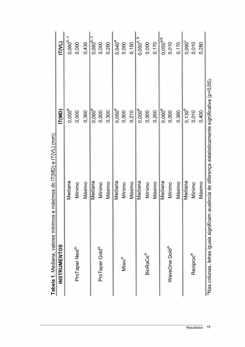
Tabela 1 – Mediana , valores mínimos e máximos do IT(MD) e IT(VL), em milímetros................19
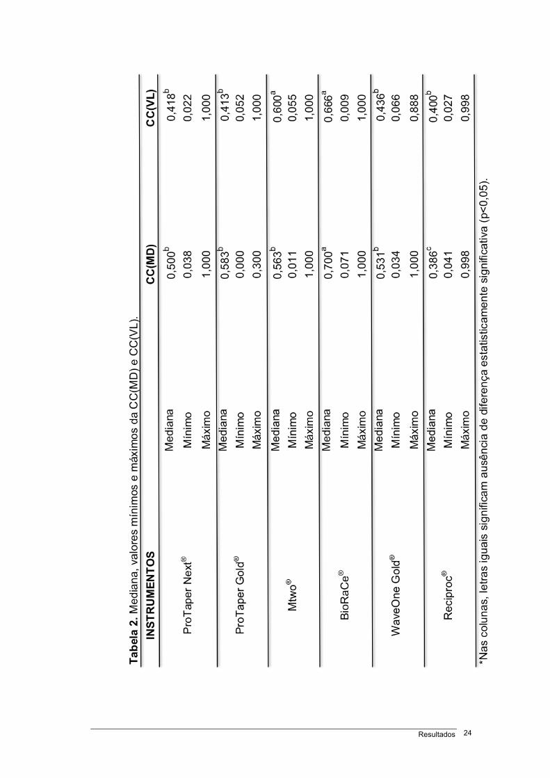
Tabela 2 – Mediana, valores mínimos e máximos da CC(MD) e CC(VL).....................................24
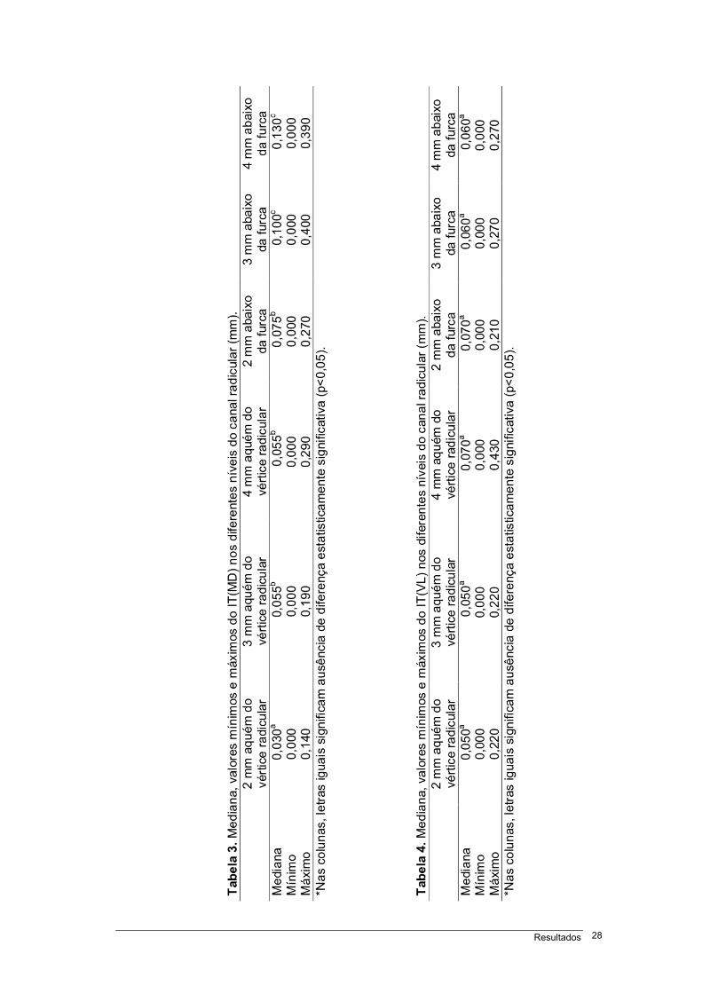
Tabela 3 – Mediana, valores mínimos e máximos do IT(MD) nos diferentes níveis do canal
radicular, em milímetros.................................................................................................................28
Tabela 4 – Mediana, valores mínimos e máximos do IT(VL) nos diferentes níveis do canal
radicular, em milímetros.................................................................................................................28
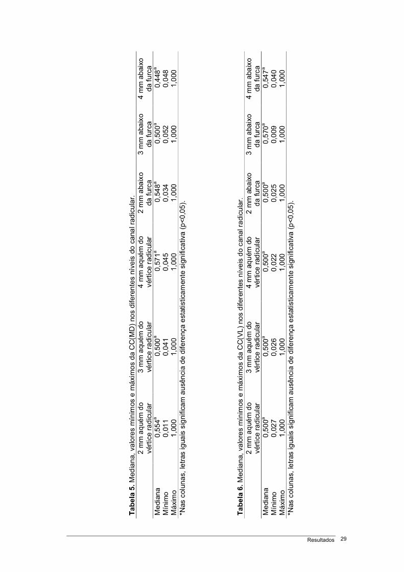
Tabela 5 – Mediana, valores mínimos e máximos da CC(MD) nos diferentes níveis do canal
radicular..........................................................................................................................................29
Tabela 6 – Mediana, valores mínimos e máximos da CC(VL) nos diferentes níveis do canal
radicular..........................................................................................................................................29
Figura 1 – Imagem em tomografia computadorizada de feixe cônico (plano axial) da raiz mesial
do molar inferior, 3 mm aquém do vértice radicular, para mensuração das distâncias nos sentidos
mesiodistal e vestíbulolingual para determinação do índice de transporte do canal radicular......13
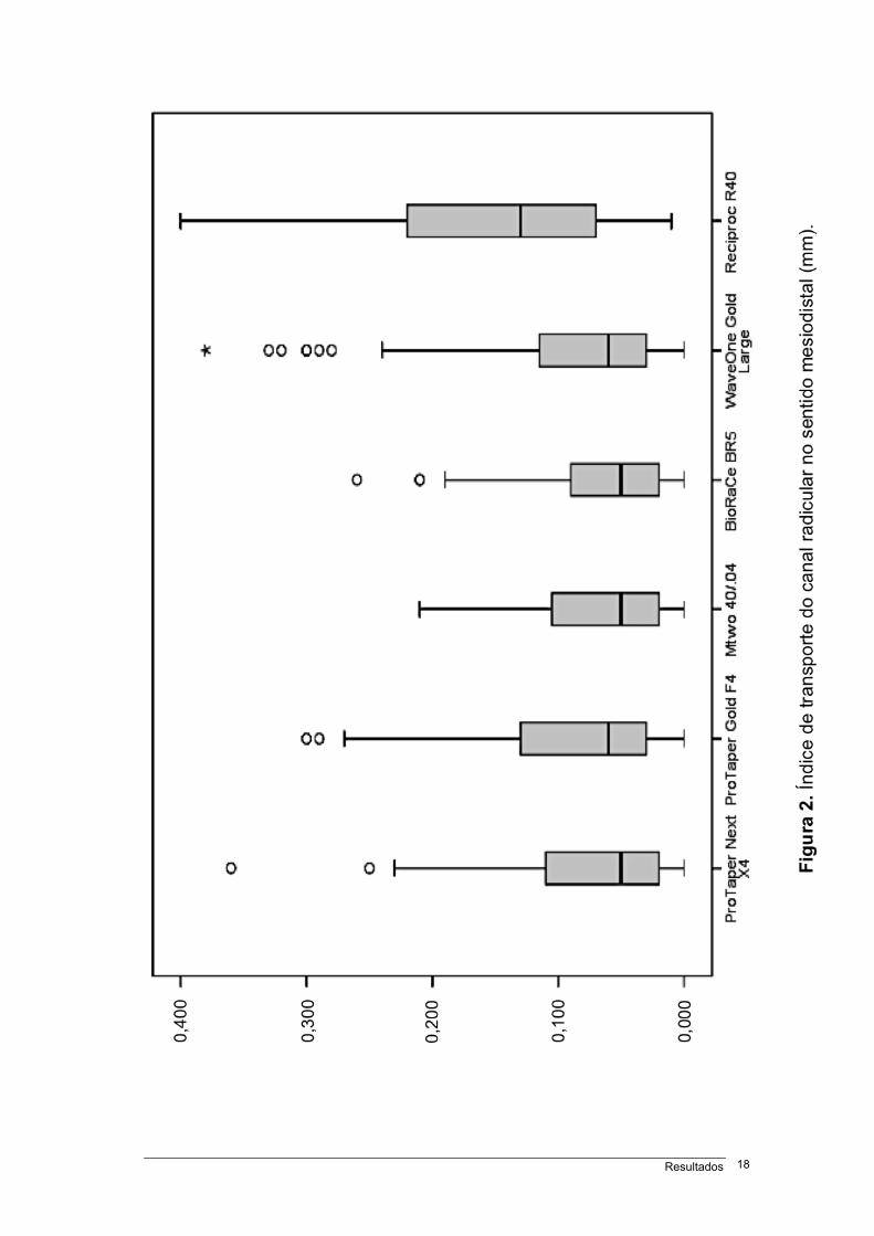
Figura 2 – Índice de transporte do canal radicular no sentido mesiodistal (mm)..........................18
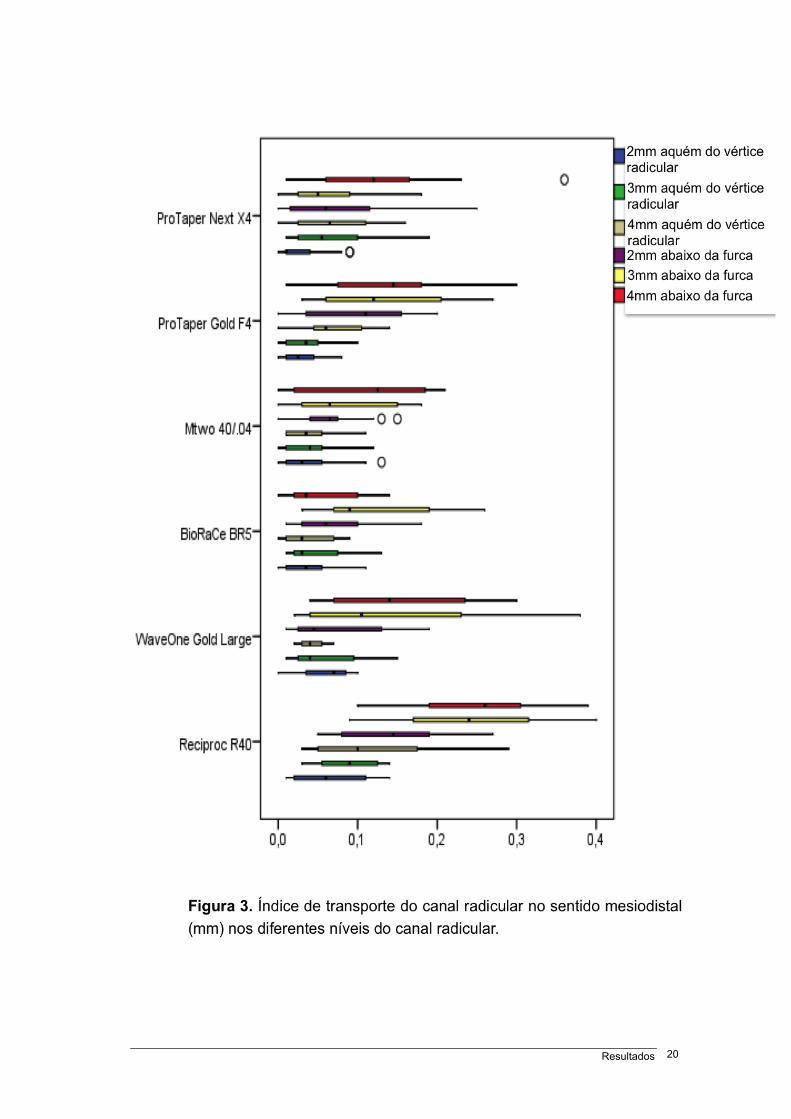
Figura 3 – Índice de transporte do canal radicular no sentido mesiodistal (mm) nos diferentes
níveis do canal radicular.................................................................................................................20
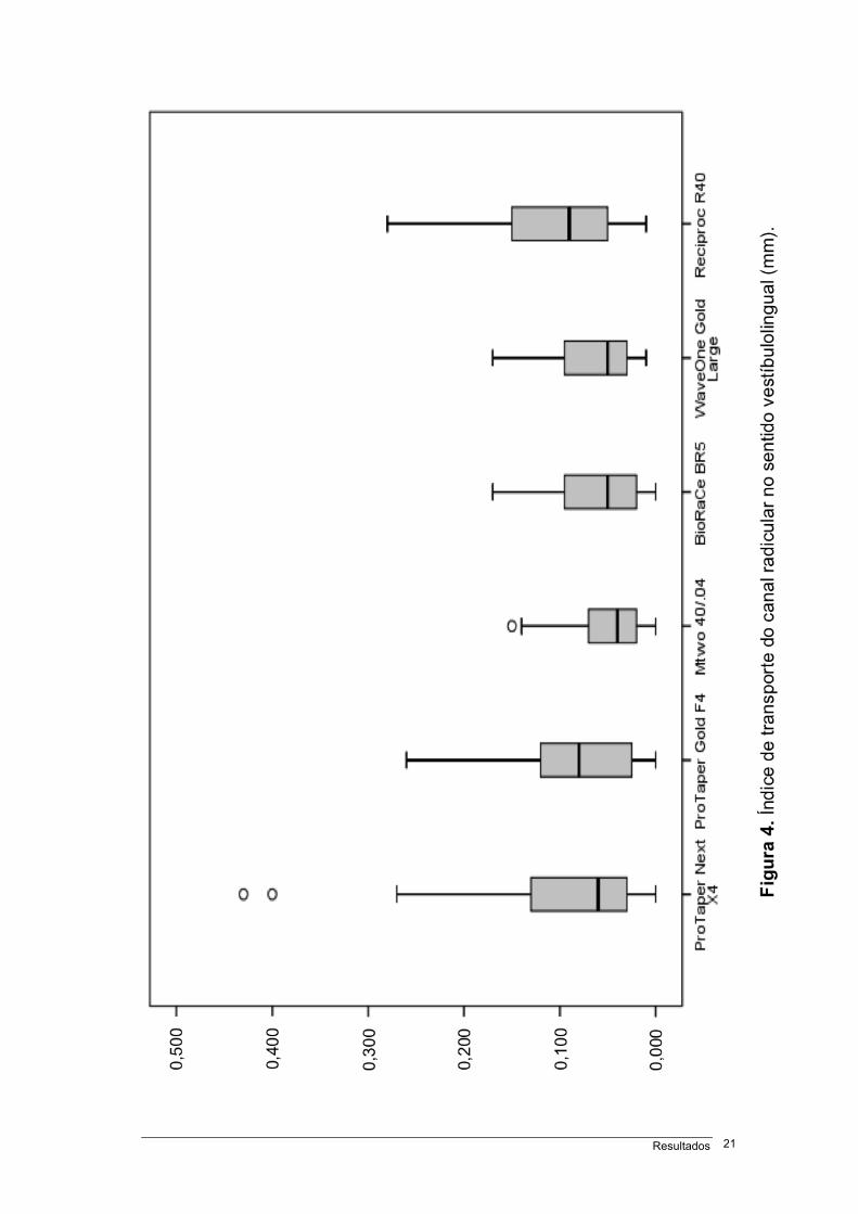
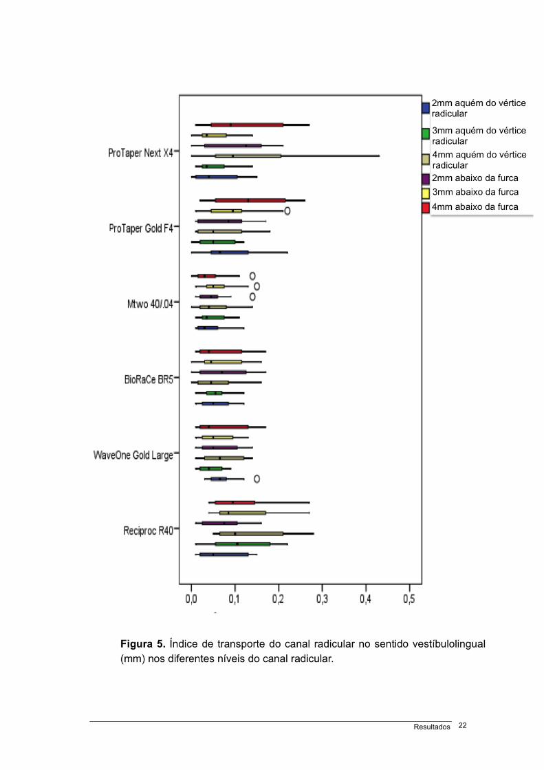
Figura 4 – Índice de transporte do canal radicular no sentido vestíbulolingual (mm)...................21
Figura 5 – Índice de transporte do canal radicular no sentido vestíbulolingual (mm) nos diferentes
níveis do canal radicular................................................................................................................22
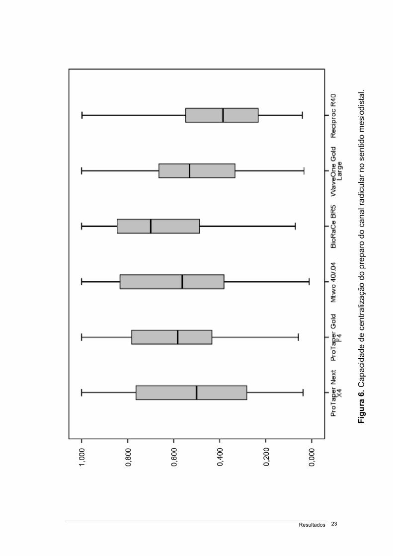
Figura 6 – Capacidade de centralização do preparo do canal radicular no sentido
mesiodistal......................................................................................................................................23
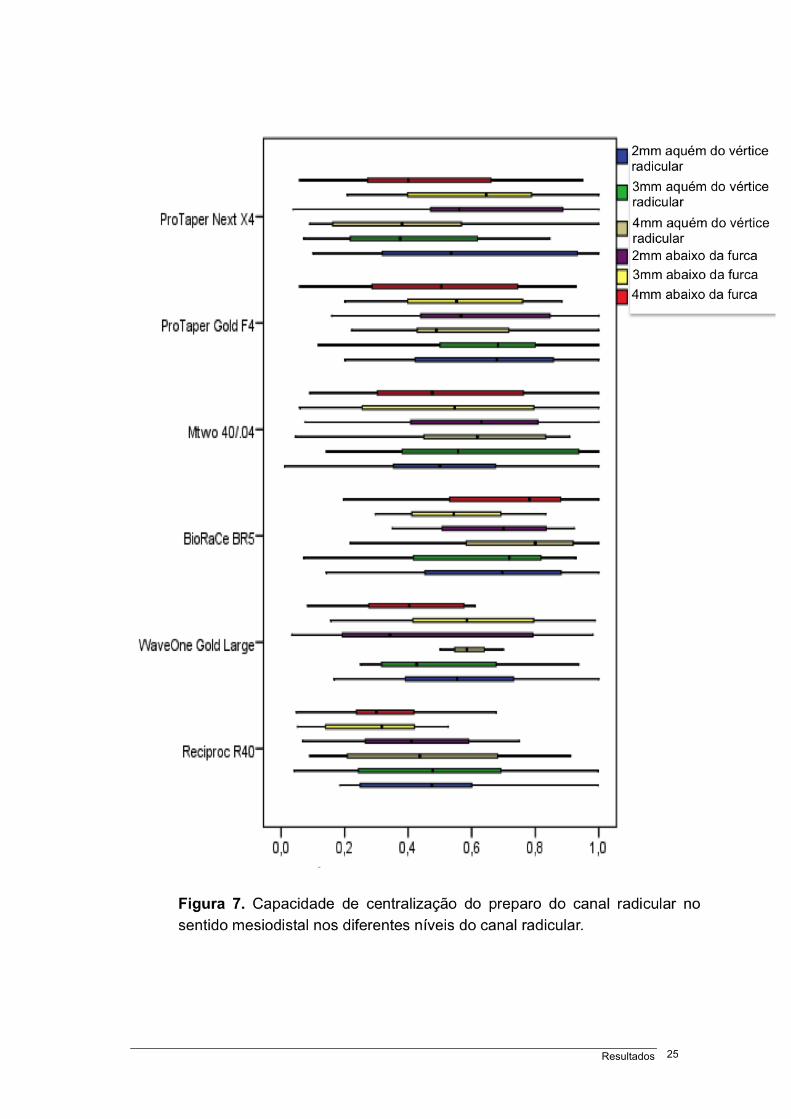
Figura 7 – Capacidade de centralização do preparo do canal radicular no sentido mesiodistal nos
diferentes níveis do canal radicular................................................................................................25
Tabelas, figuras e quadros xiv
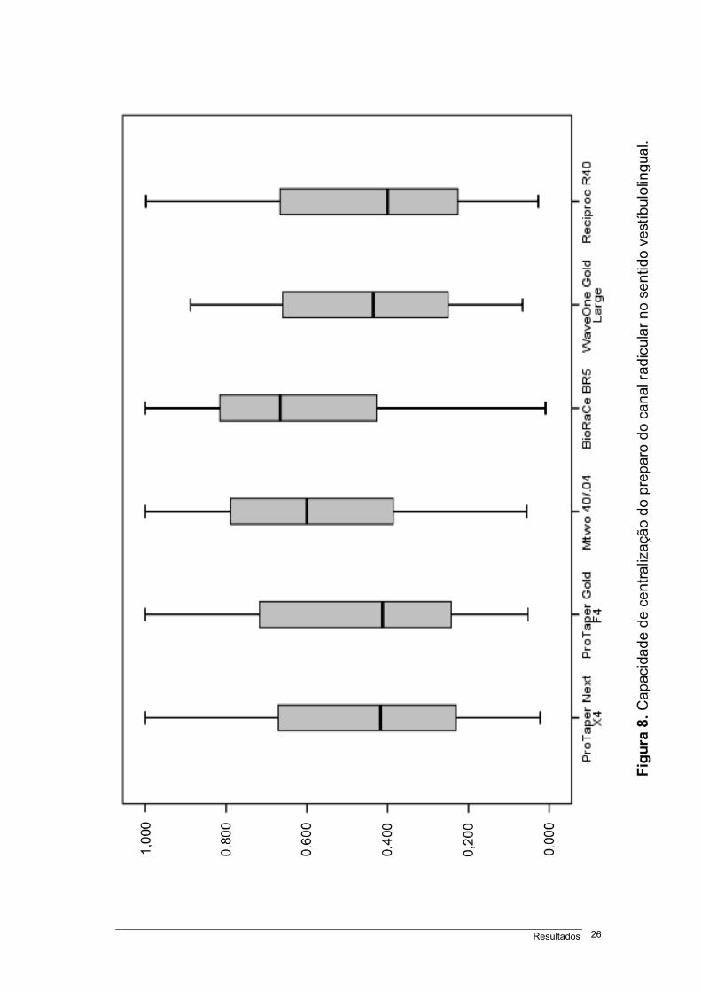
Figura 8 – Capacidade de centralização do preparo do canal radicular no sentido
vestíbulolingual...............................................................................................................................26
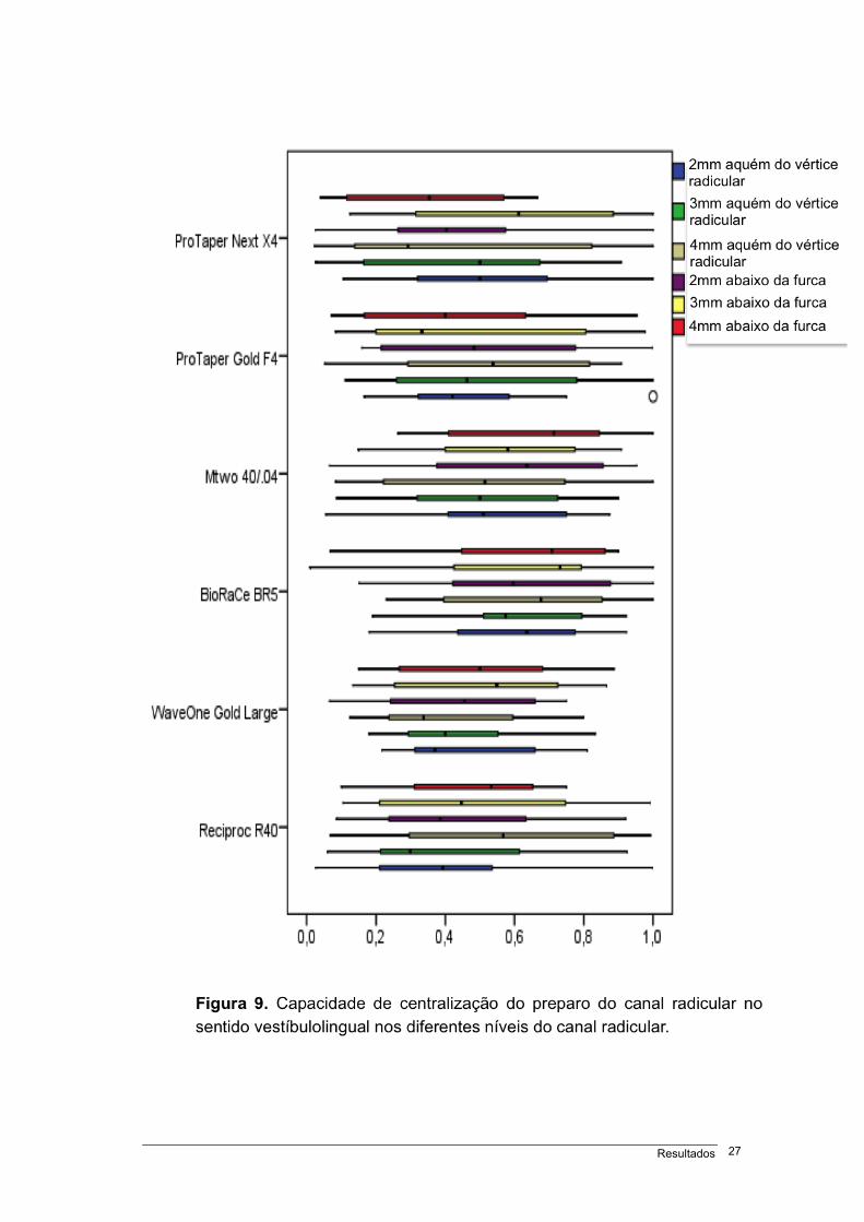
Figura 9 – Capacidade de centralização do preparo do canal radicular no sentido vestíbulolingual
nos diferentes níveis do canal radicular.........................................................................................27
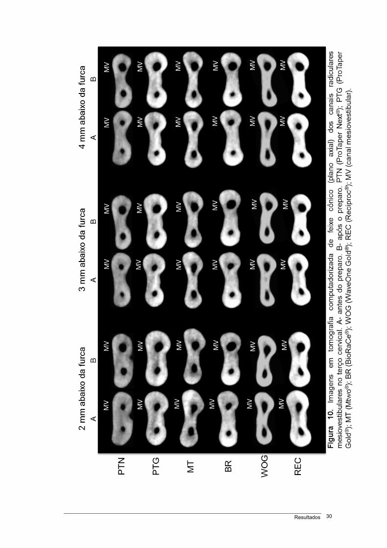
Figura 10 – Imagens em tomografia computadorizada de feixe cônico (plano axial) dos canais
radiculares mesiovestibulares no terço cervical.............................................................................30
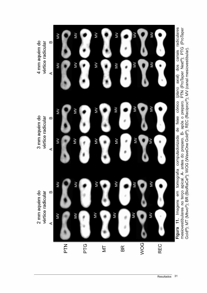
Figura 11 – Imagens em tomografia computadorizada de feixe cônico (plano axial) dos canais
radiculares mesiovestibulares no terço apical................................................................................31
Quadro 1 – Distribuição da amostra de acordo com os instrumento rotatório..............................07
Anexo 1 – Parecer do Comitê de Ética.........................................................................................49
Anexo 2 – Artigo original...............................................................................................................53
Anexo 3 – Norma de publicação do respectivo periódico.............................................................78
Símbolos, siglas e abreviaturas xv
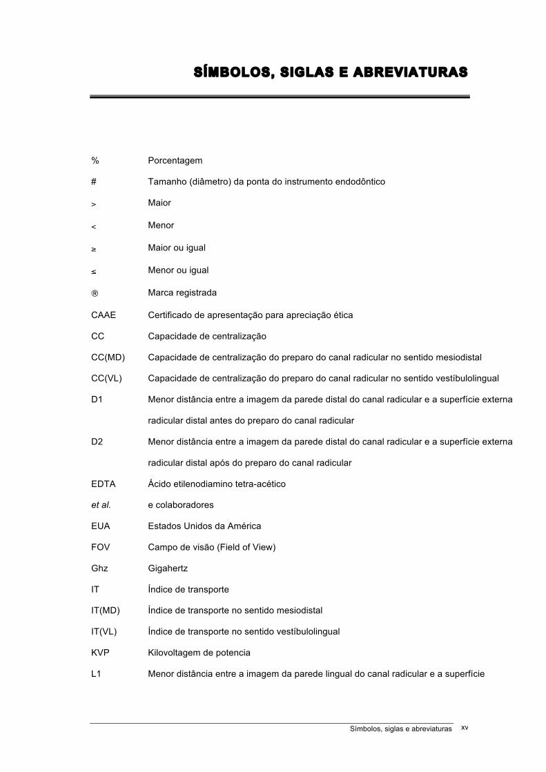
SÍMBOLOS, SIGLAS E ABREVIATURAS
% Porcentagem
# Tamanho (diâmetro) da ponta do instrumento endodôntico
> Maior
< Menor
≥ Maior ou igual
≤ Menor ou igual
® Marca registrada
CAAE Certificado de apresentação para apreciação ética
CC Capacidade de centralização
CC(MD) Capacidade de centralização do preparo do canal radicular no sentido mesiodistal
CC(VL) Capacidade de centralização do preparo do canal radicular no sentido vestíbulolingual
D1 Menor distância entre a imagem da parede distal do canal radicular e a superfície externa
radicular distal antes do preparo do canal radicular
D2 Menor distância entre a imagem da parede distal do canal radicular e a superfície externa
radicular distal após do preparo do canal radicular
EDTA Ácido etilenodiamino tetra-acético
et al. e colaboradores
EUA Estados Unidos da América
FOV Campo de visão (Field of View)
Ghz Gigahertz
IT Índice de transporte
IT(MD) Índice de transporte no sentido mesiodistal
IT(VL) Índice de transporte no sentido vestíbulolingual
KVP Kilovoltagem de potencia
L1 Menor distância entre a imagem da parede lingual do canal radicular e a superfície
Símbolos, siglas e abreviaturas xvi
externa radicular lingual antes do preparo do canal radicular
L2 Menor distância entre a imagem da parede lingual do canal radicular e a superfície
externa radicular lingual após do preparo do canal radicular
M1 Menor distância entre a imagem da parede mesial do canal radicular e a superfície
externa radicular mesial antes do preparo do canal radicular
M2 Menor distância entre a imagem da parede mesial do canal radicular e a superfície
externa radicular mesial após do preparo do canal radicular
mA Miliamperagem
MD Sentido mesiodistal
mL Mililitro
mm Milímetro
NaOCl Hipoclorito de sódio
NiTi Níquel-Titânio
p Nível de significancia
s segundo
TCFC Tomografia computadorizada de feixe cônico
V1 Menor distância entre a imagem da parede vestibular do canal radicular e a superfície
externa radicular vestibular antes do preparo do canal radicular
V2 Menor distância entre a imagem da parede vestibular do canal radicular e a superfície
externa radicular vestibular após do preparo do canal radicular
VL Sentido vestíbulolingual
Resumo xvii
RESUMO
Objetivo: Avaliar o índice de transporte (IT) e a capacidade de centralização
(CC) do preparo do canal radicular com instrumentos endodônticos de níquel-
titânio acionados em rotação contínua e reciprocante. Materiais e métodos: Noventa e seis canais mesiovestibulares de molares inferiores (primeiro e
segundo) foram aleatoriamente divididos em 06 grupos (n=16), de acordo com o
instrumento rotatório utilizado: Grupo 1- ProTaper Next®; Grupo 2- ProTaper
Gold®; Grupo 3- Mtwo®; Grupo 4- BioRaCe®; Grupo 5- WaveOne Gold®; Grupo
6- Reciproc®. A técnica de preparo do canal radicular obedeceu a orientação dos
fabricantes. Tomografia computadorizada de feixe cônico foi realizada antes e
após o preparo dos canais radiculares. Foram estabelecidos 06 níveis para a
realização das mensurações nas imagens: 2, 3 e 4 mm aquém do vértice
radicular e 2, 3 e 4 mm abaixo da furca. O índice de transporte e a capacidade
de centralização foram analizados conforme proposto por Gambill et al. (1996).
Resultados: O maior IT no sentido mesiodistal (MD) foi observado com o
Reciproc® (p<0,05), enquanto que no sentido vestíbulolingual (VL) foram
verificados com Reciproc®, ProTaper Gold® e ProTaper Next® (p<0,05). Os
maiores valores de CC(MD) e (VL) foram observados com os sistemas
BioRaCe® (p<0,05) e BioRaCe® e Mtwo® (p<0,05), respectivamente. Conclusão: Todos os sistemas promoveram transporte do canal radicular. Nenhum
instrumento mostrou capacidade de centralização do preparo perfeita. O
Reciproc® obteve os valores maiores de IT(MD) e (VL). O sistema BioRaCe®
apresentou a maior CC(MD), enquanto que no sentido (VL) o BioRaCe® e Mtwo®
mostraram capacidades similares.
Palavras-chave: Endodontia, preparo de canal radicular, tomografia
computadorizada de feixe cônico.
Abstract xviii
ABSTRACT
Objective: To evaluate transportation (T) and centering ability (CA) of root
canal preparations using continuous or reciprocating nickel-titanium
endodontic files. Material and methods: Ninety-six mesiobuccal root canals
of mandibular first and second molars were randomly divide into 6 groups
(n=16) according to the rotary file used: Group 1- ProTaper Next®; Group 2-
ProTaper Gold®; Group 3- Mtwo®; Group 4- BioRaCe®; Group 5- WaveOne
Gold®; Group 6- Reciproc®. Root canals were prepared according to
manufacturer’s instructions. Cone-beam computed tomography scans were
obtained before and after canal preparation. Measurements were made at
five different points: 2, 3 and 4 mm from the apex and 2, 3 and 4 mm below
furcation. Transportation and centering ability were analyzed following the
recommendations made by Gambill et al. (1996). Results: The greatest
mesiodistal (MD) transportation (T) was found for Reciproc® files (p<0.05),
and the greatest buccolingual (BL) T, for Reciproc®, ProTaper Gold® and
ProTaper Next® files (p<0.05). The greatest mesiodistal (MD) centering
ability (CA) was found for BioRaCe® files (p<0.05), and the greatest
buccolingual (BL) CA, for BioRaCe® and Mtwo® files (p<0.05). Conclusion: All systems produced root canal transportation. No file system achieved
perfect CA of root preparation. Reciproc® files had the greatest MD T and BL
T. BioRaCe® files had the greatest MD CA, whereas BL CA was similar for
BioRaCe® and Mtwo® files.
Key Words: Canal transportation, centering ability, cone beam computed
tomographic, endodontics, nickel-titanium instruments, reciprocating motion.
Introdução 1
1 INTRODUÇÃO
O mérito no preparo do canal radicular é esvaziar e alargar a cavidade
pulpar com manutenção do formato original, curvatura e posição espacial do
forame apical (SCHILDER, 1974; PETERS, 2004). Erros iatrogênicos de
procedimentos (como degrau, perfuração, descentralização do canal
radicular, transporte de forame apical) em canais radiculares curvos são
fatores de risco ao fracasso do tratamento endodôntico (ESTRELA et al.
2014).
A preservação da forma original de canais radiculares curvos é maior
em preparos com instrumentos flexíveis de níquel-titânio comparada a
instrumentos de aço inoxidável (WALIA et al. 1988; ESPOSITO &
CUNNINGHAM, 1995). Os instrumentos de níquel-titânio acionados em
rotação contínua e/ou em cinemática recíproca apresentam maior habilidade
em manter centralizado o preparo, e consequentemente diminuir o
transporte dos canais radiculares (CARVALHO et al. 2015; PAGLIOSA et al.
2015; GERGI et al. 2015).
Os instrumentos de níquel-titânio apresentam características
específicas em relação a secção transversal, ângulo de corte, conicidade,
número de espiras e guia de penetração (BÜRKLEIN et al. 2012). As
propriedades mecânicas e o comportamento das ligas de níquel-titânio
podem variar de acordo com a composição química e os tratamentos
termomecânicos durante a fabricação (THOMPSON, 2000; KUHN et al.
2001).
Introdução 2
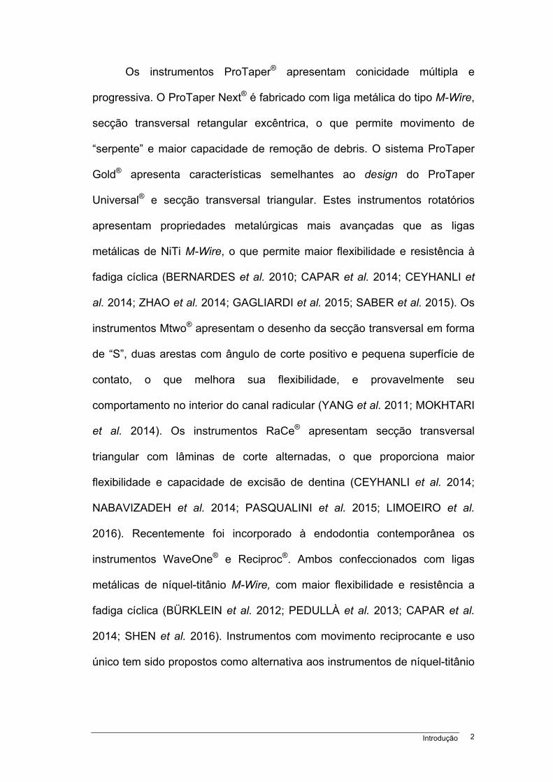
Os instrumentos ProTaper® apresentam conicidade múltipla e
progressiva. O ProTaper Next® é fabricado com liga metálica do tipo M-Wire,
secção transversal retangular excêntrica, o que permite movimento de
“serpente” e maior capacidade de remoção de debris. O sistema ProTaper
Gold® apresenta características semelhantes ao design do ProTaper
Universal® e secção transversal triangular. Estes instrumentos rotatórios
apresentam propriedades metalúrgicas mais avançadas que as ligas
metálicas de NiTi M-Wire, o que permite maior flexibilidade e resistência à
fadiga cíclica (BERNARDES et al. 2010; CAPAR et al. 2014; CEYHANLI et
al. 2014; ZHAO et al. 2014; GAGLIARDI et al. 2015; SABER et al. 2015). Os
instrumentos Mtwo® apresentam o desenho da secção transversal em forma
de “S”, duas arestas com ângulo de corte positivo e pequena superfície de
contato, o que melhora sua flexibilidade, e provavelmente seu
comportamento no interior do canal radicular (YANG et al. 2011; MOKHTARI
et al. 2014). Os instrumentos RaCe® apresentam secção transversal
triangular com lâminas de corte alternadas, o que proporciona maior
flexibilidade e capacidade de excisão de dentina (CEYHANLI et al. 2014;
NABAVIZADEH et al. 2014; PASQUALINI et al. 2015; LIMOEIRO et al.
2016). Recentemente foi incorporado à endodontia contemporânea os
instrumentos WaveOne® e Reciproc®. Ambos confeccionados com ligas
metálicas de níquel-titânio M-Wire, com maior flexibilidade e resistência a
fadiga cíclica (BÜRKLEIN et al. 2012; PEDULLÀ et al. 2013; CAPAR et al.
2014; SHEN et al. 2016). Instrumentos com movimento reciprocante e uso
único tem sido propostos como alternativa aos instrumentos de níquel-titânio
Introdução 3
acionados em rotação contínua de diâmetros e conicidades variadas
(YARED, 2008).
A efetividade de alguns destes instrumentos foi verificada frente a
manutenção da geometria e transporte do canal radicular, fratura do
instrumento endodôntico e fratura de dentina (PETERS, 2004; CAPAR et al.
2014; LOPES et al. 2013; KARATAS et al. 2015). Recentes estudos
mostraram o comportamento satisfatório destes novos instrumentos quanto
ao alargamento, o transporte e a centralização do preparo em canais curvos
(CARVALHO et al. 2015; ZANETTE et al. 2014; JAIN et al. 2015). Os
recursos envolvendo imagens mais comumente empregados para a
determinação dos erros de procedimentos operatórios incluem as
radiografias periapicais (BÜRKLEIN et al. 2013; OLIVIERI et al. 2014;
SABER et al. 2015; BÜRKLEIN et al. 2015), a microscopia eletrônica de
varredura (SCHÄFER & VLASSIS, 2004), a microtomografia
computadorizada (PETERS et al. 2015; ALMEIDA et al. 2015; PASQUALINI
et al. 2015; LIMOEIRO et al. 2016; AMARAL et al. 2016), e a TCFC
(GAMBILL et al. 1996; PETERS et al. 2001; ALENCAR et al. 2010; GERGI et
al. 2010; CAPAR et al. 2014; CARVALHO et al. 2015; HOPPE et al. 2016).
O padrão para se determinar as alterações morfológicas após o
preparo do canal radicular em relação às áreas desgastadas devem ser
cuidadosamente analisados. A verificação do índice de transporte radicular e
a capacidade de centralização do preparo, em níveis cervical e apical, em
direção mesiodistal e vestíbulolingual em imagens de tomografia
computadorizada de feixe cônico pode favorecer a incorporação de um
método com importante referencial.
Objetivos 4
2 OBJETIVOS
OBJETIVO GERAL
Determinar o índice de transporte e a capacidade de centralização do
preparo do canal radicular com instrumentos de níquel-titânio acionados em
cinemática de rotação contínua e reciprocante.
OBJETIVOS ESPECÍFICOS
1. Avaliar instrumentos de níquel-titânio quanto ao índice de
transporte do canal radicular e a capacidade de centralização do preparo do
canal radicular nos sentidos mesiodistal e vestíbulolingual;
2. Verificar o índice de transporte e a capacidade de centralização
após o preparo do canal radicular nos níveis 2, 3 e 4 mm aquém do vértice
radicular e 2, 3 e 4 mm abaixo da furca.
Materiais e Métodos 5
3 MATERIAIS E MÉTODOS
Tipo de estudo
Este é um estudo laboratorial em ex vivo para comparação do índice
de transporte do canal radicular e da capacidade de centralização do
preparo do canal radicular entre instrumentos de níquel-titânio acionados em
cinemática de rotação contínua e reciprocante, realizado em molares
inferiores humanos e avaliado em imagens de tomografia computadorizada
de feixe cônico.
Seleção e preparo das amostras
Este estudo foi aprovado pelo comitê de ética em pesquisa da
Universidade Federal de Goiás (CAAE: 53712816.1.0000.5083). Primeiros e
segundos molares inferiores permanentes humanos foram obtidos no serviço
de urgência odontológica da Faculdade de Odontologia da Universidade
Federal de Goiás. Os dentes foram armazenados em solução de timol 0,2%.
Critérios de inclusão
Radiografias periapicais pré-operatórias de cada dente foram
realizadas para a seleção da amostra dentro dos critérios adotados. Para
padronização das imagens dos dentes foi utilizada uma plataforma. As
radiografias foram adquiridas em aparelho de raio-x Spectro X70 Eletronic
(Dabi Atlante, Ribeirão Preto, SP, Brasil) e sensor digital RVG 5100
(Carestream Dental, Atlanta, EUA) usando a técnica do paralelismo. Todas
as imagens foram avaliadas pelo software do sensor digital RVG 5100
(Carestream Dental, Atlanta, EUA).
Materiais e Métodos 6
Os critérios de inclusão foram dentes com ausência de reabsorções
radiculares (internas e/ou externas), fraturas radiculares e canais radiculares
obliterados, cavidade pulpar intacta e rizogênese completa.
Critérios de exclusão
As imagens iniciais foram adquiridas no tomógrafo Prexion 3D
scanner (PreXion 3D Inc., San Mateo, EUA). As imagens foram capturadas
com espessura de 0,110 mm (dimensões de 1,170 X 1,570 X 1,925 mm),
FOV de 81,00 X 75 mm, voxel de 0,100 mm, em 33,5 s (1.024
visualizações). A voltagem do tubo foi de 90 KVP e a corrente do tubo de 4
mA. Para a análise das imagens, foi utilizado o software do próprio
tomógrafo Prexion (Prexion 3D Viewer, TeraRecon Inc, Foster City, EUA) em
uma estação de trabalho PC com o Windows 8 Professional (Microsoft
Corp., Redmond, EUA), equipado com um processador Intel i7 2,86 Ghz
(Intel Corp., Santa Clara, EUA), NVIDIA GeForce 6200 turbo cache
videocard (NVIDIA Corp., Santa Clara, EUA), e monitor ELZO-Flexscan
S2000 com resolução de 1600 X 1200 pixels (ELZO NANAO Corp.,
Hakusan, Japão).
Como critérios de exclusão foram considerados dentes com
comprimento maior que 22 mm, canais radiculares mesiovestibulares com
mais de um forame apical e raio de curvatura menor que 4 mm e maior que
8 mm, de acordo com o método proposto por ESTRELA et al. (2008). Para a
determinação do raio de curvatura do canal radicular curvo foram traçadas
duas linhas retas de 6 mm sobrepostas ao canal radicular, sendo a linha
primária a que representou a continuidade da região apical e a secundária, a
continuidade dos terços médio e cervical. Independentemente do
Materiais e Métodos 7
comprimento da linha secundária, apenas os 6 mm mais próximos à linha
primária foram utilizados para medir. Posteriormente, o ponto médio de cada
linha foi determinado e duas linhas perpendiculares foram traçadas até o
ponto central, denominado centro da circunferência. A distância entre o
centro da circunferência e o centro de cada linha (primária e secundária) foi
estabelecido como o raio da circunferência, o que determinou a magnitude
da curvatura.
Preparo dos canais radiculares
No momento do experimento, os dentes foram lavados em água
corrente com o objetivo de remover traços da solução de timol e secados
com toalhas de papel absorvente. A seguir, os mesmos foram imersos em
hipoclorito de sódio a 5% durante 30 minutos para a remoção de tecido
orgânico.
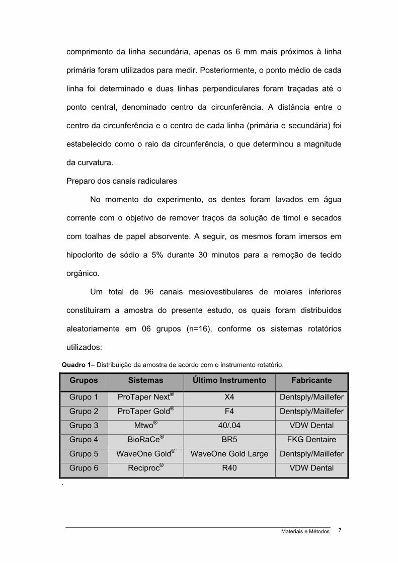
Um total de 96 canais mesiovestibulares de molares inferiores
constituíram a amostra do presente estudo, os quais foram distribuídos
aleatoriamente em 06 grupos (n=16), conforme os sistemas rotatórios
utilizados:
Quadro 1– Distribuição da amostra de acordo com o instrumento rotatório.
Grupos Sistemas Último Instrumento Fabricante
Grupo 1 ProTaper Next® X4 Dentsply/Maillefer
Grupo 2 ProTaper Gold® F4 Dentsply/Maillefer
Grupo 3 Mtwo® 40/.04 VDW Dental
Grupo 4 BioRaCe® BR5 FKG Dentaire
Grupo 5 WaveOne Gold® WaveOne Gold Large Dentsply/Maillefer
Grupo 6 Reciproc® R40 VDW Dental
.
Materiais e Métodos 8
As aberturas coronárias foram realizadas com pontas diamantadas
esféricas (#1013, #1014; KG Sorensen, Barueri, SP, Brasil) e broca Endo Z
(Dentsply/Maillefer, Suíça), ambas sob refrigeração e em alta rotação.
Posteriormente, os canais radiculares mesiovestibulares foram explorados e
esvaziados com auxílio de lima manual de aço inoxidável tipo K #10 e 15
(Dentsply/Maillefer, Suíça). O preparo do terço cervical foi realizado com a
utilização dos instrumentos de cada sistema destinados para esta função. O
comprimento de trabalho foi determinado com auxílio de lima tipo K #15
(Dentsply/Maillefer, Suíça) até obter-se a visualização do instrumento
endodôntico através do forame apical. Deste comprimento foi recuado um
milímetro, para obtenção do comprimento real de trabalho.
A técnica de preparo do canal radicular obedeceu a orientação dos
fabricantes. Todos os instrumentos foram acionados pelo motor X-Smart
Plus® (Dentsply/Maillefer, Suíça), e os canais radiculares irrigados com
hipoclorito de sódio a 2,5% (Rioquímica, São José do Rio Preto, SP, Brasil),
utilizando-se a seringa de irrigação Navitip (Ultradent Products Inc., South
Jordan, EUA). Durante o processo de sanificação do canal radicular, utilizou-
se 30 mL de irrigante. Após o último instrumento ter alcançado o
comprimento de trabalho em rotação livre, este foi removido, e considerado
finalizado o preparo. A patência foi verificada utilizando-se uma lima K #15
(Dentsply/Maillefer, Suíça). Concluída a instrumentação, os canais
radiculares foram secos com cone de papel absorvente (Dentsply,
Petrópolis, RJ, Brasil) de calibre correspondente ao último instrumento e a
seguir, irrigados com 5 mL de EDTA 17% (F&A Laboratório Farmacêutico
Ltda, São Paulo, SP, Brasil), que permaneceu por 3 minutos. Após a
Materiais e Métodos 9
irrigação final com 5 mL de NaOCl 2,5% os canais radiculares foram
secados novamente.
Cada instrumento endodôntico foi utilizado para preparar um único
canal radicular. Todos os preparos de canais radiculares foram realizados
por um especialista em endodontia, com mais de quinze anos de
experiência.
Posterior ao preparo dos canais radiculares, imagens finais de
tomografia computadorizada de feixe cônico foram obtidas para a
determinação do índice de transporte do canal radicular e da capacidade de
centralização do preparo do canal radicular dos instrumentos endodônticos.
O mesmo protocolo descrito anteriormente para a aquisição das imagens
iniciais foi utilizado e a ferramenta de sincronização de imagens do software
do tomógrafo Prexion (Prexion 3D Viewer, TeraRecon Inc, Foster City, EUA)
foi aplicada nos planos axial, coronal e sagital.
Os níveis estabelecidos para a realização das mensurações nas
imagens dos canais radiculares foram: 1- 2 mm aquém do vértice radicular;
2- 3 mm aquém do vértice radicular; 3- 4 mm aquém do vértice radicular; 4-
2 mm abaixo da furca; 5- 3 mm abaixo da furca; 6- 4 mm abaixo da furca. A
navegação no plano axial das imagens sincronizadas foi iniciada no vértice
radicular, tanto nas imagens iniciais quanto nas finais, até atingir os níveis
estabelecidos no terço apical. Para as mensurações no terço cervical a
navegação iniciou na furca e estendeu-se até 4 mm abaixo. Para facilitar as
mensurações, ajustes de ampliação, brilho e contraste disponíveis no
programa foram utilizados.
Materiais e Métodos 10
Análise do transporte do canal radicular
A análise das imagens para verificar o transporte do canal radicular foi
realizada utilizando a metodologia proposta por GAMBILL et al. (1996). O
índice de transporte do canal radicular foi determinado nos sentidos
mesiodistal e vestíbulolingual nos seis níveis descritos anteriormente e
correspondeu à variação, em milímetros, do desvio do eixo central do canal
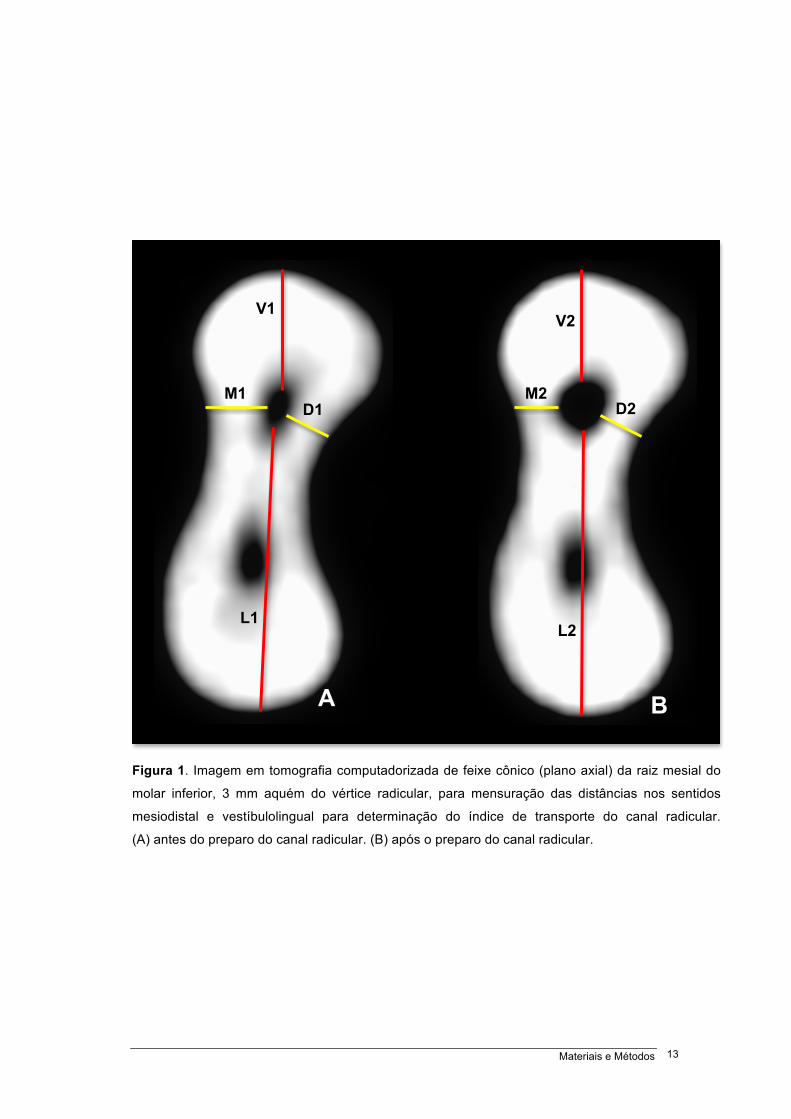
radicular após a instrumentação. O transporte do canal radicular no sentido
mesiodistal foi estabelecido a partir da mensuração da menor distância entre
a imagem das paredes mesial e distal do canal radicular e a superfície
externa radicular mesial e distal, antes (M1 e D1) e após (M2 e D2) o
preparo do canal radicular (Figura 1).
Igualmente, o transporte do canal radicular no sentido vestíbulolingual
foi determinado a partir da mensuração da menor distância entre a imagem
das paredes vestibular e lingual do canal radicular e a superfície externa
radicular vestibular e lingual, antes (V1 e L1) e após (V2 e L2) o preparo do
canal radicular (Figura 1). Para a realização das mensurações foi
empregado o software do tomógrafo Prexion (Prexion 3D Viewer, TeraRecon
Inc, Foster City, EUA) em uma estação de trabalho PC com o Windows 8
Professional (Microsoft Corp., Redmond, EUA), equipado com um
preocessador Intel i7 2,86 Ghz (Intel Corp., Santa Clara, EUA), NVIDIA
GeForce 6200 turbo cache videocard (NVIDIA Corp., Santa Clara, EUA), e
monitor ELZO-Flexscan S2000 com resolução de 1600 X 1200 pixels (ELZO
NANAO Corp., Hakusan, Japão). As mensurações foram realizadas por um
examinador em dois momentos distintos e mostraram, pelo teste de Kappa,
nível de concordância superior a 80% (K=0,882).
Materiais e Métodos 11
O cálculo do índice de transporte (IT) do canal radicular no sentido
mesiodistal (MD) e no vestíbulolingual (VL) foi realizado utilizando-se a
seguinte fórmula:
IT(MD) = (M1 – M2) – (D1 – D2)
IT(VL) = (V1 – V2) – (L1 – L2)
Quando a aplicação da fórmula resultou em IT(MD) com valor
negativo foi considerado transporte do canal radicular no sentido distal, valor
positivo, transporte no sentido mesial, e quando igual a zero, como ausência
de transporte. O resultado com valor negativo do IT(VL) foi estabelecido
como transporte do canal radicular no sentido lingual, valor positivo,
transporte no sentido vestibular, e quando igual a zero, como ausência de
transporte.
Análise da capacidade de centralização do preparo do canal radicular
A análise da capacidade de centralização (CC) foi realizada de acordo
com a metodologia proposta por GAMBILL et al. (1996), os quais definiram a
capacidade de centralização como a habilidade do instrumento endodôntico
em manter-se no eixo central do canal radicular. A determinação da CC foi
realizada a partir dos valores obtidos na mensuração das distâncias para o
cálculo do IT.
Para o estabelecimento da CC do preparo do canal radicular, no
sentido mesiodistal e vestíbulolingual, foi utilizada a seguinte fórmula:
Materiais e Métodos 12
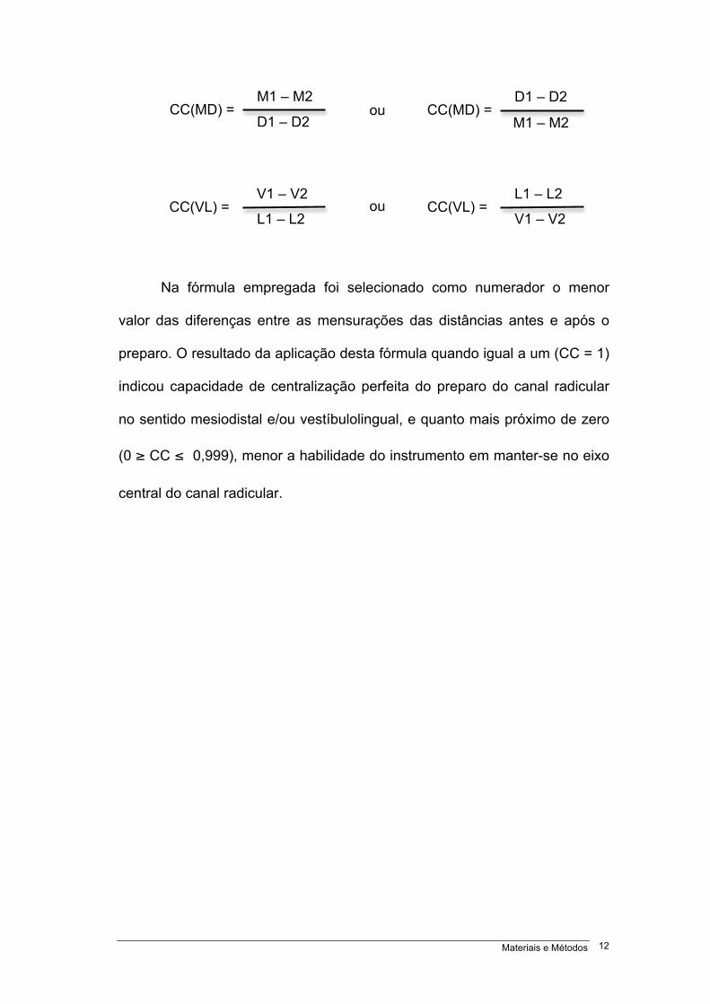
Na fórmula empregada foi selecionado como numerador o menor
valor das diferenças entre as mensurações das distâncias antes e após o
preparo. O resultado da aplicação desta fórmula quando igual a um (CC = 1)
indicou capacidade de centralização perfeita do preparo do canal radicular
no sentido mesiodistal e/ou vestíbulolingual, e quanto mais próximo de zero
(0 ≥ CC ≤ 0,999), menor a habilidade do instrumento em manter-se no eixo
central do canal radicular.
CC(MD) = M1 – M2
D1 – D2 CC(MD) =
D1 – D2
M1 – M2
CC(VL) = V1 – V2 L1 – L2
CC(VL) = L1 – L2 V1 – V2
ou
ou
Materiais e Métodos 13
Figura 1. Imagem em tomografia computadorizada de feixe cônico (plano axial) da raiz mesial do
molar inferior, 3 mm aquém do vértice radicular, para mensuração das distâncias nos sentidos
mesiodistal e vestíbulolingual para determinação do índice de transporte do canal radicular.
(A) antes do preparo do canal radicular. (B) após o preparo do canal radicular.
M1 D1
V1
L1
M2 D2
V2
L2
A B
Materiais e Métodos 14
Análise estatística
Para análise estatística, os dados originais referentes ao índice de
transporte do canal radicular e a capacidade de centralização do preparo do
canal radicular foram digitados no programa Microsoft Office Excel (Microsoft
Corporation, Washington, EUA) e posteriormente exportados para o
programa IBM SPSS versão 20.0 (SPSS Inc., Nova York, EUA) para análise
estatística. Foram descritas as variáveis pela mediana, valores mínimos e
máximos e comparadas entre os instrumentos pelo teste de Kruskal Wallis e
entre as medidas nos seis níveis específicos do canal radicular pelo teste de
Friedman. As comparações foram ajustadas com o teste de Bonferroni. Foi
considerado um nível de significância de 5%.
Resultados 15
4 RESULTADOS
Todos os instrumentos avaliados mostraram medianas do IT(MD) do
canal radicular com valor positivo, ou seja, apresentaram transporte do canal
radicular no sentido mesial (Figura 2). Os menores IT(MD) foram verificados
com os sistemas Mtwo®, ProTaper Next®, BioRaCe®, ProTaper Gold® e
WaveOne Gold® sem diferença significativa entre eles. O maior IT(MD) foi
observado com o Reciproc®, com diferença significativa quando comparado
com os demais sistemas (p<0,05) (Tabela 1). Entretanto, quando avaliados
os seis níveis pré-estabelicidos, a 2 mm aquém do vértice radicular não
houve diferença significativa entre os sistemas (Figura 3).
A análise do IT(VL) do canal radicular mostrou que as medianas de
todos os sistemas testados apresentaram valor positivo, ou seja, o
transporte do canal radicular ocorreu no sentido vestibular (Figura 4). Os
menores IT(VL) foram verificados com os sistemas Mtwo®, BioRaCe® e
WaveOne Gold® sem diferença significativa entre eles; enquanto que, os
maiores IT(VL) foram observados com os sistemas Reciproc®, ProTaper
Gold® e ProTaper Next® com diferença significativa quando comparado com
o Mtwo® (p<0,05) (Tabela 1). Apesar de, nos níveis 2 e 3 mm aquém do
vértice radicular e 2 mm abaixo da furca, não ter havido diferença
significativa entre os instrumentos, nos pontos a 4 mm aquém do vértice
radicular e a 3 mm abaixo da furca, o Reciproc® apresentou maior IT(VL) do
que os demais sistemas com diferença significativa (p<0,05). No entanto, a 4
mm abaixo da furca, os instrumentos Reciproc® e ProTaper Gold®
Resultados 16
apresentaram maior IT(VL) do que os outros com diferença significativa
(p<0,05) (Figura 5).
Nenhum dos sistemas avaliados mostrou medianas de CC(MD) do
preparo do canal radicular com valor igual a um, ou seja, capacidade de
centralização perfeita (Figura 6). O menor valor de CC(MD) foi observado
com o Reciproc®, com diferença significativa quando comparado com os
demais instrumentos (p<0,05), enquanto que o maior valor foi verificado com
o sistema BioRaCe® com diferença significativa entre eles (p<0,05) (Tabela
2). Entretanto, quando avaliados os seis níveis pré-estabelicidos, a 2 e 3 mm
aquém do vértice radicular e 2 mm abaixo da furca, não houve diferença
significativa entre os sistemas (Figura 7). A 4 mm aquém do vértice radicular
o maior valor de CC(MD) foi encontrado com o BioRaCe® com diferença
significativa entre os instrumentos ProTaper Next® e Reciproc® (p<0,05). No
nível a 3 mm abaixo da furca o menor valor de CC(MD) foi encontrado com o
Reciproc® com diferença significativa entre os demais (p<0,05). Quando
avaliado o nível a 4 mm abaixo da furca os instrumentos WaveOne Gold® e
Reciproc® apresentaram menores valores de CC(MD) com diferença
estatíticamente significativa entre os outros (p<0,05).
A análise da CC(VL) do preparo do canal radicular revelou que
nenhum dos sistemas pesquisados obteve valor das medianas igual a um,
ou seja, capacidade de centralização perfeita (Figura 8). Os maiores valores
de CC(VL) foram observados com os instrumentos BioRaCe® e Mtwo® com
diferença significativa quando comparado com os demais (p<0,05) (Tabela
2). Quando a CC(VL) foi avaliada nos níveis 2, 3 e 4 mm aquém do vértice
radicular e 2 e 3 mm abaixo da furca não foi detectada diferença
Resultados 17
estísticamente significativa entre os instrumentos rotatórios. Porém, a 4 mm
abaixo da furca, o ProTaper Next® apresentou o menor valor da CC(VL) com
diferença significativa entre o Mtwo® (p<0,05) (Figura 9).
A partir das mensurações dos seis níveis específicos nas imagens,
antes e após o preparo do canal radicular foi observado menor IT(MD) a 2
mm aquém do vértice radicular com diferença significativa (p<0,05) (Tabela
3), e menor IT(VL) a 2 e 3 mm aquém do vértice radicular, porém, sem
diferença significativa (Tabela 4).
Os maiores valores de CC(MD) ocorreram a 4 mm aquém do vértice
radicular, quando avaliados os níveis específicos, e os maiores valores de
CC(VL) observou-se a 3 mm abaixo da furca, entretanto, sem diferença
significativa (Tabelas 5 e 6).
As Figuras 10 e 11 exibem imagens em TCFC em plano axial dos
canais radiculares mesiovestibulares, nos terços cervical e apical, antes e
após o preparo de acordo com os instrumentos endodônticos de NiTi
utilizados.
Resultados 18
Resultados 19
Resultados 20
Resultados 21
Resultados 22
Resultados 23
Resultados 24
Resultados 25
Resultados 26
Resultados 27
Resultados 28
Resultados 29
Resultados 30
Resultados 31
Discussão 32
5 DISCUSSÃO
As alterações da forma do canal radicular nos terços cervicais e
apicais foram identificados em todos os instrumentos estudados. Os
instrumentos Protaper Next®, Protaper Gold®, WaveOne Gold®, Mtwo® e
BioRaCe® mostraram os menores índices de transporte mesiodistal. No
sentido vestíbulolingual os menores índices foram para os instrumentos
WaveOne Gold®, Mtwo® e BioRaCe®. Não foi identificada diferença
significativa do índice de transporte mesiodistal dos instrumentos estudados
em nível de 2 mm aquém do vértice radicular. No sentido vestíbulolingual
este fato ocorreu em nível de 2 e 3 mm aquém do vértice radicular e 2 mm
abaixo da furca.
A capacidade de centralização perfeita não foi encontrada em
nenhum dos sistemas, seja no sentido mesiodistal ou vestíbulolingual. A
melhor capacidade de centralização do preparo do canal radicular ocorreu
no sentido mesiodistal com o sistema BioRaCe®. Os maiores valores de
capacidade de centralização vestíbulolingual foram encontrados com os
sistemas BioRaCe® e Mtwo®. A capacidade de centralização mesiodistal em
nível de 2 e 3 mm aquém do vértice radicular e 2 mm abaixo da furca não
revelou diferenças significativas entre os sistemas. Contudo, no sentido
vestíbulolingual não foi verificada diferença significativa entre os sistemas
nos níveis 2, 3 e 4 mm aquém do vértice radicular e 2 e 3 mm abaixo da
furca.
Discussão 33
A determinação do índice de transporte e da capacidade de
centralização do preparo do canal radicular foi baseada em modelo
previamente descrito por GAMBILL et al. (1996). Estes autores utilizaram um
modelo de mensurações em planos axiais, a partir de ápices de dentes
unirradiculares em sentido mesiodistal, com curvatura inferior a 10 graus.
Vários estudos aplicaram esta metodologia para a avaliação do índice de
transporte e capacidade de centralização posterior ao preparo de canais
radiculares curvos (GERGI et al. 2010; MARZOUK & GHONEIM, 2013;
CAPAR et al. 2014; HONARDAR et al. 2014; CARVALHO et al. 2015; JAIN
et al. 2015; HOPPE et al. 2016).
No presente estudo foram utilizados os canais mesiovestibulares de
molares inferiores com raio de curvatura maior que 4 mm e menor que 8 mm
(ESTRELA et al. 2008). Foram consideradas duas áreas de importância
clínica como referencial de análise (2 a 4 mm abaixo da furca e 2 a 4 mm
aquém do vértice radicular), em duas direções (mesiodistal e
vestíbulolingual), analisadas em imagens de tomografia computadorizada de
feixe cônico de alta resolução.
A manutenção da forma posterior ao preparo de canais radiculares
curvos com instrumentos de aço inoxidável, de níquel-titânio acionados em
cinemática de rotação contínua e reciprocante usando diferentes métodos
experimentais tem sido avaliada em canais artificias (WU et al. 2015; LIU &
WU, 2016), em microscopia eletrônica de varredura (SCHÄFER & VLASSIS,
2004), em dentes humanos por meio de utilização de imagens radiográficas
periapicais (JAVAHERI & JAVAHERI, 2007; GARCIA et al. 2012; BÜRKLEIN
et al. 2012; BÜRKLEIN et al. 2013; OLIVIERI et al. 2014; FERRARA et al.
Discussão 34
2015; SABER et al. 2015; BÜRKLEIN et al. 2015), em microtomografia
computadorizada (LOIZIDES et al. 2007; PAQUÉ et al. 2011; YANG et al.
2011; STERN et al. 2012; GERGI et al. 2014; JUNAID et al. 2014; ZHAO et
al. 2014; HWANG et al. 2014; GERGI et al. 2015; GAGLIARDI et al. 2015;
PETERS et al. 2015; ALMEIDA et al. 2015; PASQUALINI et al. 2015;
LIMOEIRO et al. 2016; AMARAL et al. 2016), e com TCFC (GAMBILL et al.
1996; PETERS et al. 2001; HARTMANN et al. 2007; GERGI et al. 2010;
CAPAR et al. 2014; HONARDAR et al. 2014; JAIN et al. 2015; CARVALHO
et al. 2015; HOPPE et al. 2016).
A possibilidade da correta análise da geometria do canal radicular em
diferentes planos que permitam análise tridimensional, e a visão em plano
axial que evita uma sobreposição de estruturas tem valorizado o uso da
TCFC como potencial método não destrutivo (GAMBILL et al. 1996;
PETERS et al. 2001; ESTRELA et al. 2008).
Neste estudo, pode-se verificar que em ambos os sentidos
(mesiodistal e vestíbulolingual) ocorreram mudanças na forma do canal
radicular (Figuras 2 e 4). A verificação da ocorrência de alterações
morfológicas no preparo de canais radiculares curvos pode ser feita com
precisão em sentido vestíbulolingual em imagens de TCFC. As maiores
alterações morfológicas foram verificadas no sentido mesiodistal.
O comportamento dos instrumentos ProTaper Next®, ProTaper Gold®,
WaveOne Gold®, Mtwo®, BioRaCe® e Reciproc® frente ao transporte e
centralização da forma após o preparo de canais radiculares curvos
encontrados estão de acordo com os resultados de prévias investigações, os
quais mostraram capacidade de manutenção satisfatória da forma do canal
Discussão 35
radicular (YANG et al. 2011; GARCIA et al. 2012; BÜRKLEIN et al. 2015;
GAGLIARDI et al. 2015). É importante realçar nestes estudos, que embora
os novos instrumentos incorporados à endodontia tenham demonstrado um
comportamento frequente favorável a manutenção da forma após o preparo
do canal radicular, os métodos de avaliações, os níveis de avaliações dos
terços radiculares, e os graus de alargamentos empregados foram diferentes
(SCHÄFER & VLASSIS, 2004; BÜRKLEIN et al. 2012; WU et al. 2015;
AMARAL et al. 2016). No presente estudo, os canais radiculares foram
alargados com instrumentos correspondentes aos diâmetros e conicidades
40/.04, 40/.06 e 45/.05. A discussão sobre o quanto se deve alargar um
canal radicular ainda deve ser objeto de futuros estudos. WU et al. (2000)
reportaram que a instrumentação depende da morfologia e espessura das
paredes do canal radicular, além da conicidade do instrumento selecionado.
De outro lado , estudos tem destacado que nem todas as paredes tem sido
tocadas pelos instrumentos durante o preparo do canal radicular (PETERS,
2004; PETERS & PAQUÉ, 2010). Em diferentes grupos dentários, quando o
alargamento apical ocorrer até 350 µm, nem todas as paredes são tocadas
(WU et al. 2002; BARROSO et al. 2005; VANNI et al. 2005; PÉCORA et al.
2005; IBELLI et al. 2007; SCHMITZ et al. 2008). Neste estudo, a ampliação
apical ocorreu com instrumentos de diâmetro acima de 350 µm, o que
possibilitou comparar o comportamento de instrumentos de maior diâmetro e
conicidade em canais radiculares curvos. Além do mais permite a ação dos
mesmos em maior área nas paredes no canal radicular, o que favorece a
penetração da cânula irrigadora e consequentemente a ação de agentes
antimicrobianos. Apesar deste grau de alargamento, o comportamento dos
Discussão 36
instrumentos frente ao índice de transporte e capacidade de centralização do
preparo do canal radicular mostrou-se satisfatório (Figuras 2, 4, 6 e 8). Em
estudo prévio (PASTERNAK-JÚNIOR et al. 2009), a atuação dos
instrumentos RaCe® de diâmetro e conicidade 35/.02 e 50/.02 após o
preparo de canais radiculares curvos não caracterizou diferenças quanto ao
transporte do canal radicular e capacidade de centralização do preparo.
A seleção dos instrumentos analisados no presente estudo ocorreu
devido as características morfológicas, propriedades mecânicas,
composição química e capacidade de manter a forma original de canais
radiculares curvos devido a flexibilidade (THOMPSON, 2000; KUHN et al.
2001; BÜRKLEIN et al. 2012; CARVALHO et al. 2015; PAGLIOSA et al.
2015; GERGI et al. 2015). As características dos instrumentos endodônticos
estudados apresentam diferenças quanto a secção transversal, diâmetro,
conicidade, tipo e tratamento termomecânico da liga e o design da ponta
(BÜRKLEIN et al. 2012; BÜRKLEIN et al. 2015; GAGLIARDI et al. 2015;
GERGI et al. 2015; SABER et al. 2015). Comparando os instrumentos
ProTaper® (ProTaper Universal®, ProTaper Next® e ProTaper Gold®) entre
si, os instrumentos ProTaper Gold® apresentam uma tecnologia na
confecção metalúrgica com tratamento térmico, o que proporciona maior
resistência a fadiga cíclica e flexibilidade, resultando em menor transporte
apical em relação ao ProTaper Universal® e ProTaper Next® (GAGLIARDI et
al. 2015). Os instrumentos Mtwo® são confeccionados com liga de níquel-
titânio convencional. Os instrumentos BioRaCe®, em consequência da
pequena área de secção transversal, relacionado a sua flexibilidade, e
lâminas de corte alternadas, para prevenção de parafusamento, permitem o
Discussão 37
preparo de canais radiculares curvos, sem alteração da anatomia original
(SABER et al. 2015; PASTERNAK-JUNIOR et al. 2009). Os instrumentos
Reciproc® e WaveOne® apresentam conicidades elevadas nos 3 mm da
ponta (D0 - D3) (YARED, 2008; BÜRKLEIN et al. 2012; SABER et al. 2015;
SHEN et al. 2016). Além dos desenhos das secções transversais, uma
característica importante pode ser atribuída ao tratamento térmico da liga
metálica do tipo M-Wire, a qual confere maior flexibilidade ao instrumento
(PEREIRA et al. 2012; BURKLEIN et al. 2012; PEDULLÀ et al. 2013; CAPAR
et al. 2014; SHEN et al. 2016).
A variação nas metodologias utilizadas nos diferentes estudos
particularmente frente ao grau de alargamento apical e aos critérios de
avaliações e ferramentas para avaliação justificam as diferenças
encontradas. No entanto, parece consenso que instrumentos rotatórios de
níquel-titânio proporcionam pequenos índices de transporte apical e
manutenção da centralização do preparo dos canais radiculares.
BÜRKLEIN et al. (2015) analisaram a efetividade de modelagem de
canais radiculares severamente curvos com instrumentos rotatórios
(ProTaper Universal®, ProTaper Next®, BT-RaCe® e Mtwo®) com diâmetro
apical correspondente a 400 µm. Os instrumentos testados promoveram
manutenção da curvatura dos canais radiculares além de se mostrarem
seguros. O comportamento dos instrumentos rotatórios ProFile® e RaCe®
frente ao transporte do canal radicular em molares inferiores foi avaliado em
radiografias periapicais por GARCIA et al. (2012). O grau de alargamento
apical foi correspondente ao instrumento com diâmetro e conicidade de
40/.04. Os resultados não mostraram diferenças significativas no índice de
Discussão 38
transporte apical do canal radicular. YANG et al. (2011) compararam o
ProTaper Universal® e o Mtwo® na geometria do canal radicular. Ambos
instrumentos promoveram adequada manutenção da geometria após o
preparo dos canais radiculares. GAGLIARDI et al. (2015) avaliaram o índice
de transporte e a capacidade de centralização em canais radiculares curvos
de molares inferiores com o emprego dos instrumentos ProTaper Gold®,
ProTaper Next® e ProTaper Universal® usando microtomografia
computadorizada. O grau de alargamento apical correspondeu aos
instrumentos com 250 µm. Os instrumentos ProTaper Gold® e ProTaper
Next® produziram menos transporte e maior capacidade de centralização do
preparo do canal radicular do que o ProTaper Universal®.
Os instrumentos rotatórios com cinemática reciprocante também
apresentaram comportamento satisfatório frente ao índice de transporte e
capacidade de centralização do preparo de canais radiculares curvos nos
diferentes níveis estudados (terços cervical e apical). CAPAR et al. (2014)
compararam o efeito do OneShape®, ProTaper Universal®, ProTaper Next®,
Reciproc® R25, Twisted File Adaptive® e WaveOne® Primary no transporte
do canal radicular e capacidade de centralização do preparo em canais
radiculares curvos utilizando imagem de tomografia computadorizada de
feixe cônico. Os instrumentos mostraram índices de transporte e capacidade
de centralização similares. CARVALHO et al. (2015) avaliaram o transporte
apical e a capacidade de centralização do sistema reciprocante Reciproc®
associado a diferentes técnicas de esvaziamento do canal radicular. Os
canais radiculares preparados com a técnica de esvaziamento apresentaram
mínimo transporte apical, e o sistema Reciproc® mostrou boa capacidade de
Discussão 39
centralização do preparo do canal radicular. SABER et al. (2015)
compararam a capacidade de modelagem de canais radiculares com
curvaturas severas em molares humanos extraídos, com a utilização dos
sistemas WaveOne® Primary, Reciproc® R25 e OneShape®. Todos os
instrumentos mostraram-se seguros. Os instrumentos WaveOne® Primay e
Reciproc® R25 foram mais eficientes na manutenção da curvatura original do
canal radicular. BÜRKLEIN et al. (2012) compararam o Mtwo® 30/.05,
ProTaper Universal® F3, Reciproc® R25 e WaveOne® Primary na
modelagem de canais radiculares curvos de dentes extraídos em imagens
radiográficas periapicais. Todos os sistemas avaliados mantiveram a
curvatura original do canal radicular e foram seguros para o uso. AMARAL et
al. (2016) avaliararam, por meio de microtomografia computadorizada, o
transporte e centralização dos canais radiculares preparados com
WaveOne® Primary associado ou não com ampliações prévias apical e
cervical. O alargamento cervical e/ou apical prévio resultou em redução do
transporte e melhor centralização dos instrumentos em comparação com o
uso exclusivo do sistema WaveOne®.
O presente estudo mostrou que o preparo de canais radiculares
curvos com instrumentos de níquel-titânio acionados em rotação contínua
(ProTaper Next®, ProTaper Gold®, Mtwo® e BioRaCe®) ou reciprocante
(WaveOne® Gold e Reciproc®) apresentaram baixos índices de transporte
radicular e valores satisfatórios da capacidade de centralização do preparo
do canal radicular. Outro aspecto de relevância relaciona-se ao grau de
alargamento em canais radiculares curvos (raio de curvatura maior que 4
mm e menor que 8 mm) nos sentidos mesiodistal e vestíbulolingual, que
Discussão 40
indicaram uma modelagem satisfatória, indiferente aos níveis cervical e
apical.
A endodontia contemporânea alcançou um bom padrão de
modelagem com os novos sistemas rotatórios de níquel-titânio, o que
interferiu diretamente na qualidade da obturação, e consequentemente no
sucesso e sobrevida do tratamento do canal radicular. Todavia, o desafio e
enigma da destruição do biofilme em áreas inacessíveis aos instrumentos e
substâncias antimicrobianas permanece como problema a futuros estudos.
Conclusões 41
6 CONCLUSÕES
De acordo com a metodologia empregada, pode-se concluir que:
1. Todos os instrumentos acionados em cinemática de rotação contínua
e reciprocante promoveram transporte do canal radicular, e nenhum
mostrou capacidade de centralização perfeita do preparo. Os maiores
índices de transporte mesiodistal foram observados com os
instrumentos Reciproc®, e no sentido vestíbulolingual com Reciproc®,
ProTaper Gold® e ProTaper Next®. O instrumento BioRaCe®
apresentou a maior capacidade de centralização mesiodistal, e no
sentido vestíbulolingual, o BioRaCe® e o Mtwo® mostraram
capacidades similares.
2. O menor índice de transporte mesiodistal foi identificado em nível de 2
mm aquém do vértice radicular. Os maiores valores de centralização
no sentido mesiodistal foram observados a 4 mm aquém do vértice
radicular, e no sentido vestíbulolingual a 3 mm abaixo da furca.
Referências 42
REFERÊNCIAS
1. ALENCAR, A. H. G.; DUMMER, P. M. H.; OLIVEIRA, H. C. M.; PÉCORA, J. D.; ESTRELA, C. Procedural errors during root canal preparation using rotary NiTi instruments detected by periapical radiography and cone beam computed tomography. Brazilian Dental Journal, v. 21, p. 543-9, 2010. 2. ALMEIDA, B. C.; ORMIGA, F.; ARAÚJO, M. C. P.; LOPES, R. T.; LIMA, I. C. B.; SANTOS, B. C.; GUSMAN, H. Influence of heat treatment of nickel-titanium rotary endodontic instruments on apical preparation: a micro-computed study. Journal of Endodontics, v. 41, p. 2031-5, 2015. 3. AMARAL, R. O. J. F.; LEONARDI, D. P.; GABARDO, M. C. L.; COELHO, B. S.; OLIVEIRA, K. V.; BARATTO-FILHO, F. Influence of cervical and apical enlargement associated with the WaveOne system on the transportation and centralization of endodontic preparations. Journal of Endodontics, v. 42, p. 626-31, 2016. 4. BARROSO, J. M.; GUERISOLI, D. M. Z.; CAPELLI, A.; SAQUY, P.C.; PÉCORA, J. D. Influence of cervical preflaring on determination of apical file size in maxillary premolars: SEM Analysis. Brazilian Dental Journal, v. 16, p. 30-4, 2005. 5. BERNARDES, R. A.; ROCHA, E. A.; DUARTE, M. A. H.; VIVAN, R. R.; MORAES, I. G.; BRAMANTE, A. S.; AZEVEDO, J. R. Root canal area increase promoted by the EndoSequence and ProTaper systems: comparison by computed tomography. Journal of Endodontics, v. 36, p. 1179-82, 2010. 6. BÜRKLEIN, S.; HINSCHITZA, K.; DAMMASCCHKE, T.; SCHAFER, E. Shaping ability and cleaning effectiveness of two single-file systems in severely curved root canals of extracted teeth: Reciproc and WaveOne versus Mtwo and ProTaper. International Endodontic Journal, v. 45, p. 449-61, 2012. 7. BÜRKLEIN, S.; BENTEN, S.; SCHÄFER, E. Shaping ability of different single-file systems in severely curved root canals of extracted teeth. International Endodontic Journal, v. 46, p. 590-7, 2013. 8. BÜRKLEIN, S.; MATHEY, D.; SCHÄFER, E. Shaping ability of ProTaper Next and BT-RaCe nickel-titanium instruments in severely curved root canals. International Endodontics Journal, v. 48, p. 775-81, 2015. 9. CAPAR, I. D.; ERTAS, H.; OK, E.; ARSLAN, H.; ERTAS, E. T. Comparative study of different novel nickel-titanium rotary systems for root
Referências 43
canal preparation in severely curved root canals. Journal of Endodontics, v. 40, p. 852-6, 2014. 10. CARVALHO, G. M.; SPONCHIADO-JUNIOR, E. C.; GARRIDO, A. D. B.; LIA, R. C. C.; GARCIA, L. F. R.; MARQUES, A. A. F. apical transportation, centering ability, and cleaning effectiveness of reciprocating single-file system associated with different glide path techniques. Journal of Endodontics, v. 41, p. 2045-9, 2015. 11. CEYHANLI, K. T.; ERDILEK, N.; TATAR, I.; ÇETINTAV, B. Comparative micro-computed tomography evaluation of apical root canal transportation with the use of ProTaper, RaCe and Safesider systems in human teeth. Australian Endodontic Journal, v. 40, p. 12-6, 2014. 12. ESPOSITO, P. T.; CUNNINGHAM, C. J. A comparison of canal preparation with nickel-titanium and stainless steel instruments. Journal of Endodontics, v. 21, p. 173-6, 1995. 13. ESTRELA, C.; BUENO, M. R.; SOUSA-NETO, M. D.; PÉCORA, J. D. Method for determination of root curvature radius using cone-beam computed tomography images. Brazilian Dental Journal, v. 19, p. 114-8, 2008. 14. ESTRELA, C.; HOLLAND, R.; ESTRELA, C. R. A.; ALENCAR, A. H. G.; SOUSA-NETO, M. D.; PÉCORA, J. D. Characterization of successful root canal treatment. Brazilian Dental Journal, v. 25, p. 3-11, 2014. 15. FERRARA, G.; TASCHIERI, S.; CORBELLA, S.; CECI, C.; FABBRO, M. D.; MACHTOU, P. Comparative evaluation of the shaping ability of two different nickel-titanium rotary files in curved root canals of extracted human molar teeth. Journal of Investigative and Clinical Dentistry, v. 0, p.1-9, 2015. 16. GAGLIARDI, J.; VERSIANI, M. A.; SOUSA-NETO, M. D.; PLAZAS-GARZON, A.; BASRANI, B. Evaluation of the shaping characteristics of ProTaper Gold, ProTaper Next, and ProTaper Universal in curved canals. Journal of Endodontics, v. 41, p. 1718-24, 2015. 17. GAMBILL, J. M.; ALDER, M.; DEL RIO, C. E. comparison of nickel-titanium and stainless steel hand-file instrumentation using computed tomography. Journal of Endodontics, v. 22, p. 369-75, 1996. 18. GARCIA, M.; DURAN-SINDREU, F.; MERCADÉ, M.; BUENO, R.; ROIG, M. A comparison of apical transportation between ProFile and RaCe rotary instruments. Journal of Endodontics, v. 38, p. 990-2, 2012. 19. GERGI, R.; RJEILY, J. A.; SADER, J.; NAAMAN, A. Comparison of canal transportation and centering ability of twisted files, pathfile-protaper system, and stainless steel hand k-files by using computed tomography. Journal of Endodontics, v. 36, p. 904-7, 2010.
Referências 44
20. GERGI, R.; ARBAB-CHIRANI, R.; OSTA, N.; NAAMAN A. Micro–computed tomographic evaluation of canal transportation instrumented by different kinematics rotary nickel-titanium instruments. Journal of Endodontics, v. 40, p. 1223-7, 2014. 21. GERGI, R.; OSTA, N.; BOURBOUZE, G.; ZGHEIB, C.; ARBAB-CHIRANI, R.; NAAMAN, A. Effects of three nickel titanium instrument systems on root canal geometry assessed by micro-computed tomography. International Endodontic Journal, v. 48, p. 162-70, 2015. 22. HARTMANN, M. S. M.; BARLETTA, F. B.; FONTANELLA, V. R. C.; VANNI, J. R. Canal transpostation after root canal instrumentation: a comparative study with computed tomography. Journal of Endodontics, v. 33, p. 962-5, 2007. 23. HONARDAR, K.; ASSADIAN, H.; SHAHAB, S.; JAFARI, Z.; KAZEMI, A.; NAZARIMOGHADDAM, K.; KHARRAZIFARD, M. J.; LABBAF, H. Cone-beam tomography assessment of canal centering ability and transportation after preparation with Twisted File and BioRaCe instrumentation. Journal of Dentistry Tehran University of Medical Sciences, v. 11, p. 440-6, 2014. 24. HOPPE, C. B.; BOTTCHER, D. E.; JUSTO, A. M.; SÓ, M. V. R.; GRECCA, F. S. Comparison of curved root canals preparation using reciprocating, continuous and an association of motions. Scanning, v. 9999, p. 1-7, 2016. 25. HWANG, Y. H.; BAE, K. S.; BAEK, S. H.; KUM, K. Y.; LEE, W.; SHON, W. J.; CHANG, S. W. Shaping ability of the conventional nickel-titanium and reciprocating nickel-titanium file systems: a comparative study using micro–computed tomography. Journal of Endodontics, v. 40, p. 1186-9, 2014. 26. IBELLI, G. S.; BARROSO, J. M.; CAPELLI, A.; SPANÓ, J. C. E.; PÉCORA, J. D. Influence of cervical preflaring on apical file size determination in maxillary lateral incisors. Brazilian Dental Journal, v. 18, p. 102-6, 2007. 27. JAIN, D.; MEDHA, A.; PATIL, N.; KADAM, N.; YADAV, V.; JAGADALE, H. Shaping ability of the fifth generation ni-ti rotary systems for root canal preparation in curved root canals using cone-beam computed tomographic: an in vitro study. Journal of International Oral Health, v. 7, p. 57-61, 2015. 28. JAVAHERI, H. H.; JAVAHERI, G. H. A comparison of three Ni-Ti rotary instruments in apical transportation. Journal of Endodontics, v. 33, p. 284-6, 2007. 29. JUNAID, A.; FREIRE, L. G.; BUENO, C. E. S.; MELLO, I.; CUNHA, R. S. Influence of single-file endodontics on apical transportation in curved root
Referências 45
canals: an ex vivo micro-computed tomography study. Journal of Endodontics, v. 40, p. 717-20, 2014. 30. KARATAS, E.; GUNDUZ, H. A.; KIRICI, D. O.; ARSLAN, H.; TOPÇU, M. Ç.; YETER, K. Y. Dentinal crack formation during root canal preparations by the Twisted File Adaptive, ProTaper Next, ProTaper Universal and WaveOne instruments. Journal of Endodontics, v. 41, p. 261-4, 2015. 31. KUHN, G.; TAVERNIER, B.; JORDAN, L. Influence of Structure on Nickel-Titanium Endodontic Instruments Failure. Journal of Endodontics, v. 27, p. 516-20, 2001. 32. LIMOEIRO, A. G. S.; SANTOS, A. H. B.; MARTIN, A. S.; FONTANA, C. E.; GAVINI, G.; FREIRE, L. G.; BUENO, C. E. S. Micro-computed tomography evaluation of 2 nickel-titanium instruments systems in shaping root canals. Journal of Endodontics, v. 42, p. 496-9, 2016. 33. LIU, W.; WU, B. Root canal surface strain and canal center transportation induced by 3 different nickel-titanium rotary instruments systems. Journal of Endodontics, v. 42, p. 299-303, 2016. 34. LOIZIDES, A. L.; KAKAVETSOS, V. D.; TZANETAKIS, G. N.; KONTAKIOTIS, E. G.; ELIADES, G. A comparative study of the effects of two nickel–titanium preparation techniques on root canal geometry assessed by microcomputed tomography. Jounal of Endodontics, v. 33, p. 1455-9, 2007. 35. LOPES, H. P.; ELIAS, C. N.; VIEIRA, M. V. B.; SIQUEIRA-JR, J. F.; MANGELLI, M.; LOPES, W. S. P.; VIEIRA, V. T. L.; ALVES, F. R. F.; OLIVEIRA, J. C. M.; SOARES, T. G. Fatigue life of reciproc and mtwo instruments subjected to static and dynamic tests. Journal of Endodontics, v. 39, p. 693-6, 2013. 36. MARZOUK, A. M.; GHONEIM, A. G. Computed tomography evaluation of canal shape instrumented by different kinematics rotary nickel-titanium systems. Journal of Endodontics, v. 39, p. 906-9, 2013. 37. MOKHTARI, H.; NIKNAMI, M.; SOHRABI, A.; HABIBIVAND, E.; ZONOUZI, H. R. M.; RAHIMI, S.; ZAND, V. Cone-beam computed tomography comparison of canal transportation after preparation with BioRaCe and Mtwo rotary instruments and hand K-Flexofiles. Iranian Endodontic Journal, v. 9, p. 180-4, 2014. 38. NABAVIZADEH, M.; ABBASZADEGAN, A.; KHOJASTEPOUR, L.; AMIRHOSSEINI, M.; KIANI, E. A comparison of apical transportation in severely curved canals by reciproc and BioRaCe systems. Iranian Endodontic Journal, v. 9, p. 117-22, 2014. 39. OLIVIERI, J.G.; STOBER, E.; FONT, M. G.; GONZALES, J. A.; BRAGADO, P.; ROIG, M.; DURAN-SINDREU, F. In vitro comparation in a
Referências 46
manikin model: increasing apical enlargement with K3 and K3XF rotary instruments. Journal of Endodontics, v. 40, p. 1463-7, 2014. 40. PAGLIOSA, A.; SOUSA-NETO, M. D.; VERSIANI, M. A.; RAUCCI-NETO, W.; SILVA-SOUSA, Y. T. C.; ALFREDO, E. Computed tomography evaluation of rotary systems on the root canal transportation and centering ability. Brazilian Oral Research, v. 29, p. 1-7, 2015. 41. PAQUÉ, F.; ZEHNDER, M.; DE-DEUS, G. Microtomography-based comparison of reciprocating single-file F2 ProTaper technique versus rotary full sequence. Journal of Endodontics, v. 37, p. 1394-7, 2011. 42. PASQUALINI, D.; ALOVISI, M.; CEMENASCO, A.; MANCINI, L.; PAOLINO, D. S.; BIANCHI, C. C.; ROGGIA, A.; SCOTTI, N.; BERUTTI, E. Micro-computed tomography evaluation of ProTaper Next and BioRaCe shaping outcomes in maxillary first molar curved canals. Journal of Endodontics, v. 41, p. 1706-10, 2015. 43. PASTERNAK-JÚNIOR, B.; SOUZA-NETO, M. D.; SILVA, R. G. Canal transportation and centring ability of RaCe rotary instruments. International Endodontic Journal, v. 42, p.499-506, 2009. 44. PÉCORA, J.D.; CAPELLI, A.; GUERISOLI, D. M. Z.; SPANÓ, J. C. E.; ESTRELA, C. Influence of cervical preflaring on apical file size determination. International Endodontic Journal, v. 38, p. 430-5, 2005. 45. PEDULLÀ, E.; GRANDE, N. M.; PLOTINO, G.; GAMBARINI, G.; RAPISARDA, E. Influence of continuos or reciprocating motion on cyclic fatigue resistance of 4 different nickel-titanium rotary instruments. Journal of Endodontics, v. 39, p. 258-61, 2013. 46. PEREIRA, E. S.; PEIXOTO, I. F.; VIANA, A. C.; OLIVEIRA, I. I.; GONZALES, B. M.; BUONO, V. T.; BAHIA, M. G. Physical and mechanical properties of a thermomechanically treated NiTi wire used in the manufacture of rotary endodontic instruments. International Endodontic Journal, v. 45, p. 469-74, 2012. 47. PETERS, O. A.; LAIB, A.; GOHRING, T. N.; BARBAKOW, F. Changes in root canal geometry after preparation assessed by high-resolution computed tomography. Journal of Endodontics, v. 27, p. 1-6, 2001. 48. PETERS, O. A. Current challenges and concepts in the preparation of root canal systems: a review. Journal of Endodontics, v. 30, p. 559-67, 2004. 49. PETERS, O. A.; PAQUÉ, F. Current developments in rotary root canal instrument technology and clinical use: A review. Quintessence International, v. 41, p. 479-89, 2010.
Referências 47
50. PETERS, O. A.; ARIAS, A.; PAQUÉ, F. A micro-computed tomographic assessment of root canal preparation with a novel instrument, TRUShape, in mesial roots of mandibular molars. Journal of Endodontics, v. 41, p. 1545-50, 2015. 51. SABER, S. E. D. M.; NAGY, M. M.; SCHAFER, E. Comparative evaluation of the shaping ability of WaveOne, Reciproc and OneShape single-file systems in severely curved root canals of extracted teeth. International Endodontic Journal, v. 48, p. 109-14, 2015. 52. SCHÄFER, E.; VLASSIS, M. Comparative investigation of two rotary nickel-titanium instruments: ProTaper versus RaCe. Part 2. Cleaning effectiveness and shaping ability in severely curved root canals of extracted teeth. International Endodontic Journal, v.37, p. 239-48, 2004. 53. SCHMITZ, M. S.; SANTOS, R.; CAPELLI, A.; JACOBOVITZ, M.; SPANÓ, J. C. E.; PÉCORA, J. D. Influence of cervical preflaring on determination of apical file size in mandibular molars: SEM Analysis. Brazilian Dental Journal, v. 19, p. 245-51, 2008. 54. SHEN, Y.; COIL, J. M.; MO, A. J.; WANG, Z.; HIEAWY, A.; YANG, Y.; HAAPASALO, M. WaveOne rotary instruments after clinical use. Journal of Endodontics, v.42, p. 186-9, 2016. 55. SHILDER, H. Cleaning and shaping the root canal. Dental Clinics of North America, v. 8, p. 269-96, 1974. 56. STERN, S.; PATEL, S.; FOSCHI, F.; SHERRIFF, M.; MANNOCCI, F. Changes in centring and shaping ability using three nickel-titanium instrumentation techniques analysed by micro-computed tomography. International Endodontic Journal, v. 45, p.514-23, 2012. 57. THOMPSON, A. S. An overview of nickel-titanium alloys used in dentistry. International Endodontic Journal, v. 33, p. 297-310, 2000. 58. VANNI, J. R.; SANTOS, R.; LIMONGI, O.; GUERISOLI, D. M. Z.; CAPELLI, A.; PÉCORA, J. D. Influence of cervical preflaring on determination of apical file size in maxillary molars: SEM Analysis. Brazilian Dental Journal, v. 16, p. 181-6, 2005. 59. WALIA, H.; BRANTLEY, W. A.; GERSTEIN, H. An initial investigation of the bending and torsional properties of nitinol root canal files. Journal of Endodontics, v. 14, p. 346-51, 1988. 60. WU, M. K.; RORIS, A.; BARKIS, D.; WESSELINK, P. R. Prevalence and extent of long oval canals in the apical third. Oral Surgery, Oral Medicine, Oral Pathology, Oral Radiology and Endodontology, v. 89, p. 739-43, 2000.
Referências 48
61. WU, M. K.; BARKIS, D.; RORIS, A.; WESSELINK, P. R. Does the first file to bind correspond to the diameter of the canal in the apical region? International Endodontic Journal, v. 35, p. 264-7, 2002. 62. WU, H.; PENG, C.; BAI, Y.; HU, X.; WANG, L.; LI, C. Shaping ability of ProTaper Universal, WaveOne and ProTaper Next in simulated L-shaped and S-shaped root canals. BioMed Central Oral Health, v. 15, p. 1-7, 2015. 63. YANG, G.; YUAN, G.; YUN, X.; ZBOU, X.; LIU, B.; WU, H. Effects of two nickel-titanium instrument systems, Mtwo versus ProTaper Universal, on root canal geometry assessed by micro–computed tomography. Journal of Endodontics, v. 37, p. 1412-6, 2011. 64. YARED, G. M. Canal preparation using only one Ni-Ti rotary instrument: preliminary observations. International Endodontic Journal, v. 41, p. 339-44, 2008. 65. ZANETTE, F.; GRAZZIOTIN-SOARES, R.; FLORES, M. E.; FONTANELLA, V. R. C.; GAVINI, G.; BARLETTA, F. B. Apical root canal transportation and remaining dentin thickness associated with ProTaper Universal with or without PathFile. Journal of Endodontics, v. 40, p. 688-93, 2014. 66. ZHAO, D.; SHEN, Y.; PENG, B.; HAAPASALO, M. Root canal preparation of mandibular molars with 3 nickel-titanium rotary instruments: a micro-computed tomography study. Journal of Endodontics, v. 40, p. 1860-4, 2014.
Anexos 49
ANEXOS
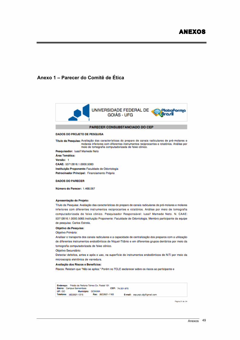
Anexo 1 – Parecer do Comitê de Ética
Anexos 50
Anexos 51
Anexos 52
Anexos 53
Anexo 2 – Original Research Article
Multidimensional analysis of curved root canal preparation using continuous or reciprocating nickel-titanium instruments.
Running Title: Canal transportation and centring ability of NiTi intruments
Keywords: Canal transportation, centering ability, cone beam computed
tomographic, endodontics, nickel-titanium instruments, reciprocating motion.
Acknowledgement: The authors deny any conflicts of interest Federal University of Goiás School of Dentistry PraçaUniversitária s/n, Setor Universitário, 74605-220 Goiânia,GO, Brazil. Phone: +55-62-3209-6254. e-mail: [email protected]
Anexos 54
Abstract Objective: To evaluate transportation (T) and centering ability (CA) of root canal
preparations using continuous or reciprocating nickel-titanium endodontic files.
Material and methods: Ninety-six mesiobuccal root canals of mandibular first and
second molars were randomly divide into 6 groups (n=16) according to the rotary
file used: 1. ProTaper Next®; 2. ProTaper Gold®; 3. Mtwo®; 4. BioRaCe®; 5.
WaveOne Gold®; 6. Reciproc®. Root canals were prepared according to
manufacturer’s instructions. Cone beam computed tomography scans were
obtained before and after root canal preparation. Measurements were made at six
different reference points: 2, 3 and 4 mm from the apex and 2, 3 and 4 mm below
furcation. Transportation and centering ability were analyzed following the
recommendations made by Gambill et al. (1). Results: The greatest mesiodistal
(MD) transportation (T) was found for Reciproc® files (p<0.05), and the greatest
buccolingual (BL) T, for Reciproc®, ProTaper Gold® and ProTaper Next® files
(p<0.05). The greatest mesiodistal (MD) centering ability (CA) was found for
BioRaCe® files (p<0.05), and the greatest buccolingual (BL) CA, for BioRaCe® and
Mtwo® files (p<0.05). Conclusion: All systems produced root canal transportation.
No file system achieved perfect CA of root preparation. Reciproc® files had the
greatest MD T and BL T. BioRaCe® files had the greatest MD CA, whereas BL CA
was similar for BioRaCe® and Mtwo® files.
Keywords: Canal transportation, centering ability, cone beam computed
tomographic, endodontics, nickel-titanium instruments, reciprocating motion.
Anexos 55
Introduction
The purpose of root canal preparation is to clean and shape the pulp
cavity while preserving its original shape and curvature and the position of the
apical foramen (1-3). Procedural iatrogenic errors in the preparation of curved
root canals, such as zips, perforations, decentralized root canals, apical
foramen transport, are risk factors that may lead to root canal treatment failure
(4, 5).
The original shape of curved root canals is better preserved when they
are prepared with flexible nickel-titanium files instead of stainless steel files
(6). The centering ability of continuous and reciprocating nickel-titanium files is
better than that of stainless steel files and, consequently, their root canal
transportation is reduced (7-9).
The cross-section, rake angle, taper, number of flutes and radial land of
nickel-titanium files have specific characteristics (10). The mechanical
properties and the behavior of nickel-titanium alloys may vary according to
their chemical composition and heat treatment during manufacture (11).
ProTaper® files have multiple and progressive tapers. ProTaper Next®,
manufactured from an m-wire alloy, has an eccentric rectangular cross-
section, which provides asymmetrical rotary motion and greater capacity to
remove debris. ProTaper Gold® has a triangular cross-section, which is similar
to the design of the ProTaper Universal® files. These rotary files have more
advanced metallurgic properties than those manufactured using NiTi m-wire
and, therefore, have greater flexibility and resistance to cyclic fatigue (12-14).
Mtwo® files have an S-shaped cross-section, two positive-rake-angle edges
and a small contact surface, which improves their flexibility and, probably, their
Anexos 56
performance inside the root canal (15). RaCe® files have a triangular cross-
section and alternate cutting blades, which results in greater flexibility and
better dentine excision capacity (16). WaveOne® and Reciproc® files have
recently been incorporated into contemporary endodontics. Manufactured
using m-wire nickel-titanium alloys, they have greater flexibility and resistance
to cyclic fatigue (10, 12, 17). Single-use reciprocating files have been
described as an alternative to continuous rotation nickel-titanium files of varied
diameters and tapers (18).
The effectiveness of some of these files has been analyzed considering
geometry preservation and root canal transportation, endodontic file fracture
and dentin fracture (3, 12). Recent studies have demonstrated their
satisfactory results in curved root canal enlargement, transportation and
centering (7, 10, 12). The imaging studies most often used to determine
procedural operative errors are periapical radiographs (4, 14, 19), scanning
electronic microscopy (20), micro-computed tomography (16, 21, 22), and
cone-beam computed tomography (CBCT) (1, 4, 7, 12, 23, 24).
The parameters to determine morphological changes in drilled areas
after root canal preparation should be carefully analyzed. Transportation and
centering ability during preparation at the cervical and apical levels in the
mesiodistal and buccolingual directions on CBCT images may be incorporated
into a method with a solid referential basis. Thus, this study evaluated the
transportation and centring ability of root canal preparation using continuous or
reciprocating nickel-titanium instruments with multidimensional imaging
method.
Anexos 57
Material and methods
Sample selection and preparation
This study was approved by the Ethics in Research Committee of the
Federal University of Goiás, Goiânia, Brazil (CAAE: 53712816.1.0000.5083).
This study included human permanent mandibular first and second molars
obtained from the Dental Urgency Department of the School of Dentistry of the
Federal University of Goiás. The specimens were stored in a solution of 0.2%
thymol.
Inclusion criteria
Preoperative periapical radiographs of each tooth were used for sample
selection according to selection criteria. A platform was used to standardize
tooth images. A Spectro X70 Electronic x-ray unit (Dabi Atlante, Ribeirão
Preto, Brazil) and a RVG 5100 digital sensor (Carestream Dental, Atlanta, GA)
were used for lateral radiographs. All images were evaluated using the RVG
5100 software (Carestream Dental, Atlanta, GA).
Inclusion criteria were: teeth with no internal or external root
resorptions, fractures or calcifications; and with an intact pulp cavity and fully
formed roots.
Exclusion criteria
Baseline images were acquired using a Prexion 3D scanner (PreXion
3D Inc., San Mateo, CA). Image thickness was 0.110 mm (size: 1.170 mm X
1.570 mm X 1.925 mm), 81.00 mm X 75 mm FOV, 0.100 mm voxel, 33.5 s
scan time (1,024 matrix), 90 KVP and 4 mA. The images were analyzed using
the CT scanner software (Prexion 3D Viewer, TeraRecon Inc, Foster City, CA)
on an Intel i7 2.86 GHz (Intel Corp., Santa Clara, CA) Windows 8 Professional
Anexos 58
(Microsoft Corp., Redmond, WA) workstation equipped with an NVIDIA
GeForce 6200 turbo cache videocard (NVIDIA Corp., Santa Clara, CA) and a
1600 X 1200 pixels ELZO-Flexscan S2000 monitor (ELZO NANAO Corp.,
Hakusan, Japan).
Teeth were excluded if their length was greater than 22 mm, or if
mesiobuccal canals had more than one apical foramen or a curvature radius
smaller than 4 mm and greater than 9 mm, according to the method described
by Estrela et al. (25). To determine the curvature radius of curved root canals,
two 6-mm straight lines were superimposed to the root canal image: the
primary line defined the apical region, and the secondary, the middle and
cervical thirds. Regardless of the total length of the secondary line, only the 6
mm closest to the primary line were used for the measurements. The midpoint
of each line was determined, and two perpendicular lines were drawn to a
central point of a circumference, the circumcenter. The distance from
circumcenter to the midpoint of each line (primary and secondary) was the
circle radius, which represented the magnitude of the curvature.
Root canal preparation
The teeth were rinsed under running water to fully remove thymol
solution and then dried with absorbing paper towels. After that, they were
immersed in 5% sodium hypochlorite for 30 min to remove all organic tissues.
The study sample comprised 96 mesiobuccal canals of mandibular
molars randomly distributed into 6 groups (n=16) of different rotary systems: 1.
ProTaper Next® X4 (Dentsply/Maillefer, Switzerland); 2. Protaper Gold® F4
(Dentsply/Maillefer, Switzerland); 3. Mtwo® 40/.04 (VDW Dental, Germany); 4.
Anexos 59
BioRaCe® BR5 (FKG Dentaire, Switzerland); 5. WaveOne Gold® Large
(Dentsply/Maillefer, Switzerland); 6. Reciproc® R40 (VDW Dental, Germany).
A high-speed handpiece, round diamond burs (#1013, #1014; KG
Sorensen, Barueri, Brazil) and an Endo Z bur (Dentsply/Maillefer, Switzerland)
were used for coronal flaring under irrigation. After that, the mesiobuccal root
canals were explored and their contents were removed using K #10 and K #12
stainless steel handfiles (Dentsply/Maillefer, Switzerland). The cervical third
was prepared using the files for this area in each system under study. Working
length was determined using a K #15 file (Dentsply/Maillefer, Switzerland) and
confirmed by visualization of the file tip through the apical foramen. The file
was pulled back one millimeter to determine actual working length.
Root canals were prepared according to manufacturer’s instructions. An
X-Smart Plus® engine (Dentsply/Maillefer, Switzerland) was used for all files,
and the root canals were irrigated with 2.5% sodium hypochlorite freshly
prepared before use (Fitofarma, Goiânia, Brazil) and delivered using a Navitip
irrigation tip (Ultradent Products Inc., South Jordan, UT). During canal
cleaning, 30 mL of irrigant was used. Canal preparation was completed when
the last file reached working length in free rotation and then removed. Patency
was checked with a K #15 file (Dentsply/Maillefer, Switzerland). After
instrumentation was completed, the root canals were dried with absorbing
paper points (Dentsply, Petrópolis, Brazil) of the same caliber as the last file
and then irrigated with 5 mL of 17% EDTA (F&A Laboratório Farmacêutico
Ltda, São Paulo, Brazil) for 3 min. After the last irrigation with 5 mL of 2.5%
NaOCl, the root canals were dried again.
Anexos 60
Each endodontic file was used to prepare only one root canal. All the
root canals were prepared by an endodontist with over 15 years’ experience.
After preparation, final CBCT scans were obtained to evaluate
transportation and centering ability of the endodontic files. The same protocol
described for the acquisition of baseline images was followed, and the image
synchronization tool of the Prexion software (Prexion 3D Viewer, TeraRecon
Inc, Foster City, CA) was used for the axial, coronal and sagittal views.
The following reference points were used for measurements on the root
canal images: 1- 2 mm short of the apex; 2- 3 mm short of the apex; 3- 4 mm
short of the apex; 4-2 mm below furcation; 5-3 mm below furcation; 6-4 mm
below furcation. Navigation on the axial view of synchronized images started
at the root apex, both on baseline and final images, and moved to the
measurement points on the apical third. For the measurements on the cervical
third, navigation started at furcation and moved down up to 4 mm. To facilitate
measurements, the enlargement, brightness and contrast tools available in the
software were used.
Analysis of root canal transportation
The evaluation of images to determine root canal transportation (T)
followed the method described by Gambill et al. (1). Root canal transportation,
determined in the mesiodistal and buccolingual directions at the six points
described above, corresponded to the variation, in millimeters, of the deviation
from the central axis of the root canal after preparation. Mesiodistal
transportation was the shortest distance between the mesial and distal walls of
the root canal and the external mesial and distal surface before (M1 and D1)
and after (M2 and D2) root canal preparation (Figure 1).
Anexos 61
In the same way, buccolingual root canal transportation was the
shortest distance between the images of the buccal and lingual wall of the root
canal and the external buccal and lingual surfaces before (B1 and L1) and
after (B2 and L2) root canal preparation (Figure 1). The images were analyzed
using the CT scanner software (Prexion 3D Viewer, TeraRecon Inc, Foster
City, CA) on an Intel i7 2.86 GHz (Intel Corp., Santa Clara, CA) Windows 8
Professional (Microsoft Corp., Redmond, WA) workstation equipped with an
NVIDIA GeForce 6200 turbo cache videocard (NVIDIA Corp., Santa Clara,
CA) and a 1600 X 1200 pixels ELZO-Flexscan S2000 monitor (ELZO NANAO
Corp., Hakusan, Japan). Measurements were made by one examiner at two
time points, and agreement was greater than 80% according to kappa
statistics (K=0.882).
Mesiodistal (MD) and buccolingual (BL) transportation was calculated
using the formula below:
T(MD) = (M1 – M2) – (D1 – D2) and T(BL) = (B1 – B2) – (L1 – L2). A
negative T(MD) result indicated distal transportation, whereas a positive
number indicated mesial transportation; a result of zero indicated no
transportation. A negative T(BL) result indicated lingual transportation,
whereas a positive number indicated buccal transportation; a result of zero
indicated no transportation.
Analysis of centering ability of root canal preparation
Centering ability (CA) was analyzed using the method described by
Gambill et al. (1), who defined centering ability as the capacity of an
endodontic file of preserving the central axis of the root canal. CA was
calculated using the distances measured to define transportation.
Anexos 62
Mesiodistal and buccolingual CA were calculated using the following
formula:
CA(MD) = (M1-M2) / (D1-D2) or CA(MD) = (D1-D2) / (M1-M2)
CA(BL) = (B1-B2) / (L1-L2) or CA(BL) = (L1-L2) / (B1-B2)
The numerator was the smallest difference between distances before
and after preparation. When the result was one (CA = 1), mesiodistal or
buccolingual CA was perfect, and the closest it was to zero
(0 ≥ CC ≤ 0.999), the poorest the centering ability was.
Statistical analysis
The original transportation and CA values were entered in a Microsoft
Office Excel spreadsheet (Microsoft Corporation, Redmond, WA) and later
exported to the IBM SPSS 20.0 software (SPSS Inc., Nova York, NY) for
statistical analyses. Data were described as median, minimum and maximum
values, and compared between files using the Kruskal-Wallis test, and
compared between specific points in the root canal using the Friedman test.
The Bonferroni correction was used to adjust comparisons. The level of
significance was set at 5%.
Results
All files under study had positive median T(MD), that is, all produced
mesial transportation (Figure 2). The lowest T(MD) was found for Mtwo®,
ProTaper Next®, BioRaCe®, ProTaper Gold® and WaveOne Gold systems,
and there were no significant differences between them. The greatest T(MD)
was found for Reciproc®, and the result was significantly different from those
found for the other systems (p<0.05) (Table 1). However, when the six points
Anexos 63
were evaluated, there were no significant differences between systems at 2
mm short of the apex.
The analysis of root canal T(BL) revealed that the median values of all
systems under study were positive, that is, transportation occurred in the
buccal direction (Figure 3). The lowest T(BL) values were found for the Mtwo®,
BioRaCe® and WaveOne Gold® systems, with no significant differences
between them; the highest T(BL) values were found for the Reciproc®,
ProTaper Gold® and ProTaper Next® systems, and their differences from the
other systems were statistically significant (p<0.05) (Table 1). Although there
were no significant differences between files at 2 mm and 3 mm short of the
apex and at 2 mm below furcation, the Reciproc® system had a greater T(BL)
than the other systems at 4 mm short of the apex and at 3 mm below furcation
(p<0.05). However, the Reciproc® and ProTaper Gold® systems had a greater
T(BL) than the other systems at 4 mm below furcation, and the difference was
significant (p<0.05).
No system under study had median values of CA(MD) equal to one,
that is, there was no perfect centering ability (Figure 4). The lowest CA(MD)
value was found for the Reciproc® system, and the difference from the other
systems was statistically significant (p<0.05), whereas the BioRaCe® system
had the highest values, and the difference between systems was also
statistically significant (p<0.05) (Table 2). However, when findings for the six
measurement points were compared, no significant differences were found
between systems at 2 mm and 3 mm short of the apex ad at 2 mm below
furcation. The highest CA(MD) value at 4 mm short of the apex was found for
the BioRaCe® system, and the values were statistically different from those
Anexos 64
found for the ProTaper Next® and Reciproc® systems (p<0.05). The lowest
CA(MD) at 3 mm below furcation was found for the Reciproc® system, and the
difference from the other systems was significant (p<0.05). The WaveOne
Gold® and Reciproc® systems had the lowest CA(MD) values at 4 mm below
furcation, and the differences from the other systems were statistically
significant (p<0.05).
The analysis of CA(BL) revealed that no system under study had
median values equal to one, that is, no system had perfect centering ability
(Figure 5). The highest CA(BL) values were found for the BioRaCe® and
Mtwo® systems, and the differences from the other systems were statistically
significant (p<0.05) (Table 2). The analysis of CA(BL) values at 2 mm, 3 mm
and 4 mm short of the apex and at 2 mm and 3 mm below furcation did not
reveal any statistically significant differences between systems. However, the
ProTaper Next® system had the lowest CA(BL) value at 4 mm below furcation,
and this result was statistically different from the value found for Mtwo®
(p<0.05).
The measurements at the six points before and after root canal
preparation revealed the lowest T(MD) was found at 2 mm short of the apex,
and this finding was statistically significant (p<0.05) (Table 3); and the lowest
T(BL) was found at 2 mm and 3 mm short of the apex, but the differences
were not statistically significant (Table 4).
The highest CA(MD) values were found at 4 mm short of the apex, and
the highest CA(BL), at 3 mm below furcation, but the differences between
systems were not statistically significant (Tables 5 and 6)
Anexos 65
Figures 6 and 7 show CBCT axial views of the cervical and apical thirds
of the mesiobuccal canals before and after preparation according to the type
of NiTi file used.
Discussion
Changes in root canal shape in the cervical and apical thirds were
found for all the files under study. The Protaper Next®, Protaper Gold®,
WaveOne Gold®, Mtwo® and BioRaCe® files had the lowest values of
mesiodistal transportation. The lowest buccolingual values were found for the
WaveOne Gold®, Mtwo® and BioRaCe® systems. There were no significant
differences in mesiodistal transportation of the files under study at 2 mm short
of the apex. The same was found for the buccolingual values at 2 mm and 3
mm short of the apex and at 2 mm below furcation.
Perfect mesiodistal or buccolingual CA was not found for any of the
systems under evaluation. The best result was the CA(MD) of the BioRaCe®
system. The greatest CA(BL) value was found for the BioRaCe® and Mtwo®
systems. CA(MD) at 2 mm and 3 mm short of the apex and at 2 mm below
furcation was not significantly different between systems. However,
buccolingual findings were not significantly different between systems at 2
mm, 3 mm or 4 mm short of the apex or at 2 mm and 3 mm below furcation.
Transportation and CA were determined using a model previously
described by Gambill et al. (1), who used a model of mesiodistal
measurements of axial plans from the apex of single-rooted teeth whose
curvature was less than 10 degrees. Several studies have adopted this
method to evaluate transportation and CA after the preparation of curved root
Anexos 66
canals (7, 12, 24).
Our study used mesiobuccal root canals of mandibular molars with a
curvature radius greater than 4 mm and shorter than 8 mm (25). Two areas of
clinical reference, 2 mm to 4 mm below furcation and 2 mm to 4 mm short of
the apex, and two directions, mesiodistal and buccolingual, were analyzed
using high resolution CBCT images.
Several studies used different methods to evaluate the preservation of
the shape of curved root canals after preparation with stainless steel files or
continuous or reciprocating NiTi files: artificial canals (26), scanning electronic
microscopy (20), periapical radiographs of human teeth (10, 14, 19, 27),
micro-computed tomography (8, 13, 15, 16, 21, 22, 28-30), and CBCT (1, 7,
12, 23, 24).
CBCT, a non-destructive procedure, may be a potential method for an
accurate evaluation of root canal geometry because of the different plans used
for three-dimensional analysis and the axial views, which avoid the
superimposition of structures (1, 23, 25).
This study found that there were both mesiodistal and buccolingual
changes in the shape of the root canal (Figures 2 and 3). The morphological
buccolingual changes after the preparation of curved root canals may be
accurately evaluated using CBCT images. The major morphological changes
were mesiodistal.
The results found for the ProTaper Next®, ProTaper Gold®, WaveOne
Gold®, Mtwo®, BioRaCe® and Reciproc® files in the analysis of transportation
and centering ability after the preparation of curved root canals are in
agreement with previous studies, which concluded that they preserved the
Anexos 67
shape of root canals (13, 15, 19, 27). Several studies found that the results of
shape preservation after root canal preparation were often favorable for new
endodontic files, but their methods, measurement points along the root thirds
and the amount of enlargement that they used were different from each other
(10, 20, 22). In this study, root canals were prepared using of the following
diameters and tapers: 40/.04, 40/.06 and 45/.05. The discussion about how
much a root canal should be enlarged remains to be investigated in future
studies. WU et al. (31) found that canal preparation depends on the
morphology and thickness of the root canal walls, as well as on the taper of
the file selected. At the same time, other studies reported that not all the walls
are touched by the files during root canal preparation (3, 23, 31-33). In this
study, the apex was enlarged using files whose diameter was larger than 350
µm, and, therefore, it was possible to compare the performance of files whose
diameter and taper were larger in curved root canals. Moreover, these files act
on a larger area of the root canal walls, which facilitates the penetration of the
irrigation tip and, consequently, the action of antimicrobials. Despite the
amount of enlargement, the files used for root canal preparation had
satisfactory transportation and CA results (Figures 2, 3, 4 and 5). An earlier
study (34) found that there were no significant differences in transportation
and CA after 35/.02 and 50/.02 RaCe® files were used for root canal
preparation.
The files under evaluation in this study were selected according to their
morphological characteristics, mechanical properties, chemical composition
and capacity of preserving the original shape of curved root canals because of
their flexibility (3, 7-11). The endodontic files included in this study have
Anexos 68
different cross-sections, diameters, tapers, types of alloy and tip designs (8,
10, 13, 14, 19). The comparison of the ProTaper files (ProTaper Universal®,
ProTaper Next® and ProTaper Gold®) to each other revealed that the
ProTaper Gold® files, because of the technology used for their manufacture
and thermal treatment, had better cyclic fatigue and flexibility and produced
less apical transportation than the ProTaper Universal® and ProTaper Next®
files (13). Mtwo® files are manufactured using the conventional nickel-titanium
alloy. BioRaCe® files, when used to prepare curved root canals, do not change
the original root canal anatomy because of their triangular cross section,
associated with their flexibility, and their alternating cutting edges, which avoid
self-threading (14, 34). The angle of the taper of Reciproc® and WaveOne®
files is high in their apical 3 mm (D0 - D3) (10, 14, 18). In addition to their
cross-sectional design, another important characteristic is a result of the use of
the m-wire alloy, responsible for their greater flexibility (10, 12, 17).
Method variations between studies, particularly the amount of apical
enlargement, evaluation criteria and evaluation tools justify the differences
found. However, there seems to be a consensus about the fact that the use of
NiTi rotary files results in low apical transportation and good centering during
root canal preparation.
Bürklein et al. (19) evaluated the shaping effectiveness of rotary flies
with a diameter of 400 µm (ProTaper Universal®, ProTaper Next®, BT-RaCe®
and Mtwo®) used to prepare severely curved root canals. The files were safe
and preserved root canal curvature. Garcia et al. (27) used periapical
radiographs to evaluate transportation of ProFile® and RaCe® files in the
preparation of mandibular molar canals. The size (diameter and taper) of the
Anexos 69
files used for apical enlargement was 40/.04. They did not find any significant
differences in apical transportation. Yang et al. (15) compared the geometry of
root canals prepared with ProTaper Universal® and Mtwo® files. Both files
preserved canal geometry during preparation. Gagliardi et al. (13) used micro-
computed tomography imaging to evaluate transportation and CA in curved
root canals of mandibular molars prepared with ProTaper Gold®, ProTaper
Next® and ProTaper Universal® files. Apical enlargement corresponded to 250
µm files. ProTaper Gold® and ProTaper Next® files produced less
transportation and had better CA than the ProTaper Universal® files.
Reciprocating rotary files also had satisfactory transportation and CA
results in the preparation of curved root canals at the different points evaluated
(cervical and apical thirds). Capar et al. (12) used CBCT to compare the
effects of OneShape®, ProTaper Universal®, ProTaper Next®, Reciproc® R25,
Twisted File Adaptive® and WaveOne® Primary files on transportation and CA
when used to prepare curved canals. All files had similar transportation and
CA. Carvalho et al. (7) evaluated apical transportation and CA of the
reciprocating system Reciproc® associated with different glide path
techniques. Root canals prepared using a glide path technique had minimal
apical transportation, and the Reciproc® system had good CA in the
preparation of root canals. Saber et al. (14) compared the shaping ability of
the WaveOne® Primary, Reciproc® R25 and OneShape® systems in the
preparation of severely curved root canals of extracted human molars. All
systems were safe. WaveOne® Primary and Reciproc® R25 files were more
efficient in preserving the original canal curvature. Bürklein et al. (10) used
periapical radiographs to compare Mtwo® 30/.05, ProTaper Universal® F3,
Anexos 70
Reciproc® R25 and WaveOne® Primary files used to shape curved root canals
of extracted teeth. All systems preserved the original root canal curvature and
were safe to use. Amaral et al. (22) used micro-computed tomography to
evaluate transportation and CA in root canals prepared with WaveOne®
Primary files alone or together with previous apical and cervical enlargement.
The association with previous cervical or apical enlargement resulted in a
reduction of transportation and in better CA than the use of WaveOne® files
alone.
Our study found that the preparation of curved root canals using
continuous (ProTaper Next®, ProTaper Gold®, Mtwo® and BioRaCe®) or
reciprocating NiTi files (WaveOne® Gold and Reciproc®) had low apical
transportation and satisfactory CA in the preparation of curved root canals.
The maintenance of the apical limit during instrumentation in rotary and
reciprocating modes was also evaluated previously (35). Seventy-two human
uniradicular mandibular premolars were prepared with #F4 ProTaper and
#R40 Reciproc instruments. The results show that the devices were able to
control the apical limit of the instrumentation independent of the kinematics
and working length applied. Another relevant finding of our study was the
degree of mesiodistal and buccolingual enlargement of curved root canals with
a curvature radius greater than 4 mm and shorter than 8 mm, which was
indicative of satisfactory shaping, both in the cervical and apical thirds.
Contemporary endodontics has developed good standards for shaping
with the new NiTi rotary systems, which has directly affected the quality of
obturations and, therefore, the success and survival of root canal treatments.
However, the challenge and enigma of biofilm destruction in areas that the
Anexos 71
files and the antimicrobials do not reach remain as problems to be further
investigated.
In summary, all the continuos and reciprocating files produced root
canal transportation, and no file had perfect CA. The greatest mesiodistal
transportation was found for the Reciproc® system, and buccolingual
transportation, for the Reciproc®, ProTaper Gold® and ProTaper Next®
systems. The BioRaCe® system had the best mesiodistal CA, and the
BioRaCe® and Mtwo® had similar buccolingual CA. The lowest mesiodistal
transportation was found at 2 mm short of the apex. The best mesiodistal CA
was found at 4 mm short of the apex, and the best buccolingual CA, at 3 mm
below furcation.
References 1. Gambill JM, Alder M, Del Rio CE. Comparison of nickel-titanium and
stainless steel hand-file instrumentation using computed tomography. J Endod 1996;22:369-75.
2. Schilder H. Cleaning and shaping the root canal. Dent Clin North Amer
1974;8:269-96. 3. Peters OA. Current challenges and concepts in the preparation of root canal
systems: a review. J Endod 2004;30:559-67. 4. Alencar AHG, Dummer PMH, Oliveira HCM, Pécora JD, Estrela C.
Procedural errors during root canal preparation using rotary NiTi instruments detected by periapical radiography and cone beam computed tomography. Braz Dent J 2010;21:543-9.
5. Estrela C, Holland R, Estrela CRA, Alencar AHG, Sousa-Neto MD, Pécora
JD. Characterization of successful root canal treatment. Braz Dent J 2014;25:3-11.
6. Esposito PT, Cunningham CJ. A comparison of canal preparation with
nickel-titanium and stainless steel instruments. J Endod 1995;21:173-6.
Anexos 72
7. Carvalho GM, Sponchiado-Júnior EC, Garrido ADB, Lia RCC, Garcia LFR, Marques AAF. Apical transportation, centering ability, and cleaning effectiveness of reciprocating single-file system associated with different glide path techniques. J Endod 2015;41:2045-9.
8. Gergi R, Osta N, Bourbouze G, Zgheib C, Arbab-Chirani R, Naaman A.
Effects of three nickel titanium instrument systems on root canal geometry assessed by micro-computed tomography. Int Endod J 2015;48:162-70.
9. Pagliosa A, Sousa-Neto MD, Versiani MA, Raucci-Neto W, Silva-Sousa
YTC, Alfredo E. Computed tomography evaluation of rotary systems on the root canal transportation and centering ability. Braz Oral Res 2015; 29:1-7.
10. Bürklein S, Hinschitza K, Dammascchke T, Schafer E. Shaping ability and
cleaning effectiveness of two single-file systems in severely curved root canals of extracted teeth: Reciproc and WaveOne versus Mtwo and ProTaper. Int Endod J 2012;45:449-61.
11. Thompson AS. An overview of nickel-titanium alloys used in dentistry. Int
Endod J 2000;33:297-310. 12. Capar ID, Ertas H, Ok E, Arslan H, Ertas ET. Comparative Study of
Different Novel Nickel-Titanium Rotary Systems for Root Canal Preparation in Severely Curved Root Canals. J Endod 2014;40:852-6.
13. Gagliardi J, Versiani MA, Sousa-Neto MD, Plazas-Garzon A, Basrani B.
Evaluation of the shaping characteristics of ProTaper Gold, ProTaper Next, and ProTaper Universal in curved canals. J Endod 2015; 41: 1718-24.
14. Saber SEDM, Nagy MM, Schafer E. Comparative evaluation of the
shaping ability of WaveOne, Reciproc and OneShape single-file systems in severely curved root canals of extracted teeth. Int Endod J 2015;48: 109-14.
15. Yang G, Yuan G, Yun X, Zbou X, Liu, B, Wu H. Effects of two nickel-
titanium instrument systems, Mtwo versus ProTaper Universal, on root canal geometry assessed by micro–computed tomography. J Endod 2011;37:1412-6.
16. Pasqualini D, Alovisi M, Cemenasco A, Mancini L, Paolino DS, Bianchi
CC, Roggia A, Scotti N, Berutti E. Micro-computed tomography evaluation of ProTaper Next and BioRaCe shaping outcomes in maxillary first molar curved canals. J Endod 2015;41:1706-10.
Anexos 73
17. Pedullà E, Grande NM, Plotino G, Gambarini G, Rapisarda E. Influence of
continuous or reciprocating motion on cyclic fatigue resistance of 4 different nickel-titanium rotary instruments. J Endod 2013;39:258-61.
18. Yared GM. Canal preparation using only one Ni-Ti rotary instrument:
preliminary observations. Int Endod J 2008;41:339-44. 19. Bürklein S, Mathey D, Schäfer E. Shaping ability of ProTaper Next and
BT-RaCe nickel-titanium instruments in severely curved root canals. Int Endod J 2015;48:775-81.
20. Schäfer E, Vlassis M. Comparative investigation of two rotary nickel-
titanium instruments: ProTaper versus RaCe. Part 2. Cleaning effectiveness and shaping ability in severely curved root canals of extracted teeth. Int Endod J 2004;37:239-48.
21. Peters OA, Arias A, Paqué F. A micro-computed tomography assessment
of root canal preparation with a novel instrument, TRUShape, in mesial roots of mandibular molars. J Endod 2015;41:1545-50.
22. Amaral ROJF, Leonardi DP, Gabardo MCL, Coelho BS, Oliveira KV,
Baratto-Filho F. Influence of cervical and apical enlargement associated with the WaveOne system on the transportation and centralization of endodontic preparations. J Endod 2016;42:626-31.
23. Peters OA, Laib A, Gohring TN, Barbakow F. Changes in root canal
geometry after preparation assessed by high-resolution computed tomography. J Endod 2001;27:1-6.
24. Gergi R, Rjeily JA, Sader J, Naaman, A. Comparison of canal
transportation and centering ability of Twisted Files, Pathfile-ProTaper system, and stainless steel hand K-files by using computed tomography. J Endod 2010;36: 904-7.
25. Estrela C, Bueno MR, Sousa-Neto MD, Pécora JD. Method for
determination of root curvature radius using cone-beam computed tomography images. Braz Dent J 2008;19:114-8.
26. Liu W, Wu B. Root canal surface strain and canal center transportation
induced by 3 different nickel-titanium rotary instruments systems. J Endod 2016;42:299-303.
Anexos 74
27. Garcia M, Duran-Sindreu F, Mercadé M, Bueno R, Roig M. A comparison of apical transportation between ProFile and RaCe rotary instruments. J Endod 2012;38:990-2.
28. Stern S, Patel S, Foschi F, Sherriff M, Mannocci F. Changes in centering
and shaping ability using three nickel-titanium instrumentation techniques analysed by micro-computed tomography. Int Endod J 2012;45:514-23.
29. Gergi R, Arbab-Chirani R, Osta N, Naaman A. Micro–computed
tomographic evaluation of canal transportation instrumented by different kinematics rotary nickel-titanium instruments. J Endod 2014;40:1223-7.
30. Junaid A, Freire LG, Bueno CES, Mello I, Cunha RS. Influence of single-
file endodontics on apical transportation in curved root canals: an ex vivo micro-computed tomography study. J Endod 2014;40:717-20.
31. Wu MK, Roris A, Barkis D, Wesselink PR. Prevalence and extent of long
oval canals in the apical third. Oral Surg Oral Med Oral Pathol 2000;89: 739-43.
32. Wu MK, Barkis D, Roris A, Wesselink PR. Does the first file to bind
correspond to the diameter of the canal in the apical region? Int Endod J 2002;35:264-7.
33. Pécora JD, Capelli A, Guerisoli DMZ, Spanó JCE, Estrela C. Influence of
cervical preflaring on apical file size determination. Int Endod J 2005;38: 430-5.
34. Pasternak-Júnior B, Souza-Neto MD, Silva RG. Canal transportation and
centering ability of RaCe rotary instruments. Int Endod J 2009;42:499-506.
35. Vasconcelos BC, Frota LMA, Souza TA, Bernardes RA, Duarte MAH. Evaluation of the maintenance of the apical limit during instrumentation with hybrid equipment in rotary and reciprocating modes. J Endod 2015;41:682–5.
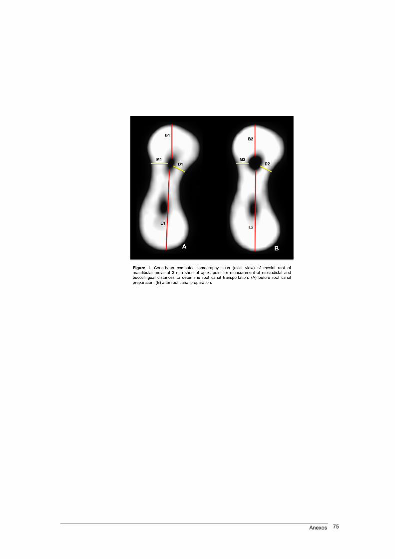
Anexos 75
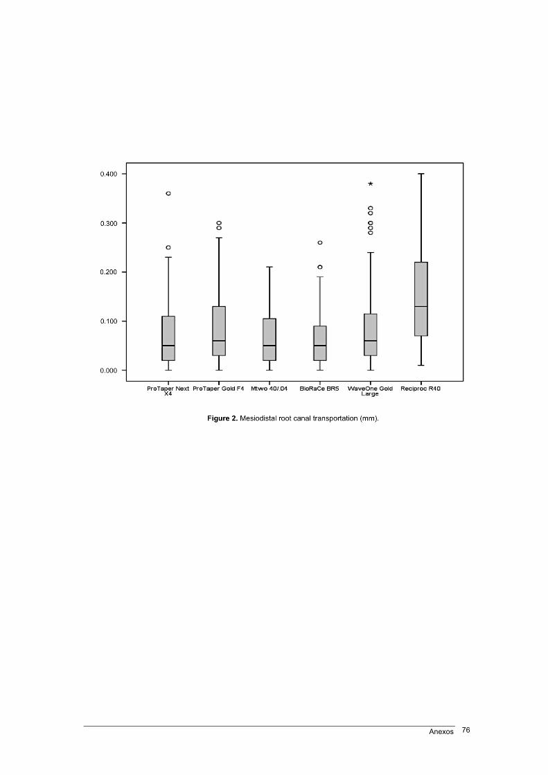
Anexos 76
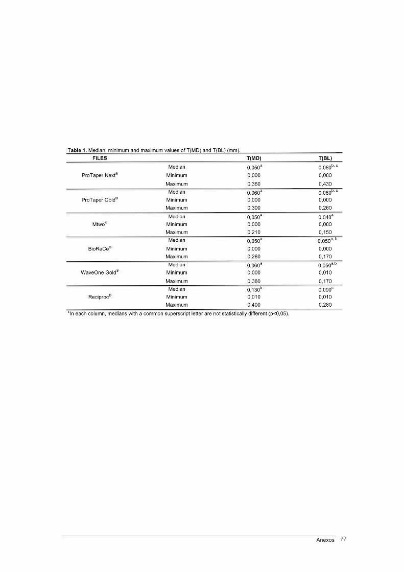
Anexos 77
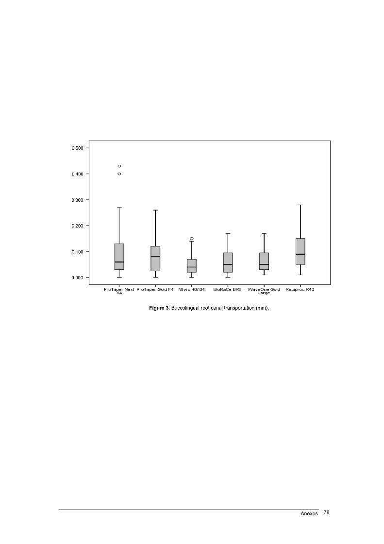
Anexos 78
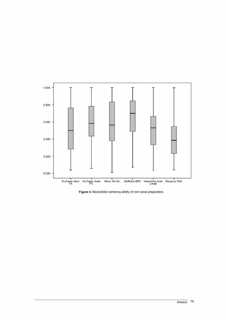
Anexos 79
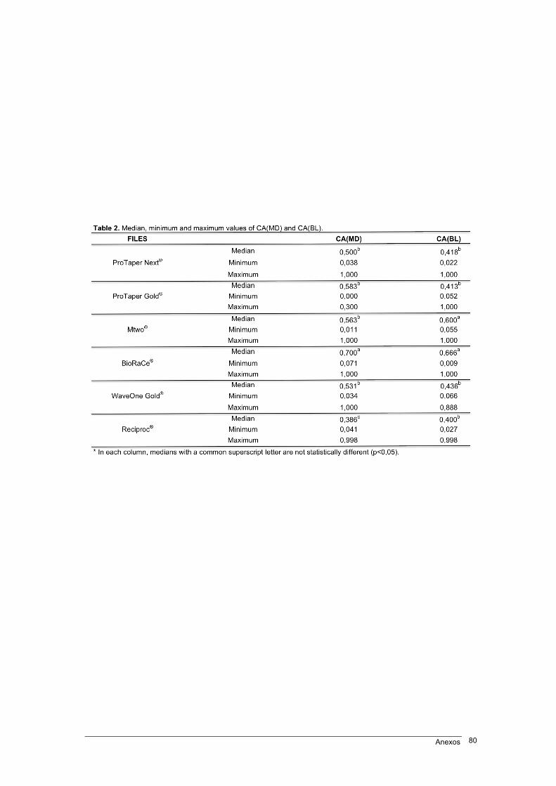
Anexos 80
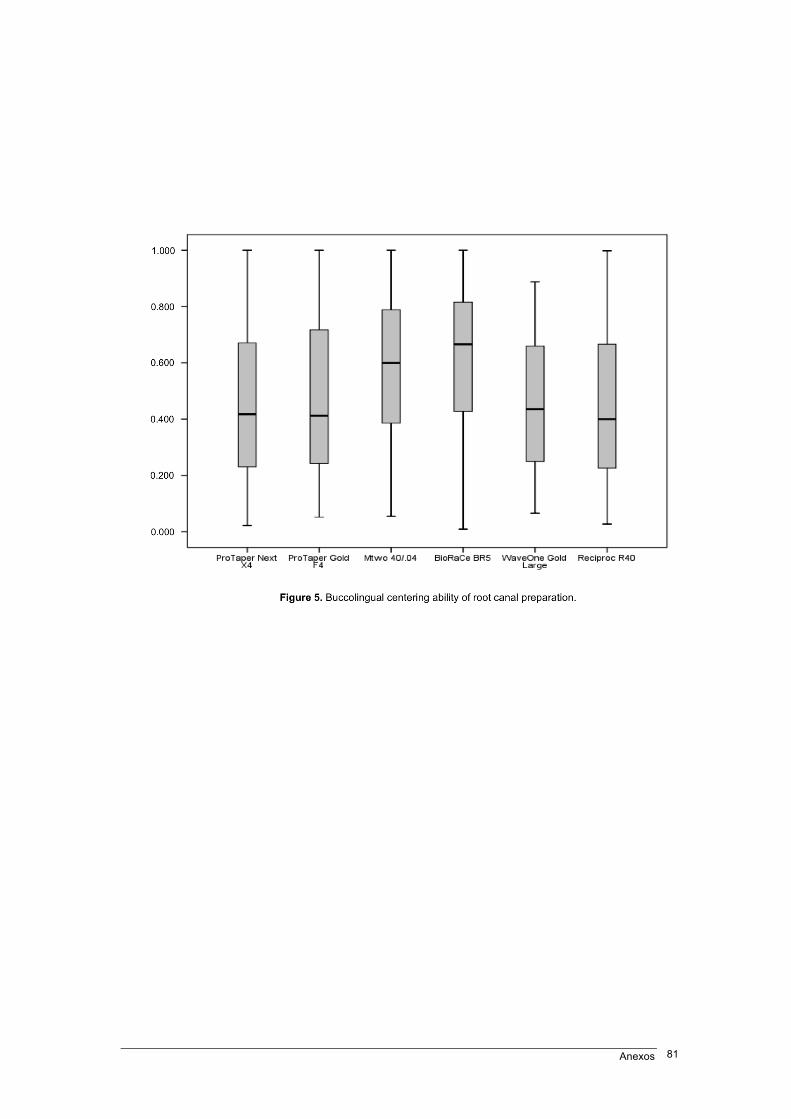
Anexos 81
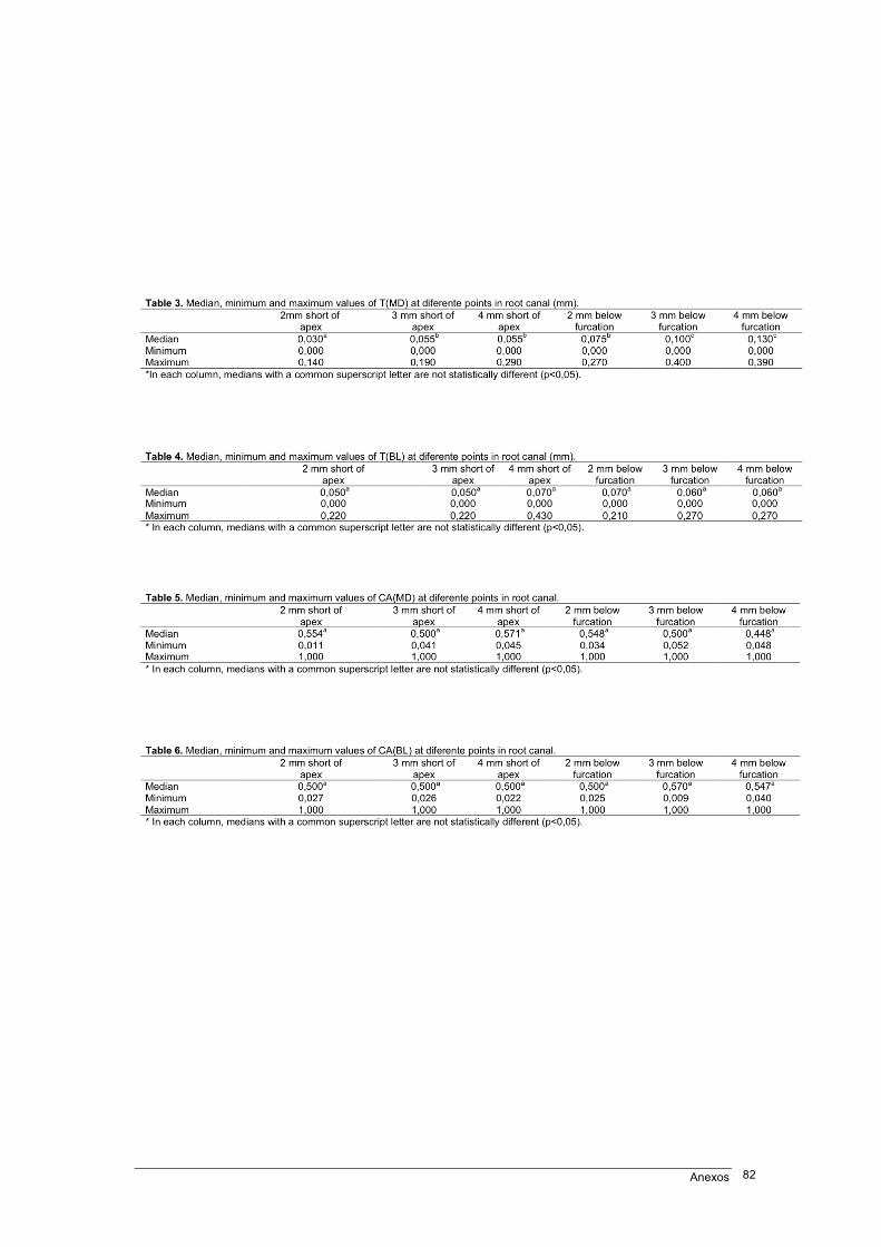
Anexos 82
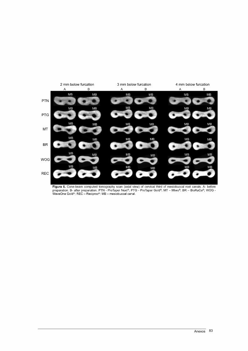
Anexos 83
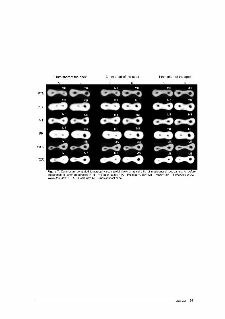
Anexos 84
Anexos 85
ANEXO 3 – Normas de publicação do respectivo periódico

Anexos 86

Anexos 87
Anexos 88
Anexos 89