ana catarina marinho da rocha - repositorio-aberto.up.pt · adicionalmente, foi observada...
TRANSCRIPT

Germline and Somatic Alk Alterations in Neuroblastoma Patients
Ana Catarina Marinho da Rocha
M 2017
MESTRADO EM ONCOLOGIA
ESPECIALIZAÇÃO EM ONCOLOGIA MOLECULAR
Ana C
atarina Marinho da R
ocha.Germ
line and Somatic Alk A
lterations in Neuroblastom
a PatientsM
.ICB
AS 2017
Germ
line and Somatic Alk A
lterations in Neuroblastom
a Patients
Ana C
atarina Marinho da Rocha
INST
ITU
TO D
E CIÊN
CIA
S BIOM
ÉDIC
AS A
BEL SALA
ZA
R


Ana Catarina Marinho da Rocha
GERMLINE AND SOMATIC ALK ALTERATIONS IN
NEUROBLASTOMA PATIENTS
Dissertação de candidatura ao grau de Mestre em
Oncologia – especialização em Oncologia Molecular –
submetida ao Instituto de Ciências Biomédicas Abel
Salazar da Universidade do Porto
Orientador: Manuel António Rodrigues Teixeira, MD, PhD
Diretor do Serviço de Genética e Centro de Investigação
Instituto Português de Oncologia do Porto
Professor Catedrático Convidado do Departamento de
Patologia e Imunologia Molecular
Instituto de Ciências Biomédicas Abel Salazar da
Universidade do Porto
Coorientadores:
Ana Luísa Pinto da Silva Lobo Peixoto, MSc
Serviço de Genética e Centro de Investigação
Instituto Português de Oncologia do Porto
Joana Virgínia Pinto Valejo de Magalhães Vieira, MSc
Serviço de Genética e Centro de Investigação
Instituto Português de Oncologia do Porto


Nothing in life is to be feared, it is only to be understood. Now is
the time to understand more, so that we may fear less.
― Marie Curie


AGRADECIMENTOS
É com muito gosto que deixo, desde já, o meu agradecimento a todos os que de
alguma forma contribuíram para a realização deste trabalho.
Em primeiro lugar, gostaria de agradecer ao PROFESSOR MANUEL TEIXEIRA,
pela oportunidade de realizar este trabalho no Serviço de Genética, sob a sua
orientação. O meu muito obrigado, pela constante disponibilidade e pelo apoio e
motivação e, sobretudo, pela oportunidade de crescimento pessoal e profissional
que me proporcionou neste último ano, e por todos os conhecimentos que me
transmitiu.
Às minhas coorientadoras, ANA PEIXOTO e JOANA VIEIRA, pela orientação
durante o último ano. O meu muito obrigado pelo apoio e disponibilidade e,
sobretudo, pela paciência que demonstraram, pelos conhecimentos transmitidos e
pela confiança depositada em mim para a realização deste trabalho.
À PROFESSORA BERTA MARTINS e à PROFESSORA CARMEN JERÓNIMO,
anterior e atual diretoras do Mestrado em Oncologia, e a todos os DOCENTES do
Mestrado por todo o conhecimento partilhado nos últimos dois anos.
À LIGA PORTUGUESA CONTRA O CANCRO, pelo apoio financeiro.
Ao Serviço de ANATOMIA PATOLÓGICA, em especial ao Dr. João Lobo, pela
cooperação com material histológico.
À MANELA, por toda a ajuda, paciência e dedicação.
Às minhas colegas, ANA, ANDREIA, MARIA, MARTA, PAULA e ISABEL, por me
terem recebido tão bem, pelos conhecimentos e risadas partilhadas, por todos os
bons momentos que me proporcionaram e pelo apoio e motivação nos momentos
de maior frustração. O meu muito obrigado pela vossa amizade no último ano.
Aos meus amigos da vida e para a vida, DANIELA, EMILIE, TITA, DIANA,
SANDRINA, ISA, GEMEAS e GONZÁLEZ, por todo o apoio moral e incondicional
e por todos os bons momentos de descontração que sempre me proporcionam.

À minha família, aos meus PAIS e IRMÃO, pelo apoio incondicional, por estarem
sempre presentes nos momentos bons, mas sobretudo nos menos bons, por serem
o meu pilar, o meu porto seguro, mesmo que por vezes à distância. OBRIGADA
POR TUDO.

Esta tese é dedicada ao meu AVÔ, que infelizmente me deixou
(fisicamente) a meio deste percurso, mas que continua sempre presente na minha
vida. Ele é a minha fonte de inspiração e a minha motivação para sempre
continuar, sem nunca desistir.
OBRIGADA AVÔ.


TABLE OF CONTENTS

xi
TABLE OF CONTENTS
FIGURE INDEX ................................................................................................... XV
TABLE INDEX ................................................................................................... XIX
SUPPLEMENTARY TABLE INDEX ................................................................. XXIII
SUMMARY .................................................................................................... XXVII
RESUMO ........................................................................................................ XXXI
LIST OF ABBREVIATIONS ........................................................................... XXXV
I. Introduction ................................................................................................... 1
1. Neuroblastoma ......................................................................................... 2
1.1. Epidemiology ........................................................................................ 2
1.2. Screening and risk classification ........................................................... 3
1.3. Clinical presentation .............................................................................. 4
1.4. Treatment ............................................................................................. 5
2. Genetic alterations in NB .......................................................................... 6
2.1. Germline mutations ............................................................................... 6
2.2. Chromosomal changes ......................................................................... 8
2.3. Somatic point mutations ........................................................................ 9
3. The anaplastic lymphoma kinase (ALK) gene .........................................10
3.1. ALK changes in various cancers ..........................................................11
3.2. ALK rearrangements in NB ..................................................................11
3.3. Somatic ALK point mutations in NB ......................................................13
3.4. Germline ALK mutations in hereditary NB ............................................14
3.5. ALK inhibitors in neuroblastoma treatment ...........................................15
II. Aims of the study......................................................................................19
III. Material and Methods ...............................................................................23
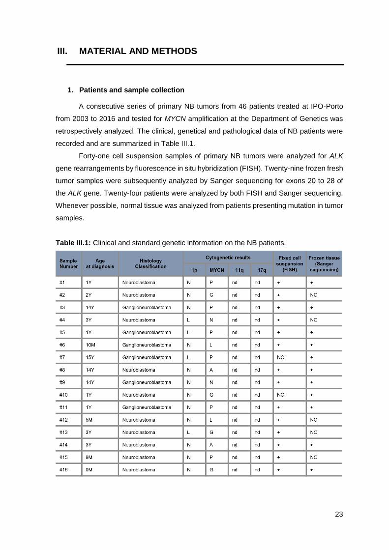
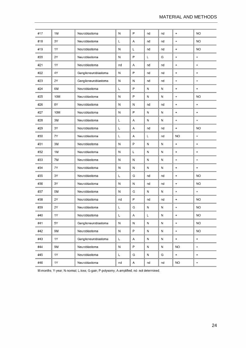
1. Patients and sample collection ................................................................23
2. Fluorescence in situ hybridization ...........................................................25
3. DNA extraction and quantification ...........................................................26
3.1. Extraction of genomic DNA from frozen fresh samples ........................26
3.2. Extraction of genomic DNA from paraffin-embedded samples .............26
4. DNA sequencing .....................................................................................27
IV. Results ......................................................................................................33
1. Assessment of ALK rearrangements in NB patients ................................33
1.1. Clinicopathological characteristics of patient with ALK amplification .....35
2. ALK mutations in NB patients ..................................................................36

xii
2.1. Clinicopathological characteristics of patients with ALK mutations .......37
V. Discussion ................................................................................................41
VI. Future perspectives ..................................................................................47
VII. Bibliography ..............................................................................................51
SUPPLEMENTARY TABLES ..............................................................................61

FIGURE INDEX


xv
FIGURE INDEX
Figure I.1: Current standard-of-care treatment strategy for high-risk neuroblastoma (NB). 6
Figure I.2: Rare and common genomic variants that predispose to NB ............................. 8
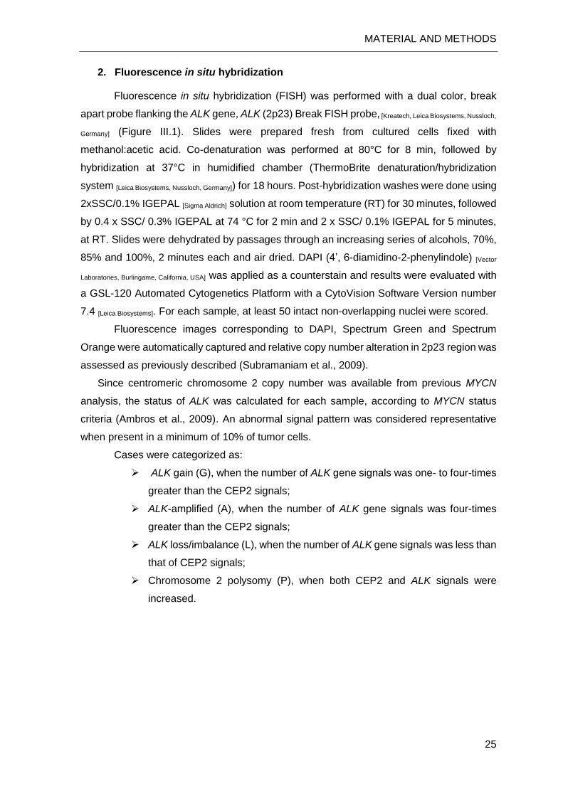
Figure III.1: ALK (2p23) break FISH probe to detect rearrangements involving the ALK gene.
........................................................................................................................................26
Figure IV.1: Representative FISH images from four selected NB tumors using a dual color,
break apart probe targeting 3′ALK and 5′ALK, labeled orange and green, respectively. ...35
Figure IV.2: DNA sequence electropherograms obtained from NB tumor samples ..........37


TABLE INDEX

xix
TABLE INDEX
Table I.1: International Neuroblastoma Risk Group Staging System ................................. 3
Table I.2: International Neuroblastoma Risk Group pretreatment classification schema ... 4
Table I.3: ALK gene rearrangements described in NB. ....................................................12
Table I.4: ALK somatic mutations described in NB patients. ............................................14
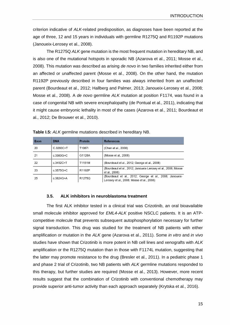
Table I.5: ALK germline mutations described in hereditary NB. ........................................15
Table III.1: Clinical and standard genetic information on the NB patients. ........................23
Table III.2: Set of primers used to amplifying exons 20 to 28 ...........................................28
Table IV.1: ALK alterations assessed by FISH in NB patients. .........................................33
Table IV.2: Mutations found in the ALK gene. ..................................................................36


SUPPLEMENTARY TABLE INDEX


xxiii
SUPPLEMENTARY TABLE INDEX
Supplementary Table 1: The International Neuroblastoma Pathology Classification
(Shimada system). ...........................................................................................................63
Supplementary Table 2: International Neuroblastoma staging system (INSS) ................64


SUMMARY


xxvii
SUMMARY
Neuroblastoma (NB) is a pediatric cancer of the developing sympathetic nervous
system that accounts for more than 7% of all childhood cancers. It is the most common
extracranial solid tumor diagnosed during infancy and is responsible for around 10 to 12%
of childhood cancer-related deaths. Although most NB tumors arise sporadically, around 1
to 2% of the cases are inherited in an autosomal dominant manner. Anaplastic lymphoma
kinase (ALK) is a cell-membrane associated tyrosine kinase receptor, preferentially
expressed in the central and peripheral nervous system. Relatively recent findings have
suggested that the ALK gene may be activated by genomic rearrangements or specific point
mutations targeting the tyrosine kinase domain (TKD), thereby contributing to tumor
development, and that NB harboring this ALK alterations might be sensitive to ALK
inhibitors. Additionally, ALK germline mutations explain a large proportion of hereditary NB,
being present in approximately half of hereditary cases.
In the present study, we aimed to search for ALK fusion genes, to compare the
pattern of amplification of the ALK and MYCN genes, to identify somatic and germline ALK
TKD point mutations and to relate the ALK alterations with clinical outcome in NB patients.
We therefore performed fluorescence in situ hybridization (FISH) in 41 cell suspension
tumor samples and Sanger sequencing in 29 fresh-frozen tumor samples from NB patients
treated at IPO-Porto from 2003 to 2016 and tested for MYCN amplification at the
Department of Genetics of that institution. Whenever possible, normal tissue was analyzed
from patients presenting mutations in tumor samples.
Of the 41 patients assessed by FISH, no translocation or inversion were found, but
aberrant copy number of the ALK gene was observed in 19 cases (46.3%). This included
ALK amplification in one of 41 (2.4%), ALK gain in 13 of 41 (31.7%), and ALK
loss/imbalance in five of 41 (12.2%), changes that in general are associated with more
advanced clinical stages and worse outcomes. Additionally, polysomy of chromosome 2
was observed in 15 cases (36.6%), which is an indicator of a better outcome in patients
without other adverse prognostic features. Synchronic MYCN and ALK aberrations
accounted for 17 of 41 (41%) tumors. Moreover, we found four different ALK TKD mutations
in five of 29 tumors tested by Sanger sequencing, namely, the F1174L, R1192P, F1174I,
and R1275Q mutations. Of these, the mutations R1192P and R1275Q were found in the
germline of two patients, being the most frequent ALK germline mutations described in
hereditary NB.
We conclude that ALK alterations are a frequent event in NB patients, either by
cytogenetic events or point mutations, and that these alterations could be a predictive and

xxviii
prognostic biomarker, which can also be a potential therapeutic target in a subset of
patients. We also show the importance of understanding which ALK mutations are more
likely to be associated with inherited predisposition to NB tumors, which can help direct
appropriate screening in families carrying ALK germline mutations.

RESUMO


xxxi
RESUMO
O neuroblastoma (NB) é um cancro pediátrico que surge durante o desenvolvimento
do sistema nervoso simpático e que representa mais de 7% de todos os cancros na
infância. É o tumor sólido extracraniano mais frequentemente diagnosticado na infância e
é responsável por cerca de 10 a 12% da mortalidade por cancro em crianças. Embora a
maioria dos NBs sejam esporádicos, cerca de 1 a 2% dos casos são hereditários, com
transmissão autossómica dominante. O gene ALK codifica um recetor tirosina quinase
transmembranar, que é preferencialmente expresso no sistema nervoso central e
periférico. Estudos relativamente recentes sugerem que o gene ALK pode ser ativado
devido à ocorrência de rearranjos cromossómicos ou mutações pontuais no domínio
tirosina quinase, contribuindo para o desenvolvimento tumoral. Tumores que apresentam
alterações no gene ALK podem responder a inibidores do ALK. Além disso, mutações
germinativas no gene ALK são responsáveis pela maioria dos tumores hereditários,
estando presente em aproximadamente metade dos casos.
O presente estudo teve como principais objetivos pesquisar genes de fusão
envolvendo o gene ALK; comparar o padrão de amplificação dos genes ALK e MYCN;
identificar mutações pontuais somáticas e germinativas no domínio tirosina quinase do
gene; e, ainda, relacionar as alterações encontradas no gene ALK com dados clinico-
patológicos dos pacientes com NB. Foi utilizada a técnica de FISH em 41 amostras de
células tumorais em suspensão e sequenciação de Sanger em 29 amostras de tumores
frescos congelados de pacientes com NB tratados no IPO-Porto de 2003 a 2016 e testados
para amplificação do gene MYCN no Serviço de Genética daquela instituição. Sempre que
possível, foi efetuada a análise de tecido normal dos pacientes com mutação no gene ALK
na amostra tumoral.
Dos 41 pacientes avaliados por FISH, não foram encontrados casos com
translocação ou inversão, mas foi observado um número de cópias aberrante do gene ALK
em 19 casos (46,3%). Destes, foi identificada amplificação em um caso (2,4%), ganho do
ALK em 13 casos (31,7%) e perda/desequilíbrio do ALK em cinco casos (12,2%),
alterações que em geral estão associadas a estádios clínicos mais avançados e a pior
prognóstico. Adicionalmente, foi observada polissomia do cromossomo 2 em 15 casos
(36,6%), que está associada a um melhor prognóstico em pacientes sem outras
características de mau prognóstico. A co-ocorrência de alterações nos genes MYCN e ALK
foi detetada em 17 casos (41%). Além disso, encontrámos quatro mutações no domínio
tirosina quinase do gene ALK em cinco dos 29 tumores analisados por sequenciação de
Sanger, nomeadamente, as mutações F1174L, R1192P, F1174I e R1275Q. Destas, as

xxxii
mutações R1192P e R1275Q foram encontradas na linha germinativa de dois pacientes,
sendo as mais frequentemente reportadas no NB hereditário.
Concluímos assim que alterações no gene ALK são um evento frequente em
pacientes com NB, quer seja devido a eventos citogenéticos ou à ocorrência de mutações
pontuais. Estas alterações poderão ser utilizadas como biomarcador preditivo e de
prognóstico, podendo também ser um potencial alvo terapêutico. Mostrámos também a
importância de entender que mutações no gene ALK estão associadas com predisposição
hereditária, permitindo orientar o rastreio nas famílias com mutações germinativas no gene
ALK.

LIST OF ABREVIATIONS

xxxv
LIST OF ABBREVIATIONS
aCGH – array-based comparative genomic hybridization
AHSCT – autologous hematopoietic stem-cell transplantation
ALCL – anaplastic large-cell lymphoma
BM – basement membrane
CCHS – congenital central hypoventilation syndrome
CNS – central nervous system
DAPI – 4’, 6-diamidino-2-phenylindole
ddNTP – Dideoxynucleotide
DLBCL – diffuse large B cell lymphoma
DNA – Deoxyribonucleic acid
dNTP – deoxynucleoside triphosphate
ECM – extracellular matrix
EFPE – formalin-fixed paraffin-embedded
EFS – event-free survival
ERK – extracellular signal-regulated kinase
FISH – Fluorescence in situ hybridization
GM-CSF – granulocyte-macrophage - colony-stimulating stimulating factor
GN – Ganglioneuroma
GNB – Ganglioneuroblastoma
GWAS – genome-wide association studies
HSCR – Hirschsprung disease
ICCC – International Classification of Childhood Cancer
IHC – immunohistochemistry
IL2 – interleukin 2
IMT – inflammatory myofibroblastic tumor
INRG – International Neuroblastoma Risk Group

xxxvi
INRGSS – International Neuroblastoma risk group staging system
INSS – International Neuroblastoma Staging System
JAK – Janus kinase
LOH – loss of heterozygosity
MAPK – mitogen activated protein kinase
MK – midkin
MKI – Mitosis Karyorrheris Index
MLPA – multiplex ligation-dependent probe amplification
mRNA – messenger ribonucleic acid
NB – Neuroblastoma
NSCLC – non-small-cell lung cancer
PCR – polymerase chain reaction
PI3K – phosphoinositide 3-kinase
PTN – pleiotropin
RAS – rat sarcoma oncogene
RCC – renal cell carcinoma
RT – room temperature
SCC – squamous cell carcinoma
SNP – single nucleotide polymorphism
STAT – signal transducer and activator of transcription
TKD – tyrosine kinase domain
GENES
ALK – anaplastic lymphoma kinase
APC – adenomatous polyposis coli
ARID1A – AT-rich interaction domain 1A
ARID1B – AT-rich interaction domain 1B
ATRX – ATP-dependent helicase
BARD1 – BRCA1-associated ring domain 1

xxxvii
CASC15 – cancer susceptibility candidate 15
CHEK2 – checkpoint kinase 2
CLPTM1L – cleft lip and palate transmembrane protein 1-like protein
DDX4 – DEAD (Asp-Glu-Ala-Asp) box polypeptide 4
DUSP12 – dual specificity phosphatase 12
EML4 – echinoderm microtubule-associated protein-like 4
GAB2 – GRB2 Associated Binding Protein 2
HACE1 – repeat containing E3 ubiquitin protein ligase 1
HRAS – Harvey rat sarcoma viral oncogene homolog
IL31RA – Interleukin 31 receptor A
LMO1 – LIM domain only 1
MYCN – MYCN proto-oncogene, bHLH transcription factor
NF1 – neurofibromin 1
NPM – nucleophosmin
PHOX2B – paired-like homeobox 2B
PINK1 – PTEN-induced putative kinase 1
PTPN11 – protein tyrosine phosphatase, nonreceptor type 11
SDHB – succinate dehydrogenase complex iron sulfer, subunit B
TERT – telomerase reverse transcriptase
TIAM1 – T-cell lymphoma invasion and metastasis 1
TP53 – tumor protein 53


I. INTRODUCTION

1
I. INTRODUCTION
Cancer arises from the progressive accumulation of genetic and epigenetic
alterations on cells, that acquire the ability to uncontrollable proliferation, suppression of
apoptosis, invasion, metastization and stimulation of angiogenesis (Tornesello et al., 2015).
Cancer is a major health problem and is one of the most common cause of death
worldwide (Ferlay et al., 2015; Torre et al., 2015). In general, cancer rates are higher in
more developed countries, however, these have been increasing in developing ones,
maybe due to the adoption of lifestyle behaviors that increase cancer risk, such as smoking,
a poor diet, sedentariness and reproductive changes (Torre et al., 2015). Endogenous
factors may also influence the individual risk for cancer, such as endogenous hormones,
medical history and/or genetic susceptibility (Siegel et al., 2016).
Although cancer incidence increases with age, there is a small proportion of cases,
less than 2% of the global cancer burden, diagnosed in children and young adults (aged 0
to 19 years) (Pritchard-Jones and Sullivan, 2013). In more developed countries, cancer is
the second commonest cause of childhood mortality (Kaatsch, 2010; Siegel et al., 2016).
Childhood cancer is classified according to the International Classification of
Childhood Cancer (ICCC). On the first year of life, neuroblastomas, retinoblastomas and
nephroblastomas combined account for about half of all malignancies, while leukemias,
central nervous system (CNS) tumors and lymphomas predominate in ages ranging from 1
to 14 years. In adolescents (aged 15 to 19 years), a smaller proportion of cancers diagnosed
are leukemias and a larger proportion are lymphomas and CNS tumors (Kaatsch, 2010;
Siegel et al., 2016). Among all ages (0 to 19 years), brain cancer is the commonest leading
cause of cancer death (Siegel et al., 2016).
Cancer incidence rates increased in children and adolescents by 0.6% per year from
1975 to 2012. In contrast, the 5-year relative survival rate improved from 58% to 83%, for
all cancer sites combined (Siegel et al., 2016). The improvements in treatment and the novel
approaches to the design of clinical trials may explain the substantial progress for all major
childhood cancers.

INTRODUCTION
2
1. Neuroblastoma
The term neuroblastoma (NB) is commonly used to refer to a spectrum of peripheral
neuroblastic tumors, including neuroblastoma, ganglioneuroblastoma (GNB) and
ganglioneuroma (GN). NB is a malignancy of early childhood that arises from the aberrant
growth of neural crest progenitor cells during the development of sympathetic nervous
system (Maris, 2010).
1.1. Epidemiology
NB is the most frequent extracranial solid tumor in childhood accounting for more
than 7% of malignancies in patients younger than 15 years-old. This pediatric cancer varies
greatly between age groups, with a peak of incidence in children less than four years old,
with boys being, in general, more affected than girls (Kaatsch, 2010; Park et al., 2013).
The median age at diagnosis of NB patients is about 18 months (Matthay et al.,
2016), and often presents with widespread metastatic disease, resulting in survival rates
less than 50% (Maris, 2010; Pugh et al., 2013). It accounts for about 10 to 12% of childhood
cancer mortality (Park et al., 2013; Pugh et al., 2013).
The age-standardized incidence rate varies internationally. A relatively higher
incidence is observed in developed countries than in developing ones. The incidence rate
of NB in Europe, about 11.2 per million children (Kaatsch, 2010), is very close to that in
North America, which is about 10.5 per million children (Park et al., 2013). Furthermore,
differences between Black and White races are observed. Black race from both Africa and
North America seems to show a significantly lower NB incidence rate than Caucasians
(Stiller and Parkin, 1992). However, in Black race, NB is diagnosed at an older age, with a
higher prevalence of high-risk disease and a worse outcome (Henderson et al., 2011; Stiller
and Parkin, 1992).
In Portugal, the incidence rate of NB is not established. The crude incidence of
malignant childhood cancers diagnosed in Portugal from 2000 to 2006 was 9.2 per 100 000
per year, comparable to 14.8 in Southern Europe, and an improvement of 5-year age-
standardized survival has been recorded from 1999 to 2007 (Gatta et al., 2014).
Additionally, the data of children diagnosed with NB in Southern Europe showed an
improvement of survival, from 65% to 71.9% (Gatta et al., 2014).
INTRODUCTION
3
1.2. Diagnostic and risk classification
Determination of tumor staging and its biology is performed at the time of diagnosis
and requires a range of exams that include analysis of different plasmatic and urinary
parameters, radiographic images and histological and genetical assessment. Therapeutic
decision is based on stratification into different risk groups according to the International
Neuroblastoma Risk Group (INRG).
The normal neural crest cells differentiate into sympathetic neurons, chromaffin,
Schwann and satellite cells, however, NB tumors contain immature sympathetic neurons
and Schwann cells (Ratner et al., 2016). In 1984, Shimada et al. introduced a new
terminology and classification of peripheral neuroblastic tumors. Tumors were classified as
favorable or unfavorable histology according to the amount of Schwannian stroma (stroma-
poor or stroma-rich), degree of neuroblast differentiation, patient’s age at diagnosis and
Mitosis Karyorrheris Index (MKI) of the tumor.
NB tumors are assigned to one of four morphologic categories based on the scheme
proposed by Shimada et al. (1984) and morphologic features described by Joshi and
colleagues (Joshi et al., 1992; Joshi et al., 1996): neuroblastoma (Schwannian stroma-
poor); ganglioneuroblastoma, intermixed (Schwannian stroma-rich); ganglioneuroma
(Schwannian stroma-dominant), and ganglioneuroblastoma, nodular (composite
Schwannian stroma-rich/stroma-dominant and stroma-poor) (Shimada et al., 1999)
(Supplementary Table 1).
The International Neuroblastoma Staging System (INSS) was stablished in 1986,
for a common diagnosis and staging (Supplementary Table 2). (Brodeur et al., 1993).
However, a new staging scheme, the International Neuroblastoma Risk Group staging
system (INRGSS), was proposed in 2009 by Monclair et. al, based on clinical criteria and
tumor imaging rather than extent of surgical resection, allowing a pretreatment staging and
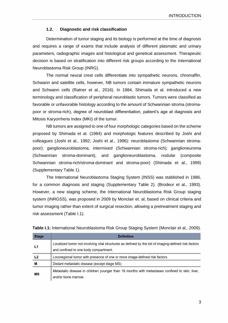
risk assessment (Table I.1).
Table I.1: International Neuroblastoma Risk Group Staging System (Monclair et al., 2009).

INTRODUCTION
4
After disease staging, each patient is stratified, based on clinical and molecular risk
factors (such as MYCN amplification, 11q aberration and ploidy) as very low-risk, low-risk,
intermediate-risk or high-risk, with 5-year event-free survival (EFS) of more than 85%, 75%
to ≤ 85%, ≥ 50% to ≤ 75%, or less than 50%, respectively (Cohn et al., 2009). The
stratification in pretreatment risk groups, summarized in Table I.2, helps the clinicians to
decide the best course of treatment (Pinto et al., 2015).
Table I.2: International Neuroblastoma Risk Group pretreatment classification schema
(Pinto et al., 2015).
1.3. Clinical presentation
Primary tumors can arise anywhere in the sympathetic nervous system. The majority
of tumors have an abdominal location, with at least half of these arising in the adrenal
medulla. The neck, chest and pelvis are other sites of disease occurrence (Maris et al.,
2007). The location of primary tumor is associated with different disease clinical and
biological features. Abdominal and extra-abdominal sites are associated with lower and
higher survival, respectively, and tumors located in thorax are more predominant in younger
age patients (Vo et al., 2014).

INTRODUCTION
5
Metastatic disease is detected in approximately half of patients at diagnosis, being
the most common locations of metastasis the regional lymph nodes, cortical bone and bone
marrow (Maris et al., 2007; Matthay et al., 2016). On the other hand, metastatic involvement
of lung and brain is rare (DuBois et al., 1999). However, there is a group of patients with
metastization to the liver, skin and bone marrow in children with less than 18 months of age,
which represents a clinical stage designed MS (D'Angio et al., 1971; Evans et al., 1971).
This specific stage is diagnosed in about 5% of the cases and almost always regress
spontaneously (DuBois et al., 1999; Maris et al., 2007; Matthay et al., 2016).
The presentation of symptoms depends on the site of the primary tumor, the
extension of metastatic disease and the presence or absence of paraneoplastic syndromes
(Maris et al., 2007; Matthay et al., 2016).
1.4. Treatment
NB treatment includes surgery, chemotherapy, radiotherapy, high-dose
chemotherapy/radiotherapy with autologous stem cell transplant. It can also be used
retinoid acid therapy, immunotherapy, and more recently targeted therapy (Pinto et al.,
2015; Simon et al., 2017).
Low- or intermediate-risk patients have excellent outcomes. For low-risk patients
without symptoms, surgery alone can be curative. Symptomatic low-risk patients at
diagnosis can receive a less-intensive chemotherapy without radiotherapy. However, some
observational studies demonstrated a subset of infants with localized tumors that can be
cured without any treatment, including surgery (Pinto et al., 2015; Simon et al., 2017).
Regarding the intermediate-risk patients, a more intensive regimen of chemotherapy and
radiotherapy is needed for older patients with unfavorable histology tumors (Pinto et al.,
2015).
Treatment regimens of high-risk patients include induction chemotherapy and
surgery, consolidation chemotherapy with myeloablative chemotherapy with autologous
hematopoietic stem-cell transplantation (AHSCT) and irradiation, and post consolidation
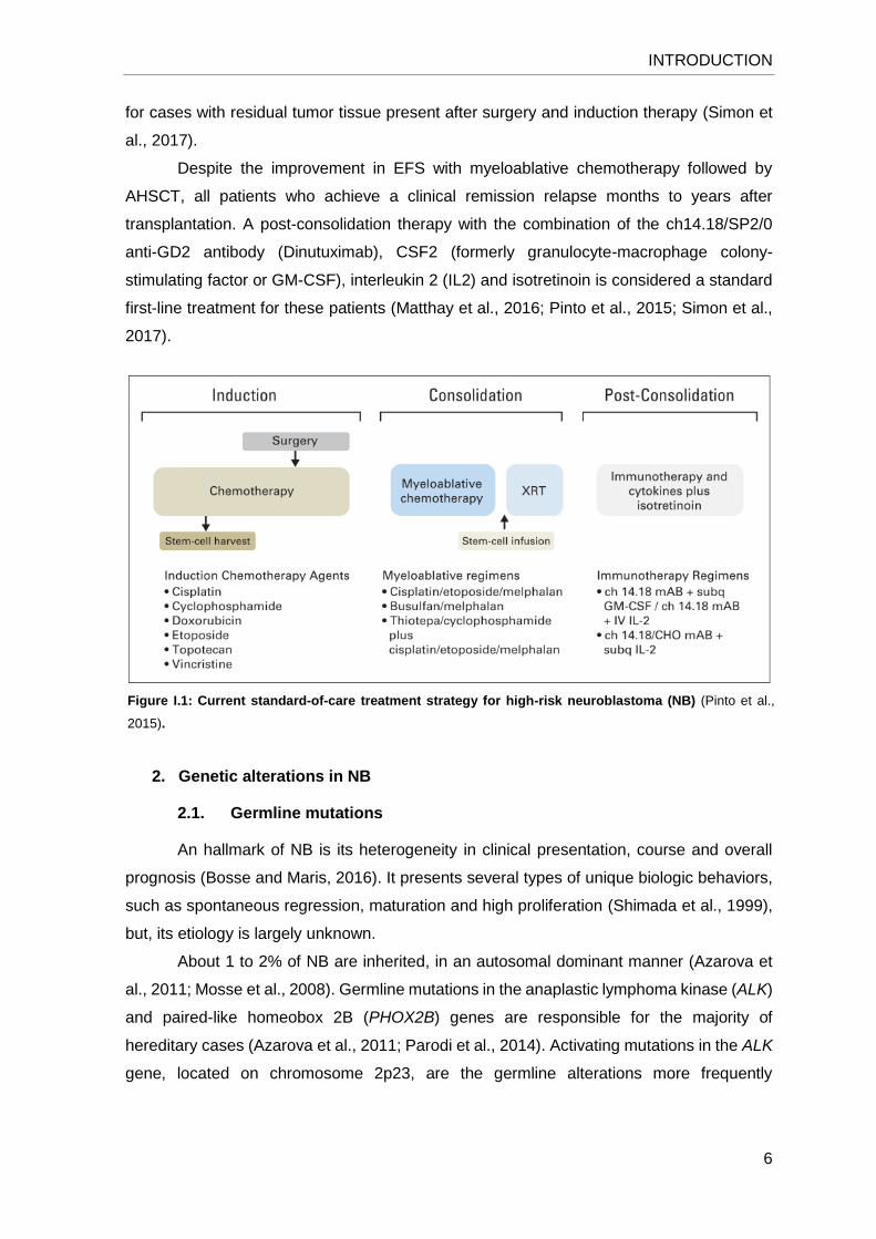
therapy to treat minimal residual disease (Pinto et al., 2015) (Figure I.2).
Resection of the primary tumor is performed during or after induction therapy and
the disadvantage of incomplete resection might be compensated by intensified local
radiotherapy (Simon et al., 2017). Induction therapy consists in a combination of agents,
including cisplatin, cyclophosphamide, carboplatin, doxorubicin, etoposide, topotecan and
vincristine (Pinto et al., 2015; Simon et al., 2017). Consolidation therapy consists in
myeloablative chemotherapy regimens with autologous stem cell transplantation. External
beam radiotherapy is recommended followed by myeloablative chemotherapy and AHSCT
INTRODUCTION
6
for cases with residual tumor tissue present after surgery and induction therapy (Simon et
al., 2017).
Despite the improvement in EFS with myeloablative chemotherapy followed by
AHSCT, all patients who achieve a clinical remission relapse months to years after
transplantation. A post-consolidation therapy with the combination of the ch14.18/SP2/0
anti-GD2 antibody (Dinutuximab), CSF2 (formerly granulocyte-macrophage colony-
stimulating factor or GM-CSF), interleukin 2 (IL2) and isotretinoin is considered a standard
first-line treatment for these patients (Matthay et al., 2016; Pinto et al., 2015; Simon et al.,
2017).
2. Genetic alterations in NB
2.1. Germline mutations
An hallmark of NB is its heterogeneity in clinical presentation, course and overall
prognosis (Bosse and Maris, 2016). It presents several types of unique biologic behaviors,
such as spontaneous regression, maturation and high proliferation (Shimada et al., 1999),
but, its etiology is largely unknown.
About 1 to 2% of NB are inherited, in an autosomal dominant manner (Azarova et
al., 2011; Mosse et al., 2008). Germline mutations in the anaplastic lymphoma kinase (ALK)
and paired-like homeobox 2B (PHOX2B) genes are responsible for the majority of
hereditary cases (Azarova et al., 2011; Parodi et al., 2014). Activating mutations in the ALK
gene, located on chromosome 2p23, are the germline alterations more frequently
Figure I.1: Current standard-of-care treatment strategy for high-risk neuroblastoma (NB) (Pinto et al.,
2015).

INTRODUCTION
7
associated with hereditary NB, being identified in about 50% of the cases (Mosse et al.,
2008; Schleiermacher et al., 2014).
There is an association of NB with other genetically determined neurocristopathies,
such as Hirschsprung disease (HSCR) and/or congenital central hypoventilation syndrome
(CCHS). Alterations in PHOX2B gene, located on chromosome 4p, are the major disease-
causing of CCHS, but, germline mutations have also been identified in NB patients with
HSCR and/or CCHS (Mosse et al., 2004; Trochet et al., 2004). However, PHOX2B
mutations explain only a small proportion of hereditary NB (less than 10%) (Mosse et al.,
2008; Raabe et al., 2008). Although more rarely, few NB cases are associated with genetic
syndromes with underlying rat sarcoma oncogene-mitogen activated protein kinase (RAS-
MAPK) pathway germline mutations, such as Neurofibromatosis type 1, Noonan syndrome
and Costello syndrome (Figure I.1 [left]) (Bosse and Maris, 2016; Schleiermacher et al.,
2014).
Mutations in other genes that can predispose to NB have been identified, but the
clinical relevance of these alterations remains poorly understood. In 2013, Pugh et al. found
the tumor protein 53 (TP53) p.Pro219Ser mutation in NB patients, consistent with prior
reports of NB being weakly associated with Li-Fraumeni syndrome (Birch et al., 2001). Two
other germline variants in the TP53 gene were found as being strongly associated with NB
(Diskin et al., 2014). Other studies have identified putative damaging mutations in
checkpoint kinase 2 (CHEK2), PTEN-induced putative kinase 1 (PINK1), BRCA1-
associated ring domain 1 (BARD1), adenomatous polyposis coli (APC) and succinate
dehydrogenase complex iron sulfur, subunit B (SDHB) genes in small percentages of NB
patients (Figure I.1 [middle]) (Pugh et al., 2013; Zhang et al., 2015).
Genome-wide association studies (GWAS) identified common DNA variations that
can influence the malignant transformation in NB. Common single nucleotide
polymorphisms (SNPs) were identified in cancer susceptibility candidate 15 (CASC15),
BARD1, LIM domain only 1 (LMO1), HECT domain and ankyrin repeat containing E3
ubiquitin protein ligase 1 (HACE1), which were associated with high-risk of NB development
(Capasso et al., 2009; Diskin et al., 2012; Russell et al., 2015; Wang et al., 2011). On the
other hand, the SNPs identified in dual specificity phosphatase 12 (DUSP12), DEAD (Asp-
Glu-Ala-Asp) box polypeptide 4 (DDX4) and interleukin 31 receptor A (IL31RA) were
associated with the low-risk NB (Figure I.1 [right]) (Bosse and Maris, 2016; Nguyen le et al.,
2011).

INTRODUCTION
8
2.2. Chromosomal changes
Oncogenes appear to be activated from their normal state due to mutations, gene
amplification or gene fusion (Ponder, 1992). In 1984, the amplification of MYCN gene,
located on chromosome 2p24.1, was described for the first time in NB patients (Brodeur et
al., 1984). MYCN amplification, present in approximately 20 to 25% of all primary tumors
(Maris et al., 2007), is associated with advanced stages of disease and with poor prognosis
(Brodeur et al., 1984; Seeger et al., 1985). Around 6% of tumors show only one or a few
additional MYCN copies, defined as MYCN-gain, which is also associated with a worse
prognosis in diploid tumors (Spitz et al., 2004). Additionally, deletion of chromosome 11q,
which is present in 35 to 45% of the cases, is inversely associated with MYCN amplification
(Maris et al., 2007), however, it is correlated with MYCN copy number gain (Spitz et al.,
Figure I.2: Rare and common genomic variants that predispose to NB. [left] mutations in anaplastic
lymphoma kinase (ALK) and paired-like, homeobox 2B (PHOX2B) are inherited in an autosomal dominant
Mendelian manner, with incomplete penetrance. Additionally, NB can also arise in a subset of genetic
syndromes with germline mutations in tumor protein 53 (TP53), succinate dehydrogenase complex iron sulfur,
subunit B (SDHB), Harvey rat sarcoma viral oncogene homolog (HRAS), protein tyrosine phosphatase, non-
receptor type 11 (PTPN11) and neurofibromin 1 (NF1). [middle] Low-frequency damaging mutations with an
intermediate effect size. [right] More common alleles with a modest effect size, specifically to high-risk (white)
or low-risk (orange) NB [Adapted from (Bosse and Maris, 2016)].
INTRODUCTION
9
2004). Therefore, the presence of deletion of 11q has emerged as a powerful prognosis
biomarker of a worse prognosis in patients without MYCN amplification (Attiyeh et al., 2005).
Additionally, other chromosome changes have been identified with prognostic value
in NB patients, including deletions of chromosomes 1p, 3p, and 4p, and a gain of 1 to 3
copies of chromosome 17q region (Janoueix-Lerosey et al., 2009). In general, tumors with
gains of whole-chromosome, resulting in hyperdiploidy, are associated with favorable
features such as non-metastatic disease or stage MS and younger age at diagnosis,
whereas tumors with segmental chromosome alterations are associated with more
aggressive forms of disease. The tumor overall genomic alteration pattern defines specific
genetic groups with different clinical outcomes (Bosse and Maris, 2016; Janoueix-Lerosey
et al., 2009; Oberthuer et al., 2009).
These segmental genetic aberrations can be assessed by interphase fluorescence
in situ hybridization (FISH), array-based comparative genomic hybridization (aCGH),
polymerase chain reaction (PCR) or multiplex ligation-dependent probe amplification
(MLPA) techniques (Ambros et al., 2009). The assessment of tumor cell DNA content
(ploidy) was shown to influence the prognosis in children younger than 2 years at diagnosis
(Look et al., 1991; Maris et al., 2007) and is assessed by flow or static cytometry, being
classified as diploid or hyperdiploid (near-triploid or penta/hexaploidy) (Ambros et al., 2009).
Allelic loss of chromosome 1p, identified in 25 to 35% of all NB, seems to be
independent of patient’s age and stage of disease, and is one of the most powerful
prognostic indicators of a worse outcome (Maris et al., 2007). A strong correlation exists
between MYCN amplification and chromosome 1p loss of heterozygosity (LOH), is
demonstrated, both correlated with poor clinical outcome and occurring together mainly in
more advanced stages and in patients older than one-year (Caron et al., 1996; Fong et al.,
1989; Spitz et al., 2002). Aberrations of chromosome 17q are the most common
chromosomal alterations described in NB patients, occurring in at least 50% of the tumors
(Bosse and Maris, 2016; Maris et al., 2007). The chromosome 17q gain is characteristic of
advanced tumors and are found in patients older than one-year, being strongly associated
with deletion of 1p and MYCN amplification. The principal mechanism involved in partial
gain of 17q is an imbalanced translocation with 1p (Bown et al., 1999; Caron et al., 1996).
2.3. Somatic point mutations
Although chromosomal aberrations are common in NB, with a well-stablished
influence in patient’s outcome, point mutations are quite rare. Some studies of whole-
genome sequencing have described mutations in cancer-related genes in NB tumors, which
occur more frequently in high-risk patients. Altogether, these studies showed the

INTRODUCTION
10
occurrence of ALK activating mutations in 7 to 14% of sporadic cases, ALK amplification in
approximately 2% and more rarely other cytogenetic rearrangements of this gene (Molenaar
et al., 2012; Mosse et al., 2008; Pugh et al., 2013). Mutations and deletions in ATP-
dependent helicase (ATRX) gene have been associated with children older than 5 years
old with metastatic disease and have been described as mutually exclusive with MYCN
amplification. ATRX plays a significant role in regulating adenosine triphosphate –
dependent chromatin remodeling, nucleosome assembly, and telomere maintenance.
These mutations were associated with a lower ATRX mRNA expression and long telomeres
(Cheung et al., 2012; Molenaar et al., 2012; Pugh et al., 2013).
Rearrangements in the telomerase reverse transcriptase (TERT) gene were also
found in 23% of high-risk NB. Increased TERT gene copy number appears to promote
metastasis, in absence of MYCN amplification (Cobrinik et al., 2013; Valentijn et al., 2015).
Chromothripsis, a located shredding of a chromosomal region with subsequent random
reassembly of the fragments, has been found in 18% of high-risk patients, and was also
associated with a poor prognosis (Molenaar et al., 2012). Additionally, an intragenic
hemizygous deletion targeting the AT-rich interaction domain 1B (ARID1B) gene and an
insertion mutation in the homologous AT-rich interaction domain 1A (ARID1A) gene, both
chromatin remodeling genes, were found in 11% of NB cases and were associated with
early treatment failure and lower survival (Sausen et al., 2013). Although more rarely,
mutations in T-cell lymphoma invasion and metastasis 1 (TIAM1) gene (3%), a central
regulator of cellular polarity and neuritogenesis (Molenaar et al., 2012), and in PTPN11
gene (2.9%) have also been described (Pugh et al., 2013).
3. The anaplastic lymphoma kinase (ALK) gene
The ALK gene, located on chromosome 2p23, codes for a membrane associated
tyrosine kinase receptor, which is a member of the insulin receptor protein-tyrosine kinase
superfamily. The ALK gene encodes a mature protein of 220 kDa that migrates as two
protein isoforms: the 220 kDa full-length receptor and the truncated 140kDa protein that
results from extracellular cleavage. This protein has an extracellular domain, a
transmembrane segment and an intracellular domain, which contains a protein kinase
domain (Azarova et al., 2011; Roskoski Jr, 2013). It is thought that, in mammalians, ALK
plays a key role in the development and function of nervous system during the neural
development. This gene is preferentially expressed in brain and spinal cord, being highly
expressed in neonatal brain (Iwahara et al., 1997; Roskoski Jr, 2013).
The normal ALK activation by the growth factors ligands pleiotropin (PTN) and
midkine (MK) results in the activation of many different pathways that are strictly

INTRODUCTION
11
interconnected and overlapping. These include the rat sarcoma oncogene-extracellular
signal-regulated kinase (RAS-ERK) pathway, the janus kinase 3-signal transducer and
activator of transcription 3 (JAK3-STAT3) pathway and the phosphoinositide 3-kinase-Akt
(PI3K-Akt) pathway, which participate in cell proliferation, cell survival, inhibition of
apoptosis and also in induction of neuronal cell differentiation through the MAPK pathway
(Azarova et al., 2011). A variety of ALK gene alterations have been described in several
types of tumors, including mutations, amplifications, deletions and translocations leading to
an aberrant activity of ALK (Chiarle et al., 2008).
3.1. ALK changes in various cancers
ALK seems to be a “hotspot” for translocation to a wide variety of loci. A translocation
involving chromosomes 2p and 5q, t(2;5)(p23;q35), which generates a fusion protein (NPM-
ALK), was described for the first time in the anaplastic large-cell lymphoma (ALCL), the first
disorder associated with ALK fusion proteins (Morris et al., 1994; Roskoski Jr, 2013). Since
then, 22 other genes have been described as rearranged with ALK in several types of
cancer, including inflammatory myofibroblastic tumor (IMT), non-small-cell lung cancer
(NSCLC), diffuse large B cell lymphoma (DLBCL), squamous cell carcinoma (SCC) and
renal cell carcinoma (RCC) (Hallberg and Palmer, 2013). Furthermore, NSCLC patients
harboring rearrangements involving ALK (ALK-EML4, most frequently) are eligible for
targeted therapy with ALK inhibitors (Solomon et al., 2014).
More recently, increased ALK activity due to overexpression of ALK or point
mutations has been reported as playing a primary role in the pathogenesis, aggressiveness
and lethality of NB (Roskoski Jr, 2013).
3.2. ALK rearrangements in NB
Since 2002, several works were published reporting the high incidence of ALK
mutations and gene amplifications in advanced sporadic NB tumors (Bagci et al., 2012;
Carén et al., 2008; Chen et al., 2008; George et al., 2008; Janoueix-Lerosey et al., 2008;
Miyake et al., 2002; Mosse et al., 2008; Osajima-Hakomori et al., 2005; Subramaniam et
al., 2009).
Miyake et al. (2002) and Osajima-Hakomori et al. (2005) showed that the
constitutively activation of ALK gene by amplification plays a role in the pathophysiology of
NB, resulting in hyperphosphorylation of ShcC, MAPK and Akt proteins. They showed that
ALK inhibition suppresses tyrosine phosphorylation of ShcC and blocks MAPK and Akt,
resulting in apoptosis. Furthermore, ALK amplification has been identified in several studies
in approximately 2% of cases and is associated with a worse outcome (Azarova et al.,

INTRODUCTION
12
2011). Additionally, ALK has been shown to be co-amplified with MYCN gene, which is
located on 2p24.3 very close to the ALK locus, in around 7 to 15% of all MYCN amplified
tumors (Azarova et al., 2011; Bagci et al., 2012; De Brouwer et al., 2010; Wang et al., 2013).
However, Chen et al. (2008) suggested that ALK and MYCN loci can also be amplified in
separate amplicons.
Additionally, there is a high frequency of tumors harboring a partial gain of
chromosome 2p region, encompassing the ALK gene (17 to 45%) (Bagci et al., 2012; Carén
et al., 2008; Chen et al., 2008; Mosse et al., 2008). Moreover, ALK gene copy number is
strongly correlated with mRNA and protein expression levels, which is significantly
correlated with a worse overall survival (De Brouwer et al., 2010; Passoni et al., 2009).
More recently, a new mechanism of aberrant ALK activation by balanced or
unbalanced translocations and deletions has been suggested (Table I.3). A balanced
translocation between the ALK gene in chromosome 2 and the CLPTM1L gene in
chromosome 5 was described in a cell line, resulting in an out-of frame fusion transcript, but
its biologic function remains to be defined (Cazes et al., 2013). Unbalanced translocations
between chromosome 2 and chromosomes 4 and 11, resulting in 2p gain, were described
in tumors samples, nevertheless, since gene fusion occurs in opposing transcriptional
directions, a chimeric transcript it is unlikely to be produced (Fransson et al., 2015).
Additionally, three different deletions, combined with ALK locus amplification, were
described as leading to truncated ALK variants, which exhibit oncogene properties and are
associated with tumor aggressiveness (Cazes et al., 2013; Fransson et al., 2015; Okubo et
al., 2012). This pattern of rearrangements and the multiple breakpoints observed in NB
tumor samples may correspond to the phenomenon of chromothripsis, recently reported in
aggressive NB (Cazes et al., 2013; Fransson et al., 2015; Molenaar et al., 2012; Wang et
al., 2013).
Table I.3: ALK gene rearrangements described in NB.
INTRODUCTION
13
3.3. Somatic ALK point mutations in NB
Somatic mutations have been identified in approximately 7 to 14% of NB patients,
mainly occurring in the catalytic loop or the C-helix kinase domains (Table I.4) (Mosse et
al., 2008). Two mutational hotspots, at positions R1275 on exon 25 and F1174 on exon 23,
have been described. These mutations occur in the kinase domain and result in the
constitutive active forms of the protein, leading to constitutive phosphorylation of ALK and,
consequently, phosphorylation of downstream targets, such as STAT3, Akt and ERK (Chen
et al., 2008; De Brouwer et al., 2010; George et al., 2008). These mutations were found
across the entire spectrum of disease, presenting similar frequencies in cases with or
without MYCN amplification (De Brouwer et al., 2010; Mosse et al., 2008). Furthermore, no
differences in survival of patients with ALK mutations were observed, suggesting that ALK
mutations themselves are not an adverse prognostic factor (Chen et al., 2008; De Brouwer
et al., 2010). However, when the survival was compared between different mutations,
patients with a F1174 mutation have shown a worse survival than those with a R1275
mutation or wild-type, which can be explained by the high frequency of MYCN amplification
in F1174 mutation carriers (De Brouwer et al., 2010). Moreover, ALK copy number gain was
not frequently found in tumors harboring ALK mutations, suggesting that 2p gain is not a
common mechanism of increasing mutated ALK expression (De Brouwer et al., 2010;
Passoni et al., 2009).
The clinical role of ALK mutations or amplification remains controversial (Bagci et
al., 2012; Brodeur et al., 1984; Carén et al., 2008; Chen et al., 2008; De Brouwer et al.,
2010). However, it seems to be clear that more aggressive and metastatic tumors exhibit a
higher expression of ALK comparatively to localized NB, even without ALK gene alterations,
suggesting that increased ALK expression might be functionally relevant in NB. It is possible
that patients with wild-type ALK overexpression might benefit from ALK inhibitors therapy,
in addition to those harboring ALK amplification or point mutations (Hallberg and Palmer,
2013; Passoni et al., 2009).

INTRODUCTION
14
Table I.4: ALK somatic mutations described in NB patients.
3.4. Germline ALK mutations in hereditary NB
Hereditary NB is an autosomal dominant disease, with incomplete penetrance. This
was the first example of a pediatric cancer associated with germline mutations in an
oncogene (Mosse et al., 2008). ALK germline mutations are identified in approximately
50% of NB families (Table I.5) (Mosse et al., 2008). The mutations R1275Q (Janoueix-
Lerosey et al., 2008; Mosse et al., 2008), R1192P (Bourdeaut et al., 2012; Janoueix-
Lerosey et al., 2008; Mosse et al., 2008) G1128A (Mosse et al., 2008) and T1151R
(Bourdeaut et al., 2012) have been described in families with NB.
The disease diagnosis at early age, presence of multifocal disease and familial
recurrence of neuroblastic tumors are characteristics highly suggestive of a hereditary NB
(Bourdeaut et al., 2012; Mosse et al., 2008). Mosse et. al. only found ALK mutations in NB
families characterized by at least two affected individuals, related in first-degree. Bourdeaut
et al. (2012) identified ALK germline mutations in 3 of 16 (18.75%) multifocal cases,
suggesting that this clinical feature might be strongly indicative of an ALK-related
predisposition in absence of familial history. However, younger age is not a major relevant
INTRODUCTION
15
criterion indicative of ALK-related predisposition, as diagnoses have been reported at the
age of three, 12 and 15 years in individuals with germline R1275Q and R1192P mutations
(Janoueix-Lerosey et al., 2008).
The R1275Q ALK gene mutation is the most frequent mutation in hereditary NB, and
is also one of the mutational hotspots in sporadic NB (Azarova et al., 2011; Mosse et al.,
2008). This mutation was described as arising de novo in two families inherited either from
an affected or unaffected parent (Mosse et al., 2008). On the other hand, the mutation
R1192P previously described in four families was always inherited from an unaffected
parent (Bourdeaut et al., 2012; Hallberg and Palmer, 2013; Janoueix-Lerosey et al., 2008;
Mosse et al., 2008). A de novo germline ALK mutation at position F1174, was found in a
case of congenital NB with severe encephalopathy (de Pontual et al., 2011), indicating that
it might cause embryonic lethality in most of the cases (Azarova et al., 2011; Bourdeaut et
al., 2012; De Brouwer et al., 2010).
Table I.5: ALK germline mutations described in hereditary NB.
3.5. ALK inhibitors in neuroblastoma treatment
The first ALK inhibitor tested in a clinical trial was Crizotinib, an oral bioavailable
small molecule inhibitor approved for EML4-ALK positive NSCLC patients. It is an ATP-
competitive molecule that prevents subsequent autophosphorylation necessary for further
signal transduction. This drug was studied for the treatment of NB patients with either
amplification or mutation in the ALK gene (Azarova et al., 2011). Some in vitro and in vivo
studies have shown that Crizotinib is more potent in NB cell lines and xenografts with ALK
amplification or the R1275Q mutation than in those with F1174L mutation, suggesting that
the latter may promote resistance to the drug (Bresler et al., 2011). In a pediatric phase 1
and phase 2 trial of Crizotinib, two NB patients with ALK germline mutations responded to
this therapy, but further studies are required (Mosse et al., 2013). However, more recent
results suggest that the combination of Crizotinib with conventional chemotherapy may
provide superior anti-tumor activity than each approach separately (Krytska et al., 2016).

INTRODUCTION
16
Another ALK inhibitor is Entrectinib, a potent inhibitor of cell proliferation that induces
cell death, causing cell cycle arrest. It was able of decrease growth and proliferation of ALK-
amplified cell lines, however, shows low efficiency in ALK mutated cells (Aveic et al., 2016).
Other ALK inhibitors have been studied, such as the potent second-generation ALK inhibitor
Alectinib, which is active against some ALK mutations including, F1174L and R1275Q,
indicating that this drug is able to overcome Crizotinib-induced chemoresistance (Lu et al.,
2017).
More recently, Infarinato et al. (2016) described a novel ALK inhibitor with a superior
potency than the others described so far. The ALK/ROS1 inhibitor PF-06463922 seems to
exert activity in ALK-driven NB models with primary Crizotinib resistance. This new drug
appears to be a key to overcome the ALK-driven therapy resistance, which is the biggest
challenge in NB treatment with ALK inhibitors.

II. AIMS OF THE STUDY

19
II. AIMS OF THE STUDY
Due to the recently clinical implications of ALK changes in NB patients, the general
aim of this work was to test for these alterations in NB patients diagnosed at the Portuguese
Institute of Oncology of Porto (IPO-Porto) from 2003 to 2016. The specific aims were:
➢ To search for ALK fusion genes in neuroblastomas;
➢ To compare the pattern of amplification of the ALK and MYCN genes in
neuroblastomas;
➢ To identify somatic and germline ALK tyrosine kinase domain (TKD) point
mutations;
➢ To relate the ALK alterations with clinical outcome in neuroblastoma patients.


III. MATERIAL AND METHODS

23
III. MATERIAL AND METHODS
1. Patients and sample collection
A consecutive series of primary NB tumors from 46 patients treated at IPO-Porto
from 2003 to 2016 and tested for MYCN amplification at the Department of Genetics was
retrospectively analyzed. The clinical, genetical and pathological data of NB patients were
recorded and are summarized in Table III.1.
Forty-one cell suspension samples of primary NB tumors were analyzed for ALK
gene rearrangements by fluorescence in situ hybridization (FISH). Twenty-nine frozen fresh
tumor samples were subsequently analyzed by Sanger sequencing for exons 20 to 28 of
the ALK gene. Twenty-four patients were analyzed by both FISH and Sanger sequencing.
Whenever possible, normal tissue was analyzed from patients presenting mutation in tumor
samples.
Table III.1: Clinical and standard genetic information on the NB patients.
MATERIAL AND METHODS
24
MATERIAL AND METHODS
25
2. Fluorescence in situ hybridization
Fluorescence in situ hybridization (FISH) was performed with a dual color, break
apart probe flanking the ALK gene, ALK (2p23) Break FISH probe, [Kreatech, Leica Biosystems, Nussloch,
Germany] (Figure III.1). Slides were prepared fresh from cultured cells fixed with
methanol:acetic acid. Co-denaturation was performed at 80°C for 8 min, followed by
hybridization at 37°C in humidified chamber (ThermoBrite denaturation/hybridization
system [Leica Biosystems, Nussloch, Germany]) for 18 hours. Post-hybridization washes were done using
2xSSC/0.1% IGEPAL [Sigma Aldrich] solution at room temperature (RT) for 30 minutes, followed
by 0.4 x SSC/ 0.3% IGEPAL at 74 °C for 2 min and 2 x SSC/ 0.1% IGEPAL for 5 minutes,
at RT. Slides were dehydrated by passages through an increasing series of alcohols, 70%,
85% and 100%, 2 minutes each and air dried. DAPI (4’, 6-diamidino-2-phenylindole) [Vector
Laboratories, Burlingame, California, USA] was applied as a counterstain and results were evaluated with
a GSL-120 Automated Cytogenetics Platform with a CytoVision Software Version number
7.4 [Leica Biosystems]. For each sample, at least 50 intact non-overlapping nuclei were scored.
Fluorescence images corresponding to DAPI, Spectrum Green and Spectrum
Orange were automatically captured and relative copy number alteration in 2p23 region was
assessed as previously described (Subramaniam et al., 2009).
Since centromeric chromosome 2 copy number was available from previous MYCN
analysis, the status of ALK was calculated for each sample, according to MYCN status
criteria (Ambros et al., 2009). An abnormal signal pattern was considered representative
when present in a minimum of 10% of tumor cells.
Cases were categorized as:
➢ ALK gain (G), when the number of ALK gene signals was one- to four-times
greater than the CEP2 signals;
➢ ALK-amplified (A), when the number of ALK gene signals was four-times
greater than the CEP2 signals;
➢ ALK loss/imbalance (L), when the number of ALK gene signals was less than
that of CEP2 signals;
➢ Chromosome 2 polysomy (P), when both CEP2 and ALK signals were
increased.
MATERIAL AND METHODS
26
3. DNA extraction and quantification
3.1. Extraction of genomic DNA from frozen fresh samples
A piece of frozen tumor was minced with a scalpel and transferred to a 50ml falcon.
Four ml of SE buffer (75mM NaCl; 25 mM EDTA), 400µl of SDS (Sodium Dodecyl Sulfate)
10% and 50µl of proteinase K [Gibco Invitrogen, Carlsbad, CA, USA] (10ng/µl) were added, followed by
incubation at 55ºC overnight. One ml of NaCl (6M) was added to the lysed sample and
incubated at 55ºC for 10 minutes. An equal volume of chloroform was added, gently mixed
for 30 minutes and centrifuged at 4000 rpm for 10 minutes. The aqueous DNA solution was
transferred to a 15ml falcon and an equal volume of isopropanol 100% was added. The
precipitated DNA was transferred to an eppendorf with 70% ethanol. Subsequently, ethanol
was discarded and air dried. Finally, the DNA was eluted in ddH2O and was quantified by
spectrophotometry with NanoDrop ND-1000® [NanoDrop Technologies, Wilmington, DE, USA].
3.2. Extraction of genomic DNA from paraffin-embedded samples
Tumor areas containing normal cells were delimited by a pathologist in the
hematoxylin and eosin (H&E) stained slides of each sample. The corresponding unstained
slides were immersed in xylene [Sigma-Aldrich, Steinheim, Germany] and then in ethanol 100% [Merck,
Darmstadt, Germany] for 5 minutes each to remove the paraffin. Tumor areas, which were
previously delimited by comparison with the correspondent H&E stained slides, were macro
dissected and transferred to a centrifuge tube and the extraction was performed with a
Figure III.1: ALK (2p23) break FISH probe to detect rearrangements involving the ALK gene. Red critical region
represents the distal ALK gene region direct-labeled with PlatinumBrighTM550 (3′ALK SpectrumOrange) and
green critical region represents the proximal ALK gene region direct-labeled with PlatinumBrighTM495 (5′ALK
SpectrumGreen).
MATERIAL AND METHODS
27
cobas® DNA Sample Preparation Kit, used for manual specimen preparation of formalin-
fixed paraffin-embedded tumor (FFPE) tissues [Roche Diagnostics, Mannheim, Germany], according to
standard procedures. Finally, the DNA was quantified by spectrophotometry with NanoDrop
ND-1000® [NanoDrop Technologies, Wilmington, DE, USA].
4. DNA sequencing
Mutation screening in the tyrosine kinase domain of ALK (exons 20 to 28) was
performed by Sanger sequencing. For this purpose, DNA was amplified in a solution
containing 1x PCR gold buffer [Thermo Fisher Scientific, Foster city, CA, USA] (150 mM Tris-HCl, 500 mM
KCI), 1.5 mM of MgCl2 [Thermo Fisher Scientific], 0.5 mM dNTP mix [Thermo Fisher Scientific], 0.17 mM of
each primer (reverse and forward) [frilabo, Portugal], 1 U of Taq DNA polymerase [Thermo Fisher
Scientific], in a final reaction volume of 20 µL. The primer sequences were used from Chen et
al. (2008) and are summarized in Table III.2. PCR reaction was performed in a thermocycler
[Gene Amp PCR Systern 9700, Perkin-EImer, Waltham, Massachusetts, USA] according to the following conditions: an
initial denaturation step at 95°C for 10 min, followed by 35 cycles of 95°C for 45 seconds,
annealing step at 58°C for 45 seconds and a 45 seconds extension step at 72°C. A final
extension step was done at 72°C for 10 min. Amplified PCR products were then analyzed
by electrophoresis in a 2% (w/v) agarose gel [Gibco Invitrogen] stained with green safe [Sigma-Aldrich]
0.05 µL/mL.

MATERIAL AND METHODS
28
Table III.2: Set of primers used to amplifying exons 20 to 28 (Chen et al., 2008).
Subsequently, the PCR products were purified using the ExoSAP-IT method for the
removal of primers and dNTPs in excess. Samples were purified adding 2 µL of ExoSAP
solution, which consists in Exonuclease I [Thermo Fisher Scientific] (20 U/μL) and Fast
Thermosensitive Alkaline Phosphatase [Thermo Fisher Scientific] (1 U/μL), in a proportion of 1:2, to
5 µL of the PCR product, followed by incubation at 37ºC for 50 minutes, and enzyme
inactivation at 80ºC for 15 minutes.
The purification was followed by the sequencing reaction, in which the BigDye®
Terminator v1.1 or v3.1 Cycle Sequencing Kit [Applied Biosystems, Foster City, CA, USA] was used. The
reaction consisted on mixing 3.4 μL of sequencing buffer, 0.5 µL of Big Dye® Terminator
v1.1 or v3.1, containing dNTPs, ddNTPs-fluorocromes, MgCl2 and Tris-HCl buffer, 0.32 µL
of one of the primers (forward or reverse), and bidestilled sterile water [B. Braun, Foster City, CA, USA]
and 1 µL of the previously purified DNA to reach a final reaction volume of 10 µL. The
sequencing reaction was performed and consisted of an initial denaturation step at 95ºC for
4 minutes, followed by 35 cycles of denaturation at 96ºC for 10 seconds, annealing at 50ºC
for 10 seconds and extension at 60ºC for 2 minutes, with a final extension of 60ºC for 10
minutes. In order to remove excess of dNTPs, labeled ddNTPs, and non-incorporated
primers, the sequencing products were purified with IIlustra Sephadex® G-50 fine [GE
MATERIAL AND METHODS
29
Healthcare, Life Sciences, Cleveland, USA], according to standard procedures. After purification, 15 μL of
Hi-DiTM Formamide [Applied Biosystems] were added to the sequencing products to help stabilize
the single stranded DNA. The products were then analyzed in a 3500 Genetic Analyzer
[Applied Biosystems] by capillary electrophoresis. The electropherograms of each sample were
analyzed with the Sequencing Analysis Software v5.4 [Applied Biosystems]. All of them were
examined at least twice, reviewed manually and with the Mutation Surveyor® DNA Variant
Analysis Software v4.0.8 [Softgenetics, State College, PA, USA].
A second PCR amplification was performed in all positive samples, followed by DNA
Sanger sequencing of both strands. All ALK variants were described according to the
LRG_488 (NM_004304.3) and to the Human Genome Variations Society guidelines.


IV. RESULTS

33
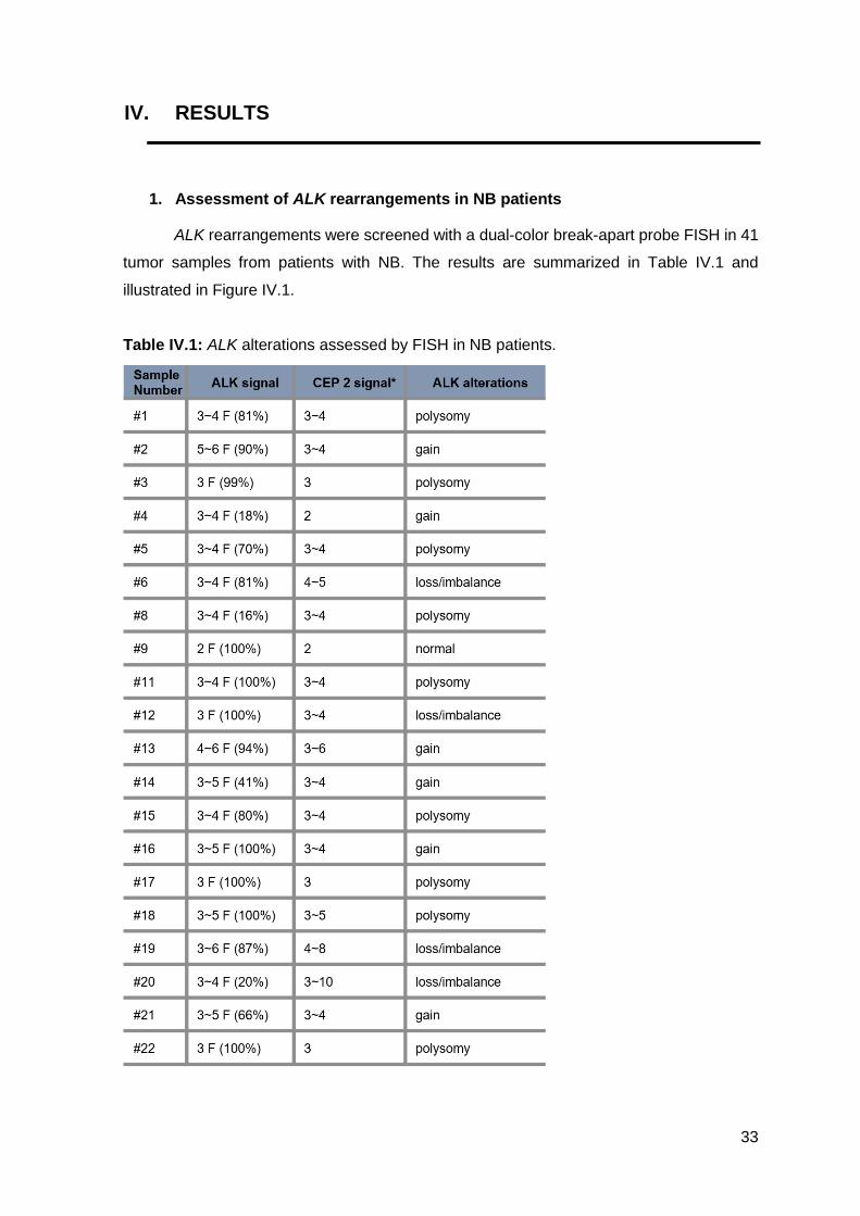
IV. RESULTS
1. Assessment of ALK rearrangements in NB patients
ALK rearrangements were screened with a dual-color break-apart probe FISH in 41
tumor samples from patients with NB. The results are summarized in Table IV.1 and
illustrated in Figure IV.1.
Table IV.1: ALK alterations assessed by FISH in NB patients.

RESULTS
34
No translocations or inversions were found in our cohort, however, aberrant copy
number of the ALK gene was observed in 19 cases (46.3%), including ALK amplification in
one of 41 (2.4%), ALK gain in 13 of 41 (31.7%) and ALK loss/imbalance in five of 41 (12.2%).
Chromosome 2 polysomy was observed in 15 cases (36.6%) and normal signal pattern was
observed in the remaining seven cases (17.1%).

RESULTS
35
Figure IV.1: Representative FISH images from four selected NB tumors using a dual color, break apart
probe targeting 3′ALK and 5′ALK, labeled orange and green, respectively. a. normal nuclei, presenting two
ALK gene signals; b. nuclei presenting three to four ALK gene signals; c. nuclei presenting three ALK gene
signals; d. nuclei with amplification of 3’ALK region.
1.1. Clinicopathological characteristics of patient with ALK amplification
The patient with ALK amplification (81% of the cells) was a female child aged 14
months old diagnosed with a neuroblastoma (stroma-poor, undifferentiated, low MKI and
unfavorable histology), located in the right adrenal medulla, with invasion of adjacent
tissues, regional lymph nodes and the bone marrow. The previous cytogenetic analysis
showed MYCN amplification in 78% of cells, similar to the ALK pattern found in this study,
as well as deletion of 1p36 region in 54% of cells. According to INRG risk stratification, the
tumor was classified as a high-risk neuroblastoma. After neoadjuvant chemotherapy and
surgery, maturation of tumor cells induced by chemotherapy was observed, being the tumor
reclassified as a ganglioneuroblastoma, subtype “intermixed” and favorable histology. The
patient died 12 months after diagnosis.
RESULTS
36
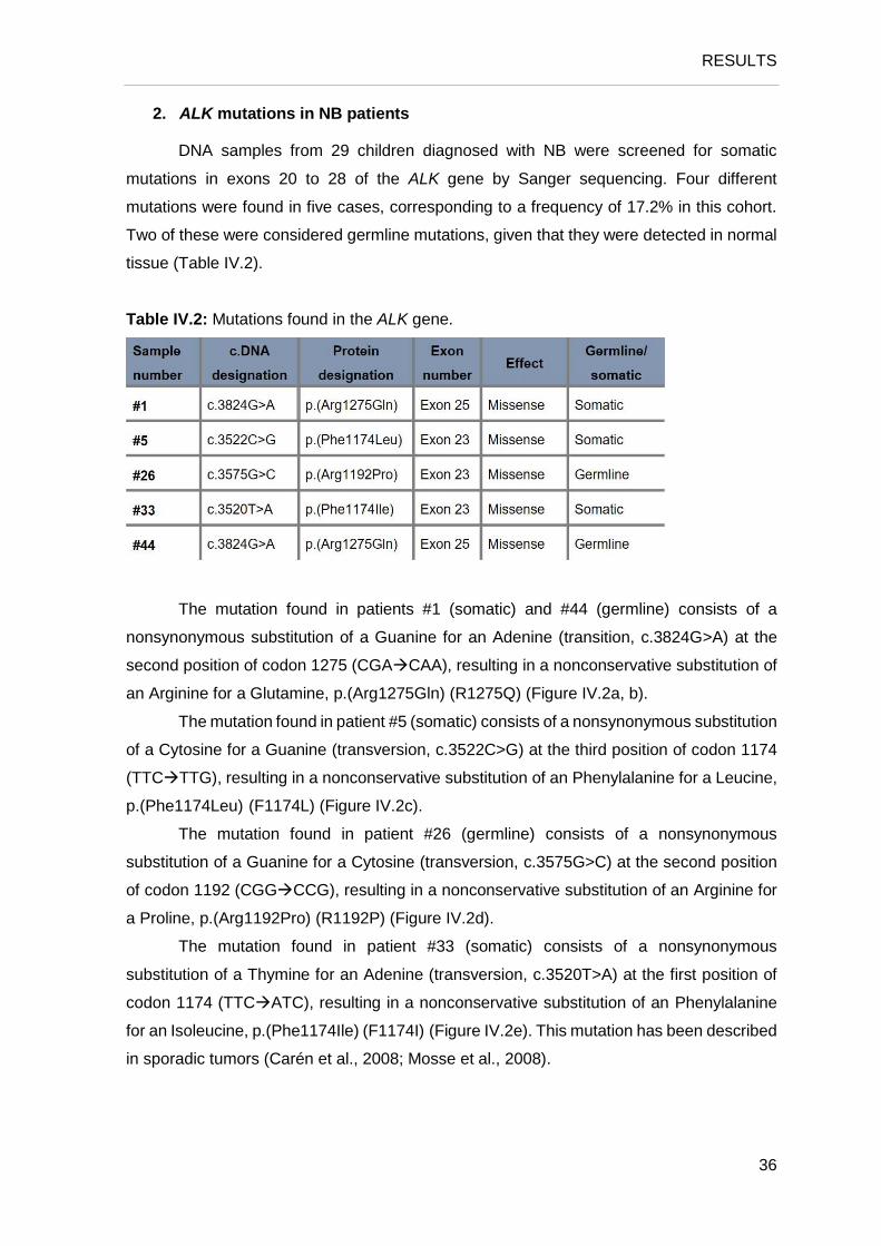
2. ALK mutations in NB patients
DNA samples from 29 children diagnosed with NB were screened for somatic
mutations in exons 20 to 28 of the ALK gene by Sanger sequencing. Four different
mutations were found in five cases, corresponding to a frequency of 17.2% in this cohort.
Two of these were considered germline mutations, given that they were detected in normal
tissue (Table IV.2).
Table IV.2: Mutations found in the ALK gene.
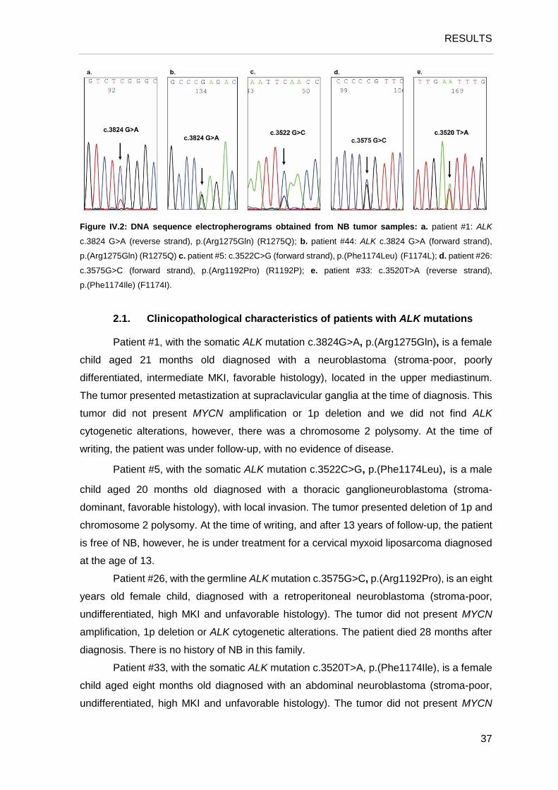
The mutation found in patients #1 (somatic) and #44 (germline) consists of a
nonsynonymous substitution of a Guanine for an Adenine (transition, c.3824G>A) at the
second position of codon 1275 (CGACAA), resulting in a nonconservative substitution of
an Arginine for a Glutamine, p.(Arg1275Gln) (R1275Q) (Figure IV.2a, b).
The mutation found in patient #5 (somatic) consists of a nonsynonymous substitution
of a Cytosine for a Guanine (transversion, c.3522C>G) at the third position of codon 1174
(TTCTTG), resulting in a nonconservative substitution of an Phenylalanine for a Leucine,
p.(Phe1174Leu) (F1174L) (Figure IV.2c).
The mutation found in patient #26 (germline) consists of a nonsynonymous
substitution of a Guanine for a Cytosine (transversion, c.3575G>C) at the second position
of codon 1192 (CGGCCG), resulting in a nonconservative substitution of an Arginine for
a Proline, p.(Arg1192Pro) (R1192P) (Figure IV.2d).
The mutation found in patient #33 (somatic) consists of a nonsynonymous
substitution of a Thymine for an Adenine (transversion, c.3520T>A) at the first position of
codon 1174 (TTCATC), resulting in a nonconservative substitution of an Phenylalanine
for an Isoleucine, p.(Phe1174Ile) (F1174I) (Figure IV.2e). This mutation has been described
in sporadic tumors (Carén et al., 2008; Mosse et al., 2008).
RESULTS
37
Figure IV.2: DNA sequence electropherograms obtained from NB tumor samples: a. patient #1: ALK
c.3824 G>A (reverse strand), p.(Arg1275Gln) (R1275Q); b. patient #44: ALK c.3824 G>A (forward strand),
p.(Arg1275Gln) (R1275Q) c. patient #5: c.3522C>G (forward strand), p.(Phe1174Leu) (F1174L); d. patient #26:
c.3575G>C (forward strand), p.(Arg1192Pro) (R1192P); e. patient #33: c.3520T>A (reverse strand),
p.(Phe1174Ile) (F1174I).
2.1. Clinicopathological characteristics of patients with ALK mutations
Patient #1, with the somatic ALK mutation c.3824G>A, p.(Arg1275Gln), is a female
child aged 21 months old diagnosed with a neuroblastoma (stroma-poor, poorly
differentiated, intermediate MKI, favorable histology), located in the upper mediastinum.
The tumor presented metastization at supraclavicular ganglia at the time of diagnosis. This
tumor did not present MYCN amplification or 1p deletion and we did not find ALK
cytogenetic alterations, however, there was a chromosome 2 polysomy. At the time of
writing, the patient was under follow-up, with no evidence of disease.
Patient #5, with the somatic ALK mutation c.3522C>G, p.(Phe1174Leu), is a male
child aged 20 months old diagnosed with a thoracic ganglioneuroblastoma (stroma-
dominant, favorable histology), with local invasion. The tumor presented deletion of 1p and
chromosome 2 polysomy. At the time of writing, and after 13 years of follow-up, the patient
is free of NB, however, he is under treatment for a cervical myxoid liposarcoma diagnosed
at the age of 13.
Patient #26, with the germline ALK mutation c.3575G>C, p.(Arg1192Pro), is an eight
years old female child, diagnosed with a retroperitoneal neuroblastoma (stroma-poor,
undifferentiated, high MKI and unfavorable histology). The tumor did not present MYCN
amplification, 1p deletion or ALK cytogenetic alterations. The patient died 28 months after
diagnosis. There is no history of NB in this family.
Patient #33, with the somatic ALK mutation c.3520T>A, p.(Phe1174Ile), is a female
child aged eight months old diagnosed with an abdominal neuroblastoma (stroma-poor,
undifferentiated, high MKI and unfavorable histology). The tumor did not present MYCN

RESULTS
38
amplification, deletion of 1p and 11q, gain of 17q or ALK cytogenetic alterations. After
neoadjuvant chemotherapy and surgery, which induced maturation of tumor cells, the tumor
was reclassified as a neuroblastoma, stroma-poor, differentiating, with low MKI and
favorable histology. After 20 months of follow-up, a relapse was detected, and classified
after surgery as a ganglioneuroma, maturing, with low MKI and favorable histology.
Patient #44, with the germline ALK mutation c.3824G>A, p.(Arg1275Gln), is a
female child diagnosed at the age 9 months old with an abdominal neuroblastoma,
presenting metastases in the femur and in the facial bones. The tumor did not present
MYCN amplification, deletion of 1p or 11q, or gain of 17q. The FISH analysis of ALK was
not performed in this case. At the present time, the patient is being prepared for an AHSCT.
Family history is not known at the time of writing.

V. DISCUSSION

41
V. DISCUSSION
The discovery of a variety of genetic alterations in tumors leading to oncogenesis
and the development of target-specific therapies have improving patients’ outcomes in
different malignancies. Rearrangements, mutations and amplification of the ALK gene have
been described in a range of tumors, including ALCL, IMT and NSCLC (Hallberg and
Palmer, 2013). This indicates the ALK protein as a powerful biological marker and a
therapeutic target in malignancies in which ALK influences carcinogenesis (Bresler et al.,
2011; Solomon et al., 2014). Relatively recent findings have suggested that the ALK gene
may be activated by amplification or specific mutations targeting the tyrosine kinase domain
(TKD) in NB patients, presumably contributing to tumor development (Bagci et al., 2012;
Carén et al., 2008; Chen et al., 2008; George et al., 2008; Janoueix-Lerosey et al., 2008;
Miyake et al., 2002; Mosse et al., 2008; Osajima-Hakomori et al., 2005; Subramaniam et
al., 2009). Moreover, germline activating mutations in ALK gene are responsible for more
than half of NB hereditary cases (Mosse et al., 2008). In the present study, we aimed to
search for the ALK fusion genes, to compare the patterns of amplification of the ALK and
MYCN genes, to identify somatic and germline ALK TKD point mutations, and to relate the
ALK alterations with clinical outcome in NB patients.
Using the FISH methodology, we searched for ALK chromosome alterations in forty-
one patients from all clinical stages. The ALK gene is involved in the initiation and
progression of different malignancies, being often activated by translocations (Hallberg and
Palmer, 2013). In the present study, we did not observe any ALK rearrangement,
characterized by a split of the ALK probe, suggestive of a translocation or inversion with
breakpoint within the ALK gene. These rearrangements, although uncommon, have been
described in previous studies, although in larger cohorts and using more sensitive
methodologies such as SNP arrays (Cazes et al., 2013; Fransson et al., 2015).
However, we found cytogenetic aberrations in 46.3% of the cases. Amplification was
detected in one high-risk NB patient (2.4%), which is in accordance with previous studies
that report frequencies of 1 to 3% in advanced stages, using FISH (Osajima-Hakomori et
al., 2005; Subramaniam et al., 2009; Wang et al., 2013) or genome-wide scanning
approaches (Bagci et al., 2012; Carén et al., 2008; Chen et al., 2008; De Brouwer et al.,
2010; Janoueix-Lerosey et al., 2008; Mosse et al., 2008). This case also presented MYCN
amplification, which is in agreement with previous studies that suggested the synchronic
co-amplification of the two genes (Bagci et al., 2012; De Brouwer et al., 2010; George et
al., 2008; Subramaniam et al., 2009), which is compatible with the similar pattern of
amplification of both genes in our patient. Although ALK amplification without MYCN

DISCUSSION
42
amplification is very rare, having only been detected in three cases (Chen et al., 2008;
Janoueix-Lerosey et al., 2008; Mosse et al., 2008), Chen et al. (2008), showed that
amplification of ALK and MYCN loci may occur in separated events.
The copy number gain without amplification was found in our cohort in 31.7% of the
cases. The ALK copy number gain was described previously in 17 to 45% of the cases,
being highly associated with more advanced clinical stages and with non-hyperdiploid
tumors with a worse outcome (Bagci et al., 2012; Carén et al., 2008; Chen et al., 2008;
Mosse et al., 2008). However, Wang et al. (2013) showed a better outcome of patients with
ALK copy number gain, which may be related with the fact that the majority of these cases
also had gain of centromere 2, a feature that suggests a polyploid karyotype that is known
to confer a favorable prognosis in NB patients. In this cohort, we detected 36.6% of tumors
with chromosome 2 polysomy, which is an indicator of a better outcome of patients without
other adverse prognostic features.
Additionally, we found five cases (12.2%) presenting loss/imbalance of the ALK
locus, characterized by increased centromere of chromosome 2 probe signals, and lower
number of the ALK gene signals. This cytogenetic category was reported for the first time
by Subramaniam et al. (2009). Four of five cases with ALK loss/imbalance also showed
MYCN loss/imbalance. This can be explained by gain of a rearranged chromosome 2 with
relative ALK and MYCN allelic loss (Subramaniam et al., 2009). Interestingly, neither
deletion of 1p and 11q or duplication of 17q showed correlation with ALK copy number
alterations, which is in agreement with the results showed by Subramaniam et al. (2009).
In this study, we searched for ALK mutations in twenty-nine NB tumors by Sanger
sequencing and we found a frequency of 17.2%, which is higher when compared to previous
studies that described frequencies of 7 to 14% (Azarova et al., 2011). Four of the five ALK
mutations identified occurred at positions R1275 and F1174, which are two mutational
hotspots in sporadic tumors. A meta-analysis of 709 NB tumors identified these mutations
in tumors of all clinical stages and genomic profiles, in 49% and 34.7% of sporadic NB,
respectively. However, they described a significantly higher frequency of F1174 mutations
in MYCN-amplified tumors, which might explain the inferior survival reported by them in
these patients. Additionally, Schonherr et al. (2012) proposed that MYCN may cooperate
with ALK activating mutations to increase cell transformation and synergistically contribute
to tumor development, since the ALK gene is a direct transcriptional target of MYCN and a
downregulation or inhibition of ALK results in a decreased MYCN expression (Hasan et al.,
2013; Schonherr et al., 2012). In the present study, we did not find any ALK mutations in
any of MYCN amplified cases, which may be due the small number of cases analyzed.
The mutation R1275Q found in two of five cases was previously described in several
studies as one of the most frequent mutations occurring in both sporadic (Carén et al., 2008;

DISCUSSION
43
Chen et al., 2008; De Brouwer et al., 2010; George et al., 2008; Janoueix-Lerosey et al.,
2008; Mosse et al., 2008) and hereditary NB cases (Janoueix-Lerosey et al., 2008; Mosse
et al., 2008). This mutation has been associated with higher autophosphorylation and
phosphorylation of downstream targets, such as ERK1/2 and Akt with high capacity of
transformation in NIH 3T3 and Ba/F3 assays (Chen et al., 2008; De Brouwer et al., 2010;
George et al., 2008). Studies performed by Ueda et al. (2016), demonstrated that the
mutation R1275Q alone was not sufficient to tumor development in mice, but NB
development occurred when the mutation was concurrent with MYCN expression. They
suggested that this activating mutation contribute to the pathogenicity of NB by
downregulation of the expression of extracellular matrix (ECM)- and basement membrane
(BM)- associated genes, enhancing the ability of tumor MYCN-expressing cells to migrate
and invade. Furthermore, the ALK-inhibitor agent-crizotinib showed almost completely
suppression of ALK R1275Q mutated tumors growth in xenograft models (Bresler et al.,
2011; Ueda et al., 2016).
The mutation F1174L found in one of five patients has been almost exclusively
detected in sporadic tumors (Carén et al., 2008; Chen et al., 2008; George et al., 2008;
Janoueix-Lerosey et al., 2008; Mosse et al., 2008; Okubo et al., 2012), probably reflecting
the higher kinase activity and cell transforming capacity of this mutation. The F1174L
mutation shows a high degree of autophosphorylation and phosphorylation of downstream
targets involved in PI3K/Akt/mTOR and MAPK signal transduction pathways, and is
preferentially associated with MYCN amplified cases (Berry et al., 2012; Chen et al., 2008;
De Brouwer et al., 2010; George et al., 2008). Berry et al. (2012) demonstrated that this
mutation did not induce tumor development by itself, however, it potentiates the oncogenic
effects of MYCN, contributing to its stabilization and, consequently, to the development of
more aggressive tumors. Studies in cell lines have showed that this mutation is extremely
sensitive to the potent ALK inhibitor TAE684 (George et al., 2008) whereas it seems to be
predictive of resistance to crizotinib (Bresler et al., 2011). Berry and colleagues associated
this resistance with the incomplete inhibition of the key signaling cascades induced by
F1174L mutation. Treatment with crizotinib leads to reduction of pALK and pAkt levels,
however, it lacks any effect in pERK or activated mTOR signaling. Therefore, patients
carrying the F1174L mutation may benefit from a combined therapy with crizotinib and
torin2, an ATP-competitive inhibitor of mTOR (Berry et al., 2012).
The mutation F1174I found in one of five tumors is located at the same position as
the F1174L mutation, however, it has been described less frequently (Berry et al., 2012;
Carén et al., 2008; Mosse et al., 2008). Similarly to ALK F1174L, this mutation is expected
to be pathogenic, however, no functional studies have been performed for this specific
mutation.

DISCUSSION
44
In this study, we identified two germline mutations, the R1275Q and the R1192P
initially identified in the tumor and subsequently confirmed to be present also in the
germline. The germline R1275Q mutation is the most frequent mutation described in
hereditary NB (Bourdeaut et al., 2012; George et al., 2008; Janoueix-Lerosey et al., 2008;
Mosse et al., 2008). Mosse et. al. (2008) who studied 20 NB families, found the R1275Q
ALK mutation in five of eight families with ALK germline mutations. Furthermore, all families
with this mutation showed at least two affected first-degree individuals, although several
asymptomatic carriers were also identified, suggesting incomplete penetrance (Janoueix-
Lerosey et al., 2008; Mosse et al., 2008). The patient with the R1275Q germline mutation
detected in the present work was diagnosed at the age of nine months old, with metastatic
disease, which is suggestive of hereditary NB.
The germline R1192P mutation found in one case has only been described in
hereditary NB cases, being the second commonest germline mutation detected in NB
patients. The mutation was previously detected in two of the eight families found with ALK
germline mutations by Mosse and colleagues (Mosse et al., 2008). Interestingly, this
mutation was always inherited from an unaffected parent, and the age at diagnosis ranged
from three months to 12 years (Bourdeaut et al., 2012; Janoueix-Lerosey et al., 2008;
Mosse et al., 2008). The patient carrying the R1192P germline mutation in the present
cohort was diagnosed at the age of eight years old, with an unfavorable tumor. Additionally,
to our knowledge there is no history of NB in this family. However, given the possibility of
incomplete penetrance, we cannot exclude the existence of unaffected carriers in this
family. Moreover, cases have been described of unaffected carriers that had spontaneously
regressive tumors or asymptomatic stroma-rich tumors (Bourdeaut et al., 2012).
Concluding, ALK alterations are a frequent event in NB patients, either by
cytogenetic events or point mutations, being a potential clinically useful predictive and
prognostic biomarker that may be a therapeutic target in a subset of patients. The results
obtained in this work suggest the need of new molecular diagnostic recommendations for
NB patients, which will allow the clinical evaluation of ALK inhibitors. However, further
studies are needed to identify which patients are more likely to respond to ALK inhibitors.
Additionally, we also found two children with germline ALK mutations, which can help direct
screening of healthy relatives with NB predisposition and of second primary in the patients,
although further studies are necessary to establish the penetrance of the various germline
mutations.

VI. FUTURE PERSPECTIVES

47
VI. FUTURE PERSPECTIVES
This study may benefit from further analysis to support our conclusions and to allow
a more complete characterization of the ALK gene in NB patients. Thus, we plan:
➢ To increase our cohort to 56 NB patients;
➢ To analyze by FISH and Sanger sequencing formalin fixed paraffin
embedded tissue from the remaining cases;
➢ To evaluate the ALK expression pattern by immunohistochemistry (IHC) in
all cases, to compare the protein expression between ALK altered and
normal cases.


VII. BIBLIOGRAPHY


51
VII. BIBLIOGRAPHY
Ambros, P.F., Ambros, I.M., Brodeur, G.M., Haber, M., Khan, J., Nakagawara, A.,
Schleiermacher, G., Speleman, F., Spitz, R., London, W.B., et al. (2009).
International consensus for neuroblastoma molecular diagnostics: report from the
International Neuroblastoma Risk Group (INRG) Biology Committee. British Journal
of Cancer 100, 1471-1482.
Attiyeh, E.F., London, W.B., Mosse, Y.P., Wang, Q., Winter, C., Khazi, D., McGrady, P.W.,
Seeger, R.C., Look, A.T., Shimada, H., et al. (2005). Chromosome 1p and 11q
deletions and outcome in neuroblastoma. The New England journal of medicine 353,
2243-2253.
Aveic, S., Pantile, M., Seydel, A., Esposito, M.R., Zanon, C., Li, G., and Tonini, G.P. (2016).
Combating autophagy is a strategy to increase cytotoxic effects of novel ALK
inhibitor entrectinib in neuroblastoma cells. Oncotarget 7, 5646-5663.
Azarova, A.M., Gautam, G., and George, R.E. (2011). Emerging importance of ALK in
neuroblastoma. Seminars in Cancer Biology 21, 267-275.
Bagci, O., Tumer, S., Olgun, N., and Altungoz, O. (2012). Copy number status and mutation
analyses of anaplastic lymphoma kinase (ALK) gene in 90 sporadic neuroblastoma
tumors. Cancer Letters 317, 72-77.
Berry, T., Luther, W., Bhatnagar, N., Jamin, Y., Poon, E., Sanda, T., Pei, D., Sharma, B.,
Vetharoy, W.R., Hallsworth, A., et al. (2012). The ALK(F1174L) mutation potentiates
the oncogenic activity of MYCN in neuroblastoma. Cancer Cell 22, 117-130.
Birch, J.M., Alston, R.D., McNally, R.J., Evans, D.G., Kelsey, A.M., Harris, M., Eden, O.B.,
and Varley, J.M. (2001). Relative frequency and morphology of cancers in carriers
of germline TP53 mutations. Oncogene 20, 4621-4628.
Bosse, K.R., and Maris, J.M. (2016). Advances in the translational genomics of
neuroblastoma: From improving risk stratification and revealing novel biology to
identifying actionable genomic alterations. Cancer 122, 20-33.
Bourdeaut, F., Ferrand, S., Brugieres, L., Hilbert, M., Ribeiro, A., Lacroix, L., Benard, J.,
Combaret, V., Michon, J., Valteau-Couanet, D., et al. (2012). ALK germline
mutations in patients with neuroblastoma: a rare and weakly penetrant syndrome.
European Journal of Human Genetics 20, 291-297.
BIBLIOGRAPHY
52
Bown, N., Cotterill, S., Lastowska, M., O'Neill, S., Pearson, A.D., Plantaz, D., Meddeb, M.,
Danglot, G., Brinkschmidt, C., Christiansen, H., et al. (1999). Gain of chromosome
arm 17q and adverse outcome in patients with neuroblastoma. The New England
journal of medicine 340, 1954-1961.
Bresler, S.C., Wood, A.C., Haglund, E.A., Courtright, J., Belcastro, L.T., Plegaria, J.S., Cole,
K., Toporovskaya, Y., Zhao, H., Carpenter, E.L., et al. (2011). Differential inhibitor
sensitivity of anaplastic lymphoma kinase variants found in neuroblastoma. Science
Translational Medicine 3, 108ra114.
Brodeur, G.M., Pritchard, J., Berthold, F., Carlsen, N.L., Castel, V., Castelberry, R.P., De
Bernardi, B., Evans, A.E., Favrot, M., Hedborg, F., et al. (1993). Revisions of the
international criteria for neuroblastoma diagnosis, staging, and response to
treatment. Journal of Clinical Oncology 11, 1466-1477.
Brodeur, G.M., Seeger, R.C., Schwab, M., Varmus, H.E., and Bishop, J.M. (1984).
Amplification of N-myc in untreated human neuroblastomas correlates with
advanced disease stage. Science (New York, NY) 224, 1121-1124.
Capasso, M., Devoto, M., Hou, C., Asgharzadeh, S., Glessner, J.T., Attiyeh, E.F., Mosse,
Y.P., Kim, C., Diskin, S.J., Cole, K.A., et al. (2009). Common variations in BARD1
influence susceptibility to high-risk neuroblastoma. Nature Genetics 41, 718-723.
Carén, H., Abel, F., Kogner, P., and Martinsson, T. (2008). High incidence of DNA mutations
and gene amplifications of the ALK gene in advanced sporadic neuroblastoma
tumours. Biochemical Journal 416, 153-159.
Caron, H., van Sluis, P., de Kraker, J., Bokkerink, J., Egeler, M., Laureys, G., Slater, R.,
Westerveld, A., Voute, P.A., and Versteeg, R. (1996). Allelic loss of chromosome 1p
as a predictor of unfavorable outcome in patients with neuroblastoma. The New
England journal of medicine 334, 225-230.
Cazes, A., Louis-Brennetot, C., Mazot, P., Dingli, F., Lombard, B., Boeva, V., Daveau, R.,
Cappo, J., Combaret, V., Schleiermacher, G., et al. (2013). Characterization of
rearrangements involving the ALK gene reveals a novel truncated form associated
with tumor aggressiveness in neuroblastoma. Cancer Research 73, 195-204.
Chen, Y., Takita, J., Choi, Y.L., Kato, M., Ohira, M., Sanada, M., Wang, L., Soda, M.,
Kikuchi, A., Igarashi, T., et al. (2008). Oncogenic mutations of ALK kinase in
neuroblastoma. Nature 455, 971-974.

BIBLIOGRAPHY
53
Cheung, N.K., Zhang, J., Lu, C., Parker, M., Bahrami, A., Tickoo, S.K., Heguy, A., Pappo,
A.S., Federico, S., Dalton, J., et al. (2012). Association of age at diagnosis and
genetic mutations in patients with neuroblastoma. JAMA 307, 1062-1071.
Chiarle, R., Voena, C., Ambrogio, C., Piva, R., and Inghirami, G. (2008). The anaplastic
lymphoma kinase in the pathogenesis of cancer. Nature Reviews Cancer 8, 11-23.
Cobrinik, D., Ostrovnaya, I., Hassimi, M., Tickoo, S.K., Cheung, I.Y., and Cheung, N.K.
(2013). Recurrent pre-existing and acquired DNA copy number alterations, including
focal TERT gains, in neuroblastoma central nervous system metastases. Genes
Chromosomes Cancer 52, 1150-1166.
Cohn, S.L., Pearson, A.D., London, W.B., Monclair, T., Ambros, P.F., Brodeur, G.M.,
Faldum, A., Hero, B., Iehara, T., Machin, D., et al. (2009). The International
Neuroblastoma Risk Group (INRG) classification system: an INRG Task Force
report. Journal of Clinical Oncology 27, 289-297.
D'Angio, G.J., Evans, A.E., and Koop, C.E. (1971). Special pattern of widespread
neuroblastoma with a favourable prognosis. Lancet (London, England) 1, 1046-
1049.
De Brouwer, S., De Preter, K., Kumps, C., Zabrocki, P., Porcu, M., Westerhout, E.M.,
Lakeman, A., Vandesompele, J., Hoebeeck, J., Van Maerken, T., et al. (2010).
Meta-analysis of neuroblastomas reveals a skewed ALK mutation spectrum in
tumors with MYCN amplification. Clinical Cancer Research 16, 4353-4362.
de Pontual, L., Kettaneh, D., Gordon, C.T., Oufadem, M., Boddaert, N., Lees, M., Balu, L.,
Lachassinne, E., Petros, A., Mollet, J., et al. (2011). Germline gain-of-function
mutations of ALK disrupt central nervous system development. Human Mutation 32,
272-276.
Diskin, S.J., Capasso, M., Diamond, M., Oldridge, D.A., Conkrite, K., Bosse, K.R., Russell,
M.R., Iolascon, A., Hakonarson, H., Devoto, M., et al. (2014). Rare variants in TP53
and susceptibility to neuroblastoma. Journal of the National Cancer Institute 106,
dju047.
Diskin, S.J., Capasso, M., Schnepp, R.W., Cole, K.A., Attiyeh, E.F., Hou, C., Diamond, M.,
Carpenter, E.L., Winter, C., Lee, H., et al. (2012). Common variation at 6q16 within
HACE1 and LIN28B influences susceptibility to neuroblastoma. Nature Genetics 44,
1126-1130.
DuBois, S.G., Kalika, Y., Lukens, J.N., Brodeur, G.M., Seeger, R.C., Atkinson, J.B., Haase,
G.M., Black, C.T., Perez, C., Shimada, H., et al. (1999). Metastatic sites in stage IV
BIBLIOGRAPHY
54
and IVS neuroblastoma correlate with age, tumor biology, and survival. Journal of
pediatric hematology/oncology 21, 181-189.
Evans, A.E., D'Angio, G.J., and Randolph, J. (1971). A proposed staging for children with
neuroblastoma. Children's cancer study group A. Cancer 27, 374-378.
Ferlay, J., Soerjomataram, I., Dikshit, R., Eser, S., Mathers, C., Rebelo, M., Parkin, D.M.,
Forman, D., and Bray, F. (2015). Cancer incidence and mortality worldwide:
Sources, methods and major patterns in GLOBOCAN 2012. International Journal of
Cancer 136, E359-E386.
Fong, C.T., Dracopoli, N.C., White, P.S., Merrill, P.T., Griffith, R.C., Housman, D.E., and
Brodeur, G.M. (1989). Loss of heterozygosity for the short arm of chromosome 1 in
human neuroblastomas: correlation with N-myc amplification. Proceedings of the
National Academy of Sciences of the United States of America 86, 3753-3757.
Fransson, S., Hansson, M., Ruuth, K., Djos, A., Berbegall, A., Javanmardi, N.,
Abrahamsson, J., Palmer, R.H., Noguera, R., Hallberg, B., et al. (2015). Intragenic
anaplastic lymphoma kinase (ALK) rearrangements: translocations as a novel
mechanism of ALK activation in neuroblastoma tumors. Genes Chromosomes
Cancer 54, 99-109.
Gatta, G., Botta, L., Rossi, S., Aareleid, T., Bielska-Lasota, M., Clavel, J., Dimitrova, N.,
Jakab, Z., Kaatsch, P., Lacour, B., et al. (2014). Childhood cancer survival in Europe
1999–2007: results of EUROCARE-5—a population-based study. The Lancet
Oncology 15, 35-47.
George, R.E., Sanda, T., Hanna, M., Fröhling, S., Luther Ii, W., Zhang, J., Ahn, Y., Zhou,
W., London, W.B., McGrady, P., et al. (2008). Activating mutations in ALK provide a
therapeutic target in neuroblastoma. Nature 455, 975-978.
Hallberg, B., and Palmer, R.H. (2013). Mechanistic insight into ALK receptor tyrosine kinase
in human cancer biology. Nature Reviews Cancer 13, 685-700.
Hasan, M.K., Nafady, A., Takatori, A., Kishida, S., Ohira, M., Suenaga, Y., Hossain, S.,
Akter, J., Ogura, A., Nakamura, Y., et al. (2013). ALK is a MYCN target gene and
regulates cell migration and invasion in neuroblastoma. Scientific Reports Rep 3,
3450.
Henderson, T.O., Bhatia, S., Pinto, N., London, W.B., McGrady, P., Crotty, C., Sun, C.L.,
and Cohn, S.L. (2011). Racial and Ethnic Disparities in Risk and Survival in Children
With Neuroblastoma: A Children's Oncology Group Study. Journal of Clinical
Oncology 29, 76-82.

BIBLIOGRAPHY
55
Infarinato, N.R., Park, J.H., Krytska, K., Ryles, H.T., Sano, R., Szigety, K.M., Li, Y., Zou,
H.Y., Lee, N.V., Smeal, T., et al. (2016). The ALK/ROS1 Inhibitor PF-06463922
Overcomes Primary Resistance to Crizotinib in ALK-Driven Neuroblastoma. Cancer
Discovery 6, 96-107.
Iwahara, T., Fujimoto, J., Wen, D., Cupples, R., Bucay, N., Arakawa, T., Mori, S., Ratzkin,
B., and Yamamoto, T. (1997). Molecular characterization of ALK, a receptor tyrosine
kinase expressed specifically in the nervous system. Oncogene 14, 439-449.
Janoueix-Lerosey, I., Lequin, D., Brugieres, L., Ribeiro, A., de Pontual, L., Combaret, V.,
Raynal, V., Puisieux, A., Schleiermacher, G., Pierron, G., et al. (2008). Somatic and
germline activating mutations of the ALK kinase receptor in neuroblastoma. Nature
455, 967-970.
Janoueix-Lerosey, I., Schleiermacher, G., Michels, E., Mosseri, V., Ribeiro, A., Lequin, D.,
Vermeulen, J., Couturier, J., Peuchmaur, M., Valent, A., et al. (2009). Overall
genomic pattern is a predictor of outcome in neuroblastoma. Journal of Clinical
Oncology 27, 1026-1033.
Joshi, V.V., Cantor, A.B., Altshuler, G., Larkin, E.W., Neill, J.S., Shuster, J.J., Holbrook,
C.T., Hayes, F.A., Nitschke, R., Duncan, M.H., et al. (1992). Age-linked prognostic
categorization based on a new histologic grading system of neuroblastomas. A
clinicopathologic study of 211 cases from the Pediatric Oncology Group. Cancer 69,
2197-2211.
Joshi, V.V., Rao, P.V., Cantor, A.B., Altshuler, G., Shuster, J.J., and Castleberry, R.P.
(1996). Modified histologic grading of neuroblastomas by replacement of mitotic rate
with mitosis karyorrhexis index. A clinicopathologic study of 223 cases from the
Pediatric Oncology Group. Cancer 77, 1582-1588.
Kaatsch, P. (2010). Epidemiology of childhood cancer. Cancer Treatment Reviews 36, 277-
285.
Krytska, K., Ryles, H.T., Sano, R., Raman, P., Infarinato, N.R., Hansel, T.D., Makena, M.R.,
Song, M.M., Reynolds, C.P., and Mossé, Y.P. (2016). Crizotinib synergizes with
chemotherapy in preclinical models of neuroblastoma. Clinical Cancer Research 22,
948-960.
Look, A.T., Hayes, F.A., Shuster, J.J., Douglass, E.C., Castleberry, R.P., Bowman, L.C.,
Smith, E.I., and Brodeur, G.M. (1991). Clinical relevance of tumor cell ploidy and N-
myc gene amplification in childhood neuroblastoma: a Pediatric Oncology Group
study. Journal of Clinical Oncology 9, 581-591.

BIBLIOGRAPHY
56
Lu, J., Guan, S., Zhao, Y., Yu, Y., Woodfield, S.E., Zhang, H., Yang, K.L., Bieerkehazhi, S.,
Qi, L., Li, X., et al. (2017). The second-generation ALK inhibitor alectinib effectively
induces apoptosis in human neuroblastoma cells and inhibits tumor growth in a TH-
MYCN transgenic neuroblastoma mouse model. Cancer Letters 400, 61-68.
Maris, J.M. (2010). Recent advances in neuroblastoma. The New England journal of
medicine 362, 2202-2211.
Maris, J.M., Hogarty, M.D., Bagatell, R., and Cohn, S.L. (2007). Neuroblastoma. The Lancet
369, 2106-2120.
Matthay, K.K., Maris, J.M., Schleiermacher, G., Nakagawara, A., Mackall, C.L., Diller, L.,
and Weiss, W.A. (2016). Neuroblastoma. Nature Reviews Disease Primers 2,
16078.
Miyake, I., Hakomori, Y., Shinohara, A., Gamou, T., Saito, M., Iwamatsu, A., and Sakai, R.
(2002). Activation of anaplastic lymphoma kinase is responsible for
hyperphosphorylation of ShcC in neuroblastoma cell lines. Oncogene 21, 5823-
5834.
Molenaar, J.J., Koster, J., Zwijnenburg, D.A., van Sluis, P., Valentijn, L.J., van der Ploeg,
I., Hamdi, M., van Nes, J., Westerman, B.A., van Arkel, J., et al. (2012). Sequencing
of neuroblastoma identifies chromothripsis and defects in neuritogenesis genes.
Nature 483, 589-U107.
Monclair, T., Brodeur, G.M., Ambros, P.F., Brisse, H.J., Cecchetto, G., Holmes, K., Kaneko,
M., London, W.B., Matthay, K.K., Nuchtern, J.G., et al. (2009). The International
Neuroblastoma Risk Group (INRG) staging system: an INRG Task Force report.
Journal of Clinical Oncology 27, 298-303.
Morris, S.W., Kirstein, M.N., Valentine, M.B., Dittmer, K.G., Shapiro, D.N., Saltman, D.L.,
and Look, A.T. (1994). Fusion of a kinase gene, ALK, to a nucleolar protein gene,
NPM, in non-Hodgkin's lymphoma. Science (New York, NY) 263, 1281-1284.
Mosse, Y.P., Laudenslager, M., Khazi, D., Carlisle, A.J., Winter, C.L., Rappaport, E., and
Maris, J.M. (2004). Germline PHOX2B Mutation in Hereditary Neuroblastoma.
American Journal of Human Genetics 75, 727-730.
Mosse, Y.P., Laudenslager, M., Longo, L., Cole, K.A., Wood, A., Attiyeh, E.F., Laquaglia,
M.J., Sennett, R., Lynch, J.E., Perri, P., et al. (2008). Identification of ALK as a major
familial neuroblastoma predisposition gene. Nature 455, 930-935.
Mosse, Y.P., Lim, M.S., Voss, S.D., Wilner, K., Ruffner, K., Laliberte, J., Rolland, D., Balis,
F.M., Maris, J.M., Weigel, B.J., et al. (2013). Safety and activity of crizotinib for

BIBLIOGRAPHY
57
paediatric patients with refractory solid tumours or anaplastic large-cell lymphoma:
a Children's Oncology Group phase 1 consortium study. Lancet Oncology 14, 472-
480.
Nguyen le, B., Diskin, S.J., Capasso, M., Wang, K., Diamond, M.A., Glessner, J., Kim, C.,
Attiyeh, E.F., Mosse, Y.P., Cole, K., et al. (2011). Phenotype restricted genome-
wide association study using a gene-centric approach identifies three low-risk
neuroblastoma susceptibility Loci. PLoS genetics 7, e1002026.
Oberthuer, A., Theissen, J., Westermann, F., Hero, B., and Fischer, M. (2009). Molecular
characterization and classification of neuroblastoma. Future Oncology 5, 625-639.
Okubo, J., Takita, J., Chen, Y., Oki, K., Nishimura, R., Kato, M., Sanada, M., Hiwatari, M.,
Hayashi, Y., Igarashi, T., et al. (2012). Aberrant activation of ALK kinase by a novel
truncated form ALK protein in neuroblastoma. Oncogene 31, 4667-4676.
Osajima-Hakomori, Y., Miyake, I., Ohira, M., Nakagawara, A., Nakagawa, A., and Sakai, R.
(2005). Biological role of anaplastic lymphoma kinase in neuroblastoma. American
Journal of Pathology 167, 213-222.
Park, J.R., Bagatell, R., London, W.B., Maris, J.M., Cohn, S.L., Mattay, K.M., Hogarty, M.,
and Comm, C.O.G.N. (2013). Children's Oncology Group's 2013 blueprint for
research: Neuroblastoma. Pediatric Blood Cancer 60, 985-993.
Parodi, S., Merlo, D.F., Ranucci, A., Miligi, L., Benvenuti, A., Rondelli, R., Magnani, C., and
Haupt, R. (2014). Risk of neuroblastoma, maternal characteristics and perinatal
exposures: the SETIL study. Cancer Epidemiology 38, 686-694.
Passoni, L., Longo, L., Collini, P., Coluccia, A.M., Bozzi, F., Podda, M., Gregorio, A.,
Gambini, C., Garaventa, A., Pistoia, V., et al. (2009). Mutation-independent
anaplastic lymphoma kinase overexpression in poor prognosis neuroblastoma
patients. Cancer Research 69, 7338-7346.
Pinto, N.R., Applebaum, M.A., Volchenboum, S.L., Matthay, K.K., London, W.B., Ambros,
P.F., Nakagawara, A., Berthold, F., Schleiermacher, G., Park, J.R., et al. (2015).
Advances in Risk Classification and Treatment Strategies for Neuroblastoma.
Journal of Clinical Oncology 33, 3008-3017.
Ponder, B.A. (1992). Molecular genetics of cancer. BMJ : British Medical Journal 304, 1234-
1236.
Pritchard-Jones, K., and Sullivan, R. (2013). Children with cancer: driving the global
agenda. The Lancet Oncology 14, 189-191.
BIBLIOGRAPHY
58
Pugh, T.J., Morozova, O., Attiyeh, E.F., Asgharzadeh, S., Wei, J.S., Auclair, D., Carter, S.L.,
Cibulskis, K., Hanna, M., Kiezun, A., et al. (2013). The genetic landscape of high-
risk neuroblastoma. Nature Genetics 45, 279-284.
Raabe, E.H., Laudenslager, M., Winter, C., Wasserman, N., Cole, K., LaQuaglia, M., Maris,
D.J., Mosse, Y.P., and Maris, J.M. (2008). Prevalence and functional consequence
of PHOX2B mutations in neuroblastoma. Oncogene 27, 469-476.
Ratner, N., Brodeur, G.M., Dale, R.C., and Schor, N.F. (2016). The "neuro" of
neuroblastoma: Neuroblastoma as a neurodevelopmental disorder. Annals of
neurology 80, 13-23.
Roskoski Jr, R. (2013). Anaplastic lymphoma kinase (ALK): Structure, oncogenic activation,
and pharmacological inhibition. Pharmacological Research 68, 68-94.
Russell, M.R., Penikis, A., Oldridge, D.A., Alvarez-Dominguez, J.R., McDaniel, L., Diamond,
M., Padovan, O., Raman, P., Li, Y., Wei, J.S., et al. (2015). CASC15-S Is a Tumor
Suppressor lncRNA at the 6p22 Neuroblastoma Susceptibility Locus. Cancer
Research 75, 3155-3166.
Sausen, M., Leary, R.J., Jones, S., Wu, J., Reynolds, C.P., Liu, X., Blackford, A.,
Parmigiani, G., Diaz, L.A., Papadopoulos, N., et al. (2013). Integrated genomic
analyses identify ARID1A and ARID1B alterations in the childhood cancer
neuroblastoma. Nature Genetics 45, 12-17.
Schleiermacher, G., Janoueix-Lerosey, I., and Delattre, O. (2014). Recent insights into the
biology of neuroblastoma. International Journal of Cancer 135, 2249-2261.
Schonherr, C., Ruuth, K., Kamaraj, S., Wang, C.L., Yang, H.L., Combaret, V., Djos, A.,
Martinsson, T., Christensen, J.G., Palmer, R.H., et al. (2012). Anaplastic Lymphoma
Kinase (ALK) regulates initiation of transcription of MYCN in neuroblastoma cells.
Oncogene 31, 5193-5200.
Seeger, R.C., Brodeur, G.M., Sather, H., Dalton, A., Siegel, S.E., Wong, K.Y., and
Hammond, D. (1985). Association of multiple copies of the N-myc oncogene with
rapid progression of neuroblastomas. The New England journal of medicine 313,
1111-1116.
Shimada, H., Ambros, I.M., Dehner, L.P., Hata, J., Joshi, V.V., and Roald, B. (1999).
Terminology and morphologic criteria of neuroblastic tumors: recommendations by
the International Neuroblastoma Pathology Committee. Cancer 86, 349-363.
Shimada, H., Chatten, J., Newton, W.A., Jr., Sachs, N., Hamoudi, A.B., Chiba, T., Marsden,
H.B., and Misugi, K. (1984). Histopathologic prognostic factors in neuroblastic

BIBLIOGRAPHY
59
tumors: definition of subtypes of ganglioneuroblastoma and an age-linked
classification of neuroblastomas. Journal of the National Cancer Institute 73, 405-
416.
Siegel, R.L., Miller, K.D., and Jemal, A. (2016). Cancer statistics, 2016. CA: A Cancer
Journal for Clinicians 66, 7-30.
Simon, T., Hero, B., Schulte, J.H., Deubzer, H., Hundsdoerfer, P., von Schweinitz, D.,
Fuchs, J., Schmidt, M., Prasad, V., Krug, B., et al. (2017). 2017 GPOH Guidelines
for Diagnosis and Treatment of Patients with Neuroblastic Tumors. Klinische
Padiatrie 229, 147-167.
Solomon, B.J., Mok, T., Kim, D.-W., Wu, Y.-L., Nakagawa, K., Mekhail, T., Felip, E.,
Cappuzzo, F., Paolini, J., Usari, T., et al. (2014). First-Line Crizotinib versus
Chemotherapy in ALK-Positive Lung Cancer. New England Journal of Medicine 371,
2167-2177.
Spitz, R., Hero, B., Skowron, M., Ernestus, K., and Berthold, F. (2004). MYCN-status in
neuroblastoma: characteristics of tumours showing amplification, gain, and non-
amplification. European Journal of Cancer Care 40, 2753-2759.
Spitz, R., Hero, B., Westermann, F., Ernestus, K., Schwab, M., and Berthold, F. (2002).
Fluorescence in situ hybridization analyses of chromosome band 1p36 in
neuroblastoma detect two classes of alterations. Genes Chromosomes Cancer 34,
299-305.
Stiller, C.A., and Parkin, D.M. (1992). International variations in the incidence of
neuroblastoma. International Journal of Cancer 52, 538-543.
Subramaniam, M.M., Piqueras, M., Navarro, S., and Noguera, R. (2009). Aberrant copy
numbers of ALK gene is a frequent genetic alteration in neuroblastomas. Human
Pathology 40, 1638-1642.
Tornesello, M.L., Buonaguro, L., and Buonaguro, F.M. (2015). An overview of new
biomolecular pathways in pathogen-related cancers. Future Oncology 11, 1625-
1639.
Torre, L.A., Bray, F., Siegel, R.L., Ferlay, J., Lortet-Tieulent, J., and Jemal, A. (2015). Global
cancer statistics, 2012. CA: A Cancer Journal for Clinicians 65, 87-108.
Trochet, D., Bourdeaut, F., Janoueix-Lerosey, I., Deville, A., de Pontual, L.,
Schleiermacher, G., Coze, C., Philip, N., Frébourg, T., Munnich, A., et al. (2004).
Germline Mutations of the Paired–Like Homeobox 2B (PHOX2B) Gene in
Neuroblastoma. American Journal of Human Genetics 74, 761-764.
BIBLIOGRAPHY
60
Ueda, T., Nakata, Y., Yamasaki, N., Oda, H., Sentani, K., Kanai, A., Onishi, N., Ikeda, K.,
Sera, Y., Honda, Z.I., et al. (2016). ALK(R1275Q) perturbs extracellular matrix,
enhances cell invasion and leads to the development of neuroblastoma in
cooperation with MYCN. Oncogene 35, 4447-4458.
Valentijn, L.J., Koster, J., Zwijnenburg, D.A., Hasselt, N.E., van Sluis, P., Volckmann, R.,
van Noesel, M.M., George, R.E., Tytgat, G.A., Molenaar, J.J., et al. (2015). TERT
rearrangements are frequent in neuroblastoma and identify aggressive tumors.
Nature Genetics 47, 1411-1414.
Vo, K.T., Matthay, K.K., Neuhaus, J., London, W.B., Hero, B., Ambros, P.F., Nakagawara,
A., Miniati, D., Wheeler, K., Pearson, A.D., et al. (2014). Clinical, biologic, and
prognostic differences on the basis of primary tumor site in neuroblastoma: a report
from the international neuroblastoma risk group project. Journal of Clinical Oncology
32, 3169-3176.
Wang, K., Diskin, S.J., Zhang, H., Attiyeh, E.F., Winter, C., Hou, C., Schnepp, R.W.,
Diamond, M., Bosse, K., Mayes, P.A., et al. (2011). Integrative genomics identifies
LMO1 as a neuroblastoma oncogene. Nature 469, 216-220.
Wang, M., Zhou, C., Sun, Q., Cai, R., Li, Y., Wang, D., and Gong, L. (2013). ALK
amplification and protein expression predict inferior prognosis in neuroblastomas.
Experimental and Molecular Pathology 95, 124-130.
Zhang, J., Walsh, M.F., Wu, G., Edmonson, M.N., Gruber, T.A., Easton, J., Hedges, D., Ma,
X., Zhou, X., Yergeau, D.A., et al. (2015). Germline Mutations in Predisposition
Genes in Pediatric Cancer. The New England Journal of Medicine 373, 2336-2346.

SUPPLEMENTARY TABLES


63
Supplementary Table 1: The International Neuroblastoma Pathology Classification
(Shimada system) [adapted from (Shimada et al., 1999)].

64
Supplementary Table 2: International Neuroblastoma staging system (Brodeur, et al.
1993).

