alteraÇÕes ultraestruturais em bactÉrias...
TRANSCRIPT

UNIVERSIDADE FEDERAL DO ESPÍRITO SANTO
DEPARTAMENTO DE CIÊNCIAS BIOLÓGICAS
INGRID AUGUSTO
ALTERAÇÕES ULTRAESTRUTURAIS EM BACTÉRIAS
EXPOSTAS A NANOPARTÍCULAS DE OURO
SINTETIZADAS COM EXTRATO DE Virola oleifera
VITÓRIA 2015
INGRID AUGUSTO
ALTERAÇÕES ULTRAESTRUTURAIS EM BACTÉRIAS
EXPOSTAS A NANOPARTÍCULAS DE OURO
SINTETIZADAS COM EXTRATO DE Virola oleifera
Trabalho de Conclusão de Curso
apresentado ao Departamento de Ciências
Biológicas da Universidade Federal do
Espírito Santo, como requisito parcial para
obtenção do grau de Bacharel em Ciências
Biológicas.
Orientador: Prof.º Marco Cesar Cunegundes
Guimarães
VITÓRIA 2015
AGRADECIMENTOS
A Deus, autor e consumador da minha fé, o qual me capacita em toda jornada e a
quem entrego toda honra;
À banca, pela disponibilidade em avaliar e contribuir para este;
Ao Danilo e Humberto por me ajudarem na execução dos experimentos; também
agradeço a Barbara, Débora e Wanderson por contribuírem na compreensão do
universo das nanopartículas;
Ao Jairo e Hélio pelas orientações e ajuda na obtenção e preparo das imagens;
À toda equipe do LUCCAR, por fazerem das horas de trabalho momentos
agradáveis;
Á Thaisa e Ana, pela companhia, apoio, e amizade e a constante pergunta ‘e o
TCC?’
A todos da minha aussie family pela ajuda em oração, e por não deixarem a
distância nos impedir de compartilharmos tristezas e alegrias como esta;
À Nathane e ao José Maria por ouvirem minhas lamurias e por sempre terem
palavras de incentivo;
Aos meus pais, Izaias e Josélia, por todo apoio e paciência importantes para eu
chegar ao fim de mais essa etapa;
Ao Prof. Marco pela orientação em todos esses anos, por se tornar inspiração e a
quem devo parte do que sou hoje acadêmica e pessoalmente.
EPIGRAFE
“We'll have more power in the volume
of a sugar cube than exists in the
entire world today.”
Ralph Merkle
RESUMO
Síntese de nanopartículas com propriedades antibacterianas tem grande potencial
para o desenvolvimento de novas aplicações biomédicas. Nanopartículas de ouro
(AuNPs) são conhecidas por terem efeitos inibitórios e bactericidas, mas existem
poucos dados disponíveis sobre os efeitos em patógenos humanos das
nanopartículas de ouro sintetizadas por rota verde. Neste trabalho de conclusão de
curso, avaliou-se a atividade antibacteriana de AuNPs sintetizadas a partir de extrato
de Virola oleifera contra dois modelos de bactérias multirresistentes, a Gram positiva
Staphylococcus aureus e a Gram negativa Escherichia coli. Os resultados
mostraram que AuNPs foram mais eficazes contra o patógeno Gram positiva com
atividade bacteriostática contra Staphylococcus aureus, em comparação com
bactérias Gram negativas. Encontrou-se também danos na parede celular, e
aumento de sua espessura e alteração na estrutura da membrana celular em S.
aureus exposta a AuNPs. Além disso, sugere-se a alteração na pressão de turgência
como mecanismo para a inibição do crescimento. Todo o trabalho é exposto em
formato de artigo científico de acordo com as normas do periódico Nanomedicine:
nanotechnology, biology and medicine.
Palavras-Chave: Nanopartículas de ouro, síntese verde, ultraestrutura, S. aureus,
E. coli.
LISTA DE SIGLAS E ABREVIATURAS AuNPs – Nanopartículas de ouro
MDR – Bacterias multiresistentes
MIC – Concentração mínima inibitória (MIC)
MET – Microscópio Eletrônico de Transmissão
MEV – Microscópio Eletrônico de Varredura
OD – Densidade óptica
PG – Peptidoglicano
LISTA DE FIGURAS
Figura 1 – Efeitos das AuNPs sobre o crescimento bacteriano................................19
Figura 2 – MEV micrografias de S. aureus e E. coli..................................................20 Figura 3 – MET micrografias de E. coli......................................................................21
Figura 4 – MET micrografias de S. aureus em presença de AuNPs.........................21
Figura 5 - Mudanças ultraestruturais induzidos por AuNP sobre S. aureus..............22
SUMÁRIO
1. INTRODUÇÃO........................................................................................................9
2. OBJETIVOS..........................................................................................................11
2.1 OBJETIVOS GERAIS...........................................................................................11
2.2 OBJETIVOS ESPECÍFICO...................................................................................11
3. JUSTIFICATIVA....................................................................................................11
4. ARTIGO..................................................................................................................12
5. REFERÊNCIA BIBLIOGRÁFICA ..........................................................................30
6. APÊNDICE.............................................................................................................32

9
1. INTRODUÇÃO
Cada vez mais a Ciência tem se aprofundado acerca dos sistemas biológicos
e tem-se deparado com estruturas cada vez menores. Partindo da escala micro, com
células, a estruturas nanométricas, como organelas e até sendo capaz de manipular
átomos e moléculas, como o próprio DNA. Nas últimas décadas, a chamada
nanotecnologia tem desenvolvido novas ferramentas e metodologias, envolvendo
estruturas em escala nano, para investigação e transformação de sistemas
biológicos (PATIL et al., 2012). Dentre essas ferramentas estão as nanopartículas,
estruturas menores que 100 nm que se apresentam em diversos formatos como
esferas, discos hexagonais e nanobarras; além de exibirem propriedades química e
físicas distintas de seu material de origem (LOVE et al., 2005; MOGHIMI et al., 2005;
MORITZ; GESZKE-MORITZ, 2013).
A síntese de nanopartículas ocorre por duas formas: top-down, na qual as
nanoestruturas são produzidas por meios físicos; e botton-down na qual são
produzidas por meios químicos (KUBIK et al., 2005). Atualmente prevalece a síntese
por meio químico, principalmente por reações químicas de oxi redução por via
aquosa utilizando-se de um agente redutor (NARAYANAN; SAKTHIVEL, 2011;
MARANGONI, 2012). Normalmente também são utilizados agentes estabilizadores
(também chamados de passivadores), que são moléculas usadas para evitar a
agregação das nanopartículas, formando uma cobertura sobre a superfície das
mesmas, impedindo o contato entre elas. Além de importantes no controle do
tamanho, os passivadores também podem funcionalizá-las (SHON; CHOO, 2003).
Entretanto, todos os agentes redutores tradicionalmente usados, como
borohidreto e citrato, são tóxicos, inflamáveis e ambientalmente danosos por
liberarem resíduos nocivos (LEE et al., 2015). Alternativamente, técnicas de síntese
verde na qual utiliza-se de agentes redutores naturais, desde de organismos simples
como bactérias a eucariotos complexos, tem sido desenvolvidas (IRAVANI, 2011).
Dentre eles destaca-se a utilização de extratos vegetais por dispensarem o cuidado
de manutenção da estrutura celular, requerida por outros organismos, e por permitir
a produção em alta escala de nanopartículas (MUTHUVEL et al., 2014). Além disso,
extratos de plantas são aos mesmo tempo agentes redutores (levando a formação
da nanoparticula), agentes estabilizadores (impedem a agregação das
nanopartículas) e em alguns casos atuam na funcionalização das nanopartículas
10
(IRAVANI, 2011; NARAYANAN; SAKTHIVEL, 2011; SADEGHI et al., 2015).
Recentemente nas áreas de medicina, odontologia, farmácia e biologia têm
sido utilizadas nanopartículas metálicas e oxido metálicas de prata, ouro, cobre,
oxido cobre, zinco, oxido zinco, entre outras (MORITZ; GESZKE-MORITZ, 2013). As
nanopartículas de ouro (AuNPs) apresentam a vantagem de serem de um material
inerte, resistente a oxidação e não tóxico para humanos (BADWAIK et al., 2012;
BINDHU; UMADEVI, 2014). Diversos estudos vem apontando o uso de
nanopartículas de ouro em aplicações biomédicas como na regulação genica,
quimioterapia, e distribuição de medicamentos (LEE et al., 2015). Mas, é seu
potencial antibacteriano que tem despertado o interesse de muitos pesquisadores.
Já foi estabelecido a atividade antibacteriana contra diferentes patógenos incluindo
bactérias multirresistentes (LOK et al., 2006; TRAN et al., 2013; BINDHU; UMADEVI,
2014; HWANG et al., 2015). Bactérias multiresistentes são definidas como
patógenos que apresentam resistência a dois ou mais antibióticos aos quais
normalmente são considerados susceptíveis (COUTO, 2003). O desenvolvimento de
novos antibióticos e novas estratégias de combate, como as nanopartículas, é de
fundamental importância devido ao aumento na frequência de bactérias
multirresistentes nas comunidades e em hospitais em todo o mundo (FISCHBACH;
WALSH, 2010).
Assim, visando encontrar alternativas para combater a resistência bacteriana
foi desenvolvida no Laboratório de Ultraestrutura Celular Carlos Alberto Redins uma
rota verde para síntese de nanopartículas de ouro (MILANEZE et al., 2014). Dados
preliminares indicam que estas nanopartículas apresentam potencial antibacteriano
contra bactérias gram-positivas. Porém, para entender como as nanopartículas
podem ser usadas no combate a bactérias é importante a total compreensão de seu
mecanismo e formas de interação com a mesmas. Desta forma este trabalho de
conclusão de curso propõe uma análise ultraestrutural para verificar o potencial
antibacteriano de nanopartículas de ouro biossintetizadas com extrato de Virola
oleífera e compreender as alterações morfológicas promovidas assim como seu
possível mecanismo.
Este trabalho foi estruturado no formato de artigo científico seguindo as
normas de publicação da revista científica Nanomedicine: nanotechnology, biology
and medicine na qual será inicialmente submetido. Assim, o artigo foi subdividido
11
nas categorias abstract, background, methods, results, discussion, e references. As
normas podem ser encontradas em detalhes na seção Apêndice.
2. OBJETIVOS
2.1 OBJETIVO GERAL
Pesquisar se nanopartículas de ouro sintetizadas a partir do extrato da planta
Virola oleífera provocam alterações morfológicas em bactérias Gram positivas e
Gram negativas.
2.2 OBJETIVOS ESPECÍFICOS
Síntese de nanopartículas de ouro (AuNP's) utilizando extrato vegetal de
Virola oleifera como agente redutor;
Investigar uma possível atividade antibacteriana das nanopartículas
sintetizadas frente a bactérias Gram positiva Staphylococcus aureus e Gram
negativa Escherichia coli;
Verificar as alterações estruturais promovidas pela interação com as
nanopartículas nos diferentes grupos de bactérias;
Propor o mecanismo de ação das nanopartículas de ouro biossintetizadas de
Virola oleifera contra bactérias;
3. JUSTIFICATIVA
A nanotecnologia tem influenciado recentemente diversas áreas, incluindo a
biomédica, com promissores avanços no desenvolvimento de diagnósticos,
tratamentos e medicamentos. Por isso explorar este “nano-universo” se torna
necessário e de fundamental importância para aumentar nossa compreensão acerca
das potencialidades e limitações de aplicação das nanopartículas. É importante não
apenas encontrar diferente formas de síntese verde, como a aqui utilizada, como
encontrar seus possíveis mecanismos de ação e efeitos em sistemas biológicos.
Assim este trabalho irá contribuir para a compreensão do mecanismo de ação
de nanopartículas de ouro e seus efeitos em bactérias fornecendo dados que podem
ser utilizados no aperfeiçoamento das mesmas e no desenvolvimento de novos
12
antibióticos.
4. ARTIGO
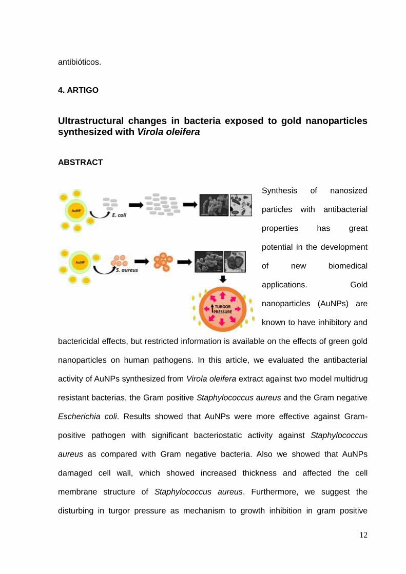
Ultrastructural changes in bacteria exposed to gold nanoparticles synthesized with Virola oleifera ABSTRACT
Synthesis of nanosized
particles with antibacterial
properties has great
potential in the development
of new biomedical
applications. Gold
nanoparticles (AuNPs) are
known to have inhibitory and
bactericidal effects, but restricted information is available on the effects of green gold
nanoparticles on human pathogens. In this article, we evaluated the antibacterial
activity of AuNPs synthesized from Virola oleifera extract against two model multidrug
resistant bacterias, the Gram positive Staphylococcus aureus and the Gram negative
Escherichia coli. Results showed that AuNPs were more effective against Gram-
positive pathogen with significant bacteriostatic activity against Staphylococcus
aureus as compared with Gram negative bacteria. Also we showed that AuNPs
damaged cell wall, which showed increased thickness and affected the cell
membrane structure of Staphylococcus aureus. Furthermore, we suggest the
disturbing in turgor pressure as mechanism to growth inhibition in gram positive
13
bacteria. In summary, our data indicates AuNPs as a promising model for the design
of novel antibacterial agents.
Keywords: Gold nanoparticles, green synthesis, ultrastructure, S. aureus, E. coli.
BACKGROUND
Nanotechnology has emerged recently in the material science field causing in
short time a great impact in diverse contemporary fields as biology, chemistry and
environment, energy, engineer, heavy industry and medicine1,2. The word ‘nano’
come from the Greek meaning ‘dwarf’ and was first applied in science in 1959 by
Richard Feynman, a Nobel Prize winning physicist3,4. Nanotechnology can be defined
as the science that involve the design, synthesis, characterization, and application of
nanomaterials (all materials of size ranging between 1 nm and 100 nm in an
unbound, or aggregate, or agglomerate state)5,6.
Due to their small size, a collection of nanoparticles show a combined high
surface area to volume ratio, resulting in unique physical, chemical, mechanical,
electrical, optical and biological properties, which differ from their respective bulk
material7–10. These distinct characteristics empower the interaction with cells and
tissues at a molecular level in many ways, for example, as biological mimetics,
nanomachines, carriers of drugs, gene therapy, and label; opening a new door at
medical research: the nanomedicine10.
Amongst the metallic nanoparticles, the most used in the biomedical field are
gold nanoparticles (AuNPs)11. This preference prevails because gold is an easy
material to obtain and manage; it is an inert metal; chemically stable; resistant to
surface oxidation; non-toxic and compatible with cells12,13. Gold nanoparticles have
14
been already applied for the diagnosis and treatment of diseases, tissue/tumor
imaging, drug delivery, photothermal therapy, antibody conjugation, and protection
against UV rays14,15.
AuNPs have emerged as potential alternative antibacterial agents against
multidrug resistant (MDR) bacteria16. Since antibiotics became a medication with a
broad and indiscriminate use, bacteria developed resistance against most standard
antibacterial agents as well as adverse side effects due to higher dose prescription17
resulting in a world health problem. Thus, the development of gold nanoparticles able
to replace or improve those standard antibacterial agents may unveil novel
biochemical and structural targets for the development of alternative therapies.
Nanoparticles are traditionally synthesized through chemical reduction of metal
ions using toxic reducing agents to convert Au+3 ions to AuNPs18,19. However, these
methods might represent an environmental and biological risk20. To overcome that
limitation, new less expensive, chemically stable, environmentally-friendly, and clean
biological methods using plant extracts as reduced and stabilizing agents have been
recently developed21. Previous works have shown the application of green route
synthesized AuNPs to control bacterial growth: AuNPs synthesized using Ananas
comosus extract reduced the growth of Staphylococcus aureus (gram-positive) and
Pseudomonas aeruginosas (gram-negative)13; also dextrose-encapsulated gold
nanoparticles (dGNPs) showed significant antibacterial activity against both
Escherichia coli (gram-negative) as well as Staphylococcus epidermidis (gram-
positive) bacteria via disruption of cell membrane21. On the other hand,
biosynthesized AuNPs with Solanum nigrum leaf extract had showed better
antibacterial activity against gram-negatives bacterias than to gram-positives22. The
anti-bacterial mechanism has electrostatic or mechanical interaction basis13, with
15
direct dependence on composition, surface modification, intrinsic properties, and the
bacterial species (cause of their different cell wall arrangement)17.
Here, we investigated the potential antibacterial mechanism of gold
nanoparticles reduced by Virola oleifera extract. Virola oleifera is a native plant of
Atlantic forest being widely spread in the southeastern region of Brazil and has been
applied in folk medicine as cicatrizant, anti-inflammatory, antirheumatic, and anti-
asthmatic agents23. To verify if AuNPs biosynthesized with Virola oleifera produce
ultrastructural changes in bacteria we tested the AuNPs with one gram positive
(Staphylococcus aureus) and one gram negative (Escherichia coli) model bacteria
and followed their growth along the time and their morphological changes by
scanning and transmission electronic microscopy techniques.
METHODS Green synthesis of gold nanoparticles
The gold nanoparticles have been synthesized by oxidation-reduction method,
using a solution of HAuCl4 (SigmaAldrich, St Louis, MO, USA) and solution of Virola
oleifera resin at a concentration of 1000 µg/ml as the reducing agent. Virola oleifera
was chosen because it is rich in phenolic compounds, which provides
functionalization to the nanoparticles, and are important for reducing the metal ions,
as well as have good antioxidant activity. In particular, 3 ml of solution of the Virola
oleifera extract was added to 10 ml of HAuCl4 (2.5 × 10⁻⁴ M) solution and shaken at
600 rpm for 10 minutes. The stirred solution was incubated at room temperature.
Antibacterial test
To measure the Minimum Inhibitory Concentration (MIC) of AuNPs against the
16
respective bacteria, the final solution of gold nanoparticles was diluted 10 and 100
times and inoculated with 1,5 x 10⁶ UFC/mL in culture tubes containing sterile liquid
media. Control experiments were performed by inoculating the media with bacteria
and water. To ensure that the possible antimicrobial activity was due nanoparticles, a
pure reducing solution was tested and at the same concentration used with
nanoparticles. Two strains of bacteria were tested, the Gram positive Staphylococcus
aureus 1117 and the Gram-negative Escherichia coli DH5α. The 96-wells microplate
was incubated with 100 µL of media plus bacteria with 100 µL of gold nanoparticles
solution in their respective concentration at 37°C. The growth of bacteria was
monitored by measuring the optical density (OD) at 620 nm for 24h using iMark
microplate reader, BIO-RAD Lab. All the experiment was done in triplicate and the
data were expressed by mean.
Scanning Electron Microscope analyse
To investigate if the gold nanoparticle from Virola oleifera produced any
ultrastructural changes we compare the bacteria exposed to AuNPs by electron
microscopy. The culture of bacteria strain was washed with PBS and fixed in 2.5%
glutaraldehyde in 0.1M sodium cacodylate buffer (pH 7.4). After incubating with the
fixative for 2 h at room temperature, samples were washed and centrifuged twice; the
supernatant was discarded, and the pellet was resuspended in 0.1M sodium
cacodylate buffer. The same process was followed during all subsequent solution
changes. Samples were then post-fixed for 1 h at room temperature with 1% osmium
tetroxide in 0.1M sodium cacodylate buffer. The post-fixed samples were washed
with 0.1M sodium cacodylate buffer once and with distilled water twice and then
dehydrated in a graded ethanol series (once in 30%, 50%, 70%, and 90%, and three
17
times in 100% ethanol for 10 min each). Following, the pellet was scattered using an
ultrasonic cleaner (SB–100D) and 10 µl droplets applied on coverglass slide. The
samples were critical point dried in a autosamdri-815 (tousimis) critical point dryer,
attached to aluminum mounting stubs, sputter coated with gold-palladium, and
imaged in a JEOL (JSM-6610LV, JEOL Inc., USA) scanning electron microscope.
Transmission Electron Microscope analyse
For TEM analysis, samples were prepared as described above for SEM with
the difference in the dehydration with a graded acetone series (once in 30%, 50%,
70%, and 90%, and three times in 100% acetone for 10 min each). Following, the
dehydrated samples were infiltrated with Epon’s epoxy resin and acetone (1:1). Then
left overnight in 100% resin. Samples were centrifuged through fresh resin in BEEM
capsules (EMS Inc., Hatfield, PA, USA) and hardened at 60°C for 24h. Ultrathin
sections (70nm) of the pelleted samples were cut on an ultramicrotome (RMC
PowerTome X) using a diamond knife. The sections were stained with 2% aqueous
uranyl acetate and Reynold’s lead citrate for 5 minutes and 2 minutes, respectively
for S. aureus and 20 minutes and 5 minutes, respectively for E. coli and examined
using a transmission electron microscope (JEM-1400, JEOL Inc, USA).
Statistical Analyses
In order to measure the differences observed in structures between the control
group and the group inoculated with AuNP a t-test analyse was performed using the
GraphPad Prism 6 (GraphPad Software, La Jolla California USA). p<0.05 was taken
as significant.
RESULTS
18
Synthesis of gold nanoparticles using Virola oleifera extract
The resulting solution showed a colour change from pale yellow to light pink.
According to previous analyses with inductively coupled plasma mass spectrometry
(ICP-MS) the final gold concentration was 40 µg/ml (data not shown). To use it in the
antibacterial test this solution was diluted 10 times and 100 times resulting
respectively in AuNPs of 4 µg/ml and 0.4 µg/ml gold concentration.
Antibacterial activity
The AuNPs were tested for their antibacterial activity at three concentrations
against S. aureus and E. coli. In the 96-wells microplate, the bacterial cells grown in
presence of AuNPs, and the growth was monitored hourly by measuring the OD at
620 nm for 24h. Optical density at 620 nm is a common method to quantify the
concentration of bacterial cells in a liquid medium. There was no interference of
AuNP or plant extract in the reading absorbance because they are read only at 530
nm and 300 nm respectively. Results were plotted with the OD on Y-axis against and
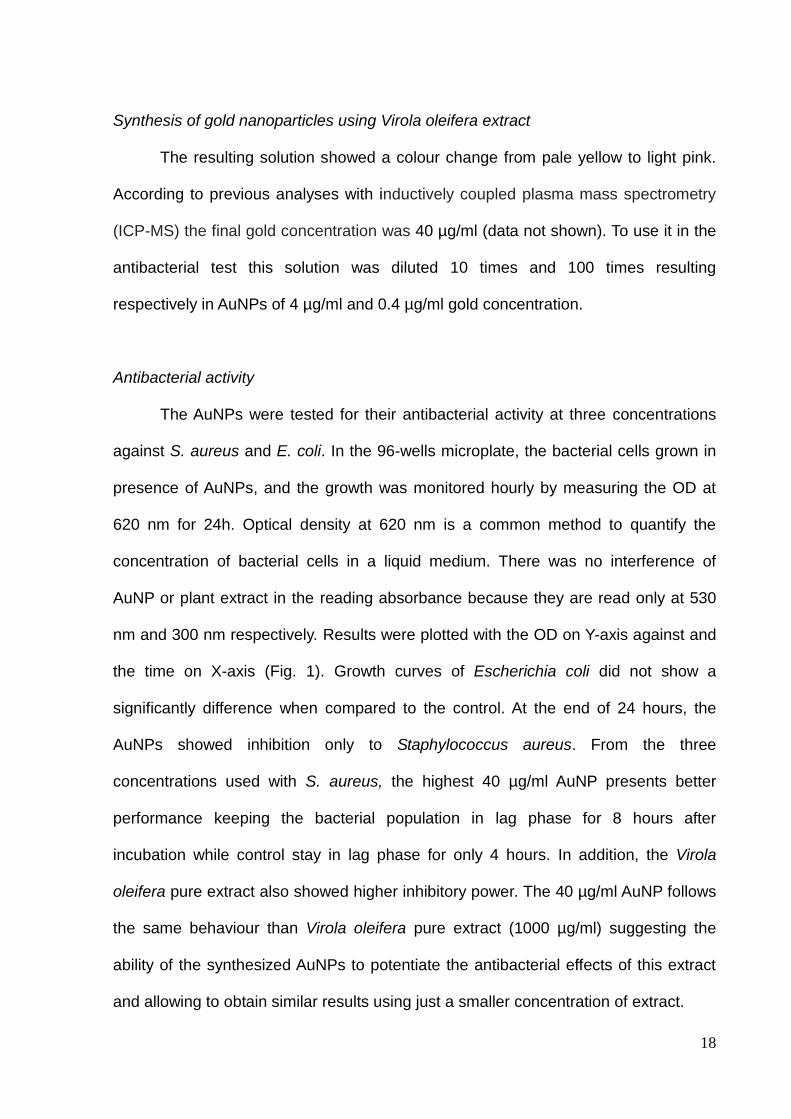
the time on X-axis (Fig. 1). Growth curves of Escherichia coli did not show a
significantly difference when compared to the control. At the end of 24 hours, the
AuNPs showed inhibition only to Staphylococcus aureus. From the three
concentrations used with S. aureus, the highest 40 µg/ml AuNP presents better
performance keeping the bacterial population in lag phase for 8 hours after
incubation while control stay in lag phase for only 4 hours. In addition, the Virola
oleifera pure extract also showed higher inhibitory power. The 40 µg/ml AuNP follows
the same behaviour than Virola oleifera pure extract (1000 µg/ml) suggesting the
ability of the synthesized AuNPs to potentiate the antibacterial effects of this extract
and allowing to obtain similar results using just a smaller concentration of extract.
19
An inhibition of approximately 65% on bacterial growth was observed to S.
aureus (Fig 1).
Figure 1. Effects of the AuNPs on the bacterial growth. Growth analysis curves were
measured by monitoring the optical density (OD) at 620 nm, and the bacteria were treated
with AuNPs at different concentrations (mg/L). Scanning Electron Microscopy
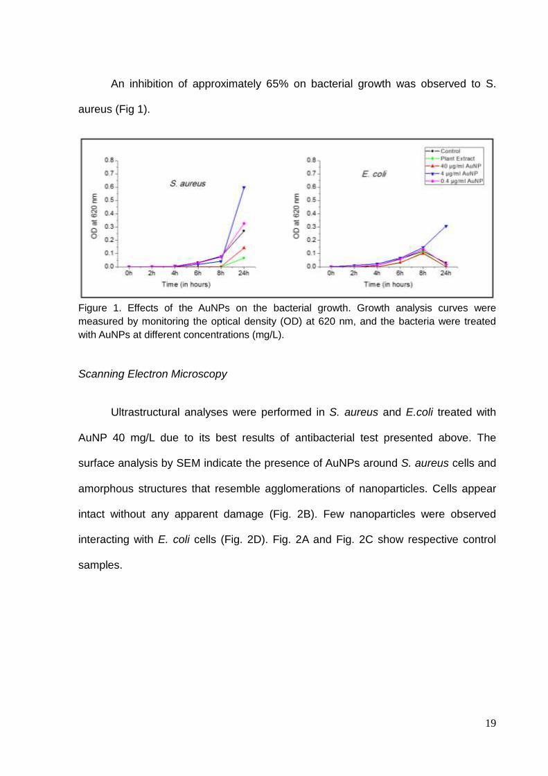
Ultrastructural analyses were performed in S. aureus and E.coli treated with
AuNP 40 mg/L due to its best results of antibacterial test presented above. The
surface analysis by SEM indicate the presence of AuNPs around S. aureus cells and
amorphous structures that resemble agglomerations of nanoparticles. Cells appear
intact without any apparent damage (Fig. 2B). Few nanoparticles were observed
interacting with E. coli cells (Fig. 2D). Fig. 2A and Fig. 2C show respective control
samples.
20
Figure 2. SEM micrographs of S.aureus and E. coli. (A) and (C) Control, (B) S. aureus treated with nanoparticles, (D) E. coli treated with nanoparticles. Arrows indicate AuNPs on contact with cells.
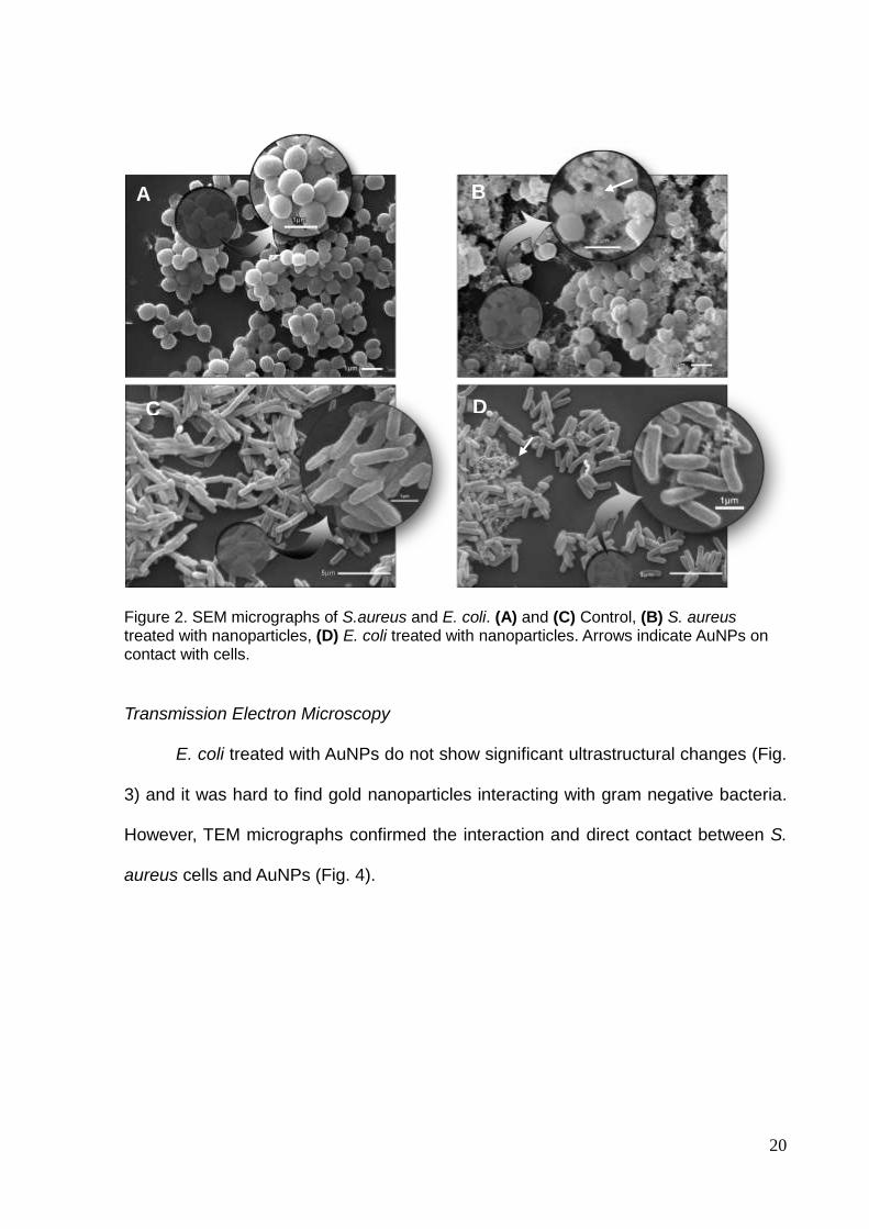
Transmission Electron Microscopy
E. coli treated with AuNPs do not show significant ultrastructural changes (Fig.
3) and it was hard to find gold nanoparticles interacting with gram negative bacteria.
However, TEM micrographs confirmed the interaction and direct contact between S.
aureus cells and AuNPs (Fig. 4).
A
A
C D
B
A
21
Figure 3. TEM micrographs of E. coli. No significative changes on structure the of cells were
found. (A) Control, (B) few AuNPs were observed along the analyses. Arrow indicates AuNPs.
Figure 4. TEM micrographs of S. aureus in the presence of AuNPs. We can see in detail the nanoparticles in direct contact with the cell.
B A

22
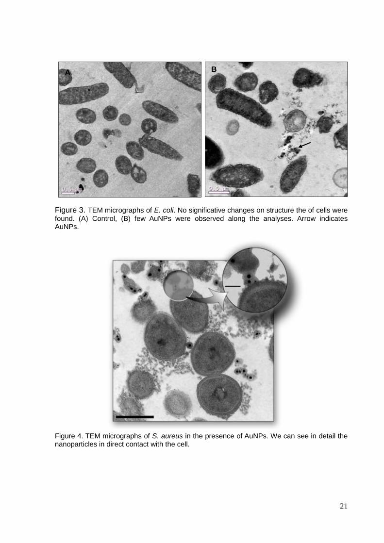
Furthermore, we observed injuries in the cell wall of S. aureus as well as
increase on its thickness and modifications on membrane structure (Fig. 5).
To confirm the increase in cell wall thickness of S. aureus population exposed to
AuNPs, the thickness of approximately 27 cells in the control sample and in the
group treated with nanoparticles was measured to calculate the mean thickness of
each group. Whereas the control group showed average of 25.7 nm, the group
treated had average of 41.6 nm of cell wall thickness (64% increase) (Fig. 5D).
Figure 5. Ultrastructural changes induced by AuNP on S. aureus. (A) Control, (B) we can
observe cell wall injuries (black arrows) and bacteria showing cell wall enlarged (*), (C)
structural changes in the plasmatic membrane (white arrows), (D) graph comparing cell wall
thickness. S. aureus treated with AuNP had a significative increase in cell wall compared with
control (p < 0.05).
Me
an
Th
ick
ne
ss
(n
m)
S . a u r e u s S . a u r e u s w ith Au N P s
0
2 0
4 0
6 0
S . a u re u s
S . a u re u s w ith A u N P s
A B C
D
*
23
DISCUSSION
Antibacterial agents are commonly divided into two categories: as bactericidal,
which kill bacteria; or bacteriostatic, which just slow down bacterial growth17.
Throughout the data from antibacterial test, we observed that the gold nanoparticles
synthesized with Virola oleifera extract as a reducing agent displayed an effective
bacteriostatic action with effects only S. aureus growth. Comparing the antibacterial
activity of the gold nanoparticles with the reducing agent (Fig. 1) it is observed similar
antibacterial activity, meaning that even after reducing gold, the Virola oleifera resin
functionalized effectively the nanoparticle, which conserve the high antibacterial
power.
There are various potential mechanisms of gold nanoparticles against bacteria
such as electrostatic interaction24, disruption of cell membrane25, induction of stress
oxidative26, and inhibiting ATP synthesis27; however the mechanisms of nanoparticles
toxicity will depend not only of its composition, surface and properties but also of the
bacterial species and morphology20.
Thus, the bacteriostatic activity and ultrastructural changes here described
against S. aureus but not E. coli are probably influenced by their cell wall
composition. As known, Staphylococcus aureus is a Gram positive bacteria which
means their wall contains a dense layer of peptidoglycan (PG) attached with teichoic
acids that are unique to Gram positive bacteria28. On the other hand, Gram negative
cell wall of Escherichia coli contains with a thin PG layer surrounded by an outer
membrane rich in lipopolysaccharides which confers resistance to hydrophobic
compounds and increase the negative charge of cell membranes29. Although both
gram positive and gram negative bacteria have a negative membrane charges, the
cell wall composition of S. aureus might modify its intensity and benefit the AuNPs
24
biosynthesized to attach on their cell wall.
A second fact to consider in the nanotoxicity analyses, it is the agglomeration
of AuNPs as observed in Fig. 2B. Agglomeration could happen cause of difference of
PH, or salts concentration between the AuNP’s solution and the media where they
were inoculated with bacteria. Agglomeration can change the size, surface area and
sedimentation properties of the gold nanoparticles influencing their ability to reach the
target and interact with bacteria11,30.
Besides, growth rate has been reported to affect the bacteria resistance to
antibiotics and nanoparticles. Species with fast-growing rates are more susceptible
than slow-growing bacteria31. It is possible that the tolerance property of slow-
growing bacteria is related to the expression of stress-response genes32.
Distinctive of most nanoparticles that have been described, the AuNP
synthesized from Virola oleifera extract does not cause inhibition through cell
disruption and leakage25,33,34,35. Nevertheless, the modification of membrane
structure, injuries and rise of thickness of cell wall visualised in S. aureus treated with
AuNP (Fig. 5) may indicate an inhibition action by increasing the turgor pressure.
Turgor pressure, or osmotic pressure, is created by the higher concentration of
solutes in the cytoplasm and impulses the membrane against wall in gram positive
bacteria36.The cell wall is responsible for more than strength, rigidity and shape but is
determinant to control the turgor pressure and protect from osmotic rupture36,37.
Then, any modification on cell wall growth can disturb the turgor pressure. As the
AuNPs touch the cell wall surface, it can breaks the peptidoglycan layer resulting in
injuries. This could activate a defence mechanism, or even trick the cell, stimulating
the wall’s synthesis by laying down unstressed peptidoglycan in layers on the inner
side of the wall while the outer layers are injuried37,38 . It could increase the turgor
25
pressure causing stress on the membrane, inducing the expression of
osmoregulation genes, influencing bacterial signal transduction systems, bacterial
periplasmic transport functions, synthesis of porines, and finally inhibiting bacteria
growth rate39.
The results of this study demonstrated that the gold nanoparticle inhibited the
growth and multiplication of the highly multidrug-resistant bacteria S. aureus. In spite
of that antimicrobial potential, are necessary further studies to improve the
nanoparticle behaviour and nanotoxicity using them as drug delivery mechanisms by
conjugating with standard antibiotics40,41.
In conclusion, the antibacterial activity of the green gold nanoparticle
synthesized from Virola oleifera extract was confirmed against gram positive bacteria
but not gram negative. We showed that AuNPs has bacteriostatic power against S.
aureus changing the cell ultrastructure. The present study speculates a possible
mechanism of growth inhibition based in the disturbing of turgor pressure and
consequently internal homeostasis of cell. Thus, AuNPs represent a potential model
for designing of antibacterial agents to target bacterial and to overcome drug
resistance.
REFERENCES
1 Niemeyer CM. Nanoparticles, Proteins, and Nucleic Acids: Biotechnology
Meets Materials Science. Angew Chem Int Ed 2001; 40: 4128 ± 4158.
2 Kanaparthy R. The changing face of dentistry: nanotechnology. Int J
Nanomedicine 2011; 6: 2799.
3 Bhardwaj A, Bhardwaj A, Misuriya A, Maroli S, Manjula S, Singh AK.
Nanotechnology in dentistry: Present and future. J Int oral Heal JIOH 2014; 6:
26
121–126.
4 Kubik T, Bogunia-Kubik K, Sugisaka M. Nanotechnology on duty in medical
applications. Curr Pharm Biotechnol 2005; 6: 17–33.
5 Saini R, Saini S, Sharma S. Nanotechnology: the future medicine. J Cutan
Aesthet Surg 2010; 3: 32–33.
6 Release R. What is a ‘ nanomaterial ’? European Commission breaks new
ground with a common definition. October 2011; : 1–2.
7 Daniel MC, Astruc D. Gold Nanoparticles: Assembly, Supramolecular
Chemistry, Quantum-Size-Related Properties, and Applications Toward Biology,
Catalysis, and Nanotechnology. Chem Rev 2004; 104: 293–346.
8 Iravani S. Green synthesis of metal nanoparticles using plants. Green Chem
2011; 13: 2638.
9 Whitesides G. Nanoscience, Nanotechnology, and Chemistry. Small 2005; 1:
172–179.
10 Moghimi SM, Hunter a C, Murray JC. Nanomedicine: current status and future
prospects. FASEB J 2005; 19: 311–330.
11 Chatterjee S, Bandyopadhyay A, Sarkar K. Effect of iron oxide and gold
nanoparticles on bacterial growth leading towards biological application. J
Nanobiotechnology 2011; 9: 34.
12 Love JC, Estroff L a., Kriebel JK, Nuzzo RG, Whitesides GM. Self-assembled
monolayers of thiolates on metals as a form of nanotechnology. 2005
doi:10.1021/cr0300789.
13 Bindhu MR, Umadevi M. Antibacterial activities of green synthesized gold
nanoparticles. Mater Lett 2014; 120: 122–125.
14 Mieszawska AJ, Mulder WJM, Fayad ZA, Cormode DP. Multifunctional Gold
Nanoparticles for Diagnosis and Therapy of Disease.pdf. 2013.
15 Ganesan RM, Prabu HG. Synthesis of gold nanoparticles using herbal Acorus
calamus rhizome extract and coating on cotton fabric for antibacterial and UV
blocking applications. Arab J Chem 2015. doi:10.1016/j.arabjc.2014.12.017.
16 Li X, Robinson SM, Gupta A, Saha K, Jiang Z, Moyano DF et al. Functional
27
Gold Nanoparticles as Potent Antimicrobial Agents against Multi-Drug-
Resistant Bacteria. 2014.
17 Hajipour MJ, Fromm KM, Akbar Ashkarran A, Jimenez de Aberasturi D,
Larramendi IR de, Rojo T et al. Antibacterial properties of nanoparticles. Trends
Biotechnol. 2012. doi:10.1016/j.tibtech.2012.06.004.
18 Lee KD, Nagajyothi PC, Sreekanth TVM, Park S. Eco-friendly synthesis of gold
nanoparticles (AuNPs) using Inonotus obliquus and their antibacterial,
antioxidant and cytotoxic activities. J Ind Eng Chem 2015; 26: 67–72.
19 Zhang Z, Patel RC, Kothari R, Johnson CP, Friberg SE, Aikens PA. Stable
Silver Clusters and Nanoparticles Prepared in Polyacrylate and Inverse
Micellar Solutions. J Phys Chem B 2000; 104: 1176–1182.
20 Moritz M, Geszke-Moritz M. The newest achievements in synthesis,
immobilization and practical applications of antibacterial nanoparticles. Chem.
Eng. J. 2013. doi:10.1016/j.cej.2013.05.046.
21 Badwaik VD, Vangala LM, Pender DS, Willis CB, Aguilar ZP, Gonzalez MS et
al. Size-dependent antimicrobial properties of sugar-encapsulated gold
nanoparticles synthesized by a green method. Nanoscale Res Lett 2012; 7:
623.
22 Muthuvel a., Adavallan K, Balamurugan K, Krishnakumar N. Biosynthesis of
gold nanoparticles using Solanum nigrum leaf extract and screening their free
radical scavenging and antibacterial properties. Biomed Prev Nutr 2014; 4:
325–332.
23 Fernandes AM a. P, Barata LES, Ferri PH. Lignans and a neolignan from Virola
oleifera leaves. Phytochemistry 1993; 32: 1567–1572.
24 Thill A, Zeyons O, Spalla O, Chauvat F, Rose J, Auffan M et al. Cytotoxicity of
CeO2 nanoparticles for Escherichia coli. Physico-chemical insight of the
cytotoxicity mechanism. Environ Sci Technol 2006; 40: 6151–6156.
25 Mei L, Lu Z, Zhang W, Wu Z, Zhang X, Wang Y et al. Bioconjugated
nanoparticles for attachment and penetration into pathogenic bacteria.
Biomaterials 2013; 34: 10328–10337.
26 Soenen SJ, Rivera-Gil P, Montenegro JM, Parak WJ, De Smedt SC,
Braeckmans K. Cellular toxicity of inorganic nanoparticles: Common aspects
and guidelines for improved nanotoxicity evaluation. Nano Today 2011; 6: 446–
28
465.
27 Cui Y, Zhao Y, Tian Y, Zhang W, Lü X, Jiang X. The molecular mechanism of
action of bactericidal gold nanoparticles on Escherichia coli. Biomaterials 2012;
33: 2327–2333.
28 Navarre WW, Schneewind O. Surface Proteins of Gram-Positive Bacteria and
Mechanisms of Their Targeting to the Cell Wall Envelope Surface Proteins of
Gram-Positive Bacteria and Mechanisms of Their Targeting to the Cell Wall
Envelope. 1999; 63: 174–229.
29 Roberts IS. The biochemistry and genetics of capsular polysaccharide
production in bacteria. Annu Rev Microbiol 1996; 50: 285–315.
30 Zook JM, MacCuspie RI, Locascio LE, Halter MD, Elliott JT. Stable nanoparticle
aggregates/agglomerates of different sizes and the effect of their size on
hemolytic cytotoxicity. Nanotoxicology 2011; 5: 517–530.
31 Chemoptherapy A, Ii M. Leading Articles. Br Med J 1931; 1: 903–907.
32 Charles Lu,* Matthew J. Brauer and DB. Slow Growth Induces Heat-Shock
Resistance in Normal and Respiratory-deficient Yeast. Mol Biol Cell 2009; 29:
891–903.
33 Rai A, Prabhune A, Perry CC. Antibiotic mediated synthesis of gold
nanoparticles with potent antimicrobial activity and their application in
antimicrobial coatings. J Mater Chem 2010; 20: 6789.
34 Feng ZV, Gunsolus IL, Qiu TA, Hurley KR, Nyberg LH, Frew H et al. Impacts of
gold nanoparticle charge and ligand type on surface binding and toxicity to
Gram-negative and Gram-positive bacteria. Chem Sci 2015; 6: 5186–5196.
35 Goswami SR, Sahareen T, Singh M, Kumar S. Role of biogenic silver
nanoparticles in disruption of cell–cell adhesion in Staphylococcus aureus and
Escherichia coli biofilm. J Ind Eng Chem 2015; 26: 73–80.
36 Deng Y, Sun M, Shaevitz JW. Direct Measurement of Cell Wall Stress Stiffening
and Turgor Pressure in Live Bacterial Cells. Phys Rev Lett 2011; 107: 158101.
37 Scheffers D, Pinho M. Bacterial cell wall synthesis: new insights from
localization studies. Microbiol Mol Biol Rev 2005; 69: 585–607.
38 Misra G, Rojas ER, Gopinathan A, Huang KC. Mechanical consequences of
29
cell-wall turnover in the elongation of a gram-positive bacterium. Biophys J
2013. doi:10.1016/j.bpj.2013.04.047.
39 Arnoldi M, Fritz M, Bäuerlein E, Radmacher M, Sackmann E, Boulbitch A.
Bacterial turgor pressure can be measured by atomic force microscopy. Phys
Rev E - Stat Physics, Plasmas, Fluids, Relat Interdiscip Top 2000; 62: 1034–
1044.
40 Gu H, Ho PL, Tong E, Wang L, Xu B. Presenting vancomycin on nanoparticles
to enhance antimicrobial activities. Nano Lett 2003; 3: 1261–1263.
41 Bhattacharya D, Saha B, Mukherjee A, Ranjan Santra C, Karmakar P. Gold
Nanoparticles Conjugated Antibiotics: Stability and Functional Evaluation.
Nanosci Nanotechnol 2012; 2: 14–21.
30
5. REFERÊNCIAS BIBLIOGRÁFICAS
BADWAIK, V. D.; VANGALA, L. M.; PENDER, D. S.; et al. Size-dependent
antimicrobial properties of sugar-encapsulated gold nanoparticles synthesized by a
green method. Nanoscale research letters, v. 7, n. 1, p. 623, 2012. Nanoscale
Research Letters. Disponível em:
<http://www.pubmedcentral.nih.gov/articlerender.fcgi?artid=3533927&tool=pmcentrez
&rendertype=abstract>. .
BINDHU, M. R.; UMADEVI, M. Antibacterial activities of green synthesized gold
nanoparticles. Materials Letters, v. 120, p. 122–125, 2014. Elsevier. Disponível em:
<http://dx.doi.org/10.1016/j.matlet.2014.01.108>. .
COUTO R 2003. Bacterias multirresistentes. In: Couto RC, Pedrosa TMG, Nogueira JM. Infecção hospitalar e outras complicações não infecciosas da doença. 3nd. ed., Medsi, Rio de Janeiro, p. 567-88.
FISCHBACH, M. A.; WALSH, C. T. NIH Public Access. , v. 325, n. 5944, p. 1089–
1093, 2010.
HWANG, S. J.; JUN, S. H.; PARK, Y.; et al. Green synthesis of gold nanoparticles
using chlorogenic acid and their enhanced performance for inflammation.
Nanomedicine: Nanotechnology, Biology and Medicine, p. 1–11, 2015. Elsevier
B.V. Disponível em:
<http://linkinghub.elsevier.com/retrieve/pii/S1549963415001136>. .
IRAVANI, S. Green synthesis of metal nanoparticles using plants. Green Chemistry,
v. 13, p. 2638, 2011.
KUBIK, T.; BOGUNIA-KUBIK, K.; SUGISAKA, M. Nanotechnology on duty in medical
applications. Current pharmaceutical biotechnology, v. 6, n. 1, p. 17–33, 2005.
LEE, K. D.; NAGAJYOTHI, P. C.; SREEKANTH, T. V. M.; PARK, S. Eco-friendly
synthesis of gold nanoparticles (AuNPs) using Inonotus obliquus and their
antibacterial, antioxidant and cytotoxic activities. Journal of Industrial and
Engineering Chemistry, v. 26, p. 67–72, 2015. The Korean Society of Industrial and
Engineering Chemistry. Disponível em:
<http://linkinghub.elsevier.com/retrieve/pii/S1226086X1400570X>. .
LOK, C.; HO, C.; CHEN, R.; et al. Proteomic Analysis of the Mode of Antibacterial
Action of Silver research articles. Journal of proteome research, v. 5, p. 916–924,
2006.
LOVE, J. C.; ESTROFF, L. A.; KRIEBEL, J. K.; NUZZO, R. G.; WHITESIDES, G. M.
Self-assembled monolayers of thiolates on metals as a form of
nanotechnology. 2005.
31
MARANGONI, V. S. ESTUDO E DESENVOLVIMENTO DE NANOCOMPÓSITOS
CONTENDO NANOPARTÍCULAS DE OURO CONJUGADAS COM
BIOMOLÉCULAS: SÍNTESE E APLICAÇÕES EM NANOMEDICINA, 2012.
UNIVERSIDADE DE SÃO PAULO.
MILANEZE, B.; KEIJOK, W.; JAIRO, O.; et al. The green synthesis of gold
nanoparticle using extract of Virola oleifera. BMC Proceedings, v. 8, n. Suppl 4, p.
P29, 2014. BioMed Central Ltd. Disponível em:
<http://www.biomedcentral.com/1753-6561/8/S4/P29>. .
MOGHIMI, S. M.; HUNTER, A C.; MURRAY, J. C. Nanomedicine: current status and
future prospects. The FASEB journal : official publication of the Federation of
American Societies for Experimental Biology, v. 19, n. 3, p. 311–330, 2005.
MORITZ, M.; GESZKE-MORITZ, M. The newest achievements in synthesis,
immobilization and practical applications of antibacterial nanoparticles. Chemical
Engineering Journal, 2013.
MUTHUVEL, A.; ADAVALLAN, K.; BALAMURUGAN, K.; KRISHNAKUMAR, N.
Biosynthesis of gold nanoparticles using Solanum nigrum leaf extract and screening
their free radical scavenging and antibacterial properties. Biomedicine and
Preventive Nutrition, v. 4, n. 2, p. 325–332, 2014. Elsevier Masson SAS. Disponível
em: <http://dx.doi.org/10.1016/j.bionut.2014.03.004>. .
NARAYANAN, K. B.; SAKTHIVEL, N. Green synthesis of biogenic metal
nanoparticles by terrestrial and aquatic phototrophic and heterotrophic eukaryotes
and biocompatible agents. Advances in Colloid and Interface Science, v. 169, n. 2,
p. 59–79, 2011. Elsevier B.V.
PATIL, R. S.; KOKATE, M. R.; KOLEKAR, S. S. Bioinspired synthesis of highly
stabilized silver nanoparticles using Ocimum tenuiflorum leaf extract and their
antibacterial activity. Spectrochimica Acta - Part A: Molecular and Biomolecular
Spectroscopy, v. 91, p. 234–238, 2012. Elsevier B.V. Disponível em:
<http://dx.doi.org/10.1016/j.saa.2012.02.009>. .
SADEGHI, B.; MOHAMMADZADEH, M.; BABAKHANI, B. Green synthesis of gold
nanoparticles using Stevia rebaudiana leaf extracts: Characterization and their
stability. Journal of Photochemistry and Photobiology B: Biology, v. 148, p. 101–
106, 2015. Elsevier B.V.
SHON, Y. S.; CHOO, H. Organic reactions of monolayer-protected metal
nanoparticles. Comptes Rendus Chimie, v. 6, n. 8-10, p. 1009–1018, 2003.
TRAN, T. T. T.; VU, T. T. H.; NGUYEN, T. H. Biosynthesis of silver nanoparticles using
Tithonia diversifolia leaf extract and their antimicrobial activity. Materials Letters, v.
105, p. 220–223, 2013. Elsevier. Disponível em:
<http://dx.doi.org/10.1016/j.matlet.2013.04.021>.
32
6. APÊNDICE Article structure Submission items include a cover letter, a list of suggested reviewers, text for the graphical abstract, image for the graphical abstract, the manuscript (including title page, abstract, manuscript text [divided into Background, Methods, Results, and Discussion for Original Articles], references, and figure legends), tables, and figures. Authors are advised to upload supporting information in Supplementary Material and permissions (see below) if needed. Revised manuscripts should also be accompanied by a unique file (separate from the cover letter) with responses to reviewers’ comments. The preferred order of files is as follows: cover letter, suggested reviewers, response to reviews (revised manuscripts only), graphical abstract files, manuscript file, table(s), figure(s), supplementary material, permission(s), if needed. Files should be labeled with appropriate and descriptive file names (e.g., SmithManuscript.doc, Figure1.eps, Table3.doc, Supplementary.pdf). If a revision is requested, upload text, tables, and figures as separate files. Do not embed figures or tables into the text document. Please upload your revised text as a Word file, and upload text-based Supplementary Materials as a PDF. All manuscripts must conform to Uniform Requirements for Manuscripts Submitted to Biomedical Journals. Cover letter Be sure to state that the manuscript, or any part of it, has not been published and will not be submitted elsewhere for publication while being considered by the journal Nanomedicine: NBM in the cover letter. If there is any overlap between the submission and any other material, published or submitted, detail the nature of and reason for the overlap; as relevant, upload a copy of the other material. Title page Essential title page information • Title. Concise and informative. Titles are often used in information-retrieval systems. Avoid abbreviations and formulae where possible. A title should describe the article's content clearly and precisely and allow the reader to decide whether it would be appropriate to consult the article further. The title is the advertisement for the article; a poorly titled article may never reach its target audience, so be specific. Omit unnecessary words such as “Novel,” “New,” “A study of,” “Investigations of,” “Observations on,” etc. Do not use abbreviations and jargon. Avoid overinflated, bombastic “marketing” titles. Indexing and abstracting services depend on the accuracy of the title and keywords used in cross-referencing are extracted from the title itself. • Author names and affiliations. Where the family name may be ambiguous (e.g., a double name), please indicate this clearly. Present the authors' affiliation addresses (where the actual work was done) below the names. Indicate all affiliations with a lower-case superscript letter immediately after the author's name and in front of the appropriate address. Provide the full postal address of each affiliation, including the country name and, if available, the e-mail address of each author. • Corresponding author. Clearly indicate who will handle correspondence at all stages of refereeing and publication, also post-publication. Ensure that phone numbers (with country and area code) are provided in addition to the e-mail address and the complete postal address. Contact details must be kept up to date by the corresponding author.
33
• Present/permanent address. If an author has moved since the work described in the article was done, or was visiting at the time, a 'Present address' (or 'Permanent address') may be indicated as a footnote to that author's name. The address at which the author actually did the work must be retained as the main, affiliation address. Superscript Arabic numerals are used for such footnotes. The title page must include the word count for abstract; complete manuscript word count (including body text and figure legends, but excluding abstract, title page, and references), and number of figures/tables. There is no length limit on Supplementary Material. Manuscripts with over-the-limit word counts will be returned to the authors without further review. The title page must include statements of funding or conflicts of interest. Please make note of any prior presentation of abstracts at meetings regarding the research. Abstract A concise and factual summary of 150 words or less without internal subheadings and citations is required. The abstract should state briefly the purpose of the research, the principal results, and major conclusions. An abstract is often presented separately from the article, so it must be able to stand alone. For this reason, references and abbreviations should be avoided, but, if essential, they must be defined at their first mention in the abstract itself. Insert 3 to 5 key words after the abstract. Be sure to include the abstract in the manuscript file. Graphical abstracts Authors are required to submit a graphical abstract for their article containing one image and a short description; both should briefly summarize the essence of the paper in a concise form designed to capture the attention of a wide readership and for compilation of databases. Graphical abstracts should be submitted as a separate file in the online submission system. The image should present the key points of the paper in a concise, pictorial form designed to seize the attention of readers and should not contain more than four panels. Please submit one image (in .tif or .eps format); see below for further resolution requirements. The text component should be ~50-100 words in .doc format, and it should not be the same as the abstract in the manuscript. If a figure in the manuscript is used as the graphical abstract image, please note this at the end of the abstract (i.e.: Graphical Abstract: Figure 2), and refer to the system's instructions for uploading. Examples of successful graphical abstract images and a link to Graphical Abstract Polishing can be found at http://www.elsevier.com/wps/find/authorsview.authors/graphicalabstracts; see also a recent issue of the Journal, particularly Example 1, Example 2, Example 3. Authors can make use of Elsevier's Illustration and Enhancement service to ensure the best presentation of their images also in accordance with all technical requirements: Illustration Service. Keywords Immediately after the abstract, provide a maximum of 6 keywords, using American spelling and avoiding general and plural terms and multiple concepts (avoid, for
34
example, 'and', 'of'). Be sparing with abbreviations: only abbreviations firmly established in the field may be eligible. These keywords will be used for indexing purposes. Abbreviations Define abbreviations that are not standard in this field in a footnote to be placed on the first page of the article. Such abbreviations that are unavoidable in the abstract must be defined at their first mention there, as well as in the footnote. Ensure consistency of abbreviations throughout the article. Structure of text For Communications and Original Articles, the text should include the headings Background, Methods, Results, and Discussion. Only essential data and descriptions should be provided in the Methods and Results. All experimental details including synthetic and analytical procedures must be provided as SUPPLEMENTARY MATERIALS (see details below). All pages must be numbered. Abbreviations must be parenthetically notated at first mention in the text. Each table and figure must be mentioned in the text. Reports of studies on humans and animals must indicate that each study has been approved by an institutional review committee and the procedures followed are in accordance with institutional guidelines. Provide generic rather than trademarked names of drugs. Acknowledgements The acknowledgments section recognizes substantive contributions of individuals who do not meet the criteria for authorship (see below). The Editorial Office must receive written, signed consent from each person recognized in the acknowledgments to be mentioned in the article because acknowledgment can imply endorsement of data and conclusions. (See a sample of an Acknowledgement.) Upload each permission separately in the online system. Do not include statements of funding, conflicts, or other disclosures in the Acknowledgments; these must appear on the title page. Units Follow internationally accepted rules and conventions: use the international system of units (SI). If other units are mentioned, please give their equivalent in SI. Math formulae Please submit math equations as editable text and not as images. Present simple formulae in line with normal text where possible and use the solidus (/) instead of a horizontal line for small fractional terms, e.g., X/Y. In principle, variables are to be presented in italics. Powers of e are often more conveniently denoted by exp. Number consecutively any equations that have to be displayed separately from the text (if referred to explicitly in the text).
35
Footnotes Do not use footnotes in the text. Artwork Electronic artwork Figures must be of professional quality and suitable for evaluation purposes. Be sure all font is at a readable size. When possible, please use first-generation artwork. If a revision is requested, source files must be provided: Figures must be submitted in electronic figure file format: .tif, .eps, or .jpg format. Figures may be black and white line art, graphs, halftones (photographs), or color. Line art (black lines on a white background) must have a minimum resolution of 1,000 ppi. Combination line art (e.g., line art with gray fill patterns) must be created at 500 ppi. Black and white or color photographs must have a minimum resolution of 300 ppi. Illustrations should be saved at the recommended resolution setting and sized as close to a column width (3 to 4 inches) as possible. Create figures in scale with each other to the extent possible. Avoid background gridlines and other formatting that do not convey information (e.g., superfluous use of 3-dimensional formatting, background shadings). Use uniform lettering and sizing of all original artwork. As a general rule the lettering on the artwork should have a finished, printed size of 7 points. Smaller lettering will yield barely legible text. Recommended font choices include Arial, Helvetica, or Symbol. Labeling of multipart figures (eg, A, B, C...) should use capital letters only and should be done consistently, preferably using uppercase type (Arial, Helvetica or Universe, 11 or 12 points) in the lower-right corner of the figure. Avoid headings on the figure when possible. Additional information on preparation of electronic artwork can be found in the Artwork Instructions available at http://www.elsevier.com/authors. For best results, please follow these guidelines carefully. There is no charge for publication of color illustrations. General points • Make sure you use uniform lettering and sizing of your original artwork. • Embed the used fonts if the application provides that option. • Aim to use the following fonts in your illustrations: Arial, Courier, Times New Roman, Symbol, or use fonts that look similar. • Number the illustrations according to their sequence in the text. • Use a logical naming convention for your artwork files. • Provide captions to illustrations separately. • Size the illustrations close to the desired dimensions of the printed version. • Submit each illustration as a separate file. A detailed guide on electronic artwork is available on our website: http://www.elsevier.com/artworkinstructions Please do not: • Supply files that are optimized for screen use (e.g., GIF, BMP, PICT, WPG); these typically have a low number of pixels and limited set of colors; • Supply files that are too low in resolution; • Submit graphics that are disproportionately large for the content. Figure captions Ensure that each illustration has a caption. Supply captions separately, not attached to the figure. A caption should comprise a brief title (not on the figure itself) and a
36
description of the illustration. Keep text in the illustrations themselves to a minimum but explain all symbols and abbreviations used. References Citation in text Cite references in numeric order according to the first mention in the text. Accuracy of reference data is the responsibility of the author. Verify all references against original sources. “In press” citations must have been accepted for publication and the name of the journal or book publisher must be included; these citations must be updated before publication. Unpublished results and personal communications should not appear in the reference list but may be mentioned in the text. Authors wishing to cite unpublished material must have a letter of permission from the originator of the communication to do so. This letter should be submitted with the manuscript. Please ensure that every reference cited in the text is also present in the reference list (and vice versa). Reference links Increased discoverability of research and high quality peer review are ensured by online links to the sources cited. In order to allow us to create links to abstracting and indexing services, such as Scopus, CrossRef and PubMed, please ensure that data provided in the references are correct. Please note that incorrect surnames, journal/book titles, publication year and pagination may prevent link creation. When copying references, please be careful as they may already contain errors. Use of the DOI is encouraged. Web references As a minimum, the full URL should be given and the date when the reference was last accessed. Any further information, if known (DOI, author names, dates, reference to a source publication, etc.), should also be given. Web references can be listed separately (e.g., after the reference list) under a different heading if desired, or can be included in the reference list. References in a special issue Please ensure that the words 'this issue' are added to any references in the list (and any citations in the text) to other articles in the same Special Issue. Reference style References can be in any style or format as long as the style is consistent. Author(s) name(s), journal title/book title, article title, year of publication, volume and issue or book chapter and the pagination must be present. The reference style required by the journal will be applied to the published version by Elsevier. For those who wish to format the references, if using EndNote software, the journal reference style is Embellished Vancouver (Style 3a). Reference format should conform to the examples shown below, and journal abbreviations should conform to the style used in the Cumulated Index Medicus. The style of citation should be as follows:
37
Journals: authors' last names and initials; title of article; journal name; date; volume number, and inclusive pages (list all authors when six or fewer; when seven or more, list six and add et al): 1. Graeber GM, Gupta NC, Murray GF. Positron emission tomographic imaging with fluorodeoxyglucose is efficacious in evaluating malignant pulmonary disease. J Thorac Cardiovasc Surg 1999;117:719-27. 2. Lytle BW, Blackstone EH, Loop FD, Houghtaling PL, Arnold JH, Akhrass R, et al. Two internal thoracic artery grafts are better than one. J Thorac Cardiovasc Surg 1999;117:855-72. Books: authors' last names and initials; chapter title, editor's name, book title, edition, city, publisher, date, and pages: 1. Mollnes TE. Analysis of in vivo complement activation: In: Herzenberg LA, Weir DM, Herzenberg LA, Blackwell C, editors. Weir's handbook of experimental immunology. Volume 78, 5th ed. Boston: Blackwell Science; 1997, p. 1-8. Journal abbreviations source Journal names should be abbreviated according to the List of Title Word Abbreviations: http://www.issn.org/services/online-services/access-to-the-ltwa/.