universidade federal do pampa programa de pÓs...
TRANSCRIPT

UNIVERSIDADE FEDERAL DO PAMPA
PROGRAMA DE PÓS-GRADUAÇÃO EM BIOQUÍMICA
HELEN LIDIANE SCHIMIDT
DÉFICITS DE MEMÓRIA E ESTRESSE OXIDATIVO NA ISQUEMIA E
REPERFUSÃO CEREBRAL: PAPEL NEUROPROTETOR DO EXERCÍCIO
FÍSICO E DA SUPLEMENTAÇÃO COM CHÁ VERDE
DISSERTAÇÃO DE MESTRADO
Uruguaiana, RS, Brasil.
2014

HELEN LIDIANE SCHIMIDT
DÉFICITS DE MEMÓRIA E ESTRESSE OXIDATIVO NA ISQUEMIA E
REPERFUSÃO CEREBRAL: PAPEL NEUROPROTETOR DO EXERCÍCIO
FÍSICO E DA SUPLEMENTAÇÃO COM CHÁ VERDE
Dissertação apresentada ao Programa de Pós-
Graduação em Bioquímica da Universidade
Federal do Pampa (UNIPAMPA, RS), como
requisito parcial para a obtenção do grau de
Mestra em Bioquímica.
Área de concentração: Química e Bioquímica
de Produtos Biologicamente Ativos
Orientador:
Prof. Dr. Felipe Pivetta Carpes
Coorientadora:
Profª. Drª. Pâmela Billig Mello Carpes
Uruguaiana, RS, Brasil

HELEN LIDIANE SCHIMIDT
DÉFICITS DE MEMÓRIA E ESTRESSE OXIDATIVO NA ISQUEMIA E
REPERFUSÃO CEREBRAL: PAPEL NEUROPROTETOR DO EXERCÍCIO
FÍSICO E DA SUPLEMENTAÇÃO COM CHÁ VERDE
Dissertação apresentada ao Programa de
Pós-Graduação em Bioquímica da
Universidade Federal do Pampa
(UNIPAMPA, RS), como requisito parcial
para a obtenção do grau de Mestra em
Bioquímica.
Dissertação defendida e aprovada em 20 de junho de 2014.
Banca examinadora:
________________________________________________
Prof. Dr. Felipe Pivetta Carpes - UNIPAMPA
Presidente/Orientador
________________________________________________
Prof. Dr. Fernando Benetti – UFRGS
________________________________________________
Profª. Drª. Mauren Souza – UNIPAMPA

DEDICO
A Deus, que com certeza é o principal
mediador dessa conquista. Aos meus pais
Dalton e Maslova e meu irmão Deilon pelo
amor, incentivo e apoio em todas minhas
decisões e a família GNAP pelo coleguismo
e amizade acima de tudo.

AGRADECIMENTO
Os dois anos de mestrado foram, sem dúvida, os dois anos mais intensos que vivi
até aqui. E muitas pessoas fizeram parte dessa intensidade contribuindo para o resultado
final, que é este trabalho e minha defesa de mestrado. Agora então é chegada a hora de
expressar minha gratidão.
Em primeiro a Deus que guiou meu caminho, me deu proteção e coragem para
superar os desafios e sabedoria para resolver os problemas e fazer as escolhas certas.
Logo, minha eterna gratidão à minha família, especialmente meu pai e minha mãe,
que são exemplos de honestidade, caráter e construção em família. Com certeza o desejo de
dar orgulho a eles foi uma das minhas motivações para continuar. Assim como, meu irmão
Deilon, que é meu eterno amigo e guardião. E ainda, ao meu noivo Tiago o qual sou grata
pelo apoio, encorajamento e paciência nos momentos de difíceis.
Do mesmo modo, devo agradecer, e também dividir a responsabilidade deste
trabalho, com meu orientador Professor Dr. Felipe Pivetta Carpes que sempre me orientou
de uma forma incentivadora me mostrando caminhos e me dando oportunidades. Sou, e serei
eternamente grata por ele ter transformado o meu futuro e me sinto extremamente orgulhosa
de tê-lo como orientador pelo grande profissional que é.
A professora Dra. Pamela Billig Mello Carpes, que cedeu o laboratório para a
coleta de dados e que esteve presente em todas as etapas desse trabalho, meu muito
obrigada.
A professora Dra. Francielli Weber Cibin e seu grupo de pesquisa pela ajuda nas
análises bioquímicas; é uma honra tê-la como colaboradora neste trabalho.
Ao Grupo de Pesquisa em Fisiologia (GPFis) que me recebeu no laboratório, em
especial aos alunos Aline, Alexandre e Caroline pelo trabalho desempenhado e pela amizade

e companheirismo de domingo a domingo. Com certeza vocês fazem parte desse trabalho e
viraram parte da minha vida.
E claro, ao meu grupo de pesquisa, o Grupo de Pesquisa em Neuromecânica
(GNAP) do qual eu tenho maior orgulho, obrigada pela força e incentivo sempre.
Por fim, meu agradecimento às instituições. A Universidade Federal do Pampa
(UNIPAMPA) da qual eu tive o prazer de fazer parte desde sua criação, e me orgulho por
isso. A universidade me faz acreditar que a educação é o caminho para um país melhor. Aos
professores e coordenação do PPG Bioquímica, pelos ensinamentos, paciência e pelo apoio,
principalmente na hora que eu quis me ausentar para fazer um intercâmbio.
A CAPES (Coordenação de Aperfeiçoamento de Pessoal de Ensino Superior) e
FAPERGS (Fundação de Amparo à Pesquisa do Estado do Rio grande do Sul) pelo auxílio
financeiro, na forma de bolsa de estudos e financiamento para pesquisa, respectivamente.
Ao Governo Canadense, que por meio do Emerging Leaders in the Americas
Program (ELAP) financiou meu período de intercâmbio na University of Alberta, Edmonton
no Canadá.
Ao Human Neurophysiology Laboratory, na figura do Professor David Collins e
Professor Matheus Wiest, por me receberem e me ensinarem muita coisa em tão pouco
tempo.

“Tenha coragem. Vá em frente.
Determinação, coragem e autoconfiança são
fatores decisivos para o sucesso. Não
importam quais sejam os obstáculos e as
dificuldades. Se estamos possuídos de uma
inabalável determinação, conseguiremos
superá-los independentemente das
circunstâncias, devemos ser sempre
humildes, recatados e despidos de orgulho. ”
Dalai Lama

RESUMO
DÉFICITS DE MEMÓRIA E ESTRESSE OXIDATIVO NA ISQUEMIA E
REPERFUSÃO CEREBRAL: PAPEL NEUROPROTETOR DO EXERCÍCIO
FÍSICO E DA SUPLEMENTAÇÃO COM CHÁ VERDE
O acidente vascular cerebral isquêmico é uma das principais causas de morbidade e
mortalidade em todo o mundo. Entre os prejuízos observados nos sobreviventes, estão os
déficits cognitivos para aprendizagem e memória. Acredita-se que essas deficiências são
resultantes de danos secundários provocado pelo processo de isquemia-reperfusão, incluindo
o estresse oxidativo. Estratégias para neuroproteção são investigadas para minimizar tais
déficits após um evento isquêmico, especialmente aquelas capazes de modular o estresse
oxidativo, seja melhorando a atividade antioxidante, ou diminuindo a produção de espécies
reativas de oxigênio. Nesse estudo, nós investigamos o potencial neuroprotetor do exercício
físico e do chá verde em um modelo animal de isquemia-reperfusão. Para isso, 80 ratos
Wistar machos foram divididos em 8 grupos de acordo com a presença das intervenções
para neuroproteção (8 semanas de exercício físico e/ou suplementação de chá verde) e para
isquemia (oclusão, ou não, bilateral das carótidas comuns por 30 minutos). O exercício
físico baseou-se em corrida, realizada em esteira, durante 30 minutos por dia, 5 vezes na
semana. Para a suplementação com chá verde, a infusão foi colocada no lugar da água de
beber, sendo trocada diariamente. Ao final das 8 semanas de intervenções foi realizado a
cirurgia e 24h depois a memória foi avaliada em uma tarefa não aversiva e um teste de
memória aversiva. Passados os testes comportamentais, o hipocampo e córtex pré-frontal

foram removidos para análise bioquímica de marcadores de estresse oxidativo. Os resultados
mostraram que a isquemia-reperfusão prejudica a aprendizagem e a memória, além de
aumentar espécies reativas de oxigênio no hipocampo e no córtex pré-frontal. Oito semanas
de exercício físico e/ou suplementação com chá verde antes do evento de isquemia-
reperfusão foram capazes de promover neuroproteção; ambos os tratamentos, por separado
ou em conjunto, reduziram os déficits cognitivos e foram capazes de manter o nível
funcional das enzimas antioxidantes.
Palavras-chaves: neuroproteção, antioxidantes; acidente vascular cerebral, reconhecimento
de objetos, esquiva inibitória.

ABSTRACT
MEMORY DEFICITS AND OXIDATIVE STRESS IN CEREBRAL ISCHEMIA-
REPERFUSION: NEUROPROTECTIVE ROLE OF PHYSICAL EXERCISE AND
GREEN TEA SUPPLEMENTATION
Ischemic stroke is a major cause of morbidity and mortality all over the world. Among
impairments observed in survivors is a significant cognitive learning and memory deficit. It
is believed that these deficits are due to oxidative stress caused by ischemia-reperfusion.
Neuroprotective strategies are investigated to minimize such deficits after an ischemic event,
especially those strategies that modulate oxidative stress by improving the antioxidant
activity or decreasing the production of reactive oxygen species. Here we investigated the
neuroprotective potential of physical exercise and green tea in an animal model of ischemia-
reperfusion. Eighty male Wistar rats were divided into 8 groups and subjected to 8 weeks of
exercise and / or supplementation with green tea before submission to a surgery and
transient cerebral ischemia or a sham operation. The physical exercise was treadmill running
performed 5 times per week during 30 minutes and the green tea was put in place of
drinking water and daily changed. Ischemia-reperfusion was performed by occlusion of the
bilateral common carotid arteries during 30 min. Later, memory was evaluated in aversive
and in a non-aversive tasks. Hippocampus and prefrontal cortex were removed for
biochemical analyses of possible oxidative stress effects. Ischemia-reperfusion impaired
learning and memory and reactive oxygen species were increased in the hippocampus and
prefrontal cortex. Eight weeks of physical exercise and/or green tea supplementation before
the ischemia-reperfusion event showed a neuroprotective effect; both treatments by separate

or together reduced the cognitive deficits and were able to maintain the functional level of
antioxidant enzymes and glutathione.
Keywords: neuroprotective, antioxidant; stroke, inhibitory avoidance, object recognition.

LISTA DE FIGURAS
Figura 1.1 - Esquema ilustrativo da arquitetura vascular cerebral no sentido transversal do
rato (base cerebral) ................................................................................................................. 24
Figura 1.2 - Ilustração dos mecanismos de lesão cerebral na isquemia reperfusão com ênfase
no estresse oxidativo .............................................................................................................. 26
Figura 1.3 - Ilustração do cérebro de rato com destaque para as estruturas envolvidas na
memória .................................................................................................................................. 29
Figura 1.4 - Teste de reconhecimento de objetos ................................................................... 29
Figura 1.5 - Teste da esquiva inibitória. ................................................................................. 30
Figura 1.6 - Instrumentos para testes comportamentais ......................................................... 32
Figura 1.7 - Esteira motorizada para ratos. ............................................................................ 32
Figure 2.1 - Experimental design. .......................................................................................... 41
Figure 2.2 - Ischemia-reperfusion impairs memory in the object recognition task. .............. 51
Figure 2.3 - Ischemia-reperfusion impairs aversive memory ................................................ 52
Figure 2.4 - Effect of ischemia-reperfusion, physical exercise and green tea supplementation
on antioxidant markers in the prefrontal cortex (right column) and the hippocampus (left
column) .................................................................................................................................. 57
Figure 2.5 - Effects of ischemia-reperfusion, physical exercise, and green tea on biochemical
markers in the prefrontal cortex (right) and hippocampus (left). ........................................... 59

LISTA DE TABELAS
Table 2.1 - Result of behavioral control tasks. Green tea supplementation and physical
exercise along eigth weeks and ischemia-reperfusion surgery had no effect in the total time
of exploration in OR task, locomotor and exploratory activities, anxiety and pain thresholds.54

LISTA DE ABREVIATURAS
ADP - Adenosina difosfato
ATP - Adenosina trifosfato
AVC – Acidente Vascular Cerebral
DCFH-DA - 2’7’diclorofluoresceina diacetato
DEF - Derivativo fluorescente oxidado
DNA - Ácido desoxirribonucleico
ECG - Epicatechin gallate
EGC - Epicatechin
EGCG - Epigalocatequina 3-galato
ERO - Espécies reativas de oxigênio
GLT-1 - Glutamate Transporter-1
GPx - Glutationa peroxidase ou Glutathione peroxidase
GR - Glutationa Redutase
GSH - Glutationa ou Glutathione
GSSG - Glutathione disulfide
H2O2 - Peróxido de Hidrogênio
HCl - Ácido clorídrico
HClO4 - Perchloric Acid
HO - Radical hidroxila
IA - Inhibitory Avoidance Task
MDA - Malondialdehyde
Na+/K+ - bomba sódio-potássio

NADPH - Nicotinamida Adenina Dinucleotídeo Fosfato
NADPH - Nicotinamide Adenine Dinucleotide Phosphate-oxidase
NOS - Óxido Nítrico Sintetase
O2 - Oxigênio
O2 – Ânion Superóxido
OF - Open field
OMS – Organização Mundial de Saúde
OR - Object Recognition Task
PM - Plus maze;
ROS - Reactive oxygen species levels
SNC- Sistema Nervoso Central
TBARS - Thiobarbituric acid reactive substances
TF - Tail Flick
VO2max - Maximal oxygen consumption

SUMÁRIO
APRESENTAÇÃO .................................................................................................................. 18
CAPÍTULO I ............................................................................................................................ 20
1.1 INTRODUÇÃO ................................................................................................................. 20
1.1.1 Natureza do Problema ..................................................................................................... 20
1.1.2 Fundamentação Teórica .................................................................................................. 22
1.1.2.1 Acidente Vascular Cerebral (AVC) ............................................................................. 22
1.1.2.2 Modelo Animal ............................................................................................................ 23
1.1.2.3 Fisiopatologia da isquemia-reperfusão ........................................................................ 24
1.1.2.4 Estresse oxidativo ......................................................................................................... 26
1.1.2.5 Memória ....................................................................................................................... 27
1.1.2.6 Testes Comportamentais e de Memória ....................................................................... 29
1.1.2.6 Neuroproteção e neuroprotetores ................................................................................. 30
1.1.2.7 Delimitação do tema .................................................................................................... 33
1.2.1 Objetivo Geral ................................................................................................................. 35
1.2.2 Objetivos específicos ...................................................................................................... 35
CAPITULO II .......................................................................................................................... 36
MEMORY DEFICITS AND OXIDATIVE STRESS IN CEREBRAL ISCHEMIA-
REPERFUSION: NEUROPROTECTIVE ROLE OF PHYSICAL EXERCISE AND
GREEN TEA SUPPLEMENTATION .................................................................................... 36
ABSTRACT ............................................................................................................................. 37
HIGHLIGHTS .......................................................................................................................... 38
2.1 Introduction ........................................................................................................................ 39
2.2 Material and methods ......................................................................................................... 40
2.2.1 Animals and experimental design ................................................................................... 40
2.2.2 Exercise Protocol ............................................................................................................ 42
2.2.3 Green tea supplementation .............................................................................................. 43
2.2.4 Ischemia-reperfusion surgery .......................................................................................... 43
2.2.5 Behavioral testing ............................................................................................................ 44

2.2.5.1. Object recognition memory test .................................................................................. 44
2.2.5.2. Inhibitory avoidance memory test ............................................................................... 45
2.2.5.3 Control behavioral tasks ............................................................................................... 45
2.2.6 Biochemical testing ......................................................................................................... 46
2.2.6.1 Tissue preparation ........................................................................................................ 46
2.2.6.2 Glutathione (GSH) ....................................................................................................... 46
2.2.6.3 Glutathione Peroxidase ................................................................................................ 47
2.2.6.4 Catalase ........................................................................................................................ 47
2.2.6.5 Reactive Oxygen Species (ROS) ................................................................................. 47
2.2.6.6. Detection of TBARS level .......................................................................................... 48
2.2.7 Statistical Analysis .......................................................................................................... 48
2.3 RESULTS .......................................................................................................................... 49
2.3.1 Animal weight and fluid intake ....................................................................................... 49
2.3.2 Indirect oxygen uptake (VO2) ......................................................................................... 49
2.3.3 Behavioral results ............................................................................................................ 50
2.3.3.1 Object recognition memory test ................................................................................... 50
2.3.3.2 Inhibitory avoidance memory test ................................................................................ 51
2.3.3.3 Control behavioral tasks ............................................................................................... 52
2.3.4 Biochemical results: antioxidant markers ....................................................................... 53
2.3.4.1 Glutathione (GSH) ....................................................................................................... 53
2.3.4.2 Glutathione Peroxidase (GPx) ...................................................................................... 55
2.3.4.3 Catalase ........................................................................................................................ 55
2.3.5 Biochemical results: oxidative stress markers ................................................................ 56
2.3.5.1 Reactive oxygen species (ROS) ................................................................................... 56
2.3.5.2 Thiobarbituric acid reactive substances (TBARS) ....................................................... 58
2.4 DISCUSSION .................................................................................................................... 60
CAPITULO III ......................................................................................................................... 63
3.1 DISCUSSÃO ...................................................................................................................... 63
3.2 CONCLUSÕES .................................................................................................................. 68
3.3 APLICAÇÕES PRÁTICAS E PERSPECTIVAS FUTURAS .......................................... 69
REFERÊNCIAS ....................................................................................................................... 70

18
APRESENTAÇÃO
Neste estudo avaliamos o papel do exercício físico e do chá verde como estratégias
de neuroproteção em ratos Wistar submetidos à cirurgia de isquemia-reperfusão cerebral
transitória. Para alcançar os objetivos traçados, realizamos intervenções envolvendo a
suplementação com chá verde e exercício físico (corrida em esteira), durante 8 semanas
antes de provocar uma lesão isquêmica por meio da oclusão bilateral temporária das
carótidas comuns. Para as avaliações comportamentais e bioquímicas, utilizamos tarefas de
memória e comportamento em ratos e avaliamos marcadores pró-oxidantes e antioxidantes
em estruturas cerebrais associadas com aprendizado e a memória.
Esse estudo foi motivado pelo o aumento na incidência de acidente vascular
cerebral (AVC) no Brasil e no mundo. Em uma grande parte, os pacientes que sobrevivem a
um AVC ficam com sequelas motoras e cognitivas que alteram sua qualidade de vida, o que
gera grande impacto econômico e social. Dessa forma, existe a necessidade de estudos que
tenham como objetivo descobrir terapias capazes de proteger o tecido cerebral de pessoas
que estão no grupo de risco de ter um AVC, tentando assim diminuir o impacto das sequelas
e melhorando a qualidade de vida após um evento isquêmico.
Os resultados que compõem essa dissertação são parte do projeto de pesquisa com
título “Efeitos neuroprotetores do chá verde e do exercício físico na isquemia-reperfusão:
Avaliação da memória e do estresse oxidativo em tecidos neurais”, registrado junto à Pró-
Reitoria de Pesquisa da Universidade Federal do Pampa com protocolo de registro nº
10.013.13 e aprovado pela Comissão de Ética no Uso de Animais da Universidade Federal
do Pampa, com parecer de aprovação nº 013/2012.

19
Esse trabalho está estruturado em três capítulos. No Capítulo I, apresentamos a
natureza do problema, fundamentação teórica, delimitação do tema e objetivos. O Capítulo
II contempla os materiais e métodos empregados, resultados encontrados, discussão
considerando a literatura pertinente e a conclusão. Estes itens estão estruturados na forma de
um artigo original, o qual foi submetido para publicação na revista Neurobiology of
Learning and Memory, que, no momento, possui classificação Qualis A2 na área de
avaliação das ciências biológicas II. Por fim, o capítulo III compreende uma discussão mais
ampla dos resultados, e uma conclusão seguida de perspectivas de aplicação dos resultados,
bem como direções futuras para pesquisa no tema.

20
CAPÍTULO I
1.1 INTRODUÇÃO
1.1.1 Natureza do Problema
Dados da Organização Mundial da Saúde (OMS, 2013) mostram que o acidente
vascular cerebral (AVC) foi, entre o ano de 2000 e 2011, a segunda principal causa de morte
no mundo, o que significa que o AVC é responsável por 12,9% do total de mortes. No
Brasil, segundo o Ministério da Saúde, o AVC chega a ser a principal causa de morte, e sua
incidência aumentou de 84.130 pessoas por ano em 2000, para 99.726 em 2010. Além disso,
o AVC é também é responsável por um grande número de morbidades que possuem grandes
impactos sociais e econômicos1. Sabe-se que 20% dos sobreviventes necessitam de cuidados
institucionais de longo prazo, e 15% a 30% serão incapazes de voltar ao trabalho e realizar
as suas tarefas de vida diária 2. Estima-se, que se medidas preventivas não sejam tomadas, o
número de mortes no mundo deverá aumentar para 6,5 milhões em 2015 e 7,8 milhões em
2030. O AVC é considerado uma doença de origem multifatorial, sendo os principais fatores
de risco a hipertensão arterial, diabetes, tabagismo, sedentarismo, obesidade, dislipidemia 3;
4; 5 e idade avançada 6. Por conseguinte, segundo dados da OMS publicados em 2008 e 2009,
o número de pessoas que apresentam esses fatores de risco tem crescido significativamente,
assim como o aumento da expectativa de vida.
Com aumento da incidência do AVC, ligado ao avanço da medicina para o
diagnóstico precoce, há uma expansão na taxa de sobrevivência pós-AVC e,
consequentemente, uma intensificação no número de pessoas com sequelas provenientes do
evento isquêmico. Estas sequelas podem ser motoras, sensoriais, perceptivas e cognitivas7.

21
Dentre elas, os déficits cognitivos e de memória têm sido recorrentes8; 9 além de serem
classificados como progressivos após o AVC9. A memória é uma habilidade essencial no dia
a dia, e por isso os déficits de memória caracterizam um dos principais fatores da queda da
qualidade de vida10. Dentre os principais mecanismos envolvidos nos déficits de memória,
no caso do AVC, está o estresse oxidativo provocado pela diminuição da perfusão
sanguínea, seguida de uma reperfusão após o evento isquêmico11. Há poucas opções de
tratamento para o AVC, e por isso a prevenção é fundamental para aqueles que estão nos
grupos de risco de sofrer um AVC1.
Terapias com agentes neuroprotetores têm sido amplamente estudadas como
estratégias para evitar a morte neuronal em virtude de um AVC9. Dentre as terapias mais
empregadas estão aquelas que buscam melhorar a capacidade antioxidante e diminuir a
produção de espécies reativas de oxigênio (ERO) no tecido cerebral12. Assim, o exercício
físico aeróbico, como a caminhada e a corrida, bem como a ingestão de chá verde13;14. A
suplementação com chá verde e seu potencial antioxidante tem sido foco de estudos que
investigam a combinação do chá verde e exercício físico tendo como base a investigação do
potencial do chá verde para recuperação ou minimização de danos musculares provocados
pelo exercício15, ou ainda, na prevenção de doenças metabólicas e obesidade16. A literatura
apresenta escassas evidências sobre a combinação de estratégias antioxidantes, como a
suplementação com chá verde e exercício, sobre características de neuroproteção.
Sendo o exercício e a suplementação com chá verde estratégias comportamentais
acessíveis, investigar se a associação de exercício e ingestão de chá, que já se mostrou
eficiente para a proteção da função neuromuscular, têm efeitos positivos para a função
cerebral pode prover importantes informações sobre formas de prevenção do tecido neural e
da sua função no caso de sobrevivência a um AVC.

22
1.1.2 Fundamentação Teórica
1.1.2.1 Acidente Vascular Cerebral (AVC)
O AVC acontece quando o fluxo sanguíneo para o cérebro é interrompido17. Essa
interrupção pode resultar do rompimento em um vaso sanguíneo, o que classifica o AVC
como hemorrágico, ou por uma obstrução do vaso sanguíneo, caracterizando um AVC
isquêmico2. Cerca de 85% a 90% dos casos de AVC têm origem isquêmica18. A isquemia é
definida como uma redução no fluxo de sangue que é capaz de produzir alterações na função
celular normal17. O diagnóstico do AVC é feito com base nos sinais clínicos, tais como
déficits motores aparentes e também por diagnóstico por imagem, como tomografia
computadorizada do crânio (TC)19. O tratamento deve ser iniciado o quanto antes, ainda na
fase aguda, e se limita ao uso de medicamentos tais como antitrombóticos, antiplaquetários e
anticoagulantes20.
O tecido cerebral é extremamente sensível à isquemia, de forma que mesmo breves
períodos sem aporte sanguíneo adequado podem iniciar uma sequência de eventos
complexos e que, em última instância, podem culminar em morte celular21. Isto ocorre
porque o encéfalo apresenta alta demanda metabólica e ausência de reservas energéticas
substanciais. Além disso, diferentes regiões do cérebro têm diferentes limiares para o dano
celular isquêmico22. A substância branca é mais resistente do que a massa cinzenta, assim
como certas populações de neurônios são seletivamente mais vulneráveis a isquemia; no
hipocampo, por exemplo, neurônios piramidais são altamente suscetíveis à isquemia,
enquanto que neurônios granulares dentados são mais resistentes17. A substância cinzenta
por sua vez está envolvida em processos de memória, sendo que o volume da área cinzenta
está associado com a performance cognitiva23.

23
A isquemia pode ser classificada quanto à duração e localização da obstrução
sanguínea. Quanto à duração, ela pode ser permanente ou transitória. Na transitória há uma
restituição do fluxo sanguíneo logo após a retirada do agente obstrutivo, o que não acontece
na isquemia permanente. Quanto à localização, ela pode ser global, focal ou multifocal. Na
global todo o encéfalo é privado do aporte sanguíneo, enquanto na focal e multifocal uma,
duas ou mais áreas são afetadas 22.
1.1.2.2 Modelo Animal
Para compreender os mecanismos do AVC, modelos de isquemia-reperfusão em
animais, tais como roedores, têm sido amplamente utilizados24. Isso porque estes modelos
nos permitem investigar efeitos sistêmicos de tratamentos alternativos o que não é possível
em estudos in vitro, o que proporciona maior segurança para procedimentos futuros com
sujeitos humanos. Um dos modelos animais mais comuns envolve a oclusão temporária
bilateral das carótidas comuns, o que caracteriza um evento isquêmico global transitório.
Esse modelo de isquemia pode diminuir em 90% o fluxo sanguíneo cerebral25, e além da
isquemia, o modelo permite investigar os efeitos deletérios do retorno do fluxo sanguíneo no
leito vascular, fenômeno denominado de reperfusão22.
A função das carótidas comuns em ratos é semelhante ao que observamos em
humanos. Elas são responsáveis por levar sangue oxigenado para os hemisférios cerebrais26.
As artérias carótidas comuns dão origem, entre outras, às artérias carótidas internas27, que
também se bifurcam e dão origem a artérias cerebral média e cerebral anterior, sendo essas
responsáveis pela irrigação de estruturas como córtex e hipocampo28. Além disso, as artérias
carótidas comuns, juntamente com as artérias vertebrais, fazem parte do polígono de Willis
(Figura 1.1). A interrupção do fluxo sanguíneo através da oclusão bilateral das carótidas

24
comuns leva à morte de células neuronais por meio de múltiplos e complexos mecanismos17.
Figura 1.1. Esquema ilustrativo da arquitetura vascular cerebral no sentido
transversal do rato (base cerebral). Destacamos as artérias carótidas comuns como origem
para outras artérias que fazem a irrigação do tecido cerebral, assim como para o principal
circuito arterial responsável do cérebro, o Polígono de Willis28.
1.1.2.3 Fisiopatologia da isquemia-reperfusão
As lesões no tecido cerebral devido ao AVC isquêmico acontecem em duas fases, na
fase isquêmica e na fase de reperfusão30, envolvendo vários mecanismos como: mecanismos
de inflamação, resultado da ruptura da barreira hematoencefálica31, excitotoxicidade, pelo
aumento na liberação de neurotransmissores excitatórios como o glutamato no espaço
extracelular32, e estresse oxidativo, pelo aumento das espécies reativas de oxigênio e
diminuição dos antioxidantes24; 33.
Na fase isquêmica, dentro de minutos após a oclusão, ocorre uma diminuição dos
componentes energéticos da célula, como oxigênio (O2), glicose, adenosina trifosfato (ATP)
e adenosina difosfato (ADP) induzindo à célula uma condição anaeróbica. Como
consequência, há um aumento da acidose e lactato, evoluindo para falência da bomba sódio-
potássio (Na+/K+)21. A quebra da bomba Na+/K+ leva a um aumento do radical superóxido
(O2-) e outros radicais, como o peroxido de oxigênio (H2O2) e hidroxila (OH-) via

25
metabolismo da xantina. Ao mesmo tempo, há um aumento do cálcio, e os neurônios
glutaminérgicos liberam glutamato, iniciando um processo de excitotoxicidade e inflamação
levando à morte neuronal30; 34.
A fase de reperfusão leva a uma lesão adicional, conhecida como lesão por
reperfusão34. A lesão por reperfusão tem um papel importante em mecanismos de morte
celular35 uma vez que nessa fase há uma reoxigenação abrupta do tecido, o que aumenta
acentuadamente o metabolismo aeróbico celular. Apenas por esse aumento repentino do
metabolismo aeróbico, já há crescimento na produção de espécies reativas de oxigênio
(ERO) em um sistema onde as defesas antioxidantes já estão insuficientes, tanto por
questões fisiológicas do tecido cerebral, como por consequência da lesão na fase isquêmica.
Essas alterações levam ao desiquilíbrio entre a produção de ERO e as defesas antioxidantes,
levando a um estado de estresse oxidativo danoso a célula, a ponto de ocorrer apoptose
celular34(Figura 1.2).
Dessa forma, o aumento do cálcio intracelular na fase isquêmica parece ser o
principal causador de morte celular na fase de reperfusão21, uma vez que na primeira fase ele
aumenta a concentração de hipoxantina. Na presença de oxigênio disponível na segunda
fase, a hipoxantina é metabolizada, e dessa via metabólica são produzidos radicais livres em
excesso30.

26
Figura 1.2. Ilustração dos mecanismos de lesão cerebral na isquemia reperfusão com
ênfase no estresse oxidativo. As linhas vermelhas representam os eventos que acontecem
durante a fase isquêmica, e as linhas azuis os eventos que acontecem durante a fase de
reperfusão.
1.1.2.4 Estresse oxidativo
O estresse oxidativo pode ser definido pelo desequilíbrio entre a formação e a
remoção de agentes oxidantes, sendo decorrente da geração excessiva de espécies reativas
de oxigênio (ERO) e/ou diminuição de antioxidantes endógenos36;37. As ERO são compostos
químicos resultantes do metabolismo energético dependente de oxigênio. Esses compostos
podem reagir com proteínas e lipídios, afetando a estrutura molecular e promovendo
desintegração da membrana plasmática e organelas celulares, levando ao desiquilíbrio iônico
e morte celular22. O estresse oxidativo está envolvido em várias doenças, tais como câncer,
diabetes, arterioscleroses, doenças cardíacas, doenças neurodegenerativas entre outras38. A
literatura sugere que o estresse oxidativo é o principal mecanismo responsável pelas

27
disfunções resultantes da isquemia-reperfusão (Figura 1.2)5;8;34;35. Em contrapartida, existem
antioxidantes, que são substâncias de origem endógenas ou exógenas que inibem a oxidação
e, portanto, a produção de radicais livres39. Entre os antioxidantes de origem endógenas, a
glutationa reduzida (GSH) é o principal antioxidante presente nos seres vivos, auxiliando na
metabolização de radicais como H2O240. A glutationa peroxidase (GPx) é uma variação da
GSH, e está envolvida na proteção de lipídios das membranas celulares, que por sua vez são
altamente susceptíveis aos danos por radicais livres41. A catalase, é conhecida como uma
enzima oxirredutase e também apresenta um importante papel antioxidante, principalmente
ajudando na metabolização de peróxidos e atuado como quelante do Ferro (Fe)40.
O tecido cerebral é altamente suscetível a danos causados pelo estresse oxidativo8.
Isto porque o tecido cerebral contém altas concentrações de lipídios, baixos níveis de
antioxidantes e ainda alto consumo de oxigênio e altos níveis de ferro, que atuam como pró-
oxidantes sob condições patológicas34. A geração excessiva de ERO acontece tanto na fase
isquêmica, quando há um acúmulo de cálcio promovendo uma ação pró-oxidante 34, quanto
na fase de reperfusão, onde o aumento na produção de ERO se acentua, principalmente por
que há uma liberação de citosinas e óxido nítrico sintetase (NOS)30. Dessa forma, os danos
oxidativos podem causar morte neuronal e danificar o tecido do cérebro por meio da
oxidação de moléculas intracelulares, tais como lipídios, proteínas e DNA33.
1.1.2.5 Memória
Dentre as consequências da isquemia-reperfusão global transitória e do estresse
oxidativo, estão os déficits de memória29;42. Memória é definida como a capacidade de reter
uma informação para que, quando necessário, se possa mais tarde evocar a informação

28
aprendida anteriormente43. A memória é uma habilidade de extrema importância para a
realização de tarefas diárias. O processo de formação da memória engloba um conjunto de
estruturas anatômicas e funcionais do sistema nervoso central (SNC) que funcionam
independentemente, mas de forma cooperativa44.
O hipocampo (Figura 1.3) é considerada a estrutura cerebral mais importante
envolvida no processo de formação e armazenamento da memória, dessa forma a capacidade
de reter informações depende da integralidade do hipocampo45. Adicionalmente, entre as
estruturas cerebrais, o hipocampo é uma das mais susceptíveis aos danos de isquemia e
reperfusão, por que os neurônios constituintes dessa estrutura são sensíveis a toxicidade por
glutamato46.
O córtex pré-frontal (Figura 1.3) também está envolvido na memória mais
especificamente na recuperação da informação memorizada e armazenada no hipocampo e
na memória contextual ou espaço temporal, de forma que danos no córtex pré-frontal levam
a déficits nesse sentido, assim como déficits na memória de longo prazo que utilizam o
armazenamento de acontecimentos pessoais para se situar no espaço47. Contudo, a
conectividade do córtex pré-frontal com o hipocampo é determinante para a tradução da
experiência passada em comportamento adaptativo atual48.
1.1.2.6 Testes Comportamentais e de Memória
Para avaliação de déficits de memória em pesquisas com ratos, as tarefas de
reconhecimento de objetos (figura 1.4) e a esquiva inibitória (figura 1.5) têm sido
amplamente utilizadas45;49;50;51;52. O teste de reconhecimento de objetos
avalia/estima/verifica a habilidade do roedor em distinguir um objeto familiar de um objeto

29
novo, e esse tipo de memória está diretamente ligado com a atividade do hipocampo45;53. A
tarefa de esquiva inibitória envolve aprendizado por condicionamento, para o qual se sabe
que regiões como a parte dorsal do hipocampo e o córtex pré-frontal participam
ativamente51.
Figura 1.3. Ilustração do cérebro de rato com destaque para as estruturas envolvidas
na memória. Em vermelho está destacada a localização do córtex pré-frontal, e em rosa o
hipocampo. Imagem adaptada de Sokolowski e Corbin, (2012).
Figura 1.4. Teste de reconhecimento de objetos. DIA 1: corresponde ao dia do treino;
DIA 2: corresponde ao dia do teste, onde o objeto antigo (conhecido) é trocado um por um
objeto novo.

30
Além disso, outros testes comportamentais têm sido bastante empregados para a
investigação de sequelas neurológicas, tais como o teste de campo aberto, que avalia a
atividade locomotora e exploratória53 (figura 1.6B), o teste de labirinto em cruz elevado, que
avalia o comportamento impulsivo e a ansiedade54 (figura 1.6A), e o teste de tail-flick, que
avalia a sensibilidade térmica55 (figura 1.6C).
Figura 1.5 - Teste da esquiva inibitória.
1.1.2.6 Neuroproteção e neuroprotetores
Com o aumento da incidência de AVC, as pesquisas têm procurado investigar
muito mais mecanismos protetores do que estratégias para reabilitação. Agentes
neuroprotetores visam preservar o tecido isquêmico, limitar o tamanho do infarto, e
minimizar a lesão de reperfusão pós-isquêmica ou inflamação56. Dentre as medidas
neuroprotetoras, a atividade física é considerada como uma das mais importantes e
acessíveis formas de prevenir e proteger as funções cerebrais57. O exercício físico regular é
capaz de induzir esta tolerância isquêmica cerebral 14. Egan et al. (2014)58, realizaram uma
revisão de literatura sobre o papel do exercício no volume de infarto cerebral em pacientes

31
com AVC. Os autores observaram que a maioria dos estudos encontraram melhores
resultados do exercício realizado antes de um AVC, do que depois. Além de reduzir o risco
de AVC3, o exercício físico está associado à diminuição da gravidade dos danos, e a uma
melhor recuperação funcional após um AVC58.
A neuroproteção pelo exercício está relacionada com o fato de estimular a
neurogênese e melhorar os níveis de antioxidantes no cérebro, corroborando a um controle
mais efetivo da produção de radicais livres59; 60. Além disso, o exercício é capaz de reduzir a
resposta inflamatória, promover a função capilar, inibir a supra expressão de glutamato e
combater a apoptose neural causada pela isquemia14. Dentre as modalidades de exercício
investigadas, o exercício físico em esteira é um dos métodos mais comuns para a
investigação dos benefícios do exercício em modelos experimentais de roedores46; 61; 62
(Figura 1.7).

32
Figura 1.6. Instrumentos para testes comportamentais. A – teste de labirinto em cruz
elevado; B – Teste de campo aberto; C – Teste tail-flick.
Figura1.7. Esteira motorizada para ratos.

33
Além do exercício físico, alimentos funcionais têm atraído o foco de pesquisas
sobre neuroproteção. O chá verde é uma das bebidas mais populares do mundo63. Evidências
crescentes sugerem que o chá verde pode ajudar a reduzir os danos neuronais em algumas
doenças neurodegenerativas13. Esse benefício se dá porque o chá verde é rico em polifenóis,
como a epigalocatequina galato (EGCG)64. A EGCG tem provado ser um antioxidante
natural e eliminador de radicais livres8. Além disso, dados obtidos em experimentos com
animais demonstraram que a EGCG melhora a lesão de isquemia cerebral induzida por
reperfusão, com a diminuição do volume de infarto e do dano neuronal, o que foi atribuído a
seu potente efeito antioxidante13. Wu, Hsieh et al. 8(2012), demonstraram que administração
oral de 10 mg/kg de chá verde uma vez por dia, durante sete dias, foi capaz de diminuir os
déficits de memória causados por isquemia-reperfusão, e atribuíram esse benefício a
capacidade do chá verde em diminuir o estresse oxidativo, aumentando as defesas
antioxidantes no tecido cerebral.
1.1.2.7 Delimitação do tema
Embora estudos recentes tenham demonstrado benefícios do exercício físico e
ingestão de chá verde para o funcionamento do tecido nervoso58; 65; 66, poucos estudos
levaram em consideração os benefícios na proteção, e não no tratamento, de doenças
neurodegenerativas. Além disso, nenhum estudo revisado investigou os benefícios das duas
modalidades quando usadas ao mesmo tempo, e ainda, pouco se sabe sobre quais
mecanismos de proteção podem ser influenciados por estas terapias em situações de
isquemia-reperfusão.

34
Sabe-se que o AVC é a principal causa de morbidades no mundo34, e que dentre
seus efeitos está o déficit de memória9. A memória é uma habilidade fundamental para que
possamos realizar tarefas diárias simples, como decorar o caminho da casa até o mercado,
por exemplo. Dentre os mecanismos envolvidos na perda de memória em casos de AVC,
está o aumento de espécies reativas de oxigênio e a diminuição da capacidade antioxidante.
Estes efeitos resultam da isquemia seguida de uma reperfusão, o que leva o tecido cerebral a
um estado de estresse oxidativo e, por consequência, ocorrência de morte neuronal de
células importantes para o aprendizado e memória, como células do hipocampo e córtex pré-
frontal.
O uso de animais como modelos experimentais de AVC se torna indispensável para
estudar agentes neuroprotetores para a doença, já que essa doença é de início súbito e não
premeditado. Além disso, os experimentos com animais nos permitem estudar o mecanismo
e a ação local da neuroproteção como em estruturas especificas do cérebro.
Sendo assim, neste estudo investigamos o potencial neuroprotetor de intervenções
baseadas no exercício físico e ingestão de chá verde, em combinação ou não, sobre
aprendizado e memória após isquemia-reperfusão, buscando fundamentar os resultados
comportamentais com base na avaliação do estado oxidativo relacionado com a reperfusão,
em um modelo experimental com roedores.
Nossa hipótese inicial foi que o exercício físico e o chá verde seriam
neuroprotetores em situações de isquemia e reperfusão, auxiliando na consolidação da tarefa
aprendida. Quando usados juntos, os benefícios das duas terapias se somariam. Além disse,
essa neuroproteção estaria associada à capacidade antioxidante das terapias.

35
1.2 OBJETIVOS
1.2.1 Objetivo Geral
Investigar quais os efeitos do exercício físico crônico e da ingestão do chá verde,
combinados ou não, quando administrados antes da isquemia-reperfusão cerebral sobre a
memória e o estresse oxidativo neural em ratos Wistar machos.
1.2.2 Objetivos específicos
Verificar os efeitos da isquemia-reperfusão, do chá verde e do exercício físico sobre
a memória de reconhecimento de objetos e memória aversiva em ratos Wistar machos.
Verificar os efeitos da isquemia-reperfusão, do chá verde e do exercício físico sobre
parâmetros oxidativos e antioxidativos em ratos Wistar machos.

36
CAPITULO II
Artigo Submetido
Neurobiology of Learning and Memory
MEMORY DEFICITS AND OXIDATIVE STRESS IN CEREBRAL ISCHEMIA-
REPERFUSION: NEUROPROTECTIVE ROLE OF PHYSICAL EXERCISE AND
GREEN TEA SUPPLEMENTATION
Abbreviated title: Green tea and exercise in ischemia-reperfusion
Helen L Schimidt1, Aline Vieira2, Caroline Altermann2, Alexandre Martins2, Priscila Sosa2,
Francielli W Santos3, Pâmela B Mello-Carpes2, Ivan Izquierdo4, Felipe P Carpes1*
1Applied Neuromechanics Group, Laboratory of Neuromechanics, Federal University of Pampa, Uruguaiana,
RS, Brazil
2Physiology Research Group, Stress, Memory and Behavior Lab, Federal University of Pampa, Uruguaiana,
RS, Brazil
3Laboratório de Biotecnologia da Reprodução, Universidade Federal do Pampa, Uruguaiana, RS, Brazil
4Center of Memory, Brain Institute, and National Institute of Translational Research, Pontifícia Universidade
Católica do Rio Grande do Sul, Porto Alegre, RS, Brazil
* Corresponding author
BR 472 km 592 - Po box 118 - ZIP 97500-970, Uruguaiana, RS, Brazil Phone: +55 55 3413
4321; Fax: +55 55 3414 1484; e-mail: [email protected]

37
ABSTRACT
Ischemic stroke is a major cause of morbidity and mortality all over the world. Among
impairments observed in survivors there is a significant cognitive learning and memory
deficit. Neuroprotective strategies are being investigated to minimize such deficits after an
ischemia event. Here we investigated the neuroprotective potential of physical exercise and
green tea in an animal model of ischemia-reperfusion. Eighty male rats were divided in 8
groups and submitted to either transient brain ischemia-reperfusion or a sham surgery after 8
weeks of physical exercise and/or green tea supplementation. Ischemia-reperfusion was
performed by bilateral occlusion of the common carotid arteries during 30 min. Later, their
memory was evaluated in an aversive and in a non-aversive task, and hippocampus and
prefrontal cortex were removed for biochemical analyses of possible oxidative stress effects.
Ischemia-reperfusion impaired learning and memory. Reactive oxygen species were
increased in the hippocampus and prefrontal cortex. Eight weeks of physical exercise and/or
green tea supplementation before the ischemia-reperfusion event showed a neuroprotective
effect; both treatments in separate or together reduced the cognitive deficits and were able to
maintain the functional levels of antioxidant enzymes and glutathione.
Keywords: ischemia-reperfusion; memory; oxidative stress; antioxidants; physical exercise;
green tea; brain.

38
HIGHLIGHTS
- Ischemia-reperfusion impairs learning and memory.
- Physical exercise has a neuroprotective effect in ischemia-reperfusion.
- Green tea has a neuroprotective effect in ischemia-reperfusion.
- ROS are increased in the hippocampus and prefrontal cortex in ischemia-reperfusion.
- Exercise and green tea maintain antioxidant enzymes and glutathione levels.

39
2.1 INTRODUCTION
Ischemic stroke results from obstruction of a blood vessel supplying the brain, and
is considered a major cause of morbidity and mortality67. Ischemia events are known to
cause learning and memory deficits68.
The reperfusion after ischemia damages neuronal cells and tissues generating
reactive oxygen species (ROS) and reactive nitrogen species (RNS)11 contributing to the
oxidative stress, which has been implicated in a variety of acute and chronic neurologic
conditions69;70. Indeed, one of the brain regions most sensitive to ischemia-reperfusion
injury, the hippocampus, plays a key role in learning and memory71.
Regular physical exercise improves hippocampus function72; 73, which helps to
prevent sequels and assists in recovery74. Pre-ischemia treadmill training up-regulated GLT-
1 expression, decreases the extracellular glutamate concentration, reduces the cerebral
infarction volume, and improves the neurobehavioral performance of rats75. Additionally,
68reported positive effects of exercise performed post-hypoperfusion or either in pre- and
post-hypoperfusion68. In general, the benefits of physical exercise on ischemia-reperfusion
are related to prevention of oxidative stress68; 76.
In addition to exercise, the consumption of natural compounds, such as omega fatty
acids or plant polyphenols benefits brain function63. The green tea (Camellia Sinensis) has
been suggested as a potential source of antioxidants8;77 available through diet8;78. Green tea
contains catechines (30-40% of its dry weight), which found in the green tea have potential
antioxidant activity79. The epigallocatechin gallate (EGCG) is a major component of green
tea and has been shown to be a neuroprotective agent in animal models of focal and global
brain ischemia77.

40
The particular effects of physical exercise and green tea on memory deficits are
documented in the literature63;74;80, but the association between these two interventions has
not been studied. If associating physical exercise and green tea promotes neuroprotection
and minimization of deficits after ischemia at a larger extent of isolated interventions, such
association could be a potential neuroprotective strategy. Therefore, we investigate if
physical exercise and green tea supplementation either associated or by separate have a
neuroprotective effect when administered before ischemia-reperfusion.
2.2 MATERIAL AND METHODS
2.2.1 Animals and experimental design
Male Wistar rats were bought from Central Vivarium of Federal University of
Santa Maria (RS/Brazil) and housed three per cage under controlled light and environmental
conditions (12h light/12h dark cycle at 23±2°C and 50±10% humidity) with food and water
or green tea ad libitum. All experiments were conducted in accordance with the National
Institute of Health Guide for the Care and Use of Laboratory Animals (NIH, 1996) and
Local Institution Animal Care and Use Committee (IRB #0132012).
The weight of each rat and the liquid consumption for each cage house were
measured daily. At the age of 2 months, they were randomly assigned to one of 4
experimental groups: (a) control: rats not submitted to intervention; (b) exercise: rats
submitted to physical training for 8 weeks; (c) green tea supplementation: rats supplemented
with green tea during 8 weeks; and (d) exercise with green tea supplementation: rats
submitted to physical training and supplemented with green tea simultaneously during 8
weeks (Figure 2.1).

41
Figure 2.1. Experimental design. OR - Object recognition memory test; TF - Tail Flick;
OF - Open field; PM - Plus maze; IA - Inhibitory avoidance memory test. Before the
ischemia-reperfusion surgery, rats in the 4 initial proposed groups were randomly divided
into 8 different groups, according sham or ischemia-reperfusion surgery. Each group had
undergone different interventions during 8 weeks. Behavioral testing started 24h after sham
or ischemia-reperfusion surgery. Biochemical testing was the last step of the study.
After 8 weeks the groups were reorganized. Sham or ischemia-reperfusion surgeries
were performed and groups were subdivided, as follow:
group 1 - sham: rats submitted to the sham surgery without the occlusion of carotid
arteries;
group 2 - sham and exercise: rats submitted to physical exercise before sham surgery;
group 3 - sham and green tea supplementation: rats supplemented with green tea
before sham surgery;
group 4 - sham and exercise with green tea supplementation: rats submitted to
physical training and supplemented with green tea simultaneously before sham
surgery;
group 5 - ischemia-reperfusion: rats submitted to the surgery with temporary bilateral
occlusion of carotid arteries (ischemia-reperfusion);
group 6 - ischemia-reperfusion and exercise: rats submitted to physical training
before ischemia-reperfusion surgery;

42
group 7 - ischemia-reperfusion and green tea supplementation: rats supplemented
with green tea before ischemia-reperfusion surgery;
group 8 - ischemia-reperfusion and exercise with green tea supplementation: rats
submitted to physical training and supplemented with green tea simultaneously
before ischemia-reperfusion surgery.
After intervention, all rats were submitted to behavioral tests. When behavioral
tests were finished, rats were euthanized for posterior brain tissue preparation. Biochemical
analyses performed in the brain tissues permitted to quantify the concentration of
glutathione (GSH)81, catalase82, reactive oxygen species levels (ROS)83 and thiobarbituric
acid reactive substances (TBARS)84, and also the activity of glutathione peroxidase (GPx)85.
NADPH, 2’,7’-dichlorofluorescein diacetate (DCFH-DA) and GSH reagents were purchased
from Sigma (St. Louis, MO, USA). Other reagents used in this study were of analytical
grades and obtained from standard commercial suppliers.
2.2.2 Exercise Protocol
Physical exercise was performed during 8 weeks in a motorized treadmill built for
rodents (Insight Ltda, SP/Brazil). Running exercise was performed at intensity of 60-70%
maximal oxygen uptake (VO2) (treadmill belt velocity between 9 m/min and 13 m/min), in
sessions lasting 30 min, 5 times a week, always in the same period of day, in light time
period86. In the week before the start of intervention, rats performed a daily treadmill
running for ten minutes to habituate before performing the first VO2 test. An indirect VO2
running test was performed to determine the individual intensity of exercise (starting with

43
low velocity and increasing it in 5 m/min every 3 min until the rat was unable to keep
running). Time to fatigue (min) and the work volume (m/min) were considered as an indirect
measure of VO2 maximum68;87. In the middle of exercise intervention (week 4), an additional
indirect VO2 running test was conducted to adjust the exercise intensity for each rat.
2.2.3 Green tea supplementation
Rats received green tea mixed with drinking water (13.33 g/L), as described
elsewhere88. Green tea was prepared daily in the early morning and administrated at ambient
temperature. The liquid volume intake for each day was monitored. Green tea samples,
Madrugada Co., used in this study were purchased from standard markets and analyzed by
spectrophotometry using the Folin-Ciocalteu modified method89, which ensured the total
polyphenols content (concentration 819.5 µg GAE/mL), and by high-performance liquid
chromatography, which ensured presences of epicatechin (EGC) (concentration of 83.35
µg/mL), EGCG (299.56 µg/mL) and epicatechin gallate (ECG) (86.05 µg/mL).
2.2.4 Ischemia-reperfusion surgery
After 8 weeks of interventions, the rats were subjected to the ischemia-reperfusion
or sham surgery. The surgery was performed always in the morning, under ketamine and
xylazine anesthesia, 75 mg/kg and 10 mg/kg i.p., respectively. The rats were placed on a
heating pad and shaved in the neck where a median incision was performed. The muscles’
planes and trachea were deviated and common carotid arteries were freed from its
adventitial sheath and vagus nerve, which was carefully separated and maintained for

44
occlusion71. The temporary occlusion of the carotid arteries was performed using a vascular
clip removed after 30 minutes. Restoration of blood flow in the carotid arteries was
confirmed by careful observation by an experienced researcher; neck skin incision was then
closed and sutured. During the surgical procedure, heating pad temperature was maintained
at 37°C to 38°C until the rat wake up. Afterwards, the rat was transferred back to the cage
house. Sham-operated rats underwent identical surgical procedures except for the no
application of the vascular clip.
2.2.5 Behavioral testing
2.2.5.1. Object recognition memory test
Training and test in the object recognition task (OR) were performed in an open-
field arena (50 x 50 x 50 cm) built with polyvinyl chloride plastic, plywood and transparent
acrylic90; 91. Rats were first habituated to the apparatus during 20 min of free exploration in 4
consecutive days. For training, two different objects (A and B) were placed in the apparatus
and rats were allowed to freely explore them during 5 min. The objects were made of metal,
glass, or glazed ceramic. Exploration was defined as sniffing or touching the objects with
the nose and/or forepaws. Sitting on or turning around the objects were not considered
exploratory behaviors. A video camera was positioned over the OR arena, and the behavior
was recorded using a video tracking system for offline analyses. After 24h, in the test phase,
one of the objects was randomly exchanged for a novel one (C), and the rats were
reintroduced into the apparatus to freely explore the objects (familiar and new one) during 5
min. To avoid confounds by lingering olfactory stimuli and preferences, the objects and the
arena were cleaned with 70% ethanol after each animal was tested.

45
2.2.5.2. Inhibitory avoidance memory test
Rats were trained in a one-trial step-down inhibitory avoidance task (IA) using a 50
x 25 x 25 cm plexiglass box with a 5 cm-high, 8 cm-wide, and 25 cm-long platform on the
left end of a series of bronze bars which made up the floor of the box. For training, rats were
gently placed on the platform facing the left rear corner of the training box. When they
stepped down and placed their four paws on the grid, a 2 s 0.5 mA scrambled foot shock was
delivered. Memory retention was evaluated in a no reinforced test session carried out 24h
after training by quantifying the step-down latency92; 93; 94.
2.2.5.3 Control behavioral tasks
To analyze exploratory and locomotor activities and ensure that any procedure
impaired such behaviors, each rat was placed on the left quadrant of a 50 x 50 x 39 cm open
field arena made with wooden pained white, and with a frontal glass wall. Black lines were
drawn on the floor to divide it into 12 equal quadrants. Crossing and rearing, as measures for
locomotor and exploratory activity, respectively, were measured over 5 min95.
To evaluate anxiety state, rats were exposed to an elevated plus maze. The time
spent and the total number of entries into the open arms was recorded over a 5 min session96.
To ensure the IA testing efficacy, nociception was measured using the tail flick
test97, with pain induced by infrared light acting on the tail of the rat 5 cm away from the tip

46
of the tail. Reaction time (tail-flick latency) was measured by the interval between placing
the tail on the infrared light source and the voluntary withdrawal of the tail.
Data from these tests were compared between the groups to verify any impairment
that could influence the behavioral results.
2.2.6 Biochemical testing
2.2.6.1 Tissue preparation
For the preparation of brain tissues, the rats were euthanized 24 h after the
behavioral experiments were finished. The brain was removed and bilateral hippocampus
and prefrontal cortex were quickly dissected out and homogenized in 50 mM Tris HCl, pH
7.4, (1/10, w/v). Afterwards, samples were centrifuged at 2400x g for 20 min, and
supernatants (S1) were used for assay.
2.2.6.2 Glutathione (GSH)
GSH levels were fluorometrically determined81. An aliquot of homogenized was
mixed (1:1) with perchloric acid (HClO4) and centrifuged at 3000 x g for 10 min. After
centrifugation, the protein pellet was discarded and free-SH groups were determined in the
clear supernatant. An aliquot of supernatant was incubated with orto-phthaladehyde, and
fluorescence was measured at excitation of 350 nm and emission of 420 nm. Results were
expressed as nmol·g-1 of tissue.

47
2.2.6.3 Glutathione Peroxidase
Glutathione peroxidase (GPx) activity was measured spectrophotometrically85 in a
system containing GSH/NADPH/GR by dismutation of H2O2 at 340 nm. S1 was added in
GSH/NADPH/glutathione reductase system and the enzymatic reaction was initiated by
adding H2O2. In this assay, the enzyme activity is indirectly measured by means of NADPH
decay. H2O2 is decomposed generating GSSG from GSH. GSSG is regenerated back to GSH
by glutathione reductase presents in the assay media at the expenses of NADPH. The
enzymatic activity was expressed by the consumption of NADPH in nmol/min/mg of
protein.
2.2.6.4 Catalase
Catalase activity was determined spectrophotometrically82 involving the monitoring
of the consumption of H2O2 in the presence of the sample (20μL) at 240 nm. Enzyme
activity was expressed in units (1U 1μmol H2O2/min decomposed at pH 7 and 25°C).
2.2.6.5 Reactive Oxygen Species (ROS)
Reactive oxygen species (ROS) content was assessed by a spectrofluorimetric
method using 2’,7’-dichlorofluorescein diacetate (DCFH-DA) as a probe83. The sample (S1)

48
was incubated in darkness with 5 µL DCFH-DA (1mM). The oxidation of DCHF-DA to
fluorescent dichlorofluorescein (DCF) is measured for the detection of intracellular ROS.
The formation of the oxidized fluorescent derivative (DCF), measured by DCF fluorescence
intensity, was recorded at 520 nm (480 nm excitation), 30 min after the addition of DCFH-
DA to the medium. Results were expressed as AU (arbitrary units).
2.2.6.6. Detection of TBARS level
Lipoperoxidation was evaluated by the thiobarbituric acid reactive substance
(TBARS) test84. One aliquot of S1 was incubated with a 0.8% thiobarbituric acid solution,
acetic acid buffer (pH 3.2) and sodium dodecyl sulfate solution (8%) at 95°C for 2h, and the
color reaction was measured at 532 nm. Results were expressed as nmol of malondialdehyde
(MDA) per mg protein.
2.2.7 Statistical Analysis
Data were checked for normality of distribution using Shapiro-Wilk. Daily intake
of water and rats’ weight were compared between the experiment days using Anova
oneway. Mean weight was compared between the start and at the end of interventions using
t-test. Oxygen uptake values in the beginning and end of interventions were compared using
t-test. Object exploration time in OR task was converted to percent of total exploration time
and therefore a two-way analysis of variance considering groups and interventions was
conducted. Exploration time for the familiar and novel object were further compared in each
group using paired t test. For IA results, differences in the step down latencies between

49
training and test were calculated. These differences were compared between the groups
using Kruskall-Wallis test. Mann-Whitney test was used for specific comparisons between
the groups (sham control vs ischemia control; ischemia control vs ischemia intervention
groups). Wilcoxon test was used for specific intra-groups comparisons (training vs test). In
OF, PM and TF tests the data were analyzed using ANOVA with Duncan post hoc if
necessary. Biochemical results were compared using ANOVA for a mixed linear model (2
groups – sham and ischemia x 4 interventions – control, exercise, green tea, exercise and
green tea) using a Bonferroni correction for multiple comparison. Where significant main
effects or interaction were observed, t-test or ANOVA with Duncan post hoc were
conducted to compare groups and interventions, respectively. The differences were
considered statistically significant when P < 0.05.
2.3 RESULTS
2.3.1 Animal weight and fluid intake
Individual weight gain of rats from all groups in daily measurements was similar
between the groups and between the start and end of the interventions. The mean of water or
green tea daily intake were similar between the groups (47.01 ± 3.14 ml for groups drinking
green tea mixed with water, and 45.99 ± 1.35 ml for groups drinking water).
2.3.2 Indirect oxygen uptake (VO2)

50
VO2 measured for each rat was higher in the second VO2 test than the beginning of
the physical training (data not shown).
2.3.3 BEHAVIORAL RESULTS
2.3.3.1 Object recognition memory test
Long term recognition memory was evaluated using the OR paradigm. Rats were
trained in the OR task when all of them, regardless of the group, explored two new objects
(A and B) for about 50% of total exploration time each one (mean of all groups for object
A=44.1±13.8 %; and for object B=55.9±13.8%). An effect of the object tested was observed
in the testing session, performed 24h after training [F(1,5)=76.48, P<0.01]. Further analysis
indicated that the percent of time exploring the novel object (C) was higher than the time
exploring the familiar object (A) in sham groups (P<0.05; Figure 2, sham groups). In this
case rats explored longer the new object.
Rats from ischemia-reperfusion (group 5) spent similar exploration time for each
one of the objects (Figure 2, ischemia-reperfusion 5). An effect of the object tested was
observed in the testing session, performed 24h after training [F(1,5)=8.67, P<0.05].
Further analysis indicated that rats from ischemia-reperfusion and physical exercise
(group 6), and ischemia-reperfusion and green tea supplementation (group 7) spent
significantly more time exploring the new object (C) compared to the familiar one (A)
(P<0.01; Figure 2, ischemia-reperfusion 6 and 7). However, the rats from group 8, in which
the exercise and the green tea supplementation were conducted simultaneously before the
ischemia-reperfusion, spent similar exploration time for each one of the objects (Figure 2,
ischemia-reperfusion 8).

51
The total exploration time (s) in each training and testing session was similar for all
groups (Table 1 – Exploration time in OR).
Figure 2.2. Ischemia-reperfusion impairs memory in the object recognition task.
Physical exercise and green tea showed a neuroprotective effect when administrated
isolated. Long term memory was tested in the object recognition task. 24h after training
animals were exposed to a familiar object (A) and a new one (C) during 5 minutes. Data of
testing (mean ± standard-deviation) are presented as a percentage of the total exploration
time, in seconds, in the experimental groups: 1: sham; 2: sham and exercise; 3: sham and
green tea supplementation; 4: sham and exercise with green tea supplementation; 5:
ischemia-reperfusion; 6: ischemia-reperfusion and exercise: 7: ischemia-reperfusion and
green tea supplementation; 8: ischemia-reperfusion and exercise with green tea
supplementation. * P<0.05; t test comparing exploration time for objects A and C; n=8-10
per group.
2.3.3.2 Inhibitory avoidance memory test
In the evaluation of aversive long term memory consolidation using IA task, rats
from all groups presented similar step down latency in the training session (mean of all
groups: 2.9 ± 0.8 s). In the test session, all groups significantly increased their step down

52
latencies, as observed in intra-groups comparisons between training and testing latencies
(P<0.05 for groups 5 and 8, and P<0.01 for groups 1-4, 6 and 7 in Wilcoxon test – data not
shown). However, the training-test step-down latencies differences were significant shorter
in the group submitted to ischemia-reperfusion than in sham groups (P<0.01, Figure 3). Rats
submitted to physical exercise or green tea supplementation previously to ischemia-
reperfusion presented higher latencies differences than ischemia group (P<0.01, Figure 3).
Figure 2.3. Ischemia-reperfusion impairs aversive memory. Physical exercise and green
tea have a neuroprotective effect when administrated isolated. Step down latency data are
presented in seconds considering mean and standard-deviation data. Experimental groups: 1:
sham; 2: sham and exercise: 3: sham and green tea supplementation; 4: sham and exercise
with green tea supplementation; 5: ischemia-reperfusion; 6: ischemia-reperfusion and
exercise: 7: ischemia-reperfusion and green tea supplementation; 8: ischemia-reperfusion
and exercise with green tea supplementation. * P<0.01 in Mann-Whitney test (5 – ischemia
– differed from all sham); # P<0.01 in Mann-Whitney test (different of ischemia 5); n=8-10
per group.
2.3.3.3 Control behavioral tasks
Rats were exposed to an open-field arena, elevated plus maze and tail flick test after
the surgery to verify exploratory and locomotor activity, anxiety, and pain threshold,
respectively. No differences in the number of crossing and rearings in the open field task

53
were observed after interventions (Table 2.1 – Open Field). The total number of entries and
the time spent in the open arms (Table 2.1 – Plus Maze), as well as latencies in the tail flick
test, were not altered in the ifferent groups (Table 2.1 – Tail Flick). 3.6 Biochemical results:
antioxidant markers.
2.3.4 Biochemical results: antioxidant markers
2.3.4.1 Glutathione (GSH)
A main effect for group [F(1,5)=17.675; P=0.008] was observed. GSH concentration
in the prefrontal cortex of ischemia-reperfusion group was lower than found in the sham
group (P<0.01 Figure 2.4A, ischemia-reperfusion 5). Ischemia-reperfusion groups submitted
to physical exercise and green tea supplementation, either in combination or not, avoided
this reduction (figure 2.4A, P<0.01, ischemia-reperfusion 6, 7 and 8). Among ischemia-
reperfusion groups, exercise and the green tea isolated promoted increase of GSH levels
when compared with paired sham animals (P<0.05; Figure 2.4A).
In hippocampus, a main effect for intervention [F(3,15)=22.830; P<0,001] and an
interaction group-intervention [F(3,15)=24.678; P=0.013] were observed. GSH concentration
in the hippocampus was reduced in the ischemia-reperfusion group compared to the sham
group (P<0.05; Figure 2.4B, ischemia-reperfusion 5). Supplementation with green tea did
not maintain GSH concentrations in the hippocampus of rats submitted to ischemia-
reperfusion when compared to the sham green tea (Figure 2.4B, ischemia-reperfusion 7). On
the other hand, exercise, either isolated or associated to the green tea supplementation
maintained GSH concentrations in the hippocampus (Figure 2.4B, ischemia-reperfusion 6
and 8). Among sham groups, physical exercise combined with green tea supplementation
increased GSH concentration in the hippocampus (P<0.01; Figure 2.4B, sham 4).

54
Table 2.1. Result of behavioral control tasks. Green tea supplementation and physical exercise along 8 weeks and ischemia-reperfusion
surgery had no effect in the total time of exploration in OR task, locomotor and exploratory activities, anxiety and pain thresholds. Data
are expressed as mean ± standard-deviation of the total time of exploration for object recognition task (OR), number of crossings and rearings
(Open Field), total number of entries and the time spent in the open arms (Plus Maze), and time latency (Tail Flick). There were no differences
between the groups (P>0.05, one-way ANOVA). Experimental groups: 1: sham; 2: sham and exercise: 3: sham and green tea supplementation; 4:
sham and exercise with green tea supplementation; 5: ischemia-reperfusion; 6: ischemia-reperfusion and exercise: 7: ischemia-reperfusion and
green tea supplementation; 8: ischemia-reperfusion and exercise with green tea supplementation (n=8-10 per group).
Behavioral tasks Sham Ischemia-reperfusion
1 2 3 4 5 6 7 8
Exploratio
n time in
OR
Total exploration time
in training (s)
40.4
±19.1
30.9
±13.4
31.5
±13.0
36.3
±17.4
18.2
±12.6
33.7
±29.0
34.5
±21.4
27.6
(13.1)
Total exploration time
in test (s)
39.3
±25.2
28.7
±20.6
38.2
±21.1
26.5
±18,4
23.2
±15.7
35.2
±23.5
25.6
±16.3
28.3
(15.1)
Open field
Crossings (nº) 34.2
±22.7
35.8
±12.2
18.1
±5.8
22.1
±10.9
20.8
±16.5
25.3
±9.2
18.0
±12.7
21.0
(14.2)
Rearing (nº) 16.3
±10.8
14.9
±9.0
10.9
±5.2
11.8
±6.8
9.3
±7.9
11.0
±3.2
7.7
±5.8
8.2
(7.9)
Plus maze
Total entries (nº) 12.8
±11.2
12.6
±14.2
11.9
±12.7
8.2
±7.0
11.1
±18.5
13.4
±14.2
8.7
±13.0
11.4
(16.6)
Time in open arms (s) 9.2
±11.0
19.9
±25.2
9.3
±11.9
16.0
±30.4
10.6
±27.8
13.7
±19.8
9.7
±14.8
12.2
(18.6)
Tail flick Times (s) 5.8
±1.8
5.1
±1.5
5.5
±1.4
4.6
±1.1
5.4
±1.7
5.2
±1.6
5.6
±1.9
5.9
(1.8)

55
2.3.4.2 Glutathione Peroxidase (GPx)
In the prefrontal cortex, an interaction group-intervention was observed
[F(3,15)=3.789; P=0.033]. GPx activity in the prefrontal cortex increased in the ischemia-
reperfusion group compared to the sham animals (P<0.05, Figure 2.4C, ischemia-reperfusion
5). Physical exercise combined with green tea supplementation increased prefrontal cortex
GPx activity in the ischemia-reperfusion animals compared to the sham physical exercise and
green tea (P<0.01, Figure 2.4C, ischemia-reperfusion 8). In the sham animals, green tea
supplementation increased GPx activity (P<0.01, Figure 2.4C, sham 3).
A main effect for intervention was observed in the hippocampus [F(3,15)=5.553;
P=0.009]. Activity of GPx in hippocampus of ischemia-reperfusion groups was similar to the
sham (Figure 2.4D). Comparisons between ischemia-reperfusion groups showed a lower
activity of GPx in the hippocampus in the group ischemia-reperfusion and green tea
supplementation.
2.3.4.3 Catalase
In the prefrontal cortex, a main effect for group [F(1,5)=15.685; P=0.011]. Catalase
activity in the prefrontal cortex was reduced in the ischemia-reperfusion group (P<0.05;
Figure 2.4E, ischemia-reperfusion 5), and in the ischemia-reperfusion group submitted to
physical exercise and supplemented with green tea (P<0.05; Figure 2.4E, ischemia-
reperfusion 8) when compared to their respective sham group. When the catalase activity was
compared between ischemia-reperfusion groups, rats in the group supplemented with green
tea presented higher catalase activity in the prefrontal cortex compared to the ischemia-
reperfusion group (P<0.05; Figure 2.4E, ischemia-reperfusion 7).

56
In the hippocampus, a main effect for group [F(1,5)=8.322; P=0.034] and a main
effect for intervention [F(3,15)=3.542; P=0.041] were observed. Rats from ischemia-
reperfusion group presented lower activity of catalase in the hippocampus compared to the
sham animals (P<0.01, Figure 2.4F, ischemia-reperfusion 5). Among the sham groups,
physical exercise (P<0.01; Figure 2.4F, sham and exercise 2), green tea (P<0.05; Figure 2.4F,
sham and green tea 3) and physical exercise associated with green tea supplementation
reduced the catalase activity in the hippocampus (P<0.05; Figure 2.4F, sham and exercise
with green tea supplementation 4).
2.3.5 Biochemical results: oxidative stress markers
2.3.5.1 Reactive oxygen species (ROS)
A main effect for group was observed in the hippocampus [F(1,5)=8.043; P=0.036].
ROS levels in the prefrontal cortex were higher in the ischemia-reperfusion group than sham
(P<0.05; Figure 2.5A, ischemia 5). In the ischemia-reperfusion groups, exercise increased
ROS in comparison to the sham submitted to exercise (P<0.01; Figure 2.5A, ischemia-
reperfusion 6).
An interaction group-intervention was observed in hippocampus [F(3,15)=3.819;
P=0.032]. Hippocampus presented higher ROS levels in the ischemia-reperfusion compared
to the sham (P<0.05; Figure 2.5B, ischemia-reperfusion 5). Sham rats supplemented with
green tea presented higher hippocampus ROS levels than sham controls (P<0.01; Figure 2.5B,
sham 3). Rats of the ischemia-reperfusion group submitted to physical exercise and green tea
supplementation presented higher ROS levels in the hippocampus compared to the sham
(P<0.05; Figure 2.5B, ischemia-reperfusion 8), ischemia-reperfusion control (P<0.05; Figure

57
2.5B, ischemia reperfusion 8), and ischemia-reperfusion submitted to physical exercise
(P<0.01; Figure 2.5B, ischemia reperfusion 8).
Figure 2.4. Effect of ischemia-reperfusion, physical exercise and green tea
supplementation on antioxidant markers in the prefrontal cortex (right column) and the
hippocampus (left column). The signs indicate differences between/within groups: *
comparison between groups (t test; ischemia-reperfusion group vs its respectively sham); # ,
& or + evidence comparisons within groups (ANOVA and Duncan post hoc within sham or
ischemia-reperfusion groups): where # means different of control; & means different of green
tea supplementation, and + indicates different of exercise with green tea supplementation.
Experimental groups: 1: sham; 2: sham and exercise: 3: sham and green tea supplementation;
4: sham and exercise with green tea supplementation; 5: ischemia-reperfusion; 6: ischemia-
reperfusion and exercise: 7: ischemia-reperfusion and green tea supplementation; 8: ischemia-
reperfusion and exercise with green tea supplementation.

58
2.3.5.2 Thiobarbituric acid reactive substances (TBARS)
A main effect for intervention [F(3,15)=4.756; P=0.016] and an interaction group-
intervention [F(3,15)=3.564; P=0.040] were observed. The concentration of TBARS in the
prefrontal cortex of ischemia-reperfusion group supplemented with green tea was higher than
the control ischemia-reperfusion (P<0.05; Figure 2.5C, ischemia-reperfusion 7). In the sham
groups, green tea supplementation, either isolated or combined with exercise, reduced
TBARS concentration (P<0.01; Figure 2.5C, sham 3). Additionally, physical exercise, either
isolated or associated with green tea, prevented the increase of TBARS concentration in
ischemia-reperfusion rats (Figure 2.5C, ischemia-reperfusion 6 (P<0.05) and 8 (P<0.01)).
In the hippocampus, a main effect for intervention [F(3,15)=4.756; P=0.016] and an
interaction group-intervention [F(3,15)=3.564; P=0.040] were observed. Ischemia-reperfusion
rats had increased TBARS concentration compared to the sham (P<0.05; Figure 2.5D,
ischemia-reperfusion 5). Among the ischemia-reperfusion groups, physical exercise, either
per se or associated to green tea supplementation, prevented increase of TBARS levels in the
hippocampus when compared to the ischemia-reperfusion control (P<0.05; Figure 2.5D,
ischemia-reperfusion 6 and 8). Within groups comparisons revealed that TBARS levels in
hippocampus were increased in the sham group supplemented with green tea compared to the
sham control (P<0.05; Figure 2.5D, sham 3). However, sham rats submitted to physical
exercise combined with green tea supplementation presented lower TBARS levels than sham
supplemented with green tea (P<0.01; Figure 2.5D, sham 4), and sham submitted to physical
exercise (P<0.05; Figure 2.5D, sham 4).
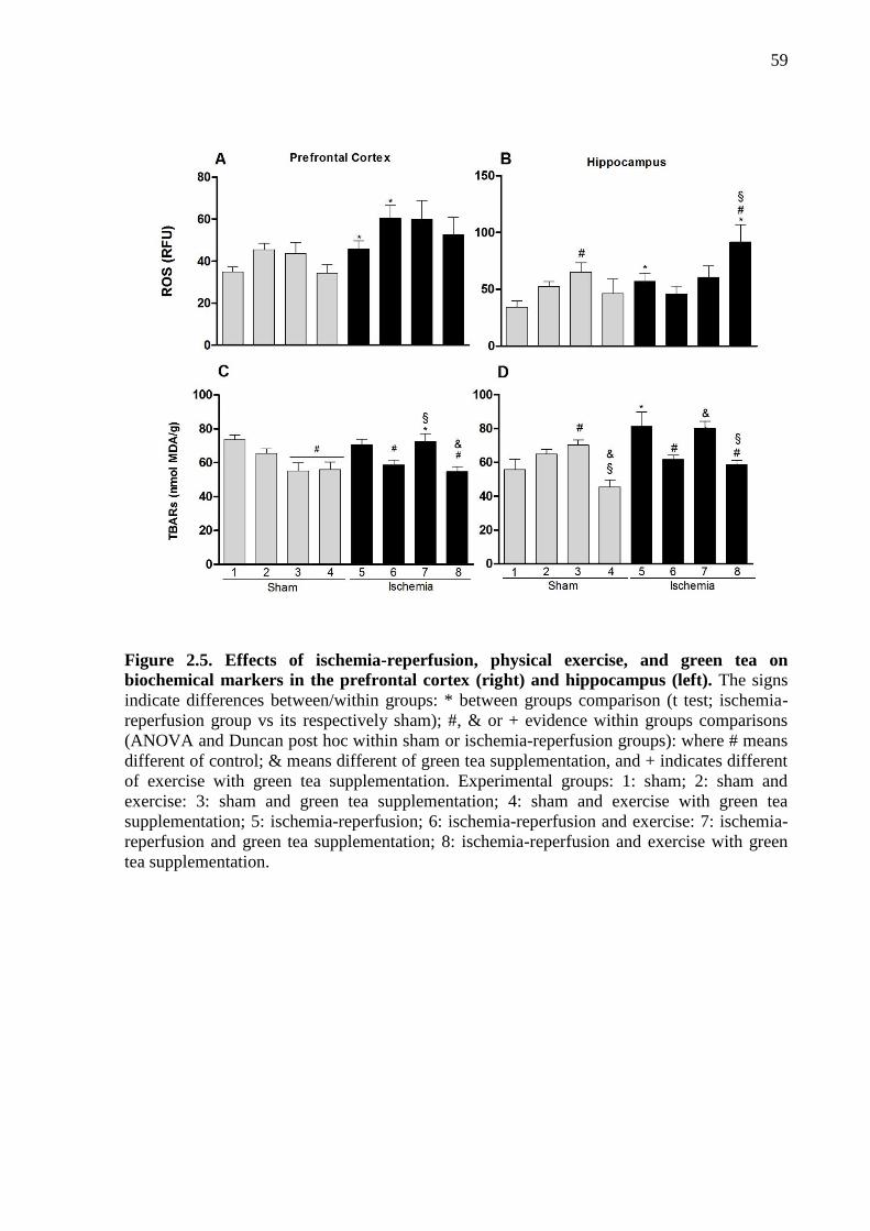
59
Figure 2.5. Effects of ischemia-reperfusion, physical exercise, and green tea on
biochemical markers in the prefrontal cortex (right) and hippocampus (left). The signs
indicate differences between/within groups: * between groups comparison (t test; ischemia-
reperfusion group vs its respectively sham); #, & or + evidence within groups comparisons
(ANOVA and Duncan post hoc within sham or ischemia-reperfusion groups): where # means
different of control; & means different of green tea supplementation, and + indicates different
of exercise with green tea supplementation. Experimental groups: 1: sham; 2: sham and
exercise: 3: sham and green tea supplementation; 4: sham and exercise with green tea
supplementation; 5: ischemia-reperfusion; 6: ischemia-reperfusion and exercise: 7: ischemia-
reperfusion and green tea supplementation; 8: ischemia-reperfusion and exercise with green
tea supplementation.

60
2.4 DISCUSSION
Here we investigated the neuroprotective potential of long term physical exercise and
green tea supplementation, either in association or by separate, to prevent or reduce learning
and memory deficits resulting from ischemia-reperfusion in a rat model. Our model showed to
be valid as it resulted in cognitive deficits without impairment in locomotor and exploratory
activities, as well as anxiety and pain thresholds.
Our results support the interpretation that the cognitive deficits were caused by
ischemia-reperfusion surgery. Oxidative damages were observed in prefrontal cortex and
hippocampus brain regions. A main finding was that combining of physical exercise and
green tea in general did not elicit better results than when interventions were conducted
separately.
Learning and memory deficits after ischemia-reperfusion may result from increased
oxidative stress (ROS and TBARS) and decreased antioxidant status (GSH and GPx) in the
hippocampus and prefrontal cortex 71. Hippocampus is one of the brain areas more sensitive to
ischemia-reperfusion98. The oxidative damages observed in hippocampus, evidenced by
increased TBARS and ROS, can contribute to the impairment of learning function49 and the
aversive learning99.
We observed that physical exercise or green tea supplementation avoided memory
deficits in the OR and IA task performed after ischemia-reperfusion. Additionally, physical
exercise had positive effects on hippocampus and prefrontal antioxidant status by avoiding
decreases of GSH, and on hippocampal pro-oxidants markers by avoiding increases in
TBARS. Although with more discrete effects, our results suggest that green tea promotes
GSH increases in prefrontal cortex and avoids catalase decreases in the same brain region.

61
Such observations help to explain the effects of physical exercise and green tea on learning
and memory. The neuroprotective effects of exercise on behavioral functions after a transient
cerebral ischemia are consistent with previous researches 62; 100; 101.
Ischemia-reperfusion also decreased concentration of catalase in the hippocampus
and prefrontal cortex. In addition, ischemia-reperfusion increased the concentration of ROS in
the hippocampus and prefrontal cortex, while TBARS increased in the hippocampus.
Although these results were expected due the injury by ischemia reperfusion, they cannot be
compared with any previous result, since our work seems to be the first to consider a long
time after reperfusion.
Physical exercise improved the performance in long term recognition and aversive
memory tasks in the ischemia-reperfusion rats. The physical exercise has been shown to
potentiate hippocampal neurogenesis and cognitive functions68; 73; 80; 102; 103. It results from
different mechanisms, including the capability of modulating oxidative stress76.
Despite of physical exercise, long term green tea supplementation also was related
with improved learning and memory among ischemia-reperfusion groups, despite of
association or not with exercise. It most likely resulted from the antioxidant activity of green
tea catechines present in green tea77. Our initial hypothesis was that when these antioxidant
strategies are combined, their positive effects could be potentiated63. However, interventions
in association not prevent against the damages caused by ischemia-reperfusion; in these
animals, behavioral performance was similar to those animals underwent the occlusion and
received no prior intervention.
In addition, physical exercise associated with green tea supplementation significantly
increased levels of ROS in the hippocampus after ischemia-reperfusion. While the
concomitant regular physical exercise and intake of green tea has been a valid strategy to
improve neuromuscular recovery after exercise15; 104; 105, it may has negative impact in the

62
case of an ischemia-reperfusion event. The combined interventions increased ROS levels in
hippocampus, which can be related to the fact that in this group/structure GSH and GPx were
also higher than in the others, leading to an oxidative imbalance. It is interesting to observe
that increase in ROS was associated with worst memory performance in this group.
In summary, our results suggest that mnemonic deficits associated to cerebral
ischemic-reperfusion can be prevented by physical exercise and green tea when used isolated,
but not when both interventions were associated. In addition, association of physical exercise
and green tea increased ROS in the hippocampus. In general, regular physical exercise
promoted better neuroprotection to an ischemic event.

63
CAPITULO III
3.1 DISCUSSÃO
Neste estudo investigamos o papel neuroprotetor do exercício físico e do chá verde
para o desempenho cognitivo depois de um evento de isquemia-reperfusão. Nossos resultados
mostraram que a oclusão bilateral das carótidas comuns por 30 minutos gerou danos no
hipocampo e no córtex pré-frontal, resultando em aumento da expressão de ERO e TBARs,
bem como a diminuição de GSH e Catalase. No entanto, a GPx se mostrou aumentada nos
grupos de isquemia reperfusão. Segundo a literatura, este resultado para GPx é aceitável, pois
caracteriza um mecanismo de defesa específico do tecido contra o aumento da H2O2 induzida
pela isquemia-reperfusão106.
Singh e Chopra (2013) mostraram que a oclusão bilateral das carótidas durante 15
minutos reduziu os níveis de GSH e Catalase, quando medidos 12 dias depois da reperfusão.
Viswanatha et al. (2013) também reportaram diminuição de GSH e Catalase em um modelo
de oclusão bilateral com 1h, com 24 horas de reperfusão. Dessa forma, podemos sugerir que
os níveis de antioxidantes vão estar diminuídos após a isquemia-reperfusão,
independentemente do tempo de oclusão e de reperfusão.
Nossos achados indicam que a isquemia-reperfusão foi responsável pelo um aumento
no estresse oxidativo, caracterizado pelo aumento de produções de ERO e diminuição nas
defesas antioxidantes, principalmente nas amostras retiradas do hipocampo. Esse mecanismo
é um dos responsáveis pela morte neuronal12; 107 e danos cognitivos subsequentes à isquemia-
reperfusão99. Isso ocorre porque o aumento da produção de radicais livres tem efeitos
deletérios sobre as funções fisiológicas importantes para a sobrevivência das células. Esse
aumento sobrecarrega a capacidade de desintoxicação pelo sistema antioxidante108. Além

64
disso, a maior expressão de ERO e de TBARs e a diminuição de antioxidantes no hipocampo
se deve a suscetibilidade desta região aos danos de isquemia e oxidação98. A importância que
o hipocampo pode ter na consolidação e armazenamento da memória29 justifica nossos
resultados no teste de reconhecimento de objetos45, onde os ratos que receberam a oclusão e
nenhuma intervenção não foram capazes de diferenciar o objeto novo do objeto já conhecido.
Além dos danos da isquemia-reperfusão sobre parâmetros bioquímicos, nós
mostramos que o exercício físico foi capaz de proteger o hipocampo e o córtex pré-frontal
contra o estresse oxidativo. No hipocampo, o exercício auxiliou na manutenção e contribuiu
para um aumento nos níveis de enzimas antioxidantes. O exercício físico também diminuiu a
produção de radicais livres. Já no córtex pré-frontal, embora as enzimas antioxidantes tenham
se mantido com o exercício físico, a produção de ERO aumentou. No entanto, a expressão de
TBARs apresentou-se diminuída em ratos submetidos à isquemia-reperfusão. Esses achados
sugerem que os efeitos do exercício físico podem ser específicos para regiões do cérebro
analisadas nesse estudo.
Nossos resultados demonstraram que os ratos submetidos à oclusão bilateral depois
que receberam um pré-tratamento com exercício físico tiveram suas funções mnemônicas
preservadas, assim como mantiveram a capacidade de aprender. Esse benefício pode ser
justificado pelo aumento das enzimas antioxidantes109, e por outros mecanismos descritos na
literatura, tais como a diminuição dos danos oxidativo, aumento da biogênese, aumento da
atividade complexa da cadeia de transporte de elétrons, e diminuição de marcadores de
apoptose110.
Marques-Aleixo., Oliveira. et al. 59 (2012) fizeram uma revisão bibliográfica a
respeito do exercício físico e neuroproteção e encontraram diversos sistemas que explicariam
o seu papel neuroprotetor. No entanto, os mecanismos que os estudos experimentais mais
destacam são:

65
- O exercício físico promove mudanças no conteúdo e atividade de enzimas
oxidativas no músculo esquelético, o que limita o uso de glicose pelo cérebro tendo efeitos
indiretos sobre o seu metabolismo, dessa forma, essa limitação induzida cronicamente
funciona como um treinamento ao cérebro para situações em que há ausência de glicose,
como em uma condição de isquemia;
- A atividade física regular aumenta a proliferação de células endoteliais do cérebro
e, portanto, influência a vasculatura cerebral, melhorando a capacidade metabólica através do
aumento de nutrientes e suprimento de oxigênio.
Assim, os autores51 concluíram que, embora os estudos descrevam diferentes
mecanismos para neuroproteção induzida pelo exercício físico, é indiscutível que o exercício
contribui para aumentar o número de células e melhorar sua função em condições em que a
perda de células neuronais, como o envelhecimento e doenças neurodegenerativas, estejam
ocorrendo. Diante disso, podemos observar que, independente do mecanismo descrito, todos
os consideram itens oxidantes, o que reforça nossos achados, que o exercício físico atua na
neuroproteção também por meio da regulação do estresse oxidativo presente em condições
como a de isquemia.
Assim como o exercício físico, o chá verde também mostrou um importante papel
neuroprotetor, uma vez que os ratos que tiveram a oclusão depois que receberam a
suplementação com chá verde mostraram um bom desempenho nos testes comportamentais
para avaliação de memória e aprendizado. Entende-se que as catequinas do chá verde podem
provocar diversos efeitos biológicos, atuando em mecanismos relacionadas a distúrbios
vasculares, estresse oxidativo e neuroinflamação, e todos esses processos estão envolvidos na
isquemia reperfusão111. Assim, embora esteja certificado o papel neuroprotetor do chá verde,
os mecanismos de neuroproteção em situações de isquemia-reperfusão ainda são
contraditórios.

66
Biasibetti, Tramontina et al. 112(2013) administraram via sonda gástrica em ratos, a
principal catequina do chá verde, epigalocatequina galato. Foram doses de 10 mg/kg diárias
durante 4 semanas. Análises bioquímicas reveleram aumento nas ERO em ratos saudáveis.
Porém assim como no nosso trabalho, esse aumento não interferiu nos resultados das tarefas
comportamentais. Adicionalmente, Panickar, Polansky et al. 113 (2009) realizaram um estudo
bastante pontual sobre o papel neuroprotetor do chá verde em um modelo de isquemia in vitro
induzida pela deprivação de oxigênio e glicose. Para isso, administraram as principais
catequinas isoladas e também o extrato purificado do chá verde. Como resultados, eles
encontraram que o extrato purificado foi mais eficiente do que as catequinas isoladas para
controlar o edema celular. No entanto, essa neuroproteção não estaria associada com a
diminuição de radicais livres, já que o extrato do chá purificado aumentou as concentrações
de oxido nítrico. Em contrapartida, estudos mostram que esse achado pode ser explicado pelo
fato de que o óxido nítrico é essencial para o processo de cicatrização, além de induzir o
crescimento de novos vasos sanguíneos através da indução do fator de crescimento endotelial
vascular, atuando assim como neuroprotetor via angiogênese111. Porém, não podemos
justificar nossos resultados por meio deste mecanismo, já que não mensuramos a
concentração de óxido nítrico.
Sabe-se que as catequinas do chá verde atuam nas duas vias de proteção ao estresse
oxidativo, tanto inibindo enzimas cuja atividade pode promover estresse oxidativo, como
também aumentando a atividade das enzimas antioxidantes114. Porém, não há informação
precisa de quais são essas enzimas. O conhecimento exato dessas enzimas seria importante
para que elas possam servir como marcadores em estudos envolvendo suplementação com chá
verde.
A nossa hipótese inicial era que a combinação do exercício físico, conhecido como
uma forma de melhorar as defesas antioxidantes endógenas36, com a ingestão de chá verde,
67
um suplemento rico em antioxidante exógeno115, poderia potencializar as defesas
antioxidantes como um todo, e isso traria um benefício neuroprotetor maior do que se
exercício e chá fossem assumidos independentemente. No entanto, os ratos que receberam as
duas intervenções apresentaram aumento de ERO no hipocampo. Porém, não podemos deixar
de considerar que para a mesma estrutura a GSH também aumentou, o que é um resultado
interessante, uma vez que esta enzima é a principal antioxidante do tecido cerebral37.
Também, os resultados dos testes comportamentais mostraram que a combinação das duas
estratégias pode não ser mais efetiva do que elas em isolado, pois nossos resultados sugeriram
que as duas intervenções juntas não foram capazes de proteger contra déficits de memória,
enquanto que intervenções isoladas tiveram efeitos positivos.
O protocolo de isquemia-reperfusão utilizado aqui já foi bastante descrito, embora os
tempos de reperfusão variem muito25. E como vimos buscando na literatura, o período de
reperfusão parece estar tanto quanto, ou até mais, envolvido no processo de morte neuronal do
que o período de isquemia34.
Nós optamos por utilizar protocolos, tanto de suplementação de chá verde quanto de
exercício físico, que simulassem a prática de humanos. Assim, optamos pela suplementação
com chá verde junto com a água de beber, e embora tenhamos feito uma medida média do
consumo diário de chá verde entre os ratos, não foi possível determinarmos o volume exato de
ingesta de líquido por rato e, por conseguinte, a concentração das catequinas do chá verde.
Isso pode ter contribuído para alguns resultados não esperados, já que o papel neuroprotetor
do chá verde está associado com o volume de ingestão111. Em relação ao exercício físico,
devemos mencionar que os estudos mostram que seus efeitos no cérebro são divergentes nos
protocolos de exercício usados com modelos animais6868, o que é uma limitação para estudos
que investigam os mecanismos de neuroproteção do exercício físico.

68
3.2 CONCLUSÕES
Diante do nosso embasamento teórico e resultados encontrados, podemos concluir que a
isquemia bilateral de carótidas comuns por 30 minutos, seguida de reperfusão, é capaz de
causar déficits de aprendizado e memória de longo prazo, sendo este um modelo válido para
estudos sobre alterações cognitivas no AVC em modelo animal de ratos Wistar machos. Esses
déficits podem ser explicados pelo aumento de ERO em estruturas envolvidas nos processos
cognitivos como hipocampo e córtex pré-frontal.
Além disso, podemos afirmar que tanto a suplementação com chá verde quanto o exercício
físico por um período de 8 semanas, são capazes de proteger contra os danos cognitivos da
isquemia-reperfusão em modelo animal mantendo os níveis funcionais de marcadores
antioxidantes. No entanto, as duas intervenções utilizadas juntas, em geral, não provocaram
resultados melhores do que cada intervenção realizada separadamente.

69
3.3 APLICAÇÕES PRÁTICAS E PERSPECTIVAS FUTURAS
A suplementação com antioxidantes tem ganho popularidade como estratégia de
prevenção de doenças, pois sabe-se que o estresse oxidativo está envolvido no mecanismo de
várias doenças. Adicionalmente, as pessoas buscam cada vez mais se exercitar para melhorar
a qualidade de vida e para prevenir doenças crônicas, como doenças cardiovasculares e
metabólicas, que são consideradas fatores de risco para o AVC. A partir da revisão de
literatura, e juntamente com os nossos resultados, as duas modalidades antioxidantes trazem
benefício à saúde além de, serem alternativas bastante populares e de fácil acesso para a
população em geral.
Pensando nisso, são perspectivas de estudos futuros o teste de outros protocolos que
simulem os danos causados por um AVC em ratos, utilizando diferentes tempos de oclusão e
também a oclusão de outras artérias, afim de constatar déficits cognitivos e motores mais
próximos da realidade humana o que contribuiria para o desenvolvimento de investigações
com potenciais neuroprotetores, e ainda poderiam elucidar melhor os mecanismos tanto de
lesão quanto de proteção com base em estratégias antioxidantes.
Considerando mecanismos de neuroproteção, temos como expectativas investigar o
impacto de variações nos protocolos de exercício físico, como por exemplo, exercício
anaeróbico, ou até mesmo protocolos aeróbicos com as variações nas frações entra volume,
intensidade e duração do exercício. Por fim, consideramos fundamental investigar outros
marcadores bioquímicos que possam esclarecer melhor os mecanismos antioxidantes e pró
oxidantes, assim como marcadores de inflamação e neurotoxicidade.

70
REFERÊNCIAS
1 CHOU, C. Y. et al. Interchangeable Skin Grafting to Camouflage Self-inflicted
Wound Scars on the Dorsal and Volar Forearm: A Case Report. Ostomy Wound
Manage, v. 60, n. 4, p. 50-2, Apr 2014. ISSN 1943-2720 (Electronic)
0889-5899 (Linking). Disponível em: < http://www.ncbi.nlm.nih.gov/pubmed/24706403 >.
2 CHEN, S. M. et al. HSP90 inhibitor AUY922 abrogates up-regulation of RTKs by
mTOR inhibitor AZD8055 and potentiates its antiproliferative activity in human
breast cancer. Int J Cancer, Apr 7 2014. ISSN 1097-0215 (Electronic)
0020-7136 (Linking). Disponível em: < http://www.ncbi.nlm.nih.gov/pubmed/24706460 >.
3 ZHENG, G. et al. Primary prevention for risk factors of ischemic stroke with
Baduanjin exercise intervention in the community elder population: study protocol for
a randomized controlled trial. Trials, v. 15, n. 1, p. 113, Apr 9 2014. ISSN 1745-6215
(Electronic)
1745-6215 (Linking). Disponível em: < http://www.ncbi.nlm.nih.gov/pubmed/24712684 >.
4 BOS, M. J. et al. Modifiable etiological factors and the burden of stroke from the
Rotterdam study: a population-based cohort study. PLoS Med, v. 11, n. 4, p.
e1001634, Apr 2014. ISSN 1549-1676 (Electronic)
1549-1277 (Linking). Disponível em: < http://www.ncbi.nlm.nih.gov/pubmed/24781247 >.
5 SIERRA, C.; COCA, A.; SCHIFFRIN, E. L. Vascular mechanisms in the pathogenesis
of stroke. Curr Hypertens Rep, v. 13, n. 3, p. 200-7, Jun 2011. ISSN 1534-3111
(Electronic)
1522-6417 (Linking). Disponível em: < http://www.ncbi.nlm.nih.gov/pubmed/21331606 >.
6 STRONG, K.; MATHERS, C.; BONITA, R. Preventing stroke: saving lives around
the world. Lancet Neurol, v. 6, n. 2, p. 182-7, Feb 2007. ISSN 1474-4422 (Print)
1474-4422 (Linking). Disponível em: < http://www.ncbi.nlm.nih.gov/pubmed/17239805 >.
7 MERCIER, L. et al. Impact of motor, cognitive, and perceptual disorders on ability to
perform activities of daily living after stroke. Stroke, v. 32, n. 11, p. 2602-8, Nov
2001. ISSN 1524-4628 (Electronic)
0039-2499 (Linking). Disponível em: < http://www.ncbi.nlm.nih.gov/pubmed/11692024 >.

71
8 WU, K. J. et al. Green Tea Extract Ameliorates Learning and Memory Deficits in
Ischemic Rats via Its Active Component Polyphenol Epigallocatechin-3-gallate by
Modulation of Oxidative Stress and Neuroinflammation. Evid Based Complement
Alternat Med, v. 2012, p. 163106, 2012. ISSN 1741-4288 (Electronic)
1741-427X (Linking). Disponível em: < http://www.ncbi.nlm.nih.gov/pubmed/22919410 >.
9 SACHDEV, P. S. et al. Progression of cognitive impairment in stroke/TIA patients
over 3 years. J Neurol Neurosurg Psychiatry, Mar 21 2014. ISSN 1468-330X
(Electronic)
0022-3050 (Linking). Disponível em: < http://www.ncbi.nlm.nih.gov/pubmed/24659793 >.
10 PINTO, J. M.; NERI, A. L. Factors associated with low life life satisfaction in
community-dwelling elderly: FIBRA Study. Cad Saude Publica, v. 29, n. 12, p.
2447-58, Dec 2013. ISSN 1678-4464 (Electronic)
0102-311X (Linking). Disponível em: < http://www.ncbi.nlm.nih.gov/pubmed/24356691 >.
11 SARKAKI, A. et al. Improvement in Memory and Brain Long-term Potentiation
Deficits Due to Permanent Hypoperfusion/Ischemia by Grape Seed Extract in Rats.
Iran J Basic Med Sci, v. 16, n. 9, p. 1004-10, Sep 2013. ISSN 2008-3866 (Print)
2008-3866 (Linking). Disponível em: < http://www.ncbi.nlm.nih.gov/pubmed/24171080 >.
12 MIFSUD, G. et al. Oligodendrocyte Pathophysiology and Treatment Strategies in
Cerebral Ischemia. CNS Neurosci Ther, Apr 7 2014. ISSN 1755-5949 (Electronic)
1755-5930 (Linking). Disponível em: < http://www.ncbi.nlm.nih.gov/pubmed/24703424 >.
13 LIU, X. et al. Green tea polyphenols alleviate early BBB damage during experimental
focal cerebral ischemia through regulating tight junctions and PKCalpha signaling.
BMC Complement Altern Med, v. 13, n. 1, p. 187, Jul 21 2013. ISSN 1472-6882
(Electronic)
1472-6882 (Linking). Disponível em: < http://www.ncbi.nlm.nih.gov/pubmed/23870286 >.
14 ZHANG, F.; WU, Y.; JIA, J. Exercise preconditioning and brain ischemic tolerance.
Neuroscience, v. 177, p. 170-6, Mar 17 2011. ISSN 1873-7544 (Electronic)
0306-4522 (Linking). Disponível em: < http://www.ncbi.nlm.nih.gov/pubmed/21241780 >.
15 HARAMIZU, S. et al. Catechins suppress muscle inflammation and hasten
performance recovery after exercise. Med Sci Sports Exerc, v. 45, n. 9, p. 1694-702,
Sep 2013. ISSN 1530-0315 (Electronic)
0195-9131 (Linking). Disponível em: < http://www.ncbi.nlm.nih.gov/pubmed/23470311 >.

72
16 MURASE, T. et al. Tea catechin ingestion combined with habitual exercise
suppresses the aging-associated decline in physical performance in senescence-
accelerated mice. American Journal of Physiology - Regulatory, Integrative and
Comparative Physiology, v. 295, 2008.
17 WOODRUFF, T. M. et al. Pathophysiology, treatment, and animal and cellular
models of human ischemic stroke. Mol Neurodegener, v. 6, n. 1, p. 11, 2011. ISSN
1750-1326 (Electronic)
1750-1326 (Linking). Disponível em: < http://www.ncbi.nlm.nih.gov/pubmed/21266064 >.
18 CHEN, S. et al. Reduction of unnecessary right ventricular pacing by managed
ventricular pacing and search AV+ algorithms in pacemaker patients: 12-month
follow-up results of a randomized study. Europace, Apr 4 2014. ISSN 1532-2092
(Electronic)
1099-5129 (Linking). Disponível em: < http://www.ncbi.nlm.nih.gov/pubmed/24706091 >.
19 RJ, G.; CN, R.; SRC, F. Tratamento da Fase Aguda do Acidente Vascular Cerebra.
Projeto Diretrizes - Associação Médica Brasileira e Conselho Federal de
Medicina, 2001. Disponível em: <
http://www.projetodiretrizes.org.br/projeto_diretrizes/010.pdf >.
20 PERSSON, M. et al. Microwave-based stroke diagnosis making global pre-hospital
thrombolytic treatment possible. IEEE Trans Biomed Eng, Jun 12 2014. ISSN 1558-
2531 (Electronic)
0018-9294 (Linking). Disponível em: < http://www.ncbi.nlm.nih.gov/pubmed/24951677 >.
21 KANEKAR, S. G.; ZACHARIA, T.; ROLLER, R. Imaging of Stroke: Part 2,
Pathophysiology at the Molecular and Cellular Levels and Corresponding Imaging
Changes. American Journal of Roentgenology v. 198, p. 63-74, 2012.
22 PAGNUSSAT, A. S. Caracterização da morte celular em neurônios da região CA1 do
hipocampo de ratos submetidos à isquemia global transitória e reperfusão.
Repositório Digital da Universidade Federal do Rio Grande do Sul, 2005 2005.
Disponível em: < http://www.lume.ufrgs.br/handle/10183/5561 >.
23 MULLER, V. I. et al. Interindividual differences in cognitive flexibility: influence of
gray matter volume, functional connectivity and trait impulsivity. Brain Struct
Funct, May 31 2014. ISSN 1863-2661 (Electronic)
1863-2653 (Linking). Disponível em: < http://www.ncbi.nlm.nih.gov/pubmed/24878823 >.
24 MAJID, A. Neuroprotection in Stroke: Past, Present, and Future. ISRN Neurol, v.
2014, p. 515716, 2014. ISSN 2090-5505 (Print)

73
2090-5505 (Linking). Disponível em: < http://www.ncbi.nlm.nih.gov/pubmed/24579051 >.
25 SPEETZEN, L. J.; ENDRES, M.; KUNZ, A. Bilateral Common Carotid Artery
Occlusion as an Adequate Preconditioning Stimulus to Induce Early Ischemic
Tolerance to Focal Cerebral Ischemia. n. 75, p. e4387, 2013/05/09/ 2013. ISSN 1940-
087X. Disponível em: < http://www.jove.com/video/4387 >.
26 ESTEVES, A. et al. Anatomical arrangement and distribution of the cerebral arterial
circle in rats. Journal of Morphological Sciences, v. 30, n. 2, p. 132-139, 2013.
27 ENGEL, O. et al. Modeling Stroke in Mice - Middle Cerebral Artery Occlusion with
the Filament Model. Journal of Visualized Experiments, v. 47, 2011. Disponível
em: < http://www.jove.com/details.php?id=2423 >.
28 HEA, Z. et al. Definition of the anterior choroidal artery territory in rats using
intraluminal occluding technique. Journal of the Neurological Sciences, v. 128, n. 1,
2000.
29 CHEN, S. et al. Asiaticoside attenuates memory impairment induced by transient
cerebral ischemia-reperfusion in mice through anti-inflammatory mechanism.
Pharmacol Biochem Behav, v. 122C, p. 7-15, Mar 12 2014. ISSN 1873-5177
(Electronic)
0091-3057 (Linking). Disponível em: < http://www.ncbi.nlm.nih.gov/pubmed/24631487 >.
30 MIGUEL, M. P.; MENEZES, L. B. D.; ARAÚJO, E. G. D. FISIOPATOLOGIA DO
ESTRESSE OXIDATIVO APÓS ISQUEMIA E REPERFUSÃO CEREBRAL E
POTENCIAL NEUROPROTEÇÃO DO PEQUI (CARYOCAR BRASILIENSE).
ENCICLOPÉDIA BIOSFERA, v. 8, n. 15, p. 1976, 2012.
31 GARCIA CARRASCO, D.; ABOITIZ CANTALAPIEDRA, J. Effectiveness of motor
imagery or mental practice in functional recovery after stroke: a systematic review.
Neurologia, Apr 16 2013. ISSN 1578-1968 (Electronic)
0213-4853 (Linking). Disponível em: < http://www.ncbi.nlm.nih.gov/pubmed/23601759 >.
32 SUN, M. K.; ALKON, D. L. The "memory kinases": roles of PKC isoforms in signal
processing and memory formation. Prog Mol Biol Transl Sci, v. 122, p. 31-59, 2014.
ISSN 1878-0814 (Electronic)
1877-1173 (Linking). Disponível em: < http://www.ncbi.nlm.nih.gov/pubmed/24484697 >.

74
33 OZTANIR, M. N. et al. Hesperidin attenuates oxidative and neuronal damage caused
by global cerebral ischemia/reperfusion in a C57BL/J6 mouse model. Neurol Sci, Mar
28 2014. ISSN 1590-3478 (Electronic)
1590-1874 (Linking). Disponível em: < http://www.ncbi.nlm.nih.gov/pubmed/24676696 >.
34 ALLEN, C. L.; BAYRAKTUTAN, U. Oxidative stress and its role in the pathogenesis
of ischaemic stroke. Int J Stroke, v. 4, n. 6, p. 461-70, Dec 2009. ISSN 1747-4949
(Electronic)
1747-4930 (Linking). Disponível em: < http://www.ncbi.nlm.nih.gov/pubmed/19930058 >.
35 GULATI, P.; SINGH, N. Neuroprotective mechanism of ischemic postconditioning in
mice: a possible relationship between protein kinase C and nitric oxide pathways. J
Surg Res, Feb 19 2014. ISSN 1095-8673 (Electronic)
0022-4804 (Linking). Disponível em: < http://www.ncbi.nlm.nih.gov/pubmed/24655662 >.
36 YU, B. P.; CHUNG, H. Y. Adaptive mechanisms to oxidative stress during aging.
Mech Ageing Dev, v. 127, n. 5, p. 436-43, May 2006. ISSN 0047-6374 (Print)
0047-6374 (Linking). Disponível em: < http://www.ncbi.nlm.nih.gov/pubmed/16497363 >.
37 ZLATKOVIC, J. et al. Different susceptibility of prefrontal cortex and hippocampus
to oxidative stress following chronic social isolation stress. Mol Cell Biochem, Mar
27 2014. ISSN 1573-4919 (Electronic)
0300-8177 (Linking). Disponível em: < http://www.ncbi.nlm.nih.gov/pubmed/24671494 >.
38 TAYLOR, J. M.; MAIN, B. S.; CRACK, P. J. Neuroinflammation and oxidative
stress: co-conspirators in the pathology of Parkinson's disease. Neurochem Int, v. 62,
n. 5, p. 803-19, Apr 2013. ISSN 1872-9754 (Electronic)
0197-0186 (Linking). Disponível em: < http://www.ncbi.nlm.nih.gov/pubmed/23291248 >.
39 TEWARI, A. et al. Antioxidants: The new frontier for translational research in
cerebroprotection. J Anaesthesiol Clin Pharmacol, v. 30, n. 2, p. 160-171, Apr 2014.
ISSN 0970-9185 (Print)
0970-9185 (Linking). Disponível em: < http://www.ncbi.nlm.nih.gov/pubmed/24803750 >.
40 PENG, C. et al. Biology of ageing and role of dietary antioxidants. Biomed Res Int,
v. 2014, p. 831841, 2014. ISSN 2314-6141 (Electronic). Disponível em: <
http://www.ncbi.nlm.nih.gov/pubmed/24804252 >.
41 CRACK, P. J. et al. Increased infarct size and exacerbated apoptosis in the
glutathione peroxidase-1 (Gpx-1) knockout mouse brain in response to

75
ischemia/reperfusion injury. J Neurochem, v. 78, n. 6, p. 1389-99, Sep 2001. ISSN
0022-3042 (Print)
0022-3042 (Linking). Disponível em: < http://www.ncbi.nlm.nih.gov/pubmed/11579147 >.
42 NEWBURGER, J. W. et al. Transplant-Free Survival and Interventions at 3 Years in
the Single Ventricle Reconstruction Trial. Circulation, Apr 4 2014. ISSN 1524-4539
(Electronic)
0009-7322 (Linking). Disponível em: < http://www.ncbi.nlm.nih.gov/pubmed/24705119 >.
43 IZQUIERDO, L. A. et al. Molecular pharmacological dissection of short- and long-
term memory. Cell Mol Neurobiol, v. 22, n. 3, p. 269-87, Jun 2002. ISSN 0272-4340
(Print)
0272-4340 (Linking). Disponível em: < http://www.ncbi.nlm.nih.gov/pubmed/12469870 >.
44 IZQUIERDO, I. Memória. 2ª. 2011.
45 LARKIN, M. C. et al. Hippocampal output area CA1 broadcasts a generalized novelty
signal during an object-place recognition task. Hippocampus, Mar 4 2014. ISSN
1098-1063 (Electronic)
1050-9631 (Linking). Disponível em: < http://www.ncbi.nlm.nih.gov/pubmed/24596296 >.
46 CECHETTI, F. et al. Effect of a neuroprotective exercise protocol on oxidative state
and BDNF levels in the rat hippocampus. Brain Res, v. 1188, p. 182-8, Jan 10 2008.
ISSN 0006-8993 (Print)
0006-8993 (Linking). Disponível em: < http://www.ncbi.nlm.nih.gov/pubmed/18021756 >.
47 TIRAPU-USTÁRROZ, J.; MUÑOZ-CÉSPEDES, J. M. Memoria y funciones
ejecutivas. Revista de Neurologia, v. 41, n. 8, p. 475-484, 2005.
48 WANG, J. X.; COHEN, N. J.; VOSS, J. L. Covert rapid action-memory simulation
(CRAMS): A hypothesis of hippocampal-prefrontal interactions for adaptive behavior.
Neurobiol Learn Mem, Apr 19 2014. ISSN 1095-9564 (Electronic)
1074-7427 (Linking). Disponível em: < http://www.ncbi.nlm.nih.gov/pubmed/24752152 >.
49 DA SILVEIRA, C. K. et al. The role of histamine receptors in the consolidation of
object recognition memory. Neurobiol Learn Mem, v. 103, p. 64-71, Jul 2013. ISSN
1095-9564 (Electronic)
1074-7427 (Linking). Disponível em: < http://www.ncbi.nlm.nih.gov/pubmed/23583502 >.

76
50 BARKER, G. R.; WARBURTON, E. C. Object-in-Place Associative Recognition
Memory Depends on Glutamate Receptor Neurotransmission Within Two Defined
Hippocampal-Cortical Circuits: A Critical Role for AMPA and NMDA Receptors in
the Hippocampus, Perirhinal, and Prefrontal Cortices. Cereb Cortex, Sep 12 2013.
ISSN 1460-2199 (Electronic)
1047-3211 (Linking). Disponível em: < http://www.ncbi.nlm.nih.gov/pubmed/24035904 >.
51 YANG, F. C.; LIANG, K. C. Interactions of the dorsal hippocampus, medial
prefrontal cortex and nucleus accumbens in formation of fear memory: Difference in
inhibitory avoidance learning and contextual fear conditioning. Neurobiol Learn
Mem, Jul 25 2013. ISSN 1095-9564 (Electronic)
1074-7427 (Linking). Disponível em: < http://www.ncbi.nlm.nih.gov/pubmed/23891992 >.
52 ROSA, J. et al. Fear extinction can be made state-dependent on peripheral
epinephrine: Role of norepinephrine in the nucleus tractus solitarius. Neurobiol
Learn Mem, Oct 23 2013. ISSN 1095-9564 (Electronic)
1074-7427 (Linking). Disponível em: < http://www.ncbi.nlm.nih.gov/pubmed/24161888 >.
53 MARTINEZ-DIAZ, J. A. et al. Effects on locomotion and memory in 2 models of
cerebral hypoperfusion in male Wistar rats. Neurologia, Apr 3 2014. ISSN 1578-1968
(Electronic)
0213-4853 (Linking). Disponível em: < http://www.ncbi.nlm.nih.gov/pubmed/24704245 >.
54 HIRAIDE, S. et al. Behavioural effects of monoamine reuptake inhibitors on
symptomatic domains in an animal model of attention-deficit/hyperactivity disorder.
Pharmacol Biochem Behav, v. 105, p. 89-97, Apr 2013. ISSN 1873-5177
(Electronic)
0091-3057 (Linking). Disponível em: < http://www.ncbi.nlm.nih.gov/pubmed/23380523 >.
55 SAINI, A. K.; KUMAR, H. S. A.; SHARMA, S. S. Preventive and curative effect of
edaravone on nerve functions and oxidative stress in experimental diabetic
neuropathy. Eur J Pharmacol, v. 568, n. 1-3, p. 164-72, Jul 30 2007. ISSN 0014-
2999 (Print)
0014-2999 (Linking). Disponível em: < http://www.ncbi.nlm.nih.gov/pubmed/17521626 >.
56 SUTHERLAND, B. A. et al. Neuroprotection for ischaemic stroke: translation from
the bench to the bedside. Int J Stroke, v. 7, n. 5, p. 407-18, Jul 2012. ISSN 1747-4949
(Electronic)
1747-4930 (Linking). Disponível em: < http://www.ncbi.nlm.nih.gov/pubmed/22394615 >.

77
57 PIETRELLI, A. et al. Aerobic exercise prevents age-dependent cognitive decline and
reduces anxiety-related behaviors in middle-aged and old rats. Neuroscience, v. 202,
p. 252-66, Jan 27 2012. ISSN 1873-7544 (Electronic)
0306-4522 (Linking). Disponível em: < http://www.ncbi.nlm.nih.gov/pubmed/22183054 >.
58 EGAN, K. J. et al. Exercise Reduces Infarct Volume and Facilitates Neurobehavioral
Recovery: Results From a Systematic Review and Meta-analysis of Exercise in
Experimental Models of Focal Ischemia. Neurorehabil Neural Repair, Feb 18 2014.
ISSN 1552-6844 (Electronic)
1545-9683 (Linking). Disponível em: < http://www.ncbi.nlm.nih.gov/pubmed/24553105 >.
59 MARQUES-ALEIXO., I. et al. Physical exercise as a possible strategy for brain
protection: Evidence from mitochondrial-mediated mechanisms. Progress in
Neurobiology, v. 99, p. 149-162, 2012. Disponível em: <
http://dx.doi.org/10.1016/j.pneurobio.2012.08.002 >.
60 GERECKE, K. M. et al. Exercise protects against chronic restraint stress-induced
oxidative stress in the cortex and hippocampus. Brain Res, v. 1509, p. 66-78, May 6
2013. ISSN 1872-6240 (Electronic)
0006-8993 (Linking). Disponível em: < http://www.ncbi.nlm.nih.gov/pubmed/23499928 >.
61 KIM, B. K. et al. Treadmill exercise improves short-term memory by enhancing
neurogenesis in amyloid beta-induced Alzheimer disease rats. J Exerc Rehabil, v. 10,
n. 1, p. 2-8, Feb 2014. ISSN 2288-176X (Print)
2288-176X (Linking). Disponível em: < http://www.ncbi.nlm.nih.gov/pubmed/24678498 >.
62 TAHAMTAN, M. et al. Exercise Preconditioning Improves Behavioral Functions
following Transient Cerebral Ischemia Induced by 4-Vessel Occlusion (4-VO) in Rats.
Arch Iran Med, v. 16, n. 12, p. 697-704, Dec 2013. ISSN 1735-3947 (Electronic)
1029-2977 (Linking). Disponível em: < http://www.ncbi.nlm.nih.gov/pubmed/24329142 >.
63 VAN PRAAG, H. Exercise and the brain: something to chew on. Trends Neurosci, v.
32, n. 5, p. 283-90, May 2009. ISSN 0166-2236 (Print)
0166-2236 (Linking). Disponível em: < http://www.ncbi.nlm.nih.gov/pubmed/19349082 >.
64 XU, Y. Q. et al. Hydrolysis of green tea residue protein using proteolytic enzyme
derived from Aspergillus oryzae. J Food Sci Technol, v. 50, n. 1, p. 171-5, Feb 2013.
ISSN 0022-1155 (Print)
0022-1155 (Linking). Disponível em: < http://www.ncbi.nlm.nih.gov/pubmed/24425904 >.

78
65 UNNO, K. et al. Suppressive effect of green tea catechins on morphologic and
functional regression of the brain in aged mice with accelerated senescence
(SAMP10). Exp Gerontol, v. 39, n. 7, p. 1027-34, Jul 2004. ISSN 0531-5565 (Print)
0531-5565 (Linking). Disponível em: < http://www.ncbi.nlm.nih.gov/pubmed/15236762 >.
66 MENARD, C.; BASTIANETTO, S.; QUIRION, R. Neuroprotective effects of
resveratrol and epigallocatechin gallate polyphenols are mediated by the activation of
protein kinase C gamma. Front Cell Neurosci, v. 7, p. 281, 2013. ISSN 1662-5102
(Electronic)
1662-5102 (Linking). Disponível em: < http://www.ncbi.nlm.nih.gov/pubmed/24421757 >.
67 TIAN, S. et al. Early exercise training improves ischemic outcome in rats by cerebral
hemodynamics. Brain Res, v. 1533, p. 114-21, Oct 2 2013. ISSN 1872-6240
(Electronic)
0006-8993 (Linking). Disponível em: < http://www.ncbi.nlm.nih.gov/pubmed/23948104 >.
68 CECHETTI, F. et al. Forced treadmill exercise prevents oxidative stress and memory
deficits following chronic cerebral hypoperfusion in the rat. Neurobiol Learn Mem,
v. 97, n. 1, p. 90-6, Jan 2012. ISSN 1095-9564 (Electronic)
1074-7427 (Linking). Disponível em: < http://www.ncbi.nlm.nih.gov/pubmed/22001013 >.
69 DAL-PIZZOL, F. et al. Lipid peroxidation in hippocampus early and late after status
epilepticus induced by pilocarpine or kainic acid in Wistar rats. Neurosci Lett, v. 291,
n. 3, p. 179-82, Sep 22 2000. ISSN 0304-3940 (Print)
0304-3940 (Linking). Disponível em: < http://www.ncbi.nlm.nih.gov/pubmed/10984636 >.
70 DAL-PIZZOL, F. et al. Neonatal iron exposure induces oxidative stress in adult
Wistar rat. Brain Res Dev Brain Res, v. 130, n. 1, p. 109-14, Sep 23 2001. ISSN
0165-3806 (Print)
0165-3806 (Linking). Disponível em: < http://www.ncbi.nlm.nih.gov/pubmed/11557099 >.
71 COLLINO, M. et al. Modulation of the oxidative stress and inflammatory response by
PPAR-gamma agonists in the hippocampus of rats exposed to cerebral
ischemia/reperfusion. Eur J Pharmacol, v. 530, n. 1-2, p. 70-80, Jan 13 2006. ISSN
0014-2999 (Print)
0014-2999 (Linking). Disponível em: < http://www.ncbi.nlm.nih.gov/pubmed/16386242 >.
72 VAN PRAAG, H. Neurogenesis and exercise: past and future directions.
Neuromolecular Med, v. 10, n. 2, p. 128-40, 2008. ISSN 1535-1084 (Print)
1535-1084 (Linking). Disponível em: < http://www.ncbi.nlm.nih.gov/pubmed/18286389 >.

79
73 VIVAR, C.; POTTER, M. C.; VAN PRAAG, H. All about running: synaptic
plasticity, growth factors and adult hippocampal neurogenesis. Curr Top Behav
Neurosci, v. 15, p. 189-210, 2013. ISSN 1866-3370 (Print)
1866-3370 (Linking). Disponível em: < http://www.ncbi.nlm.nih.gov/pubmed/22847651 >.
74 VOSS, M. W. et al. Bridging animal and human models of exercise-induced brain
plasticity. Trends Cogn Sci, v. 17, n. 10, p. 525-44, Oct 2013. ISSN 1879-307X
(Electronic)
1364-6613 (Linking). Disponível em: < http://www.ncbi.nlm.nih.gov/pubmed/24029446 >.
75 YANG, X. et al. Pre-Ischemic Treadmill Training for Prevention of Ischemic Brain
Injury via Regulation of Glutamate and Its Transporter GLT-1. Int J Mol Sci, v. 13, n.
8, p. 9447-59, 2012. ISSN 1422-0067 (Electronic)
1422-0067 (Linking). Disponível em: < http://www.ncbi.nlm.nih.gov/pubmed/22949807 >.
76 HAMAKAWA, M. et al. Repeated short-term daily exercise ameliorates oxidative
cerebral damage and the resultant motor dysfunction after transient ischemia in rats. J
Clin Biochem Nutr, v. 53, n. 1, p. 8-14, Jul 2013. ISSN 0912-0009 (Print)
0912-0009 (Linking). Disponível em: < http://www.ncbi.nlm.nih.gov/pubmed/23874064 >.
77 PARK, J. W. et al. Green tea polyphenol epigallocatechin gallate reduces neuronal
cell damage and up-regulation of MMP-9 activity in hippocampal CA1 and CA2 areas
following transient global cerebral ischemia. J Neurosci Res, v. 87, n. 2, p. 567-75,
Feb 2009. ISSN 1097-4547 (Electronic)
0360-4012 (Linking). Disponível em: < http://www.ncbi.nlm.nih.gov/pubmed/18752302 >.
78 XU, Y. et al. Green tea polyphenols inhibit cognitive impairment induced by chronic
cerebral hypoperfusion via modulating oxidative stress. J Nutr Biochem, v. 21, n. 8,
p. 741-8, Aug 2010. ISSN 1873-4847 (Electronic)
0955-2863 (Linking). Disponível em: < http://www.ncbi.nlm.nih.gov/pubmed/19615878 >.
79 BERUBE-PARENT, S. et al. Effects of encapsulated green tea and Guarana extracts
containing a mixture of epigallocatechin-3-gallate and caffeine on 24 h energy
expenditure and fat oxidation in men. Br J Nutr, v. 94, n. 3, p. 432-6, Sep 2005. ISSN
0007-1145 (Print)
0007-1145 (Linking). Disponível em: < http://www.ncbi.nlm.nih.gov/pubmed/16176615 >.
80 MARLATT, M. W. et al. Running throughout middle-age improves memory
function, hippocampal neurogenesis, and BDNF levels in female C57BL/6J mice. Dev
Neurobiol, v. 72, n. 6, p. 943-52, Jun 2012. ISSN 1932-846X (Electronic)

80
1932-8451 (Linking). Disponível em: < http://www.ncbi.nlm.nih.gov/pubmed/22252978 >.
81 HISSIN, P. J.; HILF, R. A fluorometric method for determination of oxidized and
reduced glutathione in tissues. Anal Biochem, v. 74, n. 1, p. 214-26, Jul 1976. ISSN
0003-2697 (Print)
0003-2697 (Linking). Disponível em: < http://www.ncbi.nlm.nih.gov/pubmed/962076 >.
82 AEBI, H. Catalase in vitro. Methods Enzymol, v. 105, p. 121-6, 1984. ISSN 0076-
6879 (Print)
0076-6879 (Linking). Disponível em: < http://www.ncbi.nlm.nih.gov/pubmed/6727660 >.
83 LOETCHUTINAT, C. et al. Spectrofluorometric determination of intracellular levels
of reactive oxygen species in drug-sensitive and drug-resistant cancer cells using the
2',7'-dichlorofluorescein diacetate assay. Radiation Physics and Chemistry, v. 72, n.
2-3, p. 323-331, 2005.
84 OHKAWA, H.; OHISHI, N.; YAGI, K. Assay for lipid peroxides in animal tissues by
thiobarbituric acid reaction. Anal Biochem, v. 95, n. 2, p. 351-8, Jun 1979. ISSN
0003-2697 (Print)
0003-2697 (Linking). Disponível em: < http://www.ncbi.nlm.nih.gov/pubmed/36810 >.
85 WENDEL, A. Glutathione peroxidase. Methods Enzymol, v. 77, p. 325-33, 1981.
ISSN 0076-6879 (Print)
0076-6879 (Linking). Disponível em: < http://www.ncbi.nlm.nih.gov/pubmed/7329310 >.
86 MALEK, M. H. et al. Similar skeletal muscle angiogenic and mitochondrial
signalling following 8 weeks of endurance exercise in mice: discontinuous versus
continuous training. Exp Physiol, v. 98, n. 3, p. 807-18, Mar 2013. ISSN 1469-445X
(Electronic)
0958-0670 (Linking). Disponível em: < http://www.ncbi.nlm.nih.gov/pubmed/23180811 >.
87 BROOKS, G. A.; WHITE, T. P. Determination of metabolic and heart rate responses
of rats to treadmill exercise. J Appl Physiol Respir Environ Exerc Physiol, v. 45, n.
6, p. 1009-15, Dec 1978. ISSN 0161-7567 (Print)
0161-7567 (Linking). Disponível em: < http://www.ncbi.nlm.nih.gov/pubmed/730582 >.
88 MUSTATA, G. T. et al. Paradoxical effects of green tea (Camellia sinensis) and
antioxidant vitamins in diabetic rats: improved retinopathy and renal mitochondrial
defects but deterioration of collagen matrix glycoxidation and cross-linking. Diabetes,
v. 54, n. 2, p. 517-26, Feb 2005. ISSN 0012-1797 (Print)

81
0012-1797 (Linking). Disponível em: < http://www.ncbi.nlm.nih.gov/pubmed/15677510 >.
89 CHANDRA, S.; DE MEJIA GONZALEZ, E. Polyphenolic compounds, antioxidant
capacity, and quinone reductase activity of an aqueous extract of Ardisia compressa in
comparison to mate (Ilex paraguariensis) and green (Camellia sinensis) teas. J Agric
Food Chem, v. 52, n. 11, p. 3583-9, Jun 2 2004. ISSN 0021-8561 (Print)
0021-8561 (Linking). Disponível em: < http://www.ncbi.nlm.nih.gov/pubmed/15161234 >.
90 ENNACEUR, A.; DELACOUR, J. A new one-trial test for neurobiological studies of
memory in rats. 1: Behavioral data. Behav Brain Res, v. 31, n. 1, p. 47-59, Nov 1
1988. ISSN 0166-4328 (Print)
0166-4328 (Linking). Disponível em: < http://www.ncbi.nlm.nih.gov/pubmed/3228475 >.
91 MELLO-CARPES, P. B.; IZQUIERDO, I. The nucleus of the solitary tract -->
nucleus paragigantocellularis --> locus coeruleus --> CA1 region of dorsal
hippocampus pathway is important for consolidation of object recognition memory.
Neurobiol Learn Mem, v. 100, p. 56-63, Feb 2013. ISSN 1095-9564 (Electronic)
1074-7427 (Linking). Disponível em: < http://www.ncbi.nlm.nih.gov/pubmed/23246466 >.
92 BARROS, D. M. et al. Simultaneous modulation of retrieval by dopaminergic D(1),
beta-noradrenergic, serotonergic-1A and cholinergic muscarinic receptors in cortical
structures of the rat. Behav Brain Res, v. 124, n. 1, p. 1-7, Sep 28 2001. ISSN 0166-
4328 (Print)
0166-4328 (Linking). Disponível em: < http://www.ncbi.nlm.nih.gov/pubmed/11423160 >.
93 FIORENZA, N. G. et al. Modulation of the extinction of two different fear-motivated
tasks in three distinct brain areas. Behav Brain Res, v. 232, n. 1, p. 210-6, Jun 15
2012. ISSN 1872-7549 (Electronic)
0166-4328 (Linking). Disponível em: < http://www.ncbi.nlm.nih.gov/pubmed/22525015 >.
94 MELLO, P. B. et al. Physical exercise can reverse the deficit in fear memory induced
by maternal deprivation. Neurobiol Learn Mem, v. 92, n. 3, p. 364-9, Oct 2009.
ISSN 1095-9564 (Electronic)
1074-7427 (Linking). Disponível em: < http://www.ncbi.nlm.nih.gov/pubmed/19398029 >.
95 BONINI, J. S. et al. Angiotensin II disrupts inhibitory avoidance memory retrieval.
Horm Behav, v. 50, n. 2, p. 308-13, Aug 2006. ISSN 0018-506X (Print)
0018-506X (Linking). Disponível em: < http://www.ncbi.nlm.nih.gov/pubmed/16697382 >.

82
96 PELLOW, S. et al. Validation of open:closed arm entries in an elevated plus-maze as
a measure of anxiety in the rat. J Neurosci Methods, v. 14, n. 3, p. 149-67, Aug 1985.
ISSN 0165-0270 (Print)
0165-0270 (Linking). Disponível em: < http://www.ncbi.nlm.nih.gov/pubmed/2864480 >.
97 TJOLSEN, A. et al. An improved method for tail-flick testing with adjustment for
tail-skin temperature. J Neurosci Methods, v. 26, n. 3, p. 259-65, Jan 1989. ISSN
0165-0270 (Print)
0165-0270 (Linking). Disponível em: < http://www.ncbi.nlm.nih.gov/pubmed/2918750 >.
98 ZAMANI, M. et al. Neuroprotective effect of olive oil in the hippocampus CA1
neurons following ischemia: Reperfusion in mice. J Neurosci Rural Pract, v. 4, n. 2,
p. 164-70, Apr 2013. ISSN 0976-3147 (Print)
0976-3155 (Linking). Disponível em: < http://www.ncbi.nlm.nih.gov/pubmed/23914093 >.
99 LIMA, A. M. et al. Differential effects of paradoxical sleep deprivation on memory
and oxidative stress. Naunyn Schmiedebergs Arch Pharmacol, Jan 15 2014. ISSN
1432-1912 (Electronic)
0028-1298 (Linking). Disponível em: < http://www.ncbi.nlm.nih.gov/pubmed/24424716 >.
100 SHIH, P. C.; YANG, Y. R.; WANG, R. Y. Effects of exercise intensity on spatial
memory performance and hippocampal synaptic plasticity in transient brain ischemic
rats. PLoS One, v. 8, n. 10, p. e78163, 2013. ISSN 1932-6203 (Electronic)
1932-6203 (Linking). Disponível em: < http://www.ncbi.nlm.nih.gov/pubmed/24205142 >.
101 LI, J. et al. Neuroprotection against transient cerebral ischemia by exercise pre-
conditioning in rats. Neurol Res, v. 26, n. 4, p. 404-8, Jun 2004. ISSN 0161-6412
(Print)
0161-6412 (Linking). Disponível em: < http://www.ncbi.nlm.nih.gov/pubmed/15198867 >.
102 CASTILLA-ORTEGA, E. et al. Voluntary exercise followed by chronic stress
strikingly increases mature adult-born hippocampal neurons and prevents stress-
induced deficits in 'what-when-where' memory. Neurobiol Learn Mem, v. 109C, p.
62-73, Dec 11 2013. ISSN 1095-9564 (Electronic)
1074-7427 (Linking). Disponível em: < http://www.ncbi.nlm.nih.gov/pubmed/24333647 >.
103 KOBILO, T. et al. Running is the neurogenic and neurotrophic stimulus in
environmental enrichment. Learn Mem, v. 18, n. 9, p. 605-9, 2011. ISSN 1549-5485
(Electronic)
1072-0502 (Linking). Disponível em: < http://www.ncbi.nlm.nih.gov/pubmed/21878528 >.

83
104 HARAMIZU, S. et al. Catechins attenuate eccentric exercise-induced inflammation
and loss of force production in muscle in senescence-accelerated mice. J Appl
Physiol, v. 111, n. 6, p. 1654-63, Dec 2011. ISSN 1522-1601 (Electronic)
0161-7567 (Linking). Disponível em: < http://www.ncbi.nlm.nih.gov/pubmed/21903878 >.
105 JOWKO, E. et al. Effect of a single dose of green tea polyphenols on the blood
markers of exercise-induced oxidative stress in soccer players. Int J Sport Nutr
Exerc Metab, v. 22, n. 6, p. 486-96, Dec 2012. ISSN 1543-2742 (Electronic)
1526-484X (Linking). Disponível em: < http://www.ncbi.nlm.nih.gov/pubmed/23212321 >.
106 AKSU, I. et al. Effect of acute and chronic exercise on oxidant-antioxidant
equilibrium in rat hippocampus, prefrontal cortex and striatum. Neurosci Lett, v. 452,
n. 3, p. 281-5, Mar 20 2009. ISSN 1872-7972 (Electronic)
0304-3940 (Linking). Disponível em: < http://www.ncbi.nlm.nih.gov/pubmed/18817845 >.
107 SINGH, D. P.; CHOPRA, K. Verapamil augments the neuroprotectant action of
berberine in rat model of transient global cerebral ischemia. Eur J Pharmacol, v. 720,
n. 1-3, p. 98-106, Nov 15 2013. ISSN 1879-0712 (Electronic)
0014-2999 (Linking). Disponível em: < http://www.ncbi.nlm.nih.gov/pubmed/24177287 >.
108 RAUS BALIND, S. et al. Extremely low frequency magnetic field (50 Hz, 0.5 mT)
reduces oxidative stress in the brain of gerbils submitted to global cerebral ischemia.
PLoS One, v. 9, n. 2, p. e88921, 2014. ISSN 1932-6203 (Electronic)
1932-6203 (Linking). Disponível em: < http://www.ncbi.nlm.nih.gov/pubmed/24586442 >.
109 GAO, C. et al. Myocardial mitochondrial oxidative stress and dysfunction in intense
exercise: regulatory effects of quercetin. Eur J Appl Physiol, v. 114, n. 4, p. 695-705,
Apr 2014. ISSN 1439-6327 (Electronic)
1439-6319 (Linking). Disponível em: < http://www.ncbi.nlm.nih.gov/pubmed/24368555 >.
110 KIM, D. Y.; JUNG, S. Y.; SEO, B. D. Effect of exercise intervention on changes in
free Fatty Acid levels and metabolic risk factors in stroke patients. J Phys Ther Sci, v.
26, n. 2, p. 275-9, Feb 2014. ISSN 0915-5287 (Print)
0915-5287 (Linking). Disponível em: < http://www.ncbi.nlm.nih.gov/pubmed/24648648 >.
111 SUTHERLAND, B. A.; RAHMAN, R. M.; APPLETON, I. Mechanisms of action of
green tea catechins, with a focus on ischemia-induced neurodegeneration. J Nutr
Biochem, v. 17, n. 5, p. 291-306, May 2006. ISSN 0955-2863 (Print)
0955-2863 (Linking). Disponível em: < http://www.ncbi.nlm.nih.gov/pubmed/16443357 >.

84
112 BIASIBETTI, R. et al. Green tea (-)epigallocatechin-3-gallate reverses oxidative
stress and reduces acetylcholinesterase activity in a streptozotocin-induced model of
dementia. Behav Brain Res, v. 236, n. 1, p. 186-93, Jan 1 2013. ISSN 1872-7549
(Electronic)
0166-4328 (Linking). Disponível em: < http://www.ncbi.nlm.nih.gov/pubmed/22964138 >.
113 PANICKAR, K. S.; POLANSKY, M. M.; ANDERSON, R. A. Green tea polyphenols
attenuate glial swelling and mitochondrial dysfunction following oxygen-glucose
deprivation in cultures. Nutr Neurosci, v. 12, n. 3, p. 105-13, Jun 2009. ISSN 1476-
8305 (Electronic)
1028-415X (Linking). Disponível em: < http://www.ncbi.nlm.nih.gov/pubmed/19356313 >.
114 WEINREB, O. et al. Neurological mechanisms of green tea polyphenols in
Alzheimer's and Parkinson's diseases. J Nutr Biochem, v. 15, n. 9, p. 506-16, Sep
2004. ISSN 0955-2863 (Print)
0955-2863 (Linking). Disponível em: < http://www.ncbi.nlm.nih.gov/pubmed/15350981 >.
115 OUNJAIJEAN, S. et al. Effect of green tea on iron status and oxidative stress in iron-
loaded rats. Med Chem, v. 4, n. 4, p. 365-70, Jul 2008. ISSN 1573-4064 (Print)
1573-4064 (Linking). Disponível em: < http://www.ncbi.nlm.nih.gov/pubmed/18673149 >.