soilborne filamentous fungi in brazil
TRANSCRIPT
J. Basic Microbiol. 45 (2005) 1, 72–82 DOI: 10.1002/jobm.200410418
© 2005 WILEY-VCH Verlag GmbH & Co. KGaA, Weinheim 0233-111X/05/0102-0072
(Centro de Estudos Ambientais, Universidade Estadual Paulista, Bairro Bela Vista, Rio Claro- 13.506-900, SP, Brazil and Environmental Studies Center, UNESP, 13506-900, Rio Claro, SP, Brazil)
Soilborne filamentous fungi in Brazil
SÂMIA M. TAUK-TORNISIELO*, ADRIANA GARLIPP, MARCELO RUEGGER, DERLENE S. ATTILI and ELENI MALAGUTTI
(Receivecd 15 April 2004/Accepted 27 July 2004)
The Atlantic Rainforest is a Brazilian ecosystem that is being rapidly being destroyed, along with the abiotic and biotic factors present in it. Among the biotic factors, the fungi are found in the soil which, besides being of major importance in terms of ecological niches, also have broad and significant applications in biotechnology. In order to assess the biodiversity of these microorganisms in this type of ecosystem, the Banhado Grande region was chosen at the Juréia-Itatins Ecology Station, in the state of São Paulo, Brazil. Within this region, two areas were delimited for study, one covered with natural (primary) vegetation and the other containing vegetation that regenerated following the planting of rice crops, referred to here as secondary. Collection of compound soil samples were taken (depth 0–15 cm) over a period of two and a half years, with the litter first being removed, during dry/cold and humid/hot periods. After sifting the samples, they were appropriately processed using the serial dilution technique to isolate the fungi from the soil. Six different culture media were used, having pHs of 4.5, 7.0 and 9.0. Altogether, 1,211 strains were isolated, divided into the following groups: Hyphomycetes, the most abundant followed by Ascomycetes, Zygomycetes, Coelomycetes, and Oomycetes. From these, 112 species were identified, 8 down to the genus level, and those that did not produce conidia were grouped as Mycelia sterilia. Among the strains, 67 were cellulolytic, 32 originated solely in soil under natural vegetation, and 26 originated solely in soil under secondary vegetation.
Major alterations have been occurring, possibly even the destruction of whole eco-systems, which will surely result in the disappearance of species useful to man even before they become scientifically known, in addition to interrupting natural food chains, favoring the destruction of natural enemies and consequently the dominance of certain species in inappropriate locations, indirectly causing damage to man’s environment. The study of biodiversity contributes to providing relevant information as to the geographic distribution of taxons. The Juréia-Itatins Ecology Station (EEJI) located in the state of São Paulo, Brazil, ex-tends over approximately 80 thousand hectares of dense forestland, plains, rivers, swamp-lands, marshes, coastal woodlands, dunes and seashore areas. This area of environmental preservation is very important since it constitutes one of the last remnants of Atlantic Rain-forest in the state (CORTESÃO 1989). A book was recently published on the fauna and flora of the EEJI, but none of its chapters addressed any aspect of microbiotics, nor aspects of the decomposer chain. But some studies have already been carried out on the fungus kingdom in the Atlantic Rainforest, such as those of ATTILI et al. (1993), GARLIPP (1995), GRANDI and ATTILI (1996), PINTO (1999) and TAUK et al. (2000). Most of them include a study of filamentous and macroscopic fungi in different substrates and some talk about the enzymes production in some strains of these microorganisms. For a long time fungi have been used in different human activities and today, through biotechnology, they are used increasingly and especially in molecular biology for the pur-
* Corresponding author: Dr. S. M. TAUK-TORNISIELO; e-mail: [email protected]
Soilborne filamentous fungi in Brazil 73
© 2005 WILEY-VCH Verlag GmbH & Co. KGaA, Weinheim
pose of developing new products such as medicines, proteins, hormones, disease-resistant cultivars and others. Thus the importance of fungi in ecosystems and in the daily life of the human species ranges through basic research, microbial ecology, environmental purification and biotechnology applications and reinforces the urgent need to conserve natural areas. And those are some of the reasons why biodiversity should be studied. The main objective of this paper was to discover a little more about the biodiversity of soil fungi and to com-pare the taxons found in the soil of the primary and secondary vegetation areas in the Ban-hado Grande region, of EEJI, SP.
Materials and methods
Study area: The geographic coordinates of the EEJI are the following: 24°18′47″ latitude, 47°36′10″ and 47°00′03″ and 47°30′07″ (MANTOVANI 1990). It has a typical vegetation consisting of perennial coastal wet forests known as the Atlantic Rainforest, with various sub-systems including the Banhado Grande region with areas of natural vegetation and others where rice crops had been grown but were later abandoned, making it possible to recover the Atlantic Rainforest, the latter being referred to as secondary vegetation. The natural vegetation area has trees reaching as much as 15 m in height and in the secondary vegetation region up to 10 m in height. The predominating climate in the EEJI is of the AF type, in accordance with the KÖPPEN classification and rainfall may exceed 4,000 mm/year, with air temperature ranging from a maximum of 35 °C to a minimum during the dry/cold period of 0–5 °C. Relative air humidity ranges from 80 to 100% and average annual air temperature is 21 °C (POR and IMPERATRIZ-FONSECA 1984). Collection of soil samples: Soil samples at depths of depth 0–15 cm were collected in the Banhado Grande region, in both natural and secondary vegetation areas, with prior to remove the litter of this system. Eight samples were collected on each occasion, each one of which was composed of three sub-samples to minimize their heterogeneity and obtain a better representation of the fungi taxons present in the soil of the region. The studies were carried out considering three factors: the type of plant cover, the type of culture medium and the time of year when collection occurred. The quarterly samples were collected over two and a half years (30 months), representing the two typical periods occurring in the study areas: dry/cold and humid/hot. During collection, the air temperature (°C) and relative humidity (%) were measured using a Haenni thermo-hygrometer positioned at 1.5 m from the surface of the soil. Measurements were also taken for temperatures down to 10 cm of depth, humidity, pH and organic material in the soil (RAIJ and QUAGGIO 1983). The samples collected were packaged in plastic bags and transported to the laboratory, and were processed immediately to avoid storing them. These samples were sifted using 2.38 mm screens and were homogenized (VIEIRA 1988). Isolation and identification of the strains of filamentous fungi from EEJI soil: The soil samples from the two study areas in the Banhado Grande region of the EEJI were processed by withdrawing 10 g from each, which were placed in ERLENMEYER flasks containing 90 ml of previously sterile saline solution (NaCl 0.85%). The flasks were shaken at 125 rpm for 30 minutes. A series of dilutions were made down to 10–3 using saline solution. From this last dilution, 1.0 ml of each sample was placed in Petri dishes (n = 3), followed by the respective culture medium. The culture media used were potato dextrose agar (PDA), malt agar (MA), and oat agar (OA) at three different pHs: 4.0; 7.2 and 9.5. In addition, the following culture media were used: cellulose agar (EGGINS and PUGH 1962), modified FRIES-cellulose agar and modified CZAPEK agar (FERRAZ and DURÁN 1989). The pH of these media was 6.0. Both ampicillin and 1% nalidixic acid were added to the culture media to inhibit the growth of bacteria. All the inoculated Petri dishes were incubated at 28 °C for periods of 72 hours to six days, when they were examined and the colony-forming units (CFUs) were counted. The counts were performed using the standards indicated by GAVIRIA (1978). The individual colonies were isolated in tubes with potato-dextrose agar or another medium specific to the strain. The strains from the Zygomycetes group were purified in synthetic mucor agar medium. The strains were preserved using freeze-drying, silica gel or agar block in water techniques (MURO and LUCHI 1989). Identifications were performed using standard procedures (BARRON 1972, GAMS 1980, HAWKSWORTH et al. 1983, HAWKSWORTH 1991, BISSETT 1991, PITT 1991 and others).
74 S. M. TAUK-TORNISIELO et al.
© 2005 WILEY-VCH Verlag GmbH & Co. KGaA, Weinheim
Statistical analysis of results: The CFU numbers were analyzed using a factorial variance analysis having three factors: time of year, culture media and pH. TUKEY’s test was used where factors were statistically significantly different. The statistical analysis was carried out using the SAS program, version 8.0. The SORENSEN Qualitative Index was used to analyze the similarity among the results obtained from the different areas of study and time periods, with Cs = 2j/n (a + b), where Cs = Sorensen Index; j = number of species found in both areas; b = number of species found in the natural-vegetation area; and c = number of species found in the disturbed-vegetation area. This Index varies from 0 (absolute difference) to 1 (complete similarity).
Results and discussion
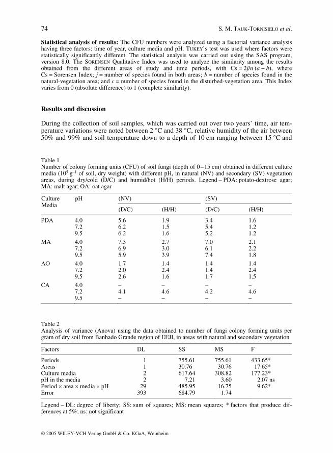
During the collection of soil samples, which was carried out over two years’ time, air tem-perature variations were noted between 2 °C and 38 °C, relative humidity of the air between 50% and 99% and soil temperature down to a depth of 10 cm ranging between 15 °C and Table 1 Number of colony forming units (CFU) of soil fungi (depth of 0–15 cm) obtained in different culture media (105 g–1 of soil, dry weight) with different pH, in natural (NV) and secondary (SV) vegetation areas, during dry/cold (D/C) and humid/hot (H/H) periods. Legend – PDA: potato-dextrose agar; MA: malt agar; OA: oat agar
pH (NV) (SV) Culture Media
(D/C) (H/H) (D/C) (H/H)
PDA 4.0 5.6 1.9 3.4 1.6 7.2 6.2 1.5 5.4 1.2 9.5 6.2 1.6 5.2 1.2
MA 4.0 7.3 2.7 7.0 2.1 7.2 6.9 3.0 6.1 2.2 9.5 5.9 3.9 7.4 1.8
AO 4.0 1.7 1.4 1.4 1.4 7.2 2.0 2.4 1.4 2.4 9.5 2.6 1.6 1.7 1.5
CA 4.0 – – – – 7.2 4.1 4.6 4.2 4.6 9.5 – – – –
Table 2 Analysis of variance (Anova) using the data obtained to number of fungi colony forming units per gram of dry soil from Banhado Grande region of EEJI, in areas with natural and secondary vegetation
Factors DL SS MS F
Periods 1 755.61 755.61 433.65* Areas 1 30.76 30.76 17.65* Culture media 2 617.64 308.82 177.23* pH in the media 2 7.21 3.60 2.07 ns Period × area × media × pH 29 485.95 16.75 9.62* Error 393 684.79 1.74
Legend – DL: degree of liberty; SS: sum of squares; MS: mean squares; * factors that produce dif-ferences at 5%; ns: not significant
Soilborne filamentous fungi in Brazil 75
© 2005 WILEY-VCH Verlag GmbH & Co. KGaA, Weinheim
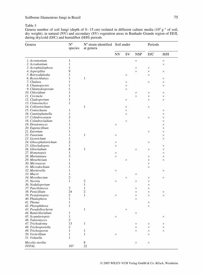
Table 3 Genera number of soil fungi (depth of 0–15 cm) isolated in different culture media (105 g–1 of soil, dry weight), in natural (NV) and secondary (SV) vegetation areas in Banhado Grande region of EEJI, during dry/cold (D/C) and humid/hot (H/H) periods
Genera Nº species
Nº strain identified at genera
Soil under Periods
NV SV NSP D/C H/H
1. Acremonium 1 × × 2. Acrodontium 1 × 3. Acrophialophora 1 × × 4. Aspergillus 9 × × × 5. Botryodiplodia 1 × × 6. Byssochlamys 1 1 × 7. Chalara 1 × × × 8. Chaunopycnis 1 × × 9. Chlamydosporum 1 × 10. Chloridium 1 × × × 11. Circinela 1 × × × 12. Cladosporium 1 × × × 13. Clonostachys 1 14. Colletotrichum 1 × × 15. Coniochaeta 1 × × 16. Cunninghamella 1 × × 17. Cylindrocarpon 2 × × 18. Cylindrocladium 1 × × 19. Doratomyces 1 × × 20. Eupenicillium 21. Eurotium
1 1
×
× ×
22. Fusarium 23. Geotrichum
1 1
1 × × ×
24. Gliocephalotrichum 2 × × 25. Gliocladiopsis 1 × × 26. Gliocladium 8 1 × × × 27. Hymenopsis 1 × × 28. Mariannaea 3 × × × 29. Metarhizium 30. Microascus 31. Microdochium
1 1 1
× × × ×
×
32. Mortierella 1 × × 33. Mucor 1 × 34. Myrothecium 2 × × × 35. Nectria 36. Nodulisporium
1 2 1
× × × ×
37. Paecilomyces 3 1 × × 38. Penicillium 24 2 × × × 39. Pestalotiopsis 40. Phialophora 41. Phoma 42. Phytophthora 43. Pseudollescheria 44. Ramichloridium
2 1 1 1 1
1 1
× ×
× × ×
× × ×
×
45. Scopulariopsis 46. Talaromyces
1 2
× ×
47. Trichoderma 13 1 × × × 48. Trichosporiella 2 × × × 49. Trichosporon 1 1 × × × 50. Verticillium 51. Volutella
1 1
1 × ×
Mycelia sterilia TOTAL
107
8 22
× ×
76 S. M. TAUK-TORNISIELO et al.
© 2005 WILEY-VCH Verlag GmbH & Co. KGaA, Weinheim
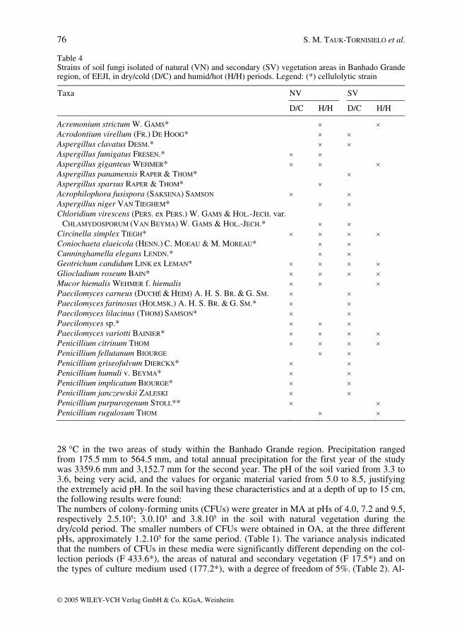
Table 4 Strains of soil fungi isolated of natural (VN) and secondary (SV) vegetation areas in Banhado Grande region, of EEJI, in dry/cold (D/C) and humid/hot (H/H) periods. Legend: (*) cellulolytic strain
NV SV Taxa
D/C H/H D/C H/H
Acremonium strictum W. GAMS* × × Acrodontium virellum (FR.) DE HOOG* × × Aspergillus clavatus DESM.* × × Aspergillus fumigatus FRESEN.* × × Aspergillus giganteus WEHMER* × × × Aspergillus panamensis RAPER & THOM* × Aspergillus sparsus RAPER & THOM* × Acrophilophora fusispora (SAKSENA) SAMSON × × Aspergillus niger VAN TIEGHEM* × × Chloridium virescens (PERS. ex PERS.) W. GAMS & HOL.-JECH. var.
CHLAMYDOSPORUM (VAN BEYMA) W. GAMS & HOL.-JECH.*
× ×
Circinella simplex TIEGH* × × × × Coniochaeta elaeicola (HENN.) C. MOEAU & M. MOREAU* × × Cunninghamella elegans LENDN.* × × Geotrichum candidum LINK ex LEMAN* × × × × Gliocladium roseum BAIN* × × × × Mucor hiemalis WEHMER f. hiemalis × × × Paecilomyces carneus (DUCHÉ & HEIM) A. H. S. BR. & G. SM. × × Paecilomyces farinosus (HOLMSK.) A. H. S. BR. & G. SM.* × × Paecilomyces lilacinus (THOM) SAMSON* × × Paecilomyces sp.* × × × Paecilomyces variotti BAINIER* × × × × Penicillium citrinum THOM × × × × Penicillium fellutanum BIOURGE × × Penicillium griseofulvum DIERCKX* × × Penicillium humuli v. BEYMA* × × Penicillium implicatum BIOURGE* × × Penicillium janczewskii ZALESKI × × Penicillium purpurogenum STOLL** × × Penicillium rugulosum THOM × ×
28 °C in the two areas of study within the Banhado Grande region. Precipitation ranged from 175.5 mm to 564.5 mm, and total annual precipitation for the first year of the study was 3359.6 mm and 3,152.7 mm for the second year. The pH of the soil varied from 3.3 to 3.6, being very acid, and the values for organic material varied from 5.0 to 8.5, justifying the extremely acid pH. In the soil having these characteristics and at a depth of up to 15 cm, the following results were found: The numbers of colony-forming units (CFUs) were greater in MA at pHs of 4.0, 7.2 and 9.5, respectively 2.5.105; 3.0.105 and 3.8.105 in the soil with natural vegetation during the dry/cold period. The smaller numbers of CFUs were obtained in OA, at the three different pHs, approximately 1.2.105 for the same period. (Table 1). The variance analysis indicated that the numbers of CFUs in these media were significantly different depending on the col-lection periods (F 433.6*), the areas of natural and secondary vegetation (F 17.5*) and on the types of culture medium used (177.2*), with a degree of freedom of 5%. (Table 2). Al-
Soilborne filamentous fungi in Brazil 77
© 2005 WILEY-VCH Verlag GmbH & Co. KGaA, Weinheim
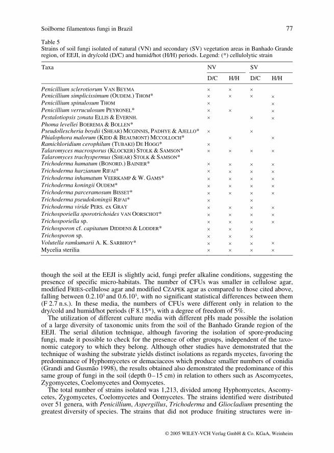
Table 5 Strains of soil fungi isolated of natural (VN) and secondary (SV) vegetation areas in Banhado Grande region, of EEJI, in dry/cold (D/C) and humid/hot (H/H) periods. Legend: (*) cellulolytic strain
NV SV Taxa D/C H/H D/C H/H
Penicillium sclerotiorum VAN BEYMA × × × Penicillium simplicissimum (OUDEM.) THOM* × × × × Penicillium spinulosum THOM × × Penicillium verruculosum PEYRONEL* × × × Pestalotiopsis zonata ELLIS & EVERNH. × × × Phoma levellei BOEREMA & BOLLEN* Pseudollescheria boydii (SHEAR) MCGINNIS, PADHYE & AJELLO* × × Phialophora malorum (KIDD & BEAUMONT) MCCOLLOCH* × × Ramichloridium cerophilum (TUBAKI) DE HOGG* × Talaromyces macrosporus (KLOCKER) STOLK & SAMSON* × × × × Talaromyces trachyspermus (SHEAR) STOLK & SAMSON* Trichoderma hamatum (BONORD.) BAINIER* × × × × Trichoderma harzianum RIFAI* × × × × Trichoderma inhamatum VEERKAMP & W. GAMS* × × × × Trichoderma koningii OUDEM* × × × × Trichoderma parceramosum BISSET* × × × × Trichoderma pseudokoningii RIFAI* × × Trichoderma viride PERS. ex GRAY × × × × Trichosporiella sporotrichoides VAN OORSCHOT* × × × × Trichosporiella sp. × × × × Trichosporon cf. capitatum DIDDENS & LODDER* × × × Trichosporon sp. × × × Volutella ramkumarii A. K. SARBHOY* × × × × Mycelia sterilia × × × ×
though the soil at the EEJI is slightly acid, fungi prefer alkaline conditions, suggesting the presence of specific micro-habitats. The number of CFUs was smaller in cellulose agar, modified FRIES-cellulose agar and modified CZAPEK agar as compared to those cited above, falling between 0.2.103 and 0.6.103, with no significant statistical differences between them (F 2.7 n.s.). In these media, the numbers of CFUs were different only in relation to the dry/cold and humid/hot periods (F 8.15*), with a degree of freedom of 5%. The utilization of different culture media with different pHs made possible the isolation of a large diversity of taxonomic units from the soil of the Banhado Grande region of the EEJI. The serial dilution technique, although favoring the isolation of spore-producing fungi, made it possible to check for the presence of other groups, independent of the taxo-nomic category to which they belong. Although other studies have demonstrated that the technique of washing the substrate yields distinct isolations as regards mycetes, favoring the predominance of Hyphomycetes or demaciaceos which produce smaller numbers of conidia (Grandi and Gusmão 1998), the results obtained also demonstrated the predominance of this same group of fungi in the soil (depth 0–15 cm) in relation to others such as Ascomycetes, Zygomycetes, Coelomycetes and Oomycetes. The total number of strains isolated was 1,213, divided among Hyphomycetes, Ascomy-cetes, Zygomycetes, Coelomycetes and Oomycetes. The strains identified were distributed over 51 genera, with Penicillium, Aspergillus, Trichoderma and Gliocladium presenting the greatest diversity of species. The strains that did not produce fruiting structures were in-
78 S. M. TAUK-TORNISIELO et al.
© 2005 WILEY-VCH Verlag GmbH & Co. KGaA, Weinheim
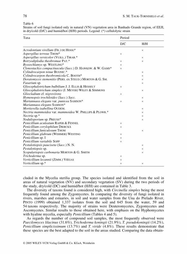
Table 6 Strains of soil fungi isolated only in natural (VN) vegetation area in Banhado Grande region, of EEJI, in dry/cold (D/C) and humid/hot (H/H) periods. Legend: (*) cellulolytic strain
Period Taxa
D/C H/H
Acrodontium virellum (FR.) DE HOOG* × Aspergillus terreus THOM* × Aspergillus versicolor (VUILL.) TIRAB.* × Botryodiplodia theobromae PAT.* × × Byssochlamys sp. WESTLING* × Clonostachys compactiuscula (Sacc.) D. HAWKSW. & W. GAMS* × Cylindrocarpon tenue BUGNIC.* × Cylindrocarpon theobromicola C. BOOTH* × Doratomyces stemonitis (PERS. ex STEUD.) MORTON & G. SM. × Fusarium sp. × Gliocephalotrichum bulbilium J. J. ELLIS & HESSELT × Gliocephalotrichum simplex (J. MEYER) WILEY & SIMMONS × Gliocladium cf. nigrovirens × Hymenopsis trochiloides (Sacc.) Sacc. × Mariannaea elegans var. punicea SAMSON* × × Mariannaea elegans SAMSON* Mortierella isabellina OUDEM. × Nectria mammoidea var. mammoidea W. PHILLIPS & PLOWR.* × Nectria sp.* × Nodulisporium sp. PREUSS* × Penicillium aculeatum RAPER & FENNEL × Penicillium corylophilum DIERCKX × Penicillium funiculosum THOM × Penicillium glabrum (WEHMER) WESTING × Penicillium sp. I × Penicillium variabile SOPP × Pestalotiopsis pauciseta (Sacc.) N. N. × Pestalotiopsis sp. × Scopulariopsis carbonaria MORTON & G. SMITH × Trichoderma sp. × Verticillium lecannii (ZIMM.) VIÉGAS × Verticillium sp.* × ×
cluded in the Mycelia sterilia group. The species isolated and identified from the soil in areas of natural vegetation (NV) and secondary vegetation (SV) during the two periods of the study, dry/cold (DC) and humid/hot (H/H) are contained in Table 3. The diversity of taxons found is considered high, with Circinella simplex being the most frequently found among the Zygomycetes. In comparing the diversity of fungi isolated in rivers, marshes and estuaries, in soil and water samples from the Una do Prelado River, PINTO (1999) obtained 1,337 isolates from the soil and 645 from the water, 59 and 54 taxons respectively. The majority of strains were Deuteromycetes, Zygomycetes and Ascomycetes. Similar results to those obtained here, with emphasis on the Hyphomycetes with hyaline mycelia, especially Penicillium (Tables 4 and 5). As regards the number of compound soil samples, the most frequently observed were Paecilomyces lilacinus (31.6%), Trichoderma koningii (21.9%), T. pseudokoningii (14.2%), Penicillium simplicissimum (13.7%) and T. viride (4.8%). These results demonstrate that these species are the best adapted to the soil in the areas studied. Comparing the data obtain-
Soilborne filamentous fungi in Brazil 79
© 2005 WILEY-VCH Verlag GmbH & Co. KGaA, Weinheim
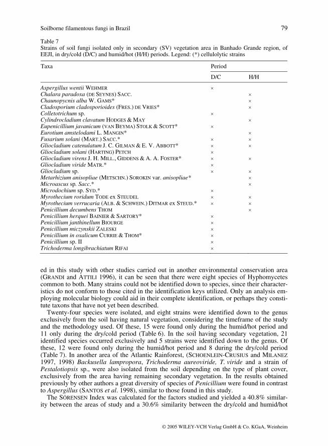
Table 7 Strains of soil fungi isolated only in secondary (SV) vegetation area in Banhado Grande region, of EEJI, in dry/cold (D/C) and humid/hot (H/H) periods. Legend: (*) cellulolytic strains
Period Taxa
D/C H/H
Aspergillus wentii WEHMER × Chalara paradoxa (DE SEYNES) SACC. × Chaunopycnis alba W. GAMS* × Cladosporium cladosporioides (FRES.) DE VRIES* × Colletotrichum sp. × Cylindrocladium clavatum HODGES & MAY × Eupenicillium javanicum (VAN BEYMA) STOLK & SCOTT* × Eurotium amstelodami L. MANGIN* × Fusarium solani (MART.) SACC.* × × Gliocladium catenulatum J. C. GILMAN & E. V. ABBOTT* × × Gliocladium solani (HARTING) PETCH × Gliocladium virens J. H. MILL., GIDDENS & A. A. FOSTER* × × Gliocladium viride MATR.* × Gliocladium sp. × × Metarhizium anisopliae (METSCHN.) SOROKIN var. anisopliae* × Microascus sp. Sacc.* × Microdochium sp. SYD.* × Myrothecium roridum TODE ex STEUDEL × × Myrothecium verrucaria (ALB. & SCHWEIN.) DITMAR ex STEUD.* × × Penicillium decumbens THOM × Penicillium herquei BAINIER & SARTORY* × Penicillium janthinellum BIOURGE × Penicillium miczynskii ZALESKI × Penicillium in oxalicum CURRIE & THOM* × Penicillium sp. II × Trichoderma longibrachiatum RIFAI ×
ed in this study with other studies carried out in another environmental conservation area (GRANDI and ATTILI 1996), it can be seen that there were eight species of Hyphomycetes common to both. Many strains could not be identified down to species, since their character-istics do not conform to those cited in the identification keys utilized. Only an analysis em-ploying molecular biology could aid in their complete identification, or perhaps they consti-tute taxons that have not yet been described. Twenty-four species were isolated, and eight strains were identified down to the genus exclusively from the soil having natural vegetation, considering the timeframe of the study and the methodology used. Of these, 15 were found only during the humid/hot period and 11 only during the dry/cold period (Table 6). In the soil having secondary vegetation, 21 identified species occurred exclusively and 5 strains were identified down to the genus. Of these, 12 were found only during the humid/hot period and 8 during the dry/cold period (Table 7). In another area of the Atlantic Rainforest, (SCHOENLEIN-CRUSIUS and MILANEZ 1997, 1998) Backusella lampropora, Trichoderma aureoviride, T. viride and a strain of Pestalotiopsis sp., were also isolated from the soil depending on the type of plant cover, exclusively from the area having remaining secondary vegetation. In the results obtained previously by other authors a great diversity of species of Penicillium were found in contrast to Aspergillus (SANTOS et al. 1998), similar to those found in this study. The SÖRENSEN Index was calculated for the factors studied and yielded a 40.8% similar-ity between the areas of study and a 30.6% similarity between the dry/cold and humid/hot
80 S. M. TAUK-TORNISIELO et al.
© 2005 WILEY-VCH Verlag GmbH & Co. KGaA, Weinheim
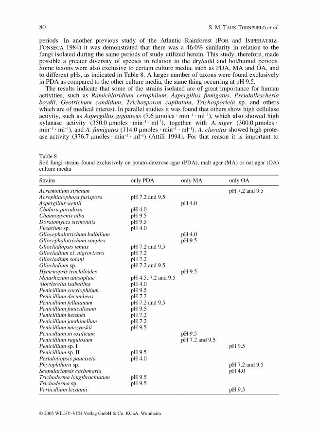
periods. In another previous study of the Atlantic Rainforest (POR and IMPERATRIZ-FONSECA 1984) it was demonstrated that there was a 46.0% similarity in relation to the fungi isolated during the same periods of study utilized herein. This study, therefore, made possible a greater diversity of species in relation to the dry/cold and hot/humid periods. Some taxons were also exclusive to certain culture media, such as PDA, MA and OA, and to different pHs, as indicated in Table 8. A larger number of taxons were found exclusively in PDA as compared to the other culture media, the same thing occurring at pH 9.5. The results indicate that some of the strains isolated are of great importance for human activities, such as Ramichloridium cerophilum, Aspergillus fumigatus, Pseudollescheria boydii, Geotrichum candidum, Trichosporon capitatum, Trichosporiela sp. and others which are of medical interest. In parallel studies it was found that others show high cellulase activity, such as Aspergillus giganteus (7.6 µmoles · min–1 · ml–1), which also showed high xylanase activity (350.0 µmoles · min–1 · ml–1), together with A. niger (300.0 µmoles · min–1 · ml–1), and A. fumigatus (114.0 µmoles · min–1 · ml–1). A. clavatus showed high prote-ase activity (376.7 µmoles · min–1 · ml–1) (Attili 1994). For that reason it is important to
Table 8 Soil fungi strains found exclusively on potato-dextrose agar (PDA), malt agar (MA) or oat agar (OA) culture media
Strains only PDA only MA only OA
Acremonium strictum pH 7.2 and 9.5 Acrophialophora fusispora pH 7.2 and 9.5 Aspergillus wentii pH 4.0 Chalara paradoxa pH 4.0 Chaunopycnis alba pH 9.5 Doratomyces stemonitis pH 9.5 Fusarium sp. pH 4.0 Gliocephalotrichum bulbilium pH 4.0 Gliocephalotrichum simplex pH 9.5 Gliocladiopsis tenuis pH 7.2 and 9.5 Gliocladium cf. nigrovirens pH 7.2 Gliocladium solani pH 7.2 Gliocladium sp. pH 7.2 and 9.5 Hymenopsis trochiloides pH 9.5 Metarhizium anisopliae pH 4.5, 7.2 and 9.5 Mortierella isabellina pH 4.0
Penicillium corylophilum pH 9.5
Penicillium decumbens pH 7.2 Penicillium fellutanum pH 7.2 and 9.5 Penicillium funiculosum pH 9.5 Penicillium herquei pH 7.2 Penicillium janthinellum pH 7.2 Penicillium miczynskii pH 9.5 Penicillium in oxalicum pH 9.5 Penicillium rugulosum pH 7.2 and 9.5 Penicillium sp. I pH 9.5 Penicillium sp. II pH 9.5 Pestalotiopsis pauciseta pH 4.0 Phytophthora sp. pH 7.2 and 9.5 Scopulariopsis carbonaria pH 4.0 Trichoderma longibrachiatum pH 9.5
Trichoderma sp. pH 9.5
Verticillium lecannii pH 9.5
Soilborne filamentous fungi in Brazil 81
© 2005 WILEY-VCH Verlag GmbH & Co. KGaA, Weinheim
learn more and more about the biodiversity of the microbiotics of soils in natural areas be-fore they are destroyed through anthropic activities.
Acknowledgements
The authors wish to thank the CNPq for the Master and Ph.D grants, FAPESP for its aid to the research and Dra. IRACEMA H. SCHOENLEIN-CRUSIUS of the Institute of Botany, SMA, São Paulo, Brazil, for her help in the identification of the fungi. Thanks too SARA CRISTINA GALVÃO.
References
ATTILI, D. S., MACEDO, A. P. and ESPOSITO, E., 1993. Screening of Aspergillus from Juréia Ecologia-cal Reserve: enzymes from solid medium. In: Proceedings of the Third Brasilian Symposium on the Chemistry of Lignins and other Wood Components, Vol. 4: 365–368.
ATTILI, D. S., 1994. Isolamento, identificação e ecologia de fungos celulolíticos do solo da Estação Ecológica de Juréia–Itatins, SP. Rio Claro, UNESP, 148p. (Ph.D. Thesis – Instituto de Biociências, UNESP).
BISSET, J., 1991. A revision of the genus Trichoderma. III. Section Pachybasium. Canad. J. Bot., 69, 2373–2417.
CORTESÃO, J., 1989. Juréia: a luta pela vida. In: Index, São Paulo, 133 p. EGGINS, H. and PUGH, G. J. F., 1962. Isolation of cellulose-decomposing fungi from the soil. Nature,
193, 94–95. FERRAZ, A. and DURÁN, N., 1990. Effect of various conditions on the Chrysonilia sitophila
“TFB27441” growth. Rev. Microbiol., 20, 89–101. GAMS, W., 1980. Chaunopycnis alba., gen. et sp. nov., a soil fungus intermediate between moniliales
and sphaeropsidales. Perssonia, 11, 75–79. GARLIPP, A. B., 1995. Isolamento e identificação de fungos filamentosos do solo do Banhado Grande,
na Estação Ecológica de Juréia-itatins, SP. UNESP. 94p. (Master Dissertation – Instituto de Bio-ciências, UNESP).
GAVIRIA, C., 1978. Normas para interpretar e reportar a contagem “standard” em placa. São Paulo: Merck, 1v.
GRANDI, R. A. P. and ATTILI, D. S., 1996. Hyphomycetes on Alchornea triplinervia (spreng.) Müll. Arg. Leaf litter from the Ecological Reserve Juréia-Itatins, State of São Paulo, Brazil. Mycotaxon, 60, 373–386.
GRANDI, R. A. P. and GUSMÃO, L. F. P., 1998. A técnica da lavagem sucessiva de substratos de plantas como subsídio para estudos de associa’[cão fungo/substrato e diversidade de Hyphomycetes nos ecossistemas. In: Simpósio de Ecossistemas Brasileiros, 4, ACIESP, 3, 80–90.
HAWKSWORTH, D. L., 1991.The fungal dimension of biodiversity: magnitude, significance and conser-vation. Mycol Res., 95, 641–655,
HAWKSWORTH, D. L., SUTTON, B. C. and AINSWORTH, G. C., 1983. Ainswortyh & Bisby’s Dictionary of the Fungi. 7 ed. Kew. In: CAB International Mycological Institute, 445 p.
MANTOVANI, W., 1990. A dinâmica das florestas na encosta atlântica. In: II Simpósio de Ecossistemas da Costa Sul e Sudeste Brasileira. Águas de Lindóia, SP. Anais, 1, 304–313.
MURO, M. A. and LUCHI, M. R., 1989. Preservação de microrganismos. In: Fundação Tropical de Pesquisas e Tecnologia “Ancré Tosello”, 65 p.
PINTO, I. M. A., 1999. As micotas filamentosas do solo e da água do rio Una do Prelado, Estação Ecológica de Juréia-Itatins, SP. UNESP, 200 p. (PhD Thesis – Instituto de Biociências, UNESP).
PITT, J. I., 1991. A laboratory guides to common Penicillium species. In: Commonwealth Sci. Ind. Res. Org., 187 p.
POR, F. D. and IMPERATRIZ-FONSECA, V. L., 1984. The Juréia Ecological Reserve, São Paulo, Brazil. Facts and Plans. Environ. Conserv., 2, 67–70.
RAIJ, B. and QUAGGIO, J. A., 1983. Métodos de análise de solo para fins de fertilidade. Bolm. Tec. Inst. Agron., 81, 1–31.
82 S. M. TAUK-TORNISIELO et al.
© 2005 WILEY-VCH Verlag GmbH & Co. KGaA, Weinheim
SANTOS, V. B., WELLBAUM, C. and SCHOENLEIN-CRUSIUS, I. H., 1998. Fungos filamentosos do solo da Ilha dos Eucaliptos na Represa do Guarapiranga em São Paulo, SP. Acta bot. bras. 12, 101–110.
SCHOENLEIN-CRUSIUS, I. H. and MILANEZ, A I., 1997. Mucorales (Zygomycotina) da Mata Atlântica da Reserva Biológica do Alto da Serra de Paranabiacaba, Santo André, SP. Acta bot. bras., 11, 95–101.
TAUK-TORNISIELO, S. M., GIANNOTTI, A. M. D., RUEGGER, M. J. S. and MALAGUTTI, E. N., 2000. Aphyllophorales from the Atlantic rainforest of the Ecological Station of Juréia-Itatins, São Paulo, Brazil. Brasilian J. Ecol., 1–2, 99–105.
VIEIRA, L. S., 1988. Manual da ciência do solo com ênfase aos solos tropicais. In: Agronômica Ceres 2.ed., 464 p.
Mailing address: Dr. SÂMIA TAUK-TORNISIELO, Centro de Estudos Ambientais, Universidade Estadual Paulista, Bairro Bela Vista, Rio Claro-13.506-900, SP, Brazil Tel.: 019-35344622; fax: 019-35340122 E-mail: [email protected]