searchforcoherent’genemodules’that’predict...
TRANSCRIPT
Universidade de Lisboa Faculdade de Ciências
Departamento de Química e Bioquímica
Search for coherent gene modules that predict Streptococcus pneumoniae strain invasiveness
Rui Ribeiro Catarino
Mestrado em Bioquímica Especialidade em Bioquímica
Dissertação de Mestrado orientada por Doutor Francisco Pinto
2012


i
Agradecimentos Quero agradecer antes de mais aos meus pais, já que, no final de contas, são os grandes responsáveis pelo percurso que segui. Fico também grato pelo sacrifício pessoal que fizeram ao suportar o meu ensino superior e qualquer ocasional má disposição. Espero poder agora começar a repagar esse esforço, começando por pagar um jantar assim que isto tudo esteja acabado. Quero também agradecer ao grupo de enzimologia pelo ambiente de investigação saudável que me proporcionou. Em particular, um enorme agradecimento ao professor Francisco Pinto, não só por me ter iniciado numa área da qual eu pouco conhecia, pela atenção que sempre me cedeu sem restrições, mas acima de tudo pela amizade e excelente disposição a que me habituou e que, confio, se irá prolongar. O ano que se avizinha é a minha melhor oportunidade para devolver o trabalho que foi investido em mim, mas, num prazo mais curto, um jantar parece-‐me uma boa forma de começar. Por fim, quero agradecer ao Daniel Fonseca que além de amigo e companheiro em toda duração (e em todas as vertentes) da vida universitária, sempre me estimulou intelectualmente em todas as áreas do saber que hoje prezo. Também a ele convidaria para jantar, não soubera que já não se irá encontrar em Portugal a essa data. Posto isto, deixo o convite para me convidares para jantar quando te for visitar.

ii
iii
Abstract Streptococcus pneumonia is a pathogenic bacterium responsible for several
human diseases, such as pneumonia, meningitis and sepsis. Any pneumococcal
disease is preceded by an asymptomatic colonization stage in the human
nasopharynx. The transition from colonization to invasion is known to depend
on both human and pathogen factors. This work aims to computationally identify
pneumococcal genetic factors that influence the likelihood of invasion events.
For this purpose, we analyze microarray based comparative genomic
hybridization data of 72 strains of pneumococcus. Each strain was classified as
Invasive, Neutral or Colonizer according to a previous study that compared the
frequencies with which strains were recovered from an asymptomatic carrier or
from invasive disease episodes.
We propose to select genes that, individually or in a coordinated way, affect the
frequency of invasion transitions among all colonization events, which we
denominate as invasiveness. To detect coordinated sets of genes, we developed
a method that uses networks of known interactions between genes to find gene
modules that predict invasiveness. Each module is founded with a single gene
and then grown with its closest neighbors in the network. Each module is then
evaluated for its predictive power, statistical significance and robustness to data
variability.
We tested the method with a network based on a distance score that integrates
gene co-‐occurrence and co-‐invasiveness. Among others functions, the found
modules implicate cell envelope, transport, sugar metabolism, osmotic response,
aminoacid synthesis, spermidine synthesis and proteolysis functions in
pneumococcal invasiveness.

iv
v
Resumo O Streptococcus pneumoniae, também chamado pneumococcus, é uma bactéria gram-
positiva do subgrupo alfa-hemolítico do género Streptococcus. É um colonizador
frequente do trato respiratório superior humano e embora possa ser encontrado em
qualquer pessoa, tem maior prevalência em crianças e idosos. A colonização decorre
tipicamente sem causar sintomas, mas pode por vezes culminar na invasão de outros
tecidos e provocar doenças como pneumonia, meningite ou otite do ouvido médio.
Sem tratamento, a infeção com pneumococcus tem uma taxa de mortalidade da ordem
dos 30 por cento mas, atualmente, com o uso de antibióticos e vacinas, este número é
muito mais reduzido. Contudo, a resistência a antibióticos tem vindo a ser
reconhecida em pneumococcus e a vacinação, mais do que reduzir o número de
doenças provocadas por pneumococcus, tem conduzido à substituição das estirpes que
as originam. Por estes motivos, torna-se urgente entender o mecanismo de invasão e
virulência do pneumococcus para que novas formas de combate a este patógeneo
possam tomar forma.
Como em muitos outros organismos que habitam meios de composição pouco
variável, na maioria patógenos, o pneumococcus tem um genoma reduzido. O genoma
apresenta grande plasticidade, variando cerca de 10 por cento entre estirpes e contem
apenas 60 a 80 por cento de genes mantidos em todas as estirpes. A totalidade dos
genes do pneumococcus, o pangenoma, é consideravelmente mais vasto que o
genoma de qualquer estirpe e juntamente com a capacidade de trocar genes entre a
própria espécie, ou por vezes com espécies próximas, confere a esta bactéria uma
grande adaptabilidade e resposta rápida a mudanças no seu meio ambiente. A
transferência de genes horizontal é de facto uma idiossincrasia do pneumococcus e é,
por vezes, acompanhada pela indução de morte de células da mesma espécie para que
estas libertem DNA. Este fenómeno, conhecido como fratricídio, acontece quando a
célula entra num estado de competência, também chamado estado X. O segundo
nome foi proposto por ser mais abrangente, evitando que o estado fosse apenas
associado à competência. Neste estado, o perfil de transcrição da bactéria é
globalmente alterado e além de expressar genes que promovem a competência,
expressa também bacteriocinas tóxicas para as células vizinhas e proteínas que
protegem a própria célula dessas bacteriocinas. A facilidade de incorporação de DNA
vi
de outras células contribui significativamente para a sobrevivência da bactéria. A
resistência à penicilina, por exemplo, é conferida por genes que foram adquiridos de
uma espécie próxima, o Streptococcus Mitis.
A invasividade e virulência do pneumococcus varia de estirpe para estirpe e é função
do conteúdo génico. A bactéria está especialmente adaptada para colonizar, visto
passar a maior parte do tempo na nasofaringe e que o principal meio de transmissão
ocorre por aerossol e quase exclusivamente durante a colonização. Embora não exista
consenso sobre o motivo desta adaptação, é consensual que algumas estirpes são mais
aptas para a invasão de outros tecidos e, consequentemente, causar doença. Entre os
determinantes de virulência, o mais estudado é a cápsula polisacarídica ou serótipo.
São conhecidos mais de 90 serótipos que diferem em estrutura e composição, mas
apenas pouco mais de vinte estão associados a doença. A cápsula é um dos mais
importantes mecanismos de defesa contra o sistema imunitário humano, já que, além
de cobrir grande parte dos epítopos que seriam facilmente reconhecíveis, ainda inibe o
sistema do complemento. Vários outros determinantes têm vindo a ser identificados
mas o contexto genético tem sido descurado. Alguns dos genes associados com
virulência numa estirpe, foram associados com colonização noutra, evidenciando a
relevância das interações entre genes. A noção de que a invasividade pode ser
conferida por interação entre genes complexifica tanto a busca de determinantes,
como os próprios determinantes.
É possível identificar determinantes de invasividade procurando diferenças entre
grupos de estirpes invasivas e grupos de estirpes colonizadoras. Estas diferenças
podem ocorrer em diferentes níveis como o conteúdo génico ou a sua expressão. Dada
a grande variabilidade do genoma do pneumococcus, é expectável encontrar
determinantes de invasividade ao nível do conteúdo génico. Estas diferenças podem
ser detetadas em larga escala por ensaios de microarrays de Hibridação Genómica
Comparativa. É importante notar que esta abordagem é observacional e que, portanto,
os resultados permitem apenas estabelecer correlações e não relações de causa efeito.
Em contrapartida, permite observar múltiplas interações com diferentes backgrounds
genéticos e a interação entre diferentes determinantes. Desta maneira, esta abordagem
encaixa-se no paradigma da biologia de sistemas, visto estudar não só os genes
individualmente, mas antes em interação com os demais.
vii
A procura de determinantes que distingam estirpes invasivas de estirpes colonizadoras
é um problema de classificação, uma área da aprendizagem supervisionada. Existem
já muitos algoritmos desenhados para resolver este tipo de problema. Tipicamente, o
sucesso destes algoritmos é avaliado pela sua capacidade de classificar corretamente
as estirpes a partir dos seus genótipos. Entre outros, algoritmos como as redes
neuronais são conhecidos por uma elevada exatidão de classificação. No entanto, o
foco deste trabalho não é a exatidão de classificação mas antes a compreensão dos
mecanismos que conduzem à invasividade. Grande parte dos algoritmos existentes
resultam num conjunto de regras difíceis de interpretar e ainda mais de traduzir para
um nível biológico, em especial se considerarmos que as estirpes invasivas podem ser
um grupo heterogéneo com diferentes mecanismos de invasividade. Por este motivo,
surgiu a necessidade de desenhar um novo algoritmo que foque primordialmente
identificar determinantes de invasividade.
A procura de determinantes que tenham em conta a interação de genes constitui um
problema computacional acrescido. A busca de múltiplos genes, módulos de genes,
que constituam um determinante transforma-se num problema combinatorial em que
o número de possibilidades aumenta exponencialmente com o número de genes. Para
evitar uma busca exaustiva de todas as combinações, o algoritmo usa informação
sobre interações entre os genes que podem ser de cariz metabólico, regulatório, físico,
entre outros, mas que podem ser facilmente descritas num formato comum – as redes.
As redes têm a vantagem de expressarem facilmente padrões de interações complexos
e de serem manipuláveis e pesquisáveis computacionalmente.
Os dados usados neste trabalho resultam de um estudo de microarray de Hibridação
Genómica Comparativa com 72 estirpes que usou como controlos as estirpes Tigr4,
G54 e R6. Estas estirpes foram previamente classificadas como invasivas, neutras ou
colonizadoras, de acordo com a frequência com que foram identificadas em
indivíduos saudáveis ou em indivíduos portadores de doença. A presença ou ausência
dos genes nas estirpes foi organizado numa matriz denominada matriz de presença
génica. As estirpes neutras não foram incluídas na matriz por terem um cariz incerto.
A classificação de uma estirpe como neutra pode dever-se tanto a motivos biológicos
como à insuficiência de poder estatístico para a classificar como invasiva ou
colonizadora.
viii
Não foi usada uma rede de interações de genes mas sim uma matriz de distância que
avalia a coocorrência e a coinvasidade. A coocorrência é um parâmetro que avalia a
frequência com que dois genes estão presentes individualmente comparativamente
com a frequência com que estão presentes em conjunto. A coinvasidade é um
parâmetro que avalia a semelhança de associação de cada um dos genes com a
invasividade. Esta associação é medida usando um teste estatístico de Fisher. Juntos,
estes parâmetros asseguram que dois genes com uma baixa distância são genes que
coocorrem frequentemente e que têm uma associação com a invasividade semelhante.
A matriz de distâncias é usada para criar módulos de genes que serão depois
avaliados. Os módulos são criados a partir de um gene semente, ao qual são
gradualmente adicionados mais genes. O gene adicionado é sempre o gene com
menor distância ao gene semente.
Os módulos de genes são inicialmente avaliados quanto à sua presença dos seus genes
em estirpes invasivas e colonizadoras através de um teste de runs. Este teste avalia se
a distribuição das presenças pelas classes de estirpes é significativa ou se pode ser
considerada aleatória, caso em que o módulo é abandonado. De seguida é definido um
número de genes, limite, acima do qual o módulo é considerado presente numa
estirpe. Este limite é definido de forma a que o módulo esteja presente
exclusivamente em estirpes invasivas. Se tal limite não existir o módulo é
abandonado. Caso tenha sido possível estabelecer um limite, é avaliada a significância
do mesmo. Para tal é usado um teste unilateral que calcula a probabilidade do limite
ter sido fixado com um valor tão ou mais baixo. Caso o limite não tenha significância
estatística de 0.05 o módulo é abandonado.
Dado o método de formação dos módulos, é possível que nem todos os genes
contribuam para a associação do módulo com a invasividade. Para eliminar essas
situações é avaliada a associação individual de cada gene com as estirpes em que o
módulo está presente usando um teste de Fisher. Os genes que não estiverem
associados são eliminados do módulo. Após a remoção de genes o limite é
recalculado e a sua significância é reavaliada. Terminado este passo, é selecionado
apenas um módulo de entre os módulos criados a partir do mesmo gene semente. O
módulo selecionado é aquele que for constituído pelo maior número de genes. Por fim
realizou-se uma correção para testes múltiplos que estabeleceu a taxa de descobertas
falsas em 5 por cento. Este passo eliminou todos os módulos com menos de 24 genes.
ix
De todo este processo resultaram 26 módulos significantes pelos padrões estatísticos
exigidos e que estão presentes exclusivamente nas estirpes invasivas. Embora os
módulos sejam distintos, existe grande sobreposição entre eles. É possível observar
submódulos que surgem repetidos em vários módulos e que eram possivelmente
módulos por si, tendo sido eliminados pela correção por testes múltiplos.
Para cada módulo, observou-se que a presença dos seus genes está correlacionada
com o rácio de probabilidade da invasividade das estirpes. Esta correlação observa-se
mesmo para as estirpes neutras, ainda que estas não tenham sido usadas como input
no algoritmo. Embora as classes invasiva e colonizadora tenham sido usadas pelo
algoritmo, os dados dos seus rácios de probabilidade de invasividade não foram. Em
conjunto, os módulos usam um total de 111 genes e, usados em conjunto, é possível
encontrar uma correlação semelhante. A correlação dos módulos, individualmente e
em conjunto, com os rácios de probabilidade de invasividade e com as estirpes
neutras é um resultado positivo que suporta a relevância e autenticidade destes
módulos como determinantes de invasividade.
Os módulos são robustos contra pequenas alterações na matriz de presença de genes.
A experiência de microarray a partir da qual os dados foram originados tem um erro
inerente e esta alta robustez confere confiança na autenticidade dos resultados do
algoritmo, mostrando que dificilmente são consequência de erros do microarray. A
existência de um limite para definir presença de módulos, por oposição à exigência de
presença de todos os genes em simultâneo, pode ser uma fonte de robustez contra
perturbações nos perfis de presença dos genes.
Não foi encontrado enriquecimento de funções entre os genes selecionados pelo
algoritmo nem entre os módulos. O enriquecimento das funções foi avaliado usando a
anotação do JCVI. Apesar de não se ter verificado enriquecimento funcional usando a
anotação da base de dados do JCVI, alguns genes têm claramente relações funcionais.
O nrdD codifica um ribozima que é ativado pelo nrdG. Os genes Argh e ArgG
codificam enzimas que catalisam reações sequenciais que constituem uma via
alternativa da síntese da arginina. O enzima manitol-1-fosfato desidrogenase (mTLD)
utiliza como substrato o manitol-1-fosfato, que é o produto do transporte de manitol
pelo sistema PTS (MTLA e mtlF). O RuvB tem a sua atividade como estimulador de
recombinação facilitada pela presença da proteína de ligação de DNA de cadeia
simples ssb. Um transportador ABC requer a presença de vários componentes que
x
foram selecionados pelo algoritmo, tais como módulos de ligação ao ATP (ou NBDs)
e permeases transmembrananares. A ação da aquaporina Z (aqpZ) tem levantado
dúvidas na comunidade científica, já que a sua ação parece conduzir ao acumular de
pressão de turgescência celular excessiva. O canal mecanosensível largo (MsCl)
proporciona uma resposta eficaz para a pressão de turgescência e pode ser um
contrapartida biológica da aqpZ. Poliaminas, como a espermidina e norespermidina,
têm sido relatadas como possíveis substitutos da colina e são, por conseguinte,
intervenientes importantes na estrutura da parede celular e possivelmente na ligação a
proteínas que se ligam a colina.
A maioria dos genes selecionados foi previamente associada com a invasão ou tem
alguma conexão plausível com os mecanismos de invasão. Proteínas da cápsula e
proteínas que ligam colina desempenham um papel importante na proteção contra as
defesas do hospedeiro. São importantes na inibição da ação do sistema imunitário,
nomeadamente pela remoção das proteínas do complemento, ou pela ligação ao fator
H, que é um inibidor do complemento. Vários elementos genéticos móveis foram
identificados dentro ou perto do locus dos genes da cápsula e tem sido relatado o
impacto destes elementos na regulação da transcrição de vários genes desse locus.
A invasão de novos tecidos requer uma adaptação rápida a um ambiente novo, tanto
às suas propriedades físicas como à disponibilidade de nutrientes. Foram selecionados
genes de resposta a mudanças da pressão osmótica que parecem mais dirigidos a uma
resposta rápida a grandes alterações da pressão do que à regulação fina da pressão e
são, portanto, de particular interesse na adaptação a novos meios. Genes de resposta
anaeróbica como o nrdD e o seu ativador, nrdG, dificilmente são funcionais na
nasofaringe, uma vez que são estritamente anaeróbicos. No interior do organismo
humano contudo, a concentração de oxigénio é reduzida, uma vez que este está quase
sempre ligado a moléculas biológicas como a hemoglobina. Nestas circunstâncias o
nrdD pode ser crucial para manter as funções dos enzimas aeróbios equivalentes. A
capacidade de utilizar diferentes fontes de energia e de carbono é de extrema
importância para a invasividade de uma estirpe. O elevado número de transportadores
de açúcar está relacionado com a capacidade das estirpes invasivas sobreviverem em
meios de variadas composições. Na mesma lógica, alguns genes foram selecionados
que codificam para enzimas do metabolismo de diferentes açúcares, aumentando
xi
também a adaptabilidade da estirpe a diferentes meios. Genes de proteólise estão
provavelmente relacionadas com as necessidades nutricionais de aminoácidos.
A síntese de proteínas é um processo constante em todas as bactérias e exige uma
disponibilidade permanente de aminoácidos e tRNA. Foram selecionados genes de
síntese de aminoácidos que proporcionam vias alternativas para a síntese de
aminoácidos, utilizando substratos alternativos. O algoritmo também selecionou
genes ligados à síntese e ligação de tRNA ao aminoácido correspondente. Estes
enzimas não foram caracterizados em Streptococcus pneumoniae e é difícil prever a
sua influência na síntese proteica.
Por fim, a grande heterogeneidade dos genomas do pneumococcus advém da sua
capacidade de recombinação. Alguns dos genes selecionados pelo algoritmo
promovem a heterogeneidade do genoma, aumentando a recombinação com o DNA
extracelular. Entre os genes selecionados é promovida a internalização de DNA, a sua
estabilização e a recombinação com DNA não homólogo. O estado de competência do
pneumococcus é acompanhado por uma apetência para induzir a apoptose em células
vizinhas, aumentando a concentração de fragmentos de DNA no meio. Várias
bacteriocinas foram associadas por este trabalho à invasividade, bem como genes que
inibem a apoptose da própria célula. Esses genes dão à célula uma vantagem natural
na competição com outros colonizadores.
Em suma, alcançou-se o objetivo pretendido de encontrar determinantes de
invasividade. Estes determinantes são fruto de um estudo observacional e é portanto
de notar que a relação que têm com a invasividade é apenas de correlação. Para
determinar o impacto que estes módulos de genes têm na invasividade é necessário
realizar estudos laboratoriais que averiguem em maior detalhe a função biológica dos
genes e a sua relação com os mecanismos de invasão.

xii
xiii
Index
Agradecimentos i Abstract iii Resumo v Index xiii Figure index xv Table index xvii Streptococcus pneumoniae 1 Pneumococcal genome dynamics 3 Pneumococcal invasiveness determinants 4 Computational methods 7
Objectives 8 Methodology 9 Methodology overview 9 Gene module definition 9
Methodology description 10 Gene presence and strain classification data 10 Gene and Strain selection 11 Construction of a Gene-‐gene distance matrix 13 Selection of gene modules 15 Module trimming 19 False discovery rate 19 Module’s robustness 20 Implementation 20
Results and Discussion 21 Gene functional analysis 26 Anaerobic Response 27 Choline binding proteins 28 Transcription regulation 29 Insertion Sequences/Transposons 30 tRNA 31 Cell envelope 32 Spermidine metabolism 33 Osmotic Regulation 34 Co-‐enzymes 35 Proteolysis 36 Competence 37
xiv
DNA recombination/repair 38 Transport 39 Carbohydrate metabolism 41 Acetyltransferases 43 Toxins 44 Aminoacid metabolism 45
Functional interactions with invasiveness 46 Conclusion 49 Future perspectives 51 References 53
xv
Figure index
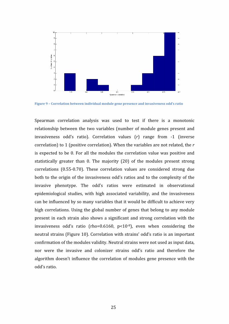
Figure 1 -‐ Gene distribution before gene and strain selection. 12 Figure 2 -‐ Gene Distribution after gene and strain selection. 12 Figure 3 -‐ Influence of module length on module presence in strains 16 Figure 4 -‐ Boxplot of module lengths with random and original gene presence matrices 20 Figure 5 -‐ Module Overlap 22 Figure 6 -‐ Presence of the selected genes in strains 22 Figure 7 -‐ Dendrogram of module based on gene composition. 23 Figure 8 -‐ Bimodal distribution of gene presence 23 Figure 9 – Correlation between individual module gene presence and invasiveness odd’s
ratio 25 Figure 10 – Correlation between global modules genes presence and invasiveness odd’s
ratio 26

xvi
xvii
Table index
Table 1 – Gene Presence vectors with highlights on the genes used to calculate the Jaccard
Distance 13 Table 2 -‐ Contingency table 14 Table 3 – Evolution of the number of modules through the selection steps 21 Table 4 -‐ Genes associated with anaerobic response 27 Table 5 -‐ Genes associated with Choline binding proteins 28 Table 6 -‐ Genes associated with transcription regulation 29 Table 7 -‐ Genes associated with insertion sequences/transposons 30 Table 8 -‐ Genes associated with tRNA 31 Table 9 -‐ Genes associated with cell envelope 32 Table 10 -‐ Genes associated with spermidine metabolism 33 Table 11 -‐ Genes associated with osmotic regulation 34 Table 12 -‐ Genes associated with co-‐enzymes 35 Table 13 -‐ Genes associated with Proteolysis 36 Table 14 -‐ Genes associated with competence 37 Table 15 -‐ Genes associated with DNA recombination/repair 38 Table 16 -‐ Genes associated with transport 39 Table 17 -‐ Genes associated with carbohydrate metabolism 41 Table 18 -‐ Genes associated with acetyltransferases 43 Table 19 -‐ Genes associated with toxins 44 Table 20 -‐ Genes associated with aminoacid metabolism 45

1
Streptococcus pneumoniae
Streptococcus pneumoniae, also known as pneumococcus, is a human commensal
gram-‐positive bacterium. Although commonly found in the human upper
respiratory tract in asymptomatic carriers, pneumococcus can spread to other
organs and cause diseases such as pneumonia, meningitis or otitis media1,2.
Attempting to reduce its impact on human life, scientists have studied
pneumococcus for more than a century and this effort resulted in ground-‐
breaking discoveries in major areas of life sciences 3,4.
Pneumococcus was first isolated in 1881 by Sternberg in the United States and
on the same year by Pasteur in France 3-‐5. Both classified it as a diplococcus. It
would later come to note that the diplococcus form was a consequence of the
liquid medium where it was isolated. The availability of new biochemical and
molecular techniques for taxonomic identification led to the classification of this
bacterium within the genus Streptococcus, as Streptococcus pneumoniae.
The association with pneumonia was not immediate. It was only in 1896 that
Weichselbaum would come to settle the argument between Friedlander’s and
Albert Fraenkel’s laboratories over pneumonia etiology. This argument is of
special importance as it is the first time Christian Gram’s stain was used.
Interestingly, it was created not to distinguish bacteria, as it is used today, but to
facilitate the visualization of pneumococcal bacteria in histologic sections of the
lung 3,5.
In 1904, Sir William Osler said about pneumococcus: “In the Mortality Bills,
pneumonia is an easy second, to tuberculosis; indeed in many cities the death-‐
rate is now higher and it has become, to use the phrase of Bunyan 'the captain of
the men of death.” 3,4,6. In that time, pneumococcal infection had a case fatality
rate of 30 to 35% and there was no successful therapy 3,7. However, several
discoveries would be made in the same decade resulting in serotherapy,
reducing the case fatality rate to ~20% 3. Neufeld and Haendel identified
pneumococcal types 1 and 2. Klemperers recognized the protective value of
antiserum against infections with homologous organism and Neufeld discovered
the lytic effect of bile on pneumococci, facilitating diagnosis. Serotherapy
2
specificity to types of pneumococcus (serotypes) pushed the identification
(typing) of pneumococcus 3,4.
In 1925, another contribution to immunology resulted from the study of
pneumococcus. Dochez and Avery discovered that the capsular composition,
which is the main immunogenic substance of pneumococcus, was not
proteinaceous in nature but in fact a polyscharide, becoming the first non-‐proteic
antigen identified 3,4.
The identification of DNA as the genetic vehicle is pointed by many as the single
greatest impact in biology to come from the study of bacteria 4. It starts in the
early 1920s, when Griffith took advantage of the morphological differences
between encapsulated (smooth) and unencapsulated (rough) pneumococcal
strains and their discrepancies in virulence proficiency to discover the
“transforming principle”. His experience consisted in injecting a mix of live rough
(non-‐virulent) and dead smooth (virulent) pneumococcus in healthy mice,
resulting in the death of the animals and the recovery of live smooth bacteria
expressing the same capsular serotype as the dead strains 3,4.
Avery, McLeod and McCarty, recreated and extended this experiment in vitro
and in 1944 identified the “transforming principle” as DNA. They took advantage
of other pneumococcal property, its natural competent state to incorporate
smooth DNA into rough strains. Previously unencapsulated strains acquired a
capsule, becoming, in McCarty’s words, “sugar-‐coated bacteria” 3,4. Unfortunately
their work was received with scepticism and wasn’t even quoted when Watson
and Crick published their seminal work, 9 years later 4.
Also in 1944, Tillet et. al published their paper on penicillin and its incredible
efficacy in recovering patients with pneumococcal pneumonia (reducing case
fatality rate to 5 to 8%) 8. The disease once feared was now regarded as casual
and typing sera, which was a byproduct of therapeutic sera ceased 3,4. The
second half of the century was marked by a shift in the scientific community
opinion towards microbiology: one Nobel laureate asked, “Who cares anymore
[about bacteria]?” and the US Surgeon General stated, “The war against
infectious diseases has been won” 4. Not even reports in the 1960s of penicillin-‐
acquired resistance changed the view that pneumococcus was an overthrown
bacteria and it was nearly abandoned from scientific research 3,4.
3
Today, the generalized use of penicillin and subsequent antibiotics resulted in
growing reports of resistant strains and even multi resistant strains 9,10. From all
the antibiotics available, only vancomicine remains to select resistant mutations 3,4. Moreover, antibiotics unspecificity to serotypes and even to organism
disturbs the ecological equilibrium of the microflora. The resident flora inhibits
colonization by S. pneumoniae, H. influenzae, S. aureus, and M. catarrhalis 1. Also,
S. pneumoniae itself can interfere with the growth of S. aureus and this effect has
been attributed to pneumococcal hydrogen peroxide 1. Nevertheless, antibiotics
importance is not to be underappreciated and will surely continue to be the
major therapy form against pneumococcus. New drugs will need to be designed
continuously as pneumococcal start to be resistant and therefore this approach
doesn’t seem to bring forward a definitive strike on pneumococcus influence in
human health.
An alternative approach to fight pneumococcal infection is through widespread
immunization with vaccines 2. Vaccines against pneumococcus exist since 1940s
but, at the time, the newly discovered penicillin overshadowed them and forced
the withdrawal from the market 3. Austrian and Gold denounced the decreasing
effectiveness of case management procedures and started to redesign the
polyvalent polysaccharide pneumococcal capsule vaccines 11. Data from trials
with vaccines have showed decrease in infections with the targeted serotypes
and carriage itself decreased 2. In particular, infections in younger hosts have
diminished. Vaccination has a clear impact on transmission dynamics as the
colonization success rate decreases in half within hosts immune to the particular
serotype 3. Nevertheless, recent data shows that the selective pressure against
some serotypes has allowed for others to strive resulting in a phenomenon called
Serotype Replacement 12. Infection diseases caused by typically less frequent
strains evidence that widespread immunization isn’t leading to the predicted
results.
Pneumococcal genome dynamics
As many organisms living in controlled environments, usually pathogens,
pneumococcus has a reduced genome 13,14. Still, much plasticity exists within the
4
pneumococcus genome, with up to 10% variation between strains, and up to 5%
of repeated sequences13. An average genome has around 2 to 2.2 Mbp (a typical
strain as TIGR4 has 2160837 bp) 15, and an average of 60 to 80% of each
sequence is conserved by all strains 16. In Donatti et al, the size of the total S.
pneumoniae gene pool accessible to the species, or pan-‐genome, was calculated
using two different static descriptive methodologies, namely the finite
supragenome model and the power law regression model 13. While the first
estimated the pan-‐genome to have 3000 to 5000 genes, the second estimated
that the pan-‐genome is open, having a potentially infinite number of genes. It is
conceivable that an open pan-‐genome, together with a mechanism to spread
genes through unrelated strains, guarantees a quick and economical response to
fluctuating environments 17,18. In fact, Horizontal Gene Transference (HGT) is an
idiosyncrasy in pneumococcus and it is sometimes preceded by fratricide, a
phenomena where pneumococcus cells induce death on other pneumococcal
cells 19. The dying cells release their DNA content, which may then be acquired
by the killer cells. Competence in pneumococcus is induced in response to
detection by a two-‐component signalling system of a peptide pheromone (Csp)
secreted by the bacterium 20. In microarray analyses, a large number of genes
whose expression is altered by CSP signalling were detected, but among 124
genes, only 23 are clearly necessary for transformation 21,22. This complex shift in
protein expression is not fully understood but plays a major role in
pneumococcal adaptability and response to antibiotics 20. For example, genetic
variation resulting in resistance to penicillin is due to acquisition of fragments of
the genes encoding penicillin-‐binding proteins from S Mitis, reducing the affinity
of these proteins for the drug 9,23.
Pneumococcal invasiveness determinants
Being a commensal bacterium, pneumococcus is highly adapted to human
nasopharynx environment and transmission between hosts occurs essentially
during carriage, by aerosol droplets14,24. It is therefore puzzling why
pneumococcal is so proficient in causing infection. Some theories point that the
main traits favouring virulence were acquired primarily to improve colonisation
5
and that infection is a by-‐product of effective colonisation14,24. Supporting this
theory is the finding of many necessary virulence factors in the core genome 13.
However, tissue specific virulence factors have also been reported, suggesting a
real adaptation to invasiveness 14. One explanation regards infection as a means
to efficiently eliminate competitor bacteria and other portraits infection as
stimuli of cough and mucus secretion as a mean to improve transmission 24.
While this remains an open question, it is undeniable that different
pneumococcal strains show different aptitude for invasive behaviour. An inverse
correlation between time of carriage and infection rate has been found and in
most cases infection occurs shortly after colonization evidencing that some
strains are especially virulent.
Search for invasiveness determinants or virulence factors is an widespread goal.
Chief among virulence factor is the polysaccharide capsule or serotype 12,25.
There are at least 93 serotypes differing in structure and composition but
infection is typically caused by only ~20 26. The capsule is responsible for
resisting complement-‐mediated opsonophagocytosis and providing camouflage
to the highly immunogenic epitopes in cell 27. Capsular polysaccharide is highly
negatively charged and sterically inhibits the interaction between phagocytic
complement proteins with receptors fixed to pneumococci 6,7.
Beside the capsule many other virulence factors have been identified. Special
attention should be given to Signature-‐tagged mutagenesis (STM) studies,
negative genetic screens that allow a complex mixture of mutagenized strains to
be screened simultaneously in a host for attenuated mutants 28-‐30. Comparison of
the three STM studies determined that the majority of loci identified were hit by
only one study 14. Although this may reflect differences in methodology, it is
likely that the use of three different strains of pneumococcus—G54 (serotype
19F); strain 0100993 (serotype 3); and T4 (serotype 4) —contributed to the
discrepancy observed between the three STM studies. This disparity suggests
that strain-‐dependent variations may influence single gene knock out impact on
virulence. On a different study, an island of genes (RD5) was linked to invasive
phenotype in serotype 6A, but also with non-‐invasive phenotype in serotype 14 26. It has been proposed that virulence factors may complement the capsular
properties or that an array of virulence factors needs to be expressed in a
6
coordinated way for tissue invasion to be successful 26,31. The notion that
virulence becomes from interaction between genes instead of single genes alone
brings the search of invasiveness determinants to a new complexity level.
Quantification of virulence or its related measure, invasiveness, and
identification of genetic factors that determine these properties has been broadly
approached through have used mutagenesis and knock outs studies followed by
in vivo tests in animal models to. Alternatively, these properties can be studied
using epidemiological data where pneumococcal strains have been recovered
from either disease carrying hosts or asymptomatic hosts. These strains can be
grouped together in genetic homogenous groups using different types of
molecular typing 31. Each group can be associated with a higher or lower
tendency to cause invasive disease – invasiveness. This property should not be
confused with virulence, which quantifies the severity of the disease.
Identifying invasiveness determinants, which are potential virulence factors,
consists in finding differences between invasive and non-‐invasive strains 32,33.
Differences may occur at gene content or gene expression. Since pneumococcal
genome is highly variable and dynamic it is expectable that differences able to
discriminate between both classes of strains may be found at the gene content
level. Differences may be found performing PCR studies or, in a larger scale, with
microarray based Comparative Genome Hybridization or with full genome
sequencing 34.
Unlike the STM and other in vivo testing experiences, this approach is
observational and, therefore, can only detect correlations or associations but not
cause-‐effect relations. It has, however, the advantage of identifying several
possible invasiveness determinants simultaneously observing them in multiple
genetic backgrounds. Also, it is possible to study interaction between multiple
invasiveness determinants and try to infer possible interactions between them.
It is also possible to identify new determinants that become from the functional
interaction between them and not from the action of the individual genes. This
holistic approach and attention to genes interaction fits in a systems biology
mold 35. Systems biology is growing area of life sciences that contrasts with
single molecule studies, promoting the study of wider systems 36. A key feature
7
of systems biology is the study of properties that emerge from the interaction of
elements, which would not be clear studying the elements in separate 37. The
famous sentence sums up the idea of systems biology: “the whole is bigger than
the sum of its parts”.
Computational methods
With information over genetic content of several strains and over the tendency
of these strains to cause invasive diseases, supervised learning algorithms could
be used to infer classification rules 38-‐40. These algorithms are designed to solve
classification problems, such as genotype-‐phenotype associations, and are
evaluated by accuracy of its predictions. Algorithms such as decision trees,
neural networks and support vector machines are widely used and with
extensive success 41. However, to the goal of biomolecular comprehension of the
mechanisms of invasion, it is more important the interpretation of the rules of
classification that the classification accuracy itself. Neural networks in particular
have been nicknamed as “black boxes” for it is particularly difficult to interpret
their classification rules 42. Recognition that invasive strains class might not be
homogenous but rather achieve invasiveness through different mechanisms
further thwarts the problem, as these mechanisms would probably be bundled in
a complex function. In this work we attempt to create an alternative to these
algorithms that efficiently unveils the mechanisms that lead to invasiveness.
Search of gene modules as invasiveness determinants instead of individual genes
represents a further increase of the computational difficulty 43. The number of
possible modules increases quickly with the size of the module. With 1000
individual genes, there are 1000 possible simple determinants, 499500 possible
determinants with two genes and the number explodes in combinatory way as
we keep searching for modules with more genes. To avoid an exhaustive search
in such a huge number of possibilities, it is possible to use a heuristic approach,
resorting to previously known interactions between genes 32,33,44. Interactions
may have any biological function affiliation (metabolic, physic, regulation) but
are always easily codified in a universal format: networks 45. Networks are
capable of expression complex interaction patterns and can be computationally

8
handled and researched. To this specific problem, we propose that gene modules
associated with invasiveness has some functional interaction between its genes.
Objectives
In this study we propose to identify invasiveness determinants. We define
determinant as a group of one or more genes that, together, correlate with
invasiveness.
We also propose to design a new algorithm that focus on unveiling rules of
classification. Classification rules must be able to discriminate invasive class
from non-‐invasive but are not required to predict all invasive strains. In the end,
classification rules should clearly translate into meaningful biological functions.
9
Methodology
Methodology overview
The aim of this work is to devise an algorithm to find gene modules associated
with pneumococcal invasiveness extracted from biological networks. The input
information is the pattern of gene presence/absence in a set of strains that
contains strains associated with both invasive disease and colonization
behaviours. After a brief filtering of non-‐informative genes, a gene network is
created from known biological information. In the present work, this network
was based on the CGH presence data and on the invasiveness association of
individual genes. Next, gene modules are selected from the network and are
evaluated according to their combined association with subsets of invasive
strains. In the end, false discovery rate is assessed comparing these modules
with modules yielded from strains with random gene composition.
Gene module definition
The core of the methodology proposed in this thesis is the definition of gene
module. The gene module is constituted by a set of genes that fulfil the following
requirements:
1 -‐ are close neighbours according to a network of gene-‐gene interactions
or to a matrix of pairwise distances between genes;
2 – when a strain contains more than t module genes in its genome, it has
a high probability of being associated with invasive disease (or with
colonization).
Given the information about gene presence/absence in strains known to be
associated with invasive disease or colonization, it is possible to establish the
optimal threshold t that maximizes the probability that strains containing the
module are in fact associated with invasive disease (or with colonization). The
proposed methodology evaluates if the value of t and the achieved predictive
probability are statistically significant and robust to noise in the input data.
10
Noteworthy, the gene modules are not penalized by the number of strains
associated with invasive disease that are not detected by the modules presence.
The present methodology does not aim to uncover modules that justify the
behaviour of all invasive strains. Being a complex phenotype, there may be
several different cellular functions that, independently or synergistically,
contribute to strain invasiveness.
Methodology description
Gene presence and strain classification data
This work is based on a data set obtained in a microarray based Comparative
Genomic Hybridization (aCGH) experiment of 72 strains of pneumococcus. The
microarray platform represented genes present in the sequenced genomes of
strains Tigr4, G54 and R6. R6 is an avirulent strain descendent of the serotype 2
fully sequenced in 2001 46. It derives from D39, the strain used by Avery and co-‐
workers to prove that DNA is the genetic material. Used in laboratories ever
since, the strain adapted and lost virulence traits including the capsule. R6 genes
are targeted in 2839 spots for each strain. Tigr4 is a strain from the serotype 4
sequenced in 2002 and is highly invasive and virulent in mouse models 15. In the
microarray, 3015 spots target Tigr4 genes. G54 genome was drafted in 2001 47
and later fully sequenced in JCVI. In the microarray 2763 of its genes were
primary targets.
The test group consisted of 72 pneumococcal strains. The association of these
strains with invasiveness has been estimated in a previous study 48. There, two
collections of pneumococcal isolates were obtained in Portugal between 2001
and 2003. One of the collections contained carriage isolates and the other
disease isolates. The study calculated the association of each group of genetically
similar strains with invasive disease or colonization based on the number of
isolates in either collection (Supplemental data). As a result, each group of
genetically similar strains was classified as invasive, neutral or colonizer. Each of
the 72 strains analysed by aCGH is a representative of a group of genetically
11
similar strains studied, and therefore, inherits its classification as invasive,
neutral or colonizer.
After microarray data analysis, each gene was represented as vector v with a
length of 72. The ith element of v, vi, will have the value 1 if the gene was detected
in the ith strain, and 0 if it was absent. This vector is here referred to as gene
presence vector and the collection of the genes presence profiles is compiled in a
matrix g. The matrix element gij indicates if the ith gene is present (gij=1) or
absent (gij=0) in the jth strain. The matrix is here referred to as gene presence
matrix.
Gene and Strain selection
Due the uncertainty nature of the neutral class, strains with this classification
were not used in the search for invasiveness modules. Neutral classification can
be a consequence of two different scenarios, one of statistical nature and one
with a biological subtext. Strains that appear with similar frequency in invasive
disease and colonization collections need a higher statistical power to be
significantly associated with either invasive of colonizer behaviours. These strain
can share phenotypic traits with invasive or colonizer strains but the neutral
class is assigned to them due to statistical power insufficiency. Contrasting with
the previous scenario, neutral classification can, in fact, be appropriated to some
strains. Neutral behaviour can be a consequence of the accessory genome
composition, with a mix of genes promoting invasion with genes promoting
colonization, or, the absence of genes promoting either behaviour. In other
words, invasiveness may be a continuous property ranging from coloniser to
invasive strains and, halfway the scale, neutral strains. We chose to use only
invasive and non-‐invasive strains to train the gene module finding algorithm.
The resulting modules can be searched for in the neutral strains, possibly
discriminating the reason why the strain was previously classified as neutral.
Not all the genes in the matrix were used in the algorithm. Some of the probes
used in the microarray were not specific to only one gene. To avoid a complex
12
and dubious analysis of the results, all the probes that could hybridize with more
than one gene per genome were not used. This accounted for 252 genes. Genes
present in all strains (1693 genes) or absent in all strains (4 genes) were also
removed. These genes are of no value in the analysis, as they don’t allow
discrimination between strains and are but a computational weight. Additionally,
genes with repeated gene presence vectors (376 genes) were clumped together.
The resulting matrix has 47 strains and 1295 genes. The remaining genes have
different presence profiles with both low and high presence frequency, which
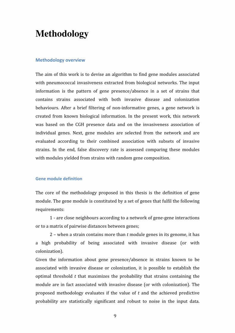
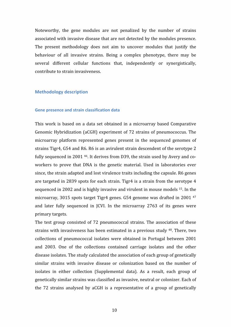
contrasts with the initial distribution (Figure 1 and 2).
Figure 1 -‐ Gene distribution before gene and strain selection.
Figure 2 -‐ Gene Distribution after gene and strain selection.
13
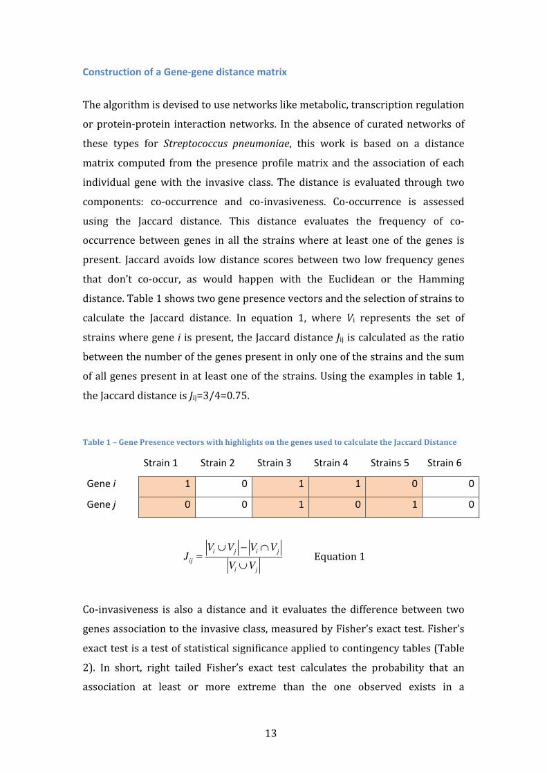
Construction of a Gene-‐gene distance matrix
The algorithm is devised to use networks like metabolic, transcription regulation
or protein-‐protein interaction networks. In the absence of curated networks of
these types for Streptococcus pneumoniae, this work is based on a distance
matrix computed from the presence profile matrix and the association of each
individual gene with the invasive class. The distance is evaluated through two
components: co-‐occurrence and co-‐invasiveness. Co-‐occurrence is assessed
using the Jaccard distance. This distance evaluates the frequency of co-‐
occurrence between genes in all the strains where at least one of the genes is
present. Jaccard avoids low distance scores between two low frequency genes
that don’t co-‐occur, as would happen with the Euclidean or the Hamming
distance. Table 1 shows two gene presence vectors and the selection of strains to
calculate the Jaccard distance. In equation 1, where Vi represents the set of
strains where gene i is present, the Jaccard distance Jij is calculated as the ratio
between the number of the genes present in only one of the strains and the sum
of all genes present in at least one of the strains. Using the examples in table 1,
the Jaccard distance is Jij=3/4=0.75.
Table 1 – Gene Presence vectors with highlights on the genes used to calculate the Jaccard Distance
Strain 1 Strain 2 Strain 3 Strain 4 Strains 5 Strain 6
Gene i 1 0 1 1 0 0
Gene j 0 0 1 0 1 0
Jij =Vi ∪Vj − Vi ∩Vj
Vi ∪Vj
Equation 1
Co-‐invasiveness is also a distance and it evaluates the difference between two
genes association to the invasive class, measured by Fisher’s exact test. Fisher’s
exact test is a test of statistical significance applied to contingency tables (Table
2). In short, right tailed Fisher’s exact test calculates the probability that an
association at least or more extreme than the one observed exists in a
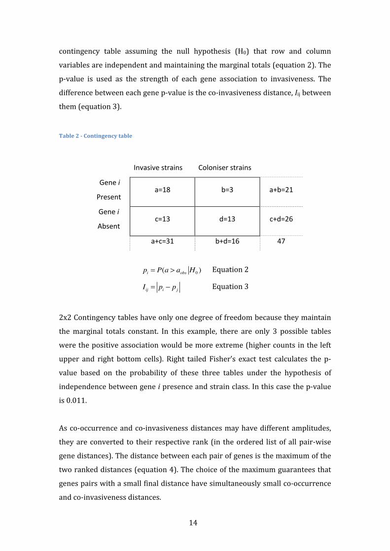
14
contingency table assuming the null hypothesis (H0) that row and column
variables are independent and maintaining the marginal totals (equation 2). The
p-‐value is used as the strength of each gene association to invasiveness. The
difference between each gene p-‐value is the co-‐invasiveness distance, Iij between
them (equation 3).
Table 2 -‐ Contingency table
Invasive strains Coloniser strains
Gene i
Present a=18 b=3 a+b=21
Gene i
Absent c=13 d=13 c+d=26
a+c=31 b+d=16 47
pi = P(a > aobs H0 ) Equation 2
Iij = pi − pj Equation 3
2x2 Contingency tables have only one degree of freedom because they maintain
the marginal totals constant. In this example, there are only 3 possible tables
were the positive association would be more extreme (higher counts in the left
upper and right bottom cells). Right tailed Fisher’s exact test calculates the p-‐
value based on the probability of these three tables under the hypothesis of
independence between gene i presence and strain class. In this case the p-‐value
is 0.011.
As co-‐occurrence and co-‐invasiveness distances may have different amplitudes,
they are converted to their respective rank (in the ordered list of all pair-‐wise
gene distances). The distance between each pair of genes is the maximum of the
two ranked distances (equation 4). The choice of the maximum guarantees that
genes pairs with a small final distance have simultaneously small co-‐occurrence
and co-‐invasiveness distances.
15
Dij =max(rank(Jij ), rank(Iij )) Equation 4
Selection of gene modules
An invasive gene module is defined as a group of genes m (that are related
according to a given network) with a determined presence threshold t, such that
strains that have in their genome more than t genes belonging to the module
tend to be invasive strains. When a strain has more that t genes of the module,
the strain is said to have the module present. A similar definition can be stated
for colonizer gene modules, but for simplicity, in the remaining methodology
section only invasive gene modules are referred.
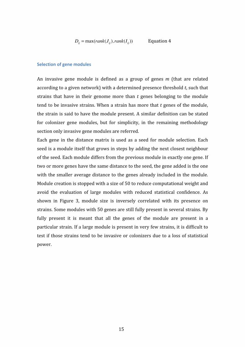
Each gene in the distance matrix is used as a seed for module selection. Each
seed is a module itself that grows in steps by adding the next closest neighbour
of the seed. Each module differs from the previous module in exactly one gene. If
two or more genes have the same distance to the seed, the gene added is the one
with the smaller average distance to the genes already included in the module.
Module creation is stopped with a size of 50 to reduce computational weight and
avoid the evaluation of large modules with reduced statistical confidence. As
shown in Figure 3, module size is inversely correlated with its presence on
strains. Some modules with 50 genes are still fully present in several strains. By
fully present it is meant that all the genes of the module are present in a
particular strain. If a large module is present in very few strains, it is difficult to
test if those strains tend to be invasive or colonizers due to a loss of statistical
power.
16
Figure 3 -‐ Influence of module length on module presence in strains
Gene modules are evaluated through three successive filtering steps. The first is
a runs test, which tests for the random distribution of strain classes (invasive or
colonizer). Strains are organized as an ordered list based on the number of
module genes present. Each strain is labelled as invasive or colonizer. The runs
test statistic R is the number of times the strain label changes when the list read
from start to end. Runs test has the null hypothesis that labels are randomly
distributed, where R has intermediate values. In the left tailed version applied in
this work, the alternative hypothesis is that labels are clustered together, which
produces low R values. The probability of observing R runs is obtained according
to the combinatorial expressions in equations 5 (when R is even) and 6 (when R
is odd), were m is the number of invasive labels and n is the number of colonizer
labels. The parenthesis notation in equations 5 and 6, with an upper number u
and a lower number l, refer to the number of possible combinations of the u
elements in subgroups with l elements. Statistical significance is obtained
through equation 7, and is required to be equal or lower than 0.05 for each
module to be maintained.
P(R = 2x) =2 m −1
x −1⎛⎝⎜
⎞⎠⎟
n −1x −1
⎛⎝⎜
⎞⎠⎟
n +mn
⎛⎝⎜
⎞⎠⎟
Equation 6
17
P(R = 2x +1) =
m −1x −1
⎛⎝⎜
⎞⎠⎟
n −1x
⎛⎝⎜
⎞⎠⎟+ m −1
x⎛⎝⎜
⎞⎠⎟
n −1x −1
⎛⎝⎜
⎞⎠⎟
n +mn
⎛⎝⎜
⎞⎠⎟
Equation 7
P(R ≤ Robs ) = P(R = r)r=2
r=Robs
∑ Equation 8
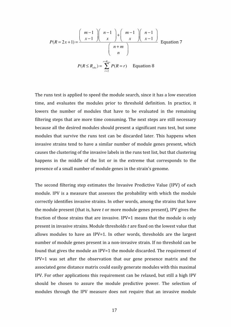
The runs test is applied to speed the module search, since it has a low execution
time, and evaluates the modules prior to threshold definition. In practice, it
lowers the number of modules that have to be evaluated in the remaining
filtering steps that are more time consuming. The next steps are still necessary
because all the desired modules should present a significant runs test, but some
modules that survive the runs test can be discarded later. This happens when
invasive strains tend to have a similar number of module genes present, which
causes the clustering of the invasive labels in the runs test list, but that clustering
happens in the middle of the list or in the extreme that corresponds to the
presence of a small number of module genes in the strain’s genome.
The second filtering step estimates the Invasive Predictive Value (IPV) of each
module. IPV is a measure that assesses the probability with which the module
correctly identifies invasive strains. In other words, among the strains that have
the module present (that is, have t or more module genes present), IPV gives the
fraction of those strains that are invasive. IPV=1 means that the module is only
present in invasive strains. Module thresholds t are fixed on the lowest value that
allows modules to have an IPV=1. In other words, thresholds are the largest
number of module genes present in a non-‐invasive strain. If no threshold can be
found that gives the module an IPV=1 the module discarded. The requirement of
IPV=1 was set after the observation that our gene presence matrix and the
associated gene distance matrix could easily generate modules with this maximal
IPV. For other applications this requirement can be relaxed, but still a high IPV
should be chosen to assure the module predictive power. The selection of
modules through the IPV measure does not require that an invasive module
18
should be present in the majority of invasive strains, only that it is present
mainly in invasive strains. This allows the detection of modules that may
contribute to the invasiveness of some strains, but not others, which is coherent
with the biological perspective that invasiveness is a complex phenotype that can
be influenced by different cellular pathways.
In the third step the significance of threshold t is evaluated. For this purpose a
one tailed test was developed that calculates the probability of the threshold to
be as the one observed or lower. This test is based in the classical theory of
extreme value statistics 49. First, a list with the number of module genes present
in every strain is obtained, from which the frequency of each number of
presences is calculated. This frequency f(n) represents the probability of a strain
having n genes from the module m (equation 9). The corresponding cumulative
frequency F(n) is the probability of a strain having at the most n genes (equation
10).
f (n) = P( gij = n : i∈m)i∑ Equation 9
F(n) = f (m)m=1
n
∑ = P( gij ≤ n : i∈m)i∑ Equation 10
FS (n) = F(n)S Equation 11
The probability FS(n) of S independent strains having each at the most n genes is
the cumulative frequency F(n) powered to S (equation 11). A threshold of t
means that among S colonizer strains, one strain has t module genes and the
remaining strains have less than t module genes. The probability FS(t) includes
this case but also sets of S colonizer strains where all have less than t module
genes. The latter cases are all included in FS(t-‐1). Therefore, the probability of
observing a threshold t is determined by the difference of those two
probabilities. The p-‐value for threshold significance is obtained by the sum of the
probabilities of thresholds lower or equal to the one observed (equation 12).
Statistical significance of 0.05 is required for each module to be maintained.
19
p(tobs ) = Fs (t)− FS (t −1)t=1
tobs
∑ Equation 12
Module trimming
The initial analysis of the modules resulting from the described selection process
showed that in some cases, the last genes added to the growing modules where
highly frequent genes. These genes did not contribute significantly to the
predictive power of the module, although they did not prevent correct
predictions, since they were also present in the invasive strains that contained
the remaining genes of the module. This observation suggested the need for a
module trimming step that would remove non-‐informative genes. Knowing that
each module is present in a subset of the invasive strains, a Fisher’s right tailed
exact test was used to assess the individual association of module genes with
their presence in that subset of invasive strains. Genes that were not significantly
associated with the subset of invasive strains were excluded from the module.
After this trimming, the threshold is recalculated and re-‐evaluated as described
above. From this step forward we select only the largest module among the ones
generated from the same seed.
False discovery rate
To correct for multiple testing, 1000 random gene presence matrices were used
as inputs. Random matrices maintain the number of genes and strains as the
original matrix and also maintain each gene frequency. Among other module
properties, the module length distribution obtained with the random matrices
presented values that tended to be lower than the module lengths obtained from
the original gene presence matrix (Figure 4). This means that in the absence of a
real association between the gene presence and the invasive classification of the
strains it is rare to find large gene modules that are only present in invasive
strains. Using this property, a module size cut-‐off s was applied, such that only
modules with k or more genes were selected. For every possible cut-‐off value the
False Discovery Rate (FDR) was estimated as the ratio between the mean
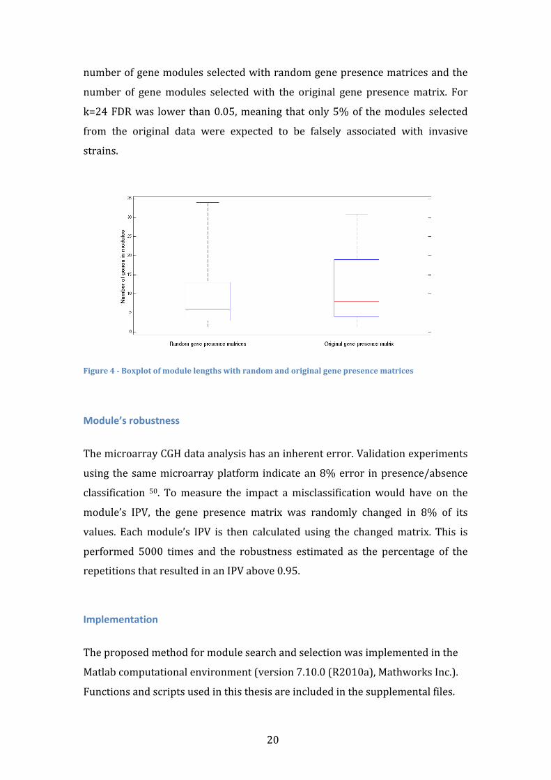
20
number of gene modules selected with random gene presence matrices and the
number of gene modules selected with the original gene presence matrix. For
k=24 FDR was lower than 0.05, meaning that only 5% of the modules selected
from the original data were expected to be falsely associated with invasive
strains.
Figure 4 -‐ Boxplot of module lengths with random and original gene presence matrices
Module’s robustness
The microarray CGH data analysis has an inherent error. Validation experiments
using the same microarray platform indicate an 8% error in presence/absence
classification 50. To measure the impact a misclassification would have on the
module’s IPV, the gene presence matrix was randomly changed in 8% of its
values. Each module’s IPV is then calculated using the changed matrix. This is
performed 5000 times and the robustness estimated as the percentage of the
repetitions that resulted in an IPV above 0.95.
Implementation
The proposed method for module search and selection was implemented in the
Matlab computational environment (version 7.10.0 (R2010a), Mathworks Inc.).
Functions and scripts used in this thesis are included in the supplemental files.
21
Results and Discussion
The search for coherent gene modules has wielded 30 modules, from which 26
are unique. Each module components and evaluated properties are presented in
a supplemental file.
Table 3 – Evolution of the number of modules through the selection steps
Modules Seeds with modules
Initial 64750 1295
Significant runs test 16989 1048
IPV=1 6665 364
Significant threshold 3534 197
Trimmed 3018 190
FDR corrected -‐ 30
Unique -‐ 26
The runs test proved to be an efficient filter to reduce the computation time in
subsequent steps (Table 3). Among the modules selected in the runs test, over
one third had a threshold value that allowed IPV=1. The fact that these modules
were selected in the runs test and the adaptability of threshold definition enables
such elevated fraction. Similarly, more than half of the modules with IPV=1
presented a statistically significant threshold value. The multiple testing
correction showed that modules with more than 24 genes are unlikely
(FDR<0.05) to result from random gene presence matrices. Still, random
datasets were able to yield modules more efficiently than expected. One reason
for this observation is the fact that the gene distance matrix is created using the
same data that is later used to evaluate the modules. This way, it is easier to
successfully create gene modules with IPV=1, significant in the runs test and in
the threshold test. If, during the analysis of random datasets, the original gene
distance matrix is used, instead of redefining it for each random gene presence
matrix, the number of modules that survived the three filtering steps would
22
strongly decrease. Using established biological networks, that encode
information that is obtained independently of the gene presence matrix, would
also avoid this situation. Additionally, it would have an impact on the
composition of selected modules, facilitating the interpretation of the association
between a biological function or pathway and invasiveness.
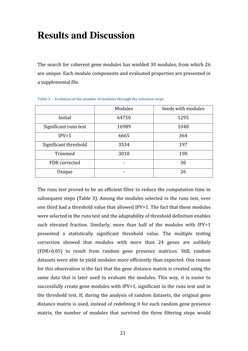
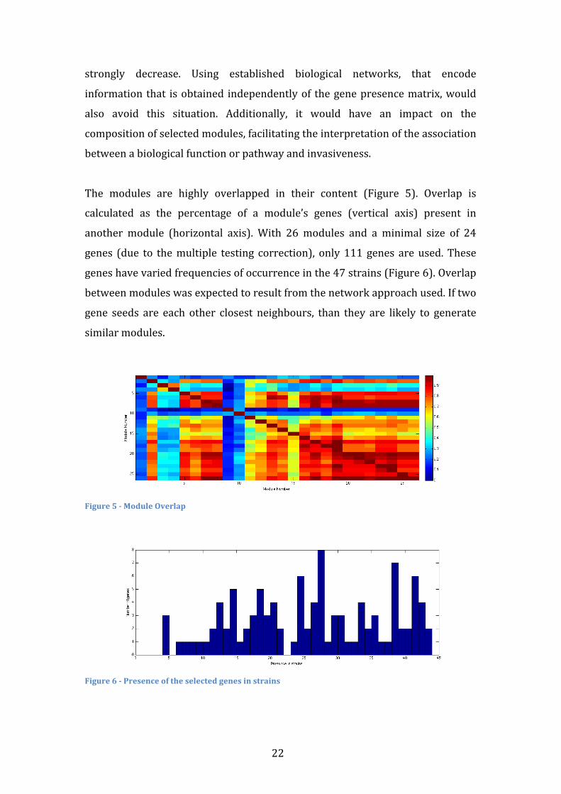
The modules are highly overlapped in their content (Figure 5). Overlap is
calculated as the percentage of a module’s genes (vertical axis) present in
another module (horizontal axis). With 26 modules and a minimal size of 24
genes (due to the multiple testing correction), only 111 genes are used. These
genes have varied frequencies of occurrence in the 47 strains (Figure 6). Overlap
between modules was expected to result from the network approach used. If two
gene seeds are each other closest neighbours, than they are likely to generate
similar modules.
Figure 5 -‐ Module Overlap
Figure 6 -‐ Presence of the selected genes in strains
23
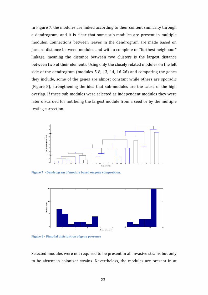
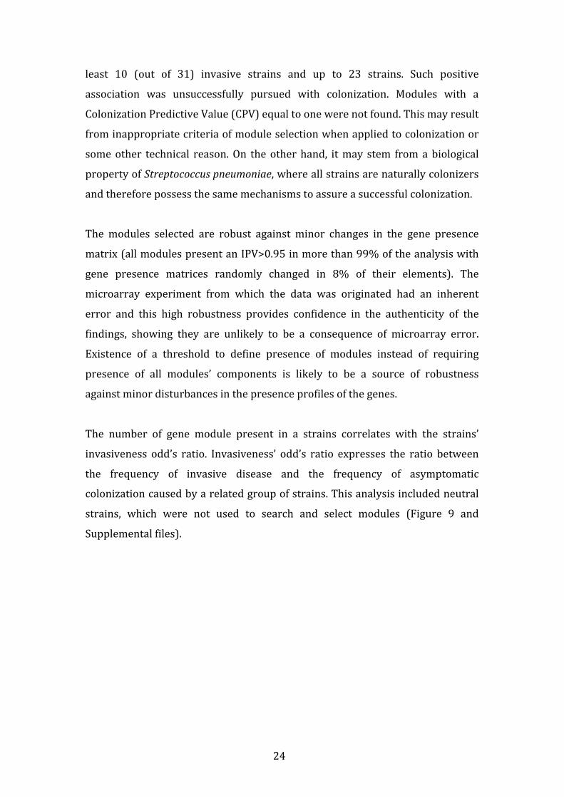
In Figure 7, the modules are linked according to their content similarity through
a dendrogram, and it is clear that some sub-‐modules are present in multiple
modules. Connections between leaves in the dendrogram are made based on
Jaccard distance between modules and with a complete or “furthest neighbour”
linkage, meaning the distance between two clusters is the largest distance
between two of their elements. Using only the closely related modules on the left
side of the dendrogram (modules 5-‐8, 13, 14, 16-‐26) and comparing the genes
they include, some of the genes are almost constant while others are sporadic
(Figure 8), strengthening the idea that sub-‐modules are the cause of the high
overlap. If these sub-‐modules were selected as independent modules they were
later discarded for not being the largest module from a seed or by the multiple
testing correction.
Figure 7 -‐ Dendrogram of module based on gene composition.
Figure 8 -‐ Bimodal distribution of gene presence
Selected modules were not required to be present in all invasive strains but only
to be absent in colonizer strains. Nevertheless, the modules are present in at
24
least 10 (out of 31) invasive strains and up to 23 strains. Such positive
association was unsuccessfully pursued with colonization. Modules with a
Colonization Predictive Value (CPV) equal to one were not found. This may result
from inappropriate criteria of module selection when applied to colonization or
some other technical reason. On the other hand, it may stem from a biological
property of Streptococcus pneumoniae, where all strains are naturally colonizers
and therefore possess the same mechanisms to assure a successful colonization.
The modules selected are robust against minor changes in the gene presence
matrix (all modules present an IPV>0.95 in more than 99% of the analysis with
gene presence matrices randomly changed in 8% of their elements). The
microarray experiment from which the data was originated had an inherent
error and this high robustness provides confidence in the authenticity of the
findings, showing they are unlikely to be a consequence of microarray error.
Existence of a threshold to define presence of modules instead of requiring
presence of all modules’ components is likely to be a source of robustness
against minor disturbances in the presence profiles of the genes.
The number of gene module present in a strains correlates with the strains’
invasiveness odd’s ratio. Invasiveness’ odd’s ratio expresses the ratio between
the frequency of invasive disease and the frequency of asymptomatic
colonization caused by a related group of strains. This analysis included neutral
strains, which were not used to search and select modules (Figure 9 and
Supplemental files).
25
Figure 9 – Correlation between individual module gene presence and invasiveness odd’s ratio
Spearman correlation analysis was used to test if there is a monotonic
relationship between the two variables (number of module genes present and
invasiveness odd’s ratio). Correlation values (r) range from -‐1 (inverse
correlation) to 1 (positive correlation). When the variables are not related, the r
is expected to be 0. For all the modules the correlation value was positive and
statistically greater than 0. The majority (20) of the modules present strong
correlations (0.55-‐0.70). These correlation values are considered strong due
both to the origin of the invasiveness odd’s ratios and to the complexity of the
invasive phenotype. The odd’s ratios were estimated in observational
epidemiological studies, with high associated variability, and the invasiveness
can be influenced by so many variables that it would be difficult to achieve very
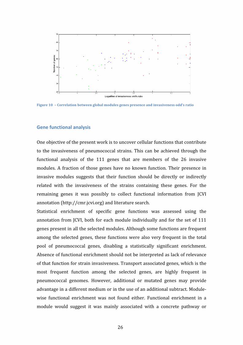
high correlations. Using the global number of genes that belong to any module
present in each strain also shows a significant and strong correlation with the
invasiveness odd’s ratio (rho=0.6160, p<10-‐8), even when considering the
neutral strains (Figure 10). Correlation with strains’ odd’s ratio is an important
confirmation of the modules validity. Neutral strains were not used as input data,
nor were the invasive and colonizer strains odd’s ratio and therefore the
algorithm doesn’t influence the correlation of modules gene presence with the
odd’s ratio.
26
Figure 10 – Correlation between global modules genes presence and invasiveness odd’s ratio
Gene functional analysis
One objective of the present work is to uncover cellular functions that contribute
to the invasiveness of pneumococcal strains. This can be achieved through the
functional analysis of the 111 genes that are members of the 26 invasive
modules. A fraction of those genes have no known function. Their presence in
invasive modules suggests that their function should be directly or indirectly
related with the invasiveness of the strains containing these genes. For the
remaining genes it was possibly to collect functional information from JCVI
annotation (http://cmr.jcvi.org) and literature search.
Statistical enrichment of specific gene functions was assessed using the
annotation from JCVI, both for each module individually and for the set of 111
genes present in all the selected modules. Although some functions are frequent
among the selected genes, these functions were also very frequent in the total
pool of pneumococcal genes, disabling a statistically significant enrichment.
Absence of functional enrichment should not be interpreted as lack of relevance
of that function for strain invasiveness. Transport associated genes, which is the
most frequent function among the selected genes, are highly frequent in
pneumococcal genomes. However, additional or mutated genes may provide
advantage in a different medium or in the use of an additional subtract. Module-‐
wise functional enrichment was not found either. Functional enrichment in a
module would suggest it was mainly associated with a concrete pathway or
27
defined biological function. Here, lack of functional enrichment suggests that
modules include several different functions that may need to act synergistically
to impact strain invasiveness.
The 111 genes present in all the modules were manually assigned to 17
functional classes that are presented and discussed in the following sections.
Anaerobic Response Table 4 -‐ Genes associated with anaerobic response
Spots Primary targets Gene symbol Description
153 SP0202 NrdD Anaerobic Ribonucleoside-‐
Tryphosphate reductase
156 SP0205 NrdG Anaerobic Ribonucleoside-‐
Tryphosphate reductase activating
protein
2506 SPN08064 -‐ Anaerobic Nucleotide Reductase
Ribonucleotide reductases (RNR) provide all living organisms with the
deoxyribonucleoside triphosphates required for DNA synthesis 51. Class III RNRs,
such as nrdD, are homodimeric and contain a stable oxygen-‐sensitive glycyl
radical formed by S-‐adenosylmethionine (SAM) and a second protein, nrdG, that
it is not required for catalysis once the radical has been generated 52. This class is
functional only under strict anaerobic conditions, contrasting with Class I – strict
aerobic conditions – and with Class II – both aerobic and anaerobic. Importance
of nrdD to virulence has been reported in several organisms such as E. Coli 53 , S.
Aureus 54, P. Aeruginosa 53 and S. Sanguinis 55, but studies in S. Pneumoniae have
not been conducted so far.
28
Choline binding proteins Table 5 -‐ Genes associated with Choline binding proteins
Spots Primary targets Gene symbol Description
2861 SPN14033 PcpC Choline binding protein F
3613 spr1995 PspC Choline binding protein A
Streptococcus pneumoniae possesses a family of proteins that bind the
phosphocholine present in the membrane and the cell wall 56,57. The choline-‐
binding proteins (CBP) of pneumococci and other gram positive organisms
contain structurally similar choline-‐binding domains, which are composed of
multiple tandem aminoacid repeats. CBPs have been reported to contribute to
adherence of pneumococci through effects on surface charge 57,58.
PspC (Pneumococcal surface protein), also known as CbpA, is a highly
polymorphic protein and its impact on pneumococcal virulence varies between
strains 59,60. Of the many biological functions that have been attributed to PspC,
its role in adherence is one of the most studied 61. While PspC does not
exclusively govern pneumococcal adherence, its absence significantly reduces
adherence and invasion of human cells. PspC also binds the secretory component
of human secretory immunoglobulin A and human factor H 61, as well as
complement component C3 58,62.
PcpC is a paralogue of CbpF 63. A study on cell adherence showed that mutants
deficient in only CbpF had no observable phenotype 64. On the other hand, it
appears to be important to counteract cellular lysis, as a negative regulator of
LytC activity in S. Pneumoniae. Deletion of PcpC was found to promote a
significant increase in competence-‐induced lysis 63.
29
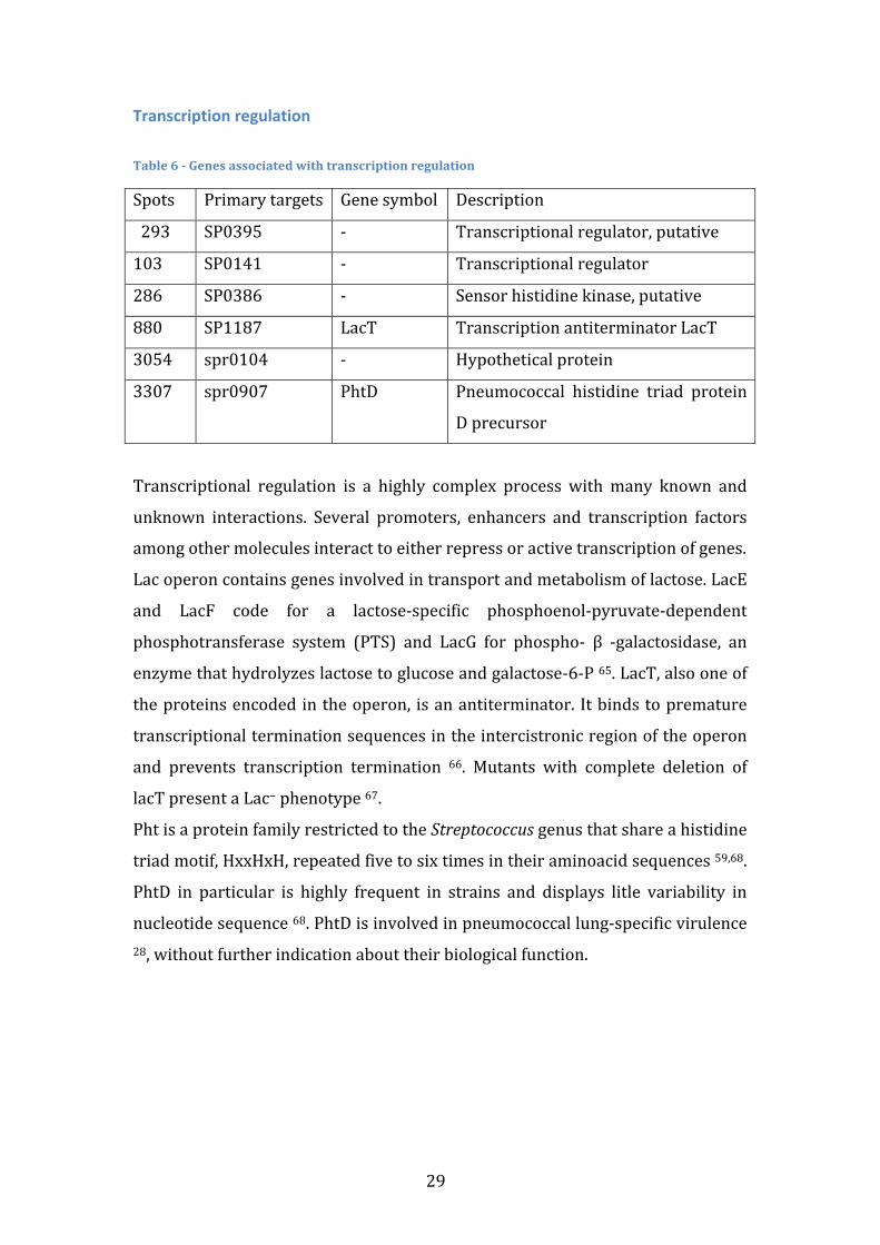
Transcription regulation Table 6 -‐ Genes associated with transcription regulation
Spots Primary targets Gene symbol Description
293
SP0395 -‐ Transcriptional regulator, putative
103 SP0141 -‐ Transcriptional regulator
286 SP0386 -‐ Sensor histidine kinase, putative
880 SP1187 LacT Transcription antiterminator LacT
3054 spr0104 -‐ Hypothetical protein
3307 spr0907 PhtD Pneumococcal histidine triad protein
D precursor
Transcriptional regulation is a highly complex process with many known and
unknown interactions. Several promoters, enhancers and transcription factors
among other molecules interact to either repress or active transcription of genes.
Lac operon contains genes involved in transport and metabolism of lactose. LacE
and LacF code for a lactose-‐specific phosphoenol-‐pyruvate-‐dependent
phosphotransferase system (PTS) and LacG for phospho-‐ β -‐galactosidase, an
enzyme that hydrolyzes lactose to glucose and galactose-‐6-‐P 65. LacT, also one of
the proteins encoded in the operon, is an antiterminator. It binds to premature
transcriptional termination sequences in the intercistronic region of the operon
and prevents transcription termination 66. Mutants with complete deletion of
lacT present a Lac− phenotype 67.
Pht is a protein family restricted to the Streptococcus genus that share a histidine
triad motif, HxxHxH, repeated five to six times in their aminoacid sequences 59,68.
PhtD in particular is highly frequent in strains and displays litle variability in
nucleotide sequence 68. PhtD is involved in pneumococcal lung-‐specific virulence 28, without further indication about their biological function.
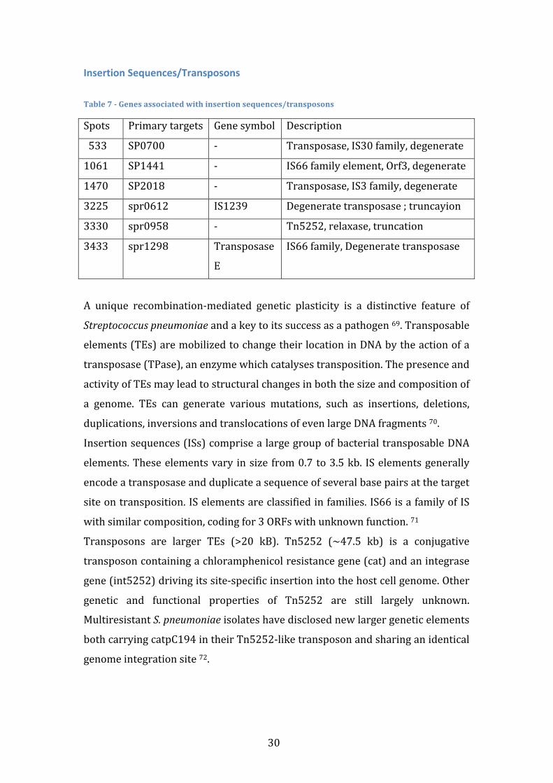
30
Insertion Sequences/Transposons Table 7 -‐ Genes associated with insertion sequences/transposons
Spots Primary targets Gene symbol Description
533
SP0700 -‐ Transposase, IS30 family, degenerate
1061 SP1441 -‐ IS66 family element, Orf3, degenerate
1470 SP2018 -‐ Transposase, IS3 family, degenerate
3225 spr0612 IS1239 Degenerate transposase ; truncayion
3330 spr0958 -‐ Tn5252, relaxase, truncation
3433 spr1298 Transposase
E
IS66 family, Degenerate transposase
A unique recombination-‐mediated genetic plasticity is a distinctive feature of
Streptococcus pneumoniae and a key to its success as a pathogen 69. Transposable
elements (TEs) are mobilized to change their location in DNA by the action of a
transposase (TPase), an enzyme which catalyses transposition. The presence and
activity of TEs may lead to structural changes in both the size and composition of
a genome. TEs can generate various mutations, such as insertions, deletions,
duplications, inversions and translocations of even large DNA fragments 70.
Insertion sequences (ISs) comprise a large group of bacterial transposable DNA
elements. These elements vary in size from 0.7 to 3.5 kb. IS elements generally
encode a transposase and duplicate a sequence of several base pairs at the target
site on transposition. IS elements are classified in families. IS66 is a family of IS
with similar composition, coding for 3 ORFs with unknown function. 71
Transposons are larger TEs (>20 kB). Tn5252 (~47.5 kb) is a conjugative
transposon containing a chloramphenicol resistance gene (cat) and an integrase
gene (int5252) driving its site-‐specific insertion into the host cell genome. Other
genetic and functional properties of Tn5252 are still largely unknown.
Multiresistant S. pneumoniae isolates have disclosed new larger genetic elements
both carrying catpC194 in their Tn5252-‐like transposon and sharing an identical
genome integration site 72.
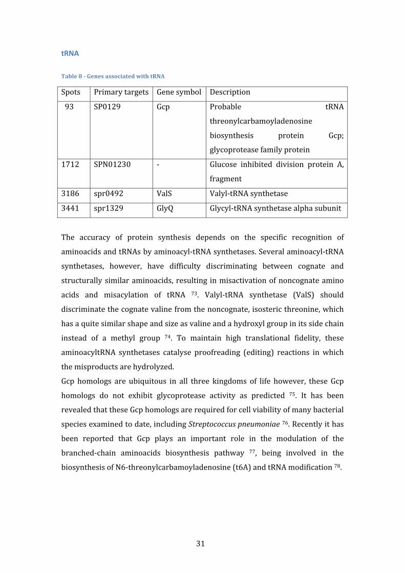
31
tRNA Table 8 -‐ Genes associated with tRNA
Spots Primary targets Gene symbol Description
93
SP0129 Gcp Probable tRNA
threonylcarbamoyladenosine
biosynthesis protein Gcp;
glycoprotease family protein
1712 SPN01230 -‐
Glucose inhibited division protein A,
fragment
3186 spr0492 ValS Valyl-‐tRNA synthetase
3441 spr1329 GlyQ Glycyl-‐tRNA synthetase alpha subunit
The accuracy of protein synthesis depends on the specific recognition of
aminoacids and tRNAs by aminoacyl-‐tRNA synthetases. Several aminoacyl-‐tRNA
synthetases, however, have difficulty discriminating between cognate and
structurally similar aminoacids, resulting in misactivation of noncognate amino
acids and misacylation of tRNA 73. Valyl-‐tRNA synthetase (ValS) should
discriminate the cognate valine from the noncognate, isosteric threonine, which
has a quite similar shape and size as valine and a hydroxyl group in its side chain
instead of a methyl group 74. To maintain high translational fidelity, these
aminoacyltRNA synthetases catalyse proofreading (editing) reactions in which
the misproducts are hydrolyzed.
Gcp homologs are ubiquitous in all three kingdoms of life however, these Gcp
homologs do not exhibit glycoprotease activity as predicted 75. It has been
revealed that these Gcp homologs are required for cell viability of many bacterial
species examined to date, including Streptococcus pneumoniae 76. Recently it has
been reported that Gcp plays an important role in the modulation of the
branched-‐chain aminoacids biosynthesis pathway 77, being involved in the
biosynthesis of N6-‐threonylcarbamoyladenosine (t6A) and tRNA modification 78.
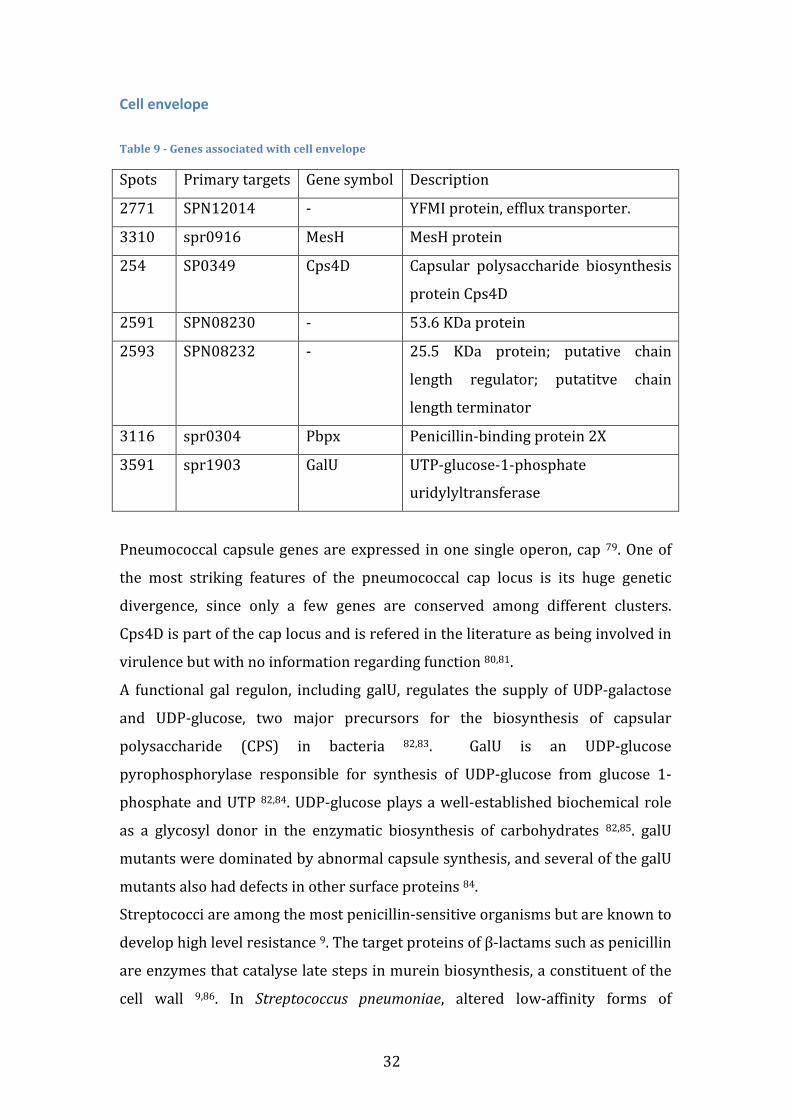
32
Cell envelope Table 9 -‐ Genes associated with cell envelope
Spots Primary targets Gene symbol Description
2771 SPN12014 -‐ YFMI protein, efflux transporter.
3310 spr0916 MesH MesH protein
254 SP0349 Cps4D Capsular polysaccharide biosynthesis
protein Cps4D
2591 SPN08230 -‐ 53.6 KDa protein
2593 SPN08232 -‐
25.5 KDa protein; putative chain
length regulator; putatitve chain
length terminator
3116 spr0304 Pbpx Penicillin-‐binding protein 2X
3591 spr1903 GalU UTP-‐glucose-‐1-‐phosphate
uridylyltransferase
Pneumococcal capsule genes are expressed in one single operon, cap 79. One of
the most striking features of the pneumococcal cap locus is its huge genetic
divergence, since only a few genes are conserved among different clusters.
Cps4D is part of the cap locus and is refered in the literature as being involved in
virulence but with no information regarding function 80,81.
A functional gal regulon, including galU, regulates the supply of UDP-‐galactose
and UDP-‐glucose, two major precursors for the biosynthesis of capsular
polysaccharide (CPS) in bacteria 82,83. GalU is an UDP-‐glucose
pyrophosphorylase responsible for synthesis of UDP-‐glucose from glucose 1-‐
phosphate and UTP 82,84. UDP-‐glucose plays a well-‐established biochemical role
as a glycosyl donor in the enzymatic biosynthesis of carbohydrates 82,85. galU
mutants were dominated by abnormal capsule synthesis, and several of the galU
mutants also had defects in other surface proteins 84.
Streptococci are among the most penicillin-‐sensitive organisms but are known to
develop high level resistance 9. The target proteins of β-‐lactams such as penicillin
are enzymes that catalyse late steps in murein biosynthesis, a constituent of the
cell wall 9,86. In Streptococcus pneumoniae, altered low-‐affinity forms of
33
penicillin-‐binding proteins (PBPs) 1a, 2x, and 2b are found invariably in
penicillin-‐resistant clinical isolates 69,87. Several mutations in one PBP may be
required to cause a substantial decrease in penicillin affinity, and more than one
PBP may have to be changed into low affinity variants in order to achieve high
resistance levels. In Streptococcus pneumoniae, alterations in the PBP2x and
PBP2b confer low resistance and are the prerequisite for high levels of resistance 9,23.
Spermidine metabolism Table 10 -‐ Genes associated with spermidine metabolism
Spots Primary targets Gene symbol Description
683 SP0918 SpeE Spermidine synthase
685 SP0920 NspC Carboxynorspermidine decarboxylase
Polyamines, such as spermidine and norspermidine, are small ubiquitous
aliphatic hydrocarbon molecule and polycationic at physiological pH 88.
Polyamines exist primarily as complexes with RNA molecules in prokaryotes and
are essential for efficient DNA replication, transcription and translation 89,90.
In the Streptococcus genus, choline-‐binding proteins are important virulence
factors. Pneumococci can substitute choline with polyamines, under laboratory
conditions, which then get incorporated in the bacterial cell wall 91. Additionally,
environmental polyamine acquisition may play an important role in
pneumococcal response to temperature, oxidative and pH stress. In
Streptococcus pneumoniae, polyamines have been linked to in vivo fitness,
pathogenesis and virulence factor expression 88,91. SP0918 and SP0920
synthetize spermidine and norspermidine, respectivily, and are therefore linked
to virulence.
34
Osmotic Regulation Table 11 -‐ Genes associated with osmotic regulation
Spots Primary targets Gene symbol Description
747 SP1010 MscL Large conductance mechanosensitive
channel
3516 spr1604 AqpZ Aquaporin Z -‐ water channel
Aqueous environments are prone to drastic changes in extracellular osmolality 92-‐94. In order to respond to osmotic pressure shifts, Streptococcus pneumoniae
has membrane proteins that change its permeability to water or ions. Besides
the osmotic changes in aqueous mediums, pneumococcus also suffers from
osmotic shifts when invades a new tissue and, therefore, response to osmotic
pressure shifts are essential for invasive traits.
Aquaporins belong to a large family of proteins that increase the rate of diffusion
of water and glycerol across cell membranes 95. Aquaporin Z in particular is a
water channel protein implicated in important processes such as osmoregulation
and volume expansion in rapidly growing cells 96. The aqpZ gene was found to be
osmotically regulated, as its expression was significantly increased in steady-‐
state hypoosmotic conditions, but greatly reduced in hyperosmolar media 95. The
need for an aquaporin to adapt to hypoosmotic stresses has been regarded as
puzzling, because the presence of water channels may result in an excessive
build-‐up of turgor pressure, which may lead to ruptures of the cell wall and
consequent cell death 96. AqpZ-‐like proteins seem to be necessary for the
virulence expressed by some pathogenic bacteria 96.
MscL is a mechanosensitive channel with low ion selectivity and high
conductance that opens in response to mechanically imposed membrane stress
or osmotic shifts 97. In response to changes in the lipid bilayer tension, the MscL
channel proteins gate and open transiently to form large aqueous pores, through
which both solutes and solvents can pass rapidly 94. Consequently, the
concentration of water-‐attracting solutes inside the cells is lowered, turgor is
reduced, and cell lysis is prevented. These channels are major routes for the
35
release of cytoplasmic solutes to achieve a rapid reduction of the turgor pressure
during the transition from media of high to low osmolality 98.
Co-‐enzymes Table 12 -‐ Genes associated with co-‐enzymes
Spots Primary targets Gene symbol Description
506 SP0665 -‐ Chorismate binding enzyme
2051 SPN04086 -‐ Hydroxyethylthiazole kinase
(EC 2.7.1.50)
3565
spr1783 KdtB
Phosphopantetheine
adenylyltransferase
Co-‐enzymes, consist of small organic molecules that many enzymes require as
cofactors to be catalytically active. Their impact on cellular physiology can be
profound as many different processes may be affected.
Chorismate is an important precursor of several biomolecules such as aromatic
aminoacids, vitamins K, ubiquinone and folic acid 99. It is of special importance as
it is the branch point in the biosynthesis pathway of the referred molecules.
Hydroxyethylthiazole kinase participates in thiamine metabolism, which is a
cofactor of several key enzymes, particularly involved in carbohydrate
metabolism 100.
KdtB is the phosphopantetheine adenylyltransferase enzyme, responsible for the
biosynthesis of Coenzyme A 101.
36
Proteolysis Table 13 -‐ Genes associated with Proteolysis
Spots Primary targets Gene symbol Description
3055 spr0105 -‐ Transporter, truncation
3056 spr0106 -‐ Transporter, truncation
1790 SPN02074 PepF Oligoendopeptidase F homolog (EC
3.4.24.-‐)
3222 spr0607 -‐ Dipeptidase
Streptococcus pneumoniae is a microorganism with multiple aminoacid
requirements and bacteria are often able to utilize peptides as a source of
aminoacids as well or better than utilizing the aminoacids themselves 102. This
way, enzymes with proteolytic activity play a major role in fulfilling the cell
aminoacid requirements. Spr0607 activity is highly specific to dipeptides and has
a putative role in protein synthesis and degradation 102. SPN02074 is an homolog
of the oligoendopeptidase F (pepF) which has collagenase activity. PepF is a
cytoplasmic endopeptidase that hydrolyses oligopeptides but cannot degrade
proteins103. An homologue enzyme in Group B Streptococci was found not to be a
collagenase and it has been suggested to be involved in degradation of peptides
transported to the cell’s interior. This would require a oligopeptide transport
system but nothing similar has yet been reported in pneumococcus 104. PepF
expression has been found to be increased during the competence state as a late
gene 21.
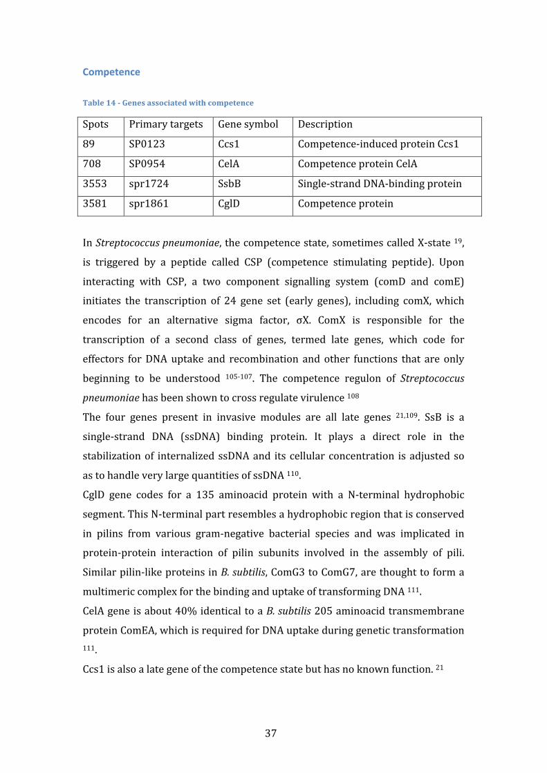
37
Competence Table 14 -‐ Genes associated with competence
Spots Primary targets Gene symbol Description
89 SP0123 Ccs1 Competence-‐induced protein Ccs1
708 SP0954 CelA Competence protein CelA
3553 spr1724 SsbB Single-‐strand DNA-‐binding protein
3581 spr1861 CglD Competence protein
In Streptococcus pneumoniae, the competence state, sometimes called X-‐state 19,
is triggered by a peptide called CSP (competence stimulating peptide). Upon
interacting with CSP, a two component signalling system (comD and comE)
initiates the transcription of 24 gene set (early genes), including comX, which
encodes for an alternative sigma factor, σX. ComX is responsible for the
transcription of a second class of genes, termed late genes, which code for
effectors for DNA uptake and recombination and other functions that are only
beginning to be understood 105-‐107. The competence regulon of Streptococcus
pneumoniae has been shown to cross regulate virulence 108
The four genes present in invasive modules are all late genes 21,109. SsB is a
single-‐strand DNA (ssDNA) binding protein. It plays a direct role in the
stabilization of internalized ssDNA and its cellular concentration is adjusted so
as to handle very large quantities of ssDNA 110.
CglD gene codes for a 135 aminoacid protein with a N-‐terminal hydrophobic
segment. This N-‐terminal part resembles a hydrophobic region that is conserved
in pilins from various gram-‐negative bacterial species and was implicated in
protein-‐protein interaction of pilin subunits involved in the assembly of pili.
Similar pilin-‐like proteins in B. subtilis, ComG3 to ComG7, are thought to form a
multimeric complex for the binding and uptake of transforming DNA 111.
CelA gene is about 40% identical to a B. subtilis 205 aminoacid transmembrane
protein ComEA, which is required for DNA uptake during genetic transformation 111.
Ccs1 is also a late gene of the competence state but has no known function. 21
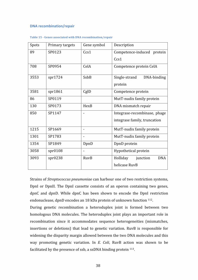
38
DNA recombination/repair Table 15 -‐ Genes associated with DNA recombination/repair
Spots Primary targets Gene symbol Description
89 SP0123 Ccs1 Competence-‐induced protein
Ccs1
708 SP0954 CelA Competence protein CelA
3553
spr1724 SsbB Single-‐strand DNA-‐binding
protein
3581 spr1861 CglD Competence protein
86 SP0119 MutT-‐nudix family protein
130 SP0173 HexB DNA mismatch repair
850 SP1147 -‐ Integrase-‐recombinase, phage
integrase family, truncation
1215 SP1669 -‐ MutT-‐nudix family protein
1301 SP1783 -‐ MutT-‐nudix family protein
1354 SP1849 DpnD DpnD protein
3058 spr0108 -‐ Hypothetical protein
3093 spr0238 RuvB Holliday junction DNA
helicase RuvB
Strains of Streptococcus pneumoniae can harbour one of two restriction systems,
DpnI or DpnII. The DpnI cassette consists of an operon containing two genes,
dpnC and dpnD. While dpnC has been shown to encode the DpnI restriction
endonuclease, dpnD encodes an 18 kDa protein of unknown function 112.
During genetic recombination a heteroduplex joint is formed between two
homologous DNA molecules. The heteroduplex joint plays an important role in
recombination since it accommodates sequence heterogeneities (mismatches,
insertions or deletions) that lead to genetic variation. RuvB is responsible for
widening the disparity margin allowed between the two DNA molecules and this
way promoting genetic variation. In E. Coli, RuvB action was shown to be
facilitated by the presence of ssb, a ssDNA binding protein 113.
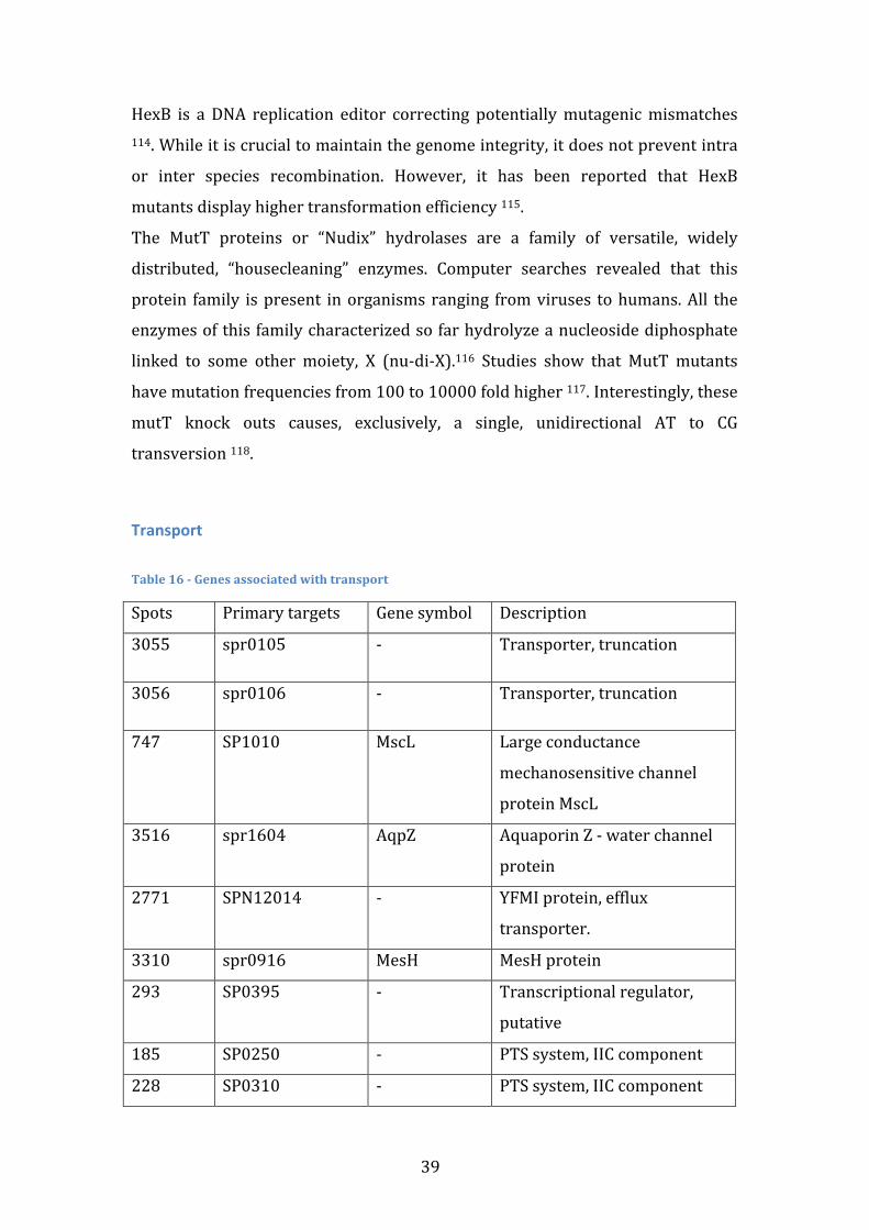
39
HexB is a DNA replication editor correcting potentially mutagenic mismatches 114. While it is crucial to maintain the genome integrity, it does not prevent intra
or inter species recombination. However, it has been reported that HexB
mutants display higher transformation efficiency 115.
The MutT proteins or “Nudix” hydrolases are a family of versatile, widely
distributed, “housecleaning” enzymes. Computer searches revealed that this
protein family is present in organisms ranging from viruses to humans. All the
enzymes of this family characterized so far hydrolyze a nucleoside diphosphate
linked to some other moiety, X (nu-‐di-‐X).116 Studies show that MutT mutants
have mutation frequencies from 100 to 10000 fold higher 117. Interestingly, these
mutT knock outs causes, exclusively, a single, unidirectional AT to CG
transversion 118.
Transport Table 16 -‐ Genes associated with transport
Spots Primary targets Gene symbol Description
3055 spr0105 -‐ Transporter, truncation
3056 spr0106 -‐ Transporter, truncation
747 SP1010 MscL Large conductance
mechanosensitive channel
protein MscL
3516 spr1604 AqpZ Aquaporin Z -‐ water channel
protein
2771 SPN12014 -‐ YFMI protein, efflux
transporter.
3310 spr0916 MesH MesH protein
293 SP0395 -‐ Transcriptional regulator,
putative
185 SP0250 -‐ PTS system, IIC component
228 SP0310 -‐ PTS system, IIC component
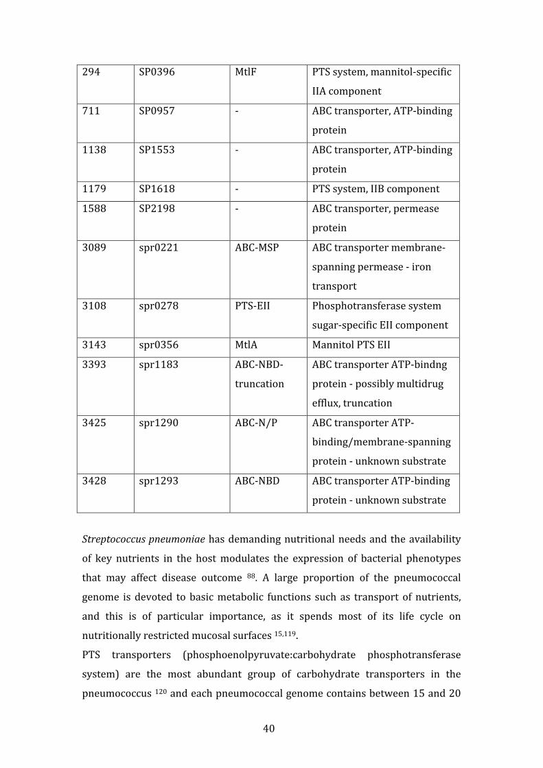
40
294 SP0396 MtlF PTS system, mannitol-‐specific
IIA component
711 SP0957 -‐ ABC transporter, ATP-‐binding
protein
1138 SP1553 -‐ ABC transporter, ATP-‐binding
protein
1179 SP1618 -‐ PTS system, IIB component
1588 SP2198 -‐ ABC transporter, permease
protein
3089 spr0221 ABC-‐MSP ABC transporter membrane-‐
spanning permease -‐ iron
transport
3108 spr0278 PTS-‐EII Phosphotransferase system
sugar-‐specific EII component
3143 spr0356 MtlA Mannitol PTS EII
3393 spr1183 ABC-‐NBD-‐
truncation
ABC transporter ATP-‐bindng
protein -‐ possibly multidrug
efflux, truncation
3425 spr1290 ABC-‐N/P ABC transporter ATP-‐
binding/membrane-‐spanning
protein -‐ unknown substrate
3428 spr1293 ABC-‐NBD ABC transporter ATP-‐binding
protein -‐ unknown substrate
Streptococcus pneumoniae has demanding nutritional needs and the availability
of key nutrients in the host modulates the expression of bacterial phenotypes
that may affect disease outcome 88. A large proportion of the pneumococcal
genome is devoted to basic metabolic functions such as transport of nutrients,
and this is of particular importance, as it spends most of its life cycle on
nutritionally restricted mucosal surfaces 15,119.
PTS transporters (phosphoenolpyruvate:carbohydrate phosphotransferase
system) are the most abundant group of carbohydrate transporters in the
pneumococcus 120 and each pneumococcal genome contains between 15 and 20
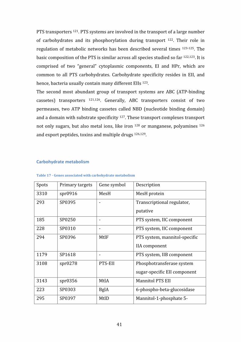
41
PTS transporters 121. PTS systems are involved in the transport of a large number
of carbohydrates and its phosphorylation during transport 122. Their role in
regulation of metabolic networks has been described several times 123-‐125. The
basic composition of the PTS is similar across all species studied so far 122,123. It is
comprised of two “general” cytoplasmic components, EI and HPr, which are
common to all PTS carbohydrates. Carbohydrate specificity resides in EII, and
hence, bacteria usually contain many different EIIs 123.
The second most abundant group of transport systems are ABC (ATP-‐binding
cassetes) transporters 121,126. Generally, ABC transporters consist of two
permeases, two ATP binding cassetes called NBD (nucleotide binding domain)
and a domain with substrate specificity 127. These transport complexes transport
not only sugars, but also metal ions, like iron 128 or manganese, polyamines 126
and export peptides, toxins and multiple drugs 126,129.
Carbohydrate metabolism Table 17 -‐ Genes associated with carbohydrate metabolism
Spots Primary targets Gene symbol Description
3310 spr0916 MesH MesH protein
293 SP0395 -‐ Transcriptional regulator,
putative
185 SP0250 -‐ PTS system, IIC component
228 SP0310 -‐ PTS system, IIC component
294 SP0396 MtlF PTS system, mannitol-‐specific
IIA component
1179 SP1618 -‐ PTS system, IIB component
3108 spr0278 PTS-‐EII Phosphotransferase system
sugar-‐specific EII component
3143 spr0356 MtlA Mannitol PTS EII
223 SP0303 BglA 6-‐phospho-‐beta-‐glucosidase
295 SP0397 MtlD Mannitol-‐1-‐phosphate 5-‐
42
dehydrogenase
3247 spr0704 -‐ Hypothetical protein
Streptococcus pneumoniae is able to utilize at least thirty-‐two substrates as
carbon sources, including sugars like mannose, galactose and hyaluronic acid,
but also many food-‐borne monosaccharides, disaccharides and polysaccharides 121,124,125. This is a hallmark of a bacteria adapted to a wide variety of ecological
niches, each one providing a wide variety of carbon sources. The sequencing of
Tigr4’s genome showed that a large part of it is dedicated to the transport and
metabolism of sugars 15. When more than one substrate is available, the cell has
mechanisms to regulate its protein expression in order to use only one of them at
a time. CcpA 119,125,130 and RegM 131 are two of the most studied proteins to play
this regulatory role and its possible that SP0395 also participates in this process.
MtlD is a mannitol-‐1-‐phosphate 5-‐dehydrogenase. After mannitol is translocated
by the respective PTS and phosphorilated to mannitol-‐1-‐phosphate, MtlD
catalyses its conversion to fructose-‐6-‐phosphate 132.
BglA, also called celA, is a 6-‐phospho-‐beta-‐glucosidase. It belongs to the
cellobiose metabolism operon which also contains a PTS transporter, a putative
DNA-‐binding protein and two proteins of unknown function 133,134.
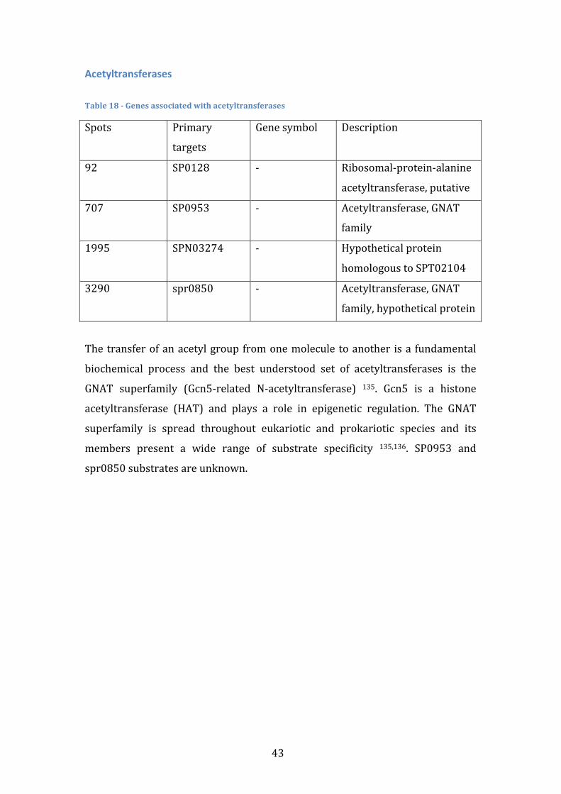
43
Acetyltransferases Table 18 -‐ Genes associated with acetyltransferases
Spots Primary
targets
Gene symbol Description
92 SP0128 -‐ Ribosomal-‐protein-‐alanine
acetyltransferase, putative
707 SP0953 -‐ Acetyltransferase, GNAT
family
1995 SPN03274 -‐ Hypothetical protein
homologous to SPT02104
3290 spr0850 -‐ Acetyltransferase, GNAT
family, hypothetical protein
The transfer of an acetyl group from one molecule to another is a fundamental
biochemical process and the best understood set of acetyltransferases is the
GNAT superfamily (Gcn5-‐related N-‐acetyltransferase) 135. Gcn5 is a histone
acetyltransferase (HAT) and plays a role in epigenetic regulation. The GNAT
superfamily is spread throughout eukariotic and prokariotic species and its
members present a wide range of substrate specificity 135,136. SP0953 and
spr0850 substrates are unknown.
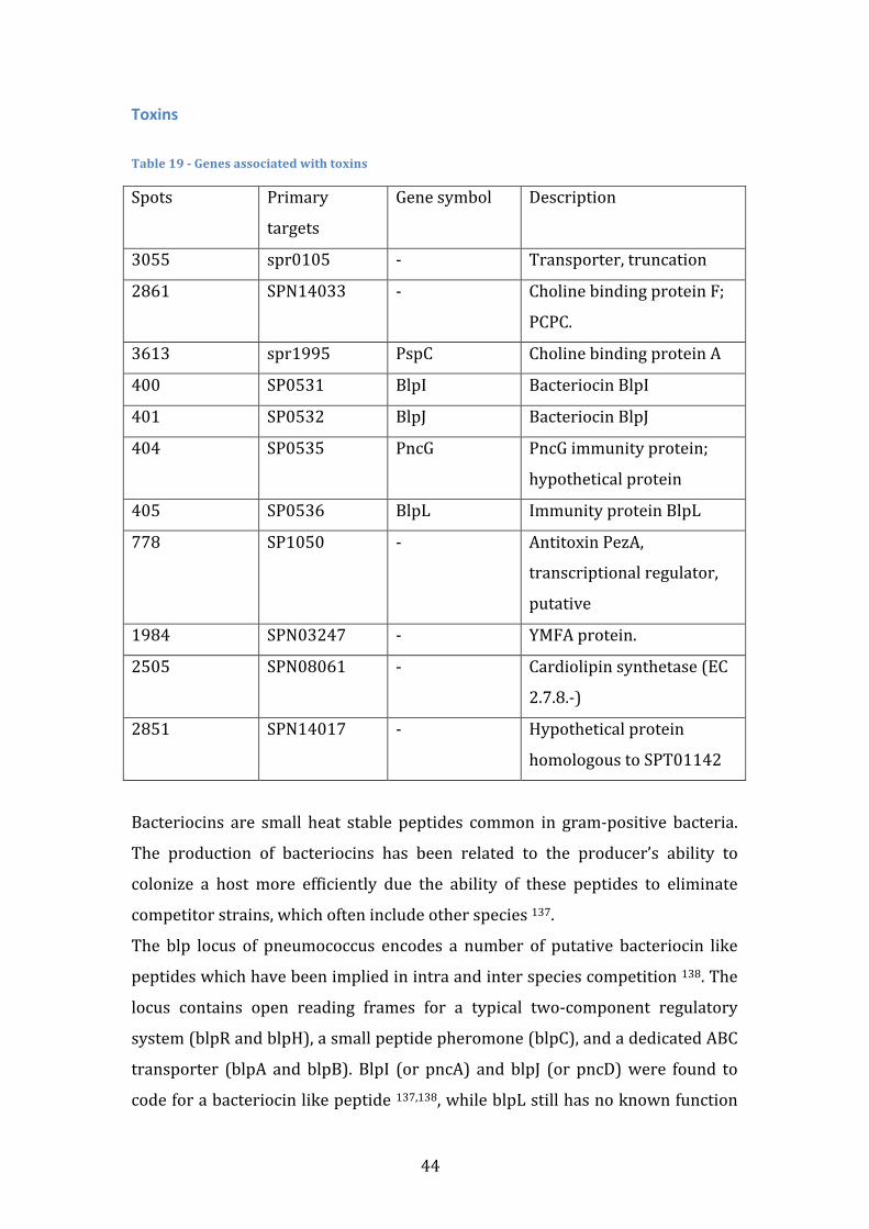
44
Toxins Table 19 -‐ Genes associated with toxins
Spots Primary
targets
Gene symbol Description
3055 spr0105 -‐ Transporter, truncation
2861 SPN14033 -‐ Choline binding protein F;
PCPC.
3613 spr1995 PspC Choline binding protein A
400 SP0531 BlpI Bacteriocin BlpI
401 SP0532 BlpJ Bacteriocin BlpJ
404 SP0535 PncG PncG immunity protein;
hypothetical protein
405 SP0536 BlpL Immunity protein BlpL
778 SP1050 -‐ Antitoxin PezA,
transcriptional regulator,
putative
1984 SPN03247 -‐ YMFA protein.
2505 SPN08061 -‐ Cardiolipin synthetase (EC
2.7.8.-‐)
2851 SPN14017 -‐ Hypothetical protein
homologous to SPT01142
Bacteriocins are small heat stable peptides common in gram-‐positive bacteria.
The production of bacteriocins has been related to the producer’s ability to
colonize a host more efficiently due the ability of these peptides to eliminate
competitor strains, which often include other species 137.
The blp locus of pneumococcus encodes a number of putative bacteriocin like
peptides which have been implied in intra and inter species competition 138. The
locus contains open reading frames for a typical two-‐component regulatory
system (blpR and blpH), a small peptide pheromone (blpC), and a dedicated ABC
transporter (blpA and blpB). BlpI (or pncA) and blpJ (or pncD) were found to
code for a bacteriocin like peptide 137,138, while blpL still has no known function
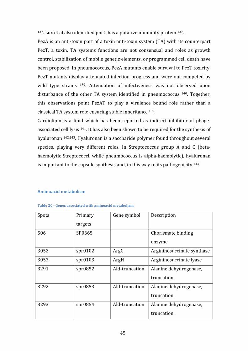
45
137. Lux et al also identified pncG has a putative immunity protein 137.
PezA is an anti-‐toxin part of a toxin anti-‐toxin system (TA) with its counterpart
PezT, a toxin. TA systems functions are not consensual and roles as growth
control, stabilization of mobile genetic elements, or programmed cell death have
been proposed. In pneumococcus, PezA mutants enable survival to PezT toxicity.
PezT mutants display attenuated infection progress and were out-‐competed by
wild type strains 139. Attenuation of infectiveness was not observed upon
disturbance of the other TA system identified in pneumococcus 140. Together,
this observations point PezAT to play a virulence bound role rather than a
classical TA system role ensuring stable inheritance 139.
Cardiolipin is a lipid which has been reported as indirect inhibitor of phage-‐
associated cell lysis 141. It has also been shown to be required for the synthesis of
hyaluronan 142,143. Hyaluronan is a saccharide polymer found throughout several
species, playing very different roles. In Streptococcus group A and C (beta-‐
haemolytic Streptococci, while pneumococcus is alpha-‐haemolytic), hyaluronan
is important to the capsule synthesis and, in this way to its pathogenicity 143.
Aminoacid metabolism Table 20 -‐ Genes associated with aminoacid metabolism
Spots Primary
targets
Gene symbol Description
506 SP0665 Chorismate binding
enzyme
3052 spr0102 ArgG Argininosuccinate synthase
3053 spr0103 ArgH Argininosuccinate lyase
3291 spr0852 Ald-‐truncation Alanine dehydrogenase,
truncation
3292 spr0853 Ald-‐truncation Alanine dehydrogenase,
truncation
3293 spr0854 Ald-‐truncation Alanine dehydrogenase,
truncation

46
3374 spr1096 MetY-‐
truncation
O-‐acetylhomoserine
sulfhydrylase, truncation
3391 spr1181 GdhA Glutamate dehydrogenase
Analysis of the genome sequence of S. pneumoniae D39 showed that it contains
only two genes that are putatively involved in arginine biosynthesis, argG and
argH, encoding the enzymes for the conversion of citrulline (and aspartate) to
arginine. There are, however, other pathways that lead to the synthesis of
arginine, e.g., carA and carB synthetize carbamoyl-‐phosphate from glutamate,
which is a precursor of arginine 144.
Glutamate dehydrogenase (GDH) activity catalyses the reversible oxidative
deamination of glutamate to 2-‐oxoglutarate and ammonia, mainly using NADP as
the co-‐factor 145. This allows pneumococcus to degrade glutamate and to convert
aminoacids to aromatic compounds. GDH activity has been correlated with
virulence in Streptococcus suis and Clostridium botulinum 146.
Methionine synthesis occurs by methylation of homocysteine. In turn,
homocysteine can be synthetized through a sulfhydrylation pathway, in which
homocysteine is synthesized from O-‐acylhomoserine and sulfide by O-‐
acylhomoserine sulfhydrylase (metY) 147,148.
Functional interactions with invasiveness
Although no functional enrichment was found using JCVI annotation, some
selected genes have clear functional interactions. The ribozyme nrdD is activated
by nrdG and both were selected in the same module 149. argG and argH catalyse
sequential reactions that constitute an alternative arginine syntheses pathway 144. The enzyme mannitol-‐1-‐phosphate 5-‐dehydrogenase (mtlD) uses as
substrate mannitol-‐1-‐phosphate, which is the result of the transport of mannitol
by PTS system (mtlA and mtlF) 132. RuvB activity as an enhancer of
recombination is facilitated by the presence of ssDNA binding protein, ssb 113.
Assembly of an ABC transporter requires several components such as permeases
and ATP binding cassettes (or NBDs) and several genes of both of them were
selected 126. Aquaporin Z (aqpZ) action in hipoosmotic conditions puzzles the
science community because its action would seemingly build up excessive turgor
47
pressure in the cell 95. Large mechanosensitive channel (mscL) provides an
effective response to excessive turgor pressure and may be a biological
counterpart of aqpZ. Polyamines, as spermidine and norspermidine, have been
reported to be possible substitutes of choline and are therefore important in cell
wall structure and possible in interaction with choline binding proteins 98.
Most genes selected have been previously linked with invasiveness or have a
plausible connection with invasiveness. Cell envelope proteins and choline
binding proteins play a major role in protection against host immunity 64,79. Both
are important in inhibiting human complement action on the cell, either by
removal of its components or by binding factor H. Several transposable elements
(TEs) have been identified in or near the capsular locus and have been reported
to affect the transcriptional regulation 71.
Invasion of new tissue requires a swift adaptation to a new environment, both in
terms of physical properties and nutrient availability. Osmotic response genes
present in invasive modules are directed towards strong shifts in osmolality
rather than fine tuning and therefore are of particular interest in adaptation to
new environments 95,98. Anaerobic response genes nrdD and its activator nrdG
are hardly functional in the nasopharynx, since they are strict anaerobic 149.
Inside the human organism however, oxygen concentration is low since it is
almost always bound to biological molecules such as haemoglobin and nrdD may
be crucial to compensate the function of the aerobic ribonucleoside-‐
tryphosphate reductases. Adaptation to different carbon sources has also been
proven to be crucial for invasiveness 150. The high number of sugar transporters
is related with the capability of invasive strains to strive in different medium
composition. Similarly, some selected genes code for enzymes involved in the
metabolism of different sugars. Proteolysis genes are likely related with the
nutritional demand for aminoacids 102.
Protein synthesis is a constant process in any bacteria and demands a permanent
pool of aminoacids and tRNA. Aminoacid synthesis genes are part of some
invasive modules and provide alternative pathways of synthesis, using
alternative substrates or the same substrates in a more economic way 144,151.
48
Genes related to the synthesis and editing of tRNA bound with the respective
aminoacid were also selected 78,152.
Finally, the huge heterogeneity of pneumococcus genome originates from its
recombination capabilities 13. Gene transfer allows the bacteria to adapt and
overcome different adversities. Some of the genes present in invasive modules
promote genome heterogeneity by enhancing recombination with extracellular
DNA 113. Among the genes selected, DNA internalization, stabilization and
enhancement of recombination with heterogenic DNA are promoted 153. The
natural competence of pneumococcus is complemented with the capacity to
induce lysis in neighbour cells, which releases DNA fragments. Several
bacterocins were associated with invasiveness, as well as genes that inhibited
cell lysis 139. These genes give the cell a natural advantage in competition with
other pathogens.
Globally, the algorithm has proven to fulfil the objective of module identification.
It has room for improvement, especially in the handling of false discovery
results. To avoid false discovery results it is possible that a large number of true
associations have been dismissed. Analysis of the modules provides optimism
about the authenticity of the selected modules. The modules robustness to minor
changes in the gene presence matrix discards the influence of microarray errors
on results. It is, however, impossible to discard it completely as not only there is
doubt concerning the presence or absence of a gene but also concerning its true
nature. Identification of gene presence uses a probe with 70 nucleotides and no
information exists about the conservation of the rest of the gene. On the other
hand, gene absence may be caused by few mutations on the sequence recognized
by the probe while the majority of the gene is preserved.
Furthermore, interaction between genes is hardly understood due to the many
loopholes in known pathways. To understand the true biological function of a
gene it is crucial to fill the gaps in the overall cell knowledge and, this way,
understand the pathways in which it is involved, the physical interactions it
makes and how it is regulated.
49
Conclusion
In this thesis a new algorithm to predict phenotype from genotype is presented
and is used to find gene modules associated with invasive strains. Unlike other
approaches, the main goal is not to achieve optimal predictive power of
invasiveness in strains but rather to unveil the gene modules that may be
responsible for the invasive traits. The method uses a network-‐based heuristic to
minimize the solution search space and to restrain the positive results to gene
modules with biological relevance. As a first approach an artificial gene distance
matrix was created based on gene co-‐occurrence and co-‐invasiveness. In the end,
26 gene modules were found to be present (above a specific threshold) only in
invasive strains and to be robust against estimated input data error. Presence of
gene modules in strains was found to correlate positively with the strain’s
invasiveness odd’s ratio. This correlation is valid to neutral strains as well, even
though they were not used in module selection.
The modules include 111 unique genes. Some of the genes were previously
described as crucial to invasiveness in Streptococcus Pneumoniae, such as PsPC
or PthD, while other genes’ impact on invasiveness has not been determined
experimentally. Many of the genes associated with invasiveness have unknown
or only putative biological functions that need to be identified and characterized.
The ones that have identified functions may be strong candidates to be
invasiveness determinants when co-‐present with other genes in the same
module. In this scenario are genes linked with cell envelope, transport, sugar
metabolism, osmotic response, aminoacid synthesis, spermidine synthesis and
proteolysis.
The algorithm proved to be able to establish association of gene modules with
the invasive class. Moreover, it was able to return rules that dictate associations
that are easily translated to a biological level: increase of module genes presence
increases the invasiveness odd’s ratio.

50
51
Future perspectives
The present work has identified several genes with unknown function, with
putative functions or with identified functions in other organism. To understand
their part in invasiveness it is necessary to understand their biological role first
and, therefore, laboratorial work needs to be executed. Moreover, while the
algorithm has associated them with invasiveness, it is not possible to clarify what
influence, if any, they have on invasiveness. This work is based in the analysis of
observational studies and therefore it is not possible to ascertain more than
correlation relationships and the genes identified may cause invasive traits or be
no more than a genetic lineage marker of invasive strains.
The algorithm rationale was devised to use biological networks. In this work an
artificial gene distance matrix was created and used as a first approach. Use of a
biological network is the logical next step, allowing the algorithm to perform
with the input data it was devised for. An additional step will need to be added to
the algorithm where a gene distance matrix is generated from the network. This
is thought to eliminate some of the problems of using a gene distance matrix
generated from the gene presence data. In this scenario the same data source is
used to generate the modules and to evaluate them, leading more easily to false
positive results. Using a different source to generate the module will likely tackle
this issue.
The algorithm itself is a work in progress and for further development new
datasets are required. It is a tool to solve classification problems using a
network-‐based heuristic and is, theoretically, adaptable to classification
problems of any background. It was, nonetheless, designed for a specific
biological problem and therefore incorporates specific assumptions. In the near
future, biological classification problems, preferentially phenotype-‐genotype
association problems would be the perfect input data for testing and developing
the algorithm.

52
53
References
1. Bogaert, D., De Groot, R. & Hermans, P. W. M. Streptococcus pneumoniae colonisation: the key to pneumococcal disease. Lancet Infect Dis 4, 144–154 (2004).
2. Mehr, S. & Wood, N. Streptococcus pneumoniae – a review of carriage, infection, serotype replacement and vaccination. Paediatric Respiratory Reviews 1–7 (2012).doi:10.1016/j.prrv.2011.12.001
3. Austrian, R. The pneumococcus at the millennium: not down, not out. J. Infect. Dis. 179, S338–S341 (1999).
4. Lopez, R. Pneumococcus: the sugar-‐coated bacteria. Int. Microbiol. 9, 179–190 (2006).
5. Austrian, R. The Gram stain and the etiology of lobar pneumonia, an historical note. Bacteriological Reviews 24, 261 (1960).
6. van der Poll, T. & Opal, S. M. Pathogenesis, treatment, and prevention of pneumococcal pneumonia. The Lancet 374, 1543–1556 (2009).
7. Kadioglu, A., Weiser, J. N., Paton, J. C. & Andrew, P. W. The role of Streptococcus pneumoniae virulence factors in host respiratory colonization and disease. Nat. Rev. Microbiol. 6, 288–301 (2008).
8. Tillett, W. S., Cambier, M. J. & McCormack, J. E. The Treatment of Lobar Pneumonia and Pneumococcal Empyema with Penicillin. Bull N Y Acad Med 20, 142–178 (1944).
9. Hakenbeck, R., Grebe, T., Zähner, D. & Stock, J. B. beta-‐lactam resistance in Streptococcus pneumoniae: penicillin-‐binding proteins and non-‐penicillin-‐binding proteins. Molecular Microbiology 33, 673–678 (1999).
10. Cardozo, D. M., Nascimento-‐Carvalho, C. M. C., Souza, F. R. & Silva, N. M. S. Nasopharyngeal colonization and penicillin resistance among pneumococcal strains: a worldwide 2004 update. Braz J Infect Dis 10, 293–304 (2006).
11. Austrian, R. The Jeremiah Metzger Lecture: Of gold and pneumococci: a history of pneumococcal vaccines in South Africa. Trans. Am. Clin. Climatol. Assoc. 89, 141–161 (1978).
12. Weinberger, D. M., Harboe, Z. B., Flasche, S., Scott, J. A. & Lipsitch, M. Prediction of Serotypes Causing Invasive Pneumococcal Disease in Unvaccinated and Vaccinated Populations. Epidemiology 22, 199–207 (2011).
13. Donati, C. et al. Structure and dynamics of the pan-‐genome of Streptococcus pneumoniae and closely related species. Genome Biology 11, R107 (2010).
14. Hava, D. L., LeMieux, J. & Camilli, A. From nose to lung: the regulation behind Streptococcus pneumoniae virulence factors. Molecular Microbiology 50, 1103–1110 (2003).
15. Tettelin, H. et al. Complete genome sequence of a virulent isolate of Streptococcus pneumoniae. Science 293, 498–506 (2001).
16. Mitchell, A. M. & Mitchell, T. J. Streptococcus pneumoniae: virulence
54
factors and variation. Clinical Microbiology and Infection 16, 411–418 (2010).
17. Lapierre, P. & Gogarten, J. P. Estimating the size of the bacterial pan-‐genome. Trends in Genetics 25, 107–110 (2009).
18. Tettelin, H., Riley, D., Cattuto, C. & Medini, D. Comparative genomics: the bacterial pan-‐genome. Current Opinion in Microbiology 11, 472–477 (2008).
19. Claverys, J.-‐P., Martin, B. & Håvarstein, L. S. Competence-‐induced fratricide in streptococci. Molecular Microbiology 64, 1423–1433 (2007).
20. Bartilson, M. et al. Differential fluorescence induction reveals Streptococcus pneumoniae loci regulated by competence stimulatory peptide. Molecular Microbiology 39, 126–135 (2001).
21. Peterson, S. N. et al. Identification of competence pheromone responsive genes in Streptococcus pneumoniae by use of DNA microarrays. Molecular Microbiology 51, 1051–1070 (2003).
22. Rimini, R. et al. Global analysis of transcription kinetics during competence development in Streptococcus pneumoniae using high density DNA arrays. Molecular Microbiology 36, 1279–1292 (2000).
23. Hakenbeck, R. et al. Mosaic Genes and Mosaic Chromosomes: Intra-‐ and Interspecies Genomic Variation of Streptococcus pneumoniae. Infection and Immunity 69, 2477–2486 (2001).
24. Weiser, J. N. The pneumococcus: why a commensal misbehaves. J Mol Med 88, 97–102 (2009).
25. Weinberger, D. M. et al. Association of Serotype with Risk of Death Due to Pneumococcal Pneumonia: A Meta-‐Analysis. CLIN INFECT DIS 51, 692–699 (2010).
26. Obert, C. et al. Identification of a Candidate Streptococcus pneumoniae Core Genome and Regions of Diversity Correlated with Invasive Pneumococcal Disease. Infection and Immunity 74, 4766–4777 (2006).
27. Melin, M., Trzciński, K., Meri, S., Käyhty, H. & Väkeväinen, M. The capsular serotype of Streptococcus pneumoniae is more important than the genetic background for resistance to complement. Infection and Immunity 78, 5262–5270 (2010).
28. Hava, D. L. & Camilli, A. Large-‐scale identification of serotype 4 Streptococcus pneumoniae virulence factors. Molecular Microbiology 45, 1389–1406 (2002).
29. Lau, G. W. et al. A functional genomic analysis of type 3 Streptococcus pneumoniae virulence. Molecular Microbiology 40, 555–571 (2001).
30. Polissi, A. et al. Large-‐Scale Identification of Virulence Genes fromStreptococcus pneumoniae. Infection and Immunity 66, 5620–5629 (1998).
31. Obert, C. A., Gao, G., Sublett, J., Tuomanen, E. I. & Orihuela, C. J. Assessment of molecular typing methods to determine invasiveness and to differentiate clones of Streptococcus pneumoniae. Infect. Genet. Evol. 7, 708–716 (2007).
32. Schmidt, M. C. et al. NIBBS-‐Search for Fast and Accurate Prediction of Phenotype-‐Biased Metabolic Systems. PLoS Comput Biol 8, e1002490
55
(2012). 33. Slonim, N., Elemento, O. & Tavazoie, S. Ab initio genotype–phenotype
association reveals intrinsic modularity in genetic networks. Mol Syst Biol 2, 1–14 (2006).
34. Önskog, J., Freyhult, E., Landfors, M., Rydén, P. & Hvidsten, T. R. Classification of microarrays; synergistic effects between normalization, gene selection and machine learning. BMC Bioinformatics 12, 390 (2011).
35. Ideker, T., Galitski, T. & Hood, L. A new approach to decoding life: systems biology. Annual review of genomics and human genetics 2, 343–372 (2001).
36. Chuang, H.-‐Y., Hofree, M. & Ideker, T. A Decade of Systems Biology. Annu. Rev. Cell Dev. Biol. 26, 721–744 (2010).
37. Sieberts, S. K. & Schadt, E. E. Moving toward a system genetics view of disease. Mamm Genome 18, 389–401 (2007).
38. Bhaskar, H., Hoyle, D. C. & Singh, S. Machine learning in bioinformatics: A brief survey and recommendations for practitioners. Computers in Biology and Medicine 36, 1104–1125 (2006).
39. Larranaga, P. Machine learning in bioinformatics. Briefings in Bioinformatics 7, 86–112 (2006).
40. Szymczak, S. et al. Machine learning in genome-‐wide association studies. Genet. Epidemiol. 33, S51–S57 (2009).
41. Jensen, L. J. & Bateman, A. The rise and fall of supervised machine learning techniques. Bioinformatics 27, 3331–3332 (2011).
42. Kamruzzaman, S. M. & Sarkar, A. M. J. A New Data Mining Scheme Using Artificial Neural Networks. Sensors 11, 4622–4647 (2011).
43. Moore, J. H., Asselbergs, F. W. & Williams, S. M. Bioinformatics challenges for genome-‐wide association studies. Bioinformatics 26, 445–455 (2010).
44. Winter, C. et al. Google Goes Cancer: Improving Outcome Prediction for Cancer Patients by Network-‐Based Ranking of Marker Genes. PLoS Comput Biol 8, e1002511 (2012).
45. Hurley, D. et al. Gene network inference and visualization tools for biologists: application to new human transcriptome datasets. Nucleic Acids Research 40, 2377–2398 (2012).
46. Hoskins, J. et al. Genome of the Bacterium Streptococcus pneumoniae Strain R6. J. Bacteriol. 183, 5709–5717 (2001).
47. Dopazo, J. et al. Annotated Draft Genomic Sequence from a Streptococcus pneumoniae Type 19F Clinical Isolate. Microbial Drug Resistance 7, 99–125 (2001).
48. Sa-‐Leao, R. et al. Analysis of Invasiveness of Pneumococcal Serotypes and Clones Circulating in Portugal before Widespread Use of Conjugate Vaccines Reveals Heterogeneous Behavior of Clones Expressing the Same Serotype. Journal of Clinical Microbiology 49, 1369–1375 (2011).
49. Coles, S. An Introduction to Statistical Modeling of Extreme Values. (Springer: 2001).
50. Cardoso, L. Classificação de genes em hibridação genómica
56
comparativa de estirpes de Streptococcus pneumoniae. (Dissertação de Mestrado em Bioestatística na Faculdade de Ciências da Universidade de Lisboa: 2009).
51. Eliasson, R. et al. The anaerobic ribonucleoside triphosphate reductase from Escherichia coli requires S-‐adenosylmethionine as a cofactor. Proc. Natl. Acad. Sci. U.S.A. 87, 3314–3318 (1990).
52. Torrents, E. The Anaerobic Ribonucleotide Reductase from Lactococcus lactis. INTERACTIONS BETWEEN THE TWO PROTEINS NrdD AND NrdG. Journal of Biological Chemistry 276, 33488–33494 (2001).
53. Sjoberg, B. M. & Torrents, E. Shift in Ribonucleotide Reductase Gene Expression in Pseudomonas aeruginosa during Infection. Infection and Immunity 79, 2663–2669 (2011).
54. Masalha, M., Borovok, I., Schreiber, R., Aharonowitz, Y. & Cohen, G. Analysis of Transcription of the Staphylococcus aureus Aerobic Class Ib and Anaerobic Class III Ribonucleotide Reductase Genes in Response to Oxygen. J. Bacteriol. 183, 7260–7272 (2001).
55. Paik, S. et al. Identification of Virulence Determinants for Endocarditis in Streptococcus sanguinis by Signature-‐Tagged Mutagenesis. Infection and Immunity 73, 6064–6074 (2005).
56. Brooks-‐Walter, A., Briles, D. E. & Hollingshead, S. K. The pspC gene of Streptococcus pneumoniae encodes a polymorphic protein, PspC, which elicits cross-‐reactive antibodies to PspA and provides immunity to pneumococcal bacteremia. Infection and Immunity 67, 6533–6542 (1999).
57. Rosenow, C. et al. Contribution of novel choline-‐binding proteins to adherence, colonization and immunogenicity of Streptococcus pneumoniae. Molecular Microbiology 25, 819–829 (1997).
58. Ogunniyi, A. D. et al. Contributions of Pneumolysin, Pneumococcal Surface Protein A (PspA), and PspC to Pathogenicity of Streptococcus pneumoniae D39 in a Mouse Model. Infection and Immunity 75, 1843–1851 (2007).
59. Melin, M. et al. Interaction of Pneumococcal Histidine Triad Proteins with Human Complement. Infection and Immunity 78, 2089–2098 (2010).
60. Yuste, J. et al. The Effects of PspC on Complement-‐Mediated Immunity to Streptococcus pneumoniae Vary with Strain Background and Capsular Serotype. Infection and Immunity 78, 283–292 (2009).
61. Quin, L. R. et al. Factor H Binding to PspC of Streptococcus pneumoniae Increases Adherence to Human Cell Lines In Vitro and Enhances Invasion of Mouse Lungs In Vivo. Infection and Immunity 75, 4082–4087 (2007).
62. Kerr, A. R. et al. The Contribution of PspC to Pneumococcal Virulence Varies between Strains and Is Accomplished by Both Complement Evasion and Complement-‐Independent Mechanisms. Infection and Immunity 74, 5319–5324 (2006).
63. Eldholm, V. et al. The Pneumococcal Cell Envelope Stress-‐Sensing System LiaFSR Is Activated by Murein Hydrolases and Lipid II-‐
57
Interacting Antibiotics. J. Bacteriol. 192, 1761–1773 (2010). 64. Gosink, K. K., Mann, E. R., Guglielmo, C., Tuomanen, E. I. & Masure, H.
R. Role of Novel Choline Binding Proteins in Virulence of Streptococcus pneumoniae. Infection and Immunity 68, 5690–5695 (2000).
65. Alpert, C. A. & Siebers, U. The lac operon of Lactobacillus casei contains lacT, a gene coding for a protein of the Bg1G family of transcriptional antiterminators. J. Bacteriol. 179, 1555–1562 (1997).
66. Rutberg, B. Antitermination of transcription of catabolic operons. Molecular Microbiology 23, 413–421 (1997).
67. Gosalbes, M. J., Esteban, C. D. & Pérez-‐Martínez, G. In vivo effect of mutations in the antiterminator LacT in Lactobacillus casei. Microbiology (Reading, Engl.) 148, 695–702 (2002).
68. Rioux, S. et al. Transcriptional regulation, occurrence and putative role of the Pht family of Streptococcus pneumoniae. Microbiology 157, 336–348 (2011).
69. Claverys, J. P., Prudhomme, M., Mortier-‐Barrière, I. & Martin, B. Adaptation to the environment: Streptococcus pneumoniae, a paradigm for recombination-‐mediated genetic plasticity? Molecular Microbiology 35, 251–259 (2000).
70. Dziewit, L. et al. Insights into the Transposable Mobilome of Paracoccus spp. (Alphaproteobacteria). PLoS ONE 7, e32277 (2012).
71. Han, C. G., Shiga, Y., Tobe, T., Sasakawa, C. & Ohtsubo, E. Structural and Functional Characterization of IS679 and IS66-‐Family Elements. J. Bacteriol. 183, 4296–4304 (2001).
72. Mingoia, M., Tili, E., Manso, E., Varaldo, P. E. & Montanari, M. P. Heterogeneity of Tn5253-‐Like Composite Elements in Clinical Streptococcus pneumoniae Isolates. Antimicrobial Agents and Chemotherapy 55, 1453–1459 (2011).
73. Tardif, K. D. Functional group recognition at the aminoacylation and editing sites of E. coli valyl-‐tRNA synthetase. RNA 10, 493–503 (2004).
74. Fukunaga, R. Structural Basis for Non-‐cognate Amino Acid Discrimination by the Valyl-‐tRNA Synthetase Editing Domain. Journal of Biological Chemistry 280, 29937–29945 (2005).
75. Zheng, L., Yu, C., Bayles, K., Lasa, I. & Ji, Y. Conditional Mutation of an Essential Putative Glycoprotease Eliminates Autolysis in Staphylococcus aureus. J. Bacteriol. 189, 2734–2742 (2007).
76. Lei, T. et al. The C-‐Terminal Domain of the Novel Essential Protein Gcp Is Critical for Interaction with Another Essential Protein YeaZ of Staphylococcus aureus. PLoS ONE 6, e20163 (2011).
77. Lei, T. et al. The Essentiality of Staphylococcal Gcp Is Independent of Its Repression of Branched-‐Chain Amino Acids Biosynthesis. PLoS ONE 7, e46836 (2012).
78. Deutsch, C., Yacoubi, El, B., de Crécy-‐Lagard, V. & Dirk Iwata-‐Reuyl Biosynthesis of threonylcarbamoyl adenosine (t6A), a universal tRNA nucleoside. J. Biol. Chem 13666–13673 (2012).
79. Moscoso, M. & García, E. Transcriptional regulation of the capsular polysaccharide biosynthesis locus of streptococcus pneumoniae: a
58
bioinformatic analysis. DNA Res. 16, 177–186 (2009). 80. Pandya, U., Sinha, M., Luxon, B. A., Watson, D. A. & Niesel, D. W. Global
transcription profiling and virulence potential of Streptococcus pneumoniae after serial passage. Gene 443, 22–31 (2009).
81. Jothi, R., Manikandakumar, K., Ganesan, K. & Parthasarathy, S. On the analysis of the virulence nature of TIGR4 and R6 strains of Streptococcus pneumoniae using genome comparison tools. Journal of chemical sciences-‐Bangalore-‐ 119, 559 (2007).
82. Aanensen, D. M., Mavroidi, A., Bentley, S. D., Reeves, P. R. & Spratt, B. G. Predicted Functions and Linkage Specificities of the Products of the Streptococcus pneumoniae Capsular Biosynthetic Loci. J. Bacteriol. 189, 7856–7876 (2007).
83. Chang, H. Y., Lee, J. H., Deng, W. L., Fu, T. F. & Peng, H. L. Virulence and outer membrane properties of a galU mutant of Klebsiella pneumoniae CG43. Microb. Pathog. 20, 255–261 (1996).
84. Vilches, S. et al. Mesophilic Aeromonas UDP-‐glucose pyrophosphorylase (GalU) mutants show two types of lipopolysaccharide structures and reduced virulence. Microbiology 153, 2393–2404 (2007).
85. Stimson et al. Meningococcal pilin: a glycoprotein substituted with digalactosyl 2,4-‐ diacetamid-‐2,4,6-‐trideoxyhexose. Mol Microbiol 1201–1214 (1995).
86. Ghuysen, J. M. Molecular structures of penicillin-‐binding proteins and beta-‐lactamases. Trends in Microbiology 2, 372–380 (1994).
87. Rieux, V., Carbon, C. & Azoulay-‐Dupuis, E. Complex relationship between acquisition of β-‐lactam resistance and loss of virulence in Streptococcus pneumoniae. J. Infect. Dis. 184, 66–72 (2001).
88. Shah, P., Nanduri, B., Swiatlo, E., Ma, Y. & Pendarvis, K. Polyamine biosynthesis and transport mechanisms are crucial for fitness and pathogenesis of Streptococcus pneumoniae. Microbiology 157, 504–515 (2011).
89. Igarashi, K. & Kashiwagi, K. Polyamines: Mysterious Modulators of Cellular Functions. Biochem. Biophys. Res. Commun. 271, 559–564 (2000).
90. Shah, P. & Swiatlo, E. A multifaceted role for polyamines in bacterial pathogens. Molecular Microbiology 68, 4–16 (2008).
91. Shah, P., Romero, D. G. & Swiatlo, E. Role of polyamine transport in Streptococcus pneumoniae response to physiological stress and murine septicemia. Microb. Pathog. 45, 167–172 (2008).
92. Wahome, P. G. & Setlow, P. Growth, osmotic downshock resistance and differentiation of Bacillus subtilis strains lacking mechanosensitive channels. Arch Microbiol 189, 49–58 (2007).
93. Wood, J. M. et al. Osmosensing and osmoregulatory compatible solute accumulation by bacteria. Comparative Biochemistry and Physiology-‐Part A: Molecular & Integrative Physiology 130, 437–460 (2001).
94. Wood, J. M. Osmosensing by bacteria: signals and membrane-‐based sensors. Microbiology and Molecular Biology Reviews 63, 230–262 (1999).
95. Soupene, E., King, N., Lee, H. & Kustu, S. Aquaporin Z of Escherichia
59
coli: Reassessment of Its Regulation and Physiological Role. J. Bacteriol. 184, 4304–4307 (2002).
96. Calamita, G. The Escherichia coli aquaporin-‐Z water channel. Molecular Microbiology 37, 254–262 (2000).
97. Hoffmann, T., Boiangiu, C., Moses, S. & Bremer, E. Responses of Bacillus subtilis to Hypotonic Challenges: Physiological Contributions of Mechanosensitive Channels to Cellular Survival. Applied and Environmental Microbiology 74, 2454–2460 (2008).
98. Levina, N. et al. Protection of Escherichia coli cells against extreme turgor by activation of MscS and MscL mechanosensitive channels: identification of genes required for MscS activity. EMBO J. 18, 1730–1737 (1999).
99. Fernandes, C. L., Breda, A., Santiago Santos, D., Augusto Basso, L. & Osmar Norberto de Souza A structural model for chorismate synthase from Mycobacterium tuberculosis in complex with coenzyme and substrate. Computers in Biology and Medicine 37, 149–158 (2007).
100. Müller, I. B. et al. The Vitamin B1 Metabolism of Staphylococcus aureus Is Controlled at Enzymatic and Transcriptional Levels. PLoS ONE 4, e7656 (2009).
101. Freiberg, C. et al. Identification of novel essential Escherichia coli genes conserved among pathogenic bacteria. J. Mol. Microbiol. Biotechnol. 3, 483–489 (2001).
102. Johnson, M. K. Physiological roles of pneumococcal peptidases. J. Bacteriol. 119, 844–847 (1974).
103. Kanamaru, K., Stephenson, S. & Perego, M. Overexpression of the PepF oligopeptidase inhibits sporulation initiation in Bacillus subtilis. J. Bacteriol. 184, 43–50 (2002).
104. Lin, B. et al. Characterization of PepB, a group B streptococcal oligopeptidase. Infection and Immunity 64, 3401–3406 (1996).
105. Johnsborg, O. & HÃ varstein, L. S. Regulation of natural genetic transformation and acquisition of transforming DNA in Streptococcus pneumoniae. FEMS Microbiology Reviews 33, 627–642 (2009).
106. Yother, J., McDaniel, L. S. & Briles, D. E. Transformation of encapsulated Streptococcus pneumoniae. J. Bacteriol. 168, 1463–1465 (1986).
107. Piotrowski, A., Luo, P. & Morrison, D. A. Competence for Genetic Transformation in Streptococcus pneumoniae: Termination of Activity of the Alternative Sigma Factor ComX Is Independent of Proteolysis of ComX and ComW. J. Bacteriol. 191, 3359–3366 (2009).
108. Zhu, L. & Lau, G. W. Inhibition of Competence Development, Horizontal Gene Transfer and Virulence in Streptococcus pneumoniae by a Modified Competence Stimulating Peptide. PLoS Pathog 7, e1002241 (2011).
109. Luo, P. & Morrison, D. A. Transient Association of an Alternative Sigma Factor, ComX, with RNA Polymerase during the Period of Competence for Genetic Transformation in Streptococcus pneumoniae. J. Bacteriol. 185, 349–358 (2003).
60
110. Attaiech, L. et al. Role of the Single-‐Stranded DNA–Binding Protein SsbB in Pneumococcal Transformation: Maintenance of a Reservoir for Genetic Plasticity. PLoS Genet 7, e1002156 (2011).
111. Pestova, E. V. & Morrison, D. A. Isolation and Characterization of Three Streptococcus pneumoniae Transformation-‐Specific Loci by Use of alacZ Reporter Insertion Vector. J. Bacteriol. 180, 2701–2710 (1998).
112. la Campa, de, A. G., Springhorn, S. S., Kale, P. & Lacks, S. A. Proteins encoded by the DpnI restriction gene cassette. Hyperproduction and characterization of the DpnI endonuclease. Journal of Biological Chemistry 263, 14696–14702 (1988).
113. Parsons, C. A., Stasiak, A. & West, S. C. The E. coli RuvAB proteins branch migrate Holliday junctions through heterologous DNA sequences in a reaction facilitated by SSB. EMBO J. 14, 5736 (1995).
114. Humbert, O., Prudhomme, M., Hakenbeck, R., Dowson, C. G. & Claverys, J. P. Homeologous recombination and mismatch repair during transformation in Streptococcus pneumoniae: saturation of the Hex mismatch repair system. Proc. Natl. Acad. Sci. U.S.A. 92, 9052–9056 (1995).
115. Burghout, P. et al. Search for Genes Essential for Pneumococcal Transformation: the RadA DNA Repair Protein Plays a Role in Genomic Recombination of Donor DNA. J. Bacteriol. 189, 6540–6550 (2007).
116. Bessman, M. J., Frick, D. N. & O'Handley, S. F. The MutT proteins or “Nudix” hydrolases, a family of versatile, widely distributed,“housecleaning” enzymes. Journal of Biological Chemistry 271, 25059–25062 (1996).
117. Treffers, H. P., Spinelli, V. & Belser, N. O. A Factor (or Mutator Gene) Influencing Mutation Rates in Escherichia Coli. Proc. Natl. Acad. Sci. U.S.A. 40, 1064–1071 (1954).
118. Yanofsky, C., Cox, E. C. & Horn, V. The unusual mutagenic specificity of an E. Coli mutator gene. Proc. Natl. Acad. Sci. U.S.A. 55, 274 (1966).
119. Iyer, R., Baliga, N. S. & Camilli, A. Catabolite control protein A (CcpA) contributes to virulence and regulation of sugar metabolism in Streptococcus pneumoniae. J. Bacteriol. 187, 8340–8349 (2005).
120. Ren, Q., Chen, K. & Paulsen, I. T. TransportDB: a comprehensive database resource for cytoplasmic membrane transport systems and outer membrane channels. Nucleic Acids Research 35, D274–D279 (2007).
121. Bidossi, A. et al. A Functional Genomics Approach to Establish the Complement of Carbohydrate Transporters in Streptococcus pneumoniae. PLoS ONE 7, e33320 (2012).
122. Postma, P. W., Lengeler, J. W. & Jacobson, G. R. Phosphoenolpyruvate:carbohydrate phosphotransferase systems of bacteria. Microbiol. Rev. 57, 543–594 (1993).
123. Deutscher, J., Francke, C. & Postma, P. W. How Phosphotransferase System-‐Related Protein Phosphorylation Regulates Carbohydrate Metabolism in Bacteria. Microbiology and Molecular Biology Reviews 70, 939–1031 (2006).
61
124. Marion, C. et al. Streptococcus pneumoniae Can Utilize Multiple Sources of Hyaluronic Acid for Growth. Infection and Immunity 80, 1390–1398 (2012).
125. Kaufman, G. E. & Yother, J. CcpA-‐Dependent and -‐Independent Control of Beta-‐Galactosidase Expression in Streptococcus pneumoniae Occurs via Regulation of an Upstream Phosphotransferase System-‐Encoding Operon. J. Bacteriol. 189, 5183–5192 (2007).
126. Davidson, A. L., Dassa, E., Orelle, C. & Chen, J. Structure, Function, and Evolution of Bacterial ATP-‐Binding Cassette Systems. Microbiology and Molecular Biology Reviews 72, 317–364 (2008).
127. Marion, C., Aten, A. E., Woodiga, S. A. & King, S. J. Identification of an ATPase, MsmK, Which Energizes Multiple Carbohydrate ABC Transporters in Streptococcus pneumoniae. Infection and Immunity 79, 4193–4200 (2011).
128. Brown, J. S., Gilliland, S. M. & Holden, D. W. A Streptococcus pneumoniae pathogenicity island encoding an ABC transporter involved in iron uptake and virulence. Molecular Microbiology 40, 572–585 (2001).
129. Putman, M., van Veen, H. W. & Konings, W. N. Molecular Properties of Bacterial Multidrug Transporters. Microbiology and Molecular Biology Reviews 64, 672–693 (2000).
130. Carvalho, S. M., Kloosterman, T. G., Kuipers, O. P. & Neves, A. R. CcpA Ensures Optimal Metabolic Fitness of Streptococcus pneumoniae. PLoS ONE 6, e26707 (2011).
131. Giammarinaro, P. & Paton, J. C. Role of RegM, a Homologue of the Catabolite Repressor Protein CcpA, in the Virulence of Streptococcus pneumoniae. Infection and Immunity 70, 5454–5461 (2002).
132. Honeyman, A. L. & Curtiss, R. Isolation, characterization, and nucleotide sequence of the Streptococcus mutans mannitol-‐phosphate dehydrogenase gene and the mannitol-‐specific factor III gene of the phosphoenolpyruvate phosphotransferase system. Infection and Immunity 60, 3369–3375 (1992).
133. McKessar, S. J. & Hakenbeck, R. The Two-‐Component Regulatory System TCS08 Is Involved in Cellobiose Metabolism of Streptococcus pneumoniae R6. J. Bacteriol. 189, 1342–1350 (2007).
134. Shafeeq, S., Kloosterman, T. G. & Kuipers, O. P. CelR-‐mediated activation of the cellobiose-‐utilization gene cluster in Streptococcus pneumoniae. Microbiology 157, 2854–2861 (2011).
135. Sterner, D. E. & Berger, S. L. Acetylation of Histones and Transcription-‐Related Factors. Microbiology and Molecular Biology Reviews 64, 435–459 (2000).
136. Dyda, F., Klein, D. C. & Hickman, A. B. GCN5-‐related N-‐acetyltransferases: a structural overview. Annu Rev Biophys Biomol Struct 29, 81–103 (2000).
137. Lux, T., Nuhn, M., Hakenbeck, R. & Reichmann, P. Diversity of Bacteriocins and Activity Spectrum in Streptococcus pneumoniae. J. Bacteriol. 189, 7741–7751 (2007).
138. Dawid, S., Roche, A. M. & Weiser, J. N. The blp Bacteriocins of
62
Streptococcus pneumoniae Mediate Intraspecies Competition both In Vitro and In Vivo. Infection and Immunity 75, 443–451 (2006).
139. Mutschler, H., Reinstein, J. & Meinhart, A. Assembly dynamics and stability of the pneumococcal epsilon zeta antitoxin toxin (PezAT) system from Streptococcus pneumoniae. Journal of Biological Chemistry 285, 21797–21806 (2010).
140. Mutschler, H. & Meinhart, A. ε/ζ systems: their role in resistance, virulence, and their potential for antibiotic development. J Mol Med 89, 1183–1194 (2011).
141. Garcia, P., López, R., Ronda, C., García, E. & Tomasz, A. Mechanism of phage-‐induced lysis in pneumococci. J. Gen. Microbiol. 129, 479–487 (1983).
142. Tlapak-‐Simmons, V. L., Baggenstoss, B. A., Clyne, T. & Weigel, P. H. Purification and lipid dependence of the recombinant hyaluronan synthases from Streptococcus pyogenes and Streptococcus equisimilis. Journal of Biological Chemistry 274, 4239–4245 (1999).
143. Tlapak-‐Simmons, V. L., Kempner, E. S., Baggenstoss, B. A. & Weigel, P. H. The active streptococcal hyaluronan synthases (HASs) contain a single HAS monomer and multiple cardiolipin molecules. Journal of Biological Chemistry 273, 26100–26109 (1998).
144. Kloosterman, T. G. & Kuipers, O. P. Regulation of Arginine Acquisition and Virulence Gene Expression in the Human Pathogen Streptococcus pneumoniae by Transcription Regulators ArgR1 and AhrC. Journal of Biological Chemistry 286, 44594–44605 (2011).
145. Tanous, C., Chambellon, E. & Yvon, M. Sequence analysis of the mobilizable lactococcal plasmid pGdh442 encoding glutamate dehydrogenase activity. Microbiology 153, 1664–1675 (2007).
146. Kutz, R. & Okwumabua, O. Differentiation of Highly Virulent Strains of Streptococcus suis Serotype 2 According to Glutamate Dehydrogenase Electrophoretic and Sequence Type. Journal of Clinical Microbiology 46, 3201–3207 (2008).
147. Sperandio, B. et al. Control of Methionine Synthesis and Uptake by MetR and Homocysteine in Streptococcus mutans. J. Bacteriol. 189, 7032–7044 (2007).
148. Kovaleva, G. Y. & Gelfand, M. S. Transcriptional regulation of the methionine and cysteine transport and metabolism in streptococci. FEMS Microbiology Letters 276, 207–215 (2007).
149. Garriga, X. et al. nrdD and nrdG genes are essential for strict anaerobic growth of Escherichia coli. Biochem. Biophys. Res. Commun. 229, 189–192 (1996).
150. Mcallister, L. J., Ogunniyi, A. D., Stroeher, U. H. & Paton, J. C. Contribution of a Genomic Accessory Region Encoding a Putative Cellobiose Phosphotransferase System to Virulence of Streptococcus pneumoniae. PLoS ONE 7, e32385 (2012).
151. Hartel, T. et al. Impact of Glutamine Transporters on Pneumococcal Fitness under Infection-‐Related Conditions. Infection and Immunity 79, 44–58 (2010).
152. Cho, K. H. & Caparon, M. G. tRNA Modification by GidA/MnmE Is Necessary for Streptococcus pyogenes Virulence: a New Strategy To

63
Make Live Attenuated Strains. Infection and Immunity 76, 3176–3186 (2008).
153. Pestova, E. V. & Morrison, D. A. Isolation and Characterization of Three Streptococcus pneumoniae Transformation-‐Specific Loci by Use of alacZ Reporter Insertion Vector. J. Bacteriol. 180, 2701–2710 (1998).