revista de pediatria do centro hospitalar do porto ... · eurico gaspar, chtmad, vila real fátima...
TRANSCRIPT
Su
ple
men
to I
20
14Revista de Pediatria do Centro Hospitalar do Porto
XLIII Conferências de Genética Doutor Jacinto Magalhães
Resumos das Comunicações
X L I I I C O N F E R Ê N C I A S D E G E N É T I C A D O U T O R J A C I N T O M A G A L H Ã E S
Auditório Prof. Doutor Alexandre Moreira - CHP, 28 março 2014 1
Revista de Pediatria do Centro Hospitalar do Porto | Departamento de Ensino, Formação e Investigação
Ano | 2014 Volume | XXIII Número | Suplemento I
Directora | Editor-in-Chief | Sílvia Álvares; Director Adjunto | Associated Editor | Rui Chorão; Directora Executiva | Executive Editor | Luísa Lobato
Presidente do Conselho de Administração do Centro Hospitalar do Porto | Director | Fernando Sollari Allegro
Corpo Redactorial | Editorial Board
Ana Rita Araújo, ULSAMArmando Pinto, IPOPFGArtur Alegria, CHPBraga da Cunha, CHTSCarmen Carvalho, CHPCláudia Pedrosa, CHVNG/ECláudia Tavares, CHAAConceição Mota, CHPCristina Rocha, CHEDVGabriela Soares, CGMJMGustavo Rocha, CHSJJoão Barreira, CHSJLaura Marques, CHPMargarida Guedes, CHPRui Almeida, HPH/ULSM
Editores especializados | Section Editors
Artigo Recomendado – Helena Mansilha, CHP; Maria do Carmo Santos, CHP
Perspectivas Actuais em Bioética – Natália Teles, CGMJM
Pediatria Baseada na Evidência – Luís Filipe Azevedo, FMUP; Altamiro da Costa Pereira, FMUP
A Cardiologia Pediátrica na Prática Clínica – António Marinho, CHUC; Fátima Pinto, HSM/CHLC; Maria Ana Sampaio, HCV, Maria João Baptista, CHSJ; Paula Martins, HPCM/CHUC, Rui Anjos, HSC/CHLO; Sílvia Álvares, CHP
Ciclo de Pediatria Inter-Hospitalar do Norte – Armando Pinto, IPOPFG; Carla Moreira, HB/EB; Conceição Santos Silva, CHPVVC; Fátima Santos, CHVNG/E; Inês Azevedo, CHSJ; Isalita Moura, HSMM; Isolina Aguiar, CHAA; Joaquim Cunha, CHTS; Susana Tavares, CHEDV; Cármen Carvalho, CHP; Rosa Lima, CHP; Sofi a Aroso, HPH/ULSM; Sónia Carvalho, CHMA
Caso Dermatológico – Manuela Selores, CHP; Susana Machado, CHP
Caso Electroencefalográfi co – Rui Chorão, CHP
Caso Endoscópico – Fernando Pereira, CHP
Caso Estomatológico – José Amorim, CHP
Caso Radiológico – Filipe Macedo, CHAA
Genes, Crianças e Pediatras – Esmeralda Martins, CHP; Gabriela Soares, CGMJM
Educação Científi ca – Margarida Lima, CHP, ICBAS-UP
Pequenas Histórias – Margarida Guedes, CHP
Consultor Técnico | Consultant
Gama de Sousa, Porto
Consultora de Epidemiologia e de Bioestatistica || Advisor of Epidemiology and Biostatistics
Maria José Bento, IPOPFGConselho Científi co Nacional |
| National Scientifi c Board
Alberto Caldas Afonso, CHSJ, FMUP, PortoAlmerinda Pereira, HB/EB, BragaAna Maria Leitão, HSSM, BarcelosAna Ramos, CHP, PortoAntónio Martins da Silva, CHP e ICBAS/UP, PortoArelo Manso, Porto Braga da Cunha, CHTS, Penafi elCidade Rodrigues, CHP, PortoConceição Casanova, CHPVVC, Póvoa de VarzimEurico Gaspar, CHTMAD, Vila RealFátima Praça, CHVNG/E, Vila Nova de GaiaGonçalves Oliveira, CHMA, FamalicãoHelena Jardim, CHP, PortoHenedina Antunes, HB/EB, BragaHercília Guimarães, CHSJ, FMUP, PortoHerculano Rocha, CHP, PortoInes Lopes, CHVNG/E, Vila Nova de GaiaJosé Barbot, CHP, PortoJosé Carlos Areias, FMUP, PortoJosé Cidrais Rodrigues, HPN/ULSM, MatosinhosJosé Pombeiro, CHP, PortoLopes dos Santos, HPH/ULSM, MatosinhosLuís Almeida Santos, CHSJ, FMUP, PortoManuel Salgado, HPCM/CHUC, CoimbraManuela Selores, CHP, PortoMarcelo Fonseca, ULSM, MatosinhosMargarida Lima, CHP, ICBAS/UP, PortoMaria Augusta Areias, HPBN, PortoNorberto Estevinho, HPP, PortoÓscar Vaz, ULSN, MirandelaPaula Cristina Ferreira, CHP, PortoPedro Freitas, CHAA, GuimarãesRei Amorim, CHAM, Viana do CasteloRicardo Costa, CHCB, CovilhãRosa Amorim, CHP, PortoRui Carrapato, CHEDV, Santa Maria da FeiraTeresa Oliveira, CHP, PortoTeresa Temudo, CHP, Porto
Conselho Científi co Internacional | | International Scientifi c Board
Alain de Broca, Centre Hospitalier Universitaire Amiens, AmiensAnnabelle Azancot-Bergel, Hôpital Robert-Debré, ParisFrancisco Alvarado Ortega, Hospital Materno Infantil
Universitario La Paz, MadridFrancisco Ruza Tarrio, Hospital Materno Infantil Universitario
La Paz, Madrid George R. Sutherland, St. George’s Hospital Medical School
Cranmer Terrace, LondresJosé Boix Ochoa, BarcelonaJean-François Chateil, Hôpital Pellegrin, BordéusJosé Quero, Hospital Universitario La Paz, MadridJuan Tovar Larrucea, Hospital Universitario La Paz, MadridJuan Utrilla, Fundacion Pedro Borras, MadridLuis Callís, Hospital Vall d’Hebron, Barcelona Peter M. Dunn, University of Bristol, Bristol
Assessores Editoriais | Editorial Assistants
Carolina Cortesão
Paulo Silva
Publicação trimestral resumida e indexada por
Catálogo LATINDEX
EMBASE / Excerpta Médica
Index das Revistas Médicas Portuguesas
SciELO
Scopus
Artigos disponíveis no Repositório Científi co do CHP
http://repositorio.chporto.pt
Design gráfi co
bmais comunicação
Execução gráfi ca e paginação
Papelmunde, SMG, Lda
Vila Nova de Famalicão
ISSN
0872-0754
Depósito legal
4346/91
Tiragem
2.500 exemplares
Autorização CTT
DE 0005/2005 DCN
Propriedade, Edição e Administração / Publisher
Departamento de Ensino, Formação e Investigação
Centro Hospitalar do Porto
Largo do Prof. Abel Salazar – 4099-001 Porto
Telefone: (+351) 222 077 500; fax: (+351) 222 082 166
Telemóvel: (+351) 915 676 516
Condições de assinatura
Anual Nacional (4 números) - 40 euros
Anual Estrangeiro (4 números) - 80 euros
Número avulso - 12 euros
CGMJM, Centro de Genética Médica Dr. Jacinto Magalhães, CHAA, Centro Hospitalar do Alto Ave; CHAM, Centro Hospitalar do Alto Minho; CHCB, Centro Hospitalar da Cova da Beira; CHEDV, Centro Hospitalar de Entre Douro e Vouga; CHMA, Centro Hospitalar do Médio Ave; CHP, Centro Hospitalar do Porto; CHPVVC, Centro Hospitalar da Póvoa de Varzim – Vila do Conde; CHSJ, Centro Hospitalar de São João; CHTMAD, Centro Hospitalar de Trás-os-Montes e Alto Douro; CHTS, Centro Hospitalar do Tâmega e Sousa; CHUC, Centro Hospitalar e Universitário de Coimbra; CHVNG/E, Centro Hospitalar de Vila Nova de Gaia/Espinho; DEFI, Departamento de Ensino, Formação e Investigação; FMUP, Faculdade de Medicina da Universidade do Porto; HB/EB, Hospital de Braga/Escala Braga; HCV, Hospital Cruz Vermelha; HPBN, Hospital Privado da Boa Nova; HPCM/CHUC, Hospital Pediátrico Carmona da Mota; HPH/ULSM, Hospital Pedro Hispano/Unidade Local de Saúde Matosinhos; HPP, Hospitais Privados de Portugal; HSC/CHLO, Hospital de Santa Cruz/Centro Hospitalar de Lisboa Ocidental; HSM/CHLC, Hospital de Santa Marta/Centro Hospitalar de Lisboa Central; HSMM, Hospital Santa Maria Maior; ICBAS/UP, Instituto de Ciências Biomédicas Abel Salazar da Universidade do Porto; IPOPFG, Instituto Português de Oncologia do Porto, Francisco Gentil; ULSN, Unidade Local de Saúde do Nordeste.
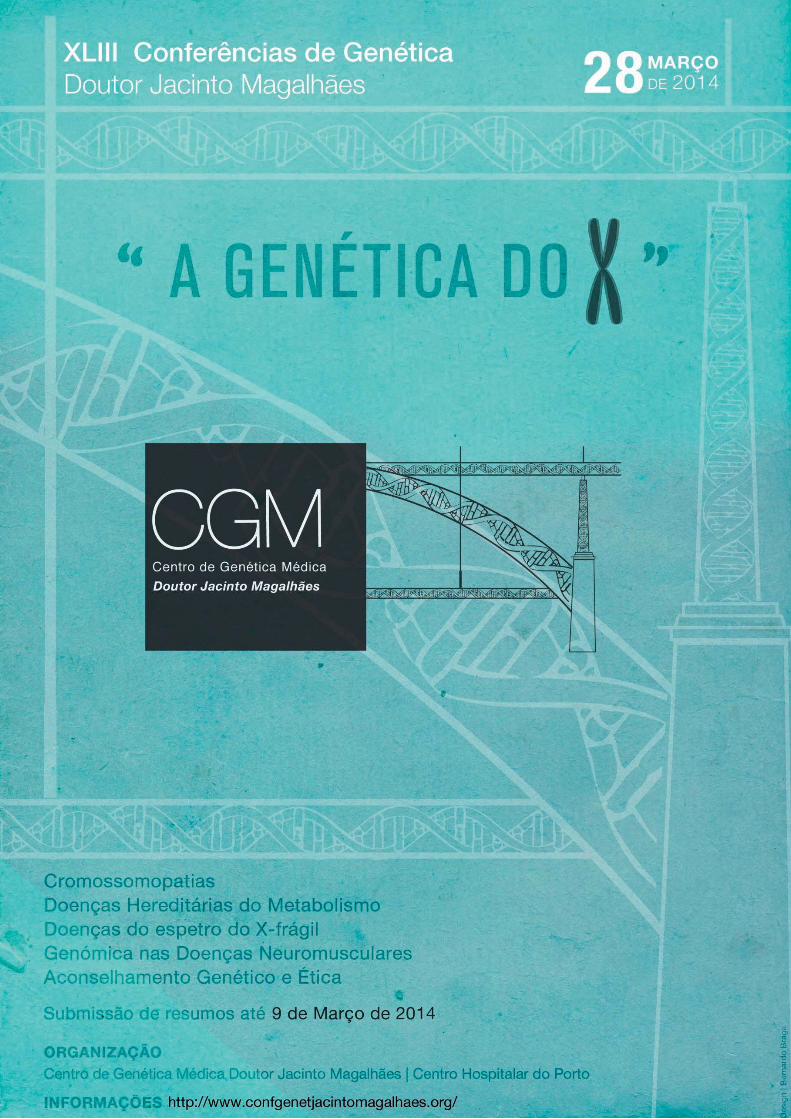
Resumo das Comunicações
“A Genética do X” - Porto, 28 Março 2014
Organização do Congresso l Congress Organization
Cristina Candeias
Dulce Quelhas
Francisco Laranjeira
Isabel Marques
Márcia Oliveira
Natália Oliva Teles
Comissão Científi ca l Scientifi c Committee
Ana Fortuna
Lúcia Lacerda
Natália Oliva Teles
Paula Jorge
Colaboração logística das Conferências l Local Support
CGMJM/CHP, DEFI/CHP e ADEMI
Contactos l Contacts [email protected]
Tel: 226 070 300
Fax: 226 070 399
Local das Conferências l Congress Venue
Auditório Prof. Doutor Alexandre Moreira, CHP
(entrada junto à URGÊNCIA - Hospital Santo António)
Index – Índice
Invited speakers – Comunicações por convite
CC-01 – X-Chromosome: Genetic Framing . . . . . . . . . . . . . . . . . . . . . . . . . S6
CC-02 – X-imbalances big and small . . . . . . . . . . . . . . . . . . . . . . . . . . . S6
CC-03 – Unravelling the X fi les: Challenges and Dilemmas. . . . . . . . . . . . . . . . . . . . . S7
CC-04 - An Introduction to X-Linked IMDs. . . . . . . . . . . . . . . . . . . . . . . . . . S7
CC-05 – Doença Hereditárias do Metabolismo: ExperiÊncia Clínica na Terapêutica . . . . . . . . . . . . . S8
CC-06 – XX, doente ou portadora? . . . . . . . . . . . . . . . . . . . . . . . . . . . . S8
CC-07 – Antisense-mediated exon skipping for Duchenne muscular dystrophy – clinical trials and beyond . . . . . S9
CC-08 – Discovering “X” in the myopathic equation . . . . . . . . . . . . . . . . . . . . . . . S9
CC-09 – FMR1 Associated Pathologies. . . . . . . . . . . . . . . . . . . . . . . . . . . S10
CC-10 – Genetic Counseling . . . . . . . . . . . . . . . . . . . . . . . . . . . . . . S10
CC-11 – Ética e Genética . . . . . . . . . . . . . . . . . . . . . . . . . . . . . . . S11
CC-12 – Um Passado com Futuro . . . . . . . . . . . . . . . . . . . . . . . . . . . . S11
Oral communication – Comunicação oral
CO-01 – Study Of The Fmr1 Gene Structure Among Women With Ovarian Dysfunction From The Basque Country . . . S12
Poster abstracts – Resumos de posters
P-01 – Investigation of X-chromosome inactivation patterns – a valuable tool in genetic diagnosis . . . . . . . . S13
P-02 – Next generation Fragile-X testing: getting away from Southern Blots . . . . . . . . . . . . . . . S14
P-03 – Portuguese patient registry for Duchenne/Becker muscular dystrophy. . . . . . . . . . . . . . . S14
P-04 – “Entre os genes e a mente” - Síndroma de Turner e manifestações psicopatológicas . . . . . . . . . . S15
P-05 – Craniofrontonasal syndrome: case report . . . . . . . . . . . . . . . . . . . . . . . . S16
P-06 – Translocação (X;10) aparentemente equilibrada de novo no sexo feminino: caso clínico . . . . . . . . . S16
P-07 – Estratégia Utilizada no Estudo Piloto Para O Rastreio Neonatal da Fibrose Quística . . . . . . . . . . S17
P-08 – XL-EDMD - genotypic spectrum among Portuguese patients . . . . . . . . . . . . . . . . . S17
P-09 – mX-Linked Centronuclear Myopathy: from clinical diagnosis to genetic counseling. . . . . . . . . . . S18
P-10 – Prenatal Diagnosis: a case of partial trisomy 6q . . . . . . . . . . . . . . . . . . . . . . S18
P-11 – Mutation analysis of genes involved in sperm motility: A study in patients with total sperm immotility . . . . . S19
P-12 – Prenatal diagnosis mosaic 45, X case with a marker chromosome . . . . . . . . . . . . . . . . S19
P-13 – X-linked Icthyosis – a metabolic ethiology for “dry skin” . . . . . . . . . . . . . . . . . . . S20
P-14 – Mosaicism with two X chromosome different rearrangements and a Turner-like phenotype: case report . . . . S21
P15 – Distal Xq27->Q28 Duplication And Functional Disomy: Clinical And Cytogenetic Characterization . . . . . . S21
P-16 – Clinical, Biochemical And Molecular Studies: Stepwise To Achieve Diagnosis Of Fabry Disease . . . . . . S22
P-17 – Hunter Syndrome, the most prevalent Mucopolysaccharidosis in Portugal . . . . . . . . . . . . . S22
P-18 – A Adrenoleucodistrofi a Ligada ao Cromossoma X em Portugal . . . . . . . . . . . . . . . . . S23
Author index – Índice remissivo . . . . . . . . . . . . . . . . . . . . . . . . . . . . . . S24
Sponsors and exhibitors – Apoios e expositores . . . . . . . . . . . . . . . . . . . . . . . . . S25
NASCER E CRESCERrevista de pediatria do centro hospitalar do porto28 de março de 2014, suplemento I
6 XLIII conferências de genética Doutor Jacinto MagalhãesSoradores convidados
CC-01 X-CHROMOSOME: GENETIC FRAMINGFernando RegateiroFaculdade de Medicina da Universidade de Coimbra, Coimbra, Portugal
In order to discuss such a vast subject as the “genetic framing of the X-chromosome in humans” it is mandatory to approach several topics, starting with the time-frame of mammalian evolution. For many years little importance was attributed to the understanding of the evolution (divergence) of the dimorphic X and Y chromosomes, sex determination in heterogametic XY species, in species without sex chromosomes and the accurate characterization of Y and X chromosomes per se.
However, with the advent of cytogenetics and molecular techniques, great advances have been achieved in the knowledge of X-inactivation and gene dosage compensation in order to equalize the gene dosage between the sexes and, possibly, also between sex chromosomes and autosomes. In a relatively short time the molecular mechanisms involved in X-inactivation as an epigenetic process have been elucidated and have enabled the scientific and medical improvement of X-linked conditions, wether dominant or recessive. These achievements were followed by other scientific advances that are now routine diagnostic tools: study of the gene XIST and the chromosome distribution of 158 IQ-related genes, the effect of sex chromosome gene dosage on brain structure, the genetic defects and genetic-environmental interactions associated with Alzheimer’s disease, fragile-X syndrome, X-linked genes and miRNA sexual dimorphism, hemophilia, Duchenne muscular dystrophy, Turner and Klinefelter syndromes.
CC-02X-IMBALANCES BIG AND SMALLNicole de LeeuwDepartment of Human Genetics, Radboud University Medical Centre,
Nijmegen, The Netherlands
The X chromosome is fascinating, but the clinical interpretation of X-chromosomal aberrations are often a challenge, in particular because of (possible) mosaicism and X-inactivation.
Various abnormalities involving X will be addressed in this lecture, including whole chromosome numerical abnormalities, supernumerary marker chromosomes, X-autosome translocations as well as recurrent and non-recurrent copy number variations. For many indications, molecular techniques such as QF-PCR and genome wide array analysis are nowadays often used to test the patient samples in prenatal and postnatal genome diagnostics. For the correct interpretation of these data, however, cytogenetic knowledge is necessary and often routine cytogenetic analysis and /or Fluorescence In Situ Hybridisation (FISH) is required to further characterise the X-chromosomal abnormality.
After doing the tests, it is crucial for the clinical laboratory geneticist to not only correctly use the existing nomenclature in the test report, but also to include a clear and concise explanation of what the test result means. The requesting clinician needs to understand the meaning of the laboratory findings and the underlying genetic mechanisms in order to be able to properly counsel the patient and the parents with regard to prognosis and recurrence risk.
A variety of illustrative case examples will be presented to address the aforementioned aspects, but most likely, some questions will remain unanswered.
Invited speakersComunicações por convite
NASCER E CRESCERrevista de pediatria do centro hospitalar do porto
28 de março de 2014, suplemento I
7XLIII conferências de genética Doutor Jacinto Magalhães S
oradores convidados
CC-03
UNRAVELLING THE X FILES: CHALLENGES AND DILEMMAS
Isabel M. Carreira
Laboratório de Citogenética e Genómica, Faculdade de Medicina da
Universidade de Coimbra, Coimbra, Portugal
CIMAGO – Centro de Investigação em Meio Ambiente, Genética e
Oncobiologia, Coimbra, Portugal
CNC – Centro de Neurociências e Biologia Celular, Universidade de
Coimbra, Coimbra, Portugal
Array-Comparative Genomic Hybridization (array-CGH)
has increased the diagnostic yield in patients with intellectual
disability (ID), autism spectrum disorders and multiple congenital
anomalies due to its improved resolution. X-chromosome has
been focus of attention due to the bias in the affected male-
to-female ratio and to the knowledge of X-linked genes
associated with ID. With array-CGH we can either detect single
gene imbalances, chromosomal region imbalances and even
aneuploidies.
In a cohort of 1000 patients studied by Agilent 180K
oligonucleotide array-CGH several X-chromosome imbalances
were detected. Single gene deletions involving ZNF41 or
IL1RAPL1 genes were equitably observed in 8 patients; DMD
imbalances in 3 females and SHOX gene duplications in 1 female
and 9 males. An intragenic deletion in SLC9A6 gene associated
with Christianson syndrome that segregated in the family was
also detected.
In 6 patients we identifi ed Xp22.31 duplications, 3 females,
1 male with maternal inheritance and 2 males whose inheritance
was not yet determined. A chromosome Xq27.1q28 interstitial
duplications in 2 males, 1 maternally inherited and the other not
yet determined were also identifi ed. We also found other genomic
imbalances but in single cases as for example a complex
rearrangement with multiple imbalances at Xp22.33p22.2 in a
male patient, maternally inherited; an Xp11.3p11.23 duplication
in a female with ID whose mother is also affected and a case of
triple X in an autistic female.
The challenge with X-chromosome imbalances is to,
understand the biological mechanism(s) behind, interpret
their impact on the phenotype, due to the presence of some
alterations in the normal population and to X-chromosome
inactivation in females. Clinical laboratory reporting has to use
the correct nomenclature and a clear and objective interpretation
of the results.
CC-04
AN INTRODUCTION TO X-LINKED IMDS
Stephen Waldek
Independent Medical Consultant, Manchester, UK
While individually the inherited metabolic diseases are rare
or very rare, overall the incidence is around 1:1,400 live births
and accounts for about 15% of all single gene disorders. The
vast majority of these diseases are inherited in a recessive
manner with 3 or 4 being dominant conditions. However, 14
are inherited in an X-linked fashion. By my estimation there are
over 200 conditions to consider, most of which are not treatable.
My presentation will focus on four diseases—Anderson-
Fabry disease (FD); Mucopolysaccharidosis type II (MPS II);
Ornithine Transcarbamylase defi ciency (OTC); and X-Linked
adrenoleucodystrophy (XALD)—that illustrate several points of
interest.
FD is a multi-system disease caused by a defi ciency of the
lysosomal enzyme alpha galactosidase. Accumulation of the
substrate globotriaosylceremide (GL3) leads to a sequence of
symptoms over time starting with severe neuropathic pain in the
peripheries and moving on to proteinuria renal failure, cardiac
and cerebrovascular disease. Without treatment death occurs by
the 4th or 5th decade. Fortunately, enzyme replacement therapy
is available. The clinical and therapeutic aspects of the disease
will be discussed as well as the issue of late onset disease and
the fact that there is a very high incidence of symptoms in the so
called female carriers.
MPS II, or Hunter syndrome, is another multisystem
lysosomal storage disorder caused by a defi ciency of iduronate-
2-sulphatase. The main features are due to skeletal involvement
and like FD there is enzyme replacement therapy. However,
unlike FD it is exceptionally rare for female carriers to develop
symptoms or signs of the disease.
OTC is the commonest of the urea cycle defects. The
symptoms are related to the accumulation of ammonia and will
be discussed. In most boys the disease presents in the neonatal
period. Many do not survive and those that do are usually severely
brain damaged and susceptible to destabilization throughout
their lives, even with the dietary treatment currently available.
Interestingly, as will be discussed, about 15% of females will
develop symptoms and require lifelong treatment. One of the
times of greatest risk is during pregnancy and delivery.
The presentation will also describe the various manifestations
of XALD from the severe childhood presentations to the adrenal
and neurological disease of the onset in older boys and young
men.
In addition to the clinical aspects of the four diseases,
information on diagnosis and genetic counselling implications
will be discussed.
NASCER E CRESCERrevista de pediatria do centro hospitalar do porto28 de março de 2014, suplemento I
8 XLIII conferências de genética Doutor Jacinto MagalhãesS
oradores convidados
CC-05
DOENÇA HEREDITÁRIAS DO METABOLISMO:
EXPERIÊNCIA CLÍNICA NA TERAPÊUTICA
Esmeralda Martins
Unidade de Doenças Metabólicas, Serviço de Pediatria, Hospital de
Santo António, Centro Hospitalar do Porto E.P.E., Porto, Portugal
As doenças hereditárias do metabolismo (DHM) são entida-
des de natureza genética, causadas por mutações num ou vá-
rios genes codifi cantes para um determinado passo metabólico.
A transmissão neste grupo de patologias pode ser mendeliana
(recessiva, dominante ou ligada ao X) ou mitocondrial.
Perante a suspeita clínica de um erro inato do metabolismo,
devemos considerar sempre em primeiro lugar as doenças tra-
táveis, uma vez que nestas, a instituição precoce de medidas
terapêuticas pode alterar o prognóstico do doente.
As diversas formas de tratamento nas doenças metabólicas
podem ser classifi cadas de acordo com o seu mecanismo de
ação:
- Restrição do substrato (redução do substrato da via me-
tabólica afetada),
- Correção da defi ciência de produto,
- Diminuição da toxicidade metabólica,
- Estimulação da atividade enzimática residual,
- Tratamento enzimático de substituição,
- Transplante de órgãos ou células estaminais,
São consideradas as principais DHM ligadas ao X para as
quais existe uma terapêutica específi ca, défi ce em ornitina car-
bamoil transferase, défi ce em piruvato desidrogenase, doença
de Hunter e doença de Fabry, referindo a evolução dos doentes
em tratamento.
CC-06
XX, DOENTE OU PORTADORA?
Francisco Laranjeira
Unidade de Bioquímica Genética, Centro de Genética Médica Doutor
Jacinto Magalhães, Centro Hospitalar do Porto E.P.E., Porto, Portugal
A Unidade de Bioquímica Genética (UBG) é o laboratório
nacional de referência para as doenças hereditárias do meta-
bolismo (DHM), nomeadamente dos grupos das doenças lisos-
somais, doenças peroxissomais e doenças congénitas da gli-
cosilação.
Nestes grupos encontram-se algumas patologias ligadas ao
cromossoma X: nas doenças peroxissomais, a adrenoleucodis-
trofi a ligada ao X (X-ALD) e nas doenças lisossomais temos a
doença de Fabry, a síndrome de Hunter, a ictiose ligada ao X
(XLI) e a doença de Danon.
A abordagem de estudo laboratorial destas patologias é di-
versa, podendo envolver diferentes tipos de metodologias bio-
químicas e ainda estudos de genética molecular:
A XLI é diagnosticada laboratorialmente pela determinação
da actividade enzimática;
O diagnóstico laboratorial de X-ALD é baseado no dosea-
mento de metabolitos, sendo complementado por estudos de
genética molecular;
Na doença de Fabry e síndrome de Hunter, é efetuado o
doseamento de metabolitos, a determinação da atividade enzi-
mática e estudos de genética molecular;
O estudo de genética molecular é a única metodologia labo-
ratorial usada no nosso laboratório para o diagnóstico da doen-
ça de Danon.
Tendo sido estudada na UBG a quase totalidade dos doen-
tes portugueses afetados por essas patologias, existem dados
que permitem traçar um quadro do panorama nacional relativo
às mesmas, com especial ênfase na análise da informação rela-
tiva aos indivíduos do sexo feminino.
Será apresentada a caracterização laboratorial, com os dife-
rentes tipos de dados recolhidos, para este grupo de indivíduos
e analisada a correlação com a apresentação clínica.
Serão apresentados exemplos de sucessos e difi culdades
que surgiram no diagnóstico de indivíduos do sexo feminino,
bem como casos com especial interesse.
É discutida também a importância da comunicação clínica/
laboratório, tanto no estabelecimento do diagnóstico como nas
tomadas de decisão relativamente a abordagens terapêuticas,
nomeadamente nas patologias para as quais existe terapia de
suplementação enzimática – doença de Fabry e síndrome de
Hunter – face à avaliação custo-benefício.
NASCER E CRESCERrevista de pediatria do centro hospitalar do porto
28 de março de 2014, suplemento I
9XLIII conferências de genética Doutor Jacinto Magalhães S
oradores convidados
CC-07
ANTISENSE-MEDIATED EXON SKIPPING FOR DUCHENNE
MUSCULAR DYSTROPHY – CLINICAL TRIALS AND BEYOND
Annemieke Aartsma-Rus
Department of Human Genetics, Leiden University Medical Centre,
Leiden, The Netherlands
Duchenne muscular dystrophy (DMD) is a severe, progressive
muscle-wasting disorder, while Becker muscular dystrophy
(BMD) is milder muscle disease. Both are caused by mutations
in dystrophin, a protein, which stabilizes muscle fi bers during
contraction by linking muscle actin to the extracellular matrix.
In DMD patients mutations disrupt the open reading frame,
generating prematurely truncated, nonfunctional dystrophins.
In BMD patients, mutations maintain the reading frame allowing
production of internally deleted, partly functional dystrophins.
The exon skipping approach uses antisense oligonucleotides
(AONs) to induce skipping of targeted exons during pre-mRNA
splicing, with the aim of reading frame restoration, converting of
the severe DMD into the milder BMD phenotype. This approach
is mutation specifi c. However, as mutations cluster in a few
hotspots, skipping of some exons applies to larger groups of
patients (e.g. exon 51 skipping applies to 13%).
After obtaining proof-of-concept in cultured patient-derived
cells, this approach was further optimized in animal models. In
each case AON treatment resulted in targeted exon skipping and
dystrophin restoration. In animal models this was accompanied
by improved muscle function and quality. Proof-of-concept in
patients was achieved in a clinical trial where 4 patients received
local injections with an AON targeting exon 51 (coordinated by
Prosensa Therapeutics). Dystrophin was restored locally for
each patient.
Towards systemic application, studies in animal models
revealed that dystrophic muscles facilitated uptake of 2OMePS
AONs and that subcutaneous delivery was feasible. In a
subsequent clinical trial, patients were subcutaneously injected
with AONS targeting exon 51. Dystrophin was restored in a
dose-dependent manner. All patients were enrolled in an open
label extension study and have received subcutaneous AON
injections at 6 mg/kg for almost 4 years. Two phase 2 and one
a pivotal, double-blind, placebo-controlled multicenter trial for
exon 51 skipping have recently been completed (coordinated by
GlaxoSmithKline).
In parallel, preclinical studies to further optimize treatment
regimens are in progress as well as clinical trials for additional
exons for exon 44 skipping (PRO044, applicable to 6% of
patients), exon 45 skipping and 53 skipping (PRO045 and
PRO053, both applicable to 8% of patients).
The mutation specifi city of the approach poses challenges to
drug development regulations. A concerted effort of academic
researchers, industry, regulators and patients is needed to adapt
regulations to enable application of these personalized medicine
approaches to rare diseases.
CC-08
DISCOVERING “X” IN THE MYOPATHIC EQUATION
Jorge Oliveira
Unidade de Genética Molecular, Centro de Genética Médica Doutor
Jacinto Magalhães, Centro Hospitalar do Porto E.P.E., Porto, Portugal
Congenital myopathies (CM) are a heterogeneous group
of diseases, generally characterized by hypotonia and muscle
weakness with onset at birth or during infancy and usually
with a slowly progressive disease course. Scientifi c and
technological developments in genomics over the last two
decades have contributed to the identifi cation of genetic causes
for a signifi cant number of myopathies. However, there are
still several challenges to address both in diagnostics and in
research. First, there is striking genetic and clinical heterogeneity
associated to CM. In fact, although muscle biopsy is paramount
for the diagnostic workup, pathognomonic fi ndings such as
cores, rods, central nuclei or fi bre-type disproportion, are not
gene-specifi c. In addition, a signifi cant subset of these patients
remains genetically unsolved, requiring further investigation that
may lead to the identifi cation of new genetic causes of CM.
Our recent research in congenital myopathies has focused
on the mutational profi le of the myotubularin gene (MTM1),
which is defective in X-linked centronuclear myopathy (CNM).
Male patients with MTM1 mutations are usually severally
affected, presenting neonatal hypotonia and inability to
maintain unassisted respiration. During the development and
implementation of a mutation database for MTM1 (http://www.
lovd.nl/MTM1), we noticed that no large duplications had been
reported. Large duplications in MTM1 were screened by the
MLPA technique in a small group of uncharacterized CNM
Portuguese patients. A large duplication spanning exons 1 to
5 was identifi ed in a boy with a mild CNM phenotype. Further
characterization revealed that this duplication causes an in-
frame deletion at the mRNA level (r.343_444del). Results
obtained using a low-coverage next generation sequencing
(NGS) approach showed that this genomic duplication extends
into the neighbouring MAMLD1 gene and subsequent analysis
unveiled the presence of a MTM1/MAMLD1 fusion transcript [1].
This work demonstrates that it is clinically relevant to screen
large MTM1 duplications in CNM patients since this type of
mutation may account for some cases that remain genetically
unanswered, as was recently validated by the publication of
additional cases. It also demonstrates how different analytical
approaches are often required to solve the genetic complexity
of congenital myopathies; the further application of NGS
technology in these disorders shall be exemplifi ed.
References
[1] Oliveira, J.; Oliveira, M.E.; Kress W.; Taipa, R.; Pires,
M.M.; Hilbert, P.; Baxter, P.; Santos, M.; Buermans, H.; den
Dunnen, J.T.; Santos, R. (2013). Expanding the MTM1 mutational
spectrum: novel variants including the fi rst multi-exonic
duplication and development of a locus-specifi c database.
European Journal of Human Genetics. 21(5): 540-549.
NASCER E CRESCERrevista de pediatria do centro hospitalar do porto28 de março de 2014, suplemento I
10 XLIII conferências de genética Doutor Jacinto MagalhãesS
oradores convidados
CC-09
FMR1 ASSOCIATED PATHOLOGIES
Montserrat Milà
Bioquímica i Genètica Molecular, Hospital Clinic, Barcelona, Spain
Fragile X syndrome is the most common form of inherited
mental retardation with a prevalence of approximately 1:2,466
men and 1:8,333 women in the Caucasian population. The
molecular basis of the syndrome is predominantly a CGG
expansion in the 5´- untranslated region of the FMR1 gene. In
the general population, individuals carry 6 to 55 repeats, and
the triplet number is usually stably transmitted. Individuals with
alleles between 55 and 200 CGG repeats are called premutated
carriers and those with more than 200 CGG are considered to
carry full mutations and present classical Fragile X syndrome.
In the premutated range, the CGG number is unstable through
transmission to the next generation and tends to expand.
Diagnosis is based on the determination of the CGG number.
FMR1 premutation is much more frequent than previously thought.
The most relevant pathologies associated with premutation have
been described to be Fragile X premature ovarian insuffi ciency
(FXPOI) and Fragile X tremor ataxia syndrome (FXTAS). Other
clinical manifestations, associated with this premutation, were
later identifi ed as thyroid dysfunction, chronic muscle pain
or fi bromyalgia, among others. While FXPOI and FXTAS are
defi nitively related, the latter manifestations require further
studies. Here we revise the current knowledge of the individuals
carrying FMR1 premutation.
CC-10
GENETIC COUNSELING
Ana Berta Sousa
Serviço de Genética, Departamento de Pediatria, Hospital de Santa
Maria, Centro Hospitalar Lisboa Norte, Lisboa, Portugal
Genetic counseling is the process by which patients or
relatives at risk of an inherited disorder are advised of the nature
and consequences of the disorder, the probability of developing
or transmitting it, and the options open to them in management
and family planning.
This complex process can be separated into diagnostic and
supportive aspects.
Establishing a correct diagnosis is crucial, otherwise
erroneous information will likely be given with potentially
tragic consequences. Reaching a diagnosis involves three
fundamental steps: taking a history, carrying out an examination
and undertaking appropriate complementary investigations.
An etiological diagnosis allows precise risk estimation.
Sometimes, even in the absence of a molecular diagnosis, a
pattern of Mendelian inheritance may be clear from the family
tree allowing the calculation of a recurrence risk. However,
in many instances it is not possible to arrive to an accurate
diagnosis and it is necessary to resort to empiric risks, derived
from family studies rather than theoretical calculations. In all
cases, recurrence risks should not only be quantifi ed but need
also to be qualifi ed and placed in context.
The supportive aspects of the counseling process involve
both communication and educational skills. Only an appropriately
trained professional can help the individual or the family gain
enough knowledge of the disorder and the options available for
risk management to allow fully informed decisions without undue
pressure or stress, in a way that promotes health, minimizes
psychological distress and increases personal control.
These concepts will be illustrated with relevant clinical
examples.
NASCER E CRESCERrevista de pediatria do centro hospitalar do porto
28 de março de 2014, suplemento I
11XLIII conferências de genética Doutor Jacinto Magalhães S
oradores convidados
CC-11
ÉTICA E GENÉTICA
Natália Oliva Teles
Unidade de Citogenética, Centro de Genética Médica Doutor Jacinto
Magalhães, Centro Hospitalar do Porto E.P.E., Porto, Portugal
UMIB-ICBAS-UP - Unidade Multidisciplinar de Investigação Biomédica,
Porto, Portugal
DCSS - Departamento de Ciências Sociais e Saúde, Faculdade de
Medicina, Universidade do Porto, Porto, Portugal
O conceito de ética, tal como o entendemos atualmente, re-
sultou da evolução do pensamento fi losófi co durante centenas
de anos e remonta à antiguidade grega e romana. Com a intro-
dução da “bioética”, termo proposto por Van Rensselaer Potter
em 1970, reconheceu-se a necessidade de conciliar os concei-
tos ancestrais da moralidade com os confl itos éticos resultantes
do evoluir da biomedicina e da tecnologia científi ca, sob pena
de comprometer os destinos da vida humana. Nos cuidados de
saúde em geral, pela sua grande aceitação, os princípios éticos
enunciados por Beauchamp e Childress – autonomia, benefi -
cência, não-malefi cência e justiça – constituem a base de refl e-
xão para a atuação de muitos profi ssionais de saúde, nos quais
se incluem os que trabalham em genética médica e humana;
nenhum destes princípios éticos deverá prevalecer sobre os ou-
tros, procurando-se habitualmente um consenso.
Na prática diária, os princípios éticos aplicam-se em genética
antes da execução de uma técnica ou do estabelecimento de um
diagnóstico, desde logo com uma consulta de aconselhamento
genético não-diretivo, a obtenção do consentimento informado
(se necessário, oral ou escrito e adequado a cada situação) e a
estrita manutenção da confi dencialidade dos dados pessoais e
clínicos recolhidos. A partir desta consulta os problemas éticos
que surgirem em cada situação serão resolvidos caso a caso,
tanto em situações pré-natais como pós-natais. Em qualquer cir-
cunstância, a multidisciplinaridade das equipas e o rigor científi co
dos profi ssionais, fundamentais para o avanço do conhecimento
e dos ganhos em saúde, não deverá descurar a legislação vigen-
te e, sobretudo, compreender a necessidade de haver uma nor-
malização razoável de procedimentos, pelo que se recomenda a
leitura da seguinte documentação: 1-Lei n.º 12/2005: Informação
genética pessoal e informação de saúde, DR-I SÉRIE-A, N.º 18,
26.01.2005; 2-Orientações e Princípios que orientarão a aplica-
ção de técnicas de biologia molecular no âmbito da prestação de
cuidados de saúde pelo SNS, Despacho nº 9108/97, DR-II Série,
N.º 237, 13.10.1997; 3-Lei nº 67/98, 26.10.1998: Lei de proteção
de Dados Pessoais, DR-I Série A, N.º 247, 26.10.1998; 4-Lei n.º
32/2006, 26.07: Procriação medicamente assistida, DR-I SÉRIE-
-A, N.º 143, 26.07.2006; 5-Decreto-Regulamentar n.º 5/2008, 11
de Fevereiro: Regula a utilização de técnicas de procriação medi-
camente assistida, DR-I SÉRIE-A, N.º 29, 11.02.2008; 6-Orienta-
ções e princípios que orientarão a estruturação do sector de DPN,
Despacho nº 5411/97, DR-II Série, N.º 180, 06.08.1997; 7-Comis-
sões de Ética para a Saúde, Decreto-Lei n.º 97/95: DR-I SÉRIE-A,
N.º 108, 10.05.1995.
CC-12
UM PASSADO COM FUTURO
Daniel Serrão
Universidade Católica Portuguesa, Porto, Portugal
O Instituto de Genética Médica Doutor Jacinto de Magalhães
tem um passado que é a garantia de ter futuro; um futuro que vai
poder ser criado e vivido sem destruição do passado
Porque acompanhei, de perto, a sua vida desde que o
saudoso Doutor Jacinto de Magalhães o concebeu, posso
apresentar um depoimento sobre todos os sucessos que
marcaram a sua existência, sobre a forma como foi sendo
fragilizado e sobre a esperança que se abre com o seu
acolhimento no Centro Hospitalar do Porto.
Para o seu arranque foi necessário que tivesse autonomia
fi nanceira e administrativa, com um Director nomeado
directamente pelo Ministro da Saúde e com ele resolvendo as
decisões a tomar para que o Instituto crescesse rapidamente e
sem os controlos burocráticos que tudo resolvem com lentidão.
Com a instalação e o desenvolvimento muito avançados
tornou-se naturalmente necessária a integração do Instituto
na estrutura geral do Ministério e a aceitação progressiva do
controlo burocrático-administrativo: orçamento próprio, quadro
de pessoal científi co, administrativo e técnico.
Esta evolução foi-se processando, com alguns acidentes de
percurso, até que uma reforma dos organismos do Ministério
da Saúde, levada a cabo pelo Ministro Correia de Campos,
originou a sua perda de identidade e a integração no Instituto
Nacional de Saúde Dr. Ricardo Jorge. Esta integração não teve
o benefício que era antecipado pelas convicções do Ministro,
nem quanto às economias, nem quanto à funcionalidade, nem
no que respeita ao enquadramento do pessoal nele existente.
A distância física entre Porto e Lisboa, e a difi culdade em
explicar as necessidades fi nanceiras e outras para um regular
funcionamento, originou um período de grande instabilidade com
saída de alguns investigadores, acolhidos noutras instituições, e
com uma dramática instabilidade para os que permaneceram
fi éis ao espírito de Jacinto de Magalhães, de um Instituto com
uma vertente clínica e uma vertente de investigação avançada
ao serviço da resolução dos problemas clínicos. Tudo estava
parado, em Lisboa, a aguardar decisões que não apareciam,
nem boas nem más.
A decisão superior de integrar o Instituto nas actividades de
investigação do Centro Hospitalar do Porto, surge como uma
solução inteligente, direi mesmo sábia, para dar a esta questão
uma saída honrosa para todas as partes e com um potencial
de crescimento no futuro que presta homenagem à memória
de Jacinto de Magalhães e à sua visão profética do futuro da
Genética Clínica e de investigação.

NASCER E CRESCERrevista de pediatria do centro hospitalar do porto28 de março de 2014, suplemento I
12 XLIII conferências de genética Doutor Jacinto MagalhãesS
comunicações orais
CO-01
STUDY OF THE FMR1 GENE STRUCTURE AMONG WOMEN
WITH OVARIAN DYSFUNCTION FROM THE BASQUE
COUNTRY
Maitane Barasoain 1, Gorka Barrenetxea 2,3, Iratxe Huerta 1,4,
Mercedes Télez 1,4, Amaia Carrillo 2, Cristina Pérez 2, Eduardo
Ortiz-Lastra 3, Javier González 3, Begoña Criado 5, Isabel Arrieta 1
1 Department of Genetics, Physical Anthropology and Animal
physiology, Faculty of Science and Technology, University of the
Basque Country, Bilbao, Spain; 2 Center for Reproductive Medicine and Infertility Quirón Bilbao, Bilbao,
Spain; 3 Department of Medical-Surgical Specialities, Faculty of Medicine,
University of the Basque Country, Bilbao, Spain; 4 Virgen de Begoña Clinical Analysis Laboratory (Medikosta), Erandio,
Spain; 5 Cooperativa de Ensino Superior Politécnico e Universitário (CESPU),
Porto, Po rtugal
FMR1 premutation and intermediate alleles have been
associated with the development of different forms of ovarian
dysfunction, being the Premature Ovarian Failure (POF)
the most serious one. A group of 68 women with ovarian
dysfunction of unknown aetiology and 47 control women
from the Basque Country has been analyzed. Considering
the number of CGG repeats, the frequency of alleles with
≥35 CGG repeats was statistically higher in the whole patient
group (12.50% vs. 0%). Concerning their ovarian condition,
the patient group was divided in three categories and, in
the three subgroups the alleles with ≥35 CGG were also
statistically higher than in controls. As the AGG interspersion
pattern seems to be correlated with the instability of the alleles,
the CGG repeat internal structures have been analyzed.
Many of the intermediate and premutation alleles found in the
patient group appeared to have two interruptions with more
than 15 CGG at the 3’ end (65%). Interestingly, among these
alleles the predominant structure was 9+9+n, indicating a
loss of AGG interruptions at the 3’ end. Therefore, the data
showed that among patients the alleles were more unstable
and that this instability infl uencing the FMR1 expansion might
be related with the development of an ovarian dysfunction.
Oral communication
Comunicação oral
NASCER E CRESCERrevista de pediatria do centro hospitalar do porto
28 de março de 2014, suplemento I
13XLIII conferências de genética Doutor Jacinto Magalhães Sposters
P-01
INVESTIGATION OF X-CHROMOSOME INACTIVATION
PATTERNS – A VALUABLE TOOL IN GENETIC DIAGNOSIS
Ana Gonçalves 1, Paula Jorge 1,2, Rosário Santos 1
1 Unidade de Genética Molecular, Centro de Genética Médica Doutor
Jacinto Magalhães, Centro Hospitalar do Porto E.P.E., Porto, Portugal; 2 Unidade Multidisciplinar de Investigação Biomédica, ICBAS-UP
To overcome the gene dosage differences between males
and females, one of the X-chromosomes is epigenetically
silenced in the early embryogenic process of the female
foetus. The choice of which one remains active in each
cell is thought to be a random process, resulting, in most
cases, in a uniform X-chromosome inactivation (XCI) pattern
of cells. However, studies in large cohorts of phenotipically
unaffected females indicate that about 8.8% exhibit skewed
profi les (>80:20). Although this skewed ratio has no clinical
signifi cance in unaffected females, it may explain disease
manifestation in otherwise non-affected carries of recessive
X-linked conditions. As suggested by several authors, the
assessment of XCI patterns can be very useful in confi rming
the diagnosis of disorders involving the X-chromosome, in
female patients. In our laboratory, we apply the HUMARA
assay to determine the pattern of XCI. This widely used
method is based on the analysis of DNA methylation and
number of CAG tandem repeats at the Human Androgen
Receptor (AR) gene locus. The AR gene’s highly polymorphic
CAG repeat enables distinction between maternal and
paternal X-chromosomes, while the close proximity
of cleavage sites for methylation-sensitive restriction
enzymes allows the discrimination of the inactive and
active X-chromosome. The work presented demonstrates
the importance of performing XCI studies and determining
their impact in both the clinical and the laboratory context.
Several examples will be described including manifesting
carriers of X-linked recessive disorders; female carriers
of translocations involving the X-chromosome, carriers of
newly described variants of undetermined pathogenicity and
female carriers with a suspected family history of X-linked
disorders associated with unilateral XCI (where no sample is
available from the affected male or where no mutation has
been identifi ed). Our results corroborate previous studies
and show that methylation status in the AR locus is a reliable
method to study XCI, therefore illustrating the confi dence
of this approach. Nevertheless, interpretation of XCI results
should be done with caution: XCI can only be ascertained
in the specimen being analyzed and may not refl ect the XCI
patterns in other tissues; it is an age-dependent phenomenon
(since skewing increases with age); in some cases, the locus
under study may not be in linkage desequilibrium with the AR
locus; other genetic factors are known to play an important
role in XCI process; symptomatic females may also have
other factors contributing to their phenotype.
Poster abstracts
Resumos de posters
NASCER E CRESCERrevista de pediatria do centro hospitalar do porto28 de março de 2014, suplemento I
14 XLIII conferências de genética Doutor Jacinto MagalhãesSposters
P-02
NEXT GENERATION FRAGILE-X TESTING: GETTING AWAY
FROM SOUTHERN BLOTS
Nuno Maia 1, Isabel Marques 1, Paula Jorge 1,2, Rosário Santos 1
1 Unidade de Genética Molecular, Centro de Genética Médica Doutor
Jacinto Magalhães, Centro Hospitalar do Porto E.P.E., Porto,
Portugal; 2 Unidade Multidisciplinar de Investigação Biomédica, ICBAS-UP
Fragile X syndrome (FXS) is the most common form
of intellectual disability in the general population, usually
caused by an expansion of a trinucleotide CGG repeat in
the 5’ untranslated region of the FMR1 gene. In most cases,
expansions over 200 repeats, termed full mutations, cause
silencing of FMR1 gene due to methylation of its promoter,
and consequently loss of protein product. Expansions
with 55-199 CGG repeats called pre-mutations or others
even smaller (45-54 CGG repeats) named intermediate,
do not cause FXS, but are frequently associated with late-
onset neurological and/or reproductive disorders (FXTAS/
FXPOI). Southern Blot (SB) is still considered the gold
standard for molecular diagnosis of FXS, because it is able
to clearly characterize size and methylation status of full
and pre-mutated FMR1 alleles (following DNA digestion
with methylation sensitive enzymes). Nevertheless, SB
is a very time-consuming technique and requires a large
amount of intact and high-molecular weight DNA. As such,
several methodologies have been developed to replace SB
and overcome its disadvantages. The aim of this work was
to test different techniques which can substitute totally or
partially the SB, by comparing (1) their ability to quantify or
discriminate normal, pre-mutated and fully mutated alleles;
(2) the maximum number of CGG repeats detected; (3) their
capacity to determine the DNA methylation state; (4) their
power to discriminate size and methylation mosaics; and (5)
the amount of DNA required for each technique. The tested
techniques were High Resolution Melting Curve Analysis
(currently in experimental process with prototype reagents),
the FragilEase™ assay from PerkinElmer®, the Amplidex®
FMR1 mPCR Protocol from Asuragen® and a multiplex assay
developed by our group, for the simultaneous screening
of 3 genes - FMR1, AFF2 e ARX. For this work we tested
7 DNA samples from patients previously characterized at
the molecular level in our laboratory: 6 from females and 1
obtained from a chorionic villus sample of a male fetus. These
samples were previously characterized as normal (n=2), fully
mutated (n=3), pre-mutated (n=1) and a size mosaic (n=1).
Although using a very small number of samples, this work
aims to describe and compare four different methodologies
in an attempt to establish if they can adequately replace SB.
P-03
PORTUGUESE PATIENT REGISTRY FOR DUCHENNE/
BECKER MUSCULAR DYSTROPHY
Jorge Oliveira 1, Ana Gonçalves 1, Teresa Moreno 2, Manuela
Santos 3, Isabel Fineza 4, Rosário Santos 1
1 Unidade de Genética Molecular, Centro de Genética Médica Doutor
Jacinto Magalhães, Centro Hospitalar do Porto E.P.E., Porto, Portugal; 2 Unidade de Neuropediatria, Hospital de Santa Maria, Centro
Hospitalar Lisboa Norte, Lisboa, Portugal; 3 Consulta de Doenças Neuromusculares, Serviço Neuropediatria,
Centro Hospitalar do Porto E.P.E., Porto, Portugal; 4 Centro de Desenvolvimento da Criança Luís Borges, Hospital
Pediátrico de Coimbra, Centro Hospitalar e Universitário de Coimbra,
Coimbra, Portugal
Duchenne/Becker muscular dystrophy (D/BMD)
collectively known as the dystrophinopathies, is one the
most frequent neuromuscular diseases with onset during
pediatric age, having an estimated incidence of about one
in every 3500 to 5000 boys. Over the last two decades,
the Molecular Genetics Unit of CGMJM has performed
the genetic characterization of over 360 D/BMD patients,
leading to the identifi cation of 189 mutations, including
46 novel variants. Comprehensive analysis also involved
expression studies at the mRNA level, the identifi cation
of splicing changes and ultimately providing evidence for
apparent exceptions to the reading-frame rule. Considering
the recent mutation-based therapeutic approaches, DMD
gene analysis has gone beyond the molecular confi rmation
of the clinical diagnosis and is now also crucial for patient
inclusion in disease registries and in ongoing clinical trials.
In 2007, the network of excellence for the neuromuscular
fi eld - TREAT-NMD - started a global patient registry for D/
BMD. This registry depends entirely on data gathered at the
national level in country-specifi c disease registries using
the same database items (mutational and clinical). This
standardization enables consensus and facilitates clinical
research, the development of new therapeutic approaches
and clinical trials for new drugs. These trial-ready registries
are also useful for phenotype/genotype correlations and
epidemiological profi les of the disease. In response to this
international endeavor, we developed the Portuguese D/BMD
registry which is currently located in the CGMJM, Centro
Hospitalar do Porto. The Portuguese registry is based on the
Leiden Open Variation Database (LOVD) software and follows
the TREAT-NMD charter for patient database/registry,
abiding by National and European legislation concerning
data. The national registry uses the clinical reporting model,
where three medical coordinators from major hospital centers
(Porto, Coimbra and Lisbon) were assigned to data collection
(personal, clinical and pathological data) and patients’
regular clinical (re)evaluation. Registry inclusion is completely
voluntary and requires a specifi c informed consent. All the
information, namely data sent by the clinician, consent and
NASCER E CRESCERrevista de pediatria do centro hospitalar do porto
28 de março de 2014, suplemento I
15XLIII conferências de genética Doutor Jacinto Magalhães Sposters
the genetic data obtained in the laboratory, is assembled
by the D/BMD registry curators and added to the database
after validation. The registry was offi cially launched in 2012
and until now eighteen patients have been included in the
database.
P-04
“ENTRE OS GENES E A MENTE” - SÍNDROMA DE TURNER
E MANIFESTAÇÕES PSICOPATOLÓGICAS
Pedro Oliveira 1, Joana Jorge 1, Otília Queirós 1
1 Psiquiatria da Infância e da Adolescência, Hospital Magalhães Lemos,
Centro Hospitalar do Porto E.P.E., Porto, Portugal
A Síndrome de Turner (ST) é uma cromossomopatia ca-
raterizada pela monossomia total ou parcial do cromossoma
X. Ocorre de forma esporádica, afetando 1 em cada 2000-
5000 recém-nascidos do género feminino, associando-se
habitualmente a baixa estatura, disgenesia gonadal, ano-
malias congénitas e adquiridas e sinais dismórfi cos. Estão
igualmente presentes alterações neuropsiquiátricas como
difi culdades cognitivas, distúrbios de perceção espacial e
temporal, memória visual, atenção, reconhecimento e inter-
pretação de emoções. São relatadas na literatura algumas
doenças psiquiátricas em doentes portadoras de Síndroma
de Turner, de que são exemplo as Perturbações do Humor,
a Esquizofrenia e a Anorexia Nervosa. No entanto, parece
não haver risco aumentado de psicopatologia grave. Verifi -
ca-se, por seu lado, maior risco de difi culdades psicosso-
ciais, difi culdades específi cas de aprendizagem, problemas
de comportamento e baixa autoestima. Tratando-se de uma
“doença crónica”, compreende-se, ainda, o possível impacto
emocional. A propósito de uma adolescente com Síndrome
de Turner, internada no Departamento de Psiquiatria da In-
fância e Adolescência do CHP, revêem-se os dados existen-
tes na literatura sobre as manifestações psicopatológicas da
Síndrome de Turner.
Palavras-chave: Síndrome de Turner; cromossomopatias;
psicopatologia.
NASCER E CRESCERrevista de pediatria do centro hospitalar do porto28 de março de 2014, suplemento I
16 XLIII conferências de genética Doutor Jacinto MagalhãesSposters
P-05
CRANIOFRONTONASAL SYNDROME: CASE REPORT
Gabriela Soares 1, Natália Tkachenko 1, Ana Maria Fortuna 1,2
1 Unidade de Genética Médica, Centro Genética Médica Doutor Jacinto
Magalhães, Centro Hospitalar do Porto E.P.E., Porto, Portugal;2 Unidade Multidisciplinar de Investigação Biomédica, ICBAS-UP
Background: Craniofrontonasal syndrome (CFNS, MIM
#304110) is a very rare X-linked developmental malformation,
caused by mutations in the EFNB1 gene, located at Xq13.1.It
was fi rst identifi ed as a subgroup of frontonasal dysplasia
by Cohen in 1979. The incidence values that were reported
ranged from 1:100.000 to 1:120.000. Heterozygous females
have craniofrontonasal dysplasia (CFND) and occasionally
extracranial manifestations including midline defects and
skeletal abnormalities, whereas hemizygous males show no
or only mild features such as hypertelorism and rarely show
cleft lip or palate.
Methods: Following fi rst description of the syndrome,
approximately 180 additional cases have been published in
medical literature. We report here on an additional case of a
Portuguese girl with CFNS. We compared the clinical features
of the previously published cases of craniofrontonasal
syndrome with our case.
Results: Common fi ndings in all reports, including our
case, are coronal craniosynostosis, craniofacial asymmetry,
hypertelorism, downslanting palpebral fi ssures, broad bifi d
nose, malocclusion and longitudinally grooved fi ngernails.
Craniofrontonasal syndrome was confi rmed in this patient by
molecular analysis of EFBN1 gene which was excluded in her
father.
Discussion: CFNS shows a phenotypic pattern not
usually seen in X-linked disorders, as heterozygous females
are more severely affected than hemizygous males.
Mutations in EFNB1 are the cause of CFNS in the majority
of patients, with a mutation detection rate of 92%. CNFS’s
clinical manifestations are sex dependent, with multiple
skeletal malformations in affected females and mild or no
malformations in male carriers. X-inactivation is proposed to
explain the more severe outcome in heterozygous females,
as this leads to functional mosaicism for cells with differing
expression of EFNB1, generating abnormal tissue boundaries
— a process that cannot occur in hemizygous males. Our
report discusses a patient with clinical characteristics
consistent with CNFS and in whom a de novo EFNB1
mutation was demonstrated. Postzygotic mutation leading
to somatic/germline mosaicism in the fi rst generation is a
relatively common feature of this condition and could not
be excluded in the father, who had mild hypertelorism. This
issue and its implications in recurrence risk were discussed
with the couple.
P-06
TRANSLOCAÇÃO (X;10) APARENTEMENTE EQUILIBRADA
DE NOVO NO SEXO FEMININO: CASO CLÍNICO
Natália Tkachenko 1, Gabriela Soares 1, Maria da Luz Silva 2,
Teresa Martins 3, Ana Maria Fortuna ,1,4
1 Unidade de Genética Médica, Centro Genética Médica Doutor Jacinto
Magalhães, Centro Hospitalar do Porto E.P.E., Porto, Portugal; 2 Unidade de Citogenética, Centro Genética Médica Doutor Jacinto
Magalhães, Centro Hospitalar do Porto E.P.E., Porto, Portugal; 3 Serviço de Neonatologia, Hospital Pedro Hispano, Unidade Local de
Saúde de Matosinhos, Matosinhos, Portugal; 4 Unidade Multidisciplinar de Investigação Biomédica, ICBAS-UP
Introdução: A translocação é uma anomalia cromossó-
mica que envolve quebras de dois cromossomas diferentes
com uma troca dos segmentos. Essa alteração pode ser
equilibrada ou desequilibrada, ocorrer de novo ou ser her-
dada. Geralmente, um portador de translocação equilibrada
não apresenta anomalias fenotípicas devidas a alteração
cromossómica, exceto um risco acrescido de anormalidades
reprodutivas, incluindo infertilidade, abortamentos de repeti-
ção e descendência com malformações e/ou atraso mental.
A translocação equilibrada entre o cromossoma X e um au-
tossoma, no sexo feminino, é uma situação específi ca. Uma
vez que um dos cromossomas X das mulheres se encon-
tra inativado, uma translocação envolvendo o cromossoma
X num indivíduo do sexo feminino, mesmo aparentemente
equilibrada, pode originar patologia por inativação do seg-
mento autossómico localizado no cromossoma X se este es-
tiver inativado.
Caso clínico: Apresentamos um caso clínico de uma
menina de 2 anos que foi referenciada à nossa consulta de
Genética por apresentar atraso do desenvolvimento psico-
motor e dismorfi a craniofacial e ter sido detetada uma trans-
locação aparentemente equilibrada envolvendo os cromos-
somas X e 10 que ocorreu de novo. Cariótipo: 46,X,t(X;10)
(p11.23;q11.21)dn. O estudo da inativação do cromossoma
X no sangue revelou um desvio completo a favor do cromos-
soma X normal.
Discussão: Uma translocação aparentemente equilibra-
da entre os cromossomas X e autossoma envolve o fenómeno
de inativação do X. No caso da inativação do cromossoma X
envolvido na translocação, este fenómeno pode originar uma
deleção funcional do segmento autossómico. O fenótipo dos
doentes descritos na literatura com deleções do segmento
terminal 10q é semelhante ao do nosso caso, o que aponta
para que o cromossoma X derivado desta translocação pos-
sa estar inativo noutros tecidos, especialmente SNC, e seja a
causa dos problemas da menina, uma vez que originaria uma
deleção funcional do segmento 10q11.21 a qter.
NASCER E CRESCERrevista de pediatria do centro hospitalar do porto
28 de março de 2014, suplemento I
17XLIII conferências de genética Doutor Jacinto Magalhães Sposters
P-07
ESTRATÉGIA UTILIZADA NO ESTUDO PILOTO PARA O
RASTREIO NEONATAL DA FIBROSE QUÍSTICA
Lurdes Lopes 1, Ana Marcão 1, Ivone Carvalho 1, Carmen Sousa 1,
Helena Fonseca 1, Hugo Rocha 1, Laura Vilarinho 1
1 Unidade de Rastreio Neonatal, Metabolismo e Genética,
Departamento de Genética Humana, Instituto Nacional de Saúde
Doutor Ricardo Jorge, Porto, Portugal
O Programa Nacional de Diagnóstico Precoce (PNDP)
realiza-se em Portugal desde 1979, e atualmente inclui o ras-
treio neonatal de 24 Doenças Hereditárias do Metabolismo
(DHM) e do Hipotiroidismo Congénito (HC). Em Outubro de
2013 iniciou-se, um estudo piloto para o rastreio neonatal
da Fibrose Quística (FQ), que deverá ser efetuado em 80000
recém-nascidos (RN) portugueses ao longo de aproximada-
mente um ano. A Fibrose Quística (Mucoviscidose) é uma
doença metabólica genética, com transmissão autossómica
recessiva, e que tem uma prevalência média ao nascimento
de 1:3000 RN, na população caucasiana. Bioquimicamente
deve-se à defi ciência na proteína CFTR, codifi cada pelo gene
CFTR, localizado no cromossoma 7. Estão descritas cerca
de 2000 variantes genéticas associadas à FQ. Clinicamente é
uma doença grave com atingimento multissistémico, carac-
terizada pela disfunção das glândulas exócrinas, incluindo
o pâncreas, as glândulas sudoríparas e as glândulas muco-
sas dos tratos respiratório, gastrointestinal e reprodutivo. O
aumento dos valores de ião cloreto no suor é típico destes
doentes, sendo o “teste do suor” a principal análise de con-
fi rmação da doença. Diagnosticar precocemente a doença
é uma fator decisivo no prognóstico, não só pela maior so-
brevida, mas também para uma melhor qualidade de vida
do doente. O aumento da concentração sanguínea da trip-
sina imunoreactiva (IRT) nos 1os dias de vida dos RN com
FQ possibilita o rastreio neonatal desta doença. No entanto,
apesar de uma boa sensibilidade, o IRT não é um marca-
dor específi co para a FQ, e um rastreio baseado unicamente
neste marcador tem um número inaceitável de falsos posi-
tivos. Por esta razão, têm sido propostos vários algoritmos
de rastreio, incluindo outros marcadores bioquímicos como a
Proteína Associada à Pancreatite (PAP) ou o estudo molecu-
lar. Neste estudo piloto, o algoritmo de diagnóstico utilizado
baseia-se na determinação do IRT e do PAP em sangue co-
lhido em papel de fi ltro, sendo a amostra de sangue a mesma
colhida para os restantes rastreios. No âmbito deste projeto
já foram estudados cerca de 29 000 RN e identifi cados 6
casos positivos. No fi nal deste estudo deverá ser avaliada a
inclusão da FQ no PNDP e o algoritmo de rastreio a utilizar.
P-08
XL-EDMD - GENOTYPIC SPECTRUM AMONG
PORTUGUESE PATIENTS
Emília Vieira 1, Ana Gonçalves 1, Elsa Bronze-da-Rocha 2,3,
Rosário Santos 1,2
1 Unidade de Genética Molecular, Centro de Genética Médica Doutor
Jacinto Magalhães, Centro Hospitalar do Porto E.P.E., Porto,
Portugal; 2 Departamento de Bioquímica, Faculdade de Farmácia, Universidade
do Porto, Porto, Portugal;3 Instituto de Biologia Molecular e Celular, Universidade do Porto,
Porto, Portugal
Emery-Dreifuss muscular dystrophy (EDMD) is
characterized by the clinical triad of joint contractures
that begin in early childhood, slowly progressive muscle
weakness and wasting initially in a humero-peroneal
distribution that later extends to the scapular and pelvic
girdle muscles, and cardiac involvement that may manifest
as palpitations, presyncope and syncope, poor exercise
tolerance, and congestive heart failure, that can result in
sudden death. Age of onset, severity, and progression of
muscle and cardiac involvement demonstrate both inter- and
intrafamilial variability. Clinical variability ranges from early
onset with severe presentation in childhood to late onset with
slow progression in adulthood. In general, joint contractures
appear during the fi rst two decades, followed by muscle
weakness and wasting. Cardiac involvement usually occurs
after the second decade. The three genes in which mutations
are known to cause EDMD are EMD (encoding emerin) and
FHL1 (encoding FHL1), which cause X-linked EDMD (XL-
EDMD) and LMNA (encoding lamin A and C), which causes
autosomal dominant and autosomal recessive EDMD (AD-
EDMD and AR-EDMD). For all forms of EDMD the diagnosis
is based on clinical fi ndings and family history. The diagnosis
of X-linked EDMD also relies on failure to detect emerin or
FHL1 protein in various tissues and molecular genetic testing
of EMD or FHL1 whereas AD- and AR-EDMD diagnosis
relies on molecular genetic testing of LMNA. We describe
the molecular results for EMD gene screening, in a group
of twenty-one Portuguese families, with clinical diagnosis
of EDMD and presenting different clinical phenotypes, with
or without cardiac involvement. Differential diagnosis of
XL-EDMD was achieved in fi ve families (eight patients).
Four different mutations were identifi ed, two of which have
not been documented in the literature. In a female patient,
a skewed X inactivation pattern was observed, explaining
disease manifestation. In the remaining families, the LMNA
gene was studied leading to confi rmation of laminopathy in a
four families. Molecular diagnosis is therefore very important
for an early diagnosis, to prevent sudden deaths, and to
distinguish X-linked EDMD from the autosomal forms, which
is essential for a correct genetic counseling and subsequent
prenatal diagnosis.
NASCER E CRESCERrevista de pediatria do centro hospitalar do porto28 de março de 2014, suplemento I
18 XLIII conferências de genética Doutor Jacinto MagalhãesSposters
P-09
MX-LINKED CENTRONUCLEAR MYOPATHY: FROM
CLINICAL DIAGNOSIS TO GENETIC COUNSELING
Maria João Sá 1,2, Ana Rita Soares 1, Gabriela Soares 1, Ana Maria
Fortuna ,1, Ricardo Taipa 4, Manuel Melo Pires 4, Jorge Oliveira 5,
Rosário Santos 5, Manuela Santos 3, Cristina Garrido 3
1 Unidade de Genética Médica, Centro de Genética Médica Doutor
Jacinto Magalhães, Centro Hospitalar do Porto, E.P.E., Porto,
Portugal; 2 Unidade Multidisciplinar de Investigação Biomédica, ICBAS-UP; 3 Consulta de Doenças Neuromusculares. Consulta de Neuropediatria,
Centro Hospitalar do Porto E.P.E., Porto, Portugal; 4 Unidade de Neuropatologia, Centro Hospitalar do Porto E.P.E., Porto,
Portugal; 5 Unidade de Genética Molecular, Centro de Genética Médica Doutor
Jacinto Magalhães, Centro Hospitalar do Porto E.P.E., Porto, Portugal
Background: X-Linked Centronuclear Myopathy is a rare
congenital myopathy characterized by hypotonia, muscle
weakness and respiratory distress at birth, although the
presentation may be delayed. It is the most severe and the
most common of the three inheritance forms, which also
include the autosomal dominant and the autosomal recessive
centronuclear myopathies. While muscle biopsy is crucial to
differentiate centronuclear myopathies from other congenital
myopathies and muscular dystrophies, genetic testing is
essential to establish a defi nitive diagnosis and to perform a
precise genetic counseling.
Clinical report: We report a proband, fi rst son of
healthy non-consanguineous parents, who presented with
severe congenital hypotonia, global muscle weakness
and bilateral hand contractures. He was born prematurely,
shortly after polyhydramnios diagnosis, at 30 gestational
weeks. Ventilatory support was required since his birth. At
examination, dolichocephaly was evident and he had ptosis
and ophtalmoparesis, facial diparesia, as well as a weak
cry. Muscle biopsy revealed fi bers with variable diameter,
including round atrophic fi bers, with centrally located nuclei,
as well as central areas of increased oxidative activity
surrounded by a bright halo, which was compatible with a
centronuclear myopathy. The previously reported pathogenic
missense variant c.566A>G (p.Asn189Ser) was detected
in the MTM1 gene, in hemizygosity in the proband and
heterozygosity in the mother, confi rming the diagnosis of
X-Linked Centronuclear Myopathy.
Discussion: The genetic testing of the X-linked form is
warranted as a fi rst-tier investigation in male infants with a
severe phenotype and a characteristic muscle biopsy, since
the autosomal forms of centronuclear myopathies present
with a relatively mild phenotype in both males and females.
The identifi cation of a pathogenic MTM1 mutation will enable
preimplantation genetic diagnosis or prenatal diagnosis, as
additional reproductive options for this couple.
P-10
PRENATAL DIAGNOSIS: A CASE OF PARTIAL TRISOMY 6Q
Rosário Pinto Leite 1, Pedro Botelho 1, Marta Souto 1, Rosete
Nogueira 2,3, António Carvalho 4, Osvaldo Moutinho 4,5, Márcia
Martins 5 1 Laboratório de Citogenética, Serviço de Genética, Centro Hospitalar
Trás-os-Montes e Alto Douro, Vila Real, Portugal; 2 Pathology Laboratory CGC Genetics /Centro Genética Clinica, Porto,
Portugal; 3 Life and Health Sciences Research Institute (ICVS), School of Health
Sciences, University of Minho, Braga, Portugal; 4 Serviço de Ginecologia/Obstetrícia, Centro Hospitalar Trás-os-Montes
e Alto Douro, Vila Real, Portugal; 5 Serviço de Genética, Centro Hospitalar Trás-os-Montes e Alto Douro,
Vila Real, Portugal
Partial distal trisomy 6q is a rare event and is characterized
by a distinct phenotype which includes microcephaly,
acrocephaly, joint contractures and profound psychomotor
retardation. The authors present a case of a 30-year-old
pregnant woman referred to prenatal diagnosis due to
ultrasound anomalies. It was the fi rst pregnancy of a non-
consanguineous couple with no familial or personal story of
anomalies. Parents karyotype was performed. Cytogenetic
analysis revealed a chromosome 15 with an increase p arm
similar to a variation in length of heterochromatic stalks
on the short arm. Both parents presented a chromosome
15 with satellites but different from the one detect at the
amniocytes. Subtelomeric FISH analysis revealed a trisomy
of 6q27-qter present at p arm of chromosome 15 - it was
a de novo rearrangement.The parents decided to terminate
the pregnancy and foetal autopsy was required. Several
polymorphic variants were described in human chromosome
15 including increased amounts of short arm hetrochromatin
(ph+), interpreted as a normal polymorphism.In the majority
of cases partial trisomy 6q results from a balanced
chromosomal rearrangement in one of the parents, usually
of maternal origin. There have also been rare cases in which
partial trisomy 6q has appeared from spontaneous (de novo)
errors very early in embryonic development. The authors
compared the cytogenetic and the foetal autopsy fi ndings
with those described in the literature. Every new case of a
rare chromosomal alteration should be reported in order to
establish a genotype/ phenotype correlation, improving risk
evaluation and genetic counseling.
NASCER E CRESCERrevista de pediatria do centro hospitalar do porto
28 de março de 2014, suplemento I
19XLIII conferências de genética Doutor Jacinto Magalhães Sposters
P-11
MUTATION ANALYSIS OF GENES INVOLVED IN SPERM
MOTILITY: A STUDY IN PATIENTS WITH TOTAL SPERM
IMMOTILITY
Rute Pereira 1,2, Jorge Oliveira 3, Rosário Santos3 , Ângela Alves 2,
Elsa Oliveira 2, Luís Ferraz 4, Alberto Barros 5, Mário Sousa 2
1 Departamento de Biologia, Faculdade de Ciências da Universidade do
Porto, Porto, Portugal; 2 Departamento de Microscopia, Laboratório de Biologia Celular,
Instituto de Ciências Biomédicas de Abel Salazar, UMIB-FCT,
Universidade do Porto, Porto, Portugal; 3 Unidade de Genética Molecular, Centro de Genética Médica Doutor
Jacinto Magalhães, Centro Hospitalar do Porto E.P.E., Porto, Portugal; 4 Departamento de Urologia, Centro Hospitalar de Vila Nova de Gaia
E.P.E., Vila Nova de Gaia, Portugal; 5 Centro de Genética da Reprodução Prof. Alberto Barros, Porto, Portugal
Reduced sperm motility represents one of the major male
causes of infertility. The axoneme (Ax) is the fl agellar motor
of the sperm cell and several mutations in genes involved in
the assembly and regulation of the Ax have been proved to
be responsible for certain cases of infertility associated with
severe sperm immotility. For instance, mutations in the genes
CCDC39, CCDC40 (that are involved in assembly of the dynein
regulatory complex and the inner dynein arm complex), DNAI1
and DNAH5 (that are involved in the assembly of outer dynein
arms) are associated with primary ciliary dyskinesia (PCD). PCD
is an inherited autosomal recessive genetic disorder whose
typical diagnostic features include the absence of dynein arms
and reduced sperm motility. Fibrous Sheath Dysplasia (FSD) is a
fl agellar pathology, which causes total sperm immotility, mainly
due to hyperplasia and disorganization of the Fibrous Sheath
(FS). Previous reports suggested that mutations in AKAP3 and
AKAP4 genes (the main components of FS) might contribute
to FSD. In a group of fi ve Portuguese patients from Assisted
Reproductive Medicine centres that presented totally sperm
immotility, transmission electron microscopy revealed several
structural defects in sperm fl agellum, such as anomalies in
dynein arms, microtubules and FS. Given the importance
of CCDC39, CCDC40, DNAH5, DNAI1, AKAP3 and AKAP4
genes in sperm motility, we decided to screen these genes in
our patients. To identify genetic alterations that could explain
their phenotype, we initiated the analysis of the exonic regions
of these 6 genes by Sanger sequencing. We have already
sequenced fi ve genes and DNAH5 analysis is still ongoing
(we have already sequenced thirty-fi ve exons that are known
to harbour a signifi cant number of mutations, from a total of
seventy-nine). Ten variants in CCDC39, twenty-six in CCDC40,
two in DHAI1, seven in AKAP3, one in AKAP4 and thirty-nine in
DNAH5 have been identifi ed. The work’s major contribution was
the identifi cation of fourteen new variants in CCDC39, CCDC40,
AKAP3 and DNAH5 genes. With this work we expect to be able
to offer a differential diagnosis to the patients and fi nd potential
genetic markers for individuals with this kind of problem.
P-12
PRENATAL DIAGNOSIS MOSAIC 45, X CASE WITH A
MARKER CHROMOSOME
Joel Pinto 1, Maria Lina Moreira 1, Ana Barbosa 1, Vânia Ventura 1,
Ana Paula Neto 1, Carla Ramalho 2, Alberto Barros 1, Sofi a Dória 1
1 Genetics Department, Faculty of Medicine, University of Porto, Porto,
Portugal; 2 Obstetrics and Gynaecology Department, Faculty of Medicine,
University of Porto, Porto, Portugal
Introduction: Small supernumerary marker chromosomes
(sSMC) are structurally abnormal chromosomes that
cannot be identifi ed or characterized unambiguously by
conventional banding cytogenetics. Generally the size is
about or smaller than a chromosome 20, and molecular
cytogenetic techniques are necessary for a comprehensive
characterization. Prenatally ascertained sSMCs occur in
0.075%, and 0,044% in subsequently studied postnatal
analysis. The overall risk of phenotypic abnormalities in
prenatally ascertained sSMCs has been estimated to be
about 13%. sSMC in Turner syndrome (sSMC) are very rare
in the common population (1:100,000) – however, they can
be observed with a 45- and even 60-times higher frequency
in infertile or intellectual disability patients, respectively. Even
though sSMC derive from one of the sex chromosome in
>99% of the cases and the majority form ring chromosome.
There are also exceptional reports on sSMC derived from one
of the autosomes. Thus, a detailed molecular cytogenetic
marker chromosome characterization is needed to provide
information on sSMC.
Methods: A 29-year-old primigravida woman underwent
amniocentesis at 16 weeks of gestation due to a positive
maternal biochemistry screening for trissomy 13 and 18, and
the presence of a single umbilical artery. Karyotype (GTL-
banding), aCGH (4x180k Agilent Human CGH Microarray)
and FISH analysis (CEPX Spectrum Green) were sequentially
performed in cultured amniocytes to better characterize this
sSMC.
Results: Amniocentesis revealed a karyotype
45,X[22]/46,X,+mar[8]. Among 30 cultured amniocytes
colonies, 8 contained the sSMC, whereas the remaining
22 colonies were 45,X. Using DNA extracted from cultured
amniocytes, aCGH showed that the sSMC was originated
from chromosome X and revealed a 19,81-Mb gene dosage
increase at Xp11.21-Xq21.1. FISH analysis showed 41%
(41/100) mosaicism for sSMC in cultured amniocytes and
confi rmed the identifi cation of the sSMC as derivate from
chromosome X.
Discussion: Althought sSMC are rare, this is a well-
known cytogenetic entity. So far, a detailed molecular
cytogenetic characterization of sSMC by aCGH was only
perfomed in a few cases. In this case, we could conclude
that sSMC was derived from Xp11.21-Xq21.1 probably as a
ring [r(X)(p11.21q21.1)] due to absence of telomeric regions.
NASCER E CRESCERrevista de pediatria do centro hospitalar do porto28 de março de 2014, suplemento I
20 XLIII conferências de genética Doutor Jacinto MagalhãesSposters
This sSMC (X) includes XIST region, allowing the inactivation
of this chromosome. Once that it is very small being all the
short arm and part of the long arm absent, it is expected
that it is preferential inactivated instead of occurring random
inactivation. This is in accordance with the absence of
ultrasound abnormalities. Nevertheless, the inactivation
pattern is not predictable. For the better characterization of
this kind of sSMC (X), aCGH should always be performed
allowing a more accurate genetic counseling.
P-13
X-LINKED ICTHYOSIS – A METABOLIC ETHIOLOGY FOR
“DRY SKIN”
Carla Caseiro 1, Jorge Sales 2, Helena Ribeiro 1, Elisabete Silva 1,
Domingos Sousa 1, Lúcia Lacerda 1
1 Unidade de Bioquímica Genética, Centro de Genética Médica
Doutor Jacinto Magalhães, Centro Hospitalar do Porto E.P.E., Porto,
Portugal; 2 Serviço de Pediatria, Centro Hospitalar de Vila Nova de Gaia/Espinho
E.P.E., Vila Nova de Gaia, Portugal
Introduction: X-linked ichthyosis is a keratinization
genetic disorder characterized by a generalized desquamation
of large, adherent, dark brown scales involving trunk and
limbs, but sparing palms and soles. It is often associated
with other clinical symptoms, such cryptorchidism (�20%),
social communication defi cits, attention defi cit hyperactivity
syndrome (40%) or autism (25%). XLI has an incidence of 1
in 6000 births and differs from other types of ichthyosis by
transmission mode, clinical manifestations and age of onset.
Biochemically, the disorder is due to defi ciency in steroid
sulfatase (STS), an enzyme localized in the endoplasmic
reticulum and responsible for hydrolysis of cholesterol
sulfate to cholesterol. Cholesterol sulfate accumulation in
patient´s epidermis leads to barrier instability and inhibits
the desmosomal degradation which is required for normal
desquamation, thereby leading to corneocyte retention.
Aims: report the etiological identifi cation of XLI, among
all genetic disorders, an entity that shows one of the highest
ratios of chromosomal deletions (found in up to 90% of
patients).
Methods: Diagnosis is based on STS enzymatic activity
determination as the fraction of total arylsulfatase C activity
which is inhibited by dehydroepiandrosterone sulfate.
Patients present undetectable levels of STS activity when
compared with normal controls.
Results: Since 1984, 28 affected males were diagnosed
with XLI, some of them within the same family in three different
generations. Icthyosis was present as the fi rst clinical signal.
Conclusions: ICX is usually identifi ed as a disease
with mild clinical impact and with satisfactory therapeutic
response. However, the accurate diagnosis of this disease
is crucial to offer patients and affected families proper
guidance, regarding attention defi cit hyperactivity with
predominantly inattentive symptoms. Prenatal diagnosis is
available and would be advocated for those cases which
have Xp22.3 larger deletions encompassing neighboring
genes. These patients may present mental retardation, or
features of X-linked chondrodysplasia punctata , in addition
to XLI. Severe XLI forms may thus represent contiguous gene
deletion syndromes.
NASCER E CRESCERrevista de pediatria do centro hospitalar do porto
28 de março de 2014, suplemento I
21XLIII conferências de genética Doutor Jacinto Magalhães Sposters
P-14
MOSAICISM WITH TWO X CHROMOSOME DIFFERENT
REARRANGEMENTS AND A TURNER-LIKE PHENOTYPE:
CASE REPORT
Sílvia Pires 1, Natália Oliva Teles 1,2, Manuela Mota Freitas 1,2, Cátia
Cardoso 3, Maria da Luz Fonseca e Silva 1
1 Unidade de Citogenética, Centro de Genética Médica Doutor Jacinto
Magalhães, Centro Hospitalar do Porto E.P.E., Porto, Portugal; 2 Unidade Multidisciplinar de Investigação Biomédica, ICBAS-UP; 3 Serviço de Pediatria, Hospital Central do Funchal, Funchal, Portugal
The frequency of Turner Syndrome (TS) has been
reported as 1/5,000 live female births. This pathology is
most commonly associated with a 45,X karyotype but in
approximately 25% of the patients the karyotype shows
both a normal X and a structurally abnormal X chromosome.
These abnormalities, which include deletions, duplications,
inversions, translocations and ring chromosomes, imply
chromosomal breaks and signifi cant imbalance of gene
content; they are generally benign because of the preferential
inactivation of the abnormal X. Six to 15% of patients with TS
are mosaics for an X ring chromosome [r(X)] line; however,
in these cases the incidence of mental disability and other
congenital abnormalities may be signifi cantly higher. Some
authors report that severe r(X) phenotypes can be seen in
patients with active r(X) chromosomes lacking the X-inactive
specifi c transcript gene (XIST gene).The authors present
a female patient aged 3 with clinical features of Turner
syndrome. Cytogenetic studies revealed a novel mosaicism
with two different abnormal cell lines: 1- the major line with a
normal X chromosome and another X with a rearrangement
corresponding to a deletion of the distal region of the short
arm (Xp) and duplication of the long arm (Xq13->qter); 2- the
other with a normal X chromosome and a r(X)(p22.3q13).
FISH studies confi rmed: in the line containing a rearranged
X chromosome a deletion of the Xp subtelomeric region
(Xp22.3) and a duplication of the Xq subtelomeric region
(Xq28); in the line with r(X) a deletion of both subtelomeric
regions. The presence of the XIST gene was demonstrated
both in normal and abnormal X chromosomes, in the two
cell lines.The authors will present a complete cytogenetic
characterization of the patient and discuss all the factors
that play an important role in determining the phenotypic
outcome.
P15
DISTAL XQ27->Q28 DUPLICATION AND FUNCTIONAL
DISOMY: CLINICAL AND CYTOGENETIC
CHARACTERIZATION
Fernanda Paula Oliveira 1, Maria do Céu Ribeiro 1, Natália Oliva
Teles 1,2, Ana Maria Fortuna 3, Maximina Rodrigues Pinto 1, Maria
da Luz Fonseca e Silva 1
1 Unidade de Citogenética, Centro de Genética Médica Doutor Jacinto
Magalhães, Centro Hospitalar do Porto E.P.E.,Porto, Portugal; 2 Unidade Multidisciplinar de Investigação Biomédica, ICBAS-UP; 3 Unidade de Genética Médica, Centro de Genética Médica Doutor
Jacinto Magalhães, Centro Hospitalar do Porto E.P.E., Porto, Portugal
Distal Xq duplications are intrachromosomal disorders
that constitute the main cause of functional disomy in males.
Most cases are inherited from heterozygote mothers. These
duplications vary in size, location and gene content of the
correspondent segment. Hemizygous male descendents
have a functional partial Xq disomy and are phenotipically
abnormal. A male proband aged 7 months was referred
for cytogenetic studies due to psychomotor delay, coarse
features and cardiopathy. Both the child and the mother’s
karyotypes were obtained from peripheral blood lymphocyte
cultures using standard techniques and chromosomes
were analysed with GTG banding. The child’s mother was
studied using fl uorescence in situ hybridization (FISH) with
a whole chromosome painting probe for the X chromosome
(wcpX, Cytocell), to exclude the involvement of any other
chromosome and X inactivation pattern techniques.
Karyotypes were requested for the mother’s parents. The
cytogenetic analysis revealed extra material on the long arm
of the X chromosome both in the proband and in the mother.
Maternal grandparents had normal karyotypes. X inactivation
studies in the mother showed that the abnormal X was always
late replicating and therefore inactive. No further testing was
possible in the child, since he deceased of pneumonia at the
age of 8 months. The extra material observed in the distal
segment of the long arm of the X chromosome in this family
was interpreted as a duplication of chromosome X terminal
region (q27.3->q28). Child’s fi nal karyotype: 46,Y,dup(X)
(q27.3-q28)mat.Large cytogenetic visible duplications of Xq
are rare, the most common being the Xq27->q28 region and
there are only about 40 cases described in the literature.
The prevalence of Xq duplications is still unknown but
the clinical outcome is a well recognized phenotype. The
proness to infections in individuals with this condition is
almost invariably the cause of death in childhood. The clinical
history and cytogenetic fi ndings of this case are in agreement
with similar cases previously reported. Parents were given
appropriate genetic counselling and offered the possibility of
prenatal genetic diagnosis in future pregnancies. Since then,
two healthy 46,XX daughters were born.
NASCER E CRESCERrevista de pediatria do centro hospitalar do porto28 de março de 2014, suplemento I
22 XLIII conferências de genética Doutor Jacinto MagalhãesSposters
P-16
CLINICAL, BIOCHEMICAL AND MOLECULAR STUDIES:
STEPWISE TO ACHIEVE DIAGNOSIS OF FABRY DISEASE
Isaura Ribeiro 1, Sónia Rocha 1, Célia Ferreira 1, Eugénia Pinto 1,
Elisabete Silva 1, Fernanda Pinto 1, Helena Ribeiro 1, Domingos
Sousa 1, Sara Pacheco 1, Francisco Laranjeira 1, Carla Caseiro 1,
Lúcia Lacerda 1 1 Unidade de Bioquímica Genética, Centro de Genética Médica Doutor
Jacinto Magalhães, Centro Hospitalar do Porto E.P.E., Porto, Portugal
Introduction: Fabry disease (FD, OMIM # 301500), a
treatable X-linked storage disorder, is caused by defi cient
activity of the lysosomal enzyme �-galactosidase A (�-Gal
A). Over 400 pathogenic mutations in GLA gene have
been associated with FD. Enzymatic defi ciency leads to
lysosomal accumulation of globotriaosylceramide (Gb3) and
lyso-Gb3. The fi rst clinical manifestations of FD (pain in the
extremities, corneal changes and angiokeratoma) develop in
childhood. Progressive renal insuffi ciency and cardiovascular
involvement are the main causes of premature death.
Aims: This work provide evidence for the needful of
combining biochemical and molecular tests to diagnose
hemizygotes, heterozygotes and symptomatic female carriers
of FD.
Methods: FD diagnosis methodologies underlay in
three approaches:�-Gal A activity: measured in capillary
Dried Blood Spots (DBS), peripheral blood plasma and total
leukocytes, and in cultured skin fi broblasts;GB3: urinary
excretion measured in 24 hour urine;Genotype analysis: GLA
gene mutations identifi ed by sequencing of exons and exon-
intron boundaries.
Results: This work report clinical, biochemical and
molecular data of 105 patients from 51 unrelated Portuguese
families. Partial reduction or absence of �-Gal A activity
confi rmed diagnosis in all male patients. Mutated alleles
associated with �-Gal A pseudodefi ciency may also result
in low/reduced �-Gal A activity. Wide phenotypic variability
in clinical manifestations and biochemical parameters was
observed in these cases, which remain to be classifi ed as
Fabry patients. In female carriers, �-Gal A activity may range
from zero to control values, thus, FD diagnosis in females can
only be made through molecular genetic tests.
Conclusions: FD diagnosis is not straight forward
through �-Gal A enzymatic activity, and frequently requires a
combination of different technical approaches, even in male
patients due to �-Gal A pseudodefi ciency. X chromosome
inactivation can mask obligate carriers, leading to normal
�-Gal A activity, so molecular analysis is the only effective
approach to overcome this obstacle. Identifi cation of a �-Gal
A mutation associated with a clinically relevant phenotype
would be extremely useful for disease progression evaluation,
as well as for enzyme replacement therapeutic decisions.
P-17
HUNTER SYNDROME, THE MOST PREVALENT
MUCOPOLYSACCHARIDOSIS IN PORTUGAL
Sónia Rocha 1, Carla Caseiro 1, Isaura Ribeiro 1, Eugénia Pinto 1,
Célia Ferreira 1, Helena Ribeiro 1, Elisabete Silva 1, Fernanda
Pinto 1, Sara Pacheco 1, Domingos Sousa 1, Francisco Laranjeira 1,
Lúcia Lacerda 1
1 Unidade de Bioquímica Genética, Centro de Genética Médica Doutor
Jacinto Magalhães, Centro Hospitalar do Porto E.P.E., Porto, Portugal
Introduction: Lysosomal storage diseases (LSD) is
a group of rare diseases which involves more than 50
inherited metabolic diseases, being Hunter disease -
Mucopolysaccharidosis type II (MPS II)- MIM 309900,
an inherited X-linked LSD. MPS II is due to iduronate-2-
sulfatase (IDS) enzymatic defi ciency, that leads to impaired
hydrolyses of terminal iduronate 2-sulfate esters into heparan
and dermatan sulfate. More than 300 mutations have been
reported in IDS gene, located at Xq28, and MPS II has an
estimated prevalence of 1 in 170 000 male live births.
As well as in other MPS disorders, there is a wide clinical
variability, ranging from mild to severe clinical phenotype.
The availability of enzyme replacement therapy improves
many of the symptoms and signs of the disease.
Aim: To report the MPS II prevalence in the Portuguese
population and present data from enzyme replacement in
treated patients.
Methods: MPS II diagnosis as a three steps analytical
approach: screening for quantitative and qualitative urinary
glycosaminoglicans accumulation, defi nitive diagnosis
iduronate-sulphatase activity determination in blood or
cultured fi broblasts and genotype identifi cation by IDS gene
sequencing to ascertain causal mutations.
Results: MPS II is the most prevalent MPS in Portugal.
Since 1984, 33 index patients, belonging to 28 families,
were diagnosed and 8 of them were submitted to enzyme
replacement therapy. Apparently clinical variability among
MPS II patients is a mere refl ection of molecular heterogeneity,
as patients with an IDS gene complete deletion seem to have
a more severe form of the disease. A more profound clinical
evaluation is required as, until now, no female patients have
diagnosed.
Conclusions: Some LSD have a overlapping clinical
phenotype with MPS. However, clinicians faced with male
affected members in different family generations, should
consider MPS II as the fi rst hypothesis in the differential
diagnosis. Once the genotype has been ascertained, genetic
counseling should include female carrier identifi cation in the
affected families. Although the incidence of these genetic
diseases is quite low, their combined incidence is 1 in 7000
births, which is in the range considered to be feasible for a
newborn screening.
NASCER E CRESCERrevista de pediatria do centro hospitalar do porto
28 de março de 2014, suplemento I
23XLIII conferências de genética Doutor Jacinto Magalhães Sposters
P-18
A ADRENOLEUCODISTROFIA LIGADA AO CROMOSSOMA
X EM PORTUGAL
Francisco Laranjeira 1, Dulce Quelhas 1, Eugénia Pinto 1, Helena
Ribeiro 1, Isaura Ribeiro 1, Sara Pacheco 1, Lúcia Lacerda 1
1 Unidade de Bioquímica Genética, Centro de Genética Médica Doutor
Jacinto Magalhães, Centro Hospitalar do Porto E.P.E., Porto, Portugal
Introdução: A Adrenoleucodistrofi a ligada ao cromosso-
ma X (X-ALD) é a mais frequente das doenças hereditárias do
metabolismo de causa peroxissomal.
O défi ce no funcionamento da proteína transportadora
membranar ABCD1 - codifi cada pelo gene ABCD1 que se
localiza em Xq28 – impede o transporte dos ácidos gordos
saturados de cadeia muito longa (AGCML) para o lúmen do
peroxissoma, e a sua beta-oxidação, o que se traduz numa
acumulação em todos os tecidos. As principais manifesta-
ções surgem no córtex adrenal, mielina do sistema nervoso
central e células de Leydig dos testículos. Clinicamente há
um espectro de apresentação, variando tanto quanto à ida-
de de surgimento como no que respeita ao tipo de manifes-
tações, podendo ocorrer dentro da mesma família. O diag-
nóstico laboratorial baseia-se na quantifi cação dos AGCML.
Uma percentagem de mulheres com mutações causais de
X-ALD apresenta valores normais nestas determinações pelo
que o estudo molecular do gene ABCD1 deve ser realizado
nos casos de indivíduos do sexo feminino com forte indica-
ção clínica.
Objetivo: Neste trabalho pretende-se apresentar a ca-
racterização dos indivíduos com mutações no gene ABCD1
estudados no nosso laboratório.
Métodos: O doseamento dos AGCML é feito por croma-
tografi a gasosa em amostras de plasma, soro ou fi broblastos
de pele cultivados.
O estudo molecular do gene ABCD1 inicia-se por PCR e
sequenciação das regiões codifi cantes do gene e respetivas
zonas fl anqueantes.
Resultados: São apresentados os dados bioquímicos e
moleculares, bem como a informação clínica disponível, re-
lativamente a 118 indivíduos - 57 do sexo masculino e 61 do
sexo feminino - portadores de mutações no gene ABCD1,
pertencentes a 43 famílias diferentes.
Foram encontradas 40 mutações diferentes no total das
43 famílias portuguesas com indivíduos com X-ALD, pelo
que praticamente cada família tem a sua mutação “privada”.
Conclusões: A grande maioria dos indivíduos do sexo
feminino estudados foi-o em resultado do estudo familiar e
aconselhamento genético, tendo sido poucos os encaminha-
dos por suspeita clínica. Pode ser o resultado tanto de uma
apresentação mais atenuada da patologia como da frequen-
te confusão dos sintomas com os de outras patologias - si-
tuação que também ocorre em indivíduos do sexo masculino
- entre as quais a esclerose múltipla.
NASCER E CRESCERrevista de pediatria do centro hospitalar do porto28 de março de 2014, suplemento I
S24 author index – índice de autores
XLIII conferências de genética Doutor Jacinto Magalhães
Author index
Índice de autores
Alberto Barros, P-11, P-12.
Amaia Carrillo, CO-01.
Ana Barbosa, P-12.
Ana Berta Sousa, CC-10.
Ana Fortuna, P-09.
Ana Gonçalves, P-01, P-03, P-08.
Ana Marcão, P-07.
Ana Maria Fortuna, P-05, P-06, P-15.
Ana Paula Neto, P-12.
Ana Rita Soares, P-09.
Ângela Alves, P-11.
Annemieke Aartsma-Rus, CC-07.
António Carvalho, P-10.
Begoña Criado, CO-01.
Carla Caseiro, P-13, P-16, P-17.
Carla Ramalho, P-12.
Carmen Sousa, P-07.
Cátia Cardoso, P-14.
Célia Ferreira, P-16, P-17.
Cristina Garrido, P-09.
Cristina Pérez, CO-01.
Daniel Serrão, CC-12.
Domingos Sousa, P-13, P-16, P-17.
Dulce Quelhas, P-18.
Eduardo Ortiz-Lastra, CO-01.
Elisabete Silva, P-13, P-16, P-17.
Elsa Bronze-da-Rocha, P-08.
Elsa Oliveira, P-11.
Emília Vieira, P-08.
Esmeralda Martins, CC-05.
Eugénia Pinto, P-16, P-17, P-18.
Fernanda Paula Oliveira, P-15.
Fernanda Pinto, P-16, P-17.
Fernando Regateiro, CC-01.
Francisco Laranjeira, CC-06, P-16,
P-17, P-18.
Gabriela Soares, P-05, P-06, P-09.
Gorka Barrenetxea, CO-01.
Helena Fonseca, P-07.
Helena Ribeiro, P-13, P-16, P-17, P-18.
Hugo Rocha, P-07.
Iratxe Huerta, C0-01.
Isabel Arrieta, CO-01.
Isabel Fineza, P-03.
Isabel M. Carreira, CC-03.
Isabel Marques, P-02.
Isaura Ribeiro, P-16, P-17, P-18.
Ivone Carvalho, P-07.
Javier González, CO-01.
Joana Jorge, P-04.
Joel Pinto, P-12.
Jorge Oliveira, CC-08, P-03, P-09,
P-11.
Jorge Sales, P-13.
Laura Vilarinho, P-07.
Lúcia Lacerda, P-13, P-16, P-17,
P-18.
Luís Ferraz, P-11.
Lurdes Lopes, P-07.
Maitane Barasoain, C0-01.
Manuel Melo Pires, P-09.
Manuela Mota Freitas, P-14.
Manuela Santos, P-03, P-09.
Márcia Martins, P-10.
Maria da Luz Fonseca e Silva, P-14,
P-15.
Maria da Luz Silva, P-06.
Maria do Céu Ribeiro, P-15.
Maria João Sá, P-09.
Maria Lina Moreira, P-12.
Mário Sousa, P-11.
MartaSouto, P-10.
Maximina Rodrigues Pinto, P-15.
Mercedes Télez, C0-01.
Montserrat Milà, CC-09.
Natália Oliva Teles, CC-11, P-14,
P-15.
Natália Tkachenko, P-05, P-06.
Nicole de Leeuw, CC-02.
Nuno Maia, P-02.
Osvaldo Moutinho, P-10.
Otília Queirós, P-04.
Paula Jorge, P-01., P-02.
Pedro Oliveira, P-04.
PedroBotelho, P-10.
Ricardo Taipa, P-09.
Rosário Pinto Leite, P-10.
Rosário Santos, P-01, P-02, P-03,
P-08, P-09, P-11.
Rosete Nogueira, P-10.
Rute Pereira, P-11.
Sara Pacheco, P-16, P-17, P-18.
Sílvia Pires, P-14.
Sofi a Dória, P-12.
Sónia Rocha, P-16, P-17.
Stephen Waldek, CC-04.
Teresa Martins, P-06.
Teresa Moreno, P-03.
Vânia Ventura, P-12.
NASCER E CRESCERrevista de pediatria do centro hospitalar do porto
28 de março de 2014, suplemento I
25S25sponsors and exhibitors – apoios e expositores
XLIII conferências de genética Doutor Jacinto Magalhães
Sponsors and exhibitors
Apoios e expositores