relato de caso - cardiolatina.comcardiolatina.com/wp-content/uploads/2017/09/sudden_death2.pdf ·...
TRANSCRIPT

RELATO DE CASO
Paciente do sexo masculino, pardo, 42 anos, casado, evangélico, primeiro grau incompleto, natural de uma zona rural pobre do interior de Minas Gerais (vilarejo próximo da cidade de Montes Claros) nas margens do rio São Francisco. Brasil. Trabalhador rural até os 20 anos de idade quando ocorrera sua migração para Santo André(grande São Paulo) em procura de melhores oportunidades. Desempenha-se como trabalhador braçal (ajudante de pedreiro na construção civil) desde essa data. Refere que na sua terra natal morou em casa de “pau a pique”, sem água encanada (água de poço) nem luz elétrica e que frequentemente passava “o pessoal do governo”, “da Sucan”limpando as moradias.Pai falecido de morte súbita com 38 anos. Quinto irmão de uma prole de 10. Dois falecidos repentinamente com 37 e 40 anos de causa desconhecida.Refere que desde há aproximadamente um ano, vem sentindo fraqueza progressiva, fadiga crônica, inchaço abdominal, de membros inferiores e necessidade de urinar várias vezes durante a noite. Vários episódios de tontura, palpitações e de desmaio com perda fugaz da consciência há três meses. Novos desmaios em duas ocasiões nas duas ultimas semanas durante as tarefas habituais.Exame físico: ingurgitamento das veias jugulares ++, edema ++ de membros inferiores nos tornozelos, abdome distendido, ictus no 6to EIE na linha axilar anterior, terceira bula com cadencia de galope e SS++ em foco mitral regurgitante irradiado a axila esquerda.Pulmões livres e murmúrio vesicular presente.Fígado doloroso palpável a 5 cm da borda costal direita. FC 57bpm, pulsos periféricos alternantes e presentes. PA 100/60mm de Hg.

CASE REPORTMasculine, married, evangelical, mixed ethnic group (brown), 42 years old, first incomplete degree of instruction, natural of poor, rural area from Minas Gerais State (small village next to the Montes Claros city) at the edges of the San Francisco river, Brazil. Agricultural worker until the 20 years of age when migrate to Santo André (Great São Paulo) in search of better chances. He works as a manual helper (helping of mason in the civil construction) since this date. Relates that in his native land he lived in wood house, without current water (well water) no electric light and that frequently “the staff of the government”, “Sucan personnel” visited them cleaning the housings.His father departs suddenly with 38 years old. He is the fifth brother of an offspring of 10. Two of them deceased suddenly with 37 and 40 years of unknown cause. He relates that since approximately one year comes feeling, gradual weakness and chronic fatigue, abdominal distention and swell of inferior members. Additionally, it is necessary to urinate several times during the night. He relates some episodes of dizziness, palpitations and syncope three months ago. New syncope episodes in two opportunities occurred in the last two weeks during the habitual tasks. Physical examination: jugular venous pulse distension ++, peripheral edema ++ in the ankles, distended abdomen.Ictus impulse of left ventricle localized on 6th intercostals space on axillary line, A faint diastolic third sound ventricular S3 gallop is present. Systolic murmur ++ regurgitant on mitral focus irradiated to left axila.Lungs: free. Vesicular breathing present.Liver: Enlargement and painful liver palpable 5 cm of costal border.Pulsus: alternans, HR 57bpm and BP 100/60mm of Hg.

Name: JAF Gender: Male Ethnic group: mixed: High:1.67m Weigh: 64Kg Date: January 10/2009 Time: 16:40PM Medication in use: Initation with amiodarone 400mg , carvedilol 3,125 2 x + enalapril 10mg 2x+ espironolactone25mg + AAS 1

P axis: 0°P duration: 100msP voltage: 1mm
P duration: 100ms
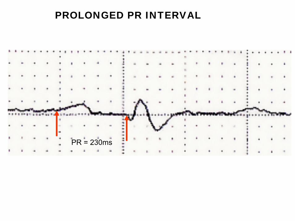
PROLONGED PR INTERVAL
PR = 230ms

I
II
3) SIII>SII
2) Initial q wave I aVL: CCW rotation on FP
rS
rS
rS
4) rS pattern in inferior leads
III
aVR
aVL
1) Extreme QRS axis deviationnear - 90°
1+ 2+3+4 = Left Septal Fascicular Block
aVF

PRECORDIAL LEAD FEATURESV1
rsR´
V2
rS
V3
rS
V4 V5 V6
rS rSRS
• Pointed first component of P wave in V1: Only suggestive Right Atrial Enlargement (RAE)?. Has not the criteria P wave height >1.5mm in led V2 • First degree AV block: PR interval > 220ms • Low QRS voltage: < 10mm of QRS complexes across all precordial leads• Trifasic pattern with wide final R´ in V1, QRS duration >120ms and broad final S wave in left leads: Complete RBBB• Transitional zone (R=S) dislocated to left: V6. CW rotation on longitudinal axis: RVH• Inverted T wave across precordial leads: non-specific repolarizations disturbance.

Rhythm: Sinus; HR: 57bpm; P wave: SÂP +0°; P duration 100ms: P voltage: 1mm. PR interval:230ms. Minimal prolongation of PR interval. QRS: QRS axis: near - 90° isodyphasic in I and positive in aVR and aVL; QRS duration:130ms; QRS voltage: low voltage in both FP and HP; Transition zone on precordial leads: dislocated to left CW rotation suggestive of RV Enlargement rS pattern in inferior leads, qR in aVL and I, extreme QRS axis deviation: LSFB..Tryphasic QRS pattern rsR´ in V1 with broad final R´wave and QRSd>120ms; CRBBB . Possible trifasicular block: first degree AV block, CRBBB, and LAFB. It is diagnosed on an ECG and has three features: prolongation of the PR interval (first degree AV block) RBBB either LAFB or LPFB. Trifascicular block is important to diagnose because it is difficult to tell based on the surface ECG whether the prolonged PR interval is due to disease in the AV node or due to diffuse distal conduction system disease. In the former case, if the block at the AV node level becomes complete, the escape rhythm will originate from the bundle of His, which typically will generate heart rates in the 40s, allowing the individual to survive and complain of symptoms of fatigue or near-syncope to their physician. In the latter case, however, because the conduction system disease is diffuse in nature, the escape rhythm may be fascicular or ventricular, which may be at rates that are life-threateningly low. Trifascicular block is important to diagnose because it is difficult to tell based on the surface ECG whether the prolonged PR interval is due to disease in the AV node or due to diffuse distal conduction system disease. In the former case, if the block at the AV node level becomes complete, the escape rhythm will originate from the bundle of His, which typically will generate heart rates in the 40s, allowing the individual to survive and complain of symptoms of fatigue or near-syncope to their physician. In the latter case, however, because the conduction system disease is diffuse in nature, the escape rhythm may be fascicular or ventricular, which may be at rates that are life-threateningly low.

EXTERNAL ECG FROM EMERGENCY ROOM OF ANOTHER HOSPITAL
DI DII DIII
aVR aVL aVF
V1 V2 V3
V4 V5 V6
Name: JAF Gender: Male Ethnic group: mixed ; High:1.67m Weigh: 64Kg Date: January 05/2009 Time: 23:25PM
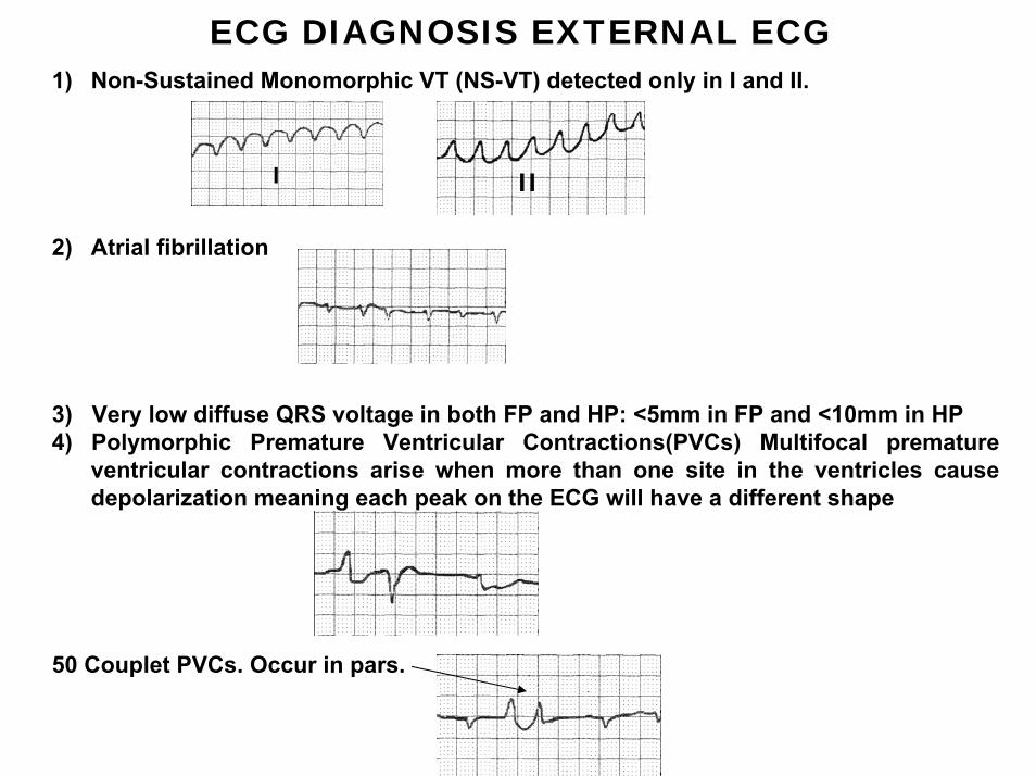
ECG DIAGNOSIS EXTERNAL ECG 1) Non-Sustained Monomorphic VT (NS-VT) detected only in I and II.
2) Atrial fibrillation
3) Very low diffuse QRS voltage in both FP and HP: <5mm in FP and <10mm in HP4) Polymorphic Premature Ventricular Contractions(PVCs) Multifocal premature
ventricular contractions arise when more than one site in the ventricles cause depolarization meaning each peak on the ECG will have a different shape
50 Couplet PVCs. Occur in pars.
I II

This ECG belong to an asymptomatic younger brother who was accompanying our patient (29 years old). We performed this ECG only by curiosity.
Sinus rhythm, suggestive of right atrial or biatrial enlargement, low QRS voltage only in frontal leads, electrically inactive anterolateral wall (pseudomyocardial infarction?), decrescent initial r voltage from V2 to V5, inferolateral repolarization changes.

Querido Andrés, complicado el tema:1. MS familiar, historia sincopal del paciente asociado a cuadro de ICC a predominio
derecho (edemas, ingu yugular, hepatomegalia, etc)2. El primer ECG presenta una bradicardia sinusal, BAV 1°, HBAI y BRD, asi parece facil,
y deberiamos pensar en primer lugar en una cardiopatia chagásica, los sintomas de IC se podrian explicar por la cardiopatia en si y probablemente la bradicardia empeore o colabore en empeorar los sintomas del paciente.
3. Este primer ECG muestra hallazgos sugestivos de patrón electrico de Brugada, especialmente en V2 y V3, sumado a la prolongacion del PR, el eje a la izquierda (hallazgos tambien presentes en el Brugada) no tiene R mayor de 3 mayor de 3 mm, y el QTc me parece dentro de lo normal (me dio 430 ms en V5).
4. El segundo ECG, en DI y DII taqui con QRS ancho, pareciera que luego cambia a unaFA, no veo onda P, es irregular, con algunos latidos mas anchos (aschmann??, no creo), pero podria haber ingresado con un flutter que se transformo en FA, tampocopuedo descartar que sea TV, pero me inclino mas a algo supraventricular.
5. El ultimo ECG del brother, lo que me llama la atención de todo a simple vista, no es un ECG facil. Onda P acuminada, tipo ‘P pulmonale”.Como encararlo para ser unisista: Sindrome de Muerte Subita familiar? padre, tios, todos con MS jovenes, todos chagasicos, una posibilidad, el chagas en Brasil afectamas el tracto digestivo que el cardiovascular (potro sigue siendo asi?), que tenganchagas y ademas algunos la genética para brugada? otra posibilidad, quetengan alguna via accesoria sumado a todo esto, no seLo dejaria internado sin dudas, monitorizado, ecocardiogrma y test para chagas lo primero y despues seguimos y quedios y el potro me ayuden.
Espero sus comentarios maestro,Saludos Francisco José Pancho Femenia MD Mendoza Argentina.

Hola, el ECG tieneBCRDHH + HBAI + BAV 1° grado (bloqueo trifasicular)QT 0,44/46 ms para una FC de 57 x min.Trastorno de repolarización ventricular cara inferior – apex y antero lateral.Falta de progresión de R en V3 – 4 y 5.Por los datos clínicos tiene una Insuficiencia cardiaca por miocardiopatía de posible etiología chagasica (es de America del Sur) vs isquémica, que hizo una arritmia ventricular maligna (TV monomorfa con EV frecuentes, precoces, en duplas, monofocales). Es un paciente con riesgo de MS. hay que valorar el estado del miocardio y el estado electrofisiológico. La presencia de TV en los pacientes con miocardiopatía dilatada es un signo de mal pronostico, mas del 60 % fallecen en los próximos meses al episodio de TV. Y tras la valoración se vera el tratamiento que a prima facie es DAI.Saludos.Dr. Tomás Campillo.
No sólo es de América del Sur, sino de Minas Gerais, un Estado endémico de Chagas, y el paciente y su hermano pertenecen a una clase socioeconómica baja. Me sumo al Dx de bloqueo trifascicular y por la nula progresión de R en precordiales correspondería a una miocardiopatía dilatada.
Luciano Pereira Ciudad del Este Paraguay

Estimado Andres:Realizar Serología para Chagas. Estoy casi seguro que es positivaICC descompensada por miocardiopatía dilatada.ECG inicial RS. FC 55 x m. PR 0,20 seg. HAI + BCRD (típico de Chagas).ECG emergencia: tiene algunas derivaciones con TVMS, resto de derivaciones son de
mala calidad.Realizar ECO 2D y evaluar FEy.Si Fey es mayor de 40% amiodarona, si Fey es menor de 40% CDI (prevención
secundaria).Mi diagnóstico es miocardiopatía dilatada chagásica, ICC descompensada y TVMS
sincopal.ECG del hermano, también es el de un chagásico. Tiene necrosis anterolateral aunque sin
trastornos de conducción típico, que también es frecuente ver. En este caso el tema merece más discusión, que la podemos hacer.
Abrazo.
Dr.Oscar Pellizon MD . [email protected]
A simple vista si chagas, uno para abrir la cabeza puede pensar en Brugada pero la verdad no me suena Brugada y sintomas de insuficiencia cardiaca. Otra patologia desconocida por la asociación familiar?.
LA REALIDAD ES LA EXTREMA POBREZA DE LATINOAMERICA y toda la familia debehaber tenido contacto con el Chagas y eso no me da diagnóstico de miocardiopatiachagasica (eso creo yo que tiene) Lastima que a los hermanos no se los ha estudiado con autopsia (hace mucho que no veo una de causas no legal). Una cosa que me dejo picando el mail del Dr. Adrian Baranchuk. Muchas veces utilice el "criterio unicista por unico" pero al buscar en el diccionario de la real academia no me sale la palabra y hasta mis clases a los residentes sobre enfoque del problema del paciente tiene la palabra asi que HELP. (tampoco me sale como unisista)
Dr. Simon [email protected]
Estimados amigos,
El ECG en ritmo sinusal de este paciente de 42 años apunta:Que el VD se expone fuertemente a las derivaciones precordiales con morfología rShasta V5 + T negativa. Esto sugiere gran dilatación de esta cavidad. Anchura del QRS en V1>V6 (ver adjunto). Eje de P muy desviado a la izquierda (P en DI>DII). BAV 1er grado. Hemibloqueo anterior izquierdo.El ECG en taquicardia, aunque parece que las distintas no derivaciones estánregistradas en el mismo momento, sugiere:TV con eje inferior en el plano frontal. En precordiales es necesario poder ver loscomplejo ectópicos en el mismo momento en el tiempo en las diferentes derivaciones; así y todo pero parece que el origen es del VD.
El ECG del hermano de 29 años:Eje QRS desviado a la derecha en plano frontal. Morfología rS en precordiales que sugiere también dilatación del VD.En conclusión, en mi opinión aparte de descartar Chagas habría que pensar tambiénen la Displasia Arritmogénica del VD como enfermedad familiar causante de muertesubita.Un abrazo.
Javier García Niebla

Querido Oscar apenas para el conocimiento de todos podrias explicar el fundamento de la conducta que externas? Es decir el trial o los trials que apoyan tal proceder amiodarona versus CDI en base a la FE.Gracias desde yáAndrés
Estimado Andrés.El estudio más importante sobre prevención secundaria es el AVID (amio vs CDI) que incluyópacientes con paro cardíaco o TV sintomática y FEy < 40%.. Por supuesto no había chagásicos. En esta situación nosotros extrapolamos. Cuando se hicieron subanálisis se observó que aquellos que presentaban una Fey > 35% no eran de tan alto riesgo y el CDI no otorgaba gran beneficio con respecto a la amio. Como en Chagas no existe tal evidencia nosotros consideramos que la amio ocupa un lugar en aquellos que tienen 35-40% de Fey y TVMS.La bibliografía que dan aval estos datos son las sgtes. David A. Cesario, MD, PHD,* G. William Dec, MD† STATE-OF-THE-ART PAPERS. Implantable Cardioverter- Defibrillator Therapy in Clinical PracticeJ Am Coll Cardiol 2006;47:1507–17.The greatest benefit of ICD therapy in all studies was observed among patients with advanced left ventricular systolic dysfunction. Little advantage over drug therapy has been observed in patients with an ejection fraction that exceeds 35%Oseroff O, Retyk E, Bochoeyer A. Subanalyses of secondary prevention implantable cardioverter-defibrillator trials: Antiarrhythmics Versus Implantable Defibrillators (AVID), Canadian Implantable Defibrillator Study (CIDS), and Cardiac Arrest Study Hamburg (CASH). Curr Opin Cardiol 2004;19:26 –30.Saludos. Oscar.

Estimado Oscar: Genial tu intervencion aclarando las indicaciones de CDI em la prevenciónsecudaria, y la relacion CDI / Amiodarona. Me gustaria hacer uma observacion, y conocer tu opinion:1. El trabajo de referencia en este tema, es el Metaanalysis de Stuart Connolly, publicado en EurHeart J en el 2000. donde se meta-analizaron el AVID, el CIDS (Canadiense, de Connolly) y elCASH (de Hamburgo). Estos son los 3 trabajos que avalan el implante de CDI para prevenciónsecundaria (y que refuerzan la indicacion "I", nivel de evidencia "A"). En este meta-analysis, se demuestra que el CDI es superior a amiodarona para prevenir la MS, en pacientes que ya hanpresentado un evento arritmico grave. Se hizo un analisis en ptes con Fr Eyeccion > y < de 35%. Los beneficios del CDI se concentraron en aquellos que presentaban Fr Eyeccion < 35%.2. Quiere esto decir que la Amiodarona es superior al CDI si tuviste una MS abortada pero tu FrEyeccion es > 35%?.Claramente NO. Porque? Porque el corte de 35% fue un analisis "POST HOC". Esto quiere decir, que no fue planteado en la hipotesis original, y testeado con estudiorandomizado. Sino que retrospectivamente, se hicieron curvas ROC, y se determinaron que por arriba y abajo del 35% quedaban concentrados los pacientes de mayor y peor beneficio para CDI. De ninguna manera, esto indica que si tenes antecedentes de cardiopatia isquemica, tuviste una FV, y tu Fr Eyeccion es 36%, la Amiodarona te va a proteger mejor que el CDI.No hay que olvidarse, que en los 3 trials, CDI fue superior a amiodarona en el analisis general. Los analisis POST HOC son generadores de hipótesis, No son reglas aplicables directamente en la clinica. Un abrazo fuerte desde el frio
AB

Estimado Adrian,es correcta tu observación y el metaanálisis que mencionás es justamente el que determinó que el CDI es clase I en aquellos que presentaron una TV/FV. De todas maneras, para continuar la polémica, el CDI prolongó 4 meses de vida en el seguimientodel AVID (2 años). En el trabajo original la Fey > 35% está en un hazard ratio de 0,9 (cerca del 1: sin ningún beneficio) y la fey < 35% es de 0,6 (amplio beneficio). Tambiénvale decir que la FEy media del AVID fue de 31-32. Es decir que esa fey está lejos del 40% que era el cut-off.El CIDS (más pequeño que el AVID) no demostró diferencias significativas (mortalidadglobal 20% y mortalidad arrítmica 30% a favor del CDI vs amio, pero esta diferencia no es significativa. Circulation. 2000;101:1297-1302).Seguramente vos podrás hablar mucho de este trial ya que es canadiense.Acuerdo con tu observación que el análisis de + o - 35% es post hoc y eso sirve para hipótesis de un trial randomizado y eso no se realizó.En relación a tu pregunta del paciente que tuvo una FV, y te diría que en la cancha de Rosario Central o Ñuls (porque no todo pasa por Bs. As., jaja, aunque Dios y Cristina están ahí) le pongo un CDI, tuvo una MS abortada. Ahora, es lo mismo tener una TVMS que una FV en una cardiopatía isquémica, no isquémica o chagásica? Me gustaría saber qué pensás al respecto.
Me encantó el desafío. Gran abrazo de la calurosa y húmeda Rosario. Oscar Pellizón.

AmigosQue ECG tan hermosos, felicitaciones por hacer el rapido screening del hermano, que
pegada!Ya se barajaron todos los diagnosticos, creo.A favor de Chagas: la certera epidemiologia, el trastorno de conducción del paciente (el del
hermano lo encuentro menos “tipico” pero como dice Oscar, tambien podria ser. Para sostener esta hipótesis, hay que aceptar que toda la flia tiene Chagas, y que otros ya se murireron por arritmias asociadas a Chagas.
A favor de ARVD: Bajo voltaje, BCRD + HBAI, T negativa de V1 a V5. No veo Epsilon. Para sostener esta hipótesis,, hay que aceptar que los fliares previos la tenian, y que el hermano la tiene, tal vez con compromiso biventricular, ya que hay necrosis anterior. Tiene demasiado voltaje, pero claro, como es una enfermedad progresiva, podria ser.
Brugada: me parece que nadie sostiene esta posibilidad, por el claro compromiso estructural.Si se confirma Cardiopatia Chagasica con deterioro severo de la Fr eyeccion, corresponde
implantar un CDI (Ver Chagtop, Registro de Muratore (Europace 2009) y trabajos de A Rassi al primer paciente, en este caso por prevencion secundaria (ya tuvo TV como muestra el extraño ECG 2).
Para el hermano, si se confirma lo mismo, estamos en una encrucijada, y trataria de tener mas elementos de riesgo (Holter, valoración del sistema nervioso autonomo, Clase funcional, etc). Los datos para prevencion promaria son mas escasos.
El Registro de Muratore (de todo Latinoamérica) indica CDI tambien, pero es menos concluyente.
SaludosAB

FINAL DIAGNOSISChronic Chagasic Dilated Cardiomyopathy mixed form:
• Arrhytmogenic: Polymorphic Ventricular Contractions (PVCs), Couplets pairs, and NS-VT
• Dormotropic: with ventricular conduction abnormalities: first degree AV block Complete Right Bundle Branch Block( CRBBB) and Left Anterior Fascicular Block (LAFB)
• With severe ventricular systolic dysfunction, dilated chambers secondary to extensive fibrosis (biventricular and biatrial enlargement) and significative decrease of ejection fraction. Complement fixation test Guerreiro Machado reaction + in both
• Chagas risk index between 12-20 point: 85% risk of death in 10 years. Risk factor Point
NYHA class iii or IV 5
Cardiomegaly ≥ +++ chest X-ray 5
Non-Sustained Ventricular Tachycardia (NS-VT) 3
Low voltage on ECG 3
Male gender 2
Total points Risk of death in 10 years
0 a 6 10%
7-11 40%
12-20 85%
1. Rassi A Jr, et al. Development and validation of a risk score for predicting death in Chagas heart disease. New Engalnd J of Medicine 2006;
355: 700-808.
CHRONIC CHAGASIC CARDIOMYOPATHY
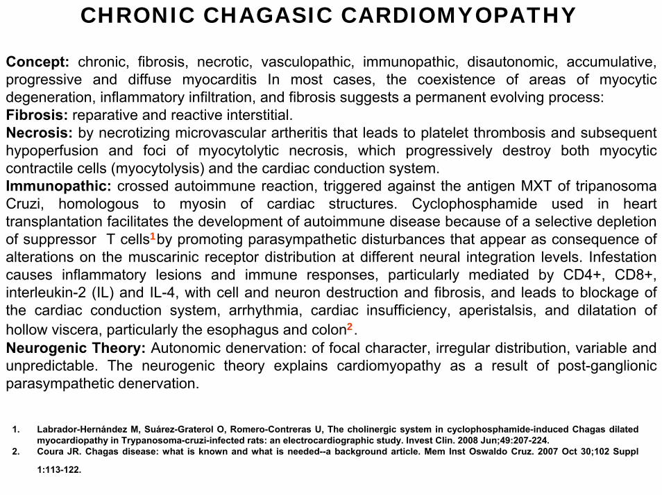
Concept: chronic, fibrosis, necrotic, vasculopathic, immunopathic, disautonomic, accumulative, progressive and diffuse myocarditis In most cases, the coexistence of areas of myocyticdegeneration, inflammatory infiltration, and fibrosis suggests a permanent evolving process: Fibrosis: reparative and reactive interstitial. Necrosis: by necrotizing microvascular artheritis that leads to platelet thrombosis and subsequent hypoperfusion and foci of myocytolytic necrosis, which progressively destroy both myocyticcontractile cells (myocytolysis) and the cardiac conduction system.Immunopathic: crossed autoimmune reaction, triggered against the antigen MXT of tripanosomaCruzi, homologous to myosin of cardiac structures. Cyclophosphamide used in heart transplantation facilitates the development of autoimmune disease because of a selective depletion of suppressor T cells1by promoting parasympathetic disturbances that appear as consequence of alterations on the muscarinic receptor distribution at different neural integration levels. Infestation causes inflammatory lesions and immune responses, particularly mediated by CD4+, CD8+, interleukin-2 (IL) and IL-4, with cell and neuron destruction and fibrosis, and leads to blockage of the cardiac conduction system, arrhythmia, cardiac insufficiency, aperistalsis, and dilatation of hollow viscera, particularly the esophagus and colon2.Neurogenic Theory: Autonomic denervation: of focal character, irregular distribution, variable and unpredictable. The neurogenic theory explains cardiomyopathy as a result of post-ganglionicparasympathetic denervation.
1. Labrador-Hernández M, Suárez-Graterol O, Romero-Contreras U, The cholinergic system in cyclophosphamide-induced Chagas dilated myocardiopathy in Trypanosoma-cruzi-infected rats: an electrocardiographic study. Invest Clin. 2008 Jun;49:207-224.
2. Coura JR. Chagas disease: what is known and what is needed--a background article. Mem Inst Oswaldo Cruz. 2007 Oct 30;102 Suppl
1:113-122.

Chagas disease denominations and transmission
Others denominations: Portuguese: doença de Chagas, Spanish: enfermedad de Chagas-Mazza, mal de Chagas in both languages; also called American trypanosomiasis
Concept and transmission way:is a tropical parasitic disease caused by the flagellate protozoan Trypanosoma cruzi. T. cruzi is commonly transmitted to humans and other mammals by an insect vector, the blood-sucking assassin bugs of the subfamily Triatominae (family Reduviidae) most commonly species belonging to the Triatoma, Rhodnius, and Panstrongylus genera. The disease may also be spread through blood transfusion1 and organ donation/transplantation, ingestion of food contaminated with parasites, and from a mother to her fetus(vertical transmission).
1. Wendel S. TRANSFUSION TRANSMITTED CHAGAS DISEASE: IS IT REALLY UNDER CONTROL? Acta Trop. 2009 Dec 29. [Epub ahead of print]
2. Dauby N, Alonso-Vega C, Suarez E, Flores A, et al. Maternal infection with Trypanosoma cruzi and congenital Chagas disease induce a trend to a type 1 polarization of infant immune responses to vaccines. PLoS Negl Trop Dis. 2009 Dec 22;3(12):e571.

EPIDEMIOLOGIC ASPECTSEndemic in Latin America from the north of Mexico to the South of Argentina and Chile. It is estimated that 15 to 16 million people are infected with Trypanosoma cruzi in Latin America and 75 to 90 million people are exposed to infection1. 120,000 new cases diagnosed in Latin America. The Pan American Health Organization (PAHO) estimates that currently 7.7 million of people have Trypanosoma cruzi infection in the 21 endemic countries from the southern and southwestern United States to central Argentina and Chile2. Most of patients do not know they are infected 17 thousand deaths/year in Brazil, from which 5 thousand by heart disease ≈ 3 millions of Chagasic patients in Brazil, from whom 1 million with heart failure ≈60% of patients display the indeterminate form and only 10% severe heart diseaseIn Sao Paulo Capital City, it is estimated in 300,000 infected patients. In the state of Sao
Paulo there are 500Large-scale population movements (globalization) from rural to urban areas of Latin America and to other regions around the world have increased it geographic distribution3.Mortality is around 45.000 to 50.000 people/year and the main cause is cardiac cardiomyopathy: 60% sudden cardiac death: causes: Ventricular fibrillation, bradyarrhtymias, thromboembolism, rarely aneurysmal rupture and 30% congestive heart failure.
1. Coura JR. Chagas disease: what is known and what is needed--a background article. Mem Inst Oswaldo Cruz. 2007 Oct 30;102 Suppl 1:113-122.2. Sánchez-Sancho F, Campillo NE, Páez JA. Chagas Disease: Progress and New Perspectives. Curr Med Chem. 2009 Dec 17. [Epub ahead of
print] 3. Carod-Artal FJ. Neurologia. Globalization, stroke and Chagas disease on the hundredth anniversary of its discovery 2009 Jul-Aug; 24: 431-432.

Different prevalence rates in Latin American Countries
Country Infection prevalence ratesBolivia 20% of populationArgentina, Paraguay, Honduras, El Salvador
5-10%
Chile, Colombia, Equator, Uruguay 1-5%
Brazil 1.3%
Mexico <1%
Nicaragua <1%
With the intense human migratory movement from developing to developed countries, it became more common and evident. Because of population migration from endemic areas and newly instituted blood bank screening, US clinicians are likely to see an increasing number of patients with suspected or confirmed. Chagas disease presents an increasing challenge for clinicians in the United States. Despite gaps in the evidence base, current knowledge is sufficient to make practical recommendations to guide appropriate evaluation, management, and etiologic treatment1.
1. Bern C, Montgomery SP, Herwaldt BL, et al. Evaluation and treatment of chagas disease in the United States: a systematic review. JAMA. 2007 Nov 14; 298: 2171-2181.

Economic hardship and/or political turmoil stimulated migration of Trypanosoma cruzi-infected population from Latin American countries to the United States and Europe; originating cases of Chagas disease transmitted through blood, organ donation, and vertical transmission. Hispanic immigrant women of reproductive age in the United States coming from Chagasdisease-endemic countries accounted for 2,384,644, and 5,841,538 in 1990 and 2000, respectively. Considering the prevalence rates for T. cruzi infection in their country of origin and the risk of newborns from infected mothers to acquire congenital infection as 1.33% and 5%, is estimated that the number of T. cruzi-infected newborns was 85-318 in 1990 and 166-638 in 2000. Diagnosis of infection in the mother and newborns at risk is needed. A high rate of cure is achieved, almost 100%, when the offspring is treated early. Health authorities, professional associations, physicians, and Hispanic groups should pay more attention to the subject1.
SwitzerlandSeveral cases have been diagnosed in Switzerland, where systematic screening of groups at risk should be implemented. As the vast majority of persons at risk belong to marginalized communites with limited access to heath care, systematic screening and treatment of infected individuals represent a majorchallenge in order to interrupt the congenital transmission and improve the long tern prognosis2.
1. Yadon ZE, Schmunis GA. Congenital Chagas disease: estimating the potential risk in the United States. Am J Trop Med Hyg.2009 Dec;81:927-933. .
2. Jacksn Y et al. Chagas disease in Switzerland: Managing an emerging infection and interrupting its transmission. 2008;4: 1212-1214., 1216-117.
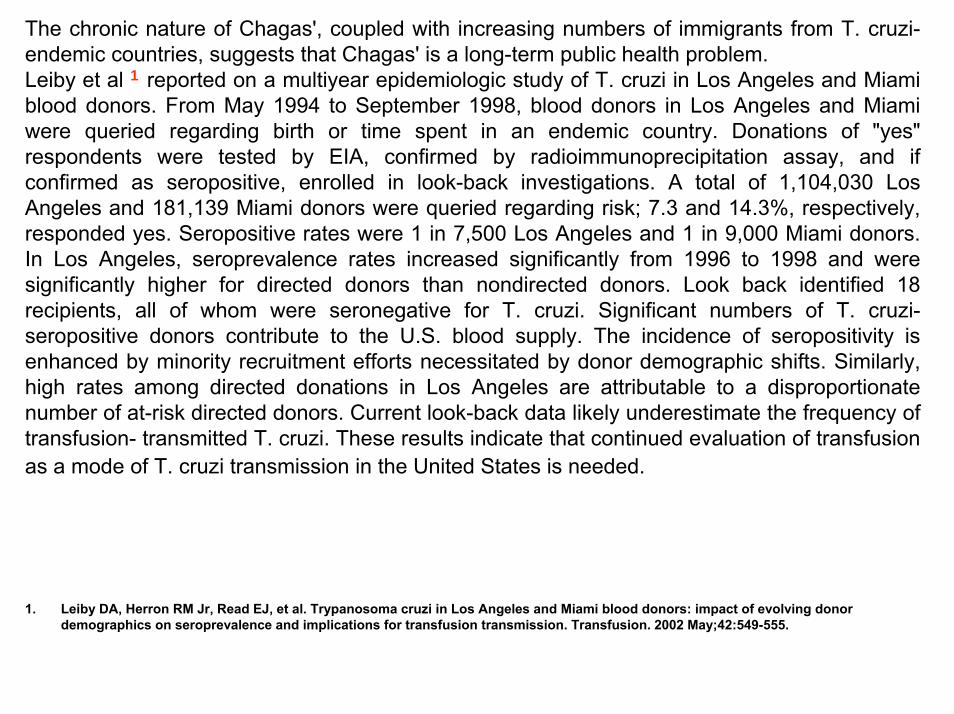
The chronic nature of Chagas', coupled with increasing numbers of immigrants from T. cruzi-endemic countries, suggests that Chagas' is a long-term public health problem. Leiby et al 1 reported on a multiyear epidemiologic study of T. cruzi in Los Angeles and Miami blood donors. From May 1994 to September 1998, blood donors in Los Angeles and Miami were queried regarding birth or time spent in an endemic country. Donations of "yes" respondents were tested by EIA, confirmed by radioimmunoprecipitation assay, and if confirmed as seropositive, enrolled in look-back investigations. A total of 1,104,030 Los Angeles and 181,139 Miami donors were queried regarding risk; 7.3 and 14.3%, respectively, responded yes. Seropositive rates were 1 in 7,500 Los Angeles and 1 in 9,000 Miami donors. In Los Angeles, seroprevalence rates increased significantly from 1996 to 1998 and were significantly higher for directed donors than nondirected donors. Look back identified 18 recipients, all of whom were seronegative for T. cruzi. Significant numbers of T. cruzi-seropositive donors contribute to the U.S. blood supply. The incidence of seropositivity is enhanced by minority recruitment efforts necessitated by donor demographic shifts. Similarly, high rates among directed donations in Los Angeles are attributable to a disproportionate number of at-risk directed donors. Current look-back data likely underestimate the frequency of transfusion- transmitted T. cruzi. These results indicate that continued evaluation of transfusion as a mode of T. cruzi transmission in the United States is needed.
1. Leiby DA, Herron RM Jr, Read EJ, et al. Trypanosoma cruzi in Los Angeles and Miami blood donors: impact of evolving donordemographics on seroprevalence and implications for transfusion transmission. Transfusion. 2002 May;42:549-555.
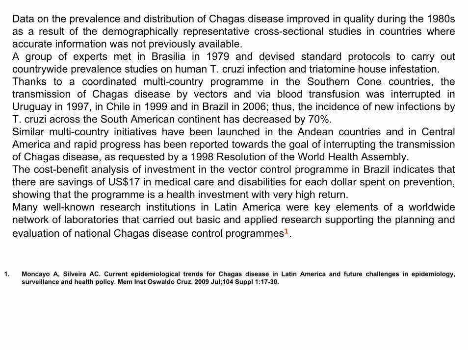
Data on the prevalence and distribution of Chagas disease improved in quality during the 1980s as a result of the demographically representative cross-sectional studies in countries where accurate information was not previously available. A group of experts met in Brasilia in 1979 and devised standard protocols to carry out countrywide prevalence studies on human T. cruzi infection and triatomine house infestation. Thanks to a coordinated multi-country programme in the Southern Cone countries, the transmission of Chagas disease by vectors and via blood transfusion was interrupted inUruguay in 1997, in Chile in 1999 and in Brazil in 2006; thus, the incidence of new infections by T. cruzi across the South American continent has decreased by 70%. Similar multi-country initiatives have been launched in the Andean countries and in Central America and rapid progress has been reported towards the goal of interrupting the transmission of Chagas disease, as requested by a 1998 Resolution of the World Health Assembly. The cost-benefit analysis of investment in the vector control programme in Brazil indicates that there are savings of US$17 in medical care and disabilities for each dollar spent on prevention, showing that the programme is a health investment with very high return. Many well-known research institutions in Latin America were key elements of a worldwide network of laboratories that carried out basic and applied research supporting the planning and evaluation of national Chagas disease control programmes1.
1. Moncayo A, Silveira AC. Current epidemiological trends for Chagas disease in Latin America and future challenges in epidemiology,surveillance and health policy. Mem Inst Oswaldo Cruz. 2009 Jul;104 Suppl 1:17-30.

Population movements from Chagas disease-endemic areas to non-endemic countries due to immigration make the occurrence of this disease in these latter areas possible. Rodríguez-Guardado et al1 describe the results of a screening program conducted in an immigrant population from endemic areas, attending the Tropical Medicine Unit of the Hospital Central de Asturias between June 2006 and June 2008. The ID-Chagas antibody test (particle gel immunoassay (PaGIA); DiaMed-ID) was used as a screening assay. The authors analyzed 64 patients, 9 of whom (14%) tested positive for Chagas disease antibodies, a diagnosis that was confirmed in all cases. Six patients came from Bolivia, 2 from Paraguay and 1 from Brazil. Chagas disease is of increasing importance, even in areas with low migratory flows; hence screening programmes for this population group are especially important.The Catalonian Blood Bank has implemented a screening program for Chagas disease in at-risk blood donors and has performed a study to determine the seroprevalence of Trypanosoma cruziinfection in the donor population. Overall seroprevalence was 0.6%, with 11 donors confirmed positive among the 1770 at-risk donors studied; the highest rate (10.2%) was in Bolivian donors. Interestingly, 1 of the 11 positive donors was a Spaniard who had resided various years in a Chagas disease endemic area. Furthermore, 1 of the positive donors presented detectable parasitemia. The results emphasize the need for T. cruzi screening in at-risk blood donors in nonendemic countries. If T. cruzi screening is not routinely performed in all donations, it remains highly dependent on proper identification of at-risk donors during the predonation interview2.
1. Rodríguez-Guardado A, Rodríguez M, Alonso P, et al. Serological screening of Chagas disease in an immigrant population in Asturias, Spain proceeding from Chagas-endemic areas. Scand J Infect Dis. 2009;41:774-776.
2. Piron M, Vergés M, Muñoz J, et al. Seroprevalence of Trypanosoma cruzi infection in at-risk blood donors in Catalonia (Spain). Transfusion.
2008 Sep;48:1862-1868.

CARDIAC FORMS
1) Indeterminate*
2) Arrhythmogenic:• Predominantly Dromotropic: those with ventricular conduction abnormalities• Predominantly Polimorphic PVCs: those with rhythm disturbances• With rhythm disturbances plus ventricular conduction abnormalities
3) With Ventricular Dysfunction: Chronic Dilated Chagasic Cardiomyopathy
4) Thromboembolic
5) Mixed
Chagasic patients, in very late stage of the disease, have a very variable degree of ventricular systolic dysfunction. Furthermore, distinct tendency for the left ventricular volumes to increase, and for the ejection fraction to decrease; when the electrocardiogram becomes progressively more abnormal, and "mixed" ECG abnormalities appear1.
1. Casado J, Davila DF, Donis JH, et al. Electrocardiographic abnormalities and left ventricular systolic function in Chagas' heart disease. Int J Cardiol. 1990 Apr;27:55-62.

Indeterminate Forms
The majority of patients with Chagas' disease remain for 10 to 30 years in the indeterminate form. They have no symptoms, serologic positivity, normal ECG, normal chest X-rays, normal left ventricular global and segmental systolic function on 2-dimensional echocardiography and absence of clinical, manifestations of cardiac or digestive involvement. When submitted to advanced cardiovascular tests, these patients may present significant abnormalities1; 2. However, the indeterminate form concept was reaffirmed as valid, since diagnostic criteria are simple and prognosis is benignant. In clinical practice, diagnostic difficulties are frequent, related to subjectivity and uncertain meaning of clinical, ECG and radiological findings. Clomipramine(5 mg/kg/day for one month) used in the chronic indeterminate phase of the T. cruzi infection modified the natural evolution of the chagasic cardiopathy 3.
1. Pazin-Filho A, Romano MM, Gomes Furtado R, Left ventricular global performance and diastolic function in indeterminate and cardiac forms of Chagas' disease. J Am Soc Echocardiogr. 2007 Dec;20:1338-1343.
2. Dubner S, Schapachnik E, Riera AR, Valero E. Chagas disease: state-of-the-art of diagnosis and management. Cardiol J. 2008;15:493-504.
3. Bazán PC, Lo Presti MS, Rivarola HW, Chemotherapy of chronic indeterminate Chagas disease: a novel approach to treatment. ParasitolRes. 2008 Aug;103:663-669.
THE ECG VALUE IN CHRONIC CHAGASIC CARDIOMYOPATHY
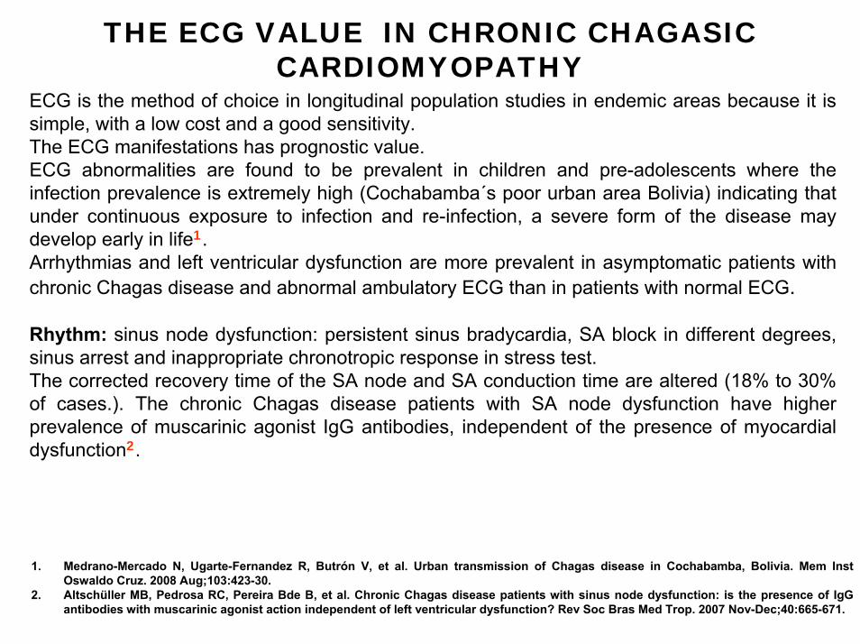
ECG is the method of choice in longitudinal population studies in endemic areas because it is simple, with a low cost and a good sensitivity. The ECG manifestations has prognostic value. ECG abnormalities are found to be prevalent in children and pre-adolescents where the infection prevalence is extremely high (Cochabamba´s poor urban area Bolivia) indicating that under continuous exposure to infection and re-infection, a severe form of the disease may develop early in life1. Arrhythmias and left ventricular dysfunction are more prevalent in asymptomatic patients with chronic Chagas disease and abnormal ambulatory ECG than in patients with normal ECG.
Rhythm: sinus node dysfunction: persistent sinus bradycardia, SA block in different degrees, sinus arrest and inappropriate chronotropic response in stress test. The corrected recovery time of the SA node and SA conduction time are altered (18% to 30% of cases.). The chronic Chagas disease patients with SA node dysfunction have higher prevalence of muscarinic agonist IgG antibodies, independent of the presence of myocardial dysfunction2.
1. Medrano-Mercado N, Ugarte-Fernandez R, Butrón V, et al. Urban transmission of Chagas disease in Cochabamba, Bolivia. Mem Inst Oswaldo Cruz. 2008 Aug;103:423-30.
2. Altschüller MB, Pedrosa RC, Pereira Bde B, et al. Chronic Chagas disease patients with sinus node dysfunction: is the presence of IgGantibodies with muscarinic agonist action independent of left ventricular dysfunction? Rev Soc Bras Med Trop. 2007 Nov-Dec;40:665-671.

Atrial fibrillation (AF) affects subjects with Chagas' disease and is an indicator of poor prognosis. In univariate analysis, left atrial diameter ≥3.2 cm, pulmonary arterial hypertension, frequent premature supraventricular contractions (PSPC) and ventricular contraction (PVC) counts/24 h, ventricular couplets/24 h and ventricular tachycardia (VT) are long-term predictors of AF.
P-wave signal-averaged ECG revealed a limited long-term predictive value for AF.
In chronic Chagas' disease are long-term predictors of AF the following factors1:1. Large left atrial diameter2. Pulmonary arterial hypertension3. Frequent Supraventricular Premature Contractions4. PVCs5. VT.
The rate of left ventricular mass enlargement and systolic function deterioration impact AF incidence in this population.
Sick sinus syndrome, atrial extrasystoles, intraatrial conduction disturbances, and AF or flutter are common findings in different stages of the disease2.
1. Benchimol-Barbosa PR, Barbosa-Filho J. Mechanical cardiac remodeling and new-onset atrial fibrillation in long-term follow-up of subjects with chronic Chagas' disease. Braz J Med Biol Res. 2009 Mar;42:251-262.
2. Elizari MV, Chiale PA.Cardiac arrhythmias in Chagas' heart disease. J Cardiovasc Electrophysiol. 1993 Oct;4:596-608.

Dromotropic alterations in the AV node and intraventricular conduction system:Further compromise of the conduction system can lead to different degrees of AV block. Chagas' disease is the main cause of bundle branch block and AV block in endemic areas. type II first or second degree blocks (14.3%) trifascicular block and even total AV block: (2.5%). Post-His: the most frequent ones are first degree AV blocks, with broad QRS, which in 50% of cases are located in the AV node and the rest in the His-Purkinje system or in both.Right bundle branch block alone or in combination with Left Anterior Fascicular Block (LAFB) are the most common conduction defects. Negative T wave and Polymorphic Premature Ventricular Contractions (PVCs) are typical (25%) features. In advanced cases of Chagas' heart disease, ventricular premature contractions are extremely frequent, multiform, and repetitive (couplets and runs of ventricular tachycardia), and show R on T phenomenon. Trifascicular block, which consists of impaired conduction in the three main fascicles of the ventricular conduction system, may progress to high-grade or complete AV block. Exceptionally, it is possible to register in the same patient paroxysmal alternating atrioventricular block and bilateral bundle branch block1. S-VT or Non-Sustained Ventricular Tachycardia (NS-VT): the most frequent location of VT focus are inferoposterior and lateral regions, followed by septal and apical regions, and their main mechanism is reentry, involving fibrotic and/or aneurysmatic areas.Arrhythmias are aggravated by increased sympathetic tone, implying an enhanced risk of cardiac sudden cardiac death(SCD), which is sometimes the first manifestation of the illness. Chronic chagasic myocarditis is the leading cause of cardiovascular death, mostly as a consequence of heart failure and SCD, in endemic regions.
1. Femenia F, Cuesta A, Mauricio A. A rare form of trifascicular block with intermittent complete atrioventricular block in a patient with Chagasdisease. Cardiol J. 2009;16:582-584.

Electrically inactive areas Electrically inactive myocardium (pseudomyocardial infarction) typically is located in subendocardial anterolateral regions of the left ventricle, areas and extensive subepicardial injury. Injured zones corresponded to location of the inflammatory foci at necropsy1.
Other ECG featuresInferolateral repolarization changes (≈28%)Left ventricular overload (≈ 24%2).
1. Aranda Fraustro A, Chávez Rentería B, Ballinas Verdugo MA, et al. Electro-histological comparison in a case of Chagasic chronic cardiomyopathy. Arch Cardiol Mex. 2007 Jul-Sep;77:249-252.
2. Marques DS, Canesin MF, Barutta Júnior F,et al. Evaluation of asymptomatic patients with chronic Chagas disease through ambulatory electrocardiogram, echocardiogram and B-Type natriuretic peptide analyses. Arq Bras Cardiol.2006 Sep;87:336-343.

ELECTROCARDIOGRAPHIC PREDICTORS OF ADVERSE PROGNOSTIC IN CHRONIC CHAGASIC
CARDIOMYOPATHY
1) Atrial fibrillation1 or flutter2) CLBBB (rare) in 91.3% of the cases and decreased ejection fraction(EF)3) Total AV block4) Anterior and inferior electrically inactive area5) Polymorphic premature ventricular contractions or in salvoes6) Frequent supraventricular and ventricular premature beats, and VT1
7) NS-VT on 24-hour Holter monitoring2
7) Presence of NS-VT associated to decreased LVEF: 80% of mortality in 13 years of follow up. When the EF is normal, the prognosis is good8) Presence of S-VT: 100% of mortality in five years.9 ) The ECG abnormalities that correlated independently with depressed EF are: PVCs, VT, LBBB, AF, complete AV block, and anterior and inferior electrically inactive areas ( fibrosis3.) 10) Prolonged filtered QRS duration obtained by SAECG is an independent predictor of death. A prediction score including three risk factors, depressed LVEF, VT and prolonged filtered QRS complex, has shown to be useful for stratifying risk4.
1. Benchimol-Barbosa PR, Barbosa-Filho J. Mechanical cardiac remodeling and new-onset atrial fibrillation in long-term follow-up of subjects with chronic Chagas' disease. Braz J Med Biol Res. 2009 Mar;42:251-62.
2. Rassi A Jr, Rassi A, Rassi SG. Predictors of mortality in chronic Chagas disease: a systematic review of observational studies. Circulation. 2007 Mar 6;115:1101-1108.
3. Garzon SA, Lorga AM, Nicolau JC. Electrocardiography in Chagas' heart disease. Sao Paulo Med J. 1995 Mar-Apr;113:802-8013.4. Ribeiro AL, Cavalvanti PS, Lombardi F, et al. Prognostic value of signal-averaged electrocardiogram in Chagas disease. J Cardiovasc
Electrophysiol. 2008 May;19:502-509.
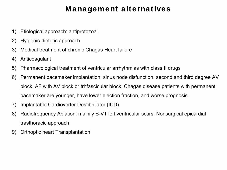
Management alternatives
1) Etiological approach: antiprotozoal
2) Hygienic-dietetic approach
3) Medical treatment of chronic Chagas Heart failure
4) Anticoagulant
5) Pharmacological treatment of ventricular arrhythmias with class II drugs
6) Permanent pacemaker implantation: sinus node disfunction, second and third degree AV
block, AF with AV block or trhfascicular block. Chagas disease patients with permanent
pacemaker are younger, have lower ejection fraction, and worse prognosis.
7) Implantable Cardioverter Desfibrillator (ICD)
8) Radiofrequency Ablation: mainily S-VT left ventricular scars. Nonsurgical epicardial
trasthoracic approach
9) Orthoptic heart Transplantation