artrite reumatoide como modelo de...
TRANSCRIPT
PONTIFÍCIA UNIVERSIDADE CATÓLICA DO RIO GRANDE DO SUL
FACULDADE DE BIOCIÊNCIAS
PROGRAMA DE PÓS-GRADUAÇÃO EM BIOLOGIA CELULAR E MOLECULAR
LAURA ESTEVES PETERSEN
ARTRITE REUMATOIDE COMO MODELO DE
IMUNOSSENESCÊNCIA PREMATURA
Porto Alegre
2013
LAURA ESTEVES PETERSEN
ARTRITE REUMATOIDE COMO MODELO DE
IMUNOSSENESCÊNCIA PREMATURA
Orientador: Prof. Dr. Moisés Evandro Bauer
Porto Alegre
2013
Dissertação de mestrado
apresentada ao Programa de Pós-
Graduação em Biologia Celular e
Molecular da Pontifícia Universidade
Católica do Rio Grande do Sul como
requisito para a obtenção do grau de
Mestre.

DEDICATÓRIA
A minha mãe e minha madrinha pelo
constante apoio, carinho e estímulo que me
ofereceram, dedico esta conquista como
gratidão.
i
AGRADECIMENTOS
Agradeço a minha mãe Maria Isabel Esteves Petersen e minha irmã Marcela
Esteves Petersen pelo carinho, dedicação, apoio e estímulo diário durante toda
minha vida escolar, acadêmica e ao longo dessa trajetória de dois anos. Sem este
apoio, possivelmente, esta caminhada teria sido mais difícil para mim.
Gostaria de fazer um agradecimento muito especial a minha madrinha Sílvia
Regina Ferraz Petersen, que foi e continua sendo a minha principal motivadora e
incentivadora durante minha escolha e qualificação profissional. Agradeço aos meus
familiares e amigos que direta ou indiretamente contribuíram para o
desenvolvimento deste trabalho.
Agradeço ao meu pai Julio Cesar Canhada Petersen pelo carinho, dedicação
e auxílio na busca por participantes nessa pesquisa.
Agradeço ao Rafael Pittas, pela paciência, compreensão e carinho durante
essa trajetória. Obrigada pelo apoio que foi de extrema importância para a
realização deste trabalho.
Gostaria de agradecer ao professor Moisés Evandro Bauer por ter me
recebido com toda a dedicação para compor seu grupo de orientandos. Além disto,
agradeço por todo o conhecimento que adquiri durante esses dois anos que se
passaram. Agradeço pela confiança em mim e no meu trabalho.
As minhas até então colegas, e agora amigas Talita Siara, Suyam Gehlm e
Mariana Ilha que foram de extrema importância para o sucesso desta pesquisa.
Muito obrigada pela força, ajuda, companheirismo durante toda esta trajetória. Com
toda a certeza minha busca por pacientes e controles se tornaram muito mais
agradáveis na companhia destas meninas.
Agradeço também Carine Hartmann do Prado, Andréia Wieck e Lucas Rizzo,
por me ensinarem a usar software e equipamentos essenciais para o
desenvolvimento desta pesquisa. Além disso, registro os meus mais sinceros
agradecimentos aos três citados acima, Ágatha Schommer, Carina Zuppa, Julia
Motta, Bruna Luz, Guilherme Muller e Ana Paula Ornaghi pela companhia e parceria
diária no laboratório de imunologia do envelhecimento.
ii
LISTA DE ABREVIATURAS
AINEs – drogas anti-inflamatórias não esteroidais
AR – artrite reumatoide
DAS – score da atividade da doença
DMARDs – drogas anti-reumáticas moduladoras da doença
FCS – fluido cérebro-espinhal
FR – fator reumatoide
GC – glicocorticoides
GR – receptor de glicocorticoides
HLA – complexo de histocompatibilidade humano
Ig - imunoglobulina
IL – interleucina
MMSE – mini-exame do estado mental
NK – células natural killer
PBMCs – células mononucleares do sangue periférico
PCR – proteína C reativa
SNC – sistema nervoso central
TCR – receptor de célula T
TGF – fator de crescimento de transformação
Th – células T helper
TNF – fator de necrose tumoral
iii
TRECs – alças de ciclos de excisão de células T
Treg – células T regulatórias
TSE – taxa de sedimentação eritrocitária

iv
“A mente que se abre a uma nova ideia
jamais voltará a seu tamanho original”
Albert Einstein
v
RESUMO
A artrite reumatoide (AR) é uma doença autoimune, que além da presença de
danos físicos, tem sido associada com o envelhecimento prematuro do sistema
imune (imunossenescência) e morbidades relacionadas à idade, incluindo declínio
no funcionamento cognitivo. Fatores como a inflamação crônica e o uso de
glicocorticoides (GCs), ambos relacionados a AR, são potenciais mecanismos
envolvidos com a disfunção cognitiva na população geral. Estudos experimentais
tem revelado a contribuição benéfica das células imunológicas sobre o sistema
nervoso central (SNC). Além disto, patologias como o dano cognitivo leve e doença
de Alzheimer apresentam alterações nos subtipos linfocitários periféricos. Com base
nisto, neste trabalho nos exploramos as relações entre função cognitiva, score de
atividade da doença (DAS-28), e subtipos linfocitários na AR. Trinta pacientes com
AR e dezenove controles saudáveis, que não diferiram significativamente em relação
a sexo, idade e escolaridade, foram recrutados neste estudo. A função cognitiva
(mini-exame do estado mental - MMSE, memória lógica e memória de trabalho),
estresse e depressão foram avaliados através de entrevistas onde foram aplicados
questionários clínicos específicos. Os linfócitos foram isolados das células
mononucleares do sangue periférico (PBMCs), e imunofenotipados por citometria de
fluxo para investigar a presença dos seguintes subgrupos: células B, células T
ativadas, células T naive/memória, células T regulatória (reg) CD4+FoxP3+, células
IL-17+, células natural killer (NK), e células T CD28- associadas a senescência. Os
pacientes com AR tiveram um desempenho cognitivo inferior no MMSE, memória
lógica e memória de trabalho se comparado a controles saudáveis. Embora todos os
indivíduos, de ambos os grupos, tiveram uma pontuação superior à estabelecida
pelo cutoff do MMSE. O tempo de uso de GCs e os níveis de proteína C - reativa
(PCR) não se correlacionaram com avaliação cognitiva. Os pacientes tem aumento
nas proporções de células Treg, células T CD4+ naive e células T associadas a
senescência (CD28), mas baixas porcentagens de células B e células T CD8+ de
memória do que controles saudáveis. Células T recém ativadas e células T
CD8+CD28- foram negativamente associadas com a cognição. Concluindo,
pacientes com AR tem um desempenho cognitivo inferior se comparado a controles
saudáveis. GCs e PCR não se correlacionaram com memória, no entanto, expansão

vi
da população de células T ativadas e células T associadas à senescência foram
correlacionadas com performance de memória.
Palavras chave: Artrite reumatoide, glicocorticoides, prejuízo cognitivo, inflamação,
células T, linfócitos.
vii
ABSTRACT
The rheumatoid arthritis (RA) is an autoimmune disease, besides the physical
damage, the RA has been associated with premature aging of the immune system
(immunosenescence) and age-related morbidities, including a decline in cognitive
functioning. Factors such as chronic inflammation and the use of glucocorticoids
(GCs) for a long time, both related to RA, are potential mechanisms involved in
cognitive dysfunction in the general population. Experimental studies have shown the
beneficial contribution of immune cells on the central nervous system (CNS).
Moreover, disorders, such as mild cognitive impairment and Alzheimer’s disease,
exhibit alterations in peripheral lymphocytes subtypes. Based on this, here we
explore the relationship between cognitive function, disease activity score (DAS-28)
and lymphocytes subsets in RA. Thirty patients with RA and 19 healthy controls,
which did not differ significantly in sex, age and schooling were recruited in this
study. Cognitive function (MMSE, logic and working memory), stress and depression
were assessment through interviews where specific clinical questionnaires were
applied. Lymphocytes were isolated from mononuclear cells of peripheral blood
(PBMCs) and immuphenotyped by flow cytometry to investigate the following
lymphocytes subsets: B cells, activated T cells, naïve/memory T cells, regulatory
FoxP3+ T cells, IL-17+ cells, natural killer (NK) cells, and senescence-associated
CD28- T cells. RA patients had a lower cognitive performance on the MMSE, logical
and working memory compared to healthy controls. Though, all individuals in both
groups had a score higher than the cutoff point established by the MMSE. The time
use of GC and the C-reactive protein (CRP) levels did not correlated with cognitive
assessment. Patients had an increased proportions of regulatory T cells, naïve CD4+
T cells and senesce-associated T cells (CD28-), but lowered percentages of B and
memory CD8+ T cells compared to healthy controls. Early activated T cells
(CD3+CD69+) and CD8+CD28- T cells were found negatively associated with
cognition. Concluding, patients with RA have a lower cognitive performance
compared to healthy controls. GC and CRP were not correlated with memory;
however expansions of activated and senescence-associated T cells were correlated
with poor memory performance.

viii
Keywords: rheumatoid arthritis, glucocorticoids, cognitive impairment, inflammation,
T cells, lymphocytes
ix
SUMÁRIO
1. CAPÍTULO 1 ............................................................................................................................................. 1
1.1 INTRODUÇÃO ............................................................................................................................................ 2
Patogênese da artrite reumatoide .................................................................................................. 2 1.2.1
Células T helper 1, T helper 17 e T regulatórias na imunopatogênese da artrite reumatoide ........ 2 1.2.2
Artrite reumatoide como modelo de imunossenescência prematura ............................................. 4 1.2.3
Tratamento da artrite reumatoide .................................................................................................. 6 1.2.4
Prejuízo cognitivo em pacientes com artrite reumatoide .............................................................................. 8
OBJETIVOS ........................................................................................................................................................ 11
JUSTIFICATIVA ................................................................................................................................................... 12
2. CAPITULO 2 ............................................................................................................................................13
2.1. ARTIGO CIENTÍFICO .................................................................................................................................... 14
Abstract .........................................................................................................................................................15
Introduction ...................................................................................................................................................16
SUBJECTS ........................................................................................................................................................... 17
ASSESSMENT OF COGNITIVE FUNCTION AND STRESS LEVELS .................................................................................. 17
COLLECTION OF PERIPHERAL BLOOD AND ISOLATION OF MONONUCLEAR CELLS ................................................... 18
IMMUNOPHENOTYPING ....................................................................................................................................... 18
STATISTICAL ANALYSIS ........................................................................................................................................ 19
Results............................................................................................................................................................19
DEMOGRAPHIC DATA AND CLINICAL CHARACTERISTICS ........................................................................................ 19
COGNITIVE FUNCTION AND PSYCHOLOGICAL DISTRESS ......................................................................................... 20
IMMUNOPHENOTYPING ....................................................................................................................................... 20
CLINICAL CORRELATES OF COGNITION AND LYMPHOCYTE SUBSETS ....................................................................... 20
RELATIONSHIPS BETWEEN COGNITION AND LYMPHOCYTE SUBSETS........................................................................ 21
Discussion ......................................................................................................................................................22
Acknowledgments ..........................................................................................................................................25
Conflict of interest .........................................................................................................................................25
References ......................................................................................................................................................26
3. CAPÍTULO 3 ............................................................................................................................................36
3.1. CONSIDERAÇÕES FINAIS ............................................................................................................................. 37
4. REFERÊNCIAS .........................................................................................................................................40

1
1. CAPÍTULO 1
2
1.1 INTRODUÇÃO
Patogênese da artrite reumatoide 1.2.1
A artrite reumatoide (AR) é uma doença autoimune, progressiva e
potencialmente destrutiva. É principalmente definida por características clínicas,
notavelmente inflamação crônica. Apresenta efeitos articulares, extra-articulares e
sistêmicos (1-4). Do ponto de vista epidemiológico, a prevalência mundial da AR é
aproximadamente 0,5 a 1% enquanto que na população brasileira é em torno de
0,46% (5, 6).
As causas da ocorrência da AR não são totalmente esclarecidas, mas
acredita-se que fatores genéticos, ambientais e imunológicos possam desencadear
o surgimento da doença (7-9). Estudos anteriores mostraram que a AR é
proximamente associada com alelos do complexo maior de histocompatibilidade
humano (HLA), comumente referido como epítopo compartilhado (10). Dentre as
contribuições imunológicas, destaca-se a presença do fator reumatoide (FR). O FR é
um anticorpo produzido pelas células B do sistema imunológico que reage contra a
porção Fc da imunoglobulina (Ig) G.(8, 11). Como já mencionado anteriormente, a
AR é uma doença autoimune parcialmente caracterizada pela presença do FR. No
entanto, este por sua vez, não ocorre em todos os casos em que se desenvolve a
doença, podendo também ser observado em outras patologias (4, 12).
Com o passar do tempo e se não for propriamente tratada, a AR
progressivamente leva a destruição articular e desabilidade funcional. Caracterizada
pela inflamação sinovial, as células T, células B, células plasmáticas, células
dendríticas, macrófagos e mastócitos penetram na articulação ocasionando uma
hiperplasia, comumente chamada de pannus (1, 2, 13). A porção da membrana
sinovial rica em osteoclastos destrói a estrutura óssea, enquanto que as enzimas
secretadas pelos neutrófilos, sinoviócitos e condrócitos degradam a cartilagem (1).
Células T helper 1, T helper 17 e T regulatórias na imunopatogênese da 1.2.2
artrite reumatoide
3
As células T são os principais mediadores do desenvolvimento da AR. As
células T podem interagir com outros tipos celulares perpetuando a inflamação com
consequente destruição da articulação (12). Historicamente a AR é considerada uma
doença com perfil exacerbado de resposta imunológica do tipo T helper 1 (Th1). No
entanto, a AR é baseada em um desequilíbrio entre as células Th1/Th2 - portanto,
um desequilíbrio entre as citocinas pró e anti-inflamatórias (14).
É bem estabelecido que as citocinas pró-inflamatórias, especialmente o fator
de necrose tumoral (TNF-) e a Interleucina (IL) -6, estão envolvidos na patogênese
da AR (1, 15). O TNF- é considerado o desencadeador de eventos pro-
inflamatórios devido a sua capacidade de induzir a produção de outras citocinas
como, por exemplo, a IL-1β, IL-6 e IL-8, frequentemente encontradas nas
articulações de pacientes com AR (16). Além da capacidade de induzir a produção
citocinas, o TNF favorece a liberação de metaloproteinases de matriz, que
promovem a destruição tecidual, induz a expressão de moléculas de adesão sobre
as células endoteliais, promovendo a infiltração de células inflamatórias para dentro
da sinóvia, auxilia a angiogênese e hiperplasia sinovial, contribuindo para a
manutenção e formação do pannus, e induz a ativação de células T, resultando na
perpetuação de respostas autoimunes (15).
Estudos recentes têm sugerido que as células Th17, produtoras de IL-17, são
um novo subgrupo de células críticas para a patogênese da AR (17, 18). A
diferenciação em Th17 é dirigida principalmente pela ação do fator β de Crescimento
de Transformação (TGF-β). Em sua ausência, as células alteram o perfil para Th1
(17, 19). Segundo ZIZZO et al.(20) as porcentagens de células Th17 do fluido
sinovial diretamente correlacionam-se com marcadores inflamatórios articulares
(contagem de linfócitos totais e porcentagem de neutrófilos do fluido sinovial) e
marcadores inflamatórios sistêmicos (fibrinogênio, proteína C - reativa e taxa de
sedimentação eritrocitária). A IL-17 induz a produção de citocinas inflamatórias como
a IL-1, IL-6, IL-8 e TNF-α, é detectada no tecido e no fluido sinovial de pacientes
com AR onde apresenta um papel patogênico por causa de sua associação com a
destruição óssea. (18, 21, 22). Um estudo realizado por NAKANO et. al. (17)
verificou que a utilização de um anticorpo monoclonal contra o receptor de IL-6
promove uma redução na síntese de IL-17, confirmando a intima relação entre estas
duas citocinas. A IL-17 compartilha muitas propriedades biológicas com o TNF-α e
4
IL-1β. A interação dessas citocinas sustenta o processo inflamatório dentro da
articulação e amplificam o envolvimento das células T na patogênese da AR (19,
22).
O desenvolvimento de doenças autoimunes requer um desequilíbrio na
tolerância imunológica que frequentemente controla a discriminação do próprio e do
não próprio (23). Dois maiores subgrupos de células T regulatórias (Treg) podem ser
distinguidos: células Treg naturais, que se originam no timo, e células Treg
induzidas, conhecidas como Tr1 ou Th3, que se originam a partir de células
precursoras CD4+CD25+Foxp3- (24). O Foxp3 é um fator de transcrição
especificamente expresso em células Treg sendo crucial para a manutenção da
função supressora de células Treg periféricas maduras (3). Estas células suprimem
respostas de células T CD4+ e CD8+ através do contato célula-célula, apresentando
um papel fundamental na manutenção da tolerância periférica (19). A diminuição da
expressão de Foxp3 fortalece respostas autoimunes por subverter a função
supressora das células Treg convertendo-as em células T efetoras (25). Segundo
HAN et al.(3) pacientes com AR apresentam menos células CD4+CD25highFoxp3+ no
sangue periférico do que indivíduos saudáveis. No entanto, indivíduos com AR
expressam mais Foxp3+ por célula do que indivíduos saudáveis. Embora as células
Treg estejam presentes na artrite reumatoide, há indícios de que sua função esteja
prejudicada devido à presença de citocinas como, por exemplo, o TNF-α (26).
Artrite reumatoide como modelo de imunossenescência prematura 1.2.3
O envelhecimento é caracterizado por um prejuízo gradual de funcionamento
do sistema imune conhecido como imunossenescência (15). O processo de
imunossenescência é evidenciado, exteriormente, pelo aumento da ocorrência de
infecções, falhas nas respostas a vacinações e reativações virais crônicas (27, 28).
A redução da eficácia imunológica se evidencia a partir da sexta década de vida e
afeta todos os aspectos do sistema imune, especialmente a imunidade adaptativa
(29, 30). No entanto, a AR apresenta altas taxas de incidência na idade adulta,
atingindo o pico máximo entre 75 e 85 anos de idade (29). No tema que nos
interessa, observa-se que pacientes com AR são imunocomprometidos e evidências
5
sugerem que o envelhecimento prematuro do sistema imune contribua para a
patogênese da doença.
O processo de envelhecimento do sistema imunológico é caracterizado por
atrofia do timo, alterações fenotípicas celulares e modificações nos subgrupos de
células T (31). A timopoiese é a principal, se não a única, força de geração de novas
células T naive. A função tímica é estritamente dependente da idade e a involução
tímica inicia-se no primeiro ano de vida, é acelerada na adolescência e por volta dos
45 anos, sua atividade já é severamente limitada (27, 32). Como consequência, a
quantidade de células T naive decai com a idade, sendo um fenômeno mais
pronunciado nas células CD8+ do que nas células CD4+ (27). Uma estimativa da
atividade tímica pode ser observada através da frequência de células T naive que
expressam alças de ciclos de excisão de células T (TRECs) (29). Os TRECs são
subprodutos do rearranjamento do receptor de células T (TCR) que não são
duplicados durante a divisão celular, servindo como um marcador de células recém-
migradas do timo (30). Um estudo realizado por THEWISSEN et al. (33),
comparando pessoas saudáveis, pacientes com AR e indivíduos com esclerose
múltipla, mostrou uma redução na quantidade de células TREC+ em pacientes com
AR e em indivíduos com esclerose múltipla. Estes dados indicam que em ambas as
doenças os sujeitos apresentam uma involução tímica prematura que não condiz
com a idade cronológica.
A falha na produção de novas células T favorece um mecanismo de
autoproliferação celular na periferia (33). O estresse replicativo, associado à
proliferação homeostática para manter o compartimento de células T na ausência da
produção tímica, contribui para a senescência celular. A senescência celular, tanto
no envelhecimento natural quanto no envelhecimento precoce do sistema imune, é
fundamentada em três características: (a) trocas funcionais e fenotípicas, (b)
encurtamento dos telômeros e (c) produção de grandes quantidades de citocinas
(30, 34). Uma das características mais marcantes de trocas fenotípicas é a perda da
expressão da molécula co-estimulatória CD28. O CD28 é expresso sobre a
superfície de células CD4+ e CD8+ (35). Tem sido observado que indivíduos com
AR têm mais células T CD4+CD28- que os indivíduos saudáveis (33). Embora a
função da molécula CD28 seja garantir a ativação apropriada das células T, células
T CD4+CD28- retém a habilidade de produzir citocinas (15). Na deficiência da
6
principal molécula co-estimulatória, as células ganham capacidades pró-
inflamatórias, função citotóxica além se tornarem resistentes a apoptose (33).
Um dos mais notáveis marcadores de envelhecimento celular é o
encurtamento dos telômeros. Telômeros são sequencias repetidas de nucleotídeos
localizadas na porção terminal dos cromossomos. Apresentam função crítica para a
manutenção da integridade do genoma (29). No entanto, servem como marcadores
da divisão celular, e ultimamente, senescência celular (15). A extenção dos
telômeros é vista como um relógio mitótico porque em cada ciclo de divisão celular
eles erodem progressivamente até que a célula entre em estado de senescência
onde sofrerá apoptose (15). O encurtamento dos telômeros é um reflexo de um
aumento na história replicativa de linfócitos ou de seus precursores podendo
também ser causado por um defeito na expressão e função da telomerase (27). A
telomerase é uma enzima que atenua o atrito e a erosão dos telômeros e por tanto,
na sua ausência as sequências teloméricas são incompletamente duplicadas (27).
Em indivíduos saudáveis, os telômeros de células T CD4+ e CD8+
progressivamente erodem ate os 65 anos de idade. Em contrapartida, em pacientes
com AR o processo é claramente acelerado e independentemente da idade (29).
Além disto, a erosão telomérica esta associada, via estresse oxidativo, com a
exposição ao tabaco e também com o epítopo compartilhado do gene HLA, que são
os principais fatores de risco ambiental e genético, respectivamente, do
desenvolvimento da AR (29).
Tratamento da artrite reumatoide 1.2.4
Em poucos anos o tratamento da AR tem sofrido diversas trocas, devido a
agressividade da doença e o aumento na quantidade de novos agentes terapêuticos
(36). Os fármacos utilizados se distinguem em quatro categorias: drogas anti-
inflamatórias não esteroidais (AINEs), drogas anti-inflamatórias esteroidais, drogas
anti-reumáticas moduladoras da doença (DMARDs) e agentes biológicos (37).
Os AINEs na AR amenizam os sintomas da doença, reduzindo a inflamação
sinovial (38). Os AINEs tem perdido seu papel histórico como primeira linha de
7
tratamento devido a sua limitada efetividade, inabilidade de modificar o curso da
doença a longo prazo além de seus efeitos gastrintestinais e cardíacos (37).
As DMARDs são fármacos utilizados desde 1980 e acredita-se que sejam um
grupo heterogêneo de agentes farmacológicos que tenham uma propriedade anti-
reumática (36). Estes medicamentos reduzem o inchaço e a dor articular, limitam o
progressivo dano articular e melhoram a função (37). Eles são considerados a
terapia farmacológica de primeira linha para o tratamento de AR (39). Dentre os
componentes deste grupo, destaca-se o metotrexato que é amplamente e
mundialmente utilizado na terapia da AR. O metotrexato é um dos primeiros agentes
terapêuticos que teve seu efeito protetor da articulação documentado (36). Apesar
de o metotrexato apresentar uma linha de proteção específica na AR, muitos
indivíduos respondem insuficientemente ou não respondem ao metotrexato como
uma monoterapia, sendo necessário a combinação de agentes, onde o fármaco de
escolha é um outro medicamento do grupo DMARDs ou algum medicamento da
classe dos agentes biológicos (40).
Os agentes biológicos são uma classe de medicamentos que tem aberto
novos horizontes terapêuticos no tratamento da AR. Dentre os imunobiológicos,
destaca-se os agentes anti-TNF. A descoberta desta nova terapia partiu de um
estudo sobre a função desta citocina pro-inflamatória na articulação de pacientes
com AR, e verificou-se que, através da sua supressão, este, por sua vez, era capaz
de regular outros mediadores inflamatórios (16). Com base nisso, surgiu a hipótese
de que a utilização de um antagonista de TNF seria um alvo interessante para o
tratamento da AR (36). A utilização da terapia anti-TNF, em doses apropriadas,
reduz tanto a inflamação quanto o dano da articulação (16).
Os glicocorticoides são os representantes dos fármacos anti-inflamatórios
esteroidais que foram introduzidos no tratamento da AR há mais de 60 anos atrás
(41). Os glicocorticoides tem sido comumente usados como potentes drogas anti-
inflamatórias para o tratamento da AR, asma, lúpus eritematoso sistêmico,
leucemias e linfomas (42). Aproximadamente um terço dos pacientes com AR são
usuários atuais e dois terços dos pacientes nunca usaram esteroides (41). Os
sintomas dos pacientes com AR melhoram consideravelmente dentro de poucos
dias (43). No entanto, existem evidências em relação à segurança de sua utilização.
O uso de glicocorticoides por muito tempo possui vários efeitos adversos que
8
incluem: resistência à insulina, diabetes, doenças cardiovasculares, cataratas,
imunossupressão, depressão maior e prejuízo cognitivo (37, 43).
Prejuízo cognitivo em pacientes com artrite reumatoide
O prejuízo cognitivo e a demência são condições de desabilidade mental que
aumentam comumente com o avanço da idade (44). Além do envelhecimento
natural, o uso de glicocorticoides e a presença de marcadores inflamatórios, ambos
vistos na AR, também são contribuintes para o déficit cognitivo.
Os fatores que tem sido relacionado a um declínio cognitivo na população
geral são: inflamação sistêmica, doenças cardiovasculares e uso de glicocorticoides,
ambos de particular relevância para a AR (45, 46). Para pessoas com AR, o
funcionamento cognitivo é essencial para a realização de atividades diárias e para a
aderência ao tratamento (47). O estudo conduzido por MELO et al. (48) investigou a
presença de distúrbios cognitivos associados a presença de AR, entre outras
doenças. Neste estudo, pacientes com AR apresentaram um desempenho reduzido
nos testes que avaliam as esferas cognitivas referentes à apraxia visual-construtiva,
possivelmente relacionada ao comprometimento físico, motor e cognitivo da doença.
SHIN et al. (45) explorou a prevalência de déficit cognitivo e os fatores associados
com prejuízo cognitivo em 115 pacientes com AR. No entanto, o estudo em questão
revelou que um terço dos pacientes selecionados para o estudo apresentou declínio
cognitivo, e que as características clínicas (por exemplo, duração da doença e
severidade) não foram associadas com redução do desempenho cognitivo. Em
contrapartida, fatores relacionados ao tratamento (por exemplo, uso de
glicocorticoides), e consequências significantes do curso da doença (risco de doença
cardiovascular) foram associados com prejuízo cognitivo nesta população.
No entanto, declínios cognitivos também ocorrem com o avanço da idade
(49). Um estudo com Suecos OCTO e NONAGENÁRIOS mostrou que indivíduos
com prejuízo cognitivo e fenótipo de risco imunológico (caracterizado por uma
relação CD4/CD8 menor do que um), têm um elevado risco de mortalidade se
comparado com indivíduos cognitivamente intactos com ou sem fenótipo de risco
imunológico, evidenciando a possível relação entre cognição e sistema imunológico
(50). A neurogênese, processo de formação de novos neurônios, ocorre durante
9
toda vida adulta e é pré-requisito para certos aspectos da cognição (51). Focando
sobre o papel das células T no auxílio para a manutenção da integridade do sistema
nervoso central, ZIV et al. (51), utilizando um modelo experimental com ratos,
demonstrou uma associação entre células T, atividade microglial e neurogênese
hipocampal. Neste estudo foi observado que as células T são importantes para a
plasticidade cerebral, sugerindo que as células T afetam a neurogênese adulta.
Além disso, o processo de envelhecimento natural e a exposição crônica aos
glicocorticoides são associados a uma inflamação residual persistente
(inflammaging) (28). De fato, marcadores inflamatórios e a exposição crônica aos
glicocorticoides têm sido amplamente relacionados a prejuízos cognitivos (44, 52-
55). A terapia prolongada com glicocorticoides, amplamente usada na prática clínica,
é conhecida por induzir déficit cognitivo (46). O hipocampo, uma região cerebral que
processa o aprendizado e a memória de curta duração, pode ser um alvo para
lesões biológicas e injúrias teciduais relacionadas ao envelhecimento (56). Grandes
quantidades de receptores de glicocorticoides (GRs) são encontradas nesta área.
(57). Evidências suportam que os glicocorticoides não tem um efeito específico em
nenhuma função cognitiva particular, mas eles podem ser relacionados a um
declínio cognitivo geral, contribuindo para déficits de memória (46, 57). Além da
exposição aos glicocorticoides, a inflamação pode ser um importante contribuinte
para o declínio cognitivo. Estudos tem focado sobre os marcadores inflamatórios e
desempenho cognitivo (53, 58). Entre os diversos marcadores pró-inflamatórios, os
dois mais frequentemente estudados são a PCR e a IL-6 (59). No entanto, um
estudo conduzido por GIMENO et al.(59) evidenciou que a inflamação sistêmica de
baixo grau é associada com uma reduzida performance cognitiva em domínios
específicos, sendo eles raciocínio indutivo e vocabulário. O aumento de proteína C
reativa de alta sensibilidade no soro tem sido associado a um aumento do risco de
demência e doença de Alzheimer. No entanto, um estudo de acompanhamento
evidenciou que altas concentrações de PCR prediz 12 anos antes o surgimento de
prejuízo de memória (54). Com base nisto, elevados níveis PCR podem ser usados
como biomarcadores para identificar indivíduos com uma maior probabilidade de
apresentar prejuízo de memória e eventualmente demência. Além disso, a PCR tem
sido associada com prejuízos de aprendizado e memória, sugerindo que esta por
sua vez é um possível desencadeador de demência relacionada a doença de
10
Alzheimer (60). Um estudo recente demonstrou a associação da PCR com a
produção da proteína β-amilóide, sendo que a β-amilóide é um componente chave
na formação de placas senis que patologicamente caracterizam a doença. No
entanto, a PCR tem sido encontrada ao redor de proteínas β-amilóide em lesões
cerebrais de pacientes com doença de Alzheimer, onde o prejuízo cognitivo e
demência são características bem estabelecidas (58). Além disso, PHILLIPS et al.
(61), utilizando a taxa de sedimentação eritrocitária (TSE) como marcador da
inflamação, mostrou que a habilidade cognitiva é inversamente proporcional a TSE,
por exemplo, menor habilidade cognitiva, maior a TSE. Estes dados suportam que a
inflamação é um potencial contribuinte para prejuízos cognitivos.
Neste trabalho, investigamos o estado cognitivo funcional e os níveis de
estresse de pacientes com AR e controles saudáveis. Além disso, exploramos as
relações entre a função cognitiva, gravidade da doença (DAS-28) e subpopulações
de linfócitos periféricos.
11
OBJETIVOS
Objetivo geral
Correlacionar marcadores imunológicos com desempenho cognitivo em pacientes
com artrite reumatoide
Objetivos específicos
- Identificar o déficit cognitivo;
- Identificar subtipos linfocitários associados com a imunossenescência;
- Relacionar o déficit cognitivo com tempo de terapia com glicocorticoides;
- Relacionar o déficit cognitivo com marcadores de imunossenescência;
- Relacionar déficit cognitivo com marcadores inflamatórios.
12
JUSTIFICATIVA
A artrite reumatoide é uma doença autoimune associada com o
envelhecimento, apresenta um perfil inflamatório crônico que provoca danos físicos
e progressivamente incapacitantes. Devido a seu curso crônico, leva a prejuízos
econômicos significativos para o sistema único de saúde. Por outro lado, a
capacidade cognitiva dos indivíduos é indispensável para a qualidade de vida dos
idosos na medida em que se observa um rápido aumento na expectativa de vida
brasileira.
Estas constatações justificam o desenvolvimento e pesquisas que contribuam
para melhor conhecer esta doença, particularmente, a relação entre funções
cognitivas, marcadores de imunossenescência e tempo de exposição aos
glicocorticoides em pacientes com artrite reumatoide.

13
2. CAPITULO 2
14
2.1. ARTIGO CIENTÍFICO
A ser submetido para o Journal of Clinical Immunology
IMMUNOLOGICAL CORRELATES OF COGNITIVE DYSFUNCTION IN
RHEUMATOID ARTHRITIS
Laura Esteves Petersen a, Rodrigo Grassi-Oliveira
b, Talita Siara
a, Suyan Gehlm
a,
Mariana Ilha a, Tatiana de Nardi
b, Mauro Keisermann
c and Moisés Evandro Bauer
a,b,d
a Laboratory of Immunosenescence, Institute of Biomedical Research, Pontifical Catholic
University of the Rio Grande do Sul (PUCRS), Porto Alegre, Brazil;
b Center of Studies and Research in Traumatic Stress (NEPTE), Faculty of Psychology,
PUCRS, Porto Alegre, Brazil;
c Division of Rheumatology, São Lucas Hospital, PUCRS, Porto Alegre, Brazil.
d Faculty of Biosciences, PUCRS, Porto Alegre, Brazil.
Correspondent author: Moisés Evandro Bauer, PhD. Instituto de Pesquisas Biomédicas,
Hospital São Lucas da PUCRS, Av. Ipiranga 6690, 2º andar. P.O. Box 1429. Porto Alegre, RS
90.610-000, Brasil. E-mail: [email protected]
15
Abstract
Purpose: Rheumatoid arthritis (RA) has been associated with premature aging of the immune
system and age-related morbidities, including poor cognitive function. The underlying
mechanisms of cognitive impairment in RA are poorly understood. Here, we explored the
relationships between cognitive function, disease activity and lymphocyte subsets in RA.
Methods: Thirty patients with RA and 19 age-matched healthy controls took part in this
study. Cognitive function (MMSE, logical and working memories), stress and depression
scores were evaluated by structured clinical questionnaires. Lymphocytes were
immunophenotyped by flow cytometry to investigate the following lymphocyte subsets: B
cells, activated T cells, naïve/memory T cells, regulatory FoxP3+ T cells, IL-17+ cells, NK
cells and senescence-associated CD28- T cells. Results: Patients with RA were cognitively
impaired compared to controls, as shown by reduced MMSE, logical memory and working
memory scores. The duration of GC use and C-reactive protein levels did not correlate with
cognitive assessments. Patients had increased proportions of regulatory T cells, naïve CD4+ T
cells and senescence-associated T cells (CD28-) but lowered percentages of B and memory
CD8+ T cells compared to controls. Early activated T cells (CD3+CD69) and CD8+CD28- T
cells were found negatively associated with cognition. Conclusions: RA patients had poor
cognitive performance as compared to healthy controls. GC and CRP were not correlated with
memory; however expansions of activated and senescence-associated T cells were correlated
with poor memory performance.
Keywords: Rheumatoid arthritis, glucocorticoids, cognitive impairment, inflammation, T
cells
16
Introduction
Aging is a strong risk factor for developing RA, and the majority of women are
diagnosed after the menopause. RA has been associated with premature aging of the immune
system and age-related co-morbidities, including poor cognitive function (1-4). The
prevalence rates of cognitive impairment in RA ranged from 30% to 70% (5, 6). However, the
underlying mechanisms of cognitive impairment in RA are poorly understood. Recent studies
have indicated key roles for peripheral lymphocytes in healthy brain functions, including
psychological stress responses (7), spatial learning and memory (8, 9), as well as adult
neurogenesis in rodents (10). The autoreactive CD4+ T cells were especially involved in
cognitive processes and animals deficient in T cells were found cognitively impaired (10).
Individuals with RA are immunocompromised and recent evidence suggests that premature
aging of the immune system (immunosenescence) contributes to the pathogenesis of disease
(11). Important features of accelerated immunosenescence in RA are thymic involution,
increased clonal population of T cells in the periphery, decline in the telomere lengths and
loss of CD28 co-stimulatory receptor (12). The decline in the generation of new T cells by the
thymus has been associated with increased compensatory proliferation in the periphery. As a
result, the telomeres of circulating T cells exhibit accelerated erosion (13). The expansion of
late differentiated CD28- T cells, a hallmark of immunosenescence, was found more
pronounced in CD8+ than CD4+ T cells (14). Clones of these CD28- T cells from patients
with RA are consistently autoreactive (14). It remains to be established to what extent
peripheral lymphocytes are involved with cognitive dysfunction in RA.
Here, we investigated functional cognitive functions and stress levels of patients with RA and
age-matched healthy controls. In addition, we explored the relationships between cognitive
function, disease activity and peripheral lymphocyte subsets.
17
Materials and methods
Subjects
Thirty-five patients with RA were recruited from the Rheumatology Unit at São Lucas
Hospital, PUCRS (Porto Alegre, Brazil). The diagnosis of RA was made according to the
criteria of the American College of Rheumatology (15). Five patients were excluded from the
study because they met at least one of the exclusion criteria. Thirty RA patients (50.6 ± 13.45
yrs) took part in this study. In addition, 19 age-, sex-, and schooling–matched healthy controls
(49.37 ± 15.23 yrs, 4 men and 15 women) also took part in this study. The exclusion criteria
included: a) infections, b) under nourishment, c) anemia, d) neoplasias, e) HIV and f) HCV.
The study protocol was approved by both scientific and ethics committees of PUCRS (Porto
Alegre, Brazil) and written informed consent was obtained from all participants.
Assessment of cognitive function and stress levels
Exclusion of dementia was determined by the Mini-Mental State Examination
(MMSE) (16), N-Back task (17) and logical memory tests (18). These structured clinical
interviews were applied by a trained investigator. The MMSE scores varied from 0 to 30
points, adjusted for education level. The cutoff point for cognitive impairment was ≤ 13
points for those without schooling and ≤ 18 points for individuals with less than 8 years of
education and ≤ 26 points for those over 8 years of education. The N-back tasks measure the
accuracy of working memory. The N-back are spoken a series of numbers and asked the
participant which number was spoken N position ago: N=0 (0-back), N=1 (1-back), N=2 (2-
back) and N=3 (3-back). The maximum score per scale is 10, and the sum of all scales is 80
(total N-back). Declarative memory was assessed by logical memory (LM) tests. It consists of
two specific stories told to the individual evoked immediately and 30 min after. Here we used
only the scores of the two stories 30 min after to assess the delay recall. The degree of
18
depression levels were investigated by Beck Depression Inventories-II (BDI-II)(19). Levels of
stress were checked by the Perceived Stress Scale (PSS). This scale measures the degree to
which individuals perceive situations as stressful during the last month (20).
Collection of peripheral blood and isolation of mononuclear cells
Ten milliliters of peripheral blood was collected by venepuncture in the morning
(between 10 and 12 h) and the samples were stored into EDTA tubes prior to analyses.
Peripheral blood mononuclear cells (PBMCs) were isolated by density gradient centrifugation
for 30 min at 900g. Cells were counted by means of microscopy (100x) and viability always
exceeded 95%, as judge from their ability to excluded Trypan Blue (Sigma).
Immunophenotyping
A large panel of lymphocyte subpopulations was identified by multi-color flow
cytometry in freshly isolated PBMCs. Briefly, PBMCs were washed in flow cytometry buffer
(PBS containing 1% FCS and 0.01% sodium azide) and treated with Fc block solution for 20
min. Cells were stained for 30 min with combinations of the follow monoclonal antibodies:
anti-CD3 FITC and PECy5 (T cells), anti-CD4 PE (Th cells), anti-CD8 PECy5 (Tc cells),
anti-CD19 PE (B cells), anti-CD56 FITC (NK cells), anti-CD28 FITC (late differentiated T
cells), anti-CD45RO FITC (memory T cells), anti-CD69 FITC (activated cells), anti-CD45RA
FITC (naïve T cells) all from BD Biosciences, San Jose, CA, USA. Immediately after
staining, cells were washed resuspended and analyzed by flow cytometry. For intracellular
staining, PBMCs were cultured in a final concentration of 5x10⁵ cell/well in RPMI medium
with 10% FCS, 50ng/ml PMA (Phorbol-ester) and 1µg/ml Ionomycin (IONO, all from Sigma
–Aldrich) for 5 hours at 37°C and in 5% CO2 atmosphere. Cells were immediately
permeabilized and stained according to the manufacturer’s instructions (Human Th17/Treg
Phenotyping Kit, BD Biosciences, San Jose, CA, USA). A minimum of 20,000 lymphocytes
19
were identified by size (FSC) and granularity (SSC) and acquired with a FACS Canto II flow
cytometer (BD Biosciences). The instrument has been checked for sensitivity and overall
performance with Cytometer Setup & Tracking beads (BD Biosciences) prior to date
acquisition. Date were analyzed using the Flowjo V10 software (Tree Star Inc., Ashland, Or,
USA).
Statistical Analysis
All variables were tested for homogeneity of variances and normality of distribution by means
of the Levene and Kolmogorov-Smirnov tests, respectively. For continuous variables,
differences between groups were analyzed by Student t-test or Mann-Whitney U test when
appropriate. Statistical interactions between categorical variables were compared by means of
the chi-square (2) test. Interrelationships between variables were analyzed by Pearson and
Spearman’s correlation tests. Statistical analyses were performed using the Statistical Package
for Social Sciences, SPSS Statistics V.18 software (SPSS Inc., Chicago, IL, USA). The
significance level was set at α=0.05 (two tailed).
Results
Demographic data and clinical characteristics
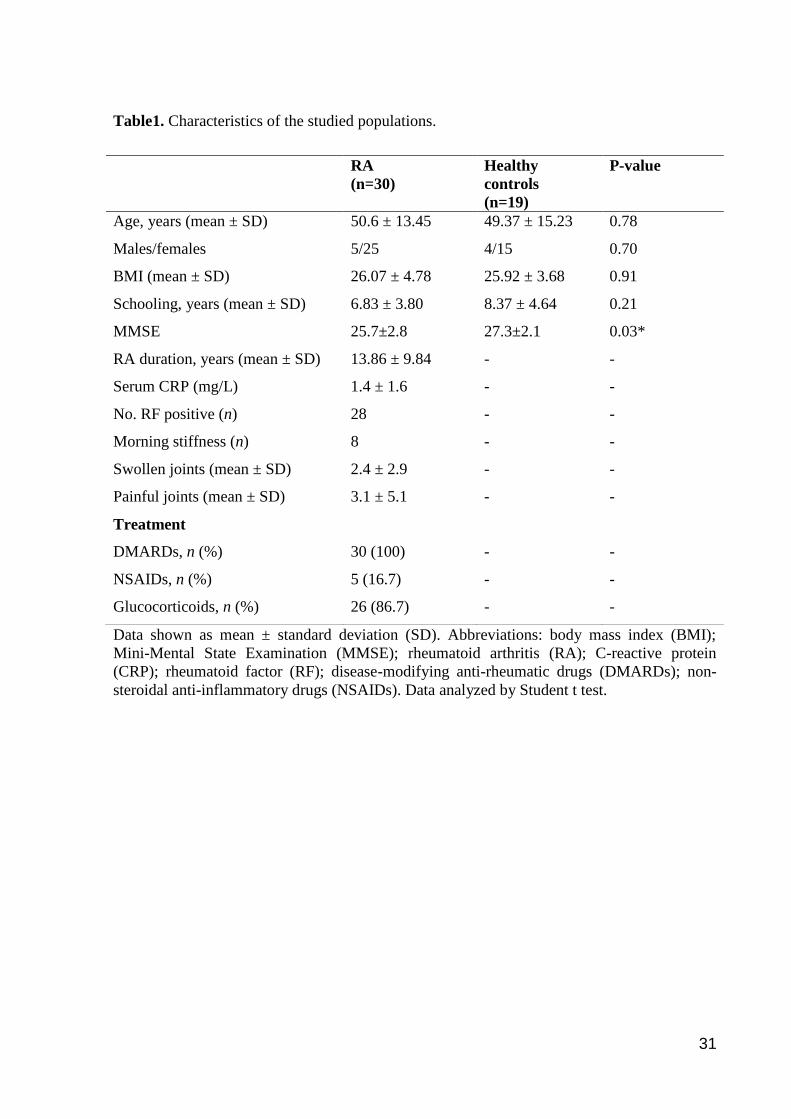
Demographic and clinical characteristics of the sample are summarized in Table 1.
Both groups were homogeneous regarding age, sex and education. All subjects in both groups
had a score higher than the cutoff points established by the MMSE (adjusted for schooling; t =
-2.13, p < 0.05) (Table 1). All patients were taken different drugs that included disease-
modifying anti-rheumatic drugs (DMARDs; methotrexate, hydroxychloroquine, sulfasalazine,
leflunomide), glucocorticoids (prednisone) and non-steroidal anti-inflammatory drugs
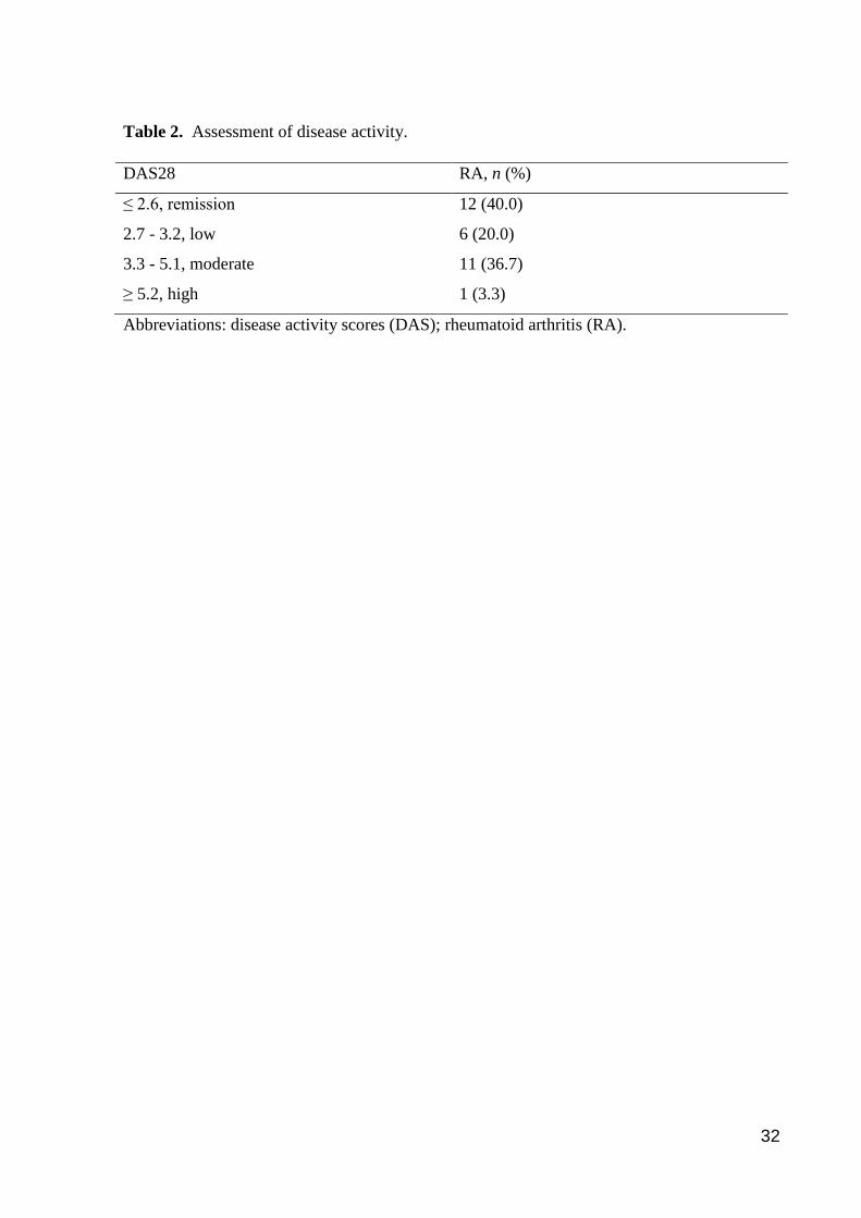
(NSAIDs; ibuprofen and diclofenac). Data about 28-joint disease activity score (DAS28) (21)
20
are shown in Table 2. Patients were taking GC on average for 7.01 ± 6.49 yrs (ranging from 0
to 25 yrs).
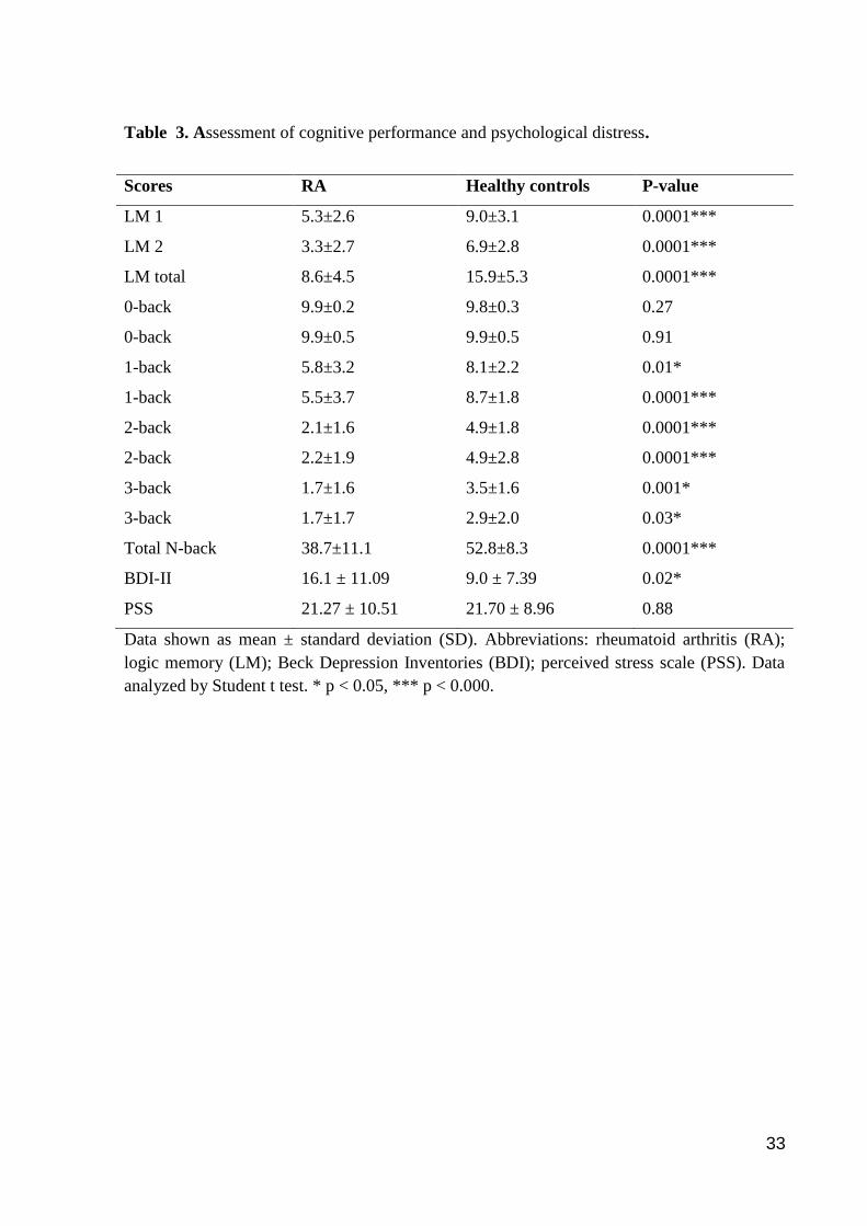
Cognitive function and psychological distress
A comprehensive analysis of the functional cognitive state was performed in this
study. Patients had a reduced cognitive performance compared to healthy subjects (Table 3).
Patients with RA had impaired evocative memory as shown by reduced scores of the logical
memory tests as compared to controls (t = -4.96, p < 0.0001). In addition, working memory
was significantly impaired as shown by reduced total N-back scores than healthy individuals
(t = -4.53, p < 0.0001). Patients with RA were also more depressed than controls (t = 2.35, p
= 0.02). The perception of stressful situations (PSS) during the last month did not differ
between groups (t = -0.74, p > 0.05).
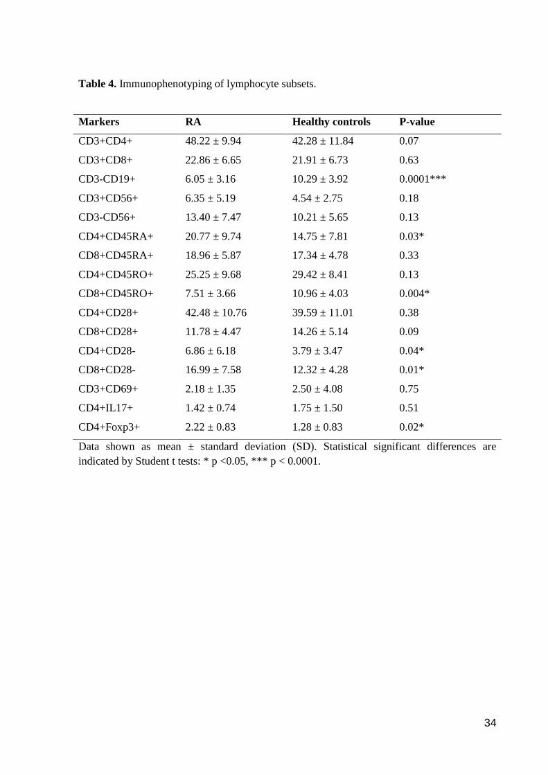
Immunophenotyping
PBMCs were screened for different lymphocyte markers including cell activation,
immunosenescence, as well as regulatory phenotypes (Table 4). B cells (CD3-CD19+) and
memory T cells (CD8+CD45RO+) were found significantly reduced in RA patients (t = -4.06,
p < 0.0001 and t = -3.04, p = 0.004, respectively). In contrast, increased populations of naïve
T cells (CD4+CD45RA+; t = 2.23, p < 0.05), senescence-associated T cells (CD8+CD28- , t =
2.59, p = 0.01 and CD4+CD28-; U = 174.0, p < 0.05), Treg cells (CD4+Foxp3+; t = 2.494, p
= 0.02) were observed in patients as compared to controls. The CD4/CD8 ratio did not differ
between patients as compared to controls (2.31 ± 0.89 vs. 2.02 ± 0.65, respectively), p = 0.22.
Clinical correlates of cognition and lymphocyte subsets
We first sought to investigate potential clinical correlates of cognitive functions.
However, zero-order analyses revealed no significant correlations between CRP levels,
21
DAS28 and cognitive variables (data not shown). Only duration of disease correlated
negatively with total scores of logical memory (r = -0.45, p = 0.01) and approached a
significant correlation with total N-back scores (r = -0.30, p = 0.10). The duration of GC use
also correlated negatively with the PSS scores (r = -0.41, p = 0.02).
Correlation analyses indicated age-related influences on CD3+CD8+ T cells (r = -0.46,
p = 0.001), NK cells (CD3-CD56+; r = 0.393, p = 0.006), naïve T cells (CD8+CD45RA+; r =
-0.357, p = 0.01) and Th17 cells (CD4+IL-17+; r = 0.473, p < 0.05). Furthermore, the
duration of GC use correlated positively with Treg cells (CD4+Foxp3+; r = 0.59, p < 0.05).
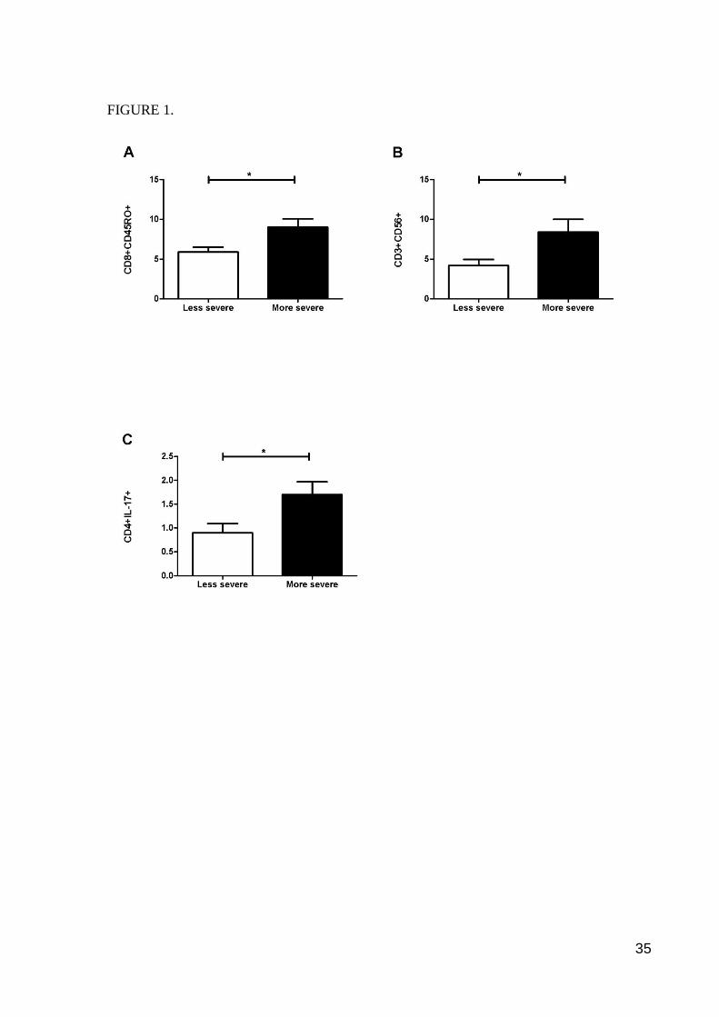
To further investigate the associations of disease activity with cell subsets, two subgroups of
patients were created (less and more severe) accordingly to a median split of DAS28. Patients
with more severe symptomatology (i.e. DAS28 >= 3.09) had more memory T cells
(CD8+CD45RO+; t = -2.532, p = 0.02), NK T cells (CD3+CD56+; t = -2.395, p = 0.02) and
Th17+ cells (CD4+IL17+; U = 5.000, p < 0.05) compared with the group less severe
(Figure 1).
Relationships between cognition and lymphocyte subsets
We also sought to investigate the relationships between cognitive performance and
lymphocyte subsets. The logical memory was found positively correlated with memory T
cells (CD8+CD45RO+; r = 0.37, p = 0.01) and B cells (CD3-CD19+; rs = 0.31, p < 0.05). In
addition, a strong negative correlation was found between 0-back and activated T cells
(CD3+CD69+; r = -0.92, p < 0.0001) as well as moderate negative correlation between the N-
back total and the percentage of CD8+CD28- T cells (r = -0.43, p = 0.004). To get a better
insight into these relationships, the RA patients were classified into two subgroups
accordingly the median split of total N-back scores (median = 38). Patients with worse
22
working memory had more CD8+CD28- T cells than patients with better working memory
(20.02 ± 7.85 vs. 13.71 ± 5.96, U = 40.50, p < 0.05, respectively).
Discussion
To the best of our knowledge, this is the first work reporting the complex relationships
between cognition, disease activity and peripheral lymphocyte subsets in RA. A
comprehensive analysis of cognitive performance (declarative and working memory)
indicated patients with more pronounced cognitive impairments. Furthermore, data provided
here suggest interesting novel relationships between cognition and T-cell subsets.
Data presented here further suggest that patients with RA have poor cognitive
performance, concurring previous studies (1-4). It has recently been shown that patients with
RA had reduced performance in tests assessing the cognitive spheres of visual-constructional
apraxia (1). In addition, individuals with RA had cognitive impairments in domains associated
with executive function, visual-spatial learning/memory and verbal learning/memory (2).
Several mechanisms are potentially involved with cognitive dysfunction in RA, including the
chronic inflammatory condition per se as well as long-term GC use. Glucocorticoids have
been linked with cognitive impairment (22-24) and glucocorticoid receptors (GRs) are
particularly involved in consolidation and retention of learned information (25). Persistent
inflammation may also contribute to cognitive decline (26, 27) and systemic inflammation has
also been observed to increase with advancing age in humans (inflammaging) (26). Indeed,
some studies have previously demonstrated the presence of low–grade inflammation and
reduced cognitive performance (26, 28-30). Komulainen et al. (28), in a follow-up study with
elderly women, found that elevated serum concentrations of high-sensitivity CRP can predict
an increased risk for memory impairment and eventually dementia. In our study, we did not
observe a relationship between CRP levels, duration of exposure to GC and memory deficits.
23
This may be due to possible differences in the dosage of GC taken by individuals with RA
and also due to low CRP levels reported. Other dimensions of cognition may be better
correlated with disease activity in RA.
RA has been associated with premature immunosenescence. In accordance, we
observed several age-related changes in peripheral lymphocyte subsets in RA including a
significant drop in B cells (-41%) as well as expansion in Tregs (73%), CD4+CD28- T cells
(81%) and CD8+CD28- T cells (38%). The loss of expression of the CD28 molecule is a
major phenotypic T-cell change during healthy aging as well as during multiple sclerosis,
bipolar disorder and RA (12, 14, 31, 32). The loss of CD28 expression occurs in both CD4+
and CD8+ T cells but is more common in CD8+ T cells (11). The CD28- T cells have reached
the late-differentiated stage (senescent) and are highly anergic and resistant to apoptosis (33).
CD4+CD28- T cells also express NK-cell surface receptors (KIRs) which provide the ability
to produce large amounts of cytokines, favoring the development of RA (33). Regulatory T
cells (Tregs) have been extensively studied in chronic inflammatory diseases as they can
shutdown effector adaptive immune responses including autoreactive immune cells (34). Our
data also concur to previous studies reporting increased expansions of Tregs in RA. Cao et al.
(35) showed that patients with RA had more regulatory T cells (CD25bright
CD4+) in the
synovial fluid when compared with peripheral blood lymphocytes. Although these cells were
found increased in RA patients, it is believed that Tregs have their functions impaired. The
underlying mechanism is not fully understood, but it has been observed that pro-inflammatory
cytokines (e.g. TNF-) can impair the suppressive function of Tregs (36).
Peripheral lymphocyte subsets, in particular T cells, have been implicated with
cognitive functions. Experimental models have indicated key roles for peripheral lymphocytes
in healthy brain functions, including cognition (8, 9, 37). T cells are rarely detected in the
brain parenchyma, however are rather found in the ‘boundaries’ of the brain, in meningeal
24
structures bathed by cerebrospinal fluid (CSF) (38). The CSF of healthy individuals contains
large amounts of memory T cells (CD45RO+) (39). In accordance, we reported a positive
relationship between cognitive performance and CD8+CD45RO+ T cells. Animals deficient
in T cells are cognitively impaired, and re-population with T cells from wild-type donors can
reverse this defect (8). These studies suggest the importance of the integrity of the peripheral
adaptive immune system in participating of cognitive functions. In addition, changes in
lymphocyte subsets have been observed in patients with mild cognitive impairment (MCI)
and Alzheimer’s disease (AD). Patients with MCI or AD showed lower percentages of B cells
(40, 41) but increased expansions of late-differentiated T cells (CD28-), especially in CD4+
cells (42-44). These data are in line with findings reported here in RA and give further support
to the role of peripheral lymphocytes in human cognition. However, it remains to be
established whether these changes are correlates of the chronic inflammatory condition or
medication.
Some peripheral T-cell subsets have been recently studied in RA and were implicated
in physical damage and disease progression. Here, we observed increased expansions of NKT
(CD3+CD56+), memory CD8+ T cells and Th17 associated with clinical severity. Of special
note, Th17 cells have an inflammatory role and it is characterized by the ability to produce
large amounts of cytokines such as IL-17 and IL-22 (45). In accordance to previous work
(46), no differences in the frequency of circulating Th17 cells were observed in RA compared
with healthy controls. More recently, another study reported increased peripheral Th17 cells
in RA as compared with osteoarthritis (47). All studies, including ours, observed an increase
in the frequency of Th17 cells in individuals with more severe RA than individuals with less
severe form. Interestingly, patients with AD had also increased activity of peripheral Th17
cells (44). Memory T cells (CD8+CD45RO+) have been correlated with levels of IgM
rheumatoid factor. The presence of rheumatoid factor provides evidence of a more aggressive
25
disease development. NKT is a T-cell subset expressing some NK cell markers that can react
with self and microbial ligands, leading to activation of B cells. These cells may be thus
involved in the aggravation of the disease. However, new studies are needed to better
characterize the functional activities of these cells in the synovium.
In conclusion, our data give further support to the hypothesis of accelerated aging in
RA. Patients with RA had major cognitive dysfunctions and alterations in certain lymphocyte
subsets commonly found in healthy immunosenescence. Furthermore, expansions of activated
and senescence-associated T cells were correlated with poor memory performance.
Acknowledgments
We are very grateful to the patients and staff at the Hospital São Lucas (Porto Alegre,
Brazil). This work was supported by grants from CNPq (MEB and RG-O) and by a
scholarship from CAPES (LEP).
Conflict of Interest
The authors declare that they have no conflict of interest.
26
References
1. de Melo LF, Da-Silva SL. Neuropsychological assessment of cognitive disorders in
patients with fibromyalgia, rheumatoid arthritis, and systemic lupus erythematosus. Revista
Brasileira De Reumatologia. 2012;52(2):175-88.
2. Shin SY, Katz P, Wallhagen M, Julian L. Cognitive impairment in persons with rheumatoid
arthritis. Arthritis Care Res (Hoboken). 2012;64(8):1144-50.
3. Boyd TD, Bennett SP, Mori T, Governatori N, Runfeldt M, Norden M, et al. GM-CSF
upregulated in rheumatoid arthritis reverses cognitive impairment and amyloidosis in
Alzheimer mice. J Alzheimers Dis. 2010;21(2):507-18.
4. Shin SY, Julian L, Katz P. The Relationship Between Cognitive Function and Physical
Function in Rheumatoid Arthritis. J Rheumatol. 2013.
5. Bartolini M, Candela M, Brugni M, Catena L, Mari F, Pomponio G, et al. Are behaviour
and motor performances of rheumatoid arthritis patients influenced by subclinical cognitive
impairments? A clinical and neuroimaging study. Clin Exp Rheumatol. 2002;20(4):491-7.
6. Appenzeller S, Bertolo MB, Costallat LT. Cognitive impairment in rheumatoid arthritis.
Methods Find Exp Clin Pharmacol. 2004;26(5):339-43.
7. Cohen H, Ziv Y, Cardon M, Kaplan Z, Matar MA, Gidron Y, et al. Maladaptation to
mental stress mitigated by the adaptive immune system via depletion of naturally occurring
regulatory CD4+CD25+ cells. J Neurobiol. 2006;66(6):552-63.
8. Kipnis J, Cohen H, Cardon M, Ziv Y, Schwartz M. T cell deficiency leads to cognitive
dysfunction: implications for therapeutic vaccination for schizophrenia and other psychiatric
conditions. Proc Natl Acad Sci U S A. 2004;101(21):8180-5.
9. Brynskikh A, Warren T, Zhu J, Kipnis J. Adaptive immunity affects learning behavior in
mice. Brain Behav Immun. 2008;22(6):861-9.
10. Ziv Y, Ron N, Butovsky O, Landa G, Sudai E, Greenberg N, et al. Immune cells
contribute to the maintenance of neurogenesis and spatial learning abilities in adulthood. Nat
Neurosci. 2006;9(2):268-75.
11. Lindstrom TM, Robinson WH. Rheumatoid arthritis: a role for immunosenescence? J Am
Geriatr Soc. 2010;58(8):1565-75.
12. Thewissen M, Linsen L, Somers V, Geusens P, Raus J, Stinissen P. Premature
immunosenescence in rheumatoid arthritis and multiple sclerosis patients. Ann N Y Acad Sci.
2005;1051:255-62.
13. Koetz K, Bryl E, Spickschen K, O'Fallon WM, Goronzy JJ, Weyand CM. T cell
homeostasis in patients with rheumatoid arthritis. Proc Natl Acad Sci U S A.
2000;97(16):9203-8.
14. Weyand CM, Fulbright JW, Goronzy JJ. Immunosenescence, autoimmunity, and
rheumatoid arthritis. Exp Gerontol. 2003;38(8):833-41.
27
15. Arnett FC, Edworthy SM, Bloch DA, McShane DJ, Fries JF, Cooper NS, et al. The
American Rheumatism Association 1987 revised criteria for the classification of rheumatoid
arthritis. Arthritis Rheum. 1988;31(3):315-24.
16. Folstein MF, Folstein SE, McHugh PR. "Mini-mental state". A practical method for
grading the cognitive state of patients for the clinician. J Psychiatr Res. 1975;12(3):189-98.
17. Owen AM, McMillan KM, Laird AR, Bullmore E. N-back working memory paradigm: a
meta-analysis of normative functional neuroimaging studies. Hum Brain Mapp.
2005;25(1):46-59.
18. Wechsler D. Wechsler Memory Scale - revised manual. San Antonio: Psychological
Corporation; 1987.
19. Beck AT, Steer RA, Ball R, Ranieri W. Comparison of Beck Depression Inventories -IA
and -II in psychiatric outpatients. J Pers Assess. 1996;67(3):588-97.
20. Luft CD, Sanches Sde O, Mazo GZ, Andrade A. [Brazilian version of the Perceived Stress
Scale: translation and validation for the elderly]. Rev Saude Publica. 2007;41(4):606-15.
21. Pinheiro GR. Pooled indices to measure rheumatoid arthrits activity – Why and how to
use them. Rev Bras Reumatol. 2007;47:362-5.
22. Wolf OT, Convit A, McHugh PF, Kandil E, Thorn EL, De Santi S, et al. Cortisol
differentially affects memory in young and elderly men. Behav Neurosci. 2001;115(5):1002-
11.
23. Coluccia D, Wolf OT, Kollias S, Roozendaal B, Forster A, de Quervain DJ.
Glucocorticoid therapy-induced memory deficits: acute versus chronic effects. J Neurosci.
2008;28(13):3474-8.
24. Almela M, van der Meij L, Hidalgo V, Villada C, Salvador A. The cortisol awakening
response and memory performance in older men and women. Psychoneuroendocrinology.
2012;37(12):1929-40.
25. Jameison K, Dinan TG. Glucocorticoids and cognitive function: from physiology to
pathophysiology. Hum Psychopharmacol. 2001;16(4):293-302.
26. Trollor JN, Smith E, Agars E, Kuan SA, Baune BT, Campbell L, et al. The association
between systemic inflammation and cognitive performance in the elderly: the Sydney
Memory and Ageing Study. Age (Dordr). 2012;34(5):1295-308.
27. Bauer ME. Chronic stress and immunosenescence: a review. Neuroimmunomodulation.
2008;15(4-6):241-50.
28. Komulainen P, Lakka TA, Kivipelto M, Hassinen M, Penttila IM, Helkala EL, et al.
Serum high sensitivity C-reactive protein and cognitive function in elderly women. Age
Ageing. 2007;36(4):443-8.
29. van den Kommer TN, Dik MG, Comijs HC, Jonker C, Deeg DJ. Homocysteine and
inflammation: predictors of cognitive decline in older persons? Neurobiol Aging.
2010;31(10):1700-9.
28
30. Phillips AC, Batty GD, van Zanten JJ, Mortensen LH, Deary IJ, Calvin CM, et al.
Cognitive ability in early adulthood is associated with systemic inflammation in middle age:
the Vietnam experience study. Brain Behav Immun. 2011;25(2):298-301.
31. do Prado CH, Rizzo LB, Wieck A, Lopes RP, Teixeira AL, Grassi-Oliveira R, et al.
Reduced regulatory T cells are associated with higher levels of Th1/TH17 cytokines and
activated MAPK in type 1 bipolar disorder. Psychoneuroendocrinology. 2012.
32. Goronzy JJ, Shao L, Weyand CM. Immune aging and rheumatoid arthritis. Rheum Dis
Clin North Am. 2010;36(2):297-310.
33. Goronzy JJ, Weyand CM. Aging, autoimmunity and arthritis: T-cell senescence and
contraction of T-cell repertoire diversity - catalysts of autoimmunity and chronic
inflammation. Arthritis Res Ther. 2003;5(5):225-34.
34. Haque R, Lei F, Xiong X, Bian Y, Zhao B, Wu Y, et al. Programming of regulatory T
cells from pluripotent stem cells and prevention of autoimmunity. J Immunol.
2012;189(3):1228-36.
35. Cao D, Malmstrom V, Baecher-Allan C, Hafler D, Klareskog L, Trollmo C. Isolation and
functional characterization of regulatory CD25brightCD4+ T cells from the target organ of
patients with rheumatoid arthritis. Eur J Immunol. 2003;33(1):215-23.
36. Valencia X, Stephens G, Goldbach-Mansky R, Wilson M, Shevach EM, Lipsky PE. TNF
downmodulates the function of human CD4+CD25hi T-regulatory cells. Blood.
2006;108(1):253-61.
37. Yoles E, Hauben E, Palgi O, Agranov E, Gothilf A, Cohen A, et al. Protective
autoimmunity is a physiological response to CNS trauma. J Neurosci. 2001;21(11):3740-8.
38. Kipnis J, Gadani S, Derecki NC. Pro-cognitive properties of T cells. Nat Rev Immunol.
2012;12(9):663-9.
39. Engelhardt B, Ransohoff RM. The ins and outs of T-lymphocyte trafficking to the CNS:
anatomical sites and molecular mechanisms. Trends Immunol. 2005;26(9):485-95.
40. Richartz-Salzburger E, Batra A, Stransky E, Laske C, Kohler N, Bartels M, et al. Altered
lymphocyte distribution in Alzheimer's disease. J Psychiatr Res. 2007;41(1-2):174-8.
41. Magaki S, Yellon SM, Mueller C, Kirsch WM. Immunophenotypes in the circulation of
patients with mild cognitive impairment. J Psychiatr Res. 2008;42(3):240-6.
42. Larbi A, Pawelec G, Witkowski JM, Schipper HM, Derhovanessian E, Goldeck D, et al.
Dramatic shifts in circulating CD4 but not CD8 T cell subsets in mild Alzheimer's disease. J
Alzheimers Dis. 2009;17(1):91-103.
43. Pellicano M, Larbi A, Goldeck D, Colonna-Romano G, Buffa S, Bulati M, et al. Immune
profiling of Alzheimer patients. J Neuroimmunol. 2012;242(1-2):52-9.
44. Saresella M, Calabrese E, Marventano I, Piancone F, Gatti A, Alberoni M, et al. Increased
activity of Th-17 and Th-9 lymphocytes and a skewing of the post-thymic differentiation
pathway are seen in Alzheimer's disease. Brain Behav Immun. 2011;25(3):539-47.
29
45. Nakano K, Yamaoka K, Hanami K, Saito K, Sasaguri Y, Yanagihara N, et al. Dopamine
induces IL-6-dependent IL-17 production via D1-like receptor on CD4 naive T cells and D1-
like receptor antagonist SCH-23390 inhibits cartilage destruction in a human rheumatoid
arthritis/SCID mouse chimera model. J Immunol. 2011;186(6):3745-52.
46. Arroyo-Villa I, Bautista-Caro MB, Balsa A, Aguado-Acin P, Nuno L, Bonilla-Hernan
MG, et al. Frequency of Th17 CD4+ T cells in early rheumatoid arthritis: a marker of anti-
CCP seropositivity. PLoS One. 2012;7(8):e42189.
47. Kim J, Kang S, Kwon G, Koo S. Elevated levels of T helper 17 cells are associated with
disease activity in patients with rheumatoid arthritis. Ann Lab Med. 2013;33(1):52-9.
30
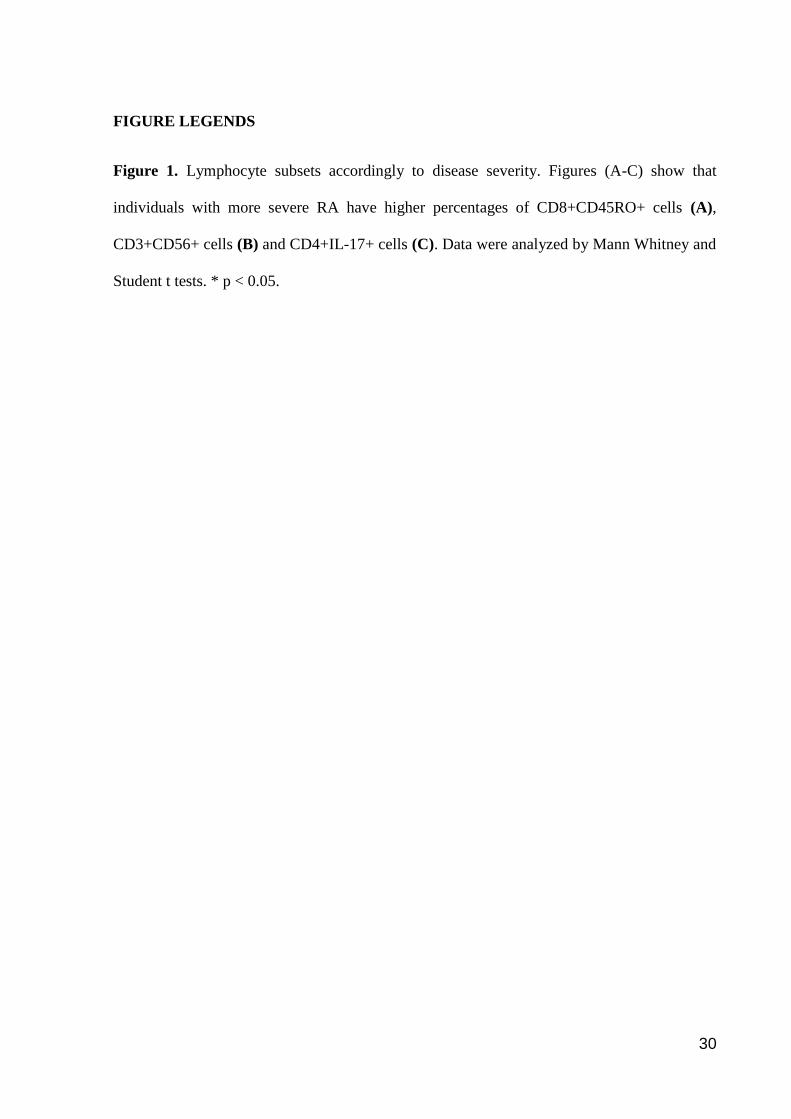
FIGURE LEGENDS
Figure 1. Lymphocyte subsets accordingly to disease severity. Figures (A-C) show that
individuals with more severe RA have higher percentages of CD8+CD45RO+ cells (A),
CD3+CD56+ cells (B) and CD4+IL-17+ cells (C). Data were analyzed by Mann Whitney and
Student t tests. * p < 0.05.
31
Table1. Characteristics of the studied populations.
Data shown as mean ± standard deviation (SD). Abbreviations: body mass index (BMI);
Mini-Mental State Examination (MMSE); rheumatoid arthritis (RA); C-reactive protein
(CRP); rheumatoid factor (RF); disease-modifying anti-rheumatic drugs (DMARDs); non-
steroidal anti-inflammatory drugs (NSAIDs). Data analyzed by Student t test.
RA
(n=30)
Healthy
controls
(n=19)
P-value
Age, years (mean ± SD) 50.6 ± 13.45 49.37 ± 15.23 0.78
Males/females 5/25 4/15 0.70
BMI (mean ± SD) 26.07 ± 4.78 25.92 ± 3.68 0.91
Schooling, years (mean ± SD) 6.83 ± 3.80 8.37 ± 4.64 0.21
MMSE 25.7±2.8 27.3±2.1 0.03*
RA duration, years (mean ± SD) 13.86 ± 9.84 - -
Serum CRP (mg/L) 1.4 ± 1.6 - -
No. RF positive (n) 28 - -
Morning stiffness (n) 8 - -
Swollen joints (mean ± SD) 2.4 ± 2.9 - -
Painful joints (mean ± SD) 3.1 ± 5.1 - -
Treatment
DMARDs, n (%) 30 (100) - -
NSAIDs, n (%) 5 (16.7) - -
Glucocorticoids, n (%) 26 (86.7) - -
32
Table 2. Assessment of disease activity.
Abbreviations: disease activity scores (DAS); rheumatoid arthritis (RA).
DAS28 RA, n (%)
≤ 2.6, remission 12 (40.0)
2.7 - 3.2, low 6 (20.0)
3.3 - 5.1, moderate 11 (36.7)
≥ 5.2, high 1 (3.3)
33
Table 3. Assessment of cognitive performance and psychological distress.
Data shown as mean ± standard deviation (SD). Abbreviations: rheumatoid arthritis (RA);
logic memory (LM); Beck Depression Inventories (BDI); perceived stress scale (PSS). Data
analyzed by Student t test. * p < 0.05, *** p < 0.000.
Scores RA Healthy controls P-value
LM 1 5.3±2.6 9.0±3.1 0.0001***
LM 2 3.3±2.7 6.9±2.8 0.0001***
LM total 8.6±4.5 15.9±5.3 0.0001***
0-back 9.9±0.2 9.8±0.3 0.27
0-back 9.9±0.5 9.9±0.5 0.91
1-back 5.8±3.2 8.1±2.2 0.01*
1-back 5.5±3.7 8.7±1.8 0.0001***
2-back 2.1±1.6 4.9±1.8 0.0001***
2-back 2.2±1.9 4.9±2.8 0.0001***
3-back 1.7±1.6 3.5±1.6 0.001*
3-back 1.7±1.7 2.9±2.0 0.03*
Total N-back 38.7±11.1 52.8±8.3 0.0001***
BDI-II 16.1 ± 11.09 9.0 ± 7.39 0.02*
PSS 21.27 ± 10.51 21.70 ± 8.96 0.88
34
Table 4. Immunophenotyping of lymphocyte subsets.
Data shown as mean ± standard deviation (SD). Statistical significant differences are
indicated by Student t tests: * p <0.05, *** p < 0.0001.
Markers RA Healthy controls P-value
CD3+CD4+ 48.22 ± 9.94 42.28 ± 11.84 0.07
CD3+CD8+ 22.86 ± 6.65 21.91 ± 6.73 0.63
CD3-CD19+ 6.05 ± 3.16 10.29 ± 3.92 0.0001***
CD3+CD56+ 6.35 ± 5.19 4.54 ± 2.75 0.18
CD3-CD56+ 13.40 ± 7.47 10.21 ± 5.65 0.13
CD4+CD45RA+ 20.77 ± 9.74 14.75 ± 7.81 0.03*
CD8+CD45RA+ 18.96 ± 5.87 17.34 ± 4.78 0.33
CD4+CD45RO+ 25.25 ± 9.68 29.42 ± 8.41 0.13
CD8+CD45RO+ 7.51 ± 3.66 10.96 ± 4.03 0.004*
CD4+CD28+ 42.48 ± 10.76 39.59 ± 11.01 0.38
CD8+CD28+ 11.78 ± 4.47 14.26 ± 5.14 0.09
CD4+CD28- 6.86 ± 6.18 3.79 ± 3.47 0.04*
CD8+CD28- 16.99 ± 7.58 12.32 ± 4.28 0.01*
CD3+CD69+ 2.18 ± 1.35 2.50 ± 4.08 0.75
CD4+IL17+ 1.42 ± 0.74 1.75 ± 1.50 0.51
CD4+Foxp3+ 2.22 ± 0.83 1.28 ± 0.83 0.02*
35
FIGURE 1.

36
3. CAPÍTULO 3
37
3.1. CONSIDERAÇÕES FINAIS
Os resultados apresentados nessa dissertação apoiam a hipótese de um
envelhecimento acelerado na AR. Verificamos um prejuízo cognitivo acentuado e
várias alterações em subtipos linfocitários que sugerem uma imunossenescência
acelerada.
Embora existam estudos prévios que identificaram disfunções cognitivas em
pacientes com AR, esse é o primeiro trabalho que verificou relações entre o sistema
imunológico e a cognição. O prejuízo cognitivo foi evidenciado pela aplicação de
questionários específicos. Estes questionários nos forneceram escores de avaliação
do desempenho cognitivo geral e exclusão da presença de demência (mini exame
do estado mental; MMSE) e dois diferentes tipos de memória, memória de trabalho
(tarefa N-back) e memória declarativa (memória lógica). Além disso, nós pudemos
constatar a presença de uma correlação positiva entre células T de memória
(CD8+CD45RO+) e células B (CD3-CD19+) com performance cognitiva. Estudos
experimentais com roedores tem demostrado os efeitos benéficos do sistema imune
sobre o sistema nervoso central (SNC). No entanto, em condições fisiológicas, as
células T são raramente detectadas no parênquima cerebral, e acredita-se que as
células T estejam localizadas nos ‘limites cerebrais’ (leptomeninges, plexo coroide e
espaço perivascular) banhados pelo fluido cérebro-espinhal (FCS). Estudos tem
mostrado que o fluido cérebro-espinhal apresenta grandes quantidades de células T
de memória central (CD45RO+). Como mencionado anteriormente, em nosso estudo
pudemos observar que indivíduos com maiores porcentagens de células CD45RO+
apresentam um melhor desempenho de memória do que indivíduos com
porcentagens inferiores. Além disso, evidências suportam que as células T,
principalmente células T autoreativas a um antígeno do SNC, são importantes para a
plasticidade cerebral adulta, auxiliando no processo da neurogênese. Por outra via,
estudos tem focado sobre o sistema imune de pessoas com comprometimento
cognitivo leve e doença de Alzheimer, cujo comprometimento cognitivo e demência
são características bem estabelecidas. No entanto, em ambas as patologias os
indivíduos apresentam baixas porcentagens de células B e expansão de células
diferenciadas tardiamente (CD28-), especialmente em células CD4+. Estes dados
38
corroboram com os encontros reportados aqui na AR e fornecem indícios da
participação dos linfócitos periféricos na cognição humana.
Embora alguns estudos tenham relacionado à exposição aos glicocorticoides
e níveis de proteína C reativa com um mau funcionamento cognitivo, neste trabalho
não foram observadas tais relações. Especula-se que os pacientes com prognóstico
mais severo, e/ou uso mais intenso de glicocorticoides, possam replicar mais
claramente essas relações. Além disso, indivíduos acometidos pela doença em
questão, apresentaram um grau de depressão mais elevado do que controles
saudáveis. Por outra via, ambos os grupos não tiveram diferenças em relação à
percepção do estresse psicossocial nos 30 dias antecessores a entrevista.
AR é caracterizada por imunossenescência prematura. Com base nisso,
alterações quantitativas e fenotípicas podem ser observadas nas subpopulação de
células imunológicas periféricas. Neste estudo, verificamos a presença de subtipos
linfocitários que caracterizam o processo de imunossenescência. Sendo assim,
pudemos confirmar que pessoas com AR apresentam uma expansão na população
de células T regulatórias senescentes (CD4+CD28-, CD8+CD28-) e células Treg
(CD4+Foxp3+). No entanto, um aumento na população de células Treg deveria ser
um bom prognóstico para os pacientes com AR, visto a capacidade que estas
células em suprimir células imunes autorreativas, se não fosse a ação de citocinas
que atenuam a capacidade supressora das células T reg. Além disso, identificamos
um aumento na população de células de memória (CD8+CD45RO+), células NKT
(CD3+CD56+) e células Th17 no grupo de pacientes com AR mais severa se
comparado com o grupo menos severa. Indicando uma possível ligação entre
severidade clínica e os respectivos subtipos linfocitários citados acima.
A partir da identificação de prejuízo cognitivo e alterações linfocitárias,
partimos para investigar a associação entre sistema imune e cognição. No entanto,
observamos que subtipos linfocitários, especificamente células de memória
(CD8+CD45RO+) e células B (CD3-CD19+) se correlacionaram positivamente com o
teste de memória lógica. Por outra via, os linfócitos T ativados apresentaram uma
correlação negativa com o teste N-back.
Concluindo, levando em consideração o exposto, os achados deste trabalho
corroboram com estudos anteriores que observaram prejuízo cognitivo em pacientes
com AR. Além disso, nosso estudo fornece indícios da relação entre subtipos de
39
células imunológicas e cognição. No entanto, mais estudos a respeito desta relação
deveriam ser realizados com o objetivo de verificar se as células imunes influenciam
a cognição ou se a cognição influencia as células imunes. O desenho experimental
deste estudo não permitiu responder esta questão, mas desperta para a
necessidade de explorar melhor estas relações.
40
4. REFERÊNCIAS
1. Choy E. Understanding the dynamics: pathways involved in the pathogenesis of rheumatoid arthritis. Rheumatology (Oxford). 2012 Jul;51 Suppl 5:v3-11. 2. Korb A, Pavenstadt H, Pap T. Cell death in rheumatoid arthritis. Apoptosis. 2009 Apr;14(4):447-54. 3. Han GM, O'Neil-Andersen NJ, Zurier RB, Lawrence DA. CD4+CD25high T cell numbers are enriched in the peripheral blood of patients with rheumatoid arthritis. Cell Immunol. 2008 May-Jun;253(1-2):92-101. 4. Klareskog L, Padyukov L, Ronnelid J, Alfredsson L. Genes, environment and immunity in the development of rheumatoid arthritis. Curr Opin Immunol. 2006 Dec;18(6):650-5. 5. Gibofsky A. Overview of epidemiology, pathophysiology, and diagnosis of rheumatoid arthritis. Am J Manag Care. 2012 Nov;18(13 Suppl):s295-302. 6. Cavalcante LO, Melo MR, Dinis VG, Castro RB, Souza BD, Longui CA. Quantitation of glucocorticoid receptor alpha and NF-kappaB pathway mRNA and its correlation with disease activity in rheumatoid arthritis patients. Genet Mol Res. 2010;9(4):2300-10. 7. Alamanos Y, Drosos AA. Epidemiology of adult rheumatoid arthritis. Autoimmun Rev. 2005 Mar;4(3):130-6. 8. Goronzy JJ, Weyand CM. Developments in the scientific understanding of rheumatoid arthritis. Arthritis Res Ther. 2009;11(5):249. 9. Klareskog L, Malmstrom V, Lundberg K, Padyukov L, Alfredsson L. Smoking, citrullination and genetic variability in the immunopathogenesis of rheumatoid arthritis. Semin Immunol. 2011 Apr;23(2):92-8. 10. van der Helm-van Mil AH, Verpoort KN, le Cessie S, Huizinga TW, de Vries RR, Toes RE. The HLA-DRB1 shared epitope alleles differ in the interaction with smoking and predisposition to antibodies to cyclic citrullinated peptide. Arthritis Rheum. 2007 Feb;56(2):425-32. 11. Dorner T, Egerer K, Feist E, Burmester GR. Rheumatoid factor revisited. Curr Opin Rheumatol. 2004 May;16(3):246-53. 12. Firestein GS. Immunologic mechanisms in the pathogenesis of rheumatoid arthritis. J Clin Rheumatol. 2005 Jun;11(3 Suppl):S39-44. 13. McInnes IB, Schett G. The pathogenesis of rheumatoid arthritis. N Engl J Med. 2011 Dec 8;365(23):2205-19. 14. Lina C, Conghua W, Nan L, Ping Z. Combined treatment of etanercept and MTX reverses Th1/Th2, Th17/Treg imbalance in patients with rheumatoid arthritis. J Clin Immunol. 2011 Aug;31(4):596-605. 15. Lindstrom TM, Robinson WH. Rheumatoid arthritis: a role for immunosenescence? J Am Geriatr Soc. 2010 Aug;58(8):1565-75. 16. Feldmann M. Development of anti-TNF therapy for rheumatoid arthritis. Nat Rev Immunol. 2002 May;2(5):364-71. 17. Nakano K, Yamaoka K, Hanami K, Saito K, Sasaguri Y, Yanagihara N, et al. Dopamine induces IL-6-dependent IL-17 production via D1-like receptor on CD4 naive T cells and D1-like receptor antagonist SCH-23390 inhibits cartilage destruction in a human rheumatoid arthritis/SCID mouse chimera model. J Immunol. 2011 Mar 15;186(6):3745-52.
41
18. Kim J, Kang S, Kwon G, Koo S. Elevated levels of T helper 17 cells are associated with disease activity in patients with rheumatoid arthritis. Ann Lab Med. 2013 Jan;33(1):52-9. 19. Imboden JB. The immunopathogenesis of rheumatoid arthritis. Annu Rev Pathol. 2009;4:417-34. 20. Zizzo G, De Santis M, Bosello SL, Fedele AL, Peluso G, Gremese E, et al. Synovial fluid-derived T helper 17 cells correlate with inflammatory activity in arthritis, irrespectively of diagnosis. Clin Immunol. 2011 Jan;138(1):107-16. 21. Pickens SR, Volin MV, Mandelin AM, 2nd, Kolls JK, Pope RM, Shahrara S. IL-17 contributes to angiogenesis in rheumatoid arthritis. J Immunol. 2010 Mar 15;184(6):3233-41. 22. Paradowska A, Masliniski W, Grzybowska-Kowalczyk A, Lacki J. The function of interleukin 17 in the pathogenesis of rheumatoid arthritis. Arch Immunol Ther Exp (Warsz). 2007 Sep-Oct;55(5):329-34. 23. Leipe J, Skapenko A, Lipsky PE, Schulze-Koops H. Regulatory T cells in rheumatoid arthritis. Arthritis Res Ther. 2005;7(3):93. 24. Haque R, Lei F, Xiong X, Bian Y, Zhao B, Wu Y, et al. Programming of regulatory T cells from pluripotent stem cells and prevention of autoimmunity. J Immunol. 2012 Aug 1;189(3):1228-36. 25. Wan YY, Flavell RA. Regulatory T-cell functions are subverted and converted owing to attenuated Foxp3 expression. Nature. 2007 Feb 15;445(7129):766-70. 26. Valencia X, Stephens G, Goldbach-Mansky R, Wilson M, Shevach EM, Lipsky PE. TNF downmodulates the function of human CD4+CD25hi T-regulatory cells. Blood. 2006 Jul 1;108(1):253-61. 27. Goronzy JJ, Shao L, Weyand CM. Immune aging and rheumatoid arthritis. Rheum Dis Clin North Am. 2010 May;36(2):297-310. 28. Bauer ME. Chronic stress and immunosenescence: a review. Neuroimmunomodulation. 2008;15(4-6):241-50. 29. Goronzy JJ, Weyand CM. Rheumatoid arthritis. Immunol Rev. 2005 Apr;204:55-73. 30. Weyand CM, Fulbright JW, Goronzy JJ. Immunosenescence, autoimmunity, and rheumatoid arthritis. Exp Gerontol. 2003 Aug;38(8):833-41. 31. Costenbader KH, Prescott J, Zee RY, De Vivo I. Immunosenescence and rheumatoid arthritis: does telomere shortening predict impending disease? Autoimmun Rev. 2011 Jul;10(9):569-73. 32. Weyand CM, Goronzy JJ. Premature immunosenescence in rheumatoid arthritis. J Rheumatol. 2002 Jun;29(6):1141-6. 33. Thewissen M, Linsen L, Somers V, Geusens P, Raus J, Stinissen P. Premature immunosenescence in rheumatoid arthritis and multiple sclerosis patients. Ann N Y Acad Sci. 2005 Jun;1051:255-62. 34. Goronzy JJ, Weyand CM. Aging, autoimmunity and arthritis: T-cell senescence and contraction of T-cell repertoire diversity - catalysts of autoimmunity and chronic inflammation. Arthritis Res Ther. 2003;5(5):225-34. 35. Straub RH, Scholmerich J, Cutolo M. The multiple facets of premature aging in rheumatoid arthritis. Arthritis Rheum. 2003 Oct;48(10):2713-21. 36. van Vollenhoven RF. Treatment of rheumatoid arthritis: state of the art 2009. Nat Rev Rheumatol. 2009 Oct;5(10):531-41. 37. Scott DL, Wolfe F, Huizinga TW. Rheumatoid arthritis. Lancet. 2010 Sep 25;376(9746):1094-108.
42
38. Zayat AS, Conaghan PG, Sharif M, Freeston JE, Wenham C, Hensor EM, et al. Do non-steroidal anti-inflammatory drugs have a significant effect on detection and grading of ultrasound-detected synovitis in patients with rheumatoid arthritis? Results from a randomised study. Ann Rheum Dis. 2011 Oct;70(10):1746-51. 39. Kovalchik SA, Charles-Schoeman C, Khanna D, Paulus HE. An association study of disease activity score components and patient satisfaction with overall health for early RA patients on non-biologic DMARD therapy. Rheumatol Int. 2012 Sep;32(9):2725-9. 40. Breedveld FC, Combe B. Understanding emerging treatment paradigms in rheumatoid arthritis. Arthritis Res Ther. 2011;13 Suppl 1:S3. 41. Dixon WG, Kezouh A, Bernatsky S, Suissa S. The influence of systemic glucocorticoid therapy upon the risk of non-serious infection in older patients with rheumatoid arthritis: a nested case-control study. Ann Rheum Dis. 2011 Jun;70(6):956-60. 42. Borowski LC, Lopes RP, Gonzalez TP, Dummer LA, Chies JA, Silveira IG, et al. Is steroid resistance related to multidrug resistance-I (MDR-I) in rheumatoid arthritis? Int Immunopharmacol. 2007 Jun;7(6):836-44. 43. Baschant U, Lane NE, Tuckermann J. The multiple facets of glucocorticoid action in rheumatoid arthritis. Nat Rev Rheumatol. 2012 Nov;8(11):645-55. 44. Trollor JN, Smith E, Agars E, Kuan SA, Baune BT, Campbell L, et al. The association between systemic inflammation and cognitive performance in the elderly: the Sydney Memory and Ageing Study. Age (Dordr). 2012 Oct;34(5):1295-308. 45. Shin SY, Katz P, Wallhagen M, Julian L. Cognitive impairment in persons with rheumatoid arthritis. Arthritis Care Res (Hoboken). 2012 Aug;64(8):1144-50. 46. Coluccia D, Wolf OT, Kollias S, Roozendaal B, Forster A, de Quervain DJ. Glucocorticoid therapy-induced memory deficits: acute versus chronic effects. J Neurosci. 2008 Mar 26;28(13):3474-8. 47. Shin SY, Julian L, Katz P. The Relationship Between Cognitive Function and Physical Function in Rheumatoid Arthritis. J Rheumatol. 2013 Jan 15. 48. de Melo LF, Da-Silva SL. Neuropsychological assessment of cognitive disorders in patients with fibromyalgia, rheumatoid arthritis, and systemic lupus erythematosus. Revista Brasileira De Reumatologia. 2012 Mar-Apr;52(2):175-88. 49. Ballesteros S, Mayas J, Reales JM. Cognitive function in normal aging and in older adults with mild cognitive impairment. Psicothema. 2013 Feb;25(1):18-24. 50. Wikby A, Ferguson F, Forsey R, Thompson J, Strindhall J, Lofgren S, et al. An immune risk phenotype, cognitive impairment, and survival in very late life: impact of allostatic load in Swedish octogenarian and nonagenarian humans. J Gerontol A Biol Sci Med Sci. 2005 May;60(5):556-65. 51. Ziv Y, Ron N, Butovsky O, Landa G, Sudai E, Greenberg N, et al. Immune cells contribute to the maintenance of neurogenesis and spatial learning abilities in adulthood. Nat Neurosci. 2006 Feb;9(2):268-75. 52. Almela M, van der Meij L, Hidalgo V, Villada C, Salvador A. The cortisol awakening response and memory performance in older men and women. Psychoneuroendocrinology. 2012 Dec;37(12):1929-40. 53. Gorelick PB. Role of inflammation in cognitive impairment: results of observational epidemiological studies and clinical trials. Ann N Y Acad Sci. 2010 Oct;1207:155-62. 54. Komulainen P, Lakka TA, Kivipelto M, Hassinen M, Penttila IM, Helkala EL, et al. Serum high sensitivity C-reactive protein and cognitive function in elderly women. Age Ageing. 2007 Jul;36(4):443-8.
43
55. van den Kommer TN, Dik MG, Comijs HC, Jonker C, Deeg DJ. Homocysteine and inflammation: predictors of cognitive decline in older persons? Neurobiol Aging. 2010 Oct;31(10):1700-9. 56. Zhang R, Kadar T, Sirimanne E, MacGibbon A, Guan J. Age-related memory decline is associated with vascular and microglial degeneration in aged rats. Behav Brain Res. 2012 Dec 1;235(2):210-7. 57. Jameison K, Dinan TG. Glucocorticoids and cognitive function: from physiology to pathophysiology. Hum Psychopharmacol. 2001 Jun;16(4):293-302. 58. Bi BT, Lin HB, Cheng YF, Zhou H, Lin T, Zhang MZ, et al. Promotion of beta-amyloid production by C-reactive protein and its implications in the early pathogenesis of Alzheimer's disease. Neurochem Int. 2012 Feb;60(3):257-66. 59. Gimeno D, Marmot MG, Singh-Manoux A. Inflammatory markers and cognitive function in middle-aged adults: the Whitehall II study. Psychoneuroendocrinology. 2008 Nov;33(10):1322-34. 60. Lin HB, Yang XM, Li TJ, Cheng YF, Zhang HT, Xu JP. Memory deficits and neurochemical changes induced by C-reactive protein in rats: implication in Alzheimer's disease. Psychopharmacology (Berl). 2009 Jul;204(4):705-14. 61. Phillips AC, Batty GD, van Zanten JJ, Mortensen LH, Deary IJ, Calvin CM, et al. Cognitive ability in early adulthood is associated with systemic inflammation in middle age: the Vietnam experience study. Brain Behav Immun. 2011 Feb;25(2):298-301.