pontificia u niversidade catolica do rio grande do … · o que mais desejo é que tu sejas um...
TRANSCRIPT

0
PONTIFÍCIA UNIVERSIDADE CATÓLICA DO RIO GRANDE DO SUL
PROGRAMA DE PÓS-GRADUAÇÃO
MESTRADO PROFISSIONAL EM BIOTECNOLOGIA FARMACÊUTICA
FABIANA GARBACHI DE OLIVEIRA MENDES OURIQUES
EFEITO ANTIFIBRÓTICO DO EXTRATO AQUOSO DA Pluchea sagitallis (Lam.)
Cabrera SOBRE LINHAGEM CELULAR GRX
Porto Alegre
2015

1
FABIANA GARBACHI DE OLIVEIRA MENDES OURIQUES
EFEITO ANTIFIBRÓTICO DO EXTRATO AQUOSO DA Pluchea sagitallis (Lam.)
Cabrera SOBRE LINHAGEM CELULAR GRX
Projeto de Pesquisa apresentado como
requisito para a obtenção do grau de Mestre
em Biotecnologia Farmacêutica pelo Programa
de Pós-Graduação da Faculdade de Farmácia
da Pontifícia Universidade Católica do Rio
Grande do Sul.
Orientador: Prof. Dr. Jarbas Rodrigues de
Oliveira
Porto Alegre
2015

2
“Quem anda sozinho pode até chegar mais
rápido, mas aquele que vai acompanhado dos
amigos, com certeza vai mais longe.”

3
AGRADECIMENTOS
É com muita alegria e satisfação que concluo minha dissertação de mestrado.
Nestes últimos dois anos muitas coisas aconteceram, coisas boas, coisas ruins...que me
fizeram refletir depois que tudo passou, que o mais importante na vida é não desistir do que
se quer, mesmo que aquilo muitas vezes pareça impossível. O segredo é respirar fundo,
levantar a cabeça e trabalhar! E foi o que fiz!
Neste caminho, muitas pessoas especiais estiveram ao meu lado, foram elas peças
fundamentais para que eu chegasse até aqui, e é para elas que dedico todo o meu amor,
carinho e gratidão!
Ao meu orientador, Prof. Dr. Jarbas Rodrigues de Oliveira, que me estendeu a mão,
num momento difícil. Obrigada, por confiar em mim, e assumir um compromisso tendo tantas
outras coisas. Obrigada pela ajuda, ensinamentos, orientações e contribuições. Por me receber
em seu laboratório de portas abertas e sempre estar à disposição, respondendo minhas dúvidas
e me incentivando a acreditar que tudo daria certo. Realmente, deu certo, e o senhor é parte
essencial desse trabalho.
Aos professores Doutores, membros da banca examinadora, Ana Ligia Bender,
Edyane Cardoso Lopes e Pablo Machado pelas importantes contribuições e sugestões que
aprimoraram este trabalho.
A Pontifícia Universidade Católica do Rio Grande do Sul, que me proporcionou a
oportunidade de cursar o Mestrado em Biotecnologia Farmacêutica.
Ao Programa de Pós-Graduação da Faculdade de Farmácia da Pontifícia Universidade
Católica do Rio Grande do Sul, seus professores e secretários, em especial a profª. Drª.
Fernanda Morrone por toda atenção e carinho.
Ao Laboratório de Pesquisa em Biofísica Celular e Inflamação, seus professores e
colaboradores, em especial a profª. Dr.ª Fernanda Bordignon que me proporcionaram a
realização deste trabalho.
A Paula Caruso, querida reparto contigo a alegria desse momento, esse trabalho não é
só meu, é NOSSO. Obrigada por toda dedicação, empenho, carinho e comprometimento com
que te dedicastes a este trabalho, sem o qual não teria sido possível sem a tua ajuda. Você
esteve ao meu lado durante esses meses e não mediu esforços para me ajudar, sempre com
muita paciência e profissionalismo. Como te disse, jamais esquecerei do teu jeitinho, da
cultura de células, das nossas GRXs...foi tudo muito bom, foram 4 meses muito intensos que

4
deixarão saudades e marcas no meu coração. Obrigada por tudo, mas principalmente pela tua
amizade e apoio que foram fundamentais para que eu chegasse até aqui. Quero em 2018 estar
na primeira fila de pé te aplaudindo no dia da tua formatura. Sucesso querida!
A Gabriela Viegas pela atenção, apoio, amizade e profissionalismo. Obrigada.
A Juliana R. Marques, pela ajuda, carinho, pelas palavras de incentivo e
principalmente pelo sorriso amigo. Adorei te conhecer JUJU!
Aos colegas do laboratório, Eduardo, Rafinha, João, Bruno, Kelly, Gabriele, Bianca,
Henrique, Anderson, obrigada por terem me recebido tão bem! Obrigada, pelo carinho,
paciência, companheirismo. Obrigada, pelos ensinamentos, aprendi muito com vocês! Os
levarei em meu coração!
Aos meus queridos colegas de mestrado, pelos momentos incríveis que vivemos
juntos, foi maravilhoso conhecer e ver o crescimento de cada um.
Aos meus pais, vocês foram essenciais, o alicerce para o meu crescimento, ao longo da
vida vocês me conduziram mostrando a cada momento, compreensão, amor, dedicação e
paciência. A vocês, todo o meu respeito, gratidão e amor.
Querida mãe, o que seria da minha vida se eu não te tivesse comigo? Não sei. Jamais
teria conseguido chegar até aqui! Jamais conseguiria trabalhar e estudar se não tivesse a tua
ajuda, cuidando de mim e de minha família em minha ausência. Sempre disposta a ajudar,
sempre com aquela palavra de carinho e conforto. Minha parceria incondicional na vida.
Obrigada mãe por tudo e por tanto!
Pai me fazes ver todos os dias que nunca é tarde para recomeçar. Depois de uma vida
de muito trabalho, depois de formar os filhos, foi atrás do seu sonho e realizou, hoje fazes
aquilo que sempre quisestes, ser advogado. Quanto orgulho e admiração sinto por ti!
Ao meu esposo, obrigada pelo amor, parceria e incentivo. Contigo vivi os momentos
mais importantes de minha vida!Obrigada pela preocupação em me veres feliz e realizada
profissionalmente, sabes o quanto é importante para mim a minha profissão. Obrigada por
estes 20 anos de convívio. Te amo.
Ao meu filho João Pedro, amor da minha vida. É por ti para ti que tento cada dia ser
melhor. Trouxestes para minha vida e de nossa família muita alegria. O que mais desejo é que
tu sejas um homem feliz. Quando for adulto e leres este trabalho a mensagem que quero
deixar-te é que o mais importante na vida é se fazer o que se gosta com amor e dedicação, é
não desistir frente as adversidades, e se realmente tiveres que desistir, faça com a certeza que
tentastes tudo que foi possível. Ame e respeite as pessoas como elas são, saiba valorizar cada

5
minuto e cada pessoa que entrar em tua vida. Filho amado, o que quero deixar para ti não são
bens materiais, é o gosto pela vida, pelas pessoas e pelos estudos, porque somente ele nos abre
portas e concretiza sonhos. Te amo infinito.
Ao meu filho do coração Jr., obrigada pelo amor e carinho, obrigada pela confiança
que depositastes em mim quando viestes morar conosco. Estarei sempre ao teu lado quando
precisares Sinto muito orgulho em ver teu esforço e dedicação nas coisas que fazes. Sempre
acreditei que conseguirias!
A meus irmãos, Juliana e Tiago, obrigada por fazerem parte da minha vida! Como é
bom olhar para o lado e ver que tenho pessoas tão especiais que me amam e fariam tudo para
me ver feliz. Com vocês vivi e vivo momentos maravilhosos!
“Como galhos de uma árvore, todos crescemos em direções diferentes, mas a nossa
raiz continua sendo a mesma”
Amo vocês!
Ao meu cunhado Marcelo, pelo carinho, apoio e incentivo sempre! Obrigada
compadre!
A Ana Karine, minha querida e grande amiga, a irmã que a vida me deu. Obrigada
pela presença constante em minha vida. Obrigada pelo amor, amizade, carinho e
companheirismo. Obrigada por todos os momentos que compartilhamos juntas fossem eles
bons ou ruins, tu estavas lá me apoiando. Teu incentivo, tuas palavras dizendo: “vai amiga
que tu consegues”, “que orgulho sinto de ti”, com certeza sempre me fortaleceram e
impulsionaram, e é por estas palavras e por toda ajuda que me destes que hoje estou
concluindo meu Mestrado. Obrigada amiga, por fazer do MEU SONHO o TEU SONHO.
Obrigada amiga, por acreditar em mim me pondo para cima e me fazendo acreditar que posso
mais que imagino. Te amo.

6
RESUMO
A fibrose hepática apresenta uma patogênese complexa causada por reparo tecidual
inadequado devido à deposição de tecido conectivo. Quando um dano crônico acomete o
fígado, a resposta regenerativa falha e os hepatócitos são substituídos por matriz extracelular
(ECM) excedente. Assim, o desequilíbrio entre a degradação e a produção de ECM acarretará
no acúmulo dessas proteínas que alteram a arquitetura normal do fígado e, consequentemente,
sua funcionalidade. A principal fonte de ECM é a célula estrelada hepática (HSC) ativada.
Sendo assim, na tentativa de elucidar possíveis abordagens terapêuticas para a doença, o
objetivo desse trabalho foi avaliar a possível ação antifibrótica do extrato aquoso de Pluchea
sagitallis (Lam.) Cabrera sobre uma linhagem imortalizada de HSC ativadas (GRX). Nossos
resultados demonstraram que as concentrações de 0,039 e 0,078 mg/mL do extrato aquoso de
P. sagitallis foram capazes de diminuir o crescimento e a proliferação celular. Quanto à
avaliação do estresse oxidativo, não foi observada diferença estatisticamente significativa
entre o grupo tratado e controle. A coloração com oil red (ORO) mostrou aumento
significativo do conteúdo lipídico intracelular após 5 dias de tratamento, indicando efeito in
vitro sobre a mudança fenotípica em linhagem GRX, do estado ativado para o estado
quiescente. Esses resultados foram confirmados pela quantificação colorimétrica de lipídios.
Em relação à produção de TGF-β1 e colágeno total, não foram observadas diferenças
estatisticamente significativas entre os grupos. Concluindo, o extrato aquoso da P. sagittalis
diminuiu o crescimento e a proliferação das células GRX e induziu a reversão do fenótipo
ativado para quiescente. A diminuição na proliferação celular não ocorreu nem por necrose
nem por ativação da apoptose e senescência. Sendo assim, nossos resultados sugerem que o
extrato apresenta um efeito antifibrótico, possivelmente pela via que ativa a reversão do
fenótipo.
Palavras-chave: Fibrose hepática. Células Estreladas Hepáticas. Pluchea sagitallis (Lam.)
Cabrera.

7
ABSTRACT
Liver fibrosis is a complex disease that is caused by inappropriate tissue repair due to the
deposition of connective tissue. When a chronic lesion affects the liver, regenerative response
fails and hepatocytes are replaced with abundant extracellular matrix (ECM). The imbalance
between production and degradation of ECM will result in the accumulation of proteins that
change normal liver architecture, and thus its functionality. The main source of ECM is the
activated hepatic stellate cell (HSC). In order, to clarify possible therapeutic approaches to the
disease, the this work aimed to evaluate the possible antifibrotic action of Pluchea sagitallis
(Lam.) Cabrera on an activated HSC immortalized lineage (GRX). Our results demonstrated
that the P. sagittalis aqueous extract at 0.039 and 0.078 mg/mL concentrations was able to
reduce cell growth and proliferation. Regarding to oxidative stress evaluation, there was no
statistically significant difference between the treated group and the control. Staining with
OilRed-O (ORO) showed a statistically significant increase in intracellular lipid content after
5 days of treatment, exerting in vitro effect on the GRX phenotypic change of activated
towards the quiescent state. These results were confirmed by colorimetric quantification of
lipid content. Regarding the TGF-β1 and collagen production, there were no statistically
significant differences observed between the groups. In conclusion, the P. sagittalis aqueous
extract reduces the growth and proliferation of GRX cells and induces the reversal of
activated towards a quiescent phenotype. There was no decrease in cell proliferation either by
necrosis or by apoptosis via activation of the senescence. Thus, our data suggest that the
extract showed an antifibrotic effect, possibly by activating phenotype reversal.
Keywords: Hepatic fibrosis. Hepatic stellate cell. Pluchea sagitallis.

8
LISTA DE ABREVIATURAS
α-SMA Smooth Muscle α-Actin
DMEM Dulbecco's Modified Eagle's Médium
ECM Extracellular Matrix
FBS Fetal Bovine Serum
HSC Hepatic Stellate Cells
IL-10 Interleukin-10
LDH Lactate Dehydrogenase
MDA Malondialdehyde
MMP-9 Matrix Metalloproteinase-9
NMA Nuclear Morphometric Analysis
NO Nitric Oxide
ORO OilRed-O
TBARS Acid Reactive Substances
TGF-β1 Transforming Growth Fator
9
SUMÁRIO
1 INTRODUÇÃO .................................................................................................................................... 10
1.1 FIBROSE HEPÁTICA ...................................................................................................................... 10
1.2 CÉLULAS ESTRELADAS HEPÁTICAS .................................................................................... 11
1.2.1 Localização e função ..................................................................................................................... 11
1.2.2 O papel das células estreladas hepáticas na patogênese da fibrose hepática .............. 12
1.3 O PAPEL DE TGF-Β1, COLÁGENO NA FIBROGÊNESE HEPÁTICA ............................ 13
1.4 A LINHAGEM CELULAR GRX ................................................................................................... 14
1.5 PLUCHEA SAGITALLIS ................................................................................................................ 15
1.5.1 Características da pluchea sagitallis ........................................................................................ 15
1.5.2 Uso de plantas medicinais no tratamento da fibrose hepática ........................................ 16
2 JUSTIFICATIVA................................................................................................................................. 18
3 OBJETIVOS .......................................................................................................................................... 19
3.1 OBJETIVO GERAL .......................................................................................................................... 19
3.2 OBJETIVOS ESPECÍFICOS ........................................................................................................... 19
4 ARTIGO CIENTÍFICO ..................................................................................................................... 20
5 CONSIDERAÇÕES FINAIS ............................................................................................................ 44
REFERÊNCIAS ....................................................................................................................................... 46

10
1 INTRODUÇÃO
1.1 FIBROSE HEPÁTICA
Dentre as diversas doenças com significativa morbidade e mortalidade, considerada
mundialmente como uma questão de saúde pública, está a fibrose hepática (DUVAL et al.,
2014a). Há previsão de um aumento da prevalência de doenças hepáticas crônicas,
parcialmente devido ao aumento da obesidade e da síndrome metabólica, especialmente em
países desenvolvidos (LIM; KIM, 2008).
A fibrose hepática é causada por reparo tecidual inadequado devido à deposição de
tecido conectivo, resultante de danos hepáticos crônicos. Dentre esses estão: danos devido ao
consumo de álcool, à hepatite viral crônica, a doenças autoimunes, a parasitas, a doenças
metabólicas e a toxinas ou outras drogas. Quando não há um controle adequado da fibrose, o
resultado pode ser a progressão para a cirrose (DUVAL et al., 2014a). A cirrose é o estágio
final da lesão hepática crônica que resulta em uma série de consequências, de modo a
produzir um importante impacto na qualidade e na expectativa de vida das pessoas
acometidas. Em 2001, estimou-se que 771 mil óbitos deveram-se à cirrose, e existe previsão
de que, em 2020, esta que atualmente é considerada a 14ª principal causa de morte em todo o
mundo, ocupará a 12ª posição entre os motivos de óbito (LIM; KIM, 2008). Contrastando
com o conceito tradicional de que a cirrose é um estado irreversível, existe evidência
significativa de que pode ser reversível (ELLIS; MANN, 2012).
É de extrema importância salientar que, embora muitas vezes utilizados
indiferenciadamente, os termos fibrose e cirrose são clinicamente distintos. O primeiro
representa um processo de menor importância clínica, com um comprometimento do fígado
que não é significativo comparado à cirrose (ELLIS; MANN, 2012).
A fibrose hepática representa uma doença de patogênese complexa. Quando ocorre
uma lesão aguda no fígado, as células do parênquima são regeneradas com a finalidade de
substituir as células necróticas e apoptóticas. Este processo regenerativo está associado a uma
resposta inflamatória e a uma deposição limitada de matriz extracelular (ECM).
Entretanto, quando um dano crônico acomete o fígado, a resposta regenerativa falha e
os hepatócitos são substituídos por ECM excedente, cujos compostos são, principalmente,
colágeno tipo I, III e IV, fibronectina, elastina, laminina e proteoglicanos. As células

11
hepáticas estrelas (HSC, do inglês, Hepatic Stellate Cells) são as principais fontes de ECM
(DUVAL et al., 2014b).
Quanto ao tratamento para a fibrose hepática, ainda não há nenhum padrão, todavia,
ações com a finalidade de minimizar o dano hepático, tais como a não ingestão de álcool ou
tratamento para hepatite viral, podem controlar a fibrose. Ainda assim, essa abordagem é,
muitas vezes, insuficiente para evitar a progressão para a cirrose. Cabe ressaltar que o
tratamento da fibrose hepática deveria levar em conta a versatilidade da sua patogênese e
atuar sobre todas as vias envolvidas, iniciando com a ativação das HSC e a deposição de ECM
(DUVAL et al., 2014a).
1.2 CÉLULAS ESTRELADAS HEPÁTICAS
1.2.1 Localização e função
As HSCs foram descobertas em 1876 por Kuppfer, como sternzellen (célula em forma
de estrela) do fígado (HENDERSON; FORBES, 2008), mas somente anos depois foram
caracterizadas por Ito e Nemoto (FRIEDMAN, 2008; GEERTS, 2001). Estão localizadas no
espaço perissinusoidal de Disse. Seus finos processos citoplasmáticos percorrem esse espaço,
englobando a face abluminal do endotélio. Essas células contêm gotículas de gordura e são as
responsáveis pelo armazenamento de vitamina A (MCCUSKEY, 1993). A maior parte dessa
vitamina, acima de 80%, é captada, armazenada e metabolizada nas HSCs, as quais podem ser
identificadas através da autofluorescência (FRIEDMAN, 2008; WINAU et al., 2008).
As células estreladas, que constituem em torno de 15% do número total de células
hepáticas (FRIEDMAN, 2000), são as responsáveis pela fibrogênese que ocorre em lesões
crônicas do tecido hepático, tais como na cirrose (ROCKEY, 1997). Além das HSCs, fazem
parte do conjunto de células hepáticas as células de Kupffer, os hepatócitos, células
endoteliais e células epiteliais da via biliar (Figura 1) (VICENTE et al., 1998).

12
Figura 1 - Função das células residentes no fígado na lesão hepática. As modificações no espaço
perissinusoidal de Disse durante o desenvolvimento da fibrose em resposta a algum dano hepático
abrangem mudanças tanto no comportamento celular quanto na composição da ECM. A ativação das
HSCs leva à síntese de colágeno e, consequentemente, à deposição de matriz fibrótica, que é um evento
que precede a falência hepática. A ativação das células de Kupffer tem ação parácrina sobre as HSC
Fonte: IREDALE, 2008
Obs.: Adaptado pela autora
1.2.2 O papel das células estreladas hepáticas na patogênese da fibrose hepática
O processo de ativação das HSCs é um evento prévio na fibrogênese hepática. Esse
processo leva as células HSCs quiescentes, ricas em vitamina A, a mudarem seu fenótipo para
células semelhantes a miofibroblastos. Essas são caracterizadas por apresentarem
contratilidade, perda de retinoide, quimiotaxia, proliferação, capacidade de degradação de
ECM, ação na fibrogênese, secretação de citocinas pró-inflamatórias e expressão de
marcadores para α-actina de músculo liso (α-SMA) (DUVAL et al., 2014b).
A ativação das HSCs é um evento considerado pleiotrópico, uma vez que constitui
uma resposta refinadamente programada e ocorre em uma sequência reproduzível. Eventos
prévios da ativação fazem parte da etapa denominada de iniciação, também chamada de
estágio pré-inflamatório. Nessa etapa, ocorrem eventos transcricionais, estímulo parácrino e
modificações iniciais na ECM. A perpetuação, etapa seguinte à ativação, engloba eventos que
amplificam o fenótipo ativado através do aumento da expressão e da capacidade de resposta
de citocinas. Essa fase da ativação resulta não somente de estímulo parácrino e autócrino,
como também do remodelamento acelerado da ECM (Figura 2) (FRIEDMAN, 2000).

13
Figura 2 - Caracterização fenotípica da ativação de células estreladas hepáticas (HSC) durante lesão
hepática e resolução. Após lesão hepática, as HSCs são submetidas ao processo de ativação, o qual é
associado à transição de células quiescentes, ricas em vitamina A, em miofibroblastos contráteis,
fibrogênicos e proliferativos. Dentre as principais modificações fenotípicas após a ativação, estão:
proliferação, contratilidade, fibrogênese, degradação da matriz extracelular, quimiotaxia, perda do retinol
e quimioatração de leucócitos. Durante a resolução da lesão hepática, o destino das HSCs é indefinido,
entretanto, pode incluir a reversão ao fenótipo quiescente e/ou a eliminação seletiva por apoptose
Fonte: FRIEDMAN, 2000
Obs.: Adaptado pela autora
1.3 O PAPEL DE TGF-Β1, COLÁGENO NA FIBROGÊNESE HEPÁTICA
Diversos estímulos parácrinos de hepatócitos danificados e outras células vizinhas
podem iniciar a ativação das HSCs (FRIEDMAN, 2008). Dentre essas células, estão as
células de Kupffer, células imunes e plaquetas. As células de Kupffer expressam o Fator de
Transformação do Crescimento (TGF)-β1, TGF-α, espécies reativas de oxigênio e peróxidos
lipídicos. Essas células levam à proliferação celular, à síntese de ECM e à liberação de
retinóides e metaloproteinase de matriz (MMP)-9 para síntese de colágeno através da ativação
de TGF-β1 latente (DUVAL et al., 2014a).

14
O TGF-β1, que é produzido não somente pelas células de Kupffer, mas também por
outras células vizinhas, como as células endoteliais sinusoidais, células epiteliais do ducto
biliar e hepatócitos e também pelas HSCs, representa um potente sinal fibrogênico, uma vez
que aumenta a produção de colágeno tipo I e outros constituintes da matriz como fibronectina
e proteoglicanos (DUVAL et al., 2014a).
As células de Kupffer também possuem a capacidade de inibir a fibrogênese através
da produção de interleucina (IL)-10 anti-inflamatória e óxido nítrico (NO), os quais diminuem
a síntese de colágeno e aumentam a produção de colagenase. Além disso, reduzem a
proliferação e a contratilidade celular a qual, por sua vez, tem influência no controle do fluxo
sanguíneo intra-hepático (ROCKEY, 1997).
Dessa forma, todos os estímulos em conjunto são importantes desencadeadores de
alterações na composição da ECM que apresenta, principalmente, um aumento de colágenos
formadores de fibrilas dos tipos I e III e fibronectina (FRIEDMAN, 2008). Essa nova
configuração da ECM induz um novo estímulo fibrogênico, o qual é responsável por
exacerbar a fibrose (DUVAL et al., 2014a).
1.4 A LINHAGEM CELULAR GRX
A linhagem celular GRX representa a linhagem de HSC mais antiga existente
(HERRMANN; GRESSNER; WEISKIRCHEN, 2007). Essa linhagem foi obtida através de
granulomas hepáticos de camundongos da linhagem C3h/HeN infectados com cercarias de
Schistosoma mansoni (VICENTE et al., 1998; Guimaraes et al., 2006).
As células GRX possuem algumas características de ambos os fenótipos quiescente e
ativado, pois encontram-se em estado transicional entre esses dois fenótipos. No entanto, sob
condições-padrão de cultivo, expressam o fenótipo de miofibroblastos (representando a HSC
ativada), proliferativo e produtor de ECM (SOUZA et al., 2008).
Por apresentarem características morfológicas e bioquímicas das culturas primárias do
tecido conjuntivo humano (MONTEIRO; BOROJEVIC, 1987), alto grau de homogeneidade,
alta proliferação bem como por serem passíveis de congelamento por longos períodos, esta
linhagem celular é considerada um bom modelo de estudo nos processos que envolvem a
fibrose hepática (MEANS, 2013).

15
1.5 PLUCHEA SAGITALLIS
1.5.1 Características da Pluchea sagitallis
A Pluchea sagitallis (Lam.) Cabrera é uma planta nativa da América do Sul,
compreendendo o sul do Brasil, o Uruguai, o norte da Argentina e o Paraguai. Popularmente,
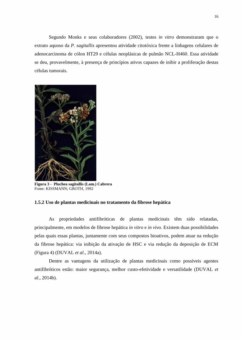
é conhecida como quitoco, madre cravo ou tabacarana (Figura 3) (KISSMANN; GROTH,
1992).
É uma planta anual ou perene, dependendo das condições ambientais. Quanto às suas
características morfológicas, é ereta, aromática, herbácea, de caule multialado e quase sem
ramificação (LORENZI, 2000). Sua reprodução é por semente e desenvolve-se bem em locais
úmidos, inclusive sobre dunas estabilizadas, no litoral. Na Região Sul, floresce durante o
verão e início do outono (KISSMANN; GROTH, 1992).
O gênero Pluchea, pertencente à família Asteraceae, engloba, aproximadamente,
25.000 espécies no mundo e tem sido utilizado devido a diversas propriedades medicinais
(ANDERBERG, 1994; BOTSARIS, 1995). Têm sido descritas importantes propriedades
associadas a espécies desse gênero, dentre as quais estão: atividade antioxidante, potencial
anti-inflamatório e anti-ulcerogênico. Desta forma, o gênero Pluchea parece representar uma
promissora fonte de matéria-prima para a investigação de novas drogas terapêuticas (PÉREZ-
GARCIA et al., 2001).
Estudos fitoquímicos realizados com a Pluchea sagitallis (Lam.) Cabrera
identificaram vários compostos bioativos, dentre eles: flavonóides, fenóis, terpenos, taninos,
alcalóides e saponinas (REYES-TREJO; JOSEPH-NATHAN,1999); (CÓRDOVA; MESA;
HILL, 2006; CÓRDOVA et al., 2010). A P. sagitallis é comumente utilizada no tratamento
de distúrbios digestivos, hepáticos além de ser uma planta carminativa, estimulante aromático,
antiespasmódica e antiulcerativa. Em forma de tintura, a P. sagitallis é utilizada para tratar
erupções cutâneas (LORENZI; MATOS, 2002). Além disso, extratos ou infusões de partes
aéreas da planta têm sido utilizados em diferentes países na medicina tradicional para tratar
processos dolorosos e desordens inflamatórias (ANDERBERG, 1994).
Estudos referentes às propriedades farmacológicas da P. sagitallis, têm sido descritos
na literatura. Os resultados obtidos por Pérez-García et al. (1996), com extrato aquoso e
hidroalcoólico confirmaram as propriedades antiinflamatórias e antioxidantes da planta,
provavelmente correlacionadas à redução de radicais livres (PÉREZ-GARCIA et al., 1996).
16
Segundo Monks e seus colaboradores (2002), testes in vitro demonstraram que o
extrato aquoso da P. sagitallis apresentou atividade citotóxica frente a linhagens celulares de
adenocarcinoma de cólon HT29 e células neoplásicas de pulmão NCL-H460. Essa atividade
se deu, provavelmente, à presença de princípios ativos capazes de inibir a proliferação destas
células tumorais.
Figura 3 - Pluchea sagitallis (Lam.) Cabrera
Fonte: KISSMANN; GROTH, 1992
1.5.2 Uso de plantas medicinais no tratamento da fibrose hepática
As propriedades antifibróticas de plantas medicinais têm sido relatadas,
principalmente, em modelos de fibrose hepática in vitro e in vivo. Existem duas possibilidades
pelas quais essas plantas, juntamente com seus compostos bioativos, podem atuar na redução
da fibrose hepática: via inibição da ativação de HSC e via redução da deposição de ECM
(Figura 4) (DUVAL et al., 2014a).
Dentre as vantagens da utilização de plantas medicinais como possíveis agentes
antifibróticos estão: maior segurança, melhor custo-efetividade e versatilidade (DUVAL et
al., 2014b).

17
Figura 4 - Plantas medicinais antifibróticas com alvo na ativação de HSC (1) e na deposição de ECM (2).
HSC: Células estreladas hepáticas; ECM: matriz extracelular
Fonte: DUVAL et al., 2014

18
2 JUSTIFICATIVA
Apesar dos significativos avanços na compreensão da patogênese da fibrose hepática
nos últimos 20 anos, existe ainda uma grande barreira em traduzir esse vasto conhecimento
científico em fármacos antifibróticos para o tratamento dessa doença.
Segundo o Departamento de Informática do Sistema Único de Saúde (Datasus), em
2008, 9.236 mortes por fibrose e cirrose hepática foram registradas no Brasil. De acordo com
o Ministério da Saúde, a cirrose e outras doenças crônicas do fígado compreendem a 4ª causa
de morte no país, com uma taxa de mortalidade de 6,9 óbitos/100mil habitantes. Assim sendo,
essa doença é um problema de saúde pública. É necessária atenção e medidas efetivas para
seu conhecimento, tratamento e prevenção.
Na busca por estratégias de tratamento que unam segurança, custo-efetividade e
versatilidade, tendo em vista a complexidade da patogênese da fibrose hepática, surgem as
plantas medicinais. No Brasil, existe uma grande variedade de plantas que são utilizadas na
medicina natural, devido às suas diferentes propriedades farmacológicas. Essas plantas
constituem uma fonte rica de compostos bioativos para a indústria farmacêutica.
As espécies da família Asteraceae têm sido amplamente estudadas por apresentarem
compostos químicos de interesse farmacológico. Dentro dessa família, o gênero Pluchea
engloba, aproximadamente, 25.000 espécies no mundo e têm sido descritas pelas importantes
propriedades medicinais associadas a esse gênero. Dentre as várias atividades comprovadas
cientificamente estão: efeito anti-inflamatório, antiulcerogênico, potencial antimicrobiano e
antioxidante.
Tendo em vista que o gênero Pluchea parece representar uma fonte de matéria-prima
para investigação de novos fármacos e possui importantes propriedades medicinais, torna-se
relevante avaliar seu papel no tratamento de fibrose hepática.

19
3 OBJETIVOS
3.1 OBJETIVO GERAL
Avaliar a ação do extrato aquoso de Pluchea sagitallis (Lam.) Cabrera sobre a
proliferação das células estreladas ativadas GRX.
3.2 OBJETIVOS ESPECÍFICOS
- Avaliar a ação da P. sagitallis sobre a reversão do fenótipo;
- Avaliar a formação de colágeno em células GRX expostas ao extrato aquoso da P.
sagitallis;
- Quantificar lipídios em células GRX expostas ao extrato aquoso da P. sagitallis.
- Avaliar a concentração de TGF-β1 em sobrenadante de cultura de células GRX
tratadas com extrato aquoso da P. sagitallis;
- Verificar a ação da P. sagitallis sobre o estresse oxidativo através do TBARS assay
kit;
- Avaliar a ação da P. sagitallis sobre a proliferação das células GRX.

20
4 ARTIGO CIENTÍFICO
Os resultados do presente trabalho serão submetidos à revista Planta Médica – Journal
of Medicinal Plant and Natural Product Research.
Current impact factor: 2.34
26-Aug-2015
Dear Dr. Caruso,
This is to acknowledge receipt of your manuscript entitled "ANTIPROLIFERATIVE
EFFECT OF Pluchea sagitallis (Lam.) Cabrera AQUEOUS EXTRACT IN GRX CELL
LINEAGE" submitted to Planta Medica. It is receiving full attention. You will be informed
in due time on the outcome of the review process.
Your manuscript ID is PLAMED-2015-08-0844-OP.
Please mention the above manuscript ID in all future correspondence. If there are any changes
in your street address or e-mail address, please log into Manuscript Central at
https://mc.manuscriptcentral.com/plamed and edit your user information as appropriate.
You can also view the status of your manuscript any time by checking your Author Center
after logging into https://mc.manuscriptcentral.com/plamed.
Thank you for submitting your manuscript to Planta Medica.
Sincerely,
Dr. Tess de Bruyne
Editorial Office Planta Medica

21
ANTIPROLIFERATIVE EFFECT OF Pluchea sagitallis (Lam.) Cabrera
AQUEOUS EXTRACT IN GRX CELL LINEAGE
Fabiana Garbachi de Oliveira Mendes Ouriques¹, Paula Bacaicoa Caruso¹*, Gabriela Viegas
Haute¹, Juliana Romeu Marques¹, Pedro Maria de Abreu Ferreira², Fernanda Bordignon
Nunes¹, Jarbas Rodrigues de Oliveira¹
Affiliation
1- Cellular Biophysics and Inflammation Laboratory, Pontifícia Universidade Católica do Rio
Grande do Sul (PUCRS), Porto Alegre, Brazil.
2 - Institute of Biosciences, Department of Botany, Universidade Federal do Rio Grande do
Sul (UFRGS), Porto Alegre, Brazil.
* Corresponding author.
Correspondence
Correspondences should be addressed to: Laboratório de Pesquisa em Biofísica Celular e
Inflamação, Pontifícia Universidade Católica do Rio Grande do Sul (PUCRS), Avenida
Ipiranga 6681, prédio 12, bloco C, sala 221, CEP 90619-900, Porto Alegre, Rio Grande do
Sul, Brazil.
Phone: +555133534147
E-mail:[email protected]
22
ABSTRACT
Liver fibrosis is a complex disease that is caused by inappropriate tissue repair due to the
deposition of connective tissue. When a chronic lesion affects the liver, regenerative response
fails and hepatocytes are replaced with abundant extracellular matrix (ECM). The imbalance
between production and degradation of ECM will result in the accumulation of proteins that
change normal liver architecture, and thus its functionality. The main source of ECM is the
activated hepatic stellate cell (HSC). In order, to clarify possible therapeutic approaches to the
disease, the this work aimed to evaluate the possible antifibrotic action of Pluchea sagitallis
(Lam.) Cabrera on an activated HSC immortalized lineage (GRX).
Our results demonstrated that the P. sagittalis aqueous extract at 0.039 and 0.078
mg/mL concentrations was able to reduce cell growth and proliferation. Regarding to
oxidative stress evaluation, there was no statistically significant difference between the treated
group and the control. Staining with OilRed-O (ORO) showed a statistically significant
increase in intracellular lipid content after 5 days of treatment, exerting in vitro effect on the
GRX phenotypic change of activated towards the quiescent state. These results were
confirmed by colorimetric quantification of lipid content. Regarding the TGF-β1 and collagen
production, there were no statistically significant differences observed between the groups.
In conclusion, the P. sagittalis aqueous extract reduces the growth and proliferation of
GRX cells and induces the reversal of activated towards a quiescent phenotype. There was no
decrease in cell proliferation either by necrosis or by apoptosis via activation of the
senescence. Thus, our data suggest that the extract showed an antifibrotic effect, possibly by
activating phenotype reversal.
Keywords: hepatic fibrosis, hepatic stellate cell, Pluchea sagitallis.

23
INTRODUCTION
Hepatic fibrosis, a disease with highly complex etiology, is caused by inappropriate
tissue repair due to the deposition of connective tissue, resulting in chronic liver damage. It
may be caused by alcohol consumption, chronic viral hepatitis, autoimmune diseases, among
others. When there is inappropriate fibrosis control, the result can be a progression to cirrhosis
[1].
When an acute injury affects the liver, parenchymal cells are regenerated and this
process is associated with the inflammatory response and limited deposition of extracellular
matrix (ECM). However, after chronic damage, the regenerative response fails and
hepatocytes are replaced by excessive ECM mainly made of collagen of types I, III and IV,
fibronectin, elastin, laminin and proteoglycans [1].
The main sources of ECM are the hepatic stellate cells (HSCs) [2], which constitute
around 15% of total liver cells [3]. Discovered in 1876 by Kuppfer and located within the
perisinusoidal space of Disse, the HSCs contain fat globules and are responsible for storing
approximately 80% of vitamin A [4, 5, 6]. These cells play an important role in the
fibrogenesis that occurs in chronic lesions of hepatic tissue [7]. The HSCs activation process,
when there is phenotypic change from the quiescent state to an activated state, can be divided
into initiation, perpetuation and resolution, and is an early event in hepatic fibrogenesis.
Several paracrine stimulation of damaged hepatocytes and other neighboring cells can initiate
HSCs activation. The activated stellate cells are characterized by increased contractility,
retinoid loss, chemotaxis, proliferation, ECM degradation capacity and secretion of pro-
inflammatory cytokines. TGF-β1 produced by liver cells represents a potent fibrogenic signal,
since it increases the production of type I collagen and other matrix constituents, such as
fibronectin and proteoglycans. Remarkably, this new ECM configuration induces a new
fibrogenic stimulus that is responsible for exacerbating fibrosis [1]. The GRX cell line, which
represents the activated HSC phenotype has been used as an important tool for the study of
the physiology of HSC and liver fibrosis [8, 9]. The Pluchea sagitallis (Lam.) Cabrera, that
belongs to the Asteraceae family, is a native plant of South America, popularly known as
“quitoco”, “madre cravo” or “tabacarana” [10]. Important properties associated with the
Pluchea gender, have been described including antioxidant activity, and anti-inflammatory
and anti-ulcerogenic potential. Phytochemical studies with P. sagitallis identified several
bioactive compounds, including flavonoids, phenols, terpenes, tannins, alkaloids and saponins

24
[11, 12, 13]. Regarding to the anti-inflammatory property of P. sagitallis, pharmacological
studies showed that the aqueous extract has anti-inflammatory activity that is correlated with a
reduction of free radicals [14].
The antifibrotic properties of medicinal plants have been mainly reported mainly in
models of in vitro and in vivo liver fibrosis. There are two ways in which these plants together
with their bioactive compounds, may act in reducing liver fibrosis: via inhibition of HSC
activation and via reduction of ECM deposition [1]. The advantages of using medicinal plants
as possible antifibrotic agents inclube high safety, cost-effectiveness and versatility [1]. Thus,
the Pluchea gender seems to be a promising source of material on the research of new
therapeutic drugs [15].
RESULTS
Viability and cell growth
After 5 days of treatment with P. sagitallis aqueous extract, cell viability of GRX
lineage was evaluated at 0.039, 0.078, 0.15625, 0.3125, 0.625, 1.25 and 2.5 mg/mL
concentrations on cell viability of GRX lineage. A significant decrease in cell proliferation
was observed with increasing concentrations of the aqueous extract (Fig.1).
Cell toxicity by release of lactate dehydrogenase
In order to evaluate whether the reduction in number of cells treated with P. sagitallis
aqueous extract was due to cytotoxicity, the lactate dehydrogenase (LDH) released was
quantified in the culture medium. Among the concentrations tested, there was a statistically
significant difference at 1.25 and 2.5mg/mL concentrations compared to the control,
indicating cytotoxicity (Fig.2). Thus, 0.039 and 0.078 mg/mL concentrations of P. sagitallis
aqueous extract were selected for this study because lower concentrations had already
presented an antiproliferatic effect.

25
Apoptosis and senescence assay
Regarding the evaluation of apoptosis and senescence measured by DAPI, there was
no statistically significant difference between treated groups and the control group. Thereby
apoptotic cells were not detected in the studied groups (Fig. 3).
Oxidative stress assessment
Oxidative stress can damage the cell and, consequently, decrease cellular proliferation,
allowing to evaluate MDA formation by MDA by TBARS assay. Our results showed that
there was no statistically significant difference between the treated groups and the control
group (Fig. 4).
Detection of lipid droplets by Oil Red-O staining and quantification of lipid accumulation
When activated, stellate hepatic cells, alter their phenotype and, therefore, increase
their proliferation rate. In order to verify whether antiproliferative mechanism of P. sagitallis
aqueous extract could be via phenotype regression, we assessed their lipid content. Control
cells, which did not receive any treatment, had preserved their myofibroblastic phenotype.
However, cells that were treated with P. sagitallis aqueous extract at 0.039 and 0.078 mg/mL
concentrations showed a reversal of activated to quiescent phenotype.
The phenotype reversal was confirmed by colorimetric quantification of intracellular
lipid content. There was a statistically significant increase in intracellular lipid content at
0.078 mg/mL concentration of P. sagitallis aqueous extract when compared to the control
(Fig. 5).

26
TGF-β1 Quantification
There was no statistically significant difference in the amount of TGF-β1 between the
treated groups and the control group after 5 days of treatment (Fig. 6).
Measurement of collagen content
There was no statistically significant difference in collagen content between the treated
groups and the control group after 5 days of treatment, (Fig. 7).
DISCUSSION
Liver fibrosis is a multifactorial process feature by an imbalance of components of
ECM synthesis and degradation, resulting in proteins accumulation that change normal liver
architecture and, consequently, their functionality. GRX cells represent an interesting study of
liver fibrosis model because in basal culture conditions exhibit the myofibroblastic phenotype
[16].
This study demonstrated that P. sagitallis aqueous extract decreased GRX cells
proliferation rate at the tested concentrations. Initially, we evaluated the possible cytotoxic
action of the aqueous extract. We found that at 1.25 and 2.5 mg/mL concentrations the extract
was toxic to cells, as it caused cell necrosis. This was evidenced by the significant increase of
LDH in the cell culture supernatant. Based on these results, we decided to investigate the
cellular mechanism involved in the antiproliferative effect of the P. sagitallis aqueous extract
at 0.039 and 0.078mg/mL concentrations.
Oxidative stress is present in many liver diseases. This process happens due to an
imbalance between oxidants and antioxidants, resulting in an excessive increase of free
radicals. This fact leads to the oxidation of biomolecules with consequent loss of biological
functions and homeostatic imbalances, ending in a powerful oxidative damage to cells and
tissues [17]. Our results showed no statistically significant differences in oxidative stress
among treated and control groups, demonstrating that the antiproliferative effect did not occur
via oxidative stress.

27
Apoptosis plays an important role in the homeostasis of the liver cells, considering that
many diseases that affect their cells are associated with increased apoptosis in hepatocytes, in
order to protect against organ inflammation. Therefore, programmed cell death causes the
hepatocytes die without causing a potentially damaging inflammatory response [18]. Our
results demonstrated that the aqueous extract treatment did not increase the cell death by
apoptosis, as well as not changing the senescent cells number, which could decrease cell
proliferation by disrupting the cell cycle.
The GRX cell proliferation is related to the myofibroblastic phenotype. Therefore, we
histologically analyzed the possible reversal of the phenotype and evaluated the intracellular
lipid content. Our results showed an accumulation in intracellular lipid content as evidenced
by ORO staining and confirmed by intracellular lipids quantification. For this reason, the P.
sagitallis aqueous extract acts as a potent inducer of quiescent phenotype. This effect may
explain the antiproliferative action of the plant.
TGF-β1 is the major pro-fibrotic cytokine in chronic hepatic injury. This mediator
activates the HSCs, resulting in increased cell proliferation and ECM deposition [19].
Increased TGF-β1 levels represent a potent fibrogenic signal and its increase is associated
with increased collagen production [20]. Ours results showed concentrations of TGF-β1,
when analyzed in supernatant GRX cell line, showed no statistically significant difference,
when comparing the groups treated with P. sagitallis aqueous extract and the control group.
The same occurred when we assessed the collagen concentration. Although these results show
no differences between the groups, there was consistency, since TGF-β1 and collagen are
related to each other. According to Ye & Dan [21], the collagen expression and production are
decreased when TGF-β1 synthesis decreases and, consequently, so does the fibrotic process.
Our results showed no significant decrease in these parameters, probably because the
incubation time of the cells and the studied doses, despite decreasing cell proliferation and
GRX cells phenotype reversion were not sufficient to decrease TGF-β1 and collagen
synthesis.
In conclusion, this study demonstrated for the first time a possible antiproliferative
effect of P. sagitallis in an in vitro model of liver fibrosis. The results showed that the plant
aqueous extract, at 0.039 and 0.078 mg/mL concentrations, was able to induce reversion from
quiescent to activated phenotype in GRX cell line. Based on our findings, this study suggests
a possible role for the P. sagitallis aqueous extract in liver fibrosis treatment. The findings
highlight the importance of further research in this area towards a more efficient and effective

28
treatment. A better understanding of fibrogenesis inhibition and HSCs pathways activation
and deactivation are still challenges to be unraveled, not only have a better understanding of
disease pathogenesis, but also to validate the therapeutic use of medicinal plants in liver
fibrosis.
MATERIALS AND METHODS
Materials
P. sagitallis leaves were collected from the Protection Center and Nature
Conservation Pro-Mata, at the town of São Francisco de Paula, Rio Grande do Sul, Brazil,
during December 2014. The extracts were inoculated in GRX cell culture.
Cell culture
The murine GRX cell line [8] was obtained from the Rio de Janeiro Cell Bank
(Federal University, Rio de Janeiro, Brazil). The GRX cell line was obtained from liver
granuloma in mice of C3h line / HeN infected with Schistosoma mansoni cercariae [22, 9].
Under standard cultivation conditions, GRX cells express the myofibroblastic phenotype,
proliferative and ECM producer [9, 23].
Cells were maintained in Dulbecco's Modified Eagle's Medium (DMEM)
supplemented with 5% fetal bovine serum (FBS) (Invitrogen, Carlsbad, CA), 2g/L HEPES
buffer, 3.7g/L NaHCO3 and 1% penicillin and streptomycin (Invitrogen) and incubated at
37°C in a humidified atmosphere with 5% CO₂.
Preparation of the aqueous extract of Pluchea sagitallis
Aqueous extracts were prepared with P. sagitallis leaves. The aqueous extracts were
obtained by adapting the methodology used by Ferris & Zheng [24]. Initially, 2 g of in nature

29
plant leaves in a properly tagged Becker were selected. After cleaning was performed with
distilled water, with subsequent drying and, thereafter, 10 mL of distilled water were added
and then this solution was placed in a water bath (80°C) for 30 min. Subsequently, the
aqueous extract was filtered and stored for centrifugation at 5000 rpm for 10 min. Finally, the
aqueous extract was divided into aliquots of 500 uL in 1.5 mL Eppendorf tubes, and stored at
-20°C.
Treatment with Pluchea sagitallis aqueous extract
The aqueous extract of P. sagitallis was in two different concentrations, at
0.039mg/mL and 0.078mg/mL. GRX cells were incubated and the analyses were performed
after 5 days of treatment. Based on previous studies, all experiments were repeated three
times and in triplicates [25].
Viability and cell growth
Cellular viability and growth were assessed by Tripan blue dye exclusion. The number
of cells was determined by the hemocytometer. GRX cells were seeded into 24-well plates
(3x10³ cells/well) and treated with P. sagitallis aqueous extract (as described above) for 5
days to evaluate the antifibrotic activity. All groups were performed in triplicates. The control
group received only DMEM and 5% SFB for 5 days.
Cell toxicity by release of lactate dehydrogenase
We used the determination of lactate dehydrogenase (LDH) in supernatants of cultures
as compared to the control group in order to evaluate the cytotoxicity of P. sagitallis aqueous
extract in GRX cells. The LDH activity was measured by colorimetric assay [26]. Control of
cell lysis was checked using 5% Tween.

30
Oxidative stress assessment
Oxidative stress was assessed by the concentration of MDA (malondialdehyde) in the
supernatant of cell cultures by Thiobarbituric Acid Reactive Substances (TBARS) assay kit
(Cayman Chemical Company). The TBARS is a well-established method for screening and
monitoring of lipid peroxidation. The results are expressed in µM per 1.000 cells.
Apoptosis and senescence assay
The GRX cells apoptosis and senescence was assessed by immunofluorescence
microscopy (x200). Cells were stained with DAPI (Invitrogen, Inc., Carlsbad, CA, USA) to
assess nuclear changes or modifications of cells undergoing apoptosis.
After treatment, the supernatant was removed and cells were washed three times with
PBS and fixed with 4% paraformaldehyde. Next, cells were washed again with PBS,
permeabilized with 0.5% Triton X-100 for 30 min and DAPI was added. After 2 min the dye
was removed, and the last wash was performed with PBS. Finally, nuclear morphometric
analysis (NMA) was performed to quantitatively evaluate the proportion of cells in
senescence, apoptosis or nuclear irregularities within this population of in vitro cells [27].
Detection of lipid droplets by Oil Red-O staining
Cells were stained with Oil Red-O (ORO) (Sigma) [28] on day 5 to show cell
morphology and lipid accumulation. After fixing cells with 10% formaldehyde, ORO (0.35g
in 60% isopropanol) was briefly added for 15 min. Intracellular lipid droplets were examined
using an inverted light microscope (BestScope, China).

31
Quantification of lipid accumulation
The procedure is also based on ORO staining of intracellular lipid droplets (Sigma
Chemical Co., St. Louis, Mo). Cells were briefly fixed with perchloric acid and incubated
with ORO dissolved in propylene glycol (2mg/mL) for 2h. The ORO within the lipid droplets
was extracted using isopropanol. The absorbance was read at 492nm using an ELISA plate
reader. Each sample was normalized to 100000 cells.
TGF- β1 Quantification
TGF-β1 concentration was measured, in cell supernatant, on day 5, using a
commercially available ELISA kit (R&D Systems, Minneapolis, MN). Results were
calculated on a standard curve concentration and multiplied for the dilution factor. TGF-β1
levels were expressed as nanograms per milliliter per 1.000 cells.
Measurement of collagen content
Collagen content in GRX cells was measured using picro-sirius red on day 5.Picro-
sirius red was added to cell supernatant to form a collagen-dye complex. After centrifugation,
unbound dye was removed and collagen-dye complex dissolved in NaOH. The absorbance
was measured at 540nm in an ELISA plate reader. Each sample was normalized to 1.000 cells
[29]. Results were calculated on a standard curve concentration. Collagen levels were
expressed as the ratio of milligrams of collagen and cells number.
Statistics
Data are reported as mean ± SD. Each experiment was performed at least three
independent times and in triplicates. Statistical testing was performed with Prism 5 software.

32
Results were analyzed by one-way analysis of variance (ANOVA), followed by Tukey’s
multiple comparison test. The level of significance was set at p <0.05.
Conflict of Interest
The authors declare no conflict of interest.
REFERENCES
1 Duval, F. et al. Protective mechanisms of medicinal plants targeting hepatic stellate cell
activation and extracellular matrix deposition in liver fibrosis. Chin Med, v. 9, n. 1, p. 27,
2014a.
2 Duval, F. et al. Liver fibrosis and protection mechanisms action of medicinal plants
targeting apoptosis of hepatocytes and hepatic stellate cells. Adv Pharmacol Sci, v. 2014, p.
373295, 2014b.
3 Friedman, S. L. Molecular regulation of hepatic fibrosis, an integrated cellular response to
tissue injury. J Biol Chem, v. 275, n. 4, p. 2247-50, Jan 2000.
4 McCuskey, R. S. Functional morphology of the liver with emphasis on microvasculature.
In: Tavaloni N, Berk PD eds. Hepatic transport and bile secretion: physiology and
pathophysiology. New York, Raven Press Ltd; 1-10. 1993.
5 Friedman, S. L. Mechanisms of hepatic fibrogenesis. Gastroenterology, v. 134, n. 6, p.
1655-69, May 2008.
6 Winau, F. et al. Starring stellate cells in liver immunology. Curr Opin Immunol, v. 20, n. 1,
p. 68-74, Feb 2008.
7 Rockey, D. The cellular pathogenesis of portal hypertension: stellate cell contractility,
endothelin, and nitric oxide. Hepatology, v. 25, n. 1, p. 2-5, Jan 1997.

33
8 Borojevic R, Monteiro AN, Vinhas SA, Domont GB, Mourão PA, Emonard H, Grimaldi G,
Grimaud JA. 1985. Establishment of a continuous cell line from fibrotic schistosomal
granulomas in mice livers. In Vitro Cell Dev Biol 21:382-90.
9 Guimarães, E. L. et al. Relationship between oxidative stress levels and activation state on
a hepatic stellate cell line. Liver Int, v. 26, n. 4, p. 477-85, May 2006.
10 Kissmann, K. G., Groth, D. Plantas Infestantes e Nocivas. BASF. Brasileira S/A. 2ª edição,
v2, p.798. TOMO II. 1992.
11 Reyes-Trejo,B., Joseph-Nathan, P. Modhephene derivates from Pluchea sericea.
Phytochemistry, 51:75-78, 1999.
12 Córdova, W.H.P., Mesa, L.G., Hill, A.L.P. Metabólitos secundarios y actividad
antimicrobiana de Pluchea carolinensis . Revista Cubana de Farmácia, 40 (2) , 2006.
13 Córdova, W.H.P., Tabart, J., Quesada, A.G., Sipel, A., Hill, A.L.P., Kevers, C.Dommes, J.
Antioxidant capacity of three Cuban species of the genus Pluchea Cass.( Asteraceae). Journal
of Food Biochemistry, 34:249-261, 2010.14
14 Pérez-García, F. et al. Anti-inflammatory action of Pluchea sagittalis: involvement of an
antioxidant mechanism. Life Sci, v. 59, n. 24, p. 2033-40, 1996.
15 Pérez-García, F. et al. Activity of plant extracts on the respiratory burst and the stress
protein synthesis. Phytomedicine, v. 8, n. 1, p. 31-8, Jan 2001.
16 Lotersztajn, S., Julien, B., Clere, F.T.,Grenard, P., Mallat, A.H. 2005. Hepatic fibrosis:
Molecular mechanisms and drug targets. Annu. harmacol. Toxicol. 45:605-28.
17 Halliwell B, Whiteman M. 2004. Measuring reactive species and oxidative damage in vivo
and in cell culture: how should you do it and what do the results mean? Br J Pharmacol
142:231-55.

34
18 Neuman, M.G. (2001) Apoptosis in diseases of the liver. Critical Reviews in Clinical and
Laboratory Science 38, pp. 109–166.
19 Friedman SL. 2008b. Hepatic stellate cells: protean, multifunctional, and enigmatic cells of
the liver. Physiol Rev 88:125-72.
20 Bissell DM, Roulot D, George J. 2001. Transforming growth factor beta and the liver.
Hepatology 34:859-67.
21 Ye Y, Dan Z. 2010. All-trans retinoic acid diminishes collagen production in a hepatic
stellate cell line via suppression of active protein-1 and c-Jun N-terminal kinase signal. J
Huazhong Univ Sci Technolog Med Sci 30:726-33.
22 Vicente CP, Fortuna VA, Margis R, Trugo L, Borojevic R. 1998. Retinol uptake and
metabolism, and cellular retinol binding protein expression in an in vitro model of hepatic
stellate cells. Mol Cell Biochem 187:11-21.
23 Souza IC, Martins LA, Coelho BP, Grivicich I, Guaragna RM, Gottfried C, Borojevic R,
Guma FC. 2008. Resveratrol inhibits cell growth by inducing cell cycle arrest in activated
hepatic stellate cells. Mol Cell Biochem 315:1-7.
24 Ferris H, Zheng L. 1999. Plant Sources of Chinese Herbal Remedies: Effects on
Pratylenchus vulnus and Meloidogyne javanica. J Nematol 31:241-63.
25 Bitencourt S, de Mesquita FC, Caberlon E, da Silva GV, Basso BS, Ferreira GA, de
Oliveira JR. 2012. Capsaicin induces de-differentiation of activated hepatic stellate cell.
Biochem Cell Biol 90:683-90.
26 Spiller F, et al. Anti-inflammatory effects of red pepper (Capsicum baccatum) on
carrageenan- and antigen-induced inflammation. J. Pharm. Pharmacol. 2007; 60: 473,478.
27 Filippi-Chiela EC, Oliveira MM, Jurkovski B, Callegari-Jacques SM, da Silva VD, Lenz
G. 2012. Nuclear morphometric analysis (NMA): screening of senescence, apoptosis and
nuclear irregularities. PLoS One 7:e42522.

35
28 Ramírez-Zacarías JL, Castro-Muñozledo F, Kuri-Harcuch W. 1992. Quantitation of
adipose conversion and triglycerides by staining intracytoplasmic lipids with Oil red O.
Histochemistry 97:493-7.
29 Bradford MM. 1976. A rapid and sensitive method for the quantitation of microgram
quantities of protein utilizing the principle of protein-dye binding. Anal Biochem 72:248-54.
LEGENDS FOR FIGURES
Fig. 1 Effect of Pluchea sagitallis in GRX cells growth, assessed by direct counting on
Neubauer´s Chamber. Cells were treated with P. sagitallis aqueous extract for 5 days. Data
represent the mean ± SD (n=4). Results are expressed as cell number per well. ***p<0.01
compared to control.
Fig 2 Effect of Pluchea Sagitallis in lactate dehydrogenase (LDH) release on cell supernatant
after 5 days. Data represent the mean ± SD (n=4). Results are expressed as percentage of
LDH. ** p<0.01 and ***p< 0.001 compared to control.
Fig. 3 DAPI Staining to assess nuclear changes of cells undergoing apoptosis and senescence
after 5 days of treatment (A) Control, 0.039 and 0.078 mg/mL; (B) Percentage of normal cells
and senescent cells.
Fig. 4 TBARS levels in GRX cell line after 5 days of treatment. Data
represent the mean ± SD (n = 4). TBARS levels were the expressed per nanomol per
1000cells.
Fig. 5 A: Oil Red-O (ORO) staining and lipid quantitation of GRX cells at day 5.
(A) Control, and cells treated with Pluchea. sagitallis aqueous extract at 0.039 and 0.078
mg/mL, respectively. Bar length = 40 µm. (B) Specific lipid content expressed
spectrophotometrically as the ratio of absorbance value obtained for ORO and the cell
number. Results are expressed as mean ± SD. **p<0.01, compared to control.

36
Fig. 6 ELISA assay of TGF-β1 in cell supernatant of 5 days treatment. Data
represent the mean ± SD (n=4). TGF-β1 levels are expressed as nanograms
per milliliter per 1000cells.
Fig. 7 Total collagen content in cell supernatant of 5 days treatment. Data
represent the mean ± SD (n=4). Results are expressed as micrograms
per milliliter/1000cells.

37
Fig. 1

38
Fig. 2

39
Fig. 3
A
B

40
Fig. 4

41
Fig. 5
A
B

42
Fig. 6

43
Fig. 7

44
5 CONSIDERAÇÕES FINAIS
Dentre as diversas doenças consideradas como questões de saúde pública que
acometem a população mundial, está a fibrose hepática. Essa doença está associada à
significativa morbidade e mortalidade (DUVAL et al., 2014a). Até o presente momento, não
existe um tratamento padronizado. Uma possibilidade mais efetiva é a intervenção através da
remoção do agente causador. Entretanto, tem ocorrido nos últimos anos um crescimento nos
conhecimentos científicos sobre essa doença, revelando potenciais alvos terapêuticos.
A HSC é um alvo atrativo para o estudo de novos agentes antifibróticos na tentativa de
tratar a fibrose em seus diferentes estágios. A redução do processo inflamatório e da resposta
imune, a inibição da ativação das HSC, a indução da apoptose das HSCs, a interrupção das
atividades fibrogênicas, contráteis, proliferativas e pró-inflamatórias das HSCs, a diminuição
da síntese dos componentes da ECM ou o aumento de sua degradação são algumas das
possíveis hipóteses para a interrupção do processo fibrótico (ALBANIS; FRIEDMAN, 2006;
Li et al., 2008).
Nos últimos anos, a importância de compostos naturais com propriedades medicinais
vem sendo amplamente reconhecida tanto na prevenção, como no tratamento de várias
doenças (WEISBURGER; WILLIAMS, 2000). As pesquisas com plantas medicinais
envolvem: investigações da medicina tradicional e popular (etnobotânica); isolamento,
purificação e caracterização de princípios ativos (química orgânica: fitoquímica); investigação
farmacológica de extratos e dos constituintes químicos isolados (farmacologia);
transformações químicas de princípios ativos (química orgânica sintética); estudo da relação
estrutura/atividade e dos mecanismos de ação dos princípios ativos (química medicinal e
farmacológica) e finalmente a operação de formulações para produção de fitoterápicos. A
integração destas áreas na pesquisa de plantas medicinais conduz a um caminho promissor e
eficaz para descobertas de novos medicamentos. O objetivo do nosso estudo foi avaliar a
possível ação do extrato aquoso da Pluchea sagittalis sobre a proliferação das células
estreladas ativadas GRX e seu efeito terapêutico no tratamento da fibrose hepática, tendo em
vista que esta planta já apresenta propriedades farmacológicas cientificamente comprovadas .
Através dos resultados, foi possível identificar atividade antiproliferativa in vitro das folhas da
P. sagittalis em linhagens celulares GRX. Portanto, este estudo é um importante passo para a
pesquisa de novas drogas antifibróticas. A proposta de prosseguir na pesquisa das propriedades
do extrato aquoso da P. sagittalis é de fundamental importância para a elucidação de seu

45
potencial terapêutico tanto para indústria farmacêutica, onde se busca medicamentos eficazes,
seguros, quanto para as populações que, eventualmente, já fazem uso medicinal desta planta.

46
REFERÊNCIAS
ALBANIS, E.; FRIEDMAN, S. L. Antifibrotic agents for liver disease. Am J Transplant,
New York, v.6, n.1, jan. 2006, p.12-19.
ANDERBERG, A. A. Asteraceae. In: BREMER, K. (ed.). Cladistics and classification.
Portland, Oregon: Timber Press, 1994, 752p.
BOTSARIS, A. S. Fitoterapia chinesa e plantas brasileiras. São Paulo: Ícone, 1995.
CÓRDOVA, W. H. P.; MESA, L. G.; HILL, A. L. P. Metabólitos secundarios y actividad
antimicrobiana de pluchea carolinensis. Revista Cubana de Farmácia, Havana, v.40, n.2,
ago. 2006, p.1-4.
______; TABART, J.; QUESADA, A. G.; SIPEL, A.; HILL, A. L. P.; KEVERS, C.;
DOMMES, J. Antioxidant capacity of three Cuban species of the genus Pluchea Cass.(
Asteraceae). Journal of Food Biochemistry, Liège, v.34, s.1, mar. 2010, p.249-261.
DUVAL, F. et al. Liver fibrosis and protection mechanisms action of medicinal plants
targeting apoptosis of hepatocytes and hepatic stellate cells. Adv Pharmacol Sci. v.2014,
2014b, p.373295.
______. Protective mechanisms of medicinal plants targeting hepatic stellate cell activation
and extracellular matrix deposition in liver fibrosis. Chin Med. v.9, n.1, 2014a, p.27.
ELLIS, E. L.; MANN, D. A. Clinical evidence for the regression of liver fibrosis. J Hepatol.
v.56, n.5, may. 2012, p.1171-80.
FRIEDMAN, S. L. Mechanisms of hepatic fibrogenesis. Gastroenterology. v.134, n.6, may.
2008, p.1655-69.
______. Molecular regulation of hepatic fibrosis, an integrated cellular response to tissue
injury. J Biol Chem. v.275, n.4, jan. 2000, p.2247-50.
GEERTS, A. History, heterogeneity, developmental biology, and functions of quiescent
hepatic stellate cells. Semin Liver Dis. v.21, n.3, aug. 2001, p.311-35.

47
GUIMARÃES, E. L. et al. Relationship between oxidative stress levels and activation state on
a hepatic stellate cell line. Liver Int. v.26, n.4, may. 2006, p.477-85.
HENDERSON, N. C.; FORBES, S. J. Hepatic fibrogenesis: from within and
outwith. Toxicology. v.254, n.3, dec. 2008, p.130-5.
HERRMANN, J.; GRESSNER, A. M.; WEISKIRCHEN, R. Immortal hepatic stellate cell
lines: useful tools to study hepatic stellate cell biology and function? J Cell Mol Med. v.11,
n.4, jul./aug. 2007, p.704-22.
IREDALE, J. Defining therapeutic targets for liver fibrosis: exploiting the biology of
inflammation and repair. Pharmacol Res. v.58, n.2, aug. 2008, p.129-36.
KISSMANN, K. G.; GROTH, D. Plantas infestantes e nocivas. 2.ed. v.2. Tomo II. [s.l]:
BASF, 1992.
LIM, Y. S.; KIM, W. R. The global impact of hepatic fibrosis and end-stage liver
disease. Clin Liver Dis. v.12, n.4, nov. 2008, p.733-46.
LORENZI, H. Plantas daninhas do Brasil: terrestres, aquáticas, parasitas e tóxicas. 3.ed.
São Paulo: Instituto Plantarum de Estudos da Flora, 2000.
______; MATOS, F. J. A. Plantas medicinais do Brasil nativas e exóticas. São Paulo:
Instituto Plantarum de Estudos da Flora, 2002.
MCCUSKEY, R. S. Functional morphology of the liver with emphasis on microvasculature.
In: TAVALONI, N.; BERK, P. D. (eds.). Hepatic transport and bile secretion: physiology
and pathophysiology. New York: Raven Press, 1993.
MEANS, R. T. Hepcidin and iron regulation in health and disease. Am J Med Sci. v.345, n.1,
jan. 2013, p.57-60.
MONKS, N. R.; FERRAZ, A.; BORDIGNON, S.; MACHADO, K. R.; LIMA, M. F. S.;
ROCHA, A. B.; SCHWARTSMAN, N. Vitro Citotoxicity of Extracts from Brazilian
Asteraceae. Pharmaceutical Biology. v.40, n.7, 2002, p.494-500.
MONTEIRO, A. N., Borojevic, R. “In vitro formation of fibrous septa by liver connective
tissue cells”. In vitro cellular & developmental biology: Journal of the Tissue Culture
Association. v.23, n.1, 1987, p.10-4.

48
REYES-TREJO, B.; JOSEPH-NATHAN, P. Modhephene derivates from Pluchea sericea.
Phytochemistry. v.51, 1999, p.75-78.
ROCKEY, D. The cellular pathogenesis of portal hypertension: stellate cell contractility,
endothelin, and nitric oxide. Hepatology. v.25, n.1, jan. 1997, p.2-5.
SOUZA, I. C. et al. Resveratrol inhibits cell growth by inducing cell cycle arrest in activated
hepatic stellate cells. Mol Cell Biochem. v.315, n.1-2, aug. 2008, p.1-7.
VICENTE, C. P. et al. Retinol uptake and metabolism, and cellular retinol binding protein
expression in an in vitro model of hepatic stellate cells. Mol Cell Biochem. v.187, n.1-2, oct.
1998, p.11-21.
WEISBURGER, J. H.; WILLIAMS, G. M. The distinction between genotoxic and epigenetic
carcinogens and implication for cancer risk. Toxicology Science. v.49, 2000, p.231-246.
WINAU, F. et al. Starring stellate cells in liver immunology. Curr Opin Immunol. v.20, n.1,
feb. 2008, p.68-74.