perilipin 2 and lipid droplets provide reciprocal
TRANSCRIPT

RESEARCH ARTICLE
Perilipin 2 and lipid droplets provide reciprocal stabilization
Shimeng Xu1,2, Fei Zou3,4, Zhiqing Diao5,6, Shuyan Zhang1, Yaqin Deng1,Xiaotong Zhu1,2, Liujuan Cui1,7, Jinhai Yu1,2, Zhiguang Zhang8,Adekunle Toyin Bamigbade1, Hongchao Zhang4, Xuan Wei4, Xuelin Zhang9,Bin Liang6&, Pingsheng Liu1&
1 National Laboratory of Biomacromolecules, CAS Center for Excellence in Biomacromolecules, Institute ofBiophysics, Chinese Academy of Sciences, Beijing 100101, China
2 University of Chinese Academy of Sciences, Beijing 100049, China3 Graduate School of Anhui Medical University, Hefei 230032, China4 General Hospital of Air Force, Beijing 100142, China5 School of Life Sciences, Anhui University, Hefei 230601, China6 Key Laboratory of Animal Models and Human Disease Mechanisms of Chinese Academy of Science & YunnanProvince, Kunming Institute of Zoology, Chinese Academy of Sciences, Kunming 650223, China
7 School of Life Sciences, University of Science and Technology of China, Hefei 230032, China8 Department of Cell Biology and Genetics, School of Basic Medical Sciences, Fujian Medical University,Fuzhou 350004, China
9 School of Kinesiology and Health, Capital University of Physical Education and Sports, Beijing 100191, China
Received: 7 January 2019 /Accepted: 29 January 2019 / Published online: 12 July 2019
Abstract The lipid droplet (LD)-associated protein adipose differentiation-related protein (ADRP or PLIN2) isrequired for the formation and stability of the LD organelle, whereas its biological roles are stillobscure. Herein, we show that PLIN2 is the most abundant protein on the lipid droplets (LDs) of mousemyoblast cell line C2C12. Both the expression of PLIN2 and the accumulation of LDs were up-regulatedin a time- and dose-dependent manner when the cells were treated with oleate (OA). The protein levelof PLIN2 was positively correlated with the formation of LDs, suggesting that LDs stabilize PLIN2.Furthermore, knocking out PLIN2 in C2C12 cells led to enlarged LDs and higher triacylglycerolhydrolysis activity. The isolated PLIN2 null LDs became closely contact with mitochondria and othercellular organelles. Additionally, mitochondrial activity was suppressed by OA in PLIN2 null cells. Ourresults reveal the pivotal roles of PLIN2 in governing LD dynamics and their relationship to mito-chondria, and suggest a reciprocal stabilization between PLIN2 and LDs.
Keywords Lipid droplets, PLIN2, Triacylglycerol, Mitochondria, Hydrolysis
INTRODUCTION
Obesity develops when energy intake exceeds energyexpenditure, leading to lipid accumulation in adiposetissues and nonadipose tissues (e.g., liver, muscle, etc.),which alters systemic metabolism and can lead tometabolic syndrome (Murphy et al. 2009; Unger et al.
Shimeng Xu, Fei Zou and Zhiqing Diao have contributed equally tothis work.
Electronic supplementary material The online version of thisarticle (https://doi.org/10.1007/s41048-019-0091-5) containssupplementary material, which is available to authorized users.
& Correspondence: [email protected] (B. Liang),[email protected] (P. Liu)
� The Author(s) 2019 145 | June 2019 | Volume 5 | Issue 3
Biophys Rep 2019, 5(3):145–160https://doi.org/10.1007/s41048-019-0091-5 Biophysics Reports

2010). Lipid droplets (LDs) are highly dynamic orga-nelles that are composed primarily of triacylglycerol(TAG) and sterol esters (SE), which form a neutral lipidcore, and are wrapped by a monolayer of phospholipidswith numerous resident proteins (Brown 2001). LDsserve as the primary mobile cellular reservoir for lipid-based energy and membrane precursors (Fujimoto andParton 2011). The dynamic of LDs has been consideredas an important indicator for the metabolic states ofcells and tissues (Greenberg et al. 2011). For example,excessive lipid accumulation in LDs can lead to obesity,fatty liver, diabetes, and atherosclerosis. Similarly, a lackof lipid or LDs can lead to diseases such as neutral lipidstorage disease and lipodystrophy.
Perilipin1 (PLIN1), adipocyte differentiation-relatedprotein (ADRP/PLIN2), Tip47 (PLIN3), S3-12 (PLIN4),and OXPAT (PLIN5), also collectively called ‘‘PAT’’ familyproteins, were recently renamed the Perilipins (PLIN)1–5 (Kimmel et al. 2010) and are the main structuralproteins of LDs (Bickel et al. 2009; Brasaemle 2007;Greenberg et al. 2011; Walther and Farese 2012). Ofwhich, PLIN2 is ubiquitously expressed as a lipid dro-plet (LD)-associated protein (Brasaemle et al. 1997),and has been shown to play a key role in LD formation(Fukushima et al. 2005) and maintenance of lipidhomeostasis. PLIN2 is thought to be involved in both LDbiogenesis and the regulation of lipolysis, primarily inadipocytes, but also in other cells and tissues (Changet al. 2006; Listenberger et al. 2007; Robenek et al.2006). PLIN2 null mice display reduced hepatic lipids,and are resistant to diet-induced fatty liver withoutdetectable changes in TAG synthesis, or fatty aciduptake, synthesis, or b-oxidation (Chang et al. 2006;McManaman et al. 2013). Furthermore, PLIN2 null ob/ob mice (leptin-/-) show improved insulin sensitivityand reduced hepatosteatosis compared to controls thatare only leptin-deficient (Chang et al. 2010). Addition-ally, in vitro results reveal that overexpression of PLIN2increases both TAG accumulation and LD formation(Fukushima et al. 2005; Larigauderie et al. 2006; Lis-tenberger et al. 2007), which may be as the result of thereduced LD localization of adipose triglyceride lipase(ATGL) and the slowdown of TAG turnover (Listen-berger et al. 2007). Conversely, knockdown of PLIN2 inmacrophages is shown to decrease cellular lipids andLD size and number (Larigauderie et al. 2006). Theseresults demonstrate an important role of PLIN2 in theregulation of LD dynamic and TAG metabolism.
Skeletal muscle is a crucial metabolic organ whichplays an important role in energy expenditure. Theoverall amount of TAG in skeletal muscle is quite smallcompared to that in adipose tissue, but an increase inskeletal muscle TAG is a major contributor to insulin
resistance (Kelley et al. 1999; Pan et al. 1997). Inskeletal muscle, PLIN2 is the most abundantly expres-sed PLIN protein (Brasaemle et al. 1997; Heid et al.1998; MacPherson and Peters 2015b; Minnaard et al.2009; Peters et al. 2012). PLIN2 overexpression eitherin vitro or in vivo increases intramyocellular LD accu-mulation and TAG storage, and also improves skeletalmuscle insulin sensitivity, whereas PLIN2 knockdowndisplays opposite effects (Bosma et al. 2012). However,the exact relationship between PLIN2 expression andTAG accumulation is still unclear (MacPherson andPeters 2015a). In addition, PLIN2 distribution andassociation with LDs remain ambiguous. For example,PLIN2 was reported to highly co-localize to the LDsurface in isolated rat soleus fibers (Prats et al. 2006),whereas other studies have found contradictory resultsin isolated rat soleus muscle (MacPherson et al. 2012)and human vastus lateralis (Shaw et al. 2009).
Thus, to explore the role of PLIN2 in skeletal muscle,we isolated LDs from the mouse myoblast cell lineC2C12. We found that PLIN2 was the most abundantprotein on the LDs, and its expression was up-regulatedin a time- and dose-dependent manner in the presenceof oleate (OA). The accumulation of TAG and the for-mation of LDs may promote the stability of the PLIN2protein. Knock out (KO) of PLIN2 in C2C12 cells led tolarger LDs that were fewer in number under both basaland OA treatment, and also resulted in higher hydrolysisactivity during OA treatment, compared to wild type(WT) cells. The KO of PLIN2 increased the contactbetween LDs and mitochondria, and inhibited mito-chondrial activity during OA treatment.
RESULTS
Oleate (OA) upregulates PLIN2 expressionand lipid droplet (LD) expansion
Excessive accumulation of lipids in LDs has been linkedto many metabolic disorders. The roles of ubiquitously-expressed LD structure protein PLIN2 in the lipidaccumulation of LDs remains elusive. To do so, we cul-tured C2C12 myoblasts in the presence or absence of OAto induce a model of lipid overload. From these cells LDswere isolated and biochemical measurements weremade to determine the purity of isolated LDs. First, wecompared the protein profile of isolated LDs to othercell fractions using silver staining. Proteins extractedfrom LDs displayed a relatively simple pattern that wasobviously distinct from the protein profiles of the otherthree cellular fractions, including post-nuclear super-natant (PNS), total membranes (TM), and cytosol (Cyto)
RESEARCH ARTICLE S. Xu et al.
146 | June 2019 | Volume 5 | Issue 3 � The Author(s) 2019

(Fig. 1A). The purity of isolated LDs was further asses-sed by the relative enrichment of LD-associated proteinsPLIN2 and PLIN3, and the absence of markers thatcorrespond to other intracellular compartments, lyso-some protein Lamp1, ER protein Bip, mitochondrionprotein Tim23, and cytoplasmic protein GAPDH. Aspreviously reported (Liu et al. 2004), the plasmamembrane protein Cav-1 was also found in the LDfraction, and its expression was induced by OA treat-ment (Fig. 1A). Collectively, these results suggest thatthe isolated LD fraction from C2C12 cells might be lar-gely free of contamination.
OA treatment increased the expression of the mostabundant band in the LD fraction (Fig. 1A). ImageJanalysis showed that the density of this band occupiedabout 25% and 41% of total LD proteins in the absenceor presence of OA, respectively (Fig. 1B). Therefore, thisband was sliced and then subjected to mass spectrom-etry (MS) analysis. Its major protein was identified asPLIN2 (supplementary material Table S1). Consistentwith the MS results, immunoblot results also confirmedthat the expression of PLIN2 was increased in the LDfraction with OA treatment (Fig. 1A).
To investigate the relationship between the expres-sion of PLIN2 and the formation of LDs, we treatedmouse C2C12 myoblasts with 200 lmol/L OA for theindicated times or with different concentrations of OAfor 12 h (Fig. 1C, D). The mRNA and the protein levelsof PLIN2 were up-regulated by OA treatment in a dose-and time-dependent manner (Fig. 1C, D). The proteinlevel of PLIN2 increased much more dramatically thanits mRNA level, implying that OA may enhance the sta-bility of the PLIN2 protein. In addition, PLIN2–EGFPknock-in (KI) C2C12 cells also showed that the intensityof GFP fluorescence was enhanced in a time- and dose-dependent manner under OA incubation (Fig. 1E, F,supplementary material Fig. S1B). Similarly, TAG con-tent was significantly increased by OA incubation in atime-dependent manner (Fig. 1G). OA treatment alsodramatically increased LD number and size, concomi-tant with the accumulation of TAG (Fig. 1H). Therefore,these results demonstrate that both PLIN2 and LDs areup-regulated by OA.
Disruption of lipid droplets (LDs) reducesthe stability of PLIN2
Since the expression of PLIN2 is positively related toTAG accumulation and LD formation during OA treat-ment, we asked whether PLIN2 expression promotesTAG accumulation and LD formation; or, conversely,whether TAG accumulation and LD formation inducePLIN2 expression and stabilization. To answer this
question, we constructed cell models that containedreduced TAG and LDs to detect the level of PLIN2expression during the incubation with OA. Diacylglyc-erol acyltransferase 1 and 2 (DGAT1/2) are the keyenzymes in the final, committed step of TAG biosyn-thesis (Yen et al. 2008). ATGL plays a key role in thehydrolysis of TAG (Villena et al. 2004; Zimmermannet al. 2004, 2009). We therefore constructed Atgl over-expressed (OE) and Dgat1/Dgat2 double knock-out(DKO) C2C12 cell lines (Fig. 2D, supplementary materialFig. S2) to reduce the accumulation of TAG and LDsduring incubation with OA.
The Dgat1/Dgat2 DKO cells accumulated much lessTAG and formed much fewer LDs during OA treatment(Fig. 2A, B). Interestingly, we found that the mRNA levelof PLIN2 was induced during OA incubation in theDgat1/Dgat2 DKO cells to a degree similar to WT cells(Fig. 2C), whereas the protein expression level of PLIN2was not induced in Dgat1/Dgat2 DKO cells (Fig. 2D).Therefore, it appears that accumulation of LDs isrequired to stabilize the PLIN2 protein and thus LDsperform a crucial role in protecting PLIN2 from rapiddegradation.
We then treated C2C12 cells with palmitate (PA) and/or OA, and measured the expression and accumulationof PLIN2 and LDs to verify this finding (Coll et al. 2008;Gao et al. 2009; Peng et al. 2011; Salvado et al. 2013).PA and PA plus OA dramatically induced PLIN2 mRNAexpression even greater than OA (Fig. 2E). However, PAtreatment did not increase the level of PLIN2 protein,unlike treatment with OA or PA plus OA (Fig. 2G).
PA induced the formation of many fewer LDs than OAor PA plus OA, as expected (Fig. 2F). In addition, theexpression level of exogenous PLIN2-MYC protein wasmuch lower in PA-treated cells than in the cells thatwere incubated with OA and PA plus OA (Fig. 2G). Sincethe transcriptional levels of exogenous PLIN2-MYCcould not be stimulated by free fatty acid, the higherexpression level of its protein represents an increase instability of the protein in OA- or PA plus OA-treated cells(Fig. 2F, G). The results further strengthen the conclu-sion that LDs enhance the stability of PLIN2.
Depletion of PLIN2 enhances lipid droplet sizeand lipolysis
The results described indicate that OA stimulated bothPLIN2 expression and LD formation and that the for-mation of LDs stabilized the newly manufactured PLIN2.We then asked whether PLIN2 affects the formation anddynamics of LDs since it is the most abundant proteinon LDs of C2C12 cells. To do so, PLIN2 KO C2C12 cellswere generated. Previous work showed that methionine
Perilipin 2 and lipid droplets provide reciprocal stabilization RESEARCH ARTICLE
� The Author(s) 2019 147 | June 2019 | Volume 5 | Issue 3

Fig. 1 Both PLIN2 and LDs are up-regulated by free fatty acids. A Silver staining and Western blot analysis of proteins from LDs andother fractions of C2C12 cells after treatment with or without OA. The band indicated by the red arrow was sliced for MS analysis. Markerproteins of cellular components were used to verify the purity of LDs (PLIN2/3: LDs, Cav-1: plasma membrane, Lamp1: lysosome, Bip: ER,Tim23: mitochondria, GAPDH: cytosol). B Quantification of silver stained LD proteins by ImageJ. C, D The mRNA and the protein levels ofPLIN2 in C2C12 cells after treatment with OA at the indicated concentrations (C) or times (D). These results had at least three biologicalrepeats. E, F EGFP fluorescence intensity of PLIN2-EGFP KI C2C12 cells after treatment with OA at the indicated concentrations (E) ortime (F). Scale bar, 20 lm. G The TAG content (TAG/protein) in C2C12 cells after treatment with OA for the indicated times. Data werecollected from two biological repeats. H LD fluorescence intensity in C2C12 cells after treatment with OA. LDs were stained with LTR andimaged with a confocal microscope. Scale bar, 5 lm
RESEARCH ARTICLE S. Xu et al.
148 | June 2019 | Volume 5 | Issue 3 � The Author(s) 2019
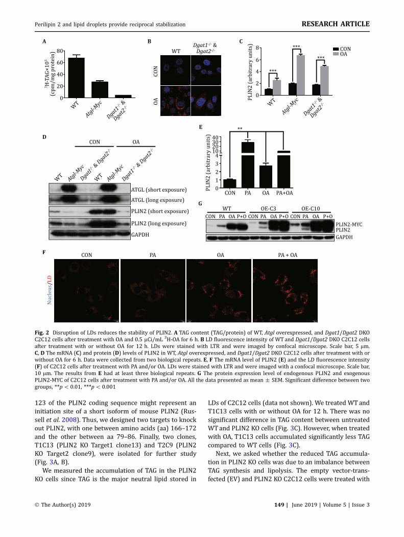
123 of the PLIN2 coding sequence might represent aninitiation site of a short isoform of mouse PLIN2 (Rus-sell et al. 2008). Thus, we designed two targets to knockout PLIN2, with one between amino acids (aa) 166–172and the other between aa 79–86. Finally, two clones,T1C13 (PLIN2 KO Target1 clone13) and T2C9 (PLIN2KO Target2 clone9), were isolated for further study(Fig. 3A, B).
We measured the accumulation of TAG in the PLIN2KO cells since TAG is the major neutral lipid stored in
LDs of C2C12 cells (data not shown). We treated WTandT1C13 cells with or without OA for 12 h. There was nosignificant difference in TAG content between untreatedWTand PLIN2 KO cells (Fig. 3C). However, when treatedwith OA, T1C13 cells accumulated significantly less TAGcompared to WT cells (Fig. 3C).
Next, we asked whether the reduced TAG accumula-tion in PLIN2 KO cells was due to an imbalance betweenTAG synthesis and lipolysis. The empty vector-trans-fected (EV) and PLIN2 KO C2C12 cells were treated with
Fig. 2 Disruption of LDs reduces the stability of PLIN2. A TAG content (TAG/protein) of WT, Atgl overexpressed, and Dgat1/Dgat2 DKOC2C12 cells after treatment with OA and 0.5 lCi/mL 3H-OA for 6 h. B LD fluorescence intensity of WT and Dgat1/Dgat2 DKO C2C12 cellsafter treatment with or without OA for 12 h. LDs were stained with LTR and were imaged by confocal microscope. Scale bar, 5 lm.C, D The mRNA (C) and protein (D) levels of PLIN2 in WT, Atgl overexpressed, and Dgat1/Dgat2 DKO C2C12 cells after treatment with orwithout OA for 6 h. Data were collected from two biological repeats. E, F The mRNA level of PLIN2 (E) and the LD fluorescence intensity(F) of C2C12 cells after treatment with PA and/or OA. LDs were stained with LTR and were imaged with a confocal microscope. Scale bar,10 lm. The results from E had at least three biological repeats. G The protein expression level of endogenous PLIN2 and exogenousPLIN2-MYC of C2C12 cells after treatment with PA and/or OA. All the data presented as mean ± SEM. Significant difference between twogroups, **p\0.01, ***p\ 0.001
Perilipin 2 and lipid droplets provide reciprocal stabilization RESEARCH ARTICLE
� The Author(s) 2019 149 | June 2019 | Volume 5 | Issue 3

or without lipase inhibitors (Atglistatin for the inhibi-tion of ATGL, CAY10499 for the inhibition of HSL), alongwith OA plus a trace amount of 3H-OA for 12 h. The EVcells accumulated more TAG than PLIN2 null cellswithout lipases inhibitors (Fig. 3D). However, bothclones of PLIN2 KO cells (T1C13 and T2C9) accumu-lated slightly more TAG than EV cells in the presence oflipases inhibitors (Fig. 3D).
Consistently, the expression of both HSL and phos-phorylated HSL was increased in the LD fraction ofPLIN2 KO cells over that of WT cells while the expres-sion of ATGL was slightly reduced (Fig. 3F). The purityof the LD fractions from WT and T1C13 cells was ana-lyzed and the results shown in supplementary materialFig. S3. The LD fractions were significantly differentfrom the other cellular fractions by silver staining, and
LD marker proteins PLIN2 and PLIN3 were highlyenriched in the LD fractions (Fig. S3 Lanes 1 and 5).
When the cells were treated with lipases inhibitorsand OA plus trace amount 3H-OA for the indicated times,the WT and T1C13 cells showed no significant differ-ences in the incorporation rate of 3H-OA (Fig. 3E). Takentogether, these lines of evidence suggest that PLIN2probably increases TAG accumulation during OA incu-bation in C2C12 cells by preventing TAG hydrolysis,rather than by promoting TAG synthesis.
Upon determining that PLIN2 was important for thestorage of TAG, we tested if PLIN2 would influence themorphology of LDs. To do so, the WT and PLIN2 KO cellswere treated with or without OA for 12 h, and werethen stained with LipidTox Red (LTR) for LDs andHoechst for the nucleus. Compared with WT cells,
Fig. 3 Depletion of PLIN2 enhances LD lipolysis. A The diagram of the two targets used in the PLIN2 genomic KO. The gRNAs of PLIN2was targeted to Exon5 (Target1) and Exon4 (Target2) respectively. B Western blot of PLIN2 to verify the KO of PLIN2 in C2C12 cells.C TAG accumulation (TAG/protein) of WT and PLIN2 KO C2C12 cells after treatment with or without OA for 12 h. D EV and PLIN2 KOC2C12 were treated with OA, 0.5 lCi/mL 3H-OA with or without lipase inhibitors for 12 h. After treatment the tritium was counted. E WTand T1C13 cells were treated with OA, 0.5 lCi/mL 3H-OA and lipase inhibitors. TAG was extracted at the indicated times and the 3H-TAGwas counted. F Western blot analysis of isolated LD proteins (TAG lipolysis enzymes) from WT and T1C13 cells after treatment with OA.Data presented as mean ± SEM. Significant difference between two groups, **p\ 0.01. The results from C, D and E had at least threebiological repeats
RESEARCH ARTICLE S. Xu et al.
150 | June 2019 | Volume 5 | Issue 3 � The Author(s) 2019

T1C13 and T2C9 cells showed fewer but larger LDseither with or without OA treatment (Fig. 4A). Thisvisual impression was confirmed by quantification ofsize (Fig. 4B) and number (Fig. 4C). Thus, PLIN2 playsan important role in maintaining LD morphology.
Next, LDs from WT and T1C13 cells were isolated tostudy the differences between them at a subcellularlevel. Consistent with the cellular level study, the iso-lated LDs of T1C13 cells were larger than those fromWT cells by morphological (Fig. 4D) and granulometricmeasures (Fig. 4E). The purity of the isolated LDs wasdetermined as previously described (supplementarymaterial Fig. S3). As visualized through silver staining,proteins extracted from LDs from T1C13 cells displayedmore proteins than LDs from WT cells (supplementarymaterial Fig. S3). A prominent band was conspicuouslyabsent in the LD proteins from T1C13 cells (supple-mentary material Fig. S3 Lane 5 Band 2), confirming theidentity of this band as PLIN2 in C2C12 LDs (Fig. 1A, B).In addition, a new band of approximately 55 kDa wasdetected in the proteins from the KO cell LDs (supple-mentary material Fig. S3, Lane 5 Band 1), which wasidentified as PLIN3 by proteomic analysis (supplemen-tary material Table S2).
LDs isolated from PLIN2 KO cells were larger thanthose from WT cells (Fig. 4D, E), which may be due tothe compensatory expression of PLIN3 on LDs. To testthis possibility, PLIN3 was knocked out in T1C13 cells,and then the LDs of these DKO cells were isolated andexamined (supplementary material Fig. S4A and B). Thesize of LDs isolated from the DKO cells was larger than
those from the EV cells (supplementary materialFig. S4C), suggesting that the compensatory expressionof PLIN3 on PLIN2 null LDs was not the reason for theenlarged LDs in the PLIN2 KO cells. Together, these dataindicate that PLIN2, the protein stability of which isdependent on the formation of LDs, maintains certainLD size and number and protects TAG from lipolysis inreturn.
PLIN2 null lipid droplets (LDs) closely contactwith mitochondria
LDs have been reported to contact other intracellularorganelles including the endoplasmic reticulum (ER)(Ozeki et al. 2005), mitochondria (Pu et al. 2011), per-oxisomes (Binns et al. 2006; Schrader 2001) and endo-somes (Liu et al. 2007). As the most abundant protein onthe LDs of C2C12 cells, PLIN2 may be involved in medi-ating these interactions. To investigate this possibility,LDs isolated from WT and T1C13 cells (supplementarymaterial Fig. S3) were probed for the marker proteins ofthe other intracellular organelles byWestern blot (Fig. 5).The levels of mitochondrial (Cpt1a, Cpt1b, Acsl1, Opa1,and Mfn1), ER (PDI and BIP), plasma membrane (Cav-1),and lysosomal (Lamp1) proteins were markedlyincreased in the LD fractions from T1C13 cells comparedwith those from WT cells (Fig. 5A).
Among all the proteins analyzed, the mitochondrialproteins were most dramatically increased (Fig. 5A),even though there were no significant differences in thecellular expression levels between WT and T1C13 cells
Fig. 4 Depletion of PLIN2 enhances LD size. A Confocal images of WT and PLIN2 KO cells stained with Hoechst and LTR. Scale bar, 10 lm.B, C Quantification of LDs size (B) and number (C) from confocal images analyzed by ImageJ. N C 18. These two data were reproduciblefrom two biological repeats. D, E LDs of WT and T1C13 cells isolated after OA treatment. LTR staining of isolated LDs visualized using theZeiss AxioImager M2 Imaging System, scale bar, 20 lm (D). The size of the isolated LDs was measured by a Delsa Nano C particle analyzer(E). Data presented as mean ± SEM. Significant difference between two groups, **p\ 0.01
Perilipin 2 and lipid droplets provide reciprocal stabilization RESEARCH ARTICLE
� The Author(s) 2019 151 | June 2019 | Volume 5 | Issue 3

Fig. 5 PLIN2 null LDs closely contact with mitochondria. A Western blot analysis of LDs isolated from WT and T1C13 cells aftertreatment with OA. Cpt1a, Cpt1b, Acsl1, Opa1, Mfn1, VADC1, and Tim23 were markers for mitochondria; PDI and Bip were for ER; Cav-1was for plasma membrane; Lamp1 was for lysosome; GAPDH was for cytosol. B Western blot analysis of whole cell lysates of WT andPLIN2 KO cells after treatment with or without OA. C Confocal microscopy of LDs isolated from OA-treated EV and PLIN2 KO C2C12 cells.LTG for LDs and MR for mitochondria. White arrows indicate the close connection between LD and mitochondrion. Scale bar, 10 lm.D Confocal microscopy of LDs isolated from OA-treated EV and PLIN2 KO C2C12 cells. All cell types were transfected with Cpt1a-EGFPbefore OA treatment. White arrows indicate the close connection between LD and Cpt1a-EGFP. Scale bar, 10 lm. E EM of theultrastructure of isolated LDs with or without PLIN2. Black arrows indicate LD connected to mitochondria. Scale bar, 0.5 lm. F EM of theultrastructure of LDs in EV and PLIN2 KO C2C12 cells. Black arrows indicate the interaction sites between LDs and mitochondria. Whitearrows indicate LD connected to mitochondria. Scale bar, 0.5 lm and 1 lm. M: mitochondrion; L: LD; N: nucleus; LS: lysosome
RESEARCH ARTICLE S. Xu et al.
152 | June 2019 | Volume 5 | Issue 3 � The Author(s) 2019

(Fig. 5B). This raised the question whether deletion ofPLIN2 led to closer contact between LDs and mito-chondria; or, alternatively, the re-localization of mito-chondrial proteins to LDs. To address this question, LDswere isolated from OA-treated EV and PLIN2 KO cells(T1C13 and T2C9 clones) and were then double stainedwith LipidTox Green (LTG) and Mitotracker Red (MR).PLIN2 null LDs indeed showed more highly abundantpunctate red signals on their surface than LDs from EVcells, suggesting an increased interaction betweenPLIN2 null LDs and mitochondria (Fig. 5C). Further-more, LDs from OA-treated Cpt1a-EGFP-transfected EVand PLIN2 KO cells (supplementary material Fig. S5)were isolated and stained with LTR. In agreement withthe findings with MR, PLIN2 null LDs showed muchmore connected green signal on their surface than thatof EV cells (Fig. 5D). The cellular expression level ofCpt1a-EGFP in the EV cells and PLIN2 KO cells did notdiffer significantly (supplementary material Fig. S5).Moreover, transmission electron microscope (TEM)images showed that PLIN2 null LDs closely interactedwith mitochondria as indicated by black arrows(Fig. 5E, F). Collectively, these results suggest thatPLIN2 is a key player in the regulation of interactionsbetween LDs and the other intracellular organelles,especially mitochondria.
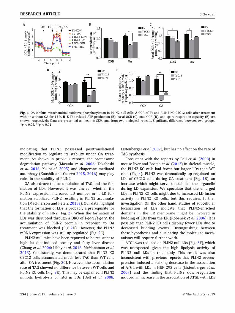
Oleate (OA) inhibits mitochondrial oxidativephosphorylation in PLIN2 null cells
The observation that interaction between LDs and mito-chondria was dramatically increased in PLIN2 KO cells(Fig. 5A) raised the question whether the function ofmitochondria would be affected. To answer this question,mitochondrial function was directly assessed by mea-suring cellular respiration in the EV and PLIN2 KO cellstreatedwith orwithout OA. The oxygen consumption rate(OCR) (Fig. 6A) and linkedATPproduction (Fig. 6B)weredecreased significantly in PLIN2 KO cells compared to theEV cells after treatment with OA. Even though the basalOCR did not change much (Fig. 6C), both the maximalOCR and the spare respiration capacity of PLIN2 KO cellswere significantly inhibited after OA treatment (Fig. 6Dand E). Taken together, these data suggest a dramaticdecrease in mitochondrial respiration activity, especiallyafter OA treatment, when PLIN2 is disrupted.
Previously, we found that cold treatment of micecould induce TAG hydrolysis, accompanied with closerinteractions between LDs and mitochondria in brownadipose tissue (Yu et al. 2015). Thus, the acceleratedTAG hydrolysis in PLIN2 null LDs (Fig. 3D, E) mightactivate mitochondria, inducing close interactionbetween LDs and mitochondria. To examine this, EV and
PLIN2 KO C2C12 cells were treated with or withoutlipases inhibitors (Atglistatin for the inhibition of ATGL,CAY10499 for the inhibition of HSL), and OA plus traceamount 3H-OA for 12 h. The LDs were then isolated aspreviously described and LD proteins were extracted forsilver staining and Western blot detection (supplemen-tary material Fig. S6). The association of mitochondrialproteins (Cpt1a, Cpt1b, Opa1, and Mfn1) with LDs wasnot blocked by lipase inhibition; rather a slight increasewas seen (supplementary material Fig. S6). Therefore,the interaction between LDs and mitochondria may beindependent of accelerated TAG hydrolysis in PLIN2 KOcells.
DISCUSSION
In this study, we show that OA induced the expression ofPLIN2 and the formation of LDs. Disruption of LDsreduces the stability of PLIN2 during OA treatmentdespite the increased PLIN2 transcriptional level.Depletion of PLIN2enhances LD size and lipolysis activity,which indicates that PLIN2 and LDs reciprocally stabilizeeach other. In addition, our results also suggest thatPLIN2 null LDs have weaker organelle compartmental-ization as PLIN2 null LDs show enhanced interactionswith the other cellular organelles, especially mitochon-dria. The increased interaction of LDs and mitochondriamay contribute to inhibition of mitochondrial oxidativephosphorylation in PLIN2 KO cells during OA treatment.These results suggest that PLIN2 and LDs reciprocallystabilize each other and PLIN2 helps alleviate mito-chondrial stress during OA treatment.
The data described herein bring us a step closer to amore complete understanding of PLIN2 function on LDs.PLIN2 is known to be abundantly expressed in skeletalmuscle (Bosma et al. 2012; Brasaemle et al. 1997; Heidet al. 1998; MacPherson and Peters 2015b; Minnaardet al. 2009; Peters et al. 2012). We found that PLIN2 wasthe most abundant protein in the proteome of LDsisolated from the mouse myoblast cell line C2C12 andthat, as expected, PLIN2 null LDs lack this abundantband by silver staining (supplementary materialFig. S3). The abundance of PLIN2 on LDs of C2C12 cellsincreased during OA treatment (Fig. 1B). The increasedabundance of PLIN2 on LDs of OA-treated C2C12 cellsplays an important role for the stability of the expandedLDs (Thiam et al. 2013).
Treatment with OA dramatically increases the mRNAand protein levels of PLIN2 in C2C12 cells, which isconsistent with the previous reports (Bosma et al. 2012;de Wilde et al. 2010). Furthermore, the increase ofPLIN2 protein was dramatically higher than its mRNA,
Perilipin 2 and lipid droplets provide reciprocal stabilization RESEARCH ARTICLE
� The Author(s) 2019 153 | June 2019 | Volume 5 | Issue 3
indicating that PLIN2 possessed posttranslationalmodification to regulate its stability under OA treat-ment. As shown in previous reports, the proteasomedegradation pathway (Masuda et al. 2006; Takahashiet al. 2016; Xu et al. 2005) and chaperone mediatedautophagy (Kaushik and Cuervo 2015, 2016) may playroles in the stability of PLIN2.
OA also drove the accumulation of TAG and the for-mation of LDs. However, it was unclear whether thePLIN2 expression increased LD number or if LD for-mation stabilized PLIN2 resulting in PLIN2 accumula-tion (MacPherson and Peters 2015a). Our data highlightthat the formation of LDs is probably a prerequisite forthe stability of PLIN2 (Fig. 2). When the formation ofLDs was disrupted through a DKO of Dgat1/Dgat2, theaccumulation of PLIN2 protein in response to OAtreatment was blocked (Fig. 2D). However, the PLIN2mRNA expression was still up-regulated (Fig. 2C).
PLIN2 null mice have been reported to be resistant tohigh fat diet-induced obesity and fatty liver disease(Chang et al. 2006; Libby et al. 2016; McManaman et al.2013). Consistently, we demonstrated that PLIN2 KOC2C12 cells accumulated much less TAG than WT cellsafter OA treatment (Fig. 3C). However, the accumulationrate of TAG showed no difference between WT cells andPLIN2 KO cells (Fig. 3E). This may be explained if PLIN2inhibits hydrolysis of TAG in LDs (Bell et al. 2008;
Listenberger et al. 2007), but has no effect on the rate ofTAG synthesis.
Consistent with the reports by Bell et al. (2008) inmouse liver and Bosma et al. (2012) in skeletal muscle,the PLIN2 KO cells had fewer but larger LDs than WTcells (Fig. 4). PLIN2 was dramatically up-regulated onLDs of C2C12 cells during OA treatment (Fig. 1B), anincrease which might serve to stabilize the organelleduring LD expansion. We speculate that the enlargedLDs in PLIN2 KO cells might due to increased LD fusionactivity in PLIN2 KO cells, but this requires furtherinvestigation. On the other hand, studies of subcellularlocalization of LDs indicate that PLIN2-enricheddomains in the ER membrane might be involved inbudding of LDs from the ER (Robenek et al. 2006). It ispossible that PLIN2 KO cells display fewer LDs due todecreased budding events. Distinguishing betweenthese hypotheses and elucidating the molecular mech-anisms will require further work.
ATGL was reduced on PLIN2 null LDs (Fig. 3F), whichwas unexpected given the high lipolysis activity ofPLIN2 null LDs in this study. This result was alsoinconsistent with previous reports that PLIN2 overex-pression induced a striking decrease in the associationof ATGL with LDs in HEK 293 cells (Listenberger et al.2007) and the finding that PLIN2 down-regulationinduced an increase in the association of ATGL with LDs
Fig. 6 OA inhibits mitochondrial oxidative phosphorylation in PLIN2 null cells. A OCR of EV and PLIN2 KO C2C12 cells after treatmentwith or without OA for 12 h. B–E The related ATP production (B), basal OCR (C), max OCR (D), and spare respiration capacity (E) areshown, respectively. Data are presented as mean ± SEM, and from two biological repeats. Significant difference between two groups,*p\ 0.05, **p\ 0.01
RESEARCH ARTICLE S. Xu et al.
154 | June 2019 | Volume 5 | Issue 3 � The Author(s) 2019

(Bell et al. 2008; Kaushik and Cuervo 2015). One pos-sible explanation is that PLIN2 interacts with ATGL onLDs to maintain basal lipolysis (MacPherson et al. 2013)and most of PLIN2 associated ATGL was released tocytoplasm in PLIN2 KO cells. The elevated lipolysisactivity of PLIN2 null LDs might be due to the re-lo-calization and activation of HSL on LDs (Fig. 3F) and thefull activation of the remaining LD-associated ATGL.How ATGL affects PLIN2 null LDs lipolysis remains to bedefined.
LDs have been reported to interact with many otherintracellular organelles (Goodman 2008; Murphy et al.2009). Our data provide original insight into theimportant role of PLIN2 in the compartmentalization ofLDs. We found that the association of marker proteins ofsome membrane structures, including ER, plasmamembrane, lysosome, and mitochondria, was signifi-cantly increased in PLIN2 null LDs (Fig. 5A), suggestingenhanced interaction between LDs and the other intra-cellular organelles in PLIN2 KO cells. LDs have beenfound to physically interact with mitochondria (Nanet al. 2006; Pu et al. 2011; Sturmey et al. 2006), and thecontact is further strengthened during exercise (Koveset al. 2013; Tarnopolsky et al. 2007). In this study, wefound that deletion of PLIN2 dramatically increased theinteraction between LDs and mitochondria as well asother organelles (Fig. 5A). Furthermore, both TEM andfluorescence imaging of whole cells and isolated LDsconsistently showed an increased close interactionbetween LDs and mitochondria when PLIN2 wasdeleted (Fig. 5C–F). Thus, as the most abundant LD coatprotein in C2C12 cells, PLIN2 may have a pivotal role inregulating the interaction between LDs andmitochondria.
Our previous work demonstrated that the expressionand LD localization of PLIN2 are up-regulated in mousebrown adipose tissue during cold treatment, accompa-nied with increased interaction between LDs and acti-vated mitochondria (Yu et al. 2015). It has beenobserved previously that the close physical interactionbetween LDs and mitochondria is positively correlatedwith the expression of PLIN2 and the activity of mito-chondria (Yu et al. 2015). On the contrary, our data hereindicate that the physical interaction between LDs andmitochondria was induced in PLIN2 null C2C12 cells,accompanied by a suppression of mitochondrial activity,especially during OA treatment (Fig. 6). Thus, theinteraction was negatively correlated with the expres-sion of PLIN2 and the activity of mitochondria. Notably,both studies show that PLIN2 expression is positivelycorrelated with mitochondrial activation. These dataindicate that the role of PLIN2 may be tissue specific(MacPherson and Peters 2015a).
MATERIALS AND METHODS
Cell culture
Mouse C2C12 myoblasts (American Type Culture Col-lections, Manassas, VA) were maintained in DMEM(Macgene Biotech., Beijing) supplemented with 10%FBS (Hyclone), 100 U/mL penicillin and 100 mg/mLstreptomycin (Macgene Biotech., Beijing) at 37 �C, 5%CO2.
Lipid droplets (LDs) isolation and verification
LDs were isolated using methods previously describedwith modification (Zhang et al. 2011). Briefly,1–5 9 109 C2C12 myoblasts were collected with ice-cold PBS, and were transferred to 50 mL buffer A(20 mmol/L tricine pH 7.8, 250 mmol/L sucrose) plus0.5 mmol/L PMSF (Sigma-Aldrich). After centrifugationat 3000 g, cell pellets were resuspended with 20 mLbuffer A plus 0.5 mmol/L PMSF and kept on ice for20 min. Then, the swollen cells were homogenized bynitrogen cavitation (500 psi for 15 min on ice). The PNS,Cyto, TM, and LD fractions were separated by centrifu-gation following procedures described previously(Zhang et al. 2011). The purified LDs were stained withLTR or LTG (1:500 (v/v), Life Technologies), and visu-alized using a ZEISS AxioImager M2 Imaging System orconfocal microscope. The sizes of the purified LDs weremeasured by a Delsa Nano C particle analyzer asdescribed previously (Zhang et al. 2012).
The lipids and the proteins of the LDs were extractedand separated with acetone before centrifuging thesample at 20,000 g for 10 min at 4 �C. The extractedproteins were dissolved in 29 sample buffer for bio-chemical analysis. The purity of LDs, relative to othercell fractions, was identified by silver staining andWestern blotting for the LDs marker proteins describedin our previous study (Bartz et al. 2007).
Fatty acids incubation
Sodium palmitic acid (Sigma-Aldrich) and sodium oleicacid were prepared as described previously (Peng et al.2011). Briefly, fatty acids were mixed with ethanol to afinal concentration of 100 mmol/L after which themixture was sonicated on ice until the mixture became amilky solution. Before use, the fatty acids were dissolvedin 55 �C preheated growth medium, and then cooleddown to 37 �C. Cells were transferred to OA mediumand cultured for the indicated period. Control cells werecultured in media containing an equivalent volume ofethanol as a vehicle control.
Perilipin 2 and lipid droplets provide reciprocal stabilization RESEARCH ARTICLE
� The Author(s) 2019 155 | June 2019 | Volume 5 | Issue 3

Quantitative real-time PCR
Total RNA was extracted with TRIzol reagent andreverse transcribed into cDNA according to the manu-facturer’s protocol (Life Technologies). Quantitativereal-time PCR was performed using the ABI Step OnePLUS and SYBR Green detection kit following the man-ufacturer’s instructions (Life Technologies).
Western blot analysis
Cells were harvested and directly lysed with 29 samplebuffer (125 mmol/L Tris Base, 20% glycerol, 4% SDS,4% b-mercaptoethanol, and 0.04% bromophenol blue).After sonication and denaturation, the samples werecooled to room temperature (RT) and then were loadedon 10% SDS-PAGE gel, electrophoresed, transferred to aPVDF membrane (Sigma-Aldrich), and blocked in 5%non-fat milk for 1 h at RT. Then, the PVDF membranewas incubated with primary antibodies and then sec-ondary antibodies. The membrane was exposed to theECL substrate (PerkinElmer Life Sciences, Waltham,MA) as described previously (Yu et al. 2015). Theantibodies used in this study are listed in supplemen-tary material Table S3.
Construction of plasmids and cell transfection
The CRISPR/Cas9 system was used to knock out andknock in target genes in C2C12 cells. For PLIN2 KO celllines, two targets corresponding separately to exon 4and exon 5 of PLIN2 were designed using the website(http://crispr.mit.edu/) and were inserted into thepX260a plasmid (a gift from Prof. Feng Zhang (Conget al. 2013)). For Dgat1 and Dgat2 KO cell lines, twotargets corresponding to these two genes, respectively,were designed using the website (http://crispr.mit.edu/)and were inserted into the pX260a plasmid. The KOdetection primers for these KO cells were designed asdisplayed in supplementary material Table S4. ForPLIN2-EGFP KI cell lines, a target near the stop codon ofPLIN2 was designed and was inserted into the pX260aplasmid. Two pairs of homologous arm primers weredesigned and inserted into the pKI-EGFP-N plasmidwith Xba I and Nt.Bbvc I restriction endonucleases. ThepKI-EGFP-N plasmid was modified from pTK-NEO-3flag-USER in John Wang’s lab and the functional sequence ofpKI-EGFP-N is displayed in supplementary materialFig. S1A. For the real-time PCR detection of target genes,primers were designed according to NCBI BLAST anddisplayed in supplementary material Table S4. Allplasmids were sequenced right by Tsingke TechnologiesInc.
For cell transfection, pQCXIP derived plasmids werepackaged into pseudo retrovirus in Plat-ET cells, and thevirus was used to infect target cells. In some instances,plasmids were transfected into C2C12 cells using elec-troporation according to the manufacturer’s instruc-tions (Amaxa Nucleofector).
Cell line construction
For stable overexpression cells, C2C12 cells wereinfected with pQCXIP-Atgl-myc or pQCXIP-Plin2-myctyped pseudo retrovirus. Cells were incubated with1 lg/mL puromycin containing DMEM medium starting48 h after infection and were cultured for at leasttwo weeks. Cells were then seeded into 96-well plateswith limiting dilution and were cultured for anothertwo weeks before picking clones. The expression stateof the clones was verified by Western blot.
For KO cells, C2C12 cells were transfected with atargeted KO plasmid. Cells were incubated with 1 lg/mL puromycin containing DMEM medium 48 h aftertransfection and were selected as previously described.The selected KO status of the clones was verified byWestern blot and/or genome PCR. For KI cells, C2C12cells were co-transfected with targeted KO plasmid andhomologous arms plasmids. Cells were sorted by flowcytometry (BD Aria III) 48 h after transfection and wereselected as previously described. The selected KI cloneswere confirmed by Western blot.
Confocal microscopy
Various cell types were treated with OA as indicated.Cells were fixed in 4% paraformaldehyde and the LDsand nucleus were stained with LTR and Hochest (LifeTechnologies), respectively, for 30 min. The coverslipswere then mounted with antifade solution (ApplygenTechnologies Inc., Beijing) and were sealed with nailpolish. Images were captured with an Olympus FV1000fluorescence confocal microscope (Olympus Corp., LakeSuccess, NY).
Detection of TAG and lipids
For the TAG accumulation assay, the indicated cells weresubcultured into 12-well plates. The cells were thentreated with or without 200 lmol/L OA with or without0.5 lCi/mL 3H-oleic acid (3H-OA) (PerkinElmer, Boston,MA) for the indicated time. Cells were then lysed todetect the total TAG level (Biosino Bio-Technology andScience Inc, Beijing) or 3H-OA incorporated TAG byscintillation counter (PerkinElmer, Boston, MA) follow-ing thin layer chromatography (TLC).
RESEARCH ARTICLE S. Xu et al.
156 | June 2019 | Volume 5 | Issue 3 � The Author(s) 2019

For the assay of TAG lipolysis, the EV and PLIN2 KOcells were subcultured into 12-well plates. The cellswere then pre-treated with or without ATGL inhibitorAtglistatin (10 lmol/L, Sigma, St. Louis, MO) (Mayeret al. 2013) and HSL inhibitor CAY10499 (1 lmol/L,Cayman, Michigan) (Saltiel 2000) for 2 h. Then, the cellswere treated with OA supplemented with 0.5 lCi/mL3H-OA, with or without lipase inhibitors for another12 h. TAG was extracted, and 3H-OA incorporated TAGwas assessed with a scintillation counter following TLC.
For the TAG synthesis assay, WT and PLIN2 KO cellswere subcultured into 12-well plates. The cells werethen treated with 3H-OA (0.5 lCi/mL) and 200 lmol/LOA with 10 lmol/L Atglistatin and 1 lmol/L CAY10499.Cells were collected at the indicated times. The 3H-OAincorporated TAG was assessed with a scintillationcounter following TLC.
In all experiments, cells were harvested in 0.3 mL 1%Triton X-100/PBS to determine 3H-OA incorporatedTAG, and to assess total protein content (Pierce BCAassay, Thermo Scientific, Rockford, IL). The lipid extractswere separated by TLC using heptane-diethyl-ether(60:40:1, v/v/v) as developing solvent. The content oflipids was normalized to total protein content.
Cellular bioenergetics
The cellular bioenergetics profile of C2C12 cells wasassessed using the Seahorse XF24 Flux Analyzer (Sea-horse Bioscience) as described (Meex et al. 2015). Inbrief, the indicated cells were seeded into a 24-wellXF24 cell culture microplate (Seahorse Bioscience).Cells were washed and incubated in 525 lL of unbuf-fered DMEM (5 mmol/L glucose, 1 mmol/L pyruvateand 1 mmol/L glutamate pH 7.4), at 37 �C in a non-CO2
incubator (1 h prior to bioenergetics assessment).Three basal OCR measurements were performed usingthe Seahorse analyzer, and measurements were repe-ated following injection of oligomycin (1 lmol/L), FCCP(0.5 lmol/L), and Rotenone/Antimycin A (0.5 lmol/L).
Colloidal blue staining and comparative massspectrum (MS) analysis
LD proteins isolated from C2C12 cells were separatedon a 10% SDS-PAGE gel and subjected to colloidal bluestaining (Life Technologies). The indicated bands werecut into slices for MS analysis. In-gel digestion of eachslice was performed as previously described (Ding et al.2012). The digested peptides were then loaded onto aC18 trap column with an autosampler, eluted onto a C18column (100 lm 9 12 cm) packed with Sunchrom
packing material (SP-120-3-ODS-A, 3 lm), and werethen subjected to nano LC-LTQ MS/MS analysis.
All MS/MS data were searched against the mouseprotein database from the NCBI using the SEQUESTprogram (Thermo, USA). BioWorks search parameterswere set as follows. Enzyme: trypsin; precursor ionmass tolerance: 2.0 Da; fragment ion mass tolerance:1.0 Da. The variable modification was set to oxidation ofmethionine. The fixed modification was set to car-boxyamidomethylation of cysteine. The search resultswere filtered with Xcorr versus Charge values of Xcorr(?1)[ 1.9, Xcorr (?2)[ 2.5, and Xcorr (?3)[ 3.75.
Transmission electron microscopy (TEM)
The ultra-structure of cells was examined by TEMthrough ultra-thin sectioning. After rinsing with0.1 mol/L PB, the cells were fixed in 2% (w/v) glu-taraldehyde (Electron Microscopy Sciences, Hatfield) inPB (pH 7.4) for 1 h and then collected to a 1.5 mLmicrocentrifuge tube. The cells were subsequently fixedin 1% (w/v) osmium tetroxide (Nakalai Tesque, Kyoto)with 1% potassium ferrocyanide (Sigma-Aldrich, Mis-souri) for 1.5 h at RT. After dehydration in an ascendingconcentration series of ethanol at RT, the cells wereembedded in Embed 812 (Electron Microscopy Sci-ences, Hatfield) and were prepared as 70 nm sectionsusing a Leica EM UC6 Ultramicrotome. After stainingwith 4% (w/v) uranyl acetate (Electron MicroscopySciences, Hatfield) for 15 min and subsequently withlead citrate (Electron Microscopy Sciences, Hatfield) for5 min at RT, the sections were viewed with a TecnaiSpirit electron microscope (FEI, Netherlands). PurifiedLDs were also examined by TEM through an ultra-thinsectioning method as described (Ding et al. 2013).
Statistical analyses
Data are presented as mean ± SEM unless specificallyindicated. The statistical analyses were performed usingGraphPad Prism 6 and Image J (NIH, USA). Comparisonof significance between groups was performed usingstudent t-tests.
Acknowledgements The authors thank Dr. John Zehmer for hiscritical reading and useful suggestions. We thank Dr. Bo Zhang forthe guidance of Seahorse, Dr. Hongjie Zhang for the suggestions ofradioisotope experiments, Dr. Yan Teng and Cunli Jiang for theassistance of confocal microscopy experiments, Dr. Zhensheng Xiefor the MS data analysis, and Dr. Zengqi Wen for the assistance ofexperiments. This work was supported by the Ministry of Scienceand Technology of China (2016YFA0500100), National NaturalScience Foundation of China (U1402225, 31571388, 61273228,and 81270932), Chinese Academy of Sciences (XDA12030201),the Strategic Priority Research Program of the Chinese Academy
Perilipin 2 and lipid droplets provide reciprocal stabilization RESEARCH ARTICLE
� The Author(s) 2019 157 | June 2019 | Volume 5 | Issue 3
of Sciences (XDB13030600), and Yunnan Oversea High-levelTalents Program (2015HA040).
Compliance with Ethical Standards
Conflict of interest Shimeng Xu, Fei Zou, Zhiqing Diao, ShuyanZhang, Yaqin Deng, Xiaotong Zhu, Liujuan Cui, Jinhai Yu, ZhiguangZhang, Adekunle Toyin Bamigbade, Hongchao Zhang, Xuan Wei,Xuelin Zhang, Bin Liang and Pingsheng Liu declare that they haveno conflicts of interest.
Animal Rights and Informed Consent This article does notcontain any studies with human or animal subjects performed byany of the authors.
Open Access This article is distributed under the terms of theCreative Commons Attribution 4.0 International License (http://creativecommons.org/licenses/by/4.0/), which permits unre-stricted use, distribution, and reproduction in any medium, pro-vided you give appropriate credit to the original author(s) and thesource, provide a link to the Creative Commons license, andindicate if changes were made.
References
Bartz R, Zehmer JK, Zhu M, Chen Y, Serrero G, Zhao Y, Liu P (2007)Dynamic activity of lipid droplets: protein phosphorylationand GTP-mediated protein translocation. J Proteome Res6:3256–3265
Bell M, Wang H, Chen H, McLenithan JC, Gong DW, Yang RZ, Yu D,Fried SK, Quon MJ, Londos C, Sztalryd C (2008) Consequencesof lipid droplet coat protein downregulation in liver cells:abnormal lipid droplet metabolism and induction of insulinresistance. Diabetes 57:2037–2045
Bickel PE, Tansey JT, Welte MA (2009) PAT proteins, an ancientfamily of lipid droplet proteins that regulate cellular lipidstores. Biochem Biophys Acta 1791:419–440
Binns D, Januszewski T, Chen Y, Hill J, Markin VS, Zhao Y, Gilpin C,Chapman KD, Anderson RG, Goodman JM (2006) An intimatecollaboration between peroxisomes and lipid bodies. J CellBiol 173:719–731
Bosma M, Hesselink MK, Sparks LM, Timmers S, Ferraz MJ,Mattijssen F, van Beurden D, Schaart G, de Baets MH,Verheyen FK, Kersten S, Schrauwen P (2012) Perilipin 2improves insulin sensitivity in skeletal muscle despiteelevated intramuscular lipid levels. Diabetes 61:2679–2690
Brasaemle DL (2007) Thematic review series: adipocyte biology.The perilipin family of structural lipid droplet proteins:stabilization of lipid droplets and control of lipolysis. J LipidRes 48:2547–2559
Brasaemle DL, Barber T, Wolins NE, Serrero G, Blanchette-MackieEJ, Londos C (1997) Adipose differentiation-related protein isan ubiquitously expressed lipid storage droplet-associatedprotein. J Lipid Res 38:2249–2263
Brown DA (2001) Lipid droplets: proteins floating on a pool of fat.Curr Biol 11:R446–R449
Chang BH, Li L, Paul A, Taniguchi S, Nannegari V, Heird WC, Chan L(2006) Protection against fatty liver but normal adipogenesisin mice lacking adipose differentiation-related protein. MolCell Biol 26:1063–1076
Chang BH, Li L, Saha P, Chan L (2010) Absence of adiposedifferentiation related protein upregulates hepatic VLDLsecretion, relieves hepatosteatosis, and improves whole body
insulin resistance in leptin-deficient mice. J Lipid Res51:2132–2142
Coll T, Eyre E, Rodriguez-Calvo R, Palomer X, Sanchez RM, MerlosM, Laguna JC, Vazquez-Carrera M (2008) Oleate reversespalmitate-induced insulin resistance and inflammation inskeletal muscle cells. J Biol Chem 283:11107–11116
Cong L, Ran FA, Cox D, Lin S, Barretto R, Habib N, Hsu PD, Wu X,Jiang W, Marraffini LA, Zhang F (2013) Multiplex genomeengineering using CRISPR/Cas systems. Science 339:819–823
de Wilde J, Smit E, Snepvangers FJ, de Wit NW, Mohren R, HulshofMF, Mariman EC (2010) Adipophilin protein expression inmuscle–a possible protective role against insulin resistance.FEBS J 277:761–773
Ding Y, Yang L, Zhang S, Wang Y, Du Y, Pu J, Peng G, Chen Y, ZhangH, Yu J, Hang H, Wu P, Yang F, Yang H, Steinbuchel A, Liu P(2012) Identification of the major functional proteins ofprokaryotic lipid droplets. J Lipid Res 53:399–411
Ding Y, Zhang S, Yang L, Na H, Zhang P, Zhang H, Wang Y, Chen Y, YuJ, Huo C, Xu S, Garaiova M, Cong Y, Liu P (2013) Isolating lipiddroplets from multiple species. Nat Protoc 8:43–51
Fujimoto T, Parton RG (2011) Not just fat: the structure andfunction of the lipid droplet. Cold Spring Harb Perspect Biol3(3):a004838
Fukushima M, Enjoji M, Kohjima M, Sugimoto R, Ohta S, Kotoh K,Kuniyoshi M, Kobayashi K, Imamura M, Inoguchi T, NakamutaM, Nawata H (2005) Adipose differentiation related proteininduces lipid accumulation and lipid droplet formation inhepatic stellate cells. In Vitro Cell Dev Biol Anim 41:321–324
Gao D, Griffiths HR, Bailey CJ (2009) Oleate protects againstpalmitate-induced insulin resistance in L6 myotubes. Br JNutr 102:1557–1563
Goodman JM (2008) The gregarious lipid droplet. J Biol Chem283:28005–28009
Greenberg AS, Coleman RA, Kraemer FB, McManaman JL, Obin MS,Puri V, Yan QW, Miyoshi H, Mashek DG (2011) The role oflipid droplets in metabolic disease in rodents and humans.J Clin Investig 121:2102–2110
Heid HW, Moll R, Schwetlick I, Rackwitz HR, Keenan TW (1998)Adipophilin is a specific marker of lipid accumulation indiverse cell types and diseases. Cell Tissue Res 294:309–321
Kaushik S, Cuervo AM (2015) Degradation of lipid droplet-associated proteins by chaperone-mediated autophagy facil-itates lipolysis. Nat Cell Biol 17:759–770
Kaushik S, Cuervo AM (2016) AMPK-dependent phosphorylationof lipid droplet protein PLIN2 triggers its degradation byCMA. Autophagy 12:432–438
Kelley DE, Goodpaster B, Wing RR, Simoneau JA (1999) Skeletalmuscle fatty acid metabolism in association with insulinresistance, obesity, and weight loss. Am J Physiol 277:E1130–E1141
Kimmel AR, Brasaemle DL, McAndrews-Hill M, Sztalryd C, LondosC (2010) Adoption of PERILIPIN as a unifying nomenclaturefor the mammalian PAT-family of intracellular lipid storagedroplet proteins. J Lipid Res 51:468–471
Koves TR, Sparks LM, Kovalik JP, Mosedale M, Arumugam R,DeBalsi KL, Everingham K, Thorne L, Phielix E, Meex RC, KienCL, Hesselink MK, Schrauwen P, Muoio DM (2013) PPAR-gamma coactivator-1alpha contributes to exercise-inducedregulation of intramuscular lipid droplet programming inmice and humans. J Lipid Res 54:522–534
Larigauderie G, Cuaz-Perolin C, Younes AB, Furman C, Lasselin C,Copin C, Jaye M, Fruchart JC, Rouis M (2006) Adipophilinincreases triglyceride storage in human macrophages bystimulation of biosynthesis and inhibition of beta-oxidation.FEBS J 273:3498–3510
RESEARCH ARTICLE S. Xu et al.
158 | June 2019 | Volume 5 | Issue 3 � The Author(s) 2019
Libby AE, Bales ES, Orlicky DJ, McManaman JL (2016) Perilipin-2deletion impairs hepatic lipid accumulation by interferingwith SREBP activation and altering the hepatic lipidome.J Biol Chem 291(46):24231–24246
Listenberger LL, Ostermeyer-Fay AG, Goldberg EB, Brown WJ,Brown DA (2007) Adipocyte differentiation-related proteinreduces the lipid droplet association of adipose triglyceridelipase and slows triacylglycerol turnover. J Lipid Res48:2751–2761
Liu P, Ying Y, Zhao Y, Mundy DI, Zhu M, Anderson RG (2004)Chinese hamster ovary K2 cell lipid droplets appear to bemetabolic organelles involved in membrane traffic. J BiolChem 279:3787–3792
Liu P, Bartz R, Zehmer JK, Ying YS, Zhu M, Serrero G, Anderson RG(2007) Rab-regulated interaction of early endosomes withlipid droplets. Biochem Biophys Acta 1773:784–793
MacPherson RE, Peters SJ (2015a) Piecing together the puzzle ofperilipin proteins and skeletal muscle lipolysis. Appl PhysiolNutr Metab 40(7):641–651
MacPherson REK, Peters SJ (2015b) Piecing together the puzzle ofperilipin proteins and skeletal muscle lipolysis. Appl PhysiolNutr Metab 40:641–651
MacPherson RE, Herbst EA, Reynolds EJ, Vandenboom R, Roy BD,Peters SJ (2012) Subcellular localization of skeletal musclelipid droplets and PLIN family proteins OXPAT and ADRP atrest and following contraction in rat soleus muscle. Am JPhysiol Regul Integr Comp Physiol 302:R29–R36
MacPherson RE, Ramos SV, Vandenboom R, Roy BD, Peters SJ(2013) Skeletal muscle PLIN proteins, ATGL and CGI-58,interactions at rest and following stimulated contraction. AmJ Physiol Regul Integr Comp Physiol 304:R644–R650
Masuda Y, Itabe H, Odaki M, Hama K, Fujimoto Y, Mori M, Sasabe N,Aoki J, Arai H, Takano T (2006) ADRP/adipophilin isdegraded through the proteasome-dependent pathway dur-ing regression of lipid-storing cells. J Lipid Res 47:87–98
Mayer N, Schweiger M, Romauch M, Grabner GF, Eichmann TO,Fuchs E, Ivkovic J, Heier C, Mrak I, Lass A, Hofler G, FledeliusC, Zechner R, Zimmermann R, Breinbauer R (2013) Develop-ment of small-molecule inhibitors targeting adipose triglyc-eride lipase. Nat Chem Biol 9:785–787
McManaman JL, Bales ES, Orlicky DJ, Jackman M, MacLean PS, CainS, Crunk AE, Mansur A, Graham CE, Bowman TA, GreenbergAS (2013) Perilipin-2-null mice are protected against diet-induced obesity, adipose inflammation, and fatty liver disease.J Lipid Res 54:1346–1359
Meex RC, Hoy AJ, Mason RM, Martin SD, McGee SL, Bruce CR, WattMJ (2015) ATGL-mediated triglyceride turnover and theregulation of mitochondrial capacity in skeletal muscle. Am JPhysiol Endocrinol Metab 308:E960–E970
Minnaard R, Schrauwen P, Schaart G, Jorgensen JA, Lenaers E,Mensink M, Hesselink MKC (2009) Adipocyte differentiation-related protein and OXPAT in rat and human skeletal muscle:involvement in lipid accumulation and type 2 diabetesmellitus. J Clin Endocr Metab 94:4077–4085
Murphy S, Martin S, Parton RG (2009) Lipid droplet-organelleinteractions; sharing the fats. Biochem Biophys Acta1791:441–447
Nan X, Potma EO, Xie XS (2006) Nonperturbative chemicalimaging of organelle transport in living cells with coherentanti-stokes Raman scattering microscopy. Biophys J91:728–735
Ozeki S, Cheng J, Tauchi-Sato K, Hatano N, Taniguchi H, Fujimoto T(2005) Rab18 localizes to lipid droplets and induces theirclose apposition to the endoplasmic reticulum-derived mem-brane. J Cell Sci 118:2601–2611
Pan DA, Lillioja S, Kriketos AD, Milner MR, Baur LA, Bogardus C,Jenkins AB, Storlien LH (1997) Skeletal muscle triglyceridelevels are inversely related to insulin action. Diabetes46:983–988
Peng G, Li L, Liu Y, Pu J, Zhang S, Yu J, Zhao J, Liu P (2011) Oleateblocks palmitate-induced abnormal lipid distribution, endo-plasmic reticulum expansion and stress, and insulin resis-tance in skeletal muscle. Endocrinology 152:2206–2218
Peters SJ, Samjoo IA, Devries MC, Stevic I, Robertshaw HA,Tarnopolsky MA (2012) Perilipin family (PLIN) proteins inhuman skeletal muscle: the effect of sex, obesity, andendurance training. Appl Physiol Nutr Me 37:724–735
Prats C, Donsmark M, Qvortrup K, Londos C, Sztalryd C, Holm C,Galbo H, Ploug T (2006) Decrease in intramuscular lipiddroplets and translocation of HSL in response to musclecontraction and epinephrine. J Lipid Res 47:2392–2399
Pu J, Ha CW, Zhang S, Jung JP, Huh WK, Liu P (2011) Interactomicstudy on interaction between lipid droplets and mitochon-dria. Protein Cell 2:487–496
Robenek H, Hofnagel O, Buers I, Robenek MJ, Troyer D, Severs NJ(2006) Adipophilin-enriched domains in the ER membraneare sites of lipid droplet biogenesis. J Cell Sci 119:4215–4224
Russell TD, Palmer CA, Orlicky DJ, Bales ES, Chang BH, Chan L,McManaman JL (2008) Mammary glands of adipophilin-nullmice produce an amino-terminally truncated form ofadipophilin that mediates milk lipid droplet formation andsecretion. J Lipid Res 49:206–216
Saltiel AR (2000) Another hormone-sensitive triglyceride lipase infat cells? Proc Natl Acad Sci USA 97:535–537
Salvado L, Coll T, Gomez-Foix AM, Salmeron E, Barroso E, PalomerX, Vazquez-Carrera M (2013) Oleate prevents saturated-fatty-acid-induced ER stress, inflammation and insulin resistancein skeletal muscle cells through an AMPK-dependent mech-anism. Diabetologia 56:1372–1382
Schrader M (2001) Tubulo-reticular clusters of peroxisomes inliving COS-7 cells: dynamic behavior and association withlipid droplets. J Histochem Cytochem 49:1421–1429
Shaw C, Sherlock M, Stewart P, Wagenmakers A (2009)Adipophilin distribution and colocalisation with lipid dro-plets in skeletal muscle. Histochem Cell Biol 131:575–581
Sturmey RG, O’Toole PJ, Leese HJ (2006) Fluorescence resonanceenergy transfer analysis of mitochondrial: lipid association inthe porcine oocyte. Reproduction 132:829–837
Takahashi Y, Shinoda A, Kamada H, Shimizu M, Inoue J, Sato R(2016) Perilipin2 plays a positive role in adipocytes duringlipolysis by escaping proteasomal degradation. Sci Rep6:20975
Tarnopolsky MA, Rennie CD, Robertshaw HA, Fedak-TarnopolskySN, Devries MC, Hamadeh MJ (2007) Influence of enduranceexercise training and sex on intramyocellular lipid andmitochondrial ultrastructure, substrate use, and mitochon-drial enzyme activity. Am J Physiol Regul Integr Comp Physiol292:R1271–R1278
Thiam AR, Farese RV Jr, Walther TC (2013) The biophysics and cellbiology of lipid droplets. Nat Rev Mol Cell Biol 14:775–786
Unger RH, Clark GO, Scherer PE, Orci L (2010) Lipid homeostasis,lipotoxicity and the metabolic syndrome. Biochem BiophysActa 1801:209–214
Villena JA, Roy S, Sarkadi-Nagy E, Kim KH, Sul HS (2004)Desnutrin, an adipocyte gene encoding a novel patatindomain-containing protein, is induced by fasting and gluco-corticoids. J Biol Chem 279:47066–47075
Walther TC, Farese RV Jr (2012) Lipid droplets and cellular lipidmetabolism. Annu Rev Biochem 81:687–714
Xu G, Sztalryd C, Lu X, Tansey JT, Gan J, Dorward H, Kimmel AR,Londos C (2005) Post-translational regulation of adipose
Perilipin 2 and lipid droplets provide reciprocal stabilization RESEARCH ARTICLE
� The Author(s) 2019 159 | June 2019 | Volume 5 | Issue 3

differentiation-related protein by the ubiquitin/proteasomepathway. J Biol Chem 280:42841–42847
Yen CL, Stone SJ, Koliwad S, Harris C, Farese RV Jr (2008) Thematicreview series: glycerolipids. DGAT enzymes and triacylglyc-erol biosynthesis. J Lipid Res 49:2283–2301
Yu J, Zhang S, Cui L, Wang W, Na H, Zhu X, Li L, Xu G, Yang F,Christian M, Liu P (2015) Lipid droplet remodeling andinteraction with mitochondria in mouse brown adipose tissueduring cold treatment. Biochem Biophys Acta 1853:918–928
Zhang H, Wang Y, Li J, Yu J, Pu J, Li L, Zhang H, Zhang S, Peng G,Yang F, Liu P (2011) Proteome of skeletal muscle lipid dropletreveals association with mitochondria and apolipoprotein a-I.J Proteome Res 10:4757–4768
Zhang P, Na H, Liu Z, Zhang S, Xue P, Chen Y, Pu J, Peng G, Huang X,Yang F, Xie Z, Xu T, Xu P, Ou G, Zhang SO, Liu P (2012)Proteomic study and marker protein identification ofCaenorhabditis elegans lipid droplets. Mol Cell Proteomics11:317–328
Zimmermann R, Strauss JG, Haemmerle G, Schoiswohl G, Birner-Gruenberger R, Riederer M, Lass A, Neuberger G, EisenhaberF, Hermetter A, Zechner R (2004) Fat mobilization in adiposetissue is promoted by adipose triglyceride lipase. Science306:1383–1386
Zimmermann R, Lass A, Haemmerle G, Zechner R (2009) Fate offat: the role of adipose triglyceride lipase in lipolysis. BiochimBiophys Acta 1791:494–500
RESEARCH ARTICLE S. Xu et al.
160 | June 2019 | Volume 5 | Issue 3 � The Author(s) 2019