pauline cordenonsi bonez
TRANSCRIPT
0
UNIVERSIDADE FEDERAL DE SANTA MARIA
CENTRO DE CIÊNCIAS DA SAÚDE
PROGRAMA DE PÓS-GRADUAÇÃO EM CIÊNCIAS
FARMACÊUTICAS
Pauline Cordenonsi Bonez
ATIVIDADE ANTIMICROBIANA, ANTIBIOFILME, CITO E
GENOTÓXICA DO S-(3,4-DICLOROBENZIL) ISOTIOUREIA (A22)
Santa Maria, RS
2017

1
Pauline Cordenonsi Bonez
ATIVIDADE ANTIMICROBIANA, ANTIBIOFILME, CITO E GENOTÓXICA DO S-
(3,4-DICLOROBENZIL) ISOTIOUREIA (A22)
Tese apresentada ao Curso de Doutorado
do Programa de Pós-Graduação em
Ciências Farmacêuticas, Área de
Concentração Análises Clínicas e
Toxicológicas: Desenvolvimento e
Aplicação de Marcadores no Diagnóstico
Laboratorial, da Universidade Federal de
Santa Maria (UFSM, RS), como requisito
parcial para obtenção do título de Doutor
em Ciências Farmacêuticas.
Orientadora: Profª. Drª. Marli Matiko Anraku de Campos
Coorientador: Prof. Dr. Roberto Christ Vianna Santos
Santa Maria, RS
2017

Ficha catalográfica elaborada através do Programa de Geração Automática da Biblioteca Central da UFSM, com os dados fornecidos pelo(a) autor(a).
Bonez, Pauline Cordenonsi ATIVIDADE ANTIMICROBIANA, ANTIBIOFILME, CITO EGENOTÓXICA DO S-(3,4-DICLOROBENZIL) ISOTIOUREIA (A22) /Pauline Cordenonsi Bonez.- 2017. 96 p.; 30 cm
Orientador: Marli Matiko Anraku de Campos Coorientador: Roberto Christ Vianna Santos Tese (doutorado) - Universidade Federal de SantaMaria, Centro de Ciências da Saúde, Programa de Pós-Graduação em Ciências Farmacêuticas, RS, 2017
1. Biofilmes 2. Pseudomonas aeruginosa 3. A22 I.Anraku de Campos, Marli Matiko II. Christ Vianna Santos,Roberto III. Título.


3
DEDICATÓRIA
Dedico este trabalho a minha família amada, minha mãe e meu irmão. Dedico também ao
meu pai que infelizmente não está mais entre nós para presenciar esse momento, mas se faz
necessário agradecer pelo legado que nos deixou, de amor e de luta pela educação e pela
vida. Amo vocês!

4
AGRADECIMENTOS
Antes de tudo, agradeço a Deus, pela minha vida, pela minha saúde e por sempre
iluminar as minhas escolhas e guiar os meus passos.
Agradeço a todos que, de alguma forma, contribuíram para a concretização deste
estudo, obrigada pelo auxílio, compreensão e dedicação. De uma maneira especial,
agradeço:
- a Universidade Federal de Santa Maria, universidade pública e de qualidade, por
me permitir desenvolver e concretizar este trabalho, tenho consciência de que muitos em
nosso país não têm essa mesma oportunidade;
- aos professores e funcionários do Programa de Pós-graduação em Ciências
Farmacêuticas (PPGCF) e do Departamento de Análises Clínicas e Toxicológicas (DACT)
por contribuírem de uma forma ou de outra pela conquista deste título;
- a CAPES (Coordenação de Aperfeiçoamento de Pessoal de Nível Superior), pela
bolsa de estudos e pelos recursos financeiros concedidos;
- aos professores membros da banca, pela disposição e por avaliarem este trabalho.
Vocês são exemplos de profissionais;
- a minha orientadora Marli Matiko Anraku de Campos pela oportunidade concedida,
pela confiança a mim depositada desde a graduação e pela pessoa incentivadora e dedicada.
Grata pela orientação e amizade;
- ao meu coorientador Roberto Christ Vianna Santos, pelo exemplo de educador, por
todos os ensinamentos, apoio, incentivo e confiança. A você, minha admiração e gratidão.
- a minha mãe, Maria Izabel Cordenonsi Bonez, pelo amor maior, doação e exemplo
de superação. Obrigada pela amizade, por me guiar, por me mostrar a importância do estudo
e por me ajudar a ser quem sou hoje. Te amo incondicionalmente!
- ao meu pai Enio Bonez (saudades eternas...) pelo exemplo que nos deixou, de amor,
de sabedoria e pela imensa preocupação com o nosso futuro e nossa educação. Sei que está
feliz, onde quer que esteja. Te amo!
- ao meu irmão Mateus Cordenonsi Bonez, pela nossa história, pelo companheirismo,
pelo amor, pelo apoio, pelo carinho e pela confiança. Te amo, irmão!
- ao meu namorado Mateus Lopes de Moraes, pelo amor, pelo carinho, pelas palavras
de incentivo e pelo exemplo de determinação. Obrigada por me fazer mais feliz a cada dia e
pela ajuda em todos os momentos durante essa caminhada. Te amo, meu bem!

5
- à família do meu namorado, Dona Shirley Lopes, Sr Ailton de Moraes, Mariana
Lopes e Arthur Klein, obrigada pelo carinho e por sempre me colocarem em suas orações;
- às minhas primas Sílvia Cordenonsi e Silvana Cordenonsi, obrigada pela ajuda e
convívio, sei que posso contar sempre com vocês;
- a toda minha família, em especial à nona, por sempre rezar por mim. Obrigada tios
e primos pela força e confiança depositadas em mim;
- ao meu pequeno amiguinho Shiva que sempre me acompanhou nos estudos,
demonstrando o mais puro amor e carinho;
- às minhas amigas Laura Vargas, Paula Bitencourt e Márcia Prior, obrigada pelas
palavras de incentivo, pela amizade e pelos momentos inesquecíveis. Sei que permaceremos
unidas apesar da distância e do rumo que nossas vidas irão traçar. Amo vocês!
- aos meus queridos colegas e amigos do Labmyco (Laboratório de
Micobacteriologia), Vanessa Agertt, Vanessa Flores, Caren Mizdal, Grazielle Rossi e Fallon
Siqueira. Obrigada pelo convívio diário, pela amizade, pela parceria e pela ajuda. Espero
encontrar pessoas como vocês em meu caminho. Contem sempre comigo.
- aos novos integrantes do Labmyco, Eloísa Dalla Nora, Viviane Somavilla, Kevim
Guterres, Daniele Wille e Gabriela Vargas muito obrigada pelo convívio. Vocês são o início
de uma nova fase no laboratório, confio em vocês. Podem contar comigo para o que
precisarem.
- a todos os intergantes do Lapemicro (Laboratório de Pesquisa em Microbiologia
Oral), pela ajuda nos experimentos, ideias e discussões. Um agradecimento especial a
Camilla Fillippi e a Márcia Ebling, que de colegas passaram a grandes amigas. Obrigada
pelos encontros e pelas conversas de sempre gurias.
- a toda a equipe do Laboratório de Cultura Celular do Centro Universitário
Franciscano (UNIFRA), obrigada pela imensa ajuda e infraestrutura concedida. Meu
agradecimento especial à professora Michele Sagrillo e aos alunos Kátia Nascimento,
Andiara Ramos e Jardel Bandeira, sem vocês, parte desse trabalho não teria sido possível;
Por fim, a todos aqueles que fazem parte da minha vida e que são essenciais para eu
ser, a cada dia, um ser humano melhor.

6
E lembre-se, nunca abandone a esperança.
A vida tem um jeito engraçado de se ajustar, espere e verá.
(A Casa das Orquídeas de Lucinda Riley)

7
RESUMO
ATIVIDADE ANTIMICROBIANA, ANTIBIOFILME, CITO E GENOTÓXICA DO S-
(3,4-DICLOROBENZIL) ISOTIOUREIA (A22)
AUTOR: PAULINE CORDENONSI BONEZ
ORIENTADOR: MARLI MATIKO ANRAKU DE CAMPOS
COORIENTADOR: ROBERTO CHRIST VIANNA SANTOS
A formação de biofilmes causa grande preocupação para a saúde pública devido à baixa
resposta aos tratamentos antimicrobianos e à colonização de superfícies como próteses e
cateteres. Microrganismos patogênicos como Pseudomonas aeruginosa são capazes de formar
biofilmes em dispositivos médico-hospitalares e tecidos vivos, podendo causar infecções
crônicas graves em humanos. Deste modo, torna-se importante buscar alternativas eficazes
contra a formação de biofilmes. O A22 inibe a proteína MreB do citoesqueleto bacteriano
alterando o formato das células microbianas, o que pode afetar muitas propriedades, incluindo
a motilidade e a formação de biofilmes. Neste contexto, este trabalho teve como objetivo
avaliar, pela primeira vez, a ação antibiofilme do A22 sobre isolados de P. aeruginosa, bem
como os seus potenciais efeitos cito e genotóxicos. A atividade antibacteriana do A22 foi
avaliada por métodos convencionais sobre cepas padrões e 28 isolados clínicos
multirresistentes de P. aeruginosa. Os ensaios de cito e genotoxicidade foram realizados pelo
teste do MTT e ensaio do Cometa, respectivamente. A capacidade de adesão dos isolados
clínicos e da cepa padrão foi avaliada em placas de poliestireno. Fatores essenciais à fisiologia
do biofilme de P. aeruginosa PAO1, como as motilidades dos tipos swimming, swarming e
twitching, bem como a adesão às células HeLa e adesão a superfície de Polietileno de Alta
Densidade (PEAD) foram avaliados na presença e na ausência do A22. A Microscopia de
Força Atômica (MFA) foi utilizada para visualizar a adesão em PEAD. Este estudo ratificou a
excelente atividade antimicrobiana do A22, principalmente em relação aos isolados
multirresistentes. Da mesma forma, o A22 não apresentou efeitos citotóxicos sobre células
mononucleares de sangue periférico humano em todos os tempos de exposição avaliados,
exceto na concentração de 32µg/mL, e não demonstrou efeitos genotóxicos sobre as células
após 24 e 48 horas. Os resultados mostraram um alto padrão de adesão em 14 isolados
clínicos multiresistentes, sendo que o A22 inibiu a adesão de 9 destes microrganismos. O A22
conseguiu reduzir a adesão e formação de biofilmes da cepa P. aeruginosa PAO1 em placas
de poliestireno, nas células HeLa e no PEAD. Do mesmo modo, as motilidades swarming e
twitching foram significativamente diminuídas pela ação do A22 em concentrações
subinibitórias. O impacto e contribuição científica deste trabalho estão alicerçados na
descoberta de uma potential nova possibilidade terapêutica contra infecções associadas a
biofilmes de P. aeruginosa. O A22 apresentou-se como uma ferramenta útil e promissora para
reduzir a adesão microbiana tanto em superfícies vivas quanto inertes, haja vista os seus
baixos efeitos tóxicos. Os resultados desta tese estimulam o aprofundamento em metodologias
que visem a inserção do A22 como um novo antibacteriano ou agente de revestimento de
materiais médico-hospitalares.
Palavras-chave: A22. Pseudomonas aeruginosa. Motilidade. PEAD. Células HeLa. Adesão
bacteriana. Biofilmes.

8
ABSTRACT
ANTIMICROBIAL, ANTIBIOFILM, CITO AND GENOTOXIC ACTIVITIES OF S-
(3,4-DICHLOROBENZYL) ISOTIOUREIA (A22)
AUTHOR: PAULINE CORDENONSI BONEZ
ADVISER: MARLI MATIKO ANRAKU DE CAMPOS
CO-ADVISER: ROBERTO CHRIST VIANNA SANTOS
Biofilm formation causes great public health concern due to the low response to antimicrobial
treatments and the colonization of surfaces such as prostheses and catheters. Pathogenic
microorganisms such as Pseudomonas aeruginosa are able to form biofilms in medical
devices and living tissues, can cause severe chronic infections in humans. Thus, it is
important to search effective alternatives against the formation of biofilms. A22 inhibits MreB
protein from the bacterial cytoskeleton altering the microbial cells shape, which can affect
many properties, including motility and biofilm formation. In this context, this work aimed to
evaluate, for the first time, the antibiofilm action of A22 on P. aeruginosa, as well as their
potencial cyto and genotoxic effects. The antibacterial activity of A22 was evaluated by
conventional methods on standard strains and 28 multiresistant clinical isolates of P.
aeruginosa. The cyto and genotoxicity tests were performed by MTT test and Comet assay,
respectively. The adhesion capacity of clinical isolates and standard strain was measured in
polyestyrebe plates. Essential factors to the biofilm physiology of P. aeruginosa, such as
swimming, swarming ant twitching motility, as well as adhesion to HeLa cells and adhesion to
High Density Polyethylene (HDPE) were evaluated in the presence and absence of A22.
Atomic Force Microscopy (AFM) was used to visualize adhesion on HDPE substrate. This
study confirmed the excellent antimicrobial activity of A22, especially in relation to
multiresistant isolates. Likewise, A22 showed no cytotoxic effects on human Peripheral
Blood Mononuclear Cells (PBMCs) at all exposure times, with exception of concentration of
the 32μg/mL, and did not demonstrate genotoxic effects on the cells after 24 and 48 hours. In
addition, the results showed a high adhesion pattern in 14 multiresistant clinical isolates, with
inhibiting the adhesion of 9 of these microorganisms. A22 was able to decrease adhesion and
biofilm formation of the P. aeruginosa PAO1 on polyestyrene plates, HeLa cells and HDPE.
Moreover, the swarming and twitching motilities were significantly decreased by A22 in
subinhibitory concentrations. The impact and scientific contribution of this work are based on
the discovery of a potencial new therapeutic possibility against infections associated with
biofilms of P. aeruginosa. The A22 presents as an useful and promising tool to decrease
microbial adhesion in both living and inert surfaces, given its low toxic effects. However, this
thesis results stimulate the deepening in methodologies that aim the insertion of A22 as a new
antibacterial or coating agent on medical materials.
Keywords: A22. Pseudomonas aeruginosa. Motility. HDPE. HeLa cells. Bacterial
adhesion. Biofilms.

9
LISTA DE TABELAS
INTRODUÇÃO
Tabela 1 – Funções da matriz exopolissacarídica em biofilmes. ............................................. 17
ARTIGO 1
Table 1 – Antimicrobial activity of S-(3,4-diclorobenzil) isotioureia (A22) against strains
standards……………………………………………………………………………………....53
Table 2 – DNA migration in the comet assay for assessment of genotoxicity of S-(3,4-
diclorobenzil) isotioureia (A22) exposition in peripheral blood mononuclear cell culture
during 24, 48 and 72 hours……………………………………………………………………53
ARTIGO 2
Table 1 –A22 against biofilm formation of multidrug-resistance clinical isolates of P.
aeruginosa………………………………………………………....………………………….72
Table 2 – Influence of S-(3,4-diclorobenzil) isotioureia (A22) on P. aeruginosa PAO1
motility……….………...……………………………………….……………………….........73

10
LISTA DE ILUSTRAÇÕES
INTRODUÇÃO
Figura 1 – Biofilme de Pseudomonas aeruginosa. .................................................................. 16 Figura 2 – Estrutura do biofilme. ............................................................................................. 17 Figura 3 – Estágios de desenvolvimento do biofilme microbiano. .......................................... 20 Figura 4 – Fatores que influenciam na adesão microbiana. ..................................................... 21
Figura 5 – Tipos de motilidade bacteriana. .............................................................................. 22 Figura 6 – Atividade metabólica em uma microcolônia de biofilme. ...................................... 28
Figura 7 – Estratégias antibiofilme. .......................................................................................... 30 Figura 8 – Proteínas do citoesqueleto e o formato celular bacteriano. ..................................... 34 Figura 9 – Estrutura do S-(3,4-diclorobenzil) isotioureia (A22). ............................................. 34 Figura 10 – Inibição reversível da proteína MreB pelo S-(3,4-diclorobenzil) isotioureia (A22).
.................................................................................................................................................. 35
Figura 11 – Efeito do S-(3,4-diclorobenzil) isotioureia (A22) sobre a célula bacteriana. ...... 35
ARTIGO 1
Fig 1 – Time-kill curve on P. aeruginosa PAO1 exposed to several concentrations of S-(3,4-
diclorobenzil) isotioureia (A22) (based on MIC). .................................................................... 53
Fig 2 – Comparison of cytotoxicity evaluated by MTT reduction among human cells exposed
to H2O2 and treated with S-(3,4-diclorobenzil) isotioureia (A22) in 24 hours (A), 48 hours and
72 hours (C) at different concentrations. .................................................................................. 53
ARTIGO 2
Fig 1 – Graphical representation of S-(3,4-diclorobenzil) isotioureia (A22) action on biofilm
formation of P. aeruginosa PAO1. ........................................................................................... 72
Fig 2 – Atomic Force Microscopy (AFM) of biofilm in HDPE surface and biofilm inhibition
by S-(3,4-diclorobenzil) isotioureia (A22).. ............................................................................. 73
Fig 3 – P. aeruginosa PAO1 bacilli adhered to HeLa cells. .................................................... 73
Fig 4 – S-(3,4-diclorobenzil) isotioureia (A22) influence on swimming, swarming and
twitching motility of P. aeruginosa PAO1. .............................................................................. 74
Fig 5 – Illustration of S-(3,4-diclorobenzil) isotioureia (A22) influence on initial stage of
biofilm formation.. .................................................................................................................... 75
11
SUMÁRIO
APRESENTAÇÃO ....................................................................................................... 12
1 INTRODUÇÃO ......................................................................................................... 13
1.1 RESISTÊNCIA ANTIMICROBIANA .................................................................... 13
1.2 Pseudomonas aeruginosa ......................................................................................... 14
1.3 BIOFILMES: CONCEITO, ESTRUTURA E COMPOSIÇÃO .............................. 16
1.4 ETAPAS DA FORMAÇÃO DO BIOFILME .......................................................... 19
1.5 FATORES QUE INFLUENCIAM NA FORMAÇÃO DE BIOFILMES ................ 20
1.5.1 Regulação genética da formação de biofilmes em P. aeruginosa .................... 23
1.5.1.1 Regulação da formação de biofilme em P. aeruginosa via QS .......................... 24
1.5.1.2 Regulação da formação de biofilme em P. aeruginosa via c-di-GMP ............... 24
1.5.1.3 Regulação da formação de biofilme em P. aeruginosa via sRNA ...................... 25
1.6 PATOGÊNESE E MECANISMOS DE RESISTÊNCIA DOS BIOFILMES ......... 25
1.7 ABORDAGENS TERAPÊUTICAS CONTRA BIOFILMES ................................. 29
1.7.1 Inibição da formação do biofilme ...................................................................... 30
1.7.2 Interferência na comunicação celular................................................................ 31
1.7.3 Erradicação do biofilme formado ...................................................................... 32
1.8 A22 E O FORMATO DA CÉLULA BACTERIANA ............................................. 33
2 OBJETIVOS .............................................................................................................. 37
2.1 OBJETIVO GERAL ................................................................................................. 37
2.2 OBJETIVOS ESPECÍFICOS ................................................................................... 37
3 PUBLICAÇÕES CIENTÍFICAS ............................................................................. 38
3.1 ARTIGO 1 ................................................................................................................ 38
3.2 ARTIGO 2 ................................................................................................................ 54
4 DISCUSSÃO .............................................................................................................. 76
5 CONCLUSÃO ............................................................................................................ 81
6 PERSPECTIVAS ....................................................................................................... 82
REFERÊNCIAS ........................................................................................................... 83
ANEXOS ....................................................................................................................... 92
ANEXO A – CARTA DE APROVAÇÃO DO COMITÊ DE ÉTICA EM PESQUISA
(UFSM) ........................................................................................................................... 92
ANEXO B – AÇÃO DO COMPOSTO A22 SOBRE A CÉLULA BACTERIANA. ... 95

12
APRESENTAÇÃO
A seção INTRODUÇÃO inclui uma apresentação sobre o assunto investigado e sua
relevância, bem como, uma revisão bibliográfica sobre os temas discutidos nesta Tese. Os
resultados encontram-se nos tópicos ARTIGO 1 e ARTIGO 2, os quais englobam as seções
Materiais e Métodos, Resultados, Discussão dos Resultados e Referências, representando a
íntegra deste estudo. Ambos os trabalhos estão formatados de acordo com o periódico aos
quais foram publicados e/ou submetidos.
Os tópicos DISCUSSÃO, CONCLUSÕES e PERSPECTIVAS apresentam
interpretações e comentários gerais acerca do conteúdo abordado nesta tese, assim como
sugestões de abordagens futuras. As REFERÊNCIAS remetem somente às citações que
aparecem nos tópicos INTRODUÇÃO e DISCUSSÃO. Na seção ANEXOS encontram-se o
parecer de aprovação do Comitê de Ética e Pesquisa da Universidade Federal de Santa Maria
(UFSM) e uma imagem adicional obtida neste trabalho.
13
1 INTRODUÇÃO
1.1 RESISTÊNCIA ANTIMICROBIANA
As doenças infecciosas, que no passado eram as principais causas de morte em
humanos, foram aparentemente controladas com a descoberta dos agentes antimicrobianos.
Contudo, durante várias décadas, os microrganismos foram adquirindo resistência a cada novo
antimicrobiano descoberto, acarretando um grave problema para a saúde pública mundial
(WOOLHOUSE; FARRAR, 2014).
Os microrganismos tornam-se resistentes por meio de mecanismos específicos, como a
produção de enzimas que podem inativar o fármaco, alteração da permeabilidade ou absorção
do medicamento, expressão de bombas de efluxo e redução do número ou afinidade aos locais
de ligação do fármaco. Além disso, podem adquirir resistência pela transferência de
plasmídeos (DNA extracromossômico) contendo genes de resistência e pela formação de
biofilmes em superfícies (WOOLHOUSE; FARRAR, 2014; SINGH et al., 2017).
O uso abusivo e inadequado, bem como a prescrição desnecessária de
antimicrobianos, contribuem de forma preponderante para o aumento da pressão seletiva
sobre os microrganismos para que adquiram resistência. Além de constituir um enorme ônus
financeiro aos serviços de saúde, a resistência antimicrobiana é responsável por milhares de
mortes por infecções persistentes (ORGANIZAÇÃO MUNDIAL DA SAÚDE, 2012).
O uso excessivo de antimicrobianos na pecuária também contribui para o aumento da
resistência às infecções em humanos, pois contribui para a propagação de genes de resistência
através do consumo de carne, leite ou água contaminadas (CENTER FOR DISEASE
CONTROL AND PREVENTION, 2015). A colistina, por exemplo, é utilizada em animais e,
apesar dos efeitos colaterais, é considerada um último recurso contra infecções em humanos.
Descobriu-se recentemente que o gene mcr-1, que confere resistência à colistina, encontrado
em bactérias isoladas de porcos na China, vem se espalhando rapidamente, tomando
proporções intercontinentais (CENTER FOR DISEASE CONTROL AND PREVENTION,
2016; NATURE NEWS, 2017). Ademais, o ritmo alarmante da disseminação de plasmídeos
de resistência aos carbapenêmicos também exige vigilância, uma vez que as opções
terapêuticas estão cada vez mais escassas (MELETIS, 2016).
Todos os anos, nos Estados Unidos, aproximadamente 2 milhões de pessoas
apresentam infecções associadas a microrganismos resistentes aos antimicrobianos e pelo
menos 23 mil indivíduos morrem como resultado direto destes processos (CENTER FOR

14
DISEASE CONTROL AND PREVENTION, 2016). Estima-se que a taxa de mortalidade no
mundo, associada à resistência antimicrobiana, chega a 700.000 mortes, podendo alcançar o
número de 10 milhões, em 2050, se nenhuma medida for tomada (O‟NEILL, 2016).
Entre os anos de 1983 e 1992, trinta novos antimicrobianos foram aprovados pelo
Food and Drug Administration (FDA), enquanto que entre os anos de 2003 e 2012, foram
apenas sete (WOOLHOUSE; FARRAR, 2014). Desta forma, o aumento da resistência aos
antimicrobianos, aliado à escassez de novos agentes, ameaça o tratamento de doenças
infecciosas comuns como pneumonia e tuberculose tornando a era pós-antibiótico – em que
infecções banais podem causar a morte – cada vez mais próxima. Neste contexto, a melhoria
das condições de higiene e de saneamento básico para prevenir e evitar a propagação de
infecções, a busca por diagnósticos rápidos para evitar a prescrição desnecessária de
antimicrobianos, o uso de vacinas em humanos e animais e o investimento em novos
fármacos são fundamentais para resolver esta problemática (CENTER FOR DISEASE
CONTROL AND PREVENTION, 2015; O‟NEILL, 2016).
Embora todos os microrganismos possam adquirir resistência, as bactérias resistentes
são, atualmente, uma das maiores causas de preocupação (WOOLHOUSE; FARRAR, 2014).
Neste contexto, incluem-se isolados de P. aeruginosa, os quais são considerados causas
comuns de infecções em pacientes hospitalizados, principalmente em internados em Unidades
de Terapia Intensiva (UTIs) (BĂLĂŞOIU et al., 2014). A formação de biofilmes apresenta-se,
muitas vezes, como causa subjacente de uma variedade de infecções provocadas por esse
microrganismo, sejam essas associadas aos tecidos vivos ou às superfícies inertes (DONLAN;
COSTERTON, 2002; RYBTKE et al., 2015).
1.2 Pseudomonas aeruginosa
P. aeruginosa é um bacilo Gram negativo, não fermentador, aeróbio estrito, reto ou
levemente curvo que se comporta basicamente como um patógeno oportunista, pois acomete,
principalmente, indivíduos imunocomprometidos (HIRSCH; TAM, 2010; BĂLĂŞOIU et al.,
2014). No entanto, pessoas saudáveis também podem desenvolver doenças leves, como otites
e erupções cutâneas mais generalizadas, especialmente após a exposição à água. Infecções
oculares também podem ocorrer após uso prolongado de lentes de contato (CENTER FOR
DISEASE CONTROL AND PREVENTION, 2014).
No ambiente hospitalar, P. aeruginosa pode causar infecções graves, incluindo
pneumonia e infecções sanguíneas e pós-cirúrgicas que podem levar a sérias complicações e

15
até à morte. Trata-se de uma das bactérias mais frequentemente isoladas no setor de
bacteriologia de laboratórios clínicos, sendo apontada como a primeira causa de pneumonia
nosocomial no Brasil (ROSSI, 2011). Nas UTIs brasileiras é o terceiro patógeno mais
frequente, estando envolvido em 30% das infecções de corrente sanguínea relacionadas ao uso
de cateteres e 36,6% apresentam resistência ao imipenem (IPM) (NEVES et al., 2011).
Estas infecções apresentam grande importância clínica, sobretudo, porque podem ser
causadas por isolados bacterianos que englobam mais de um mecanismo de resistência
atuando em conjunto, o que dificulta a escolha terapêutica e consequentemente a erradicação
da doença, promovendo, invariavelmente, o aumento dos índices de morbimortalidade
(MARQUES et al., 2015). Os principais mecanismos relacionados com fenótipos
multirresistentes de P. aeruginosa nos hospitais brasileiros são a produção de
metalobetalactamase do tipo SPM-1, a perda de porina OprD e a superexpressão de bombas
de efluxo, o que pode explicar os altos índices de resistência a carbapenêmicos e
aminoglicosídeos (NEVES et al., 2011). Neste contexto, estima-se que cerca de 51 mil casos
de infecções provocadas por P. aeruginosa ocorrem nos Estados Unidos a cada ano, sendo
6.000 dessas, resistentes aos múltiplos fármacos, ocasionando, pelo menos, 400 mortes anuais
(CENTER FOR DISEASE CONTROL AND PREVENTION, 2014).
Infecções agudas provocadas por P. aeruginosa estão associadas às células livres
(planctônicas) altamente virulentas enquanto que as infecções crônicas estão comumente
associadas ao modo séssil de crescimento (biofilmes) (MAUNDERS; WELCH, 2017). A
formação de biofilmes surge, por conseguinte, como um importante mecanismo utilizado por
esses microrganismos para sobreviverem aos tratamentos.
Fibrose cística (FC), infecções de feridas crônicas, otites e prostatites são alguns
exemplos de infecções teciduais associadas à formação de biofilmes por P. aeruginosa
(DONLAN; COSTERTON, 2002). Além disso, os biofilmes foram encontrados em quase
todos os tipos de implantes médico-hospitalares, principalmente em próteses valvares
cardíacas, marca-passos, próteses articulares e cateteres intravenosos, evidenciando assim,
outro importante mecanismo atribuído às infecções nosocomiais (RYBTKE et al., 2015). A
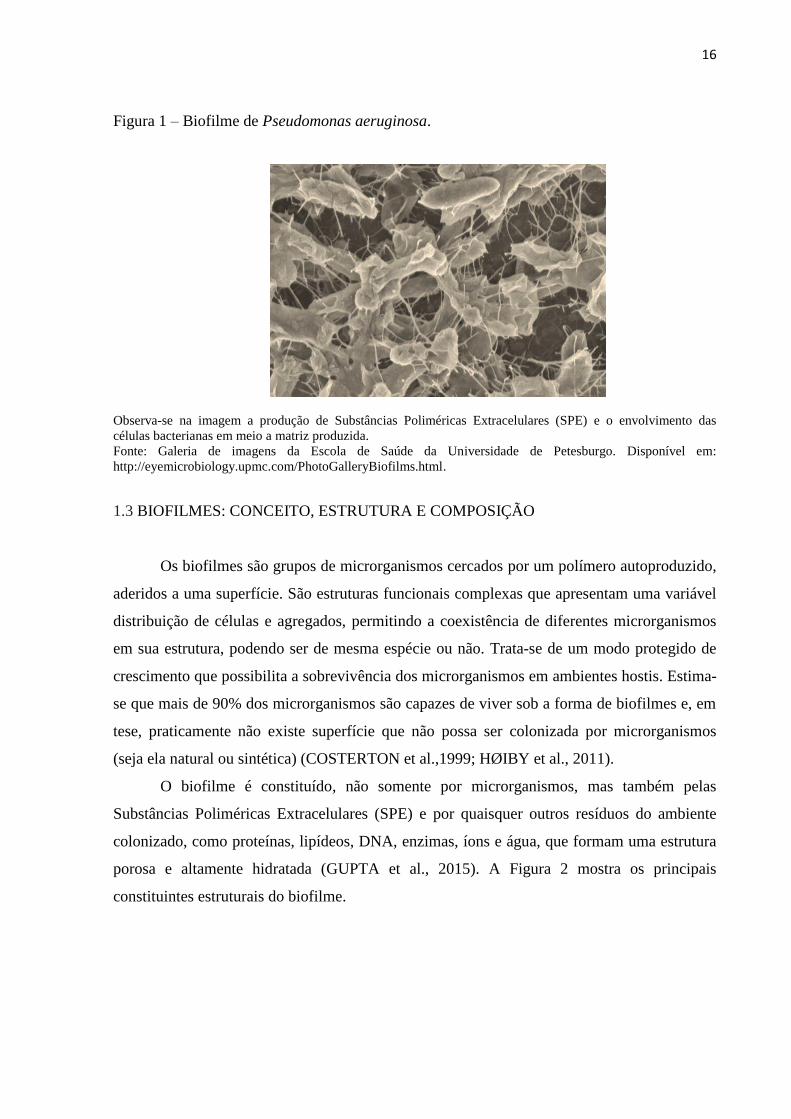
Figura 1 apresenta um biofilme de P. aeruginosa capturado por Microscopia Eletrônica de
Varredura (MEV).
16
Figura 1 – Biofilme de Pseudomonas aeruginosa.
Observa-se na imagem a produção de Substâncias Poliméricas Extracelulares (SPE) e o envolvimento das
células bacterianas em meio a matriz produzida.
Fonte: Galeria de imagens da Escola de Saúde da Universidade de Petesburgo. Disponível em:
http://eyemicrobiology.upmc.com/PhotoGalleryBiofilms.html.
1.3 BIOFILMES: CONCEITO, ESTRUTURA E COMPOSIÇÃO
Os biofilmes são grupos de microrganismos cercados por um polímero autoproduzido,
aderidos a uma superfície. São estruturas funcionais complexas que apresentam uma variável
distribuição de células e agregados, permitindo a coexistência de diferentes microrganismos
em sua estrutura, podendo ser de mesma espécie ou não. Trata-se de um modo protegido de
crescimento que possibilita a sobrevivência dos microrganismos em ambientes hostis. Estima-
se que mais de 90% dos microrganismos são capazes de viver sob a forma de biofilmes e, em
tese, praticamente não existe superfície que não possa ser colonizada por microrganismos
(seja ela natural ou sintética) (COSTERTON et al.,1999; HØIBY et al., 2011).
O biofilme é constituído, não somente por microrganismos, mas também pelas
Substâncias Poliméricas Extracelulares (SPE) e por quaisquer outros resíduos do ambiente
colonizado, como proteínas, lipídeos, DNA, enzimas, íons e água, que formam uma estrutura
porosa e altamente hidratada (GUPTA et al., 2015). A Figura 2 mostra os principais
constituintes estruturais do biofilme.

17
Figura 2 – Estrutura do biofilme.
Fonte: Adaptado de RABIN et al. (2015).
A matriz exopolissacarídica é o material extracelular - constituído por diferentes
biopolímeros - produzida pelos próprios microrganismos aos quais o biofilme está
incorporado (GARNETT; MATTHEWS, 2013). As SPE são responsáveis pela morfologia,
estrutura, coesão e integridade funcional dos biofilmes e representam cerca de 90% da massa
do biofilme. Do mesmo modo, sua composição determina a maioria das propriedades físico-
químicas e biológicas, assim como condiciona a vasta gama de vantagens a este modo de vida
(FLEMMING; WINGENDER, 2001; FLEMMING; WINGENDER, 2010). A Tabela 1
mostra didaticamente algumas das atribuições da matriz exopolissacarídica e a relevância
sobre a consolidação do biofilme.
Tabela 1 – Funções da matriz exopolissacarídica em biofilmes.
Função Relevância
Adesão Permite a colonização e fixação das células planctônicas
em superfícies bióticas e/ou abióticas.
Agregação celular
Comunicação celular: a imobilização temporária de
populações bacterianas favorece o desenvolvimento de
densidades celulares e reconhecimento célula-célula.
Coesão
Forma uma rede de polímero que estabiliza o biofilme e
determina sua arquitetura, permitindo a comunicação
célula a célula.
Retenção de água
Mantém um microambiente altamente hidratado dentro do
biofilme, levando à tolerância em ambientes deficientes
em água.

18
Barreira protetora Confere resistência às defesas do hospedeiro durante
infecções e tolerância a agentes antimicrobianos.
Atividade enzimática
Permite a digestão de macromoléculas exógenas para
aquisição de nutriente e a degradação de SPE estruturais,
permitindo a liberação de células do biofilme.
Fonte de nutrientes Fornece suprimento de carbono, nitrogênio e fósforo para
utilização pelo biofilme.
Troca genética Facilita a transferência horizontal de genes entre as
células do biofilme.
Fonte: Adaptado de FLEMMING; WINGENDER (2010).
A matriz extracelular oferece um ambiente protetor às células microbianas,
dificultando a atuação de agentes germicidas e agindo como uma barreira de filtragem,
gerando uma penetração lenta ou reduzida dos agentes antimicrobianos em geral. A matriz
também protege os microrganismos contra a dessecação, oxidação, radiação ultravioleta e
defesa imunitária (FLEMMING; WINGENDER, 2010).
Esta forma de organização também favorece a captação de elementos necessários à
sobrevivência, pois a sua estrutura permite a circulação de água, oxigênio e nutrientes. Devido
à retenção de enzimas extracelulares, um sistema versátil é gerado, onde os nutrientes são
captados e dissolvidos a partir da água existente, permitindo que sejam utilizados como fontes
de energia (COSTERTON et al., 1999).
Além disso, a matriz exopolissacarídica também é responsável por manter as células
bem próximas dentro da estrutura do biofilme, permitindo, por conseguinte, fortes interações
intercelulares (FLEMMING; WINGENDER, 2010). Este modo de organização é denominado
de “quorum sensing” (QS), que pode ser entendido como um “sentido de grupo”. Ou seja, as
células, dentro da estrutura do biofilme, se comunicam entre si por meio de moléculas
químicas exibindo um modo de vida organizado onde se estabelecem atividades coordenadas
(DONLAN, 2001). Esse comportamento somente é percebido quando os microrganismos
atingem uma determinada densidade populacional limitante, do contrário, se comportam
como simples organismos celulares (BHARDWAJ et al., 2013).
O QS é o exemplo mais bem caracterizado de comunicação química entre os
microrganismos, sendo responsável por modular uma variedade de funções celulares,
incluindo a formação do biofilme, a patogênese, a aquisição de nutrientes, a motilidade e a
produção de metabólitos secundários (RENNER; WEIBEL, 2011). Os biofilmes são,
portanto, comunidades microbianas espacialmente estruturadas, cuja função depende dessa
complexa rede de interações simbióticas (JIANG; LI, 2013).

19
1.4 ETAPAS DA FORMAÇÃO DO BIOFILME
O crescimento do biofilme é orientado por uma série de processos físicos, químicos e
biológicos. A adesão microbiana é o primeiro estágio até a formação do biofilme, sendo
considerado um processo extremamente complexo. A adesão primária (reversível) é
caracterizada pela aproximação aleatória das células planctônicas, através do movimento
browniano e força gravitacional, ou de modo induzido, por meio da motilidade com o auxílio
de pili e flagelos (CARPENTIER; CERF, 2003).
A determinação final do processo de adesão depende da soma de forças de atração ou
repulsão geradas entre as duas superfícies. Portanto, esta fase é condicionada a interações
físico-químicas não específicas entre o microrganismo e o material, incluindo forças
hidrodinâmicas, interações eletrostáticas e hidrofóbicas e forças de van der Waals (GARRET
et al., 2008). Na segunda fase, as células microbianas fracamente ligadas passam a produzir as
SPE. À medida que os nutrientes se acumulam, microcolônias altamente coesas, de mesma
espécie ou não, são originadas, tornando irreversível a formação do biofilme (GUPTA et al.,
2015).
Após a adesão irreversível do microrganismo à superfície, inicia-se o processo de
maturação do biofilme. Os microrganismos continuam secretando SPE e, com o auxílio de
substâncias autoindutoras, estabilizam a rede de biofilme, formando, ao final desse estágio,
uma estrutura de múltiplas camadas, semelhante a um cogumelo. Nesta etapa, o biofilme
aumenta sua espessura em até 10 µm, constituindo uma estrutura permeada de canais de água
que funcionam como um sistema circulatório de entrega de nutrientes e remoção de restos
metabólitos (DONLAN, 2001).
Quando o biofilme atinge uma etapa de amadurecimento, a massa microbiana é
liberada e os microrganismos desprendidos poderão colonizar novos ambientes e se tornarem
fontes de contaminação (GARNETT; MATTHEWS, 2013). Nesta fase, há a produção de
enzimas sacarolíticas que quebram os polissacarídeos estabilizadores do biofilme, provocando
a liberação das células microbianas para que possam ocupar novas superfícies. P. aeruginosa,
por exemplo, libera a enzima alginato-liase para a quebra do biofilme (SUTHERLAND,
1999). Ademais, os microrganismos aumentam a expressão de proteínas flagelares para que as
células se movam até outros sítios. Essas ações destrutivas são importantes para o ciclo do
biofilme de modo que colaboram para a propagação da infecção (OTTO, 2013). As etapas de
desenvolvimento do biofilme podem ser visualizadas na Figura 3.

20
Figura 3 – Estágios de desenvolvimento do biofilme microbiano.
Primeiro ocorre a fase de adesão reversível a uma superfície e, após, os microrganismos começam a se dividir e a
formar microcolônias, tornando irreversível a formação do biofilme. Por meio da produção de
exopolissacarídeos inicia-se o processo de maturação e estabilização do biofilme. Por fim, ocorre o estágio de
desestruturação do biofilme e dispersão das células que darão início a novos biofilmes em outros sítios de
adesão.
Fonte: Adaptado de GUPTA et al (2015).
1.5 FATORES QUE INFLUENCIAM NA FORMAÇÃO DE BIOFILMES
Um grande número de fatores contribui para a adesão de um microrganismo à
determinada superfície. Dentre eles, incluem-se as características dos microrganismos - como
espécie, concentração, capacidade de produção de SPE, hidrofobicidade, presença de flagelos
e pili - as características do material aderente, como porosidade e aspereza, além de elementos
como pH, temperatura, pressão e oxigênio (O´TOOLE; KOLTER, 1998; STEWART;
COSTERTON, 2001). Na Figura 4, o fluxograma expõe alguns fatores essenciais para adesão
microbiana e formação de biofilmes.

21
Figura 4 – Fatores que influenciam na adesão microbiana.
O fluxograma exemplifica alguns fatores relacionados ao microrganismo, à superfície de adesão e ao
microambiente que estão relacionados com a maior propensão à formação de biofilmes.
Fonte: Adaptado de TRENTIN et al (2013).
A hidrofobicidade da superfície dos microrganismos é um fator que contribui para a
sua aderência e colonização de superfícies, sejam inertes ou vivas. Em isolados de S. aureus,
provenientes de cárie dentária, verificou-se que a maioria dos isolados apresentou
comportamento hidrofílico, porém, os isolados considerados altamente hidrofóbicos, foram os
que demostraram maior capacidade de formar biofilmes em placas de poliestireno (KOUIDHI
et al., 2010). Especula-se que as interações hidrofóbicas entre microrganismo e superfície
diminuem as forças repulsivas e facilitam a adesão microbiana (GUPTA et al., 2015).
A composição química e a hidrofobicidade dos diferentes tipos de materias utilizados
na fabricação dos dispositivos médicos podem influenciar na adesão microbiana. De acordo
com Darouiche (2001), o cloreto de polivinila (PVC) e o polietileno (PE) estão entre os
materiais mais propensos à adesão microbiana. Além disso, superfícies irregulares e
texturizadas favorecem ainda mais a adesão microbiana.
A presença de flagelos e/ou pili facilita a adesão dos microrganismos à superfície e,
por conseguinte, a formação do biofilme. A motilidade pode ser necessária para permitir que
o microrganismo alcance uma determinada superfície e também para que estes se movam
dentro do biofilme maduro, fazendo com que haja a propagação e crescimento do mesmo. Do
mesmo modo, a motilidade está envolvida na libertação das células microbianas a partir do
biofilme maduro, facilitando a sua fragmentação e dispersão (VERSTRAETEN et al., 2008).
Existem diferentes modos de locomoção em procariotos, os quais incluem a
motilidade swarming, o mecanismo de natação ou motilidade swimming, o mecanismo de

22
extensão e retração de pili ou motilidade twitching e os mecanismos de deslizamento sliding e
gliding (JARRELL; MCBRIDE, 2008). Em circunstâncias adequadas, muitas espécies
bacterianas têm a capacidade de desenvolver essas formas de movimento. Contudo, P.
aeruginosa, desenvolve apenas os tipos de motilidade swimming, swarming e twitching
(TREMBLAY; DÉZIEL, 2010). Os diferentes tipos de motilidade envidenciados pelas
bactérias estão representados na Figura 5.
Figura 5 – Tipos de motilidade bacteriana.
Fonte: Adaptado de KEARNS (2010).
O tipo swimming de motilidade caracteriza-se pelo movimento aleatório e
desorganizado de somente um microrganismo, provocado pela ação de um único flagelo em
ambiente aquoso. Por outro lado, o tipo swarming de translocação diferencia-se por ser um
padrão contínuo, regular, e altamente organizado, promovido por um grupo de
microrganismos contendo múltiplos flagelos em ambiente sólido (HENRICHSEN, 1972).
Além disso, esse tipo de movimento está associado à expressão de fatores de virulência e a
um fenótipo mais resistente aos antimicrobianos (OURA et al., 2015).
Os flagelos são estruturas essenciais nesses dois tipos de motilidade e sua síntese -
única ou múltipla - consiste em uma adaptação dependente do grau de viscosidade do
ambiente (HENRICHSEN, 1972; JARELL; MCBRIDE, 2008). Neste contexto, a expressão
de genes que codificam flagelos é essencial, uma vez que a supressão dos mesmos pode
influenciar na motilidade e consequentemente no processo de adesão. Corroborando com essa

23
hipótese, o mel de Manuka (mel oriundo da flor de Manuka da Nova Zelândia) teve a
habilidade de suprimir a expressão de alguns genes flagelares específicos (fliA, fliC, fleN e
fleR) em P. aeruginosa, promovendo a inibição da motilidade swimming e swarming
(ROBERTS et al., 2015). Da mesma forma, confirmou-se que o gene flhF é substancial para a
motilidade flagelar em P. aeruginosa (MURRAY; KAZMIERCZAK, 2006).
O movimento twitching tem seu mecanismo pouco esclarecido, porém já foi observado
que o pili tipo IV está associado na motilidade de algumas estirpes de P. aeruginosa. Esse
tipo de translocação ocorre por meio da contração e a extensão do pili sobre a superfície,
sucedendo preferencialmente com células bacterianas isoladas que possuem ou não flagelos
(HENRICHSEN, 1972).
Além disso, minerais, como ferro e zinco, foram associados à formação de biofilmes
de Mycobacterium tuberculosis. O ferro (Fe) e zinco (Zn) são cofatores metálicos de
importantes enzimas envolvidas na geração e captura de CO2, bem como na síntese de ácidos
micólicos, essenciais para a formação e maturação de biofilmes em M. tuberculosis (OJHA et
al., 2008).
Determinadas condições ambientais e nutricionais como variações de pH, temperatura,
meio de cultura e adição de íons e minerais, afetam significativamente a formação de
biofilmes por alguns microrganismos. Citrobacter werkmanii, por exemplo, tem o seu
crescimento planctônico em amplas faixas de pH, em contrapartida, a formação de biofilme é
maior em valores de pH mais baixos (ZHOU et al., 2013). Dentre os demais microrganismos,
contudo, a maioria é capaz de desenvolver biofilme somente em faixas neutras de pH
(MACHADO, 2005). Verifica-se também, que um aumento na concentração de cátions como
sódio, cálcio e íons férrico afetam a fixação de P. fluorescens, reduzindo as forças de repulsão
entre a célula e a superfície, facilitando a formação de biofilme (KOKARE et al., 2009).
1.5.1 Regulação genética da formação de biofilmes em P. aeruginosa
A formação e a dispersão do biofilme são processos controlados a nível genético e por
sinais do ambiente. Atualmente, quorum sensing, o c-di-GMP (diguanosina cíclica - 5 -
monofosfato) e pequenas moléculas de RNA (“Small RNA”, sRNAs) aparecem como os
principais reguladores em biofilmes bacterianos, principalmente em Gram negativos (FAZLI
et al., 2014).

24
1.5.1.1 Regulação da formação de biofilme em P. aeruginosa via QS
A estrutura compacta do biofilme mantém as células bacterianas bem próximas,
admitindo fortes interações intercelulares (FLEMMING; WINGENDER, 2010). Esta
aproximação permite que as células se comuniquem por uma espécie de “linguagem”
especial. Este modo de organização é denominado de quorum sensing (QS) (FAZLI et al.,
2014).
A comunicação intercelular dentro da estrutura do biofilme ocorre por meio de
moléculas químicas denominadas autoindutoras, as quais são liberadas somente quando uma
densidade bacteriana limite é atingida (DONLAN, 2001). Microrganismos oportunistas como
P. aeruginosa adiam o seu fenótipo virulento até atingirem um determinado tamanho
populacional que seja capaz de superar as defesas do hospedeiro (KALIA, 2013).
O mecanismo de comunicação intraespécie difere entre bactérias Gram positivas e
Gram negativas. Em geral, as bactérias Gram positivas usam oligopeptídeos para se
comunicarem e as bactérias Gram negativas utilizam moléculas autoindutoras derivadas de N-
acil-homoserina lactonas (AHLs). A biosíntese de AHLs é controlada pelo gene luxI. No
momento em que uma concentração crítica de moléculas sinalizadoras é atingida em resposta
à densidade populacional, o autoindutor se liga à proteína LuxR e o complexo resultante ativa
genes alvo que irão modular uma variedade de funções celulares, que vão desde a aquisição
de nutrientes até a formação de biofilmes (MILLER; BASSLER, 2001; RENNER; WEIBEL,
2011).
Em P. aeruginosa, QS desempenha um papel muito importante no desenvolvimento
tardio do biofilme e na sua dispersão, todavia, não atua na fase inicial de adesão. Isso ocorre
porque a organização em QS controla a síntese de ramnolipídios que são essenciais no
desenvolvimento do biofilme de P. aeruginosa, bem como na sua dispersão a partir da
superfície (FAZLI et al., 2014; WOLSKA et al., 2015).
1.5.1.2 Regulação da formação de biofilme em P. aeruginosa via c-di-GMP
O c-di-GMP é considerado um complexo sistema secundário de sinalização entre
bactérias, estando ausente em algumas bactérias e altamente expresso em outras. Esta
molécula seria a chave para a transcrição entre o modo de vida planctônico e a vida em
biofilme (SUPPIGER et al., 2013).

25
Alguns trabalhos abordaram a importância desta molécula na biologia do biofilme,
onde níveis elevados de c-di-GMP intracelular promoveram uma maior formação de biofilme
para microrganismos como Bordetella bronchiseptica e P. aeruginosa (SISTI et al., 2013;
KIM; PARK, 2013). Em P. aeruginosa, c-di-GMP estimula a produção de alginato, sendo
este, responsável pelo fenótipo mucóide, o qual é frequentemente observado em isolados de
origem pulmonar de pacientes com FC crônica. Além disso, c-di-GMP regula fatores
fundamentais para o desenvolvimento tridimensional da estrutura do biofilme, como síntese
de exopolissacarídeos, pili e adesinas, sendo capaz de controlar a motilidade bacteriana e a
morte celular (KIM; PARK, 2013; FAZLI et al., 2014; WOLSKA et al., 2015). Altos níveis
de c-di-GMP estão associados, principalmente, à etapa inicial de formação de biofilme de P.
aeruginosa (FAZLI et al., 2014).
A síntese e degradação do c-di-GMP ocorrem, respectivamente, através da atividade
de diguanilato ciclases (DGCs) e de fosfodiesterases (PDEs). Estímulos ambientais regulam a
atuação destas enzimas, por exemplo, escassez de oxigênio e nutrientes e presença de óxido
nítrico estimulam as PDEs, promovendo a degradação de moléculas de c-di-GMP e a
consequente dispersão do biofilme (FAZLI et al., 2014).
1.5.1.3 Regulação da formação de biofilme em P. aeruginosa via sRNA
Embora menos esclarecido, pequenas moléculas de RNA ou small RNAs (sRNA)
desempenham um importante papel na regulação da formação de biofilmes, especialmente
sobre a motilidade bacteriana e inibição de exopolissacarídeos. Além disso, estão relacionados
à regulação de genes envolvidos à adaptação ao estresse e à patogênese bacteriana (FAZLI et
al., 2014; WOLSKA et al., 2015).
Em P. aeruginosa, os sRNA mais conhecidos são rsmY e rsmZ. O aumento na
expressão dessas duas moléculas está associado a uma maior facilidade de adesão inicial a
superfícies, entretanto, o desenvolvimento subsequente do biofilme é dificultado pelos altos
níveis de sRNA (WOLSKA et al., 2015).
1.6 PATOGÊNESE E MECANISMOS DE RESISTÊNCIA DOS BIOFILMES
Além de representarem uma fonte potencial de contaminação em indústrias -
obstruindo tubulações e precipitando a corrosão de equipamentos - os biofilmes têm grande

26
importância, sobretudo, para a saúde pública, pois causam infecções crônicas que perduram
apesar do tratamento e da resposta do hospedeiro. Neste contexto, estima-se que cerca de 80%
de todas as infecções microbianas em humanos estão relacionadas aos biofilmes e, em muitos
casos, estão associadas aos implantes de dispositivos médico-hospitalares como cateteres e
próteses (CORTES et al., 2011).
A persistência da infecção é caracterizada por uma grande resposta inflamatória local e
pela resistência ao sistema imune do hospedeiro e aos antimicrobianos. Microrganismos que
vivem em comunidades são cerca de 100 a 1000 vezes mais resistentes aos antimicrobianos e
biocidas do que populações equivalentes de microrganismos livres (MAKI et al., 1991;
DAVIES et al., 1998; DONLAN, 2001). Isso indica que os mecanismos envolvidos na
resistência de biofilmes aos antimicrobianos diferem dos mecanismos responsáveis pela
resistência em células planctônicas (TRENTIN et al., 2013).
Muitas infecções causadas por biofilmes estão associadas à utilização de implantes
médicos invasivos como cateteres intravenosos e urinários, próteses ortopédicas e tubos
endotraqueais. Com o aumento da expectativa de vida humana, a utilização de implantes
biomédicos é uma necessidade crescente, uma vez que esses dispositivos são úteis no reparo e
na manutenção de funções biológicas. Estima-se que a indústria de biomateriais movimente
aproximadamente 28 bilhões de dólares todos os anos e, independentemente da tecnologia
empregada nesses materiais, todos são suscetíveis à colonização microbiana e formação de
biofilmes, devido, principalmente, ao déficit imunológico que ocorre na interface implante-
hospedeiro (HOLZAPFEL et al., 2013).
Nesta conjuntura, associou-se a formação de biofilme em tubos endotraqueais ao
desenvolvimento de pneumonia associada à ventilação, que ocorre em 9-27% dos pacientes
intubados (HØIBY et al., 2014). Em pacientes utilizando cateteres urinários permanentes,
mais de 50% são colonizados nos primeiros 10-14 dias de inserção. Os cateteres
intravasculares estão associados a riscos elevados de infecção, tanto da pele, no local de
inserção, quanto do ponto de vista da sepse, sendo que 60% acabam relacionados com a
formação de biofilmes (STICKLER, 2008).
A dificuldade no tratamento de infecções relacionadas aos biofilmes em dispositivos
médicos tem impacto direto sobre desfecho clínico, ocasionando maior tempo de internação e
custos elevados com medicações. Muitas vezes, a retirada ou troca do dispositivo tem um
gasto menor que o tratamento da infecção, sendo este, por conseguinte, o principal manejo
realizado atualmente (TRENTIN et al., 2013).

27
Igualmente preocupante são as infecções provocadas pela formação de biofilme em
tecidos vivos. Sabe-se que a formação de biofilme nos pulmões está relacionada com a piora e
cronicidade da infecção em pacientes com FC (DONLAN; COSTERTON, 2002). Além disso,
feridas são, frequentemente, colonizadas por biofilmes, o que provoca atraso cicatricial e
contribui para o avanço crônico da mesma. Neste contexto, estima-se que 80% dos pacientes
com FC no mundo e cerca de 60% das feridas crônicas envolvem biofilmes (HØIBY et al.,
2014). Do mesmo modo, a adesão microbiana é uma preocupação para a odontologia, uma
vez que a cárie dentária é mediada pela formação de biofilmes, resultando em
desmineralização dentária (PITTS et al., 2017).
A diversidade de microrganismos capazes de formar biofilmes é bastante grande,
incluindo bactérias Gram positivas, Gram negativas, fungos e leveduras, entre outros. Dentre
os mais reportados, pode-se incluir Staphylococcus aureus, Staphylococcus epidermidis,
Escherichia coli, Klebsiella pneumoniae, P. aeruginosa, Candida albicans e Candida
parapsilosis (CHEN et al., 2013; AGARWAL et al., 2010).
Múltiplos mecanismos atuando conjuntamente podem explicar a resistência de
biofilmes frente a antimicrobianos e biocidas. Esses mecanismos incluem a baixa penetração
de agentes químicos, crescimento lento das células dentro do biofilme, transferência de genes
de resistência e falha na resposta imune humana (SINGH et al., 2017).
As SPE que constituem o biofilme retardam a difusão dos antimicrobianos e
desinfetantes, impedindo a penetração destes nas camadas mais profundas e,
consequentemente, sua ação sobre as células sésseis (FLEMMING; WINGENDER, 2010).
Do mesmo modo, as SPE podem interagir quimicamente com os antimicrobianos,
sequestrando compostos hidrofílicos, tais como os aminoglicosídeos. Corroborando com a
hipótese de que a matriz protege o biofilme, tem-se o fato de que as células sésseis, ao se
dispersarem da estrutura do biofilme, tornam-se mais responsivas ao tratamento
antimicrobiano (TRENTIN et al., 2013).
Além disso, a diminuição da atividade metabólica das células microbianas, provocada
pela falta de nutrientes ou acúmulo de resíduos dentro da matriz exopolissacarídica, pode
antagonizar os efeitos dos antimicrobianos, uma vez que estes tendem a atuar em células
metabolicamente ativas (STEWART; COSTERTON, 2001). Deste modo, as células da
superfície do biofilme são mais suscetíveis aos antimicrobianos do que as células residentes
no interior da matriz, pois crescem mais facilmente e são mais metabolicamente ativas,
devido, justamente, a esta heterogenicidade do biofilme que permite uma ampla variação na
taxa de crescimento de acordo com a disponibilidade de nutrientes (SINGH et al., 2017;
28
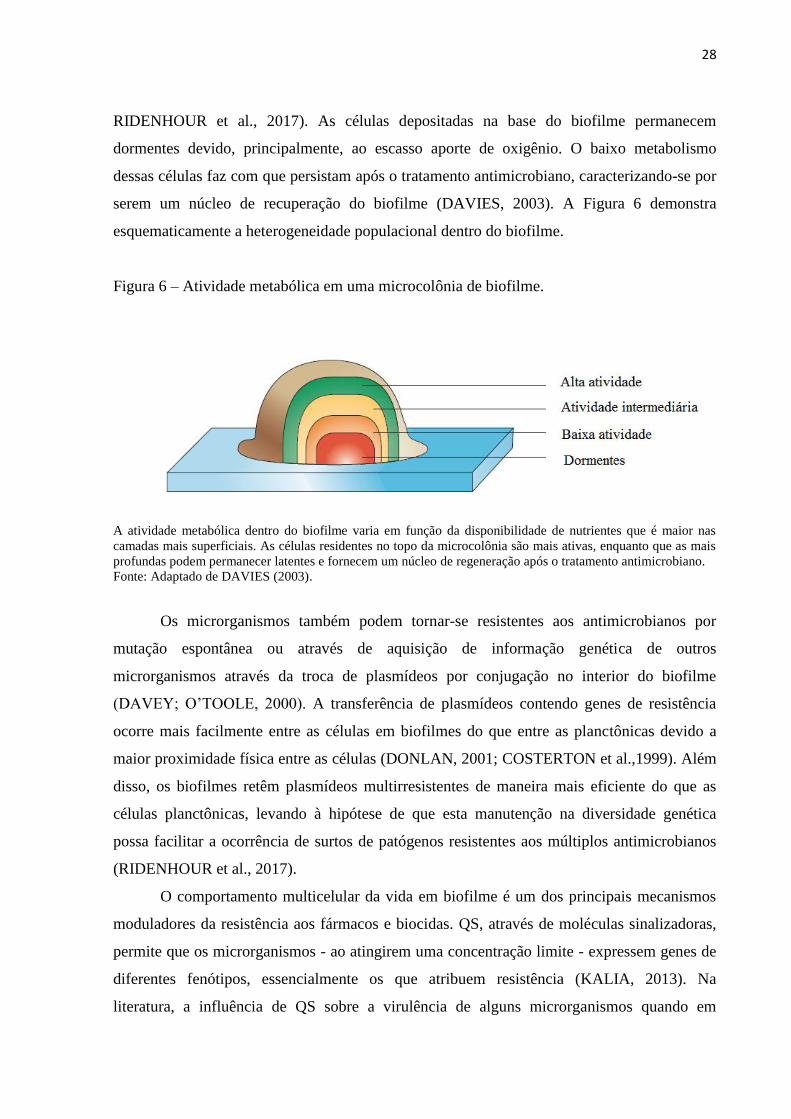
RIDENHOUR et al., 2017). As células depositadas na base do biofilme permanecem
dormentes devido, principalmente, ao escasso aporte de oxigênio. O baixo metabolismo
dessas células faz com que persistam após o tratamento antimicrobiano, caracterizando-se por
serem um núcleo de recuperação do biofilme (DAVIES, 2003). A Figura 6 demonstra
esquematicamente a heterogeneidade populacional dentro do biofilme.
Figura 6 – Atividade metabólica em uma microcolônia de biofilme.
A atividade metabólica dentro do biofilme varia em função da disponibilidade de nutrientes que é maior nas
camadas mais superficiais. As células residentes no topo da microcolônia são mais ativas, enquanto que as mais
profundas podem permanecer latentes e fornecem um núcleo de regeneração após o tratamento antimicrobiano.
Fonte: Adaptado de DAVIES (2003).
Os microrganismos também podem tornar-se resistentes aos antimicrobianos por
mutação espontânea ou através de aquisição de informação genética de outros
microrganismos através da troca de plasmídeos por conjugação no interior do biofilme
(DAVEY; O‟TOOLE, 2000). A transferência de plasmídeos contendo genes de resistência
ocorre mais facilmente entre as células em biofilmes do que entre as planctônicas devido a
maior proximidade física entre as células (DONLAN, 2001; COSTERTON et al.,1999). Além
disso, os biofilmes retêm plasmídeos multirresistentes de maneira mais eficiente do que as
células planctônicas, levando à hipótese de que esta manutenção na diversidade genética
possa facilitar a ocorrência de surtos de patógenos resistentes aos múltiplos antimicrobianos
(RIDENHOUR et al., 2017).
O comportamento multicelular da vida em biofilme é um dos principais mecanismos
moduladores da resistência aos fármacos e biocidas. QS, através de moléculas sinalizadoras,
permite que os microrganismos - ao atingirem uma concentração limite - expressem genes de
diferentes fenótipos, essencialmente os que atribuem resistência (KALIA, 2013). Na
literatura, a influência de QS sobre a virulência de alguns microrganismos quando em

29
biofilme é frequentemente relatada. Burkholderia cepacia, P. aeruginosa e Staphylococcus
sp. são somente alguns exemplos de patógenos que têm sua tolerância aos antimicrobianos
associada a este complexo sistema (DENG et al., 2013).
Outro fator responsável pela resistência aos antimicrobianos em estruturas de
biofilmes é a expressão de bombas de efluxo por alguns microrganismos (SOTO, 2013).
Trata-se de um mecanismo que permite que o ambiente interno das células microbianas seja
regulado através da remoção de substâncias tóxicas, incluindo agentes antimicrobianos
(OLIVARES et al., 2013; SOTO, 2013). Supõe-se que este mecanismo possa influenciar na
regulação exercida por QS e, por conseguinte, acarretar em alterações na biologia do biofilme.
O sistema de efluxo seria necessário para que as moléculas se difundissem através da
membrana celular, aumentando a sinalização e, consequentemente, a expressão de genes de
resistência. Ao mesmo tempo, pode estar relacionado com a extrusão dessas moléculas
(SOTO, 2013). Em estirpes do complexo Burkholderia, por exemplo, a formação de biofilme
demonstrou ser dependente de bombas de efluxo do tipo BpeAB - Opr-B (CHAN; CHUA,
2005). As bombas de efluxo são, portanto, altamente ativas em biofilmes, tornando-se um
alvo atraente para medidas antibiofilme (BAUGH et al., 2012).
Instiga-se, também, que muitos microrganismos possam adquirir resistência a
antissépticos e desinfetantes por adaptar-se a uma variedade de condições ambientais
(MURTOUGH et al., 2001). A teoria de adaptação a biocidas foi comprovada pela atividade
de concentrações subinibitórias de cloreto de benzalcônio, que mantiveram ativos ou
selecionaram microrganismos adaptados ao ambiente natural (MC CAY et al., 2010). Deste
modo, além dos conhecidos mecanismos envolvidos na resistência de biofilmes a
antimicrobianos - matriz exopolissacarídea, sinalização célula-célula, diversidade genética -
pode-se sugerir que o uso contínuo e inadequado dos desinfetantes pode estar relacionado aos
índices de resistência.
1.7 ABORDAGENS TERAPÊUTICAS CONTRA BIOFILMES
Os antimicrobianos convencionais - devido ao seu uso indiscriminado e à estrutura
complexa dos biofilmes - na maioria das vezes, não são efetivos frente a biofilmes. Além
disso, a terapia antimicrobiana contra biofilmes ainda é baseada nos testes de suscetibilidade
de microrganismos planctônicos, o que ocasiona falhas no tratamento e recorrência da
infecção. Testes de suscetibilidade específicos para biofilmes já foram projetados e

30
executados, porém, não apresentaram resultados confiáveis até o momento (HØIBY et al.,
2014).
Desta maneira, estratégias alternativas têm sido estudadas e utilizadas para prevenir a
adesão microbiana, retardar a formação de biofilmes e eliminar ou pelo menos reduzir a sua
acumulação (MARCINKIEWICZ et al., 2013; MARTINEZ-GUTIERREZ et al., 2013). A
Figura 7 representa esquematicamente algumas etapas da formação do biofilme que podem
ser afetadas para eliminar os biofilmes.
Figura 7 – Estratégias antibiofilme.
A ilustração exemplifica alguns estágios do desenvolvimento do biofilme que podem ser afetados para sua
eliminição. Uso de agentes de revestimento para inibir a fixação microbiana na superfície (clorexidina). Uso de
substâncias capazes de interromper a sobrevivência das bactérias (IQS). Uso de compostos que destroem o
biofilme formado (Nanopartículas de Prata (Ag)).
Fonte: Adaptado de GUPTA et al (2015).
1.7.1 Inibição da formação do biofilme
A inibição da formação de biofilmes pode ser obtida através do impedimento da
adesão microbiana a uma superfície ou por meio do impedimento da sinalização celular.
Neste sentido, substâncias que possam revestir materiais têm sido objeto de estudos com o
propósito de modificar superfícies de dispositivos médicos e dificultar a adesão microbiana
(MARCINKIEWICZ et al., 2013). Formulações adesivas de clorexidina com liberação
sustentada têm sido utilizadas no ponto de inserção de cateteres para prevenir a entrada de
microrganismos e a formação de biofilmes. Essa formulação apresentou excelente atividade
antibiofilme contra P. aeruginosa isolada de cateter urinário (SHAPUR et al., 2012). Do

31
mesmo modo, há evidências de que próteses ortopédicas impregnadas com gentamicina
reduzem a incidência de infecções associadas aos biofilmes (JOHANNSSON et al., 2010).
Outra possibilidade concernente à inibição de biofilmes é utilizar a motilidade e os
apêndices bacterianos como alvo de ação, uma vez que muitos microrganismos dependem de
flagelos e pili para acessar e se aderir às superfícies. Evidenciou-se, neste contexto, que
nanopartículas de M. alternifolia são capazes de interferir significativamente na motilidade de
P. aeruginosa PAO1 e, consequentemente, na formação do biofilme por essa cepa (COMIN et
al., 2016).
Neste mesmo cenário, incluem-se os compostos sulfatiazol e extrato de gengibre, os
quais são capazes de interferir na produção do segundo mensageiro c-di-GMP ou facilitar sua
degradação. O c-di-GMP está relacionado com a motilidade e a produção de adesinas e, uma
vez inibido, afeta diretamente a adesão microbiana, demonstrando outra possível categoria de
inibidores de biofilme (FAZLI et al., 2014).
1.7.2 Interferência na comunicação celular
A descoberta de compostos capazes de inibir a ação de moléculas sinalizadoras de QS
parece ser uma estratégia promissora para controlar a formação e virulência dos biofilmes.
Diversos Inibidores de quorum sensing (IQS) foram descritos, os quais incluem, por exemplo,
furanonas, triclosan, macrolídeos e ciprofloxacino (KALIA, 2013; MARCINKIEWICZ et al.,
2013). Estes compostos impedem a comunicação celular dos microrganismos previamente
aderidos ou não, impossibilitando a continuidade nas etapas de formação do biofilme
(MARCINKIEWICZ et al., 2013). Alguns IQS têm sido isolados a partir de plantas
medicinais como a alga marinha Delisea pulchra que produz furanonas halogenadas capazes
de inibir QS e, consequentemente, protegê-la da colonização microbiana. O extrato de alho
tem atividade semelhante à furanona e reduz a formação de biofilme por P. aeruginosa pela
inibição de QS (KIM; PARK, 2013).
Ainda neste contexto, encontra-se a relação existente entre Inibidores de Bombas de
Efluxo (IBE) e a formação de biofilmes (KOURTESI et al., 2013). Algumas publicações têm
comprovado que os IBEs são potenciais agentes capazes de inibir a formação de biofilmes,
assim como os IQS. A atuação de IBE sobre biofilme de Salmonella typhimurium, E. coli, K.
pneumoniae, S. aureus e P. putida evidenciaram diminuição e, até mesmo, erradicação das
películas formadas (KVIST et al., 2008; BAUGH et al., 2012). Da mesma forma, o verapamil
- conhecido IBE - desempenhou excelente atividade antibiofilme sobre C. albicans. Neste

32
mesmo estudo, o verapamil, quando em combinação com fluconazol, demostrou efeito
sinérgico contra o biofilme (YU et al., 2013).
1.7.3 Erradicação do biofilme formado
Dada a importância da matriz exopolissacarídica para a resistência dos biofilmes, é
interessante direcionar estudos de moléculas ou compostos que atuem diretamente sobre seus
componentes. As SPE são atraentes alvos para a ruptura de biofilmes, deixando o
microrganismo vulnerável ao tratamento antimicrobiano (MAUNDERS; WELCH, 2017).
Neste contexto, a enzima dispersina B foi documentada como opção terapêutica para a
eliminação de biofilmes, pois é capaz de promover a dispersão das células sésseis a partir da
matriz do biofilme (KIM; PARK, 2013).
A utilização de bacteriófagos - vírus específicos que infectam bactérias - também pode
ser um tratamento promissor contra os biofilmes. Os bacteriófagos poderiam atuar na
degradação de exopolissacarídeos da matriz extracelular que compõe o biofilme e, assim,
destruí-los (SHARMA et al., 2013). Processos físicos como a terapia fotodinâmica também
têm sido estudados. Tratam-se de lasers de baixa potência que combatem os microrganismos
tratados com fármacos fotossintetizantes. Nas leveduras C. albicans, C. glabrata e
Streptococcus mutans a utilização desta terapia mostrou-se bastante promissora, uma vez que
reduziu significativamente a atividade metabólica do biofilme quando comparado ao grupo
controle (QUISHIDA et al., 2013).
Nos últimos anos, a aplicação de nanopartículas se expandiu consideravelmente
(MARTINEZ-GUTIERREZ et al., 2013). As nanopartículas de prata possuem excelente
eficácia na prevenção da formação e na erradicação de biofilmes estabelecidos por isolados de
Acinetobacter baumanni, P. aeruginosa, C. albicans, MRSA (Staphylococcus aureus
resistente a meticiclina) e S. mutans (BARBOUR et al., 2013). Da mesma forma,
nanopartículas de Melaleuca alternifolia inibiram a formação de biofilme de diferentes
espécies de Candida (SOUZA et al., 2017).
Ademais, terapias antimicrobianas combinadas têm representado uma promissora
opção para o tratamento de infecções crônicas persistentes causadas por biofilmes. Neste
sentido, a associação entre antimicrobianos e terapia fotodinâmica também tem sido relatada
para o combate de biofilmes em feridas crônicas (HØIBY et al., 2014). A associação de
fosfomicina e claritromicina demostrou ser uma terapia útil frente a biofilme formado por
Staphylococcus pseudintermedius resistente à meticilina (DICICCO et al., 2014). Da mesma

33
maneira, foi documentado que biofilmes formados por P. aeruginosa PACI22 em modelo
experimental foram completamente erradicados pela combinação entre claritromicina e
ciprofloxacina, demonstrando substancial sinergismo (ELKHATIB; NOREDDIN, 2014).
1.8 A22 E O FORMATO DA CÉLULA BACTERIANA
As formas das células bacterianas (cocoides, bacilares ou espirais) são biologicamente
importantes e desempenham um papel crucial na sobrevivência dos microrganismos. À
medida que as condições ambientais se tornam desfavoráveis, as bactérias podem,
evolutivamente, alterar sua forma (YOUNG, 2006). A morfologia celular está interligada com
a aquisição de nutrientes, divisão celular, fixação em superfícies, motilidade e patogênese
(YOUNG, 2007). Deste modo, aumenta-se o interesse por conhecer os mecanismos
envolvidos na construção e manutenção da estrutura das células bacterianas.
Sabe-se que a célula bacteriana é essencialmente composta por duas estruturas
moleculares, a parede celular e o citoesqueleto. A parede celular é responsável por manter a
estrutura celular da maioria das bactérias e tem, como constituinte principal, o
peptideoglicano (PG). A camada de PG está situada externamente à membrana plasmática,
garantindo à célula uma pressão osmótica interna estável. Mutações em genes envolvidos na
síntese do PG ou a interferência química sobre as enzimas que participam da via de síntese
podem levar a alterações significativas na forma da célula, podendo haver a perda de sua
viabilidade (SCHEFFERS; PINHO, 2005).
O citoesqueleto bacteriano é constituído por homólogos eucarióticos, sendo
encontrados no citoplasma da célula, que ajudam na manutenção da forma e integridade das
células, além de participarem de muitas funções celulares, incluindo motilidade, segregação
cromossômica e transdução de sinais (CABEEN; JACOBS-WAGNER, 2005). Tratam-se das
proteínas CreS (crescentina), FtsZ e MreB, semelhantes aos filamentos intermediários, à
tubulina e à actina, respectivamente (DYE et al., 2011). A Figura 8 mostra a atuação de cada
uma dessas proteínas.
A CreS é responsável pela manutenção da forma filamentosa e quando suprimida, as
células tornam-se curvas (CELLER et al., 2013). A FtsZ forma uma espécie de “cinta” interna
na bactéria no momento de sua divisão celular, sem a atuação desta proteína, a bactéria
cresceria indefinidamente, sem dividir, até estourar. A MreB está envolvida no alongamento
celular em forma de bastonete. Para assumir o formato de haste, os bacilos precisam da
inserção de PG ao longo de comprimento que é feita pela MreB. Deste modo, a deleção desta

34
proteína resulta em microrganismos esféricos e irregulares. Quando esse estágio acaba, a FtsZ
atua para formar novas extremidades e dividir a célula bacteriana (CALLAWAY, 2008).
Além disso, alguns estudos reportam a relação direta entre MreB e motilidade bacteriana
(MAURIELLO et al., 2010; BULMER et al., 2012).
Figura 8 – Proteínas do citoesqueleto e o formato celular bacteriano.
Bactérias como S. aureus têm a divisão celular mediada pela FtsZ, a qual forma um anel (azul) durante a divisão
celular (a). E coli assume formato de bacilo, pois possui a proteína MreB, a qual aparece como uma estrutura
helicoidal ao longo da célula (vermelho) quando vista por microscopia de fluorescência (b). As células de
Caulobacter crescentus contêm CreS, além de MreB durante o alongamento celular e FtxZ durante a divisão
células (c).
Fonte: Adaptado de CABEEN; JACOBS-WAGNER (2005).
O composto A22 encontra-se, neste contexto, como um inibidor competitivo reversível
da ligação ATP - MreB, capaz de induzir a remodelação da parede celular, resultando em
alterações no formato da célula. Trata-se de uma molécula pequena, derivada do S-
benzilisotiureia, constituída de uma parte polar e uma apolar, sendo solúvel em água apenas
em baixas concentrações (HOCKENHULL; SHI, 2006). A estrutura do A22 e seu mecanismo
de ação estão representados nas Figuras 9 e 10, respectivamente.
Figura 9 – Estrutura do S-(3,4-diclorobenzil) isotioureia (A22).
Fonte: IWAI et al. (2002).

35
Figura 10 – Inibição reversível da proteína MreB pelo S-(3,4-diclorobenzil) isotioureia (A22).
Em verde está representada a proteína FtsZ e o amarelo representa o local de divisão celular. Nota-se que após a
atuação do A22 e supressão da MreB (vermelho), a célula bacteriana adota um padrão esférico.
Fonte: Adaptado de MARGOLIN (2009).
O A22 foi primeiramente descrito por induzir a forma esférica em isolados de E. coli,
tornando o microrganismo mais favorável à fagocitose e à liberação de endotoxinas (IWAI et
al., 2002). Além disso, demonstrou alterar a morfologia celular normal da cianobactéria
Anabaena sp., enquanto que para uma estirpe mutante, resistente ao A22, a forma bacilar se
manteve (WU et al., 2011). A Figura 11 demonstra o efeito da ação do A22 sobre a célula
bacteriana.
Figura 11 – Efeito do S-(3,4-diclorobenzil) isotioureia (A22) sobre a célula bacteriana.
A imagem de microscopia óptica mostra a célula bacilar normal de Caulobacter crescentus (A) e a alteração
neste formato após a exposição por 6 horas ao A22 na concentração 2,5µg/mL (B).
Fonte: Adaptado de HOCKENHULL; SHI (2006).
O A22 atua sobre o crescimento bacteriano, pois exibe atividade antibacteriana contra
diversas bactérias Gram negativas e efeito bacteriostático contra E. coli e Shigella flexneri
(HOCKENHULL; SHI, 2006; NOGUCHI et al., 2008; YAMACHIKA et al., 2012). Além
A B

36
disso, foi documentado que a despolimerização da proteína MreB provocada pelo A22, resulta
em defeitos na divisão e segregação irregular de cromossomos em E. coli e Caulobacter
crescentus (IWAI et al., 2002; GITAI et al., 2005; BEAN et al., 2009).
Neste contexto, tem-se direcionado a atividade do A22 à combinação com outros
fármacos, de modo a potencializar sua atividade. Da mesma forma, compostos
estruturalmente relacionados, assim como modificações químicas têm sido realizadas em sua
estrutura com a finalidade de aumentar o arsenal de adjuvantes antimicrobianos e inserir a
proteína MreB como um novo alvo terapêutico (NICHOLSON et al., 2012).
Considerando que pouco se sabe sobre o citoesqueleto bacteriano e as interações
moleculares existentes entre as proteínas supracitadas, o A22 aparece, na maioria das vezes,
como uma ferramenta útil na elucidação dos mecanismos envolvidos no estabelecimento do
formato bacteriano (CABEEN; JACOBS-WAGNER, 2005). O baixo número de publicações
relacionadas ao A22 evidencia que esta molécula deve ser mais explorada, principalmente
porque atua sobre o citoesqueleto bacteriano, alvo de nenhum antimicrobiano anteriormente
reportado (CALLAWAY, 2008). Do mesmo modo, o A22 possui um grande potencial
antibiofilme, visto que o processo de adesão é dependente da integridade conformacional do
microrganismo (FLEMMING; WINGENDER 2010).

37
2 OBJETIVOS
2.1 OBJETIVO GERAL
Avaliar a atividade antimicrobiana, antibiofilme, cito e genotóxica do S-(3,4-
diclorobenzil) isotioureia (A22) sobre cepas padrões e isolados clínicos multirresistentes de P.
aeruginosa provenientes do Hospital Universitário de Santa Maria (HUSM).
2.2 OBJETIVOS ESPECÍFICOS
2.2.1 Determinar a Concentração Inibitória Mínima (CIM) do composto A22 frente a cepas
padrões e isolados clínicos de P. aeruginosa;
2.2.2 Investigar os potenciais efeitos cito e genotóxicos do A22 em células mononucleares
de sangue periférico humano (PBMCs);
2.2.3 Caracterizar fenotipicamente os biofilmes através dos testes de motilidade, agregação
às células HeLa e adesão à superfície de Polietileno de Alta Densidade (PEAD);
2.2.4 Avaliar a capacidade de inibição do composto A22 sobre a formação de biofilmes e
sobre os parâmetros fenotípicos previamente caracterizados;
2.2.5 Analisar microscopicamente o biofilme, formado na ausência e na presença do
composto A22 em superfície de PEAD;

38
3 PUBLICAÇÕES CIENTÍFICAS
As publicações científicas incluem dois artigos publicados no periódico Microbial
Pathogenesis e estão formatadas de acordo com as normas exigidas pelo mesmo, acessadas no
site: https://www.elsevier.com/journals/microbial-pathogenesis/0882-4010/guide-for-authors.
3.1 ARTIGO 1
Título: Antibacterial, cyto and genotoxic activities of A22 compound ((S-3,4
dichlorobenzyl) isothiourea hydrochloride)
Autores: Pauline Cordenonsi Bonez, Andiara Ramos, Kátia Nascimento, Priscila
Copetti, Márcia Ebling de Souza, Grazielle Guidolin Rossi, Vanessa Albertina Agertt,
Michele Sagrilo, Roberto Christ Vianna Santos, Marli Matiko Anraku de Campos.
Artigo publicado no periódico Microbial Pathogenesis (Fator de impacto: 2,009,
Qualis B2) (Microbial Pathogenesis, n. 99, p. 14-18, 2016) Doi:
http://dx.doi.org/10.1016/j.micpath.2016.07.007

39
Antibacterial, cyto and genotoxic activities of A22 compound ((S-3, 4 -
dichlorobenzyl) isothiourea hydrochloride)
Bonez, P C
a, Ramos, A P
b, Nascimento, K
b, Copetti, P M
b, Souza, M E
c, Rossi, G G
a,
Agertt, V Aa, Sagrillo, M R
b, Santos, R C V
a, Campos, M M A
a
a Graduate Program in Pharmaceutical Sciences, Universidade Federal de Santa Maria, Brazil.
b Cell Culture Laboratory, Centro Universitário Franciscano, Brazil.
c Laboratory of Microbiological Research, Centro Universitário Franciscano, Santa Maria, Brazil
ABSTRACT
The A22 is a chemical compound that acts as a reversible inhibitor of a bacterial cell
wall protein MreB leading the rods to the coccoid form. Thus, by changing the
bacterial form, many properties can be affected, as the acquisition of nutrients, cell
division, the clamping surfaces, motility and pathogenesis. Infections caused by
strains of Pseudomonas aeruginosa have great clinical importance because these
microorganisms can include more than one resistance mechanism acting together,
limiting treatment options. Thus, it is important to investigate the action of A22
against P. aeruginosa, once there are urgent needs for new antimicrobial compounds
for increase the arsenal therapeutic to treat diseases caused by this microrganism.
Therefore, this study investigated for the first time the antimicrobial activity of A22
against seve standards strains of Gram negative microorganisms and twenty-eight
clinical isolates of P. aeruginosa. This study performed an additional investigation to
analyze the cyto and genotoxic potencial effects from A22 on human peripheral blood
mononuclear cells (PBMCs). The antibacterial activity of A22 was studied by broth
microdilution method and time-kill assay. The cytotoxicity was evaluated by MTT
assay at 24, 48 and 72 hours of exposure to A22 and the genotoxicity was evaluated
by the Comet assay. The susceptibility tests showed A22 has a relevant antibacterial
activity against P. aeruginosa, including multidrug-resistant (MDR) clinical isolates.
The A22 treatment not showed genotoxic effects against PBMCs in almost all
concentrations tested at 24 and 48 hours of exposure. Only for concentration of 32
µg/mL (highest tested) the damage index was significantly higher in all moments. The
MTT assay demonstrated that A22 was able to maintain cell viability in all exposure
times. In summary, the A22 demonstrated important anti-Pseudomonas activity and
showed no cyto and genotoxic significant effect. These results need to be considered
in future in vitro and in vivo studies in order to introduce the A22 as a possible
therapeutic option.
Key-words: P. aeruginosa, A22, cytoxicity, genotoxicity.

40
1. Introduction
Antimicrobial resistance is a significant concern for the global healthcare community
[1]. Treatment of Gram-negative bacterial infections is increasingly challenging due to
absence of new agents in the pharmaceutical pipeline and global spread of resistance. In this
context, P. aeruginosa is among the most frequently isolated microorganisms in clinical
laboratories, being reported as one of the main pathogens responsible for infections of the
respiratory and urinary tract, as well as bloodstream infections [2]. Pseudomonas infection
can be treated with a combination of an anti-Pseudomonas beta-lactam, as carbapenems, and
an aminoglycoside, as ciprofloxacin [3].
However, P. aeruginosa has developed resistance to nearly every classes of
antimicrobials available as aminoglycosides, quinolones and carbapenems [3]. These
microorganisms become resistant through specific mechanisms such as induction of
alternative enzymes which can inactivate the drug, alterations in the permeability or uptake of
the drug, expression of efflux pumps, reduction in the number or affinity to the drug-binding
sites and biofilm formation [4]. Infections caused by these strains may include more than one
resistance mechanism acting together what makes it difficult to choose treatment and
eradication of the disease promoting the increase in morbidity and mortality [5]. In this
context, it is evident the need for research to advance the search for new compounds able to
combat MDR microorganisms.
The compound A22 is a reversible, competitive inhibitor of the actin-like protein
MreB, able to induce changes in cell shape, division defects and irregular segregation of
chromosome in Escherichia coli and Caulobacter crescentus [6, 7, 8]. The A22 was first
described to induce spherical cell in E. coli [6]. In addition, the A22 showed antibacterial
activity against P. aeruginosa strains and present bacteriostatic effect against E. coli and
Shigella flexneri [9, 10, 11]. On the other hand, the A22 is used as tool in studies involving
bacterial cytoskeleton and its protein structures [12].
Thus, becomes promising to investigate the action of A22 compound, once it alters the
structural conformation of the microorganism. Besides, could be inserted as a new therapeutic
option in the future, being used alone or in combination with other antimicrobial drugs.
However, it will be important to determine the range of concentrations that do not present
toxic effects on cells. For this reason, were performed some complementary investigations to
evaluate if A22 compound present some cyto and genotoxic effect using human PBMCs, such
as an in vitro experimental model.

41
2. Materials and methods
2.1 Microorganisms
Were used in this study the standard strains of P. aeruginosa PAO1, Acinetobacter
baummanni ATCC 19606, Klebsiella pneumonie ATCC 700603, K. pneumonie ATCC 1705,
Escherichia coli ATCC 35218, E. coli ATCC 8739 and E. coli ATCC 25922. Besides, were
used 28 multidrug-resistant (MDR) clinical isolates of P. aeruginosa from the Hospital
Universitário de Santa Maria (HUSM), Santa Maria, Rio Grande do Sul, Brazil. The isolates
were classified as MDR according to the results of susceptibility tests conducted in the
Clinical Analysis Laboratory (CAL) of HUSM and definitions established by Magiorakos et
al. [13].
This work was conducted with authorization of Research Ethics Committee (CEP) of
the UFSM an approved by CAAE (Certificate Presentation for Ethics Assessment) number
12114713.1.0000.5346.
2.2 Compound
The compound A22 was purchased from Sigma-Aldrich (Product nº SML0471, ≥ 95%
purity (HPLC), St. Louis, MO, USA), was diluted in dimethyl sulfoxide (DMSO, Sigma-
Aldrich, St. Louis, MO, USA) and evaluated against to microrganisms at concentrations
ranging from 0.25 to 128 μg/mL according to the Clinical Laboratory Standards Institute
guidelines CLSI M100-S23 [14].
In cyto and genotoxicity assays, the concentrations ranging from of 0.25 to 32 μg/mL.
These concentrations were established from the drug molecular weight in relation to volume
of well culture plate so as not to saturate the middle of the cell and do not alter the pH. The
calculations of the concentrations were made from a mother concentration of 512 μg/mL. It
was taken into consideration the total volume of well of 250 µL and standardized treatment
volume of 20 µL to each well.

42
2.3 Antibacterial assays
2.3.1 Preparation of inoculums
The inoculums sizes were standardized according to CLSI guidelines [14]. Isolated
colonies were grown for 18–24 h in Muller Hinton Ágar (Difco) and the suspension were
prepared in saline solution (NaCl 0,85%).
2.3.2 Determination of minimum inhibitory concentration (MIC)
The antibacterial activity of A22 was evaluated by broth microdilution method. The
MIC of A22 were determined in 96-well microtitre plates using Mueller–Hinton broth
(Difco). The A22 was diluted twofold serially ranging from 0.25 to 128 µg/mL. The plates
were incubated at 37°C and the MICs were recorded after 24 h of incubation. The MIC was
defined as the lowest concentration of compound that inhibits bacterial visible growth. This
test was performed in triplicate and the 2, 3, 5 -triphenyltetrazolium chloride was used as an
indicator of microbial growth [14].
2.3.3 Time kill assay
The in vitro time kill assay was performed by diluting an overnight culture of P.
aeruginosa PAO1 in Muller Hinton broth (Difco) to reach the final concentration of 1x10⁵
CFU/ml. The A22 was added according to their MICs (½ MIC, MIC, 2 and 4 × the MIC).
Samples were removed for colony counts at various time intervals (3, 6, 9, 12, and 24 h).
Drug free inoculated medium was plated as a growth control. Plates were incubated at 37ºC
for 24 h. The inhibitory effect was calculated based on the CFU (colony forming units). [15]
The bactericidal activity was defined as a 3-log reduction of viable counts in culture treated
with the A22 compared with that of the untreated control at a specific time point [16]. The
procedure was repeated three times.
43
2.4 Evaluation of toxicity
2.4.1 Cell culture
The PBMCs derived from blood discard samples of healthy adults from the Analysis
Clinics Laboratory of Centro Universitário Fransiscano (LEAC-UNIFRA). Blood specimens
were subjected to the procedure of separation of its constituents by density gradient using
Ficoll Histopaque-1077® reagent (Sigma-Aldrich, St. Louis, MO, USA) followed by
centrifugation for 30 min. The PBMCs were plated in 6-well plates containing RPMI 1640
(Sigma-Aldrich, St. Louis, MO, USA) culture with 10% fetal bovine serum, 1% penicillin and
streptomycin. The cells were cultured at an initial density of 2 x 10⁵ cells mL
-1 of material
[17]. The use of cells was approved by the ethics committee in research with human beings of
the UNIFRA by CAAE number: 31211214.4.0000.5306.
2.4.2 Cytotoxicity evaluation
The cytotoxicity evaluation was performed by MTT assay (tetrazolium colorimetric
assay). This technique is based on the reduction of MTT bromide salt {[3- (4, 5-
dimethylthiazol-2-yl)-2,5-diphenyltetrazolium bromide]} (Sigma-Aldrich, St. Louis, MO,
USA) by viable cells, producing a product called formazan in the mitochondria [18]. This test
was performed according Denizot; Lang [19] with some modifications. The cell culture
exposed to different concentrations of A22 (0.25 µg/mL until 32 µg/mL) were incubated for
24, 48 and 72 h at 37°C. After, 20 µ L of MTT (5 mg mL-1
diluted in phosphate buffer pH
7.4) were added to plates and incubated for 4h at 37ºC. The supernatant was removed and the
formazam was dissolved in 200 µL of DMSO, such that the formazan crystals were released
into the extracellular medium. Viable cells with active metabolism convert MTT into a purple
colored formazan, when cells die, they lose the ability to convert MTT into formazan, thus
color formation serves as an useful and convenient marker of only the viable cells. The test
was performed in triplicate and the absorbance was measured colorimetricaly by
spectrophotometry at a wavelength of 570 nm. It has made a positive control for cell death
using H2O2 and a negative control containing the cell culture only.

44
2.4.3 Genotoxicity evaluation
The genotoxicity evaluation was performed by Comet assay. This analysis was
adapted following the protocol described by Garcia et al. [20]. The cells treated with different
concentrations (0.25 µg/mL until 32 µg/mL) of A22 were suspended in low-melting agarose
point (deposited in glass slide precoated with a layer of 1.5% agarose). The material was
immersed in lysis solution (lysis solution of 89–10 mL of dimethyl sulfoxide and 1 mL of
Triton X-100) for the removal of membranes and cytoplasm. Subsequently, the slides were
incubated in alcaline electrophoresis buffer at pH 13 (300 mmol L-1
NaOH and 1 mmol L-1
EDTA in distilled water) and subjected to electrophoresis for 30 min at 25 V and 300 mA.
Then, the processes of neutralization (neutralizing buffer pH 7.5), fixation (15%
trichloroacetic acid) and coloration (acridine orange) were performed, so the genetic material
could be analysed. One hundred cells were analysed in an optical microscope and were
classified according to their tail length. The cells received scores from 0 (no damage) to 4
(maximum damage). The test was performed in triplicate, and the data were transformed into
a damage index to be submitted to statistical analysis [21]. The genotoxic potential was
meansured at 24, 48 and 72 hours after exposure to A22 and was considered genotoxic, the
concentrations that resulted in a damage index higher that 0.1.
2.5 Statistical analysis
All tests were performed in 3 replicates and the results were expressed on Average ±
Standard derivation. The time-kill curve was performed using GraphPad Prism® 5. The
Statistical analysis for geno and cytoxicity were performed using SPSS®
software, version
19.0. Analysis of variance (ANOVA) followed by Tukey post hoc test was applied for
statistical analysis with the level of significance at p < 0.05.

45
3. Results
3.1 Antibacterial assays
3.1.1 Determination of MIC
Inhibitory activity of A22 was observed on Escherichia coli and P. aeruginosa strains.
The MIC values of strains standards are described in table 1. The A22 showed high inhibitory
activity against isolates of P. aeruginosa, once inhibited visible growth at low concentrations.
The MIC values of isolates range between 0.25 µg/mL and 16 µg/mL. 25% of the isolates
obtained value of 4 µg/mL, 28.5% of 2 µg/mL, 25% resulted in MIC of 1 µg/mL, 14,2% of
0.5 µg/mL and 7% include the values of 0.25 µg/mL and 16 µg/mL.
3.1.2 Time-kill assay
After identification of susceptible microorganisms to A22, a death time curve for P.
aeruginosa PAO1 was performed using the following concentrations of A22 treatment: ½
MIC (0.5 µg/mL), MIC (1 µg/mL), and 2 x MIC (2 µg/mL) and 4 x MIC (4 µg/mL). As
figure 1 shows, the concentration of MIC, 2 x MIC and 4 x MIC present significant activity
against the growth of P. aeruginosa PAO1, however, only the concentration of 2 x MIC and 4
x MIC were able to zero growth on the plates. The A22 began to decrease the growth after 12
hours of exposure to 2 x MIC and 4 x MIC of A22 and 3-log reduction was seen after 24
hours.
3.2 Cytotoxicity evaluation
As expected, the H2O2 (death control) decreased significantly cell viability when
compared to control. As seen in figure 2, the A22 was able to maintain cell viability in almost
all concentrations in all times of exposure. There was a decrease in cell viability only the
concentration of 32 µg/mL, however, this decrease was smaller than that observed in death
control.

46
3.3 Genotoxicity evaluation
The results obtained in genotoxicity assay are shown in table 2. The damage index was
low in most concentrations of A22 when compared to control, being dependent on
concentration and exposition time. The A22 treatment no showed genotoxic effects against
PBMCs in almost all concentrations tested at 24 and 48 hours of exposure. However, for the
concentration 32µg/mL, the damage index was significantly higher in all the times.
4. Discussion
The results reported here are the first to evaluate the activity of A22 against clinical
isolates of P. aeruginosa and the first study to check the cyto and genotoxicity effects of A22.
The broth microdilution assay and time-kill assay demonstrate that compound A22 is a
promising agent against P. aeruginosa, because the MIC values were significantly lower,
corroborating with previous studies in standards strains of P. aeruginosa, E. coli and Shigella
flexneri [9, 10, 11]. In addition, the MIC values found can be compared to reported for
antimicrobials already used in anti-Pseudomonas therapy such as meropenem, β-lactam that
as A22, acts on the bacterial cell wall [14].
The good performance of the A22 is due to probably, its inhibition of binding ATP –
MreB, leading to diffuse distribution MreB in the cytoplasm and loss of cell morphology and
viability [8]. This happens because the MreB protein is responsible for keeping the
organization of lipids and membrane proteins. Therefore, the MreB inhibition by A22
prevents the synthesis of cell wall by the inability to recruit the necessary proteins to the site
of synthesis [22]. Thus, by changing the shape of the bacterial cell, many properties are
affected as acquisition of nutrients, cell division, fixing surfaces, motility and pathogenesis
[23]. Besides, the A22 makes the microoganisms more favorable to phagocytosis and the
release of endotoxins [6].
The time-kill assay confirmed the broth microdilution method. The kill curve
demonstrated low survivability of P. aeruginosa strain PAO1 when exposed to A22, mainly,
because it had no growth in time of 24 hours in concentrations of 2 x MIC (2 µg/mL) and 4 x
MIC (4 µg/mL). As can be seen in figure 1, the MIC concentration did not reduce
significantly the growth on the plates at all times evaluated and the growth obtained on plates
containing 1/2 MIC was similar to the positive growth control. Thus, suggest that the action
of A22 is dose-dependent. Moreover, the A22 had fast onset of action with a large decrease in

47
growth in 12 hours, keeping until the time of 24 hours when there was no growth. These
results were similar to that obtained against the ciprofloxacin under the same testing
conditions, where there was low growth for 12 hours and sterilization in 24 hours [24].
The A22 is used as a tool to understand mechanisms of action of other antimicrobial
and the complexity around the organization of membrane bacterial cell [7, 12]. Other studies,
investigating the action of compound A22 and the modification of its structure with the
purpose to discover antimicrobial analogue [6, 9, 10, 11, 25]. However, no were studies
reported about the cyto and genotoxic effects.
The MTT assay evaluates cell viability from the sample, and the level of
mitochondrial metabolic activity of viable cells. The main advantage of this colorimetric
assay is the speed with which samples can be processed, besides showing high degree of
accuracy [18]. In the present study, the A22 was able to maintain cell viability in all exposure
times. As seen in figure 2, A22 presented a good protective effect in low concentrations
except in concentration of 32 µg/mL (highest tested). Our results are similar to those found by
Sueke et al. [26], which assessed the cytotoxicity of meropenem. Thus, the cytotoxicity of
A22 can be compared to cytotoxicity presented by this extensively used carbapenem. After
adding the DMSO, there was the formation of purple crystals formazam, showing that the
cells remained were alive and metabolically active, even after long period of exposure to the
A22. The results were also apparent visually through the purple color formed (data not show)
and the amount of formazan generated was directly proportional to the viable cell number,
characterizing another advantage of this method.
The Comet assay is a standard method for evaluating DNA damage, of simple
execution and is highly sensitive method to detect low levels of DNA damage. It is an
ultrasensitive, simple, fast and accurate test [20]. In addition, it can be applied in different
cells kind: PBMCs, bucal epithelial cells, nasal epithelial cells, lens epithelium, tear duct
epithelial cells, sperms as well as biopsy tissues. So, the Comet assay is a versatile and
potential tool of choice to assess the DNA damage and repair efficiency [27]. The A22
treatment not showed genotoxic effects against PBMCs in almost all concentrations tested at
24 and 48 hours of exposure, some genotoxic damage was observed in all concentrations in
time of 72 hours. Only for concentration of 32µg/mL the index damage was significantly
higher in all the times. Comparing these results with those obtained for ciprofloxacin
performed by Itoh et al. [28], A22 was less genotoxic. Although the concentrations evaluated
vary according to the antibacterial activity of each compound, this is an important result.

48
The biological activity of S-benzylisothiourea derivatives has not described in detail
yet. There are reports that some acylisothiourea derivatives inhibit aldehyde dehydrogenase in
vivo and derivatives S-benzylisothiourea were toxic in rats [6, 29]. However, is no report
about the cyto and genotoxic potential of the A22 specifically. Thus, the results obtained in
this study are encouraging, once the range of concentrations tested in cyto and genotoxicity
assays are not harmful to the cells and to DNA and comprises the MICs obtained for all tested
microorganisms.
5. Conclusion
In summary, the results showed that changing the structural conformation of the
bacterial cell, through inhibition of protein MreB by the A22, obtained highly effective anti-
Pseudomonas. Considering that the isolates of this study are resistant to multiple classes of
drugs, these results can have impactful because the A22 can be confirmed as a new
antimicrobial agent, besides to insert MreB protein as a new therapeutic target. The low
toxicity of A22 revealed in cyto and genotoxicity assays, makes promising invest in more
research in vitro and in vivo for the purpose to insert it as a new antimicrobial and increase the
therapeutic arsenal against MDR microorganisms.
References
[1] M. Martins, M. P. McCusker, M. Viveiros, I. Couto, S. Fanning, J. M. Pagès, L. Amaral.
A simple method for assessment of MDR bacteria for over-expressed efflux pumps. The Open
Microbiology Journal. 7 (2013) 72-82, http://dx.doi.org/ 10.2174/1874285801307010072.
[2] E. B. Hirsch; V. H. Tam. Impact of multidrug-resistant Pseudomonas aeruginosa infection
on patient outcomes. Expert review of pharmacoeconomics & outcomes research. 10 (2010)
441-451, http://dx.doi.org/ 10.1586/erp.10.49.
[3] CDC. Antibiotic resistance threats in the united states, 2013. centers for disease control
and prevention. Available at http://www.cdc.gov/drugresistance/pdf/ar-threats-2013-508.pdf.
[4] M. Woolhouse; J. Farrar. Policy: An intergovernmental panel on antimicrobial resistance.
Nature. 509 (2014) 555-557.
[5] J. B. Marques, P. C. Bonez, V. A. Agertt, V. C. Flores, T. V. Dalmolin, G. G. Rossi, N. L.
F. Dal Forno, B. V. Bianchini, C. R. Mizdal, F. S. Siqueira, R. A. Righi, B. H. Meneghetti, P.
A. Trindade, R. C. V. Santos, M. M. A. Campos. Molecular characterization of
Enterobacteriaceae resistant to carbapenem antimicrobials. Jornal Brasileiro de Patologia e
Medicina Laboratorial. 51 (2015) 162-165.

49
[6] N. Iwai; K. Nagai, M. Wachi. Novel S-benzylisothiourea compound that induces spherical
cells in escherichia coli probably by acting on a rod-shape-determining protein(s) other than
penicillin-binding protein 2. Bioscience Biotecnology and Biochemestry. 66 (2002) 2658-
2662, http://dx.doi.org/ 10.1271/bbb.66.2658.
[7] Z. Gitai, N. A. Dye, A. Reisenauer, M. Wachi, L. Shapiro. MreB Actin-mediated
segregation of a specific region of a bacterial chromosome. Cell. 120 (2005) 329-341,
http://dx.doi.org/10.1016/j.cell.2005.01.007.
[8] G. J. Bean, S. T. Flickinger, W. M. Westler, M. E. McCully, D. Sept, D. B. Weibel, K. J.
Amann. A22 Disrupts the bacterial actin cytoskeleton by directly binding and inducing a low-
affinity state in MreB. Biochemistry. 48 (2009), http://dx.doi.org/10.1021/bi900014d.
[9] N. Noguchi, K. Yanagimoto, H. Nakaminami, M. Wakabayashi, N. Iwai, M. Wachi, M.
Sasatsu. Anti-infectious effect of s-benzylisothiourea compound a22, which inhibits the actin-
like protein, mreb, in Shigella flexneri. Biological & Pharmaceutical Bulletin. 31 (2008)
1327-1332.
[10] A. Nicholson, J. D. Perry, A. L. James, S. P. Stanforth, S. Carnell, K. Wilkinson, C. M.
Anjam Khan, A. De Soyza, F. K. Gould. In vitro activity of S-(3, 4-dichlorobenzyl)
isothiourea hydrochloride and novel structurally related compounds against multidrug-
resistant bacteria, including Pseudomonas aeruginosa and Burkholderia cepacia complex.
International Journal of Antimicrobial Agents. 39 (2012) 27-32,
http://dx.doi.org/10.1016/j.ijantimicag.2011.08.015.
[11] S. Yamachika, C. Sugihara, H. Tsuji, Y. Muramatsu, Y. Kamai, M. Yamashita. Anti-
Pseudomonas aeruginosa compound, 1, 2, 3, 4-tetrahydro-1,3,5-triazine derivative, exerts its
action by primarily targeting MreB. Biological & Pharmaceutical Bulletin. 35 (2012) 1740-
1744.
[12] E. M. Mauriello, F. Mouhamar, B. Nan, A. Ducret, D. Dai, D. R. Zusman, T. Mignot.
Bacterial motility complexes require the actin-like protein, MreB and the Ras homologue
MglA. The EMBO Journal. 29 (2010) 315-326, http://dx.doi.org/doi:10.1038/
emboj.2009.356.
[13] A. P. Magiorakos, A. Srinivasan, R. B. Carey, Y. Carmeli, M. E. Falagas, C. G. Giske, S.
Harbarth, J. F. Hindler, G. Kahlmeter, B. Olsson-Liljequist, D. L. Paterson, L. B. Rice, J.
Stelling, M J. Struelens, A. Vatopoulos, J. T Weber, D. L. Monnet. Multidrug-resistant,
extensively drug-resistant and pandrug-resistant bacteria: an international expert proposal for
interim standard definitions for acquired resistance. Clinical Microbiology and Infection. 18
(2012) 168-281, http://dx.doi.org/ 10.1111/j.1469-0691. 2011.03570.x.
[14] CLSI Clinical and Laboratory Standards Institute. Performance Standards for
antimicrobial susceptibility testing, 22th informational supplement. M100 - S23. Clinical and
Laboratory Standards Institute, Wayne, PA, 2013.
[15] G. Meiyazhagan, R. Raju, S. B. Winfred, B. Mannivanan, H. Bhoopalan , V. Shankar, S.
Sekar, D. P. Venkatachalam , R. Pitani, V. Nagendrababu, M. Thaiman, K. Devivanayagam ,
J. Jayaraman, R. Ragavachary, G. Venkatraman. Bioactivity studies of β-lactam derived
polycyclic fused pyrroli-dine/pyrrolizidine derivatives in dentistry: in vitro, in vivo and in
silico studies. PlosOne. 10 (2015), http://dx.doi.org/ 10.1371/journal.pone.0131433.

50
[16] T. T. Aung , J. K. Yam, S. Lin, S. M. Salleh, M. Givskov, S. Liu, N. C. Lwin N.
C. Yang , R. W. Beuerman. Biofilms of pathogenic nontuberculous mycobacteria targeted by
new therapeutic approaches. Antimicrobial Agents and Chemotherapy. 60 (2016) 24-35,
http://dx.doi.org/ 10.1128/AAC.01509-15.
[17] G. Botton , C. W. Pires, F. C. Cadoná , A. K. Machado , V. F. Azzoli, I. B. Cruz, M. R.
Sagrillo, J. R. Praetzel . Toxicity of irrigating solutions and pharmacological associations
used in pulpectomy of primary teeth. International Endodontic Journal (2015) 1-9,
http://dx.doi.org/ 10.1111/iej.12509.
[18] T. Mosmann. Rapid colorimetric assay for cellular growth and survival: application to
proliferation and cytotoxicity assays. Journal of lmmunological Methods. 65 (1983) 55-63.
[19] F. Denizot; R. Lang. Rapid colorimetric assay for cell growth and survival.
Modifications to the tetrazolium dye procedure giving improved sensitivity and reliability.
Journal of Immunological Methods. 89 (1986) 271-277.
[20] O. García, T. Mandina, A. I. Lamadrid, A. Diaz , A. Remigio, Y. Gonzalez, J. Piloto, J.
E. Gonzalez, A. Alvarez . Sensitivity and variability of visual scoring in the comet assay.
Results of an inter-laboratory scoring exercise with the use of silver staining. Mutation
Research. 556 (2004) 25-34, http://dx.doi.org/ 10.1016/j.mrfmmm.2004.06.035.
[21] G. F. dos Santos Montagner , M. Sagrillo, M. M. Machado, R. C. Almeida, C. P.
Mostardeiro, M. M. Duarte, I. B. da Cruz. Toxicological effects of ultraviolet radiation on
lymphocyte cells with different manganese superoxide dismutase Ala16Val polymorphism
genotypes. Toxicology In Vitro. 24 (2010) 1410-6,
http://dx.doi.org/10.1016/j.tiv.2010.04.010.
[22] H. Strahl, F. Burmann, L.W. Hamoen. The actin homologue MreB organizes the
bacterial cell membrane. Nature Communications (2014) 1-11,
http://dx.doi.org/doi:10.1038/ncomms4442.
[23] K. D. Young. Bacterial morphology: why have different shapes? Current Opinion in
Microbiology. 10 (2007) 596-600, http://dx.doi.org/10.1016/j.mib.2007.09.009.
[24] H. C. Su , K. Ramkissoon, J. Doolittle, M. Clark, J. Khatun, A. Secrest, M. C. Wolfgang,
M. C. Giddings. The development of ciprofloxacin resistance in pseudomonas aeruginosa
involves multiple response stages and multiple proteins. 54 (2010) 4626-4635,
http://dx.doi.org/ 10.1128/AAC.00762-10.
[25] X. Wu, N. Iwai, W. L. Chen. Effects of S-(3,4-dichlorobenzyl) isothiourea on different
cellular events in the cyanobacterium Anabaena sp. strain PCC 7120. Research in
Microbiology 162 (2011) 375-381, http://dx.doi.org/10.1016/j.resmic.2011.02.001.
[26] H. Sueke, S. Kaye, M. C. Wilkinson, S. Kennedy, V. Kearns, Y. Zheng,
P. Roberts, S.Tuft, T. Neal. Pharmacokinetics of Meropenem for Use in Bacterial Keratitis.
Investigative ophthalmology & visual science 56 (2015) 5731-5738,
http://dx.doi.org/.10.1167/iovs.15-17077.
51
[27] V. Gunasekarana, G. Victor raj, P. A. Chand. Comprehensive review on clinical
applications of comet assay. Journal of Clinical and Diagnostic Research 9 (2015) GE01-
GE05. http://dx.doi.org/ 10.7860/JCDR/2015/12062.5622.
[28] T. Itoh, K. Mitsumori, S. Kawaguchi, Y. F. Sasaki. Genotoxic potential of quinolone
antimicrobials in the in vitro comet assay and micronucleus test 603 (2006) 135–144.
http://dx.doi.org/ 10.1016/j.mrgentox.2005.11.003.
[29] F. N. Shirota, J. M. Stevens-Johnk, E. G. DeMaster, H. T. Nagasawa HT. Novel
prodrugs of cyanamide that inhibit aldehyde dehydrogenase in vivo. Journal of Medicinal
Chemestry 40 (1997) 1870-1875, http://dx.doi.org/ 10.1021/jm9606296.

52
Table 1
Antimicrobial activity of S-(3,4-diclorobenzil) isotioureia (A22) against strains standards.
MIC: Minimal Inhibitory Concentration
ATCC: American Type Culture Collection
Table 2
DNA migration in the comet assay for assessment of genotoxicity of S-(3,4-diclorobenzil)
isotioureia (A22) exposition in peripheral blood mononuclear cell culture during 24, 48 and
72 hours.
Strains standards MIC (µg/mL)
K. pneumoniae ATCC 700603 128
K. pneumoniae ATCC 1705 128
E. coli ATCC 35218 2
E. coli ATCC 8739 1
E. coli ATCC 25922 128
P. aeruginosa PAO1 1
A. baummanni ATCC 19606 32
Index damage
Based in comet frequency (per 100 nuclei analyzed)
Treatments A22
(µg/mL) 24 hours 48 hours 72 hours
Negative control __ __ 0.05
Positive control 0.5 0.7 0.7
0.25 __ __ 0.05
0.5 __ __ 0.05
1 __ __ 0.06
2 __ __ 0.08
4 __ __ 0.08
8 __ __ 0.08
16 __ __ 0.1
32 0.13 0.23 0.38

53
Time-kill kinetic of A22 on Pseudomonas aeruginosa PAO1
0 10 20 300
2
4
6Control
1/2MIC (0.5µg/mL)
MIC (1µg/mL)
2xMIC (2µg/mL)
4xMIC (4µg/mL)
Time (hours)
Lo
g1
0 (
CF
U /m
L)
Fig 1. Time-kill curve on P. aeruginosa PAO1 exposed to several concentrations of S-(3,4-
diclorobenzil) isotioureia (A22) (based on MIC).
C- 2O
2H 0,25 0,
5 1 2 4 8 16 32
0
50
100
150
ab
c
ab
abcab
aab
bc
A22
Treatments (ug/mL) - 24 horas
% C
ell
Via
bili
ty
C- 2O
2H 0,25 0,
5 1 2 4 8 16 32
0
50
100
150
c
e
abc aabc bc
ab
d
A22
Treatments (ug/mL) - 48 horas
% C
ell
viabili
ty
C- 2O
2H 0,25 0,
5 1 2 4 8 16 32
0
50
100
150
abcd
e
abc a
bcd cd
ab
d
A22
Treatments (ug/mL) - 72 horas
% C
ell
viabili
ty
A B C
Fig 2. Comparison of cytotoxicity evaluated by MTT reduction among human cells exposed
to H2O2 and treated with S-(3,4-diclorobenzil) isotioureia (A22) in 24 hours (A), 48 hours
and 72 hours (C) at different concentrations. Values are mean ± SE. Different letters within
treatment indicate

54
3.2 ARTIGO 2
Título: Anti-biofilm activity of A22 ((S-3,4-dichlorobenzyl) isothiourea
hydrochloride) against Pseudomonas aeruginosa: influence on biofilm formation,
motility and bioadhesion
Autores: Pauline Cordenonsi Bonez, Grazielle Guidolin Rossi, Jardel Rodrigo
Bandeir, Andiara Prates Ramos, Caren Rigon Mizdal, Vanessa Albertina Agertt, Eloísa Salete
Segatto Dalla Nora, Márcia Ebling de Souza, Camilla Fillippi dos Santos Alve, Fallon
Siqueira dos Santos, Rodrigo de Almeida Vaucher, Roberto Christ Vianna Santos, Marli
Matiko Anraku de Campos.
Manuscrito submetido em 26 de abril de 2017 para o periódico Microbial
Pathogenesis (Fator de impacto: 2,009, Qualis: B2).

55
Anti-biofilm activity of A22 ((S-3,4-dichlorobenzyl) isothiourea
hydrochloride) against Pseudomonas aeruginosa: influence on biofilm
formation, motility and bioadhesion
Pauline Cordenonsi Bonez
a, Grazielle Guidolin Rossi
a, Jardel Rodrigo Bandeira
b, Andiara
Prates Ramos b, Caren Rigon Mizdal
a, Vanessa Albertina Agertt
a, Eloísa Salete Segatto
Dalla Nora a, Márcia Ebling de Souza
c, Camilla Fillippi dos Santos Alves
a, Fallon Siqueira
dos Santosa, Rodrigo de Almeida Vaucher
d, Roberto Christ Vianna Santos
a, Marli Matiko
Anraku de Campos a
a Graduate Program in Pharmaceutical Sciences, Universidade Federal de Santa Maria, Avenue Roraima 1000,
zip code: 97105-900, Santa Maria, Brazil. b Cell Culture Laboratory, Centro Universitário Franciscano, Rua dos Andradas 1614, zip code: 97010-032,
Santa Maria, Brazil. c Oral, Universidade Federal de Santa Maria, Avenue Roraima 1000, zip code: 97105-900, Santa Maria, Brazil.
d Microbiology Research Laboratory, Universidade Federal de Pelotas, Rua Gomes Carneiro, 1, zip code:
96010-610, Pelotas, Brazil.
ABSTRACT
Bacterial biofilms are involved in various medical infections and for this reason it is
essential understanding adhesion mechanisms of microorganisms to develop new
strategies of prevention and control. Different approaches have been used for
preventing biofilm related infections in health care settings, such as use of surface
coatings agents in medical implants. In this context, is necessary to explore new
compounds with anti-biofilm activity. Thus, this study evaluated for the first time the
action of A22 against biofilms of Pseudomonas aeruginosa PAO1 strain and multi-
resistant clinical isolates on biotic and abiotic surfaces. A22 acts as inhibitor of the
MreB protein of the bacterial cell wall, causing the rods to change shape to the
coccoid form. In this work, A22 at subinhibitory concentrations was able to prevent
biofilm formation, and atomic force microscopy images showed that A22 was highly
effective in inhibiting adhesion on polyethylene surfaces. Pseudomonas aeruginosa
PAO1 exhibited a strong ability to adhere to HeLa cells, and A22 inhibited the
aggregation after 4 h of exposure. Swarming and twitching motilities were
significantly altered by A22 at subinhibitory concentrations. Thus, by changing the
shape of the bacterial cell, many properties can be affected, such as motility, surface
adhesion and biofilm formation. This work presents A22 as a promising novel
antibacterial or surface coating agent of medical materials.
Keywords: A22; adhesion; bacterial motility; biofilms; Pseudomonas aeruginosa;
surfaces.

56
1. Introduction
Microorganisms can migrate from their planktonic state to a sessile state. They grow
and spread throughout the environment, where they can adhere to a surface and form into
groups and cellular aggregates, ultimately forming complex communities called biofilms.
Microbial adhesion and biofilm formation can occur on biotic or abiotic surfaces in natural
and industrial environments [1, 2].
Biofilm formation is a public health concern. Pathogenic microorganisms can colonise
the surfaces of medical implants and surgical instruments as well as living tissues, such as
lungs, wounds and bones [3, 4]. In addition, infections caused by microorganisms in biofilms
are serious and persistent because antimicrobials and biocides face difficulties penetrating into
the compact and dense structure of the biofilm to exert their action [5].
Pseudomonas aeruginosa is among the microorganisms most frequently associated
with biofilm formation in medical devices of permanent use, causing hospital infections that
persist after antimicrobial treatment [6]. Pseudomonas aeruginosa is among the bacteria most
isolated from clinical laboratories, being responsible for respiratory and urinary tract
infections as well as bloodstream infections [7].
Treatment of infections associated with biofilm formation is difficult. Therefore, it is
important to find alternatives capable of preventing and combating the adhesion and
formation of biofilms on surfaces. In this context, the compound A22 could be of use. A22 is
a competitive and reversible inhibitor of the MreB protein present in the cell wall of Gram-
negative bacteria that is capable of inducing changes in bacterial form and defects in cell
division [8, 9, 10]. A22 was first described by its ability to induce spherical cells in
Escherichia coli, and A22 has been shown to demonstrate excellent antibacterial activity
against clinical isolates of P. aeruginosa and bacteriostatic effects against E. coli and Shigella
flexneri [11, 12, 13]. In addition, A22 does not present significant cytotoxic and genotoxic
effects against human mononuclear cells [13].
In summary, the anti-biofilm activity evaluation of the A22 compound appears to be a
promising approach, since many antimicrobials and sanitizers have been shown to be
ineffective against the growth of microbial resistance, especially in the case of biofilms. It is
suggested that the change in the structural conformation of the microbial cell caused by A22
can alter parameters essential for biofilm formation, such as motility and adhesion capacity.
Therefore, the goal of the present work was to evaluate, for the first time, the influence

57
exerted by A22 on the ability of bacterial adhesion on inert and living surfaces. Likewise, the
influence of A22 on bacterial motility was also evaluated in order to present it as a possible
new antimicrobial or surface coating agent.
2. Materials and methods
2.1. Microorganisms
In this study, the standard strain P. aeruginosa PAO1 was used in all assays. In
addition, 28 multidrug-resistant (MDR) clinical isolates of P. aeruginosa from the Hospital
Universitário de Santa Maria (HUSM), Santa Maria, Rio Grande do Sul, Brazil were used,
collected during a 12-month period between 2015 and 2016. The isolates were classified as
MDR according to the results of susceptibility tests conducted in the Clinical Analysis
Laboratory of HUSM and the definitions established by Magiorakos et al. [14].
This work was conducted with authorisation from the Research Ethics Committee of
the UFSM (Universidade Federal de Santa Maria) and approved by the Certificate
Presentation for Ethics Assessment, number 12114713.1.0000.5346.
2.2. Compound
The compound A22 was purchased from Sigma-Aldrich (Product nº SML0471, ≥ 95%
purity (HPLC - High Performance Liquid Chromatography), St. Louis, MO, USA) and diluted
in dimethyl sulfoxide (Sigma-Aldrich) for a stock solution with a concentration of 20 mg ml-1
.
The concentrations of compound A22 used in all tests were based on the results of the
susceptibility, genotoxicity and cytotoxicity tests performed by Bonez et al. [13]. The
concentrations tested in all assays did not present genotoxic or cytotoxic effects to human
mononuclear cells.
2.3. Biofilm formation in polystyrene plates: semi-quantitative assay
This assay used conventional 96-well polystyrene plates as an adhesion material,
following the protocol described by Bonez et al. [15]. As a positive control for biofilm
formation, the P. aeruginosa PAO1 standard strain was used. Likewise, the MDR clinical
isolates were subjected to the assay to evaluate their adhesion abilities. Bacterial suspensions

58
were prepared in Mueller Hinton broth (Himedia®, Mumbai, Índia), adjusted to the 0.5
McFarland scale and incubated for 24 h at 37 °C to promote adhesion.
The biofilm formed was revealed using the crystal violet technique. After incubation,
successive washes were performed with physiological solution to remove the weakly adhered
cells. Then, 200 μl of 0.1% crystal violet suspension were added to the wells and allowed to
stand for 10 min. The plates were subsequently washed again, and 200 μl of 95% ethanol
were added for 15 min. Finally, the ethanol was transferred to another plate for reading the
optical density (OD) at 570 nm using a TP – Reader
(ThermoPlate, Inc, Ohio, USA) (Bonez
et al. [15].
Based on the absorbances, each microorganism was classified by its adhesion capacity
in polystyrene. Microorganisms were classified as being non-adhesive when OD ≤ ODnc as
well was having weak adhesion when ODnc < OD > 2 x ODnc, moderate adhesion when 2 x
DOnc < OD > 4 x ODnc and strong adhesion when 4 x ODnc ≤ OD, where „nc‟ denotes the
absorbance of the negative control [16]. The highly adhesive microorganisms were subjected
to the assay to determine their minimum concentration of biofilm inhibition (MCBI).
2.4. Determination of the MCBI
In this technique, the standard strain and the highly adhesive clinical isolates in the
semi-quantitative assay were subjected to the A22 susceptibility test using subinhibitory
concentrations in order to determine the MCBI. The subinhibitory concentrations were based
on the minimum inhibitory concentration (MIC) values previously published by Bonez et al.
[13]. Plates were subjected to the semi-quantitative as previously described by Bonez et al.
[15].
2.5. Bioadhesion assays
2.5.1. Biofilm formation on the surface of high density polyethylene (HDPE)
In this assay, small HDPE surfaces (Braskem, São Paulo, Brazil) of 1 cm² each were
inoculated into 12-well plates containing 1 ml of brain heart infusion broth and 100 μl of
suspension of P. aeruginosa PAO1 on the 0.5 McFarland scale per well. The substrates
(HDPE surfaces) were inoculated in the absence or presence of A22 at the MIC (1 μg ml-1
)
and at the highest concentration capable of inhibiting biofilms in the semi-quantitative assay

59
[13]. After 24 h of incubation at 37 °C, the surfaces were fixed in absolute methanol for 1 min
and then analysed via atomic force microscopy (AFM) to visualise the three-dimensional
structure of the biofilm [17].
2.5.2. Biofilm observation by AFM
The three-dimensional analysis of the biofilm structure was performed by AFM
according to the methodology described by Quatrin et al. [17], with some modifications.
Images of the biofilms formed on the HDPE substrates, with and without A22, were captured
using an Agilent Technologies 5500 microscope (California, USA). The images (1 μm × 1
μm) were collected in non-contact mode using PPP-NCL tips (Nanosensors, Neuchatel,
Switzerland; force constant = 48 N m-1
) and were analysed using PicoView software
(Keysight Technologies, São Paulo, Brazil).
2.5.3. Bacterial adhesion assay to HeLa cells
The assay to verify the adhesion of microorganisms to human cells was based on the
methodology proposed by Castro et al. [18], with some modifications. This methodology was
performed with the HeLa human cell line, which is widely used in research. HeLa cells were
cultured in RPMI 1640 media (Vitrocell Embriolife, Campinas, Brazil) supplemented with
10% inactivated fetal bovine serum and antimicrobials (ciprofloxacin and amphotericin B)
until a high degree of confluence (± 90%) was reached. The cells were then trypsinised,
resuspended and centrifuged, after which they were ready for use. The suspension of the P.
aeruginosa PAO1 strain was prepared from its cultivation on Muller–Hinton agar at a 0.5
McFarland density.
The bioadhesion assay was performed in 12-well plates containing 18-mm-diameter
circular coverslips. Cells were added onto the coverslips at a concentration of 2 x 10⁵ cells per
well and incubated at 37 °C until approximately 50 to 70% confluency was reached. After this
period, the cells were washed with phosphate buffered saline (PBS), and 100 μl of bacterial
suspension were added to the coverslips with replacement of the RPMI media, without
antimicrobials. Positive controls for adhesion consisted of wells containing only bacterial and
cell cultures. This same process was also performed in the presence of A22 at concentrations
of the MIC (1 µg ml-1
), MIC/2 (0.5 µg ml-1
) and MIC/4 (0.25 µg ml-1
). The readings were
performed after 4 and 8 h of incubation. Longer time intervals were not evaluated because the

60
cells lost their viability by acidifying the media over time. The plates were incubated at 37 °C
in a CO2 atmosphere. The wells were washed with PBS and the coverslips subjected to
Papanicolaou staining. The reading was obtained by visualising and counting the bacilli
adhered to the cells using an optical microscope.
2.6. Motility assay
The evaluation of bacterial motility was performed based on the protocol described by
Déziel et al. [19], with some modifications. Swimming, swarming and twitching motilities
were analysed by inoculating 4 μl of bacterial suspension of the P. aeruginosa PAO1 standard
strain onto the centre of the plates containing culture media specific for each type of motility.
After 24 h of incubation at 37 °C, motility was evaluated by analysing the circular area
formed by bacterial cells migrating away from the inoculation site. The halos formed were
measured and subjected to statistical analysis.
The motility assay was performed on the culture media without treatment and on the
media containing the subinhibitory concentration of 0.5 μg ml-1
of A22. The interpretation
was performed by observing the variation in the colony growth (in mm) from the inoculation
point, comparing the cultures on the plates with and without the compound.
2.7. Statistical analysis
All tests were performed in three replicates, and the results were expressed as the
average ± standard deviation. The optical density results obtained from the biofilm formation
tests on polystyrene plates, the adhesion to HeLa cells and the averages of the halos
corresponding to the motility test were subjected to the Student t test, considering statistical
differences when p < 0.05. GraphPad Prism® 5 software (São Paulo, Brazil) was used to
construct the graphs.

61
3. Results
3.1. Biofilm formation in polystyrene plates: semi-quantitative assay and determination of the
MCBI
In the biofilm formation test on the polystyrene surface, as verified in Table 1, 14 of
the 28 clinical isolates evaluated were classified as highly adhesive to this material and were
subjected to the A22 inhibition test. By analysing the absorbance obtained (Table 1) for each
isolate, it was verified that A22 was able to inhibit the biofilm formation of six clinical
isolates at all the subinhibitory concentrations tested. Likewise, A22 was able to inhibit the
formation of biofilms of three other isolates, even though it did not show efficiency at all the
subinhibitory concentrations tested. For only five isolates, A22 was not effective. A careful
analysis of the data in Table 1 also allows definition of the MCBI for each isolate.
Figure 1 graphically represents the performance of A22 on the P. aeruginosa PAO1
strain. It can be observed that subinhibitory concentrations were highly effective in preventing
the biofilm formation of this microorganism on polystyrene plates. The minimum
concentration needed to inhibit the biofilm formation (MCBI) was only 0.0625 μg ml-1
.
3.2. Bioadhesion assays
3.2.1. Biofilm formation on HDPE and biofilm visualisation by AFM
Figure 2 shows the AFM results of the HDPE substrates not treated with the A22
compound and other substrates treated at the MIC (1 μg ml-1
) and concentration of 128 μg ml-
1 for P. aeruginosa PAO1 strain (highest concentration tested). The image shows the negative
control (A) and the positive control (B) for biofilm formation; there is considerable variation
in the values of the peaks of the wavelengths produced. While the biofilm produced a peak of
200 nm, the absence of adhesion reached only 13 nm. This difference is better displayed by
comparing the three-dimensional images, in which the mass and density of the biofilm formed
can be visualised, which were considerably smaller in the negative control (A).
Images in panels C and D demonstrate the action of A22 on biofilm formation.
Although the three-dimensional structure in image C shows some dense and higher peaks, the
activity of A22 at 1 μg l-1
(MIC) was effective, since it produced its highest peak wavelength
at 40 nm, which was substantially less than the highest peak found in the positive control (200
62
nm). The same occurs in the image D analysis, which represents the A22 activity at 128 μg
ml-1
, where the maximum peak was only 60 nm. The three-dimensional image analysis shows
how the A22 compound is effective against the formation of the biofilm at a concentration of
128 μg ml-1
. At this concentration, there are few peaks, and little biofilm mass has formed; the
colouration of the image is darker, covering a range of low values at the wavelengths
produced, as well as the image of the negative control.
3.2.2. Bacterial adhesion assay to HeLa cells
The results of the bacterial adhesion assay to HeLa cells are shown in Figure 3. The
analysis of graphic A (Figure 3) shows that A22 is able to reduce the adhesion potential of
bacilli to HeLa cells at all subinhibitory concentrations tested compared to the control
adhesion after 4 h of exposure. After 8 h of incubation (data not shown), only the MIC (1 μg
ml-1
) was effective at inhibiting the bacterial cell adhesion to HeLa cells. Image B (Figure 3)
elucidates the adhesion pattern established between the bacterial cells and human cells after 4
h of exposure, allowing the conclusion that a visible decrease in the number of bacterial cells
adhered to HeLa cells occurred.
3.3. Motility assay
The influence of the subinhibitory concentration of 0.5 μg ml-1
of the A22 compound
on the swimming, swarming and twitching motility of P. aeruginosa PAO1 strain can be
visualised from Figure 4 and Table 2. The results show that there was a slight decrease (2.4
mm) in the diameter of the halo in the presence of A22 in terms of swimming motility.
However, regarding the swarming and twitching motilities, there was a statistically significant
reduction in the colony growth on the plate containing A22, with reductions of 14.3 mm and
17.4 mm, respectively.
4. Discussion
The capability to form biofilms is closely associated with a microorganism‟s ability to
cause infection. As such, this capability should be a public health concern and object of
investigation in scientific research [3, 20]. In this sense, the semi-quantitative biofilm

63
formation method in the present study demonstrates the great capacity that MDR clinical
isolates have to adhere to abiotic surfaces. In the same way, the use of the P. aeruginosa
PAO1 strain as a standard strain for the formation of biofilms is justified since the absorbance
values were high, evidencing the formation of a dense biofilm. The importance of these
results lies in the fact that these microorganisms isolated from the hospital environment can
colonise and adhere to surfaces of medical instruments and implants. In addition, because they
are already resistant to multiple drugs, the adhesion capability can effectively reduce the
antimicrobial options and further aggravate the infectious condition. In this study, the data in
Table 1 and Figure 1 demonstrate the anti-biofilm activity exerted by compound A22, which
was able to prevent biofilm formation at concentrations below the MIC for the P. aeruginosa
PAO1 strain and for nine clinical isolates classified, which are highly adhesive.
However, A22 did not exert anti-biofilm activity against all MDR isolates, which may
be justified by the possible existence of more than one mechanism of resistance acting
together, such as expression of efflux pumps and change in membrane permeability [21].
Likewise, compound A22 was not effective in destroying biofilms already formed (data not
shown). Therefore, it may be assumed that compound A22 acts at the initial stages of
adhesion, influencing fundamental parameters at this stage of biofilm development, such as
bacterial motility.
Bacterial motility is a very important feature, as it is intertwined with biological
functions essential for bacterial survival, such as nutrient acquisition, expression of virulence
and biofilm formation [22, 23, 24]. In biofilm formation, the motility appears as a
predominant factor in the initial process of adhesion, since bacterial cells approach the
surfaces to be colonised [25]. In the present study, A22 was shown to have a considerable
influence on the bacterial locomotion, especially the swarming and twitching motility.
There are different modes of locomotion in prokaryotes, including swarming motility,
swimming motility and twitching motility as well as extension and retraction mechanisms and
sliding and gliding mechanisms [26]. In appropriate circumstances, many bacterial species
have the capability of developing these forms of movement. However, P. aeruginosa only
develops three types of motility: swimming, swarming and twitching [27].
The swimming type of motility is characterised by the random and disorganised
movement of only one microorganism, caused by the action of a single flagellum. On the
other hand, the swarming type of translocation is described by a continuous and regular
highly organised pattern promoted by a group of microorganisms containing multiple flagella

64
[28]. Moreover, this type of movement is associated with the expression of virulence factors
and phenotypes that are more resistant to antimicrobial compounds [24].
The mechanism of twitching movement is less clear. However, it has been observed
that type IV pili are associated with the motility of some strains of P. aeruginosa. This type of
translocation occurs through the contraction and extension of pili on the surface, preferentially
occurring with isolated bacterial cells with or without flagella [28].
Since compound A22 acts on the MreB protein of the bacterial cell wall by de-
structuring the bacillary form, it can be assumed that components such as flagella and
fimbriae have been destabilised or have disintegrated, influencing the locomotion ability of
the bacteria. In addition, it is suspected that the MreB protein may be required for the three
types of motility for P. aeruginosa PAO1, since Mauriello et al. [22] have shown, by means
of the action of compound A22, that the MreB protein is essential for gliding motility in
Myxococcus xanthus. In this same study, it was proposed that the MreB protein may be
involved in the positioning of several protein structures associated with bacterial cell motility.
Thus, continuing to explore the influence of this chemical molecule on this important
parameter is a successful alternative for the understanding of bacterial physiology, and the
inhibition of the MreB protein might be seen as a new therapeutic targeting option.
In addition, it is speculated that A22 may influence bacterial cell signalling performed
by the secondary messenger c-di-GMP (cyclic diguanosine-5-monophosphate). Some studies
have approached the importance of this molecule in biofilm biology, in that high levels of
intracellular c-di-GMP have been shown to promote more biofilm formation for
microorganisms such as Bordetella bronchiseptica and P. aeruginosa [29, 30]. The c-di-GMP
molecule regulates fundamental factors involved in the three-dimensional development of the
biofilm structure, such as synthesis of exopolysaccharides, pili and adhesins; ability to control
bacterial motility; and cell death (29, 31, 32]. High levels of c-di-GMP are mainly associated
with the initial stage of biofilm formation of P. aeruginosa [31]. Considering that A22
appears to act during the initial phases of adhesion and that high levels of c-di-GMP are found
during these phases, it is assumed that the mechanism by which A22 prevents bacterial
adhesion and bacterial locomotion may be specifically due to suppression of this molecule.
Although less work has been done in this area, it is known that small molecules of
RNA (sRNAs) play an important role in regulating the formation of biofilms, especially on
the bacterial motility and inhibition of exopolysaccharides [31, 32]. In P. aeruginosa, the
most well known sRNAs are rsmY and rsmZ. The increased expression of these two
molecules is associated with greater initial adhesion to surfaces; however, subsequent biofilm

65
development is difficult when high levels of sRNAs are present [32]. Thus, the hypothesis
that the A22 can suppress these molecules and prevent bacterial motility, initial adhesion and
consequent biofilm formation can be considered.
Another assumption could be that communication between bacterial cells, called
„quorum sensing‟ (QS), could be prevented by A22. However, QS plays a very important role
during the late development of biofilms and in their dispersion, not during the initial adhesion
phase, during which A22 seems to act more substantially. If the A22 compound was able to
inhibit the signalling molecules of QS, it would probably be effective at destroying or
attenuating pre-existing biofilms, which was not proven in this study [31, 33]. In this context,
Figure 5 compares the formation of biofilm (A) and the action of A22 against the bacterial
bacillary cell and the possible mechanisms that make this compound an effective inhibitor of
biofilm formation (B).
Biofilm structure can be evaluated using different microscopy techniques. However,
some of these techniques, such as scanning electron microscopy, do not allow careful
evaluation of the entire structural architecture of a biofilm. A more efficient alternative for
this type of analysis is AFM. AFM requires pre-treatment during sample preparation, but
AFM is able to capture images of liquid samples in their physiological state. This microscopy
technique is based on the variation of the force exerted between the tip of the probe and the
surface of the sample [34]. The images obtained by AFM, shown in Figure 2, confirm the use
of this method as a tool for visualisation of the biofilm. The images clearly show the action of
compound A22, proving the low absorbance obtained in the semi-quantitative assay for the P.
aeruginosa PAO1 standard strain.
A large number of studies have explored the use of bioactive materials (ie surfaces
containing coating compounds able to prevent and control biofilm formation). In this sense, it
was shown that fibrinolytic agents prevented the adhesion of Staphylococcus aureus, both in
vitro and in vivo [35]. Similarly, zinc and silver nanoparticles also have been demonstrated to
control bacterial adhesion to medical device materials [36, 37]. Some formulations, such as
the coating of catheters containing the synergistic combination of chlorhexidine and
sulfadiazine, are already used in clinical practice. Moreover, the combination of triclosan and
dispersin showed a broad-spectrum synergistic effect, preventing biofilm formation of
Staphylococcus epidermidis, S. aureus and E. coli in catheters [38].
The abiotic surface adhesion tests performed in this study on polystyrene boards and
polyethylene surfaces are important because they reproduce the formation of biofilms on
devices used in the hospital environment. The promising performance of A22 on biofilm

66
formation on these surfaces allows consideration of its use as a possible coating agent for
these materials. Since A22 appears to act during the early stages of bacterial adhesion, it can
prevent aggregation and irreversible adhesion of bacterial cells. Beyond that, these results
could lead to studies that associate A22 with antimicrobials to obtain new formulations for
coating surfaces to serve as a prophylactic for bacterial adhesion.
The in vitro assay of adhesion to HeLa cells proved to be easy to standardise, and the
assay was reproducible. Although the assay did not allow the evaluation of the compound for
a longer time period, due to acidification of the medium and the loss of cell viability, it was
possible to verify the high adhesion capacity of P. aeruginosa PAO1 strain on human cells. In
the same way, this study points to the use of A22 as an inhibitor of bacterial adhesion on
living tissues, since this compound was able to decrease bacterial adhesion to HeLa cells.
Considering that diseases such as cystic fibrosis, endocarditis, periodontitis and osteomyelitis
are closely linked to biofilm formation in human tissue, these results reinforce the importance
of analysing the ability of microorganisms to adhere to human cells, especially those
microorganisms capable of causing serious diseases [36]. Furthermore, A22 has no
genotoxicity or cytotoxicity effects across a broad range of concentrations, encouraging even
more the advancement of in vitro and in vivo studies that aim at using A22 on living tissues
[13].
5. Conclusion
The present work evaluated for the first time the efficacy of the A22 compound
against biofilm formation by P. aeruginosa PAO1 and MDR clinical isolates. Through assays
and microscopy, it was possible to prove that A22, by the inhibition of the MreB protein in
the cell wall, was able to inhibit biofilm formation and to influence essential parameters for its
formation, such as bacterial motility. Considering that the clinical isolates used in this study
are resistant to multiple drug classes, these results may impact the possible use of A22 as a
novel antimicrobial agent or coating agent for medical instrument surfaces. However, more
detailed research is needed to clarify the mechanisms by which A22 acts on the genetic and
physiological factors associated with biofilm formation.
67
References
[1] D. G. Davies, M. R. Parsek, J. P. Pearson, B. H. Iglewski, J. W. Costerton , E. P.
Greenberg, The involvement of cell-to-cell signals in the development of a bacterial biofilm.
Science (1998) 280: 295-257.
[2] I. Behlau, M. Gilmore, Microbial biofilms in ophthalmology and infectious disease. Arch
Ophthalmol (2008) 12:61572-1581, DOI: 10.1001/archopht.126.11.1572
[3] J. W. Costerton, P.S. Stewart, E. P. Greenberg, Bacterial biofilms: A common cause of
persistent infections, Science (1999) 284: 1318-1322.
[4] N. Hoiby, O. Ciofu, H. K. Johansen, Z. J. Song, C. Moser, P. Ø. Jensen, S. Molin,
M. Givskov, T. Tolker-Nielsen, T. Bjarnsholt, The clinical impact of bacterial biofilms, Int J
Oral Sci (2011) 3: 55-65, DOI: 10.4248/IJOS11026
[5] P. S. Stewart, J. W. Costerton, Antibiotic resistance of bacteria in biofilms, Lancet (2001)
358: 135-138.
[6] M. Rybtke, L. D. Hultqvist, M. Givskov, T. Tolker-Nielsen, Pseudomonas aeruginosa
biofilm infections: community structure, antimicrobial tolerance and immune response, J Mol
Biol (2015) 3: 1-18, DOI: 10.1016/j.jmb.2015.08.016
[7] E.B. Hirsch, V. H. Tam, Impact of multidrug-resistant Pseudomonas aeruginosa infection
on patient outcomes, Exp Rev Pharmacoecon Outcomes Res (2010) 10: 441-451,
DOI:10.1586/erp.10.49
[8] N. Noguchi, K. Yanagimoto, H. Nakaminami, M. Wakabayashi, N. Iwai, M. Wachi, M.
Sasatsu, Anti-infectious Effect of S-Benzylisothiourea Compound A22, Which Inhibits the
Actin-Like Protein, MreB, in Shigella flexneri, Biol Pharm Bull (2008) 31: 1327-1332.
[9] G.J. Bean, S.T. Flickinger, W.M. Westler, M.E. McCully, D. Sept, D.B. Weibel, K.J. Amann, A22 Disrupts the bacterial actin cytoskeleton by directly binding and inducing a low-
affinity state in MreB, Biochemistry (2009) 48:4852-4857, DOI:10.1021/bi900014d
[10] A. Nicholson, J. D. Perry, A. L. James, S. P. Stanforth, S. Carnell, K. Wilkinson, C. M.
Anjam Khan, A. De Soyza, F. K. Gould, In vitro activity of S-(3, 4-dichlorobenzyl)
isothiourea hydrochloride and novel structurally related compounds against multidrug-
resistant bacteria, including Pseudomonas aeruginosa and Burkholderia cepacia complex, Int
J Antimicrob Agents (2012) 39: 27-32, DOI: 10.1016/j.ijantimicag.2011.08.015
[11] N. Iwai, K. Nagai, M. Wachi, Novel S-Benzylisothiourea Compound That Induces
Spherical Cells in Escherichia coli Probably by Acting on a Rod-shape-determining
Protein(s) Other Than Penicillin-binding Protein 2, Biosci, Biotecnol and Biochem (2002) 66:
2658-2662, DOI: 10.1271/bbb.66.2658

68
[12] S. Yamachika, C. Sugihara, H. Tsuji, Y. Muramatsu, Y. Kamai, M. Yamashita, Anti
Pseudomonas aeruginosa compound, 1,2,3,4-tetrahydro-1,3,5-triazine derivative, exerts its
action by primarily targeting MreB, Biol Pharm Bull (2012) 35: 1740-1744.
[13] P.C. Bonez, A. P. Ramos, K. Nascimento, P.M. Copetti, M.E. Souza, G. G. Rossi, V.A.
Agertt, M.R. Sagrillo, R.C.V. Santos, M.M.A. Campos, Antibacterial, cyto and genotoxic
activities of A22 compound ((S-3, 4-dichlorobenzyl) isothiourea hydrochloride), Microb
Pathog (2016) 14 -18, DOI: 10.1016/j.micpath.2016.07.007
[14] A. P. Magiorakos, A. Srinivasan, R. B. Carey, Y. Carmeli, M. E. Falagas, C. G. Giske, S.
Harbarth, J. F. Hindler, G. Kahlmeter, B. Olsson-Liljequist, D. L. Paterson, L. B. Rice, J.
Stelling, M. J. Struelens, A. Vatopoulos, J. T. Weber, D. L. Monnet, Multidrug-resistant,
extensively drug-resistant and pandrug-resistant bacteria: an international expert proposal for
interim standard definitions for acquired resistance, Clin Microbiol Infect (2012) 18: 168-281,
DOI: 10.1111/j.1469-0691.2011.03570.x
[15] P.C. Bonez , C.F. Dos Santos Alves , T.V. Dalmolin , V.A. Agertt , C.R. Mizdal, V.C.
Flores, J.B. Marques, R.C.V. Santos, M.M.A. Campos, Chlorhexidine activity against
bacterial biofilms, Am J of Infect Control (2013) 41: 119-122, DOI:
10.1016/j.ajic.2013.05.002
[16] S. Stepanovic‟s, D. Vukovic, I. Dakic, B. Savic, M. Svabic-Vlahovic, A modified
microtiter-plate test for quantification of staphylococcal biofilm formation, J Microbiol
Methods (2000) 40: 175-179.
[17] P. M. Quatrin, V. M. Comim, L. Q. S. Lopes, A. Gündel, R. A. Vaucher, R. C. V. Santos,
Evaluation of different substrates for in vitro biofilm formation by Pseudomonas aeruginosa,
Disciplinarum Scientia. Série: Ciências da Saúde (2015) 16: 191-203.
[18] J. Castro, A. Henriques, A. Machado, M. Henriques, K. K. Jefferson, N. Cerca,
Reciprocal interference between Lactobacillus spp. and Gardnerella vaginalis on initial
adherence to epithelial cells, Int J Med Sci (2013) 10: 1193-1198, DOI: 10.7150/ijms.6304
[19] E. Déziel, Y. Comeau, R. Villemur, Initiation of biofilm formation by Pseudomonas
aeruginosa 57RP correlates with emergence of hyperpiliated and highly adherent phenotypic
variants deficient in Swimming, Swarming, and Twitching motilities, J Bacteriol (2001) 183:
1195-1204, DOI: 10.1128/JB.183.4.1195-1204.2001
[20] G. A. O‟Toole, R. Kolter, Flagellar and twitching motility are necessary for
Pseudomonas aeruginosa biofilm development, Mol Microbiol (1998) 30: 295-304.
[21] Y. Cag, H. Caskurlu, Y. Fan, B. Cao, H. Vahaboglu, Resistance mechanisms, Ann Transl
Med (2016) 4: 326, DOI: 10.21037/atm.2016.09.14
[22] E. M. Mauriello, F. Mouhamar, B. Nan, A. Ducret, D. Dai, D. R. Zusman, T. Mignot. Bacterial motility complexes require the actin-like protein, MreB and the Ras homologue
MglA, EMBO J (2010) 29: 315-326, DOI: 10.1038/emboj.2009.356
[23] J. H. Lee, H. S. Cho, S. W. Joo, S. Chandra Regmi, J. A. Kim, C. M. Ryu, S. Y. Ryu, M.
H. Cho, J. Lee, Diverse plant extracts and trans-resveratrol inhibit biofilm formation and

69
swarming of Escherichia coli O157:H7, Biofouling (2013), DOI:
10.1080/08927014.2013.832223
[24] H. Oura, Y. Tashiro, M. Toyofuku, K. Ueda, T. Kiyokawa, S. Ito, Y. Takahashi, S. Lee,
H. Nojiri, T. Nakajima-Kambe, H. Uchiyama, H. Futamata, N. Nomura, Inhibition of
Pseudomonas aeruginosa swarming motility through 1-naphthol and other bicyclic
compounds bearing hydroxyl groups, Environ Appl Microbiol (2015) v. 81: 2808-2818, DOI:
10.1128/AEM.04220-14
[25] N. Verstraeten, K. Braeken, B. Debkumari, M. Fauvart, J. Fransaer, J. Vermant, J.
Michiels, living on a surface: swarming and biofilm formation, Trends Microbiol (2008) 16:
496-50, DOI: 10.1016/j.tim.2008.07.004
[26] K. F. Jarrell, M. J. Mcbride, The surprisingly diverse ways that prokaryotes move,
Nature (2008) 6: 466-476, DOI: 10.1038/nrmicro1900
[27] J. Tremblay, E. Déziel, Gene expression in Pseudomonas aeruginosa swarming motility,
BMC Genomics (2010) 11: 1-15, DOI: 10.1186/1471-2164-11-587
[28] J. Henrichsen, Bacterial surface translocation: a survey and a classification, Bacteriol
Rev (1972) 36: 478-503.
[29] H-S. Kim, H-D. Park, Ginger Extract Inhibits Biofilm Formation by Pseudomonas
aeruginosa PA14, Plos One (2013) 8: 1-16, DOI: 10.1371/journal.pone.0076106
[30] F. Sisti, D. G. Ha, G. A. O'Toole, D. Hozbor, J. Fernández, Cyclic-di-GMP signalling
regulates motility and biofilm formation in Bordetella bronchiseptica, Microbiology
(2013)159: 869-879, DOI: 10.1099/mic.0.064345-0
[31] M. Fazli, H. Almblad, M.L. Rybtke, M. Givskov, L. Eberl, T. Tolker-Nielsen,
Regulation of biofilm formation in Pseudomonas and Burkholderia species, Environ
Microbiol (2014)16: 1961-1981, DOI:10.1111/1462-2920.12448
[32] K. I. Wolska, A. M. Grudniak, Z. Rudnicka, K. Markowska, Genetic control of bacterial
biofilms. J Appl Gen (2015) 57: 225-38, DOI: 10.1007/s13353-015-0309-2
[33] V. C. Kalia, Quorum sensing inhibitors: An overview, Biotechnol Adv (2013) 31: 224-
245, DOI: 10.1016/j.biotechadv.2012.10.004
[34] S. Chatterjee, N. Biswas, A. Datta, R. Dey, P. Maiti, Atomic force microscopy in biofilm
study, Microscopy (2014) 63: 269-278, DOI: 10.1093/jmicro/dfu013
[35] J. Kwiecinski, M. Na, A. Jarneborn, G. Jacobsson, M. Peetermans, P. Verhamme, T. Jin,
Tissue Plasminogen Activator Coating on Implant Surfaces Reduces Staphylococcus aureus
Biofilm Formation, Appl Environ Microbiol (2016) 82: 394-401, DOI:10.1128/AEM.02803-
15
[36] P. Gupta, S. Sarkar, B. Das, S. Bhattacharjee, P. Tribedi, Biofilm, pathogenesis and
prevention a journey to break the wall: a review, Arch Microbiol (2015) 198:1-15, DOI:
10.1007/s00203-015-1148-6

70
[37] M. J. McGuffie, J. Hong, J. H. Bahng, E. Glynos, P. F. Green, N. A. Kotov, J.G.
Younger, S. VanEpps, Zinc oxide nanoparticle suspensions and layer-by-layer coatings
inhibit staphylococcal growth, Nanomedicine: NBM (2016)12: 33-42, DOI:
10.1016/j.nano.2015.10.002
[38] R. O. Darouiche, M.D. Mansouri, P. V. Gawande, S. Madhyastha, Antimicrobial and
antibiofilm efficacy of triclosan and DispersinB®
combination. JAntimicrob Chemother
(2009) 64: 88-93, DOI: 10.1093/jac/dkp158

71
Table 1
A22 against biofilm formation of multidrug-resistance clinical isolates of P. aeruginosa.
Action of A22 on the inhibition of biofilm formation of highly adhesive clinical isolates of P.
aeruginosa. The results show the mean absorbance obtained at subinhibitory concentrations
for each isolate. „a‟ Indicates significant difference between the controls, and „b‟ indicates the
difference between the concentrations tested and the positive control.
OD ± SD
Biofilm inhibition by subinibitory concentrations of A22
Isolates Positive
control
Negative
control
2
µg/mL
1
µg/mL
0,5
µg/mL
0,25
µg/mL
0,125
µg/mL
1 0.59 ±
0.017a 0.07 ± 0.00
a 0.21 ± 0.00
b 0.10 ± 0.00
b 0.10 ± 0.00
b 0.09 ± 0.01
b 0.07 ± 0.00
b
3 0.40 ± 0.02a 0.07 ± 0.00
a - - 0.15 ± 0.01
b 0.13 ± 0.02
b 0.09 ± 0.01
b
4 0.45 ± 0.02a 0.08± 0.01
a - 0.30 ± 0.01
b 0.27 ± 0.02
b
0.37 ± 0.04
0.36 ± 0.03
5 0.29 ± 0.01a 0.07± 0.01
a 0.11 ± 0.01
b 0.18 ± 0.04 0.17 ± 0.05 0.21 ± 0.04 0.19 ± 0.04
7 0.32 ± 0.01a 0.03 ± 0.00
a - - 0.17 ± 0.00
b 0.17 ± 0.00
b 0.26 ± 0.00
b
15 0.36± 0.02a 0.06± 0.00
a - - 0.32 ± 0.03 0.39 ± 0.03 0.38 ± 0.04
16 0.42 ± 0.01a 0.09 ± 0.01
a - 0.14 ± 0.01
b 0.14 ± 0.01
b 0.17 ± 0.02
b 0.35 ± 0.03
b
17 0.30 ± 0.01a 0.03± 0.00
a - 0.28 ± 0.01 0.37 ± 0.06 0.32 ± 0.01 0.3± 0.01
19 0.50± 0.00a 0.10 ± 0.01
a - 0.42 ± 0.01
b 0.34 ± 0.00
b 0.32 ± 0.00
b 039 ± 0.00
b
20 0.33± 0.03a 0.10 ± 0.00
a 0.32 ± 0.01 0.40 ± 0.00 0.37 ± 0.02 0.34 ± 0.03 0.38 ± 0.02
22 0.48± 0.01a 0.07± 0.00
a 0.26 ± 0.04
b 0.29 ± 0.02
b 0.29 ± 0.03
b 0.28 ± 0.01
b 0.22 ± 0.04
b
24 0.35± 0.02a 0.07 ± 0.01
a 0.30 ± 0.011 0.32 ± 0.02 0.34 ± 0.04 0.34 ± 0.01 0.33 ± 0.02
25 0.30± 0.01a 0.06 ± 0.00
a - - - 0.20 ± 0.05 0.27 ± 0.03
26 0.32± 0.00a 0.03 ± 0.00
a - 0.25± 0.01
b 0.24 ±0.02
b 0.29 ± 0.04 0.29 ± 0.04

72
Table 2
Influence of S-(3,4-diclorobenzil) isotioureia (A22) on P. aeruginosa PAO1 motility.
Mean values of colony growth sizes of P. aeruginosa PAO1 in the presence and absence of a
subinhibitory concentration (0.5 µg ml-1
) of A22. * Indicates significant difference between
halo sizes with and without A22.
Neg
ativ
e co
ntro
l
Positi
ve con
trol
0,06
25
0,12
50,
250,
50 1 2 4 8 16 32
0.0
0.5
1.0
1.5
2.0
*
** * * * * * *
*
Subinibitory
concentrations
Inhibition of biofilm formation
of P. aeruginosa PAO1
Treatments A22 (µg/mL)
O.D
(570nm
)
MIC
Fig 1. Graphical representation of S-(3,4-diclorobenzil) isotioureia (A22) action on biofilm
formation of P. aeruginosa PAO1.
* Indicates significant difference between concentrations tested and the positive control.
Type of motility
Colony growth
(in mm) without
the A22
Colony growth
(in mm) with
the A22
Swimming 19mm 16.6mm
Swarmming 16.6mm 2.3mm*
Twitching 20mm 2.6mm*

73
Fig 2. Atomic Force Microscopy (AFM) of biofilm in HDPE surface and biofilm inhibition
by S-(3,4-diclorobenzil) isotioureia (A22). Comparative images among the negative control
(A), biofilm (B) and inhibition of biofilm formation by A22 at concentrations of 1 µg ml-1
(C)
and 128 μg ml-1
(D).
Fig 3. P. aeruginosa PAO1 bacilli adhered to HeLa cells.
(A) Number of P. aeruginosa PAO1 bacilli adhered to 100 HeLa cells after 4 h of exposure to
S-(3,4-diclorobenzil) isotioureia (A22) at the MIC (1 µg ml-1
), MIC/2 (0.5 µg ml-1
) and
MIC/4 (0.25 µg ml-1
). * Indicates significant difference between the treatments and the
positive control adhesion. (B) HeLa cells with P. aeruginosa PAO1 bacilli adhered after 4 h
of exposure. Positive control for adherence (1), acting at concentrations of 0.25 μg ml-1
(2),
0.5 μg ml-1
(3) and 1 μg ml-1
(4) of A22.

74
Fig 4. S-(3,4-diclorobenzil) isotioureia (A22) influence on swimming, swarming and
twitching motility of P. aeruginosa PAO1.
Swimming, swarming and twitching motility of P. aeruginosa PAO1 without A22 (A) and
with A22 (B) at a subinhibitory concentration of 0.5 μg ml-1. Growth halos were measured
and subjected to the statistical t test.

75
Fig 5. Illustration of S-(3,4-diclorobenzil) isotioureia (A22) influence on initial stage of
biofilm formation. Schematic reproduction of the steps of biofilm formation (A) and
reversible inhibition of the MreB protein (green) by the A22 compound and the resultant
inhibition of biofilm formation.
Fonte: Próprio autor.
76
4 DISCUSSÃO
Anualmente as infecções hospitalares afetam milhões de pacientes em todo o mundo,
resultando em um aumento da morbidade e mortalidade e em perdas financeiras substanciais
para os sistemas de saúde. As infecções sanguíneas, do trato urinário e as pneumonias
adquiridas no ambiente hospitalar estão, na maioria dos casos, associadas ao uso de
dispositivos médicos como cateteres urinários e tubos traqueais (ORGANIZAÇÃO
MUNDIAL DA SAÚDE, 2011). Os microrganismos se aderem a esses materiais
organizando-se em comunidades multicelulares chamadas de biofilmes e provocam infecções
que persistem e progridem, apesar do tratamento e da resposta do hospedeiro. Desta forma, a
formação de biofilmes é considerada um importante fator de virulência apresentado por
alguns patógenos (DAVIES, 2003).
Os hospitais utilizam uma grande quantidade de biocidas para eliminar
microrganismos tanto da pele humana como de superfícies inanimadas (NUÑEZ;
MORETTON, 2007). Neste contexto, a resistência dos biofilmes a estes produtos tem sido
alvo de estudos, sendo que alguns desses concluíram que biofilmes de A. baumanni e P.
aeruginosa podem tolerar a ação da clorexidina e S. epidermidis, os efeitos de isopropanol e
cloreto de benzalcônio (CHAIEB et al., 2010; BONEZ et al., 2013). Do mesmo modo,
biofilme de P. aeruginosa formado na superfície de endoscópios demostraram resistência ao
ácido peracético, utilizado na desinfecção deste material (AKINBOBOLA et al., 2017).
Diante do exposto, fica evidente a necessidade do monitoramento da atividade
antimicrobiana de desinfetantes e fármacos utilizados no âmbito hospitalar, assim como o
avanço de pesquisas em busca de novos compostos capazes de interferir no desenvolvimento
do biofilme. A tendência atual é a utilização de pequenas moléculas que possam interferir no
QS e o uso de nanopartículas, principalmente, no revestimento de materiais médicos. Neste
sentido, cateteres centrais revestidos com Xilitol, Triclosan - um IQS - e Polihexametileno
reduziram significativamente a adesão de S. aureus em um modelo in vivo (SILVA PAES
LEME et al., 2015). Do mesmo modo, discos de titânio, utilizados em implantes dentários,
impreganados com nanopartículas de prata (Ag), foram capazes de barrar a colonização
bacteriana e impedir a formação de biofilme, reduzindo as chances de infecções peri-
implantares (POKROWIECK et al., 2017).
Outras abordagens incluem a utilização de moléculas de baixo peso molecular capazes
de atuarem como IQS, combinação de antimicrobianos, utilização de produtos naturais e
inibidores de bombas de efluxo (MARCINKIEWICZ et al., 2013). Entretanto, foi

77
demonstrado que a formação de biofilme por P. aeruginosa e S. aureus possa ocorrer
independentemente da modulação via QS (TRENTIN et al., 2013). Isso atesta que, embora
existam muitas explicações e comprovações acerca dos mecanismos envolvidos na resistência
dos biofilmes aos antimicrobianos, trata-se de um processo complexo e multifatorial
tornando, portanto, necessário e estimulante abordar novos sítios de ação para tentar combater
estas estruturas.
Desta maneira, avaliar a ação antibiofilme do A22 configurou uma importante
estratégia, pois esta molécula atua diretamente sobre a proteína MreB do citoesqueleto
bacteriano, desestruturando o formato bacilar. Suspeitou-se que o A22, ao mudar a
conformação estrutural da célula microbiana, poderia alterar alguns parâmetros essenciais
para a formação do biofilme, como a motilidade e a capacidade de adesão.
A primeira parte deste trabalho centrou na avaliação antimicrobiana, cito e genotóxica
do A22. Considerando que o A22 atua sobre células bacilares, optou-se por trabalhar somente
com bacilos Gram negativos. Haja vista a ascendência de surtos de infecções relacionados
com a resistência antimicrobiana por esses microrganismos tornou-se ainda mais pertinente
esta investigação (KELLY et al., 2017).
Os resultados publicados evidenciaram a significativa atividade antibacteriana do A22
frente às cepas Gram negativas e 28 isolados multirresistentes de P. aeruginosa oriundos de
ambiente hospitalar. Esses achados são comparáveis aos encontrados por outros autores, que
demonstraram a alta suscetibilidade de P. aeruginosa a baixas concentrações de A22
(NOGUCHI et al., 2008; NICHOLSON et al., 2012).
O ensaio de curva de viabilidade celular bacteriana demonstrou que somente
concentrações acima da CIM são capazes de exercer atividade bactericida, porém, estas
concentrações não provocaram efeitos cito e genotóxicos sobre as PBMCs. Desta forma, este
artigo ampliou as expectativas iniciais de explorar o A22 somente enquanto agente de
revestimento ou desinfetante ambiental, mas também apresentou o A22 como uma alternativa
para futuros ensaios in vivo. Por conseguinte, o bom desfecho deste estudo incentivou o
avanço para metodologias (trabalhadas posteriormente no segundo artigo) que pudessem
englobar a atividade do A22 sobre os processos de adesão e formação de biofilme de P.
aeruginosa, tanto sobre superfícies inertes quanto vivas.
Além disso, o A22 tem sido mais explorado somente como agente indutor da alteração
celular em estudos que visam aprofundar o entendimento sobre as proteínas e mecanismos
envolvidos na manutenção da forma celular bacteriana e não como um potencial agente
antimicrobiano. Tendo em vista que se trata de uma molécula pequena, o A22 poderia
78
transpor a barreira imposta pela matriz exopolissacarídica e atingir as células sésseis mais
facilmente.
Neste contexto, os resultados do manuscrito demonstraram a alta capacidade de
formação de biofilme por 14 dos isolados clínicos em superfície de poliestireno, mostrando
que além da resistência aos múltiplos fármacos por mecanismos conhecidos, esses
microrganismos também encontram na formação de biofilmes, uma estratégia contra a ação
antimicrobiana. Contudo, o A22 inibiu a adesão desses microrganismos em concentrações
subinibitórias, corroborando com a hipótese de que a alteração do formato da célula é possível
em baixas concentrações, prejudicando funções vitais para a adesão bacteriana. Segundo Gitai
et al., 2005, altas concentrações de A22 não afetam a morfologia da célula bacteriana porque
as células são mortas antes da molécula exercer sua ação, indicando que o A22 possa ter outro
alvo de ação além da MreB. Este achado é muito relevante, visto que concentrações
subinibitórias do A22, pertencentes a uma faixa não tóxica, podem não selecionar
microrganismos resistentes e ainda exercer atividade anti-adesiva. Isso caracteriza uma
abordagem atual na busca por novos antimicrobianos, onde a tendência é optar por
substâncias capazes de impedir o crescimento bacteriano ou a formação de biofilmes sem que
as células morram, de modo que não haja estímulo para a adaptação dos microrganismos
(O‟NEILL, 2016).
No entanto, o A22 não foi eficiente em destruir o biofilme previamente formado pela
cepa padrão P. aeruginosa PAO1, sugerindo, por consequência, que o A22 influencie em
propriedades determinantes na etapa inicial de desenvolvimento do biofilme e não nos
estágios avançados de maturação. Dentre esses fatores, pode-se incluir a capacidade de
locomoção e fixação do microrganismo, bem como a expressão do segundo mensageiro c-di-
GMP que atua preferencialmente nesta etapa, sendo responsável pela transição entre o modo
de vida planctônico e o modo séssil (FAZLI et al., 2014).
A inibição da motilidade pelo A22 sugere que a mudança conformacional provocada
pela inibição da MreB tenha impacto sobre a manutenção dos apêndices como pili e flagelos,
impedindo que a bactéria se mova e, consequentemente, alcance e se fixe em uma superfície.
Esta hipótese está sustentada no fato de que alguns estudos relatam a relação entre a
motilidade e a maior capacidade de adesão. Pequenos peptídeos catiônicos são capazes de
inibir a formação de biofilme por isolados de P. aeruginosa pela supressão da motilidade
swimming e swarming (FUENTE-NÚÑEZ et al., 2012). Igualmente, foi documentado que
cepas mutantes de Clostridium difficile, desprovidas de pili VI, formaram um biofilme de
massa significativamente reduzida em comparação com o tipo selvagem (MALDARELLI et
79
al., 2016). Em contrapartida, o composto 1-naftol inibiu a motilidade swarmming em P.
aeruginosa, porém a biomassa do biofilme se manteve intacta (OURA et al., 2015). Deste
modo, mesmo que exista uma relação controversa entre a motilidade e a maior capacidade de
adesão, é pertinente e de grande interesse científico avaliar a influência de novos compostos,
como o A22, sobre a locomoção bacteriana e seus apêndices.
A inibição da motilidade pelo A22 propõe que a proteína MreB pode estar envolvida
no posicionamento de proteínas essenciais para a motilidade bacteriana (CALLAWAY,
2008). Do mesmo modo, a motilidade pode ter sido inibida pela supressão do segundo
mensageiro c-di-GMP que regula a motilidade e a adesão inicial bacteriana. Especula-se que o
A22 não exerça atividade como IQS, pois não foi efetivo contra o biofilme maduro, onde as
moléculas sinalizadoras do QS são mais atuantes.
Diferentes materiais são utilizados na confecção de dispositivos médicos, como por
exemplo, o polipropileno (PP) que é utilizado em uma variedade de cateteres, exceto em
cateteres urinários e o PEAD que é comumente encontrado na constituição de próteses
ortopédicas. Os microrganismos modulam sua expressão genética para a formação de
biofilme de acordo com as diferenças nas propriedades dos substratos, ou seja, o mecanismo
de interação é diferente para distintas combinações de microrganismos e biomateriais (SINHA
et al, 2017). Deste modo, no segundo ensaio de bioadesão, utilizou-se o PEAD como
substrato para a formação de biofilme e observou-se que a cepa padrão foi capaz de formar
um biofilme altamente denso neste material, tendo em vista os altos picos atingindos na MFA
em relação ao controle negativo. Ao mesmo tempo, o A22 limitou a adesão microbiana na
concentração da CIM e na maior concentração testada. Por não necessitar de tratamento
prévio e pelas imagens capturadas, a MFA demonstrou ser uma ferramenta útil na
visualização da estrutura tridimensional do biofilme, bem como para avaliar a efetividade do
A22 contra biofilmes (CHATTERJEE et al., 2014). Este resultado enfatiza o possível uso do
A22 como agente de revestimento de materiais médicos, esta molécula serviria como uma
barreira à adesão inicial das células planctônicas e a consequente formação do biofilme.
O estabelecimento bem-sucedido da infecção por populações bacterianas requer
adesão às células hospedeiras, colonização do tecido e invasão celular (PIZARRO-CERDÁ;
COSSART, 2006). O ensaio de adesão às células HeLa foi realizado com a intenção de
avaliar a eficiência de ligação dos bacilos às células humanas e a atuação do A22 frente a esta
adesão. O resultado revelou uma alta habilidade de aderência por partes dos bacilos às células,
bem como evidenciou que os microrganismos apresentaram dificuldade em se aderir às
células humanas quando expostos a concentrações subinibitórias de A22. Como neste

80
trabalho, MERGHNI et al (2015) constataram o potencial adesivo e invasivo de células de S.
aureus em células HeLa.
O pili tipo IV está interligado não só com a motilidade do tipo twitching em P.
aeruginosa, mas também com a adesão, fornecendo a estas estirpes, uma ferramenta potente
para aumentar o contato com as superfícies alvo (PIZARRO-CERDÁ; COSSART, 2006).
Pressupõe-se que o A22 tenha desestruturado ou suprimido o pili tipo IV e dificultado a
aderência dos bacilos às células epiteliais humanas. Este achado é de suma importância, pois
associado aos baixos efeitos cito e genotóxicos do A22, apresenta esta molécula como
potencial novo agente antimicrobiano. Além disso, nenhum antimicrobiano previamente
descoberto atua sobre o citoesqueleto bacteriano, revelando a MreB como um possível novo
alvo de ação antimicrobiana (CALLAWAY, 2008).
Embora nos últimos anos muitos estudos tenham sido publicados na tentativa de
elucidar alternativas eficientes e prósperas para combater a formação de biofilmes, existem
ainda, muitas dificuldades a serem superadas, principalmente no que norteia o diagnóstico das
infecções provocadas por biofilmes. Por isso, HØIBY et al (2014) elaboraram um guia de
orientações para microbiologistas clínicos e especialistas em doenças infecciosas para
atualizá-los sobre o diagnóstico e a terapia de biofilmes. Os autores salientam que é
fundamental padronizar e viabilizar testes de sucetibilidade para biofilmes, assim como
aprimorar diagnósticos não invasivos (exames de imagem, por exemplo) para a detecção de
infecções in vivo em pacientes. É oportuno também pesquisar marcadores específicos que
indiquem o risco de infecção por biofilme, bem como estabelecer modelos animais que
reflitam realisticamente infecções por biofilmes em humanos. Do mesmo modo, é importante
investir em abordagens anti-inflamatórias, uma vez que grande parte do dano tecidual
provocado pelas infecções associadas a biofilmes é devido à resposta inflamatória do
hospedeiro.
Por fim, mesmo que este estudo não tenha conseguido comprovar por métodos
experimentais algumas teorias defendidas, os resultados aqui expostos e discutidos são de
interesse suficiente para dar continuidade às investigações, principalmente no que diz respeito
à elucidação do mecanismo de ação antibiofilme do A22. Além disso, tornou-se evidente a
necessidade de abordar ensaios que permitam esclarecer ainda mais sobre a diversidade
genética do biofilme para que se possa compreender o seu potencial patogênico e, por
conseguinte, encontrar as alternativas adequadas para o seu combate.

81
5 CONCLUSÃO
Considerando os objetivos propostos neste trabalho e os resultados experimentais
obtidos, pode-se concluir que:
- o S-(3,4-diclorobenzil) isotioureia (A22) foi capaz de inibir o crescimento bacteriano
visível de isolados multirresistentes de P. aeruginosa e da cepa padrão P. aeruginosa PAO1
em concentrações baixas e satisfatórias;
- o ensaio de curva de morte confirmou a baixa capacidade de sobrevivência de P.
aeruginosa PAO1 quando a bactéria foi exposta às concentrações superiores à CIM de A22;
- o A22 foi capaz de manter a viabilidade celular em todos os tempos de exposição
avaliados (24, 48 e 72 horas), excetuando-se na concentração de 32µg/mL (maior
concentração testada);
- o composto A22 não demonstrou efeitos genotóxicos sobre as PBMCs após 24 e 48
horas, exceto na concentração de 32 µg/mL, onde o índice de dano foi significativamente
maior em todos os tempos de exposição;
- o ensaio semi-quantitativo utilizando cristal violeta revelou que 14 dos 28 dos
isolados clínicos multirresistentes de P. aeruginosa, assim como a cepa padrão P. aeruginosa
PAO1 foram capazes de formar um biofilme fortemente aderido;
- a atividade antibiofilme do A22 foi confirmada no ensaio semi-quantitativo, onde o
mesmo foi efetivo em inibir a formação de biofilme da maioria dos isolados multirresistentes
de P. aeruginosa e também da cepa padrão P. aeruginosa PAO1. Contudo, não demonstrou
habilidade em destruir o biofilme previamente formado pela cepa padrão P. aeruginosa
PAO1;
- o A22 prejudicou significativamente, em concentrações subinibitórias, as motilidades
dos tipos swarming e twitching, as quais são essenciais para a locomoção, adesão e
patogênese de isolados de P. aeruginosa;
- concentrações subinibitórias do composto A22 impediram a adesão dos bacilos de P.
aeruginosa PAO1 às células HeLa;
- as imagens capturadas por MFA mostraram a alta capacidade de formação de
biofilme por P. aeruginosa PAO1 em superfícies de PEAD e, do mesmo modo, demonstraram
a efetividade do A22 em prejudicar a formação de biofilmes sobre este substrato.

82
6 PERSPECTIVAS
Os resultados encontrados nesta tese e publicados comprovaram, através dos ensaios e
pela microscopia, que o A22 inibe, em concentrações subinibitórias, a formação do biofilme
de P. aeruginosa e influencia em parâmetros essenciais para sua formação, como a motilidade
bacteriana. Além disso, o A22 foi capaz de prejudicar a adesão dos bacilos Gram negativos
em células humanas e em superfície de PEAD. Entretanto, os resultados até aqui observados
permitiram um entendimento preliminar sobre a atuação do A22 no combate de biofilmes,
uma vez que muitos questionamentos surgiram e novos ensaios se tornaram pertinentes,
incluindo sugestões apresentadas pelos referees do periódico de referência da área, a revista
Biofouling. Desta maneira, é imprescindível que estudos complementares sejam realizados,
visando um melhor entendimento sobre a formação de biofilmes, bem como elucidar os
mecanismos envolvidos na ação do A22 sobre essas complexas estruturas.
Propõe-se avaliar a influência do A22 na sinalização celular bacteriana realizada pelo
c-di-GMP. Uma vez que a literatura evidencia que altos níveis do segundo mensageiro c-di-
GMP são encontrados na fase inicial da formação do biofilme e o A22 demonstrou atuar nesta
etapa, torna-se importante esclarecer a relação entre essas duas moléculas. Pressupõe-se que o
mecanismo pelo qual o A22 impede a adesão e a locomoção bacteriana, possa ser, justamente,
a supressão desta molécula.
Considerando que o A22 prejudicou consideravelmente a motilidade bacteriana,
acredita-se que componentes como flagelos e pili tenham sido desestabilizados ou até mesmo
desintegrados pela ação do A22 sobre a proteína MreB, desestruturando a forma bacilar e
influenciando, consequentemente, na capacidade de locomoção da bactéria. Deste modo,
torna-se instigante avaliar a influência do A22 sobre a expressão de genes flagelares e
codificadores de pili.
Além disso, pode-se expandir a avaliação da atividade do A22 sobre microrganismos
formadores de biofilme em modelos in vivo e in situ. Nossos resultados prévios encorajam o
uso do A22 como um possível agente antimicrobiano, uma vez que não foram verificados
efeitos citotóxicos e genotóxicos nas concentrações utilizadas nos ensaios. Neste sentido,
deve-se avançar também em ensaios que avaliem a toxicidade do A22. Do mesmo modo,
propõe-se estudar, in vitro, combinações do A22 com antimicrobianos de diferentes classes,
buscando-se detectar ações sinérgicas. Considerando as frequentes falhas terapêuticas, bem
como as elevadas taxas de mortalidade associadas a biofilmes, é pertinente que novas
possibilidades terapêuticas sejam investigadas.

83
REFERÊNCIAS
AGARWAL, A.; SINGH, K. P.; JAIN, A. Medical significance and management of
staphylococcal biofilm. FEMS Immunology and Medical Microbiol. v. 58, p. 147-160,
2010.
AKINBOBOLA, A. et al. Tolerance of Pseudomonas aeruginosa in in vitro biofilms to high
level peracetic acid. The Journal of Hospital Infection. p. 1-18, 2017.
BĂLĂŞOIU, M. et al. Pseudomonas aeruginosa resistance phenotypes and phenotypic
highlighting methods. Current Health Sciences Journal. v. 40, n. 2, p. 85-92, 2014.
BARBOUR, M. et al. Synthesis, characterization, and efficacy of antimicrobial chlorhexidine
hexametaphosphate nanoparticles for applications in biomedical materials and consumer
products. International Journal of Nanomedicine. v. 8, p. 3507-3519, 2013.
BAUGH, S. et al. Loss of or inhibition of all multidrug resistance efflux pumps of Salmonella
enterica serovar Typhimurium results in impaired ability to form a biofilm. Journal of
Antimicrobial Chemotherapy. v. 67, p. 2409-2417, 2012.
BEAN, G. J. et al. A22 Disrupts the bacterial actin cytoskeleton by directly binding and
inducing a low-affinity state in MreB. Biochemistry. 2009.
BHARDWAJ, A. K.; VINOTHKUMAR, K.; RAJPARA, N. Bacterial Quorum Sensing
Inhibitors: Attractive Alternatives for Control of Infectious Pathogens Showing Multiple Drug
Resistance. Recent Patents on Anti-Infective Drug Discovery. v. 8, p. 68-83, 2013.
BONEZ, P. C. et al. Chlorhexidine activity against bacterial biofilms. American Journal of
Infection Control. v. 41, p. 119-122, 2013.
BULMER, D. M. et al. The bacterial cytoskeleton modulates motility, Type 3 secretion, and
colonization in Salmonella. PLoS Pathogens. v. 8, n. 1, 2012.
CABEEN, M. T.; JACOBS-WAGNER, C. Bacterial cell shape. Nature Reviews
Microbiology. v. 3, p. 601-610, 2005.
CALLAWAY, E. Bacteria‟s new bones. Nature. v. 451, n. 10, p: 124-126, 2008.
CARPENTIER, B.; CERF, O. Biofilms and their consequences, with particular reference to
hygiene in the food industry. Journal of Applied Bacteriology. v. 75, p. 499-511, 2003.
CELLER, K. et al. Multidimensional view of the bacterial cytoskeleton. Journal of
Bacteriology. v. 195, n. 8, p. 1627, 2013.
CENTER FOR DISEASE CONTROL AND PREVENTION (CDC). Pseudomonas
aeruginosa in Healthcare Settings. Georgia, 2014. Disponível em:
https://www.cdc.gov/hai/organisms/pseudomonas.html. Acesso em: 15 jun. 2017.

84
CENTER FOR DISEASE CONTROL AND PREVENTION (CDC). About Antimicrobial
Resistance. Georgia, 2015. Disponível em: https://www.cdc.gov/drugresistance/about.html.
Acesso em: 15 jun. 2017.
CENTER FOR DISEASE CONTROL AND PREVENTION (CDC). Newly Reported Gene,
mcr -1, Threatens Last-Resort Antibiotics. Georgia, 2016. Disponível em:
https://www.cdc.gov/drugresistance/mcr1.html?s_cid=dhqp_011. Acesso em: 15 jun. 2017.
CHAIEB, K. et al. XTT assay for evaluating the effect of alcohols, hydrogen peroxide and
benzalkonium chloride on biofilm formation of Staphylococcus epidermidis. Microbial
Pathogenesis. v. 50, p. 1-5, 2010.
CHAN, Y. Y.; CHUA, K. L. The Burkholderia pseudomallei BpeAB-OprB Efflux Pump:
Expression and Impact on Quorum Sensing and Virulence. Journal of Bacteriology. v. 187,
n.14, p. 4707-4719, 2005.
CHATTERJEE, S. et al. Atomic force microscopy in biofilm study. Microscopy. 2014.
CHEN, M.; YU, Q.; SUN, H. Novel strategies for the prevention and treatment of biofilm
related infections. International Journal of Molecular Science. v. 14, p. 18488-18501,
2013.
COMIN, V. M. et al. Influence of Melaleuca alternifolia oil nanoparticles on aspects of
Pseudomonas aeruginosa biofilm. Microbial Pathogenesis. n.93, p. 120-125, 2016.
CORTES, M. E.; CONSUEGRA, J.; SINISTERRA, R. D. Biofilm formation, control and
novel strategies for eradication. Science against Microbial Pathogens: Communicating
Current Research and Technological Advances. p. 896-905, 2011.
COSTERTON, J. W.; STEWART, P. S.; GREENBERG, E. P. Bacterial biofilms: A common
cause of persistent infections. Science. v. 284, p. 1318-1322, 1999.
DAROUICHE, R. Device-associated infections: a macroproblem that starts with a mi-
croadherence. Clinical Infectious Disease. v. 33, p. 1567-1572, 2001.
DAVEY, M. E.; O‟TOOLE, G. Microbial Biofilms: from ecology to molecular genetics.
American Society for Microbiology. v. 64, n. 4, p. 847-867, 2000.
DAVIES, D. G. et al. The involvement of cell-to-cell signals in the development of a bacterial
biofilm. Science. v. 280, p. 295-257, 1998.
DAVIES, D. Understanding biofilm resistance to antimicrobial agents. Nature Reviews
Drug Discovery. v. 2, p. 114-122, 2003.
DENG, Y. et al. Cis-2-dodecenoic acid signal modulates virulence of Pseudomonas
aeruginosa through interference with quorum sensing systems and T3SS. BMC
Microbiology. v. 13, p. 1-11, 2013.
85
DICICCO, M. et al. In vitro synergism of fosfomycin and clarithromycin antimicrobials
against methicillin-resistant Staphylococcus pseudintermedius. BMC Microbiology. v. 14, n.
129, p. 1-9, 2014.
DONLAN, R. M. Biofilm Formation: A clinically relevant microbiological process.
Healthcare Epidemiology. v. 33, p. 1387-1392, 2001.
DONLAN, R. M.; COSTERTON, J. W. Biofilms: survival mechanisms of clinically relevant
microorganisms. Clinical Microbiology. v. 15, n. 2, p. 167-193, 2002.
DYE, N. A. et al. Mutations in the nucleotide binding pocket of MreB can alter cell curvature
and polar morphology in Caulobacter. Molecular Microbiology. v. 81, n. 2, p. 368-394,
2011.
ELKHATIB, W.; NOREDDIN, A. Efficacy of ciprofloxacin-clarithromycin combination
against drug-resistant Pseudomonas aeruginosa mature biofilm using in vitro experimental
model. Microbial Drug Resistance. p. 1-8, 2014.
FAZLI, M. et al. Regulation of biofilm formation in Pseudomonas and Burkholderia species.
Environmental Microbiology. v. 16, n. 7, p. 1961-1981, 2014.
FLEMMING, H-C.; WINGENDER, J. Extracellular Polymeric Substances (EPS): the biofilm
construction material. Water Science Technology. v. 43, n. 6, p. 1-8, 2001.
FLEMMING, H-C.; WINGENDER, J. The biofilm matrix. Nature. v. 8, p. 623-633, 2010.
FUENTE-NÚÑEZ, C. et al. Inhibition of bacterial biofilm formation and swarming motility
by a small synthetic cationic peptide. Antimicrobial Agents and Chemotherapy. v. 56, n. 5,
p. 2696-2704, 2012.
GARNETT, J. A.; MATTHEWS, S. Interactions in bacterial biofilm development: A
structural perspective. Current Protein and Peptide Science. v. 13, p.739-755, 2013.
GARRETT, T. R., BHAKOO, M., ZHANG, Z. Bacterial adhesion and biofilms on surfaces.
Progress in Natural Science, v. 18, n. 9, p. 1049-1056, 2008.
GITAI, G. et al. MreB Actin-mediated segregation of a specific region of a bacterial
chromosome. Cell. v. 120, n.3, p. 329-341, 2005.
GUPTA, P. et al. Biofilm, pathogenesis and prevention-a journey to break the wall: a review.
Archives Microbiology. p. 1-15, 2015.
HENRICHSEN, J. Bacterial surface translocation: a survey and a classification.
Bacteriological Reviews. v. 36, n. 4, p. 478-503, 1972.

86
HIRSCH, E. B.; TAM, V. H. Impact of multidrug-resistant Pseudomonas aeruginosa
infection on patient outcomes. Expert review of pharmacoeconomics & outcomes
research. v. 10, n. 4, p. 441-451, 2010.
HOCKENHULL, S.; SHI, W. Characterization of a novel antibacterial compound A22.
Molecular biology. 2006.
HØIBY, N. et al. ESCMID guideline for the diagnosis and treatment of biofilm infections
2014. Clinical and Microbiology Infection. p. 1-25, 2014.
HØIBY, N. et al. The clinical impact of bacterial biofilms. International Journal of Oral
Science. v. 3, p. 55-65, 2011.
HOLZAPFEL, B. M. et al. How smart do materials need to be? a translational science and
clinical poiny of view. Advanced Drug Delivery Reviews. v. 64, n. 4, 2013.
IWAI, N.; NAGAI, K.; WACHI, M. Novel S-Benzylisothiourea Compound That Induces
Spherical Cells in Escherichia coli Probably by Acting on a Rod-shape-determining
Protein(s) Other Than Penicillin-binding Protein 2. Bioscience Biotecnology and
Biochemestry. v. 66, n. 12, p. 2658-2662, 2002.
JARRELL, K. F.; MCBRIDE, M. J. The surprisingly diverse ways that prokaryotes move.
Nature. v. 6, p. 466-476, 2008.
JIANG, T.; LI, M. Quorum sensing inhibitors: a patent review. Expert Opinion on
Therapeutic Patents. v. 23, n. 7, p. 867-94, 2013.
JOHANNSSON, B. et al. Treatment approaches to prosthetic joint infections: results of an
Emerging Infections Network survey. Diagnostic Microbiology and Infectious Disease, n.
66, p.16-23, 2010.
KALIA, V. C. Quorum sensing inhibitors: An overview. Biotechnology Advances. v. 31, p.
224-245, 2013.
KEARNS, D. B. A field guide to bacterial swarming motility. Nature Reviews
Microbiology. v. 8, n. 9, p. 634-644, 2010.
KELLY, A. M., MATHEMA, B., LARSON, E. L. Carbapenem-resistant Enterobacteriaceae
in the community: a scoping review. International Journal of Antimicrobial Agents, p. 1-
37, 2017.
KIM, H-S.; PARK, H-D. Ginger Extract Inhibits Biofilm Formation by Pseudomonas
aeruginosa PA14. Plos One. v. 8, n. 9, p. 1-16, 2013.
KOKARE, C. R. et al. Biofilm: Importance and applications. Indian Journal of
Biotechnology. v. 8, p. 159-168, 2009.
KOUIDHI, B. et al. Cell surface hydrophobicity, biofilm formation, adhesives properties and
molecular detection of adhesins genes in Staphylococcus aureus associated to dental caries.
Microbial Pathogenesis. v. 49, p. 14-22, 2010.
87
KOURTESI, C. et al. Microbial Efflux Systems and Inhibitors: Approaches to Drug
Discovery and the Challenge of Clinical Implementation. The Open Microbiology Journal.
v. 7, p. 34-52, 2013.
KVIST, M.; HANCOCK, V.; KLEMM, P. Inactivation of Efflux Pumps Abolishes Bacterial
Biofilm Formation. Applied and Environmental Microbiology. v. 74, n. 23, p. 7376-7382,
2008.
MACHADO, S. M. O. Avaliação do efeito antimicrobiano do surfactante cloreto de
benzalcónio no controlo da formação de biofilmes indesejáveis. 2005. Dissertação.
(Mestrado em Tecnologia do Ambiente). Universidade do Minho, Braga, 2005.
MAKI, D. G.; RINGER, M.; ALVARADO, C. J. Prospective randomized trial of povidone-
iodine, alcohol, and chlorhexidine for prevention of infection associated with central venous
and arterial catheters. The Lancet. v. 338, p. 339-43, 1991.
MALDARELLI, G. A. et al. Type IV pili promote early biofilm formation by Clostridium
difficile. Pathogens and Disease, v. 74, n. 6, 2016.
MARCINKIEWICZ, J.; STRUS, M.; PASICH, E. Antibiotic resistance: a “dark side” of
biofilm-associated chronic infections. Polskie Archiwum Medycyny Wewnętrznej. v. 123,
n. 6, p. 309-312, 2013.
MARGOLIN, W. Sculpting the bacterial cell. Current Biology. v. 19, n. 17, p. R812-R822,
2009.
MARQUES, J. B. et al. Molecular characterization of Enterobacteriaceae resistant to
carbapenem antimicrobials. Jornal Brasileiro de Patologia e Medicina Laboratorial. v. 51,
n. 3, p. 162-165, 2015.
MARTINEZ-GUTIERREZ, F. et al. Anti-biofilm activity of silver nanoparticles against
different microrganisms. Biofouling. v. 29, n. 6, p. 651-60, 2013.
MAUNDERS, E.; WELCH, M. Matrix exopolysaccharides: The sticky side of biofilm
formation. FEMS Microbiology Letters. p. 1-20, 2017.
MAURIELLO, E. M. F. et al. Bacterial motility complexes require the actin-like protein,
MreB and the Ras homologue MglA. The EMBO Journal. v. 29, p. 315-326, 2010.
MC CAY, P. H.; OCAMPO-SOSA, A. A.; FLEMING, G. T. A. Effect of subinhibitory
concentrations of benzalkonium chloride on the competitiveness of Pseudomonas aeruginosa
grown in continuous culture. Microbiology. v. 156, p. 30-38, 2010.
MELETIS, G. Carbapenem resistance: overview of the problem and future perspectives.
Therapeutic advances in infectious disease. v. 3, n. 1, 2016.
MERGHNI, A. et al. High potential of adhesion to biotic and abiotic surfaces by opportunistic
Staphylococcus aureus strains isolated from orthodontic appliances. Microbial pathogenesis.
p. 1-23, 2015.

88
MILLER, M. B.; BASSLER, B. L. Quorum sensing in bacteria. Annual Review
Microbiology. v. 55, p. 165-199, 2001.
MURRAY, T. S.; KAZMIERCZAK, B. I. FlhF is required for swimming and swarming in
Pseudomonas aeruginosa. Journal of Bacteriology. v. 188, n. 19, p. 6995-7004, 2006.
MURTOUGH, S. M. et al. Biocide rotation in the healthcare setting: is there a case for policy
implementation? Journal of Hospital Infection. v. 8, p. 1-6, 2001.
NATURE NEWS. Resistance to last-ditch antibiotic has spread farther than anticipated.
Disponível em: http://www.nature.com/news/resistance-to-last-ditch-antibiotic-has-spread-
farther-than-anticipated
1.22140?WT.mc_id=FBK_NA_1706_FHNEWSCOLISTINRESISTANCE_PORTFOLIO.
Acesso em: 9 jun 2017.
NEVES, P. R. et al. Multidrug-resistant Pseudomonas aeruginosa: an endemic problem in
Brazil. Jornal Brasileiro de Patologia e Medicina Laboratorial. v. 47, n. 4, p. 409-420,
2011.
NICHOLSON, A. et al. In vitro activity of S-(3, 4-dichlorobenzyl) isothiourea hydrochloride
and novel structurally related compounds against multidrug-resistant bacteria, including
Pseudomonas aeruginosa and Burkholderia cepacia complex. International Journal of
Antimicrobial Agents. v. 39, p. 27-32, 2012.
NOGUCHI, N. et al. Anti-infectious Effect of S-Benzylisothiourea Compound A22, Which
Inhibits the Actin-Like Protein, MreB, in Shigella flexneri. Biological & Pharmaceutical
Bulletin. v. 31, n. 7, p. 1327-1332, 2008.
NUÑEZ, L.; MORETTON, J. Disinfectant-resistant bacteria in Buenos Aires city hospital
wastewater. Brazilian Journal of Microbiol. v. 38, n. 38, p. 644-648, 2007.
O‟NEILL, J. Tackling drug-resistant infections globally: final report and recommendation.
Review on antimicrobial resistance. p. 1-79, 2016.
O´TOOLE, G. A.; KOLTER, R. Flagellar and twitching motility are necessary for
Pseudomonas aeruginosa biofilm development. Molecular Microbiology. v. 30, n. 2, p. 295-
304, 1998.
OJHA, A. H. et al. Growth of Mycobacterium tuberculosis biofilms containing free mycolic
acids and harbouring drug-tolerant bacteria. Molecular Microbiology. v. 69, n. 1, p. 164-174,
2008.
OLIVARES, J. et al. The intrinsic resistome of bacterial pathogens. Frontiers in
Microbiology. v. 4, n.103, p. 1-15, 2013.
ORGANIZAÇÃO MUNDIAL DA SAÚDE (OMS). A crescente ameaça da resistência
antimicrobiana: opções de ação. p. 1-14, 2012. Disponível em:
http://apps.who.int/iris/bitstream/10665/75389/3/OMS_IER_PSP_2012.2_por.pdf?ua=1.
Acesso em: 28 de maio de 2017.
89
ORGANIZAÇÃO MUNDIAL DA SAÚDE (OMS). Report on the Burden of Endemic
HealthCare-Associated Infection Worldwide. p. 1-40, 2011. Disponível em:
http://apps.who.int/iris/bitstream/10665/80135/1/9789241501507_eng.pdf. Acesso em: 8 de
abril de 2017.
OTTO, M. Staphylococcal infections: mechanisms of biofilm maturation and detachment as
critical determinants of pathogenicity. Annual Reviews, n. 64, p.175-188, 2013.
OURA, H. et al. Inhibition of Pseudomonas aeruginosa swarming motility through 1-
naphthol and other bicyclic compounds bearing hydroxyl groups. Environmental Applied
and Microbiology. v. 81, p. 2808-2818, 2015.
PITTS, N. B. et al. Dental caries. Nature Reviews Disease Primers. v. 3, n. 1730, p. 1-17,
2017.
PIZARRO-CERDÁ, J; COSSART, P. Bacterial Adhesion and Entry into Host Cells. Cell. v.
124, p. 715–727, 2006.
POKROWIECKI, R. et al. In vitro studies of nanosilver-doped titanium implants for oral and
maxillofacial surgery. International Journal of Nanomedicine. v. 12, p. 4285-4297, 2017.
QUISHIDA, C. C. C. et al. Susceptibility of multispecies biofilm to photodynamic therapy
using Photodithazine®. Lasers Medical Science, 2013.
RABIN, N. et al. Biofilm formation mechanisms and targets for developing antibiofilm
agentes. Future Medicinal Chemestry. v. 7, n. 4, p. 493-512, 2015.
RENNER, L. D.; WEIBEL, D. B. Physicochemical regulation of biofilm formation. MRS
Bulletin. v. 36, n. 5, p. 347-355, 2011.
RIDENHOUR, B. J. et al. Persistence of antibiotic resistance plasmids in bacterial biofilms.
Evolutionary Applications. v. 10, p. 640-647, 2017.
ROBERTS, A. E. L.; MADDOCKS, S. E.; COOPER, R. A. Manuka honey reduces the
motility of Pseudomonas aeruginosa by suppression of flagella-associated genes. Journal of
Antimicrobial Chemotherapy. v. 70, p. 716-725, 2015.
ROSSI, F. The challenges of antimicrobial resistance in Brazil. Antimicrobial Resistance. v.
52, n. 1, p. 1138-1143, 2011.
RYBTKE, M. et al. Pseudomonas aeruginosa biofilm infections: community structure,
antimicrobial tolerance and immune response. Journal of Molecular Biology. v. 3, n.7, p. 1-
18, 2015.
SCHEFFERS, D. J.; PINHO, M. G. Bacterial cell wall synthesis: new Insights from
Localization Studies. Microbiology and Molecular Biology Reviews. v. 69, n. 4, p. 585-607,
2005.

90
SHAPUR, N. K. et al. Sustained release varnish containing chlorhexidine for prevention of
biofilm formation on urinary catheter surface: in vitro study. Journal of Endourology. v. 26,
n. 1, p. 26-31, 2012.
SHARMA, G. et al. Pseudomonas aeruginosa biofilm: Potential therapeutic targets.
Biologicals. v. 13, p. 1045-1056, 2013.
SILVA PAES LEME, A. F. et al. An effective and biocompatible antibiofilm coating for
central venous catheter. Canadian Journal of Microbiology. v. 61, n. 5, p. 357-65, 2015.
SINGH, S. et al. Understanding the Mechanism of Bacterial Biofilms Resistance to
Antimicrobial Agents. The Open Microbiology Journal. v. 11, p. 53-62, 2017.
SINHA, S. D. et al. Evaluation of the role of substrate and albumin on Pseudomonas
aeruginosa biofilm morphology through FESEM and FTIR studies on polymeric
biomaterials. Progress in Biomaterials. v. 6, p. 27–38, 2017.
SISTI, F. et al. Cyclic-di-GMP signalling regulates motility and biofilm formation in
Bordetella bronchiseptica. Microbiology. v. 159, p. 869-879, 2013.
SOTO, S. M. Role of efflux pumps in the antibiotic resistance of bacteria embedded in a
biofilm. Virulence. v. 4, n. 3, p. 223-229, 2013.
SOUZA, M. E. et al. Melaleuca alternifolia nanoparticles against Candida species biofilms.
Microbial Pathogenesis. v. 104, 125-132, 2017.
STEWART, P. S.; COSTERTON, J. W. Antibiotic resistance of bacteria in biofilms. The
Lancet. v. 358, n. 14, p. 135-138, 2001.
STICKLER, D. J. Bacterial biofilms in patients with indwelling urinary catheters. Nature
Clinical Practice Urology. n. 5, p. 5:598-608, 2008.
SUPPIGER, A. et al. Two quorum sensing systems control biofilm formation and virulence in
members of the Burkholderia cepacia complex. Virulence. v. 4, n. 5, p. 400-409, 2013.
SUTHERLAND, I. W. Polysaccharases for microbial exopolysaccharides. Carbohydrate
Polymers. v. 38, n. 4, p. 319-328, 1999.
TREMBLAY, J.; DÉZIEL, E. Gene expression in Pseudomonas aeruginosa swarming
motility. BMC Genomics. v. 11, n. 587, p. 1-15, 2010.
TRENTIN, D. S., GIORDANI, R. B., MACEDO, A. J. Biofilmes bacterianos patogênicos:
aspectos gerais, importância clínica e estratégias de combate. Revista Liberato. v. 14, n. 22,
p. 113-238, 2013.
UNIVERSITY OF PITTSBURG SCHOOLS OF THE HEALTH SCIENCES (UPMC). Photo
Galleries. Disponível em: http://eyemicrobiology.upmc.com/PhotoGalleryBiofilms.html
Acesso em: 28 de maio de 2017.

91
VERSTRAETEN, N. et al. Living on a surface: swarming and biofilm formation. Trends in
Microbiology. v.16, n.10, p. 496-506, 2008.
WOLSKA, K. I. et al. Genetic control of bacterial biofilms. Journal Applied Genetics, 2015.
WOOLHOUSE, M.; FARRAR, J. Policy: An intergovernmental panel on antimicrobial
resistance. Nature. v. 509, p. 555-557, 2014.
WU, X. et al. Effects of S-(3,4-dichlorobenzyl) isothiourea on different cellular events in the
cyanobacterium Anabaena sp. strain PCC 7120. Research in Microbiology. v. 162, p. 375-
381, 2011.
YAMACHIKA, S. et al. Anti-Pseudomonas aeruginosa compound, 1,2,3,4-tetrahydro-1,3,5-
triazine derivative, exerts its action by primarily targeting MreB. Biological &
Pharmaceutical Bulletin. v. 35, n. 10, p. 1740-1744, 2012.
YOUNG, K. D. Bacterial morphology: why have different shapes? Current Opinion in
Microbiology. v. 10, p. 596-600, 2007.
YOUNG, K. D. The Selective Value of Bacterial Shape. Microbiology and Molecular
Biology Reviews. v. 70, n. 3, p. 660-703, 2006.
YU, Q. et al. In vitro activity of verapamil alone and in combination with fluconazole or
tunicamycin against Candida albicans biofilms. International Journal of Antimicrobial
Agents. v. 41, p. 179-182, 2013.
ZHOU, G. et al. Effects of nutritional and environmental conditions on planktonic growth and
biofilm formation of Citrobacter werkmanii BF-6. Journal of Microbiology and
Biotechnology. v. 23, n. 12, p. 1673-82, 2013.
92
ANEXOS
ANEXO A – CARTA DE APROVAÇÃO DO COMITÊ DE ÉTICA EM PESQUISA
(UFSM)

93

94

95
ANEXO B – AÇÃO DO COMPOSTO A22 SOBRE A CÉLULA BACTERIANA.
Imagem representativa da ação da concentração subinibitória de 1µg/mL do composto A22 sobre a forma bacilar
da cepa P. aeruginosa PAO1. As setas indicam bacilos (A) e cocos (B).
Fonte: Resultado do próprio autor.