paula simplicio da silva influÊncia da … · influÊncia da suplementaÇÃo de Ácidos graxos...
TRANSCRIPT

FUNDAÇÃO OSWALDO CRUZ
INSTITUTO NACIONAL DE INFECTOLOGIA EVANDRO CHAGAS
DOUTORADO EM PESQUISA CLÍNICA EM DOENÇAS INFECCIOSAS
PAULA SIMPLICIO DA SILVA
INFLUÊNCIA DA SUPLEMENTAÇÃO DE ÁCIDOS GRAXOS POLI-INSATURADOS
ÔMEGA-3 NOS PERFIS DE CITOCINAS E LIPÍDICO DE PACIENTES PORTADORES DE
CARDIOPATIA CHAGÁSICA CRÔNICA
Rio de Janeiro
2017

INFLUÊNCIA DA SUPLEMENTAÇÃO DE ÁCIDOS GRAXOS POLI-INSATURADOS
ÔMEGA-3 NOS PERFIS DE CITOCINAS E LIPÍDICO DE PACIENTES PORTADORES DE
CARDIOPATIA CHAGÁSICA CRÔNICA
PAULA SIMPLICIO DA SILVA
Tese apresentada ao programa de pós-graduação Stricto
Sensu em pesquisa clínica em doenças infecciosas do
Instituto Nacional de Infectologia Evandro Chagas para
a obtenção do título de Doutor em Ciências.
Orientadores: Dr. Pedro Emmanuel Alvarenga
Americano do Brasil
Drª Andrea Silvestre de Sousa
Rio de Janeiro
2017


PAULA SIMPLICIO DA SILVA
INFLUÊNCIA DA SUPLEMENTAÇÃO DE ÁCIDOS GRAXOS POLI-INSATURADOS
ÔMEGA-3 NOS PERFIS DE CITOCINAS E LIPÍDICO DE PACIENTES PORTADORES DE
CARDIOPATIA CHAGÁSICA CRÔNICA
Tese apresentada ao programa de pós-graduação Stricto
Sensu em pesquisa clínica em doenças infecciosas do
Instituto Nacional de Infectologia Evandro Chagas para
a obtenção do título de Doutor em Ciências.
Orientadores: Dr. Pedro Emmanuel Alvarenga Americano do Brasil
Drª Andrea Silvestre de Sousa
Aprovada em: / /
BANCA EXAMINADORA
________________________________________________________
Drª Patricia Dias de Brito
Instituto Nacional de Infectologia Evandro Chagas - Fiocruz
________________________________________________________
Dr. Marcelo Teixeira de Holanda
Instituto Nacional de Infectologia Evandro Chagas - Fiocruz
________________________________________________________
Dr. Alejandro Marcel Hasslocher Moreno
Instituto Nacional de Infectologia Evandro Chagas - Fiocruz
________________________________________________________
Drª Claudia dos Santos Cople-Rodrigues
Instituto de Nutrição - UERJ
________________________________________________________
Drª Glorimar Rosa
Instituto de Nutrição Josué de Castro - UFRJ
________________________________________________________
Dr. Mauro Felippe Felix Mediano
Instituto Nacional de Infectologia Evandro Chagas - Fiocruz

AGRADECIMENTOS
A Deus, fonte de vida e de sabedoria, e à Nossa Senhora, mãe santíssima, por suas bênçãos.
A minha orientadora Dra Andrea Silvestre de Sousa, pela orientação, por todo apoio,
incentivo, ensinamentos, paciência e amizade. E por ser um exemplo de profissional que acredita na
Saúde Pública do Brasil, aliando a pesquisa clínica ao cuidado do paciente de forma ética e humana.
Ao meu orientador Dr Pedro Emmanuel Alvarenga Americano do Brasil, pela orientação,
apoio, revisões e sugestões.
Ao Dr. Mauro Felippe Felix Mediano, por toda contribuição no desenvolvimento e análise
deste trabalho.
A Dra Patricia Dias de Brito pelo apoio, amizade e pela valorosa revisão deste trabalho, ao
longo de todos os seminários.
Aos Profissionais do Laboratório de Pesquisa Clínica em doença de Chagas – INI/Fiocruz,
por todo incentivo no desenvolvimento do projeto.
Aos amigos do Serviço de Nutrição do INI-Fiocruz, por todo incentivo e amizade, além de
dividir as angústias durante todo o período do doutorado.
A Dra Roberta Olmo e ao Laboratório de Hanseníase-IOC/Fiocruz, pela contribuição no
desenvolvimento do projeto e análise das citocinas.
Aos funcionários do Laboratório do INI-Fiocruz responsáveis, pela obtenção das amostras.
A toda minha família, pai, irmãos e especialmente a minha mãe, por estar sempre ao meu lado,
por todo carinho, confiança e paciência.
A todos que, direta ou indiretamente, participaram da realização deste trabalho.
Aos membros da banca examinadora por aceitarem o convite.

Silva, P.S. Influência da suplementação de ácidos graxos poli-insaturados ômega-3 nos perfis
de citocinas e lipídico de pacientes portadores de cardiopatia chagásica crônica. Rio de Janeiro,
2017. Tese [Doutorado em Pesquisa Clínica em Doenças Infecciosas] – Instituto Nacional de
Infectologia Evandro Chagas, Fundação Oswaldo Cruz.
RESUMO
Introdução: A Cardiopatia Chagásica Crônica (CCC) é uma cardiopatia de caráter inflamatório, que
ocorre em cerca de 30% dos pacientes infectados pelo protozoário Trypanosoma cruzi. A progressão
para estágios mais avançados da CCC está associada à resposta imune dos indivíduos infectados, com
produção de citocinas pró-inflamatórias e inibição de citocinas anti-inflamatórias. Alguns nutrientes
estão relacionados com a inibição e/ou estímulo da produção de citocinas, entre eles os ácidos graxos
poli-insaturados (PUFAs - Polyunsaturated fatty acids). Porém, são escassos os estudos que avaliam
o estado nutricional de pacientes com doença de Chagas e não há na literatura dados sobre o consumo
alimentar e suplementação de PUFAs nessa população. Objetivo: avaliar o efeito da suplementação
de ácidos graxos poli-insaturados ômega-3 no perfil de citocinas e no perfil lipídico em pacientes
portadores de CCC. Métodos: Foi realizado um ensaio clínico randomizado, duplo-cego, em maiores
de 18 anos, ambos os sexos, portadores de CCC. Os pacientes foram divididos em dois grupos e
receberam por um período de 8 semanas, cápsulas de ômega – 3 (1,8g de EPA e 1,2g de DHA) ou
placebo (cápsulas de óleo de milho). Foram avaliados, no início e no final do estudo, parâmetros
antropométricos, consumo alimentar, glicemia de jejum, colesterol total, triglicerídeos, HDL-c, LDL-
c, VLDL-c e os marcadores inflamatórios (IL-1β, IL-4, IL-6, IL-8, IL-10, IL-17α, IL-33 TNF-α e
IFN-γ). Resultados: Foram incluídos no estudo 42 pacientes (21 no grupo ômega-3 e 21 no grupo
placebo). Houve redução nos níveis de triglicerídeos (-21.1 vs. -4.1; p=0.05) e melhora nos níveis da
citocina anti-inflamatória IL-10 (-10.6 vs. -35.7; p=0.01) no grupo ômega-3. Não houve alteração
significativa na avaliação do consumo alimentar, parâmetros antropométricos e demais marcadores
inflamatórios e bioquímicos. Conclusão: Este estudo demonstrou que a suplementação com ácidos
graxos poli-insaturados ômega-3 melhorou o perfil inflamatório e de lipídeos em portadores de CCC.
Esses resultados evidenciam que a suplementação com ômega-3 PUFAs pode ser considerada como
uma nova estratégia na atenção ao portador de CCC.
Palavras Chave: Trypanosoma cruzi, Doença de Chagas, cardiomiopatia chagásica, ácidos graxos
ômega-3, avaliação nutricional.

Silva, P.S. Influence of omega-3 polyunsaturated fatty acids supplementation on the cytokine
and lipid profile of patients with chronic Chagas cardiomyopathy. Rio de Janeiro, 2017. Tese
[Doutorado em Pesquisa Clínica em Doenças Infecciosas] – Instituto Nacional de Infectologia
Evandro Chagas, Fundação Oswaldo Cruz.
ABSTRACT
Background: Chronic Chagasic Cardiopathy (CCC) is an inflammatory heart disease that occurs in
about 30% of the patients infected by the protozoan Trypanosoma cruzi. In CCC there is an important
immune response with the production of proinflammatory cytokines and inhibition of anti-inflamma-
tory cytokines, which are related to the progression of the disease. Some nutrients are related to the
inhibition and / or stimulation of the production of cytokines, among them polyunsaturated fatty acids
(PUFAs). However, there are few studies evaluating the nutritional status of patients with Chagas'
disease and there are no data on dietary intake and PUFAs supplementation in this population in the
literature. Objective: To evaluate the effect of omega-3 polyunsaturated fatty acid supplementation
on cytokine profile and lipid profile in patients with CCC. Methods: A randomized, double-blind
clinical trial was conducted in 18-year-old men and women with CCC. The patients were divided into
two groups and received, for a period of 8 weeks, omega - 3 capsules (1.8g EPA and 1.2g DHA) or
control (corn oil capsules). Anthropometric parameters, dietary intake, fasting glycaemia, total cho-
lesterol, triglyceride, HDL-c, LDL-c, VLDL-c and inflammatory markers (IL-1β, IL-4, IL-6, IL-8,
IL-10, IL-17α, IL-33 TNF-α e IFN-γ) were measured before and after the study. Results: Forty-two
patients (21 in the omega-3 group and 21 in the placebo group) were included in the study. There was
a reduction in triglyceride levels (-21.1 vs. -4.1, p = 0.05) and improvement in IL-10 levels (-10.6 vs.
-35.7, p = 0.01) in the omega-3 group. There was no significant change in food consumption, anthro-
pometric parameters and other inflammatory and biochemical markers. Conclusion: This study
demonstrated that supplementation with omega-3 polyunsaturated fatty acids improved the inflam-
matory and lipid profile in patients with CCC. These results show that supplementation with omega-
3 PUFAs may be considered as a new strategy in the care of patients with CCC.
Keywords: Trypanosoma cruzi, Chagas disease, chronic Chagasic cardiopathy, fatty acids omega-3,
nutritional assessment.

SUMÁRIO
1 INTRODUÇÃO --------------------------------------------------------------------------------
11
2 REVISÃO DA LITERATURA--------------------------------------------------------------
13
2.1 Doença de Chagas----------------------------------------------------------------------------
13
2.2 Cardiopatia Chagásica crônica-----------------------------------------------------------
14
2.3 Estado nutricional e Doença de Chagas------------------------------------------------- 16
2.4 Resposta Imunológica a infecção por T. cruzi -----------------------------------------
17
2.5 Ácidos graxos poli-insaturados------------------------------------------------------------
19
3 JUSTIFICATIVA------------------------------------------------------------------------------
24
4 OBJETIVO GERAL --------------------------------------------------------------------------
25
4.1 Objetivos Específicos------------------------------------------------------------------------
25
5 MÉTODO ---------------------------------------------------------------------------------------
26
5.1 Desenho de estudo---------------------------------------------------------------------------
26
5.2 Seleção de pacientes (critérios de inclusão e exclusão) ------------------------------
26
5.3 Grupos intervenção e placebo ------------------------------------------------------------
26
5.4 Métodos e técnicas de avaliação ---------------------------------------------------------
27
5.4.1 Avaliação antropométrica-----------------------------------------------------------------
27
5.4.2 Consumo alimentar-------------------------------------------------------------------------
28
5.4.3 Avaliação bioquímica e de marcadores inflamatórios---------------------------------
29
5.5 Cálculo amostral ---------------------------------------------------------------------------- 29
5.6 Plano de Análise ----------------------------------------------------------------------------- 30
5.7 Considerações éticas ------------------------------------------------------------------------ 30

5.8 Limitações ------------------------------------------------------------------------------------
30
6 ARTIGOS--------------------------------------------------------------------------------------- 32
6.1 Artigo 1 ---------------------------------------------------------------------------------------- 33
6.2 Artigo 2 ----------------------------------------------------------------------------------------
53
7 CONCLUSÕES---------------------------------------------------------------------------------
80
8 PERSPECTIVAS FUTUTRAS-------------------------------------------------------------
81
9 REFERÊNCIAS BIBLIOGRÁFICAS----------------------------------------------------- 82
10 APÊNDICES----------------------------------------------------------------------------------
91
11 ANEXO----------------------------------------------------------------------------------------- 97

LISTA DE FIGURA
Figura 1 – Produção de eicosanóides derivados de PUFAs ômega-3 e ômega-6 20
Figura 2 – Efeitos da Suplementação de PUFAs omega-3 22
LISTA DE SIGLAS E ABREVIATURAS
AI = ingestão adequada (Adequate Intake)
ANSG = Avaliação Nutricional Subjetiva Global
CCC = cardiopatia chagásica crônica
CT = colesterol total
DCT = dobra cutânea triciptal
DHA = ácido docosahexaenóico
ECG = eletrocardiograma
ECO = ecocardiograma
EPA = ácido eicosapentaenóico
FDA = Food and Drugs Administration
FEVE = fração de ejeção do ventrículo esquerdo
HDL-c= lipoproteína de alta densidade-colesterol
IC = insuficiência cardíaca
IFN-γ = Inteferon-γ
IMC = Índice de Massa Corporal
INI = Instituto Nacional de Infectologia Evandro Chagas
IL-1β = Interleucina-1β
IL-4 = Interleucina-4
IL-6 = Interleucina-6
IL-8 = Interleucina-8
IL-10 = Interleucina 10
IL-12 = Interleucina-12

IL-17α = Interleucina-17α
IL-18 = Interleucina-18
IL-27 = Interleucina-27
IL-33 = Interleucina 33
LA = ácido linoléico
LDL-c = lipoproteína de baixa densidade-colesterol
OMS = Organização Mundial da Saúde
PA = peso atual
PB = perímetro braquial
PC = perímetro da cintura
PMB = perímetro muscular do braço
PUFAs = ácidos graxos poli-insaturados (Polyunsaturated fatty acids)
T. cruzi = Trypanosoma cruzi
TGF-β = Fator transformador de crescimento-β
TGL = triglicerídeos
TNF-α = Fator de necrose tumoral-α
Treg = T regulatórias
VLDL-c = lipoproteína de muito baixa densidade-colesterol

11
1 INTRODUÇÃO
A doença de Chagas é uma doença negligenciada e um importante problema de saúde
pública, principalmente na América Latina, onde é considerada endêmica. Nos últimos anos
houve uma diminuição do número de casos da doença, principalmente pelo combate aos vetores
(Triatoma infestans e Rhodnius prolixus) e políticas de controle de doadores de sangue (DIAS,
2015; MONCAYO; SILVEIRA, 2009), porém a Organização Mundial da Saúde (OMS) estima
que entre 6 a 7 milhões de pessoas estejam infectadas no mundo, sendo 5,7 milhões em 21
países da América Latina (“WHO | Chagas disease (American trypanosomiasis)”, [s.d.]). A
globalização facilitou a mobilidade de pessoas da América Latina, com isso, locais
considerados não endêmicos, passaram a relatar casos de doença de Chagas, como países da
Europa, América do Norte, Ásia e Oceania (COURA; VIÑAS; JUNQUEIRA, 2014).
O agente etiológico da doença de Chagas é o protozoário Trypanosoma cruzi. Os
processos patológicos básicos que o T. cruzi induz nos vertebrados são a resposta inflamatória,
as lesões celulares e a fibrose, que podem ocorrer em diversos órgãos e tecidos, sendo
observados com maior frequência e intensidade no coração, no sistema digestivo e no sistema
nervoso, podendo levar ao comprometimento cardíaco (cardiopatia chagásica) e/ou digestivo
(megaesôfago e megacólon) (BERN, 2015; PÉREZ-MOLINA; MOLINA, 2017).
A cardiopatia chagásica crônica (CCC) é uma importante forma clínica da doença de
Chagas, devido ao seu impacto na morbidade e mortalidade, diminuição da qualidade de vida
e limitação da capacidade de trabalho, sendo mais precoce e grave no sexo masculino
(HABERLAND et al., 2013; RASSI; RASSI; LITTLE, 2000). As características da CCC são a
fibrose, as arritmias cardíacas, a insuficiência cardíaca, a grande incidência de morte súbita e
os fenômenos tromboembólicos, sistêmicos e pulmonares (BENZIGER; DO CARMO;
RIBEIRO, 2017; BERN, 2015). A sua apresentação clínica varia de acordo com a duração da
doença e a localização e extensão de lesões cardíacas (RASSI; RASSI; LITTLE, 2000). Quando
instalada, a CCC apresenta caráter lentamente progressivo, com deterioração da função
miocárdica e alterações funcionais e estruturais, que ocorrem pela superposição de inflamação,
destruição celular e fibrose (BENZIGER; DO CARMO; RIBEIRO, 2017). O pior prognóstico
e a progressão da CCC associam-se a resposta imune exacerbada com produção de citocinas
pró-inflamatórias e inibição de citocinas anti-inflamatórias, o que também ocorre em outras
cardiopatias (GUEDES et al., 2012; ROCHA RODRIGUES et al., 2012). Os níveis mais
elevados de inteferon-γ (IFN-γ) e fator de necrose tumoral-α (TNF-α) são relacionados a valores

12
mais baixos de interleucina-4 (IL-4) e interleucina-10 (IL-10), sendo encontrados nas formas
cardíacas ou nas formas mais graves; e níveis elevados de IL-10 ou moderados de IFN-γ estão
associados a pacientes na forma indeterminada (GOMES et al., 2003; PENAS et al., 2013).
Alguns nutrientes estão relacionados com a inibição e/ou estímulo da produção de
marcadores inflamatórios, entre eles os ácidos graxos poli-insaturados (PUFAs -
Polyunsaturated fatty acids), que são precursores da biossíntese de vários metabólitos
importantes, especialmente os eicosanóides (prostaglandinas, leucotrienos, tromboxanas,
lipoxinas, entre outros) que são sintetizados a partir do ácido araquidônico (CALDER, 2013).
Os PUFAs ômega-6 (como o linoléico - 18:2n-6) e ômega-3 (como linolênico - 18:3n-3, ácido
eicosapentaenoico (EPA) – 20:5n-3 e docosahexaenoico (DHA) – 22:6n-3) são imprescindíveis
ao organismo e têm importante papel no metabolismo celular, incluindo a fluidez das
membranas, liberação de citocinas e moléculas de adesão em macrófagos. Desta forma,
alterações na sua síntese e metabolismo podem estar vinculadas às alterações endoteliais e
hemodinâmicas que contribuem para o aumento da morbimortalidade cardiovascular
(SIMOPOULOS, 1999). Uma metanálise que avaliou os efeitos da suplementação de ômega-3
PUFAs nos marcadores inflamatórios em pacientes com insuficiência cardíaca crônica
demonstrou uma redução nos níveis circulatórios de citocinas TNF-α, Interleucina-1β (IL-1β)
e Interleucina-6 (IL-6) (citocinas pró-inflamatórias) após um período de suplementação que
variou de 3 a 12 meses, utilizando uma dosagem de EPA e DHA de 600 a 5540 mg/dia. Os
dados dessa metanálise sugerem que a suplementação com doses maiores de ômega-3 ou o
acompanhamento por um período mais prolongado estão associados à redução mais efetiva das
citocinas estudadas (XIN; WEI; LI, 2012). Baseado nesses dados, construímos a hipótese de
que a suplementação de omega-3 poderia promover a modulação da resposta inflamatória em
pacientes com CCC, com redução de citocinas pró-inflamatórias e aumento das citocinas anti-
inflamatórias.
Portanto, o presente estudo tem por finalidade investigar o efeito da suplementação de
PUFAs ômega-3 no perfil lipídico e no perfil de citocinas em portadores de CCC, e também
avaliar o consumo alimentar e estado nutricional nestes pacientes.

13
2 REVISÃO DA LITERATURA
2.1 Doença de Chagas
A doença de Chagas foi descoberta em 1909, em Lassance, interior de Minas Gerais,
por Carlos Ribeiro Justiniano Chagas, pesquisador do Instituto Oswaldo Cruz (KROPF;
AZEVEDO; FERREIRA, 2000). Trata-se de uma doença infecciosa cujo agente etiológico é o
T. cruzi, que é um protozoário flagelado da família Trypanosomatidae. A principal forma de
transmissão é por contato da pele lesionada e de mucosas com as fezes contaminadas de insetos
vetores da subfamília Triatominae (Reduviidae) (BENZIGER; DO CARMO; RIBEIRO, 2017).
Outras formas de transmissão são a transfusão de sangue, transmissão congênita, transplante de
órgãos de indivíduos infectados e acidentes de laboratório. Mais recentemente a transmissão
por via oral tem sido relatada na região Norte do Brasil, ocorrendo pela ingestão de alimentos
contaminados com o inseto vetor infectado ou suas fezes, como suco de goiaba, açaí, caldo de
cana e a ingestão de carne crua ou mal cozida de animais silvestres contaminados (BELLO
CORASSA et al., 2016; FILIGHEDDU; GÓRGOLAS; RAMOS, 2017).
A fase aguda da doença de Chagas é caracterizada por sintomas inespecíficos como
febre e mal estar; aumento do volume de linfonodos e hepatoesplenomegalia também podem
ser identificados. Em casos mais raros pode-se observar o chagoma de inoculação. A maioria
dos casos de infecção aguda não é diagnosticada, e menos de 1% dos casos agudos cursam com
sintomas mais graves como meningoencefalite ou miocardite. O período de incubação pode
variar de 1 a 2 semanas (BERN, 2015; MALIK; SINGH; AMSTERDAM, 2015).
A evolução dos pacientes da forma aguda, que pode durar de poucas semanas a meses,
para as formas crônicas indeterminada, cardíacas e/ou digestivas é acompanhada pelo
desaparecimento gradativo das manifestações clínicas, quando presentes, e diminuição da
parasitemia (RASSI; RASSI; MARCONDES DE REZENDE, 2012).
A fase crônica pode se apresentar nas formas clínicas indeterminada, cardíaca, digestiva
e mista. Na forma indeterminada os indivíduos apresentam o exame sorológico positivo, porém
não são identificados os sintomas clínicos da doença, ou alterações nos exames específicos
(MUÑOZ-SARAVIA et al., 2012; RIBEIRO; ROCHA, 1998). A apresentação clínica da forma
cardíaca varia de acordo com o grau de dano miocárdico, podendo ser caracterizada apenas pela
presença de anormalidades assintomáticas no eletrocardiograma (ECG) ou apresentar uma
ampla gama de manifestações, incluindo insuficiência cardíaca, arritmias, bloqueios cardíacos,

14
morte súbita, tromboembolismo e acidente vascular cerebral (BERN, 2015; ROCHA;
RIBEIRO; TEIXEIRA, 2003). Na forma digestiva há alterações em todo trato digestivo,
ocasionadas por lesões dos plexos nervosos (destruição neuronal simpática), com consequentes
alterações da motilidade e morfologia, sendo o megaesôfago e o megacólon as formas mais
comuns. As manifestações do megaesôfago variam de distúrbios de motilidade assintomática
e acalasia leve ao megaesôfago grave, com sintomas como disfagia, odinofagia, refluxo
esofágico, perda de peso, aspiração, tosse e regurgitação. O megacólon é caracterizado por
constipação prolongada e pode dar origem a fecaloma, ao volvo e a isquemia intestinal (DE
OLIVEIRA et al., 1998; PINAZO et al., 2010). A forma mista ocorre quando no mesmo
paciente são identificadas pelo menos duas formas da doença (geralmente cardíaca e digestiva).
Aproximadamente 5-10% das pessoas infectadas desenvolvem a forma digestiva e de 20-30%
a forma cadíaca (PRATA, 2001; TANOWITZ et al., 2009).
2.2 Cardiopatia Chagásica Crônica (CCC)
A presença de cardiopatia em pacientes com doença de Chagas foi inicialmente descrita
por Carlos Chagas em 1910 quando descreveu a presença de arritmia cardíaca em pacientes
crônicos e um caso de insuficiência cardíaca associada a inflamação do miocárdio e à presença
de ninhos de parasita durante a autopsia (BESTETTI; RESTINI; COUTO, 2016).
A CCC é uma cardiomiopatia inflamatória que afeta aproximadamente 30% dos
indivíduos infectados e ocorre entre 5 e 30 anos após a fase aguda. Aproximadamente 1/3 dos
portadores de CCC desenvolvem uma forma de cardiopatia dilatada com disfunção ventricular,
insuficiência cardíaca e arritmia (CUNHA-NETO; CHEVILLARD, 2014).
A CCC é caracterizada pela presença de anormalidades eletrocardiográficas sugestivas
de comprometimento cardíaco, em indivíduo sintomático ou assintomáticos (RASSI JR;
RASSI; MARIN-NETO, 2009). O dano do miocárdio devido à persistência do parasito no
tecido é considerado o mecanismo mais importante no desenvolvimento da cardiomiopatia
chagásica (ZHANG; TARLETON, 1999). Embora necessário para o controle da proliferação
parasitária, a inflamação resulta em danos nos tecidos que levam à fibrose miocárdica e ao
remodelamento cardíaco que, por sua vez, leva a miocardiopatia chagásica (MACHADO et al.,
2012; TANOWITZ et al., 2009). Também é possível observar a presença de arritmias
ventriculares complexas em associação com os distúrbios da formação e condução do estímulo
elétrico atrioventricular e intraventricular, elevada incidência de morte súbita e de fenômenos

15
tromboembólicos, além de disfunção ventricular direita e aneurismas ventriculares (CRUZ et
al., 2017).
A CCC apresenta caráter progressivo e tende a agravar-se pela superposição de infla-
mação, destruição celular e fibrose (ROSSI, 1998). Existem algumas hipóteses para explicar a
natureza da reação inflamatória cardíaca. Destas, a resposta auto-imune e a persitência do
parasita que persistem no coração são as mais cogitadas (PUNUKOLLU et al., 2007).
Os sintomas e sinais físicos presentes na forma crônica da cardiopatia chagásica derivam
de três síndromes essenciais, que podem coexistir no mesmo paciente: insuficiência cardíaca
(IC), arritmias, e tromboembolismo sistêmico e/ou pulmonar (MARIN NETO; SIMÕES;
SARABANDA, 1999).
A Sociedade Americana de Cardiologia (ACC/AHA - American College of Cardiology
and American Heart Association) subdivide os pacientes com doença cardíaca em quatro
estágios:
• A – presença de condições clínicas de risco para cardiopatia, sem lesão estrutural (ex.
hipertensão arterial sistêmica ou diabetes mellitus), que já devem iniciar tratamento a
fim de evitar doença estrutural cardíaca;
• B – presença de doença estrutural cardíaca assintomática;
• C – IC presente ou prévia, controlada com tratamento;
• D – IC refratária.
O Consenso de Doença de Chagas (DIAS et al., 2016) apresenta a classificação da CCC
adaptada por Xavier et al. (2005) (XAVIER; SOUSA; MORENO, 2005), aplicando a
classificação de insuficiência cardíaca da ACC/AHA para a doença de Chagas, permitindo
identificar subgrupos distintos do ponto de vista prognóstico e terapêutico. Segundo esses
mesmos autores, a classificação da ACC/AHA aplicada de forma modificada melhora o
desempenho prognóstico quando o grupo B é estratificado de acordo com a fração de ejeção do
ventrículo esquerdo (FEVE), conforme apresentado a seguir:
• Estágio A: ECG alterado e ecocardiograma (ECO) normal;
• Estágio B: ECO alterado e IC ausente;
- B1: FEVE ≥45%;
- B2: FEVE <45%;
• Estágio C: IC compensada;
• Estágio D: IC refratária.

16
O tratamento da CCC nos estágios assintomáticos ou leves da IC tem por objetivo
retardar a evolução da doença, e nos estágios mais avançados, melhorar a qualidade de vida e
a sobrevida dos pacientes. Recomenda-se dieta para correção da obesidade e manutenção do
peso ideal; ingestão controlada de sal: 3 a 4g/dia de cloreto de sódio para aqueles com doença
leve e moderada ou 2g/dia para os casos mais graves; restrição hídrica nos casos mais graves;
não ingestão de bebida alcoólica; atividade física individualizada de acordo com o grau da
insuficiência cardíaca e a idade do paciente (DIAS et al., 2016).
2.3 Estado nutricional e Doença de Chagas
São poucos os estudos que relatam o estado nutricional dos pacientes com doença de
Chagas. Os estudos que avaliaram os pacientes com a forma digestiva da doença encontraram
um elevado percentual de desnutrição. Um estudo avaliando pacientes com megaesôfago
chagásico (n=27), avaliados pelos métodos de Avaliação Nutricional Subjetiva Global (ANSG)
e Índice de Massa Corporal (IMC) encontrou cerca de 66,7% e 63% respectivamente, dos
pacientes com diagnóstico de desnutrição, de acordo com cada um dos métodos (PENHAVEL
et al., 2004). Em outro estudo foi avaliado o estado nutricional de 33 pacientes com diagnóstico
de megacólon chagásico, demonstrando que 63,6% dos pacientes encontravam-se desnutridos
(VIEIRA et al., 1996). Porém, outro estudo que avaliou pacientes diagnosticados com
megaesôfago chagásico (n=10), encontrou apenas um paciente com desnutrição, os demais
foram classificados em eutrofia ou sobrepeso, mas quando avaliaram a porcentagem de perda
de peso nos último seis meses, esta foi considerada grave (> 10%) em 8 pacientes (CELANO
et al., 2007). Embora o número total de pacientes avaliados seja reduzido, parece haver uma
associação entre formas digestivas avançadas e desnutrição.
Estudo realizado em São Paulo avaliou o estado nutricional de 66 pacientes portadores
da doença de Chagas atendidos no ambulatório de nutrição entre 2002 e 2006. Foi observada
uma elevada prevalência de sobrepeso e obesidade (94%) e perímetro da cintura aumentada em
55% dos indivíduos. A forma indeterminada da doença foi a mais prevalente com 71% dos
casos. A prevalência de dislipidemia foi de 74%, sendo que em 36,4% dos pacientes foi
diagnosticada dislipidemia mista, em 22,7% hipercolesterolemia e 15,1% hipertrigliceridemia
(GERAIX et al., 2007). O estudo sugere que nos anos mais recentes, a população com doença
de Chagas de um grande centro urbano sem sintomas digestivos ou cardíacos parece estar
sujeita aos mesmos riscos metabólicos que aqueles sem doença de Chagas, configurando uma
mudança histórica neste perfil populacional, antes mais acometido por determinantes sociais.

17
Um estudo realizado na coorte de Bambuí, Minas Gerais, teve como objetivo investigar
a associação do estado nutricional e a infecção crônica pelo T. cruzi em uma população idosa.
Foram incluídos no estudo 1479 pessoas, 38,1% destes com sorologia positiva para T. cruzi. Os
idosos com sorologia positiva para T. cruzi apresentaram valores significativamente menores
para IMC, perímetro da cintura (PC), perímetro braquial (PB), perímetro muscular do braço
(PMB) e área muscular do braço corrigida (SANTOS; LIMA-COSTA; PEIXOTO, 2013). Uma
publicação deste mesmo grupo discutiu se a associação do baixo peso com a infecção por T.
cruzi, maior hospitalização e menor renda poderia refletir na perda de peso secundária a doenças
ou à privação social do idoso nesta comunidade (BARRETO; PASSOS; LIMA-COSTA, 2003).
Outro estudo realizado na coorte de Bambuí demonstrou que o IMC mais elevado (30-32kg/m2)
foi associado com menor taxa de mortalidade absoluta em uma população idosa, mesmo com
alta prevalência de doença de Chagas (n = 1271), independente da presença (46-47%) ou
ausência de doença cardíaca (20-21%) (BELEIGOLI et al., 2013).
Por fim, em estudo que avaliou o estado nutricional de 100 pacientes com CCC, nos
estágios B, C e D, por meio do IMC, observou uma diminuição significativa do IMC de acordo
com os estágios da doença respectivamente (27,2, 25,7, e 23,8 kg/m2, p < 0,008)
(ECHEVERRÍA et al., 2017), sugerindo que condições mórbidas, sejam cardíacas, ou
digestivas (como previamente descrito) seriam as responsáveis pela alteração do estado
nutricional.
2.4 Resposta imunológica a infecção por T. cruzi
A resposta imunológica durante a infecção pelo T. cruzi (fase aguda e crônica) é um
importante mecanismo relacionado com a progressão na doença de Chagas. A produção
aumentada de citocinas pró-inflamatórias e a diminuição dos níveis de citocinas anti-inflama-
tórias podem ser associadas a progressão da doença e aumento da morbimortalidade nestes
pacientes (CHAVES et al., 2016; SOUSA et al., 2014). Estudos demonstram que o T. cruzi
induz uma forte ativação do sistema imune celular e humoral durante a infecção aguda, e que
esta influencia a atividade imune presente na fase crônica (MARIN NETO; SIMÕES;
SARABANDA, 1999; MARIN-NETO et al., 2007; SATHLER-AVELAR et al., 2003).
Durante a infecção por T. cruzi muitas células do sistema imune inato, como células
dendríticas, macrófagos e células “natural Killer” promovem endocitose dos parasitas,
desempenhando um importante papel nesse controle inicial de replicação do T. cruzi
(SATHLER-AVELAR et al., 2003). A produção elevada de citocinas pró-inflamatórias é

18
necessária para ativar as respostas efetoras dos linfócitos T, que initerruptamente ou em excesso
podem estar associadas com a patogênese tardia da CCC (GOMES et al., 2003). Por outro lado,
a produção de citocinas pelas células T regulatórias (Treg) está relacionada ao controle local da
resposta inflamatória, evitando destruição tecidual extensiva (GUEDES et al., 2016).
As citocinas são importantes mediadores envolvidos na manutenção do processo
inflamatório, podendo estimular ou inibir a resposta imune (SHER et al., 1992). Dentre as
diversas citocinas estudadas na infecção pelo T. cruzi, o IFN-γ tem sido associado, tanto em
modelos experimentais quanto em humanos, com a resistência do hospedeiro a infecção. O
IFN-γ estimula a produção de metabólitos do óxido nítrico, que tem atividade tripanocida em
macrófagos infectados (BAHIA-OLIVEIRA et al., 1998; GOMES et al., 2003).
Na fase aguda há estimulo da produção de citocinas pró-inflamatórias como IL-1β, IL-
6, Interleucina-12 (IL-12), Interleucina-18 (IL-18), Interleucina-27 (IL-27) e TNF-α. A ativação
de macrófagos por TNF-α e IFN-γ e a produção de óxido nítrico desenvolve um importante
papel no controle de crescimento de parasitas (DUTRA; ROCHA; TEIXEIRA, 2005).
Pacientes com CCC avançada apresentam deficiência na atividade supressiva
inflamatória, levando a produção exacerbada de citocinas pró-inflamatórias, como TNF-α e
IFN-γ pelos leucócitos, enquanto que indivíduos com a forma indeterminada da doença
apresentam níveis mais altos de citocinas regulatórias, como a IL-10 e Interleucina-17α (IL-
17α) (BELKAID, 2007; GUEDES et al., 2012). Adicionalmente, a redução de células Treg,
associada à redução de IL-10 foi correlacionada com o desenvolvimento de formas mais severas
de cardiomiopatia (BELKAID, 2007). A produção excessiva de IFN- γ associada à redução dos
níveis de IL-10 pode resultar no controle eficiente da replicação do parasita, no entanto a
inflamação crônica mediada pelo IFN- γ pode contribuir com as lesões teciduais mais
tardiamente no miocárdio (GUEDES et al., 2012).
A produção de TGF-β (Fator transformador de crescimento-β) e TNF-α, que contribuem
para o desenvolvimento e gravidade da cardiomiopatia, está aumentada na infecção por T. cruzi
e este aumento provavelmente estaria relacionado com o processo de fibrose encontrada na
CCC. Esses resultados sugerem que as citocinas podem desenvolver um papel fundamental no
controle da morbidade na doença crônica (CUNHA-NETO et al., 2009; DUTRA; ROCHA;
TEIXEIRA, 2005; PÉREZ-FUENTES et al., 2003).

19
2.5 Ácidos graxos poli-insaturados
Os PUFAs, ômega-6 (como o linoléico - 18:2n-6) e ômega-3 (como linolênico - 18:3n-
3) são imprescindíveis ao organismo e têm importante papel no metabolismo celular, incluindo
a fluidez das membranas, ativação de enzimas e receptores de membrana, acilação de proteínas
e regulação da expressão gênica (CALDER, 2010). São precursores da biossíntese de vários
metabólitos importantes, especialmente os eicosanóides (prostaglandinas, leucotrienos,
tromboxanas, lipoxinas, entre outros) que são sintetizados a partir do ácido araquidônico
(PERINI et al., 2010). As prostaglandinas têm um importante papel fisiológico, pois participam
de diversos processos como a modulação dos processos de contração, dilatação e
permeabilidade vascular, adesão leucocitária, promoção da agregação plaquetária, mobilização
de cálcio intracelular, entre outros (SIMOPOULOS, 1999, 2008). Desta forma, alterações na
síntese e metabolismo dos PUFAs podem estar vinculadas às alterações endoteliais e
hemodinâmicas que contribuem para o aumento da morbimortalidade (CALDER, 2001;
SIMOPOULOS, 1991).
Os PUFAs ômega-3 (EPA e DHA) atenuam a resposta inflamatória por meio de
dieferentes mecanismos, como alteração na constituição de fosfolipídeos da membrana celular,
influenciando a síntese de mediadores inflamatório derivados de lipídeos, como as
prostaglandinas, os tromboxanos e os leucotrienos (CALDER, 2010; PERINI et al., 2010). A
suplementação com PUFAs ômega-3 promove uma competição com o ácido araquidônico
como precursores da síntese de eicosanóides. Essa via favorece a síntese de prostaglandinas e
lucotrienos das séries 3 e 5, em detrimento da das prostraglandina e tromboxanos da série 2 e
leucotrienos da série 4, os quais apresentam propriedade pró-inflamátória mais exarcebada
(CALDER, 2010). Portanto, o PUFAs ômega-3 limita o efeito pró-inflamatório, uma vez que
prostaglandinas e tromboxanos de série 3 e lucotrienos de série 5 apresentam potencial pró-
inflamatório reduzido (Figura 1).

20
Figura 1. Produção de eicosanóides derivados de PUFAs ômega-3 e ômega-6. AA, Ácido
araquidônico; COX, ciclo-oxigenase; EPA, ácido eicosapentanóico; LOX, lipoxigenase; LTB,
leucotrienos; PGE, prostaglandinas; TX, tromboxanos.
A substituição dos ácidos graxos saturados na dieta por PUFAs reduz o colesterol total
(CT) e a lipoproteína de baixa densidade (LDL-C) plasmática. Os PUFAs ômega-3 (linolênico,
EPA e DHA) são encontrados respectivamente nos vegetais (soja, canola e linhaça) e em peixes
de águas frias (cavala, sardinha, salmão, arenque). Promovem redução dos triglicerídeos
plasmáticos pela diminuição da síntese hepática de lipoproteína de muito baixa densidade
(VLDL-c) (FRENOUX et al., 2001; SONG et al., 2003; SPOSITO et al., 2007).
O perfil de ácidos graxos sérico apresenta uma relação com marcadores inflamatórios e
com disfunção endotelial. Estudo realizado com pacientes obesos que apresentavam resistência
insulínica demonstrou que os PUFAs ômega-6 apresentam associação positiva com IL-6,

21
enquanto que os PUFAs ômega-3 séricos apresentam associação negativa com a proteína C
reativa (FERNÁNDEZ-REAL et al., 2003).
Os níveis plasmáticos de PUFAs ômega-3 de cadeia longa (EPA e DHA) estão
inversamente relacionados ao risco de morte súbita (ALBERT et al., 2002) e podem reduzir o
risco de doença cardíaca isquêmica (LEMAITRE et al., 2003). Estes compostos possuem
atividade anti-inflamatória e seu uso tem sido indicado em doenças inflamatórias como artrite
reumatóide e doença de Crohn (GIUGLIANO; CERIELLO; ESPOSITO, 2006). Os PUFAs
ômega-3 reduzem o conteúdo de ácido araquidônico nas membranas celulares, resultando na
síntese de eicosanóides que têm menor propriedade inflamatória do que aqueles derivados da
família dos ômega-6 (CALDER, 2001; GIUGLIANO; CERIELLO; ESPOSITO, 2006). Os
PUFAs ômega-3 também inibem a síntese de citocinas pró-inflamatórias, como TNF-α, IL-1 e
Interleucina-2 (IL-2) (VON SCHACKY, 2000) e reduzem a expressão de moléculas de adesão
no endotélio (BROWN; HU, 2001).
Estudo realizado pelo grupo italiano GISSI-HF (TAVAZZI et al., 2008) em 326 centros
cardiológicos e 31 centros de medicina interna selecionou 6.975 pacientes com diagnóstico de
insuficiência cardíaca para receber uma cápsula diária contendo 1g de PUFAs ômega-3
(n=3.494) ou placebo (n=3.481). Todos os pacientes foram estimulados a continuar com seu
tratamento medicamentoso regular para insuficiência cardíaca. A mortalidade total do grupo
com suplementação foi de 27% e, no grupo placebo, 29%. A redução absoluta de risco de morte
foi pequena, 1,8%, mas significativa. A admissão hospitalar por eventos cardiovasculares foi
de 57% no grupo PUFAs ômega-3 versus 59% no placebo. Esta redução de risco apresentou
significância estatística marginal.
A suplementação de 1g /dia de PUFAs ômega-3 em cápsulas, reduziu em 10% os
eventos cardiovasculares (morte, infarto do miocárdio, acidente vascular cerebral) em
portadores de doença arterial coronária (SPOSITO et al., 2007). Alguns estudos realizados em
adultos saudáveis, para verificar o efeito da suplementação de PUFAs ômega-3 na resposta
imunológica, utilizaram dosagens de EPA e/ou DHA que variou de 1g a 9g por dia, semelhante
ao consumo dos Esquimós da Groelândia, que é de 6-14g/dia, o que corresponde a 2,7%-6,3%
do consumo calórico total (KIM; MCMURRAY; CHAPKIN, 2010). Outro estudo verificou
que a suplementação de PUFAs ômega-3, por 8 semanas, é correlacionado com o aumento do
número de células mononucleares periféricas enriquecidas com EPA e DHA (DAMSGAARD;
FRØKIAER; LAURITZEN, 2008). Na Figura 2 podemos observar de forma esquemática as
principais efeitos esperados da suplementarão dos PUFAs a partir dos dados avaliados pelos
recentes estudos.
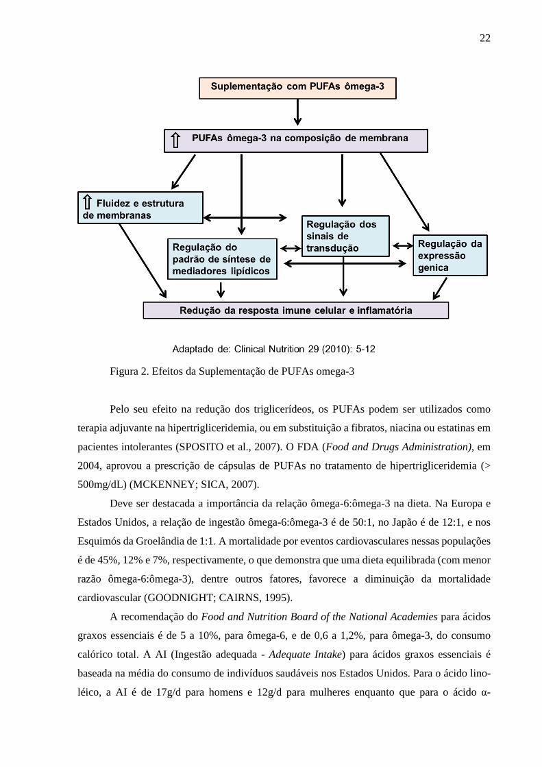
22
Figura 2. Efeitos da Suplementação de PUFAs omega-3
Pelo seu efeito na redução dos triglicerídeos, os PUFAs podem ser utilizados como
terapia adjuvante na hipertrigliceridemia, ou em substituição a fibratos, niacina ou estatinas em
pacientes intolerantes (SPOSITO et al., 2007). O FDA (Food and Drugs Administration), em
2004, aprovou a prescrição de cápsulas de PUFAs no tratamento de hipertrigliceridemia (>
500mg/dL) (MCKENNEY; SICA, 2007).
Deve ser destacada a importância da relação ômega-6:ômega-3 na dieta. Na Europa e
Estados Unidos, a relação de ingestão ômega-6:ômega-3 é de 50:1, no Japão é de 12:1, e nos
Esquimós da Groelândia de 1:1. A mortalidade por eventos cardiovasculares nessas populações
é de 45%, 12% e 7%, respectivamente, o que demonstra que uma dieta equilibrada (com menor
razão ômega-6:ômega-3), dentre outros fatores, favorece a diminuição da mortalidade
cardiovascular (GOODNIGHT; CAIRNS, 1995).
A recomendação do Food and Nutrition Board of the National Academies para ácidos
graxos essenciais é de 5 a 10%, para ômega-6, e de 0,6 a 1,2%, para ômega-3, do consumo
calórico total. A AI (Ingestão adequada - Adequate Intake) para ácidos graxos essenciais é
baseada na média do consumo de indivíduos saudáveis nos Estados Unidos. Para o ácido lino-
léico, a AI é de 17g/d para homens e 12g/d para mulheres enquanto que para o ácido α-

23
linolênico é 1,6 e 1,1g/d para homens e mulheres, respectivamente (INSTITUTE OF
MEDICINE, 2005). Segundo o FDA o consumo máximo de 3g/dia de EPA + DHA é
considerado seguro para população (HARRIS et al., 2009).
Os efeitos adversos relacionados com a suplementação com ômega-3 são em sua
maioria sintomas gastrintestinais sem gravidade como observado em estudos com mulheres
grávidas e pacientes com câncer, que relataram mudanças gastrointestinais (diarréia,
flatulência, refluxo gastroesofágico, náuseas), embora não tenham sido observadas diferenças
entre os grupos de tratamento e placebo (FABER et al., 2012; FREEMAN; SINHA, 2007).
Também pode ocorrer sangramentos, devido à atividade trombolítica da suplemntação com
ômega-3, que atua inibindo a síntese do tromboxano A2, responsável por induzir a agregação
plaquetária (DYERBERG; BANG, 1979; VILLANI et al., 2013). No entanto esta última
possibilidade é incomum, não havendo relato deste efeito em grandes séries de indivíduos
suplementados (TAVAZZI et al., 2008).

24
3 JUSTIFICATIVA
A doença de Chagas é uma doença negligenciada e representa um dos principais
problemas de saúde pública e uma importante causa de insuficiência cardíaca em áreas endêmi-
cas (BRAGA et al., 2006). A cardiopatia chagásica crônica é uma importante forma clínica da
doença de Chagas, devido ao seu impacto na morbidade e mortalidade, diminuição da qualidade
de vida e limitação da capacidade de trabalho, sendo mais precoce e grave no sexo masculino
(HABERLAND et al., 2013; RASSI; RASSI; LITTLE, 2000).
A cardiopatia chagásica crônica tem caráter progressivo e tem sua gênese associada à
resposta imunológica alterada com grande produção de citocinas inflamatórias e diminuição da
produção de citocinas anti-inflamatórias, o que está diretamente associado à progressão para
formas mais graves da doença (CHAVES et al., 2016). A modulação da resposta inflamatória
pode representar um potencial alvo terapêutico para evitar a progressão para a cardiomiopatia
chagásica e suas formas mais graves de apresentação.
Os ácidos graxos poli-insaturados ômega-3 apresentam atividade anti-inflamatória, pois
reduzem o conteúdo de ácido araquidônico nas membranas celulares, resultando na síntese de
eicosanóides que têm menor propriedade inflamatória do que aqueles derivados dos ácidos
graxos poli-insaturados ômega-6. Diante dos potenciais efeitos benéficos dos ácidos graxos
poli-insaturados ômega-3 em processos inflamatórios, dislipidema e doenças cardiovasculares,
ofertar suplementação de PUFAs ômega-3, em pacientes com CCC, pode se constituir em
instrumento terapêutico neste grupo de indivïduos.

25
4 OBJETIVO GERAL
Investigar o efeito da suplementação de ácidos graxos poli-insaturados ômega-3 no
perfil lipídico e no perfil de citocinas em pacientes portadores de cardiopatia chagásica crônica.
4.1 Objetivos específicos
Este estudo, realizado em pacientes com cardiopatia chagásica crônica, tem como
objetivos específicos:
• Avaliar o efeito da suplementação de ômega-3 no perfil sérico de citocinas,
lipidograma e glicemia de jejum antes e após a suplementação com ômega-3;
• Avaliar o efeito da suplementação com ômega-3 no estado nutricional;
• Avaliar se há diferença entre os grupos placebo e intervenção quanto ao
consumo alimentar de macronutrientes e ácidos graxos poli-insaturados.

26
5 MÉTODOS
5.1 Desenho de estudo
Ensaio clínico randomizado [1:1], duplo-cego, placebo-controlado, no qual pacientes
adultos portadores de cardiopatia chagásica crônica foram suplementados diariamente com
cápsulas de ômega-3 ou placebo, por 8 semanas, para avaliação da eficácia da suplementação
com ômega-3 nos perfis de citocinas e lipídico.
5.2 Seleção de pacientes (critérios de inclusão e exclusão)
Os pacientes foram recrutados, no período de maio a setembro de 2013, de uma
sequência de atendimentos no ambulatório de cardiologia do INI/Fiocruz, Rio de Janeiro/RJ,
Brasil, no período determinado para o rastreamento de inclusão até completar o tamanho
amostral estimado.
Foram selecionados pacientes maiores de 18 anos, de ambos os sexos, portadores de
cardiopatia chagásica crônica nos estágios B, C ou D, de acordo com a classificação do
Consenso Brasileiro de doença de Chagas.
Os seguintes critérios de exclusão foram aplicados: doença diarréica crônica, doença
inflamatória intestinal, uso de medicações anti-inflamatórias, gestantes e nutrizes, alcoolismo,
utilização de suplementação de vitaminas, minerais ou ômega-3 nos últimos trinta dias,
internação hospitalar no período do estudo e presença de outras cardiopatias, além da CCC.
5.3 Grupos intervenção e placebo
Cada participante recebeu 5 cápsulas por dia de ômega-3 (1,8g de EPA e 1,2 g de DHA)
ou óleo de milho por um período de 8 semanas. As cápsulas de ômega-3 e óleo de milho foram
fornecidas pela empresa Relthy Laboratórios Ltda., registro na ANVISA n º: 6.2582.0022.001-
1. As embalagens do tipo blíster e as cápsulas gelatinosas de ômega-3 e óleo de milho eram
idênticas, sendo diferenciadas pelo número do lote na embalagem, só o farmacêutico tinha
acesso a essa informação. A adesão ao tratamento do estudo foi verificada pela contabilidade
de comprimidos retornados de ômega-3/óleo de milho. Nas consultas das semanas 4 e 8 foram
contabilizados os comprimidos retornados de ômega-3/óleo de milho, calculando-se a relação

27
entre número de comprimidos retornados e de comprimidos que deveriam ter sido retornados
de acordo com a dose prescrita.
Em 20 de novembro de 2011 o Instituto Nacional de Infectologia Evandro Chagas e
Relthy Laboratórios Ltda assinaram um contrato de colaboração interinstitucional, que
constitui-se de um acordo técnico científico com o objetivo de facilitação de pesquisa, educação
e desenvolvimento tecnológico, onde foi firmado o acordo de fornecimento de cápsulas de
PUFAs ômega-3 (8000 cápsulas) e de óleo de milho (8000 cápsulas). Os pesquisadores do
INI/Fiocruz envolvidos no protocolo permaneceram com total autonomia na elaboração do
desenho e condução do estudo, bem como a responsabilidade de análise e publicação dos dados
ao final da pesquisa, sendo o Laboratório Relthy apenas o fornecedor das cápsulas, através de
doação.
5.4 Métodos e técnicas de avaliação
Foram coletados os seguintes dados nesse estudo: sexo, idade, renda, escolaridade,
classificação da disfunção cardíaca, medicamentos utilizados, recordatório de 24 horas
(HEBERT et al., 1999), avaliação antropométrica (altura, peso, índice de massa corporal,
perímetro da cintura, dobra cutânea tricipital e perímetro do braço), perfil lipídico (colesterol
total, triglicerídeos, HDL-colesterol, LDL-colesterol), glicemia de jejum, citocinas (IL-1β, IL-
4, IL-6, Interleucina-8 (IL-8), IL-10, IL-17α, Interleucina-33 (IL-33), TNF-α, IFN-γ) e adesão
ao tratamento (apêndice A).
A avaliação do consumo alimentar (recordatório de 24h) (apêndice B), os dados antro-
pométricos e avaliação de adesão ao tratamento ocorreram nos tempos 0, 4 e 8 semanas. Os
perfis lipídicos e de citocinas foram avaliados nos tempos 0 e 8 semanas.
5.4.1 Avaliação antropométrica
Os parâmetros antropométricos utilizados neste estudo foram peso, estatura, perímetro
braquial (PB), dobra cutânea triciptal (DCT), perímetro muscular do braço (PMB) e perímetros
da cintura e do quadril. A avaliação antropométrica foi realizada nos tempos 0, 4 e 8 semanas.
O peso atual (PA) foi aferido com balança de precisão de plataforma, da marca Filizola,
com capacidade de 150 kg com divisões de 10 gramas, com o valor expresso em quilogramas.
Para a obtenção da estatura foi utilizado o estadiômetro da própria balança antropométrica com
escala de 0,1cm. Os indivíduos foram colocados sobre a plataforma da balança, de costas para

28
o seu marcador, com os pés unidos, em posição ereta, com o olhar no horizonte. A leitura foi
realizada no 0,5 centímetro mais próximo, quando a haste horizontal da barra vertical da escala
da estatura encostou-se à cabeça do indivíduo.
O Índice de Massa Corporal, ou índice de Quetelet, é um índice simples de peso/estatura
utilizado pela Organização Mundial da Saúde para a classificação do estado nutricional,
conforme descrito no Quadro 1. É calculado a partir da seguinte equação:
IMC = PA (kg) / estatura2 (m2)
O perímetro braquial foi aferida no braço esquerdo no ponto médio entre o acrômio e o
olécrano, com o braço flexionado junto ao corpo, formando um ângulo de 90° e o valor da PB
foi obtido com o braço relaxado na lateral do corpo e a palma da mão voltada para a coxa, com
o cuidado para não comprimir partes moles. Foi utilizada fita celulóide inextensível de
graduação de 0,1 cm.
A dobra cutânea tricipital foi aferida a partir do local da medição do ponto médio obtido
na avaliação da CB. Após a marcação do ponto médio, foi pinçado o correspondente ao
subcutâneo, mensurando-o com plicômetro (Cescorf®). Foram realizadas três medidas,
assumindo-se como resultado final a média das três mensurações.
O perímetro muscular do braço foi obtido a partir dos valores da PB e da DCT,
utilizando-se a seguinte fórmula:
PMB = PB (cm) – [0.314 x DCT (mm)]
A aferição do perímetro da cintura foi realizada com o paciente em pé, utilizando-se fita
métrica inextensível horizontalmente ao redor da menor curvatura localizada no ponto médio
entre a última costela e a crista ilíaca, tendo sua classificação descrita no Quadro 5.
5.4.2 Consumo alimentar
O consumo alimentar foi avaliado utilizando-se o recordatório de 24 horas de dia típico,
que consiste em definir e quantificar todos os alimentos e bebidas ingeridos no dia anterior a
entrevista. O paciente foi entrevistado, nos tempos 0, 4 e 8 semanas, por um mesmo profissional
habilitado (nutricionista), sempre às terças-feiras, respondendo detalhadamente sobre todos os
alimentos e bebidas consumidos, informando o tamanho e o volume da porção consumida.

29
(apêndice B). Após transformação das porções alimentares consumidas em gramas de
alimentos, os nutrientes foram calculados utilizando-se o Diet Win plus 3.0® software package
(Brubins Ltda.). Foi calculada a média de calorias e nutrientes de três dias de avaliação.
5.4.3 Avaliação bioquímica e de marcadores inflamatórios
A coleta de material biológico (sangue) foi realizada no setor de coleta do INI/Fiocruz.
As análises da glicemia de jejum, do colesterol total (CT) (ALLAIN et al., 1974), dos
triglicerídeos (TGL) (FOSSATI; PRENCIPE, 1982) e lipoproteína de alta densidade-colesterol
(HDL-c) (HINO, 1996) foram realizadas pelo método colorimétrico enzimático, utilizando-se
kits Siemens (Tarrytown, NY, USA), no Laboratório de Bioquímica do INI/Fiocruz.
O LDL-colesterol foi avaliado pela equação de Friedewald et al. (1972)
(FRIEDEWALD; LEVY; FREDRICKSON, 1972) que é expressa como: lipoproteína de baixa
densidade-colesterol (LDL-c) (mg/dL) = colesterol total – (HDL-c – Triglicerídios) ÷ 5.
A lipoproteína de muito baixa densidade-colesterol (VLDL-c) foi calculado a partir da
fórmula descrita por Friedwald et al. (1972) (FRIEDEWALD; LEVY; FREDRICKSON, 1972),
válida somente se a concentração sérica de triglicerídios for menor do que 400 mg/dL:
VLDL-c (mg/dL) = Triglicerídios ÷ 5.
Foram analisados os seguintes marcadores inflamatórios: IL-1β, IL-4, IL-6, IL-8, IL-
10, IL-17α, IL-33, TNF-α, IFN-γ pelo método Elisa, utilizando-se kits eBioscience (San Diego,
CA, USA). Essas dosagens foram realizadas no Laboratório de Hanseníase do IOC/Fiocruz.
5.5 Cálculo amostral
A amostra calculada foi de 40 pacientes, inferida a partir de dados da literatura,
considerando uma prevalência estimada de 60% de consumo inadequado de ácidos graxos poli-
insaturados (SALVADOR et al., 2008), utilizando-se o pacote estatístico EpiCalc; versão 1.02.
Foram considerados os seguintes parâmetros: níveis de IFN-γ, basais e após suplementação
com ácidos graxos ômega-3; 3986 ± 738 pg/mL e 2922 ± 1275 pg/mL, respectivamente (KEW
et al., 2004), com poder de 0,8 e alfa de 5%.

30
5.6 Plano de análise
As análises estatísticas foram realizadas com o software Stata 13.0 (College Station,
TX, 2013) e o nível de significância foi definido em p ≤ 0,05 para todas as análises.
As características basais dos dois grupos foram expressas como média (desvio padrão)
para variáveis contínuas e percentuais para variáveis categóricas. O teste Skewness e Kurtosis
foi realizado para avaliar a normalidade dos dados que foram transformados em log em caso de
distribuição não normal. As mudanças longitudinais entre os grupos para todas as variáveis
foram examinadas usando modelo linear misto, que considera a atribuição do tratamento e
incluem todas as observações de cada participante, independentemente da perda ao
acompanhamento, caracterizando uma análise de intenção de tratar. O modelo longitudinal foi
ajustado para os valores basais da variável dependente em caso de desbalanceamento maior
para valores basais.
Houve alteração no plano de análise previsto no primeiro artigo, para a realizada
efetivamente no artigo subsequente. O objetivo geral do estudo não foi alterado, tendo sido esta
mudança apenas um ajuste ao pacote estatístico de maior familiaridade ao pesquisador, que
propôs ainda a utilização do modelo linear misto, corrigindo as perdas e realizando a análise
por intenção de tratar.
5.7 Considerações éticas
O estudo foi aprovado pelo Comitê de Ética em Pesquisa do Instituto de Pesquisa
Clínica Evandro Chagas (INI/Fiocruz) número CAAE-0037.0.009.000-10 (anexo A), e
registrado no ClinicalTrials.gov número NCT01863576. Todos os pacientes que aceitaram
participar do estudo assinaram um termo de consentimento livre e esclarecido (TCLE)
(apêndice C).
5.8 Limitações
O presente estudo tem algumas limitações. Embora o tamanho da amostra tenha sido
calculado para identificar diferenças estatísticas longitudinais para citocinas entre os grupos
PUFAs ômega-3 e placebo, o número pequeno de pacientes pode ter contribuído para as
diferenças observadas no perfil de citocinas entre os grupos na linha de base. No entanto,
incluímos valores basais em modelos longitudinais, a fim de minimizar a influência do

31
desequilíbrio de linha de base na análise estatística. O período de seguimento de curto prazo
não nos permitiu avaliar resultados clínicos, embora esse não fosse o escopo do presente estudo.

32
6 ARTIGOS
O protocolo e os resultados deste ensaio clínico foram apresentados em formato de
artigo.
6.1 Artigo 1: Apresenta o protocolo do estudo, descrevendo a metodologia desenvolvida
neste ensaio clínico.
Título: “Effects of omega-3 polyunsaturated fatty acid supplementation in patients with
chronic chagasic cardiomyopathy: study protocol for a randomized controlled trial”
Submetido a Trials em 5 de junho de 2013.
Aceito em 28 de outubro de 2013.
Publicado em 11 de novembro de 2013.
6.2 Artigo 2: Discute o efeito da suplementação de ômega-3 nos marcadores
inflamatórios e no perfil lipídico dos pacientes portadores de CCC. Foram apresentados ainda
dados da avaliação antropométrica e consumo alimentar.
Título: “Omega-3 supplementation on inflammatory markers in patients with chronic
Chagas cardiomyopathy: a randomized clinical study”
Submetido a Nutrition Journal em 10 de março de 2017.
Aceito em 05 de junho de 2017.
Publicado em 09 de junho de 2017.

33
6.1 Artigo 1
Study protocol
Effects of omega-3 polyunsaturated fatty acid supplementation in patients with chronic
chagasic cardiomyopathy: study protocol for a randomized controlled trial.
Paula S. Silva1,*, Gilberto Marcelo Sperandio da Silva2, Andréa P. de Souza3, Claudia S. A.
Cardoso1, Cristiane A. Fonseca1, Patricia D. Brito1, Roberto M. Saraiva2, Pedro E. A. Brasil2,
Roberta O. Pinheiro4, Alejandro M. Hasslocher-Moreno2, Sérgio S. Xavier2 and Andréa S.
Sousa2
1 Serviço de Nutrição, Instituto de Pesquisa Clínica Evandro Chagas, Fundação Oswaldo Cruz,
Av. Brasil 4365, Manguinhos, Rio de Janeiro, Brasil
2 Laboratório de Pesquisa Clínica em Doença de Chagas, Instituto de Pesquisa Clínica Evandro
Chagas, Fundação Oswaldo Cruz, Av. Brasil 4365, Manguinhos, Rio de Janeiro, Brasil
3 Laboratório de Inovações em Terapias, Ensino e Bioprodutos, Instituto Oswaldo Cruz,
Fundação Oswaldo Cruz, Av. Brasil 4365, Manguinhos, Rio de Janeiro, Brasil
4 Laboratório de Hanseníase, Instituto Oswaldo Cruz, Fundação Oswaldo Cruz, Av. Brasil 4365,
Manguinhos, Rio de Janeiro, Brasil
* Corresponding author:
Paula Simplício da Silva
Serviço de Nutrição, Instituto de Pesquisa Clínica Evandro Chagas, Fundação Oswaldo Cruz,
Av. Brasil 4365, Manguinhos, Rio de Janeiro, Brasil
Tel (fax): 55-21-3865-9602
E-mail: [email protected]

34
Abstract
Background: Chronic chagasic cardiomyopathy is an inflammatory disease that occurs in
approximately 30% of patients infected by the protozoan Trypanosoma cruzi, and it has a profile
of high morbidity and mortality. The worst prognosis and the progression of this
cardiomyopathy are associated with an exacerbated immune response and the production of
pro-inflammatory cytokines, which also occur in other cardiomyopathies. Some nutrients,
including omega-3 polyunsaturated fatty acids (PUFAs), promote the inhibition and/or
stimulation of cytokine production. The objective of this trial is to study the effects of omega-
3 PUFAs supplementation on the inflammatory response and lipid profile in patients with
chronic chagasic cardiomyopathy.
Methods/Design: This is a parallel, randomized, placebo-controlled, double-blind clinical trial
with 40 patients that will be conducted at a reference unit for Chagas disease patients, where
the patients will be selected. The study will include patients with chronic chagasic
cardiomyopathy who are 18 years of age or older. The exclusion criteria are (a) ongoing
diarrheal disease, (b) inflammatory bowel disease, (c) diabetes or other endocrine disease, (d)
use of fibrates, niacin, or statins, (e) use of anti-inflammatory drugs, (f) pregnant and lactating
women, (g) use of vitamin, mineral, or omega-3 supplementation during the previous 30 days,
(h) hospital admission during the study, and (i) other associated cardiomyopathies. The
intervention will be treatment with omega-3 PUFAs at a dose of 3 g/day for 8 weeks, compared
to placebo (corn oil). The primary endpoints will be the concentrations of inflammatory markers
(IL-1, IL-2, IL-4, IL-6, IL-10, TNF-α, IFN-γ, and TGF-β). Secondary endpoints will be the
fasting glucose, lipid, and anthropometric profiles. For statistical analysis, we plan to run either
a t-test or Wilcoxon test (numerical variables) and Pearson’s chi-squared or Fisher’s exact test
(categorical data), as appropriate.
Discussion: Evidence suggests that the anti-inflammatory action of omega-3 PUFAs may have
beneficial effects on chronic chagasic cardiomyopathy, as shown for other cardiomyopathies,
due to improved control of the inflammatory response. At the end of the study, we predict that
patients will have lower inflammatory markers and an improved metabolic and anthropometric
profile.
Trial registration: Current Controlled Trials NCT01863576.
Key words: Chagas disease, Trypanosoma cruzi, chronic chagasic cardiomyopathy, omega-3,
cytokines, lipid profile, nutritional assessment.

35
Background
Chagas disease is endemic in Latin American countries, where approximately 8 to 10
million people are infected with Trypanosoma cruzi [1]. In Brazil, the number of infected
people is approximately 1.9 million, or 1% of the Brazilian population [2].
The etiologic diagnosis of Chagas' disease is performed by detection of the parasite by
parasitological methods (direct or indirect) and confirmed by two different serological
techniques indirect immunofluorescence and enzyme-linked immunosorbent assay (ELISA)
[3].
Chagas disease has two distinct clinical phases: acute and chronic [3]. The chronic phase
has three forms: indeterminate, cardiac involvement, and/or digestive involvement [3–5]. In
most cases, acute disease is not recognized at onset, and the individual remains asymptomatic
with no evidence of damage to any organ but with reactive serology for Chagas disease and low
parasitemia during the chronic phase [4]. After two or more decades, 20 to 30% of the patients
progress to the cardiac stage of the disease, when the majority of the deaths and severe
complications related to the disease occur [6–8]. Therefore, chronic chagasic cardiomyopathy
(CCC) is an important clinical form of Chagas disease due to its impact on morbidity and its
high mortality, worsening of the quality of life, and limitation of the ability to work. It is
characterized by a myocarditis with multifocal mononuclear inflammatory infiltrates, varying
degrees of fibrosis, constant low-grade tissue parasitism, and low or undetectable parasitemia
[9] and it occurs earlier and more severely in males [8, 10]. Patients in the chronic cardiac
phase may manifest heart failure, ventricular and atrial arrhythmias, atrioventricular blocks,
thromboembolism, stroke, and sudden death [2, 7, 11], which together carry a high economic
and mortality burden.
Once settled, CCC is progressive and tends worsen due to overlap with inflammation,
cell destruction, and fibrosis. Some hypotheses have been proposed to explain the nature of the
cardiac inflammatory reaction, mainly persistent heart immune response and reaction to the
parasite [12]. Cytokines are important mediators in the maintenance of the inflammatory
process, and they can stimulate or inhibit the immune response [13]. Among the several
cytokines studied in T. cruzi infection, interferon-γ (IFN-γ) has been associated with host
resistance to infection in both experimental models and humans [14, 15]. Transforming growth
factor-β (TGF-β) production is increased in T. cruzi infection, and this increase is most likely

36
related to the fibrotic process observed in CCC [16]. These results suggest that cytokines play
essential roles in controlling CCC morbidity [17–19].
Some nutrients are associated with the inhibition and/or stimulation of cytokine
production, including polyunsaturated fatty acids (PUFAs). Omega-3 PUFAs (linolenic acid,
eicosapentaenoic acid, and docosahexaenoic acid) are found in vegetables (soy, canola, linseed)
and cold-water fish (mackerel, sardines, salmon, herring). PUFAs are precursors to the
biosynthesis of several important metabolites, especially eicosanoids (prostaglandins,
leukotrienes, thromboxanes, lipoxins, and others), that are synthesized from arachidonic acid
[20]. Omega-6 PUFAs (such as linoleic acid - 18:2n-6) and omega-3 PUFAs (such as linolenic
acid - 18:3n-3) are fundamental to the body and have important roles in cell metabolism,
including influencing membrane fluidity, promoting the release of cytokines, and acting as
adhesion molecules on macrophages. Thus, changes in their synthesis and metabolism may be
associated with endothelial and hemodynamic changes that contribute to the increase in
cardiovascular morbidity and mortality [21].
The fatty acid composition of inflammatory cells can be modified by increasing the
intake of marine n-3 fatty acids, which leads to a higher content of eicosapentaenoic acid (EPA)
and docosahexaenoic acid (DHA). A number of cell culture studies have reported that both EPA
and DHA decrease the activation of the transcription factor nuclear factor kappa B (NF-κB) in
response to inflammatory stimuli, such as lipopolysaccharide and inflammatory cytokines [22].
Omega-3 PUFAs reduce arachidonic acid content in cell membranes, resulting in the synthesis
of eicosanoids that have weaker inflammatory properties than the eicosanoids derived from
omega-6 PUFAs [23].
Omega-3 supplementation is safe and well tolerated in infectious disease as demon-
strated in HIV-infected patients treated with antiretroviral therapy [24, 25], and on host re-
sistance to infection from Plasmodium berghei or Plasmodium falciparum, the causative agents
of malaria [26], which is a parasitic infection as well as Chagas disease.
Several studies involving supplementation of the diet with marine n-3 PUFAs in healthy
human volunteers have demonstrated decreased production of tumor necrosis factor-α (TNF-
α), interleukin-1β (IL-1β), IL-2, and IL-6 [22, 23, 27]. Omega-3 PUFAs supplementation for 8
weeks has been associated with an increase of peripheral mononuclear cells with high
concentrations of EPA and DHA, which have an immunomodulating effect [28]. In healthy
adults, fish oil supplementation at 5 mL/day for 8 weeks reduced the production of IL-6 but did
not change the production of IL-10, TNF-α, or INF-γ [29]. Long-chain omega-3 PUFAs (EPA
and DHA) plasma levels are inversely associated with the risk of sudden death [30] and may

37
reduce the risk of ischemic heart disease [31]. EPA and DHA are also involved in the reduction
of plasma very low-density lipoprotein cholesterol (VLDL-c) and triglycerides [32].
A study conducted by the Italian group GISSI-HF at 326 cardiology sites and 31 internal
medicine sites selected 6,975 patients with heart failure diagnosis to receive one daily capsule
containing 1 g of omega-3 PUFAs (n=3,494) or placebo (n=3,481) [33]. The total mortality of
the supplemented group was 27%, and that of the placebo group was 29% (adjusted HR 0.91;
95.5% confidence interval (CI) 0.833–0.998; p=0.041). Hospital admissions due to
cardiovascular events were 57% in the omega-3 PUFAs group vs. 59% in the placebo group
(adjusted HR 0.92; 99% CI 0.849–0.999; p=0.009). The advantages of n-3 PUFAs, which were
reduced fatal events and hospital admissions from cardiovascular causes, suggest that they have
an effect on the mechanisms leading to progression of heart failure [33].
Supplementation with 1 g/day of omega-3 PUFAs in capsules can decrease
cardiovascular events by 10% (death, myocardial infarction, stroke) in coronary arterial disease
patients [34]. Some studies that assessed the effect of omega-3 supplementation on the immune
response used EPA and/or DHA at 1 g to 9 g per day, similar to the ingestion levels of Greenland
Eskimos: 6 to 14 g/day, corresponding to 2.7% to 6.3% of their total calorie intake. The highest
doses can reduce arachidonic acid in peripheral mononuclear cells [35].
Studies conducted in heart failure patients have not shown adverse events after omega-
3 supplementation [33, 36]. In contrast, studies in pregnant women and cancer patients have
reported gastrointestinal changes (diarrhea, flatulence, gastroesophageal reflux, nausea),
although no differences between the treatment and placebo groups were noted [37, 38].
Few studies have evaluated the nutritional status of Chagas’ patients. Three studies have
evaluated patients with the digestive form of the disease. In two of these studies [39, 40], there
was a high prevalence of malnutrition (> 60%). The other study evaluated 10 patients diagnosed
with megaesophagus, and only one had malnutrition. The others were eutrophic or overweight
[41]. A study performed in São Paulo, Brazil, assessed the nutritional state of 66 Chagas’
patients and found a high prevalence of overweight and obesity (94%) and large waist
circumference (55%). The indeterminate form of the disease was the most prevalent and
constituted 71% of cases [42]. There is no literature on the nutritional assessment of CCC
patients.
Supplementation with omega-3 PUFAs may lead to increased body weight in patients
with heart cachexia, as reported by a study that assessed 14 patients with severe heart failure
supplemented with 8 g/day omega-3 PUFAs or placebo [43].

38
Although several studies have shown the beneficial effects of polyunsaturated fatty
acids on inflammatory processes, dyslipidemia, and cardiovascular diseases, there are no
reports about food intake and PUFAs supplementation in CCC patients. Thus, the objective of
this study is to assess the effects of omega-3 PUFAs supplementation on the lipid profile and
the pro-inflammatory and anti-inflammatory cytokine profiles in CCC patients.
Methods/Design
Study Design
This is a parallel, double-blind, placebo-controlled clinical trial with balanced
randomization [1:1].
Settings
The proposed clinical trial will be held at a single site, the Evandro Chagas Clinical
Research Institute (IPEC), which is one of the technical scientific units of the Oswaldo Cruz
Foundation (Fiocruz). IPEC is a 93-year-old institution that is fully dedicated to clinical
research on infectious diseases, including Chagas disease. IPEC is a major reference center for
Chagas disease research, care, and training.
Participants (inclusion and exclusion criteria)
Patients will be recruited from the cardiology ambulatory service at IPEC/Fiocruz, Rio
de Janeiro, Brazil, during the period determined for screening until the minimum estimated
sample size is reached. All patients had a previous epidemiologic history of Chagas disease
confirmed with at least two Chagas serology tests (indirect immunofluorescence and ELISA),
and clinical evaluations, electrocardiographic and echocardiographic data obtained recently.
Selected patients will be older than 18 years, males and females with CCC at stage B (no heart
failure symptoms but with segmental or global left ventricular systolic dysfunction by
echocardiogram), stage C (symptomatic heart failure), or stage D (end-stage heart failure),
according to the current Brazilian Chagas disease Consensus [3]. The following exclusion
criteria will be applied: diarrheal disease; inflammatory bowel syndrome; diagnosis of diabetes
or other endocrine pathologies; use of fibrates, niacin, or statins; use of anti-inflammatory
drugs; pregnant and lactating women; vitamin mineral or omega-3 supplementation during the
previous 30 days; hospital admission during the study; and the presence of cardiomyopathies
other than CCC.

39
Interventions: omega-3 vs. corn oil placebo
Each participant will receive five omega-3 capsules per day (1.8 g EPA and 1.2 g DHA)
or placebo for 8 weeks (Figure 1). The omega-3 and corn oil capsules will be provided by
Relthy Laboratórios Ltda. (Brazilian Health Surveillance Agency (Agência Nacional de
Vigilância Sanitária – ANVISA) registration: 6.2582.0022.001-1).
Endpoints
The primary endpoint of this study will be the cytokine profile. IL-1, IL-2, IL-4, IL-6,
IL-10, TNF-α, IFN-γ, and TGF-β will be measured in the serum of patients using specific
sandwich enzyme-linked immunosorbent assays. Capture and detection antibodies will be
obtained from eBioscience (San Diego, CA, USA). The tests will be conducted according to
the manufacturer's instructions and performed in triplicate.
The first secondary endpoint will be the lipid profile. Total cholesterol, triglycerides,
and high-density lipoprotein cholesterol (HDL-c) will be measured with enzymatic-
colorimetric assays using Siemens reagents on a Siemens Dimension RXL chemistry analyzer
(Siemens Healthcare Diagnostics, Tarrytown, NY, USA). Low-density lipoprotein cholesterol
(LDL-c) and VLDL-c will be calculated according to the Friedewald equation [44].
Another secondary endpoint of this trial will be anthropometric measures. The
anthropometric assessment will consist of body mass index (BMI), waist circumference,
tricipital skinfold thickness, and arm circumference [45, 46]. BMI will be determined using the
standard formula: BMI=weight/height2 [47].
Procedures, follow-up, and data collection.
After patients are selected by cardiologists, they will be seen by study nutritionists, who
will explain the study procedures to the patients and administer the free and informed consent
form. The patients who agree to participate in the study will sign the consent form and undergo
the initial assessment.
The following data will be collected and evaluated in the study: sociodemographic data
(age, sex, ethnicity/race, education, and domicile), clinical data (functional class and vital
signs), alcoholism, smoking, prescription drugs, 3-day food record [48], 24-hour recall [49],
anthropometric assessment (height, weight, BMI, waist circumference, tricipital skinfold
thickness, and arm circumference), lipid profile (total cholesterol, triglycerides, HDL-c, LDL-
c, and VLDL-c), and cytokines (IL-1, IL-2, IL-4, IL-6, IL-10, TNF-α, IFN-γ, and TGF-β).

40
Clinical, nutritional, and anthropometric assessments will take place immediately before
starting the intervention and after 4 and 8 weeks during the study; lipid profile and cytokines
will be evaluated before the intervention and at the end of 8 weeks. Each patient will be
followed for 8 weeks.
Compliance with study treatment will be assessed by the 3-day treatment recall and the
number of omega-3/placebo capsules returned. At every visit, the patients will be asked about
the number of doses of the prescribed treatment that they did not take during the previous 3
days, and the ratio between the number of doses taken and the number prescribed will be
calculated. The capsules will be dispensed at the beginning of the study and after 4 weeks, and
the returned capsules will be verified at weeks 4 and 8.
The dietary omega-3 PUFAs intake will be followed-up during the study (first visit and
weeks 4 and 8) by analyzing the 24-hour recall and 3-day food record, which are considered
indirect methods of analyzing the diet, current dietary standards of a certain population, and
their development over time [50].
Patients may be excluded from the study in case of complications during follow-up,
such as significant changes in intestinal transit, use of anti-inflammatory drugs, and hospital
admissions. Other complications will be assessed by the study team. All endpoints will be
collected at time 0 and 8 weeks after omega-3/placebo supplementation starts.
Safety monitoring
Patients will be followed up by a multidisciplinary team at three time points: study start,
week 4, and week 8. Possible complications will be assessed by the study cardiologist and
pharmacist. The safety assessment will consist of monitoring adverse events, such as changes
in the digestive tract, physical exams, and laboratory tests. Events, onset, and duration will be
reported to the cardiologist, who will determine if it is necessary to suspend supplement use.
Regular intake of omega-3 PUFAs is not a risk to health. According to the United States Food
and Drug Administration (FDA), the maximum intake of 3 g/day EPA + DHA is considered
safe [51].
Randomization and concealment
After the screening phase, patients will be randomly assigned to one of two groups of
20 patients each: intervention group or control group. Assignment to either group will be
performed by a computer-generated 1:1 randomization. This assignment will occur within

41
randomly ordered blocks of size 4 or 6. This procedure will be conducted using R-project
software with package “blockrand” [52].
The allocation sequence will be concealed from the researchers, who will enroll and
assess participants in sequentially numbered, opaque, sealed, stapled envelopes. To prevent
subversion of the allocation sequence, the name and date of birth of the participant will be
written on the envelope before the envelope is opened. The information in the envelope will be
transferred onto the allocation card inside the envelope. Corresponding envelopes will be
opened only after the enrolled participants have completed all baseline assessments and at the
time of allocation to the intervention. These procedures will be carried out by a professional
without access to the baseline evaluation results. The blister packaging and gel capsules of
omega-3 PUFAs and placebo will be identical. They will be identified by the lot number on the
package, which will be retained in the pharmacy, and only the blisters will be provided to
patients. The pharmacist responsible for distribution and compliance assessment will be the
only person who can access this information.
Blinding
The nutritionists and physician that will perform the initial evaluation before
randomization, the monthly evaluations and anthropometric profile, and the final evaluation of
the health care questionnaires after the end of the patient follow-up will be blinded to the
patients’ assignments.
Statistical methods
The EpiData [53] and Statistical Package for Social Sciences [54] software will be used
for data entry and analysis, respectively. For categorical data, we will use Pearson’s chi-squared
or Fisher’s exact test to verify the association between variables in the two groups (omega-3
and placebo). Statistical analysis will be performed by descriptive analysis, with numerical
variables expressed as the mean and standard deviation (SD) or median and inter-quartile range.
The Kolmogorov-Smirnov test will be used to test the normality of the sample distribution.
Student’s t-test will be used in cases with a normal distribution to compare means between the
two groups. The Wilcoxon test will be used in cases with a non-normal distribution. P values
<0.05 will indicate a significant association in all of the statistical tests used.

42
Minimum sample size estimation
The minimum estimated sample size is 40 patients according to previous data,
considering a prevalence of 60% of inadequate PUFAs intake [55]. The following parameters
were considered: baseline IFN-γ = 3986 ± 738 pg/mL, IFN-γ after n-3 PUFAs supplementation
= 2922 ± 1275 pg/mL [56], power = 0.8, and α = 5%. We used the R-project software with
package “Epicalc” version 1.02 [57] to obtain this estimate.
Ethical issues
The study was approved by the Research Ethics committee of the Evandro Chagas’
Clinical Research Institute (IPEC/Fiocruz), registration number CAAE-0037.0.009.000-10
[58]. It is registered at ClinicalTrials.gov, number NCT01863576. All study supplies and data
collected will be used exclusively in this study, and its results will be made public, whether they
are favorable or not.
Discussion
Omega-3 PUFAs intake is associated with beneficial health effects. Omega-3 PUFAs
are helpful in the prevention and treatment of pathologies such as inflammatory diseases,
hypertension, hypertriglyceridemia, metabolic syndrome, and heart diseases. An Italian study
of heart failure patients showed a reduction in mortality and hospital admissions due to
cardiovascular events, suggesting beneficial effects of omega-3 PUFAs on the progression of
heart disease [33].
Omega-3 administration is effective at treating hypertriglyceridemia. According to
clinical trials, supplementation with 2 to 4 g of EPA/DHA per day may reduce triglyceride
levels by 25% to 30% [59] by decreasing the liver synthesis of VLDL-c [34]. Omega-3
supplementation for Chronic chagasic cardiomyopathy may be similar to other
cardiomyopathies and promote reductions in triglyceride and VLDL-c levels.
Omega-3 has beneficial effects against heart cachexia after 18 weeks of supplementa-
tion [43]. Women with polycystic ovarian syndrome and BMI between 25 and 40 kg/m2, how-
ever, exhibited no significant changes in BMI after supplementation for 8 weeks [60]. The pe-
riod of treatment in this study may not be long enough to observe significant anthropometric
changes after omega-3 administration with no other associated instruction or intervention.
The action of omega-3 PUFAs is most likely related to their anti-inflammatory effects,
which are not yet completely understood; however, it is known that omega-3 PUFAs

43
supplementation reduces arachidonic acid content in cell membranes, resulting in the synthesis
of eicosanoids that are less inflammatory than the ones derived from omega-6 PUFAs [23].
Omega-3 PUFAs can also decrease activation of NF-κB in response to inflammatory stimuli,
such as lipopolysaccharide and inflammatory cytokines [22]. A meta-analysis of the effects of
fish oil supplementation on the inflammatory markers of patients with chronic heart failure
showed that circulating TNF-α, IL-1, and IL-6 (pro-inflammatory cytokines) decreased after a
supplementation period of 3 to 12 months with an EPA and DHA dose of 600 to 5540 mg/day.
These data suggest that supplementation with higher doses of omega-3 PUFAs or longer follow-
up times are associated with more substantially decreased cytokines [61], which could benefit
CCC patients.
A clinical trial evaluated 3-month omega-3 PUFAs supplementation in patients with
heart failure. Patients were divided into three groups: placebo, 1 g/day omega-3 PUFAs, and 4
g/day omega-3 PUFAs. Only the 4 g/day group had an improvement in ventricular function
with an increase in the ejection fraction and endothelial function and a reduction in IL-6 [36].
We will use 3 g/day omega-3 PUFAs in our study based on recent results suggesting higher
efficacy when compared to the usual 1 g dose, in addition to the fact that 3 g/day is considered
safe by the FDA [51]. According to American College of Cardiology and American Heart
Association (ACC/AHA) in a recent Heart Failure Guideline, omega-3 supplementation is
reasonable to use as adjunctive therapy in heart failure patients (Class of Recommendation IIa,
Level of Evidence B ) [62].
The worse prognosis and progression of CCC are related to increased immune response
with the production of pro-inflammatory cytokines and the inhibition of anti-inflammatory cy-
tokines, which also occur in other cardiomyopathies [63, 64]. In Chagas disease, heart tissue is
an important target of T. cruzi infection, and substantial amounts of pro-inflammatory cyto-
kines, chemokines, and enzymes, including inducible nitric oxide synthase and metalloprotein-
ases, are produced, resulting in inflammation and cardiac remodeling in response to parasite
infection [65]. Higher levels of IFN-γ and TNF-α are associated with lower levels of IL-4 and
IL-10, in both cardiac and more severe forms of Chagas disease, and high levels of IL-10 or
moderate levels of IFN-γ are associated with the indeterminate form [15, 66]. Omega-3 and
omega-6 PUFAs intake may lead to an increase in IL-10 production, as shown in an experi-
mental study in Wistar rats supplemented with ethyl-eicosapentaenoic acid and ethyl-γ-lino-
lenic acid compared to a group supplemented with palm oil [67]. Thus, omega-3 PUFAs sup-
plementation in CCC patients may lead to increased IL-4 and IL-10, reducing the inflammatory
effect on heart tissue.

44
Omega-3 PUFAs supplementation, due to its anti-inflammatory effects, is expected to
decrease the serum levels of pro-inflammatory cytokines, which are associated with the
proliferation of parasites and the fibrotic process characteristic of Chagas’ myocarditis. We thus
suppose that the benefits to CCC patients with regard to the immunological profile could
translate into a less severe progression of cardiomyopathy, with subsequent reduction in
morbidity, in addition to secondary gains in the lipid profile.
Trial Status
This study is currently recruiting patients.
Competing Interests
The authors declare that they have no competing interests.
Acknowledgements
This study is funded by Evandro Chagas Clinical Research Institute.
Authors Contributions
PSS, APS and ASS conceived the study. GMSS, AMHM, PEAAB, RMS, SSS, PSS,
APS and ASS participated in the study design. PSS, CSAC, CAF and PDB will recruit,
select, and collect clinical data of the patients. PSS, ROP and GMSS will randomize
patients into the two arms of the protocol, deliver the proposed intervention, and
collect data. PSS, PEAAB, GMMS and ASS will perform statistical analysis. APS is
the study coordinator and ASS the principal investigator. PSS, GMSS, PEAAB,
AMHM and ASS helped to draft the manuscript. All authors read and approved the
final manuscript.

45
References
1. Rassi A, Rassi A, Marcondes de Rezende J: American Trypanosomiasis (Chagas Disease).
Infectious Disease Clinics of North America 2012, 26:275–291.
2. Rassi A, Rassi A, Marin-Neto JA: Chagas disease. The Lancet 2010, 375:1388–1402.
3. [Brazilian Consensus on Chagas disease]. Rev. Soc. Bras. Med. Trop. 2005, 38 Suppl 3:7–
29.
4. Dias JC: The indeterminate form of human chronic Chagas’ disease A clinical
epidemiological review. Rev. Soc. Bras. Med. Trop. 1989, 22:147–156.
5. Biolo A, Ribeiro AL, Clausell N: Chagas cardiomyopathy--where do we stand after a hundred
years? Prog Cardiovasc Dis 2010, 52:300–316.
6. Mady C, Nacruth R: Natural history of chronic Chagas’ heart disease: prognosis factors. Sao
Paulo Med J 1995, 113:791–796.
7. Marin Neto JA, Simões MV, Sarabanda AV: Chagas’ heart disease. Arq. Bras. Cardiol. 1999,
72:247–280.
8. Rassi A Jr, Rassi A, Little WC: Chagas’ heart disease. Clin Cardiol 2000, 23:883–889.
9. Ribeiro AL, Nunes MP, Teixeira MM, Rocha MOC: Diagnosis and management of Chagas
disease and cardiomyopathy. Nature Reviews Cardiology 2012, 9:576–589.
10. Castro C, Prata A, Macêdo V: A folow-up period of 13 years prospective study in 190
chagasic patients of Mambaí, Goiás, State, Brazil. Revista da Sociedade Brasileira de Medicina
Tropical 2001, 34:309–318.
11. Rocha MOC, Ribeiro ALP, Teixeira MM: Clinical management of chronic Chagas
cardiomyopathy. Front. Biosci. 2003, 8:e44–54.
12. Punukollu G, Gowda RM, Khan IA, Navarro VS, Vasavada BC: Clinical aspects of the
Chagas’ heart disease. Int. J. Cardiol. 2007, 115:279–283.
13. Sher A, Gazzinelli RT, Oswald IP, Clerici M, Kullberg M, Pearce EJ, Berzofsky JA,
Mosmann TR, James SL, Morse HC 3rd: Role of T-cell derived cytokines in the downregulation

46
of immune responses in parasitic and retroviral infection. Immunol. Rev. 1992, 127:183–204.
14. Bahia-Oliveira LM, Gomes JA, Rocha MO, Moreira MC, Lemos EM, Luz ZM, Pereira ME,
Coffman RL, Dias JC, Cançado JR, Gazzinelli G, Corrêa-Oliveira R: IFN-gamma in human
Chagas’ disease: protection or pathology? Braz. J. Med. Biol. Res. 1998, 31:127–131.
15. Gomes JAS, Bahia-Oliveira LMG, Rocha MOC, Martins-Filho OA, Gazzinelli G, Correa-
Oliveira R: Evidence that development of severe cardiomyopathy in human Chagas’ disease is
due to a Th1-specific immune response. Infect. Immun. 2003, 71:1185–1193.
16. Araújo-Jorge TC, Waghabi MC, Soeiro M de NC, Keramidas M, Bailly S, Feige J-J: Pivotal
role for TGF-beta in infectious heart disease: The case of Trypanosoma cruzi infection and
consequent Chagasic myocardiopathy. Cytokine Growth Factor Rev. 2008, 19:405–413.
17. Pérez-Fuentes R, Guégan J-F, Barnabé C, López-Colombo A, Salgado-Rosas H, Torres-
Rasgado E, Briones B, Romero-Díaz M, Ramos-Jiménez J, Sánchez-Guillén M del C: Severity
of chronic Chagas disease is associated with cytokine/antioxidant imbalance in chronically
infected individuals. Int. J. Parasitol. 2003, 33:293–299.
18. Dutra WO, Rocha MOC, Teixeira MM: The clinical immunology of human Chagas disease.
Trends Parasitol. 2005, 21:581–587.
19. Cunha-Neto E, Nogueira LG, Teixeira PC, Ramasawmy R, Drigo SA, Goldberg AC,
Fonseca SG, Bilate AM, Kalil J: Immunological and non-immunological effects of cytokines
and chemokines in the pathogenesis of chronic Chagas disease cardiomyopathy. Mem. Inst.
Oswaldo Cruz 2009, 104 Suppl 1:252–258.
20. Perini JÂDL, Stevanato FB, Sargi SC, Visentainer JEL, Dalalio MMDO, Matshushita M,
Souza NED, Visentainer JV: Omega-3 and omega-6 polyunsaturated fatty acids: metabolism in
mammals and immune response. Revista de Nutrição 2010, 23:1075–1086.
21. Simopoulos AP: Essential fatty acids in health and chronic disease. Am. J. Clin. Nutr. 1999,
70:560S–569S.
22. Calder PC: Omega-3 polyunsaturated fatty acids and inflammatory processes: nutrition or
pharmacology? Br J Clin Pharmacol 2013, 75:645–662.
23. Giugliano D, Ceriello A, Esposito K: The effects of diet on inflammation: emphasis on the

47
metabolic syndrome. J. Am. Coll. Cardiol. 2006, 48:677–685.
24. De Truchis P, Kirstetter M, Perier A, Meunier C, Zucman D, Force G, Doll J, Katlama C,
Rozenbaum W, Masson H, Gardette J, Melchior J-C: Reduction in Triglyceride Level With N-
3 Polyunsaturated Fatty Acids in HIV-Infected Patients Taking Potent Antiretroviral Therapy:
A Randomized Prospective Study. JAIDS Journal of Acquired Immune Deficiency Syndromes
2007, 44:278–285.
25. Thusgaard M, Christensen JH, Mørn B, Andersen TS, Vige R, Arildsen H, Schmidt EB,
Nielsen H: Effect of fish oil (n-3 polyunsaturated fatty acids) on plasma lipids, lipoproteins and
inflammatory markers in HIV-infected patients treated with antiretroviral therapy: a
randomized, double-blind, placebo-controlled study. Scand. J. Infect. Dis. 2009, 41:760–766.
26. Anderson M, Fritsche KL: (n-3) Fatty acids and infectious disease resistance. J. Nutr. 2002,
132:3566–3576.
27. Trebble TM, Wootton SA, Miles EA, Mullee M, Arden NK, Ballinger AB, Stroud MA,
Burdge GC, Calder PC: Prostaglandin E2 production and T cell function after fish-oil
supplementation: response to antioxidant cosupplementation. Am. J. Clin. Nutr. 2003, 78:376–
382.
28. Damsgaard CT, Frøkiaer H, Lauritzen L: The effects of fish oil and high or low linoleic acid
intake on fatty acid composition of human peripheral blood mononuclear cells. Br. J. Nutr. 2008,
99:147–154.
29. Damsgaard CT, Lauritzen L, Calder PC, Kjaer TR, Frøkiaer H: Reduced ex vivo interleukin-
6 production by dietary fish oil is not modified by linoleic acid intake in healthy men. J. Nutr.
2009, 139:1410–1414.
30. Albert CM, Campos H, Stampfer MJ, Ridker PM, Manson JE, Willett WC, Ma J: Blood
levels of long-chain n-3 fatty acids and the risk of sudden death. N. Engl. J. Med. 2002,
346:1113–1118.
31. Lemaitre RN, King IB, Mozaffarian D, Kuller LH, Tracy RP, Siscovick DS: n-3
Polyunsaturated fatty acids, fatal ischemic heart disease, and nonfatal myocardial infarction in
older adults: the Cardiovascular Health Study. Am. J. Clin. Nutr. 2003, 77:319–325.

48
32. Frenoux JM, Prost ED, Belleville JL, Prost JL: A polyunsaturated fatty acid diet lowers
blood pressure and improves antioxidant status in spontaneously hypertensive rats. J. Nutr.
2001, 131:39–45.
33. Tavazzi L, Maggioni AP, Marchioli R, Barlera S, Franzosi MG, Latini R, Lucci D, Nicolosi
GL, Porcu M, Tognoni G: Effect of n-3 polyunsaturated fatty acids in patients with chronic
heart failure (the GISSI-HF trial): a randomised, double-blind, placebo-controlled trial. Lancet
2008, 372:1223–1230.
34. Sposito AC, Caramelli B, Fonseca FAH, Bertolami MC, Afiune Neto A, Souza AD,
Lottenberg AMP, Chacra AP, Faludi AA, Loures-Vale AA, Carvalho AC, Duncan B, Gelonese
B, Polanczyk C, Rodrigues Sobrinho CRM, Scherr C, Karla C, Armaganijan D, Moriguchi E,
Saraiva F, Pichetti G, Xavier HT, Chaves H, Borges JL, Diament J, Guimarães JI, Nicolau JC,
Dos Santos JE, De Lima JJG, Vieira JL, Novazzi JP, Faria Neto JR, Torres KP, Pinto L de A,
Bricarello L, Bodanese LC, Introcaso L, Malachias MVB, Izar MC, Magalhães MEC, Schmidt
MI, Scartezini M, Nobre M, Foppa M, Forti NA, Berwanger O, Gebara OCE, Coelho OR,
Maranhão RC, Dos Santos Filho RD, Costa RP, Barreto S, Kaiser S, Ihara S, Carvalho T de,
Martinez TLR, Relvas WGM, Salgado W: [IV Brazilian Guideline for Dyslipidemia and
Atherosclerosis prevention: Department of Atherosclerosis of Brazilian Society of Cardiology].
Arq. Bras. Cardiol. 2007, 88 Suppl 1:2–19.
35. Kim W, McMurray DN, Chapkin RS: n-3 polyunsaturated fatty acids--physiological
relevance of dose. Prostaglandins Leukot. Essent. Fatty Acids 2010, 82:155–158.
36. Moertl D, Hammer A, Steiner S, Hutuleac R, Vonbank K, Berger R: Dose-dependent effects
of omega-3-polyunsaturated fatty acids on systolic left ventricular function, endothelial
function, and markers of inflammation in chronic heart failure of nonischemic origin: a double-
blind, placebo-controlled, 3-arm study. Am. Heart J. 2011, 161:915.e1–9.
37. Freeman MP, Sinha P: Tolerability of omega-3 fatty acid supplements in perinatal women.
Prostaglandins Leukot. Essent. Fatty Acids 2007, 77:203–208.
38. Faber J, Berkhout M, Fiedler U, Avlar M, Witteman BJ, Vos AP, Henke M, Garssen J, Van
Helvoort A, Otten MH, Arends J: Rapid EPA and DHA incorporation and reduced PGE(2)
levels after one week intervention with a medical food in cancer patients receiving radiotherapy,
a randomized trial. Clin Nutr 2012.

49
39. Vieira MJ, Gama-Rodrigues JJ, Habr-Gama A, Faintuch J, Waitzberg DL, Pinotti HW:
Preoperative assessment in cases of adult megacolon suffering from moderate malnutrition.
Nutrition 1996, 12:491–495.
40. Penhavel FAS, Waitzberg DL, Trevenzol HP, Alves L, Zilberstein B, Gama-Rodrigues J:
Pre-and postoperative nutritional evaluation in patients with chagasic megaesophagus. Nutr
Hosp 2004, 19:89–94.
41. Celano RMG, Ebram Neto J, Bottoni A, Gagliardi D: Pre- operatory nutritional evaluation
of patients with non-advanced megaesophagus. Revista do Colégio Brasileiro de Cirurgiões
2007, 34:147–152.
42. Geraix J, Ardisson LP, Marcondes-Machado J, Pereira PCM: Clinical and nutritional profile
of individuals with Chagas disease. Braz J Infect Dis 2007, 11:411–414.
43. Mehra MR, Lavie CJ, Ventura HO, Milani RV: Fish oils produce anti-inflammatory effects
and improve body weight in severe heart failure. J. Heart Lung Transplant. 2006, 25:834–838.
44. Friedewald WT, Levy RI, Fredrickson DS: Estimation of the concentration of low-density
lipoprotein cholesterol in plasma, without use of the preparative ultracentrifuge. Clin. Chem.
1972, 18:499–502.
45. Frisancho AR: New norms of upper limb fat and muscle areas for assessment of nutritional
status. Am. J. Clin. Nutr. 1981, 34:2540–2545.
46. Frisancho AR: Anthropometric standards for the assessment of growth and nutritional
status. University of Michigan. 1990.
47. Murphy PM, Blackshaw GRJC, Paris HJ, Edwards P, Barry JD, Lewis WG: Prospective
evaluation of nutritional status related to body mass indices and outcomes after modified D2
gastrectomy for carcinoma. Clin Nutr 2004, 23:477–483.
48. Thompson FE, Byers T: Dietary assessment resource manual. J. Nutr. 1994, 124:2245S–
2317S.
49. Hebert JR, Gupta PC, Bhonsle RB, Sinor PN, Mehta H, Mehta FS: Development and testing
of a quantitative food frequency questionnaire for use in Gujarat, India. Public Health Nutr
1999, 2:39–50.

50
50. Monteiro CA, Mondini L, Costa RB: Secular changes in dietary patterns in the metropolitan
areas of Brazil (1988-1996). Revista de Saúde Pública 2000, 34:251–258.
51. Harris WS, Mozaffarian D, Lefevre M, Toner CD, Colombo J, Cunnane SC, Holden JM,
Klurfeld DM, Morris MC, Whelan J: Towards establishing dietary reference intakes for
eicosapentaenoic and docosahexaenoic acids. J. Nutr. 2009, 139:804S–19S.
52. R Core Team: R: A Language and Environment for Statistical Computing. Vienna, Austria.:
R Foundation for Statistical Computing; 2013.
53. Lauritsen JM, Bruus M: EpiData (version 3). A comprehensive tool for validated entry and
documentation of data. Odense Denmark, The EpiData Association. 2003.
54. SPSS Inc. Released 2007. SPSS for Windows, Version 16.0. Chicago, SPSS Inc.
[http://www-01.ibm.com/support/docview.wss?uid=swg21476197].
55. Salvador MB, Sarkis KS, Silva RG, Zerbini CA de F, Martini LA: Avaliação da ingestão de
ácidos graxos, antioxidantes e da composição corporal em mulheres com artrite reumatóide;
Fatty acids, antioxidants and body composition evaluation in woman with reumathoid arthritis.
Nutrire Rev. Soc. Bras. Aliment. Nutr 2008, 33:17–30.
56. Kew S, Mesa MD, Tricon S, Buckley R, Minihane AM, Yaqoob P: Effects of oils rich in
eicosapentaenoic and docosahexaenoic acids on immune cell composition and function in
healthy humans. Am. J. Clin. Nutr. 2004, 79:674–681.
57. Virasakdi Chongsuvivatwong: epicalc: Epidemiological calculator. 2012.
58. The National Committee for Ethics in Research-MOH (CONEP/MS)
[http://conselho.saude.gov.br/web_comissoes/conep/index.html].
59. Santos RD, Gagliardi ACM, Xavier HT, Magnoni CD, Cassani R, Lottenberg AM, Et al: I
Diretriz sobre o consumo de gorduras e saúde cardiovascular. Arquivos Brasileiros de
Cardiologia 2013, 100:1–40.
60. Rafraf M, Mohammadi E, Asghari-Jafarabadi M, Farzadi L: Omega-3 Fatty Acids Improve
Glucose Metabolism without Effects on Obesity Values and Serum Visfatin Levels in Women
with Polycystic Ovary Syndrome. J Am Coll Nutr 2012, 31:361–368.

51
61. Xin W, Wei W, Li X: Effects of fish oil supplementation on inflammatory markers in chronic
heart failure: a meta-analysis of randomized controlled trials. BMC Cardiovasc Disord 2012,
12:77.
62. Yancy CW, Jessup M, Bozkurt B, Butler J, Casey DE, Drazner MH, Fonarow GC, Geraci
SA, Horwich T, Januzzi JL, Johnson MR, Kasper EK, Levy WC, Masoudi FA, McBride PE,
McMurray JJV, Mitchell JE, Peterson PN, Riegel B, Sam F, Stevenson LW, Tang WHW, Tsai
EJ, Wilkoff BL: 2013 ACCF/AHA Guideline for the Management of Heart Failure A Report of
the American College of Cardiology Foundation/American Heart Association Task Force on
Practice Guidelines. Circulation 2013.
63. Rocha Rodrigues DB, Dos Reis MA, Romano A, Pereira SA de L, Teixeira V de PA, Tostes
S Jr, Rodrigues V Jr: In situ expression of regulatory cytokines by heart inflammatory cells in
Chagas’ disease patients with heart failure. Clin. Dev. Immunol. 2012, 2012:361730.
64. Guedes PMM, Gutierrez FRS, Silva GK, Dellalibera-Joviliano R, Rodrigues GJ, Bendhack
LM, Rassi A Jr, Rassi A, Schmidt A, Maciel BC, Marin Neto JA, Silva JS: Deficient regulatory
T cell activity and low frequency of IL-17-producing T cells correlate with the extent of
cardiomyopathy in human Chagas’ disease. PLoS Negl Trop Dis 2012, 6:e1630.
65. Corrêa-Oliveira R, Gomes J, Lemos EM, Cardoso GM, Reis DD, Adad S, Crema E,
Martins-Filho OA, Costa MO, Gazzinelli G, Bahia-Oliveira LM: The role of the immune
response on the development of severe clinical forms of human Chagas disease. Mem. Inst.
Oswaldo Cruz 1999, 94 Suppl 1:253–255.
66. Penas F, Mirkin GA, Hovsepian E, Cevey A, Caccuri R, Sales ME, Goren NB: PPARγ
ligand treatment inhibits cardiac inflammatory mediators induced by infection with different
lethality strains of Trypanosoma cruzi. Biochim. Biophys. Acta 2013, 1832:239–248.
67. Song C, Li X, Leonard BE, Horrobin DF: Effects of dietary n-3 or n-6 fatty acids on
interleukin-1beta-induced anxiety, stress, and inflammatory responses in rats. J. Lipid Res.
2003, 44:1984–1991.

52
53
6.2 Artigo 2
Omega-3 supplementation on inflammatory markers in patients with chronic Chagas cardio-
myopathy: a randomized clinical study
Paula Simplício da Silva M.Sc.1,*
*Corresponding author
Email: [email protected]
Mauro Felippe Felix Mediano PhD2
Email: [email protected]
Gilberto Marcelo Sperandio da Silva PhD2
Email: [email protected]
Patricia Dias de Brito PhD1
Email: [email protected]
Claudia Santos de Aguiar Cardoso M.Sc.1
Email: [email protected]
Cristiane Fonseca de Almeida M.Sc.1
Email: [email protected]
Luiz Henrique Conde Sangenis PhD2
Email: [email protected]
Roberta Olmo Pinheiro PhD3
Email: [email protected]
Alejandro Marcel Hasslocher-Moreno PhD2
Email: [email protected]
Pedro Emmanuel Alvarenga Americano do Brasil PhD2
Email: [email protected]
Andrea Silvestre de Sousa PhD2
Email: [email protected]
1Serviço de Nutrição, Instituto Nacional de Infectologia Evandro Chagas, Fundação Oswaldo
Cruz, Av. Brasil 4365, Manguinhos, Rio de Janeiro, Brasil; 2Laboratório de Pesquisa Clínica
em Doença de Chagas, Instituto de Pesquisa Clínica Evandro Chagas, Fundação Oswaldo Cruz,
Av. Brasil 4365, Manguinhos, Rio de Janeiro, Brasil; 3Laboratório de Hanseníase, Instituto
Oswaldo Cruz, Fundação Oswaldo Cruz, Av. Brasil 4365, Manguinhos, Rio de Janeiro, Brasil

54
Abstract
Background: Several studies have been focusing on the effect of omega-3 polyunsaturated fatty
acids on modulation of inflammatory markers in several cardiopathies. Although immunoreg-
ulatory dysfunction has been associated to the chronic cardiac involvement in Chagas disease,
there is no study examining the effects of omega-3 supplementation in these patients. We in-
vestigated the effects of omega-3 PUFAs on markers of inflammation and lipid profile in
chronic Chagas cardiomyopathy patients.
Methods: The present study was a single-center double-blind clinical trial including patients
with chronic Chagas cardiomyopathy. Patients were randomly assigned to receive omega-3
PUFAs capsules (1.8g EPA and 1.2g DHA) or placebo (corn oil) during an 8-week period.
Cytokines, fasting glucose, lipid, and anthropometric profiles were evaluated.
Results: Forty-two patients (23 women and 19 men) were included in the study and there were
only two losses to follow-up during the 8-week period. Most of sociodemographic and clinical
characteristics were similar between the groups at baseline, except for the cytokines IL-1β, IL-
6, IL-8, IL-10, IL-17α, and IFNγ. The omega-3 PUFAs group demonstrated greater improve-
ments in serum triglycerides (-21.1 vs. -4.1; p=0.05) and IL-10 levels (-10.6 vs. -35.7; p=0.01)
in comparison to controls after 8 weeks of intervention. No further differences were observed
between groups.
Conclusion: Omega-3 PUFAs supplementation may favorably affect lipid and inflammatory
profile in chronic Chagas cardiomyopathy patients, demonstrated by a decrease in triglycerides
and improvements on IL-10 concentration. Further studies examining the clinical effects of
omega-3 fatty acids supplementation in chronic Chagas cardiomyopathy are necessary.
Trial registration: NCT01863576.
https://clinicaltrials.gov/ct2/show/NCT01863576
Keywords: Chagas disease, Chagas cardiomyopathy, Omega-3, Cytokines, Lipid profile

55
Background
Chagas disease is a neglected disease infection caused by the protozoan Trypanosoma
cruzi [1], affecting approximately 6 million people worldwide, mostly in Latin America [2].
Over the last years, migratory movements have been increasing the number of cases reported
in non-endemic countries such as North America and Europe [3], with the possibility of non-
vectorial transmission in these countries.
Chronic Chagas cardiomyopathy (CCC) is an important clinical manifestation of Cha-
gas disease, including heart failure, ventricular and atrial arrhythmias, atrioventricular blocks,
thromboembolism, stroke and sudden death [4–6]. The degree of heart dysfunction is associated
with a progressive inflammatory reaction, which is characterized by a myocarditis with multi-
focal mononuclear inflammatory infiltrates, a varied degree of myocardium fibrosis, a constant
low-grade tissue parasitism, and a low or undetectable parasitemia [7–10].
Immunoregulatory dysfunction has been speculated as an important mechanism related
to the progression of Chagas disease[11]. The unbalance between excessive pro-inflammatory
cytokines and decreased anti-inflammatory cytokines produces a low-grade pro-inflammatory
state that could be associated to the progression of disease and the increased morbidity and
mortality rates among these patients [11,12].
Several studies have been focusing on the effect of nutritional interventions on modula-
tion of inflammatory markers, including omega-3 polyunsaturated fatty acids (PUFAs)[13–15].
A study including 133 patients with a diagnosis of non-ischemic dilated cardiomyopathy and
left ventricular systolic dysfunction (defined as an ejection fraction ≤ 45%) showed that omega-
3 PUFAs supplementation promoted improvements on inflammatory markers, cardiac function
and functional capacity in comparison to placebo after 12 months of follow-up. In addition, the
56
hospitalization rates were 6% in the omega-3 PUFAs and 30% in the placebo group, highlight-
ing the important clinical benefits promoted by omega-3 PUFAs supplementation among pa-
tients with systolic dysfunction [14].
The anti-inflammatory properties of omega-3 PUFAs are associated with short and
long-term cardioprotective effects [16,17]. The GISSI-HF trial, the major study evaluating the
long-term effects of omega-3 PUFAs in individuals with chronic heart failure (CHF), showed
a significant decrease in all-cause mortality and a composite of cardiovascular mortality and
cardiovascular hospitalization in omega-3 PUFAs group in comparison to controls [13]. In ad-
dition, another study including 250 CHF patients achieved important improvements on cardiac
function among those who received omega-3 PUFAs supplementation after six months of fol-
low-up [18].
In this context, although several studies have been showing beneficial effects of omega-
3 PUFAs on inflammatory and biochemical markers in individuals with CHF, none of them
included patients with CCC and the potential effects of omega-3 PUFAs in this population is
still unknown. In this study, we sought to investigate the effects of omega-3 PUFAs on inflam-
matory markers and lipid profile in CCC patients.
Methods
Study design
The present study was a single-center double-blind clinical trial conducted at the
Evandro Chagas National Institute of Infectious Disease (Rio de Janeiro, Brazil). The full de-
scription of the study protocol have been published previously [19].
Patients aging > 18 years, with a diagnosis of CCC on stages B, C or D according to the
current Brazilian Chagas disease consensus were included in the study [20]. The exclusion
criteria were diarrheal disease, inflammatory bowel syndrome, the use of anti-inflammatory

57
drugs, pregnant and lactating women, vitamin mineral or omega-3 PUFAs supplementation
during the previous 30 days before the beginning of study, and the presence of
cardiomyopathies other than CCC. All patients received information about the goals and the
procedures of the study and signed an informed consent. The research protocol was approved
by the local Institutional Review Board (CAAE-0037.0.009.000-10) and was registered in the
ClinicalTrials.gov website (NCT01863576).
Intervention
Patients were randomly assigned in a blinded fashion in a 1:1 ratio using sequentially
numbered opaque sealed envelopes into one of the two groups: 1) an intervention group that
received omega-3 PUFAs capsules (1.8g eicosapentaenoic acid and 1.2gdocosahexaenoic
acid); 2) control group that received placebo (corn oil). The randomization list was computer-
generated with blocking in advance (4 or 6 patients). The single researcher responsible for
placing participants on study had no participation in the process of randomization.
Participants were counseled to take five omega-3 or placebo capsules per day (three in
the morning and two the evening) and were followed during an 8-week period. They were also
advised to not change their usual dietary habits during the study. The capsules were dispensed
at the beginning of the study and after 4 weeks. The returned capsules were checked at weeks
4 and 8 to evaluate compliance to treatment.
The blister packaging and gel capsules of omega-3 PUFAs and placebo were identical
and identified by the lot number on the package (Relthy Laboratories Ltda; Brazilian Health
Surveillance Agency registration: 6.2582.0022.001-1).

58
Measurements
Sociodemographic (age, sex, race and schooling) and clinical characteristics (NYHA
functional class, alcohol consumption, smoking, and drugs prescription) were assessed at base-
line using the information contained in the medical records. Food consumption and anthropo-
metric measurements were evaluated thrice (baseline, after 4 and 8 weeks) whereas biochemical
markers and cytokines were evaluated twice during the study (baseline and after 8 weeks).
Food intake was estimated by a single-trained nutritionist using the mean of 24-hour
dietary recalls assessed in three different moments during the study (weeks 0, 4, and 8) [21].
The 24-hour dietary recall is a structured interview intended to capture detailed information
about all foods and beverages consumed by the respondent in the past 24 hours from midnight
to midnight of the previous day. The dietitians entered dietary data directly into a nutrition
analysis software (Diet Win plus 3.0® software package, Brubins Ltda.) to obtain the amount
of carbohydrates, proteins, lipids, fiber, cholesterol, fatty acids (monounsaturated, polyunsatu-
rated, saturated) and total calories intake.
The anthropometric evaluation consisted of measurements of height, body weight, waist
and hip circumferences, tricipital skinfold thickness and arm circumference. Height and body
weight were assessed with minimal clothes and without shoes using a calibrated digital scale
coupled with a stadiometer. The body mass index (BMI) was calculated to determine the nutri-
tional status [22]. Waist and hip circumferences were taken at the smallest girth of the waist
and largest girth of the hip, respectively. An inelastic measuring tape was used to measure the
mid-upper arm circumference and a scientific calibrated caliper (Cescorf®) was used to meas-
ure triceps skinfold thickness. The results were compared to the standards values established
according to age [23,24].

59
Blood samples were collected after 12h of overnight fasting. Aliquots of plasma and
serum were isolated from the blood samples and frozen at -70 C within 2 h of being drawn.
Total cholesterol, triglycerides, HDL-cholesterol, and glucose were measured using Siemens
Dimension® reagent cartridge with an intra and inter-assay coefficients of variability <5%. The
LDL- and VLDL-cholesterol concentrations were calculated according to the Friedewald equa-
tion based on the triglycerides measures [25]. Cytokine serum levels (IL-1β, IL-4, IL-6, IL-8,
IL-10, IL-17-α, IL-33 TNF-α and IFN-γ) were measured in the serum of patients using specific
sandwich enzyme-linked immunosorbent assays. Capture and detection antibodies were ob-
tained from eBioscience kits (San Diego, CA, USA). The tests were conducted according to the
manufacturer’s instructions.
Blinding
All sociodemographic, clinical characteristics, food consumption and anthropometric
measurements, biochemical markers and cytokines measurements were performed in a blinded
fashion by the same single evaluator.
Sample size
Considering a prevalence of 60% of inadequate omega-3 PUFAs intake [26], IFNγ
values of 3986 (738) pg/mL at baseline and IFNγ values of 2922 (1275) pg/mL after omega-3
PUFAs supplementation [27], and assuming 80% of power and 5% of significance level, the
minimum estimated sample size was 40 patients (20 in each group).
Data analysis
Baseline characteristics of the two groups were expressed as the mean (standard devia-
tion) for continuous and percentages for categorical variables. Skewness and Kurtosis test was

60
performed to evaluate the normality of data that were log-transformed in case of skewed distri-
bution. Longitudinal changes between groups for all variables were examined using linear
mixed models, which considers the treatment assignment and includes all observations of each
participant regardless of loss to follow-up or compliance, characterizing an intention-to-treat
analysis. The model included intervention group, time and time X intervention group interac-
tion. The term of interest was time X group interaction, which estimates the rate of changes in
the outcomes. The longitudinal model was adjusted for baseline values of the dependent varia-
ble in case of major unbalance for baseline values. Residual plots of all models were examined
and did not show major deviations from regression assumptions. All statistical analysis were
accomplished using Stata 13.0 software (College Station, TX, 2013) and significance level was
set at p <0.05 for all analyses.
Results
A total of 42 patients (23 women and 19 men) were included in the study from May,
2013 to September, 2013. Baseline characteristics of patients randomized to the two groups are
shown in table 1. Most of sociodemographic and clinical characteristics were similar between
the groups at baseline, except for the cytokines IL-1β, IL-6, IL-8, IL-10, IL-17α, and IFNγ.
The majority of patients were in use of beta-blockers (69.1%) and 85% were in use of
angiotensin-converting enzyme inhibitors/angiotensin II receptor blockers with no major treat-
ment differences between groups (81%, n=17 for control and 90.5%, n=19 for intervention),
except the omega-3 PUFAs intervention.
Losses to follow-up during the 8-week period were 5% (one sudden death during a rec-
reational soccer game and one patient in use of anti-inflammatory drug during the study period,
both not related to the omega-3 PUFAs intervention) and the flow chart of patients included in
the study is depicted in figure 1. Compliance levels were high for both groups, with no statistical

61
differences to the percentage of ingested capsules at the end of follow-up (92.6% for placebo
vs. 94.4% for omega-3 PUFAs; p = 0.41).
Crude means and changes from baseline for anthropometric, biochemical, and inflam-
matory variables are depicted in tables 2 to 4. No significant differences were found for carbo-
hydrates, proteins, lipids, fiber, cholesterol, fatty acids (monounsaturated, polyunsaturated, sat-
urated) and total calories intake between placebo and omega-3 PUFAs groups during the entire
follow-up period (table 5). There were no differences between groups for any of the investigated
anthropometric variables after 4 and 8 weeks of follow-up. However, the omega-3 PUFAs
group demonstrated improvements in serum triglycerides (-21.1 vs. -4.1; p = 0.05) and IL-10
levels (-10.6 vs. -35.7; p = 0.01) in comparison to controls at the end of study (8 weeks). No
further differences were observed between groups for biochemical and inflammatory markers.
No adverse reactions associated with omega-3 PUFAs supplementation were reported during
the follow-up.
Discussion
Omega-3 PUFAs supplementation has been demonstrating significant effects on pre-
vention and treatment of many chronic health conditions, including heart failure [13,28,29]. A
prospective cohort study conducted by Mozzafarian et al.[30] demonstrated an important asso-
ciation between an increased omega-3 PUFAs concentration and a lower incidence of conges-
tive heart failure (HR 0.70; CI 95% 0.49-0.99) following 2735 U.S. adults free of heart disease
during 14 years.
The physiological effects of omega-3 PUFAs supplementation are associated to the in-
corporation of long chain omega-3 PUFAs into the cell membranes, modulating their biophys-
ical properties and functionality. PUFAs are biosynthesis precursors of several important me-
tabolites, especially eicosanoids (prostaglandins, leukotrienes, thromboxanes, lipoxins, and

62
others), synthesized from arachidonic acid [31]. Omega-3 PUFAs reduce arachidonic acid con-
tent in cell membranes, resulting in the synthesis of eicosanoids that have greater anti-inflam-
matory properties than the eicosanoids derived from omega-6 PUFAs [32]. The American Col-
lege of Cardiology and American Heart Association (ACC/AHA) considers that omega-3
PUFAs supplementation is an important adjuvant in the heart failure therapy [33].
To our knowledge, the present study was the first one to demonstrate a reduction of
serum triglycerides as result of the omega-3 PUFAs supplementation in patients with CCC.
This is important given that elevated triglyceride levels are considered independent predictors
of cardiovascular risk [34]. Results from preclinical and clinical studies suggested that omega-
3 PUFAs decrease serum triglycerides concentrations by reducing its synthesis, reducing the
triglyceride incorporation into VLDL, reducing triglyceride secretion, and enhancing triglycer-
ide clearance from VLDL particles [15]. In a study conducted by Kastelein et al. [35], 399
subjects with severe hypertriglyceridemia (≥500 mg/dL to <2000 mg/dL) were randomly as-
signed to receive placebo (olive oil 4 g/d) or omega-3 PUFAs supplementation at different
dosages (2 g/d, 3 g/d or 4 g/d). Fasting serum triglycerides changed from baseline by −25.9%
(p < 0.01 vs. placebo), –25.5% (p < 0.01 vs. placebo), and −30.9% (p < 0.001 vs. placebo) for
2g/d, 3g/d, and 4g/d of omega-3 PUFAs, respectively. In another study, patients with metabolic
syndrome treated with 2 g/d of omega-3 PUFAs also found a decrease in triglycerides levels
from day 0 to 28 and 84 (p < 0.01) and in serum total cholesterol levels (p < 0.01), highlighting
the importance of omega-3 supplementation on improvements of lipid profile [36].
Most of studies involving omega-3 supplementation have been demonstrating a de-
creased production of pro-inflammatory cytokines such as TNF-α, IL-1β and IL-6 [37,38]. Sim-
ilar results were observed in a meta-analysis that evaluated of the effects of fish oil supplemen-
tation in patients with chronic heart failure, showing that circulating TNFα, IL-1β, and IL-6
decreased after 3 to 12 months of follow-up [39].

63
In our study, omega-3 PUFAs were able to attenuate the decrease observed in the IL-10
concentration (an anti-inflammatory cytokine) at the end of follow-up, showing an important
positive effect of omega-3 PUFAs supplementation on inflammatory modulation in patients
with CCC. Although these results have been demonstrated by studies conducted in several dif-
ferent populations [40–42], those examining the effects of omega-3 PUFAs supplementation
on IL-10 in patients with HF presented controversial results. For instance, no changes for IL-
10 were observed in a study including 16 male patients with HF that received omega-3 PUFAs
supplementation during 60 days [43]. In addition, another study including 190 patients with
paroxysmal or persistent atrial fibrillation showed no significant effects of omega-3 PUFAs
supplementation on IL-10 concentration after a 6-month period [44].
Specific immune expression of anti-inflammatory and pro-inflammatory cytokines may
play a relevant role in the clinical presentation of chronic Chagas disease [45]. T. cruzi infection
induces a strong inflammatory response dominated by the Th1 pattern, with the IFN-γ and TNF-
α expressions regulated by the IL-10 production [46]. The immunological balance in patients
with indeterminate form has a high expression of IL-10, whereas cardiac form is associated
with a reduction of IL-10 production and increased production of TNF-αand IFN-γ. The pro-
inflammatory cytokines IL-12, IFN-γ and TNF-α activate macrophages to promote parasite
killing through the production of trypanocidal radicals. These cytokines also acts as a positive
feedback for Th1 differentiation. Th1 cells orchestrate an expressive CD8+ T cell response,
causing tissue damage and fibrosis [46,47]. Correlation analysis showed that higher IL-10 ex-
pression was associated with better cardiac function, as determined by left ventricular ejection
fraction and left ventricular diastolic diameter values [12]. In this way, the results of our study
demonstrate a positive influence of omega-3 PUFAs supplementation upon inflammatory pro-
file in patients with CCC.

64
The present study has some limitations. Although sample size has been calculated to
identify longitudinal statistical differences for cytokines between omega-3 PUFAs and placebo
groups, this small number of patients may have contributed to the differences observed between
groups for some cytokines at baseline. However, we included baseline values in longitudinal
models in order to minimize the influence of baseline imbalance on statistical analysis. The
short-term follow-up period did not allow us to evaluate clinical outcomes, though that was not
the scope of the present study. In addition, the lack of a health control group and the absence
of a physical activity measurement could make the interpretation of results difficult. Strengths
included the very low frequency of losses to follow-up and the high compliance rates, which
are very difficult to achieve in clinical trials [48].
To conclude, omega-3 PUFAs supplementation may favorably affect lipid and inflam-
matory profile in chronic Chagas cardiomyopathy patients, demonstrated by the decrease in
triglycerides and improvements on IL-10 concentration. This results highlights to the im-
portance of omega-3 PUFAs supplementation as a new coadjuvant strategy to treat patients
with CCC, promoting a better control of inflammatory parameters and triglycerides. Studies
evaluating the effects of increased consumption of omega-3 rich foods as well as the long-term
effects of omega-3 PUFAs on inflammatory profile and clinical outcomes in CCC requires fur-
ther investigation.
Abbreviations
BMI: body mass index
CCC: chronic Chagas cardiomyopathy
CHF: chronic heart failure
PUFAs: polyunsaturated fatty acids

65
Acknowledgements
The authors thanks to Evandro Chagas National Institute of Infectious Diseases and to Oswaldo
Cruz Institute for clinical and laboratorial support. The authors also thanks to Relthy Laborato-
ries Ltda for donations of placebo and omega-3 PUFAs capsules. Beneficiary of financial as-
sistance from CAPES - Brazil.
Funding statement
Evandro Chagas National Institute of Infectious Disease funded this study.
Availability of data and materials
The datasets used and/or analyzed during the current study will be available on clinicaltrials.gov
according to their guidelines.
Authors’ contributions
PSS was involved in trial design, project development, manuscript drafting, patient recruitment,
and collected clinical data from the patients. ASS was involved in trial design, manuscript draft-
ing and review, and collected clinical data from the patients. MFFM, GMSS, ROP, LHCS,
AMHM and PEAAB were involved in manuscript drafting or review and collected clinical data
from the patients. CSAC, CAF and PDB were involved in patient evaluations and collected
clinical data from the patients. All authors read and approved the final version of manuscript.
Conflicts of interest statement
All authors declare that they have no conflicts of interest.
Consent for publication
Not applicable.

66
Ethics approval and consent to participate
The research protocol was approved by the local Institutional Review Board (CAAE-
0037.0.009.000-10) and was registered in the ClinicalTrials.gov website (NCT01863576). All
patients received information about the goals and the procedures of the study and signed an
informed consent.

67
References
1. Rassi Jr. A, Rassi A, Marcondes de Rezende J. American Trypanosomiasis (Chagas Disease).
Infect. Dis. Clin. North Am. 2012;26:275–91.
2. WHO | Chagas disease (American trypanosomiasis) [Internet]. WHO. [cited 2017 Feb 19].
Available from: http://www.who.int/mediacentre/factsheets/fs340/en/
3. Coura JR, Viñas PA. Chagas disease: a new worldwide challenge. Nature. 2010;465:S6–7.
4. Marin Neto JA, Simões M V, Sarabanda A V. Chagas’ heart disease. Arq. Bras. Cardiol.
1999;72:247–80.
5. Rocha MOC, Ribeiro ALP, Teixeira MM. Clinical management of chronic Chagas
cardiomyopathy. Front. Biosci. J. Virtual Libr. 2003;8:e44-54.
6. Rassi A, Rassi A, Marin-Neto JA. Chagas disease. The Lancet. 2010;375:1388–402.
7. Punukollu G, Gowda RM, Khan IA, Navarro VS, Vasavada BC. Clinical aspects of the
Chagas’ heart disease. Int. J. Cardiol. 2007;115:279–83.
8. Ribeiro AL, Nunes MP, Teixeira MM, Rocha MOC. Diagnosis and management of Chagas
disease and cardiomyopathy. Nat. Rev. Cardiol. 2012;9:576–89.
9. Cunha-Neto E, Chevillard C. Chagas Disease Cardiomyopathy: Immunopathology and
Genetics. Mediators Inflamm. 2014;2014:e683230.
10. Pereira IR, Vilar-Pereira G, Silva AA da, Lannes-Vieira J. Severity of chronic experimental
Chagas’ heart disease parallels tumour necrosis factor and nitric oxide levels in the serum:
models of mild and severe disease. Mem. Inst. Oswaldo Cruz. 2014;109:289–98.
11. Chaves AT, de Assis Silva Gomes Estanislau J, Fiuza JA, Carvalho AT, Ferreira KS, Fares

68
RCG, et al. Immunoregulatory mechanisms in Chagas disease: modulation of apoptosis in T-
cell mediated immune responses. BMC Infect. Dis. 2016;16:191.
12. Sousa GR, Gomes JAS, Fares RCG, Damásio MP de S, Chaves AT, Ferreira KS, et al.
Plasma Cytokine Expression Is Associated with Cardiac Morbidity in Chagas Disease. PLOS
ONE. 2014;9:e87082.
13. Tavazzi L, Maggioni AP, Marchioli R, Barlera S, Franzosi MG, Latini R, et al. Effect of n-
3 polyunsaturated fatty acids in patients with chronic heart failure (the GISSI-HF trial): a
randomised, double-blind, placebo-controlled trial. Lancet. 2008;372:1223–30.
14. Nodari S, Triggiani M, Campia U, Manerba A, Milesi G, Cesana BM, et al. Effects of n-3
polyunsaturated fatty acids on left ventricular function and functional capacity in patients with
dilated cardiomyopathy. J. Am. Coll. Cardiol. 2011;57:870–9.
15. Backes J, Anzalone D, Hilleman D, Catini J. The clinical relevance of omega-3 fatty acids
in the management of hypertriglyceridemia. Lipids Health Dis. 2016;15:118.
16. Blondeau N. The nutraceutical potential of omega-3 alpha-linolenic acid in reducing the
consequences of stroke. Biochimie. 2016;120:49–55.
17. Endo J, Arita M. Cardioprotective mechanism of omega-3 polyunsaturated fatty acids. J.
Cardiol. 2016;67:22–7.
18. Chrysohoou C, Metallinos G, Georgiopoulos G, Mendrinos D, Papanikolaou A, Magkas N,
et al. Short term omega-3 polyunsaturated fatty acid supplementation induces favorable
changes in right ventricle function and diastolic filling pressure in patients with chronic heart
failure; A randomized clinical trial. Vascul. Pharmacol. 2016;79:43–50.
19. Silva PS, Sperandio da Silva GM, de Souza AP, Cardoso CSA, Fonseca CA, Brito PD, et

69
al. Effects of omega-3 polyunsaturated fatty acid supplementation in patients with chronic
chagasic cardiomyopathy: study protocol for a randomized controlled trial. Trials. 2013;14:379.
20. Dias JCP, Ramos AN, Gontijo ED, Luquetti A, Shikanai-Yasuda MA, Coura JR, et al. 2 nd
Brazilian Consensus on Chagas Disease, 2015. Rev. Soc. Bras. Med. Trop. 2016;49Suppl 1:3–
60.
21. Slater B, Marchioni DL, Fisberg RM. Estimating prevalence of inadequate nutrient intake.
Rev. Saúde Pública. 2004;38:599–605.
22. Obesity: preventing and managing the global epidemic. Report of a WHO consultation.
World Health Organ. Tech. Rep. Ser. 2000;894:i–xii, 1-253.
23. Frisancho AR. New norms of upper limb fat and muscle areas for assessment of nutritional
status. Am. J. Clin. Nutr. 1981;34:2540–5.
24. Frisancho AR. Anthropometric standards for the assessment of growth and nutritional
status. University of Michigan. 1990.
25. Friedewald WT, Levy RI, Fredrickson DS. Estimation of the concentration of low-density
lipoprotein cholesterol in plasma, without use of the preparative ultracentrifuge. Clin. Chem.
1972;18:499–502.
26. Salvador MB, Sarkis KS, Silva RG, Zerbini CA de F, Martini LA. Avaliação da ingestão de
ácidos graxos, antioxidantes e da composição corporal em mulheres com artrite reumatóide;
Fatty acids, antioxidants and body composition evaluation in woman with reumathoid arthritis.
Nutr. Rev Soc Bras Aliment Nutr. 2008;33:17–30.
27. Kew S, Mesa MD, Tricon S, Buckley R, Minihane AM, Yaqoob P. Effects of oils rich in
eicosapentaenoic and docosahexaenoic acids on immune cell composition and function in
70
healthy humans. Am. J. Clin. Nutr. 2004;79:674–81.
28. Mozaffarian D, Bryson CL, Lemaitre RN, Burke GL, Siscovick DS. Fish Intake and Risk
of Incident Heart Failure. J. Am. Coll. Cardiol. 2005;45:2015–21.
29. Tajuddin N, Shaikh A, Hassan A. Prescription omega-3 fatty acid products: considerations
for patients with diabetes mellitus. Diabetes Metab. Syndr. Obes. Targets Ther. 2016;9:109–18.
30. Mozaffarian D, Lemaitre RN, King IB, Song X, Spiegelman D, Sacks FM, et al. Circulating
Long-Chain ω-3 Fatty Acids and Incidence of Congestive Heart Failure in Older Adults: The
Cardiovascular Health Study: A Cohort Study. Ann. Intern. Med. 2011;155:160.
31. Perini JÂDL, Stevanato FB, Sargi SC, Visentainer JEL, Dalalio MMDO, Matshushita M,
et al. Omega-3 and omega-6 polyunsaturated fatty acids: metabolism in mammals and immune
response. Rev. Nutr. 2010;23:1075–86.
32. Giugliano D, Ceriello A, Esposito K. The effects of diet on inflammation: emphasis on the
metabolic syndrome. J. Am. Coll. Cardiol. 2006;48:677–85.
33. Yancy CW, Jessup M, Bozkurt B, Butler J, Casey DE, Drazner MH, et al. 2013 ACCF/AHA
guideline for the management of heart failure: a report of the American College of Cardiology
Foundation/American Heart Association Task Force on Practice Guidelines. J. Am. Coll.
Cardiol. 2013;62:e147-239.
34. Gastelurrutia P, Lupón J, de Antonio M, Urrutia A, Díez C, Coll R, et al. Statins in heart
failure: the paradox between large randomized clinical trials and real life. Mayo Clin. Proc.
2012;87:555–60.
35. Kastelein JJP, Maki KC, Susekov A, Ezhov M, Nordestgaard BG, Machielse BN, et al.
Omega-3 free fatty acids for the treatment of severe hypertriglyceridemia: the EpanoVa fOr

71
Lowering Very high triglyceridEs (EVOLVE) trial. J. Clin. Lipidol. 2014;8:94–106.
36. Tousoulis D, Plastiras A, Siasos G, Oikonomou E, Verveniotis A, Kokkou E, et al. Omega-
3 PUFAs improved endothelial function and arterial stiffness with a parallel antiinflammatory
effect in adults with metabolic syndrome. Atherosclerosis. 2014;232:10–6.
37. von Schacky C. n-3 PUFA in CVD: influence of cytokine polymorphism. Proc. Nutr. Soc.
2007;66:166–70.
38. Zhao YT, Shao L, Teng LL, Hu B, Luo Y, Yu X, et al. Effects of n-3 polyunsaturated fatty
acid therapy on plasma inflammatory markers and N-terminal pro-brain natriuretic peptide in
elderly patients with chronic heart failure. J. Int. Med. Res. 2009;37:1831–41.
39. Xin W, Wei W, Li X. Effects of fish oil supplementation on inflammatory markers in chronic
heart failure: a meta-analysis of randomized controlled trials. BMC Cardiovasc. Disord.
2012;12:77.
40. Neff LM, Culiner J, Cunningham-Rundles S, Seidman C, Meehan D, Maturi J, et al. Algal
docosahexaenoic acid affects plasma lipoprotein particle size distribution in overweight and
obese adults. J. Nutr. 2011;141:207–13.
41. Satoh-Asahara N, Shimatsu A, Sasaki Y, Nakaoka H, Himeno A, Tochiya M, et al. Highly
Purified Eicosapentaenoic Acid Increases Interleukin-10 Levels of Peripheral Blood Monocytes
in Obese Patients With Dyslipidemia. Diabetes Care. 2012;35:2631–9.
42. Gharekhani A, Khatami M-R, Dashti-Khavidaki S, Razeghi E, Abdollahi A, Hashemi-
Nazari S-S, et al. Effects of Oral Supplementation With Omega-3 Fatty Acids on Nutritional
State and Inflammatory Markers in Maintenance Hemodialysis Patients. J. Ren. Nutr.
2014;24:177–85.
72
43. Silva EL da. Omega-3 Fatty Acids Improve Lipid Metabolism and Inflammatory Markers
in Patients with Heart Failure Undergoing Exercise Training. Ommega Int. 2015;2:1–8.
44. Darghosian L, Free M, Li J, Gebretsadik T, Bian A, Shintani A, et al. Effect of Omega-
Three Polyunsaturated Fatty Acids on Inflammation, Oxidative Stress, and Recurrence of Atrial
Fibrillation. Am. J. Cardiol. 2015;115:196–201.
45. Vitelli-Avelar DM, Sathler-Avelar R, Teixeira-Carvalho A, Pinto Dias JC, Gontijo ED, Faria
AM, et al. Strategy to assess the overall cytokine profile of circulating leukocytes and its
association with distinct clinical forms of human Chagas disease. Scand. J. Immunol.
2008;68:516–25.
46. Guedes PMM, Gutierrez FRS, Silva GK, Dellalibera-Joviliano R, Rodrigues GJ, Bendhack
LM, et al. Deficient regulatory T cell activity and low frequency of IL-17-producing T cells
correlate with the extent of cardiomyopathy in human Chagas’ disease. PLoS Negl. Trop. Dis.
2012;6:e1630.
47. Rocha Rodrigues DB, dos Reis MA, Romano A, Pereira SA de L, Teixeira V de PA, Tostes
SJ, et al. In situ expression of regulatory cytokines by heart inflammatory cells in Chagas’
disease patients with heart failure. Clin. Dev. Immunol. 2012;2012:361730.
48. Akl EA, Briel M, You JJ, Sun X, Johnston BC, Busse JW, et al. Potential impact on
estimated treatment effects of information lost to follow-up in randomised controlled trials
(LOST-IT): systematic review. BMJ. 2012;344:e2809.
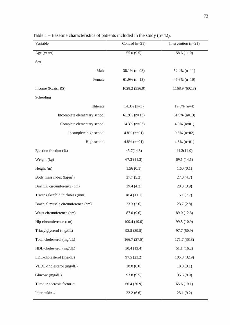
73
Table 1 – Baseline characteristics of patients included in the study (n=42).
Variable Control (n=21) Intervention (n=21)
Age (years) 55.0 (9.5) 58.6 (11.0)
Sex
Male
Female
38.1% (n=08)
61.9% (n=13)
52.4% (n=11)
47.6% (n=10)
Income (Reais, R$) 1028.2 (556.9) 1168.9 (602.8)
Schooling
Illiterate
Incomplete elementary school
Complete elementary school
Incomplete high school
High school
14.3% (n=3)
61.9% (n=13)
14.3% (n=03)
4.8% (n=01)
4.8% (n=01)
19.0% (n=4)
61.9% (n=13)
4.8% (n=01)
9.5% (n=02)
4.8% (n=01)
Ejection fraction (%) 45.7(14.8) 44.2(14.0)
Weight (kg) 67.3 (11.3) 69.1 (14.1)
Height (m) 1.56 (0.1) 1.60 (0.1)
Body mass index (kg/m2) 27.7 (5.2) 27.0 (4.7)
Brachial circumference (cm) 29.4 (4.2) 28.3 (3.9)
Triceps skinfold thickness (mm) 18.4 (11.1) 15.1 (7.7)
Brachial muscle circumference (cm) 23.3 (2.6) 23.7 (2.8)
Waist circumference (cm) 87.0 (9.6) 89.0 (12.8)
Hip circumference (cm) 100.4 (10.0) 99.5 (10.9)
Triacylglycerol (mg/dL) 93.8 (39.5) 97.7 (50.9)
Total cholesterol (mg/dL) 166.7 (27.5) 171.7 (38.8)
HDL-cholesterol (mg/dL) 50.4 (13.4) 51.1 (16.2)
LDL-cholesterol (mg/dL) 97.5 (23.2) 105.8 (32.9)
VLDL-cholesterol (mg/dL) 18.8 (8.0) 18.8 (9.1)
Glucose (mg/dL) 93.8 (9.5) 95.6 (8.0)
Tumour necrosis factor-α 66.4 (20.9) 65.6 (19.1)
Interleukin-4 22.2 (6.6) 23.1 (9.2)

74
Interleukin-6 335.4 (486.9) 163.9 (142.1)
Interleukin-8 84.8 (58.1) 59.8 (26.2)
Interleukin-10 105.2 (71.2) 70.4 (28.4)
Interleukin-33 77.2 (23.6) 86.7 (66.5)
Interleukin-1b 21.1 (8.9) 18.9 (4.4)
Interleukin-17ª 5082.3 (5086.2) 3940.4 (4685.9)
Interferon- 75.5 (112.9) 47.8 (84.2)
Data are expressed as means (standard deviation) and prevalence of frequency.

75
Table 2 – Crude means (standard deviation) and estimated changes from baseline (∆) for an-
thropometric variables during the follow-up.
4 week 8 week
(Control = 20; Intervention = 21) (Control = 20; Intervention = 21)
Mean (DP) ∆a pa Mean (DP) ∆a pa
Weight (kg)
Control 67.9 (11.6) +0.43 68.7 (10.2) +0.52
Intervention 68.2 (13.7) +0.46 0.946 69.9 (13.4) +0.84 0.386
Body mass index (kg/m2)
Control 28.1 (5.2) +0.18 28.4 (4.7) +0.36
Intervention 26.7 (4.7) 0.00 0.438 27.2 (4.5) +0.21 0.542
Arm circumference (cm)
Control 29.6 (5.0) +0.04 29.9 (4.6) +0.16
Intervention 28.1 (4.2) +0.03 0.986 28.6 (3.7) +0.33 0.703
Tricipital skinfold thickness (mm)
Control 18.4 (9.9) -0.29 18.5 (10.1) -0.17
Intervention 15.7 (8.2) +0.26 0.057 15.1 (7.6) +0.14 0.261
Arm circumference (cm)
Control 23.3 (2.3) 0.00 24.0 (2.8) +0.48
Intervention 23.6 (2.9) +0.09 0.840 23.8 (2.7) +0.08 0.323
Waist circumference (cm)
Control 87.1 (9.5) -0.28 88.1 (8.5) +0.59
Intervention 88.2 (12.7) +0.20 0.263 89.1 (12.6) +0.12 0.259
Hip circumference (cm)
Control 100.3 (10.1) -0.25 101.3 (9.1) +0.11
Intervention 99.0 (10.6) +0.29 0.213 100.1 (10.1) +0.59 0.258
aLinear mixed models include time, group and time x group interaction adjusted for baseline
values.

76
Table 3 – Crude means (standard deviation) and estimated changes from baseline (∆) for bio-
chemical variables.
aLinear mixed models include time, treatment and time x treatment interaction, adjusted for
baseline values.
8 week
(Control = 19 Intervention =21)
Mean (DP) ∆a pa
Triglycerides (mg/dL)
Control 90.4 (35.3) -4.1
Intervention 76.6 (31.0) -21.1 0.05
Total cholesterol (mg/dL)
Control 168.5 (27.0) +3.4
Intervention 179.1 (34.6) +7.4 0.60
HDL cholesterol (mg/dL)
Control 49.7 (11.5) -0.3
Intervention 53.7 (15.1) +2.6 0.31
LDL cholesterol (mg/dL)
Control 100.7 (22.7) +4.7
Intervention 110.1 (31.0) +4.3 0.96
VLDL cholesterol (mg/dL)
Control 18.1 (7.1) -0.9
Intervention 15.3 (6.1) -3.5 0.13
Glucose (mg/dL)
Control 92.3 (13.3) -2.0
Intervention 95.2 (9.2) -0.4 0.59

77
Table 4 – Crude means (standard deviation) and estimated changes from baseline (∆) for in-
flammatory markers.
8 week
(Control = 19 Intervention =21)
Mean (DP) ∆a pa
Tumor necrosis factor-α (pg/dL)
Control 84.5 (51.2) +19.4
Intervention 90.7 (48.8) +27.2 0.58
Interleukin-4 (pg/dL)
Control 21.5 (9.7) -0.8
Intervention 20.7 (6.7) -1.5 0.67
Interleukin-6 (pg/dL)
Control 67.3 (80.2) -228.1
Intervention 61.5 (54.3) -85.3 0.45
Interleukin-8 (pg/dL)
Control 64.7 (48.4) -11.3
Intervention 53.6 (12.4) -3.3 0.47
Interleukin-10 (pg/dL)
Control 53.8 (7.1) -35.7
Intervention 56.3 (8.3) -10.6 0.01
Interleukin-33 (pg/dL)
Control 70.4 (15.6) -5.2
Intervention 69.4 (17.0) -5.5 0.97
Interleukin-1b (pg/dL)
Control 18.0 (3.4) -2.3
Intervention 17.9 (3.4) -0.6 0.24
Interleukin-17a (pg/dL)
Control 3649.2 (4254.2) +951.5
Intervention 6130.0 (7644.2) +3621.7 0.24
Interferon- (pg/dL)

78
Control 82.6 (112.3) +9.3
Intervention 53.2 (100.7) +7.1 0.90
aLinear mixed models include time, treatment and time x treatment interaction, adjusted for
baseline values.

79
Table 5 – Means (standard deviation) of dietary variables and energy intake (n=42)
Variable Control (n=21) Intervention (n=21) p
Energy (kcal) 1487.59 (407.36) 1480.18 (419.08) 0.95
Carbohydrates (g) 202.75 (49.04) 200.59 (49.71) 0.89
Protein (g) 72.30 (27.77) 70.99 (26.00) 0.88
Total fat (g) 42.94 (19.93) 43.03 (19.50) 0.99
Fiber (g) 18.48 (7.01) 19.22 (7.75) 0.75
Cholesterol (mg) 215.60 (129.55) 205.70 (87.81) 0.77
Saturated fatty acids (g) 12.60 (5.93) 13.81 (7.62) 0.57
Monounsaturated fatty acids (g) 12.36 (7.82) 13.15 (8.02) 0.75
Polyunsaturated fatty acids (g) 9.54 (5.70) 8.41 (3.80) 0.45
Trans fatty acids (g) 0.65 (0.58) 0.80 (0.76) 0.48
Linoleic acid, n-6 (g) 6.17 (4.24) 5.65 (2.99) 0.65
Alfa-linoleic acid, n-3 (g) 0.72 (0.80) 0.56 (0.32) 0.42

80
7 CONCLUSÃO
Neste grupo de pacientes com cardiopatia chagásica crônica, a suplementação por 8
semanas com ácidos graxos poli-insaturados ômega-3 produziu as seguintes alterações:
a. diminuição dos níveis de triglicerídeos configurando uma melhora no perfil lipídico;
b. atuação favorável no sistema imune, ao reduzir a intensidade da queda progressiva da
citocina anti-inflamatória IL-10, já esperada na evolução da cardiopatia;
Não houve alteração na glicemia de jejum nem nos demais parâmetros do perfil lipídico
e de citocinas após oito semanas de suplementação de ômega-3.
O estado nutricional dos pacientes do grupo intervenção e placebo não foi alterado pela
suplementação de ômega-3, assim como não houve diferença entre os grupos na avaliação
basal.
Não houve diferença significativa na avaliação do consumo alimentar dos
macronutrientes e PUFAs entre os grupos ômega-3 e placebo.

81
8 PERSPECTIVAS FUTURAS
Esses resultados sugerem que a suplementação com ácidos graxos poli-insaturados
ômega-3 pode ser considerada como uma nova estratégia de tratamento coadjuvante na atenção
ao paciente com CCC, principalmente por sua atuação no perfil lipídico e possível atuação no
sistema imune. Este estudo inicial gera a hipótese de que, através da atuação benéfica no sistema
imune, haveria benefícios posteriores, retardando a progressão da cardiopatia e o surgimento
dos sintomas. Para avaliação dos desfechos clínicos, um outro estudo deve ser realizado,
possivelmente com um período mais prolongado de suplementação de ácidos graxos poli-
insaturados ômega-3, e, certamente, englobando um número maior de voluntários. A avaliação
do sistema imune e dos desfechos clínicos, como ECO, ECG, mortalidade, internação
hospitalar, entre outros, devem ser mensuradas para determinar um possível efeito da
suplementação com ácidos graxos poli-insaturados ômega-3 na evolução da cardiopatia
chagásica crônica.
A avaliação do consumo de macro e micronutrientes dos pacientes com cardiopatia
chagásica crônica pode ser uma nova linha de pesquisa para identificar as possíveis deficiências
e ou excessos no consumo alimentar, permitindo desenvolver estratégias de educação, visando
a promoção de uma alimentação adequada e saudável, estimulando a autonomia para práticas
alimentares saudáveis.

82
9 REFERÊNCIAS BIBLIOGRÁFICAS
ALBERT, C. M. et al. Blood levels of long-chain n-3 fatty acids and the risk of sudden death.
The New England journal of medicine, v. 346, n. 15, p. 1113–1118, 11 abr. 2002.
ALLAIN, C. C. et al. Enzymatic determination of total serum cholesterol. Clinical Chemistry,
v. 20, n. 4, p. 470–475, abr. 1974.
BAHIA-OLIVEIRA, L. M. et al. IFN-gamma in human Chagas’ disease: protection or
pathology? Brazilian Journal of Medical and Biological Research, v. 31, n. 1, p. 127–131,
jan. 1998.
BARRETO, S. M.; PASSOS, V. M. A.; LIMA-COSTA, M. F. F. Obesity and underweight
among Brazilian elderly: the Bambuí Health and Aging Study. Cadernos de Saúde Pública, v.
19, n. 2, p. 605–612, abr. 2003.
BELEIGOLI, A. M. et al. The “obesity paradox” in an elderly population with a high prevalence
of Chagas disease: The 10-year follow-up of the Bambuí (Brazil) Cohort Study of Aging.
International Journal of Cardiology, v. 166, n. 2, p. 523–526, jun. 2013.
BELKAID, Y. Regulatory T cells and infection: a dangerous necessity. Nature Reviews
Immunology, v. 7, n. 11, p. 875–888, nov. 2007.
BELLO CORASSA, R. et al. Evolution of Chagas’ disease in Brazil. Epidemiological
perspective and challenges for the future: a critical review. Perspectives in Public Health, p.
1757913916671160, 10 out. 2016.
BENZIGER, C. P.; DO CARMO, G. A. L.; RIBEIRO, A. L. P. Chagas Cardiomyopathy:
Clinical Presentation and Management in the Americas. Cardiology Clinics, v. 35, n. 1, p. 31–
47, fev. 2017.
BERN, C. Chagas’ Disease. New England Journal of Medicine, v. 373, n. 5, p. 456–466, 30
jul. 2015.
BESTETTI, R. B.; RESTINI, C. B. A.; COUTO, L. B. Carlos Chagas Discoveries as a Drop
Back to Scientific Construction of Chronic Chagas Heart Disease. Arquivos Brasileiros De

83
Cardiologia, v. 107, n. 1, p. 63–70, jul. 2016.
BRAGA, J. C. V. et al. Clinical and therapeutics aspects of heart failure due to Chagas disease.
Arquivos Brasileiros de Cardiologia, v. 86, n. 4, p. 297–302, abr. 2006.
BROWN, A. A.; HU, F. B. Dietary modulation of endothelial function: implications for
cardiovascular disease. The American Journal of Clinical Nutrition, v. 73, n. 4, p. 673–686,
abr. 2001.
CALDER, P. C. Polyunsaturated fatty acids, inflammation, and immunity. Lipids, v. 36, n. 9,
p. 1007–1024, set. 2001.
CALDER, P. C. The 2008 ESPEN Sir David Cuthbertson Lecture: Fatty acids and
inflammation--from the membrane to the nucleus and from the laboratory bench to the clinic.
Clinical Nutrition (Edinburgh, Scotland), v. 29, n. 1, p. 5–12, fev. 2010.
CALDER, P. C. Omega-3 polyunsaturated fatty acids and inflammatory processes: nutrition or
pharmacology? British journal of clinical pharmacology, v. 75, n. 3, p. 645–662, mar. 2013.
CELANO, R. M. G. et al. Pre- operatory nutritional evaluation of patients with non-advanced
megaesophagus. Revista do Colégio Brasileiro de Cirurgiões, v. 34, n. 3, p. 147–152, jun.
2007.
CHAVES, A. T. et al. Immunoregulatory mechanisms in Chagas disease: modulation of
apoptosis in T-cell mediated immune responses. BMC infectious diseases, v. 16, n. 1, p. 191,
2016.
COURA, J. R.; VIÑAS, P. A.; JUNQUEIRA, A. C. Ecoepidemiology, short history and control
of Chagas disease in the endemic countries and the new challenge for non-endemic countries.
Memórias do Instituto Oswaldo Cruz, v. 109, n. 7, p. 856–862, nov. 2014.
CRUZ, J. S. et al. Molecular mechanisms of cardiac electromechanical remodeling during
Chagas disease: Role of TNF and TGF-β. Trends in Cardiovascular Medicine, v. 27, n. 2, p.
81–91, fev. 2017.
CUNHA-NETO, E. et al. Immunological and non-immunological effects of cytokines and
chemokines in the pathogenesis of chronic Chagas disease cardiomyopathy. Memórias do
Instituto Oswaldo Cruz, v. 104 Suppl 1, p. 252–258, jul. 2009.

84
CUNHA-NETO, E.; CHEVILLARD, C. Chagas Disease Cardiomyopathy: Immunopathology
and Genetics. Mediators of Inflammation, v. 2014, p. e683230, 19 ago. 2014.
DAMSGAARD, C. T.; FRØKIAER, H.; LAURITZEN, L. The effects of fish oil and high or
low linoleic acid intake on fatty acid composition of human peripheral blood mononuclear cells.
The British journal of nutrition, v. 99, n. 1, p. 147–154, jan. 2008.
DE OLIVEIRA, R. B. et al. Gastrointestinal manifestations of Chagas’ disease. The American
Journal of Gastroenterology, v. 93, n. 6, p. 884–889, jun. 1998.
DIAS, J. C. P. Evolution of Chagas disease screening programs and control programs: historical
perspective. Global Heart, v. 10, n. 3, p. 193–202, set. 2015.
DIAS, J. C. P. et al. 2 nd Brazilian Consensus on Chagas Disease, 2015. Revista Da Sociedade
Brasileira De Medicina Tropical, v. 49Suppl 1, n. Suppl 1, p. 3–60, dez. 2016.
DUTRA, W. O.; ROCHA, M. O. C.; TEIXEIRA, M. M. The clinical immunology of human
Chagas disease. Trends in parasitology, v. 21, n. 12, p. 581–587, dez. 2005.
DYERBERG, J.; BANG, H. O. Haemostatic function and platelet polyunsaturated fatty acids
in Eskimos. The Lancet, v. 314, n. 8140, p. 433–435, 1 set. 1979.
ECHEVERRÍA, L. E. et al. Profiles of cardiovascular biomarkers according to severity stages
of Chagas cardiomyopathy. International Journal of Cardiology, v. 227, p. 577–582, 15 jan.
2017.
FABER, J. et al. Rapid EPA and DHA incorporation and reduced PGE(2) levels after one week
intervention with a medical food in cancer patients receiving radiotherapy, a randomized trial.
Clinical nutrition (Edinburgh, Scotland), 28 set. 2012.
FERNÁNDEZ-REAL, J.-M. et al. Insulin resistance, inflammation, and serum fatty acid
composition. Diabetes Care, v. 26, n. 5, p. 1362–1368, maio 2003.
FILIGHEDDU, M. T.; GÓRGOLAS, M.; RAMOS, J. M. Orally-transmitted Chagas disease.
Medicina Clínica (English Edition), v. 148, n. 3, p. 125–131, 9 fev. 2017.
FOSSATI, P.; PRENCIPE, L. Serum triglycerides determined colorimetrically with an enzyme
that produces hydrogen peroxide. Clinical Chemistry, v. 28, n. 10, p. 2077–2080, out. 1982.

85
FREEMAN, M. P.; SINHA, P. Tolerability of omega-3 fatty acid supplements in perinatal
women. Prostaglandins, leukotrienes, and essential fatty acids, v. 77, n. 3–4, p. 203–208,
nov. 2007.
FRENOUX, J. M. et al. A polyunsaturated fatty acid diet lowers blood pressure and improves
antioxidant status in spontaneously hypertensive rats. The Journal of nutrition, v. 131, n. 1,
p. 39–45, jan. 2001.
FRIEDEWALD, W. T.; LEVY, R. I.; FREDRICKSON, D. S. Estimation of the concentration
of low-density lipoprotein cholesterol in plasma, without use of the preparative ultracentrifuge.
Clinical chemistry, v. 18, n. 6, p. 499–502, jun. 1972.
GERAIX, J. et al. Clinical and nutritional profile of individuals with Chagas disease. Brazilian
Journal of Infectious Diseases, v. 11, n. 4, p. 411–414, ago. 2007.
GIUGLIANO, D.; CERIELLO, A.; ESPOSITO, K. The effects of diet on inflammation:
emphasis on the metabolic syndrome. Journal of the American College of Cardiology, v. 48,
n. 4, p. 677–685, 15 ago. 2006.
GOMES, J. A. S. et al. Evidence that development of severe cardiomyopathy in human Chagas’
disease is due to a Th1-specific immune response. Infection and immunity, v. 71, n. 3, p.
1185–1193, mar. 2003.
GOODNIGHT, S. H.; CAIRNS, J. A. Therapeutic use of n-3 fatty acids for vascular disease
and thrombosis. Chest, v. 108, n. 4 Suppl, p. 302S–304S, out. 1995.
GUEDES, P. M. M. et al. Deficient regulatory T cell activity and low frequency of IL-17-
producing T cells correlate with the extent of cardiomyopathy in human Chagas’ disease. PLoS
neglected tropical diseases, v. 6, n. 4, p. e1630, 2012.
GUEDES, P. M. M. et al. Inflammation Enhances the Risks of Stroke and Death in Chronic
Chagas Disease Patients. PLoS neglected tropical diseases, v. 10, n. 4, p. e0004669, abr. 2016.
HABERLAND, A. et al. Chronic Chagas disease: from basics to laboratory medicine. Clinical
Chemistry and Laboratory Medicine, v. 51, n. 2, p. 271–294, fev. 2013.
HARRIS, W. S. et al. Towards establishing dietary reference intakes for eicosapentaenoic and
docosahexaenoic acids. The Journal of nutrition, v. 139, n. 4, p. 804S–19S, abr. 2009.

86
HEBERT, J. R. et al. Development and testing of a quantitative food frequency questionnaire
for use in Gujarat, India. Public health nutrition, v. 2, n. 1, p. 39–50, mar. 1999.
HINO, K. A new method for the homogeneous assay of serum HDL-cholesterol. Clinical
Chemistry, v. 42, p. 298, 1996.
INSTITUTE OF MEDICINE. Dietary Reference Intakes for Energy, Carbohydrate, Fiber,
Fat, Fatty Acids, Cholesterol, Protein, and Amino Acids. Washington, DC: The National
Academies Press, 2005.
KEW, S. et al. Effects of oils rich in eicosapentaenoic and docosahexaenoic acids on immune
cell composition and function in healthy humans. The American journal of clinical nutrition,
v. 79, n. 4, p. 674–681, abr. 2004.
KIM, W.; MCMURRAY, D. N.; CHAPKIN, R. S. n-3 polyunsaturated fatty acids--
physiological relevance of dose. Prostaglandins, leukotrienes, and essential fatty acids, v.
82, n. 4–6, p. 155–158, jun. 2010.
KROPF, S. P.; AZEVEDO, N.; FERREIRA, L. O. Chagas disease: the construction of a
scientific fact and of a public health problem in Brazil. Ciência & Saúde Coletiva, v. 5, n. 2,
p. 347–365, 2000.
LEMAITRE, R. N. et al. n-3 Polyunsaturated fatty acids, fatal ischemic heart disease, and
nonfatal myocardial infarction in older adults: the Cardiovascular Health Study. The American
journal of clinical nutrition, v. 77, n. 2, p. 319–325, fev. 2003.
MACHADO, F. S. et al. Current understanding of immunity to Trypanosoma cruzi infection
and pathogenesis of Chagas disease. Seminars in Immunopathology, v. 34, n. 6, p. 753–770,
nov. 2012.
MALIK, L. H.; SINGH, G. D.; AMSTERDAM, E. A. Chagas Heart Disease: An Update. The
American Journal of Medicine, v. 128, n. 11, p. 1251.e7-9, nov. 2015.
MARIN NETO, J. A.; SIMÕES, M. V.; SARABANDA, A. V. Chagas’ heart disease. Arquivos
brasileiros de cardiologia, v. 72, n. 3, p. 247–280, mar. 1999.
MARIN-NETO, J. A. et al. Pathogenesis of chronic Chagas heart disease. Circulation, v. 115,
n. 9, p. 1109–1123, 6 mar. 2007.

87
MCKENNEY, J. M.; SICA, D. Prescription omega-3 fatty acids for the treatment of
hypertriglyceridemia. BELKAID, Y. Regulatory T cells and infection: a dangerous necessity.
Nature Reviews Immunology, v. 7, n. 11, p. 875–888, nov. 2007.
MCKENNEY, J. M.; SICA, D. Prescription omega-3 fatty acids for the treatment of
hypertriglyceridemia. American Journal of Health-System Pharmacy, v. 64, n. 6, p. 595–
605, 15 mar. 2007.
MONCAYO, A.; SILVEIRA, A. C. Current epidemiological trends for Chagas disease in Latin
America and future challenges in epidemiology, surveillance and health policy. Memórias do
Instituto Oswaldo Cruz, v. 104 Suppl 1, p. 17–30, jul. 2009.
MUÑOZ-SARAVIA, S. G. et al. Chronic Chagas’ heart disease: a disease on its way to
becoming a worldwide health problem: epidemiology, etiopathology, treatment, pathogenesis
and laboratory medicine. Heart Failure Reviews, v. 17, n. 1, p. 45–64, jan. 2012.
PENAS, F. et al. PPARγ ligand treatment inhibits cardiac inflammatory mediators induced by
infection with different lethality strains of Trypanosoma cruzi. Biochimica et biophysica acta,
v. 1832, n. 1, p. 239–248, jan. 2013.
PENHAVEL, F. A. S. et al. Pre-and postoperative nutritional evaluation in patients with
chagasic megaesophagus. Nutrición hospitalaria, v. 19, n. 2, p. 89–94, abr. 2004.
PÉREZ-FUENTES, R. et al. Severity of chronic Chagas disease is associated with
cytokine/antioxidant imbalance in chronically infected individuals. International journal for
parasitology, v. 33, n. 3, p. 293–299, mar. 2003.
PÉREZ-MOLINA, J. A.; MOLINA, I. Chagas disease. The Lancet, 30 jun. 2017.
PERINI, J. Â. D. L. et al. Omega-3 and omega-6 polyunsaturated fatty acids: metabolism in
mammals and immune response. Revista de Nutrição, v. 23, n. 6, p. 1075–1086, dez. 2010.
PINAZO, M. J. et al. Diagnosis, management and treatment of chronic Chagas’ gastrointestinal
disease in areas where Trypanosoma cruzi infection is not endemic. Gastroenterologia Y
Hepatologia, v. 33, n. 3, p. 191–200, mar. 2010.
PRATA, A. Clinical and epidemiological aspects of Chagas disease. The Lancet Infectious
Diseases, v. 1, n. 2, p. 92–100, set. 2001.

88
PUNUKOLLU, G. et al. Clinical aspects of the Chagas’ heart disease. International journal
of cardiology, v. 115, n. 3, p. 279–283, 14 fev. 2007.
RASSI, A. J.; RASSI, A.; MARCONDES DE REZENDE, J. American Trypanosomiasis
(Chagas Disease). Infectious Disease Clinics of North America, v. 26, n. 2, p. 275–291, jun.
2012.
RASSI, A., Jr; RASSI, A.; LITTLE, W. C. Chagas’ heart disease. Clinical cardiology, v. 23, n.
12, p. 883–889, dez. 2000.
RASSI JR, A.; RASSI, A.; MARIN-NETO, J. A. Chagas heart disease: pathophysiologic
mechanisms, prognostic factors and risk stratification. Memórias do Instituto Oswaldo Cruz,
v. 104 Suppl 1, p. 152–158, jul. 2009.
RIBEIRO, A. L. P.; ROCHA, M. O. DA C. Indeterminate form of Chagas’ disease: considera-
tions about diagnosis and prognosis. Revista da Sociedade Brasileira de Medicina Tropical,
v. 31, n. 3, p. 301–314, jun. 1998.
ROCHA, M. O. C.; RIBEIRO, A. L. P.; TEIXEIRA, M. M. Clinical management of chronic
Chagas cardiomyopathy. Frontiers in bioscience, v. 8, n. 1, p. 44–54, 2003.
ROCHA RODRIGUES, D. B. et al. In situ expression of regulatory cytokines by heart
inflammatory cells in Chagas’ disease patients with heart failure. Clinical & developmental
immunology, v. 2012, p. 361730, 2012.
ROSSI, M. A. Fibrosis and inflammatory cells in human chronic chagasic myocarditis:
scanning electron microscopy and immunohistochemical observations. International Journal
of Cardiology, v. 66, n. 2, p. 183–194, 30 set. 1998.
SALVADOR, M. B. et al. Avaliação da ingestão de ácidos graxos, antioxidantes e da
composição corporal em mulheres com artrite reumatóide; Fatty acids, antioxidants and body
composition evaluation in woman with reumathoid arthritis. Nutrire Revista da Sociedade
Brasileira de Alimentação e Nutrição, v. 33, n. 3, p. 17–30, dez. 2008.
SANTOS, J. P. DOS; LIMA-COSTA, M. F.; PEIXOTO, S. V. Aspectos nutricionais associados
à infecção crônica pelo Trypanosoma cruzi (Chagas 1909) entre idosos: Projeto Bambuí.
Cadernos de Saúde Pública, v. 29, n. 6, p. 1141–1148, jun. 2013.
89
SATHLER-AVELAR, R. et al. Phenotypic features of peripheral blood leucocytes during early
stages of human infection with Trypanosoma cruzi. Scandinavian Journal of Immunology,
v. 58, n. 6, p. 655–663, dez. 2003.
SHER, A. et al. Role of T-cell derived cytokines in the downregulation of immune responses in
parasitic and retroviral infection. Immunological reviews, v. 127, p. 183–204, jun. 1992.
SIMOPOULOS, A. P. Omega-3 fatty acids in health and disease and in growth and
development. The American Journal of Clinical Nutrition, v. 54, n. 3, p. 438–463, 9 jan.
1991.
SIMOPOULOS, A. P. Essential fatty acids in health and chronic disease. The American
journal of clinical nutrition, v. 70, n. 3 Suppl, p. 560S–569S, set. 1999.
SIMOPOULOS, A. P. The importance of the omega-6/omega-3 fatty acid ratio in
cardiovascular disease and other chronic diseases. Experimental Biology and Medicine, v.
233, n. 6, p. 674–688, 1 jun. 2008.
SONG, C. et al. Effects of dietary n-3 or n-6 fatty acids on interleukin-1beta-induced anxiety,
stress, and inflammatory responses in rats. Journal of lipid research, v. 44, n. 10, p. 1984–
1991, out. 2003.
SOUSA, G. R. et al. Plasma Cytokine Expression Is Associated with Cardiac Morbidity in
Chagas Disease. PLoS ONE, v. 9, n. 3, 6 mar. 2014.
SPOSITO, A. C. et al. IV Brazilian Guideline for Dyslipidemia and Atherosclerosis prevention:
Department of Atherosclerosis of Brazilian Society of Cardiology. Arquivos brasileiros de
cardiologia, v. 88 Suppl 1, p. 2–19, abr. 2007.
TANOWITZ, H. B. et al. Perspectives on Trypanosoma cruzi-induced heart disease (Chagas
disease). Progress in Cardiovascular Diseases, v. 51, n. 6, p. 524–539, jun. 2009.
TAVAZZI, L. et al. Effect of n-3 polyunsaturated fatty acids in patients with chronic heart failure
(the GISSI-HF trial): a randomised, double-blind, placebo-controlled trial. The Lancet, v. 372,
n. 9645, p. 1223–1230, 4 out. 2008.
VIEIRA, M. J. et al. Preoperative assessment in cases of adult megacolon suffering from
moderate malnutrition. Nutrition, v. 12, n. 7, p. 491–495, ago. 1996.

90
VILLANI, A. M. et al. Fish oil administration in older adults: is there potential for adverse
events? A systematic review of the literature. BMC geriatrics, v. 13, p. 41, 1 maio 2013.
VON SCHACKY, C. n-3 fatty acids and the prevention of coronary atherosclerosis. The
American journal of clinical nutrition, v. 71, n. 1 Suppl, p. 224S–7S, jan. 2000.
WHO | Chagas disease (American trypanosomiasis). Disponível em:
<http://www.who.int/mediacentre/factsheets/fs340/en/>. Acesso em: 10 jul. 2017.
XAVIER, S. S.; SOUSA, A. S. DE; MORENO, A. H. Aplicação da nova classificação da
insuficiência cardíaca (ACC/AHA) na cardiopatia chagásica crônica: análise crítica das curvas
de sobrevida. Revista da SOCERJ, p. 227–232, 2005.
XIN, W.; WEI, W.; LI, X. Effects of fish oil supplementation on inflammatory markers in
chronic heart failure: a meta-analysis of randomized controlled trials. BMC cardiovascular
disorders, v. 12, p. 77, 2012.
ZHANG, L.; TARLETON, R. L. Parasite persistence correlates with disease severity and
localization in chronic Chagas’ disease. The Journal of Infectious Diseases, v. 180, n. 2, p.
480–486, ago. 1999.

91
10 APÊNDICES
APÊNDICE A – AVALIAÇÃO NUTRICIONAL
1-Dados Gerais
Data ____/____/_____
Nome: __________________________________________ Registro: ___________________
Telefone:___________________________________________________________________
Ende-
reço:_______________________________________________________________________
_______________________________________________________________________
Sexo:______________ Data de Nascimento: ____________________
Idade:_________________
Naturalidade:_________________Escolaridade:__________________
Renda:__________________
Classificação da disfunção cardíaca:______________________________________________
Co-morbidades:______________________________________________________________
Etilismo: ( ) Sim Frequência______________ Quantidade______________
( ) Não ( ) Ex-etilista Tempo: _____________
Tabagismo: ( ) Sim Frequência______________ Quantidade______________
( ) Não ( ) Ex-etilista Tempo: _____________
Data de retorno:_____________________________________________________________
Medicação utilizada Quantidade horário

92
2-Avaliação Antropométrica
Antropometria 1ª avaliação 2ª avaliação 3ª avaliação
Estatura (m)
Peso Atual (kg)
IMC (kg/m2)
PCT (mm)
CB (cm)
CMB (cm)
CC (cm)
CQ (cm)
3-Avaliação Bioquímica e Imunológica
Observações:
___________________________________________________________________________
___________________________________________________________________________
___________________________________________________________________________
___________________________________________________________________________
___________________________________________________________________________
___________________________________________________________________________
Exames
Bioquímicos
1ª avaliação 2ª avaliação Citocinas 1ª avaliação 2ª avaliação
Triglicerídeo IL-1β
Colesterol Total IL-2
HDL-c IL-4
LDL-c IL-6
VLDL-c IL-8
Glicose em jejum IL-10
IL-17
IL-33
TNF-
IFN-

93
APÊNDICE B – RECORDATÓRIO DE 24 HORAS
Nome _________________________________ Data ____________________
REFEIÇÃO HORÁRIO ALIMENTO QUANTIDADE

94
APÊNDICE C – TERMO DE CONSENTIMENTO LIVRE E ESCLARECIDO
Projeto: Influência da suplementação de ácidos graxos poliinsaturados ômega-3 no perfil
de citocinas e lipídico de pacientes portadores de Cardiopatia Chagásica Crônica.
Equipe: Coordenadora Paula Simplício da Silva, Drª Andréa Silvestre de Sousa, Drª Andréa
Pereira de Souza, Dr. Alejandro Marcel Hasslocher Moreno, Cristiane Fonseca de Almeida,
Cláudia Santos de Aguiar Cardoso, Dr. Gilberto Marcelo Sperandio da Silva, Drª Patrícia Dias
de Brito, Dr. Pedro Emmanuel Americano do Brasil, Drª Roberta Olmo Pinheiro
A gordura ômega-3 é encontrada principalmente em peixes de água fria (cavala,
sardinha, salmão, arenque), é considerada uma gordura essencial e está relacionada à prevenção
de doenças cardíacas, a diminuição de gordura no sangue (colesterol, triglicerído e VLDL-c) e
diminuição citocinas (que são proteínas encontradas no sangue e que estão relacionadas à
inflamação). O(A) Senhor(a) está sendo convidado a participar desta pesquisa, coordenada pela
nutricionista Paula Simplício da Silva que tem por objetivo investigar se a suplementação de
gordura ômega-3 tem influência no perfil de citocinas, nos níveis de glicose de jejum e no perfil
lipídico (colesterol, triglicerídeo, VLDL-c, HDL-c, LDL-c). O(A) Senhor(a) será sorteado para
receber cápsulas de ômega-3 ou placebo, sendo para tomar 5 cápsulas por dia (2 cápsulas no
café da manhã, 3 no jantar), por um período de 8 semanas. Nem o senhor nem o avaliador
saberão se o senhor recebeu ômega-3 ou placebo. A coleta de sangue será realizada no início
do projeto e após 8 semanas, a avaliação nutricional e dietética será realizada no início do
projeto, com 4 e 8 semanas.
Nesta pesquisa, também será realizada uma avaliação dos compartimentos (partes) cor-
porais que é chamada bioimpedância elétrica, nesta avaliação utilizaremos quatro eletrodos po-
sicionados no corpo, que através da passagem de uma corrente elétrica imperceptível, registram
em um aparelho os valores dos tecidos muscular, gorduroso e de água. Esta avaliação é indolor
e não-invasiva, porém necessita da minha colaboração em alguns cuidados pré-teste como: estar
em jejum de pelo menos 4 horas; com a bexiga vazia; sem o consumo de cafeína e álcool 24
horas antes do teste, e que não tenha realizado atividade física pelo menos 4 horas antes.
Procedimentos
Eu concordo em participar deste estudo:

95
Permitirei que meu peso, altura e a quantidade de gordura sejam verificados através do
uso de balança, fita métrica, régua de altura, adipômetro (aparelho que mede a espessura da
camada de gordura sob a pele) e no início e após dois meses do estudo.
Permitirei que seja realizado o procedimento de bioimpedância elétrica, onde ficarei na
posição deitada, sendo os eletrodos colocados em pontos padronizados na superfície posterior
do punho direito e no tornozelo direito.
Informarei sobre o meu consumo alimentar e qualidade de vida preenchendo os formu-
lários específicos.
Tomarei as cápsulas de ômega-3/placebo conforme orientação.
Permitirei que coletem o meu sangue para exames bioquímicos (glicose, colesterol total,
triglicerídeos, HDL-c).
Poderei ser desligado do projeto caso me recuse a ingerir as cápsulas de ômega-
3/placebo conforme orientação, faltar à coleta de sangue ou não fornecer as informações
referentes ao meu consumo alimentar.
O(A) Senhor(a) deverá continuar com o seu tratamento medicamentoso regular.
Caso seja de meu interesse, poderei desistir de participar desta pesquisa em qualquer
etapa, sem qualquer prejuízo ao meu tratamento, precisando informar apenas aos pesquisadores
minha intenção neste sentido.
Concordo com a publicação dos resultados obtidos na pesquisa. Estou consciente da
importância desta pesquisa e de que receberei os resultados com explicação detalhada dos seus
significados, e se for necessário, serei encaminhado ao ambulatório de nutrição para
acompanhamento. Durante a pesquisa o(a) Senhor receberá orientação sobre alimentação
saudável.
Riscos/Desconfortos
A coleta de sangue poderá causar algum desconforto, sangramento ou hematoma no
local do furo da agulha.
A suplementação de ômega-3, ou placebo nas quantidades que serão fornecidas e se
consumido de acordo com a orientação do nutricionista não oferecem riscos ao participante.
A avaliação nutricional e de compartimentos não oferecem nenhum risco ao
participante.

96
Benefício esperados
Espera-se que a suplementação de ômega-3 reduza os níveis de gordura no sangue (tri-
glicerídeos, colesterol total, LDL-c e VLDL-c) e que diminua a produção de citocinas pró-
inflamatórias em pacientes com cardiopatia chagásica crônica, reduzindo a progressão e mor-
bidade desta cardiopatia.
Confidenciabilidade dos dados.
As informações serão codificadas e mantidas em local reservado. Somente os responsá-
veis pela pesquisa terão acesso às informações e questionários. Após o término deste estudo as
informações serão transcritas dos questionários para arquivos no computador, e estes serão des-
truídos. Os dados deste estudo poderão ser discutidos com pesquisadores de outras instituições,
mas nenhuma identificação será fornecida.
Se tiver qualquer dúvida sobre sua participação neste estudo, favor telefone para 3865-
9602 ou 8862-6916 para falar com a coordenadora da pesquisa Paula Simplício da Silva.
Data:________________
___________________________________ _________________________________
Assinatura do voluntário ou testemunha Nome completo
__________________________ ____________________________________
Assinatura do entrevistador Nome do entrevistador
Nome de alguma pessoa que possa ser contactada, caso o (a) Sr(a) não possa fornecer o seu
endereço ou telefone no momento:
Nome: _______________________________________________________Tel:___________
Rua:____________________________________ Cidade: ____________ Estado: ________

97
11 ANEXO

98