papéis avulsos de zoologia - nrm.se€¦ · papéis avulsos de zoologia issn 0031-1049 museu de...
TRANSCRIPT
Volume 43(2):11-30, 2003 www.scielo.br/paz.htm
Papéis Avulsos de Zoologia
ISSN 0031-1049
Museu de Zoologia da Universidade de São Paulo
REVISION OF THE NEOTROPICAL ANACHARITINAE GENUS ACANTHAEGILIPS
(HYM., CYNIPOIDEA, FIGITIDAE)
PALMIRA ROS-FARRÉ1
MICHAEL SPORRONG2
FREDRIK RONQUIST3
JULI PUJADE-VILLAR4
ABSTRACT
We revise the genus Acanthaegilips, a genus of anacharitine figitids ocurring in South America. Membersof this genus are characterized by having a long scutellar spine, a unique character in the Anacharitinae.The only previously known species (A. brasiliensis) is redescribed and eleven new species are described:A. macropennis, A. ashmeadi, A. levis, A. masneri, A. diazi, A. dentis, A. occultus, A. alienus,A. exiguus, A. huggerti, and A. carinatus. A key to species is presented and distribution records aregiven for each species. The known records of the genus extend from the southern Neotropics to Mexico.The biology of Acanthaegilips remains unknown; other members of the Anacharitinae are parasitoidsof aphid-feeding Neuroptera larvae.
KEYWORDS: Cynipoidea, Figitidae, Aspicerinae, Acanthaegilips, New species.
1,4 Departament de Biologia Animal (Invertebrats), Facultat de Biologia, Universitat de Barcelona, Av. Diagonal, 645, 08028, Barcelona,España. Fax: 93-403.57.40. E-mail: respectively [email protected] and [email protected].
2 Lund University. Department of Zoology. Division of Systematics Helgonavagen 3. S-223 62 Lund Sweden.3 Department of Systematic Zoology Evolutionary Biology Centre Uppsala University Norbyvägen 18D SE-752 36 Uppsala Sweden.
INTRODUCTION
The Figitidae constitute a diverse family ofcynipoid parasites comprising the subfamiliesParnipinae, Anacharitinae, Thrasorinae, Charipinae, andFigitidae ‘sensu stricto’ (Ronquist 1995, 1999; Ronquist& Nieves-Aldrey, 2001). The last taxon includes theFigitinae, Aspicerinae, Eucoilinae, Pycnostigminae andEmargininae (Ronquist 1994b, 1999).
The genus discussed in this paper, Acanthaegilips,is one of seven genera in the Anacharitinae (Ronquist1999). It is easily separated from other anacharitines
by the long and slender scutellar spine and some otherunique features. Its distribution is entirely restricted toSouth and Central America, with the northernmostrecords reported here being from Mexico.
Figitidae are parasitoids of larvae of several in-sects. The recorded hosts of Anacharitinae are aphid-feeding neuropteran larvae belonging to the familiesHemerobiidae and Chrysopidae (Weld 1952; Ronquist1994a, 1995, 1999). However, hosts are unknown formany genera, including Acanthaegilips.
The genus Acanthaegilips was established byAshmead (1897) on the basis of a single female col-
12 ROS-FARRÉ ET AL.: REVISION OF THE NEOTROPICAL ANACHARITINAE GENUS ACANTHAEGILIPS
lected in Brazil. Weld (1921) added data to the origi-nal description based on the study of the female typespecimen, and Díaz (1983) recorded the genus inArgentina, describing the male for the first time. Thegenus was recently redescribed by Ros-Farré et al.(2000).
Because of its different characters, Kovalev(1996) recently separated the genus from otheranacharitines and placed it in a separate monotypicfamily. However, phylogenetic analysis indicates thatthe genus belongs to a monophyletic group compris-ing three neotropical Anacharitinae genera:Acanthaegilips, Solenofigites and Calofigites (Ros-Farré et al.,2000). This clade is deeply nested within theAnacharitinae. Thus, the genus cannot be maintainedas a separate family (Ros-Farré et al., 2000).
Here, we describe 11 new species of Acanthaegilips.We show that the male specimens referred toA. brasiliensis by Díaz (1983) belong to a new species,which is described as A. diazi.
Despite the widespread use of new collectingtechniques, such as Malaise, Moericke, and Noyes traps,the available material of several anacharitine genera isstill relatively scanty. For this work we studied 56 speci-mens deposited in several collections that have accu-mulated specimens throughout many years of collect-ing efforts.
MATERIAL AND METHODS
We have studied material from the following in-stitutions (curator in brackets):
CNC – Canadian National Collections of Insects,Arachnids and Nematodes, Otawa, Ontario, Canada(J.D. Read)
DCBU – Departamento de Biologia da UniversidadeFederal de São Carlos, SP, Brazil (A. Penteado-Dias)
MLP – Museo de la Plata. República Argentina (N.B.Díaz)
MACN – Museo Argentino de Ciencias Naturales (A.Roig)
MZLU-MS – Museum of Zoology Lund University,Lund, Sweden – Coll. M. Sporrong
MZUSP – Museu de Zoologia da Universidade de SãoPaulo, São Paulo, SP, Brazil (C.R.F. Brandão)
UB – Universitat de Barcelona, Facultat de Biologia,Barcelona, Spain – Coll. P. Ros-Farré
UCR – Universidad De Costa Rica (C. Godoy)
USNM – United States National Museum of NaturalHistory, Smithsonian Institution, Washington, DC,USA (D. Furth)
MEAN – Museo Entomológico AsociaciónNicaraguense de Entomología, Leon Nicaragua (J.M.Maes)
All holotypes were examined with SEM, withoutany previous coating and under very low voltage so asnot to put the specimens under any risk. All other speci-mens, mounted on cardboard, were studied usingstereomicroscopy and the drawings were obtained us-ing camera-lucida.
The following special terms and measurementsare employed in the descriptions:
– Transfacial line – shortest distance between thecompound eyes.
– Postocular furrow – this furrow runs posterior tothe compound eye. Ventrally, it joins the malar fur-row close to the ventral margin of the eye.
– Malar furrow – this is a linear structure betweenthe eye and the base of the mandible. It is usuallyformed like a noticeable furrow but sometimes itis a very weak sulcus. Nevertheless, the term malarfurrow is used consistently here, in accordance withRos-Farré et al. (2000).
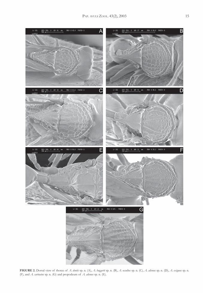
– Notauli – in Acanthaegilips, the notauli are made upof a more or less distinct row of cells that alsoform part of the areolate sculpture covering therest of the mesoscutum. Thus, the notauli are usu-ally not well differentiated. A. alienus is an excep-tion to this rule.
– Foveal carina – carina running from the anteriormargin of the scutellar fovea posteriorly, sometimesreaching the posterior margin of the fovea.
– Median scutellar carina – carina running along themidline of the scutellum, anteriorly separating thescutellar foveae.
Remaining terms for skeletal structures and ab-breviations follow Richards (1977), and Ronquist(1995).
A description of the genus is not included in thisstudy since a comprehensive description of the genuswas provided by Ros-Farré et al. (2000).
PAP. AVULS ZOOL. 43(2), 2003 13
Key to the identification ofAcanthaegilips species:
1. Scutellum concave in dorsal view, roof-shaped,strongly inclined on each side of the medianscutellar carina. Male flagellomeres never modi-fied. Propodeum with three longitudinal ridgesand without any sculpture between them. Notaulinot made up of a row of cells of the areolatesculpture (only male known) ....................................... A. alienus Ros-Farré & Pujade-Villar sp. n.
Scutellum flat in dorsal view or only slightly inclinedon each side of median scutellar carina, neverroof-shaped. Male antenna with someflagellomeres from F1 to F6 slightly expandeddorsolaterally with a longitudinal ridge on theraised part. Propodeum areolate; if there are lon-gitudinal ridges there is areolate sculpture betweenthem. Notauli made up of a more or less distinctrow of cells of the areolate sculpture ............... 2
2. Marginal cell open; marginal vein always lacking(Figs. 5A, C), sometimes there is a very conspicu-ous line of hairs that looks like a vein, and thecell then appears to be closed ............................ 3
Marginal cell closed, marginal vein present andcomplete (Fig. 5B) ............................................... 9
3. Malar furrow strongly curved. Postocular furrowspresent (Figs. 4A, B, E, F), if these furrow areweakly impressed then there is a line of hairsbelow them. Scutellar spine narrowing abruptlybefore apex (Figs. 2A, C, F, G) or end of scutel-lar disc narrowing abruptly and scutellar spineslender all the way (Fig. 2B) ............................... 4
Malar furrow slightly curved (Figs. 3B, C). Postocu-lar furrow absent. Scutellar spine never narrow-ing abruptly and never slender in all its length(Figs. 1A, B, C, D, E, F) ...................................... 8
4. Scutellar foveae with an internal longitudinal ca-rina (foveal carina), dividing them into two ar-eas each (Figs. 2A, C). Pronotal plate produced,in lateral view, into a sharp and strongly raiseddorsal tooth (Figs. 4A, C, F). Petiole as wide aslong, sometimes slightly wider than long. Scutel-lar spine narrowing abruptly before apex(Figs. 2A, C, G) .................................................... 5
Foveal carina absent (Figs. 2B, 2F). Pronotal platedorsally with a very small tooth (Figs. 4B, E).Petiole 1.5 to 2.0 times wider than long. Scutellarspine narrowed abruptly before apex or slenderin all its length ...................................................... 7
5. Malar and postocular furrows strongly impressedand without pubescence below them (Fig. 4A).
Foveal carina reaching the posterolateral marginof the scutellar foveae (Fig. 2A) (only femaleknown) .............................................................................. A. dentis Ros-Farré & Pujade-Villar sp. n.
Malar furrow effaced, postocular furrow weak butwith a line of hairs running below the postocularand malar furrows (Figs. 4C, F). Foveal carina lessevident and not reaching the posterolateral mar-gin of the scutellar foveae (Figs. 2C, G) .......... 6
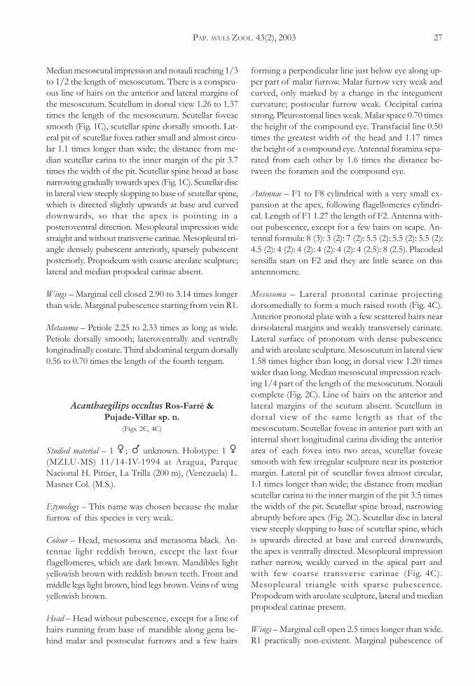
6. Pronotum in lateral view with areolate sculpture.Lateral pit of fovea present (Fig. 2C) (only fe-male known) .................................................................. A. occultus Ros-Farré & Pujade-Villar sp. n.
Lateral surface of pronotum with coarse transversecarina in ventral part and with areolate sculpturein dorsal part. Lateral pit of fovea absents onfemale (Fig. 2G) and obstructed in male, whichinstead of pit has deep fovea .................................... A. carinatus Ros-Farré & Pujade-Villar sp. n.
7. Scutellar disc narrows abruptly posteriorly and thescutellar spine is slender all the way (Fig. 2B);scutellar spine straight in lateral view (Fig. 4B).Lateral pit of scutellar fovea deep (Fig. 2B) ................... A. huggerti Sporrong & Ros-Farré n. sp.
Scutellar spine narrowing abruptly before apex.Scutellar spine (Fig. 2F) curved in lateral view(Fig. 4E). Lateral pit of fovea superficial, indis-tinct (Fig. 2F) (only male known) ............................... A. exiguus Ros-Farré & Pujade-Villar sp. n.
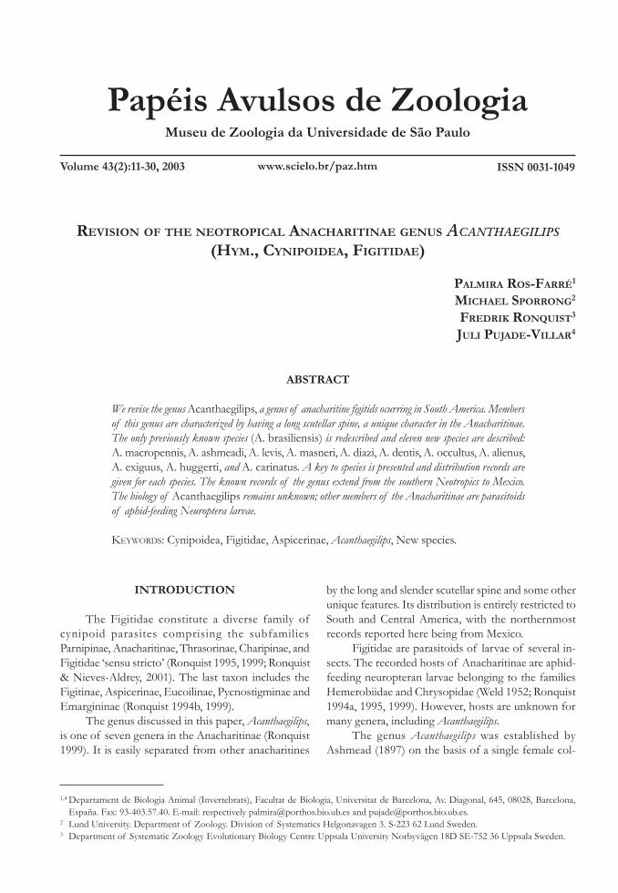
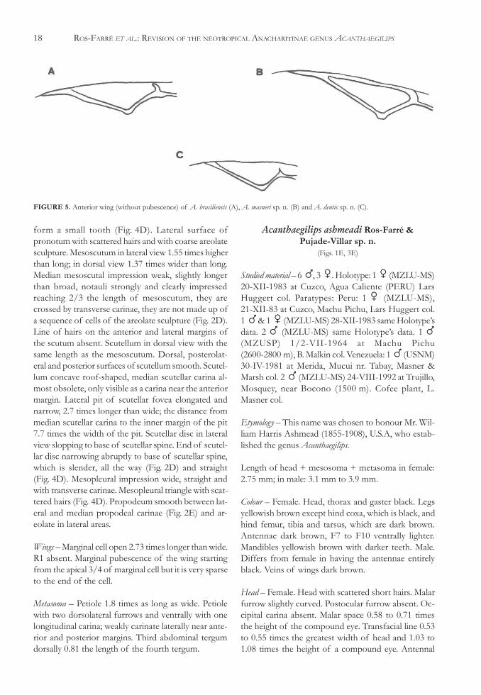
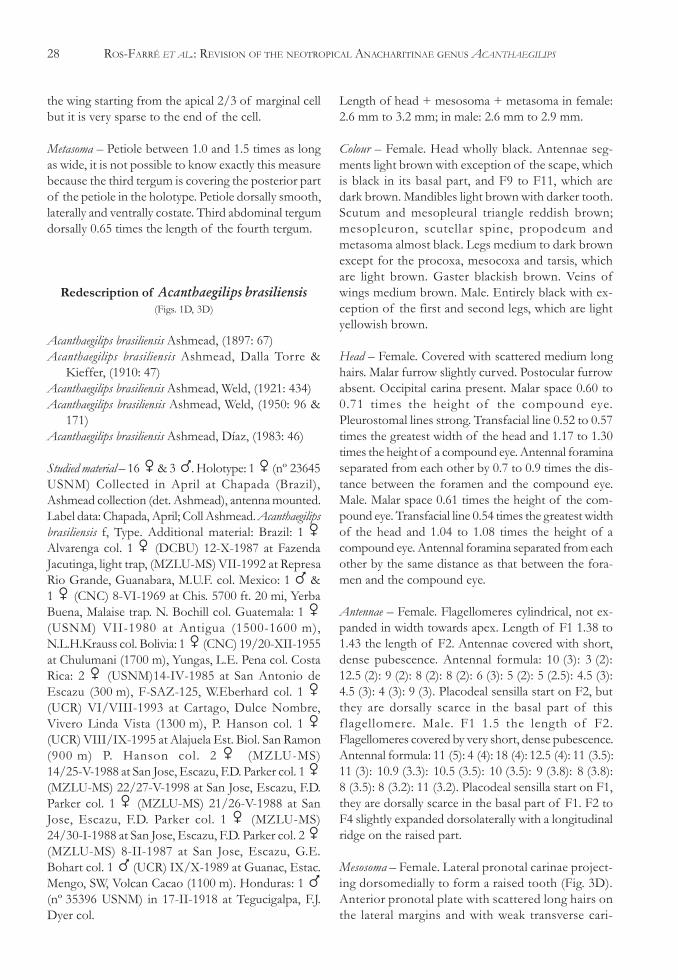
8. Sculpture over scutum from the pronotum to themedian sulcus as a carina (Fig. 3D), more evi-dent when viewed in profile. Scutum in profilewith a distinct hump. Pronotal plate, viewed lat-erally, with a pointed dorsal tooth (Fig. 3D) ................................................ A. brasiliensis Ashmead
Sculpture over scutum, when viewed in profile, notforming a carina. Scutum curved in profile, nothumped (Fig. 3B). Pronotal plate, viewed later-ally, dorsally raised but not ending in a pointeddorsal tooth (Fig. 3B) (only female known) ................... A. levis Ros-Farré & Pujade-Villar sp. n.
9. Pronotal plate ending dorsally in a small tooth(Fig. 3C). Petiole laterally costate (only femaleknown) .. A. masneri Sporrong & Ros-Farré n.sp
Pronotal plate rounded dorsally (Figs. 3A, E, F).Petiole laterally entirely smooth or slightly cari-nate anteriorly .................................................... 10
10. Scutellar foveae smooth in the anterior part andreticulate posteriorly, more clearly on females(Fig. 1A). Male placodeal sensilla present andabundant from F1 to the end of the antenna ..........A. macropennis Sporrong & Ros-Farré sp. n.
14 ROS-FARRÉ ET AL.: REVISION OF THE NEOTROPICAL ANACHARITINAE GENUS ACANTHAEGILIPS
Scutellar foveae smooth (Figs. 1E, F). Maleplacodeal sensilla absent dorsally on F1 and scarceor absent dorsally on some of the followingflagellomeres ...................................................... 11
11. Notauli complete, mesoscutum with weak ar-eolate sculpture and between parapsidalsignaand parascutal impression without sculpture,parapsidalsigna strongly raised and conspicu-ous (Fig. 1F). Male placodeal sensilla presenton F1 but dorsally absent from F1 to F5 and
dorsally sparse from F6 to F7 (only maleknown) ........................................................................... A. diazi Ros-Farré & Pujade-Villar sp. n.
Notauli incomplete reaching 1/2 the length ofmesoscutum, mesoscutum coarsely areolate,parapsidalsigna quite conspicuous and not raised(Fig. 1E). Male placodeal sensilla absent dorsallyon F1 and F2, on F3 and sometimes also on F4very scarce ...................................................................A. ashmeadi Ros-Farré & Pujade-Villar sp. n.
FIGURE 1. Dorsal view of thorax of A. macropennis sp. n. (A), A. levis sp. n. (B), A. masneri sp. n. (C), A. brasiliensis (D), A. ashmeadi sp. n.(E), A. diazi sp. n. (F).
PAP. AVULS ZOOL. 43(2), 2003 15
FIGURE 2. Dorsal view of thorax of A. dentis sp. n. (A), A. huggerti sp. n. (B), A. occultus sp. n. (C), A. alienus sp. n. (D), A. exiguus sp. n.(F), and A. carinatus sp. n. (G) and propodeum of A. alienus sp. n. (E).
16 ROS-FARRÉ ET AL.: REVISION OF THE NEOTROPICAL ANACHARITINAE GENUS ACANTHAEGILIPS
Description of the new species ofAcanthaegilips Ashmead, 1897
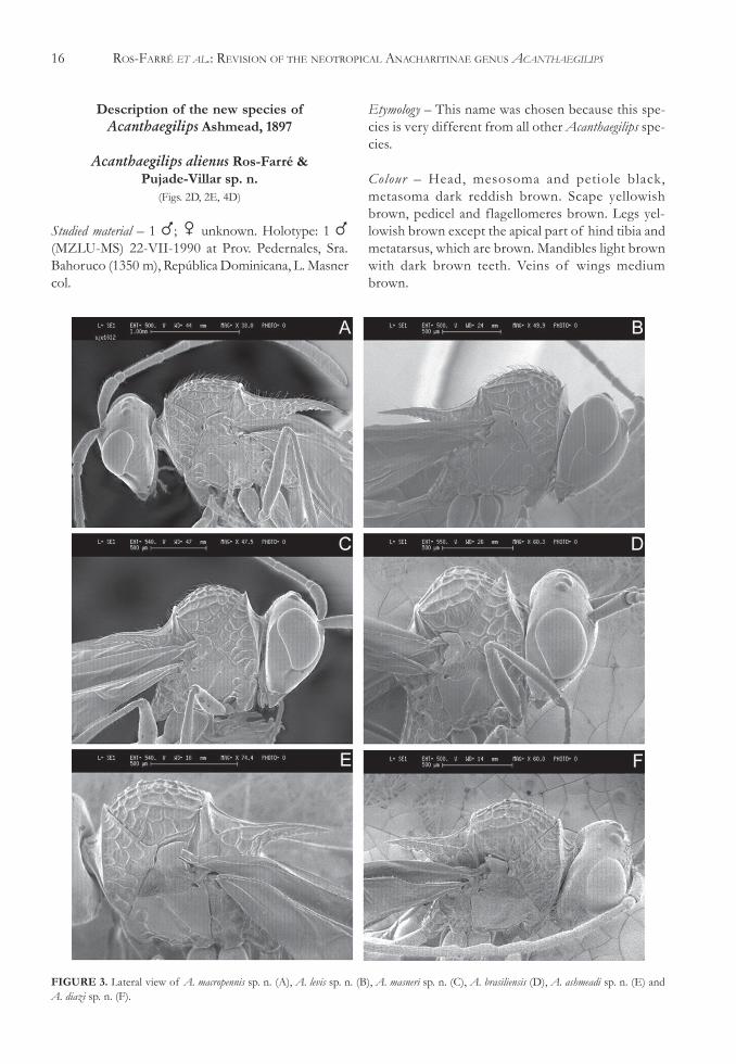
Acanthaegilips alienus Ros-Farré &Pujade-Villar sp. n.
(Figs. 2D, 2E, 4D)
Studied material – 1 �; � unknown. Holotype: 1 �(MZLU-MS) 22-VII-1990 at Prov. Pedernales, Sra.Bahoruco (1350 m), República Dominicana, L. Masnercol.
Etymology – This name was chosen because this spe-cies is very different from all other Acanthaegilips spe-cies.
Colour – Head, mesosoma and petiole black,metasoma dark reddish brown. Scape yellowishbrown, pedicel and flagellomeres brown. Legs yel-lowish brown except the apical part of hind tibia andmetatarsus, which are brown. Mandibles light brownwith dark brown teeth. Veins of wings mediumbrown.
FIGURE 3. Lateral view of A. macropennis sp. n. (A), A. levis sp. n. (B), A. masneri sp. n. (C), A. brasiliensis (D), A. ashmeadi sp. n. (E) andA. diazi sp. n. (F).
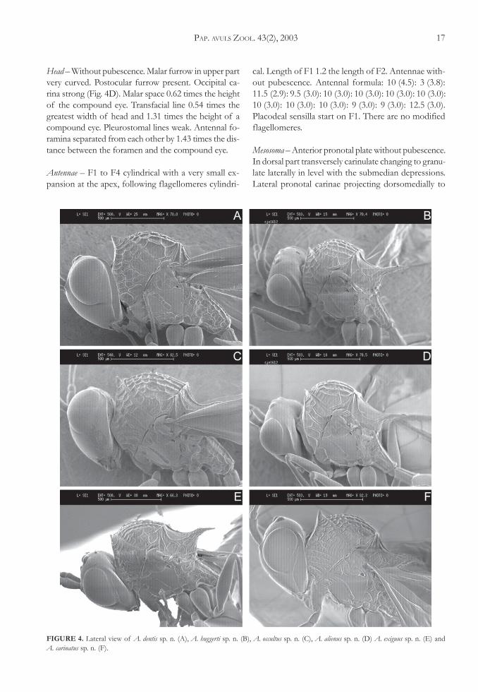
PAP. AVULS ZOOL. 43(2), 2003 17
Head – Without pubescence. Malar furrow in upper partvery curved. Postocular furrow present. Occipital ca-rina strong (Fig. 4D). Malar space 0.62 times the heightof the compound eye. Transfacial line 0.54 times thegreatest width of head and 1.31 times the height of acompound eye. Pleurostomal lines weak. Antennal fo-ramina separated from each other by 1.43 times the dis-tance between the foramen and the compound eye.
Antennae – F1 to F4 cylindrical with a very small ex-pansion at the apex, following flagellomeres cylindri-
cal. Length of F1 1.2 the length of F2. Antennae with-out pubescence. Antennal formula: 10 (4.5): 3 (3.8):11.5 (2.9): 9.5 (3.0): 10 (3.0): 10 (3.0): 10 (3.0): 10 (3.0):10 (3.0): 10 (3.0): 10 (3.0): 9 (3.0): 9 (3.0): 12.5 (3.0).Placodeal sensilla start on F1. There are no modifiedflagellomeres.
Mesosoma – Anterior pronotal plate without pubescence.In dorsal part transversely carinulate changing to granu-late laterally in level with the submedian depressions.Lateral pronotal carinae projecting dorsomedially to
FIGURE 4. Lateral view of A. dentis sp. n. (A), A. huggerti sp. n. (B), A. occultus sp. n. (C), A. alienus sp. n. (D) A. exiguus sp. n. (E) andA. carinatus sp. n. (F).
18 ROS-FARRÉ ET AL.: REVISION OF THE NEOTROPICAL ANACHARITINAE GENUS ACANTHAEGILIPS
form a small tooth (Fig. 4D). Lateral surface ofpronotum with scattered hairs and with coarse areolatesculpture. Mesoscutum in lateral view 1.55 times higherthan long; in dorsal view 1.37 times wider than long.Median mesoscutal impression weak, slightly longerthan broad, notauli strongly and clearly impressedreaching 2/3 the length of mesoscutum, they arecrossed by transverse carinae, they are not made up ofa sequence of cells of the areolate sculpture (Fig. 2D).Line of hairs on the anterior and lateral margins ofthe scutum absent. Scutellum in dorsal view with thesame length as the mesoscutum. Dorsal, posterolat-eral and posterior surfaces of scutellum smooth. Scutel-lum concave roof-shaped, median scutellar carina al-most obsolete, only visible as a carina near the anteriormargin. Lateral pit of scutellar fovea elongated andnarrow, 2.7 times longer than wide; the distance frommedian scutellar carina to the inner margin of the pit7.7 times the width of the pit. Scutellar disc in lateralview slopping to base of scutellar spine. End of scutel-lar disc narrowing abruptly to base of scutellar spine,which is slender, all the way (Fig. 2D) and straight(Fig. 4D). Mesopleural impression wide, straight andwith transverse carinae. Mesopleural triangle with scat-tered hairs (Fig. 4D). Propodeum smooth between lat-eral and median propodeal carinae (Fig. 2E) and ar-eolate in lateral areas.
Wings – Marginal cell open 2.73 times longer than wide.R1 absent. Marginal pubescence of the wing startingfrom the apical 3/4 of marginal cell but it is very sparseto the end of the cell.
Metasoma – Petiole 1.8 times as long as wide. Petiolewith two dorsolateral furrows and ventrally with onelongitudinal carina; weakly carinate laterally near ante-rior and posterior margins. Third abdominal tergumdorsally 0.81 the length of the fourth tergum.
Acanthaegilips ashmeadi Ros-Farré &Pujade-Villar sp. n.
(Figs. 1E, 3E)
Studied material – 6 �, 3 �. Holotype: 1 � (MZLU-MS)20-XII-1983 at Cuzco, Agua Caliente (PERU) LarsHuggert col. Paratypes: Peru: 1 � (MZLU-MS),21-XII-83 at Cuzco, Machu Pichu, Lars Huggert col.1 � & 1 � (MZLU-MS) 28-XII-1983 same Holotype’sdata. 2 � (MZLU-MS) same Holotype’s data. 1 �(MZUSP) 1/2-VII-1964 at Machu Pichu(2600-2800 m), B. Malkin col. Venezuela: 1 � (USNM)30-IV-1981 at Merida, Mucui nr. Tabay, Masner &Marsh col. 2 � (MZLU-MS) 24-VIII-1992 at Trujillo,Mosquey, near Bocono (1500 m). Cofee plant, L.Masner col.
Etymology – This name was chosen to honour Mr. Wil-liam Harris Ashmead (1855-1908), U.S.A, who estab-lished the genus Acanthaegilips.
Length of head + mesosoma + metasoma in female:2.75 mm; in male: 3.1 mm to 3.9 mm.
Colour – Female. Head, thorax and gaster black. Legsyellowish brown except hind coxa, which is black, andhind femur, tibia and tarsus, which are dark brown.Antennae dark brown, F7 to F10 ventrally lighter.Mandibles yellowish brown with darker teeth. Male.Differs from female in having the antennae entirelyblack. Veins of wings dark brown.
Head – Female. Head with scattered short hairs. Malarfurrow slightly curved. Postocular furrow absent. Oc-cipital carina absent. Malar space 0.58 to 0.71 timesthe height of the compound eye. Transfacial line 0.53to 0.55 times the greatest width of head and 1.03 to1.08 times the height of a compound eye. Antennal
FIGURE 5. Anterior wing (without pubescence) of A. brasiliensis (A), A. masneri sp. n. (B) and A. dentis sp. n. (C).
PAP. AVULS ZOOL. 43(2), 2003 19
foramina separated from each other by the same dis-tance as that between the foramen and the compoundeye. Pleurostomal lines weak. Male. Malar space 0.50to 0.63 times the height of the compound eye.Transfacial line 0.52 to 0.57 times the greatest widthof head and 1.03 to 1.10 times the height of a com-pound eye.
Antennae – Female. F1 to F3 cylindrical with a verysmall expansion at the apex. Following flagellomerescylindrical. Length of F1 1.4 to 1.5 the length of F2.Scape and pedicel with sparse pubescence. The pu-bescence on the flagellomeres getting shorter anddenser towards the 4 or 5 last flagellomeres, which havedense but very short pubescence. Antennal formula:10 (3.5): 3 (2): 15 (2): 10 (2): 9 (2): 9 (2): 6.5 (2): 6 (2):5 (3): 5 (3): 5 (3): 4 (2): 8 (2). Placodeal sensilla starton F2 but they are absent on the dorsal part of thissegment and very scarce on dorsal part of F3. Male.Length of F1 1.36 to 1.44 times the length of F2.Flagellomeres covered with very short pubescence.Placodeal sensilla start on F1 but they are absent inthe dorsal part of F1 and F2, on F3 and sometimesalso on F4 they are very scarce at the dorsal part. F3 toF5 slightly expanded dorsolaterally with a longitudinalridge on the raised part. Antennal formula: 13 (5.8):4.5 (5.0): 25 (5.0): 18 (4.9): 17 (4.9): 17 (4.8): 15 (4.7):15 (4.7): 12.5 (4.7): 12 (4.8): 11.5 (4.8): 11.5 (4.8):11 (4.7): 13.5 (4.5).
Mesosoma – Female. Anterior pronotal plate roundeddorsally (Fig. 3E) and with a few white hairs near lat-eral and dorsolateral margins; near the dorsal marginthere is a weak trace of transversely carinulate sculp-ture. Lateral surface of pronotum with sparse pubes-cence and with coarse areolate sculpture. Mesoscutumin lateral view 1.70 to 1.80 times higher than long; indorsal view 1.30 to 1.42 times wider than long. Medianmesoscutal impression short, not much longer thanwide. Notauli reaching 1/2 the length of mesoscutum(Fig. 1E). There is a conspicuous line of hairs on theanterior and lateral margins of the mesoscutum. Scutel-lum in dorsal view 1.22 to 1.43 times the length of themesoscutum. Scutellar foveae smooth, median and lat-eral ridges of the scutellar foveae sometimes not con-tinuous and never straight to the end sometimes di-vided in smaller carinae. Lateral pit of scutellar fovea1.4 to 1.5 times longer than wide; the distance frommedian scutellar carina to the inner margin of the pit3.1 to 3.3 times the width of the pit. Scutellar spinebroad at base and narrowing gradually (Fig. 1E). Scutel-lar disc in lateral view slightly slopping to base of scutel-
lar spine, which is horizontally directed at base andslightly curved downwards towards apex. Mesopleuralimpression wide straight and smooth or, in some speci-mens, with weak transverse carinae. Mesopleural tri-angle densely pubescent anteriorly, sparsely pubescentposteriorly. Propodeum with coarse areolate sculpture.Lateral propodeal carinae and median propodeal cari-nae absent. Male. Anterior pronotal plate with a fewhairs near lateral margins. Mesoscutum in lateral view1.53 to 1.65 times higher than long. Scutellum in dor-sal view 1.13 to 1.28 times the length of themesoscutum.
Wings – Female. Marginal cell closed 3.13 to 3.20 timeslonger than wide. Marginal pubescence of the wingpresent starting from vein R1. Male. Marginal cell 2.9to 3.0 times longer than wide.
Metasoma – Petiole 3.0 to 3.3 times as long as wide.Petiole dorsally smooth; laterally smooth but nearthe anterior end there are a few short carinae; ven-trally costate. Third abdominal tergum dorsally 0.75to 0.88 the length of the fourth tergum. Male. Peti-ole 3.2 to 3.3 times as long as broad. Third abdomi-nal tergum dorsally 0.69 to 0.85 the length of thefourth tergum.
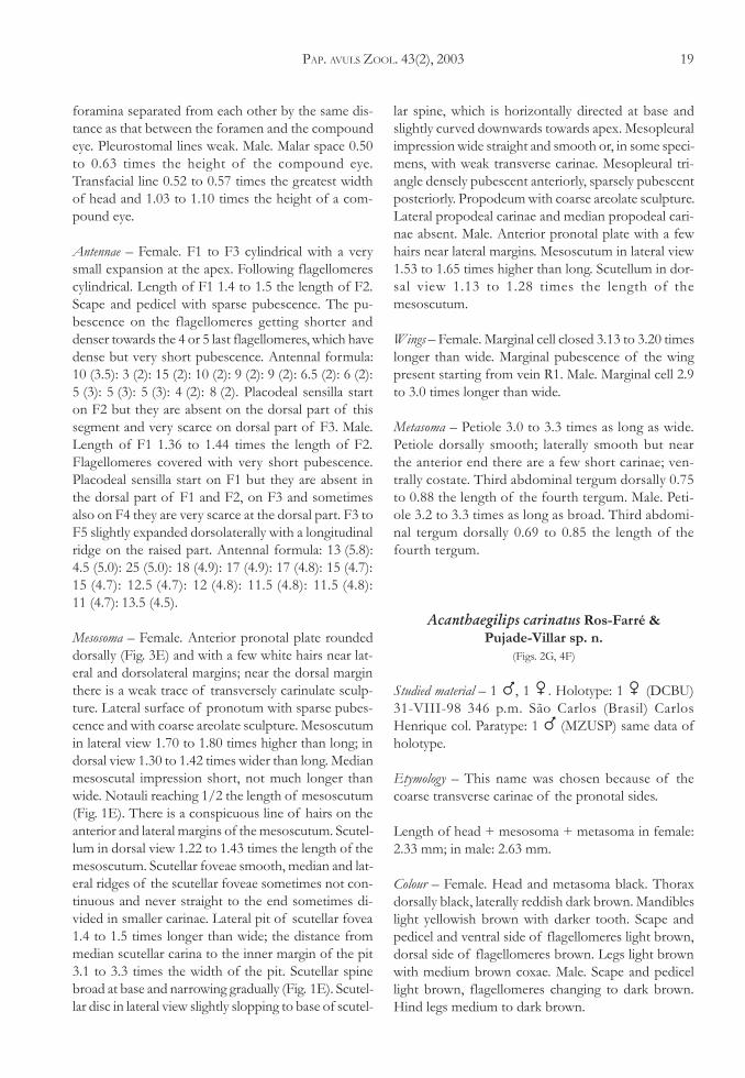
Acanthaegilips carinatus Ros-Farré &Pujade-Villar sp. n.
(Figs. 2G, 4F)
Studied material – 1 �, 1 �. Holotype: 1 � (DCBU)31-VIII-98 346 p.m. São Carlos (Brasil) CarlosHenrique col. Paratype: 1 � (MZUSP) same data ofholotype.
Etymology – This name was chosen because of thecoarse transverse carinae of the pronotal sides.
Length of head + mesosoma + metasoma in female:2.33 mm; in male: 2.63 mm.
Colour – Female. Head and metasoma black. Thoraxdorsally black, laterally reddish dark brown. Mandibleslight yellowish brown with darker tooth. Scape andpedicel and ventral side of flagellomeres light brown,dorsal side of flagellomeres brown. Legs light brownwith medium brown coxae. Male. Scape and pedicellight brown, flagellomeres changing to dark brown.Hind legs medium to dark brown.
20 ROS-FARRÉ ET AL.: REVISION OF THE NEOTROPICAL ANACHARITINAE GENUS ACANTHAEGILIPS
Head – Female. Head without pubescence, except forlines of hairs running with malar and postocular fur-rows. Malar furrow very weak, only marked by a changein the integument curvature; postocular furrow weak.Occipital carina strong. Malar space 0.63 times theheight of the compound eye. Transfacial line 0.53 timesthe greatest width of the head and 1.07 times the heightof a compound eye. Pleurostomal lines well impressed.Antennal foramina separated from each other by 2.25times the distance between the foramen and the com-pound eye. Male. Malar furrow only marked by a changeon the integument curvature but not as weak as in thefemale. Malar space 0.55 times the height of the com-pound eye. Transfacial line 0.52 times the greatest widthof the head and with the same length as the height ofa compound eye. Antennal foramina separated fromeach other by 2.16 times the distance between the fo-ramen and the compound eye.
Antennae – Female. Flagellomeres cylindrical (slightlynarrower on basis and apex). Length of F1 1.25 thelength of F2. Antenna without pubescence except fora few hairs on scape. Antennal formula: 10 (4): 3 (3):10 (2.8): 8 (2.5): 7 (2.5): 7 (2.7): 6.5 (2.9): 6 (3): 5.7 (3):5.5 (3): 5 (3): 5 (3): 10 (3). Placodeal sensilla start onF2 they are not scarce in this antennomere. Male.Flagellomeres cylindrical (slightly narrower on basis andapex). Length of F1 1.25 the length of F2. Antennawithout pubescence except for a few hairs on scape.Antennal formula: 11 (4): 3.5 (3.5): 12.5 (3): 10 (3):9 (3): 9.8 (3.5): 9 (3.5): 9 (3): 8.5 (3): 8 (3): 8 (3): 7.5 (3):7.2 (3): 10.2 (3). Placodeal sensilla start on F1 and theyare not scarce in any antennomere. F3 to F5 and first1/3 of F6 slightly expanded dorsolaterally with a lon-gitudinal ridge on the raised part.
Mesosoma – Female. Lateral pronotal carinae project-ing dorsomedially to form a very much raised tooth(Fig. 4F).
Anterior pronotal plate with a few scattered hairsnear dorsolateral margins and transversely carinate. Lat-eral surface of pronotum densely pubescent in ventralpart with coarse transverse carinae, in dorsal part withareolate-rugose sculpture. Mesoscutum in lateral view1.67 times higher than long; in dorsal view 1.25 timeswider than long. Median mesoscutal impression short,longer than wide. Notauli complete (Fig. 2G). Line ofhairs on the anterior and lateral margins of the scutumabsent. Scutellum in dorsal view 1.05 times the lengthof the mesoscutum. Scutellar foveae in anterior part withan internal short longitudinal carina, dividing the ante-
rior area of each fovea into two areas, scutellar foveaemostly smooth with rugose sculpture near its posteriormargin. Lateral pit of scutellar fovea absent. Scutellarspine broad, narrowing abruptly before apex (Fig. 2G).Scutellar disc in lateral view steeply slopping to base ofscutellar spine, which is upwards directed at base andcurved downwards, the apex is ventrally directed.Mesopleural impression rather narrow strongly curvedin the apical part and with a lot of transverse carinae(Fig. 4F). Mesopleural triangle with very sparse pubes-cence. Propodeum with areolate sculpture, lateral andmedian propodeal carinae present, these are flat and widein upper part. Male. Mesoscutum in lateral view 1.55times higher than long; in dorsal view 1.19 times widerthan long. Scutellum in dorsal view with the same lengthas that of the mesoscutum. Scutellar foveae more sculp-tured posteriorly than in the female. Lateral pit of scutel-lar fovea obstructed. Scutellar spine very slightly curved.Mesopleural impression not as curved as in the female.
Wings – Female. Marginal cell open 2.88 times longerthan wide. R1 practically absent. Marginal pubescenceof the wing starting from the apical 1/2 of marginalcell but it is very sparse to the end of the cell. Male.Marginal cell 3.38 times longer than wide. Marginalpubescence starting from the basal 1/3.
Metasoma – Female. Petiole as long as wide. Petioledorsally smooth, laterally and ventrally costate. Thirdabdominal tergum dorsally 0.5 times the length of thefourth tergum. Male. Petiole wider than long. Dorsally,laterally and ventrally costate.
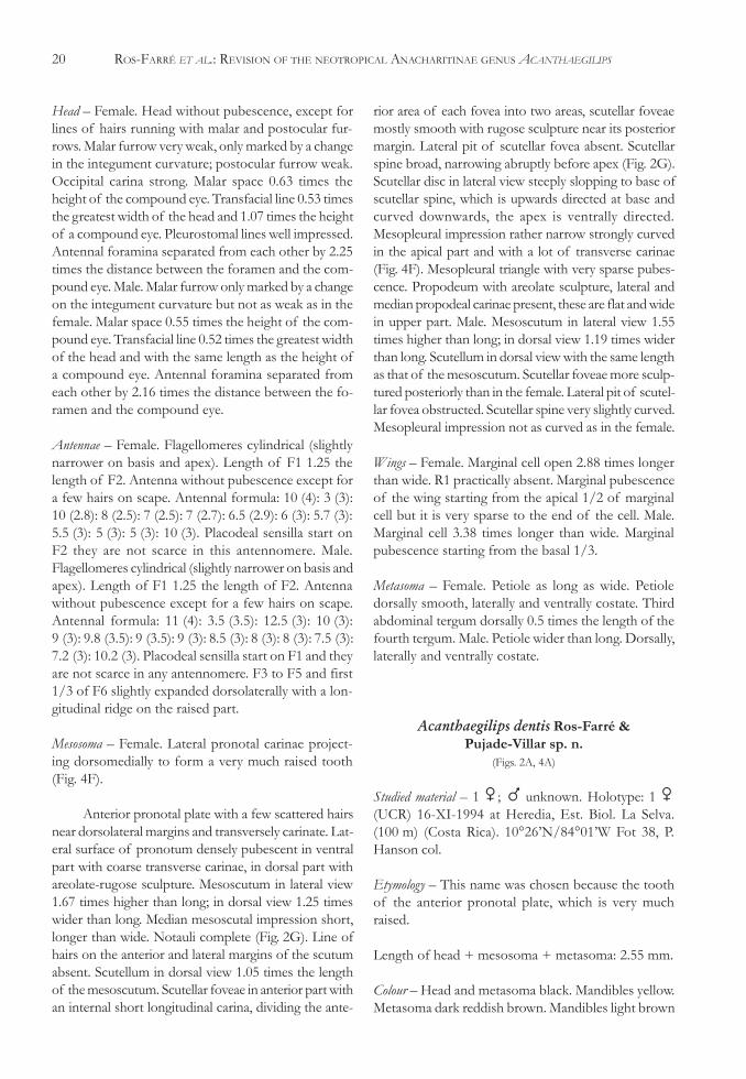
Acanthaegilips dentis Ros-Farré &Pujade-Villar sp. n.
(Figs. 2A, 4A)
Studied material – 1 �; � unknown. Holotype: 1 �(UCR) 16-XI-1994 at Heredia, Est. Biol. La Selva.(100 m) (Costa Rica). 10°26’N/84°01’W Fot 38, P.Hanson col.
Etymology – This name was chosen because the toothof the anterior pronotal plate, which is very muchraised.
Length of head + mesosoma + metasoma: 2.55 mm.
Colour – Head and metasoma black. Mandibles yellow.Metasoma dark reddish brown. Mandibles light brown
PAP. AVULS ZOOL. 43(2), 2003 21
with medium brown teeth. Antennae light brown withexception of the four last flagellomeres, which are darkbrown. Legs light brown with the exception of themetafemur and the metatibia, which are dark brown.Veins of wing light brown.
Head – Without pubescence. Malar furrow very curved,postocular furrow well impressed (Fig. 4A). Occipitalcarina strong. Clypeal zone not marked andpleurostomal lines very weak. Malar space 0.65 timesthe height of the compound eye. Transfacial line 0.58times the greatest width of the head and 1.11 timesthe height of a compound eye. Antennal foramina sepa-rated from each other by 1.5 times the distance be-tween the foramen and the compound eye.
Antennae – F1 to F4 cylindrical with a very small ex-pansion at the apex, following flagellomeres cylindri-cal. Length of F1 1.33 the length of F2. Scape withscattered short hairs, pedicel and flagellomeres gla-brous. Antennal formula: 10 (3): 2 (2): 8 (2): 6 (2):5.5 (2): 5.5 (2): 4.5 (2): 4 (2): 4 (2): 4 (2.5): 4 (3): 4 (3):8 (3). Placodeal sensilla start on F3 but they are dor-sally scarce and ventrally absent on this flagellomere,on F4 they are ventrally scarce.
Mesosoma – Lateral pronotal carinae projectingdorsomedially to form a very much raised tooth(Fig. 4A). Anterior pronotal plate with short scatteredhairs on the margins and with a very conspicuous ru-gulose sculpture. Lateral surface of pronotum withdense pubescence and with areolate sculpture.Mesoscutum in lateral view 1.66 times higher than long;in dorsal view 1.17 times wider than long. Medianmesoscutal impression short, longer than broad.Notauli complete (Fig. 2A). Line of hairs on the ante-rior and lateral margins of the scutum absent. Scutel-lum in dorsal view 1.05 times the length of themesoscutum. Scutellar foveae smooth and with an in-ternal longitudinal carina dividing them into two areaseach. There is rugose sculpture at the area compressedbetween the end of scutellar foveae and before theapex. Lateral pit of scutellar fovea almost circular, 1.1times longer than wide; the distance from medianscutellar carina to the inner margin of the pit 2.78 timesthe width of the pit. Scutellar spine broad narrowsabruptly before apex (Fig. 2A). Scutellar disc in lateralview steeply slopping to base of scutellar spine, whichis strongly directed upwards at base and curved down-wards, so that the apex is ventrally directed. Mesopleuralimpression rather narrow curved in the apical part andwith few coarse transverse carinae (Fig. 4A).
Mesopleural triangle sparsely pubescent anteriorly andmore sparsely pubescent posteriorly (Fig. 4A).Propodeum with areolate sculpture. Median and lat-eral propodeal carinae present.
Wings – Marginal cell open 2.6 times longer than wide.R1 short not reaching the margin of the wing. Mar-ginal pubescence of the wing starting from the apical1/3 of marginal cell but it is very sparse to the end ofthe cell.
Metasoma – Petiole 1.12 times as long as wide. Petioledorsally, laterally and ventrally costate. Third abdomi-nal tergum dorsally 0.63 the length of the fourth ter-gum.
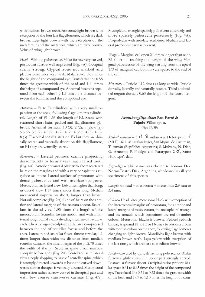
Acanthaegilips diazi Ros-Farré &Pujade-Villar sp. n.
(Figs. 1F, 3F)
Studied material – 3 �; � unknown. Holotype: 1 �(MLP) 16-11-81 at San Javier, San Miguel de Tucumán,Tucumán (República Argentina) S. Mulvany, N. Díaz,G. Armesto, P. Fidalgo col. Paratypes: 2 �, SameHolotype’s data.
Etymology – This name was chosen to honour Dra.Norma Beatriz Díaz, Argentina, who loaned us all typespecimens of this species.
Length of head + mesosoma + metasoma: 2.9 mm to3.4 mm.
Colour – Head black, mesosoma black with exception ofthe lateroventral margins of pronotum, the anterior andlateral margins of mesoscutum, the mesopleural triangleand the notauli, which sometimes are red or ambercolour. Metasoma blackish brown. Pedicel reddishbrown, scape and F1 to F5 or F6 black to blackish brownwith reddish colour on the apex, following flagellomereschanging to light brown. Mandibles light brown withmedium brown teeth. Legs yellow with exception ofthe last ones, which are dark to medium brown.
Head – Covered by quite dense long pubescence. Malarfurrow slightly curved, in upper part strongly curved.Postocular furrow absent. Occipital carina present. Ma-lar space 0.61 to 0.65 times the height of the compoundeye. Transfacial line 0.51 to 0.52 times the greatest widthof the head and 1.07 to 1.10 times the height of a com-
22 ROS-FARRÉ ET AL.: REVISION OF THE NEOTROPICAL ANACHARITINAE GENUS ACANTHAEGILIPS
pound eye. Pleurostomal lines weak. Antennal foraminaseparated from each other by 1.20 to 1.25 times the dis-tance between the foramen and the compound eye.
Antennae – Flagellomeres cylindrical. Length of F1 1.46the length of F2. Scape and pedicel with sparse pubes-cence. Flagellomeres covered with short dense pubes-cence. Antennal formula: 16 (5.5): 5 (4): 22 (3.5):15 (3.5): 14 (4): 14 (3.5): 12 (3.5): 12 (3.5): 12 (3.5):11.8 (3.3): 10 (3.3): 9.5 (3.2): 8.5 (3.5): 14 (3). Placodealsensilla start on F1 but they are dorsally absent fromF1 to F5; Sensilla are very sparse on dorsal part of F6and F7. F3 to F5 slightly expanded dorsolaterally witha longitudinal ridge on the raised part.
Mesosoma – Anterior pronotal plate rounded dorsally(Fig. 1F) with quite dense pubescence at the lateral ar-eas and sparse pubescence in lateroanterior margins,near the dorsal margin weakly transversely carinulate.Lateral face of pronotum with dense long pubescence,the sculpture is between rugose and areolate.Mesoscutum in lateral view 1.69 to 2.03 times higherthan long; in dorsal view 1.24 to 1.27 times wider thanlong. Median mesoscutal impression very short as longas broad. Notauli complete and well differentiated. Theareolate sculpture of the mesoscutum is laterally ef-faced, in some specimens is weak in medial part(Figs. 1F, 3F). There is a very conspicuous line of hairson the anterior and lateral margins of the mesoscutum.Scutellum in dorsal view 1.0 to 1.1 times the length ofthe mesoscutum. Scutellar foveae smooth (Fig. 1F) thelateral carinae of fovea are not straight in the posteriorpart. Lateral pit of scutellar fovea big, 1.5 to 2.0 timeslonger than wide; the distance from median scutellarcarina to the inner margin of the pit 2.87 to 3.50 timesthe width of the pit. Scutellar spine broad at base nar-rowing gradually towards apex (Fig. 1F). Scutellar discin lateral view slightly slopping to base of scutellarspine, which is horizontally directed at base and veryslightly curved downwards towards apex (Fig. 3F).Mesopleural impression straight and wide (stronglywider on the basal part) and without transverse cari-nae. Parapsidalsigna strongly marked and continuous,sometimes meeting on the anterior part. Mesolpeuraltriangle with dense pubescence anteriorly and sparsepubescence posteriorly. Propodeum with coarse, ar-eolate sculpture, lateral and median propodeal carinaeabsent.
Wings – Marginal cell closed, 3.22 to 3.40 times longerthan wide. Marginal pubescence of the wing startingfrom vein R1.
Metasoma – Petiole 2.0 to 2.2 times as long as wide,dorsally smooth, laterally smooth, dorsolaterally nearthe anterior end costate, ventrally longitudinally cos-tate. Third abdominal tergum dorsally 1.00 to 1.08 thelength of the fourth tergum.
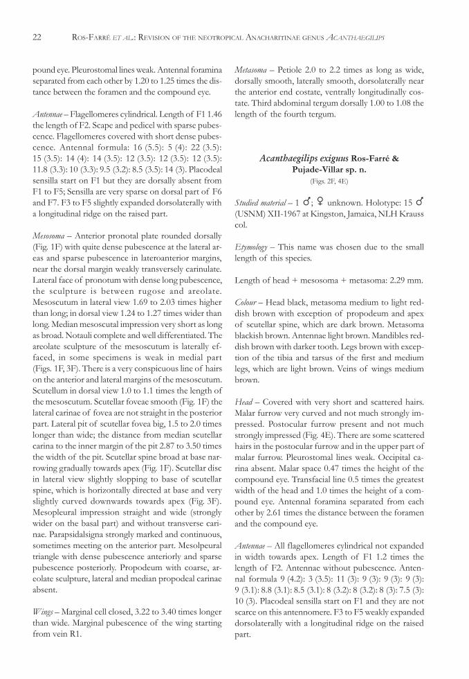
Acanthaegilips exiguus Ros-Farré &Pujade-Villar sp. n.
(Figs. 2F, 4E)
Studied material – 1 �; � unknown. Holotype: 15 �(USNM) XII-1967 at Kingston, Jamaica, NLH Krausscol.
Etymology – This name was chosen due to the smalllength of this species.
Length of head + mesosoma + metasoma: 2.29 mm.
Colour – Head black, metasoma medium to light red-dish brown with exception of propodeum and apexof scutellar spine, which are dark brown. Metasomablackish brown. Antennae light brown. Mandibles red-dish brown with darker tooth. Legs brown with excep-tion of the tibia and tarsus of the first and mediumlegs, which are light brown. Veins of wings mediumbrown.
Head – Covered with very short and scattered hairs.Malar furrow very curved and not much strongly im-pressed. Postocular furrow present and not muchstrongly impressed (Fig. 4E). There are some scatteredhairs in the postocular furrow and in the upper part ofmalar furrow. Pleurostomal lines weak. Occipital ca-rina absent. Malar space 0.47 times the height of thecompound eye. Transfacial line 0.5 times the greatestwidth of the head and 1.0 times the height of a com-pound eye. Antennal foramina separated from eachother by 2.61 times the distance between the foramenand the compound eye.
Antennae – All flagellomeres cylindrical not expandedin width towards apex. Length of F1 1.2 times thelength of F2. Antennae without pubescence. Anten-nal formula 9 (4.2): 3 (3.5): 11 (3): 9 (3): 9 (3): 9 (3):9 (3.1): 8.8 (3.1): 8.5 (3.1): 8 (3.2): 8 (3.2): 8 (3): 7.5 (3):10 (3). Placodeal sensilla start on F1 and they are notscarce on this antennomere. F3 to F5 weakly expandeddorsolaterally with a longitudinal ridge on the raisedpart.
PAP. AVULS ZOOL. 43(2), 2003 23
Mesosoma – Lateral pronotal carinae projecteddorsomedially to form a small and few raised tooth(Fig. 4E). Pronotal plate with few scattered short hairs,and with coriaceous sculpture. Lateral surface ofpronotum with quite dense pubescence, weak areolate-reticulate sculptured in the apical part and with irregu-lar transverse carinae in the ventral part. Mesoscutumin lateral view 1.48 times higher than long; in dorsalview 1.22 times wider than long. Median mesoscutalimpression very short and little longer than broad,notauli complete (Fig. 2F). Line of hairs on the ante-rior and lateral margins of the mesoscutum absent.Carinae between areolate sculpture wider and shinierthan all other species of the group. Scutellum in dorsalview 0.91 times the length of the mesoscutum. Scutel-lar foveae smooth in the most part and with rugosesculpture in their posterior part (Fig. 2F). Lateral pit offovea obstructed, superficial 1.6 times longer than wide;the distance from median scutellar carina to the innermargin of the pit 3.8 times the width of the pit. Scutel-lar spine broad narrowing abruptly before apex. Scutellardisc in lateral view steeply slopping to base of scutellarspine, which is directed upwards at base and curveddownwards, so that the apex is ventrally directed.Mesopleural impression narrow curved in upper partand with abundant weak transverse carinae. Mesopleuraltriangle glabrous. Propodeum with areolate sculpture.Median and lateral propodeal carinae present. The lat-eral carinae are wide and flat in upper part.
Wings – Marginal cell open 2.75 times longer than wide.R1 absent. Marginal pubescence of the wing startingfrom the apical 2/3 of marginal cell but it is very sparseto the end of the cell.
Metasoma – Petiole 1.8 times as long as wide, dorsallysmooth, laterally and ventrally costate. Third abdomi-nal tergum dorsally 0.6 times the length of the fourthtergum.
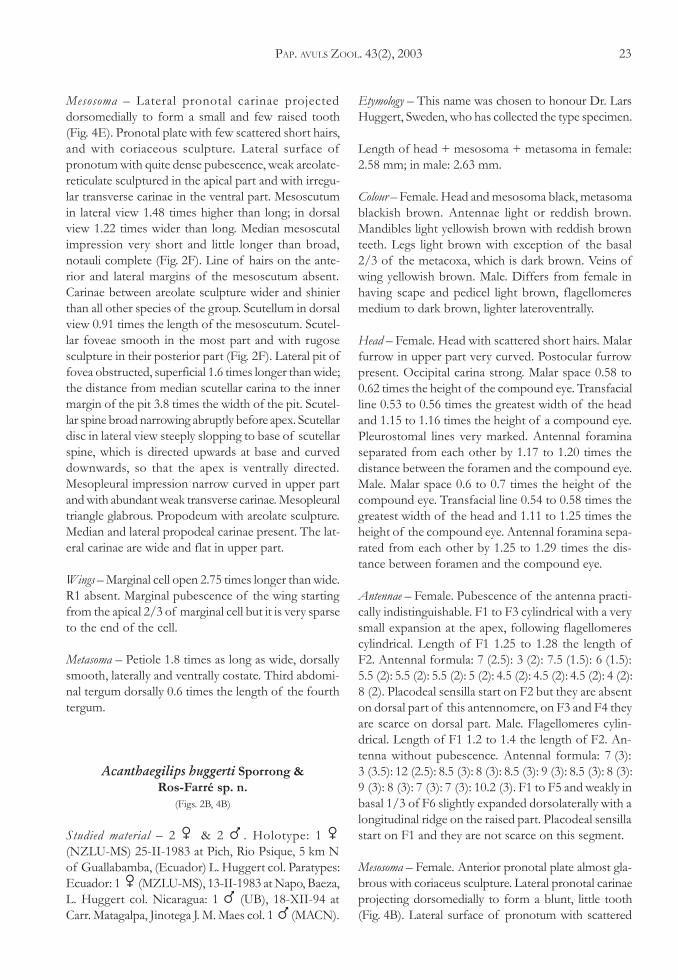
Acanthaegilips huggerti Sporrong &Ros-Farré sp. n.
(Figs. 2B, 4B)
Studied material – 2 � & 2 � . Holotype: 1 �(NZLU-MS) 25-II-1983 at Pich, Rio Psique, 5 km Nof Guallabamba, (Ecuador) L. Huggert col. Paratypes:Ecuador: 1 � (MZLU-MS), 13-II-1983 at Napo, Baeza,L. Huggert col. Nicaragua: 1 � (UB), 18-XII-94 atCarr. Matagalpa, Jinotega J. M. Maes col. 1 � (MACN).
Etymology – This name was chosen to honour Dr. LarsHuggert, Sweden, who has collected the type specimen.
Length of head + mesosoma + metasoma in female:2.58 mm; in male: 2.63 mm.
Colour – Female. Head and mesosoma black, metasomablackish brown. Antennae light or reddish brown.Mandibles light yellowish brown with reddish brownteeth. Legs light brown with exception of the basal2/3 of the metacoxa, which is dark brown. Veins ofwing yellowish brown. Male. Differs from female inhaving scape and pedicel light brown, flagellomeresmedium to dark brown, lighter lateroventrally.
Head – Female. Head with scattered short hairs. Malarfurrow in upper part very curved. Postocular furrowpresent. Occipital carina strong. Malar space 0.58 to0.62 times the height of the compound eye. Transfacialline 0.53 to 0.56 times the greatest width of the headand 1.15 to 1.16 times the height of a compound eye.Pleurostomal lines very marked. Antennal foraminaseparated from each other by 1.17 to 1.20 times thedistance between the foramen and the compound eye.Male. Malar space 0.6 to 0.7 times the height of thecompound eye. Transfacial line 0.54 to 0.58 times thegreatest width of the head and 1.11 to 1.25 times theheight of the compound eye. Antennal foramina sepa-rated from each other by 1.25 to 1.29 times the dis-tance between foramen and the compound eye.
Antennae – Female. Pubescence of the antenna practi-cally indistinguishable. F1 to F3 cylindrical with a verysmall expansion at the apex, following flagellomerescylindrical. Length of F1 1.25 to 1.28 the length ofF2. Antennal formula: 7 (2.5): 3 (2): 7.5 (1.5): 6 (1.5):5.5 (2): 5.5 (2): 5.5 (2): 5 (2): 4.5 (2): 4.5 (2): 4.5 (2): 4 (2):8 (2). Placodeal sensilla start on F2 but they are absenton dorsal part of this antennomere, on F3 and F4 theyare scarce on dorsal part. Male. Flagellomeres cylin-drical. Length of F1 1.2 to 1.4 the length of F2. An-tenna without pubescence. Antennal formula: 7 (3):3 (3.5): 12 (2.5): 8.5 (3): 8 (3): 8.5 (3): 9 (3): 8.5 (3): 8 (3):9 (3): 8 (3): 7 (3): 7 (3): 10.2 (3). F1 to F5 and weakly inbasal 1/3 of F6 slightly expanded dorsolaterally with alongitudinal ridge on the raised part. Placodeal sensillastart on F1 and they are not scarce on this segment.
Mesosoma – Female. Anterior pronotal plate almost gla-brous with coriaceus sculpture. Lateral pronotal carinaeprojecting dorsomedially to form a blunt, little tooth(Fig. 4B). Lateral surface of pronotum with scattered
24 ROS-FARRÉ ET AL.: REVISION OF THE NEOTROPICAL ANACHARITINAE GENUS ACANTHAEGILIPS
hairs and with coarse areolate sculpture. Mesoscutumin lateral view 1.6 times higher than long; in dorsal view1.29 times wider than long. Median mesoscutal impres-sion very short, at most little longer than broad. Notaulireaching 1/2 or 1/3 the length of mesoscutum (Fig. 2B).Line of hairs on the anterior and lateral margins of themesoscutum absent. Scutellum in dorsal view of thesame length as that of the mesoscutum. Scutellar foveaesmooth with some weak sculpture at its posterior mar-gin. Lateral pit of scutellar fovea rather small and al-most circular, 1.2 times longer than wide; the distancefrom median scutellar carina to the inner margin of thepit 4.0 times the width of the pit. End of scutellar discnarrowing abruptly to base of scutellar spine, which isslender all the way (Fig. 2B). Scutellar disc in lateral viewsteeply slopping to base of scutellar spine. Scutellar spinestraight not directed ventrally (Fig. 4B). Mesopleuralimpression rather narrow straight and with few trans-verse carinae. Mesopleural triangle glabrous. Propodeumareolate but not coarsely, median and lateral propodealcarinae present. Male. Mesoscutum in lateral view 1.60to 1.67 times higher than long; in dorsal view 1.15 to1.33 times wider than long. Scutellum in dorsal view ofthe same length as that of the mesoscutum. Lateral pitof scutellar fovea 1.20 to 1.33 times longer than wide;the distance from median scutellar carina to the innermargin of the pit 3.33 times the width of the pit.Mesopleural impression with only one or two weak trans-verse carinae.
Wings – Female. Marginal cell open; between 2.70 and2.80 times longer than wide. R1 practically non-exis-tent. Marginal pubescence of the wing starting fromvein R1. Margin of the marginal cell sparsely pubes-cent anteriorly and densely pubescent apically. Male.Marginal cell 3.14 to 3.44 times longer than wide; itsmargin rather densely pubescent anteriorly and apically.
Metasoma – Female. Petiole between 1.7 and 2.0 timesas long as wide. Petiole dorsally smooth laterally andventrally longitudinally costate. Third abdominal ter-gum dorsally between 0.60 and 0.71 the length of thefourth tergum.
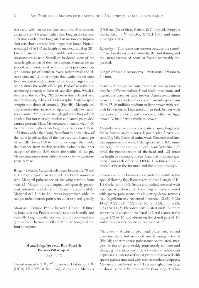
Acanthaegilips levis Ros-Farré &Pujade-Villar. sp. n.
(Figs. 1B, 3B)
Studied material – 2 �; � unknown. Holotype: 1 �(UCR) III-1995 at San José, Zurqui de Moravia
(1600 m), (Costa Rica), Hanson & Godoy col. Paratype:Costa Rica: 1 � (UCR), X/XII-1990 and sameHolotype’s data.
Etymology – This name was chosen because the scutel-lum in dorsal view is very smooth, flat and shining andthe lateral carinae of scutellar foveae are weakly im-pressed.
Length of head + mesosoma + metasoma: 2.9 mm to3.1 mm.
Colour – Although we only examined two specimensthey had different colour. Head black, mesosoma andmetasoma black or dark brown. Antennae mediumbrown or black with amber colour towards apex fromF1 to F5. Mandibles medium or light brown with red-dish brown teeth. Legs medium to dark brown withexception of procoxa and mesocoxa, which are lightbrown. Veins of wing medium brown.
Head – Covered with very few scattered quite long hairs.Malar furrow slightly curved; postocular furrow ab-sent (Fig. 3B). Occipital carina weak. Pleurostomal lineswell impressed and wide. Malar space 0.61 to 0.63 timesthe height of the compound eye. Transfacial line 0.57times the greatest width of the head and 1.21 timesthe height of a compound eye. Antennal foramina sepa-rated from each other by 1.00 to 1.14 times the dis-tance between the foramen and the compound eye.
Antennae – F2 to F4 weakly expanded in width at theapex, following flagellomeres cylindrical. Length of F11.5 the length of F2. Scape and pedicel covered withvery sparse pubescence. First flagellomeres coveredwith sparse pubescence this is getting dense towardslast flagellomeres. Antennal formula: 12 (3): 3 (2):14 (2): 9 (2): 8 (2): 7 (2): 6 (2): 5.5 (2): 5 (3): 5 (3): 5 (3):4.5 (3.5): 11 (3). Placodeal sensilla start on F1 but theyare ventrally absent at the basal 1/2 and scarce at theupper 1/2 of F1 and absent on the dorsal part of F1and F2 and scarce on the dorsal part of F3.
Mesosoma – Anterior pronotal plate very raiseddorsomedially but rounded not forming a tooth(Fig. 3B) and with sparse pubescence on the lateral mar-gins, in dorsal part weakly transversely carinate andchanging to coriaceous in level with the submediandepressions. Lateral surface of pronotum covered withsparse pubescence and with coarse areolate sculpture.Mesoscutum in lateral view 1.45 times higher than long;in dorsal view 1.25 times wider than long. Median
PAP. AVULS ZOOL. 43(2), 2003 25
mesoscutal impression very short a little longer thanbroad. Notauli reaching 1/2 to 2/3 the length ofmesoscutm (Fig. 1B). There is a weak line of hairs onthe anterior and lateral margins of the mesoscutum.Scutellum in dorsal view 1.31 times the length of themesoscutum. Scutellar foveae nude (Fig. 1B). Scutel-lar spine dorsally very smooth, lateral ridges of thefoveae not much marked, and narrowing gradually.Lateral pit of scutellar fovea rather big and oval, 1.45to 1.50 times longer than wide; the distance from me-dian scutellar carina to the inner margin of the pit 3.60to 3.90 times the width of the pit. Scutellar spine broadat base and narrowing gradually towards apex (Fig. 1B).Scutellar disc in lateral view slopping to base of scutellarspine, which is slightly upwards directed and almosthorizontally directed towards apex but the apex notventrally directed (Fig. 3B). Mesopleural impressionwide and straight with 3 or 4 very effaced transversecarinae (Fig. 3B). Mesopleural triangle with sparse pu-bescence anteriorly and with some scattered hairs pos-teriorly. Propodeum with coarse areolate sculpture, lat-eral and median propodeal carinae absent.
Wings – Marginal cell open 3.37 to 3.50 times longerthan wide. R1 arriving near the margin but not reach-ing it as in the Fig. 3A. Marginal pubescence startingfrom vein R1.
Metasoma – Petiole 2.6 times as long as wide, smoothdorsally, laterally and ventrally costate. Third abdomi-nal tergum dorsally 0.68 times the length of the fourthtergum.
Acanthaegilips macropennis Sporrong &Ros-Farré. sp. n.
(Figs. 1A, 3A)
Studied material – 10 � & 4 �. Holotype: 1 � (inCNC) 17-II-1983 at Napo bellow Papallacta 3000 m,(Ecuador), L.M. Masner col. Paratypes: Ecuador: 4 �& 1 � same Holotype’s data. 6 � & 2 � (inMZLU-MS) 24-II-83 at Napo Papallacta 3800 m.,Lars Huggert col.
Etymology – This name was chosen because the largewings, which this species has in common with severalspecies of Eucoilinae collected at the same altitudes.
Length of head + mesosoma + metasoma in female:3.5 mm to 4.4 mm; in male: 3.2 mm to 3.9 mm.
Colour – Female. Head and mesosoma black, metasomablackish brown. Scape, pedicel, F1 and F2 black, re-maining flagellomeres changing to brown. Mandiblesbrown with reddish brown teeth. Legs dark brown.Veins of wing medium to dark brown. Male. Differsfrom female in having all of the flagellomeres black.
Head – Female. Covered by sparse long hairs. Malarfurrow slightly curved; postocular furrow absent(Fig. 3A). Occipital carina absent. Malar space 0.77 to0.92 times the height of the compound eye. Transfacialline 0.56 to 0.58 times the greatest width of the headand 1.15 to 1.43 times the height of a compound eye.Pleurostomal lines very weak. Antennal foramina sepa-rated from each other by 0.76 to 0.80 times the dis-tance between the foramen and the compound eye.Male. Malar space 0.66 to 0.77 times the height of thecompound eye. Transfacial line 0.54 to 0.57 times thegreatest width of the head and 1.0 to 1.2 times theheight of a compound eye. Antennal foramina sepa-rated from each other by 0.5 to 0.8 times the distancebetween the foramen and the compound eye.
Antennae – Female. F1 to F4 slightly expanded in widthat the apex. Length of F1 1.41 to 1.60 the length ofF2. Scape and pedicel covered with sparse pubescence.Flagellomeres covered with dense pubescence. Anten-nal formula: 14 (3.5): 4 (3): 15 (3): 10 (3): 8 (3.2): 7 (3.2):6 (3.5): 6 (3.6): 6 (3.7): 5 (4): 5 (4): 5 (4): 11 (4).Placodeal sensilla start on F2 but they are absent dor-sally on F2, F3 and first 1/3 of F4. Male. Flagellomerescylindrical. Length of F1 1.3 to 1.5 the length of F2.Flagellomeres covered with short sparse pubescence.Antennal formula: 9 (4): 3 (3): 14.5 (3): 11 (2.8):10 (2.8): 10 (3): 9 (2.8): 9 (2.7): 9 (2.5): 9 (2.4): 8 (2.4):8 (2.4): 8 (2.4): 11 (2.3). Placodeal sensilla start on F1and only lacks in a small area of basal part of F1. F3to F5 slightly expanded dorsolaterally with a longitu-dinal ridge on the raised part.
Mesosoma – Female. Anterior pronotal plate roundeddorsally or weakly incised (Fig. 3A) and with long whitepubescence at the lateral areas and sparse pubescenceon the central area, near the dorsal margin smooth orwith a weak trace of transversely carinulate sculpture.Lateral surface of pronotum with quite dense pubes-cence and with coarse areolate sculpture. Mesoscutumin lateral view 1.5 to 1.6 times higher than long; indorsal view 1.2 to 1.4 times wider than long. Medianmesoscutal impression very short, at most a little longerthan broad. Notauli reaching 1/2 to 2/3 the length ofmesoscutum (Fig. 1A). There is a conspicuous line of
26 ROS-FARRÉ ET AL.: REVISION OF THE NEOTROPICAL ANACHARITINAE GENUS ACANTHAEGILIPS
hairs on the anterior and lateral margins of themesoscutum. Scutellum in dorsal view 1.40 to 1.47times the length of the mesoscutum. Scutellar foveaesmooth in the anterior part and with rugose sculpturein the posterior part (Fig. 1A). Lateral pit of scutellarfovea big, 1.4 to 1.7 times longer than wide; the dis-tance from median scutellar carina to the inner marginof the pit 2.8 to 3.2 times the width of the pit. Scutel-lar spine broad at base narrowing gradually towardsapex (Fig. 1A). Scutellar disc in lateral view slightly slop-ping to base of scutellar spine, which is horizontallydirected at base and slightly curved downwards towardsapex (Fig. 3A). Mesopleural impression straight andwide (wider on the basal part) and without transversecarinae. Messolpeural triangle with dense pubescenceanteriorly and sparse pubescence posteriorly.Propodeum with coarse, areolate sculpture, lateral andmedian propodeal carinae absent. Male. Mesoscutumin lateral view 1.50 to 1.68 times higher than long; indorsal view 1.06 to 1.20 times wider than long. Theline of hairs on the anterior and lateral margins of themesoscutum is weak. The scutellar foveae less sculp-tured than in the female. Scutellum in dorsal view 1.27to 1.42 times the length of the mesoscutum. The dis-tance from median scutellar carina to the inner marginof the pit 2.4 to 2.6 times the width of the pit. Scutel-lar spine almost straight in lateral view, the apex notventrally directed. Mesoscutal impression not as wideas in the male.
Wings – Female. Marginal cell closed, 3.0 to 3.2 timeslonger than wide. Marginal pubescence of the wingstarting from vein R1. Male. Marginal cell 2.9 to 3.0times longer than wide.
Metasoma – Female. Petiole 3.00 to 3.25 times as longas wide, dorsally smooth, laterally smooth with shortlongitudinal carinae near the anterior end, ventrallylongitudinally carinate. Third abdominal tergum dor-sally 0.85 to 0.96 the length of the fourth tergum. Male.Petiole 2.66 to 3.00 times as long as wide. Third ab-dominal tergum dorsally 0.82 to 1.07 the length of thefourth tergum.
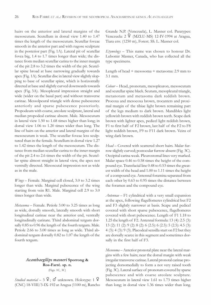
Acanthaegilips masneri Sporrong &Ros-Farré. sp. n.
(Figs. 1C, 3C)
Studied material – 3 �; � unknown. Holotype: 1 �(CNC) 18-VIII/3-IX-192 at Aragua (1100 m), Rancho
Grande N.P. (Venezuela), L. Masner col. Paratypes:Venezuela: 2 � (MZLU-MS) 12-IV-1994 at Aragua,Tiara env. (1250 m), Forest. SS. L. Masner col.
Etymology – This name was chosen to honour Dr.Lubomir Masner, Canada, who has collected all thetype specimens.
Length of head + mesosoma + metasoma: 2.9 mm to3.1 mm.
Colour – Head, pronotum, mesopleuron, mesoscutumand scutellar spine black. Scutum, mesopleural triangle,metanotum and metasoma dark reddish brown.Procoxa and mesocoxa brown, trocanters and proxi-mal margin of the tibiae light brown remaining partof the legs medium to dark brown. Mandibles lightyellowish brown with reddish brown teeth. Scape darkbrown with lighter apex, pedicel light reddish brown,F1 to first half of F2 brown, last half of the F2 to F8light reddish brown, F9 to F11 dark brown. Veins ofwing dark brown.
Head – Covered with scattered short hairs. Malar fur-row slightly curved; postocular furrow absent (Fig. 3C).Occipital carina weak. Pleurostomal lines very marked.Malar space 0.46 to 0.58 times the height of the com-pound eye. Transfacial line 0.48 to 0.53 times the great-est width of the head and 1.00 to 1.11 times the heightof a compound eye. Antennal foramina separated fromeach other by 0.63 to 0.95 times the distance betweenthe foramen and the compound eye.
Antennae – F1 cylindrical with a very small expansionat the apex, following flagellomeres cylindrical but F2and F3 slightly narrower at basis. Scape and pedicelcovered with short sparse pubescence, flagellomerescovered with short pubescence. Length of F1 1.18 to1.25 the length of F2. Antennal formula: 13 (4): 2.5 (3):13 (2): 11 (2): 9 (2): 8 (2): 6 (2.5): 6 (2.5): 5 (2.5): 4.5 (3):4 (3): 4 (3): 9 (3). Placodeal sensilla start on F2 but theyare dorsally scarce in this segment and sometimes dor-sally in the first half of F3.
Mesosoma – Anterior pronotal plate near the lateral mar-gins with a few hairs; near the dorsal margin with weakirregular transverse carinae. Lateral pronotal carinae pro-jecting dorsomedially to form a not very raised tooth(Fig. 3C). Lateral surface of pronotum covered by sparsepubescence and with coarse areolate sculpture.Mesoscutum in lateral view 1.61 to 1.73 times higherthan long; in dorsal view 1.36 times wider than long.
PAP. AVULS ZOOL. 43(2), 2003 27
Median mesoscutal impression and notauli reaching 1/3to 1/2 the length of mesoscutum. There is a conspicu-ous line of hairs on the anterior and lateral margins ofthe mesoscutum. Scutellum in dorsal view 1.26 to 1.37times the length of the mesoscutum. Scutellar foveaesmooth (Fig. 1C), scutellar spine dorsally smooth. Lat-eral pit of scutellar fovea rather small and almost circu-lar 1.1 times longer than wide; the distance from me-dian scutellar carina to the inner margin of the pit 3.7times the width of the pit. Scutellar spine broad at basenarrowing gradually towards apex (Fig. 1C). Scutellar discin lateral view steeply slopping to base of scutellar spine,which is directed slightly upwards at base and curveddownwards, so that the apex is pointing in aposteroventral direction. Mesopleural impression widestraight and without transverse carinae. Mesopleural tri-angle densely pubescent anteriorly, sparsely pubescentposteriorly. Propodeum with coarse areolate sculpture;lateral and median propodeal carinae absent.
Wings – Marginal cell closed 2.90 to 3.14 times longerthan wide. Marginal pubescence starting from vein R1.
Metasoma – Petiole 2.25 to 2.33 times as long as wide.Petiole dorsally smooth; lateroventrally and ventrallylongitudinally costate. Third abdominal tergum dorsally0.56 to 0.70 times the length of the fourth tergum.
Acanthaegilips occultus Ros-Farré &Pujade-Villar sp. n.
(Figs. 2C, 4C)
Studied material – 1 �; � unknown. Holotype: 1 �(MZLU-MS) 11/14-IV-1994 at Aragua, ParqueNacional H. Pittier, La Trilla (200 m), (Venezuela) L.Masner Col. (M.S.).
Etymology – This name was chosen because the malarfurrow of this species is very weak.
Colour – Head, mesosoma and metasoma black. An-tennae light reddish brown, except the last fourflagellomeres, which are dark brown. Mandibles lightyellowish brown with reddish brown teeth. Front andmiddle legs light brown, hind legs brown. Veins of wingyellowish brown.
Head – Head without pubescence, except for a line ofhairs running from base of mandible along gena be-hind malar and postocular furrows and a few hairs
forming a perpendicular line just below eye along up-per part of malar furrow. Malar furrow very weak andcurved, only marked by a change in the integumentcurvature; postocular furrow weak. Occipital carinastrong. Pleurostomal lines weak. Malar space 0.70 timesthe height of the compound eye. Transfacial line 0.50times the greatest width of the head and 1.17 timesthe height of a compound eye. Antennal foramina sepa-rated from each other by 1.6 times the distance be-tween the foramen and the compound eye.
Antennae – F1 to F8 cylindrical with a very small ex-pansion at the apex, following flagellomeres cylindri-cal. Length of F1 1.27 the length of F2. Antenna with-out pubescence, except for a few hairs on scape. An-tennal formula: 8 (3): 3 (2): 7 (2): 5.5 (2): 5.5 (2): 5.5 (2):4.5 (2): 4 (2): 4 (2): 4 (2): 4 (2): 4 (2.5): 8 (2.5). Placodealsensilla start on F2 and they are little scarce on thisantennomere.
Mesosoma – Lateral pronotal carinae projectingdorsomedially to form a much raised tooth (Fig. 4C).Anterior pronotal plate with a few scattered hairs neardorsolateral margins and weakly transversely carinate.Lateral surface of pronotum with dense pubescenceand with areolate sculpture. Mesoscutum in lateral view1.58 times higher than long; in dorsal view 1.20 timeswider than long. Median mesoscutal impression reach-ing 1/4 part of the length of the mesoscutum. Notaulicomplete (Fig. 2C). Line of hairs on the anterior andlateral margins of the scutum absent. Scutellum indorsal view of the same length as that of themesoscutum. Scutellar foveae in anterior part with aninternal short longitudinal carina dividing the anteriorarea of each fovea into two areas, scutellar foveaesmooth with few irregular sculpture near its posteriormargin. Lateral pit of scutellar fovea almost circular,1.1 times longer than wide; the distance from medianscutellar carina to the inner margin of the pit 3.5 timesthe width of the pit. Scutellar spine broad, narrowingabruptly before apex (Fig. 2C). Scutellar disc in lateralview steeply slopping to base of scutellar spine, whichis upwards directed at base and curved downwards,the apex is ventrally directed. Mesopleural impressionrather narrow, weakly curved in the apical part andwith few coarse transverse carinae (Fig. 4C).Mesopleural triangle with sparse pubescence.Propodeum with areolate sculpture, lateral and medianpropodeal carinae present.
Wings – Marginal cell open 2.5 times longer than wide.R1 practically non-existent. Marginal pubescence of
28 ROS-FARRÉ ET AL.: REVISION OF THE NEOTROPICAL ANACHARITINAE GENUS ACANTHAEGILIPS
the wing starting from the apical 2/3 of marginal cellbut it is very sparse to the end of the cell.
Metasoma – Petiole between 1.0 and 1.5 times as longas wide, it is not possible to know exactly this measurebecause the third tergum is covering the posterior partof the petiole in the holotype. Petiole dorsally smooth,laterally and ventrally costate. Third abdominal tergumdorsally 0.65 times the length of the fourth tergum.
Redescription of Acanthaegilips brasiliensis(Figs. 1D, 3D)
Acanthaegilips brasiliensis Ashmead, (1897: 67)Acanthaegilips brasiliensis Ashmead, Dalla Torre &
Kieffer, (1910: 47)Acanthaegilips brasiliensis Ashmead, Weld, (1921: 434)Acanthaegilips brasiliensis Ashmead, Weld, (1950: 96 &
171)Acanthaegilips brasiliensis Ashmead, Díaz, (1983: 46)
Studied material – 16 � & 3 �. Holotype: 1 � (nº 23645USNM) Collected in April at Chapada (Brazil),Ashmead collection (det. Ashmead), antenna mounted.Label data: Chapada, April; Coll Ashmead. Acanthaegilipsbrasiliensis f, Type. Additional material: Brazil: 1 �Alvarenga col. 1 � (DCBU) 12-X-1987 at FazendaJacutinga, light trap, (MZLU-MS) VII-1992 at RepresaRio Grande, Guanabara, M.U.F. col. Mexico: 1 � &1 � (CNC) 8-VI-1969 at Chis. 5700 ft. 20 mi, YerbaBuena, Malaise trap. N. Bochill col. Guatemala: 1 �(USNM) VII-1980 at Antigua (1500-1600 m),N.L.H.Krauss col. Bolivia: 1 � (CNC) 19/20-XII-1955at Chulumani (1700 m), Yungas, L.E. Pena col. CostaRica: 2 � (USNM)14-IV-1985 at San Antonio deEscazu (300 m), F-SAZ-125, W.Eberhard col. 1 �(UCR) VI/VIII-1993 at Cartago, Dulce Nombre,Vivero Linda Vista (1300 m), P. Hanson col. 1 �(UCR) VIII/IX-1995 at Alajuela Est. Biol. San Ramon(900 m) P. Hanson col. 2 � (MZLU-MS)14/25-V-1988 at San Jose, Escazu, F.D. Parker col. 1 �(MZLU-MS) 22/27-V-1998 at San Jose, Escazu, F.D.Parker col. 1 � (MZLU-MS) 21/26-V-1988 at SanJose, Escazu, F.D. Parker col. 1 � (MZLU-MS)24/30-I-1988 at San Jose, Escazu, F.D. Parker col. 2 �(MZLU-MS) 8-II-1987 at San Jose, Escazu, G.E.Bohart col. 1 � (UCR) IX/X-1989 at Guanac, Estac.Mengo, SW, Volcan Cacao (1100 m). Honduras: 1 �(nº 35396 USNM) in 17-II-1918 at Tegucigalpa, F.J.Dyer col.
Length of head + mesosoma + metasoma in female:2.6 mm to 3.2 mm; in male: 2.6 mm to 2.9 mm.
Colour – Female. Head wholly black. Antennae seg-ments light brown with exception of the scape, whichis black in its basal part, and F9 to F11, which aredark brown. Mandibles light brown with darker tooth.Scutum and mesopleural triangle reddish brown;mesopleuron, scutellar spine, propodeum andmetasoma almost black. Legs medium to dark brownexcept for the procoxa, mesocoxa and tarsis, whichare light brown. Gaster blackish brown. Veins ofwings medium brown. Male. Entirely black with ex-ception of the first and second legs, which are lightyellowish brown.
Head – Female. Covered with scattered medium longhairs. Malar furrow slightly curved. Postocular furrowabsent. Occipital carina present. Malar space 0.60 to0.71 times the height of the compound eye.Pleurostomal lines strong. Transfacial line 0.52 to 0.57times the greatest width of the head and 1.17 to 1.30times the height of a compound eye. Antennal foraminaseparated from each other by 0.7 to 0.9 times the dis-tance between the foramen and the compound eye.Male. Malar space 0.61 times the height of the com-pound eye. Transfacial line 0.54 times the greatest widthof the head and 1.04 to 1.08 times the height of acompound eye. Antennal foramina separated from eachother by the same distance as that between the fora-men and the compound eye.
Antennae – Female. Flagellomeres cylindrical, not ex-panded in width towards apex. Length of F1 1.38 to1.43 the length of F2. Antennae covered with short,dense pubescence. Antennal formula: 10 (3): 3 (2):12.5 (2): 9 (2): 8 (2): 8 (2): 6 (3): 5 (2): 5 (2.5): 4.5 (3):4.5 (3): 4 (3): 9 (3). Placodeal sensilla start on F2, butthey are dorsally scarce in the basal part of thisf lagellomere. Male. F1 1.5 the length of F2.Flagellomeres covered by very short, dense pubescence.Antennal formula: 11 (5): 4 (4): 18 (4): 12.5 (4): 11 (3.5):11 (3): 10.9 (3.3): 10.5 (3.5): 10 (3.5): 9 (3.8): 8 (3.8):8 (3.5): 8 (3.2): 11 (3.2). Placodeal sensilla start on F1,they are dorsally scarce in the basal part of F1. F2 toF4 slightly expanded dorsolaterally with a longitudinalridge on the raised part.
Mesosoma – Female. Lateral pronotal carinae project-ing dorsomedially to form a raised tooth (Fig. 3D).Anterior pronotal plate with scattered long hairs onthe lateral margins and with weak transverse cari-
PAP. AVULS ZOOL. 43(2), 2003 29
nae. Lateral surface of pronotum with very sparsepubescence and with coarse areolate sculpture.Mesoscutum in lateral view 1.63 to 1.93 times higherthan long; in dorsal view 1.20 to 1.53 wider thanlong. Median mesoscutal impression reaching be-tween 1/3 and 1/2 the length of the scutum, notaulicomplete, the basal cell reaching to the 1/3 thelength of mesoscutum (Fig. 1D). Sculpture overmesoscutum from the pronotum to the mesoscutalimpression as a strong carina, when viewed in pro-file, mesoscutum humped, in part because of thiscarina, also seen in profile (Fig. 3D). There is a con-spicuous line of hairs on the anterior and lateralmargins of the mesoscutum. Scutellum in dorsalview 1.21 to 1.36 t imes the length of themesoscutum. Scutellar foveae nude. Lateral pit ofscutellar fovea 1.15 to 1.40 longer than wide; thedistance from median scutellar carina to the innermargin of the pit 3.16 to 4.30 times the width ofthe pit. Scutellar spine broad at base and narrowinggradually towards apex (Fig. 1D). Scutellar disc inlateral view steeply sloping to base of scutellar spine,which is directed slightly upwards at base and curveddownwards towards apex, which is ventrally directed.Mesopleural impression wide (wider on the basalpart) straight and with weak transverse carinae orsmooth. Messopleural triangle densely pubescentanteriorly and very sparsely pubescent posteriorly(Fig. 3D). Propodeum with coarse areolate sculp-ture, median and lateral propodeal carinae absent.Male. Mesoscutum in lateral view 1.75 times higherthan long; in dorsal view 1.25 to 1.32 wider thanlong. Basal cell of notauli shorter than in the fe-male. Scutellum in dorsal view 1.42 times the lengthof the mesoscutum. Lateral pit of scutellar fovea1.15 to 1.35 times longer than wide; the distancefrom median scutellar carina to the inner margin ofthe pit is 2.45 to 2.90 times the width of the pit.Scutellar spine less curved than in the female.
Wings – Female. Marginal cell open 3.00 to 3.18 timeslonger than wide. R1 practically reaching the marginof the wing like in the figure 5A. Marginal pubescenceof the wing starting from vein R1. Male. Marginal cell2.73 to 3.27 times longer than wide.
Metasoma – Female. Petiole 2.2 to 2.5 times as long aswide, dorsally smooth, laterally and ventrally costate.Third abdominal tergum dorsally 0.51 to 0.75 the lengthof the fourth tergum. Male. Petiole 2.7 times as longas wide. Third abdominal tergum 0.77 the length ofthe fourth tergum.
DISCUSSION
The Acanthaegilips genus presents two morpho-logically distinct groups. The first group is characterisedby having the malar furrow slightly curved and by lack-ing the postocular furrow; the second group by havingthe malar furrow strongly curved in the upper part andthe postocular furrow present. The first group includesA. macropennis, A. levis, A. masneri, A. brasiliensis,A. ashmeadi and A. diazi; the second group includesA. dentis, A. huggerti, A. occultus, A. alienus, A. exiguus andA. carinatus.
Members of the first group have the occipitalcarina weakly marked and the scutellar spine (dorsalview) narrowing gradually towards the apex, occasion-ally slightly constricted medially but never narrowingabruptly close to the apex. The marginal cell is openor closed; if it is open then R1 is always present. Thepropodeum is entirely areolate.
Members of the second group have a stronglymarked occipital carina, with the exception ofA. exiguus, which only has a faint carina. The scutellarspine is slender throughout or narrows abruptly closeto the apex. The vein R1 is very short practically ab-sent. The propodeum is areolate except in A. alienus,in which this sculpture is missing between the lateralpropodeal carinae. Normally, longitudinal propodealcarinae are distinct despite the areolate sculpture [asopposed to the first group of species]. In A. occultusand A. carinatus the malar and postocular furrows arevery weakly impressed but there is a line of hairs run-ning slightly posterior to them.
In the species for which both sexes are known(A. macropennis, A. brasiliensis, A. ashmeadi, A. carinatus,A. huggerti), the males and females are very similarmorphologically. Therefore, we are confident that wehave correctly separated the two species knwon onlyby males from the four species known only by fe-males.
Acanthaegilips alienus is very different from othermembers of the genus in the structure of the scutellarspine, the absence of modified flagellomeres in themales, the structure of the notauli and the absence ofsculpture between the propodeal carinae. The last statediffers from the likely ground-plan state of theAnacharitinae, in which the areolate sculpture coversthe entire propodeum and more or less entirely ob-scures the propodeal carinae (Ros-Farré et al., 2000).The apomorphic state obviously evolved secondarilywithin the Anacharitinae through loss of the areolatesculpture making the propodeal carinae more conspicu-ous.
30 ROS-FARRÉ ET AL.: REVISION OF THE NEOTROPICAL ANACHARITINAE GENUS ACANTHAEGILIPS
Two potentially important diagnostic charactersfor separating species of Acanthaegilips are the distri-bution of sensilla in the antennae and the position andnumber of modified flagellomeres. Unfortunately, wehave not been able to explore the utility of these char-acters fully linked to sex – The first character variesbetween males and females and the second characteroccurs only in males – and both sexes are currentlyknown for only half the species.
ACKNOWLEDGEMENTS
We are very grateful to Dr. C.R.F. Brandão andto the department’s colleagues N. Mercader, D. Bellidoand S. Duran for helping us with the English transla-tion and for the critics made on this manuscript. Alsoto Dr. C.R.F. Brandão (MZUSP), to Dr. J.D. Read(CNC), to Dr. D. Furth (USNM), to Dr. Godoy (UCR),to Dr. A. Penteado-Dias (DCBU), to Dr. C. Henrique(DCBU) and to Dr. A. Roig, Curador of MuseoArgentino de Ciencias Naturales, for sending us partof the material cited in this work and to Dr. J.M. Maes(MEAN) for giving us some Acanthaegilips material.
RESUMO
Revisamos Acanthaegilips, um gênero de figitídeosanacharitíneos que ocorre na América do Sul. Membros dessegênero são caracterizados por apresentar um longo espinhoescutelar, caráter único entre os Anacharitinae. A única espécieconhecida anteriormente (A. brasiliensis) é redescrita e onzeespécies novas descritas: A. macropennis, A. ashmeadi,A. levis, A. masneri, A. diazi, A. dentis, A. occultus,A. alienus, A. exiguus, A. huggerti e A. carinatus. Umachave para a identificação das espécies é apresentada e registrosde distribuição apresentados para cada espécie. Os registros parao gênero estendem-se do Sul da Neotrópica ao México. A biologiade Acanthaegilips permanece desconhecida; outros membros
de Anacharitinae são parasitóides de larvas de Neuroptera quealimentam-se de afídeos.
PALAVRAS-CHAVE: Cynipoidea, Figitidae, Aspicerinae,Acanthaegilips, Novas espécies.
REFERENCES
Ashmead, W.H. 1897. Description of some new genera in the familyCynipidae. Psyche, 8:67-69.
Dalla Torre, K.W. & Kieffer, J.J. 1910. Cynipidae. Das Tierreich,24:1-891.
Díaz, N.B. 1983. El género Acanthaegilips Ashmead, 1897 en laRepública Argentina (Hymenoptera, Cynipoidea). Neotropica,29(81):45-49.
Kovalev, O. 1996. A new family, new subfamily and genus andreplacement names in cynipoids (Hymenoptera, Cynipoidea).Entomologicheskoe Obozrenie, 75:408-416.
Richards, O.W. 1977. Hymenoptera. Introduction and key to families.2ª ed. In:Handbooks for the identification of British Insects. London,British Museum/Royal Entomological Society. V. 6, pt. 1,p.1-100.
Ronquist, F. 1994a. Morphology, Phylogeny and Evolution ofCynipoid Wasps. Acta Universitatis Upsaliensis. ComprehensiveSummaries of Uppsala Dissertations from the Faculty of Science andTechnology, 38:1-29.
Ronquist, F. 1994b. Evolution of parasitism among closely relatedphylogenetic relationships and the origin of inquilinism in gallwasps (Hymenoptera, Cynipidae). Evolution, 48(2):241-266.
Ronquist, F. 1995. Phylogeny and early evolution of the Cynipoidea(Hymenoptera). Systematic Entomology, 20:309-335.
Ronquist, F. 1999. Phylogeny, classification and evolution of theCynipoidea (Hymenoptera). Zoologica Scripta, 28:139-164.
Ronquist, F. & Nieves Aldrey,J.L. 2001. A new subfamily of Figitidae(Hymenoptera, Cynipoidea). Zoological Journal of the LinneanSociety, 133:483-494.
Ros Farré, P.; Ronquist, F. & Pujade Villar, J. 2000. Redescriptionof Acanthaegilips Ashmead, 1897, with characterization of theAnacharitinae and Aspiceratinae (Hymenoptera: Cynipoidea:Figitidae). Zoological Journal of the Linnean Society, 129:467-488.
Weld, L.H. 1921. Notes on certain genera of Parasitic Cynipidaeproposed by Ashmead with descriptions of genotypes.Proceedings of the United States National Museum, 59:433-451.
Weld, L.H. 1952. Cynipoidea (Hym.) 1905 1950. Ann Arbor, Privatelyprinted. 351 p.
Recebido em 22.11.2000Aceito em 20.11.2002
Credenciamento e apoio financeiro doPrograma de Apoio às PublicaçõesCientíficas Periódicas da USPComissão de Credenciamento Se
ção
de P
ublic
açõe
s do
Mus
eu d
e Z
oolo
gia
da U
nive
rsid
ade
de S
ão P
aulo
General Information: Papéis Avulsos de Zoologia covers primarily the fields of Zoology,publishing original contributions in systematics, paleontology, evolutionary biology, ecology,taxonomy, anatomy, behavior, functional morphology, molecular biology, ontogeny, faunisticstudies, and biogeography. Papéis Avulsos de Zoologia also encourages submission of theoreticaland empirical studies that explore principles and methods of systematics.
All contributions must follow the International Code of Zoological Nomenclature. Relevantspecimens should be properly curated and deposited in a recognized public or private,non-profit institution. Tissue samples should be referred to their voucher specimens andall nucleotide sequence data (aligned as well as unaligned) should be submitted to GenBank(http://www.ncbi.nih.gov/Genbank/) or EMBL (http://www.ebi.ac.uk/).
Peer Review: All submissions to Papéis Avulsos de Zoologia are subject to review by at leasttwo referees and the Editor-in-Chief. Three legible copies (including photocopies of originalillustrations) and original illustrations must be submitted; all authors will be notified ofsubmission date. Authors may suggest potential reviewers. Communications regardingacceptance or rejection of manuscripts are made through correspondence with the firstor corresponding author only. Once a manuscript is accepted providing changes suggestedby the referees, the author is requested to return a revised version incorporating thosechanges (or a detailed explanation of why reviewer’s suggestions were not followed) withinfour weeks upon receiving the communication by the editor. Revised manuscripts must besubmitted as both hard copy and electronic file (3.5" disk, Zip Drive, or CD Rom withtext in Microsoft Word format). Figures and graphics should be sent separately (“.jpg”,“.tif ”, “.xls”, “.cdr”).
Proofs: Page-proofs with the revised version will be sent to the first or correspondingauthor. Page-proofs must be returned to the editor in two weeks, preferentially within 48 hours.Failure to return the proof promptly may be interpreted as approval with no changesand/or may delay publication. Only necessary corrections in proof will be permitted.Once page proof is sent to the author, further alterations and/or significant additions oftext are permitted only at the author’s expense or in the form of a brief appendix (“noteadded in proof ”).
Submission of Manuscripts: Manuscripts should be sent to the Editor-in-Chief(H. Zaher, Museu de Zoologia da USP, Caixa Postal 42.494, CEP 04218-970, São Paulo,SP, Brasil). Manuscripts are considered on the understanding that they have not beenpublished or will not appear elsewhere in substantially the same or abbreviated form. Thecriteria for acceptance of articles are: quality and relevance of research, clarity of text, andcompliance with the guidelines for manuscript preparation.
Manuscripts should be written preferentially in English, but texts in Portuguese or Spanishwill also be considered. Studies with a broad coverage are encouraged to be submitted inEnglish. All manuscripts should include an abstract in Portuguese and English regardlessof the original language.
Authors are requested to pay attention to the instructions concerning the preparation ofthe manuscripts. Close adherence to the guidelines will expedite processing of themanuscript, whereas manuscripts deviating from the required form will be returned forrevision prior to review.
Manuscript Form: Manuscripts should not exceed 100 pages of double-spaced typescripton 21 by 29.7 cm (A4 format) or 21.5 by 28 cm (letter format) paper, with wide margins.The pages of the manuscript should be numbered consecutively.
The text of articles should be arranged in the following order: Title Page, Abstracts, Bodyof Text, Literature Cited, Tables, Appendices, and Figure Captions. Each of these sectionsshould begin on a new page. All typescript pages must be double-spaced.
(1) Title Page: This should include the title, author(s) name(s), institutions, and keywordsin English as well as in the language of the manuscript, and a short running title in
Editor-in-Chief: Hussam Zaher, Serviço de Vertebrados, Museu de Zoologia, Universidadede São Paulo, Caixa Postal 42.494, CEP 04218-970, São Paulo, SP, Brasil. E mail:[email protected].
Associate Editors: Antonio C. Marques (Universidade de São Paulo, Brasil), Mario C.C.de Pinna (Universidade de São Paulo, Brasil), Sergio A. Vanin (Universidade de São Paulo,Brasil).
Editorial Board: Aziz N. Ab’Saber (Universidade de São Paulo, Brasil), Rudiger Bieler(Field Museum of Natural History, U.S.A.), Walter A.P. Boeger (Universidade Federal doParaná, Brasil), Carlos Roberto F. Brandão, (Universidade de São Paulo, Brasil), JamesCarpenter (American Museum of Natural History, U.S.A.), Ricardo Macedo Correa eCastro (Universidade de São Paulo, Brasil), Darrel Frost (American Museum of Natural
EDITORIAL COMMITTEEHistory, U.S.A.), W.R. Heyer (National Museum of Natural History, U.S.A.), Ralf Holzentahl(University of Minesotta, U.S.A.), Adriano Kury (Museu Nacional do Rio de Janeiro, Brasil),Gerardo Lamas (Museu Javier Prado de Lima, Peru), John Maisey (American Museum ofNatural History, U.S.A.), Ubirajara Martins (Universidade de São Paulo, Brasil), NaércioMenezes (Universidade de São Paulo, Brasil), Christian de Muizon (Museum Nationald’Histoire Naturelle, France), Nelson Papavero (Universidade de São Paulo, Brasil), JamesPatton (University of Berkeley, U.S.A.), Richard Prum (University of Kansas, U.S.A.), MarcosRaposo (Museu Nacional do Rio de Janeiro, Brasil), Olivier Rieppel (Field Museum ofNatural History, U.S.A.), Miguel T.U. Rodrigues (Universidade de São Paulo, Brasil), RandahlSchuh (American Museum of Natural History, U.S.A.), Marcos Tavares (Universidade deSão Paulo, Brasil), Paulo E. Vanzolini (Universidade de São Paulo, Brasil), Richard Vari(National Museum of Natural History, U.S.A.), Mario de Vivo (Universidade de São Paulo,Brasil) and Paulo Young (Museu Nacional do Rio de Janeiro, Brasil).
INSTRUCTIONS TO AUTHORS(MAY 2002)
the language of the manuscript. The title should be concise and, where appropriate,should include mention of families and/or higher taxa. Names of new taxa shouldnot be included in titles.
(2) Abstract: All papers should have an abstract in English and another in Portuguese,regardless of the original language. The abstract is of great importance as it may bereproduced elsewhere. It should be in a form intelligible if published alone andshould summarize the main facts, ideas, and conclusions of the article. Telegraphicabstracts are strongly discouraged. Include all new taxonomic names for referencingpurposes. Abbreviations should be avoided. It should not include references. Abstractsshould not exceed 350 words.
(3) Body of Text: The main body of the text should include the following sections:Introduction, Materials and Methods, Results, Discussion, and Acknowledgmentsat end. Primary headings in the text should be in capital letters and centered; thefollowing text should begin on the next line, indented. Secondary headings shouldbe in capital and lowercase letters and flush left; the following text should begin onthe next line, indented. Tertiary headings should be in capital and lower case letters,in italics and indented; the following text should be on the same line and separatedfrom the heading by a hyphen.
(4) Literature Cited: Citations in the text should be given as: Silva (1998)..., Silva(1998:14-20)..., Silva (1998: figs. 1, 2)..., Silva (1998a, b)..., Silva & Oliveira (1998)...,(Silva, 1998)..., (Rangel, 1890; Silva & Oliveira, 1998a, b; Adams, 2000)..., (Silva, pers.comm.)..., (Silva et al., 1998), the latter when the paper has three or more authors.The reference need not be cited when author and date are given only as authorityfor a taxonomic name. The literature section should be arranged strictly alphabeticallyand given in the following format:
Journal Article – Silva, H.R.; Oliveira, H. & Rangel, S. Year. Article title. Journal name,00:000-000. Names of journals must be spelled out in full.
Books – Silva, H.R. Year. Book title. Publisher, Place, 000p.
Articles in Books – Silva, H.R. Year. Article title. In: Oliveira, H. & Rangel, S. (Eds.),Book title. Publisher, Place. p.000-000.
Articles in Larger Works – Silva, H.R. Year. Article title. In: H. Oliveira & S. Rangel(Eds.), Title of Larger Work. Serial Publication. Publisher, Place. pp.000-000.
Dissertations and Theses – Silva, H.R. Year. Dissertation title. Ph.D. dissertation,University, Place, 000p.
Tables: All tables must be numbered in the same sequence in which they appear in thetext. Authors are encouraged to indicate where the tables should be placed in the text.They should be comprehensible without reference to the text. Tables should be formattedwith horizontal, not vertical, rules. In the text, tables should be referred as Table 1, Tables2 and 3, Tables 2-6. Use “TABLE” in the table heading.
Illustrations: Figures should be numbered consecutively, in the same sequence theyappear in the text. Separate illustrations of a composite figure should be identified bycapital letters and referred in the text as so (fig. 1A). Where possible, letters should beplaced in the lower right corner of each illustration of a composite figure. Hand-writtenlettering on illustrations is unacceptable. Illustrations should be mounted on stout, whitecardboard. Figures should be mounted in order to minimize blank areas between separateillustrations. High quality color or black and white photographs, and computer generatedfigures are preferable. Authors are encouraged to indicate where the figures should beplaced in the text. Use “(Fig(s).)” and “Figure(s)” for referring to figures in the text, but“FIGURE(S)” in the figure captions and “(fig(s).)” when referring to figures in anotherpaper.
For other details of manuscript preparation of format, consult the CBE Style Manual, available from the Council of Science Editors(http://www.councilscienceeditors.org/publications/style.cfm).
Papéis Avulsos de Zoologia and Arquivos de Zoologia are publications of the Museu de Zoologia da Universidade de São Paulo (www.mz.usp.br).