modulaÇÃo diferencial de exopolifosfatases no … · os argasídeos, carrapatos moles,...
TRANSCRIPT

i
MODULAÇÃO DIFERENCIAL DE EXOPOLIFOSFATASES NO
METABOLISMO NUCLEAR E MITOCONDRIAL DURANTE A
EMBRIOGÊNESE DO CARRAPATO Rhipicephalus (Boophilus)
microplus
ELDO CAMPOS
UNIVERSIDADE ESTADUAL DO NORTE FLUMINENSE – DARCY
RIBEIRO – UENF CAMPOS DOS GOYTACAZES - RJ
JULHO – 2008

ii

iii
MODULAÇÃO DIFERENCIAL DE EXOPOLIFOSFATASES NO
METABOLISMO NUCLEAR E MITOCONDRIAL DURANTE A
EMBRIOGÊNESE DO CARRAPATO Rhipicephalus (Boophilus)
microplus
ELDO CAMPOS
“Tese apresentada ao Centro de
Biociências e Biotecnologia da
Universidade Estadual do Norte
Fluminense, como parte das
exigências para obtenção do título
de Doutor em Biociências”.
ORIENTADOR: Prof. Dr. Carlos Logullo
CAMPOS DOS GOYTACAZES – RJ
JULHO – 2008

iv
MODULAÇÃO DIFERENCIAL DE EXOPOLIFOSFATASES NO
METABOLISMO NUCLEAR E MITOCONDRIAL DURANTE A
EMBRIOGÊNESE DO CARRAPATO Rhipicephalus (Boophilus)
microplus
ELDO CAMPOS
“Tese apresentada ao Centro de
Biociências e Biotecnologia da
Universidade Estadual do Norte
Fluminense, como parte das
exigências para obtenção do título
de Doutor em Biociências”.
Aprovada em 25 de Julho de 2008.
____________________
Carlos Logullo Prof. Associado do Laboratório de Química e Função de Proteínas e Peptídeos
UENF Orientador
____________________
Arnoldo Rocha Façanha Prof. Associado do Laboratório de Biologia Celular e Tecidual
UENF Co-Orientador

v
_____________________
Julio Alberto Mignaco Prof. Adjunto do Instituto de Bioquímica Médica
UFRJ
_____________________
Marílvia Dansa de Alencar Petretski Profa. Associada do Laboratório de Química e Função de Proteínas e
Peptídeos UENF
_________________________ Lev Alexandrovitch Okorokov
Prof. Titular do Laboratório de Fisiologia de Microorganismos UENF

vi
“A coisa mais bela que o homem
pode experimentar é o mistério. É
essa emoção fundamental que está
na raiz de toda ciência e toda arte.”
(Albert Einstein).

vii
Agradecimentos:
Aos meus pais, porque sem eles o meu caminho seria muito mais tortuoso.
Meu agradecimento especial ao Prof. Carlos Logullo pelo acolhimento em seu
laboratório, por acreditar na minha capacidade e, principalmente, por sua
amizade durante a realização deste trabalho. Sou-lhe grato pela enorme
contribuição para o meu crescimento acadêmico-científico. Obrigado.
Ao Prof. Arnoldo Façanha pelas críticas, sugestões e amizade durante o
desenvolvimento desta tese. Muito obrigado por contribuir com a minha
formação acadêmico-científica. Sou muito grato por poder subir nos ombros de
dois gigantes.
A Profª. Denise Valle, por ter contribuído com a minha formação durante a
revisão do artigo de 2006.
Ao Prof. Itabajara Vaz pela orientação durante o meu doutorado-sanduíche e
pela contribuição científica e amizade ao longo destes anos de colaboração.
A Profa. Aoi Masuda por ter me acolhido por 4 meses em seu laboratório
durante o meu doutorado-sanduíche.
Ao Prof. Jorge Moraes, um grande amigo que me ajudou muito desde o início
da minha iniciação científica.
A minha noiva, Aline, que sempre me ajudou a tomar decisões e, mais que
isso, me apoiou e incentivou. Teve muita paciência (às vezes nem tanta) e
entendeu (ou pelo menos tentou entender) o porquê de tantas horas no
laboratório.
Agradeço a todos os meus professores, amigos e colegas que, de alguma
forma, contribuíram para o desenvolvimento desta tese.

viii
Sou muito grato também a CAPES, CNPq e FAPERJ pelos financiamentos
durante toda a minha formação, e à Universidade Estadual do Norte
Fluminense – Darcy Ribeiro, pelo ensino gratuito e de qualidade.

ix
Sumário RESUMO xi
ABSTRACT xiii
1 – INTRODUÇÃO 2
1.1 – O carrapato Rhipicephalus (Boophilus) microplus e sua
Importância econômica 2
1.2 – O ciclo de vida do Rhipicephalus (Boophilus) microplus 6
1.3 – Ovogênese e embriogênese 8
1.4 – Metabolismo energético em ovos 10
1.5 – Polifosfatos 12
2 – OBJETIVOS 18
3 – MATERIAIS E MÉTODOS 20
3.1 – Animais 20
3.2 – Extração e quantificação de polifosfatos 20
3.3 – Isolamento da fração nuclear e mitocondrial 21
3.4 – Fracionamento mitocondrial 21
3.5 – Extração e quantificação de RNA total 21
3.6 – Quantificação de ortofosfato 22
3.7 – Análise eletroforética de polifosfato 22
3.8 – Ensaio da exopolifosfatase 23
3.9 – Atividade da exopolifosfatase durante a respiração
mitocondrial 23
3.10 – Parâmetros respiratórios 23
3.11 – Consumo de O2 utilizando poly P como doador de Pi 24
3.12 – Atividade da F-ATPase 24
3.13 – Atividade da glicose-6-fosfato desidrogenase 25
3.14 – Estudos cinéticos 25
4 – RESULTADOS 27
4.1 – Caracterização do conteúdo de polifosfato total na
embriogênese do R. microplus 28
4.2 – Metabolismo nuclear e mitocondrial de polifosfosfatos 31

x
4.3 – Modulação da atividade exopolifosfatásica
mitocondrial por demanda de Pi 36
4.4 – Subfracionamento mitocondrial 39
4.5 – Caracterização cinética da exopolifosfatase nuclear e
mitocondrial 42
5 – DISCUSSÃO 46
6 – CONCLUSÕES 54
7 – REFERÊNCIAS BIBLIOGRÁFICAS 56
8 – ANEXOS 65
8.1 – ANEXOI 66
8.2 – ANEXOII 76
8.3 – ANEXOIII 83
8.4 – ANEXOIV 89
xi
Resumo O metabolismo de polifosfato foi caracterizado durante a embriogênese
do Rhipicephalus (Boophilus) microplus, por meio de análises do conteúdo de
polifosfato e da atividade exopolifosfatásica ensaiada nas respectivas frações
subcelulares. Durante a embriogênese, o conteúdo de polifosfato se manteve
constante até o 7º dia de desenvolvimento, decaindo rapidamente neste
estágio caracterizado pela segmentação do embrião. Este processo mostrou
relação com o decréscimo de tamanho da cadeia dos polifosfatos e com a
atividade da exopolifosfatase total. Todavia, tal atividade não refletiu o
conteúdo de ortofosfato, o qual se elevou somente no final da embriogênese.
Após a celularização do embrião, o conteúdo endógeno de polifosfato nuclear
diminuiu linearmente até o final da embriogênese, em paralelo com o aumento
da atividade exopolifosfatásica. Por outro lado, a utilização do polifosfato
mitocondrial ocorreu entre a celularização e a segmentação do embrião, ou
seja, do 5° ao 7º dia de desenvolvimento. A atividade exopolifosfatásica nuclear
foi estimulada cerca de duas vezes por RNA total, enquanto a atividade
mitocondrial foi insensível a este polinucleotídeo. Somente a atividade
exopolifosfatásica mitocondrial foi estimulada por concentrações fisiológicas de
NADH e ADP, e completamente inibida por Pi. Esta atividade também
aumentou na presença de substratos respiratórios, ácidos pirúvico e succínico,
e este efeito desapareceu com a adição de cianeto de potássio. A respiração
mitocondrial foi ativada por ADP usando polifosfato como única fonte de Pi e
esta ativação foi inibida por heparina, um inibidor de exopolifosfatases. Após o
isolamento da fração solúvel e de membrana mitocondrial, foi detectada
atividade exopolifosfatásica nas frações solúvel e de membrana. O Kmap
utilizando poly P3, poly P15 e poly P65 como substratos foram quase os mesmos
para a fração nuclear, enquanto a mitocondrial apresentou uma afinidade 10
vezes maior para poly P3 comparando com o poly P15 e poly P65. A atividade
exopolifosfatásica foi estimulada duas vezes por Mg+2 e Co+2 na fração nuclear
e somente por Mg+2 na fração mitocondrial. A heparina inibiu ambas as
atividades das exopolifosfatases em até 95%, porém a enzima mitocondrial foi
mais sensível, apresentando um IC50 de 0,2 µg/mL enquanto a nuclear teve IC50
de 0,8 µg/mL. Estes resultados são consistentes com a existência de pelo
menos duas diferentes isoformas de exopolifosfatases operando no núcleo e

xii
na mitocôndria, sendo ambas moduladas de forma diferencial em função do
metabolismo de cada organela durante a embriogênese do carrapato R.
microplus.

xiii
Abstract The polyphosphate metabolism was characterized during Rhipicephalus
(Boophilus) microplus embryogenesis by polyphosphate content and
exopolyphosphatase activity in respectively subcelular fractions. The decline in
total polyphosphate content after the 7th day of embryogenesis does not reflect
the free phosphate (Pi) increase and the total polyphosphate chain length
decrease after embryo cellularization, reflecting the exopolyphosphatase
activity. The endogenous nuclear polyphosphate decrease and
exopolyphosphatase activity increased after embryo cellularization until the end
of embryogenesis, while the utilization of mitochondrial polyphosphate content
occurred between the embryo cellularization and segmentation stages, days 5
to 7th of development. The nuclear exopolyphosphatase activity was stimulated
about two times by total RNA, but no effect was observed on mitochondrial
exopolyphosphatase. Only the mitochondrial exopolyphosphatase activity was
stimulated by physiologic concentrations of NADH and ADP, and completely
inhibited by Pi. This activity also increased in the presence of the respiratory
substrates pyruvic and succinic acids and this stimulatory effect disappeared
upon addition of potassium cyanide. Mitochondrial respiration was activated by
ADP using polyphosphate as the only source of Pi and this activation was
inhibited by heparin, an exopolyphosphatase inhibitor. The exopolyphosphatase
activity was also analyzed after mitochondrial soluble and membrane fractions
isolation and the activity was detected in either fraction, membranar and
soluble. The Kmapp utilizing poly P3, poly P15 and poly P65 as substrate was
almost the same for the nuclear fraction, while mitochondrial fraction showed an
afinitty 10 times higher for poly P3 than for poly P15 and poly P65. The
exopolyphosphatase activity was stimulated around two times by Mg2+ and Co2+
in the nuclear fraction and only by Mg2+ in the mitochondrial fraction. Heparin
inhibited both exopolyphosphatase activities until 95%, although the
mitochondrial fraction was more sensitive with an IC50 0,2 µg / mL, while the
nuclear fraction had an IC50 0,8 µg / mL. Altogether, these data are consistent
with the existence of at least two different isoforms of exopolyphosphatase
operating in the nuclei and mitochondria, and both are modulated specifically by
the metabolism of each organelle during embryogenesis of the hard tick R.
microplus.

Eldo Campos, 2008
1
INTRODUÇÃO

Eldo Campos, 2008
2
1 Introdução
1.1 O carrapato Rhipicephalus (Boophilus) microplus e sua importância econômica
Os carrapatos são artrópodes ectoparasitas hematófagos subdivididos
em duas grandes famílias, Argasidae e Ixodidae (Sonenshine et al., 2006). São
encontrados em quase todas as regiões do mundo, com predominância em
áreas tropicais e subtropicais, abrangendo regiões com produção de gado de
corte e leiteiro na América, África, Ásia e Austrália (Johnston et al., 1986). No
Brasil, eles são descritos em aproximadamente 96% dos municípios (Horn,
1983). Possuem a capacidade de infestar vertebrados terrestres, tais como:
mamíferos, pássaros, répteis e até anfíbios (Sonenshine et al., 2006). São
capazes de transmitir várias doenças aos seres humanos e aos animais, e
nenhum outro artrópode conhecido tem a capacidade de ser vetor de uma tão
ampla variedade de organismos patogênicos. Transmitem bactérias, vírus,
fungos, riquétsias e protozoários, que resultam frequentemente em infecções
letais, e podem, ainda, induzir algumas toxemias através de suas picadas nos
hospedeiros (Sonenshine et al., 2006).
Os argasídeos, carrapatos moles, alimentam-se do sangue dos seus
hospedeiros repetidas vezes, abandonando-os após cada alimentação
(Sonenshine et al., 2006). As fêmeas efetuam várias posturas, alternando com
a alimentação sangüínea. Cada postura não ultrapassa 150 ovos, um número
pequeno quando comparado aos cerca de 3000 ovos dos ixodídeos, carrapatos
duros. Nos ixodídeos a alimentação é prolongada, ingerindo grandes
quantidades de sangue, chegando a atingir cem vezes a sua massa corporal
inicial. O sangue é utilizado como única fonte de energia para o
desenvolvimento destes carrapatos (Sonenshine et al., 2006).
O R. microplus (Figura 1), o principal representante da família dos
ixodídeos, é originário da Ásia e o seu principal hospedeiro é o bovino, embora
seja capaz de completar, eventualmente, o seu ciclo em búfalos, ovelhas,
cavalos e veados (Sonenshine, 1991).

Eldo Campos, 2008
3
Figura 1: O carrapato R. microplus parasitando seu hospedeiro, o boi (Bos taurus). Retirado de www.icb.usp.br/~marcelcp/Boophilus.htm.
É um carrapato que está amplamente distribuído na América, África,
Ásia e Oceania, entre os paralelos 32ºN e 32ºS (Johnston et al., 1986), sendo
um dos principais parasitos que afetam a pecuária destas áreas. Assim como
todos os ixodídeos, ele possui como características diagnósticas uma placa
esclerotizada na superfície do corpo, denominado de escudo quitinoso, que
serve de sustentação a importantes músculos do corpo, quatro pares de patas,
corpo composto de cefalotórax e abdômen e peças bucais modificadas
(Sonenshine, 1991). Diferentemente de outras espécies de ixodídeos, o gênero
Rhipicephalus possui olhos, e seus palpos são extremamente curtos, inseridos
dorsalmente e lateralmente. Uma característica comportamental do gênero é o
parasitismo monoxeno, tendo apenas um hospedeiro em todo o seu ciclo de
vida (Sonenshine, 1991).
O R. microplus acarreta diversos danos econômicos (Horn e Arteche,
1985), sendo considerado o principal ectoparasito dos rebanhos de gado
bovino (George, 2000). Durante o repasto sanguíneo a fêmea obtém uma
massa que pode atingir até 10 vezes o tamanho macho, tornando-se
ingurgitada (Figura 2). Segundo Guerrero et al. (2006), este parasita causa
perdas anuais de centenas de milhões de dólares no mundo todo. Além da
espoliação ao couro, causada por reações inflamatórias nos locais de fixação
do carrapato (Seifert et al., 1968), existe ainda uma grande perda na produção
de leite e carne (Sutherst, 1983) devido à perda de sangue do animal que pode
atingir 2 a 3 mL de sangue/carrapato (Sonenshine et al., 2006). Ainda, R.

Eldo Campos, 2008
4
Fêmea Ingurgitada
Macho
microplus é um importante vetor de doenças, como a tristeza parasitária
bovina, causada por protozoários do gênero Babesia e pela riquétsia do gênero
Anaplasma (McCosker, 1981; Young e Morzaria, 1986). Além das perdas
relacionadas à bovinocultura em si, existem diversos prejuízos relacionados à
mão-de-obra necessária para o controle desse parasito, despesas com
instalações, compra de acaricidas e equipamentos adequados para sua
aplicação, entre outros (Jamroz et al., 2000). Sendo o Brasil hoje um dos
maiores produtores de carne bovina do mundo, com um rebanho bovino de
aproximadamente 200 milhões de cabeças, e uma produção em torno de 8,5
milhões de toneladas de carne e 23 bilhões de litros de leite por ano (IBGE,
2006; MAPA, 2006), os custos com o controle químico podem chegar a uma
ordem de 2 bilhões de dólares, além de representar um risco à saúde humana,
através de resíduos na carne e no leite, e ao meio ambiente, através da
contaminação da água e dos solos (Fernandes et al., 2006).
Figura 2: Carrapato bovino R. microplus. Fêmea ingurgitada (esquerda) e macho (direita). Retirado de Flechimann (1976).
Tendo em vista a problemática do uso de pesticidas, a possibilidade de
desenvolver uma vacina contra R. microplus para proteger o bovino por indução
de uma resposta imune tem sido testada por diferentes grupos de pesquisa há

Eldo Campos, 2008
5
quase três décadas (McGowan et al., 1980; Willadsen e Kemp, 1988; da Silva,
Jr. et al., 1998; Willadsen, 2001). A resistência adquirida mediada
imunologicamente (Allen, 1989) pode ser aferida pela redução no número de
carrapatos que se fixam ao hospedeiro, pela diminuição no peso das
teleóginas, e pela redução da produção de ovos e, conseqüentemente, de
larvas (Wikel e Bergman, 1997). Estes parâmetros estudados fornecem bases
para as futuras tentativas de utilização de vacinas no controle de ectoparasitas.
Para a produção de uma vacina comercial contra qualquer espécie de
carrapato é necessário antes de tudo identificar antígenos protetores; produzir,
então, essas proteínas como antígenos recombinantes, de forma a tornar a
produção economicamente viável; e formular uma vacina com antígenos
capazes de estimular uma resposta imunológica efetiva contra o ectoparasita
(Willadsen, 2001). Embora as vacinas contra carrapato hoje não tenham em
princípio o mesmo efeito imediato dos produtos químicos usados atualmente e
não protejam totalmente o animal, não se corre com elas o risco de
contaminação dos alimentos e do ambiente por resíduos químicos, e podem
ainda ser espécie-específicas (Pruett, 1999).
Uma alternativa para evitar o emprego de compostos químicos no
controle do carrapato, além do controle imunológico, é o controle biológico.
Este tipo de estratégia pode ser utilizado de diversas formas: baseado nas
relações ecológicas entre o carrapato e o meio onde ele se encontra e entre ele
e seus predadores naturais, e na utilização de compostos naturais obtidos para
utilização de seu controle.
O carrapato, como qualquer organismo, tem sua viabilidade relacionada
a condições de estresse ambiental a que estiver submetido; assim sendo, as
condições climáticas, como temperatura e umidade no campo, desempenham
papel importante no equilíbrio das populações deste parasita, diminuindo ou
aumentando os índices de infestação nos bovinos (Gonzales, 1995). A
vegetação também pode atuar como um fator limitante no crescimento da
população de carrapatos. Algumas pastagens podem dificultar a sobrevivência
das larvas, diminuindo a população de carrapatos por sua ação repelente ou
tóxica, como é o caso das plantas do gênero Stylosanthes (Sutherst et al.,
1982) ou ainda por imobilizarem as larvas através de suas secreções ou
estruturas da planta, como ocorre com o capim gordura (Melinis minutiflora)

Eldo Campos, 2008
6
(Farias et al., 1986). Uma forma alternativa de controle seria o sistema de
rotação de pastagens, no qual uma área fica livre de rebanho por um
determinado período de tempo, de forma a impedir a sobrevivência das larvas.
No entanto, esta prática não pode ser aplicada em uma propriedade rural de
pequeno porte por limitações de espaço físico (Farias et al., 1986).
1.2 O ciclo de vida do Rhipicephalus (Boophilus) microplus
Este carrapato apresenta duas etapas distintas no seu ciclo de vida
(Figura 3): uma fase parasitária, durante um período médio de 22 dias sobre
um único hospedeiro, e uma fase de vida livre, que ocorre no solo, podendo
durar de dois a três meses, dependendo fundamentalmente das condições
climáticas existentes (Gonzales et al., 1974).
Figura 3: Representação esquemática do ciclo de vida do carrapato R. microplus. Retirado de Gonzales et al. (1974).
Na fase parasitária, o carrapato apresenta três variações morfológicas
distintas: larva, ninfa e adulto. A larva apresenta três pares de patas, é bastante
ativa, pois necessita encontrar o hospedeiro para nele se fixar e sobrevive às
expensas das reservas de alimento acumuladas na fase de ovo. Fixa-se em
locais específicos do hospedeiro utilizando algumas importantes estruturas,

Eldo Campos, 2008
7
como as quelíceras, as quais seccionam a pele para a introdução do
hipostômio, órgão responsável pela fixação da larva na pele do bovino
(Gonzales et al., 1974).
Após a fixação, a larva alimenta-se e inicia o processo de
desenvolvimento e crescimento tegumentário. Passa por um período de inércia
entre o 4° e 5° dia e atinge a fase de metalarva. Em torno do 6° dia, adquire
uma nova estrutura, com outro tegumento, mais um par de patas e uma fileira
de dentição do hipostômio entre outras alterações: é a fase de ninfa. Esta fase
dura em média dois a quatro dias, sendo que ao continuar seu
desenvolvimento, uma nova alteração no exoesqueleto se processa, havendo
um período igual de inatividade, denominado de metaninfa, para que ao final do
processo surja o indivíduo adulto, sexualmente diferenciado. Isto acontece em
torno do 12° dia (Gonzales et al., 1974).
A partir dessa fase, inicia-se o processo de maturação dos machos e
das fêmeas, sendo que em torno do 17° dia os machos já estão aptos à cópula.
Nota-se um crescimento mais acentuado do tegumento nessa fase, sendo que
nas últimas horas próximas ao ingurgitamento completo, a alimentação
intensifica-se, a ponto das fêmeas apresentarem um tamanho cerca de 10
vezes superior ao dos machos. No entanto, aos 22 dias, a maioria das fêmeas
cai ao solo. Os machos podem permanecer no bovino por mais de 38 dias
fecundando inúmeras fêmeas. Após serem fecundadas, as fêmeas passam de
metaninfa para neógina num período médio de 17 dias. Em seguida, em um
período de três dias, passam a partenógena (parcialmente ingurgitada) e em
mais dois dias, à teleógina (ingurgitamento máximo) (Gonzales et al., 1974).
A fase não parasitária compreende os estágios de fêmea adulta
(teleógina), ovo e larva infestante. A fêmea adulta fecundada, ao desprender-se
do bovino procura um local no solo para efetuar a postura. Em condições
adequadas de temperatura (26-27º C) e umidade (~80%) a postura pode ser
iniciada a partir do terceiro dia após a queda, podendo se estender até 60 dias
no meio ambiente. Após a postura, a fêmea apresenta uma coloração mais
amarelada chegando à morte após o término da ovoposição. Os ovos podem
iniciar a eclosão a partir da quarta semana após o início da postura. No meio
ambiente, este processo pode ser longo, sendo uma forma estratégica de
sobrevivência do parasita frente às adversidades climáticas. As larvas

Eldo Campos, 2008
8
necessitam de um período de maturação médio de uma semana para estarem
aptas a fixarem-se no hospedeiro e continuarem o desenvolvimento. Após esse
período, deslocam-se às extremidades da vegetação para alcançarem mais
facilmente o bovino. Nessa fase de larva infestante, elas podem sobreviver por
até 36 semanas (Gonzales et al., 1974).
1.3 Ovogênese e Embriogênese
Durante a ovogênese são armazenadas grandes quantidades de
proteínas, lipídeos e açúcares para o crescimento dos ovócitos. A principal
proteína de reserva dos ovos de artrópodes é a vitelina, a qual é derivada de
um precursor hemolinfático, a vitelogenina. A vitelogenina é adquirida pelos
ovócitos através de endocitose mediada por receptor (Sappington e Raikhel,
1998) e é acumulada em estruturas chamadas grânulos de vitelo. Uma das
funções da vitelina é suprir o desenvolvimento do embrião com aminoácidos, e
sua utilização está relacionada à ação de proteases (Fagotto, 1990; Yamamoto
e Takahashi, 1993; Logullo et al., 1998).
Vitelinas de todos os grupos de artrópodes são lipoglicoproteínas
fosforiladas de alto peso molecular. No entanto, a mais peculiar característica
das vitelinas de carrapatos é a presença de heme associada à proteína, dando
aos ovos a sua cor marrom escuro (Boctor e Kamel, 1976; Rosell e Coons,
1991). O R. microplus obtém seu heme da hemoglobina presente no sangue
ingerido (Logullo et al., 2002), sendo descrito como o primeiro organismo
multicelular conhecido que não é capaz de sintetizar o heme (Braz et al., 1999).
Um importante resultado dessa descoberta no contexto da reprodução de
carrapatos é que os ovos devem conter todo o heme necessário para a
construção de um novo organismo. Isso explicaria o motivo das vitelinas de
carrapatos serem heme proteínas. Assim, além de prover aminoácidos para o
embrião, a vitelina é uma reserva estratégica de heme usada para o
crescimento desse artrópode (Logullo et al., 2002).
Diferentemente de aves e mamíferos que apresentam durante o seu
desenvolvimento várias divisões mitóticas formando estruturas como a mórula
(16 células), o R. microplus se desenvolve inicialmente formando um sincício
acelular com abrupta celularização, como é observado na mosca D.

Eldo Campos, 2008
9
melanogaster (Bate e Arias, 1991). Porém, diferentemente desta, em
carrapatos este evento ocorre em um tempo muito longo, levando em média 20
dias desde a postura dos ovos até a eclosão (Sonenshine, 1991).
Os principais eventos em D. melanogaster são: fertilização, fusão dos
núcleos do espermatozóide e do óvulo gerando o zigoto que passa por rápidas
divisões mitóticas, uma a cada nove minutos. Em seguida, o processo é
realizado sem clivagem do citoplasma, resultando em uma estrutura
denominada sincício, na qual muitos núcleos estão presentes em um único
citoplasma. Até esse momento, o embrião é considerado unicelular. Após nove
divisões, os núcleos migram para a periferia formando o blastoderma sincicial.
Posteriormente, membranas crescem a partir da superfície envolvendo os
núcleos e formando células, que dão origem ao blastoderma celular. No
entanto, não são todos os núcleos que dão origem a esta estrutura.
Aproximadamente 15 núcleos se posicionam na extremidade posterior do
embrião, desenvolvendo-se em células polares, que futuramente darão origem
às células germinativas, ou seja, espermatozóides ou óvulos (Bate e Arias,
1991; Monnerat et al., 2002).
Recentemente, alguns momentos morfológicos mais marcantes na
embriogênese do R. microplus foram descritos pelo nosso grupo (Campos et
al., 2006). Conseguiu-se identificar a formação de um sincício acelular no
terceiro dia (Figura 4C), do blastoderma celular no quinto dia (Figura 4D) e a
completa segmentação do embrião no sétimo dia do desenvolvimento (Figura
4F).

Eldo Campos, 2008
10
Figura 4: Embriogênese do R. microplus. Ovos permeabilizados de
diferentes dias após a ovoposição foram submetidos a microscopia confocal de varredura a laser, somente com auto fluorescência (A) e com os marcadores Laranja de acridina (B, C e D) e Azul de Evans (E). Embriões no sexto (A, D e E), primeiro (B), quarto (C) e sétimo (F) dia de desenvolvimento. Note que somente o córion é autofluorescente (A). Laranja de acridina e Azul de Evans mostram o núcleo e o limite das células respectivamente. Projeção da reconstrução em 3D do embrião é mostrada em F. Retirado de Campos et al. (2006) em Anexo I.
1.4 Metabolismo energético em ovos Os processos catabólicos, de uma forma geral, visam suprir os
organismos de energia para manutenção de suas funções vitais. Esta energia,
em seres heterotróficos, é retirada dos alimentos, que fornecem os
carboidratos, os lipídeos e as proteínas. Já em seres autotróficos a energia da
luz é utilizada para converter água e dióxido de carbono (CO2) em glicose. A
utilização destas moléculas tem como objetivo principal a obtenção de energia
na forma de ATP para as células. Quando o ATP é hidrolisado é capaz de
liberar uma grande quantidade de energia, contida nas suas ligações de
fosfato, permitindo que reações importantes, que in vitro são
termodinamicamente desfavoráveis, aconteçam com muita facilidade dentro
dos seres vivos. No entanto, dentro das células a degradação de moléculas

Eldo Campos, 2008
11
energéticas como a glicose não acontece em um único passo e sim, em
múltiplas etapas reacionais coordenadas, denominadas vias metabólicas
(Fothergill-Gilmore e Michels, 1993).
O custo metabólico no desenvolvimento embrionário pode ser definido
como a quantidade total de energia consumida pelo embrião durante o
desenvolvimento, incluindo o gasto para o crescimento, biossíntese e
manutenção dos tecidos (Thompson e Stewart, 1997). A complexidade física e
química do ovo é muito grande devido ao papel de proteção e manutenção do
desenvolvimento embrionário neste sistema energeticamente fechado. Após a
eclosão dos novos indivíduos, um dos fatores determinantes para a sua
sobrevivência é a quantidade de vitelo remanescente da embriogênese (Sahoo
et al., 1998).
Devido aos ovos não terem conexão com o organismo materno,
consideramos que estes funcionam de forma análoga a um organismo em
jejum, ou seja, já possuiriam todas as suas reservas necessárias para a
manutenção e o suprimento energético. Os trabalhos relacionados ao estudo
do desenvolvimento de embriões em ovíparos se direcionam, principalmente,
para aspectos relacionados às estratégias de reservas aos novos indivíduos. O
metabolismo protéico é o mais estudado, provavelmente, devido ao fato das
proteínas serem os constituintes mais abundantes destes ovos (Campos et al.,
2006, Anexo I).
Durante a ovogênese, a fêmea provê o ovo com ingredientes orgânicos
e inorgânicos necessários para a construção do embrião (Stewart e Thompson,
1993). Alguns componentes extras, como o cálcio na casca e proteína na forma
de albumina, podem ser adicionados depois da fertilização do ovo (Blackburn,
1998). Desta forma, no momento da ovoposição todos os componentes
requeridos para o desenvolvimento do embrião já foram adicionados no ovo,
com exceção de oxigênio para o metabolismo aeróbio (Rahn et al., 1974) e, em
algumas espécies, água (Vleck, 1991). As reservas do ovo são compostas
predominantemente de lipídeos e proteínas, mas também contém uma
variedade de íons inorgânicos e vitaminas (Thompson e Stewart, 1997).

Eldo Campos, 2008
12
1.5 Polifosfatos
O fosfato inorgânico é essencial para todos os organismos, sendo
requerido para vários processos metabólicos, tais como: biossíntese de ácidos
nucléicos e fosfolipídeos, metabolismo energético e transdução de sinal. Sendo
assim, os organismos necessitaram desenvolver um eficiente mecanismo
regulatório para a sua aquisição, reserva e utilização. Há mais de cem anos
atrás, o pesquisador Liberman, em 1890, descreveu a existência de polímeros
de ortofosfatos em leveduras. Esses compostos são polímeros lineares
contendo desde poucos a centenas de resíduos de ortofosfatos ligados por
pontes do tipo fosfoanidrido. Eles apresentam uma fórmula geral M(n+2)PnO(3n+1),
e seus ânions compreendem cadeias nas quais cada átomo de fósforo é ligado
a seu vizinho através de átomos de oxigênio, formando assim uma estrutura
linear que pode ser representada esquematicamente como mostrado na Figura
5 (Kornberg, 1995).
Figura 5: Estrutura do polifosfato linear onde M é um H+ ou um cátion metálico.
Inicialmente, os polifosfatos foram considerados como “fóssil molecular”
ou somente como reserva de fósforo e de energia para a sobrevivência de
organismos em condições severas. Evolutivamente, alguns autores sugerem
que os polifosfatos possam ter tido uma origem abiótica (West e
Ponnamperuma, 1970; Yamagata et al., 1991). Eles são gerados por
desidratação simples de ortofosfatos em temperaturas elevadas, sendo
encontrados em condensados vulcânicos (Brown e Kornberg, 2004). O estudo
em procariontes primitivos tem mostrado um importante papel dos polifosfatos
em seu metabolismo, onde foi observado que a glucoquinase tem uma
atividade superior com polifosfatos do que com o ATP. Contraditoriamente,
organismos mais evoluídos utilizam somente o ATP (Kulaev e Kulakovskaya,
2000). Modelos experimentais mostraram que os polifosfatos provavelmente

Eldo Campos, 2008
13
desempenharam um importante papel na síntese abiogênica de ácidos
nucléicos e outras macromoléculas na Terra primordial (Kulaev e Vagabov,
1983). Na evolução pré-biótica, a abundância de polifosfatos como um
poliânion ou um agente fosforilador, poderia interagir com álcoois, açúcares,
nucleosídeos, proteínas e com aminoácidos, gerando ácidos graxos e
polipeptídeos. Dessa forma, os polifosfatos poderiam facilitar a orientação dos
principais polímeros no mundo biótico: fosfolipídeos, ácidos nucléicos e
proteínas (Brown e Kornberg, 2004).
Após evidências conclusivas de que estes compostos ocorrem em todos
os organismos, incluindo os animais superiores (Tabela 1), se tornou óbvio que
polifosfatos são necessários para praticamente todos os organismos vivos
representativos de diferentes estágios de evolução (Kornberg, 1995).
Tabela 1: Ocorrência de polifosfatos em várias células e tecidosa
Eucariotos Procariotos
Protozoários
Fungos Saccharomyces cerevisiae, 120 mM
Plantas
Animais Fígado de rato, 26 µM Citosol, 12 µM Núcleo, 89 µM
Bactéria Escherichia coli, 0.1 – 50 mM Acinetobacter johnsonii, 200 mM
Archaea Sulfolobus acidocaldaris, 0,5 – 1,5 mM
a Os níveis de polyP variam dependendo do estado metabólico da célula. Os valores dessa tabela foram obtidos sob condições metabólicas definidas e são úteis somente para ilustração. Retirado de Kornberg et al. (1999).
Em microorganismos onde se concentram a maioria dos estudos dessas
moléculas tem sido mostrado que elas regulam vários processos bioquímicos
tais como: metabolismo energético de Pi, reserva e seqüestro de cátions,
formação de canais de membrana, transporte de Pi, envolvimento na formação
e na função do envelope celular, regulação de genes e atividades de enzimas,
envolvimento na virulência de alguns patógenos e ativação de proteases
(Rashid et al., 2000; Kuroda et al., 2001; Kim et al., 2002; Nishii et al., 2005;
Zhang et al., 2005; McInerney et al., 2006). Em eucariotos superiores algumas
funções já foram descritas, tais como: ativação de cinases, envolvimento na
Formatado: Italiano Itália

Eldo Campos, 2008
14
regulação da coagulação sanguínea, apoptose e proliferação de células
cancerígenas de mamíferos (Lorenz et al., 1997; Wang et al., 2003;
Hernandez-Ruiz et al., 2006; Kawano, 2006; Smith et al., 2006).
A hidrólise de polifosfatos é catalisada pelas exopolifosfatases e
endopolifosfatases (Kulaev e Kulakovskaya, 2000). Exopolifosfatases são
consideradas como enzimas regulatórias centrais do metabolismo de
polifosfatos (Kulaev et al., 2000) e foram identificadas principalmente em
diferentes organelas da célula eucariótica (Tabela 2). Em Saccharomyces
cerevisiae foram caracterizados diferenças consideráveis do papel fisiológico
para os polifosfatos em diferentes compartimentos celulares. No compartimento
nuclear elas tem sido relacionas com a regulação gênica, e no mitocondrial
com a bioenergética celular (Kulaev e Kulakovskaya, 2000; Lichko et al., 2003).
Tabela 2: Classificação das exopolifosfatases de S. cerevisiae. Retirado de Lichko et
al. (2003).
Enzima Localização Massa molecular (KDa)
ExopolyPase 1
ExopolyPase 1a
ExopolyPase 2
ExopolyPase 2a
ExopolyPase 3
ExopolyPase 4
Envelope celular, citosol
Matriz mitocondrial
Citoplasma, matriz mitocondrial
Vacúolos
Membranas mitocondriais
Núcleo
40
40
~830
245
120, 76
57
Baseado na estrutura primária, exopolifosfatases são classificadas em
dois tipos: uma encontrada em fungos e protozoários pertencentes à
superfamília das DHH fosfoesterases, que possuem um motivo Asp – His – His
conservado (Aravind e Koonin, 1998), e uma outra presente em Eubactérias e
Arqueobactérias pertencente à superfamília kinase/actina/hsp-70 (Reizer et al.,
1993). Genes de ambos os tipos foram expressos em Escherichia coli
(Akiyama et al., 1993; Wurst et al., 1995; Rodrigues et al., 2002; Kristensen et
al., 2002) e a estrutura das enzimas de E. coli (Rangarajan et al., 2006) e S.
cerevisiae (Ugochukwu et al., 2007) foram determinadas.

Eldo Campos, 2008
15
Em geral as duas famílias de exopolifosfatases não são similares. A de
levedura possui dois domínios, enquanto a de E. coli possui quatro. Entretanto
ambas possuem o sítio ativo localizado entre domínios conectados por um
“link” flexível (Tammenkoski et al., 2007). Adicionalmente, as exopolifosfatases
de S. cerevisiae e de Aquifex aeolicus são proteínas monoméricas (Figura 6A)
(Lorenz et al., 1994; Wurst e Kornberg, 1994; Kristensen et al., 2004) e a de E.
coli é dimérica (Figura 6B) (Akiyama et al., 1993; Rangarajan et al., 2006),
sendo que S. cerevisiae possui até cinco diferentes exopolifosfatases em
diferentes compartimentos (Lichko et al., 2003, vide tabela 2).
(A) (B)
Figura 6: Estrutura geral das duas famílias de exopolifosfatases. (A) Estrutura do monômero da exopolifosfatase citoplasmática de S. cerevisiae com o seu sítio ativo localizado entre domínios conectados por um link flexível representado em PT, PE1 e PE3. Retirado de Ugochukwu et al. (2007). (B) Estrutura do dímero da exopolifosfatase de E. coli. Retirado de Rangarajan et al. (2006).
A estrutura da exopolifosfatase de levedura possui similaridade com a
família II das pirofosfatases, uma DHH fosfoesterase que catalisa uma reação
similar com pirofosfato. Apesar de apresentar somente 12-17% de identidade,
11 dos 14 resíduos polares no sítio ativo da família II de pirofosfatase são
conservadas nas exopolifosfatases (Figura 8) encontradas em fungos e
protozoários, implicando em uma provável relação evolucionária (Merckel et al.,
2001; Ahn et al., 2001). Embora existam similaridades funcionais e estruturais,
a especificidade para substrato é bem diferente, a exopolifosfatase é muito

Eldo Campos, 2008
16
ativa na hidrólise de polifosfatos formados por três ou mais resíduos de fosfato,
mas não hidrolisa pirofosfatos (Lorenz et al., 1994; Wurst e Kornberg, 1994). A
pirofosfatase pertencente à família II hidrolisa pirofosfato, mas também possui
uma pequena atividade contra polifosfatos (Parfenyev et al., 2001).
Funcionalmente, exopolifosfatases de leveduras e de outros eucariontes têm
sido preliminarmente caracterizadas, e os seus mecanismos de ação ainda não
foram completamente elucidados (Tammenkoski et al., 2007).
Figura 7: Comparação entre a exopolifosfatase da família DHH fosfoesterases e a família II das pirofosfatases: superposição do sítio ativo da exopolifosfatase de S. cerevisiae (em preto) e da pirofosfatase de Streptococcus gordonii (em cinza). Figura retirada de Tammenkoski et al. (2007). Polifosfatos são encontrados em todas as células desde o início da
evolução, possuindo diversas funções específicas e cruciais para a
sobrevivência celular, como por exemplo a capacidade de estimular a
proliferação de células cancerígenas de mamíferos (Wang et al., 2003) e na
participação com filamentos de actina nas operações celulares (Gomez-Garcia
e Kornberg, 2004). Portanto, a sua vasta ocorrência e multifuncionalidade
indicam que muitas importantes funções em distintos organismos ainda estão
por serem descobertas possibilitando, neste caso, a ampliação do
conhecimento da bioenergética celular e da embriogênese em artrópodes.

Eldo Campos, 2008
17
OBJETIVOS

Eldo Campos, 2008
18
2 Objetivos
2.1 Objetivo Geral
O presente trabalho tem por objetivo analisar o metabolismo de
polifosfatos inorgânicos durante o desenvolvimento embrionário do carrapato
R. microplus, com o intuito de avaliar o papel destas moléculas neste processo
de desenvolvimento.
2.2 Objetivos Específicos
Avaliar o papel dos polifosfatos totais e sua possível relevância como
reserva de fosfato para o embrião e/ou participação em processos
regulatórios durante o desenvolvimento embrionário do R. microplus.
Analisar o metabolismo dos polifosfatos na fração nuclear e mitocondrial,
avaliando o seu papel durante o desenvolvimento embrionário do R.
microplus.

Eldo Campos, 2008
19
MATERIAIS E MÉTODOS

Eldo Campos, 2008
20
3 Materiais e Métodos
3.1 Animais Foram utilizados carrapatos da espécie Rhipicephalus (Boophilus)
microplus criados em bovinos na Faculdade de Veterinária do Rio Grande do
Sul. Os hospedeiros foram mantidos isolados em estábulos, ou seja, sem
contato com o campo. Após 21 dias da colocação das larvas no dorso do
bovino, período no qual ocorre a queda das teleóginas, o estábulo foi lavado e
as fêmeas adultas coletadas com auxílio de peneiras, sendo utilizadas para
postura dos ovos que foram usados nos experimentos.
Os ovos foram coletados a partir de fêmeas adultas ingurgitadas, sendo
que os dias de desenvolvimento do embrião são contados a partir da
ovoposição, e foram mantidos à temperatura de 28º C e umidade de 80%.
3.2 Extração e quantificação de polifosfatos
A extração de polifosfatos foi realizada de acordo com a metodologia de
Clark et al. (1986) a partir de homogenato de ovos, fração de núcleo e
mitocôndria de diferentes dias após a ovoposição, em triplicata. Foram
adicionados 200 µL de tampão de extração (Tris-HCl 50 mM pH 7,5 Uréia 1 M,
SDS 0,5% e EDTA 10 mM) acrescidos de 300 µL de TCA 2%, e o material foi
centrifugado a 11000 X g por 10 min. O precipitado foi ressuspenso em 1 mL
de TCA/acetona 0,7-67% (v/v) e centrifugado a 11000 X g por 10 min. Em
seguida, o precipitado obtido foi lavado com acetona 67%, ressuspenso em 0,8
mL de EDTA 2 mM, e teve o pH ajustado entre 7-8. Então foram adicionados
0,3 mL de metanol/clorofórmio (1/1,v/v, saturado com (NH4)2SO4 0,1 M, pH 6,5
e a amostra mantida “overnight” a –20º C. No dia seguinte, a amostra foi
centrifugada a 11000 X g por 5 min e o sobrenadante foi transferido para outro
tubo. O precipitado foi tratado com 0,8 mL de EDTA 2 mM e 0,3 mL de
clorofórmio. Quantidades residuais de DNA e RNA foram removidos por
tratamento com DNAse e RNAse (250 µg/mL cada), na presença de MgCl2
1mM, por 1 h, a 37º C.
Para a quantificação foram adicionados 10 µL da amostra de polifosfato
em 0,35 mL de ácido acético 0,2 N e 0,35 mL de azul de toluidina 30 mg/L. A
Eldo Campos, 2008
21
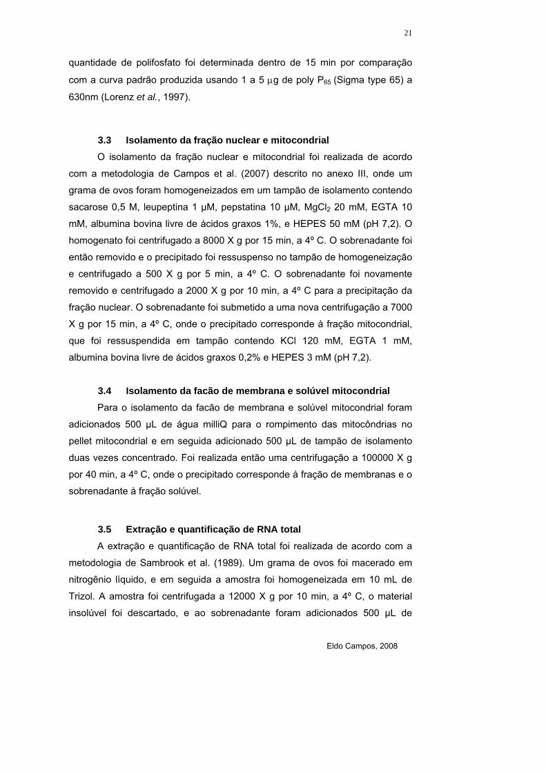
quantidade de polifosfato foi determinada dentro de 15 min por comparação
com a curva padrão produzida usando 1 a 5 µg de poly P65 (Sigma type 65) a
630nm (Lorenz et al., 1997).
3.3 Isolamento da fração nuclear e mitocondrial O isolamento da fração nuclear e mitocondrial foi realizada de acordo
com a metodologia de Campos et al. (2007) descrito no anexo III, onde um
grama de ovos foram homogeneizados em um tampão de isolamento contendo
sacarose 0,5 M, leupeptina 1 µM, pepstatina 10 µM, MgCl2 20 mM, EGTA 10
mM, albumina bovina livre de ácidos graxos 1%, e HEPES 50 mM (pH 7,2). O
homogenato foi centrifugado a 8000 X g por 15 min, a 4º C. O sobrenadante foi
então removido e o precipitado foi ressuspenso no tampão de homogeneização
e centrifugado a 500 X g por 5 min, a 4º C. O sobrenadante foi novamente
removido e centrifugado a 2000 X g por 10 min, a 4º C para a precipitação da
fração nuclear. O sobrenadante foi submetido a uma nova centrifugação a 7000
X g por 15 min, a 4º C, onde o precipitado corresponde à fração mitocondrial,
que foi ressuspendida em tampão contendo KCl 120 mM, EGTA 1 mM,
albumina bovina livre de ácidos graxos 0,2% e HEPES 3 mM (pH 7,2).
3.4 Isolamento da facão de membrana e solúvel mitocondrial Para o isolamento da facão de membrana e solúvel mitocondrial foram
adicionados 500 µL de água milliQ para o rompimento das mitocôndrias no
pellet mitocondrial e em seguida adicionado 500 µL de tampão de isolamento
duas vezes concentrado. Foi realizada então uma centrifugação a 100000 X g
por 40 min, a 4º C, onde o precipitado corresponde à fração de membranas e o
sobrenadante à fração solúvel.
3.5 Extração e quantificação de RNA total A extração e quantificação de RNA total foi realizada de acordo com a
metodologia de Sambrook et al. (1989). Um grama de ovos foi macerado em
nitrogênio líquido, e em seguida a amostra foi homogeneizada em 10 mL de
Trizol. A amostra foi centrifugada a 12000 X g por 10 min, a 4º C, o material
insolúvel foi descartado, e ao sobrenadante foram adicionados 500 µL de
Eldo Campos, 2008
22
clorofórmio. Em seguida, o material foi homogeneizado por inversão, incubado
a temperatura ambiente por 3 min, e submetido a centrifugação de 12000 X g
por 15 min, a 4º C. O sobrenadante foi recolhido e adicionado 500 µL de
isopropanol. Posteriormente, a amostra foi homogeneizada por inversão e
incubada a -20 º C por 2 hs. Em seguida, a amostra foi centrifugada a 12000 X
g por 15 min a 4º C, o sobrenadante descartado e o precipitado lavado com
etanol 75%, e após a secagem foi ressuspendido em água DEPC. Foi medida a
absorbância a 260 e 280 nm para determinar a pureza e para a quantificação
foi aceito que 1 unidade de absorbância a 260 nm corresponde a 40 µg/mL de
RNA.
3.6 Quantificação de ortofosfato Dez miligramas de ovos de diferentes dias após a ovoposição, em
triplicata, foram homogeneizados em 500 µL de água destilada e centrifugados
a 200 X g por 1 min. Foram tomadas alíquotas de 100 µL e adicionadas em 1
mL de solução de coloração feita na hora do ensaio pela mistura da solução I
que é composta de, SDS 0,5%, NH4MoO4 0,5% e H2SO4 2% com a solução II
que é feita de ácido ascórbico 0,5%. Em seguida foram quantificadas em
espectrofotômetro (Shimadzu UV-visível - 1240) a 750 nm. O conteúdo de
ortofosfato nos ovos foi calculado com base em uma curva padrão de fosfato
de sódio submetida às mesmas condições de ensaio (Fiske e Subbarow,
1925).
3.7 Análise eletroforética de polifosfato A análise eletroforética de polifosfato após a extração foi realizada de
acordo com Clark et al. (1986). Gel de poliacrilamida – Uréia foi preparado
misturando 10,51 g de uréia, 3,75 mL de solução de acrilamida e 2,5 mL de
Tris-borato 0,9 M, (pH 8,3) e EDTA 27 mM para um volume final de 25 mL. Foi
feita uma pré-corrida de 300 V por 1h, e em seguida o poly P foi misturado com
0,25 volume de tampão de amostra composto de sacarose 50%, azul de
bromofenol 0,125% e Tris-borato 450 mM (pH 8,3), EDTA 13,5 mM, e aplicado
no gel. A corrida foi realizada a 300 V até que o marcador (azul de bromofenol)
tivesse migrado 10 cm, e o gel foi corado com azul de toluidina 0,05%, metanol

Eldo Campos, 2008
23
25% e glicerol 5% por 20 min, e em seguida descorado com o mesmo solvente,
sem o azul de toluidina.
3.8 Ensaio da exopolifosfatase
As amostras foram adicionadas em 100 µL tampão Tris-HCl 50 mM (pH
7,4) contendo MgCl2 5 mM e foram incubadas a 30º C por 15 min. Foram
utilizados poly P3, poly P15 e poly P65 3 mM como substratos e heparina como
inibidor. O Pi formado durante a reação foi determinado
espectrofotometricamente adicionando molibdato de amônio 0,5%, ácido
sulfúrico 0,35 M, SDS 0,5 M e ácido ascórbico 10%. A absorbância foi medida
a 750 nm após 15 min de incubação. Foi definido como 1 unidade de atividade
enzimática a quantidade de enzima capaz de liberar 1 µmol de Pi por min. A
concentração de proteína foi determinada de acordo com Bradford, (1976),
utilizando albumina bovina como padrão.
3.9 Atividade da exopolifosfatase durante a respiração mitocondrial
A atividade da exopolifosfatase foi determinada nas mitocôndrias
isoladas dos ovos utilizando piruvato e succinato como substratos oxidativos, a
280 C, em 150 µL de meio de incubação com 0,5 mg de proteínas de
mitocôndrias. O meio de reação continha poly P15 5 mM, KCl 120 mM, EGTA 1
mM, MgCl2 5 mM, piruvato ou succinato 3 mM e ADP 0,2 mM em Tris-HCl 50
mM (pH 7,4), e foi usado KCN 1 mM como inibidor da cadeia respiratória. A
atividade da exopolifosfatase foi determinada após 15 min de incubação
(Pestov et al., 2004).
3.10 Parâmetros respiratórios
A taxa de consumo de O2 foi determinada usando um eletrodo de Clark
(YSI, mod. 5775, YellowSprings, OH). As medidas foram realizadas em 1,75
mL de meio de reação, a 28º C, contendo KCl 120 mM, EGTA 1 mM, albumina
bovina livre de ácidos graxos 0,2%, HEPES 3 mM (pH 7,2), KH2PO4 2,5 mM, e

Eldo Campos, 2008
24
KCN 1 mM ou oligomicina 2,5 µg/mL como inibidor. Foi adicionado 0,5 mg de
proteína de mitocôndrias, e após 1 min de estabilização a reação foi iniciada
com a adição de piruvato 5 mM. A razão do controle respiratório (RCR) foi
definida como a taxa de consumo de oxigênio estimulado por ADP (estado 3)
dividido pela taxa de respiração determinada na presença de oligomicina
(estado 4o), um inibidor da ATP sintase. Apesar do estado 4o não ser
equivalente ao estado 4 clássico, que é a taxa obtida após pequenas
quantidades de ADP serem quase completamente convertidas a ATP, o uso de
oligomicina elimina a re-síntese de ATP que foi degradado por contaminação
com ATPases durante o estado 4 (Kristian et al., 2006).
3.11 Consumo de O2 utilizando poly P como doador de Pi
O consumo de oxigênio utilizando-se poly P como fonte de Pi foi
determinado a 28º C em 1,75 mL de meio de reação com 0,5 mg de
mitocôndria considerando a quantidade de proteínas em mg/mL. O meio de
reação consistia de KCl 120 mM, EGTA 1 mM, albumina livre de ácidos graxos
0,2 % em HEPES 3 mM (pH 7,2). Após 1 min de estabilização, a respiração
mitocondrial foi iniciada adicionando 5 mM de piruvato e 0,2 mM de ADP.
Durante a respiração mitocondrial foram adicionados 5 mM de poly P15 e 5 mM
de KH2PO4. A concentração de oxigênio no meio foi determinada com auxílio
de um eletrodo de Clark pela inclinação da curva obtida em oxígrafo (Yellow
Springs Instruments Co).
3.12 Atividade da F-ATPase
A atividade F-ATPase sensível a azida foi determinada
espectrofotometricamente de acordo com Li e Neufeld (2001). O ensaio foi
realizado a 30º C em uma reação contendo Tris-HCl 50 mM pH 8,0, MgCl2 2
mM, KCl 100 mM e ATP 2 mM. Foi usada azida 5 mM como inibidor. Foi
definido como 1 unidade de atividade enzimática a quantidade de enzima
capaz de liberar 1 µmol de Pi por min.

Eldo Campos, 2008
25
3.13 Atividade da glicose-6-fosfato desidrogenase
A amostra foi adicionada em meio de reação contendo Tris-HCl 50 mM
pH 7,4, βNAD+ 6 mM e glicose 6 fosfato 100 mM. A atividade a 30º C foi
determinada espectrofotometricamente a 340 nm pela taxa de formação de
NADPH de acordo com Worthington (1988).
3.14 Estudos cinéticos
Os parâmetros cinéticos, como Km e Vmax da exopolifosfatase, foram
determinados por regressão não linear usando o programa GraphPad Prism.
As determinações foram realizadas com concentrações de substratos numa
escala de 0,2 a 2 Kms. Foram usados como substratos poly P3, poly P15 e poly
P65, e como inibidor foi usada heparina.

Eldo Campos, 2008
26
RESULTADOS
Eldo Campos, 2008
27
4 Resultados
A embriogênese de R. microplus é um processo complexo que envolve o
acúmulo de diversas reservas estratégicas. A cinética de utilização de
macromoléculas como lipídeos, proteínas, carboidratos e RNA durante a
embriogênese do R. microplus apresentam importantes correlações com
eventos morfológicos da embriogênese deste carrapato, possibilitando
correlacionar e entender outros fenômenos que estão ocorrendo em fases
marcantes do desenvolvimento.
Previamente aos estudos do metabolismo de polifosfatos, foi realizada
uma análise dessas reservas estratégicas durante a embriogênese do R.
microplus (Campos et al., 2006, Anexo I). Verificou-se que os carboidratos e os
lipídeos totais são mobilizados durante a primeira fase da embriogênese.
Temos considerado como primeira fase da embriogênese os estágios do 1° até
o 9° dia do desenvolvimento, que passa pelo processo de formação de um
blastoderma celular no 5° dia e posterior segmentação, entre o 7° e o 9° dia.
Observa-se então que o consumo de lipídeos ocorreu durante a formação do
blastoderma celular (Campos et al., 2006, Figura 4A do Anexo I) e o de
carboidratos durante a segmentação do embrião (Campos et al., 2006, Figura
4B do Anexo I).
A vitelina do R. microplus foi previamente isolada e caracterizada
(Logullo et al., 2002). Neste trabalho foi visto que há uma queda de
aproximadamente 15% do conteúdo de vitelina nos quatro primeiros dias, e de
20% do 8° dia até a eclosão dos ovos no 19° dia. Se considerarmos que a
vitelina pode representar até 90% do conteúdo protéico dos ovos destes
animais, e que ela é a proteína de reserva mobilizada neste processo para a
formação do embrião, pode-se dizer que o seu consumo representa a
mobilização deste substrato no ovo. Para averiguar este fenômeno, foi visto
como ocorre a mobilização das proteínas totais do embrião. Interessantemente,
o conteúdo de proteínas totais permaneceu constante durante toda a
embriogênese apesar de ocorrer um consumo em torno de 40% da vitelina,
possibilitando à larva eclodir com 60% do conteúdo de vitelo inicial (Campos et
al., 2006, Figura 5 do Anexo I).

Eldo Campos, 2008
28
A variação do RNA total durante a embriogênese apresenta uma relação
direta com a taxa metabólica dos embriões. O conteúdo de RNA total
permaneceu quase constante nos primeiros três dias, tendo um rápido
aumento a partir da formação do blastoderma celular para então permanecer
nestes patamares até próximo à eclosão (Campos et al. (2006), Figura 4C do
Anexo I).
4.1 Caracterização do conteúdo de polifosfato total na
embriogênese do R. microplus*
Para o estudo do metabolismo de moléculas de polifosfostatos durante a
embriogênese do R. microplus, inicialmente foi analisado o conteúdo de
polifosfato total e de ortofosfato, o tamanho das cadeias de polifosfato e a
atividade exopolifosfatásica no homogenato total.
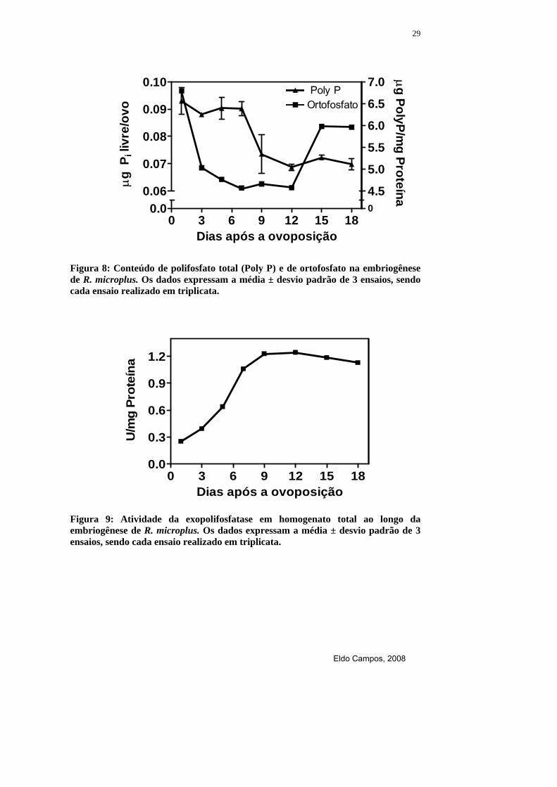
Os maiores níveis de polifosfato total foram detectados durante a
celularização e segmentação do embrião, do 5º ao 7º dia de desenvolvimento.
Após esse período ocorre uma diminuição até o 9º dia e mantém-se estável até
o fim da embriogênese. O conteúdo de ortofosfato diminui rapidamente durante
a formação do blastoderma sincicial, no 3º dia de desenvolvimento, mantendo-
se estável até o 12º dia, onde ocorre um rápido aumento até próximo à eclosão
(Figura 8). No homogenato total a atividade da exopolifosfatase é crescente até
o 12º dia, permanecendo estável até próximo da eclosão (Figura 9). O tamanho
das cadeias de polifosfato foi comparada durante o desenvolvimento
embrionário constatando o aparecimento de cadeias de polifosfato cada vez
menores a partir da segmentação do embrião, se comparada com o início do
desenvolvimento (Figura 10).
* Os resultados deste tópico foram publicados no artigo em Anexo II Campos et al., (2008).
Eldo Campos, 2008
29
Figura 8: Conteúdo de polifosfato total (Poly P) e de ortofosfato na embriogênese de R. microplus. Os dados expressam a média ± desvio padrão de 3 ensaios, sendo cada ensaio realizado em triplicata.
0 3 6 9 12 15 180.0
0.3
0.6
0.9
1.2
Dias após a ovoposição
U/m
g Pr
oteí
na
Figura 9: Atividade da exopolifosfatase em homogenato total ao longo da embriogênese de R. microplus. Os dados expressam a média ± desvio padrão de 3 ensaios, sendo cada ensaio realizado em triplicata.
0 3 6 9 12 15 180.0 0
4.5
5.0
5.5
6.0
6.5
7.0
0.06
0.07
0.08
0.09
0.10 Poly POrtofosfato
Dias após a ovoposição
µg
Pili
vre/
ovo
µg PolyP/mg Proteína

Eldo Campos, 2008
30
Dias após a ovoposição
1º 5º 9º 15º 18º
Figura 10: Análise eletroforética em gel de poliacrilamida das cadeias dos polifosfatos no 1º, 5º, 9º, 15º e 18º dia após a ovoposição de R. microplus.

Eldo Campos, 2008
31
4.2 Metabolismo nuclear e mitocondrial de polifosfatos* Para a análise do metabolismo nuclear e mitocondrial dos polifosfatos foi
realizado um fracionamento celular no qual necessitou-se de grandes
quantidades de ovos frescos (2 g) para a obtenção da fração nuclear e
mitocondrial ativa. Foram usados ovos no estágio de segmentação, no 9º dia
após a ovoposição, e a pureza e a integridade da fração nuclear foram
avaliadas através de análise por microscopia de contraste de fase (resultado
não mostrado). A fração nuclear também foi caracterizada bioquimicamente
pela ausência de marcadores de outros compartimentos, não sendo detectada
nenhuma atividade da F-ATPase, sensível a azida, um marcador mitocondrial,
e da glicose-6-fosfato desidrogenase, um marcador citoplasmático. Na fração
mitocondrial, quando oxidando piruvato, o consumo de O2 foi de 29 nmol
O2/min.mg de proteína e o RCR foi 6,8. Este processo foi sensível a KCN e
oligomicina e apresentou hidrólise de ATP pela F-ATPase maior que 80%
(Tabela 3).
* Os resultados deste tópico foram publicados no artigo em Anexo II Campos et al., (2008).

Eldo Campos, 2008
32

Eldo Campos, 2008
33
Também foram determinados, durante a embriogênese, o conteúdo de
polifosfatos e a atividade da exopolifosfatase nas frações de núcleo e
mitocôndria. Observou-se que a atividade da exopolifosfatase nuclear
aumentou ao longo do desenvolvimento embrionário, com decréscimo no
conteúdo de polifosfatos, refletindo a atividade da enzima (Figura 11A). A
fração mitocondrial apresentou uma mobilização de polifosfato diferente, tendo
maior atividade da exopolifosfatase e menores níveis de polifosfato durante a
formação do blastoderma celular e segmentação do embrião, entre o 5º e 7º
dia de desenvolvimento (Figura 11B).
A influência do RNA total na atividade da exopolifosfatase nuclear e
mitocondrial foi investigada entre concentrações de 0,3 a 1,4 µg de RNA
endógeno. Verificou-se que a exopolifosfatase nuclear apresentou um estímulo
em torno de duas vezes, enquanto a enzima mitocondrial foi insensível ao RNA
(Figura 12).

Eldo Campos, 2008
34
0 3 6 9 12 15 180.00
0.06
0.12
0.18
0.0001.00
1.25
1.50
1.75
2.00
Exopoly P Poly P(A)
U/m
g Pr
oteí
na
µg Poly P/mg Proteína
0 3 6 9 12 15 180.0 0.0
0.2
0.4
0.6
0.8
0.35
0.60
0.85
(B)
Dias após a ovoposição
U/m
g Pr
oteí
na
µg Poly P/mg Proteína
Figura 11: Atividades da exopolifosfatase (Exopoly P) e conteúdo de polifosfato (Poly P) nuclear (A) e mitocondrial (B) durante a embriogênese de R. microplus. Os dados expressam a média ± desvio padrão de 3 ensaios, sendo cada ensaio realizado em triplicata.

Eldo Campos, 2008
35
0 .0
f r a ç ã o n u c le a r f r a ç ã o m ito c o n d r ia l
0 .0
0 .1
0 .2
0 .3
0 .4
0 .5
0 .30 .0 0 .7 1 .4 0 .0 0 .3 0 .7 1 .4
0 .0 90 .1 10 .1 30 .1 50 .1 70 .1 9
R N A T o ta l (µ g )
U /
mg
Prot
eína
U / m
g Proteína
Figura 12: Influência do RNA total endógeno na atividade da exopolifosfatase nuclear e mitocondrial em ovos no estágio de segmentação, 9º dia de desenvolvimento. Os dados expressam a média ± desvio padrão de 3 ensaios, sendo cada ensaio realizado em triplicata.

Eldo Campos, 2008
36
4.3 Modulação da atividade exopolifosfatásica mitocondrial por demanda de Pi
*
Mitocôndrias de ovos no estágio de segmentação, no 9º dia de
desenvolvimento, foram isoladas, e a relação da exopolifosfatase mitocondrial
com o metabolismo energético da célula foi determinada. Para isto, foi
investigada a influência de NADH, NAD+, Pi e ADP em concentrações variando
entre 0,1 e 2,0 mM. Observou-se que a enzima foi estimulada em torno de
duas vezes por NADH, enquanto NAD+ pouco influenciou na atividade (Figura
13A). O Pi inibiu completamente (Figura 13B), ao passo que ADP também
estimulou a atividade da enzima, porém não tanto quanto observado para o
NADH (Figura 13C).
A atividade da exopolifosfatase mitocondrial foi medida durante a
respiração celular, usando piruvato ou succinato como substratos, e o consumo
de O2 foi monitorado, usando polifosfato como única fonte de Pi. Verificou-se
que a atividade da enzima aumentou em torno de 17 e 25% usando piruvato e
succinato, respectivamente. Quando a respiração mitocondrial foi inibida por
KCN, o efeito estimulatório de ambos os substratos desapareceu (Figura 14A).
A adição de poly P15 na concentração de 5 mM estimulou o consumo de O2 e
quando um novo estado 4 foi estabelecido, o Pi na concentração de 5 mM foi
adicionado estimulando o consumo de O2 novamente (Figura 14B).
*Os resultados deste tópico foram publicados no artigo em Anexo III Campos et al., (2007).

Eldo Campos, 2008
37
Figura 13: Efeito do NADH e NAD+ (A), Pi (B) e ADP (C) na atividade da exopolifosfatase mitocondrial em ovos de R. microplus no estágio de segmentação, no 9º dia de desenvolvimento. Os dados expressam a média ± desvio padrão de 3 ensaios, sendo cada ensaio realizado em triplicata.
0.1 0.3 0.5 1.0 2.00
25
50
75
100
125
150
NAD+
NADH
(A)
Concentração (mM)
% d
e es
tímul
o
0.0 0.3 0.6 0.9 1.2 1.5 1.8 2.10
25
50
75
100(B)
Pi (mM)
% d
e es
tímul
o
0.0 0.3 0.6 0.9 1.2 1.5 1.8 2.10
10
20
30
40(C)
ADP (mM)
% d
e es
tímul
o

Eldo Campos, 2008
38
Figura 14: Efeito do piruvato, succinato e KCN na atividade da exopolifosfatase durante a respiração mitocondrial (A) e consumo de O2 utilizando polifosfato como único doador de Pi (B) em ovos de R. microplus no estágio de segmentação, no 9º dia de desenvolvimento. Os dados em (A) expressam a média ± desvio padrão de 3 ensaios, sendo cada ensaio realizado em triplicata.
Controle
Piruva
to
Succinato
Piruva
to + KCN
Succinato
+ KCN
00.70.80.91.01.11.21.3(A)
U/m
g Pr
oteí
na
0 100 200 300 400 500 600 700 8000.0
MitocondriaPiruvato + ADP
Poly P15
Pi
150
175
200
225
250
Tempo (s)
nmol
O2/m
L
(B)

Eldo Campos, 2008
39
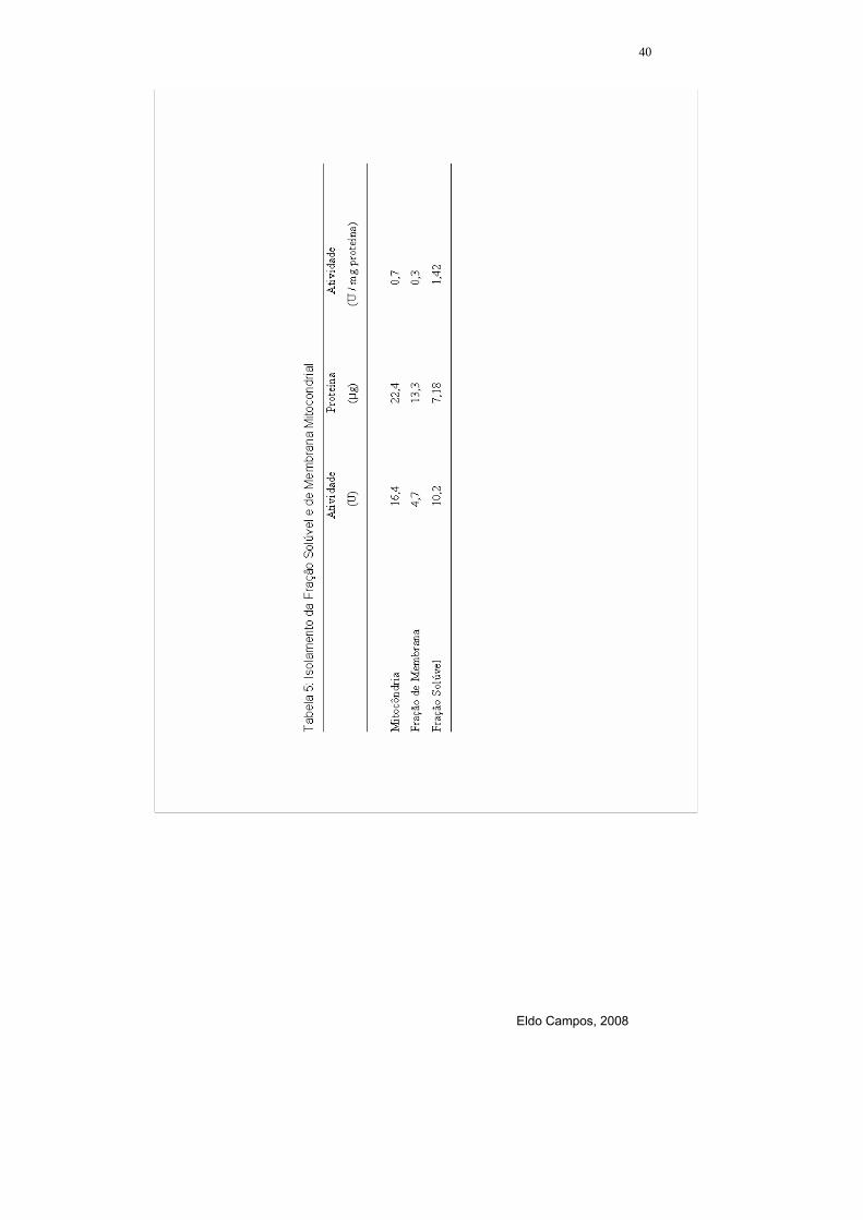
4.4 Isolamento da fração de membrana e solúvel mitocondrial
Para entender melhor o papel da exopolifosfatase mitocondrial foi
isolada a fração de membrana e a fração solúvel mitocondrial e determinada a
sua localização. A maior atividade específica foi detectada na fração de
membrana, apresentando também uma atividade na fração solúvel, porém
menor (Tabela 5). A heparina, único inibidor descrito para exopolifosfatases, foi
capaz de bloquear a ativação da respiração celular por polifosfato, mostrando a
importância da exopolifosfatase de membrana para a respiração mitocondrial
(Figura 15A). Como controle, foi visto que a heparina afeta um pouco a
respiração mitocondrial, porém não é capaz de inibi-la completamente (Figura
15B).
Eldo Campos, 2008
40

Eldo Campos, 2008
41
Figura 15: Efeito da Heparina no consumo de O2 utilizando poly P como doador de Pi (A) e na adição de Pi exógeno (B) em ovos de R. microplus no estágio de segmentação, no 9º dia de desenvolvimento.
0 100 200 300 4000.0
MitocôndriaPiruvato + ADP + Poly P15
Heparina Pi
175
200
225
(A)
Tempo (s)
nmol
O2/m
L
0 100 200 300 400 500 6000
Mitocôndria
Succinato + ADP
Heparina150
200
250
Tempo (s)
nmol
O2/m
L
(B)

Eldo Campos, 2008
42
4.5 Caracterização cinética da exopolifosfatase nuclear e mitocondrial*
As exopolifosfases nuclear e mitocondrial foram comparadas
cineticamente em função de suas atividades e afinidades. O Kmap foi medido
usando polifosfato de 3 fosfatos (poly P3), polifosfato de 15 fosfatos (poly P15) e
polifosfato de 65 fosfatos (poly P65) como substratos em ovos no estágio de
segmentação, 9º dia de desenvolvimento. A enzima mitocondrial apresentou
uma afinidade muito maior pelo poly P3 do que pelo outros substratos, em torno
de 10 vezes. Por outro lado, a enzima nuclear apresentou praticamente a
mesma afinidade por todos os substratos analisados (Tabela 4). Também foi
analisada a influência de cátions divalentes, onde a hidrólise de polifosfato
estava de acordo com a cinética de Michaelis, sendo que a exopolifosfatase
nuclear foi estimulada por Mg+2 2,5 mM e Co+2 0,1 mM, embora a mitocondrial
tenha sido estimulada somente por Mg+2 (Figura 16A). A heparina, o principal
inibidor descrito para exopolifosfatases, foi efetivo em ambas as frações,
inibindo-as quase que completamente (Figura 16B).
*Os resultados deste tópico foram publicados no artigo em Anexo II Campos et al., (2008)

Eldo Campos, 2008
43

Eldo Campos, 2008
44
N M N M0
100
200
300
400
Mg++ Co++
(A)
Exopolifosfatases
Estim
ulaç
ão (%
)
0.0 0.2 0.4 0.6 0.80.00
0.25
0.50
0.75
1.00fração mitocondrialfração nuclear
15 20 250.00
0.05
0.10
0.15
0.20
0.25(B)
Heparina (µg/mL)
U/m
g Pr
oteí
na
U/m
g Proteína
Figura 16: Efeito de cátions divalentes Mg++ e Co++ (A) e heparina (B) na atividade da exopolifosfatase nuclear (N) e mitocondrial (M) em ovos de R. microplus no estágio de segmentação, no 9º dia de desenvolvimento. Os dados expressam a média ± desvio padrão de 3 ensaios, sendo cada ensaio realizado em triplicata.

Eldo Campos, 2008
45
DISCUSSÃO

Eldo Campos, 2008
46
5 Discussão
Embora a primeira evidência da presença de polifosfatos em células de
mamíferos ter sido obtida na década de 70 (Gabel e Thomas, 1971), o
metabolismo deste biopolímero em eucariontes superiores ainda é pouco
estudado. Com o crescente interesse em caracterizar as funções dos
polifosfatos em eucariontes superiores surgem importantes relatos sobre o
assunto, conforme foi descrito para diferentes frações subcelulares e órgãos
em roedores (Kumble e Kornberg, 1996). Também existem relatos recentes
sobre a presença de polifosfatos em sangue humano e no tecido ósseo
(Leyhausen et al., 1998; Smith et al., 2006). Entretanto não foi encontrado na
literatura a descrição de polifosfatos e seu papel na embriogênese.
O aracnídeo R. microplus possui o desenvolvimento embrionário similar
aos insetos. A confirmação destas similaridades é mais evidente quando se
compara com a embriogênese do principal modelo no estudo da Biologia do
Desenvolvimento, a mosca Drosophila melanogaster (Bate e Arias, 1991;
Monnerat et al., 2002). No 4º dia de desenvolvimento do embrião em R.
microplus inicia-se a formação de um blastoderma sincicial, e em seguida, o
embrião se torna um organismo multicelular e começa a organogênese (Figura
1 do Anexo I, Campos et al., 2006). Estes dados possibilitam uma associação
do metabolismo celular com os estágios de desenvolvimento em que o embrião
se encontra. Esta abordagem permite que se defina em que evento durante o
desenvolvimento o embrião mais necessita de um determinado metabólito,
como é o caso do polifosfato.
A função do polifosfato como reserva de Pi é bem caracterizada em
procariontes e também em eucariontes inferiores (Kulaev, 1975; Kulaev e
Vagabov, 1983; Kornberg, 1995). Em R. microplus, o declínio do conteúdo de
polifosfato total após o 7º dia de desenvolvimento não reflete o aumento do
ortofosfato, que ocorre somente a partir do 12º dia, sugerindo que estes
biopolímeros também possuem outras funções, além da de reserva para o
desenvolvimento do embrião (Figura 8). Neste caso, uma fonte alternativa de Pi
para o embrião poderia ser a defosforilação da vitelina, a principal proteína do
ovo, que é gradualmente defosforilada durante a embriogênese (Silveira et al.,
2006). Esta hipótese foi corroborada quando a atividade da exopolifosfatase foi

Eldo Campos, 2008
47
analisada em homogenato total. Foi verificado que a mesma manteve-se
elevada após o 9º dia e o conteúdo de polifosfato total se mantém estável,
onde o esperado seria que ele continuasse decaindo, indicando que o embrião
está recebendo Pi de outra fonte, a qual possibilita que os níveis de polifosfato
total se mantenham estáveis nesta fase do desenvolvimento (Figura 9). O
decréscimo do tamanho da cadeia do polifosfato foi confirmado comparando o
tamanho das cadeias por eletroforese entre diferentes dias da embriogênese, e
o mesmo foi refletido na atividade exopolifosfatásica em homogenato total
(Figura 10).
Estudos mostram que além dos polifosfatos serem importantes como
reserva de Pi, nos eucariontes eles também participam em processos
regulatórios (Kornberg et al., 1999). O polifosfato em diferentes compartimentos
celulares tem sido mais estudado em S. cerevisiae, e o seu metabolismo
depende da idade da cultura e das condições de cultivo (Kulaev, 1975; Kulaev
e Kulakovskaya, 2000; Lichko et al., 2003). A participação do polifosfato
nuclear em diversas etapas que ocorrem ao longo da via do gene à proteína é
uma das mais importantes funções desses compostos em microorganismos
eucariontes e procariontes, no qual tem sido bem documentada sua relevância
no controle da expressão gênica, como por exemplo na adaptação à fase
estacionária ou outras transições de desenvolvimento (Brown e Kornberg,
2004). Os nossos resultados confirmam que embriões de R. microplus
possuem polifosfato no núcleo e que o conteúdo nuclear reflete a atividade da
exopolifosfatase durante a embriogênese (Figura 11A). Esta mobilização é
completamente diferente da mitocondrial (Figura 11B), e o conteúdo de
polifosfato nuclear diminuiu no mesmo estágio de desenvolvimento em que o
RNA total aumentou, após a formação do blastoderma celular no 5º dia de
desenvolvimento (Figura 4C do Anexo I, Campos et al., 2006).
Tem sido demonstrado que o polifosfato está envolvido em vários
aspectos da transcrição do RNA sendo capaz de interagir fisicamente com a
RNA polimerase (Kusano e Ishihama, 1997). Também há relatos sobre o
envolvimento destas moléculas na estabilidade do mRNA (Blum et al., 1997),
no aumento da produção de proteínas (Itoh et al., 2006) e no aumento da
estabilidade de polissomos in vivo durante a síntese protéica (McInerney et al.,
2006). Em núcleo de fígado de rato, o polifosfato foi relacionado com a fração

Eldo Campos, 2008
48
protéica não correspondente às histonas, podendo interagir com o complexo
DNA-histona se ligando à cromatina, e esta ligação possibilita a inibição da
atividade de algumas enzimas nucleares, incluindo a topoisomerase (Schroder
et al., 1999). Todos estes dados dão suporte a hipótese de que os polifosfatos
estão envolvidos na regulação gênica em eucariontes superiores. A simultânea
mobilização e ativação de polifosfatos nucleares e de RNA total (Campos et al.,
2008) durante a embriogênese do R. microplus sugere uma participação destes
compostos na regulação gênica. Esta hipótese foi analisada quando somente a
atividade da exopolifosfatase nuclear foi estimulada por RNA total, em
contraste com a mitocondrial (Figura 12). Este dado sugere que, além da
possível função dos polifosfatos na regulação gênica como relatado na
literatura, o ambiente nuclear é também responsável pela regulação da
atividade exopolifosfatásica.
Para o desenvolvimento do embrião, além de fatores regulatórios como
os RNAs, outros constituintes necessários ao embrião são fornecidos na
ovogênese. Durante a ovogênese, o ovário e os ovócitos de artrópodes
crescem rapidamente, acumulando grandes quantidades de carboidratos,
lipídeos e proteínas. A quantificação desses constituintes durante a
embriogênese do R. microplus sugere que os lipídeos e os carboidratos são as
principais fontes de energia para o embrião, principalmente durante a fase
inicial da embriogênese, onde os lipídeos totais apresentam uma diminuição
em torno de 45% entre o 5° e 7º dia do desenvolvimento embrionário, e os
carboidratos totais reduzem 30% entre o 7° e o 9º dia (Figuras 4A e 4B do
Anexo I, Campos et al., 2006). Os lipídeos estariam sendo utilizados na
primeira parte da embriogênese, servindo para o suporte energético durante a
celularização, como também foi observado em D. melanogaster (Bate e Arias,
1991). Nesta condição, este processo está sendo direcionado pelo organismo
materno, porque até o início da diferenciação celular todo o controle ainda está
sendo dirigido por genes exógenos ao embrião (Bate e Arias, 1991). Por outro
lado, os carboidratos seriam as principais reservas energéticas para a rápida
segmentação do embrião, um processo de natureza zigótica, marcando o início
do controle metabólico e gênico das próprias células do embrião (Nusslein-
Volhard e Roth, 1989; Bate e Arias, 1991).
Eldo Campos, 2008
49
As proteínas são os principais componentes do vitelo de ovos de
artrópodes, sendo a vitelina a sua principal constituinte. O conteúdo de proteína
total dos ovos do R. microplus permanece inalterado durante toda a
embriogênese (Figura 5 do Anexo I, Campos et al., 2006), o que está de
acordo com os mesmos dados obtidos de outro carrapato, o Hyalomma
dromedarii (Kamel e Fahmy, 1982). Por outro lado, resultados de nosso grupo
mostram que ocorre um decréscimo do conteúdo de vitelina durante os
primeiros quatro dias da embriogênese (Figura 5 do Anexo I, Campos et al.,
2006, Logullo et al., 2002), momento este em que não houve detecção de
guanina (Moraes et al., 2007). A guanina é classicamente descrita como o
único produto de degradação de aminoácidos em aracnídeos (Urich, 1990).
Essa diminuição do conteúdo de vitelina sem uma queda paralela das
proteínas totais e sem o surgimento de guanina, na primeira fase da
embriogênese, sugere que os aminoácidos da vitelina poderiam estar sendo
mobilizados apenas para a construção de novas proteínas necessárias para o
desenvolvimento do embrião. Este fenômeno é esperado, principalmente nos
quatro primeiros dias da embriogênese, onde os aminoácidos derivados da
quebra da vitelina provavelmente darão suporte à formação do blastoderma
celular. A rápida cinética de desenvolvimento embrionário do R. microplus
requer um grande suporte energético. De fato, um aumento do consumo de
oxigênio dos ovos foi visto até o 12º dia de desenvolvimento, período que inclui
a celularização e segmentação do embrião (Figura 2 do Anexo I, Campos et al.,
2006). Este dado confirma a importância do metabolismo mitocondrial para o
desenvolvimento embrionário deste carrapato.
As funções dos polifosfatos na mitocôndria estão associadas com a
fosforilação oxidativa e com a síntese de ATP (Beauvoit et al., 1989).
Desacopladores inibiram a acumulação de polifosfato em mitocôndrias de S.
cerevisiae e após “sonicação” ocorreu uma rápida hidrólise dos polifosfatos
sugerindo que a acumulação de polifosfatos depende da força próton – motriz
da membrana (Pestov et al., 2004; Pestov et al., 2005). Foi demonstrado que a
exopolifosfatase mitocondrial de R. microplus é estimulada em 100% por
NADH, 35 % por ADP e é completamente inibida por Pi (Figuras 13A, 13B e
13C) sugerindo uma regulação por Pi e por carga energética. Esta atividade

Eldo Campos, 2008
50
exopolifosfatásica aumentou durante a respiração mitocondrial quando piruvato
ou succinato foi adicionado, e o efeito estimulatório desapareceu após a adição
de KCN, um inibidor da fosforilação oxidativa (Figura 14A). Estes resultados
indicam que o gradiente de prótons e/ou o fluxo de elétrons possivelmente
regulam a exopolifosfatase para fornecer Pi para a síntese de ATP. Esta
hipótese foi confirmada, ao medir o consumo de O2 mitocondrial dependente de
ADP na presença de polifosfato e na ausência de qualquer outra fonte de Pi
(Figura 14B).
Após o isolamento da fração solúvel e de membrana mitocondrial foi
verificado que a maior atividade exopolifosfatásica se encontra na fração de
membrana (Tabela 5). Este dado sugere que é a exopolifosfatase de
membrana a responsável por fornecer Pi do polifosfato para a síntese de ATP,
já que foi possível inibir completamente o consumo de O2 ADP-dependente
utilizando heparina (Figura 15A). Para analisar um possível papel da
exopolifosfatase solúvel são necessários estudos sobre sua regulação e
função. Na literatura já foi relatada a regulação de cinases por polifosfatos
(Smith et al., 2006), portanto uma hipótese para a sua função seria a regulação
de cinases mitocondriais, já que foi verificado anteriormente que as mesmas
possuem papel importante na defesa antioxidante, prevenindo a formação de
espécies reativas de oxigênio (ROS) na mitocôndria (da-Silva et al., 2004;
Meyer et al., 2006). Nossos resultados indicam que os polifosfatos
provavelmente estão sendo mobilizados em uma situação de grande
necessidade energética, o que resultaria em um aumento na formação de
ROS.
Para analisar um possível papel fisiológico do polifosfato mitocondrial, os
seus níveis foram quantificados e a atividade da exopolifosfatase mitocondrial
foi medida ao longo da embriogênense. A maior atividade exopolifosfatásica
associada com o maior consumo dos polifosfatos ocorreu durante a formação
do blastoderma celular e segmentação do embrião, entre o 5º e 7º dia de
desenvolvimento (Figura 11B). Dessa maneira, a atividade da exopolifosfatase
mitocondrial parece estar correlacionada com a demanda energética do
embrião durante esta fase do desenvolvimento embrionário, na qual o embrião
utiliza grande parte de suas reservas de carboidratos e lipídeos (Figuras 4A e
4B do Anexo I, Campos et al., 2006). Após o 7º dia de desenvolvimento, o

Eldo Campos, 2008
51
conteúdo de polifosfato mitocondrial aumenta, enquanto a atividade
exopolifosfatásica diminui, ficando estável entre o 12º e 18º dia de
desenvolvimento. Estes resultados sugerem que no final da embriogênese
ocorre uma mudança no balanço da síntese e hidrólise de polifosfatos
mitocondriais. Neste caso, uma fonte alternativa de Pi poderia ser o polifosfato
citoplasmático ou mesmo o Pi proveniente da defosforilação da vitelina (Silveira
et al., 2006).
Os parâmetros cinéticos das duas exopolifosfatases, nuclear e
mitocondrial, foram comparados. Exopolifosfatases descritas em procariontes
apresentam maior afinidade para hidrólise de polifosfatos de cadeia longa
(Kumble e Kornberg, 1996) e o mesmo é observado para microorganismos
eucariontes, porém algumas enzimas de S. cerevisiae e Leishmania major são
mais ativas, hidrolisando polifosfatos de cadeia curta, como o poly P3 (Kumble
e Kornberg, 1996; Rodrigues et al., 2002). Em R. microplus, os resultados
mostraram que a afinidade da enzima nuclear praticamente não se alterou com
os substratos testados, porém, a mitocondrial apresentou um Km 10 vezes
menor para o poly P3 quando comparado com o poly P15 e o poly P65 (Tabela
4). Estes resultados indicam que a exopolifosfatase nuclear é única, pois em
nosso conhecimento não há relato na literatura de uma enzima que atua com
praticamente a mesma afinidade entre polifosfatos de cadeias curtas e longas,
e a exopolifosfatase mitocondrial faz parte de um grupo específico que
apresenta maior afinidade para polifosfatos de cadeias curtas.
A Exopolifosfatase de E. coli, a qual pertence à superfamília
kinase/actina/hsp-70, requer cátions divalentes e K+ para sua atividade
máxima, enquanto a de levedura, que pertence à superfamília da DHH
fosfoesterases, requer somente cátions divalentes (Lichko et al., 2003). A
exopolifosfatase nuclear de R. microplus foi estimulada por Mg+2 e Co+2
enquanto a mitocondrial foi estimulada somente por Mg+2 (Figura 16A), e
ambas foram insensíveis a K+, indicando que as exopolifosfatases destas
frações celulares de R. microplus são parecidas com a superfamília da DHH
fosfoesterases.
São poucos os compostos descritos como efetivos inibidores da
atividade exopolifosfatásica (Kornberg et al., 1999). Tratamentos com
molibdato, um inibidor de fosfohidrolases, e fluoreto, um inibidor de

Eldo Campos, 2008
52
pirofosfatases, não inibiram nenhuma das duas exopolifosfatases de R.
microplus testadas (resultado não mostrado). Entretanto, a heparina, um bom
inibidor para exopolifosfatase de bactérias e leveduras (Lichko et al., 2003), foi
efetiva em ambas as frações, sendo a cinética de inibição diferente, onde a
mitocondrial foi mais sensível a heparina do que a nuclear, embora as duas
fossem quase totalmente inibidas (Figura 16B). Tais diferenças cinéticas
confirmam a existência de duas diferentes enzimas expressas em núcleo e
mitocôndrias, e refletem as diferentes funções dos polifosfatos nestes
compartimentos celulares para o desenvolvimento do embrião.

Eldo Campos, 2008
53
CONCLUSÕES

Eldo Campos, 2008
54
6 Conclusões
1) Em geral, as variações do conteúdo de polifosfato acompanharam a
variação da atividade exopolifosfatásica, indicando a importância
destas enzimas no controle do metabolismo de polifosfatos na
embriogênese do R. microplus.
2) Polifosfatos possuem outras funções durante o desenvolvimento
embrionário do R. microplus, não sendo exclusivamente uma fonte
de reserva de fosfato.
3) O corpo de dados sugere a presença de duas distintas formas de
esopolifosfatases, uma presente na mitocôndria e outra no núcleo,
todavia os resultados sugerem que ambas são parecidas com a
superfamília da DHH fosfoesterases.
4) A exopolifosfatase nuclear é ativada por RNA total, e este dado
fornece uma nova perspectiva sobre o fenômeno previamente
descrito da regulação gênica mediada por polifosfato.
5) A exopolifosfatase mitocondrial é regulada por demanda de fosfato e
parcialmente por carga energética.
6) O polifosfato induz um consumo de oxigênio ADP-dependente,
funcionando como fonte de fosfato para a síntese de ATP.
7) Fisiologicamente, o polifosfato mitocondrial é consumido
especificamente nos estágios da embriogênese em que o embrião
utiliza as principais reservas energéticas do ovo, sugerindo que o
polifosfato mitocondrial esteja envolvido com a necessidade
energética do embrião.
8) Foram identificadas atividades exopolifosfatásicas associadas tanto
às frações de membranas mitocondriais quanto às frações solúveis.
E pelo menos a fração de membrana está possivelmente envolvida
com o processo de síntese de ATP por apresentar um consumo de
oxigênio ADP-dependente .

Eldo Campos, 2008
55
REFERÊNCIAS BIBLIOGRÁFICAS

Eldo Campos, 2008
56
7 Referências Bibliográficas
Ahn, S., Milner, A.J., Futterer, K., Konopka, M., Ilias, M., Young, T.W., White, S.A., 2001. The "open" and "closed" structures of the type-C inorganic pyrophosphatases from Bacillus subtilis and Streptococcus gordonii. J.Mol.Biol. 313, pp. 797-811.
Akiyama, M., Crooke, E., Kornberg, A., 1993. An exopolyphosphatase of Escherichia coli. The enzyme and its ppx gene in a polyphosphate operon. J.Biol.Chem. 268, pp. 633-639.
Allen, J.R., 1989. Immunology of interactions between ticks and laboratory animals. Experiment. Appl. Acarol. 7, pp. 5-13.
Aravind, L., Koonin, E.V., 1998. The HD domain defines a new superfamily of metal-dependent phosphohydrolases. Trends Biochem. Sci. 23, pp. 469-472.
Bate, M., Arias, A.M., 1991. The embryonic origin of imaginal discs in Drosophila. Develop.112, pp. 755-761.
Beauvoit, B., Rigoulet, M., Guerin, B., Canioni, P., 1989. Polyphosphates as a source of high-energy phosphates in yeast mitochondria: a 31P NMR-study. FEBS Lett. 252, pp. 17-21.
Blackburn, D.G., 1998. Structure, function, and evolution of the oviducts of squamate reptiles, with special reference to viviparity and placentation. J.Exp.Zool. 282, pp. 560-617.
Blum, E., Py, B., Carpousis, A.J., Higgins, C.F., 1997. Polyphosphate kinase is a component of the Escherichia coli RNA degradosome. Mol.Microbiol. 26, pp. 387-398.
Boctor, F.N., Kamel, M.Y., 1976. Purification and characterization of two lipovitellins from eggs of the tick, Dermacentor eersoni. Comp. Biochem. Physiol. pp. 223-240.
Bradford, M.M., 1976. A rapid and sensitive method for the quantitation of microgram quantities of protein utilizing the principle of protein-dye binding. Anal.Biochem. 72, pp. 248-254.
Braz, G.R., Coelho, H.S., Masuda, H., Oliveira, P.L., 1999. A missing metabolic pathway in the cattle tick Boophilus microplus. Curr.Biol. 9, pp. 703-706.
Brown, M.R., Kornberg, A., 2004. Inorganic polyphosphate in the origin and survival of species. Proc.Natl.Acad.Sci.U.S.A 101, pp. 16085-16087.
Campos, E., Façanha, A.R., Costa,E.P., da Silva, V.I., Jr., Masuda, A., Logullo, C., 2008. Exopolyphosphatases in nuclear and mitochondrial fractions

Eldo Campos, 2008
57
during Rhipicephalus (Boophilus) microplus embryonic development. Comp. Biochem. Physiol. Part B (aceito para publicação).
Campos, E., Facanha, A.R., Costa, E.P., da Silva, V.I., Jr., Masuda, A., Logullo, C., 2007. A mitochondrial exopolyphosphatase activity modulated by phosphate demand in Rhipicephalus (Boophilus) microplus embryo. Ins. Biochem Mol.Biol. 37, pp. 1103-1107.
Campos, E., Moraes, J., Facanha, A.R., Moreira, E., Valle, D., Abreu, L.,
Manso, P.P., Nascimento, A., Pelajo-Machado, M., Lenzi, H., Masuda, A., Vaz Ida S Jr, Logullo, C., 2006. Kinetics of energy source utilization in Boophilus microplus (Canestrini, 1887) (Acari: Ixodidae) embryonic development. Vet.Parasitol. 138, pp. 349-357.
Clark, J.E., Beegen, H., Wood, H.G., 1986. Isolation of intact chains of
polyphosphate from Propionibacterium shermanii grown on glucose or lactate. J.Bacteriol. 168, pp. 1212-1219.
da Silva, V.I., Jr., Logullo, C., Sorgine, M., Velloso, F.F., Rosa de Lima, M.F., Gonzales, J.C., Masuda, H., Oliveira, P.L., Masuda, A., 1998. Immunization of bovines with an aspartic proteinase precursor isolated from Boophilus microplus eggs. Vet.Immunol.Immunopathol. 66, pp. 331-341.
da-Silva, W.S., Gomez-Puyou, A., de Gomez-Puyou, M.T., Moreno-Sanchez, R., De Felice, F.G., de, M.L., Oliveira, M.F., Galina, A., 2004. Mitochondrial bound hexokinase activity as a preventive antioxidant defense: steady-state ADP formation as a regulatory mechanism of membrane potential and reactive oxygen species generation in mitochondria. J.Biol.Chem. 279, pp. 39846-39855.
Fagotto, F., 1990. Yolk degradation in tick eggs: I. Occurrence of a cathepsin L-like acid proteinase in yolk spheres. Arch.Insect Biochem.Physiol 14, pp. 217-235.
Farias, N.A.R., Gonzales, J.C., Saibro J.C., 1986. Antibiose e antixenose entre forrageiras em larvas de carrapato do boi. Pesq. Agrop. Bras. 21, pp. 1313-1320.
Fernandes, E.K., Costa, G.L., Moraes, A.M., Zahner, V., Bittencourt, V.R., 2006. Study on morphology, pathogenicity, and genetic variability of Beauveria bassiana isolates obtained from Boophilus microplus tick. Parasitol. Res. 98, pp. 324-332.
Fiske, C.F., Subbarow, Y., 1925. The colorimetric determination of phosphorus. J.Biol.Chem. 66, pp. 375-400.
Flechimann, C.H.W., 1976. Elementos de acarologia. Livraria Nobel, São Paulo.

Eldo Campos, 2008
58
Fothergill-Gilmore, L.A., Michels, P.A., 1993. Evolution of glycolysis. Prog.Biophys.Mol.Biol. 59, pp. 105-235.
Gabel, N.W., Thomas, V., 1971. Evidence for the occurrence and distribution of inorganic polyphosphates in vertebrate tissues. J.Neurochem. 18, pp. 1229-1242.
George, J.E., 2000. Present and future technologies for tick control. Ann.N.Y.Acad.Sci. 916, pp. 583-588.
Gomez-Garcia, M.R., Kornberg, A., 2004. Formation of an actin-like filament concurrent with the enzymatic synthesis of inorganic polyphosphate. Proc.Natl.Acad.Sci.U.S.A 101, pp. 15876-15880.
Gonzales J.C., 1974. O controle do carrapato bovino. Porto Alegre, Sulina.
Gonzales, J.C., 1995. O controle do carrapato do boi. 2ed.Porto Alegre.Edição do Autor.
Gonzales, J.C., Silva, N.R., Franco, N., 1974. A vida livre do Boophilus microplus. Arq.Fac.Vet.UFRGS 3, pp. 21-28.
Guerrero, F.D., Nene, V.M., George, J.E., Barker, S.C., Willadsen, P., 2006. Sequencing a new target genome: the Boophilus microplus (Acari: Ixodidae) genome project. J.Med.Entomol. 43, pp. 9-16.
Hernandez-Ruiz, L., Gonzalez-Garcia, I., Castro, C., Brieva, J.A., Ruiz, F.A., 2006. Inorganic polyphosphate and specific induction of apoptosis in human plasma cells. Haematologica 91, pp. 1180-1186.
Horn, S.C., 1983. Prováveis prejuízos causados pelos carrapatos. Ministério da Educação 2a Ed.
Horn, S.C., Arteche, C.C.P., 1985. Situação parasitária da pecuária no Brasil. A hora Veterinária 23, pp. 12-32.
IBGE (Instituto Brasileiro de Geografia e Estatística), 2006. Disponível on line no endereço http://www.ibge.gov.br/.
Itoh, H., Kawazoe, Y., Shiba, T., 2006. Enhancement of protein synthesis by an inorganic polyphosphate in an E. coli cell-free system. J.Microbiol.Methods 64, pp. 241-249.
Jamroz, R.C., Guerrero, F.D., Pruett, J.H., Oehler, D.D., Miller, R.J., 2000. Molecular and biochemical survey of acaricide resistance mechanisms in larvae from Mexican strains of the southern cattle tick, Boophilus microplus. J.Insect. Physiol. 46, pp. 685-695.
Johnston, L.A., Kemp, D.H., Pearson, R.D., 1986. Immunization of cattle against Boophilus microplus using extracts derived from adult female ticks: effects of induced immunity on tick populations. Int.J.Parasitol 16, pp. 27-34.

Eldo Campos, 2008
59
Kamel, M.Y., Fahmy, A.S., 1982. Biochemical studies of tick embryogenesis. V. Purification and partial characterization of isocitrate lyase from eggs of the tick Hyalomma dromedarii. Comp. Biochem. Physiol. B 72, pp. 107-115.
Kawano, M.M., 2006. Inorganic polyphosphate induces apoptosis specifically in human plasma cells. Haematol. 91, p. 1154A.
Kim, K.S., Rao, N.N., Fraley, C.D., Kornberg, A., 2002. Inorganic polyphosphate is essential for long-term survival and virulence factors in Shigella and Salmonella spp. Proc.Natl.Acad.Sci.U.S.A 99, pp. 7675-7680.
Kornberg, A., 1995. Inorganic polyphosphate: toward making a forgotten polymer unforgettable. J.Bacteriol. 177, pp. 491-496.
Kornberg, A., Rao, N.N., ult-Riche, D., 1999. Inorganic polyphosphate: a molecule of many functions. Annu.Rev.Biochem. 68, pp. 89-125.
Kristensen, O., Laurberg, M., Gajhede, M., 2002. Crystallization of a stringent response factor from Aquifex aeolicus. Acta Crystallogr.D.Biol.Crystallogr. 58, pp. 1198-1200.
Kristensen, O., Laurberg, M., Liljas, A., Kastrup, J.S., Gajhede, M., 2004. Structural characterization of the stringent response related exopolyphosphatase/guanosine pentaphosphate phosphohydrolase protein family. Biochemistry 43, pp. 8894-8900.
Kristian, T., Hopkins, I.B., McKenna, M.C., Fiskum, G., 2006. Isolation of mitochondria with high respiratory control from primary cultures of neurons and astrocytes using nitrogen cavitation. J.Neurosci.Methods 152, pp. 136-143.
Kulaev, I., Kulakovskaya, T., 2000. Polyphosphate and phosphate pump. Annu.Rev.Microbiol. 54, pp. 709-734.
Kulaev, I.S., 1975. Biochemistry of inorganic polyphosphates. Rev.Physiol Biochem.Pharmacol. 73, pp. 131-158.
Kulaev, I.S., Vagabov, V.M., 1983. Polyphosphate metabolism in micro-organisms. Adv.Microb.Physiol. 24, pp. 83-171.
Kulaev, I.S., Vagabov, V.M., Kulakovskaya, T.V., Lichko, L.P., Andreeva, N.A., Trilisenko, L.V., 2000. The development of A. N. Belozersky's ideas in polyphosphate biochemistry. Biochem. (Mosc.) 65, pp. 271-278.
Kumble, K.D., Kornberg, A., 1996. Endopolyphosphatases for long chain inorganic polyphosphate in yeast and mammals. J.Biol.Chem. 271, pp. 27146-27151.
Kuroda, A., Nomura, K., Ohtomo, R., Kato, J., Ikeda, T., Takiguchi, N., Ohtake, H., Kornberg, A., 2001. Role of inorganic polyphosphate in promoting

Eldo Campos, 2008
60
ribosomal protein degradation by the Lon protease in E. coli. Sci. 293, pp. 705-708.
Kusano, S., Ishihama, A., 1997. Functional interaction of Escherichia coli RNA polymerase with inorganic polyphosphate. Gen.Cells 2, pp. 433-441.
Leyhausen, G., Lorenz, B., Zhu, H., Geurtsen, W., Bohnensack, R., Muller, W.E., Schroder, H.C., 1998. Inorganic polyphosphate in human osteoblast-like cells. J.Bone Miner.Res. 13, pp. 803-812.
Li, Z., Neufeld, G.J., 2001. Isolation and characterization of mitochondrial F(1)-ATPase from crayfish (Orconectes virilis) gills. Comp. Biochem. Physiol. B 128, pp. 325-338.
Lichko, L.P., Andreeva, N.A., Kulakovskaya, T.V., Kulaev, I.S., 2003. Exopolyphosphatases of the yeast Saccharomyces cerevisiae. FEMS Yeast Res. 3, pp. 233-238.
Logullo, C., Moraes, J., nsa-Petretski, M., Vaz, I.S., Masuda, A., Sorgine, M.H., Braz, G.R., Masuda, H., Oliveira, P.L., 2002. Binding and storage of heme by vitellin from the cattle tick, Boophilus microplus. Insect Biochem Mol.Biol. 32, pp. 1805-1811.
Logullo, C., Vaz, I.S., Sorgine, M.H., Paiva-Silva, G.O., Faria, F.S., Zingali, R.B., De Lima, M.F., Abreu, L., Oliveira, E.F., Alves, E.W., Masuda, H., Gonzales, J.C., Masuda, A., Oliveira, P.L., 1998. Isolation of an aspartic proteinase precursor from the egg of a hard tick, Boophilus microplus. Parasitology 116 ( Pt 6), pp. 525-532.
Lorenz, B., Muller, W.E., Kulaev, I.S., Schroder, H.C., 1994. Purification and characterization of an exopolyphosphatase from Saccharomyces cerevisiae. J.Biol.Chem. 269, pp. 22198-22204.
Lorenz, B., Munkner, J., Oliveira, M.P., Kuusksalu, A., Leitao, J.M., Muller, W.E., Schroder, H.C., 1997. Changes in metabolism of inorganic polyphosphate in rat tissues and human cells during development and apoptosis. Biochim.Biophys.Acta 1335, pp. 51-60.
MAPA (Ministério da Agricultura, Pecuária e abastecimento), 2006. Disponível on line no endereço www.agricultura.gov.br.
Mccosker, P.L., 1981. The global importance of babesiosis. Babesiosis.New York, Academic Press.
McGowan, M.J., Homer, J.T., O'Dell, G.V., McNew, R.W., Barker, R.W., 1980. Performance of ticks fed on rabbits inoculated with extracts derived from homogenized ticks Amblyomma maculatum koch (Acarina: Ixodidae). J.Parasitol. 66, pp. 42-48.
McInerney, P., Mizutani, T., Shiba, T., 2006. Inorganic polyphosphate interacts with ribosomes and promotes translation fidelity in vitro and in vivo. Mol.Microbiol. 60, pp. 438-447.

Eldo Campos, 2008
61
Meyer, L.E., Machado, L.B., Santiago, A.P., da-Silva, W.S., De Felice, F.G., Holub, O., Oliveira, M.F., Galina, A., 2006. Mitochondrial creatine kinase activity prevents reactive oxygen species generation: antioxidant role of mitochondrial kinase-dependent ADP re-cycling activity. J.Biol.Chem. 281, pp. 37361-37371.
Merckel, M.C., Fabrichniy, I.P., Salminen, A., Kalkkinen, N., Baykov, A.A., Lahti, R., Goldman, A., 2001. Crystal structure of Streptococcus mutans pyrophosphatase: a new fold for an old mechanism. Structure. 9, pp. 289-297.
Monnerat, A.T., Machado, M.P., Vale, B.S., Soares, M.J., Lima, J.B., Lenzi, H.L., Valle, D., 2002. Anopheles albitarsis embryogenesis: morphological identification of major events. Mem.Inst.Oswaldo Cruz 97, pp. 589-596.
Moraes, J., Galina, A., Alvarenga, P.H., Rezende, G.L., Masuda, A., da Silva, V.I., Jr., Logullo, C., 2007. Glucose metabolism during embryogenesis of the hard tick Boophilus microplus. Comp. Biochem. Physiol. A 146, pp. 528-533.
Nishii, W., Suzuki, T., Nakada, M., Kim, Y.T., Muramatsu, T., Takahashi, K., 2005. Cleavage mechanism of ATP-dependent Lon protease toward ribosomal S2 protein. FEBS Lett. 579, pp. 6846-6850.
Nusslein-Volhard, C., Roth, S., 1989. Axis determination in insect embryos. Ciba Found.Symp. 144, pp. 37-55.
Parfenyev, A.N., Salminen, A., Halonen, P., Hachimori, A., Baykov, A.A., Lahti, R., 2001. Quaternary structure and metal ion requirement of family II pyrophosphatases from Bacillus subtilis, Streptococcus gordonii, and Streptococcus mutans. J.Biol.Chem. 276, pp. 24511-24518.
Pestov, N.A., Kulakovskaya, T.V., Kulaev, I.S., 2004. Inorganic polyphosphate in mitochondria of Saccharomyces cerevisiae at phosphate limitation and phosphate excess. FEMS Yeast Res. 4, pp. 643-648.
Pestov, N.A., Kulakovskaya, T.V., Kulaev, I.S., 2005. Effects of inactivation of the PPN1 gene on exopolyphosphatases, inorganic polyphosphates and function of mitochondria in the yeast Saccharomyces cerevisiae. FEMS Yeast Res. 5, pp. 823-828.
Pruett, J.H., 1999. Immunological control of arthropod ectoparasites--a review. Int.J.Parasitol. 29, pp. 25-32.
Pruett, J.H., Pound, J.M., 2006. Biochemical diagnosis of organophosphate-insensitivity with neural acetylcholinesterase extracted by sonication from the adult tick synganglion. Vet.Parasitol. 135, pp. 355-363.
Rahn, H., Paganelli, C.V., Ar, A., 1974. The avian egg: air-cell gas tension, metabolism and incubation time. Respir.Physiol. 22, pp. 297-309.
Eldo Campos, 2008
62
Rangarajan, E.S., Nadeau, G., Li, Y., Wagner, J., Hung, M.N., Schrag, J.D., Cygler, M., Matte, A., 2006. The structure of the exopolyphosphatase (PPX) from Escherichia coli O157:H7 suggests a binding mode for long polyphosphate chains. J.Mol.Biol. 359, pp. 1249-1260.
Rashid, M.H., Rumbaugh, K., Passador, L., Davies, D.G., Hamood, A.N., Iglewski, B.H., Kornberg, A., 2000. Polyphosphate kinase is essential for biofilm development, quorum sensing, and virulence of Pseudomonas aeruginosa. Proc.Natl.Acad.Sci.U.S.A 97, pp. 9636-9641.
Reizer, J., Reizer, A., Saier, M.H., Jr., Bork, P., Sander, C., 1993. Exopolyphosphate phosphatase and guanosine pentaphosphate phosphatase belong to the sugar kinase/actin/hsp 70 superfamily. Trends Biochem. Sci. 18, pp. 247-248.
Rodrigues, C.O., Ruiz, F.A., Vieira, M., Hill, J.E., Docampo, R., 2002. An acidocalcisomal exopolyphosphatase from Leishmania major with high affinity for short chain polyphosphate. J.Biol.Chem. 277, pp. 50899-50906.
Rosell, R., Coons, L.B., 1991. Determination of vitellogenin titer in the hemolymph of Dermacentor variabilis (Acarina: Ixodidae) using an indirect enzyme-linked immunosorbent assay. J.Med.Entomol. 28, pp. 41-44.
Sahoo, G., Sahoo, R.K., Mohanty-Hejmadi, P., 1998. Calcium metabolism in olive ridley turtle eggs during embryonic development. Comp Biochem.Physiol A 121, pp. 91-97.
Sappington, T.W., Raikhel, A.S., 1998. Molecular characteristics of insect vitellogenins and vitellogenin receptors. Insect Biochem.Mol.Biol. 28, pp. 277-300.
Sambrook, J., Fritsch, E. F., Maniatis, T., 1989. Molecular cloning, a laboratory manual, 2nd ed. Cold Spring Harbor Laboratory Press, New York, USA.
Schroder, H.C., Lorenz, B., Kurz, L., Muller, W.E., 1999. Inorganic
polyphosphate in eukaryotes: enzymes, metabolism and function. Prog.Mol.Subcell.Biol. 23, pp. 45-81.
Seifert, G.W., Springell, P.H., Tatchell R.J., 1968. Radioactive studies on the feeding of larvae, nymphs and adults of the cattle tick Boophilus microplus. Parasitology 58, pp. 415-430.
Silveira, A.B., Castro-Santos, J., Senna, R., Logullo, C., Fialho, E., Silva-Neto, M.A., 2006. Tick vitellin is dephosphorylated by a protein tyrosine phosphatase during egg development: effect of dephosphorylation on VT proteolysis. Insect Biochem.Mol.Biol. 36, pp. 200-209.
Smith, S.A., Mutch, N.J., Baskar, D., Rohloff, P., Docampo, R., Morrissey, J.H., 2006. Polyphosphate modulates blood coagulation and fibrinolysis. Proc.Natl.Acad.Sci.U.S.A 103, pp. 903-908.

Eldo Campos, 2008
63
Sonenshine, D.E., 1991. Biology of ticks. Oxford University Press, New York.
Sonenshine, D.E., Kocan, K.M., de la, F.J., 2006. Tick control: further thoughts on a research agenda. Trends Parasitol. 22, pp. 550-551.
Stewart, J.R., Thompson, M.B., 1993. A novel pattern of embryonic nutrition in a viviparous reptile. J. of Experim. Biol. 174, pp. 97-108.
Sutherst, R.W., 1983. The numbers of bush ticks, Haemaphysalis longicornis, parasitic on grazing cattle before and after the acquisition of host resistance. Aust.Vet.J. 60, pp. 20-21.
Sutherst, R.W., Jones, R.J., Schnitzerling, H.J., 1982. Tropical legumes of the genus Stylosanthes immobilize and kill cattle ticks. Nature 295, pp. 320-321.
Tammenkoski, M., Moiseev, V.M., Lahti, M., Ugochukwu, E., Brondijk, T.H., White, S.A., Lahti, R., Baykov, A.A., 2007. Kinetic and mutational analyses of the major cytosolic exopolyphosphatase from Saccharomyces cerevisiae. J.Biol.Chem. 282, pp. 9302-9311.
Thompson, B.M., Stewart, R.J., 1997. Embryonic Metabolism and Growth in Lizards of the Genus Eumeces. Comp.Biochem.and Physiol 118A, pp. 647-654.
Ugochukwu, E., Lovering, A.L., Mather, O.C., Young, T.W., White, S.A., 2007. The crystal structure of the cytosolic exopolyphosphatase from Saccharomyces cerevisiae reveals the basis for substrate specificity. J.Mol.Biol. 371, pp. 1007-1021.
Urich, K., 1990. Comparative animal biochemistry. Springer-Verlag, New York.
Vleck, D., 1991. Water economy and solute regulation of reptilian and avian embryos. Cambridge University Press, Cambridge, pp. 245-259.
Wang, L., Fraley, C.D., Faridi, J., Kornberg, A., Roth, R.A., 2003. Inorganic polyphosphate stimulates mammalian TOR, a kinase involved in the proliferation of mammary cancer cells. Proc.Natl.Acad.Sci.U.S.A 100, pp. 11249-11254.
West, M.W., Ponnamperuma, C., 1970. Chemical evolution and the origin of life. A comprehensive bibliography. Space Life Sci. 2, pp. 225-295.
Wikel, S.K., Bergman, D., 1997. Tick-host immunology: significant advances and challenging opportunities. Parasitol. Today 13, pp. 383-389.
Willadsen, P., 2001. The molecular revolution in the development of vaccines against ectoparasites. Vet.Parasitol 101, pp. 353-368.
Willadsen, P., Kemp, D.H., 1988. Vaccination with 'concealed' antigens for tick control. Parasitol. Today 4, pp. 196-198.

Eldo Campos, 2008
64
Worthington, C.C., 1988. Worthington Enzyme Manual: Enzymes and Related Biochemicals. Worthington Biochemical corporation, Freehold, New Jersey.
Wurst, H., Kornberg, A., 1994. A soluble exopolyphosphatase of Saccharomyces cerevisiae. Purification and characterization. J.Biol.Chem. 269, pp. 10996-11001.
Wurst, H., Shiba, T., Kornberg, A., 1995. The gene for a major exopolyphosphatase of Saccharomyces cerevisiae. J.Bacteriol. 177, pp. 898-906.
Yamagata, Y., Watanabe, H., Saitoh, M., Namba, T., 1991. Volcanic production of polyphosphates and its relevance to prebiotic evolution. Nature 352, pp. 516-519.
Yamamoto, Y., Takahashi, S.Y., 1993. Cysteine proteinase from Bombyx eggs: role in programmed degradation of yolk proteins during embryogenesis. Comp Biochem.Physiol. B 106, pp. 35-45.
Young, A.S., Morzaria S.P., 1986. Biology of Babesia. Parasitology Today 2, pp. 211-219.
Zhang, H., Gomez-Garcia, M.R., Brown, M.R., Kornberg, A., 2005. Inorganic polyphosphate in Dictyostelium discoideum: influence on development, sporulation, and predation. Proc.Natl.Acad.Sci.U.S.A. 102, pp. 2731-2735.

Kinetics of energy source utilization in Boophilus microplus
(Canestrini, 1887) (Acari: Ixodidae) embryonic development
Eldo Campos a, Jorge Moraes a, Arnoldo R. Facanha a, Erica Moreira a,Denise Valle b,c, Leonardo Abreu a, Pedro P.A. Manso d,
Aline Nascimento b,c, Marcelo Pelajo-Machado d, Henrique Lenzi d,Aoi Masuda e, Itabajara da Silva Vaz Jr.e, Carlos Logullo a,*
a Laboratorio de Quımica e Funcao de Proteınas e Peptıdeos and Laboratorio de Biologia Celular e Tecidual – CBB – UENF,
Avenida Alberto Lamego, 2000, Horto, Campos dos Goytacazes, RJ, CEP 28015-620, Brazilb Laboratorio de Fisiologia e Controle de Artropodes Vetores, Departamento de Entomologia, Instituto Oswaldo Cruz,
FIOCRUZ, Avenida Brasil, 4365, Manguinhos, Rio de Janeiro, RJ, CEP 21045-900, Brazilc Instituto de Biologia do Exercito, IBEx, Laboratorio de Entomologia, Rua Francisco Manuel,
102 Benfica, Rio de Janeiro, RJ, CEP 20911-270, Brazild Departamento de Patologia, Instituto Oswaldo Cruz, FIOCRUZ, Avenida Brasil, 4365,
Manguinhos, Rio de Janeiro, RJ, CEP 21045-900, Brazile Centro de Biotecnologia e Faculdade de Veterinaria, UFRGS, Avenida Bento Goncalves, 9500,
Predio 43421, Porto Alegre, RS, C.P. 15005, CEP 91501-970, Brazil
Received 16 June 2005; received in revised form 23 January 2006; accepted 5 February 2006
Abstract
The present work evaluates the kinetics of utilization of the main potential energy sources throughout the embryonic
developmental stages of Boophilus microplus. The embryonic development of this arthropod is completed in 21 days.
Cellularization of the blastoderm occurs on the 6th day and is rapidly followed by germ band extension and segmentation,
whose first signs are visible on the 7th day. Cellularization is typically a maternal-driven process, carried out by molecular
determinants deposited in the oocyte during oogenesis. On the other hand, segmentation is of zygotic nature, being the
consequence of the synthesis of various components by the growing embryo. The enhancement in total B. microplus RNA was
observed after cellularization, corroborating the replacement of maternal-driven processes by embryonic zygotic expression. An
abrupt increase in oxygen consumption was observed from cellularization until the 8th day of development. The reduction in dry
weight at the same period and the susceptibility of oxygen consumption to KCN suggest that the respiration process is activated
during early embryonic development. A marked decrease in total lipid content occurred between the 5th and 7th days of
development, suggesting this is the main energy source for cellularization. A major reduction in carbohydrate content occurred
later, between the 7th and 9th days, and it could be assigned to the morphological segmentation of the embryo. Although the total
amount of proteins remains unchanged from oviposition to hatching, a 15% reduction in vitellin (VT) content was observed
www.elsevier.com/locate/vetpar
Veterinary Parasitology 138 (2006) 349–357
* Corresponding author. Tel.: +55 22 2726 1467; fax: +55 22 2726 1520.
E-mail address: [email protected] (C. Logullo).
0304-4017/$ – see front matter # 2006 Elsevier B.V. All rights reserved.
doi:10.1016/j.vetpar.2006.02.004

E. Campos et al. / Veterinary Parasitology 138 (2006) 349–357350
before cellularization, up to the 4th day after egglaying. This observation was correlated to the synthesis of new proteins needed
to support early embryo development. Additional 20% of VT was consumed thereafter, mainly at the end of embryogenesis, and
in this case VT is probably used as energy source to the older embryo. Altogether, these data indicate different energy sources for
maternal and zygotic driven processes.
# 2006 Elsevier B.V. All rights reserved.
Keywords: Egg; Energy metabolism; Boophilus; Embryogenesis; Yolk proteins
1. Introduction
Bovine cattle blood is the unique feeding source
supporting development of the Ixodidae tick Boophilus
microplus. This ectoparasite is also the vector of
babesiosis and anaplasmosis, imposing serious eco-
nomic losses to several countries (Guerrero et al.,
2005). Larval, nymphal, and adult development of this
tick takes place on a single bovine host, within a period
of 2–4 weeks. The adult female, after completion of
engorgement, drops off the host and initiates oviposi-
tion 3 days later. Hatching occurs around 21 days after
egglaying and the resulting larvae can survive many
months before finding a new host, where development
proceeds up to adult stages (Corson et al., 2004).
This arthropod increases its body weight about 100
times after blood meal. As a consequence of digestion, a
great amount of food is processed and used in yolk
synthesis, wherewith the growing embryo meets energy
and nutrition needs (Sonenshine, 1991). The main yolk
protein, called Vitellin (VT), is a heme-protein in ticks
and is responsible for the brown egg color (Boctor and
Kamel, 1976; Rosell and Coons, 1991; James and
Oliver, 1997; Logullo et al., 2002). Heme-proteins are
involved in a wide variety of biological processes,
including respiration, oxygen transport and oxygen
metabolism (Ponka, 1997). Oxygen consumption is a
major parameter characterizing respiratory metabo-
lism, and has been widely used to study the influence of
several environmental factors in arthropod develop-
ment and physiology (Madhavan, 1975; Braune, 1976;
Tonzetich et al., 1976; Petitpren and Knight, 1979).
As a rule, oviparous animals face embryogenesis in
the absence of exogenous nutrients. In this case,
maternal nutrients are packaged into the oocytes, stored
mostly as yolk granules (Fagotto, 1990; Sappington and
Raikhel, 1998). Maturation occurs during the pre-
oviposition phase, characterized by a rapid increase in
ovarian size (Cherry, 1973). It is at this phase that
oocytes exhibit fast growth, accumulating RNAs,
carbohydrates, lipids and proteins that will meet the
regulatory and metabolic needs of the developing
embryo (Chippendale, 1978). Lack of any required yolk
component may restrain or even block embryo
development inside the egg (Thompson and Russel,
1999). Previous studies performed with birds have
focused on the importance of various egg components
to the developing embryo, as well as on the mechanisms
that transport nutrients from the yolk to their utilization
sites in embryonic tissues (Speake et al., 1998).
Current literature provides ample data regarding
the regulatory cascade of events that control devel-
opment during arthropod embryogenesis. Neverthe-
less, little information is available concerning energy
metabolism during embryonic development. Knowl-
edge of this process could reveal potential targets to
control arthropod disease vectors or agricultural pests.
The B. microplus tick is an ectoparasite responsible for
weight losses and skin damage in bovine cattle. Apart
from this, the organism is a vector of several diseases.
Here we quantified the major classes of potential
energy sources in the course of B. microplus
embryogenesis. Oxygen consumption was also mea-
sured in order to follow the respiratory metabolism.
These data were correlated to the different embryonic
stages, whose morphology was evaluated by confocal
microscopy. We suggest that distinct sources of energy
are mobilized in the course of B. microplus
embryogenesis, and that switches among different
energy sources occur at specific development stages.
2. Materials and methods
2.1. Ticks
Ticks were obtained from a colony maintained at
the Faculdade de Veterinaria, Universidade Federal do

E. Campos et al. / Veterinary Parasitology 138 (2006) 349–357 351
Rio Grande do Sul, Brazil. B. microplus ticks from the
Porto Alegre strain, free of Babesia spp., were reared
on calves obtained from a tick-free area. Engorged
adult females were kept in Petri dishes at 28 8C and
80% relative humidity upon completion of oviposi-
tion, which starts about 3 days after dropping off the
calves. Eggs were collected on different days during
embryogenesis and stored at �20 8C.
2.2. Histological procedures
B. microplus eggs underwent the protocol classi-
cally used to dechorionate Drosophila melanogaster
embryos (Forbes and Ingham, 1993). Briefly, eggs
were submerged in a 20% sodium hypochlorite bath
for 2–4 min. After rinsing in water, eggs were fixed in
a biphasic 10:9:1 mixture of heptane: 4% parafor-
maldehyde in PBS: dimethyl sulfoxide (DMSO), for
20 min under mild agitation. The lower aqueous phase
(containing the fixative plus DMSO) was then replaced
with 100% methanol and the remaining biphasic
mixture was vigorously shaken for 30 s. Completely or
partially dechorionated B. microplus eggs fell to the
bottom of the tube and were collected, rinsed twice
with 100% methanol and stored at �20 8C. Prior to
use, eggs were submitted to a serial rehydration with
70, 50 and 30% ethanol in PBS, followed by three
washes in PBS. Incubation with Acridine Orange or
Evans Blue (both at 0.01% in PBS) was performed for
20 min and enabled the visualization of nuclei and cell
limits, respectively. Whole eggs were directly
observed on a Zeiss LSM 410 laser scanning confocal
microscope, using a 488 nm laser beam and a BP 510–
525 nm filter for the visualization of acridine orange
or, alternatively, a 543 nm laser beam and a LP 570 nm
filter for Evans blue, respectively.
2.3. Oxygen consumption
The total oxygen consumed by embryonated eggs
at different times after oviposition was assayed using a
Clark-type electrode (YSI, mod. 5775, Yellow
Springs, OH). The calibration process was executed
using 100% as complete air-saturated buffer at 28 8C.
Measurements were carried out in 3 mL 20 mM PBS
buffer (pH 7.4); 0.1 mM EDTA, and the rate of oxygen
consumption was calculated in mmol O2/min mg egg.
A solution containing 1 mM KCN was added to inhibit
cytochrome oxidase. The same solution was used as
negative control. For each day after oviposition
evaluated, three assays were performed using
200 mg of eggs.
2.4. Determination of dry weight and water
content
Three samples of 300 eggs were used in each
determination. After counting, groups of eggs at
different days after oviposition were weighed on a
Shimadzu balance AX200 and dried at 85 8C until
constant weight was attained. The water content was
calculated as the difference in weight values before
and after drying. Three samples were analyzed for
each experimental point.
2.5. RNA content
RNAwas extracted from 50 mg of egg homogenates
with Tri-reagent (Sigma, inc.) according to the
manufacturer’s instructions and precipitated with
0.5 mL isopropanol for 10 min at room temperature.
Subsequently, theextractedRNAwaswashedwith1 mL
75% ethanol, vacuum-dried for 10 min and resuspended
in 1 mL water. Absorbance was measured at 260 and
280 nm, assuming that one absorbance unity at A260
corresponds to 40 mg RNA (Sambrook et al., 1989).
2.6. Total protein content
Five milligrams of eggs were homogenized in 1 mL
PBS 20 mM pH 7.4, leupeptin 100 mM, pepstatin
100 nM and benzamidine. 1 mM and centrifuged at
11,000 � g for 5 min. Total protein content was
quantified according to Bradford (1976) in 10 mL
aliquots, using bovine serum albumin as standard. Three
samples were analyzed for each experimental point.
2.7. Total sugar content
Ten milligrams of eggs were homogenized in 1 mL
PBS 20 mM pH 7.4 and centrifuged at 11,000 � g for
5 min. Aliquots of 50 mL were added to 20 mL 80%
phenol, 1 mL H2SO4 and 350 mL water, vortexed and
then left at room temperature for 30 min. Absorbance
was read at 490 nm using a molar extinction
coefficient of 6.22 M�1 (Dubois et al., 1956), and

E. Campos et al. / Veterinary Parasitology 138 (2006) 349–357352
total sugar content was calculated based on a glucose
standard curve. Three samples were analyzed for each
experimental point.
2.8. Total lipid content
Egg homogenate aliquots containing 10 mg of
protein each were extracted with chloroform–metha-
nol–water (2:1:0.5; v/v) (Bligh and Dyer, 1959). The
organic phase was dried under a N2 stream and
weighed to determine lipid content. Three samples
were analyzed for each experimental point.
2.9. Vitellin quantification
Determination of VT content was performed using
an enzyme-linked immunosorbent assay (ELISA).
Egg homogenates from the 1st to the 20th day of
embryogenesis (90 ng protein) in PBS were spotted
onto ELISA plates as 100 mL aliquots and incubated
overnight. Plates were blocked with 5% (W/V) non-fat
dry milk, 0.5% (V/V) Tween 20 in PBS, and incubated
with anti-VT polyclonal antibody (Logullo et al.,
Fig. 1. Morphology of early B. microplus embryos. Permeabilized eggs of dif
microscopy. (A) No labeling (only auto-fluorescence); (B–F) Acridine Orang
(C) 4-day-old; (F) 7-day-old embryo. A black and white projection of a 3D rec
only the chorion is auto-fluorescent (panel A). Acridin Orange and Evans b
2002) diluted 1:5000 in PBS containing 5% non-fat
dry milk for 2 h. Goat anti-rabbit IgG conjugated to
Alkaline Phosphatase (Sigma, inc.) diluted to
1:10,000 (V/V) was used as secondary antibody.
After 90 min incubation in p-nitrophenyl phosphate,
which served as substrate, the amount of VT in each
sample was determined using a Thermomax micro-
plate reader (Molecular Devices, Sunnyvale CA). A
standard curve was made using VT purified from eggs
collected on the 1st day after oviposition.
3. Results
The present work correlates the mobilization
kinetics of some components of B. microplus eggs
with the morphological changes that take place during
early embryogenesis.
3.1. Morphology
The early stages of B. microplus embryogenesis
were visualized by confocal scanning microscopy
ferent days after oviposition were submitted to laser scanning confocal
e labeling; (E) Evans blue labeling. (A, D, E) 6-day-old; (B) 1-day-old;
onstruction of a well-segmented embryo is shown in panel F. Note that
lue labeling show nuclei and cell limits, respectively.

E. Campos et al. / Veterinary Parasitology 138 (2006) 349–357 353
Fig. 2. Oxygen consumption. Oxygen consumption of B. microplus
embryos was assayed using a Clark-type electrode calibrated to
100% with air-saturated buffer at 28 8C. (&) Kinetics of oxygen
consumption at different days after oviposition. (~) effect of a
1 mM KCN solution, inhibitor of cytochrome oxidase. Aliquots
containing 200 mg of eggs were assayed in all cases. Data shown are
mean � S.E.M. (n = 3).
Fig. 3. Changes in dry weight and water content. The dry weight of
eggs at different days after oviposition was determined with an
analytical balance to the nearest 0.0001 g. Each aliquot contained
300 eggs. The water content was calculated as the difference in weight
values before and after drying.Data shown are mean� S.E.M. (n = 3).
(Fig. 1). It was first verified that, with exception of the
chorion, embryos of this species are not auto-
fluorescent (Fig. 1A), a characteristic that allowed
labeling with exogenous markers. Visualization of
nuclei labeled with Acridine Orange revealed almost
no staining on the 1st day after oviposition (Fig. 1B).
On the 4th day, nuclei had proliferated and attained the
cortical cytoplasm, a pattern typical of syncitial
blastoderm (Fig. 1C). A mitotic burst occurred
between days four and six, as judged by the density
of nuclei observed on the 6th day after oviposition
(Fig. 1D). Evans Blue staining of 6-day-old embryos,
but not of earlier ones, confirmed that cellularization
had taken place (Fig. 1E), characterizing the cellular
blastoderm stage. Afterwards, embryo development
quickly progressed to the germ band extension phase:
on the 7th day after egglaying, the embryo was clearly
distinguished from the extra-embryonic membrane
and segmentation was evident (Fig. 1F).
3.2. Oxygen consumption
No detectable oxygen consumption was verified on
the 1st day after oviposition. From that point on, intense
activation of the respiration process occurred until the
12th day of embryonic development. Oxygen con-
sumption was completely blocked at all stages by
addition of 1 mM KCN in the reaction medium (Fig. 2).
3.3. Determination of dry weight and water
content
Both the B. microplus eggs’ dry weight and the
water content remained constant during early devel-
opment, up to the 6th day after egglaying (Fig. 3). A
decrease in dry weight was observed between the 7th
and 9th days, when the total dry mass dropped from
15.7 � 1.3 to 12.6 � 1.4 mg/egg, corresponding to a
loss of 18.4%. At the same time, the water content
increased from 24.3 � 1.3 to 27.2 � 1.4 mg/egg of the
total eggs’ weight, corresponding to 11.2% (Fig. 3).
3.4. Consumption of energetic sources during
embryogenesis
The total lipid content remained stable until the 5th
day (2.07 � 0.46 mg/egg), dropping to approximately
1.1 mg/egg on the 7th day, a value that was kept roughly
unchanged until hatching (Fig. 4A). Total sugars
exhibited similar kinetics, although slightly delayed:
values remained stable until the 7th day (1.86�0.40 mg/egg), dropped to 1.18 mg/egg on the 9th day
(a 36.6% decrease) and were kept constant until
hatching (Fig. 4B). The sugar and the lipid kinetics
accompanied the dry weight changes, shown in Fig. 3.
The changes in total RNA amount during embryogen-
esis are shown in Fig. 4C. The total amount of RNAwas
almost constant during the first 3 days, rose quickly
between the 6th and the 9th days after oviposition, and
did not exhibit significant changes thereafter.

E. Campos et al. / Veterinary Parasitology 138 (2006) 349–357354
Fig. 4. Consumption of energetic sources. Major B. microplus egg
storage components were quantified at different days after oviposi-
tion. (A) Lipid quantification, determined by gravimetric method
(Bligh and Dyer, 1959); (B) total sugar concentration, measured
using Dubois‘ method; (C) total RNA, extracted from egg (50 mg)
homogenate; absorbance was read at 260 and 280 nm, using a molar
extinction coefficient of 6.22 M�1. Data shown are mean � S.E.M.
(n = 3).
Fig. 5. Protein mobilization. Total protein concentration was mea-
sured, during B. microplus embryogenesis, using Bradford’ method.
The amount of VT in egg homogenates was determined using anti-
VT polyclonal antibody with the enzyme-linked imunosorbent assay
(ELISA). Data shown are mean � S.E.M. (n = 3 or 4).
3.5. Protein mobilization
Vitellin (VT), the main yolk protein, represents 80%
of total B. microplus egg protein content on the 1st day
after oviposition. No significant changes in total protein
content were observed throughout B. microplus
embryogenesis (Fig. 5). In contrast, around 15% of
VT is mobilized during the initial 4 days of embryonic
development while an additional 20% of VT is
consumed until the 18th day after oviposition (Fig. 5).
4. Discussion
Development in invertebrates has been analyzed
under different aspects, morphogenesis and cell cycle
control being outstanding examples (Nusslein-Vol-
hard and Roth, 1989; Bate and Arias, 1993; Edgar
et al., 1994; Patel, 1994). Mobilization of yolk
components, especially Vitellin, the major protein
constituent of arthropod’s eggs, has also been the
subject of several reports (Takahashi et al., 1993;
Abreu et al., 2004). However, there is a lack of
information regarding the dynamics of energetic
metabolism during embryogenesis.
The metabolic costs of embryonic development can
be defined as the total amount of energy consumed by
an embryo during its development, including the
demands by growth or biosynthesis and by tissue
maintenance (Thompson and Stewart, 1997). The
present work evaluated the kinetics from the utiliza-
tion of the main potential energy sources available
during the embryonic development of the cattle tick,
B. microplus. As information on tick embryo
development was but scarcely available (Sonenshine,
1991), we first evaluated the morphology of embryos
mainly on the early phases of their development.

E. Campos et al. / Veterinary Parasitology 138 (2006) 349–357 355
The B. microplus embryonic development is
completed in 21 days when the specimens are reared
at 28 8C. Until the 5th day after oviposition, the B.
microplus embryo is a syncitium. Cellularization of
the blastoderm occurs on the 6th day, being followed
by impressively quick movements: by the 7th day a
segmented germ band can already be distinguished
(Fig. 1). Rapid early embryonic movements are typical
of the model system D. melanogaster and have also
been observed in mosquitoes (Bate and Arias, 1993;
Monnerat et al., 2002).
Cellularization of D. melanogaster embryos is a
synchronous process and a maternal-driven phenom-
enon, regulated exclusively by factors deposited in the
oocyte during oogenesis. In this species, cellulariza-
tion is a key event that definitively switches control of
the embryonic development to the embryo itself: cells
loose synchrony and the progression to later stages
relies on zygotic transcription (Bate and Arias, 1993).
The quick developmental kinetics observed in early
B. microplus embryos requires a readily available
energetic support. In fact, an increase in oxygen
consumption was noted until the 12th day after
oviposition, a period that includes the cellularization
and segmentation of B. microplus embryos. Oxygen
consumption was completely inhibited by addition of
KCN, further confirming the activation of the respira-
tion process (Fig. 2). To our knowledge, evaluations of
oxygen consumption during the life cycle of arthropods
are restricted to post-embryonic development stages
(Guerra and Cochran, 1970; Petitpren and Knight,
1979; Gromysz-Kalkowska and Lechowski, 1992).
Parallel to the abrupt rise in oxygen consumption at the
beginning of development, a significant reduction of B.
microplus eggs’ dry weight is observed, corroborating
an active respiration process. Moreover, this is
accompanied by an increase in egg total water content,
detectable between the 7th and the 9th days after
oviposition (Fig. 3). Increase in water content is
probably a consequence of the intense metabolic
activity and respiration that take place inside the egg
during this period (Figs. 2 and 3). It is noteworthy that a
decrease in B. microplus eggs’ dry weight occurs
mainly after the tick embryo cellularization, when
segmentation is rapidly achieved. The reduction in dry
weight and the increase in oxygen consumption were
also observed in the fish Solea senegalensis, but only in
the latest embryonic stages (Parra et al., 1999).
Quantification of the major constituents of the egg
in the course of B. microplus embryogenesis suggests
that lipids and carbohydrates are the major energy
sources, specifically during early embryo develop-
ment. For both classes of molecules, amounts were
observed to remain the same, except between the 5th
and the 7th days for lipids and between the 7th and the
9th days for carbohydrates. This kinetics suggests the
utilization of lipids in the course of cellularization, a
maternal-driven process (Bate and Arias, 1993). On
the other hand, carbohydrates would be the major
energy source for the quick segmentation of the
embryo, of zygotic nature (Nusslein-Volhard and
Roth, 1989; Bate and Arias, 1993).
Changes in nucleic acids content, particularly
RNA, reflect the anabolic activities of the developing
arthropod embryo and the degree of embryonic
independence of the material of maternal origin.
The increase in B. microplus total RNA content
between the 6th and the 9th days after oviposition
confirms an intense zygotic transcription activity after
cellularization of the blastoderm, when development
proceeds towards segmentation. These data suggest
that maternal-driven processes are replaced by
embryonic zygotic expression after cellularization
of the blastoderm. A steady increase in total RNA
content at the beginning of embryonic development
has also been described for Tribolium confusum and
for the tick Hyalomma dromedarii (Amnai-Devi et al.,
1963; Kamel et al., 1982).
Proteins are the largest components of the yolk and
VT is its major constituent. The total protein content in
B. microplus eggs remains unchanged from oviposition
to hatching (Fig. 5). This is in agreement with data from
the tick H. dromedarii (Kamel et al., 1982). However, a
significant decrease in B. microplus VT content during
the first 4 days of embryogenesis was verified. An
additional decrease in VTwas observed between the 6th
and the 18th days of development. The decrease in VT
content without a parallel depletion of total proteins
suggests that its final destination should be the synthesis
of new proteins. This is expected to be the case, mainly
with the VT depletion during the first 4 days, when VT-
derived amino acids are probably the source of newly
synthesized proteins that will support blastoderm
formation. Guanine is the product of amino acid
catabolism in arachnids (Urich, 1990) and its quanti-
fication in B. microplus’ eggs revealed an increase in the
E. Campos et al. / Veterinary Parasitology 138 (2006) 349–357356
concentration of this metabolite after cellularization, on
the 6th day (data not shown). Therefore, part of the VT
that is consumed from that point on could provide the
amino acids to be used as energy source to the growing
embryo, at later development stages (Fig. 5).
It should be noted that only 40% of the VT
deposited in B. microplus oocytes is utilized during
embryogenesis (Logullo et al., 2002). It is essential for
the newly hatched tick larvae to identify a new host in
order to proceed to further development stages. Their
survival depends upon the remaining nutritional
supply brought from the egg and on the metabolic
rate of energy consumption during the early stages of
post-embryonic development (Sonenshine, 1991).
In conclusion, it is likely that the energetic
metabolism of B. microplus embryos is a dynamic
process which varies with the progression of
morphological stages and with the control of the cell
cycle during development. Early embryogenesis
requires maternal regulatory factors, like specific
RNAs, deposited in the oocyte during oogenesis. An
intense translational activity is inferred from the
depletion of the major yolk protein, VT, without
concomitant decrease in the embryo total proteins.
With cellularization of the blastoderm, on the 6th day,
the embryo itself assumes the control of its devel-
opment, through activation of zygotic expression.
This fact is reflected in the increase of total RNA,
starting on the 6th day after oviposition. The
development progresses quickly to segmentation,
whose first signs are already visible on the 7th day
after egglaying. An intense metabolism during
embryo cellularization and segmentation is reflected
both on the decrease in total egg’s dry weight and on
the increase of oxygen consumption at this period. A
switch in energy source is also proposed to occur in
parallel to the progression from maternal to zygotic
control of development, since our data suggest lipids
as the main energy source during cellularization, with
carbohydrates accomplishing this role during embryo
segmentation.
B. microplus is a one-host tick that causes major
losses to bovine herds, especially in tropical regions,
and major efforts have been made to develop
immunoprophylatic tools against it. In this sense,
knowledge of the processes involved in embryonic
development may unravel additional targets that could
be effective in the control of this ectoparasite.
Acknowledgements
We are grateful to Cristovao Barros Pinheiro for his
excellent technical assistance. The authors are
indebted to Regina Aparedida Caldeira for a critical
reading of the manuscript. This work was supported
by grants from Fundacao de Amparo a Pesquisa do
Estado do Rio de Janeiro – FAPERJ, Conselho
Nacional de Desenvolvimento Cientıfico e Tecnolo-
gico – CNPq and Programa de Nucleos de Excelencia
– PRONEX.
References
Abreu, L., Valle, D., Manso, P.P.A., Facanha, A.R., Pelajo-Machado,
M., Masuda, H., Masuda, A., Vaz Jr., I.S., Lenzi, H.L., Oliveira,
P.L., Logullo, C., 2004. Proteolytic activity of Boophilus micro-
plus yolk pro-cathepsin D (BYC) is coincident with cortical
acidification during embryogenesis. Insect Biochem. Mol. Biol.
34, 443–449.
Amnai-Devi, A.L., Strivastava, U., Sarkar, N.K., 1963. Nucleic acid
and protein metabolism in Tribolium confusum. Exp. Cell Res.
29, 443–450.
Bate, M., Arias, M., 1993. The Development of Drosophila mela-
nogaster. Cold Spring Harbor Laboratory Press, New York.
Bligh, E.G., Dyer, W.J., 1959. A rapid method of total lipid
extraction and purification. Can. J. Biochem. Physiol. 37,
911–917.
Boctor, F.N., Kamel, M.Y., 1976. Purification and characterization
of two lipovitellins from eggs of the tick Dermacentor ander-
soni. Insect Biochem. 6, 223–240.
Bradford, M.M., 1976. A rapid and sensitive method for the
quantification of microgram quantities of protein utilizing the
principle of protein-dye binding. Anal. Biochem. 72, 248–
254.
Braune, H.J., 1976. Effects of temperature on the rates of oxygen
consumption during morphogenesis and diapause in the egg
stage of Leptopterna dolobrata. Oecologia (Berl.) 25, 77–
87.
Cherry, L.M., 1973. The accumulation and utilization of food
reserves by adult female cattle tick, Boophilus microplus (canes-
trine). Aust. J. Zool. 21, 403–412.
Chippendale, G.M., 1978. Carbohydrates in reproduction and
embryonic development. In: Biochemistry of Insects, Academic
Press, New York, pp. 42–45.
Corson, M.S., Teel, P.D., Grant, W.E., 2004. Microclimate influence
in a physiological model of cattle-fever tick (Boophilus spp.)
population dynamics. Ecol. Modell. 180, 487–514.
Dubois, Gilles, K.A., Hamilton, J.K., Rebers, P.A., Smith, F., 1956.
Calorimetric method for determination of sugar and related
substances. Anal. Chem. 25, 350–356.
Edgar, B.A., Lehman, D.A., O’Farrell, P.H., 1994. Transcriptional
regulation of string (cdc25): a link between developmental
programming and the cell cycle. Development 120, 3131–3143.
E. Campos et al. / Veterinary Parasitology 138 (2006) 349–357 357
Fagotto, F., 1990. Yolk degradation in tick eggs: I. Occurrence of a
cathepsin L-like acid proteinase in yolk spheres. Arch. Insect
Biochem. Physiol. 14, 217–235.
Forbes, Z., Ingham, P., 1993. Drosophila embryos. In: Stern, C.D.,
Holland, P.W.H. (Eds.), Essential Developmental Biology, A
Practical Approach,, Oxford University Press, Oxford, pp. 3–20.
Gromysz-Kalkowska, K., Lechowski, L., 1992. Oxygen consump-
tion in Trigonotylus coelestialium (Kirk) (Heteroptera) during its
life cycle. Comp. Biochem. Physiol. 103A, 527–530.
Guerra, A.A., Cochran, D.J., 1970. Respiration during the life cycle
of the face fly. J. Econ. Entomol. 63, 918–921.
Guerrero, F.D., Miller, R.J., Rousseau, M.E., Sunkara, S., Quacken-
bush, J., Lee, Y., Nene, V., 2005. A database of cDNAs expressed
in Boophilus microplus, the tropical/southern cattle tick. Insect
Biochem. Mol. Biol. 35, 585–595.
James, A.M., Oliver Jr., H., 1997. Purification and partial character-
ization of vitellin from the black-legged tick, Ixodes scapularis.
Mol. Biol. 27, 639–649.
Kamel, Y.M., Fahmy, Y., Shalaby, Y., Ghazy, A.M., 1982. Biochem-
ical studies of tick embryogenesis DNA, RNA, haemoprotein,
guansine and guanine in developing eggs of Hyalomma drome-
darii. Insect Biochem. 12, 15–23.
Logullo, C., Moraes, J., Dansa-Petretski, M., VazJr, I.S., Masuda, A.,
Sorgine, M.H.F., Braz, G.R., Masuda, H., Oliveira, P.L., 2002.
Binding and storage of heme by vitellin from the cattle tick,
Boophilus microplus. Insect Biochem. Mol. Biol. 32, 1805–1811.
Madhavan, M.M., 1975. Respiratory metabolism and energy sources
during embryogenesis of the aquatic hemipteran, Sphoerodema
molestum. Physiol. Zool. 3, 263–268.
Monnerat, A.T., Pelajo-Machado, M., Vale, B.S., Soares, M.J.,
Lima, J.B.P., Lenzi, H.L., Valle, D., 2002. Anopheles albitarsis
embryogenesis: morphological identification of major events.
Memorias do Instituto Oswaldo Cruz 97, 589–596.
Nusslein-Volhard, C., Roth, S., 1989. Axis determination in insect
embryos. Ciba Found. Symp. 144, 37–55.
Parra, G., Ronnestad, I., Yufera, M., 1999. Energy metabolism and
larvae of the Senegal sole. J. Fish Biol. 55, 205–214.
Patel, N.H., 1994. Developmental evolution: insights from studies of
insect segmentation. Science 266, 581–590.
Petitpren, M.F., Knight, A.W., 1979. Oxygen consumption of the
dragonfly Anax junius. J. Insect Physiol. 16, 449–459.
Ponka, P., 1997. Tissue-specific regulation of iron metabolism and
heme synthesis: distinct control mechanisms in erythroid cells.
Blood 89, 1–25.
Rosell, R., Coons, L.B., 1991. Purification and partial characteriza-
tion of vitelin from the eggs of the hard tick Dermacentor
variabilis. Insect Biochem. 21, 871–885.
Sambrook, J., Fritsch, E.F., Maniatis, T., 1989. Molecular cloning, a
laboratory manual, 2nd ed. Cold Spring Harbor Laboratory
Press, New York, USA.
Sappington, T.W., Raikhel, A.S., 1998. Molecular characteristics of
insect vitellogenins and vitellogenin receptors. Insect Biochem.
Mol. Biol. 28, 177–300.
Sonenshine, D.E., 1991. Biology of Ticks, vol. 1. Oxford University
Press, New York.
Speake, B.K., Murray, A.M., Noble, R.C., 1998. Transport and
transformations of yolk lipids during development of the avian
embryo. Prog. Lipid Res. 37, 1–32.
Takahashi, S.Y., Yamamoto, Y., Shionoya, Y., Kageyama, T., 1993.
Cisteine proteinase from the eggs of the silkmoth, Bombyx mori:
identification of a latent enzime and characterization of activa-
tion and proteolytic processing in vivo and in vitro. J. Biochem.
114, 267–272.
Thompson, M.B., Russel, K.J., 1999. Embryonic energetics in eggs
of two species of Australian skink, Morethia boulengeri and
Morethia adelaidensis. J. Herpetol. 33, 291–297.
Thompson, M.B., Stewart, R.J., 1997. Embryonic metabolism and
growth in lizards of the genus Eumeces. Comp. Biochem.
Physiol. 118A, 647–654.
Tonzetich, J., Ward, C.L., Denny, E., 1976. Effects of temperature
and humidity on hydration and respiration in four strains of
Drosophila. J. Insect Physiol. 22, 107–113.
Urich, K., 1990. Comparative Animal Biochemistry. Springer-Ver-
lag, New York.
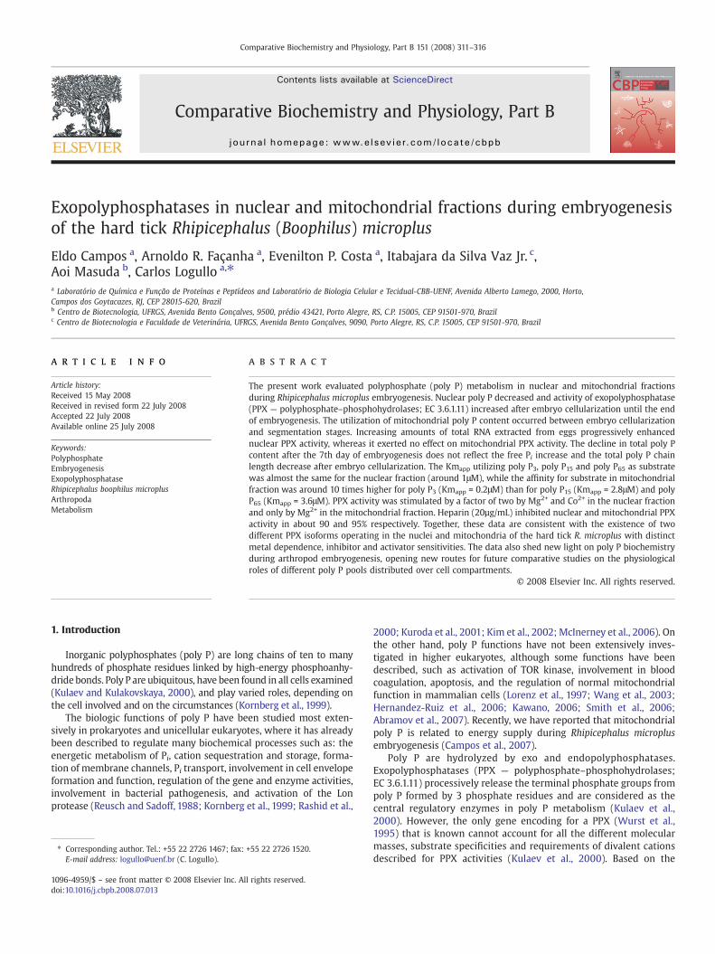
Comparative Biochemistry and Physiology, Part B 151 (2008) 311–316
Contents lists available at ScienceDirect
Comparative Biochemistry and Physiology, Part B
j ourna l homepage: www.e lsev ie r.com/ locate /cbpb
Exopolyphosphatases in nuclear and mitochondrial fractions during embryogenesisof the hard tick Rhipicephalus (Boophilus) microplus
Eldo Campos a, Arnoldo R. Façanha a, Evenilton P. Costa a, Itabajara da Silva Vaz Jr. c,Aoi Masuda b, Carlos Logullo a,⁎a Laboratório de Química e Função de Proteínas e Peptídeos and Laboratório de Biologia Celular e Tecidual-CBB-UENF, Avenida Alberto Lamego, 2000, Horto,Campos dos Goytacazes, RJ, CEP 28015-620, Brazilb Centro de Biotecnologia, UFRGS, Avenida Bento Gonçalves, 9500, prédio 43421, Porto Alegre, RS, C.P. 15005, CEP 91501-970, Brazilc Centro de Biotecnologia e Faculdade de Veterinária, UFRGS, Avenida Bento Gonçalves, 9090, Porto Alegre, RS, C.P. 15005, CEP 91501-970, Brazil
⁎ Corresponding author. Tel.: +55 22 2726 1467; fax:E-mail address: [email protected] (C. Logullo).
1096-4959/$ – see front matter © 2008 Elsevier Inc. Aldoi:10.1016/j.cbpb.2008.07.013
a b s t r a c t
a r t i c l e i n f oArticle history:
The present work evaluate Received 15 May 2008Received in revised form 22 July 2008Accepted 22 July 2008Available online 25 July 2008Keywords:PolyphosphateEmbryogenesisExopolyphosphataseRhipicephalus boophilus microplusArthropodaMetabolism
d polyphosphate (poly P) metabolism in nuclear and mitochondrial fractionsduring Rhipicephalus microplus embryogenesis. Nuclear poly P decreased and activity of exopolyphosphatase(PPX — polyphosphate–phosphohydrolases; EC 3.6.1.11) increased after embryo cellularization until the endof embryogenesis. The utilization of mitochondrial poly P content occurred between embryo cellularizationand segmentation stages. Increasing amounts of total RNA extracted from eggs progressively enhancednuclear PPX activity, whereas it exerted no effect on mitochondrial PPX activity. The decline in total poly Pcontent after the 7th day of embryogenesis does not reflect the free Pi increase and the total poly P chainlength decrease after embryo cellularization. The Kmapp utilizing poly P3, poly P15 and poly P65 as substratewas almost the same for the nuclear fraction (around 1μM), while the affinity for substrate in mitochondrialfraction was around 10 times higher for poly P3 (Kmapp = 0.2μM) than for poly P15 (Kmapp = 2.8μM) and polyP65 (Kmapp = 3.6μM). PPX activity was stimulated by a factor of two by Mg2+ and Co2+ in the nuclear fractionand only by Mg2+ in the mitochondrial fraction. Heparin (20µg/mL) inhibited nuclear and mitochondrial PPXactivity in about 90 and 95% respectively. Together, these data are consistent with the existence of twodifferent PPX isoforms operating in the nuclei and mitochondria of the hard tick R. microplus with distinctmetal dependence, inhibitor and activator sensitivities. The data also shed new light on poly P biochemistryduring arthropod embryogenesis, opening new routes for future comparative studies on the physiologicalroles of different poly P pools distributed over cell compartments.
© 2008 Elsevier Inc. All rights reserved.
1. Introduction
Inorganic polyphosphates (poly P) are long chains of ten to manyhundreds of phosphate residues linked by high-energy phosphoanhy-dride bonds. Poly P are ubiquitous, have been found in all cells examined(Kulaev and Kulakovskaya, 2000), and play varied roles, depending onthe cell involved and on the circumstances (Kornberg et al., 1999).
The biologic functions of poly P have been studied most exten-sively in prokaryotes and unicellular eukaryotes, where it has alreadybeen described to regulate many biochemical processes such as: theenergetic metabolism of Pi, cation sequestration and storage, forma-tion of membrane channels, Pi transport, involvement in cell envelopeformation and function, regulation of the gene and enzyme activities,involvement in bacterial pathogenesis, and activation of the Lonprotease (Reusch and Sadoff, 1988; Kornberg et al., 1999; Rashid et al.,
+55 22 2726 1520.
l rights reserved.
2000; Kuroda et al., 2001; Kim et al., 2002; McInerney et al., 2006). Onthe other hand, poly P functions have not been extensively inves-tigated in higher eukaryotes, although some functions have beendescribed, such as activation of TOR kinase, involvement in bloodcoagulation, apoptosis, and the regulation of normal mitochondrialfunction in mammalian cells (Lorenz et al., 1997; Wang et al., 2003;Hernandez-Ruiz et al., 2006; Kawano, 2006; Smith et al., 2006;Abramov et al., 2007). Recently, we have reported that mitochondrialpoly P is related to energy supply during Rhipicephalus microplusembryogenesis (Campos et al., 2007).
Poly P are hydrolyzed by exo and endopolyphosphatases.Exopolyphosphatases (PPX — polyphosphate–phosphohydrolases;EC 3.6.1.11) processively release the terminal phosphate groups frompoly P formed by 3 phosphate residues and are considered as thecentral regulatory enzymes in poly P metabolism (Kulaev et al.,2000). However, the only gene encoding for a PPX (Wurst et al.,1995) that is known cannot account for all the different molecularmasses, substrate specificities and requirements of divalent cationsdescribed for PPX activities (Kulaev et al., 2000). Based on the
312 E. Campos et al. / Comparative Biochemistry and Physiology, Part B 151 (2008) 311–316
primary structure, PPXs are classified into two types, whose proto-types are PPXs from yeast (Saccharomyces cerevisiae) and Escherichiacoli. Yeast-type PPX, reported in fungi and protozoa, belongs to thesuperfamily of DHH phosphoesterases (Aravind and Koonin, 1998),whereas E. coli-type PPX is present in Eubacteria and Archaea andbelongs to a sugar kinase/actin/hsp-70 superfamily (Reizer et al.,1993).
The present study is focused on the tick R.microplus, which is a one-host tick that causesmajor losses to bovine herds, especially in tropicalregions. In this scenario, major efforts have been made to developimmunoprophylatic tick control tools (Guerrero et al., 2006). Apartfrom this, the tick is the vector of parasites that cause hemoparasiticdiseases, endemic in many cattle production areas (Sonenshine et al.,2006). R. microplus takes one host only throughout all three life stages,usually a bovine, with a long feeding period (approximately 21days).The adult female, after completion of engorgement, drops off the hostand initiates oviposition around 3days later. Being an oviparousanimal, embryogenesis occurs in the absence of exogenous nutrients,and maternal nutrients are packaged in oocytes and stored mostlyas yolk granules. Hatching occurs around 21days after oviposition,and the emerging larvae can survive several weeks before finding ahost, using the remaining yolk as the only source of energy (Fagotto,1990).
Vitellogenin is a hemolymphatic phospholipoglycoprotein stored ingrowing oocytes. Once inside the oocytes, it is conventionally calledvitellin and represents the main component of yolk platelets (Sapping-tonandRaikhel,1998).Mostorganismscontainvitellin covalently boundto Pi, which is the main source of Pi during embryogenesis (Sappingtonand Raikhel, 1998). More specifically, in R. microplus it has beendemonstrated that vitellin is a phosphotyrosine-containing protein(Silveira et al., 2006).
The present study characterizes PPX activity in nuclear andmitochondrial fractions, in order to observe the importance of polyP metabolism in arthropod embryogenesis.
2. Materials and methods
2.1. Ticks
Ticks were obtained from a colony maintained at the Faculdade deVeterinária, Universidade Federal do Rio Grande do Sul, Brazil. R.microplus (Acarina, Ixodidae) ticks from the Porto Alegre strain, free ofparasites, were reared on calves obtained from a tick-free area.Engorged adult females were kept in Petri dishes at 28°C and 80%relative humidity upon completion of oviposition, which starts about3days after adult ticks drop off the calves.
2.2. Isolation of nuclei and mitochondria
The cell fractionation procedure used required large amounts offresh eggs (at least 2g) to obtain nuclear and functional activemitochondrial fractions. For the characterization of nuclear andmitochondrial fraction, eggs in the segmentation stage (9th dayafter oviposition) were used. Nuclei andmitochondriawere isolated aspreviously described (Campos et al., 2007). Eggs were homogenized in1.5mL of a buffer containing 0.5M sucrose, 100μM leupeptin, 100nMpepstatin, 20mMMgCl2,10mMEGTA,1% bovine albumin (fat acid free)and 50mM HEPES pH 7.2. The homogenate was centrifuged at 8000gfor 15min. The supernatant was carefully removed and the pellet wasresuspended in the homogenization buffer, and centrifuged at 500gfor 5min. The supernatant was carefully removed again andcentrifuged at 2000g for 10min to yield a nuclear pellet. Then, thesupernatant was submitted to another centrifugation at 7000g for15min, and the mitochondrial pellet that resulted therefrom wasresuspended in a storage buffer containing 120mM KCl, 1mM EGTA,0.2% bovine albumin, and 3mM HEPES pH 7.2.
2.3. Extraction and determination of poly P content
Poly P was extracted from eggs homogenates, nuclei andmitochondria in three steps as previously described (Clark et al.,1986; Lorenz et al., 1997). Residual amounts of DNA and RNA wereremoved from the extracts obtained in steps 2 and 3 by treatmentwith RNase and DNase 1 (250μg/mL each) in the presence of 1mMMgCl2 for 1h at room temperature. Protein was removed by oneextraction with phenol/chloroform (1:1, v/v), followed by threesuccessive extractions with chloroform. The poly P content in step 2and step 3 extracts was estimated by determining the metachromaticeffect of polyphosphate on the absorbance spectrum of toluidine blueat 530nm and 630nm (Clark et al., 1986; Lorenz et al., 1997). Acalibration curve was obtained using poly P65 (Sigma type 65) as astandard. The amount of poly P (plus Pi) in step 1 extracts and ofresidual poly P not extracted by steps 2 and 3 was determined afterhydrolysis at 100°C (Lorenz et al., 1997). The resulting Piwasmeasuredspectrophotometrically as described by Fiske and Subbarow (1925).
2.4. PPX activity, Pi and protein assay
The reaction mixture consisted of 50mM Tris–HCl buffer (pH 7.5),and 5mM MgCl2, using poly P3, poly P15 or poly P65 as substrate.Reactions were carried out at 30°C for various time periods. The Piformed during the reaction was spectrophotometrically determinedas previously described by Fiske and Subbarow (1925), by adding asolution of 0.5% ammonium molybdate, 0.35M sulfuric acid, 0.5%SDS, and 10% ascorbic acid. Measurements of absorbance at 750nmwere performed after 15min. The enzyme amount liberating 1µmoLof Pi/1min was defined as one unit of enzyme activity (U). Proteinconcentrationwas measured by Bradford (1976), using bovine serumalbumin as a standard.
2.5. Extraction of total RNA
Total RNA was extracted from 50mg of egg homogenates with Tri-reagent (Sigma-Aldrich) according to the manufacturer's instructionsand precipitated with 0.5mL isopropanol for 2h at − 20°C. Subse-quently, the extracted RNA was washed with 1mL 75% ethanol,vacuum-dried for 10min and resuspended in water DEPC. Absorbancewasmeasured at 260 and 280nm, assuming that one absorbance unityat A260 corresponds to 40μg/mL RNA (Sambrook et al., 1989).
2.6. Electrophoresis of poly P
The poly P preparation obtained was electrophoresed on 7M urea/20% polyacrylamide gels (30 × 15 × 0.3cm; bisacrylamide:acrylamideratio, 1:20) in TBE (90mM Tris, 90mM borate, 2.7mM EDTA, pH 8.3).Gels were stained with toluidine blue as previously described (Clarket al., 1986; Lorenz et al., 1994).
2.7. F-ATPase activity
The F-ATPase (EC 3.6.14); azide sensitive activity of the mitochon-drial fractionwas spectrophotometrically measured as described by Liand Neufeld (2001). The activity was assayed at 30°C and the reactionmixture contained 50mM Tris–HCl (pH 8.0), 2mM MgCl2, 100mM KCl,2mM ATP. As inhibitor, 5mM azide was used. The enzyme amountliberating 1µmoL of Pi/1minwas taken as a unit of enzyme activity (U).The Pi formed was determined as described by Fiske and Subbarow(1925).
2.8. Glucose-6-phosphate dehydrogenase activity
G6PDH (EC 1.1.1.49) activity was determined according toWorthington (1988). G6PDH activity was obtained by determining

Fig. 1. PPX activity and poly P content of nuclear fraction during R. microplusembryogenesis. Activity is expressed as U/mg of total protein in the nuclear fractionpreparations. (■) PPX activity was measured with 2.5 mM poly P15, 0.1 mM Co2+ and5 mM MgCl2. (●) Poly P levels during embryogenesis in the nuclear fractionpreparations. The results represent mean±S.D. of three independent experiments, intriplicates.
313E. Campos et al. / Comparative Biochemistry and Physiology, Part B 151 (2008) 311–316
the rate of NADPH formation at 30°C, which absorbs ultraviolet light at340nm, using a Beckman DU640 spectrophotometer.
2.9. Respiration measurements
The rate of O2 consumption by mitochondria was estimated by aClark oxygen electrode (YSI, mod. 5775, Yellow Springs, OH). Thecalibration process was conducted using the initial O2 concentration ofthe medium as 100% — saturated buffer measured at 28°C. Measure-ments were carried out in 1.5mL reaction buffer containing 120mMKCl, 1mM EGTA, 0.2% bovine albumin, 3mM HEPES (pH 7.2), 2.5mMphosphate and 1mM KCN or 2.5µg/mL oligomycin was used asinhibitor. Mitochondrial protein (0.5mg/mL) was used, and after a 1-min equilibration period, mitochondrial respirationwas started by theaddition of pyruvate to a final concentration of 5mM (Campos et al.,2007). Rates of oxygen consumption are expressed as nmol O2/(mgmitochondrial protein min). The respiratory control ratio (RCR) wasdefined as the rate of ADP (0.5mM)-stimulated oxygen consumption(State 3) divided by the rate of respiration determined in the presenceof oligomycin (2.5µg/mL) (State 4o), an inhibitor of ATP synthase.While the State 4o respiration measured in the presence of oligomycinis not equivalent to the classical State 4 rate obtained after a smallbolus of ADP has almost completely been converted to ATP, the use ofoligomycin eliminates the contribution of ATP cycling via hydrolysisby contaminating ATPases and resynthesis by the mitochondrial ATPsynthase to State 4 respiration. The oligomycin-induced State 4o rateof respiration is therefore a more specific indicator of mitochondrialproton cycling limited by passive proton leakiness of the innermembrane (Kristian et al., 2006).
2.10. Statistical analysis
All data values were expressed as mean ± S.D. ANOVA was used todetermine significant differences between groups when data werenormally distributed. The t-tests were used when comparing databetween two groups. Significance was set at p b 0.05 (Swinscow,1996).
3. Results
3.1. Nuclei and mitochondria isolation
The purity and intactness of the nuclear fraction was rathersatisfactory, as determined by examination in the phase-contrast (datanot shown). The nuclear purity was also biochemically characterizedby the absence of marker enzymes of the other compartment. In theisolated nuclear fraction, no activity of either F-ATPase azide sensitive(a mitochondrial marker), or glucose-6-phosphate dehydrogenase (acytosol marker) could be detected (Table 1). In the mitochondrialfraction, when oxidizing pyruvate, O2 consumption was 29.7nmol O2/min mg protein, and RCR was 6.8. This process was KCN- andoligomycin-sensitive and ATP hydrolysis azide-sensitivity was higherthan 80% (Table 1).
Table 1Characterization of the cell fractionation of Rhipicephalus microplus embryos on the 9thday of embryogenesis
ATPase activity(U/mg protein)
G6PDH(U/mg protein)
O2 consumption(nmol min−1 mgprotein−1)
RCR % F-ATPaseazidesensitive
Homogenate 0.7 2.9 – – 49.5%Nuclearfraction
– – – – –
Mitochondrialfraction
1.5 – 29.7 6.8 83.4%
3.2. Characterization of nuclear and mitochondrial poly P content in R.microplus embryogenesis
The poly P content and PPX activity were measured using thenuclear fraction isolated from the R. microplus eggs during embry-ogenesis. Nuclear PPX activity increased throughout embryogenesis.Poly P levels decreased after embryo cellularization (i.e. 5th day ofdevelopment), reflecting PPX activity (Fig. 1). The mobilization inmitochondrial poly P content is different, where a higher PPX activityassociated with a higher poly P utilization occurred during blastodermformation and segmentation of the embryo, between the 5th and 7thday of development, as we previously reported (Campos et al., 2007).
The influence of total RNA in nuclear and mitochondrial PPXactivity was investigated in concentrations that ranged from 0.3 to1.4µg. Nuclear PPX activity was stimulated by a factor of two, whilemitochondrial PPX activity did not change (Fig. 2).
3.3. Characterization of total poly P content during R. microplusembryogenesis
Total poly P levels were quantified throughout R. microplusembryogenesis. Total poly P levels were higher during embryocellularization and segmentation, from the 5th to 7th day of develop-ment, and declined after that until reaching a plateau. Free Pi contentrapidly decreased during syncitial blastoderm formation, on the 3rd dayof development, continuing low until the 12th of embryogenesis, whenit's rapidly increased thereafter (Fig. 3A).
Total poly P extracted on different days of embryogenesis waselectrophoresed by 20% urea–PAGE to compare their size throughout
Fig. 2. Influence of total RNA in PPX activity of nuclear and mitochondrial fractions.Nuclei and mitochondria of eggs on the 9th day of embryogenesis were isolated, andPPX activity was determined using poly P15 as substrate in the presence of 0.3 to 1.4 μgof total RNA. The results represent mean±S.D. of three independent experiments, intriplicates.

Fig. 3. Characterization of total poly P content in R. microplus embryogenesis. In (A): total poly P (▲) was extracted and quantified and free Pi (■) was quantified in eggs homogenate indifferent days after oviposition. The results represent mean±S.D. of three independent experiments, in triplicates. In (B): Electrophoresis was performed on a 7 M urea/20%polyacrylamide gel. The gel was stained with toluidine blue. Equal amounts of extracts were applied per lane. Lanes 1, 2, 3, 4 and 5 contain total poly P extracted from eggs on the 1st,5th, 9th, 15th, and 18th day after oviposition, respectively. This experiment was repeated at least three times with different preparations and this figure shows a representativeexperiment.
314 E. Campos et al. / Comparative Biochemistry and Physiology, Part B 151 (2008) 311–316
embryogenesis of R. microplus. The chain length of total poly P contentdecreased after embryo cellularization, on the 5th day of development(Fig. 3B).
3.4. Characterization of nuclear and mitochondrial PPX
The apparent Kmwasmeasured for nuclear andmitochondrial PPXusing poly P3, poly P15 and poly P65 as substrates in eggs in thesegmentation stage (9th day after oviposition). The mitochondrialaffinity PPX for poly P3 was as great as 10 times that for poly P15 or forpoly P65; on the other hand, nuclear PPX had almost the same affinityfor all substrates (Table 2). The dependence of hydrolysis rates of polyP in the presence of 2.5mM Mg2+ and 0.1mM Co2+ was in agreementwith Michaelis kinetics. The nuclear fraction was stimulated by bothcations, while themitochondrial fractionwas stimulated only byMg2+.Yet, no stimulation was detected using Co2+ (Fig. 4). Heparin, a goodinhibitor for other PPX (Kornberg et al., 1999), was also effectiveagainst these PPX, as 20µg/mL heparin inhibited nuclear andmitochondrial PPX activity by about 90 and 95%, respectively (Fig. 5).
4. Discussion
Although the first evidence for the presence of poly P inmammalian cells was obtained a long time ago (Gabel and Thomas,1971), since then relatively few works have addressed their physio-logical roles in animals cells (Kornberg et al., 1999; Wang et al., 2003;Hernandez-Ruiz et al., 2006; Kawano, 2006; Smith et al., 2006;Abramov et al., 2007, Campos et al., 2007). In this study, we havecompared poly P metabolism between nuclear and mitochondrialfractions. The results indicate differentmobilizations in these fractionsduring the whole embryogenesis stage, suggesting distinct andimportant functions of poly P in tick embryo development.
Early R. microplus embryonic stages are similar to those of D.melanogaster and of mosquitoes (Bate and Arias, 1991; Monnerat et al.,2002). Tick embryogenesis is characterized by the formation of a non-cellular syncitium up to day 4 (Campos et al., 2006). After this, theembryo becomes a multicellular organism and starts organogenesis(Campos et al., 2006).
Table 2Apparent Km values of the PPX in nuclear and mitochondrial fractions of R. microplusembryos on 9th day of embryogenesis
Substrate Nuclear fraction Mitochondrial fraction
Km (µM)
Poly P3 0.7 0.2Poly P15 1.1 2.8Poly P65 0.9 3.6
The poly P in different cell compartments was best studied in S.cerevisiae, and has been proved to depend on culture age andcultivation conditions (Kulaev, 1975; Kulaev and Kulakovskaya,2000; Lichko et al., 2003). The participation of nuclear poly P in themultiple steps that occur along the pathway from gene to protein isone of the most important functions of these compounds ineukaryotic and prokaryotic microorganisms, and it is well documen-ted in controlling gene expression, as in the adaptation to stationaryphase or other developmental transitions, for example (Brown andKornberg, 2004). Additionally, it has been demonstrated that poly P isinvolved in several aspects of mRNA transcription, and that purifiedpoly P physically interacts with purified RNA polymerase (Kusano andIshihama, 1997). Poly P has also been implicated in mRNA stability(Blum et al., 1997). Similarly, it has been shown that an increase inprotein production is caused by poly P (Itoh et al., 2006), and that anenhancement in polysome stability in vivo prevents miscoding eventsduring protein synthesis (McInerney et al., 2006). In nuclei of rat liver,poly P is related to the fraction of non-histone proteins (Offenbacherand Kline, 1984). Poly P may interact with DNA-histone binding inchromatin, and this binding has been shown to inhibit the activity ofsome nuclear enzymes, including topoisomerases (Schroder et al.,1999). These data support the hypothesis that poly P are involved inthe regulation of chromatin functions in higher eukaryotes.
The results obtained demonstrate that poly P is present in thenuclei of R. microplus embryos, and that nuclear poly P content reflectsthe nuclear PPX activity during embryogenesis (Fig. 1). The datasuggest that the mobilization of the nuclear fraction is completelydifferent from that of the mitochondrial fraction, as shown in aprevious work (Campos et al., 2007). During embryo celullarization,the nuclear fraction showed higher polyphosphate levels (9 to 10times) in comparison to the mitochondrial fraction. Similarly, the
Fig. 4. Effect of divalent cations on PPX activity in nuclear andmitochondrial fractions ofR. microplus embryos. Nuclei (N) and mitochondria (M) of eggs on the 9th day of em-bryogenesis were isolated and the PPX activity was determined using poly P15 as sub-strate in the presence of 2.5mMMg2+ and 0.1mMCo2+. The results representmean±S.D.of three independent experiments, in triplicates.

Fig. 5. Effect of heparin on PPX activity in nuclear and mitochondrial fractions of R.microplus. Nuclei (N) and mitochondria (M) of eggs on the 9th day of embryogenesiswere isolated and the PPX activity was determined using poly P15 as substrate andheparin in a concentration range of 0.1–20 µg/mL. The results represent mean±S.D. ofthree independent experiments, in triplicates.
315E. Campos et al. / Comparative Biochemistry and Physiology, Part B 151 (2008) 311–316
same has been described for nuclear andmitochondrial fractions in ratliver (Kumble and Kornberg,1995). In the present study, nuclear poly Pdecreased while total RNA increased, after the formation of a cellularsyncitium (6th day of development), as also demonstrated in previousresearch (Campos et al., 2006). Thus, nuclear poly P levels seemed tocorrelate with total RNA content during R. microplus embryogenesis.Yet, contrasting with the mitochondrial PPX activity, only the nuclearactivity is stimulated by total RNA (Fig. 2).
The function of poly P as Pi reserve is well known in prokaryotesand also in eukaryote microorganisms (Kulaev, 1975; Kulaev andVagabov, 1983; Kornberg, 1995). The cells of higher eukaryotes carrypoly P, but in smaller amounts than those found in microorganisms.So, beyond the source of Pi, these biopolymers probably participated inregulatory processes (Kornberg et al., 1999). It is interesting to notethat in R. microplus, the decline in total poly P content after the 7th dayof embryogenesis does not reflect the increase in free Pi, since thisoccurs only after the 12th day, suggesting that poly P also plays otherroles, apart from the Pi reserve for embryo development (Fig. 3A). Inthis case, an alternative source of Pi could be the dephosphorylation ofvitellin, a major yolk protein that is gradually dephosphorylatedduring embryogenesis (Silveira et al., 2006). This source can be usedmainly until the segmentation of embryo, 7th day of development,because in this period there is no total poly P mobilization. Thedecrease in chain length after embryo segmentation (9th day ofembryogenesis) is confirmed by electrophoresis of total poly Pcontent, which compared poly P chain lengths during embryogenesis(Fig. 3B).
Additionally, here we compare the kinetic parameters of nuclearand mitochondrial fraction. PPXs have been found in prokaryotes andeukaryotes and although in bacteria these enzymes hydrolyze mostlyhigh molecular weight poly P (Kumble and Kornberg, 1996), at leastsome of the enzymes from S. cerevisiae and Leishmania major aremoreactive in hydrolyzing short chain poly P, such as poly P3 (Kumble andKornberg, 1996; Rodrigues et al., 2002). Our results show that nuclearPPX activities with poly P3, poly P15 and poly P65 were nearly thesame; however, affinity of mitochondrial PPX for poly P3 was around10 times higher than that for poly P15 and poly P65 (Table 2). To ourknowledge, the R. microplus nuclear PPX is the only enzyme thathydrolyzes short and long polyphosphate with the same affinity. PPXfrom E. coli requires divalent cations and K+ for maximal activity, whilePPX from yeast requires only divalent cations (Lichko et al., 2003). Thenuclear PPX activity is stimulated by Mg2+ and Co2+, while themitochondrial PPX activity is stimulated only by Mg2+ (Fig. 4), andboth are insensitive to K+ (data not shown). These differences confirmand reflect the different poly P functions of these fractions for embryodevelopment.
Only fewcompounds that inhibit PPXhavebeen identified (Kornberget al., 1999). Treatment with molybdate (a common phosphohydrolaseinhibitor), and fluoride (a pyrophosphatase inhibitor) showed that PPX
present in nuclear and mitochondrial fractions are insensitive to thesecompounds (data not shown). However, heparin, a good inhibitor ofother well characterized PPX (Lichko et al., 2003), was effective in bothfractions, and the kinetics of inhibition was different between thefractions, the mitochondrial fraction being more sensitive than thenuclear fraction (Fig. 5). Together, these results confirmed the presenceof two different PPXs specific for each one of the fractions studied, sinceboth showed differential sensitivity for inhibitors and different kineticcharacteristics for each cell compartment.
The ubiquity of poly P and variations in its chain length, location, andmetabolism indicate the relevant functions this polymer plays, also inanimal systems. The results suggest that inorganic polyphosphate isinvolved in the mitochondrial and nuclear metabolism in R. microplusembryogenesis. In nuclei, PPX activity was activated by RNA and thusshouldbe involved in theRNAmetabolism. In contrast, themitochondriapoly P was insensitive to RNA, which seems to be related to energysupply (Campos et al., 2007). The present work clearly demonstratesthat these two PPX activities represent at least two different PPXenzymes with distinct metal dependence, inhibitor and activatorssensitivities. In this sense, amore comprehensive understanding of polyP biochemistry during arthropod embryo development may support tounravel additional targets that could be effective in the control of thisectoparasite, and shed new light on poly P metabolism.
Acknowledgments
We are grateful to Cristóvão Barros Pinheiro for his excellenttechnical assistance. This work was supported by grants fromFundação de Amparo à Pesquisa do Estado do Rio de Janeiro —
FAPERJ, Conselho Nacional de Desenvolvimento Científico e Tecnoló-gico — CNPq, Programa de Núcleos de Excelência — PRONEX andPrograma Nacional de Cooperação Acadêmica — PROCAD-CAPES.
References
Abramov, A.Y., Fraley, C., Diao, C.T., Winkfein, R., Colicos, M.A., Duchen, M.R., French, R.J.,Pavlov, E., 2007. Targeted polyphosphatase expression alters mitochondrial metabo-lism and inhibits calcium-dependent cell death. Proc. Natl. Acad. Sci. U. S. A. 104,18091–18096.
Aravind, L., Koonin, E.V., 1998. The HD domain defines a new superfamily of metal-dependent phosphohydrolases. Trends Biochem. Sci. 23, 469–472.
Bate, M., Arias, A.M., 1991. The embryonic origin of imaginal discs in Drosophila.Development 112, 755–761.
Blum, E., Py, B., Carpousis, A.J., Higgins, C.F., 1997. Polyphosphate kinase is a componentof the Escherichia coli RNA degradosome. Mol. Microbiol. 26, 387–398.
Bradford, M.M., 1976. A rapid and sensitive method for the quantitation of microgramquantities of protein utilizing the principle of protein-dye binding. Anal. Biochem.72, 248–254.
Brown, M.R., Kornberg, A., 2004. Inorganic polyphosphate in the origin and survival ofspecies. Proc. Natl. Acad. Sci. U. S. A. 101, 16085–16087.
Campos, E., Moraes, J., Facanha, A.R., Moreira, E., Valle, D., Abreu, L., Manso, P.P.,Nascimento, A., Pelajo-Machado, M., Lenzi, H., Masuda, A., Vaz Ida Jr, S., Logullo, C.,2006. Kinetics of energy source utilization in Boophilus microplus (Canestrini, 1887)(Acari: Ixodidae) embryonic development. Vet. Parasitol. 138, 349–357.
Campos, E., Facanha, A., Moraes, J., da Silva Jr., V.I., Masuda, A., Logullo, C., 2007. Amitochondrial exopolyphosphatase activity modulated by phosphate demand inRhipicephalus (Boophilus)microplus embryo. Insect Biochem. Mol. Biol. 37, 1103–1107.
Clark, J.E., Beegen, H., Wood, H.G., 1986. Isolation of intact chains of polyphosphate from“Propionibacterium shermanii” grownonglucose or lactate. J. Bacteriol.168,1212–1219.
Fagotto, F., 1990. Yolk degradation in tick eggs: I. occurrence of a cathepsin L-like acidproteinase in yolk spheres. Arch. Insect Biochem.Physiol 14, 217–235.
Fiske, C.F., Subbarow, Y., 1925. The colorimetric determination of phosphorus. J. Biol.Chem. 66, 375–400.
Gabel, N.W., Thomas, V., 1971. Evidence for the occurrence and distribution of inorganicpolyphosphates in vertebrate tissues. J. Neurochem. 18, 1229–1242.
Guerrero, F.D., Nene, V.M., George, J.E., Barker, S.C., Willadsen, P., 2006. Sequencing anew target genome: the Boophilus microplus (Acari: Ixodidae) genome project.J. Med. Entomol. 43, 9–16.
Hernandez-Ruiz, L., Gonzalez-Garcia, I., Castro, C., Brieva, J.A., Ruiz, F.A., 2006. Inorganicpolyphosphate and specific induction of apoptosis in human plasma cells. Haemato-logica 91, 1180–1186.
Itoh, H., Kawazoe, Y., Shiba, T., 2006. Enhancement of protein synthesis by an inorganicpolyphosphate in an E. coli cell-free system. J. Microbiol. Methods 64, 241–249.
Kawano, M.M., 2006. Inorganic polyphosphate induces apoptosis specifically in humanplasma cells. Haematologica 91, 1154A.

316 E. Campos et al. / Comparative Biochemistry and Physiology, Part B 151 (2008) 311–316
Kim, K.S., Rao, N.N., Fraley, C.D., Kornberg, A., 2002. Inorganic polyphosphate is essentialfor long-term survival and virulence factors in Shigella and Salmonella spp. Proc.Natl. Acad. Sci. U. S. A. 99, 7675–7680.
Kornberg, A., 1995. Inorganic polyphosphate: toward making a forgotten polymerunforgettable. J. Bacteriol. 177, 491–496.
Kornberg, A., Rao, N.N., ult-Riche, D., 1999. Inorganic polyphosphate: a molecule ofmany functions. Annu. Rev. Biochem. 68, 89–125.
Kristian, T., Hopkins, I.B., McKenna, M.C., Fiskum, G., 2006. Isolation of mitochondriawith high respiratory control from primary cultures of neurons and astrocytesusing nitrogen cavitation. J. Neurosci. Methods 152, 136–143.
Kulaev, I.S., 1975. Biochemistry of inorganic polyphosphates. Rev. Physiol., Biochem.Pharmacol. 73, 131–158.
Kulaev, I., Kulakovskaya, T., 2000. Polyphosphate and phosphate pump. Annu. Rev.Microbiol. 54, 709–734.
Kulaev, I.S., Vagabov, V.M., 1983. Polyphosphate metabolism in micro-organisms. Adv.Microb. Physiol. 24, 83–171.
Kulaev, I.S., Vagabov, V.M., Kulakovskaya, T.V., Lichko, L.P., Andreeva, N.A., Trilisenko, L.V.,2000. The development of A. N. Belozersky's ideas in polyphosphate biochemistry.Biochemistry (Mosc.) 65, 271–278.
Kumble, K.D., Kornberg, A., 1995. Inorganic polyphosphate in mammalian cells andtissues. J. Biol. Chem. 270, 5818–5822.
Kumble, K.D., Kornberg, A., 1996. Endopolyphosphatases for long chain inorganicpolyphosphate in yeast and mammals. J. Biol. Chem. 271, 27146–27151.
Kuroda, A., Nomura, K., Ohtomo, R., Kato, J., Ikeda, T., Takiguchi, N., Ohtake, H., Kornberg,A., 2001. Role of inorganic polyphosphate in promoting ribosomal proteindegradation by the Lon protease in E. coli. Science 293, 705–708.
Kusano, S., Ishihama, A., 1997. Functional interaction of Escherichia coli RNA polymerasewith inorganic polyphosphate. Genes Cells 2, 433–441.
Li, Z., Neufeld, G.J., 2001. Isolation and characterization of mitochondrial F(1)-ATPasefrom crayfish (Orconectes virilis) gills. Comp. Biochem. Physiol., Part B Biochem. Mol.Biol. 128, 325–338.
Lichko, L.P., Andreeva, N.A., Kulakovskaya, T.V., Kulaev, I.S., 2003. Exopolyphosphatasesof the yeast Saccharomyces cerevisiae. FEMS Yeast Res. 3, 233–238.
Lorenz, B., Muller, W.E., Kulaev, I.S., Schroder, H.C., 1994. Purification and characteriza-tion of an exopolyphosphatase from Saccharomyces cerevisiae. J. Biol. Chem. 269,22198–22204.
Lorenz, B., Munkner, J., Oliveira,M.P., Kuusksalu, A., Leitao, J.M., Muller,W.E., Schroder, H.C.,1997. Changes in metabolism of inorganic polyphosphate in rat tissues and humancells during development and apoptosis. Biochim. Biophys. Acta 1335, 51–60.
McInerney, P., Mizutani, T., Shiba, T., 2006. Inorganic polyphosphate interacts withribosomes and promotes translation fidelity in vitro and in vivo. Mol. Microbiol. 60,438–447.
Monnerat, A.T., Machado, M.P., Vale, B.S., Soares, M.J., Lima, J.B., Lenzi, H.L., Valle, D.,2002. Anopheles albitarsis embryogenesis: morphological identification of majorevents. Mem. Inst. Oswaldo Cruz 97, 589–596.
Offenbacher, S., Kline, E.S., 1984. Evidence for polyphosphate in phosphorylatednonhistone nuclear proteins. Arch. Biochem. Biophys. 231, 114–123.
Rashid, M.H., Rumbaugh, K., Passador, L., Davies, D.G., Hamood, A.N., Iglewski, B.H.,Kornberg, A., 2000. Polyphosphate kinase is essential for biofilm development,quorum sensing, and virulence of Pseudomonas aeruginosa. Proc. Natl. Acad. Sci. U. S. A.97, 9636–9641.
Reusch, R.N., Sadoff, H.L., 1988. Putative structure and functions of a poly-beta-hydroxybutyrate/calcium polyphosphate channel in bacterial plasma membranes.Proc. Natl. Acad. Sci. U. S. A. 85, 4176–4180.
Reizer, J., Reizer, A., Saier Jr., M.H., Bork, P., Sander, C., 1993. Exopolyphosphatephosphatase and guanosine pentaphosphate phosphatase belong to the sugarkinase/actin/hsp 70 superfamily. Trends Biochem. Sci. 18, 247–248.
Rodrigues, C.O., Ruiz, F.A., Vieira, M., Hill, J.E., Docampo, R., 2002. An acidocalcisomalexopolyphosphatase from Leishmania major with high affinity for short chainpolyphosphate. J. Biol. Chem. 277, 50899–50906.
Sambrook, J., Fritsch, E.F., Maniatis, T., 1989. Molecular Cloning, A Laboratory Manual,second ed. Cold Spring Harbor Laboratory Press, New York, USA.
Sappington, T.W., Raikhel, A.S., 1998. Molecular characteristics of insect vitellogeninsand vitellogenin receptors. Insect Biochem. Mol. Biol. 28, 277–300.
Schroder, H.C., Lorenz, B., Kurz, L., Muller, W.E., 1999. Inorganic polyphosphate ineukaryotes: enzymes, metabolism and function. Prog. Mol. Subcell. Biol. 23, 45–81.
Silveira, A.B., Castro-Santos, J., Senna, R., Logullo, C., Fialho, E., Silva-Neto, M.A., 2006.Tick vitellin is dephosphorylated by a protein tyrosine phosphatase during eggdevelopment: effect of dephosphorylation on VT proteolysis. Insect Biochem. Mol.Biol. 36, 200–209.
Smith, S.A., Mutch, N.J., Baskar, D., Rohloff, P., Docampo, R., Morrissey, J.H., 2006.Polyphosphate modulates blood coagulation and fibrinolysis. Proc. Natl. Acad. Sci.U. S. A. 103, 903–908.
Sonenshine, D.E., Kocan, K.M., de la, F.J., 2006. Tick control: further thoughts on aresearch agenda. Trends Parasitol. 22, 550–551.
Swinscow, T.D.V., 1996. Statistics at Square One, 9th ed. BMJ Books, London. Revised byMJ Campbell.
Wang, L., Fraley, C.D., Faridi, J., Kornberg, A., Roth, R.A., 2003. Inorganic polyphosphatestimulates mammalian TOR, a kinase involved in the proliferation of mammarycancer cells. Proc. Natl. Acad. Sci. U. S. A. 100, 11249–11254.
Worthington, C.C.,1988.Worthington EnzymeManual: Enzymesand Related Biochemicals.Worthington Biochemical corporation, Freehold, New Jersey.
Wurst, H., Shiba, T., Kornberg, A., 1995. The gene for a major exopolyphosphatase ofSaccharomyces cerevisiae. J. Bacteriol. 177, 898–906.
ARTICLE IN PRESS
InsectBiochemistry
andMolecularBiology
0965-1748/$ - se
doi:10.1016/j.ib
�CorrespondE-mail addr
Insect Biochemistry and Molecular Biology 37 (2007) 1103–1107
www.elsevier.com/locate/ibmb
Short communication
A mitochondrial exopolyphosphatase activity modulated by phosphatedemand in Rhipicephalus (Boophilus) microplus embryo
Eldo Camposa, Arnoldo Fac-anhaa, Jorge Moraesa, Itabajara da Silva Vaz Jr.c,Aoi Masudab, Carlos Logulloa,�
aLaboratorio de Quımica e Func- ao de Proteınas e Peptıdeos and Laboratorio de Biologia Celular e Tecidual—CBB—UENF,
Avenida Alberto Lamego 2000, Horto, CEP 28015-620 Campos dos Goytacazes, RJ, BrazilbCentro de Biotecnologia, UFRGS, Avenida Bento Gonc-alves 9500, Predio 43421, Porto Alegre, C.P. 15005, CEP 91501-970, RS, Brazil
cCentro de Biotecnologia e Faculdade de Veterinaria, UFRGS, Avenida Bento Gonc-alves 9090, Porto Alegre, C.P. 15005, CEP 91501-970, RS, Brazil
Received 23 April 2007; received in revised form 5 June 2007; accepted 12 June 2007
Abstract
This study describes Exopolyphosphatases (PPX) activity in mitochondria of Rhipicephalus microplus embryos. Mitochondria were
isolated by differential centrifugation and PPX activity was analyzed through the hydrolysis of the substrate Polyphosphate (Poly P15).
We investigated the influence of NADH, NAD+, Pi and ADP in a concentration range of 0.1–2.0mM. Poly P hydrolysis was stimulated
about two-fold by NADH and strongly inhibited by Pi. The PPX activity also increased in the presence of the respiratory substrates
pyruvic and succinic acids, and this stimulatory effect disappeared upon addition of KCN. Mitochondrial respiration was activated by
ADP using poly P as the only source of Pi. Endogenous poly P content changed following PPX activity during embryogenesis from the
first up to 18th day of development. The data describe exopoly P as being modulated by Pi demand and related to energy supply during
embryogenesis of hard ticks.
r 2007 Elsevier Ltd. All rights reserved.
Keywords: Polyphosphate; Bioenergetics; Phosphate demand; Rhipicephalus boophilus microplus; Arthropoda
1. Introduction
Inorganic linear polyphosphates (poly P) are energy-richpolymers of up to several hundreds of orthophosphate (Pi)residues, and are found in bacteria, fungi, plants andanimals (Kornberg et al., 1999). Poly P is best character-ized in bacteria and yeast cells, where it has already beendescribed to regulate many biochemical processes such as:the energetic metabolism of Pi, cation sequestration andstorage, formation of membrane channels, Pi transport,involvement in cell envelope formation and function,regulation of the gene and enzyme activities, involvementin bacterial pathogenesis and activation of the Lonprotease (Kornberg et al., 1999; Rashid et al., 2000;Kuroda et al., 2001; Kim et al., 2002; McInerney et al.,2006). Poly P functions have not been extensively
e front matter r 2007 Elsevier Ltd. All rights reserved.
mb.2007.06.003
ing author. Tel.: +55 22 2726 1467; fax: +55 22 2726 1520.
ess: [email protected] (C. Logullo).
investigated in higher eukaryotes, although some functionswere described such as activation of TOR kinase, involve-ment in blood coagulation and apoptosis in mammaliancells (Lorenz et al., 1997; Wang et al., 2003; Smith et al.,2006; Hernandez-Ruiz et al., 2006; Kawano, 2006).Poly P metabolism in eukaryotic cells shows specific
peculiarities for different cellular compartments. Physiolo-gical studies include the development of mutants deficientin enzymes, mainly from Saccharomyces cerevisiae andEscherichia coli (Akiyama et al., 1993; Ogawa et al., 2000;Freimoser et al., 2006). Exopolyphosphatases (PPX;polyphosphate-phosphohydrolases; EC 3.6.1.11) split Pi
off the end of a poly P chain, while endopolyphosphatases(PPN; polyphosphate-polyphosphohydrolase; EC 3.6.1.10)cleave the phosphoanhydride bounds inside the polyP chain. These are the main enzymes responsible for polyP hydrolysis (Kornberg et al., 1999). Mitochondria ofS. cerevisiae possess their own pool of poly P (Pestov et al.,2005), which has been described as a potential Pi source

ARTICLE IN PRESSE. Campos et al. / Insect Biochemistry and Molecular Biology 37 (2007) 1103–11071104
that can be used for oxidative phosphorylation (Beauvoitet al., 1989). At least two PPX have been found inS. cerevisiae mitochondria, and a vacuolar localization forPPN has been proposed (Huh et al., 2003). The PPN1 genehas been found to have a substantial effect on the PPXspectrum in the cytosol and yeast mutants with inactivationof PPN has a defect in mitochondrial respiration activity(Pestov et al., 2005).
The present study focused on the tick Rhipicephalus
(Boophilus) microplus, which is an important cattleectoparasite in South and Central Americas, Asia, Africaand Oceania (Leal et al., 2006). This ixodidae tick isrecognized worldwide as vector of several parasitic proto-zoa (Johnston et al., 1986). R. microplus is a one-host tick,with larval, nymphal and adult stages that feed in sequenceon the same host, usually a bovine, for approximately 21days. The adult female, after completion of engorgement,drops off the host and initiates oviposition 3 days later. Asan oviparous animal, it faces embryogenesis in the absenceof exogenous nutrients. Thus, maternal nutrients arepackaged into the oocytes and stored mostly as yolkgranules. Hatching occurs around 21 days after egglayingand the resulting larvae can survive several weeks beforefinding a host, using the remained yolk as the only sourceof energy (Fagotto, 1990). Vitellogenin is a hemolymphaticphospholipoglycoprotein stored in growing oocytes. Onceinside the oocytes, it is conventionally called vitelin (VT)and represents the main component of yolk platelets(Sappington and Raikhel, 1998). Most organisms containVT covalently bound to Pi, which is the main source of Pi
during embryogenesis (Sappington and Raikhel, 1998), andin R. microplus it has been demonstrated that VT is aphosphotyrosine-containing protein (Silveira et al., 2006).
The present study investigates the regulation of mitochon-drial PPX activity during embryogenesis of R. microplus.Data are discussed in terms of a role for poly P in the energymetabolism during tick embryogenesis regulated by Pi andenergy demand.
2. Materials and methods
Ticks were obtained from a colony maintained at theFaculdade de Veterinaria, Universidade Federal do RioGrande do Sul, Brazil. R. microplus ticks from the PortoAlegre strain, free of Babesia spp., were reared on calvesobtained from a tick-free area. Engorged adult femaleswere kept in Petri dishes at 28 1C and 80% relativehumidity upon completion of oviposition, which startsabout 3 days after dropping off the calves.
Poly P was extracted from egg homogenates ormitochondrial fraction in three steps as previouslydescribed (Lorenz et al., 1997). The content of poly P insteps 2 and 3 of poly P extraction was estimated bymeasuring the metachromatic effect produced by poly P onthe absorption spectrum of toluidine blue, as described in(Lorenz et al., 1997), and a calibration curve was obtainedusing poly P65 as standard. The amounts of poly P (plus Pi)
in step 1 and of residual poly P not extracted in steps 2 and3 were determined after acid hydrolysis at 100 1C (Lorenzet al., 1997). The resulting Pi was spectrophotometri-cally measured as previously described (Malpartida andSerrano, 1981).PPX activities were determined by the rate of Pi
formation at 30 1C for different periods of time, using polyP15 as substrate (Lorenz et al., 1997). The F-ATPase azidesensible activity of the mitochondrial fraction wasspectrophotometrically measured as described by (Li andNeufeld, 2001). The resulting Pi was spectrophotometri-cally determined with ascorbic acid and a solution-containing ammonium molybdate and SDS (Malpartidaand Serrano, 1981). The amount of enzyme that releases1 mmol of Pi per 1min was defined as one unit of enzymeactivity (U). Protein concentration was measured accord-ing to Bradford (1976), using bovine serum albumin as astandard.For isolation of mitochondria, eggs were homogenized in
a buffer containing 0.5M sucrose, 100 mM leupeptin,100 nM pepstatin, 1mM PMSF, 20mM MgCl2, 10mMEGTA, 1% bovine albumin (fat acid free) and 50mMHEPES pH 7.2. The homogenate was centrifuged at 8000g
for 15min. The supernatant was carefully removed and thepellet was re-suspended in the homogenization buffer andcentrifuged at 500g for 5min. The supernatant wascarefully removed again and centrifuged at 2000g for10min to yield a nuclear pellet. Then, the supernatant wassubmitted to another centrifugation at 7000g for 15minand the mitochondrial pellet that resulted therefrom wasre-suspended in a storage buffer containing 120mM KCl,1mM EGTA, 0.2% bovine albumin, and 3mM HEPESpH 7.2.The rate of O2 uptake by mitochondria was estimated by
a Clark oxygen electrode (YSI, mod. 5775, Yellow Springs,OH). The calibration process was conducted using theinitial O2 concentration of the medium as 100%—saturated buffer measured at 28 1C. Measurements werecarried out in 1.5mL reaction buffer containing 120mMKCl, 1mM EGTA, 0.2% bovine albumin, and 3mMHEPES (pH 7.2) in the absence of any Pi source containing0.5mg/mL of mitochondrial protein. After a 1-minequilibration period, mitochondrial respiration was startedby addition of pyruvate to a final concentration of 5mM.Each experiment was repeated at least three times withdifferent mitochondrial preparations. Fig. 2B showsrepresentative experiments, and other additions areindicated in the figure legend (Pestov et al., 2004).The PPX activity during mitochondrial respiration was
measured using a reaction mixture that consisted of 50mMTris–HCl buffer (pH 7.2), 120mM KCl, 1mM EGTA,5mM MgCl2 and 0.2mM ADP in the absence of any Pi
source. Poly P15 (5mM) was used as substrate for PPXactivity, and 5mM pyruvate and 3mM succinate were usedas oxidative substrates. Cyanide (1mM KCN) was used toinhibit the cytochrome oxidase. The reaction was carriedout at 28 1C for 15min (Pestov et al., 2004). The Pi formed

ARTICLE IN PRESSE. Campos et al. / Insect Biochemistry and Molecular Biology 37 (2007) 1103–1107 1105
during the reaction was spectrophotometrically determined(Malpartida and Serrano, 1981).
3. Results
This study describes a mitochondrial preparation fromtick eggs. The cell fractionation procedure used requiredlarge amounts of fresh eggs (at least 2 g) to obtainfunctional active mitochondrial fractions. Table 1 showsan enrichment of 165% of the mitochondrial fraction, andthis fraction exhibited an ATP hydrolyses azide sensitivityhigher than 80%, which is related to the F-ATPase activity,a mitochondria marker.
Mitochondria of the eggs in the segmentation stage (9thday after ovoposition) were isolated, and the PPX activitywas measured in order to evaluate the regulation of theactivity of this enzyme. The influence of NADH, NAD+,Pi and ADP was investigated in concentrations that rangedfrom 0.1 to 2.0mM. In the absence of Pi, PPX activity wasstimulated by a factor of two by NADH, while NAD+
Table 1
F-ATPase activities in the cell fractionation of Rhipicephalus (Boophilus) micr
Total activity
(U/mg protein)
Total protein
(mg/mL)
Homogenate 0.7613 3.98
Nuclear fraction 0.0000 2.81
Mitochondrial fraction 1.5041 1.30
Fig. 1. Mitochondrial PPX activity in R. microplus embryos. Mitochondria of
determined using poly P15 in the presence of 0.1–2mM (A) NADH and NAD+,
experiments, in triplicates.
barely changed this activity (Fig. 1A). The activity wascompletely inhibited with 2mM Pi (Fig. 1B), and slightlystimulated by ADP (Fig. 1C).The mitochondrial PPX activity was measured during
cellular respiration using pyruvate or succinate as sub-strates, and the oxygen consumption was monitored usingpoly P as the only source of Pi. PPX activity increasedaround 17% and 25% using pyruvate and succinate,respectively. When mitochondrial respiration was inhibitedby KCN, the stimulatory effects of both respiratorysubstrates disappeared (Fig. 2A). The addition of 5mMPoly P15 stimulated oxygen consumption and when a newstate four was established, 5mM Pi was added, thusstimulating again oxygen consumption (Fig. 2B).Mitochondrial poly P was quantified and PPX was
measured throughout embryogenesis of R. microplus. PPXactivity was higher during embryo cellularization andsegmentation, from the 5th to 7th day of development,and at the same time poly P levels declined (Fig. 3). Totalpoly P was also quantified throughout embryogenesis, with
oplus embryos on 9th day of embryogenesis
Specific activity
(U/mg protein)
Enrichment (%) F-ATPase azide
sensitive (%)
0.3769 – 49.50
0.0000 – 0
1.2552 165 83.45
eggs on the 9th day of embryogenesis were isolated, and PPX activity was
(B) ADP and (C) Pi. The results represent mean7SD of three independent

ARTICLE IN PRESS
Fig. 2. Poly P as source of Pi for ATP synthesis. In (A), PPX activity was measured in mitochondria of the eggs on the 9th day of development during
mitochondrial respiration, using pyruvate or succinate as oxidative substrates and KCN as inhibitor of the respiratory chain. In (B), oxygen consumption
was monitored using a reaction buffer in the absence of a Pi source. The addition of 0.2mM ADP, 5mM pyruvate, 5mM poly P and 5mM Pi is
represented in the figure. The results represent mean7SD of three independent experiments, in triplicates.
Fig. 3. Polyphosphate metabolism during R. microplus embryogenesis.
Activity is expressed as units per milligram of total protein in the
mitochondrial fraction preparations. (’) PPX activity was measured with
2.5mM poly P15 and 5mMMgCl2. (K)Poly P levels during embryogenesis
in the mitochondrial fraction preparations. Inset represents the levels of
total poly P content during embryogenesis. The results represent
mean7SD of three independent experiments, in triplicates.
E. Campos et al. / Insect Biochemistry and Molecular Biology 37 (2007) 1103–11071106
poly P content being higher during embryo cellularizationand segmentation, from the 5th to 7th day of developmentand decreasing after that (Fig. 3, inset).
4. Discussion
This work reports a PPX activity that was stimulated100% by NADH, 35% by ADP and completely inhibitedby 2mM Pi (Figs. 1A–C). These data suggest thatmitochondrial PPX activity is regulated by Pi and energydemand. PPX activity increased accordingly during mito-chondrial respiration when either pyruvate or succinatewas added, and its stimulatory effect disappeared upon theaddition of KCN, an inhibitor of the oxidative phosphor-ylation (Fig. 2A). Furthermore, it was possible to measureADP-dependent mitochondrial oxygen consumption in thepresence of poly P and in the absence of any other source
of Pi, which supports the hypothesis that poly P has beenused as a Pi donor for ATP synthesis (Fig. 2B).Uncouplers inhibited poly P accumulation in S. cerevi-
siae mitochondria under phosphate limitation and fasthydrolysis of poly P was observed after sonication ofisolated mitochondria (Pestov et al., 2004, 2005). Thissuggests that the accumulation of poly P depends on themitochondrial membrane proton-motive force (Pestovet al., 2005).In order to gain insights into the possible physiological
role of poly P metabolism, poly P levels and PPX activitieswere analyzed throughout embryogenesis. Early R. micro-
plus embryonic stages are typical of the D. melanogaster
model system, and have also been observed in mosquitoes(Bate and Arias, 1991; Monnerat et al., 2002). Tickembryogenesis is characterized by the formation of anon-cellular syncitium up to day 4 (Campos et al., 2006).After this, the embryo becomes a multicellular organismand starts organogenesis (Campos et al., 2006). A higherPPX activity associated with a higher poly P utilization inmitochondrial fraction occurred during the blastodermformation and segmentation of the embryo, between the5th and 7th day of development (Fig. 3) and a higher totalpoly P utilization occurred after blastoderm formation andsegmentation of the embryo, after the 7th day ofdevelopment (Fig. 3, inset). Thus, PPX activity andmitochondrial poly P levels seemed to correlate with theenergy demand of the embryo during these developmentalstages, in which the embryo utilizes a large part of itsreserve lipids and sugars (Campos et al., 2006). After the7th day, the mitochondrial poly P content increases, whilePPX activity decreased upon reaching a stable plateaubetween the 12th and 18th day of development. These datasuggest that in the late stages, the balance betweensynthesis and hydrolysis of mitochondrial poly P changes.In this case, an alternative source of Pi at these stages maybe the poly P content of other cell compartments, becausethe mitochondrial poly P levels increase while total poly

ARTICLE IN PRESSE. Campos et al. / Insect Biochemistry and Molecular Biology 37 (2007) 1103–1107 1107
P levels decrease (Fig. 3). Another possibility is thedephosphorylation of vitelin, a major yolk protein thatis gradually dephosphorylated during embryogenesis(Silveira et al., 2006).
In conclusion, we provide the first compelling biochem-ical evidences for the presence of a mitochondrial polyP metabolism in an arthropod and describe a role for polyP in energy supply during embryogenesis of the hard tickR. microplus.
Acknowledgments
We are grateful to Cristovao Barros Pinheiro for hisexcellent technical assistance. This work was supported bygrants from Fundac- ao de Amparo a Pesquisa do Estado doRio de Janeiro—FAPERJ, Conselho Nacional de Desen-volvimento Cientıfico e Tecnologico—CNPq, Programa deNucleos de Excelencia—PRONEX and Programa Nacio-nal de Cooperac- ao Academica—PROCAD-CAPES.
References
Akiyama, M., Crooke, E., Kornberg, A., 1993. An exopolyphosphatase of
E. coli. The enzyme and its ppx gene in a polyphosphate operon.
J. Biol. Chem. 268, 633–639.
Bate, M., Arias, A.M., 1991. The embryonic origin of imaginal discs in
Drosophila. Development 112, 755–761.
Beauvoit, B., Rigoulet, M., Guerin, B., Canioni, P., 1989. Polyphosphates
as a source of high-energy phosphates in yeast mitochondria: a 31P
NMR-study. FEBS Lett. 252, 17–21.
Bradford, M.M., 1976. A rapid and sensitive method for the quantitation
of microgram quantities of protein utilizing the principle of protein-
dye binding. Anal. Biochem. 72, 248–254.
Campos, E., Moraes, J., Facanha, A.R., Moreira, E., Valle, D., Abreu, L.,
Manso, P.P., Nascimento, A., Pelajo-Machado, M., Lenzi, H.,
Masuda, A., Vaz Ida Jr., S., Logullo, C., 2006. Kinetics of energy
source utilization in Boophilus microplus (Canestrini, 1887)
(Acari: Ixodidae) embryonic development. Vet. Parasitol. 138,
349–357.
Fagotto, F., 1990. Yolk degradation in tick eggs: I. occurrence of a
cathepsin L-like acid proteinase in yolk spheres. Arch. Insect Biochem.
Physiol. 14, 217–235.
Freimoser, F.M., Hurlimann, H.C., Jakob, C.A., Werner, T.P., Amrhein,
N., 2006. Systematic screening of polyphosphate (poly P) levels in
yeast mutant cells reveals strong interdependence with primary
metabolism. Genome Biol. 7, R109.
Hernandez-Ruiz, L., Gonzalez-Garcia, I., Castro, C., Brieva, J.A., Ruiz,
F.A., 2006. Inorganic polyphosphate and specific induction of
apoptosis in human plasma cells. Haematologica 91, 1180–1186.
Huh, W.K., Falvo, J.V., Gerke, L.C., Carroll, A.S., Howson, R.W.,
Weissman, J.S., O’Shea, E.K., 2003. Global analysis of protein
localization in budding yeast. Nature 425, 686–691.
Johnston, L.A., Kemp, D.H., Pearson, R.D., 1986. Immunization of cattle
against Boophilus microplus using extracts derived from adult female
ticks: effects of induced immunity on tick populations. Int. J. Parasitol.
16, 27–34.
Kawano, M.M., 2006. Inorganic polyphosphate induces apoptosis
specifically in human plasma cells. Haematologica 91, 1154A.
Kim, K.S., Rao, N.N., Fraley, C.D., Kornberg, A., 2002. Inorganic
polyphosphate is essential for long-term survival and virulence factors
in Shigella and Salmonella spp. Proc. Natl. Acad. Sci. USA 99,
7675–7680.
Kornberg, A., Rao, N.N., ult-Riche, D., 1999. Inorganic polyphosphate: a
molecule of many functions. Annu. Rev. Biochem. 68, 89–125.
Kuroda, A., Nomura, K., Ohtomo, R., Kato, J., Ikeda, T., Takiguchi, N.,
Ohtake, H., Kornberg, A., 2001. Role of inorganic polyphosphate in
promoting ribosomal protein degradation by the Lon protease in
E. coli. Science 293, 705–708.
Leal, A.T., Pohl, P.C., Ferreira, C.A., Nascimento-Silva, M.C., Sorgine,
M.H., Logullo, C., Oliveira, P.L., Farias, S.E., da Silva Jr., V.I.,
Masuda, A., 2006. Purification and antigenicity of two recombinant
forms of Boophilus microplus yolk pro-cathepsin expressed in inclusion
bodies. Protein Expr. Purif. 45, 107–114.
Li, Z., Neufeld, G.J., 2001. Isolation and characterization of mitochon-
drial F(1)-ATPase from crayfish (Orconectes virilis) gills. Comp.
Biochem. Physiol. B: Biochem. Mol. Biol. 128, 325–338.
Lorenz, B., Munkner, J., Oliveira, M.P., Kuusksalu, A., Leitao, J.M.,
Muller, W.E., Schroder, H.C., 1997. Changes in metabolism of
inorganic polyphosphate in rat tissues and human cells during
development and apoptosis. Biochim. Biophys. Acta 1335, 51–60.
Malpartida, F., Serrano, R., 1981. Reconstitution of the proton-
translocating adenosine triphosphatase of yeast plasma membranes.
J. Biol. Chem. 256, 4175–4177.
McInerney, P., Mizutani, T., Shiba, T., 2006. Inorganic polyphosphate
interacts with ribosomes and promotes translation fidelity in vitro and
in vivo. Mol. Microbiol. 60, 438–447.
Monnerat, A.T., Machado, M.P., Vale, B.S., Soares, M.J., Lima, J.B.,
Lenzi, H.L., Valle, D., 2002. Anopheles albitarsis embryogenesis:
morphological identification of major events. Mem. Inst. Oswaldo
Cruz 97, 589–596.
Ogawa, N., DeRisi, J., Brown, P.O., 2000. New components of a system
for phosphate accumulation and polyphosphate metabolism in
Saccharomyces cerevisiae revealed by genomic expression analysis.
Mol. Biol. Cell 11, 4309–4321.
Pestov, N.A., Kulakovskaya, T.V., Kulaev, I.S., 2004. Inorganic polypho-
sphate in mitochondria of Saccharomyces cerevisiae at phosphate
limitation and phosphate excess. FEMS Yeast Res. 4, 643–648.
Pestov, N.A., Kulakovskaya, T.V., Kulaev, I.S., 2005. Effects of
inactivation of the PPN1 gene on exopolyphosphatases, inorganic
polyphosphates and function of mitochondria in the yeast Saccharo-
myces cerevisiae. FEMS Yeast Res. 5, 823–828.
Rashid, M.H., Rumbaugh, K., Passador, L., Davies, D.G., Hamood,
A.N., Iglewski, B.H., Kornberg, A., 2000. Polyphosphate kinase is
essential for biofilm development, quorum sensing, and virulence of
Pseudomonas aeruginosa. Proc. Natl. Acad. Sci. USA 97, 9636–9641.
Sappington, T.W., Raikhel, A.S., 1998. Molecular characteristics of insect
vitellogenins and vitellogenin receptors. Insect Biochem. Mol. Biol. 28,
277–300.
Silveira, A.B., Castro-Santos, J., Senna, R., Logullo, C., Fialho, E., Silva-
Neto, M.A., 2006. Tick vitellin is dephosphorylated by a protein
tyrosine phosphatase during egg development: effect of dephosphory-
lation on VT proteolysis. Insect Biochem. Mol. Biol. 36,
200–209.
Smith, S.A., Mutch, N.J., Baskar, D., Rohloff, P., Docampo, R.,
Morrissey, J.H., 2006. Polyphosphate modulates blood coagulation
and fibrinolysis. Proc. Natl. Acad. Sci. USA 103, 903–908.
Wang, L., Fraley, C.D., Faridi, J., Kornberg, A., Roth, R.A., 2003.
Inorganic polyphosphate stimulates mammalian TOR, a kinase
involved in the proliferation of mammary cancer cells. Proc. Natl.
Acad. Sci. USA 100, 11249–11254.

gy, Part A 146 (2007) 688–694www.elsevier.com/locate/cbpa
Comparative Biochemistry and Physiolo
Relationship between glutathione S-transferase, catalase, oxygenconsumption, lipid peroxidation and oxidative stress in eggs and
larvae of Boophilus microplus (Acarina: Ixodidae)☆
D.R.J. Freitas a, R.M. Rosa b, J. Moraes e, E. Campos e, C. Logullo e,I. Da Silva Vaz Jr. a,c, A. Masuda a,d,⁎
a Centro de Biotecnologia do Estado do Rio Grande do Sul, Universidade Federal do Rio Grande do Sul, Av. Bento Gonçalves 9500, C. P. 15005,91501-970 Porto Alegre, RS, Brazil
b Departamento de Biofísica, Universidade Federal do Rio Grande do Sul, Av. Bento Gonçalves 9500, C. P. 15005, 91501-970 Porto Alegre, RS, Brazilc Faculdade de Veterinária, Universidade Federal do Rio Grande do Sul, Av. Bento Gonçalves 9500, C. P. 15005, 91501-970 Porto Alegre, RS, Brazil
d Departamento de Biologia Molecular e Biotecnologia, Universidade Federal do Rio Grande do Sul, Av. Bento Gonçalves 9500, C. P. 15005,91501-970 Porto Alegre, RS, Brazil
e Laboratório de Química e Função de Proteínas e Peptídeos, CBB, Universidade Estadual do Norte Fluminense, Av. Alberto Lamego 2000, Horto,CEP 28015-620, Campos dos Goytacazes, Rio de Janeiro, Brazil
Received 14 February 2006; received in revised form 20 April 2006; accepted 28 April 2006Available online 16 May 2006
Abstract
Glutathione S-transferases (GSTs) are enzymes that act in excretion of physiologic and xenobiotic substances, protecting cells against chemicaltoxicity and stress. In this work, we characterized the enzymatic activity of GST in eggs and larvae of cattle tick Boophilus microplus, on differentdays after oviposition and eclosion. The results showed that the GST activity varied depending on the time elapsed after oviposition and eclosion.Molecules involved in mechanism of protection from oxidative stress are correlated with the increase in GST activity. The oxygen consumptionkinetics showed a positive correlation with the increase in GST activity during embryogenesis. A high content of thiobarbituric acid reactivesubstances were observed in egg and larva extracts, indicating that ticks face high oxidative stress during embryogenesis and aging. In eggs andlarvae, GST activity can be correlated to kinetic parameters of oxidative stress such as catalase and glutathione. In addition, GST activity showedstrong positive correlation with lipid peroxidation, an indication that it plays a role in oxidant defences in eggs.© 2006 Elsevier Inc. All rights reserved.
Keywords: Boophilus microplus; Embryonic development; Catalase; Glutathione S-transferases; Larval aging; Lipid peroxidation; Oxidative stress; Total glutathione
☆ This paper is part of a special issue of CBP dedicated to “The Face of LatinAmerican Comparative Biochemistry and Physiology” organized by MarceloHermes-Lima (Brazil) and co-edited by Carlos Navas (Brazil), Rene Beleboni(Brazil), Tania Zenteno-Savín (Mexico) and the editors of CBP. This issue is inhonour of Cicero Lima and the late Peter W. Hochachka, teacher, friend anddevoted supporter of Latin American science.⁎ Corresponding author. Centro de Biotecnologia do Estado do Rio Grande do
Sul–UFRGS, C. P. 15005–Campus do Vale, 91501-970 Porto Alegre RS,Brazil. Tel.: +55 51 33166078; fax: +55 51 33167309.
E-mail address: [email protected] (A. Masuda).
1095-6433/$ - see front matter © 2006 Elsevier Inc. All rights reserved.doi:10.1016/j.cbpa.2006.04.032
1. Introduction
Ticks (Arachnida) are blood-sucking arthropods that infest awide array of species (Sauer et al., 1995), including humans andalmost every animal of economic importance, and causesignificant losses to livestock production (Bowman et al.,1996). The tick Boophilus microplus is one of the mostimportant cattle ectoparasites, and causes great economic lossesdue to the diseases it transmits, along with bovine weight lossand the related high control costs (Jamroz et al., 2000). Studiesabout ecology, behaviour and physiology of ticks afford a betterunderstanding of these organisms and therefore becomeimportant tools to develop new control methods. Due to the
689D.R.J. Freitas et al. / Comparative Biochemistry and Physiology, Part A 146 (2007) 688–694
rapid increase in pesticide-resistant tick populations (Daveyand George, 1998), the study of tick physiology has gainedincreasing importance as regards the mechanisms involved indetoxification of toxins (Hemingway, 2000; Kostaropoulos etal., 2001). These mechanisms, in general, not only act againstspecific toxic molecules, but also help in the maintenance ofphysiologic homeostasis, avoiding oxidative damage generat-ed by reactive oxygen species (ROS). There are twoantioxidant systems: non-enzymatic and enzymatic systems.The non-enzymatic antioxidant system use scavenger mole-cules as GSH (Sies, 1999), α-tocopherol (vitamin E) andascorbic acid (vitamin C) (Dandapat et al., 2003), apart fromother smaller molecules; the enzymatic antioxidant system isrepresented by enzymes such as SOD (superoxide dismutase),CAT (catalase), GPx (glutathione peroxidase) (Rikans andHornbrook, 1997), and GSTs (glutathione S-transferases)(Ketterer et al., 1983).
Glutathione S-transferases form a group of ubiquitousenzymes that catalyze the conjugation between glutathioneand several molecules, and play the most important role in thecellular detoxification mechanism of xenobiotic and endoge-nous compounds (Agianian et al., 2003). There are at leasttwo ubiquitously distributed groups of GSTs, microsomal andcytosolic. A third group of GSTs, structurally distinct fromthe microsomal and cytosolic GSTs, form the Kappa class andwere described in mammalian mitochondria and peroxisomes(Morel et al., 2004; Robinson et al., 2004). This superfamilyis subdivided into several classes, such as the five cytosolicmammalian classes Alpha, Mu, Pi, Theta and Sigma (Wide-rsten and Mannervik, 1995); some cytosolic classes of GSTs,such as the Omega (Sheehan et al., 2001) and Zeta (Board etal., 1997) classes are represented in a wide range of species,including mammals (Hayes et al., 2005) In rodents andhumans, cytosolic GST isoenzymes within a class typicallyshare >40% identity, and those between classes share <25%identity (Hayes et al., 2005). The Epsilon class is present ininsects (Ding et al., 2003), and the Phi and Tau classes arepresent in plants (Dixon et al., 2002).
The chemical exposure of insects is a classical event thatselects pesticide resistance, and has been related with a highGST activity (Ketterman et al., 2001; Wei et al., 2001). It hasbeen suggested that the pesticide may conjugate to glutathioneby GST and that the compound obtained may therefore act as adetoxification mechanism in arthropods (Beall et al., 1992; Weiet al., 2001). GSTs also perform other physiological roles suchas sequestration and transport of endogenous hydrophobiccompounds, which include hormones, steroids, heme, bilirubin,bile acids and their metabolites (Salinas and Wong, 1999). Thecharacterization and molecular cloning of GST of B. microplusfrom larvae (He et al., 1999) and from partially engorged femalesalivary glands have been reported (Rosa de Lima et al., 2002).In our work we identified a relationship between GST andphysiologic stress by measuring the oxygen consumption andlipid peroxidation in eggs and larvae, through the analysis ofGSTs enzymatic activity. The data obtained were comparedwith levels of others known antioxidants, CAT and GSH. Basedon the correlation between GST activity and oxidative stress
during larva senescence and starvation, we proposed theinvolvement of GST in oxidative stress protection.
2. Materials and methods
2.1. Ticks
Fully engorged females of Boophilus microplus (Acarina,Ixodidae) (Porto Alegre isolate) were experimentally main-tained in the laboratory at 28 °C and 85% relative humidity, andtheir parasitic life completed in calves housed in individual penson slatted floors.
2.2. Eggs and larvae
Eggs and larvae were collected every 5 days up to the20th day of oviposition and egg eclosion, respectively. Thematerials were stored at −70 °C or in liquid nitrogen.
2.3. Extract preparation
Egg and larva extracts (100 mg/mL buffer) were maceratedin 100 mM Tris–HCl 5 mM EDTA buffer pH 8.0, centrifuged at15,000×g for 5 min. Next, the supernatants were strainedthrough a 0.45-μm pore size filter to remove fat present in thesupernatant after centrifugation. The extracts obtained wereincubated in ice bath and then immediately tested for thedetermination of the enzymatic activity with a specific substratefor GST, or for the determination of catalase activity, lipidperoxidation or GSH content.
2.4. Biochemical analysis
The GST activity of the extracts was measured as describedby Habig et al. (1974) using 1-chloro-2,4-dinitrobenzene(CDNB) (Sigma) as substrate. About 90 μL of the reactionmixture, consisting of 50 mM CDNB in methanol, 5 mMglutathione in 100 mM Tris–HCl pH 7.5, and 10 μl of extract in100 mM Tris–HCl pH 7.5 were tested in a 96-well plate,according to Da Silva Vaz et al. (2004). Buffer without enzymewas used as negative control. The concentration of the productformed was calculated using the extinction coefficient of9.6 mM−1 cm−1 for S-(2,4-dinitrophenyl glutathione) (Wide-rsten and Mannervik, 1995). The protein concentrations ofextracts were measured using the Bradford method (Bradford,1976) with bovine serum albumin as standard. Each assay wasrun in duplicate and the results were expressed as the calculatedmean and standard deviation of three separate experiments.
Oxygen consumption units were assayed using a Clark-typeelectrode (YSY, model 5775, Yellow Springs, OH, USA),calibrated to 100% with air-saturated buffer at 28 °C, accordingto Logullo et al. (2002). Reactions were carried out with 200 mgof eggs or larvae in 2 mL of 20 mM PBS buffer pH 7.4, 0.1 mMEDTA, for 60 min. 1 mM KCN was added to inhibit respirationin control samples. Eggs and larvae at 1, 5, 10, 15 and 20 daysof age were tested. Assays were carried out in three independentexperiments, each in duplicate.
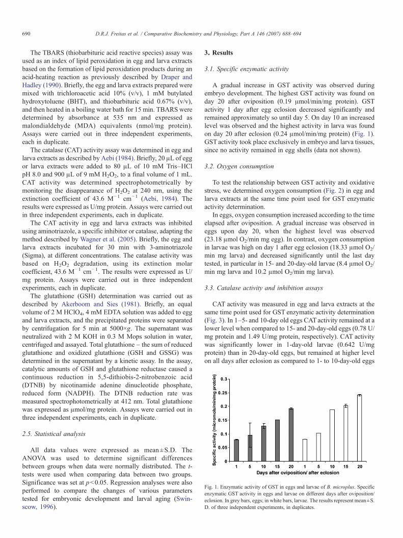
Fig. 1. Enzymatic activity of GST in eggs and larvae of B. microplus. Specificenzymatic GST activity in eggs and larvae on different days after oviposition/eclosion. In grey bars, eggs; in white bars, larvae. The results represent mean±S.D. of three independent experiments, in duplicates.
690 D.R.J. Freitas et al. / Comparative Biochemistry and Physiology, Part A 146 (2007) 688–694
The TBARS (thiobarbituric acid reactive species) assay wasused as an index of lipid peroxidation in egg and larva extractsbased on the formation of lipid peroxidation products during anacid-heating reaction as previously described by Draper andHadley (1990). Briefly, the egg and larva extracts prepared weremixed with trichloroacetic acid 10% (v/v), 1 mM butylatedhydroxytoluene (BHT), and thiobarbituric acid 0.67% (v/v),and then heated in a boiling water bath for 15 min. TBARS weredetermined by absorbance at 535 nm and expressed asmalondialdehyde (MDA) equivalents (nmol/mg protein).Assays were carried out in three independent experiments,each in duplicate.
The catalase (CAT) activity assay was determined in egg andlarva extracts as described by Aebi (1984). Briefly, 20 μL of eggor larva extracts were added to 80 μL of 10 mM Tris–HClpH 8.0 and 900 μL of 9 mM H2O2, to a final volume of 1 mL.CAT activity was determined spectrophotometrically bymonitoring the disappearance of H2O2 at 240 nm, using theextinction coefficient of 43.6 M−1 cm−1 (Aebi, 1984). Theresults were expressed as U/mg protein. Assays were carried outin three independent experiments, each in duplicate.
The CAT activity in egg and larva extracts was inhibitedusing aminotriazole, a specific inhibitor or catalase, adapting themethod described by Wagner et al. (2005). Briefly, the egg andlarva extracts incubated for 30 min with 3-aminotriazole(Sigma), at different concentrations. The catalase activity wasbased on H2O2 degradation, using its extinction molarcoefficient, 43.6 M−1 cm−1. The results were expressed as U/mg protein. Assays were carried out in three independentexperiments, each in duplicate.
The glutathione (GSH) determination was carried out asdescribed by Akerboom and Sies (1981). Briefly, an equalvolume of 2 M HClO4, 4 mM EDTA solution was added to eggand larva extracts, and the precipitated proteins were separatedby centrifugation for 5 min at 5000×g. The supernatant wasneutralized with 2 M KOH in 0.3 M Mops solution in water,centrifuged and assayed. Total glutathione – the sum of reducedglutathione and oxidized glutathione (GSH and GSSG) wasdetermined in the supernatant by a kinetic assay. In the assay,catalytic amounts of GSH and glutathione reductase caused acontinuous reduction in 5,5-dithiobis-2-nitrobenzoic acid(DTNB) by nicotinamide adenine dinucleotide phosphate,reduced form (NADPH). The DTNB reduction rate wasmeasured spectrophotometrically at 412 nm. Total glutathionewas expressed as μmol/mg protein. Assays were carried out inthree independent experiments, each in duplicate.
2.5. Statistical analysis
All data values were expressed as mean±S.D. TheANOVA was used to determine significant differencesbetween groups when data were normally distributed. The t-tests were used when comparing data between two groups.Significance was set at p<0.05. Regression analyses were alsoperformed to compare the changes of various parameterstested for embryonic development and larval aging (Swin-scow, 1996).
3. Results
3.1. Specific enzymatic activity
A gradual increase in GST activity was observed duringembryo development. The highest GST activity was found onday 20 after oviposition (0.19 μmol/min/mg protein). GSTactivity 1 day after egg eclosion decreased significantly andremained approximately so until day 5. On day 10 an increasedlevel was observed and the highest activity in larva was foundon day 20 after eclosion (0.24 μmol/min/mg protein) (Fig. 1).GST activity took place exclusively in embryo and larva tissues,since no activity remained in egg shells (data not shown).
3.2. Oxygen consumption
To test the relationship between GST activity and oxidativestress, we determined oxygen consumption (Fig. 2) in egg andlarva extracts at the same time point used for GST enzymaticactivity determination.
In eggs, oxygen consumption increased according to the timeelapsed after oviposition. A gradual increase was observed ineggs upon day 20, when the highest level was observed(23.18 μmol O2/min mg egg). In contrast, oxygen consumptionin larvae was high on day 1 after egg eclosion (18.33 μmol O2/min mg larva) and decreased significantly until the last daytested, in particular in 15- and 20-day-old larvae (8.4 μmol O2/min mg larva and 10.2 μmol O2/min mg larva).
3.3. Catalase activity and inhibition assays
CAT activity was measured in egg and larva extracts at thesame time point used for GST enzymatic activity determination(Fig. 3). In 1–5- and 10-day old eggs CATactivity remained at alower level when compared to 15- and 20-day-old eggs (0.78 U/mg protein and 1.49 U/mg protein, respectively). CAT activitywas significantly lower in 1-day-old larvae (0.642 U/mgprotein) than in 20-day-old eggs, but remained at higher levelon all days after eclosion as compared to 1- to 10-day-old eggs

Fig. 3. Catalase activity and inhibition assays. (A) Catalase activity in egg andlarva extracts of B. microplus in different days. In grey bars, eggs; in white bars,larvae. The results represent mean±S.D. of three independent experiments, induplicates (B) Catalase inhibition in egg (upper) and larva (lower) extracts of B.microplus on different days, using different concentrations of 3-aminotriazole(Sigma). The catalase activity was based on H2O2 degradation, using itsextinction molar coefficient, 43.6 M−1 cm−1. The results represent mean±S.D.of three independent experiments, in duplicates.
Fig. 4. Determination of total GSH content in egg and larva extracts of B.microplus. The GSH content was expressed as μmol/mg protein. In grey bars,eggs; in white bars, larvae. The results represent mean±S.D. of threeindependent experiments, in duplicates.
Fig. 2. Oxygen consumption in eggs and larvae. Oxygen consumption wasmonitored using a Clark-type oxygen electrode. Three assays of 200 mg of eggsor larvae were made in cuvettes containing 2 mL of 20 mM PBS, pH 7.4,0.1 mM EDTA. In grey bars, eggs; in white bars, larvae. The results representmean±S.D. of three independent experiments, in duplicates.
691D.R.J. Freitas et al. / Comparative Biochemistry and Physiology, Part A 146 (2007) 688–694
(Fig. 3A). The specificity of CAT activity to degrade H2O2 inegg and larva extracts was confirmed by inhibition of theactivity by aminotriazole at several concentrations, a compoundthat is a specific catalase inhibitor (Fig. 3B).
3.4. Glutathione determination
The levels of GSH in eggs increased significantly duringembryo development. The highest GST activity was observed in20-day-old eggs (92.23 μmol/mg protein). A decrease in GSHlevels was observed in 1-day-old larvae (2.33 μmol/mg protein)when compared to the days before egg eclosion. Until day 5after eclosion, GSH concentration was low and graduallyincreased thereafter until day 20 (Fig. 4).
3.5. Lipid peroxidation
A significant increase in lipid peroxidation in 20-day-oldeggs (3.50 nmol MDA/mg protein) was observed whencompared to 1-day-old eggs (2.86 nmol MDA/mg protein). In1-day-old larvae (3.33 nmol MDA/mg protein) and 5-day-oldlarvae (2.82 nmol MDA/mg protein) the lipid peroxidation wasmaintained at the same level as in 20-day-old eggs, butdecreased significantly in 10-day-old larvae (2.05 nmol MDA/mg protein). Afterwards, a significant increase was observed in15- and 20-day-old larvae (2.32 and 2.74 nmol MDA/mgprotein) in comparison to 10-day-old larvae, but the concentra-tion of MDA produced in 15- and 20-day-old larvae wassignificantly lower than that observed for 1- and 5-day-oldlarvae (Fig. 5).
3.6. Correlation between GST activity and other biologicalparameters
A correlation between GST activity and other biologicalparameters involved in oxidative stress was established toevaluate the GST role in physiologic oxidative stress. Thekinetics of GST activity in eggs and larvae was compared withoxygen consumption, CAT activity and GSH content in its

Fig. 5. Determination of thiobarbituric acid reactive substances (TBARS) ineggs and larvae of B. microplus on different days. In grey bars, eggs; in whitebars, larvae. The results represent mean±S.D. of three independent experiments,in duplicates.
692 D.R.J. Freitas et al. / Comparative Biochemistry and Physiology, Part A 146 (2007) 688–694
respective life stages. A positive correlation was observedbetween GST activity and O2 consumption in embryos(r=0.930), and a negative correlation in larvae (r=−0.832).There were positive correlations between kinetics of CAT andGST activities in eggs (r=0.941) and larvae (r=0.879). Also,positive correlations were found between GSTactivity and GSHcontent for eggs (r=0.985) and larvae (r=0.993). Thecorrelation between kinetics of GST activity and lipid peroxida-tion in eggs was positive (r=0.944), whereas in larvae thecorrelation was negative (r=−0.703).
4. Discussion
GSTs are present in almost all eukaryotes and in most ofthem this happens in multiple isoenzymic forms, constituting asignificant intracellular mechanism of detoxification (Papado-poulos et al., 2004). Detoxification is reached through transportand subsequent excretion of toxic organic compounds. Otherfunctions, not associated with detoxification, include repair ofmacromolecules oxidized by reactive oxygen species, regener-ation of S-thiolated proteins, and biosynthesis of physiologi-cally important metabolites (Armstrong, 1997; Sheehan et al.,2001).
In order to understand the role of GST in the tickdevelopment, we analyzed its physiological variation duringthe life stages of eggs and larvae.
The enzymatic GST activity had a significant increase duringembryonic development (Fig. 1). The O2 consumption increasesprogressively and reached its peak when the embryo is about tohatch (Fig. 2). This increase can be explained by the increasedmetabolism during the embryonic development in B. microplus.We observed strong correlation between O2 consumption andGST activity.
The enzymatic GST activity increased during the senescenceof larvae (Fig. 1). By analyzing the kinetics of GST activity andO2 consumption during larva aging a strong negative correlationwas observed. This decrease in O2 consumption can beexplained by the observation that the larva keeps a highmetabolic activity only during the first 5 days after hatching, inorder to mature and thus acquire the ability to infest a host. The
optimum period for the Porto Alegre B. microplus isolate toinfest the bovine is approximately between the 5th and the7th day post-eclosion (Da Silva Vaz, personal communication).Ogunji and Dipeolu (1977) showed the feeding of larval stagesof Amblyomma variegatum, Hyalomma rufipes and Boophilusdecoloratus in sheep to be more effective when larvae were fedwithin 1–4 days of hatching. Also, similar observations weredescribed for other ixodid ticks (Sonenshine, 1991). After thistime, the metabolic activity of B. microplus larvae decreasedbecause the larva is senescent; however, GST activity remainshigh, since this enzyme is important to protect against oxidativestress to which the larva is submitted, as shown by the equallyhigh CAT activity and GSH content (Figs. 3 and 4).
One of the consequences of elevated O2 in the cellularenvironment is the elevation of ROS levels that could causeoxidative damage, such as nuclear DNA and protein break-down, as well as lipid peroxidation (Mackay and Bewley, 1989;Barata et al., 2005). The antioxidants CAT and GSH testedpresented a positive correlation with GST activity in eggs and inlarvae. In fact, a high content of thiobarbituric acid reactivesubstances was observed in egg and larva extracts, indicatingthat ticks face high oxidative stress during embryogenesis andaging (Fig. 5). It is possible that the high level of lipidperoxidation verified during embryonic development occursdue to a high concentration of lipids, which are used by the eggas energy source (Logullo et al., 2002; Atella et al., 2005). Sincethe kinetic parameters of GST and catalase as well as GSHcontent during embryo development were modulated duringdifferent stages, and taking into account the fact that theseparameters presented a positive correlation with lipid peroxida-tion, it is possible to suggest a role of GST in oxidative stressprocess.
The decreased oxygen consumption during larval agingmay be related to metabolic depression and starvation. Phillipset al. (1995) showed that the metabolic rates of Ornithodorosturicata adults and larvae were affected by starvation and thatoxygen consumption was lower than fed ticks. Some animalsmaximize their survival time using metabolic depression inresponse to low concentrations of oxygen available, as acommon strategy through which they reduce their metabolism(Lutz and Storey, 1997; Hochachka and Lutz, 2001). Severalauthors have reported that the level of endogenous antioxidantdefences is increased in several species during metabolicdepression as a means to protect against oxidative stress.Oliveira et al. (2005) reported an increase in GST activity ingills of Chasmagnathus granulata during a metabolicdepression induced by anoxia. At low temperatures, thelarvae of B. microplus (Davey and Cooksey, 1989) and ofother ticks (Labruna et al., 2003; Estrada-Pena et al., 2004)reduced their metabolism and managed to survive for longperiods without feeding. These data are in accordance with theexperiments conduced by Papadopoulos et al. (2004), whichdemonstrated that adult bees kept at low temperatures andstarvation had GST activity increased, suggesting that B.microplus larvae decrease metabolism and increase GSTactivity in order to survive longer in the environment, until ahost is found.

693D.R.J. Freitas et al. / Comparative Biochemistry and Physiology, Part A 146 (2007) 688–694
The results of this work help to elucidate the role of GST intick development and assist in the understanding of theimportance of GST in the excretion of toxic compounds.
Acknowledgements
This work was supported by grants from CNPq, PRONEXand FAPERGS.
References
Aebi, H., 1984. Catalase in vitro. Methods Enzymol. 105, 121–126.Agianian, B., Tucker, P.A., Schouten, A., Leonard, K., Bullard, B., Gros, P.,
2003. Structure of aDrosophila sigma class glutathione S-transferase revealsa novel active site topography suited for lipid peroxidation products. J. Mol.Biol. 326, 151–165.
Akerboom, T.P., Sies, H., 1981. Assay of glutathione, glutathione disulfide, andglutathione mixed disulfides in biological samples. Methods Enzymol. 77,373–382.
Armstrong, R.N., 1997. Structure, catalytic mechanism, and evolution of theglutathione S-transferase. Chem. Res. Toxicol. 10, 2–18.
Atella, G.C., Gondim, K.C., Machado, E.A., Medeiros, M.N., Silva-Neto, M.A.,Masuda, H., 2005. Oogenesis and egg development in triatomines: abiochemical approach. An. Acad. Bras. Ciênc. 77, 405–430.
Barata, C., Lekumberri, I., Vila-Escale, M., Prat, N., Porte, C., 2005. Trace metalconcentration, antioxidant enzyme activities and susceptibility to oxidativestress in the tricoptera larvae Hydropsyche exocellata from the LlobregatRiver Basin (NE Spain). Aquat. Toxicol. 74, 3–19.
Beall, C., Fyrberg, C., Song, S., Fyrberg, E., 1992. Isolation of a Drosophilagene encoding glutathione S-transferase. Biochem. Genet. 30, 515–527.
Board, P.G., Baker, R.T., Chelvanayagam, G., Jermiin, L.S., 1997. Zeta, a novelclass of glutathione transferases in a range of species from plants to humans.Biochem. J. 328, 929–935.
Bowman, A.S., Dillwith, J.W., Sauer, J.R., 1996. Tick salivary prostaglandins:presence, origin and significance. Parasitol. Today 12, 388–396.
Bradford, M.M., 1976. A rapid and sensitive method for the quantification ofmicrogram quantities of protein utilizing the principle of protein–dyebinding. Anal. Biochem. 72, 248–254.
Dandapat, J., Chainy, G.B., Rao, K.J., 2003. Lipid peroxidation and antioxidantdefence status during larval development and metamorphosis of giantprawn, Macrobrachium rosenbergii. Comp. Biochem. Physiol. C 135,221–233.
Da Silva Vaz Jr., I., Torino Lermen, T., Michelon, A., Ferreira, C.A.S., Freitas,D.R.J., Termignoni, C., Masuda, A., 2004. Effect of acaricides on theactivity of a Boophilus microplus glutathione S-transferase. Vet. Parasitol.119, 237–245.
Davey, R.B., Cooksey, L.M., 1989. Effects of prolonged exposure at lowtemperature on Boophilus microplus (Acari: Ixodidae). J. Med. Entomol. 26,407–410.
Davey, R.B., George, J.E., 1998. In vitro and in vivo evaluations of a strain ofBoophilus microplus (Acari: Ixodidae) selected for resistance to permethrin.J. Med. Entomol. 35, 1013–1019.
Ding, Y., Ortelli, F., Rossiter, L.C., Hemingway, J., Ranson, H., 2003. TheAnopheles gambiae glutathione transferase supergene family: annotation,phylogeny and expression profiles. BMC Genomics 4, 35.
Dixon, D.P., Lapthorn, A., Edwards, R., 2002. Plant glutathione transferases.Genome Biol. 3 (REVIEWS3004).
Draper, H.H., Hadley, M., 1990. Malondialdehyde determination as index oflipid peroxidation. Methods Enzymol. 186, 421–431.
Estrada-Pena, A., Martinez, J.M., Sanchez Acedo, C., Quilez, J., Del Cacho, E.,2004. Phenology of the tick, Ixodes ricinus, in its southern distribution range(central Spain). Med. Vet. Entomol. 18, 387–397.
Habig, W.H., Pabst, M.J., Jakoby, W.B., 1974. Glutathione S-transferases. Thefirst enzymatic step in mercapturic acid formation. J. Biol. Chem. 249,7130–7139.
Hayes, J.D., Flanagan, J.U., Jowsey, I.R., 2005. Glutathione transferases. Annu.Rev. Pharmacol. Toxicol. 45, 51–88.
He, H., Chen, A.C., Davey, R.B., Ivie, G.W., George, J.E., 1999. Character-ization and molecular cloning of a glutathione S-transferase gene from thetick, Boophilus microplus (Acari: Ixodidae). Insect Biochem. Mol. Biol. 29,737–743.
Hemingway, J., 2000. The molecular basis of two contrasting metabolicmechanisms of insecticide resistance. Insect Biochem. Mol. Biol. 30,1009–1015.
Hochachka, P.W., Lutz, P.L., 2001. Mechanism, origin, and evolution of anoxiatolerance in animals. Comp. Biochem. Physiol. B 130, 435–459.
Jamroz, R.C., Guerrero, F.D., Pruett, J.H., Oehler, D.D., Miller, R.J., 2000.Molecular and biochemical survey of acaricide resistance mechanisms inlarvae fromMexican strains of the southern cattle tick, Boophilus microplus.J. Insect Physiol. 46, 685–695.
Ketterer, B., Coles, B., Meyer, D.J., 1983. The role of glutathione indetoxification. Environ. Health Perspect. 49, 59–69.
Ketterman, A.J., Prommeenate, P., Boonchauy, C., Chanama, U., Leetachewa,S., Promtet, N., Prapanthadara, L., 2001. Single amino acid changes outsidethe active site significantly affect activity of glutathione S-transferases.Insect Biochem. Mol. Biol. 31, 65–74.
Kostaropoulos, I., Papadopoulos, A.I., Metaxakis, A., Boukouvala, E.,Papadopoulou-Mourkidou, E., 2001. Glutathione S-transferase in thedefence against pyrethroids in insects. Insect Biochem. Mol. Biol. 31,313–319.
Labruna, M.B., Amaku, M., Metzner, J.Á., Pinter, A., Ferreira, F., 2003. Larvalbehavioral diapause regulates life cycle of Amblyomma cajennense (Acari:Ixodidae) in Southeast Brazil. J. Med. Entomol. 40, 170–178.
Logullo, C., Moraes, J., Dansa-Petretski, M., Vaz, I.S., Masuda, A., Sorgine, M.H., Braz, G.R., Masuda, H., Oliveira, P.L., 2002. Binding and storage ofheme by vitellin from the cattle tick, Boophilus microplus. Insect Biochem.Mol. Biol. 32, 1805–1811.
Lutz, P.L., Storey, K., 1997. Strategies for dealing with variations in gas tensions— vertebrates and invertebrates. In: Dantzler, W. (Ed.), Handbook ofPhysiology, Section 13, Comparative Physiology. American PhysiologySociety. Oxford University Press, pp. 1479–1522.
Mackay, W.J., Bewley, G.C., 1989. The genetics of catalase in Drosophilamelanogaster: isolation and characterization of acatalasemic mutants.Genetics 122, 643–652.
Morel, F., Rauch, C., Petit, E., Piton, A., Theret, N., Coles, B., Guillouzo, A.,2004. Gene and protein characterization of the human glutathione S-transferase Kappa and evidence for a peroxisomal localization. J. Biol.Chem. 279, 16246–16253.
Ogunji, F.O., Dipeolu, O.O., 1977. On the use of the scrotum for feeding larvalixodid ticks on hosts. Z. Parasitenkd. 51, 285–288.
Oliveira, U.O., Da Rosa Araujo, A.S., Bello-Klein, A., Da Silva, R.S.,Kucharski, L.C., 2005. Effects of environmental anoxia and differentperiods of reoxygenation on oxidative balance in gills of the estuarine crabChasmagnathus granulata. Comp. Biochem. Physiol. B 140, 51–57.
Papadopoulos, A.I., Polemitou, I., Laifi, P., Yiangou, A., Tananaki, C., 2004.Glutathione S-transferase in the insect Apis mellifera macedonica kineticcharacteristics and effect of stress on the expression of GST isoenzymes inthe adult worker bee. Comp. Biochem. Physiol. C 139, 93–97.
Phillips, J.S., Adeyeye, O., Bruni, D., 1995. Respiratory metabolism of the softtick, Ornithodoros turicata (Duges). Exp. Appl. Acarol. 19, 103–115.
Rikans, L.E., Hornbrook, K.R., 1997. Lipid peroxidation, antioxidant protectionand aging. Biochim. Biophys. Acta 1362, 116–127.
Robinson, A., Huttley, G.A., Booth, H.S., Board, P.G., 2004. Modelling andbioinformatics studies of the human Kappa-class glutathione transferasepredict a novel third glutathione transferase family with similarity toprokaryotic 2-hydroxychromene-2-carboxylate isomerases. Biochem. J.379, 541–552.
Rosa de Lima, M.F., Ferreira, C.A.S., Freitas, D.R.J., Valenzuela, J.G., Masuda,A., 2002. Cloning and partial characterization of a Boophilus microplus(Acari: Ixodiae) glutathione S-transferase. Insect Biochem. Mol. Biol. 32,747–754.
Salinas, A.E., Wong, M.G., 1999. Glutathione S-transferases — a review. Curr.Med. Chem. 6, 279–309.

694 D.R.J. Freitas et al. / Comparative Biochemistry and Physiology, Part A 146 (2007) 688–694
Sauer, J.R., McSwain, J.L., Bowman, A.S., Essenberg, R.C., 1995. Tick salivarygland physiology. Annu. Rev. Entomol. 40, 245–267.
Sheehan, D., Meade, G., Foley, V.M., Dowd, C.A., 2001. Structure, function andevolution of glutathione transferase: implications for classification of non-mammalian members of an ancient enzyme superfamily. Biochem. J. 360,1–16.
Sies, H., 1999. Glutathione and its role in cellular functions. Free Radic. Biol.Med. 27, 916–921.
Sonenshine, D.E., 1991. 1st ed. Biology of Ticks, vol. 1. Oxford UniversityPress, New York.
Swinscow, T.D.V., 1996. Statistics at Square One, 9th ed. BMJ Books, London.Revised by MJ Campbell.
Wagner, B.A., Evig, C.B., Reszka, K.J., Buettner, G.R., Burns, C.P., 2005.Doxorubicin increases intracellular hydrogen peroxide in PC3 prostatecancer cells. Arch. Biochem. Biophys. 440, 181–190.
Wei, S.H., Clark, A.G., Syvanen, M., 2001. Identification and cloning of a keyinsecticide-metabolizing glutathione S-transferase (MdGST-6A) from ahyperinsecticide-resistant strain of the housefly Musca domestica. InsectBiochem. Mol. Biol. 31, 1145–1153.
Widersten, M., Mannervik, B., 1995. Glutathione transferases with novel activesites isolated by phage display from a library of random mutants. J. Mol.Biol. 250, 115–122.