i universidade federal de são carlos centro de ciências biológicas e
TRANSCRIPT

I
UNIVERSIDADE FEDERAL DE SÃO CARLOS
CENTRO DE CIÊNCIAS BIOLÓGICAS E DA SAÚDE
PROGRAMA DE PÓS-GRADUAÇÃO EM ECOLOGIA E RECURSOS
NATURAIS
Biodiversidade de Selenastraceae (Sphaeropleales, Chlorophyceae): características
morfológicas e sequenciamento dos marcadores moleculares 18S rDNA, rbcL e ITS
como base taxonômica tradicional.
Thaís Garcia da Silva
Orientador: Prof. Dr. Armando Augusto Henriques Vieira
São Carlos – SP
2016
Tese apresentada ao Programa de
Pós-Graduação em Ecologia e
Recursos Naturais da Universidade
Federal de São Carlos, como parte
dos requisitos para obtenção do título
de DOUTOR EM CIÊNCIAS, área
de concentração: ECOLOGIA E
RECURSOS NATURAIS.

Ficha catalográfica elaborada pelo DePT da Biblioteca Comunitária UFSCar Processamento Técnico
com os dados fornecidos pelo(a) autor(a)
S586bSilva, Thaís Garcia da Biodiversidade de Selenastraceae (Sphaeropleales,Chlorophyceae): características morfológicas esequenciamento dos marcadores moleculares 18S rDNA,rbcL e ITS como base taxonômica tradicional / ThaísGarcia da Silva. -- São Carlos : UFSCar, 2016. 148 p.
Tese (Doutorado) -- Universidade Federal de SãoCarlos, 2016.
1. Taxonomia. 2. Sistemática molecular. 3.Selenastraceae. 4. Biodiversidade. 5. Filogenia. I.Título.
III

IV
“Longe se vai sonhando demais
Mas onde se chega assim?
Vou descobrir o que me faz sentir
Eu, caçador de mim.”
Milton Nascimento
V
Agradecimentos
Primeiramente, ao Dr. Armando Augusto Henriques Vieira por ter me aceitado como
estagiária e aluna de doutorado. Agradeço imensamente por todo apoio e dedicação nesses 6
anos de trabalho no Laboratório de Ficologia, me mostrando novas possibilidades de
aprendizado e estimulando o meu caminhar na pesquisa e conhecimento na ficologia. Sinto-
me imensamente honrada por ter trilhado um caminho tão importante de minha vida sob sua
tutoria e me espelho no seu exemplo de verdadeiro amor e dedicação à profissão.
À minha co-orientadora Dra. Célia Leite Sant’Anna, Núcleo de pesquisa em Ficologia
do Instituto de Botânica, pelo grande estímulo que me deu para que eu prosseguisse na
taxonomia e que não desanimasse em momentos decisivos desta jornada, dando valor ao que
eu acreditava ser importante neste estudo.
À Dra. Christina Bock, Departamento de Biodiversidade da Universidade Duisburg-
Essen, por ter me aceitado como sua aluna, pelo grande conhecimento sobre biologia
molecular que humildemente me transmitiu em minha estadia na Alemanha e por ter confiado
que eu seria capaz de enfrentar esse desafio “selenastrágico”.
À Dra. Sabina Wodniok pelo auxilio nas análises de microscopia eletrônica.
Ao Dr. Jens Boenigk por ter permitido à Christina me receber como aluna no
Departamento de Biodiversidade e pelo auxílio burocrático envolvido.
À CAPES pela concessão da bolsa de estudos nos 12 primeiros meses de doutorado.
À Fundação de Amparo à Pesquisa do Estado de São Paulo, FAPESP, pelas
concessões das bolsas de estudos no Brasil (Processo n° 2012/19520-1) e, no exterior por
meio do programa “Bolsa Estágio de Pesquisa no Exterior” (Processo n° 2013/17457-3).
À Universidade Federal de São Carlos, ao Instituto de Botânica de São Paulo e a
Universidade Duisburg-Essen por fornecer a infraestrutura necessária à realização deste

VI
trabalho e ao Programa de Pós-Graduação em Ecologia e Recursos Naturais, pela
oportunidade de aprimoramento científico.
Ao Programa de Pós-graduação em Biodiversidade Vegetal e Meio Ambientedo
Instituto de Botânica de São Paulo, pela oportunidade de aprimoramento científico por conta
das disciplinas cursadas.
Ao Luizinho pelo apoio durante as coletas do Projeto Biota e pelo exemplo
profissional que nos dá.
Aos colegas e amigos do Laboratório de Ficologia pelos momentos compartilhados:
Naiara Carolina, Cilene, Inessa, Fabrício, Luiz, Letícia, Alessandra, Helena, Guilherme,
Ingritt, Chico, Lucas, Érica, Moira (e, possivelmente, mais alguns que devo ter me
esquecido...rsrsrs). Meu muito obrigada por tudo o que foi vivido.
À Inessa por clarear meus pensamentos e não me deixar ser tão tendenciosa na
taxonomia, no auxílio com a biologia molecular, na redação de artigos e crises existenciais
no final deste processo.
À Zezé por ter me ensinado o básico da taxonomia para as análises das minhas
amostras quali e quantitativas, ainda na minha iniciação científica. Por um acaso do destino
você me sugeriu procurar o Armando para tentar uma pós e aqui cheguei.
Aos meus amigos muito queridos da UDE e que tornaram minha estadia muito
agradável: Julia, Lars, Vesna, Yesim, Edward, Elif, Saskia, Philpp, Sabina, Nikoletta,
Fernando, Saeed, Farnoush, Sarah, Susy, Beate e Christina.
À Andrea pela companhia agradabilíssima em todas as vezes que fui ao Instituto de
Botânica, pelas conversas, hospedagem, por me encorajar na pesquisa e na vida.
Agradeço especialmente ao Luiz pelo apoio cotidiano no laboratório, pelas conversas
durante o trabalho e, principalmente, por ter dividido a “nossa” salinha de microscopia por
longos dias.

VII
Aos funcionários do PPGERN: João, Roseli e, especialmente Beth (que eu encontrava
com frequência ao retornar para casa) pela ajuda com a burocracia envolvida neste
doutorado.
Aos meus amigos por todos os momentos durante minha caminhada. Não citarei
nomes porque fatalmente esquecer-me-ei de alguém.
Ao Neto, pelos importantes jogos de forca que praticávamos durante as aulas
entediantes e por ter feito esses momentos mais leves.
À Priscilla por ter estado ao meu lado por algum tempo neste processo.
Muitos foram importantes pra que eu chegasse aqui mas, agradeço sobretudo à:
minha mãe, vó e irmã por estarem ao meu lado e me ensinarem coisas que nem consigo por
no papel de tamanha grandeza.
À minha família toda por ter torcido por mim, pelo carinho e pelo porto seguro que só
uma família, mesmo que torta, pode propiciar.
Aos meus mestres da vida inteira, que de alguma forma me ajudaram a construir o
que sou e olhar para o que quero ser. Por cada momento de ensinamento, obrigada!

VIII
Resumo
A filogenia da família Selenastraceae foi investigada por microscopia ótica, análises
moleculares dos marcadores 18S rDNA, rbcL, ITS1-5,8S-ITS2 e ITS-2. Várias características
morfológicas tradicionalmente utilizadas para identificação de gêneros e espécies foram
investigadas. Todas as cepas de Selenastraceae estudadas têm pirenóides nus dentro do
cloroplasto, exceto o gênero Chlorolobion, que apresentou pirenóide amilóide. As análises
moleculares mostraram que nenhum critério morfológico isolado considerado até agora é
significativo para a sistemática do Selenastraceae, mas o uso de um conjunto de
características morfológicas pode ser adequado para identificar espécies dos gêneros
Ankistrodesmus, Chlorolobion, Kirchneriella, Raphidocelis e Tetranephris. As análises
filogenéticas moleculares mostraram que os gêneros Monoraphidium, Kirchneriella e
Selenastrum são polifiléticos e não distinguíveis como gêneros. O morfotipo de Selenastrum
revelou três linhagens moleculares diferentes, levando à descrição de dois novos gêneros,
Curvastrum gen. nov. e Messastrum gen. nov. Além disso, as análises filogenéticas revelaram
quatro linhagens moleculares atribuídas ao morfotipo de Kirchneriella, levando à descrição de
cinco espécies novas: Gênero 1 sp. nov. 1, Gênero 2 sp. nov. 1, Gênero 2 sp. nov. 2,
Raphidocelis sp. nov. e Tetranephris sp. nov.
IX
Abstract
The phylogeny of the family Selenastraceae was investigated by light microscopy, molecular
analysis of 18S rDNA, rbcL, ITS1-5.8S-ITS2 and ITS-2 markers. Several morphological
features traditionally used for identification of genera and species were investigated. All
Selenastraceae strains studied presented naked pyrenoids within the chloroplast, except for
Chlorolobion, which presented starched pyrenoid. Molecular analysis showed that no isolated
morphological criteria considered so far is significant for the systematic of Selenastraceae, but
a set of characteristics may be appropriate to identify species of genera Ankistrodesmus,
Chlorolobion, Kirchneriella, Raphidocelis and Tetranephris. Phylogenetic analyses showed
that genera Monoraphidium, Kirchneriella and Selenastrum are polyphyletic and not
distinguishable as separate genera. The Selenastrum morphotype revealed three different
molecular lineages, leading to the description of two new genera, Curvastrum gen. nov and
Messastrum gen. nov. In addition, molecular phylogenetic analysis revealed four lineages
assigned to Kirchneriella morphotype, leading to the description of five new species: Genus 1
sp. nov. 1, Genus 2 sp. nov. 1, Genus 2 sp. nov. 2, Raphidocelis sp. nov. and Tetranephris sp.
nov.

X
Lista de siglas e abreviaturas
18S rDNA 18S DNA ribossômico (18S ribosomal RNA)
28S rDNA 28S DNA ribossômico (28S ribosomal RNA)
5.8S rDNA 5.8S DNA ribossômico (5.8S ribossonal RNA)
BP Probabilidade Bayesiana (Bayesian probability)
CB Christina Bock
CBC Mudança de bases compensatórias (Compensatory base changes)
CCMA Coleção de culturas de Microalgas de Água
Comas Augusto Abilio Comas González
DNA Ácido desoxirribonucleico (desoxyribonucleic acid)
gen. nov. Gênero novo
ITS Espaçador interno transcrito (Internal transcribed spacer)
ITS1 Espaçador interno transcrito situado entre os genes 18S rDNA and 5.8S rDNA
ITS2 Espaçador interno transcrito localizado entre os genes 5.8S rDNA e 28S rDNA
nas algas
KF Alena Lukešová Culture Collection
KR Lothar Krienitz
LM Microscopia ótica (Light microspy)
MCMC Monte Carlo via Cadeias de Markov (Markov Chain Monte Carlo)
MFE Energia mínima livre (Minimum free energy)
ML Máxima verossimilhança (Maximum likelihood)
MP Máxima parsimônia (Maximum parsimony)
NCBI National Center for Biotechnology Information
NGS Sequenciamento de nova geração (Next generation sequencing)

XI
NJ Agrupamento de vizinhos (Neighbor-joining)
PCR Reação em cadeia da polimerase (Polimerase Chain Reaction)
PP Probabilidade posterior (posterior probability)
rbcL Subunidade grande da RUBISCO (RUBISCO large subunit)
rRNA Ácido ribonucléico ribossômico (ribosomal rubonucleic acid)
SAG Sammlung von Algenkulturen der Universität Göttingen
SEM Microscopia eletrônica de varredura (Scanning Electron microscopy)
sp. nov. Espécie nova
SSU rRNA Subunidade menor do ribossomo (ribossome small subunit)
TEM Microscopia eletrônica de transmissão (Transmission Electron Microscopy)
UTEX The Culture Collection of Algae at the University of Texas at Austin.
WDCM World Data Center for Microorganisms

XII
Lista de figuras
Figure 1.1-1.5. 1.1. Messastrum gracile. Original picture of strain CCMA-UFSCar 622
showing a frontal view of colony; 1.2. Selenastrum bibraianum. Original picture of strain
CCMA-UFSCar 125 showing a frontal view of colony. 1.3-1.4. Curvastrum pantanale. (1.3)
Original picture of strain CCMA-UFSCar 350, showing free cells and colony; (1.4) Original
picture of strain CCMA-UFSCar 350, showing cells in autospore liberation. presenting a cell
wall remnant (arrowhead) and protoplasm cleavage (star). 1.5. Ankistrodesmus arcuatus.
Original picture of strain CCMA-UFSCar 24, showing free cells and colony. Note autospore
formation (star) and mucilaginous lump (arrowhead). Scale bar 10 μm……………………..77
Figure 1.6: Maximum–likelihood (ML) phylogenetic tree inferred from rbcL gene sequences
of some members of Selenastraceae. Support values correspond to Bayesian PP (Posterior
Probability), ML BP (Bootstrap), MP (Maximum Parsimony) BP, NJ (Neighbor-Joining) BP.
Hyphens correspond to values <50% for BP and <0.95 for PP. Scale represents the expected
number of substitutions per site. Strain numbers used as mentioned in Table 1……………..78
Figure 1.7: Maximum–likelihood (ML) phylogenetic tree inferred from 18S rDNA gene
sequences of some members of Selenastraceae. Support values correspond to Bayesian PP
(Posterior Probability), ML BP (Bootstrap), MP (Maximum Parsimony) BP, NJ (Neighbor-
Joining)BP. Hyphens correspond to values <50% for BP and <0.95 for PP. Scale represents
the expected number of substitutions per site. Strain numbers used as mentioned in Table
1……………………………………………………………………………………………….79

XIII
Figure 1.8: Scanning electron micrographs of Curvastrum pantanale (CCMA-UFSCar 350) in
culture. Scale bar, 10 µm. (a) typical colony formation, (b) young cells detaching from each
other, (c) young cells, note the autospores position…………………………………………..80
Figure 1.9: Transmission electron micrographs of Curvastrum pantanale (CCMA-UFSCar
350) in culture. Scale bar, 1 µm. Key to labeling: CW = cell wall, C = chloroplast, D =
dictyosome, ER = endoplasmic reticulum, L = lipid drop, M = mitochondrion, N = nucleus, P
= pyrenoid, PV = polyphosphate vacuole, S = starch grain. a) longitudinal section; cell
presenting lipid drops, polyphosphate vacuoles (arrowhead), chloroplast penetrated with
starch grains, and a central pyrenoid. b) detail of figure a, where an endoplasmic reticulum
(arrowhead) can be observed. c) longitudinal section; cell presenting polyphosphate vacuoles
on both cell apexes, mitochondria, chloroplast filled with starch grains and a central pyrenoid.
d) cross section; chloroplast containing starch grains and a pyrenoid situated at the left, a
central nucleus can be observed. e) longitudinal section; mature cell containing a central
nucleus, mitochondrion and pyrenoid (upper part). All the cell content is surrounded by a cell
wall……………………………………………………………………………………………81
Figure 1.10: Transmission electron micrographs of two Selenastraceae in culture. Scale bar, 1
µm. a - c) Ankistrodesmus arcuatus (CCMA – UFSCar 24). a) longitudinal section; cell with a
nucleus, dictyosome and a chloroplast. b) cross section; a cup-shaped chloroplast penetrated
by a pyrenoid (star). On the opposite direction of the pyrenoid, the nucleus is situated. An
arrowhead indicates a polyphosphate vacuole. c) detail of figure a, where a dictyosome can
be observed. d - e) Monoraphidium contortum (CCMA-UFSCar 349), d) longitudinal section;
cells on different life cycle phase. The upper 3 cells are young indidivuals presenting starch
grains (arrowhead), polyphosphate vacuoles (arrowhead), chloroplast and nucleus. The lower
XIV
cell is a mature individual containing many starch grains on the chloroplast, some
polyphosphate vacuoles (arrowhead), nucleus and big lipid drops. e) cross section; dense
chloroplast with starch grains. Two big polyphosphate vacuoles (arrowhead) and a nucleus
can be observed. All the cell content is surrounded by a cell wall…………………………...82
Figuras suplementares
1.A) ITS-2 model for the type strain of Selenastrum bibraianum (CCMA-UFSCar 125). In
black boxes are the different bases compared to strain Messastrum gracile (CCMA-UFSCar
622)……….…………………………………………………………………………………..83
1.B) ITS-2 model for the type strain of Selenastrum bibraianum (CCMA-UFSCar 125). In
gray boxes are the different bases compared to strain Selenastrum bibraianum (CCMA-
UFSCar 47) and black boxes compared to Selenastrum bibraianum (CB 2012/47).…….…..83
1.C) ITS-2 model for the type strain of Messastrum gracile (CCMA-UFSCar 622). In gray
boxes are the different bases of M. gracile (CCMA-UFSCar 470) and black boxes M. gracile
(CCMA-UFSCar 5). ………………………...………………………………………………..83
1.D) ITS-2 model for the type strain of Curvastrum pantanale (CCMA-UFSCar 350). In gray
boxes are the different bases of C. pantanale (CCMA-UFSCar 608)………………………..84
Fig. 2.1-2.5. Drawings of light microscopical characters. 1. Tetranephris sp. nov. (CB
2009/6). Note the cell wall remnant (arrowhead); 2. Gen. nov. 1 sp1 (CCMA-UFSCar 230); 3.
Raphidocelis sp. nov. (CCMA-UFSCar 229). Note the cell wall remnant (arrowhead); 4. Gen.

XV
nov. 2 sp 1. (CCMA-UFSCar 342); 5. Gen. nov. 2 sp 2. (KR 1979/222). Note the cell wall
remnant (arrowhead). Scale bar 10 μm. …………………………………………………….135
Fig. 2.6-2.8. Drawings of light microscopical characters. 6. Kirchneriella pseudoaperta
(CCMA-UFSCar 346). 7. Kirchneriella obesa (CCMA-UFSCar 345). 8. Kirchneriella lunaris
(CCMA-UFSCar 87). Scale bar 10 μm. …………………………………………………….136
Figure 2.9: Maximum–likelihood (ML) phylogenetic tree inferred from ITS1-5.8S-ITS2 gene
sequences of some members of Selenastraceae. Support values correspond to Bayesian PP
(Posterior Probability), ML BP (Bootstrap), MP (Maximum Parsimony) BP, NJ (Neighbor-
Joining)BP. Hyphens correspond to values <50% for BP and <0.95 for PP. Scale represents
the expected number of substitutions per site. Strain numbers used as mentioned in Table 1.
…………………………………………………………………………………….…………137
Figure 2.10: Maximum–likelihood (ML) phylogenetic tree inferred from 18S rDNA gene
sequences of some members of Selenastraceae. Support values correspond to Bayesian PP
(Posterior Probability), ML BP (Bootstrap), MP (Maximum Parsimony) BP, NJ (Neighbor-
Joining) BP. Hyphens correspond to values <50% for BP and <0.95 for PP. Scale represents
the expected number of substitutions per site. Strain numbers used as mentioned in Table
2.1……………………………………………………………………………………………138
XVI
Lista de tabelas
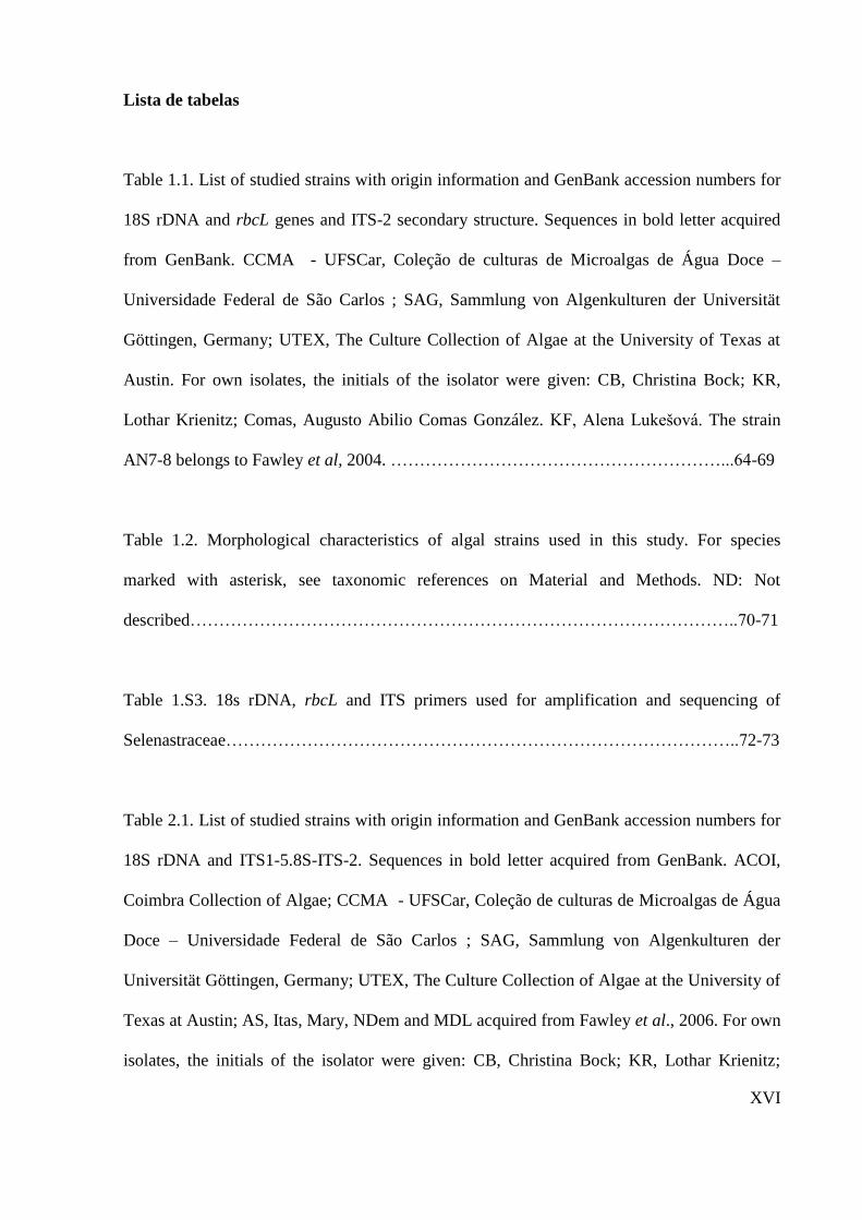
Table 1.1. List of studied strains with origin information and GenBank accession numbers for
18S rDNA and rbcL genes and ITS-2 secondary structure. Sequences in bold letter acquired
from GenBank. CCMA - UFSCar, Coleção de culturas de Microalgas de Água Doce –
Universidade Federal de São Carlos ; SAG, Sammlung von Algenkulturen der Universität
Göttingen, Germany; UTEX, The Culture Collection of Algae at the University of Texas at
Austin. For own isolates, the initials of the isolator were given: CB, Christina Bock; KR,
Lothar Krienitz; Comas, Augusto Abilio Comas González. KF, Alena Lukešová. The strain
AN7-8 belongs to Fawley et al, 2004. …………………………………………………...64-69
Table 1.2. Morphological characteristics of algal strains used in this study. For species
marked with asterisk, see taxonomic references on Material and Methods. ND: Not
described…………………………………………………………………………………..70-71
Table 1.S3. 18s rDNA, rbcL and ITS primers used for amplification and sequencing of
Selenastraceae……………………………………………………………………………..72-73
Table 2.1. List of studied strains with origin information and GenBank accession numbers for
18S rDNA and ITS1-5.8S-ITS-2. Sequences in bold letter acquired from GenBank. ACOI,
Coimbra Collection of Algae; CCMA - UFSCar, Coleção de culturas de Microalgas de Água
Doce – Universidade Federal de São Carlos ; SAG, Sammlung von Algenkulturen der
Universität Göttingen, Germany; UTEX, The Culture Collection of Algae at the University of
Texas at Austin; AS, Itas, Mary, NDem and MDL acquired from Fawley et al., 2006. For own
isolates, the initials of the isolator were given: CB, Christina Bock; KR, Lothar Krienitz;

XVII
Comas, Augusto Abilio Comas González. Accession number indicated with x will be
deposited prior to publication……………………………………………………..……123-128
Table 2.2. Morphological characteristics of Kirchneriella-like strains used in this study. Aa:
autospore arrangement; P: pyrenoid; C: colony; M: mucilage…………….………………129
Table 2.3.Revision of taxa included in Kirchneriella, Raphidocelis, Gen 1 gen nov, Gen 2 gen
nov and Tetranephris. Names marked with asterisks represents organisms with 18S rDNA
sequences available on NCBI and used on previous phylogenetic studies……………..130-133

XVIII
Apresentação da tese
A tese foi elaborada para conter os itens: (1) Introdução geral; (2) Hipóteses e
Objetivos; (3) Capítulos (com resultados e discussão); (4) Discussão geral; e (5) Conclusões.
Cada capítulo será apresentado no formato de artigo científico: com resumo,
introdução, material e métodos, resultados, discussão, referências bibliográficas e material
suplementar. Este formato foi escolhido para facilitar a publicação dos resultados obtidos. A
introdução geral está em português, bem como a discussão geral e a conclusão.
O primeiro capítulo encontra-se formatado para a publicação na revista Fottea, à qual
foi submetido e aceito. Neste capítulo, apresentamos os resultados de uma análise filogenetica
utilizando os marcadores rbcL e 18S rDNA.
No segundo capítulo da tese abordamos o complexo Kirchneriella-Raphidocelis-
Pseudokirchneriella, os menores organismos de Selenastraceae, em um estudo filogenético
baseado em 18S rDNA e na analise multigene dos marcadores ITS1-5.8S-ITS2, formatado
nos moldes da Journal of Phycology, possível periódico para submissão.
Ao final, uma breve discussão geral e as conclusões foram elaboradas baseadas nos
capítulos apresentados.
XIX
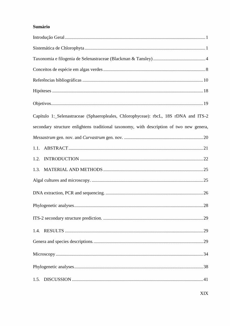
Sumário
Introdução Geral ......................................................................................................................... 1
Sistemática de Chlorophyta ........................................................................................................ 1
Taxonomia e filogenia de Selenastraceae (Blackman & Tansley) ............................................. 4
Conceitos de espécie em algas verdes ........................................................................................ 8
Referências bibliográficas ........................................................................................................ 10
Hipóteses .................................................................................................................................. 18
Objetivos ................................................................................................................................... 19
Capítulo 1: Selenastraceae (Sphaeropleales, Chlorophyceae): rbcL, 18S rDNA and ITS-2
secondary structure enlightens traditional taxonomy, with description of two new genera,
Messastrum gen. nov. and Curvastrum gen. nov. .................................................................... 20
1.1. ABSTRACT .................................................................................................................... 21
1.2. INTRODUCTION .......................................................................................................... 22
1.3. MATERIAL AND METHODS ...................................................................................... 25
Algal cultures and microscopy. ................................................................................................ 25
DNA extraction, PCR and sequencing. .................................................................................... 26
Phylogenetic analyses ............................................................................................................... 28
ITS-2 secondary structure prediction. ...................................................................................... 29
1.4. RESULTS ....................................................................................................................... 29
Genera and species descriptions. .............................................................................................. 29
Microscopy ............................................................................................................................... 34
Phylogenetic analyses ............................................................................................................... 38
1.5. DISCUSSION ................................................................................................................. 41
XX
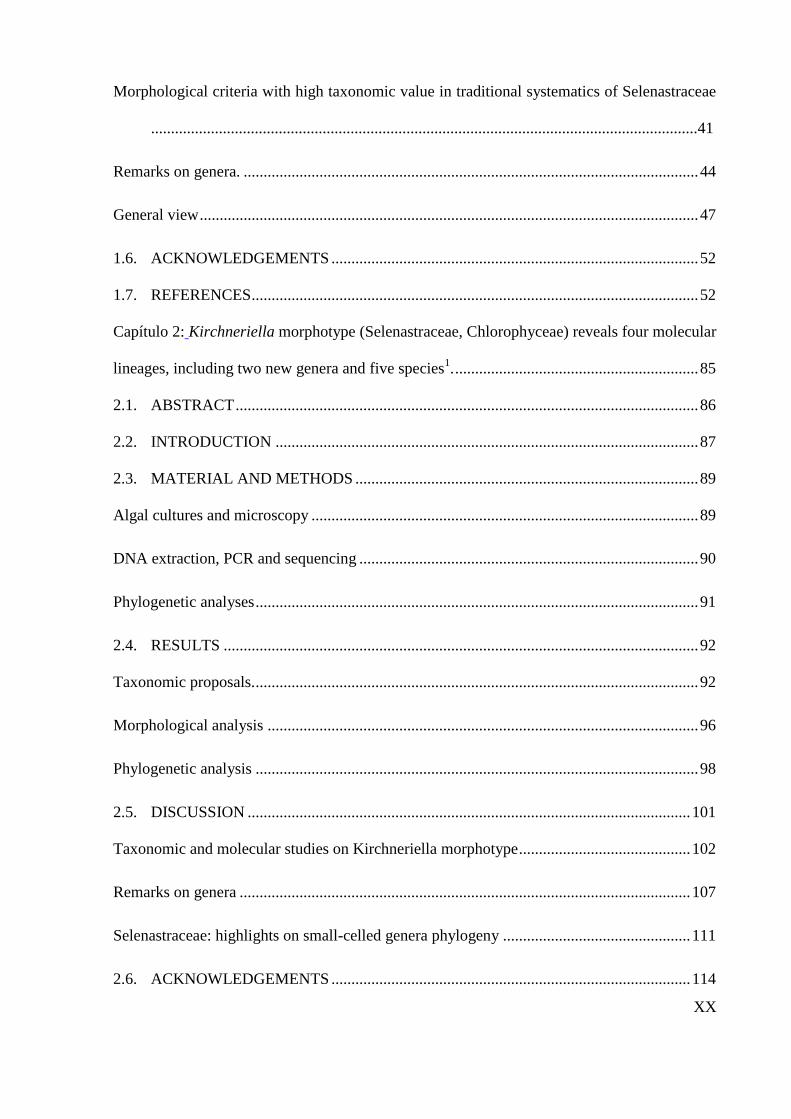
Morphological criteria with high taxonomic value in traditional systematics of Selenastraceae
.........................................................................................................................................41
Remarks on genera. .................................................................................................................. 44
General view ............................................................................................................................. 47
1.6. ACKNOWLEDGEMENTS ............................................................................................ 52
1.7. REFERENCES ................................................................................................................ 52
Capítulo 2: Kirchneriella morphotype (Selenastraceae, Chlorophyceae) reveals four molecular
lineages, including two new genera and five species1. ............................................................. 85
2.1. ABSTRACT .................................................................................................................... 86
2.2. INTRODUCTION .......................................................................................................... 87
2.3. MATERIAL AND METHODS ...................................................................................... 89
Algal cultures and microscopy ................................................................................................. 89
DNA extraction, PCR and sequencing ..................................................................................... 90
Phylogenetic analyses ............................................................................................................... 91
2.4. RESULTS ....................................................................................................................... 92
Taxonomic proposals. ............................................................................................................... 92
Morphological analysis ............................................................................................................ 96
Phylogenetic analysis ............................................................................................................... 98
2.5. DISCUSSION ............................................................................................................... 101
Taxonomic and molecular studies on Kirchneriella morphotype ........................................... 102
Remarks on genera ................................................................................................................. 107
Selenastraceae: highlights on small-celled genera phylogeny ............................................... 111
2.6. ACKNOWLEDGEMENTS .......................................................................................... 114
XXI
2.7. REFERENCES .............................................................................................................. 114
Discussão Geral ...................................................................................................................... 139
Referências bibliográficas ...................................................................................................... 143

1
Introdução Geral
Sistemática de Chlorophyta
O termo Chlorophyta refere-se, tradicionalmente, ao grupo de algas verdes, que
se caracteriza por cloroplastos com membrana dupla, amido como polissacarídeo de
reserva, tilacóides empilhados e presença de clorofila a e b (Friedl 1997, Chapman et al.
1998). Contudo, alguns gêneros reconhecidamente pertencentes à Chlorophyta,
perderam seus pigmentos em processos secundários (Pringsheim 1963). Uma enorme
diversidade morfológica está inclusa em Chlorophyta, compreendendo desde
organismos unicelulares cocóides ou flagelados, coloniais, filamentosas ramificadas ou
não, membranosas e cenocíticas (van den Hoek et al 1988).
A divisão Chlorophyta abrange organismos viventes em ambientes de água doce
ou marinha, sendo um dos principais produtores primários em ambientes aquáticos
(Bock 2010). O hábito dessas algas pode ser epifítico, planctônico e algumas espécies
vivem em comunidades edáficas.
O primeiro sistema natural de classificação de algas criado remete há
aproximadamente 200 anos e considerou o nível de organização do estado vegetativo
como principal característica morfológica para a separação dos grandes grupos
(Blackman 1900, Blackman & Tansley 1902, Pascher 1918). Desde então, a sistemática
de algas verdes passou por grandes mudanças, sendo aperfeiçoada ao longo dos anos,
resultando em uma classificação baseada em ciclos de vida, dados ultra-estruturais,
forma e organização das células vegetativas, mitose, composição da parede celular e
arquiteturas de células flageladas (Christensen 1962, van den Hoek & Jahns 1978, Ettl
& Komárek 1982, Mattox & Stewart 1984, van den Hoek et al. 1988, 1995). A partir da
combinação de diferentes critérios morfológicos, esses sistemas de classificação
2
delinearam classes, famílias, gêneros e espécies (Kornmann 1973, Ettl 1981, Komárek
& Fott 1983, Mattox & Stewart, 1984).
O advento das análises moleculares aprofundou a visão do sistema natural de
classificação de algas verdes (Melkonian & Surek 1995, Friedl 1997, Lewis & McCourt
2004), trazendo o consenso de que as algas verdes evoluiram em duas grandes
linhagens, chamadas de Clado Charophyta e Clado Chlorophyceae sensu Lewis &
McCourt (2004).
O Clado Charophyta (ou Streptophyta sensu Bremer 1985) compreende as
plantas terrestres e um número de algas verdes, grupos como Mesostigmatophyceae,
Chlorokybophyceae, Klebsormidiophyceae, Zygnemophyceae, Coleochaetophyceae e
Charophyceae.
O Clado Chlorophyceae (ou Chlorophyta) compreende a maioria das algas que
eram tradicionalmente referidas como algas verdes. Este clado contém os três grupos
monofiléticos de algas verdes (Chlorophyceae, Trebouxiophyceae e Ulvophyceae) e o
clado parafilético traz Prasinophyceae, que contém, ao menos, seis diferentes clados
(Lewis & McCourt 2004) na parte basal de Chlorophyta (Fawley et al. 2000, Lewis &
McCourt 2004, Marin et al. 2010).
A origem polifilética de várias famílias e gêneros definidos morfologicamente
foi revelada ao combinar dados moleculares e morfológicos. Um estudo com o gene
SSU rRNA mostrou que a morfologia “Chlorella-like” evoluiu independentemente
dentro de Chlorellaceae e Trebouxiophyceae (Huss et al. 1999). Chlorella vulgaris
Beijerinck, representada pela autêntica cepa SAG 211-11b, estabeleceu uma linhagem
dentro de Chlorellaceae (Trebouxiophyceae), ficando, por conseguinte, o nome genérico
Chlorella Beijerinck válido apenas para os membros deste grupo (Huss et al. 1999,
Krienitz et al. 2004). Consequentemente, vários novos gêneros foram admitidos para as

3
algas “Chlorella-like” (Marinichlorella Aslam et al. 2007, Kalinella Neustupa et al.
2009) e espécies conhecidas (como Chlorella saccharophila, C. ellipsoidea) geraram
novas combinações para gêneros diferentes (por exemplo, Chloroidium Nadson) (Aslam
et al. 2007, Neustupa et al. 2009, Darienko et al. 2010).
Assim, admite-se que os caracteres morfológicos são particularmente sujeitos à
convergência ou evolução paralela, podendo apresentar plasticidade fenotípica. Formas
celulares simples podem subestimar a diversidade genética, como demonstrado no caso
de Chlorella, onde o formato "bola verde" foi observado em várias linhagens
filogenéticas independentes, correspondendo a gêneros e espécies diferentes, definidos
por biologia molecular (Huss et al. 1999, Aslam et al. 2007, Neustupa et al. 2009).
Diferentes espécies do gênero Scenedesmus, quando submetidos a crescimento
sob a influência do zooplâncton Daphnia, apresentaram um aumento significativo do
tamanho da colônia (Trainor 1998). Ademais, indivíduos isolados de Scenedesmus
podem exibir características morfológicas que abrangem grupos e conduzirem a
sobrestimação da riqueza de espécies (Trainor 1998).
Resta ainda a incerteza de como distinguir espécies sabendo-se da existência de
variabilidade genética extremamente elevada versus a quantidade limitada de caracteres
morfológicos, levando a expressão de morfologia celular simples, acrescido do fator
ambiental que pode ocasionar plasticidade fenotípica dependendo das suas variações.

4
Taxonomia e filogenia de Selenastraceae (Blackman & Tansley)
A família Selenastraceae é composta de algas verdes cocóides, com células com
aspecto fusiforme a cilíndrico, solitárias ou coloniais, cujo principal critério para sua
definição é a típica citocinese para a liberação de autósporos. Entretanto, seus gêneros
principais nem sempre foram classificados em Selenastraceae.
Blackman e Tansley estabeleceram em 1902 o conteúdo da família
Selenastraceae, tendo esta sofrido alterações no decorrer do tempo. Por incluir os
gêneros Selenastrum Reinsch e Scenedesmus Meyen, Scenedesmaceae Bohlin 1904 foi
usada por algum tempo como sinônimo de Selenastraceae (Silva 1980). Todavia, West e
Fritsch (1927) separaram claramente as duas famílias em Scenedesmaceae, sendo
Scenedesmus o gênero-tipo, e Selenastraceae, com Selenastrum como gênero-tipo.
A partir do modo de reprodução, Brunnthaler (1915) citou os gêneros
Ankistrodesmus Corda ex Korshikov e Selenastrum, com reprodução exclusiva por
autosporia, quando dividiu a antiga ordem Protococcales em duas séries denominadas
Autosporinae, com reprodução por autosporia, e Zoosporinae, com reprodução por
zoósporos.
Korshikov (1953) estabeleceu a família Ankistrodesmaceae como sinônimo de
Selenastraceae, incluindo nove gêneros, como Chlorolobion Korsikov, Ankistrodesmus,
Nephroclamys (G.S.West) Korshikov e Kirchneriella Schmidle. Mais adiante, Bourrelly
(1972) inseriu os gêneros Ankistrodesmus, Monoraphidium Komárková-Legnerová,
Podohedriella (Duringer) Hindák, Quadrigula Printz, Selenastrum, Chlorella,
Raphidium Schroeder e Kirchneriella, na família Oocystaceae.
Posteriormente, os gêneros Ankistrodesmus, Monoraphidium, Podohedriella e
Quadrigula e mais 12 gêneros foram colocados na família Chlorellaceae, sub-família
Ankistrodesmoideae, por Komárek & Fott (1983), voltando-se a admitir amplamente o
5
sistema de classificação de Brunnthaler (1915) (Comas, 1996, Hindák, 1984, 1988,
1990).
A divergência entre os autores no que diz respeito à classificação na antiga
ordem Chloroccocales, da qual Selenastraceae fez parte, é responsável pelos vários
sistemas de classificação propostos até hoje (Sant‟Anna 1984). Dependendo dos
julgamentos dos autores, os gêneros são colocados no “complexo” de famílias
Selenastraceae/Chlorellaceae/Ankistrodesmaceae/Oocystaceae e, frequentemente, nem
todas as obras específicas reconhecem todas as famílias deste “complexo”.
Na tentativa de estabelecer critérios morfológicos aplicáveis para Selenastraceae,
Marvan et al. (1984) estudaram a morfologia de 18 gêneros, já classificados em
Selenastraceae pelo menos alguma vez, por avaliações numéricas da morfologia e
características ontogenéticas (formato celular ou das colônias, o arranjo dos autósporos
dentro da célula mãe, a presença/ausência de mucilagem ou de incrustações na parede
celular, e a presença, número e tipo de pirenóides) foram usados para a definição
morfométrica e qualitativa dos gêneros. Como conclusão, os gêneros diferenciam-se uns
dos outros por apenas um caráter acima citado, não apresentando definições precisas
dentro do grupo.
O emprego direto das características morfológicas em espécimes coletados no
campo é também problemático pela subjetividade dos caracteres empregados, sendo, a
variedade morfológica, comumente revelada somente por cultivos das espécies, por
exemplo, a presença ou ausência de pirenóides é um caractere utilizado em nível de
gênero e espécie, mas em muitos organismos este só é visível em microscopia eletrônica
ou, em alguns casos, sua ocorrência/ausência na mesma espécie depende das condições
de crescimento. Claramente, os estudos sobre a família Selenastraceae (e o mesmo deve
acontecer para outras famílias intimamente relacionadas) mostraram que linhagens

6
morfologicamente semelhantes podem ser bem diferentes em termos moleculares e que
cepas distintas morfologicamente podem ser muito semelhantes em termos do gene 18S
rDNA. Ressalta-se que a maioria das espécies de Selenastraceae são descritas como
cosmopolitas e nas mais diversas regiões climáticas: dos trópicos até próximo dos ciclos
polares, o que significa que a prospecção em ambientes tropicais poderá originar
resultados muito diferentes daqueles obtidos em regiões temperadas.
A ocorrência de diversidade críptica e classificações errôneas em nível de
gênero, baseadas às vezes em apenas um caractere diacrítico de difícil definição em
microscopia óptica, são recorrentes em Selenastraceae (Fawley et al. 2005). Isso ocorre
principalmente no “complexo” de famílias
Ankistrodesmaceae/Selenastraceae/Chlorellaceae/Oocystaceae, grupo reconhecido por
ter taxonomia problemática, principalmente em espécies pertencentes aos gêneros
Ankistrodesmus, Monoraphidium, Selenastrum e do complexo Kirchneriella-
Pseudokirchneriella-Raphidocelis.
Com base na variabilidade encontrada na natureza, pode-se especular que devam
existir centenas de taxons em Selenastraceae (Fawley et al. 2005), tendo em vista que
trabalhos anteriores estudaram principalmente espécies isoladas de ambientes
temperados do hemisfério norte. O conhecimento atual da diversidade específica e da
ecologia de Selenastraceae é, ainda, muito pouco entendido mundialmente, embora
esses organismos sejam considerados cosmopolitas e muito frequentes em amostras dos
maios diversos corpos de água continentais (Krienitz et al. 2011).
Cepas similares à Selenastrum capricornutum foram analisadas por filogenia do
18S rDNA e morfologia, revelando que os conceitos morfológicos atribuídos a
Kirchneriella-Pseudokirchneriella-Raphidocelis são altamente questionáveis, indicando

7
que a maioria dos táxons podem pertencer a outros clados ou gêneros (Krienitz et al.
2011).
Chapman et al. (1998) dividiram a classe Chlorophyceae em dois clados: o
primeiro inclui as ordens tradicionais (Chlorococcales, Volvocales e Chlorosarcinales) e
o segundo clado inclui a ordem monofilética Sphaeropleales, o gênero Bracteacoccus e
todas as clorofíceas que possuem autosporia. Uma associação próxima ocorre entre as
clorofíceas autospóricas, Scenedesmus e Ankistrodesmus por exemplo, e zoospóricas,
como Sphaeroplea Agardh e Neochloris Starr, com aparato flagelar diretamente oposto,
pois há similaridade na estrutura celular e formas de crescimento cenobiais (Chapman et
al.1998).
De acordo com Wolf et al. (2002) deve ser feita uma emenda para incluir diversas
clorofíceas autospóricas que, presumivelmente, perderam a habilidade de reprodução
por zoosporia e se encontram nas famílias Scenedesmaceae e Selenastraceae.
Estudos recentes (Krienitz et al. 2001, Fawley et al. 2005), realizados a partir da
análise morfológica e filogenia por 18S rDNA, têm mostrado que algumas espécies dos
gêneros Ankistrodesmus, Monoraphidium, Quadrigula, e Podohedriella estão bem
definidos na família Selenastracae. Para Fawley et al. (2005), a utilização do 18S
rDNA, por ser um gene muito conservado, provavelmente revelou apenas parte da
diversidade real, o que sugere que o uso de marcadores moleculares que sofreram maior
pressão evolutiva possam ser melhores marcadores filogenéticos. Entretanto, ambos os
trabalhos acima citados não encontraram semelhanças quanto às espécies estudadas, o
que mostra que a resolução para os gêneros e as espécies de Selenastraceae está ainda
muito longe de ser alcançada.
Em uma tentativa para solucionar as dificuldades quanto à morfologia e
filogenia de Selenastraceae, o mais apropriado seria o uso de múltiplos genes mais

8
variáveis do que o 18S rDNA e o aumento da riqueza de espécies estudada. Desta
forma, a criação de uma base taxonômica robusta obtida com taxonomia tradicional,
dados ecológicos, relações filogenéticas, dados quimiotaxonômicos e observações em
cultivos seria um excelente ponto de partida.
Conceitos de espécie em algas verdes
O conceito biológico de espécie aceito amplamente pela comunidade científica é
o proposto por Mayr (1948), onde a compatibilidade sexual é critério para a delimitação
de espécie, não sendo aplicável a grupos que possuem reprodução assexuada ou cuja
reprodução é desconhecida.
A abordagem filogenética com marcadores genéticos é aplicável nesses casos,
desde que seja escolhida a região gênica e um marcador molecular apropriado (Bock
2010). Com base na filogenia, o uso de uma região conservada demais distinguirá
menos espécies e caracteres morfológicos podem conflitar com a posição das espécies
na árvore filogenética. Por outro lado, se uma região altamente variável é escolhida, a
quantidade de espécies pode ser superestimada (Hoef-Emden 2007, Rindi et al. 2009).
O espaçador transcrito interno 2 (do inglês Internal Transcriber Spacer 2 - ITS2)
faz parte do operon rRNA, localizado entre o 5.8S e 28S. As moléculas de rRNA
funcionais são obtidas em todo um operon rRNA, que é transcrito como um único
rRNA precursor, seguido de processos complexos de excisão de ambas as regiões ITS.
Mudança de bases compensatórias (do inglês Compensatory base changes -
CBC) nas estruturas secundárias do ITS2 correlaciona-se com o conceito biológico de
espécie (Mayr 1948). CBC ocorrem em uma região pareada de um transcrito primário
de RNA quando ambos os nucleotídeos de um sítio sofrem mutação, mantendo o

9
pareamento (por exemplo, G-C sofre mutações para A-U). A comparação de posições
homólogas entre organismos diferentes, em busca de nucleotídeos não conservados,
mas que sofreram co-evolução, pode ser revelada pela estabilidade e funcionalidade da
estrutura secundária do RNA. A ocorrência de CBC em regiões conservadas do ITS2
coincide com a incompatibilidade sexual entre duas espécies (Coleman & Mai 1997,
Mai & Coleman 1997, Coleman 2000, 2003, 2009, Amato et al. 2007). A presença de
CBCs ou hemi-CBCs (apenas alterações unilaterais de bases) também é usada
frequentemente para a delimitação de espécies em grupos cuja morfologia é de difícil
resolução ou quando só se conhece a reprodução assexuada (Krienitz et al. 2004, Hoef-
Emden, 2007).
Tem-se comprovado que o ITS2 é um marcador apropriado para o estudo
filogenético de pequena escala entre espécies aparentadas, sendo comum o seu uso entre
espécies dentro de um mesmo gênero (Coleman 2003, Coleman & Vacquier 2002,
Coleman 2007, Young & Coleman, 2004, Schultz et al. 2005). As propriedades
altamente divergentes e com rápida evolução legitimam o ITS2 para discriminar
organismos estreitamente relacionados, que exibem sequências quase idênticas nos
genes rRNA (Wolf et al. 2013).
A ordem Sphaeropleales apresenta hélices bem conservadas evolutivamente,
preservando a estrutura do ITS2 e promovendo alusões para estudos taxonômicos mais
amplos. Uma ramificação incomum da hélice 1 do ITS2 dentro dos gêneros
Hydrodictyon (Hydrodyctiaceae), Desmodesmus e Scenedesmus (Scenedesmaceae) foi
descrita por van Hannen et al. (2002), sendo estes gêneros intimamente relacionados
com Selenastraceae.
Além dos estudos conduzidos a partir das regiões nucleares (18S, ITS, 28S),
muitos marcadores tem sido utilizados para a delimitação de espécies utilizando o gene

10
rbcL (ribulose-1,5-bisfosfato, ou RuBisCO), cujos dados são desconhecidos para
Selenastraceae e famílias relacionadas. Há décadas o gênero Ulva (Ulvophyceae,
Ulvaceae) vem sendo amplamente estudado, utilizando também o rbcL para resolver
problemas taxonômicos (Hayden & Waaland 2002, Hayden et al. 2003, Hayden &
Waaland 2004, Loughnane et al. 2008). Estabelecido entre os grupos de plantas como
DNA Barcode, o rbcL é considerado um marcador promissor (Hollingsworth et al.
2009) por causa de seu uso em estudos taxonômicos e filogenéticos em macroalgas
verdes marinhas (Saunders & Kucera 2010). Inferência filogenética em algas verdes
com base em rbcL está sendo amplamente utilizada principalmente por causa de suas
variações e resolução em níveis mais baixos do que o 18S rDNA (Fucivoká et al. 2011).
Referências bibliográficas
Amato, A., W. H. Kooistra, J. H. L. Ghiron, D. G. Mann, T. Pröschold & M. Montresor,
(2007). Reproductive isolation among sympatric cryptic species in marine
diatoms. Protist 158(2):193-207.
Aslam, Z., W. Shin, M. K. Kim, W. T. Im & S. T. Lee, (2007). Marinichlorella kaistiae
gen. et sp. nov.(Trebouxiophyceae, Chlorophyta) based on polyphasic
taxonomy1. Journal of Phycology 43(3):576-584.
Blackman, F. & A. Tansley, (1902). A Revision of the Classification of the Green Algae
(Continued) page 72). New Phytologist 1(4):89-96.
Blackman, F. F., (1900). The primitive algae and the flagellata. An account of modern
work bearing on the evolution of the algae. Annals of Botany:647-688.
Bock, C., (2011). Genetic diversity and polyphyletic origin of the Dictyosphaerium
morphotype. Freie Universität Berlin, Germany.
11
Bourrelly, P., (1972): Les Algues d'eau douce. Initiation à la Systématique: Tome I. Les
Algues vertes. Société Nouvelle des Éditions Boubée.
Brunnthaler, J., (1915). Protococcales in A. Pascher‟s die Susswasserflora
Deutschlands, Osterrichs und der Schweiz, Hefts, Chlorophyceae 2. Vena.
Chapman, R. L., M. A. Buchheim, C. F. Delwiche, T. Friedl, V. A. Huss, K. G. Karol,
L. A. Lewis, J. Manhart, R. M. McCourt & J. L. Olsen, (1998). Molecular
systematics of the green algae Molecular systematics of plants II. Springer,
508-540.
Christensen, T. (1962). Algae. In: Bocher, T. W., Lange, W. & Sorensen, T. (Eds):
Botanik, 2, Systematisk botanik. Munksgard, Copenhagen, Denmark, pp. 1-
178.
Coleman, A. W., (2000). The significance of a coincidence between evolutionary
landmarks found in mating affinity and a DNA sequence. Protist 151(1):1-9.
Coleman, A. W., (2003). ITS2 is a double-edged tool for eukaryote evolutionary
comparisons. TRENDS in Genetics 19(7):370-375.
Coleman, A. W., (2007). Pan-eukaryote ITS2 homologies revealed by RNA secondary
structure. Nucleic Acids Research 35(10):3322-3329.
Coleman, A. W., (2009). Is there a molecular key to the level of “biological species” in
eukaryotes? A DNA guide. Molecular Phylogenetics and Evolution 50(1):197-
203.
Coleman, A. W. & V. D. Vacquier, (2002). Exploring the phylogenetic utility of ITS
sequences for animals: a test case for abalone (Haliotis). Journal of molecular
evolution 54(2):246-257.
Comas, A., (1996). Las Chlorococcales dulciacuícolas de Cuba. Bibliotheca
Phycologica 99. J. Cramer, Berlin, 192.

12
Darienko, T., L. Gustavs, O. Mudimu, C. R. Menendez, R. Schumann, U. Karsten, T.
Friedl & T. Pröschold, (2010). Chloroidium, a common terrestrial coccoid
green alga previously assigned to Chlorella (Trebouxiophyceae, Chlorophyta).
European Journal of Phycology 45(1):79-95.
Ettl, H., (1981). Die neue Klasse Chlamydophyceae, eine natürliche Gruppe der
Grünalgen (Chlorophyta)/The New Class Chlamydophyceae, a Natural Group
of the Green Algae (Chlorophyta). Plant Systematics and Evolution:107-126.
Ettl, H. & J. Komárek, (1982). Was versteht man unter dem Begriff coccale
Grünalgen?(Systematische Bemerkungen zu den Grünalgen II). Algological
Studies/Archiv für Hydrobiologie, Supplement Volumes:345-374.
Fawley, M. W., M. L. Dean, S. K. Dimmer & K. P. Fawley, (2006). Evaluating the
morphospecies concept in the Selenastraceae (Chlorophyceae, Chlorophyta)
Journal of Phycology 42(1):142-154.
Fawley, M. W., Y. Yun & M. Qin, (2000). Phylogenetic analyses of 18S rDNA
sequences reveal a new coccoid lineage of the Prasinophyceae (Chlorophyta).
Journal of Phycology 36(2):387-393.
Friedl, T., (1997): The evolution of the green algae. Plant systems Evolution
[Supplement] 11:87-101.
Fritsch, F., (1927). A treatise on the British freshwater algae in which are included all
the pigmented protophyta hitherto found in Bri.
Fučíková, K., J. C. Rada, A. Lukešová & L. A. Lewis, (2011). Cryptic diversity within
the genus Pseudomuriella Hanagata (Chlorophyta, Chlorophyceae,
Sphaeropleales) assessed using four barcode markers. Nova Hedwigia 93(1-
2):29-46.

13
Hayden, H. S., J. Blomster, C. A. Maggs, P. C. Silva, M. J. Stanhope & J. R. Waaland,
(2003). Linnaeus was right all along: Ulva and Enteromorpha are not distinct
genera. European Journal of Phycology 38(3):277-294.
Hayden, H. S. & J. R. Waaland, (2004). A molecular systematic study of Ulva
(Ulvaceae, Ulvales) from the northeast Pacific. Phycologia 43(4):364-382.
Hindák, F. (ed) (1984). Studies on the chlorococcal algae (Chlorophyceae) III, Veda,
Bratislava.
Hindák, F. (ed) (1988). Studies on the chlorococcal algae (Chlorophyceae) IV., Veda,
Bratislava.
Hindák, F. (ed) (1990). Studies on the chlorococcal algae (Chlorophyceae). V, Veda,
Bratislava.
Hoef-Emden, K., (2007). Revision of the genus Cryptomonas (Cryptophyceae) II:
incongruences between the classical morphospecies concept and molecular
phylogeny in smaller pyrenoid-less cells. Phycologia 46(4):402-428.
Hollingsworth, P. M., Forrest L.L., Spouge J.L., Hajibabaei M., Ratnasingham S., Van
Der Bank M., C. R. S. Chase M.W., Erickson D.L., Fazekas A. J., Graham
S.W., James K.E., Kim K. J., Kress W.J., Schneider H., Van Alphenstahl J.,
Barrett S. C.H., Van Den Berg C., Bogarin D., Burgess K.S., Cameron K. M.,
Carine M., Chacon J., Clark A., Clarkson J.J., Conrad F., Devey D. S., Ford
C.S., Hedderson T.A.J., Hollingsworth M. L., Husband B.C., Kelly L.J.,
Kesanakurti P.R., Kim J.S., Kim Y. D., Lahaye R., Lee H.L., Long D.G.,
Madrinan S., Maurin O., Meusnier I., Newmaster S.G., Park C.W., Percy D.M.,
Petersen G., Richardson J. E., Salazar G. A., Savolainen V., Seberg O.,
Wilkinson M.J., Yi D.K. & Little D.P., (2009). A DNA barcode for land plants.
Proceedings of the National Academy of Sciences USA 106:12794-12797.

14
Huss, V. A., C. Frank, E. C. Hartmann, M. Hirmer, A. Kloboucek, B. M. Seidel, P.
Wenzeler & E. Kessler, (1999). Biochemical taxonomy and molecular
phylogeny of the genus Chlorella sensu lato (Chlorophyta). Journal of
Phycology 35(3):587-598.
Komárek, J. & B. Fott, (1983). Das Phytoplankton des Sübwassers. Systematik und
Biologie. 7. Teil, 1. Hälfte. Chlorophyceae (Grünalgen) Ordnung:
Chroococcales. E. Schweizerbart‟sche Verlagsbuchhandlung (Nägele u.
Obemiller), Stuttgart. 1043p.
Kornmann, P., (1973). Codiolophyceae, a new class of Chlorophyta. Helgoländer
Wissenschaftliche Meeresuntersuchungen 25(1):1-13.
Korshikov, O., (1953). Viznachnik prisnovodnih vodorostey Ukrainskoy RSR. V
Protococcineae Naukova dumka, Kiıv (in Ukrainian).
Krienitz, L., E. H. Hegewald, D. Hepperle, V. A. Huss, T. Rohr & M. Wolf, (2004).
Phylogenetic relationship of Chlorella and Parachlorella gen.
nov.(Chlorophyta, Trebouxiophyceae). Phycologia 43(5):529-542.
Krienitz, L., I. Ustinova, T. Friedl & V. A. Huss, (2001). Traditional generic concepts
versus 18S rRNA gene phylogeny in the green algal family Selenastraceae
(Chlorophyceae, Chlorophyta). Journal of Phycology 37(5):852-865.
Lewis, L. A. & R. M. McCourt, (2004). Green algae and the origin of land plants.
American Journal of Botany 91(10):1535-1556.
Loughnane, C. J., L. M. McIvor, F. Rindi, D. B. Stengel & M. D. Guiry, (2008).
Morphology, rbcL phylogeny and distribution of distromatic Ulva
(Ulvophyceae, Chlorophyta) in Ireland and southern Britain. Phycologia
47(4):416-429.
15
Mai, J. C. & A. W. Coleman, (1997). The internal transcribed spacer 2 exhibits a
common secondary structure in green algae and flowering plants. Journal of
Molecular Evolution 44(3):258-271.
Marin, B. & M. Melkonian, (2010). Molecular phylogeny and classification of the
Mamiellophyceae class. nov. (Chlorophyta) based on sequence comparisons of
the nuclear-and plastid-encoded rRNA operons. Protist 161(2):304-336.
Marvan, P., J. Komárek & A. Comas, (1984). Weighting and scaling of features in
numerical evaluation of coccal green algae (genera of the Selenastraceae).
Algological Studies/Archiv für Hydrobiologie, Supplement Volumes:363-399.
Mattox, K., (1984). Classification of the green algae: a concept based on comparative
cytology. Systematics of the green algae:29-72.
Mayr, E., (1947). The bearing of the new systematics on genetical problems; the nature
of species. Advances in genetics 3(2):205-237.
Melkonian, M., (1989). Systematics and evolution of the algae Progress in botany.
Springer, 214-245.
Neustupa, J., Y. Němcová, M. Eliáš & P. Škaloud, (2009). Kalinella bambusicola gen.
et sp. nov.(Trebouxiophyceae, Chlorophyta), a novel coccoid Chlorella‐like
subaerial alga from Southeast Asia. Phycological Research 57(3):159-169.
Pascher, A., (1918). Von einer allen Algenreihen gemeinsamen Entwicklungsregel.
Pringsheim, E., (1963). Chlorophyllarme Algen I Chlamydomonas pallens nov. spec.
Archives of Microbiology 45(2):136-144.
Rindi, F., D. W. Lam & J. M. López-Bautista, (2009). Phylogenetic relationships and
species circumscription in Trentepohlia and Printzina (Trentepohliales,
Chlorophyta). Molecular phylogenetics and evolution 52(2):329-339.
16
Sant'Anna, C. L., (1984). Chlorococcales (Chlorophyceaea) do Estado de São Paulo,
Brasil. Bibliotheca Phycologica 67. J. Cramer, Berlin, 348.
Saunders, G. W. & H. Kucera, (2010). An evaluation of rbcL, tufA, UPA, LSU and ITS
as DNA barcode markers for the marine green macroalgae. Cryptogamie
Algologie 31(4):487-528.
Schultz, J., S. Maisel, D. Gerlach, T. Müller & M. Wolf, (2005). A common core of
secondary structure of the internal transcribed spacer 2 (ITS2) throughout the
Eukaryota. Rna 11(4):361-364.
Trainor, F., (1998). Biological aspects of Scenedesmus: phenotypic plasticity.
Schweizerbart'sche Verlagsbuchhandlung, Stuttgart.
van den Hoek, C. & H. M. Jahns, (1978): Algen: Einführung in die Phykologie. Thieme.
van den Hoek, C., W. Stam & J. Olsen, (1988). The emergence of a new chlorophytan
system, and Dr. Kornmann's contribution thereto. Helgoländer
Meeresuntersuchungen 42(3-4):339-383.
van den Hoek, C., D. Mann & H. M. Jahns, (1995): Algae: an introduction to
phycology. Cambridge university press.
van Hannen, E., P. FinkGodhe & M. Lurling, (2002). A revised secondary structure
model for the internal transcribed spacer 2 of the green algae Scenedesmus and
Desmodesmus and its implication for the phylogeny of these algae. European
Journal of Phycology 37(2):203-208.
Wolf, M., M. Buchheim, E. Hegewald, L. Krienitz & D. Hepperle, (2002). Phylogenetic
position of the Sphaeropleaceae (Chlorophyta). Plant Systematics and
Evolution 230(3-4):161-171.
Wolf, M., S. Chen, J. Song, M. Ankenbrand & T. Müller, (2013). Compensatory base
changes in ITS2 secondary structures correlate with the biological species

17
concept despite intragenomic variability in ITS2 sequences–a proof of concept.
PloS one 8(6):e66726.
Young, I. & A. W. Coleman, (2004). The advantages of the ITS2 region of the nuclear
rDNA cistron for analysis of phylogenetic relationships of insects: a
Drosophila example. Molecular phylogenetics and evolution 30(1):236-242.

18
Hipóteses
A) O morfotipo Selenastrum tem origem polifilética.
As análises moleculares disponíveis na literatura sugerem a origem polifilética de
alguns gêneros de Selenastraceae, como Selenastrum, gênero tipo da família,
Ankistrodesmus, Monoraphidium e Kirchneriella.
B) Uma grande diversidade genética está escondida entre espécies
morfológicas.
Espécies frequentes em corpos d‟água, como as pertencentes aos gêneros
Ankistrodesmus, Monoraphidium e Kirchneriella e Selenastrum, tem distribuição
cosmopolita e ocupam diferentes habitats, segundo a literatura. Alguns autores
acreditam que a diversidade genética de microalgas verdes é, por vezes, muito maior do
que sua morfologia simples sugere. Estes complexos de espécies genéticas de diferentes
gêneros, supostamente, teriam evoluído através de convergência morfológica e
localizam-se em diferentes posições filogenéticas.
C) Os marcadores moleculares utilizados revelariam a diversidade genética de
Selenastraceae.
As relações entre os gêneros Kirchneriella e Monoraphidium e as espécies
pertencentes a estes, podem revelar a diversidade de organismos dentro desta família,
apontando a problemática encontrada para a identificação morfológica do grupo.

19
Objetivos
Utilizando técnicas de taxonomia tradicional e biologia molecular para estudar a
família Selenastraceae, pretendeu-se:
1) Identificar gêneros e espécies pertencentes à família Selenastraceae, grupo
reconhecidamente problemático quanto à identificação, principalmente dos gêneros
Ankistrodesmus, Monoraphidium, Selenastrum e do complexo Kirchneriella-
Pseudokirchneriella-Kirchneria-Raphidocelis.
2) Avaliar os genes (18S rDNA, 5.8S e rbcL) e o espaçadores intergênicos (ITS1 e
ITS2) como potenciais marcadores taxonômicos e moleculares para Selenastraceae,
tentando elucidar a variação encontrada entre as espécies estudadas.
3) Detectar as relações filogenéticas dentro da família Selenastraceae por meio da
comparação de morfotipos de cepas de diferentes espécies e regiões geográficas.

20
Capítulo 1:
Selenastraceae (Sphaeropleales, Chlorophyceae): rbcL, 18S rDNA and ITS-2
secondary structure enlightens traditional taxonomy, with description of two new
genera, Messastrum gen. nov. and Curvastrum gen. nov.
21
1.1. ABSTRACT
The phylogeny of the family Selenastraceae was investigated by light microscopy, 18S
rDNA, rbcL and ITS-2 analyses. Various morphological features traditionally used for
species and genera identification were investigated. All selenastracean strains studied
have naked pyrenoids within the chloroplast, except the genus Chlorolobion, which
presented starch envelope. The molecular analyses showed that no morphological
criterion considered so far is significant for the systematics of the Selenastraceae, but a
set of features may be suitable to identify the genera Ankistrodesmus and Chlorolobion.
Phylogenetic analyses showed the genera Monoraphidium, Kirchneriella and
Selenastrum were not monophyletic and not distinguishable as separate genera, what led
to the description of two new genera, Curvastrum gen. nov and Messastrum gen. nov.
KEYWORDS: Ankistrodesmus, Chlorolobion, Kirchneriella, molecular systematics,
morphology, phylogeny, Selenastrum.

22
1.2. INTRODUCTION
Selenastraceae BLACKMAN & TANSLEY 1903 (Chlorophyceae, Sphaeropleales) is
a green algae family common in freshwater bodies all over the world, presenting a high
morphological diversity (KRIENITZ et al. 2001). This family includes the most common
members of phytoplankton in nearly all types of inland waters. They can produce mass
developments in lakes, ponds, pools and rivers (MESSYASZ 2003; TAS & GONULOL
2007). Although they commonly occur in freshwater habitats, some species tolerate
moderate saline habitats as well, i.e. some taxa are reported from brackish and low
saline areas of the Baltic Sea (PANKOW 1990). The Selenastraceae are valuable indicator
organisms for ecosystem health, and several species of this family are regularly used as
indicator species, e.g. in the frame of the European Water Framework Directive
(MISCHKE & KUSBER 2009). The morphology of this group comprises a variety of
shapes: from coccoid to elongated, cylindrical to fusiform, sickle-shaped to spirally
curved, with sharp or rounded ends, where cell arrangements varies from the solitary to
colonial forms (KOMÁREK & FOTT 1983; KORSHIKOV 1987; KOMÁRKOVÁ-LEGNEROVÁ
1969; COMAS 1996; HINDÁK, 1977; HINDÁK 1980; HINDÁK 1984; HINDÁK 1988;
HINDÁK 1990; SANT'ANNA 1984). Their reproduction is exclusively by autospore
formation, in which the cytokinesis of the mother cell protoplasm gives rise to 2-4-8
young cells (KOMÁREK & FOTT 1983). The combination of cell size, shape, solitary or
colonial lifestyle, the releasing process of the autospores and special habitat preferences,
are considered to be species-specific (HINDÁK 1977, KOMÁREK & FOTT 1983). Based on
these criteria, up to 100 species were described in various genera and included in this
family (HINDÁK 1977; HINDÁK 1984; KOMÁREK & FOTT 1983; FAWLEY et al. 2006;
KRIENITZ et al. 2011).

23
Since its description in 1903, the family Selenastraceae has passed by many
taxonomic changes, being recognized as: Scenedesmaceae BOHLIN 1904, Selenastraceae
WEST & FRITSCH 1927, Ankistrodesmacaeae KORSHIKOV 1953, Oocystaceae
BOURRELLY 1972, Chlorellaceae – Ankistrodesmoidea KOMAREK & FOTT 1983.
However, first studies in the SSU of the commonly observed genera in this family, e.g.
Ankistrodesmus, Selenastrum, Monoraphidium, Quadrigula, Podohedriella and
Kirchneriella, show that they form a monophyletic group within the Chlorophyceae
(FAWLEY et al. 2006; KRIENITZ et al. 2011; KRIENITZ et al. 2001), apart from other
members of Scenedesmaceae (FAWLEY et al. 2006; KRIENITZ et al. 2001), Oocystaceae
[which is now placed within the Class Trebouxiophyceae (FRIEDL 1995)] and
Chlorellaceae (FRIEDL 1995; KRIENITZ et al. 2001). Since the onset of molecular
phylogeny, several genera were excluded from the family due to their molecular traits,
e.g., Closteriopsis was transferred to the Chlorellaceae and Hyloraphidium is in fact a
fungus (LUO et al. 2010; USTINOVA et al. 2001).
Despite the monophyly of the family, the genera still need revision, since
morphological features are usually not in accordance with molecular data (KRIENITZ et
al. 2001; KRIENITZ et al. 2011). For example, defined genera cluster polyphyletic on
different clades within the Selenastraceae, e.g. Selenastrum bibraianum (type species of
Selenastrum), and Selenastrum gracile belong to different phylogenetic lineages based
on 18S rDNA phylogeny (FAWLEY et al. 2006; KRIENITZ et al. 2001) but no taxonomic
changes were made in the genus, since the authors suggested further studies with the
family to ensure these findings.
Due to the difficulties in their identification and taxonomy, the current
knowledge of species diversity and ecology of Selenastraceae is still poorly understood
worldwide (FAWLEY et al. 2006; KRIENITZ et al. 2001). In addition, previous studies
24
were focused on temperate northern hemisphere isolates, whereas the molecular
diversity of tropical Selenastraceae remains unknown.
Phylogenetic inference in green algae is mainly based on 18S rDNA gene
sequences (BOOTON et al. 1998; BUCHHEIM et al. 2001; FAWLEY et al. 2006; HEGEWALD
& HANAGATA 2000; KRIENITZ et al. 2011; KRIENITZ et al. 2003; KRIENITZ et al. 2001;
LEWIS 1997). Nevertheless, several studies have shown that the 18S rDNA is in some
cases too conserved to distinguish between closely related genera and species (Luo et al.
2010). Different studies take a second marker into account as well, to gain a higher
resolution (RINDI et al. 2011). The gene rbcL is being widely used mainly because of its
higher variations and better resolution than the 18S rDNA at lower taxonomic levels
(FUČÍKOVÁ et al. 2011) and is also used as a DNA barcode in marine green macroalgae
(SAUNDERS & KUCERA). The ITS-2 has proven to be a suitable marker for small scale
phylogenies and it is commonly applied among species within the same genus (BOCK et
al. 2011b; COLEMAN 2003; COLEMAN 2007; COLEMAN & VACQUIER 2002; SCHULTZ et
al. 2005; YOUNG & COLEMAN 2004) or for the resolution of closely related genera (LUO
et al. 2010; LUO et al. 2011b). The highly divergent properties and the rapid evolution
legitimate ITS-2 to discriminate closely related organisms, which exhibit nearly
identical sequences in rRNA genes (WOLF et al. 2013).
In response to the difficulty in accurately identifying Selenastraceae species
worldwide, this study aimed to clarify the taxonomic status of some members of this
algae family using morphological traits, and 18S rRNA and rbcL gene sequences, and
contribute to the knowledge about their diversity. The present study is the first attempt
to evaluate Selenastraceae combining morphology, gene sequences of 18S rRNA and
rbcL, and ITS-2 secondary structure.
25
1.3. MATERIAL AND METHODS
Algal cultures and microscopy.
Forty five Selenastraceae strains were investigated (Table 1). The algal cultures
were obtained from Freshwater Microalgae Culture Collection from Universidade
Federal de São Carlos (CCMA – UFSCar, WDCM 835) and from an author personal
collection (CB strains). All the strains were grown in WC medium (GUILLARD &
LORENZEN 1972) and maintained at of 23 ± 1 ºC, under photoperiod 12/12 hours
light/dark, and luminous intensity of ~200 µmol/m2/s.
The whole life cycle of cultured strains were examined using an Axioplan 2
Imaging Zeiss or Nikon Eclipse E600 light microscope with differential interference
contrast. Micrographs were taken using an AxioCam with software AxioVision 4.6
(Carl Zeiss Group, Oberkochen, Germany) and a Nikon digital camera DS-Fi1 with
Nikon software NIS-Elements D (Nikon Corporation, Tokyo, Japan). The algal strains
were identified according to the published keys (KORSHIKOV 1987; KOMÁREK & FOTT
1983; KOMÁRKOVÁ-LEGNEROVÁ 1969; COMAS 1996; HINDÁK 1977; HINDÁK 1980;
HINDÁK 1984; HINDÁK 1988; HINDÁK 1990; SANT'ANNA 1984).
Before Scanning Electron Microscopy (SEM) and Transmission Electron
Microscopy (TEM) cells were fixed in 2.5%, glutaraldehyde in culture medium
(HEGEWALD et al. 1994) for 24 hours at -5ºC, and dehydrated as follows. The cells were
washed three times with culture medium and dehydration series was taken in a graded
acetone series: 20, 35, 50, 70, 90% (twice) for 15 minutes each step, and 100% kept
overnight. Samples were washed three times with culture medium and postfixed with
1% osmium tetroxide for 2 hours.

26
For SEM, a Critical Point Dryer (BAL-TEC 030, Germany) was used at 80-90
bars and 30-34 ºC .The samples were placed on a gold-palladium-coater (High
Resolution Ion Beam Coater Model 681, Germany) and then 2 depositions of palladium
were made (±1 kÅ). SEM images were taken with ESEM Quanta 400 FEG (FEI, The
Netherlands).
TEM was performed according to KRIENITZ et al. (2011) with infiltration in
epon. Thin sections were prepared on a Reichert UltraCut S (Reichert Inc., Depew, NY,
USA), with no poststain and examined in a Hitachi S-4000 Scanning Electron
Microscope (IMCES-Imaging Center Essen).
DNA extraction, PCR and sequencing.
For DNA extraction, the algae cultures were grown in the conditions described
above for microscopy analyses. The cell suspension was centrifuged at 16.000 xg for 10
minutes and the pellet stored at -80°C until the next step. The cells were disrupted using
glass beads (150-212 µm, Sigma-Aldrich), vortex briefly, and extracted using Invisorb®
Spin Plant Mini (STRATEC Biomedical AG; Germany) or My-Budget DNA Mini Kit®
(Bio-Budget Technologies GmbH; Krefeld; Germany).
PCR amplification was carried out using established target-specific primers and
some primers proposed in this study. For 18S rDNA, the primers used were 18SF1
(KATANA et al. 2001), 1F, 300F, 528F, 690F, 920F, 1055F, 1400F, 920R, 1200R,
1520R (HUSS et al. 1999), 18SR1 (5‟ – TGATCCTTCTGCAGGTTCACCTA – 3‟)
modified from KATANA et al. (2001). Four new primers were designed for the rbcL:
rbcL 320 mod (5‟ – TATTYGAASAAGGTTCWGTWAC – 3‟, modified from RINDI
(2008), Selenastraceae rbcL F (5‟ – CGYTACAAAGGDCGTTGYT – 3‟), rbcL Orb
modified 5‟ – CTGGNGCRTTACCCCAAGG – 3‟, modified from PAZOUTOVA

27
unpublished), and Selenastraceae rbcL R 5‟ – RTTACCCCAWGGGTGHCCTA – 3‟).
These proposed primers were used in association with the following published primers,
rbcL1, rbcL1181, 1421 (NOZAKI et al. 1995), rbcL 320 (RINDI et al. 2008), rbcL RH1
(MANHART 1994), rbcL 1385 (MCCOURT et al. 2000), and rbcL ORB (PAZOUTOVA
unpublished). For ITS-2 the primers used were 1420F (ROGERS et al. 2006), NS7m and
LR1850 (AN et al. 1999) and ITS055R (MARIN et al. 1998). For more details about
primers, see Table S3 (supplementary material). The PCR amplifications for rbcL gene
were performed using the following reaction conditions: 95°C for 5 min followed by 25
cycles, each including 1 min at 95° C, 1 min at 52° C, and 2 min at 72° C with Taq
DNA Polymerase QIAGEN® or DreamTaq DNA Polymerase Thermo Scientific®. 18S
rDNA PCR amplifications were conducted according to KATANA et al. (2001) and
KRIENITZ et al. (2011). ITS-2 PCR amplifications were performed according to BOCK et
al. (2011). Each PCR product was electrophoresed in a 1% agarose gel, stained with
ethidium bromide.
Purification of the PCR products was conducted using the polyethylene glycol
protocol (PEG) according to ROSENTHAL et al. (1993). The PCR products were
sequenced by Macrogen Inc. (ABI 3130-Genetic-Analyzer, Applied Biosystems GmbH,
Darmstadt, Germany) with the same primers used for amplification. Part of the genomic
DNA is stored at the Phycology Lab – UFSCar and Department of Biodiversity –
University Duisburg-Essen.
Twenty three new 18S rDNA sequences, 34 new rbcL sequences and 8 ITS-2
new sequences were amplified and obtained on this study, totalizing 65 new entries in
GenBank (National Center for Biotechnology Information [NCBI],
http://www.ncbi.nlm.nih.gov/). The whole dataset used for phylogenetic analyses with

28
accession numbers are reported in Table 1, including 29 reference sequences acquired
from GenBank.
Phylogenetic analyses
Sequences were manually aligned using Align - Manual Sequence Alignment
Editor (HEPPERLE 2004). For the phylogenetic analyses, two different datasets were
prepared. The rbcL analyses contained a dataset of 40 sequences with 767 base
positions. For the 18S rDNA analyses, 46 sequences with 1511 base positions were
acquired. The two genes (18S rDNA and rbcL) were analyzed separately, both in
maximum likelihood (ML) on Treefinder (JOBB 2008), distance (neighbor joining; NJ)
and maximum parsimony (MP) using PAUP* (portable version 4.0b10) (SWOFFORD
2002). For ML and Bayesian analyses, the evolutive model for both genes (18S rDNA
and rbcL) (GTR[Optimum, Empirical]: G[optimum]:5) was applied as suggested by
MrModeltest (NYLANDER 2004), with tree sampling every 100 generations. The
confidence of the tree topology was tested by calculating 1000 bootstrap values for NJ,
MP, ML criteria. For all datasets, Bayesian analyses were performed using MrBayes
version 3.1. (HUELSENBECK & RONQUIST 2001). Two runs with four chains of Markov
chain Monte Carlo (MCMC) iterations were performed (3 million generations for rbcL
and 8 million generations for 18S rDNA). The stationary distribution was assumed
when the average standard deviations of split frequencies between two runs were lower
than 0.01 and Tracer V1.4 (RAMBAUT AND DRUMMOND 2007) was used to check the
stationary phase and to identify an appropriate burn-in value. The first 25% of the
calculated trees were discarded as burn–in. 50% majority–rule consensus trees were
calculated for posterior probabilities (PP). The trees were edited using TreeGraph 2
(STÖVER & MÜLLER 2010). Previous publications indicated that Bracteacoccus

29
(KRIENITZ et al. 2001) and Pediastrum (FUČÍKOVÁ et al. 2014) members were suitable
as an outgroup for the phylogeny of Selenastraceae.
ITS-2 secondary structure prediction.
The ITS-2 model for Scenedesmus, proposed by van Hannen, (VAN HANNEN et al.
2002), was used as a template and adapted by hand. The secondary structure was
obtained using the RNAfold Webserver (GRUBER et al. 2008) where the minimum free
energy (MFE) of the secondary structure of single sequences and the equilibrium base-
pairing probabilities were predicted. The RNA secondary structures were visualized
with Pseudoviewer 3 (BYUN & HAN 2009).
1.4. RESULTS
Genera and species descriptions.
Messastrum gen. nov. T. S. GARCIA.
Green, planktonic microalgae. Narrow, fusiform to semilunate cells, ends gradually
pointed, arcuate. Colonies with 2-4-8 or multi irregularly arranged cells, mostly with the
convex side towards the center of the colony. One parietal chloroplast, containing a
pyrenoid, no starch cover observed.
Asexual reproduction by autosporulation (2-4-8 autospores per sporangium), sexual
reproduction not known. An unseparated fragment of the mother cell membrane remains
and is covered by a thin mucous layer. Cells single or on 2-4-8 or multi celled colony
formation. Cells often single celled in culture. A difuse thin layer of mucilage is often
concentrated as a ring on the middle of the cells on both colonies and free individuals.
30
Genus differs from other genera in the Selenastraceae based on differences in 18S
rDNA and rbcL gene sequences.
Typus generis: Messastrum gracile comb. nov.
Etymology: From the Latim mess (= mess) and astrum (= star).
Messastrum gracile comb. nov. (REINSCH) T. S., GARCIA.
Synonym: Ankistrodesmus gracilis (REINSCH) KORSHIKOV 1953, Selenastrum westii
G.M.SMITH 1920, Dactylococcopsis pannonicus HORTOBÁGYI 1943.
Basyonym: Selenastrum gracile REINSCH 1866: 65, pl. IV: Fig. III.
Cells narrow, fusiform to semilunate, ends gradually pointed, arcuate. Planktonic,
solitary or 2-4-8 or multi celled colonies with irregularly arranged cells, mostly with the
convex sides towards the center of the colonies. Reproduction by autospore formation,
where the sporangium gives rise to 2-4-8 young cells, with parallel or zigzag
orientation. Pyrenoid without starch cover, observed just under TEM. One parietal
chloroplast. Cell wall covered by a diffuse thin layer of mucilage on both colonies and
free individuals. A diffuse thin layer of mucilage is often concentrated as a ring on the
middle of the cells on both colonies and free individuals. Cells 19-55 x 1-6 µm, distance
between the opposite cell ends 6-34 µm.
Holotype: Selenastrum gracile REINSCH 1866: 65, pl. IV: Fig. III.
Epitype (designated here): A formaldehyde fixed sample of strain CCMA-UFSCar 622
is deposited at the Botanical Institute at São Paulo, Brazil, under the designation SP
469319.
Isotype: Material of the authentic strain CCMA-UFSCar 622 (Fig. 1.1), maintained at
the Culture Collection of Freshwater Microalgae, Federal University of São Carlos, São
Carlos, Brazil.
31
Etymology: The species epithet is based on a morphological feature, “thin, slender”,
kept on this nomenclatural change.
Notes: Epitype - isolated from a pond in Conchas, in the country side of the state of São
Paulo, in August 2013. More strains were collected inside the state of São Paulo
(CCMA-UFSCar 5 and CCMA-UFSCar 470 (For GPS see Table 1).
Curvastrum gen. nov. T. S., GARCIA.
Green, planktonic microalgae. Narrow, fusiform to semilunate cells, ends gradually
pointed, arcuate. Colonies with 4 irregularly arranged cells. One parietal chloroplast,
cointaining a pyrenoid without starch cover, observed just under TEM.
Asexual reproduction by autosporulation (four autospores per sporangium), sexual
reproduction not known. Cells single or on 4-celled colony formation. Cells often single
celled in culture. Cell wall covered by a difuse thin layer of mucilage on both colonies
and free individuals. Genus morphologic similar to Messastrum, distinguishing for the
numbers of cells on colony and 18S rDNA, ITS-2 rDNA and rbcL sequences
Typus generis: Curvastrum pantanale sp. nov.
Etymology: From the Latim curvus (= curved) and astrum , (= star).
Curvastrum pantanale sp. nov. T. S., GARCIA.
Cells narrow, fusiform to semilunate, ends gradually pointed, arcuate. Planktonic,
solitary or 4-celled colonies with 4 irregularly arranged cells, mostly with the convex
sides towards the center of the colonies. Reproduction by autospore formation, where
the sporangium gives rise to 4 young cells, with zigzag orientation. Pyrenoid without
starch cover. One parietal chloroplast. Cell wall covered by a difuse thin layer of

32
mucilage on both colonies and free individuals. Cells 8-21 x 1,9-3,5 µm, distance
between the opposite cell ends: 4-14 µm.
Holotype: A formaldehydefixed sample of this strain is deposited at the Botanical
Institute at São Paulo, Brazil, under the designation SP 469320.
Iconotype: our figure number 1.3-1.4.
Isotype: Material of the authentic strain CCMA-UFSCar 350, maintained at the Culture
Collection of Freshwater Microalgae, Federal University of São Carlos, São Carlos,
Brazil.
Type locality: Isolated from a water fountain used as a supply for animals on Pantanal
(Table 1), a Brazilian wetland, in October 2011. GPS 19°17'59.0"S 55°47'45.0"W.
Etymology: The species epithet was derived from the place of origin of the first isolate
of this genus, “Pantanal”, which means “great Swamp”.
Notes: Also collected inside the state of São Paulo (CCMA-UFSCar 608).
Selenastrum bibraianum (REINSCH) KORSHIKOV 1953
Synonym: Ankistrodesmus bibraianus (REINSCH) KORSHIKOV 1953
Basyonym: Selenastrum bibraianum REINSCH 1866: 65, pl. IV: Fig. II.
Cells narrow, fusiform to semilunate, ends gradually pointed, arcuate. Planktonic,
solitary or 2-4-8 or multi celled colonies with regularly arranged cells, mostly with the
convex sides towards the center of the colonies, by their convex side. Reproduction by
autospore formation, where the sporangium gives rise to 2-4-8 young cells, with parallel
or zigzag orientation. One parietal chloroplast. Cell wall covered by a strong layer of
mucilage on colonies. A diffuse thin layer of mucilage is often concentrated as a ring on
the middle of the cells on colonies and free individuals. Cells 16-40 x 2-8 µm, distance
between the opposite cell ends 2-20 µm.
33
Holotype: Selenastrum bibraianum REINSCH 1866: 65, pl. IV: Fig. II.
Epitype (designated here): A formaldehyde fixed sample of strain CCMA-UFSCar 47 is
deposited at the Botanical Institute at São Paulo, Brazil, under the designation SP
469321.
Isotype: Material of the authentic strain CCMA-UFSCar 47 , maintained at the Culture
Collection of Freshwater Microalgae, Federal University of São Carlos, São Carlos,
Brazil.
Etymology: no reference.
Notes: Epitype - isolated from a reservoir in São Carlos (Represa do Monjolinho), in the
country side of the state of São Paulo, in November 2008. More strains were collected
inside the state of São Paulo (CCMA-UFSCar 125 and CCMA-UFSCar 630), two
strains were isolated from Germany (CB 2009/39 and CB 2009/43) and one from
Sweden (CB 2012/47) (Table 1).
Ankistrodesmus arcuatus KORSHIKOV 1953
Synonym: Monoraphidium arcuatum (KORSHIKOV) HINDÁK 1970: 24, Figs 19, 10,
Ankistrodesmus pseudomirabilis KORSHIKOV 1953: 297, Fig. 258 a-f, Ankistrodesmus
sabrinensis BELCHER & SWALE 1962: 131, Fig. 1:H.
Basyonym: Ankistrodesmus arcuatus KORSHIKOV 1953: 296, Fig. 257a, b.
Emend diagnosis: Cell solitary or in colonies, fusiform, narrow or acute at the ends,
arched or curved. Four celled colony where cells are arranged by one end attached in
mucilage. Presence of mucilage. One parietal chloroplast. Reproduction by autospore
formation, where the sporangium gives rise to 4-8 young cells, with parallel orientation.
Mother cell wall ruptures along the whole length to autospore liberation. Pyrenoid
without starch cover.

34
Dimensions: 26-60x0,8-4,4 µm, distance between the cell ends: 30 µm.
Holotype: Ankistrodesmus arcuatus KORSHIKOV 1953: 296, Fig. 257a, b.
Epitype (designated here): A formaldehyde fixed sample of strain CCMA-UFSCar 24 is
deposited at the Botanical Institute at São Paulo, Brazil, under the designation SP
469322.
Isotype: Material of the authentic strain CCMA-UFSCar 24 (Fig. 1.5), maintained at the
Culture Collection of Freshwater Microalgae, Federal University of São Carlos, São
Carlos, Brazil.
Etymology: The species epithet is based on a morphological feature, “arched”, kept on
this nomenclatural change.
Note: According to the original diagnosis (KORSHIKOV 1953), the author adds a
suggestion that the cells may belong to decomposed colonies of Messastrum gracile,
differing only by the absence of mucus. Komárková-Legnerová (Komárková-Legnerová
1969) considered a possible colony formation and mucilage presence but did not make a
nomenclatural recombination. Epitype: isolated from Broa Reservoir, Itirapina, in the
country side of the state of São Paulo, in 1979 (Table 1).
Microscopy
This section is focused on morphological traits that conducted to species
differentiation. Cells were examined for their cell shape and dimensions, autospore
arrangement, pyrenoid presence or absence and colony formation, respecting the
diacritical features of each species (for identification keys, see Material and methods).
For a summary see Table 2.
Ankistrodesmus CORDA strains had fusiform and isopolar cells, with diameter of
0,5-5 and width of 15-105 µm. It was observed both solitary cells and colony formation
35
in culture. Except for Ankistrodesmus sigmoides (RABENHORST) BRÜHL & BISWAS (CB
2009/9), which autospores were not observed, all the autospores were arranged in
parallel. Mother cell wall remnants were cone-shaped. Ankistrodesmus spiralis
(TURNER) LEMMERMANN (CB2012/29) had colonies with central twisted bundles,
proper from this species. Mucilage was observed on the colonies but not on solitary
cells.
Ankistrodesmus stipitatus (CHODAT) KOMÁRKOVÁ-LEGNEROVÁ (CCMA-
UFSCar 277 and CCMA-UFSCar 278) were arranged with the cells frequently in
parallel in free floating fascicular colonies. Some cells were attached to the walls of the
glass tubes, producing basal mucilage on their ends. Autospores seemed to be attached
to each other by the middle of the cell even before the mother cell rupture.
Ankistrodesmus fusiformis CORDA (CB 2012/6CCMA-UFSCar 423, CCMA-UFSCar
611 and CCMA-UFSCar 593) presented cruciform or stellate colonies that were joined
by a central mucilaginous area. Ankistrodesmus fasciculatus (LUNDBERG) KOMÁRKOVÁ-
LEGNEROVÁ (CB 2012/3) presented 2-4 celled colonies connected to each other by their
convex side, revealing a fasciculate shape, surrounded by a mucus layer.
Ankistrodesmus sigmoides had sigmoid colony formation. Ankistrodesmus arcuatus
(KORSHIKOV) HINDÁK (CCMA-UFSCar 24) arcuated cells were arranged frequently in
parallel in free floating fascicular colonies.
Chlorolobion braunii (NÄGELI) KOMÁREK (CCMA-UFSCar 137, CCMA-
UFSCar 455,CCMA-UFSCar 462 and CCMA-UFSCar 476) had fusiform, heteropolar
to isopolar cells, showing some asymmetry. Many cells were attached to the walls of the
glass tubes, producing basal mucilage on their ends, as in A. stipitatus. The autospore
formation is peculiar and very discernible in this species.
36
Raphidocelis microscopica (NYGAARD) MARVAN, KOMÁREK & COMAS (CB
2009/6, CB 2009/18 and CB 2012/39) formed colonies with mucilage and very small
cells irregularly distributed on the colony.
All Kirchneriella SCHMIDLE species were similar in cell shape and in the colony
morphology, varying on cell size and on the next exposed features. Kirchneriella
pseudoaperta KOMÁREK (CCMA-UFSCar 346) presented visible pyrenoid under LM.
Mucilage was exhibited when in colony, where mainly 4-celled colonies were observed.
Some cells kept connected to each other before leaving the mother cell. Kirchneriella
obesa (WEST) WEST & WEST (CCMA-UFSCar 345) presented mucilage when in colony
and solitary cells were bigger in size than the colony living ones. Kirchneriella aperta
TEILING (CCMA-UFSCar 482) exhibited mucilage when in colony but not when in
solitary cells. Kirchneriella lunaris (KIRCHNER) MÖBIUS (CCMA-UFSCar 443) had
acute cell apex, with solitary cells bigger than the colonies one, always presenting
pyrenoids. Kirchneriella contorta var. elegans (PLAYFAIR) KOMÁREK (CCMA-UFSCar
447) had circular, crescent-shaped to sigmoid cells with rounded cell apex.
Kirchneriella irregularis (SMITH) KORSHIKOV (CCMA-UFSCar 348) cells were
fusiform to strongly twisted, very small in size and without mucilage.
Genus Monoraphidium KOMÁRKOVÁ-LEGNEROVÁ always presented solitary and
pyrenoid less cells. Monoraphidium komarkovae NYGAARD (CCMA-UFSCar 353 and
CCMA-UFSCar 632) had very long and thin cells, without mucilage. Monoraphidium
contortum (THURET) KOMÁRKOVÁ-LEGNEROVÁ (CCMA-UFSCar 306, CCMA-UFSCar
329, CCMA-UFSCar 349 and CB 2009/10) presented strongly twisted cells, with basal
mucilage joining young cells after autospores liberation, like in A. stipitatus.
Monoraphidium pseudobraunii (BELCHER & SWALE) HEYNIG (CCMA-UFSCar 325)
showed slightly heteropolarity, with no mucilage and 2 autospores per sporangium.

37
Monoraphidium indicum HINDÁK (CCMA-UFSCar 549) cells were c-shaped, with two
autospores per sporangium and the mother cell wall was constantly found in culture.
Morphologically alike, genera Selenastrum (REINSCH) KORSHIKOV, Curvastrum
T. S., GARCIA and Messastrum T. S., GARCIA have similar cell shape, 2-4-8 autospores
per sporangium, mother cells ruptures in the meridian part with 4-8 autospores per
sporangium. The three genera exhibited a great mucus layer both in colony and in
solitary cells.
Selenastrum bibraianum (CCMA-UFSCar 47, CCMA-UFSCar 125, CCMA-
UFSCar 630, CB 2009/39, CB 2009/41 and CB 2012/47) and Messastrum gracile
(CCMA-UFSCar 5, CCMA-UFSCar 470, CCMA-UFSCar 622, CB 2009/3 and CB
2009/35) frequently showed big colonies with an arcuated chain of cells, regularly
arranged in colony in the first and irregularly on the second species. Curvastrum
pantanale T. S., GARCIA (CCMA-UFSCar 350 and CCMA-UFSCar 608) exhibited
colonies with no more than 4 cells, irregularly arranged in colony (Fig.1.8a).
TEM micrographs showed similarities among the Selenastraceae strains studied,
CCMA-UFSCar 24, CCMA-UFSCar 349 and CCMA-UFSCar 350. A large parietal and
cup-shaped chloroplast is a typical feature between the observed strains (as seen on Fig.
1.9, d; Fig.1.10 b and e). The parietal chloroplast is filled with a large number of
thylakoids situated in parallel to the cell wall (Fig.1.9, c-e; Fig.1.10, b, d, e). In addition,
some polyphosphate vacuoles (Fig.1.9, a, c; Fig.1.10, b, d, e) can be found on cells on
stationary phase and often some lipid drops (Fig.1.9, a, b; Fig.1.10, d).The nucleus can
be observed at the opposite direction of the pyrenoid (Fig.1.9, d, e; Fig.1.10, a, b, e)
and, in between them, there is a dictyosome (Fig. 1.10c). Mitochondria (Fig.1.9, c, e)
can be observed at the inner surface of the chloroplast.
38
In our study, except for Chlorolobion braunii (Nägeli) Komárek, all
Selenastracean taxa studied have naked pyrenoids (without starch envelopes), both
observed by LM or TEM.
Phylogenetic analyses
Representatives of Selenastraceae are included in our rbcL (Fig. 1.6) and 18S rRNA
gene sequence analyses (Fig. 1.7). Due to the low support of some internal branches in
both phylogenies, the relationships among some lineages are not clearly resolved.
However, some well-supported clades can be identified in both trees: (i) Selenastrum
gracile, (ii) Selenastrum bibraianum, (iii) Raphidocelis, (iv) Curvastrum pantanale nov.
gen. et. sp., (v) Kirchneriella, (vi) Chlorolobion (including Podohedriella falcata on the
18S rDNA tree), (vii) Ankistrodesmus, (viii) Monoraphidium. For Tetranephris,
Quadrigula, Rhombocystis, Nephrochlamys, one clade for each genera on the 18S
rDNA tree was obtained, with no rbcL sequences for them. The major clades resolved
in our trees correspond well to the main genera traditionally included in Selenastraceae,
that are, Ankistrodesmus and the distinguishable species of genus Selenastrum gracile
and Selenastrum bibraianum (Reinsch) Korshikov. Due to the different number of
strains inside 18S rDNA and rbcL trees, different results were obtained for some genera
such as, Monoraphidium, Raphidocelis and Kirchneriella.
rbcL phylogenetic analyses (Fig.1.6) shows Selenastrum gracile (syn.
Ankistrodesmus gracilis) strains are closely related to Ankistrodesmus (Ankistrodesmus
clade). 18S rDNA phylogeny (Fig.1.7) reveals Ankistrodesmus distributed in two
clades: Ankistrodesmus nannoselene Skuja (KF673373) (Kirchneriella I) and
Ankistrodesmus fusiformis (type species, Ankistrodesmus I). Selenastrum gracile strains
(Selenastrum gracilis clade) (syn. Ankistrodesmus gracilis) and Selenastrum
39
bibraianum (syn. Ankistrodesmus bibraianum) was placed on two different clades on
both phylogenetic analyses.
Therefore some strains are only represented in one of the interfered phylogenies,
they are in general congruent (see Figs. 1.6-1.7). The rbcL proved to be a difficult gene
to amplify in Selenastraceae.
Strains with the Selenastrum-like morphology (M. gracile, C. pantalale and S.
bibraianum; details see section morphology) form three separate clades in both analyses
with weak statistical support, except for the Curvastrum-clade within the rbcL
phylogeny which has high statistical support in all analyses (1.0 for BI; 98 BS for ML;
100 BS for NJ and MP).
Morphologically similar to Selenastrum and Messastrum, the fusiform to
semilunate cells of Curvastrum are organized in colonies with no more than 4
irregularly arranged cells (see results). The irregularly arranged cells in Curvastrum
pantanale (CCMA-UFSCar 350) form its colony (Fig. 1.3, 1.8a), where cells are
grouped in a loose fascicle arrangement (Fig.1.8b) and present parallel arrangement of
autospores (Fig.1.8c). The likeness to Messastrum was distinguished by molecular data
(18S rDNA and rbcL sequences). ITS-2 Secondary structure analyses demonstrated that
strains of the type species Curvastrum pantanale (CCMA-UFSCar 350) when compared
to another strain belonging to the same species (CCMA-UFSCar 608) differs from each
other in 5 base changes, demonstrating intraspecific variations, with 2 CBC on helix II
and 1 on helix III (for ITS-2 secondary structure see Supplementary Fig. 1.A-D). The
phylogenetic inference of rbcL (Fig. 1.6) allocates Curvastrum near Ankistrodesmus
clade. ITS-2 Secondary structure analyses showed that strains of the type species S.
bibraianum differ from M. gracile type species in 34 base changes and 3 base
insertions, where helix I has 1 base insertion and 6 CBC, helix II has 4 CBC, helix III
40
has 8 CBC and helix IV has 2 base insertions and 4 CBC (for ITS-2 secondary structure
see Supplementary Fig. 1.A-D).
Species with Ankistrodesmus designation form two clades based on rbcL
(CCMA-UFSCar 423; CB 2012/3; CB2012/6; CCMA-UFSCar 593; CCMA-UFSCar
611; CB2012/29; CCMA-UFSCar 278; EF113406) and one clade on 18S phylogeny
(CCMA-UFSCar 277; X97352; CB2009/9; CCMA-UFSCar 278; X56100). For the
strain CCMA-UFSCar 24, based on differences in rbcL gene sequences and on the
colony formation observed, we decided to consider this species as belonging to genus
Ankistrodesmus.
Species of the genus Monoraphidium are distributed in different clades.
Although using different strains, Raphidocelis was located near Monoraphidium clade I
for both rbcL and 18S rDNA analyses (Figs. 1.6 and 1.7). One clade constituted by M.
contortum (strains CCMA-UFSCar 349 and CB 2009/10) closely related to
Raphidocelis microscopica. A well supported clade for Monoraphidium contortum (2
strains), Monoraphidium indicum and Monoraphidium pseudobraunii was presented on
rbcL phylogeny. Also, M. komarkovae, a needle-shaped representative, constitutes an
independent lineage on both phylogenies.
Kirchneriella species, including the type species Kirchneriella lunaris (CCMA-
UFSCar 443), are distributed on some branches on both phylogenetic analyses, always
close to Monoraphidium species.
Chlorolobion braunii (Chlorolobion clade) forms a well-supported clade based
on rbcL and the clade that includes sequences designated as Podohedriella falcata
(X91263) and Monoraphidium neglectum (AJ300526) based on 18SrRNA gene
interference with strong bootstrap support.
41
The species Quadrigula closterioides (BOHLIN) PRINTZ (Y17924), Tetranephris
brasiliensis LEITE & BICUDO (HM565929; HM565927), Rhombocystis complanata
KOMÁREK (HM483518) and Nephrochlamys subsolitaria (WEST) KORSHIKOV
(HM560960) were distributed at one branch each, on the 18S rDNA tree (Fig.1.7).
1.5. DISCUSSION
This study has presented a large dataset of Selenastraceae species, including
many strains of subtropical environments, enlightening the phylogeny of this family.
The polyphyly of some genera as Monoraphidium, Kirchneriella and Selenastrum
have been confirmed, as already suggested by KRIENITZ et al. (2011; 2001) and
respectively two new genera could be described.
The better taxonomical placement of genera and species within the
Selenastraceae using molecular data enabled pointing enhanced morphological features
to distinguish the members of this complex group for traditional taxonomy, as further
discussed.
Morphological criteria with high taxonomic value in traditional systematics of
Selenastraceae.
Pyrenoids. The presence or absence of pyrenoids is an important problem for the
taxonomy of the studied genera, since using conventional light microscopy (LM),
naked pyrenoids are almost impossible to detect, whereas starch covered pyrenoids are
easily visualized. For that reason, most Selenastraceae were considered to be pyrenoid-
less (KOMÁREK & FOTT 1983; KORSHIKOV 1953) until the study of ELORANTA (1979)
that reported the presence of pyrenoids in Monoraphidium griffithii (BERKELEY)
KOMÁRKOVÁ-LEGNEROVÁ, using TEM images. Later studies detected pyrenoids in all

42
taxa studied, presenting a matrix naked or covered by starch grains (HEGEWALD et al.
1994; KRIENITZ et al. 2011; KRIENITZ et al. 1985; KRIENITZ et al. 2001). Thus, TEM is
necessary for the observation of details on the pyrenoid structure (KRIENITZ et al. 2001).
Furthermore, presence or absence of starch-covered pyrenoids may be not a valuable
character to the Selenastraceae taxonomy when not in controlled conditions (KRIENITZ
& KLEIN 1988; MIYASHI et al. 1986).
In our study, except for Chlorolobion braunii (NÄGELI) KOMÁREK, all
Selenastracean taxa studied have ellipsoidal or spherical naked pyrenoids (without
starch envelopes), both investigated by LM or TEM, but they can also be spherical to
irregular in Selenastraceae (see also KRIENITZ, 2011).
Regarding to the pyrenoid differences between genera, the thylakoid membranes
permeated the pyrenoid matrix in Curvastrum pantanale and Ankistrodesmus arcuatus
(Fig. 1.9-1.10), but this character demonstrated to be highly variable in the genus
Ankistrodesmus and Monoraphidium. Similar variation in pyrenoid structures were
observed by KRIENITZ et. al (2001).
Colony formation. Traditionally used to delimit genera, colony formation is a
questionable criterion. Despite the presence of colonies, many solitary cells and
intermediates in the formation of colonies can be found, both in fast growing cultures
(KRIENITZ et al. 2001) and in environmental samples. Many Ankistrodesmus (as an
example, see KOMAREK & FOTT, 1983, page 689, plate 193-1), members of
Kirchneriella and Chlorolobion detaches from the colonies when the reproduction
begins, due to the lack of mucilage and the protoplasm differentiation process.
Similarly, colonies of Ankistrodesmus frequently disintegrate into solitary cells and can
hardly be differentiated from Monoraphidium species. Ankistrodesmus arcuatus was

43
described as solitary cells, and for this reason, later transferred to genus Monoraphidium
(Syn. Monoraphidium arcuatum). Observations on strain CCMA-UFSCar 24 on culture
conditions demonstrated that this species is able to form colonies and should be
realocated to genus Ankistrodesmus, data corroborated by rbcL phylogeny.
Arrangement of autospore. A parallel arrangement of autospores was observed for the
genera Ankistrodesmus, Chlorolobion, Messastrum, Selenastrum and Curvastrum.
Monoraphidium and Kirchneriella presented both parallel and serial arrangement of
autospores. For Raphidocelis species studied, Raphidocelis microscopica, the literature
does not describe this feature, but it was observed serial arrangement of autospore.
While studying the ontogenesis of genera Ankistrodesmus and Monoraphidium,
KOMÁRKOVÁ-LEGNEROVÁ (1969) observed that the position of autospores was found to
be an invariable and stable qualitative characteristic both in nature and in culture.
Cell shape and size. Due to the high variability of these organisms on environmental
samples and in culture (FAWLEY et al. 2006; KOMÁRKOVÁ-LEGNEROVÁ 1969), criteria
as cell shape and size are problematic. For some species, as Selenastrum bibraianum for
example, cell shape vary during life cycle, detaching from the colony, and changing
from semilunate to fusiform, with an increase in protoplasm content. Cell size is also a
doubtful characteristic. The cell dimension described for A. fusiformis, for example, is
big enough to accommodate A. fasciculatus inside the same description (KOMÁREK &
FOTT 1983).
The diacritic features of Selenastraceae have been extensively discussed.
According to the numerical investigation of MARVAN et al. (1984) several genera inside
Selenastraceae differ from each other only by one of the above mentioned characters.

44
As example, Selenastrum differs from Ankistrodesmus by the curvature of the cells
(KOMÁREK & COMAS GONZÁLEZ 1982), the presence of cell wall incrustations in
Raphidocelis differs it from Kirchneriella (HINDÁK 1977), and Monoraphidium differs
from Chlorolobion by the absence of starched pyrenoid (HEYNIG & KRIENITZ 1982;
KOMÁREK 1979). Indeed, these examples could be observed on rbcL inference (Fig.1.6).
Morphological features associated with 18S rDNA in Selenastraceae showed
that a morphotype can represent different phylotypes, suggesting that the diversity of
the family have been considerably underestimated (FAWLEY et al. 2006).
Overall a combination of colony formation, presence/absence of starched
pyrenoid and cell shape and size were valuable for the taxonomy of the selenastracean
strains.
Remarks on genera.
Selenastrum, Messastrum and Curvastrum. The phylogenetic inference of rbcL and 18S
rDNA (see Figs 1.6, 1.7) showed that Selenastrum bibraianum (syn. Ankistrodesmus
bibraianus), Selenastrum gracile (syn. Ankistrodesmus gracilis) and Curvastrum
pantanale represent unrelated lineages. Previous works had presented similar data for
Selenastrum and Messastrum (FAWLEY et al. 2006; KRIENITZ et al. 2001; KRIENITZ et
al. 2011), but no taxonomical change was made based on molecular data. The
differences between the ITS-2 secondary structure, 18S rDNA and rbcL of the type
species S. bibraianum, M. gracile and C. pantanale (for ITS-2 secondary structure see
supplemental material) were enough to suggest the new genera. Selenastrum is distinct
from Messastrum as indicated by phylogenetic interference and by eight CBCs in the
conserved region of Helix III of the ITS2, which has been considered as reliable

45
criterion for differentiation at species (COLEMAN 2009) and higher taxonomic levels
(COLEMAN 2007; 2010).
Curvastrum pantanale sp. nov. was a new genus, proposed by the 18S rDNA
and rbcL inference and only one diacritical morphological character (number of cells in
colony).
Ankistrodesmus. All Ankistrodesmus-like strains studied were fusiform, isopolar,
arcuate or needle-shaped, with parallel arrangement of autospores. Both phylogenies
(Figs 1.6, 1.7) closely related all Ankistrodesmus strains. It was possible to recognize
the genus using both phylogenies (Figs 1.6, 1.7), but not species within it, since the
strains share very similar morphological traits and present a low number of variable
positions in the sequences of rbcL and 18S rDNA.
The strain Ankistrodesmus nannoselene (SAG 202.6), a halfmoon-shaped or
croissant-shaped solitary representative, seem to be related to Kirchneriella species and
is not clearly positioned in Selenastraceae (BOCK et al. 2011a; KRIENITZ & BOCK 2012).
Molecular data revealed that Monoraphidium arcuatus (syn. Ankistrodesmus
arcuatus), here represented by strain CCMA-UFSCar 24, should be recognized as
belonging to genus Ankistrodesmus (Fig.1.6), since it shows a typical colony formation
when attached to a mucilage aggregate (Fig 1.5 - arrowhead).
Monoraphidium. On our rbcL inference, Monoraphidium contortum showed the same
morphotype for different phylotypes, feature also found by FAWLEY et al. (2006) for
Monoraphidium.
The morphology diversity inside Monoraphidium has a huge range of variations,
including fusiform to straight cells, spiral to thin crescent-shaped organisms. The
absence of typus generis Monoraphidium griffthii on this study did not allow the
identification of real Monoraphidium clade. Further studies including more
46
representatives of Monoraphidium are necessary to elucidate inter and intraspecific
relations and designate new genera. The diacritic characters settled for the other genera
in Selenastraceae could not distinguish all Monoraphidium species.
Two strains of Monoraphidium convolutum, one Raphidocelis subcapitata
(KORSHIKOV) NYGAARD, KOMÁREK, KRISTIANSEN & SKULBERG and Monoraphidium
dybowskii (WOLOSZYNSKA) HINDÁK & KOMÁRKOVA-LEGNEROVÁ were closely related
and these relationships was supported statistically. Recent study (KRIENITZ et al. 2011)
placed M. convolutum and Monoraphidium dybowskii as insertae sedis in
Selenastraceae, emphasizing that future studies should be done.
As well as for the genus Monoraphidium, other genera as Quadrigula and
Nephroclamys need further studies based on larger taxon sampling.
Raphidocelis and Kirchneriella. On our rbcL inference, Raphidocelis microscopica is
closely related to M. contortum (strains CCMA-UFSCar 349 and CB 2009/10). This
species presents conical, isopolar and capricornutum shape differing from M. contortum
by cell shape, autospore arrangement, and colony formation.
Further investigations for the genus Kirchneriella is needed due to the lack of
molecular data. Only two sequences for the same strain (K. aperta - SAG 2004) were
available on NCBI. As observed on both phylogenies, Kirchneriella species present
diverse phylotypes inside one morphotype, always closely related to Monoraphidium-
like members, suggesting that the different morphotypes applied for both genera hides
similar genotypes. Similar findings were obtained for Dictyosphaerium, which
morphotypes formed distinct lineages inside Chlorella and Parachlorella, presenting
independent evolution and confirming the polyphyletic origin of the Dictyosphaerium
morphotype within the Chlorellaceae (LUO et al. 2010).
47
Chorolobion. On the 18S rDNA tree, Chlorolobion braunii clustered in one well
supported branch, with Podohedriella falcata and Monoraphidium neglectum. We could
observe that the strains SAG 202-2, SAG 48.87 analyzed by KRIENITZ et al. (2001), and
our strains CCMA-UFSCar 137, 462, 477 and 476 refer to three different morphological
species. The small difference between these three taxa (up to 20 variable sequence
positions) on 18S rDNA was not enough to separate the different morphotypes. Based
on 18S rDNA, the distinction among Podohedriella, Monoraphidium, and Chlorolobion
remains unclear. However, for rbcL inference, the four Chlorolobion braunii formed a
well-supported clade, with no sequences for P. falcata and M. neglectum. Thus, since
there is few molecular data and the morphology is too similar, not enforcing a
nomenclatural change, we maintained our strains as belonging to genus Chlorolobion.
The numbers of variable sequence positions on 18S rDNA among these six taxa were 0-
2
The main morphological differences are that Chlorolobion braunii own starch
sheathed pyrenoids (KOMÁREK), is fusiform, with serial arrangement of autospores;
Podohedriella falcata is needle-shaped, representing one of the few periphytic
Selenastraceae (DÜRINGER 1958; HINDÁK 1988), and M. neglectum have naked
pyrenoid serially arranged (KRIENITZ et al. 2001).
Thus, based on morphology and rbcL analysis, we maintained our strains as
belonging to genus Chlorolobion.
General view
Selenastraceae, at this moment, is composed of eleven genera. Except for Ourococcus
GROBÉTY representatives of all the other ten genera were represented in our

48
phylogenetic study as well as in other previous studies (FUČÍKOVÁ et al. 2014; KRIENITZ
& BOCK 2012).
A joined analysis of rbcL and 18S rRNA gene data sets is used in some studies
of algal phylogeny (RINDI et al. 2007), since 18S rRNA gene is too conserved to
elucidate the phylogenetic trends at genus and species levels. Accordingly, due to the
lower variation of the 18S rRNA sequences observed in this study, a better resolution of
this group was achieved by using a more variable marker, the rbcL. This gene had
higher sequence divergence in Selenastraceae, and, therefore, was more useful than the
18S rDNA gene for phylogenetic inference at the genus and species levels. In most algal
groups this is commonly the case (GURGEL & FREDERICQ 2004; HAYDEN & WAALAND
2002; HEPPERLE et al. 1998; HOHAM et al. 2002; MÜLLER et al. 2002).
Using both genes, the substantial number of variable sequence positions among
the studied taxa (0-76 for 18S rRNA and 0-86 for rbcL) revealed the relationship
patterns within Selenastraceae.
An usual problem in the systematics of Selenastraceae was the absence or a low
bootstrap support for the basis of the trees and a good support only of smaller groups of
species or genera (FAWLEY et al. 2006; KRIENITZ et al. 2001). However, our clusters are
in agreement with previous studies in Selenastraceae (FAWLEY et al. 2006; KRIENITZ et
al. 2011; KRIENITZ et al. 2001), supporting the probability of the phylogenetic
information on the branches, despite some low bootstrap values.
Since MAYR‟s (1942) species concept cannot be applied to coccoid green algae,
limnologists adopted the morphological species concept using morphology based
diacritical characteristics as a way to deal with this limitation (KRIENITZ & BOCK 2012).
In addition to the phenotype, discussions about species concepts should consider
intraspecific genetic variation (HILT 2006). The description of well-established species,

49
with known phenotypic population, should be completed by molecular, genetic,
physiological, and biochemical features of all those cultured strains that follow to the
original type description (WOOD & LEATHAM 1992), leading to the resolution of
taxonomic position of morphological variants from the type.
Taxonomical value of diacritic features were discussed based on morphology or
ultrastructure (KRIENITZ et al. 2001) suggesting that the concept of “small genera”
(KOMÁREK & FOTT 1983) including only a few species for the Selenastraceae is not
appropriated, since the existence of many transitional forms, and many diacritic features
are considered not reliable (FAWLEY et al. 2006). Since it was detected real phylogenetic
lineages in this family conceiving „small‟ genera was considered uncertain for
Selenastraceae (KRIENITZ et al. 2001). However, some genera presented a set of
diacritic characteristics. For its wide variability, the establishment of mucilage as
diacritic feature is of limited taxonomic value (KRIENITZ et al. 2012). Our findings
suggest that the small genera concept could be applied to Monoraphidium,
Kirchneriella and also for Selenastrum and Messastrum.
Although establishing smaller genera, differentiated from other genera by only a
few diacritic characteristics and containing only a few numbers of species, is a trend
(LUO et al. 2010). On the last decades some studies have divided previous
morphological well established genus in many smaller ones, as proposed for
Scenedesmus (AN et al. 1999; KRIENITZ et al. 2003) and Pediastrum (BUCHHEIM et al.
2005). Scenedesmus Meyen has been differentiated in four genera: Scenedesmus,
Acutodesmus (HEGEWALD) TSARENKO, Desmodesmus CHODAT, and Neodesmus
HINDÁK (AN et al. 1999; KRIENITZ et al. 2003).
Studies on Chlorellaceae (LUO et al. 2010) differentiated six genera within the
Chlorella clade of Chlorellaceae, mostly by molecular criteria (SSU and ITS-1 and -2)

50
and revealed that several morphological criteria (mucilage and connecting strands,
colonial versus solitary life form and bristle formation) are phenotypic characteristics,
representing adaptive responses to environmental factors such as grazing pressure,
endosymbiosis or edaphic life strategies.
If in one hand some species may present phenotypic plasticity, on the other
hand, analogous morphotypes could hide high genotypic diversity. Crescent-shaped
Selenastraceae isolates of belonging to the same morphospecies were found by FAWLEY
et al. (2006), with differences regarding SSU rRNA (KRIENITZ et al. 2011) for the same
morphotypes studied. High levels of variability among isolates concerned to a particular
taxon could actually represent numerous taxa (KOMÁRKOVÁ-LEGNEROVÁ 1969;
NYGAARD & KOMAREK 1986).
Despite the possible phenotypical plasticity, the molecular analyses associated
with morphological review in different growth phases of the phytoplankton strains,
showed that some morphological criteria are important for the systematics of
Selenastraceae on genus and species level. Cell shape and size, colony formation, and
arrangement of autospores in the mother cell wall demonstrated to be useful for genera
separation, but should be applied carefully inside species. The observation of a
substantial number of colonies and solitary organisms (10-50 individuals) and
observation of the whole life cycle, with special emphasis to autospores liberation and
the initial colony formation, should be done in order to identify a taxon.
Unfortunately, some species description were published with missing
information on the original publication, regarding morphological information (including
plates) not covering all the necessary information to identify many taxa (for
morphological references see material and methods). Although 18S rDNA and rbcL
51
were important, morphological characteristics were observed and considered
individually (morphology or genetic data) for taxonomical decisions.
However, Monoraphidium and Kirchneriella seem to be polyphyletic genera and
therefore these traditional traits could not be employed, otherwise the identification
would not be reliable.
Evidently, there might be a huge number of lineages within the Selenastraceae
that are not covered by our study. Unfortunately, no cultures are available for hundreds
of taxa designated in this cosmopolitan family and some strains are difficult to obtain all
the target genes.
Thus, more efforts and new studies with isolated and cultivable strains are
necessary. The many sequences from taxonomic and phylogenetic studies submitted to
databases, are fundamental to be used as references for future practical applications and
scientific studies (JI et al. 2013), including metabarcoding approaches. The increasingly
community studies using high-throughput sequencing still lacks references for
phytoplankton species (EILER et al. 2012; PAWLOWSKI et al. 2012).
The coccoid green algae are important in ecosystem studies but little information
is available about its geographic distributions, even with the improvement of the
taxonomy (NORTON et al. 1996; PADISÁK et al. 2015). It is estimated that exists around
14900 freshwater phytoplankton species (BOURRELLY 1990). Species richness in
freshwater lakes has been claimed to be significantly higher in temperate lakes than in
tropical ones (LEWIS JR 1978), but this result could be also a consequence of the poorly
exploration and understanding about green microalgae diversity and phylogeny in the
tropics.
52
1.6. ACKNOWLEDGEMENTS
The authors would like to thank Fundação de Amparo à Pesquisa do Estado de
São Paulo (Processes: 2011/50054-4, 2012/19520-1, 2013/17457-3 and 2013/18083-0)
for financial and scientific support and scholarships granted and the staff at the IMCES
imaging facility for their help and assistance with imaging and analysis.
1.7. REFERENCES
AN, S.; FRIEDL T. & HEGEWALD E. (1999): Phylogenetic Relationships of Scenedesmus
and Scenedesmus-like Coccoid Green Algae as Inferred from ITS-2 rDNA
Sequence Comparisons. - Plant Biology 1(4):418-428.
BOCK, C.; KRIENITZ L. & PRÖSCHOLD T. (2011a): Taxonomic reassessment of the genus
Chlorella (Trebouxiophyceae) using molecular signatures (barcodes), including
description of seven new species. - Fottea 11(2):293-312.
BOCK, C.; PRÖSCHOLD T. & KRIENITZ L. (2011b): Updating the genus Dictyosphaerium
and description of Mucidosphaerium Gen. Nov. (Trebouxiophyceae) based on
morphological and molecular data1. - Journal of Phycology 47(3):638-652.
BOOTON, G. C.; FLOYD, G. L. & FUERST P. A. (1998): Origins and affinities of the
filamentous green algal orders Chaetophorales and Oedogoniales based on 18S
rRNA gene sequences. - Journal of Phycology 34(2):312-318.
BOURRELLY, P. (1990): Les Algues d‟eau douce. Initation a la Systématique, I: Les
Algues Vertes. - 572pp. N Boubée & Cie, Paris.
BUCHHEIM, M.; BUCHHEIM, J.; CARLSON, T.; BRABAND, A.; HEPPERLE, D.; KRIENITZ, L.;
WOLF M. & HEGEWALD E. (2005): Phylogeny of the Hydrodictyaceae
(Chlorophyceae): inferences from rDNA data1. - Journal of Phycology
41(5):1039-1054.
53
BUCHHEIM, M. A.; MICHALOPULOS, E. A. & BUCHHEIM J. A. (2001): Phylogeny of the
Chlorophyceae with special reference to the Sphaeropleales: a study of 18S and
26S rDNA data. - Journal of Phycology 37(5):819-835.
BYUN, Y. & HAN K. (2009): PseudoViewer3: generating planar drawings of large-scale
RNA structures with pseudoknots. - Bioinformatics 25(11):1435-1437.
COLEMAN, A. W. (2003): ITS2 is a double-edged tool for eukaryote evolutionary
comparisons. - Trends in Genetics 19(7):370-375.
COLEMAN, A. W. (2007): Pan-eukaryote ITS2 homologies revealed by RNA secondary
structure. - Nucleic Acids Research 35(10):3322-3329.
COLEMAN, A. W. (2009): Is there a molecular key to the level of “biological species” in
eukaryotes? A DNA guide. - Molecular Phylogenetics and Evolution 50(1):197-
203.
COLEMAN, A. W. & VACQUIER V. D. (2002): Exploring the phylogenetic utility of ITS
sequences for animals: a test case for abalone (Haliotis). - Journal of molecular
evolution 54(2):246-257.
COMAS, A. (1996): Las Chlorococcales dulciacuícolas de Cuba. Bibliotheca
Phycologica 99. – 192pp., J. Cramer, Berlin.
DÜRINGER, I. (1958): Über die Verteilung epiphytischer Algen auf den Blättern
wasserbewohnender Angiospermen sowie systematisch-
entwicklungsgeschichtliche Bemerkungen über einige grüne Algen. - Plant
Systematics and Evolution 105(1):1-43.
EILER, A.; DRAKARE, S.; BERTILSSON, S.; PERNTHALER, J.; PEURA, S.; ROFNER, C.;
SIMEK, K.; YANG, Y.; ZNACHOR, P. AND LINDSTRÖM E. S. (2013): Unveiling
distribution patterns of freshwater phytoplankton by a next generation
sequencing based approach. - PLoS ONE 8:e53516.
54
ELORANTA, V. (1979): The compound internal pyrenoid in cultured cells of the green
alga Monoraphidium griffithii (Berkel.) Komar.-Legner. - Protoplasma
99(3):229-235.
FAWLEY, M. W.; DEAN, M. L.; DIMMER, S. K. & FAWLEY K. P. (2006): Evaluating the
morphospecies concept in the Selenastraceae (Chlorophyceae, Chlorophyta). -
Journal of Phycology 42(1):142-154.
FRIEDL, T. (1995): Inferring taxonomic positions and testing genus level assignments in
coccoid green lichen algae: a phylogenetic analysis of 18s ribosomal rna
sequences from Dictyochloropsis reticulata and from members of the genus
Myrmecia (Chlorophyta, Trebouxiophyceae Cl. Nov.) 1. - Journal of Phycology
31(4):632-639.
FUČÍKOVÁ, K; LEWIS, P. O.; GONZÁLEZ-HALPHEN D. & LEWIS L. A. (2014): Gene
arrangement convergence, diverse intron content, and genetic code
modifications in mitochondrial genomes of Sphaeropleales (Chlorophyta). -
Genome biology and evolution 6(8):2170-2180.
FUČÍKOVÁ, K.; RADA, J. C.; LUKEŠOVÁ, A. & LEWIS L. A. (2011): Cryptic diversity
within the genus Pseudomuriella Hanagata (Chlorophyta, Chlorophyceae,
Sphaeropleales) assessed using four barcode markers. - Nova Hedwigia 93(1-
2):29-46.
GRUBER, A. R.; LORENZ, R.; BERNHART, S. H.; NEUBÖCK, R.; & HOFACKER I. L. (2008):
The vienna RNA websuite. - Nucleic acids research 36(suppl 2):W70-W74.
GUILLARD, R. R. & LORENZEN C. J. (1972): Yellow-green algae with chlorophyllide C1,
2. - Journal of Phycology 8(1):10-14..

55
GURGEL, C. F. D. & FREDERICQ S. (2004): Systematics of the Gracilariaceae
(Gracilariales, Rhodophyta): a critical assessment based on rbcL sequence
analyses1. - Journal of Phycology 40(1):138-159.
HAYDEN, H. S. & WAALAND J. R. (2002): Phylogenetic systematics of the Ulvaceae
(Ulvales, Ulvophyceae) using chloroplast and nuclear dna sequences. - Journal
of Phycology 38(6):1200-1212.
HEGEWALD, E. & HANAGATA N. (2000): Phylogenetic studies on Scenedesmaceae
(Chlorophyta). - Archiv für Hydrobiologie Supplementband, Algological studies
136:29-49.
HEGEWALD, E.; L. KRIENITZ & SCHNEPF E. (1994): Studies on Scenedesmus costato-
granulatus Skuja. - Nova Hedwigia 59(1):97-128.
HEPPERLE, D. (2004): Align Ver. 07/04©. Multisequence alignment-editor and
preparation/manipulation of phylogenetic datasets. Win32-Version. Distributed
by the author via: http://www. sequentix. de.
HEPPERLE, D.; NOZAKI, H.; HOHENBERGER, S.; HUSS, V.; MORITA, E. & KRIENITZ, L.
(1998): Phylogenetic position of the Phacotaceae within the Chlamydophyceae
as revealed by analysis of 18S rDNA and rbcL sequences. - Journal of Molecular
Evolution 47(4):420-430.
HEYNIG, H. & KRIENITZ L. (1982): Monoraphidium negledum n. sp. sowie einige
Bemerkungen zu den Gattungen Monoraphidium, Chlorolobion und
Keratococcus (Chlorococcales). - Archiv für Protistenkunde 125(1):335-344.
HILT, S. (2006): Allelopathic inhibition of epiphytes by submerged macrophytes. -
Aquatic Botany 85(3):252-256 doi:DOI 10.1016/j.aquabot.2006.05.004.
HINDÁK, F. (1977): Studies on the chlorococcal algae (Chlorophyceae). Biol. Práce 23:
1–292.

56
HINDÁK, F. (1980): Studies on the chlorococcal algae (Chlorophyceae) - II. Biol. Práce
26: 1–296.
HINDÁK, F. (1984): Studies on the chlorococcal algae (Chlorophyceae) – III. Biol.
Práce 30: 1–263.
HINDÁK, F. (1988): Studies on the chlorococcal algae (Chlorophyceae). – IV. Biol.
Práce 34: 1–320.
HINDÁK, F. (1990): Studies on the chlorococcal algae (Chlorophyceae). – V. Biol.
Práce 36: 1–227.
HOHAM, R. W.; BONOME, T. A.; MARTIN, C. W. & LEEBENS‐MACK, J. H. (2002): A
combined 18s rDNA and rbcL phylogenetic analysis of Chloromonas and
Chlamydomonas (Chlorophyceae, Volvocales) emphasizing snow and other
cold‐temperature habitats. - Journal of phycology 38(5):1051-1064.
HUELSENBECK, J. P. & RONQUIST, F. (2001): MRBAYES: Bayesian inference of
phylogenetic trees. - Bioinformatics 17(8):754-755.
HUSS, V. A.; FRANK, C.; HARTMANN, E. C.; HIRMER, M.; KLOBOUCEK, A.; SEIDEL, B.
M.; WENZELER, P. & KESSLER, E. (1999): Biochemical taxonomy and molecular
phylogeny of the genus Chlorella sensu lato (Chlorophyta). - Journal of
Phycology 35(3):587-598.
JI, Y.; ASHTON, L.; PEDLEY, S. M.; EDWARDS, D. P.; TANG, Y.; NAKAMURA, A.;
KITCHING, R.; DOLMAN, P. M.; WOODCOCK, P. & EDWARDS, F. A. (2013):
Reliable, verifiable and efficient monitoring of biodiversity via metabarcoding. -
Ecology letters 16(10):1245-1257.
JOBB, G. (2008): TREEFINDER version of October 2008. Munich, Germany
Distributed by the author at www treefinder de.

57
KATANA, A.; KWIATOWSKI, J.; SPALIK, K.; ZAKRYŚ, B.; SZALACHA, E. & SZYMAŃSKA,
H. (2001): Phylogenetic position of Koliella (Chlorophyta) as inferred from
nuclear and chloroplast small subunit rDNA. - Journal of Phycology 37(3):443-
451.
KOMÁREK, J. (1979): Änderungen in der Taxonomie der Chlorokokkalalgen. -
Algological Studies/Archiv für Hydrobiologie, Supplement Volumes:239-263.
KOMÁREK, J. & COMAS GONZÁLEZ, A. (1982): Taxonomical definition of the genera and
several species of Ankistrodesmus and Selenastrum (Chlorococcales). -
Algological Studies/Archiv für Hydrobiologie, Supplement Volumes:259-277.
Komárek, J. & Fott, B. (1983): Chlorophyceae (Grünalgen) Ordnung: Chlorococcales. –
In: Huber–Pestalozzi, G. (ed.): Das Phytoplankton des Süßwassers 7. Teil, 1.
Hälfte. – 1044 pp., Schweizerbart‟sche Verlagsbuchhandlung (Nägele u.
Obermiller), Stuttgart.
KOMÁRKOVÁ-LEGNEROVÁ, J. (1969): The systematics and ontogenesis of the genera
Ankistrodesmus Corda and Monoraphidium gen. nov. In: FOTT, B. (ed) Studies
in Phycology. 75-122.Academia – Praha.
KORSHIKOV, O. A. (1953): Viznachnik prisnovodnihk vodorostey Ukrainsykoi RSR
[Vyp] V. Pidklas Protokokovi (Protococcineae). Bakuol'ni (Vacuolales) ta
Protokokovi (Protococcales). 1-439. Akad. NAUK URSR – Kyjv.
KORSHIKOV, O. (1987): The freshwater algae of the Ukrainian SSR – V. Sub-class
Protococcineae: Vacuolales and Protococcales (translated by J. WG. Lund & W.
Tylka). Koeltz Scientific Books, Köenigstein 4:12: 1-412.
KRIENITZ, L. & BOCK, C. (2012): Present state of the systematics of planktonic coccoid
green algae of inland waters. - Hydrobiologia 698 (1):295-326.
58
KRIENITZ, L.; BOCK, C.; KOTUT, K. & PRÖSCHOLD, T. (2012): Genotypic diversity of
Dictyosphaerium–morphospecies (Chlorellaceae, Trebouxiophyceae) in African
inland waters, including the description of four new genera. - Fottea 12:231-253.
KRIENITZ, L.; BOCK, C.; NOZAKI, H. & WOLF, M. (2011): SSU rRNA gene phylogeny of
morphospecies affiliated to the bioassay alga “Selenastrum capricornutum”
recovered the polyphyletic origin of crescent-shaped Chlorophyta. - Journal of
Phycology 47(4):880-893.
KRIENITZ, L.; HEGEWALD, E.; HEPPERLE, D. & WOLF, M. (2003): The systematics of
coccoid green algae: morphology versus 18S rRNA gene phylogeny. - Biológia
58:437-446.
KRIENITZ, L. & KLEIN, G. (1988): Morphologie und Ultrastruktur einiger Arten der
Gattung Monoraphidium (Chlorellales) III. Monoraphidium terrestre Bristol
nov. comb. - Algological Studies/Archiv für Hydrobiologie, Supplement
Volumes:447-463.
KRIENITZ, L.; KLEIN, G. & BÖHM, H. (1985): Zur morphologie und ultrastruktur von
Selenastrum gracile Reinsch 1867 (Chlorellales). - Archiv für Protistenkunde
130(1):79-92.
KRIENITZ, L.; USTINOVA, I.; FRIEDL, T. & HUSS, V. A. (2001): Traditional generic
concepts versus 18S rRNA gene phylogeny in the green algal family
Selenastraceae (Chlorophyceae, Chlorophyta). - Journal of Phycology
37(5):852-865.
LEWIS JR, W. M. (1978): A compositional, phytogeographical and elementary structural
analysis of the phytoplankton in a tropical lake: Lake Lanao, Philippines. - The
Journal of Ecology:213-226.
59
LEWIS, L. A. (1997): Diversity and phylogenetic placement of Bracteacoccus Tereg
(Chlorophyceae, Chlorophyta) based on 18s ribosomal RNA gene sequence data.
- Journal of Phycology 33(2):279-285.
LUO, W.; PRÖSCHOLD, T.; BOCK C. & KRIENITZ, L. (2010): Generic concept in
Chlorella‐related coccoid green algae (Chlorophyta, Trebouxiophyceae). - Plant
Biology 12(3):545-553.
MANHART, J. R., (1994): Phylogenetic analysis of green plant rbcL sequences. -
Molecular phylogenetics and evolution 3(2):114-127.
MARIN, B.; KLINGBERG, M. & MELKONIAN, M. (1998): Phylogenetic relationships
among the Cryptophyta: analyses of nuclear-encoded SSU rRNA sequences
support the monophyly of extant plastid-containing lineages. - Protist
149(3):265-276.
MARVAN, P.; KOMÁREK, J. & COMAS, A. (1984): Weighting and scaling of features in
numerical evaluation of coccal green algae (genera of the Selenastraceae). -
Algological Studies/Archiv für Hydrobiologie, Supplement Volumes:363-399.
MAYR, E. (1942): Systematics and the origin of species, from the viewpoint of a
zoologist. Columbia University Press. 1-334.
MCCOURT, R. M.; KAROL, K. G.; BELL, J.; HELM‐BYCHOWSKI, K. M.; GRAJEWSKA, A.;
WOJCIECHOWSKI M. F. & HOSHAW, R. W. (2000): Phylogeny of the conjugating
green algae (Zygnemophyceae) based on rbcL sequences. - Journal of Phycology
36(4):747-758.
MESSYASZ, B. (2003): Spatial distribution of chlorococcalean genera in the phytoseston
of the Wena and Nielba rivers. - Oceanological and Hydrobiological
studies(2):33-44.
60
MISCHKE, U. & KUSBER, W. (2009): Die harmonisierte Taxaliste des Phytoplanktons für
Seen und Flüsse in Deutschland. Excel-Datei-Liste mit ausführlichen
Anmerkungen. Stand 2009/05/20. Available in: http://www.igb-
berlin.de/datenbanken.html.
MIYASHI, T.; KAMATA M. & MUKAI, T. (1986): Oxygenation and [3+ 2]-cycloaddition
of methylenecyclopropanes through electron donor-acceptor complexes with
tetracyanoethylene by photoexcitation and in the dark. - Journal of the American
Chemical Society 108(10):2755-2757.
MÜLLER, K. M.; SHERWOOD, A. R.; PUESCHEL, C. M.; GUTELL R. R. & SHEATH, R. G.
(2002): A proposal for a new red algal order, the Thoreales. - Journal of
Phycology 38(4):807-820.
NORTON, T. A.; MELKONIAN M. & ANDERSEN, R. A. (1996): Algal biodiversity. -
Phycologia 35(4):308-326.
NOZAKI, H.; ITOH, M.; SANO, R.; UCHIDA, H.; WATANABE M. M. & KUROIWA, T. (1995):
Phylogenetic relationships within the colonial Volvocales (Chlorophyta) inferred
from rbcL gene sequence data. - Journal of Phycology 31(6):970-979.
NYGAARD, G.; KOMAREK, J. & SKILBERG, O. (1986): Taxonomic designations of the
bioassay alga, NIVA-CHL I (Selenastrum capricornutum) and some related
strains. - Opera Botanica 90:1-46.
NYLANDER, J. (2004): MrModeltest v2. Program distributed by the author. Evolutionary
Biology Centre, Uppsala University 2.
PADISÁK, J.; VASAS G. & BORICS, G. (2015): Phycogeography of freshwater
phytoplankton: traditional knowledge and new molecular tools. -
Hydrobiologia:1-25.
PANKOW, H. (1990): Ostsee-algenflora. Gustav Fischer. Jena. 1-648.

61
PAWLOWSKI, J.; AUDIC, S.; ADL, S.; BASS, D.; BELBAHRI, L.; BERNEY, C.; BOWSER, S. S.;
CEPICKA, I.; DECELLE J. & DUNTHORN, M. (2012): CBOL protist working group:
barcoding eukaryotic richness beyond the animal, plant, and fungal kingdoms. -
PLoS biology 10(11):e1001419.
RAMBAUT, A. & DRUMMOND, A. (2007): Tracer (http://beast. bio. ed. ac. uk/Tracer).
Version.
RINDI, F.; GUIRY M. D. & LÓPEZ‐BAUTISTA, J. M. (2008): Distribution, Morphology,
And Phylogeny Of Klebsormidium (Klebsormidiales, Charophyceae) In Urban
Environments In Europe1. - Journal of Phycology 44(6):1529-1540.
RINDI, F.; MCIVOR, L.; SHERWOOD, A. R.; FRIEDL, T.; GUIRY M. D. & SHEATH, R. G.
(2007): Molecular phylogeny of the green algal order Prasiolales
(Trebouxiophyceae, Chlorophyta)1. - Journal of Phycology 43(4):811-822.
RINDI, F.; MIKHAILYUK, T. I.; SLUIMAN, H. J.; FRIEDL T. & LÓPEZ-BAUTISTA, J. M.
(2011): Phylogenetic relationships in Interfilum and Klebsormidium
(Klebsormidiophyceae, Streptophyta). - Molecular phylogenetics and evolution
58(2):218-231.
ROGERS, J. E.; LEBLOND, J. D. & MONCREIFF, C. A. (2006): Phylogenetic relationship of
Alexandrium monilatum (Dinophyceae) to other Alexandrium species based on
18S ribosomal RNA gene sequences. - Harmful Algae 5.3 275-280.
ROSENTHAL, A.; COUTELLE O. & CRAXTON, M. (1993): Large-scale production of DNA
sequencing templates by microtitre format PCR. - Nucleic Acids Research
21(1):173-174.
SANT'ANNA, C. L. (1984): Chlorococcales (Chlorophyceaea) do Estado de São Paulo,
Brasil. Bibliotheca Phycologica 67. J. Cramer, Berlin. 1-348.

62
SAUNDERS, G. W. & H. KUCERA. (2010): An evaluation of rbcL, tufA, UPA, LSU and
ITS as DNA barcode markers for the marine green macroalgae. - Cryptogamie
Algologie 31(4):487-528.
SCHULTZ, J.; MAISEL, S.; GERLACH, D.; MÜLLER, T. & WOLF, M. (2005): A common
core of secondary structure of the internal transcribed spacer 2 (ITS2)
throughout the Eukaryota. - RNA 11(4):361-364.
STÖVER, B. C. & MÜLLER, K. F. (2010): TreeGraph 2: combining and visualizing
evidence from different phylogenetic analyses. - BMC bioinformatics 11(1):7.
Swofford, D.L. (2002): Phylogenetic analysis using parsimony (* and other methods),
version 4.0b 10. – Sinauer Associates, Sunderland, Massachusetts.
TAS, B. & GONULOL, A. (2007). An ecologic and taxonomic study on phytoplankton of
a shallow lake, Turkey. - Journal of Environmental Biology 28(2):439.
USTINOVA, I.; KRIENITZ L. & HUSS, V. A. (2001): Closteriopsis acicularis (GM Smith)
Belcher & Swale is a fusiform alga closely related to Chlorella kessleri Fott &
Nováková (Chlorophyta, Trebouxiophyceae). - European Journal of Phycology
36(04):341-351.
VAN HANNEN, E.; FINKGODHE P. & LURLING, M. (2002): A revised secondary structure
model for the internal transcribed spacer 2 of the green algae Scenedesmus and
Desmodesmus and its implication for the phylogeny of these algae. - European
Journal of Phycology 37(2):203-208.
WOLF, M.; CHEN, S.; SONG, J.; ANKENBRAND M. & MÜLLER, T. (2013): Compensatory
base changes in ITS2 secondary structures correlate with the biological species
concept despite intragenomic variability in ITS2 sequences–a proof of concept. -
PloS one 8(6):e66726.
63
WOOD, A. M. & LEATHAM, T. (1992): The species concept in phytoplankton ecology. -
Journal of Phycology 28(6):723-729.
YOUNG, I. & COLEMAN, A. W. (2004): The advantages of the ITS2 region of the nuclear
rDNA cistron for analysis of phylogenetic relationships of insects: a Drosophila
example. - Molecular phylogenetics and evolution 30(1):236-242.
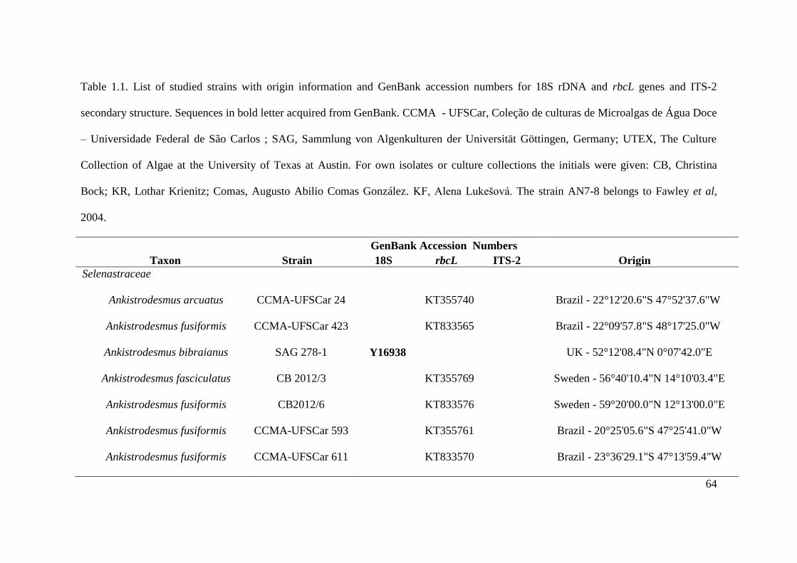
64
Table 1.1. List of studied strains with origin information and GenBank accession numbers for 18S rDNA and rbcL genes and ITS-2
secondary structure. Sequences in bold letter acquired from GenBank. CCMA - UFSCar, Coleção de culturas de Microalgas de Água Doce
– Universidade Federal de São Carlos ; SAG, Sammlung von Algenkulturen der Universität Göttingen, Germany; UTEX, The Culture
Collection of Algae at the University of Texas at Austin. For own isolates or culture collections the initials were given: CB, Christina
Bock; KR, Lothar Krienitz; Comas, Augusto Abilio Comas González. KF, Alena Lukešová. The strain AN7-8 belongs to Fawley et al,
2004.
GenBank Accession Numbers
Taxon Strain 18S rbcL ITS-2 Origin
Selenastraceae
Ankistrodesmus arcuatus CCMA-UFSCar 24 KT355740 Brazil - 22°12'20.6"S 47°52'37.6"W
Ankistrodesmus fusiformis CCMA-UFSCar 423 KT833565 Brazil - 22°09'57.8"S 48°17'25.0"W
Ankistrodesmus bibraianus SAG 278-1 Y16938 UK - 52°12'08.4"N 0°07'42.0"E
Ankistrodesmus fasciculatus CB 2012/3 KT355769 Sweden - 56°40'10.4"N 14°10'03.4"E
Ankistrodesmus fusiformis CB2012/6 KT833576 Sweden - 59°20'00.0"N 12°13'00.0"E
Ankistrodesmus fusiformis CCMA-UFSCar 593 KT355761 Brazil - 20°25'05.6"S 47°25'41.0"W
Ankistrodesmus fusiformis CCMA-UFSCar 611 KT833570 Brazil - 23°36'29.1"S 47°13'59.4"W
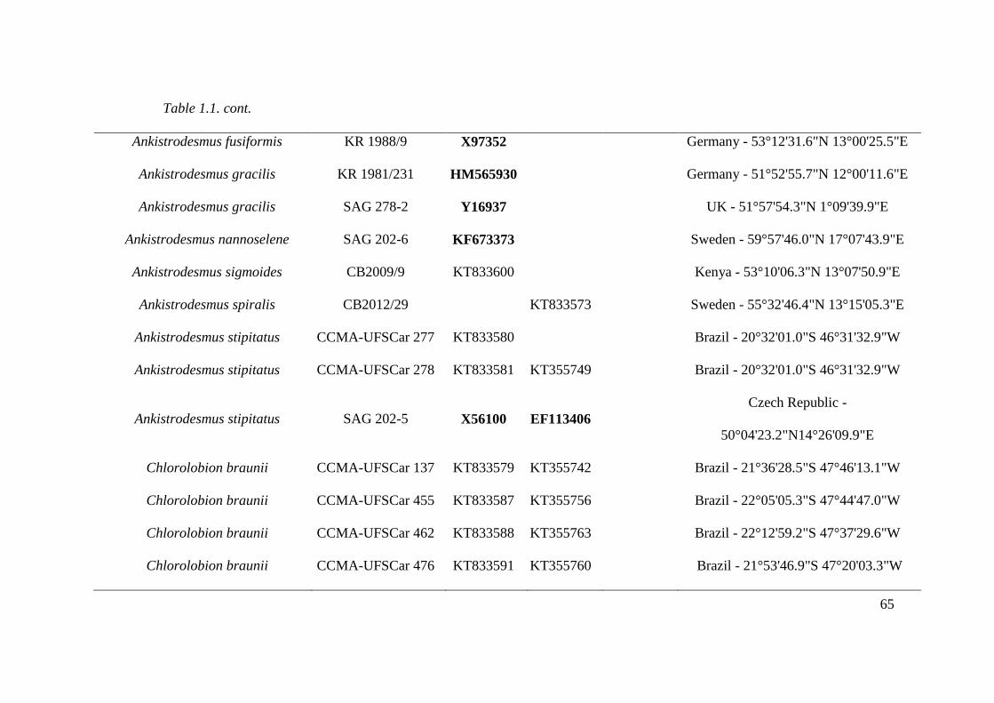
65
Table 1.1. cont.
Ankistrodesmus fusiformis KR 1988/9 X97352 Germany - 53°12'31.6"N 13°00'25.5"E
Ankistrodesmus gracilis KR 1981/231 HM565930 Germany - 51°52'55.7"N 12°00'11.6"E
Ankistrodesmus gracilis SAG 278-2 Y16937 UK - 51°57'54.3"N 1°09'39.9"E
Ankistrodesmus nannoselene SAG 202-6 KF673373 Sweden - 59°57'46.0"N 17°07'43.9"E
Ankistrodesmus sigmoides CB2009/9 KT833600 Kenya - 53°10'06.3"N 13°07'50.9"E
Ankistrodesmus spiralis CB2012/29 KT833573 Sweden - 55°32'46.4"N 13°15'05.3"E
Ankistrodesmus stipitatus CCMA-UFSCar 277 KT833580 Brazil - 20°32'01.0"S 46°31'32.9"W
Ankistrodesmus stipitatus CCMA-UFSCar 278 KT833581 KT355749 Brazil - 20°32'01.0"S 46°31'32.9"W
Ankistrodesmus stipitatus SAG 202-5 X56100
EF113406
Czech Republic -
50°04'23.2"N14°26'09.9"E
Chlorolobion braunii CCMA-UFSCar 137 KT833579 KT355742 Brazil - 21°36'28.5"S 47°46'13.1"W
Chlorolobion braunii CCMA-UFSCar 455 KT833587 KT355756 Brazil - 22°05'05.3"S 47°44'47.0"W
Chlorolobion braunii CCMA-UFSCar 462 KT833588 KT355763 Brazil - 22°12'59.2"S 47°37'29.6"W
Chlorolobion braunii CCMA-UFSCar 476 KT833591 KT355760 Brazil - 21°53'46.9"S 47°20'03.3"W

66
Table 1.1. cont.
Curvastrum pantanale CCMA-UFSCar 350 KT833584 KT355754 KU180822 Brazil - 19°17'59.0"S 55°47'45.0"W
Curvastrum pantanale CCMA-UFSCar 608 KT833569 KU180824 Brazil - 23°15'25.7"S 47°18'18.6"W
Kirchneriella aperta CCMA-UFSCar 482 KT833592 Brazil - 22°33'09.2"S 48°57'47.5"W
Kirchneriella aperta SAG 2004 AJ271859
KC145514 Germany - 53°08'51.7"N 13°01'41.9"E
Kirchneriella contorta var. elegans CCMA-UFSCar 447 KT833586 Brazil - 22°19'01.0"S 48°03'05.4"W
Kirchneriella irregularis CCMA-UFSCar 348 KT833583 KT355752 Brazil - 19°17'59.0"S 55°47'45.0"W
Kirchneriella lunaris CCMA-UFSCar 443 KT833566 Brazil - 22°19'01.0"S 48°03'05.4"W
Kirchneriella obesa CCMA-UFSCar 345 KT833564 Brazil - 19°17'59.0"S 55°47'45.0"W
Kirchneriella pseudoaperta CCMA-UFSCar 346 KT833582 Brazil - 19°17'59.0"S 55°47'45.0"W
Messastrum gracile CB 2009/3 KT833596 Germany - 53°00'50.8"N 13°10'00.5"E
Messastrum gracile CB 2009/35 KT861784 Germany - 52°31'32.1"N13°17'15.5"E
Messastrum gracile CCMA-UFSCar 470 KT833589 KT355759 KU180823 Brazil - 21°48'38.0"S 47°16'26.0"W
Messastrum gracile CCMA-UFSCar 5 KT833577 KT355739 KU180819 Brazil - 22°12'20.6"S 47°52'37.6"W
Messastrum gracile CCMA-UFSCar 622 KT833593 KT355762 KU180825 Brazil - 23°02'28.8"S 48°03'15.9"W

67
Table 1.1. cont.
Monoraphidium braunii KR 1986/28 AJ300527 Germany - 53°08'37.9"N 13°01'07.3"E
Monoraphidium contortum CB2009/10 KT833571 Kenya - 53°10'06.3"N 13°07'50.9"E
Monoraphidium contortum CCMA-UFSCar 306 KT355750 Brazil - 23°44'04.3"S 46°45'44.4"W
Monoraphidium contortum CCMA-UFSCar 329 KT833563 Brazil - 8°35'17.9"S 63°37'12.3"W
Monoraphidium contortum CCMA-UFSCar 349 KT355753 Brazil - 19°17'59.0"S 55°47'45.0"W
Monoraphidium convolutum AS7-3 AY846377 USA - 47º17‟26.8”N 98º50‟07.0”W
Monoraphidium convolutum KR 1981/262 HM565926 Germany - 51°49'37.8"N 11°59'41.7"E
Monoraphidium dybowskii SAG 202-7e Y16939 France - 43°06'26.3"N 0°05'10.4"W
Monoraphidium indicum CCMA-UFSCar 549 KT833568 Brazil - 22°04'56.2"S 48°29'20.0"W
Monoraphidium komarkovae CCMA-UFSCar 353 KT833585 KT355755 Brazil - 19°20'29.0"S 55°43'40.0"W
Monoraphidium komarkovae CCMA-UFSCar 632 KT833595 KT355763 Brazil - 24°0'19.44''S 48°20'33.41''W
Monoraphidium neglectum SAG 48.87 AJ300526 Germany - 51°44'43.9"N 11°58'48.6"E
Monoraphidium pseudobraunii CCMA-UFSCar 325 KT355751 Brazil - 8°35'17.9"S 63°37'12.3"W
Monoraphidium terrestre SAG 49.87 Y17817 Germany - 51°47'22.9"N 12°03'53.6"E

68
Table 1.1. cont.
Nephrochlamys subsolitaria SAG 243-2a HM560960 UK - 54°55'25.5"N 2°18'02.7"W
Podohedriella falcata SAG 202-2 X91263 Switzerland - 47°10'37.2"N 8°12'35.0"E
Quadrigula closterioides SAG 12.94 Y17924 USA - 47°11'43.2"N 95°10'00.9"W
Raphidocelis microscopica CB 2009/6 KT355768 Sweden - 0°21'20.1"S 36°03'20.5"E
Raphidocelis microscopica CB2012/39 KT833574 Sweden - 58°25'11.5"N 14°30'30.6"E
Raphidocelis microscopica CB 2009/18 KT355764 Kenya - 0°25'49.0"S 36°13'54.6"E
Raphidocelis subcapitata KR 1991/19 HM483520 Germany - 53°08'49.3"N 13°01'53.9"E
Rhombocystis complanata KR 1998/2 HM483518 Dominica - 15°23'29.9"N 61°15'19.6"W
Selenastrum bibraianum CB2009/41 HM483514 Germany - 51°00'47.8"N 9°52'58.2"E
Selenastrum bibraianum CB 2009/43 KT833599 Germany - 52°27'10.4"N 13°18'25.3"E
Selenastrum bibraianum CB 2012/47 KT833575 KU180826 Sweden - 55°52'01.4"N 13°33'21.5"E
Selenastrum bibraianum CB2009/39 KT833597 Sweden - 55°52'01.4"N 13°33'21.5"E
Selenastrum bibraianum CCMA-UFSCar 125 KT833578 KT355741 KU180821 Brazil - 21°36'28.5"S 47°46'13.1"W
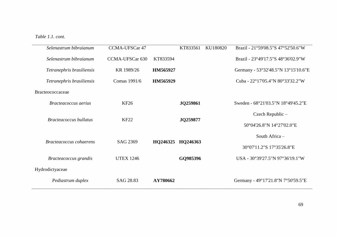
69
Table 1.1. cont.
Selenastrum bibraianum CCMA-UFSCar 47 KT833561 KU180820 Brazil - 21°59'08.5"S 47°52'50.6"W
Selenastrum bibraianum CCMA-UFSCar 630 KT833594 Brazil - 23°49'17.5"S 48°36'02.9"W
Tetranephris brasiliensis KR 1989/26 HM565927 Germany - 53°32'48.5"N 13°15'10.6"E
Tetranephris brasiliensis Comas 1991/6 HM565929 Cuba - 22°17'05.4"N 80°33'32.2"W
Bracteococcaceae
Bracteacoccus aerius KF26 JQ259861 Sweden - 68°21'03.5"N 18°49'45.2"E
Bracteacoccus bullatus KF22 JQ259877
Czech Republic –
50°04'26.8"N 14°27'02.0"E
Bracteacoccus cohaerens SAG 2369 HQ246325
HQ246363
South Africa –
30°07'11.2"S 17°35'26.8"E
Bracteacoccus grandis UTEX 1246 GQ985396 USA - 30°39'27.5"N 97°36'19.1"W
Hydrodictyaceae
Pediastrum duplex SAG 28.83 AY780662 Germany - 49°17'21.8"N 7°50'59.5"E
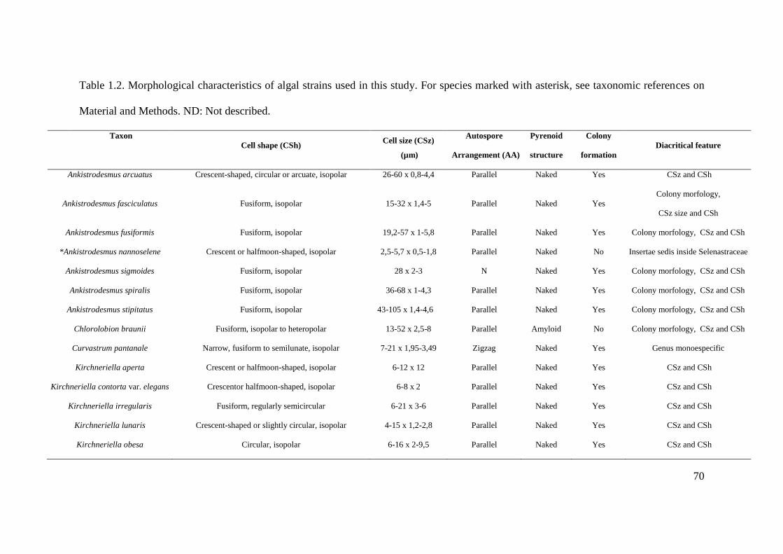
70
Table 1.2. Morphological characteristics of algal strains used in this study. For species marked with asterisk, see taxonomic references on
Material and Methods. ND: Not described.
Taxon
Cell shape (CSh) Cell size (CSz)
(µm)
Autospore
Arrangement (AA)
Pyrenoid
structure
Colony
formation
Diacritical feature
Ankistrodesmus arcuatus Crescent-shaped, circular or arcuate, isopolar 26-60 x 0,8-4,4 Parallel Naked Yes CSz and CSh
Ankistrodesmus fasciculatus Fusiform, isopolar 15-32 x 1,4-5 Parallel Naked Yes
Colony morfology,
CSz size and CSh
Ankistrodesmus fusiformis Fusiform, isopolar 19,2-57 x 1-5,8 Parallel Naked Yes Colony morfology, CSz and CSh
*Ankistrodesmus nannoselene Crescent or halfmoon-shaped, isopolar 2,5-5,7 x 0,5-1,8 Parallel Naked No Insertae sedis inside Selenastraceae
Ankistrodesmus sigmoides Fusiform, isopolar 28 x 2-3 N Naked Yes Colony morfology, CSz and CSh
Ankistrodesmus spiralis Fusiform, isopolar 36-68 x 1-4,3 Parallel Naked Yes Colony morfology, CSz and CSh
Ankistrodesmus stipitatus Fusiform, isopolar 43-105 x 1,4-4,6 Parallel Naked Yes Colony morfology, CSz and CSh
Chlorolobion braunii Fusiform, isopolar to heteropolar 13-52 x 2,5-8 Parallel Amyloid No Colony morfology, CSz and CSh
Curvastrum pantanale Narrow, fusiform to semilunate, isopolar 7-21 x 1,95-3,49 Zigzag Naked Yes Genus monoespecific
Kirchneriella aperta Crescent or halfmoon-shaped, isopolar 6-12 x 12 Parallel Naked Yes CSz and CSh
Kirchneriella contorta var. elegans Crescentor halfmoon-shaped, isopolar 6-8 x 2 Parallel Naked Yes CSz and CSh
Kirchneriella irregularis Fusiform, regularly semicircular 6-21 x 3-6 Parallel Naked Yes CSz and CSh
Kirchneriella lunaris Crescent-shaped or slightly circular, isopolar 4-15 x 1,2-2,8 Parallel Naked Yes CSz and CSh
Kirchneriella obesa Circular, isopolar 6-16 x 2-9,5 Parallel Naked Yes CSz and CSh

71
Table 2. 1 cont.
Kirchneriella pseudoaperta Crescent or halfmoon-shaped, isopolar 1,6-9,6 x 2,4 x 4,2 Zigzag Naked Yes CSz and CSh, AA
Messastrum gracile Narrow, fusiform to semilunate, isopolar 19-55 x 1-6 Zigzag Naked Yes CSz and CSh
Monoraphidium braunii Fusiform, isopolar to heteropolar 13-52 x 2,5-8 Parallel Amyloid No Colony morfology, CSz and CSh
Monoraphidium contortum Fusiform, crescent-shape, circular, twisted, isopolar 7-40 x 1-5,2 Parallel Naked No CSz and CSh
*Monoraphidium convolutum Fusiforme, semicircular, isopolar 5-16 x 1-6 Parallel Naked No CSz and CSh
*Monoraphidium dybowskii Cylindrical,fusiform, isopolar 4-15,5 x 1,2-8 Parallel Naked No CSz and CSh
Monoraphidium indicum Fusiform, circular, isopolar 120-260 x 3-5 Parallel Naked No CSz and CSh
Monoraphidium komarkovae Needle-shaped, isopolar 25-182 x 1,4-3,5 Parallel Naked No CSz and CSh
*Monoraphidium neglectum Fusiform 15-37 x 2-6,5 Serial Naked No CSz and CSh
Monoraphidium pseudobraunii Fusiform to sigmoid, isopolar or heteropolar 8-25 x 1-2,5 Parallel Naked No CSz and CSh
*Monoraphidium terrestre Fusiform, cylindrical 14-30 x 2-8 Serial Naked No CSz and CSh
*Nephrochlamys subsolitaria Semilunate, isopolar 3,5-9,6-12 Serial Naked Yes Proximity of cell ends
*Podohedriella falcata Needle-shaped, heteropolar 35-50 x 1,5-2,5 Serial Naked No CSz and CSh
*Quadrigula closterioides Cylindrical with rounded ends, isopolar 12-30 x 1-4 Parallel Naked Yes Cell end and CSz
Raphidocelis microscopica Conical, capitata, isopolar 3-5 x 1-2 Serial Naked Yes CSz and CSh
*Raphidocelis subcapitata Cylindrical, circular or sigmoid, isopolar 7-23 x 1,2-3 Serial Naked Yes CSz and CSh
*Rhombocystis complanata Rhomboidal, isopolar to heteropolar 9,5-14,5x 2,4-3,6 Parallel Naked Occasionally CSz and CSh
Selenastrum bibraianum Narrow, fusiform to semilunate, isopolar 16-40 x 2,5-4,5 Zigzag Naked Yes CSz and CSh
*Tetranephris brasiliensis Bean-shaped 6-7,6 x 4-5,8 Serial Naked Yes CSz and CSh

72
Table 1.S3. 18s rDNA, rbcL and ITS primers used for amplification and sequencing of Selenastraceae.
Primer Name Sequence 5'-3' Direction Target gene Reference
18S F1 AATCTGGTTGATCCTGCCAGT FW 18S Katana et al.2001
1 F CTGGTTGATCCTGCCAG FW 18S Huss et al.1999
300 F AGGGTTCGATTCCGGAG FW 18S Huss et al.1999
528 F CGGTAATTCCAGCTCC FW 18S Huss et al.1999
690 F YAGAGGTGAAATTCT FW 18S Huss et al.1999
920F GAAACTTAAAKGAATTG FW 18S Huss et al.1999
1055 F GGTGGTGCATGGCCG FW 18S Huss et al.1999
1400F TGYACACACCGCCCGTC FW 18S Huss et al.1999
920 R ATTCCTTTRAGTTTC RV 18S Huss et al.1999
1200 R GGGCATCACAGACCTG RV 18S Huss et al.1999
1520 R CYGCAGGTTCACCTAC RV 18S Huss et al.1999
18SR1 TGATCCTTCTGCAGGTTCACCTA RV 18S Katana et al.2001 (modified)
rbcL 1 ATGGTTCCACAAACAGAAAC FW rbcL Nozaki et al. 1995

73
Table 1.S3. cont.
rbcL 320 TATTCGAAGAAGGTTCAGTAAC FW rbcL Rindi et al. 2008
rbcL RH1 ATGTCACCACAAACAGAAACTAAAGC FW rbcL Manhart, J.R. 1994
rbcL 320 mod TATTYGAASAAGGTTCWGTWAC FW rbcL Proposed on this study
Selenastraceae rbcL F CGYTACAAAGGDCGTTGYT FW rbcL Proposed on this study
rbcL 1181 AAGATTTCAACTAAAGCTGGCA RV rbcL Nozaki et al. 1995
rbcL 1385 GGAAAGAAATTAAATTTGAATT RV rbcL McCourt et al. 2000
rbcL 1421 TTGTCAATAGTATCAAATTC RV rbcL Nozaki et al. 1995
rbcL ORB CTGGAGCATTACCCCAAGG RV rbcL Pazoutova unpublished
rbcL Orb modified CTGGNGCRTTACCCCAAGG RV rbcL Proposed on this study
Selenastraceae rbcL R RTTACCCCAWGGGTGHCCTA RV rbcL Proposed on this study
1420F CAGGTCTGTGATGCCC FW ITS Rogers et al. 2006
NS7m GGCAATAACAGGTCTGT FW ITS An et al. 1999
ITS055R CTCCTTGGTCCGTGTTTCAAGACGGG RV ITS Marin et al. 1998
LR1850 CCTCACGGTACTTGTTC RV ITS An et al. 1999
74
Figure 1.1-1.5. 1.1. Messastrum gracile. Original picture of strain CCMA-UFSCar 622
showing a frontal view of colony; 1.2. Selenastrum bibraianum. Original picture of
strain CCMA-UFSCar 125 showing a frontal view of colony. 1.3-1.4. Curvastrum
pantanale. (1.3) Original picture of strain CCMA-UFSCar 350, showing free cells and
colony; (1.4) Original picture of strain CCMA-UFSCar 350, showing cells in autospore
liberation. presenting a cell wall remnant (arrowhead) and protoplasm cleavage (star).
1.5. Ankistrodesmus arcuatus. Original picture of strain CCMA-UFSCar 24, showing
free cells and colony. Note autospore formation (star) and mucilaginous lump
(arrowhead). Scale bar 10 μm.
Figure 1.6: Maximum–likelihood (ML) phylogenetic tree inferred from rbcL gene
sequences of some members of Selenastraceae. Support values correspond to Bayesian
PP (Posterior Probability), ML BP (Bootstrap), MP (Maximum Parsimony) BP, NJ
(Neighbor-Joining) BP. Hyphens correspond to values <50% for BP and <0.95 for PP.
Scale represents the expected number of substitutions per site. Strain numbers used as
mentioned in Table 1.
Figure 1.7: Maximum–likelihood (ML) phylogenetic tree inferred from 18S rDNA gene
sequences of some members of Selenastraceae. Support values correspond to Bayesian
PP (Posterior Probability), ML BP (Bootstrap), MP (Maximum Parsimony) BP, NJ
(Neighbor-Joining)BP. Hyphens correspond to values <50% for BP and <0.95 for PP.
Scale represents the expected number of substitutions per site. Strain numbers used as
mentioned in Table 1.

75
Figure 1.8: Scanning electron micrographs of Curvastrum pantanale (CCMA-UFSCar
350) in culture. Scale bar, 10 µm. (a) typical colony formation, (b) young cells
detaching from each other, (c) young cells, note the autospores position.
Figure 1.9: Transmission electron micrographs of Curvastrum pantanale (CCMA-
UFSCar 350) in culture. Scale bar, 1 µm. Key to labeling: CW = cell wall, C =
chloroplast, D = dictyosome, ER = endoplasmic reticulum, L = lipid drop, M =
mitochondrion, N = nucleus, P = pyrenoid, PV = polyphosphate vacuole, S = starch
grain. a) longitudinal section; cell presenting lipid drops, polyphosphate vacuoles
(arrowhead), chloroplast penetrated with starch grains, and a central pyrenoid. b) detail
of figure a, where an endoplasmic reticulum (arrowhead) can be observed. c)
longitudinal section; cell presenting polyphosphate vacuoles on both cell apexes,
mitochondria, chloroplast filled with starch grains and a central pyrenoid. d) cross
section; chloroplast containing starch grains and a pyrenoid situated at the left, a central
nucleus can be observed. e) longitudinal section; mature cell containing a central
nucleus, mitochondrion and pyrenoid (upper part). All the cell content is surrounded by
a cell wall.
Figure 1.10: Transmission electron micrographs of two Selenastraceae in culture. Scale
bar, 1 µm. a - c) Ankistrodesmus arcuatus (CCMA – UFSCar 24). a) longitudinal
section; cell with a nucleus, dictyosome and a chloroplast. b) cross section; a cup-
shaped chloroplast penetrated by a pyrenoid (star). On the opposite direction of the
pyrenoid, the nucleus is situated. An arrowhead indicates a polyphosphate vacuole. c)
detail of figure a, where a dictyosome can be observed. d - e) Monoraphidium
contortum (CCMA-UFSCar 349), d) longitudinal section; cells on different life cycle

76
phase. The upper 3 cells are young indidivuals presenting starch grains (arrowhead),
polyphosphate vacuoles (arrowhead), chloroplast and nucleus. The lower cell is a
mature individual containing many starch grains on the chloroplast, some
polyphosphate vacuoles (arrowhead), nucleus and big lipid drops. e) cross section;
dense chloroplast with starch grains. Two big polyphosphate vacuoles (arrowhead) and
a nucleus can be observed. All the cell content is surrounded by a cell wall.
Supplementary figures
1.A) ITS-2 model for the type strain of Selenastrum bibraianum (CCMA-UFSCar 125).
In black boxes are the different bases compared to strain Messastrum gracile (CCMA-
UFSCar 622).
1.B) ITS-2 model for the type strain of Selenastrum bibraianum (CCMA-UFSCar 125).
In gray boxes are the different bases compared to strain Selenastrum bibraianum
(CCMA-UFSCar 47) and black boxes compared to Selenastrum bibraianum (CB
2012/47).
1.C) ITS-2 model for the type strain of Messastrum gracile (CCMA-UFSCar 622). In
gray boxes are the different bases of M. gracile (CCMA-UFSCar 470) and black boxes
M. gracile (CCMA-UFSCar 5).
1.D) ITS-2 model for the type strain of Curvastrum pantanale (CCMA-UFSCar 350). In
gray boxes are the different bases of C. pantanale (CCMA-UFSCar 608).

77
Fig. 1.1-1.5.

78
Figure 1.6.

79
Figure 1.7.

80
Figure 1.8.
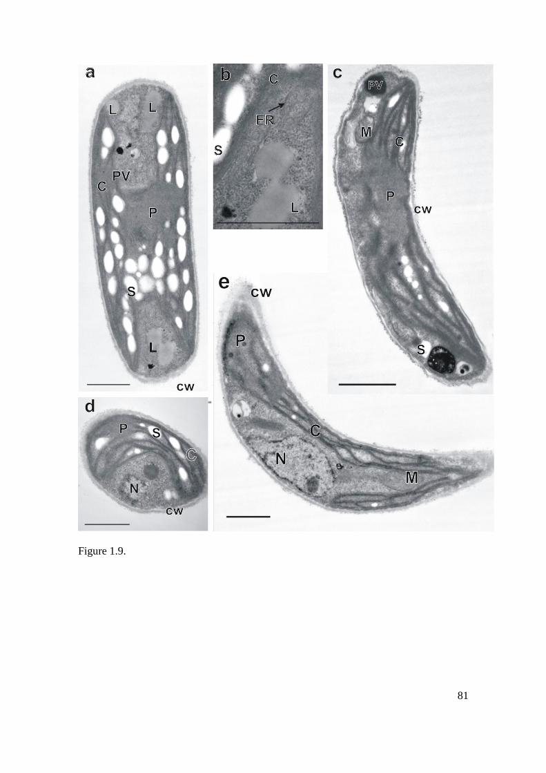
81
Figure 1.9.

82
Figure 1.10.

83
Supplementary figure
1. A, B, C. D.
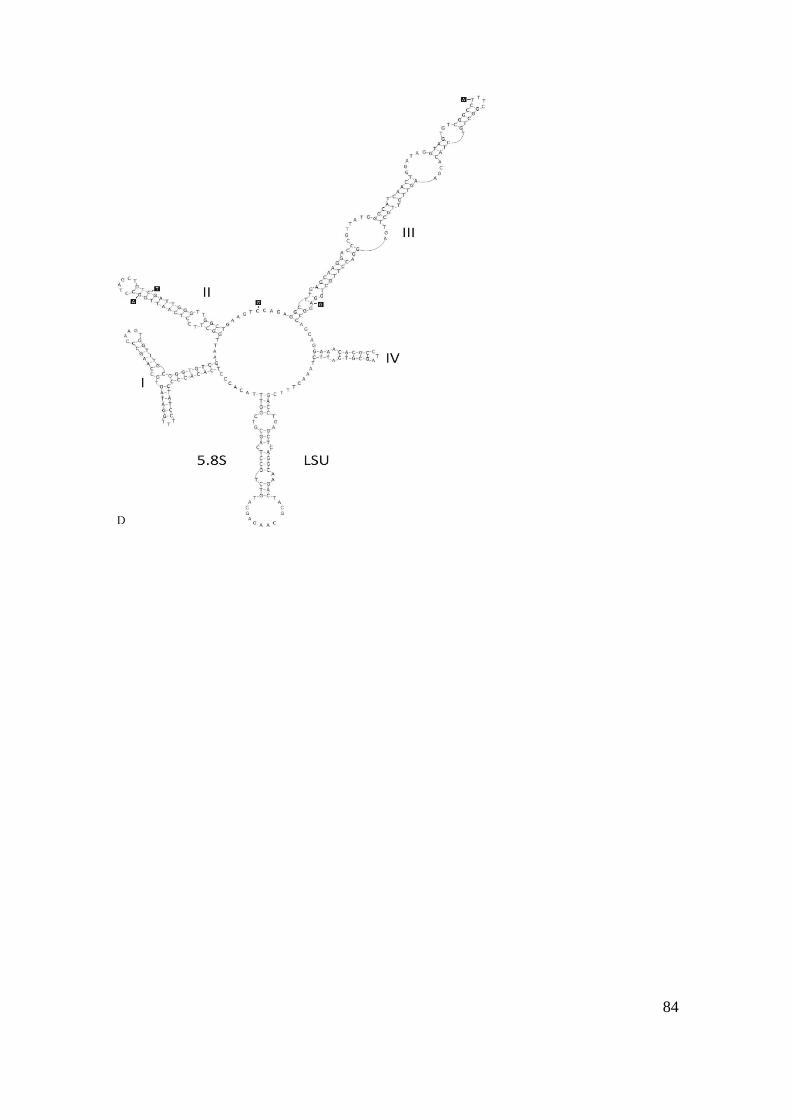
84

85
Capítulo 2:
KIRCHNERIELLA MORPHOTYPE (SELENASTRACEAE,
CHLOROPHYCEAE) REVEALS FOUR MOLECULAR LINEAGES,
INCLUDING TWO NEW GENERA AND FIVE SPECIES1.
1 Paper to be submitted
2 Corresponding author: [email protected].
86
2.1. ABSTRACT
The phylogeny of the Kirchneriella morphotype was investigated by light
microscopy, 18S rDNA and ITS1-5.8S-ITS2 analyses. Morphological features
traditionally used for species and genera identification were investigated in genera
Kirchneriella, Raphidocelis and Tetranephris. Phylogenetic analyses revealed four
molecular lineages attributed to Kirchneriella morphotype and led to the
description of five new species: Genus 1 sp. nov. 1, Genus 2 sp. nov. 1, Genus 2 sp.
nov. 2, Raphidocelis sp. nov. and Tetranephris sp. nov. The molecular analyses
showed that a number of morphological features, such as pyrenoid, cell size and
shape, may be suitable to identify the genera Kirchneriella, Raphidocelis and
Tetranephris.
KEYWORDS: Molecular systematics, morphology, phylogeny, Kirchneriella,
Pseudokirchneriella, Raphidocelis, Tetranephris.
87
2.2. INTRODUCTION
Kirchneriella Schmidle 1892 (Selenastraceae, Chlorophyceae) is a green algae
genus with cosmopolitan distribution in nearly all types of inland waters. The
morphology of Kirchneriella morphotype contain an assortment of shapes: from
coccoid to elongated, cylindrical to fusiform or spirally curved, with sharp or rounded
ends, where cell arrangements differs from the solitary to colonial forms (Komárek &
Fott 1983; Korshikov 1987; Komárková-Legnerová 1969; Comas 1996; Hindák, 1977;
Hindák 1980; Hindák 1984; Hindák 1988; Hindák 1990; Sant'Anna 1984).
Kirchneriella and relatives show the same reproduction pattern of all members of
Selenastraceae: a simple autospore formation, in which the cytokinesis of the mother
cell protoplasm gives rise to 2-4-8 young cells (Komárek & Fott 1983).
Since the description of the genus Kirchneriella, morphological revaluations
regarding presence of pyrenoids and cell wall incrustations (Hindák 1977; 1980; 1984;
1988; 1990; Marvan et al. 1984) led to its division in three other morphological defined
genera: Raphidocelis Hindák (strains with incrustation on cell wall and lacking
pyrenoid), Kirchneria Hindák (to comprehend the strains with no cell wall incrustations
and no pyrenoid) and Pseudokirchneriella Hindák (a valid name for Kirchneria, since
Kirchneria was a name already published for a fossile plant). Molecular studies on the
SSU of Kirchneriella and Raphidocelis species placed them as common members of
monophyletic group Selenastraceae (Fawley et al. 2006; Krienitz et al. 2011; Krienitz et
al. 2001).
The cell size and shape, solitary or colonial organization, releasing process of
the autospores and special habitat preferences, considered to be species-specific
(Hindák 1977; Komárek & Fott 1983), was used to separate genera inside the family.
Those features conducted to the description of up to 81 taxa (including forms and

88
variations) described in the Kirchneriella morphotype (Kirchneriella, Raphidocelis,
Kirchneria and Pseudokirchneriella complex) (Guiry & Guiry, 2016; Korshikov 1987;
Komárek & Fott 1983; Komárková-Legnerová 1969; Comas 1996; Hindák 1977;
Hindák 1980; Hindák 1984; Hindák 1988; Hindák 1990; Sant'Anna 1984). As the
taxonomic and phylogenetic studies go forward, possibly various genera and species are
to be described and included in this complex (Hindák 1977; Hindák 1984; Komárek &
Fott 1983; Fawley et al. 2006; Krienitz et al. 2011; Garcia et al. in press).
Thus, a combined molecular approach based on different genetic markers like
18S, rbcL or ITS associated with a morphological evaluation and the inclusion of strains
from different continents would be a valuable tool for identification and classification
not only of Selenastraceae but other green algae as well (Bock et al. 2011; Krienitz et
al. 2012).
Indeed, due to its simple morphology and phenotypic plasticity, including new
lineages from different environments and geographically apart may lead to changes in
the Kirchneriella complex.
In front of the difficulties with species identification and taxonomy, species
diversity and ecology of members of Selenastraceae is poorly understood worldwide
(Fawley et al. 2006; Krienitz et al. 2001). Additionally, studies with molecular diversity
of tropical Selenastraceae remain largely unknown, since previous studies were focused
on temperate northern hemisphere isolates.
Phylogenetic studies in green algae are generally based on 18S rRNA gene
sequences (Booton et al. 1998; Buchheim et al. 2001; Fawley et al. 2006; Hegewald &
Hanagata 2000; Krienitz et al. 2011; Krienitz et al. 2003; Krienitz et al. 2001; Lewis
1997). However, this gene is in some cases too preserved to distinguish between closely
related genera and species (Luo et al. 2010). Therefore, some studies have taken into

89
account the use of a second marker as well, to gain a higher resolution (Rindi et al.
2011).
Several studies have employed the ITS for phylogenetic reconstruction in
diverse eukaryotes (Bock et al. 2011; Coleman 2003; Coleman 2007; Coleman &
Vacquier 2002; Schultz et al. 2005; Young & Coleman 2004) or for the resolution of
closely related genera (Luo et al. 2010; Luo et al. 2011). The highly divergent
properties and the rapid evolution legitimate ITS-2 to discriminate closely related
organisms, which exhibit nearly identical sequences in rRNA genes level. Indeed, ITS
have been reported to be more informative than gene sequences for those organisms
(Coleman 2003) and is increasingly being used as a unique marker between the most
frequently utilized DNA region, at least for plant studies (Hershkovitz, et al. 1999).
In numerous cases were under consent to employ ITS as barcode region for
dinoflagellates (Litaker et al. 2007) and fungi (Seifert 2009) since the variation in this
DNA region correlates with taxonomic classification in several cases (Coleman, 2009).
Therefore, using a multigene analysis employing conserved genes for a good
phylogeny and a second variable marker to obtain higher resolution for lower
taxonomic levels is necessary for taxonomically complicated groups as Kirchneriella
complex. The present study is the first to evaluate Kirchneriella morphospecies with
18S rDNA and ITS1, 5.8S rDNA and ITS2 multigene phylogeny, to clarify the
phylogenetic relation between genera Kirchneriella, Raphidocelis and Tetranephris.
2.3. MATERIAL AND METHODS
Algal cultures and microscopy

90
The algal cultures were obtained either from Freshwater Microalgae Culture
Collection from Universidade Federal de São Carlos (CCMA – UFSCar, WDCM 835)
and from an author personal collection (CB strains). KR strains were donated by
Professor Lothar Krienitz. Thirty five Selenastraceae strains were investigated (Table
2.1). All the strains were grown at 23 ± 1 ºC under a photoperiod 12/12 hours light/dark
and irradiance of 200 µmol/m2/s in WC medium (Guillard & Lorenzen).
The morphology of all strains was examined using an Axioplan 2 Imaging Zeiss
or Nikon Eclipse E600 light microscope with differential interference contrast.
Micrographs were obtained using an AxioCam with software AxioVision 4.6 (Carl
Zeiss Group, Oberkochen, Germany) and a Nikon digital camera DS-Fi1 with Nikon
software NIS-Elements D (Nikon Corporation, Tokyo, Japan). The species
identification were done according to the specialized bibliography: Korshikov 1987a,
Komárek & Fott 1983, Komárková-Legnerová 1969, Comas 1996, Hindák 1977,
Hindák 1980, Hindák 1984, Hindák 1988, Hindák 1990 and Sant'Anna 1984.
DNA extraction, PCR and sequencing
For DNA extraction, the algal cultures were grown as described for microscopy
analyses. The cell cultures were centrifuged at 16.000 xg for 10 minutes and the pellet
stored at -80°C until further processing. The cells were mechanically disrupted using
glass beads (150-212 µm, Sigma-Aldrich), vortex briefly, and extracted using Invisorb®
Spin Plant Mini (STRATEC Biomedical AG; Germany) or My-Budget DNA Mini Kit®
(Bio-Budget Technologies GmbH; Krefeld; Germany). PCR amplification, purification
and sequencing were carried out as previously reported (Garcia et al. in press). Part of
the genomic DNA is stored at the Phycology Lab – UFSCar, and Department of
Biodiversity – University Duisburg-Essen.

91
Fifteen new 18S rDNA sequences and 32 ITS-1-5.8S-ITS-2 new sequences were
amplified and obtained on this study, totalizing 47 new entries in GenBank (National
Center for Biotechnology Information [NCBI], http://www.ncbi.nlm.nih.gov/). The
whole dataset used for phylogenetic analyses with accession numbers are reported in
Table 1, including 30 references sequences acquired from GenBank and 9 from our
previous publication (Garcia et al. in press).
Phylogenetic analyses
Sequences were aligned using Align - Manual Sequence Alignment Editor
(Hepperle 2004). For the phylogenetic analyses, two different datasets were prepared.
For the 18S rDNA analyses, 49 sequences with 1592 base positions were acquired. The
concatenated ITS-1-5.8S-ITS2 analyses was conducted with 36 sequences, were ITS-1
had 223 base positions (bp), 5.8S had 159bp and ITS-2 with 213 bp, totalizing 597bp.
Both trees (18S rDNA and ITS-1-5.8S-ITS2) were analyzed separately, both in
maximum likelihood (ML) on Treefinder (Jobb 2008), distance (neighbor joining; NJ)
and maximum parsimony (MP) using PAUP* (portable version 4.0b10) (Swofford
2002).
For ML and Bayesian analyses, the evolutive model for 18S, ITS-1 and ITS-2
(GTR[Optimum, Empirical]:GI[Optimum]:5) and 5.8S
(HKY[{3,1,1,1,1,3},Empirical]:G[Optimum]:5) were applied as suggested Treefinder
(Jobb 2008). To assure tree topology, bootstrap analyses (1000) were tested by
calculating values for NJ, MP, ML criteria. For both datasets, Bayesian analyses were
performed using MrBayes version 3.1. (Huelsenbeck & Ronquist 2001). Two runs with
four chains of Markov chain Monte Carlo (MCMC) iterations were performed (1
million generations for ITS-1-5.8S-ITS2and 8 million generations for 18S rDNA). The

92
stationary distribution was assumed when the average standard deviations of split
frequencies between two runs were lower than 0.01 and Tracer V1.4 (Rambaut and
Drummond 2007) was used to check the stationary phase and to identify an appropriate
burn-in value. The first 25% of the calculated trees were discarded as burn–in. 50%
majority–rule consensus trees were calculated for posterior probabilities (PP). The trees
were edited using TreeGraph 2 (Stöver & Müller 2010). Previous publications indicated
that Bracteacoccus (Krienitz et al. 2001), Pediastrum (Fučíková et al. 2014) and
Scenedesmaceae (Krienitz et al. 2011) members were suitable as an outgroup for the
phylogeny of Selenastraceae.
2.4. RESULTS
Taxonomic proposals.
Tetranephris sp. nov. 1 T. S. Garcia & Sant‟Anna (Fig 2.1).
Solitary cells or in cross-shaped coenobium with 4-8-16 cells, group of 4 cells always
with 2 cells organized in one plane and 2 organized on a perpendicular plane to the
others. Cells cylindrical, regularly narrowed towards the ends or gradually obtuse, bean-
shaped, slightly arcuate. Cells 3-8 long x 1-4 µm large. Asexual reproduction by
autosporulation (4-8 autospores per mother cell, with parallel orientation), sexual
reproduction unknown. A diffuse and irregular layer of mucilage is often involving both
colonies and free individuals. One parietal chloroplast, no pyrenoid observed.
Holotype: Kenya, Nakuru County, Lake Nakuru National Park. Sample deposited in
Herbarium of Institute of Botany, SP, Brazil, under the designation SP 469323.
Type strain: strain CB 2009/6, maintained at the Culture Collection of Freshwater
Microalgae, Federal University of São Carlos, São Carlos, Brazil.

93
Etymology: to be designated.
Genus 1gen. nov. T. S. Garcia & Sant‟Anna.
Type species: Genus 1 sp nov. 1 T. S., Garcia.
Diagnosis: Green, planktonic microalgae. Colonies with 4-8 or multi-celled, cells with
the convex or the concave side outwards the center of the colony. Cells fusiform,
regularly narrowed towards the ends or gradually obtuse, regularly curved along its
length and slightly bent to one side. Asexual reproduction by autosporulation (4
autospores per mother cell with parallel arrangement), sexual reproduction unknown.
Mucilage often involving both colonies and free cells. One parietal chloroplast,
containing a pyrenoid, no starch cover observed. Genus differs from other
Selenastraceae genera by differences in 18S rDNA and ITS-1-5.8S-ITS2 gene
sequences.
Genus 1 sp nov. 1 T. S. Garcia & Sant‟Anna (Fig 2.2).
Colonies with 4-8 cells or multi-celled, some with the convex others with the concave
side outwards the center of the colony or solitary cells, where two cells are in one plane
and two are in a perpendicular plane. Cells fusiform, regularly narrowed towards the
ends or gradually obtuse, regularly curved along its length and slightly bent to one side.
Cells 6-20 long x 3-7 µm large. Asexual reproduction by autosporulation (4 autospores
per mother cell, with parallel orientation), sexual reproduction unknown. A diffuse and
irregular layer of mucilage is often involving both colonies and free individuals. One
parietal chloroplast, containing a pyrenoid, no starch cover observed.
Holotype: Brazil, São Paulo State, Piracicaba. Sample deposited in Herbarium of
Institute of Botany, SP, Brazil, under the designation SP 469324.
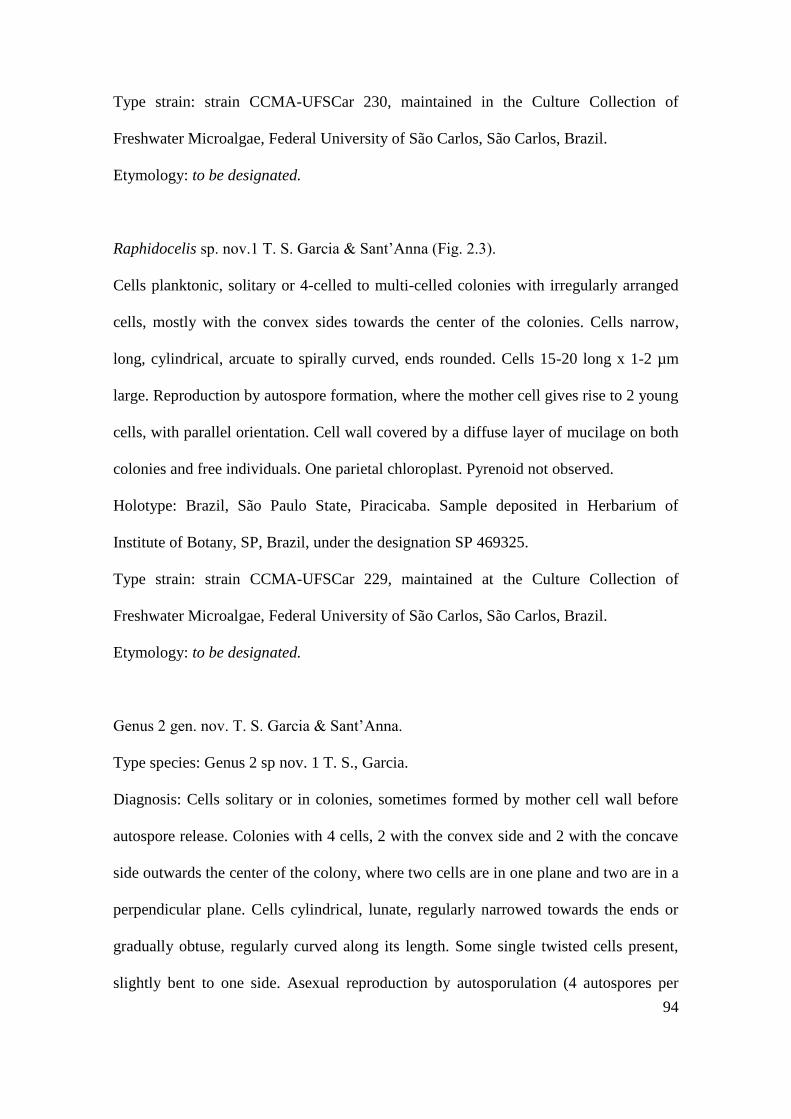
94
Type strain: strain CCMA-UFSCar 230, maintained in the Culture Collection of
Freshwater Microalgae, Federal University of São Carlos, São Carlos, Brazil.
Etymology: to be designated.
Raphidocelis sp. nov.1 T. S. Garcia & Sant‟Anna (Fig. 2.3).
Cells planktonic, solitary or 4-celled to multi-celled colonies with irregularly arranged
cells, mostly with the convex sides towards the center of the colonies. Cells narrow,
long, cylindrical, arcuate to spirally curved, ends rounded. Cells 15-20 long x 1-2 µm
large. Reproduction by autospore formation, where the mother cell gives rise to 2 young
cells, with parallel orientation. Cell wall covered by a diffuse layer of mucilage on both
colonies and free individuals. One parietal chloroplast. Pyrenoid not observed.
Holotype: Brazil, São Paulo State, Piracicaba. Sample deposited in Herbarium of
Institute of Botany, SP, Brazil, under the designation SP 469325.
Type strain: strain CCMA-UFSCar 229, maintained at the Culture Collection of
Freshwater Microalgae, Federal University of São Carlos, São Carlos, Brazil.
Etymology: to be designated.
Genus 2 gen. nov. T. S. Garcia & Sant‟Anna.
Type species: Genus 2 sp nov. 1 T. S., Garcia.
Diagnosis: Cells solitary or in colonies, sometimes formed by mother cell wall before
autospore release. Colonies with 4 cells, 2 with the convex side and 2 with the concave
side outwards the center of the colony, where two cells are in one plane and two are in a
perpendicular plane. Cells cylindrical, lunate, regularly narrowed towards the ends or
gradually obtuse, regularly curved along its length. Some single twisted cells present,
slightly bent to one side. Asexual reproduction by autosporulation (4 autospores per

95
mother cell, with parallel orientation), sexual reproduction unknown. Absence of
mucilage. One parietal chloroplast, pyrenoid not observed. Genus differs from other
Selenastraceae genera by the differences in 18S rDNA and ITS-1-5.8S-ITS2 gene
sequences and morphology.
Etymology: to be designated.
Genus 2 sp. nov. 1 T. S. Garcia & Sant‟Anna (Fig. 2.4).
Cells isolated or in colonies with 4 cells, 2 with the convex side and 2 with the concave
side outwards the center of the colony, where two cells are in one plane and two are in a
perpendicular plane. Sometimes the colony is surrounded by mother cell wall before
autospore release. Cells cylindrical, lunate, regularly narrowed towards the ends or
gradually obtuse, regularly curved along its length. Most individuals present twisted
cells, slightly bent to one side. Cells 6-15 long x 3-5 µm large. Asexual reproduction by
autosporulation (4 autospores per mother cell, with parallel orientation). Absence of
mucilage. One parietal chloroplast, pyrenoid not observed.
Holotype: Brazil, Mato Grosso do Sul State, Brazilian Pantanal wetlands. Sample
deposited in Herbarium of Institute of Botany, SP, Brazil, under the designation SP
469326.
Type strain: strain CCMA-UFSCar 348, maintained at the Culture Collection of
Freshwater Microalgae, Federal University of São Carlos, São Carlos, Brazil.
Etymology: to be designated.
Genus 2 sp. nov. 2 T. S. Garcia & Sant‟Anna (Fig. 2.5).
Cells isolated or in colonies with 4 cells, 2 with the convex side and 2 with the concave
side outwards the center of the colony, where two cells are in one plane and two are in a

96
perpendicular plane. Cells cylindrical, ends gradually obtuse, bean-shaped, crescent-
shaped or slightly arcuate. Cells 7-11 long x 1,2-3 µm large. Asexual reproduction by
autosporulation (4-8-multi autospores per mother cell, with parallel orientation), sexual
reproduction not known. A diffuse and irregular layer of mucilage is often involving
both colonies and free individuals. One parietal chloroplast, no pyrenoid observed.
Holotype: Germany, Saxony-Anhalt State, Zörbig City, Schortewitz Village. Sample
deposited in Herbarium of Institute of Botany, SP, Brazil, under the designation SP
469327.
Type strain: strain KR 1979/222, maintained at the Culture Collection of Freshwater
Microalgae, Federal University of São Carlos, São Carlos, Brazil.
Etymology: to be designated.
Morphological analysis
The morphological investigated parameters were cell shape and dimensions,
autospores arrangement, pyrenoid presence/absence and colony formation, respecting
the diacritical features of each species. These parameters are shown on Table 2.2.
Kirchneriella species were alike in presence of pyrenoid, cell shape and in the
colony morphology. They diverged on cell size and on the following features.
Kirchneriella pseudoaperta Komárek (CCMA-UFSCar 346 and CCMA-UFSCar 482,
Fig. 2.6) presented small but discernable pyrenoid under LM. Mucilage was exhibited
when cells were disposed in colony. Kirchneriella obesa (West) West &West (CCMA-
UFSCar 345 and CB 2012/49, Fig. 2.7) presented mucilage involving the colony, and
solitary cells presented bigger size than the cells in colonies. Big colonies typical for
Kirchneriella lunaris (Kirchner) Möbius (CCMA-UFSCar 87, CCMA-UFSCar 123 and
CCMA-UFSCar 443, Fig. 2.8) were observed, exhibiting acute cell apex, with solitary

97
cells bigger than the colonial one, always presenting pyrenoids and involved in
mucilage.
Tetranephris sp. nov. T.S. Garcia & Sant‟Anna (CB 2009/6, CB 2009/7 and CB
2009/18) were bean-shaped, slightly arcuate and had narrowed or gradually obtuse
apex. Mucilage was observed on free cells and colony formation. The typical cross-
shaped colony formation of genus Tetranephris Sant‟Anna& Bicudo was observed, with
2 cells on a perpendicular plane to the others. Pyrenoid was not observed.
Morphology and molecular data revealed Raphidocelis sp. nov. T.S.Garcia &
Sant‟Anna (CCMA-UFSCar 228 and CCMA-UFSCar 229, Fig. 2.3), a species with
long and thin cells, arcuate to spirally curved and rounded apex, without pyrenoid.
Colonies were observed with 4 to multi irregularly arranged cells, mostly with the
convex sides towards the center of the colonies. It was possible to observe incrustations
on cell wall, a typical feature of Raphidocelis Hindák species.
Similar to Kirchneriella morphotype, two new genera were discovered in our
study. Genus 1 gen. nov. sp 1 T.S.Garcia & Sant‟Anna (CCMA-UFSCar 230, CCMA-
UFSCar 234, CCMA-UFSCar 601 CB 2012/26, Fig. 2.2) presented fusiform cells,
regularly narrowed towards the ends or gradually obtuse, usually curved along its length
and slightly bent to one side. In this species it was observed colonies with 4-8 or multi-
celled, some with the convex others with the concave side outwards the center of the
colony or solitary, where two cells are in one plane and two are in a perpendicular
plane. Mucilage was frequently involving both colonies and free individuals. Pyrenoid
was observed.
Alike to Genus 1 gen. nov. sp.1, Genus 2gen. nov. sp 1 T.S. Garcia & Sant‟Anna
(CCMA-UFSCar 132, CCMA-UFSCar 348 and CCMA-UFSCar 447, Fig. 2.4) had
lunate and regularly curved cells, presenting twisted cells with narrowed or gradually
98
obtuse cell apex, always small in size and curved along its length. No mucilage was
observed both in colony and free living cells. Mother cell wall was often involving
young colonies, which were formed by 4 cells, 2 with the convex and 2 with the
concave side outwards the center of the colony where two cells are in one plane and two
are in a perpendicular plane.
For Genus 2 gen. nov. sp 2 T.S. Garcia & Sant‟Anna (CB 2012/21, CB 2012/22
e KR 1979/222, Fig. 2.5) similar characteristics to Genus 2 gen. nov. sp 1 were found.
Cells were bean-shaped, crescent-shaped or slightly arcuate, with obtuse apex. Mucilage
involved both free living cells and colony. Colony showed the same pattern as Genus 2
gen. nov. sp 1, with 2 cells with the convex and 2 with the concave side outwards the
center of the colony disposed in a perpendicular plane to the others.
Thus, the differences regarding both species of Genus 2 gen nov is the cell
shape.
Phylogenetic analysis
The ITS1-5.8S-ITS2 (Fig. 2.9) and 18S rDNA (Fig. 2.10) inference were
constructed with representative strains of Selenastraceae. Although one of the sequences
are missing for few strains, they were considered for the analysis once the inferred
phylogenies for both DNA sequences were congruent. The relations among some
lineages are resolved although the low support of some external branches in both
phylogenies. Some well-supported clades can be identified in both trees: (i) Messastrum
gracile, (ii) Selenastrum bibraianum, (iii) Raphidocelis, (iv) Kirchneriella (v)
Rhombocystis, (vi) Monoraphidium, (vii) Ankistrodesmus, (viii) Genus 1 gen. nov., (iv)
Genus 2 gen. nov. and (v) Tetranephris. Genera Quadrigula, Nephrochlamys,
Chlorolobion and Podohedriella were assorted to other ones on the 18S rDNA tree due

99
to the absence of more similar sequences, with no ITS1-5.8S-ITS2 sequences for them.
The main genera traditionally included in Selenastraceae (Ankistrodesmus, Messastrum
and Selenastrum are well settled clades in our trees. The different number of strains
inside 18S rDNA and ITS1-5.8S-ITS2 trees lead to different results for some genera
such as, Monoraphidium and Ankistrodesmus.
Monoraphidium Komárková-Legnerová species are distributed in different
clades on both phylogenies. ITS1-5.8S-ITS2 inference (Fig. 2.9) grouped
Monoraphidium dybowskii (Woloszynska) Hindák & Komárkova-Legnerová (SAG
202-7e and CB 2009/27) and two strains assigned as Raphidocelis sp. (CB 2012/39 and
CB 2012/51), designated here as Monoraphidium dybowskii clade. Monoraphidium
terrestre (Bristol) Krienitz & Klein (SAG 49.87) was placed near to Ankistrodesmus
falcatus (Corda) Ralfs (UTEX 101), forming the Ankistrodesmus/Monoraphidium clade.
A similar result was obtained for 18S rDNA phylogeny where A. falcatus and M.
terrestre were related. The 18S rDNA tree (Fig. 2.10) assigned Monoraphidium saxatile
Komárková-Legnerová (Mary 9/21 T-5w and NDem 9/21 T-9d) and Monoraphidium
terrestre (SAG 49.87) as closely related on clade Monoraphidium. Also on the 18S
rDNA phylogeny, Monoraphidium sp. (Itas 9/21 14-6w) and A. falcatus (UTEX 101)
demonstrated to be related (Monoraphidium/Ankistrodesmus clade). M. dybowskii
strains clustered together on Monoraphidium dybowskii clade.
On the 18S rDNA phylogeny, Monoraphidium neglectum Heynig & Krienitz
(SAG 487.87) demonstrated to be closely related to Podohedriella falcata (Düringer)
Hindák (SAG 202-2) and Chlorolobion braunii (Nägeli) Komárek (CCMA-UFSCar
462) (Monoraphidium/Ankistrodesmus/Chlorolobion clade). Monoraphidium pusilum
(Printz) Komárková-Legnerová (MDL 1/12-5) and Monoraphidium contortum (Thuret)
Komárková-Legnerová (AS6-3) clustered together near to Monoraphidium minutum
100
(Nägeli) Komárková-Legnerová (AS3-5) and Nephroclamys solitaria (West) Korshikov
(SAG 2432a) (Monoraphidium/Nephroclamys clade). Monoraphidium convolutum
(Corda) Komárková-Legnerová (AS7-3) and M. convolutum (KR 1981/262) clustered
with Monoraphidium sp. (CCMA-UFSCar 364).
18S rDNA phylogeny (Fig. 2.10) revealed Ankistrodesmus Corda distributed in
tree clades: Ankistrodesmus nannoselene Skuja (SAG 202-6), recognized as insertae
sedis in Selenastraceae (Insertae sedis), Ankistrodesmus fusiformis (SAG 2005) and
Ankistrodesmus stipitatus (Chodat) Komárková-Legnerová (CCMA-UFSCar 277 and
SAG 202-5) near Quadrigula closterioides (Bohlin) Printz (SAG 12.94)
(Quadrigula/Ankistrodesmus clade). Two clades also had Ankistrodesmus as
representatives on the ITS1-5.8S-ITS2 inference (Fig. 2.9): A. falcatus
(Ankistrodesmus/Monoraphidium clade) and A. stipitatus (Ankistrodesmus clade).
For Messastrum gracile (Reinsch) T.S. Garcia (CCMA-UFSCar 5, CCMA-
UFSCar 622, KR 1981/231 and SAG 278-2) (Messastrum clade) a concise and well-
supported clade were obtained on both phylogenies. Selenastrum bibraianum Reinsch
(CCMA-UFSCar 125 and SAG 278-1) (Selenastrum clade on 18S rDNA and
Kirchneriella/Selenastrum clade on ITS1-5.8S-ITS2 phylogenies) was closely related to
Kirchneriella, belonging to the same clade on the ITS1-5.8S-ITS2 tree.
The Kirchneriella morphotype strains were focused in this study and were
divided into five distinct lineages representing five genera: Kirchneriella (10 strains, 4
species), Raphidocelis (7 strains, 2 species), Genus 1 gen. nov. (4 strains, 1 species),
Genus 2 gen. nov. (6 strains, 2 species) and Tetranephris (5 strains,2 species).
All these five genera were highly supported by all bootstraps and Bayesian analyses.
The genus Kirchneriella Schmidle (clade Kirchneriella, Fig. 2.10 and
Kirchneriella/Selenastrum, Fig. 2.9) contained the „true‟ crescent to circular shaped

101
cells, as K. lunaris (type species), Kirchneriella aperta Teiling, K. obesa and K.
pseudoaperta.
Genus Tetranephris (Tetranephris clade) had Tetranephris brasiliense (18S
rDNA phylogeny) and the new species Tetranephris sp. nov. T.S.Garcia& Sant‟Anna
(18S rDNA and ITS1-5.8S-ITS2 phylogenies) as representatives, constituting a
cohesive clade.
Members of the genus Raphidocelis Hindák (Raphidocelis clade), presented
Raphidocelis subcapitata (Korshikov) Nygaard, Komárek, J.Kristiansen &
O.M.Skulberg on both phylogenetic inferences and included Raphidocelis sp nov. on
the ITS1-5.8S-ITS2 tree (Fig. 2.9).
The species proposed in this study, Gen. nov.1 sp1, (Gen. nov. 1 clade ), Gen.
nov. 2 sp1 and Gen. nov. 2 sp 2(Gen. nov. 2 clade) presented good bootstrap values and
were positioned inside Selenastraceae, demonstrating closely relation to its
representatives.
2.5. DISCUSSION
A large dataset of Selenastraceae species was presented on this study, including
strains of subtropical, tropical and temperate environments, improving the phylogeny of
this family. Some polyphyletic genera could be observed, as Monoraphidium,
Kirchneriella and Ankistrodesmus, also suggested by Krienitz et al. (2001; 2011) and
respectively two new genera and five species could be described.
The molecular data enabled pointing highlighted morphological features to
distinguish the members of this group, reaching better taxonomical placement of genera
and species within the Selenastraceae, as further discussed.
102
Taxonomic and molecular studies on Kirchneriella morphotype
Kirchneriella was briefly described by Schmidle (1893, holotype plate 3, figs. 1-
2) as cells more or less pointed at the ends, 3 to 5 µm wide and twice times long,
embedded in mucilage, which was continually ignored by the taxonomists. In this
publication, Schmidle indicated Kirchneriella lunata (Basyonym Raphidium
convolutum var. lunare Kirchner) as the only representative for the genus. Reporting a
personal communication with Professor Oskar Kirchner, Professor Schmidle informed
that, according to pictures sent to Kirchner, R. convolutum var. lunare and K. lunata
belonged to the same taxon. Moreover, Professor Schmidle declared that Selenastrum
obesum West (Syn. Kirchneriella obesa (W.West) Schmidle 1893, Kirchneriella
intermedia Korsikov 1953) and Scenedesmus radiatus (syn. Scenedesmus bijugatus var.
radiatus ? (Reinsch) Hansgirg) pictures should be considered as representatives of the
new genus proposed. Probably because of a misunderstanding on Schmidle notes, the
scientific community adopted K. obesa as type-species for genus Kircheneriella (cf.
Komarek & Fott 1983; Krienitz 2011; Silva 2013). The correct type-species is
Kirchneriella lunata Schmidle 1983, according to the Index Nominum Genericorum-
Plantarum (Farr & Zijlstra 1996) and Index Nominum Algarum (Silva 1996). Möbius in
1894 unnecessarily corrected the name to K. lunaris (Silva et al. 1997), referring to an
error on epithet reference.
After Schmidle publication, taxonomic studies revealed dozens of species
belonging to the newly described genus (Korshikov 1987; Komárek & Fott 1983;
Komárková-Legnerová 1969; Comas 1996; Hindák 1977; Hindák 1980; Hindák 1984;
Hindák 1988; Hindák 1990).
According to Korshikov (1953) the composition of genus Kirchneriella was
insufficiently studied and the species descriptions were brief, with little substantial and,

103
in some cases, possibly inexact, what led to mismatch of the same species in different
literatures. In addition, precise designation and species variability are doubtful inside
the genus (Korshikov 1953).
Granulated cell walls (or “surface ornamentation”, in Hindák 1988) and visible
pyrenoids in some Kirchneriella species were features that lead Hindák (Hindák1977)
propose Raphidocelis was as a new genus, suggesting Raphidocelis sigmoidea Hindák
as type species. Raphidocelis extensa (Korshikov) Komárek, a species without
incrustations on the cell wall, was also included in Raphidocelis (Komárek 1979).
Hindák (1988) established Kirchneria as Kircheneriella-like cells without
pyrenoid and cell wall granulation, restricting Raphidocelis to the ones with cell wall
incrustations, which Marvan et al. (1984) had formerly transferred to Raphidocelis,
appointing K. subcapitata (Korshikov) Hindák (syn. Raphidocelis subcapitata) as
specie type. On 1990, Hindák introduced Pseudokirchneriella as a new name for
Kirchneria, once he realized that the name had already been used for a fossile plants.
Consequently, Pseudokirchneriella subcapitata (Hindák) Hindák was the type species
designated for the created genus. 18S rDNA phylogeny conducted to the right position
of Raphidocelis subcapitata (Korshikov) Nygaard, Komárek, Kristiansen& Skulberg.
(Syn. Pseudokirchneriella subcapitata) elucidating the journey of Selenastrum
capricornutum Printz (Krienitz et al. 2011a) and showed that Pseudokirchneriella was
an uneven genus, since it presents pyrenoid under Transmission Electron Microscopy.
The beginning of molecular studies on Selenastraceae evidenced Raphidocelis
really represents a molecular lineage not related to Kirchneriella (Fawley et al. 2006 ;
Krienitz et al. 2001; Krienitz et al. 2011).
Morphotypical concepts of the genus Raphidocelis sensu Hindák (1977) and
Marvan et al. (1984) are highly questionable according to Krienitz et. al, (2011). The
104
referred publication considerate that crescent-shaped morphology with only sporadically
starch-covered or naked pyrenoids were distributed in 10 different clades, and
highlighted that could be expected that the majority of taxa assigned to Raphidocelis by
Marvan et al. (1984) probably belong to other genera, particularly to Nephrochlamys
and Tetranephris.
Facing the limitation of Mayr‟s species concept (1942) to coccoid green algae,
limnologists adopted the morphological species concept, based on morphological
diacritical characteristics as a way to deal with this limitation (Krienitz & Bock 2012).
Feature as presence or absence of pyrenoids is an important problem of generic
importance concerning the Selenastraceae. Naked pyrenoids are almost impossible to
distinguish using conventional LM. On the other hand, starch covered pyrenoids are
visualized simply using conventional LM. For that reason, most Selenastraceae were
considered to be pyrenoid-less (Korshikov 1953, Komárek and Fott 1983). Eloranta
(1979) was the first to report the presence of pyrenoids in a Selenastracean taxa,
Monoraphidium griffithii (Berkeley) Komárková-Legnerová, by TEM images. Later
studies detected this structure in all taxa studied, some presenting naked matrix and
some exhibiting starch grains covering the pyrenoid matrix (Krienitz et al. 1985, 2001;
Krienitz & Scheffler, 1994; Krienitz et al. 2011).
The physiological stage of the algae can alter pyrenoid structure, as
demonstrated by Eloranta (1979), for Monoraphidium. Depending on the culture
technique, pyrenoids can develop in a different way within the chloroplast or even
disappear, once influenced by the concentration of CO2 (Miyachi et al. 1986; Krienitz
and Klein 1988). Consequently, TEM is necessary for the observation in details of the
pyrenoid structure (Krienitz et al. 2001) and presence or absence may be not a valuable
character to the Selenastraceae taxonomy when not in controlled conditions.

105
Kirchneriella and Genus 1 sp nov 1 had ellipsoidal or spherical naked pyrenoids
(without starch envelopes), both investigated by LM (Table 2). All the other taxa did
not present such structure under LM observations. After Eloranta (1979) report on
naked-pyrenoids in Monoraphidium, Marvan et al. (1984) accepted that members of
Raphidocelis could similarly contain such pyrenoids. Pyrenoids were also observed in
R. subcapitata and T. brasiliense (strains Comas 1991/6, KR 1989/26 and CB 2009/7)
on ultrastructure observations (Krienitz et al.2011).
Colony morphology was considered a species specific attribute, since all studied
taxa presented such character. Cross-shaped colonies were observed in Tetranephris sp
nov 1. Genus 1 gen nov and Genus 2 gen nov. presented 2 cells with the convex side
and 2 with the concave side outwards the center of the colony, in two perpendicular
planes to each other. Raphidocelis sp nov presented irregular arranged cells on colony
or with the convex side towards the center of the colony; Kirchneriella species
presented irregularly arranged cells, mostly with the convex side towards the center of
the colony.
When identifying Kirchneriella-like species, cell size and shape and cell
curvature or cell twist should be considered, since these features were crucial for strains
identification. A parallel arrangement of autospores was observed in all studied strains.
A combination of colony formation, presence/absence of starched pyrenoid and
cell shape and size demonstrated to be valuable for the taxonomy of the selenastracean
strains (Garcia et al. in press).
Accordingly, subtle differences regarding cell shape and size, mucilage, cell
arrangement in colony were significant to distinct members of the different lineages.
Nevertheless, the establishment of mucilage was considered a diacritic feature of limited
taxonomic value (Krienitz et al. 2012), since its presence is variable.However, the

106
monomeric composition and glycosidic bonds are being studied as diacritic feature in
Selenastraceae and a pattern has been observed inside these taxa (Meccheri et al. in
development).
Taxonomic value of generic features based on morphology or ultrastructure,
usually applied by one single criterion (as pyrenoid presence/absence or granulations on
cell wall), was considered not appropriated for Selenastraceae (Krienitz et al. 2011).
The changing views about the use of diacritic characters in the Selenastraceae and the
fluctuations of generic circumscription were reflected on the description of new
morphological genera, such Raphidocelis (Krienitz et al. 2011) and
Pseudokirchneriella.
18S rDNA phylogeny in Selenastraceae revealed that a morphotype can
represent dissimilar phylotypes, suggesting that the diversity of the family have been
considerably underestimated when morphologically evaluated (Fawley et al. 2006).
Based on morphology and molecular data, Selenastraceae is compounded of twelve
genera at the moment (Fučíková et al. 2014; Krienitz & Bock 2012, Garcia et al. in
press), wherein Ourococcus was the only representatives that was not represented in our
phylogenetic inference.
The lower variation of the 18S rRNA sequences observed in Selenastraceae
suggested the use of a more variable marker, with higher sequence divergence, as ITS1
and ITS2, to improve the resolution of the group, also employed to phylogenetic studies
of other green algal phylogeny (Luo et al. 2009; Bock et al. 2011; Hegewald et al.
2013).
The number of variable sequence positions was improved among the studied
taxa by using a higher number of variable markers, (0-68 for 18S rRNA and 0-132 for
ITS1-5.8S-ITS2) revealing the relationship arrangements within Selenastraceae.

107
The absence or a low bootstrap support for the basis of the trees and a good
support only of smaller groups of species or genera is a known problem in the
systematics of Selenastraceae (Fawley et al. 2006; Krienitz et al. 2001; Garcia et al. in
press). Although for some important clades as Kirchneriella and
Kichneriella/Selenastrum the support using both DNA sequences was no good, they
were clustered using such different DNA regions, a very conservative and a variable
one. Furthermore, the clusters obtained are in agreement with previous studies in
Selenastraceae (Fawley et al. 2006; Krienitz et al. 2011; Krienitz et al. 2001; Garcia et
al. in press), supporting the probability of the phylogenetic information on the branches.
Remarks on genera
Kirchneriella. Kirchneriella “true species” are the ones related to K. lunaris (type
species). Apparently, this genus contains dozens of morphologically related species and
varieties (Komarek & Fott 1983). Species identification in taxa with simple morphology
is a known issue, since diacritic features can be overlapped, as in K. lunaris and K.
dianae, which cell size and shape are nearly the same, presenting similar colony
formation, similar cell shape and presence of mucilage and pyrenoid (Komárek &Fott
1983; Komarek 1983; Hindák 1980; Korsikov 1953; Comas 1996).
Our phylogenetic analyses present 4 species associated to K. lunaris: K. dianae
(SAG 2004), K. obesa, K. aperta and K. pseudoaperta. K. dianae sequence was
acquired from GenBank and it was not possible to evaluate its morphology. All
Kirchneriella strains that belong to CCMA-UFSCar were identified respecting their
type description.
Previous 18S rDNA phylogenetic study with Selenastraceae demonstrated that S.
bibraianum was closely related to K. aperta, justified by their crescent-shaped or
108
semilunate morphotype and ultrastructure features (Krienitz et al 2001). Type species
for Selenastraceae, S. bibraianum is recognized as a typical morphotype of this family.
At the present time, we do not want to assume taxonomical conclusions between
Kirchneriella and Selenastrum relation. Nomenclatural changes can only be reached
after more molecular data from these taxa are available.
Raphidocelis. Regarding its importance as a famous experimental organism,
Raphidocelis subcapitata NIVA-CHL1 was one of the first strain of Selenastraceae
subjected to SSU rRNA gene sequence analyses (Booton et al. 2004, NCBI access. no.
AF169628). NIVA-CHL1, CCMA-UFSCar 48 (NIVA-CHL1, donated by Dr. Skulberg
in 1983), KR 1991/19 and MDL 1/12-3 demonstrated to be a molecular lineage and a
morphological species well established both for taxonomic and phylogenetic inferences
(Krienitz et al. 2001; Fawley et al. 2006; Krienitz et al. 2011). The strains CCMA-
UFSCar 228 and CCMA-UFSCar 229, settled in Raphidocelis clade, has uniformly
granulated cell wall, one of the diacritic features to separate Raphidocelis from
Kirchneriella (Komárek and Fott 1983). Raphidocelis sp. nov., a new species proposed
in this study, presented similar morphology to Raphidocelis contorta var. elongata
(G.M.Smith) Marvan, Komárek & Comas (Syn. Kirchneriella elongata G.M.Smith,
Pseudokirchneriella elongata (G.M.Smith) F.Hindák, Kirchneriella contorta var.
elongata (G.M.Smith) Komárek 1979). The holotype for Raphidocelis contorta var.
elongata is a drawing (Schmidle 1894: 44, pl. VII: fig. 2) and does not have the type
strain in culture collections and any sequence deposited on internet database (as NCBI,
for example). Therefore, it is not possible to claim both species as belonging to the same
taxon.

109
According to the 18S rDNA, M. convolutum and Monoraphidium sp. (also on the ITS1-
5.8S-ITS2) are closely related to Raphidocelis. Previous study already discussed the
similarity between M. convolutum, Raphidocelis van-goori and Tetranephris,
suggesting that further Tetranephris species continue undiscovered below other
crescent-shaped genera (Krienitz et al. 2011). We believe that these strains recognized
as M. convolutum (AS7-3 and KR 1981/262) and Monoraphidium sp. (CCMA-UFSCar
364) may belong to a genus not yet described. Further molecular studies with tropical
strains are being conducted and are necessary to elucidate Monoraphidium and
Raphidocelis relation, improving Selenastraceae phylogeny.
Genus 1 gen nov. Besides Kirchneriella, Raphidocelis and Tetranephris, Genus 1 gen
nov. represents a monoespecific genus in Selenastraceae, described in this study,
according to the molecular data. The geographic origin of the studied strains are São
Paulo State country side (CCMA-UFSCar 174, 230, 234 and 601) and one was isolated
from France (CB 2012/26), where cosmopolitan distribution is suggested by molecular
and morphological data, which consider them as an identical species. Kirchneriella
irregularis var. spiralis Korshikov is morphologically similar to Genus 1 gen nov. The
holotype for K. irregularis var. spiralis is a drawing (Korsikov 1953 : 319, fig. 292) and
does not have the type strain in culture collections and any sequence deposited on
internet database (as NCBI, for example). Therefore, it is not possible to claim both
species as belonging to the same taxon.
Tetranephris. Previously assigned to the Scenedesmaceae (Sant‟Anna & Bicudo, 1977;
Komárek & Fott 1983), a study with 18S rDNA allocated the genus Tetranephris as
belonging to the Selenastraceae (Krienitz et al. 2011). The 18S rDNA similarity to

110
strains Comas 1991/6 and KR 1989/26, recognized as T. brasiliense (Krienitz et al.
2011), permitted the proposal of Tetranephris sp nov in this study. Regarding cell size
and shape Tetranephris sp nov (3-8 long x 1-4 µm large) does not correspond to any
described species of the genus at the moment (cf. Komarék & Fott 1983), since T.
brasiliense (6-7,6 long x 4-5,8 large) and T. europae (4-8 long x 2-4 large) are bigger in
size. The strain CB 2009/7 was formerly studied and was identified as T. brasiliense,
but investigations under LM revealed the smaller size compared to the others, not in
agreement with T. brasiliense diagnosis. The geographic origins of the studied strains
were Cuba (strain Comas 1991/6), Kenya (CB 2009/6, CB 2009/7, CB 2009/18) and
Germany (KR1989/26).
Genus 2 gen nov. According to the molecular data, a new Kirchneriella-like genus is
proposed in this study. Genus 2 gen nov. is composed of 2 species at the moment, with
good bootstrap values for 18S rDNA and ITS1-5.8S-ITS2 phylogenies.
Kirchneriella irregularis var. irregularis Korsikov (1953) (Syn. Kirchneriella lunaris
var. irregularis G.M Smith 1920, Kirchneria irregularis (G.M.Smith) Hindák 1988,
Kirchneriella irregularis (G.M.Smith) Korshikov 1953, Kirchneriella lunaris var.
dianae Bohl sensu G.M. Smith 1920) is morphologically similar to Genus 2 sp nov. 1,
except for its bigger cell size (6-21 long X 3-6 large, according to the species
diagnosis). The holotype for K. irregularis var. irregularis is a drawing (Korsikov 1953:
319, fig. 291a, b) and does not have the type strain in culture collections and any
sequence deposited on internet database (as NCBI, for example). Therefore, it is not
possible to relate both species as belonging to the same taxon. The geographic origin of
the studied strains from Genus 2 sp nov 1 are from São Paulo State country side

111
(CCMA-UFSCar 132 and 447) and Pantanal, a Brazilian wetland (CCMA-UFSCar
348).
Also placed at Genus 2 gen nov, Genus 2 sp nov 2 is morphologically and
molecular similar to Genus 2 sp nov 1. Regarding morphology, the known species
Kirchneriella pinguis Hindák resembles Genus 2 sp nov 2, except for its bigger cell size
(5-10 long X 2-4 large, according to the species diagnosis). The holotype for K. pinguis
is a drawing (Hindák 1977: 91, figs 33, 34) and does not have the type strain in culture
collections and any sequence deposited on internet database (as NCBI, for example).
Therefore, it is not possible to appoint both species as belonging to the same taxon. The
geographic origin of the studied strains from Genus 2 sp nov 2 are from Slovakia (CB
2012/21 and CB 2012/22) and Germany (KR 1979/222).
For taxa review on Kichneriella, Raphidocelis, Tetranephris and Genus 1 gen
nov and Genus2 gen nov, including latest taxonomic bibliography, see table 2.3.
Selenastraceae: highlights on small-celled genera phylogeny
Morphological and modern molecular phylogenetic approaches in systematics
concerning the concept of genera in coccoid green algae have lately motivated
controversy (Luo et al. 2009). Several genera have recently passed though
reformulation after molecular studies. Recently, some morphological well-known
genera were divided in many smaller ones as proposed for Scenedesmus Meyen (An et
al. 1999; Krienitz et al. 2003) and Pediastrum (Buchheim et al. 2005) in 18S rDNA
phylogenetic approaches.
The idea that some morphological criteria represent phenotypical adaptations to
environment conditions and do not effectively replicate phylogenetic relationships was
supported by (Krienitz et al. 2004; Luo et al. 2006).
112
A molecular study differentiated six genera within the Chlorella Beyerinck
[Beijerinck]clade of Chlorellaceae (SSU and ITS-1 and -2) and revealed that several
morphological criteria used on identification (mucilage and connecting strands, colonial
or solitary life form and bristle formation) were phenotypic characteristics and
represented adaptive responses to environmental factors such as grazing pressure,
endosymbiosis or edaphic life strategies (Luo et al. 2010).
The significant variability among isolates concerned to a particular taxon in
Selenastraceae could in fact represent numerous taxa (Komárková-Legnerová 1969;
Nygaard & Komarek 1986). The concept of small genera, including a small number of
species (cf. Komarék & Fott 1983; Krienitz et al. 2001; Krienitz et al. 2011; Krienitz &
Bock 2012) for Selenastraceae has been recently discussed and confirmed. The small
genera concept was suggested for genera Kirchneriella on our previous phylogenetic
inference (Garcia et al. in press).
Also, the existence of many transitional forms and a wide morphospecies
concept would result in a considerable underestimation of species diversity (Krienitz,
2001; Krienitz, 2012).
It was observed that analogous morphotypes can hide high genotypic diversity
inside Kirchneriella, Tetranephris and Raphidocelis. Our data corroborates to previous
findings on Selenastracean crescent-shaped isolates, which exhibited the same
morphotypes and differences regarding SSU rRNA (Krienitz et al. 2011; Fawley et al.
2006).
The combination of molecular analyses and morphological review in different
growth phases of strains was already exposed as important for the systematics of
Selenastraceae on genus and species level (Garcia et al. in press). Also, diacritic
features as cell shape and colony formation, arrangement of autospores in the mother

113
cell wall, cell shape and size and cell twist proved to be convenient for genera
separation, being applied carefully inside species (Garcia et al in press). However,
polyphyletic genera, as Monoraphidium and Ankistrodesmus, should employ these
traditional traits carefully, otherwise there could be misidentification of species.
Although morphological characteristics were observed and considered
individually in this study, the 18S rDNA and ITS1-5.8S-ITS2 phylogenies were crucial
to designate taxonomical decisions in these small coccoid green algae.
The absence of cultures of the type strains for most of the taxa elected in this
cosmopolitan family and the few number of sequences on databases did not propitiated
an enhanced coverage of lineages within the Selenastraceae. This scenery linked to the
phenotypic plasticity of this group reinforces how necessary are the new studies
involving isolated and cultivable strains to elucidate phylogenetic relation on coccoid
green algae.
Such phylogenetic studies are crucial to provide reference sequences to practical
applications and scientific studies including metabarcoding approaches (Ji et al. 2013)
and high-throughput sequencing, which lacks references for phytoplankton species
(Eiler et al. 2012; Pawlowski et al. 2012).
Some species descriptions in Kircheneriella complex seem to be lost and some
were published with missing information on the original publication (including plates
with holotype or old “iconotypus”), mainly the material published by the end of 1800‟s.
The missing material could serve as subsidiary for many taxonomic reviews
absolutely necessary for the relevant reassessment of many genera inside
Selenastraceae, as Monoraphidium and Ankistrodesmus.
Additionally, little is known about geographic distributions of the coccoid green
algae even though their importance in ecosystem and the advance in the taxonomy of
114
the group (Norton et al. 1996; Padisák et al. 2015). Biodiversity in the tropics is
considered substantially low compared to temperate lakes (Lewis Jr 1978), suggesting a
poor exploration and understanding about green microalgae diversity and phylogeny in
these areas.
2.6. ACKNOWLEDGEMENTS
The authors would like to thank Fundação de Amparo à Pesquisa do Estado de
São Paulo (Processes: 2011/50054-4, 2012/19520-1, 2013/17457-3 and 2013/18083-0)
for financial and scientific support and scholarships granted. A special thanks to Prof.
Lothar Krienitz, who donated us his lovely green algae collection.
2.7. REFERENCES
An, S., T. Friedl & E. Hegewald, (1999). Phylogenetic Relationships of Scenedesmus
and Scenedesmus-like Coccoid Green Algae as Inferred from ITS-2 rDNA
Sequence Comparisons. Plant Biology 1(4):418-428.
Bock, C., 2011. Genetic diversity and polyphyletic origin of the Dictyosphaerium
morphotype. Ph.D. thesis. Freie Universität Berlin,Berlin, 48 pp.
Bock, C., T. Pröschold & L. Krienitz, 2011. Updating the genus Dictyosphaerium and
description of Mucidosphaerium gen. nov. (Trebouxiophyceae) based on
morphological and molecular data. Journal of Phycology 47(3):638-652.
Booton, G. C., G. L. Floyd & P. A. Fuerst, 1998. Polyphyly of tetrasporalean green
algae inferred from nuclear small‐subunit ribosomal DNA. Journal of
Phycology 34(2):306-311.

115
Buchheim, M., J. Buchheim, T. Carlson, A. Braband, D. Hepperle, L. Krienitz, M. Wolf
& E. Hegewald, 2005. Phylogeny of the Hydrodictyaceae (Chlorophyceae):
inferences from rDNA data. Journal of Phycology 41(5):1039-1054.
Buchheim, M. A., E. A. Michalopulos & J. A. Buchheim, 2001. Phylogeny of the
Chlorophyceae with special reference to the Sphaeropleales: a study of 18S
and 26S rDNA data. Journal of Phycology 37(5):819-835.
Coleman, A. W., 2003. ITS2 is a double-edged tool for eukaryote evolutionary
comparisons. Trends in Genetics 19(7):370-375.
Coleman, A. W., 2007. Pan-eukaryote ITS2 homologies revealed by RNA secondary
structure. Nucleic Acids Research 35(10):3322-3329.
Coleman, A. W., 2009. Is there a molecular key to the level of “biological species” in
eukaryotes? A DNA guide. Molecular Phylogenetics and Evolution
50(1):197-203.
Coleman, A. W. & V. D. Vacquier, 2002. Exploring the phylogenetic utility of ITS
sequences for animals: a test case for abalone (Haliotis). Journal of Molecular
Evolution 54(2):246-257.
Comas, A., 1996. Las Chlorococcales dulciacuícolas de Cuba. Bibliotheca Phycologica
99. J. Cramer, Berlin, 192.
Eiler, A., S. Drakare, S. Bertilsson, J. Pernthaler, S. Peura, C. Rofner, K. Simek, Y.
Yang, P. Znachor, and E. S. Lindström. 2013. Unveiling distribution patterns of
freshwater phytoplankton by a next generation sequencing based approach.
PLoS ONE 8:e53516.
Eloranta, V., 1979. The compound internal pyrenoid in cultured cells of the green alga
Monoraphidium griffithii (Berkel.) Komar.-Legner. Protoplasma 99(3):229-
235.

116
Farr, E. & G. Zijlstra, 1996. Index Nominum Genericorum (Plantarum).< h ttp. botany
si edu/ing/>[accessed 05 January 2016].
Fawley, M. W., M. L. Dean, S. K. Dimmer & K. P. Fawley, 2006. Evaluating the
morphospecies concept in the Selenastraceae (Chlorophyceae, Chlorophyta)
Journal of Phycology 42(1):142-154.
Fucikova, K., P. O. Lewis & L. A. Lewis, 2014. Widespread desert affiliation of
Trebouxiophycean algae (Trebouxiophyceae, Chlorophyta) including
discovery of three new desert genera. Phycological Research 62(4):294-305
doi:10.1111/pre.12062.
Garcia, T. S., Bock C., Sant‟Anna C. L., Bagatini I. L., Wodniok S., Vieira A. A. H.,
2016. Selenastraceae (Sphaeropleales, Chlorophyceae): rbcL, 18S rDNA and
ITS-2 secondary structure enlightens traditional taxonomy, with description of
two new genera, Messastrum gen. nov. and Curvastrum gen. nov. Fottea, in
press.
Guillard, R. R. & C. J. Lorenzen, 1972. Yellow-green algae with chlorophyllide C1, 2.
Journal of Phycology 8(1):10-14.
Guiry, M.D. & Guiry, G.M. 2016. AlgaeBase. World-wide electronic publication,
National University of Ireland, Galway. http://www.algaebase.org; searched
on 5 January 2016.Hegewald, E., C. Bock & L. Krienitz, 2013. A
phylogenetic study on Scenedesmaceae with the description of a new species
of Pectinodesmus and the new genera Verrucodesmus and Chodatodesmus
(Chlorophyta, Chlorophyceae). Fottea 13(2):14.
Hegewald, E. & N. Hanagata, 2000. Phylogenetic studies on Scenedesmaceae
(Chlorophyta). Archiv für Hydrobiologie Supplementband, Algological Studies
136:29-49.
117
Hepperle, D., 2004. Align Ver. 07/04©. Multisequence alignment-editor and
preparation/manipulation of phylogenetic datasets. Win32-Version.
Distributed by the author via: http://www. sequentix. de.
Hershkovitz, M., E. Zimmer & W. Hahn, 1999. Ribosomal DNA sequences and
angiosperm systematics. Molecular systematics and plant evolution
(Systematics Association special volume 57:268-326.
Hindák, F., 1977. Studies on the Chlorococcal algae (Chlorophyceae). Veda,
Publishing House of the Slovak Academy of Science, Bratislava:186.
Hindák, F. 1980. Studies on the chlorococcal algae (Chlorophyceae) II., Veda,
Bratislava.
Hindák, F. 1984. Studies on the chlorococcal algae (Chlorophyceae) III, Veda,
Bratislava.
Hindák, F. 1988. Studies on the chlorococcal algae (Chlorophyceae) IV., Veda,
Bratislava.
Hindák, F. 1990. Studies on the chlorococcal algae (Chlorophyceae) V, Veda,
Bratislava.
Huelsenbeck, J. P. & F. Ronquist, 2001. MRBAYES: Bayesian inference of
phylogenetic trees. Bioinformatics 17(8):754-755.
Ji, Y., L. Ashton, S. M. Pedley, D. P. Edwards, Y. Tang, A. Nakamura, R. Kitching, P.
M. Dolman, P. Woodcock & F. A. Edwards, 2013. Reliable, verifiable and
efficient monitoring of biodiversity via metabarcoding. Ecology letters
16(10):1245-1257.
Jobb, G., 2008. TREEFINDER version of October 2008. Munich, Germany Distributed
by the author at www treefinder de.
118
Komárek, J., 1979. Änderungen in der Taxonomie der Chlorokokkalalgen. Algological
Studies/Archiv für Hydrobiologie, Supplement Volumes:239-263.
Komárek, J. & B. Fott, 1983. Das Phytoplankton des Sübwassers. Systematik und
Biologie. 7. Teil, 1. Hälfte. Chlorophyceae (Grünalgen) Ordnung:
Chroococcales. E. Schweizerbart‟sche Verlagsbuchhandlung (Nägele u.
Obemiller), Stuttgart. 1043p.
Komárková-Legnerová, J., 1969. The systematics and ontogenesis of the genera
Ankistrodesmus Corda and Monoraphidium gen. nov. In Fott, B. (ed) Studies
in Phycology. Academia, Praha, 75-122.
Korshikov, O., 1953. Viznachnik prisnovodnih vodorostey Ukrainskoy RSR. V
Protococcineae Naukova dumka, Kiıv (in Ukrainian).
Korshikov, O. A. 1987. The Freshwater Algae of the Ukrainian SSR. V. Sub-Class
Protococcineae Vacuolales and Protococcales. (trans: Lund, J. W. G. T., W. ).
Bishen Singh Mahendra Pal Singh and Koeltz Scientific Book. Translated by
Lund, J. W. G. & Tylka, W., Dehra Dun, India.
Krienitz, L. & C. Bock, 2012. Present state of the systematics of planktonic coccoid
green algae of inland waters. Hydrobiologia 698(1):295-326.
Krienitz, L., C. Bock, K. Kotut & T. Pröschold, 2012. Genotypic diversity of
Dictyosphaerium–morphospecies (Chlorellaceae, Trebouxiophyceae) in
African inland waters, including the description of four new genera. Fottea
12:231-253.
Krienitz, L., C. Bock, H. Nozaki & M. Wolf, 2011. SSU rRNA gene phylogeny of
morphospecies affiliated to the bioassay alga “Selenastrum capricornutum”
recovered the polyphyletic origin of crescent-shaped Chlorophyta. Journal of
Phycology 47(4):880-893.
119
Krienitz, L., E. H. Hegewald, D. Hepperle, V. A. Huss, T. Rohr & M. Wolf, 2004.
Phylogenetic relationship of Chlorella and Parachlorella gen. nov.
(Chlorophyta, Trebouxiophyceae). Phycologia 43(5):529-542.
Krienitz, L. & G. Klein, 1988. Morphologie und Ultrastruktur einiger Arten der Gattung
Monoraphidium (Chlorellales) III. Monoraphidium terrestre Bristol nov.
comb. Algological Studies/Archiv für Hydrobiologie, Supplement
Volumes:447-463.
Krienitz, L., G. Klein & H. Böhm, 1985. Zur morphologie und ultrastruktur von
Selenastrum gracile Reinsch 1867 (chlorellales). Archiv für Protistenkunde
130(1):79-92.
Krienitz, L. & W. Scheffler, 1994. The Selenastraceae of the oligotrophic Lake Stechlin
(Brandenburg, Germany). Biologia Ser A (Slovakia).
Krienitz, L., I. Ustinova, T. Friedl & V. A. Huss, 2001. Traditional generic concepts
versus 18S rRNA gene phylogeny in the green algal family Selenastraceae
(Chlorophyceae, Chlorophyta). Journal of Phycology 37(5):852-865.
Krienitz, L., M. Wolf, E. Hegewald & D. Hepperle, 2002. Systematics of Coccoid
Green Algae: Morphology vs. 18S rRNA Gene Phylogeny. Journal of
Phycology 38(s1):19-20.
Lewis Jr, W. M., 1978. A compositional, phytogeographical and elementary structural
analysis of the phytoplankton in a tropical lake: Lake Lanao, Philippines. The
Journal of Ecology: 213-226.
Lewis, L. A., 1997. Diversity and phylogenetic placement of Bracteacoccus Tereg
(Chlorophyceae, Chlorophyta) based on 18s ribosomal RNA gene sequence
data. Journal of Phycology 33(2):279-285.

120
Litaker, R. W., M. W. Vandersea, S. R. Kibler, K. S. Reece, N. A. Stokes, F. M.
Lutzoni, B. A. Yonish, M. A. West, M. N. Black & P. A. Tester, 2007.
Recognizing dinoflagellate species using its rDNA sequences. Journal of
Phycology 43(2):344-355.
Luo, W., C. Bock, H. Li, J. Padisák & L. Krienitz, 2011. Molecular and microscopic
diversity of planktonic eukaryotes in the oligotrophic Lake Stechlin
(Germany). Hydrobiologia 661(1):133-143.
Luo, W., S. Pflugmacher, T. Pröschold, N. Walz & L. Krienitz, 2006. Genotype versus
phenotype variability in Chlorella and Micractinium (Chlorophyta,
Trebouxiophyceae). Protist 157(3):315-333.
Luo, W., T. Pröschold, C. Bock & L. Krienitz, 2010. Generic concept in Chlorella‐
related coccoid green algae (Chlorophyta, Trebouxiophyceae). Plant Biology
12(3):545-553.
Marvan, P., J. Komárek & A. Comas, 1984. Weighting and scaling of features in
numerical evaluation of coccal green algae (genera of the Selenastraceae).
Algological Studies/Archiv für Hydrobiologie, Supplement Volumes:363-399.
Mayr, E., 1942: Systematics and the origin of species, from the viewpoint of a zoologist.
Harvard University Press.
Meccheri, F. S., Polissacarídeos extracelulares de microalgas: quimiotaxonomia e
prospecção de interações em filmes nanoestruturados. (PhD thesis on
development)., Universidade Federal de São Carlos.
Miyachi, S., M. Tsuzuki, I. Maruyama, M. Gantar, S. Miyachi & H. Matsushima, 1986.
Effects of CO2 concentration during growth on the intracellular structure of
Chlorella and Scenedesmus (Chlorophyta). Journal of Phycology 22(3):313-
319.

121
Norton, T. A., M. Melkonian & R. A. Andersen, 1996. Algal biodiversity. Phycologia
35(4):308-326.
Nygaard, G. & Komarek, J. & Skilberg, O.,1986 Taxonomic designations of the
bioassay alga, NIVA-CHL I (Selenastrum capricornutum) and some related
strains. Opera Botanica 90:1-46.
Padisák, J., G. Vasas & G. Borics, 2015. Phycogeography of freshwater phytoplankton:
traditional knowledge and new molecular tools. Hydrobiologia:1-25.
Pawlowski, J., S. Audic, S. Adl, D. Bass, L. Belbahri, C. Berney, S. S. Bowser, I.
Cepicka, J. Decelle & M. Dunthorn, 2012. CBOL protist working group:
barcoding eukaryotic richness beyond the animal, plant, and fungal kingdoms.
PLoS biology 10(11):e1001419.
Rambaut, A. & A. J. Drummond, (2007). Tracer v 1. 4.
Rindi, F., T. I. Mikhailyuk, H. J. Sluiman, T. Friedl & J. M. López-Bautista, 2011.
Phylogenetic relationships in Interfilum and Klebsormidium
(Klebsormidiophyceae, Streptophyta). Molecular phylogenetics and evolution
58(2):218-231.
Sant'Anna, C. L., 1984. Chlorococcales (Chlorophyceae) do Estado de São Paulo,
Brasil. Bibliotheca Phycologica 67. J. Cramer, Berlin, 348.
Schultz, J., S. Maisel, D. Gerlach, T. Müller & M. Wolf, 2005. A common core of
secondary structure of the internal transcribed spacer 2 (ITS2) throughout the
Eukaryota. RNA 11(4):361-364.
Schmidle, W. (1893). Beiträge zur Algenflora des Schwartzwaldes und der Rheinebene.
Bericht der Naturforschenden Gesammelt Freiberg 7: 68-112.
122
Schultz, J., S. Maisel, D. Gerlach, T. Müller & M. Wolf, (2005). A common core of
secondary structure of the internal transcribed spacer 2 (ITS2) throughout the
Eukaryota. RNA 11 (4):361-364.
Seifert, K. A., 2009. Progress towards DNA barcoding of fungi. Molecular Ecology
Resources 9(s1):83-89.
Silva, D., C. L. Sant‟Anna, A. Tucci & A. Comas, 2013. New planktic species of
Kirchneriella Schmidle (Chlorophyceae, Selenastraceae) from Brazilian
freshwaters. Brazilian Journal of Botany 36(2):153-157.
Silva, P.C. (1996-to date). Index Nominum Algarum, University Herbarium, University
of California, Berkeley http://ucjeps.berkeley.edu/INA.html. Updated
continuously.
Stöver, B. C. & K. F. Muller, 2010. TreeGraph 2: combining and visualizing evidence
from different phylogenetic analyses. BMC bioinformatics 11:7-7.
Young, I. & A. W. Coleman, 2004. The advantages of the ITS2 region of the nuclear
rDNA cistron for analysis of phylogenetic relationships of insects: a
Drosophila example. Molecular phylogenetics and evolution 30(1):236-242.
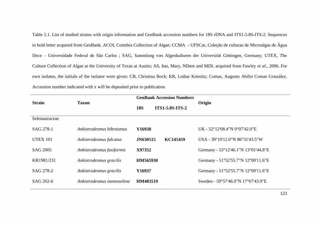
123
Table 2.1. List of studied strains with origin information and GenBank accession numbers for 18S rDNA and ITS1-5.8S-ITS-2. Sequences
in bold letter acquired from GenBank. ACOI, Coimbra Collection of Algae; CCMA - UFSCar, Coleção de culturas de Microalgas de Água
Doce – Universidade Federal de São Carlos ; SAG, Sammlung von Algenkulturen der Universität Göttingen, Germany; UTEX, The
Culture Collection of Algae at the University of Texas at Austin; AS, Itas, Mary, NDem and MDL acquired from Fawley et al., 2006. For
own isolates, the initials of the isolator were given: CB, Christina Bock; KR, Lothar Krienitz; Comas, Augusto Abilio Comas González.
Accession number indicated with x will be deposited prior to publication.
Strain Taxon
GenBank Accession Numbers
18S ITS1-5.8S-ITS-2
Origin
Selenastraceae
SAG 278-1 Ankistrodesmus bibraianus Y16938 UK - 52°12'08.4"N 0°07'42.0"E
UTEX 101 Ankistrodesmus falcatus JN630515 KC145459 USA - 39°10'12.0"N 86°31'43.5"W
SAG 2005 Ankistrodesmus fusiformis X97352 Germany - 53°12'46.1"N 13°01'44.8"E
KR1981/231 Ankistrodesmus gracilis HM565930 Germany - 51°52'55.7"N 12°00'11.6"E
SAG 278-2 Ankistrodesmus gracilis Y16937 Germany - 51°52'55.7"N 12°00'11.6"E
SAG 202-6 Ankistrodesmus nannoselene HM483519 Sweden - 59°57'46.0"N 17°07'43.9"E
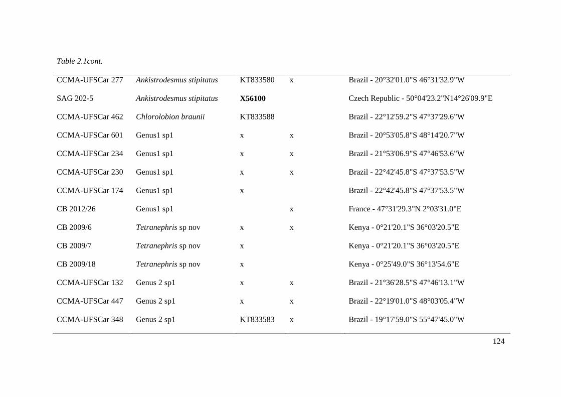
124
Table 2.1cont.
CCMA-UFSCar 277 Ankistrodesmus stipitatus KT833580 x Brazil - 20°32'01.0"S 46°31'32.9"W
SAG 202-5 Ankistrodesmus stipitatus X56100 Czech Republic - 50°04'23.2"N14°26'09.9"E
CCMA-UFSCar 462 Chlorolobion braunii KT833588 Brazil - 22°12'59.2"S 47°37'29.6"W
CCMA-UFSCar 601 Genus1 sp1 x x Brazil - 20°53'05.8"S 48°14'20.7"W
CCMA-UFSCar 234 Genus1 sp1 x x Brazil - 21°53'06.9"S 47°46'53.6"W
CCMA-UFSCar 230 Genus1 sp1 x x Brazil - 22°42'45.8"S 47°37'53.5"W
CCMA-UFSCar 174 Genus1 sp1 x Brazil - 22°42'45.8"S 47°37'53.5"W
CB 2012/26 Genus1 sp1 x France - 47°31'29.3"N 2°03'31.0"E
CB 2009/6 Tetranephris sp nov x x Kenya - 0°21'20.1"S 36°03'20.5"E
CB 2009/7 Tetranephris sp nov x Kenya - 0°21'20.1"S 36°03'20.5"E
CB 2009/18 Tetranephris sp nov x Kenya - 0°25'49.0"S 36°13'54.6"E
CCMA-UFSCar 132 Genus 2 sp1 x x Brazil - 21°36'28.5"S 47°46'13.1"W
CCMA-UFSCar 447 Genus 2 sp1 x x Brazil - 22°19'01.0"S 48°03'05.4"W
CCMA-UFSCar 348 Genus 2 sp1 KT833583 x Brazil - 19°17'59.0"S 55°47'45.0"W

125
Table 2.1. cont.
CB 2012/21 Genus 2 sp2 x Slovakia - 48°51'48.0"N 20°22'48.8"E
CB 2012/22 Genus 2 sp2 x Slovakia - 48°51'48.0"N 20°22'48.8"E
KR 1979/222 Genus 2 sp2 x Germany - 51°38'57.7"N 12°01'46.3"E
SAG 2004 Kirchneriella aperta AJ271859 KC145464 Germany - 53°08'51.7"N 13°01'41.9"E
ACOI 287 Kirchneriella dianae HM483512 Portugal - 40°26'29.0"N 8°45'20.5"W
CCMA-UFSCar 123 Kirchneriella lunaris x x Brazil - 21°36'28.5"S 47°46'13.1"W
CCMA-UFSCar 87 Kirchneriella lunaris x x Brazil - 21°36'28.5"S 47°46'13.1"W
CCMA-UFSCar 443 Kirchneriella lunaris x x Brazil - 22°19'01.0"S 48°03'05.4"W
CCMA-UFSCar 345 Kirchneriella obesa x x Brazil - 19°17'59.0"S 55°47'45.0"W
CB 2012/49 Kirchneriella obesa x Sweden - 55°52'01.4"N 13°33'21.5"E
ACOI 3125 Kirchneriella obesa HM483513 Portugal - 40°12'07.5"N 8°24'53.9"W
CCMA-UFSCar 346 Kirchneriella pseudoaperta KT833582 x Brazil - 19°17'59.0"S 55°47'45.0"W
CCMA-UFSCar 482 Kirchneriella pseudoaperta KT833592 x Brazil - 22°33'09.2"S 48°57'47.5"W
CCMA-UFSCar 5 Messastrum gracile KT833577 x Brazil - 22°12'20.6"S 47°52'37.6"W

126
Table 2.1. cont.
CCMA-UFSCar 622 Messastrum gracile KT833593 x Brazil - 23°02'28.8"S 48°03'15.9"W
Itas 9/21 14-6w Monoraphidium sp. AY846379 USA - 47°12'57.9"N 95°12'23.2"W
CCMA-UFSCar 364 Monoraphidium sp. x x Brazil - 19°01'27.0"S 55°55'28.0"W
AS6-3 Monoraphidium contortum AY846382 USA - 47º17‟26.8”N 98º50‟07.0”W
AS7-3 Monoraphidium convolutum AY846377 USA - 47º17‟26.8”N 98º50‟07.0”W
KR 1981/262 Monoraphidium convolutum HM565926 Germany - 51°49'37.8"N 11°59'41.7"E
SAG 202-7e Monoraphidium dybowskii Y16939 France - 43°06'26.3"N 0°05'10.4"W
CB 2009/27 Monoraphidium dybowskii HM483515 x Kenya - 0°22'29.9"S 35°55'34.4"E
AS3-5 Monoraphidium minutum AY846380 USA - 47º17‟26.8”N 98º50‟07.0”W
MDL 1/12-5 Monoraphidium pusillum AY846376 USA - 46°56'48.0"N 99°42'19.0"W
SAG 48.87 Monoraphidium neglectum AJ300526 Germany - 51°44'43.9"N 11°58'48.6"E
Mary 9/21 T-5w Monoraphidium saxatile AY846384 USA - 45°49'39.6"N 95°28'24.6"W
NDem 9/21 T-9d Monoraphidium saxatile AY846385 USA - 47°11'40.6"N 95°09'51.4"W
SAG 49.87 Monoraphidium terrestre Y17817 x Germany - 51°47'22.9"N 12°03'53.6"E
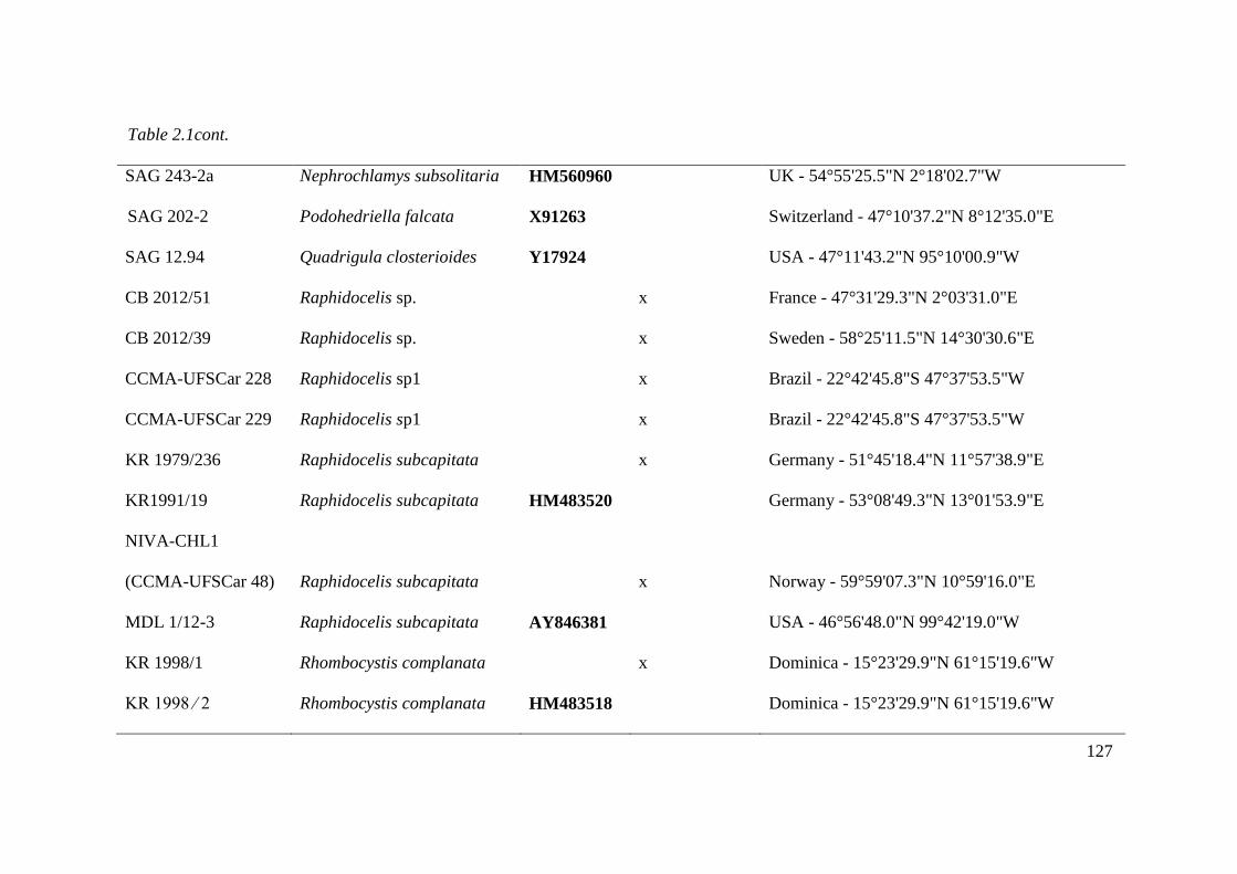
127
Table 2.1cont. UK - 54°55'25.5"N 2°18'02.7"W
SAG 243-2a Nephrochlamys subsolitaria HM560960 UK - 54°55'25.5"N 2°18'02.7"W
SAG 202-2 Podohedriella falcata X91263 Switzerland - 47°10'37.2"N 8°12'35.0"E
SAG 12.94 Quadrigula closterioides Y17924 USA - 47°11'43.2"N 95°10'00.9"W
CB 2012/51 Raphidocelis sp. x France - 47°31'29.3"N 2°03'31.0"E
CB 2012/39 Raphidocelis sp. x Sweden - 58°25'11.5"N 14°30'30.6"E
CCMA-UFSCar 228 Raphidocelis sp1 x Brazil - 22°42'45.8"S 47°37'53.5"W
CCMA-UFSCar 229 Raphidocelis sp1 x Brazil - 22°42'45.8"S 47°37'53.5"W
KR 1979/236 Raphidocelis subcapitata x Germany - 51°45'18.4"N 11°57'38.9"E
KR1991/19 Raphidocelis subcapitata HM483520 Germany - 53°08'49.3"N 13°01'53.9"E
NIVA-CHL1
(CCMA-UFSCar 48) Raphidocelis subcapitata x Norway - 59°59'07.3"N 10°59'16.0"E
MDL 1/12-3 Raphidocelis subcapitata AY846381 USA - 46°56'48.0"N 99°42'19.0"W
KR 1998/1 Rhombocystis complanata x Dominica - 15°23'29.9"N 61°15'19.6"W
KR 1998 ⁄ 2 Rhombocystis complanata HM483518 Dominica - 15°23'29.9"N 61°15'19.6"W

128
Table 2.1. cont.
CCMA-UFSCar 125 Selenastrum bibraianum KT833578 x Brazil - 21°36'28.5"S 47°46'13.1"W
NIVA-CHL1 Raphidocelis subcapitata AF169628 Norway - 59°59'07.3"N 10°59'16.0"E
Comas 1991/6 Tetranephris brasiliense HM565929 Cuba - 22°17'05.4"N 80°33'32.2"W
KR 1989/26 Tetranephris brasiliense HM565927 Germany - 53°30'28.8"N 13°12'48.4"E
Bracteacoccaceae
SAG 23.69 Bracteacoccus cohaerens HQ246325 South Africa – 30°07'11.2"S 17°35'26.8"E
Hidrodyctiaceae
SAG 28.83 Pediastrum duplex AY780662 Germany - 49°17'21.8"N 7°50'59.5"E
Scenedesmaceae
SAG 276-3a Acutodesmus obliquus X56103 Not found
UTEX 76 Scenedesmus communis X739941 USA - 42°22'23.9"N 71°06'29.2"W

129
Table 2.2. Morphological characteristics of Kirchneriella-like strains used in this study. Aa: autospore arrangement; P: pyrenoid; C:
colony; M: mucilage.
Taxon Cell shape
Cell size (long
x large µm)
Aa P C M
Genus 1 sp. nov. 1 Fusiform regularly curved to bent to one side., with narrowed or obtuse ends. 6-20 x 3-7 Parallel + + +
Genus 2 sp. nov. 1
Lunate, curved or twisted cells, slightly bent to one side, with narrowed or
obtuse ends.
6-15 x 2,7-5 Parallel - + -
Genus 2 sp. nov. 2 Bean-shaped, crescent-shaped or slightly arcuate, ends gradually obtuse. 7-11 x 1,2-3 Parallel - + +
Kirchneriella lunaris Half-moon or horseshoe-shaped. 4-15 x 1,2-8 Parallel + + +
Kirchneriella obesa Half-moon or horseshoe-shaped. 6-16 x 2-9,5 Parallel + + +
Kirchneriella pseudoaperta Half-moon or horseshoe-shaped. 4-8 x 2-4 Parallel + + +
Raphidocelis sp. nov. 1 Narrow and long cell, arcuate to spirally curved with rounded ends. 15-20 x 1-2 Parallel - + +
Tetranephris sp. nov. 1 Bean-shaped to slightly arcuate, narrowed or obtuse ends. 3-8 x 1-4 Parallel - + +

130
Table 2.3.Revision of taxa included in Kirchneriella, Raphidocelis, Gen 1 gen nov, Gen 2 gen nov and Tetranephris. Names marked with
asterisks represents organisms with 18S rDNA sequences available on NCBI and used on previous phylogenetic studies.
Taxon
Kirchneriella
Kirchneriella pseudoaperta Komárek 1983: 126, 176, pl. 21: 50
*Kirchneriella aperta Teiling 1912: 276, fig. 9
Kirchneriella brasiliana Silva, Sant'Anna, Tucci & Comas2013, fig. 1
Kirchneriella cornuta Korshikov 1953: 319, fig. 293a-d
*Kirchneriella dianae (Bohlin) Comas Gonzalez 1980: 4
Kirchneriella dianae var. major (Korshikov) Comas (= Kirchneriella lunaris var. acuta Playfair?, cf. Komarek & Fott, 1983)
Kirchneriella hindakiana P.Marvan, J.Komárek & A.Comas 1984: 390
Kirchneriella incurvata J.H.Belcher & Swale 1962: 125, fig. 1: Q-S
Kirchneriella irregularis (G.M.Smith) Korshikov 1953: 319, fig. 291a, b
Kirchneriella irregularis var. spiralis Korshikov 1953: 319, fig. 292
Kirchneriella lagerheimii Teiling; Unverified - Missing reference
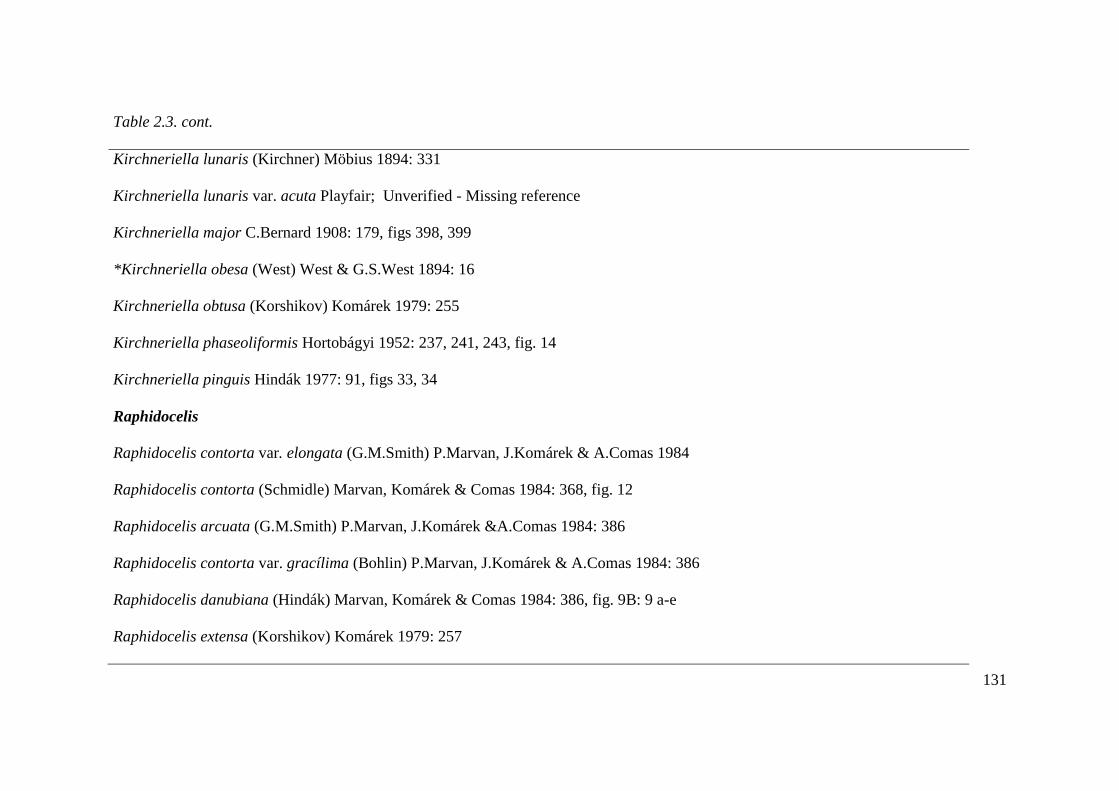
131
Table 2.3. cont.
Kirchneriella lunaris (Kirchner) Möbius 1894: 331
Kirchneriella lunaris var. acuta Playfair; Unverified - Missing reference
Kirchneriella major C.Bernard 1908: 179, figs 398, 399
*Kirchneriella obesa (West) West & G.S.West 1894: 16
Kirchneriella obtusa (Korshikov) Komárek 1979: 255
Kirchneriella phaseoliformis Hortobágyi 1952: 237, 241, 243, fig. 14
Kirchneriella pinguis Hindák 1977: 91, figs 33, 34
Raphidocelis
Raphidocelis contorta var. elongata (G.M.Smith) P.Marvan, J.Komárek & A.Comas 1984
Raphidocelis contorta (Schmidle) Marvan, Komárek & Comas 1984: 368, fig. 12
Raphidocelis arcuata (G.M.Smith) P.Marvan, J.Komárek &A.Comas 1984: 386
Raphidocelis contorta var. gracílima (Bohlin) P.Marvan, J.Komárek & A.Comas 1984: 386
Raphidocelis danubiana (Hindák) Marvan, Komárek & Comas 1984: 386, fig. 9B: 9 a-e
Raphidocelis extensa (Korshikov) Komárek 1979: 257
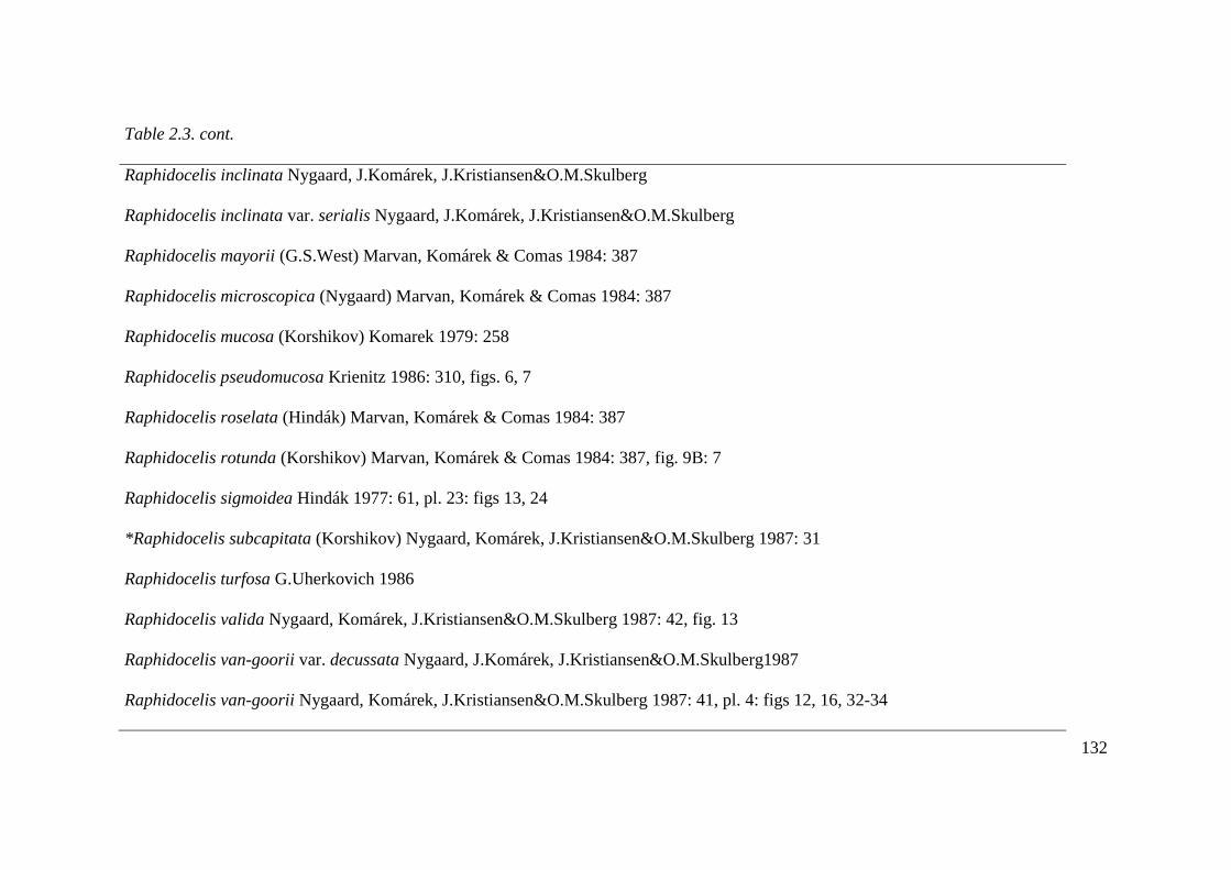
132
Table 2.3. cont.
Raphidocelis inclinata Nygaard, J.Komárek, J.Kristiansen&O.M.Skulberg
Raphidocelis inclinata var. serialis Nygaard, J.Komárek, J.Kristiansen&O.M.Skulberg
Raphidocelis mayorii (G.S.West) Marvan, Komárek & Comas 1984: 387
Raphidocelis microscopica (Nygaard) Marvan, Komárek & Comas 1984: 387
Raphidocelis mucosa (Korshikov) Komarek 1979: 258
Raphidocelis pseudomucosa Krienitz 1986: 310, figs. 6, 7
Raphidocelis roselata (Hindák) Marvan, Komárek & Comas 1984: 387
Raphidocelis rotunda (Korshikov) Marvan, Komárek & Comas 1984: 387, fig. 9B: 7
Raphidocelis sigmoidea Hindák 1977: 61, pl. 23: figs 13, 24
*Raphidocelis subcapitata (Korshikov) Nygaard, Komárek, J.Kristiansen&O.M.Skulberg 1987: 31
Raphidocelis turfosa G.Uherkovich 1986
Raphidocelis valida Nygaard, Komárek, J.Kristiansen&O.M.Skulberg 1987: 42, fig. 13
Raphidocelis van-goorii var. decussata Nygaard, J.Komárek, J.Kristiansen&O.M.Skulberg1987
Raphidocelis van-goorii Nygaard, Komárek, J.Kristiansen&O.M.Skulberg 1987: 41, pl. 4: figs 12, 16, 32-34

133
Table 2.3. cont.
Raphidocelis sp nov T. S. Garcia & Sant‟Anna
Genus 1gen nov
Genus 1 sp nov 2 T.S.Garcia& Sant'Anna
Genus 2gen nov
Genus 2 sp nov 1 T.S.Garcia & Sant'Anna
Genus 2 sp nov 2 T.S.Garcia & Sant'Anna
Tetranephris
*Tetranephris brasiliense C.R.Leite & C.E.M.Bicudo 1977: 231-233, figs 1-8
Tetranephris europaea (Hindák) Komárek 1979: 260
Tetranephris sp. nov. T.S.Garcia & Sant'Anna

134
Fig. 2.1-2.5. Drawings of light microscopical characters. 1. Tetranephris sp. nov. (CB
2009/6). Note the cell wall remnant (arrowhead); 2. Gen. nov. 1 sp1 (CCMA-UFSCar
230); 3. Raphidocelis sp. nov. (CCMA-UFSCar 229). Note the cell wall remnant
(arrowhead); 4. Gen. nov. 2 sp 1. (CCMA-UFSCar 342); 5. Gen. nov. 2 sp 2. (KR
1979/222). Note the cell wall remnant (arrowhead). Scale bar 10 μm.
Fig. 2.6-2.8. Drawings of light microscopical characters. 6. Kirchneriella pseudoaperta
(CCMA-UFSCar 346). 7. Kirchneriella obesa (CCMA-UFSCar 345). 8. Kirchneriella
lunaris (CCMA-UFSCar 87). Scale bar 10 μm.
Figure 2.9: Maximum–likelihood (ML) phylogenetic tree inferred from ITS1-5.8S-ITS2
gene sequences of some members of Selenastraceae. Support values correspond to
Bayesian PP (Posterior Probability), ML BP (Bootstrap), MP (Maximum Parsimony)
BP, NJ (Neighbor-Joining)BP. Hyphens correspond to values <50% for BP and <0.95
for PP. Scale represents the expected number of substitutions per site. Strain numbers
used as mentioned in Table 2.1.
Figure 2.10: Maximum–likelihood (ML) phylogenetic tree inferred from 18S rDNA
gene sequences of some members of Selenastraceae. Support values correspond to
Bayesian PP (Posterior Probability), ML BP (Bootstrap), MP (Maximum Parsimony)
BP, NJ (Neighbor-Joining)BP. Hyphens correspond to values <50% for BP and <0.95
for PP. Scale represents the expected number of substitutions per site. Strain numbers
used as mentioned in Table 2.1.

135
Fig. 2.1-2.5
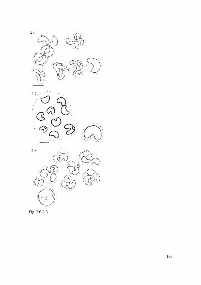
136
Fig. 2.6-2.8

137
Fig. 2.9

138
Fig. 2.10

139
Discussão Geral
A partir da grande quantidade de dados moleculares e morfológicos obtidos no
presente estudo, tanto de cepas tropicais como de ambientes temperados, pode-se
observar que os gêneros Monoraphidium, Kirchneriella e Ankistrodesmus
demonstraram ser polifiléticos, também sugerido em trabalhos anteriores (Krienitz et al.
2001, Krienitz et al. 2011).
A inferência filogenética baseada em 18S rDNA e rbcL reafirmam a separação
de Selenastrum bibraianum e Selenastrum gracile (Syn. Messastrum gracile) em dois
gêneros distintos, levando ao surgimento de Messastrum e a descrição de um novo
gênero, Curvastrum, morfologicamente similar à Selenastrum.
O emprego do 18S rDNA e a análise multigene com ITS1-5.8S-ITS2 mostrou
que, para a identificação de espécies semelhantes à Kirchneriella, tamanho, forma,
curvatura ou torção celular são características sutis e relevantes, uma vez que estas
características foram cruciais para a identificação das espécies. Ainda, as análises
filogenéticas revelaram que o morfotipo associado à Kirchnriella ocultava quatro
linhagens filogenéticas, levando à descrição de uma espécie nova de Raphidocelis, uma
espécie nova de Tetranephris e dois novos gêneros.
Estudos filogenéticos em Selenastraceae, baseados em 18S rDNA e ITS1-5.8S-
ITS2, revelaram que um tipo morfológico pode representar diferentes filotipos, como
Selenastrum e Kirchneriella, sugerindo que a diversidade da família tem sido
subestimada consideravelmente quando avaliada morfologicamente (Fawley et al.
2006).

140
O conceito de gêneros em algas verdes cocóides, bastante debatido em trabalhos
recentes sobre sistemática de algas, têm gerado discussão (Luo et al. 2009) e demonstra
ser uma tendência (Luo et al. 2010). Vários gêneros têm passado recentemente por
reformulação após estudos moleculares. Atualmente, alguns gêneros com morfologia
bem conhecidas foram divididos em gêneros muitos menores, como proposto para
Scenedesmus Meyen (An et al. 1999, Krienitz et al. 2003) e Pediastrum (Buchheim et
al. 2005), em abordagens filogenéticas com o 18S rDNA.
Embora morfologicamente semelhantes, estudos moleculares evidenciaram que
Raphidocelis representa uma linhagem molecular não relacionada com Kirchneriella,
corroborando a separação morfológica destes gêneros (Fawley et al. 2006, Krienitz et
al. 2001, Krienitz et al. 2011). Morfotipos semelhantes podem esconder alta diversidade
genética dentro dos gêneros Kirchneriella, Tetranephris e Raphidocelis.
Achados semelhantes foram obtidos para Dictyosphaerium, onde os morfotipos
formaram linhagens distintas dentro de Chlorella e Parachlorella, apresentando
evolução independente e confirmando o polifiletismo do morfotipo Dictyosphaerium
dentro de Chlorellaceae (Luo et al. 2010).
Além do fenótipo, discussões sobre conceitos de espécies devem considerar a
variação genética (Hilt 2006). A descrição de espécies bem estabelecidas, com
população fenotípica conhecida, deve ser investigada de acordo com características
moleculares, genéticas, fisiológicas e bioquímicas de todas essas cepas cultivadas que
seguem a descrição do tipo de origem (Wood & Leatham 1992), levando à resolução da
posição taxonômica das variantes morfológicas do tipo.
O valor taxonômico de caracteres diacríticos utilizados em Selenastraceae foi
discutido com base na morfologia e na ultra-estrutura (Krienitz et al. 2001)
141
demonstrando que o conceito de "gêneros pequenos" (Komárek & Fott 1983), incluindo
apenas algumas espécies, para a Selenastraceae não era apropriado, uma vez que a
existência de muitas formas de transição, e muitas características diacríticas não são
confiáveis (Fawley et al. 2006). Portanto, a ideia gêneros "pequeno" foi considerada
incerta para Selenastraceae (Krienitz et al. 2001). Entretanto, em um trabalho posterior
Krienitz et al. (2011) reempregou esse conceito ao estudar os morfotipos associados à
alga teste Selenastrum capricornutum, cujo morfotipo está associado a cerca de 10
linhagens evolutivas diferentes.
Nossos resultados sugeriram que o conceito de gêneros pequenos poderia ser
aplicado em Monoraphidium, Kirchneriella, Selenastrum e Messastrum.
A observação de um número substancial de colônias e organismos solitários (10-
50 indivíduos) e observação de todo o ciclo de vida, com especial ênfase para a
liberação de autósporos e a formação de colônias, foi proposta para a identificaçao
acurada dos taxa compreendidos em Selenastraceae (Garcia et al. no prelo)
Algumas descrições originais das espécies foram publicadas faltando
informações quanto à morfologia (incluindo holotipos ou iconotipos) não fornecendo
todas as informações necessárias para a identificação de muitos taxa.
A combinação das análises moleculares e avaliação morfológica em diferentes
fases de crescimento das cepas demonstrou ser importante para a sistemática de
Selenastraceae (Garcia et al. no prelo).
Somado às informações do ciclo de vida em que estão faltando informações, a
ausência de culturas de espécies tipo de centenas de taxa pertencentes a essa família
cosmopolita associada à dificuldade de amplificação de alguns marcadores moleculares,

142
como o rbcL, limitam os estudos taxonômicos. Desta forma, faz-se necessário mais
esforço para o isolamento de linhagens e posterior estudo taxonomico.
Com o passar dos anos houve uma grande flutuação nas circunscrições dos
gêneros e na mudança no ponto de vista dos taxonomistas no que se refere à delimitação
morfológica das espécies, refletindo na descrição de novos gêneros morfológicos, tais
como Raphidocelis (Krienitz et al. 2011) e Pseudokirchneriella.
Alguns critérios morfológicos podem representar adaptações fenotípicas às
condições ambientais e não refletir efetivamente as relações filogenéticas, como a
presença e ausencia de pirenóides (Krienitz et al. 2004;. Luo et al. 2006).
O emprego dos marcadores SSU, ITS-1 e ITS-2 diferenciaram seis gêneros
dentro do morfotipo de Chlorella Beyerinck [Beijerinck], revelando que vários critérios
morfológicos utilizados na identificação (mucilagem e fios de conexão entre as células,
forma de vida colonial ou solitária e formação de cerdas) foram considerados
características fenotípicas e representaram respostas adaptativas a fatores ambientais,
tais como a pressão por grazing, endossimbiose ou estratégias para adaptação ao
ambiente terrestre (Luo et al. 2010).
O modo de reprodução das algas cocóides não permite o emprego do conceito de
espécies de Mayr (1942) limitando os limonologistas à adoção do conceito de espécie
morfológica, usando características diacríticas baseadas em morfologia como uma
maneira de lidar com essa limitação (Krienitz & Bock 2012).
Estima-se que existam cerca de 14.900 espécies de fitoplâncton de água doce
(Bourrelly 1990). Afirma-se que a riqueza de espécies em lagos temperados de água
doce é significativamente maior do que nos lagos tropicais (Lewis Jr 1978). Tal

143
afrmação dá-se como consequência da pouca exploração e conhecimento sobre a
diversidade de microalgas verdes e a filogenia de algas nos trópicos.
Além disso, pouco se sabe sobre as distribuições geográficas de algas verdes
cocóides apesar de sua importância nos ecossistema de água doce e do avanço na
taxonomia do grupo (Norton et al. 1996;. Padisák et al. 2015).
Estudos filogenéticos têm papel fundamental para fornecer sequências de
referência para aplicações práticas de alguns estudos científicos, como abordagens
metabarcoding (Ji et al. 2013) e seqüenciamentos de nova geração, que carecem de
referências para espécies de fitoplâncton (Eiler et al. 2012;. Pawlowski et al. 2012).
Referências bibliográficas
An, S., T. Friedl & E. Hegewald, (1999). Phylogenetic Relationships of Scenedesmus
and Scenedesmus-like Coccoid Green Algae as Inferred from ITS-2 rDNA
Sequence Comparisons. Plant Biology 1(4):418-428.
Bourrelly, P., (1990). Les Algues d‟eau douce. Initation a la Systématique, I: Les
Algues Vertes. 572pp. N Boubée & Cie Paris.
Buchheim, M., J. Buchheim, T. Carlson, A. Braband, D. Hepperle, L. Krienitz, M. Wolf
& E. Hegewald, (2005). Phylogeny of the Hydrodictyaceae (Chlorophyceae):
inferences from rDNA data1. Journal of Phycology 41(5):1039-1054.
Eiler, A., S. Drakare, S. Bertilsson, J. Pernthaler, S. Peura, C. Rofner, K. Simek, Y.
Yang, P. Znachor, and E. S. Lindström. 2013. Unveiling distribution patterns of
freshwater phytoplankton by a next generation sequencing based approach.
PLoS ONE 8:e53516.

144
Fawley, M. W., M. L. Dean, S. K. Dimmer & K. P. Fawley, (2006). Evaluating the
morphospecies concept in the Selenastraceae (Chlorophyceae, Chlorophyta)
Journal of Phycology 42(1):142-154.
Hilt, S., (2006). Allelopathic inhibition of epiphytes by submerged macrophytes.
Aquatic Botany 85(3):252-256 doi:DOI 10.1016/j.aquabot.2006.05.004.
Ji, Y., L. Ashton, S. M. Pedley, D. P. Edwards, Y. Tang, A. Nakamura, R. Kitching, P.
M. Dolman, P. Woodcock & F. A. Edwards, (2013). Reliable, verifiable and
efficient monitoring of biodiversity via metabarcoding. Ecology letters
16(10):1245-1257.
Komárek, J. & B. Fott, (1983). Das Phytoplankton des Sübwassers. Systematik und
Biologie. 7. Teil, 1. Hälfte. Chlorophyceae (Grünalgen) Ordnung:
Chroococcales. E. Schweizerbart‟sche Verlagsbuchhandlung (Nägele u.
Obemiller), Stuttgart. 1043p.
Krienitz, L. & C. Bock, (2012). Present state of the systematics of planktonic coccoid
green algae of inland waters. Hydrobiologia 698(1):295-326.
Krienitz, L., C. Bock, H. Nozaki & M. Wolf, (2011). SSU rRNA gene phylogeny of
morphospecies affiliated to the bioassay alga “Selenastrum capricornutum”
recovered the polyphyletic origin of crescent-shaped Chlorophyta. Journal of
Phycology 47(4):880-893.
Krienitz, L., E. Hegewald, D. Hepperle & M. Wolf, (2003). Systematics of coccoid
green algae: morphology versus 18S rRNA gene phylogeny. Biologia 58:437-
446.

145
Krienitz, L., I. Ustinova, T. Friedl & V. A. Huss, (2001). Traditional generic concepts
versus 18S rRNA gene phylogeny in the green algal family Selenastraceae
(Chlorophyceae, Chlorophyta). Journal of Phycology 37(5):852-865.
Lewis Jr, W. M., (1978). A compositional, phytogeographical and elementary structural
analysis of the phytoplankton in a tropical lake: Lake Lanao, Philippines. The
Journal of Ecology:213-226.
Luo, W., T. Pröschold, C. Bock & L. Krienitz, (2010). Generic concept in Chlorella‐
related coccoid green algae (Chlorophyta, Trebouxiophyceae). Plant Biology
12(3):545-553.
Mayr, E., (1942). Systematics and the origin of species, from the viewpoint of a
zoologist. Harvard University Press.
Norton, T. A., M. Melkonian & R. A. Andersen, (1996). Algal biodiversity. Phycologia
35(4):308-326.
Padisák, J., G. Vasas & G. Borics, (2015). Phycogeography of freshwater
phytoplankton: traditional knowledge and new molecular tools.
Hydrobiologia:1-25.
Pawlowski, J., S. Audic, S. Adl, D. Bass, L. Belbahri, C. Berney, S. S. Bowser, I.
Cepicka, J. Decelle & M. Dunthorn, (2012). CBOL protist working group:
barcoding eukaryotic richness beyond the animal, plant, and fungal kingdoms.
PLoS biology 10(11):e1001419.
Wood, A. M. & T. Leatham, (1992). The species concept in phytoplankton ecology.
Journal of Phycology 28(6):723-729.
Garcia, T. S., Bock C., Sant‟Anna C. L., Bagatini I. L., Wodniok S., Vieira A. A. H.,
2016. Selenastraceae (Sphaeropleales, Chlorophyceae): rbcL, 18S rDNA and

146
ITS-2 secondary structure enlightens traditional taxonomy, with description of
two new genera, Messastrum gen. nov. and Curvastrum gen. nov. Fottea, in
press.
Krienitz, L., E. H. Hegewald, D. Hepperle, V. A. Huss, T. Rohr & M. Wolf, 2004.
Phylogenetic relationship of Chlorella and Parachlorella gen. nov.
(Chlorophyta, Trebouxiophyceae). Phycologia 43(5):529-542.
Luo, W., S. Pflugmacher, T. Pröschold, N. Walz & L. Krienitz, 2006. Genotype versus
phenotype variability in Chlorella and Micractinium (Chlorophyta,
Trebouxiophyceae). Protist 157(3):315-333.
147
Conclusões
1. O morfotipo associado à Selenastrum revelou três linhagens evolutivas distintas,
conduzindo à separação de Selenastrum bibraianum e Selenastrum gracile (Syn.
Messastrum gracile) em dois gêneros, levando ao surgimento de Messastrum e à
descrição de um novo gênero, Curvastrum, morfologicamente similar à Selenastrum.
2. Embora morfologicamente semelhantes Raphidocelis representa uma linhagem
molecular não relacionada com Kirchneriella;
3. O morfotipo associado à Kirchneriella revelou quatro linhagens evolutivas distintas,
conduzindo a descrição de dois novos gêneros, uma nova espécie de Raphidocelis e uma
nova espécie de Tetranephris;
4. A análise molecular demonstrou que os gêneros Kirchneriella, Selenastrum e
Monoraphidium possuem origem polifilética;
5. Embora as características morfologicas tenham sido avaliadas e consideradas
importantes para a diferenciação dos taxa, a filogenia baseada em 18S rDNA, rbcL e
ITS1-5.8S-ITS2 foram cruciais para subsidiar as decisões taxonômicas neste grupo de
algas verdes cocóides;
148
6. As caracteristicas diacríticas utilizadas em Selenastraceae se mostraram relevantes
para a separação dos taxa dessa família. Deve-se, entretanto, utilizá-los em conjunto e
não individualmente;
7. A combinação das análises moleculares e avaliação morfológica em diferentes fases
de crescimento das cepas foi importante para a sistemática de Selenastraceae;
8. O uso de cepas provenientes de ambientes tropicais e temperados, provenientes de
três continentes distintos, comprovou o cosmopolitismo de Selenastraceae.