fatores preditores para o aumento do valgismo dinâmico do ... · músculos da articulação do...
TRANSCRIPT

Natalia Franco Netto Bittencourt
Fatores preditores para o aumento do valgismo
dinâmico do joelho em atletas
Belo Horizonte
Universidade Federal de Minas Gerais
2010

Natalia Franco Netto Bittencourt
Fatores preditores para o aumento do valgismo
dinâmico do joelho em atletas
Dissertação apresentada ao Programa de Pós-Graduação do curso de Ciências da Reabilitação da Universidade Federal de Minas Gerais, como requisito a obtenção de título de Mestre em Ciências da Reabilitação.
Área de Concentração: Desempenho motor e Funcional Humano
Orientador: Prof.Dr. Sérgio Teixeira da Fonseca
Co-Orientadora: Profa. Dra. Juliana M. Ocarino
Belo Horizonte
2010

PREFÁCIO
De acordo com as normas estabelecidas pelo Colegiado do Programa de Pós-
Graduação em Ciências da Reabilitação da UFMG, a estrutura deste trabalho é
composta por três partes. A primeira parte é composta por uma introdução,
com o objetivo de apresentar a revisão bibliográfica sobre o tema, a
problematização e a justificativa do estudo, bem como pela descrição
detalhada de toda a metodologia utilizada. A segunda parte é composta por um
artigo em que os resultados e a discussão são apresentados, redigidos de
acordo com as normas adotadas pela American Medical Association Manual of
Style, 9ª edição, preconizadas pelo periódico para o qual este trabalho será
posteriormente enviado para publicação (Journal of Orthopaedic & Sports
Physical Therapy -JOSPT). Por fim, na terceira parte do trabalho, são
apresentadas as considerações finais relacionadas aos resultados
encontrados.

AGRADECIMENTOS
Aos meus queridos pais pelo incentivo incondicional. Pai, obrigada por não
medir esforços para que eu realizasse meus sonhos e por estar ao meu lado
nos momentos de dúvida e dificuldade. Mãe, você é um exemplo para mim,
uma grande mulher, mãe e profissional!!!
Á minha familia (Vovó Lila, Vovô, irmãos Gabriel e Pedro, Dinda, Lin e Tia
Ligia) pelo apoio e momentos de alegria, onde recarreguei minhas energias ao
longo dos 2 anos de mestrado.
Á minha amiga, irmã e parceira: Luciana de Michelis. Lu, esse projeto também
é seu! Obrigada pela cumplicidade, competência e persistência!!! Obrigada a
Táta também!!!
Á Giovana pelas coletas e ajuda durante o processo de revisão da dissertação.
Á Mayara, Livia, Thiago, Viviane,Vanessa, Luciana Signorini e demais colegas
do LAPREV pela dedicação as coletas e profissionalismo ao longo das
análises!
Á Deborah por acreditar e tornar realidade a avaliação fisioterapêutica pré-
temporada do Minas Tênis Clube. Á Izabel e a Gabriela por viabilizar através
do NICE- MTC toda a estrutura necessária para as coletas.
Á Juliana Ocarino pela eficiência e profissionalismo ao longo das análises e
revisão. Obrigada Ju!!!!
Ao Prof. Sergio Fonseca por guiar com sabedoria este longo processo e
desafio de levar a padronizacão científica para a prática da fisioterapia
esportiva. E também pelo aprendizado ao longo do mestrado!
Ao Prof. Anderson pelo apoio irrestrito do LAPREV a este projeto e
principalmente pela vinda do Prof. Hewett.
Ao Thales pelas sábias idéias ao longo do desenvolvimento da medida do pé.
As amigas Aline e Pati que mesmo longe do país sempre acreditaram em mim!!
Ao Hugo pela tranquilidade , paciência e amor ao logo do processo.
RESUMO
O aumento do valgismo dinâmico do joelho está relacionado com lesões dos
membros inferiores em atletas, como ruptura do ligamento cruzado anterior
(LCA), sindrome patelofemoral e tendinopatia patelar. A redução da força dos
músculos da articulação do quadril tem sido relacionada ao aumento da
excursão do joelho no plano frontal. Além disso, atletas que apresentam
pronação excessiva da subtalar realizam maior valgismo do joelho. Entretanto,
a interdependência da biomecânica dos segmentos distais e proximais da
cadeia cinética dos membros inferiores tem sido pouco investigada. Dessa
forma, o objetivo do presente estudo foi analisar como os fatores preditores
para a ocorrência do aumento do valgismo dinâmico do joelho se interagem
durante atividades funcionais. Para tanto, foram avaliados 227 atletas durante a
avaliação pré-temporada do Minas Tenis Clube. O valgismo dinâmico do joelho
(variável depende) foi operacionalizado como ângulo de projeção frontal do
joelho (APFJ). As variáveis independentes foram: torque isométrico abdutor do
quadril; ADM passiva de rotação interna do quadril e o alinhamento tibia-
antepé. A Árvore de Classificação e Regressão (CART), uma análise estatística
não –paramétrica e que captura a relação não- linear entre as varíaveis
independentes foi utilizada para desenvolver as regras de decisão para os
fatores preditores relacionados aos maiores ângulos de projeção frontal do
joelho. Os resultados deste estudo demonstraram que ,durante o
agachamento a ocorrência de maiores valores do APFJ foi resultado da
interação entre o torque abductor do quadril e a ADM passiva de RI do quadril.
Por outro lado, durante a aterrissagem o alinhamento tibia-antepé, juntamente

com o torque abdutor do quadril e a ADM passiva de RI do quadril foram os
fatores preditores para a ocorrência de maiores angulos de projeção frontal do
joelho. Além disso, com a utilização da CART foi possível identificar os pontos
de corte para cada preditor, facilitando o raciocínio clinico do fisioterapeuta
esportivo durante o planejamento de intervenções preventivas efetivas. Dessa
forma, a avaliação pré-temporada de atletas deve incluir a mensuração da
força dos abdutores do quadril, da ADM passiva de RI do quadril e do
alinhamento tibia-antepé.
ABSTRACT
Excessive dynamic knee valgus has been identified as a contributing factor to
many lower extremity injuries, such as anterior cruciate ligament rupture,
patellofemoral joint pain and patellar tendinopathy. Some factors related to the
hip joint, such as decreased hip abductor strength, have been associated to
increased knee valgus angle. In addition, athletes with excessive foot pronation
have increased frontal plane knee excursion. However, the biomechanical
interdependence of distal and proximal segments of the lower limb kinetic chain
has been poorly investigated. Thus, the purpose of this study was to analyze
how some typical predictors of increased dynamic knee valgus angle interact to
produce altered knee alignment during functional tasks. During preseason, 227
athletes belonging to a sport club were evaluated. The dynamic knee valgus
(dependent variable) was measured as the frontal plane projection angle of the
knee (FPPAK), in two different conditions: single leg squat and landing from
vertical jump. The independent variables were: isometric hip abductor torque,
passive range of motion (ROM) of hip internal rotation (IR) and shank-forefoot
alignment. Classification and Regression Trees (CART), a non-parametric
statistical analysis that incorporates nonlinear relationships between predictors,
was used to develop decision rules to predict the presence of excessive
FPPAK. The results demonstrated that during squat the occurrence of high
FPPAK was due to interaction between hip abductor torque and passive ROM
of hip IR. During landing, the shank-forefoot alignment together with abductor
torque and passive hip IR were the predictors of high FPPAK. In addition, the
CART model identified meaningful cut-off points that classified the sample into
the outcome categories of High and Low-FPPAK. These findings may guide

sports professionals to plan preventive programs and suggest that preseason
assessments must include measurements of hip abductor torque, passive ROM
of hip IR and shank-forefoot alignment.
SUMÁRIO
Capítulo1 – INTRODUÇÃO ..................................................................... 11
1.1 - Pressuposto do estudo ..................................................................... 16
Capítulo 2 - MATERIAIS E MÉTODOS .....................................................17
2.1 – Amostra ............................................................................................ 17
2.2 – Procedimentos ................................................................................. 18
2.3 - Redução dos dados .......................................................................... 22
2.4 - Análise estatística ............................................................................. 24
Capítulo 3 - REFERÊNCIAS BIBLIOGRÁFICAS ................................... 27
Capítulo 4 - “PREDICTING DINAMIC KNEE VALGUS IN ATHLETES USING CLASSIFICATION TREE APPROACH”....................31
Capítulo 5 - CONSIDERAÇÕES FINAIS ................................................. 61
ANEXO – Aprovação do Comitê de Ética................................................64

11
Capítulo 1- INTRODUÇÃO
A incidência de lesões nos membros inferiores (MMII), durante a prática de
esportes como voleibol, basquetebol e futebol é alta e as regiões mais
frequentemente acometidas são o joelho e tornozelo 1,2. Lesões como síndrome
patelofemoral (SPF), tendinopatia patelar e ruptura do ligamento cruzado anterior
(LCA) são as lesões com maior tempo de afastamento da prática esportiva e
necessitam maior tempo para recuperação em relação às lesões de tornozelo
(131%) e em relação às lesões de coluna, quadril e coxa (41%) 3. Além disso, um
maior gasto financeiro está envolvido no tratamento das lesões do joelho, sendo que
a lesão do LCA gera um custo estimado de aproximadamente U$17.000,00 por
atleta 4. A alta ocorrência de lesões nessa articulação parece estar relacionada com
as demandas sobre o sistema musculoesquelético produzidas pelo esporte. Por
exemplo, aproximadamente 63% das lesões no joelho ocorrem durante o movimento
de impulsão e aterrissagem do salto vertical no voleibol e 43% no basquetebol 1,5.
Dessa forma, é necessário um melhor entendimento dos mecanismos envolvidos
nestas lesões para que o tratamento e as intervenções preventivas sejam
desenvolvidas de maneira eficiente.
Um dos fatores que podem contribuir para as lesões na articulação do joelho
é a incapacidade do atleta em manter um bom alinhamento dinâmico entre os
segmentos corporais dos membros inferiores (MMII) nos planos frontal e transverso
durante a prática esportiva 6,7,8,9. Hewett et al 10 demonstraram que a presença de
valgismo (momento abdutor) dinâmico de joelho é um importante preditor para
ruptura do LCA em atletas10. Essa alteração de alinhamento dinâmico impõe forças
rotacionais e de cisalhamento na articulação do joelho, gerando aumento da

12
sobrecarga no LCA 10. A presença do valgismo durante os movimentos do membro
inferior pode, também, alterar dinamicamente o alinhamento da patela, o que pode
aumentar a sobrecarga em estruturas como os retináculos patelares, cartilagem
articular e coxim adiposo e predispor o desenvolvimento de dor anterior no joelho
(Síndrome Patelofemoral)11 . Além disso, o padrão de movimento incorreto e,
consequentemente, a alteração de alinhamento da patela podem promover um
aumento das forças de cisalhamento no tendão patelar, favorecendo o surgimento
de tendinopatias patelares12 . Portanto, alterações dinâmicas da articulação do
joelho associadas à demanda complexa envolvida em esportes como vôlei,
basquete e futebol parecem aumentar a demanda imposta sobre o sistema músculo-
esquelético do atleta, contribuindo para o desenvolvimento de vários tipos de lesões
e disfunções na articulação do joelho.
O joelho, por ser uma articulação intermediária na cadeia cinemática,
depende do comportamento mecânico adequado do quadril e do tornozelo para
atenuar e distribuir adequadamente as forças impostas ao sistema músculo-
esquelético durante as atividades esportivas13 . Várias alterações cinéticas e
cinemáticas nas articulações proximais e distais dos MMII podem predispor o atleta
a apresentar um aumento do valgismo dinâmico no joelho durante a realização de
suas atividades esportivas 14,15,16. Por exemplo, o valgismo dinâmico do joelho pode
ser resultado do aumento da rotação interna e da adução do fêmur e da pronação do
pé 16,17,18,19. Durante a adução do quadril, o fêmur roda internamente e o joelho é
colocado em posição de valgismo 20. Essas alterações dinâmicas do quadril podem
ocorrer devido a fraqueza dos músculos abdutores e rotadores externos do quadril,
principalmente glúteo médio e máximo 3,20,21. Em uma revisão sistemática, Prins e

13
Wurff 22 demonstraram que mulheres com SPF possuem menor força muscular
dos abdutores, rotadores externos e extensores de quadril no lado afetado quando
comparado com individuos controle22. Além disso, um estudo prospectivo
demonstrou que atletas não lesionados apresentaram maior força dos abdutores e
rotadores externos de quadril do que aqueles que apresentaram lesões nos
membros inferiores3. Finalmente, Geiser et al 23 induziram um protocolo de fadiga
específico para os abdutores de quadril em participantes fisicamente ativos e
concluiram que o joelho encontrava-se em uma posição mais aduzida no plano
frontal, aumentando o valgismo dinâmico em atividades funcionais23. Estes estudos
demonstram, portanto, que os movimentos de quadril e, mais especificamente, a
força dos músculos abdutores do quadril são fatores que parecem influenciar o grau
de valgismo de joelho apresentado por atletas durante as atividades esportivas.
O aumento da excursão do joelho no plano frontal, além de ser influenciado
pela força dos músculos abdutores de quadril, parece também estar associado a
amplitude de movimento (ADM) disponível de rotação do quadril 24.
Tradicionalmente, a ADM é avaliada com aplicação de um torque externo e assim
essa medida pode ser mais influenciada pela tolerância do indivíduo ao
alongamento25 . Tendo em vista essa questão, se a ADM de rotação for avaliada de
forma que ela represente a posição em que o torque produzido pelos pesos da
perna e pé se iguale ao torque de resistência passiva gerado pelo quadril , essa
medida seria mais informativa da rigidez dessa articulação ao movimento de
rotação. Essa rigidez representa a mudança do torque de resistência passiva,
fornecida pelos tecidos da articulação do quadril (músculos, ligamentos, capsula e
fáscia), ao longo do movimento articular13 . Sendo assim, durante algumas

14
atividades funcionais, mecanismos passivos de uma articulação podem contribuir
para impedir um movimento exagerado ou manter um alinhamento adequado. Além
disso, Gleim et al 26 verificaram que indivíduos com maior rigidez passiva de tronco e
MMII apresentaram menor gasto energético em relação aos indivíduos com baixos
níveis de rigidez passiva durante a marcha e corrida26 . Dessa forma, níveis
adequados de rigidez tecidual podem ser necessários para garantir uma
transferencia eficiente de energia entre os segmentos da cadeia cinética. Essa maior
eficiência em transferência de energia permitiria padrões de movimento adequados
com menor gasto energético e sobrecarga dos tecidos do sistema músculo-
esquelético 13.
Além da influência dos movimentos do quadril para a ocorrência de valgismo
no joelho, a rotação interna do membro inferior e a eversão do calcâneo também
têm sido apontados como fatores que contribuem para a cinemática inadequada do
joelho 14,16. O eixo da articulação subtalar por ser inclinado a 45º em relação a
horizontal permite o acoplamento do movimento do pé no plano frontal (eversão do
calcâneo) com o movimento no plano transverso (rotação interna do talus e membro
inferior), com uma correlação relatada de r2= 0.991 27. Essa forte associação sugere
que, alterações nos movimentos da subtalar podem levar a compensações na
articulação do joelho 27. Mc Clay et al 28 observaram que corredores classificados
como pronadores (overpronators) aterrissavam com maior valgismo de joelho no
momento do choque de calcanhar durante a corrida 28. Além disso, atletas que
utilizavam palmilhas com 5º de elevação da borda medial do pé apresentaram uma
redução do valgismo de joelho e eversão/pronação do calcâneo durante a
aterrissagem do salto 29. Essa pronação excessiva tem sido relacionada com a

15
presença do alinhamento em varo do antepé, o qual pode aumentar o torque eversor
no complexo tornozelo-pé após o contato do antepé com a superfície de suporte12,30.
Esses achados reforçam a necessidade de incluir a mensuração padronizada do
antepé na avaliação de atletas, pois a presença de varismo excessivo no antepé
pode gerar compensações biomecânicas de todo o membro inferior, influenciando a
ocorrência de lesões.
As evidências relacionadas aos fatores que influenciam a ocorrência de
valgismo na articulação do joelho de atletas durante atividades esportivas, indicam
que o alinhamento dinâmico adequado do joelho depende de características
anatômicas e biomecânicas dos segmentos distais e proximais da cadeia cinética
3,16,20,24. Neste contexto, tanto alterações biomecânicas do quadril e tronco como as
do pé podem influenciar a cinemática da articulação do joelho e, dependendo das
características da atividade executada, impor diferentes demandas sobre cada
componente da cadeia cinética. Sendo assim, as forças externas geradas durante
um salto vertical precisam ser absorvidas e dissipadas pelos segmentos do MMII em
um curto período de tempo9 . Por outro lado, durante o agachamento, a perna
apoiada no chão precisa apresentar grande estabilidade e resistência ao longo de
toda a amplitude do movimento20 .
Alguns estudos têm utilizado análises baseadas na contribuição linear e
individualizada de cada fator biomecânico para predizer o valgismo dinâmico do
joelho20,24 . Entretanto, o mau alinhamento estático e dinâmico desta articulação
possui característica multifatorial e depende da maneira como o sistema musculo-
esqueletico se adapta as possíveis interações entre força muscular, rigidez tecidual
e alinhamento articular dos segmentos proximais e distais da cadeia cinética13. Essa
16
interação entre fatores é essencial para o desenvolvimento de uma melhor
capacidade de estabilização dinâmica durante a demanda imposta por uma
determinada atividade. Sendo assim, para identificar as interações clínicas
relevantes que podem não ter sido capturadas por estudos prévios que focam na
investigação de efeitos estatíticos principais (main effects), o objetivo do presente
estudo foi identificar os fatores preditores para o aumento do ângulo de projeção
frontal do joelho (APFJ) durante o agachamento unipodal e aterrissagem do salto
vertical em atletas. A Árvore de Classificação e Regressão (Classification and
Regression Trees –CART) foi utilizada como um método estatístico que permite
capturar relações não lineares entre as variáveis preditoras e de desfecho.
1.1 Pressuposto do estudo
Este estudo pressupõe a contribuição dos segmentos distais e proximais dos
MMII para o aumento do APFJ e a interdependência da biomecânica do complexo
do tornozelo-pé e da articulação do quadril para a cinemática do joelho no plano
frontal durante atividades esportivas.

17
Capítulo 2- MATERIAIS E MÉTODO
2.1- Amostra
Este estudo observacional analítico foi realizado no Minas Tênis Clube e no
Laboratório de Prevenção e Reabilitação de Lesões Esportivas (LAPREV), que
pertence ao Centro de Excelência Esportiva - CENESP da Escola de Educação
Física, Fisioterapia e Terapia Ocupacional da Universidade Federal Minas Gerais
(UFMG).
Foram recrutados 227 atletas (78 do sexo feminino e 149 do sexo masculino)
participantes dos treinamentos e competições de basquete, futebol, voleibol e
ginástica olímpica. Os atletas foram avaliados durante o período de pré-temporada.
A idade média foi de 16,59 + 5 anos; massa corporal média de 67,16+ 16,4 Kg e
altura média de 157+ 59,43 cm. Para participação no estudo não havia restrição em
relação ao tempo de prática esportiva. Os critérios de inclusão do estudo foram:
ausência de dor ou história de cirurgia nos membros inferiores nos últimos seis
meses. Aqueles atletas que apresentaram dor durante a realização de qualquer
teste foram excluídos do estudo. Cada participante leu e assinou o termo de
consentimento livre e esclarecido concordando com sua participação no estudo. O
protocolo do mesmo foi aprovado pelo Comite de Ética e Pesquisa da UFMG (n°
ETIC 493/2009)

18
2.2- Procedimentos
Avaliação da Força dos Músculos Abdutores de Quadril
Para avaliação da força muscular isométrica dos abdutores de quadril, o
indivíduo foi posicionado em decúbito lateral em uma maca com os membros
superiores posicionados na frente do corpo. Uma faixa estabilizadora foi utilizada
para fixação do tronco e outra, posicionada cinco centímetros superiormente a
interlinha articular do joelho, foi utilizada para limitar a amplitude de movimento de
abdução de quadril e para posicionar um dinamômetro manual (Hand Held –
Microfet2 ®) (Figura 1). Antes do início do teste, foi realizado um procedimento de
familiarização e em seguida o atleta foi solicitado a realizar contração isométrica
máxima dos abdutores de quadril durante 5 segundos. Este procedimento foi
realizado três vezes com intervalo de 15 segundos entre cada contração isométrica.
Durante o teste, foi dado incentivo verbal para garantir que o atleta realizasse a
contração máxima. Em um estudo piloto prévio para determinar a confiabilidade das
medidas, foi encontrado um Coeficiente de Correlação Intraclasse (CCI) intra-
examinador de 0.94 e o erro padrão da medida (SEM) de 8.64 Nm/Kg .
Figura 1. Posicionamento para a avaliação do torque isométrico dos abdutores do quadril

19
Avaliação da Amplitude de Movimento (ADM) Passiva de Rotação Interna do Quadril
O participante foi posicionado em decúbito ventral na maca com a pelve
estabilizada por uma faixa e o joelho flexionado a 90º Para a realização do teste, foi
solicitado ao atleta que estivesse o mais relaxado possível. Se o avaliador
observasse qualquer contração muscular visualmente ou por meio de palpação, a
medida era descartada e, então, repetida. O examinador permitiu o movimento
passivo de rotação interna (RI) de quadril, produzido pelo peso da perna e pé do
atleta, até que a tensão das estrututas passivas e musculares do quadril
interrompesse este movimento. Neste momento, a ADM passiva de RI de quadril foi
mensurada com um inclinômetro (Starrett®) posicionado 5 cm distalmente a
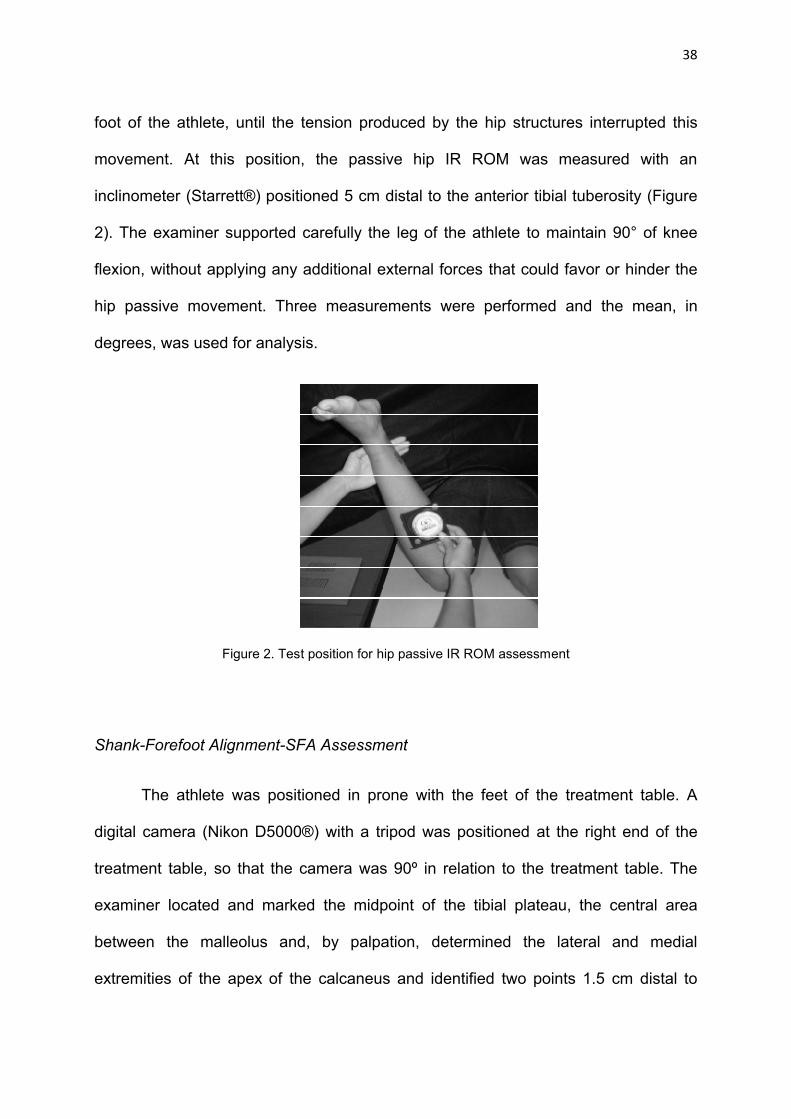
tuberosidade anterior da tíbia (Figura 2). O examinador apoiava a perna do atleta
apenas para manter os 90° de flexão de joelho, com o cuidado para não aplicar
nenhuma força externa que favorecesse ou impedisse o movimento passivo de RI.
Foram realizadas três medidas e a média, em graus, foi utilizada para análise. O
Coeficiente de Correlação Intraclasse (CCI) intra-examinador foi 0.99 e o erro
padrão da medida (SEM) foi 1,4º.
Figura 2. Posicionamento para a avaliação da ADM passiva de RI do quadril

20
Avaliação do Alinhamento Tibia-Antepé (Shank-Forefoot Alignment- SFA).
Inicialmente, o atleta foi posicionado em decúbito ventral com os pés para fora
da maca. Uma câmera digital (Nikon D5000®) com um tripé foram posicionados na
extremidade direita da maca, de forma que a câmera estivesse a 90o em relação a
superfície horizontal da maca. O avaliador estabeleceu o ponto médio entre os
platôs tibiais,entre os maléolos e através da palpação determinou as extremidades
laterais e mediais do ápice do calcâneo e dois pontos 1,5 cm distal a estas
referências. Em seguida, foi traçada uma linha sobre a tíbia, unindo os pontos
médios entre os platôs tibiais e os maléolos (Figura 3-A) . Após as marcações, o
avaliador fixou uma haste metálica no antepé (região metatarsofalangiana) por meio
de uma faixa estabilizadora. O membro inferior do atleta foi posicionado de forma
que o calcâneo estivesse direcionado para cima e que todas as suas marcações
estivessem proporcionalmente visíveis no visor da câmera. Em seguida, com o
auxílio de um goniômetro universal (Carci ®), o avaliador posicionou o pé do atleta a
90o de dorsiflexão de tornozelo, solicitou que o mesmo mantivesse ativamente essa
posição e tirou a foto (Figura 3-B). Este último procedimento foi realizado três
vezes.
Figura 3. A:Marcações correspondentes a bissecção do platô tibial, maléolos e do calcâneo e
B: Posição do atleta para avaliação do SFA
A B

21
Avaliação do Ângulo de Projeção Frontal do Joelho (APFJ)
Para a avaliação do APFJ nas atividades de agachamento e salto, foram
fixados marcadores reflexivos nas espinhas ilíacas ântero-superiores (EIAS), nos
epicôndilos medial e lateral do joelho e no ponto médio entre os maléolos medial e
lateral do tornozelo (anteriormente). Em seguida, o atleta foi solicitado a realizar um
agachamento unipodal até 60º de flexão dos joelhos. Essa angulação foi
previamente determinada pelo avaliador com um goniômetro e então um suporte foi
posicionado na parede, na altura da região glútea do atleta, para ser utilizado como
referência para a posição de 60º de flexão de joelho (Figura 4-A). O atleta realizou
três agachamentos unipodais, alternando entre os membros inferiores direito e
esquerdo, para evitar fadiga. Após cinco minutos de repouso, o atleta foi solicitado a
realizar três saltos verticais bipodais com as mãos posicionadas na cintura (Figura 4-
B). Foi dado um intervalo de 5 segundos entre cada um dos saltos. Os
agachamentos e os saltos de cada atleta foram filmados para posterior análise.
Figura 4. Posição do agachamento unipodal a 60º de flexão (A) e do salto vertical (B)
A B

22
2.3- Redução dos Dados
O torque produzido pelos músculos abdutores de quadril foi calculado
como o produto da média das três medidas de força e a distância do trocânter maior
até cinco centímetros acima da linha articular do joelho, local em que o dinamômetro
manual foi posicionado. O valor de torque foi normalizado pelo respectivo peso
corporal do atleta (Nm/kg).
O alinhamento da tíbia-antepé (SFA) foi determinado por meio da análise
das fotos pelo software Simi Motion Twinner®. O ângulo do alinhamento tíbia-antepé
foi definido como sendo o ângulo formado pela linha de bissecção da tíbia e a linha
sob a haste posicionada na região metatarsofalangiana, correspondente ao antepé
(Figura 5). Essa análise foi realizada nas três fotos para se obter a média do ângulo
do SFA. O CCI intra-examinador para esta medida foi 0.81 e o SEM foi de 3,89º .
Figura 5. Analise do SFA: ângulo entre a bissecção da tíbia e a linha correspondente ao antepé.
Para a determinação do APFJ, os vídeos referentes aos agachamentos e
saltos foram analisados no software Simi Motion Twinner®. O APFJ foi definido
através da união entre o ponto referente a EIAS, o ponto médio entre os epicôndilos
femorais (realizado atraves do software Simi Motion Twinner®) e o ponto médio

23
entre os maléolos (Figura 6). Este ângulo foi medido com o indivíduo na posição
unipodal estática e a 60º de flexão durante o agachamento unipodal. O CCI intra-
examinador obtido para esta medida foi de 0.83 e o SEM foi de 1,65º. A média de
três medidas foi calculada produzindo um valor do APFJ do joelho dominante para
condição estática e para o agachamento.
Na atividade de salto, o APFJ foi analisado no momento da aterrissagem do
salto. A aterrissagem foi definida como os dois quadros (frames) após o choque de
calcanhar. As médias de três APFJ de ambas as pernas na aterrissagem do salto
foram utilizadas para análise. O CCI intra-examinador obtido para esta medida foi
de 0.88 e o SEM foi de 1,93º.
Figura 6. Analise do APFJ: união entre o ponto referente a EIAS, o ponto médio entre os epicôndilos femorais e o ponto médio entre os maléolos.
24
2.4- Análise estatística
Estatística descritiva foi utilizada para caracterizar a amostra em relação às
variáveis APFJ, sexo, torque dos abdutores normalizado pelo peso corporal,
alinhamento tíbia ante-pé e ADM passiva de RI de quadril.
A árvore de classificação e regressão (Classification and Regression Trees –
CART) foi utilizada para avaliar quais os fatores preditivos influenciam a ocorrência
de um FPKA aumentado nos atletas avaliados no presente estudo. A CART é um
modelo de classificação (regressão) multivariado, não-paramétrico que desenvolve
uma árvore de decisão, a qual representa graficamente a associação entre as
variáveis preditoras e a variável desfecho 31. Essa árvore de decisão é criada por
meio de divisões binárias sucessivas do conjunto inicial de dados que ocorrem até
que futuras divisões não sejam possíveis ou que critérios pré-estabelecidos para o
crescimento da árvore sejam alcançados31. Para cada uma dessas divisões, todas
as variáveis preditoras são avaliadas e todos os possíveis pontos de corte (no caso
de variável preditora do tipo contínua) são considerados para se estabelecer aquele
preditor que melhor divide os dados em subgrupos (nodos) cada vez mais
homogêneos 31,32,33. A ordem de entrada das variáveis preditoras no modelo ilustra
hierarquicamente a força de associação entre cada preditor e a variável desfecho, e
as divisões subsequentes a divisão inicial identificam possíveis interações entre
preditores. A escolha da CART para análise dos dados do presente estudo foi
baseada, portanto, no fato dessa ser uma análise robusta, que consegue capturar a
relação não-linear entre preditores e por produzir resultados de fácil aplicação por
meio de regras de tomada de decisão clínica32,34,35,36.
25
No presente estudo, para a construção da CART, a variável dependente foi
dicotomizada como Maior - APFJ e Menor - APFJ. Este procedimento foi realizado
por meio da utilização dos percentis equivalentes ao terço com os maiores valores
do APFJ e ao terço com os menores valores de APFJ. Foram desenvolvidos dois
modelos: um para avaliar os fatores preditivos do APFJ da perna dominante durante
o agachamento unipodal e o outro modelo para a aterrissagem do salto vertical da
perna dominante. As variáveis preditoras foram: sexo, torque dos músculos
abdutores de quadril normalizado pelo corporal, ADM passiva de RI de quadril e
alinhamento tíbia-antepé (SFA). Foram estabelecidos os seguintes critérios para o
processo de divisão e, conseqüente, desenvolvimento da árvore: número mínimo de
8 indivíduos em cada subgrupo (nodo) para permitir a divisão, número mínimo de 4
indivíduos para que um subgrupo (nodo) seja gerado, para maximizar a
homogeneidade dos nodos em cada divisão foi selecionado o índice Gini como
medida de impureza com um nível de diminuição da impureza de 0,0001 para que
ocorra uma divisão. O custo para classificações incorretas foi considerado simétrico
entre as categorias da variável desfecho. Um procedimento denominado poda da
árvore (pruning) foi utilizado, quando necessário, para se obter a melhor árvore de
classificação dos dados, através da eliminação dos nodos com informações
redundantes. Além disso, no modelo desenvolvido para a atividade do agachamento
foi considerado como variável de influência o APFJ da perna dominante na condição
estática. No modelo desenvolvido para a aterrissagem, o APFJ da perna
contralateral foi utilizado com variável de influência. O risco de classificação
incorreta e a porcentagem de classificação total do modelo foram utilizados para
expressar a acurácia preditiva do modelo.

26
A área sob a curva ROC (Receiver Operating Characteristics) foi calculada
também para avaliar a acurácia preditiva (performance) dos modelos desenvolvidos
no presente estudo. A curva ROC representa a habilidade do teste (modelo) para
fazer a distinção entre as categorias da variável desfecho dados todos os valores de
corte possíveis. Um modelo com acurácia preditiva perfeita apresenta uma área sob
a curva ROC igual a 1. Quando essa área é igual ou menor do que 0,5 o modelo é
considerado não acurado para predizer as diferentes categorias da variável
desfecho, ou seja, a distinção que o modelo faz entre ter ou não ter a categoria alvo
da variável desfecho é meramente ao acaso. Um nível de significância de 0,05 foi
estabelecido para verificar se a área sob a curva ROC de cada modelo
desenvolvido no presente estudo foi diferente de 0,5.
Os dois modelos de predição foram desenvolvidos e a área sob a curva ROC foi
calculada utilizando o software SPSS v.17.0 para Windows.

27
Capítulo 3- REFERÊNCIAS BIBLIOGRÁFICAS
(1) SALCI, Y., KENTEL, B. B., HEYCAN, C., AKIN, S., KORKUSUZ, F. Comparison of Landing Maneuvers Between Male and Female College Volleyball Players. Clinical Biomechanics, v.19, p.622-628, 2004.
(2) HOOTMAN, J., DICK, R., ANGEL, J. Epidemiology of Collegiate Injuries for 15 Sports: Summary and Recommendations for Injury Prevention Initiatives. Journal of Athletic Training, v. 42, p. 311-319, 2007.
(3) LEETUN, D. T.; IRELAND, M. L., WILLSON, J. D., BALLANTYNE, B. T., DAVIS, I. M. Core Stability Measures As Risk Factors for Lower Extremity Injury in Athletes. Medicine and Science in Sports and Exercise,v.36,p.926-934, 2004.
(4) HEWETT, T. E., LINDENFELD, T. N., RICCOBENE, J. V., NOYES, F. R. The Effect of Neuromuscular Training on the Incidence of Knee Injury in Female Athletes. A Prospective Study. American Journal of Sports Medicine, v.27,p.699-706, 1999.
(5) BURNHAM, B. R., COPLEY, G. B., SHIM, M. J., KEMP, P. A. Mechanisms of Basketball Injuries Reported to the HQ Air Force Safety Center a 10-Year Descriptive Study, 1993-2002. American Journal of Preventive Medicine, v. 38, S134-S140, 2010.
(6) FORD, K. R., MYER, G. D., HEWETT, T. E. Valgus Knee Motion During Landing in High School Female and Male Basketball Players. Medicine and Science in Sports and Exercise,v. 35, p.1745-1750, 2003.
(7) POWERS, C. M. The Influence of Altered Lower-Extremity Kinematics on Patellofemoral Joint Dysfunction: a Theoretical Perspective. Journal of Orthopaedic and Sports Physical Therapy, v. 33, p.639-646, 2003.
(8) HEWETT, T., SNYDER-MACKLER, L., SPINDLER, K. P. The Drop-Jump Screening Test: Difference in Lower Limb Control by Gender and Effect of Neuromuscular Training in Female Athletes. American Journal of Sports Medicine, v.35, p.145-147, 2007.
(9) NOYES, F. R., BARBER-WESTIN, S. D., FLECKENSTEIN, C., WALSH, C., WEST, J. The Drop-Jump Screening Test: Difference in Lower Limb Control by Gender and Effect of Neuromuscular Training in Female Athletes. American Journal of Sports Medicine, v.33, p.197-207, 2005.
(10) HEWETT, T. E., MYER, G. D., FORD, K. R., HEIDT, R. S. Jr., COLOSIMO, A. J., MCLEAN, S. G., VAN DEN BOGERT, A. J., PATERNO, M. V., SUCCOP, P. Biomechanical Measures of Neuromuscular Control and Valgus Loading of the Knee Predict Anterior Cruciate Ligament Injury Risk in Female Athletes: a Prospective Study. American Journal of Sports Medicine, v.33, p.492-501, 2005.

28
(11) BOLGLA, L. A., MALONE, T. R., UMBERGER, B. R., UHL, T. L. Hip Strength and Hip and Knee Kinematics During Stair Descent in Females With and Without Patellofemoral Pain Syndrome. Journal of Orthopaedic and Sports Physical Therapy, v.38, p.12-18, 2008.
(12) MENDONÇA, L. D. M., MACEDO, L. G., FONSECA, S. T., SILVA, A. A. Comparação do alinhamento anatômico de membros inferiores entre indivíduos saudáveis e indivíduos com tendinose patelar. Revista Brasileira de Fisioterapia, v. 9, n.1, p.101-107, 2005.
(13) FONSECA, S. T., OCARINO, J. M., SILVA, P. L. P. Integration of stress and their relationship to the kinetic chain. In Magee DJ, Zachazewski JE, Quillen WS. Science foundations and principles of pratice in musculoskeletal rehabilitation. St. Louis: Saunders, 2007.
(14) BELLCHAMBER, T. L., VAN DEN BOGERT, A. J. Contributions of Proximal and Distal Moments to Axial Tibial Rotation During Walking and Running. Journal of Biomechanics, v.33, p.1397-1403, 2000.
(15) FREDERICSON, M., COOKINGHAM, C. L., CHAUDHARI, A. M., DOWDELL, B. C., OESTREICHER, N., SAHRMANN, S. A. Hip Abductor Weakness in Distance Runners With Iliotibial Band Syndrome. Clinical Journal of Sports Medicine, v.10, p.169-175, 2000.
(16) DONATELLI, R., WOODEN, M., EKEDAHL, S. R., WILKES, J. S., COOPER, J., BUSH, A. J. Relationship Between Static and Dynamic Foot Postures in Professional Baseball Players. Journal of Orthopaedic and Sports Physical Therapy, v. 29, p.316-325, 1999.
(17) HEWETT, T. E., MYER, G. D., FORD, K. R., SLAUTERBECK, J. R. Dynamic Neuromuscular Analysis Training for Preventing Anterior Cruciate Ligament Injury in Female Athletes. Instructional Course Lectures, v.56, p.397-406, 2007.
(18) JACOBS, C. A., UHL, T. L.; MATTACOLA, C. G., SHAPIRO, R., RAYENS, W. S. Hip Abductor Function and Lower Extremity Landing Kinematics: Sex Differences. Journal of Athletic Training, v.42, p.76-83, 2007.
(19) NIEMUTH, P.E., JOHNSON, R.J., MYERS M.J., THIEMAN T.J. Hip Muscles Weakness and Overuse Injuries in Recreational Runners. Clinical Journal of Sports Medicine, v. 15, p.14-21, 2005.
(20) ZELLER, B. L., MCCRORY, J. L., KIBLER, W. B., UHL, T. L. Differences in Kinematics and Electromyographic Activity Between Men and Women During the Single-Legged Squat. American Journal of Sports Medicine, v.31,p.449-456, 2003.

29
(21) HEINERT, B. L., KERNOZEK, T. W., GREANY, J. F., FATER, D. C. Hip Abductor Weakness and Lower Extremity Kinematics During Running. Journal of Sport Rehabilation, v.17, p.243-256, 2008.
(22) PRINS, M. R., VAN DER WURFF, P. Females With Patellofemoral Pain Syndrome Have Weak Hip Muscles: a Systematic Review. Australian Journal of Physiotherapy, v.55, p.9-15, 2009.
(23) GEISER, C. F., O'CONNOR, K. M., EARL, J. E. Effects of Isolated Hip Abductor Fatigue on Frontal Plane Knee Mechanics. Medicine and Science in Sports and Exercise, v.42, p.535-545, 2010.
(24) SIGWARD, S. M., OTA, S., POWERS, C. M. Predictors of Frontal Plane Knee Excursion During a Drop Land in Young Female Soccer Players. Journal of Orthopaedic and Sports Physical Therapy, v.38, p.661-667, 2008.
(25) DE AQUINO, C. F., GONÇALVES, G. G. P., FONSECA.S.T., MANCINI, M. C. Análise da relação entre flexibilidade e rigidez passiva dos isquiotibiais. Revista Brasileira de Medicina do Esporte, v.12, n.4, p.195-200, 2006.
(26) GLEIM, G. W., MCHUGH, M. P. Flexibility and Its Effects on Sports Injury and Performance. Sports Medicine, v.24, p.289-299, 1997.
(27) NIGG, B. M., COLE, G. K., NACHBAUER, W. Effects of Arch Height of the Foot on Angular Motion of the Lower Extremities in Running. Journal of Biomechanics, v.26, p.909-916, 1993.
(28) MCCLAY, I., MANAL, K. A. Comparison of Three-Dimensional Lower Extremity Kinematics During Running Between Excessive Pronators and Normals. Clinical Biomechanics, v.13, p.195-203, 1998.
(29) JOSEPH M., TIBERIO, D., BAIRD, J.L., TROJIAN, T.H., ANDERSON, J.M., KRAEMER, W.J., MARESH, C.M. Knee Valgus During Drop Jumps in National Collegiate Athletic Association Division I Female Athletes. American Journal of Sports Medicine, v.36, p.285-289, 2008.
(30) SOUZA, T. R., PINTO, R. Z., TREDE, R. G., KIRKWOOD, R. N., PERTENCE, A. E., FONSECA, S. T. Late Rearfoot Eversion and Lower-Limb Internal Rotation Caused by Changes in the Interaction Between Forefoot and Support Surface. Journal of the American Podiatric Medical Association, v.99,p.503-511, 2009.
(31) BREIMAN, L., FRIEDMAN, J. H., OLSHEN, R. A., et al. Classification and Regression Trees. Belmont, Calif: Wadsworth International, 1984.
(32) LEMON, S. C., ROY, J., CLARK, M. A., FRIEDMANN, P. D., RAKOWSKI, W. Classification and Regression Tree Analysis in Public Health: Methodological Review and Comparison With Logistic Regression. Annals Behavioral Medicine, v.26, p.172-181, 2003.
30
(33) D'ALISA, S., MISCIO, G., BAUDO, S., SIMONE, A., TESIO, L., MAURO, A. Depression Is the Main Determinant of Quality of Life in Multiple Sclerosis: a Classification-Regression (CART) Study. Disability and Rehabilitation, v.28,p.307-314, 2006.
(34) FALCONER, J. A., NAUGHTON, B. J., DUNLOP, D. D., ROTH, E. J., STRASSER, D. C., SINACORE, J. M. Predicting Stroke Inpatient Rehabilitation Outcome Using a Classification Tree Approach. Archives of Physical Medicine and Rehabilitation, v.75, p.619-625, 1994.
(35) TEMKIN, N. R., HOLUBKOV, R., MACHAMER, J. E., WINN, H. R., DIKMEN, S. S. Classification and Regression Trees (CART) for Prediction of Function at 1 Year Following Head Trauma. Journal of Neurosurgery, v.82, p.764-771, 1995.
(36) MANCINI, M. C., COSTER, W. J., TROMBLY, C. A., HEEREN, T. C. Predicting Elementary School Participation in Children With Disabilities.Archives of Physical Medicine and Rehabilitation, v. 81, p.339-347, 2000.
31
CAPÍTULO 4 - ARTIGO
“Predicting dynamic knee valgus in athletes using a classification and regression tree approach”
Abstract
STUDY DESIGN: Cross-sectional.
OBJECTIVE: To analyze how typical predictors of increased dynamic knee valgus angle interact to produce altered knee alignment during functional tasks
BACKGROUND: Excessive dynamic knee valgus is thought to be a contributing factor to many lower extremity injuries. Decreased hip abductor strength have been associated to increased knee valgus angle. In addition, athletes with excessive foot pronation have increased frontal plane knee excursion. However, the biomechanical interdependence of distal and proximal segments of the lower limb kinetic chain has been poorly investigated.
METHODS AND MEASURES: During preseason, 227 athletes belonging to a sport club were evaluated. The dynamic knee valgus (dependent variable) was measured as the frontal plane projection angle of the knee (FPPAK), in two different conditions: single leg squat (SLS) and landing. The independent variables were: isometric hip abductor torque, passive range of motion (ROM) of hip internal rotation (IR) and shank-forefoot alignment (SFA). Classification and Regression Trees (CART), a non-parametric statistical analysis that incorporates nonlinear relationships between predictors, was used to develop decision rules to predict the presence of excessive FPPAK
RESULTS: During SLS the occurrence of high FPPAK was due to interaction between hip abductor torque and passive ROM of hip IR. During landing, the SFA together with abductor torque and passive hip IR were the predictors of high FPPAK. In addition, the CART model identified meaningful cut-off points that classified the sample into the outcome categories of High and Low-FPPAK.
CONCLUSION: These findings may guide sports professionals to plan preventive programs and indicate that preseason assessments must include measurements of hip abductor torque, passive ROM of hip IR and shank-forefoot alignment.
KEY WORDS: Knee valgus, hip strength, CART, sports injuries, lower limb.
Authors: Natalia Franco N. Bittencourt, Luciana De Michelis Macedo, Juliana M. Ocarino, Sergio Teixeira da Fonseca. Journal: Journal of Orthopaedic & Sports Physical Therapy -JOSPT
website: http://mc.manuscriptcentral.com/jospt

32
INTRODUCTION
Knee injuries such as patellofemoral syndrome (PFS), patellar tendinopathy
and Anterior Cruciate Ligament (ACL) tear require longer time to recover than ankle
injuries (131%), hip and thigh injuries (41%)18 and result in increased time away from
sports. Furthermore, an ACL injury generates an estimated cost of approximately
U$17,000.00 per athlete13. Knee joint injury seems to be related to the demands on
musculoskeletal system produced by sports activities. For example, approximately
63% of knee injuries occur during takeoff and landing from vertical jump in volleyball
and 43% in basketball 4,27. Thus, a better understanding of the mechanisms involved
in these injuries may help the development of effective treatment and preventive
interventions.
One factor that may contribute to production of knee injuries in sports is the
athlete’s inability to maintain a proper dynamic alignment among lower limb
segments in the frontal and transverse planes during sports practice 10, 21, 24 . Hewett
et al 14 demonstrated that the presence of dynamic knee valgus (abductor moment) is
an important predictor of ACL rupture in athletes14. The presence of knee valgus
during lower limb movement can also lead to dynamic changes in patellar alignment.
Such changes have been related to increases in the overload of structures such as
the retinaculum patellar, articular cartilage and fat pad, predisposing the development
of anterior knee pain (Patellofemoral Syndrome)2. Moreover, incorrect movement
patterns and the related change in patella alignment may increase patellar tendon
shear forces, predisposing the developement of patellar tendinopathy22. Therefore,
dynamic changes in knee alignment associated with the complex demands involved
in sports like volleyball, basketball and soccer seem to increase the load imposed on
33
the musculoskeletal system of the athlete, contributing to the development of various
types of injuries and disorders around the knee joint.
The knee is an intermediate joint in the kinematic chain, and thus depends on
appropriated mechanical behavior of hip and ankle joints to dissipate and/or properly
distribute the forces imposed on the musculoskeletal system during sports activities9.
For example, during performance of close kinetic chain activities dynamic knee
valgus may result from excessive femur internal rotation and hip adduction12,16,17.
This altered kinematics of the hip joint may occur due to weakness of the hip
abductors and/or external rotators, especially gluteus medius and maximus 12,18. In a
systematic review, Prins and Wurff 26 demonstrated that women with PFS have
decreased strength of hip muscles, in particular hip abductors, external rotators and
extensors on the affected side when compared with healthy participants 26. Geiser et
al 11 induced a fatigue protocol specific to hip abductors in physically active subjects
and observed that the knee joint assumed a more adducted position in frontal plane,
which resulted in an increased dynamic knee valgus during functional activities11.
These studies demonstrate, therefore, that excessive hip movement particularly
those associated with reduced hip abductor strength appear to influence the degree
of knee valgus in athletes during the performance of sports activities.
Excessive knee excursion in the frontal plane is not only influenced by hip
abductor muscle strength but seems to be also associated with the available hip
range of motion (ROM) 28. Traditionally, the ROM of a joint is assessed by applying to
it an external torque, as the total motion produced is recorded. Thus, this measure
may be highly influenced by the tolerance to stretching of the subject 6. If the
transverse plane ROM is assessed so that it represents the position where the torque
34
produced by the leg and foot weights equals the passive resistance torque generated
by the hip, this measure could be informative about joint stiffness. This stiffness
represents the change in the torque of passive resistance supplied by the tissues of
the hip joint (muscles, ligaments, capsule and fascia), offered against the joint
motion9. Thus, for some functional activities, passive mechanisms of a joint can help
to prevent an exaggerated movement or maintain proper alignment.
In addition the influence of hip movements for the knee valgus occurrence,
excessive lower limb internal rotation and calcaneal eversion have also been
postulated to contribute to inadequate knee kinematics 1,7,20 . The subtalar joint axis,
which is tilted 45o from horizontal plane, allows the coupling of the motion of the foot
in frontal plane (calcaneal eversion) with the motions in transverse plane (talus and
lower limb rotation), with a reported correlation of r2 = 0.991 23. This strong
association indicates that changes in the subtalar joint movements may lead to
compensation at knee joint1.In addition, athletes who wore foot orthosis wedged 5º
medially showed a decrease in knee valgus and calcaneal eversion/pronation during
landing17. Excessive pronation has been linked with the presence of forefoot varus
alignment, which can increase the eversion torque in ankle-foot complex after
forefoot contact with the support surface22,29. These findings reinforce the need to
include the standardized measurement of the forefoot in the assessment of athletes,
since the presence of excessive forefoot varus can cause biomechanical
compensations throughout the lower limb and influence the occurrence of injuries.
Some studies have used analysis based on linear and individual contribution
of each biomechanical factor to predict dynamic knee valgus5, 28. However, the
occurrence of increased static and dynamic valgus alignment of the knee has

35
multifactorial nature and depends on how the musculoskeletal system adapts to the
possible interactions between muscle strength, tissue stiffness and joint alignment of
proximal and distal segments of the kinetic chain9. This interaction between factors is
essential to develop a greater capability for dynamic stabilization during the demand
imposed by a particular activity. Therefore, to identify clinically relevant interactions
that may not have been captured by previous studies that focus on the investigation
of statistical main effects, the aim of this study was to determine the predicting
factors of increased frontal plane projection angle of the knee during the single leg
squat and landing in athletes. The Classification and Regression Trees (CART) was
used as the statistical method for analysis, since it captures nonlinear relationships
between the predictors and outcome variables.

36
METHODS
Participants
In this study, 227 athletes (78 female and 149 male), who regularly
participated in training and competitions of basketball, soccer, volleyball and
gymnastics, were recruited during pre-season. The mean age was 16.59 ± 5 years;
the mean body mass was 67.16 ± 16.4 kg and the mean height was 157 ± 59.43 cm.
There was no restriction regarding the time of sports practice in this study. The
inclusion criteria were: absence of pain or a history of lower extremity surgery in the
last six months. Those athletes who felt pain or discomfort while performing any test
were excluded from the study. The University’s Institutional Ethics Review Committee
approved the procedures of this study and all participants signed an informed
consent (ETIC No. 493/2009).
Procedures
Strength Testing
Isometric strength of the hip abductors was measured using a manual
dynamometer (Hand Held - Microfet2 ®). The subject was positioned in side lying on
a treatment table with arms in front of the body. The dynamometer was positioned
under a stabilizing strap five centimeters above the knee joint line. A second strap
was used for trunk stabilization (Figure 1). After a familiarization procedure the
athlete was asked to perform maximal isometric contractions of the hip abductors for
5 seconds. This procedure was repeated three times with intervals of 15 seconds
between each isometric contraction. During testing, verbal encouragement was given
to ensure that the athlete would produce maximum contraction.
37
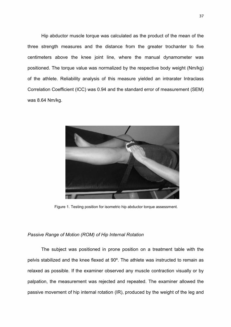
Hip abductor muscle torque was calculated as the product of the mean of the
three strength measures and the distance from the greater trochanter to five
centimeters above the knee joint line, where the manual dynamometer was
positioned. The torque value was normalized by the respective body weight (Nm/kg)
of the athlete. Reliability analysis of this measure yielded an intrarater Intraclass
Correlation Coefficient (ICC) was 0.94 and the standard error of measurement (SEM)
was 8.64 Nm/kg.
Figure 1. Testing position for isometric hip abductor torque assessment.
Passive Range of Motion (ROM) of Hip Internal Rotation
The subject was positioned in prone position on a treatment table with the
pelvis stabilized and the knee flexed at 90º. The athlete was instructed to remain as
relaxed as possible. If the examiner observed any muscle contraction visually or by
palpation, the measurement was rejected and repeated. The examiner allowed the
passive movement of hip internal rotation (IR), produced by the weight of the leg and
38
foot of the athlete, until the tension produced by the hip structures interrupted this
movement. At this position, the passive hip IR ROM was measured with an
inclinometer (Starrett®) positioned 5 cm distal to the anterior tibial tuberosity (Figure
2). The examiner supported carefully the leg of the athlete to maintain 90° of knee
flexion, without applying any additional external forces that could favor or hinder the
hip passive movement. Three measurements were performed and the mean, in
degrees, was used for analysis.
Figure 2. Test position for hip passive IR ROM assessment
Shank-Forefoot Alignment-SFA Assessment
The athlete was positioned in prone with the feet of the treatment table. A
digital camera (Nikon D5000®) with a tripod was positioned at the right end of the
treatment table, so that the camera was 90º in relation to the treatment table. The
examiner located and marked the midpoint of the tibial plateau, the central area
between the malleolus and, by palpation, determined the lateral and medial
extremities of the apex of the calcaneus and identified two points 1.5 cm distal to

39
these references. Then a line was drawn on the tibia, by joining the points marked on
the tibial plateaus and between the malleolus (Figure 3-A). After, bisecting the tibia,
the examiner positioned a metal rod on the plantar area of the forefoot, following the
orientation of the metatarsophalangeal heads. The lower limb of the athlete was
positioned so that the calcaneus was directed upwards and that all markings were
visible on the center of the camera display. With the aid of an universal goniometer
(Carci®), the examiner positioned the foot of the athlete at 90º of ankle dorsiflexion,
requested the athlete to actively maintain this position and took a still photograph
(Figure 3-B). This procedure was performed three times.
The shank-forefoot alignment (SFA) was measured by means of the analysis
of the still photos. This analysis was performed with the program Simi Motion
Twinner®. The angle of the shank-forefoot alignment was defined as the angle
between the bisection line of the tibia and the line along the rod positioned on the
metatarsophalangeal region, corresponding to the forefoot alignment. This analysis
was performed on the three different photos to obtain the mean angle of the SFA.
The intrarater ICC for this measure was 0.81 and the SEM was 3.89 º.
Figure 3. A: Markings corresponding to bisection of the tibial plateau, malleolus and the calcaneus and
B: Athlete position for SFA assessment
40
Frontal Plane Projection Angle of the Knee (FPPAK)
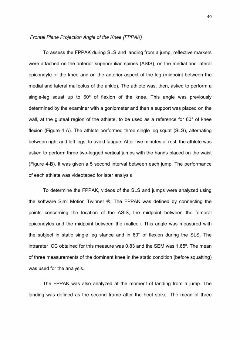
To assess the FPPAK during SLS and landing from a jump, reflective markers
were attached on the anterior superior iliac spines (ASIS), on the medial and lateral
epicondyle of the knee and on the anterior aspect of the leg (midpoint between the
medial and lateral malleolus of the ankle). The athlete was, then, asked to perform a
single-leg squat up to 60º of flexion of the knee. This angle was previously
determined by the examiner with a goniometer and then a support was placed on the
wall, at the gluteal region of the athlete, to be used as a reference for 60° of knee
flexion (Figure 4-A). The athlete performed three single leg squat (SLS), alternating
between right and left legs, to avoid fatigue. After five minutes of rest, the athlete was
asked to perform three two-legged vertical jumps with the hands placed on the waist
(Figure 4-B). It was given a 5 second interval between each jump. The performance
of each athlete was videotaped for later analysis
To determine the FPPAK, videos of the SLS and jumps were analyzed using
the software Simi Motion Twinner ®. The FPPAK was defined by connecting the
points concerning the location of the ASIS, the midpoint between the femoral
epicondyles and the midpoint between the malleoli. This angle was measured with
the subject in static single leg stance and in 60° of flexion during the SLS. The
intrarater ICC obtained for this measure was 0.83 and the SEM was 1.65º. The mean
of three measurements of the dominant knee in the static condition (before squatting)
was used for the analysis.
The FPPAK was also analyzed at the moment of landing from a jump. The
landing was defined as the second frame after the heel strike. The mean of three

41
FPPAK of both legs in landing were used for analysis. The intrarater ICC obtained for
this measure was 0.88 and the SEM was 1.93º.
.
Figure 4. Position of the SLS to 60º of flexion (A) and of the vertical jump (B)
Statistics
Descriptive statistics were used to characterize the sample in relation to the
variables: FPPAK, gender, normalized abductors torque, shank-forefoot alignment
angle and hip internal rotation passive ROM. Classification and Regression Trees
(CART) was used to assess which predictive factors influenced the occurrence of
high- FPPAK. CART is a multivariate, nonparametric classification (regression)
model, which develops a decision tree by successive binary divisions of the initial set
of data that occur until further divisions are not possible or until pre-established
criteria for tree growth are reached 3. For each of these divisions, all possible cutoff
points are considered to establish the predictor that best divides the data into
subgroups (nodes) increasingly homogeneous 3, 8. The choice of CART to analyze
the data of this study relies on the fact that it is a robust analysis, which can capture
A B
42
the non-linear relationship between predictors and produce results easily applied by
clinical decision rules 8, 19, 30.
To build the CART model, the dependent variable was dichotomized as High -
FPPAK and Low-FPPAK. This procedure was performed by identifying the
percentiles corresponding to the inferior (low- FPPAK) and superior (high- FPPAK)
thirds of the sample’s distribution. Two models were developed to assess the
predictor factors of FPPAK of the dominant leg during the SLS and landing from a
jump. The predictor variables were: gender, normalized hip abductor torque, passive
ROM of hip IR and shank-forefoot alignment angle (SFA). In the SLS model the
FPPAK of the dominant leg in the static condition was considered as an influence
variable. In the landing model the FPPAK of the contralateral leg was used as
influence variable. The area under the ROC curve (Receiver Operating
Characteristics) was also calculated to assess the predictive performance of the
models developed in this study. A significance level of 0.05 was established to verify
whether the area under the ROC curve of each model developed in this study was
different from 0.5.

43
RESULTS
The cut-off values of the inferior (Low FPPAK) and superior (High FPPAK)
thirds of the FPPAK distribution during SLS were 4.69º e 8.16º, respectively.
Considering that only subjects that had values below or above the cut-off values
were included in the final sample, data from only 101 athletes were analysed. Fifty
athletes had High- FPPAK (mean=11.79º; SD=+2.66) and fifty-one athletes had Low-
FPPAK (mean=2.86º; SD= + 1.36). During landing, the cut-off values for Low and
High FPPAK were -0.09º e 3.59º, respectively. Due to this classification and to the
use of the values of FPPAK of the opposite limb as influence variable, data from only
72 athletes were analysed. Thirty-three athletes had High- FPPAK (mean=7.59º;
SD=+2.5) and thirty-nine athletes had Low-FPPAK (mean=-3.93; SD=+4.2). The
mean and standard deviation of the sample’s demographics characteristics and
independent variables: normalized hip abductor torque, passive ROM of hip IR and
shank-forefoot alignment angle (SFA) are presented in table 1.
Table 1: Mean and SD for demographics characteristics and independents variables
Variable SLS Group (n = 101)
Landing Group (n= 72 )
Age (years) 17.1 (5.3) 16.7 (4.6)
Body Mass (kg) 68.9 (17.3) 68.6 (16.9)
Sex Female Male
32 (31%) 69 (69%)
22 (30%) 50 (70%)
Height (cm) 150.4 (68.1) 179.7 (14.7)
Shank-Forefoot Alignment (o) 11.8 (8.2) 11.9 (8.9)
Passive ROM of Hip IR (o) 43.01 (15.1) 45.7 (14.9)
Hip Abductor Torque (Nm/Kg) 117.92 (39.4) 127.55 (35.2)

44
Predictive model for Single Leg Squat (SLS)
The classification tree (Figure 5) has identified, from the four independent
variables entered in the analysis, the normalized hip abductor torque and passive
ROM of hip IR as FPPAK predictors during SLS. The normalized hip abductor torque,
with cut-off point of 103 Nm/kg, was the first predictor to separate the sample into two
groups. Twenty-three athletes of those who had normalized hip torque < 103 Nm/Kg
(76.7%) had High- FPPAK, while only seven athletes had Low- FPPAK (node 1).
Normalized hip torque values great than 103 Nm/Kg was predictor of Low-FPPAK in
62% (n=44) of the 71 athletes who had normalized hip torque great than 103 Nm/hg
(node 2).
In the subgroup with normalized hip abductor torque of less than 103 Nm/kg,
passive ROM of hip IR greater than 43º was a predictor for High- FPPAK in 90% of
the athletes (node 4). Five athletes with low abductor torque and passive ROM of hip
IR between 37º and 43º (83% of this subgroup) demonstrated Low-FPPAK (node 8),
just one of those presented High- FPPAK. One hundred per cent (n=4) of the athletes
that showed abductor torque less than 103Nm/kg and passive ROM of hip IR less
than 37º presented High-FPPAK (node 7).
Among the athletes with hip abductor torque greater than 103 Nm/Kg (n=71),
seven out of nine (77.8%) of those with hip IR passive ROM of less than 21.6º (node
5) had High-PPAK. The athletes who had hip IR passive ROM greater than 21.6º,
were divided into two subgroups according to their normalized hip abductor torque in
a new cutoff point. Normalized hip abductor torque greater than 148.8 Nm/Kg was
predictor of Low -FPPAK in 90% of these athletes (n=19) (node 10). Similarly, 22
athletes with hip IR passive ROM greater than 21.6º and abductor torque between
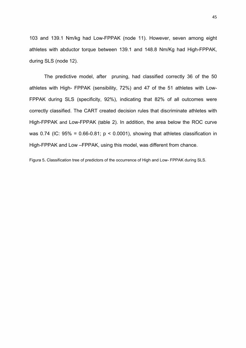
45
103 and 139.1 Nm/kg had Low-FPPAK (node 11). However, seven among eight
athletes with abductor torque between 139.1 and 148.8 Nm/Kg had High-FPPAK,
during SLS (node 12).
The predictive model, after pruning, had classified correctly 36 of the 50
athletes with High- FPPAK (sensibility, 72%) and 47 of the 51 athletes with Low-
FPPAK during SLS (specificity, 92%), indicating that 82% of all outcomes were
correctly classified. The CART created decision rules that discriminate athletes with
High-FPPAK and Low-FPPAK (table 2). In addition, the area below the ROC curve
was 0.74 (IC: 95% = 0.66-0.81; p < 0.0001), showing that athletes classification in
High-FPPAK and Low –FPPAK, using this model, was different from chance.
Figura 5. Classification tree of predictors of the occurrence of High and Low- FPPAK during SLS.
46

47
Table 2. Classification tree rules for predicting the occurrence of High and Low--FPPAK during SLS
Predictive model for Landing
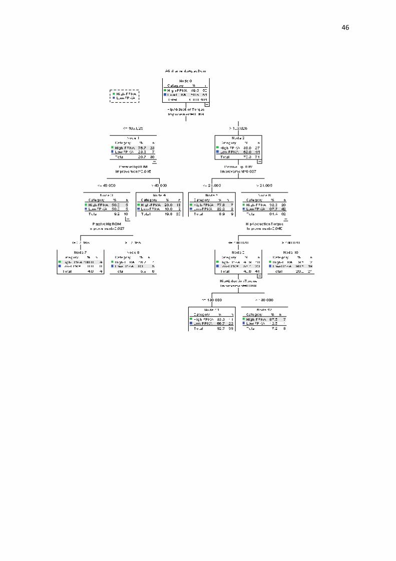
The CART model (Figure 6) selected SFA angle, normalized hip abductor
torque and hip IR passive ROM as predicting factors for FPPAK during landing. The
classification tree showed the SFA angle as the first predictor of High- FPPAK. About
60% (n=23) of the athletes with SFA angle great than 10.9º had High-FPPAK (node
2). Conversely, out of the 34 athletes with SFA angle lesser than 10.9º, 70.6 %
(n=24) had Low-FPPAK, while only 10 athletes had High – FPPAK (node 1). In the
subgroup of subjects with SFA lesser than 10.9º, normalized hip abductor torque
lesser than 109 Nm/kg was predictor of High- FPPAK in 75% of athletes (n=9) (node
3). When the subjects had SFA angles lower than 10.9o and normalized hip abductor
torque greater than 109 Nm/kg, 95% of the athletes (n=21) had Low-FPPAK (node
4).
I- Decision rules for occurrence of High-FPPAK
NODE 4 Hip Abductor Torque < 103 Nm/kg and Passive ROM of Hip IR > 43.6 o
NODE 5 Hip Abductor Torque > 103 Nm/kg and Passive ROM of Hip IR < 21.6 o
NODE 12 Hip Abductor Torque between 139 and 148 Nm/Kg and Passive ROM of Hip IR > 21.6 o
II- Decision rules for occurrence of Low-FPPAK
NODE 8 Hip Abductor Torque < 103 Nm/kg and Passive ROM of hip IR between 37 o and 43.6 o
NODE 10 Hip Abductor Torque > 148 Nm/kg and Passive ROM of hip IR > 21.6 o

48
In those athletes who had SFA angles higher than 10.9º, the model selected
passive ROM of hip IR as second predictor. Among athletes with SFA > 10.9º and
passive ROM smaller than 34º, five out of seven (71.4%) had Low-FPPAK (node 5).
On the other hand, 21 of 31 athletes with passive ROM of hip IR greater than 34.3o
(67%) had High-FPPAK (node 6). Fourteen athletes with passive ROM of hip IR
between 34.3º and 47.8º presented high-FPPAK (node 7). Concerning the subgroup
of athletes with passive ROM of hip IR greater than 47º, another split was based on
shank-forefoot alignment angle: SFA values between 10.9º and 17.3º were
associated to Low-FPPAK in 75% of athletes (n=6) (node 9). On the other hand,
passive ROM of hip IR greater than 47.8º and SFA angle great than 17.3º were
predictors of High-FPPAK in 83.3% of the athletes belonging to this subgroup (n=5)
(node 10).
The CART model classified correctly 28 of the 33 athletes with High- FPPAK
(sensibility, 84%) and 32 of the 39 with Low- FPPAK (specificity, 82%) during
landing, indicating that 83% of all outcomes were correctly classified. The CART
model allowed the creation of decision rules that discriminate athletes with High-
FPPAK and Low-FPPAK (Table 3). The area below the ROC curve was 0.79 (IC:
95% = 0.70-0.88; p < 0.0001), showing that athletes classification in High-FPPAK
and Low –FPPAK, using this model, was not by chance.

49
Figure 6. Classification tree of the occurrence of High and Low- FPPAK during landing.

50
Table 3. Classification tree rules for predicting the occurrence of High and Low-FPPAK during landing
I- Decision rules for occurrence of High-FPPAK
NODE 3 SFA < 10.9o but Hip Abductor Torque < 109 Nm/kg
NODE 7 SFA > 10.9o and Passive ROM of hip IR between 34o and 47o
NODE 10 SFA > 17 o and Passive ROM of hip IR > 47 o
II- Decision rules for occurrence of Low-FPPAK
NODE 4 SFA < 10.9o and Hip Abductor Torque > 109 Nm/kg
NODE 5 SFA > 10.9o but Passive ROM of hip IR < 34 o
NODE 9 Passive ROM of hip IR > 47 o and SFA between 10.9o and 17o
51
DISCUSSION
The results of this study demonstrate that the occurrence of High-FPPAK
during landing from a jump was due to an interaction between SFA angle, normalized
abductor torque and passive ROM of hip IR. Similarly, during one leg squat, the
occurrence of High-FPPAK was the result of the interaction between abductor torque
and passive ROM of hip IR. These results illustrate the influence of biomechanical
factors of the distal and proximal segments of the lower limb kinetic chain in the
production of increased dynamic knee valgus (High-FPPAK) during functional
activities. The findings of the present study are in agreement with previous studies,
which identified significant association between lower limb biomechanical factors with
the occurrence of knee valgus5, 28, 32.
The results of the SLS analysis indicated that the hip abductor torque was the
main predictor of High-FPPAK. This finding is consistent with the study of Claiborne
et al5, which found that hip abductor torque alone explained 13% of the occurrence of
increased FPPAK. However, Wilson et al31 tested the association between FPPAK
and strength of hip abductors and external rotators during squat to 45° of knee
flexion and found that only the external rotators had significant association with
FPPAK (r=0.40). These results could be attributed to the smaller range of knee
flexion used (45o as opposed to 60o), or to the lower mean of FPPAK shown by those
subjects (4o) in comparison with athletes of the present study who demonstrated a
mean of 7.59° in High-FPPAK group. The performance of the single leg squat to 60°
increases the perpendicular distance from the knee joint axis to the body center of
gravity, which increases the external knee flexor moment. This greater demand of
stabilization at in this range may have increased the contribution of the hip abductors

52
to control the occurrence of knee valgus and values of normalized hip abduction
torque smaller than 103 Nm/kg strongly contributes to the occurrence of high FPPAK.
Another relevant predictor selected by the classification tree, during SLS, was
the passive ROM of hip IR. The inclusion of this variable allowed a better
understanding of how the available range of hip internal rotation interacts with the
abductor torque. The results of the present study indicated that the association of
reduced hip abductor torque (<103 Nm/kg) with large range of hip internal rotation
(>43°) was crucial for the occurrence of High-FPPAK. Furthermore, 90% of athletes
with high abductor torque (>148 Nm/Kg), even showing available passive ROM of IR
hip greater than 21° did not demonstrate High-FPPAK. This fact reinforces the
influence of hip abductors strength in controlling knee kinematics. However, it is
important to notice that seven athletes with torque between 139 and 148 Nm/kg and
hip internal rotation passive ROM above 21° showed High-FPPAK. In this subgroup,
it is possible that other factors not assessed in the present study, such as a
decreased strength hip external rotators or level of athlete’s sports ability could help
explain the occurrence of High-FPPAK. Despite the existence of other possible
contributor to increased knee valgus, the interaction between hip abductors strength
and passive ROM of hip IR (as a possible index of hip stiffness), shown in the
present study, suggests that clinical and preventive interventions should focus on
both the contractile and passive elements of the hip joint.
Contrary to expectations that hip abductor strength and stiffness could help
prevent the occurrence of knee valgus, seven athletes with hip abductor torque
greater than 103Nm/Kg and marked decreased passive ROM of hip IR (<21°;
subgroup mean angle of 17°) were classified into High-FPPAK group. This marked
53
restriction of available range of hip IR may indicate an existence of femoral neck
retroversion, which can alter the kinematics behavior of the lower limb segments
during the performance of activities required in this study. For example, if the athlete
does not have the appropriate range of hip internal rotation to allow absorption of
possible trunk and pelvis rotations resulting from squating, it would cause that the
projection of the body’s center of gravity to fall outside of the base of support and
require an increase in hip adduction and thus, an increase of knee valgus to allow
control of the movement. In this case, not only low hip stiffness but also excessive
hip stiffness may be related to the occurrence of knee valgus during certain
functional activities.
During landing, the main predictor selected by CART was the shank-forefoot
alignment (SFA) angle. Considering that the ankle-foot complex is the first segment
to get in contact with the ground when landing from a jump, changes of the alignment
of this structure seems to be crucial for kinematic and kinetic adaptations of other
segments of the lower limb1. The relationship between static forefoot varus
measurements and foot kinematics is in agreement with the findings of Donatelli et
al7 who observed increased foot pronation in baseball athletes with marked forefoot
varus at heel strike during running. Furthermore, the present study demonstrated that
inter-segmental relationships are complex, since, in the individuals with small SFA
angle (below 10.9º), reduced normalized abductor torque (<109 Nm/kg) was the next
predictor for athletes to show High-FPPAK. On the other hand, for those athletes
SFA angle greater than 10.9º, increased passive ROM of hip IR (>34.3) was the most
important factor in the production of High-FPPAK. Again, during landing from a jump,
54
structural factors such as foot alignment and hip stiffness seem to play important
roles in controlling the motions of the knee joint.
In the analysis of landing in 2D, the FPPAK represents the combination of
movements of hip adduction and internal rotation21. Athletes with small forefoot varus
angle (SFA<10.9°), may have had a lower demand for lower limb internal rotation
and the component of femur adduction became more relevant to knee kinematics.
Once landing also imposes a high hip adduction moment, the athlete’s inability to
produce proper hip abductor torque influenced the occurrence of High-FPPAK, as
75% of the athletes who had low normalized hip adductor torque (< 109 Nm/kg)
demonstrated knee valgus when landing. Heinert el al12 also found lower values of
hip abductor torque in athletes who had higher knee valgus angles when compared
with athletes with higher values of torque. Thus, the findings of both studies reinforce
the need of specific strengthening of hip abductors in athletes, independently of the
degree of foot alignment.
In the subgroup with the largest degree of SFA angle (>10.9º), an increased in
demand of lower limb internal rotation may have occurred, which would explain the
increases in FPPAK. During landing from a jump, if the hip joint had less available
ROM of IR, the femoral movements in the transverse plane would be more restricted
and help prevent the occurrence of dynamic knee valgus. This happened in 71% of
the athletes with and available range of hip internal rotation of less than 34o. In
addition, when the athletes showed passive ROM of IR between 34º and 47º,
allowing more movements of the femur in the transverse plane, there was an
increase in the occurrence of High-FPPAK. It is interesting to observe that, in
athletes with greater magnitude of ROM of hip IR available (>47.8º) and with high

55
SFA angle (>17°), the demand for lower limb internal rotation was probably more
pronounced, as it influenced the occurrence of High-FPPAK in high a percentage of
athletes. Similarly, Sigward et al28 found that the ROM of hip rotation and ankle
dorsiflexion explained 27% of the variance of FPPAK during landing. Despite the
difference between variables in both studies, these findings suggest that FPPAK is
influenced by the adaptations of the passive distal and proximal structures of the
kinetic chain. When an athlete with large magnitude of forefoot varus associated with
a large ROM of hip IR lands from a jump, the knee joint accompanies the transfer of
mechanical energy from the adjacent segments and, thus, demonstrates an increase
in its frontal plane projection angle that is associated to the presence of increased
dynamic knee valgus.
Several studies have indicated a higher prevalence of knee valgus in women
15, 31, 32. However, the independent variable gender was not selected as predictor of
FPPAK in none of the CART models. Furthermore, data from this study
demonstrated that changes in strength production capability, foot alignment and
passive ROM of hip internal rotation are the most relevant factors related to
increased knee excursion in the frontal plane, suggesting that the occurrence of knee
valgus in women is not determined by gender, but by the biomechanical
characteristics of these individuals. Therefore, the pre-season assessment of
athletes of both genders should focus on screening of the biomechanical factors,
revealed by the presented CART models, to prevent the occurrence of High-FPPAK.
The results of this study showed that the capacity of the musculoskeletal
system to maintain good knee alignment against the demands imposed by the
external moment during landing depends on the alignment of the forefoot in relation

56
to the tibia (SFA) and its interaction with hip strength and stiffness. The cross-
sectional design of this study imposes limitations on the relation of cause and effect
between the predictors and the outcome. Despite this limitation, the use of CART
statistical method permitted the analysis of the interaction between predictors and
identification of cutoff points for independent variables8, 19. In this context, clinical
decision rules were developed for the classification of athletes in High and Low-
FPPAK groups with accuracy of 82% and 83% for single-leg squat and landing from
a jump, respectively. In addition, the area under ROC curve also demonstrated the
performance of both models to predict the target category of the dependent variable.
In the SLS, the area indicated that a randomly selected athlete with increased frontal
plane projection knee angle has a higher chance of being classified as High FPKA
based on predictive model than an athlete with low FPKA in 74% of the time. In the
landing model, this classification occurs in 79% of the time. Thus, these results
demonstrated that measures of forefoot alignment, abductor torque and passive
ROM of hip IR should be included in pre-season assessment of athletes. The
identification of changes in these variables will enable preventive interventions aimed
at correcting the factors relevant to the occurrence of high FPPAK during sports
activities.

57
CONCLUSION
The results demonstrated that during SLS the occurrence of high FPPAK was
due to interaction between hip abductor torque and passive ROM of hip IR. During
landing, the SFA together with hip abductor torque and passive hip IR were the
predictors of high FPPAK. These findings suggest that ankle-foot complex alignment,
hip abductor strength and passive ROM of hip IR should be assessed in athletes to
screen for knee valgus during dynamics task.
KEY POINTS
FINDINGS: During single leg squat, High- FPPAK was predicted by decreased hip abductor torque
related to increase passive ROM of hip IR. During landing the excessive forefoot varus alignment
together with increased passive ROM of hip IR and decreased hip abductor torque were the predictors
of High-FPPAK. The gender variable was not associated to High- FPPAK.
IMPLICATIONS: ankle-foot complex alignment, hip abductor strength and passive ROM of hip IR
should be measured in athlete’s preseason assessment.
CAUTION: Athletes who had hip abductor torque > 103 Kg/Nm associated to excessive decreased
passive ROM of hip IR (17º) produced High-FPPAK.

58
REFERENCES
(1) Bellchamber TL, van den Bogert AJ. Contributions of proximal and distal moments to axial tibial rotation during walking and running. J Biomech 2000 November;33(11):1397-1403.
(2) Bolgla LA, Malone TR, Umberger BR, Uhl TL. Hip strength and hip and knee kinematics during stair descent in females with and without patellofemoral pain syndrome. J Orthop Sports Phys Ther 2008 January;38(1):12-18.
(3) Breiman L, Friedman JH, Olshen RA, et al. Classification and Regression Trees.Belmont, Calif: Wadsworth International; 1984.
(4) Burnham BR, Copley GB, Shim MJ, Kemp PA. Mechanisms of basketball injuries reported to the HQ Air Force Safety Center a 10-year descriptive study, 1993-2002. Am J Prev Med 2010 January;38(1 Suppl):S134-S140.
(5) Claiborne TL, Armstrong CW, Gandhi V, Pincivero DM. Relationship between hip and knee strength and knee valgus during a single leg squat. J Appl Biomech 2006 February;22(1):41-50.
(6) De Aquino CF, Gonçalves GGP, da Fonseca.S.T, Mancini MC. Análise da relação entre flexibilidade e rigidez passiva dos isquiotibiais. Revista Brasileira de Medicina do Esporte 2006;12:195-200.
(7) Donatelli R, Wooden M, Ekedahl SR, Wilkes JS, Cooper J, Bush AJ. Relationship between static and dynamic foot postures in professional baseball players. J Orthop Sports Phys Ther 1999 June;29(6):316-325.
(8) Falconer JA, Naughton BJ, Dunlop DD, Roth EJ, Strasser DC, Sinacore JM. Predicting stroke inpatient rehabilitation outcome using a classification tree approach. Arch Phys Med Rehabil1994 June;75(6):619-625.
(9) Fonseca ST, Ocarino JM, Silva PL. Integration of stress and their relationship to the kinetic chain.In Magee DJ, Zachazewski JE, Quillen WS. Science foundations and principles of pratice in musculoskeletal rehabilitation. St. Louis: Saunders; 2007.
(10) Ford KR, Myer GD, Hewett TE. Valgus knee motion during landing in high school female and male basketball players. Med Sci Sports Exerc 2003 October;35(10):1745-1750.
(11) Geiser CF, O'Connor KM, Earl JE. Effects of isolated hip abductor fatigue on frontal plane knee mechanics. Med Sci Sports Exerc 2010 March;42(3):535-545.
(12) Heinert BL, Kernozek TW, Greany JF, Fater DC. Hip abductor weakness and lower extremity kinematics during running. J Sport Rehabil 2008 August;17(3):243-256.
(13) Hewett TE, Lindenfeld TN, Riccobene JV, Noyes FR. The effect of neuromuscular training on the incidence of knee injury in female athletes. A prospective study. Am J Sports Med 1999 November;27(6):699-706.
59
(14) Hewett TE, Myer GD, Ford KR et al. Biomechanical measures of neuromuscular control and valgus loading of the knee predict anterior cruciate ligament injury risk in female athletes: a prospective study. Am J Sports Med 2005 April;33(4):492-501.
(15) Hewett TE, Myer GD, Ford KR, Slauterbeck JR. Dynamic neuromuscular analysis training for preventing anterior cruciate ligament injury in female athletes. Instr Course Lect2007;56:397-406.
(16) Jacobs CA, Uhl TL, Mattacola CG, Shapiro R, Rayens WS. Hip abductor function and lower extremity landing kinematics: sex differences. J Athl Train 2007 January;42(1):76-83.
(17) Joseph M, Tiberio D, Baird J.L et al. Knee valgus during drop jumps in national collegiate athletic association division I female athletes. Am J Sports Med 2008;36(2):285-289.
(18) Leetun DT, Ireland ML, Willson JD, Ballantyne BT, Davis IM. Core stability measures as risk factors for lower extremity injury in athletes. Med Sci Sports Exerc 2004 June;36(6):926-934.
(19) Lemon SC, Roy J, Clark MA, Friedmann PD, Rakowski W. Classification and regression tree analysis in public health: methodological review and comparison with logistic regression. Ann Behav Med 2003 December;26(3):172-181.
(20) McClay I, Manal K. A comparison of three-dimensional lower extremity kinematics during running between excessive pronators and normals. Clinical Biomechanics 1998;13(3):195-203.
(21) McLean SG, Walker K, Ford KR, Myer GD, Hewett TE, van den Bogert AJ. Evaluation of a two dimensional analysis method as a screening and evaluation tool for anterior cruciate ligament injury. Br J Sports Med 2005 June;39(6):355-362.
(22) Mendonça L, Macedo LDM, Fonseca ST, Silva AA. Comparação do alinhamento anatômico de membros inferiores entre indivíduos saudáveis e indivíduos com tendinose patelar. Revista Brasileira de Fisioterapia 2005;9:101-107.
(23) Nigg BM, Cole GK, Nachbauer W. Effects of arch height of the foot on angular motion of the lower extremities in running. J Biomech 1993 August;26(8):909-916.
(24) Noyes FR, Barber-Westin SD, Fleckenstein C, Walsh C, West J. The drop-jump screening test: difference in lower limb control by gender and effect of neuromuscular training in female athletes. Am J Sports Med 2005 February;33(2):197-207.
(25) Powers CM. The influence of altered lower-extremity kinematics on patellofemoral jointdysfunction: a theoretical perspective. J Orthop Sports Phys Ther 2003 November;33(11):639-646.
(26) Prins MR, van der Wurff P. Females with patellofemoral pain syndrome have weak hip muscles: a systematic review. Aust J Physiother 2009;55(1):9-15.
(27) Salci Y, Kentel BB, Heycan C, Akin S, Korkusuz F. Comparison of landing maneuvers between male and female college volleyball players. Clin Biomech (Bristol , Avon ) 2004 July;19(6):622-628.

60
(28) Sigward SM, Ota S, Powers CM. Predictors of frontal plane knee excursion during a drop land in young female soccer players. J Orthop Sports Phys Ther 2008 November;38(11):661-667.
(29) Souza TR, Pinto RZ, Trede RG, Kirkwood RN, Pertence AE, Fonseca ST. Late rearfoot eversion and lower-limb internal rotation caused by changes in the interaction between forefoot and support surface. J Am Podiatr Med Assoc 2009 November;99(6):503-511.
(30) Temkin NR, Holubkov R, Machamer JE, Winn HR, Dikmen SS. Classification and regression trees (CART) for prediction of function at 1 year following head trauma. J Neurosurg 1995 May;82(5):764-771.
(31) Willson JD, Ireland ML, Davis I. Core strength and lower extremity alignment during single leg squats. Med Sci Sports Exerc 2006 May;38(5):945-952.
(32) Zeller BL, McCrory JL, Kibler WB, Uhl TL. Differences in kinematics and electromyographic activity between men and women during the single-legged squat. Am J Sports Med 2003 May;31(3):449-456.
61
Capítulo 5 - CONSIDERAÇÕES FINAIS
O presente estudo demonstrou a interação das disfunções do segmentos
distais e proximais dos membros inferiores na ocorrência de maiores valores do
APFJ durante as atividades de aterrissagem e agachamento. A presença do
movimento excessivo do joelho no plano frontal está relacionada com a sobrecarga
nesta região e portanto, a triagem das alterações biomecânicas dos membros
inferiores que modificam a cinemática do joelho durante atividades dinâmicas é
necessária para a identificação dos atletas com maior risco de desenvolver lesões
nesta articulação.
Durante o agachamento, a relação entre a redução do torque abdutor do
quadril e o aumento da ADM passiva de RI de quadril foi preditor para maiores
valores do APFJ. Este achado se assemelha ao que vem sendo reportado na
literatura, caracterizando a influência das disfunções da articulação do quadril na
cinemática do joelho. Além disso, o agachamento unipodal a 60º de flexão por ser
um movimento funcional e comumente utilizado na prática de treinamentos dos
atletas possibilitou a análise dos segmentos dos membros inferiores em uma
situação de maior demanda. Assim, estes resultados reforçam a necessidade de se
considerar tanto o componente contrátil do quadril quanto o passivo para a
estabilização da articulação do joelho nos planos frontal e transverso.
Com a utilização do modelo CART foi possivel verificar as interações
complexas entre as articulações do pé e do quadril durante a aterrissagem. O
alinhamento do pé foi um importante preditor para o aumento do valgismo dinâmico
do joelho. Em atletas com leve aumento do varismo do antepé, a redução do torque
normalizado do quadril foi preditor para a presença de maiores valores do APFJ. No
62
entanto, em atletas com maiores valores de varismo do SFA, o aumento da ADM
passiva de RI do quadril foi preditor para maiores valores do APFJ. Estes resultados
indicam que padrões da cinemática no joelho são influenciados pela dinâmica do
contato do antepé com o solo no momento da aterrissagem e também dependem da
capacidade da articulação do quadril em restringir ou facilitar a rotação interna e
adução no femur durante esta atividade.
O modelo de classificação forneceu pontos de cortes para cada variável
preditora durante o agachamento e aterrissagem. Estes pontos de corte podem
auxiliar os profissionais do esporte a direcionar suas ações aos valores preditores
específicos que podem ser modificados através de exercícios ou orientação para
uso de órteses para o pé. Dessa forma, o entendimento da interação entre os fatores
preditores na produção de maiores valores do APFJ pode levar ao desenvolvimento
de programas de prevenção e reabilitação mais efetivos .
Tendo em vista os resultados do presente estudo, a avaliação pré-temporada
de atletas deve incluir o alinhamento tíbia-antepé, a força dos abdutores do quadril e
ADM passiva de RI do quadril. A primeira medida não é usualmente realizada devido
a dificuldade de padronização da mesma, entretanto para este estudo foi
desenvolvido um método confiável e prático para ser utilizado nas avaliações de
atletas. Além disso, utilização do método em 2D para triar o aumento do APFJ
também possui capacidade e confiabilidade para identificação do valgismo do joelho
em relação ao método 3D e pode ser usado no ambiente clínico da fisioterapia
esportiva. A quantificação de alterações de movimento, como o aumento do APFJ
que está relacionado as lesões nos MMII em atletas e, principalmente dos fatores
que podem contribuir para o aumento do APFJ, é um passo importante para

63
melhorar o entendimento da relação entre as demandas esportivas e as
capacidades do sistema músculo-esquelético. Dessa forma, os profissionais do
esporte podem desenvolver um raciocício clinico apropriado para o planejamento de
um programa de reabilitação e preventivo adequado aos atletas.

64
ANEXO 1– APROVAÇÃO DO COMITÊ DE ÉTICA DA UFMG