este trabalho foi realizado no - estudo geral: home mestrado... · inhibition of mapk pathway...
TRANSCRIPT


i
ESTE TRABALHO FOI REALIZADO NO CENTRO DE
NEUROCIÊNCIAS E BIOLOGIA CELULAR – CNC,
UNIVERSIDADE DE COIMBRA

ii

iii
Agradecimentos
No final de mais uma etapa, é com o sentimento de missão cumprida e com
todo o coração, que me sinto no dever de agradecer a todos os que contribuíram e me
apoiaram incondicionalmente ao longo de todo este percurso.
Em primeiro lugar, gostaria de agradecer à Doutora Inês Araújo e à Professora
Doutora Caetana Carvalho por me terem acolhido no seu grupo. Obrigada por me
orientarem exemplarmente, por me fazerem encarar cada adversidade como um
desafio, por todo o apoio e paciência disponibilizados e por toda a confiança
depositada em mim. Agradeço ainda pelo tempo disponibilizado para a revisão desta
tese. Guardo em mim os vossos ensinamentos e conselhos.
Seguidamente, mas não menos importantes, estão o Bruno e a Inês, a quem
devo todos os ensinamentos laboratoriais, os conselhos e os bons momentos
proporcionados. Agradeço toda a paciência e o facto de estarem sempre dispostos a
dispensar parte do seu tempo para as minhas dúvidas, por me fazerem levantar a
cabeça e olhar em frente nos momentos menos bons e por nunca me deixarem
desmotivar. Obrigada por terem sido incansáveis e por toda a vossa boa disposição e
entusiasmo.
Devo igualmente muito à Sofia e à Vanessa. Creio que o termo “colegas” é
insuficiente para explicar o que foram para mim durante este ano. Apesar de todas as
nossas diferenças, a verdade é que formamos uma excelente equipa. Tenho a
agradecer-lhes por toda a ajuda laboratorial naqueles dias “loucos” e por toda a
solidariedade e compreensão sempre disponibilizadas. Obrigada ainda por todos os
momentos de galhofa, por encararem sempre tudo com boa disposição, mesmo aos
fins-de-semana, feriados e, sobretudo, às míticas quintas-feiras.
iv
Agradeço aos restantes membros do grupo de neurogénese e
neuroendocrinologia por estarem sempre prontos a ajudar no que fosse necessário e,
principalmente, à Professora Doutora Cláudia Cavadas por ser uma verdadeira líder
do nosso laboratório. Agradeço ainda a todos os investigadores, técnicos e
funcionários do CNC que, de alguma forma, contribuíram para um melhor
desempenho do meu trabalho. Devo também um agradecimento ao Luís Ribeiro pela
ajuda prestada e pelas conversas sempre bem-humoradas.
Agradeço a todos os meus amigos por preencherem a minha vida de bons
momentos e por me ajudarem a ultrapassar os menos bons. Aos que estão perto e
aos que, por circunstâncias do destino, estão longe, agradeço toda a vossa energia,
preocupação e apoio. Agradeço em especial à Rita, por estar sempre perto, apesar de
fisicamente distante, por ser uma fonte incansável de motivação em todas as
situações. Obrigada a todos por me fazerem rir, pelas conversas, mesmo que sem
fundamento, e por serem aquele “ombro amigo” sempre que precisei. Obrigada por
serem simplesmente fantásticos!
Finalizo com um agradecimento especial aos meus pais e irmão, por sempre
me apoiarem em todos os aspectos ao longo da minha vida, sem questionarem as
minhas decisões. Agradeço toda a compreensão, principalmente nos meus momentos
de mau humor. Obrigada por serem os melhores!
v
Table of contents
Chapter 1 Introduction ..................................................................................... 5
1.1. Neurodegeneration ..................................................................................... 7
1.1.1. Acute brain injury .......................................................................................... 7
1.1.2. Slow-progressive neurodegenerative disorders ............................................ 8
1.2. Neurogenesis in the adult brain ................................................................. 10
1.2.1. Neurogenesis following brain injury ............................................................. 11
1.2.2. Regulation of neurogenesis ........................................................................ 13
1.3. Nitric oxide and cGMP signaling in adult neurogenesis ............................. 14
1.3.1. Nitric oxide .................................................................................................. 14
1.3.2. cGMP synthesis .......................................................................................... 16
1.3.3. Phosphodiesterases ................................................................................... 17
1.3.3.1. cGMP-specific phosphodiesterases ................................................... 18
1.3.3.2. Inhibition of PDE5 as a therapeutic strategy ....................................... 21
1.3.3.3. cGMP and neurogenesis .................................................................... 23
1.4. Objectives ................................................................................................. 24
Chapter 2 Materials and Methods ................................................................... 25
2.1. Materials ................................................................................................... 27
2.2. Methods .................................................................................................... 29
2.2.1. Animals ....................................................................................................... 29
2.2.2. Subventricular zone cell cultures ................................................................. 29
2.2.3. Experimental treatments ............................................................................. 30
2.2.4. Immunocytochemistry ................................................................................. 31

vi
2.2.5. Analysis of cell proliferation by flow cytometry ............................................ 31
2.2.6. Preparation of cytosolic lysates ................................................................... 33
2.2.7. Western blot analysis .................................................................................. 33
2.2.8. Affinity binding precipitation of ubiquitinated p27Kip1 .................................... 35
2.2.9. Determination of cGMP levels ..................................................................... 36
2.2.10. Data analysis .............................................................................................. 36
Chapter 3 Results ......................................................................................... 39
3.1. Characterization of SVZ cell cultures ......................................................... 41
3.1.1. NSC are predominant in the SVZ ................................................................ 41
3.1.2. Presence of NO• and cGMP-producing enzymes ........................................ 43
3.2. Modulation of SVZ cell proliferation by PDE inhibitors ............................... 43
3.2.1. PDE inhibitors stimulate neural stem cell proliferation ................................. 43
3.2.2. Treatment with PDE5 inhibitors increases cGMP levels .............................. 45
3.2.3. Involvement of the MAPK and sGC pathways on SVZ cell proliferation ...... 46
3.2.3.1. Inhibition of the MAPK pathway abolishes the proliferative effect of the
PDE5 inhibitors .................................................................................................... 46
3.2.3.2. sGC inhibition affects the proliferative effect of PDE5 inhibitors ......... 48
3.2.3.3. PKG inhibition abolishes the proliferative effect of PDE5 inhibitors .... 50
3.2.4. Direct activation of sGC stimulates SVZ cell proliferation ............................ 51
3.3. Activation of proliferative signaling pathways by inhibition of PDE5 ........... 52
3.3.1. PDE5 inhibition induces ERK 1/2 phosphorylation and decreases p27Kip1
levels ................................................................................................................... 52
3.3.2. Signaling through MAPK pathway: evaluation of ERK 1/2 phosphorylation . 54

vii
3.3.2.1. Treatment with PDE5 inhibitors increases ERK 1/2 phosphorylation .. 54
3.3.2.2. Inhibition of PKG prevents the increase in the phospho-ERK 1/2 levels .
........................................................................................................... 55
3.3.3. Evaluation of p27Kip1 levels .......................................................................... 57
3.3.3.1. p27Kip1 levels are decreased following treatment with T0156 and YC-1 ..
........................................................................................................... 57
3.3.3.2. Inhibition of MAPK pathway prevents the decrease of p27Kip1 levels .. 58
3.3.3.3. PKG inhibition prevents the decrease in p27Kip1 levels by YC-1 .......... 59
3.3.4. Phospho-ERK 1/2 and p27Kip1 levels were not altered following treatment
with sildenafil for longer treatments ......................................................................... 61
3.3.5. Ubiquitination of p27Kip1 is not changed by PDE5 inhibition ......................... 62
Chapter 4 Discussion .................................................................................. 65
4.1. T0156 stimulates proliferation of SVZ cells ................................................ 67
4.2. Sildenafil stimulates cell proliferation but does not activate ERK 1/2 pathway
.................................................................................................................. 69
4.3. Zaprinast enhances SVZ cell proliferation ................................................. 70
4.4. T0156, sildenafil and zaprinast: Different mechanisms of action? ............. 71
Chapter 5 Conclusions ................................................................................ 73
5.1. Conclusion ................................................................................................ 75
5.2. Future perspectives ................................................................................... 75
Chapter 6 References .................................................................................. 77

viii
ix
Abbreviations
7-AAD (7-actinomycin D)
AMPA (α-amino-3-hydroxyl-5-methyl-4-isoxazole-propionate)
ANOVA (Analysis of variance)
BBB (Blood-brain barrier)
BCA (Bicinchoninic acid)
BSA (Bovine serum albumin)
cAMP (Cyclic adenosine monophosphate)
CAPS (N-cyclohexyl-3-aminopropanesulfonic acid)
CLAP (chymostatin, leupeptin, antiparin, pepstatin A)
cGMP (Cyclic guanosine monophosphate)
CNS (Central nervous system)
CSF (Cerebrospinal fluid)
DG (Dentate gyrus)
D-MEM/F-12 (Dulbecco’s Modified Eagle’s Medium: F-12 nutrient mixture)
DNA (Deoxyribonucleic acid)
EDTA (Ethylenediaminetetraacetic acid)
EdU (Ethynyl-2’-deoxyuridine)
EGF (Epidermal growth factor)
EGFR (Epidermal growth factor receptor)
ERK (Extracellular signal-regulated kinase)
FGF (Fibroblast growth factor)
eNOS (Endothelial nitric oxide synthase)
GFAP (Glial fibrillary acidic protein)
GMP (Guanosine monophosphate)
GTP (Guanosine triphosphate)
HEPES (4-(2-hydroxyethyl)-1-piperazineethanesulfonic acid)

x
HBSS (Hank's balanced salt solution)
IC (Immunocytochemistry)
IC50 (50 % inhibitory concentration)
iNOS (Inducible nitric oxide synthase)
MAPK (Mitogen-activated protein kinase)
NMDA (N-methyl-D-aspartic acid)
nNOS (Neuronal nitric oxide synthase)
NO• (Nitric oxide)
NOS (Nitric oxide synthase)
NSC (Neural stem cells)
OB (Olfactory bulb)
PBS (Phosphate-buffered saline)
PDE (Phosphodiesterases)
PI3-K (Phosphatidylinositol 3-kinase)
PKG (Protein kinase G)
PMSF (Phenylmethylsulfonyl fluoride)
RMS (Rostral migratory stream)
SDS (Sodium dodecyl sulfate)
SEM (Standard error of the mean)
sGC (Soluble guanylyl cyclase)
SGZ (Subgranular zone)
SVZ (Subventricular zone)
TBS-T (Tris buffer saline with 0.1% Tween 20)
TEMED (Tetramethylethylenediamine)
TUBEs (Tandem Ubiquitin Binding Entities)
VEGF (Vascular endothelial growth factor)
WB (Western blot)

1
Abstract
cGMP is a second messenger signaling molecule, whose levels are regulated
by an equilibrium between its production and hydrolysis. cGMP is produced in a
reaction catalyzed by soluble guanylyl cyclase (sGC) and this enzyme can be activated
by factors such as nitric oxide (NO•). NO• and cGMP are important in a wide array of
biological processes, and in the last years increasing attention has been given to their
involvement in the formation of new neurons – neurogenesis. Neurogenesis occurs
during the entire adult life in the mammalian brain, and is affected by several agents,
including NO•. Phosphodiesterases are the enzymes responsible for the degradation of
cGMP. In several conditions, such as aging, cGMP levels are decreased and may be
involved in age-related neurodegeneration, decreased neurogenesis and cognitive
decline. The inhibition of cGMP hydrolysis could be a strategy to increasing the levels
of cGMP and, consequently, reverse these effects. Phosphodiesterase type 5 (PDE5)
is specific for cGMP degradation and is present in the brain. Interestingly, there are a
few studies reporting the enhancement of neurogenesis by the use of PDE5 inhibitors.
Within this background, we tested the effect of three inhibitors with different selectivity
and potency for PDE5, T0156, sildenafil and zaprinast, in the proliferation of
subventricular zone cells. With this work we show that PDE5 inhibitors increase cell
proliferation, an effect that appears to involve the activation of the sGC and MAPK
pathways. However, these do not seem to be the only pathways activated and further
studies are needed in order to clarify the mechanisms involved. In agreement with the
present results, PDE5 inhibitors may be an interesting therapeutic approach for
enhancing adult neurogenesis.
Keywords: Adult neurogenesis, cGMP, phosphodiesterases, PDE5 inhibitors.

2

3
Resumo
O cGMP é uma molécula sinalizadora que actua como segundo mensageiro e
os seus níveis são regulados por um equilíbrio entre a sua produção e a sua hidrólise.
O cGMP é sintetizado numa reacção catalisada pela guanilil ciclase solúvel (sGC),
cuja activação depende de alguns factores, em particular o óxido nítrico (NO•). O NO• e
o cGMP são importantes numa grande variedade de processos biológicos e, nos
últimos anos, tem sido dada atenção ao seu envolvimento na formação de novos
neurónios – neurogénese. A neurogénese ocorre ao longo da vida adulta no cérebro
dos mamíferos e é afectada por vários factores, incluindo o NO•. As fosfodiesterases
são enzimas responsáveis pela degradação do cGMP. Em algumas condições, como
o envelhecimento, os níveis de cGMP estão diminuídos, podendo estar envolvidos na
neurodegeneração relacionada com a idade, na diminuição da neurogénese e no
declínio cognitivo. A inibição da hidrólise de cGMP poderá ser uma estratégia para
aumentar os níveis de cGMP e, consequentemente, reverter estes efeitos. A
fosfodiesterase tipo 5 (PDE5) é específica para a degradação de cGMP e está
presente no cérebro. Curiosamente, existem alguns estudos sobre o aumento da
neurogénese pelo uso de inibidores da PDE5. Nesta perspectiva, testámos o efeito de
três inibidores com diferentes selectividades e potências para a PDE5, o T0156, o
sildenafil e o zaprinast, na proliferação de células da zona subventricular. Com este
trabalho mostrámos que os inibidores da PDE5 aumentam a proliferação celular, um
efeito que parece estar envolvido na activação das vias das MAPK e da sGC.
Contudo, estas não parecem ser as únicas vias activadas, sendo necessários mais
estudos de modo a esclarecer os possíveis mecanismos envolvidos. De acordo com
os resultados obtidos, os inibidores da PDE5 podem tornar-se numa estratégia
terapêutica interessante para estimular a neurogénese adulta.
Palavras-chave: Neurogénese, cGMP, fosfodiesterases, inibidores da PDE5.

4

5
Chapter 1 Introduction

6

Introduction
7
1.1. Neurodegeneration
Neurodegeneration is characterized by progressive neuronal death and loss of
synaptic function in vulnerable areas of the central nervous system (CNS).
Neurodegeneration can be a result of acute neuronal lesions or due to idiopathic or
genetic slow-progressing disorders in the CNS.
1.1.1. Acute brain injury
Acute neuronal lesions, such as stroke, spinal cord injury, brain trauma and
seizures, have a strong participation on excitotoxicity of the neuronal damage, which
may contribute to cause inflammation and activate local signals that induce scar
formation by reactive gliosis.
In pathological conditions, the excitatory neurotransmitter glutamate is
excessively released into the lesioned areas, which causes an overactivation of the
ionotropic glutamate receptors, AMPA and NMDA. When overactivated, glutamate
receptors trigger calcium and sodium influx into the cells. Excitotoxicity causes cell
death by necrosis or apoptosis. The latest research in excitotoxicity mechanisms was
recently reviewed by Dong et al. 2009 and Lau et al. 2010.
Neuroinflammation is a response in which the brain attempts to defend against
insults, such as injuries, diseases or infections, in an effort to return the affected area to
its normal state. In physiological conditions, the brain is protected by the blood-brain
barrier (BBB), which provides a high selectivity, only allowing the passage of certain
specific molecules to and from the brain. However, after a brain insult, the inflammatory
process leads to astrocyte and resident microglia activation and cell migration
(peripheral macrophages and lymphocytes) from the hematopoietic system to the
injured site. The inflammatory response causes the release of several regulatory
molecules, such as anti- and pro-inflammatory cytokines, chemokines,

Introduction
8
neurotransmitters and reactive oxygen and nitrogen species (e.g. nitric oxide, NO•),
which contribute to the disruption of the BBB and to the recruitment of monocytes and
lymphocytes to cross the BBB to inflammation site (extensively reviewed in Taupin
2008 and Lossinsky et al. 2004). Scar formation is thought to inhibit or compete with
neuronal differentiation of endogenous stem cells, which means that glial cells are
crucial to maintain a suitable environment for neuron maturation, survival and function,
being imperative the complete restitution of cellular environment (reviewed in Lie et al.
2004).
1.1.2. Slow-progressive neurodegenerative disorders
Most neurodegenerative disorders are known as “protein misfolding diseases”
or “proteinopathies”, due to the involvement of protein conformational changes that
result in intra- and/or extracellular accumulation of misfolded proteins that self-
aggregate and form high-ordered insoluble fibrils. Protein aggregates, together with
gliosis, are hallmarks of many neurodegenerative diseases (Jellinger 2009, Woulfe
2007, Herczenik et al. 2008). The protein aggregation process occurs slowly over time,
contributing to a gradual appearance of symptoms. Neurodegenerative diseases such
as Alzheimer’s, Huntington’s, Parkinson’s diseases or amyotrophic lateral sclerosis are
classified according to their protein deposits. They all have in common the feature that
when enough protein is accumulated, a cascade of symptoms occurs and can last
various years (from 2 to 20), with increasing cognitive and/or motor disability and
ultimately resulting in death. There are many factors that contribute to induce
neurodegenerative diseases, i.e., genetic, environmental and endogenous factors,
such as factors related to aging. However, their pathogenic role and the molecular
mechanisms behind most neurodegenerative diseases are not yet completely
understood (Jellinger 2009, Forman et al. 2004, Jellinger 2003, Selkoe 2004,
Skovronsky et al. 2006), despite the major advances in this area over the last few

Introduction
9
decades. The main pathological mechanisms underlying these diseases are abnormal
protein dynamics, oxidative stress and formation of free radicals, impaired
bioenergetics, mitochondrial dysfunctions and DNA damage, fragmentation of neuronal
Golgi apparatus, disruption of cellular/axonal transport, calcium entry, excitotoxicity,
among others (Jellinger 2009, Bredesen et al. 2006). Although the pathways behind
these mechanisms are not entirely known, they seem to trigger vicious cycles of
aberrant neuronal activity and compensatory alterations in neurotransmitter receptors
and related signaling pathways that lead to synaptic deficits, disintegration of neural
networks and, ultimately, failure of neurological functions.
The development of some neurodegenerative disorders is directly related to
aging. One common feature of aging is the decrease in the levels of second
messengers, namely NO• and cyclic guanosine monophosphate (cGMP) (Chalimoniuk
et al. 1998). This effect is responsible for the loss of cognitive functions and synaptic
plasticity, which occur in this type of pathologies, such as Alzheimer’s disease
(Chalimoniuk et al. 1998, Sabayan et al. 2010). Therefore, alterations in some signaling
pathways are closely related to the emergence of these diseases.
Understanding the mechanisms underlying the evolution of these diseases, the
factors that lead to their initiation and the biology of neuronal injury is of extreme
importance, providing a therapeutic rationale to treat these diseases, as well as
allowing to act on risk groups in order to prevent their occurrence. However, the major
limitation that researchers face is the restricted regeneration ability of the CNS. Within
this scenario, effective treatments must be able to limit the progression of degeneration
in patients after the injury is detected (Yanamadala et al. 2010), and new therapies to
promote regeneration or graft survival are needed.

Introduction
10
1.2. Neurogenesis in the adult brain
Neurogenesis is the biological process of generating new neurons from
progenitor cells or neural stem cells (NSC). Since the early 1900s, it was believed that
the CNS was not able to form new brain cells, unlike what happens during embryonic
life, during which neurogenesis occurs to form the nervous system. This paradigm was
challenged approximately 40 years ago, by the pioneer work of Altman and Das
suggesting, for the first time, that the adult brain has the ability of creating new neurons
– neurogenesis (Lewis 1968, Privat et al. 1972, Altman 1969, Altman et al. 1965). In
fact, all mammals, including nonhuman primates and humans, have the distinctive
feature of creating new neurons during adulthood that can be integrated into its
complex circuit (Eriksson et al. 1998, Curtis et al. 2007).
NSC are a type of precursor cells derived from the nervous system that can
originate the several cell types of neural tissue. NSC exhibit two main features: the
ability to self-renew, producing progeny similar to themselves, and they are multipotent
cells for the different neuroectodermal lineages of the CNS (Emsley et al. 2005). For
this last reason, NSC give rise to the main cell types of the mammalian CNS: neurons
and glial cells, which include astrocytes and oligodendrocytes.
In the CNS, the subventricular zone (SVZ) of the lateral ventricles and the
subgranular zone (SGZ) of dentate gyrus (DG) of the hippocampus are the two main
regions where NSC are found to proliferate lifelong (Fig. 1). Anatomically, the SVZ is a
thin layer extended along the length of the lateral walls of the lateral ventricles,
separated from the cerebrospinal fluid (CSF) by a layer of ciliated ependymal cells. The
SGZ, in turn, is located between the hilus and the granule cell layer of the DG.
Nevertheless, NSC were found in many other regions of the CNS, like the neocortex
(Magavi et al. 2000), spinal cord (Yamamoto et al. 2001a, Yamamoto et al. 2001b,
Chen et al. 2004), tegmentum (Hermann et al. 2006), substantia nigra (Zhao et al.
2003), amygdala (Bernier et al. 2002) and brainstem (St-John 1998), but some of these

Introduction
11
findings are not consensual among the scientific community and the number of cells
found is very small, in comparison to the SVZ and SGZ. Although neurogenesis in
these regions does not occur in the adult brain, neurogenesis in non-neurogenic
regions could be induced by stimulating local NSC or by recruiting NSC from
neurogenic areas to other regions (Whitney et al. 2009).
Studies in the SVZ and in the SGZ of rodents showed that new neurons formed
from NSC are physiologically mature by their ability to fire action potentials and receive
synaptic inputs (Carlén et al. 2002). The integration of functional neurons in the neural
networks is constituted by several sequential steps, equivalent to those occurring in
developmental neurogenesis as shown in Fig. 1. The main neurogenesis steps are,
firstly, the proliferation and fate determination, in which stem cells in the SGZ and SVZ
give rise to transit amplifying cells that differentiate into immature neurons. Secondly,
immature neurons of the SGZ and the SVZ migrate into the granule cell layer of the
dentate gyrus and through the rostral migratory stream (RMS), respectively. Thirdly,
differentiation occurs, along with growth of axon and dendrites, formation of synapses
with other neurons in the circuits and, finally, integration of the newborn neuron in the
neural network, ending with the maturation into a fully functional neuron.
1.2.1. Neurogenesis following brain injury
Adult neurogenesis is modulated by pathological (Kee et al. 2001, Parent et al.
1997, Parent et al. 2002) and physiological stimuli (Gould et al. 1999). Several types of
brain injury appear to stimulate neurogenesis in the adult brain, mainly in the SVZ and
SGZ. Some evidence suggests that newborn neurons are able to replace lost neurons
and, thus, contribute to brain function recovery (Nakatomi et al. 2002, Blaiss et al.
2011, Im et al. 2010). Several types of brain injuries, such as ischemic brain injury,
traumatic brain injury and epileptic seizures, are able to induce NSC proliferation and to
stimulate migration of newborn cells to the lesion sites.

Introduction
12
Figure 1. Schematic sagittal view of the adult rodent brain showing the
two main regions of persistent neurogenesis: the SGZ of the
hippocampal DG and the SVZ of the lateral ventricles. In the DG: 1.
Proliferation: neural stem cells (gray) in the SGZ molecular layer; 2. Fate
determination: NSC give origin to transit amplifying cells (not shown) that
become immature neurons (red); 3. Migration: immature neurons migrate

Introduction
13
into the granule cell layer; 4. Integration: immature neurons differentiate in
mature neurons and integrate the neural network, becoming totally functional
neuronal cells, receiving inputs from the entorhinal cortex and extending
axonal projections to the CA3 region. In the olfactory bulb (OB) system: 1.
Proliferation and fate determination: neural stem cells give rise to transit
amplifying cells (green) in the SVZ of the lateral ventricle, differentiating into
immature neurons (red); 2. Migration: immature neurons (red) ensheathed
by astrocytes (blue) migrate along the RMS; and, 3. Integration: in the OB,
immature neurons differentiate in local interneurons (red) in the granule layer
(granule neurons, Gr) and in the periglomerular layer (periglomerular
neurons, PG). (Adapted from Lie et al. 2004).
1.2.2. Regulation of neurogenesis
Adult neurogenesis seems to be modulated by various factors. Factors such as
aging (Kuhn et al. 1996, Enwere et al. 2004, Jin et al. 2003) and stress (Duman et al.
2001) appear to decrease neurogenesis, mainly by activation of the hypothalamic-
pituitary-adrenal axis that increases corticosteroids levels (Cameron et al. 1994). On
the other hand, factors such as environmental enrichment (Brown et al. 2003,
Kempermann et al. 1997, Nilsson et al. 1999), physical exercise (van Praag et al.
1999a, van Praag et al. 1999b) and dietary restrictions seem to stimulate neurogenesis
in the adult DG and SVZ (reviewed in Ming et al. 2005, Parent 2003).
Growth factors, neurotransmitters and hormones appear to influence adult NSC
proliferation. NO• is a neuromodulator that can either stimulate (Carreira et al. 2010) or
decrease (Matarredona et al. 2004, Matarredona et al. 2005, Moreno-Lopez et al.
2004, Torroglosa et al. 2007) the proliferation of NSC in both the SVZ and the SGZ. In
the brain, NO• modulates neurotransmitter release (Kahn et al. 1995), contributes to
synaptic formation and remodeling (Cserep et al. 2011, Tegenge et al. 2009) and
participates in the control of cerebral blood flow (Zhang et al. 1994). Moreover, Reif et

Introduction
14
al. showed that vascular endothelial growth factor (VEGF) could mediate the positive
effect of NO• in adult neurogenesis (Reif et al. 2004). The effect of VEGF is probably
via activation of the kinase Akt and the subsequent downstream effectors (Conover et
al. 2000). In addition, NO• stimulates the secretion of VEGF (Zhang et al. 2003) and,
thus, further modulates VEGF induction of neurogenesis. This positive reciprocal
modulation is an important neurogenic control.
1.3. Nitric oxide and cGMP signaling in adult neurogenesis
1.3.1. Nitric oxide
NO• is a gaseous free radical that acts as an important second messenger,
having a crucial role in intercellular communication and in intracellular signaling in
many tissues (Kerwin et al. 1995, Murad 1994a, Moncada et al. 1989), including the
brain (Garthwaite et al. 1988). NO• can be produced by three nitric oxide synthase
(NOS) genetically different isoforms: the neuronal NOS (nNOS) is constitutively
expressed in neuronal tissues, constituting the predominant source of NO• in neurons,
the endothelial isoform (eNOS) is constitutively expressed in endothelial cells from all
vessel types and, the inducible NOS (iNOS) is expressed in microglia and cells from
the immune system, leading to a production of large amounts of NO• that may be
cytotoxic.
NO• mediates proliferative signaling in NSC through two main pathways: the
mitogen-activated protein kinase (MAPK)/extracellular signal-regulated kinase (ERK)
1/2 pathway and the soluble guanylyl cyclase (sGC) pathway (Fig. 2). The activation of
these pathways seems to be biphasic, depending on the time of exposure to NO•
(Carreira et al. 2011).

Introduction
15
Figure 2. NO• enhances SVZ cell proliferation by two main pathways:
the MAPK and the sGC pathways. On the one hand, the activation of the
elements of the ERK 1/2 pathway, bypassing the epidermal growth factor
receptor (EGFR) activation, stimulates cell proliferation by the inhibition of
p27Kip1
, which is translocated to the cytosol and degraded. On the other
hand, NO• activates sGC, thus, increasing the levels of the second
messenger cGMP. cGMP activates protein kinase G (PKG), which signal to
the nucleus and increase cell proliferation. cGMP is maintained at basal
levels by the action of phosphodiesterases (PDE), responsible for the
formation of guanosine monophosphate (GMP) from cGMP.
On the one hand, NO• signals via the MAPK pathway, triggering cell
proliferation downstream of the EGFR (Carreira et al. 2010). Studies from our group
suggest that NO• induces cell proliferation by activation of p21Ras, by S-nitrosylation, a
Introduction
16
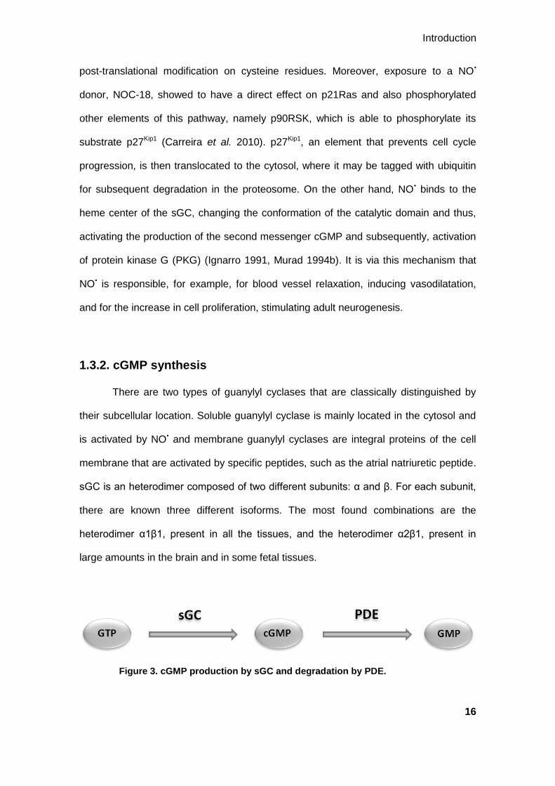
post-translational modification on cysteine residues. Moreover, exposure to a NO•
donor, NOC-18, showed to have a direct effect on p21Ras and also phosphorylated
other elements of this pathway, namely p90RSK, which is able to phosphorylate its
substrate p27Kip1 (Carreira et al. 2010). p27Kip1, an element that prevents cell cycle
progression, is then translocated to the cytosol, where it may be tagged with ubiquitin
for subsequent degradation in the proteosome. On the other hand, NO• binds to the
heme center of the sGC, changing the conformation of the catalytic domain and thus,
activating the production of the second messenger cGMP and subsequently, activation
of protein kinase G (PKG) (Ignarro 1991, Murad 1994b). It is via this mechanism that
NO• is responsible, for example, for blood vessel relaxation, inducing vasodilatation,
and for the increase in cell proliferation, stimulating adult neurogenesis.
1.3.2. cGMP synthesis
There are two types of guanylyl cyclases that are classically distinguished by
their subcellular location. Soluble guanylyl cyclase is mainly located in the cytosol and
is activated by NO• and membrane guanylyl cyclases are integral proteins of the cell
membrane that are activated by specific peptides, such as the atrial natriuretic peptide.
sGC is an heterodimer composed of two different subunits: α and β. For each subunit,
there are known three different isoforms. The most found combinations are the
heterodimer α1β1, present in all the tissues, and the heterodimer α2β1, present in
large amounts in the brain and in some fetal tissues.
Figure 3. cGMP production by sGC and degradation by PDE.

Introduction
17
sGC is activated by NO• and acts on guanosine triphosphate (GTP), converting
it into the second messenger, cGMP, as illustrated in Fig. 3. In physiological conditions,
intracellular cGMP is maintained at basal levels by phosphodiesterases. Cyclic
nucleotide phosphodiesterases (PDE) are enzymes that hydrolyze the 3’-
phosphodiester bound of cyclic adenosine monophosphate (cAMP) or cGMP,
originating their respective monophosphates, 5’-AMP or 5’-GMP, respectively.
1.3.3. Phosphodiesterases
There are 11 known PDE families. Each family encompasses 1 to 4 distinct
genes and each gene encodes multiple protein products, existing more than 50
different PDE proteins in mammalian cells. PDE activity is found in every cell in the
body, presenting different substrate specificity, kinetic properties and cellular and
subcellular distribution of the 11 isoenzymes (Table I). As different PDE families
present such a wide distribution among the tissues, including the brain, inhibition of one
or more PDE is an option to treat several diseases, by controlling the levels of the
respective second messengers. cAMP and cGMP levels can be altered in several
pathologies, such as cancer, inflammation, neurodegeneration and oxidative stress. As
shown in Table I, the PDE isoenzymes display different affinities for either cAMP or
cGMP, being that some are selective for cGMP or for cAMP and others are able to
hydrolyze both substrates with equal or different specificity. Among these various
types, only two selectively hydrolyze cGMP. Here, we focused our studies on PDE5,
because of its involvement in the regulation of cGMP signaling in the brain.

Introduction
18
Table I. Human cyclic nucleotide PDE superfamily (adapted from
Boswell-Smith et al. 2006, Essayan 2001 and Bischoff 2004).
PDE family Substrate specificity Main tissue distribution
1 cGMP>cAMP
Ca2+
/calmodulin-stimulated Heart, brain, lung, smooth muscle
2 cGMP=cAMP Adrenal gland, heart, lung, liver, platelets
3 cAMP>cGMP Heart, liver, lung, platelets, adipose tissue, inflammatory
cells
4 cAMP Sertolli cells, kidney, brain, liver, lung, inflammatory cells
5 cGMP Lung, brain, vascular smooth muscle, platelets
6 cGMP>cAMP Photoreceptors
7 cAMP>>cGMP
(cAMP high affinity)
Skeletal muscle, T lymphocytes, heart, kidney, brain,
pancreas
8 cAMP Testis, eye, liver, skeletal muscle, heart, kidney, ovary,
brain, T lymphocytes
9 cGMP Kidney, liver, lung, brain
10 cGMP<cAMP Brain, testis
11 cGMP=cAMP Skeletal muscle, prostate, liver, kidney, pituitary and
salivary glands, testis
1.3.3.1. cGMP-specific phosphodiesterases
Phosphodiesterases type 5 are enzymes that specifically hydrolyze cGMP. So
far, only one gene of PDE5 was discovered, PDE5A, and three variants of this gene
have been identified: PDE5A1 and PDE5A2 that are widely expressed, and PDE5A3,
which is specific to vascular smooth muscle (Loughney et al. 1998, Lin et al. 2000,
Kotera et al. 1999). PDE5A is considered a cytosolic enzyme and its protein activity
was found in the lung, vascular and tracheal smooth muscle, spleen, platelets, corpus
cavernosum (Lincoln et al. 1976, Coquil et al. 1985, Wallis et al. 1999, Wang et al.
2005, Bender et al. 2004) and in several brain regions, being particularly abundant in
Purkinje cells (Shimizu-Albergine et al. 2003, Bender et al. 2004). Therefore, cGMP-
related physiological functions can be regulated by controlling the levels of PDE5 in
these tissues.

Introduction
19
Moreover, there are other enzymes with high specificity for the cGMP present in
the brain. For example, PDE9 is an enzyme that specifically hydrolyzes cGMP. Until
now there is only one isoform identified, PDE9A. It is one of the more recently
discovered PDE families and very little is known about this enzyme. However, it was
suggested that PDE9A might be a regulator of cGMP in the brain (van Staveren et al.
2002) but more studies are required to understand PDE9 functions in its targets.
PDE10 was also recently discovered and to date, only one gene is known,
PDE10A. PDE10 has higher specificity to cAMP rather than cGMP. This PDE is highly
expressed in the brain, mainly in the striatum, but also in the cerebellum, thalamus,
hippocampus and spinal cord. Furthermore, inhibitors of PDE10 have been developed
for the treatment of schizophrenia.
PDE1 was the first family identified (Cheung 1970) and is a calcium- and
calmodulin-dependent PDE. There are three isoforms of this PDE and its substrate
specificity is different for each isoform, being mainly specific to cGMP. All the isoforms
are present in several brain regions. Therefore, there are few definitive studies on the
functional roles of the various PDE1 isoenzymes.
Although not expressed in the brain, PDE6, is structurally highly homologous to
PDE5 but, contrarily to PDE5, is able to hydrolyze cGMP and cAMP, with higher
specificity to cGMP (Lugnier 2006). There are three genes in the family of this PDE.
PDE6 is highly expressed in the photoreceptors of the mammalian retina, where it
mediates the conversion of a light signal into a photoresponse (Bender et al. 2006).
Taking into account the existing background on the therapeutic effects of the
specific cGMP-hydrolyzing PDE5, namely at the brain level, we decided to focus our
studies on this PDE.
Many PDE5 inhibitors have been developed, being very useful in the study of
PDE5 distribution among tissues and, thus, essential in the treatment of several
pathologies in which the levels of cGMP are altered. The most characterized inhibitor of

Introduction
20
PDE5 is sildenafil. PDE5A is, mostly, a regulator of vascular smooth muscle
contraction through regulation of cGMP in two main tissues: the penis and the lung.
PDE5 inhibition enhances relaxation of the cavernal smooth muscle by nitric oxide and
cGMP, stimulating penile erection (Rosen et al. 2003, Corbin 2004). Therefore, PDE5
inhibitors started to be used for the treatment of erectile dysfunction. In the lung, PDE5
inhibitors act as vasodilators, increasing the blood supply, antagonizing the
vasoconstriction of smooth muscle and decreasing pulmonary arterial resistance.
Therefore, PDE5 inhibitors have been used to treat pulmonary hypertension
(extensively reviewed in Lewis et al. 2004, Patel et al. 2005 and Steiner et al. 2005).
Similarly to pulmonary hypertension, some studies suggest that PDE5 inhibition by
sildenafil prevents the pressure-induced cardiac hypertrophy, even though the levels of
PDE5 in cardiomyocytes are rather low (Takimoto et al. 2005). In the CNS, recent
reports show that sildenafil has a neuroprotective role, being effective in improving the
symptoms in a model of multiple sclerosis, suggesting that PDE5 can be a target for
the therapy against this disease (Pifarre et al. 2011). The various pharmacological
effects of sildenafil are reviewed in Uthayathas et al. 2007.
By its presence in the brain, PDE5 was reported to have a role in learning and
memory. For example, in Alzheimer’s disease, the progressive neurodegeneration
results in a cognitive dysfunction, with memory loss and motoneural impairment. The
administration of PDE5 inhibitors can be a possible therapy for this disease, due to
their ability to reverse deficits in long-term memory caused by pharmacological agents
or aging. It has also been described that the administration of sildenafil enhances
memory and restores learning ability in animal models (Baratti et al. 1999, Devan et al.
2006, Erceg et al. 2005, Prickaerts et al. 2005, Prickaerts et al. 2004, Prickaerts et al.
2002b, Prickaerts et al. 2002a, Rutten et al. 2005, Singh et al. 2003, Devan et al.
2004). Beyond this important role in memory and cognition, PDE5 inhibitors also
Introduction
21
appear to stimulate neuronal plasticity and to be neuroprotective, through the
enhancement of endogenous neurogenesis in the adult brain.
1.3.3.2. Inhibition of PDE5 as a therapeutic strategy
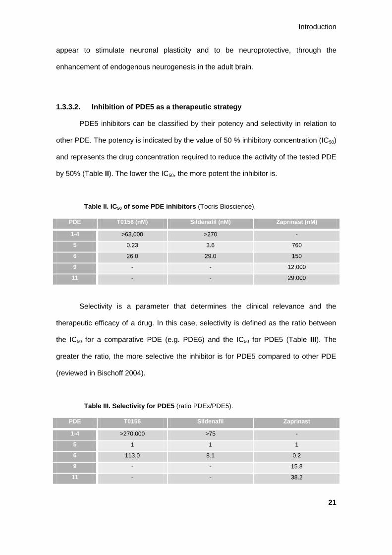
PDE5 inhibitors can be classified by their potency and selectivity in relation to
other PDE. The potency is indicated by the value of 50 % inhibitory concentration (IC50)
and represents the drug concentration required to reduce the activity of the tested PDE
by 50% (Table II). The lower the IC50, the more potent the inhibitor is.
Table II. IC50 of some PDE inhibitors (Tocris Bioscience).
PDE T0156 (nM) Sildenafil (nM) Zaprinast (nM)
1-4 >63,000 >270 -
5 0.23 3.6 760
6 26.0 29.0 150
9 - - 12,000
11 - - 29,000
Selectivity is a parameter that determines the clinical relevance and the
therapeutic efficacy of a drug. In this case, selectivity is defined as the ratio between
the IC50 for a comparative PDE (e.g. PDE6) and the IC50 for PDE5 (Table III). The
greater the ratio, the more selective the inhibitor is for PDE5 compared to other PDE
(reviewed in Bischoff 2004).
Table III. Selectivity for PDE5 (ratio PDEx/PDE5).
PDE T0156 Sildenafil Zaprinast
1-4 >270,000 >75 -
5 1 1 1
6 113.0 8.1 0.2
9 - - 15.8
11 - - 38.2

Introduction
22
Zaprinast (Fig. 4) was the first PDE inhibitor to be used in the studies of PDE5
functions. However, zaprinast proved to be a nonpotent and nonselective inhibitor. Its
inhibitory potency is higher for PDE6 than for PDE5, and it also inhibits other PDE such
as PDE9 and PDE11. Some years later, a more specific PDE5 inhibitor was developed,
sildenafil, commercially known as Viagra. Sildenafil (Fig. 4) was the first oral drug to be
approved by the United States Food and Drug Administration, in 1998, for the
treatment of erectile dysfunction. Sildenafil showed to inhibit PDE5 with concentrations
in the nanomolar range, about 200-fold lower than the concentration of zaprinast
required to inhibit PDE5. Sildenafil is typically considered a PDE5 inhibitor, with an IC50
of approximately 4 nM (Ballard et al. 1998). However, it also inhibits PDE1 and PDE6
(IC50 values of 281 and 29 nM, respectively, (Ballard et al. 1998), Table II), indicating
that sildenafil is not the ideal compound for the selective inhibition of PDE5. Similarly to
sildenafil, other two inhibitors were developed for the treatment of erectile dysfunction:
tadalafil (Cialis) and vardenafil (Levitra). These inhibitors have higher selectivity for
PDE5 than sildenafil, and tadalafil has a much longer period of responsiveness than
sildenafil and vardenafil.
Figure 4. Chemical structures of T0156, sildenafil and zaprinast.
Adapted from Mochida et al. 2002 and Turko et al. 1999.

Introduction
23
More recently, a new compound was developed, T0156 (Fig. 4), which potently
inhibits PDE5 (Mochida et al. 2002). In fact, T0156 has a much lower IC50 for PDE5
(0.23 nM), thus, inhibiting it with higher potency than sildenafil (IC50 3.6 nM) and also
presenting higher selectivity for PDE5 (113.0) in comparison to PDE6, than sildenafil
(8.1) as shown in Tables II and III, respectively. Therefore, the rank order of potency
and, thus, of selectivity for PDE5 is T0156 > sildenafil > zaprinast.
The effects of these inhibitors on the brain are dependent on their permeability
to the BBB. For instance, zaprinast is unable to cross the BBB and, thus, it needs to be
added directly into the brain in order to exert its effects in this organ. On the contrary,
sildenafil crosses the BBB to the brain and can be easily administered (e.g. orally).
Unfortunately, there is no information about the BBB permeability of T0156 but the use
of PDE5 inhibitors as an effective therapy for neurodegenerative diseases is dependent
on this factor.
1.3.3.3. cGMP and neurogenesis
Neurogenesis generally declines with aging and is correlated with the
emergence of neurodegenerative diseases. With the increasing age, the levels of NO•
gradually decrease, leading to a subsequent decrease in the levels of cGMP and an
abolishment of cell proliferation and impairments in learning and memory. Moreover, in
aged rats, levels of cGMP are decreased by the increasing phosphodiesterase activity
comparatively to the adult brain (Chalimoniuk et al. 1998). Therefore, targeting an
enzyme specific for the hydrolysis of cGMP, such as PDE5, is a good strategy to
reverse this process and, thus, enhance neurogenesis.
There are some reports about the effect of sildenafil in the stimulation of
endogenous neurogenesis. Sildenafil was shown to induce neurogenesis in the SVZ
and DG (Zhang et al. 2002) and neuronal function recovery in rats following a stroke
(Zhang et al. 2005) and an ischemic injury either in adult young rats as in aged rats

Introduction
24
(Zhang et al. 2006b). Furthermore, PDE5 inhibition by sildenafil stimulated cell
proliferation in rat SVZ cultures, an effect that appears to be associated with the
activation and phosphorylation of Akt in the phosphatidylinositol 3-kinase (PI3-K)/Akt
pathway (Wang et al. 2005). Another study has demonstrated that SVZ cell
proliferation was enhanced in a rat model of ischemia following the administration of
tadalafil, another PDE5 inhibitor (Zhang et al. 2006a).
Thus, the use of inhibitors for PDE5 deserves further investigation in order to
clarify their role on neurogenesis and to understand the mechanisms behind this effect.
1.4. Objectives
Within the background of the potentially neurogenic effect of sildenafil in the
subventricular zone, our first aim was to study the PDE inhibitors with different
selectivities for PDE5 on the proliferation of SVZ neural stem cells, the first step of
neurogenesis. To address this question, we compared the proliferation of SVZ cells
exposed to T0156, sildenafil or zaprinast.
The pathways that mediate this proliferative effect of PDE5 inhibitors have not
been characterized. As recently described by our group, nitric oxide is known to
stimulate SVZ cell proliferation and this effect is mediated by two main pathways, the
MAPK and the sGC pathways (Carreira et al. 2010, Carreira et al. 2011). Within this
background, our second aim was to address whether these pathways are activated
during cell proliferation triggered by PDE5 inhibition.

25
Chapter 2 Materials and Methods

26

Materials and Methods
27
2.1. Materials
Dulbecco’s Modified Eagle’s Medium: F-12 nutrient mixture, (D-MEM/F-12, with
GlutaMAXTM I), B27 supplement, gentamicin, trypsin-ethylenediaminetetraacetic acid
(EDTA) solution and antibiotics (10,000 units/ml of penicillin, 10 mg/ml streptomycin),
Hoechst 33342, Click-iT® EdU Alexa Fluor® 488 HCS Assay kit and StemPro®
Accutase® cell dissociation reagent were purchased from Invitrogen (Paisley, UK).
Epidermal growth factor (EGF) and fibroblast growth factor (FGF) were from
PeproTech Inc. (London, UK). The Matrix used was from Stoelting Co. (Wood Dale, IL,
USA). Bicinchoninic acid (BCA) Protein Assay kit was from Pierce (Rockford, IL, USA).
Phenylmethylsulfonyl fluoride (PMSF), orthovanadate, chymostatin, leuptin, antiparin,
pepstatin A, trypan blue, Tween-20, tetramethylethylenediamine (TEMED), dimethyl
sulfoxide and KT5823 were purchased from Sigma Chemical (St Louis, MO, USA).
T0156 hydrochloride, ODQ, sildenafil citrate and zaprinast were obtained from Tocris
Bioscience (Bristol, UK) and NOC-18 from Alexis Biochemicals (San Diego, CA, USA).
U0126 was obtained from Cell Signaling (Danvers, MA, USA) and YC-1 from Santa
Cruz Biotechnology (Santa Cruz, CA, USA). Agarose-TUBEs and PR-619 were
obtained from LifeSensors (Malvern, PA, USA). DAKO fluorescent mounting medium
was purchased from DAKO (Glostrup, Denmark). Bovine serum albumin (BSA) was
purchased from Calbiochem (San Diego, CA, USA) and low-fat dry milk from Nestlé
(Vevey, Switzerland). Polyvinylidene difluoride membranes were purchased from
Millipore (Madrid, Spain). Amersham cGMP Enzymeimmunoassay Biotrak System,
enhanced chemifluorescence reagent and anti-rabbit and anti-mouse alkaline
phosphatase-conjugated antibodies were from GE Healthcare Life Sciences
(Buckinghamshire, UK). Other reagents used in immunoblotting experiments were
purchased from Bio-Rad (Hercules, CA, USA). All the antibodies used are described in
Table IV.

Materials and Methods
28
Table IV. Primary and secondary antibodies used in Western blot (WB)
and immunocytochemistry (IC).
Antibody Host Dilution Application Origin
Anti-Sox-2 Mouse 1:200 IC R&D Systems
(Minneapolis, MN, USA)
Anti-β-III-tubulin Mouse 1:500 IC Covance
(Emersonville, CA, USA)
Anti-GFAP Mouse 1:400 IC Invitrogen
(Paisley, UK)
Anti-nestin Mouse 1:500 IC BD biosciences
(Franklin Lakes, NJ, USA)
Anti-musashi-1 Rabbit 1:250 IC Abcam
(Cambridge, UK)
Anti-guanylyl cyclase Rabbit 1:500 WB Abcam
(Cambridge, UK)
Anti-nNOS Mouse 1:500 WB BD biosciences
(Franklin Lakes, NJ, USA)
Anti-p27Kip1
Rabbit 1:1,000 WB Cell Signaling
(Danvers, MA, USA)
Anti-phospho-ERK1/2 Rabbit 1:1,000 WB Cell Signaling
(Danvers, MA, USA)
Anti-ERK 1/2 Rabbit 1:1,000 WB Cell Signaling
(Danvers, MA, USA)
Anti-α-tubulin Mouse 1:10,000 WB Sigma Chemical
(St Louis, MO, USA)
Anti-mouse IgG labeled
with Alexa Fluor 594 Goat 1:200 IC
Invitrogen
(Paisley, UK)
Anti-rabbit IgG labeled
with Alexa Fluor 488 Goat 1:200 IC
Invitrogen
(Paisley, UK)
Alkaline phosphatase-
conjugated anti-rabbit Mouse 1:20,000 WB
GE Healthcare Life Sciences
(Buckinghamshire, UK)
Alkaline phosphatase-
conjugated anti-mouse Mouse 1:20,000 WB
GE Healthcare Life Sciences
(Buckinghamshire, UK)

Materials and Methods
29
2.2. Methods
2.2.1. Animals
C57BL/6J mice were obtained from Charles River (Barcelona, Spain) and kept
in our animal facilities with food and water ad libitum in a 12 hours dark:light cycle. All
experiments were performed in accordance with institutional and European guidelines
(86/609/EEC) for the care and use of laboratory animals.
2.2.2. Subventricular zone cell cultures
Neural stem cell cultures were obtained from the SVZ of 0-3 day C57BL/6J
mice as described previously (Agasse et al. 2008). The brains were removed from the
skull, following decapitation, and placed in a plate dish containing Hank's balanced salt
solution (HBSS, 137 mM NaCl, 5.36 mM KCl, 0.44 mM KH2PO4, 0.34 mM
Na2PO4.2H2O, 4.16 mM NaHCO3, 5 mM glucose, supplemented with 0.001 % phenol
red, 1 mM sodium pyruvate, and 10 mM 4-(2-hydroxyethyl)-1-piperazineethanesulfonic
acid, HEPES, pH 7.4) supplemented with 0.24 % gentamicin. The enveloping
meninges were removed and the brains were sectioned in 1 mm thickness coronal
slices with a mouse brain matrix, from which the SVZ was excised. The sections were
kept in 0.24 % gentamicin/HBSS and the SVZ was isolated from each section. The
fragments of SVZ in 0.24 % gentamicin/HBSS were digested in 0.025 % trypsin /0.265
mM EDTA, for 15-20 minutes at 37 ºC, washed with 0.24 % gentamicin/HBSS and then
mechanically dissociated by gentle dissociation with a pipette tip. The cells were
resuspended in a 37 ºC D-MEM/F-12 with 2 mM GlutaMAXTM-I (L-Ala-L-Gln),
supplemented with 1 % B27, 1 % antibiotic (10,000 units/ml of penicillin, 10 mg/ml
streptomycin), 10 ng/ml EGF and 5 ng/ml FGF, and plated on uncoated flasks with filter
cap at a density of 100,000 cells/ml. Cell viability was evaluated by 0.1 % Trypan blue
exclusion assay. The SVZ stem cells were grown as floating aggregates in a 95 % air/5

Materials and Methods
30
% CO2 humidified atmosphere at 37 ºC, during approximately 7 days. Then, the primary
neurospheres were harvested, centrifuged and mechanically dissociated as single
cells. Cells were replated as above and allowed to grow as secondary neurospheres.
6-7 days later, the floating neurospheres were collected and plated for 2-3 days on
poly-L-lisine-coated 16-mm diameter glass coverslips, for immunocytochemistry
assays, or on 12-well plates for preparation of lysates or flow cytometry assays, in the
same medium as above. Then, the medium was exchanged by a similar medium but
without growth factors (EGF and FGF) and cells were kept in this medium for 24 h
before the experiments.
2.2.3. Experimental treatments
SVZ cells were left 24 h without growth factors before applying the stimuli. For
cell proliferation analysis, a 6-hour and 24-hour treatment was applied, except for YC-1
experiments, in which cell proliferation was only analyzed following 24 h of treatment.
To analyze the phosphorylation of ERK 1/2 and the levels of p27Kip1, SVZ neural stem
cells were treated for 2 h, or as indicated in the figure legends. In both experiments, the
stimuli applied were as follows: the PDE inhibitors, T0156 1 µM, sildenafil 1 µM and
zaprinast 10 µM, and the sGC activator, YC-1 20 µM, were applied alone or together
with the MEK 1/2 inhibitor, U0126 1 µM, the PKG inhibitor, KT5823 1 µM, and the sGC
inhibitor, ODQ 50 µM. U0126, KT5823 and ODQ were applied 30 min before the
treatment with the PDE5 inhibitors and YC-1.
For the experiments of pulldown of ubiquitilated p27Kip1, cells were treated with
10 µM NOC-18 for 1 h and with T0156 (1 µM), sildenafil (1 µM) or zaprinast (10 µM) for
2 h.
For the determination of cGMP levels, cells were treated with T0156 (1 µM),
sildenafil (1 µM) or zaprinast (10 µM) for 6 h.
Materials and Methods
31
All the experiments were performed together with the respective controls
(untreated cells).
2.2.4. Immunocytochemistry
The culture medium was removed and the SVZ cells were washed with
phosphate-buffered saline 0.01 M (PBS, 7.8 mM Na2HPO4.2H2O, 2.7 mM
NaH2PO4.H2O, 154 mM NaCl, pH 7.2) and fixed with 4 % paraformaldehyde/4 %
sucrose in PBS 0.01 M, for 20 min, and washed again with PBS 0.01 M. Fixed cells
were permeabilized with Triton X-100 (1 % Triton in PBS 0.01 M), for 5 min, and the
non-specific binding was blocked with albumin solution (3 % BSA, and 0.2 % Tween-20
in PBS 0.01 M) at room temperature, for 1 h. The incubation with primary antibodies in
3% albumin solution was performed overnight, at 4ºC. The primary antibodies and the
dilutions used were as follows: mouse anti-Sox-2, 1:200; mouse anti-β-III-tubulin,
1:500; mouse anti-GFAP, 1:400; mouse anti-nestin, 1:500 and rabbit anti-musashi-1,
1:250. After rinsing with PBS 0.01 M, the cells were exposed to the appropriate
secondary antibodies, anti-mouse or anti-rabbit IgGs conjugated with Alexa Fluor 488
or 594, 1:200, in 3 % albumin solution, for 90 min, at room temperature. Nuclei were
stained with Hoechst 33342 (2 µg/ml) for 10 min. Cells were washed with PBS 0.01 M
and mounted in uncoated glass slides with DAKO fluorescence mounting medium.
Images were acquired in a laser scanning microscope LSM 510 META (Zeiss, Jena,
Germany). Results of labeled cells are expressed in percentage of total of live cells of 3
independent experiments.
2.2.5. Analysis of cell proliferation by flow cytometry
SVZ neural stem cells proliferation was assessed by incorporation of ethynyl-2’-
deoxyuridine (EdU) using the Click-iT® EdU Alexa Fluor® 488 HCS Assay kit. For the

Materials and Methods
32
6 h stimuli, 10 µM EdU (available in the kit) was added to the SVZ cultures at the same
time of the treatment and for the 24 h treatment, EdU was added 4 h before cell fixation
(20 h after stimuli). For fixation, cells were washed with sterile 0.01 M PBS and then
sterile StemPro® Accutase® cell dissociation reagent was added to cells for 20 min, at
37 ºC. Cells were detached from the plate by gently pipetting and harvested into flow
tubes. Following centrifugation at 180 x g for 20 min, the supernatant was discarded
and cells were resuspended in 70 % ethanol overnight, at 4 ºC, as described previously
(Carreira et al. 2010). Ethanol acts as a permeability and fixation agent,
simultaneously. Cells can be stored in 70 % ethanol and used for flow cytometry for a
maximum of 4 days.
Detection of EdU incorporation was based on click chemistry, a copper
catalyzed covalent reaction between an azide (conjugated with the Alexa Fluor 488
fluorophore) and an alkyne (EdU). Fixed cells were pelleted by centrifugation at 218 x g
for 20 min and the supernatant was discarded. A rinse with PBS 0.01 M was
performed, followed by centrifugation at 218 x g for 15 min and the supernatant was
discarded again. The reaction cocktail (Alexa Fluor® 488 azide, copper sulfate, 1x
Click-iT reaction buffer and 1 x reaction buffer additive, available from the kit) was
added and cells incubated for 30 minutes with the azide conjugate and copper
sulphate, at room temperature, protected from light. After the reaction, the cells were
pelleted at 218 x g for 15 min. The supernatant was removed and a new rinse with PBS
0.01 M was performed (218 x g, 15 min). The supernatant was discarded and PBS
0.01 M was added. Cells were incubated with ribonuclease A and the nuclear dye 7-
actinomycin D (7-AAD), also available in the kit, for 30 min, at room temperature,
protected from light. Ribonuclease A is used to ensure that 7-AAD only binds to DNA.
The EdU incorporation was detected on a BD FACScaliburTM Flow Cytometer, using
the Cellquest Pro software, version 0.3.efab (Becton Dickinson, San Jose, CA, USA).
Thirty thousand events were acquired per each experiment in the region of interest

Materials and Methods
33
(including apoptosis, G0/G1, S and G2/M). A minimum of 4 independent experiments
was analyzed for each condition. Data were analyzed using the WinMDI2.9 software
and are presented as means ± SEM of the number of non-apoptotic cells that
incorporated EdU (% of control).
2.2.6. Preparation of cytosolic lysates
The treated cells were washed with PBS 0.01 M and scraped and lysed in 50
mM Tris-HCl, 0.15 M NaCl, 1 mM EDTA, 1 % Igepal and 10 % glycerol, supplemented
with 200 µM PMSF, 1 µg/ml CLAP (chymostatin, leupeptin, antiparin, pepstatin A), 1
mM sodium orthovanadate, 1 µM dithiothreitol and 5 mM NaF, pH 7.5, 4ºC. Three
freezing/thawing cycles followed by five 5-second sonication cycles were applied and
the lysate was clarified by centrifugation at 14,000 x g, 4ºC. The cytosolic fraction was
collected and the pellet (nuclear fraction) was discarded. Protein concentration was
determined by the BCA method, according to manufacturer’s instructions. 6x
concentrated sample buffer was added and samples were denatured at 95ºC for 5 min.
Samples were analyzed by Western blot.
2.2.7. Western blot analysis
Samples destined for Western blot analysis were electrophoresed in sodium
dodecyl sulfate (SDS)-polyacrylamide gels using MiniPROTEAN® 3 systems (Bio-Rad
Laboratories). Resolving gels were composed by 12 % bis-acrylamide, 25% Tris-HCl
1.5 M pH 8.0, 0.1 % SDS, 0.05 % TEMED and 0.05 % ammonium persulfate, in milliQ
water, for all western blot experiments except for the identification of nNOS and sGC
that resolving gels contained 8 % bis-acrylamide. Stacking gels were composed by 4 %
bis-acrylamide, 25 % Tris-HCl 0.5 M pH 6.8, 0.1 % SDS, 0.05 % TEMED and 0.05 %
ammonium persulfate, in milliQ water. Equal amounts of protein were applied on each

Materials and Methods
34
lane of the SDS-polyacrylamide gels submerged in a running buffer (25 mM Tris, 25
mM bicine and 0.1 % SDS, in milliQ water). Proteins were separated by
electrophoresis, firstly at 60 V for 10 min and then at 120 V until proper bands
separation were reached. A molecular ladder was used to control molecular weight
separation. The polyvinylidene difluoride membranes were activated, first in 100 %
methanol (2.5 to 5 min), followed by water and finally 15 to 30 min in
electrotransference buffer (CAPS 10 mM, methanol 10 %, pH 11.0). Proteins were
electrophoretically transferred to the activated membranes submerged in
electrotransference buffer at 750 mA, for 90 min, at 4ºC, using the Trans-Blot Cell
apparatus (Bio-Rad Laboratories). Membranes were blocked by 1 h incubation, at room
temperature, with Tris-buffered saline (137 mM NaCl, 20 mM Tris-HCl, pH 7.6)
containing 0.1 % Tween-20 (TBS-T) and 5 % low-fat dry milk or, for phosphorylated
proteins, 3 % BSA. Incubations with the primary antibodies (rabbit phospho-ERK 1/2,
rabbit anti-p27kip1, 1:1,000; mouse anti-nNOS, rabbit anti-sGC, 1:500) in TBS-T
containing 1 % blocking solution were performed overnight, at 4ºC. After rinsing with
TBS-T (20 min, with 2 quick washes before and after), incubation with the appropriated
alkaline phosphatase-linked secondary antibodies (anti-rabbit or anti-mouse, 1:20,000
in TBS-T containing 1 % blocking solution) was performed at room temperature, during
1 h. After extensive washing in TBS-T (for 1 h, changing into new TBS-T every 20 min),
followed by the incubation of the membranes with the enhanced chemifluorescence
reagent for the maximum of 5 min, immunoreactive bands were visualized in the
VersaDoc 3000 imaging system (Bio-Rad, Hercules, CA, USA). Data were analyzed
with the Quantity One software version 4.6.9 (Bio-Rad Laboratories). Protein control
loadings were either performed after membranes reactivation (5-10 s in 100 %
methanol and 20 min in TBS-T) using primary antibodies against rabbit ERK 1/2
(1:1,000) or mouse α-tubulin (1:10,000). The protocol used was the same as explained
above.

Materials and Methods
35
2.2.8. Affinity binding precipitation of ubiquitinated p27Kip1
SVZ cells were treated as explained above for 2 h, at 37ºC. Cells were briefly
washed with PBS 0.01 M and cytosolic lysates were then prepared by adding 500 µl of
lysis buffer (50 mM Tris-HCl, 0.15 M NaCl, 1 mM EDTA, 1 % Igepal and 10 % glycerol,
supplemented with 200 µM PMSF, 1 µg/ml CLAP, 1 mM sodium orthovanadate, 5 mM
NaF and 10 µM PR-619, pH 7.5, 4ºC) and scraping on ice. Freezing/thaw cycles
followed by sonication cycles were applied and the lysate was clarified by
centrifugation at 14,000 x g, 4ºC. The supernatant was collected and protein
concentration was determined by the BCA method using the BCATM Protein Assay kit,
following the manufacturer’s instructions. An input of 50 µg of protein was removed
from each sample, for control of the cellular levels of p27Kip1.
Agarose-TUBEs are Tandem Ubiquitin Binding Entities moieties that are
coupled to agarose beads and are used for the identification and characterization of
ubiquitinated proteins by Western blotting or downstream proteomic studies.
Agarose-TUBEs were equilibrated by inverting the vial several times to ensure a
homogeneous solution and the necessary volume was collected and washed three
times with TBS-T (20 mM Tris-HCl, 0.15 M NaCl, 0.1 % Tween-20, pH 8.0) followed by
centrifugation at 1,000 x g at 4ºC. 600 µg of protein of each lysate were added to
different tubes with the equilibrated slurry of agarose beads and incubated for 4 h at
4ºC, with agitation. The beads were collected by centrifugation at 1,500 x g and the
supernatant was saved as the unbound fraction. Both, input and unbound fraction,
were treated with 6 x concentrated sample buffer (0.5 M Tris-HCl/0.4 % SDS pH 6.8,
30 % glycerol, 10 % sodium dodecyl sulfate, 0.6 M dithiothreitol, 0.012 % bromophenol
blue) and heated at 95ºC for 5 min. The agarose beads were resuspended in 50 µl of 2
x concentrated sample buffer followed by heating at 95ºC for 5 min. The bound fraction
was separated from the beads by centrifugation at 13,000 x g. The ubiquitinated
proteins were analyzed by Western blot in parallel with inputs and unbound fractions to

Materials and Methods
36
evaluate the levels of p27Kip1. For the inputs 20 µg of protein were applied in the gel
and 50 µg of protein in the case of the unbound fraction. As to the bound fraction, half
of the final volume was used, i.e, 25 µl. The protocol used for Western blot was the
same as explained above. The primary antibody used was the rabbit anti-p27kip1,
1:1,000 and protein control loadings were performed using a primary antibody against
mouse α-tubulin (1:10,000).
2.2.9. Determination of cGMP levels
The cGMP levels in cultured SVZ stem cells were determined after exposure to
drugs for 6 h, using a cGMP Enzymeimmunoassay Biotrak System. This experimental
assay is based on competition between unlabelled cGMP from cell lysates and a fixed
quantity of peroxidase-labelled cGMP, for a limited number of binding sites on a cGMP
specific antibody coated on a 96-well plate. Extraction and measurement was
performed according to manufacturer’s instructions. Briefly, cells were lysed with lysis
buffer 1, available from the kit, which hydrolyses cell membranes to release
intracellular cGMP. Cell lysis was facilitated by shaking during 10 min, followed by
scraping. The concentrated samples were resuspended in assay buffer and acetylated
in order to increase the sensitivity of the assay. Cell lysates were then immediately
used for the enzyme immunoassay protocol, as previously described (Araujo et al.
2003). Optical density was read at 450 nm on a plate reader. All the experiments were
carried out in duplicate. The results are expressed as fentomoles per million of cells.
2.2.10. Data analysis
Data are expressed as means ± SEM. Statistical significance was determined
by using two-tailed t tests or one-factor analysis of variance (ANOVA) followed by
Bonferroni’s or Dunnett’s post-tests, as appropriate and indicated in the figure legends

Materials and Methods
37
and in the text. Differences were considered significant when p<0.05. The software
used was GraphPad Prism 5.0 (GraphPad Software, La Jola, CA, USA).

Materials and Methods
38

39
Chapter 3 Results

40

Results
41
3.1. Characterization of SVZ cell cultures
3.1.1. NSC are predominant in the SVZ
Neural stem cells isolated from the subventricular zone and cultured as floating
aggregates were plated on poly-L-lysine-coated coverslips for 4 days and were
characterized at this stage. The cells were stained against Sox-2, a transcription factor
essential to maintain self-renewal of undifferentiated stem cells and musashi-1, an
evolutionary conserved RNA binding protein with maximum expression in proliferating
multipotent neural precursor cells, decreasing during neuronal differentiation. The
percentage of Sox-2-positive (80.2 % ± 3.2 %) and musashi-1-positive cells (83.5 % ±
4.3 %) was approximately 80 %, suggesting that the majority of cells remained
undifferentiated after plating. The percentage of double-labeled cells was
approximately 100 %, showing that both, Sox-2 and musashi-1, are good markers of
undifferentiated cells (Fig. 5A-5C). SVZ cells were also stained against the neural
precursor marker, nestin, the glial fibrillary acidic protein (GFAP), a marker for
astrocytes that also labels neural stem cells, and the neuron-specific marker, β-III-
tubulin (Tuj1). Only 0.5 % of cells were positive for β-III-tubulin (0.5 % ± 0.1 %; Fig. 5J-
5L), suggesting that very few cells were differentiated into neurons at this stage.
Moreover, approximately 20 % of live cells were GFAP positive (21.5 % ± 3.7 %; Fig.
5G-5I) and almost 50 % expressed nestin (46.9 % ± 2.0 %; Fig. 5D-5F). Furthermore,
previous studies by our group showed that about 70 % of cells colocalize for nestin and
GFAP (Carreira et al. 2010).

Results
42
Figure 5. SVZ cultures are enriched in markers of NSC. The micrographs
show laser scanning confocal images of SVZ cells labeled against musashi-
1 (A, green), Sox-2 (B, red), nestin (D, red), GFAP (G, red) and Tuj1 (J, red).
Nuclei were labeled with Hoechst 33342 (E, H and J, blue). Merged images
are shown in C, F, I and L. Scale bars: 20 µM.

Results
43
3.1.2. Presence of NO• and cGMP-producing enzymes
In order to confirm that sGC and nNOS were present in the SVZ cultures, we
evaluated the presence of these proteins by Western blot. In fact, sGC was present in
SVZ cells (Fig. 6A). Likewise, the presence of the neuronal isoform of the nitric oxide
synthase (nNOS) was evaluated. In agreement with sGC, nNOS was also present in
the SVZ cultures (Fig. 6B), meaning that endogenous NO• is produced in these cells.
Furthermore, previous work by our group showed that these cells do not express iNOS
or eNOS (B.Carreira, unpublished observations).
Figure 6. sGC and nNOS are present in the SVZ stem cell cultures.
cGMP (A) and NO•-producing enzymes (B), respectively, can be detected in
SVZ cultures by Western blot, using 40 µg of protein.
3.2. Modulation of SVZ cell proliferation by PDE inhibitors
3.2.1. PDE inhibitors stimulate neural stem cell proliferation
To initiate the studies with the three PDE5 inhibitors selected, different
concentrations were tested for sildenafil and zaprinast. For the more selective inhibitor
for PDE 5, T0156, the concentration used was the more used in the literature and used
by our group in previous studies, 1 µM. A treatment of 6 h was performed in the
presence of the PDE5 inhibitors. EdU incorporation was assessed in a flow cytometer.

Results
44
Figure 7. T0156, sildenafil and zaprinast stimulate the proliferation of
SVZ neural stem cells following a 6 h exposure. Cells were treated for 6 h
with 1 µM T0156 (A), 1 µM, 10 µM and 50 µM sildenafil (B) or 10 µM and 50
µM zaprinast (C). The incorporation of EdU was assessed by flow cytometry.
Data are expressed as means ± SEM of at least five independent
experiments. Two-tailed t test, *p<0.05, significantly different from control,
and one-way ANOVA (Dunnett’s post-test), *p<0.05 and **p<0.01,
significantly different from control.
All the PDE5 inhibitors showed an increase in the proliferation of SVZ cells.
Treatment with T0156 (1 µM) significantly increased the incorporation of EdU (140.7 %
± 11.9 %, p<0.05; Fig. 7A), when compared to untreated cells. In the case of sildenafil,
the three concentrations tested increased EdU incorporation (1 µM, 137.4 % ± 7.0 %,
p<0.05; 10 µM, 120.8 % ± 10.4 %, p>0.05; 50 µM, 141.3 % ± 12.6 %, p<0.01; Fig. 7B)
as well as the two concentrations tested for zaprinast (10 µM, 125.6 % ± 7.8 %,
p<0.05; 50 µM, 118.1 % ± 5.6 %, p>0.05; Fig. 7C), in comparison to untreated cells. In
agreement with this, 1 µM sildenafil and 10 µM zaprinast were selected for all the
subsequent experiments.
In order to investigate whether this effect was maintained in time, the selected
concentrations were tested for a longer period of exposure. SVZ cells were treated for

Results
45
24 h with the chosen concentrations of the PDE5 inhibitors and EdU incorporation was
evaluated by flow cytometry. The data are shown in Fig. 8.
Figure 8. PDE5 inhibitors enhance SVZ cell proliferation following a
treatment of 24 h. Cells were treated for 24 h with 1 µM T0156 (A) 1 µM
sildenafil (B) or 10 µM zaprinast (C). The incorporation of EdU was
assessed by flow cytometry. Data are expressed as means ± SEM of at least
five independent experiments. Two-tailed t test, *p<0.05 and **p<0.01,
significantly different from control.
In agreement with the results for 6 h of treatment, following 24 h of treatment,
T0156 (1 µM; 135.6 % ± 7.3 %, p<0.01; Fig. 8A), sildenafil (1 µM; 130.6 % ± 9.4 %,
p<0.05; Fig. 8B) and zaprinast (10 µM; 146.5 % ± 13.9 %, p<0.05; Fig. 8C) also
increased SVZ cell proliferation, in comparison to untreated cells.
3.2.2. Treatment with PDE5 inhibitors increases cGMP levels
In order to confirm that the stimulation of cell proliferation was due to an
increase in the levels of cGMP by inhibition of PDE5, cGMP levels were measured
using an immunoenzymatic commercial kit. SVZ cells were treated with T0156,
sildenafil and zaprinast, according to the concentrations selected above.

Results
46
Table V. PDE5 inhibitors increase the levels of cGMP in SVZ cells.
Treatment cGMP levels (fmol/106 cells)
Control 7.4 ± 0.9
T0156 29.7 ± 4.2*
Sildenafil 28.8 ± 5.1*
Zaprinast 33.1 ± 6.1**
Cells were treated for 6 h with 1 µM T0156, 1 µM sildenafil or 10 µM zaprinast. Data
are expressed as means ± SEM of two independent experiments. One-way ANOVA
(Dunnett´s post-test), *p<0.05 and **p<0.01, significantly different from control.
In fact, cGMP levels were significantly increased in cultures exposed to T0156
(29.7 ± 4.2 fmol/106 cells, p<0.05), sildenafil (28.8 ± 5.1 fmol/106 cells, p<0.05) or
zaprinast (33.1 ± 6.1 fmol/106 cells, p<0.01) in comparison to the values of cGMP
levels in untreated cells (7.4 ± 0.9 fmol/106 cells). The data are shown in Table V.
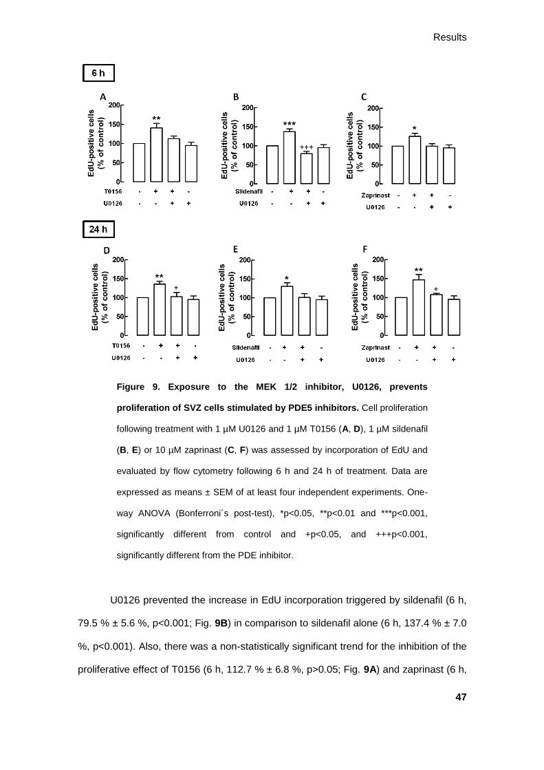
3.2.3. Involvement of the MAPK and sGC pathways on SVZ cell
proliferation
3.2.3.1. Inhibition of the MAPK pathway abolishes the proliferative effect of the
PDE5 inhibitors
With the purpose of evaluating the involvement of the MAPK/ERK 1/2 pathway
on the proliferation stimulated by the PDE5 inhibitors, we treated SVZ cultures with
U0126, the inhibitor of MEK 1/2, the kinase immediately upstream to ERK 1/2. In the
presence of U0126, together with the PDE5 inhibitors, the percentage of EdU
incorporation decreased in comparison to what happened when T0156, sildenafil or
zaprinast were administered alone.
Results
47
Figure 9. Exposure to the MEK 1/2 inhibitor, U0126, prevents
proliferation of SVZ cells stimulated by PDE5 inhibitors. Cell proliferation
following treatment with 1 µM U0126 and 1 µM T0156 (A, D), 1 µM sildenafil
(B, E) or 10 µM zaprinast (C, F) was assessed by incorporation of EdU and
evaluated by flow cytometry following 6 h and 24 h of treatment. Data are
expressed as means ± SEM of at least four independent experiments. One-
way ANOVA (Bonferroni´s post-test), *p<0.05, **p<0.01 and ***p<0.001,
significantly different from control and +p<0.05, and +++p<0.001,
significantly different from the PDE inhibitor.
U0126 prevented the increase in EdU incorporation triggered by sildenafil (6 h,
79.5 % ± 5.6 %, p<0.001; Fig. 9B) in comparison to sildenafil alone (6 h, 137.4 % ± 7.0
%, p<0.001). Also, there was a non-statistically significant trend for the inhibition of the
proliferative effect of T0156 (6 h, 112.7 % ± 6.8 %, p>0.05; Fig. 9A) and zaprinast (6 h,

Results
48
100.1 % ± 6.0, p>0.05; Fig.9C) by U0126, keeping cell proliferation similar to basal
levels, in comparison to T0156 (6 h, 140.7 % ± 11.9 %, p<0.01) and zaprinast (6 h,
125.6 % ± 7.8 %, p<0.05) alone, respectively.
Similar results were obtained following 24 h of treatment. In the presence of
U0126, the increase in the incorporation of EdU by T0156 (24 h, 102.5 % ± 10.7 %,
p<0.05; Fig. 9D) and zaprinast (24 h, 107.6 % ± 3.4 %, p<0.05; Fig. 9F) was prevented,
comparatively to T0156 (24 h, 135.6 % ± 7.3 %, p<0.01) and zaprinast (24 h, 146.5 %
± 13.9 %, p<0.01) alone, respectively. Moreover, U0126 appeared to prevent the
increase in EdU incorporation by sildenafil (24 h, 101.2 % ± 9.9 %, p>0.05; Fig. 9E) in
comparison to sildenafil alone (24 h, 130.6 % ± 9.4 %, p<0.05), despite the lack of
statistical significance.
3.2.3.2. sGC inhibition affects the proliferative effect of PDE5 inhibitors
To analyze the activation of the sGC pathway on cell proliferation stimulated by
PDE5 inhibition, SVZ cells were exposed to the sGC inhibitor, ODQ. Together with the
PDE5 inhibitors, ODQ appeared to have an inhibitory role on cell proliferation, mainly
upon a 6 h treatment.
Following 6 h of treatment, ODQ prevented SVZ cell proliferation stimulated by
T0156 (6 h, 105.5 % ± 7.9 %, p<0.05; Fig. 10A), in comparison to T0156 alone (6 h,
140.7 % ± 11.9 %, p<0.01). Moreover, although not statistically significant, the
treatment with ODQ appeared to inhibit the proliferative effect of sildenafil (6 h, 108.9
% ± 15.2 %, p>0.05; Fig. 10B) and zaprinast (6 h, 113.4 % ± 9.7 %, p>0.05; Fig. 10C)
in comparison to sildenafil (6 h, 137.4 % ± 7.0 %, p<0.05) and zaprinast (6 h, 125.6 %
± 7.8 %, p<0.05) alone, respectively.

Results
49
Figure 10. sGC inhibition by ODQ appears to abolish the proliferation
of SVZ cells stimulated by PDEs inhibitors following 6 h of treatment.
Cell proliferation following treatment with 50 µM ODQ and 1 µM T0156 (A,
D), 1 µM sildenafil (B, E) or 10 µM zaprinast (C, F) was assessed by
incorporation of EdU and evaluated by flow cytometry, following 6 h and 24 h
treatment. Data are expressed as means ± SEM of at least four independent
experiments. One-way ANOVA (Bonferroni´s post-test), *p<0.05 and
**p<0.01, significantly different from control and +p<0.05, significantly
different from the PDE inhibitor.
Following 24 h of treatment, ODQ appeared to be ineffective in preventing the
increase of EdU-positive cells by T0156 (24 h; 121.9 % ± 10.9 %, p>0.05; Fig. 10D)
and sildenafil (24 h; 125.7 % ± 8.7 %, p>0.05, Fig. 10E) in comparison to T0156 (24 h,
135.6 % ± 7.3 %, p<0.01) and sildenafil (24 h, 130.6 % ± 9.4 %, p>0.05) alone,

Results
50
respectively. However, following the same period of treatment, the proliferative effect of
zaprinast was prevented in the presence of ODQ (24 h, 105.1 % ± 10.1 %, p<0.05, Fig.
10F) when compared to zaprinast alone (24 h, 146.5 % ± 13.9 %, p<0.01).
3.2.3.3. PKG inhibition abolishes the proliferative effect of PDE5 inhibitors
We next studied the effect of the inhibition of PKG, using the PKG inhibitor
KT5823, on the proliferation triggered by PDE5 inhibition. PKG is activated by cGMP,
inducing cell proliferation. Therefore, following 6 h of treatment with KT5823, the
increase in the incorporation of EdU by T0156 (6 h, 102.6 % ± 7.1 %, p<0.05; Fig.
11A), sildenafil (6 h, 101.1 % ± 9.0 %, p<0.01; Fig. 11B) and zaprinast (6 h, 99.5 % ±
7.4 %, p<0.05; Fig. 11C) was prevented, being cell proliferation similar to basal levels,
comparatively to T0156 (6 h, 140.7 % ± 11.9 %, p<0.01), sildenafil (6 h, 137.4 % ± 7.0
%, p<0.001) and zaprinast (6 h, 125.6 % ± 7.8 %, p<0.05) alone, respectively.
Similarly, following 24 h of treatment with KT5823, the increase in cell
proliferation induced by T0156 (24 h, 112.1 % ± 8.3 %, p>0.05; Fig. 11D) and sildenafil
(24 h, 107.6 % ± 6.0 %, p>0.05; Fig. 11E) is decreased, in comparison to T0156 (24 h,
135.6 % ± 7.3 %, p<0.05) and sildenafil (24 h, 130.6 % ± 9.4 %, p<0.05) alone,
respectively, even though the lack of statistical significance. Likewise, KT5823
prevented the increase of EdU incorporation triggered by zaprinast (24 h, 108.1 % ±
4.9 %, p<0.05; Fig. 11F) when compared to zaprinast alone (24 h, 146.5 % ± 13.9 %,
p<0.05).

Results
51
Figure 11. Inhibition of PKG, by KT5823, inhibits proliferation of SVZ
cells stimulated by the PDE5 inhibitors. Cell proliferation following
treatment with 1 µM KT5823 and 1 µM T0156 (A, D), 1 µM sildenafil (B, E)
or 10 µM zaprinast (C, F) was assessed by incorporation of EdU and
evaluated by flow cytometry, following 6 h and 24 h of treatment. Data are
expressed as means ± SEM of at least four independent experiments. One-
way ANOVA (Bonferroni´s post-test), *p<0.05, **p<0.01 and ***p<0.001,
significantly different from control and +p<0.05 and ++p<0.01 significantly
different from the PDE5 inhibitor alone.
3.2.4. Direct activation of sGC stimulates SVZ cell proliferation
In order to test the effects of the direct activation of sGC, SVZ cells were treated
with 20 µM YC-1, a NO•-independent activator of sGC. Cell proliferation was assessed
by flow cytometry following a treatment of 24 h with YC-1. Exposure to YC-1 increased
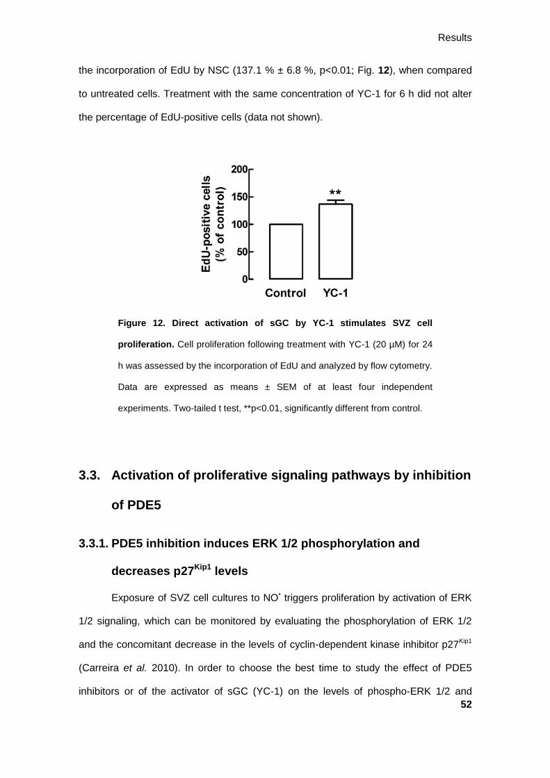
Results
52
the incorporation of EdU by NSC (137.1 % ± 6.8 %, p<0.01; Fig. 12), when compared
to untreated cells. Treatment with the same concentration of YC-1 for 6 h did not alter
the percentage of EdU-positive cells (data not shown).
Figure 12. Direct activation of sGC by YC-1 stimulates SVZ cell
proliferation. Cell proliferation following treatment with YC-1 (20 µM) for 24
h was assessed by the incorporation of EdU and analyzed by flow cytometry.
Data are expressed as means ± SEM of at least four independent
experiments. Two-tailed t test, **p<0.01, significantly different from control.
3.3. Activation of proliferative signaling pathways by inhibition
of PDE5
3.3.1. PDE5 inhibition induces ERK 1/2 phosphorylation and
decreases p27Kip1 levels
Exposure of SVZ cell cultures to NO• triggers proliferation by activation of ERK
1/2 signaling, which can be monitored by evaluating the phosphorylation of ERK 1/2
and the concomitant decrease in the levels of cyclin-dependent kinase inhibitor p27Kip1
(Carreira et al. 2010). In order to choose the best time to study the effect of PDE5
inhibitors or of the activator of sGC (YC-1) on the levels of phospho-ERK 1/2 and

Results
53
p27Kip1, we treated the SVZ cultures with these drugs for different periods of time. Cells
were treated with T0156 (1 µM; Fig. 13A), sildenafil (1 µM; Fig. 13B), zaprinast (10 µM;
Fig. 13C), or YC-1 (20 µM; Fig. 13D) for 30 min, 1 h or 2 h. Treatment with T0156,
zaprinast and YC-1 increased ERK 1/2 phosphorylation following 2 h of treatment.
Furthermore, the cellular levels of p27Kip1 appear to be decreased, mainly following a 2
h treatment with the more selective inhibitor for PDE5 (T0156). According to these
results, we selected a treatment of 2 h for the following experiments.
Figure 13. PDE5 inhibitors increase ERK 1/2 phosphorylation and
decrease the levels of p27Kip1
following 2 h of treatment. SVZ stem cells
were treated with 1 µM T0156 (A), 1 µM sildenafil (B), 1 µM zaprinast (C)
and 20 µM YC-1 (D) for different times: 30 min, 1h and 2h.
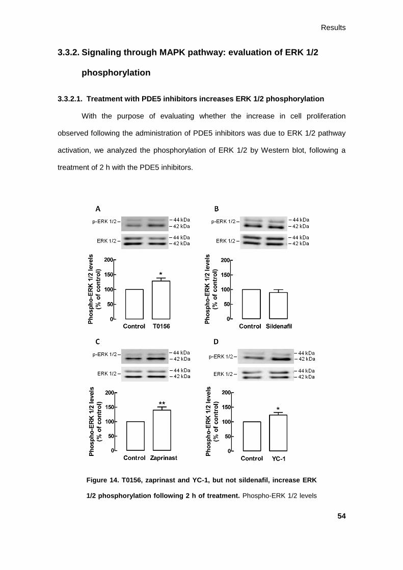
Results
54
3.3.2. Signaling through MAPK pathway: evaluation of ERK 1/2
phosphorylation
3.3.2.1. Treatment with PDE5 inhibitors increases ERK 1/2 phosphorylation
With the purpose of evaluating whether the increase in cell proliferation
observed following the administration of PDE5 inhibitors was due to ERK 1/2 pathway
activation, we analyzed the phosphorylation of ERK 1/2 by Western blot, following a
treatment of 2 h with the PDE5 inhibitors.
Figure 14. T0156, zaprinast and YC-1, but not sildenafil, increase ERK
1/2 phosphorylation following 2 h of treatment. Phospho-ERK 1/2 levels
Results
55
following treatment with 1 µM T0156 (A), 1 µM sildenafil (B), 10 µM
zaprinast (C) and 20 µM YC-1 (D) for 2 h were assessed by Western blot.
Data are expressed as means ± SEM of at least 6 independent experiments.
Two-tailed t test, *p<0.05 and **p<0.01, significantly different from control.
In fact, T0156 (1 µM; 128.5 % ± 10.0 %, p<0.05; Fig. 14A) and zaprinast (10
µM; 140.0 % ± 11.0 %, p<0.01; Fig. 14C) significantly increased ERK 1/2
phosphorylation in comparison to control. However, treatment with sildenafil (1 µM;
89.9 % ± 9.8 %, p>0.05; Fig. 14B) did not increase the levels of phospho-ERK 1/2 in
relation to basal levels. Furthermore, treatment with YC-1 increased ERK 1/2
phosphorylation (123.3 % ± 8.9 %, p<0.05; Fig. 14D), in comparison to untreated cells.
3.3.2.2. Inhibition of PKG prevents the increase in the phospho-ERK 1/2 levels
In order to evaluate whether the inhibition of PKG was able to antagonize the
effect of the PDE inhibitors and the sGC activator, samples obtained following 2 h of
treatment were analyzed by Western blot.
Treatment with KT5823 appeared to suppress the increase of phospho-ERK 1/2
levels by T0156 (107.2 % ± 6.2 %, p>0.05; Fig. 15A) and zaprinast (110.5 % ± 7.9 %,
p>0.05; Fig. 15C) in comparison to T0156 (128.5 % ± 10.0 %, p<0.05) and zaprinast
(140.0 % ± 11.0 %, p<0.05) alone, respectively. On the contrary, treatment with
KT5823 did not appear to decrease the levels of phospho-ERK 1/2 in the samples
treated with sildenafil (120.2 % ± 11.0 %, p>0.05; Fig. 15B) when compared to
sildenafil alone (89.9 % ± 9.8 %, p>0.05).
Moreover, inhibition of PKG prevented the increase of phospho-ERK 1/2 levels
by exposure to YC-1 (85.6 % ± 10.4 %, p<0.05; Fig. 15D) in comparison to YC-1 alone
(123.3 % ± 8.9 %, p>0.05).

Results
56
Figure 15. Inhibition of PKG, by KT5823, prevents the increase of ERK
1/2 phosphorylation. Levels of phospho-ERK 1/2 following treatment with 1
µM KT5823 and 1 µM T0156 (A), 1 µM sildenafil (B), 10 µM zaprinast (C)
and 20 µM YC-1 (D) for 2 h were assessed by Western blot. Data are
expressed as means ± SEM of at least 6 independent experiments. One-way
ANOVA (Bonferroni´s post-test), *p<0.05, significantly different from control
and +p<0.05, significantly different from YC-1.
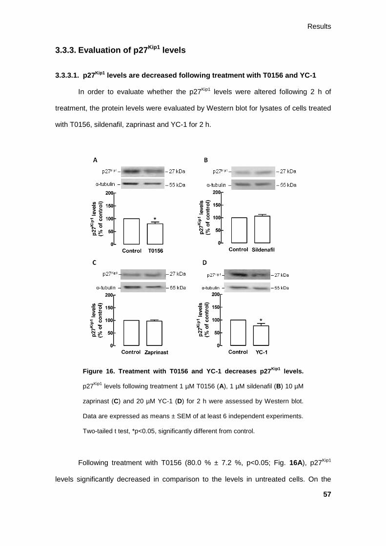
Results
57
3.3.3. Evaluation of p27Kip1 levels
3.3.3.1. p27Kip1 levels are decreased following treatment with T0156 and YC-1
In order to evaluate whether the p27Kip1 levels were altered following 2 h of
treatment, the protein levels were evaluated by Western blot for lysates of cells treated
with T0156, sildenafil, zaprinast and YC-1 for 2 h.
Figure 16. Treatment with T0156 and YC-1 decreases p27Kip1
levels.
p27Kip1
levels following treatment 1 µM T0156 (A), 1 µM sildenafil (B) 10 µM
zaprinast (C) and 20 µM YC-1 (D) for 2 h were assessed by Western blot.
Data are expressed as means ± SEM of at least 6 independent experiments.
Two-tailed t test, *p<0.05, significantly different from control.
Following treatment with T0156 (80.0 % ± 7.2 %, p<0.05; Fig. 16A), p27Kip1
levels significantly decreased in comparison to the levels in untreated cells. On the

Results
58
contrary, treatment with sildenafil (106.4 % ± 6.7 %, p>0.05; Fig. 16B) and zaprinast
(97.4 % ± 4.8 %, p>0.05; Fig. 16C) did not change the levels of p27Kip1 in comparison
to control. Furthermore, activating sGC with YC-1 significantly decreased p27Kip1 levels
(77.5 % ± 9.0 %, p<0.05, Fig. 16C), in comparison to untreated cells.
3.3.3.2. Inhibition of MAPK pathway prevents the decrease of p27Kip1 levels
As seen above, p27Kip1 levels are decreased in the presence T0156 and the
sGC activator, YC-1. In order to test whether this effect can be reversed by inhibiting
the MAPK/ERK 1/2 pathway, treatment of the cells for 2 h with U0126 was performed.

Results
59
Figure 17. Inhibition of MEK 1/2, by U0126, blocks the decrease of
p27Kip1
levels mediated by T0156 and sGC. p27Kip1
levels following
treatment with 1 µM U0126 and 1 µM T0156 (A), 1 µM sildenafil (B), 10 µM
zaprinast (C) or 20 µM YC-1 (D) for 2 h were assessed by Western blot.
Data are expressed as means ± SEM of at least 6 independent experiments.
One-way ANOVA (Bonferroni´s post-test), *p<0.05, significantly different
from control, +p<0.01, significantly different from YC-1 and ++p<0.01,
significantly different from T0156.
The analysis of the Western blots revealed that U0126 alone increased p27Kip1
for levels superior to the baseline (118.0 % ± 7.5 %). Furthermore, the levels of p27Kip1
were significantly increased following treatment with U0126 and T0156 (123.5 % ± 13.0
%, p<0.01; Fig. 17A) and YC-1 (114.9 % ± 3.4 %, p<0.05; Fig. 17D), comparatively to
T0156 (80.0 % ± 7.2 %, p>0.05) and YC-1 (77.5 % ± 9.0 %, p>0.05) alone,
respectively.
On the other hand, U0126 did not change the levels of p27Kip1 in the samples
treated with sildenafil (99.6 % ± 9.6 %, p>0.05; Fig. 17B) neither zaprinast (99.3 % ±
4.6 %, p>0.05; Fig. 17C) when compared to sildenafil (106.4 % ± 6.7 %, p>0.05) and
zaprinast (97.4 % ± 4.8 %, p>0.05) alone, respectively.
3.3.3.3. PKG inhibition prevents the decrease in p27Kip1 levels by YC-1
We next evaluated whether the change in the levels of p27Kip1 could be
reversed by inhibiting the sGC pathway at the PKG level, following a treatment of 2 h.
Inhibition of PKG did not appear to affect the levels of p27Kip1 following
treatment with T0156 (84.1 % ± 10.2 %, p>0.05; Fig. 18A), sildenafil (96.3 % ± 7.8 %,
p>0.05; Fig. 18B) and zaprinast (105.1 % ± 10.0 %, p>0.05; Fig. 18C), in comparison
to T0156 (80.0 % ± 7.2 %, p<0.05), sildenafil (106.4 % ± 6.7 %, p>0.05) and zaprinast
(97.4 % ± 4.8 %, p>0.05) alone, respectively. Furthermore, treatment with KT5823 and

Results
60
YC-1 raises p27Kip1 to levels close to basal (104.5 % ± 11.3 %, p>0.05; Fig. 18D) when
compared to YC-1 alone (77.5 % ± 9.0 %, p>0.05).
Figure 18. KT5823, inhibitor of PKG, prevents the decrease of p27Kip1
levels by YC-1. p27Kip1
levels following treatment with 1 µM KT5823 and 1
µM T0156 (A), 1 µM sildenafil (B), 10 µM zaprinast (C) and ) or 20 µM YC-1
(D) for 2 h were assessed by Western blot. Data are expressed as means ±
SEM of at least 6 independent experiments. One-way ANOVA (Bonferroni´s
post-test), *p <0.05, significantly different from control.
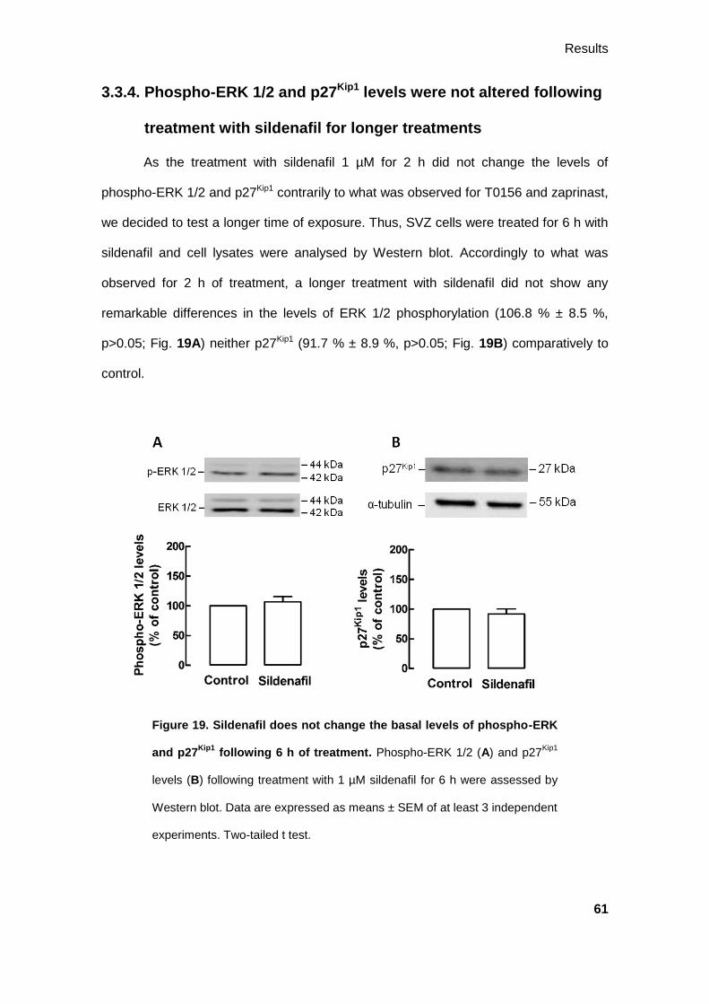
Results
61
3.3.4. Phospho-ERK 1/2 and p27Kip1 levels were not altered following
treatment with sildenafil for longer treatments
As the treatment with sildenafil 1 µM for 2 h did not change the levels of
phospho-ERK 1/2 and p27Kip1 contrarily to what was observed for T0156 and zaprinast,
we decided to test a longer time of exposure. Thus, SVZ cells were treated for 6 h with
sildenafil and cell lysates were analysed by Western blot. Accordingly to what was
observed for 2 h of treatment, a longer treatment with sildenafil did not show any
remarkable differences in the levels of ERK 1/2 phosphorylation (106.8 % ± 8.5 %,
p>0.05; Fig. 19A) neither p27Kip1 (91.7 % ± 8.9 %, p>0.05; Fig. 19B) comparatively to
control.
Figure 19. Sildenafil does not change the basal levels of phospho-ERK
and p27Kip1
following 6 h of treatment. Phospho-ERK 1/2 (A) and p27Kip1
levels (B) following treatment with 1 µM sildenafil for 6 h were assessed by
Western blot. Data are expressed as means ± SEM of at least 3 independent
experiments. Two-tailed t test.

Results
62
3.3.5. Ubiquitination of p27Kip1 is not changed by PDE5 inhibition
As reported in a previous work from our group, treatment with a NO• donor, 10
µM NOC-18, for 1 h, decreased the levels of p27Kip1 in SVZ cells (Carreira et al. 2010).
Here we tested whether p27Kip1 in the SVZ cultures was being tagged with ubiquitin for
subsequent degradation in the proteosome. In fact, p27Kip1 levels were lower when
cells were treated for 1 h with NOC-18 (Inputs; 89.0 %) and the levels of ubiquitinated
p27Kip1 were higher in the presence of NOC-18, as can be seen by an increase in the
p27Kip1 levels in the bound fraction (153.6 %) and a decrease in the unbound fraction
(91.0 %), in comparison to untreated cells (Fig. 20A).
A similar experiment was performed for the PDE inhibitors selected. In order to
assess whether the results observed above for the levels of p27Kip1 were due to protein
tagging with ubiquitin for subsequent degradation, similarly to what happens in the
presence of NOC-18, a 2 h treatment was applied. We chose this time of treatment as
it was the time used in the previous experiments to evaluate the intracellular levels of
p27Kip1. In fact, the treatment with T0156 substantially decreased p27Kip1 levels (Inputs;
49.5 %), but the levels of p27Kip1 in the bound fraction were decreased (46.7 %) and
increased in the unbound fraction (143.9 %), comparatively to the control (Fig. 20B).
As for zaprinast and sildenafil, neither of them seemed to alter p27Kip1 levels
significantly (Fig. 20C). However, with sildenafil (Inputs; 103.8 %), the percentage of
p27Kip1 bound to ubiquitin substantially decreases (bound fraction; 53.5 %) and the
levels of unbound p27Kip1 remained unaltered (unbound fraction; 100.5 %). In the
presence of zaprinast (Inputs; 88.1 %), the amount of p27Kip1 that bind to ubiquitin is
similar to the basal (bound fraction; 108.0 %) and p27Kip1 levels in the unbound fraction
are slightly increased (139.0 %).

Results
63
Figure 20. Exposure to NOC-18 and T0156 decreases the intracellular
levels of p27Kip1
and the levels of ubiquitinated p27Kip1
increase
following a NOC-18 stimulus. SVZ cells were treated with 10 µM NOC-18
for 1 h or with 1 µM T0156, 1 µM sildenafil, or 10 µM zaprinast for 2 h. Inputs
refer to the cytosolic levels of p27Kip1
and the unbound fraction to the p27Kip1
that did not bound to agarose beads, the non-ubiquitinated p27Kip1
. The
bound fraction represents the fraction of p27Kip1
that is ubiquitinated. Image
representative of one experiment. Abbreviations: Sild, sildenafil; Zap,
zaprinast.

Results
64

65
Chapter 4 Discussion

66

Discussion
67
In this study, we show that the inhibition of the PDE5 stimulates the proliferation
of neural stem cells isolated from the SVZ to the same extent following 6 or 24 h of
treatment. The three PDE inhibitors used in this work were able to increase
proliferation. T0156, the more potent and selective inhibitor of PDE5, was particularly
efficient in increasing SVZ cell proliferation. On the one hand, it increases ERK 1/2
activation and, on the other hand, it decreases the levels of the cyclin-dependent
kinase inhibitor, p27Kip1, responsible for the control of cell cycle progression.
4.1. T0156 stimulates proliferation of SVZ cells
Our analysis of cell proliferation shows a significant increase in the proliferation
of NSC following 6 and 24 h of treatment with T0156. The inhibition of the production of
cGMP by sGC, with ODQ, blocked this proliferative effect following 6 h of treatment but
not upon 24 h. This effect may be due to the action of ODQ in other compensatory
mechanisms at that time. Also in the sGC pathway, our observations in the prevention
of cell proliferation by inhibition of PKG show that this pathway is effectively activated
by T0156. However, inhibiting MEK 1/2, the proliferative effect of T0156 is suppressed
at both the times tested, suggesting the involvement of the MAPK pathway on SVZ cell
proliferation.
A previous study by our group showed that a cGMP analogue, 8-Br-cGMP, had
a similar effect to this of T0156 in increasing SVZ cell proliferation, an effect that was
also blocked by U0126 (Carreira et al. 2011). Here we show that the direct activation of
sGC and thus, increasing cGMP production, has a similar effect in stimulating cell
proliferation.
By the evaluation of phospho-ERK 1/2 levels, we show that T0156 and also YC-
1 increase ERK 1/2 phosphorylation. These results are in agreement with the previous

Discussion
68
observations of 8-Br-cGMP increasing phospho-ERK 1/2 levels (Carreira et al. 2011).
Moreover, ERK 1/2 phosphorylation is prevented by the administration of the PKG
inhibitor, KT5823, suggesting that PKG contributes to ERK 1/2 activation.
p27Kip1 is a cyclin-dependent kinase inhibitor that prevents progression from G1
to S-phase, acting as a potent tumor suppressor protein. As previously reported by our
group, p27Kip1 levels are decreased in the presence of high levels of NO• (Carreira et al.
2010). In agreement with this, here we show that inhibiting PDE5 with T0156 also
decreases p27Kip1 levels and hence, stimulates cell proliferation. Furthermore,
exposure to U0126 alone increases p27Kip1 to levels above the baseline and this effect
is maintained even in the presence of T0156. On the other hand, the inhibition of PKG
does not appear to restore basal levels of p27Kip1.
Once in the cytosol, p27Kip1 is ubiquitinated and, subsequently degraded by the
proteosome (Fujita et al. 2003, Vlach et al. 1997), which allows for cell cycle
progression. It had already been shown by our group that the treatment with 10 µM
NOC-18 for 1 h significantly decreased the levels of p27Kip1 in the SVZ cells (Carreira et
al. 2010). In fact, we show that this NO•-induced decrease of p27Kip1 levels is justified
by its binding to ubiquitin, which tags p27Kip1 for proteosomal degradation. Contrary to
these observations, the decrease in the levels of p27Kip1 in lysates treated for 2 h with
T0156 does not seem to be justified by p27Kip1 binding to ubiquitin. However, this could
be explained by the time of detection of the levels of p27Kip1. In fact, 2 h might be a very
long time to test the ubiquitination of this protein and, at that time, p27Kip1 may have
already been degraded by the proteosome. Therefore, shorter times may be tested,
such as 1 h of treatment as it was used for NOC-18. Within this scenario, p27Kip1 may
possibly be a mediator of the proliferative effect of NO• and T0156.
Together, these results point out for the existence of a crosstalk between sGC
and ERK 1/2 pathways. As the proliferative effect is abolished by inhibition of MEK 1/2,

Discussion
69
in the MAPK pathway the crosstalk should occur at the level of MEK 1/2 or in an
element upstream of it, namely, c-Raf1 or p21Ras.
4.2. Sildenafil stimulates cell proliferation but does not
activate ERK 1/2 pathway
As shown in chapter 3, sildenafil significantly increases SVZ cell proliferation
following 6 and 24 h of treatment. Similarly to what was observed to T0156, sildenafil
proliferative effect was abolished following administration of ODQ, for 6 h, and KT5823
for 6 and 24 h treatment, showing the activation of sGC pathway. On the other hand,
U0126 also prevents this effect, with both periods of treatment, suggesting the
involvement of MAPK pathway in the enhancement of proliferation by sildenafil.
Despite the increase of EdU incorporation in cells treated with sildenafil and the
blockade of this effect by inhibiting MAPK pathway, sildenafil did not increase ERK 1/2
phosphorylation in any of the times tested in this work. This effect goes against what
we have observed for the other inhibitors. However, a similar lack of effect of sildenafil
on ERK 1/2 phosphorylation had already been described by Wang and colleagues, in
2005. According to that study, sildenafil (300 nM) did not increase phosphorylation of
ERK 1/2 following 1 h of treatment (Wang et al. 2005). Furthermore, these data
suggest that in the SVZ, the increase in the levels of cGMP via the inhibition of PDE5
activity enhance neurogenesis through the PI3-K/Akt pathway, by Akt phosphorylation
(Wang et al. 2005). However, there are some reports referring to an increase in the
phosphorylation of ERK 1/2 and glycogen synthase kinase-3β, an element of the PI3-
K/Akt pathway, in the induction of cardioprotection (Das et al. 2008) and the increase of
ERK 1/2 phosphorylation in cultured endothelial cells (Pyriochou et al. 2007), following
exposure to sildenafil.
Although this apparent lack of involvement of the ERK 1/2 pathway, we still
observed that SVZ cell proliferation stimulated by sildenafil was suppressed by the

Discussion
70
inhibition of MEK 1/2. In fact, MEK 1/2 can be indirectly activated by other factors, such
as protein kinase A (Ambrosini et al. 2000) and protein kinase C (Kawakami et al.
2003), which are activated by cAMP. Therefore, sildenafil may possibly be acting by
inhibition of a cAMP-hydrolyzing PDE that could indirectly activate MEK 1/2 and
thereby increase cell proliferation. Within this scenario, sildenafil may also promote the
activation of other pathways, namely the Akt/PI3-K pathway (Wang et al. 2005). ERK
1/2 can also be inhibited by other factors involved in other pathways. Thus, further
studies on the sildenafil signaling pathways are needed in order to understand the
mechanism of enhancement of endogenous neurogenesis, a mechanism that goes
beyond the increasing levels of cGMP via the sGC pathway.
Moreover, and contrarily to T0156, in the presence of sildenafil the levels of
p27Kip1 are unaltered for both times tested in this work, a fact that may be related to the
non-activation of ERK 1/2.
4.3. Zaprinast enhances SVZ cell proliferation
In agreement with the data obtained with the other PDE5 inhibitors tested, our
results show that zaprinast effectively stimulates proliferation in the SVZ following 6
and 24 h of treatment. A prevention of this effect is observed inhibiting either sGC
pathway, with ODQ or KT5823, or MAPK pathway, with U0126, showing that these
pathways are both involved in the proliferative effect of zaprinast.
By the evaluation of phospho-ERK 1/2 levels, we show that zaprinast increases
ERK 1/2 phosphorylation, an effect that is prevented by the administration of the PKG
inhibitor, KT5823.
All these results are in agreement with the observations for T0156, supporting
our hypothesis of the existence of a crosstalk between the two pathways. However,
contrarily to T0156, the levels of p27Kip1 do not decrease following treatment with

Discussion
71
zaprinast, which could possibly be due to differences in their mechanisms of action.
This point will be discussed in more detail in the next section.
4.4. T0156, sildenafil and zaprinast: Different mechanisms of
action?
As introduced in Chapter 1, T0156, zaprinast and sildenafil are three inhibitors
with different selectivity and potency for PDE5. Thus, depending on their concentration,
they can inhibit other PDE, being one of the more commonly inhibited, the PDE6.
PDE6 hydrolyzes cGMP as well as cAMP and is structurally similar to PDE5 (Lugnier
2006). PDE6 inhibition is reported to be responsible for some of the side effects of
sildenafil, such as the impaired vision caused by the increase in cGMP levels in the
retina (Laties et al. 2006). Therefore the interaction with other PDE and, consequently,
the activation of other pathways could be pointed out as possible reasons for the
different effects observed in this study for the three inhibitors. Also, it would be
interesting to investigate which PDE are present in the SVZ cell cultures in order to
understand the possible pathways involved.
Nevertheless, T0156, sildenafil and zaprinast have distinct chemical structures
(Fig. 4). Despite their action on PDE, we cannot exclude the hypothesis of these
inhibitors act in other pathways, other than the cGMP pathway. In fact, inside the PDE
family, these inhibitors can be selective to just one or two isoforms, but they might be
able to directly act in other types of enzymes, which may then affect different signaling
pathways.
Further studies on the activity and signaling pathways of these PDE5 inhibitors
are needed, to clarify their mechanisms of action and, thus, to assess the best targets
to enhance neurogenesis.

Discussion
72

73
Chapter 5 Conclusions

74

Conclusions
75
5.1. Conclusion
The first aim of this work was to clarify the effect of PDE5 inhibitors on the
proliferation of SVZ stem cell cultures. Our data indicate that cell proliferation is
enhanced by the inhibition of PDE5 upon 6 and 24 h of treatment.
As a second goal, we proposed to investigate which pathways are activated by
the PDE5 inhibitors. Our results point out for a dual activation: on the one hand, the
sGC pathway and, on the other hand, the MAPK/ERK 1/2 pathway. Therefore, this
study suggests that the effects of the PDE5 inhibitors on NSC proliferation are
mediated by both cGMP-dependent and independent mechanisms. Although we
suggested the existence of a crosstalk between both pathways, the element at which
this interaction occurs is still unknown.
Therapies involving the specific inhibition of PDE5 are being evaluated for the
treatment of several diseases. In regard to enhancing endogenous neurogenesis by
this strategy, our study helps in better understanding the initial events that promote
proliferation of SVZ cells by PDE5 inhibition. Our observations that cell proliferation is
stimulated upon treatment with PDE5 inhibitors open many doors to the study of the
subjacent mechanisms as well as their effect in the remaining neurogenesis steps.
5.2. Future perspectives
In our study, we suggested that NSC proliferation stimulated by PDE5 inhibition
is due to a crosstalk between two or more pathways. Therefore, it would be of interest
the study of the elements that mediate this response as well as clarifying whether there
are other pathways involved besides the two studied here. For a more complete study
of the effect of PDE5 inhibitors on neurogenesis, a similar study to this could be
performed in cells from other neurogenic niches, such as the dentate gyrus, in order to
assess whether PDE5 inhibition also stimulates cell proliferation in cells from other
regions.

Conclusions
76
Furthermore, it would be useful to confirm that new neurons are effectively
formed from the cells that proliferated. Following this, electrophysiological studies
should be performed to understand whether the newly formed neurons are able to
integrate the pre-existing neuronal network. This will provide us useful information
about the functionality of those neurons.
Moreover, in vivo studies are needed in order to study the effect of the PDE5
inhibitor administration into a more complex system. The use of an in vivo model of
brain injury, such as brain ischemia, will provide additional data about the effect of
these compounds on the recovery of brain lesions. The proliferation and neuronal
differentiation should be analyzed in these models.
Overall, this work may help to identify PDE5 as an interesting therapeutic target
to enhance the formation of new brain cells.

77
Chapter 6 References

78

References
79
Agasse F, Bernardino L, Kristiansen H, Christiansen SH, Ferreira R, Silva B, Grade S,
Woldbye DP and Malva JO (2008) Neuropeptide Y promotes neurogenesis in
murine subventricular zone. Stem Cells, 26, 1636-1645.
Altman J (1969) Autoradiographic and histological studies of postnatal neurogenesis.
IV. Cell proliferation and migration in the anterior forebrain, with special
reference to persisting neurogenesis in the olfactory bulb. J Comp Neurol., 137,
433-457.
Altman J and Das GD (1965) Autoradiographic and Historical Evidence of Postnatal
Hippocampal Neurogenesis in Rats. J. Comp. Neurol., 124, 319-335.
Ambrosini A, Tininini S, Barassi A, Racagni G, Sturani E and Zippel R (2000) cAMP
cascade leads to Ras activation in cortical neurons. Brain Res Mol Brain Res,
75, 54-60.
Araujo IM, Ambrosio AF, Leal EC, Santos PF, Carvalho AP and Carvalho CM (2003)
Neuronal nitric oxide synthase proteolysis limits the involvement of nitric oxide
in kainate-induced neurotoxicity in hippocampal neurons. J Neurochem, 85,
791-800.
Ballard SA, Gingell CJ, Tang K, Turner LA, Price ME and Naylor AM (1998) Effects of
sildenafil on the relaxation of human corpus cavernosum tissue in vitro and on
the activities of cyclic nucleotide phosphodiesterase isozymes. J Urol, 159,
2164-2171.
Baratti CM and Boccia MM (1999) Effects of sildenafil on long-term retention of an
inhibitory avoidance response in mice. Behav Pharmacol, 10, 731-737.
Bender AT and Beavo JA (2004) Specific localized expression of cGMP PDEs in
Purkinje neurons and macrophages. Neurochem Int, 45, 853-857.
Bender AT and Beavo JA (2006) Cyclic nucleotide phosphodiesterases: molecular
regulation to clinical use. Pharmacol Rev, 58, 488-520.

References
80
Bernier PJ, Bedard A, Vinet J, Levesque M and Parent A (2002) Newly generated
neurons in the amygdala and adjoining cortex of adult primates. Proc Natl Acad
Sci U S A, 99, 11464-11469.
Bischoff E (2004) Potency, selectivity, and consequences of nonselectivity of PDE
inhibition. Int J Impot Res, 16 Suppl 1, S11-14.
Blaiss CA, Yu TS, Zhang G, Chen J, Dimchev G, Parada LF, Powell CM and Kernie
SG (2011) Temporally specified genetic ablation of neurogenesis impairs
cognitive recovery after traumatic brain injury. J Neurosci, 31, 4906-4916.
Boswell-Smith V, Spina D and Page CP (2006) Phosphodiesterase inhibitors. Br J
Pharmacol, 147 Suppl 1, S252-257.
Bredesen DE, Rao RV and Mehlen P (2006) Cell death in the nervous system. Nature,
443, 796-802.
Brown J, Cooper-Kuhn CM, Kempermann G, Praag HV, Winkler J, Gage FH and Kuhn
HG (2003) Enriched environment and physical activity stimulate hippocampal
but not olfactory bulb neurogenesis. European Journal of Neuroscience, 17,
2042-2046.
Cameron HA and Gould E (1994) Adult neurogenesis is regulated by adrenal steroids
in the dentate gyrus. Neuroscience, 61, 203-209.
Carlén M, Cassidy RM, Brismar H, Smith GA, Enquist LW and Frisén J (2002)
Functional Integration of Adult-Born Neurons. Current Biology, 12, 606-608.
Carreira BP, Morte MI, Inacio A et al. (2010) Nitric oxide stimulates the proliferation of
neural stem cells bypassing the epidermal growth factor receptor. Stem Cells,
28, 1219-1230.
Carreira BP, Morte MI, Lourenço AS, Santos AI, Inácio A, Ambrósio AF, Carvalho CM
and Araújo IM (2011) Differential contribution of the guanylyl cyclase-cyclic
GMP-protein kinase G pathway to the proliferation of neural stem cells
stimulated by nitric oxide (submitted).

References
81
Chalimoniuk M and Strosznajder JB (1998) Aging modulates nitric oxide synthesis and
cGMP levels in hippocampus and cerebellum. Effects of amyloid beta peptide.
Mol Chem Neuropathol, 35, 77-95.
Chen J, Magavi SS and Macklis JD (2004) Neurogenesis of corticospinal motor
neurons extending spinal projections in adult mice. Proc Natl Acad Sci U S A,
101, 16357-16362.
Cheung WY (1970) Cyclic 3',5'-nucleotide phosphodiesterase : Demonstration of an
activator. Biochemical and Biophysical Research Communications, 38, 533-
538.
Conover JC, Doetsch F, Garcia-Verdugo JM, Gale NW, Yancopoulos GD and Alvarez-
Buylla A (2000) Disruption of Eph/ephrin signaling affects migration and
proliferation in the adult subventricular zone. Nat Neurosci, 3, 1091-1097.
Coquil JF, Brunelle G and Guedon J (1985) Occurrence of the methylisobutylxanthine-
stimulated cyclic GMP binding protein in various rat tissues. Biochem Biophys
Res Commun, 127, 226-231.
Corbin JD (2004) Mechanisms of action of PDE5 inhibition in erectile dysfunction. Int J
Impot Res, 16 Suppl 1, S4-7.
Cserep C, Szonyi A, Veres JM, Nemeth B, Szabadits E, de Vente J, Hajos N, Freund
TF and Nyiri G (2011) Nitric Oxide Signaling Modulates Synaptic Transmission
during Early Postnatal Development. Cereb Cortex.
Curtis MA, Kam M, Nannmark U et al. (2007) Human Neuroblasts Migrate to the
Olfactory Bulb via a Lateral Ventricular Extension. Science, 315, 1243-1249.
Das A, Xi L and Kukreja RC (2008) Protein kinase G-dependent cardioprotective
mechanism of phosphodiesterase-5 inhibition involves phosphorylation of ERK
and GSK3beta. J Biol Chem, 283, 29572-29585.
Devan BD, Bowker JL, Duffy KB, Bharati IS, Jimenez M, Sierra-Mercado D, Jr., Nelson
CM, Spangler EL and Ingram DK (2006) Phosphodiesterase inhibition by

References
82
sildenafil citrate attenuates a maze learning impairment in rats induced by nitric
oxide synthase inhibition. Psychopharmacology (Berl), 183, 439-445.
Devan BD, Sierra-Mercado D, Jr., Jimenez M, Bowker JL, Duffy KB, Spangler EL and
Ingram DK (2004) Phosphodiesterase inhibition by sildenafil citrate attenuates
the learning impairment induced by blockade of cholinergic muscarinic
receptors in rats. Pharmacol Biochem Behav, 79, 691-699.
Dong XX, Wang Y and Qin ZH (2009) Molecular mechanisms of excitotoxicity and their
relevance to pathogenesis of neurodegenerative diseases. Acta Pharmacol Sin,
30, 379-387.
Duman RS, Malberg J and Nakagawa S (2001) Regulation of Adult Neurogenesis by
Psychotropic Drugs and Stress. Journal of Pharmacology and Experimental
Therapeutics, 299, 401-407.
Emsley JG, Mitchell BD, Kempermann G and Macklis JD (2005) Adult neurogenesis
and repair of the adult CNS with neural progenitors, precursors, and stem cells.
Progress in Neurobiology, 75, 321-341.
Enwere E, Shingo T, Gregg C, Fujikawa H, Ohta S and Weiss S (2004) Aging Results
in Reduced Epidermal Growth Factor Receptor Signaling, Diminished Olfactory
Neurogenesis, and Deficits in Fine Olfactory Discrimination. J. Neurosci., 24,
8354-8365.
Erceg S, Monfort P, Hernandez-Viadel M, Rodrigo R, Montoliu C and Felipo V (2005)
Oral administration of sildenafil restores learning ability in rats with
hyperammonemia and with portacaval shunts. Hepatology, 41, 299-306.
Eriksson PS, Perfilieva E, Bjork-Eriksson T, Alborn A-M, Nordborg C, Peterson DA and
Gage FH (1998) Neurogenesis in the adult human hippocampus. Nat Med, 4,
1313-1317.
Essayan DM (2001) Cyclic nucleotide phosphodiesterases. J Allergy Clin Immunol,
108, 671-680.

References
83
Forman MS, Trojanowski JQ and Lee VM-Y (2004) Neurodegenerative diseases: a
decade of discoveries paves the way for therapeutic breakthroughs. Nature
Medicine, 10, 1055 - 1063.
Fujita N, Sato S and Tsuruo T (2003) Phosphorylation of p27Kip1 at threonine 198 by
p90 ribosomal protein S6 kinases promotes its binding to 14-3-3 and
cytoplasmic localization. J Biol Chem, 278, 49254-49260.
Garthwaite J, Charles SL and Chess-Williams R (1988) Endothelium-derived relaxing
factor release on activation of NMDA receptors suggests role as intercellular
messenger in the brain. Nature, 336, 385-388.
Gould E, Beylin A, Tanapat P, Reeves A and Shors TJ (1999) Learning enhances adult
neurogenesis in the hippocampal formation. Nat Neurosci, 2, 260-265.
Herczenik E and Gebbink MFBG (2008) Molecular and cellular aspects of protein
misfolding and disease. FASEB J., 22, 2115-2133.
Hermann A, Maisel M, Wegner F, Liebau S, Kim DW, Gerlach M, Schwarz J, Kim KS
and Storch A (2006) Multipotent neural stem cells from the adult tegmentum
with dopaminergic potential develop essential properties of functional neurons.
Stem Cells, 24, 949-964.
Ignarro LJ (1991) Signal transduction mechanisms involving nitric oxide. Biochem
Pharmacol, 41, 485-490.
Im SH, Yu JH, Park ES, Lee JE, Kim HO, Park KI, Kim GW, Park CI and Cho SR
(2010) Induction of striatal neurogenesis enhances functional recovery in an
adult animal model of neonatal hypoxic-ischemic brain injury. Neuroscience,
169, 259-268.
Jellinger K (2003) General aspects of neurodegeneration. J Neural Transm Suppl, 65,
101-144.
Jellinger KA (2009) Recent advances in our understanding of neurodegeneration.
Journal of Neural Transmission, 116, 1111-1162.

References
84
Jin K, Sun Y, Xie L, Batteur S, Mao XO, Smelick C, Logvinova A and Greenberg DA
(2003) Neurogenesis and aging: FGF-2 and HB-EGF restore neurogenesis in
hippocampus and subventricular zone of aged mice. Aging Cell, 2, 175-183.
Kahn RA, Weinberger J, Brannan T, Prikhojan A and Reich DL (1995) Nitric oxide
modulates dopamine release during global temporary cerebral ischemia.
Anesth Analg, 80, 1116-1121.
Kawakami Y, Kitaura J, Yao L et al. (2003) A Ras activation pathway dependent on
Syk phosphorylation of protein kinase C. Proc Natl Acad Sci U S A, 100, 9470-
9475.
Kee NJ, Preston E and Wojtowicz JM (2001) Enhanced neurogenesis after transient
global ischemia in the dentate gyrus of the rat. Experimental Brain Research,
136, 313-320.
Kempermann G, Kuhn HG and Gage FH (1997) More hippocampal neurons in adult
mice living in an enriched environment. Nature, 386, 493-495.
Kerwin JF, Jr., Lancaster JR, Jr. and Feldman PL (1995) Nitric oxide: a new paradigm
for second messengers. J Med Chem, 38, 4343-4362.
Kotera J, Fujishige K, Imai Y, Kawai E, Michibata H, Akatsuka H, Yanaka N and Omori
K (1999) Genomic origin and transcriptional regulation of two variants of cGMP-
binding cGMP-specific phosphodiesterases. Eur J Biochem, 262, 866-873.
Kuhn H, Dickinson-Anson H and Gage F (1996) Neurogenesis in the dentate gyrus of
the adult rat: age-related decrease of neuronal progenitor proliferation. J.
Neurosci., 16, 2027-2033.
Laties A and Sharlip I (2006) Ocular safety in patients using sildenafil citrate therapy for
erectile dysfunction. J Sex Med, 3, 12-27.
Lau A and Tymianski M (2010) Glutamate receptors, neurotoxicity and
neurodegeneration. Pflugers Arch, 460, 525-542.

References
85
Lewis GD and Semigran MJ (2004) Type 5 phosphodiesterase inhibition in heart failure
and pulmonary hypertension. Curr Heart Fail Rep, 1, 183-189.
Lewis PD (1968) A quantitative study of cell proliferation in the subependymal layer of
the adult rat brain. Experimental Neurology, 20, 203-207.
Lie DC, Song H, Colamarino SA, Ming G-l and Gage FH (2004) Neurogenesis in the
Adult Brain: New Strategies for Central Nervous System Diseases. Annual
Review of Pharmacology and Toxicology, 44, 399-421.
Lin CS, Lau A, Tu R and Lue TF (2000) Expression of three isoforms of cGMP-binding
cGMP-specific phosphodiesterase (PDE5) in human penile cavernosum.
Biochem Biophys Res Commun, 268, 628-635.
Lincoln TM, Hall CL, Park CR and Corbin JD (1976) Guanosine 3':5'-cyclic
monophosphate binding proteins in rat tissues. Proc Natl Acad Sci U S A, 73,
2559-2563.
Lossinsky AS and Shivers RR (2004) Structural pathways for macromolecular and
cellular transport across the blood-brain barrier during inflammatory conditions.
Review. Histol Histopathol, 19, 535-564.
Loughney K, Hill TR, Florio VA et al. (1998) Isolation and characterization of cDNAs
encoding PDE5A, a human cGMP-binding, cGMP-specific 3',5'-cyclic nucleotide
phosphodiesterase. Gene, 216, 139-147.
Lugnier C (2006) Cyclic nucleotide phosphodiesterase (PDE) superfamily: a new target
for the development of specific therapeutic agents. Pharmacol Ther, 109, 366-
398.
Magavi SS, Leavitt BR and Macklis JD (2000) Induction of neurogenesis in the
neocortex of adult mice. Nature, 405, 951-955.
Matarredona ER, Murillo-Carretero M, Moreno-López B and Estrada C (2004) Nitric
oxide synthesis inhibition increases proliferation of neural precursors isolated
from the postnatal mouse subventricular zone. Brain Research, 995, 274-284.

References
86
Matarredona ER, Murillo-Carretero M, Moreno-López B and Estrada C (2005) Role of
nitric oxide in subventricular zone neurogenesis. Brain Research Reviews, 49,
355-366.
Ming G-l and Song H (2005) Adult Neurogenesis in the Mammalian Central Nervous
System. Annual Review of Neuroscience, 28, 223-250.
Mochida H, Takagi M, Inoue H et al. (2002) Enzymological and pharmacological profile
of T-0156, a potent and selective phosphodiesterase type 5 inhibitor. Eur J
Pharmacol, 456, 91-98.
Moncada S, Palmer RM and Higgs EA (1989) Biosynthesis of nitric oxide from L-
arginine. A pathway for the regulation of cell function and communication.
Biochem Pharmacol, 38, 1709-1715.
Moreno-Lopez B, Romero-Grimaldi C, Noval JA, Murillo-Carretero M, Matarredona ER
and Estrada C (2004) Nitric Oxide Is a Physiological Inhibitor of Neurogenesis
in the Adult Mouse Subventricular Zone and Olfactory Bulb. J. Neurosci., 24,
85-95.
Murad F (1994a) The nitric oxide-cyclic GMP signal transduction system for
intracellular and intercellular communication. Recent Prog Horm Res, 49, 239-
248.
Murad F (1994b) Regulation of cytosolic guanylyl cyclase by nitric oxide: the NO-cyclic
GMP signal transduction system. Adv Pharmacol., 26, 19-33.
Nakatomi H, Kuriu T, Okabe S, Yamamoto S-i, Hatano O, Kawahara N, Tamura A,
Kirino T and Nakafuku M (2002) Regeneration of Hippocampal Pyramidal
Neurons after Ischemic Brain Injury by Recruitment of Endogenous Neural
Progenitors. Cell, 110, 429-441.
Nilsson M, Perfilieva E, Johansson U, Orwar O and Eriksson PS (1999) Enriched
environment increases neurogenesis in the adult rat dentate gyrus and
improves spatial memory. Journal of Neurobiology, 39, 569-578.
References
87
Parent JM (2003) Injury-Induced Neurogenesis in the Adult Mammalian Brain.
Neuroscientist, 9, 261-272.
Parent JM, Vexler ZS, Gong C, Derugin N and Ferriero DM (2002) Rat forebrain
neurogenesis and striatal neuron replacement after focal stroke. Ann Neurol,
52, 802-813.
Parent JM, Yu TW, Leibowitz RT, Geschwind DH, Sloviter RS and Lowenstein DH
(1997) Dentate Granule Cell Neurogenesis Is Increased by Seizures and
Contributes to Aberrant Network Reorganization in the Adult Rat Hippocampus.
J. Neurosci., 17, 3727-3738.
Patel MD and Katz SD (2005) Phosphodiesterase 5 inhibition in chronic heart failure
and pulmonary hypertension. Am J Cardiol, 96, 47M-51M.
Pifarre P, Prado J, Baltrons MA, Giralt M, Gabarro P, Feinstein DL, Hidalgo J and
Garcia A (2011) Sildenafil (Viagra) ameliorates clinical symptoms and
neuropathology in a mouse model of multiple sclerosis. Acta Neuropathol, 121,
499-508.
Prickaerts J, de Vente J, Honig W, Steinbusch HW and Blokland A (2002a) cGMP, but
not cAMP, in rat hippocampus is involved in early stages of object memory
consolidation. Eur J Pharmacol, 436, 83-87.
Prickaerts J, Sik A, van der Staay FJ, de Vente J and Blokland A (2005) Dissociable
effects of acetylcholinesterase inhibitors and phosphodiesterase type 5
inhibitors on object recognition memory: acquisition versus consolidation.
Psychopharmacology (Berl), 177, 381-390.
Prickaerts J, Sik A, van Staveren WC, Koopmans G, Steinbusch HW, van der Staay
FJ, de Vente J and Blokland A (2004) Phosphodiesterase type 5 inhibition
improves early memory consolidation of object information. Neurochem Int, 45,
915-928.
References
88
Prickaerts J, van Staveren WC, Sik A, Markerink-van Ittersum M, Niewohner U, van der
Staay FJ, Blokland A and de Vente J (2002b) Effects of two selective
phosphodiesterase type 5 inhibitors, sildenafil and vardenafil, on object
recognition memory and hippocampal cyclic GMP levels in the rat.
Neuroscience, 113, 351-361.
Privat A and Leblond C (1972) The subependymal layer and neighboring region in the
brain of the young rat. J Comp Neurol., 146, 277-302.
Pyriochou A, Zhou Z, Koika V, Petrou C, Cordopatis P, Sessa WC and
Papapetropoulos A (2007) The phosphodiesterase 5 inhibitor sildenafil
stimulates angiogenesis through a protein kinase G/MAPK pathway. J Cell
Physiol, 211, 197-204.
Reif A, Schmitt A, Fritzen S et al. (2004) Differential effect of endothelial nitric oxide
synthase (NOS-III) on the regulation of adult neurogenesis and behaviour. Eur J
Neurosci, 20, 885-895.
Rosen RC and Kostis JB (2003) Overview of phosphodiesterase 5 inhibition in erectile
dysfunction. Am J Cardiol, 92, 9M-18M.
Rutten K, Vente JD, Sik A, Ittersum MM, Prickaerts J and Blokland A (2005) The
selective PDE5 inhibitor, sildenafil, improves object memory in Swiss mice and
increases cGMP levels in hippocampal slices. Behav Brain Res, 164, 11-16.
Sabayan B, Zamiri N and Farshchizarabi S (2010) Phoshphodiesterase-5 inhibitors:
novel weapons against Alzheimer's disease? Int J Neurosci, 120, 746-751.
Selkoe DJ (2004) Cell biology of protein misfolding: The examples of Alzheimer's and
Parkinson's diseases. Nat Cell Biol, 6, 1054-1061.
Shimizu-Albergine M, Rybalkin SD, Rybalkina IG, Feil R, Wolfsgruber W, Hofmann F
and Beavo JA (2003) Individual cerebellar Purkinje cells express different
cGMP phosphodiesterases (PDEs): in vivo phosphorylation of cGMP-specific

References
89
PDE (PDE5) as an indicator of cGMP-dependent protein kinase (PKG)
activation. J Neurosci, 23, 6452-6459.
Singh N and Parle M (2003) Sildenafil improves acquisition and retention of memory in
mice. Indian J Physiol Pharmacol, 47, 318-324.
Skovronsky DM, Lee VM-Y and Trojanowski JQ (2006) Neurodegenerative Diseases:
New Concepts of Pathogenesis and Their Therapeutic Implications. Annual
Review of Pathology: Mechanisms of Disease, 1, 151-170.
St-John WM (1998) Neurogenesis of patterns of automatic ventilatory activity. Prog
Neurobiol, 56, 97-117.
Steiner MK, Preston IR, Klinger JR and Hill NS (2005) Pulmonary hypertension:
inhaled nitric oxide, sildenafil and natriuretic peptides. Curr Opin Pharmacol, 5,
245-250.
Takimoto E, Champion HC, Li M et al. (2005) Chronic inhibition of cyclic GMP
phosphodiesterase 5A prevents and reverses cardiac hypertrophy. Nat Med,
11, 214-222.
Taupin P (2008) Adult neurogenesis, neuroinflammation and therapeutic potential of
adult neural stem cells. Int J Med Sci., 5, 127-132.
Tegenge MA, Stern M and Bicker G (2009) Nitric oxide and cyclic nucleotide signal
transduction modulates synaptic vesicle turnover in human model neurons. J
Neurochem, 111, 1434-1446.
Torroglosa A, Murillo-Carretero M, Romero-Grimaldi C, Matarredona ER, Campos-
Caro A and Estrada C (2007) Nitric Oxide Decreases Subventricular Zone Stem
Cell Proliferation by Inhibition of Epidermal Growth Factor Receptor and
Phosphoinositide-3-Kinase/Akt Pathway. Stem Cells, 25, 88-97.
Turko IV, Ballard SA, Francis SH and Corbin JD (1999) Inhibition of cyclic GMP-binding
cyclic GMP-specific phosphodiesterase (Type 5) by sildenafil and related
compounds. Mol Pharmacol, 56, 124-130.

References
90
Uthayathas S, Karuppagounder SS, Thrash BM, Parameshwaran K, Suppiramaniam V
and Dhanasekaran M (2007) Versatile effects of sildenafil: recent
pharmacological applications. Pharmacol Rep, 59, 150-163.
van Praag H, Christie BR, Sejnowski TJ and Gage FH (1999a) Running enhances
neurogenesis, learning, and long-term potentiation in mice. Proceedings of the
National Academy of Sciences of the United States of America, 96, 13427-
13431.
van Praag H, Kempermann G and Gage FH (1999b) Running increases cell
proliferation and neurogenesis in the adult mouse dentate gyrus. Nature
Neuroscience, 2, 266 - 270.
van Staveren WC, Glick J, Markerink-van Ittersum M, Shimizu M, Beavo JA,
Steinbusch HW and de Vente J (2002) Cloning and localization of the cGMP-
specific phosphodiesterase type 9 in the rat brain. J Neurocytol, 31, 729-741.
Vlach J, Hennecke S and Amati B (1997) Phosphorylation-dependent degradation of
the cyclin-dependent kinase inhibitor p27. EMBO J, 16, 5334-5344.
Wallis RM, Corbin JD, Francis SH and Ellis P (1999) Tissue distribution of
phosphodiesterase families and the effects of sildenafil on tissue cyclic
nucleotides, platelet function, and the contractile responses of trabeculae
carneae and aortic rings in vitro. Am J Cardiol, 83, 3C-12C.
Wang L, Gang Zhang Z, Lan Zhang R and Chopp M (2005) Activation of the PI3-K/Akt
pathway mediates cGMP enhanced-neurogenesis in the adult progenitor cells
derived from the subventricular zone. J Cereb Blood Flow Metab, 25, 1150-
1158.
Whitney NP, Eidem TM, Peng H, Huang Y and Zheng JC (2009) Inflammation
mediates varying effects in neurogenesis: relevance to the pathogenesis of
brain injury and neurodegenerative disorders. Journal of Neurochemistry, 108,
1343-1359.

References
91
Woulfe JM (2007) Abnormalities of the nucleus and nuclear inclusions in
neurodegenerative disease: a work in progress. Neuropathology and Applied
Neurobiology, 33, 2-42.
Yamamoto S, Nagao M, Sugimori M et al. (2001a) Transcription factor expression and
Notch-dependent regulation of neural progenitors in the adult rat spinal cord. J
Neurosci, 21, 9814-9823.
Yamamoto S, Yamamoto N, Kitamura T, Nakamura K and Nakafuku M (2001b)
Proliferation of parenchymal neural progenitors in response to injury in the adult
rat spinal cord. Exp Neurol, 172, 115-127.
Yanamadala V and Friedlander RM (2010) Complement in neuroprotection and
neurodegeneration. Trends in Molecular Medicine, 16, 69-76.
Zhang F, White JG and Iadecola C (1994) Nitric oxide donors increase blood flow and
reduce brain damage in focal ischemia: evidence that nitric oxide is beneficial in
the early stages of cerebral ischemia. J Cereb Blood Flow Metab, 14, 217-226.
Zhang L, Zhang RL, Wang Y, Zhang C, Zhang ZG, Meng H and Chopp M (2005)
Functional recovery in aged and young rats after embolic stroke: treatment with
a phosphodiesterase type 5 inhibitor. Stroke, 36, 847-852.
Zhang L, Zhang Z, Zhang RL, Cui Y, LaPointe MC, Silver B and Chopp M (2006a)
Tadalafil, a long-acting type 5 phosphodiesterase isoenzyme inhibitor, improves
neurological functional recovery in a rat model of embolic stroke. Brain Res,
1118, 192-198.
Zhang R, Wang L, Zhang L, Chen J, Zhu Z, Zhang Z and Chopp M (2003) Nitric oxide
enhances angiogenesis via the synthesis of vascular endothelial growth factor
and cGMP after stroke in the rat. Circ Res, 92, 308-313.
Zhang R, Wang Y, Zhang L, Zhang Z, Tsang W, Lu M and Chopp M (2002) Sildenafil
(Viagra) induces neurogenesis and promotes functional recovery after stroke in
rats. Stroke, 33, 2675-2680.

References
92
Zhang RL, Zhang Z, Zhang L, Wang Y, Zhang C and Chopp M (2006b) Delayed
treatment with sildenafil enhances neurogenesis and improves functional
recovery in aged rats after focal cerebral ischemia. J Neurosci Res, 83, 1213-
1219.
Zhao M, Momma S, Delfani K et al. (2003) Evidence for neurogenesis in the adult
mammalian substantia nigra. Proc Natl Acad Sci U S A, 100, 7925-7930.