electrophysiology of phagocytic membranes. role of divalent cations in membrane hyperpolarizations...
TRANSCRIPT

362 Biochimica et Biophysica Acta 856 (1986) 362 372 Elsevier
BBA 73021
Electrophysiology of phagocytic membranes. Role of divalent cations in
membrane hyperpolarizations of macrophage polykaryons *
Elizabeth G. Araujo **, Pedro M. Persechini and Gilberto M. Oliveira-Castro ***
lnstituto de Biofisica, Universidade Federal do Rio de Janeiro, llha do Fundgto, Rio de Janeiro, ILl 21.941 (Brazil)
(Received August 20th, 1985)
Key words: Macrophage polykaryon; Electrophysiology; Divalent cation; K + transport; Membrane potential; Hyperpolarization
The electrophysiological properties of the membrane of mouse peritoneal macrophage polykaryons are studied. Slow hyperpolarizations can be elicited by iontophoretic injections of either Ca 2+ or Sr 2+ into the cytoplasm. The effect of both cations is identical, since: (1) it is invariably triggered by the cation injection, (2) the amplitude is dependent on the K + gradient, (3) quinine blocks reversibly the response to both cation injections. Mg 2+, Ba 2+ and Mn 2+ did not elicit responses when injected into the cytoplasm. Ca 2+ induced slow hyperpolarizations were reversibly blocked by the addition of Ba 2+ to the external saline, but were not affected by the presence of external tetraethylammonium chloride. Cells maintained in saline containing high concentrations of Ca 2+, Sr 2+ or Mn 2+ exhibited sustained hyperpolarizations. Quinine blocked the hyperpolarization induced by high Ca 2+ or Sr 2+, but was ineffective for the case of Mn 2+. Cells hyperpolarized by external Mn 2+ frequently exhibited nonlinear, voltage-current characteristics. Similar patterns could also be observed in a small fraction (less than 10%) of the cells in control conditions. Current-induced shifts between two stable membrane potentials were seen either in high Ca 2+ or normal medium. The great variability of the responses described for this phagocytic membrane is discussed. The evidence supports the assumption that Ca 2+ and Sr 2+ can induce transient or persistent hyperpolarized states by activating a potassium permeability. External Mn 2 ÷ may act in part by reducing impalement-related current leakage from the phagocytic membrane.
Introduction
The macrophage membrane is the interface in- volved in several mechanisms of host defense and immune recognition. Evidence for a variety of ionic channels and transport system has been ac- cumulated, although the precise physiologic corre-
* This paper is the fifth of a series. ** Present address: Departamento de Neurobiologia, ln-
stituto de Biologia, UFF, Niteroi, R J, Brazil. *** To whom correspondence should be addressed. Abbreviation: Hepes, 4-(2-hydroxyethyl)-l-piperazineethane- sulfonic acid.
lates of the ionic movements and macrophage functions are far from being established.
The macrophage polykaryons are multi- nucleated cells derived from fusion of macro- phages in experimental foreign-body reactions [1-3], and they retain several of the physiological capabilities of the mononucleated cells, such as phagocytosis, locomotion and exocytosis [4-8]. Polykaryons are easier to handle than macro- phages, less sensitive to electrode damage and constitute a good model for the study of phago- cytic membranes [9]. Both kinds of cells display spontaneous membrane potential fluctuations characterized by transitory slow hyperpolariza-
0005-2736/86/$03.50 c~ 1986 Elsevier Science Publishers B.V. (Biomedical Division)

tions with concomitant increase in membrane con- ductance. These fluctuations vary greatly in ampli- tude, are due to a transient increase in a CaZ+-de - pendent K + permeability [10-12] and can be in- duced by intracellular Ca 2+ injection [9].
Similar oscillations of membrane potential have been found in a variety of cells [13]. The red blood cells, where the Ca2+-dependent K + permeability was first described [14], and the fibroblastic L cells [15,16] are cases where the triggering features, channel specificity and blocking agents have been studied. In the previous paper of this series, we presented evidence in favor of the assumption that the slow hyperpolarization is mainly a conse- quence of a Ca2+-dependent membrane conduc- tance to K + in macrophage polykaryons and their mononucleated precursors [9]. Nothing is known about the ability of other divalent cations to sub- stitute for Ca 2+ in the intracellular medium, nor about their effects in the extracellular medium. In this paper, we compare the ability of several diva- lent cations to induce membrane potential and conductance changes in macrophage polykaryons when present in the extracellular medium or when iontophoretically injected into the cytoplasm. It is shown that increased Ca 2+ and Sr 2+, either inside or outside the cells, can cause an increase in K + conductance, whereas Ba 2+ and Mg 2+ cannot. Mn 2+ induces membrane hyperpolarization only when added to the extracellular medium. This effect differs from that of Ca 2+ and Sr 2+, because it is quinine-insensitive and is usually accompa- nied by the emergence of nonlinear, excitable-like, voltage-current characteristics. The mechanisms that may be involved in the Mn 2 +-induced nonlin- earity and depolarizing excitability are discussed, together with similar behavior that is sometimes observed in the absence of Mn 2+ in cells exposed to normal culture medium.
Preliminary reports of this work have already been published (Ann. Acad. Bras. Cien. (1983) 55, 456; Braz. J. Med. Biol. Res. (1983) 16, 426 and Braz. J. Med. Biol. Res. (1983) 16, 427.
Materials and Methods
Cells. Macrophage polykaryons were formed on the surface of round glass coverslips (6 mm diame- ter) implanted for 4-60 days in the peritoneal
363
cavity of AKR or C3H strains or outbred albino mice [1,9]. After variable periods, the coverslips were removed and washed in culture medium (RPMI-1640, Gibco, Grand Island, NY) contain- ing 5% fetal calf serum and buffered with 6 mM Hepes at pH 7.2-7.4. Cells were then kept is this medium at 37°C for at least 30 min prior to any electrophysiological experiment.
Solutions and reagents. The recordings were per- formed either in culture medium or in saline solu- tions. Normal medium (described above) had a potassium concentration of 5.3 mM. In high- potassium medium, [K +] was increased to 30.3 mM by the addition of concentrated KC1 to the normal medium. The normal saline comprised 140 mM NaC1/5 mM KC1/1 mM CAC12/0.5 mM MgC12/6 mM Hepes. High divalent-cation salt solutions were obtained by addition of the ap- propriate chloride. Quinine sulfate was dissolved in normal saline at a concentration of 1.5 mM and kept frozen until dilution. Tetraethylammonium chloride was dissolved in culture medium im- mediately before use. Tetraethylammonium chlo- ride, Hepes and quinine sulfate were obtained from Sigma (St. Louis, MO).
Electrophysiological measurements. A standard electrophysiological recording system was used for simultaneously monitoring membrane potential and input resistance [9], Glass recording micro- electrodes (20-60 M£2) were filled with 2.5 M KC1 solution and connected to a high input-impedance preamplifier with an active bridge circuit (M4A Electrometer, WP Instruments, Hamdem, CT). For divalent cation injections, a second microelectrode pulled in the same conditions and filled with a 0.5 M solution of the appropriate chloride was con- nected through a 200 M~2 resistor to a voltage source, as previously described [9]. Injected cur- rent was recorded through the current monitor of the electrometer, when a single recording electrode was used. When an ion-injecting microelectrode was used, a virtual ground circuit was interpolated between the bath and ground, allowing current measurements.
In experiments where the saline was changed during an impalement, a peristaltic perfusion pump (Minipuls 2, Gilson, Middleton, WI) was employed at a flow-rate of 5 or 1.4 ml /min . Complete renewal of the solution occurred from
364
1 min. In some experiments, the increase of diva- lent cation concentration was achieved by direct addition of 0.1 ml of 0.5 M chloride solution during an impalement (final concentration, 10 raM).
Impalements were considered acceptable only when good bridge balance (within 10% of the input resistance of the cell) and return to base line (within 10% of the resting potential) were observed upon withdrawal of the electrode.
Results
lntracellular injection of divalent cation We have previously demonstrated that intracell-
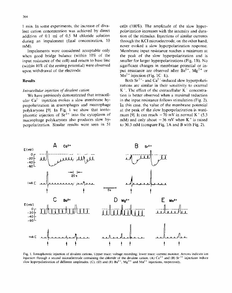
ular Ca 2+ injection evokes a slow membrane hy- perpolarization in macrophages and macrophage polykaryons [9]. In Fig. 1 we show that ionto- phoretic injection of Sr 2+ into the cytoplasm of macrophage polykaryons also produces slow hy- perpolarization. Similar results were seen in 51
cells (100%). The amplitude of the slow hyper- polarization increases with the intensity and dura- tion of the stimulus. Injections of similar currents through the KC1 microelectrode, on the other hand, never evoked a slow hyperpolarization response. Membrane input resistance reaches a minimum at the peak of the slow hyperpolarization and is smaller for larger hyperpolarizations (Fig. 1B). No significant changes in membrane potential or in- put resistance are observed after Ba 2+, Mg 2+ or Mn 2+ injection (Fig. 1C-E).
Both Sr 2 +_ and Ca 2 +-induced slow hyperpolari- zations are similar in their sensitivity to external K +. The effect of the extracellular K + concentra- tion is better observed when a maximal reduction in the input resistance follows stimulation (Fig. 2). In this case, the value of the membrane potential at the peak of the slow hyperpolarization is maxi- mum [9]. It can reach - 7 0 mV in normal K + (5.3 mM) and only about - 3 6 mV when K + is raised to 30.3 mM (compare Fig. 1A and B with Fig. 2).
CO z+ E (rnV)
- 2 0 - 4 0 - 6 0
t C Bo 2+
°E - 2 0 - 4 0 - 6 0
tnA I
E~ Sr =+
I 1
5s l
D Mg z+ F" Mn z+
Fig. 1. lontophoretic injection of divalent cations. Upper trace: voltage recording; lower trace: current monitor. Arrows indicate ion injection through a second microelectrode containing the chloride of the divalent cation. (A) Ca 2+ and (B) Sr 2÷ injections induce slow hyperpolarization of different amplitudes. (C), (D) and (E) Ba 2÷, Mg 2÷ and Mn 2÷ injections, respectively.

A E(mV)
-20 -40
InAE I~F~~
HIGH [K+]o B
Fig. 2. Potassium dependence of the slow hyperpolarization amplitude. Representative recordings of two different cells in culture medium containing 30.3 mM K +. Arrows indicate injections of Ca 2+ (A) or Sr 2+ (B). Sr 2+ injection to the same cell in (B) in normal K + medium is shown in Fig. lB.
A QUININE(Ca z÷) E(mV)
0
E
B RECOVERY
InA E
C QUININE(Sr a+)
- 2 0 I - 4 0 - 6 0
365
Fig. 3. Effects of channel blockers on slow hyperpolarization. (A) Record from a cell maintained for 20 min in a saline solution containing quinine sulfate (1.5 mM). (B) Record of another cell from the same coverslip, 11 min after removing the quinine (culture medium). (C) Quinine (0.15 mM) also blocks Sr 2 +-induced slow hyperpolarization (20 min in saline solution). (D) Removal of quinine restores response to injected Sr 2+ (18 rain in culture medium, another cell). Arrows indicate Ca 2+ injection in (A) and (B), and Sr 2+ injection in (C) and (D).
Effect of channel blockers on slow hyperpolarization Fig. 3 illustrates the inhibitory effect of quinine
on slow hyperpolarization elicited both by Ca 2+ and by Sr 2+ injection. This potent blocker of Ca2+-sensitive K + permeability [13,17] is active at concentrations ranging from 1.5 /~M to 1.5 raM. Throughout this concentration range, the effect is noted in less than 10 min and is completely re- versible after a few minutes of washing.
The application of external Ba 2 + has a marked and reversible inhibitory effect on the CaZ+-in - duced slow hyperpolarization (Fig. 4A). When a dose-response analysis is performed, it is found
A HIGH [BaZ+]o
E (mY)
-20 -40 -60
RECOVERY
I I
B 5s C
D RECOVERY TEA ( Ca 2*) TEA(spontaneous)
o /jr - 2 0
4O / 6O
t
/
t Fig. 4. Effect of external Ba ?+ and tetraethylammonium chlo- ride (TEA) on the slow hyperpolarization. (A) Left: Ca 2C induced slow hyperpolarization is blocked after 10 min in 20 mM Ba 2+. Right: slow hyperpolarization response in the same cell recovers after 15 min in normal medium. (B) tetraethylam- monium chloride (50 mM, 10 min in culture medium) does not affect Ca2+-induced slow hyperpolarization. (C) Recording of spontaneous slow hyperpolarization, after 23 min in 50 mM tetraethylammonium chloride (culture medium). Arrows indi- cate Ca 2+ injections.

366
t 2
-20 _
4 O
6O
8 O /
0 .5 nA E
3 4
- - -4 k - - - - - - I F-,-- 13s 6 0 s
i i II / r._,,._.,,._,,_~,
Fig. 5. Transition between two levels of stable potential in normal medium. Arrows 1 and 3 indicate stable transitions induced by current injections. Note the slow repolarization that follows each of the first three inward-going pulses at arrow 2, and the slow repolarization after an outward-going pulse at arrow 4.
E(mV) 4
-40 f 8O
A
5s
05nA[ ~ ~
B E (rnV)tO0
8O
6O
4O
20
1 I -45 -I!O -O.L5
- j . /
i / " • I"
0 5 t 0 15 2,0 I ( n A )
./6 i , 6 , , / - 8 0
- t 0 0 -
Fig. 6. Current-vohage relationship of macrophage polykaryons in normal medium. (A). Potential changes (upper trace) in- duced by different current injections (lower trace). Note con- ductance changes during the inward current pulses. (B) Cur- rent-voltage relationship of the cell in (A). Measurements were made at the onset of the pulses (O) and just before (A) the cessation of current injection. The straight line was obtained by linear regression on the negative potentials.
E(mV)
$$L
I nA [-
A HIGH [K+]o
. . . . . . . .
1 5s
E (mY)
3O
2 0
I0
0
- I 0
B
I I
. . . . . f . . . . . . . . I i I I
- 2 0 J
-30 ..........o .,.....o ~ -~ID~ _ 4 0 I I I I I l I l
0 0.1 0.2 0 3 0.4 0.5 0.6 0.7 I ( n A )
Fig. 7. Current-voltage relationship in high-potassium med ium. (A) Record from a cell in culture medium. An increase of 19% in the amplitude of the injected current causes a 440% increase in the peak depolarization. Arrow indicates an inflection point in the upstroke of depolarization. (B) Nonlinear current-voltage relationship of the cell in (A), revealing a discontinuity (dashed line). Voltage measurements were taken at the onset (O) and the cessation (A) of current pulses.

that at 20 mM Ba 2+, 100% of the cells fail to respond to the Ca 2+ injection and at 4 mM Ba 2+, only some cases of partial inhibition are seen. Tetraethylammonium chloride, another K + per- meability blocker, was ineffective on either Ca 2+- induced or spontaneous slow hyperpolarization in concentrations up to 100 mM (Fig. 4B and C). Maximum exposure time to tetraethylammonium chloride was 60 min.
Two leuels of membrane potential Fig. 5 illustrates the shifts between two stable
levels of membrane potential in a cell maintained in normal medium. In this cell, both hyperpolariz- ing and depolarizing current pulses (arrows) caused a 40 mV shift in membrane potential, and slow conductance activations were induced by pulses of different amplitudes and durations applied in both directions. This record shows also a spontaneous shift from the high to the low level of membrane potential (after arrow 4). Hyperpolarized cells with excitable-like characteristics may occur either in normal medium or in the presence of a high divalent cation concentration (as will be shown later). Similar findings have been described for mononuclear macrophages in culture medium containing normal Ca 2+ concentration [18,19]. In the case of macrophage polykaryons, 11% of the cells studied in normal culture medium had mem- brane potentials between - 4 0 and - 7 5 mV. In the high-K + medium, values greater than - 4 0 mV have never been found.
Current-ooltage characteristics Current-voltage relationships of the macro-
phage polykaryon membrane are shown in the next two figures. Fig. 6 represents a cell with low membrane potential and a linear response to cur- rent injection from - 1 0 0 to +5 inV. Fig. 7 il- lustrates the occurrence of nonlinear voltage-cur- rent characteristics in high-K + medium. In these cases, the cells are not hyperpolarized, but it is still possible to observe nonlinear curves with a sharp discontinuity.
Effects of externally added divalent cations Addition of Ca 2+, Sr 2+ or Mn 2+ to normal
saline induces a rapid and sustained increase in membrane potential, whereas Mg 2+ and Ba 2 + have
367
no effect (Fig. 8). Membrane potential is strongly dependent on extracellular divalent cation con- centration, as shown in Table I, In normal saline ([Ca 2+] = 1 raM), the mean membrane potential is - 2 2 mV. If this mean plus two standard devia- tions is taken as the criterion for a hyperpolarized cell, we find that 5% of the cells in control saline are hyperpolarized (membrane potential greater than - 4 0 mV). The mean membrane potential increases to - 5 5 mV in high-Ca 2+ and high-Sr 2+ salines. In these situations, 75% of the cells are hyperpolarized, according to our criteria. In high- Mn 2+ saline, the mean membrane potential is - 7 9 mV and again 75% of the cells have membrane potentials greater than - 4 0 mV. It is noteworthy that in the presence of Mn 2+ this parameter may reach - 1 3 2 mV. As already shown for the slow hyperpolarization responses, the action of external Ca 2+ or Sr 2+ can also be blocked by quinine
TABLE I
EFFECT OF DIVALENT CATIONS AND Q U I N I N E ON MEMBRANE POTENTIAL
Membrane potentials were measured in normal saline (control) and in salines containing the indicated divalent cation con- centrations, in the presence or absence of 1.5 mM quinine sulfate. Values are expressed as means + S.D. (number of cells). Figures in square brackets indicate minimum and maximum values.
Added Concn. Membrane potential ( - mV)
cation (mM) no quinine quinine (1.5 mM)
Ca 2+ 1 22_+ 9(49) 16_+ 4(13) [6, 621 [10, 20]
5 49_+14(14)* 17_+ 7 (6) [26, 70] [10, 30]
20 55+_12(14)* 23_+ 7 (6) [28, 70] [16, 361
Sr 2+ 5 44_+17(17)* 17_+ 2(15) [20, 801 [14, 201
20 55_+15 (8)* - [32, 721
Mn 2+ 5 49_+23 (47) * 48_+32 (18) * [22, 116] [16, 100]
20 794-35 (29) * [24, 132]
* Significantly different from control (P < 0.05), according to Student 's t-test.

368
E(mV)
° I -40
-80 05nA E
E(mV) 0
-BO
05hA [
~ S r z+ B
E(mV)
° E -40 -BO
Mn 2+ C i zo,' i
OSnAE
E(mV)
° f -40 - 80
05nAE
Bo'+ D ~Mq'+ E
I I 5s
Fig. 8. Effects of divalent cations in the bathing medium. Records from cells perfused with salines containing 20 mM divalent cation. Arrows indicate the arrival of this solution in the chamber. Hyperpolarization is seen in the presence of Ca 2 + (A, from - 24 to - 60 mV), Sr 2÷ (B, from - 2 0 to - 6 0 mV) and Mn 2+ (C, from - 3 6 to - 7 6 mV). In the case of Ba 2÷ (D) and Mg 2+ (E), no hyperpolarization is observed.
A
::of I= d I ~ ~I I ~ =I tOOs tOs 70s
Q5nA E ~ ~
B C D E(mV)
. . . . . . . . . . . . . . . . . .
-40 ~ ~ -60
05 nA E ~ ~
Fig. 9. Current-induced responses in the presence of Ca 2+ and Sr 2+. (A and C) Patterns of response to current injection in high-Ca 2+ saline (20 mM). Note in (A) the shift of transmembrane potential from - 1 6 to - 7 6 mV and a rectifying behavior. (B) Hyperpolarized cell with ohmic responses in 20 mM Sr 2+. (D) Another cell in 5 mM Sr 2+.

(Table I). However, Mn2+-treated cells undergo a distinctive effect, since a large hyperpolarization insensitive to blockade by quinine is observed (Table I). Persistent membrane hyperpolarization induced by high extracellular concentrations of either Ca 2+ or Sr 2÷ is dependent on the trans- membrane K + gradient. The amplitude of Ca 2+- induced membrane hyperpolarization in high-K + medium never exceeded - 4 0 mV, as compared to - 70 mV in normal K ÷ medium.
The addition of high Ca 2+ or Sr 2+ salines in- duces a variable pattern of responses to injected current, as shown in Fig. 9. In many cases, a rectifying effect is observed, since potential changes induced by inward current are greater than those observed for similar outward currents.
369
A shift between two levels of membrane potential, an increase of the time constant and an active depolarization may also occur (Fig. 9A). The most frequent pattern found in hyperpolarized cells ex- posed to high Ca 2÷ or Sr 2÷ is shown in Fig. 9B, where both inward and outward currents induced time-invariant voltage responses and the cells had relatively low input resistance (68% of the hyper- polarized cells). Non-hyperpolarized cells that ex- hibit ohmic behavior are also frequent (25%) in the presence of either high Ca 2+ (Fig. 9C) or Sr 2+ (Fig. 9D).
The presence of Mn 2+ usually induces changes in the voltage-current characteristics. Current-in- duced active potential changes that depend on amplitude and duration of the stimulus may be
A E(mV) 0 . . . . . . . . . . . . . . . . . . . . . . . . . . . . . . . . . . .
A - 8 0
F---- I {= =: == == t5s 65s lOs 25s
I n A E - - _ _ ¢ ~ a~. [~ , ._ . . . I nx
E(mV)
o
-120
05 nAE
B 5s
E(mV)
° f -40 -80
-120
0.tnAE
C D E(mV) ] ~ ~
. . . . . . . . . . . . . . . 0 f ~ -4o ~ -80
- 1 2 0
Fig. 10. Current-induced responses and Ca 2+ injection in the presence of Mn 2+. (A) A hyperpolarized cell ( - 1 1 6 mV) exhibiting a rectifying behavior and slow conductance variations induced by inward current pulses. (B) Another hyperpolarized cell ( - 80 mV, 10 mM Mn 2+ ) showing slow voltage variations at the termination of current pulses. (C) A cell exhibiting rectification in 5 mM Mn 2÷ (membrane potential of - 4 0 mV). (D) Slow hyperpolarization induced by Ca 2÷ injection after 20 min in the presence of 10 mM Mn 2 +.
370
observed (Fig. 10). Rectification is seen in the first two panels of Fig. 10A and also in a less hyper- polarized Mn 2 +-treated cell (Fig. 10C). In contrast to experiments with Sr 2+ and Ca 2+ (Fig. 8A, B and 9B), a decrease in the input resistance during Mn2+-induced hyperpolarization was never ob- served.
It is interesting to note that a concentration of Mn 2 + which is sufficient to hyperpolarize the cells has no effect on the Ca2+-induced slow hyper- polarization (Fig. 10D). This observation suggests that different mechanisms are involved in the two situations.
Discussion
The similarities between the polykaryons and their mononuclear macrophage precursors are ex- tensive: (a) macrophage polykaryons fused in vitro present Ia antigens in their membranes [20]; (b) the phagocytosis of different opsonized and non- opsonized particles [7]; (c) the formation of phagolysosomes [8]; (d) the release of lysosomal enzymes [5]; (e) the locomotory behavior [6]; and (f) the electrophysiological characteristics of mac- rophages previously described [9,11,12,21] and re- cently reviewed [22] correlate well with the find- ings for the larger polykaryon model [9,23],
The experiments described here provide evi- dence about the selectivity of the triggering mech- anism of a divalent cation-dependent K + channel in a phagocytic membrane model, We show here that Sr 2+ substitutes for Ca 2+ in inducing the activation of the K + channel. The following evi- dence supports this hypothesis: (a) each cation injection invariably triggers one slow hyperpolari- zation; (b) slow hyperpolarization time-course is similar whether Ca 2+ or Sr 2+ is injected; (c) slow hyperpolarization amplitude increases with total charge injected; (d) slow hyperpolarization ampli- tude shows a similar K ~ dependence to that previ- ously described in a model where K + conductance changes are the main events underlying the slow potential variations [9,23]; (e) the quinine sensitiv- ity described here is very similar for the case of both alkaline-earth cations. Quinine blockade is considered a strong indication that we are dealing with a relatively ubiquitous CaZ+-activated K + channel (for reviews see Refs. 13, 17, 24, 25, and
the volume containing Ref. 22). The similarity of the effects of Ca 2+ and Sr 2+ is
also seen when the cations are applied to the external fluid. In this case, the main event also seems to be activation of a K + permeability, since: (a) persistent membrane hyperpolarization can be induced in the presence of increasing divalent ion concentrations (Table I); (b) both cations can cause a decrease in input resistance; (c) quinine blocks both effects of external cations; (d) K + modulation is observed in the presence of high external Ca 2+ concentrations.
The effects of Ca 2+ and Sr 2+ in the induction of the K + channel activation are similar to those described for L-cells [26] and in red blood cells [27]. A possible mechanism for the external effects of Ca 2+ and Sr 2+ is the intracellular activation of the K + channel causing a hyperpolarization and a reduction of input resistance.
The finding that tetraethylammonium chloride does not block Ca2+-induced or spontaneous slow hyperpolarization differs from our previous report in macrophages [11]. One possible explanation may be that macrophage membranes are more permeable to tetraethylammonium chloride, which may act only on the intracellular side of the membrane, as described for other cells [17,28].
Ba 2+ is a blocker of voltage-dependent K + channels in axons [29], muscles [30] and macro- phages [29]. In macrophage polykaryons, we have shown that 20 mM Ba 2+ reversibly blocks the Ca2+-induced slow hyperpolarization responses. However, at a concentration of 4 mM, the major- ity of the cells were insensitive to Ba 2+. This could indicate competition of Ba 2+ for the site of Ca 2+ action, as proposed for pancreatic fl-cells [31].
Mn2+-induced effects differ from those caused by Ca 2+ or Sr 2+. These cations cause a hyper- polarization when present in the extracellular medium, but Mn2+-induced hyperpolarization is quinine-insensitive. Unlike Ca 2+ and Sr 2+, Mn 2+ does not elicit slow hyperpolarization responses when iontophoretically injected into the cyto- plasm. These findings differ from those described for L-cells [26]. We have observed that the Mn 2+- induced hyperpolarization is K+-dependent, since increasing external K + causes a decrease in mem- brane potential (data not shown). In the presence of Mn 2+, the cells respond normally to calcium

injections, indicating that the mechanism involved in slow hyperpolarization is not influenced by Mn 2 + itself. However, Mn 2 + can hyperpolarize the cells to amplitudes of the order of - 1 3 0 mV (Table I), posing another question. This trans- membrane potential is about 50 mV more negative than the maximum value found in all other situa- tions. One of the possible effects of Mn 2+ may be an increase in the intracellular K + concentration. This seems to occur, since slow hyperpolarization peak amplitude, which can be used as an indica- tion of the K + equilibrium potential [9], is also signfiicantly increased. It can reach values higher than - 9 0 mV in the presence of Mn 2+ (Fig. 10D), while in normal medium it never exceeds - 6 7 mV [9].
An interesting effect of Mn 2+ is that it induces excitability in response to currents that usually cause ohmic voltage variations. These active re- sponses displayed by hyperpolarized cells are not confined to cells in Mn 2+ solutions; they occur in less than 10% of the cells in normal medium and in the presence of high Ca 2+. In macrophages, a nonlinear voltage-current relationship has been re- lated to the existence of a voltage-dependent, BaZ+-sensitive, K + channel [32], and a delayed outward-rectifying K + conductance has been dem- onstrated with the use of the patch-clamp tech- nique [33]. The occurrence of two stable levels of membrane potential has also been described in heart cells [34]. Similar mechanisms may occur in the Mn2+-induced behavior of the membrane of the macrophage polykaryons.
The evidence available does not enable a de- scription of the pathway by which Mn 2+ can in- duce such membrane properties. A possible ex- planation is that Mn 2+, and to a lesser degree Ca 2+ and Sr 2+, may somehow stabilize the cell membrane and reduce leakage induced by incom- plete sealing at the site of microelectrode penetra- tion. We do not have direct measurements of this sealing, but it seems that leakage is present in these cells. Macrophages depolarize as a conse- quence of microelectrode penetration [35,36], and we have evidence indicating that a considerable depolarization due to leakage currents may also occur in the case of polykaryons: (a) microelec- trode penetration is followed by a rapidly decay- ing negative potential (10-100 ms) with an ampli-
371
rude that may reach - 8 4 mV [23]; (b) this peak potential is usually greater than the more stable transmembrane potential that follows and its value is K+-dependent, reaching a maximum of only - 4 3 mV in high K + [23]; (c) impalement by a second microelectrode may induce depolarization and membrane resistance decrease, which can be reversed by electrode withdrawal [23]: (d) the be- ginning of the record rarely reveals hyperpolarized cells immediately after electrode penetration. The usual pattern is an increase in membrane potential during the initial 5-60 s after impalement.
Gross leakage can be discarded here, because even the non-hyperpolarized cells exhibit char- acteristics of a healthy cell, such as: (a) impaled macrophages are viable, since they exclude dyes used to indicate damage [37]; (b) impaled cells retain their phagocytic capacity [37]: (c) fluo- rescein injected into macrophages is retained by the cytoplasm of previously impaled cells for about 15 rain [21]; (d) stable levels of potential and input resistance can be maintained for consider- able lengths of time [9,22,23,37]; (e) some cells exhibit spontaneous slow hyperpolarization for more than 2 min even when impaled by two electrodes [23]; (f) intracellular K + activity, mea- sured with ion-sensitive electrodes, is maintained at constant levels in impaled cells [23]. These arguments suggest that leakage currents are small and are not accompanied by severe cell damage.
The use of Mn 2+ in the bathing solution may be a way to circumvent the small leakage currents. However, since the Mn 2+ induction of nonlinear voltage-current relationships is more effective than induction by Ca 2+ or Sr 2+, and since Mn 2+ in- duces levels of transmembrane potential which are frequently higher than the expected equilibrium potential for K + in normal conditions [9], it seems that besides damage prevention other effects of Mn 2+ on the polykaryon membrane are likely to exist. Two possibilities that cannot be excluded in analyzing divalent cation effects are: (a) the con- tribution of electrogenic pumps such as the (Na- + K +)-ATPase, already demonstrated for the case of macrophages [38] and macrophage polykaryons [23], and (b) modifications of membrane char- acteristics induced by the screening of negative surface charges [39,40].
The great variability of the electrical signals

372
desc r ibed in this p a p e r der ives f r o m the var ie ty of
c o m p l e x ion p e r m e a b i l i t y changes o c c u r r i n g in
p h a g o c y t i c cells. O n e e x a m p l e of this h e t e r o g e n e -
ity is the o c c u r r e n c e of ac t ion po t en t i a l s desc r ibed
in m a c r o p h a g e s as a r e sponse to in jec ted cu r ren t
[19]. In the case o f m a c r o p h a g e po lyka ryons , we
have not obse rved typica l spike act ivi ty , even in
the h i g h - M n 2+ saline. T h e phys io log ica l m e a n i n g
and ionic charac te r i s t i c s of the e l ec t rophys io log i -
cal p rope r t i e s shou ld be fu r the r inves t iga ted . Im-
p r o v e m e n t s in m o n i t o r i n g t echn iques cou ld p ro-
v ide a h igher r e so lu t ion p ic tu re of the p h a g o c y t i c
m e m b r a n e s . O u r d a t a suggest tha t M n 2 + m a y be a
useful tool in such studies.
Acknowledgements
E.G,A. is a Fe l low, and P.M.P. and G . M . O . - C .
a re Inves t iga to rs of the N a t i o n a l Resea rch C o u n -
cil ( C N P q ) . This w o r k was s u p p o r t e d by g ran t s
f r o m the C N P q , the N a t i o n a l F o u n d for D e v e l o p -
m e n t of Sc i ence and T e c h n o l o g y ( F I N E P -
F N D C T ) , and the C o u n c i l for G r a d u a t e E d u c a -
t ion, Fede ra l U n i v e r s i t y of R io de j a n e i r o
( C E P G - U F R J ) . W e wish to thank M a r t h a Soren-
son and G e o r g e A. D o s Reis for d i scuss ions and
sugges t ions , Laura , E.P. F re i t as for ass i s tance dur-
ing s o m e expe r imen t s , and T~n ia Lhc ia T. V e n t u r a
for typ ing the manusc r ip t .
References
1 Papadimitriou, J.M. and Spector, W.G. (1971) J. Pathol. 105, 187-203
2 Mariano, M. and Spector, W.G. (1974) J. Pathol. 113, 1-19 3 Papadimitriou, J.M. (1979) J. Pathol. 128, 93-97 4 Mariano, M,, Nikitin, T. and Malucelli, B.E. (1976) J.
Pathol. 120, 151-159 5 Papadimitriou, J.M. and Wee, S.H. (1976) J. Pathol. 120,
193-199 6 Papadimitriou, J.M. and Kingston, K.J. (1977) J. Pathol.
121, 27 36 7 Felipe, I. and Oliveira-Castro, G.M. (1982) Ann. Acad.
Bras. Cien. 54, 262 8 Persechini, P.M., Vale, M.A.B., Machado, R.D. and
Oliveira-Castro, G.M. (1983) Braz. J. Med. Biol. Res. 16, 435
9 Persechini, P.M., Araujo, E.G. and Oliveira-Castro, G.M. (1981) J. Membrane Biol. 6l, 81-90
10 Gallim E.K., Wiederhold, M.L., Lipsky, P.E. and Rosenthal, A.S. (1975) J. Cell. Physiol. 86, 653 662
11 Dos Reis, G.A. and Oliveira-Castro, G.M. (1977) Biochim. Biophys. Acta 469, 257-263
12 Oliveira-Castro, G.M. and Dos Reis, G.A. (1981) Biochim. Biophys. Acta 640, 500-511
13 Lew, V. and Ferreira, H.G. (1978) Curr. Top. Membrane Transp. 10, 217-277
14 Gardos, G. (1958) Biochim. Biophys. Acta 30, 653-654 15 Nelson, P.G., Peacock, J. and Minna, J. (1972) J. Gen.
Physiol. 60, 58-71 16 Okada, Y., Doida, Y., Roy, G.. Tsuchiya, W., Inouye, K.
and Inouye, A. (1977) J. Membrane Biol. 35,319-335 17 Schwarz, W. and Passow, H. (1983) Annu. Rev. Physiol. 45,
359-374 18 Gallin. E.K. and Livengood, D.R. (1980) J. Cell. Biol. 85,
160-165 19 McCann, F.V., Cole, J.J., Guyre, P.M. and Russell, J.A.G.
(1983) Science 219, 991-993 20 Schlesinger, L., Musson, R.A. and Johnston, R.B., Jr. (1984)
J. Exp. Med. 159, 1289 1294 21 Dos Reis, G.A., Persechini, P.M., Ribeiro, J.M.C. and
Oliveira-Castro, G.M. (1979) Biochim. Biophys. Acta 552, 331-340
22 Oliveira-Castro, G.M. (1983) Cell Calcium 4, 475-492 23 Persechini, P.M. (1984) PhD Thesis, pp. 1-190, Instituto de
Biofisica da Universidade Federal do Rio de Janeiro 24 Meech, R.W. (1976) in Calcium in Biological Systems
(Duncan, CJ., ed.), pp. 161 191, Cambridge University Press, Cambridge
25 Petersen, O.H. and Maruyama, Y. (1984) Nature 307, 693-696
26 Okada, Y., Tsuchiya, W. and Inouye, A. (1979) J. Mem- brane Biol. 47, 357-376
27 Simons, T.J.B. (1976) J. Physiol. 256, 227-244 28 Hille, B. (1967) J. Gen. Physiol. 50, 1287-1302 29 Armstrong, C.M. and Taylor, S.R. (1980) Biophys. J. 30,
473-488 30 Sperelakis, N., Schneider, M.F. and Harris, E.S. (1967) J.
Gen. Physiol. 50, 1665-1583 31 Atwater, I., Rosario, L. and Rojas, E. (1983) Cell Calcium
4. 451-461 32 Gallin, E.K. and Livengood, D.R. (1981) Am. J. Physiol.
241, C9-C17 33 Ypey, D.L. and Clapham, D.E. (1984) Proc. Natl. Acad.
Sci. USA 81, 3083-3088 34 Gadsby, D.C. and Cranefield, P.F. (1977) J. Gen. Physiol.
70, 725-746 35 lnce, C,, Ypey, D.L., Van Furth, R. and Verveen, A.A.
(1983) J. Cell. Biol. 96, 796 801 36 ince, C., Leijh, P.C.J., Meyer, J., Van Bavel, E. and Ypey,
D.L. (1984) J. Physiol. 352, 625 636 37 Gallin. E.K. (1982) in Phagocytosis - Past and Future
(Karnovsky, M. and Bolis, L., eds.), pp. 29-46, Academic Press, New York
38 Gallin, E.K. and Livengood, D.R. (1983) Am. J. Physiol. 245, C184-C188
39 Frankenhaeuser, B. and Hodgkin, A.L. (1957) J. Physiol. 137, 218-244
40 Hille, B. (1968) J. Gen. Physiol. 51,221 236