einstein's brain - albert einstein college of medicine
TRANSCRIPT
O F Y E S H I V A U N I V E R S I T Y
Albert Einstein College of Medicine
EINSTEIN
Science at the heart of medicine
Allen M. Spiegel, M.D.The Marilyn and Stanley M. Katz Dean
Jack and Pearl Resnick Campus1300 Morris Park Avenue Belfer Building, Room 312 Bronx, NY 10461
718.430.2801 718.430.8705 fax [email protected]
June 2013
Dean’s Letter: “Einstein’s Brain”
Albert Einstein died on April 18, 1955, at the age of 76, from a ruptured abdominal aortic aneurysm. Einstein had requested that his body be cremated, but Thomas Harvey, the Princeton Hospital patholo-gist who conducted the autopsy, removed the brain and eyes and preserved them in a formalin fixative. Whether Harvey received permission from Einstein’s son, Hans, or executor, Otto Nathan, is still a matter of dispute.
Harvey brought Einstein’s brain to the University of Pennsylvania pathology lab, where a gifted techni-cian, Marta Keller, skillfully cut the brain into 240 “blocks” and then into thin microscope sections.1 Interestingly, Keller had worked previously at Montefiore Hospital for the noted neuropathologist Harry Zimmerman. Harvey distributed microscopic slides of Einstein’s brain to a number of clinical scientists, including Zimmerman. Upon receiving his set of slides, Zimmerman was quoted as saying he did not expect to “find the cells that made him [Einstein] a genius.”
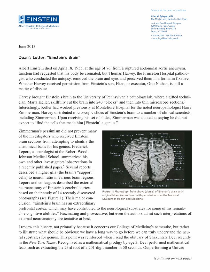
Zimmerman’s pessimism did not prevent many of the investigators who received Einstein brain sections from attempting to identify the anatomical basis for his genius. Frederick Lepore, a neurologist at the Robert Wood Johnson Medical School, summarized his own and other investigators’ observations in a recently published paper.2 Several reports described a higher glia (the brain’s “support” cells) to neuron ratio in various brain regions. Lepore and colleagues described the external neuroanatomy of Einstein’s cerebral cortex based on their study of 14 recently discovered photographs (see Figure 1). Their major con-clusion: “Einstein’s brain has an extraordinary prefrontal cortex, which may have contributed to the neurological substrates for some of his remark-able cognitive abilities.” Fascinating and provocative, but even the authors admit such interpretations of external neuroanatomy are tentative at best.
I review this history, not primarily because it concerns our College of Medicine’s namesake, but rather to illustrate what should be obvious: we have a long way to go before we can truly understand the neu-ral substrates for genius. This point was reinforced when I read the obituary of Shakuntala Devi recently in the New York Times. Recognized as a mathematical prodigy by age 3, Devi performed mathematical feats such as extracting the 23rd root of a 201-digit number in 50 seconds. Outperforming a Univac
(continued on next page)
Figure 1: Photograph from above (dorsal) of Einstein’s brain with original labels (reproduced with permission from the National Museum of Health and Medicine).

(continued on next page)
computer, she earned the title “the human computer.” Unlike the Dustin Hoffman character in Rain Man, an autistic savant, Devi was described as extroverted, affable and articulate. Do we even have a clue how her brain enabled her to perform prodigious mathematical calculations? Next-generation genetic sequencing methods are being applied to identifying a genetic basis for childhood intelli-gence, but we are very far from even a rudimentary understanding of the genetic and environmental basis for “genius.”
Another New York Times obituary reminded me that genius may have a darker side. Ilya Zhitomirsky died at age 22, an apparent suicide. He was a cofounder of Diaspora, a social network start-up described as the “anti-Facebook” because it emphasized personal privacy. As a student at NYU’s Courant Institute of Mathematical Sciences, he was described as an immensely talented young mathematician. Both his father and grandfather were mathematicians, raising the usual questions about Nature, Nurture or both. For me, the more poignant question is what drove him to suicide. The causes of severe depression and other “affective“ disorders such as mania and bipolar disorder are also thought to involve genes, environment and their interaction, but here, too, we are still scratching the surface of the problem.
Modern medicine divides brain diseases into neurologic and psychiatric disorders. Each specialty has its own separate residency training programs. What is the basis for this dichotomy? A crude answer might be that for neurologic diseases we have some combination of genetic, biochemical and anatomical information about disease pathogenesis. The brain, like other organs, is subject to infec-tions, tumors, toxins, autoimmune attack, and vascular damage from clots or hemorrhage. There are also developmental abnormalities and degenerative disorders, most prominently Alzheimer’s. All of these fall into the neurologic diseases category. Having some understanding of neurologic disease pathogenesis does not mean that we fully understand many of these diseases, much less that we know how to treat them.
Decades after we learned that Huntington’s disease is caused by an expansion of a trio of letters in the genetic code of a specific gene on chromosome 4, we still don’t understand how having one abnormal copy of this gene leads to the abnormal movements and cognitive decline charac-teristic of Huntington’s. But at least the diagnosis can be made with a high degree of certainty by testing for the gene abnor-mality.
In contrast, the state of the art for diag-nosis of most psychiatric diseases is to rely on a descriptive set of symptoms (see Figure 2) codified into the Diagnostic and Statistical Manual of Mental Disorders, or the DSM, just released in its 5th edition. Thomas Insel, director of the National Institute of Mental Health, touched off a controversy when he dismissed the DSM-5 because it is not based on “any objective laboratory measure.” Insel did not reject the manual’s utility in “real-world” clinical practice, but as a tool to guide research, he considered it counterproductive.
Figure 2: 1857 lithograph by Armand Gautier, showing personifications of dementia, megalomania, acute mania, melancholia, idiocy, halluci-nation, erotomania and paralysis in the gardens of the Hospice de la Salpêtrière, the facility featured in the current film Augustine about neurologist Jean-Martin Charcot.
Our lack of understanding of the biologic basis for genius; our inability to treat effectively neuro-degenerative disorders such as Huntington’s and, of course, Alzheimer’s; our lack of fundamental understanding of brain diseases such as autism, depression and schizophrenia—all of these demand a dramatic increase in our commitment to fundamental and applied research on the brain. There has been enormous progress over the past several decades in the field of neuroscience, but understand-ing the brain is undoubtedly the greatest challenge faced by biomedical research. Will we ever be able to explain the biologic basis for conscious thought, an emergent property of the human brain’s estimated 100 billion neurons and their estimated 100 trillion synaptic connections? We need more research in neuroscience both to begin to understand the complexities of normal brain function and to gain a better understanding of how brain dysfunction causes some of the most common and severe diseases we confront.
Albert Einstein College of Medicine established one of the first departments of neuroscience, under the leadership of former dean Dominick P. Purpura. Housed in our Rose F. Kennedy building, the department has a record of major research accomplishments. Pioneering work on autism; understand-ing how developmental disorders distort neuron structure; and characterization of gap junctions, specialized membrane structures important in electrical signal transmission, are but a few examples.
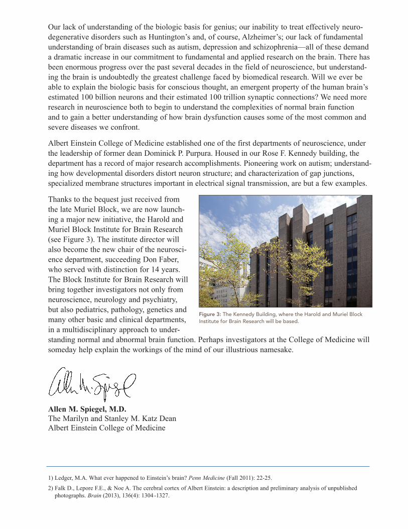
Thanks to the bequest just received from the late Muriel Block, we are now launch-ing a major new initiative, the Harold and Muriel Block Institute for Brain Research (see Figure 3). The institute director will also become the new chair of the neurosci-ence department, succeeding Don Faber, who served with distinction for 14 years. The Block Institute for Brain Research will bring together investigators not only from neuroscience, neurology and psychiatry, but also pediatrics, pathology, genetics and many other basic and clinical departments, in a multidisciplinary approach to under-standing normal and abnormal brain function. Perhaps investigators at the College of Medicine will someday help explain the workings of the mind of our illustrious namesake.
Allen M. Spiegel, M.D.The Marilyn and Stanley M. Katz DeanAlbert Einstein College of Medicine
1) Ledger, M.A. What ever happened to Einstein’s brain? Penn Medicine (Fall 2011): 22-25.
2) Falk D., Lepore F.E., & Noe A. The cerebral cortex of Albert Einstein: a description and preliminary analysis of unpublished photographs. Brain (2013), 136(4): 1304-1327.
Figure 3: The Kennedy Building, where the Harold and Muriel Block Institute for Brain Research will be based.