efeito da radiação ultravioleta b no sistema ... · efeito da radiação ultravioleta b no...
TRANSCRIPT

Efeito da Radiação Ultravioleta B no Sistema Endocanabinoide Cutâneo
Effect of Ultraviolet B Radiation on Cutaneous Endocannabinoid System
Sofia Beatriz Loureiro Marques de Vasconcelos Magina Silva Ramos
Porto | 2012

título
autor
impressão
edição
isbn
ano
Efeito da Radiação Ultravioleta B no Sistema Endocanabinoide Cutâneo
Effect of Ultraviolet B Radiation on Cutaneous Endocannabinoid System
Sofia Beatriz Loureiro Marques de Vasconcelos Magina Silva Ramos
A Medisa
do Autor
978 - 989 - 20 - 3341 - 9
2012

Júri da Prova de Doutoramento
Programa Doutoral em Medicina
Presidente: Reitor da Universidade do Porto
Vogais:
Doutor Patrício Manuel Vieira Araújo Soares da Silva, professor catedrático da Faculdade de
Medicina da Universidade do Porto
Doutor António Albino Coelho Marques Abrantes Teixeira, professor catedrático da Faculdade
de Medicina da Universidade do Porto
Doutor Daniel Filipe de Lima Moura, professor catedrático da Faculdade de Medicina da
Universidade do Porto
Doutor Manuel António Fernandez Esteves, professor associado da Faculdade de Medicina da
Universidade do Porto
Doutor Américo Manuel da Costa Figueiredo, professor associado da Faculdade de Medicina
da Universidade de Coimbra
Doutora Ana Maria Ferreira de Sousa Sebastião, professora associada da Faculdade de
Medicina da Universidade de Lisboa
Orientador : Professor Doutor Daniel Filipe de Lima Moura (Departamento de Farmacologia e
Terapêutica, Faculdade de Medicina da Universidade do Porto)
Co-orientadora: Professora Doutora Maria Augusta Vieira Coelho (Departamento de
Farmacologia e Terapêutica, Faculdade de Medicina da Universidade do Porto)


Dissertação de candidatura ao grau de Doutora
apresentada à Faculdade de Medicina da Universidade do Porto

Artigo 48º, parágrafo 3: A Faculdade não responde pelas doutrinas expendidas na dissertação.
(Regulamento da Faculdade de Medicina do Porto, Decreto Lei nº19337, de 29 de Janeiro de 1931)

CORPO CATEDRÁTICO DA FACULDADE DE MEDICINA DA UNIVERSIDADE DO PORTO
Professores Catedráticos Efetivos
Doutor Alberto Manuel Barros da Silva Doutor Altamiro Manuel Rodrigues Costa Pereira Doutor António Albino Coelho Marques Abrantes Teixeira Doutor António Carlos Freitas Ribeiro Saraiva Doutor Daniel Filipe Lima Moura Doutor Deolinda Maria Valente Alves Lima Teixeira Doutor Francisco Fernando Rocha Gonçalves Doutor Isabel Maria Amorim Pereira Ramos Doutor João Francisco Montenegro Andrade Lima Bernardes Doutor Joaquim Adelino Correia Ferreira Leite Moreira Doutor Jorge Manuel Mergulhão Castro Tavares Doutor José Agostinho Marques Lopes Doutor José Carlos Neves da Cunha Areias Doutor José Eduardo Torres Eckenroth Guimarães Doutor José Henrique Dias Pinto de Barros Doutor José Manuel Lopes Teixeira Amarante Doutor José Manuel Pereira Dias de Castro Lopes Doutor Manuel Alberto Coimbra Sobrinho Simões Doutor Manuel Jesus Falcão Pestana Vasconcelos Doutor Maria Amélia Duarte Ferreira Doutor Maria Dulce Cordeiro Madeira Doutor Maria Fátima Machado Henriques Carneiro Doutor Maria Leonor Martins Soares David Doutor Patrício Manuel Vieira Araújo Soares Silva Doutor Rui Manuel Almeida Mota Cardoso Doutor Rui Manuel Lopes Nunes

Professores Catedráticos Jubilados ou Aposentados
Doutor Abel José Sampaio da Costa Tavares Doutor Abel Vitorino Trigo Cabral Doutor Alexandre Alberto Guerra Sousa Pinto Doutor Amândio Gomes Sampaio Tavares Doutor António Augusto Lopes Vaz Doutor António Carvalho Almeida Coimbra Doutor António Fernandes da Fonseca Doutor António Fernandes Oliveira Barbosa Ribeiro Braga Doutor António Germano Pina Silva Leal Doutor António José Pacheco Palha Doutor António Luís Tomé da Rocha Ribeiro Doutor António Manuel Sampaio de Araújo Teixeira Doutor Belmiro dos Santos Patrício Doutor Cândido Alves Hipólito Reis Doutor Carlos Rodrigo Magalhães Ramalhão Doutor Cassiano Pena De Abreu E Lima Doutor Daniel Santos Pinto Serrão Doutor Eduardo Jorge Cunha Rodrigues Pereira Doutor Fernando de Carvalho Cerqueira Magro Ferreira Doutor Fernando Tavarela Veloso Doutor Francisco de Sousa Lé Doutor Henrique José Ferreira Gonçalves Lecour de Menezes Doutor José Augusto Fleming Torrinha Doutor José Carvalho de Oliveira Doutor José Fernando Barros Castro Correia Doutor José Luís Medina Vieira Doutor José Manuel Costa Mesquita Guimarães Doutor Levi Eugénio Ribeiro Guerra Doutor Luís Alberto Martins Gomes de Almeida Doutor Manuel Augusto Cardoso de Oliveira Doutor Manuel Machado Rodrigues Gomes Doutor Manuel Maria Paula Barbosa Doutor Maria da Conceição Fernandes Marques Magalhães Doutor Maria Isabel Amorim de Azevedo Doutor Mário José Cerqueira Gomes Braga Doutor Serafim Correia Pinto Guimarães Doutor Valdemar Miguel Botelho dos Santos Cardoso Doutor Walter Friedrich Alfred Oswald Doutor Álvaro Jerónimo Leal Machado de Aguiar Doutor Manuel António Caldeira Pais Clemente

Ao Miguel
Aos meus filhos, Pedro, Francisca e Joana


Aos meus pais, Beatriz e Augusto
Ao meu irmão Nuno


Ao Professor Doutor
Daniel Filipe de Lima Moura


Agradecimentos
Ao terminar esta etapa gostaria de expressar o meu agradecimento a quem a
tornou possível...
As minhas primeiras palavras vão para o Professor Doutor Serafim Guimarães
agradecendo o convite que me fez, quando eu era estudante de Medicina, para integrar
o grupo docente da disciplina de Farmacologia. Este primeiro passo foi determinante
para toda a minha formação profissional e pessoal. Com o Professor Doutor Serafim
Guimarães aprendi a exigência e o rigor da atividade científica. Tenho plena
consciência que a oportunidade de, numa idade muito jovem e potencialmente
“moldável”, ter um contato direto e aprender com um grupo de pessoas de exceção
foram para mim, muito importantes.
Neste percurso científico tive o privilégio de ter como orientador o Professor
Doutor Daniel Moura. Com a sua notável capacidade de crítica científica e paciência,
foi sempre capaz de me ajudar a focar no que era importante quando eu facilmente
divagava em assuntos menos farmacológicos. Agradeço o seu permanente incentivo,
confiança e todos os ensinamentos prestados durante a realização deste trabalho, mas
acima de tudo agradeço-lhe a amizade que me dedicou ao longo de todos estes anos.
Depois de ter iniciado o meu percurso pedagógico e científico iniciei a minha
atividade clínica e neste caminho tive a sorte de trabalhar com o Dr. Carlos Resende.
Pessoa dotada duma capacidade notável de comunicar com os doentes e adivinhar os
seus sentimentos e necessidades. Não podia ter tido melhor orientador clínico, com ele
aprendi a “ver” doentes. Agradeço-lhe sobretudo a amizade e todo o incentivo e apoio
que me deu para que estes dois caminhos dermatológico e farmacológico fossem
conciliáveis.
Ao Professor Doutor Patrício Soares da Silva, Diretor do Departamento de
Farmacologia e Terapêutica, por ter permitido a realização deste trabalho e pela
amabilidade com que sempre me tratou.

Ao Professor Doutor António Albino Teixeira, pelas palavras de incentivo que
sempre me dirigiu e pelo constante interesse pelo prosseguimento do meu trabalho.
À Professora Doutora Maria Augusta Vieira Coelho, devo um agradecimento
muito especial! A sua capacidade de trabalho, o seu entusiasmo pela ciência e pela
diversidade de temas e projetos foram para mim um exemplo! Agradeço a sua total
disponibilidade para as minhas dúvidas, para a discussão de resultados e para os meus
desabafos profissionais e pessoais!
Ao Professor Doutor Vincenzo Di Marzo, agradeço o entusiasmo com que
aceitou colaborar neste trabalho, as sugestões e as técnicas laboratoriais que
disponibilizou.
À Engenheira Paula Serrão, agradeço o indispensável apoio laboratorial
prestado, a sua boa disposição e amizade genuínas.
Ao Doutor Eduardo Moura, com quem partilhei quase todos os trabalhos
laboratoriais, agradeço os ensinamentos técnicos e laboratoriais, a sua colaboração
empenhada e a sua constante disponibilidade.
À Professora Doutora Fátima Martel, pela revisão cuidada que fez desta
dissertação e pela sua amizade...
A todos os Professores e Investigadores do Departamento de Farmacologia e
Terapêutica agradeço a permanente colaboração e disponibilidade para todas as
minhas dúvidas...
Aos meus colegas do Serviço de Dermatologia por todo o apoio e amizade
demonstrados.
À equipa de Enfermagem do Serviço de Dermatologia pelo empenho e apoio
logístico nos estudos clínicos.
.

INDEX
Introduction 21
Ultraviolet B radiation 23
Endocannabinoid system 31
Annexe 1 43
“Mechanisms regulating melanogenesis”
An Bras Dermatol 2012 in press
Annexe 2 51
“Phototherapy and photopheresis: old and new indications”
Exp Review Dermatol 2011;6(6):613-623
Aims 63
Chapter I- Ultraviolet B radiation on melanocyte and keratinocyte cell lines 65
“Inhibition of basal and ultraviolet B-induced melanogenesis by cannabinoid
CB(1) receptors: a keratinocyte-dependent effect”
Arch Dermatol Res 2011;303(3):201-10 67
“Anandamide increases UVB-induced cell death of human keratinocytes
through transient receptor potential vanilloid-1 channel”
Submitted for publication 2012 77
"Effect of ultraviolet B radiation on endocannabinoid metabolizing enzymes”
Unpublished data 2012 81
“Ultraviolet B radiation differentially modifies catechol-O-methyltransferase
activity in keratinocytes and melanoma cells”
Photodermatol Photoimmunol Photomed 2012;28(3):137-141 83

Chapter II- Ultraviolet B radiation on patients with psoriasis 89
“ Catechol-O-methyltransferase activity is higher in psoriasis patients and is
down-regulated by narrowband ultraviolet B treatment”
Eur J Dermatol 2012 in press 91
“Narrowband ultraviolet B treatment for psoriasis increases serum vitamin A
levels”
Br J Dermatol 2012;167(4):958-960 97
“Effect of narrowband ultraviolet B treatment on endocannabinoid plasma
levels in psoriasis patients”
Submitted for publication 2012 101
Discussion and Conclusions 107
Summary 123
Resumo 125
Bibliography 127

List of abbreviations
ACEA arachidonoyl-2-chloroethylamide
AEA anandamide
2-AG 2-arachidonoylglycerol
BMI body mass index
Ca2+ calcium
CB1 cannabinoid receptor 1
CB2 cannabinoid receptor 2
COMT catechol-O-methyltransferase
COX-2 cyclooxygenase-2
CPDs cyclobutyl pyrimidine dimers
CRP C-reactive protein
hsCRP high sensitivity C-reactive protein
DAG diacylglicerol
DGL diacylglicerol lipase
FAAH fatty acid amide hydrolase
IL interleukin
IP3 inositol-1,4,5-phosphate
MAGL monoacylglycerol lipase
MAPK mitogen-activated protein kinase
MC1 melanocortin-1 receptor
MITF microphthalmia-associated transcription factor
α-MSH α-melanocyte-stimulating hormone
NF-kB nuclear factor kappa B
OEA oleylethanolamine
PEA palmitoylethanolamide
PIP2 phosphatidylinositol bisphosphate
PLC phospholypase C
PLC-ß phospholypase C-ß
PKA protein kinase A
PKC protein kinase C
PKC-β protein kinase C-β
POMC pro-opiomelanocortin

RE retinyl esters
ROS reactive oxygen species
THC ∆9 tetrahydrocannabinol
TNFα tumor necrosis factor alpha
TRPV1 transient receptor potential vanilloid receptor 1
UV ultraviolet
UVR ultraviolet radiation
UVA ultraviolet A
PUVA psoralen plus UVA
UVB ultraviolet B
nbUVB narrowband ultraviolet B
UVC ultraviolet C

Introduction and Aims


Introduction and Aims
23
Ultraviolet B radiation
General considerations
Sunlight and its adjacent spectral regions, ultraviolet (UV) and infrared are basic
requirements for life on earth and humankind has always been exposed to their
influence. Health benefits to sun exposure were reported in ancient times. Several
therapeutic actions of UV radiation (UVR) are now supported by sound scientific
evidence. However it became clear that solar UVR is also the major environmental
insult to the skin.
The word ultraviolet means “beyond violet” and refers to electromagnetic
radiation with a wavelength shorter than visible violet light but longer than X-rays
(Maverakis et al. 2010). UVR represents approximately 5% of terrestrial solar
radiation and is divided according to the wavelength into UVA (315-380 nm)
separated into UVA1 (340-380 nm) and UVA2 (315-340 nm), UVB (280 to 315 nm)
and UVC (100 to 280 nm). Solar UVR at sea level is approximately 95-98% UVA and
2 to 5% UVB while UVC is completely absorbed by stratospheric ozone that also
attenuates UVB. Little UVB radiation reaches the earth's surface. However, UVB
radiation has much more energy than UVA (energy is inversely proportional to
wavelength) and is approximately 1000 times more erythematogenic than UVA. In
other words, although more UVA reaches the earth, it is mainly UVB that “burns” the
skin.
UVR ionizes molecules and induces chemical reactions. When UVR strikes the
skin, part is remitted (reflected and scattered), part is absorbed in various layers by
molecules termed chromophores, and part is transmitted inward until the energy has
been dissipated. Photochemical reactions convert chromophores (proteins, DNA and
other components of epidermal cells) into new molecules and these photoproducts
activate cellular signal transduction pathways leading to biochemical changes and
different cellular responses.

Effect of ultraviolet B radiation on cutaneous endocannabinoid system
24
The two major processes limiting the penetration of UVR into skin (absorption
and scattering) vary with wavelength (Anderson and Parrish 1981). The depth of
penetration is directly proportional to the wavelength of the radiation and UVB is
mainly absorbed in the epidermis while UVA can penetrate deep into the dermis
(Anderson and Parrish 1981).
An action spectrum indicates which wavelengths produce a photochemical
response most effectively. The rate at which the radiant energy is delivered to the skin
is expressed as W/cm2 and is called irradiance. The total radiant energy delivered per
unit area of skin surface is called the exposure dose or fluence and is the product of
irradiance and time:
[irradiance (W/cm2) x time (seconds) = exposure dose (J/cm2)]
The magnitude of the response to UVR is determined by the exposure dose at a
particular wavelength.
Exposure of human skin to UVB results in several distinct pathophysiological
responses that may be divided into acute and chronic. The most prominent cutaneous
acute effects are the sunburn reaction, tanning, immunosuppression, and vitamin D
synthesis. Photoaging and skin cancer are the consequences of chronic exposure. UVR,
especially UVB, is a major causal factor for all types of skin cancer and UVB
signature mutations in p53 tumour suppressor gene are very prevalent in these tumours
(Raj et al. 2006).
Acute UVB irradiation causes apoptosis and the term “sunburn cell” refers to
keratinocytes undergoing apoptosis. This programmed cell-death of UV-damaged skin
cells is a definitive cancer-prevention pathway.
After UVB irradiation, two photoproducts are well recognized, the four-
membered ring structure called cyclobutyl pyrimidine dimers (CPDs) that lead to DNA
damage (Freeman et al. 1989) and previtamin D3 (Holick et al. 1987). The action
spectrum for sunburn (erythema) closely correlates with the absorption spectrum of
DNA and is within the UVB range.

Introduction and Aims
25
UVB also increases cyclooxygenase-2 (COX-2) expression in cultured human
keratinocytes (Buckman et al. 1998; Van Dross et al. 2007) and COX-2 protein levels
become elevated in the epidermis of human skin following UVB irradiation (Buckman
et al. 1998).
UVR has been shown to activate phospholipase A2 in human skin and
keratinocytes (De Leo et al. 1984), thereby releasing arachidonic acid. UVR also
stimulates a biphasic rise in diacylglicerol (DAG) production in melanocytes and
keratinocytes resulting from diacylglycerol kinase and phospholipases C and D
activation and their action on plasma membrane phospholipids (Punnonen and Yuspa
1992; Carsberg et al. 1995).
It is recognized that UVB and UVA radiation enhance reactive oxygen species
(ROS) production. This oxidative stress in epidermal cells plays an important role in
the photodamage pathway (Portugal-Cohen et al. 2011).
UVB increases intracellular calcium (Ca2+) level in both keratinocytes cell
culture (HaCat cells) and in human skin in vivo (Lee et al. 2009; Masaki et al. 2009). It
has been difficult to identify the specific mechanism for UVB-induced Ca2+-channel
activation. Recently it was demonstrated in keratinocytes (HaCat cells) that UVB
radiation increases the intracellular Ca2+ via transient receptor potential vanilloid type I
(TRPV1) activation (Lee et al. 2009). In addition UVB increases TRPV1 expression in
both keratinocytes and human skin in vivo (Lee et al. 2009). A protein kinase C
(PKC)-dependent activation of TRPV1 and subsequent Ca2+ influx was already
suggested (Lee et al. 2009). Probably, PKC is not the initial chromophore or target
molecule of UVB, but PKC activation occurs by UVB-induced release of DAG,
arachidonic acid and ROS (Matsui et al. 1996).
UVB activates the nuclear factor kappa B (NF-kB) that is a major inducer of
inflammatory response. NF-kB induces the synthesis of proinflammatory interleukins
(IL-1, IL-6, IL-8) and tumor necrosis factor alpha (TNFα) by keratinocytes (Wang et
al. 1995; Portugal-Cohen et al. 2011).

Effect of ultraviolet B radiation on cutaneous endocannabinoid system
26
Investigating the molecular targets and the signal transduction pathways
activated by UVR will increase understanding of both detrimental and therapeutic
effects of UVR.
Melanogenesis
Part of this topic has already been reviewed (see Videira I et al 2012,
Introduction annex 1)
In the skin, melanin is the first defense against UVR. This pigment is able to
absorb and dissipate UVR as harmless heat. The increase of melanin in the epidermis
after UV exposure, the so-called tanning, is understood as a host response against
future photodamage. Melanogenesis has been the interest of research for many
decades, however, until the 1990s the initiating molecular events of tanning were
unknown.
UVB and UVA induce tanning by different mechanisms. UVB induces a slow
but stable type of pigmentation termed delayed tanning, which requires the increased
synthesis of melanin following the stimulation of the entire melanogenic cascade
(Alaluf et al. 2002; Tadokoro et al. 2005). In contrast UVA causes mainly immediate
tanning, due to redistribution of melanosomes and generation of ROS via oxidation,
polymerization or both of existing melanin or melanin precursors (Maeda and Hatao
2004).
Melanocytes, located in the basal layer of the epidermis, synthesize melanin in
discrete organelles, the melanosomes, which once filled with melanin are transferred
via long dendritic processes to the surrounding keratinocytes, where they form a
supranuclear cap to protect DNA against UVR [reviewed by Schallreuter (Schallreuter
2007)].
The key enzyme in melanogenesis, tyrosinase, is located in melanosomes. The
rate of melanin production at baseline and following UVB exposure depends on
tyrosinase activity, rather than simply on total tyrosinase protein (Park et al. 1999).

Introduction and Aims
27
Tyrosinase is a copper-containing membrane glycoprotein with the N-terminal located
inside the melanosome, whereas the C-terminal sits in the melanocyte cytosol. The
enzyme is activated when two serine residues close to C-terminal are phosphorylated
by protein kinase C-β (PKC-β). Tyrosinase contains two cooper atoms in its active
site, and when copper atoms are oxidized the enzyme is inactive. The rate-limiting step
in melanogenesis is the oxidation of tyrosine to L-DOPA (Schallreuter 2007). L-
DOPA acts as a cofactor and also as a substrate for tyrosinase being oxidized into
DOPAquinone. Although the exact interaction between tyrosinase and its substrates is
not completely understood, kinetic studies suggest that L-tyrosine and L-DOPA have
separate binding sites (Olivares et al. 2002; Schallreuter 2007).
Catechol-O-methyltransferase (COMT) is present in melanocytes and catalyses
the O-methylation of catecholic compounds such as L-DOPA (Shibata et al. 1993;
Smit et al. 1994). A potential role of COMT in the regulation of melanogenesis was
first suggested in the paper by Axelrod et al (Axelrod and Lerner 1963) and more
recently it has been shown that skin tyrosinase activity is significantly increased in
hairless COMT-deficient male pups but the role of COMT in melanogenesis is not yet
clarified (Forsberg et al. 2004).
Melanocytes and keratinocytes interact closely as a structural and functional unit,
named “epidermal unit”. Melanogenesis is greater in intact skin or in co-cultures of
melanocytes and keratinocytes than in isolated melanocyte cultures, suggesting that
keratinocyte-derived products contribute to the UV-induced tanning (Archambault et
al. 1995; Duval et al. 2001). Among the wide variety of autocrine and paracrine
melanogenic factors produced by keratinocytes are, melanocyte-stimulating hormone
(α-MSH), adrenocorticotropic hormone (ACTH), endotelin-1, nitric oxide,
prostaglandins E2 and F2aα and granulocyte-macrophage colony stimulating factor
(Suzuki et al. 1999; Kadekaro et al. 2003; Yamaguchi et al. 2007; Park et al. 2009;
Choi et al. 2010). These factors can act alone or synergistically with each other to
modulate melanocyte function. Some UVR-induced cytokines such as IL-1 and TNFα

Effect of ultraviolet B radiation on cutaneous endocannabinoid system
28
inhibit melanogenesis suggesting a fine regulation between melanogenic stimulation
and inhibition after UVR (Slominski et al. 2004).
The most well known melanogenesis-regulating receptor on melanocytes is the,
G-protein-coupled, melanocortin-1 receptor (MC1). Its activation increases
intracellular cAMP and through protein kinase-A (PKA) induces microphthalmia-
associated transcription factor (MITF) transcription. MITF in turn upregulates the
transcription of tyrosinase, tyrosinase-related-protein-1, tyrosinase-related-protein-2
and PKC-ß genes, increasing melanin production (Shibahara et al. 2000; Schallreuter
2007; Park et al. 2009; Choi et al. 2010). α-MSH and ACTH are among the MC1
agonists.
The key observation of the coinciding spectrum, within the UVB range, for
producing a delayed tan (Parrish et al. 1982) and for induction of CPDs (Freeman et
al. 1989) suggested for the first time a cause effect relation between DNA damage and
melanogenesis. It was recognized that the tumor-supressor protein p53 is a
transcription factor that plays a pivotal role in stimulating melanogenesis after UVB-
induced DNA damage (Khlgatian et al. 2002). In keratinocytes, UVB triggers a more
than 30-fold increase in the formation of pro-opiomelanocortin (POMC) and of its
cleavage product α-MSH (Lin and Fisher 2007; Choi et al. 2010). This UVB-induced
α-MSH production by keratinocytes is regulated via a p53 consensus sequence in the
POMC gene promoter (Cui et al. 2007). Keratinocyte-derived α-MSH then stimulates
MC1 on melanocytes, suggesting the important role of keratinocytes for sensing UVR.
Despite the strong influence of MC1 receptors on skin pigmentation, it is clear
that other melanocyte receptors such as β2 adrenoceptor (Gillbro et al. 2004), M1, M3
and M5 muscarinic receptors (Grando et al. 2006) and oestrogen receptors (Thornton et
al. 2006) are also involved in melanogenesis regulation via adenylcyclase activation .
UVR induces the release of DAG from plasma membrane lipids, which then
activates PKC-β that in turn activates tyrosinase and stimulates melanogenesis
(Punnonen and Yuspa 1992; Park et al. 1993; Park et al. 1999; Schallreuter 2007; Park

Introduction and Aims
29
et al. 2009; Choi et al. 2010). DAG release after UVR may also be mediated indirectly
via activation of endotelin-1 receptors or α1 adrenoceptors in melanocyte cell
membranes (Park et al. 2009).
Identification of factors that modulate UV-induced melanogenesis could increase
opportunities for targeted therapeutics ranging from the development of cosmetics to
the prevention of skin cancer.
Therapeutic use
Part of this topic has already been reviewed (see Osório F et al 2012,
Introduction annex 2)
UVR has been used in the management of skin diseases with great success and at
a constantly increasing rate for decades, since Goeckerman first used it in 1925.
Thereby UV phototherapy becomes an essential part of modern dermatological
therapy.
There are many types of phototherapy including: broadband UVB (280-320 nm),
narrowband UVB (nbUVB) (311-313 nm), UVA1 (340-400 nm), and combination
therapy of UVA (320-400 nm) plus the photosensitizer psoralen (PUVA). More
recently handheld phototherapy technologies were developed to deliver UVB targeted
to skin lesions.
Psoriasis is the prototypic skin disease with a favourable response to UVB
phototherapy. In 1977, Fischer (Fischer 1977) found that ultraviolet light at a
wavelength of 313 nm was effective in clearing psoriatic plaques. Few years later, it
was determined that the most effective wavelengths were between 295 and 313 nm,
indicating that this wavelength may possess the optimal “phototherapy index” for
clearing psoriasis (Parrish and Jaenicke 1981). Over the past 30 years, the introduction
of fluorescent bulbs with a limited spectrum of 311–313 nm (nbUVB) has marked an
advance in psoriasis phototherapy. Its success as therapeutic agent has stimulated
studies about the underlying mechanisms involved in resolution of psoriatic plaques.

Effect of ultraviolet B radiation on cutaneous endocannabinoid system
30
UVB exerts a multitude of biological effects within the skin, however it remains
unclear which of these induce clearance of psoriasis and what is the reason for the
marked interpatient variation in clinical responses (Ryan et al. 2010). In addition
identification of molecular biomarkers of UV sensitivity may facilitate treatment
predictability.
The development of psoriasis is dependent on complex interactions between the
innate imune system, dendritic cells and activated T-cells driving hyperproliferation of
genetically predisposed abnormal keratinocytes. Most of the recent studies have
highlighted the nbUVB immunosuppressive effect, leading, in the epidermis, to
downregulation of T helper 17 and interferon signaling pathways (Racz et al. 2011),
reduction of T cells (Krueger et al. 1995; Carrascosa et al. 2007; Erkin et al. 2007),
depletion of Langerhans cells (Murphy et al. 1993) and induction of T regulatory cells
(Schwarz et al. 2004). In addition, soluble factors induced upon UVB exposure, such
as IL-10, IL-22, IL-17, IL-23, IL-8, vascular endothelial growth factor, TNFα, cis-
urocanic acid and vitamin D3, could also be involved in UVB therapeutic effect
(Ullrich 1994; Beissert et al. 2001; Gorman et al. 2007; Cicarma et al. 2010; Coimbra
et al. 2010; Ryan et al. 2010). Moreover it was observed that UVB 311nm induces
significant keratinocytes apoptosis in lesional epidermis, and keratinocyte apoptosis
was proposed as the key mechanism in psoriatic plaques clearance (Aufiero et al.
2006; Weatherhead et al. 2011).
Patients with moderate to severe psoriasis have been found to be at greater risk
of developing comorbidities such as metabolic syndrome, obesity and cardiovascular
diseases (McDonald 1989; Wakkee et al. 2007; Yiu et al. 2011). More recently an
effect of phototherapy on inflammatory markers and circulating adipokine levels was
described and a relation with clinical benefit was hypothesised (Coimbra et al. 2009;
Coimbra et al. 2010; Kawashima et al. 2011; Shibata et al. 2011).

Introduction and Aims
31
Endocannabinoid system
Cannabinoid receptors
In the mid-1960s the main psychoactive component of Cannabis sativa, the
lipophilic compound ∆9tetrahydrocannabinol (THC) was discovered (Mechoulam and
Gaoni 1965). More than two decades later, the first THC-specific receptor named
cannabinoid receptor 1 (CB1) was identified in rat brain (Devane et al. 1988) and then
cloned from mammalian tissues (Matsuda et al. 1990). Three years later, in 1993, a
second cannabinoid receptor named CB2 was identified in the human promyelocytic
leukemic cell line HL60 (Munro et al. 1993). The distinction between these two
receptors is based on differences in their amino acid sequence, signalling mechanisms,
tissue distribution, and sensitivity to selective agonists and antagonists.
CB1 and CB2 receptors are G-protein-coupled receptors members of Gi/o family
(Gi1, 2 and 3, and Go1 and 2), for review see (Howlett 2005). Both these receptors
inhibit adenylcyclase in most cells via Gi, although CB1 receptors can signal via Gs and
stimulate adenylcyclase in some experimental models (Glass and Felder 1997; Maneuf
and Brotchie 1997; Calandra et al. 1999).
Activation of CB1 and CB2 cannabinoid receptors leads to phosphorylation and
activation of p42/p44 mitogen-activated protein kinases (MAPK) (Bouaboula et al.
1995; Bouaboula et al. 1996), p38 MAPK (Derkinderen et al. 2001) and Jun N-
terminal kinase (JNK) (Rueda et al. 2000) as signaling pathways to regulate nuclear
transcription factors.
Evidence exists that CB1 receptor via Gi/o inhibits N- and P/K-types of voltage-
gated Ca2+ channels and stimulates A-type and inwardly rectifying potassium channels
(Howlett 2005; Pertwee et al. 2010).
The CB1 receptor possesses one or more allosteric sites and different ligands
may enhance or inhibit the activation of this receptor by direct agonists (Price et al.
2005; Horswill et al. 2007; Navarro et al. 2009).

Effect of ultraviolet B radiation on cutaneous endocannabinoid system
32
It is well known that CB1 and CB2 receptors are much more widely distributed
than originally believed and that both receptor types can control central and peripheral
functions. The CB1 receptors are preferentially expressed in the central nervous
system, being mainly distributed in brain areas associated with motor control,
emotional responses, motivated behaviour and energy homeostasis, where they
mediate inhibition of transmitter release (Matsuda et al. 1990). In the periphery, CB1
receptors are expressed in the adipose tissue, pancreas, liver, gastrointestinal tract,
skeletal muscles, heart and the reproduction system (Mackie 2008). While CB2
receptors occur in the brain, pancreas, bone and adipose tissue, they are mainly
expressed by immune cells (Munro et al. 1993; Patel et al. 2010).
Endocannabinoids
The discovery of CB1 and CB2 receptors opened the way to the identification of
their cannabis-like endogenous ligands, named endocannabinoids. The N-
arachidonoylethanolamine (anandamide) was the first endocannabinoid to be
discovered (Devane et al. 1992). Shortly after, it was observed that an already known
endogenous metabolite, 2-arachidonoyl-glycerol (2-AG) also exhibits high affinity for
cannabinoid receptors (Mechoulam et al. 1995; Sugiura et al. 1995). Meanwhile, other
polyunsaturated fatty acids have also been proposed to be endocannabinoids including,
N-dihomo-γ-linolenoylethanolamine, N-docosatetraenoylethanolamine, virodhamine,
oleamide and N-arachidonoyl dopamine, but are less well-characterized (Di Marzo
2008; Pertwee et al. 2010). Palmitoylethanolamide (PEA), an endogenous lipid
congener of anandamide, with anti-inflammatory and anti-nociceptive properties, has
been postulated to act by enhancing anandamide effects but it also activates transient
receptor potential vanilloid receptor type 1 (TRPV1) (Petrosino et al. 2010). Therefore,
anandamide and 2-AG are still referred as the “major” endocannabinoids.
Anandamide and 2-AG have affinity for both CB1 and CB2 receptors with
slightly greater affinity for CB1 (Pertwee et al. 2010; Cluny et al. 2012). The
Introduction and Aims
33
endocannabinoids also activate other receptors, such as the deorphanized G-protein-
coupled receptor-GPR55, 5-HT3 receptors, opioid receptors, peroxisome proliferator-
activated receptors (PPARs), TRPV1 (anandamide, but not 2-AG) and TRPV4 (both
anandamide and 2-AG) (Pertwee et al. 2010; Cluny et al. 2012).
As lipids, endocannabinoids cannot be stored in vesicles, but are produced via
several biosynthetic pathways, as needed (“on demand”) from membrane
phospholipids in response to an increase in intracellular Ca2+ or after activation of G-
protein-coupled receptors (Alger and Kim 2011; Cluny et al. 2012).
2-AG can be formed when Ca2+ stimulates phospholipase C which then
transforms membrane phosphoinositides into DAG, from which 2-AG is liberated by
DAG lipase (DGL) (Alger and Kim 2011). Alternatively DAG can be produced from
phosphatidic acid, a reaction catalysed by either phospholipase A2 or D (Alger and
Kim 2011). Two isoforms of DGL have been cloned: DGLα and DGLβ, but DGLα
seems to be sufficient for most endocannabinoid signalling (Alger and Kim 2011).
There is no consensus as to which of the multiple pathways of anandamide
synthesis is physiologically more relevant (Di Marzo 2009; Alger and Kim 2011;
Cluny et al. 2012; Fowler 2012). N-Arachidonoyl-phosphatidyletanolamine (NArPE)
is the major biosynthetic precursor of anandamide (Cadas et al. 1997; Di Marzo 2009).
The enzyme that catalyses the direct conversion of NArPE to anandamide is known as
N-acylphosphatidyl-ethanolamine specific phospholipase D (NAPE-PLD) (Di Marzo
2009; Cluny et al. 2012) but other routes of synthesis are described since NAPE-PLD
“knock-out” do not exhibit reduced levels of anandamide in most tissues (Leung et al.
2006).
Endocannabinoids are produced on demand and are rapidly cleared by a process
of cellular uptake that is incompletely characterized, followed by enzymatic
metabolism. There is general consensus that the enzyme fatty acid amide hydrolase
(FAAH) is the key-enzyme of the breakdown of anandamide, (Di Marzo 2009). On the
other hand, besides the major contribution of monoacylglycerol lipase (MAGL) for 2-

Effect of ultraviolet B radiation on cutaneous endocannabinoid system
34
AG metabolism, it may also be metabolized with a lesser extent by α/β hydrolase 6,
α/β hydrolase 12 and FAAH (Di Marzo 2009; Alger and Kim 2011). While the
searches for α/β hydrolases inhibitors are still in their beginning, a growing number of
selective and potent inhibitors are now available to inhibit FAAH and MAGL activities
(Petrosino and Di Marzo 2010; Feledziak et al. 2012).
Enzymes of the arachidonic acid cascade such as COX-2 and lipoxygenases are
also alternative pathways for 2-AG and anandamide metabolism, leading to the
formation of active metabolites capable of acting at the cannabinoid, TRPV and PPAR
receptors (Duggan et al. 2011; Vecchio and Malkowski 2011; Cluny et al. 2012). The
biological relevance of these reactions remains to be established.
Relevance of endocannabinoid system
In recent years, it becomes clear that the functions of endocannabinoid system
are exerted in the whole organism and are not limited to the central nervous system (Di
Marzo 2008; Pacher and Mechoulam 2011). To date, it has been documented that
endocannabinoid system-regulated functions include: neuronal transmission, pain
initiation, thermogenesis, appetite and energy metabolism, inflammatory and immune
responses, bone remodelling, lipid metabolism, cardiovascular, respiratory and
reproductive functions, hormone release, as well as cellular proliferation and apoptosis
(Calignano et al. 1998; Di Marzo 2008; Pacher and Mechoulam 2011).
The endocannabinoid system is activated “on demand” and a pro-homeostatic
effect has been claimed in different tissues. This “plasticity” of the endocannabinoid
system is clearly observed in central nervous system, where it underlies adaptive
responses to anxiety, chronic stress, neuronal damage and neuroinflammation
(Bisogno and Di Marzo 2007; Moreira et al. 2008). The tissue localization of the
cannabinoid receptors and metabolic enzymes also support this proposed pro-
homeostatic strategy of action.

Introduction and Aims
35
The expression of cannabinoid receptors and also the tissue and plasma levels of
the “major” endocannabinoids undergo significant changes following physiological
and pathological stimuli. However, for the same pathological condition, there are often
reports of both positive and negative changes and of both protective and deleterious
effects of endocannabinoid system activation (Di Marzo 2008). It is becoming
increasingly evident that within a certain tissue, the endocannabinoid system may be
affected in different ways by the same stressful stimulus according to the duration of
this stimulus.
The endocannabinoid system is involved in various pathological conditions in
central and peripheral tissues such as, neurodegenerative disorders, psychiatric
conditions, cardiovascular disease, liver disorders, osteoporosis, inflammatory bowel
disease, auto-immunity, cancer, obesity and metabolic syndrome (Di Marzo 2008;
Moreira et al. 2008; Pacher and Mechoulam 2011).
Obesity and metabolic syndrome are well- known pathological conditions
associated with endocannabinoid system dysregulation. (Maccarrone et al. 2010;
Cluny et al. 2012). Endocannabinoid system plays a major role in the regulation of
energy homeostasis and is generally upregulated in chronic overeating (Maccarrone et
al. 2010). The endocannabinoid plasma levels and FAAH activity in subcutaneous
adipocytes are positively correlated with body mass index (BMI) (Cable et al. 2011)
and in obesity elevated endocannabinoid plasma levels were associated with coronary
circulatory dysfunction (Quercioli et al. 2011). It was demonstrated that
endocannabinoids also play a key role in the development of fatty liver in response to
high fat diets or chronic alcohol intake (Jeong et al. 2008). Furthermore hypothalamic
endocannabinoids seem to be part of the neural circuitry involved in the modulating
effects of leptin on energy homeostasis.
Strategies for manipulating the endocannabinoid system for therapeutic reasons
will require a thorough understanding of the roles of the different endocannabinoids,
the stimuli that mobilize them and their sources and metabolism. Regulating

Effect of ultraviolet B radiation on cutaneous endocannabinoid system
36
endocannabinoid levels in vivo represents an interesting therapeutic perspective.
Several inhibitors of 2-AG and anandamide metabolizing enzymes have attracted
growing interest as potential therapeutic drugs. Particularly, the FAAH inhibitors have
demonstrated benefit in animal models of several disorders, including pain, anxiety
and inflammatory bowel diseases, as well as against proliferation and migration of
cancer cells (Clapper et al. 2010; Petrosino and Di Marzo 2010).
Cannabinoid compounds have also been proposed as promising therapeutic
agents in multiple sclerosis given their capability to alleviate symptoms (e.g.,
spasticity, pain) and reduction of inflammatory events by the activation of CB1
receptors (de Lago et al. 2012). CB1 receptor antagonists have been explored, and
found to be effective, as therapeutic agents for obesity and related cardiometabolic
problems. However, the use of rimonabant, the first marketed CB1 receptor antagonist,
has been suspended due to its anxiogenic and depressogenic effects (Kirilly et al.
2011).
Endocannabinoid system in the skin and appendages
The skin and its appendages (hair follicle and sebaceous gland) function as a
“neuro-immuno-endocrine” organ. Indeed, almost all skin cell types are capable of
producing and releasing pro and/or anti-inflammatory mediators, hormones, growth
factors, neuropeptides and vasoactive substances that can exert paracrine or autocrine
regulation of skin cells functions (Roosterman et al. 2006).
Recent evidences suggest that endocannabinoid system has an important role in
this complex cutaneous network (Kupczyk et al. 2009).
Anandamide and 2-AG are detectable in rodent skin (Calignano et al. 1998;
Karsak et al. 2007) and the distribution and expression of CB1 receptors was uniformly
found in human skin biopsies taken from different body sites (Stander et al. 2005).

Introduction and Aims
37
CB1 and CB2 receptors were predominantly expressed on cutaneous nerves
(Stander et al. 2005) and it was proposed that cannabinoid receptors present in the skin
might act as cutaneous nociceptors (Ibrahim et al. 2005; Khasabova et al. 2008).
In normal skin, CB1 and CB2 receptors were mostly present in suprabasal layers
of the epidermis and in skin appendages like epithelial cells of hair follicles and
sebocytes (Casanova et al. 2003; Stander et al. 2005).
There is evidence that anandamide and 2-AG and both cannabinoid receptors are
present on human keratinocytes, which also express the main enzymes involved in
anandamide synthesis (NAPE-PLD) and degradation (FAAH) (Maccarrone et al. 2003;
Ibrahim et al. 2005).
Anandamide in a CB1 receptor-dependent manner inhibits keratinocyte
differentiation by transcriptional downregulation of keratin 1, keratin 5, involucrin and
transglutaminase 5 (Paradisi et al. 2008). These effects were mediated by an increase
in DNA methylation through MAPK-dependent pathways (p38, p42/44) (Paradisi et al.
2008).
Published data on effects of cannabinoids on regulation of keratinocytes cell
death are conflicting. Wilkinson et al (Wilkinson and Williamson 2007) found that
phytocanabinoids such as ∆9-tetrahydrocannabinol as well as synthetic cannabinoid
agonists inhibited growth of cultured transformed human epidermal keratinocytes
(HPV-16E6/E7), yet these effects were CB1 and CB2 independent. In other study,
activation of both cannabinoid receptors by synthetic agonists induced the apoptotic
death of tumorigenic epidermal cells whereas the viability of non-tumorigenic human
(HaCat cells) and murine (MCA3D) keratinocytes remained unaffected (Casanova et
al. 2003). In contrast, recent data on CB1 and CB2 knockout mice suggest that
cannabinoid receptors and the related signalling pathways might be involved in skin
cancer development (Zheng et al. 2008). The effect of cannabinoids in keratinocytes
death needs further investigation since manipulation of this pathway could become a

Effect of ultraviolet B radiation on cutaneous endocannabinoid system
38
useful adjunct treatment option in hyperproliferative dermatoses such as psoriasis or
keratinocytes-derived skin cancers.
Despite this well documented endocannabinoid system in human keratinocytes
the role of endocannabinoids in melanocytes has not yet been studied. There is one
study (Blazquez et al. 2006) demonstrating that human and mouse melanoma cells
contain cannabinoid CB1 and CB2 receptors. In vitro experiments on A353 and
Meljuso melanoma cell lines, reveled that cannabinoids significantly decrease the
number of viable melanoma cells by inducing apoptosis and that this effect was
prevented by selective cannabinoid receptors antagonists (Blazquez et al. 2006).
Interestingly proliferation of normal melanocyte cell lines was not inhibited (Blazquez
et al. 2006). Furthermore, in the same paper, it was documented that CB2 receptor
agonists inhibit melanoma progression and metastatic spreading in the mouse
(Blazquez et al. 2006).
In human scalp, it was demonstrated that hair follicles are sources of
endocannabinoids and express CB1 receptors. CB1 activation by anandamide inhibited
hair growth and induced apoptosis-driven premature hair follicles regression (catagen)
in vitro (Telek et al. 2007). Hair follicles are most common arranged in pilosebaceous
units, which display another adnexal structure of the human skin, the sebaceous gland.
Dobrosi et al (Dobrosi et al. 2008) by using a human sebaceous gland-derived cell line
demonstrated that anandamide and 2-AG are produced by epithelial cells of the
sebaceous glands (sebocytes) and both via CB2 receptor and MAPK pathway, dose-
dependently induce lipid production and cell death (Dobrosi et al. 2008). Moreover,
endocannabinoids also upregulated the expression of key genes involved in lipid
synthesis, suggesting that CB2 ligands may be exploited in the management of
sebaceous glands dysfunctions (Dobrosi et al. 2008).
Since the original discovery of the CB2 receptors in immune cells, much
evidence suggest that the endocannabinoid system has important immune modulator
effects during inflammation (Klein 2005). Karsak et al (Karsak et al. 2007) observed
Introduction and Aims
39
an increase in cutaneous nickel-induced allergic responses in CB1 and CB2 knockout
mice as well as in the presence of cannabinoid receptors antagonists. In contrast
FAAH-deficient mice displayed reduced allergic responses in the skin (Karsak et al.
2007). By using an animal model of induced allergic contact dermatitis it was also
demonstrated that after allergen exposure, the cutaneous levels of 2-AG and
anandamide significantly increased and that CB1 receptor RNAm was downregulated
while CB2 receptor mRNA was upregulated (Karsak et al. 2007). Taken together these
data suggest a protective role of the endocannabinoid system in contact allergic
dermatitis (Karsak et al. 2007). A protective role of PEA against inflammation was
also observed in the same animal model of contact allergic dermatitis and in a
keratinocyte cell line (HaCat) (Petrosino et al. 2009).
Endocannabinoid signals involved in skin inflammation are complex and remain
poorly understood. Overall, anandamide appears to mediate an anti-inflammatory
effect since inhibition of its degradation significantly reduced inflammation (Karsak et
al. 2007) and, on the other hand, there is growing evidence suggesting that 2-AG
modulates the inflammatory response by acting on the CB2 receptors (Oka et al. 2006).
By using different animal models for acute and chronic dermatitis, Oka et al (Oka et
al. 2006) reported elevated 2-AG levels in the affected skin and that CB2 (but not CB1)
antagonists markedly attenuated cutaneous inflammation. Likewise, others reported a
decrease in the cutaneous inflammation of CB2 receptor deficient mice (Ueda et al.
2007).
In addition the endocannabinoid system has a crucial role in the control of skin-
derived sensory phenomena as pain and itch.
Despite significant research on the role of endocannabinoid signaling in
keratinocytes and pilosebaceous units the potential role of endocannabinoid system in
melanogenesis and the influence of UVR in cutaneous endocannabinoid tone are not
yet elucidated.


Introduction
Annex 1 | Mechanisms regulating melanogenesis
An Bras Dermatol 2012 in press
Annex 2 | Phototherapy and photopheresis: old and new indications
Exp Review Dermatol 2011;6(6):613-623


Introduction Annex 1
43
1
Mechanisms regulating melanogenesis Videira I, 1Moura D, 1,2,3Magina S1Department of Pharmacology and Therapeutics, Faculty of Medicine, University of Porto,2Institute for Molecular and Cell Biology, University of Porto,3Dermatology and Venereology Department, H. S. João, Porto, Portugal.
Abstract The skin pigmentation is an important human phenotypic trait but despite many efforts its regulation is still not fully understood. The pigment melanin is produced in melanosomes by melanocytes in a complex process named melanogenesis. The melanocyte interacts with endocrine, immune, inflammatory and central nervous systems and its activity is also regulated by extrinsic factors such as ultraviolet radiation and drugs. We review current understanding of intrinsic and extrinsic factors regulating skin pigmentation, melanogenesis steps and the known related gene defects. We focus on melanocyte-keratinocyte interaction, enzymatic components of melanosomes, activation of melanocortin receptor type1 (MC1-R) by proopiomelanocortin (POMC) cleavage products (melanocyte stimulating and adrenocorticotrofic hormones) and mechanisms of ultraviolet-induced skin pigmentation. The identification and understanding of the melanogenesis mechanisms facilitate the knowledge of the pathogenesis of pigmentation disorders and the development of potential therapeutic options.
Introduction The skin has epidermal units that are responsible for melanin production and distribution, in a process called melanogenesis. These units are composed by a melanocyte surrounded by keratinocytes and they are regulated by a closed paracrine system. Melanin is the main responsible for skin, hair and eyes pigmentation and besides defining an important human phenotypic trait, it has a critical role in photoprotection due to its ability to absorb ultraviolet radiation (UVR) [1-3]. The Fitzpatrick system is the most commonly used system to distinguish the diversity of cutaneous pigmentation phenotypes. It characterizes six phototypes (I-VI), by grading the erythema and the acquired pigmentation after exposure to UVR [1, 4]. Constitutive pigmentation reflects the genetically determined level of melanin and can be changed by several regulator factors [3, 5]. These factors may be intrinsic (released by keratinocytes and fibroblasts, endocrine, inflammatory and neuronal cells) or extrinsic (UVR and drugs) [3, 6]. The melanogenesis is a complex process with different steps and when disturbed may determine different types of pigmentation defects, which are classified as hypo or hyperpigmentation and may occur with or without altered melanocytes number [1, 2, 7]. There are several dermatoses associated with pigmentation defects which can be congenital or acquired, permanent or temporary and restricted to the skin or systemic [7]. Since these dermatoses have an important impact on patient’s quality of life and their treatment can be unsatisfactory, the pharmaceutical and cosmetic industries have been continuously seeking for new solutions [8, 9]. The understanding of the melanogenesis mechanisms helps us to explain the pigmentation defects observed in genodermatoses
and allows the development of potential therapeutic strategies [3, 10]. In this review we describe the intrinsic and extrinsic factors that regulate human skin pigmentation, focusing on melanogenesis mechanisms and related genodermatoses.
Term Abbreviation
ACTH Adrenocorticotrophic hormone α-MSH Melanocyte stimulating hormone bFGF Basic fibroblast growth factor BMP Bone morphogenic protein cAMP Cyclic adenosine monophosphate cGMP Cyclic guanosine monophosphatec-kit Mast cell growth factor CREB cAMP response element ET Endothelin GM-CSF Granulocyte-macrophage colony-stimulating factorIL Interleukin IP3/DAG Inositol triphosphate / diacylglycerol MAP Mitogenic activated protein MATP Membrane-associated tranporter protein MC1-R Melanocortin recetor type 1 MITF Microphthalmia-associated transcription factor NGF Neuronal growth factor NO Nitric oxide PG Prostaglandin PKA Protein kinase APKC-β Protein kinase C-βPOMC Proopiomelanocortin ROS Reactive oxygen speciesSCF Stem cell factor TNF-α Tumor necrosis factor αTRP Tyrosinase-related protein UVR Ultraviolet radiation
Methods This review includes research articles which are supported by PubMed (basic electronic bibliographic database), are written in English and are available over the Internet by using search terms as: Human melanogenesis; Melanocyte Biology and Pigmentation Skin; Pigmentation disorders.

Effect of ultraviolet B radiation on cutaneous endocannabinoid system
44
2
Mechanisms of melanogenesis
Steps in melanogenesis Melanocytes originate in neural crest melanoblaststhat, after the closure of the neural tube, migrate to different destinations, including the basal layer of the epidermis and hair follicles [3, 11, 12]. The migration, proliferation and differentiation into melanin producing cells depend on mediators produced by cells of the dorsal neural tube, ectoderm and keratinocytes, such as, respectively, the family of glycoproteins WNT, endothelin 3 (EDN3) and stem cell factor (SCF) that binds the c-Kit tyrosine kinase receptor on the melanocyte and melanoblast [3, 12]. The bone morphogenic proteins antagonize these events, and its expression is reduced in melanocytes migration. The Piebaldism (Table 2), a genodermatose with depigmented macules, is caused by mutations in the c-kit and SCF genes [3, 10, 12]. The melanin synthesis occurs in melanosomes,lysosome-related organelles (LROs), whose defects are responsible for Chediak-Higashi Syndromeand Hermansky-Pudlak Syndrome (Table 2), diseases with cutaneous hypopigmentation and systemic manifestations [6, 10, 11, 13]. The key proteins involved in skin pigmentation, such as the components of the fibrillar matrix that binds to melanin (glycoprotein Pmel17) and melanogenic enzymes, are localized in melanosomes. In these organelles, across four maturation stages, the structural matrix is arranged, the enzyme tyrosinase is acquired and melanin is synthesized [2, 3, 6, 13]. The acquisition of melanogenic enzymes is regulated by a membrane-associated transporter protein (MATP) and mutations of the respective gene determine Oculocutaneous Albinism type 4 [3, 6, 10]. When melanin synthesis is completed, melanosomes move bi-directionally from the perinuclear area towards melanocyte dendrites, in a movement controlled by microtubule proteins (kinesin, dynein). This transport ends with melanosome linkage to actin filament by a complex formed by myosin Va, Rab27a and melanophilin (mlph) [2]. Mutations in the corresponding genes determine various forms of Griscelli Syndrome (Table 2) [6, 10]. An increase of intramelanossomal pH from 5 to 6.8, which depends on the proton pump p-protein in melanosomes membrane, is needed to full maturation of melanosomes [14]. The importance of this step is supported in on one hand by Oculocutaneous Albinism type 2 (Table 2), a disease that is caused by the loss of functional p-protein, and in another hand by the lower response to repigmentation treatments observed in Vitiligo
patients who are also treated with proton pump inhibitors [6, 12, 14]. In the epidermis, each melanocyte interacts through dendrites with 30 to 40 keratinocytes allowing the transfer of mature melanosomes into the cytoplasm of keratinocytes positioned strategically over nuclei [1, 11]. This transfer is not fully understood and different mechanisms are described: exocytosis, citophagocytosis, fusion of plasma membranes and transfer by membrane vesicles [2].
Phenotypic diversity of pigmentation and types of melanin The phenotypic pigmentation diversity is not due to a variation in melanocytes number, which is relatively constant in different ethnic groups, but to the size and number of melanosomes, the amount and type of melanin and its transfer and distribution in the keratinocytes [1, 3, 11]. The melanosomes of dark-skinned individuals are larger, more numerous and elongated resulting in a delayed degradation in the keratinocytes and consequently in an increased visible pigmentation [3, 11, 13]. These differences in melanossomes are present at birth and are not determined by extrinsic factors such as UVR [3]. There are two types of melanin (Figure 1): eumelanin - brown-black or dark insoluble polymer, and phaeomelanin - red-yellow soluble polymer formed by the conjugation of cysteine or glutathione [11, 13, 15]. The eumelanin is the major type in individuals with dark skin and hair and is more efficient in photoprotection. The pheomelanin is predominantly found in individuals with red hair and phototypes I and II, in which skin tumors are more common [5, 11].
Figure 1 – Synthesis of two types of melanin with representation of functions of the major enzymes involved.
Enzymes of melanogenesisTyrosinase is a glycoprotein located in the melanossomal membrane, with an internal, a transmembrane and a cytoplasmic domain. It is a copper-dependent enzyme that catalyzes the

Introduction Annex 1
45
3
Table 2 – Genodermatoses with hypopigmentation or depigmentation. LYST (gene of factor that regulates lysosomes transport); OCA2 (gene of p-protein pumping proton), TR (gene of tyrosinase). Genodermatoses Defect in
melanogenesisAffected gene
Heredity; clinical characteristics
Piebaldism Melanoblasts proliferation and migration
C-KIT, SCF AD; depigmented skin macules and white forelock
Waardenburg Syndrome (WS)
Melanoblasts proliferation and migration
WS1 and WS3: PAX3 WS2 : MITF, SOX10 WS4: SOX10, EDN3
AD; depigmented skin macules and white forelock, heterochromia of the iris, deafness
Tietz Syndrome Melanoblasts proliferation and migration
MITF AD; hypopigmentation and deafness
Oculocutaneous Albinism (OCA)
Melanin synthesis OCA1 : TR OCA2:OCA2 (p gene) OCA3: TRP1 OCA4:MATP
AR; pink skin, white hair, blonde, brown, red pupils, reduced visual acuity, nystagmus, photophobia
Menkes Syndrome Melanin synthesis ATP7A X-linked recessive; sparse scalp hair, spleen, white or gray, neurological disorders
Chediak-Higashi Syndrome
Melanosomes synthesis LYST AR; silvery sheen of the skin and hair, hypopigmentation of the iris, neurological disorders, ocular albinism, immunodeficiency, pancytopenia
Hermansky-Pudlak Syndrome (HPS)
Melanosomes synthesis HPS AR; white patches on the skin and ocular albinism, bleeding tendency
Griscelli Syndrome (GS)
Melanosomes transfer GS1: MYO5A GS2, RAB27A GS3: HPLM
AR; skin and hair hypopigmentation, ocular albinism, neurological disorders
conversion of L-tyrosine into L-DOPA, the rate-limiting step in the melanin synthesis (Figure 1) [2, 14, 15]. Mutations that inactivate this enzyme are responsible for the most severe form of Albinism, Oculocutaneous Albinism type 1 (Table 2) [6]. The cytoplasmic domain participates in the transport of the enzyme from the nucleus to the melanosomes. The internal domain contains the catalytic region (approximately 90% of the protein) with histidine residues, where the copper ions binds [2]. Mutations in the copper carrier (ATP7A) result in Menkes Disease (Table 2) [6]. If copper is oxidized, the enzyme is inactivated and can be activated by electrons donors such as L-DOPA, ascorbic acid, superoxide anion, and possibly nitric oxide (NO) [14, 15]. Due to the fact that this enzyme can use superoxide anion as a substrate for melanogenesis it may protect melanocytes from reactive oxygen species (ROS) [11, 16]. The phosphorylation of two serine residues from cytoplasmic domain by the protein kinase C-β(PKC-β) is also important for tyrosinase activation [17]. The tyrosine hydroxylase isoform I (THI) is present in melanosome’s membrane adjacent to tyrosinase and catalyzes the conversion of L-tyrosine into L-DOPA promoting the activation of tyrosinase. In cytosol, the phenylalanine hydroxylase (PAH),
6BH4 (6-tetrahydrobiopterin) cofactor dependent, catalyzes the conversion of L-phenylalanine to L-tyrosine, the tyrosinase substrate, thus also promoting its activation [14, 15]. Schallreuter and colleagues [14], underlining the central role of tyrosinase, consider that these three enzymes are required for the beginning of melanogenesis. Two proteins similar to tyrosinase (40% homologous amino acids), tyrosinase-related protein-1 (TRP-1) and tyrosinase-related protein-2 (TRP-2), are also present in the membrane of melanosomes. Although its precise role is not yet clarified, it is possible that the TRP-1 has a role in the activation and stabilization of tyrosinase, melanosomes synthesis, increased ratio eumelanin/pheomelanin (Figure 1) and against oxidative stress by its peroxidase effect [2, 15]. The results of Jimbow and colleagues [18] suggest that the premature death of melanocytes in Vitiligo is related to an increased sensitivity to oxidative stress by changes in the TRP-1. Mutations of TRP-1, present in Oculocutaneous Albinism type 3 (Table 2), result in skin and hair hypopigmentation [6]. TRP-2 acts as a dopacroma tautomerase (Figure 1) and like tyrosinase requires a metal ion for its activity - zinc instead of copper [2, 14, 15]. Figure 1 shows the synthesis of the two types of melanin and the functions of the major enzymes involved.

Effect of ultraviolet B radiation on cutaneous endocannabinoid system
46
4
Table 3 – Effects of paracrine factors secreted by keratinocytes after UVR exposure. (adapted from Fitzpatrick's Dermatology in General Medicine [12]). ACTH (adrenocorticotrophic hormone), α-MSH (melanocyte stimulating hormone), bFGF (basic fibroblast growth factor), BMP-4 (bone morphogenic protein-4), ET-1 (endothelin-1), GM-CSF (granulocyte-macrophage colony-stimulating factor), IL-1 (interleukin 1), NO (nitric oxide), NGF (nerve growth factor); PGE2/PGF2 α (prostaglandin E2 and F2α), TNF-α (tumor necrosis factor-α).
Melanocytes proliferation
Dendricity Melanin synthesis
Melanosomes transfer
Survival / Cytoprotection
ACTH ↑ ↑ ↑α-MSH ↑ ↑ ↑ ↑bFGF ↑↑ET-1 ↑ ↑ ↑GM-CSF ↑ ↑NO ↑ NGF ↑ ↑PGE2/PGF2α ↑ ↑ ↑IL-1 ↓ ↑ ↓TNF-α ↓BMP-4 ↓
Melanocortin-receptor type 1 (MC1-R) Melanocortin receptors belong to G-protein-receptors family. MC1-R predominates in melanocytes and its agonists include melanocyte stimulating hormone (α-MSH) and adrenocorticotropic hormone (ACTH), both cleavage products of proopiomelanocortin (POMC). POMC is cleaved by carboxypeptidase-1 in ACTH and β-lipotrofina and by carboxypeptidase-2 β in endorphin and ACTH. ACTH is fragmented in ACTH 1-17 and α-MSH. ACTH and α-MSH share the tetrapeptide His-Phe-Arg-Trp, which is essential to the melanotrophic activity. These peptides are the main intrinsic regulators of pigmentation, but its pituitary production is insufficient to stimulate melanogenesis, being keratinocytes and melanocytes the main responsible for its production on the skin [1, 5, 11, 13, 15, 16, 19]. Addison's Disease with high levels of ACTH, ACTH producing tumors (Nelson Syndrome) and cases of prolonged administration of this hormone are associated with hyperpigmentation, particularly in sun-exposed areas [1, 2, 15]. MC1-R genetic polymorphisms are responsible for ethnic differences of constitutive pigmentation and different responses to UVR exposure [2, 5, 11, 16]. In individuals with red hair and light skin there is a high incidence of MC1-R mutations that may be responsible for a decreased response to α-MSH, resulting in a decreased eumelanogenesis and reduced pigmentation induced by UVR exposure [11]. The Agouti signaling protein, although poorly documented, is the only known antagonist of MC1-R, competing with α-MSH and therefore stimulating pheomelanogenesis. MC1-R activation
by POMC peptides stimulates the accumulation of eumelanin instead of pheomelanin. MC1-R agonists activate the adenylate cyclase enzyme, increasing intracellular cAMP and activating protein kinase A (PKA). PKA phosphorylates CREB (cAMP response element), which acts as a transcription factor in several genes, including the microftalmia-associated transcription factor (MITF). MITF in its phosphorylated active form regulates the expression of melanogenic enzymes promoting eumelanogenis [2, 5, 11, 13, 15]. Its phosphorylation depends on kinases of mitogenic activated protein (MAP) whose activity is induced by the binding of keratinocyte-produced-SCF to the c-kit tyrosine kinase receptor [11, 12]. In addition to the CREB, the expression of the protein MITF is regulated by other transcription factors and mediators produced by keratinocytes and fibroblasts [6, 20]. Moreover, the protein MITF also regulates the expression of the Rab27a protein important in melanosome transport, Pmel17 protein of the melanosome matrix and of an anti-apoptotic protein (bcl-2) of melanocytes often expressed on melanoma [2, 10, 12, 20]. MITF gene mutations are responsible for Waardenburg Syndrome type 2with cutaneous and iris hypopigmentation, and Tietz Syndrome (Table 2) with hypopigmentation and deafness [1, 10, 20].
Intrinsic regulation of skin pigmentation Melanocytes produce POMC peptides, cytokines, NO, prostaglandins and leukotrienes, which act through an autocrine or paracrine way on keratinocytes, and are involved in immune and inflammatory responses. Keratinocytes also produce several factors in response to UVR

Introduction Annex 1
47
5
exposure, with paracrine action on melanocytes, that may stimulate or inhibit melanogenesis (Table 3) [2, 6, 11, 12]. Our group investigated the role of the recently described cutaneous endocannabinoid system in melanogenesis and we demonstrated that UVR also activates endocannabinoid production by keratinocytes and that a paracrine cannabinoid receptor type 1-mediated endocannabinoid signaling negatively regulates melanin synthesis [21]. Despite POMC/MC1-R/cAMP being the main pathway, there are other melanocyte receptors associated with adenylciclase and cAMP production such as muscarinic receptors and α and β estrogen receptors [6]. The increase in estrogen levels during pregnancy can cause hyperpigmentation (melasma, areola hyperpigmentation and line nigricans). Catecholamines may be produced by keratinocytes from L-DOPA, the melanin precursor, and can bind to the melanocyte α1 and β2 adrenergic receptors stimulating melanogenesis via cAMP pathway and PKC-β [15, 22]. This redundancy of cAMP production reveals the importance of this second messenger in melanogenesis. However, norepinephrine / α1 adrenergic receptor, ACTH 1-17/MC1-R can also activate the pathway inositol triphosphate / diacylglycerol (IP3/DAG), which promotes the release of calcium in the cytoplasm of melanocytes [2, 14, 22]. DAG is important for the activation of PKC-β, which phosphorylates tyrosinase, and can also be released from melanocyte by UVR action in the lipid membrane [3, 14, 17]. Figure 3 illustrates some of the different pathways, receptors, second messengers and melanogenic enzymes involved in melanogenesis.
Extrinsic regulation of skin pigmentation by ultraviolet radiation (UVR) UVR is the most important extrinsic factor in the regulation of melanogenesis. It is the main stimulus for induced or acquired pigmentation, known as "tanning" [1, 2, 6, 11]. There are two types of induced pigmentation, which depends on genetic factors and are more evident in individuals with dark skin and hair [3, 23]. The immediate pigmentation which appears 5-10 minutes after exposure to UVR, disappears in minutes or days later, is largely due to UVA and is not dependent on the increased melanin synthesis but on the oxidation of melanin pre-existing and redistribution of melanosomes to the epidermal upper layers. The delayed pigmentation which occurs 3-4 days after exposure to UVR, disappears in weeks, is due to the UVA and mainly to UVB radiation and results from an increased level of
epidermal melanin, particularly eumelanin, providing photoprotection [3, 12, 13, 16, 23].
Figure 2 – Melanocyte role with representation of different signaling pathways regulating melanogenesis: activation factors, receptors, second messengers and melanogenic enzymes.
By one hand, UVR increases: proliferation and / or recruitment of melanocytes, the number of dendrites and the transfer of melanosomes to a supranuclear location on the keratinocytes for DNA photoprotection. On the other hand, the expression of POMC peptides, MC1-R and melanogenesis enzymes increases in keratinocytes and melanocytes respectively [11, 13, 16, 23]. DNA, the main cellular chromophore, directly absorbs UVR with the formation of thymine dimers and other pyrimidine derivatives and defects in DNA repair increase the risk of skin cancer [1-3, 5]. The key observation that the UVR spectra for producing a delayed tan [24] and for induction of thymine dimers following UVR [25] were virtually identical and within the UVB range suggested for the first time a cause effect relation between DNA damage and melanogenesis. In the last decade, it was recognized that the tumor-supressor protein p53 is a transcription factor that plays a pivotal role in the tanning response after UVB-induced DNA damage [26]. Using an elegant mouse model in which UVR causes tanning it was demonstrated that α-MSH production is regulated in keratinocytes by p53 via a p53 consensus sequence in the POMC gene promoter [27]. Furthermore, Eller and colleagues [28] demonstrated in vitro that small fragments of DNA induced pigmentation by increasing tyrosinase expression and activity and tumor suppressor protein p53 levels [1].. Even in the absence of keratinocytes, there is a strong melanogenic response to UVR mediated by p53 in

Effect of ultraviolet B radiation on cutaneous endocannabinoid system
48
6
human melanocytes and melanoma cells in vitro [2, 26], which can be explained by the fact that p53 regulates in melanocytes the transcription of the hepatocyte nuclear factor 1α (HNF-1α), that is a tyrosinase transcription factor [14].
The UVR also enhances reactive oxygen species (ROS) formation in keratinocytes and melanocytes, with consequent DNA damage [3, 5]. Regardless of the specific mechanisms, the acquired pigmentation is part of the adaptive response of skin, mediated by p53, to DNA damage caused by exposure to UVR and will provide skin protection to future exposures [2, 26]. It has been shown that plasma membrane lipids are also affected by UVR to release membrane-associated diacylglycerol (DAG) which activates PKC-β that in turn activates tyrosinase resulting in stimulation of melanogenesis [14]. An elderly individual, depending on the constitutive pigmentation and the cumulative UVR dose, may have hyperpigmented lesions (solar lentigines) that indicate photoaging. This can be explained by the fact that aged melanocytes possess an enhanced functional activity after years of cumulative UVR exposure. However with aging there is also a decrease in the number of functional melanocytes [3]. The eumelanin acts as a natural sunscreen against photoaging and photocarcinogenesis, in part by reducing ROS and increasing repair of DNA damage [5, 13, 15].
Conclusion The melanocytes are responsible for the cutaneous synthesis and distribution of melanin, an essential pigment for photoprotection. This process named melanogenesis involves different stages since melanocyte embryogenesis to the melanosomes transfer to neighbor keratinocytes. The importance of each of these stages and their mechanisms is evident in the clinical genetic defects (genodermatoses with depigmentation or hypopigmentation). The identification of these defects has contributed to a better understanding of the melanocytes biology and the melanogenesis regulation. The melanogenesis study has revealed different interactions of melanocytes with other cells (including keratinocytes) and systems (CNS, immune, inflammatory, endocrine and endocannabinoid), and this raise the role of skin as a neuroendocrine organ.
Many factors are known as regulators of melanin synthesis, and we point UVR as an important extrinsic factor and α-MSH as an important intrinsic factor. The α-MSH exerts its effect mainly as an agonist of the MC1-R, whose genetic polymorphisms are part of the justification
for phenotypic diversity and differential response to UVR. In induced pigmentation, the UVR has a direct effect mediated by p53 tumor suppressor protein, and an indirectly effect by keratinocytes production of intrinsic factors, promoting eumelanin synthesis. The UVR is responsible for the "tanning", but is also associated with aging skin with hyperpigmented lesions and development of skin tumors. The investigation of melanogenesis mechanisms is important in the understanding of the pigmentation defects and the consequent development of potential therapeutic agents. On the other hand, allows for the development of photoprotective measures, which reduce photoaging and photocarcinogenesis. This is an important area of research where still there is much to clarify and learn.
References
[1] LIN JY and FISHER DE: Melanocyte biology and skin pigmentation. Nature 2007; 445:843-850
[2] PARK HY, KOSMADAKI M, YAAR M and GILCHREST BA: Cellular mechanisms regulating human melanogenesis. Cell Mol Life Sci 2009; 66:1493-1506
[3] COSTIN GE and HEARING VJ: Human skin pigmentation: melanocytes modulate skin color in response to stress. FASEB J 2007; 21:976-994
[4] PLENSDORF S and MARTINEZ J: Common pigmentation disorders. Am Fam Physician 2009; 79:109-116
[5] ROUZAUD F, KADEKARO AL, ABDEL-MALEK ZA and HEARING VJ: MC1R and the response of melanocytes to ultraviolet radiation. Mutat Res 2005; 571:133-152
[6] YAMAGUCHI Y and HEARING VJ: Physiological factors that regulate skin pigmentation. Biofactors 2009; 35:193-199
[7] FISTAROL SK and ITIN PH: Disorders of pigmentation. J Dtsch Dermatol Ges 2010; 8:187-201; quiz 201-182
[8] GRIMES PE: Management of hyperpigmentation in darker racial ethnic groups. Semin Cutan Med Surg 2009; 28:77-85
[9] EBANKS JP, WICKETT RR and BOISSY RE: Mechanisms regulating skin pigmentation: the rise and fall of complexion coloration. Int J Mol Sci 2009; 10:4066-4087
[10] DESSINIOTI C, STRATIGOS AJ, RIGOPOULOS D and KATSAMBAS AD: A review of genetic disorders of hypopigmentation: lessons learned from the biology of melanocytes. Exp Dermatol 2009; 18:741-749
[11] TSATMALI M, ANCANS J and THODY AJ: Melanocyte function and its control by melanocortin peptides. J Histochem Cytochem 2002; 50:125-133
[12] PARK HY, PONGPUDPUNTH M, LEE J and YAAR M: Biology of Melanocytes. In: Wolff, K., Goldsmith, L.A., Katz, S.I., Gilchrest, B.A., Paller, A. S., Leffel, D.J, eds. Fitzpatricks's Dermatology in General Medicine. McGraw Hill, New York 2007; 591-608
Introduction Annex 1
49
7
[13] SCHIAFFINO MV: Signaling pathways in melanosome biogenesis and pathology. Int J Biochem Cell Biol 2010; 42:1094-1104
[14] SCHALLREUTER KU, KOTHARI S, CHAVAN B and SPENCER JD: Regulation of melanogenesis--controversies and new concepts. Exp Dermatol 2008; 17:395-404
[15] SLOMINSKI A, TOBIN DJ, SHIBAHARA S and WORTSMAN J: Melanin pigmentation in mammalian skin and its hormonal regulation. Physiol Rev 2004; 84:1155-1228
[16] THODY AJ and GRAHAM A: Does alpha-MSH have a role in regulating skin pigmentation in humans? Pigment Cell Res 1998; 11:265-274
[17] PARK HY, PEREZ JM, LAURSEN R, HARA M and GILCHREST BA: Protein kinase C-beta activates tyrosinase by phosphorylating serine residues in its cytoplasmic domain. J Biol Chem 1999; 274:16470-16478
[18] JIMBOW K, CHEN H, PARK JS and THOMAS PD: Increased sensitivity of melanocytes to oxidative stress and abnormal expression of tyrosinase-related protein in vitiligo. Br J Dermatol 2001; 144:55-65
[19] GANTZ I and FONG TM: The melanocortin system. Am J Physiol Endocrinol Metab 2003; 284:E468-474
[20] STEINGRIMSSON E, COPELAND NG and JENKINS NA: Melanocytes and the microphthalmia transcription factor network. Annu Rev Genet 2004; 38:365-411
[21] MAGINA S, ESTEVES-PINTO C, MOURA E, et al.: Inhibition of basal and ultraviolet B-induced melanogenesis by cannabinoid CB(1) receptors: a keratinocyte-dependent effect. Arch Dermatol Res 2011; 303:201-210
[22] GRANDO SA, PITTELKOW MR and SCHALLREUTER KU: Adrenergic and cholinergic control in the biology of epidermis: physiological and clinical significance. J Invest Dermatol 2006; 126:1948-1965
[23] TADOKORO T, YAMAGUCHI Y, BATZER J, et al.: Mechanisms of skin tanning in different racial/ethnic groups in response to ultraviolet radiation. J Invest Dermatol 2005; 124:1326-1332
[24] PARRISH JA, JAENICKE KF and ANDERSON RR: Erythema and melanogenesis action spectra of normal human skin. Photochem Photobiol 1982; 36:187-191
[25] FREEMAN SE, HACHAM H, GANGE RW, MAYTUM DJ, SUTHERLAND JC and SUTHERLAND BM: Wavelength dependence of pyrimidine dimer formation in DNA of human skin irradiated in situ with ultraviolet light. Proc Natl Acad Sci U S A 1989; 86:5605-5609
[26] KHLGATIAN MK, HADSHIEW IM, ASAWANONDA P, et al.: Tyrosinase gene expression is regulated by p53. J Invest Dermatol 2002; 118:126-132
[27] CUI R, WIDLUND HR, FEIGE E, et al.: Central role of p53 in the suntan response and pathologic hyperpigmentation. Cell 2007; 128:853-864
[28] ELLER MS, OSTROM K and GILCHREST BA: DNA damage enhances melanogenesis. Proc Natl Acad Sci U S A 1996; 93:1087-1092


Introduction Annex 2
51
613
Review
www.expert-reviews.com ISSN 1746-9872© 2011 Expert Reviews Ltd10.1586/EDM.11.71
PhototherapyPhototherapy is the use of UV radiation in the treatment of skin disease [1]. Radiation within the UV spectrum can be divided by wavelength into UVC (200–280 nm), UVB (280–320 nm) and UVA (320–400 nm). Owing to the filter-ing effect of the earth’s atmosphere, almost all UVC radiation and approximately 90% of the UVB is absorbed. Consequently, UVA makes up approximately 95% of the UV radiation that reaches the earth. With its longer wavelength, it can penetrate deep into the dermis, while UVB is absorbed in the epidermis. It is mainly UVB that burns the skin because it possesses much more energy than UVA (energy is inversely proportional to wavelength) [1].
Types of phototherapy include broad-band-UVB (BB-UVB, 290–320 nm, peaks at 313 nm), narrowband-UVB (NB-UVB, 311–313 nm), UVA1 (340–400 nm, peaks at 365 nm), and combination therapy of pso-ralen plus UVA (PUVA photochemotherapy, 320–400 nm, peaks at 352 nm) [1,2]. Available psoralens include 8-methoxypsoralen (8-MOP; only form available in the USA), 5-methoxyp-soralen (5-MOP; primarily used in Europe) and 4,5,8-trimethoxypsoralen [2]. The psoralen can be administered orally (5-MOP or 8-MOP) or
topically, either in the form of ointment, cream or lotion, either as bath-PUVA (8-MOP, 5-MOP or 4,5,8-trimethoxypsoralen) [2,3]. These treat-ment modalities are generally administered in a light booth or by hand and/or foot units [2].
Recently, the xenon chloride gas excimer offers a means for delivering larger fluences of 308 nm UVB selectively to the cutaneous lesions, with a decreased carcinogenic potential. Two systems have been developed: laser technology (known as excimer laser) and a new nonlaser technol-ogy (known as excimer light). The advantages of excimer light over the laser system are low operating costs and the fact that a large area can be treated quickly [4].
Photodynamic therapy and low-level light therapy are out of the scope of this review.
Mechanisms of actionThe following mechanisms have been proposed to explain UV’s efficacy in the treatment of skin diseases:
Apoptosis of pathogenically relevant cells, including T-cell apoptosis, in the treatment of psoriasis, mycosis fungoids and atopic derma-titis [5,6], and mast-cell apoptosis, for pruritic skin disorders [7];
Filipa Osório1 and Sofia Magina*1,2
1Department of Dermatology and Venereology, Centro Hospitalar de São João EPE, Porto, Portugal 2Pharmacology and Therapeutics Institute, Faculty of Medicine, University of Porto, Portugal *Author for correspondence: [email protected]
The benefit of UV light in the treatment of skin disorders has been known for decades, but new discoveries have been made throughout the years since its first use, including new applications and different UV delivering devices. Broadband UVB has been largely replaced by narrowband UVB and, for selected uses, targeted narrowband UVB (excimer) is becoming more widely available. Psoralen UVA (photochemotherapy) remains the phototherapy of choice for certain disorders, but it has widely been replaced by narrowband UVB. UVA1 has currently a growing number of applications. Photopheresis is an extracorporeal form of photochemotherapy, mainly used in cutaneous T-cell lymphoma and graft-versus-host disease but also in autoimmune diseases. Herein, we review phototherapy’s and photopheresis’ applications after a brief summary on their mechanisms of action, dosing and administration, and safety issues.
KEYWORDS:
Phototherapy and photopheresis: old and new indicationsExpert Rev. Dermatol. 6(6), 613–623 (2011)
For reprint orders, please contact [email protected]
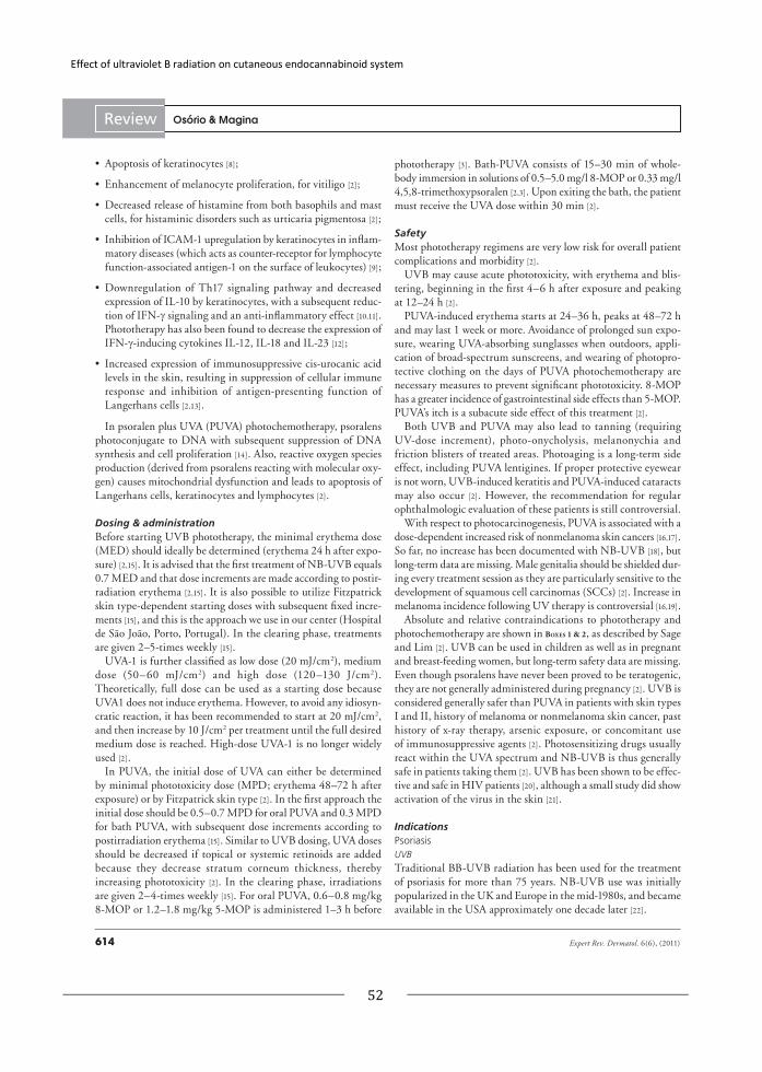
Effect of ultraviolet B radiation on cutaneous endocannabinoid system
52
Expert Rev. Dermatol. 6(6), (2011)614
Review
Apoptosis of keratinocytes [8];
Enhancement of melanocyte proliferation, for vitiligo [2];
Decreased release of histamine from both basophils and mast cells, for histaminic disorders such as urticaria pigmentosa [2];
Inhibition of ICAM-1 upregulation by keratinocytes in inflam-matory diseases (which acts as counter-receptor for lymphocyte function-associated antigen-1 on the surface of leukocytes) [9];
Downregulation of Th17 signaling pathway and decreased expression of IL-10 by keratinocytes, with a subsequent reduc-tion of IFN- signaling and an anti-inflammatory effect [10,11]. Phototherapy has also been found to decrease the expression of IFN- -inducing cytokines IL-12, IL-18 and IL-23 [12];
Increased expression of immunosuppressive cis-urocanic acid levels in the skin, resulting in suppression of cellular immune response and inhibition of antigen-presenting function of Langerhans cells [2,13].
In psoralen plus UVA (PUVA) photochemotherapy, psoralens photoconjugate to DNA with subsequent suppression of DNA synthesis and cell proliferation [14]. Also, reactive oxygen species production (derived from psoralens reacting with molecular oxy-gen) causes mitochondrial dysfunction and leads to apoptosis of Langerhans cells, keratinocytes and lymphocytes [2].
Dosing & administrationBefore starting UVB phototherapy, the minimal erythema dose (MED) should ideally be determined (erythema 24 h after expo-sure) [2,15]. It is advised that the first treatment of NB-UVB equals 0.7 MED and that dose increments are made according to postir-radiation erythema [2,15]. It is also possible to utilize Fitzpatrick skin type-dependent starting doses with subsequent fixed incre-ments [15], and this is the approach we use in our center (Hospital de São João, Porto, Portugal). In the clearing phase, treatments are given 2–5-times weekly [15].
UVA-1 is further classified as low dose (20 mJ/cm2), medium dose (50–60 mJ/cm2) and high dose (120–130 J/cm2). Theoretically, full dose can be used as a starting dose because UVA1 does not induce erythema. However, to avoid any idiosyn-cratic reaction, it has been recommended to start at 20 mJ/cm2, and then increase by 10 J/cm2 per treatment until the full desired medium dose is reached. High-dose UVA-1 is no longer widely used [2].
In PUVA, the initial dose of UVA can either be determined by minimal phototoxicity dose (MPD; erythema 48–72 h after exposure) or by Fitzpatrick skin type [2]. In the first approach the initial dose should be 0.5–0.7 MPD for oral PUVA and 0.3 MPD for bath PUVA, with subsequent dose increments according to postirradiation erythema [15]. Similar to UVB dosing, UVA doses should be decreased if topical or systemic retinoids are added because they decrease stratum corneum thickness, thereby increasing phototoxicity [2]. In the clearing phase, irradiations are given 2–4-times weekly [15]. For oral PUVA, 0.6–0.8 mg/kg 8-MOP or 1.2–1.8 mg/kg 5-MOP is administered 1–3 h before
phototherapy [3]. Bath-PUVA consists of 15–30 min of whole-body immersion in solutions of 0.5–5.0 mg/l 8-MOP or 0.33 mg/l 4,5,8-trimethoxypsoralen [2,3]. Upon exiting the bath, the patient must receive the UVA dose within 30 min [2].
SafetyMost phototherapy regimens are very low risk for overall patient complications and morbidity [2].
UVB may cause acute phototoxicity, with erythema and blis-tering, beginning in the first 4–6 h after exposure and peaking at 12–24 h [2].
PUVA-induced erythema starts at 24–36 h, peaks at 48–72 h and may last 1 week or more. Avoidance of prolonged sun expo-sure, wearing UVA-absorbing sunglasses when outdoors, appli-cation of broad-spectrum sunscreens, and wearing of photopro-tective clothing on the days of PUVA photochemotherapy are necessary measures to prevent significant phototoxicity. 8-MOP has a greater incidence of gastrointestinal side effects than 5-MOP. PUVA’s itch is a subacute side effect of this treatment [2].
Both UVB and PUVA may also lead to tanning (requiring UV-dose increment), photo-onycholysis, melanonychia and friction blisters of treated areas. Photoaging is a long-term side effect, including PUVA lentigines. If proper protective eyewear is not worn, UVB-induced keratitis and PUVA-induced cataracts may also occur [2]. However, the recommendation for regular ophthalmologic evaluation of these patients is still controversial.
With respect to photocarcinogenesis, PUVA is associated with a dose-dependent increased risk of nonmelanoma skin cancers [16,17]. So far, no increase has been documented with NB-UVB [18], but long-term data are missing. Male genitalia should be shielded dur-ing every treatment session as they are particularly sensitive to the development of squamous cell carcinomas (SCCs) [2]. Increase in melanoma incidence following UV therapy is controversial [16,19].
Absolute and relative contraindications to phototherapy and photochemotherapy are shown in BOXES 1 & 2, as described by Sage and Lim [2]. UVB can be used in children as well as in pregnant and breast-feeding women, but long-term safety data are missing. Even though psoralens have never been proved to be teratogenic, they are not generally administered during pregnancy [2]. UVB is considered generally safer than PUVA in patients with skin types I and II, history of melanoma or nonmelanoma skin cancer, past history of x-ray therapy, arsenic exposure, or concomitant use of immunosuppressive agents [2]. Photosensitizing drugs usually react within the UVA spectrum and NB-UVB is thus generally safe in patients taking them [2]. UVB has been shown to be effec-tive and safe in HIV patients [20], although a small study did show activation of the virus in the skin [21].
IndicationsPsoriasis UVBTraditional BB-UVB radiation has been used for the treatment of psoriasis for more than 75 years. NB-UVB use was initially popularized in the UK and Europe in the mid-1980s, and became available in the USA approximately one decade later [22].
Osório & Magina

Introduction Annex 2
53
www.expert-reviews.com 615
Review
NB-UVB is superior to BB-UVB in respect to clearing and remission times [22], and it currently represents the phototherapeu-tic modality of choice for the treatment of psoriasis in Europe [23]. However, it is probably not as effective as PUVA, both in terms of clearing efficiency and duration of remission [15].
Patients with psoriasis who are compliant, motivated and adher-ent with instructions and follow-up examinations could, under dermatologist supervision, be considered appropriate candidates for home UVB therapy [22]. A recent multicenter, single-blind, randomized clinical trial demonstrated that home NB-UVB is just as effective as outpatient-administered NB-UVB, with supe-rior patient quality of life [24]. We do not have experience with home phototherapy in our center.
In order to improve UVB’s efficacy, it can be combined with other treatments, either topical or systemic.
Topical application of calcipotriol (after irradiation) or tazaro-tene [25] (before irradiation) may increase therapeutic efficacy of phototherapy alone [26]. By contrast, it seems that combining topi-cal steroids adds little benefit to phototherapy [27]. Emmolients can increase UV transmission, increasing efficacy, but thick appli-cation of petrolatum and water-in-oil creams as well as salicylic acid and tar act as sunscreens and should be discouraged [28].
In regard to systemic drugs, retinoids increase efficacy and reduce the carcinogenic potential of UVB phototherapy [29,30]. Retinoids can be maintained after ceasing phototherapy to pre-vent recurrence. The short time combination of methotrexate with UVB therapy is of potential value because of the synergistic effects of these two therapies [22]. We find this combination particularly useful in patients with psoriatic arthritis in whom the articular disease is successfully managed with low-dose methrotrexate, as phototherapy is not efficacious in treating arthritis. The combina-tion of cyclosporine and UVB has not been studied extensively because of the increased risk of nonmelanoma skin cancer with cyclosporine monotherapy and it should generally be avoided [22]. Combination therapy of UVB with biologics has been reported to have a higher response rate than biologics alone, with a similar safety profile [22], but further investigation is needed. Potential concerns are increased phototoxicity and photocarcinogenesis.
Although UVB may be adequate for maintenance therapy [31], it is suggested to consider a rotational therapeutic approach in order to minimize a greater cumulative dose [32].
The first report on the use of the excimer laser to treat pso-riasis goes back to 1997 [33]. Advantages include the possibility to treat exclusively the affected skin with higher doses (supra-erythemogenic) and lower number of treatments [34].
PUVACompared to oral 8-MOP, oral 5-MOP shows equal clearing rates with a similar number of exposures, but higher cumulative doses, lower incidence of pruritus or severe phototoxic reactions, and no nausea or vomiting [3].
Compared to oral 8-MOP, bath PUVA shows equal clearing rates with fewer exposures, similar or lower incidence of ery-thema and pruritus and no systemic intolerance such as nausea or vomiting [3].
Topical PUVA with psoralen creams, ointment or lotions is now used only for limited plaque psoriasis and for palmo-plantar disease [3].
With respect to combination therapy, the same considerations are valid as for UVB when it comes to topics. The combination of PUVA with systemic retinoids (RePUVA) is one of the most potent therapeutic regimens for psoriasis [15]. Methotrexate may also be considered for combination therapy, although the long-term risk of cutaneous malignancies, though never proved, should not be forgotten [15]. By contrast, concomitant use of cyclosporine has been recognized to enhance skin carcinogenesis [35]. Further investigation is needed to verify presumptive additive effects of biologics and PUVA [36].
PUVA can be considered for maintenance therapy but atten-tion should be paid to long-term risks related to total cumulative phototoxic doses [3].
Pediatric population: In children, when topical treatment fails, NB-UVB phototherapy in combination with topical agents is generally the recommended treatment option before considering
Box 1. Contraindications to UV phototherapy.
Absolute contraindicationsXeroderma pigmentosum
Systemic lupus erythematosus
Basal cell nevus syndrome
Relative contraindicationsPrevious history of skin cancer
Treatment of genital area
Photosensitive disorders
Contact photosensitive substances
Photosensitizing medications with an action spectrum with the corresponding UVA or UVB wavelengths
Box 2. Contraindications to psoralen plus UVA.
Absolute contraindicationsXeroderma pigmentosum
Systemic lupus erythematosus
Basal cell nevus syndrome
Photosensitivity disorders
Age less than 10 years
Pregnant and breast-feeding patients
History of melanoma
Relative contraindicationsFamily history of melanoma
History of dysplastic nevi or nonmelanoma skin cancers
Photosensitizing medications
Significant past UV exposure
History of arsenic exposure, ionizing radiation, methotrexate, cyclosporine or tacrolimus
CautionHepatic or renal insufficiency
Patients 10–18 years of age
Phototherapy & photopheresis: old & new indications

Effect of ultraviolet B radiation on cutaneous endocannabinoid system
54
Expert Rev. Dermatol. 6(6), (2011)616
Review
systemic therapy, including retinoids, metho trexate, cyclosporine and biological agents. The long-term risk of NB-UVB light in children is yet to be determined. PUVA can also be considered as a treatment option [37].
Cutaneous T-cell lymphoma (mycosis fungoides) PUVAThe use of PUVA in cutaneous T-cell lymphoma (CTCL) was first reported in 1976 [38]. It is an excellent treatment option for the early stages of the disease (IA–IIA), achieving long-lasting free-disease intervals. It is also capable of reducing tumor burden in later stages (IIB–IVB), where it should be combined with other treatments, such as retinoids, bexarotene, INF- 2a, local x-ray or systemic chemotherapy. Even with aggressive combination thera-pies, these patients experience multiple recurrences. Although effective in inducing remission in skin-confined disease, the effect of PUVA on the CTCL disease course and overall survival is yet to be determined. Currently there is no treatment option known to arrest progression of later stages [3,15,39].
After clearing, a maintenance phase is usually recommended, with two sessions per week for 1 month, followed by one session per week for another. Resolution should be confirmed with a biopsy. A follow-up is then performed, either without therapy, with one treatment per month or one treatment every other month. Relapses respond to therapy as well as initial lesions [3,15].
UVBThe first report of UVB phototherapy for CTCL goes back to 1982 [40]. NB-UVB can also induce high rates of complete remis-sion in early disease [39]. Evidence is poor, however, regarding the question how NB-UVB compares with other treatments [39]. Studies comparing PUVA and NB-UVB have found them both effective in the treatment of CTCL [41,42]. NB-UVB and BB-UVB therapy have not been compared.
Excimer laser can also be considered a useful tool in the treatment of early-stage mycosis fungoids (MF) [43].
UVA1UVA1 also appears to be effective [44], although the experience is limited. It offers advantages over PUVA in terms of side effects.
Pediatric population: In children, MF is rare and usually limited to early patch-stage disease. Both PUVA and UVB have been used [37].
Vitiligo PUVAPUVA has been used for longer than UVB in vitiligo and the available information for this therapy is greater than for UVB.
Patients should be aware that they will require months of treat-ment (100–200 treatments), with 70% of patients responding after 12–24 months. Responsiveness is defined as development of multiple perifollicular macules of repigmentation, or, in the case of small (<2 cm) lesions, contraction in size. However, if after 4–6 months or 30–50 treatments they do not respond, PUVA
should be terminated. Locations such as lips, distal dorsal hands, fingers and toes, palms, soles and nipples are very refractory to treatment, as are large areas with only white hairs. Stable disease (for at least a year) is usually easier to treat. Duration of disease before PUVA therapy does not affect response rate [3,15].
Completely repigmented areas can be stable for a decade or more without relapse. But if a lesion is not fully repigmented, reversal of acquired pigmentation may occur when treatment is discontinued [15].
KUVA and topical PUVA are alternatives to oral PUVA. KUVA uses khellin as a photosensitizer and this is not phototoxic. Topical PUVA may be suitable for few and small lesions [15]. UVA alone is of limited benefit [15].
UVBAlthough the action spectrum for phototherapy of vitiligo is not known [15], UVB, in particular NB-UVB, is currently more often used than PUVA because it has been shown to be at least as effec-tive and possibly safer [45,46]. As for PUVA, it is not very effective in certain anatomic areas (hands and digits, feet and toes) and treat-ment should be ceased if patients do not respond [23]. Excimer laser should be considered as a therapeutic option for localized lesions [47].
Pediatric population: In children, NB-UVB is the preferred pho-totherapeutic option owing to potential increased cancer risks asso-ciated with PUVA. However, PUVA can also be used in children older than 12 years of age [37].
Atopic dermatitis Phototherapy of acute, severe atopic dermatitisUVA1 phototherapy is a highly effective monotherapy for a lim-ited period of time (ten to 15 exposures), particularly in cases of acute, severe atopic dermatitis [48]. It should not be used for maintenance therapy or for patients under the age of 18 years because its long-term effects are not known [23]. The effectiveness is dose-dependent, with high-dose UVA1 being more efficient than UVA/UVB phototherapy [49].
Phototherapy of chronic, moderate atopic dermatitisBB-UVB, combined UVA/UVB, broadband UVA, low-dose UVA1 and, in particular, narrow-band UVB phototherapy are effective treatments in mild and moderate atopic dermatitis and they are generally used in combination regimens with topical steroids. They are relatively safe when used over extended periods of time for long-term management [23].
NB-UVB and UVA/UVB phototherapy are superior to BB-UVB, broadband UVA or low-dose UVA1 [23]. However, in a small number of children, BB-UVB may be less irritating and more effective than NB-UVB because it requires smaller doses to achieve minimal erythema and lymphocyte apoptosis [37]. The excimer laser can also be used to treat localized lesions of atopic dermatitis with faster clearance after fewer treatments compared with traditional NB-UVB or BB-UVB phototherapy [50].
After NB-UVB, the microbial population in atopic dermati-tis patients is similar to control patients [51], but it is unknown
Osório & Magina

Introduction Annex 2
55
www.expert-reviews.com 617
Review
whether this translates clinically into reduced skin infections requiring antibiotics or decreased pruritus [37].
PUVAPUVA can also be used to treat atopic dermatitis, but a high number of treatments are usually required and recurrences are high and rapid. Long-term treatment should be avoided [15].
Lichen planus Oral and topical PUVA are therapeutic options for extensive lichen planus (LP) [3,15]. However, LP is usually more resistant than psori-asis, requiring more treatment sessions and higher cumulative UVA doses, with earlier relapses [3]. Combined PUVA-retinoid therapy may accelerate clearing [3]. Postinflammatory hyper pigmentation should be a potential concern. Besides, LP exacerbation during PUVA therapy has occurred in a few patients [15].
NBUVB therapy can represent an effective option for refrac-tory generalized LP according to a study of 16 patients in which complete response was achieved in 56.25% at 30 sessions and in 68.75% at 40 sessions, confirming the presence of significant cor-relation between session numbers and improvement of response, that had already been found in previous studies [52].
A study of 28 patients, 15 treated with oral PUVA and 13 with NB-UVB, concluded that even though oral PUVA pro-duces a better initial clinical response rate, both oral PUVA and NB-UVB are effective treatments for LP, producing similar long-term outcomes.
Graft-versus-host disease Currently, PUVA represents the mainstay for phototherapy of graft-versus-host disease (GVHD) [15]. It has been initially evalu-ated for lichenoid GVHD owing to clinical and histologic simi-larities with lichen planus, and it can clear or improve chronic and acute disease [53,54]. In sclerodermoid GVHD, results are con-troversial [3,15]. Effects may be systemic because mucosal lesions respond to treatment, but there is no improvement in other organs [3,15]. Patients should be followed-up for cutaneous tumors because there seems to be an overall increased risk of malignancies in bone marrow/stem cell recipients, independent of PUVA [3,15]. UVA1 therapy is also promising for the treatment of chronic GVHD, in either of its lichenoid or sclerodermoid variants [44,55]. UVB therapy has been described in a limited number of patients [15].
Pityriasis lichenoides PUVA seems to be more effective than UVB in pityriasis lichen-oids, especially in the acute form of the disease, where it has been recommended as the phototherapy of choice [15]. In pityriasis lichenoides chronic, PUVA may be reserved for cases of that are UVB resistant [15]. The experience with UVA1 is limited [56] and further investigation is needed.
Urticaria pigmentosa PUVAIn cutaneous mastocytosis, PUVA therapy results in improvement of skin lesions [57], but in most patients these recur 5–8 months
after treatment discontinuation [3]. Response rate in relapses is though similar to initial lesions [3]. Systemic symptoms such as histamine-induced migraines and flushing also respond to PUVA [57]. PUVA therapy should be considered for patients in which disease is causing severe distress [3].
UVA1Cutaneous mastocytosis has also been demonstrated to respond to UVA1 therapy, in its skin and systemic manifestations [58,59]. In a study of four patients, there were no recurrences more than 2 years after cessation of high-dose UVA1 therapy [59].
Photodermatoses Phototherapy of patients with polymorphous light eruption (PMLE) induces tolerance to sunlight in a process known as hardening. Involved mechanisms include epidermal thickening, pigmentation and probable immunologic effects [3,15].
PUVA is the most effective preventive treatment in PMLE [3], but NB-UVB is currently the phototherapeutic option of choice, with recent studies showing that it is an effective alternative to PUVA [60].
With PUVA, 70% of patients develop hardening with a 3–4-week course treatment of 2–3 sessions a week in early spring. Patients remain protected if they have regular sunlight exposures. However, patients may remain protected for 2–3 months even after pigmentation has faded [3,15].
Patients with other photodermatosis, including actinic prurigo, hydroa vacciniforme and erythropoietic porphyria, may benefit from NB-UVB [61] or PUVA [3,15]. In solar urticaria, PUVA ther-apy appears to be the most effective therapy available [3]. Single UVA irradiation of quadrants of body surface hours before PUVA treatment may be useful when patients develop urticarial lesions in the beginning of treatment [3]. In therapy-resistant forms of solar urticaria, rush-hardening consists of multiple daily UVA irradiations at 1-h intervals, providing protection within 3 days [62]. Unlike PMLE, extended treatment may be required in solar urticaria and chronic actinic dermatitis [3].
Morphea, scleroderma & other sclerosing skin conditionsMorphea was shown to be effectively treated by UVA1 photherapy in a dose-dependent manner [63]. Medium-dose UVA1 seems to be superior to low-dose UVA1 or NB-UVB [64,65]. The existing evi-dence indicates that medium–high dose UVA1 therapy delivered over 24–30 treatments can provide significant benefit to patients with morphea, particularly linear and plaque subtypes of disease [44]. UVA1 is likely ineffective in burned out atrophic lesions, deep morphea, Parry Romberg/facial hemiatrophy and eosinophilic fas-ciitis owing to the depth of the pathology, and systemic treatment should be considered as first-line therapy in these cases [44]. Little to no adverse effects have been reported with this therapy [44].
UVA1 might also be effective in treating patients with systemic sclerosis, studies to date showing improvement in hand/forearm disease, with a decreased hand score after treatment [66].
Localized scleroderma and pansclerotic morphea have also been successfully treated with bath PUVA and oral PUVA [67,68]. In our
Phototherapy & photopheresis: old & new indications

Effect of ultraviolet B radiation on cutaneous endocannabinoid system
56
Expert Rev. Dermatol. 6(6), (2011)618
Review
center we have a good experience with topical PUVA for localized scleroderma and we consider this modality worth a try.
Lichen sclerosus has shown to be responsive to UVA1, in both genital and nongenital disease [69,70].
Other indicationsUVBNB-UVB was found to be an effective treatment in severe cases of seborrheic dermatitis, although flares may occasionally occur [71].
It has been shown that NB-UVB is effective in controlling uremic pruritus [72]. Although it can also be beneficial in other forms of pruritus, such as idiopathic or that associated with liver disease or diabetes [15], we have better results in uremic pruritus.
PUVA The experience with PUVA in lymphomatoid papulosis is limited to a few successful anedoctal cases [73].
PUVA has been used in pityriasis rubra pilaris, but results are inconsistent [15,23].
Generalized granuloma annulare has been reported to clear completely, but long-term maintenance treatment was required to maintain remissions [74].
Uncontrolled studies of PUVA treatment for alopecia areata, using all types of PUVA (oral or topical psoralen, local or whole body UVA irradiation), report success rates of up to 60–65% [75], which are in accordance to our experience. Continued treat-ment might be needed however to maintain hair growth because relapse rates are high [75]. 308-nm excimer has also been reported as effective [76,77].
UVA1Dyshidrotic hand eczema has been treated effectively with UVA1 therapy [78]. Long exposure to low-dose UVA1 might be beneficial for patients with systemic lupus erythematous, with decreased fatigue, joint pain, malaise, stiffness and mouth ulcers, lasting over 3 years [79]. UVA1 therapy has also been shown to improve subacute cutaneous lupus erythematosus lesions, but not those seen in discoid lupus erythematosus [44]. A consensus statement by the North American Rheumatologic Dermatology Society has recently confirmed the potential beneficial effects of UVA1 phototherapy in systemic lupus erythematous patients, but acknowledged that further investigation is needed to address its full benefit [44]. In TABLE 1, we summarize phototherapy’s main clinical applications.
PhotopheresisMechanisms of actionExtracorporeal photochemotherapy, also known as extracorporeal photopheresis (ECP), is a promising form of immuno-modulatory therapy. In apheresis, the blood of a donor or patient is passed through an apparatus that separates out one particular constituent and returns the remainder to the circulation. In photopheresis, a par-ticular kind of apheresis, the majority of white blood cells are treated with 8-MOP, which becomes biologically active under exposure to UVA light, making pathogenic T cells, monocytes, natural killer cells, as well as B and T lymphocytes, susceptible to apoptosis. Furthermore, an immunological response follows the reinfusion of irradi-ated cells, with activation and differen-tiation of antigen-presenting cells (APCs, monocytes). APCs recognize apoptotic leukocytes through receptors that stimu-late production of anti-inflammatory cyto-kines such as IL-10 and TGF- . ECP also induces immune tolerance through the induction of Tregs, with increased pro-duction of IL-10 and TGF- . ECP alters lymphocyte proliferation favoring a shift from a Th1 to a Th2 cytokine profile. Sub-cellular particles from apoptotic lympho-cytes known as blebs show increased CD5 and CD8 and decreased CD28 and CD26
Table 1. Summary of the applications of phototherapy.
Condition Standard of care Other therapeutic options
Psoriasis NBUVB PUVA, Excimer
Cutaneous T-cell lymphoma (IA–IIA)
PUVA NBUVB, Excimer, UVA1
Vitiligo NBUVB PUVA, Excimer
Atopic dermatitis:Acute, severeChronic, mild
UVA1NBUVB, UVA/UVB
Lichen planus PUVA NBUVB
Lichenoid graft-versus-host disease PUVA UVA1, NBUVB
Pityriasis lichenoides PUVA NBUVB
Urticaria pigmentosa PUVA UVA1
Photodermatosis:Polymorphic light eruptionSolar urticariaOthers
NBUVBPUVANBUVB, PUVA
PUVA
Morphea, systemic sclerosis UVA1 PUVA
Lichen sclerosus UVA1
Other conditions:Seborrheic dermatitisUremic pruritusLymphomatoid papulosisPytiriasis rubra pilarisGranuloma annulareAlopecia areataDesydrotic hand eczemaSystemic lupus erythematosus
NBUVBNBUVBPUVAPUVAPUVAPUVA, ExcimerUVA1UVA1
Summary of phototherapy’s main clinical applications.NBUVB: Narrow band-UVB; PUVA: Psoralen plus UVA.
Osório & Magina

Introduction Annex 2
57
www.expert-reviews.com 619
Review
expression, surface markers consistent with an immunosuppres-sive phenotype. Apoptotic cells show increased expression of phos-phatidylserine which is recognized by phagocyting macrophages, thereby leading to their anti-inflammatory clearance [80].
AdministrationECP involves three stages – leucapheresis, photoactivation with a photosensitizer plus UVA and re-infusion of buffy coat – each treatment taking 3–4 h. One main difficulty of performing photopheresis results from the venous access. One cycle of ECP consists of two treatments on consecutive days, and one cycle is typically given every 2–4 weeks [81].
SafetyECP’s side effects are listed in BOX 3 [80]. Side-effects are typically mild and transient and severe reactions such as vasovagal syn-cope or infections are uncommon. The lack of major side effects, including malignancies or infections, is very valuable in diseases for which alternative treatments are highly immunosuppressive [81]. Contraindications to ECP include severe renal, hepatic or cardiac impairment, coagulation disorders and hypersensitivity to psoralen compounds.
IndicationsCTCLErythrodermic CTCL was the first disease to be treated with ECP in the early 1980s [82], an indication for which it received US FDA approval in 1988.
ECP is currently approved for the treatment of skin manifesta-tions in all stages of CTCL, but there is good evidence to support the rejection of the use of ECP for the treatment of nonerythro-dermic MF and fair evidence to support the use of ECP for eryth-rodermic MF, including stage III and IVA, and Sézary syndrome [80,83]. There are several guidelines on the use of ECP in patients with CTCL [83–85].
The role of ECP in treating earlier stages of CTCL may be limited. One plausible explanation are the low levels of circulating Sézary cells in the peripheral blood of these patients [80].
Other attempts to better characterize those CTCL patients who are more likely to respond to ECP revealed that patients with a ratio CD4:CD8 of<10 are more likely to respond than those with a ratio >10 and that a nonelevated LDH at the start of treatment tends to predict a better response to ECP compared with patients with an elevated LDH [15].
GVHDSteroids and other immunosupressive agents are the mainstay initial modes of therapy in the treatment of GVHD after allogenic bone-marrow transplantation. ECP has been successfully used in refractory acute (aGVHD) and chronic (cGVHD) disease for the past 10 years [80,83,86].
Studies have found good results on cutaneous improvement of cGVHD but they were not as consistent in respect to liver and lung involvement [87,88]. Patients with progressive development of cGVHD may be more likely to respond to therapy versus those
with de novo onset [89]. Another study reported a higher response rate in participants who started therapy earlier than 1 year after onset of cGVHD [88]. ECP may also work as a steroid-spering treatment, more effective than conventional steroid-spearing immunosuppressants, as shown in a multicenter, prospective, Phase II clinical trial involving 95 patients with cutaneous cGVHD [90]. This may represent an advantage owing to its fairly safe profile [80].
ECP also seems promising in the treatment of patients with aGVHD, particularly those with cutaneous manifestations but also liver and gut involvement [80].
Solid organ transplantsIn a randomized controlled trial, the addition of ECP to triple-drug immunosuppressive therapy significantly decreased the risk of cardiac rejection without increasing the incidence of infection [91]. Multiple case reports and other anecdotal data involving the use of ECP in transplant recipients of lung, liver, kidney and allo-geneic stem cell transplantation can also be found in the literature [80]. ECP is effective for patients resistant to conventional treat-ment, particularly if started early [3]. Besides reversal of allograft rejection, a reduction in immunosuppressive therapy has also been frequently achieved [92,93].
Autoimmune diseasesLimited data has also been published regarding the use of ECP in a variety of autoimmune diseases [80].
A small number of patients with pemphigus vulgaris have been successfully treated with ECP, with tapering of concomitant immunosuppressive therapy. However, since clinical trials with a large cohort of patients have not yet been conducted, there is not sufficient evidence to support the use of ECP in patients with this disease [80].
Two randomized clinical trials found improvements in skin and joint severity scores in patients with systemic scleroderma after ECP [94,95], pointing to a favorable outcome when ECP is started early in the course of the disease, with a low incidence of side effects [94].
Box 3. Side effects of extracorporeal photopheresis.
Common side effectsHeadache
Nausea
Chills
Low-grade fever
Other side effectsHypotension
Vasovagal syncope
Septicemia
Anemia
Abnormal clotting response
Skin infections over the venous access site
Bacterial endocarditis
Phototherapy & photopheresis: old & new indications
Effect of ultraviolet B radiation on cutaneous endocannabinoid system
58
Expert Rev. Dermatol. 6(6), (2011)620
Review
An open-label pilot study published in 1992 stated an improvement of clinical skin and joint scores in seven of ten patients with systemic lupus erythematosus, although no sig-nificant changes were noted in laboratory parameters [96]. Since then only a few anecdotal cases have been reported in the lit-erature [80].
Although at one time promising, the role of ECP for the treat-ment of rheumatoid arthritis is still questionable [80].
Other applicationsSevere atopic dermatitis has been successfully treated with ECP, the first report going back to 1994 [97]. It has also been effective in the treatment of recalcitrant mucous and cutaneous erosive lichen planus [98,99]. Other recently reported potential applications of ECP might be refractory Crohn’s disease, Type 1 diabetes mellitus and the prevention of restenosis in patients after percutaneous transluminal coronary angioplasty [80]. In BOX 4, we summarize ECP’s main clinical applications.
Expert commentaryPhototherapy is an important treatment option for psoriasis and other skin diseases and is the first choice in case of contraindica-tions for immunosupression, such as patients with infections or cancer-prone patients. There are some discrepancies in the litera-ture, regarding dosimetry recomendations, efficacy, risk of phototoxicity and long-term risks, particularly for NB-UVB and UVA-1 therapies. More data are needed in order to optimize these phototherapy modalities.
Besides advanced cutaneous T-cell lym-phoma, theoretically, photopheresis is an exciting tool for the treatment of a variety of T-cell-mediated diseases and should be considered in severe and difficult-to-treat
conditions, particularly in systemic scleroderma.
Five-year viewPhototherapy is a time-consuming treatment and this raises the question: what is the future position of phototherapy as a classical treatment for psoriasis in the era of biologics? The better under-standing of phototherapy-induced molecular pathways and the identification of clinical and genetic predictors of treatment response will contribute to maintain the importance of phototherapy. On the other hand, it is attractive to speculate that at least an additive or possibly a synergistic effect can be expected in case of combina-tion of phototherapy with a biologic. Targeted phototherapy using high-intensity UV devices may improve safety and efficacy, namely in difficult-to-treat cases of morphea, alopecia areata and vitiligo.
In the near future, more studies will corroborate the role of extracorporeal photopheresis in the prevention of solid organ transplant rejection and this therapeutic option will be available in more institutions worldwide.
Key issues
Narrow band-UVB (NB-UVB) currently represents the phototherapeutic modality of choice for the treatment of psoriasis.
Psoralen plus UVA (PUVA) is an excellent treatment option for the early stages of cutaneous T-cell lymphoma (IA–IIA), here achieving long-lasting free-disease intervals. NB-UVB can also induce high rates of complete remission in early disease.
NB-UVB is currently used more than PUVA in vitiligo because it has been shown to be at least as effective and possibly safer.
UVA1 phototherapy is a highly effective monotherapy for acute, severe atopic dermatitis. NB-UVB and UVA/UVB phototherapy are effective treatments in chronic, mild and moderate disease.
PUVA is a therapeutic option for extensive lichen planus and for lichenoid graft-versus-host disease.
NB-UVB is currently the phototherapeutic option of choice in polymorphous light eruption. In solar urticaria, PUVA appears to be the most effective therapy available. Patients with other photodermatosis, including actinic prurigo, hydroa vacciniforme and erythropoietic porphyria, may benefit from NB-UVB or PUVA.
Medium–high dose UVA1 therapy can provide significant benefit to patients with morphea, particularly linear and plaque subtypes of disease.
Photopheresis is used in patients with erythrodermic cutaneous T-cell lymphoma, including stage III and IVA, and those with Sézary syndrome with good results.
It has also been successfully used in refractory acute and chronic graft-versus-host disease, with cutaneous improvement. Similar benefit has also been reported in transplant recipients of heart, lung, liver, kidney and allogeneic stem cell transplantation.
Limited data has been published regarding the use of extracorporeal photopheresis in a variety of autoimmune diseases, including pemphigus vulgaris, systemic scleroderma, systemic lupus erythematosus and rheumatoid arthritis.
Box 4. Summary of the applications of extracorporeal photopheresis.
Main applicationsErythrodermic CTCL (IIIA, IV), Sézary syndrome
Acute and chronic graft-versus-host disease
Rejection in transplant recipients of heart, lung, liver, kidney and allogenic stem cells
Other applicationsAutoimmune diseases: penphigus vulgaris, systemic scleroderma, systemic lupus erythematosus, rheumatoid arthritis
Atopic dermatitis
Erosive lichen planus
Crohn’s disease
Type 1 diabetes mellitus
Prevention of restenosis in patients after percutaneous transluminal coronary angioplasty
CTCL: Cutaneous T-cell lymphoma.
Osório & Magina

Introduction Annex 2
59
www.expert-reviews.com 621
Review
Financial & competing interests disclosureThe authors have no relevant affiliations or financial involvement with any organization or entity with a financial interest in or financial conflict with the subject matter or materials discussed in the manuscript. This includes
employment, consultancies, honoraria, stock ownership or options, expert testimony, grants or patents received or pending, or royalties.
No writing assistance was utilized in the production of this manuscript.
References1 Maverakis E, Miyamura Y, Bowen MP,
Correa G, Ono Y, Goodarzi H. Light, including ultraviolet. J. Autoimmun. 34(3), J247–J257 (2010).
2 Sage RJ, Lim HW. UV-based therapy and vitamin D. Dermatol. Ther. 23(1), 72–81 (2010).
3 Hönigsmann H, Szeimies RR, Knobler R. Photochemotherapy and Photodinamic Therapy. In: Fitzpatrick Dermatology in General Medicine. McGraw-Hill Professional, New York, NY, USA, 2249–2262 (2008).
4 Aubin F, Vigan M, Puzenat E et al. Evaluation of a novel 308-nm monochromatic excimer light delivery system in dermatology: a pilot study in different chronic localized dermatoses. Br. J. Dermatol. 152(1), 99–103 (2005).
5 Ozawa M, Ferenczi K, Kikuchi T et al. 312-nanometer ultraviolet B light (narrow-band UVB) induces apoptosis of T cells within psoriatic lesions. J. Exp. Med. 189(4), 711–718 (1999).
6 Morita A, Werfel T, Stege H et al. Evidence that singlet oxygen-induced human T helper cell apoptosis is the basic mechanism of ultraviolet-A radiation phototherapy. J. Exp. Med. 186(10), 1763–1768 (1997).
7 Szepietowski JC, Morita A, Tsuji T. Ultraviolet B induces mast cell apoptosis: a hypothetical mechanism of ultraviolet B treatment for uraemic pruritus. Med. Hypotheses 58(2), 167–170 (2002).
8 Aufiero BM, Talwar H, Young C et al. Narrow-band UVB induces apoptosis in human keratinocytes. J. Photochem. Photobiol. B 82(2), 132–139 (2006).
9 Krutmann J, Czech W, Parlow F et al. Ultraviolet radiation effects on human keratinocyte ICAM-1 expression: UV-induced inhibition of cytokine-induced ICAM-1 mRNA expression is transient, differentially restored for IFN gamma versus TNF alpha, and followed by ICAM-1 induction via a TNF alpha-like pathway. J. Invest. Dermatol. 98(6), 923–928 (1992).
10 Grewe M, Gyufko K, Krutmann J. Interleukin-10 production by cultured human keratinocytes: regulation by ultraviolet B and ultraviolet A1 radiation. J. Invest. Dermatol. 104(1), 3–6 (1995).
11 Racz E, Prens EP, Kurek D et al. Effective treatment of psoriasis with narrow-band UVB phototherapy is linked to suppression of the IFN and Th17 pathways. J. Invest. Dermatol. 131(7), 1547–1558 (2011).
12 Piskin G, Tursen U, Sylva-Steenland RM, Bos JD, Teunissen MB. Clinical improvement in chronic plaque-type psoriasis lesions after narrow-band UVB therapy is accompanied by a decrease in the expression of IFN-gamma inducers – IL-12, IL-18 and IL-23. Exp. Dermatol. 13(12), 764–772 (2004).
13 Beissert S, Mohammad T, Torri H et al. Regulation of tumor antigen presentation by urocanic acid. J. Immunol. 159(1), 92–96 (1997).
14 Krutmann J, Morita A. Mechanisms of ultraviolet (UV)B and UVA phototherapy. J. Invest. Dermatol. Symp. Proc. 4(1), 70–72 (1999).
15 Hönigsmann H, Schwartz T. Ultraviolet therapy. In: Bolognia: Dermatology 2nd Edition. Elsevier Inc., Amsterdam, The Netherlands (2008).
16 Lindelof B, Sigurgeirsson B, Tegner E et al. PUVA and cancer risk: the Swedish follow-up study. Br. J. Dermatol. 141(1), 108–112 (1999).
17 Nijsten TE, Stern RS. The increased risk of skin cancer is persistent after discontinuation of psoralen+ultraviolet A: a cohort study. J. Invest. Dermatol. 121(2), 252–258 (2003).
18 Hearn RM, Kerr AC, Rahim KF, Ferguson J, Dawe RS. Incidence of skin cancers in 3867 patients treated with narrow-band ultraviolet B phototherapy. Br. J. Dermatol. 159(4), 931–935 (2008).
19 Stern RS. The risk of melanoma in association with long-term exposure to PUVA. J. Am. Acad. Dermatol. 44(5), 755–761 (2001).
20 Akaraphanth R, Lim HW. HIV, UV and immunosuppression. Photodermatol. Photoimmunol. Photomed. 15(1), 28–31 (1999).
21 Breuer-McHam J, Simpson E, Dougherty I et al. Activation of HIV in human skin by ultraviolet B radiation and its inhibition by NFkappaB blocking agents. Photochem. Photobiol. 74(6), 805–810 (2001).
22 Menter A, Korman NJ, Elmets CA et al. Guidelines of care for the management of
psoriasis and psoriatic arthritis: section 6. Guidelines of care for the treatment of psoriasis and psoriatic arthritis: case-based presentations and evidence-based conclusions. J. Am. Acad. Dermatol. 65(1), 137–174 (2011).
23 Krutmann J, Morita A. Therapeutic photomedicine: phototherapy. In: Fitzpatrick Dermatology in General Medicine. McGraw-Hill Inc., New York, NY, USA, 2243–2249 (2008).
24 Koek MB, Buskens E, van Weelden H, Steegmans PH, Bruijnzeel-Koomen CA, Sigurdsson V. Home versus outpatient ultraviolet B phototherapy for mild to severe psoriasis: pragmatic multicentre randomised controlled non-inferiority trial (PLUTO study). BMJ 338, B1542 (2009).
25 Koo JY, Lowe NJ, Lew-Kaya DA et al. Tazarotene plus UVB phototherapy in the treatment of psoriasis. J. Am. Acad. Dermatol. 43(5 Pt 1), 821–828 (2000).
26 Kerscher M, Volkenandt M, Plewig G, Lehmann P. Combination phototherapy of psoriasis with calcipotriol and narrow-band UVB. Lancet 342(8876), 923 (1993).
27 LeVine MJ, Parrish JA. The effect of topical fluocinonide ointment on phototherapy of psoriasis. J. Invest. Dermatol. 78(2), 157–159 (1982).
28 Lebwohl M, Martinez J, Weber P, DeLuca R. Effects of topical preparations on the erythemogenicity of UVB: implications for psoriasis phototherapy. J. Am. Acad. Dermatol. 32(3), 469–471 (1995).
29 Green C, Lakshmipathi T, Johnson BE, Ferguson J. A comparison of the efficacy and relapse rates of narrowband UVB (TL-01) monotherapy vs. etretinate (re-TL-01) vs. etretinate-PUVA (re-PUVA) in the treatment of psoriasis patients. Br. J. Dermatol. 127(1), 5–9 (1992).
30 Iest J, Boer J. Combined treatment of psoriasis with acitretin and UVB phototherapy compared with acitretin alone and UVB alone. Br. J. Dermatol. 120(5), 665–670 (1989).
31 Stern RS, Armstrong RB, Anderson TF et al. Effect of continued ultraviolet B phototherapy on the duration of remission of psoriasis: a randomized study. J. Am. Acad. Dermatol. 15(3), 546–552 (1986).
32 Menter MA, See JA, Amend WJ et al. Proceedings of the Psoriasis Combination
Phototherapy & photopheresis: old & new indications

Effect of ultraviolet B radiation on cutaneous endocannabinoid system
60
Expert Rev. Dermatol. 6(6), (2011)622
Review
and Rotation Therapy Conference. Deer Valley, Utah, Oct. 7–9, 1994. J. Am. Acad. Dermatol. 34(2 Pt 1), 315–321 (1996).
33 Bonis B, Kemeny L, Dobozy A, Bor Z, Szabo G, Ignacz F. 308 nm UVB excimer laser for psoriasis. Lancet 350(9090), 1522 (1997).
34 Kollner K, Wimmershoff MB, Hintz C, Landthaler M, Hohenleutner U. Comparison of the 308-nm excimer laser and a 308-nm excimer lamp with 311-nm narrowband ultraviolet B in the treatment of psoriasis. Br. J. Dermatol. 152(4), 750–754 (2005).
35 van de Kerkhof PC, de Rooij MJ. Multiple squamous cell carcinomas in a psoriatic patient following high-dose photochemotherapy and cyclosporin treatment: response to long-term acitretin maintenance. Br. J. Dermatol. 136(2), 275–278 (1997).
36 Lebwohl M. Combining the new biologic agents with our current psoriasis armamentarium. J. Am. Acad. Dermatol. 49(2 Suppl.), S118–S124 (2003).
37 Pugashetti R, Koo J. Phototherapy in pediatric patients: choosing the appropriate treatment option. Semin. Cutan. Med. Surg. 29(2), 115–120 (2010).
38 Gilchrest BA, Parrish JA, Tanenbaum L, Haynes HA, Fitzpatrick TB. Oral methoxsalen photochemotherapy of mycosis fungoides. Cancer 38(2), 683–689 (1976).
39 Trautinger F. Phototherapy of mycosis fungoides. Photodermatol. Photoimmunol. Photomed. 27(2), 68–74 (2011).
40 Milstein HJ, Vonderheid EC, Van Scott EJ, Johnson WC. Home ultraviolet phototherapy of early mycosis fungoides: preliminary observations. J. Am. Acad. Dermatol. 6(3), 355–362 (1982).
41 Diederen PV, van Weelden H, Sanders CJ, Toonstra J, van Vloten WA. Narrowband UVB and psoralen-UVA in the treatment of early-stage mycosis fungoides: a retrospective study. J. Am. Acad. Dermatol. 48(2), 215–219 (2003).
42 Ahmad K, Rogers S, McNicholas PD, Collins P. Narrowband UVB and PUVA in the treatment of mycosis fungoides: a retrospective study. Acta Derm. Venereol. 87(5), 413–417 (2007).
43 Nistico S, Costanzo A, Saraceno R, Chimenti S. Efficacy of monochromatic excimer laser radiation (308 nm) in the treatment of early stage mycosis fungoides. Br. J. Dermatol. 151(4), 877–879 (2004).
44 York NR, Jacobe HT. UVA1 phototherapy: a review of mechanism and therapeutic
application. Int. J. Dermatol. 49(6), 623–630 (2010).
45 Westerhof W, Nieuweboer-Krobotova L. Treatment of vitiligo with UV-B radiation vs topical psoralen plus UV-A. Arch. Dermatol. 133(12), 1525–1528 (1997).
46 Yones SS, Palmer RA, Garibaldinos TM, Hawk JL. Randomized double-blind trial of treatment of vitiligo: efficacy of psoralen-UV-A therapy vs narrowband-UV-B therapy. Arch. Dermatol. 143(5), 578–584 (2007).
47 Baltas E, Csoma Z, Ignacz F, Dobozy A, Kemeny L. Treatment of vitiligo with the 308-nm xenon chloride excimer laser. Arch. Dermatol. 138(12), 1619–1620 (2002).
48 Krutmann J, Czech W, Diepgen T, Niedner R, Kapp A, Schopf E. High-dose UVA1 therapy in the treatment of patients with atopic dermatitis. J. Am. Acad. Dermatol. 26(2 Pt 1), 225–230 (1992).
49 Krutmann J, Diepgen TL, Luger TA et al. High-dose UVA1 therapy for atopic dermatitis: results of a multicenter trial. J. Am. Acad. Dermatol. 38(4), 589–593 (1998).
50 Baltas E, Csoma Z, Bodai L, Ignacz F, Dobozy A, Kemeny L. Treatment of atopic dermatitis with the xenon chloride excimer laser. J. Eur. Acad. Dermatol. Venereol. 20(6), 657–660 (2006).
51 Silva SH, Guedes AC, Gontijo B et al. Influence of narrow-band UVB phototherapy on cutaneous microbiota of children with atopic dermatitis. J. Eur. Acad. Dermatol. Venereol. 20(9), 1114–1120 (2006).
52 Gamil H, Nassar A, Saadawi A, El-Qashishi K, Ahmed F. Narrow-band ultraviolet B phototherapy in lichen planus. J. Eur. Acad. Dermatol. Venereol. 23(5), 589–590 (2009).
53 Volc-Platzer B, Honigsmann H, Hinterberger W, Wolff K. Photochemotherapy improves chronic cutaneous graft-versus-host disease. J. Am. Acad. Dermatol. 23(2 Pt 1), 220–228 (1990).
54 Kunz M, Wilhelm S, Freund M, Zimmermann R, Gross G. Treatment of severe erythrodermic acute graft-versus-host disease with photochemotherapy. Br. J. Dermatol. 144(4), 901–902 (2001).
55 Grundmann-Kollmann M, Behrens S, Gruss C, Gottlober P, Peter RU, Kerscher M. Chronic sclerodermic graft-versus-host disease refractory to immunosuppressive treatment responds to UVA1
phototherapy. J. Am. Acad. Dermatol. 42(1 Pt 1), 134–136 (2000).
56 Pinton PC, Capezzera R, Zane C, De Panfilis G. Medium-dose ultraviolet A1 therapy for pityriasis lichenoides et varioliformis acuta and pityriasis lichenoides chronica. J. Am. Acad. Dermatol. 47(3), 410–414 (2002).
57 Christophers E, Honigsmann H, Wolff K, Langner A. PUVA-treatment of urticaria pigmentosa. Br. J. Dermatol. 98(6), 701–702 (1978).
58 Gobello T, Mazzanti C, Sordi D et al. Medium- versus high-dose ultraviolet A1 therapy for urticaria pigmentosa: a pilot study. J. Am. Acad. Dermatol. 49(4), 679–684 (2003).
59 Stege H, Schopf E, Ruzicka T, Krutmann J. High-dose UVA1 for urticaria pigmentosa. Lancet 347(8993), 64 (1996).
60 Bilsland D, George SA, Gibbs NK, Aitchison T, Johnson BE, Ferguson J. A comparison of narrow band phototherapy (TL-01) and photochemotherapy (PUVA) in the management of polymorphic light eruption. Br. J. Dermatol. 129(6), 708–712 (1993).
61 Collins P, Ferguson J. Narrow-band UVB (TL-01) phototherapy: an effective preventative treatment for the photodermatoses. Br. J. Dermatol. 132(6), 956–963 (1995).
62 Beissert S, Stander H, Schwarz T. UVA rush hardening for the treatment of solar urticaria. J. Am. Acad. Dermatol. 42(6), 1030–1032 (2000).
63 Stege H, Berneburg M, Humke S et al. High-dose UVA1 radiation therapy for localized scleroderma. J. Am. Acad. Dermatol. 36(6 Pt 1), 938–944 (1997).
64 Kreuter A, Hyun J, Stucker M, Sommer A, Altmeyer P, Gambichler T. A randomized controlled study of low-dose UVA1, medium-dose UVA1, and narrowband UVB phototherapy in the treatment of localized scleroderma. J. Am. Acad. Dermatol. 54(3), 440–447 (2006).
65 Sator PG, Radakovic S, Schulmeister K, Honigsmann H, Tanew A. Medium-dose is more effective than low-dose ultraviolet A1 phototherapy for localized scleroderma as shown by 20-MHz ultrasound assessment. J. Am. Acad. Dermatol. 60(5), 786–791 (2009).
66 Morita A, Kobayashi K, Isomura I, Tsuji T, Krutmann J. Ultraviolet A1 (340–400 nm) phototherapy for scleroderma in systemic sclerosis. J. Am. Acad. Dermatol. 43(4), 670–674 (2000).
Osório & Magina

Introduction Annex 2
61
www.expert-reviews.com 623
Review
67 Kerscher M, Volkenandt M, Meurer M, Lehmann P, Plewig G, Rocken M. Treatment of localised scleroderma with PUVA bath photochemotherapy. Lancet 343(8907), 1233 (1994).
68 Scharffetter-Kochanek K, Goldermann R, Lehmann P, Holzle E, Goerz G. PUVA therapy in disabling pansclerotic morphoea of children. Br. J. Dermatol. 132(5), 830–831 (1995).
69 Kreuter A, Gambichler T, Avermaete A et al. Low-dose ultraviolet A1 phototherapy for extragenital lichen sclerosus: results of a preliminary study. J. Am. Acad. Dermatol. 46(2), 251–255 (2002).
70 Beattie PE, Dawe RS, Ferguson J, Ibbotson SH. UVA1 phototherapy for genital lichen sclerosus. Clin. Exp. Dermatol. 31(3), 343–347 (2006).
71 Pirkhammer D, Seeber A, Honigsmann H, Tanew A. Narrow-band ultraviolet B (ATL-01) phototherapy is an effective and safe treatment option for patients with severe seborrhoeic dermatitis. Br. J. Dermatol. 143(5), 964–968 (2000).
72 Gilchrest BA. Ultraviolet phototherapy of uremic pruritus. Int. J. Dermatol. 18(9), 741–748 (1979).
73 Wantzin GL, Thomsen K. PUVA-treatment in lymphomatoid papulosis. Br. J. Dermatol. 107(6), 687–690 (1982).
74 Kerker BJ, Huang CP, Morison WL. Photochemotherapy of generalized granuloma annulare. Arch. Dermatol. 126(3), 359–361 (1990).
75 MacDonald Hull SP, Wood ML, Hutchinson PE, Sladden M, Messenger AG. Guidelines for the management of alopecia areata. Br. J. Dermatol. 149(4), 692–699 (2003).
76 Al-Mutairi N. 308-nm excimer laser for the treatment of alopecia areata. Dermatol. Surg. 33(12), 1483–1487 (2007).
77 Al-Mutairi N. 308-nm excimer laser for the treatment of alopecia areata in children. Pediatr. Dermatol. 26(5), 547–550 (2009).
78 Schmidt T, Abeck D, Boeck K, Mempel M, Ring J. UVA1 irradiation is effective in treatment of chronic vesicular dyshidrotic hand eczema. Acta Derm. Venereol. 78(4), 318–319 (1998).
79 McGrath H Jr. Ultraviolet A1 (340–400 nm) irradiation and systemic lupus erythematosus. J. Invest. Dermatol. Symp. Proc. 4(1), 79–84 (1999).
80 Chiesa-Fuxench ZC, Gonzalez-Chavez J. Extracorporeal photopheresis: a review on the immunological aspects and clinical applications. PR Health Sci. J. 29(4), 337–347 (2010).
81 Scarisbrick J. Extracorporeal photopheresis: what is it and when should it be used? Clin. Exp. Dermatol. 34(7), 757–760 (2009).
82 Edelson R, Berger C, Gasparro F et al. Treatment of cutaneous T-cell lymphoma by extracorporeal photochemotherapy. Preliminary results. N. Engl. J. Med. 316(6), 297–303 (1987).
83 McKenna KE, Whittaker S, Rhodes LE et al. Evidence-based practice of photopheresis 1987–2001: a report of a workshop of the British Photodermatology Group and the U.K. Skin Lymphoma Group. Br. J. Dermatol. 154(1), 7–20 (2006).
84 Scarisbrick JJ, Taylor P, Holtick U et al. U.K. consensus statement on the use of extracorporeal photopheresis for treatment of cutaneous T-cell lymphoma and chronic graft-versus-host disease. Br. J. Dermatol. 158(4), 659–678 (2008).
85 Trautinger F, Knobler R, Willemze R et al. EORTC consensus recommendations for the treatment of mycosis fungoides/Sezary syndrome. Eur. J. Cancer 42(8), 1014–1030 (2006).
86 Carcagni MR, De Aloe G, D’Ascenzo G, Rubegni P, Fimiani M. Extracorporeal photopheresis in graft-versus-host disease. J. Dtsch. Dermatol. Ges. 6(6), 451–457 (2008).
87 Greinix HT, Volc-Platzer B, Rabitsch W et al. Successful use of extracorporeal photochemotherapy in the treatment of severe acute and chronic graft-versus-host disease. Blood 92(9), 3098–3104 (1998).
88 Child FJ, Ratnavel R, Watkins P et al. Extracorporeal photopheresis (ECP) in the treatment of chronic graft-versus-host disease (GVHD). Bone Marrow Transplant. 23(9), 881–887 (1999).
89 Foss FM, DiVenuti GM, Chin K et al. Prospective study of extracorporeal photopheresis in steroid-refractory or steroid-resistant extensive chronic graft-versus-host disease: analysis of response and survival incorporating prognostic factors. Bone Marrow Transplant. 35(12), 1187–1193 (2005).
90 Flowers ME, Apperley JF, van Besien K et al. A multicenter prospective Phase 2 randomized study of extracorporeal photopheresis for treatment of chronic graft-versus-host disease. Blood 112(7), 2667–2674 (2008).
91 Barr ML, Meiser BM, Eisen HJ et al. Photopheresis for the prevention of rejection in cardiac transplantation. Photopheresis Transplantation Study Group. N. Engl. J. Med. 339(24), 1744–1751 (1998).
92 Urbani L, Mazzoni A, Catalano G et al. The use of extracorporeal photopheresis for allograft rejection in liver transplant recipients. Transplant. Proc. 36(10), 3068–3070 (2004).
93 Dall’Amico R, Murer L, Montini G et al. Successful treatment of recurrent rejection in renal transplant patients with photopheresis. J. Am. Soc. Nephrol. 9(1), 121–127 (1998).
94 Rook AH, Freundlich B, Jegasothy BV et al. Treatment of systemic sclerosis with extracorporeal photochemotherapy. Results of a multicenter trial. Arch. Dermatol. 128(3), 337–346 (1992).
95 Knobler RM, French LE, Kim Y et al. A randomized, double-blind, placebo-controlled trial of photopheresis in systemic sclerosis. J. Am. Acad. Dermatol. 54(5), 793–799 (2006).
96 Knobler RM, Graninger W, Lindmaier A, Trautinger F, Smolen JS. Extracorporeal photochemotherapy for the treatment of systemic lupus erythematosus. A pilot study. Arthritis Rheum. 35(3), 319–324 (1992).
97 Prinz B, Nachbar F, Plewig G. Treatment of severe atopic dermatitis with extracorporeal photopheresis. Arch. Dermatol. Res. 287(1), 48–52 (1994).
98 Elewa R, Altenburg A, Zouboulis CC. Recalcitrant severe erosive cutaneous lichen planus treated with extracorporeal photopheresis monotherapy. Br. J. Dermatol. 165(2), 441–443 (2011).
99 Kunte C, Erlenkeuser-Uebelhoer I, Michelsen S, Scheerer-Dhungel K, Plewig G. [Treatment of therapy-resistant erosive oral lichen planus with extracorporeal photopheresis (ECP)]. J. Dtsch. Dermatol. Ges. 3(11), 889–894 (2005).
Phototherapy & photopheresis: old & new indications


Introduction and Aims
63
Aims
The major aims of this study are described below in two chapters, according to
the model used. In chapter I, results were obtained in three models of skin cell cultures,
monoculture of human melanotic melanoma cells (SK-mel-1), monoculture of human
keratinocytes (HaCat) and co-culture of melanoma cells and keratinocytes and in
chapter II, results were obtained from psoriasis patients submitted to UVB
phototherapy.
Chapter I
- To evaluate the presence of a functional endocannabinoid system in the three
models of skin cell cultures.
- To assess the role of cannabinoid receptors on melanogenesis in human
melanotic melanoma cells alone or in co-culture with human keratinocytes (HaCat).
- To evaluate the influence of ultraviolet B (UVB) radiation on the expression of
CB1 and CB2 receptor mRNA, endocannabinoid levels and endocannabinoid enzymes
in the three models of skin cell cultures.
- To evaluate the influence of cannabinoid drugs in UVB-induced cell death of
keratinocytes.
- To evaluate the role of catechol-O-methyltransferase (COMT) on
melanogenesis and on UVB-induced cell death in human melanoma cells and human
keratinocytes.
- To evaluate the influence of UVB irradiation on COMT activity in human
melanoma cells and human keratinocytes.

Effect of ultraviolet B radiation on cutaneous endocannabinoid system
64
Chapter II
- To evaluate erythrocyte catechol-O-methyltransferase activity in patients with
psoriasis and to determine whether narrowband ultraviolet B (nbUVB) phototherapy
modifies this activity.
- To assess the influence of nbUVB phototherapy in vitamin A and vitamin D
serum levels in patients with psoriasis.
- To assess endocannabinoid plasma levels in psoriasis patients and to determine
whether nbUVB phototherapy modifies these levels.
- To evaluate the impact of nbUVB phototherapy for psoriasis on clinical
response, inflammatory and metabolic parameters and respective correlation with
endocannabinoid plasma levels.

Inhibition of basal and ultraviolet B-induced melanogenesis
by cannabinoid CB(1) receptors: a keratinocyte-dependent effect
Arch Dermatol Res 2011; 303(3):201-10
Anandamine increases UVB - induced cell death of human
keratinocytes through transient receptor potential vanilloid-1 channel
Submitted for publication 2012
Effect of ultraviolet B radiation on endocannabinoid metabolizing enzymes
Unpublished data 2012
Ultraviolet B radiation differentially modifies
catechol-O-methyltransferase activity in keratinocytes and melanoma cells
Photodermatol Photoimmunol Photomed 2012; 28(3):137-141
Chapter I
Ultraviolet B radiation on melanocyte and keratinocyte cell lines


Chapter I Ultraviolet B radiation on melanocyte and keratinocyte cell lines
67
ORIGINAL PAPER
Inhibition of basal and ultraviolet B-induced melanogenesisby cannabinoid CB1 receptors: a keratinocyte-dependent effect
Sofia Magina • Carina Esteves-Pinto • Eduardo Moura • Maria Paula Serrao •
Daniel Moura • Stefania Petrosino • Vincenzo Di Marzo • Maria Augusta Vieira-Coelho
Received: 8 June 2010 / Revised: 14 January 2011 / Accepted: 18 January 2011 / Published online: 5 February 2011
� Springer-Verlag 2011
Abstract Ultraviolet radiation is the major environmental
insult to the skin and stimulates the synthesis of melanin in
melanocytes, which then distribute it to the neighboring
keratinocytes where it confers photo-protection. Skin color
results from the paracrine interaction between these two
cell types. Recent studies suggest that endocannabinoids
are potential mediators in the skin. Here, we investigated
whether cannabinoid drugs play a role in melanogenesis
and if ultraviolet radiation modifies the cutaneous endo-
cannabinoid system. We used human melanotic melanoma
cell line (SK-mel-1) in monoculture or co-culture with
human keratinocytes (HaCat). Endocannabinoid levels,
cannabinoid receptors expression, and melanin content
were evaluated under basal conditions and after ultraviolet-
B irradiation (311 nm). We provide evidence that human
melanoma cells (SK-mel-1) express CB1 receptors, and
when in co-culture with keratinocytes (HaCat), the selec-
tive CB1 receptor agonist arachidonyl-2-chloroethylamide
(ACEA 1 and 10 lM) inhibited (by 33.4 and 37.3%,
respectively) basal melanogenesis. In addition, ultraviolet-
B-induced melanogenesis in co-cultures was abolished
by ACEA 10 lM. Both ACEA inhibitory effects were
reversed by AM251 (1 lM), a selective CB1 antagonist.
Furthermore, ultraviolet-B radiation increased endocan-
nabinoids levels only in keratinocytes, whereas CB1
cannabinoid receptor expression was up-regulated only in
melanoma cells. Our results collectively suggest that ultravi-
olet radiation activates paracrine CB1-mediated endocan-
nabinoid signaling to negatively regulate melanin synthesis.
The endocannabinoid system in the skin may be a possible
target for future therapies in pigmentary disorders.
Keywords Melanin � Melanoma � Ultraviolet radiation �Cannabinoids � Co-culture
Introduction
In the last two decades, research has dramatically increased
the knowledge of endocannabinoid biology and pharma-
cology. The cloning of two cannabinoid receptors, CB1
and CB2 and the discovery of their endogenous ligands,
anandamide (AEA) and 2-arachidonoyl glycerol (2-AG),
increased the interest in understanding the physiological
role of the endocannabinoid system. This research provided
evidence that there is a wide range of disorders in which
endocannabinoid levels and cannabinoid receptor density
are increased and that this up-regulation can either coun-
teract or contribute to the pathological mechanisms
reviewed by Di Marzo and Pertwee [4, 14].
The skin functions as a neuro-endocrine organ with
functional epidermal units formed by one melanocyte
surrounded by keratinocytes in intimae paracrine
S. Magina (&) � C. Esteves-Pinto � E. Moura �M. P. Serrao � D. Moura � M. A. Vieira-Coelho
Institute of Pharmacology and Therapeutics,
Faculty of Medicine, University of Porto,
Alameda Prof. Hernani Monteiro, 4200-319 Porto, Portugal
e-mail: [email protected]
S. Magina � E. Moura � M. A. Vieira-Coelho
Institute for Molecular and Cell Biology, University of Porto,
Rua do Campo Alegre, 823, 4150-180 Porto, Portugal
S. Magina
Department of Dermatology and Venereology, H. S. Joao,
Alameda Prof. Hernani Monteiro, 4200-319 Porto, Portugal
S. Petrosino � V. Di Marzo
Endocannabinoid Research Group, Institute of Biomolecular
Chemistry, Consiglio Nazionale delle Ricerche,
Via Campi Flegrei 34, 80078 Pozzuoli, NA, Italy
123
Arch Dermatol Res (2011) 303:201–210
DOI 10.1007/s00403-011-1126-z

Effect of ultraviolet B radiation on cutaneous endocannabinoid system
68
regulation. The distribution and expression of CB1 recep-
tors were uniformly found in skin biopsies taken from
different body sites [20] and recent studies strongly suggest
the existence of a functional cutaneous endocannabinoid
system reviewed by Biro et al. [2]. There is evidence that
human keratinocytes have the biochemical machinery to
bind, synthesize and hydrolyze endocannabinoids [9] and,
in a CB1 receptor-dependent manner, cannabinoids regu-
late keratinocyte differentiation [12]. More recently, a
protective role of the endocannabinoid system in contact
allergic dermatitis was described [7]. A previous study
carried out in mouse epidermal JB6 P(?) cells showed that
UVB irradiation can increase the levels of AEA and PEA
[1]. In addition, it was demonstrated that human and mouse
melanoma cells contain cannabinoid CB1 and CB2 recep-
tors [3]. As activation of these receptors increases apop-
tosis in melanoma cells, they might represent novel targets
for the treatment of melanoma. Very recent data on CB1
and CB2 knockout mice suggest that cannabinoid receptors
are required for ultraviolet-induced inflammation and skin
cancer development [22]. Melanocytes play a fundamental
role in ultraviolet (UV) protection by synthesizing the
pigment melanin in discrete organelles, the melanosomes.
Once filled with melanin, melanosomes are transferred to
keratinocytes, where they form a supranuclear cap to pro-
tect DNA against UV irradiation reviewed by Schallreuter
[18]. The lack of safe and efficient drugs for skin pigment
regulation or protection of melanocytes against UV light
increases the interest in studying the cutaneous endocan-
nabinoid system and its response to UV. However, direct
evidence for the presence of a functional endocannabinoid
system in melanocytes and data on the pharmacological
effects of cannabinoids on melanin synthesis are lacking in
the literature. Although UV radiation is the major envi-
ronmental insult to the skin, and the endocannabinoid
system is suggested to play a pro-homeostatic role, the
influence of UV radiation on cutaneous endocannabinoid
system activity is unknown. On the other hand, due to their
highly lipophilic nature, the potential use of cannabinoid
drugs for skin diseases is an interesting field of research,
because they can be used as topical preparations without
important side effects.
In order to investigate the response of the cutaneous
endocannabinoid system to UV radiation and the proposed
role of cannabinoid drugs in the regulation of melanogen-
esis, we chose two models of cell cultures, one with human
melanotic melanoma cells alone and the other in co-culture
with keratinocytes. Specifically, we evaluated cell melanin
content under basal conditions and after acute low dose
UVB irradiation in the absence and in the presence of
cannabinoid drugs. Furthermore, we measured the expres-
sion of cannabinoid receptors (CB1 and CB2) and endo-
cannabinoids levels (AEA, 2-AG) in melanoma cells and
keratinocytes under basal conditions and after UVB acute
irradiation. The effect in cell viability was also quantified.
Materials and methods
Materials
Minimum essential medium Eagle (MEM), Dulbecco’s
minimal Eagle’s medium (DMEM), antibiotic antimycotic
solution, HEPES, synthetic melanin, and human anti-can-
nabinoid receptor 1 antibody were purchased from Sigma-
Aldrich, Inc. (St. Louis, MO, USA). Rabbit anti-cannabi-
noid receptors CB1 and CB2 antibodies were purchased
from Chemicon� International, Inc. (Temecula, CA, USA).
Fetal bovine serum (FBS) and trypsin were purchased from
Gibco (Paisley, UK). NaHCO3 and EDTA were purchased
from Merck (Germany). Arachidonoyl-20-chloroethyla-mide (ACEA) was purchased from Biotrend Chemicals AG
(Zurich, Switzerland) and AM251 from Tocris Bioscience
(Bristol, UK).
Cell lines and culture conditions
Human SK-mel-1 melanoma cells (ATCC no. HTB-67)
were grown routinely in MEM supplemented with 10%
FBS, 1% antibiotic antimycotic solution, 25 mM HEPES,
and 0.26 M NaHCO3, at 37�C in a 5% CO2 (v/v), humid-
ified atmosphere.
HumanHaCaTkeratinocytes, providedbyProf.N.Fusenig
(Deutsches Krebsforschungszentrum, Heidelberg, Germany),
were grown in DMEM supplemented with 10% FBS, 1%
antibiotic antimycotic solution, and 0.44 M NaHCO3. Cells
were maintained at\80% confluency, at 37�C in a 5% CO2
(v/v), humidified atmosphere.
SK-mel-1 and HaCaT cells ratio 1.8:2, as described in
[16] were co-cultivated in six-well tissue culture plates,
96 h prior to the beginning of the experiment, in DMEM
supplemented with 10% FBS, 1% antibiotic antimycotic
solution, and 0.44 M NaHCO3. However, due to their
different proliferation rates, the ratio of melano-
cytes:keratinocytes changes in the first 48 h, eventually
reaching a stable ratio of 1:4 (which is the one used during
the experiments). Co-cultures were incubated 2 h before
the first UVB irradiation with DMEM medium containing
either ACEA 1 lM, ACEA 10 lM, AM251 1 lM, AM251
1 lM ? ACEA 1 lM or AM251 1 lM ? ACEA 10 lMand the medium containing the different drugs tested was
replaced every 24 h, until the end of the experiment.
Cannabinoid agonists and antagonists were directly applied
at a final ethanol concentration\0.01% (v/v). No signifi-
cant influence of the vehicle was observed on any of the
parameters determined.
202 Arch Dermatol Res (2011) 303:201–210
123

Chapter I Ultraviolet B radiation on melanocyte and keratinocyte cell lines
69
Western blot
Western blot analysis was carried out as previously
described [10]. Briefly, the membranes were incubated
with either rabbit anti-cannabinoid receptor 1 antibody
(diluted 1:250) or rabbit anti-cannabinoid receptor 2 anti-
body (diluted 1:500) for 2 h and then were incubated
with the fluorescently labeled secondary antibody (goat
anti-rabbit from Rockland). The specificity of the anti-
cannabinoid receptor antibodies was assessed by antigen
preabsorption with the corresponding blocking peptides.
Membranes were imaged by scanning at 700 nm with the
Odyssey Infrared System (LICOR Biosciences, Lincoln,
NE, USA).
UVB irradiation
Cells in co-culture were washed with phosphate-buffered
saline (PBS) and then exposed to low (3 mJ/cm2) and high
(7 mJ/cm2) dose of UVB light wavelength (312 nm),
emitted by a pre-heated, filtered, UV lamp (VL-6.M, Vilber
Lourmat, France) at t = 0 min, t = 24 and 48 h. This was
followed by 48-h period of incubation after which the cells
were harvested [10 min incubation with EDTA (0.05%,
pH 7.4) and 5 min incubation with trypsin] for analysis.
SK-mel-1 cells in monoculture were washed with PBS,
resuspended in 1 ml of PBS and returned to the plate well
for exposure to low (3 mJ/cm2) and high (7 mJ/cm2) dose
UVB light under the same conditions as the cells in co-
culture. Following the 48-h period of incubation, these
cells did not require incubation with EDTA and trypsin for
harvesting, as they grow in suspension. HaCat cells in
monoculture were washed with PBS and then exposed to
low (30 mJ/cm2) and high (60 mJ/cm2) dose UVB light at
t = 0 min. The low and high irradiation doses were defined
according to cell type based on the UVB radiation effect
on cell viability. With low UVB doses, cell viability was
unchanged; by contrast the high doses produced an
equivalent toxic effect (around 40% loss).
Non-irradiated controls were not exposed to UVB light,
but subjected to all other manipulations. UV dosimetry was
performed with a Waldmann 585.100 UV-meter (Herbert
Waldmann GmbH & Co., Villingen-Schwenningen,
Germany).
Melanin and cell viability determination
Harvested cells were centrifuged at 3009g for 5 min at
room temperature, washed twice with PBS and resus-
pended in 1 ml of PBS. From this suspension, 180 ll weretransferred into a black/clear bottom MicrotestTM 96-well
Assay Plate, OpliluxTM (BD Biosciences, Franklin Lakes,
NJ, USA) for cell viability assay. Cell viability was
measured using calcein-AM (Molecular Probes, Inc.,
Eugene, OR, USA) according to the manufacturer’s
instructions. Each well containing 180 ll of cell suspen-
sion was loaded with 20 ll of calcein-AM (10 lM, in PBS
solution). Fluorescence was measured at 485 nm excitation
and 530 nm emission wavelengths using a SpectraMax,
Gemini XS spectrofluorometer (Molecular Devices, Sun-
nyvale, CA, USA) at 40-min incubation at room
temperature.
The melanin content of cells was determined according
to [17]. Briefly, cells were centrifuged at 3009g for 5 min,
resuspended in 500 ll of a NaOH (1 M, in 10% DMSO)
solution and incubated at 80�C for 2 h. This was followed
by centrifugation at 5009g for 10 min, and the resulting
supernatant was used for measurement of melanin at
420 nm in a PowerWaveTM HT microplate scanning
spectrophotometer (BioTek Instruments, Inc., VT, USA).
Synthetic melanin was used to construct a standard curve
for the range of 1–100 lg/ml. Melanin absorbance was
measured at 420 nm wavelength.
Real-time polymerase chain reaction (RT-PCR)
Real-time polymerase chain reaction (RT-PCR) was car-
ried out as previously described [6]. Total RNA from
keratinocytes and melanoma cells in co-cultures (n = 5,
per group) was isolated using the PureZOL RNA isolation
reagent (Bio-Rad, CA, USA). Total RNA (1 lg per sam-
ple) was DNase treated and reverse transcribed according
to the manufacturer’s instructions (iScriptTM cDNA Syn-
thesis Kit, Bio-Rad). For RT-PCR, 50 ll of amplification
mixture (iQTM SYBR� Green Supermix, Bio-Rad) was
used containing 20 ng of reverse transcribed RNA and
specific primers (250 nM) (Sigma) for CNR1 (sense: 50-TGACATTCAGTACGAAGACATCAA-30; anti-sense: 50-CACTCTATGTCCATGAAGTTCTCC-30), CNR2 (sense: 50-TGGGAGAGGACAGAAAACAACT-30; anti-sense: 50-GAGCTTGTCTAGAAGGCTTTGGG-30) and GAPDH (sense:
50-AGCCACATCGCTCAGACAC-30; anti-sense: 50-GCCCAATACGACCAAATCC-30). Reactions were run in
duplicates (20 ll) on a MJ Mini detector (Bio-Rad, CA,
USA). The cycling conditions were 15 min polymerase
activation at 95�C and 40 cycles at 95�C for 15 s, at 58�Cfor 30 s, and at 72�C for 30 s. Results were normalized to
GAPDH values and expressed as log2 (sample/control).
Extraction, purification, and quantification
of endocannabinoids and PEA
Cells were homogenized in chloroform:methanol:Tris–HCl
50 mM (2:1:1) containing 10 pmol of d8-AEA, d4-palmito-
ylethanolamide (PEA) and d5-2-arachidonoylglycerol (2-AG)
as internal standards. Homogenates were centrifuged at
Arch Dermatol Res (2011) 303:201–210 203
123

Effect of ultraviolet B radiation on cutaneous endocannabinoid system
70
13,0009g for 16 min (4�C), the aqueous phase plus
debris were collected and extracted again twice with
1 vol. of chloroform. The organic phases from the three
extractions were pooled and the organic solvents evap-
orated in a rotating evaporator. Lyophilized extracts were
resuspended in chloroform:methanol (99:1, v/v). The
solutions were then purified by open bed chromatography
on silica as previously described [15]. Fractions eluted
with chloroform:methanol (9:1, v/v) and containing
AEA, PEA, and 2-AG were collected, the excess solvent
evaporated with a rotating evaporator, and extracts ana-
lyzed by isotope dilution-liquid chromatography/atmo-
spheric pressure chemical ionization/mass spectrometry
(APCI–LC–MS) carried out under conditions described
previously [15] and allowing the separations of 2-AG,
PEA and AEA. Results are expressed as pmol/mg of
lipid extract.
Statistical analysis
Data are presented as mean ± SD. Student’s t test was
used to analyze differences. Values of P\ 0.05 are con-
sidered significant.
Results
Expression of cannabinoid receptors
Cannabinoid CB1 and CB2 receptors mRNA expression
was identified by quantitative real-time PCR using RNA
extracted from SK-mel-1 and HaCat cells. Cannabinoid
CB1 and CB2 receptors protein expression was identified
by western blot using SK-mel-1 cell extracts (Fig. 1).
Time-course of basal melanogenesis
In non-irradiated control situation, basal melanin produc-
tion increased in a time-dependent manner, both in co-
cultures of human melanotic melanoma cells (SK-mel-1)
with human immortalized keratinocytes HaCat (SK-mel-1-
HaCat) and monocultures of human melanoma cells (SK-
mel-1) alone. However, melanin content was significantly
increased in co-culture model when compared to mela-
noma cells alone. After 48-h incubation, this difference
was already evident, reaching a 2.5-fold increase at 96 h
(Fig. 2).
Effect of CB1 receptor activation on basal
melanogenesis
In co-culture model, time-course experiments showed that
the CB1 selective agonist ACEA 1 lM, had an inhibitory
effect on melanogenesis. Although this inhibitory effect
was already observed after 72 h, it was more evident
(37.3% reduction) after 96 h (Fig. 2). By contrast, until
96-h incubation time, ACEA 1 lM did not change melanin
content in melanoma cells alone (data not shown).
Since the 96-h time point was the one at which we found
more significant differences, we chose this incubation time
to evaluate the effect of all tested compounds.
Fig. 1 Western blot analysis of cannabinoid CB1 and CB2 receptors
in SK-mel-1 cell extracts. Molecular mass markers (MW) are shown
on the left in kDa. A single band of the expected size (37 kDa for CB1
and 60 kDa for CB2) was revealed following incubation with the
respective anti-cannabinoid receptor antibody. The specificities of the
anti-cannabinoid receptor antibodies were assessed by antigen
preabsorption with the corresponding blocking peptides (not shown)
Fig. 2 Time-course of basal melanogenesis. Basal melanogenesis in
monoculture of SK-mel-1(filled square), in co-culture of keratino-
cytes (HaCat) with melanocytes (SK-mel-1) under control situation
(filled circle) and in the presence of ACEA 1 lM (open circle).Values are mean ± SD (n = 6) *P\ 0.05 compared to monocul-
tures, #P\ 0.05 compared to co-cultures under control conditions
204 Arch Dermatol Res (2011) 303:201–210
123

Chapter I Ultraviolet B radiation on melanocyte and keratinocyte cell lines
71
In the co-culture model, both 1 and 10 lM of ACEA
had a similar inhibitory effect in melanogenesis (37.3 and
33.4%, respectively), however, in melanoma cells alone,
even the higher concentration tested (10 lM) did not
change melanin content (Fig. 3a).
The specific CB1 antagonist AM-251 (1 lM), which
alone did not modify melanin content, completely reverted
the melanogenesis-inhibitory effect of both 1 and 10 lM of
ACEA in the co-culture (Fig. 3a).
Two well-known melanogenesis inhibitors, kojic acid
and hydroquinone, were used as positive controls. The
ACEA inhibitory effect on melanin content had the same
magnitude as kojic acid (1 mg/ml) in co-cultures.
Interestingly, unlike ACEA, kojic acid inhibited melano-
genesis also in melanoma cells monocultures (Fig. 3a).
On the other hand, both kojic acid and hydroquinone
had a significant cytotoxic effect while ACEA (1 and
10 lM) did not change cell viability (Fig. 3b).
Effect of UVB irradiation on melanogenesis
Initially, we investigated the effect of different UVB irra-
diation doses (3–24 mJ/cm2) on melanogenesis in co-cul-
tures and melanoma cells alone (data not shown). We
found that, in the co-culture model, three consecutive, daily
UVB exposures at 3 mJ/cm2 significantly increased cellu-
lar melanin content (*40% over the controls) (Fig. 4a).
Interestingly, the same UVB treatment did not significantly
Fig. 3 Melanin content and cell viability in monocultures (SK-mel-
1) and co-cultures (SK-mel-1-Ha-Cat). a Cellular melanin content in
monocultures (SK-mel-1) and co-cultures (SK-mel-1-Ha-Cat), after
96-h incubation. Melanin content was determined in total cell extracts
and expressed in lg/ml. The results were normalized to respective
cell viability. b Cell viability in monocultures (SK-mel-1) and co-
cultures (SK-mel-1-Ha-Cat), after 96 h incubation. Cell viability was
expressed as the percentage of untreated controls, set to 100. Barsrepresent mean ± SD (n = 10) *P\ 0.05 compared to respective
control values, #P\ 0.05 compared to respective ACEA values
Fig. 4 Effect of UVB irradiation on melanin content and cell
viability. a Effect of UVB irradiation (3 mJ/cm2) on melanogenesis
in monocultures (SK-mel-1) and in co-cultures (SK-mel-1-HaCat).
Results were expressed as percentage of increase in melanin content
after irradiation, over the non-irradiated controls. b Effect of UVB
irradiation (3 mJ/cm2) on cell viability in monocultures (SK-mel-1)
and in co-cultures (SK-mel-1-HaCat). Cell viability results were
expressed as the percentage of untreated controls set to 100. Barsrepresent mean ± SD (n = 15) *P\ 0.05 compared to monocultures
Arch Dermatol Res (2011) 303:201–210 205
123

Effect of ultraviolet B radiation on cutaneous endocannabinoid system
72
modify melanin content of melanoma cells alone (Fig. 4a).
On the other hand, this UVB dose (3 mJ/cm2) did not lead
to a significant decrease in cell viability (Fig. 4b). UVB
irradiation with doses [3 mJ/cm2 had a dose-dependent
cytotoxic effect in SK-mel-1 monocultures (data not
shown).
Effect of CB1 receptor activation on UVB-induced
melanogenesis
We also evaluated whether stimulation of CB1 receptors
could modulate the increase in melanin content observed in
co-culture model after UVB irradiation. Therefore, we
performed UVB experiments after 96-h incubation with the
CB1 receptor-selective agonist ACEA (1 and 10 lM) and
with the CB1 receptor selective antagonist AM-251 (1 lM)
(Fig. 5). We found that ACEA 10 lM abolished the
increase in melanin content observed after UVB irradia-
tion. Furthermore, AM-251, which alone did not modify
UVB-induced melanogenesis, reversed the inhibitory effect
of ACEA 10 lM. Again, the melanogenesis inhibitor, kojic
acid was used as a positive control but, comparatively,
ACEA 10 lM exhibited a stronger effect on UVB-induced
melanogenesis (Fig. 5).
Effect of UVB irradiation on cannabinoid receptor
mRNA expression
CB1 and CB2 cannabinoid receptors mRNA expression was
identified by quantitative real-time PCR using RNA
extracted from SK-mel-1 and HaCat cells from co-cultures
under basal conditions and after UVB irradiation. We
found that, in melanoma cells, acute UVB irradiation
(3 mJ/cm2) significantly up-regulated CB1 receptor mRNA
(Fig. 6a), whereas in keratinocytes neither CB1 nor CB2
mRNA expression was modified by UVB (Fig. 6b, d).
Effect of UVB irradiation on endocannabinoids levels
We quantified the levels of the two most studied endo-
cannabinoids, AEA and 2-AG, and of palmitoylethanola-
mide (PEA), in SK-mel-1 monocultures, in HaCat
monocultures and in co-cultures (SK-mel-1, HaCat) under
basal conditions and after acute low and high dose UVB
irradiation. Our results, shown in Fig. 7, indicate that AEA
and PEA were elevated in HaCat cells irradiated with low
UVB dose (1.59 ± 1.09 and 16.64 ± 3.78 pmol/mg of
lipid extract) when compared with non-irradiated control
cells (0.15 ± 0.08; 6.37 ± 0.59 pmol/mg of lipid extract).
The levels of 2-AG were even more strongly elevated in
irradiated HaCat cells (13.95 ± 1.91 pmol/mg of lipid
extract) when compared with non-irradiated controls
(2.87 ± 1.18 pmol/mg of lipid extract). By contrast,
although basal levels of AEA and 2-AG were higher in
melanoma cells (3.49 ± 2.09 and 14.32 ± 1.22 pmol/mg
of lipid extract) than in keratinocytes (0.15 ± 0.08 and
2.87 ± 1.18 pmol/mg of lipid extract), they remained
unaltered by UVB irradiation. In co-cultures, the ratio
2-AG:AEA is 0.6 under baseline conditions and increases
to 8.7 and 8.1 after low and high dose UVB irradiation,
respectively. In contrast to AEA, both 2-AG and PEA
levels were significantly increased by UVB irradiation. In
general, the levels of 2-AG and AEA after irradiation were
lower in co-cultures than in melanoma cell line mono-
cultures, whereas the levels of PEA were significantly
higher (Fig. 7).
Discussion
In the present study, we provide the first evidence that
cannabinoid CB1 receptors exert an inhibitory effect on
melanin production both under basal conditions and after
UVB irradiation. Furthermore, we show that the endocan-
nabinoids AEA and 2-AG, and PEA, a non-cannabinoid
receptor active congener of AEA with anti-inflammatory
actions [15], are produced by human melanoma cells at
physiologically relevant concentrations. We demonstrated
that UVB increases the mRNA expression of CB1 receptors
in melanoma cells but not in keratinocytes, and that
keratinocytes respond to UVB radiation with an elevation
of 2-AG and AEA levels not observed in melanoma cells.
Taken together, our data support the concept that human
melanoma cells are both sources and, particularly, targets
Fig. 5 Effect of cannabinoids on UVB-induced melanogenesis in co-
cultures (SK-mel-1-HaCat). Results were expressed as percentage of
variation in melanin content after irradiation, over the non-irradiated
controls. Bars represent mean ± SD (n = 18) *P\ 0.05 compared to
control values, #P\ 0.05 compared to respective ACEA 10 lMvalues
206 Arch Dermatol Res (2011) 303:201–210
123
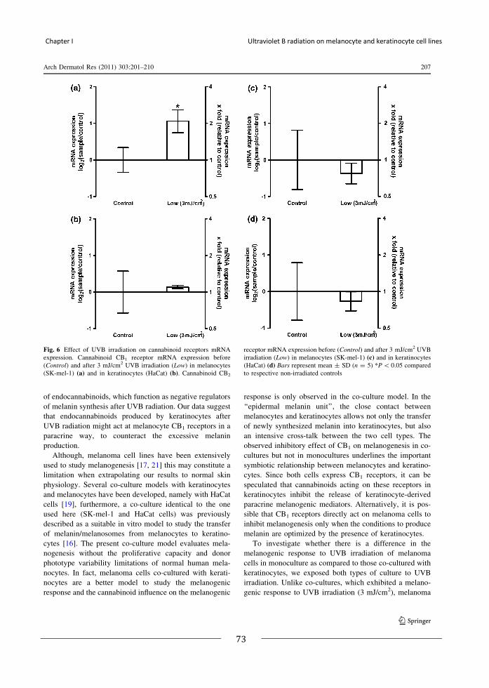
Chapter I Ultraviolet B radiation on melanocyte and keratinocyte cell lines
73
of endocannabinoids, which function as negative regulators
of melanin synthesis after UVB radiation. Our data suggest
that endocannabinoids produced by keratinocytes after
UVB radiation might act at melanocyte CB1 receptors in a
paracrine way, to counteract the excessive melanin
production.
Although, melanoma cell lines have been extensively
used to study melanogenesis [17, 21] this may constitute a
limitation when extrapolating our results to normal skin
physiology. Several co-culture models with keratinocytes
and melanocytes have been developed, namely with HaCat
cells [19], furthermore, a co-culture identical to the one
used here (SK-mel-1 and HaCat cells) was previously
described as a suitable in vitro model to study the transfer
of melanin/melanosomes from melanocytes to keratino-
cytes [16]. The present co-culture model evaluates mela-
nogenesis without the proliferative capacity and donor
phototype variability limitations of normal human mela-
nocytes. In fact, melanoma cells co-cultured with kerati-
nocytes are a better model to study the melanogenic
response and the cannabinoid influence on the melanogenic
response is only observed in the co-culture model. In the
‘‘epidermal melanin unit’’, the close contact between
melanocytes and keratinocytes allows not only the transfer
of newly synthesized melanin into keratinocytes, but also
an intensive cross-talk between the two cell types. The
observed inhibitory effect of CB1 on melanogenesis in co-
cultures but not in monocultures underlines the important
symbiotic relationship between melanocytes and keratino-
cytes. Since both cells express CB1 receptors, it can be
speculated that cannabinoids acting on these receptors in
keratinocytes inhibit the release of keratinocyte-derived
paracrine melanogenic mediators. Alternatively, it is pos-
sible that CB1 receptors directly act on melanoma cells to
inhibit melanogenesis only when the conditions to produce
melanin are optimized by the presence of keratinocytes.
To investigate whether there is a difference in the
melanogenic response to UVB irradiation of melanoma
cells in monoculture as compared to those co-cultured with
keratinocytes, we exposed both types of culture to UVB
irradiation. Unlike co-cultures, which exhibited a melano-
genic response to UVB irradiation (3 mJ/cm2), melanoma
Fig. 6 Effect of UVB irradiation on cannabinoid receptors mRNA
expression. Cannabinoid CB1 receptor mRNA expression before
(Control) and after 3 mJ/cm2 UVB irradiation (Low) in melanocytes
(SK-mel-1) (a) and in keratinocytes (HaCat) (b). Cannabinoid CB2
receptor mRNA expression before (Control) and after 3 mJ/cm2 UVB
irradiation (Low) in melanocytes (SK-mel-1) (c) and in keratinocytes
(HaCat) (d) Bars represent mean ± SD (n = 5) *P\ 0.05 compared
to respective non-irradiated controls
Arch Dermatol Res (2011) 303:201–210 207
123

Effect of ultraviolet B radiation on cutaneous endocannabinoid system
74
cells alone did not show any increase in melanin content
after irradiation with the same dose. It is known that UVB
irradiation of keratinocytes triggers the release of cyto-
kines, growth factors, and other mediators such as endo-
thelin-1, nitric oxide or proopiomelanocortin-derived
peptides known to be effective modulators of melanin
synthesis in a paracrine way [5, 13]. This may explain the
differences observed between mono- and co-cultures in
their response to UVB. Under basal co-culture conditions,
the CB1-mediated inhibitory effect was also observed on
UVB-induced melanogenesis, and was comparable to the
effect of the well-known hypo-pigmentary agent, kojic
acid. From these results, we suggest that keratinocytes play
a major role in cannabinoid-mediated inhibition of basal
and, particularly, UVB-induced pigmentation.
Endocannabinoid levels were stimulated by UVB irra-
diation in keratinocytes and co-cultures but not in mela-
noma cells alone. Interestingly, the co-cultures contain
Fig. 7 Effect of UVB irradiation on endocannabinoids and PEA
levels. Anandamide (a), 2-AG (b) and PEA (c) levels in HaCat
monocultures, SK-mel-1 monocultures and in co-cultures (HaCat-SK-
mel-1), either non-irradiated (CT) or after irradiation with UVB at
low and high doses. Data are mean ± SD (n = 4 separate determi-
nations). *P\ 0.05 compared to respective non-irradiated controls#P\ 0.05 compared to respective experimental condition in co-
cultures
208 Arch Dermatol Res (2011) 303:201–210
123

Chapter I Ultraviolet B radiation on melanocyte and keratinocyte cell lines
75
overall less endocannabinoids (except PEA) than mela-
noma cells alone under basal conditions, and even more
following UVB irradiation. This phenomenon may be due to
the presence of endocannabinoid-degrading enzymes in
keratinocytes [9] in levels possibly up-regulated by UVB.
This could suggest a new endocannabinoid-mediated
mechanism tightly regulating UVB-induced melanogenesis.
Besides melanogenesis, acute UVB irradiation also
induces skin inflammation and immunosuppression. The
standard model for studying UVB-induced immunosup-
pression is the UVB-ability to block either induction or
elicitation of cutaneous allergic sensitization. It is well
known that UVB irradiation of keratinocytes induces the
synthesis and secretion of a whole set of pro-inflammatory
and immunosuppressive soluble factors. Interestingly, a
recent study [7] demonstrated that endocannabinoids
attenuate the cutaneous allergic response and, more
recently, a protective role of PEA in the same context was
described [15]. Also antagonism of CB1 cannabinoid
receptor in human HaCat keratinocytes was associated with
the increased expression of pro-allergic chemokines [8].
Since we found here that UVB irradiation is accompanied
by up-regulation of both 2-AG and PEA levels, we
hypothesize that, whilst cutaneous endocannabinoids may
play a role in both UVB-induced melanogenesis and
immunosuppression, PEA might represent an endogenous
protective agent against UVB-induced inflammation. These
phenomena might contribute to the well-known therapeutic
effect of UVB radiation in chronic inflammatory skin dis-
eases. Endocannabinoid signals involved in skin inflam-
mation are complex and remain poorly understood.
Overall, anandamide appears to mediate an anti-inflam-
matory effect since inhibition of its degradation signifi-
cantly reduced inflammation [7] and there is growing
evidence suggesting that 2-AG modulates the inflammatory
response by acting on the cannabinoid CB2 receptor [11].
Interestingly, we found a significant increase in 2-AG:AEA
ratio after UVB irradiation in co-cultures, suggesting
that endocannabinoids, namely 2-AG, may play a role in
UVB-induced inflammation.
A recent study by Zheng et al. [22] suggested that CB
receptors and the related signaling pathways might be
involved in the promotion of in vivo skin carcinogenesis.
In CB1 and CB2 double gene-deficient mice, there was a
marked decrease in UVB-induced skin carcinogenesis.
This finding was quite surprising in view of previous data
indicating that the endocannabinoid system is protective
against the growth and spreading of skin cancer and mel-
anoma [3]. Since the major known function of melanin is to
provide protection against ultraviolet-induced DNA dam-
age by absorbing and scattering UV radiation, our in vitro
results provide a possible explanation to the findings of
Zheng and colleagues.
In conclusion, the present study suggests that ultraviolet
radiation activates paracrine CB1-mediated endocannabi-
noid signaling to negatively regulate melanin synthesis.
Furthermore, endocannabinoids might act as hypopig-
mentary agents either under basal conditions or after
ultraviolet exposure. Although the use of cannabinoids in
medicine is limited by their psychotropic effects, in cuta-
neous diseases these effects could be avoided by their
topical use on the skin.
Acknowledgments The authors wish to thank Fabiana Piscitelli for
valuable technical help and Epitech Italia for partly supporting this
study.
References
1. Berdyshev EV, Schmid PC, Dong Z, Schmid HH (2000) Stress-
induced generation of N-acylethanolamines in mouse epidermal
JB6 P? cells. Biochem J 346(Pt 2):369–374
2. Biro T, Toth BI, Hasko G, Paus R, Pacher P (2009) The endo-
cannabinoid system of the skin in health and disease: novel
perspectives and therapeutic opportunities. Trends Pharmacol Sci
30:411–420
3. Blazquez C, Carracedo A, Barrado L, Real PJ, Fernandez-Luna
JL, Velasco G, Malumbres M, Guzman M (2006) Cannabinoid
receptors as novel targets for the treatment of melanoma. FASEB
J 20:2633–2635
4. Di Marzo V (2008) Targeting the endocannabinoid system: to
enhance or reduce? Nat Rev Drug Discov 7:438–455
5. Duval C, Regnier M, Schmidt R (2001) Distinct melanogenic
response of human melanocytes in mono-culture, in co-culture
with keratinocytes and in reconstructed epidermis, to UV expo-
sure. Pigment Cell Res 14:348–355
6. Gilsbach R, Brede M, Beetz N, Moura E, Muthig V, Gerstner C,
Barreto F, Neubauer S, Vieira-Coelho MA, Hein L (2007)
Heterozygous alpha 2C-adrenoceptor-deficient mice develop
heart failure after transverse aortic constriction. Cardiovasc Res
75:728–737
7. Karsak M, Gaffal E, Date R, Wang-Eckhardt L, Rehnelt J,
Petrosino S, Starowicz K, Steuder R, Schlicker E, Cravatt B,
Mechoulam R, Buettner R, Werner S, Di Marzo V, Tuting T,
Zimmer A (2007) Attenuation of allergic contact dermatitis
through the endocannabinoid system. Science 316:1494–1497
8. Leonti M, Casu L, Raduner S, Cottiglia F, Floris C, Altmann KH,
Gertsch J (2010) Falcarinol is a covalent cannabinoid CB1
receptor antagonist and induces pro-allergic effects in skin.
Biochem Pharmacol 79:1815–1826
9. Maccarrone M, Di Rienzo M, Battista N, Gasperi V, Guerrieri P,
Rossi A, Finazzi-Agro A (2003) The endocannabinoid system in
human keratinocytes. Evidence that anandamide inhibits epider-
mal differentiation through CB1 receptor-dependent inhibition of
protein kinase C, activation protein-1, and transglutaminase.
J Biol Chem 278:33896–33903
10. Moura E, Afonso J, Serrao MP, Vieira-Coelho MA (2009) Effect
of clonidine on tyrosine hydroxylase activity in the adrenal
medulla and brain of spontaneously hypertensive rats. Basic Clin
Pharmacol Toxicol 104:113–121
11. Oka S, Wakui J, Gokoh M, Kishimoto S, Sugiura T (2006)
Suppression by WIN55212–2, a cannabinoid receptor agonist, of
inflammatory reactions in mouse ear: interference with the
actions of an endogenous ligand, 2-arachidonoylglycerol. Eur J
Pharmacol 538:154–162
Arch Dermatol Res (2011) 303:201–210 209
123

Effect of ultraviolet B radiation on cutaneous endocannabinoid system
76
12. Paradisi A, Pasquariello N, Barcaroli D, Maccarrone M (2008)
Anandamide regulates keratinocyte differentiation by inducing
DNA methylation in a CB1 receptor-dependent manner. J Biol
Chem 283:6005–6012
13. Park HY, Kosmadaki M, Yaar M, Gilchrest BA (2009) Cellular
mechanisms regulating human melanogenesis. Cell Mol Life Sci
66:1493–1506
14. Pertwee RG (2005) Pharmacological actions of cannabinoids.
Handb Exp Pharmacol 168:1–51
15. Petrosino S, Cristino L, Karsak M, Gaffal E, Ueda N, Tuting T,
Bisogno T, De Filippis D, D’Amico A, Saturnino C, Orlando P,
Zimmer A, Iuvone T, Di Marzo V (2009) Protective role of
palmitoylethanolamide in contact allergic dermatitis. Allergy
698–711
16. Potsch L, Emmerich P, Skopp G (2002) Preliminary approach to
elucidate the role of pigment as a binding site for drugs and
chemicals in anagen hairs: pigments as carriers for 3H-haloper-
idol in HaCaT/Sk-Mel-1 co-cultures. Int J Legal Med 116:12–16
17. Rad HH, Yamashita T, Jin HY, Hirosaki K, Wakamatsu K, Ito S,
Jimbow K (2004) Tyrosinase-related proteins suppress
tyrosinase-mediated cell death of melanocytes and melanoma
cells. Exp Cell Res 298:317–328
18. Schallreuter KU (2007) Advances in melanocyte basic science
research. Dermatol Clin 25(vii):283–291
19. Springer M, Engelhart K, Biesalski HK (2003) Effects of 3-iso-
butyl-1-methylxanthine and kojic acid on cocultures and skin
equivalents composed of HaCaT cells and human melanocytes.
Arch Dermatol Res 295:88–91
20. Stander S, Schmelz M, Metze D, Luger T, Rukwied R (2005)
Distribution of cannabinoid receptor 1 (CB1) and 2 (CB2) on
sensory nerve fibers and adnexal structures in human skin.
J Dermatol Sci 38:177–188
21. Sugimoto K, Nishimura T, Nomura K, Kuriki T (2004) Inhibitory
effects of alpha-arbutin on melanin synthesis in cultured human
melanoma cells and a three-dimensional human skin model. Biol
Pharm Bull 27:510–514
22. Zheng D, Bode AM, Zhao Q, Cho YY, Zhu F, Ma WY, Dong Z
(2008) The cannabinoid receptors are required for ultraviolet-
induced inflammation and skin cancer development. Cancer Res
68:3992–3998
210 Arch Dermatol Res (2011) 303:201–210
123

Chapter I Ultraviolet B radiation on melanocyte and keratinocyte cell lines
77
1
Anandamide increases UVB-induced cell death of human keratinocytes through
transient receptor potential vanilloid-1 channel
1,2,3Sofia Magina, 1,2Eduardo Moura, 1Maria Paula Serrão, 1Daniel Moura, 1,2Maria Augusta Vieira-Coelho1Department of Pharmacology and Therapeutics, Faculty of Medicine, University of Porto,2Institute for Molecular and Cell Biology, University of Porto,3Dermatology and Venereology Department, H. S. João, Porto, Portugal
Abstract Endocannabinoids like anandamide (AEA) play a key role in skin biology. Exposure to high-dose ultraviolet B (UVB) radiation induces keratinocyte apoptosis and increases the synthesis of anandamide (AEA) in keratinocytes. Published data on the effects of cannabinoids on the regulation of keratinocyte cell death are conflicting. Moreover the influence of cannabinoids on UVB-induced cell death has never been studied. Here, we investigated whether AEA and other cannabinoid receptors ligands modify keratinocyte survival and UVB-induced keratinocyte death. Cell viability in human immortalized HaCat keratinocytes was evaluated under basal conditions and 48 h after high-dose (70 mJ/cm2) UVB (311 nm) irradiation. Pretreatment of HaCat cells with AEA at high concentrations (30 µM) significantly increased UVB-induced cell death. This effect was replicated by the transient receptor potential vanilloid-1 channel (TRPV1) agonist capsaicin (10 µM). The selective CB1antagonist AM-251 (1 µM) did not modify the effect of AEA. In contrast, the selective TRPV1 antagonist, capsazepine (5 µM), which alone did not modify cell viability, abolished the increase in UVB-induced cell death of either AEA or capsaicin. Our results suggest that AEA potentiates cell death induced by UVB radiation through TRPV1 in human keratinocytes. Endocannabinoids participate in the adaptive skin response to UVB radiation and could be useful in combination to phototherapy for treatment of hyperproliferative dermatoses such as psoriasis.
Introduction The endocannabinoid system is not limited to the central nervous system [4] [9] and has a role in skin physiology [1]. Several human skin cells produce the major endocannabinoids, anandamide (AEA) and 2-arachidonoylglycerol (2-AG) and express the enzymes responsible for their synthesis and metabolism [8,11,7]. Endocannabinoids bind to G-protein-coupled cannabinoid receptors, CB1 and CB2 [10]. AEA but not 2-AG is also an agonist of the transient receptor potential vanilloid-1 channel (TRPV1) [10]. TRPV1, CB1 and CB2 receptors were identified, both in situ and in vitro, on different cutaneous cells, including human keratinocytes [8] [7] [12]. Cannabinoids may induce apoptosis in different cell types and they have been tested as potential antitumoral agents. However, published data on the effects of cannabinoids on the regulation of keratinocytes cell death are conflicting. Wilkinson et al [16] found that ∆9-tetrahydrocannabinol as well as synthetic cannabinoid agonists inhibited growth of cultured transformed human epidermal keratinocytes, yet these effects were CB1 and CB2independent. In another study, activation of both types of cannabinoid receptors by synthetic agonists induced the apoptotic death of tumorigenic epidermal cells whereas the viability of non-
tumorigenic human (HaCat cells) and murine (MCA3D) keratinocytes remained unaffected [3]. In addition, it was observed that metabolism of AEA by cyclooxygenase-2 (COX-2) is important for AEA-induced cell death in tumorigenic keratinocytes and that neither CB1 nor CB2 nor TRPV1 receptors were required for this response [13] [5].We have recently reported that the cutaneous endocannabinoid system responds to ultraviolet B (UVB) irradiation corroborating the known pro-homeostatic role of the endocannabinoid system in other tissues [8]. UVB irradiation increases the levels of AEA and 2-AG in human keratinocytes [8]. Furthermore UVB increases TRPV1 expression in both keratinocytes and human skin in vivo [6] and increases COX-2 expression in cultured human keratinocytes [2][14]. UVB radiation decreases keratinocyte viability, however the role of cannabinoid receptor ligands in UVB-induced keratinocyte cell death has not yet been studied.Therefore we evaluated in human cultured keratinocytes (HaCat), the effect of the most extensively studied endocannabinoid, AEA, and of synthetic cannabinoid receptor agonists and antagonists on cell survival in basal conditions and after acute UVB irradiation.

2
Materials and Methods
Materials
Dulbecco's minimal Eagle's medium (DMEM), antibiotic antimycotic solution, HEPES, anandamide (AEA), capsaicin (CAPS), capsazepine (CPZ) and indomethacin (IND) were purchased from Sigma-Aldrich, Inc. (St. Louis, MO, USA). Foetal bovine serum (FBS) and trypsin were purchased from Gibco (Paisley, UK). NaHCO3 and EDTA were purchased from Merck (Germany). Arachidonoyl-2’-chloroethylamide (ACEA) was purchased from Biotrend Chemicals AG (Zurich, Switzerland) and AM251, AM630, JWH133 from Tocris Bioscience (Bristol, UK).
Cell line and culture conditions Human HaCaT keratinocytes, provided by Prof. N. Fusenig (Deutsches Krebsforschungszentrum, Heidelberg, Germany), were grown in DMEM supplemented with 10% FBS, 1% antibiotic antimycotic solution and 0.44 M NaHCO3. Cells were maintained at <80% confluency, at 37ºC in a 5% CO2 (v/v), humidified atmosphere. HaCat cell cultures were incubated 2 h before the UVB irradiation with DMEM medium containing the different drugs studied and the medium was replaced every 24 h until the end of the experiment. Cannabinoid receptor agonists and antagonists were directly applied at a final ethanol concentration less than 0.3% (v/v). No significant influence of the vehicle was observed on the parameters determined.
UVB irradiation HaCat cells in monoculture were washed with PBS and then exposed to acute UVB (312 nm) irradiation (70 mJ/cm2) emitted by a pre-heated, filtered, UV lamp (VL-6.M, Vilber Lourmat, France) at t = 0 min. This was followed by a 48 h period of incubation after which the cells were harvested [10 min incubation with EDTA (0.05%, pH 7.4) and 5 min incubation with trypsin] for analysis. The irradiation dose was defined according to UVB radiation cytotoxic effect. Non-irradiated controls were not exposed to UVB light, but subjected to all other manipulations. UV dosimetry was performed with a Waldmann 585.100 UV-meter (Herbert Waldmann GmbH & Co., Villingen-Schwenningen, Germany).
Cell viability determination Harvested cells were centrifuged at 300 x g for 5 min at room temperature, washed twice with PBS and resuspended in 1 ml of PBS. From this
suspension, 180 µl were transferred into a black/clear bottom Microtest™ 96-well Assay Plate, Oplilux™ (BD Biosciences, Franklin Lakes, NJ, USA) for cell viability assay. Cell viability was measured using calcein-AM (Molecular Probes, Inc., Eugene, OR, USA) according to the manufacturer’s instructions. Each well containing 180 µl of cell suspension was loaded with 20 µl of calcein-AM (10 µM, in PBS solution). Fluorescence was measured at 485 nm excitation and 530 nm emission wavelengths using a SpectraMax, Gemini XS spectrofluorometer (Molecular Devices, Sunnyvale, CA, USA) at 40 min incubation at room temperature. Statistical analysis
Data are presented as means ± SEM. Student’s t-test connected by the Newman Keuls method for multiple comparisons was used to analyze differences. Values of P<0.05 are considered significant.
Results Effect of anandamide on keratinocytes (HaCat) cell viability in basal conditions and after UVB irradiationIn basal conditions, we used AEA 3 and 30 µM and in the lowest concentration AEA (3 µM) slightly increased cellular viability while AEA 30 µM had no effect (Fig.1a). This effect of AEA (3 µM) was not reverted by CB1 antagonist (data not shown). Initially, we investigated the effect of different UVB irradiation doses (30-100 mJ/cm2) on keratinocytes cell viability (data not shown). We found that after high-dose (70 mJ/cm2) UVB irradiation a 60% reduction on cell viability was observed in control situation (Fig. 1a). AEA 30µM significantly increased (P<0.05, n=12) UVB-induced cytotoxic effect (Fig. 1a). To assess whether the metabolism of anandamide by COX-2 was necessary for endocannabinoid-induced cell death as previously proposed, cells were incubated with AEA and the COX-2 inhibitor, indomethacin. Indomethacin (1 µM) did not modify the AEA effect on cell viability in irradiated keratinocytes (fig.1b). As anandamide can activate both cannabinoid receptors and TRPV1 we then investigated which “AEA-receptor” was mediating this effect of AEA. Cells were treated with AEA 30 µM and the CB1selective antagonist AM251 (1 µM) and the specific TRPV1 antagonist, capsazepine (5 µM). Figure 1b shows that CB1 antagonist did not revert AEA effect. In contrast, capsazepine (5 µM), which alone did not modify cell viability, completely abolished
Effect of ultraviolet B radiation on cutaneous endocannabinoid system
78

3
the increase in UVB-induced cell death by AEA (Fig.1b).
Figure. 1. Effect of anandamide on keratinocytes (HaCat) cell viability in basal conditions and after UVB irradiation. Cell viability was evaluated before (CT) and after UVB (70 mJ/cm2) irradiation (UV) a) HaCat keratinocytes were treated for 48 h by vehicle (CT) and AEA (3-30 µM) b) HaCatkeratinocytes were incubated with AEA 30 µM, COX-2 inhibitor, indomethacin (IND) 1µM, CB1 selective antagonist, AM251 (1 µM) and TRPV1 selective antagonist, capsazepine (CPZ) 5 µM. Cell viability was expressed as the percentage of untreated controls, set to 100. Bars represent means ± SEM (n = 12) *P<0.05 compared to respective control values. #P<0.05compared to AEA 30 µM after UVB.
Effect of cannabinoid receptors and TRPV1 on UVB-induced keratinocytes cell death
In basal conditions neither the CB1 selective agonist ACEA (1 µM) nor the CB1 selective antagonist AM251 (1 µM) modified keratinocytes cell viability (Fig.2a). The CB2 selective agonist JWH 133 (1-10µM) and the CB2 selective antagonist AM 630 (1µM) had no effect (data not shown).
Neither the CB1 selective agonist nor the CB1selective antagonist modified modified UVB-induced keratinocytes cell death (Fig. 2a). As a positive control, we used the TRPV1 agonist capsaicin (10 µM), which had no effect in basal conditions but after UVB irradiation replicated the effect of AEA 30 µM increasing the UVB-induced
cytotoxic effect (Fig. 2b). This effect of capsaicin was also dependent on TRPV1 since it was completely reverted by capsazepine (Fig.2b).
Figure. 2. Effect of CB1 receptor and TRPV1, in basal conditions and after UVB irradiation. Cell viability was evaluated before (CT) and after UVB (70 mJ/cm2) irradiation (UV).a) HaCat keratinocytes were incubated with CB1 selective agonist, ACEA (1 µM) and CB1 selective antagonist AM251 (1 µM) b) HaCat keratinocytes were treated for 48 h with TRPV1 agonist, capsaicin (CAPS) 10 µM and TRPV1 selective antagonist, capsazepine (CPZ) 5 µM. Cell viability results were expressed as the percentage of untreated controls set to 100. Bars represent means ± SEM (n = 12) * P<0.05 compared to controls. #P<0.05 compared to CAPS 10 µM after UVB.
Discussion
In this study we provide evidence that the prototypic endocannabinoid AEA that is produced in keratinocytes after UVB irradiation [8] increases UVB-induced human keratinocytes cell death. We also demonstrated that this AEA effect is TRPV1 dependent. Anandamide produced by keratinocytes after UVB irradiation might act at TRPV1 in a paracrine or autocrine manner and negatively regulates their survival. Acute UVB irradiation causes keratinocyte apoptosis and this programmed cell-death of UV-damaged skin cells is a definitive cancer-prevention pathway. Furthermore keratinocyte apoptosis after UVB irradiation is a key mechanism in psoriatic
Chapter I Ultraviolet B radiation on melanocyte and keratinocyte cell lines
79
4
plaques clearance after phototherapy [15]. Our data support the concept that the fine-tuned endogenous endocannabinoid tone of the skin could contribute to eliminate damaged keratinocytes after UVB irradiation. In basal conditions, neither AEA nor the other CB1and CB2 cannabinoid receptors agonists used suppressed keratinocyte cell viability. Indeed a slight increase of HaCat viability was observed with AEA at lower concentrations. This increase has already been described in a previous study [13]. By contrast, another research found that AEA, dose-dependently, inhibits proliferation and cell viability of human epidermal keratinocytes in culture [12]. However in that study a shorter incubation period with AEA was used and the effect of UVB radiation was not investigated. The authors proposed that AEA-induced keratinocyte death resulted from the sequential engagement of CB1 receptor and TRPV1 with consequent calcium influx [12]. UVB increases TRPV1 expression in both keratinocytes and human skin in vivo [6] and this could explain that we only observed TRPV1-mediated AEA effect after UVB exposure. The need for higher concentrations of AEA to reduce cell viability after UVB radiation is consistent with the lower affinity of AEA for TRPV1 in comparison to CB1, as clearly demonstrated in vitro through binding assays [10]. Furthermore the lack of effect of the CB1 selective agonist ACEA also corroborates this TRPV1 dependent pathway. Previous studies have shown that metabolism of AEA by COX-2 was important for AEA-induced cell death in tumorigenic keratinocytes and that neither CB1 nor CB2 nor TRPV1 receptors were required for this response [13] [5]. Interestingly, in our model with non-tumorigenic HaCat cells, COX-2 inhibition by indomethacin did not modify the AEA effect in irradiated keratinocytes. In conclusion, our results suggest that AEA through TRPV1 decreases cell viability in keratinocytes after ultraviolet exposure. Endocannabinoids participate in the adaptive skin response to UVB radiation and could be in the future useful in combination to phototherapy for the treatment of hyperproliferative dermatoses such as psoriasis.
References
1. Biro T, Toth BI, Hasko G, Paus R, Pacher P (2009) The endocannabinoid system of the skin in health and disease: novel perspectives and therapeutic opportunities. Trends Pharmacol Sci 30:411-420
2. Buckman SY, Gresham A, Hale P, Hruza G, Anast J, Masferrer J, Pentland AP (1998) COX-2 expression is induced by UVB exposure in human skin: implications for the development of skin cancer. Carcinogenesis 19:723-729
3. Casanova ML, Blazquez C, Martinez-Palacio J, Villanueva C, Fernandez-Acenero MJ, Huffman JW, Jorcano JL, Guzman M (2003) Inhibition of skin tumor growth and angiogenesis in vivo by activation of cannabinoid receptors. J Clin Invest 111:43-50
4. Di Marzo V (2008) Targeting the endocannabinoid system: to enhance or reduce? Nat Rev Drug Discov 7:438-455
5. Kuc C, Jenkins A, Van Dross RT (2012) Arachidonoyl ethanolamide (AEA)-induced apoptosis is mediated by J-series prostaglandins and is enhanced by fatty acid amide hydrolase (FAAH) blockade. Mol Carcinog 51:139-149
6. Lee YM, Kim YK, Kim KH, Park SJ, Kim SJ, Chung JH (2009) A novel role for the TRPV1 channel in UV-induced matrix metalloproteinase (MMP)-1 expression in HaCaT cells. J Cell Physiol 219:766-775
7. Maccarrone M, Di Rienzo M, Battista N, Gasperi V, Guerrieri P, Rossi A, Finazzi-Agro A (2003) The endocannabinoid system in human keratinocytes. Evidence that anandamide inhibits epidermal differentiation through CB1 receptor-dependent inhibition of protein kinase C, activation protein-1, and transglutaminase. J Biol Chem 278:33896-33903
8. Magina S, Esteves-Pinto C, Moura E, Serrao MP, Moura D, Petrosino S, Di Marzo V, Vieira-Coelho MA (2011) Inhibition of basal and ultraviolet B-induced melanogenesis by cannabinoid CB(1) receptors: a keratinocyte-dependent effect. Arch Dermatol Res 303:201-210
9. Pacher P, Mechoulam R (2011) Is lipid signaling through cannabinoid 2 receptors part of a protective system? Prog Lipid Res 50:193-211
10. Pertwee RG (2010) Receptors and channels targeted by synthetic cannabinoid receptor agonists and antagonists. Curr Med Chem 17:1360-1381
11. Telek A, Biro T, Bodo E, Toth BI, Borbiro I, Kunos G, Paus R (2007) Inhibition of human hair follicle growth by endo- and exocannabinoids. FASEB J 21:3534-3541
12. Toth BI, Dobrosi N, Dajnoki A, Czifra G, Olah A, Szollosi AG, Juhasz I, Sugawara K, Paus R, Biro T (2011) Endocannabinoids modulate human epidermal keratinocyte proliferation and survival via the sequential engagement of cannabinoid receptor-1 and transient receptor potential vanilloid-1. J Invest Dermatol 131:1095-1104
13. Van Dross RT (2009) Metabolism of anandamide by COX-2 is necessary for endocannabinoid-induced cell death in tumorigenic keratinocytes. Mol Carcinog 48:724-732
14. Van Dross RT, Hong X, Essengue S, Fischer SM, Pelling JC (2007) Modulation of UVB-induced and basal cyclooxygenase-2 (COX-2) expression by apigenin in mouse keratinocytes: role of USF transcription factors. Mol Carcinog 46:303-314
15. Weatherhead SC, Farr PM, Jamieson D, Hallinan JS, Lloyd JJ, Wipat A, Reynolds NJ (2011) Keratinocyte apoptosis in epidermal remodeling and clearance of psoriasis induced by UV radiation. J Invest Dermatol 131:1916-1926
16. Wilkinson JD, Williamson EM (2007) Cannabinoids inhibit human keratinocyte proliferation through a non-CB1/CB2 mechanism and have a potential therapeutic value in the treatment of psoriasis. J Dermatol Sci 45:87-92
Effect of ultraviolet B radiation on cutaneous endocannabinoid system
80
4
plaques clearance after phototherapy [15]. Our data support the concept that the fine-tuned endogenous endocannabinoid tone of the skin could contribute to eliminate damaged keratinocytes after UVB irradiation. In basal conditions, neither AEA nor the other CB1and CB2 cannabinoid receptors agonists used suppressed keratinocyte cell viability. Indeed a slight increase of HaCat viability was observed with AEA at lower concentrations. This increase has already been described in a previous study [13]. By contrast, another research found that AEA, dose-dependently, inhibits proliferation and cell viability of human epidermal keratinocytes in culture [12]. However in that study a shorter incubation period with AEA was used and the effect of UVB radiation was not investigated. The authors proposed that AEA-induced keratinocyte death resulted from the sequential engagement of CB1 receptor and TRPV1 with consequent calcium influx [12]. UVB increases TRPV1 expression in both keratinocytes and human skin in vivo [6] and this could explain that we only observed TRPV1-mediated AEA effect after UVB exposure. The need for higher concentrations of AEA to reduce cell viability after UVB radiation is consistent with the lower affinity of AEA for TRPV1 in comparison to CB1, as clearly demonstrated in vitro through binding assays [10]. Furthermore the lack of effect of the CB1 selective agonist ACEA also corroborates this TRPV1 dependent pathway. Previous studies have shown that metabolism of AEA by COX-2 was important for AEA-induced cell death in tumorigenic keratinocytes and that neither CB1 nor CB2 nor TRPV1 receptors were required for this response [13] [5]. Interestingly, in our model with non-tumorigenic HaCat cells, COX-2 inhibition by indomethacin did not modify the AEA effect in irradiated keratinocytes. In conclusion, our results suggest that AEA through TRPV1 decreases cell viability in keratinocytes after ultraviolet exposure. Endocannabinoids participate in the adaptive skin response to UVB radiation and could be in the future useful in combination to phototherapy for the treatment of hyperproliferative dermatoses such as psoriasis.
References
1. Biro T, Toth BI, Hasko G, Paus R, Pacher P (2009) The endocannabinoid system of the skin in health and disease: novel perspectives and therapeutic opportunities. Trends Pharmacol Sci 30:411-420
2. Buckman SY, Gresham A, Hale P, Hruza G, Anast J, Masferrer J, Pentland AP (1998) COX-2 expression is induced by UVB exposure in human skin: implications for the development of skin cancer. Carcinogenesis 19:723-729
3. Casanova ML, Blazquez C, Martinez-Palacio J, Villanueva C, Fernandez-Acenero MJ, Huffman JW, Jorcano JL, Guzman M (2003) Inhibition of skin tumor growth and angiogenesis in vivo by activation of cannabinoid receptors. J Clin Invest 111:43-50
4. Di Marzo V (2008) Targeting the endocannabinoid system: to enhance or reduce? Nat Rev Drug Discov 7:438-455
5. Kuc C, Jenkins A, Van Dross RT (2012) Arachidonoyl ethanolamide (AEA)-induced apoptosis is mediated by J-series prostaglandins and is enhanced by fatty acid amide hydrolase (FAAH) blockade. Mol Carcinog 51:139-149
6. Lee YM, Kim YK, Kim KH, Park SJ, Kim SJ, Chung JH (2009) A novel role for the TRPV1 channel in UV-induced matrix metalloproteinase (MMP)-1 expression in HaCaT cells. J Cell Physiol 219:766-775
7. Maccarrone M, Di Rienzo M, Battista N, Gasperi V, Guerrieri P, Rossi A, Finazzi-Agro A (2003) The endocannabinoid system in human keratinocytes. Evidence that anandamide inhibits epidermal differentiation through CB1 receptor-dependent inhibition of protein kinase C, activation protein-1, and transglutaminase. J Biol Chem 278:33896-33903
8. Magina S, Esteves-Pinto C, Moura E, Serrao MP, Moura D, Petrosino S, Di Marzo V, Vieira-Coelho MA (2011) Inhibition of basal and ultraviolet B-induced melanogenesis by cannabinoid CB(1) receptors: a keratinocyte-dependent effect. Arch Dermatol Res 303:201-210
9. Pacher P, Mechoulam R (2011) Is lipid signaling through cannabinoid 2 receptors part of a protective system? Prog Lipid Res 50:193-211
10. Pertwee RG (2010) Receptors and channels targeted by synthetic cannabinoid receptor agonists and antagonists. Curr Med Chem 17:1360-1381
11. Telek A, Biro T, Bodo E, Toth BI, Borbiro I, Kunos G, Paus R (2007) Inhibition of human hair follicle growth by endo- and exocannabinoids. FASEB J 21:3534-3541
12. Toth BI, Dobrosi N, Dajnoki A, Czifra G, Olah A, Szollosi AG, Juhasz I, Sugawara K, Paus R, Biro T (2011) Endocannabinoids modulate human epidermal keratinocyte proliferation and survival via the sequential engagement of cannabinoid receptor-1 and transient receptor potential vanilloid-1. J Invest Dermatol 131:1095-1104
13. Van Dross RT (2009) Metabolism of anandamide by COX-2 is necessary for endocannabinoid-induced cell death in tumorigenic keratinocytes. Mol Carcinog 48:724-732
14. Van Dross RT, Hong X, Essengue S, Fischer SM, Pelling JC (2007) Modulation of UVB-induced and basal cyclooxygenase-2 (COX-2) expression by apigenin in mouse keratinocytes: role of USF transcription factors. Mol Carcinog 46:303-314
15. Weatherhead SC, Farr PM, Jamieson D, Hallinan JS, Lloyd JJ, Wipat A, Reynolds NJ (2011) Keratinocyte apoptosis in epidermal remodeling and clearance of psoriasis induced by UV radiation. J Invest Dermatol 131:1916-1926
16. Wilkinson JD, Williamson EM (2007) Cannabinoids inhibit human keratinocyte proliferation through a non-CB1/CB2 mechanism and have a potential therapeutic value in the treatment of psoriasis. J Dermatol Sci 45:87-92
Effect of ultraviolet B radiation on cutaneous endocannabinoid system
80
4
plaques clearance after phototherapy [15]. Our data support the concept that the fine-tuned endogenous endocannabinoid tone of the skin could contribute to eliminate damaged keratinocytes after UVB irradiation. In basal conditions, neither AEA nor the other CB1and CB2 cannabinoid receptors agonists used suppressed keratinocyte cell viability. Indeed a slight increase of HaCat viability was observed with AEA at lower concentrations. This increase has already been described in a previous study [13]. By contrast, another research found that AEA, dose-dependently, inhibits proliferation and cell viability of human epidermal keratinocytes in culture [12]. However in that study a shorter incubation period with AEA was used and the effect of UVB radiation was not investigated. The authors proposed that AEA-induced keratinocyte death resulted from the sequential engagement of CB1 receptor and TRPV1 with consequent calcium influx [12]. UVB increases TRPV1 expression in both keratinocytes and human skin in vivo [6] and this could explain that we only observed TRPV1-mediated AEA effect after UVB exposure. The need for higher concentrations of AEA to reduce cell viability after UVB radiation is consistent with the lower affinity of AEA for TRPV1 in comparison to CB1, as clearly demonstrated in vitro through binding assays [10]. Furthermore the lack of effect of the CB1 selective agonist ACEA also corroborates this TRPV1 dependent pathway. Previous studies have shown that metabolism of AEA by COX-2 was important for AEA-induced cell death in tumorigenic keratinocytes and that neither CB1 nor CB2 nor TRPV1 receptors were required for this response [13] [5]. Interestingly, in our model with non-tumorigenic HaCat cells, COX-2 inhibition by indomethacin did not modify the AEA effect in irradiated keratinocytes. In conclusion, our results suggest that AEA through TRPV1 decreases cell viability in keratinocytes after ultraviolet exposure. Endocannabinoids participate in the adaptive skin response to UVB radiation and could be in the future useful in combination to phototherapy for the treatment of hyperproliferative dermatoses such as psoriasis.
References
1. Biro T, Toth BI, Hasko G, Paus R, Pacher P (2009) The endocannabinoid system of the skin in health and disease: novel perspectives and therapeutic opportunities. Trends Pharmacol Sci 30:411-420
2. Buckman SY, Gresham A, Hale P, Hruza G, Anast J, Masferrer J, Pentland AP (1998) COX-2 expression is induced by UVB exposure in human skin: implications for the development of skin cancer. Carcinogenesis 19:723-729
3. Casanova ML, Blazquez C, Martinez-Palacio J, Villanueva C, Fernandez-Acenero MJ, Huffman JW, Jorcano JL, Guzman M (2003) Inhibition of skin tumor growth and angiogenesis in vivo by activation of cannabinoid receptors. J Clin Invest 111:43-50
4. Di Marzo V (2008) Targeting the endocannabinoid system: to enhance or reduce? Nat Rev Drug Discov 7:438-455
5. Kuc C, Jenkins A, Van Dross RT (2012) Arachidonoyl ethanolamide (AEA)-induced apoptosis is mediated by J-series prostaglandins and is enhanced by fatty acid amide hydrolase (FAAH) blockade. Mol Carcinog 51:139-149
6. Lee YM, Kim YK, Kim KH, Park SJ, Kim SJ, Chung JH (2009) A novel role for the TRPV1 channel in UV-induced matrix metalloproteinase (MMP)-1 expression in HaCaT cells. J Cell Physiol 219:766-775
7. Maccarrone M, Di Rienzo M, Battista N, Gasperi V, Guerrieri P, Rossi A, Finazzi-Agro A (2003) The endocannabinoid system in human keratinocytes. Evidence that anandamide inhibits epidermal differentiation through CB1 receptor-dependent inhibition of protein kinase C, activation protein-1, and transglutaminase. J Biol Chem 278:33896-33903
8. Magina S, Esteves-Pinto C, Moura E, Serrao MP, Moura D, Petrosino S, Di Marzo V, Vieira-Coelho MA (2011) Inhibition of basal and ultraviolet B-induced melanogenesis by cannabinoid CB(1) receptors: a keratinocyte-dependent effect. Arch Dermatol Res 303:201-210
9. Pacher P, Mechoulam R (2011) Is lipid signaling through cannabinoid 2 receptors part of a protective system? Prog Lipid Res 50:193-211
10. Pertwee RG (2010) Receptors and channels targeted by synthetic cannabinoid receptor agonists and antagonists. Curr Med Chem 17:1360-1381
11. Telek A, Biro T, Bodo E, Toth BI, Borbiro I, Kunos G, Paus R (2007) Inhibition of human hair follicle growth by endo- and exocannabinoids. FASEB J 21:3534-3541
12. Toth BI, Dobrosi N, Dajnoki A, Czifra G, Olah A, Szollosi AG, Juhasz I, Sugawara K, Paus R, Biro T (2011) Endocannabinoids modulate human epidermal keratinocyte proliferation and survival via the sequential engagement of cannabinoid receptor-1 and transient receptor potential vanilloid-1. J Invest Dermatol 131:1095-1104
13. Van Dross RT (2009) Metabolism of anandamide by COX-2 is necessary for endocannabinoid-induced cell death in tumorigenic keratinocytes. Mol Carcinog 48:724-732
14. Van Dross RT, Hong X, Essengue S, Fischer SM, Pelling JC (2007) Modulation of UVB-induced and basal cyclooxygenase-2 (COX-2) expression by apigenin in mouse keratinocytes: role of USF transcription factors. Mol Carcinog 46:303-314
15. Weatherhead SC, Farr PM, Jamieson D, Hallinan JS, Lloyd JJ, Wipat A, Reynolds NJ (2011) Keratinocyte apoptosis in epidermal remodeling and clearance of psoriasis induced by UV radiation. J Invest Dermatol 131:1916-1926
16. Wilkinson JD, Williamson EM (2007) Cannabinoids inhibit human keratinocyte proliferation through a non-CB1/CB2 mechanism and have a potential therapeutic value in the treatment of psoriasis. J Dermatol Sci 45:87-92
Effect of ultraviolet B radiation on cutaneous endocannabinoid system
80
4
plaques clearance after phototherapy [15]. Our data support the concept that the fine-tuned endogenous endocannabinoid tone of the skin could contribute to eliminate damaged keratinocytes after UVB irradiation. In basal conditions, neither AEA nor the other CB1and CB2 cannabinoid receptors agonists used suppressed keratinocyte cell viability. Indeed a slight increase of HaCat viability was observed with AEA at lower concentrations. This increase has already been described in a previous study [13]. By contrast, another research found that AEA, dose-dependently, inhibits proliferation and cell viability of human epidermal keratinocytes in culture [12]. However in that study a shorter incubation period with AEA was used and the effect of UVB radiation was not investigated. The authors proposed that AEA-induced keratinocyte death resulted from the sequential engagement of CB1 receptor and TRPV1 with consequent calcium influx [12]. UVB increases TRPV1 expression in both keratinocytes and human skin in vivo [6] and this could explain that we only observed TRPV1-mediated AEA effect after UVB exposure. The need for higher concentrations of AEA to reduce cell viability after UVB radiation is consistent with the lower affinity of AEA for TRPV1 in comparison to CB1, as clearly demonstrated in vitro through binding assays [10]. Furthermore the lack of effect of the CB1 selective agonist ACEA also corroborates this TRPV1 dependent pathway. Previous studies have shown that metabolism of AEA by COX-2 was important for AEA-induced cell death in tumorigenic keratinocytes and that neither CB1 nor CB2 nor TRPV1 receptors were required for this response [13] [5]. Interestingly, in our model with non-tumorigenic HaCat cells, COX-2 inhibition by indomethacin did not modify the AEA effect in irradiated keratinocytes. In conclusion, our results suggest that AEA through TRPV1 decreases cell viability in keratinocytes after ultraviolet exposure. Endocannabinoids participate in the adaptive skin response to UVB radiation and could be in the future useful in combination to phototherapy for the treatment of hyperproliferative dermatoses such as psoriasis.
References
1. Biro T, Toth BI, Hasko G, Paus R, Pacher P (2009) The endocannabinoid system of the skin in health and disease: novel perspectives and therapeutic opportunities. Trends Pharmacol Sci 30:411-420
2. Buckman SY, Gresham A, Hale P, Hruza G, Anast J, Masferrer J, Pentland AP (1998) COX-2 expression is induced by UVB exposure in human skin: implications for the development of skin cancer. Carcinogenesis 19:723-729
3. Casanova ML, Blazquez C, Martinez-Palacio J, Villanueva C, Fernandez-Acenero MJ, Huffman JW, Jorcano JL, Guzman M (2003) Inhibition of skin tumor growth and angiogenesis in vivo by activation of cannabinoid receptors. J Clin Invest 111:43-50
4. Di Marzo V (2008) Targeting the endocannabinoid system: to enhance or reduce? Nat Rev Drug Discov 7:438-455
5. Kuc C, Jenkins A, Van Dross RT (2012) Arachidonoyl ethanolamide (AEA)-induced apoptosis is mediated by J-series prostaglandins and is enhanced by fatty acid amide hydrolase (FAAH) blockade. Mol Carcinog 51:139-149
6. Lee YM, Kim YK, Kim KH, Park SJ, Kim SJ, Chung JH (2009) A novel role for the TRPV1 channel in UV-induced matrix metalloproteinase (MMP)-1 expression in HaCaT cells. J Cell Physiol 219:766-775
7. Maccarrone M, Di Rienzo M, Battista N, Gasperi V, Guerrieri P, Rossi A, Finazzi-Agro A (2003) The endocannabinoid system in human keratinocytes. Evidence that anandamide inhibits epidermal differentiation through CB1 receptor-dependent inhibition of protein kinase C, activation protein-1, and transglutaminase. J Biol Chem 278:33896-33903
8. Magina S, Esteves-Pinto C, Moura E, Serrao MP, Moura D, Petrosino S, Di Marzo V, Vieira-Coelho MA (2011) Inhibition of basal and ultraviolet B-induced melanogenesis by cannabinoid CB(1) receptors: a keratinocyte-dependent effect. Arch Dermatol Res 303:201-210
9. Pacher P, Mechoulam R (2011) Is lipid signaling through cannabinoid 2 receptors part of a protective system? Prog Lipid Res 50:193-211
10. Pertwee RG (2010) Receptors and channels targeted by synthetic cannabinoid receptor agonists and antagonists. Curr Med Chem 17:1360-1381
11. Telek A, Biro T, Bodo E, Toth BI, Borbiro I, Kunos G, Paus R (2007) Inhibition of human hair follicle growth by endo- and exocannabinoids. FASEB J 21:3534-3541
12. Toth BI, Dobrosi N, Dajnoki A, Czifra G, Olah A, Szollosi AG, Juhasz I, Sugawara K, Paus R, Biro T (2011) Endocannabinoids modulate human epidermal keratinocyte proliferation and survival via the sequential engagement of cannabinoid receptor-1 and transient receptor potential vanilloid-1. J Invest Dermatol 131:1095-1104
13. Van Dross RT (2009) Metabolism of anandamide by COX-2 is necessary for endocannabinoid-induced cell death in tumorigenic keratinocytes. Mol Carcinog 48:724-732
14. Van Dross RT, Hong X, Essengue S, Fischer SM, Pelling JC (2007) Modulation of UVB-induced and basal cyclooxygenase-2 (COX-2) expression by apigenin in mouse keratinocytes: role of USF transcription factors. Mol Carcinog 46:303-314
15. Weatherhead SC, Farr PM, Jamieson D, Hallinan JS, Lloyd JJ, Wipat A, Reynolds NJ (2011) Keratinocyte apoptosis in epidermal remodeling and clearance of psoriasis induced by UV radiation. J Invest Dermatol 131:1916-1926
16. Wilkinson JD, Williamson EM (2007) Cannabinoids inhibit human keratinocyte proliferation through a non-CB1/CB2 mechanism and have a potential therapeutic value in the treatment of psoriasis. J Dermatol Sci 45:87-92
Effect of ultraviolet B radiation on cutaneous endocannabinoid system
80

Chapter I Ultraviolet B radiation on melanocyte and keratinocyte cell lines
81
1
Effect of ultraviolet B radiation on endocannabinoid metabolizing enzymes 1,2,3S Magina *, 4Di Marzo V, 1,2E Moura, 4Petrosino S, 1D Moura1,2M A Vieira-Coelho 1Department of Pharmacology and Therapeutics, Faculty of Medicine, University of Porto, 2Institute for Molecular and Cell Biology, 3Dermatology and Venereology Department, H. S. João, 4Endocannabinoid Research Group, Institute of Biomolecular Chemistry, Consiglio Nazionale delle Ricerche, Via Campi Flegrei 34, 80078 Pozzuoli, NA, Italy
Introduction Endocannabinoids are produced on demand and are rapidly cleared by a process of cellular uptake, followed by enzymatic metabolism. There is general consensus that the enzyme fatty acid amide hydrolase (FAAH) is the key-enzyme of the breakdown of anandamide, On the other hand, monoacylglycerol lipase (MAGL) is the major contribution for 2-AG metabolism.
Aim To evaluate the influence of ultraviolet B (UVB) radiation on endocannabinoid enzymes in the three models of skin cell cultures.
Methods Human SK-mel-1 melanoma cells (ATCC no. HTB-67), human HaCat keratinocytes, provided by Prof. N. Fusenig (Deutsches Krebsforschungszentrum, Heidelberg, Germany), were used. SK-mel-1 and HaCat cells, ratio 1.8:2, were co-cultivated. Cells in co-culture were exposed to high (7 mJ/cm2) dose of UVB light wavelength (312 nm). SK-mel-1 cells in monoculture were exposure to UVB light under the same conditions as the cells in co-culture. HaCat cells in monoculture were exposed to high (60 mJ/cm2) dose UVB light. The irradiation doses were defined according to cell type based on the UVB radiation effect on cell viability. Non-irradiated controls were not exposed to UVB light, but subjected to all other manipulations. MAGL activity and FAAH activity were measured as previously described (De Petrocellis L et al., 2011).
Results Basal activity of both MAGL and FAAH was higher in HaCat cells than in SK-mel cells (Vmax;84.13 and 16.65 versus 45.7 and 2.08 pmol/min/mg, respectively).
Table 1- Influence of UVB irradiation on the activity of endocannabinoid metabolizing enzymes. Results expressed in percentage of the respective control value.
MAGL (% control)
FAAH (% control)
Sk-mel-1 CT 100 100
High UVB dose 103.8 159.6
HaCat CT 100 100
High UVB dose 135.0 108.1
Co-cultures CT 100 100
High UVB dose 94.3 130.4
Conclusions The metabolizing enzymes in keratinocytes respond to UVB with an increase of MAGL activity whereas in melanoma cells and co-cultures only FAAH activity increases.
De Petrocellis L et al. Br J Pharmacol. 2011 Aug;163(7):1479-94


Ultraviolet B radiation differentially modifiescatechol-O-methyltransferase activity in keratinocytes andmelanoma cellsSofia Magina1,2,3, Maria Augusta Vieira-Coelho1,2, Maria Paula Serrão1, Carina Kosmus1, Eduardo Moura1,2 &Daniel Moura1
1Institute of Pharmacology and Therapeutics, Faculty of Medicine, University of Porto, Porto, Portugal, 2Institute for Molecular andCell Biology, University of Porto, Porto, Portugal, and 3Dermatology and Venereology Department, H. S. João, Porto, Portugal
Key words:catechol-O-methyltransferase; cell death;
keratinocytes; melanoma; tolcapone;
ultraviolet radiation
Correspondence:Sofia Magina, Instituto de Farmacologia e
Terapêutica, Faculdade de Medicina, Universidade do
Porto, Alameda Prof. Hernâni Monteiro, 4200-319
Porto, Portugal.
Tel: +351 225513642
Fax: +351 225513643
e-mail: [email protected]
Accepted for publication:30 January 2012
Conflicts of interest:None declared.
Summary
Background: Catechol-O-methyltransferase (COMT) is a ubiquitous enzyme inactivating cat-echolic compounds. COMT is expressed also in human skin samples, and in melanoma cellsit may be cytoprotective. A role of COMT in keratinocytes (HaCat) is unknown.Objective: The objective of this study is to investigate whether ultraviolet-B (UVB) radiationmodifies COMT activity in melanocytes and HaCat and whether COMT inhibition plays arole in UVB-induced cell death.Methods: Human cell lines of melanotic melanoma (SK-mel-1) and HaCat were used. COMTactivity was evaluated under basal conditions and after UVB irradiation (311 nm) at a low(8 mJ/cm2) and a high dose (60 mJ/cm2). Tolcapone 1 mM was used to inhibit COMT.Results: Both SK-mel-1 and Ha-Cat cells express COMT activity. In SK-mel-1, COMT activityis reduced nearly 50% both 24 h and 48 h after a high dose UVB. In Ha-Cat cells, COMTactivity increased 24 h after a high dose UVB but decreased at 48 h. Tolcapone increasessignificantly the cytotoxic effect of high dose UVB irradiation only in HaCat. High concen-trations of tolcapone reduced melanin levels in melanoma cells parallel to reduced cellnumbers.Conclusions: Ultraviolet radiation differentially modifies COMT activity in melanoma cellsand HaCat. Furthermore, tolcapone increased death of HaCat after irradiation but did notaffect melanoma cells.
Keratinocytes (HaCat) and melanocytes are exposed tonumerous environmental insults, including ultraviolet
(UV) radiation, against which they form the body’s first line ofdefense. UV radiation, particularly its ultraviolet-B (UVB) com-ponent, initiates complex molecular processes, resulting intanning, inflammation, apoptosis, and skin cancer. In skin cells,UVB exposure produces reactive oxygen species (ROS) and qui-nones resulting in several types of DNA and lipid componentsdamage (1). Skin is endowed with a variety of enzymes andoxidative stress-specific pathways, modified following UVexposure that can reduce cell damage (2). Catechol-O-methyltransferase (COMT) is an ubiquitous enzyme that cata-lyzes the transfer of a methyl group from S-adenosylmethionine(SAM) to endogenous and exogenous subtracts containing acatechol moiety (3). Catecholic compounds may be oxidatedto quinones and again reconverted to the original catechols.This redox cycling process produces potentially hazardous freeradicals that are all substrates of COMT (3). It has beendemonstrated by different authors that COMT is present inmelanocytes and that, during melanin synthesis, COMT may
reduce the formation of reactive quinones and therefore beingconsidered as one of the protective mechanisms of melanocytes(4–6). The association of COMTval158met polymorphism withlow COMT activity with an increased risk of vitiligo in Chinesepopulation underscores this protective role of COMT (7). Morerecently, a high mRNA expression level of COMT was found ina reconstructed epidermis model prepared from human HaCat,and a detoxifying effect was speculated, but the role of theenzyme in HaCat has not been studied yet (8). Although UVradiation is the major environmental stress to the skin, andCOMT is suggested to play a cytoprotective role, the influenceof UV radiation on COMT activity and the effect of COMTinhibition on UV-induced cell death are unknown.
In order to investigate the influence of UV radiation on COMTactivity and the effect of COMT inhibition on UV-induced cell-death, we chose two models of cell cultures, one with humanmelanotic melanoma cells and the other with human HaCat.COMT activity and cell viability in the absence and in the pres-ence of a COMT inhibitor (tolcapone) were evaluated before andafter two irradiation doses of UVB (cytotoxic and noncytotoxic).
O R I G I N A L A R T I C L E
© 2012 John Wiley & Sons A/SPhotodermatology, Photoimmunology & Photomedicine 2012, 28, 137–141
137
Chapter I Ultraviolet B radiation on melanocyte and keratinocyte cell lines
83

Materials and methods
Materials
Minimum essential medium Eagle (MEM), antimycotic solution,4-(2-hydroxyethyl)piperazine-1-ethanesulfonic acid (HEPES),and synthetic melanin were purchased from Sigma-Aldrich, Inc.(St. Louis, MO, USA).Tolcapone was synthesized at laboratory ofChemistry, Department of Research and Development, BIAL(Porto, Portugal). Fetal bovine serum (FBS) and trypsin werepurchased from Gibco (Paisley, UK). NaHCO3 and ethylenedi-aminetetraacetic acid (EDTA) were purchased from Merck(Darmstadt, Germany).
Cell line and culture conditions
Human SK-mel-1 melanoma cells (ATCC n°. HTB-67) weregrown routinely in MEM, supplemented with 10% FBS, 1%antimycotic solution, 25 mM HEPES, and 0.26 M NaHCO3, at37°C in a 5% CO2 (v/v) humidified atmosphere. Human HaCat,provided by Prof. N. Fusenig (Deutsches Krebsforschungszen-trum, Heidelberg, Germany), were grown in Dulbecco’s modi-fied eagle’s medium (DMEM) supplemented with 10% FBS, 1%antimycotic solution, and 0.44 M NaHCO3. Cells were main-tained at < 80% confluency, at 37°C in a 5% CO2 (v/v), humidi-fied atmosphere.
For experiments without irradiation, cultures were incubatedfor 48, 72, and 96 h with MEM medium containing tolcapone(0.01–100 mM), and the medium was replaced every 24 h untilthe end of the experiment. COMT inhibitor was directly appliedat a final dimethyl sulfoxide (DMSO) concentration less than0.01% (v/v). No significant influence of the vehicle wasobserved on any of the parameters determined.
Melanin determination
The melanin content of cells was determined according to Radet al. (9). Briefly, cells were centrifuged at 300¥g for 5 min,resuspended in 500 ml of a NaOH (1 M, in 10% DMSO) solutionand incubated at 80°C for 2 h. This was followed by centrifuga-tion at 500¥g for 10 min, and the resulting supernatant was usedfor measurement of melanin absorbance at 420 nm in a Power-Wave™ HT microplate scanning spectrophotometer (BioTekInstruments, Inc., Winooski, Vermont, USA). Synthetic melaninwas used to construct a standard curve for the range of1–100 mg/ml.
Cell viability and protein content determination
Harvested cells were centrifuged at 300¥g for 5 min at roomtemperature, washed twice with phosphate-buffered saline (PBS)and resuspended in 1 ml of PBS. From this suspension, 180 mlwere transferred into a black/clear bottom Microtest™ 96-wellAssay Plate, Oplilux™ (BD Biosciences, Franklin Lakes, NJ, USA)for cell viability assay. Cell viability was measured usingcalcein-AM (Molecular Probes, Inc., Eugene, OR, USA) accordingto the manufacturer’s instructions. Each well containing 180 ml
of cell suspension was loaded with 20 ml of calcein-AM (10 mM,in PBS solution). Fluorescence was measured at 485 nm excita-tion and 530 nm emission wavelengths using a SpectraMax,Gemini XS spectrofluorometer (Molecular Devices, Sunnyvale,CA, USA) at 40 min incubation at room temperature.
The protein content in the samples was determined by themethod of Bradford (10) with bovine serum albumin as standard.
UVB irradiation
Melanocytes in culture were washed with PBS and then resus-pended in 1 ml of PBS and returned to the plate well for exposureto low (8 mJ/cm2) and high (60 mJ/cm2) doses of UVB light(312 nm), emitted by a preheated, filtered, UV lamp (VL-6.M,Vilber Lourmat, Marne-La-Vallée, France) at t = 0 min. This wasfollowed by 24 or 48 h incubation period after which the cellswere washed with PBS. HaCat cells in monoculture were washedwith PBS and then exposed to low (8 mJ/cm2) and high (60 mJ/cm2) dose UVB light at t = 0 min.
The low and high irradiation doses were defined according toUVB radiation effect on cell viability. With low UVB doses, cellviability was unchanged; by contrast, the high doses produced acytotoxic effect. Nonirradiated controls were not exposed to UVBlight but subjected to all other manipulations. UV dosimetry wasperformed with a Waldmann 585.100 UV-meter (Herbert Wald-mann GmbH & Co., Villingen-Schwenningen, Germany).
For experiments with UVB and COMT inhibition, cultureswere incubated for 24 and 48 h with MEM medium containingtolcapone (1 mM), and the medium was replaced every 24 huntil the end of the experiment.Tolcapone (1 mM) was present inincubation medium 1 h before irradiation.
COMT assay
COMT activity was evaluated by the ability to methylate adrenalineto metanephrine, as previously described (3). Aliquots of 100 mlof enzyme preparation were preincubated for 20 min with 80 mlof phosphate buffer (5 mM); thereafter, the reaction mixture wasincubated for 5 min with increasing concentrations of adrenaline(0.1–30 mM) in the presence of a saturating concentration of themethyl donor (SAM, 100 mM), the incubation medium containedalso pargyline (100 mM), MgCl2 (100 mM) and EGTA (1 mM).The preincubation and incubation were carried out at 37°C, inconditions of light protection, with continuous shaking andwithout oxygenation. In experiments conducted with the aim ofstudying the effect of the COMT inhibitor, the reaction mixturewas preincubated for 20 min with adrenaline 30 mM and increas-ing concentrations of tolcapone. KM and Vmax values for COMTactivity were calculated from nonlinear regression analysis usingthe GraphPad Prism statistics software package (version 5.0;GraphPad Software, La Jolla, California, USA). For the calculationof the IC50, the parameters of the equation for one site inhibitionwere fitted to the experimental data.
For irradiation experiments COMT activity was evaluated 24and 48 h after UVB irradiation (low and high dose), and themedium was replaced every 24 h until the end of the experiment.
Magina et al.
© 2012 John Wiley & Sons A/SPhotodermatology, Photoimmunology & Photomedicine 2012, 28, 137–141
138
Effect of ultraviolet B radiation on cutaneous endocannabinoid system
84

Na+, K+-ATPase assay
Na+-K+-ATPase activity in melanoma cells was used to exclude anonspecific effect of UVB irradiation on cellular enzymatic activ-ity.The activity was measured by the method of Quigley & Gotterer(11), with minor modifications. Briefly, melanocytes were per-meabilized by rapid freezing, in liquid nitrogen, and thawing.Thereaction mixture contained (in mM): buffer [imidazole 37.5,NaCl 75, KCl 5, sodium EDTA 1, MgCl2 5, NaN3 6, tris(hydro-xymethyl)aminomethane(tris)hydrochloride 75] and 100 ml cellsuspension (100 mg protein). The reaction was initiated by theaddition of 4 mM ATP. For determination of ouabain-insensitiveadenosine 5'-triphosphate (ATPase), NaCl and KCl were omitted,and ouabain (1 mM; 100 ml) and vehicle (water; 100 ml) wereadded to the assay. After incubation at 37°C for 20 min, thereaction was terminated by the addition of 50 ml of ice-coldtrichloroacetic acid. Samples were centrifuged (1500¥g), andliberated Pi in supernatant was measured by spectrophotometry at740 nm. Na+, K+-ATPase activity is expressed as nanomoles Pi permilligram protein per minute and determined as the differencebetween total and ouabain-insensitive ATPase.
Statistical analysis
Data are presented as means � standard error of the meanor 95% confidence intervals. Analysis of variance was usedto analyze differences. Values of P < 0.05 are consideredsignificant.
Results
Incubation of melanocytes (SK-mel-1) and HaCat homogenatesin the presence of increasing concentrations of adrenalineresulted in a concentration-dependent formation of metane-phrine. We showed that SK-mel-1 and HaCat cells expressCOMT activity [Vmax = 8.91 � 0.29 nmol./(mg protein)/h;KM = 0.78 (0.52-1.03) mM)] and [Vmax = 4.43 � 0.22 nmol./(mg protein)/h; KM = 4.65 (3.14- 6.17) mM)], respectively. Tol-capone produced a concentration-dependent decrease in COMTactivity with an IC50 value of 4.3 (2.8-6.4) nM for SK-mel-1 and6.3 (2.0–19.6) nM for HaCat cells.
In our culture model, time-course experiments (48, 72 and96 h) showed that tolcapone (0.01–10 mM) had no influenceon melanin content but 30–100 mM reduced it dose depend-ently (Fig. 1a). Although this effect was already observed after72 h (data not shown), it was more evident after 96 h(Fig. 1a), and it was associated with a reduction in cell viability(Fig. 1b). Two well-known melanogenesis inhibitors, kojic acid(1 mg/ml) and hydroquinone (40 mg/ml), were used as posi-tive controls and both had a significant cytotoxic effect similarto tolcapone 30 mM and 100 mM, respectively (data notshown).
COMT activity in HaCat increased 24 h after high dose UVBirradiation (60 mJ/cm2) but decreased 48 h after this irradiation(Fig. 2b). By contrast, in melanoma cells, COMT activity hasalready decreased 24 h after high dose irradiation (Fig. 2a) whenno cytotoxic effect of UVB was present (Fig. 3a). Na+-K+-ATPase
Fig. 1. (a) Cellular melanin content and (b) cell viability in SK-mel-1 cells after 96 h incubation with increasing concentrations of tolcapone
(0.01–100 mM). Melanin and cell viability were determined in total cell extracts and expressed as the percentage of untreated controls. Bars
represent means � SEM of eight experiments. *P < 0.05 compared with respective control values. SK-mel-1, melanocytes, SEM, standard error of
the mean.
UVB radiation modifies COMT activity
© 2012 John Wiley & Sons A/SPhotodermatology, Photoimmunology & Photomedicine 2012, 28, 137–141
139
Chapter I Ultraviolet B radiation on melanocyte and keratinocyte cell lines
85

activity in melanoma cells excluded a nonspecific effect of UVBirradiation on cellular enzymatic activity since, after UVB irradia-tion in the same conditions, the activity of this enzyme did notchange and the results [in nmol./(mg protein)/min] were35.38 � 1.18, 29.67 � 2.73, and 36.13 � 4.29, in control situ-ation, 24 h and 48 h after high dose UVB irradiation, respectively.
Tolcapone (1 mM) did not change cell viability of melanomacells after UVB irradiation (Fig. 3a), but it decreased significantlythe viability of HaCat at 48 h after a high dose UVB irradiation(Fig. 3b).
Discussion
The results presented here show that 24 h after high dose UVBirradiation, COMT activity was significantly increased in HaCat,but it was decreased in melanoma cells. The effect in melanomacells persisted 48 h after and was not a nonspecific effect oncellular enzymatic activity since the Na+-K+-ATPase activity wasnot modified. On the other hand, the effect in HaCat was inverted48 h after irradiation, when a reduction in COMT activity wasobserved.
As expected, high dose UVB irradiation had a cytotoxic effectin both HaCat and melanoma cells, but tolcapone significantly
increased this UVB-cytotoxic effect only in HaCat. Although aneffect of tolcapone itself cannot be excluded, this response maysuggest a protective role of COMT in this skin type cell. UVradiation results in an increase generation of ROS that over-whelms the antioxidant defense mechanisms of the target system(1). COMT might be one of the several enzymes that modulatethis oxidative stress in HaCat.
The single COMT gene codes for two separate enzymes aresoluble (S-COMT) and membrane-bound (MB-COMT) forms.Both COMT isoforms have been reported to be widely distrib-uted in the brain and peripheral tissues; however MB-COMT, ismore prevalent in the brain tissues and the highest amount ofS-COMT is located in the liver, duodenum, and kidneys (12).Thetwo isoforms of COMT are proposed to have at least partiallydistinct roles; in according with these different tissue distribu-tions, MB-COMT is believed to be mainly responsible forO-methylation of neurotransmitters; on the other hand, S-COMTis thought to have a main detoxification role (12). Despite thetwo COMT forms were not separated, we found much lower Kmvalues for COMT activity in melanoma cells than in HaCat and wecan speculate that MB-COMT is the predominant isoform inmelanoma cells and that S-COMT is more important in HaCat.These data are in agreement with previous studies reporting that
Fig. 2. COMT activity 24 h and 48 h after
UVB irradiation with low (8 mJ/cm2) and
high (60 mJ/cm2) doses in SK-mel-1 (a)
and HaCat (b). The concentration of
adrenaline used was 30 mM. Bars represent
means � SEM of eight experiments. P < 0.05
compared with respective control values.
COMT, catechol-O-methyltransferase; CT,
control; UVB, ultraviolet-B; SK-mel-1,
melanocytes; HaCat, keratinocytes; SEM,
standard error of the mean; UV, ultraviolet.
Fig. 3. Effect of COMT inhibition by
tolcapone (1 mM) in cell viability 24 h and
48 h after UVB irradiation with low
(8 mJ/cm2) and high (60 mJ/cm2) doses
in SK-mel-1 (a) and HaCat (b). Cell viability
results are expressed as the percentage of
untreated controls. Bars represent
means � SEM of eight experiments.
*P < 0.05 compared with respective control
values, # P < 0.05 compared with respective
irradiation dose without tolcapone. COMT,
catechol-O-methyltransferase; CT, control;
UVB, ultraviolet-B; SK-mel-1, melanocytes;
HaCat, keratinocytes; SEM, standard error of
the mean.
Magina et al.
© 2012 John Wiley & Sons A/SPhotodermatology, Photoimmunology & Photomedicine 2012, 28, 137–141
140
Effect of ultraviolet B radiation on cutaneous endocannabinoid system
86
a substantial part of COMT activity in melanocytes was repre-sented by the membrane-bound enzyme (4). Since melanocytesare considered as ‘neurons of the skin’, this distribution is notsurprising. On the other hand, as HaCat are the main barrier tothe transmission of UVB to the underlying melanocytes, theyhave several mechanisms to protect themselves from the toxicityof UV radiation. The immediate increase in COMT activity thatwe observed may represent one of these mechanisms. Corrobo-rating this hypothesis is the increase of UVB-cytotoxic effectwhen COMT activity was inhibited by tolcapone. Since this effectwas not observed in melanoma cells, we could hypothesize thatdifferent COMT isoforms may have different responses to UVBirradiation and consequently different roles in UVB-cell protec-tion but this needs further investigation.
Although it has been described in the past an induction ofcytotoxicity in melanoma cells through inhibition of COMT (13)and it was speculated a potential role for COMT in melanogenesis(5), in our melanoma cell line we did not find these effects.Tolcapone in concentrations up to 10 mM (IC50 value was4.3 nM) did not modify either cell viability or melanin content,and the effect observed with higher concentrations can beexplained by the drug toxicity (14). The rate-limiting step inmelanogenesis is the oxidation of tyrosine to 3,4-dihydroxy-L-phenylalanine (L-DOPA) by the enzyme tyrosinase, and since ithas been shown that skin tyrosinase activity is significantlyincreased in hairless COMT-deficient male pups, an increase inmelanin synthesis could be expected (15). This potential role ofthe O-methylation of L-DOPA by COMT in the regulation ofmelanogenesis was also suggested in the paper by Axelrod et al.(16) and substantiated by the finding that highly pigmentedmelanomas show less COMT activity than low pigmentedmelanomas (17). In our experimental conditions, melanin levelswere not altered with COMT inhibition, although we used amelanoma cell line instead of a primary human melanocyteculture and that may constitute a limitation in extrapolating ourresults to normal skin physiology. On the other hand, it should benoted that melanoma cell lines have been extensively used tostudy melanogenesis and UV radiation effects (9, 18).
Conclusion
Tolcapone increased death of HaCat after irradiation but did notaffect melanoma cells, and although this suggests a protectiverole for this enzyme in HaCat, a direct effect of tolcapone cannotbe excluded. Furthermore UVB, radiation differentially modifiesCOMT activity in HaCat and melanoma cells.
References
1. Portugal-Cohen M, Soroka Y, Frusic-Zlotkin M et al. Skin organculture as a model to study oxidative stress, inflammation andstructural alterations associated with UVB-induced photodam-age. Exp Dermatol 2011; 20: 749–755.
2. Corre S, Mekideche K, Adamski H, Mosser J, Watier E, GalibertMD. In vivo and ex vivo UV-induced analysis of pigmentationgene expressions. J Invest Dermatol 2006; 126: 916–918.
3. Vieira-Coelho MA, Soares-da-Silva P. Effects of tolcapone uponsoluble and membrane-bound brain and liver catechol-O-methyltransferase. Brain Res 1999; 821: 69–78.
4. Shibata T, Pavel S, Smit NP, Mishima Y. Differences in subcellulardistribution of catechol-O-methyltransferase and tyrosinase inmalignant melanoma. J Invest Dermatol 1993; 100: 222S–225S.
5. Smit N, Tilgmann C, Karhunen T et al. O-methylation of L-dopain melanin metabolism and the presence of catechol-O-methyltransferase in melanocytes. Pigment Cell Res 1994; 7: 403–408.
6. Pavel S, Muskiet FA, de Ley L, The TH, van der Slik W. Identifi-cation of three indolic compounds in a pigmented-melanomacell-culture supernatant by gas chromatography-mass spectrom-etry. J Cancer Res Clin Oncol 1983; 105: 275–279.
7. Li K, Li C, Gao L et al. A functional single-nucleotide polymor-phism in the catechol-O-methyltransferase gene alter vitiligorisk in a Chinese population. Arch Dermatol Res 2009; 301: 681–687.
8. Luu-The V, Duche D, Ferraris C, Meunier JR, Leclaire J, Labrie F.Expression profiles of phases 1 and 2 metabolizing enzymes inhuman skin and the reconstructed skin models Episkin and fullthickness model from Episkin. J Steroid Biochem Mol Biol 2009; 116:178–186.
9. Rad HH, Yamashita T, Jin HY et al. Tyrosinase-related proteinssuppress tyrosinase-mediated cell death of melanocytes andmelanoma cells. Exp Cell Res 2004; 298: 317–328.
10. Bradford MM. A rapid and sensitive method for the quantitationof microgram quantities of protein utilizing the principle ofprotein-dye binding. Anal Biochem 1976; 72: 248–254.
11. Quigley JP, Gotterer GS. Distribution of (Na+-K+)-stimulatedATPase activity in rat intestinal mucosa. Biochim Biophys Acta 1969;173: 456–468.
12. Myohanen TT, Schendzielorz N, Mannisto PT. Distribution ofcatechol-O-methyltransferase (COMT) proteins and enzymaticactivities in wild-type and soluble COMT deficient mice. J Neu-rochem 2010; 113: 1632–1643.
13. Smit NP, Pavel S. Induction of cytotoxicity in melanoma cellsthrough inhibition of catechol-O-methyltransferase. BiochemPharmacol 1995; 50: 1955–1962.
14. Korlipara LV, Cooper JM, Schapira AH. Differences in toxicity ofthe catechol-O-methyl transferase inhibitors, tolcapone andentacapone to cultured human neuroblastoma cells. Neuropharma-cology 2004; 46: 562–569.
15. Forsberg MM, Juvonen RO, Helisalmi P et al. Lack of increasedoxidative stress in catechol-O-methyltransferase (COMT)-deficient mice. Naunyn Schmiedebergs Arch Pharmacol 2004; 370: 279–289.
16. Axelrod J, Lerner AB. O-Methylation in the conversion of tyro-sine to melanin. Biochim Biophys Acta 1963; 71: 650–655.
17. Wakamatsu K, Ito S, Fujita K. Production, circulation, and excre-tion of melanin-related metabolites in B16 melanoma-bearingmice. Acta Derm Venereol 1990; 70: 367–372.
18. Sugimoto K, Nishimura T, Nomura K, Kuriki T. Inhibitoryeffects of alpha-arbutin on melanin synthesis in cultured humanmelanoma cells and a three-dimensional human skin model. BiolPharm Bull 2004; 27: 510–514.
UVB radiation modifies COMT activity
© 2012 John Wiley & Sons A/SPhotodermatology, Photoimmunology & Photomedicine 2012, 28, 137–141
141
Chapter I Ultraviolet B radiation on melanocyte and keratinocyte cell lines
87


Chapter II
Ultraviolet B radiation on patients with psoriasis
Catechol-O-methyltransferase activity is higher in psoriasis patients
and is down-regulated by narrowband ultraviolet B treatment
Eur J Dermatol 2012 in press
Narrowband ultraviolet B treatment for psoriasis increases serum vitamin A levels
Br J Dermatol 2012; 16(4):958-960
Effect of narrowband ultraviolet B treatment
on endocannabinoid plasma levels in psoriasis patients
Submitted for publication 2012


Chapter II Ultravioleta B radiation on patients with psoriasis
91
1
Catechol-O-methyltransferase activity is higher in psoriasis patients and is down-
regulated by narrowband ultraviolet B treatment 1,2,3Sofia Magina, 1,2Eduardo Moura, 1Maria Paula Serrão, 1Daniel Moura, 1,2Maria Augusta Vieira-Coelho1Department of Pharmacology and Therapeutics, Faculty of Medicine, University of Porto,2Institute for Molecular and Cell Biology, University of Porto,3Dermatology and Venereology Department, H. S. João, Porto, Portugal.
AbstractBackground –Narrowband ultraviolet B (nbUVB) phototherapy is widely used in psoriasis treatment. UVB irradiation decreases catechol-O-methyltransferase (COMT) activity in human keratinocytes and melanoma cells. COMT activity is higher in psoriatic lesions than in normal skin but the effect of nbUVB on COMT activity in patients with psoriasis is unknown.
Objectives - Evaluate COMT activity in patients with psoriasis and to determine whether nbUVB modifies this activity.
Methods - An open observational study was conducted with 20 psoriasis patients and 15 healthy volunteers. Patients were treated with nbUVB thrice weekly during six weeks and evaluated at baseline, three and six weeks after phototherapy and four weeks after stopping. In each evaluation body mass index (BMI), Psoriasis Area and Severity Index (PASI) and Dermatology Life Quality Index (DLQI) were calculated and blood samples for erythrocytes soluble (S-) COMT activity assessment were taken.
Results – Before phototherapy (baseline), using a single concentration of substrate adreneline (1000µM), S-COMT activity levels (pmol/mg protein/h) were significantly higher in psoriasis patients (41.95±16.14) than in controls (30.38±14.89). After nbUVB treatment for psoriasis, S-COMT activity significantly decreased to 34.43±9.18. This decrease was positively correlated with baseline activity. Four weeks after stopping phototherapy, S-COMT activity returned to levels identical to ones before treatment (baseline levels). After phototherapy, PASI score improved significantly but no correlation to baseline S-COMT values or decrease in S-COMT activity was found.
Conclusions - This study shows that baseline S-COMT activity is higher in psoriasis patients than in controls and that this activity is significantly decreased by nbUVB treatment for psoriasis. This decrease is more evident in patients with higher baseline S-COMT activity. Key words nbUVB, psoriasis, COMT
Introduction
Psoriasis is a chronic inflammatory disease of the skin that has been linked with psychiatric and cardiovascular co-morbidities although the underlying mechanisms remain unclear [1] [2]. The catechol-O-methyltransferase (COMT) enzyme degrades dopamine, noradrenaline, adrenaline, and other catechol-containing compounds and is dynamically regulated in response to environmental stimuli. The enzyme has been involved in various neurobiological functions including mood, cognition, stress response, pain, immunological response, cardiovascular control and fat distribution [3] [4] [5]. COMT activity in humans can be reliably measured in blood samples since its activity in erythrocytes reflects central and peripheral activities [6] [7]. The gene encoding the COMT enzyme has functional polymorphisms contributing to the interindividual variability in the
enzymatic activity [3]. In Chinese population, no significant association was found between the low COMT activity, COMTval158met polymorphism, and the risk of psoriasis [8]. COMT is expressed at high levels in human skin and increased COMT activity in lesional psoriatic skin compared with nonlesional skin or with skin of normal individuals has been reported, suggesting a role for catecholamines on psoriasis lesion [9] [10]. However the impact of psoriasis treatment on COMT activity has never been studied. We have recently shown that ultraviolet B (UVB) irradiation decreases COMT activity in human keratinocytes and melanoma cells [11]. Since narrowband UVB phototherapy (nbUVB) is widely used in psoriasis treatment the aim of the present study was to evaluate COMT activity in psoriasis patients and examine whether this activity is modified by nbUVB treatment.

Effect of ultraviolet B radiation on cutaneous endocannabinoid system
92
2
Table 1. Characteristics among the 20 patients before starting phototherapy (baseline), three (W3) and six (W6) weeks after nbUVB phothotherapy thrice weekly and one month after the last nbUVB exposure (W10.
values are mean±SD (range). BMI, body mass index; PASI, Psoriasis Area and Severity Index, DLQI; Dermatology Life Quality Index. *P<0.05 (W3 vs. baseline), **P<0.05 (W6 vs. W3), # P<0.05 (W6 vs. baseline),*** P<0.05 (W10 vs. W6).
Patients and methods
The regional ethical committee approved this open observational study and all participants gave informed consent. The inclusion criteria were patients with chronic plaque psoriasis, indication for phototherapy and free of psoriasis medication or other systemic therapy, at least 12 weeks before and during the study. Patients presenting psoriatic arthritis or other inflammatory diseases were excluded. We enrolled 20 patients with Fitzpatrick skin types II-III and 15 matched controls with no evidence of psoriasis or other systemic inflammatory disease. Subjects were admitted only in winter and early spring. Psoriasis severity was measured by Psoriasis Area and Severity Index (PASI) and Dermatology Life Quality Index (DLQI). To diminish subjectivity, the same dermatologist evaluated PASI. All psoriasis patients were evaluated before phototherapy (baseline), after three (W3) and six (W6) weeks of treatment and four weeks after stopping (W10). In each evaluation body mass index (BMI), PASI and DLQI were calculated and blood samples for red blood cell soluble S-COMT assay were taken in the morning, in a fasting state.
The nbUVB irradiation (311±2 nm) was administered using a Waldmann 7001 cabin (UVB-TL01; Waldmannn Medizintechnik, Villigen-Schwenningen, Germany); the initial dose was 0.3-0.5 J/cm2, according to patient’s phototype, increasing 0.1 J/cm2 in every session until a maximum of 2.0 J/cm2, totalizing eighteen nbUVB exposures (thrice weekly, six weeks). Only eyes and genitalia were shielded during the irradiation.
S-COMT activity was determined according to the method of Schultz et al with minor modifications [12]. In brief, prewashed erythrocyte samples were hemolyzed with four volumes of ice-cold water. After vortexing, the samples were left standing on ice for 10 minutes and then centrifuged for 20 min at 4°C at 20,000 g. The supernatant was used for the S-COMT assay, which was carried out immediately after sample preparation. The incubation mixture contained 300 L enzyme preparation, 375 L incubation medium, and 75 L 10 mM (final concentration 1 mM) adrenaline as the enzyme substrate. The final 750 L reaction volume contained 100 mM sodium phosphate buffer (pH 7.8), 2 mM MgCl2, and 200 M S-adenosyl-L-methionine. The samples were incubated in a water bath at 37°C for 60 min. The tubes were transferred to ice, and the reaction was
Baseline W3 W6 W10
Sex, womenmen (%)
9(42.1)/ 11(57.9) - - -
Age (Years) 50.5±13.4 (21-69) - - -
BMI (Kg/m2) 26.42±4.08
(18.00-34.20) 26.49±3.99
(17.90-33.80) 26.55±4.02
(18.20-33.90) 26.43±3.85
(18.20-33.10)
UVB total dose (J/cm2) 0 6.62±0.53 (6.3-8.1)
21.82 ± 3.97 (14.0-29.1) -
PASI 9.74±4.70 (5-20)
4.95±3.55* (1-14)
2.16±2.54**#
(0-7) 2.84±3.17
(0-12)
DLQI 9.95±6.52 (1-23)
5.32±5.15* (0-17)
3.74±4.3**#
(0-12) 3.42±4.19
(0-12)
COMT
KM(µM) 212±51 216±51 218±43 212±47
Vmax(pmol/mg prot/h) 51.05±3.22 43.39±2.69 41.39±2.14# 49.59±2.85***

Chapter II Ultravioleta B radiation on patients with psoriasis
93
stopped by adding 75 L of ice-cold 2 M perchloric acid. After 10 min, the samples were centrifuged for 10 min at 4°C at 5400 g, and 500 the supernatant filtered on 0.22-m pore size SpinX filter tubes (Costar) were used for the assay of metanephrine by means of high performance liquid chromatography with electrochemical detection. SCOMT activity was expressed as the amount of metanephrine formed (in pmol) per milprotein in the sample, per hour (pmol/mg prot/h), by the action of COMT on a single concentration (1000 µM) or on increasing concentrations (5µM) of the substrate adrenaline to perform kinetic studies.
The kinetic parameters, Vmax (maximumand KM (Michaelis-Menten constant) values were calculated from non-linear regression analysis by using the Graphpad Prism Software package (version 5.0). Data are presented as mean±SD and repeated measures of ANOVA followed by student’s paired t test was used to determine the statistical significance. Values of P<0.05 are considered significant. The correlation analysis was performed by calculating Spearman coefficient correlation.
Results
Baseline data and the effect of nbUVB for the different parameters studied in psoriasis patients are summarized in Table 1. Phototherapy significantly reduced PASI and DLQI scores (P<0.0001) but did not change BMI. Before phototherapy (baseline) there was some interindividual variability of SCOMT activity but, using a single concentration of substrate adrenaline (1000µM), mean values were significantly higher in psoriasis patients (41.95±16.14 pmol/mg prot/h, ranging from 19.20 to 70.61) compared with controls (30.38±14.89 pmol/mg prot/h, ranging from 12.39 to (Fig.1). In psoriasis patients, nbUVB phototherapy significantly decreased (P<0.05) COMT activity, resulting in a consistent decrease, to levels identical to controls, at the end of treatment (W6) (Fig.2). Four weeks after stopping phototherapy (W10COMT Vmax values returned to baseline levels (Table 1). Change in the enzyme activity was due to a variation on Vmax values with no change in KM values (Table 1). The magnitude of decrease in SCOMT activity after phototherapy (∆positively correlated with enzyme activity before phototherapy (P<0.0001;R2=0.76). No significant correlation was found between baseline Sor ∆S-COMT and the following variables: age, baseline BMI, baseline PASI, ∆PASI, baseline
cold 2 M perchloric acid. After 10 min, the samples were centrifuged for 10 min at 4°C at 5400 g, and 500 L aliquots of
m pore size Spin-X filter tubes (Costar) were used for the assay of metanephrine by means of high performance liquid chromatography with electrochemical detection. S-COMT activity was expressed as the amount of metanephrine formed (in pmol) per milligram of protein in the sample, per hour (pmol/mg prot/h), by the action of COMT on a single concentration (1000 µM) or on increasing concentrations (5-2000 µM) of the substrate adrenaline to perform kinetic
The kinetic parameters, Vmax (maximum velocity) Menten constant) values were linear regression analysis by
using the Graphpad Prism Software package (version 5.0). Data are presented as mean±SD and repeated measures of ANOVA followed by
test was used to determine the statistical significance. Values of P<0.05 are considered significant. The correlation analysis was performed by calculating Spearman coefficient
Baseline data and the effect of nbUVB for the parameters studied in psoriasis patients are
summarized in Table 1. Phototherapy significantly reduced PASI and DLQI scores (P<0.0001) but did not change BMI. Before phototherapy (baseline) there was some interindividual variability of S-
using a single concentration of substrate adrenaline (1000µM), mean values were significantly higher in psoriasis patients (41.95±16.14 pmol/mg prot/h, ranging from 19.20 to 70.61) compared with controls (30.38±14.89 pmol/mg prot/h, ranging from 12.39 to 54.65) (Fig.1). In psoriasis patients, nbUVB phototherapy significantly decreased (P<0.05) COMT activity, resulting in a consistent decrease, to levels identical to controls, at the end of treatment (W6) (Fig.2). Four weeks after stopping phototherapy (W10), S-
values returned to baseline levels (Table 1). Change in the enzyme activity was due to
values with no change in KM values (Table 1). The magnitude of decrease in S-
∆S-COMT) was correlated with enzyme activity before
=0.76). No significant correlation was found between baseline S-COMT
COMT and the following variables: age, ∆PASI, baseline
DLQI, ∆DLQI, and cumulative nbUV
Figure 1. Red blood cell soluble (S(pmol/mg prot/h) in psoriatic patients (n=20) and in controls (n=15), using a single concentration of substrate adrenaline (1000 µM). Each circle represents a patient , *P<0.05.
Figure 2. Red blood cell soluble (Sprot/h) in psoriatic patients (n=20) before (baseline) and six (W6) weeks after nbUVB phototherapy thrice weekly, using increasing concentrations of the substrate adrenaline, *P<0.05.
Discussion
The present study demonstrates that compared with controls, patients with psoriasis have significantly higher erythrocyte S-COMT activity. NbUVB phototherapy significantly decreased Sactivity, while clearing psoriasis. This UVB effect is in agreement with our data from human keratinocytes and melanoma cell lines where 48h after a high dose UVB (311nm) irradiation COMT
3
DLQI, and cumulative nbUVB dose.
Red blood cell soluble (S-COMT) baseline activity (pmol/mg prot/h) in psoriatic patients (n=20) and in controls (n=15), using a single concentration of substrate adrenaline (1000 µM). Each circle represents a patient , *P<0.05.
Red blood cell soluble (S-COMT) activity (pmol/mg prot/h) in psoriatic patients (n=20) before (baseline) and six (W6) weeks after nbUVB phototherapy thrice weekly, using increasing concentrations of the substrate adrenaline, *P<0.05.
The present study demonstrates that compared with controls, patients with psoriasis have significantly
COMT activity. NbUVB phototherapy significantly decreased S-COMT activity, while clearing psoriasis. This UVB effect
t with our data from human keratinocytes and melanoma cell lines where 48h after a high dose UVB (311nm) irradiation COMT

Effect of ultraviolet B radiation on cutaneous endocannabinoid system
94
4
activity was significantly decreased in both cell types [11].
The previously reported lack of association between low activity COMTval158met polymorphism and psoriasis risk [8] is in agreement with the high erythrocyte S-COMT activity observed in our psoriasis patients. Furthermore, higher activity of COMT in lesional compared with nonlesional skin was observed in patients with psoriasis, suggesting a role for this enzyme in psoriasis [10]. It has also been reported that in several neurological cases treatment with levodopa, an important substrate of COMT, a significant improvement in accompanying psoriasis was observed and that in a study of nineteen cases of psoriasis treated with levodopa, most of them showed improvement [13] [14].
Phototherapy has been shown to induce apoptosis, inhibition of cell proliferation and immunosuppression but little is known about the in vivo effect of phototherapy at the enzymatic level. NbUVB reduces tumor necrosis factor (TNF) serum levels [15] and COMT expression is regulated by TNFα in a tissue-specific way [16] [17]. It can be speculated that the observed reduction in COMT activity might in part be related to the TNFα pathway.
COMT has attracted strong neuroscientific interest due to its central role in brain catecholamines signaling and has been linked to both depression and anxiety with contradictory results [4] [18] [3]. Patients with psoriasis are at increased risk for the development of depression, anxiety, and suicidality compared with the general population [1]. However the mechanisms by which psoriasis is associated with psychiatric outcomes are not completely understood and the role of COMT has never been studied. On the other hand, it is well known that phototherapy for psoriasis results in improved well-being and we hypothesize that phototherapy-induced decrease in COMT activity could be involved [19].
We found a positive correlation between baseline COMT activity and the magnitude of nbUVB-induced decrease in COMT activity. Despite not having found a correlation with clinical response the hypothesis that COMT activity could be related with therapeutic response to nbUVB treatment should not be excluded. Keratinocytes express beta 2 adrenergic receptors and produce adrenaline [20]. It has been proposed that the beta 2 adrenergic receptor activation plays a major role in calcium homeostasis, differentiation and proliferation in
human keratinocytes [20]. More recently reduction of beta 2 adrenergic response in keratinocytes has been implicated in the pathogenesis of psoriasis reviewed in [21]. Considering the nbUVB-induced decrease in S-COMT in psoriasis patients and our previous data of UVB-induced decrease in COMT activity in human keratinocyte cell lines [11] we may speculate that nbUVB in some way improve the disease by reducing adrenaline metabolism and consequently increasing beta2 adrenergic response in keratinocytes. Although erythrocyte S-COMT is considered a good indicator of COMT activity in other tissues, the effect of nbUVB on cutaneous COMT activity in psoriasis patients should be addressed. It was previously described [22] higher levels of cutaneous COMT activity in vitiligo patients than in healthy controls but such differences were not found in erythrocyte S-COMT activity. Taken together our results from psoriasiserythrocyte S-COMT activity and Bamshad et al [10] data from COMT activity in lesional psoriatic skin, we could suggest that contrarily to vitiligo, in psoriasis both erythrocytes and cutaneous COMT activities are higher than in controls.
This study has some limitations: it is an observational study and a direct causal relationship between COMT activity and psoriasis could not be established and healthy volunteers were not irradiated for obvious ethical reasons. The correlation between COMT activity, serum cytokine levels and the incidence of psoriasis comorbidities remains to be evaluated in future studies.
Our data indicate that COMT activity is higher in patients with psoriasis and that nbUVB decreases S-COMT activity, particularly in those patients with high baseline levels, while clearing psoriasis. COMT participates in an adaptive response to nbUVB exposure, the clinical and psychological impact of this response needs to be further iinvestigated.
Acknowledgments
We thank the participants for their contribution to the study. We also thank all the nurse staff from our Dermatology Department for their help in the project. A special thanks to Carla Vieira (specialist nurse) for helping with the study.
References
[1] Kurd SK, Troxel AB, Crits-Christoph P, Gelfand JM. The risk of depression, anxiety, and suicidality in patients with psoriasis: a population-based cohort study. Arch Dermatol. 2010;146; 891-895.
[2] Li WQ, Han JL, Manson JE, Rimm EB, Rexrode KM, Curhan GC, et al. Psoriasis and risk of non-fatal

Chapter II Ultravioleta B radiation on patients with psoriasis
95
5
cardiovascular disease in US women: a cohort study. Br J Dermatol. 2011.
[3] Witte AV, Floel A. Effects of COMT polymorphisms on brain function and behavior in health and disease. Brain Res Bull. 2011.
[4] Aberg E, Fandino-Losada A, Sjoholm LK, Forsell Y, Lavebratt C. The functional Val158Met polymorphism in catechol-O-methyltransferase (COMT) is associated with depression and motivation in men from a Swedish population-based study. J Affect Disord. 2011;129; 158-166.
[5] Seiffert K, Hosoi J, Torii H, Ozawa H, Ding W, Campton K, et al. Catecholamines inhibit the antigen-presenting capability of epidermal Langerhans cells. J Immunol. 2002;168; 6128-6135.
[6] Guldberg HC, Marsden CA. Catechol-O-methyl transferase: pharmacological aspects and physiological role. Pharmacol Rev. 1975;27; 135-206.
[7] Weinshilboum RM. Human erythrocyte catechol-O-methyltransferase: correlation with lung and kidney activity. Life Sci. 1978;22; 625-630.
[8] Gao L, Li H, Li K, Shen Z, Liu L, Li C, et al. Polymorphism of the catechol-O-methyltransferase gene in Han Chinese patients with psoriasis vulgaris. Genet Mol Biol. 2009;32; 32-36.
[9] Luu-The V, Duche D, Ferraris C, Meunier JR, Leclaire J, Labrie F. Expression profiles of phases 1 and 2 metabolizing enzymes in human skin and the reconstructed skin models Episkin and full thickness model from Episkin. J Steroid Biochem Mol Biol. 2009;116; 178-186.
[10] Bamshad J, Serot DI, Szoko EW. Catechol-O-methyl transferase (COMT) in skin of patients with psoriasis. J Invest Dermatol. 1970;55; 147-148.
[11] Magina S, Vieira-Coelho MA, Serrao MP, Kosmus C, Moura E, Moura D. Ultraviolet B radiation differentially modifies catechol-O-methyltransferase activity in keratinocytes and melanoma cells. Photodermatol Photoimmunol Photomed. 2012;28; 137-141.
[12] Schultz E, Nissinen E, Kaakkola S. Determination of catechol-O-methyltransferase activity in erythrocytes by high performance liquid chromatography with electrochemical detection. Biomed Chromatogr. 1989;3; 64-67.
[13] Lewis MB. A pilot study of levodopa in psoriasis. Australas J Dermatol. 1973;14; 140-142.
[14] Barbeau A. [The use of levodopa in diseases other than Parkinsonism]. Union Med Can. 1972;101; 849-852.
[15] Coimbra S, Oliveira H, Reis F, Belo L, Rocha S, Quintanilha A, et al. Interleukin (IL)-22, IL-17, IL-23, IL-8, vascular endothelial growth factor and tumour necrosis factor-alpha levels in patients with psoriasis before, during and after psoralen-ultraviolet A and narrowband ultraviolet B therapy. Br J Dermatol. 2010;163; 1282-1290.
[16] Tchivileva IE, Nackley AG, Qian L, Wentworth S, Conrad M, Diatchenko LB. Characterization of NF-kB-mediated inhibition of catechol-O-methyltransferase. Mol Pain. 2009;5; 13.
[17] Tunbridge EM. The catechol-O-methyltransferase gene: its regulation and polymorphisms. Int Rev Neurobiol. 2010;95; 7-27.
[18] Nyman ES, Sulkava S, Soronen P, Miettunen J, Loukola A, Leppa V, et al. Interaction of early environment, gender and genes of monoamine neurotransmission in the aetiology of depression in a large population-based Finnish birth cohort. BMJ Open. 2011;1; e000087.
[19] Edstrom DW, Linder J, Wennersten G, Brismar K, Ros AM. Phototherapy with ultraviolet radiation: a study of hormone parameters and psychological effects. J Eur Acad Dermatol Venereol. 2010;24; 403-409.
[20] Schallreuter KU, Lemke KR, Pittelkow MR, Wood JM, Korner C, Malik R. Catecholamines in human keratinocyte differentiation. J Invest Dermatol. 1995;104; 953-957.
[21] Sivamani RK, Lam ST, Isseroff RR. Beta adrenergic receptors in keratinocytes. Dermatol Clin. 2007;25; 643-653, x.
[22] Le Poole IC, van den Wijngaard RM, Smit NP, Oosting J, Westerhof W, Pavel S. Catechol-O-methyltransferase in vitiligo. Arch Dermatol Res. 1994;286; 81-86.


Chapter II Ultravioleta B radiation on patients with psoriasis
97
opment of new blisters. Azathioprine 50 mg daily was started
as adjuvant therapy to create a corticoid-sparing regimen, but
was discontinued soon because of gastrointestinal side-effects.
Two months later, the patient achieved complete remission.
We, for the first time, identify laminin-332 antibodies as the
cause of orf-induced pemphigoid. The mechanism involved in
the relation between orf and pemphigoid is still unclear. There
might be a homology between laminin-332 and virus proteins,
leading to a cross-reactivity also known as molecular mimicry.
We performed a protein-protein blast of all protein sequences
of the four orf strains whose complete genomes are deposited at
NCBI GenBank (NZ2, OV-SA00, OV-18A2 and D1701) against
all seven transcript variants of laminin-332. Unlike the situation
described for the torque teno virus and the 180-kDa bullous
pemphigoid antigen9 we found no overlap that was the size of
an epitope (6–7 amino acids). The largest overlap was restricted
to only four consecutive amino acids. If molecular mimicry of
orf underlies pemphigoid here then it probably is a conforma-
tional epitope, which would also account for it binding to
native skin substrate but not on immunoblot. On the other
hand, it has been suggested that orf could alter basement mem-
brane proteins, making them more antigenic.3 Using knockout
skin for future cases could demonstrate if laminin-332 is the
consistently targeted antigen in orf-induced pemphigoid.
R .R . VAN DEN BOS1
T . MIDDE L BURG1
P . VAN B I E Z EN2
A .A . VAN DER E I J K3
H.H. PA S4
G.F .H. D I E RCK S4 , 5
Departments of 1Dermatology,2Internal Medicine and 3Virology,
Erasmus Medical Centre Rotterdam,
Burg S Jacobsplein S1, 3105, CA Rotterdam,
the Netherlands
Departments of 4Dermatology and 5Pathology,
University Medical Centre Groningen, University
of Groningen, Groningen, the Netherlands
E-mail: [email protected]
References
1 Lederman ER, Austin C, Trevino I et al. Orf virus infection in chil-
dren: clinical characteristics, transmission, diagnostic methods, andfuture therapeutics. Pediatr Infect Dis J 2007; 26:740–4.
2 Mendez B, Burnett JW. Orf. Cutis 1989; 44:286–7.3 White KP, Zedek DC, White WL et al. Orf-induced immunobullous
disease: a distinct autoimmune blistering disorder. J Am Acad Derma-tol 2008; 58:49–55.
4 van Lingen RG, Frank RG, Koopman RJ, Jonkman MF. Human orfcomplicated by mucous membrane pemphigoid. Clin Exp Dermatol
2006; 31:711–12.5 Macfarlane AW. Human orf complicated by bullous pemphigoid.
Br J Dermatol 1997; 137:656–7.6 Murphy JK, Ralfs IG. Bullous pemphigoid complicating human orf.
Br J Dermatol 1996; 134:929–30.7 Vodegel RM, Jonkman MF, Pas HH, de Jong MC. U-serrated
immunodeposition pattern differentiates type VII collagen targetingbullous diseases from other subepidermal bullous autoimmune
diseases. Br J Dermatol 2004; 151:112–18.
8 Vodegel RM, de Jong MC, Pas HH et al. Anti-epiligrin cicatricialpemphigoid and epidermolysis bullosa acquisita: differentiation by
use of indirect immunofluorescence microscopy. J Am Acad Dermatol2003; 48:542–7.
9 Blazsek A, Sillo P, Ishii N et al. Searching for foreign antigens aspossible triggering factors of autoimmunity: torque teno virus
DNA prevalence is elevated in sera of patients with bullous pem-phigoid. Exp Dermatol 2008; 17:446–54.
Funding sources: none.
Conflicts of interest: none declared.
Narrowband ultraviolet B treatment for psoria-sis increases serum vitamin A levels
DOI: 10.1111/j.1365-2133.2012.11006.x
MADAM, Vitamin A (retinol) is obtained from diet, stored
mainly in liver and adipose tissue and released ‘on demand’.
It is primarily transported bound to the adipokine, serum reti-
nol-binding protein-4 (RBP4).1,2 The skin contains retinol-
metabolizing enzymes and stores significant amounts of retinol
as retinyl esters (RE).2,3 Retinol regulates several cutaneous
responses but little is known about the mechanisms that
mobilize its stores.2,3
Photodegradation of cutaneous retinol and RE has been
observed following exposure to ultraviolet (UV) radiation
(UVR), either UVA or UVB.3–5 This photolability suggests that
circulating retinol might be affected by phototherapy. No differ-
ences were observed in plasma vitamin A levels in volunteers
exposed to UVR,6 but this issue should be analysed further. Our
aim was to examine whether narrowband UVB (NB-UVB)
phototherapy for psoriasis modifies vitamin A balance.
Patients with psoriasis, indication for phototherapy, and free
of medication at least 12 weeks before and during the study
were included. Psoriasis severity was measured by Psoriasis Area
and Severity Index (PASI) and Dermatology Life Quality Index
(DLQI). All patients were evaluated before phototherapy (base-
line), after 3 (W3) and 6 (W6) weeks of treatment and 4 weeks
after stopping (W10). In each evaluation body mass index
(BMI), PASI and DLQI were calculated and blood samples for
serum vitamin A, 25-hydroxyvitamin D (vitamin D) and folic
acid measurements were taken in the morning, in a fasting state.
The NB-UVB irradiation (311 ± 2 nm) was administered
using a Waldmann 7001 cabin (UVB-TL01; Waldmannn Med-
izintechnik, Villingen-Schwenningen, Germany): initial dose
0Æ3–0Æ5 J cm)2, increasing by 0Æ1 J cm)2 every session up to
2Æ0 J cm)2, thrice weekly for 6 weeks.
Serum retinol was measured by high-performance liquid
chromatography; serum vitamin D and serum folate were
analysed by competitive radioimmunoassay. Data are presented
as mean ± SD, and repeated-measures ANOVA followed by
Student’s paired t-test were used to determine the statistical sig-
nificance. P < 0Æ05 was considered significant. The correlation
� 2012 The Authors
BJD � 2012 British Association of Dermatologists 2012 167, pp937–963
958 Correspondence
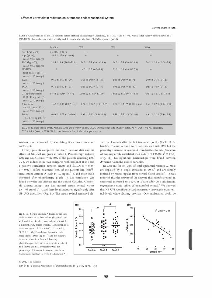
Effect of ultraviolet B radiation on cutaneous endocannabinoid system
98
analysis was performed by calculating Spearman correlation
coefficient.
Twenty patients completed the study. Baseline data and the
effects of NB-UVB are given in Table 1. Phototherapy reduced
PASI and DLQI scores, with 70% of the patients achieving PASI
75 (75% reduction in PASI compared with baseline) at W6 and
a positive correlation between DPASI and DDLQI (r = 0Æ25;P = 0Æ02). Before treatment, 60% of the patients had insuffi-
cient serum vitamin D levels (< 20 ng mL)1), and these levels
increased after phototherapy (Table 1). No correlation was
found between this increase and the studied variables. At onset,
all patients except one had normal serum retinol values
(> 1Æ05 lmol L)1), and these levels increased significantly after
NB-UVB irradiation (Fig. 1a). The serum retinol remained ele-
vated at 1 month after the last treatment (W10) (Table 1). At
baseline, vitamin A levels were not correlated with BMI but the
percentage increase in vitamin A from baseline to W6 (Dvitamin
A) was negatively correlated with BMI (P < 0Æ0001; r2 = 0Æ54)(Fig. 1b). No significant relationships were found between
Dvitamin A and the studied variables.
RE account for 85–90% of total epidermal vitamin A. Most
are depleted by a single exposure to UVB,4 and are rapidly
replaced by retinol uptake from dermal blood vessels.3–5 It was
reported that the activity of the enzyme that esterifies retinol in
epidermis increased to 167% at 2 days after UVB irradiation,
suggesting a rapid influx of unesterified retinol.7 We showed
that NB-UVB significantly and persistently increased serum reti-
nol levels while clearing psoriasis. One explanation could be
Table 1 Characteristics of the 20 patients before starting phototherapy (baseline), at 3 (W3) and 6 (W6) weeks after narrowband ultraviolet B(NB-UVB) phothotherapy thrice weekly and 1 month after the last NB-UVB exposure (W10)
Baseline W3 W6 W10
Sex, F ⁄M, n (%) 8 (33) ⁄12 (67) – – –Age (years),
mean ± SD (range)
51Æ2 ± 13Æ4 (21–69) – – –
BMI (kg m)2),
mean ± SD (range)
26Æ3 ± 3Æ9 (20Æ0–33Æ8) 26Æ2 ± 3Æ8 (20Æ1–33Æ9) 26Æ3 ± 3Æ8 (20Æ0–33Æ9) 26Æ3 ± 3Æ9 (20Æ0–33Æ9)
NB-UVB
total dose (J cm)2),mean ± SD (range)
0 6Æ5 ± 0Æ5 (6Æ3–8Æ1) 21Æ9 ± 6Æ1 (14Æ0–27Æ9) –
PASI,mean ± SD (range)
9Æ65 ± 4Æ59 (5–20) 5Æ00 ± 3Æ46* (1–14) 2Æ30 ± 2Æ55** (0–7) 2Æ70 ± 3Æ14 (0–12)
DLQI,mean ± SD (range)
9Æ75 ± 6Æ40 (1–23) 5Æ30 ± 5Æ02* (0–17) 3Æ75 ± 4Æ19** (0–12) 3Æ35 ± 4Æ09 (0–12)
25-hydroxyvitaminD (> 30 ng mL)1)a,
mean ± SD (range)
18Æ44 ± 12Æ56 (3–47) 24Æ35 ± 13Æ00* (7–49) 34Æ05 ± 12Æ24** (8–56) 34Æ61 ± 12Æ50 (11–55)
Vitamin A(> 1Æ05 lmol L)1)a,
mean ± SD (range)
1Æ62 ± 0Æ34 (0Æ87–2Æ3) 1Æ76 ± 0Æ46* (0Æ94–2Æ65) 1Æ86 ± 0Æ44** (1Æ08–2Æ76) 1Æ97 ± 0Æ53 (1Æ12–3Æ16)
Folate
(2Æ2–17Æ5 ng mL)1)a,mean ± SD (range)
6Æ64 ± 2Æ75 (3Æ2–14Æ6) 6Æ49 ± 2Æ12 (3Æ5–10Æ8) 6Æ38 ± 2Æ31 (3Æ7–11Æ4) 6Æ41 ± 3Æ15 (2Æ4–15Æ5)
BMI, body mass index; PASI, Psoriasis Area and Severity Index, DLQI, Dermatology Life Quality Index. *P < 0Æ05 (W3 vs. baseline),**P < 0Æ05 (W6 vs. W3). aReference intervals for biochemical parameters.
(a) (b)
Fig 1. (a) Serum vitamin A levels in patients
with psoriasis (n = 20) before (baseline) and
at 3 and 6 weeks after narrowband ultraviolet
B phototherapy thrice weekly. Horizontal lines
indicates means. **P = 0Æ0001, *P = 0Æ02,#P = 0Æ04. (b) Correlation between body
mass index (BMI) (kg m)2) and the change
in serum vitamin A levels following
phototherapy. Each circle represents a patient
and shows the BMI compared with the
percentage of increase in serum vitamin A
levels from baseline to week 6 (Dvitamin A).
� 2012 The Authors
BJD � 2012 British Association of Dermatologists 2012 167, pp937–963
Correspondence 959

Chapter II Ultravioleta B radiation on patients with psoriasis
99
that phototherapy-induced cutaneous depletion of vitamin A
increases its skin uptake from circulation and consequently
increases mobilization of hepatic and fat stores. Despite this flux
to the skin, mobilization of retinol stores may be disproportion-
ate, contributing to the observed increase in serum levels. Only
two studies have dealt with the relationship between UVR and
serum retinol levels.6,7 One involving healthy subjects irradiated
for 2 weeks with UVA or broadband UVB did not find variation,
and the other with hairless mice showed a transient depletion of
serum retinol a few days after a single broadband UVB irradia-
tion.6,7 Our data indicate that in patients with psoriasis, vitamin
A participates in an adaptive response to NB-UVB exposure; the
physiological role of this response needs to be further investi-
gated.
It is well documented that vitamin A and RBP4 are involved
in lipid metabolism.1 Subjects with nonalcoholic hepatic stea-
tosis had lower retinol liver stores and an association between
obesity and vitamin A deficiency was proposed.8 We found a
negative correlation between BMI and Dvitamin A and we can
hypothesize that overweight patients have less adaptive vita-
min A response.
In agreement with data from higher-latitude regions, we
found a high prevalence of vitamin D insufficiency and a sub-
stantial benefit of NB-UVB.9,10
The limitations of the present study include the absence of
a control group, and cutaneous retinol and serum RBP4 levels
should be addressed in future studies.
In addition to a significant increase of the vitamin D status,
NB-UVB increases serum vitamin A levels while clearing psori-
asis, the magnitude of this increase being determined by the
patient’s BMI.
Acknowledgments
We thank the participants for their contribution to the
study. We also thank all the nursing staff from our Derma-
tology Department for their help in the project. A special
thanks to Carla Vieira (specialist nurse) for helping with
the study.
S . MAG INA1 , 2 , 3
M. J . CRUZ3
F . AZ EV EDO3
D. MOURA1
E . MOURA1 , 2
M.A. V I E I RA -COE LHO1 , 2
1Department of Pharmacology and Therapeutics,
Faculty of Medicine, University of Porto, Alameda Prof.
Hernani Monteiro, 4200-319 Porto, Portugal2Institute for Molecular and Cell Biology,
University of Porto, Rua do Campo Alegre 823,
4150-180 Porto, Portugal3Department of Dermatology and Venereology,
University of Porto, Alameda Prof. Hernani Monteiro,
4200-319 Porto, Portugal
E-mail: [email protected]
References
1 Berry DC, Noy N. Signaling by vitamin A and retinol-binding pro-
tein in regulation of insulin responses and lipid homeostasis. Bio-chim Biophys Acta 2012; 1821:168–76.
2 Schreiber R, Taschler U, Preiss-Landl K et al. Retinyl ester hydrolasesand their roles in vitamin A homeostasis. Biochim Biophys Acta 2012;
1821:113–23.3 Fu PP, Xia Q, Yin JJ et al. Photodecomposition of vitamin A and
photobiological implications for the skin. Photochem Photobiol 2007;83:409–24.
4 Sorg O, Tran C, Carraux P et al. Oxidative stress-independent deple-tion of epidermal vitamin A by UVA. J Invest Dermatol 2002;
118:513–18.5 Tran C, Sorg O, Carraux P et al. Topical delivery of retinoids coun-
teracts the UVB-induced epidermal vitamin A depletion in hairless
mouse. Photochem Photobiol 2001; 73:425–31.6 White WS, Kim CI, Kalkwarf HJ et al. Ultraviolet light-induced
reductions in plasma carotenoid levels. Am J Clin Nutr 1988;47:879–83.
7 Torma H, Berne B, Vahlquist A. UV irradiation and topical vitaminA modulate retinol esterification in hairless mouse epidermis. Acta
Derm Venereol (Stockh) 1988; 68:291–9.8 Pereira SE, Saboya CJ, Saunders C, Ramalho A. Serum levels and
liver store of retinol and their association with night blindness inindividuals with class III obesity. Obes Surg 2012; 22:602–8.
9 Gisondi P, Rossini M, Di Cesare A et al. Vitamin D status in patientswith chronic plaque psoriasis. Br J Dermatol 2011; 166:505–10.
10 Ryan C, Moran B, McKenna MJ et al. The effect of narrowband UV-Btreatment for psoriasis on vitamin D status during wintertime in
Ireland. Arch Dermatol 2010; 146:836–42.
Funding sources: none.
Conflicts of interest: none declared.
Lichen planus is not associated with humanherpesvirus type 7
DOI: 10.1111/j.1365-2133.2012.11009.x
MADAM, Human herpesvirus type 7 (HHV-7) has been proposed
to be associated with lichen planus (LP).1 Furthermore, it has
been postulated that a decrease in HHV-7 replication is
associated with LP remission.2 However, we came to the
opposite conclusion in a case–control study conducted at
Shohada-e-Tajrish University Hospital from June 2007 to
March 2010.
According to the previous study,1 we arrived at a study size
of n = 53 in each group with the power of 0Æ80 to detect a sig-
nificant difference between cases and controls. We ascertained
one control for each case, matched for age. Cases were patients
with a histopathologically confirmed diagnosis of LP during
acute exacerbation of disease. We obtained control group skin
samples from patients admitted for cosmetic procedures who
were otherwise healthy. We examined both skin and plasma
specimens by polymerase chain reaction (PCR) using QIAamp
DNA Mini Kit (Qiagen, Hilden, Germany) according to the
manufacturer’s instructions. For amplification and detection of
HHV-7 genome we used the forward primer 5¢-CCC AAC TAT
TTT CAG TAG GGT TGG TG-3¢, reverse primer 5¢-TTT AGT
� 2012 The Authors
BJD � 2012 British Association of Dermatologists 2012 167, pp937–963
960 Correspondence


Chapter II Ultravioleta B radiation on patients with psoriasis
101
1
1
Effect of narrowband ultraviolet B treatment on endocannabinoid plasma levels in psoriasis patients 1,2,3S Magina *, 1,2MA Vieira-Coelho, 1,2E Moura, 1MP Serrão, 4F Piscitelli, 1D Moura, 4V Di Marzo 1Department of Pharmacology and Therapeutics, Faculty of Medicine, University of Porto, 2Institute for Molecular and Cell Biology, 3Dermatology and Venereology Department, H. S. João, 4Endocannabinoid Research Group, Institute of Biomolecular Chemistry, Consiglio Nazionale delle Ricerche, Via Campi Flegrei 34, 80078 Pozzuoli, NA, Italy
ABSTRACT
Background- Psoriasis is associated with increased risk of obesity and metabolic syndrome. Although endocannabinoid system dysregulation was demonstrated in these comorbidities, no data exist on endocannabinoid plasma levels in psoriasis. We have recently shown that the cutaneous endocannabinoid system responds to ultraviolet B (UVB) irradiation, but the effect of narrowband UVB (nbUVB) treatment on plasma endocannabinoid levels is unknown.
Objectives- To evaluate endocannabinoid plasma levels in psoriasis patients. To determine whether exposure to nbUVB modifies these levels. To correlate plasma endocannabinoid levels with metabolic parameters and leptin levels.
Methods- An open observational study was conducted with 21 psoriasis patients and 15 matched controls. Plasma levels of anandamide, 2-arachidonoylglycerol (2-AG), oleylethanolamide (OEA) and palmitoylethanolamide (PEA) were recorded. Patients were evaluated at weeks 0, 3 and 6 of nbUVB phototherapy and 4 weeks after stopping the treatment. In each evaluation body mass index (BMI), Psoriasis Area and Severity Index (PASI) and Dermatology Life Quality Index (DLQI) were calculated and fasting levels of C-reactive protein (CRP), high sensitivity CRP (hsCRP), cholesterol, triglycerides, glucose and leptin were measured.
Results- Endocannabinoid plasma levels did not differ significantly between controls and psoriasis patients, in whom, before phototherapy, anandamide levels were positively correlated with hsCPR. The nbUVB treatment significantly decreased anandamide levels to an extent positively correlated with BMI. PASI score improved but no correlation with the decrease of anandamide levels was found.
Conclusions- The present study shows that nbUVB treatment significantly decreases anandamide plasma levels in patients with psoriasis in a manner that is stronger in patients with higher baseline BMI.
Introduction
The major endocannabinoids, anandamide and 2-arachidonoylglycerol (2-AG), are lipid mediators that are produced “on demand” from membrane phospholipid precursors in the central nervous system, peripheral organs, skin and adipose tissue 1-2. Anandamide and 2-AG act through specific G-coupled cannabinoid receptors type 1 (CB1) and type 2 (CB2) 1-2. The biological activity of endocannabinoids is exerted in all organs and is not limited to the central nervous system as previous believed 3 4. In the skin, a fully functional endocannabinoid system (ECS) has been identified 5. We have shown that this cutaneous ECS responds to ultraviolet B (UVB) irradiation 6. However the effect of narrowband UVB (nbUVB) phototherapy on endocannabinoid plasma levels in humans is still unknown. Several studies found a link between psoriasis, obesity, metabolic syndrome and serum levels
of certain adipokines 7-8. Obesity and metabolic syndrome are well known pathological conditions where endocannabinoid system dysregulation was demonstrated 9 2,10. It has been suggested that ECS tone is overall increased in human obesity 11. In obese patients, elevated endocannabinoid plasma levels were associated with coronary circulatory dysfunction 12. The relationship between obesity and systemic inflammation has been described in psoriasis patients and considered detrimental 8. This interaction provides the inflammatory base of the ‘psoriatic march’ that describes the development of vascular complications in psoriasis 13 8. Recent data support a link between the endocannabinoid system and obesity-associated inflammation 2. Previous studies suggested that mediators released from the adipose tissue such as leptin and/or endocannabinoids are involved in the initiation and development of cardiovascular disease 12. At present, the impact of endocannabinoids on

Effect of ultraviolet B radiation on cutaneous endocannabinoid system
102
2
2
psoriasis is unknown. In this context, we aimed to evaluate endocannabinoid plasma levels in psoriasis patients, the influence of nbUVB phototherapy on these levels and their correlation with leptin levels and inflammatory and metabolic parameters. Since the two cannabinoid receptor-inactive and metabolically related anandamide congeners, oleoylethanolamide (OEA) and palmitoylethanolamide (PEA), were recently found to be dysregulated in obese rats 14, and are known to activate receptors involved in inflammation, i.e. the nuclear peroxisome proliferator-activated receptor-� (PPAR-�) and transient receptor potential vanilloid type-1 (TRPV1) channels, we have also measured the plasma levels of these two lipid mediators.
Patients and methods
The regional ethical committee approved this open observational study and all participants gave informed consent. The inclusion criteria were patients with chronic plaque psoriasis, indication for phototherapy and free of psoriasis medication or other systemic therapy, at least 12 weeks before and during the study. Patients presenting diabetes, psoriatic arthritis or other inflammatory diseases were excluded. We enrolled 21 patients with Fitzpatrick skin types II-III patients and 15 age, sex and body mass index (BMI) matched controls. Subjects were admitted only in winter and early spring. They were advised to not make any nutritional or lifestyle changes that could interfere with the results during the treatment period. Psoriasis severity was measured by Psoriasis Area and Severity Index (PASI) and Dermatology Life Quality Index (DLQI). To diminish subjectivity, the same dermatologist evaluated PASI. All psoriasis patients were evaluated before phototherapy (baseline), after three (W3) and six (W6) weeks of treatment and four weeks after stopping (W10). In each evaluation BMI, PASI and DLQI were calculated and fasting venous blood samples were taken in the morning, for anandamide, 2-AG, OEA and PEA measurement. The blood parameters shown in Table 2 were also assessed in the same conditions. Plasma levels of anandamide, 2-AG, PEA and OEA were measured by isotope-dilution liquid chromatography-atmospheric pressure chemical ionization-mass spectrometry as described 9. Fasting plasma leptin was determined by ELISA kit (Millipore, Billerica, MA, USA), according to manufacturer instructions. The nbUVB irradiation (311 ± 2 nm) was administered using a Waldmann 7001 cabin;
(UVB-TL01; Waldmannn Medizintechnik, Villigen-Schwenningen, Germany); the initial dose, dependent on patient’s phototype, was 0.3-0.5 J/cm2, increasing 0.1 J/cm2 in every session until a maximum dose of 2.0 J/cm2, totalizing eighteen nbUVB exposures (thrice weekly, during six weeks). Only eyes and genital were shielded during the irradiation. Data are presented as means ± SD and ANOVA with repeated measures was used to determine the statistical significance. Values of P<0.05 are considered significant. The correlation analysis was performed by calculating Spearman coefficient correlation.
Results
As shown in Table 1, patients and controls were not significantly different regarding age or BMI with a mean BMI > 25 Kg/m2 (overweight) in both groups. Endocannabinoid plasma levels did not differ significantly between controls and psoriasis patients (Table 1). Phototherapy significantly reduced PASI and DLQI scores (P<0.0001) but did not change BMI (Table 1). We observed a significant positive correlation between the percentage of decrease in PASI (∆PASI) and the percentage of decrease in DLQI (∆DLQI) from baseline to week 6 (R2=0.25; P=0.02). In psoriasis patients, before phototherapy, hsCPR levels, were positively correlated with BMI (R2=0.29; P=0.02) and LDL-cholesterol (R2=0.43; P=0.004) and negatively with HDL-cholesterol (R2=0.23; P=0.04). No correlation was observed between PASI score and hsCPR or CPR values. As shown in Fig.1, anandamide plasma levels significantly decreased after completion of the nbUVB treatment (P<0.001). We found that the percentage of decrease in anandamide from baseline to week 6 was positively correlated with BMI (R2=0.35; P=0.006) and with anandamide levels before phototherapy (R2=0.57; P=0.0002), but no correlation was found with PASI. No significant nbUVB-effect on the levels of the other endocannabinoid, 2-AG, or of OEA and PEA was found (Table 1).

Chapter II Ultravioleta B radiation on patients with psoriasis
103
3
3
Table 1. Characteristics and endocannabinoid levels of controls and psoriasis patients before phototherapy (Psoriasis baseline), three (W3) and six (W6) weeks after nbUVB phothotherapy thrice weekly and one month after the last nbUVB exposure (W10).
Values are means ± SD. BMI, body mass index; PASI, Psoriasis Area and Severity Index; DLQI, Dermatology Life Quality Index; AEA, anandamide; 2-AG, 2-arachidonoylglycerol; OEA, N-oleylethanolamine; and PEA, N-palmitoylethanolamine *P<0.05 (W3 vs. baseline), **P<0.05 (W6 vs. W3), # P<0.05 (W6 vs. baseline), ## P<0.05 (W10 vs. baseline).
Table 2. Inflammatory parameters and lipid profile in psoriasis patients before starting phototherapy (Psoriasis baseline), three (W3) and six (W6) weeks after nbUVB phothotherapy thrice weekly and one month after the last nbUVB exposure (W10).
Psoriasis baseline
Psoriasis W3
Psoriasis W6
Psoriasis W10
Total cholesterol (mg/dL)
212.70±40.66 (116-285)
211.10±40.35 (130-281)
212.40±37.71 (119-297)
206.90±43.08 (108-277)
LDL-cholesterol (mg/dL)
138.10±42.74 (77-214)
137.30±33.39 (88-202)
137.50±29.10 (89-214)
134.1±39.36 (73-201)
HDL-cholesterol (mg/dL)
52.89±10.10 (35-77)
52.13±11.24 (36-80)
51.71±10.12 (35-69)
54.00±11.25 (33-74)
Triglycerides (mg/dL)
136.30±74.85 (50-300)
137.70±69.30 (40-290)
137.10±63.50 (40-288)
134.00±59.98 (54-267)
Glucose (mg/dL)
87.74±19.10 (64-132)
88.76±21.88 (64-136)
85.27±23.31 (63-137)
90.67±19.65 (66-137)
Total Leptin (ng/mL)
10.46±7.19 (2.38-27.71)
11.24±6.27 (3.67-26.26)
10.19±5.78* (3.73-25.52)
12.13±7.41 (4.44-31.84)
Males Leptin (ng/mL)
10.19±7.02 (2.38-21.78)
10.96±5.47 (4.32-18.10)
9.53±4.72* (3.73-15.20)
11.60±5.95 (4.44-21.67)
Females Leptin (ng/mL)
11.37±8.05 (3.00-27.71)
11.40±7.81 (3.67-26.26)
11.01±7.36 (3.78-25.52)
12.72±9.17 (6.89-31.84)
CPR (<3.0 mg/L) a
2.53±1.74 (0.3-6.4)
2.37±1.77 (0.3-6.0)
2.82±1.91 (0.3-6.0)
2.68±2.00 (0.2-7.1)
hsCPR (<5.0 mg/L) a
2.24±1.61 (0.3-5.9)
3.55±4.41 (0.4-8.2)
2.96±2.83 (0.2-5.4)
2.79±2.74 (0.2-6.5)
Values are means ± SD (range). CRP, C-reactive protein; hsCRP, high sensitivity C-reactive protein. *P<0.05 (W6 vs. W3). a
Reference intervals for biochemical parameters.
Controls Psoriasis baseline
Psoriasis W3
Psoriasis W6
Psoriasis W10
Sex, women 5 (33.3) /10(66.7) 8 (33.3) /13(66.7) - - -
Age, Years 46.33±2.36 51.2±13.4 - - -
BMI (Kg/m2) 25.6±4.2 26.3±3.9 26.2±3.8 26.3±3.8 26.3±3.9
UVB total dose (J/cm2) - 0 6.5±0.5 21.9±6.1 -
PASI - 10.20±5.02 5.21±3.42* 2.42±2.57** 2.84±3.16##
DLQI - 9.60±6.23 5.12±5.08* 3.84±4.29** 3.84±4.19##
AEA (pmol/ml) 2.55±0.35 3.19±0.49 2.56±0.28 1.90±0.12# 1.99±0.13##
2-AG (pmol/ml) 7.14±1.17 7.37±0.79 6.88±0.76 6.29±0.58 6.98±1.12
OEA (pmol/ml) 19.13±1.41 20.80±1.46 21.42±1.85 18.81±1.17 19.24±1.29
PEA (pmol/ml) 33.64±2.24 32.04±2.14 36.31±4.87 32.35±2.77 32.51±2.85

Effect of ultraviolet B radiation on cutaneous endocannabinoid system
104
Table 3. Correlation of endocannabinoid plasma levels before phototherapy (anandamide, 2and PEA) with clinical variables in patients with psoriasis
variable r
Age 0.20
PASI 0.005
DLQI -0.31
BMI -0.36
Total cholesterol -0.32
LDL-cholesterol -0.35
HDL-cholesterol -0.23
Triglycerides 0.08
Glucose 0.29
Total Leptin -0.02
CPR 0.64
CPRhs 0.63
BMI, body mass index; PASI, Psoriasis Area and Severity Index; DLQI, Dermatology Life Quality Index; AEA, anandamide; 2AG, 2-arachidonoylglycerol; OEA, N-oleylethanolamine; and PEA,
Figure 1. Plasma anandamide levels in psoriatic patients (n=21) before (baseline) and three and six weeks after nbUVB phototherapy thrice weekly. **P<0.01
Regarding to metabolic and inflammatory parameters, there was no significant effect of phototherapy (Table 2). Only a slight redof leptin levels was observed in males (Table
Correlation of endocannabinoid plasma levels before phototherapy (anandamide, 2and PEA) with clinical variables in patients with psoriasis
AEA 2-AG PEA
P-value r P-value r P-value
0.39 -0.16 0.50 -0.04 0.85
0.98 0.30 0.20 -0.28 0.22
0.18 -0.01 0.95 -0.44 0.05
0.11 0.23 0.32 -0.33 0.15
0.18 0.29 0.22 -0.45 0.05
0.14 0.12 0.63 -0.36 0.13
0.40 -0.08 0.74 -0.12 0.63
0.76 0.34 0.15 0.03 0.89
0.25 0.005 0.99 -0.13 0.59
0.94 0.56 0.02* 0.33 0.18
0.002* -0.29 0.21 0.33 0.18
0.002* -0.29 0.21 0.36 0.15
Psoriasis Area and Severity Index; DLQI, Dermatology Life Quality Index; AEA, anandamide; 2oleylethanolamine; and PEA, N-palmitoylethanolamine *P<0.05
levels in psoriatic patients (n=21) before (baseline) and three and six weeks after
**P<0.01
Regarding to metabolic and inflammatory there was no significant effect of
phototherapy (Table 2). Only a slight reduction of leptin levels was observed in males (Table 2).
A positive correlation was seen between baseline hsCPR and anandamide levels (Table 3). No correlation was found between baseline endocannabinoid, OEA and PEAother clinical and metabolic parameters (Table 3).
Discussion
The present study provides evidencenbUVB phototherapy indecreases plasma levels of anandamidechanging 2-AG, OEA or PEAWe observed that around 65patients were overweight with body mass index (BMI) ≥25 and < 30 kg/m2
decrease in anandamide plasma levelpositively correlated with BMIwas previously reported that increanandamide plasma levels are with binge eating disorder 15
obesity 16. Furthermore, elevated circulating anandamide levels are associated with coronary circulatory dysfunction in obese individuals proposed as a novel endogenous cardiovascular
4
4
Correlation of endocannabinoid plasma levels before phototherapy (anandamide, 2-AG, OEA
OEA
r P-value
0.16 0.51
-0.18 0.45
-0.43 0.06
-0.37 0.09
-0.39 0.09
-0.29 0.24
-0.09 0.73
0.16 0.51
-0.19 0.43
0.25 0.32
0.25 0.35
0.22 0.35
Psoriasis Area and Severity Index; DLQI, Dermatology Life Quality Index; AEA, anandamide; 2-
A positive correlation was seen between hsCPR and anandamide levels (Table
3). No correlation was found between baseline , OEA and PEA levels and
other clinical and metabolic parameters (Table
resent study provides evidence that psoriasis patients
anandamide without or PEA levels.
65% of the studied patients were overweight with body mass index
2. Interestingly, the decrease in anandamide plasma levels was positively correlated with BMI at baseline. It was previously reported that increased anandamide plasma levels are present in women
or post-menopausal , elevated circulating
associated with coronary circulatory dysfunction in obese individuals and proposed as a novel endogenous cardiovascular

Chapter II Ultravioleta B radiation on patients with psoriasis
105
5
5
risk factor 12. The observed UVB-induced anandamide decrease might, therefore, suggest a beneficial effect of phototherapy in psoriasis patients at cardiovascular risk. This hypothesis, which, however, needs further investigation, is supported by the observation that baseline anandamide levels were positively correlated with hsCRP, a biomarker for both increased cardiovascular events and inflammation. In another study 12, only anandamide plasma levels were correlated with CRP and an endocannabinoid-mediated association between metabolic and inflammatory cardiovascular disease mechanisms was proposed. The link between psoriasis and increased cardiovascular risk appears to be dependent of disease severity 17. A major limitation of our study was that patient profiles corresponded to those of candidates for phototherapy and, therefore, subjects with very severe cutaneous disease are not represented in the study population. This might explain why anandamide plasma levels were not significantly different between psoriasis patients and controls, also because the two populations did not differ for their BMI and were overweight (≥25 kg/m2) 11
16. Despite the fact that the clinical response to UVB was good, no correlation was observed between endocannabinoid levels and disease activity. Furthermore, despite the recognized role of endocannabinoids in the metabolic syndrome, no correlation was observed between endocannabinoid levels and leptin, LDL cholesterol, HDL cholesterol, triglycerides or glucose fasting levels in our populations. Indeed, some of these correlations have been found so far only for obese patients (BMI>30) and/or only for 2-AG 9,16. Unlike 2-AG, anandamide, OEA and PEA are all N-acylethanolamines and share similar metabolic pathways 18. Therefore, it was somewhat surprising not to find for OEA and PEA similar changes or correlations as with anandamide, especially considering the fact that for both compounds anti-inflammatory actions mediated by non-cannabinoid receptors have been reported 19. This finding might suggest that the observed changes specifically reflect, or impact on, the activity of the endocannabinoid system, thus supporting their possible relevance to psoriasis- and cardiovascular-related inflammation 18. However, the mechanism by which UVB might specifically alter anandamide remains to be explored. It will be important to evaluate in the future the influence of other psoriasis treatments on endocannabinoid plasma levels in order to clarify whether the UVB-induced decrease of
anandamide plasma levels is a radiation effect per se or is related with disease remission.
Acknowledgments We thank the participants for their contribution to the study. We also thank all the nurse staff from our Dermatology Department for their help in the project. A special thanks to Carla Vieira for helping with the study.
References
1 Pertwee RG, Howlett AC, Abood ME et al.International Union of Basic and Clinical Pharmacology. LXXIX. Cannabinoid receptors and their ligands: beyond CB and CB. Pharmacol Rev 2010; 62: 588-631.
2 Cluny NL, Reimer RA, Sharkey KA. Cannabinoid signalling regulates inflammation and energy balance: The importance of the brain-gut axis. Brain Behav Immun 2012.
3 Di Marzo V. Targeting the endocannabinoid system: to enhance or reduce? Nat Rev Drug Discov 2008; 7: 438-55.
4 Pacher P, Mechoulam R. Is lipid signaling through cannabinoid 2 receptors part of a protective system? Prog Lipid Res 2011; 50: 193-211.
5 Biro T, Toth BI, Hasko G et al. The endocannabinoid system of the skin in health and disease: novel perspectives and therapeutic opportunities. Trends Pharmacol Sci 2009; 30: 411-20.
6 Magina S, Esteves-Pinto C, Moura E et al.Inhibition of basal and ultraviolet B-induced melanogenesis by cannabinoid CB(1) receptors: a keratinocyte-dependent effect. Arch Dermatol Res 2011; 303: 201-10.
7 Kaur S, Zilmer K, Leping V et al. The levels of adiponectin and leptin and their relation to other markers of cardiovascular risk in patients with psoriasis. J Eur Acad Dermatol Venereol 2011; 25: 1328-33.
8 Reich K. The concept of psoriasis as a systemic inflammation: implications for disease management. J Eur Acad Dermatol Venereol2012; 26 Suppl 2: 3-11.
9 Cote M, Matias I, Lemieux I et al. Circulating endocannabinoid levels, abdominal adiposity and related cardiometabolic risk factors in obese men. Int J Obes (Lond) 2007; 31: 692-9.
10 Maccarrone M, Gasperi V, Catani MV et al. The endocannabinoid system and its relevance for nutrition. Annu Rev Nutr 2010; 30: 423-40.
11 Cable JC, Tan GD, Alexander SP et al. The activity of the endocannabinoid metabolising enzyme fatty acid amide hydrolase in subcutaneous adipocytes correlates with BMI in metabolically healthy humans. Lipids Health Dis2011; 10: 129.
12 Quercioli A, Pataky Z, Vincenti G et al. Elevated endocannabinoid plasma levels are associated with coronary circulatory dysfunction in obesity. Eur Heart J 2011; 32: 1369-78.
13 Boehncke WH, Boehncke S, Tobin AM et al.The 'psoriatic march': a concept of how severe psoriasis may drive cardiovascular comorbidity. Exp Dermatol 2011; 20: 303-7.
14 Izzo AA, Piscitelli F, Capasso R et al. Basal and fasting/refeeding-regulated tissue levels of endogenous PPAR-alpha ligands in Zucker rats. Obesity (Silver Spring) 2010; 18: 55-62.
15 Monteleone P, Matias I, Martiadis V et al. Blood levels of the endocannabinoid anandamide are

Effect of ultraviolet B radiation on cutaneous endocannabinoid system
106
6
6
increased in anorexia nervosa and in binge-eating disorder, but not in bulimia nervosa. Neuropsychopharmacology 2005; 30: 1216-21.
16 Engeli S, Jordan J. The endocannabinoid system: body weight and metabolic regulation. Clin Cornerstone 2006; 8 Suppl 4: S24-35.
17 Yiu KH, Yeung CK, Chan HT et al. Increased arterial stiffness in patients with psoriasis is associated with active systemic inflammation. Br J Dermatol 2011; 164: 514-20.
18 Petrosino S, Ligresti A, Di Marzo V. Endocannabinoid chemical biology: a tool for the development of novel therapies. Curr Opin Chem Biol 2009; 13: 309-20.
19 Petrosino S, Iuvone T, Di Marzo V. N-palmitoyl-ethanolamine: Biochemistry and new therapeutic opportunities. Biochimie 2010; 92: 724-7.

Discussion and Conclusions


Discussion and Conclusions
109
Discussion
In chapter I, we report that keratinocytes and melanoma cells express CB1 and
CB2 receptors, produce physiologically relevant concentrations of the two major
endocannabinoids (2-AG and anandamide) and of the related N-acylethanolamine
(PEA), and have the main cannabinoid metabolizing enzymes FAAH and MAGL.
These findings show that these cutaneous cells are endowed with all the components of
an endogenous cannabinoid system. Levels of endocannabinoids are much higher in
melanoma cells than in keratinocytes, but FAAH activity and particularly MAGL
activity are higher in keratinocytes. This much higher activity of MAGL in
keratinocytes may explain why the basal levels of 2-AG are lower in co-cultures than
in melanoma cells alone.
We have also demonstrated that UVB irradiation increases the levels of
anandamide, 2-AG and PEA in keratinocytes and significantly increases the ratio 2-
AG/anandamide in co-cultures whereas in melanoma cells endocannabinoid levels
remain unaltered after UVB. These findings suggest that endocannabinoids are also
pro-homeostatic in the skin, because UV irradiation is the major environmental insult
to the skin and elicits an increase in the levels of endocannabinoids.
Since UVB increases Ca2+ influx in keratinocyte (HaCat) via TRPV1 activation
(Lee et al. 2009; Masaki et al. 2009) and endocannabinoids are produced “on demand”
from membrane phospholipids in response to intracellular Ca2+ (Alger and Kim 2011;
Cluny et al. 2012), we can hypothesize that the observed increase in endocannabinoid
levels is dependent on UVB-induced Ca2+-influx. Moreover, in human skin and
keratinocytes, UVR activates phospholipases A2, C and D and diacylglycerol kinase,
thereby releasing arachidonic acid and DAG (De Leo et al. 1984; Punnonen and Yuspa
1992; Carsberg et al. 1995) which are the main precursors for endocannabinoid
synthesis. The UVB-induced release of DAG, which is the 2-AG precursor, may
explain why 2-AG increases more than anandamide in keratinocytes irradiated with

Effect of ultraviolet B radiation on cutaneous endocannabinoid system
110
UVB. The increase in the ratio 2-AG/anandamide that we observed in co-cultures after
exposure to UVB can also be explained by the UVB-induced release of DAG. Our data
agree with the findings that mouse epidermal JB6P+ cells respond to stress factors
such as UVB irradiation or serum deprivation with the production of increased levels
of N-acylethanolamines and their precursors, N-acylethanolamine phospholipids
(Berdyshev et al. 2000).
An alternative explanation for the increase in endocannabinoid levels after UVB
irradiation would be a decrease in metabolizing pathways. However we observed that
UVB even caused an increase of MAGL and FAAH activities. These results suggest
that the UVB increased levels of endocannabinoids are mostly due to an increased
synthesis.
Total endocannabinoids (except PEA) are less in co-culture than in melanoma
cells alone under basal conditions, and even less under UVB irradiation. This
phenomenon may be due to the high levels of endocannabinoid-degrading enzymes in
keratinocytes and their up-regulation by UVB.
On the other hand UVB increases the mRNA expression of CB1 receptors in
melanoma cells but not in keratinocytes. Taken together, our data support the concept
that endocannabinoids may participate in the important cross talk between
keratinocytes and melanocytes in the epidermal unit after exposure to UVR. These
results are in agreement with the classic idea that keratinocytes respond to UVR
producing several paracrine factors that will act on melanocytes helping in their
protection.
Endocannabinoids (Karsak et al. 2007) and PEA (Petrosino et al. 2009) attenuate
the cutaneous allergic response whereas the antagonism of CB1 cannabinoid receptors
in human HaCat keratinocytes increases the expression of pro-allergic chemokines
(Leonti et al. 2010). Acute UVB irradiation is known to cause immunosuppression.
Since we found, in chapter I, that UVB irradiation is accompanied by up-regulation of
both endocannabinoids and PEA levels, we hypothesize that the cutaneous
Discussion and Conclusions
111
endocannabinoid system could play a role in UVB-induced immunosuppression and
might contribute to the well-known therapeutic effect of UVB radiation.
Acute UVB exposure also causes inflammation and there is growing evidence
suggesting that 2-AG modulates the inflammatory response by acting on the
cannabinoid CB2 receptor (Oka et al. 2006). Interestingly, we found a significant
increase in 2-AG after UVB irradiation, suggesting a role for endocannabinoids,
namely 2-AG, in UVB-induced inflammation. In agreement with this hypothesis,
Zheng et al. (Zheng et al. 2008) suggested that CB receptors and the related signaling
pathways might be involved in the promotion of in vivo UVB-induced skin
inflammation. In CB1 and CB2 double gene-deficient mice, the authors observed a
marked decrease in UVB-induced inflammation and an attenuation of UVB-induced
activation of MAPK and NF-kB. (Zheng et al. 2008). On the other hand, PEA with its
anti-inflammatory effect (Petrosino et al. 2010) might represent an endogenous
protective agent against UVB-induced inflammation.
The first defense against UVR is the cutaneous melanin produced in
melanocytes. However, as reported in chapter I melanoma cells alone did not show
any increase in melanin content after UVB irradiation. In contrast, co-cultures
exhibited a marked melanogenic response to UVB irradiation (≈ 40% melanin over the
controls). It is known that UVB irradiation of keratinocytes triggers the release of
cytokines, growth factors, and other mediators such as POMC-derived peptides that
regulate melanin synthesis in a paracrine way (Duval et al. 2001; Park et al. 2009) and
this may explain the differences that we observed between mono- and co-cultures in
their response to UVB.
In chapter I, we showed that the CB1 selective agonist arachidonoyl-2-
chloroethylamide (ACEA), had an inhibitory effect on basal melanogenesis in co-
cultures. Furthermore, in the same model, ACEA abolished the increase in melanin
content caused by UVB irradiation. The CB1 selective antagonist (AM-251), that alone
did not modify UVB-induced melanogenesis, reversed this inhibitory effect of ACEA.

Effect of ultraviolet B radiation on cutaneous endocannabinoid system
112
The inhibitory effect of CB1 receptor activation on melanogenesis was observed in co-
cultures but not in monocultures, which again, underlines the important symbiotic
relationship between melanocytes and keratinocytes. Since both cells express CB1
receptors, it can be speculated that cannabinoids acting in keratinocytes inhibit the
release of keratinocyte-derived paracrine melanogenic mediators. Alternatively, it is
possible that CB1 agonists directly act on melanoma cells to inhibit melanogenesis
only when the conditions to produce melanin are optimized by the presence of
keratinocytes. Our data suggest that human melanoma cells are both sources and,
mainly, targets of endocannabinoids, which act as negative regulators of melanin
synthesis after UVB radiation. The endocannabinoids produced by keratinocytes after
UVB radiation might act at over-expressed melanocyte CB1 receptors in a paracrine
way, to counteract the excessive melanin production. From these results, we suggest
that keratinocytes play a major role in cannabinoid-mediated inhibition of basal and,
particularly, UVB-induced pigmentation. This could suggest a new endocannabinoid-
mediated mechanism tightly regulating UVB-induced melanogenesis.
UVB irradiation evokes a signaling response through several pathways and is
known to induce keratinocytes apoptosis as a mechanism to prevent malign
transformation. The DNA damage-dependent pathway has been extensively
investigated in recent years, but the underlying biochemical and enzymatic
mechanisms are not completely understood. In chapter I we report that anandamide
increases UVB-induced cell death in human keratinocytes through TRPV1 channel.
Locally synthesized anandamide by keratinocytes, after UVB irradiation, might act at
TRPV1 receptors in a paracrine or autocrine manner and reduces their survival. In
basal conditions anandamide and the other cannabinoid agonists did not reduce
keratinocyte cell viability. Recently anandamide has been found to inhibit proliferation
and cell viability of human epidermal keratinocytes in culture by a sequential
engagement of CB1 receptor and TRPV1 channel (Toth et al. 2011). However in that
study a shorter incubation period with anandamide was used and the effect of UVB

Discussion and Conclusions
113
radiation was not investigated. Moreover UVB increases TRPV1 expression in both
keratinocytes and human skin in vivo (Lee et al. 2009), which could explain that we
observed TRPV1-mediated anandamide effect only after UVB exposure.
In addition, it was previously observed that metabolism of anandamide by COX-
2 is important for anandamide-induced cell death in tumorigenic keratinocytes (Van
Dross 2009; Kuc et al. 2012). Interestingly, in our model with non-tumorigenic HaCat
cells, COX-2 inhibition by indomethacin did not modify the anandamide effect in
irradiated keratinocytes.
Using the same experimental model, we decided to investigate other potential
protective mechanisms against UVB irradiation. Despite the involvement of COMT in
melanogenesis has been claimed in the past (Axelrod and Lerner 1963; Shibata et al.
1993), the role of this enzyme in keratinocytes and in UVB adaptive response had
never been studied. In chapter I, we demonstrated that similarly to melanocytes,
keratinocytes also have COMT activity and that acute UVB irradiation differentially
modifies this activity in both cell types. Twenty-four hours after UVB irradiation, we
observed a decrease in COMT activity in melanocytes while in keratinocyes there was
an increase, but 48 h after UVB, the COMT activity decreases in both cell types. This
COMT inhibition by UVB was not a nonspecific effect on cellular enzymatic activity
since the Na+-K+-ATPase activity was not modified. Furthermore the COMT inhibitor
tolcapone increased the UVB-induced cell death only in keratinocytes. This response
suggests that COMT has a protective effect against UVB-induced cell death in
keratinocytes. Although, one cannot exclude that tolcapone has direct effects
independent from COMT inhibition. Inhibition of COMT has been described to induce
cytotoxicity in melanoma cells (Smit and Pavel 1995). It has also been proposed that
COMT influences melanogenesis (Smit et al. 1994). In our melanoma cell line, COMT
inhibition had no effect on cell viability nor on melanogenesis.
Afterwards we decided to study the effects of UVB radiation in vivo (chapter
II) . As previously mentioned, psoriasis is a common inflammatory skin disease
Effect of ultraviolet B radiation on cutaneous endocannabinoid system
114
characterized by hyperproliferation of epidermal keratinocytes and it the prototypic
disease showing a favourable response to nbUVB phototherapy. In addition to
imunossupression, keratinocyte apoptosis also occurs as a result of nbUVB (Aufiero et
al. 2006; Weatherhead et al. 2011) and contributes to the beneficial effect of
phototherapy in psoriasis. In chapter I , we report the effect of UVB irradiation in
COMT activity in vitro, thus we decided to study COMT activity in patients with
psoriasis treated with nbUVB (chapter II) . For the in vitro studies the same
wavelength (311nm) as in psoriasis phototherapy was used.
In agreement with our data from human keratinocyte and melanoma cell lines
(chapter I) we demonstrated, in chapter II, that nbUVB phototherapy for psoriasis
significantly decreases erythrocytes soluble COMT (S-COMT) activity.
UVB irradiation, of the skin is known to activate the NF-kB pathway (Chang et
al. 2011) which regulates the COMT gene (Tchivileva et al. 2009). Since it was
demonstrated that NF-kB transcription factor inhibits COMT expression in central
nervous system (Tchivileva et al. 2009), we might speculate that this pathway could
also be involved in UVB-induced COMT inhibition. Another explanation could be that
COMT expression regulated by TNFα (Tchivileva et al. 2009; Tunbridge 2010)
decreases because nbUVB reduces TNFα serum levels (Coimbra et al. 2010).
It was previously observed that the activity of COMT in patients with psoriasis is
higher in lesional than in nonlesional skin and it was proposed that COMT might have
a role in psoriasis (Bamshad et al. 1970; Barbeau and Giroux 1972). Interestingly, in
chapter II , we found that patients with psoriasis have significantly higher S-COMT
activity than controls. COMT has attracted strong neuroscientific interest due to its
central role in brain catecholamines signaling and has been linked to both depression
and anxiety with contradictory results (Aberg et al. 2011; Nyman et al. 2011; Witte
and Floel 2011). Patients with psoriasis are at increased risk of depression, anxiety, and
suicide compared with the general population (Kurd et al. 2010). The role of COMT in

Discussion and Conclusions
115
psoriasis lesions and the potential relation between psoriasis, depression and COMT
activity should be further explored.
In addition to psychiatric comorbidities (Kurd et al. 2010), it is well documented
that psoriasis is associated with obesity and metabolic syndrome (McDonald 1989;
Wakkee et al. 2007; Yiu et al. 2011). Co-occurrence of obesity and psoriasis could
lead to interactions between the two diseases since adipokine disturbances are present
in both (Coimbra et al. 2010; Gerdes et al. 2011).
We observed that around 70% of the studied psoriasis patients were overweight
(body mass index (BMI) ≥25 kg/m2). Similarly to endocannabinoids, it has been
recognized that retinol (vitamin A) and retinol binding protein 4 (RBP4), a recently
described adipokine, are involved in lipid metabolism (Berry and Noy 2012). Recently
it was observed that the CB1 receptor gene transcription is up-regulated by retinol-
derivate retinoic acid via retinoic acid receptor γ (RAR γ) (Svensson et al. 2006;
Mukhopadhyay et al. 2010).
Retinol regulates several cutaneous responses and retinyl esters (RE) account for
85% to 90% of total epidermal vitamin A (Sorg et al. 2002). Cutaneous retinol and RE
are depleted by a single exposure to UVB and rapidly replaced by retinol uptake from
dermal blood vessels (Tran et al. 2001; Sorg et al. 2002; Fu et al. 2007). In chapter II ,
in order to clarify whether circulating retinol might also be affected by phototherapy
we measured retinol in psoriasis patients treated with nbUVB. We demonstrated that
nbUVB significantly and persistently increased serum retinol levels in psoriasis
patients. One explanation could be that phototherapy-induced vitamin A cutaneous
depletion increases skin uptake of retinol from circulation and consequently increases
mobilization of hepatic and fat stores. It was reported that the activity of the enzyme
that esterifies retinol in epidermis increased to 167% two days after UVB irradiation,
which may cause a rapid influx of unesterified retinol (Torma et al. 1988). Despite its
increased uptake by the skin, the mobilization of retinol stores may be disproportionate
contributing to the observed increase in serum levels. We found a negative correlation

Effect of ultraviolet B radiation on cutaneous endocannabinoid system
116
between BMI and this retinol increase and we hypothesize that overweight patients
have less adaptive vitamin A response after nbUVB phototherapy.
As expected, we also identified a substantial benefit of nbUVB on vitamin D
status but no correlation with the clinical response was found.
It is increasingly being recognized that in addition to obesity, psoriasis is
associated with an increased cardiovascular risk (Wakkee et al. 2007; Yiu et al. 2011).
Although a relationship between systemic inflammation of psoriasis and premature
atherosclerosis was proposed (Yiu et al. 2011) the mechanisms remain poorly
understood. The effect of psoriasis treatments on the development of cardiovascular
and metabolic comorbidities has been a recent focus of awareness but the matter is far
from clear. However the endocannabinoid system dysregulation has been implicated in
the development of obesity and several cardiometabolic risk factors (Montecucco and
Di Marzo 2012) and recent data support a link between the endocannabinoid system
and obesity-associated inflammation (Cluny et al. 2012) no data exist on
endocannabinoids plasma levels in psoriasis. In chapter I, we showed that UVB
radiation modifies cutaneous endocannabinoid levels in vitro thus in chapter II , we
studied, in psoriasis patients, the influence of nbUVB phototherapy in
endocannabinoid (anandamide and 2-AG) plasma levels and its correlation with
inflammatory mediators, metabolic parameters and leptin levels. Furthermore, since
two cannabinoid receptor-inactive and metabolically related anandamide congeners,
oleylethanolamine (OEA) and PEA are emerging as potent endogenous ligands of
peroxisome proliferator-activated receptor alpha (PPARα), a well established target for
dyslipidemia, we have also measured the plasma levels of these two mediators.
Phototherapy with nbUVB significantly decreases the plasma levels of the
endocannabinoid anandamide while the levels of the other endocannabinoids remained
constant. This decrease in anandamide plasma level was positively correlated with
BMI. Increased anandamide plasma levels were previously reported to be associated
with coronary circulatory dysfunction in obese individuals (Quercioli et al. 2011). The

Discussion and Conclusions
117
observed UVB-induced anandamide decrease might suggest a beneficial effect of
phototherapy on psoriasis patients cardiovascular risk, but this needs further
investigation. Interestingly in psoriasis patients anandamide levels were positively
correlated with high sensitivity C-reactive protein levels (hsCRP), a biomarker for
increased cardiovascular events and inflammation.
Despite a good clinical response of psoriasis patients to UVB, no correlation was
observed between endocannabinoid levels and disease activity. Although the
recognized role of endocannabinoids on metabolic syndrome, no correlation was
observed between endocannabinoid levels and leptin, LDL cholesterol, HDL
cholesterol, triglycerides or glucose fasting levels.
Further studies are required to understand the cellular and tissue origin of plasma
endocannabinoids in order to interpret the meaning of these UVB-induced changes. It
would be important to evaluate in the future the influence of other psoriasis treatments,
on endocannabinoid plasma levels in order to clarify whether the UVB- induced
decrease of anandamide plasma levels is a radiation effect per se or is related with the
disease remission itself.


Discussion and Conclusions
119
Conclusions
1. Human keratinocytes and melanocytes cells have a fully functional
endocannabinoid system. Cutaneous endocannabinoids participate in the well-
known cross talk between keratinocytes and melanocytes.
2. UVB radiation up-regulates the cutaneous endocannabinoid system and activates
a paracrine CB1-mediated signaling that decreases melanin synthesis.
3. The major endocannabinoid anandamide increases UVB-induced cell death of
human keratinocytes through TRPV1 receptors. Anandamide synthesized by
keratinocytes after UVB irradiation acts at TRPV1 receptors in an autocrine
manner and reduces their survival.
4. In our experimental conditions, melanin levels were not altered with COMT
inhibition, contrary to the previously suggested role of O-methylation of L-
DOPA by COMT in the regulation of melanogenesis.
5. UVB radiation decreases COMT activity in keratinocytes and melanoma cells.
COMT inhibition by tolcapone increased UVB-induced cell death only in
keratinocytes. Although this suggests a protective role for this enzyme in
keratinocytes a direct effect of tolcapone cannot be excluded.
6. Patients with psoriasis have significantly higher S-COMT activity than controls.
UVB significantly decreased this activity in patients as well as in human
keratinocyte and melanoma cell lines. Further investigation is needed to explain
the lack of correlation between COMT activity reduction and clinical response.
7. In addition to a significant increase of the vitamin D status, UVB radiation
increases serum retinol levels in psoriasis patients. The degree of this UVB-
induced increase in retinol levels is determined by the patients BMI.

Effect of ultraviolet B radiation on cutaneous endocannabinoid system
120
8. Basal anandamide plasma levels, in psoriasis patients, were positively correlated
with hsCPR levels. After UVB treatment a significantly decrease in the plasma
levels of anandamide was observed. This UVB-induced decrease was positively
correlated with the patients BMI.
9. Phototherapy for psoriasis failed to induce significant changes in LDL
cholesterol, HDL cholesterol, triglycerides or glucose fasting levels and caused
only a slight decrease in leptin in males. Although the recognized role of
endocannabinoids on metabolic syndrome, no correlation was observed between
endocannabinoid levels and the studied metabolic parameters.

Summary
Resumo


Summary
123
Summary
Ultraviolet radiation (UVR) is a permanent environmental threat, and the skin is
endowed with several adaptive mechanisms against its damaging effects. In the present
work a fully functional endocannabinoid system in cell lines of human keratinocytes
and melanocytes and its response to ultraviolet B (UVB) radiation are described. These
cutaneous cells express both CB1 and CB2 receptors, produce physiologically relevant
concentrations of endocannabinoids and have the main cannabinoid metabolizing
enzymes fatty acid amide hydrolase (FAAH) and monoacylglycerol lipase (MAGL).
We report that UVB radiation up-regulates this cutaneous endocannabinoid
system. In keratinocytes, UVB increases the levels of anandamide, 2-
arachidonoylglycerol (2-AG) and palmitoylethanolamide (PEA) and the activities of
MAGL and FAAH. On the other hand, in melanocytes, UVB increases the mRNA
expression of CB1 receptors. In keratinocyte-melanocyte co-cultures, UVB activates a
paracrine CB1-mediated signaling that decreases melanin synthesis. Moreover,
anandamide synthesized by keratinocytes after UVB irradiation acts at transient
receptor potential vanilloid receptor type 1 (TRPV1) channel in an autocrine manner
and reduces their survival. Our findings suggest that endocannabinoids have a role in
the cross talk between keratinocytes and melanocytes, after exposure to UVR.
The enzyme catechol-O-methyltransferase (COMT) was investigated as another
potential protective mechanism against UVB irradiation. We show that similarly to
melanocytes, keratinocytes also have COMT activity and that UVB radiation decreases
COMT activity in both cell types. COMT inhibition did not modify melanin levels but
increases UVB-induced cell death in keratinocytes.
Psoriasis is the prototypic disease successfully treated with UVB. Moreover,
psoriasis patients have an increased risk of obesity and metabolic syndrome that are
well-known pathological conditions associated with endocannabinoid system
dysregulation. Thus psoriasis patients were selected to study UVB effects in vivo.
Effect of ultraviolet B radiation on cutaneous endocannabinoid system
124
In psoriasis patients, basal anandamide plasma levels were positively correlated
with the levels of high sensitive C-reactive protein (hsCPR), a biomarker for
cardiovascular risk and inflammation. After UVB treatment a significantly decrease in
the plasma levels of anandamide was observed. Although endocannabinoids have a
recognized role on metabolic syndrome, no correlation was observed between
endocannabinoid levels and the metabolic parameters studied.
We also report that UVB significantly decreased S-COMT activity in vivo as it
was observed in cutaneous cell lines. No correlation was observed between COMT
activity and clinical response. Interestingly, these patients have significantly higher S-
COMT activity than controls. In addition, we report that UVB significantly increases
the vitamin A and vitamin D serum levels in psoriasis patients.
In conclusion, the results presented here show that UVB radiation modifies
endocannabinoid system activity and that the cutaneous endocannabinoid system may
participate in the UVB effect on melanogenesis and cell viability.

Resumo
125
Resumo
A pele dispõe de vários mecanismos que a protegem da agressão pela radiação
ultravioleta (UV).
Neste trabalho descreve-se um sistema endocanabinoide funcionalmente reativo
à radiação ultravioleta B (UVB), em linhas celulares de queratinócitos e melanócitos
humanos. Estas células cutâneas produzem concentrações fisiologicamente relevantes
de endocanabinoides, exprimem os recetores dos canabinoides (CB1 e CB2) e têm as
principais enzimas de degradação dos endocanabinoides, hidrolase das amidas dos
ácidos gordos (FAAH) e a monoacilglicerol-lipase (MAGL).
Este sistema endocanabinoide cutâneo é ativado pela radiação UVB. Nos
queratinócitos, a radiação UVB aumenta os níveis de anandamida, 2-
araquidonoilglicerol (2-AG) e palmitoiletanolamida (PEA), e a atividade das enzimas
MAGL e FAAH. Nos melanócitos, a radiação UVB aumenta a expressão do RNAm
dos receptores CB1. Em co-culturas de queratinócitos e melanócitos, a radiação UVB
ativa uma inibição parácrina da síntese de melanina, mediada pelos recetores CB1. A
anandamida sintetizada pelos queratinócitos após a radiação UVB atua de forma
autócrina nos canais de tipo vaniloide 1 do potencial transitório dos fotorrecetores
(TRPV1) e aumenta a morte celular. Estes resultados mostram que os
endocanabinoides têm um papel importante na interação entre queratinócitos e
melanócitos, que se torna mais evidente após exposição à radiação UV.
A enzima catecol-O-metiltransferase (COMT) foi estudada como um possível
mecanismo protetor da pele contra a radiação UVB. Verificou-se que tal como os
melanócitos, também os queratinócitos têm atividade da enzima COMT e que esta é
diminuída pela radiação UVB. A inibição da COMT não modifica os níveis de
melanina, mas aumenta a morte celular induzida pela radiação UVB nos
queratinócitos.

Effect of ultraviolet B radiation on cutaneous endocannabinoid system
126
A radiação UVB é um dos tratamentos de primeira linha na psoríase. Por outro
lado os doentes com psoríase têm um risco aumentado de obesidade e síndrome
metabólico que são patologias reconhecidamente associadas à desregulação do sistema
endocanabinoide. Assim, escolhemos os doentes com psoríase para estudar in vivo os
efeitos da radiação UVB.
Em doentes com psoríase, observamos uma correlação positiva entre os níveis
plasmáticos basais de anandamida e os níveis de proteína C reativa medidos pelo
método de alta sensibilidade (hsCPR) que são um marcador de risco cardiovascular e
inflamação. Após a fototerapia com UVB, os níveis plasmáticos de anandamida
diminuíram significativamente. Apesar dos endocanabinoides terem um papel
reconhecido na síndrome metabólica, não se observou qualquer correlação entre os
níveis de endocanabinoides e os parâmetros metabólicos estudados.
Em concordância com os resultados em linhas celulares cutâneas verificou-se
que a fototerapia UVB reduz significativamente a atividade da S-COMT eritrocitária
em doentes com psoríase. Os doentes com psoríase têm maior atividade basal da
COMT eritrocitária que os controlos mas não se observou correlação entre a atividade
da COMT e a resposta clínica à fototerapia. Demonstramos também nestes doentes que
a radiação UVB aumenta significativamente os níveis séricos de vitamina A e de
vitamina D.
Em conclusão, os resultados aqui apresentados mostram que a radiação UVB
altera a atividade do sistema endocanabinoide e que o sistema endocanabinoide
cutâneo parece contribuir para os efeitos da radiação UVB na melanogénese e na
viabilidade celular.

Bibliography

Bibliography
129
Bibliography
Aberg E, Fandino-Losada, A, Sjoholm, LK, Forsell, Y and Lavebratt, C (2011). "The
functional Val158Met polymorphism in catechol-O-methyltransferase (COMT)
is associated with depression and motivation in men from a Swedish
population-based study." J Affect Disord 129(1-3): 158-166.
Alaluf S, Atkins, D, Barrett, K, Blount, M, Carter, N and Heath, A (2002). "Ethnic
variation in melanin content and composition in photoexposed and
photoprotected human skin." Pigment Cell Res 15(2): 112-118.
Alger BE and Kim, J (2011). "Supply and demand for endocannabinoids." Trends
Neurosci 34(6): 304-315.
Anderson RR and Parrish, JA (1981). "The optics of human skin." J Invest Dermatol
77(1): 13-19.
Archambault M, Yaar, M and Gilchrest, BA (1995). "Keratinocytes and fibroblasts in a
human skin equivalent model enhance melanocyte survival and melanin
synthesis after ultraviolet irradiation." J Invest Dermatol 104(5): 859-867.
Aufiero BM, Talwar, H, Young, C, Krishnan, M, Hatfield, JS, Lee, HK, Wong, HK,
Hamzavi, I and Murakawa, GJ (2006). "Narrow-band UVB induces apoptosis
in human keratinocytes." J Photochem Photobiol B 82(2): 132-139.
Axelrod J and Lerner, AB (1963). "O-Methylation in the conversion of tyrosine to
melanin." Biochim Biophys Acta 71: 650-655.
Bamshad J, Serot, DI and Szoko, EW (1970). "Catechol-O-methyl transferase (COMT)
in skin of patients with psoriasis." J Invest Dermatol 55(2): 147-148.
Barbeau A and Giroux, JM (1972). "Levadopa and psoriasis." Lancet 1(7743): 204-
205.
Beissert S, Ruhlemann, D, Mohammad, T, Grabbe, S, El-Ghorr, A, Norval, M,
Morrison, H, Granstein, RD and Schwarz, T (2001). "IL-12 prevents the
inhibitory effects of cis-urocanic acid on tumor antigen presentation by

Effect of ultraviolet B radiation on cutaneous endocannabinoid system
130
Langerhans cells: implications for photocarcinogenesis." J Immunol 167(11):
6232-6238.
Berdyshev EV, Schmid, PC, Dong, Z and Schmid, HH (2000). "Stress-induced
generation of N-acylethanolamines in mouse epidermal JB6 P+ cells."
Biochem J 346 Pt 2: 369-374.
Berry DC and Noy, N (2012). "Signaling by vitamin A and retinol-binding protein in
regulation of insulin responses and lipid homeostasis." Biochim Biophys Acta
1821(1): 168-176.
Bisogno T and Di Marzo, V (2007). "Short- and long-term plasticity of the
endocannabinoid system in neuropsychiatric and neurological disorders."
Pharmacol Res 56(5): 428-442.
Blazquez C, Carracedo, A, Barrado, L, Real, PJ, Fernandez-Luna, JL, Velasco, G,
Malumbres, M and Guzman, M (2006). "Cannabinoid receptors as novel
targets for the treatment of melanoma." FASEB J 20(14): 2633-2635.
Bouaboula M, Poinot-Chazel, C, Bourrie, B, Canat, X, Calandra, B, Rinaldi-Carmona,
M, Le Fur, G and Casellas, P (1995). "Activation of mitogen-activated protein
kinases by stimulation of the central cannabinoid receptor CB1." Biochem J
312 ( Pt 2): 637-641.
Bouaboula M, Poinot-Chazel, C, Marchand, J, Canat, X, Bourrie, B, Rinaldi-Carmona,
M, Calandra, B, Le Fur, G and Casellas, P (1996). "Signaling pathway
associated with stimulation of CB2 peripheral cannabinoid receptor.
Involvement of both mitogen-activated protein kinase and induction of Krox-
24 expression." Eur J Biochem 237(3): 704-711.
Buckman SY, Gresham, A, Hale, P, Hruza, G, Anast, J, Masferrer, J and Pentland, AP
(1998). "COX-2 expression is induced by UVB exposure in human skin:
implications for the development of skin cancer." Carcinogenesis 19(5): 723-
729.

Bibliography
131
Cable JC, Tan, GD, Alexander, SP and O'Sullivan, SE (2011). "The activity of the
endocannabinoid metabolising enzyme fatty acid amide hydrolase in
subcutaneous adipocytes correlates with BMI in metabolically healthy
humans." Lipids Health Dis 10: 129.
Cadas H, di Tomaso, E and Piomelli, D (1997). "Occurrence and biosynthesis of
endogenous cannabinoid precursor, N-arachidonoyl phosphatidylethanolamine,
in rat brain." J Neurosci 17(4): 1226-1242.
Calandra B, Portier, M, Kerneis, A, Delpech, M, Carillon, C, Le Fur, G, Ferrara, P and
Shire, D (1999). "Dual intracellular signaling pathways mediated by the human
cannabinoid CB1 receptor." Eur J Pharmacol 374(3): 445-455.
Calignano A, La Rana, G, Giuffrida, A and Piomelli, D (1998). "Control of pain
initiation by endogenous cannabinoids." Nature 394(6690): 277-281.
Carrascosa JM, Tapia, G, Bielsa, I, Fuente, MJ and Ferrandiz, C (2007). "Effects of
narrowband UV-B on pharmacodynamic markers of response to therapy: an
immunohistochemical study over sequential samples." J Cutan Pathol 34(10):
769-776.
Carsberg CJ, Ohanian, J and Friedmann, PS (1995). "Ultraviolet radiation stimulates a
biphasic pattern of 1,2-diacylglycerol formation in cultured human melanocytes
and keratinocytes by activation of phospholipases C and D." Biochem J 305 (
Pt 2): 471-477.
Casanova ML, Blazquez, C, Martinez-Palacio, J, Villanueva, C, Fernandez-Acenero,
MJ, Huffman, JW, Jorcano, JL and Guzman, M (2003). "Inhibition of skin
tumor growth and angiogenesis in vivo by activation of cannabinoid receptors."
J Clin Invest 111(1): 43-50.
Chang EJ, Kundu, JK, Liu, L, Shin, JW and Surh, YJ (2011). "Ultraviolet B radiation
activates NF-kappaB and induces iNOS expression in HR-1 hairless mouse
skin: role of IkappaB kinase-beta." Mol Carcinog 50(4): 310-317.

Effect of ultraviolet B radiation on cutaneous endocannabinoid system
132
Choi W, Miyamura, Y, Wolber, R, Smuda, C, Reinhold, W, Liu, H, Kolbe, L and
Hearing, VJ (2010). "Regulation of human skin pigmentation in situ by
repetitive UV exposure: molecular characterization of responses to UVA and/or
UVB." J Invest Dermatol 130(6): 1685-1696.
Cicarma E, Mork, C, Porojnicu, AC, Juzeniene, A, Tam, TT, Dahlback, A and Moan, J
(2010). "Influence of narrowband UVB phototherapy on vitamin D and folate
status." Exp Dermatol 19(8): e67-72.
Clapper JR, Moreno-Sanz, G, Russo, R, Guijarro, A, Vacondio, F, Duranti, A, Tontini,
A, Sanchini, S, Sciolino, NR, Spradley, JM, Hohmann, AG, Calignano, A,
Mor, M, Tarzia, G and Piomelli, D (2010). "Anandamide suppresses pain
initiation through a peripheral endocannabinoid mechanism." Nat Neurosci
13(10): 1265-1270.
Cluny NL, Reimer, RA and Sharkey, KA (2012). "Cannabinoid signalling regulates
inflammation and energy balance: The importance of the brain-gut axis." Brain
Behav Immun.
Coimbra S, Oliveira, H, Reis, F, Belo, L, Rocha, S, Quintanilha, A, Figueiredo, A,
Teixeira, F, Castro, E, Rocha-Pereira, P and Santos-Silva, A (2009).
"Circulating levels of adiponectin, oxidized LDL and C-reactive protein in
Portuguese patients with psoriasis vulgaris, according to body mass index,
severity and duration of the disease." J Dermatol Sci 55(3): 202-204.
Coimbra S, Oliveira, H, Reis, F, Belo, L, Rocha, S, Quintanilha, A, Figueiredo, A,
Teixeira, F, Castro, E, Rocha-Pereira, P and Santos-Silva, A (2010).
"Circulating adipokine levels in Portuguese patients with psoriasis vulgaris
according to body mass index, severity and therapy." J Eur Acad Dermatol
Venereol 24(12): 1386-1394.
Coimbra S, Oliveira, H, Reis, F, Belo, L, Rocha, S, Quintanilha, A, Figueiredo, A,
Teixeira, F, Castro, E, Rocha-Pereira, P and Santos-Silva, A (2010).
"Interleukin (IL)-22, IL-17, IL-23, IL-8, vascular endothelial growth factor and

Bibliography
133
tumour necrosis factor-alpha levels in patients with psoriasis before, during and
after psoralen-ultraviolet A and narrowband ultraviolet B therapy." Br J
Dermatol 163(6): 1282-1290.
Cui R, Widlund, HR, Feige, E, Lin, JY, Wilensky, DL, Igras, VE, D'Orazio, J, Fung,
CY, Schanbacher, CF, Granter, SR and Fisher, DE (2007). "Central role of p53
in the suntan response and pathologic hyperpigmentation." Cell 128(5): 853-
864.
de Lago E, Moreno-Martet, M, Cabranes, A, Ramos, JA and Fernandez-Ruiz, J (2012).
"Cannabinoids ameliorate disease progression in a model of multiple sclerosis
in mice, acting preferentially through CB(1) receptor-mediated anti-
inflammatory effects." Neuropharmacology.
De Leo VA, Horlick, H, Hanson, D, Eisinger, M and Harber, LC (1984). "Ultraviolet
radiation induces changes in membrane metabolism of human keratinocytes in
culture." J Invest Dermatol 83(5): 323-326.
Derkinderen P, Ledent, C, Parmentier, M and Girault, JA (2001). "Cannabinoids
activate p38 mitogen-activated protein kinases through CB1 receptors in
hippocampus." J Neurochem 77(3): 957-960.
Devane WA, Dysarz, FA, 3rd, Johnson, MR, Melvin, LS and Howlett, AC (1988).
"Determination and characterization of a cannabinoid receptor in rat brain."
Mol Pharmacol 34(5): 605-613.
Devane WA, Hanus, L, Breuer, A, Pertwee, RG, Stevenson, LA, Griffin, G, Gibson,
D, Mandelbaum, A, Etinger, A and Mechoulam, R (1992). "Isolation and
structure of a brain constituent that binds to the cannabinoid receptor." Science
258(5090): 1946-1949.
Di Marzo V (2008). "Endocannabinoids: synthesis and degradation." Rev Physiol
Biochem Pharmacol 160: 1-24.
Di Marzo V (2008). "Targeting the endocannabinoid system: to enhance or reduce?"
Nat Rev Drug Discov 7(5): 438-455.

Effect of ultraviolet B radiation on cutaneous endocannabinoid system
134
Di Marzo V (2009). "The endocannabinoid system: its general strategy of action, tools
for its pharmacological manipulation and potential therapeutic exploitation."
Pharmacol Res 60(2): 77-84.
Dobrosi N, Toth, BI, Nagy, G, Dozsa, A, Geczy, T, Nagy, L, Zouboulis, CC, Paus, R,
Kovacs, L and Biro, T (2008). "Endocannabinoids enhance lipid synthesis and
apoptosis of human sebocytes via cannabinoid receptor-2-mediated signaling."
FASEB J 22(10): 3685-3695.
Duggan KC, Hermanson, DJ, Musee, J, Prusakiewicz, JJ, Scheib, JL, Carter, BD,
Banerjee, S, Oates, JA and Marnett, LJ (2011). "(R)-Profens are substrate-
selective inhibitors of endocannabinoid oxygenation by COX-2." Nat Chem
Biol 7(11): 803-809.
Duval C, Regnier, M and Schmidt, R (2001). "Distinct melanogenic response of
human melanocytes in mono-culture, in co-culture with keratinocytes and in
reconstructed epidermis, to UV exposure." Pigment Cell Res 14(5): 348-355.
Erkin G, Ugur, Y, Gurer, CK, Asan, E, Korkusuz, P, Sahin, S and Kolemen, F (2007).
"Effect of PUVA, narrow-band UVB and cyclosporin on inflammatory cells of
the psoriatic plaque." J Cutan Pathol 34(3): 213-219.
Feledziak M, Lambert, DM, Marchand-Brynaert, J and Muccioli, GG (2012).
"Inhibitors of the endocannabinoid-degrading enzymes, or how to increase
endocannabinoid's activity by preventing their hydrolysis." Recent Pat CNS
Drug Discov 7(1): 49-70.
Fischer T (1977). "Comparative treatment of psoriasis with UV-light, trioxsalen plus
UV-light, and coal tar plus UV-light." Acta Derm Venereol 57(4): 345-350.
Forsberg MM, Juvonen, RO, Helisalmi, P, Leppanen, J, Gogos, JA, Karayiorgou, M
and Mannisto, PT (2004). "Lack of increased oxidative stress in catechol-O-
methyltransferase (COMT)-deficient mice." Naunyn Schmiedebergs Arch
Pharmacol 370(4): 279-289.
Fowler CJ (2012). "Anandamide uptake explained?" Trends Pharmacol Sci.
Bibliography
135
Freeman SE, Hacham, H, Gange, RW, Maytum, DJ, Sutherland, JC and Sutherland,
BM (1989). "Wavelength dependence of pyrimidine dimer formation in DNA
of human skin irradiated in situ with ultraviolet light." Proc Natl Acad Sci U S
A 86(14): 5605-5609.
Fu PP, Xia, Q, Yin, JJ, Cherng, SH, Yan, J, Mei, N, Chen, T, Boudreau, MD, Howard,
PC and Wamer, WG (2007). "Photodecomposition of vitamin A and
photobiological implications for the skin." Photochem Photobiol 83(2): 409-
424.
Gerdes S, Rostami-Yazdi, M and Mrowietz, U (2011). "Adipokines and psoriasis."
Exp Dermatol 20(2): 81-87.
Gillbro JM, Marles, LK, Hibberts, NA and Schallreuter, KU (2004). "Autocrine
catecholamine biosynthesis and the beta-adrenoceptor signal promote
pigmentation in human epidermal melanocytes." J Invest Dermatol 123(2):
346-353.
Glass M and Felder, CC (1997). "Concurrent stimulation of cannabinoid CB1 and
dopamine D2 receptors augments cAMP accumulation in striatal neurons:
evidence for a Gs linkage to the CB1 receptor." J Neurosci 17(14): 5327-5333.
Gorman S, Kuritzky, LA, Judge, MA, Dixon, KM, McGlade, JP, Mason, RS, Finlay-
Jones, JJ and Hart, PH (2007). "Topically applied 1,25-dihydroxyvitamin D3
enhances the suppressive activity of CD4+CD25+ cells in the draining lymph
nodes." J Immunol 179(9): 6273-6283.
Grando SA, Pittelkow, MR and Schallreuter, KU (2006). "Adrenergic and cholinergic
control in the biology of epidermis: physiological and clinical significance." J
Invest Dermatol 126(9): 1948-1965.
Holick MF, Smith, E and Pincus, S (1987). "Skin as the site of vitamin D synthesis and
target tissue for 1,25-dihydroxyvitamin D3. Use of calcitriol (1,25-
dihydroxyvitamin D3) for treatment of psoriasis." Arch Dermatol 123(12):
1677-1683a.

Effect of ultraviolet B radiation on cutaneous endocannabinoid system
136
Horswill JG, Bali, U, Shaaban, S, Keily, JF, Jeevaratnam, P, Babbs, AJ, Reynet, C and
Wong Kai In, P (2007). "PSNCBAM-1, a novel allosteric antagonist at
cannabinoid CB1 receptors with hypophagic effects in rats." Br J Pharmacol
152(5): 805-814.
Howlett AC (2005). "Cannabinoid receptor signaling." Handb Exp Pharmacol(168):
53-79.
Ibrahim MM, Porreca, F, Lai, J, Albrecht, PJ, Rice, FL, Khodorova, A, Davar, G,
Makriyannis, A, Vanderah, TW, Mata, HP and Malan, TP, Jr. (2005). "CB2
cannabinoid receptor activation produces antinociception by stimulating
peripheral release of endogenous opioids." Proc Natl Acad Sci U S A 102(8):
3093-3098.
Jeong WI, Osei-Hyiaman, D, Park, O, Liu, J, Batkai, S, Mukhopadhyay, P, Horiguchi,
N, Harvey-White, J, Marsicano, G, Lutz, B, Gao, B and Kunos, G (2008).
"Paracrine activation of hepatic CB1 receptors by stellate cell-derived
endocannabinoids mediates alcoholic fatty liver." Cell Metab 7(3): 227-235.
Kadekaro AL, Kavanagh, RJ, Wakamatsu, K, Ito, S, Pipitone, MA and Abdel-Malek,
ZA (2003). "Cutaneous photobiology. The melanocyte vs. the sun: who will
win the final round?" Pigment Cell Res 16(5): 434-447.
Karsak M, Gaffal, E, Date, R, Wang-Eckhardt, L, Rehnelt, J, Petrosino, S, Starowicz,
K, Steuder, R, Schlicker, E, Cravatt, B, Mechoulam, R, Buettner, R, Werner, S,
Di Marzo, V, Tuting, T and Zimmer, A (2007). "Attenuation of allergic contact
dermatitis through the endocannabinoid system." Science 316(5830): 1494-
1497.
Kawashima K, Torii, K, Furuhashi, T, Saito, C, Nishio, E, Nishida, E, Shintani, Y and
Morita, A (2011). "Phototherapy reduces serum resistin levels in psoriasis
patients." Photodermatol Photoimmunol Photomed 27(3): 152-155.
Khasabova IA, Khasabov, SG, Harding-Rose, C, Coicou, LG, Seybold, BA, Lindberg,
AE, Steevens, CD, Simone, DA and Seybold, VS (2008). "A decrease in

Bibliography
137
anandamide signaling contributes to the maintenance of cutaneous mechanical
hyperalgesia in a model of bone cancer pain." J Neurosci 28(44): 11141-11152.
Khlgatian MK, Hadshiew, IM, Asawanonda, P, Yaar, M, Eller, MS, Fujita, M, Norris,
DA and Gilchrest, BA (2002). "Tyrosinase gene expression is regulated by
p53." J Invest Dermatol 118(1): 126-132.
Kirilly E, Gonda, X and Bagdy, G (2011). "CB(1) receptor antagonists: new
discoveries leading to new perspectives." Acta Physiol (Oxf).
Klein TW (2005). "Cannabinoid-based drugs as anti-inflammatory therapeutics." Nat
Rev Immunol 5(5): 400-411.
Krueger JG, Wolfe, JT, Nabeya, RT, Vallat, VP, Gilleaudeau, P, Heftler, NS, Austin,
LM and Gottlieb, AB (1995). "Successful ultraviolet B treatment of psoriasis is
accompanied by a reversal of keratinocyte pathology and by selective depletion
of intraepidermal T cells." J Exp Med 182(6): 2057-2068.
Kuc C, Jenkins, A and Van Dross, RT (2012). "Arachidonoyl ethanolamide (AEA)-
induced apoptosis is mediated by J-series prostaglandins and is enhanced by
fatty acid amide hydrolase (FAAH) blockade." Mol Carcinog 51(2): 139-149.
Kupczyk P, Reich, A and Szepietowski, JC (2009). "Cannabinoid system in the skin - a
possible target for future therapies in dermatology." Exp Dermatol 18(8): 669-
679.
Kurd SK, Troxel, AB, Crits-Christoph, P and Gelfand, JM (2010). "The risk of
depression, anxiety, and suicidality in patients with psoriasis: a population-
based cohort study." Arch Dermatol 146(8): 891-895.
Lee YM, Kim, YK, Kim, KH, Park, SJ, Kim, SJ and Chung, JH (2009). "A novel role
for the TRPV1 channel in UV-induced matrix metalloproteinase (MMP)-1
expression in HaCaT cells." J Cell Physiol 219(3): 766-775.
Leonti M, Casu, L, Raduner, S, Cottiglia, F, Floris, C, Altmann, KH and Gertsch, J
(2010). "Falcarinol is a covalent cannabinoid CB1 receptor antagonist and
induces pro-allergic effects in skin." Biochem Pharmacol 79(12): 1815-1826.

Effect of ultraviolet B radiation on cutaneous endocannabinoid system
138
Leung D, Saghatelian, A, Simon, GM and Cravatt, BF (2006). "Inactivation of N-acyl
phosphatidylethanolamine phospholipase D reveals multiple mechanisms for
the biosynthesis of endocannabinoids." Biochemistry 45(15): 4720-4726.
Lin JY and Fisher, DE (2007). "Melanocyte biology and skin pigmentation." Nature
445(7130): 843-850.
Maccarrone M, Di Rienzo, M, Battista, N, Gasperi, V, Guerrieri, P, Rossi, A and
Finazzi-Agro, A (2003). "The endocannabinoid system in human keratinocytes.
Evidence that anandamide inhibits epidermal differentiation through CB1
receptor-dependent inhibition of protein kinase C, activation protein-1, and
transglutaminase." J Biol Chem 278(36): 33896-33903.
Maccarrone M, Gasperi, V, Catani, MV, Diep, TA, Dainese, E, Hansen, HS and
Avigliano, L (2010). "The endocannabinoid system and its relevance for
nutrition." Annu Rev Nutr 30: 423-440.
Mackie K (2008). "Cannabinoid receptors: where they are and what they do." J
Neuroendocrinol 20 Suppl 1: 10-14.
Maeda K and Hatao, M (2004). "Involvement of photooxidation of melanogenic
precursors in prolonged pigmentation induced by ultraviolet A." J Invest
Dermatol 122(2): 503-509.
Maneuf YP and Brotchie, JM (1997). "Paradoxical action of the cannabinoid WIN
55,212-2 in stimulated and basal cyclic AMP accumulation in rat globus
pallidus slices." Br J Pharmacol 120(8): 1397-1398.
Masaki H, Izutsu, Y, Yahagi, S and Okano, Y (2009). "Reactive oxygen species in
HaCaT keratinocytes after UVB irradiation are triggered by intracellular
Ca(2+) levels." J Investig Dermatol Symp Proc 14(1): 50-52.
Matsuda LA, Lolait, SJ, Brownstein, MJ, Young, AC and Bonner, TI (1990).
"Structure of a cannabinoid receptor and functional expression of the cloned
cDNA." Nature 346(6284): 561-564.

Bibliography
139
Matsui MS, Wang, N and DeLeo, VA (1996). "Ultraviolet radiation B induces
differentiation and protein kinase C in normal human epidermal keratinocytes."
Photodermatol Photoimmunol Photomed 12(3): 103-108.
McDonald CJ (1989). "Cardiovascular disease in psoriasis." J Invest Dermatol 92(4):
646-647.
Mechoulam R, Ben-Shabat, S, Hanus, L, Ligumsky, M, Kaminski, NE, Schatz, AR,
Gopher, A, Almog, S, Martin, BR, Compton, DR and et al. (1995).
"Identification of an endogenous 2-monoglyceride, present in canine gut, that
binds to cannabinoid receptors." Biochem Pharmacol 50(1): 83-90.
Mechoulam R and Gaoni, Y (1965). "A Total Synthesis of Dl-Delta-1-
Tetrahydrocannabinol, the Active Constituent of Hashish." J Am Chem Soc 87:
3273-3275.
Montecucco F and Di Marzo, V (2012). "At the heart of the matter: the
endocannabinoid system in cardiovascular function and dysfunction." Trends
Pharmacol Sci 33(6): 331-340.
Moreira FA, Kaiser, N, Monory, K and Lutz, B (2008). "Reduced anxiety-like
behaviour induced by genetic and pharmacological inhibition of the
endocannabinoid-degrading enzyme fatty acid amide hydrolase (FAAH) is
mediated by CB1 receptors." Neuropharmacology 54(1): 141-150.
Mukhopadhyay B, Liu, J, Osei-Hyiaman, D, Godlewski, G, Mukhopadhyay, P, Wang,
L, Jeong, WI, Gao, B, Duester, G, Mackie, K, Kojima, S and Kunos, G (2010).
"Transcriptional regulation of cannabinoid receptor-1 expression in the liver by
retinoic acid acting via retinoic acid receptor-gamma." J Biol Chem 285(25):
19002-19011.
Munro S, Thomas, KL and Abu-Shaar, M (1993). "Molecular characterization of a
peripheral receptor for cannabinoids." Nature 365(6441): 61-65.

Effect of ultraviolet B radiation on cutaneous endocannabinoid system
140
Murphy GM, Norris, PG, Young, AR, Corbett, MF and Hawk, JL (1993). "Low-dose
ultraviolet-B irradiation depletes human epidermal Langerhans cells." Br J
Dermatol 129(6): 674-677.
Navarro HA, Howard, JL, Pollard, GT and Carroll, FI (2009). "Positive allosteric
modulation of the human cannabinoid (CB) receptor by RTI-371, a selective
inhibitor of the dopamine transporter." Br J Pharmacol 156(7): 1178-1184.
Nyman ES, Sulkava, S, Soronen, P, Miettunen, J, Loukola, A, Leppa, V, Joukamaa, M,
Maki, P, Jarvelin, MR, Freimer, N, Peltonen, L, Veijola, J and Paunio, T
(2011). "Interaction of early environment, gender and genes of monoamine
neurotransmission in the aetiology of depression in a large population-based
Finnish birth cohort." BMJ Open 1(1): e000087.
Oka S, Wakui, J, Gokoh, M, Kishimoto, S and Sugiura, T (2006). "Suppression by
WIN55212-2, a cannabinoid receptor agonist, of inflammatory reactions in
mouse ear: Interference with the actions of an endogenous ligand, 2-
arachidonoylglycerol." Eur J Pharmacol 538(1-3): 154-162.
Olivares C, Garcia-Borron, JC and Solano, F (2002). "Identification of active site
residues involved in metal cofactor binding and stereospecific substrate
recognition in Mammalian tyrosinase. Implications to the catalytic cycle."
Biochemistry 41(2): 679-686.
Pacher P and Mechoulam, R (2011). "Is lipid signaling through cannabinoid 2
receptors part of a protective system?" Prog Lipid Res 50(2): 193-211.
Paradisi A, Pasquariello, N, Barcaroli, D and Maccarrone, M (2008). "Anandamide
regulates keratinocyte differentiation by inducing DNA methylation in a CB1
receptor-dependent manner." J Biol Chem 283(10): 6005-6012.
Park HY, Kosmadaki, M, Yaar, M and Gilchrest, BA (2009). "Cellular mechanisms
regulating human melanogenesis." Cell Mol Life Sci 66(9): 1493-1506.

Bibliography
141
Park HY, Perez, JM, Laursen, R, Hara, M and Gilchrest, BA (1999). "Protein kinase
C-beta activates tyrosinase by phosphorylating serine residues in its
cytoplasmic domain." J Biol Chem 274(23): 16470-16478.
Park HY, Russakovsky, V, Ohno, S and Gilchrest, BA (1993). "The beta isoform of
protein kinase C stimulates human melanogenesis by activating tyrosinase in
pigment cells." J Biol Chem 268(16): 11742-11749.
Parrish JA and Jaenicke, KF (1981). "Action spectrum for phototherapy of psoriasis." J
Invest Dermatol 76(5): 359-362.
Parrish JA, Jaenicke, KF and Anderson, RR (1982). "Erythema and melanogenesis
action spectra of normal human skin." Photochem Photobiol 36(2): 187-191.
Patel KD, Davison, JS, Pittman, QJ and Sharkey, KA (2010). "Cannabinoid CB(2)
receptors in health and disease." Curr Med Chem 17(14): 1393-1410.
Pertwee RG, Howlett, AC, Abood, ME, Alexander, SP, Di Marzo, V, Elphick, MR,
Greasley, PJ, Hansen, HS, Kunos, G, Mackie, K, Mechoulam, R and Ross, RA
(2010). "International Union of Basic and Clinical Pharmacology. LXXIX.
Cannabinoid receptors and their ligands: beyond CB and CB." Pharmacol Rev
62(4): 588-631.
Pertwee RG, Howlett, AC, Abood, ME, Alexander, SP, Di Marzo, V, Elphick, MR,
Greasley, PJ, Hansen, HS, Kunos, G, Mackie, K, Mechoulam, R and Ross, RA
(2010). "International Union of Basic and Clinical Pharmacology. LXXIX.
Cannabinoid receptors and their ligands: beyond CB(1) and CB(2)." Pharmacol
Rev 62(4): 588-631.
Petrosino S, Cristino, L, Karsak, M, Gaffal, E, Ueda, N, Tuting, T, Bisogno, T, De
Filippis, D, D'Amico, A, Saturnino, C, Orlando, P, Zimmer, A, Iuvone, T and
Di Marzo, V (2009). "Protective role of palmitoylethanolamide in contact
allergic dermatitis." Allergy.

Effect of ultraviolet B radiation on cutaneous endocannabinoid system
142
Petrosino S and Di Marzo, V (2010). "FAAH and MAGL inhibitors: therapeutic
opportunities from regulating endocannabinoid levels." Curr Opin Investig
Drugs 11(1): 51-62.
Petrosino S, Iuvone, T and Di Marzo, V (2010). "N-palmitoyl-ethanolamine:
Biochemistry and new therapeutic opportunities." Biochimie 92(6): 724-727.
Portugal-Cohen M, Soroka, Y, Frusic-Zlotkin, M, Verkhovsky, L, Bregegere, FM,
Neuman, R, Kohen, R and Milner, Y (2011). "Skin organ culture as a model to
study oxidative stress, inflammation and structural alterations associated with
UVB-induced photodamage." Exp Dermatol 20(9): 749-755.
Price MR, Baillie, GL, Thomas, A, Stevenson, LA, Easson, M, Goodwin, R, McLean,
A, McIntosh, L, Goodwin, G, Walker, G, Westwood, P, Marrs, J, Thomson, F,
Cowley, P, Christopoulos, A, Pertwee, RG and Ross, RA (2005). "Allosteric
modulation of the cannabinoid CB1 receptor." Mol Pharmacol 68(5): 1484-
1495.
Punnonen K and Yuspa, SH (1992). "Ultraviolet light irradiation increases cellular
diacylglycerol and induces translocation of diacylglycerol kinase in murine
keratinocytes." J Invest Dermatol 99(2): 221-226.
Quercioli A, Pataky, Z, Vincenti, G, Makoundou, V, Di Marzo, V, Montecucco, F,
Carballo, S, Thomas, A, Staub, C, Steffens, S, Seimbille, Y, Golay, A, Ratib,
O, Harsch, E, Mach, F and Schindler, TH (2011). "Elevated endocannabinoid
plasma levels are associated with coronary circulatory dysfunction in obesity."
Eur Heart J 32(11): 1369-1378.
Racz E, Prens, EP, Kurek, D, Kant, M, de Ridder, D, Mourits, S, Baerveldt, EM,
Ozgur, Z, van, IWF, Laman, JD, Staal, FJ and van der Fits, L (2011).
"Effective treatment of psoriasis with narrow-band UVB phototherapy is linked
to suppression of the IFN and Th17 pathways." J Invest Dermatol 131(7):
1547-1558.

Bibliography
143
Raj D, Brash, DE and Grossman, D (2006). "Keratinocyte apoptosis in epidermal
development and disease." J Invest Dermatol 126(2): 243-257.
Roosterman D, Goerge, T, Schneider, SW, Bunnett, NW and Steinhoff, M (2006).
"Neuronal control of skin function: the skin as a neuroimmunoendocrine
organ." Physiol Rev 86(4): 1309-1379.
Rueda D, Galve-Roperh, I, Haro, A and Guzman, M (2000). "The CB(1) cannabinoid
receptor is coupled to the activation of c-Jun N-terminal kinase." Mol
Pharmacol 58(4): 814-820.
Ryan C, Moran, B, McKenna, MJ, Murray, BF, Brady, J, Collins, P, Rogers, S and
Kirby, B (2010). "The effect of narrowband UV-B treatment for psoriasis on
vitamin D status during wintertime in Ireland." Arch Dermatol 146(8): 836-
842.
Ryan C, Renfro, L, Collins, P, Kirby, B and Rogers, S (2010). "Clinical and genetic
predictors of response to narrowband ultraviolet B for the treatment of chronic
plaque psoriasis." Br J Dermatol 163(5): 1056-1063.
Schallreuter KU (2007). "Advances in melanocyte basic science research." Dermatol
Clin 25(3): 283-291, vii.
Schwarz A, Maeda, A, Wild, MK, Kernebeck, K, Gross, N, Aragane, Y, Beissert, S,
Vestweber, D and Schwarz, T (2004). "Ultraviolet radiation-induced regulatory
T cells not only inhibit the induction but can suppress the effector phase of
contact hypersensitivity." J Immunol 172(2): 1036-1043.
Shibahara S, Yasumoto, K, Amae, S, Udono, T, Watanabe, K, Saito, H and Takeda, K
(2000). "Regulation of pigment cell-specific gene expression by MITF."
Pigment Cell Res 13 Suppl 8: 98-102.
Shibata S, Tada, Y, Hau, C, Tatsuta, A, Yamamoto, M, Kamata, M, Karakawa, M,
Asano, Y, Mitsui, H, Sugaya, M, Kadono, T, Saeki, H, Kanda, N and Sato, S
(2011). "Adiponectin as an anti-inflammatory factor in the pathogenesis of

Effect of ultraviolet B radiation on cutaneous endocannabinoid system
144
psoriasis: induction of elevated serum adiponectin levels following therapy." Br
J Dermatol 164(3): 667-670.
Shibata T, Pavel, S, Smit, NP and Mishima, Y (1993). "Differences in subcellular
distribution of catechol-O-methyltransferase and tyrosinase in malignant
melanoma." J Invest Dermatol 100(2 Suppl): 222S-225S.
Slominski A, Tobin, DJ, Shibahara, S and Wortsman, J (2004). "Melanin pigmentation
in mammalian skin and its hormonal regulation." Physiol Rev 84(4): 1155-
1228.
Smit N, Tilgmann, C, Karhunen, T, Slingerland, R, Ulmanen, I, Westerhof, W and
Pavel, S (1994). "O-methylation of L-dopa in melanin metabolism and the
presence of catechol-O-methyltransferase in melanocytes." Pigment Cell Res
7(6): 403-408.
Smit NP and Pavel, S (1995). "Induction of cytotoxicity in melanoma cells through
inhibition of catechol-O-methyltransferase." Biochem Pharmacol 50(12): 1955-
1962.
Sorg O, Tran, C, Carraux, P, Didierjean, L, Falson, F and Saurat, JH (2002).
"Oxidative stress-independent depletion of epidermal vitamin A by UVA." J
Invest Dermatol 118(3): 513-518.
Stander S, Schmelz, M, Metze, D, Luger, T and Rukwied, R (2005). "Distribution of
cannabinoid receptor 1 (CB1) and 2 (CB2) on sensory nerve fibers and adnexal
structures in human skin." J Dermatol Sci 38(3): 177-188.
Sugiura T, Kondo, S, Sukagawa, A, Nakane, S, Shinoda, A, Itoh, K, Yamashita, A and
Waku, K (1995). "2-Arachidonoylglycerol: a possible endogenous cannabinoid
receptor ligand in brain." Biochem Biophys Res Commun 215(1): 89-97.
Suzuki I, Im, S, Tada, A, Scott, C, Akcali, C, Davis, MB, Barsh, G, Hearing, V and
Abdel-Malek, Z (1999). "Participation of the melanocortin-1 receptor in the
UV control of pigmentation." J Investig Dermatol Symp Proc 4(1): 29-34.
Bibliography
145
Svensson AC, Johansson, M, Persson, E, Carchenilla, MS and Jacobsson, SO (2006).
"Expression of functional CB1 cannabinoid receptors in retinoic acid-
differentiated P19 embryonal carcinoma cells." J Neurosci Res 83(6): 1128-
1140.
Tadokoro T, Yamaguchi, Y, Batzer, J, Coelho, SG, Zmudzka, BZ, Miller, SA, Wolber,
R, Beer, JZ and Hearing, VJ (2005). "Mechanisms of skin tanning in different
racial/ethnic groups in response to ultraviolet radiation." J Invest Dermatol
124(6): 1326-1332.
Tchivileva IE, Nackley, AG, Qian, L, Wentworth, S, Conrad, M and Diatchenko, LB
(2009). "Characterization of NF-kB-mediated inhibition of catechol-O-
methyltransferase." Mol Pain 5: 13.
Telek A, Biro, T, Bodo, E, Toth, BI, Borbiro, I, Kunos, G and Paus, R (2007).
"Inhibition of human hair follicle growth by endo- and exocannabinoids."
FASEB J 21(13): 3534-3541.
Thornton MJ, Nelson, LD, Taylor, AH, Birch, MP, Laing, I and Messenger, AG
(2006). "The modulation of aromatase and estrogen receptor alpha in cultured
human dermal papilla cells by dexamethasone: a novel mechanism for selective
action of estrogen via estrogen receptor beta?" J Invest Dermatol 126(9): 2010-
2018.
Torma H, Berne, B and Vahlquist, A (1988). "UV irradiation and topical vitamin A
modulate retinol esterification in hairless mouse epidermis." Acta Derm
Venereol 68(4): 291-299.
Toth BI, Dobrosi, N, Dajnoki, A, Czifra, G, Olah, A, Szollosi, AG, Juhasz, I,
Sugawara, K, Paus, R and Biro, T (2011). "Endocannabinoids modulate human
epidermal keratinocyte proliferation and survival via the sequential engagement
of cannabinoid receptor-1 and transient receptor potential vanilloid-1." J Invest
Dermatol 131(5): 1095-1104.

Effect of ultraviolet B radiation on cutaneous endocannabinoid system
146
Tran C, Sorg, O, Carraux, P, Didierjean, L and Saurat, JH (2001). "Topical delivery of
retinoids counteracts the UVB-induced epidermal vitamin A depletion in
hairless mouse." Photochem Photobiol 73(4): 425-431.
Tunbridge EM (2010). "The catechol-O-methyltransferase gene: its regulation and
polymorphisms." Int Rev Neurobiol 95: 7-27.
Ueda Y, Miyagawa, N and Wakitani, K (2007). "Involvement of cannabinoid CB2
receptors in the IgE-mediated triphasic cutaneous reaction in mice." Life Sci
80(5): 414-419.
Ullrich SE (1994). "Mechanism involved in the systemic suppression of antigen-
presenting cell function by UV irradiation. Keratinocyte-derived IL-10
modulates antigen-presenting cell function of splenic adherent cells." J
Immunol 152(7): 3410-3416.
Van Dross RT (2009). "Metabolism of anandamide by COX-2 is necessary for
endocannabinoid-induced cell death in tumorigenic keratinocytes." Mol
Carcinog 48(8): 724-732.
Van Dross RT, Hong, X, Essengue, S, Fischer, SM and Pelling, JC (2007).
"Modulation of UVB-induced and basal cyclooxygenase-2 (COX-2) expression
by apigenin in mouse keratinocytes: role of USF transcription factors." Mol
Carcinog 46(4): 303-314.
Vecchio AJ and Malkowski, MG (2011). "The structural basis of endocannabinoid
oxygenation by cyclooxygenase-2." J Biol Chem 286(23): 20736-20745.
Wakkee M, Thio, HB, Prens, EP, Sijbrands, EJ and Neumann, HA (2007).
"Unfavorable cardiovascular risk profiles in untreated and treated psoriasis
patients." Atherosclerosis 190(1): 1-9.
Wang P, Wu, P, Siegel, MI, Egan, RW and Billah, MM (1995). "Interleukin (IL)-10
inhibits nuclear factor kappa B (NF kappa B) activation in human monocytes.
IL-10 and IL-4 suppress cytokine synthesis by different mechanisms." J Biol
Chem 270(16): 9558-9563.

Bibliography
147
Weatherhead SC, Farr, PM, Jamieson, D, Hallinan, JS, Lloyd, JJ, Wipat, A and
Reynolds, NJ (2011). "Keratinocyte apoptosis in epidermal remodeling and
clearance of psoriasis induced by UV radiation." J Invest Dermatol 131(9):
1916-1926.
Wilkinson JD and Williamson, EM (2007). "Cannabinoids inhibit human keratinocyte
proliferation through a non-CB1/CB2 mechanism and have a potential
therapeutic value in the treatment of psoriasis." J Dermatol Sci 45(2): 87-92.
Witte AV and Floel, A (2011). "Effects of COMT polymorphisms on brain function
and behavior in health and disease." Brain Res Bull.
Yamaguchi Y, Brenner, M and Hearing, VJ (2007). "The regulation of skin
pigmentation." J Biol Chem 282(38): 27557-27561.
Yiu KH, Yeung, CK, Chan, HT, Wong, RM, Tam, S, Lam, KF, Yan, GH, Yue, WS,
Chan, HH and Tse, HF (2011). "Increased arterial stiffness in patients with
psoriasis is associated with active systemic inflammation." Br J Dermatol
164(3): 514-520.
Zheng D, Bode, AM, Zhao, Q, Cho, YY, Zhu, F, Ma, WY and Dong, Z (2008). "The
cannabinoid receptors are required for ultraviolet-induced inflammation and
skin cancer development." Cancer Res 68(10): 3992-3998.