Renan Alves Resende
INFLUÊNCIA DA RIGIDEZ DA PARTE ANTERIOR DA
ENTRESSOLA DO TÊNIS NO DESLOCAMENTO ANGULAR
DO PÉ E NA POTÊNCIA DE JOELHO DURANTE A MARCHA
Belo Horizonte Escola de Educação Física, Fisioterapia e Terapia Ocupacional/UFMG
2011
2
Renan Alves Resende
INFLUÊNCIA DA RIGIDEZ DA PARTE ANTERIOR DA ENTRESSOLA DO TÊNIS NO DESLOCAMENTO ANGULAR DO PÉ E NA POTÊNCIA DE JOELHO
DURANTE A MARCHA
Belo Horizonte Escola de Educação Física, Fisioterapia e Terapia Ocupacional/UFMG
2011
Dissertação apresentada ao Programa de Pós-Graduação em Ciências da Reabilitação da Escola de Educação Física, Fisioterapia e Terapia Ocupacional da Universidade Federal de Minas Gerais como requisito parcial à obtenção do título de Mestre em Ciências da Reabilitação Linha de Pesquisa: Estudos do desempenho motor e funcional humano. Orientadora: Prof.ª Renata Noce Kirkwood, PhD, Professora Adjunta, Departamento de Fisioterapia, Escola de Educação Física, Fisioterapia e Terapia Ocupacional, UFMG.

3

4
5
PREFÁCIO
De acordo com as normas estabelecidas pelo Colegiado do Programa de Pós-
Graduação em Ciências da Reabilitação da UFMG, a estrutura deste trabalho é
composta por três partes. A primeira parte é composta por uma introdução com o
objetivo de apresentar a revisão bibliográfica sobre o tema, a problematização e a
justificativa do estudo, bem como por uma descrição detalhada do método utilizado
para realização do trabalho. A segunda parte é composta por um artigo em que os
resultados e a discussão são apresentados, redigidos de acordo com as normas
preconizadas pelo periódico para o qual este trabalho será posteriormente enviado
para publicação (Clinical Biomechanics – ISSN: 0268-0033). Por fim, na terceira
parte do trabalho, são apresentadas as considerações finais relacionadas aos
resultados encontrados.

6
AGRADECIMENTOS
Agradeço à minha mãe, Marly Aparecida Alves Rezende, a maior responsável por
tudo de bom que aconteceu e que possa acontecer em minha vida, meu maior
exemplo. Sua história de vida e a forma heróica como conseguiu criar cinco filhos
me incentivam a lutar pelos meus objetivos. Você nos educou da melhor forma que
uma mãe pode fazer, pelo exemplo. Talvez você nunca vá entender a real dimensão
do que você fez por nós. Amo-te acima de tudo!
À minha irmã, Patrícia Resende Camilo, o maior presente que a vida podia me dar.
Você participou intensamente de toda a minha criação e, de forma sutil, de segunda
mãe se transformou em minha grande amiga! A ligação que tenho com você é o que
me faz acreditar em uma força maior! Você é o elo que nunca vai ser quebrado!
Ao meu pai, Newton Rezende, meus irmãos Raphael, Rodrigo e Ricardo e ao meu
cunhado Abraão Camilo. Obrigado por me apoiarem em todas as decisões que
tomei! Vocês fazem parte dessa conquista! Amo vocês!
À minha orientadora, Renata Noce Kirkwood, que acima de tudo se tornou uma
grande amiga ao longo desses seis anos de convivência. Professora extremamente
capacitada e apaixonada por aquilo que faz, você foi o meu exemplo a seguir ao
longo desses anos. Obrigado por ter acreditado em mim desde o início da
graduação e por ter me possibilitado despertar o interesse pela pesquisa. É
extremamente gratificante poder contar com uma orientadora tão presente e tão
dedicada ao meu processo de aprendizado.
Aos colegas de mestrado. Nunca achei que pudesse conhecer e conviver ao mesmo
tempo com pessoas tão capacitadas em suas respectivas áreas e que ainda sim se
mantiveram agradáveis e acessíveis no convívio diário. Fico feliz em poder
presenciar o sucesso de cada um de vocês. Obrigado a todos pela colaboração, em
especial à Janaíne Polese, minha eterna parceira de Qualisys, Henrique Gomes,
Natália Bittencourt, Sabrina Baracho, Camila Mourão, Luciana Mundim, Paulinha
TO, Susan Lage, Sílvia Lanziotti e Rita de Cássia Miguel.

7
Ao Lucas Rodrigues Nascimento. Se eu soubesse que ao final do mestrado além do
título de mestre eu teria um amigo como você eu teria feito antes! Com sua energia
única, você tem o dom de tornar cada pequeno detalhe em algo muito especial!
Obrigado por resgatar em mim o prazer pela pesquisa, pela escrita e pela enorme
ajuda em todos os passos que dei ao longo desse projeto. Só você sabe cada
pequeno obstáculo que tive que superar para chegar até aqui e isso mostra o quanto
a sua amizade é importante pra mim!
À Ana Cisalpino Pinheiro e Maria Clarice Lopes da Silva, pela enorme dedicação
durante todas as coletas e por me acalmar nos momentos de maior desespero.
Vocês me ajudaram a carregar o piano!
Ao Renato Trede e em especial ao Professor Antônio Pertence pela grande
colaboração durante a fase inicial desse projeto e por disponibilizar o seu laboratório
para o meu projeto!
Aos amigos da vida, Daniel Faria, Inalda Burni, Rafael Ferreira e Rafael Pais e em
especial ao Marcos Henrique Ferreira, meu eterno grande amigo! Quando olho pra
trás e vejo tudo o que nós dois já passamos juntos, tenho vontade de voltar no
tempo e fazer tudo de novo. Obrigado por continuar me aguentando! Ao Guilherme
Pacheco, por ser essa pessoa tão especial na minha vida e na daqueles que o
cercam. Obrigado por me fazer acreditar em tudo aquilo que eu já não acreditava
mais!
Aos grandes amigos que fiz na graduação, em especial, Gabriela Gonçalves, Gisele
Alves, Juliana Antero, Leandro Moura, Priscilla Santos, Rafael Tavares e Vivian
Yankous. Os cinco anos que passamos juntos jamais serão esquecidos! Vocês
foram a base da minha formação! Fico muito feliz com o sucesso de todos vocês!
À equipe Fisioemcasa, Adriana Magalhães, Kamile Tiradentes, Mary Avelar, Niziane
Fonseca, Thaís Ischaber, Wendel Couto e em especial ao Mauro Castelo Branco,
que acreditou em mim e contribuiu imensamente para o meu amadurecimento
profissional!

8
Aos brilhantes professores Elyonara Mello de Figueiredo, Luci Fuscaldi Teixeira-
Salmela, Marisa Cotta Mancini e em especial ao professor Sérgio Teixeira da
Fonseca, que de maneira direta e indireta me auxiliou na concepção teórica desse
projeto e aceitou representar a minha orientadora durante a defesa dessa
dissertação!
Aos funcionários dos Departamentos de Fisioterapia e Terapia Ocupacional da
UFMG pela disposição e ajuda contínua, em especial a: Gilvana Gomes de Souza,
Marilane Soares, Margaret Amaral de Morais, Pollyana Maria Francisco Gomes,
Richard Marques Perdigão e Rivamar Conceição de Souza.
A todos os voluntários que participaram desse estudo. Obrigado por ter dedicado
parte do seu tempo para que esse projeto fosse possível. Esse trabalho é uma
somatória de cada um de vocês. Muito Obrigado!

9
RESUMO
Introdução: A rigidez de compressão da parte anterior da entressola do tênis pode
influenciar na cinemática e cinética dos membros inferiores. É possível que uma
entressola com menor rigidez aumente o deslocamento angular dos segmentos do
pé e aumente a potência dos membros inferiores. Objetivo: Avaliar a influência da
rigidez da parte anterior da entressola do tênis no deslocamento angular dos
segmentos do pé, joelho e quadril e na potência de tornozelo, joelho e quadril
durante a fase de apoio da marcha. Método: Dados cinemáticos e cinéticos
tridimensionais do membro inferior de 37 sujeitos saudáveis utilizando dois níveis
diferentes de rigidez da parte anterior da entressola do tênis (condições baixa-rigidez
e alta-rigidez) foram obtidos durante a fase de apoio da marcha por meio da
utilização do sistema Qualisys ProReflex sincronizado a uma plataforma de força. As
variáveis dependentes do estudo foram: deslocamento angular de antepé-tíbia e
retropé nos planos frontal e transverso e de joelho e quadril no plano transverso e
potência de tornozelo, joelho e quadril no plano sagital durante a fase de apoio da
marcha. Os dados foram analisados inicialmente extraindo a amplitude total de
movimento de antepé-tíbia e retropé das curvas de deslocamento angular e pela
aplicação da análise de componentes principais para extrair as diferenças em
formato e magnitude nas curvas de deslocamento angular e de potência entre as
duas condições. Resultados: A condição baixa-rigidez apresentou maior amplitude
de movimento total no plano transverso de antepé-tíbia e retropé do que a condição
alta-rigidez (α=0,05). A análise de componentes principais detectou diferenças
estatisticamente significativas em formato e magnitude entre as curvas de
deslocamento angular e de potências duas condições. Conclusões: Os achados
sugerem a influência da rigidez de compressão da parte anterior da entressola sobre
a cinemática dos segmentos do pé e sobre a geração de potência na articulação do
joelho durante a fase de apoio da marcha. A condição baixa-rigidez foi associada a
um atraso na geração de energia durante a fase final de apoio o que pode influenciar
negativamente o desempenho do indivíduo durante a marcha.
Palavras chave: Calçado. Marcha. Membros inferiores. Biomecânica.

10
ABSTRACT
Introduction: The compression stiffness of the midsole at the forefoot may influence
the kinematics and the kinetics of the lower extremity. It is possible that a midsole
with low stiffness may increase the angular displacement of the foot segments and
increase the power of the lower extremity. Purpose: Evaluating the influence of the
stiffness of the forefoot midsole on the motion of the segments of the foot, knee and
hip and on the power of the ankle, knee and hip during the stance phase of gait.
Method: Three-dimensional kinematics and kinetics data of the lower extremity of 37
healthy subjects, wearing two different levels of forefoot midsole shoe stiffness (low-
stiffness and high-stiffness conditions), were obtained during the stance phase of gait
using the Qualisys ProReflex synchronized with a force platform. The dependent
variables were: forefoot and rearfoot motion in the frontal and transverse planes;
knee and hip motion in the transverse plane; sagittal power at the ankle, knee and
hip joints. Data were analyzed by first extracting the total range of motion from the
forefoot and rearfoot waveforms and by applying principal component analysis to
extract the shape and magnitude differences from the waveforms between
conditions. Results: Low-stiffness condition had a higher range of motion in the
transverse plane of the forefoot and rearfoot than the high-stiffness condition
(α=0.05). The principal component analysis detected magnitude and waveform
characteristics that were significantly different between conditions. Conclusions:
The findings suggest the influence of the compression stiffness of the forefoot
midsole on the kinematics of the segments of the foot and on the power generation at
the knee joint during the stance of gait. The low-stiffness condition was associated
with a delay in the generation of energy during late stance which may negatively
influence the performance during gait.
Keywords: Shoe. Gait. Lower extremities. Biomechanics.
11
SUMÁRIO
1 INTRODUÇÃO............................................................................... 13
1.1 Objetivos...................................................................................... 17
2 MATERIAIS E MÉTODO................................................................ 20
2.1 Delineamento do Estudo............................................................. 20
2.2 Amostra.........................................................................................
2.3 Instrumentos e Medidas...............................................................
2.3.1 Ficha de identificação e avaliação..............................................
2.3.2 Sistema de análise de movimento...............................................
20
20
20
21
2.3.3 Tênis............................................................................................... 25
2.4 Procedimentos.............................................................................
2.5 Redução dos dados.....................................................................
27
28
2.6 Análise estatística......................................................................... 30
3 REFERÊNCIAS .............................................................................. 32
4 ARTIGO.......................................................................................... 37
5 CONSIDERAÇÕES FINAIS............................................................ 62
APENDICE A.............................................................................................. 64
APENDICE B.............................................................................................. 67
ANEXO A.................................................................................................... 68
ANEXO B.................................................................................................... 69
12
1 – INTRODUÇÃO
13
1 INTRODUÇÃO
Diversos estudos realizados em diferentes grupos de indivíduos tiveram como
objetivo explorar os mecanismos relacionados com o desempenho do indivíduo e
com a ocorrência de lesões durante a marcha (BERETTA, 2009). Especificamente
em relação a tênis desenvolvidos para utilização durante esta atividade, duas
estratégias tem sido foco de atenção principal da comunidade científica:
minimização da dissipação de energia e maximização do retorno energético (NIGG,
1987). A minimização da dissipação de energia se refere a todas as estratégias que
têm como objetivo reduzir a perda de energia mecânica gerada pelo sistema
musculoesquelético na deformação dos tênis. Por outro lado, a maximização do
retorno energético objetiva o retorno da maior quantidade possível de energia
elástica absorvida pelos tênis para o sistema musculoesquelético.
Estudos que investigaram o retorno de energia elástica durante a corrida
demonstraram que a capacidade dos tênis de retornar energia elástica para o
sistema musculoesquelético é limitada (NIGG, 1987; ALEXANDER, 1989;
MCMAHON, 1987; TURNBALL, 1989). Os principais fatores atribuídos a essa
limitação são as propriedades mecânicas dos materiais amortecedores usados na
entressola dos tênis de retornar energia para o sistema musculoesquelético
(SHORTEN, 1993). Nigg e Segesser (1992) sugerem que durante a construção de
tênis o retorno energético não é a melhor abordagem para melhorar o desempenho
do indivíduo, mas sim, a minimização da dissipação de energia. Dois principais
fatores dificultam o retorno de energia dos tênis para o sistema musculoesquelético:
a baixa capacidade de absorver e devolver energia dos materiais utilizados como
amortecedores nos tênis e o local do tênis em que a maior parte da energia é
absorvida (retropé) não ser o local onde essa energia pode ser utilizada, antepé.
Dessa forma, a menor rigidez da parte anterior da entressola do tênis devido à
presença de materiais amortecedores nessa região poderia favorecer a dissipação
de energia e não o retorno de energia para o sistema musculoesquelético.
Esforços significativos foram feitos na tentativa de desenvolver tênis que
propiciem otimização energética e, consequentemente, melhorem o desempenho
dos indivíduos durante a marcha e a corrida (CIKAJLO, 2007; ROY, 2006). Roy e
colaboradores (2006) examinaram o gasto energético durante a corrida
(armazenamento de energia metabólica e consumo de oxigênio) em um grupo de
14
indivíduos utilizando tênis de corrida comerciais cuja rigidez da entressola foi
modificada por meio da inserção de uma placa de fibra de carbono do antepé até o
retropé da entressola. Os autores concluíram que a rigidez da entressola do tênis é
um parâmetro importante para obter eficiência energética durante a corrida, pois foi
encontrado menor gasto energético com o aumento da rigidez da entressola do
tênis. Apesar do grande número de estudos que avaliaram as repercussões de
diferentes tipos de entressola sobre os membros inferiores (AERTS, 1993; ARNDT,
2003; BENNO, 1988; CHEUNG, 2008), não foram encontrados estudos que
avaliaram especificamente a influência da alteração da rigidez da parte anterior da
entressola do tênis. Nesse contexto, investigar a influência da rigidez específica da
parte anterior da entressola do tênis poderia melhorar o entendimento acerca da
influência da mesma sobre a biomecânica do membro inferior, pois é possível que a
região posterior e a região anterior da entressola devam possuir características
distintas para propiciar o melhor desempenho do indivíduo durante a marcha.
Uma das possíveis influências da rigidez da parte anterior da entressola
ocorre sobre a habilidade da musculatura flexora plantar do tornozelo em gerar
potência durante a impulsão na fase final de apoio da marcha (REQUIAO, 2005). A
musculatura posterior da perna, representada pelos músculos gastrocnêmio e sóleo,
encontra-se ativa entre 10% e 60% do ciclo da marcha, gerando energia
principalmente entre 40% e 60% do ciclo, quando a força de reação do solo está
direcionada para cima e para frente (NADEAU, 1997; NADEAU, 1999; WINTER,
1983). Segundo Winter (1983), os flexores plantares são responsáveis pela
produção de 50% do trabalho positivo durante essa fase da marcha o que é
importante para levar a perna à frente na fase de oscilação e acelerar a massa do
corpo também à frente (LEWIS, 2008), aumentando o tamanho do passo e,
consequentemente, a velocidade da marcha, o que resulta em uma marcha mais
eficiente (REQUIAO, 2005).
A diminuição da rigidez da parte anterior da entressola do tênis pode
aumentar a dissipação da energia gerada pela musculatura flexora plantar devido à
maior deformação sofrida pela entressola menos rígida. O resultado seria a
diminuição da quantidade de energia empregada para retirar o pé do solo e levar o
membro inferior à frente. Essa diminuição na potência de propulsão durante a fase
de retirada do pé do solo pode levar a alterações compensatórias nas articulações
do tornozelo, joelho e quadril, como o aumento da potência de flexão de quadril, e
15
predispor o indivíduo à ocorrência de lesões por sobrecarga nessas articulações
(REQUIAO, 2005).
Roy e Stefanyshyn (2006) investigaram a influência do aumento da rigidez de
toda a extensão medial da entressola do tênis no consumo de oxigênio e seus
efeitos locais sobre a absorção e geração de energia nas articulações dos membros
inferiores durante a corrida em indivíduos jovens saudáveis. Os resultados
mostraram que os indivíduos diminuíram o consumo de oxigênio e aumentaram a
absorção de energia apenas na articulação do tornozelo durante a condição de
maior rigidez da entressola do tênis. Em relação às articulações do quadril, joelho e
metatarsofalangeana (MTF) não foram encontradas diferenças significativas. É
possível que o aumento da absorção de energia na articulação do tornozelo possa
ser uma justificativa para o menor consumo de oxigênio encontrado na condição
com aumento da rigidez, pois a energia que foi absorvida e, possivelmente,
transferida a outros grupos musculares, por meio das conexões de tecido conectivo
existentes, favorece a redução do gasto energético.
Por outro lado, Stefanyshyn e Nigg (2000) investigaram a influência do
aumento da rigidez da entressola do tênis sobre a potência articular dos membros
inferiores durante a corrida e o salto em um grupo de indivíduos jovens saudáveis.
Foi encontrada uma menor dissipação de energia na articulação MTF com o uso da
entressola com maior rigidez, porém não foram observadas diferenças nas
articulações do tornozelo, joelho e quadril. Os autores justificaram que a ausência de
diferença estatisticamente significativa pode ter ocorrido devido à limitação do
tamanho da amostra, sendo que não foi realizado cálculo do poder estatístico do
estudo. Foi encontrada, no entanto, uma tendência para a diferença entre os grupos
na articulação do tornozelo, a qual absorveu e gerou menos energia com o tênis
com maior rigidez.
Dessa forma, são encontrados resultados conflitantes na literatura sobre a
influência da rigidez da entressola do tênis na geração e absorção de energia pelas
articulações dos membros inferiores. No entanto, os resultados devem ser
analisados com cautela, pois existem diferenças nos métodos utilizados por cada
estudo no que se refere à forma de definir o eixo da articulação MTF e na forma
como foi manipulada a rigidez da entressola do tênis. Além disso, alguns estudos
utilizaram tamanho amostral insuficiente para o número de variáveis de desfecho
incluídas no estudo.
16
A movimentação dos segmentos do pé durante a fase de apoio da marcha
também pode ser influenciada por diferenças na rigidez da parte anterior da
entressola do tênis. É possível que a diminuição da rigidez da parte anterior da
entressola do tênis ofereça menor resistência mecânica aos movimentos de eversão
de antepé, calcâneo e consequentemente da articulação subtalar, permitindo maior
movimentação dessas articulações durante a fase de apoio da marcha. Essas
alterações podem levar a uma maior susceptibilidade para o desenvolvimento de
patologias específicas do pé e tornozelo conseqüentes à interação desse padrão
com as características mecânicas das fases de médio apoio e apoio terminal
(STEFANYSHYN, 1997; STEFANYSHYN, 2000; SOUZA, 2009).
A subtalar, que se encontra deslocada 45º no plano frontal, permite o
acoplamento dos movimentos do pé no plano frontal (eversão do calcâneo) com os
movimentos de rotação interna do talus e do membro inferior no plano transverso
(NIGG, 1993). Essa conexão sugere que mudanças nos movimentos da articulação
subtalar podem levar a compensações nas articulações proximais (NIGG, 1993),
dentre estas a rotação interna excessiva de joelho e quadril, que pode ocorrer como
consequência da adução do talus (SOUZA, 2009) e estar associada a inúmeras
lesões de membros inferiores (MENDONÇA, 2005).
A entressola do tênis deve ter características de rigidez distintas ao longo da
sua extensão para propiciar melhor eficiência energética do sistema músculo-
esquelético durante a marcha. No entanto, a maior parte dos estudos que
investigaram a influência da rigidez do tênis sobre a cinemática e cinética dos
membros inferiores utilizaram uma densidade única de entressola, que foi definida
como rígida ou macia (RUBIN, 2009). Esse tipo de entressola difere dos tipos de
tênis fabricados atualmente para a prática de exercício físico, que utilizam
entressolas com características anisotrópicas (densidades múltiplas).
É provável que a parte da entressola sob o calcâneo deva possuir algum
mecanismo de absorção de impacto, já que o contato inicial de indivíduos saudáveis
é realizado com o calcanhar, o que iria diminuir o impacto sobre o calcâneo e,
consequentemente, sobre as outras articulações. Tal fato pode ser a justificativa
para os resultados encontrados por alguns estudos (ARNDT, 2003; DIXON, 2003),
nos quais a entressola com menor rigidez mecânica apresentou maior habilidade
para diminuir a potência do impacto no calcâneo. Porém, a partir dos métodos de
análise de marcha disponíveis é possível sugerir que a parte anterior do tênis deva

17
possuir maior rigidez mecânica que a parte posterior (STEFANYSHYN, 1997), o que
potencializaria a função do pé como uma alavanca rígida durante a fase de retirada
do pé do solo.
Ao avaliar a rigidez mecânica da parte anterior de tênis comerciais vendidos
especificamente para a prática de caminhada e corrida, foi encontrado nos tênis
pesquisados um mecanismo amortecedor na parte anterior da entressola, e existe
uma tendência dos fabricantes de produzir tênis com entressolas menos rígidas na
região anterior (STEFANYSHYN, 1997). É especulado que essa tendência seja
guiada por aspectos relacionados ao conforto do indivíduo e não necessariamente
pela melhor adequação à biomecânica dos membros inferiores durante a caminhada
e a corrida (STEFANYSHYN, 1997). Não foram encontrados estudos que avaliaram
as repercussões cinéticas e cinemáticas sobre os membros inferiores durante a
marcha com o uso de tênis utilizados para a prática de exercício físico com
diferentes níveis de rigidez mecânica da parte anterior da entressola. Portanto, o
objetivo do presente estudo foi avaliar a influência da rigidez da parte anterior da
entressola do tênis nas variáveis cinemáticas (deslocamento angular nos planos
frontal e transverso de antepé e retropé e no plano transverso de joelho e quadril) e
cinéticas (potência das articulações do quadril, joelho, tornozelo no plano sagital)
dos membros inferiores durante a fase de apoio da marcha.
1.1 Objetivos
1.1.1 Objetivo Geral
Avaliar a influência da rigidez da parte anterior da entressola do tênis nas
variáveis cinemáticas e cinéticas dos membros inferiores durante a fase de apoio da
marcha.
1.1.2 Objetivos Específicos
� Comparar a amplitude de movimento (ADM) total durante a fase de apoio da
marcha de antepé-tíbia e de retropé nos planos frontal e transverso entre as
condições parte anterior da entressola alta-rigidez e baixa-rigidez;
18
� Comparar a forma e as magnitudes das curvas de deslocamento angular de
antepé-tíbia e de retropé nos planos frontal e transverso e do joelho e quadril
no plano transverso entre as condições alta-rigidez e baixa-rigidez;
� Comparar a forma e as magnitudes das curvas de potência articular no plano
sagital das articulações do tornozelo, joelho e quadril entre as condições alta-
rigidez e baixa-rigidez.

19
2 – MATERIAIS E MÉTODO
20
2 MATERIAIS E MÉTODO
2.1 Delineamento do estudo
Foi conduzido um estudo experimental com medidas repetidas, realizado no
Laboratório de Análise de Movimento da Escola de Educação Física, Fisioterapia e
Terapia Ocupacional da Universidade Federal de Minas Gerais.
2.2 Amostra
A amostra do tipo conveniência foi constituída por 37 voluntários, recrutados
na comunidade a partir de uma divulgação prévia de acordo com os seguintes
critérios de inclusão: (1) idade entre 18 e 35 anos de idade; (2) utilizar no dia-a-dia
tênis com tamanhos entre 37 e 39 (medida brasileira); (3) não ter sofrido lesões ou
ter sido submetido a qualquer tipo de cirurgia de membros inferiores nos últimos seis
meses; (4) não possuir déficits visuais significativos não corrigidos por lentes
corretivas; (5) não possuir alterações musculares ou neurológicas que interferiam no
desempenho da marcha; (5) aceitar participar da pesquisa por meio da assinatura do
Termo de Consentimento Livre e Esclarecido (TCLE). (APÊNDICE A) Foi utilizado
como critério de exclusão: (1) relatar desconforto durante qualquer procedimento da
coleta de dados. O projeto foi aprovado pelo Comitê de Ética em Pesquisa da
UFMG: ETIC: 0047.0.203.000-10. (ANEXO A)
2.3 Instrumentos e medidas
2.3.1 Ficha de Identificação e Avaliação
Os participantes foram submetidos a uma avaliação inicial em que foi aplicado
um questionário com o objetivo de investigar a presença dos critérios de exclusão.
Em seguida, foram coletados os dados antropométricos.
21
2.3.2 Sistema de Análise de Movimento
As variáveis dependentes: deslocamento angular do antepé e retropé nos
planos frontal e transverso e do joelho e quadril no plano transverso e potência de
tornozelo, joelho e quadril no plano sagital foram obtidas por meio do sistema de
fotogrametria baseada em vídeo Qualisys – Pro Reflex MCU (QUALISYS MEDICAL
AB, 411 12 Gothenburg, Suécia). O Sistema Qualisys permite a reconstrução em
três dimensões (3D) de marcas passivas refletoras localizadas em estruturas ósseas
específicas. O sistema possui oito câmeras com iluminação produzida por um grupo
de diodos emissores de luz infravermelha, localizados em volta da lente de cada
uma das câmeras. As marcas passivas refletoras captam e refletem a luz
infravermelha, que é então captada pela lente das câmeras. Os dados captados
foram processados pelo programa de aquisição Qualisys Track Manager 1.6.0.x –
QTM, que calcula a posição de cada marca em duas dimensões. Por meio da
triangulação das posições das marcas obtidas pelas oito câmeras, as coordenadas
de cada marca são reconstruídas em três dimensões.
A calibração do sistema para determinar as coordenadas de referência global
foi realizada utilizando uma estrutura de referência metálica em forma de “L”, que
contém quatro marcadores refletivos. Dois marcadores refletivos estavam presos ao
eixo mais curto X que determina a direção látero–medial. O eixo mais longo possui
dois marcadores refletivos, que determinam a direção Y, ou ântero–posterior. A
referência metálica foi colocada sobre a plataforma de força e uma batuta em forma
de “T”, contendo dois marcadores refletivos fixos na extremidade da haste superior a
uma distância de 751 mm foi usada na varredura do volume de interesse. A batuta
foi movida em todos os planos dentro desse volume por 30 segundos permitindo
assim gerar os dados que determinam a localização e orientação das marcas. A
frequência de captação foi de 120 Hz para calibração e para coleta (GARD, 2004).
Sincronizado ao sistema Qualisys, encontrava-se uma plataforma de força
AMTI® (Advanced Mechanical Tecnology, modelo OR6-6, Watertown, MA, USA)
embutida na passarela, que forneceu os dados da força de reação de solo (FRS)
verticais e ântero-posteriores necessários para o cálculo da potência e para a
delimitação da fase de apoio da marcha. Um sistema de aquisição analógico com 16
canais permitiu a sincronização da plataforma com o sistema de análise de

22
movimento. A calibração da plataforma ocorreu simultaneamente à calibração do
sistema Qualisys. A colocação da referência metálica sobre o canto direito da
plataforma alinha as coordenadas da plataforma de força com as coordenadas do
sistema Qualisys. Dessa forma, os dados cinemáticos e cinéticos são extraídos
usando o mesmo sistema de referência. Para definir o momento do contato inicial e
da retirada do pé do solo, caracterizando a fase de apoio da marcha, foi utilizada a
força de reação do solo obtida pela plataforma de força.
Para a captura do movimento é necessário definir o tamanho e a posição de
cada segmento. Para isso, o sistema faz uso de dois tipos de marcadores: os
marcadores anatômicos e os marcadores de rastreamento. Os marcadores
anatômicos são necessários para a construção do modelo biomecânico por meio da
identificação do comprimento dos segmentos e a localização dos eixos articulares.
Dessa forma, foi atribuído ao modelo um sistema de coordenadas para cada
segmento de maneira coerente com a definição de planos e eixos anatômicos. Os
marcadores anatômicos eram esféricos com 15 mm de diâmetro. Os segmentos
construídos foram: pelve, coxa, perna, complexo tornozelo/pé, retropé e antepé. As
referências anatômicas para a colocação dos marcadores foram detectadas por
meio de palpação e incluíram os seguintes pontos: ponto mais alto da crista ilíaca
direita e esquerda, trocânter maior direito e esquerdo, epicôndilo lateral e medial do
fêmur, maléolo lateral e medial, sustentáculo do tálus, tuberosidade peroneal, base e
cabeça do primeiro e quinto metatarsos e região do tênis correspondente à cabeça
do 1° e 5° metatarsos.
Os marcadores de rastreamento têm como objetivo rastrear a trajetória de
cada segmento durante o movimento por meio de no mínimo três marcadores por
segmento, posicionados de forma não-colinear (CAPOZZO, 1984). Esses
marcadores de rastreamento foram posicionados com o requerimento técnico, tal
como a visibilidade em um número suficiente de câmeras e para minimizar o
movimento relativo entre as marcas e o osso subjacente (CAPOZZO, 2005). Nos
segmentos pelve, coxa, perna, complexo tornozelo/pé, retropé e antepé foram
usados clusters rígidos para afixar os marcadores de rastreamento. Um cluster de
forma retangular com quatro refletores foi colocado na base do sacro entre as
espinhas ilíacas posteriores e dois clusters, com três refletores com distribuição não-
colinear, foram colocados lateralmente nos terços médios da coxa e perna. Para
rastreamento dos segmentos tornozelo/pé e retropé, foi utilizado um cluster com três

23
marcadores posicionado sobre o calcâneo próximo à inserção do Tendão Calcâneo
e no segmento antepé foi utilizado um cluster com três marcadores posicionado
sobre a região correspondente à diáfise do segundo e terceiro metatarso (foi
realizada uma adaptação em forma de abertura na parte posterior do tênis e retirada
a lingüeta do mesmo para permitir a colocação dos clusters). Duas coletas estáticas
foram necessárias para o registro da posição e a orientação das marcas anatômicas
e de rastreamento. A primeira coleta estática era realizada com o indivíduo descalço
o que possibilitou a utilização dos marcadores de definição (maléolo lateral e medial,
sustentáculo do talus, tuberosidade peroneal, base e cabeça do 1º e 5º metatarsos)
e de rastreamento (cluster sobre o calcâneo e sobre o antepé) dos segmentos do pé
e de definição e rastreamento da tíbia durante a mesma. (Fig.1) Os marcadores de
sustentáculo do talus, tuberosidade peroneal, base e cabeça do 1° e 5º metatarsos
eram retirados, permanecendo os clusters de rastreamento de tíbia, calcâneo e
antepé e os marcadores de epicôndilo lateral e medial do joelho e maléolo lateral e
medial. O indivíduo então era calçado com o tênis sorteado para a primeira coleta
sem que houvesse movimento dos clusters de rastreamento de calcâneo e antepé.
Eram colocados os marcadores nas regiões dos tênis correspondentes às cabeças
do 1º e 5º metatarsos e os marcadores de coxa e pelve. A segunda coleta estática
era realizada com o indivíduo calçado em posição ortostática (Fig. 2) e então eram
iniciadas as coletas dinâmicas, nas quais no mínimo seis passadas em velocidade
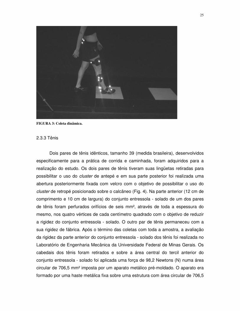
auto-selecionada eram coletadas com cada um dos pares de tênis. (Fig. 3)

24
FIGURA 1: Primeira coleta estática.
FIGURA 2: Segunda coleta estática.
25
FIGURA 3: Coleta dinâmica.
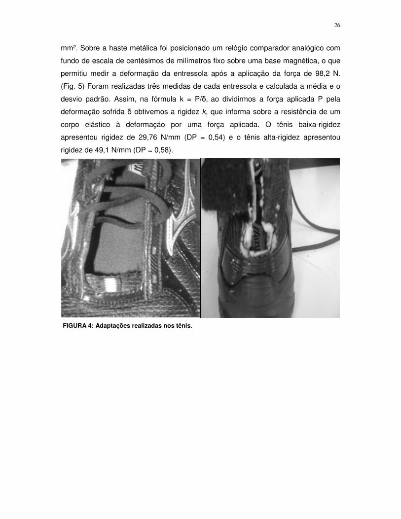
2.3.3 Tênis
Dois pares de tênis idênticos, tamanho 39 (medida brasileira), desenvolvidos
especificamente para a prática de corrida e caminhada, foram adquiridos para a
realização do estudo. Os dois pares de tênis tiveram suas lingüetas retiradas para
possibilitar o uso do cluster de antepé e em sua parte posterior foi realizada uma
abertura posteriormente fixada com velcro com o objetivo de possibilitar o uso do
cluster de retropé posicionado sobre o calcâneo (Fig. 4). Na parte anterior (12 cm de
comprimento e 10 cm de largura) do conjunto entressola - solado de um dos pares
de tênis foram perfurados orifícios de seis mm², através de toda a espessura do
mesmo, nos quatro vértices de cada centímetro quadrado com o objetivo de reduzir
a rigidez do conjunto entressola - solado. O outro par de tênis permaneceu com a
sua rigidez de fábrica. Após o término das coletas com toda a amostra, a avaliação
da rigidez da parte anterior do conjunto entressola - solado dos tênis foi realizada no
Laboratório de Engenharia Mecânica da Universidade Federal de Minas Gerais. Os
cabedais dos tênis foram retirados e sobre a área central do tercil anterior do
conjunto entressola - solado foi aplicada uma força de 98,2 Newtons (N) numa área
circular de 706,5 mm² imposta por um aparato metálico pré-moldado. O aparato era
formado por uma haste metálica fixa sobre uma estrutura com área circular de 706,5
26
mm². Sobre a haste metálica foi posicionado um relógio comparador analógico com
fundo de escala de centésimos de milímetros fixo sobre uma base magnética, o que
permitiu medir a deformação da entressola após a aplicação da força de 98,2 N.
(Fig. 5) Foram realizadas três medidas de cada entressola e calculada a média e o
desvio padrão. Assim, na fórmula k = P/δ, ao dividirmos a força aplicada P pela
deformação sofrida δ obtivemos a rigidez k, que informa sobre a resistência de um
corpo elástico à deformação por uma força aplicada. O tênis baixa-rigidez
apresentou rigidez de 29,76 N/mm (DP = 0,54) e o tênis alta-rigidez apresentou
rigidez de 49,1 N/mm (DP = 0,58).
FIGURA 4: Adaptações realizadas nos tênis.

27
FIGURA 5: Mensuração da rigidez da parte anterior da entressola.
2.4 Procedimentos
O estudo foi realizado no Laboratório de Análise de Movimento (LAM) do
Departamento de Fisioterapia da Escola de Educação Física, Fisioterapia e Terapia
Ocupacional da Universidade Federal de Minas Gerais (UFMG), Belo Horizonte -
MG. Os participantes foram instruídos acerca dos objetivos do estudo antes de
assinarem o termo de consentimento livre e esclarecido (APÊNDICE A). Em
seguida, foi aplicado (por um acadêmico de fisioterapia da UFMG previamente
treinado) o questionário com o objetivo de investigar sobre a presença de possíveis
critérios de exclusão.
A coleta dos dados foi realizada pelo pesquisador auxiliado por um acadêmico
de fisioterapia da UFMG. Inicialmente, foi realizado um estudo piloto com 10
indivíduos com o objetivo de avaliar a confiabilidade teste-reteste do examinador nas
medidas realizadas durante o estudo. O estudo piloto demonstrou que as medidas
apresentaram confiabilidade de moderada a excelente (APÊNDICE B).
Para a análise da marcha, o participante utilizou um short da cor preta
fornecido pelo pesquisador para permitir a visualização do membro inferior em teste.
Em seguida, foi sorteada a ordem de utilização do tênis para a realização da coleta e
foram afixados, nos pontos específicos, os marcadores anatômicos e de

28
rastreamento. Os marcadores dos clusters foram fixados em um neoprene e
colocados nos participantes com auxílio de um velcro. A colocação dos marcadores
anatômicos e de rastreamento foi feita pelo mesmo pesquisador em todas as coletas
dos participantes.
Antes de iniciar a coleta da marcha, o sistema Qualisys foi calibrado como
explicitado anteriormente na página 21 e em seguida foi realizada a primeira coleta
estática com o indivíduo descalço para possibilitar a posterior definição dos
segmentos tíbia, retropé e antepé. Os participantes foram então calçados com o
tênis sorteado para a primeira coleta e instruídos a caminharem ao longo da
passarela por um período de prática de cinco minutos com o tênis para a
familiarização com o mesmo e com os marcadores. Foi realizada a segunda coleta
estática com o indivíduo em posição ortostática para possibilitar a posterior definição
dos segmentos pelve, coxa, tíbia e complexo do pé. Cada participante, então,
deambulou pelo menos seis vezes pelos dez metros de extensão da passarela com
o primeiro tênis na sua velocidade auto-selecionada. Para iniciar a coleta, o
pesquisador dava o comando verbal “pode ir” ao mesmo tempo em que acionava o
computador do Qualisys
Após cada coleta, era verificado se todas as marcas de rastreamento haviam
sido visualizadas durante cem por cento da fase de apoio da marcha e então, os
dados eram armazenados para análise. Em seguida, foram repetidos os mesmos
procedimentos com o tênis sorteado para a segunda coleta. O tempo médio de
permanência de cada participante no LAM foi em torno de 60 minutos.
Após o término da coleta de dados de toda a amostra, foi realizada a
avaliação da rigidez da parte anterior da entressola do tênis no laboratório de
Engenharia Mecânica da Universidade Federal de Minas Gerais.
2.5 Redução dos dados
A aquisição dos dados cinemáticos foi realizada pelo software de aquisição
Qualisys Track Manager 1.9.2.(Gothenburg). Nessa etapa, foi realizada a
interpolação quando a trajetória dos marcadores foi perdida por, no máximo, 10
quadros. Foram excluídos os dados referentes a perdas superiores a 10 quadros e
as passadas que apresentaram algum artefato, como má qualidade do sinal.
29
Em seguida, os dados foram transferidos para o software Visual3D para
processamento, no qual foram construídos dois modelos biomecânicos para cada
indivíduo: o primeiro modelo baseado na coleta estática com o indivíduo descalço,
necessário para a definição do deslocamento angular dos segmentos do pé e o
segundo modelo baseado na coleta estática com o indivíduo calçado, necessário
para a definição do deslocamento angular de joelho e quadril e potência de
tornozelo, joelho e quadril. Os modelos foram construídos com base na posição das
marcas de referência capturadas durante as duas coletas estáticas e, em seguida,
esses modelos foram aplicados aos arquivos dinâmicos. Os dados da plataforma de
força foram então processados, delimitando-se o contato inicial e a retirada do pé ou
toe-off. Essas informações foram usadas para a normalização da fase de apoio da
marcha, de 0 a 100% (contato do pé até retirada do mesmo pé).
Os ângulos e a potência articulares foram calculados usando-se a sequência
de Cardan e definidos como a orientação do sistema de coordenadas de um
segmento em relação ao sistema de coordenadas do segmento de referência. Para
a normalização dos ângulos, o software calcula a orientação relativa do segmento e
do segmento referência, em relação à posição de referência (coleta estática) e aplica
essa orientação relativa dos dois segmentos às coletas dinâmicas.
Para os ângulos nos planos frontal e transverso de retropé e antepé/tíbia a
tíbia foi utilizada como segmento referência. Os ângulos no plano transverso do
quadril e joelho foram obtidos usando-se como segmento referência a pelve e a
coxa, respectivamente. As potências de quadril, joelho e tornozelo foram obtidas
usando-se como segmento referência a pelve, a coxa e a tíbia, respectivamente.
Em seguida, foi aplicado um filtro de passa baixa Butterworth de quarta ordem
com a frequência de corte estabelecida em seis Hz aos gráficos gerados para
diminuir os ruídos provenientes da movimentação de marcadores. Foram gerados os
gráficos da média dos deslocamentos angulares de retropé e antepé/tíbia nos planos
frontal e transverso de movimento e de quadril e joelho no plano transverso. Durante
a geração dos gráficos, foi utilizada a convenção de deslocamentos angulares
positivos para rotação interna de quadril e joelho, e adução e inversão de retropé e
antepé/tíbia.
Os dados cinéticos também foram exportados para o programa Visual 3D no
qual foram computados e gerados os gráficos de potência para quadril, joelho e
complexo tornozelo/pé no plano sagital. As potências articulares foram obtidas a

30
partir do produto das velocidades angulares com os momentos de força articulares.
(ENG, 1995).
2.6 Análise estatística
Os dados antropométricos dos participantes foram descritos por meio de
medidas de tendência central e de dispersão. Foram extraídas as ADMs de antepé e
retropé nos planos frontal e transverso. Para identificar diferenças entre as condições
foi aplicado o teste t pareado para dados com distribuição normal e teste de Wilcoxon
para dados que não possuem distribuição normal.
A Análise de Componentes Principais (ACP) foi realizada para sumarizar as
informações contidas nas 101 variáveis, correspondentes às 101 porcentagens do
ciclo da marcha, em um número pequeno de componentes. A ACP tem como objetivo
explicar a estrutura da variabilidade de um vetor aleatório de p-variáveis por meio de
combinações lineares feitas a partir dessas variáveis (KIRKWOOD, 2011). As
combinações lineares são chamadas de componentes principais, não correlacionadas
entre si e capazes de reter o máximo da informação contida nas p-variáveis originais.
Essa análise resulta em coeficientes, que são os pesos dados a cada variável para
cada um dos componentes, e nos escores, que são os valores resultantes da
multiplicação desses coeficientes pelo valor dos deslocamentos angulares e
potências de cada indivíduo, em cada plano de movimento, durante o ciclo da marcha
(KIRKWOOD, 2011). Os escores representam a distância que as ondas temporais de
cada indivíduo estão da média de uma determinada componente principal. A partir
dos escores encontrados, foram realizados testes t pareado para escores com
distribuição normal e teste de Wilcoxon nos casos em que a normalidade foi violada,
para testar a diferença de médias entre os dois níveis de rigidez dos tênis. Para a
escolha do número de componentes, foi considerado que 90% da variabilidade dos
dados deveriam ser explicados (KIRKWOOD, 2011). A interpretação das
componentes foi realizada por meio dos coeficientes e da comparação entre a média
das curvas dos indivíduos com escores altos e baixos, como sugerido por Deluzio e
Astephen (DELUZIO, 2007). Escores com valores altos apresentam uma onda mais
próxima do máximo descrito pela componente, e com valores baixos estão mais
próximos do mínimo descrito pela mesma componente (KIRKWOOD, 2011). Todas as
31
análises estatísticas foram desenvolvidas no software Minitab versão 14.0 e no SPSS
versão 15, com nível de confiança de 5%.
3 – REFERÊNCIAS

32
REFERÊNCIAS
AERTS, P.; DE C.D. Deformation characteristics of the heel region of the shod foot during a simulated heel strike: the effect of varying midsole hardness. J. Sports Sci., v.11, n. 5, p. 449-61, 1993.
ALEXANDER, R.M.; BENNET, M. How elastic is a running shoe? New Science, v. 15, p. 45-6, 1989.
ARNDT, A.; WESTBLAD, P.; EKENMAN, I.; LUNDBERG, A. A comparison of external plantar loading and in vivo local metatarsal deformation wearing two different military boots. Gait Posture, v. 18, n. 2, p. 20-6, 2003. BENNO, M.; NIGG, H.; BAHLSEN, A. Influence of Heel Flare and Midsole Construction on Pronation Supination and Impact Forces for Heel-Toe Running. Int. J. Sport Biomech. v. 4, p. 205-19, 1988. BERETTA, E.; CIMOLIN, V.; PICCININI, L.; CARLA, T.A.; GALBIATI, S.; CRIVELLINI, M.; et al. Assessment of gait recovery in children after Traumatic Brain Injury. Brain Inj, v. 23, n. 9, p. 751-9, 2009.
CAPPOZO, A.; CROCE, U.D.; LEARDINI, A.; CHIARI, L. Human Movement Analysis Using Stereophotogrammetry. Part 1: Theoretical Background. Gait Posture, v. 21, p. 186-96, 2005. CAPPOZZO, A. Gait Analysis Methodoly. Hum. Mov. Sci., v. 3, p. 27-50, 1984. CHEUNG, R.T.; NIGG, G.Y. Influence of different footwear on force of landing during running. Phys. Ther., v. 88, n. 5, p. 620-8, 2008. CIKAJLO, I.; MATJACIC, Z. The influence of boot stiffness on gait kinematics and kinetics during stance phase. Ergonomics, v. 50, n. 12, p. 2171-82, 2007. DELUZIO, K.J.; ASTEPHEN, J.L. Biomechanical features of gait waveform data associated with knee osteoarthritis: an application of principal component analysis. Gait Posture, v. 25, n. 1, p. 86-93, 2007.
33
DIXON, S.J.; WATERWORTH, C.; SMITH, C.V.; HOUSE, C.M. Biomechanical analysis of running in military boots with new and degraded insoles. Med. Sci. Sports. Exerc. v. 35, n. 3, p. 472-9, 2003. ENG, J.J.; WINTER, D.A. Kinetic analysis of the lower limbs during walking: what information can be gained from a three-dimensional model? J. Biomech., v. 28, n. 6, p. 753-8, 1995. GARD, S.A.; MIFF, S.C.; KUO, A.D. Comparison of kinematic and kinetic methods for computing the vertical motion of the body center of mass during walking. Hum. Mov. Sci., v. 22, n. 6, p. 597-610, 2004. KIRKWOOD, R.N.; RESENDE, R.A.; MAGALHÃES, C.M.B.; GOMES, H.A.; MINGOTI, S.A.; SAMPAIO, R.F. Application of principal component analysis on gait kinematics in elderly women with knee osteoarthritis. Rev. Bra. Fisioter., v. 15, n. 1, p. 52-58, 2011. LEWIS, C.L.; FERRIS, D.P. Walking with increased ankle pushoff decreases hip muscle moments. J. Biomech., v. 19, n. 41, p. 2082-9, 2008. MCKEAN, K.A.; LANDRY, S.C.; HUBLEY-KOZEY, C.L.; DUNBAR, M.J.; STANISH, W.D.; DELUZIO, K.J. Gender differences exist in osteoarthritic gait. Clin. Biomech. (Bristol, Avon), v. 22, n. 4, p. 400-9, 2007. MCMAHON, T.A. Biomechanics. The spring in the human foot. Nature, Jan., v. 8, n. 325, p. 108-9, 1987. MENDONÇA, L.D.M.; MACEDO, L.G.; FONSECA, S.T.; ET AL. Comparison of the anatomical alignment of lower limbs between healthy individuals and those with patellar tendinosis. Rev. Bras. Fisioter. 2005 9: 101-107. NADEAU, S.; GRAVEL, D.; ARSENAULT, A.B.; BOURBONNAIS, D. A mechanical model to study the relationship between gait speed and muscular strength. IEEE Trans. Rehabil. Eng. v. 4, n. 4, p. 386-94, 1996. NADEAU, S.; GRAVEL, D.; ARSENAULT, A.B.; BOURBONNAIS, D. Plantarflexor weakness as a limiting factor of gait speed in stroke subjects and the compensating role of hip flexors. Clin. Biomech. (Bristol, Avon), n. 14, n. 2, p. 125-35, 1999. NIGG, B.M.; BAHLSEN, H.A.; LUETHI, S.M.; STOKES, S. The influence of running velocity and midsole hardness on external impact forces in heel-toe running. J. Biomech, v. 20, n. 10, p. 951-9, 1987.
34
NIGG, B.M.; COLE, G.K.; NACHBAUER, W. Effects of arch height of the foot on angular motion of the lower extremities in running. J. Biomech., v. 26, p. 909-916, 1993. NIGG, B.M.; SEGESSER, B. Biomechanical and orthopedic concepts in sport shoe construction. Med. Sci. Sports Exerc., v. 24, n. 5, p. 595-602, 1992. PORTNEY, L.G.; WATKINS, M.P. Foundations of clinical research: Applications to practice. New Jersey: Prentice Hall Health, 3 ed, 2009. REQUIAO, L.F.; NADEAU, S.; MILOT, M.H.; GRAVEL, D.; BOURBONNAIS, D.; GAGNON, D. Quantification of level of effort at the plantarflexors and hip extensors and flexor muscles in healthy subjects walking at different cadences. J. Electromyogr. Kinesiol., v. 15, n. 4, p. 393-405, 2005. ROY, J.P.; STEFANYSHYN, D.J. Shoe midsole longitudinal bending stiffness and running economy, joint energy, and EMG. Med. Sci. Sports Exerc., v. 38, n. 3, p. 562-9, 2006. RUBIN, D.A.; BUTLER, R.J.; BECKMAN, B.; HACKNEY, A.C. Footwear and running cardio-respiratory responses. Int. J. Sports. Med., v. 30, n. 5, p. 379-82, 2009. SHORTEN, M.R. The energetics of running and running shoes. J. Biomech., v. 26, Suplemento, n. 1, p. 41-51, 1993. SOUZA, T.R.; PINTO, R.Z.; TREDE, R.G.; KIRKWOOD, R.N.; PERTENCE, A.E.; FONSECA, S.T. Late rearfoot eversion and lower-limb internal rotation caused by changes in the interaction between forefoot and support surface. J. Am. Podiatr. Med. Assoc., v. 99, n. 6, p. 503-511, 2009. STEFANYSHYN, D.J., NIGG, B.M. Influence of midsole bending stiffness on joint energy and jump height performance. Med. Sci. Sports Exerc., v. 32, n. 2, p. 471-476, 2000. STEFANYSHYN, D.J.; NIGG, B.M. Mechanical energy contribution of the metatarsophalangeal joint to running and sprinting. J. Biomech., v. 30, n. 11-12, p. 1081-5, 1997. TURNBALL, A. The race for a better running shoe. New Science, v. 15, p. 42-4, 1989.
35
WINTER, D.A. Energy generation and absorption at the ankle and knee during fast, natural, and slow cadences. Clin. Orthop. Relat. Res., n. 175, p. 147-54, 1983.
36
4 – ARTIGO

37
4 ARTIGO
The influence of shoe stiffness on foot motion and power at the knee joint
during gait.
ABSTRACT
Background: The compression stiffness of the midsole at the forefoot may influence the kinematics and the kinetics of the lower extremity. It is possible that a midsole with low stiffiness may increase the angular displacement of the foot segmentes and increase the power of the lower extremity. Method: Three-dimensional kinematics and kinetics data of the lower extremity of 37 healthy subjects, wearing two different levels of forefoot midsole shoe stiffness, were obtained during the stance phase of gait using the Qualisys ProReflex synchronized with a force platform. The dependent variables were: forefoot and rearfoot motion in the frontal and transverse planes; knee and hip motion in the transverse plane; sagittal power at the ankle, knee and hip joints. Data were analyzed by first extracting the total range of motion from the forefoot and rearfoot waveforms and then applying principal component analysis to extract the shape and magnitude differences from the waveforms between conditions. Findings: Low-stiffness condition had a higher range of motion in the transverse plane of the forefoot and rearfoot than the high-stiffness condition (α=0.05). The principal component analysis detected magnitude and waveform characteristics that were significantly different between conditions. Interpretation: The findings suggest the influence of the compression stiffness of the forefoot midsole on the kinematics of the segments of the foot and on the power generation at the knee joint during the stance of gait. The low-stiffness condition was associated with a delay in the generation of energy during late stance which may negatively influence the performance during gait. Key words: Shoes; gait; lower extremity; biomechanics.
_________________________________________________________________________
Autores: Renan A Resende, Renata N Kirkwood, Antônio E M Pertence, Sérgio T Fonseca, Paula L. Silva. Periódico: Clinical Biomechanics ISSN: 0268-0033
Endereço eletrônico: http://www.clinbiomech.com/ (ver ANEXO B)
38
1. Introduction
Walking is a common activity chosen by a large group of people as typical
physical exercise. In this context, significant efforts have been made toward
development of better footwear designed to prevent injuries and to improve
performance during physical exercise (Cikajlo and Matjacic, 2007). The focus has
been on efficient transmission of mechanical power produced by the musculoskeletal
system through the footwear and the return of elastic energy from the footwear to the
musculoskeletal system (Cikajlo and Matjacic, 2007). However, the results have
demonstrated the inability of the footwear to return elastic energy to the body
(Alexander and Bennet 1989;McMahon 1987;Turnball 1989). The reason is related to
the type of materials that cushion the footwear, which are usually of low quality as
energy returners (Shorten, 1993). Nigg and Segesser (1992) suggested that shoe
manufacturers should aim to reduce the dissipation of mechanical energy instead of
spending time improving energy return.
The compression stiffness of the shoe midsole on the forefoot may influence
the amount of energy dissipated during gait (Stefanyshyn and Nigg, 2000a). It is
especially important during the late stance phase, since part of the energy generated
by the musculoskeletal system during the push off phase, to accelerate the lower
limb forward, is lost due to the mechanical deformation of the shoe midsole.
Therefore, stiffening the shoe midsole of the forefoot could decrease the amount of
energy lost and improve lower limb progression.
Excessive foot motion, measured as rearfoot inversion/eversion, has been
associated with lower stiffness of the shoe midsole (Morio et al., 2009). A less rigid
midsole would fail to work as a lever arm system for the medial arch of the foot,
increasing rearfoot pronation past the mid-stance phase (Allen et al., 2004) and

39
maybe contributing to the occurrence of musculoskeletal injuries during gait (Gross et
al., 2007; Levinger and Gilleard, 2007; Mendonça et al., 2005; Viitasalo and Kvist,
1983). A rigid lever system for the medial arch of the foot could reduce foot pronation
and, consequently, decrease knee and hip internal rotation range of motion (Souza et
al., 2009). Therefore, a forefoot midsole with higher stiffness would have positive
effects on the kinematics of the foot complex, possibly preventing lower limb injuries.
The rearfoot midsole of most shoes might contribute to the impact absorption
at the calcaneus, reducing the reaction forces on the lower limb during heel strike.
However, it has been speculated that the compliance required for appropriate
cushioning at the rearfoot may not be appropriated for the demands of the forefoot
(Stefanyshyn and Nigg, 1997), since the foot should function as a rigid lever during
late stance. Most studies that investigated the influence of the stiffness of the midsole
were conducted with shoes composed of a single density midsole designed as hard
(dense) or soft (less dense), which are markedly different from current shoes which
are molded with anisotropic (multiple density) midsoles (Roy and Stefanyshyn, 2006;
Rubin et al., 2009; Stefanyshyn and Fusco, 2004; Stefanyshyn and Nigg, 2000b).
Therefore, the present study investigated two different forefoot midsole
stiffness, high- and low-stifness conditons, on the kinematics and power of the lower
extremity joints during the stance phase of gait. It is hypothesized that walking with a
less stiff shoe of the forefoot would increase total range of motion and the
mechanical power of the lower limb joints during gait. This study will contribute to a
preliminary understanding of the influence of low forefoot midsole stiffness on the
biomechanics of the lower extremity during gait
2. Methods
2.1. Participants

40
Thirty seven young healthy subjects (35 females, 2 males) participated in the
study. Inclusion criteria were as follow: age between 18 and 35 years and no history
of surgery or injuries in the lower limbs or lumbar-pelvic complex. The exclusion
criterion was the report of any discomfort during data collection. The participants
signed an informed consent, and this study was approved by the Ethics Research
Committee of the Universidade Federal de Minas Gerais, under the number: ETIC
0047.0.203.000-10.
2.2. Footwear
Two identical pairs of commercial shoes specially designed for walking, with a
midsole composed by ethylene vinyl acetate - EVA rubber, size 7.5, were used in the
study. The tongues of the shoes were removed to allow the placement of a cluster
with three passive markers over the forefoot. An opening was cut at the back part to
allow the placement of a rearfoot cluster with three markers over the calcaneus. The
opening was surrounded by velcro to close the back of the shoe to ensure firmness
during gait.
To decrease the stiffness of the forefoot midsole of one of the pairs of the
shoe referred as the low-stiffness condition, four 6 mm² holes were drilled in the
corners of each square centimeter of the forefoot (length of 12 cm, width of 10 cm).
The other pair of shoes remained with the manufacturer’s stiffness and is referred in
the present study as the high-stiffness condition.
The stiffness of each pair of forefoot midsole was determined by removing the
upper part of the shoes and applying, in the circular central area of 706.5 mm², a
force of 98.2 N. The force device consisted of a metal rod fixed on a structure with
negligible mass. Above the metal rod, a metric dial indicator was placed with an
analog full scale of hundredths of a millimeter fixed on a magnetic base. The

41
deformation of the midsole after the application of the force was measured by the dial
indicator. Three trials were performed on each midsole and the mean and standard
deviations of the trials were obtained. The compression stiffness of the forefoot
midsole was determined as the ratio of the applied force by the deformation of the
midsole, and it was measured in newtons per millimeter. The low-stiffness shoe had
a forefoot midsole mean value of 29.8 N/mm (SD = 0.54) and the high-stiffness shoe
a mean value of 49.1 N/mm (SD = 0.58). The compression stiffness of the forefoot
midsole of both low and high stiffness shoe conditions was measured after data
collection.
2.3. Instrumentation and experimental design
Three-dimensional kinematic and kinetic data of the lower extremity were
measured during the stance phase of gait with an eight-camera motion analysis
system (Qualisys ProReflex Medical AB, Gothenburg, Sweden) synchronized with
one force plate (AMTI – Advanced Mechanical Technology, OR6-6 model,
Watertown, MA, USA) placed in the center of a 10-meter walkway.
Anatomical markers and clusters of tracking markers were used to determine
the coordinate system of the segments and motion at the pelvis, thigh, shank,
rearfoot and forefoot (Souza et al., 2009), according to the recommendations for
minimizing soft tissue artefacts (Chiari et al., 2005; Manal et al., 2000; Schache et al.,
2008). A reference position for both conditions, considered the neutral position for the
foot segments and tibia, was obtained during a barefoot bipedal standing trial.
Anatomical markers over the foot were placed on the following locations: lateral and
medial malleolus, sustentaculum tali, peroneal tuberosities and at the base and
heads of the first and fifth metatarsals. The anatomical markers at the foot were used
to create an anterior directed Y-axis and an upwardly directed Z-axis for the forefoot
42
and for the rearfoot. Forefoot and rearfoot motions were measured with a rigid cluster
firmly attached to the top of the second metatarsal and another cluster attached
exclusively to the calcaneus.
For the knee and hip motion in the transverse plane and the power of the
ankle, knee and hip in the sagittal plane, two standing trials were performed, one for
each type of midsole (low-stiffness and high-stiffness). In each condition, the
reference position was obtained while standing with the given shoes. For dynamic
analysis of gait, the subjects walked on an 8-meter walkway (six trials) in their self-
selected speed. The order of data collection using the high or low-stiffness shoe was
randomized.
A pilot test was conducted with ten subjects to define the test-retest reliability
of the examiner on the measurements taken during the study. The results
demonstrated test-retest reliability from moderate to excellent with an average intra-
class correlation coefficient of 0.77 (SD 0.12).
2.4. Analysis and dependent measures
The software Visual 3D (C-Motion, Inc., Rockville, USA) was used to calculate
the following kinematic and kinetic variables: forefoot adduction-abduction (Z-axis)
and inversion-eversion (Y-axis) represented by the motion of the forefoot relative to
the shank – positive values for adduction and inversion, rearfoot adduction-abduction
(Z-axis) and inversion-eversion (Y-axis) represented by the motion of the rearfoot
relative to the shank – positive values for adduction and inversion, knee internal-
external rotation represented by the motion of the shank relative to the thigh –
positive values for internal rotation and hip internal-external rotation represented by
the motion of the thigh relative to the pelvis – positive values for internal rotation -
and power of the ankle, knee and hip joints in the sagittal plane. The mean values of

43
the six trials, for each subject, were considered for analysis. The stance phase was
determined by the contact and lack of contact of the foot on the force plate. Data
were collected at a frequency of 120 Hz and filtered with a Butterworth low-pass filter
with a cut-off frequency of 6 Hz (Winter, 2005). Kinematic and kinetic data were
interpolated to 101 data points, with each data point representing 1% of the stance
phase.
2.5. Statistical analysis
The normality of the data was tested using the Kolmogorov-Smirnov and the
Shapiro-Wilk tests. Paired Student’s t-test for parametric data and Wilcoxon Test for
non-parametric data were performed to detect differences between low and high-
stiffness conditions for the total ROM extracted from the forefoot and rearfoot in the
frontal and transverse planes.
2.6. Principal component analysis
Principal component analysis (PC) was applied to the gait waveforms for both
high and low-stiffness conditions (Deluzio and Astephen, 2007; Kirkwood et al.,
2011). An n by p matrix, 72 participants by 101 variables representing 0 to a100% of
the gait stance phase, was used in the analyses at each plane of motion. The matrix
was first transformed into a covariance matrix, which contains the variability of the
data. To maximize the variance, the mean for each variable was subtracted from the
corresponding variable value. Then, the PCs were extracted from the covariance
matrix through a method called diagonalization that realigns the data into a new
coordinate system. The new coordinate system are called principal components or
loading vectors and the maximum number of loading vectors is 101, or the number of
time points of the gait cycle. The components are ordered according to the amount of
variation explained; therefore, the first component accounts for the maximum
44
proportion of variation explained, followed by the subsequent PCs. A criteria of 90%
of variance explained was used to determine the number of PCs to retain for data
analysis (Deluzio and Astephen, 2007).
The principal components scores are represented by the individual elements
that compose the loading vectors. Therefore, the numbers of scores are equal to the
number of the sample size studied, and the scores represent the distance each
individual is from the mean of one specific component. The scores are important in
describing what these components of variation mean in terms of the characteristics of
specific groups or cases. The higher the score the more correlated the subject’s
waveform is with a specific PC. Therefore, further analysis can be performed to
determine differences between groups.
In the present study, the scores were analyzed for difference between stiffness
conditions using Paired Student’s t-tests for parametric data and Wilcoxon test for
non-parametric data, with Bonferroni correction. To interpret the components, two
waveforms were created based on the mean waveform +/- one standard deviation of
the PC scores times the loading vector for each PC (Ramsay and Silverman, 1997).
All the data was analyzed with a significance level of 0.05.
3. Results
3.1. Range of motion analysis
Data from one subject were discarded due to poor quality; therefore, thirty-six
(36) healthy young subjects, with an average age of 24.3 years (SD 2.9), mass and
height of 65.5 kg (SD 8.3) and 169 cm (SD 5.2), respectively, participated in the
study. The low- and high-stiffness condition had an average gait velocity of 1.31 m/s
(SD 0.15) and 1.32 m/s (SD 0.15), respectively, and this difference was not
statistically significant (p=0.35). The total ROM of the forefoot and the rearfoot in the
45
transverse plane (adduction/abduction), was significantly higher for the low-stiffness
condition across the stance phase of gait (Table 1). For the forefoot the difference
between conditions was 1.9º (SD 3.1), and 1.0º (SD 1.9) for the rearfoot. In the
frontal plane (inversion/eversion), the total ROM showed no statistically significant
difference in the forefoot and rearfoot between shoe conditions (Table 1).
3.2. Principal component analysis
The PC analysis demonstrated differences between conditions at the forefoot
and rearfoot in the frontal plane and at the power of the knee. The significant PC
scores between high and low stiffness midsole condition and their respective
interpretation are provided in Table 2. The waveform data for each gait measure
along with the PC loading vectors and its interpretation are shown on Figs. 1-3.
At the forefoot, PC1 captured the largest source of variation in the data
(85%), in a total of 91.2% explained by two PCs (Table 2 and Fig. 1). PC1 loading
vector had positive coefficients throughout the entire stance (Fig. 1B). As seen in
previous studies, this describes an overall magnitude feature in the data (Deluzio
and Astephen, 2007; Linley et al., 2010). Therefore, PC1 captured a measure of the
average angle throughout stance phase. Examination of gait waveforms
corresponding to the mean waveform and the high and low curves created based on
the mean waveform +/- one standard deviation of the PC1 score times the loading
vector for each PC1 are shown on Fig. 1C. The low and high curves support this
interpretation, showing that the low-stiffness condition increased the forefoot
inversion angle compared to the high stiffness shoe condition. The statistical
analysis of the PC1 scores revealed that the low-stiffness condition presented higher
forefoot inversion during the stance phase of the gait cycle compared to the high-
stiffness condition (p = 0.005, α=0.025).

46
A different magnitude in the rearfoot movement in the frontal plane was also
found (Table 2 and Fig. 2). Two PCs explained 94.9% of the variance and PC1
captured the largest source of variation in the data (89.4%). Only PC1 was
significant and showed positive values of approximately the same magnitude during
the stance phase of the gait cycle (Fig. 2B). The low-stiffness condition had a higher
rearfoot inversion during the stance phase of gait cycle than the high-stiffness
condition (p = 0.013, α=0.025). Examination of gait waveforms on Fig. 2C supports
this interpretation.
Power analysis at the knee joint in the sagittal plane was different between the
shoe conditions. Three PCs explained 93.1% of the variance but only PC2 was
significant and explained 9.1% of the variance. PC2 measured the timing and peak
of power during early stance relative to mid-stance and terminal stance relative to
pre-swing (p=0.010, α=0.017). The loading vector corresponding to PC2 revealed a
peak during terminal stance (Fig. 3B) and the gait high and low curve waveforms
created revealed that the low-stiffness condition had a delay in timing and a lower
positive peak power during late stance compared to the high-stiffness condition (Fig.
3C).
4. Discussion
The results of this study demonstrated that reducing forefoot midsole stiffness
increased forefoot and rearfoot total ROM in the transverse plane during the stance
phase of walking. In addition, PCI analyses demonstrated that the low-stiffness
condition increased the inversion angle at the forefoot and rearfoot when compared
to the high-stiffness condition during gait stance. The present study also identified a
delay in timing and a decrease in the magnitude of the knee power during late stance
during the low-stiffness midsole condition.

47
The increase in the total ROM of adduction/abduction in the transverse plane
of the forefoot may be attributed to the shoe’s lower forefoot midsole stiffness. As the
forefoot contacts the ground and the foot reaches full support during mid-stance, the
midsole influences the amount of movement of the foot segments, because it is
located between the foot and the ground. In the present study, the reduced forefoot
midsole stiffness offered less mechanical support to the forefoot adduction/abduction
movements, especially during the mid- and late stance, which contributed to the
increased total ROM. Since some gait injuries are often related to excessive motion
of the foot segments (Gross et al., 2007; Levinger and Gilleard, 2007; Mendonça et
al., 2005; Tiberio, 1988; Viitasalo and Kvist, 1983), the results of the present study
suggest that shoes with low-stiffness forefoot midsole may contribute to the
development of some lower extremities musculoskeletal injuries.
Similarly to the forefoot, the total rearfoot ROM in the transverse plane was
significantly greater during the low-stiffness condition. This kinematic change
suggests that distal factors, such as lower stiffness of the forefoot midsole, may
affect lower-extremity mechanics during walking (Souza et al., 2009). During the
stance phase of walking, the rearfoot normally moves from an initial adduction
position at initial contact toward abduction. At mid-stance the rearfoot motion
normally reverses and begins to adduct until toe off (Hunt and Smith, 2004). It has
been proposed that hypermobility of the first ray reduces the stiffness of the medial
arch of the foot and increases the amount of rearfoot motion (Allen et al., 2004). The
reduced forefoot midsole stiffness, seems to produce the same effect. Hence, without
a rigid support, rearfoot abduction may be prolonged past mid-stance. The results
suggested that the low-stiffness forefoot midsole condition might have contributed to
a less rigid foot structure during mid to late stance, which possibly contributed to the
48
observed increase in rearfoot total ROM in the transverse plane. Similarly, Hamill et
al. (1992) reported a significant decrease in maximum rearfoot eversion angles
during running with hard and medium midsole conditions compared with a soft
midsole. However, in the study by Hamill et al. (1992), the whole length of the
midsole stiffness was changed, while in the current study changes were made only at
the forefoot. This fact emphasizes the possible contribution of the forefoot midsole
stiffness to the occurrence of excessive rearfoot motion.
The magnitude of the inversion angle at the forefoot and rearfoot during the
stance phase of gait was also affected by the low-stiffness condition. This finding was
not in accordance with our expectations that the lower forefoot midsole stiffness
would increase the magnitude of eversion of the segments of the foot. However, the
greater inversion angles occurred at the initial contact phase of walking, when the
lower forefoot midsole stiffness could not influence the kinematics of the lower
extremity (as the forefoot had not contacted the ground). Possibly, other factors not
evaluated in the present study, such as the kinematics of the contralateral lower limb
and the line of progression of the body, influenced the increased magnitude of
inversion during the low-midsole stiffness condition.
The PCI analyses have shown a delay and a decrease in the positive power
peak at the knee joint during late stance in the low-stiffness condition. Based on
studies from muscle-based simulations, which demonstrated that the plantar flexors
produce nearly all the work in late stance, our findings are consistent with net joint
power analyses using inverse dynamics (Meinders et al., 1998; Winter, 1983).
According to Zajac and Gordon (1989), the force generated by the muscles not only
accelerates the segments and the joints it spans, but also the distal and proximal
segments of the body. The energy produced by the gastrocnemius muscle in late
49
stance is delivered to the lower limb to accelerate it forward (Zajac et al., 2003),
which is critical to leg progression (Robertson and Winter, 1980). One potential
influence of reducing the midsole stiffness is the change of the point of application of
the ground reaction force. Reducing the midsole stiffness may dislocate the centre of
pressure posteriorly (Stefanyshyn and Fusco, 2004). This would decrease the lever
arm of the ankle plantar flexors during late stance and delay and decrease the
positive power peak at the knee during late stance. This effect may overload the
plantar flexors because, during the push-off phase, most of the plantar flexors muscle
energy has been already generated and these muscles’ force-length relationship is
no longer optimal.
The observed kinematics and kinetic effects of forefoot midsole stiffness must
be interpreted with caution in light of possible inaccuracy of quantitative measures of
lower-extremity obtained with external markers, attributable to soft-tissues artefacts
(Leardini et al., 2007; Westblad et al., 2002). However, cluster configuration and
placement were carried out according to recent recommendations (Schache et al.,
2008).
Our results demonstrated that forefoot midsole with low-stiffness increased the
forefoot and rearfoot transverse motion and magnitude of inversion, and decreased
knee power generation and delayed knee positive power peak during late stance of
walking. One may argue that the loads imposed on the musculoskeletal tissues
during walking are smaller than those produced during running and that the isolated
kinematic and kinetic effects observed in the present study are not likely to increase
the chance for injuries. However, the occurrence of overuse injuries has a
multifactorial aetiology and other factors such as frequency and duration of the
activity and the individual’s capability to deal with mechanical stresses must be

50
addressed (Fonseca et al., 2007; Meeuwisse 1994). Walking is a motor task chosen
by a large group of people as a physical exercise, independent of their age. In this
context, people with different musculoskeletal individual capabilities are subject to
different intensity levels of physical exercise over a long period of time. The effect of
wearing a lower stiffness midsole shoe as observed in the present study thus,
combined with each individual’s characteristics, the intensity and duration of the
exercise may be sufficient to cause long-term injuries.
5. Conclusions
Our results demonstrated that low-stiffness of the forefoot midsole affected
both the kinematics and kinetics of the lower limb, increasing forefoot and rearfoot
transverse motion and decreasing knee power and delaying a peak positive power at
the knee in the late stance of gait. The lower stiffness of the midsole seems to offer
less mechanical resistance to the movements of the foot and influences the function
of the plantar flexors which alter the power at knee. A decrease and a delay of the
positive power at the knee during the late stance may interfere with leg progression
which can overload other muscle groups. Therefore, we suggest using shoes with
higher forefoot midsole stiffness when walking in order to favour the function of the
foot as a rigid lever during the late stance of gait.

51
References
Alexander, R.M., Bennet, M. 1989. How elastic is a running shoe? New Science, 15,
45-46
Allen, M.K., Cuddeford, T.J., Glasoe, W.M., DeKam, L.M., Lee, P.J., Wagner, K.J.,
Yack, H.J. 2004. Relationship between static mobility of the first ray and first ray,
midfoot, and hindfoot motion during gait. Foot Ankle Int., 25, (6) 391-396
Chiari, L., Della, C.U., Leardini, A., Cappozzo, A. 2005. Human movement analysis
using stereophotogrammetry. Part 2: instrumental errors. Gait Posture., 21, (2) 197-
211
Cikajlo, I., Matjacic, Z. 2007. The influence of boot stiffness on gait kinematics and
kinetics during stance phase. Ergonomics, 50, (12) 2171-2182
Davies, H., Zhaeentan, S., Tavakkolizadeh, A., Janes, G. 2009. Surgical repair of
chronic tears of the hip abductor mechanism. Hip. Int., 19, (4) 372-376
Deluzio, K.J., Astephen, J.L. 2007. Biomechanical features of gait waveform data
associated with knee osteoarthritis: an application of principal component analysis.
Gait Posture, 25, (1) 86-93
Fonseca, S. T., Ocarino, J. M., Silva, P. L. P. 2007, "Integration of Stresses and Their
Relationship to the Kinetic Chain," In Scientific Foundations and Principles of
Practice in Musculoskeletal Rehabilitation, Saunders Elsevier ed. vol. 1 D. J. Magee,
W. S. Zachazewsky, W. S. Quillem, eds., St. Louis: p. 476.
52
Gross, K.D., Niu, J., Zhang, Y.Q., Felson, D.T., McLennan, C., Hannan, M.T., Holt,
K.G., Hunter, D.J. 2007. Varus foot alignment and hip conditions in older adults.
Arthritis Rheum., 56, (9) 2993-2998
Hamill, J., Bates, B.T., Holt, K.G. 1992. Timing of lower extremity joint actions during
treadmill running. Med. Sci. Sports Exerc., 24, (7) 807-813
Hunt, A.E., Smith, R.M. 2004. Mechanics and control of the flat versus normal foot
during the stance phase of walking. Clin. Biomech.(Bristol., Avon.), 19, (4) 391-397
Jackson J.E. 1991. A user's guide to principal components New York: John Wiley
and Sons.
Keskin, D. 2008. Fibular collateral ligament-biceps femoris calcific bursitis causing
flexion contracture in the knee, external rotation in the leg, and equinus deformity in
the ankle. J. Manipulative Physiol. Ther., 31, (3) 247-250
Kirkwood, R. N., Resende, R. A., Magalhães, C. M. B., Gomes, H. A., Mingoti, S. A.,
Sampaio, R. F. 2011 Aplicação da análise de componentes principais na cinemática
da marcha de idosas com osteoartrite de joelho. Rev. Bra. Fisioter., 15, (1), 52-58.
Leardini, A., Benedetti, M.G., Berti, L. 2007. Rear-foot, mid-foot and fore-foot motion
during the stance phase of gait. Gait Posture., 25, 453
Levinger, P., Gilleard, W. 2007. Tibia and rearfoot motion and ground reaction forces
in subjects with patellofemoral pain syndrome during walking. Gait Posture., 25, (1)
2-8
53
Linley, H.S., Sled, E.A., Culham, E.G., Deluzio, K.J. 2010. A biomechanical analysis
of trunk and pelvis motion during gait in subjects with knee osteoarthritis compared to
control subjects. Clin. Biomech.(Bristol., Avon.), 25, (10) 1003-1010
Manal, K., McClay, I., Stanhope, S., Richards, J., Galinat, B. 2000. Comparison of
surface mounted markers and attachment methods in estimating tibial rotations
during walking: an in vivo study. Gait Posture., 11, (1) 38-45
McMahon, T.A. 1987. Biomechanics. The spring in the human foot. Nature, 325,
(7000) 108-109
Meeuwisse, W.H. 1994. Athletic injury etiology: distinguishing between interaction
and counfounding. Clin. J. Sports Med., 4, 171
Meinders, M., Gitter, A., Czerniecki, J.M. 1998. The role of ankle plantar flexor
muscle work during walking. Scand. J. Rehabil. Med., 30, (1) 39-46
Mendonça, L. D. M., Macedo, L. G., Fonseca, S. T. Comparison of the anatomical
alignment of lower limbs between healthy individuals and those with patellar
tendinosis. Rev. Bras. Fisioter. , 101. 2005.
Morio, C., Lake, M.J., Gueguen, N., Rao, G., Baly, L. 2009. The influence of footwear
on foot motion during walking and running. J. Biomech., 42, (13) 2081-2088
Nigg, B.M., Segesser, B. 1992. Biomechanical and orthopedic concepts in sport shoe
construction. Med. Sci. Sports Exerc., 24, (5) 595-602
Ramsay, J.O. and Silverman, B.W. 1997. Functional data analysis. Springer Series in
Statistics.

54
Robertson, D.G., Winter, D.A. 1980. Mechanical energy generation, absorption and
transfer amongst segments during walking. J. Biomech., 13, (10) 845-854
Roy, J.P. & Stefanyshyn, D.J. 2006. Shoe midsole longitudinal bending stiffness and
running economy, joint energy, and EMG. Med. Sci. Sports Exerc., 38, (3) 562-569
Rubin, D.A., Butler, R.J., Beckman, B., Hackney, A.C. 2009. Footwear and running
cardio-respiratory responses. Int. J. Sports Med., 30, (5) 379-382
Schache, A.G., Baker, R., Lamoreux, L.W. 2008. Influence of thigh cluster
configuration on the estimation of hip axial rotation. Gait Posture., 27, (1) 60-69
Shorten, M.R. 1993. The energetics of running and running shoes. J. Biomech., 26
Suppl 1, 41-51
Souza, T.R., Pinto, R.Z., Trede, R.G., Kirkwood, R.N., Pertence, A.E., Fonseca, S.T.
2009. Late rearfoot eversion and lower-limb internal rotation caused by changes in
the interaction between forefoot and support surface. J. Am. Podiatr. Med. Assoc.,
99, (6) 503-511
Stefanyshyn, D., Fusco, C. 2004. Increased shoe bending stiffness increases sprint
performance. Sports Biomech., 3, (1) 55-66
Stefanyshyn, D.J., Nigg, B.M. 1997. Mechanical energy contribution of the
metatarsophalangeal joint to running and sprinting. J. Biomech., 30, (11-12) 1081-
1085
Stefanyshyn, D.J., Nigg, B.M. 2000a. Energy aspects associated with sport shoes.
Sportverletz. Sportschaden, 14, (3) 82-89

55
Stefanyshyn, D.J., Nigg, B.M. 2000b. Influence of midsole bending stiffness on joint
energy and jump height performance. Med. Sci. Sports Exerc., 32, (2) 471-476
Tiberio, D. 1988. Pathomechanics of structural foot deformities. Phys. Ther., 68, (12)
1840-1849
Turnball, A. 1989. The race for a better running shoe. New Science, 15, 42-44
Viitasalo, J.T., Kvist, M. 1983. Some biomechanical aspects of the foot and ankle in
athletes with and without shin splints. Am. J. Sports Med., 11, (3) 125-130
Westblad, P., Hashimoto, T., Winson, I., Lundberg, A., Arndt, A. 2002. Differences in
ankle-joint complex motion during the stance phase of walking as measured by
superficial and bone-anchored markers. Foot Ankle Int., 23, (9) 856-863
Winter, D.A. 1983. Energy generation and absorption at the ankle and knee during
fast, natural, and slow cadences. Clin. Orthop. Relat Res. (175) 147-154
Winter, D.A. 2005. Biomechanics and Motor Control of Human Movement Wiley,
Hoboken.
Zajac, F.E., Neptune, R.R., Kautz, S.A. 2003. Biomechanics and muscle coordination
of human walking: part II: lessons from dynamical simulations and clinical
implications. Gait Posture, 17, (1) 1-17
56
Table 1: Total range of motion values in degrees (º) and comparisons between the two conditions.
Condition
Means (SD) Condition effect P-value High-stiffness Low-stiffness
Forefoot Frontal 11.3 º (4.8 º) 11.1 º (4.0 º) 0.57
Transverse* 16.2 º (5.2 º) 18.1 º (7.3 º) 0.017
Rearfoot Frontal 10.3 º (3.3 º) 10.9 º (3.8 º) 0.078
Transverse* 12.0 º (4.0 º) 13.0 º (4.6 º) 0.043
* Statistically significant difference between conditions are noted for α = 0.05. Frontal: inversion/eversion. Transverse: adduction/abduction.
57
Table 2: Interpretation of significant PCs Gait measure Interpretation of significant PCs Condition
effect P-value PC Interpretation
Forefoot
Kinematic - frontal plane PC1 Magnitude of inversion during
stance 0.005
Rearfoot
Kinematic – frontal plane PC1 Magnitude of inversion during
stance 0.013
Knee
Power - sagittal plane PC2 Differences in timing and peak
during late stance 0.010
58
Fig. 1. Analysis of forefoot inversion-eversion kinematic. (A) Average forefoot
kinematic in frontal plane waveforms for each condition: high stiffness in dotted lines
and low stiffness solid lines. (B) The loading vector for PC1 (first eigenvector). (C)
Frontal kinematic waveforms corresponding to high and low PC1 scores demonstrate
the difference in magnitude, captured by PC1.
59
Fig. 2. Analysis of rearfoot inversion-eversion kinematic. (A) Average rearfoot
kinematic in frontal plane waveforms for each condition: low stiffness in solid line and
high stiffness grey line. (B) The loading vector for PC1 (first eigenvector). (C) Frontal
kinematic waveforms corresponding to high and low PC1 scores demonstrate the
difference in magnitude, captured by PC1.
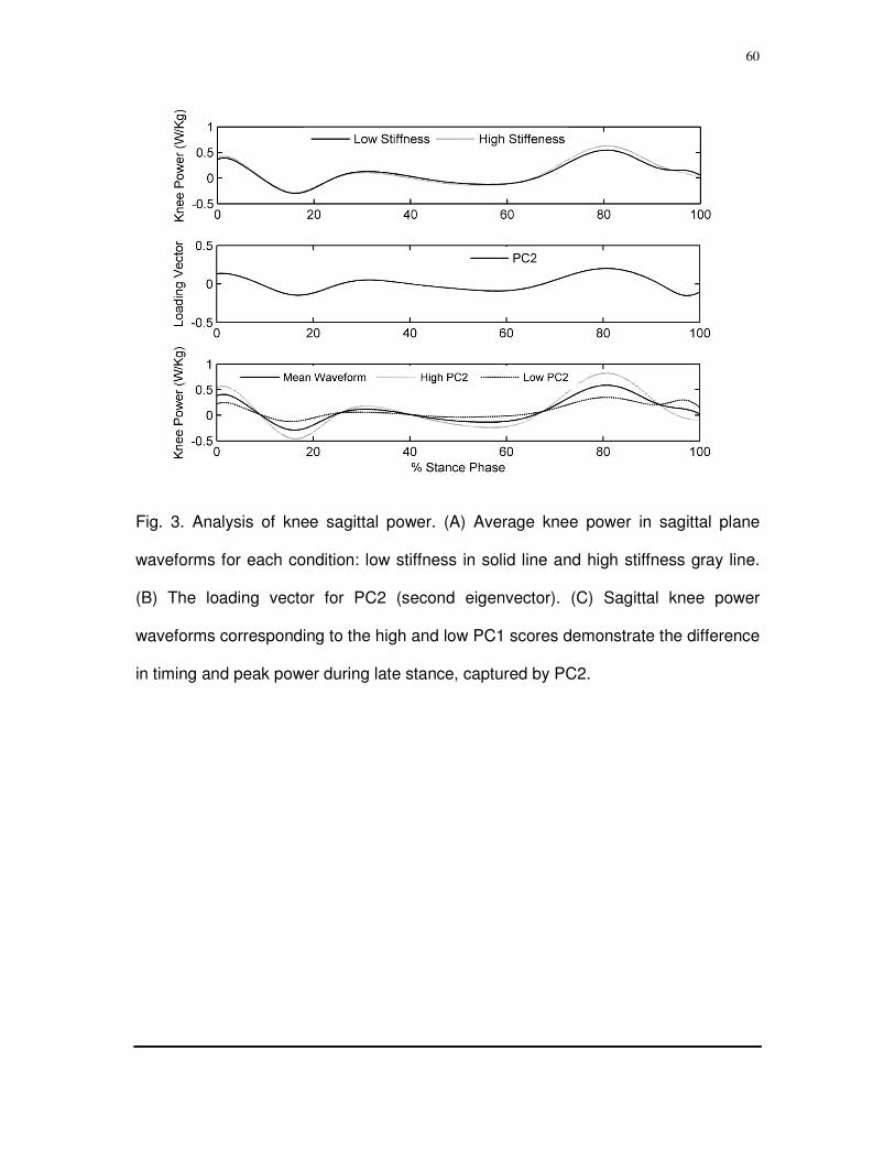
60
Fig. 3. Analysis of knee sagittal power. (A) Average knee power in sagittal plane
waveforms for each condition: low stiffness in solid line and high stiffness gray line.
(B) The loading vector for PC2 (second eigenvector). (C) Sagittal knee power
waveforms corresponding to the high and low PC1 scores demonstrate the difference
in timing and peak power during late stance, captured by PC2.
61
5 – CONSIDERAÇÕES FINAIS
62
5 CONSIDERAÇÕES FINAIS
O presente estudo investigou a influência da rigidez da parte anterior da
entressola do tênis nas variáveis cinemáticas (deslocamento angular nos planos
frontal e transverso de antepé e retropé e no plano transverso de joelho e quadril) e
cinéticas (potência das articulações do quadril, joelho e tornozelo no plano sagital)
dos membros inferiores durante a fase de apoio da marcha.
A análise dos dados referentes à ADM total dos segmentos do pé indicou
maior movimentação de antepé-tíbia e do retropé no plano transverso durante a
condição baixa-rigidez. Esses achados sugerem que a menor rigidez da parte
anterior da entressola ofereceu menor resistência aos movimentos de abdução e
adução de antepé-tíbia durante a fase de apoio da marcha, o que favoreceu o
aumento da ADM total durante a condição baixa-rigidez. O excesso de
movimentação dos segmentos do pé está associado a inúmeras lesões insidiosas de
membros inferiores, dessa forma, é possível que andar com um tênis com menor
rigidez na parte anterior da entressola possa favorecer o desenvolvimento de
algumas lesões de membros inferiores.
A ACP identificou maior magnitude de inversão dos segmentos do pé durante
a condição baixa-rigidez. Esse achado não foi de acordo com a nossa hipótese
inicial de que a baixa rigidez da parte anterior da entressola iriam aumentar a
magnitude de eversão dos segmentos do pé. No entanto, a maior magnitude de
inversão dos segmentos do pé durante a condição baixa-rigidez ocorreu desde a
fase de contato inicial, momento no qual a rigidez da parte anterior da entressola do
mesmo lado não influencia na cinemática do membro inferior pois o antepé ainda
não encontra-se em contato com o solo. Possivelmente, outros fatores não avaliados
no presente estudo como a cinemática do membro inferior contra-lateral e a linhda
63
de progressão da marcha podem ter influenciado no aumento da magnitude de
inversão durante a condição baixa-rigidez. A maior magnitude de inversão,
principalmente durante a fase de contato inicial, está associada a uma maior
sobrecarga do compartimento lateral o que pode contribuir para o surgimento de
lesões nas estruturas constituintes do mesmo.
Nos dados referentes à potência articular foi identificado um atraso no pico de
potência positivo na articulação do joelho durante a fase final de apoio durante a
condição baixa-rigidez. É possível que a menor rigidez da parte anterior da
entressola tenha diminuído o braço de alavanca em relação ao solo da musculatura
flexora plantar, o que contribuiu para o atraso na geração de energia na articulação
do joelho. A geração de energia durante essa fase do ciclo da marcha é importante
para a aceleração de tíbia e fêmur à frente e o atraso na geração de energia durante
a mesma pode prejudicar a propulsão do membro inferior a fase final de apoio.
Este estudo demonstrou que a baixa rigidez da parte anterior da entressola do
tênis aumenta a ADM e a magnitude de inversão dos segmentos do pé e atrasa e
reduz a geração de energia na articulação do joelho durante a fase final de apoio da
marcha. Estudos futuros com mais de dois valores de rigidez da parte anterior da
entressola e que investiguem outras formas de exercício físico são necessários para
melhor esclarecer a relação entre a baixa rigidez da parte anterior da entressola do
tênis e a biomecânica dos membros inferiores.
64
APÊNDICE A
TERMO DE CONSENTIMENTO LIVRE E ESCALRECIDO Projeto de pesquisa: Influência dos tênis com parte anterior rígida e flexível nas variáveis cinéticas e cinemáticas dos membros inferiores durante a marcha. Pesquisadores: Renan Alves Resende Profa. Renata Noce Kirkwood (Orientadora - Responsável) Instituição: Universidade Federal de Minas Gerais Escola de Educação Física, Fisioterapia e Terapia Ocupacional Departamento de Fisioterapia Laboratório de Análise de Movimento (LAM) Endereço : Departamento de Fisioterapia – Av. Antônio Carlos, 6627 Cep: 31270-901• Belo Horizonte - MG Telefax: (31) 3409-4781
Objetivo da pesquisa: O objetivo desta pesquisa é avaliar a influência de tênis com parte anterior rígida ou flexível nas características cinemáticas e cinéticas dos membros inferiores durante a marcha.
Com as informações obtidas pretendemos obter informações acerca das repercussões de diferenças nas características do tênis sobre os membros inferiores.
Antes de autorizar sua participação neste Projeto de Pesquisa é necessário que você leia atentamente e compreenda as explicações sobre os procedimentos, a estimativa do tempo de sua permanência no estudo, seus direitos, assim como os benefícios, riscos e desconfortos da pesquisa.
Caso você aceite participar, irá submeter-se aos seguintes procedimentos:
1. Coleta dos seus dados demográficos (dados pessoais, idade, peso e altura) e responderá a um questionário sobre possível

65
histórico de lesões e/ou cirurgias nos membros inferiores. Após esta entrevista será submetido (a) a uma análise do seu andar.
2. Análise de seu andar é feita utilizando marcas de isopor revestidas por fita reflexiva em pontos específicos do seu corpo (instrumento não invasivo e indolor) e utilização de dois tipos diferentes de tênis. Após calçar o tênis e ser realizada a colocação das marcas, você andará sobre uma passarela feita de madeira, o que será filmado por 8 câmeras de vídeo. O tempo estimado de sua permanência no laboratório é de 90 minutos.
Riscos e desconfortos: A análise do andar apresenta um risco mínimo de quedas e será feita em condições de extrema segurança e por pessoas altamente treinadas. Você poderá cansar durante a análise do seu andar e caso isso ocorra poderá interromper a coleta para repouso. Privacidade: Para assegurar anonimato e confidencialidade das informações obtidas, você receberá um número de identificação ao entrar no estudo e seu nome nunca será revelado em nenhuma situação. Qualquer dúvida ou questionamento será atendido pelos responsáveis da pesquisa. Você será informado (a) sobre os resultados e andamento da pesquisa, mesmo que isso implique na retirada do seu consentimento. Quando os resultados desta pesquisa forem divulgados em qualquer evento ou revista científica, seus dados não serão identificados, pois serão apresentados sob forma de estatísticas. Benefícios: As medidas obtidas nesse estudo contribuirão para maior entendimento das características adequadas para a confecção de tênis feitos para a prática de atividade física, com melhor entendimento das possíveis alterações nas pernas decorrentes das características do tênis. Recusa ou abandono: Sua participação neste estudo é inteiramente voluntária e você é livre para se recusar a participar ou abandonar o estudo a qualquer momento, sem qualquer prejuízo. Você não terá nenhum gasto financeiro nem lhe será paga nenhuma remuneração. Depois de ter lido as informações acima, se for de sua vontade participar deste estudo, por favor, preencha o consentimento abaixo.
Para obter informações adicionais
Você receberá uma cópia deste termo onde consta o telefone e o endereço do pesquisador principal, podendo tirar suas dúvidas sobre o projeto e sua participação, agora ou a qualquer momento. Caso você

66
venha a sofrer uma reação adversa ou danos relacionados ao estudo, ou tenha mais perguntas sobre o estudo, por favor, ligue para Dra. Renata Noce Kirkwood, no telefone (31) 3409-4792 ou Fisioterapeuta Renan Alves Resende no telefone (31) 92073391/(31)34841117.
Se você tiver perguntas com relação a seus direitos como participante deste estudo clínico, você também poderá contatar o Comitê de Ética em Pesquisa (COEP) no endereço Av. Pres. Antonio Carlos, 6627, Unidade Administrativa II, 2o. andar, Sala 2005 - Belo Horizonte - MG - Telefax: 3409-4592 - e-mail: [email protected]
Consentimento
Declaro que li e entendi a informação contida acima, sendo que minhas dúvidas foram satisfatoriamente respondidas. Este formulário esta sendo assinado voluntariamente por mim, indicando meu consentimento em participar do estudo. Belo Horizonte de de 2009
Pesquisador
Participante

67
APÊNDICE B Tabela com os valores dos Coeficientes de Correlação Intra-Classe, para cada
variável dependente, obtidos a partir do estudo piloto.
Variável ICC (3, 5)
Potência de quadril (n=10) 0,65
Potência de joelho (n=10) 0,75
Potência de tornozelo (n=10) 0,91
Quadril cinemática (n=10) 0,63
Joelho cinemática (n=10) 0,95
Antepé cinemática (n=9) 0,70
Retropé cinemática (n=9) 0,82
SPSS versão 15.0
Intraclass correlation coefficient
Model: two-way mixed
Type: consistency
ICC (3,5)

68
ANEXO A
Aprovação do Comitê de Ética

69
ANEXO B
Normas para submissão de manuscrito no Clinical Biomechanics
Aims Clinical Biomechanics aims to strengthen the link between clinic and laboratory by publishing biomechanics research which helps to explain the causes of musculoskeletal disorders and provides knowledge contributing to improved management. Scope Clinical Biomechanics explores all facets of musculoskeletal biomechanics with an emphasis on clinical management. The role of basic and medical science is recognized in a clinical context. The readership of the journal closely reflects its contents, being a balance of scientists, engineers and clinicians. Authorship All authors should have made substantial contributions to all of the following: (1) the conception and design of the study, or acquisition of data, or analysis and interpretation of data, (2) drafting the article or revising it critically for important intellectual content, (3) final approval of the version to be submitted. Instructions for Authors Contributions falling into the following categories will be considered for publication and are accepted on the understanding that they have not been published previously, not are under consideration for publication in any other journal. Papers - scientific reports within the scope of the journal. The length should not normally exceed 4000 words with around six figures/tables (large data tables and multi-part figures are generally best placed in Supplementary Data - see below). Reports focused on validity/reliability of methods in the absence of an experimental application are not acceptable. Reports on model development should address a specific question of clinical interest or report a novelty not yet understood. Reports of implant tests should involve a clinical application, not solely a laboratory test. Submissions are screened by an editorial panel; if considered suitable for the journal, two or more peer reviewers will be allocated. Only a proportion of scientifically acceptable papers can be accepted for publication, so authors should be aware that submissions requiring extensive revisions are unlikely to be offered the opportunity to revise and resubmit. In cases where the original reviewers disagree, the editor may opt to obtain further opinion. Appeals can only be considered where the authors can identify an irregularity in the review process: it is not acceptable simply to state that the reviewers' concerns can be addressed. Authors are invited to submit to the journal online http://ees.elsevier.com/clbi/. You will be guided through the creation and uploading of the various files. Once the uploading is done, the system automatically generates an electronic (PDF) proof, which is then used for reviewing. All correspondence, including notification of the Editor's decision and requests for revisions, will be by e-mail. Enquiries about the suitability of potential articles should be sent to the Editor: Prof Kim Burton, Clinical Biomechanics, 30 Queen Street, Huddersfield HD1 2SP, UK Tel: +44(0)1484 535200; fax: +44(0)1484 435744; e-mail: [email protected] When submitting a paper you are expecting a number of colleagues to review your work. As a matter of courtesy you should ensure your manuscript is neatly presented as well as complying with the journal's requirements. Submissions will be returned immediately without review if they do not follow all these guidance notes. • English language; double spaced; single sided; page-numbered and line-numbered.
70
• A title page including name(s) of author(s), qualifications, institute and correspondence addresses should be provided. Also provide a word count for the abstract and the main text (excluding reference list), and give the number of Tables and Figures. • When compiling the author list for a manuscript, please list only those members of the team who have made a significant contribution to the work. To assist the Editor in accepting a list of more than five authors, a statement detailing the part played by each author must be included in the cover letter. The difficulties facing authors whose native language is not English is appreciated. Nevertheless, it is the authors' responsibility to ensure correct use of English (through a scientific translator or similar). It is also the responsibility of the author to check the manuscript carefully for errors prior to submission. The Journal has a list of topics used to classify papers. During the online submission process, authors must select as many as are relevant to their paper. These classifications are included in issue 1 of each volume, and as a PDF file on the Journal's homepage
http://www.elsevier.com/wps/find/journaldescription.cws_home/30397/description#description • An accompanying cover letter should include: (a)information on any duplicate publication elsewhere of any part of the work; (b)a statement of any commercial relationships which may lead to a conflict of interests; (c)a statement that the typescript has been read and agreed by all authors; (d) name, address and e-mail of the corresponding author. (e)a reference to any closely related paper you have previously published in Clinical Biomechanics. •The Abstract should start on a new page, and must be in structured format. The following section headings (in italics) should each start a new line: Background, Methods, Findings, Interpretation. Please give an idea of the effect size of the results of hypothesis tests rather than simply quoting the statistical significance. The interpretation paragraph should explain how the findings add to understanding of the topic and outline the clinical implications. Only universally accepted and understood abbreviations are allowed in the Abstract (e.g. CT, MR), but no specialties or author-defined abbreviations (e.g. OA, osteoarthritis; TKR, total knee replacement etc). References are not permitted. The abstract should not exceed 250 words in total. Keywords should be added for indexing. •The main text should be divided into appropriate headings, e.g. Introduction, Methods, Results, Discussion, Conclusions. Subheadings may also be used, and review papers may use other formats. The technical basis of new experiments should be fully detailed; previously used methods should also be described briefly, together with reference to previous publications. Statistical methods should be detailed where appropriate. Footnotes are not permitted. •Ensure all acronyms/abbreviations are defined at first use. The use of many abbreviations in the text makes reading difficult and tiring: keep to a minimum. For products ensure the source details are complete (company, city, country) [All US addresses must include USA]. •Authors must suggest two or more referees although the choice is left to the Editors. Please supply the address and e-mail address. Papers will be reviewed by at least two referees and their comments will be made known to the corresponding author. •In a separate file labelled "Conflict of Interest Statement" all authors must disclose any financial and personal relationships with other people or organizations that could inappropriately influence (bias) their work. Examples of potential conflicts of interest include employment, consultancies, stock ownership, honoraria, paid expert testimony, patent applications/registrations, and grants or other funding. •All sources of funding should be declared as an acknowledgement at the end of the text. Authors should declare the role of the study sponsors, if any, in the study design, in the collection, analysis and interpretation of data: in the writing of the manuscript; and in the decision to submit the manuscript for publication. If the study sponsors had no such involvement, the authors should so state. References: Must follow the Harvard style and should be listed alphabetically at the end of the text. Please consult an issue of the journal for the details of how references should be formatted. Text: All citations in the text should be referenced: 1. Single author - the author's name (without initials unless there is ambiguity) and the year of publication; 2. Two authors - both authors' names and the year of publication; 3. Three or more authors - first author's name followed by 'et al.' and the year of publication.
71
In-text citation styles: Citations may be made directly (or parenthically). Groups of references should be listed first alphabetically, then chronologically. Multiple citations to a single point are generally not required and can impact on readability: if unavoidable, they must come at the end of a sentence. References should be restricted to those that are retrievable through normal library sources. References to conference proceedings, internal reports and theses are only appropriate when they have been published and readily can be retrieved. Otherwise the reference should be in-text as (Author name, year, personal communication). Around 30 references is typical for original papers, though review papers will be more extensively referenced. Tables These must be provided as a separate file. Each table should begin on a separate page and should be numbered as Table 1, Table 2 etc., each with its fully explanatory title above the table with footnotes (if any) beneath. Vertical rules and shading should be avoided. Figures The final reproduction will be either single or double column; single column is preferred: please scale your originals accordingly. Ensure legibility of all components, and avoid excessive "white space". All figures to be referred to as Figure 1, Figure 2 etc. Legends to figures to be listed together on a separate page. Figures and Tables must be constructed and labelled in such a way that they may be understood without reference to the text. Scientific measurements Avoid the +/- symbol both in tables and text - use for example "mean xx (SD yy)". Ensure statistical abbreviations are in correct case and style (e.g., capital italic for P). Use n for number. SI units must be used. Conventions for abbreviations can be found in Units, Symbols and Abbreviations (available from the Royal Society of Medicine, www.rsmpress.co.uk ). Confidence intervals are preferred over just P values; their use is described in Statistics with Confidence (BMJ Books, 2000). Ethics Work on human beings that is submitted to Clinical Biomechanics should comply with the principles laid down in the Declaration of Helsinki; Recommendations guiding physicians in biomedical research involving human subjects. Adopted by the 18th World Medical Assembly, Helsinki, Finland, June 1964, amended by the 29th World Medical Assembly, Tokyo, Japan, October 1975, the 35th World Medical Assembly, Venice, Italy, October 1983, and the 41st World Medical Assembly, Hong Kong, September 1989. For all studies involving human or animal participants. The manuscript should contain a statement that the work has been approved by the appropriate ethical committees related to the institution(s) in which it was performed and that subjects gave informed consent to the work. Studies involving experiments with animals must state that their care was in accordance with institution guidelines. Patients' and volunteers' names, initials, and hospital numbers should not be used.


