
UNIVERSIDADE FEDERAL RURAL DE PERNAMBUCO
PRÓ-REITORIA DE PESQUISA E PÓS-GRADUAÇÃO
MESTRADO EM SANIDADE E REPRODUÇÃO DE RUMINANTES
FLÁVIA CAMILA SIQUEIRA PEREIRA RAMALHO
EFEITO DA COENZIMA Q10 NO MEIO DE FERTILIZAÇÃO IN
VITRO DE EMBRIÕES BOVINOS
GARANHUNS
2015

FLÁVIA CAMILA SIQUEIRA PEREIRA RAMALHO
EFEITO DA COENZIMA Q10 NO MEIO DE FERTILIZAÇÃO IN
VITRO DE EMBRIÕES BOVINOS
Dissertação apresentada ao Programa de Pós-
Graduação em Sanidade e Reprodução de
Ruminantes da Universidade Federal Rural de
Pernambuco, como requisito parcial para
obtenção do grau de mestre em Sanidade e
Reprodução de Ruminantes.
Orientador: Prof. Dr. Gustavo Ferrer Carneiro
Coorientador: André Mariano Batista
GARANHUNS
2015

FICHA CATALOGRÁFICA
Ficha Catalográfica
Setor de Processos Técnicos da Biblioteca Setorial UFRPE/UAG
CDD: 636.0829 26
1. Reprodução de bovinos 2. Melhoramento genético 3. Fertilização in vitro 4. Sêmem 5. Estudos quantitativos
I. Carneiro, Gustavo Ferrer
II. Título
R165e Ramalho, Flávia Camila Siqueira Pereira
Efeito da coenzima q10 no meio de fertilização in vitro
de embriões bovinos/Flávia Camila Siqueira P. Ramalho.-
Garanhuns, 2015
47fs.
Orientador: Gustavo Ferrer Carneiro
Dissertação (Mestrado em Sanidade e Reprodução de
Ruminantes) – Universidade Federal Rural de Pernambuco
– Unidade Acadêmica de Garanhuns, 2015. Inclui anexo e bibliografias

FLÁVIA CAMILA SIQUEIRA PEREIRA RAMALHO
EFEITO DA COENZIMA Q10 NO MEIO DE FERTILIZAÇÃO IN
VITRO DE EMBRIÕES BOVINOS
Dissertação apresentada ao Programa de Pós-
Graduação em Sanidade e Reprodução de
Ruminantes da Universidade Federal Rural de
Pernambuco, como requisito parcial para
obtenção do grau de mestre em Sanidade e
Reprodução de Ruminantes.
Aprovada em: _____/_____/_____
Prof. Dr. Gustavo Ferrer Carneiro
Presidente da Banca-Unidade Acadêmica de Garanhuns/UFRPE
Profa. Dra. Aurea Wischral
Universidade Federal Rural de Pernambuco/UFRPE
Dr. André Mariano Batista
Universidade Federal Rural de Pernambuco/UFRPE
GARANHUNS
2015

AGRADECIMENTOS
Inicio meus agradecimentos por DEUS, já que Ele colocou pessoas tão especiais
a meu lado, sem as quais certamente não teria dado conta!
A meus pais, Carlos e Flávia, meu infinito agradecimento. Sempre acreditaram
em minha capacidade, isso só me fortaleceu e me fez tentar, não ser A MELHOR, mas a
fazer o melhor de mim. Obrigada pelo amor incondicional!
A meu querido esposo, Thiago, por ser tão importante na minha vida. Sempre a
meu lado, me pondo para cima e me fazendo acreditar que posso mais que imagino.
Devido a seu companheirismo, amizade, paciência, compreensão, apoio, alegria e amor,
este trabalho pôde ser concretizado. Obrigada por ter feito do meu sonho o nosso sonho!
A minha filha, Júlia, que amo incondicionalmente e que foi tão presente no
desenvolvimento deste trabalho e que, agora, me inspira a querer ser mais que fui até
hoje!
Agradeço também a meus sogros, Adolfo e Silvia, pelo incentivo e apoio,
colocando-se sempre à disposição para me ajudar. Obrigada pelo carinho!
A meu orientador e co-orientador, sempre disponíveis e dispostos a ajudar.
Fizeram-me enxergar que existe mais que pesquisadores e resultados por trás de uma
dissertação. Vocês foram e são referências profissionais e pessoais para meu
crescimento.
A toda equipe do ANDROLAB, em especial a Lucinha pela paciência e ajuda
na realização do experimento. Obrigada pela força!
A meus colegas do mestrado, pelos momentos divididos juntos e tornaram mais
leve meu trabalho. Foi bom poder contar com vocês!
A todos os professores da pós – graduação que, com ensinamentos, orientações e
amizade, me ajudaram ativa ou passivamente neste projeto.
Ninguém vence sozinho... OBRIGADA A TODOS!

RESUMO
A produção de embriões in vitro (PIV) em bovinos tornou-se importante ferramenta
comercial nos programas de melhoramento genético do rebanho mundial como técnica
de multiplicação, sendo amplamente utilizada para esse fim. Entretanto a Fertilização in
vitro (FIV) provoca geração de espécies reativas de oxigênio que podem afetar a
viabilidade embrionária. A Coenzima Q10, um cofator de importância na cadeia de
transporte das mitocôndrias tem função antioxidante na membrana lipídica e foi
verificado uma correlação direta entre a Coenzima e os parâmetros normais de sêmen
tais como, densidade, motilidade, morfologia e o volume. O objetivo desse trabalho foi
avaliar o efeito da Coenzima Q10 na função espermática em FIV utilizando-se sêmen
convencional e sexado; e se a adição desse cofator pode melhorar a produção
embrionária in vitro de oócitos bovinos. No Experimento o meio de FIV foi
suplementado com 0 (grupo controle), 5 µM, 10 µM, 20 µM da Coenzima Q10. Os
Oócitos foram coletados de um abatedouro localizado a 20 minutos do laboratório. Foi
observado um efeito deletério da coenzima com diferença significativa nas taxas de
clivagem ou na produção de blastocisto (p<0,05) na concentração de 20 µM quando
comparado com os demais grupos tanto com sêmen sexado como convencional. Estes
resultados demonstram que a suplementação da Coenzima Q10 no meio FIV não altera
a função espermática, entretanto tem um efeito deletério a partir da concentração de 20
µM. Podemos ainda inferir que na concentração de 5 µM no meio FIV há uma
tendência na melhoria da produção embrionária.
Palavras-chaves: FIV, PIV, Antioxidante.

ABSTRACT
In vitro embryo production (IVP) in cattle became an important commercial tool in
genetic improvement programs of the world herd, being widely used for this purpose.
However in vitro fertilization (IVF) can cause generation of reactive oxygen species that
can affect embryo viability. Coenzyme Q10, an important cofactor in the transport chain
of mitochondria has antioxidant function on lipid membrane and it was proved a direct
correlation between the presence of Coenzyme Q10 and normal spermatozoa parameters
such as density, motility, morphology and volume. The objective of this study was to
evaluate the effect of Coenzyme Q10 on sperm function in IVF using conventional or
sexed semen; also if the addition of this cofactor can improve embryo IVP in bovine
oocytes. In experiment 1 was evaluated the effect of sperm function during incubation
periods of sexed and conventional semen samples. In experiment 2, IVF medium was
supplemented with 0 (control group), 5 µM, 10 µM, 20 µM of Coenzyme Q10. Bovine
oocytes were collected from a slaughterhouse located 20 minutes from the lab. It was
observed a negative effect of Coenzyme with significant differences in the rates of
cleavage or in the production of blastocyst (p< 0.05) at a concentration of 20 µM when
compared with all other groups with either sexed as conventional semen. These results
demonstrate that supplementation of the Coenzyme Q10 in the IVF medium, do not
alter spermatozoa function. We can also infer that there is a tendency to improve
embryo production in the concentration of 5 µM in IVF medium.
Keywords: IVF, IVP, Antioxidant.
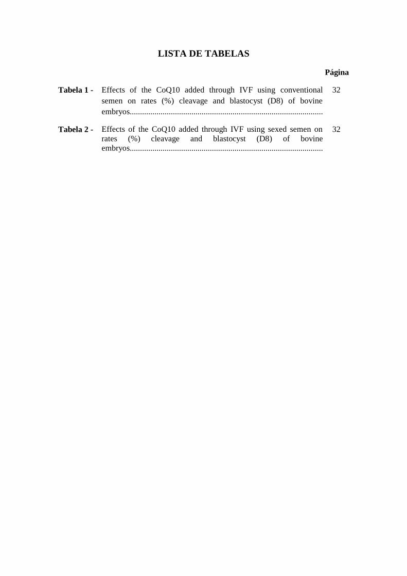
LISTA DE TABELAS
Página
Tabela 1 - Effects of the CoQ10 added through IVF using conventional
semen on rates (%) cleavage and blastocyst (D8) of bovine
embryos..............................................................................................
32
Tabela 2 - Effects of the CoQ10 added through IVF using sexed semen on
rates (%) cleavage and blastocyst (D8) of bovine
embryos..............................................................................................
32

LISTA DE ABREVIATURAS E SIGLAS
IA – Inseminação Artificial
FIV – Fertilização in vitro
TE – Transferência de Embrião
PIVE – Produção in vitro de Embrião
PIV – Produção in vitro
MIV – Maturação in vitro
CIV – Cultivo in vitro
FSH – Hormônio Folículo Estimulante
LH – Hormônio Luteinizante
ROS – Espécies Reativas de Oxigênio
Bi – Blastocisto inicial
BL – Blastocisto
BX – Blastocisto Expandido
BE – Blastocisto Eclodido
CoQ10 – Coenzima Q10

SUMÁRIO
Pág.
1.INTRODUÇÃO.................................................................................................. 10
2.OBJETIVOS........................................................................................................... 12
2.1. Objetivo Geral...................................................................................................... 12
2.2. Objetivos específicos............................................................................................ 12
3. REVISÃO DE LITERATURA.............................................................................
3.1 Produção in vitro de embriões bovinos.................................................................
3.1.1 Maturação in vitro (MIV)...................................................................................
3.1.2 Fertilização in viro (FIV)...................................................................................
3.1.3 Co-incubação (Estresse Oxidativo)....................................................................
3.1.4 Cultivo in vitro (CIV).........................................................................................
3.2 Coenzima Q10.......................................................................................................
12
12
14
14
15
17
17
4. REFERÊNCIAS....................................................................................................
5. ARTIGO CIENTÍFICO........................................................................................
6. CONSIDERAÇÕES FINAIS................................................................................
7.ANEXOS..................................................................................................................
20
27
37
38

10
1. INTRODUÇÃO
No atual contexto de evolução da produtividade na pecuária nacional, associado
às evoluções científicas e tecnológicas, várias biotecnologias ligadas à reprodução
animal vêm sendo desenvolvidas e aprimoradas com o intuito de aumentar a eficiência
reprodutiva, maximizando a produção de animais geneticamente superiores
(RENESTO e COELHO, 2004). Nesse sentido, especialmente no que se refere aos
ruminantes domésticos, biotecnias como a inseminação artificial (IA), fertilização in
vitro (FIV) e a transferência de embriões (TE) vêm sendo utilizadas com sucesso
(FIGUEIREDO et al., 2007).
O desenvolvimento de novas tecnologias e a melhora na eficiência na produção
de embriões nos últimos anos proporcionou uma tendência mundial de aumento na
produção de embriões. Mundialmente, o número total de embriões produzidos
(coletados in vivo e produzidos in vitro) entre 2008 e 2012 teve um aumento de 5,8%
(IETS, 2013) e entre 2012 e 2013 o aumento foi de 11,6% chegando ao total de
1.275.g874 de embriões (IETS, 2014).
Com relação aos embriões produzidos in vitro, a produção da América do Sul
corresponde a 72,7% da produção mundial, sendo o Brasil o maior produtor mundial
com 366.517 embriões produzidos, cerca de 70% do total mundial (IETS, 2014). O
grande destaque brasileiro na produção in vitro de embriões se deve, em grande parte,
ao tamanho (211.764.000 de bovinos segundo dados do IBGE em 2013) e as
características do rebanho nacional com base, principalmente, em raças zebuínas que
apresentam maior produção de oócitos por coleta. Vacas Nelores apresentam uma média
aproximada de 30 oócitos por sessão de OPU, e devido a grande variação individual
podem chegar até 128 oócitos viáveis coletados em um único animal (PONTES et al.,
2011).
A Coenzima Q10 (2,3-dimetoxi-5-metil-6-decaprenil-1,4-benzoquinona) é uma
provitamina lipossolúvel sintetizada endogenamente também conhecida como CoQ10
ou ubiquinona (COLLINS;KEMPER, 1999). A CoQ10 é encontrada em todas as células
do corpo humano, e as maiores concentrações são observadas nos tecidos do cérebro,
coração, fígado e músculo esquelético (SPINDLER; BEAL; HENCHCLIFFE, 2009).
Está localizada na membrana interna das mitocôndrias, onde realiza a interação com

11
enzimas complexas específicas, atuando como um cofator essencial na cadeia
respiratória mitocondrial (KUMAR, 2009). Essa coenzima possui a capacidade de
proteger fosfolipídeos, proteínas da membrana mitocondrial e o DNA dos danos
oxidativos (TOMASETTI, 1999).
A Coenzima Q10 foi encontrada na mitocôndria dos espermatozoides, sendo que
a sua biodisponibilidade vai condicionar a energia destas células (LEWINS & LAVON
cit. in Safarinejad, 2009). Também graças a certos estudos, foi verificado que existe
uma correlação direta entre a presença da Coenzima Q10 e os parâmetros do sêmen
como a densidade, mobilidade, morfologia e o volume (MANCINI et al. cit. in
Safarinejad, 2009).
Normalmente as avaliações laboratoriais realizadas com o objetivo de estimar o
potencial de fertilidade de uma partida de sêmen são: motilidade espermática (%);
vigor (1-5); concentração espermática (milhões/dose); anormalidades espermáticas (%)
e o teste de termo-resistencia (lento ou rápido). Estas avaliações vêm sendo utilizadas,
desde a década de 80, baseadas nas técnicas e padrões mínimos sugeridos pelo Colégio
Brasileiro de Reprodução Animal (CBRA, 1998) para a avaliação do sêmen de
bovinos, in natura ou criopreservado (ARRUDA et al., 1992). O desenvolvimento de
técnicas de associação de sondas fluorescentes, que permitam a avaliação simultânea da
integridade das membranas plasmática e acrossomal e da função mitocondrial em
sêmen bovino (CELEGHINI, 2004; CELEGHINI et al., 2007) tem servido como
ferramenta para avaliar os efeitos da criopreservação sobre o sêmen bovino.
Espera-se que com a utilização do antioxidante no meio FIV e
consequentemente a produção in vitro de embriões bovinos seja mais uma ferramenta a
ser utilizada para aumentar a qualidade dos embriões, acelerando o ganho genético e o
aumento do potencial de produção, com custos mais baixos e de mais fácil aplicação.
Esperamos com os resultados obtidos poder transferir conhecimentos, fortalecer a
cadeia produtiva e contribuir para o resgate social e geração de emprego e renda em
toda região Nordeste.

12
2. OBJETIVOS
Geral: determinar o efeito da suplementação com Coenzima Q10 no meio de FIV,
sobre a função espermática e a produção de embriões in vitro, utilizando sêmen
convencional e sexado.
Específicos:
Avaliar o efeito da Coenzima Q10 na taxa de clivagem e blastocisto dos
embriões produzidos in vitro;
Avaliar a qualidade seminal através da suplementação do meio FIV com a
Coenzima Q10.
3. REVISÃO DE LITERATURA
3.1 Produção in vitro de embriões bovinos
A produção in vitro de embriões (PIVE) expandiu-se nas diferentes espécies
animais pouco tempo após o nascimento de Louise Brown em 1978, na Inglaterra, o
primeiro bebê de proveta do mundo (STEPTOE e EDWARDS cit. in Varago et al.
2008). Em 1982, nasceu o primeiro bezerro bovino produzido por fecundação in vitro
nos Estados Unidos (BRACKETT cit. in Varago et al. 2008), o que só foi possível
graças às pesquisas iniciais desenvolvidas com animais de laboratório. A produção
comercial de embriões bovinos in vitro no Brasil teve início no ano de 1998 com um
projeto de inovação tecnológica financiado parcialmente pela Fundação de Apoio a
Pesquisa do Estado de São Paulo (FAPESP) e pelas Empresas Beabisa Agricultura Ltda
e Gertec Tecnologia de Embriões (GALLI et al., 2003).
A produção in vitro de embriões (PIVE) tem sido utilizada como base para
diferentes estudos relacionados à biotecnologia da reprodução, tanto em animais como
em humanos (PALMA & BREM, 1993). A PIVE em bovinos tornou-se importante
ferramenta comercial nos programas de melhoramento genético do rebanho mundial e
brasileiro como técnica de multiplicação, sendo amplamente utilizada para esse fim
(NEVES et al., 2010).

13
O aprimoramento dos sistemas envolvidos no processo de produção in vitro
(PIV) de embriões na espécie bovina a partir de oócitos recuperados de folículos de
ovários de vacas abatidas tem sido fundamentais para o estudo e compreensão de vários
fenômenos e mecanismos biológicos que ocorrem durante este período (HOSHI, 2003).
A obtenção de oócitos de animais vivos, mediante a técnica de laparoscopia e mais
recentemente, por meio da aspiração folicular transvaginal guiada por ultrassonografia,
tornou possível a aplicação da técnica de PIV in vivo visando aumentar o
aproveitamento do potencial genético das fêmeas consideradas superiores (NAGAI,
2001).
Para a produção in vitro de embriões, uma das técnicas mais utilizadas para a
recuperação de oócitos é a aspiração folicular transvaginal guiada por ultrassonografia
(RODRIGUES, 2000). Com o auxílio de um ultrassom e um transdutor acoplado a um
guia de aspiração, realiza-se a aspiração mediante introdução de uma agulha no interior
dos folículos ovarianos (AVELINO et al., 2002). Um sistema de bomba a vácuo permite
a recuperação dos oócitos e do líquido folicular para um tubo coletor (GALLI e
LAZZARI, 1996). Realiza-se em seguida a procura e seleção dos oócitos, em
microscópio estereoscópico, de acordo com o número de camadas de células do
cumulus e o aspecto do citoplasma do oócito (NAGAI, 2001). Os oócitos selecionados
são então transportados até o laboratório para que se tenha início o processo de
produção in vitro de embriões (SANGILD et al., 2000).
A aspiração folicular, em geral, não promove danos ao sistema reprodutor
feminino, embora em alguns casos já tenha sido relatada a predominância de tecido
conjuntivo no parênquima ovariano em algumas vacas doadoras que estavam sendo
submetidas a sessões de aspiração quinzenais (THOMPSON, 2000). Ao contrário da
transferência de embriões, a aspiração folicular é considerada uma técnica que apresenta
maior flexibilidade, uma vez que pode-se obter oócitos de fêmeas a partir dos 6 meses
de idade, vacas prenhes até o terceiro mês de gestação, vacas entre duas a três semanas
após o parto, pode-se usar oócitos de ovários de doadoras de várias idades e estado
fisiológico reprodutivo distintos (GALLI e LAZZARO, 1996; YOUNG et al., 1998). A
periodicidade pode variar desde a realização de sessões de aspiração de forma
esporádica em intervalos de duas semanas, durante várias semanas ou meses (NAGAI,
2001).
14
Outra vantagem da realização da aspiração folicular em comparação a TE está
ligado ao fato de que não é necessário o tratamento das doadoras com gonadotrofinas, o
que é considerado benéfico especialmente em novilhas jovens, pois a estimulação
hormonal pode provocar edema mamário e síndrome de ovário cístico (YOUNG et al.,
1998). Além da possibilidade de levar a casos de infertilidade, a superovulação, feita
repetidas vezes, pode provocar ainda relaxamento dos ligamentos do úbere (AVELINO
et al., 2002).
A PIVE envolve as etapas de colheitas de oócitos dos folículos ovarianos,
maturação in vitro (MIV) dos oócitos, fertilização in vitro (FIV) e cultivo in vitro (CIV)
de zigotos e estruturas embrionárias (HOSHI,2003).
3.1.1 Maturação in vitro (MIV)
A maturação in vitro (MIV) é uma técnica reprodutiva que permite que os
oócitos atinjam a metáfase II em condição laboratorial e adquiram a competência para
serem fecundados e, assim, iniciem a embriogênese (SANTL et al., 1998). Além da
maturação nuclear e citoplasmática, foi constatado que as células foliculares, ou seja, as
células da granulosa e do "cumulus oophorus " têm um papel importante durante a
aquisição da competência oocitária na MIV (STAIGNILLER E MOOR, 1984).
A grande maioria dos laboratórios tem utilizado o Tissue Culture Medium (TCM
199) como meio de MIV para oócitos bovinos, sendo geralmente adicionado de soro
fetal bovino, hormônio folículo estimulante (FSH), hormônio luteinizante (LH),
piruvato, lactato, aminoácidos, bicarbonato de sódio, vitaminas, antibiótico e/ou outros
fatores (GUIXUE et al., 2001; GONÇALVES et al., 2007). A MIV é realizada em
estufa incubadora com temperatura de 38° C, atmosfera a 5% de CO2 em ar e umidade
saturada e também comumente realizada utilizando meio coberto por óleo mineral para
evitar a evaporação (EPPIG, 2001).
3.1.2 Fertilização in vitro (FIV)
A FIV é a união do espermatozoide com o oócito no laboratório, formando o
embrião que posteriormente será transferido para cavidade uterina (MARTINS, 2007).
Essa etapa depende da qualidade dos oócitos e dos espermatozoides utilizados
(CARVALHO NETO, 2009). Deve ser proporcionado um ambiente adequado para

15
permitir o metabolismo dos oócitos e células do cumulus, mantendo a função
espermática eficiente e para essa finalidade o meio mais utilizado é o FERT-TALP
contendo heparina para capacitação espermática, albumina sérica bovina, além das
fontes de energia lactato e piruvato (RENESTO e COELHO, 2004; MINGOTI et al.,
2002).
A utilização do sêmen sexado na PIV de embriões permite reduzir o tempo para
atingir certos objetivos, por exemplo, o de melhorar a qualidade do rebanho e o número
de animais que o integram, produzindo uma proporção ideal de machos e fêmeas. É
esperada a elevação do ganho genético em até 15% comparado ao sêmen convencional
(TANNO, 2009).
Na maioria dos laboratórios, o processo de FIV em bovinos usa-se sêmen
congelado. No entanto, após o descongelamento, é necessário selecionar os
espermatozoides vivos e capazes de fecundar, onde é realizada esta seleção na maioria
das vezes pela separação em gradiente de Percoll, embora outros sistemas possam ser
utilizados como o "swin-up" ou lavado espermático (GALLI e LAZZARI, 1996).
O Co-cultivo dos espermatozoides com oócitos é realizado por um período que,
dependendo do laboratório, pode variar de 18 a 22 horas, a uma temperatura de 38° C,
em uma atmosfera de 5% de CO2 em ar e umidade saturada (GONÇALVES et al.,
2007).
3.1.3 Co-incubação (Estresse Oxidativo)
O estresse oxidativo consiste num termo genérico dado à situação em que existe
desequilíbrio entre as espécies reativas de oxigênio e as substâncias antioxidantes, com
predominância das primeiras. Nos sistemas de cultivo in vitro, o estresse oxidativo
consiste numa das principais causas da baixa eficiência da maturação oocitária e
desenvolvimento embrionário em várias espécies (AGARWAL et al., 2005;
LUBERDA, 2005).
Segundo Pasqualotto et al. (2000) , o estresse oxidativo tem efeitos deletérios
sobre a fisiologia dos espermatozoides como a peroxidação lipídica, dano ao DNA e
também tem sido associado com a diminuição da motilidade espermática através da
alteração da fluidez da membrana (SALEH et al., 2002; WHATES et al., 2007). De
acordo com Aitken et al. (2010), esse estresse não só pode prejudicar a habilidade de

16
fertilização do espermatozoide, mas também sua competência para apoiar um
desenvolvimento normal do embrião.
As espécies reativas de oxigênio (ROS) são formadas a partir das reações de
redução do oxigênio (O2), e constituem parte dos radicais livres. Os principais ROS são
os radicais superóxido (O2- ) e hidroxil (OH), e o peróxido de hidrogênio (H2O2), que
correspondem a redução por um, três e dois elétrons, respectivamente (GUÉRIN et al.,
2001). A produção de radicais livres faz parte da fisiologia da célula, porém em excesso
podem causar danos as mesmas, levando ao estresse oxidativo.
A produção de ROS pode ter origem diretamente a partir de gametas e embriões,
ou através do ambiente, sendo que vários fatores podem contribuir para aumentar essa
produção, entre eles pode-se destacar, a alta concentração de oxigênio associada à
interferência da luz, o excesso de manipulação e à presença de espermatozoides
(GUÉRIN et al., 2001; WANG et al., 2002; LIVINGSTON et al., 2009). O período de
incubação dos gametas masculino e feminino pode desencadear um aumento na
produção de ROS pelos espermatozoides, prejudicando a formação dos pronúcleos
(ALVAREZ et al., 1996). Bedaiwy et al (2004) avaliando a produção e
desenvolvimento de embriões humanos in vitro, verificaram que, quando o tempo de
incubação é reduzido durante a fecundação, juntamente com a adição de vitaminas C e
E no meio FIV, a produção de embriões foi maior, e possivelmente devido à menor
produção de ROS.
Como proteção aos efeitos nocivos do excesso de metabólitos de oxigênio,
chamadas de defesas antioxidantes, são definidas como qualquer substância que, quando
presentes em baixas concentrações em relação ao substrato oxidável, retarda ou inibe a
oxidação deste (SIES, 1993). Os antioxidantes podem ser divididos em compostos não
enzimáticos e enzimáticos, o primeiro inclui compostos de baixo peso molecular
presentes na dieta como ácido ascórbico (vitamina C), tocoferol (vitamina E), selênio,
zinco, taurinas, hipotaurinas, caroteno, ácido lipóico, ubiquinonas (coenzima Q), entre
outros (GUÉRIN et al., 2001 ; NORDBERG & ARNÉR, 2001; MATOS et al., 2002). O
sistema antioxidante enzimático inclui as enzimas superóxido dismutase, catalase e o
sistema glutationa redutase / peroxidase (GUÉRIN et al., 2001; MATOS et al., 2002).
3.1.4 Cultivo in vitro (CIV)

17
O cultivo in vitro corresponde à etapa de desenvolvimento do oócito fertilizado
até o estágio de blastocisto (SANGILD et al., 2000). É durante este período de
desenvolvimento pré-implantação que ocorrem eventos como ativação do genoma
embrionário, processo de divisão celular, compactação dos blastômeros no estádio de
mórula, início da diferenciação embrionária com a formação da blastocele (HOSHI,
2003).
Após o tempo de fecundação, os prováveis zigotos são lavados e transferidos
para microgotas de meio de cultivo que é baseado nos fluidos do útero e do oviduto
durante o início da gestação, recobertas por óleo mineral, permanecendo nestas por um
período de 6 a 7 dias até os zigotos atingirem os estágios de blastocisto inicial (Bi),
Blastocisto (BL), Blastocisto expandido (BX) e Blastocisto eclodido (BE), onde serão
transferidos ou criopreservados (ANTONIOLLI, 2005).
Geralmente, é esperado que, após a MIV, aproximadamente 90% dos oócitos
submetidos à maturação atinjam a metáfase II com expulsão do primeiro corpúsculo
polar. Destes, 80% são fecundados e começam a se dividir, pelo menos até o estágio de
duas a quatro células. No entanto, apenas 25 a 40% destes embriões alcançam o estágio
de blastocisto à blastocisto expandido (BAVISTER et al., 1992; LONERGAN et al.,
2001; NEVES et al., 2010).
A CIV é realizada em estufa incubadora com temperatura de 38,5° C, atmosfera
a 5% de CO2 em ar e umidade saturada e também comumente realizada utilizando meio
coberto por óleo mineral para evitar a evaporação (WELLS et al., 1999).
3.2 Coenzima Q10
A coenzima Q10 (2,3-dimetoxi-5-metil-6-decaprenil-1,4-benzoquinona) também
conhecida por ubiquinona foi descoberta em 1957 por Fredrick Crane e a sua equipe, na
mitocôndria do coração de boi (KUMAR, 2009; PRAKASH et al., 2010). Essa
coenzima pertence a uma série de compostos homólogos que compartilham na sua
estrutura um anel benzoquinona, semelhante a uma vitamina, é lipossolúvel e um pó
cristalino na sua forma pura (BHAGAVAN & CHOPRA, 2007).
Ela pode ser obtida por duas vias: via exógena pela ingestão de alimentos na
dieta ou pela via endógena pelo ciclo do mevalonato (MASON, 2011). Os alimentos

18
que contêm essa coenzima são produto lácteos, cereais, ovos, frutos secos como nozes e
nos vegetais, principalmente espinafre e brócolis (LITTARRU & TIANO, 2010), carne,
aves domésticas e em peixes gordos como cavala e sardinhas (KUMAR, 2009). No
entanto, a dose da coenzima Q10 que se consegue obter com a ingestão de alimentos,
cerca de 2-5 mg/dia, nunca é suficiente para suprir as necessidades do organismo
(KUMAR, 2009), isto porque apenas 10% é absorvida lentamente no trato
gastrointestinal devido ao seu elevado peso molecular e à sua baixa solubilidade em
água (PEPE et al., 2007; SINGH et al., 2007). No ciclo do mevalonato tem como
substrato inicial a acetil-CoA e prossegue com a produção do mevalonato e outros
intermediários que, tem como produto final o colesterol, o dolicol e a coenzima Q10
(BETINGER et al., 2010).
A coenzima Q10 (CoQ10) é encontrada em todas as células do corpo humano, e as
maiores concentrações são observadas nos tecidos do cérebro, coração, fígado e
músculo esquelético (SPINDLER et al., 2009). Está localizada na membrana interna das
mitocôndrias, onde realiza a interação com enzimas complexas específicas, atuando
como um cofator essencial na cadeia respiratória mitocondrial (KUMAR, 2009). Essa
coenzima tem a capacidade de proteger fosfolipídios, proteínas da membrana
mitocondrial e o DNA dos danos oxidativos (TOMASETTI, 1999), além de apresentar a
capacidade de regenerar outros antioxidantes como o ácido ascórbico e o α-tocoferol
(KIM & PARK, 2010).
O interesse pela coenzima CoQ10 tem aumentado nos últimos anos,
principalmente devido a capacidade de transferir elétrons e atuar como antioxidante
(GRONEBERG, 2005; KUMAR et al., 2009). A CoQ10 exerce sua principal função
natural na mitocôndria, como parte da cadeia de transporte de elétrons (cadeia
respiratória), mas, está presente também em baixas concentrações no plasma e
membranas celulares onde funciona como um antioxidante, prevenindo a peroxidação
dos lipídios (NORDBERG & ARNÉR, 2001; MARCOFF & THOMPSON, 2007;
BENTINGER et al., 2010). De acordo Bentinger et al. (2010), a CoQ10 também se
encontra envolvida na regulação do crescimento e diferenciação celular. Apresenta,
igualmente efeitos anti-inflamatórios e promove a liberação de óxido nítrico ajudando
na disfunção endotelial.

19
Tem grande importância no tratamento de desordens mitocondriais e
neuromusculares, bom como nas doenças neurodegenerativas, doenças cardíacas
(CLEREN, 2008; KUMAR, 2009). Além de sua utilização nessas doenças, a CoQ10 é
também utilizada na eficácia do tratamento e melhoria da qualidade do sêmen de
homens com infertilidade idiopática (BALERCIA, 2009; LITTARRU & TIANO,
2010), em pacientes com câncer de mama (BAHAR, 2010) e mais recentemente, a sua
utilização no tratamento da enxaqueca (SUN-EDELSTEIN & MAUSKOP, 2011).
Segundo Safarinejad (2009), a coenzima Q10 foi encontrada na mitocôndria dos
espermatozoides de homens, sendo que a sua biodisponibilidade vai condicionar a
energia destas células, onde existe uma direta correlação entre a presença da CoQ10 e os
parâmetros do sêmen, como a densidade, a motilidade, a morfologia e o volume. Ainda
o mesmo autor considera que a melhoria dos parâmetros de sêmen verificados nos
estudos anteriores ocorre devido à ação antioxidante dessa coenzima, uma vez que esta
é capaz de equilibrar a quantidade de agentes antioxidantes e de espécies reativas de
oxigênio que, foram verificados como agentes etiológicos da infertilidade masculina.
Entretanto, há apenas escassas informações sobre o efeito desse antioxidante na
motilidade de espermatozoides de touros (BALERCIA et al., 2004).

20
4. REFERÊNCIAS
AGARWAL, A., GUPTA, S., SHARMA, R. K . Role of oxidative stress in female
reproduction. Reprod Biol Endocrinol, v. 3, p.1-21, 2005.
AITKEN, R. J.; BAKER, M. A.; LULIIS, G. N.; NIXON, B. New insights into sperm
physiology and pathology. Handb Exp Pharmacol, v. 198, p. 19-199, 2010.
ALVAREZ, J. G.; MINARETZIS, D.; BARRET, C. B. The sperm stress test: a novel
test that predicts pregnancy in assisted reproductive technologles. Fertil Steril, v. 65, p.
400-405, 1996.
ANTONIOLLI, C.B. Produção in vitro de embriões bovinos utilizando diferentes
condições de maturaçã oocitária. Dissertação (Mestrado). Arquivo da Faculdade de
Veterinária da UFRGS, 2005
ARRUDA, R.P.; BARNABE, V.H.; ALENCAR, M.M.; BARNABE, R.C. Avaliação
de sêmen congelado de bovinos. Provas lenta e rápida de termo-resistência: efeitos
sobre a fertilidade. Brazilian Journal of Veterinary Research and Animal Science,
v.29, n.1, p.131-7, 1992.
ANTONIOLLI, C. B. Produção in vitro de embriões bovinos utilizando diferentes
ondições de maturação oocitária. Tese (Mestrado). Arquivo da Faculdade de
Veterinária da Universidade Federal do Rio Grande do Sul. Porto Alegre, 2005.
AVELlNO, K.B.; VANTINI, E.; SENEDA, M.M. In vitro production of embryos of
cows with acquired infertility. Theriogenology, v.57, p.656, 2002.
BAHAR, M. Exogenous coenzyme Q10 modulates MMP-2 activity in MCF-7 cell line
as a breast cancer cellular model. Nutrition Journal, v. 62, p. 2-8, 2010.
BALERCIA, G.; MOSCA, F.; MANTERO, F.; BOSCARO, M.; MANCINI, A.;
RICCIARDO LAMONICA, G.; LITTARRU, G. Coenzyme Q10 supplementation in
infertile men with idiopathic asthenozoospermia: an open, uncontrolled pilot study.
Fertility and Sterility, v. 81,p. 93-98, 2004.

21
BALERCIA, G. Coenzyme Q10 treatment in infertile men with idiopathic
asthenozoospermia: a placebo- controlled, double-blind randomized trial. Fertility and
Sterility, v. 91, p. 1785-1792, 2009.
BAVISTER, B. D.; ROSE-HELLEKANT, T. A.; PINYOPUMMINTR, T.
Development of in vitro mature/in vitro fertilized bovine embryos into morulae and
blastocysts in defined culture media. Theriogenology, v.37, p.127-146, 1992.
BEDAIWY, M. A.; FALCONE, T.; MOHAMED, M. S. Differential growth of human
embryos in vitro: role of reactive oxygen species. Fertility and Sterility, v. 82, p. 593-
600, 2004.
BENTINGER, M.; TEKLE, M.; DALLNER, G. Coenzyme Q - Biosyntesis and
functions. Biochmical and Biophysical Research Communications, v. 396, p. 74-79,
2010.
BHAGAVA, H.; CHOPRA, R. Plasma coenzyme Q10 response to oral ingestion of
coenzyme Q10 formulation. Mithocondrion, v.7, p. 72-88, 2007.
CARVALHO NETO, J. O. Avaliação da qualidade do espermatozoide bovino
criopreservado após sexagem por citometria de fluxo e sua utilização na produção in
vitro de embriões. Dissertação (Mestrado). Arquivo da Faculdade de Medicina
Veterinária da Universidade de Brasília, 2009.
CBRA: MANUAL PARA EXAME ANDROLÓGICO E AVALIAÇÃO DO SÊMEN
ANIMAL. Belo Horizinte: Colégio Brasileiro de Reprodução Animal. 2. ed. 1998.
49 p.
CELEGHINI, E. C. C.; ARRUDA, R. P.; ANDRADE, A. F. C.; NASCIMENTO, J.;
RAPHAEL, C. F. Pratical Techniques for Bovine Sperm Simultaneous Fluorimetric
Assessment of Plasma, Acrosomal and Mitochondrial Membranes Reproduction in
Domestic Animals, 2007.
CELEGHINI, E.C.C.; ARRUDA, R.P.; ANDRADE, A.F.C.; RAPHAEL, C.F.;
NASCIMENTO, J. Simultaneous evaluation of the plasmatic, acrosomal, and
mitochondrial membranes in equine spermatozoa. In: INTERNACIONAL CONGRESS

22
OF ANIMAL REPRODUCTION, 15, 2004, p.511. Porto Seguro. Abstracts... Belo
Horizonte: Colégio Brasileiro de Reprodução Animal, 2004.
CLEREN, C. Therapeutics effects of coenzyme Q10 (CoQ10) and reduced CoQ10 in
the MPTP model of Parkinsonism. Journal of Neurochemistry, v. 104, p. 1613-1621,
2008.
COLLINS, C.; KEMPER, K. J. Coenzyme Q10 (CoQ10 or ubiquinona). The Longwood
Herbal Task Force and The Center for Holistic Pediatric Education and Research, 1999.
Disponível em: www.mcp.edu/herbal/default.htm. Acesso em: 21 de outubro de 2014.
EPPIG, J. J. Oocytes control of ovarian follicular developmental and function in
mammals. Reproduction, v. 122, p. 829-838, 2001.
FIGUEIREDO, J.R.; CELESTINO, J.J.H.; RODRIGUES, A.P.R.; SILVA, J.R.V.
Importância da biotécnica de MOIFOPA para o estudo da foliculogênese e produção in
vitro de embriões em larga escala. Revista Brasileira de Reproduçao Animal. v.31,
p.143-152, 2007.
GALLI, C. LAZZARI, G. Practical aspects of IVM/IVF in cattle. Journal
Reproduction Sience, v. 42, p. 371-379, 1996.
GALLI,C.; DUCHI, R.; CROTTI, G.; TURINI, P.; PONDERATO, N.; COLLEONI, S.;
LAGUTINA, I.; LAZZARI, G. Bovine embryo technologies. Theriogenology, v.59,
p.599-616, 2003.
GONÇALVES, P.B.D.; BARRETA, M. H.; SANDRI, L. R.; FERREIRA, R.;
ANTONIAZZI, A. Q. Produção in vitro de embriões bovinos: o estado da arte. Revista
Brasileira de Reprodução Animal, v. 31, p. 212-217, 2007.
GUÉRIN P.; MOUATASSIM, S.; MÉNÉZO, Y. Oxidative stress and protection against
reactive oxygen species in the pre-implantation embryo and its surroundings. Human
Reproduction Update, v. 7, p. 175-189, 2001.
GUIXUE, Z.; LUCIANO, A. M.; COENEN, K. The influence of cAMP before or
during bovine oocyte maturation on ombryonicndevelopmental competence.
Theriogenology, v. 55, p. 1733-1743, 2001.

23
GRONEBERG, D.A. Coenzyme Q10 affects expression of genes involved in cell
signalling, metabolism and transport in human CaCo-2 cells. The International
Journal of Biochemistry & Cell Biology, v. 37, p. 1208-1218, 2005.
HOSHI, H. In vitro production of bovine embryos and their application for embryo
tranfer. Theriogenology, v. 59, p. 675-685, 2003.
IETS. IETS 2013 Statistics and Data Retrieval Committee Report. Embryo Transfer
Newsletter, v. 31 (4) p. 24-, 2013.
IETS. 2013 Statistics of Embryo Collection and Transfer in Domestic Farm Animals.
Embryo Transfer Newsletter, v. 32 (4) p. 14-26, 2014 .
JAINUDEEN, M. R.; WAHID, H.; HAFEZ, E.S.E. Indução da ovulação, produção e
transferências de embriões. In: HAFEZ, E.S.E.; HAFEZ, B. Reprodução Animal, 7ª
ed., São Paulo: Manole, 2004. Cap.29, p. 409-434.
KIM, J.M.; PARK, E. Coenzyme Q10 Attenuated DMH-induced precancerous lesions
in SD rats. Journal of Nutritional Science and Vitaminology, v. 56, p. 139-144, 2010.
KUMAR, A. Role of coenzyme Q10 (CoQ10) in cardiac disease hypertension and
Meniere-like syndrome. Pharmacology & Therapeutics, v. 124, p. 259-268, 2009.
LITTARRU G.; TIANO, L. Clinical aspects of coenzyme Q10: An update. Nutrition,
v. 26, p. 250-254, 2010.
LIVINGSTON T, RICH K, MACKENZIE S, GODKIN JD. Glutathione content and
antioxidant enzyme expression of in vivo matured sheep oocytes. Anim Reprod Sci, v.
116, p. 265-273, 2009.
LONERGAN, P.; RIZOS, D.; WARD, F.; BOLAND, M. P. Factors influencing oocyte
and embryo quality in cattle. Reprod Nutr Dev, v.41, p.427-437, 2001.
LUBERDA, Z. The role of glutathione in mammalian gametes. Reprod Biol, v. 5, p. 5-
17, 2005.
PALMA, G. A.; BREM, G. Transferencia de Embriones y Biotecnología de la
Reproducción en la Especie Bovina. Editora Hemisfério Sur, p. 243-266, 1993.

24
PASQUALOTTO, F.; SHARMA, R.; NELSON, D. Relationship between oxidative
stress, semen characteristis, and clinical diagnosis in men undergoing infertily
investigation. Fert and Stert, v. 73, p. 459-464, 2000.
PEPE, S.; MARASCO, S.; HAAS, S.; SHEERAN, F.; KRUM, H. ROSENFELDT, F.
Coenzyme Q10 in cardiovascular disease. Mitochondrion, v. 7, p. 154-167, 2007.
PONTES, J. H. F.; MELO STERZA, F. A.; BASSO, A. C.; FERREIRA, C. R.;
SANCHES, B. V.; RUBIN, K. C.; SENEDA, M. M. Ovum pick up, in vitro embryo
production, and pregnancy rates from a large-scale commercial program using Nelore
cattle (Bos indicus) donors. Theriogenology, v. 75, p 1640-1646, 2011
PRAKASH, S.; SUNITHAN, J.; HANS, M. Role of coenzyme Q10 as an antioxidant
and bioenergizer in periodontal deseases. Indian Journal of Pharmacology, v. 42, p.
334-337, 2010.
MARCOFF, L.; THOMPSON, P.D. The role of coemzyme Q10 in statin-associated
myopathy. Journal of the American College of Cardiology, v. 49, p. 2231-2237,
2007.
MARTINS, J. R. A.; TAKADA, L.; ABRAHÃO, R. G.; FREITAS, C. P.; CALEGARI,
R. S. Aspiração folicular de oócitos de bezerras através de videoedoscopia: um
procedimento promissor pra maximizar a produção de embriões bovinos in vitro. Acta
Scientiae Veterinariae, v. 35, p. 1994-1194, 2007 (Resumo).
MASON, P. Potencial uses of coenzyme Q10. The Pharmaceutical Journal, v. 275, p.
379-382, 2011.
MATOS, D.G.; GAPARRINI, B.; PASQUALINI, S.R.; THOMPSON, J.G. Effect of
glutathione synthesis stimulation during in vitro maturation of ovine oocytes on embryo
development and intracellular peroxide content. Theriogenology, v. 57, p. 1443-1451,
2002.
MINGOTI, G. Z.; GARCIA, J. M.; ROSA-e-SILVA, A. A. M. Steroidogenesis in
cumulus cells of bovine cumulus-oocyte-complexes matured in vitro with BSA and
different concentrations of steroids. Animal Reproduction Science, v. 69, p. 175-186,
2002.

25
HOSHI, H. In vitro production of bovine embryos and their application for embryo
transfer. Theriogenology, v.59, p.675-685, 2003.
MINGOTI, G. Z.; GARCIA, J. M.; ROSA-E-SILVA, A. A. M. Steroidogenesis in
cumulus cells of bovine cumulus-oocytecomplexes matured in vitro with BSA and
different concentrations of steroids. Anim Reprod Sci, v.69, p.175- 186, 2002.
NAGAI, T. The improvement of in vitro maturations systems for bovine and pordne
oocytes. Theriogenology, v.55, p.1291-1301, 2001.
NEVES, J. P.; MIRANDA, K. L.; TORTORELLA, R. D. Progresso científico em
reprodução na primeira década do século XXI. Revista Brasileira de Zootecnia. V.
39, p. 418. Brasília -DF, 2010.
NORDBERG, J.; ARNÉR, E.S.J. Reactive oxygen species, antioxidants and the
mammalian thioredoxin system. Free Radical Biology & Medicine, v. 31, p. 1287-
1312, 2001.
RENESTO, A. COELHO, L. Aspiração folicular guiada por ultrassonografia e
superovulação n produção in vitro e in vivo de embriões bovinos. Dissertação
(Mestrado). Arquivo da Faculdade de Ciêncas Agrárias UNESP, 2004.
RODRIGUES, C.F.M.; GARCIA, J.M. Fecundação in vitro em bovinos: aplicação
comercial. Arq. Fac. Vet. UFRGS Supl., v.28, p.186-187, 2000.
SAFARINEJAD, M. Efficacy of Coenzyme Q10 on semen parameters, sperm function
and reproductive hormones in infertile men. The Journal of Urology, v. 182, p. 237-
248, 2009.
SALEH, R.; AGARWAL, A. Oxidative stress and male infertility: from research bench
to clinical practive. J Androl, v. 23, p. 737-753, 2002.
SANGILD, P.T.; SCHMIDT, M.; JACOBSEN, H. Blood chemistry, nutrient
metabolism, and organ weights in fetal and newborn calves derived from in vitro
produced bovine embryos. Biol. Reprod., v.62, p.1495-1504, 2000.

26
SANTL, B.; WENIGERKIND, H.; SCHERNTHANER, W.; MODL, J.; STOJKOVIC,
M.; PRELLE, K.; HOLTZ, W.; BREM, G.; WOLF, E. Comparison of ultrasound -
guided vs laparoscopic transvaginal ovum pick-up (OPU) in simmental heifers.
Theriogenology, v. 50, p. 89-100, 1998.
SIES H. Strategies of antioxidant defense - review. Eur J Biochem, v. 215, p. 213-219,
1993.
SINGH, U.; DEVARAJ, S.; JIALAL, I. Coenzyme Q10 Supplementation and Heart
Failure. Nutrition Reviews, v. 65, p. 286-293, 2007.
SPLINDER, M.; BEAL, M.F.; HENCHCLIFFE, C. Coenzyme Q10 effects in
neurodegenerative disease. Neuropsychiatric Disease and Treatment, v.5, p. 597-610,
2009.
STAIGNILLER, R. B.; MOOR, R. M. Effect cells on the maturation and developmental
competence of ovine occytes matured outside of the follicle. Gamete RES. v. 9, p. 221-
229, 1984.
STRINGFELLOW, D.A.; SEIDEL, S.M. Manual da Sociedade Internacional de
Transferencia de Embrioes. Sociedade Brasileira de Transferencia de Embrioes,
3.ed. 180p., 1998.
SUN-ELDELSTEIN, C.; MAUSKOP, A. Alternative headache treatments:
nutraceuticals, behavioral and physical treatments. The Journal of Head and Face
Pain, v. 51, p. 469-483, 2011.
TANNO, P.H. Estudo das alterações morfo funcionais de espermatoziodes bovinos
submetidos à sexagem por meio da técnica de citometria de fluxo. Tese (Mestrado).
Arquivo da Faculdade de Medicina Veterinária e Zootecnia da Universidade de São
Paulo, 2009.
TOMASETTI, M. Coenzyme Q10 enrichment decreases oxidative DNA damage in
human lymphocytes. Free Radical Biology & Medicine, v. 27, p. 1027-1032, 1999.
THOMPSON, J.G. In vitro culture and embryos metabolism of cattle and sheep
embryos – a decade of achievement. Anim Reprod Sci, v.60/61, p.263-275, 2000.

27
VARAGO, F.C.; MENDONÇA, L.F.; LAGARES, M.A. Produção in vitro de embriões
bovinos: estado da arte e perspectiva de uma técnica em constante evolução. Rev Bras
Reprod Anim, Belo Horizonte, v.32, n.2, p.100-109, 2008.
WANG, X., FALCONE, T., ATTARAN, M., GOLDBERG, J. M., AGARWAL, A.,
SHARMA, R. K. Vitamin C and Vitamin E supplementation reduce oxidative stress-
induced embryo toxicity and improve the blastocyst development rate. Fertil Steril,
v.78, p. 1272-1277, 2002.
WATHES, D. C.; ABAYASEKARA, D. R.; AITKEN, R. J. Polyunsaturated fatty acids
in male and female reproduction. Biol Reproduction, v. 77, p. 190-201, 2007.
WELLS, D.N.; MISICA, P.M.; TERVIT, H.R. Production of cloned calves following
nuclear transfer with cultured adult mural granulosa cells. Biology of Reproduction,
Champaign, v. 60, p. 996-1005, 1999.
YOUNG, L. E.; SINCLAIR, K. D.; WILMUT, I. Large offspring syndrome in cattle
and sheep. Rev Reprod, v.3, p.155-163, 1998.

27
________________
*Corresponding author. Tel.: +55 81 96896422;
E-mail address: [email protected] (F.C.S.P.Ramalho).
5. ARTIGO CIENTÍFICO
Effect of Coenzymne Q10 in IVF medium on sperm function and bovine in vitro
production.
F.C.S.P. Ramalhoa*
, B.B. Santanaa, T.A. Pereira
a, T.R. Vianna
a, A.M. Batista
b, M.M.P.
Guerrab, G.F. Carneiro
a
a* Garanhuns Academic Unity, Federal Rural University of Pernambuco, 55.292-270,
Garanhuns, PE, Brazil; bAndrology Laboratory, Veterinary Medicine Department, Federal
Rural University of Pernambuco, 52.171-900, Recife PE, Brazil;
Abstract
In vitro fertilization (IVF) generates free radical levels which may impair adequate
embryo viability. Coenzyme Q10
(CoQ10
) is an essential cofactor of electron transport chain
which plays a role as a lipophilic antioxidant component of the lipid membranes that surround
all cells and the various organelles such as microsomes and mitochondria. Also, CoQ10
is
found in the mitochondria of the sperm, and there is a direct correlation between its presence
and semen parameters such as density, motility, morphology and volume. The aim of this
study was to evaluate the effect of CoQ10
in sperm function in sexed and conventional semen,
and if supplementation on IVF media could improve in vitro embryo production. We
evaluated its effect on bull sperm function during IVF co-incubation time. We supplemented

28
IVF media culture with 0 (control), 5 µM (T1), 10 µM (T2), 20 µM (T3) using sexed and
conventional semen. Bovine oocytes were collected from a local abattoir 20 minutes from the
laboratory. There was a decrease in the proportion of oocytes that cleaved in T3 compared to
all groups in sexed and conventional semen. In the embryos developed to blastocyst stage in
the conventional and sexed semen the behavior was the same as the cleavage status, however
when we observed the sexed semen, there was a dose response with significant difference (P <
0,05) between all groups compared to T3 group. The results showed that supplementation of
IVF media with CoQ10 do not alter spermatozoa function as the we had cleavage and
blastocyst stage rate results in both conventional and sexed semen. The findings also indicate
that supplementation of IVF media with CoQ10 (5 and 10 µM) did not improve in vitro bovine
embryo production, hawever 20 µM CoQ10 could impair blastocyst development in vitro.
Keywords: bovine; IVF, IVP, CoenzymeQ10.
1. Introduction
In vitro embryo production is a complex procedure and a challenge for those who try to
achieve successful embryo development [1]. This is characterized for many processes and
culture media. Different culture media may have varying concentrations of ions and energy
substrates, cellular adjustments, and expenditure of energy may be necessary due to change of
osmolarity and/or pH. Also metabolic pathways of the embryo may be forced to adjust to
changing environment and the consequence of all this imbalance can generate oxidative stress
due to the amount of reactive oxygen species (ROS), which are also commonly known as free
radicals and may result in reduced developmental potential [2]. To reverse the oxidative stress
framework is needed to reduce the production of ROS's or increase the amount of antioxidants
available [3].
An excess of ROS affects sperm cell function and might play a negative role in male
fertility. CoQ10
may play a positive role in the sperm because of its antioxidant properties.
Demonstrated positive effects of CoQ10
in the treatment of asthenozoospermia due to its

29
antioxidant properties. CoQ10
levels increased in seminal plasma and in sperm cells after
treatment [4].
It is important to take into consideration sperm quality during the IVF process.
Integrity of sperm membranes is extremely important for maintenance of spermatozoa
fertilizing capacity. The plasma membrane ensures the maintenance of cellular homeostasis,
and it exerts a crucial role on sperm survival and preservation of its fertility potential [5].
Also, acrosome integrity is essential to oocyte fertilization process because is well known that
the acrosome reaction allows sperm penetration into the zona pellucida and oocyte plasma
membrane fusion [6]. Finally, mitochondrial membrane potential leads to ATP production,
which is vital to flagellar and sperm motility [7]. In this study, CoQ10
was dilute in Ethanol
(EtOH) and it was necessary to assess sperm viability after EtOH exposure.
The present research was conducted to determine the effect of CoQ10
in sperm function
analyzing the cleavage and blastocysts rate results after IVF in sexed and conventional semen
and if the supplementation of CoQ10
on IVF media could improve in vitro embryo production.
2. Materials and methods
In vitro embryo production
2.1. Coenzyme Q10 preparation
Coenzyme Q10 was purchased from Sigma Chemical Co (cat# C9538). It was diluted in
absolute ethanol and kept at - 20 °C until use. Coenzyme Q10 was used in 3 concentrations; 5
μM, 10μM and 20μM. In experiment, a culture well dish without CoQ10 was used as a control.
Because CoQ10 is lipophilic and practically insoluble in water, it needs to be diluted in alcohol
before being added to the culture medium. It was prepared a working solution of CoQ10 after
dissolving 1,726 mg in 2 mL of ethanol to achieve the solution of 1 mM. From this working
solution, 5, 10 and 20μL was added to 995, 990 and 980 µl of IVF medium to obtain a
concentration of 5, 10 and 20 μM, respectively.

30
2.2. Oocyte collection and in vitro maturation
Bovine oocytes were collected from a local abattoir 20 minutes from the laboratory and
immediately transported to the Laboratory in saline 0.9% NaCl, supplemented with antibiotics
at a temperature of 30 °C. Arriving at the lab, Cumulus–oocyte complexes (COC) were
aspirated from 2- to 8-mm follicles using an 18-gauge needle attached to a 20-ml syringe.
Good-quality oocytes having a corona of cells of at least four layers and a uniformly
granulated cytoplasm were selected and washed in holding medium (MM; TCM-199 with
Earle's salts, with 25 mM HEPES) and divided into two dishes containing four 70 µL droplets.
A total of 852 oocytes were cultured in this research: 107 oocytes were distributed into each
of four treatments.
2.3. In vitro fertilization
After IVM for 20-24 h, oocytes were divided into 2 dishes, one for sexed semen and
another for conventinal semen, containing four 70 µL droplets divided into a control group
(CG) containing only IVF medium, and the three droplets following the treatment groups (TG)
containing 5, 10 and 20 µM of Coenzyme Q10. For the IVF process, frozen bovine semen
obtained from Artificial Insemination Center (ALTA GENETICA®) was used. Semen
samples were sexed and conventional, at a concentration of 1 x 10⁶
sptz/mL. Straws were
thawed in a water bath (37 °C for 30 s). Spermatozoa were selected through a Percoll gradient
(90-45%) and washed by centrifugation 3000 g with SP-TALP medium. After sperm
capacitation in vitro, were co-incubated for 20-24 h in IVF-TALP [8].
2.4. embryo culture
After fertilization, zygotes were partially denuded and transferred the 60 µl microdrops
containing oviductal fluid synthetic medium (SOF). Culture was conducted in incubator at a
temperature of 38.5 °C, containing 5% CO2 in atmospheric air and 99% humidity, for seven

31
days. Every 48 hours, 50% of the medium was renewed. Cleavage rate was reviewed 48 hours
post fertilization according to the Stringfellow and Seidel [9].
2.5. Statistical analysis
All data were tested for normality and homogeneity of variance using Kolmogorov-
Smirnov tests and Bartlett. When necessary, data were transformed into square root. Data
were analyzed using one-way ANOVA for testing rates of cleavage and blastocyst production,
followed by multiple comparison test of Tukey-Kramer (GraphPad Instat, version 10.03,
2009). Data are presented as mean ± SD and P < 0.05 were considered significant.
3. Results
Semen did not show any difference in any parameter measured, however sexed semen
had a tendency to decrease quality in motility and increase mitochondrial potential compared
to conventional semen.
Cleavage or blastocyst rates were not affected by CoQ10 or solvent supplementation
and all groups were able to develop to blastocyst stage. No significant difference was seen in
the percentage of cleavage formation either with sexed or conventional semen, however there
was a decreased number in cleavage rate in both types of semen when 20 µM was added to
IVF medium but not significant. As for the blastocyst formation, behavior was similar as
cleavage with a decrease in both types of semen when concentration of CoQ10 increase from
10 µM with a significant difference (P < 0,05) in the conventional semen in the embryos that
were cultured in the presence of 20 µM CoQ10 compared to those in the other groups. The
percentage and number of embryos cleaving and reaching blastocyst stage, in control group
(without CoQ10) and media supplemented with CoQ10 are shown in Table 1 and Table 2.

32
Table 1. Effects of the CoQ10 added through IVF using conventional semen on rates (% )
cleavage and blastocyst (D8) of bovine embryos.
Values with different letters within the same column differ ( P <0.05 ).
Table 2. Effects of the CoQ10 added through IVF using sexed semen on rates (% )
cleavage and blastocyst (D8) of bovine embryos.
Values with different letters within the same column differ ( P <0.05 ).
4. Discussion
Deleterious effects of Reactive Oxygen Species (ROS) produced during gamete
coincubation on IVF processes has been described, which has a negative effect on embryo
quality [10, 11]. As a result, it has been hypothesized that antioxidant supplementation of
conventional fecundation media could avoid oxidative stress, thereby improving technique
efficiency and embryo quality. Coenzyme Q10 is a potent antioxidant molecule having a
protective effects on human sperm motility, DNA fragmentation, and lipid peroxidation [12].
Treatment Nº oocytes Cleavage, % ( mean ± SD ) Blastocysts , % ( mean ± SD)
Control 107 57,89 ± 18,52 29,38 ± 10,67a
5 µM CoQ10 107 64,12 ± 20,63 33,25 ± 14,66 a
10 µM CoQ10 107 71,09 ± 15,16 25,32 ± 7,07 a
20 µM CoQ10 107 54,62 ± 25,88 0,93 ± 2,07b
Treatment Nº oocytes Cleavage, % ( mean ± SD ) Blastocysts , % ( mean ± SD)
Control 107 77,96 ± 12,44 42,40 ± 18,95
5 µM CoQ10 107 78,27 ± 18,45 49,97 ± 16,29
10 µM CoQ10 107 64,49 ± 9,20 29,91 ± 13,20
20 µM CoQ10 107 59,87 ± 11,00 21,58 ± 10,45

33
It is described that physiological level of free radicals is needed for normal sperm-
oocyte interaction [13], implantation and early embryonic development [14] and this CoQ10
concentration (20 µM) possibly generated a complete depletion of ROS that impairs embryo
development. In other hand, a pro-oxidant action of CoQ10 has been described, which
stimulates formation of superoxide anion/hydrogen peroxide [15] and can affect the embryo
development.
This work check the effects of CoQ10 during IVF and the influence on the subsequent
embryo development because it has been hypothesized that CoQ10 present in reproductive
tissues contribute to development of IVM-IVF embryos [16].
The results of our study demonstrated that lower concentrations of CoQ10 (5 and 10
µM) did not improve either in vitro cleavage rate or blastocyst production. On the other hand,
our results demonstrated that CoQ10 (5 µM) slightly increases the proportion of embryos
developing to blastocyst stages compared to control and other groups. To the best of our
knowledge the effect of CoQ10 added to IVF media has not yet been reported in the literature.
However, these results are consistent with reported [17], which shows that the same CoQ10
concentrations, add to embryo culture medium, slightly increases the proportion of mouse
embryos developing to blastocyst stages after 48 hours and provides similar blastocyst rate
after 72 hours compared to control. Similar results also have been reported with other
antioxidants added to bovine IVF media [18].
In contrast, our results shown that 20 µM CoQ10 has a deleterious effect during IVF,
using conventional semen, because it subsequently damages embryo development. These
results contrast with findings that demonstrated that increasing concentrations of CoQ10 from
10 to 30 or 100 µM improve the rate of early cleavage and that significantly more blastocysts
hatched and expanded blastocysts had significantly more inner cell mass cells and
trophectoderm cells at 30 µM CoQ10 in bovine embryo cultures [16]. Although 100 μM CoQ10
was found to be optimum for early cleavage, it significantly reduced the blastocyst
development rate compared to 30 μM CoQ10. Similarly, adding 50 µM CoQ10 to in vitro
maturation medium improved oocyte developmental competence and seems to be a promising
candidate for improving in vitro production of bovine embryos under moderate harm stress
[19].

34
In summary, this study we have demonstrated that, whereas, the supplementation of
IVF medium with CoQ10 (5 and 10 µM) did not improve in vitro bovine embryo production,
20 µM CoQ10 could impairs blastocyst development in vitro.
Acknowledgments
The authors are grateful to Conselho Nacional de Desenvolvimento Científico e
Tecnológico (CNPq) due to part of this study was financed by REPENSA PROJECT
(PROJETO REPENSA/CNPq/562455/2010-8). and, as well as to CAPES for the scholarship.
References
[1] Besenfelder U, Havlicek V, Kuzmany A, Brem G (2010) Endoscopic approaches to
manage in vitro and in vivo embryo development: use of the bovine oviduct. Theriogenology
2010; 73: 768–776.
[2] Gandhi AP, Lane M, Gardner DK, Krisher RL. A single medium supports development of
bovines embryos throughout maturation, fertilization and culture. Human Reproduction 2000;
15: 395-401.
[3] Agarwal A, Gupta S, Sharma RK. Role of oxidative stress in female reproduction. Reprod
Biol Endocrinol 2005; 14: 3:28.
[4] Balercia G, Mosca F, Mantero F, Boscaro M, Mancini A, Ricciardo Lamonica, G, Littarru
G. Coenzyme Q10 supplementation in infertile men with idiopathic asthenozoospermia: an
open, uncontrolled pilot study. Fertility and Sterility 2004; 81: 93-98.
[5] Õura C.; Toshimori K. Ultrastructural studies on the fertilization of mammalian gametes.
Int Rev Citol 1990; 122: 105–151.
[6] Yanagimachi R. Mammalian fertilization. In: Knobil E.; Neill J. D. (eds) The Physiology
of Reproduction. Raven, New York; 1994 , p. 189–318.
[7] Flesch F. M.; Gadella B. M. Dynamics of the mammalian sperm membrane in the process
of fertilization. Biochim Biophys Acta 2000; 1469: 197–235.
[8] Adona, P.R,; Pires, P.R.L.; Quetglas, M.D.; Schwarz, K.R.L.; Leal, C.L.V. Prematuration
of bovine oocytes with butyrolactone I: Effects on meiosis progression, cytoskeleton,

35
organelle distribution and embryo development. Animal Reproduction Science, 2008; 108: 49-
65.
[9] Stringfellow, D.A.; Seidel, S.M. Manual of the International Embryo Transfer Society.
Savoy: IETS 1998; 3: 83-88.
[10] Nedambale TL, Du F, Xu J, Chaubal SA, Dinnyes A, Groen W, Faber D, Dobrinsky JR,
Yang X, Tian XC. Prolonging bovine sperm-oocyte incubation in modified medium 199
improves embryo development rate and the viability of vitrified blastocysts. Theriogenology
2006; 66:1951–1960.
[11] Enkhmaa D, Kasai T, Hoshi K. Long-time exposure of mouse embryos to the sperm
produces high levels of reactive oxygen species in culture medium and relates to poor embryo
development. Reprod Domest Anim 2009; 44: 634–637.
[12] Talevi R, Barbato V, Fiorentino I, Braun S, Longobardi S, Gualtieri R. Protective effects
of in vitro treatment with zinc, d-aspartate and coenzyme q10 on human sperm motility, lipid
peroxidation and DNA fragmentation. Reprod Biol Endocrinol 2013; 11: 72 - 81.
[13] de Lamirande E, Leclerc P, Gagnon C. Capacitation as a regulatory event that primes
spermatozoa for the acrosome reaction and fertilization. Mol Human Reprod 1997; 3: 175-
194.
[14] Sakkas, D., Urner, F., Bizzaro D., Manicardi G., Bianchi, P.G., Shoukir Y., Campana, A.
Sperm nuclear DNA damage and altered chromatin structure: effect on fertilization and
embryo development. Human Reprod 1998; 4: 11-19.
[15] Linnane, A.W., Kios M., Vitetta L. Coenzyme Q10 – Its role as a prooxidant in the
formation of superoxide anion/hydrogen peroxide and the regulation of the metabolome.
Mitochondrion 2007; 7: S51–S61.
[16] Stojkovic M, Westesen K, Zakhartchenko V, Stojkovic P, Boxhammer K, Wolf E.
Coenzyme Q(10) in submicron-sized dispersion improves development, hatching, cell
proliferation, and adenosine triphosphate content of in vitro-produced bovine embryos. Biol
Reprod 1999; 61:541–547.
[17] Nozemian Z. Infertility and Women's Age. Thesis of Master. University of Toronto,
2001.
[18] Cheuqueman C., Arias M. E., Risopatron J, Felmer R., Alvarez J., Mogas T, Sanchez R.

36
Supplementation of IVF medium with melatonin: effect on sperm functionality and in vitro
produced bovine embryos. Andrologia 2014; 20: 1111-1123.
[19] Gendelman M, Roth Z. Incorporation of coenzyme Q10 into bovine oocytes improves
mitochondrial features and alleviates the effects of summer thermal stress on developmental
competence. Biol Reprod. 2012; 16: 87 - 118.
37
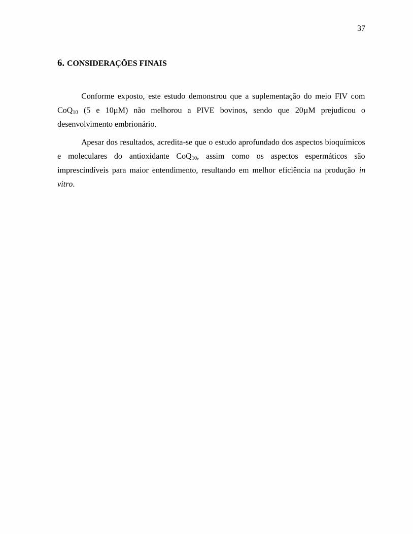
6. CONSIDERAÇÕES FINAIS
Conforme exposto, este estudo demonstrou que a suplementação do meio FIV com
CoQ10 (5 e 10µM) não melhorou a PIVE bovinos, sendo que 20µM prejudicou o
desenvolvimento embrionário.
Apesar dos resultados, acredita-se que o estudo aprofundado dos aspectos bioquímicos
e moleculares do antioxidante CoQ10, assim como os aspectos espermáticos são
imprescindíveis para maior entendimento, resultando em melhor eficiência na produção in
vitro.

38
7. ANEXOS - Normas da Revista Theriogenology
Article structure
Subdivision - numbered sections
Divide your article into clearly defined and numbered sections. Subsections should be
numbered 1.1 (then 1.1.1, 1.1.2, ...), 1.2, etc. (the abstract is not included in section
numbering). Use this numbering also for internal cross-referencing: do not just refer to 'the
text'. Any subsection may be given a brief heading. Each heading should appear on its own
separate line.
Introduction
State the objectives of the work and provide an adequate background, avoiding a detailed
literature survey or a summary of the results.
Material and methods
Provide sufficient detail to allow the work to be reproduced. Methods already published
should be indicated by a reference: only relevant modifications should be described.
Results
Results should be clear and concise.
Discussion
This should explore the significance of the results of the work, not repeat them. A combined
Results and Discussion section is often appropriate. Avoid extensive citations and discussion
of published literature.
Conclusions
The main conclusions of the study may be presented in a short Conclusions section, which
may stand alone or form a subsection of a Discussion or Results and Discussion section.
Essential title page information

39
• Title. Concise and informative. Titles are often used in information-retrieval systems. Avoid
abbreviations and formulae where possible.
• Author names and affiliations. Please clearly indicate the given name(s) and family
name(s) of each author and check that all names are accurately spelled. Present the authors'
affiliation addresses (where the actual work was done) below the names. Indicate all
affiliations with a lower-case superscript letter immediately after the author's name and in
front of the appropriate address. Provide the full postal address of each affiliation, including
the country name and, if available, the e-mail address of each author.
• Corresponding author. Clearly indicate who will handle correspondence at all stages of
refereeing and publication, also post-publication. Ensure that the e-mail address is given
and that contact details are kept up to date by the corresponding author.
• Present/permanent address. If an author has moved since the work described in the article
was done, or was visiting at the time, a 'Present address' (or 'Permanent address') may be
indicated as a footnote to that author's name. The address at which the author actually did the
work must be retained as the main, affiliation address. Superscript Arabic numerals are used
for such footnotes.
Abstract
A concise and factual abstract is required. The abstract should state briefly the purpose of the
research, the principal results and major conclusions. Since an abstract is often presented
separately from the article, it must be able to stand alone. For this reason, references should
generally be avoided, but if essential, they must be cited in full, without reference to the
reference list. Also, non-standard or uncommon abbreviations should be avoided, but if their
use is essential, they must be defined at their first mention in the abstract itself. Abstracts must
be limited to a single paragraph with no more than 2,500 keystrokes (characters plus spaces).
Keywords
Immediately after the abstract, provide a maximum of 6 keywords, using American spelling
and avoiding general and plural terms and multiple concepts (avoid, for example, 'and', 'of').

40
Be sparing with abbreviations: only abbreviations firmly established in the field may be
eligible. These keywords will be used for indexing purposes.
Acknowledgements
Collate acknowledgements in a separate section at the end of the article before the references;
therefore, do not include them on the title page, as a footnote to the title, etc.. List individuals
who provided help during the research (e.g., providing language help, writing assistance or
proof reading the article, etc.), sources of financial support, and donations of products and
materials.
Nomenclature and units
Follow internationally accepted rules and conventions: use the international system of units
(SI). If other quantities are mentioned, give their equivalent in SI. You are urged to consult
IUB: Biochemical Nomenclature and Related Documents:
http://www.chem.qmw.ac.uk/iubmb/ for further information.
Math formulae
Please submit math equations as editable text and not as images. Present simple formulae in
line with normal text where possible and use the solidus (/) instead of a horizontal line for
small fractional terms, e.g., X/Y. In principle, variables are to be presented in italics. Powers
of e are often more conveniently denoted by exp. Number consecutively any equations that
have to be displayed separately from the text (if referred to explicitly in the text).
Footnotes
Footnotes should be used sparingly. Number them consecutively throughout the article. Many
word processors can build footnotes into the text, and this feature may be used. Otherwise,
please indicate the position of footnotes in the text and list the footnotes themselves separately
at the end of the article. Do not include footnotes in the Reference list.

41
Artwork
Electronic artwork
General points
• Make sure you use uniform lettering and sizing of your original artwork.
• Embed the used fonts if the application provides that option.
• Aim to use the following fonts in your illustrations: Arial, Courier, Times New Roman,
Symbol, or use fonts that look similar.
• Number the illustrations according to their sequence in the text.
• Use a logical naming convention for your artwork files.
• Provide captions to illustrations separately.
• Size the illustrations close to the desired dimensions of the published version.
• Submit each illustration as a separate file.
A detailed guide on electronic artwork is available on our website:
http://www.elsevier.com/artworkinstructions.
You are urged to visit this site; some excerpts from the detailed information are given
here.
Formats
If your electronic artwork is created in a Microsoft Office application (Word, PowerPoint,
Excel) then please supply 'as is' in the native document format.
Regardless of the application used other than Microsoft Office, when your electronic artwork
is finalized, please 'Save as' or convert the images to one of the following formats (note the
resolution requirements for line drawings, halftones, and line/halftone combinations given
below):
EPS (or PDF): Vector drawings, embed all used fonts.
TIFF (or JPEG): Color or grayscale photographs (halftones), keep to a minimum of 300 dpi.
TIFF (or JPEG): Bitmapped (pure black & white pixels) line drawings, keep to a minimum of
1000 dpi.
TIFF (or JPEG): Combinations bitmapped line/half-tone (color or grayscale), keep to a
minimum of 500 dpi.
Please do not:

42
• Supply files that are optimized for screen use (e.g., GIF, BMP, PICT, WPG); these typically
have a low number of pixels and limited set of colors;
• Supply files that are too low in resolution;
• Submit graphics that are disproportionately large for the content.
Color artwork
Please make sure that artwork files are in an acceptable format (TIFF (or JPEG), EPS (or
PDF), or MS Office files) and with the correct resolution. If, together with your accepted
article, you submit usable color figures then Elsevier will ensure, at no additional charge, that
these figures will appear in color online (e.g., ScienceDirect and other sites) regardless of
whether or not these illustrations are reproduced in color in the printed version. For color
reproduction in print, you will receive information regarding the costs from Elsevier
after receipt of your accepted article. Please indicate your preference for color: in print or
online only. For further information on the preparation of electronic artwork, please see
http://www.elsevier.com/artworkinstructions.
Please note: Because of technical complications that can arise by converting color figures to
'gray scale' (for the printed version should you not opt for color in print) please submit in
addition usable black and white versions of all the color illustrations.
Figure captions
Ensure that each illustration has a caption. Supply captions separately, not attached to the
figure. A caption should comprise a brief title (not on the figure itself) and a description of the
illustration. Keep text in the illustrations themselves to a minimum but explain all symbols and
abbreviations used.
Text graphics
Text graphics may be embedded in the text at the appropriate position. If you are working with
LaTeX and have such features embedded in the text, these can be left. See further under
Electronic artwork.

43
Tables
Please submit tables as editable text and not as images. Tables can be placed either next to the
relevant text in the article, or on separate page(s) at the end. Number tables consecutively in
accordance with their appearance in the text and place any table notes below the table body.
Be sparing in the use of tables and ensure that the data presented in them do not duplicate
results described elsewhere in the article. Please avoid using vertical rules.
References
Citation in text
Please ensure that every reference cited in the text is also present in the reference list (and vice
versa). Any references cited in the abstract must be given in full. Unpublished results and
personal communications are not recommended in the reference list, but may be mentioned in
the text. If these references are included in the reference list they should follow the standard
reference style of the journal and should include a substitution of the publication date with
either 'Unpublished results' or 'Personal communication'. Citation of a reference as 'in press'
implies that the item has been accepted for publication.
Web references
As a minimum, the full URL should be given and the date when the reference was last
accessed. Any further information, if known (DOI, author names, dates, reference to a source
publication, etc.), should also be given. Web references can be listed separately (e.g., after the
reference list) under a different heading if desired, or can be included in the reference list.
References in a special issue
Please ensure that the words 'this issue' are added to any references in the list (and any
citations in the text) to other articles in the same Special Issue.
Reference management software
Most Elsevier journals have a standard template available in key reference management
packages. This covers packages using the Citation Style Language, such as Mendeley

44
(http://www.mendeley.com/features/reference-manager) and also others like EndNote
(http://www.endnote.com/support/enstyles.asp) and Reference Manager
(http://refman.com/support/rmstyles.asp). Using plug-ins to word processing packages which
are available from the above sites, authors only need to select the appropriate journal template
when preparing their article and the list of references and citations to these will be formatted
according to the journal style as described in this Guide. The process of including templates in
these packages is constantly ongoing. If the journal you are looking for does not have a
template available yet, please see the list of sample references and citations provided in this
Guide to help you format these according to the journal style.
If you manage your research with Mendeley Desktop, you can easily install the reference style
for this journal by clicking the link below:
http://open.mendeley.com/use-citation-style/theriogenology
When preparing your manuscript, you will then be able to select this style using the Mendeley
plug-ins for Microsoft Word or LibreOffice. For more information about the Citation Style
Language, visit http://citationstyles.org.
Reference style
Text: Indicate references by number(s) in square brackets in line with the text. The actual
authors can be referred to, but the reference number(s) must always be given.
List: Number the references (numbers in square brackets) in the list in the order in which they
appear in the text.
Examples:
Reference to a journal publication:
[1] Van der Geer J, Hanraads JAJ, Lupton RA. The art of writing a scientific article. J Sci
Commun 2010;163:51–9.
Reference to a book:
[2] Strunk Jr W, White EB. The elements of style. 4th ed. New York: Longman; 2000.
Reference to a chapter in an edited book:
[3] Mettam GR, Adams LB. How to prepare an electronic version of your article. In: Jones
BS, Smith RZ, editors. Introduction to the electronic age, New York: E-Publishing Inc; 2009,

45
p. 281–304.
Note shortened form for last page number. e.g., 51–9, and that for more than 6 authors the first
6 should be listed followed by 'et al.' For further details you are referred to 'Uniform
Requirements for Manuscripts submitted to Biomedical Journals' (J Am Med Assoc
1997;277:927–34) (see also http://www.nlm.nih.gov/bsd/uniform_requirements.html).
Journal Abbreviation Source
Journal names should be abbreviated according to Index Medicus journal abbreviations:
http://www.nlm.nih.gov/tsd/serials/lji.html; List of serial title word abbreviations:
http://www.issn.org/2-22661-LTWA-online.php; CAS (Chemical Abstracts Service):
http://www.cas.org/sent.html
Submission checklist
The following list will be useful during the final checking of an article prior to sending it to
the journal for review. Please consult this Guide for Authors for further details of any item.
Ensure that the following items are present:
One author has been designated as the corresponding author with contact details:
• E-mail address
• Full postal address
All necessary files have been uploaded, and contain:
• Keywords
• All figure captions
• All tables (including title, description, footnotes)
Further considerations
• Manuscript has been 'spell-checked' and 'grammar-checked'
• References are in the correct format for this journal
• All references mentioned in the Reference list are cited in the text, and vice versa
• Permission has been obtained for use of copyrighted material from other sources (including
the Internet)
Printed version of figures (if applicable) in color or black-and-white
• Indicate clearly whether or not color or black-and-white in print is required.
46
• For reproduction in black-and-white, please supply black-and-white versions of the figures
for printing purposes.
For any further information please visit our customer support site at
http://support.elsevier.com.
Use the following expressions
• transrectal palpation, not rectal palpation
• nucleus transfer, not nuclear transplant
• estrus (noun) synchronization, but, estrous (adjective) behavior
• sperm can be used as both noun and adjective
• 120 to 125, not 120-125
• treatment by period, not treatment X period
• gravity: 100 X g (in lieu of speed for centrifugation)
• magnification: X 100
• identification number of an animal: No. 10, but 30 animals: n = 30
Standard definitions
• oogonium: female gamete before meiosis
• oocyte, primary: female gamete from onset of the first maturation division (meiosis) to
extrusion of the first polar body
• oocyte secondary: female gamete from onset of second meiosis to extrusion of the second
polar body
• ovum: female gamete from the end of both meiotic divisions until the union of the male and
female pronuclei (differs from the common use of ovum as a general term for any female
gamete)
• germinal vesicle: nucleus of the ovum
• zygote: a fertilized ovum, from fusion of the male and female gamete to completion of first
cleavage
• embryo: a conceptus from the 2-cell stage to the stage when cell migration and
differentiation are largely complete

47
• fetus: a conceptus after organogenesis is mostly complete (primarily increasing in size)
• conceptus: an embryo or fetus with all its membranes and accessory structures
• abortion: expulsion of a conceptus incapable of independent life
• premature parturition: expulsion (before full term) of a conceptus capable of independent life
• stillbirth: avoid this term (use fetal death or abortion)
Abbreviations
Never use an abbreviation to start a sentence. Some abbreviations may be used anywhere else,
including the manuscript's title and in figures, table titles and legends, without definition;
others may not be used in the title, but may be used in the text without definition. In general,
abbreviations must be defined when used for the first time (this may be avoided in the
ABSTRACT if necessary to conserve space). To make reading the paper more pleasant, avoid
using excessive abbreviations and acronyms; instead use short synonyms, for instance: for
"Cesarean section" instead of "CS" use "section" or "hysterotomy."
The following abbreviations may be used in the text without definition (note that abbreviations
exclude periods):
Units of Measure
g - gram
h - hour
kg - kilogram
L - liter
mL - milliliter
µL - microliter


