diana isabel morais da silva
TRANSCRIPT


DIANA ISABEL MORAIS DA SILVA
THE ROLE OF PROTEIN QUALITY CONTROL IN THE REGULATION OF E-CADHERIN AND ITS RELEVANCE IN CANCER
Dissertação de candidatura ao grau de
Mestre em Oncologia submetida ao
Instituto de Ciências Biomédicas Abel
Salazar da Universidade do Porto.
Orientadora – Doutora Joana Rita
Simões Correia
Categoria – Bolseira de Pós-
Doutoramento
Affiliação – Instituto de Patologia e
Imunologia Molecular da Universidade
do Porto.
Co-orientadora – Doutora Carmen de
Lurdes Fonseca Jerónimo
Categoria – Professora Associada
Convidada
Affiliação – Instituto de Ciências
Biomédicas Abel Salazar da
Universidade do Porto.

II
III
This work was supported by funds from Fundação para a Ciência e a Tecnologia (FCT)
through the following grants: “Exploring the role of E-cadherin trafficking deregulation in
epithelial cancer progression” (PTDC/SAU-OBD/64319/2006); “The role of protein quality
control in the regulation of E-cadherin and its relevance in cancer” (PTDC/SAU-
OBD/104017/2008); “Tumour spectrum in hereditary diffuse gastric cancer” (PTDC/SAU-
ONC/110294/2009).

IV
V
ACRONYMS AND ABBREVIATIONS LIST
A AA – amino acid AJ – adherens junctions
ANOVA – analysis of variance
ATP – adenosine-5'-triphosphate
αctn – α-catenin
B BSA – bovine serum albumin
βctn – β-catenin
C CAM – chick embryo chorioallantoic membrane
CCC – cadherin-catenin complex
CCV – clathrin-coated vesicles
CFTR – cystic fibrosis transmembrane conductance regulator
CHIP – C-terminus of Hsc70-interacting protein
CHO – chinese hamster ovary
CHX – cycloheximide
CQ – chloroquine
D DAPI – 4',6-diamidino-2-phenylindole
DGC – diffuse gastric cancer
DMSO – dimethyl sulphoxide
E Ecad – E-cadherin
EC – extracellular cadherin
ECL – enhanced chemiluminescence
EDTA – ethylenediaminetetraacetic acid
ELISA – enzyme-linked immunosorbent assay
VI
EMT – epithelial-mesenchymal transition
ER – endoplasmic reticulum
ERAD – endoplasmic reticulum associated degradation
ESCRT – endosomal sorting complexes required for transport
F FBS – fetal bovine serum
G GAPDH – glyceraldehydes-‐3-‐phosphate dehydrogenase
GC – gastric cancer
H HDGC – hereditary diffuse gastric cancer
HRP – horseradish peroxidase
Hsc – heat shock cognate
Hsp – heat shock protein
I IF – immunofluorescence
IGCLC – international gastric cancer linkage consortium
K kDa – kilodalton
L LOH – loss of heterozygosity
M MEM – minimum essential medium
MVB – multivesicular bodies
VII
N NRTKs – non receptors tyrosine kinases
NSCLC – non-small cell lung carcinoma
P PBS – phosphate-buffered saline
PM – plasma membrane
PPQC – peripheral protein quality control
PQC – protein quality control
p120ctn – p120-catenin
R RNA – ribonucleic acid
RPMI – Roswell park memorial institute
RTKs – Receptors tyrosine kinases
S SDS-PAGE – sodium dodecyl sulphate–polyacrylamide gel electrophoresis
shRNA – short hairpin RNA
siRNA – small interfering RNA
T TBS – tris-buffered saline
TM – transmembrane
TPR – tetratricoeptide repeats
TSG – tumor suppressor gene
U Ub – ubiquitin
UPS – ubiquitin-proteasome system
VIII
W WB – western blot
WC – wound closure
WT – wild type
IX
TABLE OF CONTENTS
ACRONYMS AND ABBREVIATIONS LIST ............................................................... V
ABSTRACT ................................................................................................................ XI
RESUMO .................................................................................................................. XIII
01.INTRODUCTION .................................................................................................... 1
1.1. E-CADHERIN STRUCTURE AND FUNCTION ............................................................ 3
1.2. POSTTRANSLATIONAL REGULATION OF E-CADHERIN ............................................ 4
1.3. E-CADHERIN AND CANCER .................................................................................. 6
1.3.1. Hereditary Diffuse Gastric Cancer ............................................................. 6
1.4. PROTEOSTASIS AND PROTEIN QUALITY CONTROL ............................................... 8
1.4.1. The Ubiquitin-Proteasome System ...................................................... 8
1.4.2. Autophagy ......................................................................................... 10
1.5. ENDOPLASMIC RETICULUM QUALITY CONTROL .................................................. 11
1.5.1. Chaperones Associated to the Endoplasmic Reticulum Quality Control . 11
1.6. PLASMA MEMBRANE QUALITY CONTROL ........................................................... 13
02. AIMS .................................................................................................................... 15
03. MATERIAL AND METHODS .............................................................................. 19
3.1. CELL CULTURE, TRANSFECTIONS AND TREATMENTS .......................................... 21
3.2. PROTEIN EXTRACTION, QUANTIFICATION AND WESTERN BLOT ........................... 22
3.3. IMMUNOPRECIPITATION AND CELL SURFACE BIOTINYLATION .............................. 22
3.4. SUBCELLULAR PROTEIN FRACTIONATION ........................................................... 23
3.5. SLOW AGGREGATION ASSAY ............................................................................ 23
3.6. CELL MIGRATION ASSAY .................................................................................. 23
3.7. CAM ASSAY,IMMUNOHISTOCHEMISTRY AND STATISTICAL ANALYSES ................. 24
3.8. CELL SURFACE ELISA ..................................................................................... 25
3.9. IMMUNOFLUORESCENCE .................................................................................. 25
04. RESULTS ............................................................................................................ 27
4.1. ENDOPLASMIC RETICULUM QUALITY CONTROL .................................................. 29
4.1.1 Subcellular Distribution of DNAJB4 is Influenced by the Expression of a Mutant
E-cadherin ......................................................................................................... 29
4.1.2. DNAJB4 Determines the Half-life of Mutant E-cadherin .......................... 30
4.1.3. DNAJB4 Mediates the Recognition of E-cadherin for Degradation in the
Proteasome ....................................................................................................... 31
X
4.1.4. DNAJB4 Stimulates the Adhesion Role of WT E-cadherin in vitro .......... 32
4.1.5. E-cadherin is Necessary for the Anti-angiogenis Effect of DNAJB4 in vivo.
........................................................................................................................... 33
4.1.6. E-cadherin is Necessary for the Anti-invasive Effect of DNAJB4 in vivo . 34
4.2. PLASMA MEMBRANE QUALITY CONTROL ........................................................... 35
4.2.1. Hsc70 Regulates the Endocytosis of Unfolded E-cadherin ..................... 35
4.2.2. Unfolded E-cadherin Leads to a Recruitment of Hsc70 and CHIP to the
Plasma Membrane ............................................................................................. 36
4.2.3. Silencing of CHIP Potentiates the Adhesion Role of WT E-cadherin ...... 38
05. DISCUSSION ...................................................................................................... 39
5.1. ENDOPLASMIC RETICULUM QUALITY CONTROL .................................................. 41
5.2. PLASMA MEMBRANE QUALITY CONTROL ........................................................... 43
06. CONCLUDING REMARKS ................................................................................. 47
07. REFERENCES .................................................................................................... 51
ANNEXES .................................................................................................................... i
XI
ABSTRACT
E-cadherin (Ecad) is a calcium-dependent, TransMembrane (TM) glycoprotein
responsible for the homophilic binding between epithelial neighboring cells. Its role in
tumour development is well established, with many human carcinomas exhibiting loss of
Ecad at the Plasma Membrane (PM) especially in the invasive front. Hereditary Diffuse
Gastric Cancer (HDGC) is frequently associated to germline mutations of CDH1 (gene
encoding Ecad), reflecting the causative nature of Ecad loss in Gastric Cancer (GC). Our
group has previously shown that Ecad expression is tightly regulated by mechanisms of
Protein Quality Control (PQC), the process by which the cell examines protein folding and
determines if one protein is suitable for its final destination and function. If the proper
folding is not achieved, the protein is prematurely degraded. It has been shown that Ecad
missense mutations associated to HDGC are recognized as unfolded and consequently
eliminated by the proteasome in a process termed Endoplasmic Reticulum Associated
Degradation (ERAD), but the molecular determinants of this fate are obscure. Using a
Drosophila-based genetic screen, our group identified DnaJ-1 as a new Ecad interactor,
suggesting that its human homolog DNAJB4 could be a molecular chaperone of Ecad.
The aims of this work are to investigate the role of the molecular chaperone DNAJB4 in
the regulation of Ecad and to identify other PQC molecular components implicated in the
stabilization of Ecad at the PM. For this, we used Chinese Hamster Ovary (CHO) cell lines
stably expressing Wild Type (WT) Ecad or ERAD-sensitive Ecad (unfolded HDGC-
associated CDH1 missense mutant E757K), and MKN28 GC cell line expressing
endogenous WT Ecad.
We demonstrate that DNAJB4 subcellular distribution is influenced by the presence of WT
or unfolded HDGC-associated mutant Ecad. DNAJB4 preferencially binds to mutant Ecad
and its overexpression determines the half-life of this misfolded variant, mediating its
recognition for degradation in the proteasome. Interestingly, the half-life of WT Ecad is not
significantly affected, sugesting that DNAJB4 is a molecular chaperone of Ecad that
preferentialy regulates unfolded mutant Ecad. Posttranslational regulation of Ecad by
DNAJB4 is sufficient to induce cell adhesion only in a WT Ecad cellular context, but does
not reduce cell migration in vitro. The results of MKN28 GC cell line inoculation in a chick
embryo ChorioAllantoic Membrane (CAM) showed that the anti-angiogenic and anti-
invasive potential of DNAJB4 depends on Ecad expression. We also show that the presence of unfolded Ecad at the PM leads to the recruitment of
Heat shock cognate (Hsc) 70 and the Ubiquitin (Ub) ligase C-terminus of Hsc70-
Interacting Protein (CHIP), indicating an involvement of the Peripheral Protein Quality
XII
Control (PPQC) machinery, in the regulation of unfolded Ecad at the PM. Furthermore,
the molecular chaperone Hsc70 regulates the endocytosis of unfolded Ecad. Interestingly,
silencing of CHIP promotes the adhesion properties of Ecad, at the posttranslational level.
Herein, we present evidences that molecular components of PQC mechanisms, DNAJB4,
Hsc70 and CHIP, play a role in the regulation of Ecad expression and function.
XIII
RESUMO
A caderina-E é uma glicoproteína transmembranar dependente de cálcio, responsável
pela ligação homofílica entre células epiteliais vizinhas. O seu papel no desenvolvimento
tumoral está bem estabelecido, exibindo muitos carcinomas humanos a perda de
Caderina-E na frente invasiva. O cancro gástrico difuso hereditário está frequentemente
associado a mutações germinativas no CDH1 (gene que codifica a Caderina-E),
reflectindo o envolvimento da Caderina-E na patogénese do cancro gástrico. O nosso
grupo mostrou anteriormente que a expressão da Caderina-E é altamente regulada por
mecanismos de controlo de qualidade proteica, processo pelo qual a célula analisa o
folding das proteínas e determina se determinada proteína é adequada para o seu
destino final e função. Se o folding adequado não for atingido, a proteína é degradada
prematuramente. Tem sido demonstrado que mutações missense associadas ao cancro
gástrico difuso hereditário resultam predominantemente em Caderina-E unfolded, que é
eliminada precocemente pelo proteossoma através de um processo designado por
degradação associada ao retículo endoplasmático. No entanto, os determinantes
moleculares deste mecanismo ainda não são conhecidos. Com base num rastreio
genético realizado em Drosophila, o nosso grupo identificou o chaperone molecular Dna-
J1 como um novo parceiro de interacção da Caderina-E. Esta descoberta sugere que o
homólogo humano do DnaJ-1, DNAJB4, poderá ser chaperone da Caderina-E.
Os principais objectivos deste trabalho consistem em investigar o papel do chaperone
molecular DNAJB4 na regulação da Caderina-E e identificar outros componentes
moleculares do mecanismo de controlo de qualidade de proteinas envolvidos na
regulação da estabilidade da Caderina-E na membrana plasmática. Para isso, utilizámos
linhas celulares CHO (Chinese Hamster Ovary), que sobreexpressam de forma estável
Caderina-E WT ou Caderina-E com uma mutação missense do CDH1 associada ao
cancro gástrico difuso hereditário (E757K), e uma linha celular derivada de cancro
gástrico que expressa Caderina-E WT endógena, designada MKN28.
Neste trabalho demonstramos que a distribuição subcelular do DNAJB4 é influenciada
diferencialmente pela presença de Caderina-E wild type ou de Caderina-E mutante,
associada ao cancro gástrico difuso. Este chaperone molecular interage
preferencialmente com a Caderina-E mutante e a sua sobreexpressão determina o tempo
de meia-vida desta variante unfolded, mediando o seu reconhecimento para degradação
no proteossoma. Curiosamente, o tempo de meia-vida da Caderina-E wild type não é
afectado de forma significativa, sugerindo que o DNAJB4 é um chaperone da Caderina-E
que regula preferencialmente a Caderina-E mutante. O controlo pós-traducional da
XIV
Caderina-E por DNAJB4 é suficiente para induzir adesão celular apenas no contexto de
uma Caderina-E wild type, mas não para reduzir a migração celular in vitro. Os resultados
da inoculação da linha celular de cancro gástrico MKN28 na membrana corioalantóide do
embrião de galinha mostraram que o potencial anti-angiogénico e anti-invasivo do
DNAJB4 depende da expressão da Caderina-E.
Neste trabalho mostramos também que a presença de Caderina-E unfolded na
membrana plasmática conduz ao recrutamento de Hsc70 e da ligase de ubiquitina CHIP.
Esta observação indica o envolvimento da maquinaria de controlo de qualidade periférico
na regulação da Caderina-E unfolded na membrana. Além disso, o chaperone molecular
Hsc70 regula a endocitose da Caderina-E unfolded. Uma outra observação interessante
é que o silenciamento de CHIP promove, ao nível pós-traducional, as propriedades de
adesão da Caderina-E.
Em conclusão, neste trabalho apresentamos evidências de que os componentes
moleculares dos mecanismos de controlo de qualidade, DNAJB4, Hsc70 e CHIP
desempenham um papel na regulação da expressão e função da Caderina-E.

1
INTRODUCTION.01

2
3
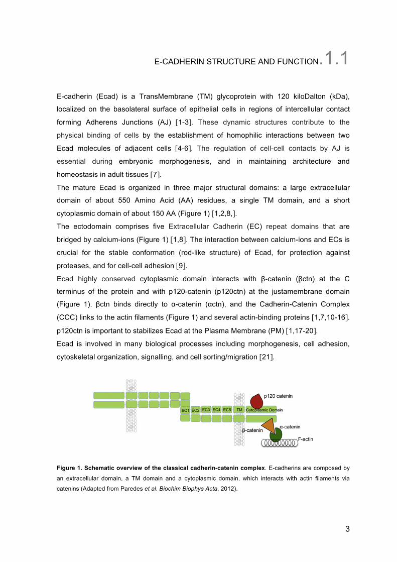
E-CADHERIN STRUCTURE AND FUNCTION.1.1
E-cadherin (Ecad) is a TransMembrane (TM) glycoprotein with 120 kiloDalton (kDa),
localized on the basolateral surface of epithelial cells in regions of intercellular contact
forming Adherens Junctions (AJ) [1-3]. These dynamic structures contribute to the
physical binding of cells by the establishment of homophilic interactions between two
Ecad molecules of adjacent cells [4-6]. The regulation of cell-cell contacts by AJ is
essential during embryonic morphogenesis, and in maintaining architecture and
homeostasis in adult tissues [7].
The mature Ecad is organized in three major structural domains: a large extracellular
domain of about 550 Amino Acid (AA) residues, a single TM domain, and a short
cytoplasmic domain of about 150 AA (Figure 1) [1,2,8,].
The ectodomain comprises five Extracellular Cadherin (EC) repeat domains that are
bridged by calcium-ions (Figure 1) [1,8]. The interaction between calcium-ions and ECs is
crucial for the stable conformation (rod-like structure) of Ecad, for protection against
proteases, and for cell-cell adhesion [9].
Ecad highly conserved cytoplasmic domain interacts with β-catenin (βctn) at the C
terminus of the protein and with p120-catenin (p120ctn) at the justamembrane domain
(Figure 1). βctn binds directly to α-catenin (αctn), and the Cadherin-Catenin Complex
(CCC) links to the actin filaments (Figure 1) and several actin-binding proteins [1,7,10-16].
p120ctn is important to stabilizes Ecad at the Plasma Membrane (PM) [1,17-20].
Ecad is involved in many biological processes including morphogenesis, cell adhesion,
cytoskeletal organization, signalling, and cell sorting/migration [21].
Figure 1. Schematic overview of the classical cadherin-catenin complex. E-cadherins are composed by
an extracellular domain, a TM domain and a cytoplasmic domain, which interacts with actin filaments via
catenins (Adapted from Paredes et al. Biochim Biophys Acta, 2012).
4
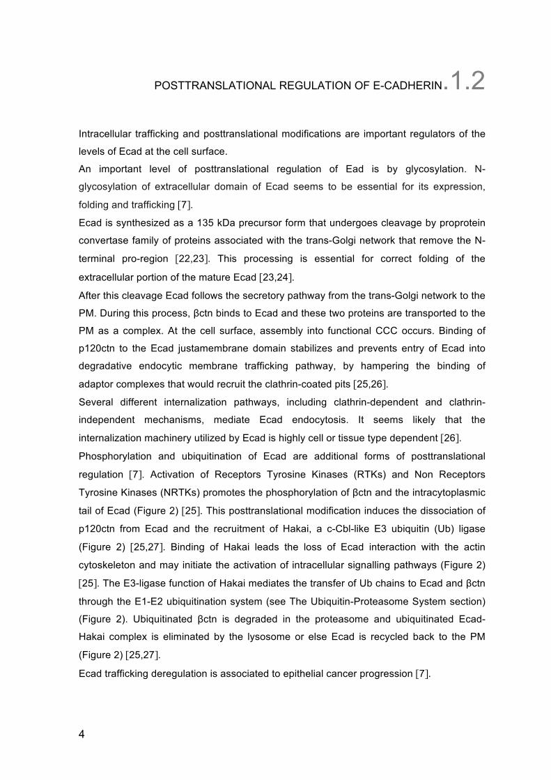
POSTTRANSLATIONAL REGULATION OF E-CADHERIN.1.2
Intracellular trafficking and posttranslational modifications are important regulators of the
levels of Ecad at the cell surface.
An important level of posttranslational regulation of Ead is by glycosylation. N-
glycosylation of extracellular domain of Ecad seems to be essential for its expression,
folding and trafficking [7].
Ecad is synthesized as a 135 kDa precursor form that undergoes cleavage by proprotein
convertase family of proteins associated with the trans-Golgi network that remove the N-
terminal pro-region [22,23]. This processing is essential for correct folding of the
extracellular portion of the mature Ecad [23,24].
After this cleavage Ecad follows the secretory pathway from the trans-Golgi network to the
PM. During this process, βctn binds to Ecad and these two proteins are transported to the
PM as a complex. At the cell surface, assembly into functional CCC occurs. Binding of
p120ctn to the Ecad justamembrane domain stabilizes and prevents entry of Ecad into
degradative endocytic membrane trafficking pathway, by hampering the binding of
adaptor complexes that would recruit the clathrin-coated pits [25,26].
Several different internalization pathways, including clathrin-dependent and clathrin-
independent mechanisms, mediate Ecad endocytosis. It seems likely that the
internalization machinery utilized by Ecad is highly cell or tissue type dependent [26].
Phosphorylation and ubiquitination of Ecad are additional forms of posttranslational
regulation [7]. Activation of Receptors Tyrosine Kinases (RTKs) and Non Receptors
Tyrosine Kinases (NRTKs) promotes the phosphorylation of βctn and the intracytoplasmic
tail of Ecad (Figure 2) [25]. This posttranslational modification induces the dissociation of
p120ctn from Ecad and the recruitment of Hakai, a c-Cbl-like E3 ubiquitin (Ub) ligase
(Figure 2) [25,27]. Binding of Hakai leads the loss of Ecad interaction with the actin
cytoskeleton and may initiate the activation of intracellular signalling pathways (Figure 2)
[25]. The E3-ligase function of Hakai mediates the transfer of Ub chains to Ecad and βctn
through the E1-E2 ubiquitination system (see The Ubiquitin-Proteasome System section)
(Figure 2). Ubiquitinated βctn is degraded in the proteasome and ubiquitinated Ecad-
Hakai complex is eliminated by the lysosome or else Ecad is recycled back to the PM
(Figure 2) [25,27].
Ecad trafficking deregulation is associated to epithelial cancer progression [7].
5
Figure 2. A model for Hakai in the dynamic regulation of Ecad. a - At steady-state, Ecad are organized in
multiprotein complexes in AJ. b - Activation of RTKs and NRTKs promotes the phosphorylation (P) of βctn and
the intracytoplasmic tail of Ecad, the recruitment of Hakai in a tyrosine-phosphoylation dependent manner, the
loss of Ecad interaction with the actin cytoskeleton, and the disruption of AJ. c - Ecad bound to Hakai may
initiate the activation of intracellular signalling pathways, whereas the E3-ligase function of Hakai mediates the
transfer of Ub chains to Ecad and βctn through the E1–E2 ubiquitination system. d - Ubiquitinated βctn is
degraded in the proteasome, but ubiquitinated Ecad-Hakai complexes are likely internalized by clathrin-coated
pits, e - which are then rapidly uncoated and fuse to early endosomes. f - In endosomes, depending on a
balance between ubiquitination and de-ubiquitination systems, g - Ecad may be either recycled back to the
cell surface, h- or targeted for degradation in the lysosomal compartment (Adapted from Pece S and Gutkind
JS, Nat Cell Biol, 2002).
6
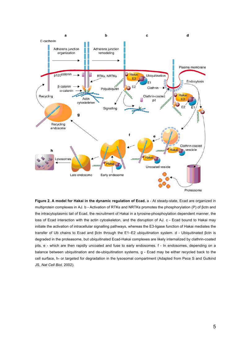
E-CADHERIN AND CANCER.1.3
Ecad gene (CDH1, located at human chromosome 16q22.1) is considered a Tumor
Suppressor Gene (TSG) wich plays an important role as suppressor of invasiveness and
epithelial cell migration [28,29].
Genetic or epigenetic alterations in this gene often result in abnormal Ecad expression or
function. Loss of Ecad is a defining characteristic of Epithelial-Mesenchymal Transition
(EMT), a process associated with invasion and metastasation that are a hallmark in late
carcinogenesis (Figure 3) [30-32].
In sporadic lobular breast cancer and Diffuse Gastric Cancer (DGC), Ecad inactivation is
associated with somatic mutations of CDH1, as well as loss of heterozygosity (LOH),
promoter hypermethylation, aberrant glycosylation, or overexpression of transcriptional
repressors (e.g. Snail, Slug, Sip5 and Ets) [33-35].
In 1998, Guilford et al. identified in three Maori families the first germline mutations in
Ecad gene associated with Hereditary Diffuse Gastric Cancer (HDGC) [36].
Figure 3. EMT in cancer progression and metastasis. Loss of Ecad-mediated cell adhesion contributes to
the transition from benign, non-invasive tumours (adenoma) to malignant, invasive tumours (carcinoma)
(Adapted from Christofori G and Semb H, Trends Biochem Sci, 1999).
1.3.1. Hereditary Diffuse Gastric Cancer
HDGC is a rare (30% of the familial cases, which represent 1-3% of all Gastric Cancer
(GC) cases) autosomal dominant cancer susceptibility syndrome. Germline mutations of
7
CDH1 are the only known genetic cause of HDGG [37-38].
Stringent criteria for genetic test were defined by the International Gastric Cancer Linkage
Consortium (IGCLC) in 1999, and include the following: (i) two or more documented cases
of GC in first/second degree relatives, with at least one DGC diagnosed before 50 years
of age; (ii) three or more cases of documented GC in first/second degree relatives, with at
least one DGC diagnosed at any age; (iii) single individual with DGC diagnosed before 40
years age without a family history; or (iv) single individual and families with diagnoses of
both DGC (including one case below the age of 50 years) and lobular breast cancer
[36,37].
The average age of onset of HDGC is 38 years, with a range of 14-69 years [37,38]. The
estimated cumulative risk of GC by age 80 years is 63%-83% for women and 40%-67%
for men [38]. Women also have a 39%-52% risk for developing lobular breast
adenocarcinoma [36].
Prophylactic total gastrectomy is the only preventive treatment and is recommended to the
CDH1 mutation carriers after 20 years of age, especially if they are carriers of nonsense
mutations [37-39]. Missense mutations account for approximately 25% of all HDGC
cases harboring CDH1 mutations [40].
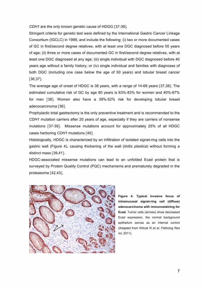
Histologically, HDGC is characterized by an infiltration of isolated signet-ring cells into the
gastric wall (Figure 4), causing thickening of the wall (linitis plastica) without forming a
distinct mass [39,41].
HDGC-associated missense mutations can lead to an unfolded Ecad protein that is
surveyed by Protein Quality Control (PQC) mechanisms and prematurely degraded in the
proteasome [42,43].
Figure 4. Typical invasive focus of intramucosal signet-ring cell (diffuse)
adenocarcinoma with immunostaining for
Ecad. Tumor cells (arrows) show decreased
Ecad expression, the normal background
epithelium serves as an internal control
(Adapted from Wilcok R et al, Patholog Res
Int, 2011).
8
PROTEOSTASIS AND PROTEIN QUALITY CONTROL.1.4
Eukaryotic protein homeostasis, or proteostasis, enables healthy cell and organismal
development as well as ensures a better adaptation to aging. Proteostasis is a complex
and integrated biological network within cells, comprising various pathways that include
the biogenesis, folding, trafficking and turnover of proteins [40]. The accumulation of
misfolded, aggregation prone and potentially cytotoxic proteins can be generated by
mutations, transcriptional and translational errors or cellular and environmental stresses.
To advert these dangers for protein homeostasis, cells have developed powerful
strategies of PQC [44,45].
PQC is a general term used to refer the mechanisms by which cells control and survey
proteins folding and decides if the protein is suitable for its final destination and function
[46,47]. Deficiencies in PQC system lead to many metabolic, oncological,
neurodegenerative, and cardiovascular disorders [40].
PQC consists of a large arsenal of molecular chaperones and proteolytic systems.
Protein folding, unfolding, and refolding are constantly occurring throughout the lifetime of
nearly all proteins [44]. Molecular chaperones promote folding and maintenance of
conformation within the cell largely by minimizing misfolding and aggregation, but the
chaperones also escort terminally misfolded proteins or irreversibly damaged proteins to
the proteolytic pathways for degradation [48].
The main proteolytic systems associated to the PQC are Ubiquitin-Proteasome System
(UPS) and the autophagy pathway.
1.4.1. The Ubiquitin-Proteasome System
The UPS is the main proteolytic system involved in the selective degradation of soluble
proteins. This pathway is implicated in numerous cellular events where protein
degradation is required either to dispose of obsolete proteins or to regulate various
biological processes. Degradation of a protein by the UPS involves two different
successive steps: the first step implies the covalent attachment of small (8.5 kDa)
regulatory polipeptides called Ub to the target protein, in a process known as
ubiquitination; the second step involves recognition and final degradation of the targeted
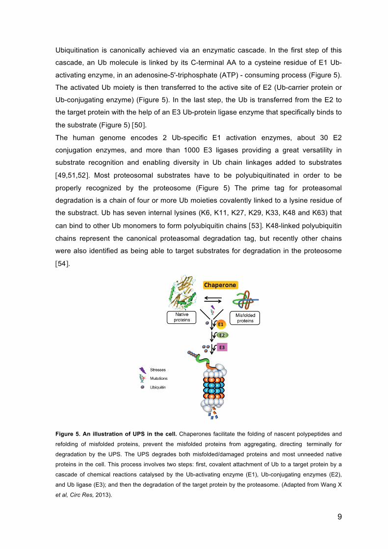
protein by the proteasome (Figure 5) with the release of free and reusable Ub [49].
9
Ubiquitination is canonically achieved via an enzymatic cascade. In the first step of this
cascade, an Ub molecule is linked by its C-terminal AA to a cysteine residue of E1 Ub-
activating enzyme, in an adenosine-5'-triphosphate (ATP) - consuming process (Figure 5).
The activated Ub moiety is then transferred to the active site of E2 (Ub-carrier protein or
Ub-conjugating enzyme) (Figure 5). In the last step, the Ub is transferred from the E2 to
the target protein with the help of an E3 Ub-protein ligase enzyme that specifically binds to
the substrate (Figure 5) [50].
The human genome encodes 2 Ub-specific E1 activation enzymes, about 30 E2
conjugation enzymes, and more than 1000 E3 ligases providing a great versatility in
substrate recognition and enabling diversity in Ub chain linkages added to substrates
[49,51,52]. Most proteosomal substrates have to be polyubiquitinated in order to be
properly recognized by the proteosome (Figure 5) The prime tag for proteasomal
degradation is a chain of four or more Ub moieties covalently linked to a lysine residue of
the substract. Ub has seven internal lysines (K6, K11, K27, K29, K33, K48 and K63) that
can bind to other Ub monomers to form polyubiquitin chains [53]. K48-linked polyubiquitin
chains represent the canonical proteasomal degradation tag, but recently other chains
were also identified as being able to target substrates for degradation in the proteosome
[54].
Figure 5. An illustration of UPS in the cell. Chaperones facilitate the folding of nascent polypeptides and
refolding of misfolded proteins, prevent the misfolded proteins from aggregating, directing terminally for
degradation by the UPS. The UPS degrades both misfolded/damaged proteins and most unneeded native
proteins in the cell. This process involves two steps: first, covalent attachment of Ub to a target protein by a
cascade of chemical reactions catalysed by the Ub-activating enzyme (E1), Ub-conjugating enzymes (E2),
and Ub ligase (E3); and then the degradation of the target protein by the proteasome. (Adapted from Wang X
et al, Circ Res, 2013).
10
1.4.2. Autophagy
Autophagy was initially thought to be a form of cell response and adaptation to lack of
nutrients, it is now realized that autophagy is a highly regulated and multipurpose system.
The best-characterized signal for the activation of autophagy is nutrient deprivation. When
nutrients are insufficient, this pathway allows a cell to break down its own components,
including proteins and organelles and recycle important molecules. It is a very important
PQC system that represents the adaptation of the cell to starvation/nutrient deprivation
allowing the cell to survive until there is food available in the medium [55,56].
To date, at least three types of autophagic pathways have been described, which differ in
their routes to lysosomes: macroautophagy (also commonly called “autophagy”),
microautophagy and Chaperone-Mediated Autophagy (CMA). The essential component of
these proteolic systems is the lysosome, a single membrane vesicle that contains in its
lumen a large variety of cellular hydrolases including proteases, lipases, glycosidades,
and nucleotidases [54].
11
ENDOPLASMIC RETICULUM QUALITY CONTROL.1.5
To accomplish Endoplasmic Reticulum (ER) quality control, cells have a complex network
of molecular chaperones associated with the ER, which interact with nascent proteins in
the ER lumen, promoting the folding of client proteins until a proper native state is
achieved. When native structure is accomplished the client proteins leave the ER and are
transported to the Golgi apparatus where they are modified, sorted and sent towards their
final destinations [45,47]. Chemical chaperones are organic molecules that seem to
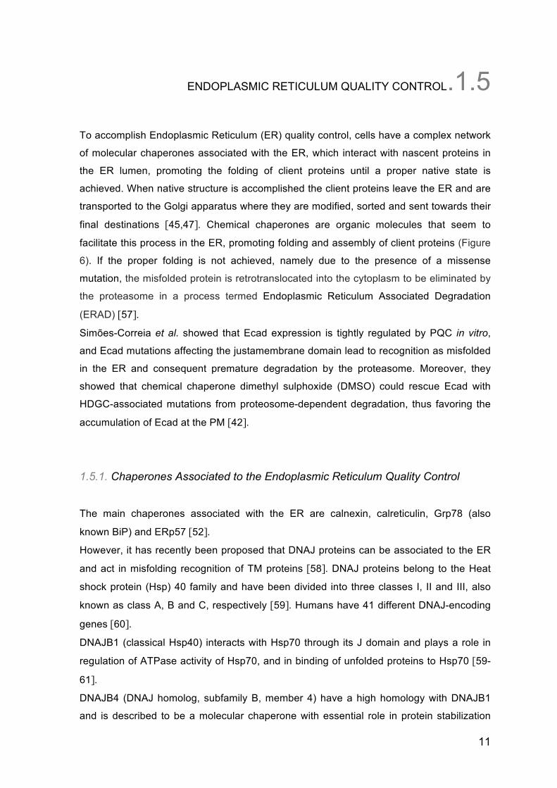
facilitate this process in the ER, promoting folding and assembly of client proteins (Figure
6). If the proper folding is not achieved, namely due to the presence of a missense
mutation, the misfolded protein is retrotranslocated into the cytoplasm to be eliminated by
the proteasome in a process termed Endoplasmic Reticulum Associated Degradation
(ERAD) [57].
Simões-Correia et al. showed that Ecad expression is tightly regulated by PQC in vitro,
and Ecad mutations affecting the justamembrane domain lead to recognition as misfolded
in the ER and consequent premature degradation by the proteasome. Moreover, they
showed that chemical chaperone dimethyl sulphoxide (DMSO) could rescue Ecad with
HDGC-associated mutations from proteosome-dependent degradation, thus favoring the
accumulation of Ecad at the PM [42].
1.5.1. Chaperones Associated to the Endoplasmic Reticulum Quality Control
The main chaperones associated with the ER are calnexin, calreticulin, Grp78 (also
known BiP) and ERp57 [52].
However, it has recently been proposed that DNAJ proteins can be associated to the ER
and act in misfolding recognition of TM proteins [58]. DNAJ proteins belong to the Heat
shock protein (Hsp) 40 family and have been divided into three classes I, II and III, also
known as class A, B and C, respectively [59]. Humans have 41 different DNAJ-encoding
genes [60].
DNAJB1 (classical Hsp40) interacts with Hsp70 through its J domain and plays a role in
regulation of ATPase activity of Hsp70, and in binding of unfolded proteins to Hsp70 [59-
61].
DNAJB4 (DNAJ homolog, subfamily B, member 4) have a high homology with DNAJB1
and is described to be a molecular chaperone with essential role in protein stabilization
12
[62]. It has been demonstrated that DNAJB4 has little impact in refolding and aggregation
suppression [63], but its binding to unfolded substrates has been described [61,62].
DNAJB4 acts as a tumor supressor in Non-Small Cell Lung Carcinoma (NSCLC) model,
while inhibiting tumorigenesis and metastasis [64]. There are no reports exploring the
potential of DNAJB4 as a molecular chaperone of Ecad, or demonstrating its relevance for
GC progression.
Figure 6. Putative mechanism of chemical chaperones in the ER and Golgi apparatus. Newly
synthesized polypeptides are translocated into the lumen of the ER. Folding is facilitated by interaction with
molecular chaperones. If the polypeptide is mutated, there is misfolding and misassembly and then dislocation
into the cytoplasm for proteosomal degradation after dissociation of the molecular chaperones. In the
presence of chemical chaperones, folding and assembly of the mutated polypeptide is presumably facilitated
so that it can exit the ER by vesicular transport to the Golgi and then from Golgi to PM and/or extracellular
fluid. Molecular chaperones dissociate within the ER but chemical chaperones are associated throughout the
secretory pathway because they presumably saturate all of the transport compartments. (Adapted from
Perlmutter DH, Pediatric Research, 2002).
13
PLASMA MEMBRANE QUALITY CONTROL.1.6
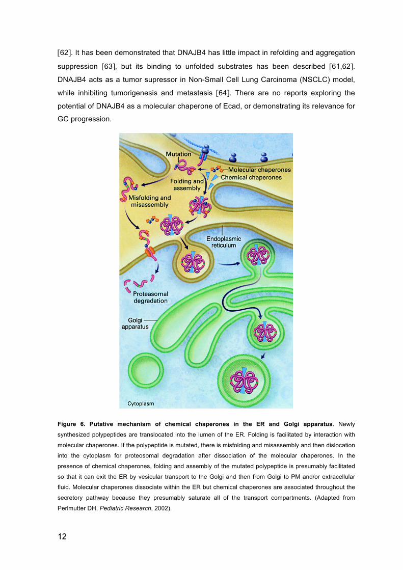
Peripheral Protein Quality Control (PPQC) has recently been proposed to be a specialized
pathway for the regulation of unfolded proteins at the PM. In this pathway, unfolded
substrate is internalized and subsequently degraded in the lysosomal compartment
(Figure 7). Lysosome-dependent degradation depends of the Endosomal Sorting
Complexes Required for Transport (ESCRT) to recognize ubiquitinated proteins in the
endosome and it promotes cargo delivery into multivesicular bodies (MVB)/lysosome and
protein degradation (Figure 7) [65,66].
The molecular components of PPQC seem to be partially shared with ER quality control
[67]. Heat shock cognate (Hsc) 70 has their main chaperoning activity within the
cytoplasm [68,69]. However, it has been reported recently, that Hsc70/Hsp90 and
Hsc70/C-terminus of Hsc70-Interacting Protein (CHIP) are part of the PPQC machinery,
and that they are involved on the recognition and ubiquitination of unfolded proteins at the
cell surface (Figure 7) [65].
Figure 7. Working model for the PPQC network. 1- Unfolded PM protein is recognized by Hsc70/DNAJA1,
possibly in conjunction with Hsp90/Hop/Aha1. 2- Recruitment of CHIP/UbcH5 leads to ubiquitination of the
unfolded substract. 3- Recruitment of endocytic adaptors to endocytose unfolded substrate. 4, 5- Depending
on the folding propensy of the cargo and the proteostasis network state, interaction with chaperones and co-
chaperones may favor the client refolding, deubiquitination and recycling to the PM. Irreversible unfolded PM
substrates will lead to persistent ubiquitination (involving CHIP and/or other E3 ligases), recruitment of ESCRT
components and sorting to the lysosome compartment for degradation (Adapted from Okiyoneda T et al, Curr
Opin Cell Biol, 2011).
14
CHIP is an E3 Ub ligase containing three TetratricoPeptide Repeats (TPR) domains at its
N-‐terminal and an U-‐box domain at its C-‐terminal and plays a central role in protein triage
decision [70,71].
Previous studies show that CHIP complex recognizes the mutant variant ΔF508 of Cystic
Fibrosis Transmembrane Conductance (CFTR). This complex catalyzes its ubiquitination
that diverts unfolded proteins into the lysosomal degradative pathway [66].
This quality control mechanism is thought to be determinant for the clearance of unfolded
proteins at the PM that either result from genetic mutations or denaturing extracellular
stimuli [67].

15
AIMS.02

16
17
AIMS.02
The main objective of this project is to understand the importance of PQC in the regulation
of Ecad and evaluate its significance in cancer. To this end the following specific
objectives were sought:
• Determine the subcellular distribution of DNAJB4 in the context of WT or mutant
Ecad;
• Explore in vitro the role of DNAJB4 in the stabilization and/or degradation of Ecad;
• Understand if DNAJB4 has an impact in cellular adhesion and migration;
• Investigate in vivo the role of DNAJB4 in invasion and angiogenesis;
• Analyse the stability of WT Ecad at the PM after extracellular calcium depletion;
• Analyse the stability of Ecad mutant at the PM after folding/unfolding stimulus with
DMSO;
• Test if PPQC components, namely Hsc70 and CHIP, are recruited by unfolded
Ecad to the PM;
• Determine if the PPQC machinery, namely Hsc70 and CHIP, regulates cell
adhesion.

18
19
MATERIAL AND METHODS.03

20
21
CELL CULTURE, TRANSFECTIONS AND TREATMENTS.3.1
Chinese Hamster Ovary (CHO) cells (CCL-61; ATCC™, Barcelona, Spain) were grown in
MEM Alpha (Gibco®, Life Technologies™, Barcelona, Spain) and MKN28 GC cell line was
maintained in RPMI medium (Gibco®), both supplemented with 10% Fetal Bovine Serum
(FBS; HyClone®, Salt Lake City, Utah) and 1% penicillin-streptomycin (Gibco®). All cell
lines were maintained in a humidified incubator with 5% CO2 at 37°C.
CHO stable cell lines, Wild Type (WT) human Ecad, mutant Ecad (E757K) or empty
vector (Mock), were established previously [42] and maintained in the presence of
antibiotic selection with 5µg/mL blasticidin (Gibco®).
For stable silencing of Ecad, MNK28 were transduced by lentiviral infection of a short
hairpin RNA (shRNA) and corresponding control (Table 1, annexes), using polybrene.
Stable cell lines were established by antibiotic selection with 5µg/mL puromycin (Sigma-
Aldrich®, Sintra, Portugal).
For transient transfections, 2x10⁵ cells were seeded in 6-well plates and, at 30%
confluence, they were transfected with 1µg of vector DNA (Table 1, annexes) or 50nM of
small interfering RNA (siRNA) (Table 2, annexes), using Lipofectamine® 2000
Transfection Reagent (Invitrogen™, Life Technologies™) according to the manufacture
procedure.
For the protein synthesis inhibition, 24h after transfection, CHO cells were treated with
50µM of Cycloheximide (CHX; Sigma-Aldrich®) for 0, 2, 4 and 8h.
For proteasome inhibition, transiently transfected cells were incubated for 8h with 10µM of
MG132 (CalBioChem®, Millipore™, Billerica, Massachusetts, USA). Lysosomal inhibition
was obtained with Chloroquine (CQ; Sigma-Aldrich®), according to times and
concentrations indicated. The chemical chaperone 2% DMSO (Sigma-Aldrich®) effect
was induced for 24h.
To destabilize WT Ecad at the PM, 48h after transfection, MKN28 and CHO cells were
incubated for indicated times in medium without calcium supplemented with 2mM
ethylenediaminetetraacetic acid (EDTA; Stratagene®, La Jolla, California, USA). To
stabilize mutant Ecad at the PM, transiently transfected cells were treated with 2% DMSO
for 24h. Afterwards, destabilization was recovered by removing the folding stimulus
(incubation with normal medium for 4h).
For inhibition of endocytosis, CHO cells were incubated for 15min with 0,4M sucrose.
22
PROTEIN EXTRACTION, QUANTIFICATION AND WESTERN BLOT.3.2
Cells lysates were obtained with cold Catenin lysis buffer1 enriched with a protease
inhibitor cocktail (Roche, Amadora, Portugal) and phosphatase inhibitor cocktail (Sigma-
Aldrich®). To separate the proteins from cellular debris, cells lysates were spun down and
the supernatant (proteins) were quantified using a modified Bradford assay (Bio-Rad,
Amadora, Portugal). 40µg of total protein was denatured in loading buffer2, separated in
7.5% Sodium Dodecyl Sulphate–PolyAcrylamide Gel Electrophoresis (SDS-PAGE), and
electroblotted to nitrocellulose membrane (Bio-Rad). Membranes were blocked overnight
at 4°C with 5% non-fat dry milk (Molico®, Nestlé®, Vevey, Switzerland) and 0.5% Tween-
20 in Tris-Buffered Saline (TBS), and immunoblotted with primary antibodies (Table 3,
annexes). HorseRadish Peroxidase (HRP) - conjugated secondary antibodies (Table 4,
annexes) were used accordingly, followed by Enhanced ChemiLuminescence (ECL)
detection (Bio-Rad). Immunoblots were quantified with Quantity One® Software (Bio-
Rad).
IMMUNOPRECIPITATION AND CELL SURFACE BIOTINYLATION.3.3
To immunoprecipite Ecad, 400µg of protein were coupled to a mouse monoclonal anti-
Ecad antibody (BD Transduction Laboratories™, BD Biosciences, San Jose, California,
USA), and immunocomplexes were incubated with PureProteome™ Protein G Magnetic
Beads (Millipore™), washed and eluted in loading buffer, according to manufacture
instructions.
Cell surface biotinylation was performed at 4°C by incubation with freshly prepared EZ-
Link®Sulpho-NHS-SS Biotin (Thermo Scientific™, Pierce Biotechnology, Rockford,
Illinois, USA) at 0,5mg/mL in phosphate-buffered saline (PBS) - PLUS3 for 30min. The
reaction was quenched with PBS-PLUS containing 100mM glycine (NZYTech, Lisboa,
Portugal). Whole cell lysates were prepared according to the protocol described above.
An aliquot of 400 µg of total protein was incubated on a rotator overnight with 50µL of
Streptavidin Sepharose Beads (GE Healthcare, Little Chalfont, UK) at 4°C. To separate
1 1% Triton X-100, 1% Nonidet P-40 in PBS 2 62.5mM Tris–HCl (pH 6.8), 2% SDS, 10% glycerol, 0.01%, bromophenol blue, and 5% β-mercaptoethanol 3 PBS supplemented with 1mM CaCl2 and 1mM MgCl2
23
the membrane fraction from the cytoplasmic fraction the samples were spun down, and
the pellet (membrane fraction) was washed, eluted in loading buffer and analysed by WB.
SUBCELLULAR PROTEIN FRACTIONATION.3.4
For subcellular fraccionation, 5x10⁵ CHO cells (stably expressing WT or unfolded E757K
mutant Ecad) were harvested and fractionated into different subcellular extracts using the
subcellular protein fractionation kit (Thermo Scientific™, Pierce Biotechnology) according
to manufacture protocol. 15% of the total content from the membranous and cytoplasmic
fractions was analysed by WB with the primary antibodies against Ecad, V5-tag and
Calnexin (Table 3, annexes).
SLOW AGGREGATION ASSAY.3.5
Wells of 96-well plate were coated with 50µL of agar solution4. Transiently transfected
CHO cells were detached with 0.05% trypsin-EDTA (Gibco®) and ressuspended in
culture medium. A suspension of 1x10⁵ cells/mL was prepared and 2x10⁴ cells were
seeded in each well. The plate was incubated at 37°C in a humidified chamber with 5%
CO2. Aggregation was evaluated in an inverted fluorescent microscope (Leica DMIRE2,
x10 magnification) 48h after seeding, and images captured using a digital camera (Leica
DFC 350 FX).
CELL MIGRATION ASSAY.3.6
The migratory/motility behavior of CHO cells stably expressing WT Ecad (in control
conditions or upon DNAJB4 transient transfection) was analysed in an in vitro wound
healing assay. Cells were grown to confluence in 6-well plates, an artificial wound was
created with a yellow pipette tip, and cells were carefully washed twice with PBS to
remove detached cells. Migration was assessed by measuring the distance between
wound edges at time intervals (0 and 4h). The cells were visualized by phase-contrast
4 100mg Bacto-Agar in 15mL of sterile PBS
24
light microscopy at x5 magnification and images captured using a Leica DFC 350 FX
digital camera mounted on a Leica DMRIE2 microscope.
CAM ASSAY, IMMUNOHISTOCHEMISTRY AND STATISTICAL ANALYSIS.3.7
The chick embryo ChorioAllantoic Membrane (CAM) assay was used to evaluate the
angiogenic response and invasive potential of MKN28 GC cells stably transduced as
described above and transiently transfected with DNAJB4. Fertilized chick (Gallus gallus)
eggs obtained from commercial sources were incubated horizontally at 37.8°C in a
humidified atmosphere and referred to embryonic day (E). On E3 a square window was
opened in the shell after removal of 1.5-2mL of albumin to allow detachment of the
developing CAM. The window was sealed with a transparent adhesive tape and the eggs
returned to the incubator. 1x10⁶ cells re-suspended in 10µL of complete medium, were
placed on top of E10 growing CAM into a 3mm silicon ring under sterile conditions. The
eggs were re-sealed and returned to the incubator for an additional 3 days. The embryos
were euthanized by adding 2mL of fixative in the top of the CAM. After removing the ring,
the CAM was excised from the embryos and photographed ex ovo under a stereoscope at
x20 magnification (Olympus, SZX16 coupled with a DP71 camera). The number of new
vessels (less than 15µm diameter) growing radial towards the ring area was counted in a
blind fashion manner. The CAM assay was performed by Marta Teixeira Pinto of
IPATIMUP.
Excided CAM were fixed in 10% neutral-buffered formalin, paraffin-embedded for slide
sections and stained with hematoxilin-eosin for histological examination. Tumour sections
obtained from CAM were de-paraffinized, re-hydrated with graded ethanol and washed in
destiled water followed by 0.1% Tween-20 in TBS. Heat induced antigen retrieval was
performed using a Digest-All™ 3 (Pepsin Solution; Invitrogen™, Life Technologies™). To
block endogenous peroxidase activity, slides were treated with 0.5% H2O2 in methanol, for
20min at room temperature. To block non-specific binding, slides were exposed large
volume Ultra V Block (Thermo Scientific™, Lab Vision™), for 30min at room temperature.
Slides were subsequently incubated with mouse monoclonal antibody against pan
Cytokeratin (Sigma-Aldrich®) at 1:200 in Large Volume UltraClean Diluent (Thermo
Scientific™, Lab Vision™). After washing, sections were incubated with EnVision™
Detection System Peroxidase/DAB (Dako, Glostrup, Denmark) followed by hematoxilin
staining. Invasion was evaluated under the microscope, in a blind fashion by three
independent users, using a semi-quantitative approach taking into consideration the
25
behavior of the human cells (pan-Cytokeratin positive) in the CAM (scored 1 if invasive,
and 0 if not invasive). The histological processing of CAM was performed in the
Diagnostic Unit of IPATIMUP.
For statistical analysis of the angiogenic response was used GraphPad Prism® software.
ANalysis Of VAriance (ANOVA) tests were used to calculate significance in an interval of
95% confidence level and values of p<0.05 were considered to be statistically significant.
MKN28 cell are not naturally invasive or tumorigenic thus making it difficult to have clear
histological slides. For this reason, statistical analysis of the invasion score was not
performed given the small number of animals in each group.
CELL SURFACE ELISA.3.8
For cell surface Enzyme-Linked ImmunoSorbent Assay (ELISA), 5x10⁴ CHO cells (stably
expressing WT or unfolded E757K mutant Ecad) were seeded in 24-well plate and, at
80% of confluence, were treated with 2% DMSO in culture medium for 24h. The unfolding
was stimulated by removing the folding stimulus (incubation with normal medium for 4h).
After treatments, cell surface density of Ecad was measured by primary antibody HECD-1,
that recognized an extracellular epitope of Ecad, and HRP-conjugated secondary
antibody in the presence of Amplex®Red (Molecular Probes®, Life Technologies™), a
fluorescent HRP substrate, according as described by Kathryn W. Peters [72].
IMMUNOFLUORESCENCE.3.9
MKN28 cells were seeded on glass coverslips and, 48h after transfection, they were fixed
and permeabilized with ice-cold methanol for 10min, washed and incubated with primary
antibodies against (Table 3, annexes) diluted in PBS containing 5% Bovine Serum
Albumin (BSA; Sigma-Aldrich®), for 1h at room temperature. Secondary antibodies (Table
4, annexes) were used as appropriate for 1h at room temperature in the dark. The
coverslips were mounted on slides using Vectashield® mounting medium with 4',6-
diamidino-2-phenylindole (DAPI; Vector Laboratories, Burlingame, California, USA).
Images were acquired using a confocal laser point-scanning microscope (Zeiss LSM 710).

26

27
RESULTS.04

28
29
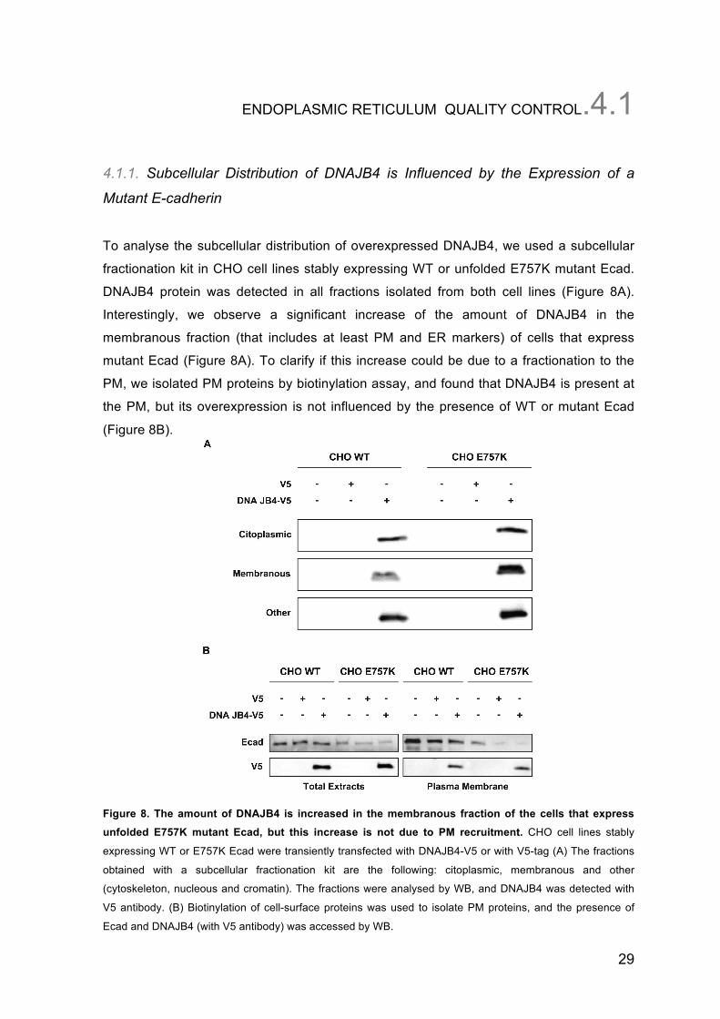
ENDOPLASMIC RETICULUM QUALITY CONTROL.4.1
4.1.1. Subcellular Distribution of DNAJB4 is Influenced by the Expression of a
Mutant E-cadherin
To analyse the subcellular distribution of overexpressed DNAJB4, we used a subcellular
fractionation kit in CHO cell lines stably expressing WT or unfolded E757K mutant Ecad.
DNAJB4 protein was detected in all fractions isolated from both cell lines (Figure 8A).
Interestingly, we observe a significant increase of the amount of DNAJB4 in the
membranous fraction (that includes at least PM and ER markers) of cells that express
mutant Ecad (Figure 8A). To clarify if this increase could be due to a fractionation to the
PM, we isolated PM proteins by biotinylation assay, and found that DNAJB4 is present at
the PM, but its overexpression is not influenced by the presence of WT or mutant Ecad
(Figure 8B).
Figure 8. The amount of DNAJB4 is increased in the membranous fraction of the cells that express unfolded E757K mutant Ecad, but this increase is not due to PM recruitment. CHO cell lines stably
expressing WT or E757K Ecad were transiently transfected with DNAJB4-V5 or with V5-tag (A) The fractions
obtained with a subcellular fractionation kit are the following: citoplasmic, membranous and other
(cytoskeleton, nucleous and cromatin). The fractions were analysed by WB, and DNAJB4 was detected with
V5 antibody. (B) Biotinylation of cell-surface proteins was used to isolate PM proteins, and the presence of
Ecad and DNAJB4 (with V5 antibody) was accessed by WB.
30
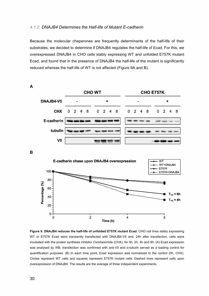
4.1.2. DNAJB4 Determines the Half-life of Mutant E-cadherin
Because the molecular chaperones are frequently determinants of the half-life of their
substrates, we decided to determine if DNAJB4 regulates the half-life of Ecad. For this, we
overexpressed DNAJB4 in CHO cells stably expressing WT and unfolded E757K mutant
Ecad, and found that in the presence of DNAJB4 the half-life of the mutant is significantly
reduced whereas the half-life of WT is not affected (Figure 9A and B).
A
B
Figure 9. DNAJB4 reduces the half-life of unfolded E757K mutant Ecad. CHO cell lines stably expressing
WT or E757K Ecad were transiently transfected with DNAJB4-V5 and, 24h after transfection, cells were
incubated with the protein synthesis inhibitor Cicloheximide (CHX), for 0h, 2h, 4h and 8h. (A) Ecad expression
was analysed by WB, transfection was confirmed with anti-V5 and α-tubulin served as a loading control for
quantification purposes. (B) In each time point, Ecad expression was normalized to the control (0h, CHX).
Circles represent WT cells and squares represent E757K mutant cells. Dashed lines represent cells upon
overexpression of DNAJB4. The results are the average of three independent experiments.
31
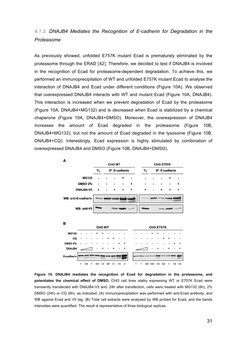
4.1.3. DNAJB4 Mediates the Recognition of E-cadherin for Degradation in the
Proteasome
As previously showed, unfolded E757K mutant Ecad is prematurely eliminated by the
proteasome through the ERAD [42]. Therefore, we decided to test if DNAJB4 is involved
in the recognition of Ecad for proteasome-dependent degradation. To achieve this, we
performed an immunoprecipitation of WT and unfolded E757K mutant Ecad to analyse the
interaction of DNAJB4 and Ecad under different conditions (Figure 10A). We observed
that overexpressed DNAJB4 interacts with WT and mutant Ecad (Figure 10A, DNAJB4).
This interaction is increased when we prevent degradation of Ecad by the proteasome
(Figure 10A, DNAJB4+MG132) and is decreased when Ecad is stabilized by a chemical
chaperone (Figure 10A, DNAJB4+DMSO). Moreover, the overexpression of DNAJB4
increases the amount of Ecad degraded in the proteasome (Figure 10B,
DNAJB4+MG132), but not the amount of Ecad degraded in the lysosome (Figure 10B,
DNAJB4+CQ). Interestingly, Ecad expression is highly stimulated by combination of
overexpressed DNAJB4 and DMSO (Figure 10B, DNAJB4+DMSO).
Figure 10. DNAJB4 mediates the recognition of Ecad for degradation in the proteasome, and potentiates the chemical effect of DMSO. CHO cell lines stably expressing WT or E757K Ecad were
transiently transfected with DNAJB4-V5 and, 24h after transfection, cells were treated with MG132 (8h), 2%
DMSO (24h) or CQ (6h), as indicated. (A) Immunoprecipitation was performed with anti-Ecad antibody, and
WB against Ecad and V5 tag. (B) Total cell extracts were analysed by WB probed for Ecad, and the bands
intensities were quantified. The result is representative of three biological replicas.
32
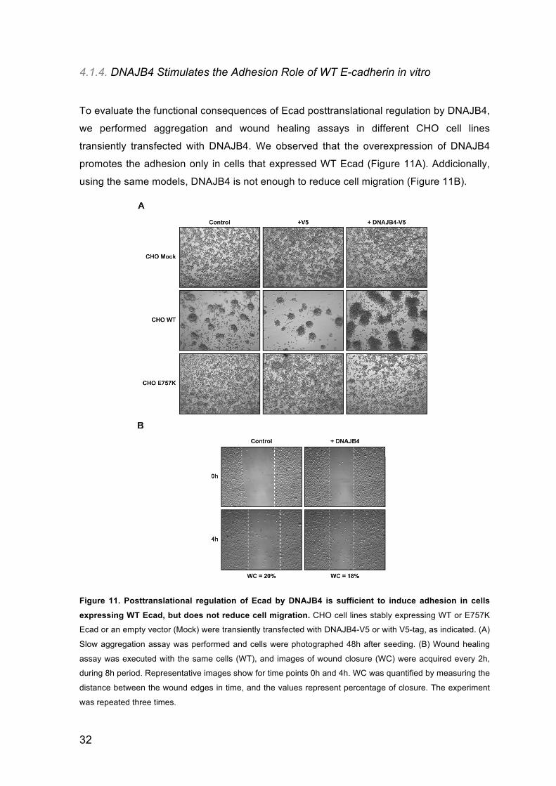
4.1.4. DNAJB4 Stimulates the Adhesion Role of WT E-cadherin in vitro
To evaluate the functional consequences of Ecad posttranslational regulation by DNAJB4,
we performed aggregation and wound healing assays in different CHO cell lines
transiently transfected with DNAJB4. We observed that the overexpression of DNAJB4
promotes the adhesion only in cells that expressed WT Ecad (Figure 11A). Addicionally,
using the same models, DNAJB4 is not enough to reduce cell migration (Figure 11B).
Figure 11. Posttranslational regulation of Ecad by DNAJB4 is sufficient to induce adhesion in cells
expressing WT Ecad, but does not reduce cell migration. CHO cell lines stably expressing WT or E757K
Ecad or an empty vector (Mock) were transiently transfected with DNAJB4-V5 or with V5-tag, as indicated. (A)
Slow aggregation assay was performed and cells were photographed 48h after seeding. (B) Wound healing
assay was executed with the same cells (WT), and images of wound closure (WC) were acquired every 2h,
during 8h period. Representative images show for time points 0h and 4h. WC was quantified by measuring the
distance between the wound edges in time, and the values represent percentage of closure. The experiment
was repeated three times.
33
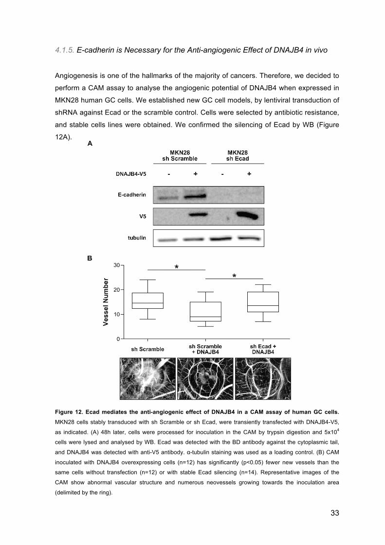
4.1.5. E-cadherin is Necessary for the Anti-angiogenic Effect of DNAJB4 in vivo
Angiogenesis is one of the hallmarks of the majority of cancers. Therefore, we decided to
perform a CAM assay to analyse the angiogenic potential of DNAJB4 when expressed in
MKN28 human GC cells. We established new GC cell models, by lentiviral transduction of
shRNA against Ecad or the scramble control. Cells were selected by antibiotic resistance,
and stable cells lines were obtained. We confirmed the silencing of Ecad by WB (Figure
12A).
Figure 12. Ecad mediates the anti-angiogenic effect of DNAJB4 in a CAM assay of human GC cells.
MKN28 cells stably transduced with sh Scramble or sh Ecad, were transiently transfected with DNAJB4-V5,
as indicated. (A) 48h later, cells were processed for inoculation in the CAM by trypsin digestion and 5x10⁴
cells were lysed and analysed by WB. Ecad was detected with the BD antibody against the cytoplasmic tail,
and DNAJB4 was detected with anti-V5 antibody. α-tubulin staining was used as a loading control. (B) CAM
inoculated with DNAJB4 overexpressing cells (n=12) has significantly (p<0.05) fewer new vessels than the
same cells without transfection (n=12) or with stable Ecad silencing (n=14). Representative images of the
CAM show abnormal vascular structure and numerous neovessels growing towards the inoculation area
(delimited by the ring).
34
These cell lines, with stable silencing of Ecad (sh Ecad) or with stable expression of sh
control (sh Scramble), were both transiently transfected with DNAJB4, as confirmed by
WB (Figure 12A). The cells were inoculated over the CAM and processed for subsequent
analysis. We found that overexpression of DNAJB4 decreases the number of vessels
formed, as compared to the control cells (Figure 12B). Interestingly, in the absence of
Ecad expression, the anti-angiogenic phenotype of DNAJB4 is reversed (Figure 12B).
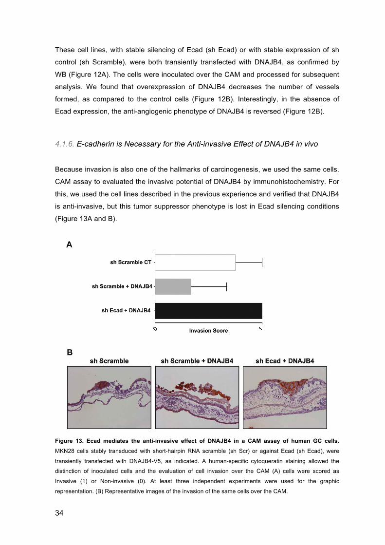
4.1.6. E-cadherin is Necessary for the Anti-invasive Effect of DNAJB4 in vivo
Because invasion is also one of the hallmarks of carcinogenesis, we used the same cells.
CAM assay to evaluated the invasive potential of DNAJB4 by immunohistochemistry. For
this, we used the cell lines described in the previous experience and verified that DNAJB4
is anti-invasive, but this tumor suppressor phenotype is lost in Ecad silencing conditions
(Figure 13A and B).
Figure 13. Ecad mediates the anti-invasive effect of DNAJB4 in a CAM assay of human GC cells.
MKN28 cells stably transduced with short-hairpin RNA scramble (sh Scr) or against Ecad (sh Ecad), were
transiently transfected with DNAJB4-V5, as indicated. A human-specific cytoqueratin staining allowed the
distinction of inoculated cells and the evaluation of cell invasion over the CAM (A) cells were scored as
Invasive (1) or Non-invasive (0). At least three independent experiments were used for the graphic
representation. (B) Representative images of the invasion of the same cells over the CAM.
35
PLASMA MEMBRANE QUALITY CONTROL.4.2
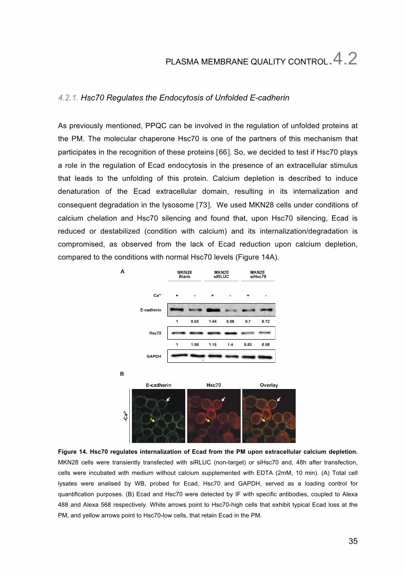
4.2.1. Hsc70 Regulates the Endocytosis of Unfolded E-cadherin
As previously mentioned, PPQC can be involved in the regulation of unfolded proteins at
the PM. The molecular chaperone Hsc70 is one of the partners of this mechanism that
participates in the recognition of these proteins [66]. So, we decided to test if Hsc70 plays
a role in the regulation of Ecad endocytosis in the presence of an extracellular stimulus
that leads to the unfolding of this protein. Calcium depletion is described to induce
denaturation of the Ecad extracellular domain, resulting in its internalization and
consequent degradation in the lysosome [73]. We used MKN28 cells under conditions of
calcium chelation and Hsc70 silencing and found that, upon Hsc70 silencing, Ecad is
reduced or destabilized (condition with calcium) and its internalization/degradation is
compromised, as observed from the lack of Ecad reduction upon calcium depletion,
compared to the conditions with normal Hsc70 levels (Figure 14A).
Figure 14. Hsc70 regulates internalization of Ecad from the PM upon extracellular calcium depletion.
MKN28 cells were transiently transfected with siRLUC (non-target) or siHsc70 and, 48h after transfection,
cells were incubated with medium without calcium supplemented with EDTA (2mM, 10 min). (A) Total cell
lysates were analised by WB, probed for Ecad, Hsc70 and GAPDH, served as a loading control for
quantification purposes. (B) Ecad and Hsc70 were detected by IF with specific antibodies, coupled to Alexa
488 and Alexa 568 respectively. White arrows point to Hsc70-high cells that exhibit typical Ecad loss at the
PM, and yellow arrows point to Hsc70-low cells, that retain Ecad in the PM.
36
To evaluate if Hsc70 regulates the internalization by endocytosis of unfolded WT Ecad at
the PM, we performed an immunofluorescence (IF) against Ecad and Hsc70, in the same
conditions of the previous experience. We observe a loss of Ecad at the PM in cells that
exhibit high levels of Hsc70, in contrast to the cells in which the expression of Hsc70 is
decreased (Figure 14B).
4.2.2. Unfolded E-cadherin Leads to a Recruitment of Hsc70 and CHIP to the
Plasma Membrane
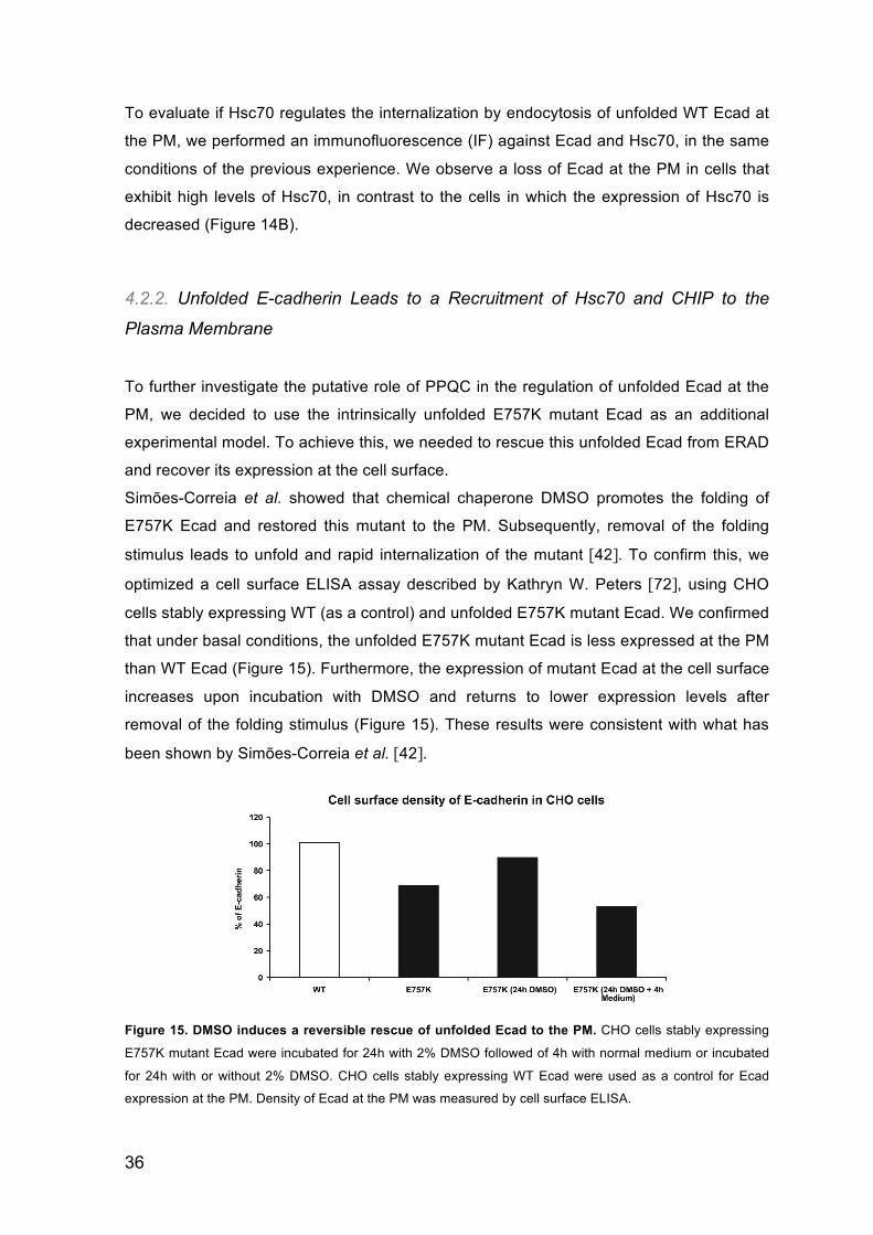
To further investigate the putative role of PPQC in the regulation of unfolded Ecad at the
PM, we decided to use the intrinsically unfolded E757K mutant Ecad as an additional
experimental model. To achieve this, we needed to rescue this unfolded Ecad from ERAD
and recover its expression at the cell surface.
Simões-Correia et al. showed that chemical chaperone DMSO promotes the folding of
E757K Ecad and restored this mutant to the PM. Subsequently, removal of the folding
stimulus leads to unfold and rapid internalization of the mutant [42]. To confirm this, we
optimized a cell surface ELISA assay described by Kathryn W. Peters [72], using CHO
cells stably expressing WT (as a control) and unfolded E757K mutant Ecad. We confirmed
that under basal conditions, the unfolded E757K mutant Ecad is less expressed at the PM
than WT Ecad (Figure 15). Furthermore, the expression of mutant Ecad at the cell surface
increases upon incubation with DMSO and returns to lower expression levels after
removal of the folding stimulus (Figure 15). These results were consistent with what has
been shown by Simões-Correia et al. [42].
Figure 15. DMSO induces a reversible rescue of unfolded Ecad to the PM. CHO cells stably expressing
E757K mutant Ecad were incubated for 24h with 2% DMSO followed of 4h with normal medium or incubated
for 24h with or without 2% DMSO. CHO cells stably expressing WT Ecad were used as a control for Ecad
expression at the PM. Density of Ecad at the PM was measured by cell surface ELISA.
37
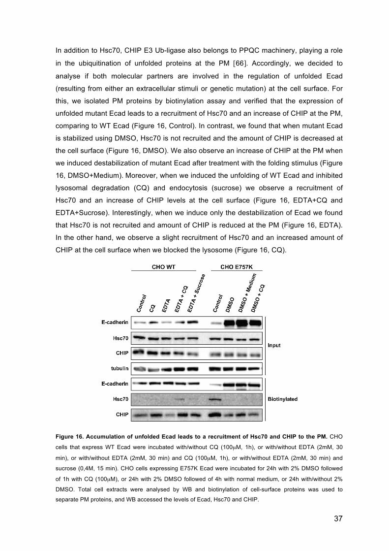
In addition to Hsc70, CHIP E3 Ub-ligase also belongs to PPQC machinery, playing a role
in the ubiquitination of unfolded proteins at the PM [66]. Accordingly, we decided to
analyse if both molecular partners are involved in the regulation of unfolded Ecad
(resulting from either an extracellular stimuli or genetic mutation) at the cell surface. For
this, we isolated PM proteins by biotinylation assay and verified that the expression of
unfolded mutant Ecad leads to a recruitment of Hsc70 and an increase of CHIP at the PM,
comparing to WT Ecad (Figure 16, Control). In contrast, we found that when mutant Ecad
is stabilized using DMSO, Hsc70 is not recruited and the amount of CHIP is decreased at
the cell surface (Figure 16, DMSO). We also observe an increase of CHIP at the PM when
we induced destabilization of mutant Ecad after treatment with the folding stimulus (Figure
16, DMSO+Medium). Moreover, when we induced the unfolding of WT Ecad and inhibited
lysosomal degradation (CQ) and endocytosis (sucrose) we observe a recruitment of
Hsc70 and an increase of CHIP levels at the cell surface (Figure 16, EDTA+CQ and
EDTA+Sucrose). Interestingly, when we induce only the destabilization of Ecad we found
that Hsc70 is not recruited and amount of CHIP is reduced at the PM (Figure 16, EDTA).
In the other hand, we observe a slight recruitment of Hsc70 and an increased amount of
CHIP at the cell surface when we blocked the lysosome (Figure 16, CQ).
Figure 16. Accumulation of unfolded Ecad leads to a recruitment of Hsc70 and CHIP to the PM. CHO
cells that express WT Ecad were incubated with/without CQ (100µM, 1h), or with/without EDTA (2mM, 30
min), or with/without EDTA (2mM, 30 min) and CQ (100µM, 1h), or with/without EDTA (2mM, 30 min) and
sucrose (0,4M, 15 min). CHO cells expressing E757K Ecad were incubated for 24h with 2% DMSO followed
of 1h with CQ (100µM), or 24h with 2% DMSO followed of 4h with normal medium, or 24h with/without 2%
DMSO. Total cell extracts were analysed by WB and biotinylation of cell-surface proteins was used to
separate PM proteins, and WB accessed the levels of Ecad, Hsc70 and CHIP.
38
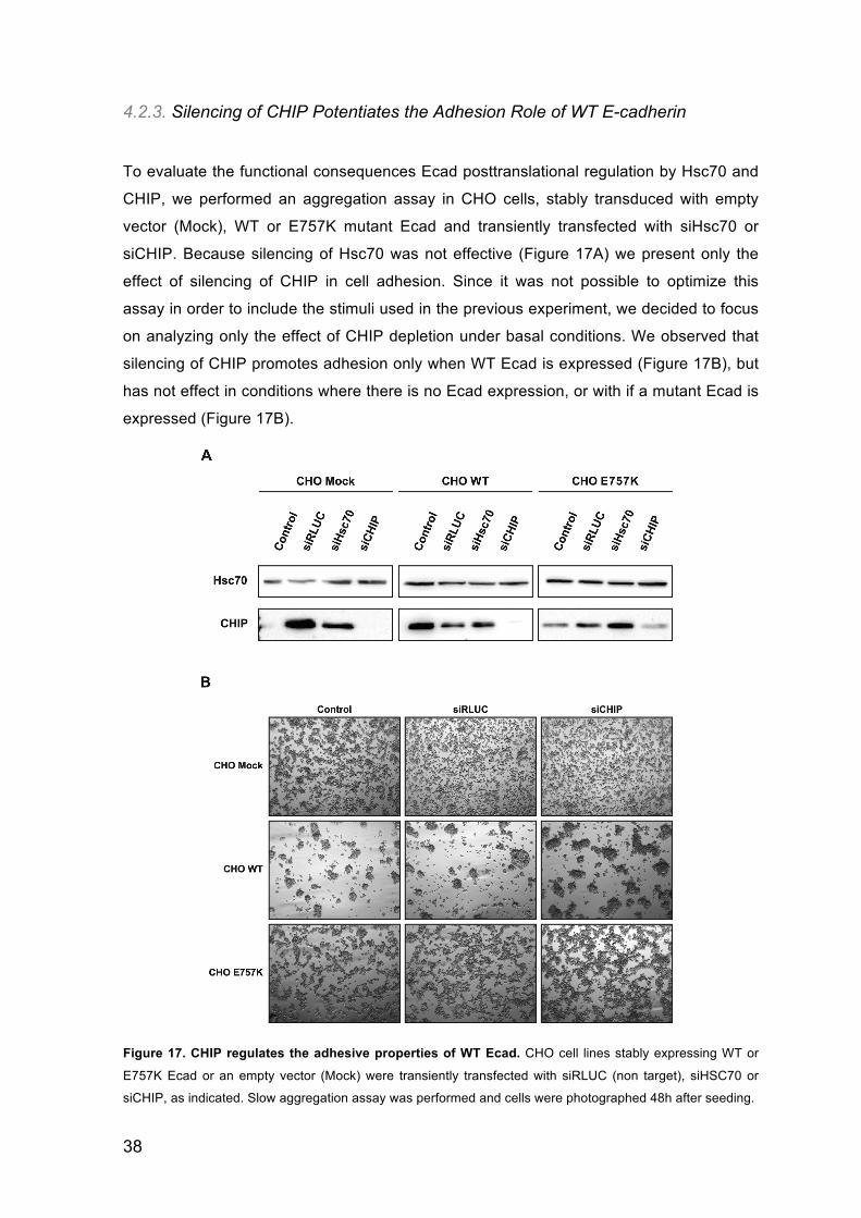
4.2.3. Silencing of CHIP Potentiates the Adhesion Role of WT E-cadherin
To evaluate the functional consequences Ecad posttranslational regulation by Hsc70 and
CHIP, we performed an aggregation assay in CHO cells, stably transduced with empty
vector (Mock), WT or E757K mutant Ecad and transiently transfected with siHsc70 or
siCHIP. Because silencing of Hsc70 was not effective (Figure 17A) we present only the
effect of silencing of CHIP in cell adhesion. Since it was not possible to optimize this
assay in order to include the stimuli used in the previous experiment, we decided to focus
on analyzing only the effect of CHIP depletion under basal conditions. We observed that
silencing of CHIP promotes adhesion only when WT Ecad is expressed (Figure 17B), but
has not effect in conditions where there is no Ecad expression, or with if a mutant Ecad is
expressed (Figure 17B).
Figure 17. CHIP regulates the adhesive properties of WT Ecad. CHO cell lines stably expressing WT or
E757K Ecad or an empty vector (Mock) were transiently transfected with siRLUC (non target), siHSC70 or
siCHIP, as indicated. Slow aggregation assay was performed and cells were photographed 48h after seeding.

39
DISCUSSION.05

40
41
DISCUSSION.05
Ecad is a calcium dependent cell adhesion molecule. It is the major component of AJ and
is essential for the establishment and maintenance of polarization and differentiation of
embryonic and adult epithelial tissues [7,42]. Mutations in Ecad gene are often associated
with cancer progression and poor patient prognosis [7,42,30]. An example that illustrates
the carcinogenic potential of these mutations is HDGC. A small proportion (28%) of
germline mutation identified in HDGC are missense that gives rise to single AA
substitution, resulting in a codon that codes for a different AA [74]. HDGC-associated
CDH1 germline missense mutations often lead to folding defects of Ecad that are
surveyed by quality control mechanisms [42,45]. These mechanisms play an important
role in the regulation of Ecad expression and function.
Simões-Correia et al. demonstrated that mutants of Ecad associated to the HDGC are
subjected to the ER quality control that leads a loss of Ecad surface expression. However,
a small fraction of these mutants escape ERAD and are trafficked to the PM without
achieving proper folding [42].
Recent studies suggest that unfolded proteins at the cell surface that either result from
genetic mutations or denaturing extracellular stimuli can be regulated by PM quality
control mechanisms [66-67].
In this work, we explored the role of the molecular chaperone DNAJB4 in the regulation of
Ecad associated to the ER and investigated if unfolded Ecad could be regulated by PPQC
mechanisms at the PM.
5.1. Endoplasmic Reticulum Quality Control
ER quality control plays a major role in Ecad regulation [42]. However, the molecular
chaperones involved in the specific degradation of Ecad were not known.
From a genetic screen for Ecad interactors, in the Drosophila fly, we identified DnaJ-1
(human homolog DNAJB4) as a partner that may be related with folding, stability and/or
protein degradation (data not shown).
DNAJB4 is an Hsp40-like molecular chaperone that was previously described to act as a
TSG in NSCLC. In this context, DNAJB4 promotes the indirect induction of Ecad
expression at the transcriptional level, by downregulation of its transcriptional repressor
42
Slug [65,75]. Interestingly, it has also been shown that in lung cancer cell models,
curcumin induces transcription of DNAJB4 and increases Ecad expression [76]. Because
curcumin is described to act as a chemical chaperone [77] and is an inducer of the heat
shock response [78], we hypothesized that the regulation of Ecad by DNAJB4 expression
could also happen at the posttranslational level. To prove our hypothesis, we used
cadherin-null CHO cell lines stably transduced with WT or unfolded E757K mutant Ecad
lacking the proximal promoter region subjected to regulation by the transcriptional
repressor Slug.
After subcellular protein fractionation, DNAJB4 was detected in citoplasmic, membranous
and other (cytoskeleton, nucleous and cromatin) fractions, of CHO cells expressing WT or
unfolded E757K mutant Ecad (Figure 8A). Interestingly, the amount of DNAJB4 is
increased in the membranous fraction of cells that express unfolded mutant Ecad (Figure
8A). This fraction includes at least PM and ER proteins (as inferred by the presence of
specific markers). However, this increase is not observed at the PM, as inferred from the
biotynilation assay, used to detect cell surface proteins (Figure 8B), suggesting that the
enrichment of DNAJB4 in the membranous fraction is associated to other membranes
(e.g. ER) and not to increased PM recruitment. Previously other authors raised the
hypothesis that DNAJ proteins are associated with ER [58].
Using cicloheximide to inhibit protein synthesis, we show that DNAJB4 overexpression
reduces the half-life on unfolded E757K mutant Ecad (Figure 9). This result supports the
hypothesis that DNAJB4 regulates Ecad at the posttranslational level, inducing unfolded
Ecad degradation.
We also demonstrate that DNAJB4 interacts with Ecad and this interaction is increased
under proteasome inhibition (Figure 10A, DNAJB4+MG132) suggesting that DNAJB4
preferentially interacts with the proteasomal degradation-prone fraction of Ecad. In the
opposite direction, when Ecad is stabilized by chemical chaperone treatment, the
interaction between Ecad and DNAJB4 is decreased (Figure 10A, DNAJB4+DMSO). This
confirms that DNAJB4 preferentially binds unfolded Ecad. Moreover, the overexpression
of DNAJB4 increases the fraction of Ecad degraded in the proteasome (Figure 10B,
DNAJB4+MG132) suggesting that it mediates the degradation of unfolded Ecad by the
proteasome. Interestingly, DNAJB4 indirectly potentiates the DMSO effect of Ecad
stabilization (Figure 10B, DNAJB4+DMSO). This is consistent with other authors, where it
is described that DMSO promotes the multiple functions of DNAJB4 [75].
After we have partially elucidated the mechanism whereby DNAJB4 influences Ecad
stability at the posttranslational level, we investigated the functional consequences of this
regulation. We observe that overexpression of DNAJB4 is not sufficient to increase cell
43
adhesion in the absence of Ecad or in the presence of the non-functional unfolded E757K
mutant Ecad, but induce increased cell aggregation in the presence of WT Ecad (Figure
11A) suggesting that its pro-adhesive role is cadherin-dependent. This was expected,
because it increases the amount of WT Ecad in the PM (data not shown). To further
analyse the role of DNAJB4 in cell migration, we used the wound healing assay. The
results show that, in conditions of WT Ecad without transcriptional regulation, DNAJB4 is
not enough to reduce cell migration (Figure 11B), indicating that Ecad dominantes over
DNAJB4 for the regulation of cell migration.
Analysis of the neovascularizing potential of the human cells over the CAM shows that
DNAJB4 is anti-angiogenic, and that this tumor suppressor feature is also Ecad-
dependent (Figure 12). Labeling of these cells inoculated in the CAM revealed that
DNAJB4 stimulates the anti-invasive function of WT Ecad, but this invasion-suppressor
potential is lost if Ecad is not expressed (Figure 13), suggesting that in the GC model
Ecad is the dominant anti-invasive molecule.
5.2. Plasma Membrane Quality Control
PPQC is a recently described specialized pathway for the regulation of unfolded proteins
at the PM. Molecular components of this pathway include chaperones, co-chaperones
and ubiquitinating enzymes [66]. In this work we sought to test the hypothesis that
unfolded Ecad at the cell surface can be regulated by PPQC components such as Hsc70
and CHIP.
To clarify this hypothesis, we used two different experimental models: unfolded WT Ecad
at the PM, resulting from an extracellular stimuli (calcium depletion), and an intrinsically
unfolded mutant Ecad, rescued to the cell surface by treatment with the chemical
chaperone DMSO.
Under conditions of extracellular calcium depletion, we observed a decrease in the total
levels of Ecad (Figure 14A). However, in cells with stable silencing of Hsc70, the amount
of Ecad is not affected under the same conditions (Figure 14A). Additionally, after calcium
chelation there was a loss of Ecad at the PM in cells that exhibit high levels of Hsc70, in
contrast to the cells in which the expression of Hsc70 is decreased (Figure 14B),
indicating that Hsc70 regulates the endocytosis of unfolded Ecad. Some studies
demonstrated that extracellular calcium depletion is one of main factors that significantly
increase the process of Ecad clathrin-mediated endocytosis [73], namely because it is
recognized as a denaturing stimulus for the extracellular domain of Ecad denaturation.
44
These results suggest that Hsc70 is likely to be involved in the recognition of unfolded
Ecad at the PM for internalization and subsequent degradation. This is in accordance with
the theoretical model of PPQC, where Hsc70 is predicted to play a role in the recognition
of the unfolded protein substrates. However, Hsc70 also plays multiple roles in the
endocytic pathway. First Hsc70 is required for budding of Clathrin-Coated Vesicles (CCV).
It then participates in uncoating of CCV to allow the fusion of these vesicles with the early
endosome. Finally, Hsc70 may be involved in the rebinding of clathrin to the PM to form
new CCV [79, 80]. Given that Ecad undergoes clathrin-mediated endocytosis, our results
might also suggest that the Hsc70 effect in Ecad endocytosis is dependent on its
chaperoning role over clathrin, and not directly on unfolded Ecad. Therefore, it is not clear
if Hsc70 is indeed playing a role in the recognition of unfolded Ecad at the PM, or
generally influencing clathrin-dependent endocytosis.
Through biotinylation of cell-surface proteins we show that the expression of unfolded
mutant Ecad leads to a recruitment of Hsc70 and an increase of CHIP at the PM,
comparing to the conditions with WT Ecad expression (Figure 16, Control). In contrast,
when mutant Ecad is stabilized using DMSO, Hsc70 is not recruited and the amount of
CHIP is decreased at the cell surface (Figure 16, DMSO). In the other hand, when we
remove the stabilizing stimulus for mutant Ecad the levels of CHIP increase (Figure 16,
DMSO+Medium). Moreover, when we induced the unfolding of WT Ecad and inhibited
endocytosis/lysosomal degradation we verify a recruitment of Hsc70 and an increase of
CHIP levels at the cell surface (Figure 16, EDTA+Sucrose and EDTA+CQ). Taken
together, these results show that Hsc70 and CHIP are dynamically recruited to the PM in
response to Ecad unfolding and suggest their participation in its regulation at the PM.
Interestingly, in cells expressing WT Ecad, calcium depletion per se did not result in the
recruitment of Hsc70 and reduced the amount of CHIP at the PM (Figure 16, EDTA). This
may be a consequence of the rapid internalization of membrane proteins upon the
unfolding stimulus. As the affected proteins enter the endocytic pathway, only the properly
folded membrane proteins (wich are not targeted by the PPQC machinery) are left in the
membrane, thus decreasing the levels of Hsc70 and CHIP at this site. In the other hand,
we observe a slight recruitment of Hsc70 and an increased amount of CHIP at the cell
surface when we blocked the lysosomal degradation (Figure 16, EDTA+CQ) or
endocytosis (Figure 16, EDTA+Sucrose). These results suggest that Hsc70 and CHIP are
recruited when we accumulate unfolded Ecad at the PM.
Because CHIP could regulate Ecad stability at the posttranslational level, we investigated
the functional consequences of this regulation, and observed that silencing of CHIP
potentiates de adhesive properties of WT Ecad (Figure 17B). Although cells were not
exposed to an unfolding stimulus, this result is in line with the hypothesis that CHIP does

45
regulate Ecad at the PM and is likely to reflect the role of CHIP in targeting damaged WT
Ecad for degradation.

46

47
CONCLUDING REMARKS.06

48
49
CONCLUDING REMARKS.06 We conclude that DNAJB4 is a molecular chaperone of Ecad. This newly described
interaction partner is able to distinguish between properly folded Ecad and its unfolded
counterpart (e.g. resulting from hereditary/sporadic mutation, or unfolding due to a stress
insult during cancer progression), directing Ecad towards the PM (WT Ecad) or to the
proteasome for degradation (unfolded Ecad).
The posttranslational regulation of Ecad by DNAJB4 has functional consequences at the
level of cell adhesion, but not cell migration. Our in vivo data suggests that in GC DNAJB4
has an anti-angiogenic and anti-invasive effect, if Ecad expression is retained.
Recent work reported that DNAJB4 acts as a tumour suppressor in NSCLC. However, in
GC, Ecad is frequently lost or mutated, meaning that the DNAJB4 expression might
promote tumour progression. We believe that DNAJB4 acts as a conditional tumour
suppressor in carcinomas, where its function depends of the presence of Ecad.
In addition, we suggest that PPQC is involved in the regulation of Ecad at the PM, and
that this level of regulation is especially relevant when Ecad is unfolded due to mutation or
to an external denaturing stimulus.
Indeed, Hsc70 is a regulator of Ecad endocytosis and CHIP regulates the adhesive
properties of Ecad. However, further experiments are required to clarify the roles of these
two key players of PPQC in the regulation of Ecad expression at the PM.
In summary, we conclude that PQC plays a major role in the regulation of Ecad levels and
function. Understanding this mechanism of regulation will help us to design new
therapeutic strategies for HDGC and other cancers associated to Ecad mutations.

50

51
REFERENCES.07

52
53
REFERENCES.07
1. van Roy F and Berx J (2008) The cell-cell adhesion molecule E-cadherin. Cell Mol Life
Sci, 65:3756-3788.
2. Takeichi M (1995) Morphogenetic roles of classic cadherins. Curr Opin Cell Biol 7:
619-627.
3. Yagi T and Takeichi M (2000) Cadherin superfamily genes: functions, genomic
organization, and neurologic diversity. Genes Dev, 14:1169-1180.
4. Gumbiner BM (2000) Regulation of cadherin adhesive activity. J Cell Biol, 148:399-
404.
5. Baum B and Georgiou M (2011) Dynamics of adherens junctions in epithelial
establishment, maintenance, and remodeling. J Cell Biol, 192:907-917.
6. Meng W and Takeichi M (2009) Adherens junction: molecular architecture and
regulation. Cold Spring Harb Perspect Biol, 1:a002899.
7. Paredes J, Figueiredo J, Albergaria A, et al. (2012) Epithelial E- and P-cadherins: role
and clinical significance in cancer. Biochim Biophys Acta, 1826:297-311
8. Cavallaro U and Christofori G (2004) Cell adhesion and signaling by cadherins and Ig-
CAMs in cancer. Nat Rev Cancer, 4:118-132.
9. Pertz O, Bozic D, Koch AW, et al. (1999) A new crystal structure, Ca2+ dependence
and mutational analysis reveal molecular details of E-cadherin homoassociation.
EMBO J, Vol. 18:1738-1747.
10. Ozawa M, Ringwald M, and Kemler R (1990) Uvomorulin–catenin complex formation
is regulated by a specific domain in the cytoplasmic region of the cell adhesion
molecule. Proc Natl Acad Sci, 87:4246-4250.
11. Bershadsky A (2004) Magic touch: how does cell-cell adhesion trigger actin
assembly? Trends Cell Biol, 14:589-593.
12. Yonemura SY, Wada T, Watanabe A, et al. (2010) alpha-catenin as a tension
transducer that induces adherens junction development. Nat. Cell Biol, 12:533-542.
13. Baum B and Perrimon N (2001) Spatial control of the actin cytoskeleton in Drosophila
epithelial cells. Nat Cell Biol, 3:883-890.
14. Perez-Moreno M, Jamora C, and Fuchs E (2003) Sticky business: orchestrating
cellular signals at adherens junctions. Cell, 112:535-548
15. Ozawa M, Baribault H, and Kemler R (1989) The cytoplasmic domain of the cell
adhesion molecule uvomorulin associates with three independent proteins structurally
related in different species. EMBO J, 8:1711-1717.
54
16. Gumbiner BM and McCrea PD (1993) Catenins as mediators of the cytoplasmic
functions of cadherins. J Cell Sci Suppl, 17:155-158.
17. Petrova Y, Spano M, and Gumbiner B (2012) Conformational epitopes at cadherin
calcium-binding sites and p120-catenin phosphorylation regulate cell adhesion. Mol
Biol Cell, 2092-2108.
18. Hartsock A and Nelson W (2012) Competitive regulation of E-Cadherin
juxtamembrane domain degradation by p120-catenin binding and Hakai-mediated
ubiquitination. PLoS One, 7: e37476.
19. Nanes BA, MacKenzie CC, Lowery AM, et al. (2012) p120-catenin binding masks an
endocytic signal conserved in classical cadherins. J Cell Biol, Vol. 199:365-380.
20. Bajpai S, Correia J, Feng Y, et al. (2008) α-catenin mediates initial E-cadherin
dependent cell–cell recognition and subsequent bond strengthening. PNAS,
105:18331–18336
21. Angst BD, Marcozzi C, and Magee AL (2001) The cadherin superfamily: diversity in
form and function. J Cell Sci, 114:629-641.
22. Posthaus H, Dubois CM, Laprise MH, et al. (1998) Proprotein cleavage of E-cadherin
by furin in baculovirus over-expression system: potential role of other convertases in
mammalian cells. FEBS Lett, 438:306-310.
23. Beavon IR (2000) The E-cadherin-catenin complex in tumour metastasis: structure,
function and regulation. Eur J Cancer, 36:1607-1620.
24. Ozawa M and Kemler R (1990) Correct proteolytic cleavage is required for the cell
adhesive function of uvomorulin. J Cell Biol, 111:1645-1650.
25. Pece S and Gutkind JS (2002) E-cadherin and Hakai: signalling, remodeling or
destruction? Nat Cell Biol, 4:72-74.
26. Kanyan X, Rebecca GO, Christine M, et al. (2006) Role of p120-catenin in cadherin
trafficking. Biochim Biophys Acta, 1773:8-16.
27. Fujita Y, Scheffner M, Zechner D, et al. (2002) Hakai, a c-Cbl-like protein,
ubiquitinates and induces endocytosis of the E-cadherin complex. Nat Cell Biol, 4:222-
231.
28. Birchmeier W and Behrens J (2001) Cadherin expression in carcinomas: role in the
formation of cell junctions and the prevention of invasiveness. Biochim Biophys Acta,
1198:11-26.
29. Mareel M and Leroy A (2003) Clinical, cellular, and molecular aspects of cancer
invasion. Physiol Rev, 83:337-376.
30. Mohamet L, Hawkins K, and Ward CM (2010) Loss of function of E-Cadherin in
embryonic stem cells and the relevance to models of tumorigenesis. J Oncol, 2011:1-
19.
55
31. Larue L and Bellacosa A (2005) Epithelial-mesenchymal transition in development and
cancer: role of phosphatidylinositol 3' kinase/AKT pathways. Oncogene, 24:7443-
7454.
32. Tiwari N, Gheldof A, Tatari M, et al. (2012) EMT as the ultimate survival mechanism of
cancer cells. Semin Cancer Biol, 22:194-207.
33. Machado JC, Oliveira C, Carvalho R, et al. (2001) E-cadherin gene (CDH1) promoter
methylation as the second hit in sporadic diffuse gastric carcinoma. Oncogene,
20:1525-1528.
34. Peinado H, Portillo F, and Cano A (2004) Transcriptional regulation of cadherins
during development and carcinogenesis. Int. J Dev Biol, 48:365-375.
35. Becker KF, Atkinson MJ, Reich U, et al. (1994) E-cadherin gene mutations provide
clues to diffuse type gastric carcinomas. Cancer Res, 54:3845-3852.
36. Guilford P, Hopkins J, Harraway J, et al. (1998) E-cadherin germline mutations in
familial gastric cancer. Nature, 392:402-405.
37. Corso G. et al. (2011) E-cadherin genetic screening and clinico-pathologic
characteristics of early onset gastric cancer. Eur J Cancer, 47:631-639.
38. Carneiro F, Oliveira C, Suriano G, Seruca R (2008) Molecular pathology of familial
gastric cancer, with an emphasis on hereditary diffuse gastric cancer. J Clin Pathol,
61:25-30.
39. Wolf EM, Geigl JB, Svrcek M, et al. (2010) Hereditary gastric cancer. Pathologe,
31:423-429.
40. Balch WE, Morimoto, RI, Dillin, A, and Kelly, JW (2008) Adapting proteostasis for
disease intervention. Science, 319:916-919.
41. Mastoraki A, Danias N, Arkadopoulos N, et al. (2011) Prophylactic total gastrectomy
for hereditary diffuse gastric cancer. Review of the literature. Surg Oncol, 20:223-226.
42. Simões-Correia J, Figueiredo J, Oliveira C, et al. (2008) Endoplasmic reticulum quality
control: a new mechanism of E-cadherin regulation and its implication in cancer. Hum
Mol Genet, 17:3566-3576.
43. Simoes-Correia J, Figueiredo J, Lopes R, et al. (2012) E-cadherin destabilization
accounts for the pathogenicity of missense mutations in hereditary diffuse gastric
cancer. PloS one, 7:e33783.
44. Buchberger A, Bukau B, and Sommer T (2010) Protein quality control in the cytosol
and the endoplasmic reticulum: brothers in arms. Mol Cell, 40:238-252.
45. Chen B, Retzlaff M, Roos T, and Frydman J (2011) Cellular strategies of protein
quality control. Cold Spring Harb Perspect Biol, 3:a004374.
46. Hammond C and Helenius A (1995) Quality control in the secretory pathway. Curr
Opin Cell Biol, 7:523-529.
56
47. Sitia R and Braakman I (2003) Quality control in the endoplasmic reticulum protein
factory. Nature, 426:891-894.
48. Bukau B, Weissman J and Horwich A (2006) Molecular chaperones and protein
quality control. Cell, 125:443-451.
49. Jung T, Catalgol B, and Grune T (2009) The proteasomal system. Mol Aspects Med,
30:191-296.
50. Lilienbaum A (2013) Relationship between the proteasomal system and autophagy. Int
J Biochem Mol Biol, 4:1-26.
51. Weissman AM (2001) Themes and variations on ubiquitylation. Nat Rev Mol Cell Biol,
2:169-178.
52. Lamark T and Johansen T (2012) Aggrephagy: selective sisposal of protein
aggregates by macroautophagy. Int J Cell Biol, 2012:1-21.
53. Komander D (2009) The emerging complexity of protein ubiquitination. Biochem Soc
Trans, 37:937-953.
54. Wong E and Cuervo AM (2010) Integration of clearance mechanisms: the proteasome
and autophagy. Cold Spring Harbor Perspect Biol, 2:a006734.
55. Cuervo AM and Macian F (2012) Autophagy, nutrition and immunology. Mol Asp Med,
33:2-13.
56. Mizushima N, Levine B, Cuervo AM, and Klionsky DJ (2008) Autophagy fights disease
through cellular self-digestion. Nature, 451:1069-75.
57. Meusser B, Hirsch C, Jarosch E, and Sommer T (2005) ERAD: the long road to
destruction. Nat Cell Biol, 7:766-772.
58. Kakoi S, Yorimitsu T, and Sato K (2013) COPII machinery cooperates with ER-
localized Hsp40 to sequester misfolded membrane proteins into ER-associated
compartments. Mol Biol Cell, 24:633-642.
59. Hageman J and Kampinga HH (2009) Computational analysis of the human
HSPH/HSPA/DNAJ family and cloning of a human HSPH/HSPA/DNAJ expression
library. Cell Stress Chaperon 14:1-21.
60. Minami Y, Hohfeld J, Ohtsuka K, and Hartl FU (1996) Regulation of the heat-shock
protein 70 reaction cycle by the mammalian DnaJ homolog, Hsp40. J Biol Chem, 27:
19617-19624.
61. Kampinga HH and Craig EA (2010) The HSP70 chaperone machinery: J proteins as
drivers of functional specificity. Nature reviews. Mol Cell Biol, 11:579-592.
62. Borges JC, Fischer H, Craievich AF, and Ramos CH (2005) Low resolution structural
study of two human HSP40 chaperones in solution. DJA1 from subfamily A and DJB4
from subfamily B have different quaternary structures. J Biol Chem, 280:13671-13681.
57
63. Hageman J, Rujano MA, van Waarde MA, et al. (2010) A DNAJB chaperone subfamily
with HDAC-dependent activities suppresses toxic protein aggregation. Mol Cell,
37:355-369.
64. Tsai MF, Wang CC, Chang GC, et al. (2006) A new tumor suppressor DnaJ-like heat
shock protein, HLJ1, and survival of patients with non-small-cell lung carcinoma. J
Natl Cancer Inst, 98:825-838.
65. Apaja PM, Xu H, Lukacs GL (2010) Quality control for unfolded proteins at the plasma
membrane. J Cell Biol, 191:553-570.
66. Okiyoneda T, Barriere H, Bagdany M, et al. (2010) Peripheral protein quality control
removes unfolded CFTR from the plasma membrane. Science, 329:805-810.
67. Okiyoneda T, Apaja PM, Lukacs GL (2011) Protein quality control at the plasma
membrane. Curr Opin Cell Biol, 23:483-491.
68. Dickey CA, Patterson C, Dickson D, Petrucelli L (2007) Brain CHIP: removing the
culprits in neurodegenerative disease. Trends Mol Med, 1:32-8.
69. Pratt WB, Morishima Y, Peng HM, and Osawa Y (2010) Proposal for a role of the
Hsp90/Hsp70-based chaperone machinery in making triage decisions when proteins
undergo oxidative and toxic damage. Exp Biol Med (Maywood), 3:278-89.
70. Zhang M, Windheim M, Roe SM, et al. (2005) Chaperoned ubiquitylation-‐crystal
structures of the CHIP U box E3 ubiquitin ligase and a CHIP-‐Ubc13-‐Uev1a complex.
Mol Cell, 20:525-38.
71. Xu Z, Kohli E, Devlin KI, Bold M, Nix JC, and Misra S (2008) Interactions between the
quality control ubiquitin ligase CHIP and ubiquitin conjugating enzymes. BMC Struct
Biol, 8:26-39.
72. Peters KW, Okiyoneda T, Balch HE, et al. (2011) CFTR Folding Consortium: Methods
available for studies of CFTR folding and correction. Methods Mol Biol, 742:335-353.
73. Ivanov AI, Nusrat A, and Parkos CA (2004) Endocytosis of epithelial apical junctional
proteins by a clathrin-mediated pathway into a unique storage compartment. Mol Biol
Cell, 15:176-188.
74. Corso G, Marrelli D, Pascale V, Vindigni C, and Roviello F (2012) Frequency of CDH1
germline mutations in gastric carcinoma coming from high- and low-risk areas:
metanalysis and systematic review of the literature. BMC Cancer, 12, 8.
75. Wang CC, Tsai MF, Hong TM, Chang GC, Chen CY, Yang WM, et al. (2005) The
transcriptional factor YY1 upregulates the novel invasion suppressor HLJ1 expression
and inhibits cancer cell invasion. Oncogene, 24:4081-93.
76. Chen HW, Lee JY, Huang JY, Wang CC et al. (2008) Curcumin inhibits lung cancer
cell invasion and metastasis through the tumor suppressor HLJ1. Cancer research,
68:7428-7438.
58
77. Albuquerque JA, Lamers ML, Castiblanco-Valencia MM, et al. (2012) Chemical
chaperones curcumin and 4-phenylbutyric acid improve secretion of mutant factor H
R127H by fibroblasts from a factor H-deficient patient. J Immunol, 189:3242-3248.
78. Kato K, Ito H, Kamei K, and Iwamoto I (1998) Stimulation of the stress-induced
expression of stress proteins by curcumin in cultured cells and in rat tissues in vivo.
Cell Stress Chaperon, 3:152-160.
79. Chang HC, Newmyer SL, Hull MJ, et al. (2002) Hsc70 is required for endocytosis and
clathrin function in Drosophila. J Cell Biol, 3:477-487.
80. Eisenberg E and Greene LE (2007) Multiple roles of auxilin and Hsc70 in clathrin-
mediated endocytosis. Traffic, 8:640-646.
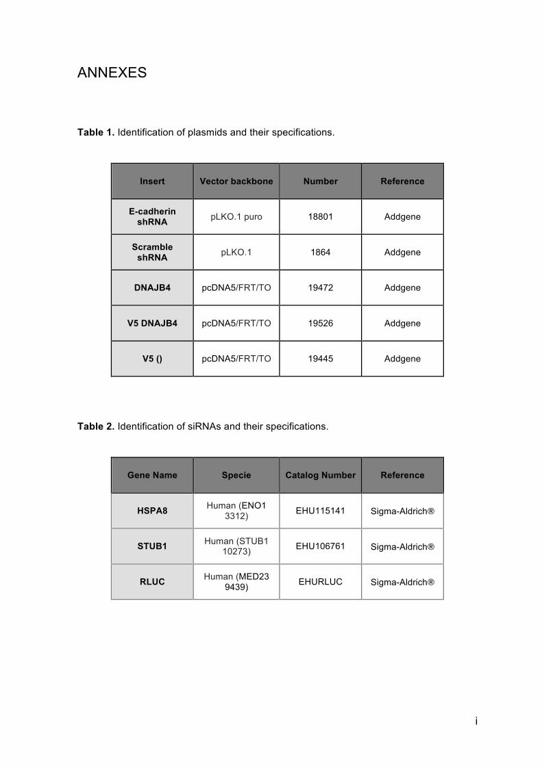
i
ANNEXES Table 1. Identification of plasmids and their specifications.
Insert
Vector backbone Number Reference
E-cadherin shRNA pLKO.1 puro 18801 Addgene
Scramble shRNA pLKO.1 1864 Addgene
DNAJB4 pcDNA5/FRT/TO 19472 Addgene
V5 DNAJB4 pcDNA5/FRT/TO 19526 Addgene
V5 () pcDNA5/FRT/TO 19445 Addgene
Table 2. Identification of siRNAs and their specifications.
Gene Name Specie Catalog Number Reference
HSPA8 Human (ENO1 3312) EHU115141 Sigma-Aldrich®
STUB1 Human (STUB1 10273) EHU106761 Sigma-Aldrich®
RLUC Human (MED23 9439) EHURLUC Sigma-Aldrich®
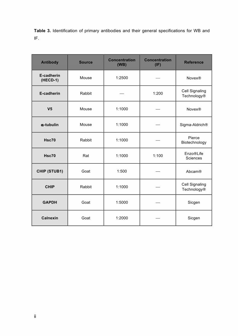
ii
Table 3. Identification of primary antibodies and their general specifications for WB and
IF.
Antibody Source Concentration (WB)
Concentration (IF) Reference
E-cadherin (HECD-1) Mouse 1:2500 ⎯ Novex®
E-cadherin Rabbit ⎯ 1:200 Cell Signaling Technology®
V5 Mouse 1:1000 ⎯ Novex®
α-tubulin Mouse 1:1000 ⎯ Sigma-Aldrich®
Hsc70 Rabbit 1:1000 ⎯ Pierce Biotechnology
Hsc70 Rat 1:1000 1:100 Enzo®Life Sciences
CHIP (STUB1) Goat 1:500 ⎯ Abcam®
CHIP Rabbit 1:1000 ⎯ Cell Signaling Technology®
GAPDH Goat 1:5000 ⎯ Sicgen
Calnexin Goat 1:2000 ⎯ Sicgen
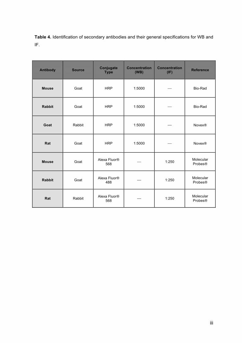
iii
Table 4. Identification of secondary antibodies and their general specifications for WB and
IF.
Antibody Source Conjugate Type
Concentration (WB)
Concentration (IF) Reference
Mouse Goat HRP 1:5000 ⎯ Bio-Rad
Rabbit Goat HRP 1:5000 ⎯ Bio-Rad
Goat Rabbit HRP 1:5000 ⎯ Novex®
Rat Goat HRP 1:5000 ⎯ Novex®
Mouse Goat Alexa Fluor® 568 ⎯ 1:250 Molecular
Probes®
Rabbit Goat Alexa Fluor® 488 ⎯ 1:250 Molecular
Probes®
Rat Rabbit Alexa Fluor® 568 ⎯ 1:250 Molecular
Probes®

iv

v

vi