current medicinal chemistry, 127-151 127 the search of dna
TRANSCRIPT
Current Medicinal Chemistry, 2005, 12, 127-151 127
0929-8673/05 $50.00+.00 © 2005 Bentham Science Publishers Ltd.
The Search of DNA-Intercalators as Antitumoral Drugs: What it Workedand What did not Work
R. Martínez*, 1 and L. Chacón-García2
1Instituto de Química, Universidad Nacional Autónoma de México, Circuito Exterior, Ciudad Universitaria,Coyoacán 04510, México, D. F. México2Instituto de Investigaciones Químico-Biológicas, Universidad Michoacana de San Nicolás de Hidalgo, Edif.-B1,Ciudad Universitaria 58030, Morelia, Mich., México
Abstract: The discovery of new compounds with antitumoral activity has become one of the most importantgoals in medicinal chemistry. One interesting group of chemotherapeutic agents used in cancer therapycomprises molecules that interact with DNA. Research in this area has revealed a range of DNA recognizingmolecules that act as antitumoral agents, including groove binders, alkylating and intercalator compounds.DNA intercalators (molecules that intercalate between DNA base pairs) have attracted particular attention dueto their antitumoral activity. For example, a number of acridine and anthracycline derivatives are excellentDNA intercalators that are now on the market as chemotherapeutic agents. Commercially available acridine andanthracycline derivatives have been widely studied from a variety of viewpoints, such as physicochemicalproperties, structural requirements, synthesis and biological activity. However, the clinical application ofthese and other compounds of the same class has encountered problems such as multidrug resistance (MRD),and secondary and/or collateral effects. These shortcomings have motivated the search for new compounds tobe used either in place of, or in conjunction with, the existing compounds. Unfortunately, the results of thissearch have not met expectations. The vast majority of candidate intercalator compounds tested for use asanticancer agents have shown little or no biological activity. Research in this area has not been withoutbenefits, however, for it has produced much information on the synthesis and antitumoral properties ofhundreds of compounds, which have been tested on diverse tumoral cell lines. This review considers thestructural and biological considerations relevant to the use of DNA intercalators and bis-intercalators asantitumoral agents, with an emphasis on the relationship between structure and activity, produced in lastdecade.
Keyword: DNA- intercalators, cytotoxic compounds, DNA-binding.
INTRODUCTION
Intercalators as Cytotoxic Agents
It is 50 years since Watson and Crick determined thatgenetic material exists structurally as a double helix withnow well-established characteristics [1]. Its role in thecontrol of cellular functions immediately suggested it as anexcellent target for treating illnesses of genetic origin, suchas cancer. The first compounds discovered to act on DNAwere the sulfur mustards, but their high toxicity prompted toa search for less toxic and more efficient compounds [2]. Inthe 1960s, some compounds with cytotoxic activity werediscovered to act as anticancer agents, although theirmechanism of action was unknown. Interestingly, afterLerman reported the occurrence of a noncovalent interactionbetween acridine and DNA, suggesting an intercalativeprocess, it was established that some of these anticanceragents worked by interacting with DNA [3,4].
Intercalators are molecules that insert perpendicularly intoDNA without forming covalent bonds. The only recognizedforces that maintain the stability of the DNA–intercalators
*Address correspondence to this author at the Instituto de Química,Universidad Nacional Autónoma de México, Circuito Exterior, CiudadUniversitaria, Coyoacán 04510, México, D. F. México; E-mail:[email protected]
complex, even more than DNA alone, are van der Waals,hydrogen bonding, hydrophobic, and/or charge transferforces [5,6,7,8]. A frontier orbital interaction has also beensuggested [8]. This means that such a process has thepossibility of being reversed, and as a consequence it musthave an equilibrium constant. It has been argued that the 9-amino group is important in the DNA-recognizing regionbecause of its ability to form hydrogen bonds [5,9].Experiments in which inosine was replaced by guanineindicated the relevance of this group. However, manyintercalators specifically recognize AT bases, which indicatesthat there are factors other than hydrogen bonding toconsider [10]
Based on the difference in stabilities between DNA aloneand its intercalator complex, the stability of DNA whenheated is frequently used to measure DNA intercalation. Themelting temperature (Tm) is taken as the temperature atwhich half of the DNA has denatured. In other words, half ofthe bases of a single strand do not interact in any way withtheir complementary bases on the second strand. In anoligonucleotide, the first bases to break the interaction willbe those in the middle of the strand and not those at theextreme ends. When DNA is subjected to UVspectrophotometry and the temperature is varied, theresulting absorption data (which represents denaturation) is asigmoidal curve, from which Tm can be determined. If thesame DNA sequence or oligonucleotide is complexed with

128 Current Medicinal Chemistry, 2005, Vol. 12, No. 2 Martínez and García
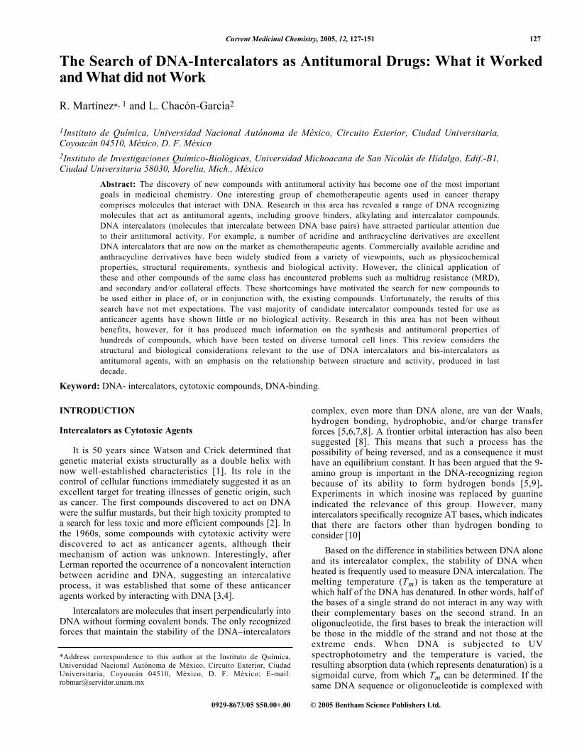
Fig. (1). Curve of temperature Vs. DNA denaturalization in DNA and DNA-Intercalator complex, indicating Tm.
an intercalator and then subjected to the same thermalconditions, another sigmoidal curve is obtained, but thiscurve is shifted to higher temperature compared to thatobtained with the DNA alone, as shown in (Fig. 1). Due tothe higher stability of the complex, Tm will be higher. Themost common forms of DNA that have been examined inthis kind of experiment are calf thymus and salmon spermDNA.
Of course Tm is not selective in the determination ofDNA-intercalation, since it can be influenced by other kindsof molecules that recognize DNA such as groove binders.Methods more selective are viscosity measurements [11] andethidium bromide displacement [12].
Other frequently used methods for the measurement DNAintercalation are circular dichroism (CD) and fluorescencespectroscopy [13,14]. More complicated, but also useful, arenuclear magnetic resonance (NMR) and mass detection usingthe electrospray technique. Finally, the technique thatprovides the most unequivocal results is X-ray diffraction[15]. NMR and X-ray diffraction are arguably the bestmethods by which to examine DNA intercalation. NMR canbe used to extract information on the geometry dynamicsand NMR and X-ray diffraction yield indispensablephysicochemical data; however, they are among the mosttime-consuming and expensive of the available methods.
Measurement of the binding constant and biologicalactivity of DNA-intercalator complexes in the 1970’s, andQSAR studies in the 1980’s, leads to the conclusion thatthere should exist a relationship between cytotoxic activityand binding force [16,17,18]. For some time after Lerman’sdiscovery, the cytotoxic agents were thought to causeinhibition of RNA synthesis as a result of distortion of theshape of the genetic material. However, this and othersimilar proposals remained equivocal, since some potentDNA intercalators, such as daunomycin, did not causeinhibition of RNA synthesis. A satisfactory explanation wasproposed when proteins that were thought to be involved inthe control of the shape of DNA were found in the 1970’s.This group of enzymes, called the topoisomerases (Topos),have helped to clarify the situation, and to date someinteresting conclusions have emerged [19].
Although there is a relationship between the bindingenergy of a DNA intercalator and its biological activity, asdiscussed earlier, cytotoxicity is not only dependent on theability to interact with DNA, since there are many DNAintercalators that are incapable of working as cytotoxicagents. To be effective, a drug must first overcome many
barriers, including metabolic pathways, and cytoplasmic andnuclear membranes. Once the drug is situated in the nucleus,it must be capable of interacting with DNA by intercalatingthat is, forming a stable complex with a relatively long half-life. Achieving entry into the nucleus and forming a DNAcomplex are only the first stages of a series of events thatunderlie the cytotoxic activity of DNA intercalators; thuscytotoxicity is more than just an interaction with DNA.Cytotoxicity is a consequence of the poisoning of Topos,enzymes that are directly involved in DNA recognition, inthe fundamental steps of cellular growth when DNAreplication is active, in the S phase of the cell cycle, inwhich the topology of DNA plays a significant role. Toposalso work and can be poisoned, in the M phase of the cellcycle, arranging the chromatin.
The spatial arrangement of DNA before, during, and afterreplication is essential to a high-quality cell-divisionprocess. In this way, DNA topology is governed by Topos[20]. Three parameters describe the topology of DNA:linking number, twist, and writhe. Topos regulate thetopology of DNA, ensuring that it is arranged adequately forDNA replication by reversibly modifying the linkingnumber, twist, and writhe, breaking one or two strands ofthe DNA, depending on its mechanism of action. Topos canbe classified into two main classes: Topo I, which breaksonly one strand of the DNA [21], although both strands areinvolved in the interaction with the enzyme [22], and TopoII [23], which breaks both strands of the duplex [24,25]. Athird group, Topo III, has also been described but it remainsunclear how the Topos in this group act and to what degreethey are useful with regard to cancer chemotherapy. A fourthclass, known as the gyrases, have been found in bacteria.Topo I and Topo II are good leads for DNA intercalators[26,27]. For an excellent discussion of the mechanisms ofaction of the Topos, we refer the reader to the reviews byPommier, Osheroff and Stivers [28,29,30,31].
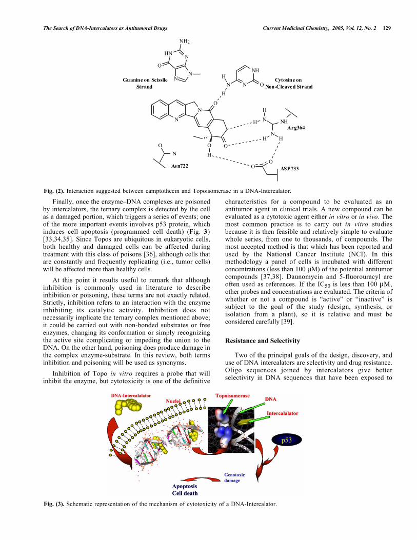
A DNA intercalator has cytotoxic activity when itpoisons the Topo by stabilizing the ternary, DNA–intercalator–Topo complex in such a way that the enzymaticprocess cannot continue forward or backward. The structureof DNA–Topo complexes has been obtained by X-raydiffraction. Some Authors have also suggested that aninteraction occurs between the cytotoxic compoundcamptothecin and a Topo, by interacting with Arg364,Asp533, and Asn722, (Fig. 2). It is noteworthy that this lastamino acid lies in the vicinity of Tyr723, which isresponsible for breaking DNA in the phosphate region [32].
The Search of DNA-Intercalators as Antitumoral Drugs Current Medicinal Chemistry, 2005, Vol. 12, No. 2 129
N
N
O
O
H
N
H
N
NH
O
H N
H
NHH
NH
O
HO
O
N
O
NN
NHN
NH2
O
Asn722 ASP733
Arg364
Guanine on Scisslle Strand
Cytosine on Non-Cleaved Strand
Fig. (2). Interaction suggested between camptothecin and Topoisomerase in a DNA-Intercalator.
Fig. (3). Schematic representation of the mechanism of cytotoxicity of a DNA-Intercalator.
Finally, once the enzyme–DNA complexes are poisonedby intercalators, the ternary complex is detected by the cellas a damaged portion, which triggers a series of events; oneof the more important events involves p53 protein, whichinduces cell apoptosis (programmed cell death) (Fig. 3)[33,34,35]. Since Topos are ubiquitous in eukaryotic cells,both healthy and damaged cells can be affected duringtreatment with this class of poisons [36], although cells thatare constantly and frequently replicating (i.e., tumor cells)will be affected more than healthy cells.
At this point it results useful to remark that althoughinhibition is commonly used in literature to describeinhibition or poisoning, these terms are not exactly related.Strictly, inhibition refers to an interaction with the enzymeinhibiting its catalytic activity. Inhibition does notnecessarily implicate the ternary complex mentioned above;it could be carried out with non-bonded substrates or freeenzymes, changing its conformation or simply recognizingthe active site complicating or impeding the union to theDNA. On the other hand, poisoning does produce damage inthe complex enzyme-substrate. In this review, both termsinhibition and poisoning will be used as synonyms.
Inhibition of Topo in vitro requires a probe that willinhibit the enzyme, but cytotoxicity is one of the definitive
characteristics for a compound to be evaluated as anantitumor agent in clinical trials. A new compound can beevaluated as a cytotoxic agent either in vitro or in vivo. Themost common practice is to carry out in vitro studiesbecause it is then feasible and relatively simple to evaluatewhole series, from one to thousands, of compounds. Themost accepted method is that which has been reported andused by the National Cancer Institute (NCI). In thismethodology a panel of cells is incubated with differentconcentrations (less than 100 µM) of the potential antitumorcompounds [37,38]. Daunomycin and 5-fluorouracyl areoften used as references. If the IC50 is less than 100 µM,other probes and concentrations are evaluated. The criteria ofwhether or not a compound is “active” or “inactive” issubject to the goal of the study (design, synthesis, orisolation from a plant), so it is relative and must beconsidered carefully [39].
Resistance and Selectivity
Two of the principal goals of the design, discovery, anduse of DNA intercalators are selectivity and drug resistance.Oligo sequences joined by intercalators give betterselectivity in DNA sequences that have been exposed to

130 Current Medicinal Chemistry, 2005, Vol. 12, No. 2 Martínez and García
triplex formation by Hoogsteen base pairs [40,41,42,43]. Inpractice, one of the biggest problems is the susceptibility ofphosphate linkage to breakage by nucleases, causing them tobe inactivated. Many efforts have been made to improve theselectivity and stability of triplexes. An interesting proposalis found in the peptide nucleic acids (PNAs), which containpeptide chains or analogs thereof instead of sugar phosphate[44,45]. Resistance to nucleases and high affinity constantshas been observed.
Quadruplexes are also a potential target, since sequencesof guanosine-rich oligos form these complexes with the aidof Hoogsteen base pairs, presumably in Telomeres [46]. Oneclass of molecules found to interact with quadruplexes is thecationic porphyrines. To improve selectivity, manystrategies have been devised including, for example, linkingintercalators to oligo sequences, and antibody-directed andgene-directed enzyme prodrug therapy (ADEPT and GDEPT,respectively) [47].
Drug resistance has lead researchers to synthesize and testmany compounds. The mechanism underlying drugresistance to Topo poisons can be intrinsic or acquired: theformer is due to poor uptake, poor drug activation, orincreased drug catabolism, and the latter has been attributedto an ability to repair the drug-induced damage by increasingthe expression of DNA repair enzymes or by altering Topobinding [48].
Non-Anti-Tumor Activity of Topo Poisons
Topos are also present in bacteria (gyrase), in which theyact in a manner similar to the other Topos enzymes [49].Quinolones are very useful and are probably the bestexample of antibacterial gyrase inhibitors in clinical use[50]. Some viruses also have a code for Topos; in thiscontext, is noteworthy that camptothecin, a potent Topopoison, inhibits human immunodeficiency virus (HIV)transcription. In vitro experiments have also demonstratedthat cellular Topo I enhances HIV-1 reverse transcriptaseactivity [51].
As it can be seen that intercalators have a broad range ofproperties. The variety of compounds in this field is veryextensive and some of them are found in the clinic trials. Inthis review, we will focus on new compounds that act asintercalators or potential intercalators according to theirphysicochemical and structural properties such as flatness.The period search cover publications reported on the lastdecade. For a complement discussion of the theme, excellentreviews of the topic have been written by Braña [52], Haq[53], Denny [54], Graves [55], Holden [56], Bischoff [57]and Wilson [58].
MOLECULES
Acridines
Acridine derivatives constitute an important class ofDNA-intercalating antitumor drugs. Recently, Demeunynck[59], Denny [60] and Tsann-Lang [61] have reviewedproperly this topic.
Heterodimeric molecules that interact atapurinic/apyrimidinic (AP)sites constitute an attractive toolwith which to potentiate the action of known anticanceragents. These molecules are composed of three units: (1) anintercalator for targeting DNA, (2) a nucleic acid base for therecognition of the abasic site, and (3) a polyamino linkerthat is able to stabilize the drug–DNA complex byelectrostatic interaction with the phosphate backbone [62].One example of these molecules are compounds 1-6, inwhich a 2,6-diaminopurine is linked to a 9-aminoacridinechromophore [63]. The linking chain contains a central N,N-disubstituted guanidine connected to the two chromophoresvia polymethylene units of variable length. The moleculesbind strongly to native DNA, with the exception of 3,however, this compound causes stabilization of the abasicTX duplex at a ratio of 1:1. These data suggest a veryspecific interaction of 3 at the abasic site. The relativeaffinity of these heterodimers for abasic sites was determinedon synthetic oligonucleotides containing a chemically stableanalog of the abasic lesion. Cytostatic activity on L1210cells was weak and cytotoxicity on A549 cells wasmoderate. No apparent relationship was found between thelength of the linker and toxicity.
N
N N
N
(CH2)n
H2 N
HN
NH
HN
(CH2)m
NH
N
OMe
Cl
1: n = 2, m = 32: n = 2, m = 43: n = 3, m = 34: n = 3, m = 4
5: n = 4, m = 36: n = 4, m = 4
Solid-phase synthesis has improved the rate of discoveryof small molecules that bind to DNA [64]. Threadingintercalation is a binding mode wherein the intercalatinggroup directs substituents into both grooves of a duplexsimultaneously [65]. 9-Anilinoacridine-4-carboxamidesbelong to this group of intercalators [66]. The synthesis ofan acridine–peptide conjugate 7 using solid-phasemethodology has been described recently [67]. Thisnonnatural amino acid shows a bathochromic shift in itsvisible absorption spectrum in the presence of DNAconsistent with intercalative binding to DNA.
NH
NH
NH
NH2
O
O
NH3+
NH
OHN
O
H3 N
HO
7

The Search of DNA-Intercalators as Antitumoral Drugs Current Medicinal Chemistry, 2005, Vol. 12, No. 2 131
n
N C C C N C C
C S C C N C C
O
R
R O N
NO C C N C C OH
C C N C C OH
macrocyclic bis-9-aminoacridine2 Amentantrone (R = H)
Miltoxantrone (R = OH)
Fig. (4). Molecular formula of the macrocyclic bisacridine and mithoxanthrone.
X-ray diffraction analysis of the complexes betweenmacrocyclic bisacridine and the antitumor intercalatorametantrone, (Fig. 4) and CGTACG showed that only oneacridine of the bisacridine drug binds at the C5pG6 step ofthe DNA, with the other acridine plus both linkers beingcompletely disordered [68]. An unusual intercalationplatform is formed by bringing four complexes together(involving 222 symmetry) such that the intercalator cavity isflanked by two sets of GC base pairs (i.e., C5G8 andG6C7*) on each side, joined together by G6G8* tertiarybase-pairing interactions. In the bisacridine-CGTACGcomplex, the intercalation platform is intercalated with twoacridines, whereas in the amentantrone-CGTACG complex,only one amentantrone is bound. NMR titration of thebisacridine to AACGATCGTT suggests that the bisacridineprefers to bridge more than one DNA duplex by intercalatingeach acridine to different duplexes.
In the search for spin-labeled intercalators that can beintroduced into DNA-binding conjugates, and to study theirfate in the cell, a nitroxide group was introduced at position2 of 9-phenoxyacridines 8. Reaction of this intermediatewith a suitable amine gave a labeled 9-amino-substitutedacridine conjugate 9. Moreover, the binding properties ofthis conjugate do not suffer appreciable alterations [69].
NCl
O
OC6H5
N
O*
N
Cl
O
HN
N
N
N N
(H2 C
)2
NH2
NH
(CH
2 )3
NH
(H3 C
)3
NO*
2
1 0
6
92
6
9
8
9
With the aim of improving the water solubility andDNA-interacting properties of potential candidates for Boronneutron capture therapy, compound 10 was synthesized.These compounds have an acridine system that would serve
as a DNA intercalating fragment, and a spermidine residuethat would function both as a water-solubilizing and DNA-interacting element [70].
HN NH2 (CH2)3NH(H2C)3H2N
Cl
Cl
Cl
Cl
10
Imidazoacridinones are cytotoxic in vitro and in vivoagainst human and murine cell lines. The most prominentanalog, C-1311, is currently being tested in clinical trials[71]. It has been found that C-1311 must bind noncovalentlyto DNA to induce its cytotoxic effects. On the other hand,irreversible binding to DNA in the cell-free system occursonly in the presence of the horseradish peroxidase (HPR) andhydrogen peroxide(H2O2) system, in a manner dependent onthe drug: H2O2 ratio [72]. In the case of ratios of 1:3 and1:5, the reaction gave highly reactive species that werequickly transformed into the products p2 and p3, which wereunable to intercalate into DNA (Fig. 5). In the presence ofDNA, C-1311 first intercalated into DNA, and theintercalated compound was then oxidized. This oxidationproduced only one product. Since peroxidase-type enzymesare present in the cell nucleus, the proposed sequence ofevents may also be expected to take place in the cellularenvironment in vivo.
In contrast, studies of drug-DNA interactions ofcompounds 11 that share the same core structure of C-1311,with two varying elements (ring substitution in positions 5,7, or 8, and a diaminoalkyl side chain of variable length)showed that all such derivatives bind to DNA byintercalation. However, none of the analyzed drug–DNAbinding parameters was significantly correlated with thebiological activity of the drug [73]. Likewise, the need for atechnique to analyze C1311 in mouse and human plasmaleads to the development of a high-performance liquidchromatography method. This method is selective, sensitive,and reproducible [74].
Synthesis of a series of bis{[(dihydroacridine-4-carbonyl)amino]alkyl}alkylamines 12 was achieved and theirantiproliferative activity was tested against HT-29 cell lines[75]. Both 12a and 12b , showed interesting cytotoxicprofile. DNA experiments suggest that the high cytotoxicactivity of these compounds may be related to their strongbis-intercalative binding to DNA. Modelling studies on 12a

132 Current Medicinal Chemistry, 2005, Vol. 12, No. 2 Martínez and García
N
R8
R9
O
N
HN(CH2)n N
R
R
R1
NH
NH
YNH
O
R
O
NH
R OO
12
R Y
a H -(CH2)2-N(CH3)-(CH2)3-N(CH3)-(CH2)2-b Cl -(CH2)2-N(CH3)-(CH2)3-N(CH3)-(CH2)2-c NO2 -(CH2)2-N(CH3)-(CH2)3-N(CH3)-(CH2)2-d NH2 -(CH2)2-N(CH3)-(CH2)3-N(CH3)-(CH2)2-
1
58
9
R n R1 R8 R9C-1176 CH3 2 H H HC-1212 CH3 3 H H HC-1263 CH3 2 H OH HC-1266 CH3 5 H H HC-1310 C2H5 2 CH3 OH HC-1311 C2H5 2 H OH HC-1330 C2H5 2 H OCH3 HC-1371 CH3 3 H OH HC-1415 C2H5 2 H H HC-1419 C2H5 2 H H OHC-1492 CH3 5 H OH HC-1554 C2H5 2 H CH3 HC-1558 C2H5 2 H t-Bu H
11
demonstrating the marked preference of its chromophores tobind in parallel orientation relative to each to other andpreferentially through the minor groove of a DNA hexamer.An excellent revision on the synthesis and antitumor activityof pyrazolo and pyrimidinoacridines has been published byAntonini´s group [76].
N
HO
O NHCH2CH2 N(CH2CH3 )2
N
C1311
Fig. (5). HRP-mediated activation of C-1311 with differentdrug: H2O2 ratios.
Alkaloids
The pentacyclic DNA-intercalating alkaloids are veryinteresting natural products because of their cytotoxicproperties. In 1994, seven pyridoacridine alkaloids –dehydrokuanoniamine B (13), shermilamine C (14) ,cystodytin J (15), cystodytin A (16), kuanoniamine D (17),shermilamine B (18), and eilatin (19)- were isolated from aFijian ascidian (Cystodytes sp) [77]. These compounds,along with diplamine (20 ), exhibit dose-dependentinhibition of proliferation in human colon tumor cells invitro. In addition, all compounds inhibited the Topo-II-mediated decatenation of kinetoplast DNA in a dose-dependent manner. This ability correlated with theircytotoxic potencies and their ability to intercalate into calfthymus DNA. Cystodytin J and diplamine are the bestintercalators, and Topo II inhibitors are the most potentcytotoxins of the series; two of their structural characteristicsare that they have only four rings and are iminoquinones. Ithas also been reported that ascididemin (ASC) isolated fromthe Mediterranean ascidian Cystodytes dellechiajei, as wellfrom Didemnum species [78, 79], is also highly toxic toseveral cancer cells lines [80]. Furthermore, ASC stimulatesTopo-II-induced DNA cleavage preferentially at sites thatpossess a C on the 3´ side of the cleaved bond; in contrast,ASC has minimal, if any, effects on Topo I. However, it hasbeen demonstrated that neither Topo I nor Topo II could beconsidered as a potential cellular target for ASC [81]. Thisalkaloid also induces apoptosis in HL-60 and P388 leukemiacells, an effect that is mediated by the enzyme caspase-3.Therefore, an alternative mechanism of cytotoxicity shouldbe considered. Recently, another study has indicated thatASC-mediated DNA cleavage can occur via the productionof reactive oxygen species [82]. Cryptolepine is anindoloquinoline alkaloid that was first isolated from theroots of Cryptolepsis triangularis, and afterward from C.sanguinolenta [83, 84, 85, 86]. It has been reported thatcryptolepine has many pharmacological properties, such asanti-inflammatory, antibacterial, hypotensive, antipyretic,and antimalarial properties. Cryptolepine also possesses

The Search of DNA-Intercalators as Antitumoral Drugs Current Medicinal Chemistry, 2005, Vol. 12, No. 2 133
NH
N
Cl
N
RHN
SN
HN
N
RHNHN
S
NH
O
N
RHNN
O
N
HNN
O
MeS
O
N
N
N
NH
Cryptolepine
13 R= -COCH=C(CH3)2 14 R= -COCH=C(CH3)2 15 R= -COCH3
17 R= -COCH3 18 R= -COCH3 16 R= -COCH=C(CH3)2
19 20
cytotoxic activity and inhibits DNA synthesis in B16melanoma cells. In addition, it is a potent inhibitor of TopoII [87, 88]. Recently, it was reported that cryptolepine bindsto DNA in a different manner to other intercalators;specifically it binds to CG-rich sequences containing noalternating CC sites [89].
Anthracyclines
Doxorubicin 21 is the precursor of anthracyclinemolecules [90]. Although the majority of these compoundsare potential DNA intercalators, this property correlatespoorly with their anticancer activity [91]. Structuralmodification is an effective approach that has been used bothto understand the mechanism of drug action and as a route todesigning better drugs. So, the simple anthracycline analogs22 were prepared. The main structural modifications todoxorubicin that were effected are that the basic chain linkedto the anthraquinone aglycone was not restrictedstereochemically, a fluorine atom was inserted at position 2or 3 and the cyclohexane moiety is missing. Preliminaryevaluation of drugs that have been designed as inhibitors ofin vitro growth of the P388 cell line demonstrated a potencyof one order of magnitude less than that of daunosaminylderivatives [92].
In the light of this result, it was reasoned that if a linkerjoined two molecules of 22 it might be possible to create anew class of DNA bis-intercalators 23, and consequentlythese new compounds should have enhanced affinity forDNA. However, preliminary results suggest that the bis-intercalating molecules 23 are less effective inhibitors of invi tro growth in the P388 cell line than the simpleanthraquinones described above [93].
OMe
O
O
OH
OH
CH2OH
O
OH
O
OH3C
OHNH2 21
The bioisosteric modification of the C-2 carbon atom ofthe anthracenedione chromophore by a nitrogen atomproduces the aza derivative 24 [94]. This compoundintercalates into poly (dA-dT) poly (dA-dT), and poly (dA)poly (dT) sequences, inhibiting the DNA gyrase and theactivity of mammalian Topos I and II.
A parallel study of the behavior under physiologicalconditions of doxorubicin and the novel disaccharide MEN10755 showed that their chemical properties in solution andthe pH-dependence in their properties are practically identical[95]. These results imply that the presence of a second sugarmoiety and the different spatial location of the chargedamino group on MEN 10755 do not appreciably modify thestability of DNA adducts. In addition, both compounds actas inhibitors of Topo II, through MEN 10755 seems to bemore effective. Preliminary studies have revealed significantdifferences in the pharmacokinetic behavior of MEN 10755compared to doxorubicin. A revision on the synthesis andantitumor activities of anthracyclines has been published byBineschi [96].

134 Current Medicinal Chemistry, 2005, Vol. 12, No. 2 Martínez and García
O
O
OH
OH
(CH2)n
O
O
OH
OHO O
NH3H3NCl
Cl
O
O
OH
OH
R1
R2
O
O
H3NCl
23 n = 2, 4, 6
22a: R1 = R2 = H22b: R1 = F, R2 = H22c: R1 = H, R2 = F
O
O
NHCO(CH2 )2 NHR2
R2HN(H2C)2OCHN
O
O
R8
R5
NH(CH2)2NHR
R4
2
6
25a : R4 = R5 = R8= H
25b : R5 = R8= H; R4 =NH(CH2)2NH+Et225c : R4 = R8= H; R5 =NH(CH2)2NH+Et225d : R4 = R5= H; R8 =NH(CH2)2NH+Et2
26a : -NR2= NEt226b : -NR2= N(piperidine)26c : -NR2= N[2-(HOCH2CH2)piperidine]26d : -NR2= N(CH2CH2)2
N
OH O
ONHN
H3C
CH3
MeO
24
OH
O
OH
O OH
OHO O
O
OOH
O
OH NH2
MEN 10755
Anthracenediones
Mitoxantrone, and other anthracenedione derivatives, areDNA intercalators that are able to poison Topo II [97]. Thebioisosteric modification of two carbon atoms by nitrogenatoms, at positions 2 and 3 of the anthracenedionechromophore, produces the aza derivatives BBR2853 andBBR 2894, which are characterized by less negativereduction potentials, a lower affinity for DNA, and amodified geometry of intercalation [98]. In addition, theyshow a marked decrease in cytotoxic potency and efficacy intumor systems that are responsive to classical Topo IIinhibitors of the anthraquinone family. These results suggest
that 2,3-diaza anthracenediones are able to use anothermechanism of cytotoxicity that is probably associated withoxidative damage following drug activation.
Molecular modeling studies have suggested thatanthraquinone derivatives substituted at the 1,4 and 1,8positions 25 with a –NH(CH2)2NH(CH2CH3)2+ side chainintercalate with DNA with both substituents in the samegroove (classical intercalation), while a similarly substituted1,5 derivative intercalates in a threading mode, with a sidechain in each groove [99]. Modeling studies also suggestthat anthraquinone derivatives substituted at the 2,6positions 26 should bind to DNA by the threading mode[100]. Stopped-flow kinetics association and dissociationexperiments on the interaction between these anthraquinonesand calf thymus DNA, and with DNA polymer withalternating AT and CG bases pairs allowed the elucidation ofboth the binding mode and the way in which the threadingmode affects intercalation rates relative to similarlysubstituted classical intercalators. The experimental bindingmodes agree completely with the modes predicted bymolecular modeling. This series of compounds demonstratedthat it is possible to design intercalators with very similarbinding constants but with significantly different bindingkinetics [101].
Arylaminoalcohols
Studies of the interaction between DNA and carbocyclic2-[(arylmethyl) amino]-2-methyl-1,3-propanodiols (AMAPs)27a were carried out to identify and eliminate possible

The Search of DNA-Intercalators as Antitumoral Drugs Current Medicinal Chemistry, 2005, Vol. 12, No. 2 135
effects due to systematic heteroatoms. In general, theinteraction between AMAPs and DNA increases as theintercalating ring system grows in area [102]. Antitumoractivity is not a function of the ring system per se, but ratherappears to be related to the shape of specific molecule. Thevariation of the amine side chains 27b produced enhancedDNA binding due to electrostatic interactions, and the bestchain was the 2-amino-1,3-propanediol moiety [103].
HN
OH
ArOH
CH3
HN
R2
PyrenylOH
R1
27a
Ar= phenyl, naphthyl, dodecyl, biphenylyl, fluorenyl , anthracenyl, phenanthrenyl, fluoranthenyl, pyrenyl, chrysenyl, t riphenylenyl, benzanthacenyl, benzophenanthrenyl, benzopyrenyl, perylenyl
27b
R1 = H, CH3, (CH3)2CH, CH3CH2, CH2 OH, OCH2CH3R2 = H, Ch3, CH2 OH, CH2CH(OH)CH2OH, CH2CH(CH3)OH,CH2 OCH3
The synthesis and evaluation of DNA binding affinityand cleavage activity of novel 2-naphthyl propargylicsulfones 28 confirmed that these compounds are DNAintercalators and DNA cleavers [104]. In addition, theircytotoxic activity correlates with their DNA cleavingactivity. However, a significantly reduced tethering effect isnoted for the ester conjugates 29-31 as a result of interferenceof the aromatic ester moiety with the intercalation of the 2-naphthalene nucleus. Another finding was that the 2-naphthyl nucleus is a much better DNA intercalator than isthe phenyl group, and its intercalating capacity with DNA iscorrelated with its DNA cleavage potency.
OSO O
R
( )n
DNA-alkylat ing unit spacer
DNA-intercalator DNA-intercalator R # H
28: R= H 29: R= anthraquinone-2-carbonyl30: R= anthracene-2-carbonyl31: R= anthracene-1-carbonyl
n= 1-3
Naphthalimides and bis-naphthalimides are a class ofcompounds with high antitumor activity upon a variety ofmurine and human tumor cells. One of them, elinafide 32 iscurrently being used in clinical trials against solid tumors
[105]. For an excellent and exhaustive revision on thesynthesis and anticancer activity on naphthalimides we referthe reader to the review by Braña [52].
N N
NH NH
O
O O
O
2CH3SO3H
32
1,4,5,8-Naphthalenetetracarboxylic diimides (NDI) aresuccessful intercalating molecules, and when tethered to oneor both termini of a third strand can contribute significantlyto the overall stabilization of DNA triplexes. The synthesisand t r iplex s tabi l iz ing propert ies of theoligodeoxyribonucleotides (ODNs) functionalized at the 5´-and/or 3´-termini with a naphthalene-diimide-based (NDI)intercalator (Fig. 6) indicate that NDI intercalators are veryeffective as ligands to enhance binding of the third strand tothe target duplex in DNA triplexes [106, 107]. The presenceof a single conjugated NDI as 33a results in two transitions,the first with a change in Tm (∆Tm) of 17°C, representsdenaturation of the third strand, while the second, with a∆Tm of at least 26°C represents unstacking of the NDIconjugate from the target. Complexes with two terminalNDI conjugates exhibit ∆Tm enhancements as great as 41°C.Subsequently, it was found that alterations to the linkertethering the DNA sequence and the intercalator can alsoenhance stabilization. Less flexible linkers, in particularthose with a phenyl ring present 33c, appear to permit thestabilization afforded by the bound intercalator to betransferred more effectively to the three-stranded complex.The conjugate containing the phenyl linker exhibits a Tm28°C higher than that of the unconjugated triplex. That thelinker itself contributes to the observed stabilization is clear,since introduction of the phenyl linker increases the observedTm by 11°C relative to a simple flexible linker.
Polyintercalators consisting of NDI units bonded in ahead-to-tail arrangement via a peptide linker produced thefirst tetrakis intercalator, which spans at least eight basepairs [108]. Recently, the synthesis of the first octakis-intercalating derivative with eight units of NDI was reported(Fig. 7). UV spectroscopy and viscometry measurementsindicate that the molecule binds to a double-stranded DNAwith all eight NDI units intercalated simultaneously, and hasa preference for binding to G-C regions [109].
It is well-known that the stacked bases of the DNAdouble helix provide an efficient medium for chargetransport, and that this charge migration on oligonucleotideduplexes can produce permanent base lesions at distances ofat least ten base pairs from the damaging agent [110]. Sincenaphthalene diimide molecules intercalate into DNA and arepotent electron acceptors, the distribution damage along aDNA restriction fragment was explored through the site-specific targeting of NDIs using oligonucleotide-directedtriplex formation [111]. When covalently tethered to thecenter of a triplex-forming oligonucleotide 38 and deliveredby triplex formation within a pyrimidine-purine-pyrimidinemotif to a specific site on a restriction fragment, NDI can

136 Current Medicinal Chemistry, 2005, Vol. 12, No. 2 Martínez and García
N
N
O
O
N
O
OH
O
O
N
N
O
O
N
O
O
OH
N
N
O
O
N
O
O
OH
33a 33b
33c
5' NDI-TTTCTTTTCTCTCTT 3'5' GGGCGAAAAGAGAGAACCCGG 3'3' CGCGCTTTCTTTTCTCTCTTGGGCC 3
Fig. (6). NDI-conjugates triplexes 33.
photo-oxidize guanine over at least 25–38 base pairs in eachdirection from the site of binding. These triplex-directedNDI intercalators demonstrate that charges can migratethrough genomic DNA over 25–34 base pairs (~200 Å) inboth directions down the helix and along both duplexstrands, to generate permanent base lesions.
N
O
O
N
O
O
HN
O O
NH
NH3
HN
NH
HN N
O
O
N
O
O
HN
O O
O
NH3
O
O
O
HN
H3N
O
n Compound0 34 (monomer)1 35 (dimer)3 36 (tetramer)7 37 (octamer
Fig. (7). General structure of polyintercalators.
NN
O
OO
O
N
3'TTTCCTTTTTT5TCTT5'
38
Molecular association is a common strategy used todevise more potent antitumor agents. Pyrrolo[9,10-b]phenanthrene-enediyne conjugate 39 was synthesized usingthis strategy. Preliminary bioassays of an enediyne conjugatesuggest DNA interaction with this conjugate is favorable,and that strand scission can be induced at low concentrations[112].
Studies on the synthesis and DNA binding properties ofthe geometric isomers 9(3-aminopropyl) anthracene (APAC)
and 9(N-ethyl)aminomethylanthracene (N-Et-AMAC) wereperformed to determine the basis of DNA sequencerecognition by small molecules. In these studies, a planarhydrophobic fluorescent moiety, anthryl, which is capable ofintercalation, is attached to a hydrophilic cation functionthrough an appropriate linker. While the planar anthryl
moiety is expected to intercalate into the helix, the cationicfunction can provide favorable electrostatic interactions. Thelinker connecting these two functionalities is varied to testhow the linker controls the DNA recognition properties ofthe anthryl probe. Spectroscopic and thermal studies indicateintercalation of the probe into the helix. The bindingproperties, however, depend on the side chain as well as theDNA sequence. It is noteworthy that APAC intercalates intoalternating AT and IC sequences, while discriminatingagainst GC sequences [113].
N
H
H
O
O
39
Recent advances have shown that the pharmacologicalproperties of phenanthro[9,10-f]heterocycles may be

The Search of DNA-Intercalators as Antitumoral Drugs Current Medicinal Chemistry, 2005, Vol. 12, No. 2 137
N
N
R2
R1
R3
R3
NH
N
O
MeHO
R
41a: R = NMe241b: R = NH241c: R = OCONH241d: R = O41d: R = OH40
R1 =H, OMeR2 = H, OMeR3 = H, OMeR1 R2= OCH2 O
correlated with their good DNA-chain intercalating ability.This researches to synthesize phenantrho[9,10-d]pyrimidines40 via an intramolecular Stille-type biaryl coupling [114]. β-Carboline-carbohydrate hybrids 41 were synthesized as partof the search for photochemical DNA cleaving agents. Thesecompounds were found to cleave DNA at the guanine siteupon irradiation with UV light with a long wavelengthwithout any additives. In addition, it was suggested that theDNA cleaving activity is dependant on the C3 substitutionof the sugar moiety in the hybrids [115].
Coumarins
Oligonucleotide derivatives containing attachedheterocyclic compounds, such as acridine, phenazine,ethidium, are attractive compounds for use in antisense andantigene strategies due to their ability to form specificcomplexes of high stability with complementary oligo- andpolynucleotides (biostability) [116]. Another feature of theseoligonucleotides bearing stabilizing agents (SAs) is theability to cross cellular and nuclear membranes rapidly(bioavailability). To find new stabilizing agents, ODNs ofdifferent lengths have been prepared and linked to moleculesrelated to the coumarin family. These ODNSAs, (Fig. 8),were tested against acridine-connected oligomers of the samesequence. Tm experiments demonstrated that all ODNSAsformed complexes of increased stability with complementarysequences of deoxyribo-20-mer. The order of stability ofduplexes showed that the coumarins stabilize the complexesmore than acridine and the chromone derivatives[117].
Psoralens are a group of natural and synthetic linearfurocoumarins with photobiological properties. Thebiological activity of psoralens is due mainly to their abilityto photoreact with DNA. Upon irradiation with UV-A light,photoaddition of the 4´,5´ double bond of their furane ringand/or the 3,4 double bond of their pyrone ring with the 5,6double bond of pyrimidine bases (usually thymine) occurs,leading to the formation of mono- or di-adducts[118].Compounds that cause bifunctional damage to themacromolecules are usually more mutagenic and provokeskin phototoxicity [119]. Searching for psoralen derivativesthat retain a high affinity toward the macromolecule whileinducing only monoaddition lead to the synthesis ofderivatives 42 and 43 [120]. In the first compound, acyclopentane ring is fused to a 4´,5´ double bond, andderivative 42 is able to photobind with DNA, forming crosslinks; damage to the macromolecule is responsible for theantiproliferative activity. In compound 43, the cyclopentanering is fused to the 3,4 double bond, and it is devoid of
biological activity. The differential behavior of compounds42 and 43 may be explained by theoretical calculations,which demonstrate that introduction of the cyclopentane ringleads to different molecular curvatures. Furthermore, lineardichroism measurements of compounds 42 and 43 in thepresence of DNA indicate that the complexed 32 and 33molecules assume a position parallel to the plane of DNAbases, this orientation would be expected if the ligand wasintercalated between two base pairs.
O
P
O-
ONH
SA5' ODN 3'
O
Cl
O
NO2
NHR
N
OCH3
Cl
NHR
O O
OR
SA
O ORO
R = -CH2CH2CH2 NH2
Fig. (8). General structure of ODNSAS
OO O
OO O
42
43
Indoles
Compound B-220 has been shown to have cytotoxic andantiviral activity [121]. In order to determine its mechanism
138 Current Medicinal Chemistry, 2005, Vol. 12, No. 2 Martínez and García
N
NH2N(H2C)3H3CN(H2C)3HN
HMeO 1
2
3
4
5
6 78
9
1011
TUC-R-95
OOH
OCH3
HO
HO
A B
C
N N
N OO
R2
R1H
D
F
N
NN
MeO
NH(CH2)3NCH3(CH2 )3 NH2
TUC-R-6
Oligonucleotides.
5' MPMPMPMPMPPPPPPP 3' TUC5' MPMPMPMPMPPPPPPP - 1256 3' TUC - BPQ(1256)5' MPMPMPMPMPPPPPPP -L18-rU-R-6 3' TUC-R-65' MPMPMPMPMPPPPPPP-L 18-R-95 3' TUC-R-55
E
R - 6 :
R1 =
R-95: R1 = (CH2)3NH2 R2 = CH3
1256:
R2 = NH2
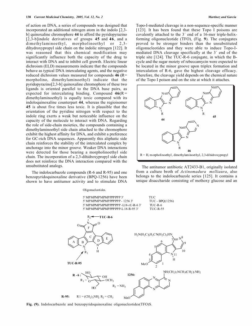
Fig. (9). Indolocarbazole and benzopyridoquinoxaline oligonucleotides(TFO)S.
of action on DNA, a series of compounds was designed thatincorporated an additional nitrogen atom in the indolo [2,3-b] quinoxaline chromophore 44 to afford the pyridopyrazino[2,3-b]indole derivatives of groups 45 and 46 , and adimethylaminoethyl, morpholinoethyl or 2,3-dihydroxypropyl side chain on the indolic nitrogen [122]. Itwas reasoned that this chemical modification maysignificantly influence both the capacity of the drug tointeract with DNA and to inhibit cell growth. Electric lineardichroism (ELD) measurements indicate that the compoundsbehave as typical DNA intercalating agents, and the negativereduced dichroism values measured for compounds 46 (R=morpholino, dimethylaminoethyl) indicate that thepyridopyrazino[2,3-b] quinoxaline chromophore of these twoligands is oriented parallel to the DNA base pairs, asexpected for intercalating binding. Compound 46(R =dimethylaminoethyl) is equally toxic compared with itsindoloquinoxaline counterpart 44, whereas the regioisomer45 is about five times less toxic. It is plausible that theorientation of the pyridine nitrogen with respect to theindole ring exerts a weak but noticeable influence on thecapacity of the molecule to interact with DNA. Regardingthe role of side-chain moieties, the compounds containing adimethylaminoethyl side chain attached to the chromophoreexhibit the highest affinity for DNA, and exhibit a preferencefor GC-rich DNA sequences. Apparently this aliphatic sidechain reinforces the stability of the intercalated complex byanchorage into the minor groove. Weaker DNA interactionswere detected for those bearing a morpholinoethyl sidechain. The incorporation of a 2,3-dihidroxypropyl side chaindoes not reinforce the DNA interaction compared with theunsubstituted analogs.
The indolocarbazole compounds (R-6 and R-95) and onebenzopyridoquinoxaline derivative (BPQ-1256) have beenshown to have antitumor activity and to stimulate DNA
Topo-I-mediated cleavage in a non-sequence-specific manner[123]. It has been found that these Topo I poisons arecovalently attached to the 3´ end of a 16-mer triple-helix-forming oligonucleotide (TFO), (Fig. 9). The conjugatesproved to be stronger binders than the unsubstitutedoligonucleotides and they were able to induce Topo-I-mediated DNA cleavage specifically at the 3´ end of thetriple site [124]. The TUC-R-6 conjugate, in which the B-cycle and the sugar moiety of rebeccamycin were expected tobe located in the minor groove upon triplex formation andintercalation of R-6, gave the highest cleavage efficacy.Therefore, the cleavage yield depends on the chemical natureof the Topo I poison and on the site at which it attaches.
N
N
N
R
N
N
N
R
N
N
N
N
R
N
R = H, morpholinoethyl , dimethylaminoethyl, 2,3-dihidroxypropyl
4445
46
The antitumor antibiotic AT2433-B1, originally isolatedfrom a culture broth of Actinomadura melliaura, alsobelongs to the indolocarbazole series [125]. It contains aunique disaccharide consisting of methoxy glucose and an

The Search of DNA-Intercalators as Antitumoral Drugs Current Medicinal Chemistry, 2005, Vol. 12, No. 2 139
NH
N
NO
CH3
O
O OH
OOHO
O CH3
OHHN
NH
N
NO
CH3
O
O OH
OOHO
OCH3
HN
HO
AT2433-B1 iso-AT2433-B1
X NN
R
O
O
NR2
R1
47a: X = CH, R = H, R1 = CH3 R2 = CH347b: X = CH, R = CH3, R1 = CH3 R2 = CH347c: X = CH, R = CH3 , R1 = H R2 = CH2CH2OH47d: X = N, R = CH3, R1 = CH3 R2 = CH347e: X = N, R = CH3, R1 = H R2 = CH2CH2OH
NH
N
NO
H
O
HO OHO
OH
HO
HO
HO NB-506
aminosugar subunit, 2,4-dideoxy-4-methylamino-L-xylose.The configuration of the amino sugar distinguishes AT2433-B1 from its diastereoisomer iso-AT2433-B1. Previousstudies with rebeccamycin monoglucoside derivatives haveshown that the presence of an amino group in a suitableposition significantly reinforces DNA interaction; based onthat finding, these antibiotics were investigated to establishwhether the configuration of the amino-sugar moietyinfluences their DNA interactions [126]. Accurate bindingmeasurements using the BIAcore surface plasmon resonancemethod revealed that AT2433-B1 binds strongly to hairpinoligomers containing a [CG]4 block but not to oligomerswith a central [AT]4 tract. In contrast, the diastereoisomeriso- exhibits a very weak sequence preference, thus showingthat the configuration of the xylose subunit of AT2433- B1is essential for DNA intercalation.
The interaction of the tetrahydropyrrolo[3,4-a]carbazole-1,3-diones 4 7 a - c and two tetrahydropyrido[3,2-b]pyrrolo[3,4-g]indole-1,3-diones 47d-e with DNA wasinvestigated by absorption spectroscopy and thermal meltingstudies. Compounds vary in the substitution pattern of thearomatic ring and the substituents on the N-pyrrolo and thepropylamine side chain [127]. ELD and CD measurementsshowed that the compounds behave as typical DNA-intercalating agents. In addition, it was found that thestrength of interaction with DNA is dependent on the natureof the side chain. Compounds with a hydroxyethyl-aminoethyl side chain demonstrated higher affinities forpoly(dA-dT)2 than compounds bearing a dimethylaminoe-
thyl side chain. Furthermore, they stabilize DNA–Topo IIcovalent complexes but their Topo II inhibitory propertiesdo not correlate with their cytotoxic potential. However,compounds 47c, 47d and 47e, exhibited a high toxicity toP388 murine leukemia cells and produced a markedaccumulation in the G2/M phase of the cell cycle, whereascompounds 47a and 47b were essentially inactive.
NB-506 (13-N-glucopyranosyl-6-N-formylamino-1-hydroxy-indolocarbazole) is a Topo I inhibitor withnoteworthy antitumor activity [128]. An investigation of thestructural selectivity of the nucleic acid binding of NB-506among 12 different nucleic acid structures and sequencesrevealed that NB-506 has a pronounced preference forbinding to the DNA triplex poly[dA]:(poly[dT])2. Thisselectivity was attributed to a complementary shape betweenits extended aromatic ring system and the planar triple stack[129]. A general revision on indolocarbazole chemistry andtheir cytotoxic activity has been described by Pindur [130].
Oligonucleotide–intercalator conjugates consisting ofbenzopyridoindole and benzopyrido quinoxaline derivativeswere synthesized successfully by joining the intercalator tothe 5´ end or an internucleotide position in the center of a14-mer, (Fig. 10). All the derivatives were observed to forma stable DNA triple helix with a DNA target duplex underphysiological conditions [131]. In particular, the derivativesB[h]PQ attached to the 5´ end and B[e]PI attached to aninternal position on the phosphate diester backbonestabilized the triple helix. In addition, it was found that the

140 Current Medicinal Chemistry, 2005, Vol. 12, No. 2 Martínez and García
O
P
O-
oligoNH
OH3'
O
OHoligo
NH
O3P oligo OH3'
N
NHN
R
H
MeO
N
N
Me
HN
R
MeO
N
N
Me
HN
R
HMeO
N
NN
MeO
NHR
Benzo[e]pyridoindol (B[E]Pl)
Benzo [f] pyridoquinoxaline (B[f]PQ)
1126: R = (CH2)3NH21309: R = (CH2)4NH21310: R = (CH2)5NH21388: R = CH2 )3N(CH3)(CH2 )3NH2
1261: R = (CH2)4NH21333: R = (CH2)5NH21334: R = (CH2)6NH2
Benzo [g] pyridoindole (B[g]PI)
1263: R = (CH2)4 NH2
Benzo [h] pyridoquinoxaline (B[h]PQ)
1260: R = (CH2)4NH21335: R = (CH2)3NH21256: R = (CH2)3NH(CH3 )(CH2)3NH2
Attachmet through5' end
Attachmet throughintemucleotide linkage:
BPI/BPQ Attachment
5'
Fig. (10). Benzopyrindole and benzopyridoquinoxaline oligonucleotide conjugates.
N
H3CO
H3CO
OH
OCH3
CH3Cl
N
H3CO
H3CO
OCH3
OCH3
CH3
O
CH3
CH3SO3
N
H3 CO
H3 CO
OCH3
OCH3
CH3
OC2 H5
CH3
I N
H3CO
H3CO
OH
OCH3
CH3
OC2H5
N
H3CO
H3CO
OMs
OCH3
CH3
OC2H5
I CH3SO3
Fagaronine Ethoxidine
48 5049
stability of these triplexes is higher than the acridineconjugates. Molecular modeling studies support thesefindings.
Phenanthridines
Surface-enhanced Raman scattering (SERS) spectroscopyand flow linear dichroism are powerful techniques forselective analysis of the structure of anticancer agents andtheir complexes with DNA [132]. When these techniqueswere used to study the interactions of the potent anticanceragent fagaronine and its derivative ethoxidine with DNA, the
results confirmed that both compounds are strong majorgroove intercalators. In addition, SERS spectroscopy revealspronounced differences in molecular interactions ofFagaronine and Ethoxidine with DNA [133]. Thesedifferences were explained in terms of the chemical nature ofthe substituent groups of the drugs. It was supposed that theOH group of fagaronine is directed to the minor groove,whereas the OCH2CH3 group of ethoxidine protrudes intothe minor groove where it is accessible for interactions withthe DNA-binding intracellular enzymes.
The search for better anticancer drugs has resulted in thesynthesis of three new derivatives 48, 49, and 50, which are
The Search of DNA-Intercalators as Antitumoral Drugs Current Medicinal Chemistry, 2005, Vol. 12, No. 2 141
N
S
N
N
NH2
H2N CH2CH3
Br
Thiazole Orange Ethidium Bromide
N N N OH
N N N OH
HO
N N N
53
54
analogs of nitidine and fagaronine. The role of the 12-O-ethyl, 2-hydroxy, and 6-methyl substituents in thebiological activity of these derivatives has been examined[134] Analysis of DNA binding and the inhibitory activityof Topo I in human DNA demonstrates that the newcompounds combining 2-, 6-, and 12-position substitutioninteract strongly with DNA and exhibit important Topo Iinhibition. Although fagaronine and ethoxidine werecytotoxic toward all cell lines evaluated, compounds 49 and50 exhibited weaker activities in all in vitro tests. Finally,poor activity was found with compound 48, in which 6-position substitution is known to reduce cytotoxicity. Theseresults indicate that compounds 48, 49, and 50 experiencemajor problems in reaching their cellular targets.
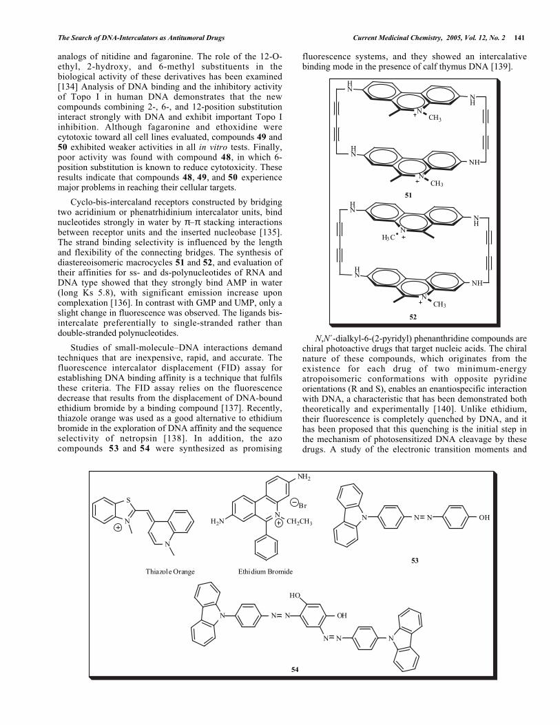
Cyclo-bis-intercaland receptors constructed by bridgingtwo acridinium or phenatrhidinium intercalator units, bindnucleotides strongly in water by π–π stacking interactionsbetween receptor units and the inserted nucleobase [135].The strand binding selectivity is influenced by the lengthand flexibility of the connecting bridges. The synthesis ofdiastereoisomeric macrocycles 51 and 52, and evaluation oftheir affinities for ss- and ds-polynucleotides of RNA andDNA type showed that they strongly bind AMP in water(long Ks 5.8), with significant emission increase uponcomplexation [136]. In contrast with GMP and UMP, only aslight change in fluorescence was observed. The ligands bis-intercalate preferentially to single-stranded rather thandouble-stranded polynucleotides.
Studies of small-molecule–DNA interactions demandtechniques that are inexpensive, rapid, and accurate. Thefluorescence intercalator displacement (FID) assay forestablishing DNA binding affinity is a technique that fulfilsthese criteria. The FID assay relies on the fluorescencedecrease that results from the displacement of DNA-boundethidium bromide by a binding compound [137]. Recently,thiazole orange was used as a good alternative to ethidiumbromide in the exploration of DNA affinity and the sequenceselectivity of netropsin [138]. In addition, the azocompounds 53 and 54 were synthesized as promising
fluorescence systems, and they showed an intercalativebinding mode in the presence of calf thymus DNA [139].
NCH3
HN
NH
NCH3
HN
NH
NH3 C
HN
NH
NCH3
HN
NH
51
52
N,N´-dialkyl-6-(2-pyridyl) phenanthridine compounds arechiral photoactive drugs that target nucleic acids. The chiralnature of these compounds, which originates from theexistence for each drug of two minimum-energyatropoisomeric conformations with opposite pyridineorientations (R and S), enables an enantiospecific interactionwith DNA, a characteristic that has been demonstrated boththeoretically and experimentally [140]. Unlike ethidium,their fluorescence is completely quenched by DNA, and ithas been proposed that this quenching is the initial step inthe mechanism of photosensitized DNA cleavage by thesedrugs. A study of the electronic transition moments and

142 Current Medicinal Chemistry, 2005, Vol. 12, No. 2 Martínez and García
N
X
N N
N
X
55 X= (CH2)256 X= (CH2)3
S R
N
HN
Me
N
OO
NO
O MeH
OHN
R
H
O N
NH
Me
N
OO
N O
OMeH
O
NH
R
H
O
N
OH
ON
OBn
O
N
OMe
O
N
O
O
OR
O
N
OH
O
N
OBn
O
N
O
N
OH
O
MeO
N
OBn
O
MeO
N
O
MeO
N
OR
O
MeO
N
OR
O
Cl N
N
O
N
N OH
O
N
N OBn
O
N
O N
O
R =
sandramycin 57a57b
57c
57d, R = H57e, R = Bn 57f
57m R = H57n R = Bn
57g 57h
57i 57j 57k 57l
57o, R = H57p, R = Bn
57q57r
57s 57t 57v
excited states of two pyridinium-phenanthridiniumcompounds, 55 and 56, revealed two different electronictransitions, a lower-energy transition that is polarized towardthe pyridine ring, and a higher-energy transition that isparallel to the long axis of phenatrhidinium [141]. A second
band, peaking at 250 nm, can be modeled by assuming fouradditional transitions, each with a different polarization. Allof the transition moments lie in the phenanthridinium plane.The highest occupied molecular orbital (HOMO) ofcompounds 5 5 and 5 6 is essentially that of thephenanthridinium moiety, while the lowest unoccupiedmolecular orbital (LUMO) results from the interaction of thedegenerate LUMOs of the pyridinium and phenanthridiniummoieties. These results explain the observed high efficiencyof the photo-oxidation, since the single occupied HOMOorbital is centered on the phenanthridinium moiety, and thisring becomes intercalated between two base pairs uponbinding to DNA. Therefore, a large overlap between theHOMO of phenanthridinium moiety and the molecularorbitals of the DNA bases occurs in the complex, enablingvery effective electro transfer.

The Search of DNA-Intercalators as Antitumoral Drugs Current Medicinal Chemistry, 2005, Vol. 12, No. 2 143
Quinolines and Quinoxalines
Sandramycin is a cyclic decadepsipeptide that possessestwo heteroaromatic chromophores and it is a potentantitumor antibiotic [142]. To determine the influence of theintercalator moiety on DNA binding affinity, a systematicstudy of sandramycin analogs 57a-v was made using calfthymus DNA, within a single high-affinity bis-intercalationbinding site, 5´-d(GCATGC)2. The sandramycin analogsstudied differ with regard to the type and position of thesubstituents on the aromatic ring (OH, OMe, OBz) and theclass of aromatic ring (phenanthrenyl, quinoxalyl,isoquinolyl, and pyridyl) replacing quinolinyl on thecompound [143]. The role of the individual structuralfeatures of the chromophore was evaluated with the high-affinity duplex sequence 5'-d(GCATGC)2. To a firstapproximation, the cytotoxic properties were found toparallel trends established in the DNA binding affinities,with the exception of compound 57c, which lacks thesandramycin chromophore phenol. In addition, it wasdetermined that sandramycin binds to 5´-(GCXXGC)2,where XX=AT, TA, GC, or CG, showing a preference thatfollows the order: 5´-d(GCATGC)2 > 5´-d(GCGCGC)2 > 5´-d(GCTAGC)2 > 5´-d(GCGCGC)2.
The DNA intercalators 6-[[2-(dimethylamino)et-hyl]-amino]3-hydroxy-7H-indeno[2,1-c]quinoline-7-one dihydro-chloride (TAS 103) and N-[2-(dimethylamino)ethyl]-acridine-4-carboxamide (DACA) are dual Topo I/II inhibitorswith potent cytotoxicity in a panel of leukemia lines[144,145]. One of their most prominent structuralcharacteristics is the presence of a carboxamide group. Thebiological activity and binding sequence specificity of thisgroup is considered essential to its mechanism of action.Thus, new intercalators 58a-k were synthesized in whichindeno[1,2-b]quinoline-carboxamides, [1]benzothieno[3,2-b]quinoline-4-carboxamides, and 10H-quindoline-4-carboxamides were substituted. The cytotoxic effectsobserved in a panel of cell lines revealed that smalllipophilic substituents in the noncarboxamide ring, in apseudo-peri position to the side chain, increased thecytotoxic potency; in particular the methyl-substitutedindeno[1,2-b]quinoline-6-carboxamide showed substantiallyincreased effectiveness (20-day growth delays) in asubcutaneous colon 38 in vivo tumor mode that iscomparable to that reported for DACA, which is currentlybeing tested in clinical trials. Crystallographic studies ofdrug–oligonucleotide complexes suggest that thesecompounds bind via van der Waals interactions between thesubstituent and the cytosine residue, forming theintercalation site [146]. In addition, TAS-103, was found tobind to DNA by two binding modes [147]. The majorbinding mode is an outside binding to the major groove andthe minor binding mode is an intercalation. It has also beendiscovered that TAS-103 self-associates in aqueous solutionthrough π–π stacking and hydrophobic interactions.
Fascaplysin is an indoloquinoline derivative that wasfirst isolated from the marine sponge Fascaplysinopsisberquist sp [148]. This natural product inhibits the growthof several microbes and suppresses the proliferation ofleukemia cells and a G1 arrest of tumor and normal cells. Toinvestigate its interaction with DNA, studies involvingisothermal titration calorimetry, absorption spectroscopy,
and CD were performed. The results showed that fascaplysinis an intercalator of DNA with a base-pair-to-drug ratio of2:1 [149]. These results indicate that some of its biologicalactivity could be attributed to interference with the geneticmaterial.
N
O NH
N
X
NH
ON
Me
MeY
OH
HN NMe2
DACA1
2
34
1
34
TAS 103
58a X= CO Y= H 58b X= CO Y= 4-Me58c X= CO Y= 1,4-diMe 58d X= CO Y= 2,4-diMe58e X= CO Y= 4-OMe 58f X= CO Y= 1,4-diOMe58g X= CO Y= 2,3,4-triOMe 58h X= S Y= H58i X= S Y= 1,4-diMe 58j X= NH Y= H58k X= NH Y= 4-Me
Oxazole yellow exhibits enhanced fluorescence uponbinding to DNA and behaves as a typical intercalating agent[150]. The search for an intercalator-linked oligonucleotidethat can form a triple helix with interleukin-2 receptor α-chain promoters led to the synthesis of an oxazole yellow-linked oligonucleotide 59 [151]. Upon light illumination,this compound exhibited a linear increase in fluorescenceduring triple helix formation with double-stranded DNA,and induced photocleavage of the targeted DNA in thepresence of spermine. The cleavage site of one strand wasseven or eight bases away from the intercalation site,whereas the other strand was cleaved at the intercalation site.
NH
N
O
Cl
Fascaplysin
O
N
N
PO-AGAGGGAGAGGAAAA-3'
O
O
I
59
Bis-intercalators have two clear advantages overmonofunctional derivatives. Firstly, bis-intercalators have ahigher binding affinity and hence can be used at lowerconcentrations to facilitate triplex formation, therebyreducing the incidence of unwanted side effects. In theory,the affinity of an ideal bis-intercalator should be the product

144 Current Medicinal Chemistry, 2005, Vol. 12, No. 2 Martínez and García
N
NH
N
CH3
NH
OHN
O
N
N NH
HN
O
O
N
CH3
NH
N
60
of its monofunctional components. Such dramatic increasesin binding are rarely achieved, as there are additional stericand entropic constraints. For efficient bis-intercalation, thelength and nature of the linker needs to be optimized. Atriplex-specific bis-intercalator, naphthylquinoline dimer 60,was designed with this in mind. It was found thatnaphthylquinoline dimmer I stabilizes DNA triplexes at least30 times more effectively than the monofunctionalcompound [152].
Since phenylquinolines and benzimidazoles are “minimalintercalators”, and when an amino-alkyl side chain isattached to the chromophore the compounds producedexhibit the highest affinity for DNA, 2-phenylquinolinederivatives 61a-e that have a (2-aminoethyl) aminomethylgroup at positions 7, 6, or 4 of the aromatic system weresynthesized [153]. It was found that the order of strength ofbinding to DNA was 61d > 61e > 61c > 61a. In addition,the supercoiled DNA unwinding assay supports theintercalation of these drugs. Likewise, the trend ofcytotoxicity of the compounds, 61d > 61c > 61a = 61e, isin quite good agreement with the DNA-binding ability.These results demonstrate that the location of theethylenediamine side chain in these derivatives has a largeeffect on the control of binding ability, DNA binding mode,and cytotoxicity of the compounds.
HNH2NN
NN
DiMIQ
xHCl
61a: 4-, x = 361b: 6-, x = 261c: 7-, x = 361d: 8-, x = 361e: 4'-, x= 3
4
4'
6
8
7
5,11-Dimethyl-5H-indolo[2,3-b]quinoline (DiMIQ) is aDNA intercalator that displays significant cytotoxic activity[154]. The mechanism of its action depends on its ability to
induce and stabilize drug–Topo-II–DNA cleavablecomplexes. Site-specific intercalation of DiMIQ wasinvestigated in vitro by DNAse I footprinting and bymolecular modeling [155]. The footprinting experimentrevealed that the DiMIQ molecule binds preferentially topBR322 plasmid in the 5'-TGCTAACGC-3' region betweenadjacent adenine bases. The molecular modeling resultscorroborate the intercalation preference between adjacentadenines and indicate that the position of DiMIQ inside theDNA duplex is in an orientation parallel to the long axis ofDiMIQ and the neighbor base-pair axes.
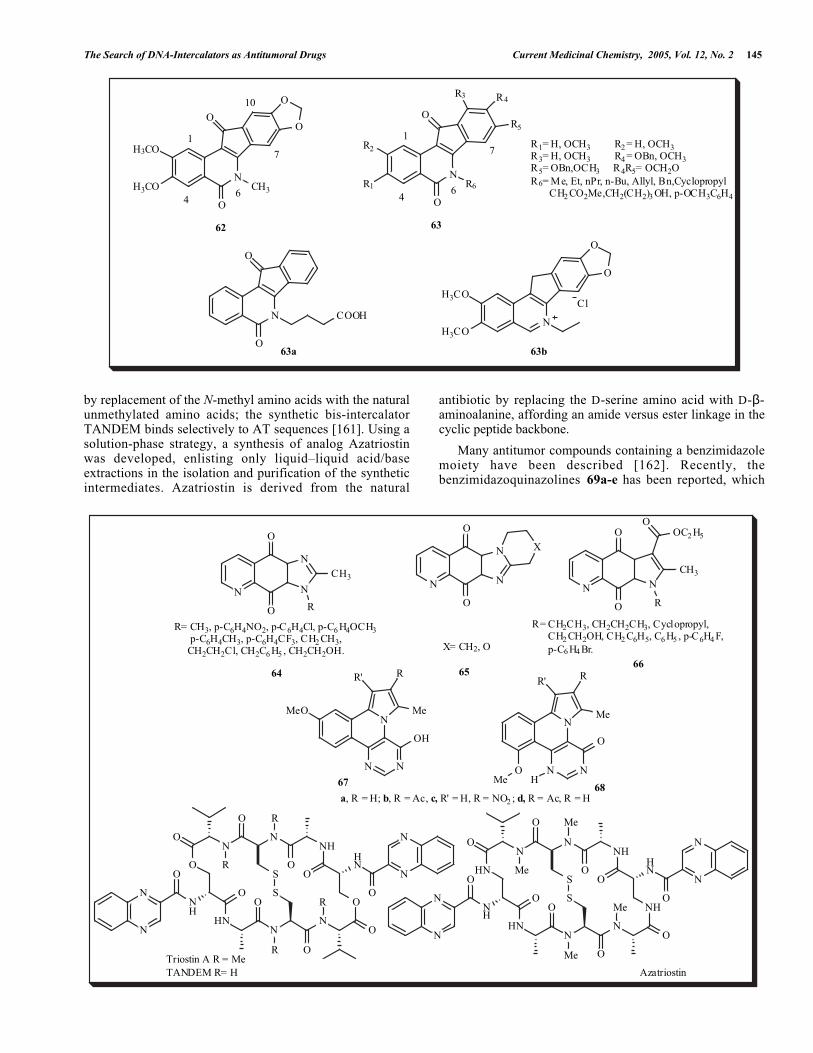
In order to maximize the cytotoxicity of the leadcompound, 62 [156] in human cancer cell cultures, as wellas its activity as a Topo 1 poison, severalindenoisoquinolines 63 were synthesized and evaluated forcytotoxicity and for activity against Topo I in 55 humancancer cell cultures [157]. Nine compounds displayed alower Mean graph midpoint (MGM) than the lead compound62, and four rivaled its Topo I activity. Two of the mostpotent Topo I inhibitors were compounds 63a and 63b, bothof which also inhibited Topo II, unwound DNA, and areassumed to be DNA intercalators. However, the potencies ofthese compounds as Topo I inhibitors did not correlate withtheir potencies as cytotoxic agents.
Three-dimensional quantitative structure activityrelationship (QSAR) analysis, comparative molecular fieldanalysis (CoMFA), and comparative molecular similarityindices analysis (CoMSIA) were carried out for the imidazo-and pyrrolo-quinolindione derivatives 64, 65 and 66 topredict their biological activities [158]. Excellent agreementwas obtained among the three analyses, with an error rangeof 0.01–0.15 between the calculated values and theirmeasured in vitro cytotoxic activity against human lung A-549 cancer cell lines. The planar aromatic character of thepyrimido[5,4-c]pyrrolo[2,1-a]isoquinoline system suggeststhat it is acting as a DNA intercalator. The synthesis ofderivatives 67 and 68, and preliminary molecular modelingshow that they are potential DNA-interactive compounds[159].
Triostin A is a member of the quinoxaline family ofantitumor antibiotics that bind to DNA by bis-intercalation[160]. Triostin A binding in the minor groove exhibits asequence preference for CG. This GC selectivity is removed
The Search of DNA-Intercalators as Antitumoral Drugs Current Medicinal Chemistry, 2005, Vol. 12, No. 2 145
N
O
CH3
O
O
O
H3CO
H3CO
N
O
O
COOHN
O
O
H3CO
H3CO
Cl
N
O
R6
R5
R4
O
R2
R1
R3
63a 63b
1
46
7
10
62
1
46
7
63
R1= H, OCH3 R2 = H, OCH3R3= H, OCH3 R4 = OBn, OCH3R5= OBn,OCH3 R4R5= OCH2OR6= Me, Et, nPr, n-Bu, Allyl, Bn,Cyclopropyl CH2 CO2Me,CH2(CH2)3 OH, p-OCH3C6H4
O R
N
SO
N
O
NHHN
O R
O
NH
O
S
HN
OON
N N
R
N
O
O
ORO
N
N
HN Me
N
SO
N
O
NHHN
O Me
O
NH
O
S
HN
OON
N N
Me
N
O
O
NHMeO
N
N
Triostin A R = MeTANDEM R= H Azatriostin
N
O
N
N
O
CH3
R
X
N N
N
O
ON N
O
O
CH3
R
OOC2 H5
64 6566
R= CH3, p-C6H4NO2, p-C6H4Cl, p-C6 H4OCH3 p-C6H4CH3, p-C6H4CF3, CH2 CH3, CH2CH2Cl, CH2C6 H5 , CH2CH2OH. X= CH2, O
R= CH2CH3, CH2CH2CH3, Cyclopropyl, CH2 CH2OH, CH2 C6H5, C6 H5 , p-C6H4 F, p-C6 H4 Br.
N
N NOHMe
O
Me
R' R
N
N N
OH
Me
R' R
MeO
6867
a, R = H; b, R = Ac, c, R' = H, R = NO2 ; d, R = Ac, R = H
by replacement of the N-methyl amino acids with the naturalunmethylated amino acids; the synthetic bis-intercalatorTANDEM binds selectively to AT sequences [161]. Using asolution-phase strategy, a synthesis of analog Azatriostinwas developed, enlisting only liquid–liquid acid/baseextractions in the isolation and purification of the syntheticintermediates. Azatriostin is derived from the natural
antibiotic by replacing the D-serine amino acid with D-β-aminoalanine, affording an amide versus ester linkage in thecyclic peptide backbone.
Many antitumor compounds containing a benzimidazolemoiety have been described [162]. Recently, thebenzimidazoquinazolines 69a-e has been reported, which

146 Current Medicinal Chemistry, 2005, Vol. 12, No. 2 Martínez and García
either position 6, a dialkylamino alkyl side chain or ahydroxyethylaminomethyl side chain [163]. Thesederivatives exhibit antiproliferative activity toward humantumor cell lines in in vitro assays. The cytotoxic effectdepends on the type of side chain inserted into the planarnucleus, and in some cases it is comparable to the well-known drug ellipticine. Studies of the interaction betweenthese molecules and DNA revealed that the molecular planeof the ligand chromophore is preferentially orientated parallelto the plane of DNA bases.
N
NN
O
R
69a: R = (CH2 )2N(CH3)269b: R = (CH2)3N(CH3)269c: R = (CH2)2N(C2H5)269d: R = (CH2)3N(C2 H5 )269e: R = (CH2)2N(CH2 )2OH
PNAs are mimics of DNA that have potentialapplications in molecular biology [164]. Targeting ofdouble-stranded DNA by homopyrimidine PNA occurs by aprocess of invasion of the double helix, whereby two PNAsbind to the complementary DNA strand while the non-complementary polynucleotide is displaced as a singlestrand. A DNA-binding ligand capable of specificallypromoting the strand invasion process is usually employedto reinforce the binding of PNA to double-stranded DNA.With the aim of identifying ligands that promote PNAbinding, more than 50 ligands were tested, including groovebinders, intercalators, and bis-intercalators [165]. Almost allof drugs have either no effect or inhibit PNA binding;however, the members of the quinoxaline family ofantibiotics (echinomycin, triostin A, 2QN, Tandem) increasethe binding of PNA to a double-stranded DNA target. It wassuggested that quinoxalines enlarge the binding of PNA toDNA by deformation of the double helix, which facilitatesthe PNA invasion. A revision on the use of PNA as a toolfor the development of gene expression has been publishedby Gambari [166].
Miscellaneous
Recently, there has been interest in the synthesis ofTröger´s base derivatives due to their DNA intercalativeproperties [167]. Compounds 70 were synthesized using 4-nitrobenzyl chloride as the starting material, resulting ingood yields [168].
NN
XX
8
761
2
3
45
X = SH, SSCH2C6H4 NO2, SSCH2C6H5, SCH2 C6H5
70
The clinical failure of most intercalator cytotoxic drugs isattributed to their poor solubility, which in turn isattributable to their polycyclic structures [169]. Their
solubility can be enhanced by synthesizing small-sizedtricyclic systems. Therefore, novel 1-aza-9-oxafluorenes 71were obtained from 3-carbonyl-substituted 1,4-dihydropyridines and p-benzoquinone [170]. These aresimilar to α-carbolines, with the indolo-nitrogen replaced byoxygen. Cytotoxic evaluation of various cancer cell linesshowed that these derivatives are more potent than theintercalative carboline systems, and that the 4-phenylsubstituent is more active than the 4-methyl substituent. 1H-NMR data show that the 4-phenyl substituent does not liewithin the plane of the aromatic system. These experimentalresults suggest that the novel cytostatics do not act asintercalators.
NO
HOR2
R1
O
1
5
4
R1 = OC2H5R1 = CH3R1 = OC2H5R1 = CH3
R2 = PhR2 = PhR2 = CH3R2 = CH3
71
Compounds 72 were initially designed to act as bis-intercalators. Either an N2-methyldiethylenetriamine or a3,3´-diamino-N-methyldipropylamine linker holds thetetracyclic ring systems together. The asymmetricalcompounds 73, in which one of the imidazoacridinone ringsystems was replaced by a triazoloacridinone ring system,were found to be cytostatic and cytotoxic in vitro [171]. Inparticular, compound 73b showed remarkably high activity.However, preliminary experimental data and modelingresults suggest that these compounds have a different modeof binding to nucleic acids that does not involveintercalation.
In a current research project focused on the synthesis andbiochemistry of novel intercalators based on quinoliziniumand aza-quinolizinium chromophores, the synthesis of thebenz[f]azino[2,1-a]phthalazinium salt 74 was described usingan intermolecular Westphal condensation [172].
Lucanthone is an antitumor drug that intercalates intoDNA and inhibits Topo II [173]. IA-5, an indazole analog oflucanthone also inhibits Topo II and intercalates into DNApreferentially at A-T-rich sequences, but the anticancerproperties of IA-5 are superior to those of lucanthone [174].The drug stimulates DNA cleavage by Topo II with a weakpreference for sites bearing a C on the 3´ side of the cleavagebond. However, binding to DNA and Topo II inhibition aretwo distinct processes that contribute separately to thecytotoxic activity of IA-5.
Aza-bioisosteric modification of a N,N-dimethylderivative of IA-5 has produced several potent antitumoragents, and it has been found that the position of thenitrogen atom has an influence on the antitumor activity.The synthesis and antitumor activity of compounds 75a-dand 76a-c have been described in studies aimed atdetermining the best antitumor drugs [175]. The in vitrocytotoxicity of these drugs were evaluated in five differentcell lines, which included murine leukemia L1210, murinesarcoma S-180, murine sarcoma resistant to Doxorubicin,S180/Dx), human colon adenocarcinoma, LoVo, and human

The Search of DNA-Intercalators as Antitumoral Drugs Current Medicinal Chemistry, 2005, Vol. 12, No. 2 147
N
O
C NH
N
R
H (CH2)nN(CH2)n
N
O
CNH
N
R
H
CH3
N
O
C NH
NH (CH2 )3 N (CH2)3
CH3 NH
Int. (A, B o C)
N
O
CNH
R
N
O
NN
R
NH
O
NO2
R
a: R = H, n = 2b: R = H; n = 3c: R = OCH3 ; n = 2d: R = OCH3; n = 3e: R = OH; n = 3f: R = C (CH3)3; n = 3
where:
A = B =C =
a: Int. = A, R = OCH3b: Int. = A, R = C(CH2)CH3c: Int. = B, R = Hd: Int. = b, R = OHe: Int. = c, R = OCH3
72 73
colon adenocarcinoma resistant to Doxorubicin, LoVo/Dx.The data clearly shows a pronounced effect of the position ofthe nitrogen atom on the observed cytotoxicities.Comparison of the cytotoxicity of compounds 75a (acarbocyclic chromophore), 75b (a 7-aza analog), and 75c, (an8-aza analog) reveals little difference in potency between thefive cell lines. On the other hand, the analog 75d (9-aza)exhibits a dramatic increase in potency compared to 75a-c inall cell lines studied. In the LoVo and LoVo/Dx cell lines,the 9-aza analog 75d is more potent than doxorubicin ormitoxantrone. The 1-substituted aza-benzothiopyranoin-dazoles 76a-c, in comparison with the corresponding 2-substituted analogs, exhibits a much lower potency.
MSHTS
R1
NN Ar
S
O NH N
CH3
S
NN
CH3
N
IA5LUC
74
Ring= pyridine R1= H, 4-Me, 6-Me, Ar= C6H5, 4-EtO-C6 H4
Ring= QuinolineR1= H, 6-Me, Ar= C6H5, 4-EtO-C6H4
Bis[benzo[c]acridin-7-yl]phenyl]alkanediamides 77 andbis[benzo[a]acridin-12-yl]phenyl] alkanediamides 78 weresynthesized in four steps using 5,5-dimethyl-1,3-cyclohexanedione as the starting material [176]. Thesecompounds were evaluated against 60 tumor cell lines.
Whereas the benzo[c]acridin-7-yl]phenyl]alkanediamidesdisplay significant cytotoxic activity, the corresponding[benzo[a]acridin-12-yl]phenyl]alkanediamide derivatives werefound to be less cytotoxic. This difference was explained interms of fusion of the benzene moiety to the acridine systemin these drugs.
(CH2)2 NMe2
S
Z
YX
N N
CH3
S
Z
N N
CH3
Me2N(H2C)2
YX
75 76
1 2 1 2
a: X = Y = Z = CHb: X = N, Y = Z = CH
c: X = Y = Z = CHd: Z = N, X = Y = CH
Pyrrolo[2,3-e] indole der ivat ives 79 and adihydrobenzoindol analog 80 were synthesized to elucidatethe major structural requirements for their cytotoxic activityin six different tumor cell lines [177]. The results showedthat cytotoxic activity appears to be influenced by thesubstituents on the phenyl group, the presence of an amidegroup generating strong interactions such as hydrogenbonding and increased solubility.
In general, in the synthesis of oligodeoxynucleotides thatare stabilized by intercalators (ODNSAs), the linkers liebetween the nucleobase and the intercalating agent or theintercalator is directly linked to the sugar part [178]. TheDNA-conjugated phenoxyaniline intercalators 81 weresynthesized, and their novelty lay in the fact that theseintercalators are linked to neither a sugar part nor anucleobase. However, only moderate stabilization of DNAthree-way junctions was observed [179].
New pyrazole-annulated azathioxanthenes 82a-e weresynthesized as potential intercalators using 2-chloronicotinicacid as a starting material[180]. In addition, the synthesis of
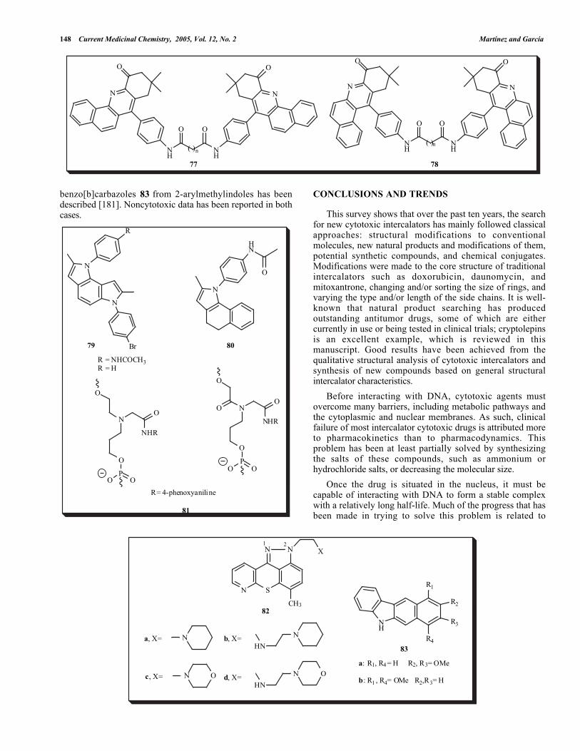
148 Current Medicinal Chemistry, 2005, Vol. 12, No. 2 Martínez and García
N
O
N
O
NH
NH
O O
N
O
NH
N
O
NH
O O
( )n
77 78
( )n
SN
N N
CH3
X
N N
HN
N O N O
HN
R2
NH
R1
R3
R4
82
b, X=
c, X= d, X=
1 2
a, X=83
a: R1, R4 = H R2, R3= OMe
b: R1 , R4= OMe R2,R3= H
benzo[b]carbazoles 83 from 2-arylmethylindoles has beendescribed [181]. Noncytotoxic data has been reported in bothcases.
N
N
Br
R
N
HN
O
R = NHCOCH3 R = H
8079
O
N
NHR
OP
O
O
O
O
N
NHR
OP
O
O
OO
81
R= 4-phenoxyaniline
CONCLUSIONS AND TRENDS
This survey shows that over the past ten years, the searchfor new cytotoxic intercalators has mainly followed classicalapproaches: structural modifications to conventionalmolecules, new natural products and modifications of them,potential synthetic compounds, and chemical conjugates.Modifications were made to the core structure of traditionalintercalators such as doxorubicin, daunomycin, andmitoxantrone, changing and/or sorting the size of rings, andvarying the type and/or length of the side chains. It is well-known that natural product searching has producedoutstanding antitumor drugs, some of which are eithercurrently in use or being tested in clinical trials; cryptolepinsis an excellent example, which is reviewed in thismanuscript. Good results have been achieved from thequalitative structural analysis of cytotoxic intercalators andsynthesis of new compounds based on general structuralintercalator characteristics.
Before interacting with DNA, cytotoxic agents mustovercome many barriers, including metabolic pathways andthe cytoplasmic and nuclear membranes. As such, clinicalfailure of most intercalator cytotoxic drugs is attributed moreto pharmacokinetics than to pharmacodynamics. Thisproblem has been at least partially solved by synthesizingthe salts of these compounds, such as ammonium orhydrochloride salts, or decreasing the molecular size.
Once the drug is situated in the nucleus, it must becapable of interacting with DNA to form a stable complexwith a relatively long half-life. Much of the progress that hasbeen made in trying to solve this problem is related to

The Search of DNA-Intercalators as Antitumoral Drugs Current Medicinal Chemistry, 2005, Vol. 12, No. 2 149
tethering intercalator moieties to nucleic acid bases, PNAs,and ODNSAs, with the aim of generating TFO andquadruplex-forming oligonucleotides. This strategy willassure the synthesis of the best intercalators with enhancedaffinity and selectivity, without losing the goal of improvedcytotoxic activity.
One of the main problems has been the improvement of drugselectivity. Although protein engineering has contributedmuch in recent years to research into ADEPT and GDEPT,there remains a considerable amount of work to be done inthe area. Thus far, research has concentrated mainly oncompounds that are currently in clinical use, such asdaunomycin. In order to overcome drug resistance, it appearsthat the best alternative is to probe all new potential ormodified compounds.
Finally, 50 years have passed since the structure of DNAwas elucidated, and methods of in silico design are nowavailable to any chemist and QSAR, COMFA and structure-based drug design methods are well explored. Common anduseful alternatives by which to seek new antitumorcompounds include the synthesis of potential cytotoxiccompounds based on their structural and physicochemicalcharacteristics, high-throughput screening, the search fornatural products, and synthesis of numerous quantities ofcompounds to obtain data on the structure–activityrelationship. It is also fascinating to see that theseprocedures will provide interesting information about thephenomenon of DNA intercalation and its relationship tocytotoxic activity. It should be remembered that althoughthey are toxic, nonselective, and sometimes expensive, at thepresent time DNA intercalators are among the mostimportant drugs for treating cancer.
ACKNOWLEDGEMENTS
We thank to DGAPA-UNAM (IN211601) and CIC-UMSNH for financial support. Contribution No. 2473 fromInstituto de Química, UNAM.
REFERENCES
[1] Watson, J. D.; Crick, F. H. C. Nature, 1953, 171, 964.[2] Goodman, L. S.; Wintrobe, M. M.; Damescheck, W.; Goodman,
M. J.; Gilman, A.; McLennan, M. T. J. Am. Med. Assoc., 1946,132,126.
[3] Lerman, L. S., J. Mol. Biol., 1961, 3, 18.[4] Lerman, L.S. J. Cell. Comp. Physiol., 1964, 64, 1-6[5] Waring, M. J.; Bailly, C. Gene, 1994, 149, 69.[6] Rehn, C.; Pindur, U. Monatsh. Chem., 1996, 127, 631.[7] Baginski, M.; Fogolari, F; Briggs, J. M. J. Mol. Biol., 1997, 274,
253.[8] Shui, X.; Peek, M. E.; Lipscomb, L. A.; Gao, Q.; Ogata, C.;
Roques, B. P.; Garbay-Jaureguiberry, C.; Wilkinson, A. P.;Williams, L. D. Curr. Med. Chem., 2000, 7, 59.
[9] Paterson, S. E.; Coxon, J.M.; Strekowski, L. Bioorg., Med. Chem.,1997, 5, 277.
[10] Bailly, C.; Suh, D.; Waring, M. J.; Chaires, J., B. Biochemistry,1998, 37, 1033.
[11] Suh, D.; Chaires, J. B. Bioorg. Med. Chem., 1995, 3, 723.[12] LePec, J.B.; Paoletti, C. J. Mol. Biol., 1967, 27, 87.[13] Hogan, M.; Dattagupta, N.; Crothers, D. M. Biochemistry, 1979,
18, 280.[14] Pal, M. K.; Yadav, R. C. Ind. J. Chem., Sect. A, 1990, 29A, 723.[15] MacArthur, M. W.; Driscol, P. C.; Thornton, J. M. Trends
Biotechnol., 1994, 12, 149.
[16] Fink, S. L; Leo, A; Yamakawa, M.; Hansch, C; Quinn, F. R.Farmaco, 1980, 35, 965.
[17] Denny, W. A.; Atwell, G,J.; Cain, B. F; Leo, A.; Panthananickal,A.; Hansch. C. J. Med. Chem., 1982, 25, 276.
[18] Hartley, J. A.; Reszco, K; Zuo, E. T.; Wilson, W. D.; Morgan, A.R.; Lown, J. W. Mol. Pharmacol., 1988, 33, 265.
[19] Baguley, B. C. In Anticancer Drug Development, Baguley B. C.;Kerr, D. J. Ed.; Academic Press 2002, pp. 4-5.
[20] Lodish, H; Baltimore, D.; Berk, A.; Zipursky, S. L.; Matsudaira,P.; Darnell, J. Molecular Cell Biology; New York, 1999. pp. 110-111.
[21] Stewart, L.; Redinbo, M. R.; Qiu, X., Hola; W. W. J.; Champoux,J. J. Science, 1998, 279, 1534.
[22] Christiansen, K.; Dirac, A. V.; Anderson, A. H.; Westergaard, O.J. Biol. Chem., 1993, 268, 9690.
[23] Berger, J. M.; Gamblin, S. J.; Harrison, S. C.; Wang, J. C. Nature,1996, 379, 225.
[24] Wang, J. C. Annu. Rev. Biochem., 1996, 65, 635.[25] Berger, J. M. Biochim. Biophys. Acta, 1998, 1400, 3.[26] Hande, K. R. Biochim. Biophys. Acta, 1998, 1400, 173.[27] Pommier, Y. Biochimie, 1998, 80, 255.[28] Pommier, Y.; Pourquier, P.; Fan, Y.; Strumberg, D. Biochim.
Biophys. Acta, 1998, 1400, 83.[29] Burden, D. A.; Osheroff, N. Biochim. Biophys. Acta, 1998, 1400,
139.[30] Stivers, J. T.; Harris, T. K.; Mildvan, A.S. Biochemistry, 1997, 36,
5212.[31] Froelich-Ammon, S. J.; Osheroff, N. J. Biol. Chem., 1995, 270,
21429.[32] Redinbo, M. R.; Stewart, L.; Kuhn, P.; Champoux, J. J.; Hol, W. G.
J. Science, 1998, 279, 1504.[33] Cotter, T. G.; Lennon, S. V.; Glynn, J. G.; Martin, S. J. Anticancer
Research, 1990, 10, 1153.[34] Calabresi, P.; Chabner, B. A. In Goodman and Gilman’s The
Pharmacological Basis of Therapeutics 9th ed., Hardman, J. G.;Goodman Gilman, A.; Limbird, L. E., Eds.; Mc. Graw Hill 1996,pp. 1225-1232.
[35] Solary, E.; Bertrand, R.; Pommier, Y. Leuk. Lymph., 1994, 15, 21.[36] Boos, G.; Stopper, H. Toxicology Lett., 2000, 116, 7.[37] Skehan, P.; Storeng, R.; Scudiero, D.; Monks, A.; McMahon, J.;
Vistica, D.; Warren, J. T.; Bokesh, H.; Kenney, M.; Boyd, M. R.J. Nat. Cancer Inst., 1990, 82, 1107.
[38] Monks, A.; Scudiero, D.; Skehan, P.; Shoemaker, R.; Paull, K.;Vistica, D.; Hose, C.; Langley, J.; Cronise, P.; Vaigro-Wolff, A.;Gray-Goodrich, M.; Campbell, H.; Mayo, J.; Boyd, M. J. Nat.Cancer Inst., 1991, 83, 757.
[39] Boyd, M. R.; Paull, K. D. Drug. Develop. Res., 1995, 34, 91.[40] Portugal, J. TIBS, 1989, 14, 127.[41] Hoogsteen, K. Acta Cristallogr., 1959, 12, 822.[42] Asseline, U.; Thuong, N. T.; Héléne, C. New J. Chem., 1997, 21,
5.[43] Arimondo, P. B.; Héléne, C. Curr. Med. Chem.-Anti-Cancer
Agents, 2001, 1, 219.[44] Sforza, S; Haaima, G; Marchelli, R; Nielesen, P. E. Eur. J. Org.
Chem. 1999, 1, 197.[45] Wittung, P; Nielsen, P. E; Norden, B. Nucleosides Nucleotides,
1997, 16, 599.[46] Han, H.; Hurley, L. H. TiPS, 2000, 21, 136.[47] Springer, C. J; Niculescu-Duvaz, I. In Anticancer Drug
Development; Baguley, B.C; Kerr, D., Eds.; Academic Press,2002; pp.137.
[48] Scotto, K. W.; Bertino, J. R. In The Molecular Bases of Cancer;Mendelsohn, J.; Howley, P. M.; Israel, M. A.; Liotta, L. A., Eds.;W. B. Saunders Company 1995, pp. 387-400.
[49] Levine, C.; Hiasa, H.; Marians, K. J. Biochim. Biophys. Acta,1998, 1400, 29.
[50] Hooper, D. C. Biochim. Biophys. Acta, 1998, 1400, 45.[51] Zhang, J. L.; Sharma, P. L.; Li, C. J.; Dezube, B. J.; Pardee, A. B.;
Crumpacker, C. S. Antimicrob. Agents Chemother., 1997, 41, 977.[52] Braña, M. F.; Cacho, M.; Gradillas, A.; de Pascual-Teresa, B.;
Ramos, A. Curr. Pharm. Des., 2001, 7, 1754.[53] Haq, I. Arch. Biochem. Biophys., 2002, 403, 1.[54] Denny, W. A. Anticancer Drug Des., 1989, 4, 241.[55] Graves, D. E.; Velea, L. M. Curr. Org. Chem., 2000, 4, 915.[56] Holden, J. A. Curr. Med. Chem.:-Anti-Cancer Agents, 2001, 1, 1.[57] Bischoff, G.; Hoffmann, S. Curr. Med. Chem., 2002, 9, 321.

150 Current Medicinal Chemistry, 2005, Vol. 12, No. 2 Martínez and García
[58] Wilson, W. DF. In Comprehensive Natural Products Chemistry;Barton, D.; Nakanishi, K.; Meth-Cohn, O., Eds.; Elsevier, Oxford,1999, Vol. 7, p. 427.
[59] Demeunynck, M.; Charmantray, F.; Martelli, A. Curr. Pharm.Des., 2001, 7, 1703.
[60] Denny, W. A. Curr. Med. Chem., 2002, 9, 1655.[61] Tsann-Lang, S. Curr. Med. Chem., 2002, 9, 1677.[62] Belmont, P.; Jourdan, M.; Demeunynck, M.; Constant, J. F.;
Garcia, J.; Lhomme, J.; Croisy, A.; Carrez, D. J. Med. Chem.,1999, 42, 5153
[63] Alarcon, K.; Demeunynck, M.; Lhomme, J.; Carrez, D.; Croisy,A. Bioorg. Med. Chem. Lett., 2001, 9,1901.
[64] Carlson, C. B.; Beal, P. A. Org. Lett., 2000, 2, 1465.[65] Liaw, Y.-C.; Gao, Y.-G.; Robinson, H.; van der Marel, G.A.; van
Boom, J. H.; Wang, A. H.-J. Biochemistry, 1989, 28, 9913.[66] Wakelin, L.P.G.; Chetcuti, P.; Denny, W.A. J. Med. Chem., 1990,
33, 2039.[67] Carlson, C. B.; Beal, P.A. Bioorg. Med. Chem. Lett., 2000, 10,
1979.[68] Yang, X.; Robinson, H.; Gao, Y.-G.; Wang, A. H.-J.
Biochemistry, 2000, 39, 10950.[69] Belmont, P.; Chapelle, C.; Demeunynck, M.; Michon, J.; Michon,
P.; Lhomme, J., Bioorg. Med. Chem. Lett., 1998, 8, 669.[70] Ghaneolhosseini, H.; Tjarks, W.; Sjöberg, S. Tetrahedron, 1998,
54, 3877.[71] Mazerska, Z.; Augustin, E.; Skladanowsky, A.; Bibby, M. C.;
Double, J. A.; Konopa, J. Drugs Fut., 1998, 23, 702.[72] Mazerska, Z.; Dziegielewski, J.; Konopa, J. B i o c h e m .
Pharmacol., 2001, 61, 685.[73] Dziegielewski, J.; Slusarski, B.; Konitz, A.; Skladanowski, A.;
Konopa, J. Biochem. Pharmacol., 2002, 63, 1653.[74] Calabrese, C. R.; Loadman, P. M., J. Chromatogr. B, 1997, 690,
275.[75] Braña, M. F.; Casarrubios, L.; Domínguez, G.; Fernández, C.;
Pérez, J. M.; Quiroga, A. G.; Navarro-Ranninger, C.; Pascual-Teresa, B. Eur. J. Med. Chem., 2002, 37, 301.
[76] Antonini, I. Curr. Med. Chem., 2002, 9, 1701[77] McDonald, L. A.; Eldredge, G. S.; Barrows, L. R.; Ireland, C. M.
J. Med. Chem. 1994, 37, 3819.[78] Bonnard, I.; Bontemps, N.; Lahmy, S.; Banaigs, B.; Combaut, G.;
Francisco, C.; Colson, P.; Houssier, C.; Waring, M. J.; Bailly, C.Anticancer Drug Des., 1995, 10, 333.
[79] Kobayashi, J.; Cheng, J.; Nakamura, Y.; Ohimuzi, Y.; Hirata, Y.;Sasaki, T.; Otha, T.; Nozoe, S. Tetrahedron Lett., 1988, 29, 1177.
[80] Linsday, B.; Barrows, L.; Copp, B. Bioorg. Med. Chem. Lett.,1995, 5, 739.
[81] Dassonneville, L.; Wattez, N.; Baldeyrou, B.; Mahieu, C.;Lansiaux, A.; Banaigs, B.; Bonnard, I.; Bailly, C. Biochem.Pharmacol., 2000, 60, 527.
[82] Matsumoto, S. S.; Schmehl, D. M.; Copp, B.; Holden, J. A.;Barrows, L. Proc. Am. Assoc. Cancer Res., 1999, 40, A1914.
[83] Clinquart, E. D. Bull. Acad. R. Mèd., 1929, 9, 627.[84] Delvaux, E. J. Pharm. Belg., 1931, 13, 955.[85] Delvaux, E. J. Pharm. Belg., 1931, 13, 973.[86] Gellert, E.; Hammet, R.; Schlittler, E. Helv. Chim. Acta., 1951, 34,
642.[87] Bonjean, K.; De Pauw-Gillet, M. C.; Defresne, M. P.; Colson, P.;
Houssier, C.; Dassonneville, L.; Bailly, C.; Greimers, R.; Wright,Quetin-Leclerq, J.; Tits, M.; Angenot, L. Biochemistry, 1998, 37,5136.
[88] Dassonneville, L.; Lansiaux, A.; Wattelet, A.; Wattez, N.;Mahieu, C.; Van Miert, S.; Pieters, L.; Bailly, C. Eur. J.Pharmacol., 2000, 409, 9
[89] Lisgarten, J. N.; Coll, M.; Portugal, J.; Wright, C. W.; Aymami, J.Nature Struct. Biol., 2002, 9, 57.
[90] Priebe, W.; Anthracycline Antibiotics: New Analogoues, Methodsof Delivery, and Mechanism of Action, American ChemicalSociety: Washington, DC, 1995.
[91] Baguley, B. C. Anti-Cancer Drug Des., 1991, 6, 1.[92] Ge, P.; Russell, R. A. Tetrahedron, 1997, 53, 17469.[93] Ge, P.; Russell, R. A. Tetrahedron, 1997, 53, 17477.[94] Opitz, A.; Roemer, E.; Haas, W.; Goerls, H.; Werner, W.;
Gräefe, U. Tetrahedron, 2000, 56, 5147.[95] Messori, L.; Temperini, C.; Piccioli, F.; Animati, F.; Di Bugno, C.;
Orioli, P. Bioorg. Med. Chem., 2001, 9, 1815.
[96] Binaschi, M.; Bigioni, M.; Cipollone, A.; Rossi, C.; Goso, C.;Maggi, C. A. ; Capranico, G.; Animati, F. Curr. Med. Chem.:-Anti-Cancer Agents, 2001, 1, 113
[97] De Isabella, P.; Capranico, G.; Palumbo, M.; Sissi, C.; Krapcho,A. P.; Zunino, F. Mol. Pharmacol., 1993, 43, 715.
[98] De Isabella, P.; Palumbo, M.; Sissi, C.; Carenini, N.; Capranico,G.; Menta, E.; Oliva, A.; Spinelli, S.; Krapcho, A. P.; Giuliani F.C.; Zunino, F. Biochem. Pharmacol., 1997, 53, 161.
[99] Islam, S. A.; Neidle, S.; Gandecha, B. M.; Partridge, M.;Patterson, L. H.; Brown, J. R. J. Med. Chem., 1985, 28, 857.
[100] Agbandjie, M.; Jenkins, T.C.; McKenna, R.; Rezka, A.; Neidle, S.J. Med. Chem., 1992, 35, 1418.
[101] Tanious, F. A.; Jenkins, T. C.; Neidle, S.; Wilson, W. D.Biochemistry, 1992, 31, 11632.
[102] Bair, K. W.; Andrews, C. W.; Tuttle, R. L.; Knick, V. C.; Cory,M.; McKee, D. D. J. Med. Chem., 1991, 34, 1983.
[103] Bair, K. W.; Tuttle, R. L.; Knick, V. C.; Cory, M.; McKee, D. D.J. Med. Chem., 1990, 33, 2385.
[104] Dai, W.-M.; Chow, C. W.; Zhou, L.; Ishii, A.; Lau, C. W.; Li, Q.;Hamaguchi, W.; Nishimoto, S. Bioorg. Med. Chem. Lett., 1999, 9,2789.
[105] Braña, M. F.; Ramos, A. Curr. Med. Chem.- Anticancer Agents,2001, 1, 237.
[106] Gianolio, D. A.; McLaughlin, L.W. Bioorg. Med. Chem., 2001, 9,2329.
[107] Gianolio, D. A.; Segismundo, J. M.; McLaughlin, L. W. Nucl.Acids. Res., 2000, 28, 2128.
[108] Lokey, R. S.; Kwok, Y.; Guelev, V.; Pursell, C. J.; Hurley, L. H.;Iverson, B. L. J. Am. Chem. Soc., 1997, 119, 7202.
[109] Murr, M. M.; Harting, M. T.; Guelev, V.; Ren, J.; Chaires, J. B.;Iverson, B. L. Bioorg. Med. Chem., 2001, 9, 1141.
[110] Rajski, S. R.; Jackson, B. A.; Barton, J. K. Mutt. Res., 2000, 447,49.
[111] Núñez, M. E.; Noyes, K. T.; Gianolio, D. A.; McLaughlin, L. W.;Barton, J. K. Biochemistry, 2000, 39, 6190.
[112] Jones, G. B.; Mathews, J. E. Bioorg. Med. Chem. Lett., 1997, 7,745.
[113] Kumar, C. V.; Punzalan, E. H. A.; Tan, W. B. Tetrahedron, 2000,56, 7027.
[114] Olivera, R.; Pascual, S.; Herrero, M.; SanMartin, R.; Domínguez,E. Tetrahedron Lett., 1998, 39, 7155.
[115] Toshima, K.; Okuno, Y.; Nakajima, Y.; Matsumura, S. Bioorg.Med. Chem. Lett., 2002, 12, 671.
[116] Lokhov, S. G.; Podyminogin, M. A.; Sergreev, D. S.; Silkinov, V.N.; Kutyavin, I. V.; Shishkin, C. V.; Zarytova, V. F. BioconjugateChem., 1992, 3, 414.
[117] Balbi, A.; Sottofattori, E.; Grandi, T.; Mazzei, M.; Abramova, T.V.; Lokhov, S. G.; Lebedev, A. V. Tetrahedron, 1994, 50, 4009.
[118] Dall´Acqua, F.; Caffieri, S. Photomed. Photobiol., 1988, 10, 1.[119] Averbeck, D. Photochem. Photobiol., 1989, 50, 859.[120] Dalla Via, L.; Gia, O.; Viola, G.; Bertoloni, G.; Santana, L.;
Uriarte, E. Fármaco, 1998, 53, 638.[121] Harmenberg, J.; Wahren, B.; Bergman, J.; Akerfeld, S.; Lunblad,
L. Antimicrob. Agents Chemother. 1988, 32, 1720.[122] Arimondo, P. B.; Baldeyrou, B.; Laine, W.; Bal, C.; Alphonse, F-
A.; Routier, S.; Coudert, G.; Mérour, J-Y.; Colson, P.; Houssier,C.; Bailly, C. Chemico-Biological Interactions, 2001, 138, 59.
[123] Riou, J.; Fossé, P.; Nguyen, C. H.; Larsen, A. K.; Bissery, A. C.;Grondard, L.; Saucier, J. M.; Bisagni, A.; Lavelle, F. Cancer Res.,1993, 53, 5987.
[124] Arimondo, P.; B, Moreau, P.; Boutorine, A.; Bailly, C.;Prudhomme, M.; Sun, J-S.; Garestier, T.; Hélène, C. Bioorg. Med.Chem., 2000, 8, 777.
[125] Matson, J. A.; Claridge, C.; Bush, J. A.; Titus, J.; Bradner, W. T.;Doyle, T. W.; Horan, A. C.; Patel, M. J. Antibiot., 1989, 42, 1547.
[126] Carrasco, C.; Facompre, M.; Chisholm, J. D.; Van Vranken, D. L.;Wilson, W. D.; Bailly, C. Nucleic Acids Res., 2002, 30, 1774.
[127] Joseph, B.; Facompré, M.; Da Costa, H.; Routier, S.; Mérour, J.-Y.; Colson, P.; Houssier, C.; Bailly, C. Bioorg. Med. Chem., 2001,9, 1533.
[128] Saijo, N. Chest, 1998, 113, 17S.[129] Ren, J.; Bailly, C.; Chaires, J. B. FEBS Letters, 2000, 470, 355.[130] Pindur, U.; Kim,Y-S.; Mehrabani, F. Curr. Med. Chem. 1999, 6,
29.[131] Silver, G. C.; Sun, J.-S.; Nguyen, C. H.; Boutorine, A. S.; Bisagni,
E.; Helene, C. J. Am. Chem. Soc., 1997, 119, 263.

The Search of DNA-Intercalators as Antitumoral Drugs Current Medicinal Chemistry, 2005, Vol. 12, No. 2 151
[132] Fleury, F.; Ianoul, A.; Kryukpv, E.; Sukhanova, A.; Kudelina, I.;Wynne-Jones, A.; Bronstein, I. B.; Maizieres, M.; Berjot, M.;Dodson, G. G.; Wilkinson, A.J.; Holden, J. A.; Feofanov, A. V.;Alix, A. J. P.; Jardillier, J. C.; Nabiev, I. Biochemistry, 1998, 37,14630.
[133] Ianoul, A.; Fleury, F.; Duval, O.; Waigh, R.; Jardillier, J. C.; Alix,A. J. P.; Nabiev, I. J. Phys. Chem. B, 1999, 103, 2008.
[134] Lynch, M. A.; Duval, O.; Sukhanova, A.; Devy, J.; MacKay, S. P.;Waigh R. D.; Nabiev, I. Bioorg. Med. Chem. Lett., 2001, 11, 2643.
[135] Cudic, P.; Zinic, M.; Tomisic, V.; Simeon, V.; Vigneron, J.-P.;Lehn, J.- M. J. Chem. Soc. Chem. Commun., 1995, 1073.
[136] Piantanida, I.; Palm, B. S.; Cudic, P.; Zinic, M.; Schneider, H.-J.Tetrahedron Lett., 2001, 42, 6779.
[137] Boger, D. L.; Fink, B. E.; Brunette, S. R.; Tse, W. C.; Hedrick, M.P. J. Am. Chem. Soc., 2001, 123, 5878.
[138] Boger, D. L.; Tse, W. C. Bioorg. Med. Chem., 2001, 9, 2511.[139] Tripathi, S.; Mohan, M.; Pandey, R. K.; Misra, K. Indian J. Chem.,
1999, 38B, 317.[140] Bárcena, M.; Colmenarejo, G.; Gutiérrez-Alonso, M. C.;
Montero, F.; Orellana, G. Biochem. Biophys. Res. Común., 1995,214, 716.
[141] Colmenarejo, G.; Holmén, A.; Nordén, B. J. Phys. Chem. B, 1997,101, 5196.
[142] Matson, J. A.; Bush, J. A. J. Antibiot., 1989, 42, 1763.[143] Boger, D. L.; Chen, J.-H.; Saionz, K. W.; Jin, Q. Bioorg. Med.
Chem., 1998, 6, 85.[144] Twelves, C. J.; Gardner, C.; Flavin, A.; Sludden, J.; Dennis, I.; de
Bono, J.; Beale, P.; Vasey, P.; Hutchison, C.; Macham, M. A.;Rodríguez, A.; Judson, I.; Bleehen, N. M. Br. J. Cancer, 1999, 80,1786.
[145] Ohyama, T.; Li, Y.; Utsugi, T.; Irie, S.; Yamada, Y.; Sato, T. Jap.J. Cancer Res., 1999, 44, 39.
[146] Chen, J.; Deady, L. W.; Desneves, J.; Kaye, A. J.; Finlay, G. J.;Baguley, B. C.; Denny, W. A. Bioorg. Med. Chem., 2000, 8, 2461.
[147] Ishida, K.; Asao, T. Biochimica et Biophysica Acta(BBA)Molecular Basis of Disease, 2002, 1587, 155.
[148] Roll, D. B. M.; Ireland, C. M.; Lu, H. S. M.; Clardy, J. J. Org.Chem., 1988, 53, 3276.
[149] Hörmann, A.; Chaudhuri, B.; Fretz, H. Bioorg. Med. Chem., 2001,9, 917.
[150] Larsson, A.; Carlsson, C.; Jonsson, M. Biopolymers, 1995, 36,154.[151] Inoue, T.; Sugiura, Y.; Saitoh, J.; Ishiguro, T.; Otsuka, M. Bioorg.
Med. Chem., 1999, 7, 1207.[152] Keppler, M.; Zegrocka, O.; Strekowski, L.; Fox, K. R. FEBS
Letters, 1999, 447, 223.[153] Mikata, Y.; Yokoyama, M.; Ogura, S.; Okura, I.; Kawasaki, M.;
Maeda, M.; Yano, S. Bioorg. Med. Chem. Lett., 1998, 8, 1243.[154] Peczynska-Czoch,′ W.; Pognan, F.; Kaczmarek, L.; Boratynsky,′
J. J. Med. Chem., 1994, 37, 3503.[155] Osiadacz, J.; Majka, J.; Czarnecki, K.; Peczynska-Czoch,′ W.;
Zakrzewska-Czerwinska,′ J.; Kaczmarek, L.; Sokalski, W. A.Bioorg. Med. Chem., 2000, 8, 937.
[156] Kohlhagen, G.; Paull, K.; Cushman, M.; Nagafuji, P.; Pommier, Y.Mol. Pharmacol. 1998, 54, 50.
[157] Strumberg, D.; Pommier, Y.; Paull, K.; Jayaraman, M.; Nagafuji,P.; Cushman, M. J. Med. Chem., 1999, 42, 446.
[158] Suh, M. E.; Kang, M. J.; Park, S. Y. Bioorg. Med. Chem., 2001, 9,2987.
[159] Montalbano, A.; Diana, P.; Barraja, P.; Lauria, A.; Cirrincione,G.; Dattolo, G.; Almerico, A. M. ARKIVOC, 2002, 264.
[160] Jonson, D. S.; Boger, D. L.; DNA Binding Agents. InComprehensive Supramolecular Chemistry; Lehn, J. M., SeriesEd., Murakami, Y., Ed.; Pergamon Press: Oxford, 1996; Vol. 4,Chapter 3, pp73-176.
[161] Boger, Dale L.; Lee, J. K. J. Org. Chem., 2000, 65, 5996.[162] Braña, M. F.; Castellano, J. M.; Keihauer, G.; Machuca, A.;
Martín, Y.; Redondo, C.; Sclick, E.; Walker, N. Anticancer DrugDes., 1994, 9, 527.
[163] Dalla Via, L.; Gia, O.; Magno, S. M.; Da Settimo, A.; Marini, A.M.; Primofiore, G.; Da Settimo, F.; Salerno, S. Fármaco, 2001, 56,159.
[164] Nielsen, P. E.; Egholm, M.; Buchardt, O. Gene, 1994, 149, 139.[165] Møllegaard, N. E.; Bailly, C.; Waring, M. J.; Nielsen, P. E.
Biochemistry, 2000, 39, 9502.[166] Gambari, R. Curr. Pharm. Des., 2001, 7, 1839.[167] Bag, B. G. Curr. Sci., 1995, 68, 279.[168] Bag, B. G.; von Kiedrowski, G. Tetrahedron Lett., 1999, 40, 1289.[169] Sengupta, D.; Anad, N. Ind. J. Chem., 1986, 25B, 72.[170] Brachwitz, K. K.; Hilgeroth, A. A. Bioorg. Med. Chem. Lett.,
2002, 12, 411.[171] Cholody, W. M.; Hernandez, L.; Hassner, L.; Scudiero, D. A.;
Djurickovic, D. B.; Michejda, C. J. J. Med. Chem., 1995, 38,3043.
[172] Martínez-Barrasa, V.; Burgos, C.; Izquierdo, M. L.; Alvarez-Builla, J.; Vaquero, J. J. Tetrahedron Lett., 1999, 40, 4115.
[173] Bailly, C.; Waring, M. J. Biochemistry, 1993, 32, 5985.[174] Dassonneville, L.; Bailly, C. Biochem. Pharmacol., 1999, 58,
1307.[175] Krapcho, A. P.; Haydar, S. N.; Truong-Chiott, S.; Hacker, M. P.;
Menta, E.; Beggiolin, G. Bioorg. Med. Chem. Lett., 2000, 10, 305.[176] Martínez, R.; Cogordan, J. A.; Mancera, C.; Díaz, M. L.
Farmaco, 2000, 55 , 631.[177] Chacón-García, L; Martínez, R. Eur. J. Med. Chem., 2002, 37,
261.[178] Manoharan, M. In Antisense Research and Applications; Crooke,
S. T.; Lebleu, B., Eds.; CRC Press: Boca Raton, 1993, p 303.[179] Poulsen, C. S.; Pedersen, E. B.; Nielsen, C. Acta Chim. Scand.,
1999, 53, 425.[180] Varvaresou, A.; Tsotinis, A.; Siatra-Papastaikoudi, Th.;
Calogeropoulou, T., J. Heterocyclic Chem., 1996, 33, 831.[181] Rajeswaran, W. G.; Srinivasan, P. C. Indian J. Chem., 1994, 33B,
368.